Carcinoma, Small Cell
Carcinoma
Small Cell Lung Carcinoma
Carcinoma, Squamous Cell
Carcinoma, Hepatocellular
Carcinoma in Situ
Carcinoma, Papillary
Carcinoma, Neuroendocrine
Immunohistochemistry
Carcinoma, Ductal, Breast
Tumor Cells, Cultured
Tumor Markers, Biological
Carcinoma, Basal Cell
Carcinoma, Bronchogenic
Prognosis
Neoplasm Staging
Carcinoma, Non-Small-Cell Lung
Carcinoma, Transitional Cell
Carcinoma, Large Cell
Gene Expression Regulation, Neoplastic
Carcinoma, Intraductal, Noninfiltrating
Carcinoma, Adenoid Cystic
Carcinoma, Merkel Cell
Carcinoma, Medullary
Carcinoma, Lobular
Sarcoma, Small Cell
Antigens, Neoplasm
Mice, Nude
Gastrin-Releasing Peptide
Lymphatic Metastasis
Neoplasm Proteins
Phosphopyruvate Hydratase
Survival Rate
Immunoenzyme Techniques
Etoposide
Carcinoma, Mucoepidermoid
Neoplasm Metastasis
Combined Modality Therapy
Antineoplastic Combined Chemotherapy Protocols
Retrospective Studies
Cisplatin
Neoplasms, Multiple Primary
Neoplasm Transplantation
Neoplasm Recurrence, Local
Carcinoma, Endometrioid
Head and Neck Neoplasms
RNA, Messenger
Ovarian Neoplasms
Paraneoplastic Syndromes
Carcinoma, Embryonal
Cell Division
Carcinoma, Ductal
Treatment Outcome
Fatal Outcome
Survival Analysis
Adrenocortical Carcinoma
Bombesin
Carcinoma, Verrucous
Carcinoma, Signet Ring Cell
Reverse Transcriptase Polymerase Chain Reaction
Keratins
Chromosomes, Human, Pair 3
Tomography, X-Ray Computed
Apoptosis
Biopsy
Tumor Suppressor Protein p53
Chromogranins
Receptors, Bombesin
Tissue Array Analysis
Laryngeal Neoplasms
Disease Progression
Precancerous Conditions
Carcinoid Tumor
Adenocarcinoma, Follicular
Follow-Up Studies
Disease-Free Survival
Mutation
Pancreatic Neoplasms
Adenocarcinoma, Mucinous
Blotting, Western
Base Sequence
Embryonal Carcinoma Stem Cells
Molecular Sequence Data
Genes, Tumor Suppressor
Synaptophysin
Carcinoma, Papillary, Follicular
Carcinoembryonic Antigen
Loss of Heterozygosity
Drug Resistance, Neoplasm
Topotecan
Chondrosarcoma, Mesenchymal
Endometrial Neoplasms
Gene Expression
Adenocarcinoma, Clear Cell
Colorectal Neoplasms
Genes, p53
Kaplan-Meier Estimate
Cell Transformation, Neoplastic
Proto-Oncogene Proteins c-kit
alpha-Fetoproteins
Drug Screening Assays, Antitumor
Cystadenocarcinoma, Serous
Cell Survival
Tumor Stem Cell Assay
Sensitivity and Specificity
In Situ Hybridization, Fluorescence
Carcinoma, Lewis Lung
Gene Expression Profiling
Signal Transduction
Gene Amplification
Flow Cytometry
Tumor Suppressor Proteins
Ki-67 Antigen
Receptor, Epidermal Growth Factor
Ureteral Neoplasms
Antineoplastic Agents, Phytogenic
Xenograft Model Antitumor Assays
Lambert-Eaton Myasthenic Syndrome
Mice, Inbred BALB C
Neoplasms
Neovascularization, Pathologic
Keratin-7
Dose-Response Relationship, Drug
Transfection
Transcription Factors
Antibodies, Neoplasm
Chromogranin A
Case-Control Studies
Dopa Decarboxylase
Cytodiagnosis
Proto-Oncogene Proteins
Keratin-19
Neoplasms, Squamous Cell
Cadherins
Cell Cycle
Neuroendocrine Tumors
Adenocarcinoma, Papillary
DNA-Binding Proteins
Predictive Value of Tests
Down-Regulation
Neoplasms, Experimental
Cell Differentiation
Lymph Nodes
Keratin-6
Chemoembolization, Therapeutic
Phenotype
Paclitaxel
Drug Administration Schedule
Mammary Neoplasms, Experimental
Nuclear Proteins
Iodine Radioisotopes
Risk Factors
Mucin-1
Tumor Burden
Carcinoma, Basosquamous
APUD Cells
Receptor, erbB-2
Promoter Regions, Genetic
Polymorphism, Single-Stranded Conformational
Up-Regulation
Brain Neoplasms
Fluorescent Antibody Technique
In Situ Hybridization
Early death during chemotherapy in patients with small-cell lung cancer: derivation of a prognostic index for toxic death and progression. (1/2496)
Based on an increased frequency of early death (death within the first treatment cycle) in our two latest randomized trials of combination chemotherapy in small-cell lung cancer (SCLC), we wanted to identify patients at risk of early non-toxic death (ENTD) and early toxic death (ETD). Data were stored in a database and logistic regression analyses were performed to identify predictive factors for early death. During the first cycle, 118 out of 937 patients (12.6%) died. In 38 patients (4%), the cause of death was sepsis. Significant risk factors were age, performance status (PS), lactate dehydrogenase (LDH) and treatment with epipodophyllotoxins and platinum in the first cycle (EP). Risk factors for ENTD were age, PS and LDH. Extensive stage had a hazard ratio of 1.9 (P = 0.07). Risk factors for ETD were EP, PS and LDH, whereas age and stage were not. For EP, the hazard ratio was as high as 6.7 (P = 0.0001). We introduced a simple prognostic algorithm including performance status, LDH and age. Using a prognostic algorithm to exclude poor-risk patients from trials, we could minimize early death, improve long-term survival and increase the survival differences between different regimens. We suggest that other groups evaluate our algorithm and exclude poor prognosis patients from trials of dose intensification. (+info)Defining and analysing symptom palliation in cancer clinical trials: a deceptively difficult exercise. (2/2496)
The assessment of symptom palliation is an essential component of many treatment comparisons in clinical trials, yet an extensive literature search revealed no consensus as to its precise definition, which could embrace relief of symptoms, time to their onset, duration, degree, as well as symptom control and prevention. In an attempt to assess the importance of these aspects and to compare different methods of analysis, we used one symptom (cough) from a patient self-assessment questionnaire (the Rotterdam Symptom Checklist) in a large (>300 patient) multicentre randomized clinical trial (conducted by the Medical Research Council Lung Cancer Working Party) of palliative chemotherapy in small-cell lung cancer. The regimens compared were a two-drug regimen (2D) and a four-drug regimen (4D). No differences were seen between the regimens in time of onset of palliation or its duration. The degree of palliation was strongly related to the initial severity: 90% of the patients with moderate or severe cough at baseline reported improvement, compared with only 53% of those with mild cough. Analyses using different landmark time points gave conflicting results: the 4D regimen was superior at 1 month and at 3 months, whereas at 2 months the 2D regimen appeared superior. When improvement at any time up to 3 months was considered, the 4D regimen showed a significant benefit (4D 79%, 2D 60%, P = 0.02). These findings emphasize the need for caution in interpreting results, and the importance of working towards a standard definition of symptom palliation. The current lack of specified criteria makes analysis and interpretation of trial results difficult, and comparison across trials impossible. A standard definition of palliation for use in the analysis of clinical trials data is proposed, which takes into account aspects of onset, duration and degree of palliation, and symptom improvement, control and prevention. (+info)Coexpression of transcripts encoding EPHB receptor protein tyrosine kinases and their ephrin-B ligands in human small cell lung carcinoma. (3/2496)
The EPH family is the largest subfamily of receptor protein tyrosine kinases, consisting of the EPHA and EPHB subgroups. Ephrin-B1, ephrin-B2, and ephrin-B3 are ligands of the EPHB subgroup and are encoded by the EFNB1, EFNB2, and EFNB3 genes, respectively. We have shown previously that EPHB2 transcripts are expressed in six small cell lung carcinoma (SCLC) cell lines. In this study, we examined the expression of EPHB1, EPHB2, EPHB3, EPHB4, and EPHB6 in 4 SCLC tumor specimens and 14 cell lines including 3 cell lines derived from these tumor specimens. To investigate whether potential autocrine loops of EPHB receptors and ephrin-B ligands exist in SCLC, the expression of EFNB1, EFNB2, and EFNB3 was also examined. Our data show that transcripts encoding multiple members of the EPHB subgroup and the ephrin-B subgroup are coexpressed in SCLC cell lines and tumors. These results suggest that the EPHB subgroup receptor kinases may modulate the biological behavior of SCLC through autocrine and/or juxtacrine activation by ephrin-B ligands that are expressed in the same or neighboring cells. (+info)Combined modality therapy of lung cancer. (4/2496)
Combined modality therapy for lung cancer was first demonstrated to be successful in limited-stage small cell lung cancer. Concurrent administration of chemotherapy with chest and elective brain irradiation appears to produce the best results, with cisplatin/etoposide as the core chemotherapy. Using such programs, 2-year survival in the 40% range and 5-year survivals in excess of 20% may be expected, based on the results of multiple studies. Attempts to improve on these results through the use of altered schemes of chest irradiation or the delivery of high-dose consolidation chemotherapy are ongoing but to date have not been shown to affect survival significantly. We remain at a plateau in the effectiveness of combined modality therapy for small cell lung cancer, with little evidence that it impacts survival at all in extensive-stage disease. The incorporation of new agents in combination chemotherapy regimens, more "specific" immunotherapy directed at tumor-associated antigens, and the potential adjunctive use of broad-spectrum neuropeptide antagonists offer promise for the future. In non-small cell lung cancer, the sequential use of platinum-based chemotherapy and chest irradiation appears superior in survival to standard, daily fractionated radiation therapy used alone, with long-term survival increased from 5-10% to 15-20%. Concurrent administration of chemotherapy with cisplatin/etoposide and chest irradiation produces 2-year survival in the range of 30%, about twice that would be expected for radiation therapy alone, but has not been compared to it in the setting of a randomized trial. Low-dose cisplatin on a daily basis has been combined as a "sensitizer" with chest irradiation, producing initial results that appeared encouraging. However, these have not been reproduced in subsequent, randomized trials. Another approach to combined modalities has been to give chemotherapy or chemotherapy/radiation therapy as induction, followed by surgical resection, with or without subsequent additional treatment. Most patients (80-85%) can be resected, with encouraging survival at 2 and 3 years in the Southwest Oncology Group experience (37 and 26%, respectively). However, toxicity is greater, and such an approach is associated with an overall mortality risk in the range of 10%. A current intergroup study attempts to define the role of surgery in this setting. The major recent development that is likely to influence the future of combined modality therapy for this disease is the advent of multiple new chemotherapeutic agents, such as the taxanes, gemcitabine, vinorelbine, and the topoisomerase-I inhibitors, which have activity in stage IV disease. The immediate challenge is how to combine these agents with platinum analogues, radiation, and surgery. Aiding this process may be the use of molecular biological "markers" that may predict the chance of success or failure with a given systemic agent. The next decade is likely to see substantial improvements in the outcome of treatment for patients with stages I-III non-small cell lung cancer, based on the systemic exploration of combined modalities. (+info)Phase I and pharmacokinetic study of the topoisomerase II catalytic inhibitor fostriecin. (5/2496)
We conducted a phase I and pharmacokinetic study of the topoisomerase II catalytic inhibitor fostriecin. Fostriecin was administered intravenously over 60 min on days 1-5 at 4-week intervals. Dose was escalated from 2 mg m(-2) day(-1) to 20 mg m(-2) day(-1) in 20 patients. Drug pharmacokinetics was analysed with high performance liquid chromatography with UV-detection. Plasma collected during drug administration was tested in vitro for growth inhibition of a teniposide-resistant small-cell lung cancer (SCLC) cell line. The predominant toxicities were elevated liver transaminases (maximum common toxicity criteria (CTC) grade 4) and serum creatinine (maximum CTC grade 2). These showed only a limited increase with increasing doses, often recovered during drug administration and were fully reversible. Duration of elevated alanine-amino transferase (ALT) was dose-limiting in one patient at 20 mg m(-2). Other frequent toxicities were grade 1-2 nausea/vomiting, fever and mild fatigue. Mean fostriecin plasma half-life was 0.36 h (initial; 95% CI, 0-0.76 h) and 1.51 h (terminal; 95% CI, 0.41-2.61 h). A metabolite, most probably dephosphorylated fostriecin, was detected in plasma and urine. No tumour responses were observed, but the plasma concentrations reached in the patients were insufficient to induce significant growth inhibition in vitro. The maximum tolerated dose (MTD) has not been reached, because drug supply was stopped at the 20 mg m(-2) dose level. However, further escalation seems possible and is warranted to achieve potentially effective drug levels. Fostriecin has a short plasma half-life and longer duration of infusion should be considered. (+info)Fractionated administration of irinotecan and cisplatin for treatment of lung cancer: a phase I study. (6/2496)
A combination chemotherapy of irinotecan (CPT-11) and cisplatin (CDDP) has been reported to be active for lung cancer. In the previous trial, however, diarrhoea and leucopenia became the major obstacle for sufficient dose escalation of CPT-11 to improve the treatment outcome. We conducted a phase I study to investigate whether the fractionated administration of CDDP and CPT-11 at escalated dose was feasible and could improve the treatment outcome. Twenty-four previously untreated patients with unresectable non-small-cell lung cancer (NSCLC) or extensive disease of small-cell lung cancer (SCLC) were eligible. Both CDDP and CPT-11 were given on days 1 and 8, and repeated every 4 weeks. The dose of CDDP was fixed at 60 mg m(-2) and given by 1-h infusion before CPT-11 administration. The starting dose of CPT-11 was 40 mg m(-2), and the dose was escalated by an increase of 10 mg m(-2). The maximally tolerated dose of CPT-11 was determined as 60 mg m(-2) because grade 4 haematological or grade 3 or 4 non-haematological toxicities developed in six patients out of 11 patients evaluated. Diarrhoea became a dose-limiting toxicity. The objective response rates were 76% for NSCLC and 100% for SCLC. The recommended dose of CPT-11 and CDDP in a phase II study will be 50 mg m(-2) and 60 mg m(-2) respectively. (+info)Anti-amphiphysin I antibodies in patients with paraneoplastic neurological disorders associated with small cell lung carcinoma. (7/2496)
Patients with stiff man syndrome and breast cancer develop anti-amphiphysin I antibodies that primarily recognise the C terminus of the protein. Anti-amphiphysin I antibodies have also been identified in a few patients with paraneoplastic neurological disorders (PND) and small cell lung cancer (SCLC). The frequency of anti-amphiphysin I antibodies in patients with SCLC and PND was analysed and the epitope specificity of these antibodies was characterised. Anti-amphiphysin I antibodies were evaluated by immunohistochemistry on human and rat cerebellum and immunoblots of rat brain homogenates. Serum samples included 134 patients with PND and anti-Hu antibodies (83% had SCLC), 44 with SCLC and PND without anti-Hu-antibodies, 63 with PND and either Yo, Ri, or Tr antibodies, 146 with SCLC without PND, and 104 with non-PND. Positive serum samples were confirmed with immunoblots of recombinant human amphiphysin I and immunoreacted with five overlapping peptide fragments covering the full length of the molecule. Serum samples positive for anti-amphiphysin I antibodies included those from seven (2.9%) patients with PND and two (1.4%) with SCLC without PND. Six of the seven anti-amphiphysin I antibody positive patients with PND had SCLC (three with Hu-antibodies), and one had anti-Hu-antibodies but no detectable tumour. The PND included encephalomyelitis/sensory neuropathy (five patients), cerebellar degeneration (one), and opsoclonus (one). All anti-amphiphysin I antibodies reacted with the C terminus of amphiphysin I, but seven also recognised other fragments of the molecule. In conclusion, anti-amphiphysin I antibodies are present at low frequency in patients with SCLC irrespective of the presence of an associated PND. All anti-amphiphysin I antibody positive serum samples have in common reactivity with the C terminus of the protein. (+info)Paclitaxel and carboplatin in the treatment of small-cell lung cancer patients resistant to cyclophosphamide, doxorubicin, and etoposide: a non-cross-resistant schedule. (8/2496)
PURPOSE: To evaluate the efficacy of paclitaxel and carboplatin (PC) in small-cell lung cancer (SCLC) patients resistant to cyclophosphamide, doxorubicin, and etoposide (CDE). PATIENTS AND METHODS: We performed a phase II study with PC in SCLC patients who relapsed within 3 months after first-line treatment with CDE. Paclitaxel administration (175 mg/m2 by a 3-hour intravenous infusion) was followed by a 30-minute infusion of carboplatin (area under the curve 7; Chatelut formula) once every 3 weeks for five cycles. Dexamethasone, clemastine, and ranitidine were standard premedication before every cycle. RESULTS: Included were 35 patients (median age, 59 years; 16 with limited disease and 19 with extensive disease; Eastern Cooperative Oncology Group performance status of < or = 1; median time off treatment 6 weeks) who were previously treated with CDE (n = 33), oral etoposide (n = 2), and reinduction CDE (n = 15); only one patient had received three CDE treatments of five cycles. The CDE regimen was followed by local thoracic radiotherapy in seven patients. Hematologic toxicity of grade 3 or 4, for leukopenia was 27% and 6%, for thrombocytopenia 21% and 13%, and for anemia 17% and 0%, respectively, for a total of 132 cycles. Two patients had neutropenic fever; no toxic death occurred. Nonhematologic toxicity was paresthesia CTC grade 3, diarrhea grade 4, and myalgia grade 3 in one patient each. Reversible paresthesia (CTC grade 1 and 2) in toes and fingers was reported in 69% of patients. Thirty-four patients were assessable for response: complete response in two patients, partial response in 23 patients, stable disease in eight patients, and progressive disease in one patient (response rate, 73.5%; 95% confidence interval, 59% to 88%). One patient was found to have atypical carcinoid at pathologic review and was excluded. Median time to progression was 21 weeks (range, 3 to 40 weeks). Median survival was 31 weeks (range, 6 to 112 weeks). One-year survival was 9%. CONCLUSION: Second-line PC in CDE-resistant SCLC patients yields a high response rate and seems non-cross-resistant to CDE. Toxicity was mild in these poor-prognosis patients. (+info)Carcinoma, small cell is a type of lung cancer that typically starts in the bronchi (the airways that lead to the lungs). It is called "small cell" because the cancer cells are small and appear round or oval in shape. This type of lung cancer is also sometimes referred to as "oat cell carcinoma" due to the distinctive appearance of the cells, which can resemble oats when viewed under a microscope.
Small cell carcinoma is a particularly aggressive form of lung cancer that tends to spread quickly to other parts of the body. It is strongly associated with smoking and is less common than non-small cell lung cancer (NSCLC), which accounts for about 85% of all lung cancers.
Like other types of lung cancer, small cell carcinoma may not cause any symptoms in its early stages. However, as the tumor grows and spreads, it can cause a variety of symptoms, including coughing, chest pain, shortness of breath, hoarseness, and weight loss. Treatment for small cell carcinoma typically involves a combination of chemotherapy, radiation therapy, and sometimes surgery.
Carcinoma is a type of cancer that develops from epithelial cells, which are the cells that line the inner and outer surfaces of the body. These cells cover organs, glands, and other structures within the body. Carcinomas can occur in various parts of the body, including the skin, lungs, breasts, prostate, colon, and pancreas. They are often characterized by the uncontrolled growth and division of abnormal cells that can invade surrounding tissues and spread to other parts of the body through a process called metastasis. Carcinomas can be further classified based on their appearance under a microscope, such as adenocarcinoma, squamous cell carcinoma, and basal cell carcinoma.
Small Cell Lung Carcinoma (SCLC) is a type of lung cancer that typically originates in the central part of the lungs. It is called "small cell" because the tumor cells appear small and round under a microscope. SCLC is an aggressive form of lung cancer that tends to spread rapidly to other parts of the body, such as the lymph nodes, liver, bones, and brain.
SCLC is strongly associated with smoking and is relatively uncommon in people who have never smoked. It accounts for about 10-15% of all lung cancer cases. SCLC is often diagnosed at a later stage because it can grow quickly and cause symptoms such as coughing, chest pain, shortness of breath, and weight loss.
Treatment for SCLC typically involves a combination of chemotherapy and radiation therapy. Surgery is not usually an option due to the advanced stage of the disease at diagnosis. The prognosis for SCLC is generally poor, with a five-year survival rate of less than 7%. However, early detection and treatment can improve outcomes in some cases.
Squamous cell carcinoma is a type of skin cancer that begins in the squamous cells, which are flat, thin cells that form the outer layer of the skin (epidermis). It commonly occurs on sun-exposed areas such as the face, ears, lips, and backs of the hands. Squamous cell carcinoma can also develop in other areas of the body including the mouth, lungs, and cervix.
This type of cancer usually develops slowly and may appear as a rough or scaly patch of skin, a red, firm nodule, or a sore or ulcer that doesn't heal. While squamous cell carcinoma is not as aggressive as some other types of cancer, it can metastasize (spread) to other parts of the body if left untreated, making early detection and treatment important.
Risk factors for developing squamous cell carcinoma include prolonged exposure to ultraviolet (UV) radiation from the sun or tanning beds, fair skin, a history of sunburns, a weakened immune system, and older age. Prevention measures include protecting your skin from the sun by wearing protective clothing, using a broad-spectrum sunscreen with an SPF of at least 30, avoiding tanning beds, and getting regular skin examinations.
Lung neoplasms refer to abnormal growths or tumors in the lung tissue. These tumors can be benign (non-cancerous) or malignant (cancerous). Malignant lung neoplasms are further classified into two main types: small cell lung carcinoma and non-small cell lung carcinoma. Lung neoplasms can cause symptoms such as cough, chest pain, shortness of breath, and weight loss. They are often caused by smoking or exposure to secondhand smoke, but can also occur due to genetic factors, radiation exposure, and other environmental carcinogens. Early detection and treatment of lung neoplasms is crucial for improving outcomes and survival rates.
Hepatocellular carcinoma (HCC) is the most common type of primary liver cancer in adults. It originates from the hepatocytes, which are the main functional cells of the liver. This type of cancer is often associated with chronic liver diseases such as cirrhosis caused by hepatitis B or C virus infection, alcohol abuse, non-alcoholic fatty liver disease (NAFLD), and aflatoxin exposure.
The symptoms of HCC can vary but may include unexplained weight loss, lack of appetite, abdominal pain or swelling, jaundice, and fatigue. The diagnosis of HCC typically involves imaging tests such as ultrasound, CT scan, or MRI, as well as blood tests to measure alpha-fetoprotein (AFP) levels. Treatment options for Hepatocellular carcinoma depend on the stage and extent of the cancer, as well as the patient's overall health and liver function. Treatment options may include surgery, radiation therapy, chemotherapy, targeted therapy, or liver transplantation.
Carcinoma in situ is a medical term used to describe the earliest stage of cancer, specifically a type of cancer that begins in the epithelial tissue, which is the tissue that lines the outer surfaces of organs and body structures. In this stage, the cancer cells are confined to the layer of cells where they first developed and have not spread beyond that layer into the surrounding tissues or organs.
Carcinoma in situ can occur in various parts of the body, including the skin, cervix, breast, lung, prostate, bladder, and other areas. It is often detected through routine screening tests, such as Pap smears for cervical cancer or mammograms for breast cancer.
While carcinoma in situ is not invasive, it can still be a serious condition because it has the potential to develop into an invasive cancer if left untreated. Treatment options for carcinoma in situ may include surgery, radiation therapy, or other forms of treatment, depending on the location and type of cancer. It is important to consult with a healthcare provider to determine the best course of action for each individual case.
Carcinoma, papillary is a type of cancer that begins in the cells that line the glandular structures or the lining of organs. In a papillary carcinoma, the cancerous cells grow and form small finger-like projections, called papillae, within the tumor. This type of cancer most commonly occurs in the thyroid gland, but can also be found in other organs such as the lung, breast, and kidney. Papillary carcinoma of the thyroid gland is usually slow-growing and has a good prognosis, especially when it is diagnosed at an early stage.
Carcinoma, neuroendocrine is a type of cancer that arises from the neuroendocrine cells, which are specialized cells that have both nerve and hormone-producing functions. These cells are found throughout the body, but neuroendocrine tumors (NETs) most commonly occur in the lungs, gastrointestinal tract, pancreas, and thyroid gland.
Neuroendocrine carcinomas can be classified as well-differentiated or poorly differentiated based on how closely they resemble normal neuroendocrine cells under a microscope. Well-differentiated tumors tend to grow more slowly and are less aggressive than poorly differentiated tumors.
Neuroendocrine carcinomas can produce and release hormones and other substances that can cause a variety of symptoms, such as flushing, diarrhea, wheezing, and heart palpitations. Treatment for neuroendocrine carcinoma depends on the location and extent of the tumor, as well as the patient's overall health. Treatment options may include surgery, radiation therapy, chemotherapy, targeted therapy, or a combination of these approaches.
Liver neoplasms refer to abnormal growths in the liver that can be benign or malignant. Benign liver neoplasms are non-cancerous tumors that do not spread to other parts of the body, while malignant liver neoplasms are cancerous tumors that can invade and destroy surrounding tissue and spread to other organs.
Liver neoplasms can be primary, meaning they originate in the liver, or secondary, meaning they have metastasized (spread) to the liver from another part of the body. Primary liver neoplasms can be further classified into different types based on their cell of origin and behavior, including hepatocellular carcinoma, cholangiocarcinoma, and hepatic hemangioma.
The diagnosis of liver neoplasms typically involves a combination of imaging studies, such as ultrasound, CT scan, or MRI, and biopsy to confirm the type and stage of the tumor. Treatment options depend on the type and extent of the neoplasm and may include surgery, radiation therapy, chemotherapy, or liver transplantation.
Immunohistochemistry (IHC) is a technique used in pathology and laboratory medicine to identify specific proteins or antigens in tissue sections. It combines the principles of immunology and histology to detect the presence and location of these target molecules within cells and tissues. This technique utilizes antibodies that are specific to the protein or antigen of interest, which are then tagged with a detection system such as a chromogen or fluorophore. The stained tissue sections can be examined under a microscope, allowing for the visualization and analysis of the distribution and expression patterns of the target molecule in the context of the tissue architecture. Immunohistochemistry is widely used in diagnostic pathology to help identify various diseases, including cancer, infectious diseases, and immune-mediated disorders.
Carcinoma, ductal, breast is a type of breast cancer that begins in the milk ducts (the tubes that carry milk from the lobules of the breast to the nipple). It is called "ductal" because it starts in the cells that line the milk ducts. Ductal carcinoma can be further classified as either non-invasive or invasive, based on whether the cancer cells are confined to the ducts or have spread beyond them into the surrounding breast tissue.
Non-invasive ductal carcinoma (also known as intraductal carcinoma or ductal carcinoma in situ) is a condition where abnormal cells have been found in the lining of the milk ducts, but they have not spread outside of the ducts. These cells have the potential to become invasive and spread to other parts of the breast or body if left untreated.
Invasive ductal carcinoma (IDC) is a type of breast cancer that starts in a milk duct and then grows into the surrounding breast tissue. From there, it can spread to other parts of the body through the bloodstream and lymphatic system. IDC is the most common form of breast cancer, accounting for about 80% of all cases.
Symptoms of ductal carcinoma may include a lump or thickening in the breast, changes in the size or shape of the breast, dimpling or puckering of the skin on the breast, nipple discharge (especially if it is clear or bloody), and/or redness or scaling of the nipple or breast skin. However, many cases of ductal carcinoma are detected through mammography before any symptoms develop.
Treatment for ductal carcinoma depends on several factors, including the stage and grade of the cancer, as well as the patient's overall health and personal preferences. Treatment options may include surgery (such as a lumpectomy or mastectomy), radiation therapy, chemotherapy, hormone therapy, and/or targeted therapies.
'Tumor cells, cultured' refers to the process of removing cancerous cells from a tumor and growing them in controlled laboratory conditions. This is typically done by isolating the tumor cells from a patient's tissue sample, then placing them in a nutrient-rich environment that promotes their growth and multiplication.
The resulting cultured tumor cells can be used for various research purposes, including the study of cancer biology, drug development, and toxicity testing. They provide a valuable tool for researchers to better understand the behavior and characteristics of cancer cells outside of the human body, which can lead to the development of more effective cancer treatments.
It is important to note that cultured tumor cells may not always behave exactly the same way as they do in the human body, so findings from cell culture studies must be validated through further research, such as animal models or clinical trials.
Tumor markers are substances that can be found in the body and their presence can indicate the presence of certain types of cancer or other conditions. Biological tumor markers refer to those substances that are produced by cancer cells or by other cells in response to cancer or certain benign (non-cancerous) conditions. These markers can be found in various bodily fluids such as blood, urine, or tissue samples.
Examples of biological tumor markers include:
1. Proteins: Some tumor markers are proteins that are produced by cancer cells or by other cells in response to the presence of cancer. For example, prostate-specific antigen (PSA) is a protein produced by normal prostate cells and in higher amounts by prostate cancer cells.
2. Genetic material: Tumor markers can also include genetic material such as DNA, RNA, or microRNA that are shed by cancer cells into bodily fluids. For example, circulating tumor DNA (ctDNA) is genetic material from cancer cells that can be found in the bloodstream.
3. Metabolites: Tumor markers can also include metabolic products produced by cancer cells or by other cells in response to cancer. For example, lactate dehydrogenase (LDH) is an enzyme that is released into the bloodstream when cancer cells break down glucose for energy.
It's important to note that tumor markers are not specific to cancer and can be elevated in non-cancerous conditions as well. Therefore, they should not be used alone to diagnose cancer but rather as a tool in conjunction with other diagnostic tests and clinical evaluations.
Carcinoma, basal cell is a type of skin cancer that arises from the basal cells, which are located in the lower part of the epidermis (the outermost layer of the skin). It is also known as basal cell carcinoma (BCC) and is the most common form of skin cancer.
BCC typically appears as a small, shiny, pearly bump or nodule on the skin, often in sun-exposed areas such as the face, ears, neck, hands, and arms. It may also appear as a scar-like area that is white, yellow, or waxy. BCCs are usually slow growing and rarely spread (metastasize) to other parts of the body. However, they can be locally invasive and destroy surrounding tissue if left untreated.
The exact cause of BCC is not known, but it is thought to be related to a combination of genetic and environmental factors, including exposure to ultraviolet (UV) radiation from the sun or tanning beds. People with fair skin, light hair, and blue or green eyes are at increased risk of developing BCC.
Treatment for BCC typically involves surgical removal of the tumor, along with a margin of healthy tissue. Other treatment options may include radiation therapy, topical chemotherapy, or photodynamic therapy. Prevention measures include protecting your skin from UV radiation by wearing protective clothing, using sunscreen, and avoiding tanning beds.
Carcinoma, bronchogenic is a medical term that refers to a type of lung cancer that originates in the bronchi, which are the branching tubes that carry air into the lungs. It is the most common form of lung cancer and can be further classified into different types based on the specific cell type involved, such as squamous cell carcinoma, adenocarcinoma, or large cell carcinoma.
Bronchogenic carcinomas are often associated with smoking and exposure to environmental pollutants, although they can also occur in non-smokers. Symptoms may include coughing, chest pain, shortness of breath, wheezing, hoarseness, or unexplained weight loss. Treatment options depend on the stage and location of the cancer, as well as the patient's overall health and may include surgery, radiation therapy, chemotherapy, targeted therapy, or a combination of these approaches.
Adenocarcinoma is a type of cancer that arises from glandular epithelial cells. These cells line the inside of many internal organs, including the breasts, prostate, colon, and lungs. Adenocarcinomas can occur in any of these organs, as well as in other locations where glands are present.
The term "adenocarcinoma" is used to describe a cancer that has features of glandular tissue, such as mucus-secreting cells or cells that produce hormones. These cancers often form glandular structures within the tumor mass and may produce mucus or other substances.
Adenocarcinomas are typically slow-growing and tend to spread (metastasize) to other parts of the body through the lymphatic system or bloodstream. They can be treated with surgery, radiation therapy, chemotherapy, targeted therapy, or a combination of these treatments. The prognosis for adenocarcinoma depends on several factors, including the location and stage of the cancer, as well as the patient's overall health and age.
Prognosis is a medical term that refers to the prediction of the likely outcome or course of a disease, including the chances of recovery or recurrence, based on the patient's symptoms, medical history, physical examination, and diagnostic tests. It is an important aspect of clinical decision-making and patient communication, as it helps doctors and patients make informed decisions about treatment options, set realistic expectations, and plan for future care.
Prognosis can be expressed in various ways, such as percentages, categories (e.g., good, fair, poor), or survival rates, depending on the nature of the disease and the available evidence. However, it is important to note that prognosis is not an exact science and may vary depending on individual factors, such as age, overall health status, and response to treatment. Therefore, it should be used as a guide rather than a definitive forecast.
Neoplasm staging is a systematic process used in medicine to describe the extent of spread of a cancer, including the size and location of the original (primary) tumor and whether it has metastasized (spread) to other parts of the body. The most widely accepted system for this purpose is the TNM classification system developed by the American Joint Committee on Cancer (AJCC) and the Union for International Cancer Control (UICC).
In this system, T stands for tumor, and it describes the size and extent of the primary tumor. N stands for nodes, and it indicates whether the cancer has spread to nearby lymph nodes. M stands for metastasis, and it shows whether the cancer has spread to distant parts of the body.
Each letter is followed by a number that provides more details about the extent of the disease. For example, a T1N0M0 cancer means that the primary tumor is small and has not spread to nearby lymph nodes or distant sites. The higher the numbers, the more advanced the cancer.
Staging helps doctors determine the most appropriate treatment for each patient and estimate the patient's prognosis. It is an essential tool for communication among members of the healthcare team and for comparing outcomes of treatments in clinical trials.
A cell line that is derived from tumor cells and has been adapted to grow in culture. These cell lines are often used in research to study the characteristics of cancer cells, including their growth patterns, genetic changes, and responses to various treatments. They can be established from many different types of tumors, such as carcinomas, sarcomas, and leukemias. Once established, these cell lines can be grown and maintained indefinitely in the laboratory, allowing researchers to conduct experiments and studies that would not be feasible using primary tumor cells. It is important to note that tumor cell lines may not always accurately represent the behavior of the original tumor, as they can undergo genetic changes during their time in culture.
Carcinoma, non-small-cell lung (NSCLC) is a type of lung cancer that includes several subtypes of malignant tumors arising from the epithelial cells of the lung. These subtypes are classified based on the appearance of the cancer cells under a microscope and include adenocarcinoma, squamous cell carcinoma, and large cell carcinoma. NSCLC accounts for about 85% of all lung cancers and tends to grow and spread more slowly than small-cell lung cancer (SCLC).
NSCLC is often asymptomatic in its early stages, but as the tumor grows, symptoms such as coughing, chest pain, shortness of breath, hoarseness, and weight loss may develop. Treatment options for NSCLC depend on the stage and location of the cancer, as well as the patient's overall health and lung function. Common treatments include surgery, radiation therapy, chemotherapy, targeted therapy, or a combination of these approaches.
Transitional cell carcinoma (TCC) is a type of cancer that develops in the transitional epithelium, which is the tissue that lines the inner surface of the urinary tract. This includes the renal pelvis, ureters, bladder, and urethra. Transitional cell carcinoma is the most common type of bladder cancer and can also occur in other parts of the urinary system.
Transitional cells are specialized epithelial cells that can stretch and change shape as the organs they line expand or contract. These cells normally have a flat, squamous appearance when at rest but become more cuboidal and columnar when the organ is full. Transitional cell carcinomas typically start in the urothelium, which is the innermost lining of the urinary tract.
Transitional cell carcinoma can be classified as non-invasive (also called papillary or superficial), invasive, or both. Non-invasive TCCs are confined to the urothelium and have not grown into the underlying connective tissue. Invasive TCCs have grown through the urothelium and invaded the lamina propria (a layer of connective tissue beneath the urothelium) or the muscle wall of the bladder.
Transitional cell carcinoma can also be categorized as low-grade or high-grade, depending on how abnormal the cancer cells look under a microscope and how likely they are to grow and spread. Low-grade TCCs tend to have a better prognosis than high-grade TCCs.
Treatment for transitional cell carcinoma depends on the stage and grade of the cancer, as well as other factors such as the patient's overall health. Treatment options may include surgery, radiation therapy, chemotherapy, or immunotherapy.
Carcinoma, large cell is a type of lung cancer that is characterized by the presence of large, abnormal-looking cells when viewed under a microscope. These cells have a large nucleus and a significant amount of cytoplasm. This type of lung cancer can be further divided into subtypes based on the appearance of the cells and the presence or absence of specific genetic mutations.
Large cell carcinoma is often aggressive and tends to grow and spread quickly. It is typically treated with a combination of surgery, chemotherapy, and/or radiation therapy. The prognosis for large cell carcinoma varies depending on the stage at diagnosis and the individual's overall health.
Neoplastic gene expression regulation refers to the processes that control the production of proteins and other molecules from genes in neoplastic cells, or cells that are part of a tumor or cancer. In a normal cell, gene expression is tightly regulated to ensure that the right genes are turned on or off at the right time. However, in cancer cells, this regulation can be disrupted, leading to the overexpression or underexpression of certain genes.
Neoplastic gene expression regulation can be affected by a variety of factors, including genetic mutations, epigenetic changes, and signals from the tumor microenvironment. These changes can lead to the activation of oncogenes (genes that promote cancer growth and development) or the inactivation of tumor suppressor genes (genes that prevent cancer).
Understanding neoplastic gene expression regulation is important for developing new therapies for cancer, as targeting specific genes or pathways involved in this process can help to inhibit cancer growth and progression.
Intraductal carcinoma, noninfiltrating is a medical term used to describe a type of breast cancer that is confined to the milk ducts of the breast. It is also sometimes referred to as ductal carcinoma in situ (DCIS). Noninfiltrating means that the cancer cells have not spread beyond the ducts into the surrounding breast tissue or elsewhere in the body.
In this type of cancer, abnormal cells line the milk ducts and fill the inside of the ducts. These abnormal cells may look like cancer cells under a microscope, but they have not grown through the walls of the ducts into the surrounding breast tissue. However, if left untreated, noninfiltrating intraductal carcinoma can progress to an invasive form of breast cancer where the cancer cells spread beyond the milk ducts and invade the surrounding breast tissue.
It is important to note that while noninfiltrating intraductal carcinoma is considered a precancerous condition, it still requires medical treatment to prevent the development of invasive breast cancer. Treatment options may include surgery, radiation therapy, or hormone therapy, depending on the size and location of the tumor and other individual factors.
Adenoid cystic carcinoma (AdCC) is a rare type of cancer that can occur in various glands and tissues of the body, most commonly in the salivary glands. AdCC is characterized by its slow growth and tendency to spread along nerves. It typically forms solid, cystic, or mixed tumors with distinct histological features, including epithelial cells arranged in tubular, cribriform, or solid patterns.
The term "carcinoma" refers to a malignant tumor originating from the epithelial cells lining various organs and glands. In this case, adenoid cystic carcinoma is a specific type of carcinoma that arises in the salivary glands or other glandular tissues.
The primary treatment options for AdCC include surgical resection, radiation therapy, and sometimes chemotherapy. Despite its slow growth, adenoid cystic carcinoma has a propensity to recur locally and metastasize to distant sites such as the lungs, bones, and liver. Long-term follow-up is essential due to the risk of late recurrences.
Merkel cell carcinoma (MCC) is a rare and aggressive type of skin cancer that originates from the uncontrolled growth of Merkel cells, which are specialized nerve cells found in the top layer of the skin (epidermis). These cells are responsible for touch sensation. MCC typically presents as a painless, firm, rapidly growing nodule or mass, often on sun-exposed areas such as the head, neck, and arms of older adults.
The primary risk factors for Merkel cell carcinoma include:
1. Exposure to ultraviolet (UV) radiation from sunlight or tanning beds
2. Advanced age (most commonly occurs in people over 50)
3. A weakened immune system due to conditions like HIV/AIDS, organ transplantation, or long-term use of immunosuppressive medications
4. History of other types of skin cancer, such as melanoma or basal cell carcinoma
5. Fair skin and light eye color
MCC is considered an aggressive cancer because it can spread quickly to nearby lymph nodes and other parts of the body (metastasize). The major prognostic factor for MCC is the presence or absence of lymph node involvement at the time of diagnosis. Early detection and treatment are crucial for improving outcomes.
Standard treatments for Merkel cell carcinoma include surgical excision, radiation therapy, and chemotherapy. Immunotherapy with drugs like avelumab has also shown promising results in treating advanced stages of MCC. Regular follow-up care is essential to monitor for recurrence or metastasis.
Neoplasm invasiveness is a term used in pathology and oncology to describe the aggressive behavior of cancer cells as they invade surrounding tissues and organs. This process involves the loss of cell-to-cell adhesion, increased motility and migration, and the ability of cancer cells to degrade the extracellular matrix (ECM) through the production of enzymes such as matrix metalloproteinases (MMPs).
Invasive neoplasms are cancers that have spread beyond the original site where they first developed and have infiltrated adjacent tissues or structures. This is in contrast to non-invasive or in situ neoplasms, which are confined to the epithelial layer where they originated and have not yet invaded the underlying basement membrane.
The invasiveness of a neoplasm is an important prognostic factor in cancer diagnosis and treatment, as it can indicate the likelihood of metastasis and the potential effectiveness of various therapies. In general, more invasive cancers are associated with worse outcomes and require more aggressive treatment approaches.
Medullary carcinoma is a type of cancer that develops in the neuroendocrine cells of the thyroid gland. These cells produce hormones that help regulate various bodily functions. Medullary carcinoma is a relatively rare form of thyroid cancer, accounting for about 5-10% of all cases.
Medullary carcinoma is characterized by the presence of certain genetic mutations that cause the overproduction of calcitonin, a hormone produced by the neuroendocrine cells. This overproduction can lead to the formation of tumors in the thyroid gland.
Medullary carcinoma can be hereditary or sporadic. Hereditary forms of the disease are caused by mutations in the RET gene and are often associated with multiple endocrine neoplasia type 2 (MEN 2), a genetic disorder that affects the thyroid gland, adrenal glands, and parathyroid glands. Sporadic forms of medullary carcinoma, on the other hand, are not inherited and occur randomly in people with no family history of the disease.
Medullary carcinoma is typically more aggressive than other types of thyroid cancer and tends to spread (metastasize) to other parts of the body, such as the lymph nodes, lungs, and liver. Symptoms may include a lump or nodule in the neck, difficulty swallowing, hoarseness, and coughing. Treatment options may include surgery, radiation therapy, and chemotherapy. Regular monitoring of calcitonin levels is also recommended to monitor the effectiveness of treatment and detect any recurrence of the disease.
Carcinoma, lobular is a type of breast cancer that begins in the milk-producing glands (lobules) of the breast. It can be either invasive or non-invasive (in situ). Invasive lobular carcinoma (ILC) occurs when the cancer cells break through the wall of the lobule and invade the surrounding breast tissue, and can potentially spread to other parts of the body. Non-invasive lobular carcinoma (LCIS), on the other hand, refers to the presence of abnormal cells within the lobule that have not invaded nearby breast tissue.
ILC is usually detected as a mass or thickening in the breast, and it may not cause any symptoms or show up on mammograms until it has grown quite large. It tends to grow more slowly than some other types of breast cancer, but it can still be serious and require extensive treatment. LCIS does not typically cause any symptoms and is usually found during a biopsy performed for another reason.
Treatment options for carcinoma, lobular depend on several factors, including the stage of the cancer, the patient's overall health, and their personal preferences. Treatment may include surgery, radiation therapy, chemotherapy, hormone therapy, or targeted therapy. Regular follow-up care is essential to monitor for recurrence or the development of new cancers.
Small cell sarcoma is a very rare and aggressive type of cancer that affects the connective tissues in the body, such as muscles, tendons, bones, cartilage, and fat. It is called "small cell" because the cancer cells are small and appear round or oval in shape, with scant cytoplasm and finely granular chromatin.
Small cell sarcoma typically occurs in adults between the ages of 40 and 70, and it can develop in any part of the body. However, it is most commonly found in the extremities, trunk, and retroperitoneum. The exact cause of small cell sarcoma is not known, but it is thought to be associated with genetic mutations that occur during a person's lifetime.
Small cell sarcoma can be difficult to diagnose because it often does not cause any symptoms until it has advanced to an aggressive stage. When symptoms do occur, they may include pain, swelling, or a lump in the affected area. Diagnosis typically involves a biopsy of the tumor tissue, followed by imaging tests such as CT scans, MRI scans, or PET scans to determine the extent of the cancer.
Treatment for small cell sarcoma usually involves surgery to remove the tumor, followed by radiation therapy and/or chemotherapy to kill any remaining cancer cells. However, because small cell sarcoma is so rare and aggressive, treatment options may be limited, and the prognosis is often poor. Clinical trials of new treatments are also an option for some patients.
Thyroid neoplasms refer to abnormal growths or tumors in the thyroid gland, which can be benign (non-cancerous) or malignant (cancerous). These growths can vary in size and may cause a noticeable lump or nodule in the neck. Thyroid neoplasms can also affect the function of the thyroid gland, leading to hormonal imbalances and related symptoms. The exact causes of thyroid neoplasms are not fully understood, but risk factors include radiation exposure, family history, and certain genetic conditions. It is important to note that most thyroid nodules are benign, but a proper medical evaluation is necessary to determine the nature of the growth and develop an appropriate treatment plan.
Breast neoplasms refer to abnormal growths in the breast tissue that can be benign or malignant. Benign breast neoplasms are non-cancerous tumors or growths, while malignant breast neoplasms are cancerous tumors that can invade surrounding tissues and spread to other parts of the body.
Breast neoplasms can arise from different types of cells in the breast, including milk ducts, milk sacs (lobules), or connective tissue. The most common type of breast cancer is ductal carcinoma, which starts in the milk ducts and can spread to other parts of the breast and nearby structures.
Breast neoplasms are usually detected through screening methods such as mammography, ultrasound, or MRI, or through self-examination or clinical examination. Treatment options for breast neoplasms depend on several factors, including the type and stage of the tumor, the patient's age and overall health, and personal preferences. Treatment may include surgery, radiation therapy, chemotherapy, hormone therapy, or targeted therapy.
Antineoplastic agents are a class of drugs used to treat malignant neoplasms or cancer. These agents work by inhibiting the growth and proliferation of cancer cells, either by killing them or preventing their division and replication. Antineoplastic agents can be classified based on their mechanism of action, such as alkylating agents, antimetabolites, topoisomerase inhibitors, mitotic inhibitors, and targeted therapy agents.
Alkylating agents work by adding alkyl groups to DNA, which can cause cross-linking of DNA strands and ultimately lead to cell death. Antimetabolites interfere with the metabolic processes necessary for DNA synthesis and replication, while topoisomerase inhibitors prevent the relaxation of supercoiled DNA during replication. Mitotic inhibitors disrupt the normal functioning of the mitotic spindle, which is essential for cell division. Targeted therapy agents are designed to target specific molecular abnormalities in cancer cells, such as mutated oncogenes or dysregulated signaling pathways.
It's important to note that antineoplastic agents can also affect normal cells and tissues, leading to various side effects such as nausea, vomiting, hair loss, and myelosuppression (suppression of bone marrow function). Therefore, the use of these drugs requires careful monitoring and management of their potential adverse effects.
Nasopharyngeal neoplasms refer to abnormal growths or tumors in the nasopharynx, which is the upper part of the pharynx (throat) behind the nose. These growths can be benign (non-cancerous) or malignant (cancerous).
Malignant nasopharyngeal neoplasms are often referred to as nasopharyngeal carcinoma or cancer. There are different types of nasopharyngeal carcinomas, including keratinizing squamous cell carcinoma, non-keratinizing carcinoma, and basaloid squamous cell carcinoma.
The risk factors for developing nasopharyngeal neoplasms include exposure to the Epstein-Barr virus (EBV), consumption of certain foods, smoking, and genetic factors. Symptoms may include a lump in the neck, nosebleeds, hearing loss, ringing in the ears, and difficulty swallowing or speaking. Treatment options depend on the type, size, and stage of the neoplasm and may include surgery, radiation therapy, chemotherapy, or a combination of these treatments.
The term "DNA, neoplasm" is not a standard medical term or concept. DNA refers to deoxyribonucleic acid, which is the genetic material present in the cells of living organisms. A neoplasm, on the other hand, is a tumor or growth of abnormal tissue that can be benign (non-cancerous) or malignant (cancerous).
In some contexts, "DNA, neoplasm" may refer to genetic alterations found in cancer cells. These genetic changes can include mutations, amplifications, deletions, or rearrangements of DNA sequences that contribute to the development and progression of cancer. Identifying these genetic abnormalities can help doctors diagnose and treat certain types of cancer more effectively.
However, it's important to note that "DNA, neoplasm" is not a term that would typically be used in medical reports or research papers without further clarification. If you have any specific questions about DNA changes in cancer cells or neoplasms, I would recommend consulting with a healthcare professional or conducting further research on the topic.
Esophageal neoplasms refer to abnormal growths in the tissue of the esophagus, which is the muscular tube that connects the throat to the stomach. These growths can be benign (non-cancerous) or malignant (cancerous). Malignant esophageal neoplasms are typically classified as either squamous cell carcinomas or adenocarcinomas, depending on the type of cell from which they originate.
Esophageal cancer is a serious and often life-threatening condition that can cause symptoms such as difficulty swallowing, chest pain, weight loss, and coughing. Risk factors for esophageal neoplasms include smoking, heavy alcohol consumption, gastroesophageal reflux disease (GERD), and Barrett's esophagus. Treatment options may include surgery, radiation therapy, chemotherapy, or a combination of these approaches.
Neoplasm antigens, also known as tumor antigens, are substances that are produced by cancer cells (neoplasms) and can stimulate an immune response. These antigens can be proteins, carbohydrates, or other molecules that are either unique to the cancer cells or are overexpressed or mutated versions of normal cellular proteins.
Neoplasm antigens can be classified into two main categories: tumor-specific antigens (TSAs) and tumor-associated antigens (TAAs). TSAs are unique to cancer cells and are not expressed by normal cells, while TAAs are present at low levels in normal cells but are overexpressed or altered in cancer cells.
TSAs can be further divided into viral antigens and mutated antigens. Viral antigens are produced when cancer is caused by a virus, such as human papillomavirus (HPV) in cervical cancer. Mutated antigens are the result of genetic mutations that occur during cancer development and are unique to each patient's tumor.
Neoplasm antigens play an important role in the immune response against cancer. They can be recognized by the immune system, leading to the activation of immune cells such as T cells and natural killer (NK) cells, which can then attack and destroy cancer cells. However, cancer cells often develop mechanisms to evade the immune response, allowing them to continue growing and spreading.
Understanding neoplasm antigens is important for the development of cancer immunotherapies, which aim to enhance the body's natural immune response against cancer. These therapies include checkpoint inhibitors, which block proteins that inhibit T cell activation, and therapeutic vaccines, which stimulate an immune response against specific tumor antigens.
"Nude mice" is a term used in the field of laboratory research to describe a strain of mice that have been genetically engineered to lack a functional immune system. Specifically, nude mice lack a thymus gland and have a mutation in the FOXN1 gene, which results in a failure to develop a mature T-cell population. This means that they are unable to mount an effective immune response against foreign substances or organisms.
The name "nude" refers to the fact that these mice also have a lack of functional hair follicles, resulting in a hairless or partially hairless phenotype. This feature is actually a secondary consequence of the same genetic mutation that causes their immune deficiency.
Nude mice are commonly used in research because their weakened immune system makes them an ideal host for transplanted tumors, tissues, and cells from other species, including humans. This allows researchers to study the behavior of these foreign substances in a living organism without the complication of an immune response. However, it's important to note that because nude mice lack a functional immune system, they must be kept in sterile conditions and are more susceptible to infection than normal mice.
Gastrin-Releasing Peptide (GRP) is defined as a 27-amino acid peptide that shares structural and functional similarities with the C-terminal part of gastrin. It is widely distributed in the central and peripheral nervous systems, where it functions as a neurotransmitter or neuromodulator. GRP plays a crucial role in various physiological processes such as regulation of gastrointestinal motility, smooth muscle relaxation, and mucous secretion. Additionally, GRP has been implicated in several pathophysiological conditions, including cancer, where it can act as a growth factor for certain types of tumors, such as small cell lung carcinoma.
Lymphatic metastasis is the spread of cancer cells from a primary tumor to distant lymph nodes through the lymphatic system. It occurs when malignant cells break away from the original tumor, enter the lymphatic vessels, and travel to nearby or remote lymph nodes. Once there, these cancer cells can multiply and form new tumors, leading to further progression of the disease. Lymphatic metastasis is a common way for many types of cancer to spread and can have significant implications for prognosis and treatment strategies.
Urinary Bladder Neoplasms are abnormal growths or tumors in the urinary bladder, which can be benign (non-cancerous) or malignant (cancerous). Malignant neoplasms can be further classified into various types of bladder cancer, such as urothelial carcinoma, squamous cell carcinoma, and adenocarcinoma. These malignant tumors often invade surrounding tissues and organs, potentially spreading to other parts of the body (metastasis), which can lead to serious health consequences if not detected and treated promptly and effectively.
A neoplasm is a tumor or growth that is formed by an abnormal and excessive proliferation of cells, which can be benign or malignant. Neoplasm proteins are therefore any proteins that are expressed or produced in these neoplastic cells. These proteins can play various roles in the development, progression, and maintenance of neoplasms.
Some neoplasm proteins may contribute to the uncontrolled cell growth and division seen in cancer, such as oncogenic proteins that promote cell cycle progression or inhibit apoptosis (programmed cell death). Others may help the neoplastic cells evade the immune system, allowing them to proliferate undetected. Still others may be involved in angiogenesis, the formation of new blood vessels that supply the tumor with nutrients and oxygen.
Neoplasm proteins can also serve as biomarkers for cancer diagnosis, prognosis, or treatment response. For example, the presence or level of certain neoplasm proteins in biological samples such as blood or tissue may indicate the presence of a specific type of cancer, help predict the likelihood of cancer recurrence, or suggest whether a particular therapy will be effective.
Overall, understanding the roles and behaviors of neoplasm proteins can provide valuable insights into the biology of cancer and inform the development of new diagnostic and therapeutic strategies.
Phosphopyruvate Hydratase is an enzyme also known as Enolase. It plays a crucial role in the glycolytic pathway, which is a series of reactions that occur in the cell to break down glucose into pyruvate, producing ATP and NADH as energy-rich intermediates.
Specifically, Phosphopyruvate Hydratase catalyzes the conversion of 2-phospho-D-glycerate (2-PG) to phosphoenolpyruvate (PEP), which is the second to last step in the glycolytic pathway. This reaction includes the removal of a water molecule from 2-PG, resulting in the formation of PEP and the release of a molecule of water.
The enzyme requires magnesium ions as a cofactor for its activity, and it is inhibited by fluoride ions. Deficiency or dysfunction of Phosphopyruvate Hydratase can lead to various metabolic disorders, including some forms of muscular dystrophy and neurodegenerative diseases.
Medical survival rate is a statistical measure used to determine the percentage of patients who are still alive for a specific period of time after their diagnosis or treatment for a certain condition or disease. It is often expressed as a five-year survival rate, which refers to the proportion of people who are alive five years after their diagnosis. Survival rates can be affected by many factors, including the stage of the disease at diagnosis, the patient's age and overall health, the effectiveness of treatment, and other health conditions that the patient may have. It is important to note that survival rates are statistical estimates and do not necessarily predict an individual patient's prognosis.
Immunoenzyme techniques are a group of laboratory methods used in immunology and clinical chemistry that combine the specificity of antibody-antigen reactions with the sensitivity and amplification capabilities of enzyme reactions. These techniques are primarily used for the detection, quantitation, or identification of various analytes (such as proteins, hormones, drugs, viruses, or bacteria) in biological samples.
In immunoenzyme techniques, an enzyme is linked to an antibody or antigen, creating a conjugate. This conjugate then interacts with the target analyte in the sample, forming an immune complex. The presence and amount of this immune complex can be visualized or measured by detecting the enzymatic activity associated with it.
There are several types of immunoenzyme techniques, including:
1. Enzyme-linked Immunosorbent Assay (ELISA): A widely used method for detecting and quantifying various analytes in a sample. In ELISA, an enzyme is attached to either the capture antibody or the detection antibody. After the immune complex formation, a substrate is added that reacts with the enzyme, producing a colored product that can be measured spectrophotometrically.
2. Immunoblotting (Western blot): A method used for detecting specific proteins in a complex mixture, such as a protein extract from cells or tissues. In this technique, proteins are separated by gel electrophoresis and transferred to a membrane, where they are probed with an enzyme-conjugated antibody directed against the target protein.
3. Immunohistochemistry (IHC): A method used for detecting specific antigens in tissue sections or cells. In IHC, an enzyme-conjugated primary or secondary antibody is applied to the sample, and the presence of the antigen is visualized using a chromogenic substrate that produces a colored product at the site of the antigen-antibody interaction.
4. Immunofluorescence (IF): A method used for detecting specific antigens in cells or tissues by employing fluorophore-conjugated antibodies. The presence of the antigen is visualized using a fluorescence microscope.
5. Enzyme-linked immunosorbent assay (ELISA): A method used for detecting and quantifying specific antigens or antibodies in liquid samples, such as serum or culture supernatants. In ELISA, an enzyme-conjugated detection antibody is added after the immune complex formation, and a substrate is added that reacts with the enzyme to produce a colored product that can be measured spectrophotometrically.
These techniques are widely used in research and diagnostic laboratories for various applications, including protein characterization, disease diagnosis, and monitoring treatment responses.
Etoposide is a chemotherapy medication used to treat various types of cancer, including lung cancer, testicular cancer, and certain types of leukemia. It works by inhibiting the activity of an enzyme called topoisomerase II, which is involved in DNA replication and transcription. By doing so, etoposide can interfere with the growth and multiplication of cancer cells.
Etoposide is often administered intravenously in a hospital or clinic setting, although it may also be given orally in some cases. The medication can cause a range of side effects, including nausea, vomiting, hair loss, and an increased risk of infection. It can also have more serious side effects, such as bone marrow suppression, which can lead to anemia, bleeding, and a weakened immune system.
Like all chemotherapy drugs, etoposide is not without risks and should only be used under the close supervision of a qualified healthcare provider. It is important for patients to discuss the potential benefits and risks of this medication with their doctor before starting treatment.
Mucoepidermoid carcinoma is a type of cancer that develops in the salivary glands or, less commonly, in other areas such as the lungs or skin. It is called "mucoepidermoid" because it contains two types of cells: mucus-secreting cells and squamous (or epidermoid) cells.
Mucoepidermoid carcinomas can vary in their behavior, ranging from low-grade tumors that grow slowly and rarely spread to other parts of the body, to high-grade tumors that are aggressive and can metastasize. The treatment and prognosis for mucoepidermoid carcinoma depend on several factors, including the grade and stage of the tumor, as well as the patient's overall health.
It is important to note that while I strive to provide accurate and up-to-date information, this definition may not capture all the nuances of this medical condition. Therefore, it is always best to consult with a healthcare professional for medical advice.
Neoplasm metastasis is the spread of cancer cells from the primary site (where the original or primary tumor formed) to other places in the body. This happens when cancer cells break away from the original (primary) tumor and enter the bloodstream or lymphatic system. The cancer cells can then travel to other parts of the body and form new tumors, called secondary tumors or metastases.
Metastasis is a key feature of malignant neoplasms (cancers), and it is one of the main ways that cancer can cause harm in the body. The metastatic tumors may continue to grow and may cause damage to the organs and tissues where they are located. They can also release additional cancer cells into the bloodstream or lymphatic system, leading to further spread of the cancer.
The metastatic tumors are named based on the location where they are found, as well as the type of primary cancer. For example, if a patient has a primary lung cancer that has metastasized to the liver, the metastatic tumor would be called a liver metastasis from lung cancer.
It is important to note that the presence of metastases can significantly affect a person's prognosis and treatment options. In general, metastatic cancer is more difficult to treat than cancer that has not spread beyond its original site. However, there are many factors that can influence a person's prognosis and response to treatment, so it is important for each individual to discuss their specific situation with their healthcare team.
Adenosquamous carcinoma is a rare type of cancer that contains two types of cells: glandular (adeno) and squamous. This mixed composition leads to a unique microscopic appearance and more aggressive behavior compared to other types of carcinomas. Adenosquamous carcinoma can occur in various organs, such as the lung, pancreas, cervix, and skin.
The glandular (adeno) component is made up of columnar epithelial cells that form glands or tubular structures. These cells produce mucus or other secretions. The squamous component consists of flat, scale-like cells that resemble the cells found in the outer layer of the skin.
The presence of both adeno and squamous components in a single tumor can lead to more rapid growth, increased likelihood of metastasis (spreading to other parts of the body), and poorer prognosis compared to carcinomas with only one cell type. Treatment typically involves surgical resection, radiation therapy, chemotherapy, or a combination of these approaches, depending on the location and stage of the cancer.
Combined modality therapy (CMT) is a medical treatment approach that utilizes more than one method or type of therapy simultaneously or in close succession, with the goal of enhancing the overall effectiveness of the treatment. In the context of cancer care, CMT often refers to the combination of two or more primary treatment modalities, such as surgery, radiation therapy, and systemic therapies (chemotherapy, immunotherapy, targeted therapy, etc.).
The rationale behind using combined modality therapy is that each treatment method can target cancer cells in different ways, potentially increasing the likelihood of eliminating all cancer cells and reducing the risk of recurrence. The specific combination and sequence of treatments will depend on various factors, including the type and stage of cancer, patient's overall health, and individual preferences.
For example, a common CMT approach for locally advanced rectal cancer may involve preoperative (neoadjuvant) chemoradiation therapy, followed by surgery to remove the tumor, and then postoperative (adjuvant) chemotherapy. This combined approach allows for the reduction of the tumor size before surgery, increases the likelihood of complete tumor removal, and targets any remaining microscopic cancer cells with systemic chemotherapy.
It is essential to consult with a multidisciplinary team of healthcare professionals to determine the most appropriate CMT plan for each individual patient, considering both the potential benefits and risks associated with each treatment method.
Antineoplastic combined chemotherapy protocols refer to a treatment plan for cancer that involves the use of more than one antineoplastic (chemotherapy) drug given in a specific sequence and schedule. The combination of drugs is used because they may work better together to destroy cancer cells compared to using a single agent alone. This approach can also help to reduce the likelihood of cancer cells becoming resistant to the treatment.
The choice of drugs, dose, duration, and frequency are determined by various factors such as the type and stage of cancer, patient's overall health, and potential side effects. Combination chemotherapy protocols can be used in various settings, including as a primary treatment, adjuvant therapy (given after surgery or radiation to kill any remaining cancer cells), neoadjuvant therapy (given before surgery or radiation to shrink the tumor), or palliative care (to alleviate symptoms and prolong survival).
It is important to note that while combined chemotherapy protocols can be effective in treating certain types of cancer, they can also cause significant side effects, including nausea, vomiting, hair loss, fatigue, and an increased risk of infection. Therefore, patients undergoing such treatment should be closely monitored and managed by a healthcare team experienced in administering chemotherapy.
Retrospective studies, also known as retrospective research or looking back studies, are a type of observational study that examines data from the past to draw conclusions about possible causal relationships between risk factors and outcomes. In these studies, researchers analyze existing records, medical charts, or previously collected data to test a hypothesis or answer a specific research question.
Retrospective studies can be useful for generating hypotheses and identifying trends, but they have limitations compared to prospective studies, which follow participants forward in time from exposure to outcome. Retrospective studies are subject to biases such as recall bias, selection bias, and information bias, which can affect the validity of the results. Therefore, retrospective studies should be interpreted with caution and used primarily to generate hypotheses for further testing in prospective studies.
Cisplatin is a chemotherapeutic agent used to treat various types of cancers, including testicular, ovarian, bladder, head and neck, lung, and cervical cancers. It is an inorganic platinum compound that contains a central platinum atom surrounded by two chloride atoms and two ammonia molecules in a cis configuration.
Cisplatin works by forming crosslinks between DNA strands, which disrupts the structure of DNA and prevents cancer cells from replicating. This ultimately leads to cell death and slows down or stops the growth of tumors. However, cisplatin can also cause damage to normal cells, leading to side effects such as nausea, vomiting, hearing loss, and kidney damage. Therefore, it is essential to monitor patients closely during treatment and manage any adverse effects promptly.
Multiple primary neoplasms refer to the occurrence of more than one primary malignant tumor in an individual, where each tumor is unrelated to the other and originates from separate cells or organs. This differs from metastatic cancer, where a single malignancy spreads to multiple sites in the body. Multiple primary neoplasms can be synchronous (occurring at the same time) or metachronous (occurring at different times). The risk of developing multiple primary neoplasms increases with age and is associated with certain genetic predispositions, environmental factors, and lifestyle choices such as smoking and alcohol consumption.
Neoplasm transplantation is not a recognized or established medical procedure in the field of oncology. The term "neoplasm" refers to an abnormal growth of cells, which can be benign or malignant (cancerous). "Transplantation" typically refers to the surgical transfer of living cells, tissues, or organs from one part of the body to another or between individuals.
The concept of neoplasm transplantation may imply the transfer of cancerous cells or tissues from a donor to a recipient, which is not a standard practice due to ethical considerations and the potential harm it could cause to the recipient. In some rare instances, researchers might use laboratory animals to study the transmission and growth of human cancer cells, but this is done for scientific research purposes only and under strict regulatory guidelines.
In summary, there is no medical definition for 'Neoplasm Transplantation' as it does not represent a standard or ethical medical practice.
Local neoplasm recurrence is the return or regrowth of a tumor in the same location where it was originally removed or treated. This means that cancer cells have survived the initial treatment and started to grow again in the same area. It's essential to monitor and detect any local recurrence as early as possible, as it can affect the prognosis and may require additional treatment.
Carcinoma, endometrioid is a type of cancer that arises from the glandular cells of the endometrium, which is the lining of the uterus. This type of cancer is named for its similarity in appearance to the normal endometrial cells, and it is the second most common type of endometrial cancer after serous carcinoma.
Endometrioid carcinomas are typically divided into different grades based on how abnormal the cells look under a microscope. Low-grade tumors tend to grow more slowly and are less likely to spread beyond the uterus than high-grade tumors.
Risk factors for endometrioid carcinoma include obesity, older age, early menstruation, late menopause, never having been pregnant, and a history of taking estrogen hormone replacement therapy without progesterone. Treatment typically involves surgery to remove the uterus, fallopian tubes, ovaries, and nearby lymph nodes, followed by radiation therapy, chemotherapy, or hormonal therapy in some cases.
Head and neck neoplasms refer to abnormal growths or tumors in the head and neck region, which can be benign (non-cancerous) or malignant (cancerous). These tumors can develop in various sites, including the oral cavity, nasopharynx, oropharynx, larynx, hypopharynx, paranasal sinuses, salivary glands, and thyroid gland.
Benign neoplasms are slow-growing and generally do not spread to other parts of the body. However, they can still cause problems if they grow large enough to press on surrounding tissues or structures. Malignant neoplasms, on the other hand, can invade nearby tissues and organs and may also metastasize (spread) to other parts of the body.
Head and neck neoplasms can have various symptoms depending on their location and size. Common symptoms include difficulty swallowing, speaking, or breathing; pain in the mouth, throat, or ears; persistent coughing or hoarseness; and swelling or lumps in the neck or face. Early detection and treatment of head and neck neoplasms are crucial for improving outcomes and reducing the risk of complications.
Messenger RNA (mRNA) is a type of RNA (ribonucleic acid) that carries genetic information copied from DNA in the form of a series of three-base code "words," each of which specifies a particular amino acid. This information is used by the cell's machinery to construct proteins, a process known as translation. After being transcribed from DNA, mRNA travels out of the nucleus to the ribosomes in the cytoplasm where protein synthesis occurs. Once the protein has been synthesized, the mRNA may be degraded and recycled. Post-transcriptional modifications can also occur to mRNA, such as alternative splicing and addition of a 5' cap and a poly(A) tail, which can affect its stability, localization, and translation efficiency.
Ovarian neoplasms refer to abnormal growths or tumors in the ovary, which can be benign (non-cancerous) or malignant (cancerous). These growths can originate from various cell types within the ovary, including epithelial cells, germ cells, and stromal cells. Ovarian neoplasms are often classified based on their cell type of origin, histological features, and potential for invasive or metastatic behavior.
Epithelial ovarian neoplasms are the most common type and can be further categorized into several subtypes, such as serous, mucinous, endometrioid, clear cell, and Brenner tumors. Some of these epithelial tumors have a higher risk of becoming malignant and spreading to other parts of the body.
Germ cell ovarian neoplasms arise from the cells that give rise to eggs (oocytes) and can include teratomas, dysgerminomas, yolk sac tumors, and embryonal carcinomas. Stromal ovarian neoplasms develop from the connective tissue cells supporting the ovary and can include granulosa cell tumors, thecomas, and fibromas.
It is essential to diagnose and treat ovarian neoplasms promptly, as some malignant forms can be aggressive and potentially life-threatening if not managed appropriately. Regular gynecological exams, imaging studies, and tumor marker tests are often used for early detection and monitoring of ovarian neoplasms. Treatment options may include surgery, chemotherapy, or radiation therapy, depending on the type, stage, and patient's overall health condition.
Bronchial neoplasms refer to abnormal growths or tumors in the bronchi, which are the large airways that lead into the lungs. These neoplasms can be benign (non-cancerous) or malignant (cancerous). Malignant bronchial neoplasms are often referred to as lung cancer and can be further classified into small cell lung cancer and non-small cell lung cancer, depending on the type of cells involved.
Benign bronchial neoplasms are less common than malignant ones and may include growths such as papillomas, hamartomas, or chondromas. While benign neoplasms are not cancerous, they can still cause symptoms and complications if they grow large enough to obstruct the airways or if they become infected.
Treatment for bronchial neoplasms depends on several factors, including the type, size, location, and stage of the tumor, as well as the patient's overall health and medical history. Treatment options may include surgery, radiation therapy, chemotherapy, or a combination of these approaches.
Paraneoplastic syndromes refer to a group of rare disorders that are caused by an abnormal immune system response to a cancerous (malignant) tumor. These syndromes are characterized by symptoms or signs that do not result directly from the growth of the tumor itself, but rather from substances produced by the tumor or the body's immune system in response to the tumor.
Paraneoplastic syndromes can affect various organs and systems in the body, including the nervous system, endocrine system, skin, and joints. Examples of paraneoplastic syndromes include Lambert-Eaton myasthenic syndrome (LEMS), which affects nerve function and causes muscle weakness; cerebellar degeneration, which can cause difficulty with coordination and balance; and dermatomyositis, which is an inflammatory condition that affects the skin and muscles.
Paraneoplastic syndromes can occur in association with a variety of different types of cancer, including lung cancer, breast cancer, ovarian cancer, and lymphoma. Treatment typically involves addressing the underlying cancer, as well as managing the symptoms of the paraneoplastic syndrome.
Embryonal carcinoma is a rare and aggressive type of cancer that arises from primitive germ cells. It typically occurs in the gonads (ovaries or testicles), but can also occur in other areas of the body such as the mediastinum, retroperitoneum, or sacrococcygeal region.
Embryonal carcinoma is called "embryonal" because the cancerous cells resemble those found in an embryo during early stages of development. These cells are capable of differentiating into various cell types, which can lead to a mix of cell types within the tumor.
Embryonal carcinoma is a highly malignant tumor that tends to grow and spread quickly. It can metastasize to other parts of the body, including the lungs, liver, brain, and bones. Treatment typically involves surgical removal of the tumor, followed by chemotherapy and/or radiation therapy to kill any remaining cancer cells.
Prognosis for embryonal carcinoma depends on several factors, including the stage of the disease at diagnosis, the location of the tumor, and the patient's overall health. In general, this type of cancer has a poor prognosis, with a high risk of recurrence even after treatment.
Monoclonal antibodies are a type of antibody that are identical because they are produced by a single clone of cells. They are laboratory-produced molecules that act like human antibodies in the immune system. They can be designed to attach to specific proteins found on the surface of cancer cells, making them useful for targeting and treating cancer. Monoclonal antibodies can also be used as a therapy for other diseases, such as autoimmune disorders and inflammatory conditions.
Monoclonal antibodies are produced by fusing a single type of immune cell, called a B cell, with a tumor cell to create a hybrid cell, or hybridoma. This hybrid cell is then able to replicate indefinitely, producing a large number of identical copies of the original antibody. These antibodies can be further modified and engineered to enhance their ability to bind to specific targets, increase their stability, and improve their effectiveness as therapeutic agents.
Monoclonal antibodies have several mechanisms of action in cancer therapy. They can directly kill cancer cells by binding to them and triggering an immune response. They can also block the signals that promote cancer growth and survival. Additionally, monoclonal antibodies can be used to deliver drugs or radiation directly to cancer cells, increasing the effectiveness of these treatments while minimizing their side effects on healthy tissues.
Monoclonal antibodies have become an important tool in modern medicine, with several approved for use in cancer therapy and other diseases. They are continuing to be studied and developed as a promising approach to treating a wide range of medical conditions.
Skin neoplasms refer to abnormal growths or tumors in the skin that can be benign (non-cancerous) or malignant (cancerous). They result from uncontrolled multiplication of skin cells, which can form various types of lesions. These growths may appear as lumps, bumps, sores, patches, or discolored areas on the skin.
Benign skin neoplasms include conditions such as moles, warts, and seborrheic keratoses, while malignant skin neoplasms are primarily classified into melanoma, squamous cell carcinoma, and basal cell carcinoma. These three types of cancerous skin growths are collectively known as non-melanoma skin cancers (NMSCs). Melanoma is the most aggressive and dangerous form of skin cancer, while NMSCs tend to be less invasive but more common.
It's essential to monitor any changes in existing skin lesions or the appearance of new growths and consult a healthcare professional for proper evaluation and treatment if needed.
Cell division is the process by which a single eukaryotic cell (a cell with a true nucleus) divides into two identical daughter cells. This complex process involves several stages, including replication of DNA, separation of chromosomes, and division of the cytoplasm. There are two main types of cell division: mitosis and meiosis.
Mitosis is the type of cell division that results in two genetically identical daughter cells. It is a fundamental process for growth, development, and tissue repair in multicellular organisms. The stages of mitosis include prophase, prometaphase, metaphase, anaphase, and telophase, followed by cytokinesis, which divides the cytoplasm.
Meiosis, on the other hand, is a type of cell division that occurs in the gonads (ovaries and testes) during the production of gametes (sex cells). Meiosis results in four genetically unique daughter cells, each with half the number of chromosomes as the parent cell. This process is essential for sexual reproduction and genetic diversity. The stages of meiosis include meiosis I and meiosis II, which are further divided into prophase, prometaphase, metaphase, anaphase, and telophase.
In summary, cell division is the process by which a single cell divides into two daughter cells, either through mitosis or meiosis. This process is critical for growth, development, tissue repair, and sexual reproduction in multicellular organisms.
A mouth neoplasm refers to an abnormal growth or tumor in the oral cavity, which can be benign (non-cancerous) or malignant (cancerous). Malignant mouth neoplasms are also known as oral cancer. They can develop on the lips, gums, tongue, roof and floor of the mouth, inside the cheeks, and in the oropharynx (the middle part of the throat at the back of the mouth).
Mouth neoplasms can have various causes, including genetic factors, tobacco use, alcohol consumption, and infection with human papillomavirus (HPV). Symptoms may include a lump or thickening in the oral soft tissues, white or red patches, persistent mouth sores, difficulty swallowing or speaking, and numbness in the mouth. Early detection and treatment of mouth neoplasms are crucial for improving outcomes and preventing complications.
Carcinoma, ductal refers to a type of cancer that begins in the milk ducts (tubes that carry milk from the breast to the nipple). It is most commonly found in the breast and is often referred to as "invasive ductal carcinoma" when it has spread beyond the ducts into the surrounding breast tissue. Ductal carcinoma can also occur in other organs, such as the pancreas, where it is called "pancreatic ductal adenocarcinoma." This type of cancer is usually aggressive and can metastasize (spread) to other parts of the body.
Treatment outcome is a term used to describe the result or effect of medical treatment on a patient's health status. It can be measured in various ways, such as through symptoms improvement, disease remission, reduced disability, improved quality of life, or survival rates. The treatment outcome helps healthcare providers evaluate the effectiveness of a particular treatment plan and make informed decisions about future care. It is also used in clinical research to compare the efficacy of different treatments and improve patient care.
A fatal outcome is a term used in medical context to describe a situation where a disease, injury, or illness results in the death of an individual. It is the most severe and unfortunate possible outcome of any medical condition, and is often used as a measure of the severity and prognosis of various diseases and injuries. In clinical trials and research, fatal outcome may be used as an endpoint to evaluate the effectiveness and safety of different treatments or interventions.
Stomach neoplasms refer to abnormal growths in the stomach that can be benign or malignant. They include a wide range of conditions such as:
1. Gastric adenomas: These are benign tumors that develop from glandular cells in the stomach lining.
2. Gastrointestinal stromal tumors (GISTs): These are rare tumors that can be found in the stomach and other parts of the digestive tract. They originate from the stem cells in the wall of the digestive tract.
3. Leiomyomas: These are benign tumors that develop from smooth muscle cells in the stomach wall.
4. Lipomas: These are benign tumors that develop from fat cells in the stomach wall.
5. Neuroendocrine tumors (NETs): These are tumors that develop from the neuroendocrine cells in the stomach lining. They can be benign or malignant.
6. Gastric carcinomas: These are malignant tumors that develop from the glandular cells in the stomach lining. They are the most common type of stomach neoplasm and include adenocarcinomas, signet ring cell carcinomas, and others.
7. Lymphomas: These are malignant tumors that develop from the immune cells in the stomach wall.
Stomach neoplasms can cause various symptoms such as abdominal pain, nausea, vomiting, weight loss, and difficulty swallowing. The diagnosis of stomach neoplasms usually involves a combination of imaging tests, endoscopy, and biopsy. Treatment options depend on the type and stage of the neoplasm and may include surgery, chemotherapy, radiation therapy, or targeted therapy.
Survival analysis is a branch of statistics that deals with the analysis of time to event data. It is used to estimate the time it takes for a certain event of interest to occur, such as death, disease recurrence, or treatment failure. The event of interest is called the "failure" event, and survival analysis estimates the probability of not experiencing the failure event until a certain point in time, also known as the "survival" probability.
Survival analysis can provide important information about the effectiveness of treatments, the prognosis of patients, and the identification of risk factors associated with the event of interest. It can handle censored data, which is common in medical research where some participants may drop out or be lost to follow-up before the event of interest occurs.
Survival analysis typically involves estimating the survival function, which describes the probability of surviving beyond a certain time point, as well as hazard functions, which describe the instantaneous rate of failure at a given time point. Other important concepts in survival analysis include median survival times, restricted mean survival times, and various statistical tests to compare survival curves between groups.
Adrenocortical carcinoma (ACC) is a rare cancer that develops in the outer layer of the adrenal gland, known as the adrenal cortex. The adrenal glands are small hormone-producing glands located on top of each kidney. They produce important hormones such as cortisol, aldosterone, and sex steroids.
ACC is a malignant tumor that can invade surrounding tissues and organs and may metastasize (spread) to distant parts of the body. Symptoms of ACC depend on the size and location of the tumor and whether it produces excess hormones. Common symptoms include abdominal pain, a mass in the abdomen, weight loss, and weakness. Excessive production of hormones can lead to additional symptoms such as high blood pressure, Cushing's syndrome, virilization (excessive masculinization), or feminization.
The exact cause of ACC is not known, but genetic factors, exposure to certain chemicals, and radiation therapy may increase the risk of developing this cancer. Treatment options for ACC include surgery, chemotherapy, radiation therapy, and targeted therapy. The prognosis for ACC varies depending on the stage and extent of the disease at diagnosis, as well as the patient's overall health.
Colonic neoplasms refer to abnormal growths in the large intestine, also known as the colon. These growths can be benign (non-cancerous) or malignant (cancerous). The two most common types of colonic neoplasms are adenomas and carcinomas.
Adenomas are benign tumors that can develop into cancer over time if left untreated. They are often found during routine colonoscopies and can be removed during the procedure.
Carcinomas, on the other hand, are malignant tumors that invade surrounding tissues and can spread to other parts of the body. Colorectal cancer is the third leading cause of cancer-related deaths in the United States, and colonic neoplasms are a significant risk factor for developing this type of cancer.
Regular screenings for colonic neoplasms are recommended for individuals over the age of 50 or those with a family history of colorectal cancer or other risk factors. Early detection and removal of colonic neoplasms can significantly reduce the risk of developing colorectal cancer.
Bombesin is a type of peptide that occurs naturally in the body. It is a small protein-like molecule made up of amino acids, and it is involved in various physiological processes, including regulating appetite and digestion. Bombesin was first discovered in the skin of a frog species called Bombina bombina, hence its name. In the human body, bombesin-like peptides are produced by various tissues, including the stomach and brain. They bind to specific receptors in the body, triggering a range of responses, such as stimulating the release of hormones and increasing gut motility. Bombesin has been studied for its potential role in treating certain medical conditions, including cancer, although more research is needed to establish its safety and efficacy.
Carcinoma, verrucous is a type of slow-growing, well-differentiated squamous cell carcinoma that has a exophytic, papillary, and warty appearance. It typically occurs in the oral cavity, larynx, and genital regions. The tumor often has a long clinical course and is locally invasive but has low potential for metastasis.
It's also known as Ackerman's tumor or Buschke-Lowenstein tumor when it occurs in the genital region. It can be caused by long-standing irritation, chronic inflammation, or infection with certain types of human papillomavirus (HPV). The diagnosis is usually made through a biopsy and imaging studies may be used to determine the extent of the tumor. Treatment typically involves surgical excision, but radiation therapy and chemotherapy may also be used in some cases.
Carcinoma, signet ring cell is a type of adenocarcinoma, which is a cancer that begins in glandular cells. In signet ring cell carcinoma, the cancer cells have a characteristic appearance when viewed under a microscope. They contain large amounts of mucin, a substance that causes the nucleus of the cell to be pushed to one side, giving the cell a crescent or "signet ring" shape.
Signet ring cell carcinoma can occur in various organs, including the stomach, colon, rectum, and breast. It is often aggressive and has a poor prognosis, as it tends to grow and spread quickly. Treatment options may include surgery, chemotherapy, and radiation therapy, depending on the location and extent of the cancer.
Heterologous transplantation is a type of transplantation where an organ or tissue is transferred from one species to another. This is in contrast to allogeneic transplantation, where the donor and recipient are of the same species, or autologous transplantation, where the donor and recipient are the same individual.
In heterologous transplantation, the immune systems of the donor and recipient are significantly different, which can lead to a strong immune response against the transplanted organ or tissue. This is known as a graft-versus-host disease (GVHD), where the immune cells in the transplanted tissue attack the recipient's body.
Heterologous transplantation is not commonly performed in clinical medicine due to the high risk of rejection and GVHD. However, it may be used in research settings to study the biology of transplantation and to develop new therapies for transplant rejection.
Reverse Transcriptase Polymerase Chain Reaction (RT-PCR) is a laboratory technique used in molecular biology to amplify and detect specific DNA sequences. This technique is particularly useful for the detection and quantification of RNA viruses, as well as for the analysis of gene expression.
The process involves two main steps: reverse transcription and polymerase chain reaction (PCR). In the first step, reverse transcriptase enzyme is used to convert RNA into complementary DNA (cDNA) by reading the template provided by the RNA molecule. This cDNA then serves as a template for the PCR amplification step.
In the second step, the PCR reaction uses two primers that flank the target DNA sequence and a thermostable polymerase enzyme to repeatedly copy the targeted cDNA sequence. The reaction mixture is heated and cooled in cycles, allowing the primers to anneal to the template, and the polymerase to extend the new strand. This results in exponential amplification of the target DNA sequence, making it possible to detect even small amounts of RNA or cDNA.
RT-PCR is a sensitive and specific technique that has many applications in medical research and diagnostics, including the detection of viruses such as HIV, hepatitis C virus, and SARS-CoV-2 (the virus that causes COVID-19). It can also be used to study gene expression, identify genetic mutations, and diagnose genetic disorders.
Keratins are a type of fibrous structural proteins that constitute the main component of the integumentary system, which includes the hair, nails, and skin of vertebrates. They are also found in other tissues such as horns, hooves, feathers, and reptilian scales. Keratins are insoluble proteins that provide strength, rigidity, and protection to these structures.
Keratins are classified into two types: soft keratins (Type I) and hard keratins (Type II). Soft keratins are found in the skin and simple epithelial tissues, while hard keratins are present in structures like hair, nails, horns, and hooves.
Keratin proteins have a complex structure consisting of several domains, including an alpha-helical domain, beta-pleated sheet domain, and a non-repetitive domain. These domains provide keratin with its unique properties, such as resistance to heat, chemicals, and mechanical stress.
In summary, keratins are fibrous structural proteins that play a crucial role in providing strength, rigidity, and protection to various tissues in the body.
Uterine cervical neoplasms, also known as cervical cancer or cervical dysplasia, refer to abnormal growths or lesions on the lining of the cervix that have the potential to become cancerous. These growths are usually caused by human papillomavirus (HPV) infection and can be detected through routine Pap smears.
Cervical neoplasms are classified into different grades based on their level of severity, ranging from mild dysplasia (CIN I) to severe dysplasia or carcinoma in situ (CIN III). In some cases, cervical neoplasms may progress to invasive cancer if left untreated.
Risk factors for developing cervical neoplasms include early sexual activity, multiple sexual partners, smoking, and a weakened immune system. Regular Pap smears and HPV testing are recommended for early detection and prevention of cervical cancer.
Human chromosome pair 3 consists of two rod-shaped structures present in the nucleus of each cell in the human body. Each member of the pair is a single chromosome, and together they contain the genetic material that is inherited from both parents. Chromosomes are made up of DNA, which contains the instructions for the development and function of all living organisms.
Human chromosomes are numbered from 1 to 22, with an additional two sex chromosomes (X and Y) that determine biological sex. Chromosome pair 3 is one of the autosomal pairs, meaning it contains genes that are not related to sex determination. Each member of chromosome pair 3 is identical in size and shape and contains a single long DNA molecule that is coiled tightly around histone proteins to form a compact structure.
Chromosome pair 3 is associated with several genetic disorders, including Waardenburg syndrome, which affects pigmentation and hearing; Marfan syndrome, which affects the connective tissue; and some forms of retinoblastoma, a rare eye cancer that typically affects young children.
In the field of medicine, "time factors" refer to the duration of symptoms or time elapsed since the onset of a medical condition, which can have significant implications for diagnosis and treatment. Understanding time factors is crucial in determining the progression of a disease, evaluating the effectiveness of treatments, and making critical decisions regarding patient care.
For example, in stroke management, "time is brain," meaning that rapid intervention within a specific time frame (usually within 4.5 hours) is essential to administering tissue plasminogen activator (tPA), a clot-busting drug that can minimize brain damage and improve patient outcomes. Similarly, in trauma care, the "golden hour" concept emphasizes the importance of providing definitive care within the first 60 minutes after injury to increase survival rates and reduce morbidity.
Time factors also play a role in monitoring the progression of chronic conditions like diabetes or heart disease, where regular follow-ups and assessments help determine appropriate treatment adjustments and prevent complications. In infectious diseases, time factors are crucial for initiating antibiotic therapy and identifying potential outbreaks to control their spread.
Overall, "time factors" encompass the significance of recognizing and acting promptly in various medical scenarios to optimize patient outcomes and provide effective care.
X-ray computed tomography (CT or CAT scan) is a medical imaging method that uses computer-processed combinations of many X-ray images taken from different angles to produce cross-sectional (tomographic) images (virtual "slices") of the body. These cross-sectional images can then be used to display detailed internal views of organs, bones, and soft tissues in the body.
The term "computed tomography" is used instead of "CT scan" or "CAT scan" because the machines take a series of X-ray measurements from different angles around the body and then use a computer to process these data to create detailed images of internal structures within the body.
CT scanning is a noninvasive, painless medical test that helps physicians diagnose and treat medical conditions. CT imaging provides detailed information about many types of tissue including lung, bone, soft tissue and blood vessels. CT examinations can be performed on every part of the body for a variety of reasons including diagnosis, surgical planning, and monitoring of therapeutic responses.
In computed tomography (CT), an X-ray source and detector rotate around the patient, measuring the X-ray attenuation at many different angles. A computer uses this data to construct a cross-sectional image by the process of reconstruction. This technique is called "tomography". The term "computed" refers to the use of a computer to reconstruct the images.
CT has become an important tool in medical imaging and diagnosis, allowing radiologists and other physicians to view detailed internal images of the body. It can help identify many different medical conditions including cancer, heart disease, lung nodules, liver tumors, and internal injuries from trauma. CT is also commonly used for guiding biopsies and other minimally invasive procedures.
In summary, X-ray computed tomography (CT or CAT scan) is a medical imaging technique that uses computer-processed combinations of many X-ray images taken from different angles to produce cross-sectional images of the body. It provides detailed internal views of organs, bones, and soft tissues in the body, allowing physicians to diagnose and treat medical conditions.
Apoptosis is a programmed and controlled cell death process that occurs in multicellular organisms. It is a natural process that helps maintain tissue homeostasis by eliminating damaged, infected, or unwanted cells. During apoptosis, the cell undergoes a series of morphological changes, including cell shrinkage, chromatin condensation, and fragmentation into membrane-bound vesicles called apoptotic bodies. These bodies are then recognized and engulfed by neighboring cells or phagocytic cells, preventing an inflammatory response. Apoptosis is regulated by a complex network of intracellular signaling pathways that involve proteins such as caspases, Bcl-2 family members, and inhibitors of apoptosis (IAPs).
A biopsy is a medical procedure in which a small sample of tissue is taken from the body to be examined under a microscope for the presence of disease. This can help doctors diagnose and monitor various medical conditions, such as cancer, infections, or autoimmune disorders. The type of biopsy performed will depend on the location and nature of the suspected condition. Some common types of biopsies include:
1. Incisional biopsy: In this procedure, a surgeon removes a piece of tissue from an abnormal area using a scalpel or other surgical instrument. This type of biopsy is often used when the lesion is too large to be removed entirely during the initial biopsy.
2. Excisional biopsy: An excisional biopsy involves removing the entire abnormal area, along with a margin of healthy tissue surrounding it. This technique is typically employed for smaller lesions or when cancer is suspected.
3. Needle biopsy: A needle biopsy uses a thin, hollow needle to extract cells or fluid from the body. There are two main types of needle biopsies: fine-needle aspiration (FNA) and core needle biopsy. FNA extracts loose cells, while a core needle biopsy removes a small piece of tissue.
4. Punch biopsy: In a punch biopsy, a round, sharp tool is used to remove a small cylindrical sample of skin tissue. This type of biopsy is often used for evaluating rashes or other skin abnormalities.
5. Shave biopsy: During a shave biopsy, a thin slice of tissue is removed from the surface of the skin using a sharp razor-like instrument. This technique is typically used for superficial lesions or growths on the skin.
After the biopsy sample has been collected, it is sent to a laboratory where a pathologist will examine the tissue under a microscope and provide a diagnosis based on their findings. The results of the biopsy can help guide further treatment decisions and determine the best course of action for managing the patient's condition.
Cell proliferation is the process by which cells increase in number, typically through the process of cell division. In the context of biology and medicine, it refers to the reproduction of cells that makes up living tissue, allowing growth, maintenance, and repair. It involves several stages including the transition from a phase of quiescence (G0 phase) to an active phase (G1 phase), DNA replication in the S phase, and mitosis or M phase, where the cell divides into two daughter cells.
Abnormal or uncontrolled cell proliferation is a characteristic feature of many diseases, including cancer, where deregulated cell cycle control leads to excessive and unregulated growth of cells, forming tumors that can invade surrounding tissues and metastasize to distant sites in the body.
Tumor suppressor protein p53, also known as p53 or tumor protein p53, is a nuclear phosphoprotein that plays a crucial role in preventing cancer development and maintaining genomic stability. It does so by regulating the cell cycle and acting as a transcription factor for various genes involved in apoptosis (programmed cell death), DNA repair, and cell senescence (permanent cell growth arrest).
In response to cellular stress, such as DNA damage or oncogene activation, p53 becomes activated and accumulates in the nucleus. Activated p53 can then bind to specific DNA sequences and promote the transcription of target genes that help prevent the proliferation of potentially cancerous cells. These targets include genes involved in cell cycle arrest (e.g., CDKN1A/p21), apoptosis (e.g., BAX, PUMA), and DNA repair (e.g., GADD45).
Mutations in the TP53 gene, which encodes p53, are among the most common genetic alterations found in human cancers. These mutations often lead to a loss or reduction of p53's tumor suppressive functions, allowing cancer cells to proliferate uncontrollably and evade apoptosis. As a result, p53 has been referred to as "the guardian of the genome" due to its essential role in preventing tumorigenesis.
Gallbladder neoplasms refer to abnormal growths in the tissue of the gallbladder, which can be benign or malignant. Benign neoplasms are non-cancerous and typically do not spread to other parts of the body. Malignant neoplasms, also known as gallbladder cancer, can invade nearby tissues and organs and may metastasize (spread) to distant parts of the body. Gallbladder neoplasms can cause symptoms such as abdominal pain, jaundice, and nausea, but they are often asymptomatic until they have advanced to an advanced stage. The exact causes of gallbladder neoplasms are not fully understood, but risk factors include gallstones, chronic inflammation of the gallbladder, and certain inherited genetic conditions.
RNA (Ribonucleic acid) is a single-stranded molecule similar in structure to DNA, involved in the process of protein synthesis in the cell. It acts as a messenger carrying genetic information from DNA to the ribosomes, where proteins are produced.
A neoplasm, on the other hand, is an abnormal growth of cells, which can be benign or malignant. Benign neoplasms are not cancerous and do not invade nearby tissues or spread to other parts of the body. Malignant neoplasms, however, are cancerous and have the potential to invade surrounding tissues and spread to distant sites in the body through a process called metastasis.
Therefore, an 'RNA neoplasm' is not a recognized medical term as RNA is not a type of growth or tumor. However, there are certain types of cancer-causing viruses known as oncoviruses that contain RNA as their genetic material and can cause neoplasms. For example, human T-cell leukemia virus (HTLV-1) and hepatitis C virus (HCV) are RNA viruses that can cause certain types of cancer in humans.
Chromogranins are a group of proteins that are stored in the secretory vesicles of neuroendocrine cells, including neurons and endocrine cells. These proteins are co-released with neurotransmitters and hormones upon stimulation of the cells. Chromogranin A is the most abundant and best studied member of this protein family.
Chromogranins have several functions in the body. They play a role in the biogenesis, processing, and storage of neuropeptides and neurotransmitters within secretory vesicles. Additionally, chromogranins can be cleaved into smaller peptides, some of which have hormonal or regulatory activities. For example, vasostatin-1, a peptide derived from chromogranin A, has been shown to have vasodilatory and cardioprotective effects.
Measurement of chromogranin levels in blood can be used as a biomarker for the diagnosis and monitoring of neuroendocrine tumors, which are characterized by excessive secretion of chromogranins and other neuroendocrine markers.
Bombesin receptors are a group of G protein-coupled receptors that bind to bombesin-like peptides. These receptors play important roles in various physiological processes, including regulation of appetite and energy balance, smooth muscle contraction, and neurotransmission. There are three subtypes of bombesin receptors: BB1, BB2, and BB3 (also known as GRP receptor). They are activated by different bombesin-like peptides, such as bombesin, gastrin-releasing peptide (GRP), and neuromedin B. These receptors have been found to be expressed in a variety of tissues, including the gastrointestinal tract, lung, pancreas, and brain. They are also implicated in several pathological conditions, such as cancer, where they can contribute to tumor growth and progression.
Tissue Microarray (TMA) analysis is a surgical pathology technique that allows for the simultaneous analysis of multiple tissue samples (known as "cores") from different patients or even different regions of the same tumor, on a single microscope slide. This technique involves the extraction of small cylindrical samples of tissue, which are then arrayed in a grid-like pattern on a recipient paraffin block. Once the TMA is created, sections can be cut and stained with various histochemical or immunohistochemical stains to evaluate the expression of specific proteins or other molecules of interest.
Tissue Array Analysis has become an important tool in biomedical research, enabling high-throughput analysis of tissue samples for molecular markers, gene expression patterns, and other features that can help inform clinical decision making, drug development, and our understanding of disease processes. It's widely used in cancer research to study the heterogeneity of tumors, identify new therapeutic targets, and evaluate patient prognosis.
Laryngeal neoplasms refer to abnormal growths or tumors in the larynx, also known as the voice box. These growths can be benign (non-cancerous) or malignant (cancerous). Laryngeal neoplasms can affect any part of the larynx, including the vocal cords, epiglottis, and the area around the vocal cords called the ventricle.
Benign laryngeal neoplasms may include papillomas, hemangiomas, or polyps. Malignant laryngeal neoplasms are typically squamous cell carcinomas, which account for more than 95% of all malignant laryngeal tumors. Other types of malignant laryngeal neoplasms include adenocarcinoma, sarcoma, and lymphoma.
Risk factors for developing laryngeal neoplasms include smoking, alcohol consumption, exposure to industrial chemicals, and a history of acid reflux. Symptoms may include hoarseness, difficulty swallowing, sore throat, ear pain, or a lump in the neck. Treatment options depend on the type, size, location, and stage of the neoplasm but may include surgery, radiation therapy, chemotherapy, or a combination of these treatments.
Disease progression is the worsening or advancement of a medical condition over time. It refers to the natural course of a disease, including its development, the severity of symptoms and complications, and the impact on the patient's overall health and quality of life. Understanding disease progression is important for developing appropriate treatment plans, monitoring response to therapy, and predicting outcomes.
The rate of disease progression can vary widely depending on the type of medical condition, individual patient factors, and the effectiveness of treatment. Some diseases may progress rapidly over a short period of time, while others may progress more slowly over many years. In some cases, disease progression may be slowed or even halted with appropriate medical interventions, while in other cases, the progression may be inevitable and irreversible.
In clinical practice, healthcare providers closely monitor disease progression through regular assessments, imaging studies, and laboratory tests. This information is used to guide treatment decisions and adjust care plans as needed to optimize patient outcomes and improve quality of life.
A precancerous condition, also known as a premalignant condition, is a state of abnormal cellular growth and development that has a higher-than-normal potential to progress into cancer. These conditions are characterized by the presence of certain anomalies in the cells, such as dysplasia (abnormal changes in cell shape or size), which can indicate an increased risk for malignant transformation.
It is important to note that not all precancerous conditions will eventually develop into cancer, and some may even regress on their own. However, individuals with precancerous conditions are often at a higher risk of developing cancer compared to the general population. Regular monitoring and appropriate medical interventions, if necessary, can help manage this risk and potentially prevent or detect cancer at an early stage when it is more treatable.
Examples of precancerous conditions include:
1. Dysplasia in the cervix (cervical intraepithelial neoplasia or CIN)
2. Atypical ductal hyperplasia or lobular hyperplasia in the breast
3. Actinic keratosis on the skin
4. Leukoplakia in the mouth
5. Barrett's esophagus in the digestive tract
Regular medical check-ups, screenings, and lifestyle modifications are crucial for individuals with precancerous conditions to monitor their health and reduce the risk of cancer development.
A cell line is a culture of cells that are grown in a laboratory for use in research. These cells are usually taken from a single cell or group of cells, and they are able to divide and grow continuously in the lab. Cell lines can come from many different sources, including animals, plants, and humans. They are often used in scientific research to study cellular processes, disease mechanisms, and to test new drugs or treatments. Some common types of human cell lines include HeLa cells (which come from a cancer patient named Henrietta Lacks), HEK293 cells (which come from embryonic kidney cells), and HUVEC cells (which come from umbilical vein endothelial cells). It is important to note that cell lines are not the same as primary cells, which are cells that are taken directly from a living organism and have not been grown in the lab.
A carcinoid tumor is a type of slow-growing neuroendocrine tumor that usually originates in the digestive tract, particularly in the small intestine. These tumors can also arise in other areas such as the lungs, appendix, and rarely in other organs. Carcinoid tumors develop from cells of the diffuse endocrine system (also known as the neuroendocrine system) that are capable of producing hormones or biologically active amines.
Carcinoid tumors can produce and release various hormones and bioactive substances, such as serotonin, histamine, bradykinins, prostaglandins, and tachykinins, which can lead to a variety of symptoms. The most common syndrome associated with carcinoid tumors is the carcinoid syndrome, characterized by flushing, diarrhea, abdominal cramping, and wheezing or difficulty breathing.
Carcinoid tumors are typically classified as functional or nonfunctional based on whether they produce and secrete hormones that cause symptoms. Functional carcinoid tumors account for approximately 30% of cases and can lead to the development of carcinoid syndrome, while nonfunctional tumors do not produce significant amounts of hormones and are often asymptomatic until they grow large enough to cause local or distant complications.
Treatment options for carcinoid tumors depend on the location, size, and extent of the tumor, as well as whether it is functional or nonfunctional. Treatment may include surgery, medications (such as somatostatin analogs, chemotherapy, or targeted therapies), and radiation therapy. Regular follow-up with imaging studies and biochemical tests is essential to monitor for recurrence and assess treatment response.
Adenocarcinoma, follicular is a type of cancer that develops in the follicular cells of the thyroid gland. The thyroid gland is a butterfly-shaped endocrine gland located in the neck that produces hormones responsible for regulating various bodily functions such as metabolism and growth.
Follicular adenocarcinoma arises from the follicular cells, which are responsible for producing thyroid hormones. This type of cancer is typically slow-growing and may not cause any symptoms in its early stages. However, as it progresses, it can lead to a variety of symptoms such as a lump or nodule in the neck, difficulty swallowing, hoarseness, or pain in the neck or throat.
Follicular adenocarcinoma is usually treated with surgical removal of the thyroid gland (thyroidectomy), followed by radioactive iodine therapy to destroy any remaining cancer cells. In some cases, additional treatments such as radiation therapy or chemotherapy may be necessary. The prognosis for follicular adenocarcinoma is generally good, with a five-year survival rate of around 90%. However, this can vary depending on the stage and aggressiveness of the cancer at the time of diagnosis.
Follow-up studies are a type of longitudinal research that involve repeated observations or measurements of the same variables over a period of time, in order to understand their long-term effects or outcomes. In medical context, follow-up studies are often used to evaluate the safety and efficacy of medical treatments, interventions, or procedures.
In a typical follow-up study, a group of individuals (called a cohort) who have received a particular treatment or intervention are identified and then followed over time through periodic assessments or data collection. The data collected may include information on clinical outcomes, adverse events, changes in symptoms or functional status, and other relevant measures.
The results of follow-up studies can provide important insights into the long-term benefits and risks of medical interventions, as well as help to identify factors that may influence treatment effectiveness or patient outcomes. However, it is important to note that follow-up studies can be subject to various biases and limitations, such as loss to follow-up, recall bias, and changes in clinical practice over time, which must be carefully considered when interpreting the results.
Disease-free survival (DFS) is a term used in medical research and clinical practice, particularly in the field of oncology. It refers to the length of time after primary treatment for a cancer during which no evidence of the disease can be found. This means that the patient shows no signs or symptoms of the cancer, and any imaging studies or other tests do not reveal any tumors or other indications of the disease.
DFS is often used as an important endpoint in clinical trials to evaluate the effectiveness of different treatments for cancer. By measuring the length of time until the cancer recurs or a new cancer develops, researchers can get a better sense of how well a particular treatment is working and whether it is improving patient outcomes.
It's important to note that DFS is not the same as overall survival (OS), which refers to the length of time from primary treatment until death from any cause. While DFS can provide valuable information about the effectiveness of cancer treatments, it does not necessarily reflect the impact of those treatments on patients' overall survival.
A mutation is a permanent change in the DNA sequence of an organism's genome. Mutations can occur spontaneously or be caused by environmental factors such as exposure to radiation, chemicals, or viruses. They may have various effects on the organism, ranging from benign to harmful, depending on where they occur and whether they alter the function of essential proteins. In some cases, mutations can increase an individual's susceptibility to certain diseases or disorders, while in others, they may confer a survival advantage. Mutations are the driving force behind evolution, as they introduce new genetic variability into populations, which can then be acted upon by natural selection.
Pancreatic neoplasms refer to abnormal growths in the pancreas that can be benign or malignant. The pancreas is a gland located behind the stomach that produces hormones and digestive enzymes. Pancreatic neoplasms can interfere with the normal functioning of the pancreas, leading to various health complications.
Benign pancreatic neoplasms are non-cancerous growths that do not spread to other parts of the body. They are usually removed through surgery to prevent any potential complications, such as blocking the bile duct or causing pain.
Malignant pancreatic neoplasms, also known as pancreatic cancer, are cancerous growths that can invade and destroy surrounding tissues and organs. They can also spread (metastasize) to other parts of the body, such as the liver, lungs, or bones. Pancreatic cancer is often aggressive and difficult to treat, with a poor prognosis.
There are several types of pancreatic neoplasms, including adenocarcinomas, neuroendocrine tumors, solid pseudopapillary neoplasms, and cystic neoplasms. The specific type of neoplasm is determined through various diagnostic tests, such as imaging studies, biopsies, and blood tests. Treatment options depend on the type, stage, and location of the neoplasm, as well as the patient's overall health and preferences.
Adenocarcinoma, mucinous is a type of cancer that begins in the glandular cells that line certain organs and produce mucin, a substance that lubricates and protects tissues. This type of cancer is characterized by the presence of abundant pools of mucin within the tumor. It typically develops in organs such as the colon, rectum, lungs, pancreas, and ovaries.
Mucinous adenocarcinomas tend to have a distinct appearance under the microscope, with large pools of mucin pushing aside the cancer cells. They may also have a different clinical behavior compared to other types of adenocarcinomas, such as being more aggressive or having a worse prognosis in some cases.
It is important to note that while a diagnosis of adenocarcinoma, mucinous can be serious, the prognosis and treatment options may vary depending on several factors, including the location of the cancer, the stage at which it was diagnosed, and the individual's overall health.
Western blotting is a laboratory technique used in molecular biology to detect and quantify specific proteins in a mixture of many different proteins. This technique is commonly used to confirm the expression of a protein of interest, determine its size, and investigate its post-translational modifications. The name "Western" blotting distinguishes this technique from Southern blotting (for DNA) and Northern blotting (for RNA).
The Western blotting procedure involves several steps:
1. Protein extraction: The sample containing the proteins of interest is first extracted, often by breaking open cells or tissues and using a buffer to extract the proteins.
2. Separation of proteins by electrophoresis: The extracted proteins are then separated based on their size by loading them onto a polyacrylamide gel and running an electric current through the gel (a process called sodium dodecyl sulfate-polyacrylamide gel electrophoresis or SDS-PAGE). This separates the proteins according to their molecular weight, with smaller proteins migrating faster than larger ones.
3. Transfer of proteins to a membrane: After separation, the proteins are transferred from the gel onto a nitrocellulose or polyvinylidene fluoride (PVDF) membrane using an electric current in a process called blotting. This creates a replica of the protein pattern on the gel but now immobilized on the membrane for further analysis.
4. Blocking: The membrane is then blocked with a blocking agent, such as non-fat dry milk or bovine serum albumin (BSA), to prevent non-specific binding of antibodies in subsequent steps.
5. Primary antibody incubation: A primary antibody that specifically recognizes the protein of interest is added and allowed to bind to its target protein on the membrane. This step may be performed at room temperature or 4°C overnight, depending on the antibody's properties.
6. Washing: The membrane is washed with a buffer to remove unbound primary antibodies.
7. Secondary antibody incubation: A secondary antibody that recognizes the primary antibody (often coupled to an enzyme or fluorophore) is added and allowed to bind to the primary antibody. This step may involve using a horseradish peroxidase (HRP)-conjugated or alkaline phosphatase (AP)-conjugated secondary antibody, depending on the detection method used later.
8. Washing: The membrane is washed again to remove unbound secondary antibodies.
9. Detection: A detection reagent is added to visualize the protein of interest by detecting the signal generated from the enzyme-conjugated or fluorophore-conjugated secondary antibody. This can be done using chemiluminescent, colorimetric, or fluorescent methods.
10. Analysis: The resulting image is analyzed to determine the presence and quantity of the protein of interest in the sample.
Western blotting is a powerful technique for identifying and quantifying specific proteins within complex mixtures. It can be used to study protein expression, post-translational modifications, protein-protein interactions, and more. However, it requires careful optimization and validation to ensure accurate and reproducible results.
A base sequence in the context of molecular biology refers to the specific order of nucleotides in a DNA or RNA molecule. In DNA, these nucleotides are adenine (A), guanine (G), cytosine (C), and thymine (T). In RNA, uracil (U) takes the place of thymine. The base sequence contains genetic information that is transcribed into RNA and ultimately translated into proteins. It is the exact order of these bases that determines the genetic code and thus the function of the DNA or RNA molecule.
Embryonal carcinoma stem cells (ECSCs) are a type of cancer stem cell found in embryonal carcinomas, which are a rare form of germ cell tumor that primarily affect the testicles and ovaries. These stem cells are characterized by their ability to differentiate into various cell types, similar to embryonic stem cells. They are believed to play a key role in the development and progression of embryonal carcinomas, as they can self-renew and generate the heterogeneous population of cancer cells that make up the tumor.
Embryonal carcinoma stem cells have been studied extensively as a model system for understanding the biology of cancer stem cells and developing new therapies for germ cell tumors. They are known to express specific markers, such as Oct-4, Nanog, and Sox2, which are also expressed in embryonic stem cells and are involved in maintaining their pluripotency.
It is important to note that while embryonal carcinoma stem cells share some similarities with embryonic stem cells, they are distinct from them and have undergone malignant transformation, making them a target for cancer therapy.
Molecular sequence data refers to the specific arrangement of molecules, most commonly nucleotides in DNA or RNA, or amino acids in proteins, that make up a biological macromolecule. This data is generated through laboratory techniques such as sequencing, and provides information about the exact order of the constituent molecules. This data is crucial in various fields of biology, including genetics, evolution, and molecular biology, allowing for comparisons between different organisms, identification of genetic variations, and studies of gene function and regulation.
Tumor suppressor genes are a type of gene that helps to regulate and prevent cells from growing and dividing too rapidly or in an uncontrolled manner. They play a critical role in preventing the formation of tumors and cancer. When functioning properly, tumor suppressor genes help to repair damaged DNA, control the cell cycle, and trigger programmed cell death (apoptosis) when necessary. However, when these genes are mutated or altered, they can lose their ability to function correctly, leading to uncontrolled cell growth and the development of tumors. Examples of tumor suppressor genes include TP53, BRCA1, and BRCA2.
Synaptophysin is a protein found in the presynaptic vesicles of neurons, which are involved in the release of neurotransmitters during synaptic transmission. It is often used as a marker for neuronal differentiation and is widely expressed in neuroendocrine cells and tumors. Synaptophysin plays a role in the regulation of neurotransmitter release and has been implicated in various neurological disorders, including Alzheimer's disease and synaptic dysfunction-related conditions.
Papillary and follicular carcinomas are both types of differentiated thyroid cancer. They are called "differentiated" because the cells still have some features of normal thyroid cells. These cancers tend to grow slowly and usually have a good prognosis, especially if they are treated early.
Papillary carcinoma is the most common type of thyroid cancer, accounting for about 80% of all cases. It tends to grow in finger-like projections called papillae, which give the tumor its name. Papillary carcinoma often spreads to nearby lymph nodes, but it is usually still treatable and curable.
Follicular carcinoma is less common than papillary carcinoma, accounting for about 10-15% of all thyroid cancers. It tends to grow in round clusters called follicles, which give the tumor its name. Follicular carcinoma is more likely to spread to distant parts of the body, such as the lungs or bones, than papillary carcinoma. However, it is still usually treatable and curable if it is caught early.
It's important to note that while these cancers are called "papillary" and "follicular," they are not the same as benign (non-cancerous) tumors called papillomas or follicular adenomas, which do not have the potential to spread or become life-threatening.
Carcinoembryonic antigen (CEA) is a protein that is normally produced in small amounts during fetal development. In adults, low levels of CEA can be found in the blood, but elevated levels are typically associated with various types of cancer, particularly colon, rectal, and breast cancer.
Measurement of CEA levels in the blood is sometimes used as a tumor marker to monitor response to treatment, detect recurrence, or screen for secondary cancers in patients with a history of certain types of cancer. However, it's important to note that CEA is not a specific or sensitive indicator of cancer and can be elevated in various benign conditions such as inflammation, smoking, and some gastrointestinal diseases. Therefore, the test should be interpreted in conjunction with other clinical and diagnostic findings.
Loss of Heterozygosity (LOH) is a term used in genetics to describe the loss of one copy of a gene or a segment of a chromosome, where there was previously a pair of different genes or chromosomal segments (heterozygous). This can occur due to various genetic events such as mutation, deletion, or mitotic recombination.
LOH is often associated with the development of cancer, as it can lead to the loss of tumor suppressor genes, which normally help to regulate cell growth and division. When both copies of a tumor suppressor gene are lost or inactivated, it can result in uncontrolled cell growth and the formation of a tumor.
In medical terms, LOH is used as a biomarker for cancer susceptibility, progression, and prognosis. It can also be used to identify individuals who may be at increased risk for certain types of cancer, or to monitor patients for signs of cancer recurrence.
Drug resistance in neoplasms (also known as cancer drug resistance) refers to the ability of cancer cells to withstand the effects of chemotherapeutic agents or medications designed to kill or inhibit the growth of cancer cells. This can occur due to various mechanisms, including changes in the cancer cell's genetic makeup, alterations in drug targets, increased activity of drug efflux pumps, and activation of survival pathways.
Drug resistance can be intrinsic (present at the beginning of treatment) or acquired (developed during the course of treatment). It is a significant challenge in cancer therapy as it often leads to reduced treatment effectiveness, disease progression, and poor patient outcomes. Strategies to overcome drug resistance include the use of combination therapies, development of new drugs that target different mechanisms, and personalized medicine approaches that consider individual patient and tumor characteristics.
Topotecan is a chemotherapeutic agent, specifically a topoisomerase I inhibitor. It is a semi-synthetic derivative of camptothecin and works by interfering with the function of topoisomerase I, an enzyme that helps to relax supercoiled DNA during transcription and replication. By inhibiting this enzyme, topotecan causes DNA damage and apoptosis (programmed cell death) in rapidly dividing cells, such as cancer cells. It is used in the treatment of various types of cancer, including small cell lung cancer and ovarian cancer.
Chondrosarcoma, mesenchymal is a type of chondrosarcoma, which is a malignant (cancerous) tumor that arises from cartilaginous tissue. It is a rare and aggressive subtype of chondrosarcoma, accounting for less than 10% of all cases.
Mesenchymal chondrosarcomas are characterized by their undifferentiated small round blue cells intermixed with well-differentiated cartilaginous areas. They can occur in any age group but are more common in children and young adults. These tumors can arise in any bone, but they most commonly involve the long bones of the extremities, pelvis, and spine.
Mesenchymal chondrosarcomas tend to be aggressive with a high risk of local recurrence and metastasis (spread) to other parts of the body, such as the lungs, lymph nodes, or other bones. Treatment typically involves surgical resection of the tumor, often followed by radiation therapy and/or chemotherapy. The prognosis for mesenchymal chondrosarcoma is generally poorer than for other subtypes of chondrosarcoma due to its aggressive behavior and higher likelihood of metastasis.
Endometrial neoplasms refer to abnormal growths or tumors in the endometrium, which is the innermost lining of the uterus. These neoplasms can be benign (non-cancerous) or malignant (cancerous). The two main types of endometrial cancer are type I, also known as endometrioid adenocarcinoma, and type II, which includes serous carcinoma, clear cell carcinoma, and carcinosarcoma.
Type I endometrial cancers are usually estrogen-dependent and associated with risk factors such as obesity, diabetes, and prolonged exposure to estrogen without progesterone. They tend to grow more slowly and have a better prognosis than type II cancers.
Type II endometrial cancers are less common but more aggressive, often presenting at an advanced stage and having a worse prognosis. They are not typically associated with hormonal factors and may occur in women who have gone through menopause.
Endometrial neoplasms can also include benign growths such as polyps, hyperplasia, and endometriosis. While these conditions are not cancerous, they can increase the risk of developing endometrial cancer and should be monitored closely by a healthcare provider.
Gene expression is the process by which the information encoded in a gene is used to synthesize a functional gene product, such as a protein or RNA molecule. This process involves several steps: transcription, RNA processing, and translation. During transcription, the genetic information in DNA is copied into a complementary RNA molecule, known as messenger RNA (mRNA). The mRNA then undergoes RNA processing, which includes adding a cap and tail to the mRNA and splicing out non-coding regions called introns. The resulting mature mRNA is then translated into a protein on ribosomes in the cytoplasm through the process of translation.
The regulation of gene expression is a complex and highly controlled process that allows cells to respond to changes in their environment, such as growth factors, hormones, and stress signals. This regulation can occur at various stages of gene expression, including transcriptional activation or repression, RNA processing, mRNA stability, and translation. Dysregulation of gene expression has been implicated in many diseases, including cancer, genetic disorders, and neurological conditions.
Adenocarcinoma, clear cell is a type of cancer that begins in the glandular cells lining various organs and appears "clear" under the microscope due to its characteristic appearance. These cells produce and release mucus or other fluids. This type of cancer can occur in several parts of the body including the lungs, breasts, ovaries, prostate, and kidneys. Clear cell adenocarcinoma is most commonly found in the ovary and accounts for around 5-10% of all ovarian cancers. It is also associated with endometriosis, a condition where tissue similar to the lining of the uterus grows outside the uterine cavity.
Clear cell adenocarcinoma has unique features that distinguish it from other types of cancer. The cells are often large and have distinct borders, giving them a "clear" appearance under the microscope due to their high lipid or glycogen content. This type of cancer tends to be more aggressive than some other forms of adenocarcinoma and may have a poorer prognosis, particularly if it has spread beyond its original site.
Treatment for clear cell adenocarcinoma typically involves surgery to remove the tumor, followed by chemotherapy or radiation therapy to kill any remaining cancer cells. The specific treatment plan will depend on several factors, including the location and stage of the cancer, as well as the patient's overall health and medical history.
Colorectal neoplasms refer to abnormal growths in the colon or rectum, which can be benign or malignant. These growths can arise from the inner lining (mucosa) of the colon or rectum and can take various forms such as polyps, adenomas, or carcinomas.
Benign neoplasms, such as hyperplastic polyps and inflammatory polyps, are not cancerous but may need to be removed to prevent the development of malignant tumors. Adenomas, on the other hand, are precancerous lesions that can develop into colorectal cancer if left untreated.
Colorectal cancer is a malignant neoplasm that arises from the uncontrolled growth and division of cells in the colon or rectum. It is one of the most common types of cancer worldwide and can spread to other parts of the body through the bloodstream or lymphatic system.
Regular screening for colorectal neoplasms is recommended for individuals over the age of 50, as early detection and removal of precancerous lesions can significantly reduce the risk of developing colorectal cancer.
p53 is a tumor suppressor gene that encodes a protein responsible for controlling cell growth and division. The p53 protein plays a crucial role in preventing the development of cancer by regulating the cell cycle and activating DNA repair processes when genetic damage is detected. If the damage is too severe to be repaired, p53 can trigger apoptosis, or programmed cell death, to prevent the propagation of potentially cancerous cells. Mutations in the TP53 gene, which encodes the p53 protein, are among the most common genetic alterations found in human cancers and are often associated with a poor prognosis.
The Kaplan-Meier estimate is a statistical method used to calculate the survival probability over time in a population. It is commonly used in medical research to analyze time-to-event data, such as the time until a patient experiences a specific event like disease progression or death. The Kaplan-Meier estimate takes into account censored data, which occurs when some individuals are lost to follow-up before experiencing the event of interest.
The method involves constructing a survival curve that shows the proportion of subjects still surviving at different time points. At each time point, the survival probability is calculated as the product of the conditional probabilities of surviving from one time point to the next. The Kaplan-Meier estimate provides an unbiased and consistent estimator of the survival function, even when censoring is present.
In summary, the Kaplan-Meier estimate is a crucial tool in medical research for analyzing time-to-event data and estimating survival probabilities over time while accounting for censored observations.
Neoplastic cell transformation is a process in which a normal cell undergoes genetic alterations that cause it to become cancerous or malignant. This process involves changes in the cell's DNA that result in uncontrolled cell growth and division, loss of contact inhibition, and the ability to invade surrounding tissues and metastasize (spread) to other parts of the body.
Neoplastic transformation can occur as a result of various factors, including genetic mutations, exposure to carcinogens, viral infections, chronic inflammation, and aging. These changes can lead to the activation of oncogenes or the inactivation of tumor suppressor genes, which regulate cell growth and division.
The transformation of normal cells into cancerous cells is a complex and multi-step process that involves multiple genetic and epigenetic alterations. It is characterized by several hallmarks, including sustained proliferative signaling, evasion of growth suppressors, resistance to cell death, enabling replicative immortality, induction of angiogenesis, activation of invasion and metastasis, reprogramming of energy metabolism, and evading immune destruction.
Neoplastic cell transformation is a fundamental concept in cancer biology and is critical for understanding the molecular mechanisms underlying cancer development and progression. It also has important implications for cancer diagnosis, prognosis, and treatment, as identifying the specific genetic alterations that underlie neoplastic transformation can help guide targeted therapies and personalized medicine approaches.
Proto-oncogene proteins c-kit, also known as CD117 or stem cell factor receptor, are transmembrane receptor tyrosine kinases that play crucial roles in various biological processes, including cell survival, proliferation, differentiation, and migration. They are encoded by the c-KIT gene located on human chromosome 4q12.
These proteins consist of an extracellular ligand-binding domain, a transmembrane domain, and an intracellular tyrosine kinase domain. The binding of their ligand, stem cell factor (SCF), leads to receptor dimerization, autophosphorylation, and activation of several downstream signaling pathways such as PI3K/AKT, MAPK/ERK, and JAK/STAT.
Abnormal activation or mutation of c-kit proto-oncogene proteins has been implicated in the development and progression of various malignancies, including gastrointestinal stromal tumors (GISTs), acute myeloid leukemia (AML), mast cell diseases, and melanoma. Targeted therapies against c-kit, such as imatinib mesylate (Gleevec), have shown promising results in the treatment of these malignancies.
Alpha-fetoprotein (AFP) is a protein produced by the yolk sac and the liver during fetal development. In adults, AFP is normally present in very low levels in the blood. However, abnormal production of AFP can occur in certain medical conditions, such as:
* Liver cancer or hepatocellular carcinoma (HCC)
* Germ cell tumors, including non-seminomatous testicular cancer and ovarian cancer
* Hepatitis or liver inflammation
* Certain types of benign liver disease, such as cirrhosis or hepatic adenomas
Elevated levels of AFP in the blood can be detected through a simple blood test. This test is often used as a tumor marker to help diagnose and monitor certain types of cancer, particularly HCC. However, it's important to note that an elevated AFP level alone is not enough to diagnose cancer, and further testing is usually needed to confirm the diagnosis. Additionally, some non-cancerous conditions can also cause elevated AFP levels, so it's important to interpret the test results in the context of the individual's medical history and other diagnostic tests.
Tongue neoplasms refer to abnormal growths or tumors that develop in the tongue tissue. These growths can be benign (non-cancerous) or malignant (cancerous).
Benign tongue neoplasms may include entities such as papillomas, fibromas, or granular cell tumors. They are typically slow growing and less likely to spread to other parts of the body.
Malignant tongue neoplasms, on the other hand, are cancers that can invade surrounding tissues and spread to other parts of the body. The most common type of malignant tongue neoplasm is squamous cell carcinoma, which arises from the thin, flat cells (squamous cells) that line the surface of the tongue.
Tongue neoplasms can cause various symptoms such as a lump or thickening on the tongue, pain or burning sensation in the mouth, difficulty swallowing or speaking, and unexplained bleeding from the mouth. Early detection and treatment are crucial for improving outcomes and preventing complications.
Drug screening assays for antitumor agents are laboratory tests used to identify and evaluate the effectiveness of potential drugs or compounds that can inhibit the growth of tumor cells or induce their death. These assays are typically performed in vitro (in a test tube or petri dish) using cell cultures of various types of cancer cells.
The assays measure different parameters such as cell viability, proliferation, apoptosis (programmed cell death), and cytotoxicity to determine the ability of the drug to kill or inhibit the growth of tumor cells. The results of these assays can help researchers identify promising antitumor agents that can be further developed for clinical use in cancer treatment.
There are different types of drug screening assays for antitumor agents, including high-throughput screening (HTS) assays, which allow for the rapid and automated testing of a large number of compounds against various cancer cell lines. Other types of assays include phenotypic screening assays, target-based screening assays, and functional screening assays, each with its own advantages and limitations.
Overall, drug screening assays for antitumor agents play a critical role in the development of new cancer therapies by providing valuable information on the activity and safety of potential drugs, helping to identify effective treatments and reduce the time and cost associated with bringing new drugs to market.
Neoplasms are abnormal growths of cells or tissues in the body that can be benign (non-cancerous) or malignant (cancerous). When referring to "Complex and Mixed Neoplasms," it is typically used in the context of histopathology, where it describes tumors with a mixture of different types of cells or growth patterns.
A complex neoplasm usually contains areas with various architectural patterns, cell types, or both, making its classification challenging. It may require extensive sampling and careful examination to determine its nature and behavior. These neoplasms can be either benign or malignant, depending on the specific characteristics of the tumor cells and their growth pattern.
A mixed neoplasm, on the other hand, is a tumor that contains more than one type of cell or tissue component, often arising from different germ layers (the three primary layers of embryonic development: ectoderm, mesoderm, and endoderm). A common example of a mixed neoplasm is a teratoma, which can contain tissues derived from all three germ layers, such as skin, hair, teeth, bone, and muscle. Mixed neoplasms can also be benign or malignant, depending on the specific components of the tumor.
It's important to note that the classification and behavior of complex and mixed neoplasms can vary significantly based on their location in the body, cellular composition, and other factors. Accurate diagnosis typically requires a thorough examination by an experienced pathologist and may involve additional tests, such as immunohistochemistry or molecular analysis, to determine the appropriate treatment and management strategies.
Cystadenocarcinoma, serous is a type of cystic tumor that arises from the lining of the abdominal or pelvic cavity (the peritoneum). It is called "serous" because the tumor cells produce a thin, watery fluid similar to serum.
Cystadenocarcinoma is a malignant (cancerous) tumor that can invade surrounding tissues and spread (metastasize) to other parts of the body. It typically affects women over the age of 50 and can cause symptoms such as abdominal pain, bloating, and changes in bowel or bladder habits.
Serous cystadenocarcinoma is a subtype of ovarian cancer that arises from the surface of the ovary. It can also occur in other organs, including the fallopian tubes, peritoneum, and endometrium. This type of tumor tends to grow slowly but can spread widely throughout the abdominal cavity, making it difficult to treat.
Treatment for serous cystadenocarcinoma typically involves surgery to remove the tumor and any affected tissues, followed by chemotherapy to kill any remaining cancer cells. The prognosis for this type of cancer depends on several factors, including the stage of the disease at diagnosis, the patient's age and overall health, and the response to treatment.
Cell survival refers to the ability of a cell to continue living and functioning normally, despite being exposed to potentially harmful conditions or treatments. This can include exposure to toxins, radiation, chemotherapeutic drugs, or other stressors that can damage cells or interfere with their normal processes.
In scientific research, measures of cell survival are often used to evaluate the effectiveness of various therapies or treatments. For example, researchers may expose cells to a particular drug or treatment and then measure the percentage of cells that survive to assess its potential therapeutic value. Similarly, in toxicology studies, measures of cell survival can help to determine the safety of various chemicals or substances.
It's important to note that cell survival is not the same as cell proliferation, which refers to the ability of cells to divide and multiply. While some treatments may promote cell survival, they may also inhibit cell proliferation, making them useful for treating diseases such as cancer. Conversely, other treatments may be designed to specifically target and kill cancer cells, even if it means sacrificing some healthy cells in the process.
A Tumor Stem Cell Assay is not a widely accepted or standardized medical definition. However, in the context of cancer research, a tumor stem cell assay generally refers to an experimental procedure used to identify and isolate cancer stem cells (also known as tumor-initiating cells) from a tumor sample.
Cancer stem cells are a subpopulation of cells within a tumor that are believed to be responsible for driving tumor growth, metastasis, and resistance to therapy. They have the ability to self-renew and differentiate into various cell types within the tumor, making them a promising target for cancer therapies.
A tumor stem cell assay typically involves isolating cells from a tumor sample and subjecting them to various tests to identify those with stem cell-like properties. These tests may include assessing their ability to form tumors in animal models or their expression of specific surface markers associated with cancer stem cells. The goal of the assay is to provide researchers with a better understanding of the biology of cancer stem cells and to develop new therapies that target them specifically.
Sensitivity and specificity are statistical measures used to describe the performance of a diagnostic test or screening tool in identifying true positive and true negative results.
* Sensitivity refers to the proportion of people who have a particular condition (true positives) who are correctly identified by the test. It is also known as the "true positive rate" or "recall." A highly sensitive test will identify most or all of the people with the condition, but may also produce more false positives.
* Specificity refers to the proportion of people who do not have a particular condition (true negatives) who are correctly identified by the test. It is also known as the "true negative rate." A highly specific test will identify most or all of the people without the condition, but may also produce more false negatives.
In medical testing, both sensitivity and specificity are important considerations when evaluating a diagnostic test. High sensitivity is desirable for screening tests that aim to identify as many cases of a condition as possible, while high specificity is desirable for confirmatory tests that aim to rule out the condition in people who do not have it.
It's worth noting that sensitivity and specificity are often influenced by factors such as the prevalence of the condition in the population being tested, the threshold used to define a positive result, and the reliability and validity of the test itself. Therefore, it's important to consider these factors when interpreting the results of a diagnostic test.
In situ hybridization, fluorescence (FISH) is a type of molecular cytogenetic technique used to detect and localize the presence or absence of specific DNA sequences on chromosomes through the use of fluorescent probes. This technique allows for the direct visualization of genetic material at a cellular level, making it possible to identify chromosomal abnormalities such as deletions, duplications, translocations, and other rearrangements.
The process involves denaturing the DNA in the sample to separate the double-stranded molecules into single strands, then adding fluorescently labeled probes that are complementary to the target DNA sequence. The probe hybridizes to the complementary sequence in the sample, and the location of the probe is detected by fluorescence microscopy.
FISH has a wide range of applications in both clinical and research settings, including prenatal diagnosis, cancer diagnosis and monitoring, and the study of gene expression and regulation. It is a powerful tool for identifying genetic abnormalities and understanding their role in human disease.
"Carcinoma, Lewis lung" is a term used to describe a specific type of lung cancer that was first discovered in strain C57BL/6J mice by Dr. Margaret R. Lewis in 1951. It is a spontaneously occurring undifferentiated carcinoma that originates from the lung epithelium and is highly invasive and metastatic, making it a popular model for studying cancer biology and testing potential therapies.
The Lewis lung carcinoma (LLC) cells are typically characterized by their rapid growth rate, ability to form tumors when implanted into syngeneic mice, and high levels of vascular endothelial growth factor (VEGF), which promotes angiogenesis and tumor growth.
It is important to note that while the LLC model has been useful for studying certain aspects of lung cancer, it may not fully recapitulate the complexity and heterogeneity of human lung cancers. Therefore, findings from LLC studies should be validated in more clinically relevant models before being translated into human therapies.
Gene expression profiling is a laboratory technique used to measure the activity (expression) of thousands of genes at once. This technique allows researchers and clinicians to identify which genes are turned on or off in a particular cell, tissue, or organism under specific conditions, such as during health, disease, development, or in response to various treatments.
The process typically involves isolating RNA from the cells or tissues of interest, converting it into complementary DNA (cDNA), and then using microarray or high-throughput sequencing technologies to determine which genes are expressed and at what levels. The resulting data can be used to identify patterns of gene expression that are associated with specific biological states or processes, providing valuable insights into the underlying molecular mechanisms of diseases and potential targets for therapeutic intervention.
In recent years, gene expression profiling has become an essential tool in various fields, including cancer research, drug discovery, and personalized medicine, where it is used to identify biomarkers of disease, predict patient outcomes, and guide treatment decisions.
Signal transduction is the process by which a cell converts an extracellular signal, such as a hormone or neurotransmitter, into an intracellular response. This involves a series of molecular events that transmit the signal from the cell surface to the interior of the cell, ultimately resulting in changes in gene expression, protein activity, or metabolism.
The process typically begins with the binding of the extracellular signal to a receptor located on the cell membrane. This binding event activates the receptor, which then triggers a cascade of intracellular signaling molecules, such as second messengers, protein kinases, and ion channels. These molecules amplify and propagate the signal, ultimately leading to the activation or inhibition of specific cellular responses.
Signal transduction pathways are highly regulated and can be modulated by various factors, including other signaling molecules, post-translational modifications, and feedback mechanisms. Dysregulation of these pathways has been implicated in a variety of diseases, including cancer, diabetes, and neurological disorders.
Gene amplification is a process in molecular biology where a specific gene or set of genes are copied multiple times, leading to an increased number of copies of that gene within the genome. This can occur naturally in cells as a response to various stimuli, such as stress or exposure to certain chemicals, but it can also be induced artificially through laboratory techniques for research purposes.
In cancer biology, gene amplification is often associated with tumor development and progression, where the amplified genes can contribute to increased cell growth, survival, and drug resistance. For example, the overamplification of the HER2/neu gene in breast cancer has been linked to more aggressive tumors and poorer patient outcomes.
In diagnostic and research settings, gene amplification techniques like polymerase chain reaction (PCR) are commonly used to detect and analyze specific genes or genetic sequences of interest. These methods allow researchers to quickly and efficiently generate many copies of a particular DNA sequence, facilitating downstream analysis and detection of low-abundance targets.
Flow cytometry is a medical and research technique used to measure physical and chemical characteristics of cells or particles, one cell at a time, as they flow in a fluid stream through a beam of light. The properties measured include:
* Cell size (light scatter)
* Cell internal complexity (granularity, also light scatter)
* Presence or absence of specific proteins or other molecules on the cell surface or inside the cell (using fluorescent antibodies or other fluorescent probes)
The technique is widely used in cell counting, cell sorting, protein engineering, biomarker discovery and monitoring disease progression, particularly in hematology, immunology, and cancer research.
Tumor suppressor proteins are a type of regulatory protein that helps control the cell cycle and prevent cells from dividing and growing in an uncontrolled manner. They work to inhibit tumor growth by preventing the formation of tumors or slowing down their progression. These proteins can repair damaged DNA, regulate gene expression, and initiate programmed cell death (apoptosis) if the damage is too severe for repair.
Mutations in tumor suppressor genes, which provide the code for these proteins, can lead to a decrease or loss of function in the resulting protein. This can result in uncontrolled cell growth and division, leading to the formation of tumors and cancer. Examples of tumor suppressor proteins include p53, Rb (retinoblastoma), and BRCA1/2.
The Ki-67 antigen is a cellular protein that is expressed in all active phases of the cell cycle (G1, S, G2, and M), but not in the resting phase (G0). It is often used as a marker for cell proliferation and can be found in high concentrations in rapidly dividing cells. Immunohistochemical staining for Ki-67 can help to determine the growth fraction of a group of cells, which can be useful in the diagnosis and prognosis of various malignancies, including cancer. The level of Ki-67 expression is often associated with the aggressiveness of the tumor and its response to treatment.
The Epidermal Growth Factor Receptor (EGFR) is a type of receptor found on the surface of many cells in the body, including those of the epidermis or outer layer of the skin. It is a transmembrane protein that has an extracellular ligand-binding domain and an intracellular tyrosine kinase domain.
EGFR plays a crucial role in various cellular processes such as proliferation, differentiation, migration, and survival. When EGF (Epidermal Growth Factor) or other ligands bind to the extracellular domain of EGFR, it causes the receptor to dimerize and activate its intrinsic tyrosine kinase activity. This leads to the autophosphorylation of specific tyrosine residues on the receptor, which in turn recruits and activates various downstream signaling molecules, resulting in a cascade of intracellular signaling events that ultimately regulate gene expression and cell behavior.
Abnormal activation of EGFR has been implicated in several human diseases, including cancer. Overexpression or mutation of EGFR can lead to uncontrolled cell growth and division, angiogenesis, and metastasis, making it an important target for cancer therapy.
Ureteral neoplasms refer to abnormal growths or tumors in the ureters, which are the tubes that carry urine from the kidneys to the bladder. These neoplasms can be benign (non-cancerous) or malignant (cancerous). Benign ureteral neoplasms are rare and usually do not pose a significant health risk, although they may need to be removed if they cause obstructions or other complications.
Malignant ureteral neoplasms, on the other hand, are more serious and can spread to other parts of the body. The most common type of malignant ureteral neoplasm is transitional cell carcinoma (TCC), which arises from the cells that line the inside of the ureters. Other types of malignant ureteral neoplasms include squamous cell carcinoma, adenocarcinoma, and sarcoma.
Symptoms of ureteral neoplasms may include hematuria (blood in the urine), flank pain, weight loss, and fatigue. Diagnosis typically involves imaging tests such as CT scans or MRIs, as well as urine cytology and biopsy to confirm the presence of cancer cells. Treatment options may include surgery, radiation therapy, chemotherapy, or a combination of these approaches.
Antineoplastic agents, phytogenic, also known as plant-derived anticancer drugs, are medications that are derived from plants and used to treat cancer. These agents have natural origins and work by interfering with the growth and multiplication of cancer cells, helping to slow or stop the spread of the disease. Some examples of antineoplastic agents, phytogenic include paclitaxel (Taxol), vincristine, vinblastine, and etoposide. These drugs are often used in combination with other treatments such as surgery, radiation therapy, and other medications to provide a comprehensive approach to cancer care.
A xenograft model antitumor assay is a type of preclinical cancer research study that involves transplanting human tumor cells or tissues into an immunodeficient mouse. This model allows researchers to study the effects of various treatments, such as drugs or immune therapies, on human tumors in a living organism.
In this assay, human tumor cells or tissues are implanted into the mouse, typically under the skin or in another organ, where they grow and form a tumor. Once the tumor has established, the mouse is treated with the experimental therapy, and the tumor's growth is monitored over time. The response of the tumor to the treatment is then assessed by measuring changes in tumor size or weight, as well as other parameters such as survival rate and metastasis.
Xenograft model antitumor assays are useful for evaluating the efficacy and safety of new cancer therapies before they are tested in human clinical trials. They provide valuable information on how the tumors respond to treatment, drug pharmacokinetics, and toxicity, which can help researchers optimize dosing regimens and identify potential side effects. However, it is important to note that xenograft models have limitations, such as differences in tumor biology between mice and humans, and may not always predict how well a therapy will work in human patients.
Lambert-Eaton Myasthenic Syndrome (LEMS) is a rare autoimmune disorder characterized by muscle weakness and fatigability. It is caused by the presence of antibodies against voltage-gated calcium channels (VGCC) in the neuromuscular junction, which disrupts the normal transmission of signals between nerves and muscles.
The symptoms of LEMS include proximal muscle weakness, which may affect the legs more than the arms, and autonomic dysfunction such as dry mouth and constipation. The weakness tends to improve with exercise but worsens after periods of rest. In some cases, LEMS can be associated with cancer, particularly small cell lung cancer.
Diagnosis of LEMS typically involves a combination of clinical evaluation, electromyography (EMG) studies, and blood tests to detect VGCC antibodies. Treatment may include medications such as pyridostigmine, which improves neuromuscular transmission, or intravenous immunoglobulin and plasma exchange, which help to reduce the immune response. In cases where LEMS is associated with cancer, treatment of the underlying malignancy can also improve muscle strength and function.
Salivary gland neoplasms refer to abnormal growths or tumors that develop in the salivary glands. These glands are responsible for producing saliva, which helps in digestion, lubrication of food and maintaining oral health. Salivary gland neoplasms can be benign (non-cancerous) or malignant (cancerous).
Benign neoplasms are slow-growing and typically do not spread to other parts of the body. They may cause symptoms such as swelling, painless lumps, or difficulty swallowing if they grow large enough to put pressure on surrounding tissues.
Malignant neoplasms, on the other hand, can be aggressive and have the potential to invade nearby structures and metastasize (spread) to distant organs. Symptoms of malignant salivary gland neoplasms may include rapid growth, pain, numbness, or paralysis of facial nerves.
Salivary gland neoplasms can occur in any of the major salivary glands (parotid, submandibular, and sublingual glands) or in the minor salivary glands located throughout the mouth and throat. The exact cause of these neoplasms is not fully understood, but risk factors may include exposure to radiation, certain viral infections, and genetic predisposition.
BALB/c is an inbred strain of laboratory mouse that is widely used in biomedical research. The strain was developed at the Institute of Cancer Research in London by Henry Baldwin and his colleagues in the 1920s, and it has since become one of the most commonly used inbred strains in the world.
BALB/c mice are characterized by their black coat color, which is determined by a recessive allele at the tyrosinase locus. They are also known for their docile and friendly temperament, making them easy to handle and work with in the laboratory.
One of the key features of BALB/c mice that makes them useful for research is their susceptibility to certain types of tumors and immune responses. For example, they are highly susceptible to developing mammary tumors, which can be induced by chemical carcinogens or viral infection. They also have a strong Th2-biased immune response, which makes them useful models for studying allergic diseases and asthma.
BALB/c mice are also commonly used in studies of genetics, neuroscience, behavior, and infectious diseases. Because they are an inbred strain, they have a uniform genetic background, which makes it easier to control for genetic factors in experiments. Additionally, because they have been bred in the laboratory for many generations, they are highly standardized and reproducible, making them ideal subjects for scientific research.
Neoplasms are abnormal growths of cells or tissues in the body that serve no physiological function. They can be benign (non-cancerous) or malignant (cancerous). Benign neoplasms are typically slow growing and do not spread to other parts of the body, while malignant neoplasms are aggressive, invasive, and can metastasize to distant sites.
Neoplasms occur when there is a dysregulation in the normal process of cell division and differentiation, leading to uncontrolled growth and accumulation of cells. This can result from genetic mutations or other factors such as viral infections, environmental exposures, or hormonal imbalances.
Neoplasms can develop in any organ or tissue of the body and can cause various symptoms depending on their size, location, and type. Treatment options for neoplasms include surgery, radiation therapy, chemotherapy, immunotherapy, and targeted therapy, among others.
Pathologic neovascularization is the abnormal growth of new blood vessels in previously avascular tissue or excessive growth within existing vasculature, which occurs as a result of hypoxia, inflammation, or angiogenic stimuli. These newly formed vessels are often disorganized, fragile, and lack proper vessel hierarchy, leading to impaired blood flow and increased vascular permeability. Pathologic neovascularization can be observed in various diseases such as cancer, diabetic retinopathy, age-related macular degeneration, and chronic inflammation. This process contributes to disease progression by promoting tumor growth, metastasis, and edema formation, ultimately leading to tissue damage and organ dysfunction.
Keratin-7 is not a medical term itself, but it is a specific type of keratin protein that is often used in pathology as a marker for certain types of carcinomas. Keratins are a family of fibrous proteins that make up the structural framework of epithelial cells, which line the surfaces and glands of the body.
Keratin-7 is typically expressed in simple epithelia, such as those found in the gastrointestinal tract, pancreas, bile ducts, and respiratory and genitourinary tracts. It can be used as a marker to help identify carcinomas that arise from these tissues, such as adenocarcinomas of the pancreas or biliary system.
In medical terminology, keratin-7 positivity is often reported in the pathology report of a biopsy or surgical specimen to indicate the presence of this protein in cancer cells. This information can be helpful in determining the origin and behavior of the tumor, as well as guiding treatment decisions.
A dose-response relationship in the context of drugs refers to the changes in the effects or symptoms that occur as the dose of a drug is increased or decreased. Generally, as the dose of a drug is increased, the severity or intensity of its effects also increases. Conversely, as the dose is decreased, the effects of the drug become less severe or may disappear altogether.
The dose-response relationship is an important concept in pharmacology and toxicology because it helps to establish the safe and effective dosage range for a drug. By understanding how changes in the dose of a drug affect its therapeutic and adverse effects, healthcare providers can optimize treatment plans for their patients while minimizing the risk of harm.
The dose-response relationship is typically depicted as a curve that shows the relationship between the dose of a drug and its effect. The shape of the curve may vary depending on the drug and the specific effect being measured. Some drugs may have a steep dose-response curve, meaning that small changes in the dose can result in large differences in the effect. Other drugs may have a more gradual dose-response curve, where larger changes in the dose are needed to produce significant effects.
In addition to helping establish safe and effective dosages, the dose-response relationship is also used to evaluate the potential therapeutic benefits and risks of new drugs during clinical trials. By systematically testing different doses of a drug in controlled studies, researchers can identify the optimal dosage range for the drug and assess its safety and efficacy.
Transfection is a term used in molecular biology that refers to the process of deliberately introducing foreign genetic material (DNA, RNA or artificial gene constructs) into cells. This is typically done using chemical or physical methods, such as lipofection or electroporation. Transfection is widely used in research and medical settings for various purposes, including studying gene function, producing proteins, developing gene therapies, and creating genetically modified organisms. It's important to note that transfection is different from transduction, which is the process of introducing genetic material into cells using viruses as vectors.
Transcription factors are proteins that play a crucial role in regulating gene expression by controlling the transcription of DNA to messenger RNA (mRNA). They function by binding to specific DNA sequences, known as response elements, located in the promoter region or enhancer regions of target genes. This binding can either activate or repress the initiation of transcription, depending on the properties and interactions of the particular transcription factor. Transcription factors often act as part of a complex network of regulatory proteins that determine the precise spatiotemporal patterns of gene expression during development, differentiation, and homeostasis in an organism.
'Antibodies, Neoplasm' is a medical term that refers to abnormal antibodies produced by neoplastic cells, which are cells that have undergone uncontrolled division and form a tumor or malignancy. These antibodies can be produced in large quantities and may have altered structures or functions compared to normal antibodies.
Neoplastic antibodies can arise from various types of malignancies, including leukemias, lymphomas, and multiple myeloma. In some cases, these abnormal antibodies can interfere with the normal functioning of the immune system and contribute to the progression of the disease.
In addition, neoplastic antibodies can also be used as tumor markers for diagnostic purposes. For example, certain types of monoclonal gammopathy, such as multiple myeloma, are characterized by the overproduction of a single type of immunoglobulin, which can be detected in the blood or urine and used to monitor the disease.
Overall, 'Antibodies, Neoplasm' is a term that encompasses a wide range of abnormal antibodies produced by neoplastic cells, which can have significant implications for both the diagnosis and treatment of malignancies.
DNA Mutational Analysis is a laboratory test used to identify genetic variations or changes (mutations) in the DNA sequence of a gene. This type of analysis can be used to diagnose genetic disorders, predict the risk of developing certain diseases, determine the most effective treatment for cancer, or assess the likelihood of passing on an inherited condition to offspring.
The test involves extracting DNA from a patient's sample (such as blood, saliva, or tissue), amplifying specific regions of interest using polymerase chain reaction (PCR), and then sequencing those regions to determine the precise order of nucleotide bases in the DNA molecule. The resulting sequence is then compared to reference sequences to identify any variations or mutations that may be present.
DNA Mutational Analysis can detect a wide range of genetic changes, including single-nucleotide polymorphisms (SNPs), insertions, deletions, duplications, and rearrangements. The test is often used in conjunction with other diagnostic tests and clinical evaluations to provide a comprehensive assessment of a patient's genetic profile.
It is important to note that not all mutations are pathogenic or associated with disease, and the interpretation of DNA Mutational Analysis results requires careful consideration of the patient's medical history, family history, and other relevant factors.
Urothelium is the specialized type of epithelial tissue that lines the urinary tract, including the renal pelvis, ureters, bladder, and urethra. It is a type of transitional epithelium that can change its shape and size depending on the degree of distension or stretching of the organs it lines.
The main function of urothelium is to provide a barrier against urine, which contains various waste products and potential irritants, while also allowing the exchange of ions and water. The urothelial cells are joined together by tight junctions that prevent the passage of substances through the paracellular space, and they also have the ability to transport ions and water through their cell membranes.
In addition to its barrier function, urothelium is also involved in sensory and immune functions. It contains specialized nerve endings that can detect mechanical and chemical stimuli, such as stretch or irritation, and it expresses various antimicrobial peptides and other defense mechanisms that help protect the urinary tract from infection.
Overall, urothelium plays a critical role in maintaining the health and function of the urinary tract, and its dysfunction has been implicated in various urinary tract disorders, such as interstitial cystitis/bladder pain syndrome and bladder cancer.
Chromogranin A is a protein that is widely used as a marker for neuroendocrine tumors. These are tumors that arise from cells of the neuroendocrine system, which is a network of cells throughout the body that produce hormones and help to regulate various bodily functions. Chromogranin A is stored in secretory granules within these cells and is released into the bloodstream when the cells are stimulated to release their hormones.
Chromogranin A is measured in the blood as a way to help diagnose neuroendocrine tumors, monitor the effectiveness of treatment, and track the progression of the disease. Elevated levels of chromogranin A in the blood may indicate the presence of a neuroendocrine tumor, although other factors can also cause an increase in this protein.
It's important to note that while chromogranin A is a useful marker for neuroendocrine tumors, it is not specific to any one type of tumor and should be used in conjunction with other diagnostic tests and clinical evaluation.
A case-control study is an observational research design used to identify risk factors or causes of a disease or health outcome. In this type of study, individuals with the disease or condition (cases) are compared with similar individuals who do not have the disease or condition (controls). The exposure history or other characteristics of interest are then compared between the two groups to determine if there is an association between the exposure and the disease.
Case-control studies are often used when it is not feasible or ethical to conduct a randomized controlled trial, as they can provide valuable insights into potential causes of diseases or health outcomes in a relatively short period of time and at a lower cost than other study designs. However, because case-control studies rely on retrospective data collection, they are subject to biases such as recall bias and selection bias, which can affect the validity of the results. Therefore, it is important to carefully design and conduct case-control studies to minimize these potential sources of bias.
Dopa decarboxylase (DDC) is an enzyme that plays a crucial role in the synthesis of dopamine and serotonin, two important neurotransmitters in the human body. This enzyme is responsible for converting levodopa (L-DOPA), an amino acid precursor, into dopamine, a critical neurotransmitter involved in movement regulation, motivation, reward, and mood.
The gene that encodes dopa decarboxylase is DDC, located on chromosome 7p12.2-p12.1. The enzyme is widely expressed throughout the body, including the brain, kidneys, liver, and gut. In addition to its role in neurotransmitter synthesis, dopa decarboxylase also contributes to the metabolism of certain drugs, such as levodopa and carbidopa, which are used in the treatment of Parkinson's disease.
Deficiencies or mutations in the DDC gene can lead to various neurological disorders, including aromatic L-amino acid decarboxylase deficiency (AADCD), a rare autosomal recessive disorder characterized by decreased levels of dopamine and serotonin. Symptoms of AADCD may include developmental delay, movement disorders, seizures, autonomic dysfunction, and oculogyric crises.
Cytodiagnosis is the rapid, initial evaluation and diagnosis of a disease based on the examination of individual cells obtained from a body fluid or tissue sample. This technique is often used in cytopathology to investigate abnormalities such as lumps, bumps, or growths that may be caused by cancerous or benign conditions.
The process involves collecting cells through various methods like fine-needle aspiration (FNA), body fluids such as urine, sputum, or washings from the respiratory, gastrointestinal, or genitourinary tracts. The collected sample is then spread onto a microscope slide, stained, and examined under a microscope for abnormalities in cell size, shape, structure, and organization.
Cytodiagnosis can provide crucial information to guide further diagnostic procedures and treatment plans. It is often used as an initial screening tool due to its speed, simplicity, and cost-effectiveness compared to traditional histopathological methods that require tissue biopsy and more extensive processing. However, cytodiagnosis may not always be able to distinguish between benign and malignant conditions definitively; therefore, additional tests or follow-up evaluations might be necessary for a conclusive diagnosis.
Proto-oncogene proteins are normal cellular proteins that play crucial roles in various cellular processes, such as signal transduction, cell cycle regulation, and apoptosis (programmed cell death). They are involved in the regulation of cell growth, differentiation, and survival under physiological conditions.
When proto-oncogene proteins undergo mutations or aberrations in their expression levels, they can transform into oncogenic forms, leading to uncontrolled cell growth and division. These altered proteins are then referred to as oncogene products or oncoproteins. Oncogenic mutations can occur due to various factors, including genetic predisposition, environmental exposures, and aging.
Examples of proto-oncogene proteins include:
1. Ras proteins: Involved in signal transduction pathways that regulate cell growth and differentiation. Activating mutations in Ras genes are found in various human cancers.
2. Myc proteins: Regulate gene expression related to cell cycle progression, apoptosis, and metabolism. Overexpression of Myc proteins is associated with several types of cancer.
3. EGFR (Epidermal Growth Factor Receptor): A transmembrane receptor tyrosine kinase that regulates cell proliferation, survival, and differentiation. Mutations or overexpression of EGFR are linked to various malignancies, such as lung cancer and glioblastoma.
4. Src family kinases: Intracellular tyrosine kinases that regulate signal transduction pathways involved in cell proliferation, survival, and migration. Dysregulation of Src family kinases is implicated in several types of cancer.
5. Abl kinases: Cytoplasmic tyrosine kinases that regulate various cellular processes, including cell growth, differentiation, and stress responses. Aberrant activation of Abl kinases, as seen in chronic myelogenous leukemia (CML), leads to uncontrolled cell proliferation.
Understanding the roles of proto-oncogene proteins and their dysregulation in cancer development is essential for developing targeted cancer therapies that aim to inhibit or modulate these aberrant signaling pathways.
Keratin-19 is a type I acidic keratin that is primarily expressed in simple epithelia, such as the gastrointestinal tract, respiratory tract, and epidermal appendages (e.g., hair follicles, sweat glands). It plays an essential role in maintaining the structure and integrity of these tissues by forming intermediate filaments that provide mechanical support to cells.
Keratin-19 is often used as a marker for simple epithelial differentiation and has been implicated in various pathological conditions, including cancer progression and metastasis. Mutations in the KRT19 gene, which encodes keratin-19, have been associated with certain genetic disorders, such as epidermolysis bullosa simplex, a blistering skin disorder.
In summary, Keratin-19 is an important structural protein expressed in simple epithelia that plays a crucial role in maintaining tissue integrity and has implications in various pathological conditions.
Squamous cell neoplasms are abnormal growths or tumors that originate from squamous cells, which are flat, scale-like cells that make up the outer layer of the skin and the lining of mucous membranes. These neoplasms can be benign (noncancerous) or malignant (cancerous). When malignant, they are called squamous cell carcinomas.
Squamous cell carcinomas often develop in areas exposed to excessive sunlight or ultraviolet radiation, such as the skin, lips, and mouth. They can also occur in other areas of the body, including the cervix, anus, and lungs. Risk factors for developing squamous cell carcinoma include fair skin, a history of sunburns, exposure to certain chemicals or radiation, and a weakened immune system.
Symptoms of squamous cell carcinomas may include rough or scaly patches on the skin, a sore that doesn't heal, a wart-like growth, or a raised bump with a central depression. Treatment for squamous cell carcinomas typically involves surgical removal of the tumor, along with radiation therapy or chemotherapy in some cases. Early detection and treatment can help prevent the spread of the cancer to other parts of the body.
Cadherins are a type of cell adhesion molecule that play a crucial role in the development and maintenance of intercellular junctions. They are transmembrane proteins that mediate calcium-dependent homophilic binding between adjacent cells, meaning that they bind to identical cadherin molecules on neighboring cells.
There are several types of cadherins, including classical cadherins, desmosomal cadherins, and protocadherins, each with distinct functions and localization in tissues. Classical cadherins, also known as type I cadherins, are the most well-studied and are essential for the formation of adherens junctions, which help to maintain cell-to-cell contact and tissue architecture.
Desmosomal cadherins, on the other hand, are critical for the formation and maintenance of desmosomes, which are specialized intercellular junctions that provide mechanical strength and stability to tissues. Protocadherins are a diverse family of cadherin-related proteins that have been implicated in various developmental processes, including neuronal connectivity and tissue patterning.
Mutations in cadherin genes have been associated with several human diseases, including cancer, neurological disorders, and heart defects. Therefore, understanding the structure, function, and regulation of cadherins is essential for elucidating their roles in health and disease.
The cell cycle is a series of events that take place in a cell leading to its division and duplication. It consists of four main phases: G1 phase, S phase, G2 phase, and M phase.
During the G1 phase, the cell grows in size and synthesizes mRNA and proteins in preparation for DNA replication. In the S phase, the cell's DNA is copied, resulting in two complete sets of chromosomes. During the G2 phase, the cell continues to grow and produces more proteins and organelles necessary for cell division.
The M phase is the final stage of the cell cycle and consists of mitosis (nuclear division) and cytokinesis (cytoplasmic division). Mitosis results in two genetically identical daughter nuclei, while cytokinesis divides the cytoplasm and creates two separate daughter cells.
The cell cycle is regulated by various checkpoints that ensure the proper completion of each phase before progressing to the next. These checkpoints help prevent errors in DNA replication and division, which can lead to mutations and cancer.
Rectal neoplasms refer to abnormal growths in the tissues of the rectum, which can be benign or malignant. They are characterized by uncontrolled cell division and can invade nearby tissues or spread to other parts of the body (metastasis). The most common type of rectal neoplasm is rectal cancer, which often begins as a small polyp or growth in the lining of the rectum. Other types of rectal neoplasms include adenomas, carcinoids, and gastrointestinal stromal tumors (GISTs). Regular screenings are recommended for early detection and treatment of rectal neoplasms.
Neuroendocrine tumors (NETs) are a diverse group of neoplasms that arise from cells of the neuroendocrine system, which is composed of dispersed neuroendocrine cells throughout the body, often in close association with nerves and blood vessels. These cells have the ability to produce and secrete hormones or hormone-like substances in response to various stimuli. NETs can occur in a variety of organs, including the lungs, pancreas, small intestine, colon, rectum, stomach, and thyroid gland, as well as in some less common sites such as the thymus, adrenal glands, and nervous system.
NETs can be functional or nonfunctional, depending on whether they produce and secrete hormones or hormone-like substances that cause specific symptoms related to hormonal excess. Functional NETs may give rise to a variety of clinical syndromes, such as carcinoid syndrome, Zollinger-Ellison syndrome, pancreatic neuroendocrine tumor syndrome (also known as Verner-Morrison or WDHA syndrome), and others. Nonfunctional NETs are more likely to present with symptoms related to the size and location of the tumor, such as abdominal pain, intestinal obstruction, or bleeding.
The diagnosis of NETs typically involves a combination of imaging studies, biochemical tests (e.g., measurement of serum hormone levels), and histopathological examination of tissue samples obtained through biopsy or surgical resection. Treatment options depend on the type, location, stage, and grade of the tumor, as well as the presence or absence of functional symptoms. They may include surgery, radiation therapy, chemotherapy, targeted therapy, and/or peptide receptor radionuclide therapy (PRRT).
Adenocarcinoma, papillary is a type of cancer that begins in the glandular cells and grows in a finger-like projection (called a papilla). This type of cancer can occur in various organs, including the lungs, pancreas, thyroid, and female reproductive system. The prognosis and treatment options for papillary adenocarcinoma depend on several factors, such as the location and stage of the tumor, as well as the patient's overall health. It is important to consult with a healthcare professional for an accurate diagnosis and personalized treatment plan.
DNA-binding proteins are a type of protein that have the ability to bind to DNA (deoxyribonucleic acid), the genetic material of organisms. These proteins play crucial roles in various biological processes, such as regulation of gene expression, DNA replication, repair and recombination.
The binding of DNA-binding proteins to specific DNA sequences is mediated by non-covalent interactions, including electrostatic, hydrogen bonding, and van der Waals forces. The specificity of binding is determined by the recognition of particular nucleotide sequences or structural features of the DNA molecule.
DNA-binding proteins can be classified into several categories based on their structure and function, such as transcription factors, histones, and restriction enzymes. Transcription factors are a major class of DNA-binding proteins that regulate gene expression by binding to specific DNA sequences in the promoter region of genes and recruiting other proteins to modulate transcription. Histones are DNA-binding proteins that package DNA into nucleosomes, the basic unit of chromatin structure. Restriction enzymes are DNA-binding proteins that recognize and cleave specific DNA sequences, and are widely used in molecular biology research and biotechnology applications.
The Predictive Value of Tests, specifically the Positive Predictive Value (PPV) and Negative Predictive Value (NPV), are measures used in diagnostic tests to determine the probability that a positive or negative test result is correct.
Positive Predictive Value (PPV) is the proportion of patients with a positive test result who actually have the disease. It is calculated as the number of true positives divided by the total number of positive results (true positives + false positives). A higher PPV indicates that a positive test result is more likely to be a true positive, and therefore the disease is more likely to be present.
Negative Predictive Value (NPV) is the proportion of patients with a negative test result who do not have the disease. It is calculated as the number of true negatives divided by the total number of negative results (true negatives + false negatives). A higher NPV indicates that a negative test result is more likely to be a true negative, and therefore the disease is less likely to be present.
The predictive value of tests depends on the prevalence of the disease in the population being tested, as well as the sensitivity and specificity of the test. A test with high sensitivity and specificity will generally have higher predictive values than a test with low sensitivity and specificity. However, even a highly sensitive and specific test can have low predictive values if the prevalence of the disease is low in the population being tested.
Carboplatin is a chemotherapeutic agent used to treat various types of cancers, including ovarian, lung, and head and neck cancer. It is a platinum-containing compound that works by forming crosslinks in DNA, which leads to the death of rapidly dividing cells, such as cancer cells. Carboplatin is often used in combination with other chemotherapy drugs and is administered intravenously.
The medical definition of Carboplatin is:
"A platinum-containing antineoplastic agent that forms crosslinks with DNA, inducing cell cycle arrest and apoptosis. It is used to treat a variety of cancers, including ovarian, lung, and head and neck cancer."
Down-regulation is a process that occurs in response to various stimuli, where the number or sensitivity of cell surface receptors or the expression of specific genes is decreased. This process helps maintain homeostasis within cells and tissues by reducing the ability of cells to respond to certain signals or molecules.
In the context of cell surface receptors, down-regulation can occur through several mechanisms:
1. Receptor internalization: After binding to their ligands, receptors can be internalized into the cell through endocytosis. Once inside the cell, these receptors may be degraded or recycled back to the cell surface in smaller numbers.
2. Reduced receptor synthesis: Down-regulation can also occur at the transcriptional level, where the expression of genes encoding for specific receptors is decreased, leading to fewer receptors being produced.
3. Receptor desensitization: Prolonged exposure to a ligand can lead to a decrease in receptor sensitivity or affinity, making it more difficult for the cell to respond to the signal.
In the context of gene expression, down-regulation refers to the decreased transcription and/or stability of specific mRNAs, leading to reduced protein levels. This process can be induced by various factors, including microRNA (miRNA)-mediated regulation, histone modification, or DNA methylation.
Down-regulation is an essential mechanism in many physiological processes and can also contribute to the development of several diseases, such as cancer and neurodegenerative disorders.
Experimental neoplasms refer to abnormal growths or tumors that are induced and studied in a controlled laboratory setting, typically in animals or cell cultures. These studies are conducted to understand the fundamental mechanisms of cancer development, progression, and potential treatment strategies. By manipulating various factors such as genetic mutations, environmental exposures, and pharmacological interventions, researchers can gain valuable insights into the complex processes underlying neoplasm formation and identify novel targets for cancer therapy. It is important to note that experimental neoplasms may not always accurately represent human cancers, and further research is needed to translate these findings into clinically relevant applications.
Cell differentiation is the process by which a less specialized cell, or stem cell, becomes a more specialized cell type with specific functions and structures. This process involves changes in gene expression, which are regulated by various intracellular signaling pathways and transcription factors. Differentiation results in the development of distinct cell types that make up tissues and organs in multicellular organisms. It is a crucial aspect of embryonic development, tissue repair, and maintenance of homeostasis in the body.
Lymph nodes are small, bean-shaped organs that are part of the immune system. They are found throughout the body, especially in the neck, armpits, groin, and abdomen. Lymph nodes filter lymph fluid, which carries waste and unwanted substances such as bacteria, viruses, and cancer cells. They contain white blood cells called lymphocytes that help fight infections and diseases by attacking and destroying the harmful substances found in the lymph fluid. When an infection or disease is present, lymph nodes may swell due to the increased number of immune cells and fluid accumulation as they work to fight off the invaders.
Keratin-6 is a specific type of keratin protein that is expressed in the epithelial tissues, including the skin and hair follicles. It is a member of the keratin family of intermediate filament proteins, which provide structural support to cells. There are several subtypes of Keratin-6 (A, B, C, and D), each with distinct functions and expression patterns.
Keratin-6A and -6B are expressed in response to injury or stress in the epithelial tissues, where they play a role in wound healing by promoting cell migration and proliferation. They have also been implicated in the development of certain skin disorders, such as psoriasis and epidermolysis bullosa simplex.
Keratin-6C is primarily expressed in the hair follicles, where it helps to regulate the growth and structure of the hair shaft. Mutations in the gene encoding Keratin-6C have been associated with certain forms of hair loss, such as monilethrix and pili torti.
Keratin-6D is also expressed in the hair follicles, where it plays a role in maintaining the integrity of the hair shaft. Mutations in the gene encoding Keratin-6D have been linked to certain forms of wooly hair and hair loss.
Chemoembolization, therapeutic is a medical procedure that involves the delivery of chemotherapy drugs directly to a tumor through its blood supply, followed by the blocking of the blood vessel leading to the tumor. This approach allows for a higher concentration of the chemotherapy drug to be delivered directly to the tumor while minimizing exposure to the rest of the body. The embolization component of the procedure involves blocking the blood vessel with various substances such as microspheres, gel foam, or coils, which can help to starve the tumor of oxygen and nutrients.
Therapeutic chemoembolization is typically used in the treatment of liver cancer, including primary liver cancer (hepatocellular carcinoma) and metastatic liver cancer. It may also be used in other types of cancer that have spread to the liver. The procedure can help to reduce the size of the tumor, relieve symptoms, and improve survival rates in some patients. However, like all medical procedures, it carries a risk of complications such as infection, bleeding, and damage to surrounding tissues.
A phenotype is the physical or biochemical expression of an organism's genes, or the observable traits and characteristics resulting from the interaction of its genetic constitution (genotype) with environmental factors. These characteristics can include appearance, development, behavior, and resistance to disease, among others. Phenotypes can vary widely, even among individuals with identical genotypes, due to differences in environmental influences, gene expression, and genetic interactions.
Paclitaxel is a chemotherapeutic agent derived from the bark of the Pacific yew tree (Taxus brevifolia). It is an antimicrotubule agent that promotes the assembly and stabilization of microtubules, thereby interfering with the normal dynamic reorganization of the microtubule network that is essential for cell division.
Paclitaxel is used in the treatment of various types of cancer including ovarian, breast, lung, and pancreatic cancers. It works by inhibiting the disassembly of microtubules, which prevents the separation of chromosomes during mitosis, leading to cell cycle arrest and apoptosis (programmed cell death).
Common side effects of paclitaxel include neutropenia (low white blood cell count), anemia (low red blood cell count), alopecia (hair loss), peripheral neuropathy (nerve damage causing numbness or tingling in the hands and feet), myalgias (muscle pain), arthralgias (joint pain), and hypersensitivity reactions.
A "Drug Administration Schedule" refers to the plan for when and how a medication should be given to a patient. It includes details such as the dose, frequency (how often it should be taken), route (how it should be administered, such as orally, intravenously, etc.), and duration (how long it should be taken) of the medication. This schedule is often created and prescribed by healthcare professionals, such as doctors or pharmacists, to ensure that the medication is taken safely and effectively. It may also include instructions for missed doses or changes in the dosage.
'Mammary neoplasms, experimental' is not a recognized medical term. However, I can provide definitions for the individual terms:
1. Mammary: Pertaining to the breast or mammary glands in females, which are responsible for milk production.
2. Neoplasms: Abnormal growths of tissue, also known as tumors or masses, that can be benign (non-cancerous) or malignant (cancerous).
3. Experimental: Relating to a scientific experiment or study, typically conducted in a controlled setting to test hypotheses and gather data.
In the context of medical research, 'experimental mammary neoplasms' may refer to artificially induced breast tumors in laboratory animals (such as rats or mice) for the purpose of studying the development, progression, treatment, and prevention of breast cancer. These studies can help researchers better understand the biology of breast cancer and develop new therapies and strategies for its diagnosis and management.
Nuclear proteins are a category of proteins that are primarily found in the nucleus of a eukaryotic cell. They play crucial roles in various nuclear functions, such as DNA replication, transcription, repair, and RNA processing. This group includes structural proteins like lamins, which form the nuclear lamina, and regulatory proteins, such as histones and transcription factors, that are involved in gene expression. Nuclear localization signals (NLS) often help target these proteins to the nucleus by interacting with importin proteins during active transport across the nuclear membrane.
Iodine radioisotopes are radioactive isotopes of the element iodine, which decays and emits radiation in the form of gamma rays. Some commonly used iodine radioisotopes include I-123, I-125, I-131. These radioisotopes have various medical applications such as in diagnostic imaging, therapy for thyroid disorders, and cancer treatment.
For example, I-131 is commonly used to treat hyperthyroidism and differentiated thyroid cancer due to its ability to destroy thyroid tissue. On the other hand, I-123 is often used in nuclear medicine scans of the thyroid gland because it emits gamma rays that can be detected by a gamma camera, allowing for detailed images of the gland's structure and function.
It is important to note that handling and administering radioisotopes require specialized training and safety precautions due to their radiation-emitting properties.
Medical Definition:
"Risk factors" are any attribute, characteristic or exposure of an individual that increases the likelihood of developing a disease or injury. They can be divided into modifiable and non-modifiable risk factors. Modifiable risk factors are those that can be changed through lifestyle choices or medical treatment, while non-modifiable risk factors are inherent traits such as age, gender, or genetic predisposition. Examples of modifiable risk factors include smoking, alcohol consumption, physical inactivity, and unhealthy diet, while non-modifiable risk factors include age, sex, and family history. It is important to note that having a risk factor does not guarantee that a person will develop the disease, but rather indicates an increased susceptibility.
Mucin-1, also known as MUC1, is a type of protein called a transmembrane mucin. It is heavily glycosylated and found on the surface of many types of epithelial cells, including those that line the respiratory, gastrointestinal, and urogenital tracts.
Mucin-1 has several functions, including:
* Protecting the underlying epithelial cells from damage caused by friction, chemicals, and microorganisms
* Helping to maintain the integrity of the mucosal barrier
* Acting as a receptor for various signaling molecules
* Participating in immune responses
In cancer, MUC1 can be overexpressed or aberrantly glycosylated, which can contribute to tumor growth and metastasis. As a result, MUC1 has been studied as a potential target for cancer immunotherapy.
Bone neoplasms are abnormal growths or tumors that develop in the bone. They can be benign (non-cancerous) or malignant (cancerous). Benign bone neoplasms do not spread to other parts of the body and are rarely a threat to life, although they may cause problems if they grow large enough to press on surrounding tissues or cause fractures. Malignant bone neoplasms, on the other hand, can invade and destroy nearby tissue and may spread (metastasize) to other parts of the body.
There are many different types of bone neoplasms, including:
1. Osteochondroma - a benign tumor that develops from cartilage and bone
2. Enchondroma - a benign tumor that forms in the cartilage that lines the inside of the bones
3. Chondrosarcoma - a malignant tumor that develops from cartilage
4. Osteosarcoma - a malignant tumor that develops from bone cells
5. Ewing sarcoma - a malignant tumor that develops in the bones or soft tissues around the bones
6. Giant cell tumor of bone - a benign or occasionally malignant tumor that develops from bone tissue
7. Fibrosarcoma - a malignant tumor that develops from fibrous tissue in the bone
The symptoms of bone neoplasms vary depending on the type, size, and location of the tumor. They may include pain, swelling, stiffness, fractures, or limited mobility. Treatment options depend on the type and stage of the tumor but may include surgery, radiation therapy, chemotherapy, or a combination of these treatments.
Tumor burden is a term used to describe the total amount of cancer in the body. It can refer to the number of tumors, the size of the tumors, or the amount of cancer cells in the body. In research and clinical trials, tumor burden is often measured to assess the effectiveness of treatments or to monitor disease progression. High tumor burden can cause various symptoms and complications, depending on the type and location of the cancer. It can also affect a person's prognosis and treatment options.
Vincristine is an antineoplastic agent, specifically a vinca alkaloid. It is derived from the Madagascar periwinkle plant (Catharanthus roseus). Vincristine binds to tubulin, a protein found in microtubules, and inhibits their polymerization, which results in disruption of mitotic spindles leading to cell cycle arrest and apoptosis (programmed cell death). It is used in the treatment of various types of cancer including leukemias, lymphomas, and solid tumors. Common side effects include peripheral neuropathy, constipation, and alopecia.
Carcinoma, basosquamous is a rare type of skin cancer that has features of both basal cell carcinoma and squamous cell carcinoma. It is also known as metatypical carcinoma or basaloid squamous cell carcinoma. This cancer typically appears as a firm, shiny, pearly nodule or plaque on the skin, often on sun-exposed areas such as the head, neck, or hands. It can be aggressive and has a higher risk of recurrence and metastasis compared to traditional basal cell carcinomas. Treatment options include surgical excision, radiation therapy, and chemotherapy.
APUD cells are a type of neuroendocrine cell that originated from the neural crest and are widely distributed throughout the body. The term "APUD" is an acronym for "Amine Precursor Uptake and Decarboxylation," which describes the ability of these cells to take up and decarboxylate amino acid precursors to produce biologically active amines, such as serotonin, histamine, and catecholamines.
APUD cells are capable of synthesizing, storing, and releasing hormones or neurotransmitters in response to various stimuli. They can be found in several endocrine and neural tissues, including the thyroid gland, adrenal medulla, pituitary gland, pancreas, lungs, and gastrointestinal tract.
In the gastrointestinal tract, APUD cells are often referred to as enterochromaffin cells or Kulchitsky cells. They play a crucial role in regulating gut motility, secretion, and blood flow through the release of hormones such as serotonin, gastrin, and somatostatin.
It's worth noting that the APUD cell concept has been largely replaced by the more comprehensive neuroendocrine system concept, which encompasses a broader range of cells with neurosecretory functions.
"ErbB-2" is also known as "HER2" or "human epidermal growth factor receptor 2." It is a type of receptor tyrosine kinase (RTK) found on the surface of some cells. ErbB-2 does not bind to any known ligands, but it can form heterodimers with other ErbB family members, such as ErbB-3 and ErbB-4, which do have identified ligands. When a ligand binds to one of these receptors, it causes a conformational change that allows the ErbB-2 receptor to become activated through transphosphorylation. This activation triggers a signaling cascade that regulates cell growth, differentiation, and survival.
Overexpression or amplification of the ERBB2 gene, which encodes the ErbB-2 protein, is observed in approximately 20-30% of breast cancers and is associated with a more aggressive disease phenotype and poorer prognosis. Therefore, ErbB-2 has become an important target for cancer therapy, and several drugs that target this receptor have been developed, including trastuzumab (Herceptin), lapatinib (Tykerb), and pertuzumab (Perjeta).
Promoter regions in genetics refer to specific DNA sequences located near the transcription start site of a gene. They serve as binding sites for RNA polymerase and various transcription factors that regulate the initiation of gene transcription. These regulatory elements help control the rate of transcription and, therefore, the level of gene expression. Promoter regions can be composed of different types of sequences, such as the TATA box and CAAT box, and their organization and composition can vary between different genes and species.
'Neoplasms, Unknown Primary' is a medical term used to describe a condition where cancerous growths or tumors are found in the body, but the origin or primary site where the cancer started cannot be identified despite extensive diagnostic tests. This situation can occur when cancer cells spread (metastasize) to other parts of the body and form new tumors before the original (primary) tumor grows large enough to be detected or causes any symptoms. In some cases, the primary tumor may regress or become dormant, making it even more challenging to locate.
Healthcare professionals use various diagnostic techniques, such as imaging tests, biopsies, and laboratory analyses of tumor tissue samples, to identify the origin of metastatic cancer. However, when these methods fail to pinpoint the primary source, the condition is classified as 'Neoplasms, Unknown Primary.' Treatment for this condition typically involves addressing the symptoms and controlling the growth of the metastatic tumors, often involving a combination of surgery, radiation therapy, chemotherapy, or targeted therapies.
Thyroidectomy is a surgical procedure where all or part of the thyroid gland is removed. The thyroid gland is a butterfly-shaped endocrine gland located in the neck, responsible for producing hormones that regulate metabolism, growth, and development.
There are different types of thyroidectomy procedures, including:
1. Total thyroidectomy: Removal of the entire thyroid gland.
2. Partial (or subtotal) thyroidectomy: Removal of a portion of the thyroid gland.
3. Hemithyroidectomy: Removal of one lobe of the thyroid gland, often performed to treat benign solitary nodules or differentiated thyroid cancer.
Thyroidectomy may be recommended for various reasons, such as treating thyroid nodules, goiter, hyperthyroidism (overactive thyroid), or thyroid cancer. Potential risks and complications of the procedure include bleeding, infection, damage to nearby structures like the parathyroid glands and recurrent laryngeal nerve, and hypoparathyroidism or hypothyroidism due to removal of or damage to the parathyroid glands or thyroid gland, respectively. Close postoperative monitoring and management are essential to minimize these risks and ensure optimal patient outcomes.
Radiotherapy, also known as radiation therapy, is a medical treatment that uses ionizing radiation to kill cancer cells, shrink tumors, and prevent the growth and spread of cancer. The radiation can be delivered externally using machines or internally via radioactive substances placed in or near the tumor. Radiotherapy works by damaging the DNA of cancer cells, which prevents them from dividing and growing. Normal cells are also affected by radiation, but they have a greater ability to repair themselves compared to cancer cells. The goal of radiotherapy is to destroy as many cancer cells as possible while minimizing damage to healthy tissue.
Single-Stranded Conformational Polymorphism (SSCP) is not a medical condition but rather a laboratory technique used in molecular biology and genetics. It refers to the phenomenon where a single-stranded DNA or RNA molecule can adopt different conformations or shapes based on its nucleotide sequence, even if the difference in the sequence is as small as a single base pair change. This property is used in SSCP analysis to detect mutations or variations in DNA or RNA sequences.
In SSCP analysis, the denatured single-stranded DNA or RNA sample is subjected to electrophoresis on a non-denaturing polyacrylamide gel. The different conformations of the single-stranded molecules migrate at different rates in the gel, creating multiple bands that can be visualized by staining or other detection methods. The presence of additional bands or shifts in band patterns can indicate the presence of a sequence variant or mutation.
SSCP analysis is often used as a screening tool for genetic diseases, cancer, and infectious diseases to identify genetic variations associated with these conditions. However, it has largely been replaced by more sensitive and accurate methods such as next-generation sequencing.
Up-regulation is a term used in molecular biology and medicine to describe an increase in the expression or activity of a gene, protein, or receptor in response to a stimulus. This can occur through various mechanisms such as increased transcription, translation, or reduced degradation of the molecule. Up-regulation can have important functional consequences, for example, enhancing the sensitivity or response of a cell to a hormone, neurotransmitter, or drug. It is a normal physiological process that can also be induced by disease or pharmacological interventions.
Brain neoplasms, also known as brain tumors, are abnormal growths of cells within the brain. These growths can be benign (non-cancerous) or malignant (cancerous). Benign brain tumors typically grow slowly and do not spread to other parts of the body. However, they can still cause serious problems if they press on sensitive areas of the brain. Malignant brain tumors, on the other hand, are cancerous and can grow quickly, invading surrounding brain tissue and spreading to other parts of the brain or spinal cord.
Brain neoplasms can arise from various types of cells within the brain, including glial cells (which provide support and insulation for nerve cells), neurons (nerve cells that transmit signals in the brain), and meninges (the membranes that cover the brain and spinal cord). They can also result from the spread of cancer cells from other parts of the body, known as metastatic brain tumors.
Symptoms of brain neoplasms may vary depending on their size, location, and growth rate. Common symptoms include headaches, seizures, weakness or paralysis in the limbs, difficulty with balance and coordination, changes in speech or vision, confusion, memory loss, and changes in behavior or personality.
Treatment for brain neoplasms depends on several factors, including the type, size, location, and grade of the tumor, as well as the patient's age and overall health. Treatment options may include surgery, radiation therapy, chemotherapy, targeted therapy, or a combination of these approaches. Regular follow-up care is essential to monitor for recurrence and manage any long-term effects of treatment.
The Fluorescent Antibody Technique (FAT) is a type of immunofluorescence assay used in laboratory medicine and pathology for the detection and localization of specific antigens or antibodies in tissues, cells, or microorganisms. In this technique, a fluorescein-labeled antibody is used to selectively bind to the target antigen or antibody, forming an immune complex. When excited by light of a specific wavelength, the fluorescein label emits light at a longer wavelength, typically visualized as green fluorescence under a fluorescence microscope.
The FAT is widely used in diagnostic microbiology for the identification and characterization of various bacteria, viruses, fungi, and parasites. It has also been applied in the diagnosis of autoimmune diseases and certain cancers by detecting specific antibodies or antigens in patient samples. The main advantage of FAT is its high sensitivity and specificity, allowing for accurate detection and differentiation of various pathogens and disease markers. However, it requires specialized equipment and trained personnel to perform and interpret the results.
In situ hybridization (ISH) is a molecular biology technique used to detect and localize specific nucleic acid sequences, such as DNA or RNA, within cells or tissues. This technique involves the use of a labeled probe that is complementary to the target nucleic acid sequence. The probe can be labeled with various types of markers, including radioisotopes, fluorescent dyes, or enzymes.
During the ISH procedure, the labeled probe is hybridized to the target nucleic acid sequence in situ, meaning that the hybridization occurs within the intact cells or tissues. After washing away unbound probe, the location of the labeled probe can be visualized using various methods depending on the type of label used.
In situ hybridization has a wide range of applications in both research and diagnostic settings, including the detection of gene expression patterns, identification of viral infections, and diagnosis of genetic disorders.
The breast is the upper ventral region of the human body in females, which contains the mammary gland. The main function of the breast is to provide nutrition to infants through the production and secretion of milk, a process known as lactation. The breast is composed of fibrous connective tissue, adipose (fatty) tissue, and the mammary gland, which is made up of 15-20 lobes that are arranged in a radial pattern. Each lobe contains many smaller lobules, where milk is produced during lactation. The milk is then transported through a network of ducts to the nipple, where it can be expressed by the infant.
In addition to its role in lactation, the breast also has important endocrine and psychological functions. It contains receptors for hormones such as estrogen and progesterone, which play a key role in sexual development and reproduction. The breast is also a source of sexual pleasure and can be an important symbol of femininity and motherhood.
It's worth noting that males also have breast tissue, although it is usually less developed than in females. Male breast tissue consists mainly of adipose tissue and does not typically contain functional mammary glands. However, some men may develop enlarged breast tissue due to conditions such as gynecomastia, which can be caused by hormonal imbalances or certain medications.
Small interfering RNA (siRNA) is a type of short, double-stranded RNA molecule that plays a role in the RNA interference (RNAi) pathway. The RNAi pathway is a natural cellular process that regulates gene expression by targeting and destroying specific messenger RNA (mRNA) molecules, thereby preventing the translation of those mRNAs into proteins.
SiRNAs are typically 20-25 base pairs in length and are generated from longer double-stranded RNA precursors called hairpin RNAs or dsRNAs by an enzyme called Dicer. Once generated, siRNAs associate with a protein complex called the RNA-induced silencing complex (RISC), which uses one strand of the siRNA (the guide strand) to recognize and bind to complementary sequences in the target mRNA. The RISC then cleaves the target mRNA, leading to its degradation and the inhibition of protein synthesis.
SiRNAs have emerged as a powerful tool for studying gene function and have shown promise as therapeutic agents for a variety of diseases, including viral infections, cancer, and genetic disorders. However, their use as therapeutics is still in the early stages of development, and there are challenges associated with delivering siRNAs to specific cells and tissues in the body.
Microsatellite repeats, also known as short tandem repeats (STRs), are repetitive DNA sequences made up of units of 1-6 base pairs that are repeated in a head-to-tail manner. These repeats are spread throughout the human genome and are highly polymorphic, meaning they can have different numbers of repeat units in different individuals.
Microsatellites are useful as genetic markers because of their high degree of variability. They are commonly used in forensic science to identify individuals, in genealogy to trace ancestry, and in medical research to study genetic diseases and disorders. Mutations in microsatellite repeats have been associated with various neurological conditions, including Huntington's disease and fragile X syndrome.
Maxillary sinus neoplasms refer to abnormal growths or tumors that develop in the maxillary sinuses, which are located in the upper part of your cheekbones, below your eyes. These growths can be benign (non-cancerous) or malignant (cancerous).
Benign neoplasms may include conditions such as an osteoma (a benign bone tumor), a papilloma (a benign growth of the lining of the sinus), or a fibrous dysplasia (a condition where bone is replaced by fibrous tissue).
Malignant neoplasms, on the other hand, can be primary (originating in the maxillary sinuses) or secondary (spreading to the maxillary sinuses from another site in the body). Common types of malignant tumors that arise in the maxillary sinus include squamous cell carcinoma, adenocarcinoma, and mucoepidermoid carcinoma.
Symptoms of maxillary sinus neoplasms may include nasal congestion, nosebleeds, facial pain or numbness, vision changes, and difficulty swallowing or speaking. Treatment options depend on the type, size, and location of the tumor but may include surgery, radiation therapy, chemotherapy, or a combination of these approaches.
Keratin 5 is a type of keratin protein that is primarily expressed in the basal layer of epithelial tissues, including the skin, hair follicles, and nails. It forms heterodimers with keratin 14 and plays a crucial role in maintaining the structural integrity and stability of these tissues. Mutations in the gene that encodes keratin 5 (KRT5) can lead to several genetic disorders, such as epidermolysis bullosa simplex, which is characterized by blistering of the skin and mucous membranes.
Multivariate analysis is a statistical method used to examine the relationship between multiple independent variables and a dependent variable. It allows for the simultaneous examination of the effects of two or more independent variables on an outcome, while controlling for the effects of other variables in the model. This technique can be used to identify patterns, associations, and interactions among multiple variables, and is commonly used in medical research to understand complex health outcomes and disease processes. Examples of multivariate analysis methods include multiple regression, factor analysis, cluster analysis, and discriminant analysis.
Cyclin-Dependent Kinase Inhibitor p16, also known as CDKN2A or INK4a, is a protein that regulates the cell cycle. It functions as an inhibitor of cyclin-dependent kinases (CDKs) 4 and 6, which are enzymes that play a crucial role in regulating the progression of the cell cycle.
The p16 protein is produced in response to various signals, including DNA damage and oncogene activation, and its main function is to prevent the phosphorylation and activation of the retinoblastoma protein (pRb) by CDK4/6. When pRb is not phosphorylated, it binds to and inhibits the E2F transcription factor, which results in the suppression of genes required for cell cycle progression.
Therefore, p16 acts as a tumor suppressor protein by preventing the uncontrolled proliferation of cells that can lead to cancer. Mutations or deletions in the CDKN2A gene, which encodes the p16 protein, have been found in many types of human cancers, including lung, breast, and head and neck cancers.
Camptothecin is a topoisomerase I inhibitor, which is a type of chemotherapeutic agent used in cancer treatment. It works by interfering with the function of an enzyme called topoisomerase I, which helps to uncoil DNA during cell division. By inhibiting this enzyme, camptothecin prevents the cancer cells from dividing and growing, ultimately leading to their death.
Camptothecin is found naturally in the bark and stem of the Camptotheca acuminata tree, also known as the "happy tree," which is native to China. It was first isolated in 1966 and has since been developed into several synthetic derivatives, including irinotecan and topotecan, which are used clinically to treat various types of cancer, such as colon, lung, and ovarian cancers.
Like other chemotherapeutic agents, camptothecin can have significant side effects, including nausea, vomiting, diarrhea, and myelosuppression (suppression of bone marrow function). It is important for patients receiving camptothecin-based therapies to be closely monitored by their healthcare team to manage these side effects effectively.
Cell movement, also known as cell motility, refers to the ability of cells to move independently and change their location within tissue or inside the body. This process is essential for various biological functions, including embryonic development, wound healing, immune responses, and cancer metastasis.
There are several types of cell movement, including:
1. **Crawling or mesenchymal migration:** Cells move by extending and retracting protrusions called pseudopodia or filopodia, which contain actin filaments. This type of movement is common in fibroblasts, immune cells, and cancer cells during tissue invasion and metastasis.
2. **Amoeboid migration:** Cells move by changing their shape and squeezing through tight spaces without forming protrusions. This type of movement is often observed in white blood cells (leukocytes) as they migrate through the body to fight infections.
3. **Pseudopodial extension:** Cells extend pseudopodia, which are temporary cytoplasmic projections containing actin filaments. These protrusions help the cell explore its environment and move forward.
4. **Bacterial flagellar motion:** Bacteria use a whip-like structure called a flagellum to propel themselves through their environment. The rotation of the flagellum is driven by a molecular motor in the bacterial cell membrane.
5. **Ciliary and ependymal movement:** Ciliated cells, such as those lining the respiratory tract and fallopian tubes, have hair-like structures called cilia that beat in coordinated waves to move fluids or mucus across the cell surface.
Cell movement is regulated by a complex interplay of signaling pathways, cytoskeletal rearrangements, and adhesion molecules, which enable cells to respond to environmental cues and navigate through tissues.
Papillomaviridae is a family of small, non-enveloped DNA viruses that primarily infect the epithelial cells of mammals, birds, and reptiles. The name "papillomavirus" comes from the Latin word "papilla," which means nipple or small projection, reflecting the characteristic wart-like growths (papillomas) that these viruses can cause in infected host tissues.
The family Papillomaviridae includes more than 200 distinct papillomavirus types, with each type being defined by its specific DNA sequence. Human papillomaviruses (HPVs), which are the most well-studied members of this family, are associated with a range of diseases, from benign warts and lesions to malignant cancers such as cervical, anal, penile, vulvar, and oropharyngeal cancers.
Papillomaviruses have a circular, double-stranded DNA genome that is approximately 8 kbp in size. The viral genome encodes several early (E) proteins involved in viral replication and oncogenesis, as well as late (L) proteins that form the viral capsid. The life cycle of papillomaviruses is tightly linked to the differentiation program of their host epithelial cells, with productive infection occurring primarily in the differentiated layers of the epithelium.
In summary, Papillomaviridae is a family of DNA viruses that infect epithelial cells and can cause a variety of benign and malignant diseases. Human papillomaviruses are a significant public health concern due to their association with several cancer types.
A fine-needle biopsy (FNB) is a medical procedure in which a thin, hollow needle is used to obtain a sample of cells or tissue from a suspicious or abnormal area in the body, such as a lump or mass. The needle is typically smaller than that used in a core needle biopsy, and it is guided into place using imaging techniques such as ultrasound, CT scan, or MRI.
The sample obtained during an FNB can be used to diagnose various medical conditions, including cancer, infection, or inflammation. The procedure is generally considered safe and well-tolerated, with minimal risks of complications such as bleeding, infection, or discomfort. However, the accuracy of the diagnosis depends on the skill and experience of the healthcare provider performing the biopsy, as well as the adequacy of the sample obtained.
Overall, FNB is a valuable diagnostic tool that can help healthcare providers make informed decisions about treatment options and improve patient outcomes.
Drug resistance, also known as antimicrobial resistance, is the ability of a microorganism (such as bacteria, viruses, fungi, or parasites) to withstand the effects of a drug that was originally designed to inhibit or kill it. This occurs when the microorganism undergoes genetic changes that allow it to survive in the presence of the drug. As a result, the drug becomes less effective or even completely ineffective at treating infections caused by these resistant organisms.
Drug resistance can develop through various mechanisms, including mutations in the genes responsible for producing the target protein of the drug, alteration of the drug's target site, modification or destruction of the drug by enzymes produced by the microorganism, and active efflux of the drug from the cell.
The emergence and spread of drug-resistant microorganisms pose significant challenges in medical treatment, as they can lead to increased morbidity, mortality, and healthcare costs. The overuse and misuse of antimicrobial agents, as well as poor infection control practices, contribute to the development and dissemination of drug-resistant strains. To address this issue, it is crucial to promote prudent use of antimicrobials, enhance surveillance and monitoring of resistance patterns, invest in research and development of new antimicrobial agents, and strengthen infection prevention and control measures.
Bronchioloalveolar carcinoma (BAC) is a subtype of adenocarcinoma, which is a type of lung cancer that originates in the cells that line the alveoli (tiny air sacs) in the lungs. BAC is characterized by the spread of cancerous cells along the alveolar walls, without invading the surrounding tissues. It often appears as multiple nodules or a large mass in the lung and can be difficult to diagnose due to its growth pattern.
BAC is typically associated with a better prognosis compared to other types of lung cancer, but it can still be aggressive and spread to other parts of the body. Treatment options for BAC may include surgery, radiation therapy, chemotherapy, targeted therapy, or a combination of these approaches. It's important to note that medical definitions and classifications of diseases and conditions are constantly evolving as new research emerges, so it's always a good idea to consult with a healthcare professional for the most up-to-date information.
DNA primers are short single-stranded DNA molecules that serve as a starting point for DNA synthesis. They are typically used in laboratory techniques such as the polymerase chain reaction (PCR) and DNA sequencing. The primer binds to a complementary sequence on the DNA template through base pairing, providing a free 3'-hydroxyl group for the DNA polymerase enzyme to add nucleotides and synthesize a new strand of DNA. This allows for specific and targeted amplification or analysis of a particular region of interest within a larger DNA molecule.
Epithelium is the tissue that covers the outer surface of the body, lines the internal cavities and organs, and forms various glands. It is composed of one or more layers of tightly packed cells that have a uniform shape and size, and rest on a basement membrane. Epithelial tissues are avascular, meaning they do not contain blood vessels, and are supplied with nutrients by diffusion from the underlying connective tissue.
Epithelial cells perform a variety of functions, including protection, secretion, absorption, excretion, and sensation. They can be classified based on their shape and the number of cell layers they contain. The main types of epithelium are:
1. Squamous epithelium: composed of flat, scalelike cells that fit together like tiles on a roof. It forms the lining of blood vessels, air sacs in the lungs, and the outermost layer of the skin.
2. Cuboidal epithelium: composed of cube-shaped cells with equal height and width. It is found in glands, tubules, and ducts.
3. Columnar epithelium: composed of tall, rectangular cells that are taller than they are wide. It lines the respiratory, digestive, and reproductive tracts.
4. Pseudostratified epithelium: appears stratified or layered but is actually made up of a single layer of cells that vary in height. The nuclei of these cells appear at different levels, giving the tissue a stratified appearance. It lines the respiratory and reproductive tracts.
5. Transitional epithelium: composed of several layers of cells that can stretch and change shape to accommodate changes in volume. It is found in the urinary bladder and ureters.
Epithelial tissue provides a barrier between the internal and external environments, protecting the body from physical, chemical, and biological damage. It also plays a crucial role in maintaining homeostasis by regulating the exchange of substances between the body and its environment.
Experimental liver neoplasms refer to abnormal growths or tumors in the liver that are intentionally created or manipulated in a laboratory setting for the purpose of studying their development, progression, and potential treatment options. These experimental models can be established using various methods such as chemical induction, genetic modification, or transplantation of cancerous cells or tissues. The goal of this research is to advance our understanding of liver cancer biology and develop novel therapies for liver neoplasms in humans. It's important to note that these experiments are conducted under strict ethical guidelines and regulations to minimize harm and ensure the humane treatment of animals involved in such studies.
Pancreatic ductal carcinoma (PDC) is a specific type of cancer that forms in the ducts that carry digestive enzymes out of the pancreas. It's the most common form of exocrine pancreatic cancer, making up about 90% of all cases.
The symptoms of PDC are often vague and can include abdominal pain, jaundice (yellowing of the skin and eyes), unexplained weight loss, and changes in bowel movements. These symptoms can be similar to those caused by other less serious conditions, which can make diagnosis difficult.
Pancreatic ductal carcinoma is often aggressive and difficult to treat. The prognosis for PDC is generally poor, with a five-year survival rate of only about 9%. Treatment options may include surgery, chemotherapy, radiation therapy, or a combination of these approaches. However, because PDC is often not detected until it has advanced, treatment is frequently focused on palliative care to relieve symptoms and improve quality of life.
Liver cirrhosis is a chronic, progressive disease characterized by the replacement of normal liver tissue with scarred (fibrotic) tissue, leading to loss of function. The scarring is caused by long-term damage from various sources such as hepatitis, alcohol abuse, nonalcoholic fatty liver disease, and other causes. As the disease advances, it can lead to complications like portal hypertension, fluid accumulation in the abdomen (ascites), impaired brain function (hepatic encephalopathy), and increased risk of liver cancer. It is generally irreversible, but early detection and treatment of underlying causes may help slow down its progression.
Adrenal cortex neoplasms refer to abnormal growths (tumors) in the adrenal gland's outer layer, known as the adrenal cortex. These neoplasms can be benign or malignant (cancerous). Benign tumors are called adrenal adenomas, while cancerous tumors are called adrenocortical carcinomas.
Adrenal cortex neoplasms can produce various hormones, leading to different clinical presentations. For instance, they may cause Cushing's syndrome (characterized by excessive cortisol production), Conn's syndrome (caused by aldosterone excess), or virilization (due to androgen excess). Some tumors may not produce any hormones and are discovered incidentally during imaging studies for unrelated conditions.
The diagnosis of adrenal cortex neoplasms typically involves a combination of imaging techniques, such as CT or MRI scans, and hormonal assessments to determine if the tumor is functional or non-functional. In some cases, a biopsy may be necessary to confirm the diagnosis and differentiate between benign and malignant tumors. Treatment options depend on the type, size, location, and hormonal activity of the neoplasm and may include surgical excision, radiation therapy, chemotherapy, or a combination of these approaches.
Vulvar neoplasms refer to abnormal growths or tumors in the vulvar region, which is the exterior female genital area including the mons pubis, labia majora, labia minora, clitoris, and the vaginal vestibule. These neoplasms can be benign (non-cancerous) or malignant (cancerous).
Benign vulvar neoplasms may include conditions such as vulvar cysts, fibromas, lipomas, or condylomas (genital warts). They are typically slow-growing and less likely to spread or invade surrounding tissues.
Malignant vulvar neoplasms, on the other hand, are cancers that can invade nearby tissues and potentially metastasize (spread) to distant parts of the body. The most common types of malignant vulvar neoplasms are squamous cell carcinoma, vulvar melanoma, and adenocarcinoma.
Early detection and treatment of vulvar neoplasms are essential for improving prognosis and reducing the risk of complications or recurrence. Regular gynecological examinations, self-examinations, and prompt attention to any unusual symptoms or changes in the vulvar area can help ensure timely diagnosis and management.
Oligonucleotide Array Sequence Analysis is a type of microarray analysis that allows for the simultaneous measurement of the expression levels of thousands of genes in a single sample. In this technique, oligonucleotides (short DNA sequences) are attached to a solid support, such as a glass slide, in a specific pattern. These oligonucleotides are designed to be complementary to specific target mRNA sequences from the sample being analyzed.
During the analysis, labeled RNA or cDNA from the sample is hybridized to the oligonucleotide array. The level of hybridization is then measured and used to determine the relative abundance of each target sequence in the sample. This information can be used to identify differences in gene expression between samples, which can help researchers understand the underlying biological processes involved in various diseases or developmental stages.
It's important to note that this technique requires specialized equipment and bioinformatics tools for data analysis, as well as careful experimental design and validation to ensure accurate and reproducible results.
Oncogenes are genes that have the potential to cause cancer. They can do this by promoting cell growth and division (cellular proliferation), preventing cell death (apoptosis), or enabling cells to invade surrounding tissue and spread to other parts of the body (metastasis). Oncogenes can be formed when normal genes, called proto-oncogenes, are mutated or altered in some way. This can happen as a result of exposure to certain chemicals or radiation, or through inherited genetic mutations. When activated, oncogenes can contribute to the development of cancer by causing cells to divide and grow in an uncontrolled manner.
Radiotherapy dosage refers to the total amount of radiation energy that is absorbed by tissues or organs, typically measured in units of Gray (Gy), during a course of radiotherapy treatment. It is the product of the dose rate (the amount of radiation delivered per unit time) and the duration of treatment. The prescribed dosage for cancer treatments can range from a few Gray to more than 70 Gy, depending on the type and location of the tumor, the patient's overall health, and other factors. The goal of radiotherapy is to deliver a sufficient dosage to destroy the cancer cells while minimizing damage to surrounding healthy tissues.
Histochemistry is the branch of pathology that deals with the microscopic localization of cellular or tissue components using specific chemical reactions. It involves the application of chemical techniques to identify and locate specific biomolecules within tissues, cells, and subcellular structures. This is achieved through the use of various staining methods that react with specific antigens or enzymes in the sample, allowing for their visualization under a microscope. Histochemistry is widely used in diagnostic pathology to identify different types of tissues, cells, and structures, as well as in research to study cellular and molecular processes in health and disease.
A chromosome deletion is a type of genetic abnormality that occurs when a portion of a chromosome is missing or deleted. Chromosomes are thread-like structures located in the nucleus of cells that contain our genetic material, which is organized into genes.
Chromosome deletions can occur spontaneously during the formation of reproductive cells (eggs or sperm) or can be inherited from a parent. They can affect any chromosome and can vary in size, from a small segment to a large portion of the chromosome.
The severity of the symptoms associated with a chromosome deletion depends on the size and location of the deleted segment. In some cases, the deletion may be so small that it does not cause any noticeable symptoms. However, larger deletions can lead to developmental delays, intellectual disabilities, physical abnormalities, and various medical conditions.
Chromosome deletions are typically detected through a genetic test called karyotyping, which involves analyzing the number and structure of an individual's chromosomes. Other more precise tests, such as fluorescence in situ hybridization (FISH) or chromosomal microarray analysis (CMA), may also be used to confirm the diagnosis and identify the specific location and size of the deletion.
A pneumonectomy is a surgical procedure in which an entire lung is removed. This type of surgery is typically performed as a treatment for certain types of lung cancer, although it may also be used to treat other conditions such as severe damage or infection in the lung that does not respond to other treatments. The surgery requires general anesthesia and can be quite complex, with potential risks including bleeding, infection, pneumonia, and air leaks. Recovery from a pneumonectomy can take several weeks, and patients may require ongoing rehabilitation to regain strength and mobility.
Oropharyngeal neoplasms refer to abnormal growths or tumors in the oropharynx, which is the middle part of the pharynx (throat) that includes the back one-third of the tongue, the soft palate, the side and back walls of the throat, and the tonsils. These neoplasms can be benign (non-cancerous) or malignant (cancerous). Oropharyngeal cancer is a significant global health concern, with squamous cell carcinoma being the most common type of malignant neoplasm in this region. The primary risk factors for oropharyngeal cancers include tobacco use, alcohol consumption, and human papillomavirus (HPV) infection. Early detection and treatment are crucial for improving outcomes and survival rates.
Neuroendocrine cells are specialized cells that are found throughout the body, but primarily in the respiratory and digestive tracts. These cells have characteristics of both neurons and endocrine cells. Like neurons, neuroendocrine cells can receive and transmit signals to other cells using chemical messengers called neurotransmitters. Like endocrine cells, they can produce and secrete hormones into the bloodstream, where they can travel to other parts of the body and affect the function of distant organs.
Neuroendocrine cells are responsible for a variety of physiological functions, including regulating air and blood flow in the lungs, controlling the motility and secretion of the gastrointestinal tract, and modulating immune responses. They can also play a role in the development and progression of certain diseases, such as neuroendocrine tumors, which are rare but aggressive cancers that can arise from these cells.
Anatomically, neuroendocrine cells can be found as scattered individual cells or as clusters of cells called neuroepithelial bodies. They are characterized by the presence of dense-core granules containing hormones and neurotransmitters, which can be released in response to various stimuli. Neuroendocrine cells can also express a variety of receptors, including those for neurotransmitters, hormones, and growth factors, which allow them to respond to signals from other cells and modulate their own activity.
Inappropriate Antidiuretic Hormone (ADH) Syndrome, also known as the Syndrome of Inappropriate Antidiuresis (SIAD), is a condition characterized by the excessive release or action of antidiuretic hormone (ADH) leading to an imbalance of water and electrolytes in the body.
ADH is a hormone produced by the pituitary gland that helps regulate water balance in the body by controlling the amount of urine produced by the kidneys. In normal conditions, ADH levels increase in response to dehydration or decreased blood volume, causing the kidneys to retain water and decrease urine output.
However, in Inappropriate ADH Syndrome, there is an overproduction or inappropriate release of ADH, even when the body does not need it. This can lead to a condition called hyponatremia, which is low sodium levels in the blood. Hyponatremia can cause symptoms such as headache, confusion, seizures, and in severe cases, coma or death.
Inappropriate ADH Syndrome can be caused by various factors, including certain medications, brain tumors, lung diseases, and other medical conditions that affect the production or release of ADH. It is important to diagnose and treat Inappropriate ADH Syndrome promptly to prevent serious complications from hyponatremia. Treatment typically involves addressing the underlying cause and adjusting fluid intake and electrolyte levels as needed.
Northern blotting is a laboratory technique used in molecular biology to detect and analyze specific RNA molecules (such as mRNA) in a mixture of total RNA extracted from cells or tissues. This technique is called "Northern" blotting because it is analogous to the Southern blotting method, which is used for DNA detection.
The Northern blotting procedure involves several steps:
1. Electrophoresis: The total RNA mixture is first separated based on size by running it through an agarose gel using electrical current. This separates the RNA molecules according to their length, with smaller RNA fragments migrating faster than larger ones.
2. Transfer: After electrophoresis, the RNA bands are denatured (made single-stranded) and transferred from the gel onto a nitrocellulose or nylon membrane using a technique called capillary transfer or vacuum blotting. This step ensures that the order and relative positions of the RNA fragments are preserved on the membrane, similar to how they appear in the gel.
3. Cross-linking: The RNA is then chemically cross-linked to the membrane using UV light or heat treatment, which helps to immobilize the RNA onto the membrane and prevent it from washing off during subsequent steps.
4. Prehybridization: Before adding the labeled probe, the membrane is prehybridized in a solution containing blocking agents (such as salmon sperm DNA or yeast tRNA) to minimize non-specific binding of the probe to the membrane.
5. Hybridization: A labeled nucleic acid probe, specific to the RNA of interest, is added to the prehybridization solution and allowed to hybridize (form base pairs) with its complementary RNA sequence on the membrane. The probe can be either a DNA or an RNA molecule, and it is typically labeled with a radioactive isotope (such as ³²P) or a non-radioactive label (such as digoxigenin).
6. Washing: After hybridization, the membrane is washed to remove unbound probe and reduce background noise. The washing conditions (temperature, salt concentration, and detergent concentration) are optimized based on the stringency required for specific hybridization.
7. Detection: The presence of the labeled probe is then detected using an appropriate method, depending on the type of label used. For radioactive probes, this typically involves exposing the membrane to X-ray film or a phosphorimager screen and analyzing the resulting image. For non-radioactive probes, detection can be performed using colorimetric, chemiluminescent, or fluorescent methods.
8. Data analysis: The intensity of the signal is quantified and compared to controls (such as housekeeping genes) to determine the relative expression level of the RNA of interest. This information can be used for various purposes, such as identifying differentially expressed genes in response to a specific treatment or comparing gene expression levels across different samples or conditions.
Cranial irradiation is a medical treatment that involves the use of radiation therapy to target the brain. It is often used to treat various conditions affecting the brain, such as brain tumors, leukemia, and certain neurological disorders. The radiation is directed at the skull and can be focused on specific areas of the brain or delivered more broadly, depending on the nature and location of the condition being treated.
The goal of cranial irradiation may be to destroy cancer cells, reduce the size of tumors, prevent the spread of cancer, or provide symptomatic relief for patients with advanced disease. However, it is important to note that cranial irradiation can have side effects, including hair loss, fatigue, memory problems, and cognitive changes, among others. These side effects can vary in severity and duration depending on the individual patient and the specific treatment regimen.
Cyclophosphamide is an alkylating agent, which is a type of chemotherapy medication. It works by interfering with the DNA of cancer cells, preventing them from dividing and growing. This helps to stop the spread of cancer in the body. Cyclophosphamide is used to treat various types of cancer, including lymphoma, leukemia, multiple myeloma, and breast cancer. It can be given orally as a tablet or intravenously as an injection.
Cyclophosphamide can also have immunosuppressive effects, which means it can suppress the activity of the immune system. This makes it useful in treating certain autoimmune diseases, such as rheumatoid arthritis and lupus. However, this immunosuppression can also increase the risk of infections and other side effects.
Like all chemotherapy medications, cyclophosphamide can cause a range of side effects, including nausea, vomiting, hair loss, fatigue, and increased susceptibility to infections. It is important for patients receiving cyclophosphamide to be closely monitored by their healthcare team to manage these side effects and ensure the medication is working effectively.
Fluorouracil is a antineoplastic medication, which means it is used to treat cancer. It is a type of chemotherapy drug known as an antimetabolite. Fluorouracil works by interfering with the growth of cancer cells and ultimately killing them. It is often used to treat colon, esophageal, stomach, and breast cancers, as well as skin conditions such as actinic keratosis and superficial basal cell carcinoma. Fluorouracil may be given by injection or applied directly to the skin in the form of a cream.
It is important to note that fluorouracil can have serious side effects, including suppression of bone marrow function, mouth sores, stomach and intestinal ulcers, and nerve damage. It should only be used under the close supervision of a healthcare professional.
Calcitonin is a hormone that is produced and released by the parafollicular cells (also known as C cells) of the thyroid gland. It plays a crucial role in regulating calcium homeostasis in the body. Specifically, it helps to lower elevated levels of calcium in the blood by inhibiting the activity of osteoclasts, which are bone cells that break down bone tissue and release calcium into the bloodstream. Calcitonin also promotes the uptake of calcium in the bones and increases the excretion of calcium in the urine.
Calcitonin is typically released in response to high levels of calcium in the blood, and its effects help to bring calcium levels back into balance. In addition to its role in calcium regulation, calcitonin may also have other functions in the body, such as modulating immune function and reducing inflammation.
Clinically, synthetic forms of calcitonin are sometimes used as a medication to treat conditions related to abnormal calcium levels, such as hypercalcemia (high blood calcium) or osteoporosis. Calcitonin can be administered as an injection, nasal spray, or oral tablet, depending on the specific formulation and intended use.
Chemoradiotherapy is a medical treatment that combines chemotherapy and radiotherapy. Chemotherapy involves the use of drugs to kill or damage cancer cells, while radiotherapy uses ionizing radiation to achieve the same goal. In chemoradiotherapy, these two modalities are used simultaneously or sequentially to treat a malignancy.
The aim of chemoradiotherapy is to increase the effectiveness of treatment by targeting cancer cells with both chemotherapy and radiation therapy. This approach can be particularly effective in treating certain types of cancer, such as head and neck cancer, lung cancer, esophageal cancer, cervical cancer, anal cancer, and rectal cancer.
The specific drugs used in chemoradiotherapy and the doses and schedules of both chemotherapy and radiotherapy vary depending on the type and stage of cancer being treated. The side effects of chemoradiotherapy can be significant and may include fatigue, skin reactions, mucositis, nausea, vomiting, diarrhea, and myelosuppression. However, these side effects are usually manageable with appropriate supportive care.
Hyperplasia is a medical term that refers to an abnormal increase in the number of cells in an organ or tissue, leading to an enlargement of the affected area. It's a response to various stimuli such as hormones, chronic irritation, or inflammation. Hyperplasia can be physiological, like the growth of breast tissue during pregnancy, or pathological, like in the case of benign or malignant tumors. The process is generally reversible if the stimulus is removed. It's important to note that hyperplasia itself is not cancerous, but some forms of hyperplasia can increase the risk of developing cancer over time.
Drug synergism is a pharmacological concept that refers to the interaction between two or more drugs, where the combined effect of the drugs is greater than the sum of their individual effects. This means that when these drugs are administered together, they produce an enhanced therapeutic response compared to when they are given separately.
Drug synergism can occur through various mechanisms, such as:
1. Pharmacodynamic synergism - When two or more drugs interact with the same target site in the body and enhance each other's effects.
2. Pharmacokinetic synergism - When one drug affects the metabolism, absorption, distribution, or excretion of another drug, leading to an increased concentration of the second drug in the body and enhanced therapeutic effect.
3. Physiochemical synergism - When two drugs interact physically, such as when one drug enhances the solubility or permeability of another drug, leading to improved absorption and bioavailability.
It is important to note that while drug synergism can result in enhanced therapeutic effects, it can also increase the risk of adverse reactions and toxicity. Therefore, healthcare providers must carefully consider the potential benefits and risks when prescribing combinations of drugs with known or potential synergistic effects.
Surface antigens are molecules found on the surface of cells that can be recognized by the immune system as being foreign or different from the host's own cells. Antigens are typically proteins or polysaccharides that are capable of stimulating an immune response, leading to the production of antibodies and activation of immune cells such as T-cells.
Surface antigens are important in the context of infectious diseases because they allow the immune system to identify and target infected cells for destruction. For example, viruses and bacteria often display surface antigens that are distinct from those found on host cells, allowing the immune system to recognize and attack them. In some cases, these surface antigens can also be used as targets for vaccines or other immunotherapies.
In addition to their role in infectious diseases, surface antigens are also important in the context of cancer. Tumor cells often display abnormal surface antigens that differ from those found on normal cells, allowing the immune system to potentially recognize and attack them. However, tumors can also develop mechanisms to evade the immune system, making it difficult to mount an effective response.
Overall, understanding the properties and behavior of surface antigens is crucial for developing effective immunotherapies and vaccines against infectious diseases and cancer.
Bronchoscopy is a medical procedure that involves the examination of the inside of the airways and lungs with a flexible or rigid tube called a bronchoscope. This procedure allows healthcare professionals to directly visualize the airways, take tissue samples for biopsy, and remove foreign objects or secretions. Bronchoscopy can be used to diagnose and manage various respiratory conditions such as lung infections, inflammation, cancer, and bleeding. It is usually performed under local or general anesthesia to minimize discomfort and risks associated with the procedure.
Estrogen receptors (ERs) are a type of nuclear receptor protein that are expressed in various tissues and cells throughout the body. They play a critical role in the regulation of gene expression and cellular responses to the hormone estrogen. There are two main subtypes of ERs, ERα and ERβ, which have distinct molecular structures, expression patterns, and functions.
ERs function as transcription factors that bind to specific DNA sequences called estrogen response elements (EREs) in the promoter regions of target genes. When estrogen binds to the ER, it causes a conformational change in the receptor that allows it to recruit co-activator proteins and initiate transcription of the target gene. This process can lead to a variety of cellular responses, including changes in cell growth, differentiation, and metabolism.
Estrogen receptors are involved in a wide range of physiological processes, including the development and maintenance of female reproductive tissues, bone homeostasis, cardiovascular function, and cognitive function. They have also been implicated in various pathological conditions, such as breast cancer, endometrial cancer, and osteoporosis. As a result, ERs are an important target for therapeutic interventions in these diseases.
Phenylurea compounds are a class of chemical compounds that contain a phenyl group (a functional group consisting of a six-membered aromatic ring with a hydrogen atom and a single bond to a carbon atom or other group) linked to a urea moiety. Urea is an organic compound that contains a carbonyl functional group connected to two amine groups.
Phenylurea compounds are commonly used as herbicides, fungicides, and insecticides in agriculture due to their ability to inhibit certain enzymes and disrupt plant growth processes. Some examples of phenylurea compounds include chlorotoluron, diuron, linuron, and monuron.
It is important to note that some phenylurea compounds have been found to be toxic to non-target organisms, including mammals, birds, and fish, and can pose environmental risks if not used properly. Therefore, it is essential to follow the recommended guidelines for their use and disposal to minimize potential health and ecological impacts.
Neoplasm grading is a system used by pathologists to classify the degree of abnormality in cells that make up a tumor (neoplasm). It provides an assessment of how quickly the tumor is likely to grow and spread. The grade helps doctors predict the prognosis and determine the best treatment options.
Neoplasm grading typically involves evaluating certain cellular features under a microscope, such as:
1. Differentiation or degree of maturity: This refers to how closely the tumor cells resemble their normal counterparts in terms of size, shape, and organization. Well-differentiated tumors have cells that look more like normal cells and are usually slower growing. Poorly differentiated tumors have cells that appear very abnormal and tend to grow and spread more aggressively.
2. Mitotic count: This is the number of times the tumor cells divide (mitosis) within a given area. A higher mitotic count indicates a faster-growing tumor.
3. Necrosis: This refers to areas of dead tissue within the tumor. A significant amount of necrosis may suggest a more aggressive tumor.
Based on these and other factors, pathologists assign a grade to the tumor using a standardized system, such as the Bloom-Richardson or Scarff-Bloom-Richardson grading systems for breast cancer or the Fuhrman grading system for kidney cancer. The grade usually consists of a number or a range (e.g., G1, G2, G3, or G4) or a combination of grades (e.g., low grade, intermediate grade, and high grade).
In general, higher-grade tumors have a worse prognosis than lower-grade tumors because they are more likely to grow quickly, invade surrounding tissues, and metastasize (spread) to other parts of the body. However, neoplasm grading is just one aspect of cancer diagnosis and treatment planning. Other factors, such as the stage of the disease, location of the tumor, patient's overall health, and specific molecular markers, are also considered when making treatment decisions.
Vascular Endothelial Growth Factor A (VEGFA) is a specific isoform of the vascular endothelial growth factor (VEGF) family. It is a well-characterized signaling protein that plays a crucial role in angiogenesis, the process of new blood vessel formation from pre-existing vessels. VEGFA stimulates the proliferation and migration of endothelial cells, which line the interior surface of blood vessels, thereby contributing to the growth and development of new vasculature. This protein is essential for physiological processes such as embryonic development and wound healing, but it has also been implicated in various pathological conditions, including cancer, age-related macular degeneration, and diabetic retinopathy. The regulation of VEGFA expression and activity is critical to maintaining proper vascular function and homeostasis.
Paraneoplastic cerebellar degeneration (PCD) is a rare disorder characterized by progressive damage to the cerebellum, the part of the brain responsible for coordinating muscle movements. It is considered a paraneoplastic syndrome, which means it is caused by an abnormal immune system response to a cancerous tumor (neoplasm) located elsewhere in the body.
In PCD, antibodies produced by the immune system to fight the tumor mistakenly attack proteins in the cerebellum that are similar to those found in the tumor. This leads to inflammation and degeneration of the Purkinje cells, a type of neuron critical for maintaining balance and coordinating movements.
PCD can present with symptoms such as unsteady gait, loss of coordination, slurred speech, nystagmus (involuntary eye movement), and tremors. These symptoms often develop rapidly, over the course of days to weeks, and may progress even after the tumor has been removed or treated.
PCD is associated with several types of cancers, including small cell lung cancer, breast cancer, ovarian cancer, Hodgkin's lymphoma, and others. Early diagnosis and treatment of the underlying cancer are essential to slowing down the progression of PCD and improving outcomes.
Bile duct neoplasms, also known as cholangiocarcinomas, refer to a group of malignancies that arise from the bile ducts. These are the tubes that carry bile from the liver to the gallbladder and small intestine. Bile duct neoplasms can be further classified based on their location as intrahepatic (within the liver), perihilar (at the junction of the left and right hepatic ducts), or distal (in the common bile duct).
These tumors are relatively rare, but their incidence has been increasing in recent years. They can cause a variety of symptoms, including jaundice, abdominal pain, weight loss, and fever. The diagnosis of bile duct neoplasms typically involves imaging studies such as CT or MRI scans, as well as blood tests to assess liver function. In some cases, a biopsy may be necessary to confirm the diagnosis.
Treatment options for bile duct neoplasms depend on several factors, including the location and stage of the tumor, as well as the patient's overall health. Surgical resection is the preferred treatment for early-stage tumors, while chemotherapy and radiation therapy may be used in more advanced cases. For patients who are not candidates for surgery, palliative treatments such as stenting or bypass procedures may be recommended to relieve symptoms and improve quality of life.
A "Giant Cell Carcinoma" is a type of cancer that originates from epithelial cells and is characterized by the presence of large, abnormal cells called giant cells. These giant cells are formed by the fusion of several individual cells, resulting in a single, large cell with multiple nuclei. Giant cell carcinomas can occur in various organs, including the lungs, esophagus, and thyroid gland.
Giant cell carcinoma of the lung is a rare and aggressive form of lung cancer that typically affects smokers. It is characterized by the presence of large, bizarre cells with multiple nuclei, as well as a high degree of cellular pleomorphism (variation in size and shape of cells). This type of lung cancer tends to grow and spread quickly, making it difficult to treat.
Giant cell carcinoma of the esophagus is also a rare and aggressive form of cancer that affects the esophagus. It is characterized by the presence of large, abnormal cells with multiple nuclei, as well as a high degree of cellular pleomorphism. This type of esophageal cancer tends to grow and spread quickly, making it difficult to treat.
Giant cell carcinoma of the thyroid gland is an extremely rare form of thyroid cancer that affects the thyroid gland. It is characterized by the presence of large, abnormal cells with multiple nuclei, as well as a high degree of cellular pleomorphism. This type of thyroid cancer tends to grow and spread quickly, making it difficult to treat.
Overall, giant cell carcinomas are aggressive forms of cancer that can occur in various organs. They are characterized by the presence of large, abnormal cells with multiple nuclei, as well as a high degree of cellular pleomorphism. Due to their aggressive nature and tendency to grow and spread quickly, giant cell carcinomas can be difficult to treat.
Urologic neoplasms refer to abnormal growths or tumors in the urinary system, which includes the kidneys, ureters, bladder, prostate, and urethra. These growths can be benign (non-cancerous) or malignant (cancerous). Common types of urologic neoplasms include renal cell carcinoma, transitional cell carcinoma, bladder cancer, prostate cancer, and testicular cancer. It is important to note that early detection and treatment can significantly improve outcomes for patients with urologic neoplasms.
Cell adhesion refers to the binding of cells to extracellular matrices or to other cells, a process that is fundamental to the development, function, and maintenance of multicellular organisms. Cell adhesion is mediated by various cell surface receptors, such as integrins, cadherins, and immunoglobulin-like cell adhesion molecules (Ig-CAMs), which interact with specific ligands in the extracellular environment. These interactions lead to the formation of specialized junctions, such as tight junctions, adherens junctions, and desmosomes, that help to maintain tissue architecture and regulate various cellular processes, including proliferation, differentiation, migration, and survival. Disruptions in cell adhesion can contribute to a variety of diseases, including cancer, inflammation, and degenerative disorders.
Phosphorylation is the process of adding a phosphate group (a molecule consisting of one phosphorus atom and four oxygen atoms) to a protein or other organic molecule, which is usually done by enzymes called kinases. This post-translational modification can change the function, localization, or activity of the target molecule, playing a crucial role in various cellular processes such as signal transduction, metabolism, and regulation of gene expression. Phosphorylation is reversible, and the removal of the phosphate group is facilitated by enzymes called phosphatases.
Membrane proteins are a type of protein that are embedded in the lipid bilayer of biological membranes, such as the plasma membrane of cells or the inner membrane of mitochondria. These proteins play crucial roles in various cellular processes, including:
1. Cell-cell recognition and signaling
2. Transport of molecules across the membrane (selective permeability)
3. Enzymatic reactions at the membrane surface
4. Energy transduction and conversion
5. Mechanosensation and signal transduction
Membrane proteins can be classified into two main categories: integral membrane proteins, which are permanently associated with the lipid bilayer, and peripheral membrane proteins, which are temporarily or loosely attached to the membrane surface. Integral membrane proteins can further be divided into three subcategories based on their topology:
1. Transmembrane proteins, which span the entire width of the lipid bilayer with one or more alpha-helices or beta-barrels.
2. Lipid-anchored proteins, which are covalently attached to lipids in the membrane via a glycosylphosphatidylinositol (GPI) anchor or other lipid modifications.
3. Monotopic proteins, which are partially embedded in the membrane and have one or more domains exposed to either side of the bilayer.
Membrane proteins are essential for maintaining cellular homeostasis and are targets for various therapeutic interventions, including drug development and gene therapy. However, their structural complexity and hydrophobicity make them challenging to study using traditional biochemical methods, requiring specialized techniques such as X-ray crystallography, nuclear magnetic resonance (NMR) spectroscopy, and single-particle cryo-electron microscopy (cryo-EM).
Niacinamide, also known as nicotinamide, is a form of vitamin B3 (niacin). It is a water-soluble vitamin that is involved in energy production and DNA repair in the body. Niacinamide can be found in various foods such as meat, fish, milk, eggs, green vegetables, and cereal grains.
As a medical definition, niacinamide is a nutritional supplement and medication used to prevent or treat pellagra, a disease caused by niacin deficiency. It can also be used to improve skin conditions such as acne, rosacea, and hyperpigmentation, and has been studied for its potential benefits in treating diabetes, cancer, and Alzheimer's disease.
Niacinamide works by acting as a precursor to nicotinamide adenine dinucleotide (NAD), a coenzyme involved in many cellular processes such as energy metabolism, DNA repair, and gene expression. Niacinamide has anti-inflammatory properties and can help regulate the immune system, making it useful for treating inflammatory skin conditions.
It is important to note that niacinamide should not be confused with niacin (also known as nicotinic acid), which is another form of vitamin B3 that has different effects on the body. Niacin can cause flushing and other side effects at higher doses, while niacinamide does not have these effects.
I'm sorry for any confusion, but "Genes, myc" is not a recognized medical term or abbreviation. It seems like there might be a misunderstanding or a missing word in the request. "Myc" could refer to the Myc family of transcription factors that are involved in cell growth and division, and are often deregulated in cancer. However, without more context, it's difficult to provide an accurate definition. If you could provide more information or clarify your question, I would be happy to help further!
Prospective studies, also known as longitudinal studies, are a type of cohort study in which data is collected forward in time, following a group of individuals who share a common characteristic or exposure over a period of time. The researchers clearly define the study population and exposure of interest at the beginning of the study and follow up with the participants to determine the outcomes that develop over time. This type of study design allows for the investigation of causal relationships between exposures and outcomes, as well as the identification of risk factors and the estimation of disease incidence rates. Prospective studies are particularly useful in epidemiology and medical research when studying diseases with long latency periods or rare outcomes.
A papilloma is a benign (noncancerous) tumor that grows on a stalk, often appearing as a small cauliflower-like growth. It can develop in various parts of the body, but when it occurs in the mucous membranes lining the respiratory, digestive, or genitourinary tracts, they are called squamous papillomas. The most common type is the skin papilloma, which includes warts. They are usually caused by human papillomavirus (HPV) infection and can be removed through various medical procedures if they become problematic or unsightly.
Adjuvant radiotherapy is a type of cancer treatment that uses radiation therapy as an adjunct to a primary surgical procedure. The goal of adjuvant radiotherapy is to eliminate any remaining microscopic cancer cells that may be present in the surrounding tissues after surgery, thereby reducing the risk of local recurrence and improving the chances of cure.
Radiotherapy involves the use of high-energy radiation to destroy cancer cells and shrink tumors. In adjuvant radiotherapy, the radiation is usually delivered to the tumor bed and regional lymph nodes in order to target any potential sites of residual disease. The timing and dosing of adjuvant radiotherapy may vary depending on the type and stage of cancer being treated, as well as other factors such as patient age and overall health status.
Adjuvant radiotherapy is commonly used in the treatment of various types of cancer, including breast, colorectal, lung, head and neck, and gynecologic cancers. Its use has been shown to improve survival rates and reduce the risk of recurrence in many cases, making it an important component of comprehensive cancer care.
Gastrointestinal (GI) neoplasms refer to abnormal growths in the gastrointestinal tract, which can be benign or malignant. The gastrointestinal tract includes the mouth, esophagus, stomach, small intestine, large intestine, rectum, and anus.
Benign neoplasms are non-cancerous growths that do not invade nearby tissues or spread to other parts of the body. They can sometimes be removed completely and may not cause any further health problems.
Malignant neoplasms, on the other hand, are cancerous growths that can invade nearby tissues and organs and spread to other parts of the body through the bloodstream or lymphatic system. These types of neoplasms can be life-threatening if not diagnosed and treated promptly.
GI neoplasms can cause various symptoms, including abdominal pain, bloating, changes in bowel habits, nausea, vomiting, weight loss, and anemia. The specific symptoms may depend on the location and size of the neoplasm.
There are many types of GI neoplasms, including adenocarcinomas, gastrointestinal stromal tumors (GISTs), lymphomas, and neuroendocrine tumors. The diagnosis of GI neoplasms typically involves a combination of medical history, physical examination, imaging studies, and biopsy. Treatment options may include surgery, radiation therapy, chemotherapy, targeted therapy, or immunotherapy.
Proportional hazards models are a type of statistical analysis used in medical research to investigate the relationship between covariates (predictor variables) and survival times. The most common application of proportional hazards models is in the Cox regression model, which is named after its developer, Sir David Cox.
In a proportional hazards model, the hazard rate or risk of an event occurring at a given time is assumed to be proportional to the hazard rate of a reference group, after adjusting for the covariates. This means that the ratio of the hazard rates between any two individuals remains constant over time, regardless of their survival times.
Mathematically, the hazard function h(t) at time t for an individual with a set of covariates X can be expressed as:
h(t|X) = h0(t) \* exp(β1X1 + β2X2 + ... + βpXp)
where h0(t) is the baseline hazard function, X1, X2, ..., Xp are the covariates, and β1, β2, ..., βp are the regression coefficients that represent the effect of each covariate on the hazard rate.
The assumption of proportionality is crucial in the interpretation of the results from a Cox regression model. If the assumption is violated, then the estimated regression coefficients may be biased and misleading. Therefore, it is important to test for the proportional hazards assumption before interpreting the results of a Cox regression analysis.
Antibiotics are a type of medication used to treat infections caused by bacteria. They work by either killing the bacteria or inhibiting their growth.
Antineoplastics, also known as chemotherapeutic agents, are a class of drugs used to treat cancer. These medications target and destroy rapidly dividing cells, such as cancer cells, although they can also affect other quickly dividing cells in the body, such as those in the hair follicles or digestive tract, which can lead to side effects.
Antibiotics and antineoplastics are two different classes of drugs with distinct mechanisms of action and uses. It is important to use them appropriately and under the guidance of a healthcare professional.
SCID mice is an acronym for Severe Combined Immunodeficiency mice. These are genetically modified mice that lack a functional immune system due to the mutation or knockout of several key genes required for immunity. This makes them ideal for studying the human immune system, infectious diseases, and cancer, as well as testing new therapies and treatments in a controlled environment without the risk of interference from the mouse's own immune system. SCID mice are often used in xenotransplantation studies, where human cells or tissues are transplanted into the mouse to study their behavior and interactions with the human immune system.
Paraneoplastic syndromes of the nervous system are a group of rare disorders that occur in some individuals with cancer. These syndromes are caused by an immune system response to the cancer tumor, which can lead to the damage or destruction of nerve cells. The immune system produces antibodies and/or activated immune cells that attack the neural tissue, leading to neurological symptoms.
Paraneoplastic syndromes can affect any part of the nervous system, including the brain, spinal cord, peripheral nerves, and muscles. Symptoms vary depending on the specific syndrome and the location of the affected nerve tissue. Some common neurological symptoms include muscle weakness, numbness or tingling, seizures, memory loss, confusion, difficulty speaking or swallowing, visual disturbances, and coordination problems.
Paraneoplastic syndromes are often associated with specific types of cancer, such as small cell lung cancer, breast cancer, ovarian cancer, and lymphoma. Diagnosis can be challenging because the symptoms may precede the discovery of the underlying cancer. A combination of clinical evaluation, imaging studies, laboratory tests, and sometimes a brain biopsy may be necessary to confirm the diagnosis.
Treatment typically involves addressing the underlying cancer with surgery, chemotherapy, or radiation therapy. Immunosuppressive therapies may also be used to manage the immune response that is causing the neurological symptoms. While treatment can help alleviate symptoms and improve quality of life, paraneoplastic syndromes are often difficult to cure completely.
Benzenesulfonates are organic compounds that contain a benzene ring substituted with a sulfonate group. In chemistry, a sulfonate group is a functional group consisting of a sulfur atom connected to three oxygen atoms (-SO3). Benzenesulfonates are often used as detergents, emulsifiers, and phase transfer catalysts in various chemical reactions. They can also be found in some pharmaceuticals and dyes.
The cell nucleus is a membrane-bound organelle found in the eukaryotic cells (cells with a true nucleus). It contains most of the cell's genetic material, organized as DNA molecules in complex with proteins, RNA molecules, and histones to form chromosomes.
The primary function of the cell nucleus is to regulate and control the activities of the cell, including growth, metabolism, protein synthesis, and reproduction. It also plays a crucial role in the process of mitosis (cell division) by separating and protecting the genetic material during this process. The nuclear membrane, or nuclear envelope, surrounding the nucleus is composed of two lipid bilayers with numerous pores that allow for the selective transport of molecules between the nucleoplasm (nucleus interior) and the cytoplasm (cell exterior).
The cell nucleus is a vital structure in eukaryotic cells, and its dysfunction can lead to various diseases, including cancer and genetic disorders.
Chromosome aberrations refer to structural and numerical changes in the chromosomes that can occur spontaneously or as a result of exposure to mutagenic agents. These changes can affect the genetic material encoded in the chromosomes, leading to various consequences such as developmental abnormalities, cancer, or infertility.
Structural aberrations include deletions, duplications, inversions, translocations, and rings, which result from breaks and rearrangements of chromosome segments. Numerical aberrations involve changes in the number of chromosomes, such as aneuploidy (extra or missing chromosomes) or polyploidy (multiples of a complete set of chromosomes).
Chromosome aberrations can be detected and analyzed using various cytogenetic techniques, including karyotyping, fluorescence in situ hybridization (FISH), and comparative genomic hybridization (CGH). These methods allow for the identification and characterization of chromosomal changes at the molecular level, providing valuable information for genetic counseling, diagnosis, and research.
Radioisotope teletherapy is a type of cancer treatment that uses high-energy radiation from a radioisotope to destroy cancer cells. In this procedure, the radioisotope is placed outside the body and aimed at the tumor site, rather than being inserted into the body like in brachytherapy. The radiation travels through space and penetrates the tissue to reach the tumor, where it damages the DNA of cancer cells and inhibits their ability to divide and grow. This type of radiotherapy is often used for larger or more difficult-to-reach tumors, as well as for palliative care in advanced stages of cancer. Examples of radioisotopes commonly used in teletherapy include cobalt-60 and cesium-137.
Hepatectomy is a surgical procedure that involves the removal of part or all of the liver. This procedure can be performed for various reasons, such as removing cancerous or non-cancerous tumors, treating liver trauma, or donating a portion of the liver to another person in need of a transplant (live donor hepatectomy). The extent of the hepatectomy depends on the medical condition and overall health of the patient. It is a complex procedure that requires significant expertise and experience from the surgical team due to the liver's unique anatomy, blood supply, and regenerative capabilities.
Immunoblotting, also known as western blotting, is a laboratory technique used in molecular biology and immunogenetics to detect and quantify specific proteins in a complex mixture. This technique combines the electrophoretic separation of proteins by gel electrophoresis with their detection using antibodies that recognize specific epitopes (protein fragments) on the target protein.
The process involves several steps: first, the protein sample is separated based on size through sodium dodecyl sulfate-polyacrylamide gel electrophoresis (SDS-PAGE). Next, the separated proteins are transferred onto a nitrocellulose or polyvinylidene fluoride (PVDF) membrane using an electric field. The membrane is then blocked with a blocking agent to prevent non-specific binding of antibodies.
After blocking, the membrane is incubated with a primary antibody that specifically recognizes the target protein. Following this, the membrane is washed to remove unbound primary antibodies and then incubated with a secondary antibody conjugated to an enzyme such as horseradish peroxidase (HRP) or alkaline phosphatase (AP). The enzyme catalyzes a colorimetric or chemiluminescent reaction that allows for the detection of the target protein.
Immunoblotting is widely used in research and clinical settings to study protein expression, post-translational modifications, protein-protein interactions, and disease biomarkers. It provides high specificity and sensitivity, making it a valuable tool for identifying and quantifying proteins in various biological samples.
Nephrectomy is a surgical procedure in which all or part of a kidney is removed. It may be performed due to various reasons such as severe kidney damage, kidney cancer, or living donor transplantation. The type of nephrectomy depends on the reason for the surgery - a simple nephrectomy involves removing only the affected portion of the kidney, while a radical nephrectomy includes removal of the whole kidney along with its surrounding tissues like the adrenal gland and lymph nodes.
Neurosecretory systems are specialized components of the nervous system that produce and release chemical messengers called neurohormones. These neurohormones are released into the bloodstream and can have endocrine effects on various target organs in the body. The cells that make up neurosecretory systems, known as neurosecretory cells, are found in specific regions of the brain, such as the hypothalamus, and in peripheral nerves.
Neurosecretory systems play a critical role in regulating many physiological processes, including fluid and electrolyte balance, stress responses, growth and development, reproductive functions, and behavior. The neurohormones released by these systems can act synergistically or antagonistically to maintain homeostasis and coordinate the body's response to internal and external stimuli.
Neurosecretory cells are characterized by their ability to synthesize and store neurohormones in secretory granules, which are released upon stimulation. The release of neurohormones can be triggered by a variety of signals, including neural impulses, hormonal changes, and other physiological cues. Once released into the bloodstream, neurohormones can travel to distant target organs, where they bind to specific receptors and elicit a range of responses.
Overall, neurosecretory systems are an essential component of the neuroendocrine system, which plays a critical role in regulating many aspects of human physiology and behavior.
An amino acid sequence is the specific order of amino acids in a protein or peptide molecule, formed by the linking of the amino group (-NH2) of one amino acid to the carboxyl group (-COOH) of another amino acid through a peptide bond. The sequence is determined by the genetic code and is unique to each type of protein or peptide. It plays a crucial role in determining the three-dimensional structure and function of proteins.
In epidemiology, the incidence of a disease is defined as the number of new cases of that disease within a specific population over a certain period of time. It is typically expressed as a rate, with the number of new cases in the numerator and the size of the population at risk in the denominator. Incidence provides information about the risk of developing a disease during a given time period and can be used to compare disease rates between different populations or to monitor trends in disease occurrence over time.
Hepatoblastoma is a rare type of liver cancer that primarily affects children, particularly those under the age of 3. It originates from the hepatoblasts, which are immature cells in the liver that eventually develop into mature liver cells (hepatocytes).
The tumor typically grows as a single mass in one lobe of the liver, although multiple tumors can also occur. Hepatoblastoma may cause symptoms such as abdominal pain or swelling, loss of appetite, weight loss, and early satiety. In some cases, it might lead to hormonal imbalances due to the production of certain proteins by the tumor.
The exact cause of hepatoblastoma remains unknown, but genetic factors and certain medical conditions like Beckwith-Wiedemann syndrome and familial adenomatous polyposis (FAP) have been associated with an increased risk of developing this type of cancer. Treatment usually involves surgical resection of the tumor, chemotherapy, and sometimes liver transplantation in advanced cases. Regular follow-up care is essential to monitor for potential recurrence.
An oxyphilic adenoma is a type of benign tumor that develops in the endocrine glands, specifically in the parathyroid gland. This type of adenoma is characterized by the presence of cells called oxyphils, which have an abundance of mitochondria and appear pink on histological examination due to their high oxidative enzyme activity. Oxyphilic adenomas are a common cause of primary hyperparathyroidism, a condition in which the parathyroid glands produce too much parathyroid hormone (PTH), leading to an imbalance of calcium and phosphorus metabolism. Symptoms of primary hyperparathyroidism may include fatigue, weakness, bone pain, kidney stones, and psychological disturbances. Treatment typically involves surgical removal of the affected parathyroid gland.
Carcinosarcoma is a rare and aggressive type of cancer that occurs when malignant epithelial cells (carcinoma) coexist with malignant mesenchymal cells (sarcoma) in the same tumor. This mixed malignancy can arise in various organs, but it is most commonly found in the female reproductive tract, particularly in the uterus and ovaries.
In a carcinosarcoma, the epithelial component typically forms glands or nests, while the mesenchymal component can differentiate into various tissue types such as bone, cartilage, muscle, or fat. The presence of both malignant components in the same tumor makes carcinosarcomas particularly aggressive and challenging to treat.
Carcinosarcomas are also known by other names, including sarcomatoid carcinoma, spindle cell carcinoma, or pseudosarcoma. The prognosis for patients with carcinosarcoma is generally poor due to its high propensity for local recurrence and distant metastasis. Treatment usually involves a combination of surgery, radiation therapy, and chemotherapy.
Radiopharmaceuticals are defined as pharmaceutical preparations that contain radioactive isotopes and are used for diagnosis or therapy in nuclear medicine. These compounds are designed to interact specifically with certain biological targets, such as cells, tissues, or organs, and emit radiation that can be detected and measured to provide diagnostic information or used to destroy abnormal cells or tissue in therapeutic applications.
The radioactive isotopes used in radiopharmaceuticals have carefully controlled half-lives, which determine how long they remain radioactive and how long the pharmaceutical preparation remains effective. The choice of radioisotope depends on the intended use of the radiopharmaceutical, as well as factors such as its energy, range of emission, and chemical properties.
Radiopharmaceuticals are used in a wide range of medical applications, including imaging, cancer therapy, and treatment of other diseases and conditions. Examples of radiopharmaceuticals include technetium-99m for imaging the heart, lungs, and bones; iodine-131 for treating thyroid cancer; and samarium-153 for palliative treatment of bone metastases.
The use of radiopharmaceuticals requires specialized training and expertise in nuclear medicine, as well as strict adherence to safety protocols to minimize radiation exposure to patients and healthcare workers.
Positron-Emission Tomography (PET) is a type of nuclear medicine imaging that uses small amounts of radioactive material, called a radiotracer, to produce detailed, three-dimensional images. This technique measures metabolic activity within the body, such as sugar metabolism, to help distinguish between healthy and diseased tissue, identify cancerous cells, or examine the function of organs.
During a PET scan, the patient is injected with a radiotracer, typically a sugar-based compound labeled with a positron-emitting radioisotope, such as fluorine-18 (^18^F). The radiotracer accumulates in cells that are metabolically active, like cancer cells. As the radiotracer decays, it emits positrons, which then collide with electrons in nearby tissue, producing gamma rays. A special camera, called a PET scanner, detects these gamma rays and uses this information to create detailed images of the body's internal structures and processes.
PET is often used in conjunction with computed tomography (CT) or magnetic resonance imaging (MRI) to provide both functional and anatomical information, allowing for more accurate diagnosis and treatment planning. Common applications include detecting cancer recurrence, staging and monitoring cancer, evaluating heart function, and assessing brain function in conditions like dementia and epilepsy.
Peptides are short chains of amino acid residues linked by covalent bonds, known as peptide bonds. They are formed when two or more amino acids are joined together through a condensation reaction, which results in the elimination of a water molecule and the formation of an amide bond between the carboxyl group of one amino acid and the amino group of another.
Peptides can vary in length from two to about fifty amino acids, and they are often classified based on their size. For example, dipeptides contain two amino acids, tripeptides contain three, and so on. Oligopeptides typically contain up to ten amino acids, while polypeptides can contain dozens or even hundreds of amino acids.
Peptides play many important roles in the body, including serving as hormones, neurotransmitters, enzymes, and antibiotics. They are also used in medical research and therapeutic applications, such as drug delivery and tissue engineering.
Scirrhous adenocarcinoma is a subtype of adenocarcinoma, which is a type of cancer that begins in glandular cells. "Scirrhous" describes a particularly aggressive and invasive form of the disease, characterized by the rapid growth and spread of cancerous cells, as well as the formation of dense, fibrous scar tissue. This scar tissue can cause the affected organs or tissues to become hardened and thickened, which can lead to organ dysfunction and other serious complications.
Scirrhous adenocarcinoma most commonly affects the stomach and breasts, but it can also occur in other areas of the body, such as the colon, rectum, and lungs. Treatment typically involves a combination of surgery, chemotherapy, and radiation therapy, with the goal of removing as much of the cancerous tissue as possible and preventing the spread of the disease to other parts of the body.
It's important to note that medical terminology can be complex and nuanced, and the specific definition and clinical implications of terms like "scirrhous adenocarcinoma" may vary depending on the context in which they are used. If you have any questions or concerns about a particular medical term or diagnosis, it's always best to consult with a qualified healthcare professional for accurate information and guidance.
Adjuvant chemotherapy is a medical treatment that is given in addition to the primary therapy, such as surgery or radiation, to increase the chances of a cure or to reduce the risk of recurrence in patients with cancer. It involves the use of chemicals (chemotherapeutic agents) to destroy any remaining cancer cells that may not have been removed by the primary treatment. This type of chemotherapy is typically given after the main treatment has been completed, and its goal is to kill any residual cancer cells that may be present in the body and reduce the risk of the cancer coming back. The specific drugs used and the duration of treatment will depend on the type and stage of cancer being treated.
Trans-activators are proteins that increase the transcriptional activity of a gene or a set of genes. They do this by binding to specific DNA sequences and interacting with the transcription machinery, thereby enhancing the recruitment and assembly of the complexes needed for transcription. In some cases, trans-activators can also modulate the chromatin structure to make the template more accessible to the transcription machinery.
In the context of HIV (Human Immunodeficiency Virus) infection, the term "trans-activator" is often used specifically to refer to the Tat protein. The Tat protein is a viral regulatory protein that plays a critical role in the replication of HIV by activating the transcription of the viral genome. It does this by binding to a specific RNA structure called the Trans-Activation Response Element (TAR) located at the 5' end of all nascent HIV transcripts, and recruiting cellular cofactors that enhance the processivity and efficiency of RNA polymerase II, leading to increased viral gene expression.
A lung is a pair of spongy, elastic organs in the chest that work together to enable breathing. They are responsible for taking in oxygen and expelling carbon dioxide through the process of respiration. The left lung has two lobes, while the right lung has three lobes. The lungs are protected by the ribcage and are covered by a double-layered membrane called the pleura. The trachea divides into two bronchi, which further divide into smaller bronchioles, leading to millions of tiny air sacs called alveoli, where the exchange of gases occurs.
Eyelid neoplasms refer to abnormal growths or tumors that develop in the tissues of the eyelids. These growths can be benign (non-cancerous) or malignant (cancerous). Common types of benign eyelid neoplasms include papillomas, hemangiomas, and nevi. Malignant eyelid neoplasms are typically classified as basal cell carcinomas, squamous cell carcinomas, or melanomas. These malignant tumors can be aggressive and may spread to other parts of the body if left untreated. Treatment options for eyelid neoplasms depend on the type, size, and location of the growth, as well as the patient's overall health. Surgical excision is often the preferred treatment approach, although radiation therapy and chemotherapy may also be used in some cases. Regular follow-up care is important to monitor for recurrence or new growths.
Nucleic acid hybridization is a process in molecular biology where two single-stranded nucleic acids (DNA, RNA) with complementary sequences pair together to form a double-stranded molecule through hydrogen bonding. The strands can be from the same type of nucleic acid or different types (i.e., DNA-RNA or DNA-cDNA). This process is commonly used in various laboratory techniques, such as Southern blotting, Northern blotting, polymerase chain reaction (PCR), and microarray analysis, to detect, isolate, and analyze specific nucleic acid sequences. The hybridization temperature and conditions are critical to ensure the specificity of the interaction between the two strands.
Papillomavirus infections are a group of diseases caused by various types of human papillomaviruses (HPVs). These viruses infect the skin and mucous membranes, and can cause benign growths such as warts or papillomas, as well as malignant growths like cervical cancer.
There are more than 100 different types of HPVs, and they can be classified into low-risk and high-risk types based on their potential to cause cancer. Low-risk HPV types, such as HPV-6 and HPV-11, commonly cause benign genital warts and respiratory papillomas. High-risk HPV types, such as HPV-16 and HPV-18, are associated with an increased risk of developing cancer, including cervical, anal, penile, vulvar, and oropharyngeal cancers.
HPV infections are typically transmitted through sexual contact, and most sexually active individuals will acquire at least one HPV infection during their lifetime. In many cases, the immune system is able to clear the virus without any symptoms or long-term consequences. However, persistent high-risk HPV infections can lead to the development of cancer over time.
Prevention measures for HPV infections include vaccination against high-risk HPV types, safe sex practices, and regular screening for cervical cancer in women. The HPV vaccine is recommended for both boys and girls aged 11-12 years old, and can also be given to older individuals up to age 45 who have not previously been vaccinated or who have not completed the full series of shots.
Acid anhydride hydrolases are a class of enzymes that catalyze the hydrolysis (breakdown) of acid anhydrides, which are chemical compounds formed by the reaction between two carboxylic acids. This reaction results in the formation of a molecule of water and the release of a new carboxylic acid.
Acid anhydride hydrolases play important roles in various biological processes, including the metabolism of lipids, carbohydrates, and amino acids. They are also involved in the regulation of intracellular pH and the detoxification of xenobiotics (foreign substances).
Examples of acid anhydride hydrolases include esterases, lipases, and phosphatases. These enzymes have different substrate specificities and catalytic mechanisms, but they all share the ability to hydrolyze acid anhydrides.
The term "acid anhydride hydrolase" is often used interchangeably with "esterase," although not all esterases are capable of hydrolyzing acid anhydrides.
Membrane glycoproteins are proteins that contain oligosaccharide chains (glycans) covalently attached to their polypeptide backbone. They are integral components of biological membranes, spanning the lipid bilayer and playing crucial roles in various cellular processes.
The glycosylation of these proteins occurs in the endoplasmic reticulum (ER) and Golgi apparatus during protein folding and trafficking. The attached glycans can vary in structure, length, and composition, which contributes to the diversity of membrane glycoproteins.
Membrane glycoproteins can be classified into two main types based on their orientation within the lipid bilayer:
1. Type I (N-linked): These glycoproteins have a single transmembrane domain and an extracellular N-terminus, where the oligosaccharides are predominantly attached via asparagine residues (Asn-X-Ser/Thr sequon).
2. Type II (C-linked): These glycoproteins possess two transmembrane domains and an intracellular C-terminus, with the oligosaccharides linked to tryptophan residues via a mannose moiety.
Membrane glycoproteins are involved in various cellular functions, such as:
* Cell adhesion and recognition
* Receptor-mediated signal transduction
* Enzymatic catalysis
* Transport of molecules across membranes
* Cell-cell communication
* Immunological responses
Some examples of membrane glycoproteins include cell surface receptors (e.g., growth factor receptors, cytokine receptors), adhesion molecules (e.g., integrins, cadherins), and transporters (e.g., ion channels, ABC transporters).
Doxorubicin is a type of chemotherapy medication known as an anthracycline. It works by interfering with the DNA in cancer cells, which prevents them from growing and multiplying. Doxorubicin is used to treat a wide variety of cancers, including leukemia, lymphoma, breast cancer, lung cancer, ovarian cancer, and many others. It may be given alone or in combination with other chemotherapy drugs.
Doxorubicin is usually administered through a vein (intravenously) and can cause side effects such as nausea, vomiting, hair loss, mouth sores, and increased risk of infection. It can also cause damage to the heart muscle, which can lead to heart failure in some cases. For this reason, doctors may monitor patients' heart function closely while they are receiving doxorubicin treatment.
It is important for patients to discuss the potential risks and benefits of doxorubicin therapy with their healthcare provider before starting treatment.
Neoplastic stem cells, also known as cancer stem cells (CSCs), are a subpopulation of cells within a tumor that are capable of self-renewal and generating the heterogeneous lineages of cells that comprise the tumor. These cells are believed to be responsible for the initiation, maintenance, and progression of cancer, as well as its recurrence and resistance to therapy.
CSCs share some similarities with normal stem cells, such as their ability to divide asymmetrically and give rise to differentiated progeny. However, they also have distinct characteristics that distinguish them from their normal counterparts, including aberrant gene expression, altered signaling pathways, and increased resistance to apoptosis (programmed cell death).
The existence of CSCs has important implications for cancer diagnosis, treatment, and prevention. Targeting these cells specifically may be necessary to achieve durable remissions and prevent relapse, as they are thought to survive conventional therapies that target the bulk of the tumor. Further research is needed to better understand the biology of CSCs and develop effective strategies for their elimination.
I'm sorry for any confusion, but "Carcinoma, Ehrlich Tumor" is not a recognized medical term or a valid medical definition. The term "Ehrlich tumor" is sometimes used to refer to a type of transplantable tumor that was first developed by the German physician Paul Ehrlich in the early 20th century for cancer research purposes. However, it's important to note that this type of tumor is not a naturally occurring cancer and is typically used only in laboratory experiments.
Carcinoma, on the other hand, is a medical term that refers to a type of cancer that starts in cells that line the inner or outer surfaces of organs. Carcinomas can develop in various parts of the body, including the lungs, breasts, colon, and skin.
If you have any specific questions about cancer or a particular medical condition, I would be happy to try to help answer them for you.
A teratoma is a type of germ cell tumor, which is a broad category of tumors that originate from the reproductive cells. A teratoma contains developed tissues from all three embryonic germ layers: ectoderm, mesoderm, and endoderm. This means that a teratoma can contain various types of tissue such as hair, teeth, bone, and even more complex organs like eyes, thyroid, or neural tissue.
Teratomas are usually benign (non-cancerous), but they can sometimes be malignant (cancerous) and can spread to other parts of the body. They can occur anywhere in the body, but they're most commonly found in the ovaries and testicles. When found in these areas, they are typically removed surgically.
Teratomas can also occur in other locations such as the sacrum, coccyx (tailbone), mediastinum (the area between the lungs), and pineal gland (a small gland in the brain). These types of teratomas can be more complex to treat due to their location and potential to cause damage to nearby structures.
An Enzyme-Linked Immunosorbent Assay (ELISA) is a type of analytical biochemistry assay used to detect and quantify the presence of a substance, typically a protein or peptide, in a liquid sample. It takes its name from the enzyme-linked antibodies used in the assay.
In an ELISA, the sample is added to a well containing a surface that has been treated to capture the target substance. If the target substance is present in the sample, it will bind to the surface. Next, an enzyme-linked antibody specific to the target substance is added. This antibody will bind to the captured target substance if it is present. After washing away any unbound material, a substrate for the enzyme is added. If the enzyme is present due to its linkage to the antibody, it will catalyze a reaction that produces a detectable signal, such as a color change or fluorescence. The intensity of this signal is proportional to the amount of target substance present in the sample, allowing for quantification.
ELISAs are widely used in research and clinical settings to detect and measure various substances, including hormones, viruses, and bacteria. They offer high sensitivity, specificity, and reproducibility, making them a reliable choice for many applications.
Papillary cystadenocarcinoma is a type of cancer that arises from the epithelial cells lining a cyst. It is called "papillary" because the tumor has finger-like projections called papillae, which are made up of fibrovascular cores covered by neoplastic cells.
Cystadenocarcinoma is a malignant tumor that has the potential to invade surrounding tissues and spread (metastasize) to other parts of the body. Papillary cystadenocarcinomas can occur in various organs, including the ovaries, pancreas, and lungs.
The symptoms of papillary cystadenocarcinoma depend on the location of the tumor. For example, an ovarian papillary cystadenocarcinoma may cause abdominal pain or bloating, while a lung papillary cystadenocarcinoma may cause coughing or shortness of breath.
The diagnosis of papillary cystadenocarcinoma typically involves imaging tests such as ultrasound, CT scan, or MRI, followed by a biopsy to confirm the presence of cancer cells. Treatment options include surgery to remove the tumor, chemotherapy, and radiation therapy. The prognosis for papillary cystadenocarcinoma depends on several factors, including the stage of the disease at diagnosis, the location of the tumor, and the patient's overall health.
The ampulla of Vater, also known as hepatopancreatic ampulla, is a dilated portion of the common bile duct where it joins the main pancreatic duct and empties into the second part of the duodenum. It serves as a conduit for both bile from the liver and digestive enzymes from the pancreas to reach the small intestine, facilitating the digestion and absorption of nutrients. The ampulla of Vater is surrounded by a muscular sphincter, the sphincter of Oddi, which controls the flow of these secretions into the duodenum.
Karyotyping is a medical laboratory test used to study the chromosomes in a cell. It involves obtaining a sample of cells from a patient, usually from blood or bone marrow, and then staining the chromosomes so they can be easily seen under a microscope. The chromosomes are then arranged in pairs based on their size, shape, and other features to create a karyotype. This visual representation allows for the identification and analysis of any chromosomal abnormalities, such as extra or missing chromosomes, or structural changes like translocations or inversions. These abnormalities can provide important information about genetic disorders, diseases, and developmental problems.
Epithelial cells are types of cells that cover the outer surfaces of the body, line the inner surfaces of organs and glands, and form the lining of blood vessels and body cavities. They provide a protective barrier against the external environment, regulate the movement of materials between the internal and external environments, and are involved in the sense of touch, temperature, and pain. Epithelial cells can be squamous (flat and thin), cuboidal (square-shaped and of equal height), or columnar (tall and narrow) in shape and are classified based on their location and function.
Paranasal sinus neoplasms refer to abnormal growths or tumors that develop within the paranasal sinuses, which are air-filled cavities located inside the skull near the nasal cavity. These tumors can be benign (noncancerous) or malignant (cancerous), and they can arise from various types of tissue within the sinuses, such as the lining of the sinuses (mucosa), bone, or other soft tissues.
Paranasal sinus neoplasms can cause a variety of symptoms, including nasal congestion, nosebleeds, facial pain or numbness, and visual disturbances. The diagnosis of these tumors typically involves a combination of imaging studies (such as CT or MRI scans) and biopsy to determine the type and extent of the tumor. Treatment options may include surgery, radiation therapy, chemotherapy, or a combination of these approaches, depending on the specific type and stage of the neoplasm.
Carcinoma of the skin appendages refers to a type of cancer that originates in the specialized cells of the skin's sweat glands, hair follicles, or sebaceous glands. These cancers are relatively rare and can present as various subtypes, including eccrine carcinoma, apocrine carcinoma, hidradenocarcinoma, and malignant adnexal tumors.
The symptoms of skin appendage carcinomas may include:
1. A firm, painless lump or nodule under the skin that may be skin-colored, red, or blue.
2. Ulceration, crusting, or bleeding from the lesion.
3. Itching, burning, or pain in the affected area.
4. Lymph node enlargement near the tumor site.
Treatment typically involves surgical excision of the tumor, often followed by radiation therapy and/or chemotherapy to ensure complete removal and reduce the risk of recurrence. Regular follow-up appointments with a dermatologist or oncologist are essential for monitoring and early detection of any potential recurrences or new primary cancers.
Paraffin embedding is a process in histology (the study of the microscopic structure of tissues) where tissue samples are impregnated with paraffin wax to create a solid, stable block. This allows for thin, uniform sections of the tissue to be cut and mounted on slides for further examination under a microscope.
The process involves fixing the tissue sample with a chemical fixative to preserve its structure, dehydrating it through a series of increasing concentrations of alcohol, clearing it in a solvent such as xylene to remove the alcohol, and then impregnating it with melted paraffin wax. The tissue is then cooled and hardened into a block, which can be stored, transported, and sectioned as needed.
Paraffin embedding is a commonly used technique in histology due to its relative simplicity, low cost, and ability to produce high-quality sections for microscopic examination.
Clinical trials are research studies that involve human participants and are designed to evaluate the safety and efficacy of new medical treatments, drugs, devices, or behavioral interventions. The purpose of clinical trials is to determine whether a new intervention is safe, effective, and beneficial for patients, as well as to compare it with currently available treatments. Clinical trials follow a series of phases, each with specific goals and criteria, before a new intervention can be approved by regulatory authorities for widespread use.
Clinical trials are conducted according to a protocol, which is a detailed plan that outlines the study's objectives, design, methodology, statistical analysis, and ethical considerations. The protocol is developed and reviewed by a team of medical experts, statisticians, and ethicists, and it must be approved by an institutional review board (IRB) before the trial can begin.
Participation in clinical trials is voluntary, and participants must provide informed consent before enrolling in the study. Informed consent involves providing potential participants with detailed information about the study's purpose, procedures, risks, benefits, and alternatives, as well as their rights as research subjects. Participants can withdraw from the study at any time without penalty or loss of benefits to which they are entitled.
Clinical trials are essential for advancing medical knowledge and improving patient care. They help researchers identify new treatments, diagnostic tools, and prevention strategies that can benefit patients and improve public health. However, clinical trials also pose potential risks to participants, including adverse effects from experimental interventions, time commitment, and inconvenience. Therefore, it is important for researchers to carefully design and conduct clinical trials to minimize risks and ensure that the benefits outweigh the risks.
"Age factors" refer to the effects, changes, or differences that age can have on various aspects of health, disease, and medical care. These factors can encompass a wide range of issues, including:
1. Physiological changes: As people age, their bodies undergo numerous physical changes that can affect how they respond to medications, illnesses, and medical procedures. For example, older adults may be more sensitive to certain drugs or have weaker immune systems, making them more susceptible to infections.
2. Chronic conditions: Age is a significant risk factor for many chronic diseases, such as heart disease, diabetes, cancer, and arthritis. As a result, age-related medical issues are common and can impact treatment decisions and outcomes.
3. Cognitive decline: Aging can also lead to cognitive changes, including memory loss and decreased decision-making abilities. These changes can affect a person's ability to understand and comply with medical instructions, leading to potential complications in their care.
4. Functional limitations: Older adults may experience physical limitations that impact their mobility, strength, and balance, increasing the risk of falls and other injuries. These limitations can also make it more challenging for them to perform daily activities, such as bathing, dressing, or cooking.
5. Social determinants: Age-related factors, such as social isolation, poverty, and lack of access to transportation, can impact a person's ability to obtain necessary medical care and affect their overall health outcomes.
Understanding age factors is critical for healthcare providers to deliver high-quality, patient-centered care that addresses the unique needs and challenges of older adults. By taking these factors into account, healthcare providers can develop personalized treatment plans that consider a person's age, physical condition, cognitive abilities, and social circumstances.
Common bile duct neoplasms refer to abnormal growths that can occur in the common bile duct, which is a tube that carries bile from the liver and gallbladder into the small intestine. These growths can be benign or malignant (cancerous).
Benign neoplasms of the common bile duct include papillomas, adenomas, and leiomyomas. Malignant neoplasms are typically adenocarcinomas, which arise from the glandular cells lining the duct. Other types of malignancies that can affect the common bile duct include cholangiocarcinoma, gallbladder carcinoma, and metastatic cancer from other sites.
Symptoms of common bile duct neoplasms may include jaundice (yellowing of the skin and eyes), abdominal pain, dark urine, and light-colored stools. Diagnosis may involve imaging tests such as CT scans or MRCP (magnetic resonance cholangiopancreatography) and biopsy to confirm the type of neoplasm. Treatment options depend on the type and stage of the neoplasm and may include surgery, radiation therapy, chemotherapy, or a combination of these approaches.
Mammary neoplasms in animals refer to abnormal growths or tumors that occur in the mammary glands. These tumors can be benign (non-cancerous) or malignant (cancerous). Benign tumors are slow growing and rarely spread to other parts of the body, while malignant tumors are aggressive, can invade surrounding tissues, and may metastasize to distant organs.
Mammary neoplasms are more common in female animals, particularly those that have not been spayed. The risk factors for developing mammary neoplasms include age, reproductive status, hormonal influences, and genetic predisposition. Certain breeds of dogs, such as poodles, cocker spaniels, and dachshunds, are more prone to developing mammary tumors.
Clinical signs of mammary neoplasms may include the presence of a firm, discrete mass in the mammary gland, changes in the overlying skin such as ulceration or discoloration, and evidence of pain or discomfort in the affected area. Diagnosis is typically made through a combination of physical examination, imaging studies (such as mammography or ultrasound), and biopsy with histopathological evaluation.
Treatment options for mammary neoplasms depend on the type, size, location, and stage of the tumor, as well as the animal's overall health status. Surgical removal is often the primary treatment modality, and may be curative for benign tumors or early-stage malignant tumors. Radiation therapy and chemotherapy may also be used in cases where the tumor has spread to other parts of the body. Regular veterinary check-ups and monitoring are essential to ensure early detection and treatment of any recurrence or new mammary neoplasms.
The liver is a large, solid organ located in the upper right portion of the abdomen, beneath the diaphragm and above the stomach. It plays a vital role in several bodily functions, including:
1. Metabolism: The liver helps to metabolize carbohydrates, fats, and proteins from the food we eat into energy and nutrients that our bodies can use.
2. Detoxification: The liver detoxifies harmful substances in the body by breaking them down into less toxic forms or excreting them through bile.
3. Synthesis: The liver synthesizes important proteins, such as albumin and clotting factors, that are necessary for proper bodily function.
4. Storage: The liver stores glucose, vitamins, and minerals that can be released when the body needs them.
5. Bile production: The liver produces bile, a digestive juice that helps to break down fats in the small intestine.
6. Immune function: The liver plays a role in the immune system by filtering out bacteria and other harmful substances from the blood.
Overall, the liver is an essential organ that plays a critical role in maintaining overall health and well-being.
Cystectomy is a surgical procedure in which all or part of the urinary bladder is removed. This procedure is often used to treat bladder cancer, but it may also be necessary in cases of severe bladder damage, infection, or inflammation that do not respond to other treatments.
There are several types of cystectomy, including:
1. Radical cystectomy: This is the most common type of cystectomy performed for bladder cancer. It involves removing the entire bladder, as well as nearby lymph nodes, the prostate gland in men, and the uterus, ovaries, fallopian tubes, and a portion of the vagina in women.
2. Partial cystectomy: In this procedure, only a part of the bladder is removed. This may be an option for patients with early-stage bladder cancer that has not spread deeply into the bladder muscle or to other parts of the body.
3. Urinary diversion: After a cystectomy, the surgeon must create a new way for urine to leave the body. This may involve creating a urostomy, in which a piece of intestine is used to form a stoma (an opening) on the abdominal wall, through which urine can be collected in a bag. Alternatively, the surgeon may create an internal pouch using a segment of intestine, which can then be connected to the ureters and allowed to drain into the rectum or vagina.
As with any surgical procedure, cystectomy carries risks such as bleeding, infection, and reactions to anesthesia. Patients may also experience long-term complications such as urinary incontinence, sexual dysfunction, and changes in bowel habits. However, for many patients with bladder cancer or other severe bladder conditions, cystectomy can be a life-saving procedure.
Cell growth processes refer to the series of events that occur within a cell leading to an increase in its size, mass, and number of organelles. These processes are essential for the development, maintenance, and reproduction of all living organisms. The main cell growth processes include:
1. Cell Cycle: It is the sequence of events that a eukaryotic cell goes through from one cell division (mitosis) to the next. The cell cycle consists of four distinct phases: G1 phase (growth and preparation for DNA replication), S phase (DNA synthesis), G2 phase (preparation for mitosis), and M phase (mitosis or meiosis).
2. DNA Replication: It is the process by which a cell makes an identical copy of its DNA molecule before cell division. This ensures that each daughter cell receives an exact replica of the parent cell's genetic material.
3. Protein Synthesis: Cells grow by increasing their protein content, which is achieved through the process of protein synthesis. This involves transcribing DNA into mRNA (transcription) and then translating that mRNA into a specific protein sequence (translation).
4. Cellular Metabolism: It refers to the sum total of all chemical reactions that occur within a cell to maintain life. These reactions include catabolic processes, which break down nutrients to release energy, and anabolic processes, which use energy to build complex molecules like proteins, lipids, and carbohydrates.
5. Cell Signaling: Cells communicate with each other through intricate signaling pathways that help coordinate growth, differentiation, and survival. These signals can come from within the cell (intracellular) or from outside the cell (extracellular).
6. Cell Division: Also known as mitosis, it is the process by which a single cell divides into two identical daughter cells. This ensures that each new cell contains an exact copy of the parent cell's genetic material and allows for growth and repair of tissues.
7. Apoptosis: It is a programmed cell death process that helps maintain tissue homeostasis by eliminating damaged or unnecessary cells. Dysregulation of apoptosis can lead to diseases such as cancer and autoimmune disorders.
Immunotoxins are biomolecules that combine the specificity of an antibody with the toxicity of a toxin. They are created by chemically linking a monoclonal antibody (that recognizes and binds to a specific cell surface antigen) to a protein toxin (that inhibits protein synthesis in cells). The immunotoxin selectively binds to the target cell, gets internalized, and releases the toxin into the cytosol, leading to cell death. Immunotoxins have been explored as potential therapeutic agents for targeted cancer therapy and treatment of other diseases.
Pharyngeal neoplasms refer to abnormal growths or tumors in the pharynx, which is the part of the throat that lies behind the nasal cavity and mouth, and above the esophagus and larynx. These growths can be benign (non-cancerous) or malignant (cancerous).
Pharyngeal neoplasms can occur in any part of the pharynx, which is divided into three regions: the nasopharynx, oropharynx, and hypopharynx. The most common type of pharyngeal cancer is squamous cell carcinoma, which arises from the flat cells that line the mucosal surface of the pharynx.
Risk factors for developing pharyngeal neoplasms include tobacco use, heavy alcohol consumption, and infection with human papillomavirus (HPV). Symptoms may include sore throat, difficulty swallowing, ear pain, neck masses, and changes in voice or speech. Treatment options depend on the type, size, location, and stage of the neoplasm, and may include surgery, radiation therapy, chemotherapy, or a combination of these approaches.
Cyclin D1 is a type of cyclin protein that plays a crucial role in the regulation of the cell cycle, which is the process by which cells divide and grow. Specifically, Cyclin D1 is involved in the transition from the G1 phase to the S phase of the cell cycle. It does this by forming a complex with and acting as a regulatory subunit of cyclin-dependent kinase 4 (CDK4) or CDK6, which phosphorylates and inactivates the retinoblastoma protein (pRb). This allows the E2F transcription factors to be released and activate the transcription of genes required for DNA replication and cell cycle progression.
Overexpression of Cyclin D1 has been implicated in the development of various types of cancer, as it can lead to uncontrolled cell growth and division. Therefore, Cyclin D1 is an important target for cancer therapy, and inhibitors of CDK4/6 have been developed to treat certain types of cancer that overexpress Cyclin D1.
Hypopharyngeal neoplasms refer to abnormal growths or tumors in the hypopharynx, which is the lower part of the pharynx or throat. These growths can be benign (non-cancerous) or malignant (cancerous). Malignant hypopharyngeal neoplasms are often squamous cell carcinomas and are aggressive with a poor prognosis due to their location and tendency to spread early. They can cause symptoms such as difficulty swallowing, pain when swallowing, sore throat, ear pain, and neck masses. Risk factors for hypopharyngeal cancer include smoking, heavy alcohol consumption, and poor nutrition.
Hep G2 cells are a type of human liver cancer cell line that were isolated from a well-differentiated hepatocellular carcinoma (HCC) in a patient with hepatitis C virus (HCV) infection. These cells have the ability to grow and divide indefinitely in culture, making them useful for research purposes. Hep G2 cells express many of the same markers and functions as normal human hepatocytes, including the ability to take up and process lipids and produce bile. They are often used in studies related to hepatitis viruses, liver metabolism, drug toxicity, and cancer biology. It is important to note that Hep G2 cells are tumorigenic and should be handled with care in a laboratory setting.
Ifosfamide is an alkylating agent, which is a type of chemotherapy medication. It works by interfering with the DNA of cancer cells, preventing them from dividing and growing. Ifosfamide is used to treat various types of cancers, such as testicular cancer, small cell lung cancer, ovarian cancer, cervical cancer, and certain types of sarcomas.
The medical definition of Ifosfamide is:
Ifosfamide is a synthetic antineoplastic agent, an oxazaphosphorine derivative, with the chemical formula C6H15Cl2N2O2P. It is used in the treatment of various malignancies, including germ cell tumors, sarcomas, lymphomas, and testicular cancer. The drug is administered intravenously and exerts its cytotoxic effects through the alkylation and cross-linking of DNA, leading to the inhibition of DNA replication and transcription. Ifosfamide can cause significant myelosuppression and has been associated with urotoxicity, neurotoxicity, and secondary malignancies. Therefore, it is essential to monitor patients closely during treatment and manage any adverse effects promptly.
A clone is a group of cells that are genetically identical to each other because they are derived from a common ancestor cell through processes such as mitosis or asexual reproduction. Therefore, the term "clone cells" refers to a population of cells that are genetic copies of a single parent cell.
In the context of laboratory research, cells can be cloned by isolating a single cell and allowing it to divide in culture, creating a population of genetically identical cells. This is useful for studying the behavior and characteristics of individual cell types, as well as for generating large quantities of cells for use in experiments.
It's important to note that while clone cells are genetically identical, they may still exhibit differences in their phenotype (physical traits) due to epigenetic factors or environmental influences.
P-glycoprotein (P-gp) is a type of membrane transport protein that plays a crucial role in the efflux (extrusion) of various substrates, including drugs and toxins, out of cells. It is also known as multidrug resistance protein 1 (MDR1).
P-gp is encoded by the ABCB1 gene and is primarily located on the apical membrane of epithelial cells in several tissues, such as the intestine, liver, kidney, and blood-brain barrier. Its main function is to protect these organs from harmful substances by actively pumping them out of the cells and back into the lumen or bloodstream.
In the context of pharmacology, P-gp can contribute to multidrug resistance (MDR) in cancer cells. When overexpressed, P-gp can reduce the intracellular concentration of various anticancer drugs, making them less effective. This has led to extensive research on inhibitors of P-gp as potential adjuvants for cancer therapy.
In summary, P-glycoprotein is a vital efflux transporter that helps maintain homeostasis by removing potentially harmful substances from cells and can impact drug disposition and response in various tissues, including the intestine, liver, kidney, and blood-brain barrier.
Paraneoplastic polyneuropathy is a rare neurological disorder that can occur in some individuals with cancer. It's caused by the immune system producing antibodies or cells that attack the nervous system (neurons, nerve axons, or myelin sheath) as a response to the presence of a tumor or cancer in the body.
The term "polyneuropathy" refers to damage or dysfunction affecting multiple peripheral nerves simultaneously. This can lead to various symptoms such as numbness, tingling, muscle weakness, and pain, typically starting in the hands and feet and progressing upwards.
In paraneoplastic polyneuropathy, these symptoms are related to the immune system's response to the cancer rather than direct invasion of the nerves by the tumor itself. The specific type of polyneuropathy can vary between individuals, and it may present as sensorimotor polyneuropathy, autonomic neuropathy, or a combination of both.
Early diagnosis and treatment of the underlying cancer are crucial for managing paraneoplastic polyneuropathy. Immunotherapy, plasma exchange, and intravenous immunoglobulin may be used to help control the immune response and alleviate symptoms.
Vimentin is a type III intermediate filament protein that is expressed in various cell types, including mesenchymal cells, endothelial cells, and hematopoietic cells. It plays a crucial role in maintaining cell structure and integrity by forming part of the cytoskeleton. Vimentin is also involved in various cellular processes such as cell division, motility, and intracellular transport.
In addition to its structural functions, vimentin has been identified as a marker for epithelial-mesenchymal transition (EMT), a process that occurs during embryonic development and cancer metastasis. During EMT, epithelial cells lose their polarity and cell-cell adhesion properties and acquire mesenchymal characteristics, including increased migratory capacity and invasiveness. Vimentin expression is upregulated during EMT, making it a potential target for therapeutic intervention in cancer.
In diagnostic pathology, vimentin immunostaining is used to identify mesenchymal cells and to distinguish them from epithelial cells. It can also be used to diagnose certain types of sarcomas and carcinomas that express vimentin.
Indole is not strictly a medical term, but it is a chemical compound that can be found in the human body and has relevance to medical and biological research. Indoles are organic compounds that contain a bicyclic structure consisting of a six-membered benzene ring fused to a five-membered pyrrole ring.
In the context of medicine, indoles are particularly relevant due to their presence in certain hormones and other biologically active molecules. For example, the neurotransmitter serotonin contains an indole ring, as does the hormone melatonin. Indoles can also be found in various plant-based foods, such as cruciferous vegetables (e.g., broccoli, kale), and have been studied for their potential health benefits.
Some indoles, like indole-3-carbinol and diindolylmethane, are found in these vegetables and can have anti-cancer properties by modulating estrogen metabolism, reducing inflammation, and promoting cell death (apoptosis) in cancer cells. However, it is essential to note that further research is needed to fully understand the potential health benefits and risks associated with indoles.
Cell size refers to the volume or spatial dimensions of a cell, which can vary widely depending on the type and function of the cell. In general, eukaryotic cells (cells with a true nucleus) tend to be larger than prokaryotic cells (cells without a true nucleus). The size of a cell is determined by various factors such as genetic makeup, the cell's role in the organism, and its environment.
The study of cell size and its relationship to cell function is an active area of research in biology, with implications for our understanding of cellular processes, evolution, and disease. For example, changes in cell size have been linked to various pathological conditions, including cancer and neurodegenerative disorders. Therefore, measuring and analyzing cell size can provide valuable insights into the health and function of cells and tissues.
Immunotherapy is a type of medical treatment that uses the body's own immune system to fight against diseases, such as cancer. It involves the use of substances (like vaccines, medications, or immune cells) that stimulate or suppress the immune system to help it recognize and destroy harmful disease-causing cells or agents, like tumor cells.
Immunotherapy can work in several ways:
1. Activating the immune system: Certain immunotherapies boost the body's natural immune responses, helping them recognize and attack cancer cells more effectively.
2. Suppressing immune system inhibitors: Some immunotherapies target and block proteins or molecules that can suppress the immune response, allowing the immune system to work more efficiently against diseases.
3. Replacing or enhancing specific immune cells: Immunotherapy can also involve administering immune cells (like T-cells) that have been genetically engineered or modified to recognize and destroy cancer cells.
Immunotherapies have shown promising results in treating various types of cancer, autoimmune diseases, and allergies. However, they can also cause side effects, as an overactive immune system may attack healthy tissues and organs. Therefore, careful monitoring is necessary during immunotherapy treatment.
Metaplasia is a term used in pathology to describe the replacement of one differentiated cell type with another differentiated cell type within a tissue or organ. It is an adaptive response of epithelial cells to chronic irritation, inflammation, or injury and can be reversible if the damaging stimulus is removed. Metaplastic changes are often associated with an increased risk of cancer development in the affected area.
For example, in the case of gastroesophageal reflux disease (GERD), chronic exposure to stomach acid can lead to metaplasia of the esophageal squamous epithelium into columnar epithelium, a condition known as Barrett's esophagus. This metaplastic change is associated with an increased risk of developing esophageal adenocarcinoma.
Radioimmunoassay (RIA) is a highly sensitive analytical technique used in clinical and research laboratories to measure concentrations of various substances, such as hormones, vitamins, drugs, or tumor markers, in biological samples like blood, urine, or tissues. The method relies on the specific interaction between an antibody and its corresponding antigen, combined with the use of radioisotopes to quantify the amount of bound antigen.
In a typical RIA procedure, a known quantity of a radiolabeled antigen (also called tracer) is added to a sample containing an unknown concentration of the same unlabeled antigen. The mixture is then incubated with a specific antibody that binds to the antigen. During the incubation period, the antibody forms complexes with both the radiolabeled and unlabeled antigens.
After the incubation, the unbound (free) radiolabeled antigen is separated from the antibody-antigen complexes, usually through a precipitation or separation step involving centrifugation, filtration, or chromatography. The amount of radioactivity in the pellet (containing the antibody-antigen complexes) is then measured using a gamma counter or other suitable radiation detection device.
The concentration of the unlabeled antigen in the sample can be determined by comparing the ratio of bound to free radiolabeled antigen in the sample to a standard curve generated from known concentrations of unlabeled antigen and their corresponding bound/free ratios. The higher the concentration of unlabeled antigen in the sample, the lower the amount of radiolabeled antigen that will bind to the antibody, resulting in a lower bound/free ratio.
Radioimmunoassays offer high sensitivity, specificity, and accuracy, making them valuable tools for detecting and quantifying low levels of various substances in biological samples. However, due to concerns about radiation safety and waste disposal, alternative non-isotopic immunoassay techniques like enzyme-linked immunosorbent assays (ELISAs) have become more popular in recent years.
Genetic transcription is the process by which the information in a strand of DNA is used to create a complementary RNA molecule. This process is the first step in gene expression, where the genetic code in DNA is converted into a form that can be used to produce proteins or functional RNAs.
During transcription, an enzyme called RNA polymerase binds to the DNA template strand and reads the sequence of nucleotide bases. As it moves along the template, it adds complementary RNA nucleotides to the growing RNA chain, creating a single-stranded RNA molecule that is complementary to the DNA template strand. Once transcription is complete, the RNA molecule may undergo further processing before it can be translated into protein or perform its functional role in the cell.
Transcription can be either "constitutive" or "regulated." Constitutive transcription occurs at a relatively constant rate and produces essential proteins that are required for basic cellular functions. Regulated transcription, on the other hand, is subject to control by various intracellular and extracellular signals, allowing cells to respond to changing environmental conditions or developmental cues.
Gene silencing is a process by which the expression of a gene is blocked or inhibited, preventing the production of its corresponding protein. This can occur naturally through various mechanisms such as RNA interference (RNAi), where small RNAs bind to and degrade specific mRNAs, or DNA methylation, where methyl groups are added to the DNA molecule, preventing transcription. Gene silencing can also be induced artificially using techniques such as RNAi-based therapies, antisense oligonucleotides, or CRISPR-Cas9 systems, which allow for targeted suppression of gene expression in research and therapeutic applications.
CD (cluster of differentiation) antigens are cell-surface proteins that are expressed on leukocytes (white blood cells) and can be used to identify and distinguish different subsets of these cells. They are important markers in the field of immunology and hematology, and are commonly used to diagnose and monitor various diseases, including cancer, autoimmune disorders, and infectious diseases.
CD antigens are designated by numbers, such as CD4, CD8, CD19, etc., which refer to specific proteins found on the surface of different types of leukocytes. For example, CD4 is a protein found on the surface of helper T cells, while CD8 is found on cytotoxic T cells.
CD antigens can be used as targets for immunotherapy, such as monoclonal antibody therapy, in which antibodies are designed to bind to specific CD antigens and trigger an immune response against cancer cells or infected cells. They can also be used as markers to monitor the effectiveness of treatments and to detect minimal residual disease (MRD) after treatment.
It's important to note that not all CD antigens are exclusive to leukocytes, some can be found on other cell types as well, and their expression can vary depending on the activation state or differentiation stage of the cells.
Smoking is not a medical condition, but it's a significant health risk behavior. Here is the definition from a public health perspective:
Smoking is the act of inhaling and exhaling the smoke of burning tobacco that is commonly consumed through cigarettes, pipes, and cigars. The smoke contains over 7,000 chemicals, including nicotine, tar, carbon monoxide, and numerous toxic and carcinogenic substances. These toxins contribute to a wide range of diseases and health conditions, such as lung cancer, heart disease, stroke, chronic obstructive pulmonary disease (COPD), and various other cancers, as well as adverse reproductive outcomes and negative impacts on the developing fetus during pregnancy. Smoking is highly addictive due to the nicotine content, which makes quitting smoking a significant challenge for many individuals.
Retinoblastoma Protein (pRb or RB1) is a tumor suppressor protein that plays a critical role in regulating the cell cycle and preventing uncontrolled cell growth. It is encoded by the RB1 gene, located on chromosome 13. The retinoblastoma protein functions as a regulatory checkpoint in the cell cycle, preventing cells from progressing into the S phase (DNA synthesis phase) until certain conditions are met.
When pRb is in its active state, it binds to and inhibits the activity of E2F transcription factors, which promote the expression of genes required for DNA replication and cell cycle progression. Phosphorylation of pRb by cyclin-dependent kinases (CDKs) leads to the release of E2F factors, allowing them to activate their target genes and drive the cell into S phase.
Mutations in the RB1 gene can result in the production of a nonfunctional or reduced amount of pRb protein, leading to uncontrolled cell growth and an increased risk of developing retinoblastoma, a rare form of eye cancer, as well as other types of tumors.
Enzyme inhibitors are substances that bind to an enzyme and decrease its activity, preventing it from catalyzing a chemical reaction in the body. They can work by several mechanisms, including blocking the active site where the substrate binds, or binding to another site on the enzyme to change its shape and prevent substrate binding. Enzyme inhibitors are often used as drugs to treat various medical conditions, such as high blood pressure, abnormal heart rhythms, and bacterial infections. They can also be found naturally in some foods and plants, and can be used in research to understand enzyme function and regulation.
'Staining and labeling' are techniques commonly used in pathology, histology, cytology, and molecular biology to highlight or identify specific components or structures within tissues, cells, or molecules. These methods enable researchers and medical professionals to visualize and analyze the distribution, localization, and interaction of biological entities, contributing to a better understanding of diseases, cellular processes, and potential therapeutic targets.
Medical definitions for 'staining' and 'labeling' are as follows:
1. Staining: A process that involves applying dyes or stains to tissues, cells, or molecules to enhance their contrast and reveal specific structures or components. Stains can be categorized into basic stains (which highlight acidic structures) and acidic stains (which highlight basic structures). Common staining techniques include Hematoxylin and Eosin (H&E), which differentiates cell nuclei from the surrounding cytoplasm and extracellular matrix; special stains, such as PAS (Periodic Acid-Schiff) for carbohydrates or Masson's trichrome for collagen fibers; and immunostains, which use antibodies to target specific proteins.
2. Labeling: A process that involves attaching a detectable marker or tag to a molecule of interest, allowing its identification, quantification, or tracking within a biological system. Labels can be direct, where the marker is directly conjugated to the targeting molecule, or indirect, where an intermediate linker molecule is used to attach the label to the target. Common labeling techniques include fluorescent labels (such as FITC, TRITC, or Alexa Fluor), enzymatic labels (such as horseradish peroxidase or alkaline phosphatase), and radioactive labels (such as ³²P or ¹⁴C). Labeling is often used in conjunction with staining techniques to enhance the specificity and sensitivity of detection.
Together, staining and labeling provide valuable tools for medical research, diagnostics, and therapeutic development, offering insights into cellular and molecular processes that underlie health and disease.
Exons are the coding regions of DNA that remain in the mature, processed mRNA after the removal of non-coding intronic sequences during RNA splicing. These exons contain the information necessary to encode proteins, as they specify the sequence of amino acids within a polypeptide chain. The arrangement and order of exons can vary between different genes and even between different versions of the same gene (alternative splicing), allowing for the generation of multiple protein isoforms from a single gene. This complexity in exon structure and usage significantly contributes to the diversity and functionality of the proteome.
Tonsillar neoplasms refer to abnormal growths or tumors that develop in the tonsils, which are two masses of lymphoid tissue located on either side of the back of the throat (oropharynx). These growths can be benign or malignant (cancerous), and their symptoms may include difficulty swallowing, sore throat, ear pain, and swollen lymph nodes in the neck.
Tonsillar neoplasms are relatively rare, but they can occur at any age. The most common type of malignant tonsillar neoplasm is squamous cell carcinoma, which accounts for about 90% of all cases. Other types of malignant tonsillar neoplasms include lymphomas and sarcomas.
The diagnosis of tonsillar neoplasms typically involves a physical examination, imaging studies such as CT or MRI scans, and sometimes a biopsy to confirm the type of tumor. Treatment options depend on the stage and location of the tumor, as well as the patient's overall health. Treatment may include surgery, radiation therapy, chemotherapy, or a combination of these approaches. Regular follow-up care is essential to monitor for recurrence and manage any long-term effects of treatment.
RNA interference (RNAi) is a biological process in which RNA molecules inhibit the expression of specific genes. This process is mediated by small RNA molecules, including microRNAs (miRNAs) and small interfering RNAs (siRNAs), that bind to complementary sequences on messenger RNA (mRNA) molecules, leading to their degradation or translation inhibition.
RNAi plays a crucial role in regulating gene expression and defending against foreign genetic elements, such as viruses and transposons. It has also emerged as an important tool for studying gene function and developing therapeutic strategies for various diseases, including cancer and viral infections.
"Cells, cultured" is a medical term that refers to cells that have been removed from an organism and grown in controlled laboratory conditions outside of the body. This process is called cell culture and it allows scientists to study cells in a more controlled and accessible environment than they would have inside the body. Cultured cells can be derived from a variety of sources, including tissues, organs, or fluids from humans, animals, or cell lines that have been previously established in the laboratory.
Cell culture involves several steps, including isolation of the cells from the tissue, purification and characterization of the cells, and maintenance of the cells in appropriate growth conditions. The cells are typically grown in specialized media that contain nutrients, growth factors, and other components necessary for their survival and proliferation. Cultured cells can be used for a variety of purposes, including basic research, drug development and testing, and production of biological products such as vaccines and gene therapies.
It is important to note that cultured cells may behave differently than they do in the body, and results obtained from cell culture studies may not always translate directly to human physiology or disease. Therefore, it is essential to validate findings from cell culture experiments using additional models and ultimately in clinical trials involving human subjects.
Tissue distribution, in the context of pharmacology and toxicology, refers to the way that a drug or xenobiotic (a chemical substance found within an organism that is not naturally produced by or expected to be present within that organism) is distributed throughout the body's tissues after administration. It describes how much of the drug or xenobiotic can be found in various tissues and organs, and is influenced by factors such as blood flow, lipid solubility, protein binding, and the permeability of cell membranes. Understanding tissue distribution is important for predicting the potential effects of a drug or toxin on different parts of the body, and for designing drugs with improved safety and efficacy profiles.
I'm sorry for any confusion, but I'm unable to provide a medical definition for "peptichemio" because it doesn't appear to be a recognized term in medicine or biochemistry. It might be a typographical error or a neologism without established meaning. If you have more context or information about where this term came from, I'd be happy to help you try to decipher it further.
Southern blotting is a type of membrane-based blotting technique that is used in molecular biology to detect and locate specific DNA sequences within a DNA sample. This technique is named after its inventor, Edward M. Southern.
In Southern blotting, the DNA sample is first digested with one or more restriction enzymes, which cut the DNA at specific recognition sites. The resulting DNA fragments are then separated based on their size by gel electrophoresis. After separation, the DNA fragments are denatured to convert them into single-stranded DNA and transferred onto a nitrocellulose or nylon membrane.
Once the DNA has been transferred to the membrane, it is hybridized with a labeled probe that is complementary to the sequence of interest. The probe can be labeled with radioactive isotopes, fluorescent dyes, or chemiluminescent compounds. After hybridization, the membrane is washed to remove any unbound probe and then exposed to X-ray film (in the case of radioactive probes) or scanned (in the case of non-radioactive probes) to detect the location of the labeled probe on the membrane.
The position of the labeled probe on the membrane corresponds to the location of the specific DNA sequence within the original DNA sample. Southern blotting is a powerful tool for identifying and characterizing specific DNA sequences, such as those associated with genetic diseases or gene regulation.
Medical Definition:
Magnetic Resonance Imaging (MRI) is a non-invasive diagnostic imaging technique that uses a strong magnetic field and radio waves to create detailed cross-sectional or three-dimensional images of the internal structures of the body. The patient lies within a large, cylindrical magnet, and the scanner detects changes in the direction of the magnetic field caused by protons in the body. These changes are then converted into detailed images that help medical professionals to diagnose and monitor various medical conditions, such as tumors, injuries, or diseases affecting the brain, spinal cord, heart, blood vessels, joints, and other internal organs. MRI does not use radiation like computed tomography (CT) scans.
Carrier proteins, also known as transport proteins, are a type of protein that facilitates the movement of molecules across cell membranes. They are responsible for the selective and active transport of ions, sugars, amino acids, and other molecules from one side of the membrane to the other, against their concentration gradient. This process requires energy, usually in the form of ATP (adenosine triphosphate).
Carrier proteins have a specific binding site for the molecule they transport, and undergo conformational changes upon binding, which allows them to move the molecule across the membrane. Once the molecule has been transported, the carrier protein returns to its original conformation, ready to bind and transport another molecule.
Carrier proteins play a crucial role in maintaining the balance of ions and other molecules inside and outside of cells, and are essential for many physiological processes, including nerve impulse transmission, muscle contraction, and nutrient uptake.
"Multiple drug resistance" (MDR) is a term used in medicine to describe the condition where a patient's infection becomes resistant to multiple antimicrobial drugs. This means that the bacteria, virus, fungus or parasite that is causing the infection has developed the ability to survive and multiply despite being exposed to medications that were originally designed to kill or inhibit its growth.
In particular, MDR occurs when an organism becomes resistant to at least one drug in three or more antimicrobial categories. This can happen due to genetic changes in the microorganism that allow it to survive in the presence of these drugs. The development of MDR is a significant concern for public health because it limits treatment options and can make infections harder, if not impossible, to treat.
MDR can develop through several mechanisms, including mutations in the genes that encode drug targets or enzymes involved in drug metabolism, as well as the acquisition of genetic elements such as plasmids and transposons that carry resistance genes. The overuse and misuse of antimicrobial drugs are major drivers of MDR, as they create selective pressure for the emergence and spread of resistant strains.
MDR infections can occur in various settings, including hospitals, long-term care facilities, and communities. They can affect people of all ages and backgrounds, although certain populations may be at higher risk, such as those with weakened immune systems or chronic medical conditions. Preventing the spread of MDR requires a multifaceted approach that includes surveillance, infection control, antimicrobial stewardship, and research into new therapies and diagnostics.
Apocrine glands are a type of sweat gland found in mammals, including humans. They are most concentrated in areas with dense hair follicles, such as the axillae (armpits) and genital region. These glands release their secretions into the hair follicle, which then reaches the skin surface through the pores.
Apocrine glands become active during puberty and are associated with the production of odorous sweat. The sweat produced by apocrine glands is initially odorless but can acquire a smell when it comes into contact with bacteria on the skin surface, which break down the organic compounds in the sweat. This can contribute to body odor.
It's important to note that while apocrine glands are often associated with body odor, they do not cause body odor directly. The odor is produced when the sweat from apocrine glands mixes with bacteria on the skin surface.
An allele is a variant form of a gene that is located at a specific position on a specific chromosome. Alleles are alternative forms of the same gene that arise by mutation and are found at the same locus or position on homologous chromosomes.
Each person typically inherits two copies of each gene, one from each parent. If the two alleles are identical, a person is said to be homozygous for that trait. If the alleles are different, the person is heterozygous.
For example, the ABO blood group system has three alleles, A, B, and O, which determine a person's blood type. If a person inherits two A alleles, they will have type A blood; if they inherit one A and one B allele, they will have type AB blood; if they inherit two B alleles, they will have type B blood; and if they inherit two O alleles, they will have type O blood.
Alleles can also influence traits such as eye color, hair color, height, and other physical characteristics. Some alleles are dominant, meaning that only one copy of the allele is needed to express the trait, while others are recessive, meaning that two copies of the allele are needed to express the trait.
Human chromosome pair 9 consists of two rod-shaped structures present in the nucleus of each cell of the human body. Each member of the pair contains thousands of genes and other genetic material, encoded in the form of DNA molecules. The two chromosomes in a pair are identical or very similar to each other in terms of their size, shape, and genetic makeup.
Chromosome 9 is one of the autosomal chromosomes, meaning that it is not a sex chromosome (X or Y) and is present in two copies in all cells of the body, regardless of sex. Chromosome 9 is a medium-sized chromosome, and it is estimated to contain around 135 million base pairs of DNA and approximately 1200 genes.
Chromosome 9 contains several important genes that are associated with various human traits and diseases. For example, mutations in the gene that encodes the protein APOE on chromosome 9 have been linked to an increased risk of developing Alzheimer's disease. Additionally, variations in the gene that encodes the protein EGFR on chromosome 9 have been associated with an increased risk of developing certain types of cancer.
Overall, human chromosome pair 9 plays a critical role in the development and function of the human body, and variations in its genetic makeup can contribute to a wide range of traits and diseases.
Isoenzymes, also known as isoforms, are multiple forms of an enzyme that catalyze the same chemical reaction but differ in their amino acid sequence, structure, and/or kinetic properties. They are encoded by different genes or alternative splicing of the same gene. Isoenzymes can be found in various tissues and organs, and they play a crucial role in biological processes such as metabolism, detoxification, and cell signaling. Measurement of isoenzyme levels in body fluids (such as blood) can provide valuable diagnostic information for certain medical conditions, including tissue damage, inflammation, and various diseases.
Carcinogens are agents (substances or mixtures of substances) that can cause cancer. They may be naturally occurring or man-made. Carcinogens can increase the risk of cancer by altering cellular DNA, disrupting cellular function, or promoting cell growth. Examples of carcinogens include certain chemicals found in tobacco smoke, asbestos, UV radiation from the sun, and some viruses.
It's important to note that not all exposures to carcinogens will result in cancer, and the risk typically depends on factors such as the level and duration of exposure, individual genetic susceptibility, and lifestyle choices. The International Agency for Research on Cancer (IARC) classifies carcinogens into different groups based on the strength of evidence linking them to cancer:
Group 1: Carcinogenic to humans
Group 2A: Probably carcinogenic to humans
Group 2B: Possibly carcinogenic to humans
Group 3: Not classifiable as to its carcinogenicity to humans
Group 4: Probably not carcinogenic to humans
This information is based on medical research and may be subject to change as new studies become available. Always consult a healthcare professional for medical advice.
Neck dissection is a surgical procedure that involves the removal of lymph nodes and other tissues from the neck. It is typically performed as part of cancer treatment, particularly in cases of head and neck cancer, to help determine the stage of the cancer, prevent the spread of cancer, or treat existing metastases. There are several types of neck dissections, including radical, modified radical, and selective neck dissection, which vary based on the extent of tissue removal. The specific type of neck dissection performed depends on the location and extent of the cancer.
Parotid neoplasms refer to abnormal growths or tumors in the parotid gland, which is the largest of the salivary glands and is located in front of the ear and extends down the neck. These neoplasms can be benign (non-cancerous) or malignant (cancerous).
Benign parotid neoplasms are typically slow-growing, painless masses that may cause facial asymmetry or difficulty in chewing or swallowing if they become large enough to compress surrounding structures. The most common type of benign parotid tumor is a pleomorphic adenoma.
Malignant parotid neoplasms, on the other hand, are more aggressive and can invade nearby tissues and spread to other parts of the body. They may present as rapidly growing masses that are firm or fixed to surrounding structures. Common types of malignant parotid tumors include mucoepidermoid carcinoma, adenoid cystic carcinoma, and squamous cell carcinoma.
The diagnosis of parotid neoplasms typically involves a thorough clinical evaluation, imaging studies such as CT or MRI scans, and fine-needle aspiration biopsy (FNAB) to determine the nature of the tumor. Treatment options depend on the type, size, and location of the neoplasm but may include surgical excision, radiation therapy, and chemotherapy.
Pleural effusion is a medical condition characterized by the abnormal accumulation of fluid in the pleural space, which is the thin, fluid-filled space that surrounds the lungs and lines the inside of the chest wall. This space typically contains a small amount of fluid to allow for smooth movement of the lungs during breathing. However, when an excessive amount of fluid accumulates, it can cause symptoms such as shortness of breath, coughing, and chest pain.
Pleural effusions can be caused by various underlying medical conditions, including pneumonia, heart failure, cancer, pulmonary embolism, and autoimmune disorders. The fluid that accumulates in the pleural space can be transudative or exudative, depending on the cause of the effusion. Transudative effusions are caused by increased pressure in the blood vessels or decreased protein levels in the blood, while exudative effusions are caused by inflammation, infection, or cancer.
Diagnosis of pleural effusion typically involves a physical examination, chest X-ray, and analysis of the fluid in the pleural space. Treatment depends on the underlying cause of the effusion and may include medications, drainage of the fluid, or surgery.
"Pyrroles" is not a medical term in and of itself, but "pyrrole" is an organic compound that contains one nitrogen atom and four carbon atoms in a ring structure. In the context of human health, "pyrroles" often refers to a group of compounds called pyrrol derivatives or pyrrole metabolites.
In clinical settings, "pyrroles" is sometimes used to refer to a urinary metabolite called "pyrrole-protein conjugate," which contains a pyrrole ring and is excreted in the urine. Elevated levels of this compound have been associated with certain psychiatric and behavioral disorders, such as schizophrenia and mood disorders. However, the relationship between pyrroles and these conditions is not well understood, and more research is needed to establish a clear medical definition or diagnostic criteria for "pyrrole disorder" or "pyroluria."
A cohort study is a type of observational study in which a group of individuals who share a common characteristic or exposure are followed up over time to determine the incidence of a specific outcome or outcomes. The cohort, or group, is defined based on the exposure status (e.g., exposed vs. unexposed) and then monitored prospectively to assess for the development of new health events or conditions.
Cohort studies can be either prospective or retrospective in design. In a prospective cohort study, participants are enrolled and followed forward in time from the beginning of the study. In contrast, in a retrospective cohort study, researchers identify a cohort that has already been assembled through medical records, insurance claims, or other sources and then look back in time to assess exposure status and health outcomes.
Cohort studies are useful for establishing causality between an exposure and an outcome because they allow researchers to observe the temporal relationship between the two. They can also provide information on the incidence of a disease or condition in different populations, which can be used to inform public health policy and interventions. However, cohort studies can be expensive and time-consuming to conduct, and they may be subject to bias if participants are not representative of the population or if there is loss to follow-up.
Deoxycytidine is a chemical compound that is a component of DNA, one of the nucleic acids in living organisms. It is a nucleoside, consisting of the sugar deoxyribose and the base cytosine. Deoxycytidine pairs with guanine via hydrogen bonds to form base pairs in the double helix structure of DNA.
In biochemistry, deoxycytidine can also exist as a free nucleoside, not bound to other molecules. It is involved in various cellular processes related to DNA metabolism and replication. Deoxycytidine can be phosphorylated to form deoxycytidine monophosphate (dCMP), which is an important intermediate in the synthesis of DNA.
It's worth noting that while deoxycytidine is a component of DNA, its counterpart in RNA is cytidine, which contains ribose instead of deoxyribose as the sugar component.
Melanoma is defined as a type of cancer that develops from the pigment-containing cells known as melanocytes. It typically occurs in the skin but can rarely occur in other parts of the body, including the eyes and internal organs. Melanoma is characterized by the uncontrolled growth and multiplication of melanocytes, which can form malignant tumors that invade and destroy surrounding tissue.
Melanoma is often caused by exposure to ultraviolet (UV) radiation from the sun or tanning beds, but it can also occur in areas of the body not exposed to the sun. It is more likely to develop in people with fair skin, light hair, and blue or green eyes, but it can affect anyone, regardless of their skin type.
Melanoma can be treated effectively if detected early, but if left untreated, it can spread to other parts of the body and become life-threatening. Treatment options for melanoma include surgery, radiation therapy, chemotherapy, immunotherapy, and targeted therapy, depending on the stage and location of the cancer. Regular skin examinations and self-checks are recommended to detect any changes or abnormalities in moles or other pigmented lesions that may indicate melanoma.
Glycoproteins are complex proteins that contain oligosaccharide chains (glycans) covalently attached to their polypeptide backbone. These glycans are linked to the protein through asparagine residues (N-linked) or serine/threonine residues (O-linked). Glycoproteins play crucial roles in various biological processes, including cell recognition, cell-cell interactions, cell adhesion, and signal transduction. They are widely distributed in nature and can be found on the outer surface of cell membranes, in extracellular fluids, and as components of the extracellular matrix. The structure and composition of glycoproteins can vary significantly depending on their function and location within an organism.
Chromosome mapping, also known as physical mapping, is the process of determining the location and order of specific genes or genetic markers on a chromosome. This is typically done by using various laboratory techniques to identify landmarks along the chromosome, such as restriction enzyme cutting sites or patterns of DNA sequence repeats. The resulting map provides important information about the organization and structure of the genome, and can be used for a variety of purposes, including identifying the location of genes associated with genetic diseases, studying evolutionary relationships between organisms, and developing genetic markers for use in breeding or forensic applications.
Protein-Serine-Threonine Kinases (PSTKs) are a type of protein kinase that catalyzes the transfer of a phosphate group from ATP to the hydroxyl side chains of serine or threonine residues on target proteins. This phosphorylation process plays a crucial role in various cellular signaling pathways, including regulation of metabolism, gene expression, cell cycle progression, and apoptosis. PSTKs are involved in many physiological and pathological processes, and their dysregulation has been implicated in several diseases, such as cancer, diabetes, and neurodegenerative disorders.
Beta-catenin is a protein that plays a crucial role in gene transcription and cell-cell adhesion. It is a key component of the Wnt signaling pathway, which regulates various processes such as cell proliferation, differentiation, and migration during embryonic development and tissue homeostasis in adults.
In the absence of Wnt signals, beta-catenin forms a complex with other proteins, including adenomatous polyposis coli (APC) and axin, which targets it for degradation by the proteasome. When Wnt ligands bind to their receptors, this complex is disrupted, allowing beta-catenin to accumulate in the cytoplasm and translocate to the nucleus. In the nucleus, beta-catenin interacts with T cell factor/lymphoid enhancer-binding factor (TCF/LEF) transcription factors to activate the transcription of target genes involved in cell fate determination, survival, and proliferation.
Mutations in the genes encoding components of the Wnt signaling pathway, including beta-catenin, have been implicated in various human diseases, such as cancer, developmental disorders, and degenerative conditions.
Mesothelioma is a rare and aggressive form of cancer that develops in the mesothelial cells, which are the thin layers of tissue that cover many of the internal organs. The most common site for mesothelioma to occur is in the pleura, the membrane that surrounds the lungs. This type is called pleural mesothelioma. Other types include peritoneal mesothelioma (which occurs in the lining of the abdominal cavity) and pericardial mesothelioma (which occurs in the lining around the heart).
Mesothelioma is almost always caused by exposure to asbestos, a group of naturally occurring minerals that were widely used in construction, insulation, and other industries because of their heat resistance and insulating properties. When asbestos fibers are inhaled or ingested, they can become lodged in the mesothelium, leading to inflammation, scarring, and eventually cancerous changes in the cells.
The symptoms of mesothelioma can take many years to develop after exposure to asbestos, and they may include chest pain, coughing, shortness of breath, fatigue, and weight loss. Treatment options for mesothelioma depend on the stage and location of the cancer, but may include surgery, radiation therapy, chemotherapy, or a combination of these approaches. Unfortunately, the prognosis for mesothelioma is often poor, with a median survival time of around 12-18 months after diagnosis.
Fluorodeoxyglucose F18 (FDG-18) is not a medical condition, but a radiopharmaceutical used in medical imaging. It is a type of glucose (a simple sugar) that has been chemically combined with a small amount of a radioactive isotope called fluorine-18.
FDG-18 is used in positron emission tomography (PET) scans to help identify areas of the body where cells are using more energy than normal, such as cancerous tumors. The FDG-18 is injected into the patient's vein and travels throughout the body. Because cancer cells often use more glucose than normal cells, they tend to absorb more FDG-18.
Once inside the body, the FDG-18 emits positrons, which interact with electrons in nearby tissue, producing gamma rays that can be detected by a PET scanner. The resulting images can help doctors locate and assess the size and activity of cancerous tumors, as well as monitor the effectiveness of treatment.
p16, also known as CDKN2A, is a tumor suppressor gene that encodes the protein p16INK4a. This protein plays a crucial role in regulating the cell cycle by inhibiting the activity of cyclin-dependent kinases (CDKs) 4 and 6, which are essential for the progression from G1 to S phase.
The p16 gene is located on chromosome 9p21 and is often inactivated or deleted in various types of cancer, including lung, breast, and head and neck cancers. Inactivation of the p16 gene leads to uncontrolled cell growth and division, which can contribute to tumor development and progression.
Therefore, the p16 gene is an important tumor suppressor gene that helps prevent cancer by regulating cell growth and division.
Lymph node excision is a surgical procedure in which one or more lymph nodes are removed from the body for the purpose of examination. This procedure is often conducted to help diagnose or stage various types of cancer, as malignant cells may spread to the lymphatic system and eventually accumulate within nearby lymph nodes.
During a lymph node excision, an incision is made in the skin overlying the affected lymph node(s). The surgeon carefully dissects the tissue surrounding the lymph node(s) to isolate them from adjacent structures before removing them. In some cases, a sentinel lymph node biopsy may be performed instead, where only the sentinel lymph node (the first lymph node to which cancer cells are likely to spread) is removed and examined.
The excised lymph nodes are then sent to a laboratory for histopathological examination, which involves staining and microscopic evaluation of the tissue to determine whether it contains any malignant cells. The results of this examination can help guide further treatment decisions and provide valuable prognostic information.
Recombinant proteins are artificially created proteins produced through the use of recombinant DNA technology. This process involves combining DNA molecules from different sources to create a new set of genes that encode for a specific protein. The resulting recombinant protein can then be expressed, purified, and used for various applications in research, medicine, and industry.
Recombinant proteins are widely used in biomedical research to study protein function, structure, and interactions. They are also used in the development of diagnostic tests, vaccines, and therapeutic drugs. For example, recombinant insulin is a common treatment for diabetes, while recombinant human growth hormone is used to treat growth disorders.
The production of recombinant proteins typically involves the use of host cells, such as bacteria, yeast, or mammalian cells, which are engineered to express the desired protein. The host cells are transformed with a plasmid vector containing the gene of interest, along with regulatory elements that control its expression. Once the host cells are cultured and the protein is expressed, it can be purified using various chromatography techniques.
Overall, recombinant proteins have revolutionized many areas of biology and medicine, enabling researchers to study and manipulate proteins in ways that were previously impossible.
The platelet-derived growth factor receptor alpha (PDGFR-α) is a type of cell surface receptor that binds to specific proteins called platelet-derived growth factors (PDGFs). PDGFR-α is a transmembrane tyrosine kinase receptor, which means it has an intracellular portion containing tyrosine kinase enzymatic activity.
When PDGFs bind to PDGFR-α, they induce receptor dimerization and activation of the tyrosine kinase domain, leading to autophosphorylation of specific tyrosine residues on the receptor. This triggers a signaling cascade that promotes cell growth, proliferation, survival, and migration. PDGFR-α is primarily expressed in cells of mesenchymal origin, such as fibroblasts, smooth muscle cells, and glial cells.
PDGFR-α plays crucial roles during embryonic development, wound healing, and tissue repair. However, aberrant activation or mutations in PDGFR-α have been implicated in various pathological conditions, including cancer, atherosclerosis, and fibrotic disorders. Therefore, PDGFR-α is an important target for therapeutic interventions in these diseases.
Electron microscopy (EM) is a type of microscopy that uses a beam of electrons to create an image of the sample being examined, resulting in much higher magnification and resolution than light microscopy. There are several types of electron microscopy, including transmission electron microscopy (TEM), scanning electron microscopy (SEM), and reflection electron microscopy (REM).
In TEM, a beam of electrons is transmitted through a thin slice of the sample, and the electrons that pass through the sample are focused to form an image. This technique can provide detailed information about the internal structure of cells, viruses, and other biological specimens, as well as the composition and structure of materials at the atomic level.
In SEM, a beam of electrons is scanned across the surface of the sample, and the electrons that are scattered back from the surface are detected to create an image. This technique can provide information about the topography and composition of surfaces, as well as the structure of materials at the microscopic level.
REM is a variation of SEM in which the beam of electrons is reflected off the surface of the sample, rather than scattered back from it. This technique can provide information about the surface chemistry and composition of materials.
Electron microscopy has a wide range of applications in biology, medicine, and materials science, including the study of cellular structure and function, disease diagnosis, and the development of new materials and technologies.
Cell surface receptors, also known as membrane receptors, are proteins located on the cell membrane that bind to specific molecules outside the cell, known as ligands. These receptors play a crucial role in signal transduction, which is the process of converting an extracellular signal into an intracellular response.
Cell surface receptors can be classified into several categories based on their structure and mechanism of action, including:
1. Ion channel receptors: These receptors contain a pore that opens to allow ions to flow across the cell membrane when they bind to their ligands. This ion flux can directly activate or inhibit various cellular processes.
2. G protein-coupled receptors (GPCRs): These receptors consist of seven transmembrane domains and are associated with heterotrimeric G proteins that modulate intracellular signaling pathways upon ligand binding.
3. Enzyme-linked receptors: These receptors possess an intrinsic enzymatic activity or are linked to an enzyme, which becomes activated when the receptor binds to its ligand. This activation can lead to the initiation of various signaling cascades within the cell.
4. Receptor tyrosine kinases (RTKs): These receptors contain intracellular tyrosine kinase domains that become activated upon ligand binding, leading to the phosphorylation and activation of downstream signaling molecules.
5. Integrins: These receptors are transmembrane proteins that mediate cell-cell or cell-matrix interactions by binding to extracellular matrix proteins or counter-receptors on adjacent cells. They play essential roles in cell adhesion, migration, and survival.
Cell surface receptors are involved in various physiological processes, including neurotransmission, hormone signaling, immune response, and cell growth and differentiation. Dysregulation of these receptors can contribute to the development of numerous diseases, such as cancer, diabetes, and neurological disorders.
Reproducibility of results in a medical context refers to the ability to obtain consistent and comparable findings when a particular experiment or study is repeated, either by the same researcher or by different researchers, following the same experimental protocol. It is an essential principle in scientific research that helps to ensure the validity and reliability of research findings.
In medical research, reproducibility of results is crucial for establishing the effectiveness and safety of new treatments, interventions, or diagnostic tools. It involves conducting well-designed studies with adequate sample sizes, appropriate statistical analyses, and transparent reporting of methods and findings to allow other researchers to replicate the study and confirm or refute the results.
The lack of reproducibility in medical research has become a significant concern in recent years, as several high-profile studies have failed to produce consistent findings when replicated by other researchers. This has led to increased scrutiny of research practices and a call for greater transparency, rigor, and standardization in the conduct and reporting of medical research.
Real-Time Polymerase Chain Reaction (RT-PCR) is a laboratory technique used in molecular biology to amplify and detect specific DNA sequences in real-time. It is a sensitive and specific method that allows for the quantification of target nucleic acids, such as DNA or RNA, through the use of fluorescent reporter molecules.
The RT-PCR process involves several steps: first, the template DNA is denatured to separate the double-stranded DNA into single strands. Then, primers (short sequences of DNA) specific to the target sequence are added and allowed to anneal to the template DNA. Next, a heat-stable enzyme called Taq polymerase adds nucleotides to the annealed primers, extending them along the template DNA until a new double-stranded DNA molecule is formed.
During each amplification cycle, fluorescent reporter molecules are added that bind specifically to the newly synthesized DNA. As more and more copies of the target sequence are generated, the amount of fluorescence increases in proportion to the number of copies present. This allows for real-time monitoring of the PCR reaction and quantification of the target nucleic acid.
RT-PCR is commonly used in medical diagnostics, research, and forensics to detect and quantify specific DNA or RNA sequences. It has been widely used in the diagnosis of infectious diseases, genetic disorders, and cancer, as well as in the identification of microbial pathogens and the detection of gene expression.
Uterine neoplasms refer to abnormal growths in the uterus, which can be benign (non-cancerous) or malignant (cancerous). These growths can originate from different types of cells within the uterus, leading to various types of uterine neoplasms. The two main categories of uterine neoplasms are endometrial neoplasms and uterine sarcomas.
Endometrial neoplasms develop from the endometrium, which is the inner lining of the uterus. Most endometrial neoplasms are classified as endometrioid adenocarcinomas, arising from glandular cells in the endometrium. Other types include serous carcinoma, clear cell carcinoma, and mucinous carcinoma.
Uterine sarcomas, on the other hand, are less common and originate from the connective tissue (stroma) or muscle (myometrium) of the uterus. Uterine sarcomas can be further divided into several subtypes, such as leiomyosarcoma, endometrial stromal sarcoma, and undifferentiated uterine sarcoma.
Uterine neoplasms can cause various symptoms, including abnormal vaginal bleeding or discharge, pelvic pain, and difficulty urinating or having bowel movements. The diagnosis typically involves a combination of imaging tests (such as ultrasound, CT, or MRI scans) and tissue biopsies to determine the type and extent of the neoplasm. Treatment options depend on the type, stage, and patient's overall health but may include surgery, radiation therapy, chemotherapy, or hormone therapy.
Cell adhesion molecules (CAMs) are a type of protein found on the surface of cells that mediate the attachment or adhesion of cells to either other cells or to the extracellular matrix (ECM), which is the network of proteins and carbohydrates that provides structural and biochemical support to surrounding cells.
CAMs play crucial roles in various biological processes, including tissue development, differentiation, repair, and maintenance of tissue architecture and function. They are also involved in cell signaling, migration, and regulation of the immune response.
There are several types of CAMs, classified based on their structure and function, such as immunoglobulin-like CAMs (IgCAMs), cadherins, integrins, and selectins. Dysregulation of CAMs has been implicated in various diseases, including cancer, inflammation, and neurological disorders.
Palliative care is a type of medical care that focuses on relieving the pain, symptoms, and stress of serious illnesses. The goal is to improve quality of life for both the patient and their family. It is provided by a team of doctors, nurses, and other specialists who work together to address the physical, emotional, social, and spiritual needs of the patient. Palliative care can be provided at any stage of an illness, alongside curative treatments, and is not dependent on prognosis.
The World Health Organization (WHO) defines palliative care as: "an approach that improves the quality of life of patients and their families facing the problems associated with life-threatening illness, through the prevention and relief of suffering by means of early identification and impeccable assessment and treatment of pain and other problems, physical, psychological and spiritual."
Lomustine is a medical term for a specific antineoplastic agent, which is a type of medication used to treat cancer. It's a nitrosourea compound that is classified as an alkylating agent, meaning it works by preventing the reproduction of cancer cells. Lomustine is used in the treatment of various types of cancer, including brain tumors, Hodgkin's lymphoma, and non-Hodgkin's lymphoma. It's usually administered orally in the form of a capsule. As with any medication, it can have side effects, which can include nausea, vomiting, and lowered blood cell counts.
Hu paraneoplastic encephalomyelitis antigens are a group of neuronal intracellular antigens associated with paraneoplastic neurological disorders (PNDs). PNDs are a group of rare, degenerative conditions that affect the nervous system and can occur in patients with cancer. The Hu antigens are part of a family of proteins known as onconeural antigens, which are expressed in both cancer cells and normal neurons.
The Hu antigens include three main proteins: HuD, HuC, and Rb/p75. These proteins are involved in the regulation of gene expression and are found in the nucleus and cytoplasm of neuronal cells. In patients with PNDs associated with Hu antigens, the immune system mistakenly recognizes these antigens as foreign and mounts an immune response against them. This leads to inflammation and damage to the nervous system, resulting in various neurological symptoms such as muscle weakness, sensory loss, and autonomic dysfunction.
Paraneoplastic encephalomyelitis is a specific type of PND that affects both the brain (encephalitis) and spinal cord (myelitis). It is often associated with small cell lung cancer but can also occur in other types of cancer. The presence of Hu antibodies in the blood or cerebrospinal fluid is a useful diagnostic marker for this condition, although not all patients with Hu-associated PNDs will have detectable Hu antibodies.
Peritoneal neoplasms refer to tumors or cancerous growths that develop in the peritoneum, which is the thin, transparent membrane that lines the inner wall of the abdomen and covers the organs within it. These neoplasms can be benign (non-cancerous) or malignant (cancerous). Malignant peritoneal neoplasms are often associated with advanced stages of gastrointestinal, ovarian, or uterine cancers and can spread (metastasize) to other parts of the abdomen.
Peritoneal neoplasms can cause various symptoms such as abdominal pain, bloating, nausea, vomiting, loss of appetite, and weight loss. Diagnosis typically involves imaging tests like CT scans or MRIs, followed by a biopsy to confirm the presence of cancerous cells. Treatment options may include surgery, chemotherapy, radiation therapy, or a combination of these approaches, depending on the type, stage, and location of the neoplasm.
Proto-oncogene proteins, such as c-Myc, are crucial regulators of normal cell growth, differentiation, and apoptosis (programmed cell death). When proto-oncogenes undergo mutations or alterations in their regulation, they can become overactive or overexpressed, leading to the formation of oncogenes. Oncogenic forms of c-Myc contribute to uncontrolled cell growth and division, which can ultimately result in cancer development.
The c-Myc protein is a transcription factor that binds to specific DNA sequences, influencing the expression of target genes involved in various cellular processes, such as:
1. Cell cycle progression: c-Myc promotes the expression of genes required for the G1 to S phase transition, driving cells into the DNA synthesis and division phase.
2. Metabolism: c-Myc regulates genes associated with glucose metabolism, glycolysis, and mitochondrial function, enhancing energy production in rapidly dividing cells.
3. Apoptosis: c-Myc can either promote or inhibit apoptosis, depending on the cellular context and the presence of other regulatory factors.
4. Differentiation: c-Myc generally inhibits differentiation by repressing genes that are necessary for specialized cell functions.
5. Angiogenesis: c-Myc can induce the expression of pro-angiogenic factors, promoting the formation of new blood vessels to support tumor growth.
Dysregulation of c-Myc is frequently observed in various types of cancer, making it an important therapeutic target for cancer treatment.
Tumor-associated carbohydrate antigens (TACAs) are a type of tumor antigen that are expressed on the surface of cancer cells. These antigens are abnormal forms of carbohydrates, also known as glycans, which are attached to proteins and lipids on the cell surface.
TACAs are often overexpressed or expressed in a different form on cancer cells compared to normal cells. This makes them attractive targets for cancer immunotherapy because they can be recognized by the immune system as foreign and elicit an immune response. Some examples of TACAs include gangliosides, fucosylated glycans, and sialylated glycans.
Tumor-associated carbohydrate antigens have been studied as potential targets for cancer vaccines, antibody therapies, and other immunotherapeutic approaches. However, their use as targets for cancer therapy is still in the early stages of research and development.
Proteins are complex, large molecules that play critical roles in the body's functions. They are made up of amino acids, which are organic compounds that are the building blocks of proteins. Proteins are required for the structure, function, and regulation of the body's tissues and organs. They are essential for the growth, repair, and maintenance of body tissues, and they play a crucial role in many biological processes, including metabolism, immune response, and cellular signaling. Proteins can be classified into different types based on their structure and function, such as enzymes, hormones, antibodies, and structural proteins. They are found in various foods, especially animal-derived products like meat, dairy, and eggs, as well as plant-based sources like beans, nuts, and grains.
Enzyme activation refers to the process by which an enzyme becomes biologically active and capable of carrying out its specific chemical or biological reaction. This is often achieved through various post-translational modifications, such as proteolytic cleavage, phosphorylation, or addition of cofactors or prosthetic groups to the enzyme molecule. These modifications can change the conformation or structure of the enzyme, exposing or creating a binding site for the substrate and allowing the enzymatic reaction to occur.
For example, in the case of proteolytic cleavage, an inactive precursor enzyme, known as a zymogen, is cleaved into its active form by a specific protease. This is seen in enzymes such as trypsin and chymotrypsin, which are initially produced in the pancreas as inactive precursors called trypsinogen and chymotrypsinogen, respectively. Once they reach the small intestine, they are activated by enteropeptidase, a protease that cleaves a specific peptide bond, releasing the active enzyme.
Phosphorylation is another common mechanism of enzyme activation, where a phosphate group is added to a specific serine, threonine, or tyrosine residue on the enzyme by a protein kinase. This modification can alter the conformation of the enzyme and create a binding site for the substrate, allowing the enzymatic reaction to occur.
Enzyme activation is a crucial process in many biological pathways, as it allows for precise control over when and where specific reactions take place. It also provides a mechanism for regulating enzyme activity in response to various signals and stimuli, such as hormones, neurotransmitters, or changes in the intracellular environment.
Aclarubicin is an anthracycline antibiotic used in cancer chemotherapy. It works by interfering with the DNA in cancer cells, preventing them from dividing and growing. Aclarubicin is often used to treat acute leukemias, lymphomas, and solid tumors.
Like other anthracyclines, aclarubicin can cause significant side effects, including damage to the heart muscle, suppression of bone marrow function, and hair loss. It may also cause nausea, vomiting, and mouth sores. Aclarubicin is usually given by injection into a vein.
It's important to note that the use of aclarubicin should be under the supervision of a healthcare professional, as its administration requires careful monitoring due to potential toxicities.
Deoxyribonucleic acid (DNA) is the genetic material present in the cells of organisms where it is responsible for the storage and transmission of hereditary information. DNA is a long molecule that consists of two strands coiled together to form a double helix. Each strand is made up of a series of four nucleotide bases - adenine (A), guanine (G), cytosine (C), and thymine (T) - that are linked together by phosphate and sugar groups. The sequence of these bases along the length of the molecule encodes genetic information, with A always pairing with T and C always pairing with G. This base-pairing allows for the replication and transcription of DNA, which are essential processes in the functioning and reproduction of all living organisms.
Protein-kinase B, also known as AKT, is a group of intracellular proteins that play a crucial role in various cellular processes such as glucose metabolism, apoptosis, cell proliferation, transcription, and cell migration. The AKT family includes three isoforms: AKT1, AKT2, and AKT3, which are encoded by the genes PKBalpha, PKBbeta, and PKBgamma, respectively.
Proto-oncogene proteins c-AKT refer to the normal, non-mutated forms of these proteins that are involved in the regulation of cell growth and survival under physiological conditions. However, when these genes are mutated or overexpressed, they can become oncogenes, leading to uncontrolled cell growth and cancer development.
Activation of c-AKT occurs through a signaling cascade that begins with the binding of extracellular ligands such as insulin-like growth factor 1 (IGF-1) or epidermal growth factor (EGF) to their respective receptors on the cell surface. This triggers a series of phosphorylation events that ultimately lead to the activation of c-AKT, which then phosphorylates downstream targets involved in various cellular processes.
In summary, proto-oncogene proteins c-AKT are normal intracellular proteins that play essential roles in regulating cell growth and survival under physiological conditions. However, their dysregulation can contribute to cancer development and progression.
Cobalt radioisotopes are radioactive forms of the element cobalt, which are used in various medical applications. The most commonly used cobalt radioisotope is Cobalt-60 (Co-60), which has a half-life of 5.27 years.
Co-60 emits gamma rays and beta particles, making it useful for radiation therapy to treat cancer, as well as for sterilizing medical equipment and food irradiation. In radiation therapy, Co-60 is used in teletherapy machines to deliver a focused beam of radiation to tumors, helping to destroy cancer cells while minimizing damage to surrounding healthy tissue.
It's important to note that handling and disposal of cobalt radioisotopes require strict safety measures due to their radioactive nature, as they can pose risks to human health and the environment if not managed properly.
Protein kinase inhibitors (PKIs) are a class of drugs that work by interfering with the function of protein kinases. Protein kinases are enzymes that play a crucial role in many cellular processes by adding a phosphate group to specific proteins, thereby modifying their activity, localization, or interaction with other molecules. This process of adding a phosphate group is known as phosphorylation and is a key mechanism for regulating various cellular functions, including signal transduction, metabolism, and cell division.
In some diseases, such as cancer, protein kinases can become overactive or mutated, leading to uncontrolled cell growth and division. Protein kinase inhibitors are designed to block the activity of these dysregulated kinases, thereby preventing or slowing down the progression of the disease. These drugs can be highly specific, targeting individual protein kinases or families of kinases, making them valuable tools for targeted therapy in cancer and other diseases.
Protein kinase inhibitors can work in various ways to block the activity of protein kinases. Some bind directly to the active site of the enzyme, preventing it from interacting with its substrates. Others bind to allosteric sites, changing the conformation of the enzyme and making it inactive. Still, others target upstream regulators of protein kinases or interfere with their ability to form functional complexes.
Examples of protein kinase inhibitors include imatinib (Gleevec), which targets the BCR-ABL kinase in chronic myeloid leukemia, and gefitinib (Iressa), which inhibits the EGFR kinase in non-small cell lung cancer. These drugs have shown significant clinical benefits in treating these diseases and have become important components of modern cancer therapy.
A "mixed tumor, malignant" is a rare and aggressive type of cancer that contains two or more different types of malignant tissue. It is also known as a "malignant mixed Mullerian tumor" (MMMT) or "carcinosarcoma." This type of tumor most commonly arises in the female reproductive organs, such as the uterus or ovaries, but can also occur in other parts of the body.
The malignant mixed Mullerian tumor is composed of both epithelial and mesenchymal components, which are two different types of tissue. The epithelial component is made up of cancerous glandular or squamous cells, while the mesenchymal component consists of cancerous connective tissue, such as muscle, fat, or bone.
Mixed tumors, malignant can be aggressive and have a high risk of recurrence and metastasis. Treatment typically involves surgical removal of the tumor, followed by radiation therapy and/or chemotherapy to kill any remaining cancer cells. The prognosis for mixed tumors, malignant varies depending on several factors, including the size and location of the tumor, the stage of the disease at diagnosis, and the patient's overall health.
CD56 is a type of antigen that is found on the surface of certain cells in the human body. It is also known as neural cell adhesion molecule 1 (NCAM-1) and is a member of the immunoglobulin superfamily. CD56 antigens are primarily expressed on natural killer (NK) cells, a type of immune cell that plays a role in the body's defense against viruses and cancer.
CD56 antigens help NK cells recognize and bind to other cells in the body, such as infected or abnormal cells. This binding can trigger the NK cells to release chemicals that can kill the target cells. CD56 antigens also play a role in the development and function of NK cells, including their ability to communicate with other immune cells and coordinate an effective response to threats.
In addition to NK cells, CD56 antigens are also found on some subsets of T cells, another type of immune cell. In these cells, CD56 antigens help regulate the activation and function of the T cells.
Abnormalities in the expression of CD56 antigens have been associated with various diseases, including certain types of cancer and autoimmune disorders.
Esophagectomy is a surgical procedure in which part or all of the esophagus (the muscular tube that connects the throat to the stomach) is removed. This surgery is typically performed as a treatment for esophageal cancer, although it may also be used to treat other conditions such as severe damage to the esophagus from acid reflux or benign tumors.
During an esophagectomy, the surgeon will make incisions in the neck, chest, and/or abdomen to access the esophagus. The affected portion of the esophagus is then removed, and the remaining ends are reconnected, often using a section of the stomach or colon to create a new conduit for food to pass from the throat to the stomach.
Esophagectomy is a complex surgical procedure that requires significant expertise and experience on the part of the surgeon. It carries risks such as bleeding, infection, and complications related to anesthesia. Additionally, patients who undergo esophagectomy may experience difficulty swallowing, chronic pain, and other long-term complications. However, for some patients with esophageal cancer or other serious conditions affecting the esophagus, esophagectomy may be the best available treatment option.
Sebaceous gland neoplasms are abnormal growths or tumors that develop in the sebaceous glands, which are small oil-producing glands found in the skin. These glands are responsible for producing sebum, a natural oil that helps keep the skin and hair moisturized. Sebaceous gland neoplasms can be benign (non-cancerous) or malignant (cancerous).
Benign sebaceous gland neoplasms include:
* Seborrheic keratosis: These are common, harmless growths that appear as rough, scaly patches on the skin. They can be tan, brown, or black in color and vary in size from small to large.
* Sebaceous adenoma: This is a benign tumor that arises from the sebaceous glands. It typically appears as a small, yellowish bump on the skin.
Malignant sebaceous gland neoplasms include:
* Sebaceous carcinoma: This is a rare but aggressive form of skin cancer that arises from the sebaceous glands. It often appears as a hard, painless nodule on the eyelid or other areas of the face and can spread to other parts of the body if left untreated.
* Basal cell carcinoma: While not exclusively a sebaceous gland neoplasm, basal cell carcinomas can sometimes arise from the sebaceous glands. These are slow-growing but invasive skin cancers that typically appear as pearly or flesh-colored bumps on the skin.
It is important to have any new or changing growths on the skin evaluated by a healthcare professional to determine whether they are benign or malignant and to develop an appropriate treatment plan if necessary.
A tumor virus infection is a condition in which a person's cells become cancerous or transformed due to the integration and disruption of normal cellular functions by a viral pathogen. These viruses are also known as oncoviruses, and they can cause tumors or cancer by altering the host cell's genetic material, promoting uncontrolled cell growth and division, evading immune surveillance, and inhibiting apoptosis (programmed cell death).
Examples of tumor viruses include:
1. DNA tumor viruses: These are double-stranded DNA viruses that can cause cancer in humans. Examples include human papillomavirus (HPV), hepatitis B virus (HBV), and Merkel cell polyomavirus (MCV).
2. RNA tumor viruses: Also known as retroviruses, these single-stranded RNA viruses can cause cancer in humans. Examples include human T-cell leukemia virus type 1 (HTLV-1) and human immunodeficiency virus (HIV).
Tumor virus infections are responsible for approximately 15-20% of all cancer cases worldwide, making them a significant public health concern. Prevention strategies, such as vaccination against HPV and HBV, have been shown to reduce the incidence of associated cancers.
Anus neoplasms refer to abnormal growths or tumors in the anus, which is the opening at the end of the digestive tract where solid waste leaves the body. These growths can be benign (non-cancerous) or malignant (cancerous). Common types of anus neoplasms include squamous cell carcinoma, adenocarcinoma, and melanoma.
Squamous cell carcinoma is the most common type of anus cancer, accounting for about 80% of all cases. It begins in the squamous cells that line the anal canal and can spread to other parts of the body if left untreated.
Adenocarcinoma is a less common type of anus cancer that arises from glandular cells in the anus. This type of cancer is often associated with long-standing inflammatory conditions, such as anal fistulas or ulcerative colitis.
Melanoma is a rare form of skin cancer that can also occur in the anus. It develops from pigment-producing cells called melanocytes and tends to be aggressive with a high risk of spreading to other parts of the body.
Other less common types of anus neoplasms include basal cell carcinoma, sarcoma, and lymphoma. Treatment options for anus neoplasms depend on the type, stage, and location of the tumor, as well as the patient's overall health.
Organoplatinum compounds are a group of chemical substances that contain at least one carbon-platinum bond. These compounds have been widely studied and used in the field of medicine, particularly in cancer chemotherapy. The most well-known organoplatinum compound is cisplatin, which is a platinum-based drug used to treat various types of cancers such as testicular, ovarian, bladder, and lung cancers. Cisplatin works by forming crosslinks with the DNA of cancer cells, disrupting their ability to replicate and ultimately leading to cell death. Other examples of organoplatinum compounds used in cancer treatment include carboplatin and oxaliplatin.
Viral DNA refers to the genetic material present in viruses that consist of DNA as their core component. Deoxyribonucleic acid (DNA) is one of the two types of nucleic acids that are responsible for storing and transmitting genetic information in living organisms. Viruses are infectious agents much smaller than bacteria that can only replicate inside the cells of other organisms, called hosts.
Viral DNA can be double-stranded (dsDNA) or single-stranded (ssDNA), depending on the type of virus. Double-stranded DNA viruses have a genome made up of two complementary strands of DNA, while single-stranded DNA viruses contain only one strand of DNA.
Examples of dsDNA viruses include Adenoviruses, Herpesviruses, and Poxviruses, while ssDNA viruses include Parvoviruses and Circoviruses. Viral DNA plays a crucial role in the replication cycle of the virus, encoding for various proteins necessary for its multiplication and survival within the host cell.
The mouth mucosa refers to the mucous membrane that lines the inside of the mouth, also known as the oral mucosa. It covers the tongue, gums, inner cheeks, palate, and floor of the mouth. This moist tissue is made up of epithelial cells, connective tissue, blood vessels, and nerve endings. Its functions include protecting the underlying tissues from physical trauma, chemical irritation, and microbial infections; aiding in food digestion by producing enzymes; and providing sensory information about taste, temperature, and texture.
"Cell count" is a medical term that refers to the process of determining the number of cells present in a given volume or sample of fluid or tissue. This can be done through various laboratory methods, such as counting individual cells under a microscope using a specialized grid called a hemocytometer, or using automated cell counters that use light scattering and electrical impedance techniques to count and classify different types of cells.
Cell counts are used in a variety of medical contexts, including hematology (the study of blood and blood-forming tissues), microbiology (the study of microscopic organisms), and pathology (the study of diseases and their causes). For example, a complete blood count (CBC) is a routine laboratory test that includes a white blood cell (WBC) count, red blood cell (RBC) count, hemoglobin level, hematocrit value, and platelet count. Abnormal cell counts can indicate the presence of various medical conditions, such as infections, anemia, or leukemia.
A pleomorphic adenoma is a type of benign (non-cancerous) tumor that typically develops in the salivary glands, although they can also occur in other areas such as the nasopharynx and skin. "Pleomorphic" refers to the diverse appearance of the cells within the tumor, which can vary in size, shape, and arrangement.
Pleomorphic adenomas are composed of a mixture of epithelial and mesenchymal cells, which can form glandular structures, squamous (scale-like) cells, and areas that resemble cartilage or bone. These tumors tend to grow slowly and usually do not spread to other parts of the body.
While pleomorphic adenomas are generally not dangerous, they can cause problems if they become large enough to press on surrounding tissues or structures. In some cases, these tumors may also undergo malignant transformation, leading to a cancerous growth known as carcinoma ex pleomorphic adenoma. Surgical removal is the standard treatment for pleomorphic adenomas, and the prognosis is generally good with proper management.
The thyroid gland is a major endocrine gland located in the neck, anterior to the trachea and extends from the lower third of the Adams apple to the suprasternal notch. It has two lateral lobes, connected by an isthmus, and sometimes a pyramidal lobe. This gland plays a crucial role in the metabolism, growth, and development of the human body through the production of thyroid hormones (triiodothyronine/T3 and thyroxine/T4) and calcitonin. The thyroid hormones regulate body temperature, heart rate, and the production of protein, while calcitonin helps in controlling calcium levels in the blood. The function of the thyroid gland is controlled by the hypothalamus and pituitary gland through the thyroid-stimulating hormone (TSH).
Cecal neoplasms refer to abnormal growths in the cecum, which is the first part of the large intestine or colon. These growths can be benign (non-cancerous) or malignant (cancerous). Common types of cecal neoplasms include adenomas (benign tumors that can become cancerous over time), carcinoids (slow-growing tumors that usually don't spread), and adenocarcinomas (cancers that start in the glands that line the inside of the cecum).
Symptoms of cecal neoplasms may include changes in bowel habits, such as diarrhea or constipation; abdominal pain or cramping; blood in the stool; and unexplained weight loss. Treatment options depend on the type and stage of the neoplasm but may include surgery, chemotherapy, radiation therapy, or a combination of these approaches. Regular screening is recommended for people at high risk for developing colorectal cancer, including those with a family history of the disease or certain genetic mutations.
Genetic predisposition to disease refers to an increased susceptibility or vulnerability to develop a particular illness or condition due to inheriting specific genetic variations or mutations from one's parents. These genetic factors can make it more likely for an individual to develop a certain disease, but it does not guarantee that the person will definitely get the disease. Environmental factors, lifestyle choices, and interactions between genes also play crucial roles in determining if a genetically predisposed person will actually develop the disease. It is essential to understand that having a genetic predisposition only implies a higher risk, not an inevitable outcome.
Gene dosage, in genetic terms, refers to the number of copies of a particular gene present in an organism's genome. Each gene usually has two copies (alleles) in diploid organisms, one inherited from each parent. An increase or decrease in the number of copies of a specific gene can lead to changes in the amount of protein it encodes, which can subsequently affect various biological processes and phenotypic traits.
For example, gene dosage imbalances have been associated with several genetic disorders, such as Down syndrome (trisomy 21), where an individual has three copies of chromosome 21 instead of the typical two copies, leading to developmental delays and intellectual disabilities. Similarly, in certain cases of cancer, gene amplification (an increase in the number of copies of a particular gene) can result in overexpression of oncogenes, contributing to tumor growth and progression.
The Von Hippel-Lindau (VHL) tumor suppressor protein is a crucial component in the regulation of cellular growth and division, specifically through its role in oxygen sensing and the ubiquitination of hypoxia-inducible factors (HIFs). The VHL protein forms part of an E3 ubiquitin ligase complex that targets HIFs for degradation under normoxic conditions. In the absence of functional VHL protein or in hypoxic environments, HIFs accumulate and induce the transcription of genes involved in angiogenesis, cell proliferation, and metabolism.
Mutations in the VHL gene can lead to the development of Von Hippel-Lindau syndrome, a rare inherited disorder characterized by the growth of tumors and cysts in various organs, including the central nervous system, retina, kidneys, adrenal glands, and pancreas. These tumors often arise from the overactivation of HIF-mediated signaling pathways due to the absence or dysfunction of VHL protein.
Proto-oncogene proteins c-MET are a group of proteins that play a crucial role in normal cell growth and development. They are encoded by the c-MET gene, which provides instructions for making a receptor protein called MET. This receptor is located on the surface of certain cells and becomes active when it binds to a specific molecule called hepatocyte growth factor (HGF).
Activation of the MET receptor triggers a series of signaling pathways inside the cell that promote cell growth, survival, and motility. Proto-oncogene proteins c-MET help regulate various biological processes, including embryonic development, tissue repair, and angiogenesis (the formation of new blood vessels).
However, when the c-MET gene undergoes mutations or is abnormally activated, it can lead to the production of excessive or constantly active MET receptors. This results in uncontrolled cell growth and division, contributing to the development and progression of various types of cancer, such as carcinomas, sarcomas, and glioblastomas. Therefore, c-MET and its signaling pathways are attractive targets for cancer therapy.
A "second primary neoplasm" is a distinct, new cancer or malignancy that develops in a person who has already had a previous cancer. It is not a recurrence or metastasis of the original tumor, but rather an independent cancer that arises in a different location or organ system. The development of second primary neoplasms can be influenced by various factors such as genetic predisposition, environmental exposures, and previous treatments like chemotherapy or radiation therapy.
It is important to note that the definition of "second primary neoplasm" may vary slightly depending on the specific source or context. In general medical usage, it refers to a new, separate cancer; however, in some research or clinical settings, there might be more precise criteria for defining and diagnosing second primary neoplasms.
Genotype, in genetics, refers to the complete heritable genetic makeup of an individual organism, including all of its genes. It is the set of instructions contained in an organism's DNA for the development and function of that organism. The genotype is the basis for an individual's inherited traits, and it can be contrasted with an individual's phenotype, which refers to the observable physical or biochemical characteristics of an organism that result from the expression of its genes in combination with environmental influences.
It is important to note that an individual's genotype is not necessarily identical to their genetic sequence. Some genes have multiple forms called alleles, and an individual may inherit different alleles for a given gene from each parent. The combination of alleles that an individual inherits for a particular gene is known as their genotype for that gene.
Understanding an individual's genotype can provide important information about their susceptibility to certain diseases, their response to drugs and other treatments, and their risk of passing on inherited genetic disorders to their offspring.
Endometrial hyperplasia is a condition in which the lining of the uterus (endometrium) becomes thickened due to an overgrowth of cells. This occurs as a result of excessive estrogen stimulation without adequate progesterone to balance it. The thickening of the endometrium can range from mild to severe, and in some cases, it may lead to the development of abnormal or precancerous cells.
There are different types of endometrial hyperplasia, including simple hyperplasia, complex hyperplasia, and atypical hyperplasia. Simple hyperplasia has an increased number of glands but no significant architectural distortion, while complex hyperplasia shows crowded glands with architectural complexity. Atypical hyperplasia is a more serious condition characterized by the presence of abnormal cells, which can increase the risk of developing endometrial cancer if left untreated.
The primary symptoms of endometrial hyperplasia include irregular menstrual periods, heavy or prolonged bleeding, and postmenopausal bleeding. The diagnosis typically involves a transvaginal ultrasound and an endometrial biopsy to evaluate the tissue sample for cell changes. Treatment options depend on the type and severity of hyperplasia, as well as the patient's age and overall health. Hormonal therapy, progestin-based medications, or a hysterectomy (surgical removal of the uterus) may be recommended to manage this condition.
Methotrexate is a medication used in the treatment of certain types of cancer and autoimmune diseases. It is an antimetabolite that inhibits the enzyme dihydrofolate reductase, which is necessary for the synthesis of purines and pyrimidines, essential components of DNA and RNA. By blocking this enzyme, methotrexate interferes with cell division and growth, making it effective in treating rapidly dividing cells such as cancer cells.
In addition to its use in cancer treatment, methotrexate is also used to manage autoimmune diseases such as rheumatoid arthritis, psoriasis, and inflammatory bowel disease. In these conditions, methotrexate modulates the immune system and reduces inflammation.
It's important to note that methotrexate can have significant side effects and should be used under the close supervision of a healthcare provider. Regular monitoring of blood counts, liver function, and kidney function is necessary during treatment with methotrexate.
Complementary DNA (cDNA) is a type of DNA that is synthesized from a single-stranded RNA molecule through the process of reverse transcription. In this process, the enzyme reverse transcriptase uses an RNA molecule as a template to synthesize a complementary DNA strand. The resulting cDNA is therefore complementary to the original RNA molecule and is a copy of its coding sequence, but it does not contain non-coding regions such as introns that are present in genomic DNA.
Complementary DNA is often used in molecular biology research to study gene expression, protein function, and other genetic phenomena. For example, cDNA can be used to create cDNA libraries, which are collections of cloned cDNA fragments that represent the expressed genes in a particular cell type or tissue. These libraries can then be screened for specific genes or gene products of interest. Additionally, cDNA can be used to produce recombinant proteins in heterologous expression systems, allowing researchers to study the structure and function of proteins that may be difficult to express or purify from their native sources.
Carcinoma, islet cell, also known as pancreatic neuroendocrine tumor or pancreatic endocrine carcinoma, is a type of malignancy that arises from the islets of Langerhans within the pancreas. These tumors can produce and release hormones such as insulin, glucagon, gastrin, and somatostatin, leading to various clinical syndromes depending on the specific hormone produced.
Islet cell carcinomas are relatively rare, accounting for less than 5% of all pancreatic malignancies. They can occur at any age but are more common in adults between 40 and 60 years old. The prognosis for islet cell carcinoma varies widely depending on the stage and grade of the tumor, as well as the presence or absence of metastases. Treatment options may include surgery, chemotherapy, radiation therapy, and targeted therapies.
Hypercalcemia is a medical condition characterized by an excess of calcium ( Ca2+ ) in the blood. While the normal range for serum calcium levels is typically between 8.5 to 10.2 mg/dL (milligrams per deciliter) or 2.14 to 2.55 mmol/L (millimoles per liter), hypercalcemia is generally defined as a serum calcium level greater than 10.5 mg/dL or 2.6 mmol/L.
Hypercalcemia can result from various underlying medical disorders, including primary hyperparathyroidism, malignancy (cancer), certain medications, granulomatous diseases, and excessive vitamin D intake or production. Symptoms of hypercalcemia may include fatigue, weakness, confusion, memory loss, depression, constipation, nausea, vomiting, increased thirst, frequent urination, bone pain, and kidney stones. Severe or prolonged hypercalcemia can lead to serious complications such as kidney failure, cardiac arrhythmias, and calcification of soft tissues. Treatment depends on the underlying cause and severity of the condition.
Cell aggregation is the process by which individual cells come together and adhere to each other to form a group or cluster. This phenomenon can occur naturally during embryonic development, tissue repair, and wound healing, as well as in the formation of multicellular organisms such as slime molds. In some cases, cell aggregation may also be induced in the laboratory setting through the use of various techniques, including the use of cell culture surfaces that promote cell-to-cell adhesion or the addition of factors that stimulate the expression of adhesion molecules on the cell surface.
Cell aggregation can be influenced by a variety of factors, including the type and properties of the cells involved, as well as environmental conditions such as pH, temperature, and nutrient availability. The ability of cells to aggregate is often mediated by the presence of adhesion molecules on the cell surface, such as cadherins, integrins, and immunoglobulin-like cell adhesion molecules (Ig-CAMs). These molecules interact with each other and with extracellular matrix components to promote cell-to-cell adhesion and maintain the stability of the aggregate.
In some contexts, abnormal or excessive cell aggregation can contribute to the development of diseases such as cancer, fibrosis, and inflammatory disorders. For example, the aggregation of cancer cells can facilitate their invasion and metastasis, while the accumulation of fibrotic cells in tissues can lead to organ dysfunction and failure. Understanding the mechanisms that regulate cell aggregation is therefore an important area of research with potential implications for the development of new therapies and treatments for a variety of diseases.
Receptor Protein-Tyrosine Kinases (RTKs) are a type of transmembrane receptors found on the cell surface that play a crucial role in signal transduction and regulation of various cellular processes, including cell growth, differentiation, metabolism, and survival. They are called "tyrosine kinases" because they possess an intrinsic enzymatic activity that catalyzes the transfer of a phosphate group from ATP to tyrosine residues on target proteins, thereby modulating their function.
RTKs are composed of three main domains: an extracellular domain that binds to specific ligands (growth factors, hormones, or cytokines), a transmembrane domain that spans the cell membrane, and an intracellular domain with tyrosine kinase activity. Upon ligand binding, RTKs undergo conformational changes that lead to their dimerization or oligomerization, which in turn activates their tyrosine kinase activity. Activated RTKs then phosphorylate specific tyrosine residues on downstream signaling proteins, initiating a cascade of intracellular signaling events that ultimately result in the appropriate cellular response.
Dysregulation of RTK signaling has been implicated in various human diseases, including cancer, diabetes, and developmental disorders. As such, RTKs are important targets for therapeutic intervention in these conditions.
I'm sorry for any confusion, but "Japan" is not a medical term. Japan is the name of a country, officially known as Nippon-koku or Nihon-koku in Japanese, and is located in East Asia. It is an island nation in the Pacific Ocean with a population of about 126 million people.
If you have any medical questions or terms that you would like me to define, please let me know!
Risk assessment in the medical context refers to the process of identifying, evaluating, and prioritizing risks to patients, healthcare workers, or the community related to healthcare delivery. It involves determining the likelihood and potential impact of adverse events or hazards, such as infectious diseases, medication errors, or medical devices failures, and implementing measures to mitigate or manage those risks. The goal of risk assessment is to promote safe and high-quality care by identifying areas for improvement and taking action to minimize harm.
Aromatic-L-amino-acid decarboxylases (ALADs) are a group of enzymes that play a crucial role in the synthesis of neurotransmitters and biogenic amines in the body. These enzymes catalyze the decarboxylation of aromatic L-amino acids, such as L-dopa, L-tryptophan, and L-phenylalanine, to produce corresponding neurotransmitters or biogenic amines, including dopamine, serotonin, and histamine, respectively.
There are two main types of ALADs in humans: dopa decarboxylase (DDC) and tryptophan hydroxylase (TPH). DDC is responsible for the conversion of L-dopa to dopamine, which is a crucial neurotransmitter involved in movement regulation. TPH, on the other hand, catalyzes the rate-limiting step in serotonin synthesis by converting L-tryptophan to 5-hydroxytryptophan (5-HTP), which is then converted to serotonin by another enzyme called aromatic amino acid decarboxylase.
Deficiencies or mutations in ALADs can lead to various neurological and psychiatric disorders, such as Parkinson's disease, dopa-responsive dystonia, and depression. Therefore, understanding the function and regulation of ALADs is essential for developing effective therapies for these conditions.
Gene deletion is a type of mutation where a segment of DNA, containing one or more genes, is permanently lost or removed from a chromosome. This can occur due to various genetic mechanisms such as homologous recombination, non-homologous end joining, or other types of genomic rearrangements.
The deletion of a gene can have varying effects on the organism, depending on the function of the deleted gene and its importance for normal physiological processes. If the deleted gene is essential for survival, the deletion may result in embryonic lethality or developmental abnormalities. However, if the gene is non-essential or has redundant functions, the deletion may not have any noticeable effects on the organism's phenotype.
Gene deletions can also be used as a tool in genetic research to study the function of specific genes and their role in various biological processes. For example, researchers may use gene deletion techniques to create genetically modified animal models to investigate the impact of gene deletion on disease progression or development.
DNA topoisomerases are enzymes that regulate the topological state of DNA during various cellular processes such as replication, transcription, and repair. They do this by introducing temporary breaks in the DNA strands and allowing the strands to rotate around each other, thereby relieving torsional stress and supercoiling. Topoisomerases are classified into two types: type I and type II.
Type II topoisomerases are further divided into two subtypes: type IIA and type IIB. These enzymes function by forming a covalent bond with the DNA strands, cleaving them, and then passing another segment of DNA through the break before resealing the original strands. This process allows for the removal of both positive and negative supercoils from DNA as well as the separation of interlinked circular DNA molecules (catenanes) or knotted DNA structures.
Type II topoisomerases are essential for cell viability, and their dysfunction has been linked to various human diseases, including cancer and neurodegenerative disorders. They have also emerged as important targets for the development of anticancer drugs that inhibit their activity and induce DNA damage leading to cell death. Examples of type II topoisomerase inhibitors include etoposide, doxorubicin, and mitoxantrone.
Leukopenia is a medical term used to describe an abnormally low white blood cell (WBC) count in the blood. White blood cells are crucial components of the body's immune system, helping to fight infections and diseases. A normal WBC count ranges from 4,500 to 11,000 cells per microliter (μL) of blood in most laboratories. Leukopenia is typically diagnosed when the WBC count falls below 4,500 cells/μL.
There are several types of white blood cells, including neutrophils, lymphocytes, monocytes, eosinophils, and basophils. Neutropenia, a specific type of leukopenia, refers to an abnormally low neutrophil count (less than 1,500 cells/μL). Neutropenia increases the risk of bacterial and fungal infections since neutrophils play a significant role in combating these types of pathogens.
Leukopenia can result from various factors, such as viral infections, certain medications (like chemotherapy or radiation therapy), bone marrow disorders, autoimmune diseases, or congenital conditions affecting white blood cell production. It is essential to identify the underlying cause of leukopenia to provide appropriate treatment and prevent complications.
Hematuria is a medical term that refers to the presence of blood in urine. It can be visible to the naked eye, which is called gross hematuria, or detected only under a microscope, known as microscopic hematuria. The blood in urine may come from any site along the urinary tract, including the kidneys, ureters, bladder, or urethra. Hematuria can be a symptom of various medical conditions, such as urinary tract infections, kidney stones, kidney disease, or cancer of the urinary tract. It is essential to consult a healthcare professional if you notice blood in your urine to determine the underlying cause and receive appropriate treatment.
Mucins are high molecular weight, heavily glycosylated proteins that are the major components of mucus. They are produced and secreted by specialized epithelial cells in various organs, including the respiratory, gastrointestinal, and urogenital tracts, as well as the eyes and ears.
Mucins have a characteristic structure consisting of a protein backbone with numerous attached oligosaccharide side chains, which give them their gel-forming properties and provide a protective barrier against pathogens, environmental insults, and digestive enzymes. They also play important roles in lubrication, hydration, and cell signaling.
Mucins can be classified into two main groups based on their structure and function: secreted mucins and membrane-bound mucins. Secreted mucins are released from cells and form a physical barrier on the surface of mucosal tissues, while membrane-bound mucins are integrated into the cell membrane and participate in cell adhesion and signaling processes.
Abnormalities in mucin production or function have been implicated in various diseases, including chronic inflammation, cancer, and cystic fibrosis.
The Chi-square distribution is a continuous probability distribution that is often used in statistical hypothesis testing. It is the distribution of a sum of squares of k independent standard normal random variables. The resulting quantity follows a chi-square distribution with k degrees of freedom, denoted as χ²(k).
The probability density function (pdf) of the Chi-square distribution with k degrees of freedom is given by:
f(x; k) = (1/ (2^(k/2) * Γ(k/2))) \* x^((k/2)-1) \* e^(-x/2), for x > 0 and 0, otherwise.
Where Γ(k/2) is the gamma function evaluated at k/2. The mean and variance of a Chi-square distribution with k degrees of freedom are k and 2k, respectively.
The Chi-square distribution has various applications in statistical inference, including testing goodness-of-fit, homogeneity of variances, and independence in contingency tables.
Neoplasms are abnormal growths of cells or tissues that serve no purpose and can be benign (non-cancerous) or malignant (cancerous). Glandular and epithelial neoplasms refer to specific types of tumors that originate from the glandular and epithelial tissues, respectively.
Glandular neoplasms arise from the glandular tissue, which is responsible for producing and secreting substances such as hormones, enzymes, or other fluids. These neoplasms can be further classified into adenomas (benign) and adenocarcinomas (malignant).
Epithelial neoplasms, on the other hand, develop from the epithelial tissue that lines the outer surfaces of organs and the inner surfaces of cavities. These neoplasms can also be benign or malignant and are classified as papillomas (benign) and carcinomas (malignant).
It is important to note that while both glandular and epithelial neoplasms can become cancerous, not all of them do. However, if they do, the malignant versions can invade surrounding tissues and spread to other parts of the body, making them potentially life-threatening.
Bone marrow is the spongy tissue found inside certain bones in the body, such as the hips, thighs, and vertebrae. It is responsible for producing blood-forming cells, including red blood cells, white blood cells, and platelets. There are two types of bone marrow: red marrow, which is involved in blood cell production, and yellow marrow, which contains fatty tissue.
Red bone marrow contains hematopoietic stem cells, which can differentiate into various types of blood cells. These stem cells continuously divide and mature to produce new blood cells that are released into the circulation. Red blood cells carry oxygen throughout the body, white blood cells help fight infections, and platelets play a crucial role in blood clotting.
Bone marrow also serves as a site for immune cell development and maturation. It contains various types of immune cells, such as lymphocytes, macrophages, and dendritic cells, which help protect the body against infections and diseases.
Abnormalities in bone marrow function can lead to several medical conditions, including anemia, leukopenia, thrombocytopenia, and various types of cancer, such as leukemia and multiple myeloma. Bone marrow aspiration and biopsy are common diagnostic procedures used to evaluate bone marrow health and function.
A gene is the basic unit of heredity in living organisms. It is a segment of DNA (deoxyribonucleic acid) that contains the instructions for the development and function of an organism. Genes are passed down from parents to offspring and determine many of an individual's traits, such as eye color and height.
A neoplasm, on the other hand, is a term used to describe an abnormal growth of cells, also known as a tumor. Neoplasms can be benign (non-cancerous) or malignant (cancerous). Benign neoplasms are generally not harmful and do not spread to other parts of the body. Malignant neoplasms, however, can invade and destroy nearby tissues and organs, and may also metastasize (spread) to other parts of the body.
In some cases, genetic mutations can lead to the development of neoplasms. These genetic changes can be inherited from parents or can occur spontaneously during a person's lifetime. Some genes are known to play a role in the development of certain types of cancer. For example, mutations in the BRCA1 and BRCA2 genes can increase a person's risk of developing breast and ovarian cancer.
It is important to note that not all neoplasms are caused by genetic mutations. Other factors, such as exposure to certain chemicals or viruses, can also contribute to the development of neoplasms.
Animal disease models are specialized animals, typically rodents such as mice or rats, that have been genetically engineered or exposed to certain conditions to develop symptoms and physiological changes similar to those seen in human diseases. These models are used in medical research to study the pathophysiology of diseases, identify potential therapeutic targets, test drug efficacy and safety, and understand disease mechanisms.
The genetic modifications can include knockout or knock-in mutations, transgenic expression of specific genes, or RNA interference techniques. The animals may also be exposed to environmental factors such as chemicals, radiation, or infectious agents to induce the disease state.
Examples of animal disease models include:
1. Mouse models of cancer: Genetically engineered mice that develop various types of tumors, allowing researchers to study cancer initiation, progression, and metastasis.
2. Alzheimer's disease models: Transgenic mice expressing mutant human genes associated with Alzheimer's disease, which exhibit amyloid plaque formation and cognitive decline.
3. Diabetes models: Obese and diabetic mouse strains like the NOD (non-obese diabetic) or db/db mice, used to study the development of type 1 and type 2 diabetes, respectively.
4. Cardiovascular disease models: Atherosclerosis-prone mice, such as ApoE-deficient or LDLR-deficient mice, that develop plaque buildup in their arteries when fed a high-fat diet.
5. Inflammatory bowel disease models: Mice with genetic mutations affecting intestinal barrier function and immune response, such as IL-10 knockout or SAMP1/YitFc mice, which develop colitis.
Animal disease models are essential tools in preclinical research, but it is important to recognize their limitations. Differences between species can affect the translatability of results from animal studies to human patients. Therefore, researchers must carefully consider the choice of model and interpret findings cautiously when applying them to human diseases.
Cell cycle proteins are a group of regulatory proteins that control the progression of the cell cycle, which is the series of events that take place in a eukaryotic cell leading to its division and duplication. These proteins can be classified into several categories based on their functions during different stages of the cell cycle.
The major groups of cell cycle proteins include:
1. Cyclin-dependent kinases (CDKs): CDKs are serine/threonine protein kinases that regulate key transitions in the cell cycle. They require binding to a regulatory subunit called cyclin to become active. Different CDK-cyclin complexes are activated at different stages of the cell cycle.
2. Cyclins: Cyclins are a family of regulatory proteins that bind and activate CDKs. Their levels fluctuate throughout the cell cycle, with specific cyclins expressed during particular phases. For example, cyclin D is important for the G1 to S phase transition, while cyclin B is required for the G2 to M phase transition.
3. CDK inhibitors (CKIs): CKIs are regulatory proteins that bind to and inhibit CDKs, thereby preventing their activation. CKIs can be divided into two main families: the INK4 family and the Cip/Kip family. INK4 family members specifically inhibit CDK4 and CDK6, while Cip/Kip family members inhibit a broader range of CDKs.
4. Anaphase-promoting complex/cyclosome (APC/C): APC/C is an E3 ubiquitin ligase that targets specific proteins for degradation by the 26S proteasome. During the cell cycle, APC/C regulates the metaphase to anaphase transition and the exit from mitosis by targeting securin and cyclin B for degradation.
5. Other regulatory proteins: Several other proteins play crucial roles in regulating the cell cycle, such as p53, a transcription factor that responds to DNA damage and arrests the cell cycle, and the polo-like kinases (PLKs), which are involved in various aspects of mitosis.
Overall, cell cycle proteins work together to ensure the proper progression of the cell cycle, maintain genomic stability, and prevent uncontrolled cell growth, which can lead to cancer.
Antibody specificity refers to the ability of an antibody to bind to a specific epitope or antigenic determinant on an antigen. Each antibody has a unique structure that allows it to recognize and bind to a specific region of an antigen, typically a small portion of the antigen's surface made up of amino acids or sugar residues. This highly specific binding is mediated by the variable regions of the antibody's heavy and light chains, which form a pocket that recognizes and binds to the epitope.
The specificity of an antibody is determined by its unique complementarity-determining regions (CDRs), which are loops of amino acids located in the variable domains of both the heavy and light chains. The CDRs form a binding site that recognizes and interacts with the epitope on the antigen. The precise fit between the antibody's binding site and the epitope is critical for specificity, as even small changes in the structure of either can prevent binding.
Antibody specificity is important in immune responses because it allows the immune system to distinguish between self and non-self antigens. This helps to prevent autoimmune reactions where the immune system attacks the body's own cells and tissues. Antibody specificity also plays a crucial role in diagnostic tests, such as ELISA assays, where antibodies are used to detect the presence of specific antigens in biological samples.
Limbic encephalitis is a rare type of inflammatory autoimmune disorder that affects the limbic system, which is a part of the brain involved in emotions, behavior, memory, and sense of smell. It is characterized by inflammation of the limbic system, leading to symptoms such as memory loss, confusion, seizures, changes in behavior and mood, and problems with autonomic functions.
Limbic encephalitis can be caused by a variety of factors, including viral infections, cancer, or autoimmune disorders. In some cases, the cause may remain unknown. Diagnosis typically involves a combination of clinical evaluation, imaging studies (such as MRI), and analysis of cerebrospinal fluid. Treatment usually involves immunosuppressive therapy to reduce inflammation, as well as addressing any underlying causes if they can be identified.
It is important to note that limbic encephalitis is a serious condition that requires prompt medical attention and treatment. If you or someone else experiences symptoms such as sudden confusion, memory loss, or seizures, it is essential to seek medical care immediately.
Ploidy is a term used in genetics to describe the number of sets of chromosomes in a cell or an organism. The ploidy level can have important implications for genetic inheritance and expression, as well as for evolutionary processes such as speciation and hybridization.
In most animals, including humans, the normal ploidy level is diploid, meaning that each cell contains two sets of chromosomes - one set inherited from each parent. However, there are also many examples of polyploidy, in which an organism has more than two sets of chromosomes.
Polyploidy can arise through various mechanisms, such as genome duplication or hybridization between different species. In some cases, polyploidy may confer evolutionary advantages, such as increased genetic diversity and adaptability to new environments. However, it can also lead to reproductive isolation and the formation of new species.
In plants, polyploidy is relatively common and has played a significant role in their evolution and diversification. Many crop plants are polyploids, including wheat, cotton, and tobacco. In some cases, artificial induction of polyploidy has been used to create new varieties with desirable traits for agriculture and horticulture.
Overall, ploidy is an important concept in genetics and evolution, with implications for a wide range of biological processes and phenomena.
Eye neoplasms, also known as ocular tumors or eye cancer, refer to abnormal growths of tissue in the eye. These growths can be benign (non-cancerous) or malignant (cancerous). Eye neoplasms can develop in various parts of the eye, including the eyelid, conjunctiva, cornea, iris, ciliary body, choroid, retina, and optic nerve.
Benign eye neoplasms are typically slow-growing and do not spread to other parts of the body. They may cause symptoms such as vision changes, eye pain, or a noticeable mass in the eye. Treatment options for benign eye neoplasms include monitoring, surgical removal, or radiation therapy.
Malignant eye neoplasms, on the other hand, can grow and spread rapidly to other parts of the body. They may cause symptoms such as vision changes, eye pain, floaters, or flashes of light. Treatment options for malignant eye neoplasms depend on the type and stage of cancer but may include surgery, radiation therapy, chemotherapy, or a combination of these treatments.
It is important to note that early detection and treatment of eye neoplasms can improve outcomes and prevent complications. Regular eye exams with an ophthalmologist are recommended for early detection and prevention of eye diseases, including eye neoplasms.
A point mutation is a type of genetic mutation where a single nucleotide base (A, T, C, or G) in DNA is altered, deleted, or substituted with another nucleotide. Point mutations can have various effects on the organism, depending on the location of the mutation and whether it affects the function of any genes. Some point mutations may not have any noticeable effect, while others might lead to changes in the amino acids that make up proteins, potentially causing diseases or altering traits. Point mutations can occur spontaneously due to errors during DNA replication or be inherited from parents.
"Bronchi" are a pair of airways in the respiratory system that branch off from the trachea (windpipe) and lead to the lungs. They are responsible for delivering oxygen-rich air to the lungs and removing carbon dioxide during exhalation. The right bronchus is slightly larger and more vertical than the left, and they further divide into smaller branches called bronchioles within the lungs. Any abnormalities or diseases affecting the bronchi can impact lung function and overall respiratory health.
Genetic polymorphism refers to the occurrence of multiple forms (called alleles) of a particular gene within a population. These variations in the DNA sequence do not generally affect the function or survival of the organism, but they can contribute to differences in traits among individuals. Genetic polymorphisms can be caused by single nucleotide changes (SNPs), insertions or deletions of DNA segments, or other types of genetic rearrangements. They are important for understanding genetic diversity and evolution, as well as for identifying genetic factors that may contribute to disease susceptibility in humans.
Necrosis is the premature death of cells or tissues due to damage or injury, such as from infection, trauma, infarction (lack of blood supply), or toxic substances. It's a pathological process that results in the uncontrolled and passive degradation of cellular components, ultimately leading to the release of intracellular contents into the extracellular space. This can cause local inflammation and may lead to further tissue damage if not treated promptly.
There are different types of necrosis, including coagulative, liquefactive, caseous, fat, fibrinoid, and gangrenous necrosis, each with distinct histological features depending on the underlying cause and the affected tissues or organs.
I'm sorry for any confusion, but "Subrenal Capsule Assay" is not a widely recognized or established term in medicine or physiology. It appears that this term may be specific to certain research or experimental contexts.
In general, a capsule assay is a type of experimental setup where cells or tissues are encapsulated within a semi-permeable membrane, allowing for the study of cellular behavior and interactions with the external environment while being protected from immune system attack.
The term "subrenal" suggests that it may have something to do with the kidney, specifically below the renal capsule, which is the outermost layer of the kidney. However, without more context or information about the specific research or experimental procedure, it's difficult to provide a precise medical definition for this term.
If you could provide more context or details about where you encountered this term, I may be able to give a more accurate and helpful explanation.
Intravenous (IV) infusion is a medical procedure in which liquids, such as medications, nutrients, or fluids, are delivered directly into a patient's vein through a needle or a catheter. This route of administration allows for rapid absorption and distribution of the infused substance throughout the body. IV infusions can be used for various purposes, including resuscitation, hydration, nutrition support, medication delivery, and blood product transfusion. The rate and volume of the infusion are carefully controlled to ensure patient safety and efficacy of treatment.
Somatostatin is a hormone that inhibits the release of several hormones and also has a role in slowing down digestion. It is produced by the body in various parts of the body, including the hypothalamus (a part of the brain), the pancreas, and the gastrointestinal tract.
Somatostatin exists in two forms: somatostatin-14 and somatostatin-28, which differ in their length. Somatostatin-14 is the predominant form found in the brain, while somatostatin-28 is the major form found in the gastrointestinal tract.
Somatostatin has a wide range of effects on various physiological processes, including:
* Inhibiting the release of several hormones such as growth hormone, insulin, glucagon, and gastrin
* Slowing down digestion by inhibiting the release of digestive enzymes from the pancreas and reducing blood flow to the gastrointestinal tract
* Regulating neurotransmission in the brain
Somatostatin is used clinically as a diagnostic tool for detecting certain types of tumors that overproduce growth hormone or other hormones, and it is also used as a treatment for some conditions such as acromegaly (a condition characterized by excessive growth hormone production) and gastrointestinal disorders.
Neurotensin is a neuropeptide that is widely distributed in the central nervous system and the gastrointestinal tract. It is composed of 13 amino acids and plays a role as a neurotransmitter or neuromodulator in various physiological functions, including pain regulation, temperature regulation, and feeding behavior. Neurotensin also has been shown to have potential roles in the development of certain diseases such as cancer and neurological disorders. It exerts its effects by binding to specific receptors, known as neurotensin receptors (NTSR1, NTSR2, and NTSR3), which are widely distributed throughout the body.
Iodized oil is a type of oil, often sesame or soybean oil, that has been artificially enriched with the essential micromineral iodine. It is typically used as a medical treatment for iodine deficiency disorders, such as goiter and cretinism, and for preventing their occurrence.
The iodization process involves binding iodine to the oil molecules, which allows the iodine to be slowly released and absorbed by the body over an extended period of time. This makes it an effective long-term supplement for maintaining adequate iodine levels in the body. Iodized oil is usually administered via intramuscular injection, and its effects can last for several months to a year.
It's important to note that while iodized oil is a valuable tool in addressing iodine deficiency on an individual level, global public health initiatives have focused on adding iodine to table salt (known as iodization of salt) as a more widespread and sustainable solution for eliminating iodine deficiency disorders.
Medical Definition of "Herpesvirus 4, Human" (Epstein-Barr Virus)
"Herpesvirus 4, Human," also known as Epstein-Barr virus (EBV), is a member of the Herpesviridae family and is one of the most common human viruses. It is primarily transmitted through saliva and is often referred to as the "kissing disease."
EBV is the causative agent of infectious mononucleosis (IM), also known as glandular fever, which is characterized by symptoms such as fatigue, sore throat, fever, and swollen lymph nodes. The virus can also cause other diseases, including certain types of cancer, such as Burkitt's lymphoma, Hodgkin's lymphoma, and nasopharyngeal carcinoma.
Once a person becomes infected with EBV, the virus remains in the body for the rest of their life, residing in certain white blood cells called B lymphocytes. In most people, the virus remains dormant and does not cause any further symptoms. However, in some individuals, the virus may reactivate, leading to recurrent or persistent symptoms.
EBV infection is diagnosed through various tests, including blood tests that detect antibodies against the virus or direct detection of the virus itself through polymerase chain reaction (PCR) assays. There is no cure for EBV infection, and treatment is generally supportive, focusing on relieving symptoms and managing complications. Prevention measures include practicing good hygiene, avoiding close contact with infected individuals, and not sharing personal items such as toothbrushes or drinking glasses.
Taxoids are a class of naturally occurring compounds that are derived from the bark of the Pacific yew tree (Taxus brevifolia) and other species of the genus Taxus. They are known for their antineoplastic (cancer-fighting) properties and have been used in chemotherapy to treat various types of cancer, including ovarian, breast, and lung cancer.
The most well-known taxoid is paclitaxel (also known by the brand name Taxol), which was first discovered in the 1960s and has since become a widely used cancer drug. Paclitaxel works by stabilizing microtubules, which are important components of the cell's skeleton, and preventing them from disassembling. This disrupts the normal function of the cell's mitotic spindle, leading to cell cycle arrest and ultimately apoptosis (programmed cell death).
Other taxoids that have been developed for clinical use include docetaxel (Taxotere), which is a semi-synthetic analogue of paclitaxel, and cabazitaxel (Jevtana), which is a second-generation taxoid. These drugs have similar mechanisms of action to paclitaxel but may have different pharmacokinetic properties or be effective against cancer cells that have developed resistance to other taxoids.
While taxoids have been successful in treating certain types of cancer, they can also cause significant side effects, including neutropenia (low white blood cell count), anemia (low red blood cell count), and peripheral neuropathy (nerve damage). As with all chemotherapy drugs, the use of taxoids must be carefully balanced against their potential benefits and risks.
Tracheal neoplasms refer to abnormal growths or tumors in the trachea, which is the windpipe that carries air from the nose and throat to the lungs. These growths can be benign (non-cancerous) or malignant (cancerous). Malignant tracheal neoplasms are relatively rare and can be primary (originating in the trachea) or secondary (spreading from another part of the body, such as lung cancer). Primary tracheal cancers can be squamous cell carcinoma, adenoid cystic carcinoma, mucoepidermoid carcinoma, or sarcomas. Symptoms may include cough, difficulty breathing, wheezing, or chest pain. Treatment options depend on the type, size, and location of the neoplasm and can include surgery, radiation therapy, chemotherapy, or a combination of these approaches.
Proto-oncogene proteins c-RET are a group of gene products that play crucial roles in the development and functioning of the nervous system, as well as in other tissues. The c-RET proto-oncogene encodes a receptor tyrosine kinase, which is a type of enzyme that helps transmit signals from the outside to the inside of cells. This receptor is activated by binding to its ligands, leading to the activation of various signaling pathways that regulate cell growth, differentiation, and survival.
Mutations in the c-RET proto-oncogene can lead to its overactivation, resulting in the conversion of this gene into an oncogene. Oncogenes are genes that have the potential to cause cancer when they are mutated or abnormally expressed. Activating mutations in c-RET have been implicated in several types of human cancers, including multiple endocrine neoplasia type 2 (MEN2), papillary thyroid carcinoma, and certain types of lung and kidney cancers. These mutations can lead to the constitutive activation of c-RET, resulting in uncontrolled cell growth and tumor formation.
Pyrimidines are heterocyclic aromatic organic compounds similar to benzene and pyridine, containing two nitrogen atoms at positions 1 and 3 of the six-member ring. They are one of the two types of nucleobases found in nucleic acids, the other being purines. The pyrimidine bases include cytosine (C) and thymine (T) in DNA, and uracil (U) in RNA, which pair with guanine (G) and adenine (A), respectively, through hydrogen bonding to form the double helix structure of nucleic acids. Pyrimidines are also found in many other biomolecules and have various roles in cellular metabolism and genetic regulation.
Epithelioid cells are a type of cell that can be found in certain types of tissue in the body, including connective tissue and some organs. These cells have a characteristic appearance under a microscope, with an enlarged, oval or round shape and a pale, abundant cytoplasm. They may also have a nucleus that is centrally located and has a uniform, rounded shape.
Epithelioid cells are often seen in the context of inflammation or disease, particularly in relation to granulomatous disorders such as sarcoidosis and tuberculosis. In these conditions, epithelioid cells can form clusters known as granulomas, which are a hallmark of the diseases. The exact function of epithelioid cells is not fully understood, but they are thought to play a role in the immune response and may help to contain and eliminate foreign substances or pathogens from the body.
Epidermal Growth Factor (EGF) is a small polypeptide that plays a significant role in various biological processes, including cell growth, proliferation, differentiation, and survival. It primarily binds to the Epidermal Growth Factor Receptor (EGFR) on the surface of target cells, leading to the activation of intracellular signaling pathways that regulate these functions.
EGF is naturally produced in various tissues, such as the skin, and is involved in wound healing, tissue regeneration, and maintaining the integrity of epithelial tissues. In addition to its physiological roles, EGF has been implicated in several pathological conditions, including cancer, where it can contribute to tumor growth and progression by promoting cell proliferation and survival.
As a result, EGF and its signaling pathways have become targets for therapeutic interventions in various diseases, particularly cancer. Inhibitors of EGFR or downstream signaling components are used in the treatment of several types of malignancies, such as non-small cell lung cancer, colorectal cancer, and head and neck cancer.
Human chromosome pair 13 consists of two rod-shaped structures present in the nucleus of each cell in the human body. Each chromosome is made up of DNA tightly coiled around histone proteins, forming a complex structure called a chromatin.
Chromosomes carry genetic information in the form of genes, which are sequences of DNA that code for specific traits and functions. Human cells typically have 23 pairs of chromosomes, for a total of 46 chromosomes. Chromosome pair 13 is one of the autosomal pairs, meaning it is not a sex chromosome (X or Y).
Chromosome pair 13 contains several important genes that are associated with various genetic disorders, such as cri-du-chat syndrome and Phelan-McDermid syndrome. Cri-du-chat syndrome is caused by a deletion of the short arm of chromosome 13 (13p), resulting in distinctive cat-like crying sounds in infants, developmental delays, and intellectual disabilities. Phelan-McDermid syndrome is caused by a deletion or mutation of the terminal end of the long arm of chromosome 13 (13q), leading to developmental delays, intellectual disability, absent or delayed speech, and autistic behaviors.
It's important to note that while some genetic disorders are associated with specific chromosomal abnormalities, many factors can contribute to the development and expression of these conditions, including environmental influences and interactions between multiple genes.
Photochemotherapy is a medical treatment that combines the use of drugs and light to treat various skin conditions. The most common type of photochemotherapy is PUVA (Psoralen + UVA), where the patient takes a photosensitizing medication called psoralen, followed by exposure to ultraviolet A (UVA) light.
The psoralen makes the skin more sensitive to the UVA light, which helps to reduce inflammation and suppress the overactive immune response that contributes to many skin conditions. This therapy is often used to treat severe cases of psoriasis, eczema, and mycosis fungoides (a type of cutaneous T-cell lymphoma). It's important to note that photochemotherapy can increase the risk of skin cancer and cataracts, so it should only be administered under the close supervision of a healthcare professional.
S100 proteins are a family of calcium-binding proteins that are involved in the regulation of various cellular processes, including cell growth and differentiation, intracellular signaling, and inflammation. They are found in high concentrations in certain types of cells, such as nerve cells (neurons), glial cells (supporting cells in the nervous system), and skin cells (keratinocytes).
The S100 protein family consists of more than 20 members, which are divided into several subfamilies based on their structural similarities. Some of the well-known members of this family include S100A1, S100B, S100 calcium-binding protein A8 (S100A8), and S100 calcium-binding protein A9 (S100A9).
Abnormal expression or regulation of S100 proteins has been implicated in various pathological conditions, such as neurodegenerative diseases, cancer, and inflammatory disorders. For example, increased levels of S100B have been found in the brains of patients with Alzheimer's disease, while overexpression of S100A8 and S100A9 has been associated with the development and progression of certain types of cancer.
Therefore, understanding the functions and regulation of S100 proteins is important for developing new diagnostic and therapeutic strategies for various diseases.
Fallopian tube neoplasms are abnormal growths that occur in the epithelial lining of the fallopian tubes, which are a pair of narrow tubes that transport eggs from the ovaries to the uterus during ovulation. These neoplasms can be benign (non-cancerous) or malignant (cancerous).
Benign neoplasms of the fallopian tube include adenomas, papillomas, and leiomyomas. They are usually asymptomatic but can cause symptoms such as pelvic pain, abnormal vaginal bleeding, and infertility. Treatment typically involves surgical removal of the neoplasm.
Malignant neoplasms of the fallopian tube are rare and include primary fallopian tube carcinoma and metastatic tumors that have spread to the fallopian tubes from other organs. Primary fallopian tube carcinoma is a highly aggressive cancer that can cause symptoms such as abnormal vaginal bleeding, pelvic pain, and watery discharge. Treatment typically involves surgical removal of the affected tube, followed by chemotherapy and radiation therapy.
Overall, Fallopian tube neoplasms are uncommon but can have serious consequences if left untreated. Regular gynecological exams and screenings can help detect these neoplasms early and improve treatment outcomes.
Proliferating Cell Nuclear Antigen (PCNA) is a protein that plays an essential role in the process of DNA replication and repair in eukaryotic cells. It functions as a cofactor for DNA polymerase delta, enhancing its activity during DNA synthesis. PCNA forms a sliding clamp around DNA, allowing it to move along the template and coordinate the actions of various enzymes involved in DNA metabolism.
PCNA is often used as a marker for cell proliferation because its levels increase in cells that are actively dividing or have been stimulated to enter the cell cycle. Immunostaining techniques can be used to detect PCNA and determine the proliferative status of tissues or cultures. In this context, 'proliferating' refers to the rapid multiplication of cells through cell division.
Quinazolines are not a medical term per se, but they are a class of organic compounds that have been widely used in the development of various pharmaceutical drugs. Therefore, I will provide you with a chemical definition of quinazolines:
Quinazolines are heterocyclic aromatic organic compounds consisting of a benzene ring fused to a pyrazine ring. The structure can be represented as follows:
Quinazoline
They are often used as building blocks in the synthesis of various drugs, including those used for treating cancer, cardiovascular diseases, and microbial infections. Some examples of FDA-approved drugs containing a quinazoline core include the tyrosine kinase inhibitors gefitinib (Iressa) and erlotinib (Tarceva), which are used to treat non-small cell lung cancer, and the calcium channel blocker verapamil (Calan, Isoptin), which is used to treat hypertension and angina.
Cytological techniques refer to the methods and procedures used to study individual cells, known as cytopathology. These techniques are used in the diagnosis and screening of various medical conditions, including cancer. The most common cytological technique is the Pap test, which involves collecting cells from the cervix and examining them for abnormalities. Other cytological techniques include fine-needle aspiration (FNA), which involves using a thin needle to withdraw cells from a tumor or lump, and body fluids analysis, which involves examining cells present in various bodily fluids such as urine, sputum, and pleural effusions. These techniques allow for the examination of cellular structure, morphology, and other characteristics to help diagnose and monitor diseases.
Lymphoma is a type of cancer that originates from the white blood cells called lymphocytes, which are part of the immune system. These cells are found in various parts of the body such as the lymph nodes, spleen, bone marrow, and other organs. Lymphoma can be classified into two main types: Hodgkin lymphoma (HL) and non-Hodgkin lymphoma (NHL).
HL is characterized by the presence of a specific type of abnormal lymphocyte called Reed-Sternberg cells, while NHL includes a diverse group of lymphomas that lack these cells. The symptoms of lymphoma may include swollen lymph nodes, fever, night sweats, weight loss, and fatigue.
The exact cause of lymphoma is not known, but it is believed to result from genetic mutations in the lymphocytes that lead to uncontrolled cell growth and division. Exposure to certain viruses, chemicals, and radiation may increase the risk of developing lymphoma. Treatment options for lymphoma depend on various factors such as the type and stage of the disease, age, and overall health of the patient. Common treatments include chemotherapy, radiation therapy, immunotherapy, and stem cell transplantation.
Parathyroid neoplasms refer to abnormal growths in the parathyroid glands, which are small endocrine glands located in the neck, near or within the thyroid gland. These neoplasms can be benign (non-cancerous) or malignant (cancerous).
Benign parathyroid neoplasms are typically called parathyroid adenomas and are the most common type of parathyroid disorder. They result in overproduction of parathyroid hormone (PTH), leading to a condition known as primary hyperparathyroidism. Symptoms may include kidney stones, osteoporosis, fatigue, depression, and abdominal pain.
Malignant parathyroid neoplasms are called parathyroid carcinomas. They are rare but more aggressive than adenomas, with a higher risk of recurrence and metastasis. Symptoms are similar to those of benign neoplasms but may also include hoarseness, difficulty swallowing, and enlarged lymph nodes in the neck.
It is important to note that parathyroid neoplasms can only be definitively diagnosed through biopsy or surgical removal and subsequent histopathological examination.
Benzamides are a class of organic compounds that consist of a benzene ring (a aromatic hydrocarbon) attached to an amide functional group. The amide group can be bound to various substituents, leading to a variety of benzamide derivatives with different biological activities.
In a medical context, some benzamides have been developed as drugs for the treatment of various conditions. For example, danzol (a benzamide derivative) is used as a hormonal therapy for endometriosis and breast cancer. Additionally, other benzamides such as sulpiride and amisulpride are used as antipsychotic medications for the treatment of schizophrenia and related disorders.
It's important to note that while some benzamides have therapeutic uses, others may be toxic or have adverse effects, so they should only be used under the supervision of a medical professional.
Neurotransmitter receptors are specialized protein molecules found on the surface of neurons and other cells in the body. They play a crucial role in chemical communication within the nervous system by binding to specific neurotransmitters, which are chemicals that transmit signals across the synapse (the tiny gap between two neurons).
When a neurotransmitter binds to its corresponding receptor, it triggers a series of biochemical events that can either excite or inhibit the activity of the target neuron. This interaction helps regulate various physiological processes, including mood, cognition, movement, and sensation.
Neurotransmitter receptors can be classified into two main categories based on their mechanism of action: ionotropic and metabotropic receptors. Ionotropic receptors are ligand-gated ion channels that directly allow ions to flow through the cell membrane upon neurotransmitter binding, leading to rapid changes in neuronal excitability. In contrast, metabotropic receptors are linked to G proteins and second messenger systems, which modulate various intracellular signaling pathways more slowly.
Examples of neurotransmitters include glutamate, GABA (gamma-aminobutyric acid), dopamine, serotonin, acetylcholine, and norepinephrine, among others. Each neurotransmitter has its specific receptor types, which may have distinct functions and distributions within the nervous system. Understanding the roles of these receptors and their interactions with neurotransmitters is essential for developing therapeutic strategies to treat various neurological and psychiatric disorders.
Malignant pleural effusion is a medical condition characterized by the abnormal accumulation of fluid in the pleural space (the area between the lungs and the chest wall) due to the spread of malignant (cancerous) cells from a primary tumor located elsewhere in the body. This type of effusion is typically associated with advanced-stage cancer, and it can cause symptoms such as shortness of breath, coughing, and chest pain. The presence of malignant pleural effusion often indicates a poor prognosis, and treatment is generally focused on palliating symptoms and improving quality of life.
Molecular weight, also known as molecular mass, is the mass of a molecule. It is expressed in units of atomic mass units (amu) or daltons (Da). Molecular weight is calculated by adding up the atomic weights of each atom in a molecule. It is a useful property in chemistry and biology, as it can be used to determine the concentration of a substance in a solution, or to calculate the amount of a substance that will react with another in a chemical reaction.
Tissue Polypeptide Antigen (TPS) is not a medical definition itself, but rather an immunological marker that is often measured in laboratory tests. TPS is a complex of several intracellular proteins, including cytokeratins 8, 18, and 19, which are released into the bloodstream upon cell damage or death.
TPS is commonly used as a tumor marker to monitor treatment response and disease progression in patients with various types of cancer, such as breast, lung, colon, and ovarian cancers. Elevated levels of TPS in the blood may indicate active cancer growth or tissue damage due to other causes, such as inflammation or injury.
It is important to note that TPS is not specific to cancer and can be elevated in various benign conditions as well. Therefore, its interpretation should always be done in conjunction with clinical findings, imaging studies, and other laboratory tests.
Cytoplasm is the material within a eukaryotic cell (a cell with a true nucleus) that lies between the nuclear membrane and the cell membrane. It is composed of an aqueous solution called cytosol, in which various organelles such as mitochondria, ribosomes, endoplasmic reticulum, Golgi apparatus, lysosomes, and vacuoles are suspended. Cytoplasm also contains a variety of dissolved nutrients, metabolites, ions, and enzymes that are involved in various cellular processes such as metabolism, signaling, and transport. It is where most of the cell's metabolic activities take place, and it plays a crucial role in maintaining the structure and function of the cell.
Mediastinal neoplasms refer to abnormal growths or tumors located in the mediastinum, which is the central compartment of the thoracic cavity that lies between the lungs and contains various vital structures such as the heart, esophagus, trachea, blood vessels, lymph nodes, and nerves. Mediastinal neoplasms can be benign (non-cancerous) or malignant (cancerous), and they can arise from any of the tissues or organs within the mediastinum.
Benign mediastinal neoplasms may include thymomas, lipomas, neurofibromas, or teratomas, among others. These tumors are typically slow-growing and rarely spread to other parts of the body. However, they can still cause symptoms or complications by compressing adjacent structures within the mediastinum, such as the airways, blood vessels, or nerves.
Malignant mediastinal neoplasms are cancerous tumors that can invade and destroy surrounding tissues and may spread (metastasize) to other parts of the body. Common types of malignant mediastinal neoplasms include thymic carcinomas, lymphomas, germ cell tumors, and neuroendocrine tumors. These tumors often require aggressive treatment, such as surgery, radiation therapy, and chemotherapy, to control their growth and spread.
It is important to note that mediastinal neoplasms can present with various symptoms depending on their location, size, and type. Some patients may be asymptomatic, while others may experience cough, chest pain, difficulty breathing, hoarseness, or swallowing difficulties. A thorough diagnostic workup, including imaging studies and biopsies, is necessary to confirm the diagnosis and determine the best course of treatment for mediastinal neoplasms.
Antibodies are proteins produced by the immune system in response to the presence of a foreign substance, such as a bacterium or virus. They are capable of identifying and binding to specific antigens (foreign substances) on the surface of these invaders, marking them for destruction by other immune cells. Antibodies are also known as immunoglobulins and come in several different types, including IgA, IgD, IgE, IgG, and IgM, each with a unique function in the immune response. They are composed of four polypeptide chains, two heavy chains and two light chains, that are held together by disulfide bonds. The variable regions of the heavy and light chains form the antigen-binding site, which is specific to a particular antigen.
Basic Helix-Loop-Helix (bHLH) transcription factors are a type of proteins that regulate gene expression through binding to specific DNA sequences. They play crucial roles in various biological processes, including cell growth, differentiation, and apoptosis. The bHLH domain is composed of two amphipathic α-helices separated by a loop region. This structure allows the formation of homodimers or heterodimers, which then bind to the E-box DNA motif (5'-CANNTG-3') to regulate transcription.
The bHLH family can be further divided into several subfamilies based on their sequence similarities and functional characteristics. Some members of this family are involved in the development and function of the nervous system, while others play critical roles in the development of muscle and bone. Dysregulation of bHLH transcription factors has been implicated in various human diseases, including cancer and neurodevelopmental disorders.
Adenofibroma is a rare, benign tumor that occurs most commonly in the salivary glands. It is composed of both glandular tissue (adeno-) and fibrous tissue (-fibroma). These tumors are slow-growing and typically do not spread to other parts of the body.
Adenofibromas can also occur in other areas of the body, such as the skin, where they may be referred to as "fibroepithelial polyps" or "skin tags." In general, adenofibromas are not cancerous and can often be removed surgically. However, it is important to have any new growths or lumps evaluated by a healthcare professional to determine the appropriate course of treatment.
Tumor-infiltrating lymphocytes (TILs) are a type of immune cell that have migrated from the bloodstream into a tumor. They are primarily composed of T cells, B cells, and natural killer (NK) cells. TILs can be found in various types of solid tumors, and their presence and composition have been shown to correlate with patient prognosis and response to certain therapies.
TILs play a crucial role in the immune response against cancer, as they are able to recognize and kill cancer cells. They can also release cytokines and chemokines that attract other immune cells to the tumor site, enhancing the anti-tumor immune response. However, tumors can develop mechanisms to evade or suppress the immune response, including the suppression of TILs.
TILs have emerged as a promising target for cancer immunotherapy, with adoptive cell transfer (ACT) being one of the most widely studied approaches. In ACT, TILs are isolated from a patient's tumor, expanded in the laboratory, and then reinfused back into the patient to enhance their anti-tumor immune response. This approach has shown promising results in clinical trials for several types of cancer, including melanoma and cervical cancer.
Stromal cells, also known as stromal/stroma cells, are a type of cell found in various tissues and organs throughout the body. They are often referred to as the "connective tissue" or "supporting framework" of an organ because they play a crucial role in maintaining the structure and function of the tissue. Stromal cells include fibroblasts, adipocytes (fat cells), and various types of progenitor/stem cells. They produce and maintain the extracellular matrix, which is the non-cellular component of tissues that provides structural support and biochemical cues for other cells. Stromal cells also interact with immune cells and participate in the regulation of the immune response. In some contexts, "stromal cells" can also refer to cells found in the microenvironment of tumors, which can influence cancer growth and progression.
In a medical context, "survival" generally refers to the continuation of life following a serious illness, injury, or dangerous event. It is often used in research and clinical settings to describe the length and quality of life after a specific treatment or diagnosis. For example, survival rate might refer to the percentage of patients who are still alive after a certain period of time following a cancer diagnosis or surgery. Survival can also be used more broadly to describe an individual's ability to adapt and persist in the face of adversity or challenge, whether that's due to medical conditions or other life circumstances.
Cholangiocarcinoma is a type of cancer that arises from the cells that line the bile ducts, which are small tubes that carry digestive enzymes from the liver to the small intestine. It can occur in different parts of the bile duct system, including the bile ducts inside the liver (intrahepatic), the bile ducts outside the liver (extrahepatic), and the area where the bile ducts join the pancreas and small intestine (ampulla of Vater).
Cholangiocarcinoma is a relatively rare cancer, but its incidence has been increasing in recent years. It can be difficult to diagnose because its symptoms are often nonspecific and similar to those of other conditions, such as gallstones or pancreatitis. Treatment options depend on the location and stage of the cancer, and may include surgery, radiation therapy, chemotherapy, or a combination of these approaches.
Antimetabolites are a class of antineoplastic (chemotherapy) drugs that interfere with the metabolism of cancer cells and inhibit their growth and proliferation. These agents are structurally similar to naturally occurring metabolites, such as amino acids, nucleotides, and folic acid, which are essential for cellular replication and growth. Antimetabolites act as false analogs and get incorporated into the growing cells' DNA or RNA, causing disruption of the normal synthesis process, leading to cell cycle arrest and apoptosis (programmed cell death).
Examples of antimetabolite drugs include:
1. Folate antagonists: Methotrexate, Pemetrexed
2. Purine analogs: Mercaptopurine, Thioguanine, Fludarabine, Cladribine
3. Pyrimidine analogs: 5-Fluorouracil (5-FU), Capecitabine, Cytarabine, Gemcitabine
These drugs are used to treat various types of cancers, such as leukemias, lymphomas, breast, ovarian, and gastrointestinal cancers. Due to their mechanism of action, antimetabolites can also affect normal, rapidly dividing cells in the body, leading to side effects like myelosuppression (decreased production of blood cells), mucositis (inflammation and ulceration of the gastrointestinal tract), and alopecia (hair loss).
Piperazines are a class of heterocyclic organic compounds that contain a seven-membered ring with two nitrogen atoms at positions 1 and 4. They have the molecular formula N-NRR' where R and R' can be alkyl or aryl groups. Piperazines have a wide range of uses in pharmaceuticals, agrochemicals, and as building blocks in organic synthesis.
In a medical context, piperazines are used in the manufacture of various drugs, including some antipsychotics, antidepressants, antihistamines, and anti-worm medications. For example, the antipsychotic drug trifluoperazine and the antidepressant drug nefazodone both contain a piperazine ring in their chemical structure.
However, it's important to note that some piperazines are also used as recreational drugs due to their stimulant and euphoric effects. These include compounds such as BZP (benzylpiperazine) and TFMPP (trifluoromethylphenylpiperazine), which have been linked to serious health risks, including addiction, seizures, and death. Therefore, the use of these substances should be avoided.
The esophagus is the muscular tube that connects the throat (pharynx) to the stomach. It is located in the midline of the neck and chest, passing through the diaphragm to enter the abdomen and join the stomach. The main function of the esophagus is to transport food and liquids from the mouth to the stomach for digestion.
The esophagus has a few distinct parts: the upper esophageal sphincter (a ring of muscle that separates the esophagus from the throat), the middle esophagus, and the lower esophageal sphincter (another ring of muscle that separates the esophagus from the stomach). The lower esophageal sphincter relaxes to allow food and liquids to enter the stomach and then contracts to prevent stomach contents from flowing back into the esophagus.
The walls of the esophagus are made up of several layers, including mucosa (a moist tissue that lines the inside of the tube), submucosa (a layer of connective tissue), muscle (both voluntary and involuntary types), and adventitia (an outer layer of connective tissue).
Common conditions affecting the esophagus include gastroesophageal reflux disease (GERD), Barrett's esophagus, esophageal cancer, esophageal strictures, and eosinophilic esophagitis.
The Mitotic Index (MI) is a measure of cell proliferation that reflects the percentage of cells in a population or sample that are undergoing mitosis, which is the process of cell division. It is often expressed as the number of mitotic figures (dividing cells) per 100 or 1,000 cells counted in a microscopic field. The Mitotic Index is used in various fields, including pathology and research, to assess the growth fraction of cells in tissues or cultures, and to monitor the effects of treatments that affect cell division, such as chemotherapy or radiation therapy.
A fibroadenoma is a benign (noncancerous) breast tumor that is most commonly found in women between the ages of 15 and 35, although it can occur at any age. It is composed of glandular and connective tissue. The tumor typically feels firm, smooth, and rubbery, and its size may vary from quite small to over 2 inches in diameter.
Fibroadenomas are usually mobile within the breast tissue, which means they can be moved around easily when touched. They can occur as a single lump or multiple lumps (known as fibroadenomatosis). The exact cause of fibroadenomas is not known, but hormonal factors may play a role in their development.
Fibroadenomas are generally not painful, although some women may experience discomfort or tenderness, especially before their menstrual period. In most cases, fibroadenomas do not require treatment and can be monitored with regular breast exams and imaging studies such as mammography or ultrasound. However, if a fibroadenoma grows larger or becomes uncomfortable, it may be removed through a surgical procedure.
Lip neoplasms refer to abnormal growths or tumors that occur in the lip tissue. These growths can be benign (non-cancerous) or malignant (cancerous). Benign lip neoplasms include conditions such as papillomas, fibromas, and mucocele, while malignant lip neoplasms are typically squamous cell carcinomas.
Squamous cell carcinoma of the lip is the most common type of lip cancer, accounting for about 90% of all lip cancers. It usually develops on the lower lip, and is often associated with prolonged sun exposure, smoking, and alcohol consumption. Symptoms may include a sore or lump on the lip that does not heal, bleeding, pain, numbness, or difficulty moving the lips.
It's important to note that any abnormal growth or change in the lips should be evaluated by a healthcare professional for proper diagnosis and treatment.
Hypoxia-Inducible Factor 1 (HIF-1) is a transcription factor that plays a crucial role in the body's response to low oxygen levels, also known as hypoxia. HIF-1 is a heterodimeric protein composed of two subunits: an alpha subunit (HIF-1α) and a beta subunit (HIF-1β).
The alpha subunit, HIF-1α, is the regulatory subunit that is subject to oxygen-dependent degradation. Under normal oxygen conditions (normoxia), HIF-1α is constantly produced in the cell but is rapidly degraded by proteasomes due to hydroxylation of specific proline residues by prolyl hydroxylase domain-containing proteins (PHDs). This hydroxylation reaction requires oxygen as a substrate, and under hypoxic conditions, the activity of PHDs is inhibited, leading to the stabilization and accumulation of HIF-1α.
Once stabilized, HIF-1α translocates to the nucleus, where it heterodimerizes with HIF-1β and binds to hypoxia-responsive elements (HREs) in the promoter regions of target genes. This binding results in the activation of gene transcription programs that promote cellular adaptation to low oxygen levels. These adaptive responses include increased erythropoiesis, angiogenesis, glucose metabolism, and pH regulation, among others.
Therefore, HIF-1α is a critical regulator of the body's response to hypoxia, and its dysregulation has been implicated in various pathological conditions, including cancer, cardiovascular disease, and neurodegenerative disorders.
9,10-Dimethyl-1,2-benzanthracene (DMBA) is a synthetic, aromatic hydrocarbon that is commonly used in research as a carcinogenic compound. It is a potent tumor initiator and has been widely used to study chemical carcinogenesis in laboratory animals.
DMBA is a polycyclic aromatic hydrocarbon (PAH) with two benzene rings fused together, and two methyl groups attached at the 9 and 10 positions. This structure allows DMBA to intercalate into DNA, causing mutations that can lead to cancer.
Exposure to DMBA has been shown to cause a variety of tumors in different organs, depending on the route of administration and dose. In animal models, DMBA is often applied to the skin or administered orally to induce tumors in the mammary glands, lungs, or digestive tract.
It's important to note that DMBA is not a natural compound found in the environment and is used primarily for research purposes only. It should be handled with care and appropriate safety precautions due to its carcinogenic properties.
Cyclooxygenase-2 (COX-2) is an enzyme involved in the synthesis of prostaglandins, which are hormone-like substances that play a role in inflammation, pain, and fever. COX-2 is primarily expressed in response to stimuli such as cytokines and growth factors, and its expression is associated with the development of inflammation.
COX-2 inhibitors are a class of nonsteroidal anti-inflammatory drugs (NSAIDs) that selectively block the activity of COX-2, reducing the production of prostaglandins and providing analgesic, anti-inflammatory, and antipyretic effects. These medications are often used to treat pain and inflammation associated with conditions such as arthritis, menstrual cramps, and headaches.
It's important to note that while COX-2 inhibitors can be effective in managing pain and inflammation, they may also increase the risk of cardiovascular events such as heart attack and stroke, particularly when used at high doses or for extended periods. Therefore, it's essential to use these medications under the guidance of a healthcare provider and to follow their instructions carefully.
In the context of medicine and pharmacology, "kinetics" refers to the study of how a drug moves throughout the body, including its absorption, distribution, metabolism, and excretion (often abbreviated as ADME). This field is called "pharmacokinetics."
1. Absorption: This is the process of a drug moving from its site of administration into the bloodstream. Factors such as the route of administration (e.g., oral, intravenous, etc.), formulation, and individual physiological differences can affect absorption.
2. Distribution: Once a drug is in the bloodstream, it gets distributed throughout the body to various tissues and organs. This process is influenced by factors like blood flow, protein binding, and lipid solubility of the drug.
3. Metabolism: Drugs are often chemically modified in the body, typically in the liver, through processes known as metabolism. These changes can lead to the formation of active or inactive metabolites, which may then be further distributed, excreted, or undergo additional metabolic transformations.
4. Excretion: This is the process by which drugs and their metabolites are eliminated from the body, primarily through the kidneys (urine) and the liver (bile).
Understanding the kinetics of a drug is crucial for determining its optimal dosing regimen, potential interactions with other medications or foods, and any necessary adjustments for special populations like pediatric or geriatric patients, or those with impaired renal or hepatic function.
I am not aware of a specific medical definition for the term "China." Generally, it is used to refer to:
1. The People's Republic of China (PRC), which is a country in East Asia. It is the most populous country in the world and the fourth largest by geographical area. Its capital city is Beijing.
2. In a historical context, "China" was used to refer to various dynasties and empires that existed in East Asia over thousands of years. The term "Middle Kingdom" or "Zhongguo" (中国) has been used by the Chinese people to refer to their country for centuries.
3. In a more general sense, "China" can also be used to describe products or goods that originate from or are associated with the People's Republic of China.
If you have a specific context in which you encountered the term "China" related to medicine, please provide it so I can give a more accurate response.
Cell separation is a process used to separate and isolate specific cell types from a heterogeneous mixture of cells. This can be accomplished through various physical or biological methods, depending on the characteristics of the cells of interest. Some common techniques for cell separation include:
1. Density gradient centrifugation: In this method, a sample containing a mixture of cells is layered onto a density gradient medium and then centrifuged. The cells are separated based on their size, density, and sedimentation rate, with denser cells settling closer to the bottom of the tube and less dense cells remaining near the top.
2. Magnetic-activated cell sorting (MACS): This technique uses magnetic beads coated with antibodies that bind to specific cell surface markers. The labeled cells are then passed through a column placed in a magnetic field, which retains the magnetically labeled cells while allowing unlabeled cells to flow through.
3. Fluorescence-activated cell sorting (FACS): In this method, cells are stained with fluorochrome-conjugated antibodies that recognize specific cell surface or intracellular markers. The stained cells are then passed through a laser beam, which excites the fluorophores and allows for the detection and sorting of individual cells based on their fluorescence profile.
4. Filtration: This simple method relies on the physical size differences between cells to separate them. Cells can be passed through filters with pore sizes that allow smaller cells to pass through while retaining larger cells.
5. Enzymatic digestion: In some cases, cells can be separated by enzymatically dissociating tissues into single-cell suspensions and then using various separation techniques to isolate specific cell types.
These methods are widely used in research and clinical settings for applications such as isolating immune cells, stem cells, or tumor cells from biological samples.
Facial neoplasms refer to abnormal growths or tumors that develop in the tissues of the face. These growths can be benign (non-cancerous) or malignant (cancerous). Facial neoplasms can occur in any of the facial structures, including the skin, muscles, bones, nerves, and glands.
Benign facial neoplasms are typically slow-growing and do not spread to other parts of the body. Examples include papillomas, hemangiomas, and neurofibromas. While these tumors are usually harmless, they can cause cosmetic concerns or interfere with normal facial function.
Malignant facial neoplasms, on the other hand, can be aggressive and invasive. They can spread to other parts of the face, as well as to distant sites in the body. Common types of malignant facial neoplasms include basal cell carcinoma, squamous cell carcinoma, and melanoma.
Treatment for facial neoplasms depends on several factors, including the type, size, location, and stage of the tumor. Treatment options may include surgery, radiation therapy, chemotherapy, or a combination of these approaches. It is important to seek medical attention promptly if you notice any unusual growths or changes in the skin or tissues of your face.
Biological models, also known as physiological models or organismal models, are simplified representations of biological systems, processes, or mechanisms that are used to understand and explain the underlying principles and relationships. These models can be theoretical (conceptual or mathematical) or physical (such as anatomical models, cell cultures, or animal models). They are widely used in biomedical research to study various phenomena, including disease pathophysiology, drug action, and therapeutic interventions.
Examples of biological models include:
1. Mathematical models: These use mathematical equations and formulas to describe complex biological systems or processes, such as population dynamics, metabolic pathways, or gene regulation networks. They can help predict the behavior of these systems under different conditions and test hypotheses about their underlying mechanisms.
2. Cell cultures: These are collections of cells grown in a controlled environment, typically in a laboratory dish or flask. They can be used to study cellular processes, such as signal transduction, gene expression, or metabolism, and to test the effects of drugs or other treatments on these processes.
3. Animal models: These are living organisms, usually vertebrates like mice, rats, or non-human primates, that are used to study various aspects of human biology and disease. They can provide valuable insights into the pathophysiology of diseases, the mechanisms of drug action, and the safety and efficacy of new therapies.
4. Anatomical models: These are physical representations of biological structures or systems, such as plastic models of organs or tissues, that can be used for educational purposes or to plan surgical procedures. They can also serve as a basis for developing more sophisticated models, such as computer simulations or 3D-printed replicas.
Overall, biological models play a crucial role in advancing our understanding of biology and medicine, helping to identify new targets for therapeutic intervention, develop novel drugs and treatments, and improve human health.
Radiation-induced neoplasms are a type of cancer or tumor that develops as a result of exposure to ionizing radiation. Ionizing radiation is radiation with enough energy to remove tightly bound electrons from atoms or molecules, leading to the formation of ions. This type of radiation can damage DNA and other cellular structures, which can lead to mutations and uncontrolled cell growth, resulting in the development of a neoplasm.
Radiation-induced neoplasms can occur after exposure to high levels of ionizing radiation, such as that received during radiation therapy for cancer treatment or from nuclear accidents. The risk of developing a radiation-induced neoplasm depends on several factors, including the dose and duration of radiation exposure, the type of radiation, and the individual's genetic susceptibility to radiation-induced damage.
Radiation-induced neoplasms can take many years to develop after initial exposure to ionizing radiation, and they often occur at the site of previous radiation therapy. Common types of radiation-induced neoplasms include sarcomas, carcinomas, and thyroid cancer. It is important to note that while ionizing radiation can increase the risk of developing cancer, the overall risk is still relatively low, especially when compared to other well-established cancer risk factors such as smoking and exposure to certain chemicals.
Human chromosome pair 17 consists of two rod-shaped structures present in the nucleus of each human cell. Each chromosome is made up of DNA tightly coiled around histone proteins, forming a complex called chromatin. Chromosomes carry genetic information in the form of genes, which are segments of DNA that contain instructions for the development and function of an organism.
Human cells typically have 23 pairs of chromosomes, for a total of 46 chromosomes. Pair 17 is one of the autosomal pairs, meaning it is not a sex chromosome (X or Y). Chromosome 17 is a medium-sized chromosome and contains an estimated 800 million base pairs of DNA. It contains approximately 1,500 genes that provide instructions for making proteins and regulating various cellular processes.
Chromosome 17 is associated with several genetic disorders, including inherited cancer syndromes such as Li-Fraumeni syndrome and hereditary nonpolyposis colorectal cancer (HNPCC). Mutations in genes located on chromosome 17 can increase the risk of developing various types of cancer, including breast, ovarian, colon, and pancreatic cancer.
BCL-2-associated X protein, often abbreviated as BAX, is a type of protein belonging to the BCL-2 family. The BCL-2 family of proteins plays a crucial role in regulating programmed cell death, also known as apoptosis. Specifically, BAX is a pro-apoptotic protein, which means that it promotes cell death.
BAX is encoded by the BAX gene, and it functions by forming pores in the outer membrane of the mitochondria, leading to the release of cytochrome c and other pro-apoptotic factors into the cytosol. This triggers a cascade of events that ultimately leads to cell death.
Dysregulation of BAX and other BCL-2 family proteins has been implicated in various diseases, including cancer and neurodegenerative disorders. For example, reduced levels of BAX have been observed in some types of cancer, which may contribute to tumor growth and resistance to chemotherapy. On the other hand, excessive activation of BAX has been linked to neuronal death in conditions such as Alzheimer's disease and Parkinson's disease.
I'm sorry for any confusion, but "Menogaril" does not appear to have a widely recognized or established medical definition. It is possible that you may be referring to a specific chemical compound or experimental drug. However, without more context or information, it is difficult for me to provide a precise definition.
Menogaril is a synthetic compound that has been studied in preclinical trials for its potential anticancer properties. It is an analog of the natural product dictyostatin and has been shown to inhibit the activity of several enzymes involved in DNA replication and repair, including topoisomerase II and poly(ADP-ribose) polymerase (PARP). However, it is important to note that Menogaril is not currently approved for use in clinical medicine, and its safety and efficacy have not been established in human trials.
Protein-Tyrosine Kinases (PTKs) are a type of enzyme that plays a crucial role in various cellular functions, including signal transduction, cell growth, differentiation, and metabolism. They catalyze the transfer of a phosphate group from ATP to the tyrosine residues of proteins, thereby modifying their activity, localization, or interaction with other molecules.
PTKs can be divided into two main categories: receptor tyrosine kinases (RTKs) and non-receptor tyrosine kinases (NRTKs). RTKs are transmembrane proteins that become activated upon binding to specific ligands, such as growth factors or hormones. NRTKs, on the other hand, are intracellular enzymes that can be activated by various signals, including receptor-mediated signaling and intracellular messengers.
Dysregulation of PTK activity has been implicated in several diseases, such as cancer, diabetes, and inflammatory disorders. Therefore, PTKs are important targets for drug development and therapy.
Caspase-3 is a type of protease enzyme that plays a central role in the execution-phase of cell apoptosis, or programmed cell death. It's also known as CPP32 (CPP for ced-3 protease precursor) or apopain. Caspase-3 is produced as an inactive protein that is activated when cleaved by other caspases during the early stages of apoptosis. Once activated, it cleaves a variety of cellular proteins, including structural proteins, enzymes, and signal transduction proteins, leading to the characteristic morphological and biochemical changes associated with apoptotic cell death. Caspase-3 is often referred to as the "death protease" because of its crucial role in executing the cell death program.
Transgenic mice are genetically modified rodents that have incorporated foreign DNA (exogenous DNA) into their own genome. This is typically done through the use of recombinant DNA technology, where a specific gene or genetic sequence of interest is isolated and then introduced into the mouse embryo. The resulting transgenic mice can then express the protein encoded by the foreign gene, allowing researchers to study its function in a living organism.
The process of creating transgenic mice usually involves microinjecting the exogenous DNA into the pronucleus of a fertilized egg, which is then implanted into a surrogate mother. The offspring that result from this procedure are screened for the presence of the foreign DNA, and those that carry the desired genetic modification are used to establish a transgenic mouse line.
Transgenic mice have been widely used in biomedical research to model human diseases, study gene function, and test new therapies. They provide a valuable tool for understanding complex biological processes and developing new treatments for a variety of medical conditions.
Palatal neoplasms refer to abnormal growths or tumors that occur on the palate, which is the roof of the mouth. These growths can be benign (non-cancerous) or malignant (cancerous). Benign neoplasms are typically slower growing and less likely to spread, while malignant neoplasms are more aggressive and can invade nearby tissues and organs.
Palatal neoplasms can have various causes, including genetic factors, environmental exposures, and viral infections. They may present with symptoms such as mouth pain, difficulty swallowing, swelling or lumps in the mouth, bleeding, or numbness in the mouth or face.
The diagnosis of palatal neoplasms typically involves a thorough clinical examination, imaging studies, and sometimes biopsy to determine the type and extent of the growth. Treatment options depend on the type, size, location, and stage of the neoplasm but may include surgery, radiation therapy, chemotherapy, or a combination of these approaches. Regular follow-up care is essential to monitor for recurrence or spread of the neoplasm.
MicroRNAs (miRNAs) are a class of small non-coding RNAs, typically consisting of around 20-24 nucleotides, that play crucial roles in post-transcriptional regulation of gene expression. They primarily bind to the 3' untranslated region (3' UTR) of target messenger RNAs (mRNAs), leading to mRNA degradation or translational repression. MicroRNAs are involved in various biological processes, including development, differentiation, proliferation, and apoptosis, and have been implicated in numerous diseases, such as cancers and neurological disorders. They can be found in various organisms, from plants to animals, and are often conserved across species. MicroRNAs are usually transcribed from DNA sequences located in introns or exons of protein-coding genes or in intergenic regions. After transcription, they undergo a series of processing steps, including cleavage by ribonucleases Drosha and Dicer, to generate mature miRNA molecules capable of binding to their target mRNAs.
Biliary tract neoplasms refer to abnormal growths or tumors that develop in the biliary system, which includes the gallbladder, bile ducts inside and outside the liver, and the ducts that connect the liver to the small intestine. These neoplasms can be benign (non-cancerous) or malignant (cancerous).
Malignant biliary tract neoplasms are often referred to as cholangiocarcinoma if they originate in the bile ducts, or gallbladder cancer if they arise in the gallbladder. These cancers are relatively rare but can be aggressive and difficult to treat. They can cause symptoms such as jaundice (yellowing of the skin and eyes), abdominal pain, weight loss, and dark urine.
Risk factors for biliary tract neoplasms include chronic inflammation of the biliary system, primary sclerosing cholangitis, liver cirrhosis, hepatitis B or C infection, parasitic infections, and certain genetic conditions. Early detection and treatment can improve outcomes for patients with these neoplasms.
Neuroectodermal tumors (NETs) are a diverse group of neoplasms that arise from the embryonic cells of the neural crest, which is a part of the ectoderm that gives rise to various tissues such as peripheral nerves, nerve sheath, adrenal medulla, and melanocytes. These tumors can occur in both children and adults, and they can be benign or malignant.
The term "neuroectodermal tumor" encompasses a wide range of tumors, including:
1. Neuroblastoma: This is the most common extracranial solid tumor in children, which arises from the sympathetic nervous system. It typically affects children under the age of 5 and can occur anywhere along the sympathetic chain, but it most commonly occurs in the abdomen.
2. Ganglioneuroblastoma: This is a rare tumor that arises from the same cells as neuroblastoma, but it tends to have a more favorable prognosis. It can occur at any age, but it is most common in children under 10 years old.
3. Pheochromocytoma and Paraganglioma: These are rare tumors that arise from the chromaffin cells of the adrenal gland or other sympathetic ganglia. They can produce excessive amounts of catecholamines, leading to hypertension and other symptoms.
4. Medulloblastoma: This is a malignant brain tumor that arises from the cerebellum. It is the most common malignant brain tumor in children.
5. Malignant peripheral nerve sheath tumors (MPNSTs): These are rare tumors that arise from the cells that surround and protect nerves. They can occur sporadically or in association with neurofibromatosis type 1.
6. Merkel cell carcinoma: This is a rare and aggressive skin cancer that arises from the Merkel cells, which are located in the epidermis and function as touch receptors.
The diagnosis of NETs typically involves imaging studies such as CT or MRI scans, as well as biopsy and histopathological examination. Treatment may include surgery, radiation therapy, chemotherapy, or targeted therapy depending on the type and stage of the tumor.
 Extrapulmonary Small Cell Carcinoma: Practice Essentials, Pathophysiology, Epidemiology
 Small-cell carcinoma - Wikipedia
 WikiGenes - Carcinoma, Small Cell
 Dustin Diamond Has Stage 4 Small Cell Carcinoma | Entertainment Tonight
 Neuroendocrine Small Cell Carcinoma of the Cervix Associated with Endocervical Adenocarcinoma | Acta Cytologica | Karger...
 Research identifies patterns of CD24, a novel biomarker for non-small cell lung carcinomas - ScienceBlog.com
 Primary Small Cell Carcinoma of the Common Bile Duct, in Which Surgical Treatment was Performed After Neoadjuvant Chemotherapy:...
 Current Oncology | Free Full-Text | Localized Colonic Small-Cell Carcinoma with Pathological Complete Response after...
 Small primary cutaneous mucinous carcinoma mimicking an early basal cell carcinoma
 Treatment of extensive-stage small cell lung carcinoma: current status and future prospects | European Respiratory Society
 Phase 2 Trial of Voyager V1 in Combination With Cemiplimab in Patients With Hepatocellular Carcinoma, Non-Small Cell Lung...
 "Non Small Cell Carcinoma of the Lung- From Morphology to Molecular Pro" by Charalambos C. Solomides, MD
 NIH 2012 Targeted Therapy for Non Small Cell Lung Carcinoma: in Vivo Feasibility Studies | www.inknowvation.com
 Paclitaxel Injection Market (Application: Ovarian Carcinoma, Breast Carcinoma, Pancreatic Cancer, Advanced Non-small Cell Lung...
Pathology of Small Cell Prostate Carcinoma: Overview, Epidemiology, Etiology
 Epidermal growth factor receptor in non-small-cell lung carcinomas: correlation between gene copy number and protein expression...
 MicroRNA-34a: A Key Regulator in the Hallmarks of Renal Cell Carcinoma
Pathology of Nonmesothelial Cancers of the Pleura: Definition, Etiology, Epidemiology
 Small Cell Carcinoma | medtigo
 Co-expression of SOX2 and HR-HPV RISH predicts poor prognosis in small cell neuroendocrine carcinoma of the uterine cervix |...
 Lung cancer - small cell: MedlinePlus Medical Encyclopedia
Non-small-cell lung carcinoma | BioMedNews.org
 Increased 68 Ga-FAPI Activity in Primary Small Cell Neuroendocrine Carcinoma of the Gallbladder. | Clin Nucl Med;48(11): 1003...
 Small Cell Lung Carcinoma Archives - Merck Clinical Trials
 non-small cell lung carcinoma Archives | iQ Biosciences
 Primary Tonsillar Small Cell Neuroendocrine Carcinoma with Bone Metastases
 Recurrent Lung Small Cell Carcinoma (Concept Id: C0278727)
- MedGen - NCBI
Recurrent Lung Small Cell Carcinoma (Concept Id: C0278727)
- MedGen - NCBI
 Other Cancer UCLA Clinical Trial | A Study of ACR-368 in Ovarian Carcinoma, Endometrial Adenocarcinoma, and Urothelial...
Alimta, Pemfexy (pemetrexed) dosing, indications, interactions, adverse effects, and more
NSCLC15
- The May edition of the Journal of Thoracic Oncology features a study aimed to clarify expression patterns of a novel cancer biomarker, CD24, in non-small cell lung carcinomas (NSCLC) and to correlate the findings to clinicopathologic variables, such as performance status, age, gender and prognostic significance. (scienceblog.com)
- Recent data suggest that the tumour, node, metastasis classification traditionally reserved for the staging of nonsmall cell lung cancer (NSCLC) can also be applied for the staging of SCLC 3 . (ersjournals.com)
- in small cell lung cancer (SCLC) and nonsmall cell lung cancer (NSCLC). (ersjournals.com)
- AB-248 is under development for the treatment of locally advanced or metastatic solid tumors, including melanoma, renal cell carcinoma (RCC), non-small cell lung carcinoma (NSCLC) and squamous cell carcinoma of the head and neck (SCCHN). (pharmaceutical-technology.com)
- In 2010, there were 222,520 new cases of lung cancer diagnosed in the US, 85% being non small cell lung cancer (NSCLC) and 157,300 lung cancer related deaths. (inknowvation.com)
- The epidermal growth factor receptor (EGFR) is frequently overexpressed in non-small-cell lung carcinoma (NSCLC), and EGFR inhibitors are promising new therapeutic agents. (nih.gov)
- EGFR overexpression is frequent in NSCLC, is most prominent in SCC, and correlates with increased gene copy number per cell. (nih.gov)
- The third-generation epithelial growth factor receptor tyrosine kinase inhibitors (EGFR-TKIs) have shown significant therapeutic effects on patients with non-small cell lung carcinoma (NSCLC) who carry active EGFR mutations, as well as those who have developed acquired resistance to the first-generation of EGFR-TKIs due to the T790M mutation. (biomedcentral.com)
- If you have non-small-cell lung cancer (NSCLC), your disease began with changes to your genes. (webmd.com)
- It's more likely to happen if you have the squamous carcinoma type of NSCLC. (webmd.com)
- Non-small cell lung cancer (NSCLC), the major lung cancer subtype, is characterized by high resistance to chemotherapy. (oncotarget.com)
- Here we demonstrate that Tudor staphylococcal nuclease (SND1 or TSN) is overexpressed in NSCLC cell lines and tissues, and is important for maintaining NSCLC chemoresistance. (oncotarget.com)
- Downregulation of TSN by RNAi in NSCLC cells led to strong potentiation of cell death in response to cisplatin. (oncotarget.com)
- Downregulation of S100A11 by RNAi resulted in enhanced sensitivity of NSCLC cells to cisplatin, oxaliplatin and 5-fluouracil. (oncotarget.com)
- NSCLC is divided into adeno-, squamous- and large cell lung carcinomas, accounting for about 40%, 30% and 10-15% of all lung cancer cases, respectively. (oncotarget.com)
SCLC14
- The diagnostic criteria for EPSCC require that the tumor demonstrate histolgic features of small cell carcinoma in the absence of small cell lung cancer (SCLC). (medscape.com)
- Extensive stage small cell lung cancer (SCLC) is classified as a rare disorder. (wikipedia.org)
- citation needed] Because SCLC usually metastasizes widely very early on in the natural history of the tumor, and because nearly all cases respond dramatically to chemotherapy and/or radiotherapy, there has been little role for surgery in this disease since the 1970s. (wikipedia.org)
- In ES-SCLC, platinum-based combination chemotherapy is the standard of care, with radiotherapy added only to palliate symptoms such as dyspnea, pain from liver or bone metastases, or for treatment of brain metastases, which, in small-cell lung carcinoma, typically have a rapid, if temporary, response to whole brain radiotherapy. (wikipedia.org)
- citation needed] In August 2018, the FDA approved nivolumab to treat patients with metastatic small cell lung cancer (SCLC) who failed to respond to platinum-based chemotherapy and at least one other line of treatment. (wikipedia.org)
- Small cell lung cancer (SCLC) is an aggressive lung tumour strongly associated with cigarette smoking, with patients often presenting with metastatic disease at the time of diagnosis. (ersjournals.com)
- In contrast, little progress has been made in the treatment of extensive-stage SCLC (ES-SCLC), apart from the recently documented survival gain by the addition of prophylactic cranial irradiation. (ersjournals.com)
- Small cell lung cancer (SCLC) is an aggressive malignant disease, with the majority of patients presenting with distant metastasis at diagnosis. (ersjournals.com)
- Recently, prophylactic cranial irradiation (PCI) has been shown to improve outcome in patients with ES-SCLC 4 , but only small advances have been made otherwise. (ersjournals.com)
- Small cell lung cancer (SCLC) is a fast-growing type of lung cancer . (medlineplus.gov)
- Small cell lung carcinoma (SCLC): a clinicopathologic study of 100 cases with surgical specimens. (ac.ir)
- PURPOSE: Favourable small cell lung carcinoma (SCLC) survival outcomes have been reported in patients with paraneoplastic neurological disorders (PNDs) associated with neuronal antibodies (Neur-Abs), but the presence of a PND might have expedited diagnosis. (ox.ac.uk)
- Small cell lung carcinoma (SCLC) is not included in. (lu.se)
- Small cell lung carcinoma (SCLC) is not included in these guidelines. (lu.se)
Neuroendocrine14
- Small cell carcinoma of the prostate is a high-grade malignant neoplasm with neuroendocrine differentiation. (medscape.com)
- Small cell neuroendocrine carcinoma of the uterine cervix (SCNEC) is a rare cancer involving the human papilloma virus (HPV), and has few available treatments. (biomedcentral.com)
- The associations of SOX2 and/or high-risk (HR)-HPV RNA in situ hybridization (RISH) levels with clinicopathological characteristics and prognostic outcomes for 88 neuroendocrine carcinoma (NEC) cases were analyzed. (biomedcentral.com)
- Increased 68 Ga-FAPI Activity in Primary Small Cell Neuroendocrine Carcinoma of the Gallbladder. (bvsalud.org)
- ABSTRACT Primary small cell neuroendocrine carcinomas are extremely rare heterogeneous neoplasms . (bvsalud.org)
- We present 68 Ga-FAPI ( fibroblast activation protein inhibitor) PET /CT findings of small cell neuroendocrine carcinoma of the gallbladder in a 57-year-old woman . (bvsalud.org)
- This rare gallbladder small cell neuroendocrine carcinoma demonstrated intense tracer uptake on 68 Ga-FAPI PET /CT. (bvsalud.org)
- This demonstrates the potential value of 68 Ga-FAPI PET /CT for evaluation of gallbladder small cell neuroendocrine carcinoma . (bvsalud.org)
- Small cell neuroendocrine carcinoma (NEC) that arises from the tonsil is a particularly rare head and neck carcinoma. (ac.ir)
- The intraoral biopsy of the tonsil confirmed primary small cell neuroendocrine carcinoma of the tonsil. (ac.ir)
- Primary small cell neuroendocrine carcinoma of the tonsil: a case report and review of the literature. (ac.ir)
- Primary Tonsillar Small Cell Neuroendocrine Carcinoma with Bone Metastases', Iranian Journal of Otorhinolaryngology , 34(4), pp. 195-198. (ac.ir)
- It starts in the nerve-like cells called neuroendocrine cells. (medicalnewstoday.com)
- Primary gastric small cell carcinoma (GSCC) is a rare and the most poorly differentiated neuroendocrine tumor, accounting for less than 0.1% of all gastric cancers and 15% of all gastric neuroendocrine tumors [ 1 ]. (e-ce.org)
Neoplasms1
- SFTs of the pleura are localized mesenchymal neoplasms composed of fibroblastlike cells believed to arise from the subpleural connective tissue. (medscape.com)
Melanoma2
- Compared with more neoantigens in melanoma, the paucity and inefficient identification of effective neoantigens in hepatocellular carcinoma (HCC) remain enormous challenges in effectively treating this malignancy. (wjgnet.com)
- That mutation has been found in papillary thyroid carcinoma, colorectal cancer, melanoma, and non-small cell lung cancer. (news-medical.net)
Malignant6
- Small-cell carcinoma is a type of highly malignant cancer that most commonly arises within the lung, although it can occasionally arise in other body sites, such as the cervix, prostate, and gastrointestinal tract. (wikipedia.org)
- Primary mucinous carcinoma (PMC) of the skin is a rare malignant neoplasm deriving from sweat glands [ 1 ]. (cdlib.org)
- By definition, pseudomesotheliomatous carcinoma refers to an epithelial neoplasm that secondarily involves the pleura and encases the lung, thereby simulating the radiologic and macroscopic appearance of malignant mesothelioma. (medscape.com)
- A rare case of primary malignant small cell carcinoma combined with urothelial cell carcinoma in the ureter. (ac.ir)
- Clear cell RCC (ccRCC) is the most common subtype among the malignant RCCs, accounting for 70-80%, followed by papillary (15%), chromophobe (4-5%), and other fewer subtypes[ 2 ]. (medsci.org)
- An anaplastic, highly malignant, and usually bronchogenic carcinoma composed of small ovoid cells with scanty neoplasm. (bvsalud.org)
Merkel7
- Immuno- histochemical distinction between Merkel cell carcinoma and small cell carcinoma of the lung. (ac.ir)
- Merkel cell carcinoma is a rare, aggressive skin cancer that tends to affect older White people. (msdmanuals.com)
- Merkel cell carcinoma also affects younger patients who are immunosuppressed. (msdmanuals.com)
- Although Merkel cell carcinoma can affect any part of the skin, it is most common on sun-exposed areas (eg, face, upper extremities). (msdmanuals.com)
- Diagnosis of Merkel cell carcinoma is by biopsy. (msdmanuals.com)
- Treatment of Merkel cell carcinoma is determined by cancer staging and typically includes wide local excision, often followed by radiation therapy, lymph node dissection, or both. (msdmanuals.com)
- Because Merkel cell carcinoma seems to be related to ultraviolet (UV) exposure, a number of measures are recommended to limit exposure. (msdmanuals.com)
Adenocarcinoma11
- Expression rate also associated more so with adenocarcinoma histology (39 percent) than in squamous cell carcinoma histology (23 percent). (scienceblog.com)
- [ 2 ] Another 25-40% of cases are initially diagnosed as prostatic adenocarcinoma and recur as small cell carcinoma after hormonal therapy. (medscape.com)
- The median interval between initial diagnosis of prostatic adenocarcinoma and recurrence of small cell carcinoma is 25 months. (medscape.com)
- Unlike prostatic adenocarcinoma, small cell carcinoma of the prostate may present with metastases in up to 25% of patients. (medscape.com)
- The distribution of prostatic small cell carcinoma is similar to that of prostatic adenocarcinoma. (medscape.com)
- The risk factors for prostatic small cell carcinoma are related to those for prostatic adenocarcinoma and include age, race, and family history. (medscape.com)
- this finding is not surprising, since many patients actually have prostatic adenocarcinoma with a component of small cell carcinoma. (medscape.com)
- [ 3 , 4 ] The term "pseudomesotheliomatous carcinoma" was first applied by Harwood and colleagues in 1976 to 6 cases of peripheral lung adenocarcinoma that closely mimicked mesothelioma. (medscape.com)
- Months after she'd received the treatment for early-stage adenocarcinoma, a type of non-small cell lung cancer, the woman came back suffering. (biomednews.org)
- This is an open label Phase 1b/2 study to evaluate the efficacy and safety of ACR-368 as monotherapy or in combination with ultralow dose gemcitabine in participants with platinum-resistant ovarian carcinoma, endometrial adenocarcinoma, and urothelial carcinoma based on Acrivon's OncoSignature® test status. (uclahealth.org)
- prognosis differs JAM3 between the two histological subtypes ESqCC and esophageal adenocarcinoma [28 29 30 It is well known that SmCEC is similar RG2833 to small cell lung malignancy [31] and different from ESqCC and esophageal adenocarcinoma in tumor histology. (healthandwellnesssource.org)
Squamous cell8
- Differential diagnosis initially included basosquamous carcinoma, acanthoytic squamous cell carcinoma, and adnexal tumor. (cdlib.org)
- A few studies have reported that SOX2 is overexpressed in cervical squamous cell carcinoma (SCC), and plays an important role in the progression from squamous dysplasia to SCC. (biomedcentral.com)
- Berta E, Atallah I, Reyt E, Boyer E, Karkas A, Righini CA. The role of tonsillectomy in the initial diagnostic work-up of head and neck squamous cell carcinoma of unknown primary. (ac.ir)
- was considerably increased in comparison with tumor adjacent regular tissue and was considerably less in esophageal squamous cell carcinoma. (healthandwellnesssource.org)
- About 10% to 20% are squamous cell carcinomas (cancer that forms in the thin, flat cells lining the inside of the lungs). (cdc.gov)
- Adenocarcinomas and squamous cell carcinomas are two types of non-small cell lung cancer. (cdc.gov)
- Most squamous cell cancers are invasive. (medicalnewstoday.com)
- Human papillomavirus (HPV) infection is the principal cause of a distinct form of oropharyngeal squamous cell carcinoma that is increasing in incidence among men in the United States. (cdc.gov)
Tumors3
- Although the cancer cells are small, they grow very quickly and create large tumors. (medlineplus.gov)
- They will insert a cutting tool into the bladder to remove small tumors and abnormal tissue. (medicalnewstoday.com)
- Chemotherapy uses drugs to target and kill cancer cells or to shrink tumors and allow a surgeon to use a less invasive procedure. (medicalnewstoday.com)
Prognosis6
- EPSCC may have a similar prognosis to that of small cell lung cancer. (medscape.com)
- Epidermal growth factor receptor in non-small-cell lung carcinomas: correlation between gene copy number and protein expression and impact on prognosis. (nih.gov)
- High gene copy numbers per cell showed a trend toward poor prognosis. (nih.gov)
- Primary little cell esophageal carcinoma is really a uncommon and aggressive kind of gastrointestinal cancer with poor prognosis. (healthandwellnesssource.org)
- Used jointly infiltration of macrophages and eosinophils in to the solid tumor seem to be important within the development of little cell esophageal carcinoma and sufferers' prognosis. (healthandwellnesssource.org)
- Objective Small cell carcinoma of the uterine cervix is associated with a poor prognosis with a median overall survival that is quite low. (bmj.com)
Epithelial3
- [ 2 ] Since EPSCC has been reported in multiple sites, all cases are thought to have an identical cell of origin that derives from those originating in neural crest and then migrates to different epithelial sites within the body. (medscape.com)
- Immunostaining showed that the lesional cells were strongly positive for cytokeratin 7 and CAM 5.2 with focal apical/luminal staining for carcinoembryonic antigen (CEA) and epithelial membrane antigen (EMA). (cdlib.org)
- Synovial sarcomas encompass both biphasic (epithelial and spindle cell components) and monophasic (purely spindle cell) variants. (medscape.com)
Cancers9
- Approximately 1000 cases of extrapulmonary small cell carcinoma (EPSCC) are reported annually in the United States, with an overall incidence of 0.1-0.4% of all cancers and 2.5-5% of all small cell carcinomas. (medscape.com)
- Small cell carcinoma of the prostate is rare, accounting for less than 1% of all prostate cancers. (medscape.com)
- As a result, some people with smaller cancers, or who had a good response in their first round of chemotherapy, may receive radiation therapy to the brain. (medlineplus.gov)
- About 50% to 60% of lung cancers found in people who never smoked are adenocarcinomas (cancer that begins in the cells that line the lung's tiny air sacs and make substances such as mucus). (cdc.gov)
- A few (6% to 8%) are small cell lung cancers, and the rest are other types of lung cancer. (cdc.gov)
- Fewer than 1% of bladder cancers are small cell carcinomas. (medicalnewstoday.com)
- Renal cell carcinoma is one of the most common cancers in the urinary system, causing over 140,000 deaths worldwide each year[ 1 ]. (medsci.org)
- Large or recurrent basal cell cancers are treated best with Mohs' surgery (a specialized type of microscopically controlled surgery). (researchandmarkets.com)
- and inhibits or reduces neovascularization and cell proliferation associated with certain cancers. (researchandmarkets.com)
Extrapulmonary6
- however, it is not uncommon for these cells to arise in nonpulmonary sites, as extrapulmonary small cell carcinoma (EPSCC). (medscape.com)
- Histologic criteria for diagnosis of extrapulmonary small cell carcinoma (EPSCC) are same as those for pulmonary small cell carcinoma: the presence of uniform small cells with sparse cytoplasm, dense nuclei, and inconspicuous nucleoli. (medscape.com)
- Like pulmonary small cell carcinoma, small cell carcinomas arising from extrapulmonary sites may be associated with paraneoplastic syndromes , notably syndrome of inappropriate secretion of antidiuretic hormone (SIADH) and hypercalcemia . (medscape.com)
- However, deletions of chromosome arm 3p and c-myc amplification described in small cell pulmonary carcinoma have not been reported in extrapulmonary sites. (medscape.com)
- Global incidence of extrapulmonary small cell carcinoma is unknown. (medscape.com)
- Extrapulmonary small-cell carcinoma (SCC) is a rare neoplasm that shares certain features with its pulmonary counterpart and occurs predominantly in the gastrointestinal tract (GIT). (mdpi.com)
Diagnosis2
Urothelial4
- TCC is also known as urothelial carcinoma. (medicalnewstoday.com)
- In May 2016, the Food and Drug Administration (FDA) granted accelerated approval for injections of atezolizumab (Tecentriq) to treat locally advanced or metastatic urothelial carcinoma. (medicalnewstoday.com)
- In cases with bladder carcinoma which had metastasized to penis, the histopathologic type is mostly urothelial carcinoma. (amjcaserep.com)
- Small cell carcinoma of the bladder is uncommon and unlike urothelial carcinoma it behaves aggressively. (amjcaserep.com)
Renal4
- Renal cell carcinoma (RCC) incidence has increased over the past two decades. (hindawi.com)
- Renal cell carcinoma (RCC) accounts for approximately 3% of human malignancies, and its incidence appears to be increasing globally [ 1 ]. (hindawi.com)
- although it is derived from cells of the renal tubular epithelium, it has several histological subtypes which differ in their clinical outcome and biological features. (hindawi.com)
- However, the role of C1QBP in metabolism, oxidative stress, and apoptosis of renal cell carcinoma (RCC) cells have not yet been explored. (medsci.org)
Uterine cervix2
- The aim of this study was to determine the clinico-pathologic characteristics that have an impact on survival in patients with small cell carcinoma of the uterine cervix. (bmj.com)
- Inclusion criteria were patients diagnosed with histopathologically confirmed small cell carcinoma of the uterine cervix and then later treated at three participating centers, between June 2001 and March 2015. (bmj.com)
Metastases1
- Compared to non-small cell carcinoma, small cell carcinoma is more aggressive, with a shorter doubling time, higher growth fraction, and earlier development of metastases. (wikipedia.org)
Basal15
- On dermoscopy (Figure 2), the lesion appeared to be non-pigmented, cyst-like, and contained a network of fine serpiginous vessels, resembling micro-arborization typical of basal cell carcinoma (BCC). (cdlib.org)
- This "Basal cell carcinoma - Pipeline Insight, 2021" report provides comprehensive insights about 25+ companies and 25+ pipeline drugs in Basal cell carcinoma pipeline landscape. (researchandmarkets.com)
- Basal cell carcinoma (BCC) is a common, locally invasive, keratinocyte cancer (also known as nonmelanoma cancer). (researchandmarkets.com)
- Ultraviolet (UV) rays from the sun or from a tanning bed are the main cause of basal cell carcinoma. (researchandmarkets.com)
- The goal of treatment for basal cell carcinoma is to remove the cancer completely. (researchandmarkets.com)
- Basal cell carcinoma - Pipeline Insight, 2021" report by the publisher outlays comprehensive insights of present scenario and growth prospects across the indication. (researchandmarkets.com)
- A detailed picture of the Basal cell carcinoma pipeline landscape is provided which includes the disease overview and Basal cell carcinoma treatment guidelines. (researchandmarkets.com)
- The assessment part of the report embraces, in depth Basal cell carcinoma commercial assessment and clinical assessment of the pipeline products under development. (researchandmarkets.com)
- In the report, detailed description of the drug is given which includes mechanism of action of the drug, clinical studies, NDA approvals (if any), and product development activities comprising the technology, Basal cell carcinoma collaborations, licensing, mergers and acquisition, funding, designations and other product related details. (researchandmarkets.com)
- The companies and academics are working to assess challenges and seek opportunities that could influence Basal cell carcinoma R&D. The therapies under development are focused on novel approaches to treat/improve Basal cell carcinoma. (researchandmarkets.com)
- This segment of the Basal cell carcinoma report encloses its detailed analysis of various drugs in different stages of clinical development, including phase III, II, I, preclinical and Discovery. (researchandmarkets.com)
- It is a topical treatment designed to mitigate the tumor burden in patients with Gorlin Syndrome and Basal Cell Carcinomas (BCCs), and other potential indications. (researchandmarkets.com)
- AIV001 is in Phase I/II clinical studies for the treatment of patients with Basal Cell Carcinoma. (researchandmarkets.com)
- 25+ key companies which are developing the therapies for Basal cell carcinoma. (researchandmarkets.com)
- The companies which have their Basal cell carcinoma drug candidates in the most advanced stage, i.e. (researchandmarkets.com)
Prostatic3
- [ 3 ] The overall risk of developing prostatic small cell carcinoma increases with age, with disease occurrence typically being in men older than 60 years. (medscape.com)
- The genetic changes specific to prostatic small cell carcinoma have not been studied in great detail. (medscape.com)
- Left vesicula seminalis and prostatic tissue was also infi ltrated by the tumor cells. (amjcaserep.com)
Primary4
- Primary small cell carcinoma of the hepatobiliary tract is rare. (springer.com)
- We report an early case of primary cutaneous mucinous carcinoma in a male patient with chronic actinic damage. (cdlib.org)
- Sen R, Bhutani N, Saini R. Primary small cell carcinoma of tonsil: An extreme rarity. (ac.ir)
- Morphological and immonohistochemical findings of the tumor cells were identical to the primary tumor cells. (amjcaserep.com)
Cancer28
- The development of novel therapy against target critical and specific to the cancer cells but not to the normal tissues is expected to be efficacious and minimize damage to healthy cells. (inknowvation.com)
- The present application proposes feasibility studies to investigate if this agent can be inhibit lung cancer, particularly on small cell lung carcinoma. (inknowvation.com)
- An aberrant miRNA expression could contribute to cancer development and progression [ 6 , 7 ] and could affect their target genes that are involved in many biological processes, such as cell differentiation, proliferation, apoptosis, metabolism, and development [ 8 ]. (hindawi.com)
- The small infiltrating cancer nests surrounding CIN 3 margins or the CIN 3-like SCC with deep invasion generally display a decreased SOX2 level locally, and this indicates reduced SOX2 expression during invasive growth [ 13 ]. (biomedcentral.com)
- It spreads much more quickly than non-small cell lung cancer . (medlineplus.gov)
- Small cell lung cancer is slightly more common in men than women. (medlineplus.gov)
- Radiation therapy uses powerful x-rays or other forms of radiation to kill cancer cells. (medlineplus.gov)
- Here's Boston's Dana-Farber Cancer Center's quick-and-dirty cartoon video about how the anti-PD-1 drug hooks up with the PD-L1 expression on the cancer cell (antigen). (biomednews.org)
- Stereotactic ablative radiotherapy with or without immunotherapy for early-stage or isolated lung parenchymal recurrent node-negative non-small-cell lung cancer: an open-label, randomised, phase 2 trial. (nih.gov)
- Perioperative Immune Checkpoint Inhibition in Early-Stage Non-Small Cell Lung Cancer: A Review. (nih.gov)
- Neoadjuvant and Adjuvant Systemic Therapy for Early-Stage Non-small-Cell Lung Cancer. (nih.gov)
- Lurbinectedin as second-line treatment for patients with small-cell lung cancer: a single-arm, open-label, phase 2 basket trial. (nih.gov)
- Purpose: The combination of cisplatin, ifosfamide, and oral etoposide (PlEo) given concurrently with accelerated hyperfractionated thoracic radiation was studied in patients with limited small-cell lung cancer in a phase II trial to assess response, survival, and toxicity. (psu.edu)
- In vitro studies have shown that EGFR tyrosine kinase inhibitors (TKIs) greatly inhibit cellular growth and induced apoptosis in the ATC cell lines, while somatic mutations in the tyrosine kinase domain or an increased gene copy number are associated with increased sensitivity to TKIs in non-small cell lung cancer. (bmj.com)
- Summary Small cell carcinoma (SmCC) of the prostate is a rare and aggressive histologic subtype of the prostate cancer. (johnshopkins.edu)
- In a small portion of lung cancer cases, these acquired gene mutations are just random things that happen. (webmd.com)
- The cancer begins in squamous cells, the flat cells that line the inside of the lung airways. (webmd.com)
- We analyzed whole-genome sequencing data from 2,511 individuals in the Pan-Cancer Analysis of Whole Genomes (PCAWG) study as well as 489 individuals from four prospective cohorts and found distinct regional mutation type-specific frequencies in tissue and cell-free DNA from patients with cancer that were associated with replication timing and other chromatin features. (nature.com)
- Sequence alterations are abundant in cancer genomes but the proportion of fragments in cell-free DNA (cfDNA) that harbor tumor-specific (somatic) mutations is often low 8 , 9 , making it difficult to detect bona fide variants amidst background noise from sequence changes introduced in library construction and sequencing. (nature.com)
- The term "oncotarget" encompasses all molecules, pathways, cellular functions, cell types, and even tissues that can be viewed as targets relevant to cancer as well as other diseases. (oncotarget.com)
- Lung cancer is classified as small cell lung carcinomas (SCLCs) and non-small cell lung carcinomas (NSCLCs). (oncotarget.com)
- Bladder cancer develops when the cells in bladder tissue start to divide uncontrollably. (medicalnewstoday.com)
- Bladder cancer usually starts in the transitional epithelium, which are the cells that line the bladder. (medicalnewstoday.com)
- This type of bladder cancer starts in the cells that line the inside of the bladder. (medicalnewstoday.com)
- This is a rare type of bladder cancer that originates in the muscle cells of the bladder. (medicalnewstoday.com)
- Treatment for early stage bladder cancer might involve encouraging the immune system to fight cancer cells. (medicalnewstoday.com)
- The bacterium attracts and activates immune system cells, which are then able to fight any bladder cancer cells that are present. (medicalnewstoday.com)
- Small cell cancer of the urinary bladder was diagnosed. (amjcaserep.com)
Cisplatin2
Metastasis1
- Case Report: Herein we report a case of a penile metastasis from small cell carcinoma of the urinary bladder. (amjcaserep.com)
Epidermal1
- The epidermal growth factor receptor (EGFR) has been reported to be overexpressed in anaplastic thyroid carcinoma (ATC). (bmj.com)
SCLCs1
- Most SCLCs are of the oat cell type. (medlineplus.gov)
Aggressive2
- Small cell carcinoma of the ovary, hypercalcemic type (SCCOHT) is an aggressive malignancy that occurs in young women, is characterized by recurrent loss-of-function mutations in the gene, and for which effective treatments options are lacking. (cornell.edu)
- Background: Small cell carcinoma of the urinary bladder is a rare and highly aggressive tumor that has been described in case reports or small series. (amjcaserep.com)
Pathology1
- The morphologic features characteristic of the disease's pathology include small tumor cells with minimal cytoplasm, nuclear molding, fine chromatin pattern, extensive tumor necrosis/apoptosis, and a brisk mitotic rate. (medscape.com)
SmCC1
- To describe ectopic adrenocorticotropic hormone (ACTH) syndrome (EAS) to increase awareness of this condition when treating patients with gynecological small cell carcinoma (SmCC). (umn.edu)
Radiation1
- His interests are in Non-small cell lung carcinoma, radiation induced lung injury and idiopathic pulmonary fibrosis. (jefferson.edu)
Patients5
- Approximately 50% of patients have pure small cell carcinoma at initial presentation. (medscape.com)
- Tumor volumes prior to initial therapy for 33 patients with small cell carcinoma of the lung were determined from serially measurable roentgenographic lesions. (johnshopkins.edu)
- their median survival time was longer still (15.0 months, P = 0.0048), contrasting with the survival time in patients with non-neuronal anti-nuclear antibodies (detected using HEp-2 cells, n = 23 (10%), 9.25 months). (ox.ac.uk)
- Despite the low incidence of somatic EGFR gene mutation and amplification in the study samples, in view of the fact that high polysomy was often identified by FISH, as well as the current lack of therapeutic options, EGFR TKIs are worth investigating for treating the patients with ATC who have at least giant cell subtype. (bmj.com)
- Thus our aim in this study was to investigate the prognostic influence and relationship between tumor infiltrating inflammatory cells and various clinico-pathological characteristics of SmCEC patients with a slightly large sample size from two high incidence areas in China. (healthandwellnesssource.org)
Chemotherapy2
- Dustin Diamond is battling stage 4 small cell carcinoma, and has completed his first round of chemotherapy. (etonline.com)
- To our knowledge, this is the first reported case of small cell carcinoma of the CBD in which a radical resection was performed after successful neoadjuvant chemotherapy. (springer.com)
Mucus2
- The disease starts in cells that release substances, like mucus. (webmd.com)
- It occurs in the cells of the bladder glands that secrete mucus. (medicalnewstoday.com)
Epidemiology1
- However, little is known about the epidemiology of oral HPV infection. (cdc.gov)
Ovarian2
- Biochemical recurrence (eg, CA-125 in ovarian carcinoma) only is not considered as disease progression. (uclahealth.org)
- Point mutations of BRAF, are present in over 60 percent of melanomas and have been found at lower rates in lung, colon, and ovarian carcinomas. (news-medical.net)
Bladder3
- It occurs in the thin, flat cells on the surface of bladder tissue. (medicalnewstoday.com)
- A 70-year-old man was treated for small cell carcinoma of the bladder with radical cystectomy and ileal conduit in 2007. (amjcaserep.com)
- And this is the fi rst case of small cell carcinoma of the bladder which has metastasized to the penis. (amjcaserep.com)
Mutation1
- A mutation on this gene also affects how your cells grow and spread. (webmd.com)
Apoptosis2
- C1QBP knockdown and overexpression cells were established via lentiviral infection and subjected to apoptosis and ROS assay in vitro . (medsci.org)
- C1QBP promotes the catabolism of hypoxanthine and elevates the apoptosis of RCC cells by modulating XDH-mediated ROS generation. (medsci.org)
Tumor cells1
- Tumor-specific neoantigens, which are expressed on tumor cells, can induce an effective antitumor cytotoxic T-cell response and mediate tumor regression. (wjgnet.com)
Overall survival1
- Atezolizumab was associated with a significant improvement in overall survival (HR for death = 0.70) Small-cell carcinoma of the lung usually presents in the central airways and infiltrates the submucosa leading to narrowing of bronchial airways. (wikipedia.org)
Survival1
- It is involved in p53 pathways and is implicated in cell death/survival signaling, the cell cycle, and differentiation, thereby playing a regulatory role in carcinogenesis [ 12 ]. (hindawi.com)
Nests1
- An excision was performed and the histopathology revealed a carcinoma comprising strands, nests, and occasional glandular structures. (cdlib.org)
Genes4
- Genes are pieces of DNA inside each of your cells. (webmd.com)
- Turn on" the genes that help your cells grow, divide, or stay alive. (webmd.com)
- Turn off" the genes that make sure cells divide or die at the right time. (webmd.com)
- The result of all these changes to your genes: Some cells become cancerous and grow out of control. (webmd.com)
Ovary1
- Small Cell Carcinoma of the Ovary, Hypercalcemic Type (SCCOHT) beyond Mutations: A Comprehensive Genomic Analysis. (cornell.edu)
Salivary1
- Koss LG, Spiro RH, Hajdu S. Small cell (oat cell) carcinoma of minor salivary gland origin. (ac.ir)
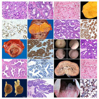
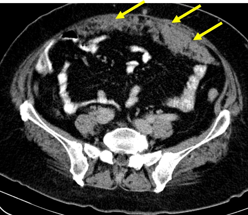


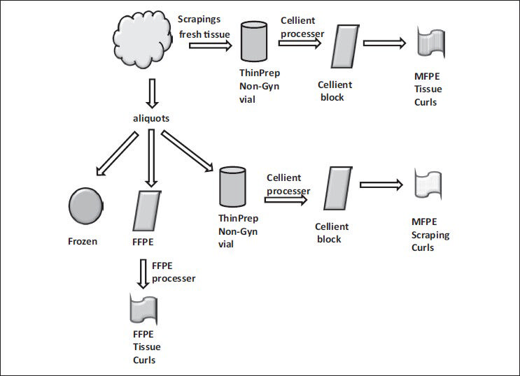


![Recombinant Anti-p53 antibody [E26] KO Tested (ab32389) | Abcam](https://www.abcam.com/ps/products/32/ab32389/Images/ab32389-389819-antip53antibodyimmunocytochemistryimmunofluorescencehap1human.jpg)