Bronchiolitis Obliterans
Cryptogenic Organizing Pneumonia
Lung Transplantation
Arteriosclerosis Obliterans
Bronchiolitis, Viral
Thromboangiitis Obliterans
Collagen Type V
Graft Rejection
Bronchoalveolar Lavage Fluid
Mediastinal Emphysema
Heart-Lung Transplantation
Lung
Respiratory Aspiration
Respiratory Function Tests
Prednisolone
Balanitis Xerotica Obliterans
Respiratory Syncytial Virus Infections
Transplantation, Homologous
Azithromycin
Biopsy
Nasal Lavage
Instillation, Drug
Radiation Pneumonitis
Radiography, Thoracic
Tomography, X-Ray Computed
Bronchi
Leukocyte Disorders
Bronchial Diseases
Forced Expiratory Volume
Postoperative Complications
Bronchoalveolar Lavage
Trachea
Costimulatory and Inhibitory T-Cell Receptors
Mustard Gas
Primary Graft Dysfunction
Respiratory Syncytial Viruses
Pulmonary Fibrosis
Follow-Up Studies
Lung Diseases, Interstitial
Retrospective Studies
Graft vs Host Disease
Oxygen Inhalation Therapy
Glucocorticoids
Treatment Outcome
Bone Marrow Transplantation
Risk Factors
Adrenal Cortex Hormones
Immunosuppressive Agents
Hematopoietic Stem Cell Transplantation
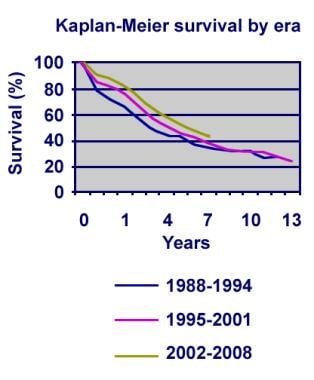
Increased risk of chronic graft-versus-host disease, obstructive bronchiolitis, and alopecia with busulfan versus total body irradiation: long-term results of a randomized trial in allogeneic marrow recipients with leukemia. Nordic Bone Marrow Transplantation Group. (1/382)
Leukemic patients receiving marrow from HLA-identical sibling donors were randomized to treatment with either busulfan 16 mg/kg (n = 88) or total body irradiation ([TBI] n = 79) in addition to cyclophosphamide 120 mg/kg. The patients were observed for a period of 5 to 9 years. Busulfan-treated patients had an increased risk of veno-occlusive disease (VOD) of the liver (12% v 1%, P =.01) and hemorrhagic cystitis (32% v 10%, P =.003). Acute graft-versus-host disease (GVHD) was similar in the two groups, but the 7-year cumulative incidence of chronic GVHD was 59% in the busulfan-treated group versus 47% in the TBI group (P =.05). Death from GVHD was more common in the busulfan group (22% v 3%, P <.001). Obstructive bronchiolitis occurred in 26% of the busulfan patients but in only 5% of the TBI patients (P <.01). Complete alopecia developed in 8 busulfan patients and partial alopecia in 17, versus five with partial alopecia in the TBI group (P <.001). Cataracts occurred in 5 busulfan-treated patients and 16 TBI patients (P =.02). The incidence of relapse after 7 years was 29% in both groups. Seven-year transplant-related mortality (TRM) in patients with early disease was 21% in the busulfan group and 12% in the TBI group. In patients with more advanced disease, the corresponding figures were 64% and 22%, respectively (P =.004). Leukemia-free survival (LFS) in patients with early disease was 68% in busulfan-treated patients and 66% in TBI patients. However, 7-year LFS in patients with more advanced disease was 17% in the busulfan group versus 49% in the TBI group (P <.01). In patients with chronic myeloid leukemia (CML) in first chronic phase, 7-year LFS was 72% and 83% in the two groups, respectively. (+info)Bilateral pneumothoraces with multiple bullae in a patient with asymptomatic bronchiolitis obliterans 10 years after bone marrow transplantation. (2/382)
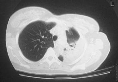
A 16-year-old boy developed bronchiolitis obliterans (BO) 10 years after BMT for myelodysplastic syndrome. Although the patient complained of almost no dyspnea on exertion, he had mild hypercapnea with a markedly reduced forced expiratory volume of 0.32 l. Chest X-rays showed occasional bilateral minimal pneumothoraces, which is in accordance with the existence of multiple small bullae found on the pleural surface at video-assisted thoracic surgery. Histologic examination of the biopsied lung revealed BO. This case indicates that BO in adolescence following BMT and possible chronic GVHD may be masked because of lung immaturity at BMT, and BO after BMT may be associated with multiple pleural bullae. (+info)Constrictive bronchiolitis obliterans and paraneoplastic pemphigus. (3/382)
Constrictive bronchiolitis obliterans is rare, and the pathogenesis of the disease often remains unknown. This study reports on the case of a 38 yr-old female with constrictive bronchiolitis obliterans and paraneoplastic pemphigus associated with malignant lymphoma. The patient developed progressive obstructive lung disease. The chest radiograph showed almost normal lungs. Paraneoplastic pemphigus is a newly described syndrome in which patients have autoantibodies binding to some epithelia, including in the respiratory tract. The disease develops in association with non-Hodgkin's lymphomas or other malignant neoplasms. The case presented here suggests that constrictive bronchiolitis obliterans associated with paraneoplastic pemphigus may be one of the facets of autoimmune responses in this context. (+info)Peripheral blood allogeneic microchimerism in lung and cardiac allograft recipients. (4/382)
We have been investigating two parameters, donor antigen-specific hyporeactivity and peripheral blood allogeneic microchimerism, to determine whether these parameters will predict a chronic rejection-free state and which recipients may be candidates for steroid withdrawal. We have identified donor antigen-specific hyporeactivity for 33% (16/48) of lung and 23% (11/47) of heart recipients. For both organ groups, the hyporeactive subgroup experienced a lower incidence of chronic rejection. The probability of donor antigen-specific hyporeactivity predicting a chronic rejection-free state is 100% for lung and 91% for heart recipients. We have identified peripheral blood allogeneic microchimerism for 77% (20/26) of lung and 36% (9/25) of heart recipients tested at 12-18 months posttransplant. Donor antigen-specific hyporeactivity correlates with a critical level of donor cells in lung recipients; the probability of high peripheral blood allogeneic microchimerism levels predicting a chronic rejection-free state in lung recipients is 100%. The results in heart recipients are not as clear with a short-, but not long-term, trend of higher chimerism levels correlating with the development of donor antigen-specific hyporeactivity. These results illustrate the usefulness of immmune parameters to predict long-term graft outcome in an organ-specific manner. (+info)Persistent high BAL fluid granulocyte activation marker levels as early indicators of bronchiolitis obliterans after lung transplant. (5/382)
The major cause of mortality in the long-term in lung transplant recipients is chronic rejection. This is a fibroproliferative process in the small airways leading to obliterative bronchiolitis and progressive loss of lung function, both constituting the clinical entity bronchiolitis obliterans syndrome (BOS). Granulocyte activation has been implicated as one factor behind BOS. Granulocyte markers in bronchoalveolar lavage (BAL) fluid were prospectively and longitudinally studied in order to identify possible association with BOS. BAL fluid from 266 bronchoscopy procedures performed in twelve single lung, eight bilateral lung and five heart/lung transplant recipients were analysed. The majority (19 of 25) were studied for a period of 2 yrs after surgery. Myeloperoxidase (MPO), eosinophil cationic protein (ECP) and interleukin-8 (IL-8) levels were used as indirect markers of activation and attraction of granulocytes. Five patients developed BOS. Ninety-eight episodes of acute rejection, nine of bacterial infection, 19 of cytomegalovirus pneumonitis, nine of Pneumocystis carinii infection, two of aspergillus infection and two of respiratory syncytial virus infection were diagnosed. BOS patients had significantly higher mean levels of MPO, ECP and IL-8 compared to patients without BOS, irrespective of acute rejection status. Over time, the five patients with BOS had significantly elevated BAL fluid levels of MPO and ECP as well as neutrophil percentages, and in four patients this increase preceded the clinical diagnosis of BOS by several months. Elevated bronchoalveolar lavage fluid neutrophil percentage as well as levels of the granulocyte activation markers myeloperoxidase and eosinophil cationic protein appear to be early signs of development of BOS in lung transplant recipients. (+info)Airway neutrophilia in stable and bronchiolitis obliterans syndrome patients following lung transplantation. (6/382)
BACKGROUND: The bronchiolitis obliterans syndrome (BOS) remains the major constraint on the long term success of lung transplantation. Neutrophils have been associated with fibrosing lung conditions and have been noted to be increased in the bronchoalveolar lavage (BAL) fluid of patients with BOS. METHODS: This study was undertaken to examine neutrophil accumulation in the BAL fluid, airway wall and lung parenchyma, as well as levels of interleukin (IL)-8 in the BAL fluid, in normal controls and lung transplant recipients with and without BOS. Bronchoscopic examination included endobronchial biopsy (EBB), BAL fluid, and transbronchial biopsy (TBB) sampling. Tissue neutrophils were identified by neutrophil elastase staining on 3 microm paraffin biopsy sections and quantified by computerised image analyser. IL-8 levels were measured in unconcentrated BAL fluid by ELISA. RESULTS: Compared with controls, airway wall neutrophilia was increased in both stable lung transplant recipients and those with BOS (p<0.05). BAL neutrophils and IL-8 levels were also increased in both groups of transplant recipients compared with controls (p<0.01), the levels being significantly higher in the BOS group (p<0.01). Neutrophil numbers in the lung parenchyma were not significantly different between the two groups of lung transplant recipients. CONCLUSION: Increased levels of neutrophils are present in the airway wall and BAL fluid of lung transplant recipients with and without BOS. BAL fluid levels of IL-8 are also increased, raising the possibility that neutrophils and/or IL-8 may play a part in the pathogenesis of BOS following lung transplantation. (+info)Clinical and radiological characteristics of lung disease in inflammatory bowel disease. (7/382)
The pulmonary associations of inflammatory bowel disease (IBD) are poorly characterized. The clinical, physiological and high-resolution computed tomographic thorax characteristics of the lung disease in patients with IBD presenting with respiratory symptoms are described. Detailed clinical information was obtained and standard pulmonary physiological tests and thorax high-resolution computed tomography performed on 14 patients with ulcerative colitis (UC) and three with Crohn's disease (CD), 10 male, aged 38-83 yrs. Respiratory symptoms had been present for 2-50 yrs and extraintestinal manifestations were present in three (17.6%). Normal pulmonary physiology (six patients) was associated with the high resolution computed tomographic changes of bronchiectasis, mosaic perfusion and air trapping suggestive of obliterative bronchiolitis and a pattern of centrilobular nodules and branching linear opacities ("tree in bud" appearance) suggestive of either cellular bronchiolitis or bronchiolectasis with mucoid secretions. Bronchiectasis was found in 13 patients (11 UC, 2 CD), 11 had air trapping and five had a "tree in bud" appearance on computed tomography. One patient had a predominantly peripheral reticular pattern at the lung bases similar to that found in cryptogenic fibrosing alveolitis and one patient had a mixed reticular and ground-glass pattern in the midzones with a patchy distribution in the central and peripheral portions of the lungs with air trapping. Eleven patients (three with alveolitis) exhibited a clinical and/or physiological response to steroids. Pulmonary abnormalities in ulcerative colitis and Crohn's disease can present years after the onset of the bowel disease and can affect any part of the lungs. Early recognition is important as they can be strikingly steroid-responsive. (+info)B7-1, B7-2 and class II MHC molecules in idiopathic pulmonary fibrosis and bronchiolitis obliterans-organizing pneumonia. (8/382)
Interstitial lung diseases are thought to be associated with the infiltration of activated T-lymphocytes. To induce an effective immune response, antigen-presenting cells have to not only present antigenic peptide with major histocompatibility complex (MHC) molecules to T-lymphocytes but also express B7 molecules. Therefore, the expression of B7-1, B7-2 and class II MHC molecules was investigated in lung tissues from patients with idiopathic pulmonary fibrosis (IPF) and bronchiolitis obliterans-organizing pneumonia (BOOP), and in normal lung parenchyma as a control, using immunohistochemical localization. B7-1 and B7-2 were aberrantly expressed in bronchiolar and alveolar epithelial cells, and class II MHC molecules were also aberrantly expressed in bronchiolar epithelial cells in IPF. B7-1 was aberrantly expressed in bronchiolar epithelial cells in BOOP. There was no significant difference in the expression of these proteins in alveolar macrophages between IPF and control subjects. However, B7-2 and class II MHC molecule expression in alveolar macrophages was decreased in BOOP compared with that in control subjects. Expression of CD28 and CTLA4, receptors for B7 molecules, was detected in infiltrating lymphocytes in lung tissues in IPF and BOOP. It was concluded that bronchiolar and alveolar epithelial cells may actively participate in the pathophysiology of idiopathic pulmonary fibrosis through the aberrant expression of B7 and class II major histocompatibility complex molecules. The dysregulation of these molecules in epithelial cells may lead to the activation of autoreactive T-lymphocytes, which might contribute to the pathogenesis of fibrosing lung diseases. (+info)Bronchiolitis obliterans is a medical condition characterized by the inflammation and scarring (fibrosis) of the bronchioles, which are the smallest airways in the lungs. This results in the narrowing or complete obstruction of the airways, leading to difficulty breathing and reduced lung function.
The condition is often caused by a respiratory infection, such as adenovirus or mycoplasma pneumonia, but it can also be associated with exposure to certain chemicals, drugs, or radiation therapy. In some cases, the cause may be unknown.
Symptoms of bronchiolitis obliterans include cough, shortness of breath, wheezing, and crackles heard on lung examination. Diagnosis typically involves a combination of medical history, physical exam, imaging studies (such as chest X-ray or CT scan), and pulmonary function tests. In some cases, a biopsy may be necessary to confirm the diagnosis.
Treatment for bronchiolitis obliterans is focused on managing symptoms and preventing further lung damage. This may include bronchodilators to help open up the airways, corticosteroids to reduce inflammation, and oxygen therapy to help with breathing. In severe cases, a lung transplant may be necessary.
Cryptogenic organizing pneumonia (COP) is a type of lung disorder that is characterized by the presence of inflammation and scarring in the lungs. The term "cryptogenic" means that the cause of the condition is unknown or unclear.
Organizing pneumonia is a specific pattern of injury to the lungs that can be caused by various factors, including infections, medications, and autoimmune disorders. However, in cases of COP, there is no clear underlying cause that can be identified.
The main symptoms of COP include cough, shortness of breath, fever, and fatigue. The condition can also cause crackles or wheezing sounds when listening to the lungs with a stethoscope. Diagnosis of COP typically involves a combination of imaging studies, such as chest X-rays or CT scans, and lung biopsy.
Treatment for COP usually involves the use of corticosteroids, which can help to reduce inflammation and improve symptoms. In some cases, other medications may also be used to manage the condition. The prognosis for people with COP is generally good, with most individuals responding well to treatment and experiencing improvement in their symptoms over time. However, recurrence of the condition is possible, and long-term monitoring may be necessary.
Bronchiolitis is a common respiratory infection in infants and young children, typically caused by a viral infection. It is characterized by inflammation and congestion of the bronchioles (the smallest airways in the lungs), which can lead to difficulty breathing and wheezing.
The most common virus that causes bronchiolitis is respiratory syncytial virus (RSV), but other viruses such as rhinovirus, influenza, and parainfluenza can also cause the condition. Symptoms of bronchiolitis may include cough, wheezing, rapid breathing, difficulty feeding, and fatigue.
In severe cases, bronchiolitis can lead to respiratory distress and require hospitalization. Treatment typically involves supportive care, such as providing fluids and oxygen therapy, and in some cases, medications to help open the airways may be used. Prevention measures include good hand hygiene and avoiding close contact with individuals who are sick.
Lung transplantation is a surgical procedure where one or both diseased lungs are removed and replaced with healthy lungs from a deceased donor. It is typically considered as a treatment option for patients with end-stage lung diseases, such as chronic obstructive pulmonary disease (COPD), cystic fibrosis, idiopathic pulmonary fibrosis, and alpha-1 antitrypsin deficiency, who have exhausted all other medical treatments and continue to suffer from severe respiratory failure.
The procedure involves several steps, including evaluating the patient's eligibility for transplantation, matching the donor's lung size and blood type with the recipient, and performing the surgery under general anesthesia. After the surgery, patients require close monitoring and lifelong immunosuppressive therapy to prevent rejection of the new lungs.
Lung transplantation can significantly improve the quality of life and survival rates for some patients with end-stage lung disease, but it is not without risks, including infection, bleeding, and rejection. Therefore, careful consideration and thorough evaluation are necessary before pursuing this treatment option.
Arteriosclerosis obliterans (ASO) is a specific type of arteriosclerosis, which is a hardening and narrowing of the arteries. ASO is also known as peripheral artery disease (PAD). It mainly affects the arteries that supply blood to the legs, but it can also affect the arms, head, and stomach.
In ASO, fatty deposits called plaques build up in the inner lining of the arterial walls, causing them to become thickened and less flexible. This leads to a decrease in blood flow, which can cause symptoms such as leg pain or cramping when walking (claudication), numbness, weakness, and coldness in the legs or feet. In severe cases, ASO can lead to tissue damage, gangrene, and even amputation if left untreated.
ASO is typically caused by risk factors such as smoking, high blood pressure, diabetes, high cholesterol, and a family history of the disease. Treatment may include lifestyle changes, medication, or surgery to improve blood flow.
Viral bronchiolitis is a common respiratory infection in infants and young children, typically caused by a viral pathogen such as the respiratory syncytial virus (RSV). The infection leads to inflammation and congestion of the small airways (bronchioles) in the lungs, resulting in symptoms like wheezing, cough, difficulty breathing, and rapid breathing.
The infection usually spreads through respiratory droplets when an infected person coughs or sneezes. The virus can also survive on surfaces for several hours, making it easy to contract the infection by touching contaminated objects and then touching the face.
Most cases of viral bronchiolitis are mild and resolve within 1-2 weeks with supportive care, including increased fluid intake, humidified air, and fever reduction. However, in severe cases or in high-risk infants (such as those born prematurely or with underlying heart or lung conditions), hospitalization may be necessary to manage complications like dehydration, respiratory distress, or oxygen deprivation.
Preventive measures include good hand hygiene, avoiding close contact with sick individuals, and ensuring that infants and young children receive appropriate vaccinations and immunizations as recommended by their healthcare provider.
Thromboangiitis obliterans, also known as Buerger's disease, is a rare inflammatory disease that affects the small and medium-sized arteries and veins, most commonly in the legs and feet but sometimes in the arms and hands. The condition is characterized by the formation of blood clots (thrombi) and inflammation in the affected blood vessels, leading to their obstruction and damage.
The exact cause of thromboangiitis obliterans is not known, but it is strongly associated with tobacco use, particularly smoking. The condition primarily affects young men, although women can also develop the disease. The symptoms include pain and cramping in the affected limbs, especially during exercise, skin discoloration, ulcers, and in severe cases, gangrene.
The diagnosis of thromboangiitis obliterans is based on a combination of clinical presentation, medical history, laboratory tests, and imaging studies. There is no cure for the disease, but quitting smoking and other tobacco products can help slow its progression and reduce the risk of complications. Treatment typically involves medications to manage symptoms, improve blood flow, and prevent further clotting. In severe cases, surgery may be necessary to remove damaged tissue or bypass blocked blood vessels.
Collagen Type V is a specific type of collagen, which is a protein that provides structure and strength to connective tissues in the body. Collagen Type V is found in various tissues, including the cornea, blood vessels, and hair. It plays a crucial role in the formation of collagen fibers and helps regulate the diameter of collagen fibrils. Mutations in the genes that encode for Collagen Type V can lead to various connective tissue disorders, such as Ehlers-Danlos syndrome and osteogenesis imperfecta.
Flavoring agents are substances added to foods, beverages, pharmaceuticals, and sometimes even medical devices to enhance or modify their taste and aroma. They can be natural, derived from plants or animals, or synthetic, created in a laboratory. Flavoring agents do not necessarily provide any nutritional value and are typically used in small quantities.
In a medical context, flavoring agents may be added to medications to improve patient compliance, especially for children or individuals who have difficulty swallowing pills. These agents can help mask the unpleasant taste of certain medicines, making them more palatable and easier to consume. However, it is essential to ensure that the use of flavoring agents does not interfere with the medication's effectiveness or safety.
Graft rejection is an immune response that occurs when transplanted tissue or organ (the graft) is recognized as foreign by the recipient's immune system, leading to the activation of immune cells to attack and destroy the graft. This results in the failure of the transplant and the need for additional medical intervention or another transplant. There are three types of graft rejection: hyperacute, acute, and chronic. Hyperacute rejection occurs immediately or soon after transplantation due to pre-existing antibodies against the graft. Acute rejection typically occurs within weeks to months post-transplant and is characterized by the infiltration of T-cells into the graft. Chronic rejection, which can occur months to years after transplantation, is a slow and progressive process characterized by fibrosis and tissue damage due to ongoing immune responses against the graft.
Bronchoalveolar lavage (BAL) fluid is a type of clinical specimen obtained through a procedure called bronchoalveolar lavage. This procedure involves inserting a bronchoscope into the lungs and instilling a small amount of saline solution into a specific area of the lung, then gently aspirating the fluid back out. The fluid that is recovered is called bronchoalveolar lavage fluid.
BAL fluid contains cells and other substances that are present in the lower respiratory tract, including the alveoli (the tiny air sacs where gas exchange occurs). By analyzing BAL fluid, doctors can diagnose various lung conditions, such as pneumonia, interstitial lung disease, and lung cancer. They can also monitor the effectiveness of treatments for these conditions by comparing the composition of BAL fluid before and after treatment.
BAL fluid is typically analyzed for its cellular content, including the number and type of white blood cells present, as well as for the presence of bacteria, viruses, or other microorganisms. The fluid may also be tested for various proteins, enzymes, and other biomarkers that can provide additional information about lung health and disease.
Mediastinal emphysema is a medical condition characterized by the presence of air or gas within the mediastinum, which is the central compartment of the thorax that contains the heart, esophagus, trachea, bronchi, thymus gland, and other associated structures.
In mediastinal emphysema, the air accumulates in the mediastinal tissues and spaces, leading to their abnormal distention or swelling. This condition can result from various causes, including:
* Pulmonary trauma or barotrauma (e.g., mechanical ventilation, scuba diving)
* Infections that cause gas-forming organisms (e.g., pneumomediastinum)
* Air leakage from the lungs or airways (e.g., bronchial rupture, esophageal perforation)
* Certain medical procedures (e.g., mediastinoscopy, tracheostomy)
Mediastinal emphysema can cause symptoms such as chest pain, cough, difficulty breathing, and swallowing problems. In severe cases, it may lead to life-threatening complications, including tension pneumothorax or mediastinitis. Treatment depends on the underlying cause and severity of the condition.
Heart-lung transplantation is a surgical procedure where both the heart and lungs of a patient are replaced with those from a deceased donor. This complex and highly specialized surgery is typically considered as a last resort for patients suffering from end-stage lung or heart-lung diseases, such as cystic fibrosis, pulmonary fibrosis, chronic obstructive pulmonary disease (COPD), or certain forms of congenital heart disease, who have exhausted all other treatment options and face imminent death.
The procedure involves removing the patient's diseased heart and lungs en bloc, followed by implanting the donor's heart and lungs in their place. The surgery requires a skilled multidisciplinary team of cardiothoracic surgeons, anesthesiologists, perfusionists, transplant coordinators, and intensive care specialists.
Following the transplantation, patients require lifelong immunosuppressive therapy to prevent rejection of the transplanted organs. Despite the significant risks associated with this procedure, including infection, bleeding, and rejection, heart-lung transplantation can significantly improve both survival and quality of life for carefully selected patients with advanced heart-lung disease.
A lung is a pair of spongy, elastic organs in the chest that work together to enable breathing. They are responsible for taking in oxygen and expelling carbon dioxide through the process of respiration. The left lung has two lobes, while the right lung has three lobes. The lungs are protected by the ribcage and are covered by a double-layered membrane called the pleura. The trachea divides into two bronchi, which further divide into smaller bronchioles, leading to millions of tiny air sacs called alveoli, where the exchange of gases occurs.
Respiratory aspiration is defined as the entry of foreign materials (such as food, liquids, or vomit) into the lower respiratory tract during swallowing, which includes the trachea and lungs. This can lead to respiratory complications such as pneumonia, bronchitis, or lung abscesses. Aspiration can occur in individuals with impaired swallowing function due to various conditions like neurological disorders, stroke, or anesthesia.
Respiratory Function Tests (RFTs) are a group of medical tests that measure how well your lungs take in and exhale air, and how well they transfer oxygen and carbon dioxide into and out of your blood. They can help diagnose certain lung disorders, measure the severity of lung disease, and monitor response to treatment.
RFTs include several types of tests, such as:
1. Spirometry: This test measures how much air you can exhale and how quickly you can do it. It's often used to diagnose and monitor conditions like asthma, chronic obstructive pulmonary disease (COPD), and other lung diseases.
2. Lung volume testing: This test measures the total amount of air in your lungs. It can help diagnose restrictive lung diseases, such as pulmonary fibrosis or sarcoidosis.
3. Diffusion capacity testing: This test measures how well oxygen moves from your lungs into your bloodstream. It's often used to diagnose and monitor conditions like pulmonary fibrosis, interstitial lung disease, and other lung diseases that affect the ability of the lungs to transfer oxygen to the blood.
4. Bronchoprovocation testing: This test involves inhaling a substance that can cause your airways to narrow, such as methacholine or histamine. It's often used to diagnose and monitor asthma.
5. Exercise stress testing: This test measures how well your lungs and heart work together during exercise. It's often used to diagnose lung or heart disease.
Overall, Respiratory Function Tests are an important tool for diagnosing and managing a wide range of lung conditions.
A syndrome, in medical terms, is a set of symptoms that collectively indicate or characterize a disease, disorder, or underlying pathological process. It's essentially a collection of signs and/or symptoms that frequently occur together and can suggest a particular cause or condition, even though the exact physiological mechanisms might not be fully understood.
For example, Down syndrome is characterized by specific physical features, cognitive delays, and other developmental issues resulting from an extra copy of chromosome 21. Similarly, metabolic syndromes like diabetes mellitus type 2 involve a group of risk factors such as obesity, high blood pressure, high blood sugar, and abnormal cholesterol or triglyceride levels that collectively increase the risk of heart disease, stroke, and diabetes.
It's important to note that a syndrome is not a specific diagnosis; rather, it's a pattern of symptoms that can help guide further diagnostic evaluation and management.
Prednisolone is a synthetic glucocorticoid drug, which is a class of steroid hormones. It is commonly used in the treatment of various inflammatory and autoimmune conditions due to its potent anti-inflammatory and immunosuppressive effects. Prednisolone works by binding to specific receptors in cells, leading to changes in gene expression that reduce the production of substances involved in inflammation, such as cytokines and prostaglandins.
Prednisolone is available in various forms, including tablets, syrups, and injectable solutions. It can be used to treat a wide range of medical conditions, including asthma, rheumatoid arthritis, inflammatory bowel disease, allergies, skin conditions, and certain types of cancer.
Like other steroid medications, prednisolone can have significant side effects if used in high doses or for long periods of time. These may include weight gain, mood changes, increased risk of infections, osteoporosis, diabetes, and adrenal suppression. As a result, the use of prednisolone should be closely monitored by a healthcare professional to ensure that its benefits outweigh its risks.
Balanitis xerotica obliterans (BXO) is a chronic, inflammatory condition that affects the foreskin and glans penis in uncircumcised males. It is also known as lichen sclerosus et atrophicus when it occurs in these areas.
The medical definition of Balanitis xerotica obliterans is a progressive inflammatory dermatosis characterized by white, shiny patches on the foreskin and glans penis. These patches can become thickened, scarred, and adhere to the underlying tissue, causing the foreskin to become difficult or impossible to retract (phimosis). BXO can also cause narrowing of the urethral meatus (the opening where urine exits), which can lead to problems with urination.
The exact cause of BXO is not known, but it is thought to be related to an autoimmune response or a localized reaction to irritants or trauma. Treatment typically involves topical corticosteroids, circumcision, or both. In severe cases, surgery may be necessary to correct urethral narrowing.
Lung diseases refer to a broad category of disorders that affect the lungs and other structures within the respiratory system. These diseases can impair lung function, leading to symptoms such as coughing, shortness of breath, chest pain, and wheezing. They can be categorized into several types based on the underlying cause and nature of the disease process. Some common examples include:
1. Obstructive lung diseases: These are characterized by narrowing or blockage of the airways, making it difficult to breathe out. Examples include chronic obstructive pulmonary disease (COPD), asthma, bronchiectasis, and cystic fibrosis.
2. Restrictive lung diseases: These involve stiffening or scarring of the lungs, which reduces their ability to expand and take in air. Examples include idiopathic pulmonary fibrosis, sarcoidosis, and asbestosis.
3. Infectious lung diseases: These are caused by bacteria, viruses, fungi, or parasites that infect the lungs. Examples include pneumonia, tuberculosis, and influenza.
4. Vascular lung diseases: These affect the blood vessels in the lungs, impairing oxygen exchange. Examples include pulmonary embolism, pulmonary hypertension, and chronic thromboembolic pulmonary hypertension (CTEPH).
5. Neoplastic lung diseases: These involve abnormal growth of cells within the lungs, leading to cancer. Examples include small cell lung cancer, non-small cell lung cancer, and mesothelioma.
6. Other lung diseases: These include interstitial lung diseases, pleural effusions, and rare disorders such as pulmonary alveolar proteinosis and lymphangioleiomyomatosis (LAM).
It is important to note that this list is not exhaustive, and there are many other conditions that can affect the lungs. Proper diagnosis and treatment of lung diseases require consultation with a healthcare professional, such as a pulmonologist or respiratory therapist.
Diacetyl is a volatile, yellow-green liquid that is a byproduct of fermentation and is used as a butter flavoring in foods. The chemical formula for diacetyl is CH3COCH3. It has a buttery or creamy taste and is often added to microwave popcorn, margarine, and other processed foods to give them a buttery flavor.
Diacetyl can also be found in some alcoholic beverages, such as beer and wine, where it is produced naturally during fermentation. In high concentrations, diacetyl can have a strong, unpleasant odor and taste.
There has been concern about the potential health effects of diacetyl, particularly for workers in factories that manufacture artificial butter flavorings. Some studies have suggested that exposure to diacetyl may increase the risk of developing lung disease, including bronchiolitis obliterans, a serious and sometimes fatal condition characterized by scarring and narrowing of the airways in the lungs. However, more research is needed to fully understand the health effects of diacetyl and to determine safe levels of exposure.
Respiratory Syncytial Virus (RSV) infections refer to the clinical illnesses caused by the Respiratory Syncytial Virus. RSV is a highly contagious virus that spreads through respiratory droplets, contact with infected surfaces, or direct contact with infected people. It primarily infects the respiratory tract, causing inflammation and damage to the cells lining the airways.
RSV infections can lead to a range of respiratory illnesses, from mild, cold-like symptoms to more severe conditions such as bronchiolitis (inflammation of the small airways in the lungs) and pneumonia (infection of the lung tissue). The severity of the infection tends to depend on factors like age, overall health status, and presence of underlying medical conditions.
In infants and young children, RSV is a leading cause of bronchiolitis and pneumonia, often resulting in hospitalization. In older adults, people with weakened immune systems, and those with chronic heart or lung conditions, RSV infections can also be severe and potentially life-threatening.
Symptoms of RSV infection may include runny nose, cough, sneezing, fever, wheezing, and difficulty breathing. Treatment typically focuses on managing symptoms and providing supportive care, although hospitalization and more aggressive interventions may be necessary in severe cases or for high-risk individuals. Preventive measures such as hand hygiene, wearing masks, and avoiding close contact with infected individuals can help reduce the spread of RSV.
Homologous transplantation is a type of transplant surgery where organs or tissues are transferred between two genetically non-identical individuals of the same species. The term "homologous" refers to the similarity in structure and function of the donated organ or tissue to the recipient's own organ or tissue.
For example, a heart transplant from one human to another is an example of homologous transplantation because both organs are hearts and perform the same function. Similarly, a liver transplant, kidney transplant, lung transplant, and other types of organ transplants between individuals of the same species are also considered homologous transplantations.
Homologous transplantation is in contrast to heterologous or xenogeneic transplantation, where organs or tissues are transferred from one species to another, such as a pig heart transplanted into a human. Homologous transplantation is more commonly performed than heterologous transplantation due to the increased risk of rejection and other complications associated with xenogeneic transplants.
Azithromycin is a widely used antibiotic drug that belongs to the class of macrolides. It works by inhibiting bacterial protein synthesis, which leads to the death of susceptible bacteria. This medication is active against a broad range of gram-positive and gram-negative bacteria, atypical bacteria, and some parasites.
Azithromycin is commonly prescribed to treat various bacterial infections, such as:
1. Respiratory tract infections, including pneumonia, bronchitis, and sinusitis
2. Skin and soft tissue infections
3. Sexually transmitted diseases, like chlamydia
4. Otitis media (middle ear infection)
5. Traveler's diarrhea
The drug is available in various forms, including tablets, capsules, suspension, and intravenous solutions. The typical dosage for adults ranges from 250 mg to 500 mg per day, depending on the type and severity of the infection being treated.
Like other antibiotics, azithromycin should be used judiciously to prevent antibiotic resistance. It is essential to complete the full course of treatment as prescribed by a healthcare professional, even if symptoms improve before finishing the medication.
A biopsy is a medical procedure in which a small sample of tissue is taken from the body to be examined under a microscope for the presence of disease. This can help doctors diagnose and monitor various medical conditions, such as cancer, infections, or autoimmune disorders. The type of biopsy performed will depend on the location and nature of the suspected condition. Some common types of biopsies include:
1. Incisional biopsy: In this procedure, a surgeon removes a piece of tissue from an abnormal area using a scalpel or other surgical instrument. This type of biopsy is often used when the lesion is too large to be removed entirely during the initial biopsy.
2. Excisional biopsy: An excisional biopsy involves removing the entire abnormal area, along with a margin of healthy tissue surrounding it. This technique is typically employed for smaller lesions or when cancer is suspected.
3. Needle biopsy: A needle biopsy uses a thin, hollow needle to extract cells or fluid from the body. There are two main types of needle biopsies: fine-needle aspiration (FNA) and core needle biopsy. FNA extracts loose cells, while a core needle biopsy removes a small piece of tissue.
4. Punch biopsy: In a punch biopsy, a round, sharp tool is used to remove a small cylindrical sample of skin tissue. This type of biopsy is often used for evaluating rashes or other skin abnormalities.
5. Shave biopsy: During a shave biopsy, a thin slice of tissue is removed from the surface of the skin using a sharp razor-like instrument. This technique is typically used for superficial lesions or growths on the skin.
After the biopsy sample has been collected, it is sent to a laboratory where a pathologist will examine the tissue under a microscope and provide a diagnosis based on their findings. The results of the biopsy can help guide further treatment decisions and determine the best course of action for managing the patient's condition.
Nasal lavage, also known as nasal washing or saline irrigation, is a procedure in which a saline solution is used to flush out the nasal passages. This is often done to help relieve symptoms associated with nasal congestion, allergies, sinusitis, and other respiratory conditions. The process involves instilling the saline solution into one nostril and allowing it to flow out through the other, taking with it any mucus, debris, or irritants that may be present in the nasal passages. This can help promote better breathing, reduce inflammation, and alleviate symptoms such as sinus pressure, headaches, and sneezing. Nasal lavage can be performed using a variety of devices, including bulb syringes, neti pots, or specialized squeeze bottles designed specifically for this purpose.
Instillation, in the context of drug administration, refers to the process of introducing a medication or therapeutic agent into a body cavity or onto a mucous membrane surface using gentle, steady pressure. This is typically done with the help of a device such as an eyedropper, pipette, or catheter. The goal is to ensure that the drug is distributed evenly over the surface or absorbed through the mucous membrane for localized or systemic effects. Instillation can be used for various routes of administration including ocular (eye), nasal, auricular (ear), vaginal, and intra-articular (joint space) among others. The choice of instillation as a route of administration depends on the drug's properties, the desired therapeutic effect, and the patient's overall health status.
Radiation pneumonitis is a inflammatory reaction in the lung tissue that occurs as a complication of thoracic radiation therapy. It usually develops 1-3 months following the completion of radiation treatment. The symptoms can range from mild to severe and may include cough, shortness of breath, fever, and chest discomfort. In severe cases, it can lead to fibrosis (scarring) of the lung tissue, which can cause permanent lung damage. Radiation pneumonitis is diagnosed through a combination of clinical symptoms, imaging studies such as chest X-ray or CT scan, and sometimes through bronchoscopy with lavage. Treatment typically involves corticosteroids to reduce inflammation and supportive care to manage symptoms.
Thoracic radiography is a type of diagnostic imaging that involves using X-rays to produce images of the chest, including the lungs, heart, bronchi, great vessels, and the bones of the spine and chest wall. It is a commonly used tool in the diagnosis and management of various respiratory, cardiovascular, and thoracic disorders such as pneumonia, lung cancer, heart failure, and rib fractures.
During the procedure, the patient is positioned between an X-ray machine and a cassette containing a film or digital detector. The X-ray beam is directed at the chest, and the resulting image is captured on the film or detector. The images produced can help identify any abnormalities in the structure or function of the organs within the chest.
Thoracic radiography may be performed as a routine screening test for certain conditions, such as lung cancer, or it may be ordered when a patient presents with symptoms suggestive of a respiratory or cardiovascular disorder. It is a safe and non-invasive procedure that can provide valuable information to help guide clinical decision making and improve patient outcomes.
X-ray computed tomography (CT or CAT scan) is a medical imaging method that uses computer-processed combinations of many X-ray images taken from different angles to produce cross-sectional (tomographic) images (virtual "slices") of the body. These cross-sectional images can then be used to display detailed internal views of organs, bones, and soft tissues in the body.
The term "computed tomography" is used instead of "CT scan" or "CAT scan" because the machines take a series of X-ray measurements from different angles around the body and then use a computer to process these data to create detailed images of internal structures within the body.
CT scanning is a noninvasive, painless medical test that helps physicians diagnose and treat medical conditions. CT imaging provides detailed information about many types of tissue including lung, bone, soft tissue and blood vessels. CT examinations can be performed on every part of the body for a variety of reasons including diagnosis, surgical planning, and monitoring of therapeutic responses.
In computed tomography (CT), an X-ray source and detector rotate around the patient, measuring the X-ray attenuation at many different angles. A computer uses this data to construct a cross-sectional image by the process of reconstruction. This technique is called "tomography". The term "computed" refers to the use of a computer to reconstruct the images.
CT has become an important tool in medical imaging and diagnosis, allowing radiologists and other physicians to view detailed internal images of the body. It can help identify many different medical conditions including cancer, heart disease, lung nodules, liver tumors, and internal injuries from trauma. CT is also commonly used for guiding biopsies and other minimally invasive procedures.
In summary, X-ray computed tomography (CT or CAT scan) is a medical imaging technique that uses computer-processed combinations of many X-ray images taken from different angles to produce cross-sectional images of the body. It provides detailed internal views of organs, bones, and soft tissues in the body, allowing physicians to diagnose and treat medical conditions.
"Bronchi" are a pair of airways in the respiratory system that branch off from the trachea (windpipe) and lead to the lungs. They are responsible for delivering oxygen-rich air to the lungs and removing carbon dioxide during exhalation. The right bronchus is slightly larger and more vertical than the left, and they further divide into smaller branches called bronchioles within the lungs. Any abnormalities or diseases affecting the bronchi can impact lung function and overall respiratory health.
Bronchoscopy is a medical procedure that involves the examination of the inside of the airways and lungs with a flexible or rigid tube called a bronchoscope. This procedure allows healthcare professionals to directly visualize the airways, take tissue samples for biopsy, and remove foreign objects or secretions. Bronchoscopy can be used to diagnose and manage various respiratory conditions such as lung infections, inflammation, cancer, and bleeding. It is usually performed under local or general anesthesia to minimize discomfort and risks associated with the procedure.
Leukocyte disorders, also known as white blood cell disorders, refer to a group of conditions that affect the production, function, or number of leukocytes (white blood cells) in the body. Leukocytes play a crucial role in protecting the body against infection and disease. Therefore, disorders that affect these cells can significantly impact an individual's immune system and overall health.
There are several types of leukocyte disorders, including:
1. Leukopenia: A condition characterized by abnormally low levels of white blood cells in the blood. This can increase the risk of infection.
2. Leukocytosis: A condition characterized by an elevated number of white blood cells in the blood. While this can be a normal response to infection or inflammation, it can also indicate an underlying medical condition such as leukemia.
3. Neutropenia: A condition characterized by abnormally low levels of neutrophils, a type of white blood cell that helps fight bacterial infections. This can increase the risk of infection.
4. Neutrophilia: A condition characterized by an elevated number of neutrophils in the blood. This can be a normal response to infection or inflammation, but it can also indicate an underlying medical condition such as an acute bacterial infection.
5. Lymphocytosis: A condition characterized by an elevated number of lymphocytes, a type of white blood cell that helps fight viral infections and cancer cells. This can be a normal response to infection or vaccination, but it can also indicate an underlying medical condition such as chronic lymphocytic leukemia.
6. Lymphopenia: A condition characterized by abnormally low levels of lymphocytes in the blood. This can increase the risk of infection and indicate an underlying medical condition such as HIV/AIDS or autoimmune disorders.
7. Monocytosis: A condition characterized by an elevated number of monocytes, a type of white blood cell that helps fight chronic infections and cancer cells. This can be a normal response to infection or inflammation, but it can also indicate an underlying medical condition such as chronic inflammatory diseases.
8. Monocytopenia: A condition characterized by abnormally low levels of monocytes in the blood. This can increase the risk of infection and indicate an underlying medical condition such as bone marrow disorders or autoimmune diseases.
These conditions can be caused by various factors, including infections, inflammation, cancer, autoimmune disorders, medications, and genetic disorders. Proper diagnosis and treatment require a thorough evaluation of the patient's medical history, physical examination, laboratory tests, and imaging studies.
Bronchial diseases refer to medical conditions that affect the bronchi, which are the large airways that lead into the lungs. These diseases can cause inflammation, narrowing, or obstruction of the bronchi, leading to symptoms such as coughing, wheezing, chest tightness, and difficulty breathing.
Some common bronchial diseases include:
1. Asthma: A chronic inflammatory disease of the airways that causes recurring episodes of wheezing, breathlessness, chest tightness, and coughing.
2. Chronic Bronchitis: A long-term inflammation of the bronchi that leads to a persistent cough and excessive mucus production.
3. Bronchiectasis: A condition in which the bronchi become damaged and widened, leading to chronic infection and inflammation.
4. Bronchitis: An inflammation of the bronchi that can cause coughing, wheezing, and chest tightness.
5. Emphysema: A lung condition that causes shortness of breath due to damage to the air sacs in the lungs. While not strictly a bronchial disease, it is often associated with chronic bronchitis and COPD (Chronic Obstructive Pulmonary Disease).
Treatment for bronchial diseases may include medications such as bronchodilators, corticosteroids, or antibiotics, as well as lifestyle changes such as quitting smoking and avoiding irritants. In severe cases, oxygen therapy or surgery may be necessary.
Forced Expiratory Volume (FEV) is a medical term used to describe the volume of air that can be forcefully exhaled from the lungs in one second. It is often measured during pulmonary function testing to assess lung function and diagnose conditions such as chronic obstructive pulmonary disease (COPD) or asthma.
FEV is typically expressed as a percentage of the Forced Vital Capacity (FVC), which is the total volume of air that can be exhaled from the lungs after taking a deep breath in. The ratio of FEV to FVC is used to determine whether there is obstruction in the airways, with a lower ratio indicating more severe obstruction.
There are different types of FEV measurements, including FEV1 (the volume of air exhaled in one second), FEV25-75 (the average volume of air exhaled during the middle 50% of the FVC maneuver), and FEV0.5 (the volume of air exhaled in half a second). These measurements can provide additional information about lung function and help guide treatment decisions.
Racepinephrine is not typically referred to as a "race" in the medical context, but rather as a form of epinephrine (also known as adrenaline). Racepinephrine is the optical isomer of epinephrine, meaning that it is a molecule with the same chemical formula but a different arrangement of atoms in space.
Racepinephrine is a naturally occurring catecholamine, a type of neurotransmitter and hormone that is produced by the adrenal glands and is involved in the "fight or flight" response. It is also used as a medication, typically in the form of the racemic mixture of epinephrine, which contains equal amounts of both isomers (R- and S-epinephrine).
Racepinephrine has similar effects to epinephrine, including increasing heart rate and blood pressure, improving respiratory function, and enhancing mental alertness. It is used in the treatment of anaphylaxis, cardiac arrest, and other emergency situations where rapid restoration of cardiovascular function is necessary.
It's important to note that while racepinephrine and epinephrine have similar effects, they are not identical and may have different therapeutic uses and potential side effects.
Postoperative complications refer to any unfavorable condition or event that occurs during the recovery period after a surgical procedure. These complications can vary in severity and may include, but are not limited to:
1. Infection: This can occur at the site of the incision or inside the body, such as pneumonia or urinary tract infection.
2. Bleeding: Excessive bleeding (hemorrhage) can lead to a drop in blood pressure and may require further surgical intervention.
3. Blood clots: These can form in the deep veins of the legs (deep vein thrombosis) and can potentially travel to the lungs (pulmonary embolism).
4. Wound dehiscence: This is when the surgical wound opens up, which can lead to infection and further complications.
5. Pulmonary issues: These include atelectasis (collapsed lung), pneumonia, or respiratory failure.
6. Cardiovascular problems: These include abnormal heart rhythms (arrhythmias), heart attack, or stroke.
7. Renal failure: This can occur due to various reasons such as dehydration, blood loss, or the use of certain medications.
8. Pain management issues: Inadequate pain control can lead to increased stress, anxiety, and decreased mobility.
9. Nausea and vomiting: These can be caused by anesthesia, opioid pain medication, or other factors.
10. Delirium: This is a state of confusion and disorientation that can occur in the elderly or those with certain medical conditions.
Prompt identification and management of these complications are crucial to ensure the best possible outcome for the patient.
Bronchoalveolar lavage (BAL) is a medical procedure in which a small amount of fluid is introduced into a segment of the lung and then gently suctioned back out. The fluid contains cells and other materials that can be analyzed to help diagnose various lung conditions, such as inflammation, infection, or cancer.
The procedure is typically performed during bronchoscopy, which involves inserting a thin, flexible tube with a light and camera on the end through the nose or mouth and into the lungs. Once the bronchoscope is in place, a small catheter is passed through the bronchoscope and into the desired lung segment. The fluid is then introduced and suctioned back out, and the sample is sent to a laboratory for analysis.
BAL can be helpful in diagnosing various conditions such as pneumonia, interstitial lung diseases, alveolar proteinosis, and some types of cancer. It can also be used to monitor the effectiveness of treatment for certain lung conditions. However, like any medical procedure, it carries some risks, including bleeding, infection, and respiratory distress. Therefore, it is important that the procedure is performed by a qualified healthcare professional in a controlled setting.
Pneumonia is an infection or inflammation of the alveoli (tiny air sacs) in one or both lungs. It's often caused by bacteria, viruses, or fungi. Accumulated pus and fluid in these air sacs make it difficult to breathe, which can lead to coughing, chest pain, fever, and difficulty breathing. The severity of symptoms can vary from mild to life-threatening, depending on the underlying cause, the patient's overall health, and age. Pneumonia is typically diagnosed through a combination of physical examination, medical history, and diagnostic tests such as chest X-rays or blood tests. Treatment usually involves antibiotics for bacterial pneumonia, antivirals for viral pneumonia, and supportive care like oxygen therapy, hydration, and rest.
The trachea, also known as the windpipe, is a tube-like structure in the respiratory system that connects the larynx (voice box) to the bronchi (the two branches leading to each lung). It is composed of several incomplete rings of cartilage and smooth muscle, which provide support and flexibility. The trachea plays a crucial role in directing incoming air to the lungs during inspiration and outgoing air to the larynx during expiration.
Costimulatory and inhibitory T-cell receptors are molecules found on the surface of T cells, a type of white blood cell that plays a central role in the immune response. These receptors play a critical role in regulating the activation, proliferation, and effector functions of T cells.
Costimulatory receptors, such as CD28, CD137, and ICOS, provide positive signals that enhance T-cell activation and promote immune responses. They do this by interacting with their ligands, which are expressed on the surface of antigen-presenting cells (APCs) such as dendritic cells, macrophages, and B cells. When a T cell encounters an APC that presents its specific antigen, engagement of both the T-cell receptor (TCR) and costimulatory receptors leads to full T-cell activation, cytokine production, and clonal expansion.
Inhibitory receptors, on the other hand, provide negative signals that dampen T-cell activation and prevent excessive or inappropriate immune responses. Examples of inhibitory receptors include CTLA-4, PD-1, and BTLA. These receptors also interact with their ligands on APCs, but instead of promoting activation, they inhibit it. This helps to maintain self-tolerance and prevent autoimmunity by suppressing T-cell responses against self-antigens.
In some cases, inhibitory receptors can be upregulated in response to chronic antigen stimulation or inflammation, leading to a state of T-cell exhaustion characterized by impaired effector functions and increased expression of multiple inhibitory receptors. This phenomenon has been observed in various diseases such as cancer, viral infections, and chronic inflammation.
Understanding the roles of costimulatory and inhibitory T-cell receptors is crucial for developing immunotherapeutic strategies to modulate T-cell responses in various clinical settings, including cancer, autoimmunity, and infectious diseases.
Mustard gas, also known as sulfur mustard or HS, is a chemical warfare agent that has been used in military conflicts. It is a viscous, oily liquid at room temperature with a garlic-like odor. Its chemical formula is (ClCH2CH2)2S.
Mustard gas can cause severe burns and blistering of the skin, eyes, and respiratory tract upon contact or inhalation. It can also damage the immune system and lead to serious, potentially fatal, systemic effects. The onset of symptoms may be delayed for several hours after exposure, making it difficult to recognize and treat the injury promptly.
Mustard gas is classified as a vesicant, which means it causes blistering or tissue damage upon contact with the skin or mucous membranes. It can also have long-term effects, including an increased risk of cancer and other health problems. The use of mustard gas in warfare is banned by international law under the Chemical Weapons Convention.
Adenoviridae infections refer to diseases caused by members of the Adenoviridae family of viruses, which are non-enveloped, double-stranded DNA viruses. These viruses can infect a wide range of hosts, including humans, animals, and birds. In humans, adenovirus infections can cause a variety of symptoms, depending on the specific type of virus and the age and immune status of the infected individual.
Common manifestations of adenovirus infections in humans include:
1. Respiratory illness: Adenoviruses are a common cause of respiratory tract infections, such as bronchitis, pneumonia, and croup. They can also cause conjunctivitis (pink eye) and pharyngoconjunctival fever.
2. Gastrointestinal illness: Some types of adenoviruses can cause diarrhea, vomiting, and abdominal pain, particularly in children and immunocompromised individuals.
3. Genitourinary illness: Adenoviruses have been associated with urinary tract infections, hemorrhagic cystitis, and nephritis.
4. Eye infections: Epidemic keratoconjunctivitis is a severe form of conjunctivitis caused by certain adenovirus types.
5. Central nervous system infections: Adenoviruses have been linked to meningitis, encephalitis, and other neurological disorders, although these are rare.
Transmission of adenoviruses typically occurs through respiratory droplets, contaminated surfaces, or contaminated water. Preventive measures include good hygiene practices, such as handwashing and avoiding close contact with infected individuals. There is no specific treatment for adenovirus infections, but supportive care can help alleviate symptoms. In severe cases or in immunocompromised patients, antiviral therapy may be considered.
Primary graft dysfunction (PGD) is a severe complication that can occur after an organ transplant, such as a lung or heart transplant. It refers to the early functional impairment of the grafted organ that is not due to surgical complications, rejection, or recurrence of the original disease.
In the case of lung transplants, PGD is defined as the evidence of poor oxygenation and stiffness in the lungs within the first 72 hours after the transplant. It is typically caused by inflammation, injury to the blood vessels, or other damage to the lung tissue during the transplant procedure or due to pre-existing conditions in the donor organ.
PGD can lead to serious complications, including respiratory failure, and is associated with increased morbidity and mortality after transplantation. Treatment may include supportive care, such as mechanical ventilation and medications to support lung function, as well as strategies to reduce inflammation and prevent further damage to the grafted organ.
Respiratory Syncytial Viruses (RSV) are a common type of virus that cause respiratory infections, particularly in young children and older adults. They are responsible for inflammation and narrowing of the small airways in the lungs, leading to breathing difficulties and other symptoms associated with bronchiolitis and pneumonia.
The term "syncytial" refers to the ability of these viruses to cause infected cells to merge and form large multinucleated cells called syncytia, which is a characteristic feature of RSV infections. The virus spreads through respiratory droplets when an infected person coughs or sneezes, and it can also survive on surfaces for several hours, making transmission easy.
RSV infections are most common during the winter months and can cause mild to severe symptoms depending on factors such as age, overall health, and underlying medical conditions. While RSV is typically associated with respiratory illnesses in children, it can also cause significant disease in older adults and immunocompromised individuals. Currently, there is no vaccine available for RSV, but antiviral medications and supportive care are used to manage severe infections.
Pulmonary fibrosis is a specific type of lung disease that results from the thickening and scarring of the lung tissues, particularly those in the alveoli (air sacs) and interstitium (the space around the air sacs). This scarring makes it harder for the lungs to properly expand and transfer oxygen into the bloodstream, leading to symptoms such as shortness of breath, coughing, fatigue, and eventually respiratory failure. The exact cause of pulmonary fibrosis can vary, with some cases being idiopathic (without a known cause) or related to environmental factors, medications, medical conditions, or genetic predisposition.
Follow-up studies are a type of longitudinal research that involve repeated observations or measurements of the same variables over a period of time, in order to understand their long-term effects or outcomes. In medical context, follow-up studies are often used to evaluate the safety and efficacy of medical treatments, interventions, or procedures.
In a typical follow-up study, a group of individuals (called a cohort) who have received a particular treatment or intervention are identified and then followed over time through periodic assessments or data collection. The data collected may include information on clinical outcomes, adverse events, changes in symptoms or functional status, and other relevant measures.
The results of follow-up studies can provide important insights into the long-term benefits and risks of medical interventions, as well as help to identify factors that may influence treatment effectiveness or patient outcomes. However, it is important to note that follow-up studies can be subject to various biases and limitations, such as loss to follow-up, recall bias, and changes in clinical practice over time, which must be carefully considered when interpreting the results.
Interstitial lung diseases (ILDs) are a group of disorders characterized by inflammation and scarring (fibrosis) in the interstitium, the tissue and space around the air sacs (alveoli) of the lungs. The interstitium is where the blood vessels that deliver oxygen to the lungs are located. ILDs can be caused by a variety of factors, including environmental exposures, medications, connective tissue diseases, and autoimmune disorders.
The scarring and inflammation in ILDs can make it difficult for the lungs to expand and contract normally, leading to symptoms such as shortness of breath, cough, and fatigue. The scarring can also make it harder for oxygen to move from the air sacs into the bloodstream.
There are many different types of ILDs, including:
* Idiopathic pulmonary fibrosis (IPF): a type of ILD that is caused by unknown factors and tends to progress rapidly
* Hypersensitivity pneumonitis: an ILD that is caused by an allergic reaction to inhaled substances, such as mold or bird droppings
* Connective tissue diseases: ILDs can be a complication of conditions such as rheumatoid arthritis and scleroderma
* Sarcoidosis: an inflammatory disorder that can affect multiple organs, including the lungs
* Asbestosis: an ILD caused by exposure to asbestos fibers
Treatment for ILDs depends on the specific type of disease and its underlying cause. Some treatments may include corticosteroids, immunosuppressive medications, and oxygen therapy. In some cases, a lung transplant may be necessary.
Retrospective studies, also known as retrospective research or looking back studies, are a type of observational study that examines data from the past to draw conclusions about possible causal relationships between risk factors and outcomes. In these studies, researchers analyze existing records, medical charts, or previously collected data to test a hypothesis or answer a specific research question.
Retrospective studies can be useful for generating hypotheses and identifying trends, but they have limitations compared to prospective studies, which follow participants forward in time from exposure to outcome. Retrospective studies are subject to biases such as recall bias, selection bias, and information bias, which can affect the validity of the results. Therefore, retrospective studies should be interpreted with caution and used primarily to generate hypotheses for further testing in prospective studies.
Graft-versus-host disease (GVHD) is a condition that can occur after an allogeneic hematopoietic stem cell transplantation (HSCT), where the donated immune cells (graft) recognize the recipient's tissues (host) as foreign and attack them. This results in inflammation and damage to various organs, particularly the skin, gastrointestinal tract, and liver.
Acute GVHD typically occurs within 100 days of transplantation and is characterized by symptoms such as rash, diarrhea, and liver dysfunction. Chronic GVHD, on the other hand, can occur after 100 days or even years post-transplant and may present with a wider range of symptoms, including dry eyes and mouth, skin changes, lung involvement, and issues with mobility and flexibility in joints.
GVHD is a significant complication following allogeneic HSCT and can have a substantial impact on the patient's quality of life and overall prognosis. Preventative measures, such as immunosuppressive therapy, are often taken to reduce the risk of GVHD, but its management remains a challenge in transplant medicine.
Oxygen inhalation therapy is a medical treatment that involves the administration of oxygen to a patient through a nasal tube or mask, with the purpose of increasing oxygen concentration in the body. This therapy is used to treat various medical conditions such as chronic obstructive pulmonary disease (COPD), pneumonia, heart failure, and other conditions that cause low levels of oxygen in the blood. The additional oxygen helps to improve tissue oxygenation, reduce work of breathing, and promote overall patient comfort and well-being. Oxygen therapy may be delivered continuously or intermittently, depending on the patient's needs and medical condition.
Glucocorticoids are a class of steroid hormones that are naturally produced in the adrenal gland, or can be synthetically manufactured. They play an essential role in the metabolism of carbohydrates, proteins, and fats, and have significant anti-inflammatory effects. Glucocorticoids suppress immune responses and inflammation by inhibiting the release of inflammatory mediators from various cells, such as mast cells, eosinophils, and lymphocytes. They are frequently used in medical treatment for a wide range of conditions, including allergies, asthma, rheumatoid arthritis, dermatological disorders, and certain cancers. Prolonged use or high doses of glucocorticoids can lead to several side effects, such as weight gain, mood changes, osteoporosis, and increased susceptibility to infections.
Treatment outcome is a term used to describe the result or effect of medical treatment on a patient's health status. It can be measured in various ways, such as through symptoms improvement, disease remission, reduced disability, improved quality of life, or survival rates. The treatment outcome helps healthcare providers evaluate the effectiveness of a particular treatment plan and make informed decisions about future care. It is also used in clinical research to compare the efficacy of different treatments and improve patient care.
Bone marrow transplantation (BMT) is a medical procedure in which damaged or destroyed bone marrow is replaced with healthy bone marrow from a donor. Bone marrow is the spongy tissue inside bones that produces blood cells. The main types of BMT are autologous, allogeneic, and umbilical cord blood transplantation.
In autologous BMT, the patient's own bone marrow is used for the transplant. This type of BMT is often used in patients with lymphoma or multiple myeloma who have undergone high-dose chemotherapy or radiation therapy to destroy their cancerous bone marrow.
In allogeneic BMT, bone marrow from a genetically matched donor is used for the transplant. This type of BMT is often used in patients with leukemia, lymphoma, or other blood disorders who have failed other treatments.
Umbilical cord blood transplantation involves using stem cells from umbilical cord blood as a source of healthy bone marrow. This type of BMT is often used in children and adults who do not have a matched donor for allogeneic BMT.
The process of BMT typically involves several steps, including harvesting the bone marrow or stem cells from the donor, conditioning the patient's body to receive the new bone marrow or stem cells, transplanting the new bone marrow or stem cells into the patient's body, and monitoring the patient for signs of engraftment and complications.
BMT is a complex and potentially risky procedure that requires careful planning, preparation, and follow-up care. However, it can be a life-saving treatment for many patients with blood disorders or cancer.
Anti-inflammatory agents are a class of drugs or substances that reduce inflammation in the body. They work by inhibiting the production of inflammatory mediators, such as prostaglandins and leukotrienes, which are released during an immune response and contribute to symptoms like pain, swelling, redness, and warmth.
There are two main types of anti-inflammatory agents: steroidal and nonsteroidal. Steroidal anti-inflammatory drugs (SAIDs) include corticosteroids, which mimic the effects of hormones produced by the adrenal gland. Nonsteroidal anti-inflammatory drugs (NSAIDs) are a larger group that includes both prescription and over-the-counter medications, such as aspirin, ibuprofen, naproxen, and celecoxib.
While both types of anti-inflammatory agents can be effective in reducing inflammation and relieving symptoms, they differ in their mechanisms of action, side effects, and potential risks. Long-term use of NSAIDs, for example, can increase the risk of gastrointestinal bleeding, kidney damage, and cardiovascular events. Corticosteroids can have significant side effects as well, particularly with long-term use, including weight gain, mood changes, and increased susceptibility to infections.
It's important to use anti-inflammatory agents only as directed by a healthcare provider, and to be aware of potential risks and interactions with other medications or health conditions.
Medical Definition:
"Risk factors" are any attribute, characteristic or exposure of an individual that increases the likelihood of developing a disease or injury. They can be divided into modifiable and non-modifiable risk factors. Modifiable risk factors are those that can be changed through lifestyle choices or medical treatment, while non-modifiable risk factors are inherent traits such as age, gender, or genetic predisposition. Examples of modifiable risk factors include smoking, alcohol consumption, physical inactivity, and unhealthy diet, while non-modifiable risk factors include age, sex, and family history. It is important to note that having a risk factor does not guarantee that a person will develop the disease, but rather indicates an increased susceptibility.
The adrenal cortex hormones are a group of steroid hormones produced and released by the outer portion (cortex) of the adrenal glands, which are located on top of each kidney. These hormones play crucial roles in regulating various physiological processes, including:
1. Glucose metabolism: Cortisol helps control blood sugar levels by increasing glucose production in the liver and reducing its uptake in peripheral tissues.
2. Protein and fat metabolism: Cortisol promotes protein breakdown and fatty acid mobilization, providing essential building blocks for energy production during stressful situations.
3. Immune response regulation: Cortisol suppresses immune function to prevent overactivation and potential damage to the body during stress.
4. Cardiovascular function: Aldosterone regulates electrolyte balance and blood pressure by promoting sodium reabsorption and potassium excretion in the kidneys.
5. Sex hormone production: The adrenal cortex produces small amounts of sex hormones, such as androgens and estrogens, which contribute to sexual development and function.
6. Growth and development: Cortisol plays a role in normal growth and development by influencing the activity of growth-promoting hormones like insulin-like growth factor 1 (IGF-1).
The main adrenal cortex hormones include:
1. Glucocorticoids: Cortisol is the primary glucocorticoid, responsible for regulating metabolism and stress response.
2. Mineralocorticoids: Aldosterone is the primary mineralocorticoid, involved in electrolyte balance and blood pressure regulation.
3. Androgens: Dehydroepiandrosterone (DHEA) and its sulfate derivative (DHEAS) are the most abundant adrenal androgens, contributing to sexual development and function.
4. Estrogens: Small amounts of estrogens are produced by the adrenal cortex, mainly in women.
Disorders related to impaired adrenal cortex hormone production or regulation can lead to various clinical manifestations, such as Addison's disease (adrenal insufficiency), Cushing's syndrome (hypercortisolism), and congenital adrenal hyperplasia (CAH).
An acute disease is a medical condition that has a rapid onset, develops quickly, and tends to be short in duration. Acute diseases can range from minor illnesses such as a common cold or flu, to more severe conditions such as pneumonia, meningitis, or a heart attack. These types of diseases often have clear symptoms that are easy to identify, and they may require immediate medical attention or treatment.
Acute diseases are typically caused by an external agent or factor, such as a bacterial or viral infection, a toxin, or an injury. They can also be the result of a sudden worsening of an existing chronic condition. In general, acute diseases are distinct from chronic diseases, which are long-term medical conditions that develop slowly over time and may require ongoing management and treatment.
Examples of acute diseases include:
* Acute bronchitis: a sudden inflammation of the airways in the lungs, often caused by a viral infection.
* Appendicitis: an inflammation of the appendix that can cause severe pain and requires surgical removal.
* Gastroenteritis: an inflammation of the stomach and intestines, often caused by a viral or bacterial infection.
* Migraine headaches: intense headaches that can last for hours or days, and are often accompanied by nausea, vomiting, and sensitivity to light and sound.
* Myocardial infarction (heart attack): a sudden blockage of blood flow to the heart muscle, often caused by a buildup of plaque in the coronary arteries.
* Pneumonia: an infection of the lungs that can cause coughing, chest pain, and difficulty breathing.
* Sinusitis: an inflammation of the sinuses, often caused by a viral or bacterial infection.
It's important to note that while some acute diseases may resolve on their own with rest and supportive care, others may require medical intervention or treatment to prevent complications and promote recovery. If you are experiencing symptoms of an acute disease, it is always best to seek medical attention to ensure proper diagnosis and treatment.
Immunosuppressive agents are medications that decrease the activity of the immune system. They are often used to prevent the rejection of transplanted organs and to treat autoimmune diseases, where the immune system mistakenly attacks the body's own tissues. These drugs work by interfering with the immune system's normal responses, which helps to reduce inflammation and damage to tissues. However, because they suppress the immune system, people who take immunosuppressive agents are at increased risk for infections and other complications. Examples of immunosuppressive agents include corticosteroids, azathioprine, cyclophosphamide, mycophenolate mofetil, tacrolimus, and sirolimus.
Hematopoietic Stem Cell Transplantation (HSCT) is a medical procedure where hematopoietic stem cells (immature cells that give rise to all blood cell types) are transplanted into a patient. This procedure is often used to treat various malignant and non-malignant disorders affecting the hematopoietic system, such as leukemias, lymphomas, multiple myeloma, aplastic anemia, inherited immune deficiency diseases, and certain genetic metabolic disorders.
The transplantation can be autologous (using the patient's own stem cells), allogeneic (using stem cells from a genetically matched donor, usually a sibling or unrelated volunteer), or syngeneic (using stem cells from an identical twin).
The process involves collecting hematopoietic stem cells, most commonly from the peripheral blood or bone marrow. The collected cells are then infused into the patient after the recipient's own hematopoietic system has been ablated (or destroyed) using high-dose chemotherapy and/or radiation therapy. This allows the donor's stem cells to engraft, reconstitute, and restore the patient's hematopoietic system.
HSCT is a complex and potentially risky procedure with various complications, including graft-versus-host disease, infections, and organ damage. However, it offers the potential for cure or long-term remission in many patients with otherwise fatal diseases.
Respiratory Syncytial Virus (RSV) is a highly contagious virus that causes infections in the respiratory system. In humans, it primarily affects the nose, throat, lungs, and bronchioles (the airways leading to the lungs). It is a major cause of lower respiratory tract infections and bronchiolitis (inflammation of the small airways in the lung) in young children, but can also infect older children and adults.
Human Respiratory Syncytial Virus (hRSV) belongs to the family Pneumoviridae and is an enveloped, single-stranded, negative-sense RNA virus. The viral envelope contains two glycoproteins: the G protein, which facilitates attachment to host cells, and the F protein, which mediates fusion of the viral and host cell membranes.
Infection with hRSV typically occurs through direct contact with respiratory droplets from an infected person or contaminated surfaces. The incubation period ranges from 2 to 8 days, after which symptoms such as runny nose, cough, sneezing, fever, and wheezing may appear. In severe cases, particularly in infants, young children, older adults, and individuals with weakened immune systems, hRSV can cause pneumonia or bronchiolitis, leading to hospitalization and, in rare cases, death.
Currently, there is no approved vaccine for hRSV; however, passive immunization with palivizumab, a monoclonal antibody, is available for high-risk infants to prevent severe lower respiratory tract disease caused by hRSV. Supportive care and prevention of complications are the mainstays of treatment for hRSV infections.
 Bronchiolitis obliterans
Gary R. Epler
High-resolution computed tomography
Idiopathic interstitial pneumonia
Activated charcoal (medication)
Activated carbon
Papaverine
Health effects of electronic cigarettes
Flock worker's lung
Lung transplantation
Sauropus androgynus
Transplant rejection
Diacetyl
Polyhexamethylene guanidine
Anti-Jo1
Bronchiole
Rheumatoid lung disease
Artificial butter flavoring
Flavored tobacco
George Born
Inflammatory bowel disease
Classification of pneumonia
Pemphigus
Inhaled ciclosporin
Interleukin 13
Paraneoplastic pemphigus
Dicarbonyl
Adenovirus infection
Mycoplasma pneumoniae
Pneumonia
Bronchiolitis obliterans - Wikipedia
 Azithromycin for Bronchiolitis Obliterans Prophylaxis
Bronchiolitis Obliterans Organizing Pneumonia (BOOP) Imaging: Practice Essentials, Radiography, Computed Tomography
MECHANISMS OF BASAL CELL DYSFUNCTION IN CHEMICAL-INDUCED BRONCHIOLITIS OBLITERANS
The impact of traffic air pollution on bronchiolitis obliterans syndrome and mortality after lung transplantation | Thorax
 Bronchiolitis obliterans - Getting a Diagnosis - Genetic and Rare Diseases Information Center
 Post-infectious bronchiolitis obliterans and mannose-binding lectin insufficiency in Argentinean children - PubMed
Constrictive bronchiolitis obliterans
 Occupational Epidemiology and the National Institute for Occupational Safety and Health
 Loading Imatinib inside targeted nanoparticles to prevent Bronchiolitis Obliterans Syndrome
 Cases reported • Cryptogenic Organizing Pneumonia; BOOP; Bronchiolitis Obliterans Organizing Pneumonia
 Bronchiolitis Obliterans Organizing Pneumonia | Citedby Results | Critical Care Nurse | American Association of Critical-Care...
 Therapeutic Apheresis | NHLBI, NIH
 Respiratory morbidity in central Australian Aboriginal children with alveolar lobar abnormalities | The Medical Journal of...
 Is Popcorn Lung Caused By Vaping?
Sirolimus-induced bronchiolitis obliterans organizing pneumonia in a kidney transplant recipient; a case report and review of...
 Breath Therapeutics Announces Initiation of Global Phase 3 Trials for the Treatment of Bronchiolitis Obliterans Syndrome -...
 Bronchial Disorders | Bronchiectasis | Bronchiolitis | MedlinePlus
Zithromax (azithromycin) dosing, indications, interactions, adverse effects, and more
National Institute of Environmental Health Sciences: September 9-10, 2014 Meeting
 The efficacy of extracorporeal photopheresis to arrest bronchiolitis obliterans in lung allograft recipients was compared...
1H NMR to Evaluate the Metabolome of Bronchoalveolar Lavage Fluid (BALf) in Bronchiolitis Obliterans Syndrome (BOS): Toward the...
 Acute Lung Toxicity of Juice and Soup of Katuk (Sauropus androgynus) Leaves as Breastmilk Booster Related to Bronchiolitis...
Role of platelet-derived growth factor in obliterative bronchiolitis (chronic rejection) in the rat
 Health impact of E-cigarettes: a prospective 3.5-year study of regular daily users who have never smoked | Scientific Reports
 BDTX-4933 by Black Diamond Therapeutics for Metastatic Colorectal Cancer: Likelihood of Approval
 Primary graft dysfunction following lung transplantation: From pathogenesis to future frontiers
NIOSHTIC-2 Search Results - Full View
 David Epstein's Profile | Stanford Profiles
Pneumonia13
- It is not related to cryptogenic organizing pneumonia, previously known as bronchiolitis obliterans organizing pneumonia. (wikipedia.org)
- When organizing pneumonia is associated with granulation tissue in the bronchiolar lumen, the qualifying term bronchiolitis obliterans (BO) is added. (medscape.com)
- Cryptogenic organizing pneumonia (COP) is often confused with bronchiolitis obliterans organizing pneumonia (BOOP). (medscape.com)
- Bronchiolitis obliterans organizing pneumonia is an inflammatory reaction with a variety of causes. (medscape.com)
- Transbronchial lung biopsy confirmed the diagnosis of bronchiolitis obliterans organizing pneumonia. (medscape.com)
- Typical lesions of bronchiolitis obliterans with organizing pneumonia (BOOP) were found. (pneumon.org)
- Cryptogenic organizing pneumonia (COP), also known as bronchiolitis obliterans organizing pneumonia (BOOP), is an uncommon lung disease characterized by the presence of granulation tissue within the alveolar ducts and alveoli. (nih.gov)
- Tissue from the open lung biopsy of the third patient indicated severe organizing fibrinous pneumonia with bronchiolitis obliterans and diffuse alveolar damage. (cdc.gov)
- Pathological examination of the biopsy specimen revealed bronchiolitis obliterans with organizing pneumonia. (virtualpediatrichospital.org)
- Murugappan M, Ahmed J, Morrow LE, Soundarajan S. Atypical Presentation of Bronchiolitis Obliterans Organizing Pneumonia. (creighton.edu)
- Areas of organizing pneumonia (bronchiolitis obliterans with organizing pneumonia) may be identified [ 6 ]. (ajronline.org)
- and organizing pneumonia (bronchiolitis obliterans with organizing pneumonia reaction) in two patients. (ajronline.org)
- Common complications of adult bronchiolitis include chronic bronchitis , pneumonia , and death. (wikidoc.org)
Syndrome23
- The disease is often termed bronchiolitis obliterans syndrome (BOS) in the setting of post lung transplantation and hematopoietic stem cell transplant (HSCT). (wikipedia.org)
- Chronic GVHD (cGVHD) associated bronchiolitis obliterans syndrome (BOS) is a serious complication after allo-SCT, and lung transplantation (LTx) may be the ultimate treatment option. (nih.gov)
- BO and its clinical correlate bronchiolitis obliterans syndrome (BOS) affect up to 50-60% of patients who survive 5 yrs after surgery, irrespective of the type of transplant procedure 1 , 4 - 6 . (ersjournals.com)
- GUILFORD, Conn., Aug. 18, 2023 (GLOBE NEWSWIRE) - AI Therapeutics, Inc. , a clinical-stage biopharmaceutical company developing novel therapeutics for rare diseases, announced today the commencement of an investigator initiated Phase 2 study at UC San Francisco (UCSF) for its novel inhaled form of sirolimus, LAM-001, for the treatment of bronchiolitis obliterans syndrome (BOS) in patients post-lung transplant. (itbusinessnet.com)
- Long-term outcomes after lung transplantation are often limited by the development of obliterative bronchiolitis (OB), which is clinically defined using spirometry as bronchiolitis obliterans syndrome (BOS). (derby.ac.uk)
- How I treat bronchiolitis obliterans syndrome after hematopoietic stem cell transplantation. (medicalnewstoday.com)
- Bronchiolitis obliterans syndrome after single-lung transplantation: impact of time to onset on functional pattern and survival. (bvsalud.org)
- Among risk factors for the progression of bronchiolitis obliterans syndrome (BOS) after lung transplantation (LT), the influence of time to BOS onset is not known. (bvsalud.org)
- The Bronchiolitis Obliterans Syndrome market report provides an edge while developing business strategies, by understanding trends shaping and driving the market. (marathaheadlines.in)
- An all-inclusive account of both the current and emerging therapies for Bronchiolitis Obliterans Syndrome, along with the assessment of new therapies, which will have an impact on the current treatment landscape. (marathaheadlines.in)
- According to the Genetic and Rare Diseases Information Center (GARD), Bronchiolitis Obliterans Syndrome (BOS) is an inflammatory condition that affects the lung's tiniest airways, the bronchioles. (marathaheadlines.in)
- The Bronchiolitis Obliterans Syndrome diagnosed prevalent population in the 7MM countries was estimated to be 31,570 cases in 2020. (marathaheadlines.in)
- According to DelveInsight, the Bronchiolitis Obliterans Syndrome market in the 7MM is expected to change in the study period 2018-2030. (marathaheadlines.in)
- Bronchiolitis Obliterans Syndrome market in the 7MM. (marathaheadlines.in)
- The market size of Bronchiolitis Obliterans Syndrome in the seven major markets was found to be USD 218.7 million in 2020. (marathaheadlines.in)
- The combination of budesonide/formoterol, montelukast, and n-acetylcysteine significantly improved lung function and respiratory symptoms in patients with Bronchiolitis Obliterans Syndrome after allogeneic HSCT. (marathaheadlines.in)
- Furthermore, combination therapy showed a better therapeutic response in patients with Bronchiolitis Obliterans Syndrome who showed prominent lung function decreases between pre-HSCT and Bronchiolitis Obliterans Syndrome diagnosis. (marathaheadlines.in)
- US Food and Drug Administration (FDA) had granted Fast Track designation to Liposomal Cyclosporine A for Inhalation (L CsA i), in late-stage clinical development for the treatment of Bronchiolitis Obliterans Syndrome (BOS). (marathaheadlines.in)
- Currently, there is no approved or standard treatment for Bronchiolitis Obliterans Syndrome beyond azithromycin, and successful treatment is defined as stabilization or reduction in the rate of FEV1 decline. (marathaheadlines.in)
- Clinical response to belumosudil in bronchiolitis obliterans syndrome: a combined analysis from 2 prospective trials. (ashpublications.org)
- This article reported on the clinical outcomes of 59 patients with bronchiolitis obliterans syndrome who were treated with belumosudil in 2 prospective trials, KD025-208 and KD025-213. (ashpublications.org)
- Epidemiology of lower respiratory tract infections and community-acquired respiratory viruses in patients with bronchiolitis obliterans syndrome after hematopoietic cell transplant: a retrospective cohort study. (stanford.edu)
- Among 55 patients with bronchiolitis obliterans syndrome, 34 (61.8%) developed lower respiratory tract infections, which were associated with impaired lung function and a trend toward increased mortality. (stanford.edu)
Obliterative6
- Bronchiolitis obliterans (BO), also known as obliterative bronchiolitis, constrictive bronchiolitis and popcorn lung, is a disease that results in obstruction of the smallest airways of the lungs (bronchioles) due to inflammation. (wikipedia.org)
- This report describes two cases of obliterative bronchiolitis identified in workers employed in a small coffee-processing facility. (cdc.gov)
- Both patients' illness was misdiagnosed before they received a diagnosis of work-related obliterative bronchiolitis, which had not been identified previously in the coffee-processing industry. (cdc.gov)
- If obliterative bronchiolitis is suspected, immediate protection from further exposure is crucial to prevent further deterioration of lung function. (cdc.gov)
- an open lung biopsy was performed, which revealed constrictive bronchiolitis (the histopathologic correlate of obliterative bronchiolitis) with both narrowed and obliterated airways with surrounding fibrous tissue and a variable mixed chronic inflammatory cell infiltrate. (cdc.gov)
- Based on this result, she received a diagnosis of obliterative bronchiolitis. (cdc.gov)
Constrictive bronchiolitis1
- Diagnosis and treatment of constrictive bronchiolitis. (medicalnewstoday.com)
BOOP1
- Bronchiolitis obliterans and BOOP: What relevance in pediatric? (medicalnewstoday.com)
Complication2
- Bronchiolitis obliterans is a common complication in lung transplants because transplanted lungs are at greater risk of alloimmunization as compared to healthy lungs. (wikipedia.org)
- This article discusses the alloimmune-dependent and -independent risk factors for bronchiolitis obliterans, the current understanding of the pathogenesis of bronchiolitis obliterans based on results of animal and human studies, the clinical staging of the complication, strategies that may contribute to the prevention and/or early detection of bronchiolitis obliterans, and suggestions for future research. (ersjournals.com)
Popcorn5
- Flavoring-Related lung disease, formally known as popcorn lung or bronchiolitis obliterans, is a bronchiolar airway disorder of the lungs in which the bronchioles are plugged with granulation tissue. (hfmlegal.com)
- Bronchiolitis Obliterans, aka Popcorn Workers Lung, is an obstructive lung disease in which the bronchioles of the lungs are blocked by the growth of fibrous tissue. (yourlawyer.com)
- Popcorn Lung, or Bronchiolitis obliterans, is making headlines again. (yourlawyer.com)
- Bronchiolitis Obliterans Among Workers at Popcorn Factories Due To A Diacetyl Exposure. (yourlawyer.com)
- A unusually high incidence of bronchiolitis obliterans among workers at microwave popcorn factories is likely the result of their exposure to diacetyl, a new study says. (yourlawyer.com)
Shortness1
- Bronchiolitis obliterans results in worsening shortness of breath, wheezing, and a dry cough. (wikipedia.org)
Pulmonary fibrosis2
- This is more common in cases of bronchiolitis obliterans, pulmonary fibrosis, or pulmonary hypertension. (medlineplus.gov)
- Rarely, there has also been evidence of interstitial lung disease, such as sarcoidosis, pulmonary fibrosis, and bronchiolitis obliterans. (cdc.gov)
Allograft dysfunction1
- However, chronic allograft dysfunction in the form of bronchiolitis obliterans remains a major hurdle that threatens both the quality of life and long-term survival of the recipients. (ersjournals.com)
Irreversible1
- However, occupational bronchiolitis tends to be irreversible and permanent. (wikidoc.org)
Adult1
- If left untreated, patients with adult bronchiolitis may progress to develop cough , wheezing , and dyspnea . (wikidoc.org)
GVHD1
- Bronchiolitis obliterans (BO) is a rare and one of the most severe manifestations of chronic GVHD. (cttjournal.com)
PIBO2
- Abstract Objective: We aimed to further assess the evolution of pulmonary function and bronchodilator response in the Chinese case series with post-infectious bronchiolitis obliterans (PIBO). (authorea.com)
- Post-infectious bronchiolitis obliterans (PIBO) is a chronic lung disease involving fibrosis and obstruction of the terminal bronchioles. (usim.edu.my)
Paediatric1
- No data are available in the literature about the levels of these AMPs in paediatric patients with post-infectious bronchiolitis obliterans (BO). (bezmialem.edu.tr)
Interstitial1
- Histologically, subacute HP is characterized by the presence of cellular bronchiolitis, noncaseating granulomas, and bronchiolocentric lymphocytic interstitial pneumonitis. (ajronline.org)
Respiratory infections1
- Many different chemicals (such as nitrogen oxides, ammonia, welding fumes, or food flavoring fumes) and respiratory infections can cause lung injury, leading to bronchiolitis obliterans. (marathaheadlines.in)
Clinical2
- Clinical profile and course of children with postinfectious bronchiolitis obliterans from a tertiary care hospital. (usim.edu.my)
- Clinical course of postinfectious bronchiolitis obliterans. (usim.edu.my)
Acute1
- Acute bronchiolitis. (wikidoc.org)
Affects1
- Bronchiolitis obliterans affects up to 5.5% of people who have received HSCT. (wikipedia.org)
Lungs1
- In order to understand how a condition such as bronchiolitis obliterans comes about, we need to take a deeper look at how the lungs work. (finchannel.com)
Include1
- Medications often prescribed to people with bronchiolitis obliterans include certain antibiotics (called macrolide antibiotics), corticosteroids, and immunosuppressive drugs. (marathaheadlines.in)
Rare1
- Bronchiolitis obliterans is rare in the general population. (wikipedia.org)
Diacetyl11
- Over the last year, e-cigarette, or vaping, product use-associated lung injury (EVALI), which include bronchiolitis obliterans from diacetyl exposure (aka: popcorn lung) have skyrocketed in people who vape. (healthline.com)
- Inhaling certain chemicals, like diacetyl, which is found in foods, wine and e-cigarettes, is associated with a fibrotic lung disease called bronchiolitis obliterans. (rochester.edu)
- Rats developed bronchiolitis obliterans-like lesions similar to those found in workers following two-week exposure to diacetyl. (nih.gov)
- Acetyl propionyl, a potential replacement for diacetyl, was also found to cause bronchiolitis obliterans in rats. (nih.gov)
- Acetyl butyryl (2,3-hexanedione), a structurally-related flavoring, did not cause bronchiolitis obliterans in rats, and was shown to be less toxic than diacetyl and acetyl propionyl at the same exposure concentrations. (nih.gov)
- Three-month inhalation studies of diacetyl, acetyl propionyl, and acetoin were conducted in rats and mice at concentrations below those shown to cause bronchiolitis obliterans in two-week studies. (nih.gov)
- On diacetyl, Mr. Foulke said Â"the science is murkyÂ" on whether the additive causes bronchiolitis obliterans, the disease that has been called Â"popcorn workerÂ's lung.Â" That claim is echoed by some industry officials, but a number of leading scientists and doctors agree with scientists at the national occupational safety institute that there is strong evidence linking the additive to the illness. (electricityforum.com)
- Studies have linked exposure to diacetyl to the development of permanent lung damage, including the rare lung disease bronchiolitis obliterans. (osha.gov)
- Findings published in the US Surgeon General's Report on e-cigarettes*, noted that diacetyl is added to some vape liquid, which destroys the airways in the lungs and can cause bronchiolitis obliterans, more commonly known as popcorn lung. (bestmed.co.za)
- Diacetyl spôsobuje chorobu bronchiolitis obliterans , známu ako „p úca pracovníka s popcornom", ale aj iné vá ne ochorenia dýchacích ciest. (priamaakcia.sk)
- Researchers also found higher concentrations of diacetyl did not lead to the development of "bronchiolitis obliterans," commonly called "popcorn lung. (heartland.org)
Obstruction of the small airways1
- The second largest flavour player in the world, in 2004 IFF faced potentially massive fines following a string of charges from 30 plaintiffs claiming they had contracted the lung disease bronchiolitis obliterans, the widespread obstruction of the small airways, from mixing flavouring oils. (foodnavigator.com)
Diagnosis1
- Bronchiolitis Obliterans Symptoms, Diagnosis and Treatment. (bestmed.co.za)
Constrictive2
- However based on some research said katuk suspected to cause constrictive bronchiolitis obliterans because of toxin exposure. (ubaya.ac.id)
- SJMS is characterized by the presence of constrictive bronchiolitis with dilatation and destruction of alveolar structures, resulting in significant air trapping and lung. (pediatriconcall.com)
Shortness of bre1
- Bronchiolitis obliterans results in worsening shortness of breath, wheezing, and a dry cough. (wikipedia.org)
Lymphocytic2
Disease1
- Bronchiolitis obliterans (BO) is an increasingly important lung disease characterized by fibroproliferative airway lesions and decrements in lung function. (nih.gov)
Airway2
- The main pathogenetic event seems to be acute bronchiolitis with obliteration of small airway in infancy or early childhood. (pediatriconcall.com)
- mean age, 49 years) diagnosed with symptomatic bronchiolitis by lung biopsy or high-resolution computed tomography showing bronchial thickening, pulmonary cysts, mosaic perfusion, and airway trapping. (empr.com)
Lung condition1
- One such myth is that the use of ENDS can cause a lung condition bronchiolitis obliterans. (finchannel.com)
Patients2
- Among 52 patients with bronchiolitis obliterans, Go sink et al. (pediatriconcall.com)
- Currently, lung transplantation is the only treatment option for patients with bronchiolitis obliterans. (rochester.edu)
Treatment1
- Systemic oral corticosteroids, the foundation of treatment in bronchiolitis, are often associated with numerous debilitating side effects. (empr.com)
Early1
- Prior descriptions occurred as early as 1956, with the term "bronchiolitis obliterans" used first by Reynaud in 1835. (wikipedia.org)
Risk1
- There has been an association shown between the increased use of peripheral stem cells and the risk of developing bronchiolitis obliterans. (wikipedia.org)