Basal Bodies
Cilia
Centrioles
Flagella
Organoids
Paramecium
Chlamydomonas
Axoneme
Microtubules
Ciliophora
Microscopy, Electron
Naegleria
Tubulin
Chlamydomonas reinhardtii
Centrosome
Ciliary Motility Disorders
Electron Microscope Tomography
Hypermastigia
Kidney Diseases, Cystic
Trypanosoma brucei brucei
Photoreceptor Connecting Cilium
Bardet-Biedl Syndrome
Vibrio alginolyticus
Organelles
Ovulation Detection
Polycystic Kidney Diseases
Chlorophyta
Phosphotungstic Acid
Orofaciodigital Syndromes
Encephalocele
DNA, Kinetoplast
Cell Biology
Eukaryota
Ependyma
Retinal Photoreceptor Cell Inner Segment
Calcium-Binding Proteins
Mutation
Sperm Tail
Molecular Sequence Data
Beluga Whale
Morphogenesis
Microscopy, Electron, Scanning
Amino Acid Sequence
Microscopy, Immunoelectron
Protein Transport
Oviducts
Cell Polarity
Marsileaceae
Fluorescent Antibody Technique
Flagellin
Group II Chaperonins
Salmonella typhimurium
Osmium
Fluorescence Recovery After Photobleaching
Cytoskeleton
Cell Division
Chromosomal Proteins, Non-Histone
Natural Family Planning Methods
Opsins
Models, Structural
Movement
Gene Knockout Techniques
Mitosis
RNA Interference
Microtubule-Organizing Center
Cerebellar Diseases
Spermatids
Microscopy, Fluorescence
Kinesin
Cell Cycle
Cytoplasm
Perchlorates
Microtomy
Contractile Proteins
Genetic Complementation Test
Microscopy, Phase-Contrast
Green Fluorescent Proteins
Proteins
Microscopy, Electron, Transmission
Cell Fractionation
Base Sequence
Spermatozoa
Cell Cycle Proteins
Shigella flexneri
Cell Membrane
Membrane Proteins
Molecular Motor Proteins
Fallopian Tubes
Sequence Homology, Amino Acid
Surface-Active Agents
Epithelial Cells
Cytoskeletal Proteins
Phenotype
Histological Techniques
Recombinant Fusion Proteins
Myosin Type II
Wnt Signaling Pathway
Spindle Apparatus
Trachea
Models, Biological
Microtubule-Associated Proteins
Proteus
Organogenesis
Cloning, Molecular
Body Patterning
Sequence Alignment
Protein Structure, Tertiary
Hedgehog Proteins
Dyneins
Cell Nucleus
Animals, Genetically Modified
Cell Differentiation
Mouth
Retina
Histocytochemistry
Zebrafish
Two-Hybrid System Techniques
Spermatogenesis
Suppression, Genetic
Inclusion Bodies
Interphase
Xenopus Proteins
Salmonella
DNA, Complementary
Nocodazole
Conserved Sequence
Alleles
Edetic Acid
Caenorhabditis elegans Proteins
Staining and Labeling
Epithelium
Carrier Proteins
Gene Expression Regulation, Bacterial
Electrophoresis, Polyacrylamide Gel
Drosophila melanogaster
Protein Binding
Embryo, Nonmammalian
Luminescent Proteins
Respiratory Mucosa
Cells, Cultured
Adaptor Proteins, Signal Transducing
Antibodies
Adenosine Triphosphatases
Cell Movement
Plant Proteins
Protein Isoforms
Multigene Family
Centrifugation, Density Gradient
Sequence Analysis, DNA
Immunohistochemistry
Immunoblotting
Signal Transduction
Drosophila Proteins
Phosphoproteins
Actins
Subcellular Fractions
Gene Expression
Escherichia coli
Models, Molecular
Gene Expression Regulation, Developmental
Xenopus laevis
Biological Transport
Protein-Serine-Threonine Kinases
Blotting, Western
Chromosome Mapping
Microscopy, Confocal
RNA, Small Interfering
Calcium
Sequence Homology, Nucleic Acid
Nuclear Proteins
Gene Deletion
Transcription, Genetic
Evolution, Molecular
Gene Expression Regulation
Protein Conformation
Dlic1 deficiency impairs ciliogenesis of photoreceptors by destabilizing dynein. (1/5)
(+info)epsilon-tubulin is essential in Tetrahymena thermophila for the assembly and stability of basal bodies. (2/5)
(+info)Independent localization of plasma membrane and chloroplast components during eyespot assembly. (3/5)
(+info)Two appendages homologous between basal bodies and centrioles are formed using distinct Odf2 domains. (4/5)
(+info)Distinct roles of a mitogen-activated protein kinase in cytokinesis between different life cycle forms of Trypanosoma brucei. (5/5)
(+info)Basal bodies are specialized, cylindrical structures that are found at the base of cilia and flagella in eukaryotic cells. They are composed of nine triplet microtubules arranged in a ring, and they serve as the foundation for the assembly of these hair-like organelles. The basal body is structurally and functionally similar to the centriole, which is a component of the centrosome and plays a role in mitosis. In some cells, basal bodies can also act as microtubule-organizing centers, helping to organize the cell's microtubule cytoskeleton.
Cilia are tiny, hair-like structures that protrude from the surface of many types of cells in the body. They are composed of a core bundle of microtubules surrounded by a protein matrix and are covered with a membrane. Cilia are involved in various cellular functions, including movement of fluid or mucus across the cell surface, detection of external stimuli, and regulation of signaling pathways.
There are two types of cilia: motile and non-motile. Motile cilia are able to move in a coordinated manner to propel fluids or particles across a surface, such as those found in the respiratory tract and reproductive organs. Non-motile cilia, also known as primary cilia, are present on most cells in the body and serve as sensory organelles that detect chemical and mechanical signals from the environment.
Defects in cilia structure or function can lead to a variety of diseases, collectively known as ciliopathies. These conditions can affect multiple organs and systems in the body, including the brain, kidneys, liver, and eyes. Examples of ciliopathies include polycystic kidney disease, Bardet-Biedl syndrome, and Meckel-Gruber syndrome.
Centrioles are small, cylindrical structures found in the centrosome of animal cells. They play a crucial role in organizing the microtubules that make up the cell's cytoskeleton and are also involved in the formation of the spindle apparatus during cell division. A typical centriole is made up of nine sets of triplet microtubules arranged in a ring-like fashion around a central hub or core.
Centrioles have two main functions:
1. Microtubule Organization: Centrioles serve as the primary site for microtubule nucleation and organization within the cell. They help to form the mitotic spindle during cell division, which is responsible for separating replicated chromosomes into two identical sets that are distributed equally between the two daughter cells.
2. Formation of Cilia and Flagella: In specialized cells, centrioles can also function as basal bodies for the formation of cilia and flagella. These hair-like structures protrude from the cell surface and play a role in cell movement and the movement of extracellular fluids over the cell surface.
It is important to note that plants and fungi do not have centrioles, and their cells use alternative mechanisms for microtubule organization and cell division.
Flagella are long, thin, whip-like structures that some types of cells use to move themselves around. They are made up of a protein called tubulin and are surrounded by a membrane. In bacteria, flagella rotate like a propeller to push the cell through its environment. In eukaryotic cells (cells with a true nucleus), such as sperm cells or certain types of algae, flagella move in a wave-like motion to achieve locomotion. The ability to produce flagella is called flagellation.
Tetrahymena thermophila is not a medical term, but rather it refers to a species of ciliated protozoan that is commonly used in scientific research, including biomedical research. Here's a brief biological definition:
Tetrahymena thermophila is a free-living, freshwater ciliate protozoan found in various aquatic environments. It has a complex cell structure with two types of nuclei (a macronucleus and a micronucleus) and numerous cilia for movement. This organism is known for its ability to reproduce both sexually and asexually, making it a valuable model for studying genetic processes. Its genome has been fully sequenced, and it is widely used in research fields such as molecular biology, cell biology, and genetics due to its ease of cultivation and manipulation.
While not directly related to medical terminology, Tetrahymena thermophila has contributed significantly to our understanding of various biological processes with potential implications for medical research, including gene regulation, protein function, and DNA repair mechanisms.
Organoids are 3D tissue cultures grown from stem cells that mimic the structure and function of specific organs. They are used in research to study development, disease, and potential treatments. The term "organoid" refers to the fact that these cultures can organize themselves into structures that resemble rudimentary organs, with differentiated cell types arranged in a pattern similar to their counterparts in the body. Organoids can be derived from various sources, including embryonic stem cells, induced pluripotent stem cells (iPSCs), or adult stem cells, and they provide a valuable tool for studying complex biological processes in a controlled laboratory setting.
I'm sorry for any confusion, but "Paramecium" is not a medical term. It is a genus of unicellular organisms commonly found in freshwater environments. Paramecia are classified as ciliates due to the presence of hair-like structures called cilia on their surface. They use these cilia for locomotion and feeding. If you have any questions about biology or another topic, I'd be happy to try to help!
Chlamydomonas is a genus of single-celled, green algae that are widely found in freshwater and marine environments. These microorganisms are characterized by their oval or spherical shape, and each cell contains a single, large chloroplast used for photosynthesis. They also have two flagella, which are hair-like structures that enable them to move through their aquatic habitats. Chlamydomonas species are often used in scientific research due to their simple cell structure and ease of cultivation in the lab.
An axoneme is the microtubular structure that forms the core of a cilium or flagellum in eukaryotic cells. It is composed of nine pairs of peripheral microtubules, known as doublets, surrounding two central single microtubules, forming a "9+2" arrangement. The axoneme is anchored to the cell membrane through a basal body and provides the structural framework for the movement of cilia and flagella. It is composed of tubulin proteins and accessory structures such as dynein arms, which are responsible for generating the force required for ciliary or flagellar movement.
Microtubules are hollow, cylindrical structures composed of tubulin proteins in the cytoskeleton of eukaryotic cells. They play crucial roles in various cellular processes such as maintaining cell shape, intracellular transport, and cell division (mitosis and meiosis). Microtubules are dynamic, undergoing continuous assembly and disassembly, which allows them to rapidly reorganize in response to cellular needs. They also form part of important cellular structures like centrioles, basal bodies, and cilia/flagella.
Ciliophora is a phylum in the taxonomic classification system that consists of unicellular organisms commonly known as ciliates. These are characterized by the presence of hair-like structures called cilia, which are attached to the cell surface and beat in a coordinated manner to facilitate movement and feeding. Ciliophora includes a diverse group of organisms, many of which are found in aquatic environments. Examples of ciliates include Paramecium, Tetrahymena, and Vorticella.
Electron microscopy (EM) is a type of microscopy that uses a beam of electrons to create an image of the sample being examined, resulting in much higher magnification and resolution than light microscopy. There are several types of electron microscopy, including transmission electron microscopy (TEM), scanning electron microscopy (SEM), and reflection electron microscopy (REM).
In TEM, a beam of electrons is transmitted through a thin slice of the sample, and the electrons that pass through the sample are focused to form an image. This technique can provide detailed information about the internal structure of cells, viruses, and other biological specimens, as well as the composition and structure of materials at the atomic level.
In SEM, a beam of electrons is scanned across the surface of the sample, and the electrons that are scattered back from the surface are detected to create an image. This technique can provide information about the topography and composition of surfaces, as well as the structure of materials at the microscopic level.
REM is a variation of SEM in which the beam of electrons is reflected off the surface of the sample, rather than scattered back from it. This technique can provide information about the surface chemistry and composition of materials.
Electron microscopy has a wide range of applications in biology, medicine, and materials science, including the study of cellular structure and function, disease diagnosis, and the development of new materials and technologies.
Naegleria is a genus of free-living excavate protists, commonly found in warm freshwater such as lakes, rivers, and hot springs. It's also found in soil. The most notorious species within this genus is Naegleria fowleri, which is known to cause a rare but often fatal brain infection called primary amoebic meningoencephalitis (PAM) in humans. This occurs when the amoeba enters the nose and migrates to the brain through the olfactory nerve. It's important to note that this type of infection is extremely rare, but can be deadly if not treated promptly and effectively.
Tubulin is a type of protein that forms microtubules, which are hollow cylindrical structures involved in the cell's cytoskeleton. These structures play important roles in various cellular processes, including maintaining cell shape, cell division, and intracellular transport. There are two main types of tubulin proteins: alpha-tubulin and beta-tubulin. They polymerize to form heterodimers, which then assemble into microtubules. The assembly and disassembly of microtubules are dynamic processes that are regulated by various factors, including GTP hydrolysis, motor proteins, and microtubule-associated proteins (MAPs). Tubulin is an essential component of the eukaryotic cell and has been a target for anti-cancer drugs such as taxanes and vinca alkaloids.
Chlamydomonas reinhardtii is a species of single-celled, freshwater green algae. It is commonly used as a model organism in scientific research due to its simple unicellular structure and the ease with which it can be genetically manipulated. C. reinhardtii has a single, large chloroplast that contains both photosynthetic pigments and a nucleomorph, a remnant of a secondary endosymbiotic event where another alga was engulfed by an ancestral eukaryote. This species is capable of both phototactic and photophobic responses, allowing it to move towards or away from light sources. Additionally, C. reinhardtii has two flagella for locomotion, making it a popular subject for ciliary and flagellar research. It undergoes closed mitosis within its single, diploid nucleus, which is surrounded by a cell wall composed of glycoproteins. The genome of C. reinhardtii has been fully sequenced, providing valuable insights into the molecular mechanisms underlying photosynthesis, flagellar assembly, and other fundamental biological processes.
Tetrahymena pyriformis is not a medical term, but rather it's a species of ciliated protozoan that is commonly used in biological research. Here's a scientific definition:
Tetrahymena pyriformis is a free-living, freshwater ciliate protozoan species with a pear-shaped (pyriform) morphology. It belongs to the genus Tetrahymena and the family Euplotidae in the phylum Ciliophora. This microorganism is widely used as a model organism in various research fields, including cell biology, genetics, and molecular biology. Its relatively large size (50-60 µm), rapid growth rate, and ease of culturing make it an ideal subject for experimental studies. Tetrahymena pyriformis has complex cellular structures, such as macronuclei and micronuclei, which are involved in its reproduction and genetic inheritance. Additionally, this species is known for its ability to undergo rapid evolutionary changes, making it a valuable tool for studying evolution and adaptation.
I'm sorry for any confusion, but "Protozoan Proteins" is not a specific medical or scientific term. Protozoa are single-celled eukaryotic organisms, and proteins are large biological molecules consisting of one or more chains of amino acid residues. Therefore, "Protozoan Proteins" generally refers to the various types of proteins found in protozoa.
However, if you're looking for information about proteins specific to certain protozoan parasites with medical relevance (such as Plasmodium falciparum, which causes malaria), I would be happy to help! Please provide more context or specify the particular protozoan of interest.
A centrosome is a microtubule-organizing center found in animal cells. It consists of two barrel-shaped structures called centrioles, which are surrounded by a protein matrix called the pericentriolar material. The centrosome plays a crucial role in organizing the microtubules that form the cell's cytoskeleton and help to shape the cell, as well as in separating the chromosomes during cell division.
During mitosis, the two centrioles of the centrosome separate and move to opposite poles of the cell, where they nucleate the formation of the spindle fibers that pull the chromosomes apart. The centrosome also helps to ensure that the genetic material is equally distributed between the two resulting daughter cells.
It's worth noting that while centrioles are present in many animal cells, they are not always present in all types of cells. For example, plant cells do not have centrioles or centrosomes, and instead rely on other mechanisms to organize their microtubules.
Algal proteins are a type of protein that are derived from algae, which are simple, plant-like organisms that live in water. These proteins can be extracted and isolated from the algae through various processing methods and can then be used as a source of nutrition for both humans and animals.
Algal proteins are considered to be a complete protein source because they contain all of the essential amino acids that the body cannot produce on its own. They are also rich in other nutrients, such as vitamins, minerals, and antioxidants. Some species of algae, such as spirulina and chlorella, have particularly high protein contents, making them a popular choice for use in dietary supplements and functional foods.
In addition to their nutritional benefits, algal proteins are also being studied for their potential therapeutic uses. For example, some research suggests that they may have anti-inflammatory, antioxidant, and immune-boosting properties. However, more research is needed to confirm these potential health benefits and to determine the optimal dosages and methods of use.
Tetrahymena is not a medical term itself, but it is a genus of unicellular organisms known as ciliates. They are commonly found in freshwater environments and can be studied in the field of biology and microbiology. Some species of Tetrahymena have been used in scientific research, including studies on genetics, cell division, and protein function. It is not a term that would typically be used in a medical context.
Ciliary motility disorders are a group of rare genetic conditions that affect the function of cilia, which are tiny hair-like structures on the surface of cells in the body. Cilia play an important role in moving fluids and particles across the cell surface, including the movement of mucus and other substances in the respiratory system, the movement of eggs and sperm in the reproductive system, and the movement of fluid in the inner ear.
Ciliary motility disorders are caused by mutations in genes that are responsible for the proper functioning of cilia. These mutations can lead to abnormalities in the structure or function of cilia, which can result in a range of symptoms depending on the specific disorder and the parts of the body that are affected.
Some common symptoms of ciliary motility disorders include recurrent respiratory infections, chronic sinusitis, hearing loss, infertility, and situs inversus, a condition in which the major organs are reversed or mirrored from their normal positions. There are several different types of ciliary motility disorders, including primary ciliary dyskinesia, Kartagener syndrome, and immotile cilia syndrome.
Treatment for ciliary motility disorders typically involves addressing the specific symptoms and underlying causes of the disorder. This may include antibiotics to treat respiratory infections, surgery to correct structural abnormalities, or assisted reproductive technologies to help with infertility.
Electron microscope tomography (EMT) is a 3D imaging technique used in electron microscopy. It involves collecting a series of images of a sample at different tilt angles, and then using computational algorithms to reconstruct the 3D structure of the sample from these images.
In EMT, a sample is prepared and placed in an electron microscope, where it is exposed to a beam of electrons. The electrons interact with the atoms in the sample, producing contrast that allows the features of the sample to be visualized. By tilting the sample and collecting images at multiple angles, a range of perspectives can be obtained, which are then used to create a 3D reconstruction of the sample.
EMT is a powerful tool for studying the ultrastructure of cells and tissues, as it allows researchers to visualize structures that may not be visible using other imaging techniques. It has been used to study a wide range of biological systems, including viruses, bacteria, organelles, and cells.
EMT is a complex technique that requires specialized equipment and expertise to perform. However, it can provide valuable insights into the structure and function of biological systems, making it an important tool in the field of biology and medicine.
I'm sorry for any confusion, but "Hypermastigia" is not a recognized term in medical literature or in the field of medicine. It's possible that you may have misspelled or misremembered a term. If you're thinking of "hypermastigation," it refers to an excessive production of saliva. If you meant something else, could you please clarify?
Cystic kidney diseases are a group of genetic disorders that cause fluid-filled sacs called cysts to form in the kidneys. These cysts can vary in size and can grow over time, which can lead to damage in the kidneys and affect their function. There are two main types of cystic kidney diseases: autosomal dominant polycystic kidney disease (ADPKD) and autosomal recessive polycystic kidney disease (ARPKD).
ADPKD is the most common type and is characterized by the presence of numerous cysts in both kidneys. It is usually diagnosed in adulthood, but it can also occur in children. The cysts can cause high blood pressure, kidney stones, urinary tract infections, and eventually kidney failure.
ARPKD is a rare, inherited disorder that affects both the kidneys and liver. It is characterized by the presence of numerous cysts in the kidneys and abnormalities in the bile ducts of the liver. ARPKD is usually diagnosed in infancy or early childhood and can cause serious complications such as respiratory distress, kidney failure, and liver fibrosis.
Other types of cystic kidney diseases include nephronophthisis, medullary cystic kidney disease, and glomerulocystic kidney disease. These conditions are also inherited and can cause kidney damage and kidney failure.
Treatment for cystic kidney diseases typically involves managing symptoms such as high blood pressure, pain, and infections. In some cases, surgery may be necessary to remove large cysts or to treat complications such as kidney stones. For individuals with advanced kidney disease, dialysis or a kidney transplant may be necessary.
Trypanosoma brucei brucei is a species of protozoan flagellate parasite that causes African trypanosomiasis, also known as sleeping sickness in humans and Nagana in animals. This parasite is transmitted through the bite of an infected tsetse fly (Glossina spp.). The life cycle of T. b. brucei involves two main stages: the insect-dwelling procyclic trypomastigote stage and the mammalian-dwelling bloodstream trypomastigote stage.
The distinguishing feature of T. b. brucei is its ability to change its surface coat, which helps it evade the host's immune system. This allows the parasite to establish a long-term infection in the mammalian host. However, T. b. brucei is not infectious to humans; instead, two other subspecies, Trypanosoma brucei gambiense and Trypanosoma brucei rhodesiense, are responsible for human African trypanosomiasis.
In summary, Trypanosoma brucei brucei is a non-human-infective subspecies of the parasite that causes African trypanosomiasis in animals and serves as an essential model organism for understanding the biology and pathogenesis of related human-infective trypanosomes.
A photoreceptor connecting cilium, also known as the connecting cilium or the outer segment initial segment, is a specialized structure found in the eye's photoreceptor cells (rods and cones). It is a thin, non-motile cilium that connects the inner segment of the photoreceptor cell to the outer segment. The outer segment contains the visual pigments that absorb light and initiate the process of vision.
The connecting cilium plays a crucial role in the maintenance and function of the outer segment by providing a passageway for the transport of proteins, lipids, and other molecules from the inner segment to the outer segment. This process is essential for the renewal and turnover of the visual pigments and other components of the outer segment. The connecting cilium also helps maintain the structural integrity of the photoreceptor cells and their sensitivity to light.
Defects in the connecting cilium can lead to various retinal disorders, such as retinitis pigmentosa and Leber congenital amaurosis, which are characterized by progressive vision loss due to the degeneration of the photoreceptor cells.
Bardet-Biedl Syndrome (BBD) is a rare genetic disorder that affects multiple organs and systems in the body. It is characterized by a combination of symptoms including:
1. Obesity: Excessive weight gain, especially around the trunk and face, is a common feature of BBS.
2. Polydactyly: Extra fingers or toes are present at birth in about 70% of individuals with BBS.
3. Retinal degeneration: Progressive loss of vision due to retinal dystrophy is a hallmark of the syndrome.
4. Renal abnormalities: Structural and functional kidney problems, such as cysts, nephronophthisis, and chronic kidney disease, are common in BBS patients.
5. Learning difficulties: Intellectual disability or developmental delay is often present in individuals with BBS.
6. Hypogonadism: Abnormalities of the reproductive system, such as small genitals, delayed puberty, and infertility, are common in both males and females with BBS.
7. Other features: Additional symptoms may include speech and language delay, behavioral problems, diabetes mellitus, heart defects, and hearing loss.
Bardet-Biedl Syndrome is inherited as an autosomal recessive trait, meaning that an individual must inherit two copies of the mutated gene (one from each parent) to develop the syndrome. The disorder affects both males and females equally and has a prevalence of about 1 in 100,000-160,000 individuals worldwide.
"Vibrio alginolyticus" is a gram-negative, comma-shaped, facultatively anaerobic bacterium that is commonly found in marine environments. It is a halophilic organism, meaning it requires a high salt concentration to grow. "Vibrio alginolyticus" can cause human infections, primarily through contact with seawater or consumption of raw or undercooked seafood. Infections may result in gastroenteritis, wound infections, and ear infections. Proper food handling, cooking, and hygiene practices can help prevent "Vibrio alginolyticus" infections.
Organelles are specialized structures within cells that perform specific functions essential for the cell's survival and proper functioning. They can be thought of as the "organs" of the cell, and they are typically membrane-bound to separate them from the rest of the cellular cytoplasm. Examples of organelles include the nucleus (which contains the genetic material), mitochondria (which generate energy for the cell), ribosomes (which synthesize proteins), endoplasmic reticulum (which is involved in protein and lipid synthesis), Golgi apparatus (which modifies, sorts, and packages proteins and lipids for transport), lysosomes (which break down waste materials and cellular debris), peroxisomes (which detoxify harmful substances and produce certain organic compounds), and vacuoles (which store nutrients and waste products). The specific organelles present in a cell can vary depending on the type of cell and its function.
Ovulation detection refers to the process of identifying the time period during which an ovary releases an oocyte (mature egg) from its follicle, ready for fertilization. This is a crucial aspect of reproductive health and assisted reproduction technologies (ART), such as in vitro fertilization (IVF).
There are several methods to detect ovulation, including:
1. Ovulation Predictor Kits (OPKs): These are home-use test kits that detect the surge of luteinizing hormone (LH) in urine, which occurs 24-36 hours prior to ovulation.
2. Basal Body Temperature (BBT) Charting: This involves tracking and recording daily basal body temperature (the lowest temperature attained by the body during rest), as it tends to rise slightly after ovulation due to increased progesterone levels.
3. Hormonal Monitoring: Blood tests can be used to measure hormone levels, such as estrogen and progesterone, throughout a menstrual cycle to detect ovulation.
4. Transvaginal Ultrasound: This imaging technique is often used in clinical settings to monitor follicular development and determine the exact time of ovulation by observing changes in the ovarian follicle and endometrial lining.
5. Saliva Ferning Tests: A microscope is used to examine the patterns formed by dried saliva, which can indicate increased estrogen levels prior to ovulation.
Accurate ovulation detection helps individuals or couples trying to conceive optimize their chances of success and provides valuable information for healthcare providers in managing reproductive health issues.
Polycystic Kidney Disease (PKD) is a genetic disorder characterized by the growth of multiple cysts in the kidneys. These cysts are fluid-filled sacs that can vary in size and can multiply, leading to enlarged kidneys. The increased size and number of cysts can result in reduced kidney function, high blood pressure, and eventually kidney failure.
There are two main types of PKD: Autosomal Dominant Polycystic Kidney Disease (ADPKD) and Autosomal Recessive Polycystic Kidney Disease (ARPKD). ADPKD is the most common form, affecting approximately 1 in every 500 people. It typically develops in adulthood. On the other hand, ARPKD is a rarer form, affecting about 1 in every 20,000 children, and it often presents in infancy or early childhood.
In addition to kidney problems, PKD can also affect other organs, such as the liver and the heart. It's important to note that while there is no cure for PKD, various treatments can help manage symptoms and slow down the progression of the disease.
Chlorophyta is a division of green algae, also known as green plants. This group includes a wide variety of simple, aquatic organisms that contain chlorophylls a and b, which gives them their characteristic green color. They are a diverse group, ranging from unicellular forms to complex multicellular seaweeds. Chlorophyta is a large and varied division with approximately 7,00
Phosphotungstic acid is not typically defined in a medical context as it is a chemical compound with the formula H3PW12O40. It is a complex polyoxometalate anion consisting of 12 tungsten atoms and one phosphorus atom, all in the +5 or +6 oxidation state, surrounded by 40 oxygen atoms.
In medicine, phosphotungstic acid is sometimes used as a negative stain for electron microscopy to enhance contrast and visualization of biological specimens. However, it is not a medication or a therapeutic agent, so it does not have a medical definition per se.
Orofaciodigital syndromes (OFDS) are a group of rare genetic disorders that primarily affect the development of the face, mouth, and digits. The term "orofaciodigital" describes the specific areas of the body that are impacted: oro (mouth), facio (face), and digital (fingers and toes).
There are several types of OFDS, each with its own set of symptoms and genetic cause. Some common features across various types of OFDS include:
1. Oral manifestations: These may include cleft lip and/or palate, tongue abnormalities, such as a lobulated or bifid tongue, and dental anomalies.
2. Facial manifestations: These can range from mild to severe and may include hypertelorism (widely spaced eyes), broad nasal bridge, low-set ears, and a thin upper lip.
3. Digital manifestations: Abnormalities of the fingers and toes may include brachydactyly (shortened digits), clinodactyily (curved digits), syndactyly (fused digits), or extra digits (polydactyly). Nail abnormalities might also be present.
The different types of OFDS are caused by mutations in various genes, such as OFD1, CCDC8, and TMEM216. The specific genetic cause determines the type of OFDS and its associated symptoms.
It is essential to consult with a medical professional or genetic counselor for an accurate diagnosis and personalized management plan if you suspect or have been diagnosed with an orofaciodigital syndrome.
An Encephalocele is a type of neural tube defect that occurs when the bones of the skull do not close completely during fetal development. This results in a sac-like protrusion of the brain and the membranes that cover it through an opening in the skull. The sac may be visible on the scalp, forehead, or back of the head, and can vary in size. Encephaloceles can cause a range of symptoms, including developmental delays, intellectual disabilities, vision problems, and seizures, depending on the severity and location of the defect. Treatment typically involves surgical repair of the encephalocele soon after birth to prevent further damage to the brain and improve outcomes.
The kinetoplast is a unique structure found in the single, mitochondrion of certain protozoan parasites, including those of the genera Trypanosoma and Leishmania. It consists of a network of circular DNA molecules that are highly concentrated and tightly packed. These DNA molecules contain genetic information necessary for the functioning of the unique mitochondrion in these organisms.
The kinetoplast DNA (kDNA) is organized into thousands of maxicircles and minicircles, which vary in size and number depending on the species. Maxicircles are similar to mammalian mitochondrial DNA and encode proteins involved in oxidative phosphorylation, while minicircles contain sequences that code for guide RNAs involved in the editing of maxicircle transcripts.
The kDNA undergoes dynamic rearrangements during the life cycle of these parasites, which involves different morphological and metabolic forms. The study of kDNA has provided valuable insights into the biology and evolution of these important pathogens and has contributed to the development of novel therapeutic strategies.
Cell biology is the branch of biology that deals with the study of cells, which are the basic units of life. It involves understanding the structure, function, and behavior of cells, as well as their interactions with one another and with their environment. Cell biologists may study various aspects of cellular processes, such as cell growth and division, metabolism, gene expression, signal transduction, and intracellular transport. They use a variety of techniques, including microscopy, biochemistry, genetics, and molecular biology, to investigate the complex and dynamic world inside cells. The ultimate goal of cell biology is to gain a deeper understanding of how cells work, which can have important implications for human health and disease.
Eukaryota is a domain that consists of organisms whose cells have a true nucleus and complex organelles. This domain includes animals, plants, fungi, and protists. The term "eukaryote" comes from the Greek words "eu," meaning true or good, and "karyon," meaning nut or kernel. In eukaryotic cells, the genetic material is housed within a membrane-bound nucleus, and the DNA is organized into chromosomes. This is in contrast to prokaryotic cells, which do not have a true nucleus and have their genetic material dispersed throughout the cytoplasm.
Eukaryotic cells are generally larger and more complex than prokaryotic cells. They have many different organelles, including mitochondria, chloroplasts, endoplasmic reticulum, and Golgi apparatus, that perform specific functions to support the cell's metabolism and survival. Eukaryotic cells also have a cytoskeleton made up of microtubules, actin filaments, and intermediate filaments, which provide structure and shape to the cell and allow for movement of organelles and other cellular components.
Eukaryotes are diverse and can be found in many different environments, ranging from single-celled organisms that live in water or soil to multicellular organisms that live on land or in aquatic habitats. Some eukaryotes are unicellular, meaning they consist of a single cell, while others are multicellular, meaning they consist of many cells that work together to form tissues and organs.
In summary, Eukaryota is a domain of organisms whose cells have a true nucleus and complex organelles. This domain includes animals, plants, fungi, and protists, and the eukaryotic cells are generally larger and more complex than prokaryotic cells.
The ependyma is a type of epithelial tissue that lines the ventricular system of the brain and the central canal of the spinal cord. These cells are specialized glial cells that help to form the blood-brain barrier, regulate the cerebrospinal fluid (CSF) composition, and provide support and protection for the nervous tissue.
Ependymal cells have a cuboidal or columnar shape and possess numerous cilia on their apical surface, which helps to circulate CSF within the ventricles. They also have tight junctions that help to form the blood-brain barrier and prevent the passage of harmful substances from the blood into the CSF.
In addition to their role in maintaining the integrity of the CNS, ependymal cells can also differentiate into other types of cells, such as neurons and glial cells, under certain conditions. This property has made them a topic of interest in regenerative medicine and the study of neurodevelopmental disorders.
Genes in protozoa refer to the hereditary units of these single-celled organisms that carry genetic information necessary for their growth, development, and reproduction. These genes are made up of DNA (deoxyribonucleic acid) molecules, which contain sequences of nucleotide bases that code for specific proteins or RNA molecules. Protozoan genes are responsible for various functions, such as metabolism, response to environmental stimuli, and reproduction.
It is important to note that the study of protozoan genes has contributed significantly to our understanding of genetics and evolution, particularly in areas such as molecular biology, cell biology, and genomics. However, there is still much to be learned about the genetic diversity and complexity of these organisms, which continue to be an active area of research.
The inner segment of a retinal photoreceptor cell, also known as the inner segment of a rod or cone cell, is the portion of the cell that contains the majority of its metabolic and energy-generating components. It is responsible for providing the energy needed for the outer segment, which is the part of the cell that contains the visual pigments and is responsible for phototransduction, or the conversion of light into electrical signals.
The inner segment is divided into two main parts: the ellipsoid and the myoid. The ellipsoid contains a high concentration of mitochondria, which provide energy to the cell through the process of oxidative phosphorylation. The myoid contains the endoplasmic reticulum and the Golgi apparatus, which are involved in protein synthesis and transport.
Damage to the inner segment of the retinal photoreceptor cells can lead to vision loss or impairment, as it can affect the ability of the outer segment to function properly and transmit visual signals to the brain.
Calcium-binding proteins (CaBPs) are a diverse group of proteins that have the ability to bind calcium ions (Ca^2+^) with high affinity and specificity. They play crucial roles in various cellular processes, including signal transduction, muscle contraction, neurotransmitter release, and protection against oxidative stress.
The binding of calcium ions to these proteins induces conformational changes that can either activate or inhibit their functions. Some well-known CaBPs include calmodulin, troponin C, S100 proteins, and parvalbumins. These proteins are essential for maintaining calcium homeostasis within cells and for mediating the effects of calcium as a second messenger in various cellular signaling pathways.
A mutation is a permanent change in the DNA sequence of an organism's genome. Mutations can occur spontaneously or be caused by environmental factors such as exposure to radiation, chemicals, or viruses. They may have various effects on the organism, ranging from benign to harmful, depending on where they occur and whether they alter the function of essential proteins. In some cases, mutations can increase an individual's susceptibility to certain diseases or disorders, while in others, they may confer a survival advantage. Mutations are the driving force behind evolution, as they introduce new genetic variability into populations, which can then be acted upon by natural selection.
The "sperm tail" is also known as the flagellum, which is a whip-like structure that enables the sperm to move or swim through fluid. The human sperm tail is made up of nine microtubule doublets and a central pair of microtubules, which are surrounded by a mitochondrial sheath that provides energy for its movement. This complex structure allows the sperm to navigate through the female reproductive tract in order to reach and fertilize an egg.
Bacterial proteins are a type of protein that are produced by bacteria as part of their structural or functional components. These proteins can be involved in various cellular processes, such as metabolism, DNA replication, transcription, and translation. They can also play a role in bacterial pathogenesis, helping the bacteria to evade the host's immune system, acquire nutrients, and multiply within the host.
Bacterial proteins can be classified into different categories based on their function, such as:
1. Enzymes: Proteins that catalyze chemical reactions in the bacterial cell.
2. Structural proteins: Proteins that provide structural support and maintain the shape of the bacterial cell.
3. Signaling proteins: Proteins that help bacteria to communicate with each other and coordinate their behavior.
4. Transport proteins: Proteins that facilitate the movement of molecules across the bacterial cell membrane.
5. Toxins: Proteins that are produced by pathogenic bacteria to damage host cells and promote infection.
6. Surface proteins: Proteins that are located on the surface of the bacterial cell and interact with the environment or host cells.
Understanding the structure and function of bacterial proteins is important for developing new antibiotics, vaccines, and other therapeutic strategies to combat bacterial infections.
Molecular sequence data refers to the specific arrangement of molecules, most commonly nucleotides in DNA or RNA, or amino acids in proteins, that make up a biological macromolecule. This data is generated through laboratory techniques such as sequencing, and provides information about the exact order of the constituent molecules. This data is crucial in various fields of biology, including genetics, evolution, and molecular biology, allowing for comparisons between different organisms, identification of genetic variations, and studies of gene function and regulation.
A Beluga Whale, also known as Delphinapterus leucas, is a marine mammal that belongs to the family Monodontidae. It is easily recognizable by its distinctive white color and bulbous forehead, called melon. Beluga whales are found primarily in the Arctic Ocean and sub-Arctic waters. They are highly social animals, known for their vocalizations, which include a series of clicks, whistles, and squawks. Adult belugas can grow up to 13-20 feet in length and weigh between 1,500-3,500 pounds. They feed on fish and invertebrates and are considered to be top predators in their ecosystem. Beluga whales have a thick layer of blubber that helps them with buoyancy and insulation in cold waters. They are also known for their ability to adapt to changes in salinity and temperature, which allows them to survive in various aquatic habitats.
Morphogenesis is a term used in developmental biology and refers to the process by which cells give rise to tissues and organs with specific shapes, structures, and patterns during embryonic development. This process involves complex interactions between genes, cells, and the extracellular environment that result in the coordinated movement and differentiation of cells into specialized functional units.
Morphogenesis is a dynamic and highly regulated process that involves several mechanisms, including cell proliferation, death, migration, adhesion, and differentiation. These processes are controlled by genetic programs and signaling pathways that respond to environmental cues and regulate the behavior of individual cells within a developing tissue or organ.
The study of morphogenesis is important for understanding how complex biological structures form during development and how these processes can go awry in disease states such as cancer, birth defects, and degenerative disorders.
Scanning electron microscopy (SEM) is a type of electron microscopy that uses a focused beam of electrons to scan the surface of a sample and produce a high-resolution image. In SEM, a beam of electrons is scanned across the surface of a specimen, and secondary electrons are emitted from the sample due to interactions between the electrons and the atoms in the sample. These secondary electrons are then detected by a detector and used to create an image of the sample's surface topography. SEM can provide detailed images of the surface of a wide range of materials, including metals, polymers, ceramics, and biological samples. It is commonly used in materials science, biology, and electronics for the examination and analysis of surfaces at the micro- and nanoscale.
An amino acid sequence is the specific order of amino acids in a protein or peptide molecule, formed by the linking of the amino group (-NH2) of one amino acid to the carboxyl group (-COOH) of another amino acid through a peptide bond. The sequence is determined by the genetic code and is unique to each type of protein or peptide. It plays a crucial role in determining the three-dimensional structure and function of proteins.
Immunoelectron microscopy (IEM) is a specialized type of electron microscopy that combines the principles of immunochemistry and electron microscopy to detect and localize specific antigens within cells or tissues at the ultrastructural level. This technique allows for the visualization and identification of specific proteins, viruses, or other antigenic structures with a high degree of resolution and specificity.
In IEM, samples are first fixed, embedded, and sectioned to prepare them for electron microscopy. The sections are then treated with specific antibodies that have been labeled with electron-dense markers, such as gold particles or ferritin. These labeled antibodies bind to the target antigens in the sample, allowing for their visualization under an electron microscope.
There are several different methods of IEM, including pre-embedding and post-embedding techniques. Pre-embedding involves labeling the antigens before embedding the sample in resin, while post-embedding involves labeling the antigens after embedding. Post-embedding techniques are generally more commonly used because they allow for better preservation of ultrastructure and higher resolution.
IEM is a valuable tool in many areas of research, including virology, bacteriology, immunology, and cell biology. It can be used to study the structure and function of viruses, bacteria, and other microorganisms, as well as the distribution and localization of specific proteins and antigens within cells and tissues.
Protein transport, in the context of cellular biology, refers to the process by which proteins are actively moved from one location to another within or between cells. This is a crucial mechanism for maintaining proper cell function and regulation.
Intracellular protein transport involves the movement of proteins within a single cell. Proteins can be transported across membranes (such as the nuclear envelope, endoplasmic reticulum, Golgi apparatus, or plasma membrane) via specialized transport systems like vesicles and transport channels.
Intercellular protein transport refers to the movement of proteins from one cell to another, often facilitated by exocytosis (release of proteins in vesicles) and endocytosis (uptake of extracellular substances via membrane-bound vesicles). This is essential for communication between cells, immune response, and other physiological processes.
It's important to note that any disruption in protein transport can lead to various diseases, including neurological disorders, cancer, and metabolic conditions.
Microtubule proteins are a class of structural proteins that make up the microtubules, which are key components of the cytoskeleton in eukaryotic cells. The main microtubule protein is tubulin, which exists in two forms: alpha-tubulin and beta-tubulin. These tubulins polymerize to form heterodimers, which then assemble into protofilaments, which in turn aggregate to form hollow microtubules. Microtubules are dynamic structures that undergo continuous assembly and disassembly, and they play crucial roles in various cellular processes, including intracellular transport, cell division, and maintenance of cell shape. Other microtubule-associated proteins (MAPs) also bind to microtubules and regulate their stability, dynamics, and interactions with other cellular structures.
Oviducts, also known as fallopian tubes in humans, are pair of slender tubular structures that serve as the conduit for the ovum (egg) from the ovaries to the uterus. They are an essential part of the female reproductive system, providing a site for fertilization of the egg by sperm and early embryonic development before the embryo moves into the uterus for further growth.
In medical terminology, the term "oviduct" refers to this functional description rather than a specific anatomical structure in all female organisms. The oviducts vary in length and shape across different species, but their primary role remains consistent: to facilitate the transport of the egg and provide a site for fertilization.
Cell polarity refers to the asymmetric distribution of membrane components, cytoskeleton, and organelles in a cell. This asymmetry is crucial for various cellular functions such as directed transport, cell division, and signal transduction. The plasma membrane of polarized cells exhibits distinct domains with unique protein and lipid compositions that define apical, basal, and lateral surfaces of the cell.
In epithelial cells, for example, the apical surface faces the lumen or external environment, while the basolateral surface interacts with other cells or the extracellular matrix. The establishment and maintenance of cell polarity are regulated by various factors including protein complexes, lipids, and small GTPases. Loss of cell polarity has been implicated in several diseases, including cancer and neurological disorders.
I apologize for any confusion, but "Marsileaceae" is not a medical term. It is a botanical term that refers to a family of ferns known as the Marsilea family. This family includes several genera of aquatic and terrestrial ferns, characterized by their unique reproductive structures.
If you have any questions related to medical terminology or health sciences, I would be happy to help. Just provide me with the specific term or concept you'd like to know more about.
The Fluorescent Antibody Technique (FAT) is a type of immunofluorescence assay used in laboratory medicine and pathology for the detection and localization of specific antigens or antibodies in tissues, cells, or microorganisms. In this technique, a fluorescein-labeled antibody is used to selectively bind to the target antigen or antibody, forming an immune complex. When excited by light of a specific wavelength, the fluorescein label emits light at a longer wavelength, typically visualized as green fluorescence under a fluorescence microscope.
The FAT is widely used in diagnostic microbiology for the identification and characterization of various bacteria, viruses, fungi, and parasites. It has also been applied in the diagnosis of autoimmune diseases and certain cancers by detecting specific antibodies or antigens in patient samples. The main advantage of FAT is its high sensitivity and specificity, allowing for accurate detection and differentiation of various pathogens and disease markers. However, it requires specialized equipment and trained personnel to perform and interpret the results.
Flagellin is a protein that makes up the structural filament of the flagellum, which is a whip-like structure found on many bacteria that enables them to move. It is also known as a potent stimulator of the innate immune response and can be recognized by Toll-like receptor 5 (TLR5) in the host's immune system, triggering an inflammatory response. Flagellin is highly conserved among different bacterial species, making it a potential target for broad-spectrum vaccines and immunotherapies against bacterial infections.
Ciliophora is a group of protozoan organisms that are characterized by the presence of hair-like structures called cilia. Some species of Ciliophora can cause infections in humans, known as ciliophoriasis or ciliate infections. These infections typically occur in individuals with weakened immune systems, such as those with HIV/AIDS, cancer, or who are taking immunosuppressive drugs.
The most common way that Ciliophora infect humans is through the ingestion of contaminated food or water. Once inside the body, the ciliates can cause a range of symptoms depending on the species and the location of the infection. For example, infections in the gastrointestinal tract can cause abdominal pain, diarrhea, and vomiting, while lung infections can lead to coughing, wheezing, and difficulty breathing.
Treatment for Ciliophora infections typically involves the use of antiprotozoal medications, such as metronidazole or tinidazole. In severe cases, hospitalization may be necessary to manage symptoms and prevent complications. Preventing ciliophoriasis involves practicing good hygiene, avoiding contaminated food and water, and taking steps to boost the immune system in individuals who are at high risk of infection.
Group II Chaperonins are large, protein-based molecular machines found in the cells of organisms across all domains of life (archaea, bacteria, and eukaryotes). They play a crucial role in facilitating the proper folding of other proteins within the cell. Unlike their Group I counterparts, which are found only in bacteria and archaea, Group II Chaperonins are present in both the cytosol and organelles (such as mitochondria and chloroplasts) of eukaryotic cells.
Group II Chaperonins have a unique structure, forming double-ring complexes composed of multiple subunits. In humans, for example, the Group II Chaperonin known as TCP-1 Ring Complex (TRiC) or CCT (Chaperonin Containing TCP-1) consists of two back-to-back rings, each containing eight different subunits (CCT1-8).
These chaperonins function by encapsulating unfolded proteins within their central cavity. Through ATP-driven conformational changes, they facilitate the folding of these client proteins into their correct three-dimensional structures, thereby preventing protein misfolding and aggregation that can lead to various diseases, including neurodegenerative disorders and cancer.
"Salmonella enterica" serovar "Typhimurium" is a subspecies of the bacterial species Salmonella enterica, which is a gram-negative, facultatively anaerobic, rod-shaped bacterium. It is a common cause of foodborne illness in humans and animals worldwide. The bacteria can be found in a variety of sources, including contaminated food and water, raw meat, poultry, eggs, and dairy products.
The infection caused by Salmonella Typhimurium is typically self-limiting and results in gastroenteritis, which is characterized by symptoms such as diarrhea, abdominal cramps, fever, and vomiting. However, in some cases, the infection can spread to other parts of the body and cause more severe illness, particularly in young children, older adults, and people with weakened immune systems.
Salmonella Typhimurium is a major public health concern due to its ability to cause outbreaks of foodborne illness, as well as its potential to develop antibiotic resistance. Proper food handling, preparation, and storage practices can help prevent the spread of Salmonella Typhimurium and other foodborne pathogens.
I'm sorry for the confusion, but "Osmium" is not a medical term. It is actually a chemical element with symbol "Os" and atomic number 76. Osmium is a hard, brittle, blue-gray or blue-black transition metal in the platinum group that is found as a minor constituent in some mineral deposits such as placer gold.
It's primarily used in the manufacture of fountain pen tips, electrical contacts, and other specialized applications. Osmium tetroxide, a volatile, pale yellow, water-soluble compound formed by the reaction of osmium with oxygen or chlorine, is used as a fixative and stain in electron microscopy, histology, and in mortuary science for the stabilization and staining of tissues. However, exposure to osmium tetroxide can cause respiratory irritation and damage, so it's handled with great care.
Fluorescence Recovery After Photobleaching (FRAP) is a microscopy technique used to study the mobility and diffusion of molecules in biological samples, particularly within living cells. This technique involves the use of an intense laser beam to photobleach (or permanently disable) the fluorescence of a specific region within a sample that has been labeled with a fluorescent probe or dye. The recovery of fluorescence in this bleached area is then monitored over time, as unbleached molecules from adjacent regions move into the bleached area through diffusion or active transport.
The rate and extent of fluorescence recovery can provide valuable information about the mobility, binding interactions, and dynamics of the labeled molecules within their native environment. FRAP is widely used in cell biology research to investigate various processes such as protein-protein interactions, membrane fluidity, organelle dynamics, and gene expression regulation.
The cytoskeleton is a complex network of various protein filaments that provides structural support, shape, and stability to the cell. It plays a crucial role in maintaining cellular integrity, intracellular organization, and enabling cell movement. The cytoskeleton is composed of three major types of protein fibers: microfilaments (actin filaments), intermediate filaments, and microtubules. These filaments work together to provide mechanical support, participate in cell division, intracellular transport, and help maintain the cell's architecture. The dynamic nature of the cytoskeleton allows cells to adapt to changing environmental conditions and respond to various stimuli.
Cell division is the process by which a single eukaryotic cell (a cell with a true nucleus) divides into two identical daughter cells. This complex process involves several stages, including replication of DNA, separation of chromosomes, and division of the cytoplasm. There are two main types of cell division: mitosis and meiosis.
Mitosis is the type of cell division that results in two genetically identical daughter cells. It is a fundamental process for growth, development, and tissue repair in multicellular organisms. The stages of mitosis include prophase, prometaphase, metaphase, anaphase, and telophase, followed by cytokinesis, which divides the cytoplasm.
Meiosis, on the other hand, is a type of cell division that occurs in the gonads (ovaries and testes) during the production of gametes (sex cells). Meiosis results in four genetically unique daughter cells, each with half the number of chromosomes as the parent cell. This process is essential for sexual reproduction and genetic diversity. The stages of meiosis include meiosis I and meiosis II, which are further divided into prophase, prometaphase, metaphase, anaphase, and telophase.
In summary, cell division is the process by which a single cell divides into two daughter cells, either through mitosis or meiosis. This process is critical for growth, development, tissue repair, and sexual reproduction in multicellular organisms.
Chromosomal proteins, non-histone, are a diverse group of proteins that are associated with chromatin, the complex of DNA and histone proteins, but do not have the characteristic structure of histones. These proteins play important roles in various nuclear processes such as DNA replication, transcription, repair, recombination, and chromosome condensation and segregation during cell division. They can be broadly classified into several categories based on their functions, including architectural proteins, enzymes, transcription factors, and structural proteins. Examples of non-histone chromosomal proteins include high mobility group (HMG) proteins, poly(ADP-ribose) polymerases (PARPs), and condensins.
Natural family planning methods (NFP) are fertility awareness-based approaches to planned pregnancy or avoiding pregnancy that involve tracking a woman's menstrual cycle and recognizing the signs and symptoms of fertility. These methods can be used to identify the fertile window, which is the time during each menstrual cycle when conception is most likely to occur.
NFP methods are based on the observation of various physiological indicators of fertility, such as changes in basal body temperature (BBT), cervical mucus, and the position and texture of the cervix. By tracking these signs over time, a woman can learn to identify her fertile window and plan or avoid sexual intercourse accordingly.
There are several different NFP methods that have been developed and studied for their effectiveness in helping couples achieve or avoid pregnancy. Some common NFP methods include:
1. The Sympto-Thermal Method (STM): This method involves tracking changes in BBT, cervical mucus, and other fertility signs to identify the fertile window.
2. The Ovulation Method (OM): This method involves tracking changes in cervical mucus to identify the fertile window.
3. The Billings Ovulation Method: This method involves tracking changes in cervical mucus and other sensations related to fertility to identify the fertile window.
4. The Standard Days Method (SDM): This method involves using a calendar to track the length of the menstrual cycle and identifying the fertile window based on the number of days before and after ovulation.
5. The TwoDay Method: This method involves tracking changes in cervical mucus over two consecutive days to identify the fertile window.
NFP methods are generally considered safe and have few side effects, as they do not involve the use of hormones or other medications. However, NFP methods require careful tracking and interpretation of fertility signs, which can be challenging for some people. The effectiveness of NFP methods in preventing pregnancy varies depending on the method used and the consistency with which it is practiced. According to the Centers for Disease Control and Prevention (CDC), typical use failure rates for NFP methods range from 2% to 23%, while perfect use failure rates are generally lower. It's important to note that NFP methods may not be effective in preventing pregnancy for people with irregular menstrual cycles or other reproductive health issues.
Opsins are a type of protein that are sensitive to light and play a crucial role in vision. They are found in the photoreceptor cells of the retina, which are the specialized cells in the eye that detect light. Opsins are activated by light, which triggers a series of chemical reactions that ultimately result in the transmission of a signal to the brain, allowing us to see.
There are several different types of opsins, including rhodopsin and the cone pigments, which are found in the rods and cones of the retina, respectively. Rhodopsin is responsible for dim-light vision, while the cone pigments are involved in color vision and bright-light vision.
Opsins belong to a larger family of proteins called G protein-coupled receptors (GPCRs), which are involved in many different physiological processes in the body. In addition to their role in vision, opsins have also been found to be involved in other light-dependent processes, such as the regulation of circadian rhythms and the entrainment of the biological clock.
Structural models in medicine and biology are theoretical or physical representations used to explain the arrangement, organization, and relationship of various components or parts of a living organism or its systems. These models can be conceptual, graphical, mathematical, or computational and are used to understand complex biological structures and processes, such as molecular interactions, cell signaling pathways, organ system functions, and whole-body physiology. Structural models help researchers and healthcare professionals form hypotheses, design experiments, interpret data, and develop interventions for various medical conditions and diseases.
In the context of medicine and healthcare, "movement" refers to the act or process of changing physical location or position. It involves the contraction and relaxation of muscles, which allows for the joints to move and the body to be in motion. Movement can also refer to the ability of a patient to move a specific body part or limb, which is assessed during physical examinations. Additionally, "movement" can describe the progression or spread of a disease within the body.
"Gene knockout techniques" refer to a group of biomedical research methods used in genetics and molecular biology to study the function of specific genes in an organism. These techniques involve introducing a deliberate, controlled genetic modification that results in the inactivation or "knockout" of a particular gene. This is typically achieved through various methods such as homologous recombination, where a modified version of the gene with inserted mutations is introduced into the organism's genome, replacing the original functional gene. The resulting organism, known as a "knockout mouse" or other model organisms, lacks the function of the targeted gene and can be used to study its role in biological processes, disease development, and potential therapeutic interventions.
Mitosis is a type of cell division in which the genetic material of a single cell, called the mother cell, is equally distributed into two identical daughter cells. It's a fundamental process that occurs in multicellular organisms for growth, maintenance, and repair, as well as in unicellular organisms for reproduction.
The process of mitosis can be broken down into several stages: prophase, prometaphase, metaphase, anaphase, and telophase. During prophase, the chromosomes condense and become visible, and the nuclear envelope breaks down. In prometaphase, the nuclear membrane is completely disassembled, and the mitotic spindle fibers attach to the chromosomes at their centromeres.
During metaphase, the chromosomes align at the metaphase plate, an imaginary line equidistant from the two spindle poles. In anaphase, sister chromatids are pulled apart by the spindle fibers and move toward opposite poles of the cell. Finally, in telophase, new nuclear envelopes form around each set of chromosomes, and the chromosomes decondense and become less visible.
Mitosis is followed by cytokinesis, a process that divides the cytoplasm of the mother cell into two separate daughter cells. The result of mitosis and cytokinesis is two genetically identical cells, each with the same number and kind of chromosomes as the original parent cell.
RNA interference (RNAi) is a biological process in which RNA molecules inhibit the expression of specific genes. This process is mediated by small RNA molecules, including microRNAs (miRNAs) and small interfering RNAs (siRNAs), that bind to complementary sequences on messenger RNA (mRNA) molecules, leading to their degradation or translation inhibition.
RNAi plays a crucial role in regulating gene expression and defending against foreign genetic elements, such as viruses and transposons. It has also emerged as an important tool for studying gene function and developing therapeutic strategies for various diseases, including cancer and viral infections.
A Microtubule-Organizing Center (MTOC) is a cellular structure that organizes and nucleates microtubules, which are important components of the cytoskeleton. MTOCs are involved in various cellular processes such as cell division, intracellular transport, and maintenance of cell shape. The largest and most well-known MTOC is the centrosome, which is typically located near the nucleus of animal cells. However, there are other types of MTOCs, including the basal bodies of cilia and flagella, and the microtubule-organizing centers found in plant cells called plastids. Overall, MTOCs play a crucial role in maintaining the structural integrity and organization of the cell.
Cerebellar diseases refer to a group of medical conditions that affect the cerebellum, which is the part of the brain located at the back of the head, below the occipital lobe and above the brainstem. The cerebellum plays a crucial role in motor control, coordination, balance, and some cognitive functions.
Cerebellar diseases can be caused by various factors, including genetics, infections, tumors, stroke, trauma, or degenerative processes. These conditions can result in a wide range of symptoms, such as:
1. Ataxia: Loss of coordination and unsteady gait
2. Dysmetria: Inability to judge distance and force while performing movements
3. Intention tremors: Shaking or trembling that worsens during purposeful movements
4. Nystagmus: Rapid, involuntary eye movement
5. Dysarthria: Speech difficulty due to muscle weakness or incoordination
6. Hypotonia: Decreased muscle tone
7. Titubation: Rhythmic, involuntary oscillations of the head and neck
8. Cognitive impairment: Problems with memory, attention, and executive functions
Some examples of cerebellar diseases include:
1. Ataxia-telangiectasia
2. Friedrich's ataxia
3. Multiple system atrophy (MSA)
4. Spinocerebellar ataxias (SCAs)
5. Cerebellar tumors, such as medulloblastomas or astrocytomas
6. Infarctions or hemorrhages in the cerebellum due to stroke or trauma
7. Infections, such as viral encephalitis or bacterial meningitis
8. Autoimmune disorders, like multiple sclerosis (MS) or paraneoplastic syndromes
9. Metabolic disorders, such as Wilson's disease or phenylketonuria (PKU)
10. Chronic alcoholism and withdrawal
Treatment for cerebellar diseases depends on the underlying cause and may involve medications, physical therapy, surgery, or supportive care to manage symptoms and improve quality of life.
Spermatids are immature sperm cells that are produced during the process of spermatogenesis in the male testes. They are the product of the final stage of meiosis, where a diploid spermatocyte divides into four haploid spermatids. Each spermatid then undergoes a series of changes, including the development of a tail for motility and the condensation of its nucleus to form a head containing the genetic material. Once this process is complete, the spermatids are considered mature spermatozoa and are capable of fertilizing an egg.
Cytokinesis is the part of the cell division process (mitosis or meiosis) in which the cytoplasm of a single eukaryotic cell divides into two daughter cells. It usually begins after telophase, and it involves the constriction of a contractile ring composed of actin filaments and myosin motor proteins that forms at the equatorial plane of the cell. This results in the formation of a cleavage furrow, which deepens and eventually leads to the physical separation of the two daughter cells. Cytokinesis is essential for cell reproduction and growth in multicellular organisms, and its failure can lead to various developmental abnormalities or diseases.
Fluorescence microscopy is a type of microscopy that uses fluorescent dyes or proteins to highlight and visualize specific components within a sample. In this technique, the sample is illuminated with high-energy light, typically ultraviolet (UV) or blue light, which excites the fluorescent molecules causing them to emit lower-energy, longer-wavelength light, usually visible light in the form of various colors. This emitted light is then collected by the microscope and detected to produce an image.
Fluorescence microscopy has several advantages over traditional brightfield microscopy, including the ability to visualize specific structures or molecules within a complex sample, increased sensitivity, and the potential for quantitative analysis. It is widely used in various fields of biology and medicine, such as cell biology, neuroscience, and pathology, to study the structure, function, and interactions of cells and proteins.
There are several types of fluorescence microscopy techniques, including widefield fluorescence microscopy, confocal microscopy, two-photon microscopy, and total internal reflection fluorescence (TIRF) microscopy, each with its own strengths and limitations. These techniques can provide valuable insights into the behavior of cells and proteins in health and disease.
Kinesin is not a medical term per se, but a term from the field of cellular biology. However, understanding how kinesins work is important in the context of medical and cellular research.
Kinesins are a family of motor proteins that play a crucial role in transporting various cargoes within cells, such as vesicles, organelles, and chromosomes. They move along microtubule filaments, using the energy derived from ATP hydrolysis to generate mechanical force and motion. This process is essential for several cellular functions, including intracellular transport, mitosis, and meiosis.
In a medical context, understanding kinesin function can provide insights into various diseases and conditions related to impaired intracellular transport, such as neurodegenerative disorders (e.g., Alzheimer's disease, Parkinson's disease, and Huntington's disease) and certain genetic disorders affecting motor neurons. Research on kinesins can potentially lead to the development of novel therapeutic strategies targeting these conditions.
The cell cycle is a series of events that take place in a cell leading to its division and duplication. It consists of four main phases: G1 phase, S phase, G2 phase, and M phase.
During the G1 phase, the cell grows in size and synthesizes mRNA and proteins in preparation for DNA replication. In the S phase, the cell's DNA is copied, resulting in two complete sets of chromosomes. During the G2 phase, the cell continues to grow and produces more proteins and organelles necessary for cell division.
The M phase is the final stage of the cell cycle and consists of mitosis (nuclear division) and cytokinesis (cytoplasmic division). Mitosis results in two genetically identical daughter nuclei, while cytokinesis divides the cytoplasm and creates two separate daughter cells.
The cell cycle is regulated by various checkpoints that ensure the proper completion of each phase before progressing to the next. These checkpoints help prevent errors in DNA replication and division, which can lead to mutations and cancer.
Cytoplasm is the material within a eukaryotic cell (a cell with a true nucleus) that lies between the nuclear membrane and the cell membrane. It is composed of an aqueous solution called cytosol, in which various organelles such as mitochondria, ribosomes, endoplasmic reticulum, Golgi apparatus, lysosomes, and vacuoles are suspended. Cytoplasm also contains a variety of dissolved nutrients, metabolites, ions, and enzymes that are involved in various cellular processes such as metabolism, signaling, and transport. It is where most of the cell's metabolic activities take place, and it plays a crucial role in maintaining the structure and function of the cell.
Perchlorates are chemical compounds containing the perchlorate ion (ClO4-). Perchloric acid is the parent compound and has the formula HClO4. Perchlorates contain chlorine in its highest oxidation state (+7) and are strong oxidizing agents. They have been used in various industrial and military applications, such as in explosives, rocket propellants, and matches.
In a medical context, perchlorates can be relevant due to their potential health effects. Exposure to high levels of perchlorates can affect the thyroid gland's function because they can compete with iodide ions for uptake by the thyroid gland. Iodide is an essential component of thyroid hormones, and disruption of iodide uptake may lead to hypothyroidism, particularly in individuals who are iodine-deficient. However, it's important to note that the evidence for adverse health effects in humans from environmental exposures to perchlorates is still a subject of ongoing research and debate.
Body temperature is the measure of heat produced by the body. In humans, the normal body temperature range is typically between 97.8°F (36.5°C) and 99°F (37.2°C), with an average oral temperature of 98.6°F (37°C). Body temperature can be measured in various ways, including orally, rectally, axillary (under the arm), and temporally (on the forehead).
Maintaining a stable body temperature is crucial for proper bodily functions, as enzymes and other biological processes depend on specific temperature ranges. The hypothalamus region of the brain regulates body temperature through feedback mechanisms that involve shivering to produce heat and sweating to release heat. Fever is a common medical sign characterized by an elevated body temperature above the normal range, often as a response to infection or inflammation.
Cytoplasmic granules are small, membrane-bound organelles or inclusions found within the cytoplasm of cells. They contain various substances such as proteins, lipids, carbohydrates, and genetic material. Cytoplasmic granules have diverse functions depending on their specific composition and cellular location. Some examples include:
1. Secretory granules: These are found in secretory cells and store hormones, neurotransmitters, or enzymes before they are released by exocytosis.
2. Lysosomes: These are membrane-bound organelles that contain hydrolytic enzymes for intracellular digestion of waste materials, foreign substances, and damaged organelles.
3. Melanosomes: Found in melanocytes, these granules produce and store the pigment melanin, which is responsible for skin, hair, and eye color.
4. Weibel-Palade bodies: These are found in endothelial cells and store von Willebrand factor and P-selectin, which play roles in hemostasis and inflammation.
5. Peroxisomes: These are single-membrane organelles that contain enzymes for various metabolic processes, such as β-oxidation of fatty acids and detoxification of harmful substances.
6. Lipid bodies (also called lipid droplets): These are cytoplasmic granules that store neutral lipids, such as triglycerides and cholesteryl esters. They play a role in energy metabolism and intracellular signaling.
7. Glycogen granules: These are cytoplasmic inclusions that store glycogen, a polysaccharide used for energy storage in animals.
8. Protein bodies: Found in plants, these granules store excess proteins and help regulate protein homeostasis within the cell.
9. Electron-dense granules: These are found in certain immune cells, such as mast cells and basophils, and release mediators like histamine during an allergic response.
10. Granules of unknown composition or function may also be present in various cell types.
Microtomy is a medical term that refers to the process of cutting thin slices of tissue for examination under a microscope, typically with the use of a microtome. A microtome is a precision instrument that allows for the uniform and controlled cutting of very thin sections of biological tissues, usually ranging from 2-10 micrometers in thickness.
The process of microtomy involves fixing, embedding, and sectioning the tissue specimen. First, the tissue is fixed using a fixative such as formalin to preserve its structure and prevent decomposition. Then, it is embedded in a support medium, often paraffin wax or a plastic resin, which helps to hold the tissue together during cutting.
Once the tissue is properly prepared, it is loaded into the microtome, where a sharp blade cuts through the tissue, producing thin sections that can be mounted on glass slides and stained with various dyes to highlight specific structures or features of interest. These stained sections are then examined under a microscope for diagnostic or research purposes.
Microtomy is an essential technique in histology, pathology, and many areas of biological research, as it allows researchers and clinicians to visualize the structure and composition of tissues at the cellular and subcellular level.
Contractile proteins are a type of protein found in muscle cells that are responsible for the ability of the muscle to contract and generate force. The two main types of contractile proteins are actin and myosin, which are arranged in sarcomeres, the functional units of muscle fibers. When stimulated by a nerve impulse, actin and myosin filaments slide past each other, causing the muscle to shorten and generate force. This process is known as excitation-contraction coupling. Other proteins, such as tropomyosin and troponin, regulate the interaction between actin and myosin and control muscle contraction.
A genetic complementation test is a laboratory procedure used in molecular genetics to determine whether two mutated genes can complement each other's function, indicating that they are located at different loci and represent separate alleles. This test involves introducing a normal or wild-type copy of one gene into a cell containing a mutant version of the same gene, and then observing whether the presence of the normal gene restores the normal function of the mutated gene. If the introduction of the normal gene results in the restoration of the normal phenotype, it suggests that the two genes are located at different loci and can complement each other's function. However, if the introduction of the normal gene does not restore the normal phenotype, it suggests that the two genes are located at the same locus and represent different alleles of the same gene. This test is commonly used to map genes and identify genetic interactions in a variety of organisms, including bacteria, yeast, and animals.
Phase-contrast microscopy is a type of optical microscopy that allows visualization of transparent or translucent specimens, such as living cells and their organelles, by increasing the contrast between areas with different refractive indices within the sample. This technique works by converting phase shifts in light passing through the sample into changes in amplitude, which can then be observed as differences in brightness and contrast.
In a phase-contrast microscope, a special condenser and objective are used to create an optical path difference between the direct and diffracted light rays coming from the specimen. The condenser introduces a phase shift for the diffracted light, while the objective contains a phase ring that compensates for this shift in the direct light. This results in the direct light appearing brighter than the diffracted light, creating contrast between areas with different refractive indices within the sample.
Phase-contrast microscopy is particularly useful for observing unstained living cells and their dynamic processes, such as cell division, motility, and secretion, without the need for stains or dyes that might affect their viability or behavior.
Green Fluorescent Protein (GFP) is not a medical term per se, but a scientific term used in the field of molecular biology. GFP is a protein that exhibits bright green fluorescence when exposed to light, particularly blue or ultraviolet light. It was originally discovered in the jellyfish Aequorea victoria.
In medical and biological research, scientists often use recombinant DNA technology to introduce the gene for GFP into other organisms, including bacteria, plants, and animals, including humans. This allows them to track the expression and localization of specific genes or proteins of interest in living cells, tissues, or even whole organisms.
The ability to visualize specific cellular structures or processes in real-time has proven invaluable for a wide range of research areas, from studying the development and function of organs and organ systems to understanding the mechanisms of diseases and the effects of therapeutic interventions.
Proteins are complex, large molecules that play critical roles in the body's functions. They are made up of amino acids, which are organic compounds that are the building blocks of proteins. Proteins are required for the structure, function, and regulation of the body's tissues and organs. They are essential for the growth, repair, and maintenance of body tissues, and they play a crucial role in many biological processes, including metabolism, immune response, and cellular signaling. Proteins can be classified into different types based on their structure and function, such as enzymes, hormones, antibodies, and structural proteins. They are found in various foods, especially animal-derived products like meat, dairy, and eggs, as well as plant-based sources like beans, nuts, and grains.
Transmission electron microscopy (TEM) is a type of microscopy in which an electron beam is transmitted through a ultra-thin specimen, interacting with it as it passes through. An image is formed from the interaction of the electrons with the specimen; the image is then magnified and visualized on a fluorescent screen or recorded on an electronic detector (or photographic film in older models).
TEM can provide high-resolution, high-magnification images that can reveal the internal structure of specimens including cells, viruses, and even molecules. It is widely used in biological and materials science research to investigate the ultrastructure of cells, tissues and materials. In medicine, TEM is used for diagnostic purposes in fields such as virology and bacteriology.
It's important to note that preparing a sample for TEM is a complex process, requiring specialized techniques to create thin (50-100 nm) specimens. These include cutting ultrathin sections of embedded samples using an ultramicrotome, staining with heavy metal salts, and positive staining or negative staining methods.
Cell fractionation is a laboratory technique used to separate different cellular components or organelles based on their size, density, and other physical properties. This process involves breaking open the cell (usually through homogenization), and then separating the various components using various methods such as centrifugation, filtration, and ultracentrifugation.
The resulting fractions can include the cytoplasm, mitochondria, nuclei, endoplasmic reticulum, Golgi apparatus, lysosomes, peroxisomes, and other organelles. Each fraction can then be analyzed separately to study the biochemical and functional properties of the individual components.
Cell fractionation is a valuable tool in cell biology research, allowing scientists to study the structure, function, and interactions of various cellular components in a more detailed and precise manner.
A base sequence in the context of molecular biology refers to the specific order of nucleotides in a DNA or RNA molecule. In DNA, these nucleotides are adenine (A), guanine (G), cytosine (C), and thymine (T). In RNA, uracil (U) takes the place of thymine. The base sequence contains genetic information that is transcribed into RNA and ultimately translated into proteins. It is the exact order of these bases that determines the genetic code and thus the function of the DNA or RNA molecule.
Spermatozoa are the male reproductive cells, or gametes, that are produced in the testes. They are microscopic, flagellated (tail-equipped) cells that are highly specialized for fertilization. A spermatozoon consists of a head, neck, and tail. The head contains the genetic material within the nucleus, covered by a cap-like structure called the acrosome which contains enzymes to help the sperm penetrate the female's egg (ovum). The long, thin tail propels the sperm forward through fluid, such as semen, enabling its journey towards the egg for fertilization.
Cell cycle proteins are a group of regulatory proteins that control the progression of the cell cycle, which is the series of events that take place in a eukaryotic cell leading to its division and duplication. These proteins can be classified into several categories based on their functions during different stages of the cell cycle.
The major groups of cell cycle proteins include:
1. Cyclin-dependent kinases (CDKs): CDKs are serine/threonine protein kinases that regulate key transitions in the cell cycle. They require binding to a regulatory subunit called cyclin to become active. Different CDK-cyclin complexes are activated at different stages of the cell cycle.
2. Cyclins: Cyclins are a family of regulatory proteins that bind and activate CDKs. Their levels fluctuate throughout the cell cycle, with specific cyclins expressed during particular phases. For example, cyclin D is important for the G1 to S phase transition, while cyclin B is required for the G2 to M phase transition.
3. CDK inhibitors (CKIs): CKIs are regulatory proteins that bind to and inhibit CDKs, thereby preventing their activation. CKIs can be divided into two main families: the INK4 family and the Cip/Kip family. INK4 family members specifically inhibit CDK4 and CDK6, while Cip/Kip family members inhibit a broader range of CDKs.
4. Anaphase-promoting complex/cyclosome (APC/C): APC/C is an E3 ubiquitin ligase that targets specific proteins for degradation by the 26S proteasome. During the cell cycle, APC/C regulates the metaphase to anaphase transition and the exit from mitosis by targeting securin and cyclin B for degradation.
5. Other regulatory proteins: Several other proteins play crucial roles in regulating the cell cycle, such as p53, a transcription factor that responds to DNA damage and arrests the cell cycle, and the polo-like kinases (PLKs), which are involved in various aspects of mitosis.
Overall, cell cycle proteins work together to ensure the proper progression of the cell cycle, maintain genomic stability, and prevent uncontrolled cell growth, which can lead to cancer.
Shigella flexneri is a species of Gram-negative, facultatively anaerobic, rod-shaped bacteria that belongs to the family Enterobacteriaceae. It is one of the four species of the genus Shigella, which are the causative agents of shigellosis, also known as bacillary dysentery.
Shigella flexneri is responsible for causing a significant proportion of shigellosis cases worldwide, particularly in developing countries with poor sanitation and hygiene practices. The bacteria can be transmitted through the fecal-oral route, often via contaminated food or water, and can cause severe gastrointestinal symptoms such as diarrhea, abdominal cramps, fever, and tenesmus (the urgent need to defecate).
The infection can lead to inflammation of the mucous membrane lining the intestines, resulting in the destruction of the epithelial cells and the formation of ulcers. In severe cases, Shigella flexneri can invade the bloodstream and cause systemic infections, which can be life-threatening for young children, the elderly, and immunocompromised individuals.
The diagnosis of Shigella flexneri infection typically involves the detection of the bacteria in stool samples using culture methods or molecular techniques such as PCR. Treatment usually involves antibiotics, although resistance to multiple drugs has been reported in some strains. Preventive measures include good hygiene practices, safe food handling, and access to clean water.
A cell membrane, also known as the plasma membrane, is a thin semi-permeable phospholipid bilayer that surrounds all cells in animals, plants, and microorganisms. It functions as a barrier to control the movement of substances in and out of the cell, allowing necessary molecules such as nutrients, oxygen, and signaling molecules to enter while keeping out harmful substances and waste products. The cell membrane is composed mainly of phospholipids, which have hydrophilic (water-loving) heads and hydrophobic (water-fearing) tails. This unique structure allows the membrane to be flexible and fluid, yet selectively permeable. Additionally, various proteins are embedded in the membrane that serve as channels, pumps, receptors, and enzymes, contributing to the cell's overall functionality and communication with its environment.
Membrane proteins are a type of protein that are embedded in the lipid bilayer of biological membranes, such as the plasma membrane of cells or the inner membrane of mitochondria. These proteins play crucial roles in various cellular processes, including:
1. Cell-cell recognition and signaling
2. Transport of molecules across the membrane (selective permeability)
3. Enzymatic reactions at the membrane surface
4. Energy transduction and conversion
5. Mechanosensation and signal transduction
Membrane proteins can be classified into two main categories: integral membrane proteins, which are permanently associated with the lipid bilayer, and peripheral membrane proteins, which are temporarily or loosely attached to the membrane surface. Integral membrane proteins can further be divided into three subcategories based on their topology:
1. Transmembrane proteins, which span the entire width of the lipid bilayer with one or more alpha-helices or beta-barrels.
2. Lipid-anchored proteins, which are covalently attached to lipids in the membrane via a glycosylphosphatidylinositol (GPI) anchor or other lipid modifications.
3. Monotopic proteins, which are partially embedded in the membrane and have one or more domains exposed to either side of the bilayer.
Membrane proteins are essential for maintaining cellular homeostasis and are targets for various therapeutic interventions, including drug development and gene therapy. However, their structural complexity and hydrophobicity make them challenging to study using traditional biochemical methods, requiring specialized techniques such as X-ray crystallography, nuclear magnetic resonance (NMR) spectroscopy, and single-particle cryo-electron microscopy (cryo-EM).
Molecular motor proteins are a type of protein that convert chemical energy into mechanical work at the molecular level. They play a crucial role in various cellular processes, such as cell division, muscle contraction, and intracellular transport. There are several types of molecular motor proteins, including myosin, kinesin, and dynein.
Myosin is responsible for muscle contraction and movement along actin filaments in the cytoplasm. Kinesin and dynein are involved in intracellular transport along microtubules, moving cargo such as vesicles, organelles, and mRNA to various destinations within the cell.
These motor proteins move in a stepwise fashion, with each step driven by the hydrolysis of adenosine triphosphate (ATP) into adenosine diphosphate (ADP) and inorganic phosphate (Pi). The directionality and speed of movement are determined by the structure and regulation of the motor proteins, as well as the properties of the tracks along which they move.
The Fallopian tubes, also known as uterine tubes or oviducts, are a pair of slender tubular structures in the female reproductive system. They play a crucial role in human reproduction by providing a passageway for the egg (ovum) from the ovary to the uterus (womb).
Each Fallopian tube is typically around 7.6 to 10 centimeters long and consists of four parts: the interstitial part, the isthmus, the ampulla, and the infundibulum. The fimbriated end of the infundibulum, which resembles a fringe or frill, surrounds and captures the released egg from the ovary during ovulation.
Fertilization usually occurs in the ampulla when sperm meets the egg after sexual intercourse. Once fertilized, the zygote (fertilized egg) travels through the Fallopian tube toward the uterus for implantation and further development. The cilia lining the inner surface of the Fallopian tubes help propel the egg and the zygote along their journey.
In some cases, abnormalities or blockages in the Fallopian tubes can lead to infertility or ectopic pregnancies, which are pregnancies that develop outside the uterus, typically within the Fallopian tube itself.
Sequence homology, amino acid, refers to the similarity in the order of amino acids in a protein or a portion of a protein between two or more species. This similarity can be used to infer evolutionary relationships and functional similarities between proteins. The higher the degree of sequence homology, the more likely it is that the proteins are related and have similar functions. Sequence homology can be determined through various methods such as pairwise alignment or multiple sequence alignment, which compare the sequences and calculate a score based on the number and type of matching amino acids.
Surfactants, also known as surface-active agents, are amphiphilic compounds that reduce the surface tension between two liquids or between a liquid and a solid. They contain both hydrophilic (water-soluble) and hydrophobic (water-insoluble) components in their molecular structure. This unique property allows them to interact with and stabilize interfaces, making them useful in various medical and healthcare applications.
In the medical field, surfactants are commonly used in pulmonary medicine, particularly for treating respiratory distress syndrome (RDS) in premature infants. The lungs of premature infants often lack sufficient amounts of natural lung surfactant, which can lead to RDS and other complications. Exogenous surfactants, derived from animal sources or synthetically produced, are administered to replace the missing or dysfunctional lung surfactant, improving lung compliance and gas exchange.
Surfactants also have applications in topical formulations for dermatology, as they can enhance drug penetration into the skin, reduce irritation, and improve the spreadability of creams and ointments. Additionally, they are used in diagnostic imaging to enhance contrast between tissues and improve visualization during procedures such as ultrasound and X-ray examinations.
Epithelial cells are types of cells that cover the outer surfaces of the body, line the inner surfaces of organs and glands, and form the lining of blood vessels and body cavities. They provide a protective barrier against the external environment, regulate the movement of materials between the internal and external environments, and are involved in the sense of touch, temperature, and pain. Epithelial cells can be squamous (flat and thin), cuboidal (square-shaped and of equal height), or columnar (tall and narrow) in shape and are classified based on their location and function.
Cytoskeletal proteins are a type of structural proteins that form the cytoskeleton, which is the internal framework of cells. The cytoskeleton provides shape, support, and structure to the cell, and plays important roles in cell division, intracellular transport, and maintenance of cell shape and integrity.
There are three main types of cytoskeletal proteins: actin filaments, intermediate filaments, and microtubules. Actin filaments are thin, rod-like structures that are involved in muscle contraction, cell motility, and cell division. Intermediate filaments are thicker than actin filaments and provide structural support to the cell. Microtubules are hollow tubes that are involved in intracellular transport, cell division, and maintenance of cell shape.
Cytoskeletal proteins are composed of different subunits that polymerize to form filamentous structures. These proteins can be dynamically assembled and disassembled, allowing cells to change their shape and move. Mutations in cytoskeletal proteins have been linked to various human diseases, including cancer, neurological disorders, and muscular dystrophies.
Bacterial physiological phenomena refer to the various functional processes and activities that occur within bacteria, which are necessary for their survival, growth, and reproduction. These phenomena include:
1. Metabolism: This is the process by which bacteria convert nutrients into energy and cellular components. It involves a series of chemical reactions that break down organic compounds such as carbohydrates, lipids, and proteins to produce energy in the form of ATP (adenosine triphosphate).
2. Respiration: This is the process by which bacteria use oxygen to convert organic compounds into carbon dioxide and water, releasing energy in the form of ATP. Some bacteria can also perform anaerobic respiration, using alternative electron acceptors such as nitrate or sulfate instead of oxygen.
3. Fermentation: This is a type of anaerobic metabolism in which bacteria convert organic compounds into simpler molecules, releasing energy in the form of ATP. Unlike respiration, fermentation does not require an external electron acceptor.
4. Motility: Many bacteria are capable of moving independently, using various mechanisms such as flagella or twitching motility. This allows them to move towards favorable environments and away from harmful ones.
5. Chemotaxis: Bacteria can sense and respond to chemical gradients in their environment, allowing them to move towards attractants and away from repellents.
6. Quorum sensing: Bacteria can communicate with each other using signaling molecules called autoinducers. When the concentration of autoinducers reaches a certain threshold, the bacteria can coordinate their behavior, such as initiating biofilm formation or producing virulence factors.
7. Sporulation: Some bacteria can form spores, which are highly resistant to heat, radiation, and chemicals. Spores can remain dormant for long periods of time and germinate when conditions are favorable.
8. Biofilm formation: Bacteria can form complex communities called biofilms, which are composed of cells embedded in a matrix of extracellular polymeric substances (EPS). Biofilms can provide protection from environmental stressors and host immune responses.
9. Cell division: Bacteria reproduce by binary fission, where the cell divides into two identical daughter cells. This process is regulated by various cell cycle checkpoints and can be influenced by environmental factors such as nutrient availability.
A phenotype is the physical or biochemical expression of an organism's genes, or the observable traits and characteristics resulting from the interaction of its genetic constitution (genotype) with environmental factors. These characteristics can include appearance, development, behavior, and resistance to disease, among others. Phenotypes can vary widely, even among individuals with identical genotypes, due to differences in environmental influences, gene expression, and genetic interactions.
Histological techniques are a set of laboratory methods and procedures used to study the microscopic structure of tissues, also known as histology. These techniques include:
1. Tissue fixation: The process of preserving tissue specimens to maintain their structural integrity and prevent decomposition. This is typically done using formaldehyde or other chemical fixatives.
2. Tissue processing: The preparation of fixed tissues for embedding by removing water, fat, and other substances that can interfere with sectioning and staining. This is usually accomplished through a series of dehydration, clearing, and infiltration steps.
3. Embedding: The placement of processed tissue specimens into a solid support medium, such as paraffin or plastic, to facilitate sectioning.
4. Sectioning: The cutting of thin slices (usually 4-6 microns thick) from embedded tissue blocks using a microtome.
5. Staining: The application of dyes or stains to tissue sections to highlight specific structures or components. This can be done through a variety of methods, including hematoxylin and eosin (H&E) staining, immunohistochemistry, and special stains for specific cell types or molecules.
6. Mounting: The placement of stained tissue sections onto glass slides and covering them with a mounting medium to protect the tissue from damage and improve microscopic visualization.
7. Microscopy: The examination of stained tissue sections using a light or electron microscope to observe and analyze their structure and composition.
These techniques are essential for the diagnosis and study of various diseases, including cancer, neurological disorders, and infections. They allow pathologists and researchers to visualize and understand the cellular and molecular changes that occur in tissues during disease processes.
Eye abnormalities refer to any structural or functional anomalies that affect the eye or its surrounding tissues. These abnormalities can be present at birth (congenital) or acquired later in life due to various factors such as injury, disease, or aging. Some examples of eye abnormalities include:
1. Strabismus: Also known as crossed eyes, strabismus is a condition where the eyes are misaligned and point in different directions.
2. Nystagmus: This is an involuntary movement of the eyes that can be horizontal, vertical, or rotatory.
3. Cataracts: A cataract is a clouding of the lens inside the eye that can cause vision loss.
4. Glaucoma: This is a group of eye conditions that damage the optic nerve and can lead to vision loss.
5. Retinal disorders: These include conditions such as retinal detachment, macular degeneration, and diabetic retinopathy.
6. Corneal abnormalities: These include conditions such as keratoconus, corneal ulcers, and Fuchs' dystrophy.
7. Orbital abnormalities: These include conditions such as orbital tumors, thyroid eye disease, and Graves' ophthalmopathy.
8. Ptosis: This is a condition where the upper eyelid droops over the eye.
9. Color blindness: A condition where a person has difficulty distinguishing between certain colors.
10. Microphthalmia: A condition where one or both eyes are abnormally small.
These are just a few examples of eye abnormalities, and there are many others that can affect the eye and its functioning. If you suspect that you have an eye abnormality, it is important to consult with an ophthalmologist for proper diagnosis and treatment.
Recombinant fusion proteins are artificially created biomolecules that combine the functional domains or properties of two or more different proteins into a single protein entity. They are generated through recombinant DNA technology, where the genes encoding the desired protein domains are linked together and expressed as a single, chimeric gene in a host organism, such as bacteria, yeast, or mammalian cells.
The resulting fusion protein retains the functional properties of its individual constituent proteins, allowing for novel applications in research, diagnostics, and therapeutics. For instance, recombinant fusion proteins can be designed to enhance protein stability, solubility, or immunogenicity, making them valuable tools for studying protein-protein interactions, developing targeted therapies, or generating vaccines against infectious diseases or cancer.
Examples of recombinant fusion proteins include:
1. Etaglunatide (ABT-523): A soluble Fc fusion protein that combines the heavy chain fragment crystallizable region (Fc) of an immunoglobulin with the extracellular domain of the human interleukin-6 receptor (IL-6R). This fusion protein functions as a decoy receptor, neutralizing IL-6 and its downstream signaling pathways in rheumatoid arthritis.
2. Etanercept (Enbrel): A soluble TNF receptor p75 Fc fusion protein that binds to tumor necrosis factor-alpha (TNF-α) and inhibits its proinflammatory activity, making it a valuable therapeutic option for treating autoimmune diseases like rheumatoid arthritis, ankylosing spondylitis, and psoriasis.
3. Abatacept (Orencia): A fusion protein consisting of the extracellular domain of cytotoxic T-lymphocyte antigen 4 (CTLA-4) linked to the Fc region of an immunoglobulin, which downregulates T-cell activation and proliferation in autoimmune diseases like rheumatoid arthritis.
4. Belimumab (Benlysta): A monoclonal antibody that targets B-lymphocyte stimulator (BLyS) protein, preventing its interaction with the B-cell surface receptor and inhibiting B-cell activation in systemic lupus erythematosus (SLE).
5. Romiplostim (Nplate): A fusion protein consisting of a thrombopoietin receptor agonist peptide linked to an immunoglobulin Fc region, which stimulates platelet production in patients with chronic immune thrombocytopenia (ITP).
6. Darbepoetin alfa (Aranesp): A hyperglycosylated erythropoiesis-stimulating protein that functions as a longer-acting form of recombinant human erythropoietin, used to treat anemia in patients with chronic kidney disease or cancer.
7. Palivizumab (Synagis): A monoclonal antibody directed against the F protein of respiratory syncytial virus (RSV), which prevents RSV infection and is administered prophylactically to high-risk infants during the RSV season.
8. Ranibizumab (Lucentis): A recombinant humanized monoclonal antibody fragment that binds and inhibits vascular endothelial growth factor A (VEGF-A), used in the treatment of age-related macular degeneration, diabetic retinopathy, and other ocular disorders.
9. Cetuximab (Erbitux): A chimeric monoclonal antibody that binds to epidermal growth factor receptor (EGFR), used in the treatment of colorectal cancer and head and neck squamous cell carcinoma.
10. Adalimumab (Humira): A fully humanized monoclonal antibody that targets tumor necrosis factor-alpha (TNF-α), used in the treatment of various inflammatory diseases, including rheumatoid arthritis, psoriasis, and Crohn's disease.
11. Bevacizumab (Avastin): A recombinant humanized monoclonal antibody that binds to VEGF-A, used in the treatment of various cancers, including colorectal, lung, breast, and kidney cancer.
12. Trastuzumab (Herceptin): A humanized monoclonal antibody that targets HER2/neu receptor, used in the treatment of breast cancer.
13. Rituximab (Rituxan): A chimeric monoclonal antibody that binds to CD20 antigen on B cells, used in the treatment of non-Hodgkin's lymphoma and rheumatoid arthritis.
14. Palivizumab (Synagis): A humanized monoclonal antibody that binds to the F protein of respiratory syncytial virus, used in the prevention of respiratory syncytial virus infection in high-risk infants.
15. Infliximab (Remicade): A chimeric monoclonal antibody that targets TNF-α, used in the treatment of various inflammatory diseases, including Crohn's disease, ulcerative colitis, rheumatoid arthritis, and ankylosing spondylitis.
16. Natalizumab (Tysabri): A humanized monoclonal antibody that binds to α4β1 integrin, used in the treatment of multiple sclerosis and Crohn's disease.
17. Adalimumab (Humira): A fully human monoclonal antibody that targets TNF-α, used in the treatment of various inflammatory diseases, including rheumatoid arthritis, psoriatic arthritis, ankylosing spondylitis, Crohn's disease, and ulcerative colitis.
18. Golimumab (Simponi): A fully human monoclonal antibody that targets TNF-α, used in the treatment of rheumatoid arthritis, psoriatic arthritis, ankylosing spondylitis, and ulcerative colitis.
19. Certolizumab pegol (Cimzia): A PEGylated Fab' fragment of a humanized monoclonal antibody that targets TNF-α, used in the treatment of rheumatoid arthritis, psoriatic arthritis, ankylosing spondylitis, and Crohn's disease.
20. Ustekinumab (Stelara): A fully human monoclonal antibody that targets IL-12 and IL-23, used in the treatment of psoriasis, psoriatic arthritis, and Crohn's disease.
21. Secukinumab (Cosentyx): A fully human monoclonal antibody that targets IL-17A, used in the treatment of psoriasis, psoriatic arthritis, and ankylosing spondylitis.
22. Ixekizumab (Taltz): A fully human monoclonal antibody that targets IL-17A, used in the treatment of psoriasis and psoriatic arthritis.
23. Brodalumab (Siliq): A fully human monoclonal antibody that targets IL-17 receptor A, used in the treatment of psoriasis.
24. Sarilumab (Kevzara): A fully human monoclonal antibody that targets the IL-6 receptor, used in the treatment of rheumatoid arthritis.
25. Tocilizumab (Actemra): A humanized monoclonal antibody that targets the IL-6 receptor, used in the treatment of rheumatoid arthritis, systemic juvenile idiopathic arthritis, polyarticular juvenile idiopathic arthritis, giant cell arteritis, and chimeric antigen receptor T-cell-induced cytokine release syndrome.
26. Siltuximab (Sylvant): A chimeric monoclonal antibody that targets IL-6, used in the treatment of multicentric Castleman disease.
27. Satralizumab (Enspryng): A humanized monoclonal antibody that targets IL-6 receptor alpha, used in the treatment of neuromyelitis optica spectrum disorder.
28. Sirukumab (Plivensia): A human monoclonal antibody that targets IL-6, used in the treatment
Myosin Type II, also known as myosin II or heavy meromyosin, is a type of motor protein involved in muscle contraction and other cellular movements. It is a hexameric protein composed of two heavy chains and four light chains. The heavy chains have a head domain that binds to actin filaments and an tail domain that forms a coiled-coil structure, allowing the formation of filaments. Myosin II uses the energy from ATP hydrolysis to move along actin filaments, generating force and causing muscle contraction or other cell movements. It plays a crucial role in various cellular processes such as cytokinesis, cell motility, and maintenance of cell shape.
The Wnt signaling pathway is a complex cell communication system that plays a critical role in embryonic development, tissue regeneration, and cancer. It is named after the Wingless (Wg) gene in Drosophila melanogaster and the Int-1 gene in mice, both of which were found to be involved in this pathway.
In essence, the Wnt signaling pathway involves the binding of Wnt proteins to Frizzled receptors on the cell surface, leading to the activation of intracellular signaling cascades. There are three main branches of the Wnt signaling pathway: the canonical (or Wnt/β-catenin) pathway, the noncanonical planar cell polarity (PCP) pathway, and the noncanonical Wnt/calcium pathway.
The canonical Wnt/β-catenin pathway is the most well-studied branch. In the absence of Wnt signaling, cytoplasmic β-catenin is constantly phosphorylated by a destruction complex consisting of Axin, APC, GSK3β, and CK1, leading to its ubiquitination and degradation in the proteasome. When Wnt ligands bind to Frizzled receptors and their coreceptor LRP5/6, Dishevelled is recruited and inhibits the destruction complex, allowing β-catenin to accumulate in the cytoplasm and translocate into the nucleus. In the nucleus, β-catenin interacts with TCF/LEF transcription factors to regulate the expression of target genes involved in cell proliferation, differentiation, and survival.
Dysregulation of the Wnt signaling pathway has been implicated in various human diseases, including cancer, developmental disorders, and degenerative conditions. For example, mutations in components of the canonical Wnt/β-catenin pathway can lead to the accumulation of β-catenin and subsequent activation of oncogenic target genes, contributing to tumorigenesis in various types of cancer.
The spindle apparatus is a microtubule-based structure that plays a crucial role in the process of cell division, specifically during mitosis and meiosis. It consists of three main components:
1. The spindle poles: These are organized structures composed of microtubules and associated proteins that serve as the anchoring points for the spindle fibers. In animal cells, these poles are typically formed by centrosomes, while in plant cells, they form around nucleation sites called microtubule-organizing centers (MTOCs).
2. The spindle fibers: These are dynamic arrays of microtubules that extend between the two spindle poles. They can be categorized into three types: kinetochore fibers, which connect to the kinetochores on chromosomes; astral fibers, which radiate from the spindle poles and help position the spindle within the cell; and interpolar fibers, which lie between the two spindle poles and contribute to their separation during anaphase.
3. Regulatory proteins: Various motor proteins, such as dynein and kinesin, as well as non-motor proteins like tubulin and septins, are involved in the assembly, maintenance, and dynamics of the spindle apparatus. These proteins help to generate forces that move chromosomes, position the spindle, and ultimately segregate genetic material between two daughter cells during cell division.
The spindle apparatus is essential for ensuring accurate chromosome separation and maintaining genomic stability during cell division. Dysfunction of the spindle apparatus can lead to various abnormalities, including aneuploidy (abnormal number of chromosomes) and chromosomal instability, which have been implicated in several diseases, such as cancer and developmental disorders.
The trachea, also known as the windpipe, is a tube-like structure in the respiratory system that connects the larynx (voice box) to the bronchi (the two branches leading to each lung). It is composed of several incomplete rings of cartilage and smooth muscle, which provide support and flexibility. The trachea plays a crucial role in directing incoming air to the lungs during inspiration and outgoing air to the larynx during expiration.
Biological models, also known as physiological models or organismal models, are simplified representations of biological systems, processes, or mechanisms that are used to understand and explain the underlying principles and relationships. These models can be theoretical (conceptual or mathematical) or physical (such as anatomical models, cell cultures, or animal models). They are widely used in biomedical research to study various phenomena, including disease pathophysiology, drug action, and therapeutic interventions.
Examples of biological models include:
1. Mathematical models: These use mathematical equations and formulas to describe complex biological systems or processes, such as population dynamics, metabolic pathways, or gene regulation networks. They can help predict the behavior of these systems under different conditions and test hypotheses about their underlying mechanisms.
2. Cell cultures: These are collections of cells grown in a controlled environment, typically in a laboratory dish or flask. They can be used to study cellular processes, such as signal transduction, gene expression, or metabolism, and to test the effects of drugs or other treatments on these processes.
3. Animal models: These are living organisms, usually vertebrates like mice, rats, or non-human primates, that are used to study various aspects of human biology and disease. They can provide valuable insights into the pathophysiology of diseases, the mechanisms of drug action, and the safety and efficacy of new therapies.
4. Anatomical models: These are physical representations of biological structures or systems, such as plastic models of organs or tissues, that can be used for educational purposes or to plan surgical procedures. They can also serve as a basis for developing more sophisticated models, such as computer simulations or 3D-printed replicas.
Overall, biological models play a crucial role in advancing our understanding of biology and medicine, helping to identify new targets for therapeutic intervention, develop novel drugs and treatments, and improve human health.
Medical Definition:
Microtubule-associated proteins (MAPs) are a diverse group of proteins that bind to microtubules, which are key components of the cytoskeleton in eukaryotic cells. MAPs play crucial roles in regulating microtubule dynamics and stability, as well as in mediating interactions between microtubules and other cellular structures. They can be classified into several categories based on their functions, including:
1. Microtubule stabilizers: These MAPs promote the assembly of microtubules and protect them from disassembly by enhancing their stability. Examples include tau proteins and MAP2.
2. Microtubule dynamics regulators: These MAPs modulate the rate of microtubule polymerization and depolymerization, allowing for dynamic reorganization of the cytoskeleton during cell division and other processes. Examples include stathmin and XMAP215.
3. Microtubule motor proteins: These MAPs use energy from ATP hydrolysis to move along microtubules, transporting various cargoes within the cell. Examples include kinesin and dynein.
4. Adapter proteins: These MAPs facilitate interactions between microtubules and other cellular structures, such as membranes, organelles, or signaling molecules. Examples include MAP4 and CLASPs.
Dysregulation of MAPs has been implicated in several diseases, including neurodegenerative disorders like Alzheimer's disease (where tau proteins form abnormal aggregates called neurofibrillary tangles) and cancer (where altered microtubule dynamics can contribute to uncontrolled cell division).
'Proteus' doesn't have a specific medical definition itself, but it is related to a syndrome in medicine. Proteus syndrome is a rare genetic disorder characterized by the overgrowth of various tissues and organs in the body. The name "Proteus" comes from the Greek god Proteus, who could change his form at will, reflecting the diverse and ever-changing nature of this condition's symptoms.
People with Proteus syndrome experience asymmetric overgrowth of bones, skin, and other tissues, leading to abnormalities in body shape and function. The disorder can also affect blood vessels, causing benign tumors called hamartomas to develop. Additionally, individuals with Proteus syndrome are at an increased risk of developing certain types of cancer.
The genetic mutation responsible for Proteus syndrome is found in the AKT1 gene, which plays a crucial role in cell growth and division. This disorder is typically not inherited but instead arises spontaneously as a new mutation in the affected individual. Early diagnosis and management of Proteus syndrome can help improve patients' quality of life and reduce complications associated with the condition.
Organogenesis is the process of formation and development of organs during embryonic growth. It involves the complex interactions of cells, tissues, and signaling molecules that lead to the creation of specialized structures in the body. This process begins in the early stages of embryonic development, around week 4-8, and continues until birth. During organogenesis, the three primary germ layers (ectoderm, mesoderm, and endoderm) differentiate into various cell types and organize themselves into specific structures that will eventually form the functional organs of the body. Abnormalities in organogenesis can result in congenital disorders or birth defects.
Molecular cloning is a laboratory technique used to create multiple copies of a specific DNA sequence. This process involves several steps:
1. Isolation: The first step in molecular cloning is to isolate the DNA sequence of interest from the rest of the genomic DNA. This can be done using various methods such as PCR (polymerase chain reaction), restriction enzymes, or hybridization.
2. Vector construction: Once the DNA sequence of interest has been isolated, it must be inserted into a vector, which is a small circular DNA molecule that can replicate independently in a host cell. Common vectors used in molecular cloning include plasmids and phages.
3. Transformation: The constructed vector is then introduced into a host cell, usually a bacterial or yeast cell, through a process called transformation. This can be done using various methods such as electroporation or chemical transformation.
4. Selection: After transformation, the host cells are grown in selective media that allow only those cells containing the vector to grow. This ensures that the DNA sequence of interest has been successfully cloned into the vector.
5. Amplification: Once the host cells have been selected, they can be grown in large quantities to amplify the number of copies of the cloned DNA sequence.
Molecular cloning is a powerful tool in molecular biology and has numerous applications, including the production of recombinant proteins, gene therapy, functional analysis of genes, and genetic engineering.
"Body patterning" is a general term that refers to the process of forming and organizing various tissues and structures into specific patterns during embryonic development. This complex process involves a variety of molecular mechanisms, including gene expression, cell signaling, and cell-cell interactions. It results in the creation of distinct body regions, such as the head, trunk, and limbs, as well as the organization of internal organs and systems.
In medical terminology, "body patterning" may refer to specific developmental processes or abnormalities related to embryonic development. For example, in genetic disorders such as Poland syndrome or Holt-Oram syndrome, mutations in certain genes can lead to abnormal body patterning, resulting in the absence or underdevelopment of certain muscles, bones, or other structures.
It's important to note that "body patterning" is not a formal medical term with a specific definition, but rather a general concept used in developmental biology and genetics.
Phylogeny is the evolutionary history and relationship among biological entities, such as species or genes, based on their shared characteristics. In other words, it refers to the branching pattern of evolution that shows how various organisms have descended from a common ancestor over time. Phylogenetic analysis involves constructing a tree-like diagram called a phylogenetic tree, which depicts the inferred evolutionary relationships among organisms or genes based on molecular sequence data or other types of characters. This information is crucial for understanding the diversity and distribution of life on Earth, as well as for studying the emergence and spread of diseases.
In genetics, sequence alignment is the process of arranging two or more DNA, RNA, or protein sequences to identify regions of similarity or homology between them. This is often done using computational methods to compare the nucleotide or amino acid sequences and identify matching patterns, which can provide insight into evolutionary relationships, functional domains, or potential genetic disorders. The alignment process typically involves adjusting gaps and mismatches in the sequences to maximize the similarity between them, resulting in an aligned sequence that can be visually represented and analyzed.
Tertiary protein structure refers to the three-dimensional arrangement of all the elements (polypeptide chains) of a single protein molecule. It is the highest level of structural organization and results from interactions between various side chains (R groups) of the amino acids that make up the protein. These interactions, which include hydrogen bonds, ionic bonds, van der Waals forces, and disulfide bridges, give the protein its unique shape and stability, which in turn determines its function. The tertiary structure of a protein can be stabilized by various factors such as temperature, pH, and the presence of certain ions. Any changes in these factors can lead to denaturation, where the protein loses its tertiary structure and thus its function.
Hedgehog proteins are a group of signaling molecules that play crucial roles in the development and regulation of various biological processes in animals. They are named after the hedgehog mutant fruit flies, which have spiky bristles due to defects in this pathway. These proteins are involved in cell growth, differentiation, and tissue regeneration. They exert their effects by binding to specific receptors on the surface of target cells, leading to a cascade of intracellular signaling events that ultimately influence gene expression and cell behavior.
There are three main types of Hedgehog proteins in mammals: Sonic hedgehog (Shh), Indian hedgehog (Ihh), and Desert hedgehog (Dhh). These protecules undergo post-translational modifications, including cleavage and lipid modification, which are essential for their activity. Dysregulation of Hedgehog signaling has been implicated in various diseases, including cancer, developmental abnormalities, and degenerative disorders.
Dyneins are a type of motor protein that play an essential role in the movement of cellular components and structures within eukaryotic cells. They are responsible for generating force and motion along microtubules, which are critical components of the cell's cytoskeleton. Dyneins are involved in various cellular processes, including intracellular transport, organelle positioning, and cell division.
There are several types of dyneins, but the two main categories are cytoplasmic dyneins and axonemal dyneins. Cytoplasmic dyneins are responsible for moving various cargoes, such as vesicles, organelles, and mRNA complexes, toward the minus-end of microtubules, which is usually located near the cell center. Axonemal dyneins, on the other hand, are found in cilia and flagella and are responsible for their movement by sliding adjacent microtubules past each other.
Dyneins consist of multiple subunits, including heavy chains, intermediate chains, light-intermediate chains, and light chains. The heavy chains contain the motor domain that binds to microtubules and hydrolyzes ATP to generate force. Dysfunction in dynein proteins has been linked to various human diseases, such as neurodevelopmental disorders, ciliopathies, and cancer.
The cell nucleus is a membrane-bound organelle found in the eukaryotic cells (cells with a true nucleus). It contains most of the cell's genetic material, organized as DNA molecules in complex with proteins, RNA molecules, and histones to form chromosomes.
The primary function of the cell nucleus is to regulate and control the activities of the cell, including growth, metabolism, protein synthesis, and reproduction. It also plays a crucial role in the process of mitosis (cell division) by separating and protecting the genetic material during this process. The nuclear membrane, or nuclear envelope, surrounding the nucleus is composed of two lipid bilayers with numerous pores that allow for the selective transport of molecules between the nucleoplasm (nucleus interior) and the cytoplasm (cell exterior).
The cell nucleus is a vital structure in eukaryotic cells, and its dysfunction can lead to various diseases, including cancer and genetic disorders.
Genetically modified animals (GMAs) are those whose genetic makeup has been altered using biotechnological techniques. This is typically done by introducing one or more genes from another species into the animal's genome, resulting in a new trait or characteristic that does not naturally occur in that species. The introduced gene is often referred to as a transgene.
The process of creating GMAs involves several steps:
1. Isolation: The desired gene is isolated from the DNA of another organism.
2. Transfer: The isolated gene is transferred into the target animal's cells, usually using a vector such as a virus or bacterium.
3. Integration: The transgene integrates into the animal's chromosome, becoming a permanent part of its genetic makeup.
4. Selection: The modified cells are allowed to multiply, and those that contain the transgene are selected for further growth and development.
5. Breeding: The genetically modified individuals are bred to produce offspring that carry the desired trait.
GMAs have various applications in research, agriculture, and medicine. In research, they can serve as models for studying human diseases or testing new therapies. In agriculture, GMAs can be developed to exhibit enhanced growth rates, improved disease resistance, or increased nutritional value. In medicine, GMAs may be used to produce pharmaceuticals or other therapeutic agents within their bodies.
Examples of genetically modified animals include mice with added genes for specific proteins that make them useful models for studying human diseases, goats that produce a human protein in their milk to treat hemophilia, and pigs with enhanced resistance to certain viruses that could potentially be used as organ donors for humans.
It is important to note that the use of genetically modified animals raises ethical concerns related to animal welfare, environmental impact, and potential risks to human health. These issues must be carefully considered and addressed when developing and implementing GMA technologies.
Cell differentiation is the process by which a less specialized cell, or stem cell, becomes a more specialized cell type with specific functions and structures. This process involves changes in gene expression, which are regulated by various intracellular signaling pathways and transcription factors. Differentiation results in the development of distinct cell types that make up tissues and organs in multicellular organisms. It is a crucial aspect of embryonic development, tissue repair, and maintenance of homeostasis in the body.
In medical terms, the mouth is officially referred to as the oral cavity. It is the first part of the digestive tract and includes several structures: the lips, vestibule (the space enclosed by the lips and teeth), teeth, gingiva (gums), hard and soft palate, tongue, floor of the mouth, and salivary glands. The mouth is responsible for several functions including speaking, swallowing, breathing, and eating, as it is the initial point of ingestion where food is broken down through mechanical and chemical processes, beginning the digestive process.
The retina is the innermost, light-sensitive layer of tissue in the eye of many vertebrates and some cephalopods. It receives light that has been focused by the cornea and lens, converts it into neural signals, and sends these to the brain via the optic nerve. The retina contains several types of photoreceptor cells including rods (which handle vision in low light) and cones (which are active in bright light and are capable of color vision).
In medical terms, any pathological changes or diseases affecting the retinal structure and function can lead to visual impairment or blindness. Examples include age-related macular degeneration, diabetic retinopathy, retinal detachment, and retinitis pigmentosa among others.
Histochemistry is the branch of pathology that deals with the microscopic localization of cellular or tissue components using specific chemical reactions. It involves the application of chemical techniques to identify and locate specific biomolecules within tissues, cells, and subcellular structures. This is achieved through the use of various staining methods that react with specific antigens or enzymes in the sample, allowing for their visualization under a microscope. Histochemistry is widely used in diagnostic pathology to identify different types of tissues, cells, and structures, as well as in research to study cellular and molecular processes in health and disease.
A zebrafish is a freshwater fish species belonging to the family Cyprinidae and the genus Danio. Its name is derived from its distinctive striped pattern that resembles a zebra's. Zebrafish are often used as model organisms in scientific research, particularly in developmental biology, genetics, and toxicology studies. They have a high fecundity rate, transparent embryos, and a rapid development process, making them an ideal choice for researchers. However, it is important to note that providing a medical definition for zebrafish may not be entirely accurate or relevant since they are primarily used in biological research rather than clinical medicine.
Pronase is not a medical term itself, but it is a proteolytic enzyme mixture derived from the bacterium Streptomyces griseus. The term "pronase" refers to a group of enzymes that can break down proteins into smaller peptides and individual amino acids by hydrolyzing their peptide bonds.
Pronase is used in various laboratory applications, including protein degradation, DNA and RNA isolation, and the removal of contaminating proteins from nucleic acid samples. It has also been used in some medical research contexts to study protein function and structure, as well as in certain therapeutic settings for its ability to break down proteins.
It is important to note that pronase is not a drug or a medical treatment itself but rather a laboratory reagent with potential applications in medical research and diagnostics.
A two-hybrid system technique is a type of genetic screening method used in molecular biology to identify protein-protein interactions within an organism, most commonly baker's yeast (Saccharomyces cerevisiae) or Escherichia coli. The name "two-hybrid" refers to the fact that two separate proteins are being examined for their ability to interact with each other.
The technique is based on the modular nature of transcription factors, which typically consist of two distinct domains: a DNA-binding domain (DBD) and an activation domain (AD). In a two-hybrid system, one protein of interest is fused to the DBD, while the second protein of interest is fused to the AD. If the two proteins interact, the DBD and AD are brought in close proximity, allowing for transcriptional activation of a reporter gene that is linked to a specific promoter sequence recognized by the DBD.
The main components of a two-hybrid system include:
1. Bait protein (fused to the DNA-binding domain)
2. Prey protein (fused to the activation domain)
3. Reporter gene (transcribed upon interaction between bait and prey proteins)
4. Promoter sequence (recognized by the DBD when brought in proximity due to interaction)
The two-hybrid system technique has several advantages, including:
1. Ability to screen large libraries of potential interacting partners
2. High sensitivity for detecting weak or transient interactions
3. Applicability to various organisms and protein types
4. Potential for high-throughput analysis
However, there are also limitations to the technique, such as false positives (interactions that do not occur in vivo) and false negatives (lack of detection of true interactions). Additionally, the fusion proteins may not always fold or localize correctly, leading to potential artifacts. Despite these limitations, two-hybrid system techniques remain a valuable tool for studying protein-protein interactions and have contributed significantly to our understanding of various cellular processes.
Spermatogenesis is the process by which sperm cells, or spermatozoa, are produced in male organisms. It occurs in the seminiferous tubules of the testes and involves several stages:
1. Spermatocytogenesis: This is the initial stage where diploid spermatogonial stem cells divide mitotically to produce more spermatogonia, some of which will differentiate into primary spermatocytes.
2. Meiosis: The primary spermatocytes undergo meiotic division to form haploid secondary spermatocytes, which then divide again to form haploid spermatids. This process results in the reduction of chromosome number from 46 (diploid) to 23 (haploid).
3. Spermiogenesis: The spermatids differentiate into spermatozoa, undergoing morphological changes such as the formation of a head and tail. During this stage, most of the cytoplasm is discarded, resulting in highly compacted and streamlined sperm cells.
4. Spermation: The final stage where mature sperm are released from the seminiferous tubules into the epididymis for further maturation and storage.
The entire process takes approximately 72-74 days in humans, with continuous production throughout adulthood.
A bacterial gene is a segment of DNA (or RNA in some viruses) that contains the genetic information necessary for the synthesis of a functional bacterial protein or RNA molecule. These genes are responsible for encoding various characteristics and functions of bacteria such as metabolism, reproduction, and resistance to antibiotics. They can be transmitted between bacteria through horizontal gene transfer mechanisms like conjugation, transformation, and transduction. Bacterial genes are often organized into operons, which are clusters of genes that are transcribed together as a single mRNA molecule.
It's important to note that the term "bacterial gene" is used to describe genetic elements found in bacteria, but not all genetic elements in bacteria are considered genes. For example, some DNA sequences may not encode functional products and are therefore not considered genes. Additionally, some bacterial genes may be plasmid-borne or phage-borne, rather than being located on the bacterial chromosome.
Genetic suppression is a concept in genetics that refers to the phenomenon where the expression or function of one gene is reduced or silenced by another gene. This can occur through various mechanisms such as:
* Allelic exclusion: When only one allele (version) of a gene is expressed, while the other is suppressed.
* Epigenetic modifications: Chemical changes to the DNA or histone proteins that package DNA can result in the suppression of gene expression.
* RNA interference: Small RNAs can bind to and degrade specific mRNAs (messenger RNAs), preventing their translation into proteins.
* Transcriptional repression: Proteins called transcription factors can bind to DNA and prevent the recruitment of RNA polymerase, which is necessary for gene transcription.
Genetic suppression plays a crucial role in regulating gene expression and maintaining proper cellular function. It can also contribute to diseases such as cancer when genes that suppress tumor growth are suppressed themselves.
Inclusion bodies are abnormal, intracellular accumulations or aggregations of various misfolded proteins, protein complexes, or other materials within the cells of an organism. They can be found in various tissues and cell types and are often associated with several pathological conditions, including infectious diseases, neurodegenerative disorders, and genetic diseases.
Inclusion bodies can vary in size, shape, and location depending on the specific disease or condition. Some inclusion bodies have a characteristic appearance under the microscope, such as eosinophilic (pink) staining with hematoxylin and eosin (H&E) histological stain, while others may require specialized stains or immunohistochemical techniques to identify the specific misfolded proteins involved.
Examples of diseases associated with inclusion bodies include:
1. Infectious diseases: Some viral infections, such as HIV, hepatitis B and C, and herpes simplex virus, can lead to the formation of inclusion bodies within infected cells.
2. Neurodegenerative disorders: Several neurodegenerative diseases are characterized by the presence of inclusion bodies, including Alzheimer's disease (amyloid-beta plaques and tau tangles), Parkinson's disease (Lewy bodies), Huntington's disease (Huntingtin aggregates), and amyotrophic lateral sclerosis (TDP-43 and SOD1 inclusions).
3. Genetic diseases: Certain genetic disorders, such as Danon disease, neuronal intranuclear inclusion disease, and some lysosomal storage disorders, can also present with inclusion bodies due to the accumulation of abnormal proteins or metabolic products within cells.
The exact role of inclusion bodies in disease pathogenesis remains unclear; however, they are often associated with cellular dysfunction, oxidative stress, and increased inflammation, which can contribute to disease progression and neurodegeneration.
Interphase is a phase in the cell cycle during which the cell primarily performs its functions of growth and DNA replication. It is the longest phase of the cell cycle, consisting of G1 phase (during which the cell grows and prepares for DNA replication), S phase (during which DNA replication occurs), and G2 phase (during which the cell grows further and prepares for mitosis). During interphase, the chromosomes are in their relaxed, extended form and are not visible under the microscope. Interphase is followed by mitosis, during which the chromosomes condense and separate to form two genetically identical daughter cells.
"Xenopus proteins" refer to the proteins that are expressed or isolated from the Xenopus species, which are primarily used as model organisms in biological and biomedical research. The most commonly used Xenopus species for research are the African clawed frogs, Xenopus laevis and Xenopus tropicalis. These proteins play crucial roles in various cellular processes and functions, and they serve as valuable tools to study different aspects of molecular biology, developmental biology, genetics, and biochemistry.
Some examples of Xenopus proteins that are widely studied include:
1. Xenopus Histones: These are the proteins that package DNA into nucleosomes, which are the fundamental units of chromatin in eukaryotic cells. They play a significant role in gene regulation and epigenetic modifications.
2. Xenopus Cyclins and Cyclin-dependent kinases (CDKs): These proteins regulate the cell cycle and control cell division, differentiation, and apoptosis.
3. Xenopus Transcription factors: These proteins bind to specific DNA sequences and regulate gene expression during development and in response to various stimuli.
4. Xenopus Signaling molecules: These proteins are involved in intracellular signaling pathways that control various cellular processes, such as cell growth, differentiation, migration, and survival.
5. Xenopus Cytoskeletal proteins: These proteins provide structural support to the cells and regulate their shape, motility, and organization.
6. Xenopus Enzymes: These proteins catalyze various biochemical reactions in the cell, such as metabolic pathways, DNA replication, transcription, and translation.
Overall, Xenopus proteins are essential tools for understanding fundamental biological processes and have contributed significantly to our current knowledge of molecular biology, genetics, and developmental biology.
Salmonella is a genus of rod-shaped, Gram-negative bacteria that are facultative anaerobes and are motile due to peritrichous flagella. They are non-spore forming and often have a single polar flagellum when grown in certain conditions. Salmonella species are important pathogens in humans and other animals, causing foodborne illnesses known as salmonellosis.
Salmonella can be found in the intestinal tracts of humans, birds, reptiles, and mammals. They can contaminate various foods, including meat, poultry, eggs, dairy products, and fresh produce. The bacteria can survive and multiply in a wide range of temperatures and environments, making them challenging to control completely.
Salmonella infection typically leads to gastroenteritis, characterized by symptoms such as diarrhea, abdominal cramps, fever, and vomiting. In some cases, the infection may spread beyond the intestines, leading to more severe complications like bacteremia (bacterial infection of the blood) or focal infections in various organs.
There are two main species of Salmonella: S. enterica and S. bongori. S. enterica is further divided into six subspecies and numerous serovars, with over 2,500 distinct serotypes identified to date. Some well-known Salmonella serovars include S. Typhi (causes typhoid fever), S. Paratyphi A, B, and C (cause paratyphoid fever), and S. Enteritidis and S. Typhimurium (common causes of foodborne salmonellosis).
Complementary DNA (cDNA) is a type of DNA that is synthesized from a single-stranded RNA molecule through the process of reverse transcription. In this process, the enzyme reverse transcriptase uses an RNA molecule as a template to synthesize a complementary DNA strand. The resulting cDNA is therefore complementary to the original RNA molecule and is a copy of its coding sequence, but it does not contain non-coding regions such as introns that are present in genomic DNA.
Complementary DNA is often used in molecular biology research to study gene expression, protein function, and other genetic phenomena. For example, cDNA can be used to create cDNA libraries, which are collections of cloned cDNA fragments that represent the expressed genes in a particular cell type or tissue. These libraries can then be screened for specific genes or gene products of interest. Additionally, cDNA can be used to produce recombinant proteins in heterologous expression systems, allowing researchers to study the structure and function of proteins that may be difficult to express or purify from their native sources.
Nocodazole is not a medical condition or disease, but rather a pharmacological agent used in medical research and clinical settings. It's a synthetic chemical compound that belongs to the class of drugs known as microtubule inhibitors. Nocodazole works by binding to and disrupting the dynamic assembly and disassembly of microtubules, which are important components of the cell's cytoskeleton and play a critical role in cell division.
Nocodazole is primarily used in research settings as a tool for studying cell biology and mitosis, the process by which cells divide. It can be used to synchronize cells in the cell cycle or to induce mitotic arrest, making it useful for investigating various aspects of cell division and chromosome behavior.
In clinical settings, nocodazole has been used off-label as a component of some cancer treatment regimens, particularly in combination with other chemotherapeutic agents. Its ability to disrupt microtubules can interfere with the proliferation of cancer cells and enhance the effectiveness of certain anti-cancer drugs. However, its use is not widespread due to potential side effects and the availability of alternative treatments.
A conserved sequence in the context of molecular biology refers to a pattern of nucleotides (in DNA or RNA) or amino acids (in proteins) that has remained relatively unchanged over evolutionary time. These sequences are often functionally important and are highly conserved across different species, indicating strong selection pressure against changes in these regions.
In the case of protein-coding genes, the corresponding amino acid sequence is deduced from the DNA sequence through the genetic code. Conserved sequences in proteins may indicate structurally or functionally important regions, such as active sites or binding sites, that are critical for the protein's activity. Similarly, conserved non-coding sequences in DNA may represent regulatory elements that control gene expression.
Identifying conserved sequences can be useful for inferring evolutionary relationships between species and for predicting the function of unknown genes or proteins.
An allele is a variant form of a gene that is located at a specific position on a specific chromosome. Alleles are alternative forms of the same gene that arise by mutation and are found at the same locus or position on homologous chromosomes.
Each person typically inherits two copies of each gene, one from each parent. If the two alleles are identical, a person is said to be homozygous for that trait. If the alleles are different, the person is heterozygous.
For example, the ABO blood group system has three alleles, A, B, and O, which determine a person's blood type. If a person inherits two A alleles, they will have type A blood; if they inherit one A and one B allele, they will have type AB blood; if they inherit two B alleles, they will have type B blood; and if they inherit two O alleles, they will have type O blood.
Alleles can also influence traits such as eye color, hair color, height, and other physical characteristics. Some alleles are dominant, meaning that only one copy of the allele is needed to express the trait, while others are recessive, meaning that two copies of the allele are needed to express the trait.
Edetic acid, also known as ethylenediaminetetraacetic acid (EDTA), is not a medical term per se, but a chemical compound with various applications in medicine. EDTA is a synthetic amino acid that acts as a chelating agent, which means it can bind to metallic ions and form stable complexes.
In medicine, EDTA is primarily used in the treatment of heavy metal poisoning, such as lead or mercury toxicity. It works by binding to the toxic metal ions in the body, forming a stable compound that can be excreted through urine. This helps reduce the levels of harmful metals in the body and alleviate their toxic effects.
EDTA is also used in some diagnostic tests, such as the determination of calcium levels in blood. Additionally, it has been explored as a potential therapy for conditions like atherosclerosis and Alzheimer's disease, although its efficacy in these areas remains controversial and unproven.
It is important to note that EDTA should only be administered under medical supervision due to its potential side effects and the need for careful monitoring of its use.
'Caenorhabditis elegans' (C. elegans) is a type of free-living, transparent nematode (roundworm) that is often used as a model organism in scientific research. C. elegans proteins refer to the various types of protein molecules that are produced by the organism's genes and play crucial roles in maintaining its biological functions.
Proteins are complex molecules made up of long chains of amino acids, and they are involved in virtually every cellular process, including metabolism, DNA replication, signal transduction, and transportation of molecules within the cell. In C. elegans, proteins are encoded by genes, which are transcribed into messenger RNA (mRNA) molecules that are then translated into protein sequences by ribosomes.
Studying C. elegans proteins is important for understanding the basic biology of this organism and can provide insights into more complex biological systems, including humans. Because C. elegans has a relatively simple nervous system and a short lifespan, it is often used to study neurobiology, aging, and development. Additionally, because many of the genes and proteins in C. elegans have counterparts in other organisms, including humans, studying them can provide insights into human disease processes and potential therapeutic targets.
'Staining and labeling' are techniques commonly used in pathology, histology, cytology, and molecular biology to highlight or identify specific components or structures within tissues, cells, or molecules. These methods enable researchers and medical professionals to visualize and analyze the distribution, localization, and interaction of biological entities, contributing to a better understanding of diseases, cellular processes, and potential therapeutic targets.
Medical definitions for 'staining' and 'labeling' are as follows:
1. Staining: A process that involves applying dyes or stains to tissues, cells, or molecules to enhance their contrast and reveal specific structures or components. Stains can be categorized into basic stains (which highlight acidic structures) and acidic stains (which highlight basic structures). Common staining techniques include Hematoxylin and Eosin (H&E), which differentiates cell nuclei from the surrounding cytoplasm and extracellular matrix; special stains, such as PAS (Periodic Acid-Schiff) for carbohydrates or Masson's trichrome for collagen fibers; and immunostains, which use antibodies to target specific proteins.
2. Labeling: A process that involves attaching a detectable marker or tag to a molecule of interest, allowing its identification, quantification, or tracking within a biological system. Labels can be direct, where the marker is directly conjugated to the targeting molecule, or indirect, where an intermediate linker molecule is used to attach the label to the target. Common labeling techniques include fluorescent labels (such as FITC, TRITC, or Alexa Fluor), enzymatic labels (such as horseradish peroxidase or alkaline phosphatase), and radioactive labels (such as ³²P or ¹⁴C). Labeling is often used in conjunction with staining techniques to enhance the specificity and sensitivity of detection.
Together, staining and labeling provide valuable tools for medical research, diagnostics, and therapeutic development, offering insights into cellular and molecular processes that underlie health and disease.
Epithelium is the tissue that covers the outer surface of the body, lines the internal cavities and organs, and forms various glands. It is composed of one or more layers of tightly packed cells that have a uniform shape and size, and rest on a basement membrane. Epithelial tissues are avascular, meaning they do not contain blood vessels, and are supplied with nutrients by diffusion from the underlying connective tissue.
Epithelial cells perform a variety of functions, including protection, secretion, absorption, excretion, and sensation. They can be classified based on their shape and the number of cell layers they contain. The main types of epithelium are:
1. Squamous epithelium: composed of flat, scalelike cells that fit together like tiles on a roof. It forms the lining of blood vessels, air sacs in the lungs, and the outermost layer of the skin.
2. Cuboidal epithelium: composed of cube-shaped cells with equal height and width. It is found in glands, tubules, and ducts.
3. Columnar epithelium: composed of tall, rectangular cells that are taller than they are wide. It lines the respiratory, digestive, and reproductive tracts.
4. Pseudostratified epithelium: appears stratified or layered but is actually made up of a single layer of cells that vary in height. The nuclei of these cells appear at different levels, giving the tissue a stratified appearance. It lines the respiratory and reproductive tracts.
5. Transitional epithelium: composed of several layers of cells that can stretch and change shape to accommodate changes in volume. It is found in the urinary bladder and ureters.
Epithelial tissue provides a barrier between the internal and external environments, protecting the body from physical, chemical, and biological damage. It also plays a crucial role in maintaining homeostasis by regulating the exchange of substances between the body and its environment.
A germ-free life refers to an existence in which an individual is not exposed to or colonized by any harmful microorganisms, such as bacteria, viruses, fungi, or parasites. This condition is also known as "sterile" or "aseptic." In a medical context, achieving a germ-free state is often the goal in certain controlled environments, such as operating rooms, laboratories, and intensive care units, where the risk of infection must be minimized. However, it is not possible to maintain a completely germ-free life outside of these settings, as microorganisms are ubiquitous in the environment and are an essential part of the human microbiome. Instead, maintaining good hygiene practices and a healthy immune system is crucial for preventing illness and promoting overall health.
Carrier proteins, also known as transport proteins, are a type of protein that facilitates the movement of molecules across cell membranes. They are responsible for the selective and active transport of ions, sugars, amino acids, and other molecules from one side of the membrane to the other, against their concentration gradient. This process requires energy, usually in the form of ATP (adenosine triphosphate).
Carrier proteins have a specific binding site for the molecule they transport, and undergo conformational changes upon binding, which allows them to move the molecule across the membrane. Once the molecule has been transported, the carrier protein returns to its original conformation, ready to bind and transport another molecule.
Carrier proteins play a crucial role in maintaining the balance of ions and other molecules inside and outside of cells, and are essential for many physiological processes, including nerve impulse transmission, muscle contraction, and nutrient uptake.
A cell line is a culture of cells that are grown in a laboratory for use in research. These cells are usually taken from a single cell or group of cells, and they are able to divide and grow continuously in the lab. Cell lines can come from many different sources, including animals, plants, and humans. They are often used in scientific research to study cellular processes, disease mechanisms, and to test new drugs or treatments. Some common types of human cell lines include HeLa cells (which come from a cancer patient named Henrietta Lacks), HEK293 cells (which come from embryonic kidney cells), and HUVEC cells (which come from umbilical vein endothelial cells). It is important to note that cell lines are not the same as primary cells, which are cells that are taken directly from a living organism and have not been grown in the lab.
Gene expression regulation in bacteria refers to the complex cellular processes that control the production of proteins from specific genes. This regulation allows bacteria to adapt to changing environmental conditions and ensure the appropriate amount of protein is produced at the right time.
Bacteria have a variety of mechanisms for regulating gene expression, including:
1. Operon structure: Many bacterial genes are organized into operons, which are clusters of genes that are transcribed together as a single mRNA molecule. The expression of these genes can be coordinately regulated by controlling the transcription of the entire operon.
2. Promoter regulation: Transcription is initiated at promoter regions upstream of the gene or operon. Bacteria have regulatory proteins called sigma factors that bind to the promoter and recruit RNA polymerase, the enzyme responsible for transcribing DNA into RNA. The binding of sigma factors can be influenced by environmental signals, allowing for regulation of transcription.
3. Attenuation: Some operons have regulatory regions called attenuators that control transcription termination. These regions contain hairpin structures that can form in the mRNA and cause transcription to stop prematurely. The formation of these hairpins is influenced by the concentration of specific metabolites, allowing for regulation of gene expression based on the availability of those metabolites.
4. Riboswitches: Some bacterial mRNAs contain regulatory elements called riboswitches that bind small molecules directly. When a small molecule binds to the riboswitch, it changes conformation and affects transcription or translation of the associated gene.
5. CRISPR-Cas systems: Bacteria use CRISPR-Cas systems for adaptive immunity against viruses and plasmids. These systems incorporate short sequences from foreign DNA into their own genome, which can then be used to recognize and cleave similar sequences in invading genetic elements.
Overall, gene expression regulation in bacteria is a complex process that allows them to respond quickly and efficiently to changing environmental conditions. Understanding these regulatory mechanisms can provide insights into bacterial physiology and help inform strategies for controlling bacterial growth and behavior.
Electrophoresis, polyacrylamide gel (EPG) is a laboratory technique used to separate and analyze complex mixtures of proteins or nucleic acids (DNA or RNA) based on their size and electrical charge. This technique utilizes a matrix made of cross-linked polyacrylamide, a type of gel, which provides a stable and uniform environment for the separation of molecules.
In this process:
1. The polyacrylamide gel is prepared by mixing acrylamide monomers with a cross-linking agent (bis-acrylamide) and a catalyst (ammonium persulfate) in the presence of a buffer solution.
2. The gel is then poured into a mold and allowed to polymerize, forming a solid matrix with uniform pore sizes that depend on the concentration of acrylamide used. Higher concentrations result in smaller pores, providing better resolution for separating smaller molecules.
3. Once the gel has set, it is placed in an electrophoresis apparatus containing a buffer solution. Samples containing the mixture of proteins or nucleic acids are loaded into wells on the top of the gel.
4. An electric field is applied across the gel, causing the negatively charged molecules to migrate towards the positive electrode (anode) while positively charged molecules move toward the negative electrode (cathode). The rate of migration depends on the size, charge, and shape of the molecules.
5. Smaller molecules move faster through the gel matrix and will migrate farther from the origin compared to larger molecules, resulting in separation based on size. Proteins and nucleic acids can be selectively stained after electrophoresis to visualize the separated bands.
EPG is widely used in various research fields, including molecular biology, genetics, proteomics, and forensic science, for applications such as protein characterization, DNA fragment analysis, cloning, mutation detection, and quality control of nucleic acid or protein samples.
'Drosophila melanogaster' is the scientific name for a species of fruit fly that is commonly used as a model organism in various fields of biological research, including genetics, developmental biology, and evolutionary biology. Its small size, short generation time, large number of offspring, and ease of cultivation make it an ideal subject for laboratory studies. The fruit fly's genome has been fully sequenced, and many of its genes have counterparts in the human genome, which facilitates the understanding of genetic mechanisms and their role in human health and disease.
Here is a brief medical definition:
Drosophila melanogaster (droh-suh-fih-luh meh-lon-guh-ster): A species of fruit fly used extensively as a model organism in genetic, developmental, and evolutionary research. Its genome has been sequenced, revealing many genes with human counterparts, making it valuable for understanding genetic mechanisms and their role in human health and disease.
Protein binding, in the context of medical and biological sciences, refers to the interaction between a protein and another molecule (known as the ligand) that results in a stable complex. This process is often reversible and can be influenced by various factors such as pH, temperature, and concentration of the involved molecules.
In clinical chemistry, protein binding is particularly important when it comes to drugs, as many of them bind to proteins (especially albumin) in the bloodstream. The degree of protein binding can affect a drug's distribution, metabolism, and excretion, which in turn influence its therapeutic effectiveness and potential side effects.
Protein-bound drugs may be less available for interaction with their target tissues, as only the unbound or "free" fraction of the drug is active. Therefore, understanding protein binding can help optimize dosing regimens and minimize adverse reactions.
A nonmammalian embryo refers to the developing organism in animals other than mammals, from the fertilized egg (zygote) stage until hatching or birth. In nonmammalian species, the developmental stages and terminology differ from those used in mammals. The term "embryo" is generally applied to the developing organism up until a specific stage of development that is characterized by the formation of major organs and structures. After this point, the developing organism is referred to as a "larva," "juvenile," or other species-specific terminology.
The study of nonmammalian embryos has played an important role in our understanding of developmental biology and evolutionary developmental biology (evo-devo). By comparing the developmental processes across different animal groups, researchers can gain insights into the evolutionary origins and diversification of body plans and structures. Additionally, nonmammalian embryos are often used as model systems for studying basic biological processes, such as cell division, gene regulation, and pattern formation.
Luminescent proteins are a type of protein that emit light through a chemical reaction, rather than by absorbing and re-emitting light like fluorescent proteins. This process is called bioluminescence. The light emitted by luminescent proteins is often used in scientific research as a way to visualize and track biological processes within cells and organisms.
One of the most well-known luminescent proteins is Green Fluorescent Protein (GFP), which was originally isolated from jellyfish. However, GFP is actually a fluorescent protein, not a luminescent one. A true example of a luminescent protein is the enzyme luciferase, which is found in fireflies and other bioluminescent organisms. When luciferase reacts with its substrate, luciferin, it produces light through a process called oxidation.
Luminescent proteins have many applications in research, including as reporters for gene expression, as markers for protein-protein interactions, and as tools for studying the dynamics of cellular processes. They are also used in medical imaging and diagnostics, as well as in the development of new therapies.
Respiratory mucosa refers to the mucous membrane that lines the respiratory tract, including the nose, throat, bronchi, and lungs. It is a specialized type of tissue that is composed of epithelial cells, goblet cells, and glands that produce mucus, which helps to trap inhaled particles such as dust, allergens, and pathogens.
The respiratory mucosa also contains cilia, tiny hair-like structures that move rhythmically to help propel the mucus and trapped particles out of the airways and into the upper part of the throat, where they can be swallowed or coughed up. This defense mechanism is known as the mucociliary clearance system.
In addition to its role in protecting the respiratory tract from harmful substances, the respiratory mucosa also plays a crucial role in immune function by containing various types of immune cells that help to detect and respond to pathogens and other threats.
Recessive genes refer to the alleles (versions of a gene) that will only be expressed when an individual has two copies of that particular allele, one inherited from each parent. If an individual inherits one recessive allele and one dominant allele for a particular gene, the dominant allele will be expressed and the recessive allele will have no effect on the individual's phenotype (observable traits).
Recessive genes can still play a role in determining an individual's genetic makeup and can be passed down through generations even if they are not expressed. If two carriers of a recessive gene have children, there is a 25% chance that their offspring will inherit two copies of the recessive allele and exhibit the associated recessive trait.
Examples of genetic disorders caused by recessive genes include cystic fibrosis, sickle cell anemia, and albinism.
"Cells, cultured" is a medical term that refers to cells that have been removed from an organism and grown in controlled laboratory conditions outside of the body. This process is called cell culture and it allows scientists to study cells in a more controlled and accessible environment than they would have inside the body. Cultured cells can be derived from a variety of sources, including tissues, organs, or fluids from humans, animals, or cell lines that have been previously established in the laboratory.
Cell culture involves several steps, including isolation of the cells from the tissue, purification and characterization of the cells, and maintenance of the cells in appropriate growth conditions. The cells are typically grown in specialized media that contain nutrients, growth factors, and other components necessary for their survival and proliferation. Cultured cells can be used for a variety of purposes, including basic research, drug development and testing, and production of biological products such as vaccines and gene therapies.
It is important to note that cultured cells may behave differently than they do in the body, and results obtained from cell culture studies may not always translate directly to human physiology or disease. Therefore, it is essential to validate findings from cell culture experiments using additional models and ultimately in clinical trials involving human subjects.
Adaptor proteins are a type of protein that play a crucial role in intracellular signaling pathways by serving as a link between different components of the signaling complex. Specifically, "signal transducing adaptor proteins" refer to those adaptor proteins that are involved in signal transduction processes, where they help to transmit signals from the cell surface receptors to various intracellular effectors. These proteins typically contain modular domains that allow them to interact with multiple partners, thereby facilitating the formation of large signaling complexes and enabling the integration of signals from different pathways.
Signal transducing adaptor proteins can be classified into several families based on their structural features, including the Src homology 2 (SH2) domain, the Src homology 3 (SH3) domain, and the phosphotyrosine-binding (PTB) domain. These domains enable the adaptor proteins to recognize and bind to specific motifs on other signaling molecules, such as receptor tyrosine kinases, G protein-coupled receptors, and cytokine receptors.
One well-known example of a signal transducing adaptor protein is the growth factor receptor-bound protein 2 (Grb2), which contains an SH2 domain that binds to phosphotyrosine residues on activated receptor tyrosine kinases. Grb2 also contains an SH3 domain that interacts with proline-rich motifs on other signaling proteins, such as the guanine nucleotide exchange factor SOS. This interaction facilitates the activation of the Ras small GTPase and downstream signaling pathways involved in cell growth, differentiation, and survival.
Overall, signal transducing adaptor proteins play a critical role in regulating various cellular processes by modulating intracellular signaling pathways in response to extracellular stimuli. Dysregulation of these proteins has been implicated in various diseases, including cancer and inflammatory disorders.
Antibodies are proteins produced by the immune system in response to the presence of a foreign substance, such as a bacterium or virus. They are capable of identifying and binding to specific antigens (foreign substances) on the surface of these invaders, marking them for destruction by other immune cells. Antibodies are also known as immunoglobulins and come in several different types, including IgA, IgD, IgE, IgG, and IgM, each with a unique function in the immune response. They are composed of four polypeptide chains, two heavy chains and two light chains, that are held together by disulfide bonds. The variable regions of the heavy and light chains form the antigen-binding site, which is specific to a particular antigen.
Adenosine triphosphatases (ATPases) are a group of enzymes that catalyze the conversion of adenosine triphosphate (ATP) into adenosine diphosphate (ADP) and inorganic phosphate. This reaction releases energy, which is used to drive various cellular processes such as muscle contraction, transport of ions across membranes, and synthesis of proteins and nucleic acids.
ATPases are classified into several types based on their structure, function, and mechanism of action. Some examples include:
1. P-type ATPases: These ATPases form a phosphorylated intermediate during the reaction cycle and are involved in the transport of ions across membranes, such as the sodium-potassium pump and calcium pumps.
2. F-type ATPases: These ATPases are found in mitochondria, chloroplasts, and bacteria, and are responsible for generating a proton gradient across the membrane, which is used to synthesize ATP.
3. V-type ATPases: These ATPases are found in vacuolar membranes and endomembranes, and are involved in acidification of intracellular compartments.
4. A-type ATPases: These ATPases are found in the plasma membrane and are involved in various functions such as cell signaling and ion transport.
Overall, ATPases play a crucial role in maintaining the energy balance of cells and regulating various physiological processes.
Cell movement, also known as cell motility, refers to the ability of cells to move independently and change their location within tissue or inside the body. This process is essential for various biological functions, including embryonic development, wound healing, immune responses, and cancer metastasis.
There are several types of cell movement, including:
1. **Crawling or mesenchymal migration:** Cells move by extending and retracting protrusions called pseudopodia or filopodia, which contain actin filaments. This type of movement is common in fibroblasts, immune cells, and cancer cells during tissue invasion and metastasis.
2. **Amoeboid migration:** Cells move by changing their shape and squeezing through tight spaces without forming protrusions. This type of movement is often observed in white blood cells (leukocytes) as they migrate through the body to fight infections.
3. **Pseudopodial extension:** Cells extend pseudopodia, which are temporary cytoplasmic projections containing actin filaments. These protrusions help the cell explore its environment and move forward.
4. **Bacterial flagellar motion:** Bacteria use a whip-like structure called a flagellum to propel themselves through their environment. The rotation of the flagellum is driven by a molecular motor in the bacterial cell membrane.
5. **Ciliary and ependymal movement:** Ciliated cells, such as those lining the respiratory tract and fallopian tubes, have hair-like structures called cilia that beat in coordinated waves to move fluids or mucus across the cell surface.
Cell movement is regulated by a complex interplay of signaling pathways, cytoskeletal rearrangements, and adhesion molecules, which enable cells to respond to environmental cues and navigate through tissues.
"Plant proteins" refer to the proteins that are derived from plant sources. These can include proteins from legumes such as beans, lentils, and peas, as well as proteins from grains like wheat, rice, and corn. Other sources of plant proteins include nuts, seeds, and vegetables.
Plant proteins are made up of individual amino acids, which are the building blocks of protein. While animal-based proteins typically contain all of the essential amino acids that the body needs to function properly, many plant-based proteins may be lacking in one or more of these essential amino acids. However, by consuming a variety of plant-based foods throughout the day, it is possible to get all of the essential amino acids that the body needs from plant sources alone.
Plant proteins are often lower in calories and saturated fat than animal proteins, making them a popular choice for those following a vegetarian or vegan diet, as well as those looking to maintain a healthy weight or reduce their risk of chronic diseases such as heart disease and cancer. Additionally, plant proteins have been shown to have a number of health benefits, including improving gut health, reducing inflammation, and supporting muscle growth and repair.
Protein isoforms are different forms or variants of a protein that are produced from a single gene through the process of alternative splicing, where different exons (or parts of exons) are included in the mature mRNA molecule. This results in the production of multiple, slightly different proteins that share a common core structure but have distinct sequences and functions. Protein isoforms can also arise from genetic variations such as single nucleotide polymorphisms or mutations that alter the protein-coding sequence of a gene. These differences in protein sequence can affect the stability, localization, activity, or interaction partners of the protein isoform, leading to functional diversity and specialization within cells and organisms.
A multigene family is a group of genetically related genes that share a common ancestry and have similar sequences or structures. These genes are arranged in clusters on a chromosome and often encode proteins with similar functions. They can arise through various mechanisms, including gene duplication, recombination, and transposition. Multigene families play crucial roles in many biological processes, such as development, immunity, and metabolism. Examples of multigene families include the globin genes involved in oxygen transport, the immune system's major histocompatibility complex (MHC) genes, and the cytochrome P450 genes associated with drug metabolism.
Centrifugation, Density Gradient is a medical laboratory technique used to separate and purify different components of a mixture based on their size, density, and shape. This method involves the use of a centrifuge and a density gradient medium, such as sucrose or cesium chloride, to create a stable density gradient within a column or tube.
The sample is carefully layered onto the top of the gradient and then subjected to high-speed centrifugation. During centrifugation, the particles in the sample move through the gradient based on their size, density, and shape, with heavier particles migrating faster and further than lighter ones. This results in the separation of different components of the mixture into distinct bands or zones within the gradient.
This technique is commonly used to purify and concentrate various types of biological materials, such as viruses, organelles, ribosomes, and subcellular fractions, from complex mixtures. It allows for the isolation of pure and intact particles, which can then be collected and analyzed for further study or use in downstream applications.
In summary, Centrifugation, Density Gradient is a medical laboratory technique used to separate and purify different components of a mixture based on their size, density, and shape using a centrifuge and a density gradient medium.
DNA Sequence Analysis is the systematic determination of the order of nucleotides in a DNA molecule. It is a critical component of modern molecular biology, genetics, and genetic engineering. The process involves determining the exact order of the four nucleotide bases - adenine (A), guanine (G), cytosine (C), and thymine (T) - in a DNA molecule or fragment. This information is used in various applications such as identifying gene mutations, studying evolutionary relationships, developing molecular markers for breeding, and diagnosing genetic diseases.
The process of DNA Sequence Analysis typically involves several steps, including DNA extraction, PCR amplification (if necessary), purification, sequencing reaction, and electrophoresis. The resulting data is then analyzed using specialized software to determine the exact sequence of nucleotides.
In recent years, high-throughput DNA sequencing technologies have revolutionized the field of genomics, enabling the rapid and cost-effective sequencing of entire genomes. This has led to an explosion of genomic data and new insights into the genetic basis of many diseases and traits.
Immunohistochemistry (IHC) is a technique used in pathology and laboratory medicine to identify specific proteins or antigens in tissue sections. It combines the principles of immunology and histology to detect the presence and location of these target molecules within cells and tissues. This technique utilizes antibodies that are specific to the protein or antigen of interest, which are then tagged with a detection system such as a chromogen or fluorophore. The stained tissue sections can be examined under a microscope, allowing for the visualization and analysis of the distribution and expression patterns of the target molecule in the context of the tissue architecture. Immunohistochemistry is widely used in diagnostic pathology to help identify various diseases, including cancer, infectious diseases, and immune-mediated disorders.
In the field of medicine, "time factors" refer to the duration of symptoms or time elapsed since the onset of a medical condition, which can have significant implications for diagnosis and treatment. Understanding time factors is crucial in determining the progression of a disease, evaluating the effectiveness of treatments, and making critical decisions regarding patient care.
For example, in stroke management, "time is brain," meaning that rapid intervention within a specific time frame (usually within 4.5 hours) is essential to administering tissue plasminogen activator (tPA), a clot-busting drug that can minimize brain damage and improve patient outcomes. Similarly, in trauma care, the "golden hour" concept emphasizes the importance of providing definitive care within the first 60 minutes after injury to increase survival rates and reduce morbidity.
Time factors also play a role in monitoring the progression of chronic conditions like diabetes or heart disease, where regular follow-ups and assessments help determine appropriate treatment adjustments and prevent complications. In infectious diseases, time factors are crucial for initiating antibiotic therapy and identifying potential outbreaks to control their spread.
Overall, "time factors" encompass the significance of recognizing and acting promptly in various medical scenarios to optimize patient outcomes and provide effective care.
Immunoblotting, also known as western blotting, is a laboratory technique used in molecular biology and immunogenetics to detect and quantify specific proteins in a complex mixture. This technique combines the electrophoretic separation of proteins by gel electrophoresis with their detection using antibodies that recognize specific epitopes (protein fragments) on the target protein.
The process involves several steps: first, the protein sample is separated based on size through sodium dodecyl sulfate-polyacrylamide gel electrophoresis (SDS-PAGE). Next, the separated proteins are transferred onto a nitrocellulose or polyvinylidene fluoride (PVDF) membrane using an electric field. The membrane is then blocked with a blocking agent to prevent non-specific binding of antibodies.
After blocking, the membrane is incubated with a primary antibody that specifically recognizes the target protein. Following this, the membrane is washed to remove unbound primary antibodies and then incubated with a secondary antibody conjugated to an enzyme such as horseradish peroxidase (HRP) or alkaline phosphatase (AP). The enzyme catalyzes a colorimetric or chemiluminescent reaction that allows for the detection of the target protein.
Immunoblotting is widely used in research and clinical settings to study protein expression, post-translational modifications, protein-protein interactions, and disease biomarkers. It provides high specificity and sensitivity, making it a valuable tool for identifying and quantifying proteins in various biological samples.
The proteome is the entire set of proteins produced or present in an organism, system, organ, or cell at a certain time under specific conditions. It is a dynamic collection of protein species that changes over time, responding to various internal and external stimuli such as disease, stress, or environmental factors. The study of the proteome, known as proteomics, involves the identification and quantification of these protein components and their post-translational modifications, providing valuable insights into biological processes, functional pathways, and disease mechanisms.
Signal transduction is the process by which a cell converts an extracellular signal, such as a hormone or neurotransmitter, into an intracellular response. This involves a series of molecular events that transmit the signal from the cell surface to the interior of the cell, ultimately resulting in changes in gene expression, protein activity, or metabolism.
The process typically begins with the binding of the extracellular signal to a receptor located on the cell membrane. This binding event activates the receptor, which then triggers a cascade of intracellular signaling molecules, such as second messengers, protein kinases, and ion channels. These molecules amplify and propagate the signal, ultimately leading to the activation or inhibition of specific cellular responses.
Signal transduction pathways are highly regulated and can be modulated by various factors, including other signaling molecules, post-translational modifications, and feedback mechanisms. Dysregulation of these pathways has been implicated in a variety of diseases, including cancer, diabetes, and neurological disorders.
'Drosophila proteins' refer to the proteins that are expressed in the fruit fly, Drosophila melanogaster. This organism is a widely used model system in genetics, developmental biology, and molecular biology research. The study of Drosophila proteins has contributed significantly to our understanding of various biological processes, including gene regulation, cell signaling, development, and aging.
Some examples of well-studied Drosophila proteins include:
1. HSP70 (Heat Shock Protein 70): A chaperone protein involved in protein folding and protection from stress conditions.
2. TUBULIN: A structural protein that forms microtubules, important for cell division and intracellular transport.
3. ACTIN: A cytoskeletal protein involved in muscle contraction, cell motility, and maintenance of cell shape.
4. BETA-GALACTOSIDASE (LACZ): A reporter protein often used to monitor gene expression patterns in transgenic flies.
5. ENDOGLIN: A protein involved in the development of blood vessels during embryogenesis.
6. P53: A tumor suppressor protein that plays a crucial role in preventing cancer by regulating cell growth and division.
7. JUN-KINASE (JNK): A signaling protein involved in stress response, apoptosis, and developmental processes.
8. DECAPENTAPLEGIC (DPP): A member of the TGF-β (Transforming Growth Factor Beta) superfamily, playing essential roles in embryonic development and tissue homeostasis.
These proteins are often studied using various techniques such as biochemistry, genetics, molecular biology, and structural biology to understand their functions, interactions, and regulation within the cell.
Phosphoproteins are proteins that have been post-translationally modified by the addition of a phosphate group (-PO3H2) onto specific amino acid residues, most commonly serine, threonine, or tyrosine. This process is known as phosphorylation and is mediated by enzymes called kinases. Phosphoproteins play crucial roles in various cellular processes such as signal transduction, cell cycle regulation, metabolism, and gene expression. The addition or removal of a phosphate group can activate or inhibit the function of a protein, thereby serving as a switch to control its activity. Phosphoproteins can be detected and quantified using techniques such as Western blotting, mass spectrometry, and immunofluorescence.
Actin is a type of protein that forms part of the contractile apparatus in muscle cells, and is also found in various other cell types. It is a globular protein that polymerizes to form long filaments, which are important for many cellular processes such as cell division, cell motility, and the maintenance of cell shape. In muscle cells, actin filaments interact with another type of protein called myosin to enable muscle contraction. Actins can be further divided into different subtypes, including alpha-actin, beta-actin, and gamma-actin, which have distinct functions and expression patterns in the body.
Subcellular fractions refer to the separation and collection of specific parts or components of a cell, including organelles, membranes, and other structures, through various laboratory techniques such as centrifugation and ultracentrifugation. These fractions can be used in further biochemical and molecular analyses to study the structure, function, and interactions of individual cellular components. Examples of subcellular fractions include nuclear extracts, mitochondrial fractions, microsomal fractions (membrane vesicles), and cytosolic fractions (cytoplasmic extracts).
Gene expression is the process by which the information encoded in a gene is used to synthesize a functional gene product, such as a protein or RNA molecule. This process involves several steps: transcription, RNA processing, and translation. During transcription, the genetic information in DNA is copied into a complementary RNA molecule, known as messenger RNA (mRNA). The mRNA then undergoes RNA processing, which includes adding a cap and tail to the mRNA and splicing out non-coding regions called introns. The resulting mature mRNA is then translated into a protein on ribosomes in the cytoplasm through the process of translation.
The regulation of gene expression is a complex and highly controlled process that allows cells to respond to changes in their environment, such as growth factors, hormones, and stress signals. This regulation can occur at various stages of gene expression, including transcriptional activation or repression, RNA processing, mRNA stability, and translation. Dysregulation of gene expression has been implicated in many diseases, including cancer, genetic disorders, and neurological conditions.
'Escherichia coli' (E. coli) is a type of gram-negative, facultatively anaerobic, rod-shaped bacterium that commonly inhabits the intestinal tract of humans and warm-blooded animals. It is a member of the family Enterobacteriaceae and one of the most well-studied prokaryotic model organisms in molecular biology.
While most E. coli strains are harmless and even beneficial to their hosts, some serotypes can cause various forms of gastrointestinal and extraintestinal illnesses in humans and animals. These pathogenic strains possess virulence factors that enable them to colonize and damage host tissues, leading to diseases such as diarrhea, urinary tract infections, pneumonia, and sepsis.
E. coli is a versatile organism with remarkable genetic diversity, which allows it to adapt to various environmental niches. It can be found in water, soil, food, and various man-made environments, making it an essential indicator of fecal contamination and a common cause of foodborne illnesses. The study of E. coli has contributed significantly to our understanding of fundamental biological processes, including DNA replication, gene regulation, and protein synthesis.
Molecular models are three-dimensional representations of molecular structures that are used in the field of molecular biology and chemistry to visualize and understand the spatial arrangement of atoms and bonds within a molecule. These models can be physical or computer-generated and allow researchers to study the shape, size, and behavior of molecules, which is crucial for understanding their function and interactions with other molecules.
Physical molecular models are often made up of balls (representing atoms) connected by rods or sticks (representing bonds). These models can be constructed manually using materials such as plastic or wooden balls and rods, or they can be created using 3D printing technology.
Computer-generated molecular models, on the other hand, are created using specialized software that allows researchers to visualize and manipulate molecular structures in three dimensions. These models can be used to simulate molecular interactions, predict molecular behavior, and design new drugs or chemicals with specific properties. Overall, molecular models play a critical role in advancing our understanding of molecular structures and their functions.
Developmental gene expression regulation refers to the processes that control the activation or repression of specific genes during embryonic and fetal development. These regulatory mechanisms ensure that genes are expressed at the right time, in the right cells, and at appropriate levels to guide proper growth, differentiation, and morphogenesis of an organism.
Developmental gene expression regulation is a complex and dynamic process involving various molecular players, such as transcription factors, chromatin modifiers, non-coding RNAs, and signaling molecules. These regulators can interact with cis-regulatory elements, like enhancers and promoters, to fine-tune the spatiotemporal patterns of gene expression during development.
Dysregulation of developmental gene expression can lead to various congenital disorders and developmental abnormalities. Therefore, understanding the principles and mechanisms governing developmental gene expression regulation is crucial for uncovering the etiology of developmental diseases and devising potential therapeutic strategies.
"Xenopus laevis" is not a medical term itself, but it refers to a specific species of African clawed frog that is often used in scientific research, including biomedical and developmental studies. Therefore, its relevance to medicine comes from its role as a model organism in laboratories.
In a broader sense, Xenopus laevis has contributed significantly to various medical discoveries, such as the understanding of embryonic development, cell cycle regulation, and genetic research. For instance, the Nobel Prize in Physiology or Medicine was awarded in 1963 to John R. B. Gurdon and Sir Michael J. Bishop for their discoveries concerning the genetic mechanisms of organism development using Xenopus laevis as a model system.
Biological transport refers to the movement of molecules, ions, or solutes across biological membranes or through cells in living organisms. This process is essential for maintaining homeostasis, regulating cellular functions, and enabling communication between cells. There are two main types of biological transport: passive transport and active transport.
Passive transport does not require the input of energy and includes:
1. Diffusion: The random movement of molecules from an area of high concentration to an area of low concentration until equilibrium is reached.
2. Osmosis: The diffusion of solvent molecules (usually water) across a semi-permeable membrane from an area of lower solute concentration to an area of higher solute concentration.
3. Facilitated diffusion: The assisted passage of polar or charged substances through protein channels or carriers in the cell membrane, which increases the rate of diffusion without consuming energy.
Active transport requires the input of energy (in the form of ATP) and includes:
1. Primary active transport: The direct use of ATP to move molecules against their concentration gradient, often driven by specific transport proteins called pumps.
2. Secondary active transport: The coupling of the movement of one substance down its electrochemical gradient with the uphill transport of another substance, mediated by a shared transport protein. This process is also known as co-transport or counter-transport.
Protein-Serine-Threonine Kinases (PSTKs) are a type of protein kinase that catalyzes the transfer of a phosphate group from ATP to the hydroxyl side chains of serine or threonine residues on target proteins. This phosphorylation process plays a crucial role in various cellular signaling pathways, including regulation of metabolism, gene expression, cell cycle progression, and apoptosis. PSTKs are involved in many physiological and pathological processes, and their dysregulation has been implicated in several diseases, such as cancer, diabetes, and neurodegenerative disorders.
Western blotting is a laboratory technique used in molecular biology to detect and quantify specific proteins in a mixture of many different proteins. This technique is commonly used to confirm the expression of a protein of interest, determine its size, and investigate its post-translational modifications. The name "Western" blotting distinguishes this technique from Southern blotting (for DNA) and Northern blotting (for RNA).
The Western blotting procedure involves several steps:
1. Protein extraction: The sample containing the proteins of interest is first extracted, often by breaking open cells or tissues and using a buffer to extract the proteins.
2. Separation of proteins by electrophoresis: The extracted proteins are then separated based on their size by loading them onto a polyacrylamide gel and running an electric current through the gel (a process called sodium dodecyl sulfate-polyacrylamide gel electrophoresis or SDS-PAGE). This separates the proteins according to their molecular weight, with smaller proteins migrating faster than larger ones.
3. Transfer of proteins to a membrane: After separation, the proteins are transferred from the gel onto a nitrocellulose or polyvinylidene fluoride (PVDF) membrane using an electric current in a process called blotting. This creates a replica of the protein pattern on the gel but now immobilized on the membrane for further analysis.
4. Blocking: The membrane is then blocked with a blocking agent, such as non-fat dry milk or bovine serum albumin (BSA), to prevent non-specific binding of antibodies in subsequent steps.
5. Primary antibody incubation: A primary antibody that specifically recognizes the protein of interest is added and allowed to bind to its target protein on the membrane. This step may be performed at room temperature or 4°C overnight, depending on the antibody's properties.
6. Washing: The membrane is washed with a buffer to remove unbound primary antibodies.
7. Secondary antibody incubation: A secondary antibody that recognizes the primary antibody (often coupled to an enzyme or fluorophore) is added and allowed to bind to the primary antibody. This step may involve using a horseradish peroxidase (HRP)-conjugated or alkaline phosphatase (AP)-conjugated secondary antibody, depending on the detection method used later.
8. Washing: The membrane is washed again to remove unbound secondary antibodies.
9. Detection: A detection reagent is added to visualize the protein of interest by detecting the signal generated from the enzyme-conjugated or fluorophore-conjugated secondary antibody. This can be done using chemiluminescent, colorimetric, or fluorescent methods.
10. Analysis: The resulting image is analyzed to determine the presence and quantity of the protein of interest in the sample.
Western blotting is a powerful technique for identifying and quantifying specific proteins within complex mixtures. It can be used to study protein expression, post-translational modifications, protein-protein interactions, and more. However, it requires careful optimization and validation to ensure accurate and reproducible results.
Chromosome mapping, also known as physical mapping, is the process of determining the location and order of specific genes or genetic markers on a chromosome. This is typically done by using various laboratory techniques to identify landmarks along the chromosome, such as restriction enzyme cutting sites or patterns of DNA sequence repeats. The resulting map provides important information about the organization and structure of the genome, and can be used for a variety of purposes, including identifying the location of genes associated with genetic diseases, studying evolutionary relationships between organisms, and developing genetic markers for use in breeding or forensic applications.
Confocal microscopy is a powerful imaging technique used in medical and biological research to obtain high-resolution, contrast-rich images of thick samples. This super-resolution technology provides detailed visualization of cellular structures and processes at various depths within a specimen.
In confocal microscopy, a laser beam focused through a pinhole illuminates a small spot within the sample. The emitted fluorescence or reflected light from this spot is then collected by a detector, passing through a second pinhole that ensures only light from the focal plane reaches the detector. This process eliminates out-of-focus light, resulting in sharp images with improved contrast compared to conventional widefield microscopy.
By scanning the laser beam across the sample in a raster pattern and collecting fluorescence at each point, confocal microscopy generates optical sections of the specimen. These sections can be combined to create three-dimensional reconstructions, allowing researchers to study cellular architecture and interactions within complex tissues.
Confocal microscopy has numerous applications in medical research, including studying protein localization, tracking intracellular dynamics, analyzing cell morphology, and investigating disease mechanisms at the cellular level. Additionally, it is widely used in clinical settings for diagnostic purposes, such as analyzing skin lesions or detecting pathogens in patient samples.
Small interfering RNA (siRNA) is a type of short, double-stranded RNA molecule that plays a role in the RNA interference (RNAi) pathway. The RNAi pathway is a natural cellular process that regulates gene expression by targeting and destroying specific messenger RNA (mRNA) molecules, thereby preventing the translation of those mRNAs into proteins.
SiRNAs are typically 20-25 base pairs in length and are generated from longer double-stranded RNA precursors called hairpin RNAs or dsRNAs by an enzyme called Dicer. Once generated, siRNAs associate with a protein complex called the RNA-induced silencing complex (RISC), which uses one strand of the siRNA (the guide strand) to recognize and bind to complementary sequences in the target mRNA. The RISC then cleaves the target mRNA, leading to its degradation and the inhibition of protein synthesis.
SiRNAs have emerged as a powerful tool for studying gene function and have shown promise as therapeutic agents for a variety of diseases, including viral infections, cancer, and genetic disorders. However, their use as therapeutics is still in the early stages of development, and there are challenges associated with delivering siRNAs to specific cells and tissues in the body.
Calcium is an essential mineral that is vital for various physiological processes in the human body. The medical definition of calcium is as follows:
Calcium (Ca2+) is a crucial cation and the most abundant mineral in the human body, with approximately 99% of it found in bones and teeth. It plays a vital role in maintaining structural integrity, nerve impulse transmission, muscle contraction, hormonal secretion, blood coagulation, and enzyme activation.
Calcium homeostasis is tightly regulated through the interplay of several hormones, including parathyroid hormone (PTH), calcitonin, and vitamin D. Dietary calcium intake, absorption, and excretion are also critical factors in maintaining optimal calcium levels in the body.
Hypocalcemia refers to low serum calcium levels, while hypercalcemia indicates high serum calcium levels. Both conditions can have detrimental effects on various organ systems and require medical intervention to correct.
Sequence homology in nucleic acids refers to the similarity or identity between the nucleotide sequences of two or more DNA or RNA molecules. It is often used as a measure of biological relationship between genes, organisms, or populations. High sequence homology suggests a recent common ancestry or functional constraint, while low sequence homology may indicate a more distant relationship or different functions.
Nucleic acid sequence homology can be determined by various methods such as pairwise alignment, multiple sequence alignment, and statistical analysis. The degree of homology is typically expressed as a percentage of identical or similar nucleotides in a given window of comparison.
It's important to note that the interpretation of sequence homology depends on the biological context and the evolutionary distance between the sequences compared. Therefore, functional and experimental validation is often necessary to confirm the significance of sequence homology.
Reproduction, in the context of biology and medicine, refers to the process by which organisms produce offspring. It is a complex process that involves the creation, development, and growth of new individuals from parent organisms. In sexual reproduction, this process typically involves the combination of genetic material from two parents through the fusion of gametes (sex cells) such as sperm and egg cells. This results in the formation of a zygote, which then develops into a new individual with a unique genetic makeup.
In contrast, asexual reproduction does not involve the fusion of gametes and can occur through various mechanisms such as budding, fragmentation, or parthenogenesis. Asexual reproduction results in offspring that are genetically identical to the parent organism.
Reproduction is a fundamental process that ensures the survival and continuation of species over time. It is also an area of active research in fields such as reproductive medicine, where scientists and clinicians work to understand and address issues related to human fertility, contraception, and genetic disorders.
Nuclear proteins are a category of proteins that are primarily found in the nucleus of a eukaryotic cell. They play crucial roles in various nuclear functions, such as DNA replication, transcription, repair, and RNA processing. This group includes structural proteins like lamins, which form the nuclear lamina, and regulatory proteins, such as histones and transcription factors, that are involved in gene expression. Nuclear localization signals (NLS) often help target these proteins to the nucleus by interacting with importin proteins during active transport across the nuclear membrane.
Gene deletion is a type of mutation where a segment of DNA, containing one or more genes, is permanently lost or removed from a chromosome. This can occur due to various genetic mechanisms such as homologous recombination, non-homologous end joining, or other types of genomic rearrangements.
The deletion of a gene can have varying effects on the organism, depending on the function of the deleted gene and its importance for normal physiological processes. If the deleted gene is essential for survival, the deletion may result in embryonic lethality or developmental abnormalities. However, if the gene is non-essential or has redundant functions, the deletion may not have any noticeable effects on the organism's phenotype.
Gene deletions can also be used as a tool in genetic research to study the function of specific genes and their role in various biological processes. For example, researchers may use gene deletion techniques to create genetically modified animal models to investigate the impact of gene deletion on disease progression or development.
Genetic transcription is the process by which the information in a strand of DNA is used to create a complementary RNA molecule. This process is the first step in gene expression, where the genetic code in DNA is converted into a form that can be used to produce proteins or functional RNAs.
During transcription, an enzyme called RNA polymerase binds to the DNA template strand and reads the sequence of nucleotide bases. As it moves along the template, it adds complementary RNA nucleotides to the growing RNA chain, creating a single-stranded RNA molecule that is complementary to the DNA template strand. Once transcription is complete, the RNA molecule may undergo further processing before it can be translated into protein or perform its functional role in the cell.
Transcription can be either "constitutive" or "regulated." Constitutive transcription occurs at a relatively constant rate and produces essential proteins that are required for basic cellular functions. Regulated transcription, on the other hand, is subject to control by various intracellular and extracellular signals, allowing cells to respond to changing environmental conditions or developmental cues.
Molecular evolution is the process of change in the DNA sequence or protein structure over time, driven by mechanisms such as mutation, genetic drift, gene flow, and natural selection. It refers to the evolutionary study of changes in DNA, RNA, and proteins, and how these changes accumulate and lead to new species and diversity of life. Molecular evolution can be used to understand the history and relationships among different organisms, as well as the functional consequences of genetic changes.
Molecular weight, also known as molecular mass, is the mass of a molecule. It is expressed in units of atomic mass units (amu) or daltons (Da). Molecular weight is calculated by adding up the atomic weights of each atom in a molecule. It is a useful property in chemistry and biology, as it can be used to determine the concentration of a substance in a solution, or to calculate the amount of a substance that will react with another in a chemical reaction.
'Gene expression regulation' refers to the processes that control whether, when, and where a particular gene is expressed, meaning the production of a specific protein or functional RNA encoded by that gene. This complex mechanism can be influenced by various factors such as transcription factors, chromatin remodeling, DNA methylation, non-coding RNAs, and post-transcriptional modifications, among others. Proper regulation of gene expression is crucial for normal cellular function, development, and maintaining homeostasis in living organisms. Dysregulation of gene expression can lead to various diseases, including cancer and genetic disorders.
Protein conformation refers to the specific three-dimensional shape that a protein molecule assumes due to the spatial arrangement of its constituent amino acid residues and their associated chemical groups. This complex structure is determined by several factors, including covalent bonds (disulfide bridges), hydrogen bonds, van der Waals forces, and ionic bonds, which help stabilize the protein's unique conformation.
Protein conformations can be broadly classified into two categories: primary, secondary, tertiary, and quaternary structures. The primary structure represents the linear sequence of amino acids in a polypeptide chain. The secondary structure arises from local interactions between adjacent amino acid residues, leading to the formation of recurring motifs such as α-helices and β-sheets. Tertiary structure refers to the overall three-dimensional folding pattern of a single polypeptide chain, while quaternary structure describes the spatial arrangement of multiple folded polypeptide chains (subunits) that interact to form a functional protein complex.
Understanding protein conformation is crucial for elucidating protein function, as the specific three-dimensional shape of a protein directly influences its ability to interact with other molecules, such as ligands, nucleic acids, or other proteins. Any alterations in protein conformation due to genetic mutations, environmental factors, or chemical modifications can lead to loss of function, misfolding, aggregation, and disease states like neurodegenerative disorders and cancer.
 Basal body - Wikipedia
 basal body temperature test - Healthy.net
 Basal body temperature - Today's Parent
 Basal Body Temperature and Ovulation | Huggies® US
 Image: Basal Body Temperature - MSD Manual Consumer Version
A single bout of whole-body resistance exercise augments basal VLDL-triacylglycerol removal from plasma in healthy untrained...
 Basal Body Temperature Basics, Pros & Cons & a brief how-to - WFA Moms Blog
 Basal Body Temperature | Temp Drop
 Basal Body Temperature (BBT) - Monica Patt Acupuncture
 HOW TO TAKE YOUR BASAL BODY TEMPERATURE
 Basal Body Temperature, Fertility and Tracking - Pregnancy Baby
 Chart Interpretations - Getaggt 'basal body temperature' - Easy@Home Fertility
 Basal Body Temperature - How to Know When You're Fertile
 Tracking Basal Body Temperature for Fertility Charting - FertilityTips.com
 Ano Ang Basal Body Temperature At Paano Ka Matutulungan Nito
 Easy@Home Basal Body Thermometer: Accurate BBT Thermometer for Ovulati
 Fertility Planner: Basal Body Temperature & Cervical Mucus Chart - ShoppingLifestyle Tools
 Planteome: Term Details for "right lateral basal body pair" (GO:0097564)
 Ovulation: Calculating, Timeline, Pain & Other Symptoms
 What Do Basal Bodies That Form Cilia and Flagella Originate From? | Sciencing
Basal Body Temperature Chart For Tracking Ovulation: Yes Or No? - twoplus Fertility blog
 Understanding the Implantation Drop to your Human body Basal Heat Maps
 Kinesin-8B controls basal body function and flagellum formation and is key to malaria transmission.
 Ovulation (prediction) - Uniprix
 Fertility Awareness
 miR-34/449 miRNAs are required for motile ciliogenesis by repressing cp110 | Nature
 Metabolites | Free Full-Text | Human Brain Lipidomics: Pilot Analysis of the Basal Ganglia Sphingolipidome in Parkinson’s...
Basal body multipotency and axonemal remodelling are two pathways to a 9+0 flagellum - Nuffield Department of Medicine
 Ftm is a novel basal body protein of cilia involved in Shh signalling | Development | The Company of Biologists
 Natural Fertility Signs - BabyHopes
Designed basal body temper1
- Specially designed basal body temperature thermometers are available from pharmacies for around $20. (huggies.com)
Ovulation21
- The main downfall with tracking basal body temperature and ovulation is that with this method, you don't actually know you're ovulating until the tracked temperature goes up," says Beltsos, which means that your fertile window has already passed. (westernfrontamerica.com)
- Recording your basal body temperature can be extremely helpful to your acupuncturist to indicate if or when ovulation has occurred, how well the corpus luteum (progesterone) is functioning in the luteal phase, thyroid function and even pregnancy. (monicapattacupuncture.ca)
- Typically, daily basal body temperature during the follicular phase of your menstrual cycle ranges between 97.0 and 98.0 F. It's essential to take your temperature before getting up, as even a few minutes of activity or being awake can cause inaccuracies in the readings, making it difficult to detect temperature rises associated with ovulation. (pregnancy-baby.com)
- Your basal body temperature is a strong indicator of ovulation. (fertilitytips.com)
- It is important to note that basal body temperature can tell you if you've already ovulated in a certain month, but cannot tell you when ovulation is coming. (fertilitytips.com)
- The first step toward tracking your basal body temp is to buy a thermometer so that you can start charting your ovulation. (fertilitytips.com)
- Ano ang paraan para ma-check ang iyong ovulation sa pamamagitan ng Basal Body Temperature? (theasianparent.com)
- Easy@Home Basal Body Thermometer is used for ovulation temperature tracking, a must-have for easily detecting your fertile window and determining your ovulation day. (healthcare-manager.com)
- Your body temperature increases slightly during ovulation (typically about 0.5 to 1 degree). (clevelandclinic.org)
- Basal Body Temperature Chart For Tracking Ovulation: Yes Or No? (twoplusfertility.com)
- A keen implantation drop was a one-date shed within the basal body temperature (BBT) that takes place during the luteal phase of the cycle (throughout the 7 so you're able to 10 weeks once ovulation). (mottolaimballaggi.it)
- At the beginning of your menstrual cycle, your body temperature is lower, with a maximum around 36.7 °C (98.1 °F). You may even notice a sudden drop in temperature 12 to 24 hours before ovulation. (uniprix.com)
- The basal temperature method is useful to confirm that ovulation has occurred but predicting it is more complex. (uniprix.com)
- As is the case with basal temperature, its often the next morning, when the mucus thickens or disappears that one realizes that ovulation has occurred. (uniprix.com)
- Just before ovulation occurs, your body will enter its high fertile stage. (babyhopes.com)
- Some women experience ovulation pain when their body is releasing an egg. (babyhopes.com)
- Checking your basal body temperature every morning before getting out of bed is one way to try to determine when ovulation occurs. (rxlist.com)
- After you ovulate, your body temperature will rise and stay at a higher level for the rest of your ovulation cycle. (medlineplus.gov)
- They also recorded their basal body temperature, which spikes at ovulation. (psychologytoday.com)
- Measuring basal body temperature every day can also help detect ovulation. (medicalnewstoday.com)
- Women who are breastfeeding are more likely to notice irregular periods, as the hormones that support breastfeeding can cause the body to delay ovulation or ovulate infrequently. (medicalnewstoday.com)
Thermometer21
- Do you really need a basal thermometer? (todaysparent.com)
- In terms of accuracy-which is critical when measuring basal body temperature-it is worthwhile buying a new thermometer. (huggies.com)
- It is important to follow the manufacturer's instructions when using a basal body thermometer. (huggies.com)
- In order to begin charting your basal body temperature, you need two things: a good digital BBT thermometer and a place to chart. (westernfrontamerica.com)
- Measuring core body temperature accurately is best done by placing a thermometer as close as possible to the "core" of our body, which is at maximal proximity to the heart. (tempdrop.com)
- All you need is a basal body thermometer (available at most drug stores in the family planning section) a pen and a piece of paper. (monicapattacupuncture.ca)
- First thing in the morning before you get out of bed, pop the digital thermometer into your mouth for 1 minute to let it adjust to your body temperature. (monicapattacupuncture.ca)
- Try to find a Basal Body Thermometer that is 2 decimal places. (saminamitha.com)
- We'll discuss choosing the right thermometer, maintaining consistency in measurement, and interpreting the data to help you unlock the full potential of basal body temperature method tracking. (pregnancy-baby.com)
- Selecting a suitable thermometer is vital for accurate basal body temperature readings. (pregnancy-baby.com)
- The iProven Digital Basal Body Thermometer is one of the most accurate options for tracking BBT, but any special thermometer designed for this purpose will suffice. (pregnancy-baby.com)
- Using a digital oral thermometer or one designed for measuring basal body temperature will ensure consistent readings, allowing you to make informed decisions about your fertility and natural family planning goals. (pregnancy-baby.com)
- Using a basal thermometer, take your temperature for the day and write down your BBT. (fertilitytips.com)
- Use your basal body temp thermometer every morning before you even get out of bed to take your temperature. (fertilitytips.com)
- Whether you are trying to conceive (TTC), avoid pregnancy naturally, or just want to know your body better, the digital basal thermometer does it all with ease. (healthcare-manager.com)
- Take your temperature every morning using a digital thermometer meant specifically for measuring basal body temperature . (clevelandclinic.org)
- You even measure it the same way, although it's recommended that you use a basal body thermometer instead of a regular one for better accuracy (the former is accurate to a tenth or a hundredth of a degree). (twoplusfertility.com)
- Basal body temperature is checked using a special oral thermometer marked in fractions of a degree. (stlukesonline.org)
- It is also important to use a special basal thermometer. (babyhopes.com)
- In order to best record these tiny fluctuations, you will need to use a basal thermometer , as they can be accurate within 1/10th of a degree. (babyhopes.com)
- Use a glass basal thermometer or a digital thermometer that is accurate to the tenth of a degree. (medlineplus.gov)
Tracking your Basal Body Temper2
- Tracking your Basal Body Temperature (BBT) is an easy and useful tool that can be done in a matter of minutes. (monicapattacupuncture.ca)
- Tracking your basal body temperature (BBT) is a good place to start. (fertilitytips.com)
Recorded their basal body temper1
- METHODS: Data on menstrual patterns were collected by a daily diary in which the nurses recorded their basal body temperature (BBT) and their menstrual bleeding status for a 3-month period. (cdc.gov)
Fertility7
- Understanding basal body temperature and its role in fertility tracking will help you effectively chart your daily temperature. (pregnancy-baby.com)
- Basal body temperature tracking is a powerful tool for natural family planning, as it helps predict fertility and facilitates timing intercourse for either conception or contraception. (pregnancy-baby.com)
- Fertility awareness is a way to check the changes your body goes through during your menstrual cycle . (stlukesonline.org)
- A great way to monitor your fertility is to track your basal body temperature or BBT. (babyhopes.com)
- Fertility awareness-based (FAB) methods of family planning involve identifying the fertile days of the menstrual cycle, whether by observing fertility signs such as cervical secretions and basal body temperature or by monitoring cycle days ( Box F1 ) ( Table F1 ). (cdc.gov)
- FAB methods based on observation of fertility signs (e.g., cervical secretions or basal body temperature) such as the cervical mucus method, the symptothermal method, and the TwoDay method. (cdc.gov)
- Natural Cycles is an app run by an algorithm that determines fertility status using basal body temperature. (lu.se)
Cilia10
- Cilia and basal bodies form during quiescence or the G1 phase of the cell cycle. (wikipedia.org)
- During cell cycle dormancy, basal bodies organize primary cilia and reside at the cell cortex in proximity to plasma membrane. (wikipedia.org)
- On cell cycle entry, cilia resorb and the basal body migrates to the nucleus where it functions to organize centrosomes. (wikipedia.org)
- What Do Basal Bodies That Form Cilia and Flagella Originate From? (sciencing.com)
- Basal bodies are the organelles needed to form cilia and flagella. (sciencing.com)
- A basal body serves as the root from which flagella and cilia sprout and anchor to the cell. (sciencing.com)
- Basal bodies perform several functions important to cilia and flagella activities. (sciencing.com)
- Caused by various mutations in basal body and cilia genes, cilia and basal body formation is abnormal in the developing fetus. (sciencing.com)
- Without proper function of cilia during development, areas of the body and cells in the fetus don't develop properly. (sciencing.com)
- In both mouse and Xenopus , miR-34/449-deficient multiciliated cells (MCCs) exhibited a significant decrease in cilia length and number, due to defective basal body maturation and apical docking. (nature.com)
Centrioles4
- Centrioles, from which basal bodies are derived, act as anchoring sites for proteins that in turn anchor microtubules, and are known as the microtubule organizing center (MTOC). (wikipedia.org)
- In multiciliated cells, however, in many cases basal bodies are not made from centrioles but are generated de novo from a special protein structure called the deuterosome. (wikipedia.org)
- Plants lack centrioles and only lower plants (such as mosses and ferns) with motile sperm have flagella and basal bodies. (wikipedia.org)
- The phrase lives of a cell refers to the independent yet interrelated parts of a human cell including mitochondria, centrioles, and basal bodies that once led independent lives. (cdc.gov)
Ganglia1
- However, prior studies have primarily focused on biological tissues outside of the basal ganglia, despite the known relevancy of this brain region in motor and cognitive dysfunction associated with PD and LBD. (mdpi.com)
Thermometers5
- Basal thermometers give more accurate and precise temperature readings than standard digital thermometers. (huggies.com)
- It is inexpensive, with basal thermometers being only a modest, one-time outlay. (westernfrontamerica.com)
- Thermometers specifically for basal body temperature charting can be found at most drug stores, and typically cost around 10 dollars. (pregnancy-baby.com)
- There are basal body temperature thermometers on the market that are specifically geared toward women who are trying to get pregnant. (fertilitytips.com)
- These highly sensitive thermometers will allow you to detect even the slightest fluctuations in body temperature. (uniprix.com)
Ciliary2
- Mutations in proteins that localize to basal bodies are associated with several human ciliary diseases, including Bardet-Biedl syndrome, orofaciodigital syndrome, Joubert syndrome, cone-rod dystrophy, Meckel syndrome, and nephronophthisis. (wikipedia.org)
- In this study we show in mice that Ftm (Rpgrip1l) is located at the ciliary basal body. (silverchair.com)
Anterior basal1
- The right middle lobe bronchus divides into lateral and medial segmental bronchi, and the right lower lobe bronchus divides into the superior, medial basal, anterior basal, lateral basal, and posterior basal segmental bronchi. (medscape.com)
Squamous cell3
- The three most common types are found on the top layer of the skin: basal cell, squamous cell, and melanoma. (medlineplus.gov)
- Melanoma cancer is much less common than basal cell and squamous cell cancers. (medlineplus.gov)
- The two most common types of skin cancer are basal cell cancer and squamous cell cancer. (cdc.gov)
Fertile4
- The Basal Body Temperature & Cervical Mucus Chart below uses 2 methods BBT and mucus methods to help you track your fertile days during the monthly cycle. (shoppinglifestyle.com)
- During a woman's fertile period, the salt content in body fluids, including saliva, increases with the rise in estrogen. (uniprix.com)
- When your body is at its most fertile, you will find that your cervical fluid resembles raw egg whites and will stretch between two fingers. (babyhopes.com)
- In order to become aware of the fertile period, you can observe the cyclic variations in cervical mucus and basal body temperature as well as other cyclic body changes. (brown.edu)
Axoneme6
- The basal body serves as a nucleation site for the growth of the axoneme microtubules. (wikipedia.org)
- The basal body transitions into a structure called an axoneme , which forms the skeleton of the flagellum or cilium. (sciencing.com)
- The nine basal body microtubules provide the template for building the axoneme. (sciencing.com)
- The basal body also orients and positions the cilium or flagellum, which is critical to the correct movement of fluids within the axoneme. (sciencing.com)
- Basal bodies regulate the entry of proteins into the axoneme and play a role in cell division . (sciencing.com)
- Live cell imaging showed that kinesin-8B-GFP did not co-localise with kinetochores in the nucleus but instead revealed a dynamic, cytoplasmic localisation with the basal bodies and the assembling axoneme during flagellum formation. (figshare.com)
Body's5
- Basal body temperature is the lowest measurement of the body's temperature when it is taken first thing in the morning after rest. (huggies.com)
- Basal body temperature or BBT is your body's temperature at rest (or just when you wake up). (saminamitha.com)
- A keen implantation drop is a-one-date drop in heat for the an effective basal body's temperature graph. (mottolaimballaggi.it)
- Even with its label, it is really not obvious whether embryo implantation do produce a temporary lose in basal body's temperature. (mottolaimballaggi.it)
- This is your resting temperature, your body's baseline temperature - the temperature of your body prior to doing any activity, including talking and walking. (babyhopes.com)
Protein2
- A basal body (synonymous with basal granule, kinetosome, and in older cytological literature with blepharoplast) is a protein structure found at the base of a eukaryotic undulipodium (cilium or flagellum). (wikipedia.org)
- A loss of this protein in the pancreas may cause insulin resistance, a condition in which the body cannot use insulin properly. (medlineplus.gov)
Hormones3
- As cells continue to divide - some developing into the fetus, others forming the placenta - hormones signal your body that a baby is growing inside your uterus. (clevelandclinic.org)
- Brand new hormones progesterone raises the body heat, whenever you are the hormone estrogen lowers they. (mottolaimballaggi.it)
- The body releases hormones that encourage maturation of eggs inside follicles of the ovaries. (rxlist.com)
Lateral4
- Link to all annotated objects annotated to right lateral basal body pair. (cyverse.org)
- Link to all direct and indirect annotations to right lateral basal body pair. (cyverse.org)
- A body fracture may cause the lateral aspect of the face to appear flattened. (medscape.com)
- The lower lobe bronchus branches into superior, anteromedial basal, lateral basal, and posterior basal segmental bronchi. (medscape.com)
Pregnancy6
- If you suspect pregnancy, it's always better to take a pregnancy test rather than relying solely on basal body temperature. (pregnancy-baby.com)
- Fortunately, basal body temperature pregnancy charts make this process much smoother. (twoplusfertility.com)
- When your temperature readings are laid out neatly, like the basal body temperature pregnancy chart above, it's easy to identify patterns and make predictions for the following month. (twoplusfertility.com)
- Even in women who are not breastfeeding, periods may be irregular, as the body takes time to recover from pregnancy and childbirth. (medicalnewstoday.com)
- It is a sign that the body is still recovering from giving birth, as the uterus sheds the lining that supported the pregnancy. (medicalnewstoday.com)
- If you're just starting out, trying to conceive can be pretty overwhelming with so much to do to prep for pregnancy, tons to learn about your body, and of course reading up to do on what to expect once baby comes. (thebump.com)
Kinetosome1
- Both types of structures are rooted to the cell at the basal body (also called a kinetosome ), which is a specialized form of a structure called a centriole . (sciencing.com)
Microtubules5
- First, basal bodies have basal feet, which are anchored to cytoplasmic microtubules and are necessary for polarized alignment of the cilium. (wikipedia.org)
- A basal body is a centriole , which is a cylinder-shaped structure composed of microtubules that in turn contain up to 13 protofilaments surrounding a hollow center. (sciencing.com)
- The microtubules in a basal body appear as a set of nine triplets. (sciencing.com)
- As an MTOC, the basal body stabilizes the microtubules and supports their movement. (sciencing.com)
- Within the transition zone, the C microtubules of the basal body terminate. (sciencing.com)
Charting4
- That method is BBT charting, named for the measurement you track: your basal body temperature (BBT). (westernfrontamerica.com)
- This is where Basal Body Temperature (BBT) charting comes in. (naturobest.com)
- If you are looking up information on implantation dips, it is vital to be aware of the principles out-of basal body temperature charting. (mottolaimballaggi.it)
- When charting your basal body temperature, you must take your temperature as soon as your wake up, before getting out of bed, and preferably at the same time every day. (uniprix.com)
Cervical mucus1
- They include the calendar (rhythm) method, the basal body temperature (BBT) method, and the cervical mucus method (Billings method). (stlukesonline.org)
Cilium1
- In cells that are destined to have only one primary cilium, the mother centriole differentiates into the basal body upon entry into G1 or quiescence. (wikipedia.org)
Temperatures2
- Tracking your basal body temperatures offers several advantages. (fertilitytips.com)
- These methods include taking basal body temperatures and observing cervical mucus. (brown.edu)
Maturation1
- Consistent with this, cp110 knockdown in miR-34/449-deficient MCCs restored ciliogenesis by rescuing basal body maturation and docking. (nature.com)
Dysfunction1
- Subcellular localization of ALMS1 supports involvement of centrosome and basal body dysfunction in the pathogenesis of obesity, insulin resistance, and type 2 diabetes. (medlineplus.gov)
Sperm4
- If sperm doesn't' fertilize the egg, your body reabsorbs it. (clevelandclinic.org)
- Unlike your egg, sperm can survive for several days in your body. (clevelandclinic.org)
- Male sperm can last for a number of days inside the body. (geniusbeauty.com)
- Sperm can live inside a woman's body for less than 5 days. (medlineplus.gov)
Premom App1
- Seamless & Smart: Basal body temperature (BBT) will be synced with Premom APP seamlessly by Bluetooth. (healthcare-manager.com)
Menstrual cycle3
- basal body temperature is the temperature your body is at when it's at rest, and it's influenced by hormonal changes throughout your menstrual cycle. (pregnancy-baby.com)
- But, basal body temperature tracking isn't just about measuring your temperature - it's about understanding the underlying physiology of your menstrual cycle. (pregnancy-baby.com)
- Isa sa mga rason kung bakit nakakatulong ang Basal Body Temparature o BBT ay dahil nangyayari ito sa kabuuan ng menstrual cycle ng babae. (theasianparent.com)
Centriole3
- Thus, the basal body in such a cell is derived from the centriole. (wikipedia.org)
- The basal body differs from the mother centriole in at least 2 aspects. (wikipedia.org)
- Second, basal bodies have pinwheel-shaped transition fibers that originate from the appendages of mother centriole. (wikipedia.org)
Temperature of your body1
- Basal temperature is the temperature of your body at rest. (uniprix.com)
Mutations1
- Caused by mutations in genes that allow basal bodies to form and function, it results in death for those affected. (sciencing.com)
Flagella1
- In some bacterial species, the flagella twine together helically outside the cell body to form a bundle large enough to be visible in a light microscope . (newworldencyclopedia.org)
Measuring2
- In day to day life we need a more practical yet still accurate means of measuring our core body temperature. (tempdrop.com)
- Another study of intensive care patients compared a range of alternative devices with core body temperature measured at the pulmonary artery by the Swan-Ganz catheter, in order to identify the most valid and reliable technique for measuring temperature in routine conditions. (tempdrop.com)
Conceive3
- You will also simply know your body better through close observation, and that will help with both trying to conceive and maintaining your health in general. (westernfrontamerica.com)
- However, for couples who are trying to conceive, it can only mean basal body temperature. (twoplusfertility.com)
- Women whose body mass index (BMI) was above normal took twice as long to conceive as those with a normal BMI in one study. (rxlist.com)
Diseases3
- Any basal body malfunctions can lead to various diseases. (sciencing.com)
- Lipidomic techniques continue to provide evidence for their association in neurological diseases, including Parkinson's disease (PD) and Lewy body disease (LBD). (mdpi.com)
- The life-course approach to health later effects (Lynch and Smith, 2005) children having a higher body mass is a conceptual framework that merg- and especially pertinent to the study index (BMI) from the age of about es social science and epidemiological of chronic diseases such as cancers 3 years. (who.int)
Flagellum1
- Kinesin-8B controls basal body function and flagellum formation and is key to malaria transmission. (figshare.com)
Woman's body2
- Cervical fluid changes occur when the woman's body is getting ready to release an egg. (medlineplus.gov)
- Having a baby is a major trauma for a woman's body, and it takes time to recover. (medicalnewstoday.com)
Differs1
- Basal body temperature differs from your regular body temperature. (pregnancy-baby.com)
Meckel1
- Another example of a basal body disorder is Meckel syndrome . (sciencing.com)
Regulation1
- Regulation of basal body production and spatial orientation is a function of the nucleotide-binding domain of γ-tubulin. (wikipedia.org)
Observation1
- Symptothermal method: A combination of body temperature at rest (basal body temperature), observation of cervical mucus, and use of the calendar method. (msdmanuals.com)
Nucleus1
- It comprises the anterior and ventral basal bodies located to the left of the right nucleus of the trophozoite when viewed dorsally. (cyverse.org)
Typically1
- Typically, they form on the head, face, neck, hands, and arms because these body parts are the most exposed to UV radiation. (cdc.gov)
Measurement4
- Thus, measurement of temperature on awakening (before arising and before any activity) is by all means NOT the basal body temperature and is being done so only for practical purposes. (tempdrop.com)
- Continuous monitoring of body temperature during sleep would identify the true BBT much better than a single measurement upon awakening. (tempdrop.com)
- This data is also used by Tempdrop's algorithms to allow the best body temperature measurement possible. (tempdrop.com)
- Measurement of body temperature in adult patients: Comparative study of accuracy, reliability and validity of different devices. (tempdrop.com)
Chart3
- How do I chart my basal body temperature? (huggies.com)
- Now that you understand the importance of basal body temperature in natural family planning, let's dive into the steps you need to take to effectively track and chart your daily temperature. (pregnancy-baby.com)
- Chart your waking basal body temperature, mucus and other indicators daily. (shoppinglifestyle.com)
Fluid1
- The best thing to do is to track basal body temperature in conjunction with checking your cervical fluid . (fertilitytips.com)
Predictor1
- Research says that the best predictor of success conceiving is sex-based either on basal body temperature results or changes in the cervical mucus. (geniusbeauty.com)
Daily1
- It's the temperature your body is at when it's resting and should be measured daily, starting from the first day of your menstrual period. (pregnancy-baby.com)
Cell1
- Basal cell cancer grows in areas of your body that are more exposed to the sun, such as your head and neck. (medlineplus.gov)
Periods1
- Moreover - continuous temperature measurements during sleep allows monitoring of many other parameters that may affect BBT such as length of sleep, body movements, short periods of awakening at night, and so on. (tempdrop.com)
Occur2
- Body fractures occur between the distal aspect of the canines and a hypothetical line corresponding to the anterior attachment of the masseter. (medscape.com)
- These fractures tend to occur in the body or in symphysis-parasymphysis areas. (medscape.com)
Cycle2
- A woman's basal temperature changes in a regular pattern throughout her cycle , based on hormonal changes in her body. (westernfrontamerica.com)
- So you must prepare each month, be familiar with your body changes, and talk with your partner about your cycle. (stlukesonline.org)


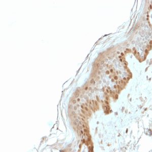


![Natural Cycles gets FDA clearance to use Apple Watch temperature data for birth control - [ Cửa Sổ Tin Học ]](https://cuasotinhoc.vn/wp-content/uploads/2023/09/Natural_Cycles_powered_by_Apple_Watch_4.0-5i4hKw-760x490.jpeg)