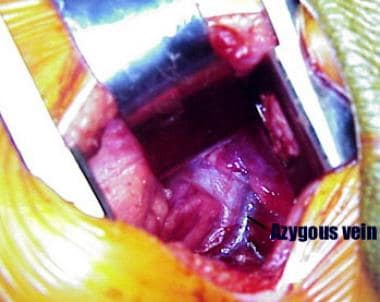
Azygos Vein
Brachiocephalic Veins
Idiopathic azygos vein aneurysm: a rare cause of mediastinal mass. (1/99)
Venous aneurysm of the azygos arch is a very rare cause of mediastinal mass and is usually an incidental finding on chest radiography. Nowadays the diagnosis is made by non-invasive tests such as thoracic CT scanning and/or magnetic resonance imaging. The case is described of an asymptomatic woman in whom a mediastinal mass due to an azygos vein aneurysm was diagnosed by non-invasive procedures, the aetiology of which, in all probability, was idiopathic. (+info)Prenatal diagnosis of interrupted inferior vena cava as an isolated finding: a benign vascular malformation. (2/99)
We report two cases of interrupted inferior vena cava with azygous continuation diagnosed as an isolated finding during routine prenatal scans. Visualization of the venous vasculature of the abdomen and thorax in the mid-sagittal plane failed to visualize the segment of the inferior vena cava between the kidneys and the liver. A vessel with venous flow was observed parallel, adjacent and posterior to the aorta between the kidney and the right atrium. This blood vessel connected with the superior vena cava. Axial planes of the thorax confirmed the presence of two vessels running paravertebrally. A detailed ultrasound examination of the fetal anatomy failed to demonstrate other anomalies. The neonatal course of both fetuses was uneventful. Isolated interruption of the inferior uena cava can be a vascular malformation without known pathological consequences. (+info)Isolated azygos continuation of the inferior vena cava in the elderly. (3/99)
A 70-year-old woman with the isolated anomaly of azygos continuation of the inferior vena cava is presented. The interest in this anomaly lies in its presentation as a mediastinal mass on the chest roentgenogram. This article reviews the embryogenesis and the diagnostic testing of this venous anomaly. (+info)Occlusion of azygos vein via direct percutaneous puncture of innominate vein following cavopulmonary anastomosis. (4/99)
A 2-year-10-month-old boy was diagnosed with a complex congenital heart disease: right atrial isomerism, left superior vena cava (LSVC), complete atrioventricular septal defect, secundum type atrial septal defect, transposition of the great arteries with pulmonary atresia, patent ductus arteriosus, absence of a right superior vena cava (RSVC), and dextrocardia. He had received a left Blalock-Taussig (BT) shunt at the age of 3 months and a left bidirectional Glenn shunt one year after BT shunt. Progressive cyanosis was noted after the second operation and cardiac catheterization showed a functional Glenn shunt with an engorged azygos vein, which was inadvertently skipped for ligation. Because of the absence of RSVC, transcatheter occlusion of the azygos vein was performed successfully via direct puncture of the innominate vein. (+info)Giant aneurysm of the azygos anterior cerebral artery--case report. (5/99)
A 77-year-old female presented with a giant aneurysm of the azygos anterior cerebral artery (ACA) manifesting as acute onset of akinetic mutism caused by enlargement of the aneurysm resulting from rapid thrombus formation within the aneurysmal sac. Thrombus removal to obtain decompression of the aneurysmal bulk and tension was performed before parent artery occlusion to prevent thromboembolic events. The aneurysmal neck was completely clipped with preservation of the parent artery and all branches. This strategy for direct neck clipping of a giant thrombosed distal ACA aneurysm can reduce the possibility of ischemic sequelae. (+info)Transcatheter coil occlusion of a thoracic arteriovenous fistula in an infant with congestive heart failure. (6/99)
An 8-week-old baby boy presented at our institution with a continuous murmur and congestive heart failure. Echocardiography showed normal cardiac anatomy. Catheterization revealed the presence of a large thoracic arteriovenous fistula between the descending thoracic aorta and the hemiazygous system, with eventual drainage into the azygous vein and the innominate vein. Coil occlusion was performed successfully with a Gianturco coil. (+info)Screw-in atrial lead in a sick sinus syndrome patient with anomalous inferior vena cava. (7/99)
Anomalous inferior vena cava without intracardiac anomaly is an unusual condition. Herein, we report a 48-year-old female with left-sided inferior vena cava and azygous continuation. accompanied by sick sinus syndrome. This anomaly resulted in difficulty in implanting a traditional hook-on atrial lead. Atrial lead dislodgment occurred repeatedly soon after implanting the pacemaker because of an anomalous zygous vein draining into the superior vena cava, making a giant connection with the right atrium, thus eliminating the space of the atrial appendage for lead lodgment. Finally, we attempted to utilize a screw-in atrial lead in this patient and she is currently doing well. We therefore suggest that a screw-in atrial lead should be taken into account for such patients in order to obtain a stable fixation. (+info)Segmental aplasia of the caudal vena cava in a dog. (8/99)
Ultrasonography, angiography, magnetic resonance imaging, and exploratory laparotomy of a 2-year-old wheaten terrier with lethargy, exercise intolerance, and ascites revealed segmental aplasia of the caudal vena cava with azygos continuation, complicated by thrombus formation. Surgeries were performed on the blind-ended vessel to remove thrombi, enhancing shunting of blood through the azygos vein. (+info)The azygos vein is a large, unpaired venous structure in the thoracic cavity of the human body. It begins as the ascending lumbar vein, which receives blood from the lower extremities and abdominal organs. As it enters the thorax through the diaphragm, it becomes the azygos vein and continues to ascend along the vertebral column.
The azygos vein receives blood from various tributaries, including the intercostal veins, esophageal veins, mediastinal veins, and bronchial veins. It then arches over the right mainstem bronchus and empties into the superior vena cava, which returns blood to the right atrium of the heart.
The azygos vein provides an important collateral pathway for venous return in cases where the inferior vena cava is obstructed or occluded. It also plays a role in the spread of certain thoracic diseases, such as tuberculosis and cancer.
The brachiocephalic veins, also known as the innominate veins, are large veins in the human body. They are formed by the union of the subclavian vein and the internal jugular vein on each side of the body. The resulting vein then carries blood from the upper limbs, head, and neck to the superior vena cava, which is the large vein that returns blood to the heart.
Here's a more detailed medical definition:
The brachiocephalic veins are paired venous structures that result from the union of the subclavian vein and the internal jugular vein on each side of the body. These veins are located in the superior mediastinum, near the base of the neck, and are typically about 2 to 3 centimeters in length. The brachiocephalic veins receive blood from several sources, including the upper extremities, head, neck, and thoracic wall. They then transport this blood to the superior vena cava, which is a large vein that returns blood to the right atrium of the heart.
It's worth noting that the brachiocephalic veins are subject to various pathological conditions, including thrombosis (blood clots), stenosis (narrowing), and compression by nearby structures such as the first rib or the scalene muscles. These conditions can lead to a variety of symptoms, including swelling, pain, and difficulty breathing.
The superior vena cava is a large vein that carries deoxygenated blood from the upper half of the body to the right atrium of the heart. It is formed by the union of the left and right brachiocephalic veins (also known as the internal jugular and subclavian veins) near the base of the neck. The superior vena cava runs posteriorly to the sternum and enters the upper right portion of the right atrium, just posterior to the opening of the inferior vena cava. It plays a crucial role in the circulatory system by allowing blood returning from the head, neck, upper limbs, and thorax to bypass the liver before entering the heart.
 Azygos vein
Superior intercostal vein
Bronchus
Renal vein
External jugular vein
Middle thyroid vein
Subclavian vein
Superior thyroid vein
Internal jugular vein
Azygos lobe
Hemiazygos vein
Lung
Carina of trachea
Deep circumflex iliac artery
List of anatomy mnemonics
Left gastric vein
Accessory hemiazygos vein
Mediastinum
Superior phrenic vein
Brachiocephalic vein
Posterior cardinal vein
Median arcuate ligament syndrome
Aortic hiatus
Radiate ligament of head of rib
Thoracic aorta
Posterior intercostal veins
Root of the lung
Esophageal veins
Bronchial veins
Inferior vena cava
Azygos vein - Wikipedia
 Pages that link to "Azygos vein" - wikidoc
Azygos Vein | Profiles RNS
 What is the Azygos Vein? (with pictures)
 Deep vein thrombosis as a presenting symptom of congenital interruption of the inferior vena cava
 Lymphovenous Anastomoses Between Thoracic Duct and Azygos Vein in a Human Cadaver: A Case Report | Koutsouflianiotis | Acta...
 Lieve Morbée
 Successful slow pathway modification using the femoral approach in a patient with interrupted Inferior Vena Cava with Azygos...
Novel technique to repair a bronchopleural fistula after esophagectomy using wide intercostal muscle flap fixed by azygos vein...
 Extracorporeal membrane oxygenation cannula malposition in the azygos vein in an infant<...
 Congenital Anomalies of Esophagus: Practice Essentials, Anatomy, Pathophysiology
 Tuberculin Test Results (1971-75)
 AZYGOS. : languagehat.com
 Bassett Collection - Lane Medical Library - Stanford University School of Medicine
 Interactive Prenatal Development Timeline - Advanced
Chronic cerebrospinal venous insufficiency in patients with multiple sclerosis | Journal of Neurology, Neurosurgery & Psychiatry
 Ascending lumbar vein | Radiology Reference Article | Radiopaedia.org
The perfect crime? CCSVI not leaving a trace in MS | Journal of Neurology, Neurosurgery & Psychiatry
Esophageal Cancer Staging: TNM Classification for Esophageal Cancer
 1b. The Trachea and Bronchi - Collection at Bartleby.com
 angelo carloni - Google Scholar
 Bastarrika Alema��, Gorka. Curriculum. Universidad de Navarra
 Necrotizing fasciitis of the chest wall: A clinical case report and literature review
 Biomedicines | Free Full-Text | Robert's Intragastric Alcohol-Induced Gastric Lesion Model as an Escalated General Peripheral...
 Right lung
 Corrected Transposition of the Great Arteries with Ebstein's Anomaly, Dysplasia of the Mitral Leaflets and Persistence of...
Survey: NHANES I
Emergent Management of Atrial Flutter: Overview, Emergency Department Care, Cardioversion for Unstable Patients
 The Abdominal Aorta - Human Anatomy
Venous16
- It has been proposed that the azygos vein develops by originally draining to the posterior cardinal vein and then to the longitudinal venous channel. (wikipedia.org)
- Azygos venous connection of anomalous inferior vena cava. (uchicago.edu)
- This is referred to as chronic cerebrospinal venous insufficiency (CCSVI), which can lead to decreased brain function as well as stenosis, or abnormal narrowing, of the azygos vein. (thehealthboard.com)
- Infrahepatic interruption of the inferior vena cava is a congenital anomaly, resulting in venous drainage of the lower extremities by way of a compensatory enlarged vena azygos system. (nih.gov)
- Introduction: Misplacement of extracorporeal membrane oxygenation (ECMO) venous cannula in the azygos vein has previously been described only in newborns. (wustl.edu)
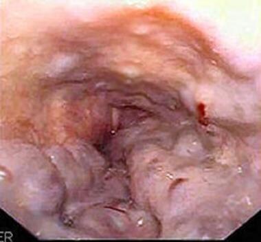
- Although the venous drainage of the esophagus is not described here, the azygos vein serves as a good landmark during surgery. (medscape.com)
- redirection of venous flow to the vertebral veins (VVs) occurs in the upright position, with compliant reduction of the CSA of the IJV. (bmj.com)
- Extra- and intracranial venous flow direction was assessed by colour-coded duplex sonography, and extracranial venous cross-sectional area (VCSA) of the internal jugular and vertebral veins (IJV/VV) was measured in B-mode to assess the five previously proposed CCSVI criteria. (bmj.com)
- 5-7 In a recent study 5 based on duplex sonographic and venographic assessment of extracranial and intracranial veins of 65 MS patients and 235 controls, Zamboni et al claimed a perfect coincidence of MS and venous stenoses in various locations. (bmj.com)
- Nine consecutive patients undergoing digital subtraction venography for petrosal venous sampling or parathormone sampling had images of their internal jugular veins obtained as part of their procedure, and they were assessed for stenosis. (ajnr.org)
- The Vancouver researchers want to determine the prevalence of the vein abnormality, which Zamboni has dubbed CCSVI -- or chronic cerebrospinal venous insufficiency. (ctvnews.ca)
- Skin breakdown or ulceration caused by VARICOSE VEINS in which there is too much hydrostatic pressure in the superficial venous system of the leg. (lookformedical.com)
- Median vein of the forearm The median vein of the forearm begins from the palmar venous network and ends in any one of the veins in front of the elbow, mostly in the median cubital vein. (vumc.org)
- Deep vein thrombosis associated with central venous catheters - a review. (vumc.org)
- The superficial veins connect via a network of interlacing branches to eventually form the dorsal venous network on the back of the hand. (vumc.org)
- Palmar metacarpal veins extend along either side of the Dorsal metacarpal veins, as previously mentioned, are formed by the union of the dorsal digital veins and receive venous blood from the fingers. (vumc.org)
Anatomical5
- As an anatomical variation in 1-2% of the population, the arch can be displaced laterally, thereby creating a pleural septum separating an azygos lobe from the upper lobe of the right lung. (wikipedia.org)
- The origin and anatomical course of the azygos vein are quite variable. (wikipedia.org)
- The study adds valuable information regarding lymphovenous communications between the thoracic duct and the azygos vein, which are very rarely discovered during anatomical dissections and very few cases have been mentioned worldwide. (ama.ba)
- Like a street that changes name as it passes through an intersection, an artery or vein can change names as it passes an anatomical landmark. (cuny.edu)
- How to cite this article: Yazkan R, Çeviker K, Güblü M. The Azygos Lobe: An Unusual Anatomical Entity with Unusual Cases. (eurasianmedicine.com)
Vena cava13
- The azygos vein is a vein running up the right side of the thoracic vertebral column draining itself towards the superior vena cava. (wikipedia.org)
- The azygos vein transports deoxygenated blood from the posterior walls of the thorax and abdomen into the superior vena cava. (wikipedia.org)
- Azygos and hemiazygos continuation of the inferior vena cava (IVC) was not common in daily life. (wikipedia.org)
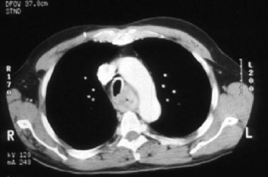
- Thus, it is crucial to diagnose the enlarged azygos vein at the confluence with the superior vena cava and in the retrocrural space to prevent misdiagnosis as a right-sided paratracheal mass. (wikipedia.org)
- A vein which arises from the right ascending lumbar vein or the vena cava, enters the thorax through the aortic orifice in the diaphragm, and terminates in the superior vena cava. (uchicago.edu)
- Blood in azygos vein empties into the superior vena cava, which then carries it to the heart to be recirculated. (thehealthboard.com)
- Blood in the azygos vein empties into the superior vena cava, which takes it to the heart for re-circulation. (thehealthboard.com)
- The case illustrates that in a case of deep vein thrombosis, especially in younger patients, interruption of the inferior vena cava should be considered. (nih.gov)
- Successful slow pathway modification using the femoral approach in a patient with interrupted Inferior Vena Cava with Azygos vein continuation. (ox.ac.uk)
- Azygos vein, is a branch of the upper trunk of the vena cava, arising on the right side. (languagehat.com)
- The contrast material in the superior vena cava (SVC) and brachiocephalic veins is diluted due to recirculation, and no longer causes streak artifacts. (siemens-healthineers.com)
- The duct crosses to the left side of the aorta ventral to the body of the fifth thoracic vertebra and continues cranioventral across the left side of the esophagus to empty at the junction of the left jugular vein and cranial vena cava. (dvm360.com)
- A vein on either side of the body which is formed by the union of the external and internal iliac veins and passes upward to join with its fellow of the opposite side to form the inferior vena cava. (lookformedical.com)
Ascending lumbar veins2
- It is formed by the union of the ascending lumbar veins with the right subcostal veins at the level of the 12th thoracic vertebra, ascending to the right of the descending aorta and thoracic duct, passing behind the right crus of diaphragm, anterior to the vertebral bodies of T12 to T5 and right posterior intercostal arteries. (wikipedia.org)
- The ascending lumbar veins are paired vertically-oriented veins of the posterior abdominal wall. (radiopaedia.org)
Posterior5
- Following retrogression of the left common cardinal vein, the left azygos vein loses contact with the posterior cardinal vein. (wikipedia.org)
- Some of these tributaries on the left side are the hemiazygos vein and the posterior intercostal veins. (thehealthboard.com)
- ascends vertically in the posterior abdominal wall, communicating with the lumbar veins as it crosses them. (radiopaedia.org)
- The ascending lumbar vein ascends posterior to the psoas major muscle and anterior to the lumbar transverse processes. (radiopaedia.org)
- A single-coil lead was selectively placed into the hemiazygous vein, which courses leftward of the spine in a posterior-anterior projection, resulting in an improved shocking vector and reduction in DFTs to less than 25 J. (duke.edu)
Drains4
- Thus, the blood drains into the right azygos line. (wikipedia.org)
- The vein which drains the foot and leg. (lookformedical.com)
- The external jugular vein drains blood primarily from the scalp and face. (vumc.org)
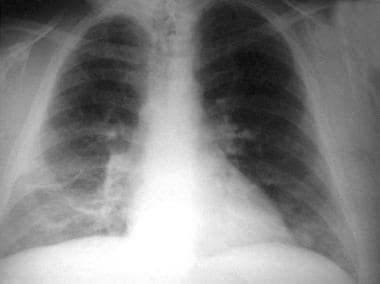
- The findings are characteristic of a partially absent IVC which drains through the azygos vein. (myesr.org)
Intercostal3
- The azygous vein also receives drainage from all the intercostal veins on the right, except for supreme intercostal vein (first intercostal vein). (wikipedia.org)
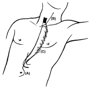
- After separating the intercostal muscles near the angle of the rib, we passed a muscle flap between the azygos vein and bronchus and sutured it securely to the fistula. (springeropen.com)
- In the dog, the caudal thoracic duct courses dorsal and to the right of the aorta, lateral to the intercostal arteries, and ventral to the azygos vein. (dvm360.com)
Hemiazygos veins3
- They can be considered the abdominal counterpart of the azygos and hemiazygos veins 1 . (radiopaedia.org)
- The azygos and hemiazygos veins were dilated. (siemens-healthineers.com)
- The veins of the esophagus drain into the systemic circulation via the azygos and the hemiazygos veins and into the portal circulation via the left gastric veins . (amboss.com)
Vertebral veins1
- 9 The proponents of this theory have devised 5 sonographic criteria when studying the internal jugular veins, vertebral veins, and intracranial veins. (ajnr.org)
Pulmonary1
- As you learn about the vessels of the systemic and pulmonary circuits, notice that many arteries and veins share the same names, parallel one another throughout the body, and are very similar on the right and left sides of the body. (cuny.edu)
Tributaries5
- Major tributaries are the hemiazygos vein and accessory hemiazygos vein, draining into the azygous vein at the midthoracic level. (wikipedia.org)
- Other tributaries include the right bronchial veins and veins from pericardium, mediastinum, and oesophagus. (wikipedia.org)
- While there is the hemiazygos vein and its accessory on the left side of the body, they are considered tributaries of the azygos vein rather than its left-side equivalent. (wikipedia.org)
- The venæ cavæ and azygos veins, with their tributaries. (wikipedia.org)
- Veins that perform a similar function can be found on the body's left side, but these are considered tributaries and thus only parts of this vein system rather than equivalent body structures. (thehealthboard.com)
Azygous4
- The trachea and oesophagus is located medially to the arch of the azygous vein. (wikipedia.org)
- per M-W). No, not "azygous" (which is an alternate spelling), azygos . (languagehat.com)
- Azygous is listed here as a synonym for azygos . (languagehat.com)
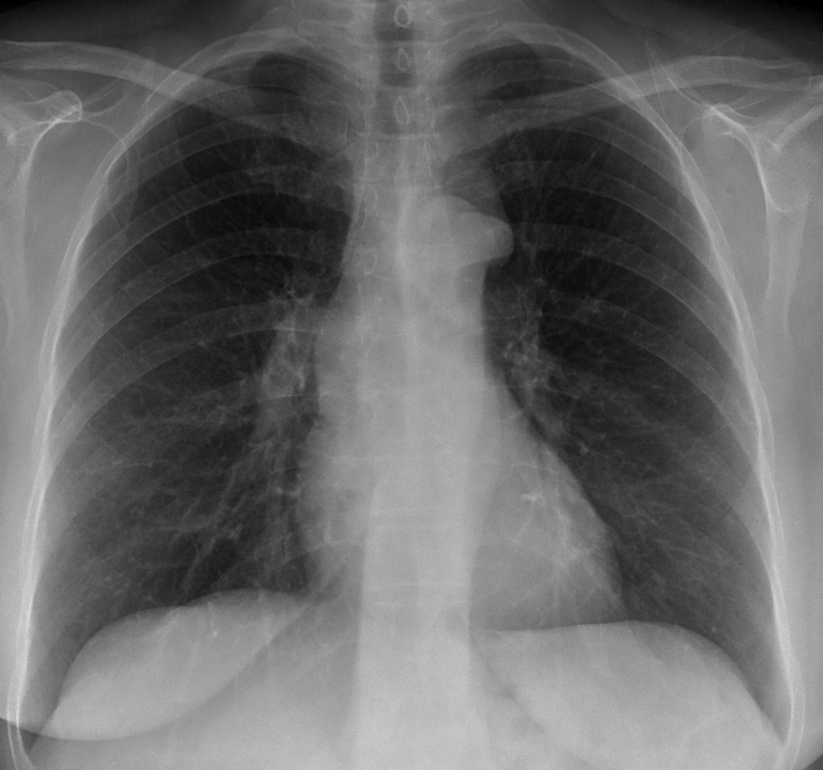
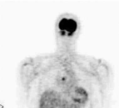
- Azygous vein also appears prominent. (myesr.org)
Lobe2
- The knowledge of azygos lobe is important during thoracic surgical procedures. (eurasianmedicine.com)
- We aimed to emphasize in this article the formation of the azygos lobe with described together pathologies in the literature. (eurasianmedicine.com)
Superficial veins5
- Moreover, some superficial veins, such as the great saphenous vein in the femoral region, have no arterial counterpart. (cuny.edu)
- There are two main types of veins in the upper extremity: superficial veins and deep veins. (vumc.org)
- Superficial veins are located close to the surface of the skin and can be easily seen and felt. (vumc.org)
- The superficial veins are placed immediately beneath the integument between the two layers of superficial fascia. (vumc.org)
- The most prominent and anatomically consistent superficial veins are the cephalic analogous to the saphenous and basilic. (vumc.org)
Abnormalities2
- Azygos vein abnormalities can be suggested on chest radiograph by enlargement of the azygos shadow to greater than 1 cm. (wikipedia.org)
- 50% in 91% of cases and azygos abnormalities in 86%, whereas a control group without MS, who had normal sonography findings, underwent venography and had no venographic stenosis. (ajnr.org)
Mediastinal2
- A right-sided mediastinal mass on the chest X-ray was mistaken for a haematological malignancy but proved later to represent an enlarged azygos vein. (nih.gov)
- Findings: the PA radiograph shows a mediastinal mass at the level of the azygos vein (A, arrow), similar to the one seen in case 178 . (myesr.org)
Bronchial veins1
- The Bronchial Veins vv. (vumc.org)
Continuation2
- Azygos and hemiazygos continuation of the IVC is rare especially when it is not associated with congenital heart disease. (wikipedia.org)
- it is a continuation of the popliteal vein and becomes the external iliac vein. (lookformedical.com)
Gonadal veins1
- Sometimes the vein system feeds different areas of the abdomen, In some cases, it not only serves the abdominal and thoracic walls, it also receives blood from bronchial and gonadal veins. (thehealthboard.com)
Internal jugu2
- The internal jugular vein receives blood from the deep structures of the neck and the brain. (vumc.org)
- The left internal jugular vein, which is usually smaller in caliber than the right internal jugular vein, crosses the common carotid artery before joining the left subclavian vein to form the left innominate vein, which then crosses the innominate artery to form, together with the right innominate vein, the superior vena cava. (vumc.org)
Jugular veins2
- Furthermore, normal physiologic narrowing is found very commonly in the internal jugular veins in healthy individuals. (ajnr.org)
- This is also, according the University of Ferrara team, the definitive way of seeing blockages in the jugular veins in the neck and the azygos vein in the chest. (ctvnews.ca)
Anatomy3
- Anatomy of the azygos vein examined by computerized tomography imaging. (uchicago.edu)
- Upper extremity vein anatomy. (vumc.org)
- Overall, the anatomy of the upper extremity veins is complex and plays a crucial role in the proper functioning of the circulatory system. (vumc.org)
Arteries and veins2
- However, we will attempt to discuss the major pathways for blood and acquaint you with the major named arteries and veins in the body. (cuny.edu)
- Neoplasms located in the vasculature system, such as ARTERIES and VEINS. (lookformedical.com)
Empties1
- the inferior mesenteric vein empties into the splenic vein, the superior mesenteric vein joins the splenic vein to form the portal vein. (lookformedical.com)
Intracranial2
- Results No participant showed retrograde flow of cervical or intracranial veins. (bmj.com)
- Stenosis of the internal jugular, azygos, and other veins detected by using intracranial and neck Doppler and B-mode sonography and confirmed by venography has been reported in MS with a high degree of sensitivity. (ajnr.org)
Abdominal1
- 38 An azygos orifice in the abdominal walls. (languagehat.com)
Midline1
- However, the azygos vein is occasionally located in the midline or two independent veins may be present like in early embryonic development. (wikipedia.org)
Upper11
- Overall narrowing or blocking of the veins that drain the upper body causes deoxygenated blood to pool in the brain, causing edema , and it slows the delivery of oxygenated blood back to the brain, depriving the brain of much-needed oxygen . (thehealthboard.com)
- Below, it is in relation to the upper border of the pancreas, and the lienal vein. (theodora.com)
- The Veins of the Upper Extremity and Thorax. (vumc.org)
- The upper extremity, also known as the arm, contains a complex network of veins that play a crucial role in the circulatory system. (vumc.org)
- The deep veins of the upper extremity include the axillary vein, which runs through the armpit and connects to the subclavian vein, and the brachial vein, which runs along the inside of the arm and connects to the axillary vein. (vumc.org)
- Both the superficial and deep veins of the upper extremity are important for maintaining proper blood flow and circulation. (vumc.org)
- However, the deep veins are especially vital as they are responsible for carrying the majority of the blood from the upper extremity back to the heart. (vumc.org)
- The upper extremity veins also have a number of important functions beyond just transporting blood. (vumc.org)
- In addition, the veins of the upper extremity are essential for the administration of intravenous fluids and medications. (vumc.org)
- Upper-extremity deep vein thrombosis. (vumc.org)
- Takeaway: Veins of the upper limb consist of arm veins, and forearms veins. (vumc.org)
Neck3
- They will be studying the findings of Italian researcher Dr. Paolo Zamboni, who believes that blocked veins in the neck and chest of MS patients lead to blood drainage problems and triggers the immune responses that mark the disease. (ctvnews.ca)
- That's where a probe is inserted, from the groin, into the vein system that travels through the chest and into the neck. (ctvnews.ca)
- Veins in the neck which drain the brain, face, and neck into the brachiocephalic or subclavian veins. (lookformedical.com)
Superior mese1
- A short thick vein formed by union of the superior mesenteric vein and the splenic vein. (lookformedical.com)
Anastomoses2
- In the current study, two sizeable obliquely directed lymphovenous anastomoses between the thoracic duct and the azygos vein at the midportion of the mediastinum are described in the same cadaver. (ama.ba)
- Anastomoses are especially common in veins, where they help maintain blood flow even when one vessel is blocked or narrowed, although there are some important ones in the arteries supplying the brain. (cuny.edu)
Anterior3
- Posteriorly, it is separated from the lumbar vertebræ and intervertebral fibrocartilages by the anterior longitudinal ligament and left lumbar veins. (theodora.com)
- At the cubital fossa on the anterior aspect of the elbow joint , this vein flows into the median cubital vein. (vumc.org)
- Thalamostriate veins are formed by the joining of anterior caudate vein and the vein of stria terminalis. (radiopaedia.org)
Artery1
- Compression of the left common ILIAC VEIN by the right common ILIAC ARTERY against the underlying fifth LUMBAR VERTEBRA is the typical underlying malformation. (lookformedical.com)
Stenosis1
- Some doctors and scientists believe that further study and treatment of CCSVI and stenosis of the azygos vein system could result in new, more effective treatments for multiple sclerosis. (thehealthboard.com)
Cows2
- Ruminants (such as sheep and cows) have paired azygos veins. (wikipedia.org)
- Humans, dogs and cats do not have paired azygos veins, but cows, sheep and other ruminants do. (thehealthboard.com)
Accessory1
- The azygos system of veins is considered to be the azygos vein, along with its left-sided counterparts, the hemiazygos vein and the accessory hemiazygos vein. (wikipedia.org)
Arch3
- The "arch of the azygos vein" (arcus venae azygos) is an important anatomic landmark. (wikipedia.org)
- Strangulation of the reconstructive gastric tube by the azygos arch. (uchicago.edu)
- in the thorax, it is covered from before backward by the manubrium sterni, the remains of the thymus, the left innominate vein, the aortic arch, the innominate and left common carotid arteries, and the deep cardiac plexus. (bartleby.com)
Ascends2
- Sometimes it communicates with the external jugular vein by a branch which ascends in front of the clavicle. (vumc.org)
- Â The cephalic vein ascends along the forearm and communicates with the The basilic vein similarly ascends within the subcutaneous tissue of the medial aspect of the forearm and inferior portion of the arm. (vumc.org)
Deep veins1
- Deep veins, on the other hand, are located deeper within the tissue and are not visible from the surface. (vumc.org)
Cerebral veins2
- Conclusions This triple-blinded extra- and transcranial duplex sonographic assessment of cervical and cerebral veins does not provide supportive evidence for the presence of CCSVI in MS patients. (bmj.com)
- They join the septal veins and form internal cerebral veins . (radiopaedia.org)
MeSH1
- Azygos Vein" is a descriptor in the National Library of Medicine's controlled vocabulary thesaurus, MeSH (Medical Subject Headings) . (uchicago.edu)
Parietal1
- Diagram showing completion of development of the parietal veins. (wikipedia.org)
Femoral1
- For example, you will find a pair of femoral arteries and a pair of femoral veins, with one vessel on each side of the body. (cuny.edu)
Subclavian1
- All these veins may play an important role in supplying collateral flow in the presence of subclavian and axillary vein thrombosis. (vumc.org)
Left5
- The vein is so named because it has no symmetrically equivalent vein on the left side of the body. (wikipedia.org)
- Unlike most veins and arteries, it does not have a corresponding vein on the left side of the body, thus explaining its name, which means "unpaired" in Greek. (thehealthboard.com)
- A compression of ILIAC VEIN that results in a decreased flow in the vein and in the left LOWER EXTREMITY due to a vascular malformation. (lookformedical.com)
- The veins that return the oxygenated blood from the lungs to the left atrium of the heart. (lookformedical.com)
- Portal hypertension results in esophageal varices , which are supplied by the left gastric veins . (amboss.com)
Thrombosis3
- We report the case of a 37-year-old male who presented with symptoms of deep vein thrombosis of the entire right lower extremity. (nih.gov)
- A condition caused by one or more episodes of DEEP VEIN THROMBOSIS, usually the blood clots are lodged in the legs. (lookformedical.com)
- Proper care and attention to these veins is essential for maintaining good health and preventing problems such as vein damage or thrombosis. (vumc.org)
Bronchopulmonary1
- It is a rare anomaly in bronchopulmonary segmentation due to an unusual course of the azygos vein. (eurasianmedicine.com)
Sonographic assessment1
- Nonblinded subjective sonographic assessment of the IJV may erroneously lead to venography, the findings of which may be misinterpreted due to the lack of widespread knowledge about the appearance of these veins in healthy individuals. (ajnr.org)
Cervical1
- Sometimes the thoracic and cervical veins can become compromised so that they do not function as well as they should. (thehealthboard.com)
Esophageal1
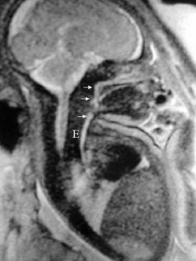
- The esophageal ends, particularly the distal segment in a TEF, are often readily visualized once the azygos vein has been divided or reflected. (medscape.com)