Autonomic Fibers, Postganglionic
Autonomic Fibers, Preganglionic
Sympathetic Fibers, Postganglionic
Parasympathetic Fibers, Postganglionic
Ganglia, Parasympathetic
Nerve Fibers
Ganglia, Sympathetic
Parasympathetic Nervous System
Sympathetic Nervous System
Hexamethonium Compounds
Bretylium Compounds
Ganglionic Blockers
Ganglia, Autonomic
Dietary Fiber
Horner Syndrome
Muscle Fibers, Skeletal
Pempidine
Guanethidine
Pentolinium Tartrate
Vagus Nerve
Mineral Fibers
Hypogastric Plexus
Atropine
Muscle Fibers, Fast-Twitch
Muscle Fibers, Slow-Twitch
Pharmacology
Stellate Ganglion
Cotton Fiber
Norepinephrine
Sympathectomy
Cats
Ganglia
Hypohidrosis
Nictitating Membrane
Autonomic Nervous System Diseases
Tail
Neurons
Acetylcholine
Guinea Pigs
Muscle Contraction
Tubocurarine
Parasympathomimetics
Bethanidine
Vas Deferens
Stress Fibers
Reflex
Vasoactive Intestinal Peptide
Neuroeffector Junction
Abducens Nerve Diseases
Action Potentials
Purkinje Fibers
Dimethylphenylpiperazinium Iodide
Tyramine
Synaptic Transmission
Nerve Fibers, Myelinated
Chlorisondamine
Hexamethonium
Submandibular Gland
Efferent Pathways
Ergotamine
Sinoatrial Node
Sweating
Tetrodotoxin
Phrenic Nerve
omega-Conotoxin GVIA
Phenoxybenzamine
Mossy Fibers, Hippocampal
Tyrosine 3-Monooxygenase
Parasympatholytics
Receptors, Muscarinic
Adrenergic alpha-2 Receptor Antagonists
Hypotension, Orthostatic
Nerve Endings
Mecamylamine
Sympatholytics
Neuropeptide Y
Methacholine Compounds
Dogs
Rabbits
Rana catesbeiana
Physostigmine
Procaine
Autonomic Nervous System
Muscle, Smooth
Peroneal Nerve
Pressoreceptors
Rats, Sprague-Dawley
Neurotransmitter Agents
Ileum
Membrane Potentials
Autonomic modification of the atrioventricular node during atrial fibrillation: role in the slowing of ventricular rate. (1/66)
BACKGROUND: Postganglionic vagal stimulation (PGVS) by short bursts of subthreshold current evokes release of acetylcholine from myocardial nerve terminals. PGVS applied to the atrioventricular node (AVN) slows nodal conduction. However, little is known about the ability of PGVS to control ventricular rate (VR) during atrial fibrillation (AF). METHODS AND RESULTS: To quantify the effects and establish the mechanism of PGVS on the AVN, AF was simulated by random high right atrial pacing in 11 atrial-AVN rabbit heart preparations. Microelectrode recordings of cellular action potentials (APs) were obtained from different AVN regions. Five intensities and 5 modes of PGVS delivery were evaluated. PGVS resulted in cellular hyperpolarization, along with depressed and highly heterogeneous intranodal conduction. Compact nodal AP exhibited decremental amplitude and dV/dt and multiple-hump components, and at high PGVS intensities, a high degree of concealed conduction resulted in a dramatic slowing of the VR. Progressive increase of PGVS intensity and/or rate of delivery showed a significant logarithmic correlation with a decrease in VR (P<0.001). Strong PGVS reduced the mean VR from 234 to 92 bpm (P<0.001). The PGVS effects on the cellular responses and VR during AF were fully reproduced in a model of direct acetylcholine injection into the compact AVN via micropipette. CONCLUSIONS: These studies confirmed that PGVS applied during AF could produce substantial VR slowing because of acetylcholine-induced depression of conduction in the AVN. (+info)Characterization of non-adrenergic, non-cholinergic inhibitory responses of the isolated guinea-pig trachea: differences between pre- and post-ganglionic nerve stimulation. (2/66)
1 Differences in the mechanism of non-adrenergic, non-cholinergic (NANC) inhibitory responses to preganglionic- and post-ganglionic nerve stimulation were investigated in the guinea-pig isolated trachea. 2 Stimulation of the vagus nerve at frequencies above 4 Hz elicited NANC relaxation of the trachealis muscle. Responses to low frequencies of stimulation (4-8 Hz) were abolished by the nitric oxide (NO) synthase inhibitor L-NOARG (10 microM), while a L-NOARG resistant component was observed at higher stimulus frequencies. The L-NOARG-resistant component of NANC inhibitory responses to higher frequencies of vagus nerve stimulation were significantly attenuated by the proteinase alpha-chymotrypsin (2 U/ml), suggesting that a neuropeptide such as VIP may contribute to NANC responses. 3 When postganglionic nerves were stimulated by electrical field stimulation (EFS), responses were readily elicited at frequencies below 4 Hz. Like responses to vagus nerve stimulation, responses to low frequency (<4 Hz) EFS were abolished by L-NOARG while a L-NOARG-resistant component was apparent at higher stimulus frequencies. 4 The L-NOARG-resistant component of NANC inhibitory responses to EFS was sensitive to alpha-chymotrypsin only if stimuli were delivered in either long trains at a low frequency (4 Hz for 10-30 s) or short trains of high frequency (16 Hz for 2.5-7.5 s). 5 Responses to preganglionic nerve stimulation were approximately 35% of the amplitude of responses to EFS in the same preparations. 6 In conclusion, responses to preganglionic and postganglionic NANC inhibitory nerve stimulation in the guinea-pig trachea differ in maximum amplitude, frequency-response characteristics and the contributions of cotransmitters. We suggest that these differences may be explained by filtering of preganglionic input to postganglionic NANC neurons. These results have implications in all studies where EFS is considered to be representative of physiological stimulation of post-ganglionic nerve stimulation. (+info)Inhibitory effects of clonidine and BS 100-141 on responses to sympathetic nerve stimulation in cats and rabbits. (3/66)
1. In pithed cats, the spinal sympathetic outflow was stimulated preganglionically at segments C7 and T1 and heart rate responses and nictitating membrane tone were measured in parallel. 2. Clonidine and a related drug, BS 100-141 (N-amidino-2(2,6-dichlorophenyl)acetamide hydrochloride), caused a dose-dependent inhibition of the stimulation-induced tachycardia but did not inhibit responses of the nictitating membrane. The inhibition of heart rate was antagonized by the alpha-adrenoceptor blocking drug, phentolamine. 3. In isolated hearts of rabbits, noradrenaline release in response to adrenergic nerve stimulation was reduced by clonidine and BS 100-141 and the effect was antagonized by phentolamine. 4. The results support the view that presynaptic alpha-adrenoceptors are involved in the regulation of transmitter release from adrenergic nerves. Cardiac adrenergic nerves appear more sensitive to alpha-adrenoceptor-mediated inhibition of inpulse transmission than the sympathetic nerves to the nictitating membrane. (+info)Innervation both of peri-orbital structures and of the heart by the cervical sympathetic nerves in mouse, rat, guinea-pig, rabbit and cat. (4/66)
1 In anaesthetized rats electrical stimulation of the intact cervical sympathetic nerve produced frequency-dependent lower eyelid contractions and tachycardia. 2 The tachycardia was caused by excitation of efferent fibres since it was equally evident in the pithed rat preparation, and the right nerve was more effective than the left. By contrast, no differences were seen between the responses to right and left vagal stimulation in either rats or rabbits. 3 Guanethidine inhibited both cardiac and eyelid responses, propranolol only the former and phentolamine only the latter, therby revealing the adrenergic nature of the nerves. Hexamethonium caused partial inhibition and the block was intensified by atropine. 4 The inferior eyelid of mice, guinea-pigs and rabbits as well as the nictitating membrane of rabbits and cats were contracted by cervical sympathetic nerve stimulation. In these species too, tachycardia occurred; this was more pronounced with the right than the left sympathetic nerve. The order of cardiac responsiveness was mouse greater than rat greater than guinea-pig greater than rabbit greater than cat. 5 In guinea-pigs histamine-induced bronchoconstriction was reduced by cervical sympathetic nerve stimulation. 6 That discrete cardiac pathways exist in the cervical sympathetic nerves is suggested by the reproducibility of the effects within any one species. The accessibility of the nerves greatly simplifies the examination of drugs in vivo on two different structures innervated by the sympathetic nervous system. (+info)Functional and structural changes in mammalian sympathetic neurones following interruption of their axons. (5/66)
The effects of interrupting the axons of principal neurones in the superior cervical ganglion of adult guinea-pigs were studied by means of intracellular recording, and light and electron microscopy. 1. Within 72 hr of axon interruption, the amplitude of exitatory postsynaptic potentials potentials (e.p.s.p.s) recorded in principal neurons in response to maximal preganglionic stimulation declined. E.p.s.p.s were maximally reduced (by more than 70% on average) 4-7 days following interruption, and failed to bring many cells to threshold. E.p.s.p.s. recorded in nearby neurones whose axons remained intact were unaffected. 2. In ganglia in which axon interruption was achieved by means of nerve crush (thus allowing prompt regeneration), mean e.p.s.p. amplitudes began to increase again after about 1-2 weeks. One month after the initial injury many neurones had e.p.s.p.s of normal amplitude, and by 2 months affected neurones were indistinguishable from control cells. Functional peripheral connexions were re-established during the period of synaptic recovery. 3. The mean number of synapses identified electron microscopically in ganglia in which all the major efferent branches had been crushed decreased by 65-70% in parallel with synaptic depression measured by intracellular recording. However synapse counts did not return to normal levels even after 3 months. 4. During the period of maximum synaptic depression, numerous abnormal profiles which contained accumulations of vesicular and tubular organelles, vesicles, and mitochondria were observed in electron microscopic sections. Injection of horseradish peroxidase into affected neurones demonstrated dendritic swelling which probably correspond to these profiles. 5. Little or no difference was found in the electrical properties of normal neurones and neurones whose axons had been interrupted 4-7 days previously. However, the mean amplitude of spontaneously occurring synaptic potentials was reduced, and the amplitude distribution was shifted. This abnormality of the synapses which remain on affected neurones also contributes to synaptic depression. 6. Counts of neurones in normal and experimental ganglia showed that approximately half the principal cells died 1-5 weeks after crushing the major efferent brances. This finding presumably explains the failure of synapse counts to return to control levels after recovery. 7. If axons were prevented from growing back to their target organ by chronic ligation, surviving neurones whose axons were enclosed by the ligature did not generally recover normal synaptic function. Following ligation, most affected cells died within a month. 8. Thus the integrity of a principal cell's axon is necessary for the maintenance of preganglionic synaptic contacts, and ultimately for neuronal survival. The basis of neuronal recovery from the effects of axon interruption appears to be some aspect of regeneration to the peripheral target. (+info)A study of peripheral input to and its control by post-ganglionic neurones of the inferior mesenteric ganglion. (6/66)
1. Intracellular recordings were made, in vitro, from neurones of guinea-pig inferior mesenteric ganglia (IMG) attached, via the lumbar colonic nerves, to segments of distal colon. 2. 'Spontaneous' synaptic input from colonic afferent fibres was observed in 79% of the neurones tested. In any given preparation, the level and pattern of this synaptic input to different neurones varied considerably. 3. Superfusion of colonic segments with drugs (papaverine, isoprenaline, and adenosine triphosphate) which reduce colonic motility decreased colonic afferent input to IMG neurones. 4. Superfusion of colonic segments with acetylcholine or stimulation of pelvic nerves, both of which increase colonic motility, increased colonic afferent input to IMG neurones. 5. Superfusion of colonic segments with either atropine or tubocurarine reduced the level of 'spontaneous', colonic afferent input. However, distension of these relaxed segments increased the colonic afferent input. 6. Repetitive stimulation of preganglionic inputs to the IMG inhibited afferent input from drug relaxed segments of colon that were moderately distended by the injection of air into the lumen. Superfusion of the colon with phentolamine blocked this inhibition. 7. The results of this study suggest that IMG neurones receive afferent input from mechanoreceptors located in the distal colon and that the mechanosensitivity of this afferent pathway is in part controlled by efferent noradrenergic neurones of the IMG. The IMG-colon neural circuitry can therefore be considered to form a feed-back control system which participates in the regulation of colonic motility. (+info)The relation between stimulus frequency and the relative size of the components of the biphasic response of the vas deferens to electrical stimulation at different temperatures. (7/66)
1. Electrical stimulation of the guinea-pig or rat vas deferens (pre- or post-ganglionically) at frequencies from 2-5 to 40 Hz with trains of stimuli of 30 sec duration induced a biphasic response. A rapid contraction (component A) was followed after a brief relaxation by a slower contraction (component B); the two phases were seen most clearly with stimulation frequencies of less than 10 Hz. 2. The responses to post-ganglionic stimulation were always larger than those to preganglionic stimulation. In general, at low frequencies component A exceeded component B whilst at high frequencies component B was the larger. Separation of the two components on the basis of their frequency response characteristics was better for rat than for guinea-pig vasa. 3. Log. frequency-response curves to transmural (post-ganglionic) electrical stimulation and log dose-response curves to noradrenaline were recorded for guinea-pig and rat vasa deferentia at 32 degrees, 22 degrees and 12 degrees C. For the guinea-pig reduction of bath temperature to 12 degrees C increased the amplitude of component A at 2-5 and 5 Hz; component B could not confidently be distinguished at this temperature. At 22 degrees C there was potentiation of B at lower frequencies and depression of B at higher frequencies. There was no response to noradrenaline at 12 degrees C. At 22 degrees C the response to noradrenaline was increased except to doses at or near the maximum to which the response was reduced. 4. For the rat was deferens component A was little changed by reduction of temperature. Component B at 12 degrees C was greatly depressed at higher frequencies. The response to noradreanaline was increased to lower doses and decreased to higher doses as the temperature was lowered. 5. The B component of the response of guinea-pig vasa at 22 degrees C and rat vasa at 32 degrees C was more sensitive than the A component to inhibition by thymoxamine. 6. Further analysis of the mechanisms underlying the A and B components of the biphasic response may be facilitated by relative isolation of each component by the appropriate selection of parameters of electrical stimulation and of temperature for the species being investigated. The contractions of the B component are similar to, if not identical with, those produced by exogenously applied noradrenaline. (+info)Synthesis of nitric oxide in postganglionic myenteric neurons during endotoxemia: implications for gastric motor function in rats. (8/66)
We have investigated the mechanisms underlying acute changes in gastric motor function triggered by endotoxemia. In fundal strips from rats pre-treated with endotoxin (40 microg/kg, i.p. 30 min), mechanical activity was analyzed and the source of nitric oxide (NO) was visualized by confocal microscopy of tissue loaded with the fluorescent dye DAF-FM. NOS expression was determined by quantitative RT-PCR and Western blot, and enzyme activity by the citrulline assay. Strips from endotoxin-treated rats were hypo-contractile. This was prevented by pre-incubation with the neurotoxin tetrodotoxin, the gangliar blocker hexamethonium, or non-selective and neuronal-specific NOS inhibitors (L-NOARG and TRIM, respectively). The soluble guanylyl cyclase (sGC) inhibitor ODQ and the inhibitor of small conductance Ca2+-activated K+ channels apamin prevented relaxation induced by endotoxin, nicotine, exogenous NO (DETA-NONOate), and the NO-independent sGC activator BAY 41-2272. NO synthesis was observed in neuronal soma, axons, and nerve endings of the myenteric plexus in the fundus of endotoxin-treated rats and was prevented by L-NAME, tetrodotoxin, and hexamethonium. nNOS and iNOS mRNA and protein contents were unchanged. Our findings demonstrate synthesis of NO in post-ganglionic myenteric neurons during early endotoxemia that mediates gastric hypo-contractility. The effect of NO is mediated via sGC and small conductance Ca2+-activated K+channels. (+info)Autonomic fibers, postganglionic, refer to the portion of the autonomic nervous system (ANS) that is responsible for the regulation of internal organs and glands. The ANS is divided into the sympathetic and parasympathetic systems, which generally have opposing effects on target organs.
Postganglionic fibers are the nerve fibers that originate from ganglia (clusters of neurons) located outside the central nervous system (CNS). These fibers transmit signals from the ganglia to effector organs such as muscles and glands. In the case of the autonomic nervous system, postganglionic fibers release neurotransmitters that act on receptors in target organs to produce physiological responses.
Sympathetic postganglionic fibers release norepinephrine (noradrenaline) as their primary neurotransmitter, which generally prepares the body for "fight or flight" responses such as increasing heart rate and blood pressure. Parasympathetic postganglionic fibers release acetylcholine as their primary neurotransmitter, which generally promotes "rest and digest" functions such as slowing heart rate and promoting digestion.
It's worth noting that there are some exceptions to this general rule, such as the sympathetic innervation of sweat glands, which releases acetylcholine as its primary neurotransmitter.
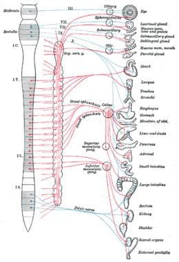
Preganglionic autonomic fibers are the nerve fibers that originate from neurons located in the brainstem and spinal cord, and synapse with postganglionic neurons in autonomic ganglia. These preganglionic fibers release acetylcholine as a neurotransmitter to activate the postganglionic neurons, which then innervate effector organs such as smooth muscle, cardiac muscle, and glands.
The autonomic nervous system is divided into two main subdivisions: the sympathetic and parasympathetic systems. The preganglionic fibers of the sympathetic nervous system originate from the lateral horn of the spinal cord from levels T1 to L2/L3, while those of the parasympathetic nervous system originate from cranial nerves III, VII, IX, and X, as well as sacral segments S2 to S4.
Preganglionic fibers are generally longer than postganglionic fibers, and their cell bodies are located in the central nervous system. They are responsible for transmitting signals from the CNS to the peripheral autonomic ganglia, where they synapse with postganglionic neurons that innervate target organs.
Postganglionic sympathetic fibers are the portion of the sympathetic nervous system's nerve fibers that originate from the cell bodies located in the ganglia ( clusters of neurons) outside the spinal cord. After leaving the ganglia, these postganglionic fibers travel to and innervate target organs such as sweat glands, blood vessels, and various smooth muscles, releasing neurotransmitters like norepinephrine and neuropeptide Y to regulate physiological functions. Acetylcholine is the neurotransmitter released by postganglionic fibers that innervate sweat glands.
Parasympathetic fibers, postganglionic, refer to the portion of the parasympathetic nervous system's peripheral nerves that arise from ganglia (clusters of neurons) located near or within the target organs. These postganglionic fibers are responsible for transmitting signals from the ganglia to the effector organs such as glands, smooth muscles, and heart, instructing them to carry out specific functions.
The parasympathetic nervous system is one of the two subdivisions of the autonomic nervous system (the other being the sympathetic nervous system). Its primary role is to conserve energy and maintain homeostasis during rest or digestion. The preganglionic fibers originate in the brainstem and sacral spinal cord, synapsing in the ganglia located near or within the target organs. Upon receiving signals from the preganglionic fibers, the postganglionic fibers release neurotransmitters like acetylcholine to activate muscarinic receptors on the effector organ, leading to responses such as decreased heart rate, increased gastrointestinal motility and secretion, and contraction of the urinary bladder.
Parasympathetic ganglia are collections of neurons located outside the central nervous system (CNS) that serve as relay stations for parasympathetic nerve impulses. The parasympathetic nervous system is one of the two subdivisions of the autonomic nervous system, which controls involuntary physiological responses.
The parasympathetic ganglia receive preganglionic fibers from the brainstem and sacral regions of the spinal cord. After synapsing in these ganglia, postganglionic fibers innervate target organs such as the heart, glands, and smooth muscles. The primary function of the parasympathetic nervous system is to promote rest, digestion, and energy conservation.
Parasympathetic ganglia are typically located close to or within the target organs they innervate. Examples include:
1. Ciliary ganglion: Innervates the ciliary muscle and iris sphincter in the eye, controlling accommodation and pupil constriction.
2. Pterygopalatine (sphenopalatine) ganglion: Supplies the lacrimal gland, mucous membranes of the nasal cavity, and palate, regulating tear production and nasal secretions.
3. Otic ganglion: Innervates the parotid gland, controlling salivary secretion.
4. Submandibular ganglion: Supplies the submandibular and sublingual salivary glands, regulating salivation.
5. Sacral parasympathetic ganglia: Located in the sacrum, they innervate the distal colon, rectum, and genitourinary organs, controlling defecation, urination, and sexual arousal.
These parasympathetic ganglia play crucial roles in maintaining homeostasis by regulating various bodily functions during rest and relaxation.
Nerve fibers are specialized structures that constitute the long, slender processes (axons) of neurons (nerve cells). They are responsible for conducting electrical impulses, known as action potentials, away from the cell body and transmitting them to other neurons or effector organs such as muscles and glands. Nerve fibers are often surrounded by supportive cells called glial cells and are grouped together to form nerve bundles or nerves. These fibers can be myelinated (covered with a fatty insulating sheath called myelin) or unmyelinated, which influences the speed of impulse transmission.
Sympathetic ganglia are part of the autonomic nervous system, which controls involuntary bodily functions. These ganglia are clusters of nerve cell bodies located outside the central nervous system, along the spinal cord. They serve as a relay station for signals sent from the central nervous system to the organs and glands. The sympathetic ganglia are responsible for the "fight or flight" response, releasing neurotransmitters such as norepinephrine that prepare the body for action in response to stress or danger.
The Parasympathetic Nervous System (PNS) is the part of the autonomic nervous system that primarily controls vegetative functions during rest, relaxation, and digestion. It is responsible for the body's "rest and digest" activities including decreasing heart rate, lowering blood pressure, increasing digestive activity, and stimulating sexual arousal. The PNS utilizes acetylcholine as its primary neurotransmitter and acts in opposition to the Sympathetic Nervous System (SNS), which is responsible for the "fight or flight" response.
The sympathetic nervous system (SNS) is a part of the autonomic nervous system that operates largely below the level of consciousness, and it functions to produce appropriate physiological responses to perceived danger. It's often associated with the "fight or flight" response. The SNS uses nerve impulses to stimulate target organs, causing them to speed up (e.g., increased heart rate), prepare for action, or otherwise respond to stressful situations.
The sympathetic nervous system is activated due to stressful emotional or physical situations and it prepares the body for immediate actions. It dilates the pupils, increases heart rate and blood pressure, accelerates breathing, and slows down digestion. The primary neurotransmitter involved in this system is norepinephrine (also known as noradrenaline).
Hexamethonium compounds are a type of ganglionic blocker, which are medications that block the transmission of nerve impulses at the ganglia ( clusters of nerve cells) in the autonomic nervous system. These compounds contain hexamethonium as the active ingredient, which is a compound with the chemical formula C16H32N2O4.
Hexamethonium works by blocking the nicotinic acetylcholine receptors at the ganglia, which prevents the release of neurotransmitters and ultimately inhibits the transmission of nerve impulses. This can have various effects on the body, depending on which part of the autonomic nervous system is affected.
Hexamethonium compounds were once used to treat hypertension (high blood pressure), but they are rarely used today due to their numerous side effects and the availability of safer and more effective medications. Some of the side effects associated with hexamethonium include dry mouth, blurred vision, constipation, difficulty urinating, and dizziness upon standing.
Bretylium compounds are a class of medications that are primarily used in the management of life-threatening cardiac arrhythmias (abnormal heart rhythms). Bretylium tosylate is the most commonly used formulation. It works by stabilizing the membranes of certain types of heart cells, which can help to prevent or stop ventricular fibrillation and other dangerous arrhythmias.
Bretylium compounds are typically administered intravenously in a hospital setting under close medical supervision. They may be used in conjunction with other medications and treatments for the management of cardiac emergencies. It's important to note that bretylium compounds have a narrow therapeutic index, which means that the difference between an effective dose and a toxic one is relatively small. Therefore, they should only be administered by healthcare professionals who are experienced in their use.
Like all medications, bretylium compounds can cause side effects, including but not limited to:
- Increased heart rate
- Low blood pressure
- Nausea and vomiting
- Dizziness or lightheadedness
- Headache
- Tremors or muscle twitching
- Changes in mental status or behavior
Healthcare providers will monitor patients closely for any signs of adverse reactions while they are receiving bretylium compounds.
Ganglionic blockers are a type of medication that blocks the activity of the ganglia, which are clusters of nerve cells located outside the central nervous system. These medications work by blocking the transmission of nerve impulses between the ganglia and the effector organs they innervate, such as muscles or glands.
Ganglionic blockers were once used in the treatment of various conditions, including hypertension (high blood pressure), peptic ulcers, and certain types of pain. However, their use has largely been abandoned due to their significant side effects, which can include dry mouth, blurred vision, constipation, difficulty urinating, and dizziness or lightheadedness upon standing.
There are two main types of ganglionic blockers: nicotinic and muscarinic. Nicotinic ganglionic blockers block the action of acetylcholine at nicotinic receptors in the ganglia, while muscarinic ganglionic blockers block the action of acetylcholine at muscarinic receptors in the ganglia.
Examples of ganglionic blockers include trimethaphan, hexamethonium, and pentolinium. These medications are typically administered intravenously in a hospital setting due to their short duration of action and potential for serious side effects.
Autonomic ganglia are collections of neurons located outside the central nervous system (CNS) that are a part of the autonomic nervous system (ANS). The ANS is responsible for controlling various involuntary physiological functions such as heart rate, digestion, respiratory rate, pupillary response, urination, and sexual arousal.
Autonomic ganglia receive inputs from preganglionic neurons, whose cell bodies are located in the CNS, and send outputs to effector organs through postganglionic neurons. The autonomic ganglia can be divided into two main subsystems: the sympathetic and parasympathetic systems.
Sympathetic ganglia are typically located close to the spinal cord and receive inputs from preganglionic neurons whose cell bodies are located in the thoracic and lumbar regions of the spinal cord. The postganglionic neurons of the sympathetic system release noradrenaline (also known as norepinephrine) as their primary neurotransmitter, which acts on effector organs to produce a range of responses such as increasing heart rate and blood pressure, dilating pupils, and promoting glucose mobilization.
Parasympathetic ganglia are typically located closer to the target organs and receive inputs from preganglionic neurons whose cell bodies are located in the brainstem and sacral regions of the spinal cord. The postganglionic neurons of the parasympathetic system release acetylcholine as their primary neurotransmitter, which acts on effector organs to produce a range of responses such as decreasing heart rate and blood pressure, constricting pupils, and promoting digestion and urination.
Overall, autonomic ganglia play a critical role in regulating various physiological functions that are essential for maintaining homeostasis in the body.
Dietary fiber, also known as roughage, is the indigestible portion of plant foods that makes up the structural framework of the plants we eat. It is composed of cellulose, hemicellulose, pectin, gums, lignins, and waxes. Dietary fiber can be classified into two categories: soluble and insoluble.
Soluble fiber dissolves in water to form a gel-like material in the gut, which can help slow down digestion, increase feelings of fullness, and lower cholesterol levels. Soluble fiber is found in foods such as oats, barley, fruits, vegetables, legumes, and nuts.
Insoluble fiber does not dissolve in water and passes through the gut intact, helping to add bulk to stools and promote regular bowel movements. Insoluble fiber is found in foods such as whole grains, bran, seeds, and the skins of fruits and vegetables.
Dietary fiber has numerous health benefits, including promoting healthy digestion, preventing constipation, reducing the risk of heart disease, controlling blood sugar levels, and aiding in weight management. The recommended daily intake of dietary fiber is 25-38 grams per day for adults, depending on age and gender.
Horner syndrome, also known as Horner's syndrome or oculosympathetic palsy, is a neurological disorder characterized by the interruption of sympathetic nerve pathways that innervate the head and neck, leading to a constellation of signs affecting the eye and face on one side of the body.
The classic triad of symptoms includes:
1. Ptosis (drooping) of the upper eyelid: This is due to the weakness or paralysis of the levator palpebrae superioris muscle, which is responsible for elevating the eyelid.
2. Miosis (pupillary constriction): The affected pupil becomes smaller in size compared to the other side, and it may not react as robustly to light.
3. Anhydrosis (decreased sweating): There is reduced or absent sweating on the ipsilateral (same side) of the face, particularly around the forehead and upper eyelid.
Horner syndrome can be caused by various underlying conditions, such as brainstem stroke, tumors, trauma, or certain medical disorders affecting the sympathetic nervous system. The diagnosis typically involves a thorough clinical examination, pharmacological testing, and sometimes imaging studies to identify the underlying cause. Treatment is directed towards managing the underlying condition responsible for Horner syndrome.
Skeletal muscle fibers, also known as striated muscle fibers, are the type of muscle cells that make up skeletal muscles, which are responsible for voluntary movements of the body. These muscle fibers are long, cylindrical, and multinucleated, meaning they contain multiple nuclei. They are surrounded by a connective tissue layer called the endomysium, and many fibers are bundled together into fascicles, which are then surrounded by another layer of connective tissue called the perimysium.
Skeletal muscle fibers are composed of myofibrils, which are long, thread-like structures that run the length of the fiber. Myofibrils contain repeating units called sarcomeres, which are responsible for the striated appearance of skeletal muscle fibers. Sarcomeres are composed of thick and thin filaments, which slide past each other during muscle contraction to shorten the sarcomere and generate force.
Skeletal muscle fibers can be further classified into two main types based on their contractile properties: slow-twitch (type I) and fast-twitch (type II). Slow-twitch fibers have a high endurance capacity and are used for sustained, low-intensity activities such as maintaining posture. Fast-twitch fibers, on the other hand, have a higher contractile speed and force generation capacity but fatigue more quickly and are used for powerful, explosive movements.
I'm sorry for any confusion, but "Pempidine" does not appear to be a recognized term in medical terminology or pharmacology. It is possible that there may be a spelling error or typo in the term you are looking for. Please double-check the spelling and try again. If you meant "Pimobendan," I would be happy to provide information on that instead.
If you indeed meant "Pempidine," please let me know, and I will do my best to find more information about it.
The superior cervical ganglion is a part of the autonomic nervous system, specifically the sympathetic division. It is a collection of nerve cell bodies (ganglion) that are located in the neck region (cervical) and is formed by the fusion of several smaller ganglia.
This ganglion is responsible for providing innervation to various structures in the head and neck, including the eyes, scalp, face muscles, meninges (membranes surrounding the brain and spinal cord), and certain glands such as the salivary and sweat glands. It does this through the postganglionic fibers that branch off from the ganglion and synapse with target organs or tissues.
The superior cervical ganglion is an essential component of the autonomic nervous system, which controls involuntary physiological functions such as heart rate, blood pressure, digestion, and respiration.
Electric stimulation, also known as electrical nerve stimulation or neuromuscular electrical stimulation, is a therapeutic treatment that uses low-voltage electrical currents to stimulate nerves and muscles. It is often used to help manage pain, promote healing, and improve muscle strength and mobility. The electrical impulses can be delivered through electrodes placed on the skin or directly implanted into the body.
In a medical context, electric stimulation may be used for various purposes such as:
1. Pain management: Electric stimulation can help to block pain signals from reaching the brain and promote the release of endorphins, which are natural painkillers produced by the body.
2. Muscle rehabilitation: Electric stimulation can help to strengthen muscles that have become weak due to injury, illness, or surgery. It can also help to prevent muscle atrophy and improve range of motion.
3. Wound healing: Electric stimulation can promote tissue growth and help to speed up the healing process in wounds, ulcers, and other types of injuries.
4. Urinary incontinence: Electric stimulation can be used to strengthen the muscles that control urination and reduce symptoms of urinary incontinence.
5. Migraine prevention: Electric stimulation can be used as a preventive treatment for migraines by applying electrical impulses to specific nerves in the head and neck.
It is important to note that electric stimulation should only be administered under the guidance of a qualified healthcare professional, as improper use can cause harm or discomfort.
Guanethidine is an antihypertensive medication that belongs to the class of drugs known as ganglionic blockers or autonomic nervous system (ANS) inhibitors. It works by blocking the action of certain chemicals (neurotransmitters) in the body, which results in decreased blood pressure and heart rate.
Guanethidine is not commonly used today due to its side effects and the availability of safer and more effective antihypertensive medications. Its medical definition can be stated as:
A synthetic antihypertensive agent that acts by depleting norepinephrine stores in postganglionic adrenergic neurons, thereby blocking their activity. Guanethidine is used primarily in the treatment of hypertension and occasionally in the management of sympathetic nervous system-mediated conditions such as essential tremor or neurogenic pain.
Pentolinium tartrate is a synthetic anticholinergic drug, which is primarily used as a peripheral nerve blocker in surgical procedures. It functions by blocking the action of acetylcholine, a neurotransmitter that stimulates involuntary muscle contractions, secretions, and other physiological responses.
The tartrate form of pentolinium is a salt of pentolinium, which increases its solubility in water and facilitates its administration as an injection. The drug works by blocking the muscarinic acetylcholine receptors, particularly those found in smooth muscle, glands, and the heart.
Pentolinium tartrate is used to reduce salivation, sweating, and other autonomic responses during surgical procedures. It may also be used to treat conditions such as hypertension or urinary incontinence, although its use for these indications has declined with the development of newer drugs.
As with any medication, pentolinium tartrate can have side effects, including dry mouth, blurred vision, dizziness, and constipation. It should be used with caution in patients with certain medical conditions, such as glaucoma or prostatic hypertrophy, and should not be used in patients with a history of allergic reactions to the drug.
The vagus nerve, also known as the 10th cranial nerve (CN X), is the longest of the cranial nerves and extends from the brainstem to the abdomen. It has both sensory and motor functions and plays a crucial role in regulating various bodily functions such as heart rate, digestion, respiratory rate, speech, and sweating, among others.
The vagus nerve is responsible for carrying sensory information from the internal organs to the brain, and it also sends motor signals from the brain to the muscles of the throat and voice box, as well as to the heart, lungs, and digestive tract. The vagus nerve helps regulate the body's involuntary responses, such as controlling heart rate and blood pressure, promoting relaxation, and reducing inflammation.
Dysfunction in the vagus nerve can lead to various medical conditions, including gastroparesis, chronic pain, and autonomic nervous system disorders. Vagus nerve stimulation (VNS) is a therapeutic intervention that involves delivering electrical impulses to the vagus nerve to treat conditions such as epilepsy, depression, and migraine headaches.
Mineral fibers are tiny, elongated particles that occur naturally in the environment. They are made up of minerals such as silica and are often found in rocks and soil. Some mineral fibers, like asbestos, have been widely used in various industries for their heat resistance, insulating properties, and strength. However, exposure to certain types of mineral fibers, particularly asbestos, has been linked to serious health conditions such as lung cancer, mesothelioma, and asbestosis.
Mineral fibers are defined by their physical characteristics, including their length, width, and aspect ratio (the ratio of the fiber's length to its width). According to the International Agency for Research on Cancer (IARC), mineral fibers with a length of at least 5 micrometers, a width of no more than 3 micrometers, and an aspect ratio of at least 3:1 are considered to be "respirable," meaning they can be inhaled and potentially become lodged in the lungs.
It's worth noting that not all mineral fibers are created equal when it comes to health risks. Asbestos, for example, is a known human carcinogen, while other mineral fibers such as fiberglass and rock wool are considered less hazardous, although they can still cause respiratory irritation and other health problems with prolonged exposure.
The hypogastric plexus is a complex network of nerves located in the lower abdomen, near the aortic bifurcation. It plays a crucial role in the autonomic nervous system, primarily controlling the parasympathetic and sympathetic innervation to the pelvic viscera, including the descending colon, rectum, bladder, and reproductive organs. The hypogastric plexus is formed by the fusion of the superior and inferior hypogastric nerves, which originate from the lumbar and sacral spinal cord levels, respectively. Damage to this plexus can lead to various pelvic autonomic dysfunctions, such as urinary and fecal incontinence or sexual impairment.
Denervation is a medical term that refers to the loss or removal of nerve supply to an organ or body part. This can occur as a result of surgical intervention, injury, or disease processes that damage the nerves leading to the affected area. The consequences of denervation depend on the specific organ or tissue involved, but generally, it can lead to changes in function, sensation, and muscle tone. For example, denervation of a skeletal muscle can cause weakness, atrophy, and altered reflexes. Similarly, denervation of an organ such as the heart can lead to abnormalities in heart rate and rhythm. In some cases, denervation may be intentional, such as during surgical procedures aimed at treating chronic pain or spasticity.
Atropine is an anticholinergic drug that blocks the action of the neurotransmitter acetylcholine in the central and peripheral nervous system. It is derived from the belladonna alkaloids, which are found in plants such as deadly nightshade (Atropa belladonna), Jimson weed (Datura stramonium), and Duboisia spp.
In clinical medicine, atropine is used to reduce secretions, increase heart rate, and dilate the pupils. It is often used before surgery to dry up secretions in the mouth, throat, and lungs, and to reduce salivation during the procedure. Atropine is also used to treat certain types of nerve agent and pesticide poisoning, as well as to manage bradycardia (slow heart rate) and hypotension (low blood pressure) caused by beta-blockers or calcium channel blockers.
Atropine can have several side effects, including dry mouth, blurred vision, dizziness, confusion, and difficulty urinating. In high doses, it can cause delirium, hallucinations, and seizures. Atropine should be used with caution in patients with glaucoma, prostatic hypertrophy, or other conditions that may be exacerbated by its anticholinergic effects.
Fast-twitch muscle fibers, also known as type II fibers, are a type of skeletal muscle fiber that are characterized by their rapid contraction and relaxation rates. These fibers have a larger diameter and contain a higher concentration of glycogen, which serves as a quick source of energy for muscle contractions. Fast-twitch fibers are further divided into two subcategories: type IIa and type IIb (or type IIx). Type IIa fibers have a moderate amount of mitochondria and can utilize both aerobic and anaerobic metabolic pathways, making them fatigue-resistant. Type IIb fibers, on the other hand, have fewer mitochondria and primarily use anaerobic metabolism, leading to faster fatigue. Fast-twitch fibers are typically used in activities that require quick, powerful movements such as sprinting or weightlifting.
Slow-twitch muscle fibers, also known as type I muscle fibers, are specialized skeletal muscle cells that contract relatively slowly and generate less force than fast-twitch fibers. However, they can maintain contraction for longer periods of time and have a higher resistance to fatigue. These fibers primarily use oxygen and aerobic metabolism to produce energy, making them highly efficient during prolonged, lower-intensity activities such as long-distance running or cycling. Slow-twitch muscle fibers also have an abundant blood supply, which allows for efficient delivery of oxygen and removal of waste products.
Pharmacology is the branch of medicine and biology concerned with the study of drugs, their actions, and their uses. It involves understanding how drugs interact with biological systems to produce desired effects, as well as any adverse or unwanted effects. This includes studying the absorption, distribution, metabolism, and excretion of drugs (often referred to as ADME), the receptors and biochemical pathways that drugs affect, and the therapeutic benefits and risks of drug use. Pharmacologists may also be involved in the development and testing of new medications.
The Stellate Ganglion is a part of the sympathetic nervous system. It's a collection of nerve cells (a ganglion) located in the neck, more specifically at the level of the sixth and seventh cervical vertebrae. The stellate ganglion is formed by the fusion of the inferior cervical ganglion and the first thoracic ganglion.
This ganglion plays a crucial role in the body's "fight or flight" response, providing sympathetic innervation to the head, neck, upper extremities, and heart. It's responsible for various functions including regulation of blood flow, sweat gland activity, and contributing to the sensory innervation of the head and neck.
Stellate ganglion block is a medical procedure used to diagnose or treat certain conditions like pain disorders, by injecting local anesthetic near the stellate ganglion to numb the area and interrupt nerve signals.
Cotton fiber is not a medical term, but I can provide you with some information about cotton fibers in a general context.
Cotton fibers are soft, fluffy staple fibers that grow in a protective case called a boll around the seeds of cotton plants. Cotton fibers are primarily composed of cellulose, which is an organic compound and the main structural component of plant cells. These fibers are typically spun into yarn or thread and used to make a wide variety of textiles, including clothing and other soft goods.
Cotton fibers have some unique properties that make them suitable for medical applications as well:
1. Hypoallergenic: Cotton is generally considered hypoallergenic, meaning it is less likely to cause an allergic reaction compared to other materials. This makes cotton a popular choice for medical bandages, swabs, and other products that come into contact with the skin.
2. Absorbent: Cotton fibers are highly absorbent, which can be useful in medical settings for managing wounds, incontinence, or excessive sweating.
3. Breathable: Cotton is a breathable material, allowing air to pass through and helping to maintain a comfortable body temperature. This property makes cotton an excellent choice for medical garments, bedding, and other products that require good ventilation.
4. Comfortable: Cotton fibers are soft, lightweight, and gentle on the skin, making them a preferred material for medical textiles and clothing designed for people with sensitive skin or medical conditions like eczema or dermatitis.
5. Durable: Although cotton fibers can be delicate when wet, they are relatively strong and durable in dry conditions. This makes cotton an appropriate choice for reusable medical products like gowns, scrubs, and linens.
Norepinephrine, also known as noradrenaline, is a neurotransmitter and a hormone that is primarily produced in the adrenal glands and is released into the bloodstream in response to stress or physical activity. It plays a crucial role in the "fight-or-flight" response by preparing the body for action through increasing heart rate, blood pressure, respiratory rate, and glucose availability.
As a neurotransmitter, norepinephrine is involved in regulating various functions of the nervous system, including attention, perception, motivation, and arousal. It also plays a role in modulating pain perception and responding to stressful or emotional situations.
In medical settings, norepinephrine is used as a vasopressor medication to treat hypotension (low blood pressure) that can occur during septic shock, anesthesia, or other critical illnesses. It works by constricting blood vessels and increasing heart rate, which helps to improve blood pressure and perfusion of vital organs.
Adrenergic fibers are a type of nerve fiber that releases neurotransmitters known as catecholamines, such as norepinephrine (noradrenaline) and epinephrine (adrenaline). These neurotransmitters bind to adrenergic receptors in various target organs, including the heart, blood vessels, lungs, glands, and other tissues, and mediate the "fight or flight" response to stress.
Adrenergic fibers can be classified into two types based on their neurotransmitter content:
1. Noradrenergic fibers: These fibers release norepinephrine as their primary neurotransmitter and are widely distributed throughout the autonomic nervous system, including the sympathetic and some parasympathetic ganglia. They play a crucial role in regulating cardiovascular function, respiration, metabolism, and other physiological processes.
2. Adrenergic fibers with dual innervation: These fibers contain both norepinephrine and epinephrine as neurotransmitters and are primarily located in the adrenal medulla. They release epinephrine into the bloodstream, which acts on distant target organs to produce a more widespread and intense "fight or flight" response than norepinephrine alone.
Overall, adrenergic fibers play a critical role in maintaining homeostasis and responding to stress by modulating various physiological functions through the release of catecholamines.
Sympathectomy is a surgical procedure that involves interrupting the sympathetic nerve pathways. These nerves are part of the autonomic nervous system, which controls involuntary bodily functions such as heart rate, blood pressure, sweating, and digestion. The goal of sympathectomy is to manage conditions like hyperhidrosis (excessive sweating), Raynaud's phenomenon, and certain types of chronic pain.
There are different types of sympathectomy, including thoracic sympathectomy (which targets the sympathetic nerves in the chest), lumbar sympathectomy (which targets the sympathetic nerves in the lower back), and cervical sympathectomy (which targets the sympathetic nerves in the neck). The specific type of procedure depends on the location of the affected nerves and the condition being treated.
Sympathectomy is usually performed using minimally invasive techniques, such as endoscopic surgery, which involves making small incisions and using specialized instruments to access the nerves. While sympathectomy can be effective in managing certain conditions, it carries risks such as nerve damage, bleeding, infection, and chronic pain.
"Cat" is a common name that refers to various species of small carnivorous mammals that belong to the family Felidae. The domestic cat, also known as Felis catus or Felis silvestris catus, is a popular pet and companion animal. It is a subspecies of the wildcat, which is found in Europe, Africa, and Asia.
Domestic cats are often kept as pets because of their companionship, playful behavior, and ability to hunt vermin. They are also valued for their ability to provide emotional support and therapy to people. Cats are obligate carnivores, which means that they require a diet that consists mainly of meat to meet their nutritional needs.
Cats are known for their agility, sharp senses, and predatory instincts. They have retractable claws, which they use for hunting and self-defense. Cats also have a keen sense of smell, hearing, and vision, which allow them to detect prey and navigate their environment.
In medical terms, cats can be hosts to various parasites and diseases that can affect humans and other animals. Some common feline diseases include rabies, feline leukemia virus (FeLV), feline immunodeficiency virus (FIV), and toxoplasmosis. It is important for cat owners to keep their pets healthy and up-to-date on vaccinations and preventative treatments to protect both the cats and their human companions.
A ganglion is a cluster of neuron cell bodies in the peripheral nervous system. Ganglia are typically associated with nerves and serve as sites for sensory processing, integration, and relay of information between the periphery and the central nervous system (CNS). The two main types of ganglia are sensory ganglia, which contain pseudounipolar neurons that transmit sensory information to the CNS, and autonomic ganglia, which contain multipolar neurons that control involuntary physiological functions.
Examples of sensory ganglia include dorsal root ganglia (DRG), which are associated with spinal nerves, and cranial nerve ganglia, such as the trigeminal ganglion. Autonomic ganglia can be further divided into sympathetic and parasympathetic ganglia, which regulate different aspects of the autonomic nervous system.
It's worth noting that in anatomy, "ganglion" refers to a group of nerve cell bodies, while in clinical contexts, "ganglion" is often used to describe a specific type of cystic structure that forms near joints or tendons, typically in the wrist or foot. These ganglia are not related to the peripheral nervous system's ganglia but rather are fluid-filled sacs that may cause discomfort or pain due to their size or location.
Hypohidrosis is a medical condition characterized by reduced or absent sweating. It's the opposite of hyperhidrosis, which is excessive sweating. Sweating is an essential function that helps regulate body temperature through the evaporation of sweat on the skin surface. When this process is impaired due to hypohidrosis, it can lead to difficulties in maintaining a normal body temperature, especially during physical exertion or in hot environments.
Hypohidrosis may be localized, affecting only certain areas of the body, or generalized, affecting the entire body. The causes of hypohidrosis are varied and include genetic factors, nerve damage, skin disorders, dehydration, burns, or the use of certain medications. Depending on its underlying cause, hypohidrosis can be managed through appropriate treatments, such as addressing nerve damage, managing skin conditions, or adjusting medication usage.
The nictitating membrane, also known as the third eyelid, is a thin, translucent or transparent partial eyelid located in the inner corner of the eye in many animals. It moves horizontally across the eye and serves to clean, moisten, and protect the eye, especially during sleep or when the animal's eyes are closed. This membrane is present in some birds, reptiles, amphibians, and mammals, including seals and dogs, but is typically absent or poorly developed in primates, including humans.
The Autonomic Nervous System (ANS) is a part of the nervous system that controls involuntary actions, such as heart rate, digestion, respiratory rate, pupillary response, urination, and sexual arousal. It consists of two subdivisions: the sympathetic and parasympathetic nervous systems, which generally have opposing effects and maintain homeostasis in the body.
Autonomic Nervous System Diseases (also known as Autonomic Disorders or Autonomic Neuropathies) refer to a group of conditions that affect the functioning of the autonomic nervous system. These diseases can cause damage to the nerves that control automatic functions, leading to various symptoms and complications.
Autonomic Nervous System Diseases can be classified into two main categories:
1. Primary Autonomic Nervous System Disorders: These are conditions that primarily affect the autonomic nervous system without any underlying cause. Examples include:
* Pure Autonomic Failure (PAF): A rare disorder characterized by progressive loss of autonomic nerve function, leading to symptoms such as orthostatic hypotension, urinary retention, and constipation.
* Multiple System Atrophy (MSA): A degenerative neurological disorder that affects both the autonomic nervous system and movement coordination. Symptoms may include orthostatic hypotension, urinary incontinence, sexual dysfunction, and Parkinsonian features like stiffness and slowness of movements.
* Autonomic Neuropathy associated with Parkinson's Disease: Some individuals with Parkinson's disease develop autonomic symptoms such as orthostatic hypotension, constipation, and urinary dysfunction due to the degeneration of autonomic nerves.
2. Secondary Autonomic Nervous System Disorders: These are conditions that affect the autonomic nervous system as a result of an underlying cause or disease. Examples include:
* Diabetic Autonomic Neuropathy: A complication of diabetes mellitus that affects the autonomic nerves, leading to symptoms such as orthostatic hypotension, gastroparesis (delayed gastric emptying), and sexual dysfunction.
* Autoimmune-mediated Autonomic Neuropathies: Conditions like Guillain-Barré syndrome or autoimmune autonomic ganglionopathy can cause autonomic symptoms due to the immune system attacking the autonomic nerves.
* Infectious Autonomic Neuropathies: Certain infections, such as HIV or Lyme disease, can lead to autonomic dysfunction as a result of nerve damage.
* Toxin-induced Autonomic Neuropathy: Exposure to certain toxins, like heavy metals or organophosphate pesticides, can cause autonomic neuropathy.
Autonomic nervous system disorders can significantly impact a person's quality of life and daily functioning. Proper diagnosis and management are crucial for improving symptoms and preventing complications. Treatment options may include lifestyle modifications, medications, and in some cases, devices or surgical interventions.
In the context of human anatomy, the term "tail" is not used to describe any part of the body. Humans are considered tailless primates, and there is no structure or feature that corresponds directly to the tails found in many other animals.
However, there are some medical terms related to the lower end of the spine that might be confused with a tail:
1. Coccyx (Tailbone): The coccyx is a small triangular bone at the very bottom of the spinal column, formed by the fusion of several rudimentary vertebrae. It's also known as the tailbone because it resembles the end of an animal's tail in its location and appearance.
2. Cauda Equina (Horse's Tail): The cauda equina is a bundle of nerve roots at the lower end of the spinal cord, just above the coccyx. It got its name because it looks like a horse's tail due to the numerous rootlets radiating from the conus medullaris (the tapering end of the spinal cord).
These two structures are not tails in the traditional sense but rather medical terms related to the lower end of the human spine.
Neurons, also known as nerve cells or neurocytes, are specialized cells that constitute the basic unit of the nervous system. They are responsible for receiving, processing, and transmitting information and signals within the body. Neurons have three main parts: the dendrites, the cell body (soma), and the axon. The dendrites receive signals from other neurons or sensory receptors, while the axon transmits these signals to other neurons, muscles, or glands. The junction between two neurons is called a synapse, where neurotransmitters are released to transmit the signal across the gap (synaptic cleft) to the next neuron. Neurons vary in size, shape, and structure depending on their function and location within the nervous system.
Acetylcholine is a neurotransmitter, a type of chemical messenger that transmits signals across a chemical synapse from one neuron (nerve cell) to another "target" neuron, muscle cell, or gland cell. It is involved in both peripheral and central nervous system functions.
In the peripheral nervous system, acetylcholine acts as a neurotransmitter at the neuromuscular junction, where it transmits signals from motor neurons to activate muscles. Acetylcholine also acts as a neurotransmitter in the autonomic nervous system, where it is involved in both the sympathetic and parasympathetic systems.
In the central nervous system, acetylcholine plays a role in learning, memory, attention, and arousal. Disruptions in cholinergic neurotransmission have been implicated in several neurological disorders, including Alzheimer's disease, Parkinson's disease, and myasthenia gravis.
Acetylcholine is synthesized from choline and acetyl-CoA by the enzyme choline acetyltransferase and is stored in vesicles at the presynaptic terminal of the neuron. When a nerve impulse arrives, the vesicles fuse with the presynaptic membrane, releasing acetylcholine into the synapse. The acetylcholine then binds to receptors on the postsynaptic membrane, triggering a response in the target cell. Acetylcholine is subsequently degraded by the enzyme acetylcholinesterase, which terminates its action and allows for signal transduction to be repeated.
I must clarify that the term "Guinea Pigs" is not typically used in medical definitions. However, in colloquial or informal language, it may refer to people who are used as the first to try out a new medical treatment or drug. This is known as being a "test subject" or "in a clinical trial."
In the field of scientific research, particularly in studies involving animals, guinea pigs are small rodents that are often used as experimental subjects due to their size, cost-effectiveness, and ease of handling. They are not actually pigs from Guinea, despite their name's origins being unclear. However, they do not exactly fit the description of being used in human medical experiments.
Muscle contraction is the physiological process in which muscle fibers shorten and generate force, leading to movement or stability of a body part. This process involves the sliding filament theory where thick and thin filaments within the sarcomeres (the functional units of muscles) slide past each other, facilitated by the interaction between myosin heads and actin filaments. The energy required for this action is provided by the hydrolysis of adenosine triphosphate (ATP). Muscle contractions can be voluntary or involuntary, and they play a crucial role in various bodily functions such as locomotion, circulation, respiration, and posture maintenance.
Tubocurarine is a type of neuromuscular blocking agent, specifically a non-depolarizing skeletal muscle relaxant. It works by competitively binding to the nicotinic acetylcholine receptors at the motor endplate, thereby preventing the binding of acetylcholine and inhibiting muscle contraction. Tubocurarine is derived from the South American curare plant and has been used in anesthesia to facilitate intubation and mechanical ventilation during surgery. However, its use has largely been replaced by newer, more selective agents due to its potential for histamine release and cardiovascular effects.
Parasympathomimetics are substances or drugs that mimic the actions of the parasympathetic nervous system. The parasympathetic nervous system is one of the two branches of the autonomic nervous system, which regulates involuntary physiological functions. It is responsible for the "rest and digest" response, and its neurotransmitter is acetylcholine.
Parasympathomimetic drugs work by either directly stimulating muscarinic receptors or increasing the availability of acetylcholine in the synaptic cleft. These drugs can have various effects on different organs, depending on the specific receptors they target. Some common effects include decreasing heart rate and contractility, reducing respiratory rate, constricting pupils, increasing glandular secretions (such as saliva and sweat), stimulating digestion, and promoting urination and defecation.
Examples of parasympathomimetic drugs include pilocarpine, which is used to treat dry mouth and glaucoma; bethanechol, which is used to treat urinary retention and neurogenic bladder; and neostigmine, which is used to treat myasthenia gravis and reverse the effects of non-depolarizing muscle relaxants.
Bethanidine is a non-cardioselective, moderately potent, short-acting antihypertensive drug. It belongs to the class of medications known as ganglionic blockers, which work by blocking the action of certain nerves in the body, leading to a decrease in blood pressure.
Bethanidine is used to treat high blood pressure and has been used in the management of symptoms associated with congestive heart failure. However, its use has declined over the years due to the availability of safer and more effective antihypertensive medications.
Like other ganglionic blockers, bethanidine can cause side effects such as dry mouth, blurred vision, constipation, difficulty urinating, dizziness, and weakness. It should be used with caution in patients with certain medical conditions, including kidney or liver disease, narrow-angle glaucoma, and bladder neck obstruction.
It is important to note that bethanidine is not commonly used in clinical practice today due to its potential for serious side effects and the availability of safer alternatives.
The vas deferens is a muscular tube that carries sperm from the epididymis to the urethra during ejaculation in males. It is a part of the male reproductive system and is often targeted in surgical procedures like vasectomy, which is a form of permanent birth control.
Stress fibers are specialized cytoskeletal structures composed primarily of actin filaments, along with myosin II and other associated proteins. They are called "stress" fibers because they are thought to provide cells with the ability to resist and respond to mechanical stresses. These structures play a crucial role in maintaining cell shape, facilitating cell migration, and mediating cell-cell and cell-matrix adhesions. Stress fibers form bundles that span the length of the cell and connect to focal adhesion complexes at their ends, allowing for the transmission of forces between the extracellular matrix and the cytoskeleton. They are dynamic structures that can undergo rapid assembly and disassembly in response to various stimuli, including changes in mechanical stress, growth factor signaling, and cellular differentiation.
A reflex is an automatic, involuntary and rapid response to a stimulus that occurs without conscious intention. In the context of physiology and neurology, it's a basic mechanism that involves the transmission of nerve impulses between neurons, resulting in a muscle contraction or glandular secretion.
Reflexes are important for maintaining homeostasis, protecting the body from harm, and coordinating movements. They can be tested clinically to assess the integrity of the nervous system, such as the knee-j jerk reflex, which tests the function of the L3-L4 spinal nerve roots and the sensitivity of the stretch reflex arc.
Efferent neurons are specialized nerve cells that transmit signals from the central nervous system (CNS), which includes the brain and spinal cord, to effector organs such as muscles or glands. These signals typically result in a response or action, hence the term "efferent," derived from the Latin word "efferre" meaning "to carry away."
Efferent neurons are part of the motor pathway and can be further classified into two types:
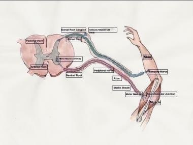
1. Somatic efferent neurons: These neurons transmit signals to skeletal muscles, enabling voluntary movements and posture maintenance. They have their cell bodies located in the ventral horn of the spinal cord and send their axons through the ventral roots to innervate specific muscle fibers.
2. Autonomic efferent neurons: These neurons are responsible for controlling involuntary functions, such as heart rate, digestion, respiration, and pupil dilation. They have a two-neuron chain arrangement, with the preganglionic neuron having its cell body in the CNS (brainstem or spinal cord) and synapsing with the postganglionic neuron in an autonomic ganglion near the effector organ. Autonomic efferent neurons can be further divided into sympathetic, parasympathetic, and enteric subdivisions based on their functions and innervation patterns.
In summary, efferent neurons are a critical component of the nervous system, responsible for transmitting signals from the CNS to various effector organs, ultimately controlling and coordinating numerous bodily functions and responses.
Vasoactive Intestinal Peptide (VIP) is a 28-amino acid polypeptide hormone that has potent vasodilatory, secretory, and neurotransmitter effects. It is widely distributed throughout the body, including in the gastrointestinal tract, where it is synthesized and released by nerve cells (neurons) in the intestinal mucosa. VIP plays a crucial role in regulating various physiological functions such as intestinal secretion, motility, and blood flow. It also has immunomodulatory effects and may play a role in neuroprotection. High levels of VIP are found in the brain, where it acts as a neurotransmitter or neuromodulator and is involved in various cognitive functions such as learning, memory, and social behavior.
A neuroeffector junction is the site where a neuron communicates with an effector cell, such as a muscle fiber or gland. This communication typically occurs through the release of neurotransmitters from the neuron's terminal button, which then bind to receptors on the effector cell and trigger a response. The neuroeffector junction is also sometimes referred to as a synapse or a neuromuscular junction (when it involves a muscle fiber).
The abducens nerve, also known as the sixth cranial nerve, is responsible for controlling the lateral rectus muscle of the eye, which enables the eye to move outward. Abducens nerve diseases refer to conditions that affect this nerve and can result in various symptoms, primarily affecting eye movement.
Here are some medical definitions related to abducens nerve diseases:
1. Abducens Nerve Palsy: A condition characterized by weakness or paralysis of the abducens nerve, causing difficulty in moving the affected eye outward. This results in double vision (diplopia), especially when gazing towards the side of the weakened nerve. Abducens nerve palsy can be congenital, acquired, or caused by various factors such as trauma, tumors, aneurysms, infections, or diseases like diabetes and multiple sclerosis.
2. Sixth Nerve Palsy: Another term for abducens nerve palsy, referring to the weakness or paralysis of the sixth cranial nerve.
3. Internuclear Ophthalmoplegia (INO): A neurological condition affecting eye movement, often caused by a lesion in the medial longitudinal fasciculus (MLF), a bundle of nerve fibers that connects the abducens nucleus with the oculomotor nucleus. INO results in impaired adduction (inward movement) of the eye on the side of the lesion and nystagmus (involuntary eye movements) of the abducting eye on the opposite side when attempting to look towards the side of the lesion.
4. One-and-a-Half Syndrome: A rare neurological condition characterized by a combination of INO and internuclear ophthalmoplegia with horizontal gaze palsy on the same side, caused by damage to both the abducens nerve and the paramedian pontine reticular formation (PPRF). This results in limited or no ability to move the eyes towards the side of the lesion and impaired adduction of the eye on the opposite side.
5. Brainstem Encephalitis: Inflammation of the brainstem, which can affect the abducens nerve and other cranial nerves, leading to various neurological symptoms such as diplopia (double vision), ataxia (loss of balance and coordination), and facial weakness. Brainstem encephalitis can be caused by infectious agents, autoimmune disorders, or paraneoplastic syndromes.
6. Multiple Sclerosis (MS): An autoimmune disorder characterized by inflammation and demyelination of the central nervous system, including the brainstem and optic nerves. MS can cause various neurological symptoms, such as diplopia, nystagmus, and INO, due to damage to the abducens nerve and other cranial nerves.
7. Wernicke's Encephalopathy: A neurological disorder caused by thiamine (vitamin B1) deficiency, often seen in alcoholics or individuals with malnutrition. Wernicke's encephalopathy can affect the brainstem and cause various symptoms such as diplopia, ataxia, confusion, and oculomotor abnormalities.
8. Pontine Glioma: A rare type of brain tumor that arises from the glial cells in the pons (a part of the brainstem). Pontine gliomas can cause various neurological symptoms such as diplopia, facial weakness, and difficulty swallowing due to their location in the brainstem.
9. Brainstem Cavernous Malformation: A benign vascular lesion that arises from the small blood vessels in the brainstem. Brainstem cavernous malformations can cause various neurological symptoms such as diplopia, ataxia, and facial weakness due to their location in the brainstem.
10. Pituitary Adenoma: A benign tumor that arises from the pituitary gland, located at the base of the brain. Large pituitary adenomas can compress the optic nerves and cause various visual symptoms such as diplopia, visual field defects, and decreased vision.
11. Craniopharyngioma: A benign tumor that arises from the remnants of the Rathke's pouch, a structure that gives rise to the anterior pituitary gland. Craniopharyngiomas can cause various neurological and endocrine symptoms such as diplopia, visual field defects, headaches, and hormonal imbalances due to their location near the optic nerves and pituitary gland.
12. Meningioma: A benign tumor that arises from the meninges, the protective covering of the brain and spinal cord. Meningiomas can cause various neurological symptoms such as diplopia, headaches, and seizures depending on their location in the brain or spinal cord.
13. Chordoma: A rare type of malignant tumor that arises from the remnants of the notochord, a structure that gives rise to the spine during embryonic development. Chordomas can cause various neurological and endocrine symptoms such as diplopia, visual field defects, headaches, and hormonal imbalances due to their location near the brainstem and spinal cord.
14. Metastatic Brain Tumors: Malignant tumors that spread from other parts of the body to the brain. Metastatic brain tumors can cause various neurological symptoms such as diplopia, headaches, seizures, and cognitive impairment depending on their location in the brain.
15. Other Rare Brain Tumors: There are many other rare types of brain tumors that can cause diplopia or other neurological symptoms, including gliomas, ependymomas, pineal region tumors, and others. These tumors require specialized diagnosis and treatment by neuro-oncologists and neurosurgeons with expertise in these rare conditions.
In summary, diplopia can be caused by various brain tumors, including pituitary adenomas, meningiomas, chordomas, metastatic brain tumors, and other rare types of tumors. It is important to seek medical attention promptly if you experience diplopia or other neurological symptoms, as early diagnosis and treatment can improve outcomes and quality of life.
An action potential is a brief electrical signal that travels along the membrane of a nerve cell (neuron) or muscle cell. It is initiated by a rapid, localized change in the permeability of the cell membrane to specific ions, such as sodium and potassium, resulting in a rapid influx of sodium ions and a subsequent efflux of potassium ions. This ion movement causes a brief reversal of the electrical potential across the membrane, which is known as depolarization. The action potential then propagates along the cell membrane as a wave, allowing the electrical signal to be transmitted over long distances within the body. Action potentials play a crucial role in the communication and functioning of the nervous system and muscle tissue.
Purkinje fibers are specialized cardiac muscle fibers that are located in the subendocardial region of the inner ventricular walls of the heart. They play a crucial role in the electrical conduction system of the heart, transmitting electrical impulses from the bundle branches to the ventricular myocardium, which enables the coordinated contraction of the ventricles during each heartbeat.
These fibers have a unique structure that allows for rapid and efficient conduction of electrical signals. They are larger in diameter than regular cardiac muscle fibers, have fewer branching points, and possess more numerous mitochondria and a richer blood supply. These features enable Purkinje fibers to conduct electrical impulses at faster speeds, ensuring that the ventricles contract simultaneously and forcefully, promoting efficient pumping of blood throughout the body.
Dimethylphenylpiperazinium iodide is not a medical term or a medication commonly used in clinical practice. It's a chemical compound with the formula (C12H18N2)I, where dimethylphenylpiperazinium is the cation and iodide is the anion.
The dimethylphenylpiperazinium portion of the molecule consists of a phenyl ring with two methyl groups attached to it and a piperazine ring, which contains two nitrogen atoms. This compound may be used in research settings for various purposes, including as a reagent or an intermediate in chemical synthesis.
As this compound is not a medication, there is no medical definition associated with it. If you have any questions about its use or potential applications, please consult a relevant professional such as a chemist or pharmacologist.
Tyramine is not a medical condition but a naturally occurring compound called a biogenic amine, which is formed from the amino acid tyrosine during the fermentation or decay of certain foods. Medically, tyramine is significant because it can interact with certain medications, particularly monoamine oxidase inhibitors (MAOIs), used to treat depression and other conditions.
The interaction between tyramine and MAOIs can lead to a hypertensive crisis, a rapid and severe increase in blood pressure, which can be life-threatening if not treated promptly. Therefore, individuals taking MAOIs are often advised to follow a low-tyramine diet, avoiding foods high in tyramine, such as aged cheeses, cured meats, fermented foods, and some types of beer and wine.
An axon is a long, slender extension of a neuron (a type of nerve cell) that conducts electrical impulses (nerve impulses) away from the cell body to target cells, such as other neurons or muscle cells. Axons can vary in length from a few micrometers to over a meter long and are typically surrounded by a myelin sheath, which helps to insulate and protect the axon and allows for faster transmission of nerve impulses.
Axons play a critical role in the functioning of the nervous system, as they provide the means by which neurons communicate with one another and with other cells in the body. Damage to axons can result in serious neurological problems, such as those seen in spinal cord injuries or neurodegenerative diseases like multiple sclerosis.
Synaptic transmission is the process by which a neuron communicates with another cell, such as another neuron or a muscle cell, across a junction called a synapse. It involves the release of neurotransmitters from the presynaptic terminal of the neuron, which then cross the synaptic cleft and bind to receptors on the postsynaptic cell, leading to changes in the electrical or chemical properties of the target cell. This process is critical for the transmission of signals within the nervous system and for controlling various physiological functions in the body.
Myelinated nerve fibers are neuronal processes that are surrounded by a myelin sheath, a fatty insulating substance that is produced by Schwann cells in the peripheral nervous system and oligodendrocytes in the central nervous system. This myelin sheath helps to increase the speed of electrical impulse transmission, also known as action potentials, along the nerve fiber. The myelin sheath has gaps called nodes of Ranvier where the electrical impulses can jump from one node to the next, which also contributes to the rapid conduction of signals. Myelinated nerve fibers are typically found in the peripheral nerves and the optic nerve, but not in the central nervous system (CNS) tracts that are located within the brain and spinal cord.
Chlorisondamine is a type of drug called an anticholinergic, which works by blocking the action of a neurotransmitter called acetylcholine in the body. It is a type of ganglionic blocker, which means that it blocks the activity of the ganglia, clusters of nerve cells that help transmit signals throughout the nervous system. Chlorisondamine has been used in the past to treat conditions such as hypertension (high blood pressure) and certain types of muscle spasms. However, it is not commonly used today due to the availability of safer and more effective treatment options.
Chlorisondamine is a synthetic compound that was first synthesized in the 1940s. It has a number of effects on the body, including decreasing heart rate and reducing the force of heart contractions. It also causes relaxation of smooth muscle tissue, which can lead to decreased blood pressure and reduced secretions from glands such as the sweat glands and salivary glands.
Like other anticholinergic drugs, chlorisondamine can cause a number of side effects, including dry mouth, blurred vision, constipation, difficulty urinating, and dizziness. It can also cause more serious side effects such as rapid heartbeat, confusion, hallucinations, and seizures. Chlorisondamine should be used with caution and only under the close supervision of a healthcare professional.
Heart rate is the number of heartbeats per unit of time, often expressed as beats per minute (bpm). It can vary significantly depending on factors such as age, physical fitness, emotions, and overall health status. A resting heart rate between 60-100 bpm is generally considered normal for adults, but athletes and individuals with high levels of physical fitness may have a resting heart rate below 60 bpm due to their enhanced cardiovascular efficiency. Monitoring heart rate can provide valuable insights into an individual's health status, exercise intensity, and response to various treatments or interventions.
Hexamethonium is defined as a ganglionic blocker, which is a type of medication that blocks the activity at the junction between two nerve cells (neurons) called the neurotransmitter receptor site. It is a non-depolarizing neuromuscular blocking agent, which means it works by binding to and inhibiting the action of the nicotinic acetylcholine receptors at the motor endplate, where the nerve meets the muscle.
Hexamethonium was historically used in anesthesia practice as a adjunct to provide muscle relaxation during surgical procedures. However, its use has largely been replaced by other neuromuscular blocking agents that have a faster onset and shorter duration of action. It is still used in research settings to study the autonomic nervous system and for the treatment of hypertensive emergencies in some cases.
It's important to note that the use of Hexamethonium requires careful monitoring and management, as it can have significant effects on cardiovascular function and other body systems.
The submandibular glands are one of the major salivary glands in the human body. They are located beneath the mandible (jawbone) and produce saliva that helps in digestion, lubrication, and protection of the oral cavity. The saliva produced by the submandibular glands contains enzymes like amylase and mucin, which aid in the digestion of carbohydrates and provide moisture to the mouth and throat. Any medical condition or disease that affects the submandibular gland may impact its function and could lead to problems such as dry mouth (xerostomia), swelling, pain, or infection.
Afferent neurons, also known as sensory neurons, are a type of nerve cell that conducts impulses or signals from peripheral receptors towards the central nervous system (CNS), which includes the brain and spinal cord. These neurons are responsible for transmitting sensory information such as touch, temperature, pain, sound, and light to the CNS for processing and interpretation. Afferent neurons have specialized receptor endings that detect changes in the environment and convert them into electrical signals, which are then transmitted to the CNS via synapses with other neurons. Once the signals reach the CNS, they are processed and integrated with other information to produce a response or reaction to the stimulus.
Efferent pathways refer to the neural connections that carry signals from the central nervous system (CNS), which includes the brain and spinal cord, to the peripheral effectors such as muscles and glands. These pathways are responsible for the initiation and control of motor responses, as well as regulating various autonomic functions.
Efferent pathways can be divided into two main types:
1. Somatic efferent pathways: These pathways carry signals from the CNS to the skeletal muscles, enabling voluntary movements and postural control. The final common pathway for somatic motor innervation is the alpha-motor neuron, which synapses directly onto skeletal muscle fibers.
2. Autonomic efferent pathways: These pathways regulate the function of internal organs, smooth muscles, and glands. They are further divided into two subtypes: sympathetic and parasympathetic. The sympathetic system is responsible for the 'fight or flight' response, while the parasympathetic system promotes rest and digestion. Both systems use a two-neuron chain to transmit signals from the CNS to the effector organs. The preganglionic neuron has its cell body in the CNS and synapses with the postganglionic neuron in an autonomic ganglion located near the effector organ. The postganglionic neuron then innervates the target organ or tissue.
In summary, efferent pathways are the neural connections that carry signals from the CNS to peripheral effectors, enabling motor responses and regulating various autonomic functions. They can be divided into somatic and autonomic efferent pathways, with further subdivisions within the autonomic system.
Ergotamine is a type of ergopeptine alkaloid, derived from the ergot fungus (Claviceps purpurea) that parasitizes certain grains, particularly rye. It is a potent vasoconstrictor and has been used medically to prevent migraines and treat cluster headaches, as well as for other uses such as controlling postpartum hemorrhage and reducing symptoms of orthostatic hypotension.
Ergotamine works by binding to serotonin receptors in the brain and causing vasoconstriction of cranial blood vessels, which can help to relieve migraine headaches. However, it can also cause serious side effects such as nausea, vomiting, muscle pain, numbness or tingling in the extremities, and in rare cases, more severe reactions such as ergotism, a condition characterized by vasoconstriction of peripheral blood vessels leading to gangrene.
Ergotamine is usually taken orally, but can also be administered rectally or by inhalation. It is important to follow the dosage instructions carefully and avoid taking excessive amounts, as this can increase the risk of serious side effects. Ergotamine should not be taken during pregnancy or while breastfeeding, and it may interact with other medications, so it is important to inform your healthcare provider of all medications you are taking before starting ergotamine therapy.
The sinoatrial (SA) node, also known as the sinus node, is the primary pacemaker of the heart. It is a small bundle of specialized cardiac conduction tissue located in the upper part of the right atrium, near the entrance of the superior vena cava. The SA node generates electrical impulses that initiate each heartbeat, causing the atria to contract and pump blood into the ventricles. This process is called sinus rhythm.
The SA node's electrical activity is regulated by the autonomic nervous system, which can adjust the heart rate in response to changes in the body's needs, such as during exercise or rest. The SA node's rate of firing determines the heart rate, with a normal resting heart rate ranging from 60 to 100 beats per minute.
If the SA node fails to function properly or its electrical impulses are blocked, other secondary pacemakers in the heart may take over, resulting in abnormal heart rhythms called arrhythmias.
Sweating, also known as perspiration, is the production of sweat by the sweat glands in the skin in response to heat, physical exertion, hormonal changes, or emotional stress. Sweat is a fluid composed mainly of water, with small amounts of sodium chloride, lactate, and urea. It helps regulate body temperature by releasing heat through evaporation on the surface of the skin. Excessive sweating, known as hyperhidrosis, can be a medical condition that may require treatment.
Tetrodotoxin (TTX) is a potent neurotoxin that is primarily found in certain species of pufferfish, blue-ringed octopuses, and other marine animals. It blocks voltage-gated sodium channels in nerve cell membranes, leading to muscle paralysis and potentially respiratory failure. TTX has no known antidote, and medical treatment focuses on supportive care for symptoms. Exposure can occur through ingestion, inhalation, or skin absorption, depending on the route of toxicity.
The phrenic nerve is a motor nerve that originates from the cervical spine (C3-C5) and descends through the neck to reach the diaphragm, which is the primary muscle used for breathing. The main function of the phrenic nerve is to innervate the diaphragm and control its contraction and relaxation, thereby enabling respiration.
Damage or injury to the phrenic nerve can result in paralysis of the diaphragm, leading to difficulty breathing and potentially causing respiratory failure. Certain medical conditions, such as neuromuscular disorders, spinal cord injuries, and tumors, can affect the phrenic nerve and impair its function.
Cholinergic fibers are nerve cell extensions (neurons) that release the neurotransmitter acetylcholine at their synapses, which are the junctions where they transmit signals to other neurons or effector cells such as muscles and glands. These fibers are a part of the cholinergic system, which plays crucial roles in various physiological processes including learning and memory, attention, arousal, sleep, and muscle contraction.
Cholinergic fibers can be found in both the central nervous system (CNS) and the peripheral nervous system (PNS). In the CNS, cholinergic neurons are primarily located in the basal forebrain and brainstem, and their projections innervate various regions of the cerebral cortex, hippocampus, thalamus, and other brain areas. In the PNS, cholinergic fibers are responsible for activating skeletal muscles through neuromuscular junctions, as well as regulating functions in smooth muscles, cardiac muscles, and glands via the autonomic nervous system.
Dysfunction of the cholinergic system has been implicated in several neurological disorders, such as Alzheimer's disease, Parkinson's disease, and myasthenia gravis.
Omega-Conotoxin GVIA is a specific type of conotoxin, a peptide toxin derived from the venom of marine cone snails. This particular variant comes from the Conus geographus species.
Omega-Conotoxins are known for their ability to block N-type voltage-gated calcium channels (VGCCs). In the case of omega-Conotoxin GVIA, it specifically and potently inhibits N-type VGCCs, which play crucial roles in neurotransmitter release and pain signaling. Therefore, it has been extensively studied as a research tool to understand these channels' functions and as a potential lead compound for developing novel therapeutics, particularly for treating chronic pain conditions.
Phenoxybenzamine is an antihypertensive medication that belongs to a class of drugs known as non-selective alpha blockers. It works by blocking both alpha-1 and alpha-2 receptors, which results in the relaxation of smooth muscle tissue in blood vessel walls and other organs. This leads to a decrease in peripheral vascular resistance and a reduction in blood pressure.
Phenoxybenzamine is primarily used for the preoperative management of patients with pheochromocytoma, a rare tumor that produces excessive amounts of catecholamines, such as adrenaline and noradrenaline. By blocking alpha receptors, phenoxybenzamine prevents the hypertensive crisis that can occur during surgery to remove the tumor.
It's important to note that phenoxybenzamine has a long duration of action (up to 14 days) and can cause orthostatic hypotension, tachycardia, and other side effects. Therefore, it should be used with caution and under the close supervision of a healthcare professional.
Mossy fibers in the hippocampus are a type of axon that originates from granule cells located in the dentate gyrus, which is the first part of the hippocampus. These fibers have a distinctive appearance and earn their name from the numerous small branches or "spines" that cover their surface, giving them a bushy or "mossy" appearance.
Mossy fibers form excitatory synapses with pyramidal cells in the CA3 region of the hippocampus, which is involved in memory and spatial navigation. These synapses are unique because they have a high degree of plasticity, meaning that they can change their strength in response to experience or learning. This plasticity is thought to be important for the formation and storage of memories.
Mossy fibers also release neurotransmitters such as glutamate and contribute to the regulation of hippocampal excitability. Dysfunction in mossy fiber function has been implicated in several neurological disorders, including epilepsy and Alzheimer's disease.
Tyrosine 3-Monooxygenase (also known as Tyrosinase or Tyrosine hydroxylase) is an enzyme that plays a crucial role in the synthesis of catecholamines, which are neurotransmitters and hormones in the body. This enzyme catalyzes the conversion of the amino acid L-tyrosine to 3,4-dihydroxyphenylalanine (L-DOPA) by adding a hydroxyl group to the 3rd carbon atom of the tyrosine molecule.
The reaction is as follows:
L-Tyrosine + O2 + pterin (co-factor) -> L-DOPA + pterin (oxidized) + H2O
This enzyme requires molecular oxygen and a co-factor such as tetrahydrobiopterin to carry out the reaction. Tyrosine 3-Monooxygenase is found in various tissues, including the brain and adrenal glands, where it helps regulate the production of catecholamines like dopamine, norepinephrine, and epinephrine. Dysregulation of this enzyme has been implicated in several neurological disorders, such as Parkinson's disease.
Parasympatholytics are a type of medication that blocks the action of the parasympathetic nervous system. The parasympathetic nervous system is responsible for the body's rest and digest response, which includes slowing the heart rate, increasing intestinal and glandular activity, and promoting urination and defecation.
Parasympatholytics work by selectively binding to muscarinic receptors, which are found in various organs throughout the body, including the heart, lungs, and digestive system. By blocking these receptors, parasympatholytics can cause a range of effects, such as an increased heart rate, decreased glandular secretions, and reduced intestinal motility.
Some common examples of parasympatholytics include atropine, scopolamine, and ipratropium. These medications are often used to treat conditions such as bradycardia (slow heart rate), excessive salivation, and gastrointestinal cramping or diarrhea. However, because they can have significant side effects, parasympatholytics are typically used only when necessary and under the close supervision of a healthcare provider.
Muscarinic receptors are a type of G protein-coupled receptor (GPCR) that bind to the neurotransmitter acetylcholine. They are found in various organ systems, including the nervous system, cardiovascular system, and respiratory system. Muscarinic receptors are activated by muscarine, a type of alkaloid found in certain mushrooms, and are classified into five subtypes (M1-M5) based on their pharmacological properties and signaling pathways.
Muscarinic receptors play an essential role in regulating various physiological functions, such as heart rate, smooth muscle contraction, glandular secretion, and cognitive processes. Activation of M1, M3, and M5 muscarinic receptors leads to the activation of phospholipase C (PLC) and the production of inositol trisphosphate (IP3) and diacylglycerol (DAG), which increase intracellular calcium levels and activate protein kinase C (PKC). Activation of M2 and M4 muscarinic receptors inhibits adenylyl cyclase, reducing the production of cAMP and modulating ion channel activity.
In summary, muscarinic receptors are a type of GPCR that binds to acetylcholine and regulates various physiological functions in different organ systems. They are classified into five subtypes based on their pharmacological properties and signaling pathways.
Adrenergic alpha-2 receptor antagonists are a class of medications that block the action of norepinephrine, a neurotransmitter and hormone, at adrenergic alpha-2 receptors. These receptors are found in the central and peripheral nervous system and play a role in regulating various physiological functions such as blood pressure, heart rate, and insulin secretion.
By blocking the action of norepinephrine at these receptors, adrenergic alpha-2 receptor antagonists can increase sympathetic nervous system activity, leading to vasodilation, increased heart rate, and increased insulin secretion. These effects make them useful in the treatment of conditions such as hypotension (low blood pressure), opioid-induced sedation and respiratory depression, and diagnostic procedures that require vasodilation.
Examples of adrenergic alpha-2 receptor antagonists include yohimbine, idazoxan, and atipamezole. It's important to note that these medications can have significant side effects, including hypertension, tachycardia, and agitation, and should be used under the close supervision of a healthcare provider.
In medical terms, the heart is a muscular organ located in the thoracic cavity that functions as a pump to circulate blood throughout the body. It's responsible for delivering oxygen and nutrients to the tissues and removing carbon dioxide and other wastes. The human heart is divided into four chambers: two atria on the top and two ventricles on the bottom. The right side of the heart receives deoxygenated blood from the body and pumps it to the lungs, while the left side receives oxygenated blood from the lungs and pumps it out to the rest of the body. The heart's rhythmic contractions and relaxations are regulated by a complex electrical conduction system.
Orthostatic hypotension is a type of low blood pressure that occurs when you stand up from a sitting or lying position. The drop in blood pressure causes a brief period of lightheadedness or dizziness, and can even cause fainting in some cases. This condition is also known as postural hypotension.
Orthostatic hypotension is caused by a rapid decrease in blood pressure when you stand up, which reduces the amount of blood that reaches your brain. Normally, when you stand up, your body compensates for this by increasing your heart rate and constricting blood vessels to maintain blood pressure. However, if these mechanisms fail or are impaired, orthostatic hypotension can occur.
Orthostatic hypotension is more common in older adults, but it can also affect younger people who have certain medical conditions or take certain medications. Some of the risk factors for orthostatic hypotension include dehydration, prolonged bed rest, pregnancy, diabetes, heart disease, Parkinson's disease, and certain neurological disorders.
If you experience symptoms of orthostatic hypotension, it is important to seek medical attention. Your healthcare provider can perform tests to determine the underlying cause of your symptoms and recommend appropriate treatment options. Treatment may include lifestyle changes, such as increasing fluid intake, avoiding alcohol and caffeine, and gradually changing positions from lying down or sitting to standing up. In some cases, medication may be necessary to manage orthostatic hypotension.
A muscle is a soft tissue in our body that contracts to produce force and motion. It is composed mainly of specialized cells called muscle fibers, which are bound together by connective tissue. There are three types of muscles: skeletal (voluntary), smooth (involuntary), and cardiac. Skeletal muscles attach to bones and help in movement, while smooth muscles are found within the walls of organs and blood vessels, helping with functions like digestion and circulation. Cardiac muscle is the specific type that makes up the heart, allowing it to pump blood throughout the body.
Nerve endings, also known as terminal branches or sensory receptors, are the specialized structures present at the termination point of nerve fibers (axons) that transmit electrical signals to and from the central nervous system (CNS). They primarily function in detecting changes in the external environment or internal body conditions and converting them into electrical impulses.
There are several types of nerve endings, including:
1. Free Nerve Endings: These are unencapsulated nerve endings that respond to various stimuli like temperature, pain, and touch. They are widely distributed throughout the body, especially in the skin, mucous membranes, and visceral organs.
2. Encapsulated Nerve Endings: These are wrapped by specialized connective tissue sheaths, which can modify their sensitivity to specific stimuli. Examples include Pacinian corpuscles (responsible for detecting deep pressure and vibration), Meissner's corpuscles (for light touch), Ruffini endings (for stretch and pressure), and Merkel cells (for sustained touch).
3. Specialised Nerve Endings: These are nerve endings that respond to specific stimuli, such as auditory, visual, olfactory, gustatory, and vestibular information. They include hair cells in the inner ear, photoreceptors in the retina, taste buds in the tongue, and olfactory receptors in the nasal cavity.
Nerve endings play a crucial role in relaying sensory information to the CNS for processing and initiating appropriate responses, such as reflex actions or conscious perception of the environment.
Mecamylamine is a non-competitive antagonist at nicotinic acetylcholine receptors. It is primarily used in the treatment of hypertension (high blood pressure) that is resistant to other medications, although it has been largely replaced by newer drugs with fewer side effects.
Mecamylamine works by blocking the action of acetylcholine, a neurotransmitter that activates nicotinic receptors and plays a role in regulating blood pressure. By blocking these receptors, mecamylamine can help to reduce blood vessel constriction and lower blood pressure.
It is important to note that mecamylamine can have significant side effects, including dry mouth, dizziness, blurred vision, constipation, and difficulty urinating. It may also cause orthostatic hypotension (a sudden drop in blood pressure when standing up), which can increase the risk of falls and fractures in older adults. As a result, mecamylamine is typically used as a last resort in patients with severe hypertension who have not responded to other treatments.
Sympatholytics are a class of drugs that block the action of the sympathetic nervous system, which is the part of the autonomic nervous system responsible for preparing the body for the "fight or flight" response. Sympatholytics achieve this effect by binding to and blocking alpha-adrenergic receptors or beta-adrenergic receptors located in various organs throughout the body, including the heart, blood vessels, lungs, gastrointestinal tract, and urinary system.
Examples of sympatholytic drugs include:
* Alpha blockers (e.g., prazosin, doxazosin)
* Beta blockers (e.g., propranolol, metoprolol)
* Centrally acting sympatholytics (e.g., clonidine, methyldopa)
Sympatholytics are used to treat a variety of medical conditions, including hypertension, angina, heart failure, arrhythmias, and certain neurological disorders. They may also be used to manage symptoms associated with anxiety or withdrawal from alcohol or other substances.
Neuropeptide Y (NPY) is a neurotransmitter and neuropeptide that is widely distributed in the central and peripheral nervous systems. It is a member of the pancreatic polypeptide family, which includes peptide YY and pancreatic polypeptide. NPY plays important roles in various physiological functions such as energy balance, feeding behavior, stress response, anxiety, memory, and cardiovascular regulation. It is involved in the modulation of neurotransmitter release, synaptic plasticity, and neural development. NPY is synthesized from a larger precursor protein called prepro-NPY, which is post-translationally processed to generate the mature NPY peptide. The NPY system has been implicated in various pathological conditions such as obesity, depression, anxiety disorders, hypertension, and drug addiction.
Methacholine compounds are medications that are used as a diagnostic tool to help identify and confirm the presence of airway hyperresponsiveness in patients with respiratory symptoms such as cough, wheeze, or shortness of breath. These compounds act as bronchoconstrictors, causing narrowing of the airways in individuals who have heightened sensitivity and reactivity of their airways, such as those with asthma.
Methacholine is a synthetic derivative of acetylcholine, a neurotransmitter that mediates nerve impulse transmission in the body. When inhaled, methacholine binds to muscarinic receptors on the smooth muscle surrounding the airways, leading to their contraction and narrowing. The degree of bronchoconstriction is then measured to assess the patient's airway responsiveness.
It is important to note that methacholine compounds are not used as therapeutic agents but rather as diagnostic tools in a controlled medical setting under the supervision of healthcare professionals.
I believe there might be a misunderstanding in your question. "Dogs" is not a medical term or condition. It is the common name for a domesticated carnivore of the family Canidae, specifically the genus Canis, which includes wolves, foxes, and other extant and extinct species of mammals. Dogs are often kept as pets and companions, and they have been bred in a wide variety of forms and sizes for different purposes, such as hunting, herding, guarding, assisting police and military forces, and providing companionship and emotional support.
If you meant to ask about a specific medical condition or term related to dogs, please provide more context so I can give you an accurate answer.
I believe there may be some confusion in your question. "Rabbits" is a common name used to refer to the Lagomorpha species, particularly members of the family Leporidae. They are small mammals known for their long ears, strong legs, and quick reproduction.
However, if you're referring to "rabbits" in a medical context, there is a term called "rabbit syndrome," which is a rare movement disorder characterized by repetitive, involuntary movements of the fingers, resembling those of a rabbit chewing. It is also known as "finger-chewing chorea." This condition is usually associated with certain medications, particularly antipsychotics, and typically resolves when the medication is stopped or adjusted.
"Rana catesbeiana" is the scientific name for the American bullfrog, which is not a medical term or concept. It belongs to the animal kingdom, specifically in the order Anura and family Ranidae. The American bullfrog is native to North America and is known for its large size and distinctive loud call.
However, if you are looking for a medical definition, I apologize for any confusion. Please provide more context or specify the term you would like me to define.
Physostigmine is a medication that belongs to a class of drugs called cholinesterase inhibitors. It works by blocking the breakdown of a neurotransmitter called acetylcholine, which is important for communication between nerves and muscles. This results in an increase in acetylcholine levels in the body, improving nerve impulse transmission and helping to restore normal muscle function.
Physostigmine is used in the treatment of certain medical conditions that are caused by a deficiency of acetylcholine, such as myasthenia gravis, which is a neuromuscular disorder characterized by weakness and fatigue of the muscles. It may also be used to reverse the effects of certain medications that block the action of acetylcholine, such as anticholinergics, which are sometimes used in anesthesia or to treat conditions like Parkinson's disease.
It is important to note that physostigmine should only be used under the close supervision of a healthcare provider, as it can have serious side effects if not used properly.
Procaine is a local anesthetic drug that is used to reduce the feeling of pain in a specific area of the body. It works by blocking the nerves from transmitting painful sensations to the brain. Procaine is often used during minor surgical procedures, dental work, or when a patient needs to have a wound cleaned or stitched up. It can also be used as a diagnostic tool to help determine the source of pain.
Procaine is administered via injection directly into the area that requires anesthesia. The effects of procaine are relatively short-lived, typically lasting between 30 minutes and two hours, depending on the dose and the individual's metabolism. Procaine may also cause a brief period of heightened sensory perception or euphoria following injection, known as "procaine rush."
It is important to note that procaine should only be administered by trained medical professionals, as improper use can lead to serious complications such as allergic reactions, respiratory depression, and even death.
The Autonomic Nervous System (ANS) is a part of the peripheral nervous system that operates largely below the level of consciousness and controls visceral functions. It is divided into two main subdivisions: the sympathetic and parasympathetic nervous systems, which generally have opposing effects and maintain homeostasis in the body.
The Sympathetic Nervous System (SNS) prepares the body for stressful or emergency situations, often referred to as the "fight or flight" response. It increases heart rate, blood pressure, respiratory rate, and metabolic rate, while also decreasing digestive activity. This response helps the body respond quickly to perceived threats.
The Parasympathetic Nervous System (PNS), on the other hand, promotes the "rest and digest" state, allowing the body to conserve energy and restore itself after the stress response has subsided. It decreases heart rate, blood pressure, and respiratory rate, while increasing digestive activity and promoting relaxation.
These two systems work together to maintain balance in the body by adjusting various functions based on internal and external demands. Disorders of the Autonomic Nervous System can lead to a variety of symptoms, such as orthostatic hypotension, gastroparesis, and cardiac arrhythmias, among others.
Smooth muscle, also known as involuntary muscle, is a type of muscle that is controlled by the autonomic nervous system and functions without conscious effort. These muscles are found in the walls of hollow organs such as the stomach, intestines, bladder, and blood vessels, as well as in the eyes, skin, and other areas of the body.
Smooth muscle fibers are shorter and narrower than skeletal muscle fibers and do not have striations or sarcomeres, which give skeletal muscle its striped appearance. Smooth muscle is controlled by the autonomic nervous system through the release of neurotransmitters such as acetylcholine and norepinephrine, which bind to receptors on the smooth muscle cells and cause them to contract or relax.
Smooth muscle plays an important role in many physiological processes, including digestion, circulation, respiration, and elimination. It can also contribute to various medical conditions, such as hypertension, gastrointestinal disorders, and genitourinary dysfunction, when it becomes overactive or underactive.
The Peroneal nerve, also known as the common fibular nerve, is a branch of the sciatic nerve that supplies the muscles of the lower leg and provides sensation to the skin on the outer part of the lower leg and the top of the foot. It winds around the neck of the fibula (calf bone) and can be vulnerable to injury in this area, leading to symptoms such as weakness or numbness in the foot and leg.
Pressoreceptors are specialized sensory nerve endings found in the walls of blood vessels, particularly in the carotid sinus and aortic arch. They respond to changes in blood pressure by converting the mechanical stimulus into electrical signals that are transmitted to the brain. This information helps regulate cardiovascular function and maintain blood pressure homeostasis.
Sprague-Dawley rats are a strain of albino laboratory rats that are widely used in scientific research. They were first developed by researchers H.H. Sprague and R.C. Dawley in the early 20th century, and have since become one of the most commonly used rat strains in biomedical research due to their relatively large size, ease of handling, and consistent genetic background.
Sprague-Dawley rats are outbred, which means that they are genetically diverse and do not suffer from the same limitations as inbred strains, which can have reduced fertility and increased susceptibility to certain diseases. They are also characterized by their docile nature and low levels of aggression, making them easier to handle and study than some other rat strains.
These rats are used in a wide variety of research areas, including toxicology, pharmacology, nutrition, cancer, and behavioral studies. Because they are genetically diverse, Sprague-Dawley rats can be used to model a range of human diseases and conditions, making them an important tool in the development of new drugs and therapies.
Neurotransmitter agents are substances that affect the synthesis, storage, release, uptake, degradation, or reuptake of neurotransmitters, which are chemical messengers that transmit signals across a chemical synapse from one neuron to another. These agents can be either agonists, which mimic the action of a neurotransmitter and bind to its receptor, or antagonists, which block the action of a neurotransmitter by binding to its receptor without activating it. They are used in medicine to treat various neurological and psychiatric disorders, such as depression, anxiety, and Parkinson's disease.
The ileum is the third and final segment of the small intestine, located between the jejunum and the cecum (the beginning of the large intestine). It plays a crucial role in nutrient absorption, particularly for vitamin B12 and bile salts. The ileum is characterized by its thin, lined walls and the presence of Peyer's patches, which are part of the immune system and help surveil for pathogens.
Membrane potential is the electrical potential difference across a cell membrane, typically for excitable cells such as nerve and muscle cells. It is the difference in electric charge between the inside and outside of a cell, created by the selective permeability of the cell membrane to different ions. The resting membrane potential of a typical animal cell is around -70 mV, with the interior being negative relative to the exterior. This potential is generated and maintained by the active transport of ions across the membrane, primarily through the action of the sodium-potassium pump. Membrane potentials play a crucial role in many physiological processes, including the transmission of nerve impulses and the contraction of muscle cells.
Electrophysiology is a branch of medicine that deals with the electrical activities of the body, particularly the heart. In a medical context, electrophysiology studies (EPS) are performed to assess abnormal heart rhythms (arrhythmias) and to evaluate the effectiveness of certain treatments, such as medication or pacemakers.
During an EPS, electrode catheters are inserted into the heart through blood vessels in the groin or neck. These catheters can record the electrical activity of the heart and stimulate it to help identify the source of the arrhythmia. The information gathered during the study can help doctors determine the best course of treatment for each patient.
In addition to cardiac electrophysiology, there are also other subspecialties within electrophysiology, such as neuromuscular electrophysiology, which deals with the electrical activity of the nervous system and muscles.
 Neuroendocrine cell
Postganglionic nerve fibers
Group C nerve fiber
Adrenal medulla
Muscle contraction
Small intensely fluorescent cell
Parotid gland
Ganglion
Muscarinic acetylcholine receptor
Human nose
Electrochemical skin conductance
Autonomic nervous system
Preganglionic nerve fibers
Enteric nervous system
List of MeSH codes (A08)
General visceral efferent fibers
Adie syndrome
Superior cervical ganglion
Sudomotor
Nasal cavity
Thoracic ganglia
Celiac ganglia
Hypogastric nerve
Cervical ganglia
Harlequin syndrome
Parasympathetic nervous system
Lateral grey column
Esophageal plexus
Submandibular ganglion
Axon reflex
Nerve35
- Autonomic nerve fibers lead directly to them from the central nervous system. (wikipedia.org)
- citation needed] Sympathetic nerve fiber impulses stimulate the release of adrenal medullary hormones. (wikipedia.org)
- These substances induce autonomic nerve terminals or vasculature in the deep lamina propria. (wikipedia.org)
- the vagus nerve contains about 75% of all parasympathetic fibers. (msdmanuals.com)
- In general, cutaneous nerve fibers are associated with blood vessels (dual autonomic inneration of arteries) (Fig. 68,90 The fibers occur as a subepidermal plexus. (catalinapawn.com)
- Although ordinarily considered somatic sensory nerves, the cutaneous nerve trunks carry myelinated postganglionic sympathetic fibers. (catalinapawn.com)
- Postganglionic sympathetic nerve fibers irritate sperm cells. (tazkiyahtours.com)
- A sensory branch of the mandibular nerve (CN V3) It passes through the parotid gland en route to the ear, where it innervates skin of the pinna, external auditory canal, and tympanic membrane. (unboundmedicine.com)
- Nerve fibers that release norepinephrine are referred to as adrenergic fibers. (clambaronline.com)
- The adrenal medulla can also be counted to such postganglionic nerve cells, although they release norepinephrine into the blood. (clambaronline.com)
- Special visceral efferent fibers (SVE) are the efferent nerve fibers that provide motor innervation to the muscles of the pharyngeal arches in humans, and the branchial arches in fish. (darkskiesfilm.com)
- Where are visceral nerve fibers found? (darkskiesfilm.com)
- General visceral efferent fibers include preganglionic parasympathetic secretomotor fibers, which innervate lacrimal and seromucous glands in the nasal cavity and palate via the greater superficial petrosal nerve and sublingual and submandibular glands via the chorda tympani nerve. (darkskiesfilm.com)
- Autonomic nervous system, showing splanchnic nerves in middle, and the vagus nerve as 'X' in blue. (cloudfront.net)
- SudoCheck's focus on the small nerve fibers within the autonomic nervous system, and their ability to quickly regenerate. (hopebraincenter.com)
- Nerve fibers which project from parasympathetic ganglia to synapses on target organs. (nih.gov)
- The preganglionic fibers of the parasympathetic system are in the superior salivatory nucleus of the pons and pass through the nervous intermedius of the facial nerve and enter the SPG as a branch of the greater petrosal nerve. (sphenopalatineganglionblocks.com)
- NF145 was expressed in nerve fibers in both myenteric and submucosal plexuses. (wustl.edu)
- Nerve Fibers, Myelinated" is a descriptor in the National Library of Medicine's controlled vocabulary thesaurus, MeSH (Medical Subject Headings) . (wakehealth.edu)
- A class of nerve fibers as defined by their structure, specifically the nerve sheath arrangement. (wakehealth.edu)
- The AXONS of the myelinated nerve fibers are completely encased in a MYELIN SHEATH. (wakehealth.edu)
- Their NEURAL CONDUCTION rates are faster than those of the unmyelinated nerve fibers (NERVE FIBERS, UNMYELINATED). (wakehealth.edu)
- Myelinated nerve fibers are present in somatic and autonomic nerves. (wakehealth.edu)
- This graph shows the total number of publications written about "Nerve Fibers, Myelinated" by people in this website by year, and whether "Nerve Fibers, Myelinated" was a major or minor topic of these publications. (wakehealth.edu)
- Below are the most recent publications written about "Nerve Fibers, Myelinated" by people in Profiles. (wakehealth.edu)
- Autonomic nerve fibers innervate cardiac muscle, smooth muscle, and glands. (blogspot.com)
- If the autonomic nerve fibers to an effector organ are cut, the organ may continue to function, but will lack the capability of adjusting to changing conditions. (blogspot.com)
- If the autonomic nerve fibers to the heart are cut, the heart will continue to beat and pump blood normally, but its ability to increase cardiac output under stress will be seriously limited. (blogspot.com)
- In either case the nerve fibers of the ANS are motor only, and represent the general visceral efferent (GVE) fibers of the cranial and spinal nerves. (blogspot.com)
- The nerve fibers which comprise the sympathetic system originate in the inter-mediolateral horn (lamina VII) of the gray matter in all twelve thoracic and the first two lumbar segments of the spinal cord. (blogspot.com)
- The axons of these GVE fibers travel through the anterior horn and exit the cord in the anterior root before entering the spinal nerve. (blogspot.com)
- While the general somatic efferent (GSE) fibers (alpha and gamma motor neurons of the anterior horn) continue in the spinal nerve trunks to innervate skeletal muscle fibers and muscle spindles, almost all of the GVE fibers leave the spinal nerve trunks to enter sympathetic ganglia via a thin arm, the white ramus (Figs-1, 2, and 3). (blogspot.com)
- Some of the fibers from nerve cells within the ganglia return to the spinal nerve trunk via a gray ramus. (blogspot.com)
- Vascular bed and nerve vessels in the skin biopsy: Beyond intraepidermal nerve fibers. (nih.gov)
- The parasympathetic fibers travel from the pre-ganglionic neurons in the medulla (nucleus ambiguus and dorsal motor nucleus of the vagus) with the vagus nerve to supply the heart. (radcliffecardiology.com)
Neurotransmitter6
- The neurotransmitter for preganglionic sympathetic and parasympathetic nervous system (PNS) as well as postganglionic parasympathetic nervous system is acetylcholine (ACh). (medscape.com)
- The neurotransmitter for the postganglionic sympathetic nervous system (innervating sweat glands) is also acetylcholine, whereas that for the remaining postganglionic sympathetic nervous system is norepinephrine (NE). (medscape.com)
- At most of the other targets of the autonomic system, the effector response is based on which neurotransmitter is released and what receptor is present. (vumc.org)
- Precursor of epinephrine that is secreted by the adrenal medulla and is a widespread central and autonomic neurotransmitter. (uchicago.edu)
- A third subsystem of neurons has been named as non-noradrenergic, non-cholinergic transmitters (because they use nitric oxide as a neurotransmitter ) and are integral in autonomic function, in particular in the gut and the lungs . (cloudfront.net)
- Both the preganglionic and postganglionic neurons release the neurotransmitter acetylcholine . (osmosis.org)
Acetylcholine4
- Fibers that secrete acetylcholine (cholinergic fibers) include all preganglionic fibers, all postganglionic parasympathetic fibers, and some postganglionic sympathetic fibers (those that innervate piloerectors, sweat glands, and blood vessels). (msdmanuals.com)
- Parasympathetic postganglionic fibers use acetylcholine as transmitter. (nih.gov)
- Acetylcholine released from preganglionic fibers acts on nicotinic receptors on the postganglionic neurons. (osmosis.org)
- And acetylcholine released from postganglionic neurons acts on muscarinic and nicotinic receptors on target organs. (osmosis.org)
Ganglia3
- Parasympathetic ganglia (eg, ciliary, sphenopalatine, otic, pelvic, and vagal ganglia) are located within the effector organs, and postganglionic fibers are only 1 or 2 mm long. (msdmanuals.com)
- Parasympathetic neurons in the central nervous system project preganglionic fibers towards parasympathetic ganglia, which are collections of neurons near the organ they are supposed to affect.From there, postganglionic fibers project towards the target cell. (osmosis.org)
- The sympathetic fibers travel from neurons in the intermediolateral columns of the spinal cord at the T1-T4 levels and synapse in the (stellate) cervical ganglia, and from here the post-ganglionic sympathetic neurons reach the heart. (radcliffecardiology.com)
Norepinephrine10
- Fibers that secrete norepinephrine (adrenergic fibers) include most postganglionic sympathetic fibers. (msdmanuals.com)
- The postganglionic fiber then projects to the iris, where it releases norepinephrine onto the radial fibers of the iris a smooth muscle. (vumc.org)
- characterization of substances, such as epinephrine and norepinephrine, that exert their effect in the sympathetic autonomic nervous system and agents that are chemically similar to these substances. (ashp.org)
- Norepinephrine is the principal transmitter of most postganglionic sympathetic fibers and of the diffuse projection system in the brain arising from the locus ceruleus. (uchicago.edu)
- What fibers secretes norepinephrine? (clambaronline.com)
- Most sympathetic postganglionic fibers release norepinephrine. (clambaronline.com)
- When norepinephrine is released from postganglionic neurons? (clambaronline.com)
- Norepinephrine gets released by postganglionic neurons of the sympathetic nervous system, which binds to and activates adrenergic receptors. (clambaronline.com)
- Some sympathetic preganglionic fibers stimulate the adrenal medulla to release norepinephrine and epinephrine into the blood, producing a "surge of adrenaline. (clambaronline.com)
- Norepinephrine is also released from postganglionic neurons of the sympathetic nervous system, to transmit the fight-or-flight response in each tissue respectively. (clambaronline.com)
Nerves9
- He also shows a plasticized human section of autonomic nerves. (autonomiceducation.com)
- So, this is the Tootsie Roll Pop analogy and as you'll see the autonomic nerves, or nerves that are a part of the autonomic nervous system, come from the brainstem and the thoracolumbar spinal cord and the sacral spinal cord, not really the cervical spinal cord. (autonomiceducation.com)
- Now I want to say right off the bat that the autonomic nervous - it's a terrible phrase - because it implies some sort of autonomy between the autonomic nervous system - I have not shown you where the autonomic nerves are yet - but the autonomic nervous system and the central nervous system, which as you just saw doesn't involve the autonomic nervous system. (autonomiceducation.com)
- So, that's where that the autonomic nerves are. (autonomiceducation.com)
- The somatic and visceral afferent fibers of the oropharynx are supplied by a plexus derived from the vagus, facial, and glossopharyngeal nerves. (darkskiesfilm.com)
- The following cranial nerves carry general visceral efferent fibers: 1. (darkskiesfilm.com)
- The QSART measures the autonomic nerves that control sweating. (hopebraincenter.com)
- The pelvis is innervated chiefly by the sacral and coccygeal spinal nerves and by the pelvic part of the autonomic nervous system. (dartmouth.edu)
- Consequently, all 31 pairs of spinal nerves are in contact with the sympathetic chain and carry fibers of the sympathetic system. (blogspot.com)
Peripheral14
- Because the autonomic nervous system maintains internal physiologic homeostasis, disorders of this system can be present with both central as well as peripheral nervous system localization. (medscape.com)
- Most peripheral neuropathies affect all fiber sizes. (medscape.com)
- Few peripheral neuropathies are associated with pure or predominantly small fiber involvement. (medscape.com)
- Electromyography (EMG) plays a key role in the evaluation of most peripheral neuropathies and helps in assessing only large myelinated fibers. (medscape.com)
- The autonomic nervous system (ANS) is a subcomponent of the peripheral nervous system (PNS) that regulates involuntary physiologic processes, including blood pressure, heart rate, respiration, digestion, and sexual arousal. (tomwademd.net)
- Motor examination is also important because a somatic peripheral neuropathy is frequently associated with an autonomic neuropathy. (symptoma.mt)
- I divide the peripheral nervous system into two parts: the somatic nervous system, which is responsible for interactions between the organism and the outside world especially by way of skeletal muscle, and the autonomic nervous system, which I think of as the automatic part of the nervous system, that's involved with regulation of the inner world inside the body by smooth muscle and glands. (autonomiceducation.com)
- Those are then subdivided into other areas and are also linked to autonomic subsystems and the peripheral nervous system. (cloudfront.net)
- Results suggest that the peripheral autonomic nervous system plays a role in painful diabetic neuropathy. (hopebraincenter.com)
- The sympathetic division is part of the autonomic nervous system of the peripheral nervous system. (flashnews.net)
- It is the largest peripheral parasympathetic ganglion with manifold connections to general sensory fibers and the internal carotid plexus [5-7]. (sphenopalatineganglionblocks.com)
- The peripheral nervous system can be divided into the somatic nervous system , which controls voluntary movement of our skeletal muscles , and the autonomic nervous system, which controls the involuntary activity of the smooth muscles and glands of our organs, and is further divided into the sympathetic and the parasympathetic nervous systems . (osmosis.org)
- Distal postganglionic sudomotor failure was observed, and impairment of the BP responses to the Valsalva maneuver (phase II) suggested peripheral adrenergic dysfunction. (elsevierpure.com)
- It is possible to identify a subset of POTS patients who have a length-dependent autonomic neuropathy, affecting the peripheral adrenergic and cardiovagal fibers, with relative preservation of cardiac adrenergic fibers. (elsevierpure.com)
Divisions of the Autonomic Nervou1
- what are the two divisions of the autonomic nervous system? (flashnews.net)
Dysfunction13
- The etiology of autonomic dysfunction can be primary or idiopathic and secondary causes. (medscape.com)
- In addition, autonomic dysfunction is associated with various medications. (medscape.com)
- In this post, I link to and excerpt from Autonomic Dysfunction by StatPearls . (tomwademd.net)
- A myriad of the factors can cause autonomic dysfunction, and more than one can concur even in the same patient. (tomwademd.net)
- Medications for erectile dysfunction such as Viagra or Cialis help improve blood flow problems caused by Sympathetic Autonomic damage. (tazkiyahtours.com)
- Autonomic neuropathy is a symptom complex associated with the dysfunction of the autonomic nervous system that is responsible for the control of everyday body functions including blood pressure , heart rate, sweating , bowel and bladder function. (symptoma.mt)
- Furthermore, autonomic dysfunction contributes to delayed gastric emptying, putting patients at risk for aspiration of gastric contents. (symptoma.mt)
- Results correlated significantly with clinical neuropathy scores, pain scores and measures of autonomic dysfunction. (hopebraincenter.com)
- Coronary artery disease, heart failure, cardiac autonomic dysfunction, heart failure, sudden death and hypertension can be seen in Parkinsons disease. (parkinsonsinfoclub.com)
- There are additional examinations aimed to study the autonomic dysfunction in various organs, that are specific of the relevant medical specialties. (journalofneuropsychiatry.cl)
- A joint evaluation of clinical findings and autonomic functional tests allow to determine the anatomic level and the severity degree of the autonomic dysfunction with a physiopathologic base. (journalofneuropsychiatry.cl)
- Study of patients with autonomic dysfunction must be made by an expert physician who must also assess clinical/lab findings (3) . (journalofneuropsychiatry.cl)
- A.- Symptoms and signs of the cardiovascular autonomic dysfunction. (journalofneuropsychiatry.cl)
Axons2
- General visceral efferent fibers carry parasympathetic autonomic axons. (darkskiesfilm.com)
- The majority of these fibers were identified as sympathetic postganglionic axons based on their disappearance in organotypic culture and on their expression of tyrosine hydroxylase. (wustl.edu)
Hereditary sensory-autonomic neuropathy1
- In addition to the acquired causes, inherited disorders like hereditary sensory-autonomic neuropathy (HSAN), familial amyloid polyneuropathy (FAP), Tangier disease, and Fabry disease also exist. (medscape.com)
Neuron3
- the preganglionic neuron must first synapse onto a postganglionic neuron before innervating the target organ. (cloudfront.net)
- The preganglionic, or first, neuron will begin at the 'outflow' and will synapse at the postganglionic, or second, neuron's cell body. (cloudfront.net)
- The postganglionic neuron will then synapse at the target organ. (cloudfront.net)
Anatomy2
- The goal for this article remains focused at step III on the anatomy of the autonomic nervous system, as follows. (medscape.com)
- Anatomy: Because the sphenopalatine ganglion (SPG) has diffuse and extensive anatomical connections within the trigemino-autonomic (parasympathetic) reflex, it is of great interest to clinicians who treat pain conditions [1]. (sphenopalatineganglionblocks.com)
Adrenergic3
- What is secreted by adrenergic fibers? (clambaronline.com)
- Autonomic failure was quantified by its distribution (cardiovagal, adrenergic and sudomotor) and severity, a symptom profile was generated, and spectral indices, based on modified Wigner distribution during rest and head-up tilt (80°), were evaluated. (elsevierpure.com)
- The autonomic nervous system (ANS) is made up of the sympathetic nervous system (SNS) (noradrenergic, adrenergic and cholinergic), the parasympathetic nervous system (PNS) cholinergic and the enteric nervous system. (journalofneuropsychiatry.cl)
Adrenal Medulla2
- Adrenal medulla): receive preganglionic fibers from Sympathetic. (easynotecards.com)
- Cognitive Psychology of Memory On receiving distress signals from amygdala, the hypothalamus signals the adrenal medulla through the autonomic system. (clambaronline.com)
Ganglion1
- The SPG is a large (THE LARGEST PARASYMPATHETIC GANGLION OF THE HEAD) extra cranial parasympathetic ganglion with multiple neural roots, including autonomic, sensory, and motor [2,3]. (sphenopalatineganglionblocks.com)
Cardiac autonomic neuropathy2
- Training Effects The presence of cardiac autonomic neuropathy (CAN) impairs exercise tolerance and lowers maximal heart rate 4. (symptoma.mt)
- In 15 Minutes Identify Patients At Risk for Sudden Death, Silent Heart Attack, Hypertension Cardiac Autonomic Neuropathy, Diabetic Autonomic Neuropathy, Vascular Abnormalities, Orthostatic Hypotension, Syncope and other Hidden Diseases Testing of the Autonomic Nervous System. (hopebraincenter.com)
Organs4
- The general visceral afferent (GVA) fibers conduct sensory impulses (usually pain or reflex sensations) from the internal organs, glands, and blood vessels to the central nervous system. (darkskiesfilm.com)
- The autonomic nervous system ( ANS ), formerly referred to as the vegetative nervous system , is a division of the nervous system that operates internal organs , smooth muscle and glands. (cloudfront.net)
- The autonomic nervous system is regulated by integrated reflexes through the brainstem to the spinal cord and organs . (cloudfront.net)
- The autonomic nervous system regulates many of the internal organs through a balance of two aspects, or divisions. (openstax.org)
Efferent fibers2
- The sympathetic nervous system (SNS), as well as the parasympathetic nervous system (PNS), contain afferent fibers that provide sensory input and efferent fibers that provide motor output to the central nervous system (CNS). (tomwademd.net)
- What is the general visceral efferent fibers? (darkskiesfilm.com)
Cause autonomic1
- Disorders of the autonomic nervous system cause autonomic insufficiency or failure and can affect any system of the body. (msdmanuals.com)
Sympathetic Autonomic3
- the endocrine gland situated on top of each kidney, which produces and releases epinephrine (also known as adrenaline) to stimulate functions of the sympathetic autonomic nervous system (SANS). (ashp.org)
- sometimes used to designate actions and responses to the sympathetic autonomic nervous system. (ashp.org)
- Define the autonomic nervous system (ANS) and its divisions, the parasympathetic and sympathetic autonomic nervous systems (PANS and SANS, respectively). (ashp.org)
Difference Between Somatic1
- What is the difference between somatic afferent fibers and visceral afferent fibers? (darkskiesfilm.com)
Afferent fibers2
- What are general somatic afferent fibers? (darkskiesfilm.com)
- Although general visceral afferent fibers are part of the ANS, they are not classified as part of the sympathetic or parasympathetic system. (darkskiesfilm.com)
Disorders2
- Given the complex nature of this system, a stepwise approach to autonomic disorders is required for proper understanding. (medscape.com)
- The FDA cleared equipment is used by physicians to help identify diseases that may be associated with Autonomic Nervous System, Vascular, and Sudomotor disorders. (hopebraincenter.com)
Afferents3
- Small myelinated fibers transmit preganglionic autonomic efferents (B fibers) and somatic afferents (A delta fibers). (medscape.com)
- Unmyelinated (C) fibers transmit postganglionic autonomic efferents as well as somatic and autonomic afferents. (medscape.com)
- Postganglionic parasympathetic, sympathetic neurons, and the somatic sensory afferents can all be blocked by an SPG block. (sphenopalatineganglionblocks.com)
Innervation2
- The motor innervation of the skin is attributable to sympathetic fibers of the autonomic nervous system. (catalinapawn.com)
- GVE fibers provide motor (parasympathetic) innervation to the viscera. (darkskiesfilm.com)
Regulates3
- The autonomic nervous system regulates physiologic processes. (msdmanuals.com)
- [1] The autonomic nervous system is a control system that acts largely unconsciously and regulates bodily functions, such as the heart rate , its force of contraction, digestion , respiratory rate , pupillary response , urination , and sexual arousal . (cloudfront.net)
- The autonomic nervous system receives its sensory input from the baroreceptors located in the major vessels, and this forms part of the arterial baroreflex feedback mechanism, which ultimately regulates the heart rate and blood pressure. (radcliffecardiology.com)
Involuntary3
- The autonomic nervous system (ANS) includes all regions implicated in controlling "autonomic," unconscious, and involuntary functions in total body homeostasis. (tomwademd.net)
- The autonomic nervous system is the visceral motor system and controls involuntary nervous movements. (flashnews.net)
- The somatic nervous system is associated with voluntary responses (though many can happen without conscious awareness, like breathing), and the autonomic nervous system is associated with involuntary responses, such as those related to homeostasis. (openstax.org)
Reflex3
- Autonomic functions include control of respiration , cardiac regulation (the cardiac control center), vasomotor activity (the vasomotor center ), and certain reflex actions such as coughing , sneezing , swallowing and vomiting . (cloudfront.net)
- Quantitative Sudomotor Axon Reflex Test (QSART) evaluate the integrity of the postganglionic sudomotor system along the axon reflex to define the distribution of sweat loss. (hopebraincenter.com)
- It contains motor, sensory (pain and reflex), and postganglionic sympathetic fibers, and it may be "blocked" medial to the ischial tuberosity, e.g., during parturition. (dartmouth.edu)
Neurotransmitters1
- The postganglionic fibers of either division release neurotransmitters onto the smooth muscles of the iris to cause changes in the pupillary size. (vumc.org)
Homeostasis1
- The autonomic nervous system (ANS) is a very complex, multifaceted neural network that maintains internal physiologic homeostasis. (medscape.com)
Sensory fibers1
- Where are the cell bodies of somatic and visceral afferent sensory fibers located? (darkskiesfilm.com)
Neuropathy4
- Painful burning feet is caused by a sensory neuropathy with small fiber involvement in more than 90% of cases. (medscape.com)
- Elderly patients who lack sural sensory responses can still be diagnosed with small fiber neuropathy. (medscape.com)
- Evaluate patients with Diabetes Mellitus, as an instrument to assess autonomic neuropathy and neuropathic pain. (hopebraincenter.com)
- Our preliminary work suggested that using autonomic function testing in conjunction with time-frequency mapping (TFM), it might be possible to characterize a subset of the postural tachycardia syndrome (POTS), that is due to a restricted autonomic neuropathy. (elsevierpure.com)
Organ2
- The sympathetic division of the autonomic nervous system influences the various organ systems of the body through connections emerging from the thoracic and upper lumbar spinal cord. (openstax.org)
- A muscle or gland innervated by autonomic fibers is called an effector organ. (blogspot.com)
Sweat1
- As sweat glands are innervated by small c fibers, which are also involved in pain pathways, precise measuring of the sweat gland function can identify that cause. (hopebraincenter.com)
Synapse1
- It is denoted as parasympathetic because preganglionic parasympathetic fibers synapse within the SPG. (sphenopalatineganglionblocks.com)
Reflexes1
- Through these fibers the ANS plays a role in regulating (1) blood pressure and flow, (2) gastrointestinal movements and secretions, (3) body temperature, (4) bronchial dilation, (5) blood glucose levels, (6) metabolism, (7) micturition and defecation, (8) pupillary light and accommodation reflexes, and (9) glandular secretions, just to name a few. (blogspot.com)
Brainstem1
- The reticular formation is a group of fibers that carry stimulation related to sleep and arousal through the brainstem. (flashnews.net)
Nervous system receives1
- The autonomic nervous system receives input from parts of the central nervous system (CNS) that process and integrate stimuli from the body and external environment. (msdmanuals.com)
Somatic nervou1
- The nervous system can be divided into two functional parts: the somatic nervous system and the autonomic nervous system. (openstax.org)