Stethoscopes
Heart Murmurs
Heart Sounds
Electronics, Medical
Multimedia
Phonocardiography
Respiratory Sounds
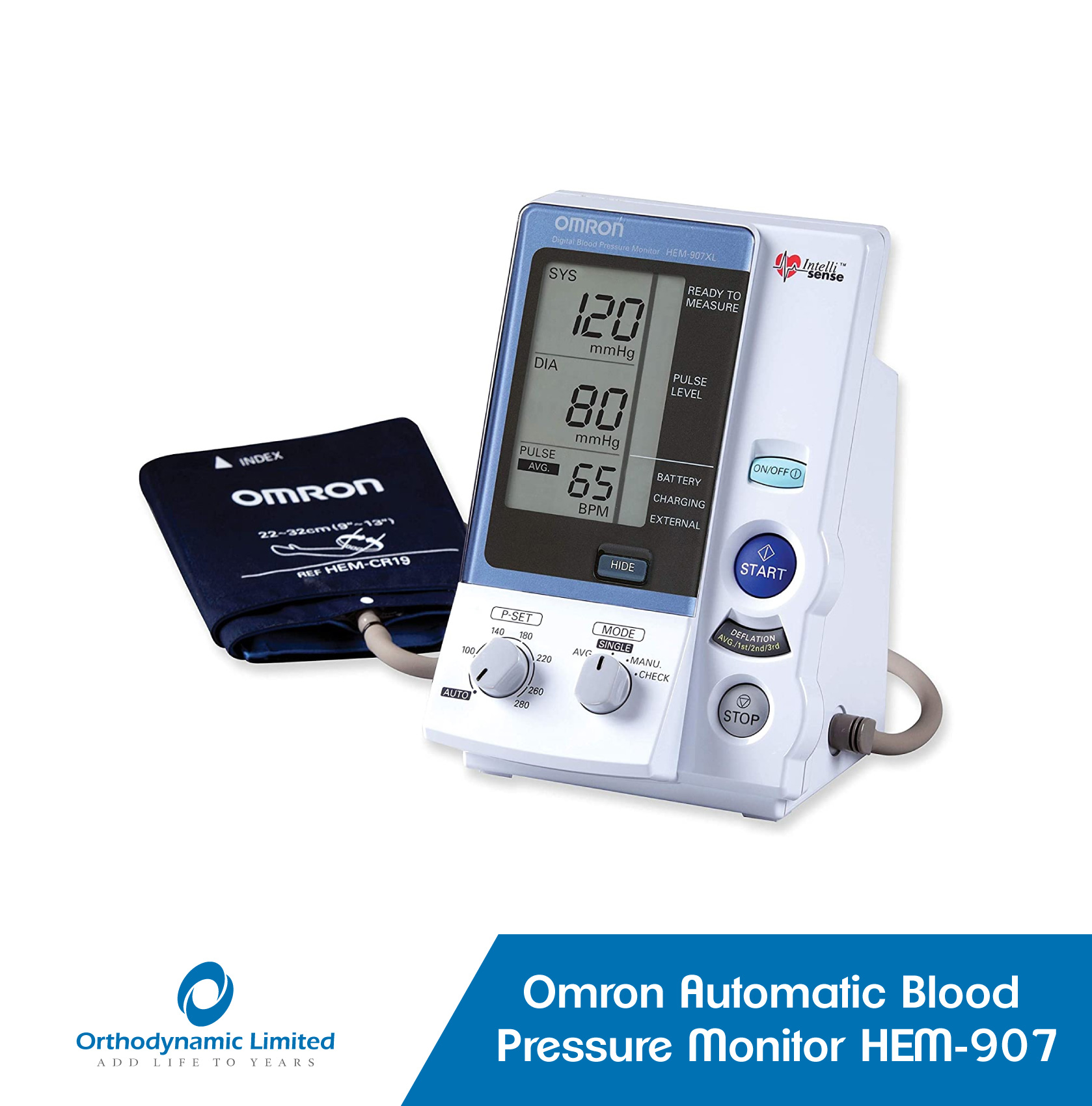
CD-ROM
Insufflation
Gastric Dilatation
Computer-Assisted Instruction
Cardiotocography
Clinical Competence
Fetal Monitoring
Physical Examination
Students, Medical
Asbestosis
Observer Variation
Illinois
Microcomputers
Point-of-Care Systems
Urologic Surgical Procedures
Radiography, Thoracic
Education, Medical, Undergraduate
Heart Diseases
Percussion
Tachycardia, Sinus
Ventricular Premature Complexes
Bruits, ophthalmodynamometry and rectilinear scanning on transient ischemic attacks. (1/162)
One hundred seventeen patients with clinical signs and symptoms of transient ischemic attacks (TIAs) were evaluated. All underwent clinical evaluation for bruit, ophthalmodynamometry, rapid sequence scintiphotography with rectilinear scanning and four-vessel cerebral angiography. The results of these tests were compared for reliability in predicting location of lesions causing transient ischemic attacks. Angiography remains the most accurate procedure in evaluating extracranial vascular lesions. When determination of bruits, ophthalmodynamometry and brain scanning are done together, accuracy is greater than when any one of the procedures is done alone. (+info)Comparison of four methods for assessing airway sealing pressure with the laryngeal mask airway in adult patients. (2/162)
We have compared four tests for assessing airway sealing pressure with the laryngeal mask airway (LMA) to test the hypothesis that airway sealing pressure and inter-observer reliability differ between tests. We studied 80 paralysed, anaesthetized adult patients. Four different airway sealing pressure tests were performed in random order on each patient by two observers blinded to each other's measurements: test 1 involved detection of an audible noise; test 2 was detection of end-tidal carbon dioxide in the oral cavity; test 3 was observation of the aneroid manometer dial as the pressure increased to note the airway pressure at which the dial reached stability; and test 4 was detection of an audible noise by neck auscultation. Mean airway sealing pressure ranged from 19.5 to 21.3 cm H2O and intra-class correlation coefficient was 0.95-0.99. Inter-observer reliability of all tests was classed as excellent. The manometric stability test had a higher mean airway sealing pressure (P < 0.0001) and better inter-observer reliability (P < 0.0001) compared with the three other tests. We conclude that for clinical purposes all four tests are excellent, but that the manometric stability test may be more appropriate for researchers comparing airway sealing pressures. (+info)Effect of positioning on recorded lung sound intensities in subjects without pulmonary dysfunction. (3/162)
BACKGROUND AND PURPOSE: Physical therapists often use positioning to assist in the reexpansion of collapsed lung segments. An increase in lung sound intensity on auscultation is considered indicative of lung expansion. This study was designed to examine whether clinical interpretation of auscultatory findings is warranted. SUBJECTS: The subjects (5 male, 6 female) were young physical therapist students without pulmonary dysfunction (mean age=20.4 years, mean height=166.3 cm, mean weight=57.5 kg). Subjects with lung disease were excluded because pulmonary pathology is difficult to standardize. METHODS: Lung sounds electronically recorded over the posterior chest wall of subjects in sitting and side-lying positions were compared. Measures included peak intensity, frequency at maximum power, and median frequency. RESULTS: In the sitting position, inspiratory sounds recorded over the left posterior chest wall were louder than those recorded on the right side. In the side-lying positions, the sound intensity recorded from the dependent chest wall was louder than that recorded from the nondependent chest wall. In side-lying positions, the upper hemithorax is "nondependent," and the side in contact with the bed is "dependent." Sound intensities recorded over both posterior chest walls in the sitting position were louder than those recorded over the same lung area in the nondependent side-lying position. There was no difference in the sound intensity recorded between the sitting and dependent side-lying postures. CONCLUSION AND DISCUSSION: When comparative auscultation of the chest wall is used by physical therapists to assess the adequacy of pulmonary ventilation, patient posture and regional differences in breath sound intensity can influence clinical interpretation. (+info)Heart murmurs in pediatric patients: when do you refer? (4/162)
Many normal children have heart murmurs, but most children do not have heart disease. An appropriate history and a properly conducted physical examination can identify children at increased risk for significant heart disease. Pathologic causes of systolic murmurs include atrial and ventricular septal defects, pulmonary or aortic outflow tract abnormalities, and patent ductus arteriosus. An atrial septal defect is often confused with a functional murmur, but the conditions can usually be differentiated based on specific physical findings. Characteristics of pathologic murmurs include a sound level of grade 3 or louder, a diastolic murmur or an increase in intensity when the patient is standing. Most children with any of these findings should be referred to a pediatric cardiologist. (+info)Methacholine challenge in preschool children: methacholine-induced wheeze versus transcutaneous oximetry. (5/162)
Tracheal/chest auscultation for wheeze and transcutaneous oximetry have both been suggested as measures of outcome in bronchial provocation tests in young children. This study aimed to compare the sensitivity and safety of these two techniques as end-points for methacholine challenge in children aged <4 yrs. Seventy-two methacholine challenges were performed in 39 children aged <4 yrs with recurrent wheeze. Arterial oxygen saturation (Sa,O2) and transcutaneous oxygen pressure tcPO2 continuously, and the test was terminated when wheeze was heard or at Sa,O2 <91%. tcPO2 was not used as an end-point. Wheeze or desaturation occurred at < or =8 mg x mL(-1) methacholine in every test. One child had transient clinical cyanosis, but no other ill-effects were seen. Fifty-six tests (78%) were terminated for wheeze, seven (10%) for fall in Sa,O2 and nine (12%) showed simultaneous responses in both parameters. Twenty-eight tests (39%) contained a fall in tcPO2 >3 kPa but six of these also showed a significant rise. Fifty-three tests (75%) contained a fall in tcPO2 >15%, but 20 of these also showed a significant rise. Tracheal/chest auscultation with Sa,O2 monitoring is a sensitive and relatively safe end-point for bronchial challenges in preschool children. The erratic pattern of transcutaneous oxygen pressure response in some children casts doubt on its reliability as a proxy measure of bronchial obstruction. (+info)Acoustic monitoring of intraoperative neuromuscular block. (6/162)
Standard methods for accurate intraoperative measurement of neuromuscular block are either expensive or inconvenient and are not used widely. We have evaluated a new method of monitoring neuromuscular block using a low-frequency microphone. The method is based on the phenomenon of low-frequency sound emission by contracting skeletal muscle. Acoustic monitoring (MIC) with an air-coupled microphone was used to evaluate intraoperative neuromuscular block in 25 anaesthetized patients. The MIC recorded the response of the adductor pollicis muscle to supramaximal electrical stimulation of the ulnar nerve with train-of-four stimuli. The ratios of the first response (TI) to control (TC) were used for evaluation. Data obtained from the MIC were compared with simultaneous recordings, from the same hand, of mechanomyography (FDT), electromyography (EMG) and accelerography (ACC). Throughout the operative procedure, TI/TC ratios of the acoustic method correlated with the three reference devices: FDT, 12 patients, 262 data sets, r = 0.86, bias (%MIC-%FDT) = mean -5.3 (SD 19.6)%; EMG, 18 patients, 490 data sets, r = 0.85, bias (%MIC-%EMG) = -0.39 (20.29)%; and ACC, 13 patients, 328 data sets, r = 0.91, bias (%MIC-%ACC) = -3.0 (15.6)%. We conclude that monitoring intraoperative neuromuscular block by a microphone which transduces low-frequency muscle sounds is clinically feasible. (+info)A new double cuff sphygmotonometer for accurate blood pressure measurement. (7/162)
Accurate measurement of blood pressure (BP) is essential in the diagnosis and treatment of hypertension, but neither auscultatory nor oscillometric methods measure intra-arterial BP accurately in all circumstances. Algorithms for automatic BP-measuring devices differ from manufacturer to manufacturer, and no clear authorized algorithm criteria have yet been established. We have devised a double-cuff sphygmotonometer to measure BP on the basis of clear algorithms, and investigated the accuracy of this new method by comparing it with the photo-oscillometric method, which is the most accurate method for non-invasive measurement of intra-arterial BP. In the new method, a small cuff (3x6 cm) replaces the photo-sensor in the brachial cuff (13x24 cm) of the photo-oscillometric device, and BP is determined by means of the oscillation within the small cuff. The comparison based on procedures of AAMI-protocol was performed in 136 hypertensive patients and 54 normotensive subjects. The difference in systolic BP between the photooscillometric and double-cuff methods was -2.26+/-2.31 mmHg (89% under 5 mmHg), and the corresponding difference in diastolic BP was 1.9+/-2.50 mmHg (94% under 5 mmHg). In conclusion, we have devised a new double-cuff method which improves on the photo-oscillometric method, and although it seems to be less accurate than the photo-oscillometric method, the clarity of its algorithm makes it superior to the conventional oscillometric and auscultatory methods employing only one cuff. (+info)The clinical evaluation of the Respi-check mask: a new oxygen mask incorporating a breathing indicator. (8/162)
Study objective-To investigate the correlation between the Respi-check sensor and simultaneous chest auscultation in determining the respiratory rates in adults. METHODS: Random visits to a local accident and emergency (A&E) department were made and all patients wearing oxygen masks were recruited into the study. The new sensor was attached to the outside of the mask. One researcher auscultated the chest to count breaths, the other counted the sensor activity. Each was blinded to the activities of the other. Breaths were counted by each researcher simultaneously and independently over one minute. A total of 40 patients were recruited into the study. A difference of more than two breaths/min compared with chest auscultation was deemed as a sensor failure. RESULTS: The respiratory rates of 40 patients were measured. There were 28 men, 12 women. Twenty six patients were wearing an Intersurgical high concentration (flow 12l/min) mask, 14 were wearing an aerosol mask with variable venturi (flow 3-12l/min) by Medicaid. Over one minute rates determined by the two methods were the same in 28 cases (70%). It was accurate to within one breath in 37 cases (93%) and to within two breaths in 39 (98%) cases and in one case (2.5%) the sensor failed. The mean difference (mean of the differences between rates obtained from auscultation and the new sensor) was -0.1282 breaths/min, with limits of agreement (d (2SD) between -1.414 to 1.157 breaths/min. CONCLUSION: The Respi-check sensor provides an accurate method of estimating the respiratory rate in adult patients attending the A&E department. (+info)Auscultation is a medical procedure in which a healthcare professional uses a stethoscope to listen to the internal sounds of the body, such as heart, lung, or abdominal sounds. These sounds can provide important clues about a person's health and help diagnose various medical conditions, such as heart valve problems, lung infections, or digestive issues.
During auscultation, the healthcare professional places the stethoscope on different parts of the body and listens for any abnormal sounds, such as murmurs, rubs, or wheezes. They may also ask the person to perform certain movements, such as breathing deeply or coughing, to help identify any changes in the sounds.
Auscultation is a simple, non-invasive procedure that can provide valuable information about a person's health. It is an essential part of a physical examination and is routinely performed by healthcare professionals during regular checkups and hospital visits.
Heart auscultation is a medical procedure in which a healthcare professional uses a stethoscope to listen to the sounds produced by the heart. The process involves placing the stethoscope on various locations of the chest wall to hear different areas of the heart.
The sounds heard during auscultation are typically related to the opening and closing of the heart valves, as well as the turbulence created by blood flow through the heart chambers. These sounds can provide important clues about the structure and function of the heart, allowing healthcare professionals to diagnose various cardiovascular conditions such as heart murmurs, valvular disorders, and abnormal heart rhythms.
Heart auscultation is a key component of a physical examination and requires proper training and experience to interpret the findings accurately.
A stethoscope is a medical device used for auscultation, or listening to the internal sounds of the body. It is most commonly used to hear the heartbeat, lung sounds, and blood flow in the major arteries. The device consists of a small disc-shaped resonator that is placed against the skin, connected by tubing to two earpieces. Stethoscopes come in different types and designs, but all serve the primary purpose of amplifying and transmitting body sounds to facilitate medical diagnosis.
A heart murmur is an abnormal sound heard during a heartbeat, which is caused by turbulent blood flow through the heart. It is often described as a blowing, whooshing, or rasping noise. Heart murmurs can be innocent (harmless and not associated with any heart disease) or pathological (indicating an underlying heart condition). They are typically detected during routine physical examinations using a stethoscope. The classification of heart murmurs includes systolic, diastolic, continuous, and functional murmurs, based on the timing and auscultatory location. Various heart conditions, such as valvular disorders, congenital heart defects, or infections, can cause pathological heart murmurs. Further evaluation with diagnostic tests like echocardiography is often required to determine the underlying cause and appropriate treatment.
Heart sounds are the noises generated by the beating heart and the movement of blood through it. They are caused by the vibration of the cardiac structures, such as the valves, walls, and blood vessels, during the cardiac cycle.
There are two normal heart sounds, often described as "lub-dub," that can be heard through a stethoscope. The first sound (S1) is caused by the closure of the mitral and tricuspid valves at the beginning of systole, when the ventricles contract to pump blood out to the body and lungs. The second sound (S2) is produced by the closure of the aortic and pulmonary valves at the end of systole, as the ventricles relax and the ventricular pressure decreases, allowing the valves to close.
Abnormal heart sounds, such as murmurs, clicks, or extra sounds (S3 or S4), may indicate cardiac disease or abnormalities in the structure or function of the heart. These sounds can be evaluated through a process called auscultation, which involves listening to the heart with a stethoscope and analyzing the intensity, pitch, quality, and timing of the sounds.
"Medical electronics" refers to the field of electronics that is specifically designed for medical applications. This can include a wide range of devices and systems, such as:
1. Medical imaging equipment, such as X-ray machines, CT scanners, MRI machines, and ultrasound machines.
2. Patient monitoring equipment, such as heart rate monitors, blood pressure monitors, and oxygen saturation monitors.
3. Therapeutic devices, such as pacemakers, defibrillators, and deep brain stimulators.
4. Laboratory equipment, such as DNA sequencers, mass spectrometers, and microarray scanners.
5. Wearable health technology, such as fitness trackers, smartwatches, and continuous glucose monitors.
6. Telemedicine systems that enable remote consultations and patient monitoring.
Medical electronics must meet strict regulatory requirements to ensure safety, effectiveness, and reliability. These devices often require specialized electronic components, such as sensors, signal processing circuits, and power management circuits, that are designed to operate in the challenging environments found in medical settings. Medical electronics engineers must have a deep understanding of both electronics and medical applications to design and develop these complex systems.
I'm sorry for any confusion, but "multimedia" is not a term that has a specific medical definition. Multimedia generally refers to the use of multiple forms of media, such as text, audio, video, graphics, and animation, in a single interactive presentation or platform. It is often used in various fields including education, entertainment, marketing, and some areas of healthcare for purposes like training, patient education, and therapy. However, it does not have a specific medical meaning itself.
Phonocardiography is a non-invasive medical procedure that involves the graphical representation and analysis of sounds produced by the heart. It uses a device called a phonocardiograph to record these sounds, which are then displayed as waveforms on a screen. The procedure is often used in conjunction with other diagnostic techniques, such as electrocardiography (ECG), to help diagnose various heart conditions, including valvular heart disease and heart murmurs.
During the procedure, a specialized microphone called a phonendoscope is placed on the chest wall over the area of the heart. The microphone picks up the sounds generated by the heart's movements, such as the closing and opening of the heart valves, and transmits them to the phonocardiograph. The phonocardiograph then converts these sounds into a visual representation, which can be analyzed for any abnormalities or irregularities in the heart's function.
Phonocardiography is a valuable tool for healthcare professionals, as it can provide important insights into the health and functioning of the heart. By analyzing the waveforms produced during phonocardiography, doctors can identify any potential issues with the heart's valves or other structures, which may require further investigation or treatment. Overall, phonocardiography is an essential component of modern cardiac diagnostics, helping to ensure that patients receive accurate and timely diagnoses for their heart conditions.
Respiratory sounds are the noises produced by the airflow through the respiratory tract during breathing. These sounds can provide valuable information about the health and function of the lungs and airways. They are typically categorized into two main types: normal breath sounds and adventitious (or abnormal) breath sounds.
Normal breath sounds include:
1. Vesicular breath sounds: These are soft, low-pitched sounds heard over most of the lung fields during quiet breathing. They are produced by the movement of air through the alveoli and smaller bronchioles.
2. Bronchovesicular breath sounds: These are medium-pitched, hollow sounds heard over the mainstem bronchi and near the upper sternal border during both inspiration and expiration. They are a combination of vesicular and bronchial breath sounds.
Abnormal or adventitious breath sounds include:
1. Crackles (or rales): These are discontinuous, non-musical sounds that resemble the crackling of paper or bubbling in a fluid-filled container. They can be heard during inspiration and are caused by the sudden opening of collapsed airways or the movement of fluid within the airways.
2. Wheezes: These are continuous, musical sounds resembling a whistle. They are produced by the narrowing or obstruction of the airways, causing turbulent airflow.
3. Rhonchi: These are low-pitched, rumbling, continuous sounds that can be heard during both inspiration and expiration. They are caused by the vibration of secretions or fluids in the larger airways.
4. Stridor: This is a high-pitched, inspiratory sound that resembles a harsh crowing or barking noise. It is usually indicative of upper airway narrowing or obstruction.
The character, location, and duration of respiratory sounds can help healthcare professionals diagnose various respiratory conditions, such as pneumonia, chronic obstructive pulmonary disease (COPD), asthma, and bronchitis.
A CD-ROM (Compact Disc Read-Only Memory) is not a medical term, but a technology term. It refers to a type of optical storage disc that contains digital information and can be read by a computer's CD-ROM drive. The data on a CD-ROM is permanent and cannot be modified or erased, unlike other types of writable discs such as CD-R or CD-RW.
CD-ROMs were commonly used in the past to distribute software, multimedia presentations, reference materials, and educational content. In medical field, CD-ROMs have been used to distribute large databases of medical information, such as clinical guidelines, drug references, and anatomical atlases. However, with the advent of internet and cloud storage technologies, the use of CD-ROMs has become less common in recent years.
Insufflation is a medical term that refers to the act of introducing a gas or vapor into a body cavity or passage, typically through a tube or surgical instrument. This procedure is often used in medical and surgical settings for various purposes, such as:
* To administer anesthesia during surgery (e.g., introducing nitrous oxide or other gases into the lungs)
* To introduce medication or other substances into the body (e.g., insufflating steroids into a joint)
* To perform diagnostic procedures (e.g., insufflating air or a contrast agent into the gastrointestinal tract to visualize it with X-rays)
* To clean out a body cavity (e.g., irrigating and insufflating the bladder during urological procedures).
It's important to note that insufflation should be performed under controlled conditions, as there are potential risks associated with introducing gases or vapors into the body, such as barotrauma (damage caused by changes in pressure) and infection.
Gastric dilatation, also known as stomach dilation or distention, refers to the abnormal enlargement or expansion of the stomach. This condition often occurs when the stomach fills with gas, food, or fluids and is unable to empty properly. Gastric dilatation can be caused by various factors such as overeating, swallowing excessive air, gastroparesis (delayed gastric emptying), intestinal obstruction, or certain medical conditions like hiatal hernia or pregnancy.
In severe cases, gastric dilatation may lead to gastric volvulus, where the stomach twists on itself, cutting off its blood supply and leading to ischemia and necrosis of the stomach tissue. This is a life-threatening condition that requires immediate medical attention. Symptoms of gastric dilatation include abdominal pain, bloating, vomiting, loss of appetite, and difficulty breathing.
Cardiology is a branch of medicine that deals with the diagnosis and treatment of diseases and disorders of the heart and blood vessels. It encompasses the study of the normal functioning of the heart, the investigation and diagnosis of heart disease, and the treatment of various cardiovascular conditions through both surgical and non-surgical interventions. Cardiologists are medical professionals who specialize in this field, providing comprehensive care for patients with conditions such as coronary artery disease, congenital heart defects, valvular heart disease, electrophysiology disorders, and hypertension, among others. They work closely with other healthcare providers to manage cardiovascular risk factors, optimize overall cardiovascular health, and improve patients' quality of life.
Computer-Assisted Instruction (CAI) is a type of educational technology that involves the use of computers to deliver, support, and enhance learning experiences. In a medical context, CAI can be used to teach a variety of topics, including anatomy, physiology, pharmacology, and clinical skills.
CAI typically involves interactive multimedia presentations, simulations, quizzes, and other activities that engage learners and provide feedback on their performance. It may also include adaptive learning systems that adjust the content and pace of instruction based on the learner's abilities and progress.
CAI has been shown to be effective in improving knowledge retention, critical thinking skills, and learner satisfaction in medical education. It can be used as a standalone teaching method or in combination with traditional classroom instruction or clinical experiences.
Cardiotocography (CTG) is a technical means of monitoring the fetal heart rate and uterine contractions during pregnancy, particularly during labor. It provides visual information about the fetal heart rate pattern and the frequency and intensity of uterine contractions. This helps healthcare providers assess the well-being of the fetus and the progression of labor.
The cardiotocograph records two main traces:
1. Fetal heart rate (FHR): It is recorded using an ultrasound transducer placed on the mother's abdomen. The normal fetal heart rate ranges from 120 to 160 beats per minute. Changes in the FHR pattern may indicate fetal distress, hypoxia, or other complications.
2. Uterine contractions: They are recorded using a pressure sensor (toco) placed on the mother's abdomen. The intensity and frequency of uterine contractions can be assessed to evaluate the progression of labor and the effect of contractions on fetal oxygenation.
Cardiotocography is widely used in obstetrics as a non-invasive method for monitoring fetal well-being during pregnancy and labor. However, it should always be interpreted cautiously by healthcare professionals, considering other factors like maternal and fetal conditions, medical history, and clinical presentation. Overinterpretation or misinterpretation of CTG traces can lead to unnecessary interventions or delays in recognizing actual fetal distress.
Clinical competence is the ability of a healthcare professional to provide safe and effective patient care, demonstrating the knowledge, skills, and attitudes required for the job. It involves the integration of theoretical knowledge with practical skills, judgment, and decision-making abilities in real-world clinical situations. Clinical competence is typically evaluated through various methods such as direct observation, case studies, simulations, and feedback from peers and supervisors.
A clinically competent healthcare professional should be able to:
1. Demonstrate a solid understanding of the relevant medical knowledge and its application in clinical practice.
2. Perform essential clinical skills proficiently and safely.
3. Communicate effectively with patients, families, and other healthcare professionals.
4. Make informed decisions based on critical thinking and problem-solving abilities.
5. Exhibit professionalism, ethical behavior, and cultural sensitivity in patient care.
6. Continuously evaluate and improve their performance through self-reflection and ongoing learning.
Maintaining clinical competence is essential for healthcare professionals to ensure the best possible outcomes for their patients and stay current with advances in medical science and technology.
Fetal monitoring is a procedure used during labor and delivery to assess the well-being of the fetus. It involves the use of electronic devices to measure and record the fetal heart rate and uterine contractions. The information obtained from fetal monitoring can help healthcare providers identify any signs of fetal distress, such as a decreased fetal heart rate, which may indicate the need for interventions or an emergency cesarean delivery.
There are two main types of fetal monitoring: external and internal. External fetal monitoring involves placing sensors on the mother's abdomen to detect the fetal heart rate and uterine contractions. Internal fetal monitoring, which is typically used during high-risk deliveries, involves inserting an electrode into the fetus' scalp to measure the fetal heart rate more accurately.
Fetal monitoring can provide valuable information about the fetus's well-being during labor and delivery, but it is important to note that it has limitations and may not always detect fetal distress in a timely manner. Therefore, healthcare providers must use their clinical judgment and other assessment tools, such as fetal movement counting and visual examination of the fetus, to ensure the safe delivery of the baby.
I apologize for any confusion, but "Teaching Materials" is not a medical term or concept. It generally refers to resources and tools used by educators to facilitate learning, which can include textbooks, multimedia presentations, handouts, and other materials used in educational settings. If you have a specific term related to medicine or healthcare in mind, please let me know so I can provide a more accurate definition.
A physical examination is a methodical and systematic process of evaluating a patient's overall health status. It involves inspecting, palpating, percussing, and auscultating different parts of the body to detect any abnormalities or medical conditions. The primary purpose of a physical examination is to gather information about the patient's health, identify potential health risks, diagnose medical conditions, and develop an appropriate plan for prevention, treatment, or further evaluation.
During a physical examination, a healthcare provider may assess various aspects of a patient's health, including their vital signs (such as blood pressure, heart rate, temperature, and respiratory rate), height, weight, body mass index (BMI), and overall appearance. They may also examine different organ systems, such as the cardiovascular, respiratory, gastrointestinal, neurological, musculoskeletal, and genitourinary systems, to identify any signs of disease or abnormalities.
Physical examinations are an essential part of preventive healthcare and are typically performed during routine check-ups, annual physicals, and when patients present with symptoms or concerns about their health. The specific components of a physical examination may vary depending on the patient's age, sex, medical history, and presenting symptoms.
Airway obstruction is a medical condition that occurs when the normal flow of air into and out of the lungs is partially or completely blocked. This blockage can be caused by a variety of factors, including swelling of the tissues in the airway, the presence of foreign objects or substances, or abnormal growths such as tumors.
When the airway becomes obstructed, it can make it difficult for a person to breathe normally. They may experience symptoms such as shortness of breath, wheezing, coughing, and chest tightness. In severe cases, airway obstruction can lead to respiratory failure and other life-threatening complications.
There are several types of airway obstruction, including:
1. Upper airway obstruction: This occurs when the blockage is located in the upper part of the airway, such as the nose, throat, or voice box.
2. Lower airway obstruction: This occurs when the blockage is located in the lower part of the airway, such as the trachea or bronchi.
3. Partial airway obstruction: This occurs when the airway is partially blocked, allowing some air to flow in and out of the lungs.
4. Complete airway obstruction: This occurs when the airway is completely blocked, preventing any air from flowing into or out of the lungs.
Treatment for airway obstruction depends on the underlying cause of the condition. In some cases, removing the obstruction may be as simple as clearing the airway of foreign objects or mucus. In other cases, more invasive treatments such as surgery may be necessary.
I'm assuming you are asking for a definition of "medical students." Here it is:
Medical students are individuals who are enrolled in a program of study to become medical doctors. They typically complete four years of undergraduate education before entering a medical school, where they spend another four years studying basic sciences and clinical medicine. After completing medical school, they become physicians (M.D.) and continue their training through residency programs in their chosen specialties. Some medical students may choose to pursue a research career and complete a Ph.D. during or after medical school.
I'm sorry for any confusion, but "teaching" is a general term that refers to the act of imparting knowledge or skills to others and is not a medical term. However, in the context of medical education, teaching refers to the instruction and training of medical students, residents, fellows, and other healthcare professionals to develop their knowledge, skills, and attitudes necessary for providing high-quality patient care. This can take place in various settings such as classrooms, clinical wards, simulation centers, or online platforms, and can involve a range of teaching methods including lectures, small group discussions, bedside teaching, case-based learning, and hands-on training.
Asbestosis is a chronic lung disease that is caused by the inhalation of asbestos fibers. It is characterized by scarring (fibrosis) of the lung tissue, which can lead to symptoms such as shortness of breath, coughing, and chest pain. The severity of the disease can range from mild to severe, and it is often progressive, meaning that it tends to worsen over time. Asbestosis is not a malignant condition, but it can increase the risk of developing lung cancer or mesothelioma, which are forms of cancer that are associated with asbestos exposure. The disease is typically diagnosed through a combination of medical history, physical examination, and imaging tests such as chest X-rays or CT scans. There is no cure for asbestosis, but treatment can help to manage the symptoms and slow the progression of the disease.
Observer variation, also known as inter-observer variability or measurement agreement, refers to the difference in observations or measurements made by different observers or raters when evaluating the same subject or phenomenon. It is a common issue in various fields such as medicine, research, and quality control, where subjective assessments are involved.
In medical terms, observer variation can occur in various contexts, including:
1. Diagnostic tests: Different radiologists may interpret the same X-ray or MRI scan differently, leading to variations in diagnosis.
2. Clinical trials: Different researchers may have different interpretations of clinical outcomes or adverse events, affecting the consistency and reliability of trial results.
3. Medical records: Different healthcare providers may document medical histories, physical examinations, or treatment plans differently, leading to inconsistencies in patient care.
4. Pathology: Different pathologists may have varying interpretations of tissue samples or laboratory tests, affecting diagnostic accuracy.
Observer variation can be minimized through various methods, such as standardized assessment tools, training and calibration of observers, and statistical analysis of inter-rater reliability.
I'm sorry for any confusion, but "Illinois" is not a medical term or condition. It is the name of a state located in the Midwestern region of the United States. If you have any questions about medical conditions or terms, I would be happy to help clarify those for you.
I'm sorry for any confusion, but "Microcomputers" is not a term commonly used in medical definitions. Microcomputers are small computers with a microprocessor as the central processing unit. They are widely used in various settings, including healthcare, to perform tasks such as data management, analysis, and patient record keeping. However, the term itself does not have a specific medical connotation. If you have any questions related to technology use in healthcare, I'd be happy to try to help with those!
Point-of-care (POC) systems refer to medical diagnostic tests or tools that are performed at or near the site where a patient receives care, such as in a doctor's office, clinic, or hospital room. These systems provide rapid and convenient results, allowing healthcare professionals to make immediate decisions regarding diagnosis, treatment, and management of a patient's condition.
POC systems can include various types of diagnostic tests, such as:
1. Lateral flow assays (LFAs): These are paper-based devices that use capillary action to detect the presence or absence of a target analyte in a sample. Examples include pregnancy tests and rapid strep throat tests.
2. Portable analyzers: These are compact devices used for measuring various parameters, such as blood glucose levels, coagulation status, or electrolytes, using small volumes of samples.
3. Imaging systems: Handheld ultrasound machines and portable X-ray devices fall under this category, providing real-time imaging at the point of care.
4. Monitoring devices: These include continuous glucose monitors, pulse oximeters, and blood pressure cuffs that provide real-time data to help manage patient conditions.
POC systems offer several advantages, such as reduced turnaround time for test results, decreased need for sample transportation, and increased patient satisfaction due to faster decision-making and treatment initiation. However, it is essential to ensure the accuracy and reliability of these tests by following proper testing procedures and interpreting results correctly.
Urologic surgical procedures refer to various types of surgeries that are performed on the urinary system and male reproductive system. These surgeries can be invasive (requiring an incision) or minimally invasive (using small incisions or scopes). They may be performed to treat a range of conditions, including but not limited to:
1. Kidney stones: Procedures such as shock wave lithotripsy, ureteroscopy, and percutaneous nephrolithotomy are used to remove or break up kidney stones.
2. Urinary tract obstructions: Surgeries like pyeloplasty and urethral dilation can be done to correct blockages in the urinary tract.
3. Prostate gland issues: Transurethral resection of the prostate (TURP), simple prostatectomy, and robotic-assisted laparoscopic radical prostatectomy are some procedures used for benign prostatic hyperplasia (BPH) or prostate cancer.
4. Bladder problems: Procedures such as cystectomy (removal of the bladder), bladder augmentation, and implantation of an artificial urinary sphincter can be done for conditions like bladder cancer or incontinence.
5. Kidney diseases: Nephrectomy (removal of a kidney) may be necessary for severe kidney damage or cancer.
6. Testicular issues: Orchiectomy (removal of one or both testicles) can be performed for testicular cancer.
7. Pelvic organ prolapse: Surgeries like sacrocolpopexy and vaginal vault suspension can help correct this condition in women.
These are just a few examples; there are many other urologic surgical procedures available to treat various conditions affecting the urinary and reproductive systems.
Thoracic radiography is a type of diagnostic imaging that involves using X-rays to produce images of the chest, including the lungs, heart, bronchi, great vessels, and the bones of the spine and chest wall. It is a commonly used tool in the diagnosis and management of various respiratory, cardiovascular, and thoracic disorders such as pneumonia, lung cancer, heart failure, and rib fractures.
During the procedure, the patient is positioned between an X-ray machine and a cassette containing a film or digital detector. The X-ray beam is directed at the chest, and the resulting image is captured on the film or detector. The images produced can help identify any abnormalities in the structure or function of the organs within the chest.
Thoracic radiography may be performed as a routine screening test for certain conditions, such as lung cancer, or it may be ordered when a patient presents with symptoms suggestive of a respiratory or cardiovascular disorder. It is a safe and non-invasive procedure that can provide valuable information to help guide clinical decision making and improve patient outcomes.
Medical education is a systematic process of acquiring knowledge, skills, and values necessary for becoming a healthcare professional, such as a doctor, nurse, or allied health professional. It involves a combination of theoretical instruction, practical training, and experiential learning in clinical settings. The goal of medical education is to produce competent, compassionate, and ethical practitioners who can provide high-quality care to patients and contribute to the advancement of medicine. Medical education typically includes undergraduate (pre-medical) studies, graduate (medical) school, residency training, and continuing medical education throughout a healthcare professional's career.
Medical education, undergraduate, refers to the initial formal educational phase in which students learn the basic sciences and clinical skills required to become a physician. In the United States, this typically involves completing a four-year Bachelor's degree followed by four years of medical school. The first two years of medical school are primarily focused on classroom instruction in subjects such as anatomy, physiology, biochemistry, pharmacology, and pathology. The final two years involve clinical rotations, during which students work directly with patients under the supervision of licensed physicians. After completing medical school, graduates must then complete a residency program in their chosen specialty before they are eligible to practice medicine independently.
Heart disease is a broad term for a class of diseases that involve the heart or blood vessels. It's often used to refer to conditions that include:
1. Coronary artery disease (CAD): This is the most common type of heart disease. It occurs when the arteries that supply blood to the heart become hardened and narrowed due to the buildup of cholesterol and other substances, which can lead to chest pain (angina), shortness of breath, or a heart attack.
2. Heart failure: This condition occurs when the heart is unable to pump blood efficiently to meet the body's needs. It can be caused by various conditions, including coronary artery disease, high blood pressure, and cardiomyopathy.
3. Arrhythmias: These are abnormal heart rhythms, which can be too fast, too slow, or irregular. They can lead to symptoms such as palpitations, dizziness, and fainting.
4. Valvular heart disease: This involves damage to one or more of the heart's four valves, which control blood flow through the heart. Damage can be caused by various conditions, including infection, rheumatic fever, and aging.
5. Cardiomyopathy: This is a disease of the heart muscle that makes it harder for the heart to pump blood efficiently. It can be caused by various factors, including genetics, viral infections, and drug abuse.
6. Pericardial disease: This involves inflammation or other problems with the sac surrounding the heart (pericardium). It can cause chest pain and other symptoms.
7. Congenital heart defects: These are heart conditions that are present at birth, such as a hole in the heart or abnormal blood vessels. They can range from mild to severe and may require medical intervention.
8. Heart infections: The heart can become infected by bacteria, viruses, or parasites, leading to various symptoms and complications.
It's important to note that many factors can contribute to the development of heart disease, including genetics, lifestyle choices, and certain medical conditions. Regular check-ups and a healthy lifestyle can help reduce the risk of developing heart disease.
In medical terms, percussion is a diagnostic procedure in which the edge of a solid object (usually the finger or a small rubber hammer) is used to quickly and sharply strike the surface of the body, producing a sound that can help determine the size, shape, and density of underlying organs and structures. The resulting sound waves travel through the body and are interpreted by the practitioner to make assessments about the condition of the patient's internal organs.
Percussion is often used in conjunction with other diagnostic techniques, such as auscultation (listening to bodily sounds) and palpation (feeling the body for abnormalities), to help form a complete picture of a patient's health. It is commonly used to assess the size and position of the lungs, heart, liver, spleen, and other organs, as well as to identify any fluid or air accumulations in the body.
Percussion is a valuable tool in physical examinations and can help healthcare providers make informed decisions about patient care. However, it requires practice and skill to perform accurately, and should be used in conjunction with other diagnostic techniques for best results.
Sinus tachycardia is a type of rapid heart rate, characterized by an abnormally fast sinus rhythm, with a rate greater than 100 beats per minute in adults. The sinoatrial node (SA node), which is the natural pacemaker of the heart, generates these impulses regularly and at an increased rate.
Sinus tachycardia is usually a physiological response to various stimuli or conditions, such as physical exertion, strong emotions, fever, anxiety, pain, or certain medications. It can also be caused by hormonal imbalances, anemia, hyperthyroidism, or other medical disorders.
In most cases, sinus tachycardia is not harmful and resolves once the underlying cause is addressed. However, if it occurs persistently or is associated with symptoms like palpitations, shortness of breath, dizziness, or chest discomfort, further evaluation by a healthcare professional is recommended to rule out any underlying heart conditions or other medical issues.
Ventricular Premature Complexes (VPCs), also known as Ventricular Extrasystoles or Premature Ventricular Contractions (PVCs), are extra heartbeats that originate in the ventricles, the lower chambers of the heart. These premature beats disrupt the normal sequence of electrical impulses in the heart and cause the ventricles to contract earlier than they should.
VPCs can result in a noticeable "skipped" or "extra" beat sensation, often followed by a stronger beat as the heart returns to its regular rhythm. They may occur occasionally in healthy individuals with no underlying heart condition, but frequent VPCs could indicate an underlying issue such as heart disease, electrolyte imbalance, or digitalis toxicity. In some cases, VPCs can be harmless and require no treatment; however, if they are frequent or associated with structural heart problems, further evaluation and management may be necessary to prevent potential complications like reduced cardiac output or heart failure.
 Auscultation
Cranial auscultation
Computer-aided auscultation
Triangle of auscultation
Hollenhorst plaque
Leopold Auenbrugger
Richard Wainright Duke Turner
Split S2
Heart click
Swimming-induced pulmonary edema
Carl Jakob Adolf Christian Gerhardt
Pulmonary insufficiency
François-Joseph Double
Lung
Austin Flint I
Hypoxia (medical)
Fourth heart sound
Third heart sound
Emergency medical personnel in the United Kingdom
Still's murmur
Physics (Aristotle)
Congenital heart defect
Atrioventricular septal defect
Abdominal examination
Mark E. Silverman
Sphygmomanometer
Chest pain
Saphena varix
Skevos Zervos
Ventricular septal defect
Auscultation - Wikipedia
 Auscultation: MedlinePlus Medical Encyclopedia
Auscultation | Midheaven Mailorder
 Auscultation | Dog News
 Essential Cardiac Auscultation | Laerdal Medical
 Heart Auscultation - Medical Dictionary online-medical-dictionary.org
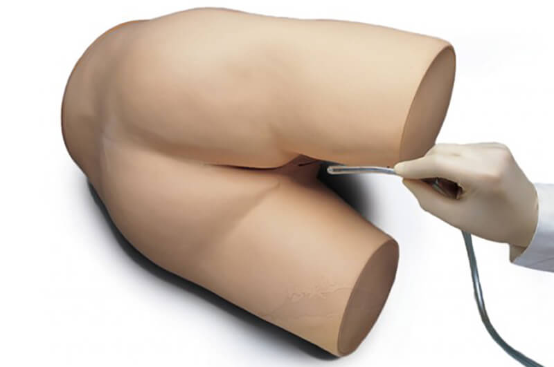
 Pediatric Auscultation Manikin | Simulators | Cardionics
 SAM II the Student Auscultation Manikin - Light
 Cardionics® SAM 3G- Student Auscultation Manikin | Ward's Science
The Art of Bovine Auscultation . . . . Cattle Plague: A History . . . . Bovine Anatomy: An Illustrated Text . . . . Veterinary...
 Accuracy of the combined method (auscultation and pH measurement) and ultrasonography for confirmation of gastric tube...
 Auscultation - Virtual Museum
 Cardiac Auscultation - Cardiovascular Disorders - MSD Manual Professional Edition
JMIR mHealth and uHealth - The Utility of Real-Time Remote Auscultation Using a Bluetooth-Connected Electronic Stethoscope:...
 Mid-Systolic Click C31 | Auscultation | Lesson, Recordings, Waveforms
 Auscultation Trainers
 Coronavirus: IIT-Bombay develops 'Digital Stethoscope' for remote auscultation, ET Government
Auscultation Repetition Training
 Lexicon:Auscultation - WikiPOBia
 Intelligent Intermittent Auscultation
 Auscultation of heart sounds
Hypertrophic Cardiomyopathy - Auscultation Lesson
 Medicine: Lung auscultation checklist - Checkton
Rales Lung Sounds - Easy Auscultation
 Carotid Auscultation | Diabetic Exam - MedSchool
 Survival Technology - Auscultation - SAM Basic - 3B Cardionics
 abdominal sounds auscultation Archives - MBBCH Health Encyclopedia
The Art of Bovine Auscultation
 Auscultation Trainer and Smartscope - Alpha Century Simulations
Cardiac auscultation15
- Cardiac Auscultation With Continuous Wave Doppler Stethoscope: A new method 200 years after Laennec's invention (1 ed. (wikipedia.org)
- The most frequent student request following a cardiac auscultation learning session is for a program that will demonstrate the most important findings in an effective, organized format. (laerdal.com)
- In response to this need, Essential Cardiac Auscultation was developed. (laerdal.com)
- Essential Cardiac Auscultation is a computer-based program that teaches the 12 most important findings (The Big 12) and their associated pathophysiology. (laerdal.com)
- Hirosawa T, Ito T, Harada Y, Ikenoya K, Yokose M, Shimizu T. The utility of phonocardiograms in real-time remote cardiac auscultation using an internet-connected electronic stethoscope: Open-label randomized controlled pilot trial. (jmir.org)
- Acoustic as well as electronic stethoscopes are used for cardiac auscultation. (medilib.ir)
- The cardiovascular section shows normal cardiac auscultation findings along with the findings in the common cardiac disorders of cattle - sinus tachycardia, atrial fibrillation, premature ventricular contractions, endocarditis, ventricular septal defects and patent ductus arteriosus. (vetvisions.com)
- on the effectiveness of high- and low-fidelity simulation-based medical education in teaching cardiac auscultation [ 1 ]. (ijohs.com)
- While the authors conclude that high-fidelity simulation has no benefit in improving cardiac auscultation knowledge or skills compared with low-fidelity simulation, we believe that this conclusion cannot be supported by the authors' work. (ijohs.com)
- Funnel plot showing high heterogeneity of selected studies comparing high- to low-fidelity cardiac auscultation simulators. (ijohs.com)
- Digital devices for teaching cardiac auscultation - a randomized pilot study. (bvsalud.org)
- Competent cardiac auscultation is a declining skill. (bvsalud.org)
- Digital stethoscopes and hand -held echo may be useful devices for teaching cardiac auscultation . (bvsalud.org)
- This pilot study provides a novel study design, a heart murmur grading system, and data that will help develop definitive studies to assess new teaching techniques for cardiac auscultation using digital technology . (bvsalud.org)
- Despite significant interobserver variability and need for habituation and training, cardiac auscultation provides important initial clues in patient evaluation and serves as a guide for further diagnostic testing. (medscape.com)
Stethoscope16
- Emily's heartbeat Sounds heard on auscultation of a healthy 16-year-old's heart while holding her breath, as heard with a stethoscope at the tricuspid valve Problems playing this file? (wikipedia.org)
- Auscultation (based on the Latin verb auscultare "to listen") is listening to the internal sounds of the body, usually using a stethoscope. (wikipedia.org)
- Auscultation is a skill that requires substantial clinical experience, a fine stethoscope and good listening skills. (wikipedia.org)
- Ultrasonography (US) inherently provides capability for computer-aided auscultation, and portable US, especially portable echocardiography, replaces some stethoscope auscultation (especially in cardiology), although not nearly all of it (stethoscopes are still essential in basic checkups, listening to bowel sounds, and other primary care contexts). (wikipedia.org)
- Mediate auscultation is an antiquated medical term for listening (auscultation) to the internal sounds of the body using an instrument (mediate), usually a stethoscope. (wikipedia.org)
- It was demonstrated in the 2000s that Doppler auscultation using a handheld ultrasound transducer enables the auscultation of valvular movements and blood flow sounds that are undetected during cardiac examination with a stethoscope. (wikipedia.org)
- Auscultation is usually done using a tool called a stethoscope. (medlineplus.gov)
- Instructors can use PAT Basic® - Affordable Pediatric Auscultation Manikin with the SimScope® Auscultation Training Stethoscope to train students in identifying normal and abnormal heart, lung and bowel sounds associated with pediatric disorders. (3bscientific.com)
- The included SimScope® Auscultation Training Stethoscope allows to auscultate the PAT® torso and enables wireless communication with the controller tablet (optional purchase). (3bscientific.com)
- The combination PAT Basic® Pediatric Auscultation Manikin and the SimScope® Training Stethoscope make teaching and learning auscultation a versatile and personal experience. (3bscientific.com)
- Ayu Devices, a startup of IIT Bombay, has developed a digital stethoscope called AyuSynk is being deployed for remote auscultation (listening to chest sounds)," stated IIT-Bombay 's statement. (indiatimes.com)
- This student auscultation trainer along with the SimScope stethoscope simulates heart, lung, and bowel sounds utilizing a large sounds library. (surtec.co.za)
- NursingScope is an electronic simulation training stethoscope created for auscultation training purposes or objective structured clinical examinations ( OSCE) . (simandskills.co.uk)
- The instructor can select and change any of the physiological and pathological sounds that the trainees will hear through the NursingScope once they place the stethoscope on one of the auscultation sites of the manikin. (simandskills.co.uk)
- Do you have any questions about Veterinary Stethoscope Aid Dual Head EMT Stethoscope Portable Medical Auscultation Stethoscope? (ivetsupply.com)
- Stethoscope not only can listen to heart rate, but also can listen to whether the heart rate is neat, whether the diaphragm auscultation area has abnormal heart sounds and noises, but also can listen to the lungs breath sounds, whether there is wet and dry rales. (ivetsupply.com)
Student Auscultation Manikin5
- With the Cardionics SAM II Student Auscultation Manikin, teaching and learning this skill becomes even more versatile and economic. (anatomywarehouse.com)
- SAM II Student Auscultation Manikin is used in teaching and learning heart, lung and bowel sounds. (anatomywarehouse.com)
- Additionally, when connected to speakers, SAM II Student Auscultation Manikin can be easily moved into a classroom or an auditorium for group instruction, where a larger audience of students can benefit from a co-learning experience and share their thoughts at the same time as high quality heart, lung and bowel sounds are emitted. (anatomywarehouse.com)
- How is SAM II Student Auscultation Manikin different from other auscultation products available on the market? (anatomywarehouse.com)
- SAM 3G Student Auscultation Manikin is world's most innovative product available on the market, used in teaching and learning heart, lung and bowel sounds. (wardsci.com)
Cardionics2
- By accessing the auscultation software, educators can pick any sound from the large Cardionics auscultation library and change the parameters on-the-fly. (3bscientific.com)
- Nikki the Nursing Manikin with Auscultation offers 11 anterior and 4 posterior auscultation sites with 42 high-quality sounds powered by Cardionics - the Leader in Auscultation Simulation. (simandskills.co.uk)
Trainer2
- The Life/form® Ausculation Trainer and Smartscope Manikin presents itself as a real patient without visible auscultation sites. (alphacenturysimulations.com)
- To be used with Life/form® Infant Auscultation Trainer with Airway Management (LF01201U). (universalmedicalinc.com)
Remote Auscultation1
- Ito T, Hirosawa T, Harada Y, Kakimoto S, Shimizu T. Evaluation of Internet-Connected Real-Time Remote Auscultation: An Open-Label Randomized Controlled Pilot Trial. (jmir.org)
Bovine Auscultation1
- The Art of Bovine Auscultation: An illustrated guide to cardiac, respiratory and gastrointestinal sounds. (vetvisions.com)
Intermittent auscultation7
- Intelligent Intermittent Auscultation (IIA) of the fetal heart is the recommended method of fetal monitoring for all women who are considered at low risk of fetal hypoxia during labour. (meducination.com)
- This programme aims to improve safety for mothers and babies in low-risk labour and birth by improving the knowledge, skills and confidence of midwives to undertake intermittent auscultation of the fetal heart in an intelligent manner (IIA). (meducination.com)
- The Intermittent auscultation online medical course takes around 60 minutes to complete, includes both pre-course and post-assessment exercises - so you can track your improvements as you progress. (meducination.com)
- I observed a common concern at all the conferences, namely, intermittent auscultation of the fetal heart rate (FHR). (cnps.ca)
- The debate among obstetrical nurses about intermittent auscultation inspired this article. (cnps.ca)
- These nurses openly analyzed and debated their own practices of intermittent auscultation of the fetal heart rate. (cnps.ca)
- Concern was still prevalent that a legal defence would not be as successful if fetal monitoring was done by intermittent auscultation as opposed to an electronic fetal monitor. (cnps.ca)
Palpation3
- Auscultation and palpation go together in physical examination and are alike in that both have ancient roots, both require skill, and both are still important today. (wikipedia.org)
- page needed] Breath sounds Heart sounds Intestinal sound Palpation, the practice of examining a patient through the use of hands Percussion (medicine) Pericardial friction rub Triangle of auscultation Constant, Jules (1999). (wikipedia.org)
- INTRODUCTION - The physical examination of the cardiovascular system includes auscultation and palpation of the heart, as well as assessment of the arterial and venous pulses. (medilib.ir)
Stethoscopes2
- Ang Y, Aw L, Koh V, Tan R. Characterization and cross-comparison of digital stethoscopes for telehealth remote patient auscultation. (jmir.org)
- STETHOSCOPES - A variety of stethoscopes are available for the auscultation of heart sounds. (medilib.ir)
Torso1
- PAT Basic® - Affordable Pediatric Auscultation Manikin is the perfect torso manikin for healthcare simulation professionals who wish to teach the critical skill of pediatric auscultation. (3bscientific.com)
Respiratory4
- Auscultation is performed for the purposes of examining the circulatory and respiratory systems (heart and breath sounds), as well as the alimentary canal. (wikipedia.org)
- The Auscultation Assistant Archived 2012-06-06 at the Wayback Machine, - "provides heart sounds, heart murmurs, and breath sounds in order to help medical students and others improve their physical diagnosis skills" MEDiscuss - Respiratory auscultation with audio examples Blaufuss Multimedia - Heart Sounds and Cardiac Arrhythmias Independent Stethoscope Review - Comparative review of stethoscopes, including frequency response graphs. (wikipedia.org)
- The respiratory section display auscultation findings in normal cattle and in the common upper and lower respiratory diseases of cattle - calf diphtheria, bacterial pneumonia, pleuritis and atypical interstitial pneumonia. (vetvisions.com)
- Respiratory examination and Chest Auscultation (Certified). (barrettmcgrathems.com)
Aortic5
- The patient sits upright for auscultation of the back, then leans forward to aid auscultation of aortic and pulmonic diastolic murmurs or pericardial friction rub. (msdmanuals.com)
- Detection of aortic insufficiency by standard echocardiography, pulsed Doppler echocardiography, and auscultation. (uky.edu)
- To determine the relative sensitivity and specificity of noninvasive methods for detecting aortic insufficiency, we compared the accuracy of auscultation, echocardiography, and pulsed Doppler echocardiography in detecting aortic insufficiency in 106 patients in whom the presence or absence of the lesion was shown by supravalvular aortography. (uky.edu)
- The sensitivity and specificity for the diagnosis of aortic regurgitation was 96% and 96% for pulsed Doppler echocardiography, 73% and 92% for auscultation, 43% and 91% for two-dimensional echocardiography, 46% and 81% for anterior mitral leaflet flutter, and 9% and 96% for ventricular septal flutter, respectively. (uky.edu)
- Dive into the research topics of 'Detection of aortic insufficiency by standard echocardiography, pulsed Doppler echocardiography, and auscultation. (uky.edu)
Skill2
- Auscultation is an essential clinical skill needed to assess and monitor patients' conditions. (anatomywarehouse.com)
- Other physicians have commented anecdotally on the connection between musical expertise and auscultation skill (e.g. (teachingheartauscultation.com)
Percussion3
- Auscultation was often used with percussion , the art of striking a surface part of the body with short, sharp taps to diagnose the condition of the parts beneath the sound, which was discovered by Leopold Auenbrugger (1722-1809) of Vienna. (hmssurprise.org)
- it has sections on auscultation findings, percussion, succussion and the grunt test. (vetvisions.com)
- The second section documents the visual, auscultation, percussion and succussion findings in the common abdominal diseases of cattle. (vetvisions.com)
Doppler2
- Moreover, Doppler auscultation was superior in the detection of impaired ventricular relaxation. (wikipedia.org)
- Since the physics of Doppler auscultation and classic auscultation are different, it has been suggested that both methods could complement each other. (wikipedia.org)
Supine2
- The patient rolls supine, and auscultation continues at the lower left sternal border, proceeds cephalad with auscultation of each interspace, then caudad from the right upper sternal border. (msdmanuals.com)
- The patient was supine during auscultation. (practicalclinicalskills.com)
Trainers1
- Our MedVision Auscultation Task Trainers (MATT) highly effective tool for learning auscultation points and sounds. (gtsimulators.com)
Ultrasonography1
- Letter to the Editor for , "Accuracy of the combined method (auscultation and pH measurement) and ultrasonography for confirmation of gastric tube placement: a study protocol for a prospective study," by Rigobello et al. (bmj.com)
Physical examination3
- Auscultation is listening to the sounds of the body during a physical examination . (medlineplus.gov)
- The physical examination suggests a normal oropharynx and prolonged expiratory phase on chest auscultation. (cdc.gov)
- Auscultation of the heart forms the core of cardiac physical examination. (medscape.com)
Pediatric3
- The manikin simulates the anatomy of a child with correct pediatric auscultation sites. (3bscientific.com)
- With one of the largest libraries of pediatric sounds and easy-to-use software, PAT Basic® - Affordable Pediatric Auscultation Manikin gives students and educators everything they need to actively engage in education and auscultation hands-on training. (3bscientific.com)
- When using PAT Basic® - Affordable Pediatric Auscultation Manikin, simulation educators can provide a variety of customizable experiences for Standardized Patient Programs and Objective Structured Clinical Examination (OSCE). (3bscientific.com)
Sounds14
- The sounds of auscultation can be depicted using symbols to produce an auscultogram. (wikipedia.org)
- Providers also use auscultation to listen to the heart sounds of unborn infants. (medlineplus.gov)
- The purpose of auscultation of the heart is to characterize heart sounds and murmurs. (medilib.ir)
- This topic will review the auscultation of heart sounds. (medilib.ir)
- An electronic device enables shared auscultation for teaching purposes and also enables direct digital recording of heart sounds for review and analysis. (medilib.ir)
- Our auscultation reference guide provides quick access to this sound as well as many other adventitious sounds. (easyauscultation.com)
- The goal of this basic course in lung sounds is to improve auscultation observational skills. (easyauscultation.com)
- Heart and Lung Sounds - A major breakthrough in auscultation training! (alphacenturysimulations.com)
- The student must palpate to identify correct auscultation locations, and will hear different heart and lung sounds as the Smartscope is moved to different locations on the manikin. (alphacenturysimulations.com)
- The instructor can easily change the auscultation sounds on the controller device for each of the 15 locations using NursingScope's wireless functionality. (simandskills.co.uk)
- We have aimed to establish whether cervical auscultation interpretation is based on the actual sounds heard or, in practice, influenced by information gleaned from other aspects of the clinical assessment, medical notes, or previous knowledge. (ox.ac.uk)
- Practitioners and students: see and hear breath and heart sounds with remarkable clarity while perfecting your auscultation technique. (bookbaz.ir)
- Auscultation Skills: Breath & Heart Sounds, Fifth Edition, pinpoints exactly how, where, and why breath and heart sounds occur and helps you to differentiate normal from abnormal sounds quickly and accurately. (bookbaz.ir)
- [ 1 ] In addition, auscultation of the left axilla, base of the heart, carotid arteries, and interscapular area should be performed to assess for radiation of heart sounds and murmurs. (medscape.com)
Findings3
- It is illustrated with images from necropsy, in some cases overlaid by the auscultation findings in life. (vetvisions.com)
- See what hear, say what you mean, correctly interpret and diagnose auscultation findings. (vetvisions.com)
- Students ' scores were compared to a ' gold standard' derived from a consensus of auscultation findings of three cardiologists . (bvsalud.org)
Bedside2
- Improve patient care skills, train auscultation and evaluate nursing skills and bedside competencies with the new and all-in-one training manikin Nikki. (simandskills.co.uk)
- The system generated sound quality similar to "live" bedside listening, a feature rarely seen in cervical auscultation studies. (ox.ac.uk)
Lungs2
- Health professionals (doctors, nurses, etc.) listen to three main organs and organ systems during auscultation: the heart, the lungs, and the gastrointestinal system. (wikipedia.org)
- His lungs are clear to auscultation. (medscape.com)
Sensitivity1
- Together, the latter two strategies (listening mnemonics and auditory-motor network engagement) may help reveal a richer acoustic picture and perceptual experience that may translate into increased sensitivity during auscultation. (teachingheartauscultation.com)
Clinical2
- Cervical auscultation is experiencing a renaissance as an adjunct to the clinical swallowing assessment. (ox.ac.uk)
- We sought to determine (a) rater reliability and its impact on the clinical value of cervical auscultation and (b) how judgments compare with the "gold standard": videofluoroscopy. (ox.ac.uk)
Murmurs2
- The auscultation of cardiac murmurs is discussed separately. (medilib.ir)
- See "Auscultation of cardiac murmurs in adults" . (medilib.ir)
Echocardiography1
- [ 2 ] Whenever possible, auscultation should be accompanied by having the patient perform dynamic maneuvers such as standing, Valsalva, squatting, and hand grip, although these maneuvers are falling out of favor with the use of echocardiography . (medscape.com)
Phonocardiograms1
- This program also has over 40 lessons on auscultation with illustrations, diagrams, phonocardiograms, questions and answers. (surtec.co.za)
Lessons2
- Included with the product is SAM's Lesson Guide: a complete guide of lessons for each auscultation sound that SAM II offers. (anatomywarehouse.com)
- We also provide auscultation lessons on several types of wheezes, crackles and stridor. (easyauscultation.com)
Skills2
- Evaluation checklist for assessing the practical skills (abilities) of an employee during lung auscultation. (checkton.com)
- Auscultation skills made easy! (gtsimulators.com)
Anterior1
- Students can practice auscultation at six anterior heart sites. (alphacenturysimulations.com)
Pulses1
- Auscultation can also be used to hear pulses in the arms and legs. (medlineplus.gov)
Normal3
- Evidence Central , evidence.unboundmedicine.com/evidence/view/EBMG/455184/all/Normal_auscultation_finding___Audio. (unboundmedicine.com)
- The 20 sound clips were classified as "normal" or "abnormal" by 19 volunteer speech-language pathologists with experience in cervical auscultation. (ox.ac.uk)
- Pulmonary auscultation was normal. (who.int)


![APA format] Physiology References for the Native American Flute](https://flutopedia.com/img/JoinCVFluteNewsletter_150.jpg)