Development of a sensitive, specific reverse transcriptase polymerase chain reaction-based assay for epithelial tumour cells in effusions. (1/1795)
We developed a sensitive and specific method for the detection of epithelial cancer cells in effusions with a two-stage molecular-based assay which combined enrichment for cancer cells by immunomagnetic bead selection and reverse transcriptase polymerase chain reaction (RT-PCR) detection of epithelial glycoprotein 2 (EGP-2) RNA. Preliminary experiments indicated that immunobead selection was essential to avoid occasional false-positive RT-PCR results, and this method detected ten breast cancer cells electively added to 10(7) cytologically negative effusion cells. We studied 110 cases of pleural (n = 68) and peritoneal (n = 42) effusions (30 from patients with known carcinoma and 80 from those without known carcinoma), and the results were compared with cytological findings. Of 18 effusions that were cytologically positive or suspicious for malignant cells, 17 (94%) were positive for EGP-2 RNA (the one negative sample was from a patient who recently received combination chemotherapy). Of 92 cytologically negative samples, 11 (12%) were positive for EGP-2, including six patients with a history of previous or current carcinoma. Our method appears to be highly specific and increases the sensitivity of detection of malignant cells; it may be a useful adjunct to routine cytopathological examination. (+info)Peritoneal cytology in the surgical evaluation of gastric carcinoma. (2/1795)
Many patients undergoing surgery for gastric carcinoma will develop peritoneal metastases. A method to identify those patients at risk of peritoneal recurrence would help in the selection of patients for adjuvant therapy. Peritoneal cytology has received little attention in the West, but may prove a useful additional means of evaluating patients with gastric cancer. The aims of this study were to evaluate sampling techniques for peritoneal cytology in patients with gastric cancer, to assess the prognostic significance of free peritoneal malignant cells and to discover the effect of the operative procedure on dissemination of malignant cells. The study is based on 85 consecutive patients undergoing surgical treatment of gastric cancer and followed up for 2 years or until death. Peritoneal cytology samples were collected at laparoscopy, and at operation prior to resection by intraperitoneal lavage and serosal brushings. After resection, samples were taken by peritoneal lavage, imprint cytology of the resected specimen and post-operatively by peritoneal irrigation via a percutaneous catheter. Malignant cells were diagnosed by two independent microscopists. Preoperative peritoneal lavage yielded malignant cells in 16 out of 85 cases (19%). The yield of free malignant cells was increased by using serosal brushings (by four cases) and imprint cytology (by two cases); all of the cases had evidence of serosal penetration. One serosa-negative case exhibited positive cytology in the post-resection peritoneal specimen in which the preresection cytology specimen was negative. Survival was worse in the cytology-positive group (chi2 = 25.1; P< 0.0001). Among serosa-positive patients, survival was significantly reduced if cytology was positive, if cases yielded by brushings and imprint cytology were included (log-rank test = 8.44; 1 df, P = 0.004). In conclusion, free peritoneal malignant cells can be identified in patients with gastric cancer who have a poor prognosis; the yield can be increased with brushings and imprint cytology in addition to conventional peritoneal lavage. Evaluation of peritoneal cytology by these methods may have a role in the selection of patients with the poorest prognosis who may benefit most from adjuvant therapy. (+info)Immunocytochemically detected free peritoneal tumour cells (FPTC) are a strong prognostic factor in gastric carcinoma. (3/1795)
We prospectively investigated the prognostic significance of free peritoneal tumour cells (FPTC) in a series of 118 patients with completely resected gastric carcinoma. Immunocytochemistry with the monoclonal antibody Ber-Ep4 was performed on cytospins from intraoperative peritoneal lavage specimens. Twenty-three patients (20%) had FPTC which was significantly correlated with pT and pN categories, stage, tumour size, lymphatic invasion, Lauren and WHO classifications and perigastric adipose tissue metastases. The median survival time for all FPTC positive compared with negative patients was significantly shorter (11 compared with >72 months), with estimated 5-year survival rates of 8% vs. 60%. None of the patients with FPTC had an early gastric cancer. In advanced tumour subgroups without and with serosal invasion (n = 59 and 35), there were 19% and 34% with FPTC. Multivariate survival analysis showed nodal status, FPTC, mesenteric lymphangiosis, and lymph node metastasis to the compartment III to be independent prognostic factors with relative risks of 6.6, 4.5, 2.9 and 2.2 respectively. Recurrent disease occurred in 91% of FPTC-positive and in 38% of FPTC-negative patients. FPTC had a positive predictive value of 91% and a specificity of 97% for tumour recurrence. FPTC is a strong negative, independent prognostic indicator for survival in gastric carcinoma. (+info)Paracrine changes in the peritoneal environment of women with endometriosis. (4/1795)
During the past decade, macrophage-derived substances such as prostanoids, cytokines, growth factors and angiogenic factors have been detected in the peritoneal fluid of women with endometriosis. In particular, growth-promoting and angiogenic factors are considered to be substantially involved in the pathogenesis of endometriosis. In this study, vascular endothelial growth factor (VEGF), transforming growth factor beta (TGF-beta) and intercellular adhesion molecule 1 (ICAM-1), substances recently detected in the peritoneal fluid of women with endometriosis, were assessed with regard to their concentrations in different stages of endometriosis and changes of the peritoneal paracrine activity after medical treatment with a gonadotrophin releasing hormone agonist (GnRHa). Peritoneal fluid was obtained from patients with endometriosis during laparoscopy before and after a 4-month treatment with a GnRHa. VEGF, TGF-beta and ICAM-1 could be detected in all women presenting with various stages of active endometriosis. After GnRHa therapy, all patients showed significant decreases in mean concentrations of VEGF (194+/-77 pg/ml), TGF-beta (902+/-273 pg/ml) and ICAM-1 (157+/-52 ng/ml). Patients with stage III and IV endometriosis (according to the rAFS score) had much higher concentrations of VEGF and TGF-beta before treatment compared with those patients with mild endometriosis (rAFS stages I and II). The most striking decrease in concentration was for TGF-beta, from 902 pg/ml before to 273 pg/ml after therapy. These results indicate an important role for paracrine activity in the establishment and maintenance of endometriosis. Indeed, treatment with a GnRHa may reduce paracrine activity in the peritoneal cavity via hypo-oestrogenism and provide proof of successful therapy. (+info)Tracing cellular and molecular mechanisms involved in endometriosis. (5/1795)
The aetiology and pathogenesis of endometriosis, defined as the presence of endometrium-like tissue outside the uterine cavity, is largely unknown. In this paper we present and discuss possibilities to study the putative pathogenic properties of endometriotic cells in vitro. The current focus of our investigations is on the invasive phenotype of the disease, assuming that this might contribute to the pathogenesis of endometriosis. So far, we have shown that: (i) cytokeratin-positive and E-cadherin-negative endometriotic cells have an invasive phenotype in a collagen invasion assay in vitro similar to metastatic carcinoma cells; (ii) the invasiveness of endometriotic but not of eutopic endometrial cells can be stimulated by a heat-stable protein present in peritoneal fluid; and (iii) the endometriotic cell line EEC145T, which we established, may be a useful tool for the identification of gene products which are, positively or negatively, invasion-related. Finally, our studies suggest that the invasive phenotype in endometriosis shares aspects with tumour metastasis, but might also have unique mechanisms. (+info)Endometriotic disease: the role of peritoneal fluid. (6/1795)
Peritoneal fluid and the intraovarian milieu are a specific microenvironment. Peritoneal fluid originates mainly as an ovarian exudation product caused by increased vascular permeability, with cyclic variation in volume and steroid hormones which are always higher than in plasma. It contains large amounts of macrophages and their secretion products, and has a large exchange area with plasma through the peritoneum, which is highly permeable for small molecules. Diffusion becomes virtually zero for molecules with a molecular weight of >100000 Da. In women with the luteinized unruptured follicle (LUF) syndrome, concentrations of oestrogens and progesterone are much lower in the luteal phase. Endometriosis is associated with sterile low-grade inflammation, increased concentrations of activated macrophages and many of their secretions, such as cytokines, growth factors and angiogenic factors. Concentrations of CA-125 and of glycodelins are also increased, secreted locally by the endometrial cells. Natural killer (NK) cell function declines, possibly mediated by glycodelins or local intercellular adhesion molecule (ICAM) -1 shedding. The ovary is also a specific microenvironment, with steroid hormone concentrations 1000-fold higher in follicles than in plasma. Endometrial and superficially implanted cells are influenced by peritoneal fluid concentrations so that local environment, rather than inherent cellular differences could explain differences between superficial endometriosis and eutopic endometrium. Differences between superficial implants and endometriotic disease, deep infiltrating or cystic ovarian endometriosis, may thus arise via different endocrine environments. Superficial endometrial implants are regulated by peritoneal fluid factors, whereas deep endometriosis and cystic ovarian endometriosis are influenced by blood or ovarian factors. The endometriotic disease theory considers superficial endometriotic implants and their remodelling as a physiological process in most women, and concentrates on the causes of severe endometriosis such as differences in the eutopic endometrium from women with and without endometriosis (which may indicate hereditary differences), the invasiveness of some endometriotic cells in vitro, focal 'shielding' of endometriotic foci by adhesions, and inhibition of NK activity by ICAM-1 and glycodelins. Endometriotic disease is thus seen as a benign tumour. The type of cellular lesion, hereditary and immunological environments and local hormone concentrations in the ovary and in peritoneal fluid, will decide expression as cystic ovarian endometriosis, deep endometriosis or adenomyosis externa, and whether the latter is associated with adhesions. (+info)2-Deoxyglucose selectively inhibits Fc and complement receptor-mediated phagocytosis in mouse peritoneal macrophages II. Dissociation of the inhibitory effects of 2-deoxyglucose on phagocytosis and ATP generation. (7/1795)
Macrophages incubated in 2-deoxy-D-glucose (2-dG)-containing medium showed a marked decrease in cellular ATP content, and were unable to ingest IgG- and complement-coated erythrocytes via the corresponding membrane receptors for these ligands. However, the inhibitory effects of 2-dG on Fc- and C3 receptor-mediated phagocytosis were not a consequence of lowered macrophage ATP levels since addition of glucose or mannose to the culture medium restored the capacity of the macrophages to ingest IgG- and C3-coated particles without increasing ATP levels. These results indicate that Fc- and C3 receptor-mediated phagocytosis (opsonin dependent) differs qualitatively from the ingestion of latex and zymosan particles (opsonin independent); they suggest that the same regulatory molecules govern the responses of phagocytic cells to signals initiated by both the Fc and C3 receptors. The possibility that these molecules are regulated by glycosylation is discussed. (+info)The requirement of an adherent cell substratum for the growth of developing plasmacytoma cells in vivo. (8/1795)
The intraperitoneal injection of pristane (2,6,10,14-tetramethylpentadecane) produces an environment conductive to primary plasmacytoma growth in as few as 3 days. After pristane injection, the total free peritoneal cell population increases from a normal value of 1.55 X 10(6) to 5.28 X 10(6) and remains at this elevated level for at least 50 days. The adherent peritoneal cell population, composed of both mononuclear cells and polymorphonuclear leukocytes, is the primary source of this increase. In the pristane-conditioned peritoneum, these cells rapidly form a chronic granuloma on the peritoneal connective tissues. Daily subcutaneous treatment of mice with 0.5 mg of hydrocortisone beginning simultaneously with pristane injection prevents the increase in the peritoneal cell population, granuloma formation, d the production of a conditoned environment. In mice treated with hydrocortisone beginning 3 days after pristane injection, however, neither the peritoneal cell increase nor the production of a conditioned environment is prevented. The intraperitoneal injection of thioglycolate medium at 4-day intervals produces an elevation of the free adherent peritoneal cell population similar to pristane, but does not produce a granuloma or a conditioned environment. The intraperitoneal transfer of thioglycolate-induced adherent peritonel cells to mice treated with pristane and hydrocortisone simultaneously restores the production of a conditioned environment. These findings indicate that the adherent peritoneal cell population is responsible for the conditioning effect, and that the establishment of a resident population of these cells is necessary to produce conditioning. (+info) ADA- Ascitic Fluid - MushkilAshan
 Chylous Ascites: Overview, Pathophysiology, Etiology
Ascitic fluid study. Ascitic fluid is usually white or milky. Gross milkiness of the ascitic fluid corresponds poorly with ... The diagnosis of chylous ascites is made by peritoneocentesis and by analysis of the ascitic fluid. An ascitic triglyceride ... True chylous ascites is defined as the presence of ascitic fluid with high fat (triglyceride) content, usually higher than 110 ... 1, 2] Moreover, an existing clear ascitic fluid can turn chylous as a secondary event. ...
 Ascitic Fluid ADA Levels prices in Pune
 Ascitic Fluid ADA Test Price in bansur
Ascitic Fluid for Creatinine prices in Pune
 MTB by GeneXpert - Ascitic Fluid - Essa Laboratory - EssaLaboratory
Neutralisation of adenovirus infectivity by ascitic fluid from ovarian cancer patients - Department of Oncology
Twelve ascitic fluid and four matched serum samples were examined. The titre and isotype of anti-adenovirus antibodies was ... In order to determine the presence of pre-existing anti-adenovirus humoral immunity we analysed ascitic fluid, collected from ... We found that the ascitic fluid samples contain antibodies that recognise both adenovirus types 2 and 5, were predominantly IgG ... Twelve ascitic fluid and four matched serum samples were examined. The titre and isotype of anti-adenovirus antibodies was ...
ArboCat Virus: Kimberley (KIMV)
Table - Listeriosis Caused by Persistence of Listeria monocytogenes Serotype 4b Sequence Type 6 in Cheese Production...
 CDC - Toxoplasmosis
 Page 1 | Search Results | Acta Cytologica | Karger Publishers
Open the PDF for Extraoral Plasmablastic Lymphoma Detected Using Ascitic Fluid Cytology and ,span class=search-highlight,Flow ... Extraoral Plasmablastic Lymphoma Detected Using Ascitic Fluid Cytology and Flow Cytometry: A Case Report with a Review of the ... View article titled, Extraoral Plasmablastic Lymphoma Detected Using Ascitic Fluid Cytology and ,span class=search-highlight, ... Open the PDF for Cytomorphologic and Flow Cytometric Analysis of Cerebrospinal Fluid with T-Cell Lymphoma Involvement: A ...
Cirrhosis: Practice Essentials, Overview, Etiology
Ascitic fluid with more than 250 PMNs/mm3 defines neutrocytic ascites and SBP. Many cases of ascites fluid with more than 1000 ... Neutrophil count and culture of ascitic fluid culture (bedside inoculation of blood culture bottles with 10 mL fluid each) ... Optimization of ascitic fluid culture technique. Gastroenterology. 1988 Nov. 95(5):1351-5. [QxMD MEDLINE Link]. ... Fluid and plasma proteins diffuse freely across the highly permeable sinusoidal endothelium into the space of Disse. Fluid in ...
 British Medical Journal: 2 (5412) | The BMJ
 Quantitative assay for acute intestinal inflammation based on myeloperoxidase activity. Assessment of inflammation in rat and...
 Ascitic fluid infection in patients with hepatitis B virus-related liver cirrhosis: Culture-negative neutrocytic ascites versus...
Ascitic fluid infection in patients with hepatitis B virus-related liver cirrhosis: Culture-negative neutrocytic ascites versus ... Ascitic fluid infection in patients with hepatitis B virus-related liver cirrhosis: Culture-negative neutrocytic ascites versus ... Abstract Background and Aim: Ascitic fluid infection (AFI) consists of culture-negative neutrocytic ascites (CNNA) and ... Thus, liver transplantation should be considered, irrespective of culture positivity of ascitic fluid. ...
 Proteomic identification of fucosylated haptoglobin alpha isoforms in ascitic fluids and its localization in ovarian carcinoma...
The aim of this study was to perform the proteomic analysis of ascitic fluids of Mexican patients with ovarian carcinoma, in ... Increased numbers of highly fucosylated Haptoglobin alpha isoforms in ascitic fluids and the presence of fucosylated ... Haptoglobin standard (ST), an ascitic fluid sample non-related with cancer (NR), and 40 ascitic fluid samples from ovarian ... in ascitic fluids from 40 patients. This analysis included an ascitic fluid sample not related to cancer (NR), as well as a ...
 Buy online ASCITIC FLUID MALIGANT CELLS at 20% discount only at www.kaer.in, Save more on ASCITIC FLUID MALIGANT CELLS with...
... or peritoneal fluid accumulationWhat sample needed?A peritoneal fluid sample obtained by inserting a needle into the abdominal ... To help diagnose the cause of peritonitis and/or peritoneal fluid accumulation (called ascites)When to get tested?When a doctor ... Buy Online Original ASCITIC FLUID MALIGANT CELLS contains and is a brand or product of tested? ... ASCITIC FLUID MALIGANT CELLS. Default Title - Rs. 450.00. Regular price Rs. 500 Kaer Price Rs. 450 ...
 Cell culture - Wikipedia
Pancreatic fistula - Wikipedia
Pleural or ascitic fluid should be sent for analysis. An elevated amylase level, usually > 1,000 IU/L, with protein levels over ... Loss of a small volume of fluid will not cause a problem but an acidosis is common if the volume of pancreatic fluid lost from ... Loss of bicarbonate-rich pancreatic fluid via a pancreatic fistula can result in a hyperchloraemic or normal anion gap ... ISBN 0-7216-2082-5 Dugernier T, Laterre PF, Reynaert MS (2000). "Ascites fluid in severe acute pancreatitis: from ...
ArboCat Virus: Main Drain (MDV)
Hyperimmune mouse serum or ascitic fluid 4. Main Drain 128 1280 2.1 128 1280 2.1 ... Tests with hyperimmune sera or ascitic fluids were performed in SM inoculated ic, whereas tests with immune hamster serum were ... 1 CF and HI titers are expressed as the reciprocal of the serum or ascitic fluid dilution. ... Immune Fluid or Antigen Ht/Ho 3. Ht/Ho Ht/Ho CF 1. HI 1. NT 2. ...
 Vol. 3 (2001)
| Journal of Medical Investigation and Practice
Vol. 3 (2001)
| Journal of Medical Investigation and Practice
 Ischemic colitis differential diagnosis - wikidoc
Pancreatitis - wikidoc
Pancreatitis - wikidoc
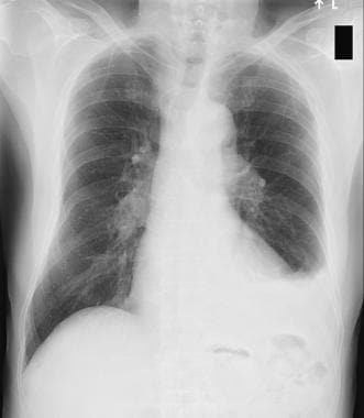
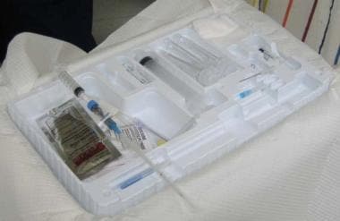
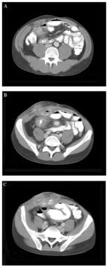
 Ultrasound for Detection of Ascites and for Guidance of the Paracentesis Procedure: Technique and Review of the Literature
Learn about techniques, safety considerations, and current literature supporting the use of ultrasound for successful fluid ... 2009) Can the Smallest Depth of Ascitic Fluid on Sonograms Predict the Amount of Drainable Fluid? Journal of Clinical ... Objective: To review the use of ultrasound (US) for the detection of free intraperitoneal fluid (ascites) and for the ... First, US techniques used for the identification of ascites and in the quantification of fluid pockets amenable to aspiration ...
Infections in Cirrhotics: Types, Microbiological Spectrum and Risk Factors-5-Year Cohort Study
... urinary tract and skin were present in cirrhosis of liver where ascitic fluid infections (AFI) were the commonest i.e. 44.89%. ... Outcomes in Culture Positive and Culture Negative Ascitic Fluid Infection in Patients with Viral Cirrhosis: Cohort Study. BMC ... A Unique Entity in the Spectrum of Infected Ascitic Fluid. Archives of Internal Medicine, 146, 2173. http://dx.doi.org/10.1001/ ...
 Frontiers | Identification of Common Genes and Pathways in Eight Fibrosis Diseases