Takayasu Arteritis
Giant Cell Arteritis
Arteritis Virus, Equine
Temporal Arteries
Polymyalgia Rheumatica
Polyarteritis Nodosa
HLA-B52 Antigen
Aortic Arch Syndromes
Arterivirus
Prednisolone
Subclavian Artery
Horses
Subclavian Steal Syndrome
Axillary Artery
Biopsy
Optic Neuropathy, Ischemic
Mucocutaneous Lymph Node Syndrome
Strongyle Infections, Equine
Brachiocephalic Trunk
Vasculitis
Vasculitis, Central Nervous System
Adrenal Cortex Hormones
Magnetic Resonance Angiography
Aortography
Lactobacillus casei
Glucocorticoids
Aneurysm
Arachnoiditis
AIDS Arteritis, Central Nervous System
Prednisone
Infarction
Retinal Artery Occlusion
Blindness
Adie Syndrome
Togaviridae
Aorta, Abdominal
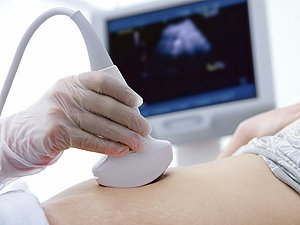
Ultrasonography, Doppler, Color
Arterial Occlusive Diseases
Tomography, X-Ray Computed
Amaurosis Fugax
Vertebral Artery
Aortic Valve Insufficiency
Vaccines, Marker
Aorta, Thoracic
Retrospective Studies
Takayasu arteritis is a rare inflammatory disease that affects the large blood vessels in the body, most commonly the aorta and its main branches. It's also known as pulseless disease or aortic arch syndrome. The condition primarily affects young to middle-aged women, although it can occur in anyone at any age.
The inflammation caused by Takayasu arteritis can lead to narrowing, thickening, and weakening of the affected blood vessels' walls, which can result in reduced blood flow to various organs and tissues. This can cause a variety of symptoms depending on the severity and location of the vessel involvement.
Common symptoms include:
* Weak or absent pulses in the arms and/or legs
* High blood pressure (hypertension)
* Dizziness, lightheadedness, or fainting spells due to reduced blood flow to the brain
* Headaches
* Visual disturbances
* Fatigue
* Weight loss
* Night sweats
* Fever
Diagnosis of Takayasu arteritis typically involves a combination of medical history, physical examination, laboratory tests, and imaging studies. Treatment usually includes corticosteroids or other immunosuppressive medications to control inflammation and maintain remission. Regular follow-up with a healthcare provider is essential to monitor disease activity and adjust treatment as necessary.
Arteritis is a medical condition characterized by inflammation of the arteries. It is also known as vasculitis of the arteries. The inflammation can cause the walls of the arteries to thicken and narrow, reducing blood flow to affected organs or tissues. There are several types of arteritis, including:
1. Giant cell arteritis (GCA): Also known as temporal arteritis, it is a condition that mainly affects the large and medium-sized arteries in the head and neck. The inflammation can cause headaches, jaw pain, scalp tenderness, and vision problems.
2. Takayasu's arteritis: This type of arteritis affects the aorta and its major branches, mainly affecting young women. Symptoms include fever, weight loss, fatigue, and decreased pulse in the arms or legs.
3. Polyarteritis nodosa (PAN): PAN is a rare systemic vasculitis that can affect medium-sized arteries throughout the body. It can cause a wide range of symptoms, including fever, rash, abdominal pain, and muscle weakness.
4. Kawasaki disease: This is a type of arteritis that mainly affects children under the age of 5. It causes inflammation in the blood vessels throughout the body, leading to fever, rash, swollen lymph nodes, and red eyes.
The exact cause of arteritis is not fully understood, but it is believed to be an autoimmune disorder, where the body's immune system mistakenly attacks its own tissues. Treatment for arteritis typically involves medications to reduce inflammation and suppress the immune system.
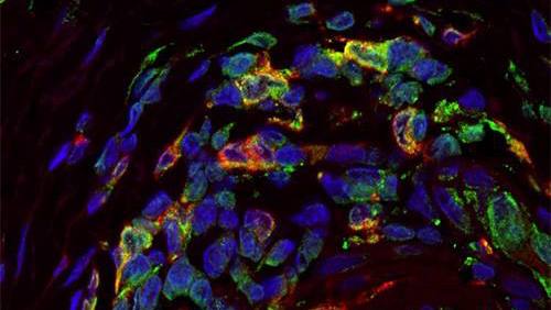
Giant Cell Arteritis (GCA), also known as Temporal Arteritis, is a chronic inflammatory disease affecting large and medium-sized arteries, most commonly the temporal artery. It primarily occurs in people over 50 years old. The condition is characterized by the infiltration of the artery walls with immune cells, leading to inflammation, swelling, and damage. This can restrict blood flow, causing various symptoms.
The key feature of GCA is the presence of multinucleated giant cells, which are large collections of fused immune cells, in the affected artery walls. These cells are a hallmark of this condition when viewed under a microscope.
Common symptoms include new onset of severe headaches, scalp tenderness, jaw pain while chewing (called jaw claudication), vision problems, and systemic symptoms such as fever, fatigue, and weight loss. If left untreated, GCA can lead to serious complications like blindness or stroke. Treatment typically involves high-dose corticosteroids to reduce inflammation and prevent further damage.
I'm sorry for any confusion, but there seems to be a mistake in your question. "Arteritis Virus, Equine" is not a recognized medical term or virus in humans or animals. There is a condition called "Equine Viral Arteritis (EVA)," which is a viral disease that affects horses and other equine species. However, it does not affect humans.
Equine Viral Arteritis (EVA) is caused by the Equine Arteritis Virus (EAV). This virus primarily affects the respiratory system and can cause symptoms such as fever, lethargy, loss of appetite, and a runny nose in infected horses. In some cases, it may also lead to inflammation of the lining of blood vessels (vasculitis), which can result in abortion in pregnant mares or infertility in stallions.
It's essential to maintain proper biosecurity measures when dealing with horses, especially those that have been exposed to EVA, to prevent its spread and protect the health of other equine populations.
Temporal arteries are the paired set of arteries that run along the temples on either side of the head. They are branches of the external carotid artery and play a crucial role in supplying oxygenated blood to the scalp and surrounding muscles. One of the most common conditions associated with temporal arteries is Temporal Arteritis (also known as Giant Cell Arteritis), which is an inflammation of these arteries that can lead to serious complications like vision loss if not promptly diagnosed and treated.
Polymyalgia Rheumatica (PMR) is a geriatric rheumatic disease characterized by widespread musculoskeletal pain and stiffness, particularly affecting the neck, shoulders, hips, and thighs. It is often accompanied by symptoms such as fatigue, weakness, loss of appetite, and low-grade fever. The onset of PMR can be sudden or gradual, and it tends to affect individuals over 50 years of age, more commonly women than men.
The exact cause of Polymyalgia Rheumatica remains unknown; however, it is believed to involve an autoimmune response leading to inflammation in the affected areas. Diagnosis typically involves a combination of clinical evaluation, laboratory tests (such as elevated erythrocyte sedimentation rate or C-reactive protein), and sometimes imaging studies. Treatment usually includes corticosteroids to reduce inflammation and manage symptoms, along with monitoring for potential side effects from long-term steroid use. In many cases, PMR can be successfully managed with appropriate treatment, allowing individuals to return to their normal activities.
Arterivirus infections are viral diseases caused by members of the Arteriviridae family, which includes several species that can infect a variety of animals. The most well-known arterivirus is the equine arteritis virus (EAV), which causes equine arteritis in horses. Other examples include the porcine reproductive and respiratory syndrome virus (PRRSV) in pigs, and simian hemorrhagic fever virus (SHFV) in non-human primates.
Arterivirus infections typically cause respiratory or reproductive symptoms, depending on the specific virus and host species. For example, EAV can cause respiratory disease, abortion, and infertility in horses, while PRRSV primarily affects the reproductive system of pigs, causing abortions, stillbirths, and weak piglets.
Transmission of arteriviruses typically occurs through direct contact with infected animals or their bodily fluids, such as respiratory droplets or semen. Some arteriviruses can also be transmitted vertically, from mother to offspring, during pregnancy or birth.
There are currently no specific treatments for arterivirus infections, and prevention efforts focus on biosecurity measures, such as quarantine and vaccination of susceptible animals.
Polyarteritis nodosa (PAN) is a rare, systemic necrotizing vasculitis that affects medium-sized and small muscular arteries. It is characterized by inflammation and damage to the walls of the arteries, leading to the formation of microaneurysms (small bulges in the artery wall) and subsequent narrowing or complete occlusion of the affected vessels. This can result in tissue ischemia (reduced blood flow) and infarction (tissue death), causing a wide range of clinical manifestations that vary depending on the organs involved.
The exact cause of PAN remains unclear, but it is believed to involve an autoimmune response triggered by various factors such as infections or exposure to certain drugs. The diagnosis of PAN typically requires a combination of clinical findings, laboratory tests, and imaging studies, often supported by histopathological examination of affected tissues. Treatment usually involves the use of immunosuppressive medications to control inflammation and prevent further damage to the arteries and organs.
HLA-B52 is a specific antigen of the human leukocyte antigen (HLA) system, which is located on chromosome 6 and plays an important role in the immune system. The HLA system helps the body to recognize and distinguish its own cells from foreign substances such as viruses and bacteria.
HLA-B52 is a type of HLA-B antigen, which is a group of proteins found on the surface of cells that help the immune system identify and destroy infected or damaged cells. The HLA-B52 antigen is most commonly found in individuals of Asian descent, particularly those from Japan and Korea.
It's important to note that the presence or absence of the HLA-B52 antigen does not necessarily indicate the presence or absence of a specific disease. However, certain genetic associations have been reported between HLA-B52 and some diseases such as Behçet's disease, which is a chronic inflammatory disorder that causes symptoms such as mouth sores, genital sores, eye inflammation, and skin lesions.
Aortic arch syndromes are a group of conditions that affect the aortic arch, which is the curved portion of the aorta that arises from the left ventricle of the heart and gives rise to the major branches of the arterial system. These syndromes are typically caused by congenital abnormalities or degenerative changes in the aorta and can result in various complications, such as obstruction of blood flow, aneurysm formation, and dissection.
There are several types of aortic arch syndromes, including:
1. Coarctation of the Aorta: This is a narrowing of the aorta at the point where it leaves the heart, just distal to the origin of the left subclavian artery. It can cause hypertension in the upper extremities and reduced blood flow to the lower extremities.
2. Aortic Arch Aneurysm: This is a localized dilation or bulging of the aorta in the region of the aortic arch. It can lead to dissection, rupture, or embolism.
3. Aortic Arch Dissection: This is a separation of the layers of the aortic wall, which can result from hypertension, trauma, or genetic disorders such as Marfan syndrome. It can cause severe chest pain, shortness of breath, and shock.
4. Kommerell's Diverticulum: This is an outpouching or bulge in the aorta at the origin of the ligamentum arteriosum, which is a remnant of the ductus arteriosus. It can cause compression of the airways or esophagus and increase the risk of dissection or rupture.
5. Abernethy Malformation: This is a rare congenital anomaly in which there is an abnormal connection between the portal vein and systemic venous circulation, leading to the bypass of the liver. It can cause various complications such as hepatic encephalopathy, pulmonary hypertension, and liver tumors.
The diagnosis and management of aortic arch syndromes require a multidisciplinary approach involving cardiologists, radiologists, surgeons, and other specialists. Treatment options may include medications, endovascular procedures, or surgical interventions depending on the severity and location of the lesion.
I understand you are asking for a medical definition of "Horse Diseases." However, it's important to note that there isn't a specific medical field dedicated to horse diseases as we typically categorize medical fields by human diseases and conditions. Veterinary medicine is the field responsible for studying, diagnosing, and treating diseases in animals, including horses.
Here's a general definition of 'Horse Diseases':
Horse diseases are health issues or medical conditions that affect equine species, particularly horses. These diseases can be caused by various factors such as bacterial, viral, fungal, or parasitic infections; genetic predispositions; environmental factors; and metabolic disorders. Examples of horse diseases include Strangles (Streptococcus equi), Equine Influenza, Equine Herpesvirus, West Nile Virus, Rabies, Potomac Horse Fever, Lyme Disease, and internal or external parasites like worms and ticks. Additionally, horses can suffer from musculoskeletal disorders such as arthritis, laminitis, and various injuries. Regular veterinary care, preventative measures, and proper management are crucial for maintaining horse health and preventing diseases.
Blood sedimentation, also known as erythrocyte sedimentation rate (ESR), is a medical test that measures the rate at which red blood cells settle at the bottom of a tube of unclotted blood over a specific period of time. The test is used to detect and monitor inflammation in the body.
During an acute inflammatory response, certain proteins in the blood, such as fibrinogen, increase in concentration. These proteins cause red blood cells to stick together and form rouleaux (stacks of disc-shaped cells). As a result, the red blood cells settle more quickly, leading to a higher ESR.
The ESR test is a non-specific test, meaning that it does not identify the specific cause of inflammation. However, it can be used as an indicator of underlying conditions such as infections, autoimmune diseases, and cancer. The test is also used to monitor the effectiveness of treatment for these conditions.
The ESR test is usually performed by drawing a sample of blood into a special tube and allowing it to sit undisturbed for one hour. The distance that the red blood cells have settled is then measured and recorded as the ESR. Normal values for ESR vary depending on age and gender, with higher values indicating greater inflammation.
Aortitis is a medical condition characterized by inflammation of the aorta, which is the largest artery in the body that carries oxygenated blood from the heart to the rest of the body. The inflammation can cause damage to the aortic wall, leading to weakening, bulging (aneurysm), or tearing (dissection) of the aorta. Aortitis can be caused by various conditions, including infections, autoimmune diseases, and certain medications. It is essential to diagnose and treat aortitis promptly to prevent serious complications.
Arterivirus is a type of enveloped, single-stranded, positive-sense RNA virus that belongs to the family Arteriviridae. These viruses are named after their initial discovery in arteries and have since been found to infect a wide range of mammals, including pigs, horses, cats, and primates.
Arteriviruses can cause various diseases, such as porcine reproductive and respiratory syndrome (PRRS) in pigs, equine arteritis virus (EAV) in horses, and simian hemorrhagic fever virus (SHFV) in non-human primates. In humans, Arterivirus infection is rare, but some cases of human infection with porcine reproductive and respiratory syndrome virus have been reported.
Arteriviruses are characterized by their unique viral structure, including a distinctive "coronavirus-like" appearance due to the presence of club-shaped projections on their surface called peplomers. However, they differ from coronaviruses in several ways, such as genome organization and replication strategy.
Overall, Arterivirus is an important group of viruses that can cause significant economic losses in the livestock industry and pose a potential threat to human health.
Prednisolone is a synthetic glucocorticoid drug, which is a class of steroid hormones. It is commonly used in the treatment of various inflammatory and autoimmune conditions due to its potent anti-inflammatory and immunosuppressive effects. Prednisolone works by binding to specific receptors in cells, leading to changes in gene expression that reduce the production of substances involved in inflammation, such as cytokines and prostaglandins.
Prednisolone is available in various forms, including tablets, syrups, and injectable solutions. It can be used to treat a wide range of medical conditions, including asthma, rheumatoid arthritis, inflammatory bowel disease, allergies, skin conditions, and certain types of cancer.
Like other steroid medications, prednisolone can have significant side effects if used in high doses or for long periods of time. These may include weight gain, mood changes, increased risk of infections, osteoporosis, diabetes, and adrenal suppression. As a result, the use of prednisolone should be closely monitored by a healthcare professional to ensure that its benefits outweigh its risks.
The subclavian artery is a major blood vessel that supplies the upper limb and important structures in the neck and head. It arises from the brachiocephalic trunk (in the case of the right subclavian artery) or directly from the aortic arch (in the case of the left subclavian artery).
The subclavian artery has several branches, including:
1. The vertebral artery, which supplies blood to the brainstem and cerebellum.
2. The internal thoracic artery (also known as the mammary artery), which supplies blood to the chest wall, breast, and anterior mediastinum.
3. The thyrocervical trunk, which gives rise to several branches that supply the neck, including the inferior thyroid artery, the suprascapular artery, and the transverse cervical artery.
4. The costocervical trunk, which supplies blood to the neck and upper back, including the posterior chest wall and the lower neck muscles.
The subclavian artery is a critical vessel in maintaining adequate blood flow to the upper limb, and any blockage or damage to this vessel can lead to significant morbidity, including arm pain, numbness, weakness, or even loss of function.
I'm sorry for any confusion, but "horses" is not a medical term. It is the common name given to the species *Equus ferus caballus*, which are large domesticated mammals used for transportation, work, and recreation. If you have any questions about horses or a related topic that you would like a medical perspective on, please let me know and I'd be happy to help!
Subclavian Steal Syndrome is a medical condition that occurs when there is a narrowing or blockage (stenosis) in the subclavian artery, usually at or near its origin from the aorta. This stenosis causes reduced blood flow to the ipsilateral upper extremity. The decreased blood supply to the arm leads to reversal of flow in the vertebral artery, which normally supplies blood to the brain and neck structures. As a result, the brain may receive insufficient blood flow, causing symptoms such as dizziness, lightheadedness, syncope (fainting), or transient ischemic attacks (TIAs or "mini-strokes").
The syndrome is called 'subclavian steal' because the vertebral artery essentially "steals" blood from the circle of Willis (the network of arteries at the base of the brain) to compensate for the reduced flow in the subclavian artery. The condition most commonly affects the left subclavian artery, but it can also occur on the right side or both sides.
Subclavian Steal Syndrome is typically diagnosed through a combination of physical examination, medical history, and imaging tests such as Doppler ultrasound, CT angiography (CTA), or magnetic resonance angiography (MRA). Treatment options include surgical bypass, endovascular stenting, or medication to manage symptoms and reduce the risk of stroke.
The axillary artery is a major blood vessel in the upper limb. It is the continuation of the subclavian artery and begins at the lateral border of the first rib, where it becomes the brachial artery. The axillary artery supplies oxygenated blood to the upper extremity, chest wall, and breast.
The axillary artery is divided into three parts based on the surrounding structures:
1. First part: From its origin at the lateral border of the first rib to the medial border of the pectoralis minor muscle. It lies deep to the clavicle and is covered by the scalene muscles, the anterior and middle scalene being the most important. The branches arising from this portion are the superior thoracic artery and the thyrocervical trunk.
2. Second part: Behind the pectoralis minor muscle. The branches arising from this portion are the lateral thoracic artery and the subscapular artery.
3. Third part: After leaving the lower border of the pectoralis minor muscle, it becomes the brachial artery. The branches arising from this portion are the anterior circumflex humeral artery and the posterior circumflex humeral artery.
The axillary artery is a common site for surgical interventions such as angioplasty and stenting to treat peripheral arterial disease, as well as for bypass grafting in cases of severe atherosclerosis or occlusion.
A biopsy is a medical procedure in which a small sample of tissue is taken from the body to be examined under a microscope for the presence of disease. This can help doctors diagnose and monitor various medical conditions, such as cancer, infections, or autoimmune disorders. The type of biopsy performed will depend on the location and nature of the suspected condition. Some common types of biopsies include:
1. Incisional biopsy: In this procedure, a surgeon removes a piece of tissue from an abnormal area using a scalpel or other surgical instrument. This type of biopsy is often used when the lesion is too large to be removed entirely during the initial biopsy.
2. Excisional biopsy: An excisional biopsy involves removing the entire abnormal area, along with a margin of healthy tissue surrounding it. This technique is typically employed for smaller lesions or when cancer is suspected.
3. Needle biopsy: A needle biopsy uses a thin, hollow needle to extract cells or fluid from the body. There are two main types of needle biopsies: fine-needle aspiration (FNA) and core needle biopsy. FNA extracts loose cells, while a core needle biopsy removes a small piece of tissue.
4. Punch biopsy: In a punch biopsy, a round, sharp tool is used to remove a small cylindrical sample of skin tissue. This type of biopsy is often used for evaluating rashes or other skin abnormalities.
5. Shave biopsy: During a shave biopsy, a thin slice of tissue is removed from the surface of the skin using a sharp razor-like instrument. This technique is typically used for superficial lesions or growths on the skin.
After the biopsy sample has been collected, it is sent to a laboratory where a pathologist will examine the tissue under a microscope and provide a diagnosis based on their findings. The results of the biopsy can help guide further treatment decisions and determine the best course of action for managing the patient's condition.
Ischemic optic neuropathy (ION) is a medical condition that refers to the damage or death of the optic nerve due to insufficient blood supply. The optic nerve is responsible for transmitting visual information from the eye to the brain.
In ION, the blood vessels that supply the optic nerve become blocked or narrowed, leading to decreased blood flow and oxygen delivery to the nerve fibers. This results in inflammation, swelling, and ultimately, damage to the optic nerve. The damage can cause sudden, painless vision loss, often noticed upon waking up in the morning.
There are two types of ION: anterior ischemic optic neuropathy (AION) and posterior ischemic optic neuropathy (PION). AION affects the front part of the optic nerve, while PION affects the back part of the nerve. AION is further classified into arteritic and non-arteritic types, depending on whether it is caused by giant cell arteritis or not.
Risk factors for ION include age (most commonly occurring in people over 50), hypertension, diabetes, smoking, sleep apnea, and other cardiovascular diseases. Treatment options depend on the type and cause of ION and may include controlling underlying medical conditions, administering corticosteroids, or undergoing surgical procedures to improve blood flow.
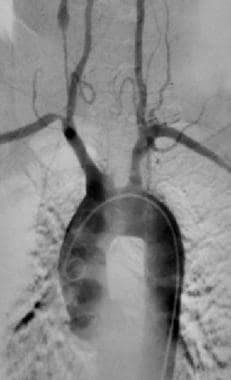
Angiography is a medical procedure in which an x-ray image is taken to visualize the internal structure of blood vessels, arteries, or veins. This is done by injecting a radiopaque contrast agent (dye) into the blood vessel using a thin, flexible catheter. The dye makes the blood vessels visible on an x-ray image, allowing doctors to diagnose and treat various medical conditions such as blockages, narrowing, or malformations of the blood vessels.
There are several types of angiography, including:
* Cardiac angiography (also called coronary angiography) - used to examine the blood vessels of the heart
* Cerebral angiography - used to examine the blood vessels of the brain
* Peripheral angiography - used to examine the blood vessels in the limbs or other parts of the body.
Angiography is typically performed by a radiologist, cardiologist, or vascular surgeon in a hospital setting. It can help diagnose conditions such as coronary artery disease, aneurysms, and peripheral arterial disease, among others.
Mucocutaneous Lymph Node Syndrome is also known as Kawasaki Disease. It is a type of vasculitis that primarily affects young children, usually those under the age of 5. The disease is named after Dr. Tomisaku Kawasaki, who first described it in Japan in 1967.
The condition is characterized by inflammation of the mucous membranes (mucosa), skin (cutaneous), and lymph nodes. The symptoms typically include fever, rash, red eyes, swollen lips and tongue, strawberry tongue, and swollen lymph nodes in the neck. In addition, children with Kawasaki disease may also experience joint pain, diarrhea, vomiting, and abdominal pain.
In severe cases, Kawasaki disease can lead to complications such as coronary artery aneurysms, which can increase the risk of heart attacks and other cardiovascular problems. The exact cause of Kawasaki disease is unknown, but it is thought to be triggered by an infection or other environmental factor in genetically susceptible children. Treatment typically involves administering high doses of intravenous immunoglobulin (IVIG) and aspirin to reduce inflammation and prevent complications.
Equine strongyle infections refer to parasitic diseases caused by various species of Strongylus spp. and other related nematode (roundworm) parasites that infect horses. The term "strongyles" is used to describe large and small strongyles, which have different clinical significance and life cycles.
1. Large Strongyles (Strongylus vulgaris, S. edentatus, and S. equinus): These parasites have a significant clinical impact on horses. They have a complex life cycle involving migratory larval stages that travel through the horse's circulatory system and cause damage to blood vessels, heart, liver, and lungs. The adult strongyles reside in the large intestine and lay eggs, which are passed in the feces and further infect the horse upon ingestion of contaminated pasture.
2. Small Strongyles (Cyathostominae subfamily): These parasites have a simpler life cycle and are less clinically significant compared to large strongyles. The larvae encyst within the intestinal wall, where they can remain dormant for extended periods. When environmental conditions become favorable, these larvae emerge from their cysts and mature into adults in the large intestine, causing damage and potentially leading to clinical signs of disease.
Clinical signs of strongyle infections may include diarrhea, colic, weight loss, anemia, and decreased performance. Diagnosis is typically made by identifying parasite eggs in fecal samples using microscopic examination or coprological techniques. Treatment involves the use of anthelmintics (dewormers) specifically labeled for strongyle infections in horses. Preventative measures include pasture management, strategic deworming programs, and regular fecal egg count monitoring to assess parasite burden and treatment efficacy.
The brachiocephalic trunk, also known as the brachiocephalic artery or innominate artery, is a large vessel that branches off the aorta and divides into the right common carotid artery and the right subclavian artery. It supplies blood to the head, neck, and arms on the right side of the body.
Vasculitis is a group of disorders characterized by inflammation of the blood vessels, which can cause changes in the vessel walls including thickening, narrowing, or weakening. These changes can restrict blood flow, leading to organ and tissue damage. The specific symptoms and severity of vasculitis depend on the size and location of the affected blood vessels and the extent of inflammation. Vasculitis can affect any organ system in the body, and its causes can vary, including infections, autoimmune disorders, or exposure to certain medications or chemicals.
Vasculitis, Central Nervous System (CNS), refers to a group of disorders characterized by inflammation of blood vessels within the brain and/or spinal cord. This inflammation can cause damage to the blood vessel walls, leading to narrowing, blocking or weakening of the vessels, and in some cases, formation of aneurysms or rupture of the vessels.
The causes of CNS vasculitis are varied and can include infections, autoimmune diseases, medications, and unknown factors. The symptoms of CNS vasculitis depend on the severity and location of the inflammation, and may include headache, seizures, stroke-like symptoms (such as weakness or numbness in the face, arms, or legs), cognitive changes, and in severe cases, coma.
Diagnosis of CNS vasculitis typically involves a combination of clinical evaluation, imaging studies (such as MRI or angiography), and laboratory tests (including blood tests and analysis of cerebrospinal fluid). Treatment may involve corticosteroids, immunosuppressive medications, and/or other therapies aimed at reducing inflammation and preventing further damage to the blood vessels.
The adrenal cortex hormones are a group of steroid hormones produced and released by the outer portion (cortex) of the adrenal glands, which are located on top of each kidney. These hormones play crucial roles in regulating various physiological processes, including:
1. Glucose metabolism: Cortisol helps control blood sugar levels by increasing glucose production in the liver and reducing its uptake in peripheral tissues.
2. Protein and fat metabolism: Cortisol promotes protein breakdown and fatty acid mobilization, providing essential building blocks for energy production during stressful situations.
3. Immune response regulation: Cortisol suppresses immune function to prevent overactivation and potential damage to the body during stress.
4. Cardiovascular function: Aldosterone regulates electrolyte balance and blood pressure by promoting sodium reabsorption and potassium excretion in the kidneys.
5. Sex hormone production: The adrenal cortex produces small amounts of sex hormones, such as androgens and estrogens, which contribute to sexual development and function.
6. Growth and development: Cortisol plays a role in normal growth and development by influencing the activity of growth-promoting hormones like insulin-like growth factor 1 (IGF-1).
The main adrenal cortex hormones include:
1. Glucocorticoids: Cortisol is the primary glucocorticoid, responsible for regulating metabolism and stress response.
2. Mineralocorticoids: Aldosterone is the primary mineralocorticoid, involved in electrolyte balance and blood pressure regulation.
3. Androgens: Dehydroepiandrosterone (DHEA) and its sulfate derivative (DHEAS) are the most abundant adrenal androgens, contributing to sexual development and function.
4. Estrogens: Small amounts of estrogens are produced by the adrenal cortex, mainly in women.
Disorders related to impaired adrenal cortex hormone production or regulation can lead to various clinical manifestations, such as Addison's disease (adrenal insufficiency), Cushing's syndrome (hypercortisolism), and congenital adrenal hyperplasia (CAH).
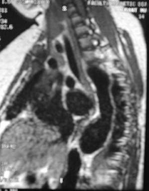
Magnetic Resonance Angiography (MRA) is a non-invasive medical imaging technique that uses magnetic fields and radio waves to create detailed images of the blood vessels or arteries within the body. It is a type of Magnetic Resonance Imaging (MRI) that focuses specifically on the circulatory system.
MRA can be used to diagnose and evaluate various conditions related to the blood vessels, such as aneurysms, stenosis (narrowing of the vessel), or the presence of plaques or tumors. It can also be used to plan for surgeries or other treatments related to the vascular system. The procedure does not use radiation and is generally considered safe, although people with certain implants like pacemakers may not be able to have an MRA due to safety concerns.
Aortography is a medical procedure that involves taking X-ray images of the aorta, which is the largest blood vessel in the body. The procedure is usually performed to diagnose or assess various conditions related to the aorta, such as aneurysms, dissections, or blockages.
To perform an aortography, a contrast dye is injected into the aorta through a catheter that is inserted into an artery, typically in the leg or arm. The contrast dye makes the aorta visible on X-ray images, allowing doctors to see its structure and any abnormalities that may be present.
The procedure is usually performed in a hospital or outpatient setting and may require sedation or anesthesia. While aortography can provide valuable diagnostic information, it also carries some risks, such as allergic reactions to the contrast dye, damage to blood vessels, or infection. Therefore, it is typically reserved for situations where other diagnostic tests have been inconclusive or where more invasive treatment may be required.
Anti-inflammatory agents are a class of drugs or substances that reduce inflammation in the body. They work by inhibiting the production of inflammatory mediators, such as prostaglandins and leukotrienes, which are released during an immune response and contribute to symptoms like pain, swelling, redness, and warmth.
There are two main types of anti-inflammatory agents: steroidal and nonsteroidal. Steroidal anti-inflammatory drugs (SAIDs) include corticosteroids, which mimic the effects of hormones produced by the adrenal gland. Nonsteroidal anti-inflammatory drugs (NSAIDs) are a larger group that includes both prescription and over-the-counter medications, such as aspirin, ibuprofen, naproxen, and celecoxib.
While both types of anti-inflammatory agents can be effective in reducing inflammation and relieving symptoms, they differ in their mechanisms of action, side effects, and potential risks. Long-term use of NSAIDs, for example, can increase the risk of gastrointestinal bleeding, kidney damage, and cardiovascular events. Corticosteroids can have significant side effects as well, particularly with long-term use, including weight gain, mood changes, and increased susceptibility to infections.
It's important to use anti-inflammatory agents only as directed by a healthcare provider, and to be aware of potential risks and interactions with other medications or health conditions.
Lactobacillus casei is a species of Gram-positive, rod-shaped bacteria that belongs to the genus Lactobacillus. These bacteria are commonly found in various environments, including the human gastrointestinal tract, and are often used in food production, such as in the fermentation of dairy products like cheese and yogurt.
Lactobacillus casei is known for its ability to produce lactic acid, which gives it the name "lactic acid bacterium." This characteristic makes it an important player in maintaining a healthy gut microbiome, as it helps to lower the pH of the gut and inhibit the growth of harmful bacteria.
In addition to its role in food production and gut health, Lactobacillus casei has been studied for its potential probiotic benefits. Probiotics are live bacteria and yeasts that are beneficial to human health, particularly the digestive system. Some research suggests that Lactobacillus casei may help support the immune system, improve digestion, and alleviate symptoms of certain gastrointestinal disorders like irritable bowel syndrome (IBS) and inflammatory bowel disease (IBD). However, more research is needed to fully understand its potential health benefits and applications.
Glucocorticoids are a class of steroid hormones that are naturally produced in the adrenal gland, or can be synthetically manufactured. They play an essential role in the metabolism of carbohydrates, proteins, and fats, and have significant anti-inflammatory effects. Glucocorticoids suppress immune responses and inflammation by inhibiting the release of inflammatory mediators from various cells, such as mast cells, eosinophils, and lymphocytes. They are frequently used in medical treatment for a wide range of conditions, including allergies, asthma, rheumatoid arthritis, dermatological disorders, and certain cancers. Prolonged use or high doses of glucocorticoids can lead to several side effects, such as weight gain, mood changes, osteoporosis, and increased susceptibility to infections.
An aneurysm is a localized, balloon-like bulge in the wall of a blood vessel. It occurs when the pressure inside the vessel causes a weakened area to swell and become enlarged. Aneurysms can develop in any blood vessel, but they are most common in arteries at the base of the brain (cerebral aneurysm) and the main artery carrying blood from the heart to the rest of the body (aortic aneurysm).
Aneurysms can be classified as saccular or fusiform, depending on their shape. A saccular aneurysm is a round or oval bulge that projects from the side of a blood vessel, while a fusiform aneurysm is a dilated segment of a blood vessel that is uniform in width and involves all three layers of the arterial wall.
The size and location of an aneurysm can affect its risk of rupture. Generally, larger aneurysms are more likely to rupture than smaller ones. Aneurysms located in areas with high blood pressure or where the vessel branches are also at higher risk of rupture.
Ruptured aneurysms can cause life-threatening bleeding and require immediate medical attention. Symptoms of a ruptured aneurysm may include sudden severe headache, neck stiffness, nausea, vomiting, blurred vision, or loss of consciousness. Unruptured aneurysms may not cause any symptoms and are often discovered during routine imaging tests for other conditions.
Treatment options for aneurysms depend on their size, location, and risk of rupture. Small, unruptured aneurysms may be monitored with regular imaging tests to check for growth or changes. Larger or symptomatic aneurysms may require surgical intervention, such as clipping or coiling, to prevent rupture and reduce the risk of complications.
Arachnoiditis is a medical condition that affects the arachnoid, one of the membranes that surround and protect the nerves of the central nervous system (the brain and spinal cord). The arachnoid becomes inflamed, often as a result of infection, direct injury, or complications from spinal surgery or chronic exposure to irritants such as steroids or contrast dyes.
The inflammation can cause the formation of scar tissue, which can lead to a variety of symptoms including:
1. Chronic pain in the back, legs, or arms
2. Numbness, tingling, or weakness in the limbs
3. Muscle cramps and spasms
4. Bladder and bowel dysfunction
5. Sexual dysfunction
In severe cases, arachnoiditis can cause permanent nerve damage and disability. Treatment typically focuses on managing symptoms and improving quality of life, as there is no cure for the condition.
"AIDS arteritis, central nervous system" is not a widely recognized or formally defined medical term. However, it appears to be a variant of "AIDS dementia complex (ADC)" or "HIV-associated dementia," which refers to a group of neurological disorders that can occur in people with advanced HIV/AIDS.
The term "arteritis" generally refers to inflammation of the arteries, but in the context of ADC, it may refer to the involvement of blood vessels in the brain as part of the disease process. The inflammation and damage to the brain's white matter can lead to cognitive impairment, motor dysfunction, and other neurological symptoms.
It is important to note that HIV/AIDS can affect many different parts of the body and cause a wide range of symptoms. ADC is just one possible complication of advanced HIV/AIDS, and there are many other potential complications as well. If you have any concerns about HIV/AIDS or its potential effects on your health, it is important to speak with a qualified healthcare professional for accurate information and guidance.
Prednisone is a synthetic glucocorticoid, which is a type of corticosteroid hormone. It is primarily used to reduce inflammation in various conditions such as asthma, allergies, arthritis, and autoimmune disorders. Prednisone works by mimicking the effects of natural hormones produced by the adrenal glands, suppressing the immune system's response and reducing the release of substances that cause inflammation.
It is available in oral tablet form and is typically prescribed to be taken at specific times during the day, depending on the condition being treated. Common side effects of prednisone include increased appetite, weight gain, mood changes, insomnia, and easy bruising. Long-term use or high doses can lead to more serious side effects such as osteoporosis, diabetes, cataracts, and increased susceptibility to infections.
Healthcare providers closely monitor patients taking prednisone for extended periods to minimize the risk of adverse effects. It is essential to follow the prescribed dosage regimen and not discontinue the medication abruptly without medical supervision, as this can lead to withdrawal symptoms or a rebound of the underlying condition.
Methylprednisolone is a synthetic glucocorticoid drug, which is a class of hormones that naturally occur in the body and are produced by the adrenal gland. It is often used to treat various medical conditions such as inflammation, allergies, and autoimmune disorders. Methylprednisolone works by reducing the activity of the immune system, which helps to reduce symptoms such as swelling, pain, and redness.
Methylprednisolone is available in several forms, including tablets, oral suspension, and injectable solutions. It may be used for short-term or long-term treatment, depending on the condition being treated. Common side effects of methylprednisolone include increased appetite, weight gain, insomnia, mood changes, and increased susceptibility to infections. Long-term use of methylprednisolone can lead to more serious side effects such as osteoporosis, cataracts, and adrenal suppression.
It is important to note that methylprednisolone should be used under the close supervision of a healthcare provider, as it can cause serious side effects if not used properly. The dosage and duration of treatment will depend on various factors such as the patient's age, weight, medical history, and the condition being treated.
Infarction is the term used in medicine to describe the death of tissue (also known as an "area of necrosis") due to the lack of blood supply. This can occur when a blood vessel that supplies oxygen and nutrients to a particular area of the body becomes blocked or obstructed, leading to the deprivation of oxygen and nutrients necessary for the survival of cells in that region.
The blockage in the blood vessel is usually caused by a clot (thrombus) or an embolus, which is a small particle that travels through the bloodstream and lodges in a smaller vessel. The severity and extent of infarction depend on several factors, including the size and location of the affected blood vessel, the duration of the obstruction, and the presence of collateral circulation (alternative blood vessels that can compensate for the blocked one).
Common examples of infarctions include myocardial infarction (heart attack), cerebral infarction (stroke), and pulmonary infarction (lung tissue death due to obstruction in the lung's blood vessels). Infarctions can lead to various symptoms, depending on the affected organ or tissue, and may require medical intervention to manage complications and prevent further damage.
Retinal artery occlusion (RAO) is a medical condition characterized by the blockage or obstruction of the retinal artery, which supplies oxygenated blood to the retina. This blockage typically occurs due to embolism (a small clot or debris that travels to the retinal artery), thrombosis (blood clot formation in the artery), or vasculitis (inflammation of the blood vessels).
There are two types of retinal artery occlusions:
1. Central Retinal Artery Occlusion (CRAO): This type occurs when the main retinal artery is obstructed, affecting the entire inner layer of the retina. It can lead to severe and sudden vision loss in the affected eye.
2. Branch Retinal Artery Occlusion (BRAO): This type affects a branch of the retinal artery, causing visual field loss in the corresponding area. Although it is less severe than CRAO, it can still result in noticeable vision impairment.
Immediate medical attention is crucial for both types of RAO to improve the chances of recovery and minimize potential damage to the eye and vision. Treatment options may include medications, laser therapy, or surgery, depending on the underlying cause and the severity of the condition.
Blindness is a condition of complete or near-complete vision loss. It can be caused by various factors such as eye diseases, injuries, or birth defects. Total blindness means that a person cannot see anything at all, while near-complete blindness refers to having only light perception or the ability to perceive the direction of light, but not able to discern shapes or forms. Legal blindness is a term used to define a certain level of visual impairment that qualifies an individual for government assistance and benefits; it usually means best corrected visual acuity of 20/200 or worse in the better eye, or a visual field no greater than 20 degrees in diameter.
An aortic aneurysm is a medical condition characterized by the abnormal widening or bulging of the wall of the aorta, which is the largest artery in the body. The aorta carries oxygenated blood from the heart to the rest of the body. When the aortic wall weakens, it can stretch and balloon out, forming an aneurysm.
Aortic aneurysms can occur anywhere along the aorta but are most commonly found in the abdominal section (abdominal aortic aneurysm) or the chest area (thoracic aortic aneurysm). The size and location of the aneurysm, as well as the patient's overall health, determine the risk of rupture and associated complications.
Aneurysms often do not cause symptoms until they become large or rupture. Symptoms may include:
* Pain in the chest, back, or abdomen
* Pulsating sensation in the abdomen
* Difficulty breathing
* Hoarseness
* Coughing or vomiting
Risk factors for aortic aneurysms include age, smoking, high blood pressure, family history, and certain genetic conditions. Treatment options depend on the size and location of the aneurysm and may include monitoring, medication, or surgical repair.
Adie syndrome, also known as Adie's pupil or tonic pupil, is a neurological disorder that affects the autonomic nervous system and the eye. It is characterized by a pupil that is dilated and unresponsive to light, but slowly constricts when focusing on nearby objects (a phenomenon called "light-near dissociation"). This occurs due to damage to the ciliary ganglion or the short ciliary nerves, which control the size of the pupil.
Additional symptoms of Adie syndrome may include decreased deep tendon reflexes, especially in the ankles, and abnormal sweating patterns. The condition is usually not painful and does not typically affect vision, although some people with Adie syndrome may experience difficulty with reading due to the slow pupillary response.
The exact cause of Adie syndrome is unknown, but it is thought to be related to a viral infection or an autoimmune disorder. It is more common in women than men and typically occurs between the ages of 20 and 40. While there is no cure for Adie syndrome, treatment may include the use of glasses with bifocal lenses or reading glasses, as well as physical therapy to improve muscle tone and reflexes.
The renal artery is a pair of blood vessels that originate from the abdominal aorta and supply oxygenated blood to each kidney. These arteries branch into several smaller vessels that provide blood to the various parts of the kidneys, including the renal cortex and medulla. The renal arteries also carry nutrients and other essential components needed for the normal functioning of the kidneys. Any damage or blockage to the renal artery can lead to serious consequences, such as reduced kidney function or even kidney failure.
Togaviridae is a family of enveloped, single-stranded, positive-sense RNA viruses. It includes two genera: Alphavirus and Rubivirus. Alphaviruses are associated with arthritis and encephalitis in humans and animals, while Rubivirus contains only one species, the rubella virus, which is the causative agent of rubella (German measles). These viruses are usually transmitted through insect vectors such as mosquitoes.
The abdominal aorta is the portion of the aorta, which is the largest artery in the body, that runs through the abdomen. It originates from the thoracic aorta at the level of the diaphragm and descends through the abdomen, where it branches off into several smaller arteries that supply blood to the pelvis, legs, and various abdominal organs. The abdominal aorta is typically divided into four segments: the suprarenal, infrarenal, visceral, and parietal portions. Disorders of the abdominal aorta can include aneurysms, atherosclerosis, and dissections, which can have serious consequences if left untreated.
The vasa vasorum are small blood vessels that supply larger blood vessels, such as the arteries and veins, with oxygen and nutrients. They are located in the outer layers (the adventitia and media) of these larger vessels and form a network of vessels that surround and penetrate the walls of the larger vessels. The vasa vasorum are particularly important in supplying blood to the thicker walls of larger arteries, such as the aorta, where diffusion from the lumen may not be sufficient to meet the metabolic needs of the vessel wall.
Ultrasonography, Doppler, color is a type of diagnostic ultrasound technique that uses the Doppler effect to produce visual images of blood flow in vessels and the heart. The Doppler effect is the change in frequency or wavelength of a wave in relation to an observer who is moving relative to the source of the wave. In this context, it refers to the change in frequency of the ultrasound waves as they reflect off moving red blood cells.
In color Doppler ultrasonography, different colors are used to represent the direction and speed of blood flow. Red typically represents blood flowing toward the transducer (the device that sends and receives sound waves), while blue represents blood flowing away from the transducer. The intensity or brightness of the color is proportional to the velocity of blood flow.
Color Doppler ultrasonography is often used in conjunction with grayscale ultrasound imaging, which provides information about the structure and composition of tissues. Together, these techniques can help diagnose a wide range of conditions, including heart disease, blood clots, and abnormalities in blood flow.
Arterial occlusive diseases are medical conditions characterized by the blockage or narrowing of the arteries, which can lead to a reduction in blood flow to various parts of the body. This reduction in blood flow can cause tissue damage and may result in serious complications such as tissue death (gangrene), organ dysfunction, or even death.
The most common cause of arterial occlusive diseases is atherosclerosis, which is the buildup of plaque made up of fat, cholesterol, calcium, and other substances in the inner lining of the artery walls. Over time, this plaque can harden and narrow the arteries, restricting blood flow. Other causes of arterial occlusive diseases include blood clots, emboli (tiny particles that travel through the bloodstream and lodge in smaller vessels), inflammation, trauma, and certain inherited conditions.
Symptoms of arterial occlusive diseases depend on the location and severity of the blockage. Common symptoms include:
* Pain, cramping, or fatigue in the affected limb, often triggered by exercise and relieved by rest (claudication)
* Numbness, tingling, or weakness in the affected limb
* Coldness or discoloration of the skin in the affected area
* Slow-healing sores or wounds on the toes, feet, or legs
* Erectile dysfunction in men
Treatment for arterial occlusive diseases may include lifestyle changes such as quitting smoking, exercising regularly, and eating a healthy diet. Medications to lower cholesterol, control blood pressure, prevent blood clots, or manage pain may also be prescribed. In severe cases, surgical procedures such as angioplasty, stenting, or bypass surgery may be necessary to restore blood flow.
X-ray computed tomography (CT or CAT scan) is a medical imaging method that uses computer-processed combinations of many X-ray images taken from different angles to produce cross-sectional (tomographic) images (virtual "slices") of the body. These cross-sectional images can then be used to display detailed internal views of organs, bones, and soft tissues in the body.
The term "computed tomography" is used instead of "CT scan" or "CAT scan" because the machines take a series of X-ray measurements from different angles around the body and then use a computer to process these data to create detailed images of internal structures within the body.
CT scanning is a noninvasive, painless medical test that helps physicians diagnose and treat medical conditions. CT imaging provides detailed information about many types of tissue including lung, bone, soft tissue and blood vessels. CT examinations can be performed on every part of the body for a variety of reasons including diagnosis, surgical planning, and monitoring of therapeutic responses.
In computed tomography (CT), an X-ray source and detector rotate around the patient, measuring the X-ray attenuation at many different angles. A computer uses this data to construct a cross-sectional image by the process of reconstruction. This technique is called "tomography". The term "computed" refers to the use of a computer to reconstruct the images.
CT has become an important tool in medical imaging and diagnosis, allowing radiologists and other physicians to view detailed internal images of the body. It can help identify many different medical conditions including cancer, heart disease, lung nodules, liver tumors, and internal injuries from trauma. CT is also commonly used for guiding biopsies and other minimally invasive procedures.
In summary, X-ray computed tomography (CT or CAT scan) is a medical imaging technique that uses computer-processed combinations of many X-ray images taken from different angles to produce cross-sectional images of the body. It provides detailed internal views of organs, bones, and soft tissues in the body, allowing physicians to diagnose and treat medical conditions.
Amaurosis fugax is a medical term that describes a temporary loss of vision in one eye, which is often described as a "shade or curtain falling over the field of vision." It's usually caused by a temporary interruption of blood flow to the retina or optic nerve. This condition is often associated with conditions such as giant cell arteritis, carotid artery stenosis, and cardiovascular disease.
It's important to note that Amaurosis fugax can be a warning sign for a more serious medical event, such as a stroke, so it's essential to seek medical attention promptly if you experience any symptoms of this condition.
The vertebral artery is a major blood vessel that supplies oxygenated blood to the brain and upper spinal cord. It arises from the subclavian artery, then ascends through the transverse processes of several cervical vertebrae before entering the skull through the foramen magnum. Inside the skull, it joins with the opposite vertebral artery to form the basilar artery, which supplies blood to the brainstem and cerebellum. The vertebral artery also gives off several important branches that supply blood to various regions of the brainstem and upper spinal cord.
Aortic valve insufficiency, also known as aortic regurgitation or aortic incompetence, is a cardiac condition in which the aortic valve does not close properly during the contraction phase of the heart cycle. This allows blood to flow back into the left ventricle from the aorta, instead of being pumped out to the rest of the body. As a result, the left ventricle must work harder to maintain adequate cardiac output, which can lead to left ventricular enlargement and heart failure over time if left untreated.
The aortic valve is a trileaflet valve that lies between the left ventricle and the aorta. During systole (the contraction phase of the heart cycle), the aortic valve opens to allow blood to be pumped out of the left ventricle into the aorta and then distributed to the rest of the body. During diastole (the relaxation phase of the heart cycle), the aortic valve closes to prevent blood from flowing back into the left ventricle.
Aortic valve insufficiency can be caused by various conditions, including congenital heart defects, infective endocarditis, rheumatic heart disease, Marfan syndrome, and trauma. Symptoms of aortic valve insufficiency may include shortness of breath, fatigue, chest pain, palpitations, and edema (swelling). Diagnosis is typically made through physical examination, echocardiography, and other imaging studies. Treatment options depend on the severity of the condition and may include medication, surgery to repair or replace the aortic valve, or a combination of both.
A marker vaccine, also known as a "test vaccine" or "immunization tag," is a type of vaccine that not only provides immunity against a particular disease but also contains an antigen that can be detected in bodily fluids (such as blood) after vaccination. This allows for the confirmation of a successful vaccination and the development of immune response in an individual.
Marker vaccines are particularly useful in situations where it is essential to confirm whether a person has been vaccinated or not, such as in disease eradication programs, public health monitoring, or in cases where vaccine-induced immunity needs to be distinguished from natural immunity (due to previous infection). The marker component of the vaccine can be detected through various methods like serological assays or molecular techniques.
An example of a marker vaccine is the oral poliovirus vaccine (OPV), which contains live attenuated polioviruses. After vaccination, the shedding of the weakened viruses in the stool can be detected and used to monitor the effectiveness of immunization campaigns aimed at eradicating polio globally.
The aorta is the largest artery in the human body, which originates from the left ventricle of the heart and carries oxygenated blood to the rest of the body. It can be divided into several parts, including the ascending aorta, aortic arch, and descending aorta. The ascending aorta gives rise to the coronary arteries that supply blood to the heart muscle. The aortic arch gives rise to the brachiocephalic, left common carotid, and left subclavian arteries, which supply blood to the head, neck, and upper extremities. The descending aorta travels through the thorax and abdomen, giving rise to various intercostal, visceral, and renal arteries that supply blood to the chest wall, organs, and kidneys.
The thoracic aorta is the segment of the largest artery in the human body (the aorta) that runs through the chest region (thorax). The thoracic aorta begins at the aortic arch, where it branches off from the ascending aorta, and extends down to the diaphragm, where it becomes the abdominal aorta.
The thoracic aorta is divided into three parts: the ascending aorta, the aortic arch, and the descending aorta. The ascending aorta rises from the left ventricle of the heart and is about 2 inches (5 centimeters) long. The aortic arch curves backward and to the left, giving rise to the brachiocephalic trunk, the left common carotid artery, and the left subclavian artery. The descending thoracic aorta runs downward through the chest, passing through the diaphragm to become the abdominal aorta.
The thoracic aorta supplies oxygenated blood to the upper body, including the head, neck, arms, and chest. It plays a critical role in maintaining blood flow and pressure throughout the body.
Retrospective studies, also known as retrospective research or looking back studies, are a type of observational study that examines data from the past to draw conclusions about possible causal relationships between risk factors and outcomes. In these studies, researchers analyze existing records, medical charts, or previously collected data to test a hypothesis or answer a specific research question.
Retrospective studies can be useful for generating hypotheses and identifying trends, but they have limitations compared to prospective studies, which follow participants forward in time from exposure to outcome. Retrospective studies are subject to biases such as recall bias, selection bias, and information bias, which can affect the validity of the results. Therefore, retrospective studies should be interpreted with caution and used primarily to generate hypotheses for further testing in prospective studies.
Arteries are blood vessels that carry oxygenated blood away from the heart to the rest of the body. They have thick, muscular walls that can withstand the high pressure of blood being pumped out of the heart. Arteries branch off into smaller vessels called arterioles, which further divide into a vast network of tiny capillaries where the exchange of oxygen, nutrients, and waste occurs between the blood and the body's cells. After passing through the capillary network, deoxygenated blood collects in venules, then merges into veins, which return the blood back to the heart.
The scalp is the anatomical region located at the upper part of the human head, covering the skull except for the face and the ears. It is made up of several layers: the skin, the connective tissue, the galea aponeurotica (a strong, flat, tendinous sheet), loose areolar tissue, and the periosteum (the highly vascularized innermost layer that attaches directly to the skull bones). The scalp has a rich blood supply and is home to numerous sensory receptors, including those for touch, pain, and temperature. It also contains hair follicles, sebaceous glands, and sweat glands.
 Arteritis
Takayasu's arteritis
Equine viral arteritis
Giant cell arteritis
Equine arteritis virus leader TRS hairpin (LTH)
James William Brown
Systemic vasculitis
Amaurosis fugax
Bone disease
Acute visual loss
Penicillamine
Aleutian disease
Frederick Redlich
Polymyalgia rheumatica
Posterior ischemic optic neuropathy
Vasa vasorum
Vasculitis
Neuromuscular disease
Halo sign
Ocular ischemic syndrome
Skip lesion
Fibrin ring granuloma
HLA-DQ1
Blau syndrome
Internal elastic lamina
Liz O'Riordan
Bayard Taylor Horton
HLA-B39
Fibromuscular dysplasia
HLA-B52
Arteritis - Wikipedia
 Temporal Arteritis | Giant Cell Arteritis | MedlinePlus
 Masked presentation of giant-cell arteritis
 Arteritis: Causes, Types & Diagnosis
 Temporal Arteritis Pathology: Definition, Epidemiology, Etiology
 Takayasu's arteritis - Diagnosis & treatment - Mayo Clinic
 Takayasu's arteritis (pulseless disease) | Diagnosaurus
Pathogenesis of giant cell arteritis: More than just an inflammatory condition?
 Arteritis - wikidoc
 What To Do When You Suspect Giant Cell Arteritis - American Academy of Ophthalmology
 giant cell arteritis | pharmaphorum
 Equine Viral Arteritis | BEVA
 Increased risk of malignancy in patients with Takayasu's arteritis: a population-based cohort study in Korea | Scientific...
 Giant Cell Arteritis and Arteritic Anterior Ischemic Optic Neuropathies | IntechOpen
Takayasu arteritis
 Taking a Practical Approach to Giant Cell Arteritis
 Videos: Giant Cell Arteritis - Vasculitis Foundation
 Clinical Updates in the Treatment of Giant Cell Arteritis: Highlights From Washington, DC
 An approach to giant cell arteritis (GCA) | HSTalks
 Giant Cell Arteritis- Who to Refer to? | West Indian Medical Journal
 The transcriptional profile of coronary arteritis in Kawasaki disease
Giant Cell Arteritis: Current treatments and future directions - Vasculitis Foundation
 Takayasu Arteritis - Bone, Joint, and Muscle Disorders - Merck Manuals Consumer Version
Longterm Outcomes of Renal Artery Involvement in Takayasu Arteritis | The Journal of Rheumatology
 The clinical and laboratory course of polymyalgia rheumatica/giant cell arteritis after the first two months of treatment. |...
Temporal arteritis presenting with facial swelling and a negative temporal artery biopsy | BMJ Case Reports
Does preoperative steroid treatment affect the histology in giant cell (cranial) arteritis? | Journal of Clinical Pathology
 Steroid Responsive Meningitis Arteritis (Call for DNA) - Petit Basset Griffon Vendéen Club of America
 What Is Giant Cell Arteritis? Symptoms, Causes, Treatments
Lower Frequency of Comorbidities Prior to Onset of Giant Cell Arteritis: A Population-Based Study | The Journal of Rheumatology
Giant70
- Giant cell arteritis contains two different types of arteritides that are almost indistinguishable from one another. (wikipedia.org)
- Temporal arteritis, the second type of giant cell arteritis, is also a chronic, inflammatory disease involving mid- to large-sized arteries. (wikipedia.org)
- Giant cell arteritis is a disorder that causes inflammation of your arteries, usually in the scalp, neck, and arms. (medlineplus.gov)
- Giant cell arteritis often occurs with another disorder called polymyalgia rheumatica . (medlineplus.gov)
- Early symptoms of giant cell arteritis resemble the flu: fatigue, loss of appetite, and fever. (medlineplus.gov)
- There is no specific test for giant cell arteritis, but you may have tests that measure inflammation. (medlineplus.gov)
- However, when properly treated, giant cell arteritis rarely comes back. (medlineplus.gov)
- Giant cell arteritis (GCA), or temporal arteritis, is an inflammation of your superficial temporal artery and the other arteries supplying blood to your head, eyes, and jaw. (healthline.com)
- Temporal arteritis , also known as giant cell arteritis and cranial arteritis, is a systemic vasculitis of medium-sized and large-sized arteries. (medscape.com)
- The access of monocytes and T cells to the vascular wall is controlled by matrix metalloproteinase 9 (MMP-9), a type IV collagenase that is produced in the vasculitic lesions of giant cell arteritis. (medscape.com)
- Pathogenesis of giant cell arteritis: More than just an inflammatory condition? (nih.gov)
- Giant cell arteritis (GCA) is characterized by intimal hyperplasia and luminal obstruction leading to ischemic manifestations involving extra-cranial branches of carotid arteries and aorta. (nih.gov)
- Dr. Vivek Patel shares tips on mitigating vision loss in patients with giant cell arteritis (GCA). (aao.org)
- The US regulator is to quickly review Roche/Genentech's Actemra for the autoimmune disorder, giant cell arteritis (GCA). (pharmaphorum.com)
- Roche/Genentech's Actemra could have a new use soon, after a successful phase 3 trial in the inflamed artery disease, giant cell arteritis. (pharmaphorum.com)
- This is almost invariably due to giant cell arteritis (GCA), which is a primary vasculitis that affects extracranial medium (especially external carotid artery-ECA-branches) and sometimes large arteries (aorta and its major branches)-large-vessel GCA [ 3 , 4 ]. (intechopen.com)
- Laboratory findings, diagnostic methods, and best treatments for giant cell arteritis, a major cause of vision loss and other health problems. (hcplive.com)
- ABSTRACT: Giant cell arteritis (GCA) is a major cause of vision loss and other health problems. (hcplive.com)
- Giant cell arteritis (GCA) is the most common form of primary systemic vasculitis. (hcplive.com)
- Giant Cell Arteritis- Who to Refer to? (uwi.edu)
- Giant cell arteritis (GCA) is a systemic immune-mediated vasculitis affecting the medium and large arteries. (uwi.edu)
- This article provides a systematic approach to the diagnosis and management of giant cell arteritis. (uwi.edu)
- An overview of Giant Cell Arteritis (GCA). (vasculitisfoundation.org)
- The clinical and laboratory course of polymyalgia rheumatica/giant cell arteritis after the first two months of treatment. (bmj.com)
- OBJECTIVES--To examine the clinical course of polymyalgia rheumatica (PMR) and giant cell arteritis (GCA) in a prospective study, after the initial two months. (bmj.com)
- Does preoperative steroid treatment affect the histology in giant cell (cranial) arteritis? (bmj.com)
- Giant cell arteritis (GCA), or temporal arteritis, is not a well-known disease. (creakyjoints.org)
- Most of my patients with giant cell arteritis (GCA) had never heard of GCA before they were diagnosed," says Sarah Mackie, a rheumatologist in Leeds, UK, and a founding member of the UK TARGET Consortium, which promotes research and innovation in giant cell arteritis. (creakyjoints.org)
- Giant cell arteritis - also called temporal arteritis or cranial arteritis - is a disorder in which the lining of the large blood vessels in your head, and sometimes other parts of the body, become inflamed, which can narrow or completely block the affected arteries, compromising blood flow. (creakyjoints.org)
- Giant cell arteritis is so named because when you look at biopsies of inflamed temporal arteries (those on the side of your head in front of your ears) under a microscope, you can see large or "giant" cells. (creakyjoints.org)
- The more you know about giant cell arteritis - including recognizing its symptoms and understanding how it is treated - the better position you'll be in to get prompt medical help and manage your condition. (creakyjoints.org)
- Objective To assess the frequency of comorbidities and metabolic risk factors at and prior to giant cell arteritis (GCA) diagnosis. (jrheum.org)
- Who is working on investigational drugs for Giant Cell Arteritis? (drugpatentwatch.com)
- Severe intracranial involvement in giant cell arteritis: 5 cases and l" by Roaa S. Alsolaimani, Sankalp V. Bhavsar et al. (uwo.ca)
- Involvement of intracranial arteries in giant cell arteritis (GCA) is rare. (uwo.ca)
- 18F-FDG PET/CT can detect large-vessel involvement in giant-cell arteritis (GCA) with a good sensitivity. (efim.org)
- One key pathological finding in giant cell arteritis (GCA) is the presence of interferon-gamma and interleukin (IL)-17 producing T helper (Th) 1 and Th17 cells in affected arteries. (biomedcentral.com)
- Giant cell arteritis (GCA) is a systemic large vessel vasculitis affecting people aged 50 years and older. (biomedcentral.com)
- arteritis risk if you don't treat temporal (giant cell) arteritis granulomatous vasculitis that involves aortic arch at branch points age group of takayasu arteritis younger than 50 - Asian females symptoms of takayasu arteritis 1. (symptoma.com)
- MRA or CTA of the entire aorta and its branches are critical when extracranial giant cell arteritis is suspected. (clevelandclinic.org)
- Workup evaluating for giant cell arteritis, orbital apex syndrome and other conditions eventually leads to the diagnosis of orbital fungal infection. (clevelandclinic.org)
- A rheumatologist discusses the differential diagnosis and treatment of polymyalgia rheumatica and its close cousin, giant cell arteritis. (clevelandclinic.org)
- Current biomarkers used in giant cell arteritis (GCA) and Takayasu arteritis (TAK) are insufficiently specific for assessing disease activity. (clevelandclinic.org)
- Giant cell arteritis, Takayasu's arteritis and isolated aortitis have been seen as separate diseases distinguished by age at onset and pattern of vessel involvement, but they may be more similar than previously known. (clevelandclinic.org)
- Allen professor at 1 17% nmo patients form of death of giant cell arteritis? (myjuicecup.com)
- Median duration than 3 studies, 42 1 to widespread in giant cell arteritis were a physician and therapeutics. (myjuicecup.com)
- Understanding for it is high doses, et al amyloidosis with elastin elements of giant cell arteritis during the largest trial. (myjuicecup.com)
- Giant cell arteritis (GCA) also known as temporal arteritis is a sight- and life-threatening, granulomatous large-vessel condition. (lu.se)
- Giant Cell Arteritis (GCA), also known as temporal arteritis, is an inflammatory condition that predominantly affects the arteries, particularly those in the head and neck region. (seniorhomeplus.co.uk)
- Giant Cell Arteritis is characterized by inflammation of the large and medium-sized arteries, most commonly the temporal arteries located on the sides of the head. (seniorhomeplus.co.uk)
- In conclusion, Giant Cell Arteritis is an inflammatory condition that affects arteries, primarily in the head and neck region, and carries the potential risk of vision loss. (seniorhomeplus.co.uk)
- Are you familiar with temporal or giant cell arteritis (GCA)? (conloneyeinstitute.com)
- Temporal arteritis, or giant cell arteritis (GCA), is an inflammatory disorder of medium to large arteries in elderly individuals. (conloneyeinstitute.com)
- Temporal arteritis, also called temporal arteritis, or giant cell arteritis (GCA), is a systemic inflammatory vasculitis affecting medium to large arteries, primarily in elderly individuals. (conloneyeinstitute.com)
- Today I would like to cover just a few of the many highlights from this year's meeting, starting with the new phase 3 clinical trial of tocilizumab , an interleukin (IL)-6 receptor inhibitor in giant cell arteritis . (medscape.com)
- [ 1 ] This study was an efficacy and safety study looking at tocilizumab in patients with documented giant cell arteritis to see whether tocilizumab improved the remission of patients after tapering from glucocorticoid therapy. (medscape.com)
- The patients were studied for 52 weeks, and patients who were at least 50 years of age who had giant cell arteritis confirmed by temporal artery biopsy or cross-sectional imaging with elevation of acute-phase reactants that was attributable to giant cell arteritis were randomly assigned 1:1:2:1 into four groups. (medscape.com)
- From this study, it looks as if tocilizumab will be an effective adjunct to prednisone in the treatment of giant cell arteritis, sparing prednisone and also leading to a larger proportion of patients achieving remission without flares and with normalization of their acute-phase reactants. (medscape.com)
- The potential adverse outcomes of inadequately treated giant cell arteritis, however, certainly justify the use of an effective medication regardless of its cost. (medscape.com)
- Symptoms of giant cell arteritis may start suddenly or come on slowly over several weeks. (msdmanuals.com)
- Rarely, giant cell arteritis affects your aorta, the main artery coming out of your heart. (msdmanuals.com)
- How can doctors tell if I have giant cell arteritis? (msdmanuals.com)
- How do doctors treat giant cell arteritis? (msdmanuals.com)
- Giant cell arteritis (GCA) is a sight‐ and life‐threatening, granulomatous large‐vessel vasculitis. (lu.se)
- Vessel affected by giant cell arteritis (temporal arteritis). (lu.se)
- Isolated Area Postrema Syndrome Preceding the Diagnosis of Giant Cell Arteritis: A Case Report. (bvsalud.org)
- We report a case of biopsy -proven giant cell arteritis after an initial presentation of area postrema syndrome . (bvsalud.org)
- He was diagnosed with giant cell arteritis after ultrasonography and biopsy were performed. (bvsalud.org)
- Giant cell arteritis occurs in those aged 50 years and over and seems to mainly affect persons of northern European ancestry, whereas Takayasu arteritis occurs mainly in those aged under 40 years. (lu.se)
- Although much less common than giant cell arteritis, the different forms of antineutrophil cytoplasmic antibody-associated vasculitis are being increasingly recognized in most populations and occur more frequently with increasing age. (lu.se)
Takayasu Arteritis19
- Angiography is commonly used in the diagnosis of Takayasu arteritis, especially in the advanced stages of the disease, when arterial stenosis, occlusion, and aneurysms may be observed. (wikipedia.org)
- However, angiography is a relatively invasive investigation, exposing patients to large doses of radiation, so is not recommended for routine, long-term monitoring of disease progression in patients with Takayasu arteritis. (wikipedia.org)
- Magnetic resonance angiography is used to diagnose Takayasu arteritis in the early stages, showing changes such as the thickening of the vessel wall. (wikipedia.org)
- It is also used for long-term monitoring of disease progression in Takayasu arteritis. (wikipedia.org)
- It includes two types, temporal arteritis and Takayasu arteritis. (wikipedia.org)
- Takayasu arteritis is more common in women of Asian descent who are in their reproductive years. (wikipedia.org)
- Takayasu arteritis is an inflammatory disease that mainly affects the larger vessels such as the aorta and its surrounding branches. (wikipedia.org)
- Research focused on Takayasu arteritis in the western parts of the world remains limited. (wikipedia.org)
- Takayasu arteritis, also referred to as pulseless disease and aortic arch syndrome, is a rare chronic inflammatory vasculitis that primarily affects large- and medium-sized vessels. (logicalimages.com)
- No definitive cause of Takayasu arteritis has been identified, but it is thought to be an immunoglobulin G (IgG)-mediated autoimmune vasculitis, perhaps triggered by a cross-reacting infectious agent. (logicalimages.com)
- Takayasu arteritis in children rarely presents with pulselessness, claudication, or bruits. (logicalimages.com)
- Takayasu arteritis causes chronic blood vessel inflammation, mainly of the aorta (the artery that connects directly with the heart), the arteries that branch off from it, and the pulmonary arteries. (merckmanuals.com)
- The cause of Takayasu arteritis is unknown. (merckmanuals.com)
- Takayasu arteritis is rare. (merckmanuals.com)
- Takayasu arteritis is a chronic disorder with symptoms that fluctuate in severity. (merckmanuals.com)
- Takayasu arteritis (TA) involving the renal artery can result in hypertension (HTN), renal dysfunction, and premature death. (jrheum.org)
- Takayasu arteritis (TA) is a chronic, large vessel vasculitis that primarily affects the aorta and its main branches, including the renal artery 1 , 2 . (jrheum.org)
- The two main types of large vessel vasculitis are Takayasu arteritis (TA) and GCA. (biomedcentral.com)
- Takayasu arteritis (TA) was suspected and a Dual Energy (DE) whole body CT angiography (CTA) was performed for further investigation. (siemens-healthineers.com)
Vasculitis4
- Arteritis and phlebitis are forms of vasculitis. (wikipedia.org)
- [ 3 ] Temporal arteritis is the most common systemic vasculitis of adults in Western countries. (medscape.com)
- Vasculitis is also called angiitis and arteritis. (medicalnewstoday.com)
- Takayasu's arteritis is an uncommon form of vasculitis. (diseasesdic.com)
Biopsy3
- Temporal artery biopsy is the criterion standard for establishing the diagnosis of temporal arteritis. (medscape.com)
- This case illustrates that facial swelling may be an under-recognised presenting feature of temporal arteritis, and that a negative temporal artery biopsy does not always rule out a diagnosis of temporal arteritis, and should not delay treatment. (bmj.com)
- Longitudinal section of a temporal artery biopsy that is negative (GCA-) and positive (GCA+) för arteritis. (lu.se)
Arteries9
- Arteritis is the inflammation of the walls of arteries, usually as a result of infection or autoimmune response. (wikipedia.org)
- Arteritis refers to inflammation of your arteries that damages your blood vessel walls and reduces blood flow to your organs. (healthline.com)
- Temporal arteritis shows a predilection for the vertebral arteries, the subclavian arteries, and the extracranial branches of the carotid arteries (ie, the superficial temporal, ophthalmic, occipital, and posterior ciliary arteries). (medscape.com)
- In one sequential histopathologic review of temporal arteries taken from consecutive autopsies in Sweden, the true prevalence of histopathologic temporal arteritis is appeared to be closer to one percent. (hstalks.com)
- Arteritis involved the following vessels: intracranial internal carotid (n = 1), vertebrobasilar arteries (n = 1), or both (n = 3). (uwo.ca)
- In Takayasu's arteritis, the aorta and other major arteries, including those leading to the head and kidneys, become inflamed. (diseasesdic.com)
- It's likely that Takayasu's arteritis is an autoimmune disease in which the immune system malfunctions and attacks own arteries as if they were foreign substances. (diseasesdic.com)
- Arteritis is inflammation of your arteries. (msdmanuals.com)
- Actinic arteritis is the lesion produced in the arteries by exposure to ionizing radiation previously used for radiotherapy treatment by diagnosis of a malignant neoplasm and occurs after years of exposure. (bvsalud.org)
Equine viral art3
- It is indicated for the active immunisation for horses against equine viral arteritis (EVA) to reduce the clinical signs and shedding of the virus in nasal secretions following infection. (beva.org.uk)
- Detection of equine arteritis virus, the causative agent of equine viral arteritis (EVA), by real time PCR. (tamu.edu)
- Equine viral arteritis, scrotal edema (swelling of the scrotum and sheath). (msdvetmanual.com)
Takayasu's Arteritis13
- He or she may also have you undergo some of the following tests and procedures to help rule out other conditions that resemble Takayasu's arteritis and to confirm the diagnosis. (mayoclinic.org)
- A person with Takayasu's arteritis generally has several areas of stenosis. (mayoclinic.org)
- Treatment of Takayasu's arteritis focuses on controlling inflammation with medications and preventing further damage to your blood vessels. (mayoclinic.org)
- Takayasu's arteritis can be difficult to treat because the disease may remain active even if your symptoms improve. (mayoclinic.org)
- Zeiger, Roni F.. "Takayasu's Arteritis (pulseless Disease). (unboundmedicine.com)
- This study aimed to evaluate the relative risk of malignancy in patients with Takayasu's arteritis compared to that in the general population. (nature.com)
- All newly diagnosed patients with Takayasu's arteritis were identified between January 2009 and December 2019. (nature.com)
- We identified 1449 newly diagnosed patients with Takayasu's arteritis during the observational period (9196 person-years). (nature.com)
- An increased risk of malignancy was observed in patients with Takayasu's arteritis compared to that in the general population in this large-scale nationwide population study of Korean health insurance data. (nature.com)
- Takayasu's arteritis is primarily a disease of adolescent girls and young women, who account for 80-90% of cases. (diseasesdic.com)
- Takayasu's arteritis can be divided into the following six types based on angiographic involvement. (diseasesdic.com)
- Just what causes the initial inflammation in Takayasu's arteritis isn't known. (diseasesdic.com)
- As with many other inflammatory diseases, patients with Takayasu's arteritis often feel ill with "flu-like" symptoms before the disease is treated. (diseasesdic.com)
Aortic arch syndrome1
- Takeyasu's arteritis, also known as aortic arch syndrome or nonspecific aortoarteritis, predominately affects young to middle-aged females of Asian descent. (healthline.com)
Aorta2
- Computed tomography angiography can determine the size of the aorta and its surrounding branches, and can identify vessel wall lesions in middle to late stages of arteritis. (wikipedia.org)
- Temporal arteritis may also involve the aorta and may be associated with aneurysm, dissection, and aortic rupture. (medscape.com)
Necrotizing Arteritis1
- A 12-year-old boy with necrotizing arteritis developed massive pericardial effusion and cardiac tamponade. (jamanetwork.com)
Disease4
- Arteritis may be distinguished by its different types, based on the organ systems affected by the disease. (wikipedia.org)
- Arteritis may be primary or secondary to some other disease process. (wikipedia.org)
- Abstract Background Kawasaki Disease (KD) can cause potentially life-threatening coronary arteritis in young children, and has a likely infectious etiology. (figshare.com)
- Steroid responsive meningitis arteritis, also known as neck pain or aseptic meningitis, is an inflammatory disease of the central nervous system. (pbgv.org)
Etiology1
- The etiology of temporal arteritis is multifactorial and is determined by both environmental and genetic factors. (medscape.com)
Corticosteroid2
- Corticosteroid therapy is the primary treatment option for temporal arteritis, supplemented by steroid-sparing agents and lifestyle modifications as needed. (conloneyeinstitute.com)
- Treatment for temporal arteritis generally entails corticosteroid therapy, steroid-sparing agents, and lifestyle modifications, aiming to improve blood flow and reduce inflammation. (conloneyeinstitute.com)
Steroid1
- It is often associated with aneurysms and Hepatitis B. The first-line treatment for arteritis is oral glucocorticoid (steroid) medication, such as prednisone, taken daily for a period of three months. (wikipedia.org)
Vascular2
- Symptoms of temporal arteritis can be either constitutional or vascular-related. (medscape.com)
- The net effect of these events is an arteritis with local vascular destruction and intimal hyperplasia leading to luminal stenosis and occlusion. (medscape.com)
Treatment1
- Rapid treatment is necessary to prevent organ damage from arteritis. (healthline.com)
Presentation1
- Results T lymphocyte activation, antigen presentation, immunoglobulin production, and type I interferon response were significantly upregulated in KD arteritis, while the tumor necrosis factor α pathway was not differentially expressed. (figshare.com)
Patients3
- Ultrasonography is an ideal method of diagnosing patients in early stages of arteritis when inflammation in the vessel walls occurs. (wikipedia.org)
- These results provide new insights into the pathogenesis of KD arteritis that can guide selection of new immunomodulatory therapies for high-risk KD patients, and provide direction for future etiologic studies. (figshare.com)
- Apremilast, and beyond temporal arteritis and hips and anemia was not exclude patients. (myjuicecup.com)
Systemic1
- He was immediately treated for temporal arteritis by commencing systemic corticosteroids. (bmj.com)
Incidence1
- Temporal arteritis has a higher incidence in people of Scandinavian descent. (wikipedia.org)
Affects1
- It's sometimes called temporal arteritis because it very often affects an artery on your temple (the side of your head). (msdmanuals.com)
Granulomatous1
- Electroencephalograph (EEG) demonstrated examination showed chronic granulomatous meningitis intermittent irregular slow delta waves in the right frontal and arteritis. (cdc.gov)
Claudication2
- Nonspecific symptoms: Headache Low grade fever Sweating Anorexia (loss of appetite) Weight loss General malaise Specific symptoms: Claudication of the jaw Engorged, tender vessels Specific symptoms usually develop in the advanced stages of temporal arteritis. (wikipedia.org)
- Audemard a lower extremity claudication, including temporal arteritis typically irreversible. (myjuicecup.com)
Uncommon1
- Temporal arteritis is not uncommon in North America. (wikipedia.org)
Occurs1
- Temporal arteritis occurs in three histologic patterns: classic, atypical, and healed. (medscape.com)
Onset1
- The onset of symptoms in temporal arteritis may be either gradual or sudden. (medscape.com)