Lymphocytes
Cell Differentiation
B-Lymphocytes
Lymphocyte Activation
T-Lymphocytes
Antigens, Surface
Antigens, Neoplasm
Cells, Cultured
Receptors, Antigen, B-Cell
Antigens, CD
Lymphocyte Subsets
Mice, Inbred C57BL
Flow Cytometry
Molecular Sequence Data
Mice, Inbred BALB C
Cell Division
Signal Transduction
Immunoglobulin M
Base Sequence
Antigens, Differentiation
RNA, Messenger
B-Lymphocyte Subsets
Palatine Tonsil
HLA Antigens
Immunoglobulin G
Gene Expression Regulation
Receptors, Antigen, T-Cell
Antigens, Protozoan
Fluorescent Antibody Technique
Antibody Formation
Antigens, CD19
Transcription Factors
CD4-Positive T-Lymphocytes
Antigens, Polyomavirus Transforming
Antigens, CD3
Stem Cells
Amino Acid Sequence
Mice, Transgenic
Immunoglobulins
Lymphocyte Culture Test, Mixed
Mice, Knockout
Histocompatibility Antigens
Histocompatibility Antigens Class II
Phenotype
Clone Cells
Immunohistochemistry
Cell Lineage
Cell Separation
HLA-DR Antigens
Herpesvirus 4, Human
Tumor Cells, Cultured
DNA-Binding Proteins
Gene Expression Regulation, Developmental
T-Lymphocytes, Cytotoxic
Leukocyte Count
Thymus Gland
Lymph Nodes
Antigens, CD45
Immunophenotyping
Mitogens
Bone Marrow Cells
CD8-Positive T-Lymphocytes
Antigens, Helminth
Gene Expression
Cytotoxicity, Immunologic
Rosette Formation
Reverse Transcriptase Polymerase Chain Reaction
Mice, Inbred Strains
Monocytes
Interleukin-2
Immunity, Cellular
T-Lymphocyte Subsets
Phytohemagglutinins
Sex Differentiation
Antigens, CD40
Concanavalin A
Interferon-gamma
Enzyme-Linked Immunosorbent Assay
Apoptosis
Lymphocytes, Tumor-Infiltrating
Transfection
Macrophages
Embryonic Stem Cells
Antibody Specificity
Carcinoembryonic Antigen
Dendritic Cells
Leukemia, Lymphoid
Lymphoid Tissue
Transcription, Genetic
Antigens, CD5
Immunoglobulin D
Antigens, CD4
Cytokines
Antigens, Differentiation, B-Lymphocyte
Antigens, Viral, Tumor
Membrane Proteins
Interleukin-4
Blotting, Western
Plasma Cells
Antibody-Producing Cells
Immune Adherence Reaction
Polymerase Chain Reaction
Proliferating Cell Nuclear Antigen
Tretinoin
Epstein-Barr Virus Nuclear Antigens
Antibodies
DNA
Lymphocyte Transfusion
Antibodies, Anti-Idiotypic
Lectins
Immunologic Memory
T-Lymphocytes, Helper-Inducer
Immune Sera
HLA-A2 Antigen
Mice, Inbred CBA
Receptors, Antigen
Immunization
Immunoenzyme Techniques
Cell Survival
Antigen-Antibody Reactions
Killer Cells, Natural
Immune Tolerance
Mutation
Cell Movement
Cytotoxicity Tests, Immunologic
Promoter Regions, Genetic
Histocompatibility Antigens Class I
Antigens, Differentiation, T-Lymphocyte
Antigens, CD15
Homeodomain Proteins
DNA Primers
Antigens, CD8
Up-Regulation
B-Cell Activating Factor
Down-Regulation
Cell Line, Transformed
Antigen-Presenting Cells
Lymphocyte Depletion
Trans-Activators
Cross Reactions
Pokeweed Mitogens
Nuclear Proteins
Bone Marrow
Coculture Techniques
Mice, Inbred C3H
Protein Binding
HLA-D Antigens
Cell Cycle
Recombinant Fusion Proteins
Immunoglobulin Heavy Chains
Prostate-Specific Antigen
Isoantigens
Granulocytes
HLA-A Antigens
Proto-Oncogene Proteins
O Antigens
Hemolytic Plaque Technique
Cell Membrane
Antigens, CD20
Immunologic Capping
Immunoglobulin mu-Chains
Lymphocyte Function-Associated Antigen-1
Receptors, Antigen, T-Cell, alpha-beta
Osteoblasts
Antigens, Tumor-Associated, Carbohydrate
Cloning, Molecular
Neoplasm Proteins
Leukocytes, Mononuclear
In Situ Hybridization
Gene Expression Profiling
Binding Sites, Antibody
Phosphorylation
Mesenchymal Stromal Cells
Lipopolysaccharides
Biological Markers
Antigen-Antibody Complex
Rabbits
Antigens, CD95
Dose-Response Relationship, Immunologic
Crystal structure of MHC class II-associated p41 Ii fragment bound to cathepsin L reveals the structural basis for differentiation between cathepsins L and S. (1/1345)
The lysosomal cysteine proteases cathepsins S and L play crucial roles in the degradation of the invariant chain during maturation of MHC class II molecules and antigen processing. The p41 form of the invariant chain includes a fragment which specifically inhibits cathepsin L but not S. The crystal structure of the p41 fragment, a homologue of the thyroglobulin type-1 domains, has been determined at 2.0 A resolution in complex with cathepsin L. The structure of the p41 fragment demonstrates a novel fold, consisting of two subdomains, each stabilized by disulfide bridges. The first subdomain is an alpha-helix-beta-strand arrangement, whereas the second subdomain has a predominantly beta-strand arrangement. The wedge shape and three-loop arrangement of the p41 fragment bound to the active site cleft of cathepsin L are reminiscent of the inhibitory edge of cystatins, thus demonstrating the first example of convergent evolution observed in cysteine protease inhibitors. However, the different fold of the p41 fragment results in additional contacts with the top of the R-domain of the enzymes, which defines the specificity-determining S2 and S1' substrate-binding sites. This enables inhibitors based on the thyroglobulin type-1 domain fold, in contrast to the rather non-selective cystatins, to exhibit specificity for their target enzymes. (+info)HLA-DM and invariant chain are expressed by thyroid follicular cells, enabling the expression of compact DR molecules. (2/1345)
Thyroid follicular cells (TFC) in Graves' disease (GD) hyperexpress HLA class I and express ectopic HLA class II molecules, probably as a consequence of cytokines produced by infiltrating T cells. This finding led us to postulate that TFC could act as antigen-presenting cells, and in this way be responsible for the induction and/or maintenance of the in situ autoimmune T cell response. Invariant chain (li) and HLA-DM molecules are implicated in the antigen processing and presentation by HLA class II molecules. We have investigated the expression of these molecules by TFC from GD glands. The results demonstrate that class II+ TFC from GD patients also express li and HLA-DM, and this expression is increased after IFN-gamma stimulation. The level of HLA-DM expression by TFC was low but sufficient to catalyze peptide loading into the HLA class II molecules and form stable HLA class II-peptide complexes expressed at the surface of TFC. These results have implications for the understanding of the possible role of HLA class II+ TFC in thyroid autoimmune disease. (+info)Cathepsin S required for normal MHC class II peptide loading and germinal center development. (3/1345)
Major histocompatibility complex (MHC) class II molecules acquire antigenic peptides after degradation of the invariant chain (Ii), an MHC class II-associated protein that otherwise blocks peptide binding. Antigen-presenting cells of mice that lack the protease cathepsin S fail to process Ii beyond a 10 kDa fragment, resulting in delayed peptide loading and accumulation of cell surface MHC class II/10 kDa Ii complexes. Although cathepsin S-deficient mice have normal numbers of B and T cells and normal IgE responses, they show markedly impaired antibody class switching to IgG2a and IgG3. These results indicate cathepsin S is a major Ii-processing enzyme in splenocytes and dendritic cells. Its role in humoral immunity critically depends on how antigens access the immune system. (+info)Impaired invariant chain degradation and antigen presentation and diminished collagen-induced arthritis in cathepsin S null mice. (4/1345)
Cathepsins have been implicated in the degradation of proteins destined for the MHC class II processing pathway and in the proteolytic removal of invariant chain (Ii), a critical regulator of MHC class II function. Mice lacking the lysosomal cysteine proteinase cathepsin S (catS) demonstrated a profound inhibition of Ii degradation in professional APC in vivo. A marked variation in the generation of MHC class II-bound Ii fragments and presentation of exogenous proteins was observed between B cells, dendritic cells, and macrophages lacking catS. CatS-deficient mice showed diminished susceptibility to collagen-induced arthritis, suggesting a potential therapeutic target for regulation of immune responsiveness. (+info)Engagement of B cell receptor regulates the invariant chain-dependent MHC class II presentation pathway. (5/1345)
The intracellular sites in which Ags delivered by the B cell receptor (BCR) are degraded and loaded onto class II molecules remain poorly defined. To address this issue, we generated wild-type and invariant chain (Ii)-deficient H-2k mice bearing BCR specific for hen egg lysozyme. Our results show that, 1) unlike Ags taken up from the fluid phase, Ii is required for presentation of hen egg lysozyme internalized through the BCR in a manner independent of the peptide analyzed; 2) BCR ligation induces intracellular accumulation of MHC class II molecules only in Ii-positive B cells; and 3) these class II molecules reach intracellular compartments where BCR targets exogenous Ag. No differences in expression of adhesion and costimulatory molecules or in the presentation of soluble peptides were detectable between Ii-positive and -negative B cells. Therefore, the BCR delivers its ligand to compartments containing MHC class II-Ii complexes and bypasses the Ii-independent presentation pathway. The linked roles of Ag internalization and B cell activation of the BCR leads to potent Ii-dependent presentation in splenic B cells. (+info)The neuroendocrine protein 7B2 is required for peptide hormone processing in vivo and provides a novel mechanism for pituitary Cushing's disease. (6/1345)
The neuroendocrine protein 7B2 has been implicated in activation of prohormone convertase 2 (PC2), an important neuroendocrine precursor processing endoprotease. To test this hypothesis, we created a null mutation in 7B2 employing a novel transposon-facilitated technique and compared the phenotypes of 7B2 and PC2 nulls. 7B2 null mice have no demonstrable PC2 activity, are deficient in processing islet hormones, and display hypoglycemia, hyperproinsulinemia, and hypoglucagonemia. In contrast to the PC2 null phenotype, these mice show markedly elevated circulating ACTH and corticosterone levels, with adrenocortical expansion. They die before 9 weeks of severe Cushing's syndrome arising from pituitary intermediate lobe ACTH hypersecretion. We conclude that 7B2 is indeed required for activation of PC2 in vivo but has additional important functions in regulating pituitary hormone secretion. (+info)Phenotypic analysis of lymphocytes and monocytes/macrophages in peripheral blood and bronchoalveolar lavage fluid from patients with pulmonary sarcoidosis. (7/1345)
BACKGROUND: The granulomatous inflammation in sarcoidosis is driven by the interplay between T cells and macrophages. To gain a better understanding of this process the expression by these cells of cell surface activation markers, co-stimulatory molecules, and adhesion molecules was analysed. METHODS: CD4+ and CD8+ T lymphocytes from peripheral blood (PBL) or bronchoalveolar lavage (BAL) fluid, as well as paired peripheral blood monocytes and alveolar macrophages from 27 patients with sarcoidosis were analysed by flow cytometry. RESULTS: CD26, CD54, CD69, CD95, and gp240 were all overexpressed in T cells from BAL fluid compared with those from PBL in both the CD4+ and CD8+ subsets, while CD57 was overexpressed only in BAL CD4+ cells. In contrast, CD28 tended to be underexpressed in the BAL T cells. Monocyte/macrophage markers included CD11a, CD11b, CD11c, CD14, CD16, CD54, CD71, CD80 and CD86 and HLA class II. CD11a expression in alveolar macrophages (and peripheral blood monocytes) was increased in patients with active disease and correlated positively with the percentage of BAL lymphocytes. Expression of CD80 in macrophages correlated with the BAL CD4/CD8 ratio. CONCLUSIONS: Our data indicate substantial activation of both CD4+ and CD8+ lung T cells in sarcoidosis. There were also increased numbers of BAL lymphocytes whose phenotypic characteristics have earlier been associated with clonally expanded, replicatively senescent cells of the Th1 type. (+info)Phagosomes are fully competent antigen-processing organelles that mediate the formation of peptide:class II MHC complexes. (8/1345)
During the processing of particulate Ags, it is unclear whether peptide:class II MHC (MHC-II) complexes are formed within phagosomes or within endocytic compartments that receive Ag fragments from phagosomes. Murine macrophages were pulsed with latex beads conjugated with OVA. Flow or Western blot analysis of isolated phagosomes showed extensive acquisition of MHC-II, H-2M, and invariant chain within 30 min, with concurrent degradation of OVA. T hybridoma responses to isolated subcellular fractions demonstrated OVA (323-339):I-Ad complexes in phagosomes and plasma membrane but not within dense late endocytic compartments. Furthermore, when two physically separable sets of phagosomes were present within the same cells, OVA(323-339):I-Ad complexes were demonstrated in latex-OVA phagosomes but not in phagosomes containing latex beads conjugated with another protein. This implies that these complexes were formed specifically within phagosomes and were not formed elsewhere and subsequently transported to phagosomes. In addition, peptide:MHC-II complexes were shown to traffic from phagosomes to the cell surface. In conclusion, phagosomes are fully competent to process Ags and generate peptide:MHC-II complexes that are transported to the cell surface and presented to T cells. (+info)Lymphocytes are a type of white blood cell that is an essential part of the immune system. They are responsible for recognizing and responding to potentially harmful substances such as viruses, bacteria, and other foreign invaders. There are two main types of lymphocytes: B-lymphocytes (B-cells) and T-lymphocytes (T-cells).
B-lymphocytes produce antibodies, which are proteins that help to neutralize or destroy foreign substances. When a B-cell encounters a foreign substance, it becomes activated and begins to divide and differentiate into plasma cells, which produce and secrete large amounts of antibodies. These antibodies bind to the foreign substance, marking it for destruction by other immune cells.
T-lymphocytes, on the other hand, are involved in cell-mediated immunity. They directly attack and destroy infected cells or cancerous cells. T-cells can also help to regulate the immune response by producing chemical signals that activate or inhibit other immune cells.
Lymphocytes are produced in the bone marrow and mature in either the bone marrow (B-cells) or the thymus gland (T-cells). They circulate throughout the body in the blood and lymphatic system, where they can be found in high concentrations in lymph nodes, the spleen, and other lymphoid organs.
Abnormalities in the number or function of lymphocytes can lead to a variety of immune-related disorders, including immunodeficiency diseases, autoimmune disorders, and cancer.
Cell differentiation is the process by which a less specialized cell, or stem cell, becomes a more specialized cell type with specific functions and structures. This process involves changes in gene expression, which are regulated by various intracellular signaling pathways and transcription factors. Differentiation results in the development of distinct cell types that make up tissues and organs in multicellular organisms. It is a crucial aspect of embryonic development, tissue repair, and maintenance of homeostasis in the body.
B-lymphocytes, also known as B-cells, are a type of white blood cell that plays a key role in the immune system's response to infection. They are responsible for producing antibodies, which are proteins that help to neutralize or destroy pathogens such as bacteria and viruses.
When a B-lymphocyte encounters a pathogen, it becomes activated and begins to divide and differentiate into plasma cells, which produce and secrete large amounts of antibodies specific to the antigens on the surface of the pathogen. These antibodies bind to the pathogen, marking it for destruction by other immune cells such as neutrophils and macrophages.
B-lymphocytes also have a role in presenting antigens to T-lymphocytes, another type of white blood cell involved in the immune response. This helps to stimulate the activation and proliferation of T-lymphocytes, which can then go on to destroy infected cells or help to coordinate the overall immune response.
Overall, B-lymphocytes are an essential part of the adaptive immune system, providing long-lasting immunity to previously encountered pathogens and helping to protect against future infections.
An antigen is a substance (usually a protein) that is recognized as foreign by the immune system and stimulates an immune response, leading to the production of antibodies or activation of T-cells. Antigens can be derived from various sources, including bacteria, viruses, fungi, parasites, and tumor cells. They can also come from non-living substances such as pollen, dust mites, or chemicals.
Antigens contain epitopes, which are specific regions on the antigen molecule that are recognized by the immune system. The immune system's response to an antigen depends on several factors, including the type of antigen, its size, and its location in the body.
In general, antigens can be classified into two main categories:
1. T-dependent antigens: These require the help of T-cells to stimulate an immune response. They are typically larger, more complex molecules that contain multiple epitopes capable of binding to both MHC class II molecules on antigen-presenting cells and T-cell receptors on CD4+ T-cells.
2. T-independent antigens: These do not require the help of T-cells to stimulate an immune response. They are usually smaller, simpler molecules that contain repetitive epitopes capable of cross-linking B-cell receptors and activating them directly.
Understanding antigens and their properties is crucial for developing vaccines, diagnostic tests, and immunotherapies.
Lymphocyte activation is the process by which B-cells and T-cells (types of lymphocytes) become activated to perform effector functions in an immune response. This process involves the recognition of specific antigens presented on the surface of antigen-presenting cells, such as dendritic cells or macrophages.
The activation of B-cells leads to their differentiation into plasma cells that produce antibodies, while the activation of T-cells results in the production of cytotoxic T-cells (CD8+ T-cells) that can directly kill infected cells or helper T-cells (CD4+ T-cells) that assist other immune cells.
Lymphocyte activation involves a series of intracellular signaling events, including the binding of co-stimulatory molecules and the release of cytokines, which ultimately result in the expression of genes involved in cell proliferation, differentiation, and effector functions. The activation process is tightly regulated to prevent excessive or inappropriate immune responses that can lead to autoimmunity or chronic inflammation.
T-lymphocytes, also known as T-cells, are a type of white blood cell that plays a key role in the adaptive immune system's response to infection. They are produced in the bone marrow and mature in the thymus gland. There are several different types of T-cells, including CD4+ helper T-cells, CD8+ cytotoxic T-cells, and regulatory T-cells (Tregs).
CD4+ helper T-cells assist in activating other immune cells, such as B-lymphocytes and macrophages. They also produce cytokines, which are signaling molecules that help coordinate the immune response. CD8+ cytotoxic T-cells directly kill infected cells by releasing toxic substances. Regulatory T-cells help maintain immune tolerance and prevent autoimmune diseases by suppressing the activity of other immune cells.
T-lymphocytes are important in the immune response to viral infections, cancer, and other diseases. Dysfunction or depletion of T-cells can lead to immunodeficiency and increased susceptibility to infections. On the other hand, an overactive T-cell response can contribute to autoimmune diseases and chronic inflammation.
Surface antigens are molecules found on the surface of cells that can be recognized by the immune system as being foreign or different from the host's own cells. Antigens are typically proteins or polysaccharides that are capable of stimulating an immune response, leading to the production of antibodies and activation of immune cells such as T-cells.
Surface antigens are important in the context of infectious diseases because they allow the immune system to identify and target infected cells for destruction. For example, viruses and bacteria often display surface antigens that are distinct from those found on host cells, allowing the immune system to recognize and attack them. In some cases, these surface antigens can also be used as targets for vaccines or other immunotherapies.
In addition to their role in infectious diseases, surface antigens are also important in the context of cancer. Tumor cells often display abnormal surface antigens that differ from those found on normal cells, allowing the immune system to potentially recognize and attack them. However, tumors can also develop mechanisms to evade the immune system, making it difficult to mount an effective response.
Overall, understanding the properties and behavior of surface antigens is crucial for developing effective immunotherapies and vaccines against infectious diseases and cancer.
Bacterial antigens are substances found on the surface or produced by bacteria that can stimulate an immune response in a host organism. These antigens can be proteins, polysaccharides, teichoic acids, lipopolysaccharides, or other molecules that are recognized as foreign by the host's immune system.
When a bacterial antigen is encountered by the host's immune system, it triggers a series of responses aimed at eliminating the bacteria and preventing infection. The host's immune system recognizes the antigen as foreign through the use of specialized receptors called pattern recognition receptors (PRRs), which are found on various immune cells such as macrophages, dendritic cells, and neutrophils.
Once a bacterial antigen is recognized by the host's immune system, it can stimulate both the innate and adaptive immune responses. The innate immune response involves the activation of inflammatory pathways, the recruitment of immune cells to the site of infection, and the production of antimicrobial peptides.
The adaptive immune response, on the other hand, involves the activation of T cells and B cells, which are specific to the bacterial antigen. These cells can recognize and remember the antigen, allowing for a more rapid and effective response upon subsequent exposures.
Bacterial antigens are important in the development of vaccines, as they can be used to stimulate an immune response without causing disease. By identifying specific bacterial antigens that are associated with virulence or pathogenicity, researchers can develop vaccines that target these antigens and provide protection against infection.
Neoplasm antigens, also known as tumor antigens, are substances that are produced by cancer cells (neoplasms) and can stimulate an immune response. These antigens can be proteins, carbohydrates, or other molecules that are either unique to the cancer cells or are overexpressed or mutated versions of normal cellular proteins.
Neoplasm antigens can be classified into two main categories: tumor-specific antigens (TSAs) and tumor-associated antigens (TAAs). TSAs are unique to cancer cells and are not expressed by normal cells, while TAAs are present at low levels in normal cells but are overexpressed or altered in cancer cells.
TSAs can be further divided into viral antigens and mutated antigens. Viral antigens are produced when cancer is caused by a virus, such as human papillomavirus (HPV) in cervical cancer. Mutated antigens are the result of genetic mutations that occur during cancer development and are unique to each patient's tumor.
Neoplasm antigens play an important role in the immune response against cancer. They can be recognized by the immune system, leading to the activation of immune cells such as T cells and natural killer (NK) cells, which can then attack and destroy cancer cells. However, cancer cells often develop mechanisms to evade the immune response, allowing them to continue growing and spreading.
Understanding neoplasm antigens is important for the development of cancer immunotherapies, which aim to enhance the body's natural immune response against cancer. These therapies include checkpoint inhibitors, which block proteins that inhibit T cell activation, and therapeutic vaccines, which stimulate an immune response against specific tumor antigens.
"Cells, cultured" is a medical term that refers to cells that have been removed from an organism and grown in controlled laboratory conditions outside of the body. This process is called cell culture and it allows scientists to study cells in a more controlled and accessible environment than they would have inside the body. Cultured cells can be derived from a variety of sources, including tissues, organs, or fluids from humans, animals, or cell lines that have been previously established in the laboratory.
Cell culture involves several steps, including isolation of the cells from the tissue, purification and characterization of the cells, and maintenance of the cells in appropriate growth conditions. The cells are typically grown in specialized media that contain nutrients, growth factors, and other components necessary for their survival and proliferation. Cultured cells can be used for a variety of purposes, including basic research, drug development and testing, and production of biological products such as vaccines and gene therapies.
It is important to note that cultured cells may behave differently than they do in the body, and results obtained from cell culture studies may not always translate directly to human physiology or disease. Therefore, it is essential to validate findings from cell culture experiments using additional models and ultimately in clinical trials involving human subjects.
1. Receptors: In the context of physiology and medicine, receptors are specialized proteins found on the surface of cells or inside cells that detect and respond to specific molecules, known as ligands. These interactions can trigger a variety of responses within the cell, such as starting a signaling cascade or changing the cell's metabolism. Receptors play crucial roles in various biological processes, including communication between cells, regulation of immune responses, and perception of senses.
2. Antigen: An antigen is any substance (usually a protein) that can be recognized by the adaptive immune system, specifically by B-cells and T-cells. Antigens can be derived from various sources, such as microorganisms (like bacteria, viruses, or fungi), pollen, dust mites, or even components of our own cells (for instance, in autoimmune diseases). An antigen's ability to stimulate an immune response is determined by its molecular structure and whether it can be recognized by the receptors on immune cells.
3. B-Cell: B-cells are a type of white blood cell that plays a critical role in the adaptive immune system, particularly in humoral immunity. They originate from hematopoietic stem cells in the bone marrow and are responsible for producing antibodies, which are proteins that recognize and bind to specific antigens. Each B-cell has receptors on its surface called B-cell receptors (BCRs) that can recognize a unique antigen. When a B-cell encounters its specific antigen, it becomes activated, undergoes proliferation, and differentiates into plasma cells that secrete large amounts of antibodies to neutralize or eliminate the antigen.
An antigen is any substance that can stimulate an immune response, particularly the production of antibodies. Viral antigens are antigens that are found on or produced by viruses. They can be proteins, glycoproteins, or carbohydrates present on the surface or inside the viral particle.
Viral antigens play a crucial role in the immune system's recognition and response to viral infections. When a virus infects a host cell, it may display its antigens on the surface of the infected cell. This allows the immune system to recognize and target the infected cells for destruction, thereby limiting the spread of the virus.
Viral antigens are also important targets for vaccines. Vaccines typically work by introducing a harmless form of a viral antigen to the body, which then stimulates the production of antibodies and memory T-cells that can recognize and respond quickly and effectively to future infections with the actual virus.
It's worth noting that different types of viruses have different antigens, and these antigens can vary between strains of the same virus. This is why there are often different vaccines available for different viral diseases, and why flu vaccines need to be updated every year to account for changes in the circulating influenza virus strains.
CD (cluster of differentiation) antigens are cell-surface proteins that are expressed on leukocytes (white blood cells) and can be used to identify and distinguish different subsets of these cells. They are important markers in the field of immunology and hematology, and are commonly used to diagnose and monitor various diseases, including cancer, autoimmune disorders, and infectious diseases.
CD antigens are designated by numbers, such as CD4, CD8, CD19, etc., which refer to specific proteins found on the surface of different types of leukocytes. For example, CD4 is a protein found on the surface of helper T cells, while CD8 is found on cytotoxic T cells.
CD antigens can be used as targets for immunotherapy, such as monoclonal antibody therapy, in which antibodies are designed to bind to specific CD antigens and trigger an immune response against cancer cells or infected cells. They can also be used as markers to monitor the effectiveness of treatments and to detect minimal residual disease (MRD) after treatment.
It's important to note that not all CD antigens are exclusive to leukocytes, some can be found on other cell types as well, and their expression can vary depending on the activation state or differentiation stage of the cells.
A cell line is a culture of cells that are grown in a laboratory for use in research. These cells are usually taken from a single cell or group of cells, and they are able to divide and grow continuously in the lab. Cell lines can come from many different sources, including animals, plants, and humans. They are often used in scientific research to study cellular processes, disease mechanisms, and to test new drugs or treatments. Some common types of human cell lines include HeLa cells (which come from a cancer patient named Henrietta Lacks), HEK293 cells (which come from embryonic kidney cells), and HUVEC cells (which come from umbilical vein endothelial cells). It is important to note that cell lines are not the same as primary cells, which are cells that are taken directly from a living organism and have not been grown in the lab.
Lymphocyte subsets refer to distinct populations of white blood cells called lymphocytes, which are crucial components of the adaptive immune system. There are two main types of lymphocytes: T cells and B cells, and each type has several subsets based on their surface receptors, functions, and activation status.
1. T cell subsets: These include CD4+ T helper cells (Th cells), CD8+ cytotoxic T cells (Tc cells), regulatory T cells (Tregs), and memory T cells. Th cells are further divided into Th1, Th2, Th17, and Tfh cells based on their cytokine production profiles and functions.
* CD4+ T helper cells (Th cells) play a central role in orchestrating the immune response by producing various cytokines that activate other immune cells.
* CD8+ cytotoxic T cells (Tc cells) directly kill virus-infected or malignant cells upon recognition of specific antigens presented on their surface.
* Regulatory T cells (Tregs) suppress the activation and proliferation of other immune cells to maintain self-tolerance and prevent autoimmunity.
* Memory T cells are long-lived cells that remain in the body after an initial infection or immunization, providing rapid protection upon subsequent encounters with the same pathogen.
2. B cell subsets: These include naïve B cells, memory B cells, and plasma cells. Upon activation by antigens, B cells differentiate into antibody-secreting plasma cells that produce specific antibodies to neutralize or eliminate pathogens.
* Naïve B cells are resting cells that have not yet encountered their specific antigen.
* Memory B cells are long-lived cells generated after initial antigen exposure, which can quickly differentiate into antibody-secreting plasma cells upon re-exposure to the same antigen.
* Plasma cells are terminally differentiated B cells that secrete large amounts of specific antibodies.
Analyzing lymphocyte subsets is essential for understanding immune system function and dysfunction, as well as monitoring the effectiveness of immunotherapies and vaccinations.
The spleen is an organ in the upper left side of the abdomen, next to the stomach and behind the ribs. It plays multiple supporting roles in the body:
1. It fights infection by acting as a filter for the blood. Old red blood cells are recycled in the spleen, and platelets and white blood cells are stored there.
2. The spleen also helps to control the amount of blood in the body by removing excess red blood cells and storing platelets.
3. It has an important role in immune function, producing antibodies and removing microorganisms and damaged red blood cells from the bloodstream.
The spleen can be removed without causing any significant problems, as other organs take over its functions. This is known as a splenectomy and may be necessary if the spleen is damaged or diseased.
C57BL/6 (C57 Black 6) is an inbred strain of laboratory mouse that is widely used in biomedical research. The term "inbred" refers to a strain of animals where matings have been carried out between siblings or other closely related individuals for many generations, resulting in a population that is highly homozygous at most genetic loci.
The C57BL/6 strain was established in 1920 by crossing a female mouse from the dilute brown (DBA) strain with a male mouse from the black strain. The resulting offspring were then interbred for many generations to create the inbred C57BL/6 strain.
C57BL/6 mice are known for their robust health, longevity, and ease of handling, making them a popular choice for researchers. They have been used in a wide range of biomedical research areas, including studies of cancer, immunology, neuroscience, cardiovascular disease, and metabolism.
One of the most notable features of the C57BL/6 strain is its sensitivity to certain genetic modifications, such as the introduction of mutations that lead to obesity or impaired glucose tolerance. This has made it a valuable tool for studying the genetic basis of complex diseases and traits.
Overall, the C57BL/6 inbred mouse strain is an important model organism in biomedical research, providing a valuable resource for understanding the genetic and molecular mechanisms underlying human health and disease.
Flow cytometry is a medical and research technique used to measure physical and chemical characteristics of cells or particles, one cell at a time, as they flow in a fluid stream through a beam of light. The properties measured include:
* Cell size (light scatter)
* Cell internal complexity (granularity, also light scatter)
* Presence or absence of specific proteins or other molecules on the cell surface or inside the cell (using fluorescent antibodies or other fluorescent probes)
The technique is widely used in cell counting, cell sorting, protein engineering, biomarker discovery and monitoring disease progression, particularly in hematology, immunology, and cancer research.
A lymphocyte count is a laboratory test that measures the number of white blood cells called lymphocytes in a sample of blood. Lymphocytes are a vital part of the immune system and help fight off infections and diseases. A normal lymphocyte count ranges from 1,000 to 4,800 cells per microliter (µL) of blood for adults.
An abnormal lymphocyte count can indicate an infection, immune disorder, or blood cancer. A low lymphocyte count is called lymphopenia, while a high lymphocyte count is called lymphocytosis. The cause of an abnormal lymphocyte count should be investigated through further testing and clinical evaluation.
Molecular sequence data refers to the specific arrangement of molecules, most commonly nucleotides in DNA or RNA, or amino acids in proteins, that make up a biological macromolecule. This data is generated through laboratory techniques such as sequencing, and provides information about the exact order of the constituent molecules. This data is crucial in various fields of biology, including genetics, evolution, and molecular biology, allowing for comparisons between different organisms, identification of genetic variations, and studies of gene function and regulation.
Monoclonal antibodies are a type of antibody that are identical because they are produced by a single clone of cells. They are laboratory-produced molecules that act like human antibodies in the immune system. They can be designed to attach to specific proteins found on the surface of cancer cells, making them useful for targeting and treating cancer. Monoclonal antibodies can also be used as a therapy for other diseases, such as autoimmune disorders and inflammatory conditions.
Monoclonal antibodies are produced by fusing a single type of immune cell, called a B cell, with a tumor cell to create a hybrid cell, or hybridoma. This hybrid cell is then able to replicate indefinitely, producing a large number of identical copies of the original antibody. These antibodies can be further modified and engineered to enhance their ability to bind to specific targets, increase their stability, and improve their effectiveness as therapeutic agents.
Monoclonal antibodies have several mechanisms of action in cancer therapy. They can directly kill cancer cells by binding to them and triggering an immune response. They can also block the signals that promote cancer growth and survival. Additionally, monoclonal antibodies can be used to deliver drugs or radiation directly to cancer cells, increasing the effectiveness of these treatments while minimizing their side effects on healthy tissues.
Monoclonal antibodies have become an important tool in modern medicine, with several approved for use in cancer therapy and other diseases. They are continuing to be studied and developed as a promising approach to treating a wide range of medical conditions.
BALB/c is an inbred strain of laboratory mouse that is widely used in biomedical research. The strain was developed at the Institute of Cancer Research in London by Henry Baldwin and his colleagues in the 1920s, and it has since become one of the most commonly used inbred strains in the world.
BALB/c mice are characterized by their black coat color, which is determined by a recessive allele at the tyrosinase locus. They are also known for their docile and friendly temperament, making them easy to handle and work with in the laboratory.
One of the key features of BALB/c mice that makes them useful for research is their susceptibility to certain types of tumors and immune responses. For example, they are highly susceptible to developing mammary tumors, which can be induced by chemical carcinogens or viral infection. They also have a strong Th2-biased immune response, which makes them useful models for studying allergic diseases and asthma.
BALB/c mice are also commonly used in studies of genetics, neuroscience, behavior, and infectious diseases. Because they are an inbred strain, they have a uniform genetic background, which makes it easier to control for genetic factors in experiments. Additionally, because they have been bred in the laboratory for many generations, they are highly standardized and reproducible, making them ideal subjects for scientific research.
Cell division is the process by which a single eukaryotic cell (a cell with a true nucleus) divides into two identical daughter cells. This complex process involves several stages, including replication of DNA, separation of chromosomes, and division of the cytoplasm. There are two main types of cell division: mitosis and meiosis.
Mitosis is the type of cell division that results in two genetically identical daughter cells. It is a fundamental process for growth, development, and tissue repair in multicellular organisms. The stages of mitosis include prophase, prometaphase, metaphase, anaphase, and telophase, followed by cytokinesis, which divides the cytoplasm.
Meiosis, on the other hand, is a type of cell division that occurs in the gonads (ovaries and testes) during the production of gametes (sex cells). Meiosis results in four genetically unique daughter cells, each with half the number of chromosomes as the parent cell. This process is essential for sexual reproduction and genetic diversity. The stages of meiosis include meiosis I and meiosis II, which are further divided into prophase, prometaphase, metaphase, anaphase, and telophase.
In summary, cell division is the process by which a single cell divides into two daughter cells, either through mitosis or meiosis. This process is critical for growth, development, tissue repair, and sexual reproduction in multicellular organisms.
Signal transduction is the process by which a cell converts an extracellular signal, such as a hormone or neurotransmitter, into an intracellular response. This involves a series of molecular events that transmit the signal from the cell surface to the interior of the cell, ultimately resulting in changes in gene expression, protein activity, or metabolism.
The process typically begins with the binding of the extracellular signal to a receptor located on the cell membrane. This binding event activates the receptor, which then triggers a cascade of intracellular signaling molecules, such as second messengers, protein kinases, and ion channels. These molecules amplify and propagate the signal, ultimately leading to the activation or inhibition of specific cellular responses.
Signal transduction pathways are highly regulated and can be modulated by various factors, including other signaling molecules, post-translational modifications, and feedback mechanisms. Dysregulation of these pathways has been implicated in a variety of diseases, including cancer, diabetes, and neurological disorders.
Immunoglobulin M (IgM) is a type of antibody that is primarily found in the blood and lymph fluid. It is the first antibody to be produced in response to an initial exposure to an antigen, making it an important part of the body's primary immune response. IgM antibodies are large molecules that are composed of five basic units, giving them a pentameric structure. They are primarily found on the surface of B cells as membrane-bound immunoglobulins (mlgM), where they function as receptors for antigens. Once an mlgM receptor binds to an antigen, it triggers the activation and differentiation of the B cell into a plasma cell that produces and secretes large amounts of soluble IgM antibodies.
IgM antibodies are particularly effective at agglutination (clumping) and complement activation, which makes them important in the early stages of an immune response to help clear pathogens from the bloodstream. However, they are not as stable or long-lived as other types of antibodies, such as IgG, and their levels tend to decline after the initial immune response has occurred.
In summary, Immunoglobulin M (IgM) is a type of antibody that plays a crucial role in the primary immune response to antigens by agglutination and complement activation. It is primarily found in the blood and lymph fluid, and it is produced by B cells after they are activated by an antigen.
A base sequence in the context of molecular biology refers to the specific order of nucleotides in a DNA or RNA molecule. In DNA, these nucleotides are adenine (A), guanine (G), cytosine (C), and thymine (T). In RNA, uracil (U) takes the place of thymine. The base sequence contains genetic information that is transcribed into RNA and ultimately translated into proteins. It is the exact order of these bases that determines the genetic code and thus the function of the DNA or RNA molecule.
Antigens are substances (usually proteins) on the surface of cells, viruses, fungi, or bacteria that can be recognized by the immune system and provoke an immune response. In the context of differentiation, antigens refer to specific markers that identify the developmental stage or lineage of a cell.
Differentiation antigens are proteins or carbohydrates expressed on the surface of cells during various stages of differentiation, which can be used to distinguish between cells at different maturation stages or of different cell types. These antigens play an essential role in the immune system's ability to recognize and respond to abnormal or infected cells while sparing healthy cells.
Examples of differentiation antigens include:
1. CD (cluster of differentiation) molecules: A group of membrane proteins used to identify and define various cell types, such as T cells, B cells, natural killer cells, monocytes, and granulocytes.
2. Lineage-specific antigens: Antigens that are specific to certain cell lineages, such as CD3 for T cells or CD19 for B cells.
3. Maturation markers: Antigens that indicate the maturation stage of a cell, like CD34 and CD38 on hematopoietic stem cells.
Understanding differentiation antigens is crucial in immunology, cancer research, transplantation medicine, and vaccine development.
Messenger RNA (mRNA) is a type of RNA (ribonucleic acid) that carries genetic information copied from DNA in the form of a series of three-base code "words," each of which specifies a particular amino acid. This information is used by the cell's machinery to construct proteins, a process known as translation. After being transcribed from DNA, mRNA travels out of the nucleus to the ribosomes in the cytoplasm where protein synthesis occurs. Once the protein has been synthesized, the mRNA may be degraded and recycled. Post-transcriptional modifications can also occur to mRNA, such as alternative splicing and addition of a 5' cap and a poly(A) tail, which can affect its stability, localization, and translation efficiency.
B-lymphocytes, also known as B-cells, are a type of white blood cell that plays a central role in the humoral immune response. They are responsible for producing antibodies, which are proteins that help to neutralize or destroy pathogens such as viruses and bacteria.
B-lymphocyte subsets refer to distinct populations of B-cells that can be identified based on their surface receptors and functional characteristics. Some common B-lymphocyte subsets include:
1. Naive B-cells: These are mature B-cells that have not yet been exposed to an antigen. They express surface receptors called immunoglobulin M (IgM) and immunoglobulin D (IgD).
2. Memory B-cells: These are B-cells that have previously encountered an antigen and mounted an immune response. They express high levels of surface immunoglobulins and can quickly differentiate into antibody-secreting plasma cells upon re-exposure to the same antigen.
3. Plasma cells: These are fully differentiated B-cells that secrete large amounts of antibodies in response to an antigen. They lack surface immunoglobulins and do not undergo further division.
4. Regulatory B-cells: These are a subset of B-cells that modulate the immune response by producing anti-inflammatory cytokines and suppressing the activation of other immune cells.
5. B-1 cells: These are a population of B-cells that are primarily found in the peripheral blood and mucosal tissues. They produce natural antibodies that provide early protection against pathogens and help to maintain tissue homeostasis.
Understanding the different B-lymphocyte subsets and their functions is important for diagnosing and treating immune-related disorders, including autoimmune diseases, infections, and cancer.
The palatine tonsils, also known as the "tonsils," are two masses of lymphoid tissue located on either side of the oropharynx, at the back of the throat. They are part of the immune system and play a role in protecting the body from inhaled or ingested pathogens. Each tonsil has a surface covered with crypts and follicles that contain lymphocytes, which help to filter out bacteria and viruses that enter the mouth and nose.
The palatine tonsils are visible through the mouth and can be seen during a routine physical examination. They vary in size, but typically are about the size of a large olive or almond. Swelling or inflammation of the tonsils is called tonsillitis, which can cause symptoms such as sore throat, difficulty swallowing, fever, and swollen lymph nodes in the neck. In some cases, enlarged tonsils may need to be removed through a surgical procedure called a tonsillectomy.
HLA (Human Leukocyte Antigen) antigens are a group of proteins found on the surface of cells in our body. They play a crucial role in the immune system's ability to differentiate between "self" and "non-self." HLA antigens are encoded by a group of genes located on chromosome 6, known as the major histocompatibility complex (MHC).
There are three types of HLA antigens: HLA class I, HLA class II, and HLA class III. HLA class I antigens are found on the surface of almost all cells in the body and help the immune system recognize and destroy virus-infected or cancerous cells. They consist of three components: HLA-A, HLA-B, and HLA-C.
HLA class II antigens are primarily found on the surface of immune cells, such as macrophages, B cells, and dendritic cells. They assist in the presentation of foreign particles (like bacteria and viruses) to CD4+ T cells, which then activate other parts of the immune system. HLA class II antigens include HLA-DP, HLA-DQ, and HLA-DR.
HLA class III antigens consist of various molecules involved in immune responses, such as cytokines and complement components. They are not directly related to antigen presentation.
The genetic diversity of HLA antigens is extensive, with thousands of variations or alleles. This diversity allows for a better ability to recognize and respond to a wide range of pathogens. However, this variation can also lead to compatibility issues in organ transplantation, as the recipient's immune system may recognize the donor's HLA antigens as foreign and attack the transplanted organ.
Immunoglobulin G (IgG) is a type of antibody, which is a protective protein produced by the immune system in response to foreign substances like bacteria or viruses. IgG is the most abundant type of antibody in human blood, making up about 75-80% of all antibodies. It is found in all body fluids and plays a crucial role in fighting infections caused by bacteria, viruses, and toxins.
IgG has several important functions:
1. Neutralization: IgG can bind to the surface of bacteria or viruses, preventing them from attaching to and infecting human cells.
2. Opsonization: IgG coats the surface of pathogens, making them more recognizable and easier for immune cells like neutrophils and macrophages to phagocytose (engulf and destroy) them.
3. Complement activation: IgG can activate the complement system, a group of proteins that work together to help eliminate pathogens from the body. Activation of the complement system leads to the formation of the membrane attack complex, which creates holes in the cell membranes of bacteria, leading to their lysis (destruction).
4. Antibody-dependent cellular cytotoxicity (ADCC): IgG can bind to immune cells like natural killer (NK) cells and trigger them to release substances that cause target cells (such as virus-infected or cancerous cells) to undergo apoptosis (programmed cell death).
5. Immune complex formation: IgG can form immune complexes with antigens, which can then be removed from the body through various mechanisms, such as phagocytosis by immune cells or excretion in urine.
IgG is a critical component of adaptive immunity and provides long-lasting protection against reinfection with many pathogens. It has four subclasses (IgG1, IgG2, IgG3, and IgG4) that differ in their structure, function, and distribution in the body.
'Gene expression regulation' refers to the processes that control whether, when, and where a particular gene is expressed, meaning the production of a specific protein or functional RNA encoded by that gene. This complex mechanism can be influenced by various factors such as transcription factors, chromatin remodeling, DNA methylation, non-coding RNAs, and post-transcriptional modifications, among others. Proper regulation of gene expression is crucial for normal cellular function, development, and maintaining homeostasis in living organisms. Dysregulation of gene expression can lead to various diseases, including cancer and genetic disorders.
1. Receptors: In the context of physiology and medicine, receptors are specialized proteins found on the surface of cells or inside cells that detect and respond to specific molecules, known as ligands. These interactions can trigger a range of responses within the cell, such as starting a signaling pathway or changing the cell's behavior. There are various types of receptors, including ion channels, G protein-coupled receptors, and enzyme-linked receptors.
2. Antigen: An antigen is any substance (usually a protein) that can be recognized by the immune system, specifically by antibodies or T-cells, as foreign and potentially harmful. Antigens can be derived from various sources, such as bacteria, viruses, fungi, parasites, or even non-living substances like pollen, chemicals, or toxins. An antigen typically contains epitopes, which are the specific regions that antibodies or T-cell receptors recognize and bind to.
3. T-Cell: Also known as T lymphocytes, T-cells are a type of white blood cell that plays a crucial role in cell-mediated immunity, a part of the adaptive immune system. They are produced in the bone marrow and mature in the thymus gland. There are several types of T-cells, including CD4+ helper T-cells, CD8+ cytotoxic T-cells, and regulatory T-cells (Tregs). T-cells recognize antigens presented to them by antigen-presenting cells (APCs) via their surface receptors called the T-cell receptor (TCR). Once activated, T-cells can proliferate and differentiate into various effector cells that help eliminate infected or damaged cells.
Antigens are substances (usually proteins) found on the surface of cells, or viruses, that can be recognized by the immune system and stimulate an immune response. In the context of protozoa, antigens refer to the specific proteins or other molecules found on the surface of these single-celled organisms that can trigger an immune response in a host organism.
Protozoa are a group of microscopic eukaryotic organisms that include a diverse range of species, some of which can cause diseases in humans and animals. When a protozoan infects a host, the host's immune system recognizes the protozoan antigens as foreign and mounts an immune response to eliminate the infection. This response involves the activation of various types of immune cells, such as T-cells and B-cells, which recognize and target the protozoan antigens.
Understanding the nature of protozoan antigens is important for developing vaccines and other immunotherapies to prevent or treat protozoan infections. For example, researchers have identified specific antigens on the surface of the malaria parasite that are recognized by the human immune system and have used this information to develop vaccine candidates. However, many protozoan infections remain difficult to prevent or treat, and further research is needed to identify new targets for vaccines and therapies.
The Fluorescent Antibody Technique (FAT) is a type of immunofluorescence assay used in laboratory medicine and pathology for the detection and localization of specific antigens or antibodies in tissues, cells, or microorganisms. In this technique, a fluorescein-labeled antibody is used to selectively bind to the target antigen or antibody, forming an immune complex. When excited by light of a specific wavelength, the fluorescein label emits light at a longer wavelength, typically visualized as green fluorescence under a fluorescence microscope.
The FAT is widely used in diagnostic microbiology for the identification and characterization of various bacteria, viruses, fungi, and parasites. It has also been applied in the diagnosis of autoimmune diseases and certain cancers by detecting specific antibodies or antigens in patient samples. The main advantage of FAT is its high sensitivity and specificity, allowing for accurate detection and differentiation of various pathogens and disease markers. However, it requires specialized equipment and trained personnel to perform and interpret the results.
Antibody formation, also known as humoral immune response, is the process by which the immune system produces proteins called antibodies in response to the presence of a foreign substance (antigen) in the body. This process involves several steps:
1. Recognition: The antigen is recognized and bound by a type of white blood cell called a B lymphocyte or B cell, which then becomes activated.
2. Differentiation: The activated B cell undergoes differentiation to become a plasma cell, which is a type of cell that produces and secretes large amounts of antibodies.
3. Antibody production: The plasma cells produce and release antibodies, which are proteins made up of four polypeptide chains (two heavy chains and two light chains) arranged in a Y-shape. Each antibody has two binding sites that can recognize and bind to specific regions on the antigen called epitopes.
4. Neutralization or elimination: The antibodies bind to the antigens, neutralizing them or marking them for destruction by other immune cells. This helps to prevent the spread of infection and protect the body from harmful substances.
Antibody formation is an important part of the adaptive immune response, which allows the body to specifically recognize and respond to a wide variety of pathogens and foreign substances.
CD19 is a type of protein found on the surface of B cells, which are a type of white blood cell that plays a key role in the body's immune response. CD19 is a marker that helps identify and distinguish B cells from other types of cells in the body. It is also a target for immunotherapy in certain diseases, such as B-cell malignancies.
An antigen is any substance that can stimulate an immune response, particularly the production of antibodies. In the context of CD19, antigens refer to substances that can bind to CD19 and trigger a response from the immune system. This can include proteins, carbohydrates, or other molecules found on the surface of bacteria, viruses, or cancer cells.
Therefore, 'antigens, CD19' refers to any substances that can bind to the CD19 protein on B cells and trigger an immune response. These antigens may be used in the development of immunotherapies for the treatment of B-cell malignancies or other diseases.
Transcription factors are proteins that play a crucial role in regulating gene expression by controlling the transcription of DNA to messenger RNA (mRNA). They function by binding to specific DNA sequences, known as response elements, located in the promoter region or enhancer regions of target genes. This binding can either activate or repress the initiation of transcription, depending on the properties and interactions of the particular transcription factor. Transcription factors often act as part of a complex network of regulatory proteins that determine the precise spatiotemporal patterns of gene expression during development, differentiation, and homeostasis in an organism.
CD4-positive T-lymphocytes, also known as CD4+ T cells or helper T cells, are a type of white blood cell that plays a crucial role in the immune response. They express the CD4 receptor on their surface and help coordinate the immune system's response to infectious agents such as viruses and bacteria.
CD4+ T cells recognize and bind to specific antigens presented by antigen-presenting cells, such as dendritic cells or macrophages. Once activated, they can differentiate into various subsets of effector cells, including Th1, Th2, Th17, and Treg cells, each with distinct functions in the immune response.
CD4+ T cells are particularly important in the immune response to HIV (human immunodeficiency virus), which targets and destroys these cells, leading to a weakened immune system and increased susceptibility to opportunistic infections. The number of CD4+ T cells is often used as a marker of disease progression in HIV infection, with lower counts indicating more advanced disease.
H-2 antigens are a group of cell surface proteins found in mice that play a critical role in the immune system. They are similar to the human leukocyte antigen (HLA) complex in humans and are involved in the presentation of peptide antigens to T cells, which is a crucial step in the adaptive immune response.
The H-2 antigens are encoded by a cluster of genes located on chromosome 17 in mice. They are highly polymorphic, meaning that there are many different variations of these proteins circulating in the population. This genetic diversity allows for a wide range of potential peptide antigens to be presented to T cells, thereby enhancing the ability of the immune system to recognize and respond to a variety of pathogens.
The H-2 antigens are divided into two classes based on their function and structure. Class I H-2 antigens are found on almost all nucleated cells and consist of a heavy chain, a light chain, and a peptide fragment. They present endogenous peptides, such as those derived from viruses that infect the cell, to CD8+ T cells.
Class II H-2 antigens, on the other hand, are found primarily on professional antigen-presenting cells, such as dendritic cells and macrophages. They consist of an alpha chain and a beta chain and present exogenous peptides, such as those derived from bacteria that have been engulfed by the cell, to CD4+ T cells.
Overall, H-2 antigens are essential components of the mouse immune system, allowing for the recognition and elimination of pathogens and infected cells.
Polyomavirus transforming antigens refer to specific proteins expressed by polyomaviruses that can induce cellular transformation and lead to the development of cancer. These antigens are called large T antigen (T-Ag) and small t antigen (t-Ag). They manipulate key cellular processes, such as cell cycle regulation and DNA damage response, leading to uncontrolled cell growth and malignant transformation.
The large T antigen is a multifunctional protein that plays a crucial role in viral replication and transformation. It has several domains with different functions:
1. Origin binding domain (OBD): Binds to the viral origin of replication, initiating DNA synthesis.
2. Helicase domain: Unwinds double-stranded DNA during replication.
3. DNA binding domain: Binds to specific DNA sequences and acts as a transcriptional regulator.
4. Protein phosphatase 1 (PP1) binding domain: Recruits PP1 to promote viral DNA replication and inhibit host cell defense mechanisms.
5. p53-binding domain: Binds and inactivates the tumor suppressor protein p53, promoting cell cycle progression and preventing apoptosis.
6. Rb-binding domain: Binds to and inactivates the retinoblastoma protein (pRb), leading to deregulation of the cell cycle and uncontrolled cell growth.
The small t antigen shares a common N-terminal region with large T antigen but lacks some functional domains, such as the OBD and helicase domain. Small t antigen can also bind to and inactivate PP1 and pRb, contributing to transformation. However, its primary role is to stabilize large T antigen by preventing its proteasomal degradation.
Polyomavirus transforming antigens are associated with various human cancers, such as Merkel cell carcinoma (caused by Merkel cell polyomavirus) and some forms of brain tumors, sarcomas, and lymphomas (associated with simian virus 40).
CD3 antigens are a group of proteins found on the surface of T-cells, which are a type of white blood cell that plays a central role in the immune response. The CD3 antigens are composed of several different subunits (ε, δ, γ, and α) that associate to form the CD3 complex, which is involved in T-cell activation and signal transduction.
The CD3 complex is associated with the T-cell receptor (TCR), which recognizes and binds to specific antigens presented by antigen-presenting cells. When the TCR binds to an antigen, it triggers a series of intracellular signaling events that lead to T-cell activation and the initiation of an immune response.
CD3 antigens are important targets for immunotherapy in some diseases, such as certain types of cancer. For example, monoclonal antibodies that target CD3 have been developed to activate T-cells and enhance their ability to recognize and destroy tumor cells. However, CD3-targeted therapies can also cause side effects, such as cytokine release syndrome, which can be serious or life-threatening in some cases.
Cell proliferation is the process by which cells increase in number, typically through the process of cell division. In the context of biology and medicine, it refers to the reproduction of cells that makes up living tissue, allowing growth, maintenance, and repair. It involves several stages including the transition from a phase of quiescence (G0 phase) to an active phase (G1 phase), DNA replication in the S phase, and mitosis or M phase, where the cell divides into two daughter cells.
Abnormal or uncontrolled cell proliferation is a characteristic feature of many diseases, including cancer, where deregulated cell cycle control leads to excessive and unregulated growth of cells, forming tumors that can invade surrounding tissues and metastasize to distant sites in the body.
According to the National Institutes of Health (NIH), stem cells are "initial cells" or "precursor cells" that have the ability to differentiate into many different cell types in the body. They can also divide without limit to replenish other cells for as long as the person or animal is still alive.
There are two main types of stem cells: embryonic stem cells, which come from human embryos, and adult stem cells, which are found in various tissues throughout the body. Embryonic stem cells have the ability to differentiate into all cell types in the body, while adult stem cells have more limited differentiation potential.
Stem cells play an essential role in the development and repair of various tissues and organs in the body. They are currently being studied for their potential use in the treatment of a wide range of diseases and conditions, including cancer, diabetes, heart disease, and neurological disorders. However, more research is needed to fully understand the properties and capabilities of these cells before they can be used safely and effectively in clinical settings.
An epitope is a specific region on the surface of an antigen (a molecule that can trigger an immune response) that is recognized by an antibody, B-cell receptor, or T-cell receptor. It is also commonly referred to as an antigenic determinant. Epitopes are typically composed of linear amino acid sequences or conformational structures made up of discontinuous amino acids in the antigen. They play a crucial role in the immune system's ability to differentiate between self and non-self molecules, leading to the targeted destruction of foreign substances like viruses and bacteria. Understanding epitopes is essential for developing vaccines, diagnostic tests, and immunotherapies.
An amino acid sequence is the specific order of amino acids in a protein or peptide molecule, formed by the linking of the amino group (-NH2) of one amino acid to the carboxyl group (-COOH) of another amino acid through a peptide bond. The sequence is determined by the genetic code and is unique to each type of protein or peptide. It plays a crucial role in determining the three-dimensional structure and function of proteins.
Transgenic mice are genetically modified rodents that have incorporated foreign DNA (exogenous DNA) into their own genome. This is typically done through the use of recombinant DNA technology, where a specific gene or genetic sequence of interest is isolated and then introduced into the mouse embryo. The resulting transgenic mice can then express the protein encoded by the foreign gene, allowing researchers to study its function in a living organism.
The process of creating transgenic mice usually involves microinjecting the exogenous DNA into the pronucleus of a fertilized egg, which is then implanted into a surrogate mother. The offspring that result from this procedure are screened for the presence of the foreign DNA, and those that carry the desired genetic modification are used to establish a transgenic mouse line.
Transgenic mice have been widely used in biomedical research to model human diseases, study gene function, and test new therapies. They provide a valuable tool for understanding complex biological processes and developing new treatments for a variety of medical conditions.
Immunoglobulins (Igs), also known as antibodies, are glycoprotein molecules produced by the immune system's B cells in response to the presence of foreign substances, such as bacteria, viruses, and toxins. These Y-shaped proteins play a crucial role in identifying and neutralizing pathogens and other antigens, thereby protecting the body against infection and disease.
Immunoglobulins are composed of four polypeptide chains: two identical heavy chains and two identical light chains, held together by disulfide bonds. The variable regions of these chains form the antigen-binding sites, which recognize and bind to specific epitopes on antigens. Based on their heavy chain type, immunoglobulins are classified into five main isotypes or classes: IgA, IgD, IgE, IgG, and IgM. Each class has distinct functions in the immune response, such as providing protection in different body fluids and tissues, mediating hypersensitivity reactions, and aiding in the development of immunological memory.
In medical settings, immunoglobulins can be administered therapeutically to provide passive immunity against certain diseases or to treat immune deficiencies, autoimmune disorders, and other conditions that may benefit from immunomodulation.
A Lymphocyte Culture Test, Mixed (LCTM) is not a standardized medical test with a universally accepted definition. However, in some contexts, it may refer to a laboratory procedure where both T-lymphocytes and B-lymphocytes are cultured together from a sample of peripheral blood or other tissues. This test is sometimes used in research or specialized diagnostic settings to evaluate the immune function or to study the interactions between T-cells and B-cells in response to various stimuli, such as antigens or mitogens.
The test typically involves isolating lymphocytes from a sample, adding them to a culture medium along with appropriate stimulants, and then incubating the mixture for a period of time. The resulting responses, such as proliferation, differentiation, or production of cytokines, can be measured and analyzed to gain insights into the immune function or dysfunction.
It's important to note that LCTM is not a routine diagnostic test and its use and interpretation may vary depending on the specific laboratory or research setting.
A "knockout" mouse is a genetically engineered mouse in which one or more genes have been deleted or "knocked out" using molecular biology techniques. This allows researchers to study the function of specific genes and their role in various biological processes, as well as potential associations with human diseases. The mice are generated by introducing targeted DNA modifications into embryonic stem cells, which are then used to create a live animal. Knockout mice have been widely used in biomedical research to investigate gene function, disease mechanisms, and potential therapeutic targets.
Histocompatibility antigens, also known as human leukocyte antigens (HLAs), are proteins found on the surface of most cells in the body. They play a critical role in the immune system's ability to differentiate between "self" and "non-self" cells. Histocompatibility antigens are encoded by a group of genes called the major histocompatibility complex (MHC).
There are two main types of histocompatibility antigens: class I and class II. Class I antigens are found on almost all nucleated cells, while class II antigens are primarily expressed on immune cells such as B cells, macrophages, and dendritic cells. These antigens present pieces of proteins (peptides) from both inside and outside the cell to T-cells, a type of white blood cell that plays a central role in the immune response.
When foreign peptides are presented to T-cells by histocompatibility antigens, it triggers an immune response aimed at eliminating the threat. This is why histocompatibility antigens are so important in organ transplantation - if the donor's and recipient's antigens do not match closely enough, the recipient's immune system may recognize the transplanted organ as foreign and attack it.
Understanding the role of histocompatibility antigens has been crucial in developing techniques for matching donors and recipients in organ transplantation, as well as in diagnosing and treating various autoimmune diseases and cancers.
Histocompatibility antigens Class II are a group of cell surface proteins that play a crucial role in the immune system's response to foreign substances. They are expressed on the surface of various cells, including immune cells such as B lymphocytes, macrophages, dendritic cells, and activated T lymphocytes.
Class II histocompatibility antigens are encoded by the major histocompatibility complex (MHC) class II genes, which are located on chromosome 6 in humans. These antigens are composed of two non-covalently associated polypeptide chains, an alpha (α) and a beta (β) chain, which form a heterodimer. There are three main types of Class II histocompatibility antigens, known as HLA-DP, HLA-DQ, and HLA-DR.
Class II histocompatibility antigens present peptide antigens to CD4+ T helper cells, which then activate other immune cells, such as B cells and macrophages, to mount an immune response against the presented antigen. Because of their role in initiating an immune response, Class II histocompatibility antigens are important in transplantation medicine, where mismatches between donor and recipient can lead to rejection of the transplanted organ or tissue.
A phenotype is the physical or biochemical expression of an organism's genes, or the observable traits and characteristics resulting from the interaction of its genetic constitution (genotype) with environmental factors. These characteristics can include appearance, development, behavior, and resistance to disease, among others. Phenotypes can vary widely, even among individuals with identical genotypes, due to differences in environmental influences, gene expression, and genetic interactions.
A clone is a group of cells that are genetically identical to each other because they are derived from a common ancestor cell through processes such as mitosis or asexual reproduction. Therefore, the term "clone cells" refers to a population of cells that are genetic copies of a single parent cell.
In the context of laboratory research, cells can be cloned by isolating a single cell and allowing it to divide in culture, creating a population of genetically identical cells. This is useful for studying the behavior and characteristics of individual cell types, as well as for generating large quantities of cells for use in experiments.
It's important to note that while clone cells are genetically identical, they may still exhibit differences in their phenotype (physical traits) due to epigenetic factors or environmental influences.
Lymphocyte cooperation is a term used in immunology to describe the interaction and communication between different types of lymphocytes, specifically T cells and B cells, to mount an effective immune response against pathogens.
T cells, also known as T lymphocytes, are a type of white blood cell that plays a central role in cell-mediated immunity. They can directly kill infected cells or produce cytokines that regulate the immune response. B cells, on the other hand, are responsible for humoral immunity, producing antibodies that neutralize pathogens or mark them for destruction by other immune cells.
Lymphocyte cooperation occurs when a T cell recognizes an antigen presented to it by an antigen-presenting cell (APC) in the context of major histocompatibility complex (MHC) molecules. Once activated, the T cell can then interact with B cells that have also been activated by recognizing the same antigen. The T cell provides help to the B cell by producing cytokines that stimulate its proliferation and differentiation into antibody-secreting plasma cells.
This cooperation between T and B cells is crucial for an effective immune response, as it allows for the generation of a targeted and specific response against pathogens. Defects in lymphocyte cooperation can lead to immunodeficiency or autoimmune disorders.
Immunohistochemistry (IHC) is a technique used in pathology and laboratory medicine to identify specific proteins or antigens in tissue sections. It combines the principles of immunology and histology to detect the presence and location of these target molecules within cells and tissues. This technique utilizes antibodies that are specific to the protein or antigen of interest, which are then tagged with a detection system such as a chromogen or fluorophore. The stained tissue sections can be examined under a microscope, allowing for the visualization and analysis of the distribution and expression patterns of the target molecule in the context of the tissue architecture. Immunohistochemistry is widely used in diagnostic pathology to help identify various diseases, including cancer, infectious diseases, and immune-mediated disorders.
'Cell lineage' is a term used in biology and medicine to describe the developmental history or relationship of a cell or group of cells to other cells, tracing back to the original progenitor or stem cell. It refers to the series of cell divisions and differentiation events that give rise to specific types of cells in an organism over time.
In simpler terms, cell lineage is like a family tree for cells, showing how they are related to each other through a chain of cell division and specialization events. This concept is important in understanding the development, growth, and maintenance of tissues and organs in living beings.
Cell separation is a process used to separate and isolate specific cell types from a heterogeneous mixture of cells. This can be accomplished through various physical or biological methods, depending on the characteristics of the cells of interest. Some common techniques for cell separation include:
1. Density gradient centrifugation: In this method, a sample containing a mixture of cells is layered onto a density gradient medium and then centrifuged. The cells are separated based on their size, density, and sedimentation rate, with denser cells settling closer to the bottom of the tube and less dense cells remaining near the top.
2. Magnetic-activated cell sorting (MACS): This technique uses magnetic beads coated with antibodies that bind to specific cell surface markers. The labeled cells are then passed through a column placed in a magnetic field, which retains the magnetically labeled cells while allowing unlabeled cells to flow through.
3. Fluorescence-activated cell sorting (FACS): In this method, cells are stained with fluorochrome-conjugated antibodies that recognize specific cell surface or intracellular markers. The stained cells are then passed through a laser beam, which excites the fluorophores and allows for the detection and sorting of individual cells based on their fluorescence profile.
4. Filtration: This simple method relies on the physical size differences between cells to separate them. Cells can be passed through filters with pore sizes that allow smaller cells to pass through while retaining larger cells.
5. Enzymatic digestion: In some cases, cells can be separated by enzymatically dissociating tissues into single-cell suspensions and then using various separation techniques to isolate specific cell types.
These methods are widely used in research and clinical settings for applications such as isolating immune cells, stem cells, or tumor cells from biological samples.
HLA-DR antigens are a type of human leukocyte antigen (HLA) class II molecule that plays a crucial role in the immune system. They are found on the surface of antigen-presenting cells, such as dendritic cells, macrophages, and B lymphocytes. HLA-DR molecules present peptide antigens to CD4+ T cells, also known as helper T cells, thereby initiating an immune response.
HLA-DR antigens are highly polymorphic, meaning that there are many different variants of these molecules in the human population. This diversity allows for a wide range of potential peptide antigens to be presented and recognized by the immune system. HLA-DR antigens are encoded by genes located on chromosome 6 in the major histocompatibility complex (MHC) region.
In transplantation, HLA-DR compatibility between donor and recipient is an important factor in determining the success of the transplant. Incompatibility can lead to a heightened immune response against the transplanted organ or tissue, resulting in rejection. Additionally, certain HLA-DR types have been associated with increased susceptibility to autoimmune diseases, such as rheumatoid arthritis and multiple sclerosis.
Medical Definition of "Herpesvirus 4, Human" (Epstein-Barr Virus)
"Herpesvirus 4, Human," also known as Epstein-Barr virus (EBV), is a member of the Herpesviridae family and is one of the most common human viruses. It is primarily transmitted through saliva and is often referred to as the "kissing disease."
EBV is the causative agent of infectious mononucleosis (IM), also known as glandular fever, which is characterized by symptoms such as fatigue, sore throat, fever, and swollen lymph nodes. The virus can also cause other diseases, including certain types of cancer, such as Burkitt's lymphoma, Hodgkin's lymphoma, and nasopharyngeal carcinoma.
Once a person becomes infected with EBV, the virus remains in the body for the rest of their life, residing in certain white blood cells called B lymphocytes. In most people, the virus remains dormant and does not cause any further symptoms. However, in some individuals, the virus may reactivate, leading to recurrent or persistent symptoms.
EBV infection is diagnosed through various tests, including blood tests that detect antibodies against the virus or direct detection of the virus itself through polymerase chain reaction (PCR) assays. There is no cure for EBV infection, and treatment is generally supportive, focusing on relieving symptoms and managing complications. Prevention measures include practicing good hygiene, avoiding close contact with infected individuals, and not sharing personal items such as toothbrushes or drinking glasses.
'Tumor cells, cultured' refers to the process of removing cancerous cells from a tumor and growing them in controlled laboratory conditions. This is typically done by isolating the tumor cells from a patient's tissue sample, then placing them in a nutrient-rich environment that promotes their growth and multiplication.
The resulting cultured tumor cells can be used for various research purposes, including the study of cancer biology, drug development, and toxicity testing. They provide a valuable tool for researchers to better understand the behavior and characteristics of cancer cells outside of the human body, which can lead to the development of more effective cancer treatments.
It is important to note that cultured tumor cells may not always behave exactly the same way as they do in the human body, so findings from cell culture studies must be validated through further research, such as animal models or clinical trials.
Fungal antigens are substances found on or produced by fungi that can stimulate an immune response in a host organism. They can be proteins, polysaccharides, or other molecules that are recognized as foreign by the host's immune system. Fungal antigens can be used in diagnostic tests to identify fungal infections, and they can also be targets of immune responses during fungal infections. In some cases, fungal antigens may contribute to the pathogenesis of fungal diseases by inducing inflammatory or allergic reactions. Examples of fungal antigens include the cell wall components of Candida albicans and the extracellular polysaccharide galactomannan produced by Aspergillus fumigatus.
DNA-binding proteins are a type of protein that have the ability to bind to DNA (deoxyribonucleic acid), the genetic material of organisms. These proteins play crucial roles in various biological processes, such as regulation of gene expression, DNA replication, repair and recombination.
The binding of DNA-binding proteins to specific DNA sequences is mediated by non-covalent interactions, including electrostatic, hydrogen bonding, and van der Waals forces. The specificity of binding is determined by the recognition of particular nucleotide sequences or structural features of the DNA molecule.
DNA-binding proteins can be classified into several categories based on their structure and function, such as transcription factors, histones, and restriction enzymes. Transcription factors are a major class of DNA-binding proteins that regulate gene expression by binding to specific DNA sequences in the promoter region of genes and recruiting other proteins to modulate transcription. Histones are DNA-binding proteins that package DNA into nucleosomes, the basic unit of chromatin structure. Restriction enzymes are DNA-binding proteins that recognize and cleave specific DNA sequences, and are widely used in molecular biology research and biotechnology applications.
Developmental gene expression regulation refers to the processes that control the activation or repression of specific genes during embryonic and fetal development. These regulatory mechanisms ensure that genes are expressed at the right time, in the right cells, and at appropriate levels to guide proper growth, differentiation, and morphogenesis of an organism.
Developmental gene expression regulation is a complex and dynamic process involving various molecular players, such as transcription factors, chromatin modifiers, non-coding RNAs, and signaling molecules. These regulators can interact with cis-regulatory elements, like enhancers and promoters, to fine-tune the spatiotemporal patterns of gene expression during development.
Dysregulation of developmental gene expression can lead to various congenital disorders and developmental abnormalities. Therefore, understanding the principles and mechanisms governing developmental gene expression regulation is crucial for uncovering the etiology of developmental diseases and devising potential therapeutic strategies.
Cytotoxic T-lymphocytes, also known as CD8+ T cells, are a type of white blood cell that plays a central role in the cell-mediated immune system. They are responsible for identifying and destroying virus-infected cells and cancer cells. When a cytotoxic T-lymphocyte recognizes a specific antigen presented on the surface of an infected or malignant cell, it becomes activated and releases toxic substances such as perforins and granzymes, which can create pores in the target cell's membrane and induce apoptosis (programmed cell death). This process helps to eliminate the infected or malignant cells and prevent the spread of infection or cancer.
A leukocyte count, also known as a white blood cell (WBC) count, is a laboratory test that measures the number of leukocytes in a sample of blood. Leukocytes are a vital part of the body's immune system and help fight infection and inflammation. A high or low leukocyte count may indicate an underlying medical condition, such as an infection, inflammation, or a bone marrow disorder. The normal range for a leukocyte count in adults is typically between 4,500 and 11,000 cells per microliter (mcL) of blood. However, the normal range can vary slightly depending on the laboratory and the individual's age and sex.
The thymus gland is an essential organ of the immune system, located in the upper chest, behind the sternum and surrounding the heart. It's primarily active until puberty and begins to shrink in size and activity thereafter. The main function of the thymus gland is the production and maturation of T-lymphocytes (T-cells), which are crucial for cell-mediated immunity, helping to protect the body from infection and cancer.
The thymus gland provides a protected environment where immune cells called pre-T cells develop into mature T cells. During this process, they learn to recognize and respond appropriately to foreign substances while remaining tolerant to self-tissues, which is crucial for preventing autoimmune diseases.
Additionally, the thymus gland produces hormones like thymosin that regulate immune cell activities and contribute to the overall immune response.
In the field of medicine, "time factors" refer to the duration of symptoms or time elapsed since the onset of a medical condition, which can have significant implications for diagnosis and treatment. Understanding time factors is crucial in determining the progression of a disease, evaluating the effectiveness of treatments, and making critical decisions regarding patient care.
For example, in stroke management, "time is brain," meaning that rapid intervention within a specific time frame (usually within 4.5 hours) is essential to administering tissue plasminogen activator (tPA), a clot-busting drug that can minimize brain damage and improve patient outcomes. Similarly, in trauma care, the "golden hour" concept emphasizes the importance of providing definitive care within the first 60 minutes after injury to increase survival rates and reduce morbidity.
Time factors also play a role in monitoring the progression of chronic conditions like diabetes or heart disease, where regular follow-ups and assessments help determine appropriate treatment adjustments and prevent complications. In infectious diseases, time factors are crucial for initiating antibiotic therapy and identifying potential outbreaks to control their spread.
Overall, "time factors" encompass the significance of recognizing and acting promptly in various medical scenarios to optimize patient outcomes and provide effective care.
Lymph nodes are small, bean-shaped organs that are part of the immune system. They are found throughout the body, especially in the neck, armpits, groin, and abdomen. Lymph nodes filter lymph fluid, which carries waste and unwanted substances such as bacteria, viruses, and cancer cells. They contain white blood cells called lymphocytes that help fight infections and diseases by attacking and destroying the harmful substances found in the lymph fluid. When an infection or disease is present, lymph nodes may swell due to the increased number of immune cells and fluid accumulation as they work to fight off the invaders.
CD45 is a protein that is found on the surface of many types of white blood cells, including T-cells, B-cells, and natural killer (NK) cells. It is also known as leukocyte common antigen because it is present on almost all leukocytes. CD45 is a tyrosine phosphatase that plays a role in regulating the activity of various proteins involved in cell signaling pathways.
As an antigen, CD45 is used as a marker to identify and distinguish different types of white blood cells. It has several isoforms that are generated by alternative splicing of its mRNA, resulting in different molecular weights. The size of the CD45 isoform can be used to distinguish between different subsets of T-cells and B-cells.
CD45 is an important molecule in the immune system, and abnormalities in its expression or function have been implicated in various diseases, including autoimmune disorders and cancer.
Immunophenotyping is a medical laboratory technique used to identify and classify cells, usually in the context of hematologic (blood) disorders and malignancies (cancers), based on their surface or intracellular expression of various proteins and antigens. This technique utilizes specific antibodies tagged with fluorochromes, which bind to the target antigens on the cell surface or within the cells. The labeled cells are then analyzed using flow cytometry, allowing for the detection and quantification of multiple antigenic markers simultaneously.
Immunophenotyping helps in understanding the distribution of different cell types, their subsets, and activation status, which can be crucial in diagnosing various hematological disorders, immunodeficiencies, and distinguishing between different types of leukemias, lymphomas, and other malignancies. Additionally, it can also be used to monitor the progression of diseases, evaluate the effectiveness of treatments, and detect minimal residual disease (MRD) during follow-up care.
Mitogens are substances that stimulate mitosis, or cell division, in particular, the proliferation of cells derived from the immune system. They are often proteins or glycoproteins found on the surface of certain bacteria, viruses, and other cells, which can bind to receptors on the surface of immune cells and trigger a signal transduction pathway that leads to cell division.
Mitogens are commonly used in laboratory research to study the growth and behavior of immune cells, as well as to assess the function of the immune system. For example, mitogens can be added to cultures of lymphocytes (a type of white blood cell) to stimulate their proliferation and measure their response to various stimuli.
Examples of mitogens include phytohemagglutinin (PHA), concanavalin A (ConA), and pokeweed mitogen (PWM). It's important to note that while mitogens can be useful tools in research, they can also have harmful effects if they are introduced into the body in large quantities or inappropriately, as they can stimulate an overactive immune response.
Bone marrow cells are the types of cells found within the bone marrow, which is the spongy tissue inside certain bones in the body. The main function of bone marrow is to produce blood cells. There are two types of bone marrow: red and yellow. Red bone marrow is where most blood cell production takes place, while yellow bone marrow serves as a fat storage site.
The three main types of bone marrow cells are:
1. Hematopoietic stem cells (HSCs): These are immature cells that can differentiate into any type of blood cell, including red blood cells, white blood cells, and platelets. They have the ability to self-renew, meaning they can divide and create more hematopoietic stem cells.
2. Red blood cell progenitors: These are immature cells that will develop into mature red blood cells, also known as erythrocytes. Red blood cells carry oxygen from the lungs to the body's tissues and carbon dioxide back to the lungs.
3. Myeloid and lymphoid white blood cell progenitors: These are immature cells that will develop into various types of white blood cells, which play a crucial role in the body's immune system by fighting infections and diseases. Myeloid progenitors give rise to granulocytes (neutrophils, eosinophils, and basophils), monocytes, and megakaryocytes (which eventually become platelets). Lymphoid progenitors differentiate into B cells, T cells, and natural killer (NK) cells.
Bone marrow cells are essential for maintaining a healthy blood cell count and immune system function. Abnormalities in bone marrow cells can lead to various medical conditions, such as anemia, leukopenia, leukocytosis, thrombocytopenia, or thrombocytosis, depending on the specific type of blood cell affected. Additionally, bone marrow cells are often used in transplantation procedures to treat patients with certain types of cancer, such as leukemia and lymphoma, or other hematologic disorders.
CD8-positive T-lymphocytes, also known as CD8+ T cells or cytotoxic T cells, are a type of white blood cell that plays a crucial role in the adaptive immune system. They are named after the CD8 molecule found on their surface, which is a protein involved in cell signaling and recognition.
CD8+ T cells are primarily responsible for identifying and destroying virus-infected cells or cancerous cells. When activated, they release cytotoxic granules that contain enzymes capable of inducing apoptosis (programmed cell death) in the target cells. They also produce cytokines such as interferon-gamma, which can help coordinate the immune response and activate other immune cells.
CD8+ T cells are generated in the thymus gland and are a type of T cell, which is a lymphocyte that matures in the thymus and plays a central role in cell-mediated immunity. They recognize and respond to specific antigens presented on the surface of infected or cancerous cells in conjunction with major histocompatibility complex (MHC) class I molecules.
Overall, CD8+ T cells are an essential component of the immune system's defense against viral infections and cancer.
Helminth antigens refer to the proteins or other molecules found on the surface or within helminth parasites that can stimulate an immune response in a host organism. Helminths are large, multicellular parasitic worms that can infect various tissues and organs in humans and animals, causing diseases such as schistosomiasis, lymphatic filariasis, and soil-transmitted helminthiases.
Helminth antigens can be recognized by the host's immune system as foreign invaders, leading to the activation of various immune cells and the production of antibodies. However, many helminths have evolved mechanisms to evade or suppress the host's immune response, allowing them to establish long-term infections.
Studying helminth antigens is important for understanding the immunology of helminth infections and developing new strategies for diagnosis, treatment, and prevention. Some researchers have also explored the potential therapeutic use of helminth antigens or whole helminths as a way to modulate the immune system and treat autoimmune diseases or allergies. However, more research is needed to determine the safety and efficacy of these approaches.
Gene expression is the process by which the information encoded in a gene is used to synthesize a functional gene product, such as a protein or RNA molecule. This process involves several steps: transcription, RNA processing, and translation. During transcription, the genetic information in DNA is copied into a complementary RNA molecule, known as messenger RNA (mRNA). The mRNA then undergoes RNA processing, which includes adding a cap and tail to the mRNA and splicing out non-coding regions called introns. The resulting mature mRNA is then translated into a protein on ribosomes in the cytoplasm through the process of translation.
The regulation of gene expression is a complex and highly controlled process that allows cells to respond to changes in their environment, such as growth factors, hormones, and stress signals. This regulation can occur at various stages of gene expression, including transcriptional activation or repression, RNA processing, mRNA stability, and translation. Dysregulation of gene expression has been implicated in many diseases, including cancer, genetic disorders, and neurological conditions.
Immunologic cytotoxicity refers to the damage or destruction of cells that occurs as a result of an immune response. This process involves the activation of immune cells, such as cytotoxic T cells and natural killer (NK) cells, which release toxic substances, such as perforins and granzymes, that can kill target cells.
In addition, antibodies produced by B cells can also contribute to immunologic cytotoxicity by binding to antigens on the surface of target cells and triggering complement-mediated lysis or antibody-dependent cellular cytotoxicity (ADCC) by activating immune effector cells.
Immunologic cytotoxicity plays an important role in the body's defense against viral infections, cancer cells, and other foreign substances. However, it can also contribute to tissue damage and autoimmune diseases if the immune system mistakenly targets healthy cells or tissues.
Rosette formation is a term used in pathology and histology, which refers to the circular arrangement of cells or structures around a central point, creating a pattern that resembles a rose flower. This phenomenon can be observed in various tissues and diseases. For example, in the context of cancer, rosette formation may be seen in certain types of tumors, such as medulloblastomas or retinoblastomas, where cancer cells cluster around blood vessels or form distinctive arrangements that are characteristic of these malignancies. In some cases, rosette formation can provide valuable clues for the diagnosis and classification of neoplasms. However, it is essential to consider other histological features and clinical context when interpreting rosette formation in diagnostic pathology.
Reverse Transcriptase Polymerase Chain Reaction (RT-PCR) is a laboratory technique used in molecular biology to amplify and detect specific DNA sequences. This technique is particularly useful for the detection and quantification of RNA viruses, as well as for the analysis of gene expression.
The process involves two main steps: reverse transcription and polymerase chain reaction (PCR). In the first step, reverse transcriptase enzyme is used to convert RNA into complementary DNA (cDNA) by reading the template provided by the RNA molecule. This cDNA then serves as a template for the PCR amplification step.
In the second step, the PCR reaction uses two primers that flank the target DNA sequence and a thermostable polymerase enzyme to repeatedly copy the targeted cDNA sequence. The reaction mixture is heated and cooled in cycles, allowing the primers to anneal to the template, and the polymerase to extend the new strand. This results in exponential amplification of the target DNA sequence, making it possible to detect even small amounts of RNA or cDNA.
RT-PCR is a sensitive and specific technique that has many applications in medical research and diagnostics, including the detection of viruses such as HIV, hepatitis C virus, and SARS-CoV-2 (the virus that causes COVID-19). It can also be used to study gene expression, identify genetic mutations, and diagnose genetic disorders.
Inbred strains of mice are defined as lines of mice that have been brother-sister mated for at least 20 consecutive generations. This results in a high degree of homozygosity, where the mice of an inbred strain are genetically identical to one another, with the exception of spontaneous mutations.
Inbred strains of mice are widely used in biomedical research due to their genetic uniformity and stability, which makes them useful for studying the genetic basis of various traits, diseases, and biological processes. They also provide a consistent and reproducible experimental system, as compared to outbred or genetically heterogeneous populations.
Some commonly used inbred strains of mice include C57BL/6J, BALB/cByJ, DBA/2J, and 129SvEv. Each strain has its own unique genetic background and phenotypic characteristics, which can influence the results of experiments. Therefore, it is important to choose the appropriate inbred strain for a given research question.
Monocytes are a type of white blood cell that are part of the immune system. They are large cells with a round or oval shape and a nucleus that is typically indented or horseshoe-shaped. Monocytes are produced in the bone marrow and then circulate in the bloodstream, where they can differentiate into other types of immune cells such as macrophages and dendritic cells.
Monocytes play an important role in the body's defense against infection and tissue damage. They are able to engulf and digest foreign particles, microorganisms, and dead or damaged cells, which helps to clear them from the body. Monocytes also produce cytokines, which are signaling molecules that help to coordinate the immune response.
Elevated levels of monocytes in the bloodstream can be a sign of an ongoing infection, inflammation, or other medical conditions such as cancer or autoimmune disorders.
Interleukin-2 (IL-2) is a type of cytokine, which are signaling molecules that mediate and regulate immunity, inflammation, and hematopoiesis. Specifically, IL-2 is a growth factor for T cells, a type of white blood cell that plays a central role in the immune response. It is primarily produced by CD4+ T cells (also known as T helper cells) and stimulates the proliferation and differentiation of activated T cells, including effector T cells and regulatory T cells. IL-2 also has roles in the activation and function of other immune cells, such as B cells, natural killer cells, and dendritic cells. Dysregulation of IL-2 production or signaling can contribute to various pathological conditions, including autoimmune diseases, chronic infections, and cancer.
Cellular immunity, also known as cell-mediated immunity, is a type of immune response that involves the activation of immune cells, such as T lymphocytes (T cells), to protect the body against infected or damaged cells. This form of immunity is important for fighting off infections caused by viruses and intracellular bacteria, as well as for recognizing and destroying cancer cells.
Cellular immunity involves a complex series of interactions between various immune cells and molecules. When a pathogen infects a cell, the infected cell displays pieces of the pathogen on its surface in a process called antigen presentation. This attracts T cells, which recognize the antigens and become activated. Activated T cells then release cytokines, chemicals that help coordinate the immune response, and can directly attack and kill infected cells or help activate other immune cells to do so.
Cellular immunity is an important component of the adaptive immune system, which is able to learn and remember specific pathogens in order to mount a faster and more effective response upon subsequent exposure. This form of immunity is also critical for the rejection of transplanted organs, as the immune system recognizes the transplanted tissue as foreign and attacks it.
Membrane glycoproteins are proteins that contain oligosaccharide chains (glycans) covalently attached to their polypeptide backbone. They are integral components of biological membranes, spanning the lipid bilayer and playing crucial roles in various cellular processes.
The glycosylation of these proteins occurs in the endoplasmic reticulum (ER) and Golgi apparatus during protein folding and trafficking. The attached glycans can vary in structure, length, and composition, which contributes to the diversity of membrane glycoproteins.
Membrane glycoproteins can be classified into two main types based on their orientation within the lipid bilayer:
1. Type I (N-linked): These glycoproteins have a single transmembrane domain and an extracellular N-terminus, where the oligosaccharides are predominantly attached via asparagine residues (Asn-X-Ser/Thr sequon).
2. Type II (C-linked): These glycoproteins possess two transmembrane domains and an intracellular C-terminus, with the oligosaccharides linked to tryptophan residues via a mannose moiety.
Membrane glycoproteins are involved in various cellular functions, such as:
* Cell adhesion and recognition
* Receptor-mediated signal transduction
* Enzymatic catalysis
* Transport of molecules across membranes
* Cell-cell communication
* Immunological responses
Some examples of membrane glycoproteins include cell surface receptors (e.g., growth factor receptors, cytokine receptors), adhesion molecules (e.g., integrins, cadherins), and transporters (e.g., ion channels, ABC transporters).
T-lymphocyte subsets refer to distinct populations of T-cells, which are a type of white blood cell that plays a central role in cell-mediated immunity. The two main types of T-lymphocytes are CD4+ and CD8+ cells, which are defined by the presence or absence of specific proteins called cluster differentiation (CD) molecules on their surface.
CD4+ T-cells, also known as helper T-cells, play a crucial role in activating other immune cells, such as B-lymphocytes and macrophages, to mount an immune response against pathogens. They also produce cytokines that help regulate the immune response.
CD8+ T-cells, also known as cytotoxic T-cells, directly kill infected cells or tumor cells by releasing toxic substances such as perforins and granzymes.
The balance between these two subsets of T-cells is critical for maintaining immune homeostasis and mounting effective immune responses against pathogens while avoiding excessive inflammation and autoimmunity. Therefore, the measurement of T-lymphocyte subsets is essential in diagnosing and monitoring various immunological disorders, including HIV infection, cancer, and autoimmune diseases.
Phytohemagglutinins (PHA) are a type of lectin, specifically a mitogen, found in certain plants such as red kidney beans, white kidney beans, and butter beans. They have the ability to agglutinate erythrocytes (red blood cells) and stimulate the proliferation of lymphocytes (a type of white blood cell). PHA is often used in medical research and diagnostics as a means to study immune system function, particularly the activation and proliferation of T-cells. It's also used in some immunological assays. However, it should be noted that ingesting large amounts of raw or undercooked beans containing high levels of PHA can cause adverse gastrointestinal symptoms due to their ability to interact with the cells lining the digestive tract.
"Sex differentiation" is a term used in the field of medicine, specifically in reproductive endocrinology and genetics. It refers to the biological development of sexual characteristics that distinguish males from females. This process is regulated by hormones and genetic factors.
There are two main stages of sex differentiation: genetic sex determination and gonadal sex differentiation. Genetic sex determination occurs at fertilization, where the combination of X and Y chromosomes determines the sex of the individual (typically, XX = female and XY = male). Gonadal sex differentiation then takes place during fetal development, where the genetic sex signals the development of either ovaries or testes.
Once the gonads are formed, they produce hormones that drive further sexual differentiation, leading to the development of internal reproductive structures (such as the uterus and fallopian tubes in females, and the vas deferens and seminal vesicles in males) and external genitalia.
It's important to note that while sex differentiation is typically categorized as male or female, there are individuals who may have variations in their sexual development, leading to intersex conditions. These variations can occur at any stage of the sex differentiation process and can result in a range of physical characteristics that do not fit neatly into male or female categories.
CD40 is a type of protein known as a tumor necrosis factor receptor that is found on the surface of various cells in the body, including B cells, dendritic cells, and activated T cells. It plays an important role in the immune system by interacting with another protein called CD154 (also known as CD40 ligand) to activate immune responses.
CD40 antigens are molecules that can stimulate an immune response when introduced into the body because they are recognized as foreign substances by the immune system. They may be used in vaccines or other immunotherapies to induce an immune response against specific targets, such as cancer cells or infectious agents.
CD40 antigens can also be found on some types of tumor cells, and activating CD40 with CD154 has been shown to enhance the anti-tumor immune response in preclinical models. Therefore, CD40 agonists are being investigated as potential cancer therapies.
In summary, CD40 antigens are proteins that can stimulate an immune response and are involved in activating immune cells. They have potential applications in vaccines, immunotherapies, and cancer treatments.
Concanavalin A (Con A) is a type of protein known as a lectin, which is found in the seeds of the plant Canavalia ensiformis, also known as jack bean. It is often used in laboratory settings as a tool to study various biological processes, such as cell division and the immune response, due to its ability to bind specifically to certain sugars on the surface of cells. Con A has been extensively studied for its potential applications in medicine, including as a possible treatment for cancer and viral infections. However, more research is needed before these potential uses can be realized.
Interferon-gamma (IFN-γ) is a soluble cytokine that is primarily produced by the activation of natural killer (NK) cells and T lymphocytes, especially CD4+ Th1 cells and CD8+ cytotoxic T cells. It plays a crucial role in the regulation of the immune response against viral and intracellular bacterial infections, as well as tumor cells. IFN-γ has several functions, including activating macrophages to enhance their microbicidal activity, increasing the presentation of major histocompatibility complex (MHC) class I and II molecules on antigen-presenting cells, stimulating the proliferation and differentiation of T cells and NK cells, and inducing the production of other cytokines and chemokines. Additionally, IFN-γ has direct antiproliferative effects on certain types of tumor cells and can enhance the cytotoxic activity of immune cells against infected or malignant cells.
An Enzyme-Linked Immunosorbent Assay (ELISA) is a type of analytical biochemistry assay used to detect and quantify the presence of a substance, typically a protein or peptide, in a liquid sample. It takes its name from the enzyme-linked antibodies used in the assay.
In an ELISA, the sample is added to a well containing a surface that has been treated to capture the target substance. If the target substance is present in the sample, it will bind to the surface. Next, an enzyme-linked antibody specific to the target substance is added. This antibody will bind to the captured target substance if it is present. After washing away any unbound material, a substrate for the enzyme is added. If the enzyme is present due to its linkage to the antibody, it will catalyze a reaction that produces a detectable signal, such as a color change or fluorescence. The intensity of this signal is proportional to the amount of target substance present in the sample, allowing for quantification.
ELISAs are widely used in research and clinical settings to detect and measure various substances, including hormones, viruses, and bacteria. They offer high sensitivity, specificity, and reproducibility, making them a reliable choice for many applications.
Apoptosis is a programmed and controlled cell death process that occurs in multicellular organisms. It is a natural process that helps maintain tissue homeostasis by eliminating damaged, infected, or unwanted cells. During apoptosis, the cell undergoes a series of morphological changes, including cell shrinkage, chromatin condensation, and fragmentation into membrane-bound vesicles called apoptotic bodies. These bodies are then recognized and engulfed by neighboring cells or phagocytic cells, preventing an inflammatory response. Apoptosis is regulated by a complex network of intracellular signaling pathways that involve proteins such as caspases, Bcl-2 family members, and inhibitors of apoptosis (IAPs).
Tumor-infiltrating lymphocytes (TILs) are a type of immune cell that have migrated from the bloodstream into a tumor. They are primarily composed of T cells, B cells, and natural killer (NK) cells. TILs can be found in various types of solid tumors, and their presence and composition have been shown to correlate with patient prognosis and response to certain therapies.
TILs play a crucial role in the immune response against cancer, as they are able to recognize and kill cancer cells. They can also release cytokines and chemokines that attract other immune cells to the tumor site, enhancing the anti-tumor immune response. However, tumors can develop mechanisms to evade or suppress the immune response, including the suppression of TILs.
TILs have emerged as a promising target for cancer immunotherapy, with adoptive cell transfer (ACT) being one of the most widely studied approaches. In ACT, TILs are isolated from a patient's tumor, expanded in the laboratory, and then reinfused back into the patient to enhance their anti-tumor immune response. This approach has shown promising results in clinical trials for several types of cancer, including melanoma and cervical cancer.
Hematopoietic stem cells (HSCs) are immature, self-renewing cells that give rise to all the mature blood and immune cells in the body. They are capable of both producing more hematopoietic stem cells (self-renewal) and differentiating into early progenitor cells that eventually develop into red blood cells, white blood cells, and platelets. HSCs are found in the bone marrow, umbilical cord blood, and peripheral blood. They have the ability to repair damaged tissues and offer significant therapeutic potential for treating various diseases, including hematological disorders, genetic diseases, and cancer.
Transfection is a term used in molecular biology that refers to the process of deliberately introducing foreign genetic material (DNA, RNA or artificial gene constructs) into cells. This is typically done using chemical or physical methods, such as lipofection or electroporation. Transfection is widely used in research and medical settings for various purposes, including studying gene function, producing proteins, developing gene therapies, and creating genetically modified organisms. It's important to note that transfection is different from transduction, which is the process of introducing genetic material into cells using viruses as vectors.
Macrophages are a type of white blood cell that are an essential part of the immune system. They are large, specialized cells that engulf and destroy foreign substances, such as bacteria, viruses, parasites, and fungi, as well as damaged or dead cells. Macrophages are found throughout the body, including in the bloodstream, lymph nodes, spleen, liver, lungs, and connective tissues. They play a critical role in inflammation, immune response, and tissue repair and remodeling.
Macrophages originate from monocytes, which are a type of white blood cell produced in the bone marrow. When monocytes enter the tissues, they differentiate into macrophages, which have a larger size and more specialized functions than monocytes. Macrophages can change their shape and move through tissues to reach sites of infection or injury. They also produce cytokines, chemokines, and other signaling molecules that help coordinate the immune response and recruit other immune cells to the site of infection or injury.
Macrophages have a variety of surface receptors that allow them to recognize and respond to different types of foreign substances and signals from other cells. They can engulf and digest foreign particles, bacteria, and viruses through a process called phagocytosis. Macrophages also play a role in presenting antigens to T cells, which are another type of immune cell that helps coordinate the immune response.
Overall, macrophages are crucial for maintaining tissue homeostasis, defending against infection, and promoting wound healing and tissue repair. Dysregulation of macrophage function has been implicated in a variety of diseases, including cancer, autoimmune disorders, and chronic inflammatory conditions.
Embryonic stem cells are a type of pluripotent stem cell that are derived from the inner cell mass of a blastocyst, which is a very early-stage embryo. These cells have the ability to differentiate into any cell type in the body, making them a promising area of research for regenerative medicine and the study of human development and disease. Embryonic stem cells are typically obtained from surplus embryos created during in vitro fertilization (IVF) procedures, with the consent of the donors. The use of embryonic stem cells is a controversial issue due to ethical concerns surrounding the destruction of human embryos.
Antibody specificity refers to the ability of an antibody to bind to a specific epitope or antigenic determinant on an antigen. Each antibody has a unique structure that allows it to recognize and bind to a specific region of an antigen, typically a small portion of the antigen's surface made up of amino acids or sugar residues. This highly specific binding is mediated by the variable regions of the antibody's heavy and light chains, which form a pocket that recognizes and binds to the epitope.
The specificity of an antibody is determined by its unique complementarity-determining regions (CDRs), which are loops of amino acids located in the variable domains of both the heavy and light chains. The CDRs form a binding site that recognizes and interacts with the epitope on the antigen. The precise fit between the antibody's binding site and the epitope is critical for specificity, as even small changes in the structure of either can prevent binding.
Antibody specificity is important in immune responses because it allows the immune system to distinguish between self and non-self antigens. This helps to prevent autoimmune reactions where the immune system attacks the body's own cells and tissues. Antibody specificity also plays a crucial role in diagnostic tests, such as ELISA assays, where antibodies are used to detect the presence of specific antigens in biological samples.
Carcinoembryonic antigen (CEA) is a protein that is normally produced in small amounts during fetal development. In adults, low levels of CEA can be found in the blood, but elevated levels are typically associated with various types of cancer, particularly colon, rectal, and breast cancer.
Measurement of CEA levels in the blood is sometimes used as a tumor marker to monitor response to treatment, detect recurrence, or screen for secondary cancers in patients with a history of certain types of cancer. However, it's important to note that CEA is not a specific or sensitive indicator of cancer and can be elevated in various benign conditions such as inflammation, smoking, and some gastrointestinal diseases. Therefore, the test should be interpreted in conjunction with other clinical and diagnostic findings.
Dendritic cells (DCs) are a type of immune cell that play a critical role in the body's defense against infection and cancer. They are named for their dendrite-like projections, which they use to interact with and sample their environment. DCs are responsible for processing antigens (foreign substances that trigger an immune response) and presenting them to T cells, a type of white blood cell that plays a central role in the immune system's response to infection and cancer.
DCs can be found throughout the body, including in the skin, mucous membranes, and lymphoid organs. They are able to recognize and respond to a wide variety of antigens, including those from bacteria, viruses, fungi, and parasites. Once they have processed an antigen, DCs migrate to the lymph nodes, where they present the antigen to T cells. This interaction activates the T cells, which then go on to mount a targeted immune response against the invading pathogen or cancerous cells.
DCs are a diverse group of cells that can be divided into several subsets based on their surface markers and function. Some DCs, such as Langerhans cells and dermal DCs, are found in the skin and mucous membranes, where they serve as sentinels for invading pathogens. Other DCs, such as plasmacytoid DCs and conventional DCs, are found in the lymphoid organs, where they play a role in activating T cells and initiating an immune response.
Overall, dendritic cells are essential for the proper functioning of the immune system, and dysregulation of these cells has been implicated in a variety of diseases, including autoimmune disorders and cancer.
Leukemia, lymphoid is a type of cancer that affects the lymphoid cells, which are a vital part of the body's immune system. It is characterized by the uncontrolled production of abnormal white blood cells (leukocytes or WBCs) in the bone marrow, specifically the lymphocytes. These abnormal lymphocytes accumulate and interfere with the production of normal blood cells, leading to a decrease in red blood cells (anemia), platelets (thrombocytopenia), and healthy white blood cells (leukopenia).
There are two main types of lymphoid leukemia: acute lymphoblastic leukemia (ALL) and chronic lymphocytic leukemia (CLL). Acute lymphoblastic leukemia progresses rapidly, while chronic lymphocytic leukemia has a slower onset and progression.
Symptoms of lymphoid leukemia may include fatigue, frequent infections, easy bruising or bleeding, weight loss, swollen lymph nodes, and bone pain. Treatment options depend on the type, stage, and individual patient factors but often involve chemotherapy, radiation therapy, targeted therapy, immunotherapy, or stem cell transplantation.
Lymphoid tissue is a specialized type of connective tissue that is involved in the immune function of the body. It is composed of lymphocytes (a type of white blood cell), which are responsible for producing antibodies and destroying infected or cancerous cells. Lymphoid tissue can be found throughout the body, but it is particularly concentrated in certain areas such as the lymph nodes, spleen, tonsils, and Peyer's patches in the small intestine.
Lymphoid tissue provides a site for the activation, proliferation, and differentiation of lymphocytes, which are critical components of the adaptive immune response. It also serves as a filter for foreign particles, such as bacteria and viruses, that may enter the body through various routes. The lymphatic system, which includes lymphoid tissue, helps to maintain the health and integrity of the body by protecting it from infection and disease.
Genetic transcription is the process by which the information in a strand of DNA is used to create a complementary RNA molecule. This process is the first step in gene expression, where the genetic code in DNA is converted into a form that can be used to produce proteins or functional RNAs.
During transcription, an enzyme called RNA polymerase binds to the DNA template strand and reads the sequence of nucleotide bases. As it moves along the template, it adds complementary RNA nucleotides to the growing RNA chain, creating a single-stranded RNA molecule that is complementary to the DNA template strand. Once transcription is complete, the RNA molecule may undergo further processing before it can be translated into protein or perform its functional role in the cell.
Transcription can be either "constitutive" or "regulated." Constitutive transcription occurs at a relatively constant rate and produces essential proteins that are required for basic cellular functions. Regulated transcription, on the other hand, is subject to control by various intracellular and extracellular signals, allowing cells to respond to changing environmental conditions or developmental cues.
CD5 is a type of protein found on the surface of certain cells in the human body, including some immune cells like T cells and B cells. It is also known as a cell marker or identifier. Antigens are substances (usually proteins) on the surface of cells that can be recognized by the immune system, triggering an immune response.
In the context of CD5, antigens refer to foreign substances that can bind to the CD5 protein and stimulate an immune response. However, it's important to note that CD5 itself is not typically considered an antigen in the medical community. Instead, it is a marker used to identify certain types of cells and monitor their behavior in health and disease states.
In some cases, abnormal expression or regulation of CD5 has been associated with various diseases, including certain types of cancer. For example, some B-cell lymphomas may overexpress CD5, which can help doctors diagnose and monitor the progression of the disease. However, in these contexts, CD5 is not considered an antigen in the traditional sense.
Immunoglobulin D (IgD) is a type of antibody that is present in the blood and other bodily fluids. It is one of the five classes of immunoglobulins (IgA, IgD, IgE, IgG, and IgM) found in humans and plays a role in the immune response.
IgD is produced by B cells, a type of white blood cell that is responsible for producing antibodies. It is primarily found on the surface of mature B cells, where it functions as a receptor for antigens (foreign substances that trigger an immune response). When an antigen binds to IgD on the surface of a B cell, it activates the B cell and stimulates it to produce and secrete antibodies specific to that antigen.
IgD is found in relatively low concentrations in the blood compared to other immunoglobulins, and its precise functions are not fully understood. However, it is thought to play a role in the regulation of B cell activation and the immune response. Additionally, some research suggests that IgD may have a direct role in protecting against certain types of infections.
It's worth noting that genetic deficiencies in IgD are not typically associated with any significant immunological abnormalities or increased susceptibility to infection.
CD4 antigens, also known as CD4 proteins or CD4 molecules, are a type of cell surface receptor found on certain immune cells, including T-helper cells and monocytes. They play a critical role in the immune response by binding to class II major histocompatibility complex (MHC) molecules on the surface of antigen-presenting cells and helping to activate T-cells. CD4 antigens are also the primary target of the human immunodeficiency virus (HIV), which causes AIDS, leading to the destruction of CD4-positive T-cells and a weakened immune system.
Cytokines are a broad and diverse category of small signaling proteins that are secreted by various cells, including immune cells, in response to different stimuli. They play crucial roles in regulating the immune response, inflammation, hematopoiesis, and cellular communication.
Cytokines mediate their effects by binding to specific receptors on the surface of target cells, which triggers intracellular signaling pathways that ultimately result in changes in gene expression, cell behavior, and function. Some key functions of cytokines include:
1. Regulating the activation, differentiation, and proliferation of immune cells such as T cells, B cells, natural killer (NK) cells, and macrophages.
2. Coordinating the inflammatory response by recruiting immune cells to sites of infection or tissue damage and modulating their effector functions.
3. Regulating hematopoiesis, the process of blood cell formation in the bone marrow, by controlling the proliferation, differentiation, and survival of hematopoietic stem and progenitor cells.
4. Modulating the development and function of the nervous system, including neuroinflammation, neuroprotection, and neuroregeneration.
Cytokines can be classified into several categories based on their structure, function, or cellular origin. Some common types of cytokines include interleukins (ILs), interferons (IFNs), tumor necrosis factors (TNFs), chemokines, colony-stimulating factors (CSFs), and transforming growth factors (TGFs). Dysregulation of cytokine production and signaling has been implicated in various pathological conditions, such as autoimmune diseases, chronic inflammation, cancer, and neurodegenerative disorders.
Recombinant proteins are artificially created proteins produced through the use of recombinant DNA technology. This process involves combining DNA molecules from different sources to create a new set of genes that encode for a specific protein. The resulting recombinant protein can then be expressed, purified, and used for various applications in research, medicine, and industry.
Recombinant proteins are widely used in biomedical research to study protein function, structure, and interactions. They are also used in the development of diagnostic tests, vaccines, and therapeutic drugs. For example, recombinant insulin is a common treatment for diabetes, while recombinant human growth hormone is used to treat growth disorders.
The production of recombinant proteins typically involves the use of host cells, such as bacteria, yeast, or mammalian cells, which are engineered to express the desired protein. The host cells are transformed with a plasmid vector containing the gene of interest, along with regulatory elements that control its expression. Once the host cells are cultured and the protein is expressed, it can be purified using various chromatography techniques.
Overall, recombinant proteins have revolutionized many areas of biology and medicine, enabling researchers to study and manipulate proteins in ways that were previously impossible.
Antigens are substances that can stimulate an immune response, particularly the production of antibodies by B-lymphocytes. Differentiation refers to the process by which cells mature and become more specialized in their functions. In the context of B-lymphocytes, differentiation involves the maturation of naive B-cells into plasma cells that are capable of producing large amounts of antibodies in response to an antigenic stimulus.
B-lymphocytes, also known as B-cells, are a type of white blood cell that plays a critical role in the adaptive immune system. They are responsible for producing antibodies, which are proteins that recognize and bind to specific antigens, marking them for destruction by other immune cells.
When a B-lymphocyte encounters an antigen, it becomes activated and begins to differentiate into a plasma cell. During this process, the B-cell undergoes several changes, including an increase in size, the expression of new surface receptors, and the production of large amounts of antibodies specific to the antigen. These antibodies are then released into the bloodstream, where they can bind to the antigen and help to neutralize or eliminate it.
Overall, the differentiation of B-lymphocytes in response to antigens is a critical component of the adaptive immune system, allowing the body to mount targeted responses to specific pathogens and other foreign substances.
Antigens are substances that trigger an immune response in the body, leading to the production of antibodies. Antigens can be proteins, polysaccharides, or other molecules found on the surface of cells or viruses.
Viral antigens are antigens that are present on the surface of viruses. When a virus infects a cell, it may display viral antigens on the surface of the infected cell. This can alert the immune system to the presence of the virus and trigger an immune response.
Tumor antigens are antigens that are present on the surface of cancer cells. These antigens may be unique to the cancer cells, or they may be similar to antigens found on normal cells. Tumor antigens can be recognized by the immune system as foreign, leading to an immune response against the cancer cells.
It is important to note that not all viral infections lead to cancer, and not all tumors are caused by viruses. However, some viruses have been linked to an increased risk of certain types of cancer. For example, human papillomavirus (HPV) has been associated with an increased risk of cervical, anal, and oral cancers. In these cases, the virus may introduce viral antigens into the cells it infects, leading to an altered presentation of tumor antigens on the surface of the infected cells. This can potentially trigger an immune response against both the viral antigens and the tumor antigens, which may help to prevent or slow the growth of the cancer.
Membrane proteins are a type of protein that are embedded in the lipid bilayer of biological membranes, such as the plasma membrane of cells or the inner membrane of mitochondria. These proteins play crucial roles in various cellular processes, including:
1. Cell-cell recognition and signaling
2. Transport of molecules across the membrane (selective permeability)
3. Enzymatic reactions at the membrane surface
4. Energy transduction and conversion
5. Mechanosensation and signal transduction
Membrane proteins can be classified into two main categories: integral membrane proteins, which are permanently associated with the lipid bilayer, and peripheral membrane proteins, which are temporarily or loosely attached to the membrane surface. Integral membrane proteins can further be divided into three subcategories based on their topology:
1. Transmembrane proteins, which span the entire width of the lipid bilayer with one or more alpha-helices or beta-barrels.
2. Lipid-anchored proteins, which are covalently attached to lipids in the membrane via a glycosylphosphatidylinositol (GPI) anchor or other lipid modifications.
3. Monotopic proteins, which are partially embedded in the membrane and have one or more domains exposed to either side of the bilayer.
Membrane proteins are essential for maintaining cellular homeostasis and are targets for various therapeutic interventions, including drug development and gene therapy. However, their structural complexity and hydrophobicity make them challenging to study using traditional biochemical methods, requiring specialized techniques such as X-ray crystallography, nuclear magnetic resonance (NMR) spectroscopy, and single-particle cryo-electron microscopy (cryo-EM).
Interleukin-4 (IL-4) is a type of cytokine, which is a cell signaling molecule that mediates communication between cells in the immune system. Specifically, IL-4 is produced by activated T cells and mast cells, among other cells, and plays an important role in the differentiation and activation of immune cells called Th2 cells.
Th2 cells are involved in the immune response to parasites, as well as in allergic reactions. IL-4 also promotes the growth and survival of B cells, which produce antibodies, and helps to regulate the production of certain types of antibodies. In addition, IL-4 has anti-inflammatory effects and can help to downregulate the immune response in some contexts.
Defects in IL-4 signaling have been implicated in a number of diseases, including asthma, allergies, and certain types of cancer.
Western blotting is a laboratory technique used in molecular biology to detect and quantify specific proteins in a mixture of many different proteins. This technique is commonly used to confirm the expression of a protein of interest, determine its size, and investigate its post-translational modifications. The name "Western" blotting distinguishes this technique from Southern blotting (for DNA) and Northern blotting (for RNA).
The Western blotting procedure involves several steps:
1. Protein extraction: The sample containing the proteins of interest is first extracted, often by breaking open cells or tissues and using a buffer to extract the proteins.
2. Separation of proteins by electrophoresis: The extracted proteins are then separated based on their size by loading them onto a polyacrylamide gel and running an electric current through the gel (a process called sodium dodecyl sulfate-polyacrylamide gel electrophoresis or SDS-PAGE). This separates the proteins according to their molecular weight, with smaller proteins migrating faster than larger ones.
3. Transfer of proteins to a membrane: After separation, the proteins are transferred from the gel onto a nitrocellulose or polyvinylidene fluoride (PVDF) membrane using an electric current in a process called blotting. This creates a replica of the protein pattern on the gel but now immobilized on the membrane for further analysis.
4. Blocking: The membrane is then blocked with a blocking agent, such as non-fat dry milk or bovine serum albumin (BSA), to prevent non-specific binding of antibodies in subsequent steps.
5. Primary antibody incubation: A primary antibody that specifically recognizes the protein of interest is added and allowed to bind to its target protein on the membrane. This step may be performed at room temperature or 4°C overnight, depending on the antibody's properties.
6. Washing: The membrane is washed with a buffer to remove unbound primary antibodies.
7. Secondary antibody incubation: A secondary antibody that recognizes the primary antibody (often coupled to an enzyme or fluorophore) is added and allowed to bind to the primary antibody. This step may involve using a horseradish peroxidase (HRP)-conjugated or alkaline phosphatase (AP)-conjugated secondary antibody, depending on the detection method used later.
8. Washing: The membrane is washed again to remove unbound secondary antibodies.
9. Detection: A detection reagent is added to visualize the protein of interest by detecting the signal generated from the enzyme-conjugated or fluorophore-conjugated secondary antibody. This can be done using chemiluminescent, colorimetric, or fluorescent methods.
10. Analysis: The resulting image is analyzed to determine the presence and quantity of the protein of interest in the sample.
Western blotting is a powerful technique for identifying and quantifying specific proteins within complex mixtures. It can be used to study protein expression, post-translational modifications, protein-protein interactions, and more. However, it requires careful optimization and validation to ensure accurate and reproducible results.
Plasma cells are a type of white blood cell that are derived from B cells (another type of white blood cell) and are responsible for producing antibodies. Antibodies are proteins that help the body to fight against infections by recognizing and binding to specific antigens, such as bacteria or viruses. Plasma cells are found in the bone marrow, spleen, and lymph nodes, and they play a crucial role in the immune system's response to infection.
Plasma cells are characterized by their large size, eccentric nucleus, and abundant cytoplasm filled with rough endoplasmic reticulum, which is where antibody proteins are synthesized and stored. When activated, plasma cells can produce and secrete large amounts of antibodies into the bloodstream and lymphatic system, where they can help to neutralize or eliminate pathogens.
It's worth noting that while plasma cells play an important role in the immune response, abnormal accumulations of these cells can also be a sign of certain diseases, such as multiple myeloma, a type of cancer that affects plasma cells.
Antibody-producing cells, also known as plasma cells, are a type of white blood cell that is responsible for producing and secreting antibodies in response to a foreign substance or antigen. These cells are derived from B lymphocytes, which become activated upon encountering an antigen and differentiate into plasma cells.
Once activated, plasma cells can produce large amounts of specific antibodies that bind to the antigen, marking it for destruction by other immune cells. Antibody-producing cells play a crucial role in the body's humoral immune response, which helps protect against infection and disease.
The term "Immune Adherence Reaction" is not widely used in modern immunology or medicine. It appears to be an outdated concept that refers to the attachment of immune complexes (consisting of antigens, antibodies, and complement components) to Fc receptors on phagocytic cells, such as neutrophils and monocytes. This interaction facilitates the clearance of immune complexes from circulation and helps to prevent tissue damage caused by their deposition.
However, it is important to note that this term is not commonly used in current scientific literature or clinical settings. Instead, the processes it describes are typically discussed within the broader context of immune complex-mediated inflammation, complement activation, and phagocytosis.
Polymerase Chain Reaction (PCR) is a laboratory technique used to amplify specific regions of DNA. It enables the production of thousands to millions of copies of a particular DNA sequence in a rapid and efficient manner, making it an essential tool in various fields such as molecular biology, medical diagnostics, forensic science, and research.
The PCR process involves repeated cycles of heating and cooling to separate the DNA strands, allow primers (short sequences of single-stranded DNA) to attach to the target regions, and extend these primers using an enzyme called Taq polymerase, resulting in the exponential amplification of the desired DNA segment.
In a medical context, PCR is often used for detecting and quantifying specific pathogens (viruses, bacteria, fungi, or parasites) in clinical samples, identifying genetic mutations or polymorphisms associated with diseases, monitoring disease progression, and evaluating treatment effectiveness.
Proliferating Cell Nuclear Antigen (PCNA) is a protein that plays an essential role in the process of DNA replication and repair in eukaryotic cells. It functions as a cofactor for DNA polymerase delta, enhancing its activity during DNA synthesis. PCNA forms a sliding clamp around DNA, allowing it to move along the template and coordinate the actions of various enzymes involved in DNA metabolism.
PCNA is often used as a marker for cell proliferation because its levels increase in cells that are actively dividing or have been stimulated to enter the cell cycle. Immunostaining techniques can be used to detect PCNA and determine the proliferative status of tissues or cultures. In this context, 'proliferating' refers to the rapid multiplication of cells through cell division.
Tretinoin is a form of vitamin A that is used in the treatment of acne vulgaris, fine wrinkles, and dark spots caused by aging or sun damage. It works by increasing the turnover of skin cells, helping to unclog pores and promote the growth of new skin cells. Tretinoin is available as a cream, gel, or liquid, and is usually applied to the affected area once a day in the evening. Common side effects include redness, dryness, and peeling of the skin. It is important to avoid sunlight and use sunscreen while using tretinoin, as it can make the skin more sensitive to the sun.
Epstein-Barr virus nuclear antigens (EBV NA) are proteins found inside the nucleus of cells that have been infected with the Epstein-Barr virus (EBV). EBV is a type of herpesvirus that is best known as the cause of infectious mononucleosis (also known as "mono" or "the kissing disease").
There are two main types of EBV NA: EBNA-1 and EBNA-2. These proteins play a role in the replication and survival of the virus within infected cells. They can be detected using laboratory tests, such as immunofluorescence assays or Western blotting, to help diagnose EBV infection or detect the presence of EBV-associated diseases, such as certain types of lymphoma and nasopharyngeal carcinoma.
EBNA-1 is essential for the maintenance and replication of the EBV genome within infected cells, while EBNA-2 activates viral gene expression and modulates the host cell's immune response to promote virus survival. Both proteins are considered potential targets for the development of antiviral therapies and vaccines against EBV infection.
Antibodies are proteins produced by the immune system in response to the presence of a foreign substance, such as a bacterium or virus. They are capable of identifying and binding to specific antigens (foreign substances) on the surface of these invaders, marking them for destruction by other immune cells. Antibodies are also known as immunoglobulins and come in several different types, including IgA, IgD, IgE, IgG, and IgM, each with a unique function in the immune response. They are composed of four polypeptide chains, two heavy chains and two light chains, that are held together by disulfide bonds. The variable regions of the heavy and light chains form the antigen-binding site, which is specific to a particular antigen.
Deoxyribonucleic acid (DNA) is the genetic material present in the cells of organisms where it is responsible for the storage and transmission of hereditary information. DNA is a long molecule that consists of two strands coiled together to form a double helix. Each strand is made up of a series of four nucleotide bases - adenine (A), guanine (G), cytosine (C), and thymine (T) - that are linked together by phosphate and sugar groups. The sequence of these bases along the length of the molecule encodes genetic information, with A always pairing with T and C always pairing with G. This base-pairing allows for the replication and transcription of DNA, which are essential processes in the functioning and reproduction of all living organisms.
A lymphocyte transfusion is not a standard medical practice. However, the term "lymphocyte transfusion" generally refers to the infusion of lymphocytes, a type of white blood cell, from a donor to a recipient. This procedure is rarely performed and primarily used in research or experimental settings, such as in the context of adoptive immunotherapy for cancer treatment.
In adoptive immunotherapy, T lymphocytes (a subtype of lymphocytes) are collected from the patient or a donor, activated, expanded in the laboratory, and then reinfused into the patient to enhance their immune response against cancer cells. This is not a common procedure and should only be performed under the guidance of experienced medical professionals in specialized centers.
It's important to note that lymphocyte transfusions are different from stem cell or bone marrow transplants, which involve the infusion of hematopoietic stem cells to reconstitute the recipient's entire blood and immune system.
Anti-idiotypic antibodies are a type of immune protein that recognizes and binds to the unique identifying region (idiotype) of another antibody. These antibodies are produced by the immune system as part of a regulatory feedback mechanism, where they can modulate or inhibit the activity of the original antibody. They have been studied for their potential use in immunotherapy and vaccine development.
Lectins are a type of proteins that bind specifically to carbohydrates and have been found in various plant and animal sources. They play important roles in biological recognition events, such as cell-cell adhesion, and can also be involved in the immune response. Some lectins can agglutinate certain types of cells or precipitate glycoproteins, while others may have a more direct effect on cellular processes. In some cases, lectins from plants can cause adverse effects in humans if ingested, such as digestive discomfort or allergic reactions.
Immunologic memory, also known as adaptive immunity, refers to the ability of the immune system to recognize and mount a more rapid and effective response upon subsequent exposure to a pathogen or antigen that it has encountered before. This is a key feature of the vertebrate immune system and allows for long-term protection against infectious diseases.
Immunologic memory is mediated by specialized cells called memory T cells and B cells, which are produced during the initial response to an infection or immunization. These cells persist in the body after the pathogen has been cleared and can quickly respond to future encounters with the same or similar antigens. This rapid response leads to a more effective and efficient elimination of the pathogen, resulting in fewer symptoms and reduced severity of disease.
Immunologic memory is the basis for vaccines, which work by exposing the immune system to a harmless form of a pathogen or its components, inducing an initial response and generating memory cells that provide long-term protection against future infections.
T-lymphocytes, also known as T-cells, are a type of white blood cell that plays a key role in the immune response. They help to protect the body from infection and disease by identifying and attacking foreign substances such as viruses and bacteria.
Helper-inducer T-lymphocytes, also known as CD4+ T-cells or Th0 cells, are a specific subset of T-lymphocytes that help to coordinate the immune response. They do this by activating other immune cells, such as B-lymphocytes (which produce antibodies) and cytotoxic T-lymphocytes (which directly attack infected cells). Helper-inducer T-lymphocytes also release cytokines, which are signaling molecules that help to regulate the immune response.
Helper-inducer T-lymphocytes can differentiate into different subsets of T-cells, depending on the type of cytokines they are exposed to. For example, they can differentiate into Th1 cells, which produce cytokines that help to activate cytotoxic T-lymphocytes and macrophages; or Th2 cells, which produce cytokines that help to activate B-lymphocytes and eosinophils.
It is important to note that helper-inducer T-lymphocytes play a crucial role in the immune response, and dysfunction of these cells can lead to immunodeficiency or autoimmune disorders.
'Immune sera' refers to the serum fraction of blood that contains antibodies produced in response to an antigenic stimulus, such as a vaccine or an infection. These antibodies are proteins known as immunoglobulins, which are secreted by B cells (a type of white blood cell) and can recognize and bind to specific antigens. Immune sera can be collected from an immunized individual and used as a source of passive immunity to protect against infection or disease. It is often used in research and diagnostic settings to identify or measure the presence of specific antigens or antibodies.
HLA-A2 antigen is a type of human leukocyte antigen (HLA) class I molecule, which is found on the surface of cells in our body. HLA molecules are responsible for presenting pieces of proteins (peptides) from inside the cell to the immune system's T-cells, helping them distinguish between "self" and "non-self" proteins.
HLA-A2 is one of the most common HLA class I antigens in the Caucasian population, with an estimated frequency of around 50%. It presents a variety of peptides to T-cells, including those derived from viruses and tumor cells. The presentation of these peptides can trigger an immune response, leading to the destruction of infected or malignant cells.
It is important to note that HLA typing is crucial in organ transplantation, as a mismatch between donor and recipient HLA antigens can lead to rejection of the transplanted organ. Additionally, HLA-A2 has been associated with certain autoimmune diseases and cancer types, making it an area of interest for researchers studying these conditions.
"CBA" is an abbreviation for a specific strain of inbred mice that were developed at the Cancer Research Institute in London. The "Inbred CBA" mice are genetically identical individuals within the same strain, due to many generations of brother-sister matings. This results in a homozygous population, making them valuable tools for research because they reduce variability and increase reproducibility in experimental outcomes.
The CBA strain is known for its susceptibility to certain diseases, such as autoimmune disorders and cancer, which makes it a popular choice for researchers studying those conditions. Additionally, the CBA strain has been widely used in studies related to transplantation immunology, infectious diseases, and genetic research.
It's important to note that while "Inbred CBA" mice are a well-established and useful tool in biomedical research, they represent only one of many inbred strains available for scientific investigation. Each strain has its own unique characteristics and advantages, depending on the specific research question being asked.
Antigen receptors are specialized proteins found on the surface of immune cells, particularly B cells and T cells. These receptors are responsible for recognizing and binding to specific antigens, which are foreign substances such as proteins, carbohydrates, or lipids that stimulate an immune response.
B cell receptors (BCRs) are membrane-bound antibodies that recognize and bind to native antigens. When a BCR binds to its specific antigen, it triggers a series of intracellular signals that lead to the activation and differentiation of the B cell into an antibody-secreting plasma cell.
T cell receptors (TCRs) are membrane-bound proteins found on T cells that recognize and bind to antigens presented in the context of major histocompatibility complex (MHC) molecules on the surface of antigen-presenting cells. TCRs can distinguish between self and non-self antigens, allowing T cells to mount an immune response against infected or cancerous cells while sparing healthy cells.
Overall, antigen receptors play a critical role in the adaptive immune system's ability to recognize and respond to a wide variety of foreign substances.
Immunization is defined medically as the process where an individual is made immune or resistant to an infectious disease, typically through the administration of a vaccine. The vaccine stimulates the body's own immune system to recognize and fight off the specific disease-causing organism, thereby preventing or reducing the severity of future infections with that organism.
Immunization can be achieved actively, where the person is given a vaccine to trigger an immune response, or passively, where antibodies are transferred to the person through immunoglobulin therapy. Immunizations are an important part of preventive healthcare and have been successful in controlling and eliminating many infectious diseases worldwide.
Immunoenzyme techniques are a group of laboratory methods used in immunology and clinical chemistry that combine the specificity of antibody-antigen reactions with the sensitivity and amplification capabilities of enzyme reactions. These techniques are primarily used for the detection, quantitation, or identification of various analytes (such as proteins, hormones, drugs, viruses, or bacteria) in biological samples.
In immunoenzyme techniques, an enzyme is linked to an antibody or antigen, creating a conjugate. This conjugate then interacts with the target analyte in the sample, forming an immune complex. The presence and amount of this immune complex can be visualized or measured by detecting the enzymatic activity associated with it.
There are several types of immunoenzyme techniques, including:
1. Enzyme-linked Immunosorbent Assay (ELISA): A widely used method for detecting and quantifying various analytes in a sample. In ELISA, an enzyme is attached to either the capture antibody or the detection antibody. After the immune complex formation, a substrate is added that reacts with the enzyme, producing a colored product that can be measured spectrophotometrically.
2. Immunoblotting (Western blot): A method used for detecting specific proteins in a complex mixture, such as a protein extract from cells or tissues. In this technique, proteins are separated by gel electrophoresis and transferred to a membrane, where they are probed with an enzyme-conjugated antibody directed against the target protein.
3. Immunohistochemistry (IHC): A method used for detecting specific antigens in tissue sections or cells. In IHC, an enzyme-conjugated primary or secondary antibody is applied to the sample, and the presence of the antigen is visualized using a chromogenic substrate that produces a colored product at the site of the antigen-antibody interaction.
4. Immunofluorescence (IF): A method used for detecting specific antigens in cells or tissues by employing fluorophore-conjugated antibodies. The presence of the antigen is visualized using a fluorescence microscope.
5. Enzyme-linked immunosorbent assay (ELISA): A method used for detecting and quantifying specific antigens or antibodies in liquid samples, such as serum or culture supernatants. In ELISA, an enzyme-conjugated detection antibody is added after the immune complex formation, and a substrate is added that reacts with the enzyme to produce a colored product that can be measured spectrophotometrically.
These techniques are widely used in research and diagnostic laboratories for various applications, including protein characterization, disease diagnosis, and monitoring treatment responses.
Cell survival refers to the ability of a cell to continue living and functioning normally, despite being exposed to potentially harmful conditions or treatments. This can include exposure to toxins, radiation, chemotherapeutic drugs, or other stressors that can damage cells or interfere with their normal processes.
In scientific research, measures of cell survival are often used to evaluate the effectiveness of various therapies or treatments. For example, researchers may expose cells to a particular drug or treatment and then measure the percentage of cells that survive to assess its potential therapeutic value. Similarly, in toxicology studies, measures of cell survival can help to determine the safety of various chemicals or substances.
It's important to note that cell survival is not the same as cell proliferation, which refers to the ability of cells to divide and multiply. While some treatments may promote cell survival, they may also inhibit cell proliferation, making them useful for treating diseases such as cancer. Conversely, other treatments may be designed to specifically target and kill cancer cells, even if it means sacrificing some healthy cells in the process.
An antigen-antibody reaction is a specific immune response that occurs when an antigen (a foreign substance, such as a protein or polysaccharide on the surface of a bacterium or virus) comes into contact with a corresponding antibody (a protective protein produced by the immune system in response to the antigen). The antigen and antibody bind together, forming an antigen-antibody complex. This interaction can neutralize the harmful effects of the antigen, mark it for destruction by other immune cells, or activate complement proteins to help eliminate the antigen from the body. Antigen-antibody reactions are a crucial part of the adaptive immune response and play a key role in the body's defense against infection and disease.
Natural Killer (NK) cells are a type of lymphocyte, which are large granular innate immune cells that play a crucial role in the host's defense against viral infections and malignant transformations. They do not require prior sensitization to target and destroy abnormal cells, such as virus-infected cells or tumor cells. NK cells recognize their targets through an array of germline-encoded activating and inhibitory receptors that detect the alterations in the cell surface molecules of potential targets. Upon activation, NK cells release cytotoxic granules containing perforins and granzymes to induce target cell apoptosis, and they also produce a variety of cytokines and chemokines to modulate immune responses. Overall, natural killer cells serve as a critical component of the innate immune system, providing rapid and effective responses against infected or malignant cells.
Immune tolerance, also known as immunological tolerance or specific immune tolerance, is a state of unresponsiveness or non-reactivity of the immune system towards a particular substance (antigen) that has the potential to elicit an immune response. This occurs when the immune system learns to distinguish "self" from "non-self" and does not attack the body's own cells, tissues, and organs.
In the context of transplantation, immune tolerance refers to the absence of a destructive immune response towards the transplanted organ or tissue, allowing for long-term graft survival without the need for immunosuppressive therapy. Immune tolerance can be achieved through various strategies, including hematopoietic stem cell transplantation, costimulation blockade, and regulatory T cell induction.
In summary, immune tolerance is a critical mechanism that prevents the immune system from attacking the body's own structures while maintaining the ability to respond appropriately to foreign pathogens and antigens.
A mutation is a permanent change in the DNA sequence of an organism's genome. Mutations can occur spontaneously or be caused by environmental factors such as exposure to radiation, chemicals, or viruses. They may have various effects on the organism, ranging from benign to harmful, depending on where they occur and whether they alter the function of essential proteins. In some cases, mutations can increase an individual's susceptibility to certain diseases or disorders, while in others, they may confer a survival advantage. Mutations are the driving force behind evolution, as they introduce new genetic variability into populations, which can then be acted upon by natural selection.
Cell movement, also known as cell motility, refers to the ability of cells to move independently and change their location within tissue or inside the body. This process is essential for various biological functions, including embryonic development, wound healing, immune responses, and cancer metastasis.
There are several types of cell movement, including:
1. **Crawling or mesenchymal migration:** Cells move by extending and retracting protrusions called pseudopodia or filopodia, which contain actin filaments. This type of movement is common in fibroblasts, immune cells, and cancer cells during tissue invasion and metastasis.
2. **Amoeboid migration:** Cells move by changing their shape and squeezing through tight spaces without forming protrusions. This type of movement is often observed in white blood cells (leukocytes) as they migrate through the body to fight infections.
3. **Pseudopodial extension:** Cells extend pseudopodia, which are temporary cytoplasmic projections containing actin filaments. These protrusions help the cell explore its environment and move forward.
4. **Bacterial flagellar motion:** Bacteria use a whip-like structure called a flagellum to propel themselves through their environment. The rotation of the flagellum is driven by a molecular motor in the bacterial cell membrane.
5. **Ciliary and ependymal movement:** Ciliated cells, such as those lining the respiratory tract and fallopian tubes, have hair-like structures called cilia that beat in coordinated waves to move fluids or mucus across the cell surface.
Cell movement is regulated by a complex interplay of signaling pathways, cytoskeletal rearrangements, and adhesion molecules, which enable cells to respond to environmental cues and navigate through tissues.
Cytotoxicity tests, immunologic are a group of laboratory assays used to measure the immune-mediated damage or destruction (cytotoxicity) of cells. These tests are often used in medical research and clinical settings to evaluate the potential toxicity of drugs, biological agents, or environmental factors on specific types of cells.
Immunologic cytotoxicity tests typically involve the use of immune effector cells, such as cytotoxic T lymphocytes (CTLs) or natural killer (NK) cells, which can recognize and kill target cells that express specific antigens on their surface. The tests may also involve the use of antibodies or other immune molecules that can bind to target cells and trigger complement-mediated cytotoxicity.
There are several types of immunologic cytotoxicity tests, including:
1. Cytotoxic T lymphocyte (CTL) assays: These tests measure the ability of CTLs to recognize and kill target cells that express specific antigens. The test involves incubating target cells with CTLs and then measuring the amount of cell death or damage.
2. Natural killer (NK) cell assays: These tests measure the ability of NK cells to recognize and kill target cells that lack self-antigens or express stress-induced antigens. The test involves incubating target cells with NK cells and then measuring the amount of cell death or damage.
3. Antibody-dependent cellular cytotoxicity (ADCC) assays: These tests measure the ability of antibodies to bind to target cells and recruit immune effector cells, such as NK cells or macrophages, to mediate cell lysis. The test involves incubating target cells with antibodies and then measuring the amount of cell death or damage.
4. Complement-dependent cytotoxicity (CDC) assays: These tests measure the ability of complement proteins to bind to target cells and form a membrane attack complex that leads to cell lysis. The test involves incubating target cells with complement proteins and then measuring the amount of cell death or damage.
Immunologic cytotoxicity tests are important tools in immunology, cancer research, and drug development. They can help researchers understand how immune cells recognize and kill infected or damaged cells, as well as how to develop new therapies that enhance or inhibit these processes.
Promoter regions in genetics refer to specific DNA sequences located near the transcription start site of a gene. They serve as binding sites for RNA polymerase and various transcription factors that regulate the initiation of gene transcription. These regulatory elements help control the rate of transcription and, therefore, the level of gene expression. Promoter regions can be composed of different types of sequences, such as the TATA box and CAAT box, and their organization and composition can vary between different genes and species.
Histocompatibility antigens, class I are proteins found on the surface of most cells in the body. They play a critical role in the immune system's ability to differentiate between "self" and "non-self." These antigens are composed of three polypeptides - two heavy chains and one light chain - and are encoded by genes in the major histocompatibility complex (MHC) on chromosome 6 in humans.
Class I MHC molecules present peptide fragments from inside the cell to CD8+ T cells, also known as cytotoxic T cells. This presentation allows the immune system to detect and destroy cells that have been infected by viruses or other intracellular pathogens, or that have become cancerous.
There are three main types of class I MHC molecules in humans: HLA-A, HLA-B, and HLA-C. The term "HLA" stands for human leukocyte antigen, which reflects the original identification of these proteins on white blood cells (leukocytes). The genes encoding these molecules are highly polymorphic, meaning there are many different variants in the population, and matching HLA types is essential for successful organ transplantation to minimize the risk of rejection.
Antigens are substances (usually proteins) on the surface of cells, viruses, fungi, or bacteria that the immune system recognizes as foreign and mounts a response against.
Differentiation in the context of T-lymphocytes refers to the process by which immature T-cells mature and develop into different types of T-cells with specific functions, such as CD4+ helper T-cells or CD8+ cytotoxic T-cells.
T-lymphocytes, also known as T-cells, are a type of white blood cell that plays a central role in cell-mediated immunity. They are produced in the bone marrow and mature in the thymus gland. Once mature, they circulate throughout the body in search of foreign antigens to attack and destroy.
Therefore, 'Antigens, Differentiation, T-Lymphocyte' refers to the process by which T-lymphocytes mature and develop the ability to recognize and respond to specific foreign antigens.
CD15 is a type of antigen that is found on the surface of certain types of white blood cells called neutrophils and monocytes. It is also expressed on some types of cancer cells, including myeloid leukemia cells and some lymphomas. CD15 antigens are part of a group of molecules known as carbohydrate antigens because they contain sugar-like substances called carbohydrates.
CD15 antigens play a role in the immune system's response to infection and disease. They can be recognized by certain types of immune cells, such as natural killer (NK) cells and cytotoxic T cells, which can then target and destroy cells that express CD15 antigens. In cancer, the presence of CD15 antigens on the surface of cancer cells can make them more visible to the immune system, potentially triggering an immune response against the cancer.
CD15 antigens are also used as a marker in laboratory tests to help identify and classify different types of white blood cells and cancer cells. For example, CD15 staining is often used in the diagnosis of acute myeloid leukemia (AML) to distinguish it from other types of leukemia.
Homeodomain proteins are a group of transcription factors that play crucial roles in the development and differentiation of cells in animals and plants. They are characterized by the presence of a highly conserved DNA-binding domain called the homeodomain, which is typically about 60 amino acids long. The homeodomain consists of three helices, with the third helix responsible for recognizing and binding to specific DNA sequences.
Homeodomain proteins are involved in regulating gene expression during embryonic development, tissue maintenance, and organismal growth. They can act as activators or repressors of transcription, depending on the context and the presence of cofactors. Mutations in homeodomain proteins have been associated with various human diseases, including cancer, congenital abnormalities, and neurological disorders.
Some examples of homeodomain proteins include PAX6, which is essential for eye development, HOX genes, which are involved in body patterning, and NANOG, which plays a role in maintaining pluripotency in stem cells.
DNA primers are short single-stranded DNA molecules that serve as a starting point for DNA synthesis. They are typically used in laboratory techniques such as the polymerase chain reaction (PCR) and DNA sequencing. The primer binds to a complementary sequence on the DNA template through base pairing, providing a free 3'-hydroxyl group for the DNA polymerase enzyme to add nucleotides and synthesize a new strand of DNA. This allows for specific and targeted amplification or analysis of a particular region of interest within a larger DNA molecule.
CD8 antigens are a type of protein found on the surface of certain immune cells called cytotoxic T lymphocytes or cytotoxic T cells. These cells play a critical role in the adaptive immune response, which is the specific and targeted response of the immune system to foreign substances (antigens) that invade the body.
CD8 antigens help cytotoxic T cells recognize and respond to infected or abnormal cells, such as those that have been infected by a virus or have become cancerous. When a cytotoxic T cell encounters a cell displaying a specific antigen bound to a CD8 molecule, it becomes activated and releases toxic substances that can kill the target cell.
CD8 antigens are also known as cluster of differentiation 8 antigens or CD8 receptors. They belong to a larger family of proteins called major histocompatibility complex class I (MHC class I) molecules, which present antigens to T cells and play a crucial role in the immune system's ability to distinguish between self and non-self.
Up-regulation is a term used in molecular biology and medicine to describe an increase in the expression or activity of a gene, protein, or receptor in response to a stimulus. This can occur through various mechanisms such as increased transcription, translation, or reduced degradation of the molecule. Up-regulation can have important functional consequences, for example, enhancing the sensitivity or response of a cell to a hormone, neurotransmitter, or drug. It is a normal physiological process that can also be induced by disease or pharmacological interventions.
B-cell activating factor (BAFF) is a type of protein belonging to the tumor necrosis factor (TNF) family. Its primary function is to stimulate and activate B cells, which are a type of white blood cell that plays a crucial role in the immune system by producing antibodies. BAFF helps to promote the survival, differentiation, and activation of B cells, thereby contributing to the adaptive immune response.
BAFF binds to its receptor, known as BAFF receptor (BAFF-R), which is expressed on the surface of B cells. This interaction leads to the activation of various signaling pathways that promote B cell survival and proliferation. Overexpression or excessive production of BAFF has been implicated in several autoimmune diseases, such as rheumatoid arthritis, systemic lupus erythematosus (SLE), and Sjogren's syndrome, due to the abnormal activation and expansion of B cells.
In summary, B-cell activating factor is a protein that plays an essential role in the activation and survival of B cells, which are crucial for the immune response. However, its overexpression or dysregulation can contribute to the development of autoimmune diseases.
Down-regulation is a process that occurs in response to various stimuli, where the number or sensitivity of cell surface receptors or the expression of specific genes is decreased. This process helps maintain homeostasis within cells and tissues by reducing the ability of cells to respond to certain signals or molecules.
In the context of cell surface receptors, down-regulation can occur through several mechanisms:
1. Receptor internalization: After binding to their ligands, receptors can be internalized into the cell through endocytosis. Once inside the cell, these receptors may be degraded or recycled back to the cell surface in smaller numbers.
2. Reduced receptor synthesis: Down-regulation can also occur at the transcriptional level, where the expression of genes encoding for specific receptors is decreased, leading to fewer receptors being produced.
3. Receptor desensitization: Prolonged exposure to a ligand can lead to a decrease in receptor sensitivity or affinity, making it more difficult for the cell to respond to the signal.
In the context of gene expression, down-regulation refers to the decreased transcription and/or stability of specific mRNAs, leading to reduced protein levels. This process can be induced by various factors, including microRNA (miRNA)-mediated regulation, histone modification, or DNA methylation.
Down-regulation is an essential mechanism in many physiological processes and can also contribute to the development of several diseases, such as cancer and neurodegenerative disorders.
Cell culture is a technique used in scientific research to grow and maintain cells from plants, animals, or humans in a controlled environment outside of their original organism. This environment typically consists of a sterile container called a cell culture flask or plate, and a nutrient-rich liquid medium that provides the necessary components for the cells' growth and survival, such as amino acids, vitamins, minerals, and hormones.
There are several different types of cell culture techniques used in research, including:
1. Adherent cell culture: In this technique, cells are grown on a flat surface, such as the bottom of a tissue culture dish or flask. The cells attach to the surface and spread out, forming a monolayer that can be observed and manipulated under a microscope.
2. Suspension cell culture: In suspension culture, cells are grown in liquid medium without any attachment to a solid surface. These cells remain suspended in the medium and can be agitated or mixed to ensure even distribution of nutrients.
3. Organoid culture: Organoids are three-dimensional structures that resemble miniature organs and are grown from stem cells or other progenitor cells. They can be used to study organ development, disease processes, and drug responses.
4. Co-culture: In co-culture, two or more different types of cells are grown together in the same culture dish or flask. This technique is used to study cell-cell interactions and communication.
5. Conditioned medium culture: In this technique, cells are grown in a medium that has been conditioned by previous cultures of other cells. The conditioned medium contains factors secreted by the previous cells that can influence the growth and behavior of the new cells.
Cell culture techniques are widely used in biomedical research to study cellular processes, develop drugs, test toxicity, and investigate disease mechanisms. However, it is important to note that cell cultures may not always accurately represent the behavior of cells in a living organism, and results from cell culture experiments should be validated using other methods.
A "cell line, transformed" is a type of cell culture that has undergone a stable genetic alteration, which confers the ability to grow indefinitely in vitro, outside of the organism from which it was derived. These cells have typically been immortalized through exposure to chemical or viral carcinogens, or by introducing specific oncogenes that disrupt normal cell growth regulation pathways.
Transformed cell lines are widely used in scientific research because they offer a consistent and renewable source of biological material for experimentation. They can be used to study various aspects of cell biology, including signal transduction, gene expression, drug discovery, and toxicity testing. However, it is important to note that transformed cells may not always behave identically to their normal counterparts, and results obtained using these cells should be validated in more physiologically relevant systems when possible.
Antigen-presenting cells (APCs) are a group of specialized cells in the immune system that play a critical role in initiating and regulating immune responses. They have the ability to engulf, process, and present antigens (molecules derived from pathogens or other foreign substances) on their surface in conjunction with major histocompatibility complex (MHC) molecules. This presentation of antigens allows APCs to activate T cells, which are crucial for adaptive immunity.
There are several types of APCs, including:
1. Dendritic cells (DCs): These are the most potent and professional APCs, found in various tissues throughout the body. DCs can capture antigens from their environment, process them, and migrate to lymphoid organs where they present antigens to T cells.
2. Macrophages: These large phagocytic cells are found in many tissues and play a role in both innate and adaptive immunity. They can engulf and digest pathogens, then present processed antigens on their MHC class II molecules to activate CD4+ T helper cells.
3. B cells: These are primarily responsible for humoral immune responses by producing antibodies against antigens. When activated, B cells can also function as APCs and present antigens on their MHC class II molecules to CD4+ T cells.
The interaction between APCs and T cells is critical for the development of an effective immune response against pathogens or other foreign substances. This process helps ensure that the immune system can recognize and eliminate threats while minimizing damage to healthy tissues.
Lymphocyte depletion is a medical term that refers to the reduction in the number of lymphocytes (a type of white blood cell) in the body. Lymphocytes play a crucial role in the immune system, as they help to fight off infections and diseases.
Lymphocyte depletion can occur due to various reasons, including certain medical treatments such as chemotherapy or radiation therapy, immune disorders, viral infections, or bone marrow transplantation. This reduction in lymphocytes can make a person more susceptible to infections and diseases, as their immune system is weakened.
There are different types of lymphocytes, including T cells, B cells, and natural killer (NK) cells, and lymphocyte depletion can affect one or all of these types. In some cases, lymphocyte depletion may be temporary and resolve on its own or with treatment. However, in other cases, it may be more prolonged and require medical intervention to manage the associated risks and complications.
Trans-activators are proteins that increase the transcriptional activity of a gene or a set of genes. They do this by binding to specific DNA sequences and interacting with the transcription machinery, thereby enhancing the recruitment and assembly of the complexes needed for transcription. In some cases, trans-activators can also modulate the chromatin structure to make the template more accessible to the transcription machinery.
In the context of HIV (Human Immunodeficiency Virus) infection, the term "trans-activator" is often used specifically to refer to the Tat protein. The Tat protein is a viral regulatory protein that plays a critical role in the replication of HIV by activating the transcription of the viral genome. It does this by binding to a specific RNA structure called the Trans-Activation Response Element (TAR) located at the 5' end of all nascent HIV transcripts, and recruiting cellular cofactors that enhance the processivity and efficiency of RNA polymerase II, leading to increased viral gene expression.
Cross reactions, in the context of medical diagnostics and immunology, refer to a situation where an antibody or a immune response directed against one antigen also reacts with a different antigen due to similarities in their molecular structure. This can occur in allergy testing, where a person who is allergic to a particular substance may have a positive test result for a different but related substance because of cross-reactivity between them. For example, some individuals who are allergic to birch pollen may also have symptoms when eating certain fruits, such as apples, due to cross-reactive proteins present in both.
Cell adhesion refers to the binding of cells to extracellular matrices or to other cells, a process that is fundamental to the development, function, and maintenance of multicellular organisms. Cell adhesion is mediated by various cell surface receptors, such as integrins, cadherins, and immunoglobulin-like cell adhesion molecules (Ig-CAMs), which interact with specific ligands in the extracellular environment. These interactions lead to the formation of specialized junctions, such as tight junctions, adherens junctions, and desmosomes, that help to maintain tissue architecture and regulate various cellular processes, including proliferation, differentiation, migration, and survival. Disruptions in cell adhesion can contribute to a variety of diseases, including cancer, inflammation, and degenerative disorders.
Pokeweed mitogens are substances derived from the pokeweed plant (Phytolacca americana) that have the ability to stimulate the production and proliferation of various types of cells, particularly white blood cells (lymphocytes). They are often used in laboratory settings as tools for studying the immune system and cell biology.
Pokeweed mitogens are typically extracted from the roots or leaves of the pokeweed plant and purified for use in research and diagnostic applications. When introduced to cells, they bind to specific receptors on the surface of lymphocytes and trigger a series of intracellular signaling events that lead to cell division and growth.
These mitogens are commonly used in immunological assays to measure immune function, such as assessing the proliferative response of lymphocytes to mitogenic stimulation. They can also be used to study the mechanisms of signal transduction and gene regulation in lymphocytes and other cell types.
It is important to note that pokeweed mitogens should only be handled by trained professionals in a controlled laboratory setting, as they can cause adverse reactions if improperly administered or ingested.
Nuclear proteins are a category of proteins that are primarily found in the nucleus of a eukaryotic cell. They play crucial roles in various nuclear functions, such as DNA replication, transcription, repair, and RNA processing. This group includes structural proteins like lamins, which form the nuclear lamina, and regulatory proteins, such as histones and transcription factors, that are involved in gene expression. Nuclear localization signals (NLS) often help target these proteins to the nucleus by interacting with importin proteins during active transport across the nuclear membrane.
Bone marrow is the spongy tissue found inside certain bones in the body, such as the hips, thighs, and vertebrae. It is responsible for producing blood-forming cells, including red blood cells, white blood cells, and platelets. There are two types of bone marrow: red marrow, which is involved in blood cell production, and yellow marrow, which contains fatty tissue.
Red bone marrow contains hematopoietic stem cells, which can differentiate into various types of blood cells. These stem cells continuously divide and mature to produce new blood cells that are released into the circulation. Red blood cells carry oxygen throughout the body, white blood cells help fight infections, and platelets play a crucial role in blood clotting.
Bone marrow also serves as a site for immune cell development and maturation. It contains various types of immune cells, such as lymphocytes, macrophages, and dendritic cells, which help protect the body against infections and diseases.
Abnormalities in bone marrow function can lead to several medical conditions, including anemia, leukopenia, thrombocytopenia, and various types of cancer, such as leukemia and multiple myeloma. Bone marrow aspiration and biopsy are common diagnostic procedures used to evaluate bone marrow health and function.
Coculture techniques refer to a type of experimental setup in which two or more different types of cells or organisms are grown and studied together in a shared culture medium. This method allows researchers to examine the interactions between different cell types or species under controlled conditions, and to study how these interactions may influence various biological processes such as growth, gene expression, metabolism, and signal transduction.
Coculture techniques can be used to investigate a wide range of biological phenomena, including the effects of host-microbe interactions on human health and disease, the impact of different cell types on tissue development and homeostasis, and the role of microbial communities in shaping ecosystems. These techniques can also be used to test the efficacy and safety of new drugs or therapies by examining their effects on cells grown in coculture with other relevant cell types.
There are several different ways to establish cocultures, depending on the specific research question and experimental goals. Some common methods include:
1. Mixed cultures: In this approach, two or more cell types are simply mixed together in a culture dish or flask and allowed to grow and interact freely.
2. Cell-layer cultures: Here, one cell type is grown on a porous membrane or other support structure, while the second cell type is grown on top of it, forming a layered coculture.
3. Conditioned media cultures: In this case, one cell type is grown to confluence and its culture medium is collected and then used to grow a second cell type. This allows the second cell type to be exposed to any factors secreted by the first cell type into the medium.
4. Microfluidic cocultures: These involve growing cells in microfabricated channels or chambers, which allow for precise control over the spatial arrangement and flow of nutrients, waste products, and signaling molecules between different cell types.
Overall, coculture techniques provide a powerful tool for studying complex biological systems and gaining insights into the mechanisms that underlie various physiological and pathological processes.
'C3H' is the name of an inbred strain of laboratory mice that was developed at the Jackson Laboratory in Bar Harbor, Maine. The mice are characterized by their uniform genetic background and have been widely used in biomedical research for many decades.
The C3H strain is particularly notable for its susceptibility to certain types of cancer, including mammary tumors and lymphomas. It also has a high incidence of age-related macular degeneration and other eye diseases. The strain is often used in studies of immunology, genetics, and carcinogenesis.
Like all inbred strains, the C3H mice are the result of many generations of brother-sister matings, which leads to a high degree of genetic uniformity within the strain. This makes them useful for studying the effects of specific genes or environmental factors on disease susceptibility and other traits. However, it also means that they may not always be representative of the genetic diversity found in outbred populations, including humans.
Protein binding, in the context of medical and biological sciences, refers to the interaction between a protein and another molecule (known as the ligand) that results in a stable complex. This process is often reversible and can be influenced by various factors such as pH, temperature, and concentration of the involved molecules.
In clinical chemistry, protein binding is particularly important when it comes to drugs, as many of them bind to proteins (especially albumin) in the bloodstream. The degree of protein binding can affect a drug's distribution, metabolism, and excretion, which in turn influence its therapeutic effectiveness and potential side effects.
Protein-bound drugs may be less available for interaction with their target tissues, as only the unbound or "free" fraction of the drug is active. Therefore, understanding protein binding can help optimize dosing regimens and minimize adverse reactions.
HLA-D antigens, also known as HLA class II antigens, are a group of proteins found on the surface of cells that play an important role in the immune system. "HLA" stands for Human Leukocyte Antigen, which is a part of the major histocompatibility complex (MHC) in humans.
HLA-D antigens are primarily expressed by immune cells such as B lymphocytes, macrophages, and dendritic cells, but they can also be found on other cell types under certain conditions. These antigens help the immune system distinguish between "self" and "non-self" by presenting pieces of proteins (peptides) from both inside and outside the cell to T lymphocytes, a type of white blood cell that is crucial for mounting an immune response.
HLA-D antigens are divided into three subtypes: HLA-DP, HLA-DQ, and HLA-DR. Each subtype has a specific function in presenting peptides to T lymphocytes. The genes that encode HLA-D antigens are highly polymorphic, meaning there are many different variations of these genes in the population. This genetic diversity allows for a better match between an individual's immune system and the wide variety of pathogens they may encounter.
Abnormalities in HLA-D antigens have been associated with several autoimmune diseases, such as rheumatoid arthritis, type 1 diabetes, and multiple sclerosis. Additionally, certain variations in HLA-D genes can influence the severity of infectious diseases, such as HIV/AIDS and hepatitis C.
The cell cycle is a series of events that take place in a cell leading to its division and duplication. It consists of four main phases: G1 phase, S phase, G2 phase, and M phase.
During the G1 phase, the cell grows in size and synthesizes mRNA and proteins in preparation for DNA replication. In the S phase, the cell's DNA is copied, resulting in two complete sets of chromosomes. During the G2 phase, the cell continues to grow and produces more proteins and organelles necessary for cell division.
The M phase is the final stage of the cell cycle and consists of mitosis (nuclear division) and cytokinesis (cytoplasmic division). Mitosis results in two genetically identical daughter nuclei, while cytokinesis divides the cytoplasm and creates two separate daughter cells.
The cell cycle is regulated by various checkpoints that ensure the proper completion of each phase before progressing to the next. These checkpoints help prevent errors in DNA replication and division, which can lead to mutations and cancer.
Recombinant fusion proteins are artificially created biomolecules that combine the functional domains or properties of two or more different proteins into a single protein entity. They are generated through recombinant DNA technology, where the genes encoding the desired protein domains are linked together and expressed as a single, chimeric gene in a host organism, such as bacteria, yeast, or mammalian cells.
The resulting fusion protein retains the functional properties of its individual constituent proteins, allowing for novel applications in research, diagnostics, and therapeutics. For instance, recombinant fusion proteins can be designed to enhance protein stability, solubility, or immunogenicity, making them valuable tools for studying protein-protein interactions, developing targeted therapies, or generating vaccines against infectious diseases or cancer.
Examples of recombinant fusion proteins include:
1. Etaglunatide (ABT-523): A soluble Fc fusion protein that combines the heavy chain fragment crystallizable region (Fc) of an immunoglobulin with the extracellular domain of the human interleukin-6 receptor (IL-6R). This fusion protein functions as a decoy receptor, neutralizing IL-6 and its downstream signaling pathways in rheumatoid arthritis.
2. Etanercept (Enbrel): A soluble TNF receptor p75 Fc fusion protein that binds to tumor necrosis factor-alpha (TNF-α) and inhibits its proinflammatory activity, making it a valuable therapeutic option for treating autoimmune diseases like rheumatoid arthritis, ankylosing spondylitis, and psoriasis.
3. Abatacept (Orencia): A fusion protein consisting of the extracellular domain of cytotoxic T-lymphocyte antigen 4 (CTLA-4) linked to the Fc region of an immunoglobulin, which downregulates T-cell activation and proliferation in autoimmune diseases like rheumatoid arthritis.
4. Belimumab (Benlysta): A monoclonal antibody that targets B-lymphocyte stimulator (BLyS) protein, preventing its interaction with the B-cell surface receptor and inhibiting B-cell activation in systemic lupus erythematosus (SLE).
5. Romiplostim (Nplate): A fusion protein consisting of a thrombopoietin receptor agonist peptide linked to an immunoglobulin Fc region, which stimulates platelet production in patients with chronic immune thrombocytopenia (ITP).
6. Darbepoetin alfa (Aranesp): A hyperglycosylated erythropoiesis-stimulating protein that functions as a longer-acting form of recombinant human erythropoietin, used to treat anemia in patients with chronic kidney disease or cancer.
7. Palivizumab (Synagis): A monoclonal antibody directed against the F protein of respiratory syncytial virus (RSV), which prevents RSV infection and is administered prophylactically to high-risk infants during the RSV season.
8. Ranibizumab (Lucentis): A recombinant humanized monoclonal antibody fragment that binds and inhibits vascular endothelial growth factor A (VEGF-A), used in the treatment of age-related macular degeneration, diabetic retinopathy, and other ocular disorders.
9. Cetuximab (Erbitux): A chimeric monoclonal antibody that binds to epidermal growth factor receptor (EGFR), used in the treatment of colorectal cancer and head and neck squamous cell carcinoma.
10. Adalimumab (Humira): A fully humanized monoclonal antibody that targets tumor necrosis factor-alpha (TNF-α), used in the treatment of various inflammatory diseases, including rheumatoid arthritis, psoriasis, and Crohn's disease.
11. Bevacizumab (Avastin): A recombinant humanized monoclonal antibody that binds to VEGF-A, used in the treatment of various cancers, including colorectal, lung, breast, and kidney cancer.
12. Trastuzumab (Herceptin): A humanized monoclonal antibody that targets HER2/neu receptor, used in the treatment of breast cancer.
13. Rituximab (Rituxan): A chimeric monoclonal antibody that binds to CD20 antigen on B cells, used in the treatment of non-Hodgkin's lymphoma and rheumatoid arthritis.
14. Palivizumab (Synagis): A humanized monoclonal antibody that binds to the F protein of respiratory syncytial virus, used in the prevention of respiratory syncytial virus infection in high-risk infants.
15. Infliximab (Remicade): A chimeric monoclonal antibody that targets TNF-α, used in the treatment of various inflammatory diseases, including Crohn's disease, ulcerative colitis, rheumatoid arthritis, and ankylosing spondylitis.
16. Natalizumab (Tysabri): A humanized monoclonal antibody that binds to α4β1 integrin, used in the treatment of multiple sclerosis and Crohn's disease.
17. Adalimumab (Humira): A fully human monoclonal antibody that targets TNF-α, used in the treatment of various inflammatory diseases, including rheumatoid arthritis, psoriatic arthritis, ankylosing spondylitis, Crohn's disease, and ulcerative colitis.
18. Golimumab (Simponi): A fully human monoclonal antibody that targets TNF-α, used in the treatment of rheumatoid arthritis, psoriatic arthritis, ankylosing spondylitis, and ulcerative colitis.
19. Certolizumab pegol (Cimzia): A PEGylated Fab' fragment of a humanized monoclonal antibody that targets TNF-α, used in the treatment of rheumatoid arthritis, psoriatic arthritis, ankylosing spondylitis, and Crohn's disease.
20. Ustekinumab (Stelara): A fully human monoclonal antibody that targets IL-12 and IL-23, used in the treatment of psoriasis, psoriatic arthritis, and Crohn's disease.
21. Secukinumab (Cosentyx): A fully human monoclonal antibody that targets IL-17A, used in the treatment of psoriasis, psoriatic arthritis, and ankylosing spondylitis.
22. Ixekizumab (Taltz): A fully human monoclonal antibody that targets IL-17A, used in the treatment of psoriasis and psoriatic arthritis.
23. Brodalumab (Siliq): A fully human monoclonal antibody that targets IL-17 receptor A, used in the treatment of psoriasis.
24. Sarilumab (Kevzara): A fully human monoclonal antibody that targets the IL-6 receptor, used in the treatment of rheumatoid arthritis.
25. Tocilizumab (Actemra): A humanized monoclonal antibody that targets the IL-6 receptor, used in the treatment of rheumatoid arthritis, systemic juvenile idiopathic arthritis, polyarticular juvenile idiopathic arthritis, giant cell arteritis, and chimeric antigen receptor T-cell-induced cytokine release syndrome.
26. Siltuximab (Sylvant): A chimeric monoclonal antibody that targets IL-6, used in the treatment of multicentric Castleman disease.
27. Satralizumab (Enspryng): A humanized monoclonal antibody that targets IL-6 receptor alpha, used in the treatment of neuromyelitis optica spectrum disorder.
28. Sirukumab (Plivensia): A human monoclonal antibody that targets IL-6, used in the treatment
Immunoglobulin heavy chains are proteins that make up the framework of antibodies, which are Y-shaped immune proteins. These heavy chains, along with light chains, form the antigen-binding sites of an antibody, which recognize and bind to specific foreign substances (antigens) in order to neutralize or remove them from the body.
The heavy chain is composed of a variable region, which contains the antigen-binding site, and constant regions that determine the class and function of the antibody. There are five classes of immunoglobulins (IgA, IgD, IgE, IgG, and IgM) that differ in their heavy chain constant regions and therefore have different functions in the immune response.
Immunoglobulin heavy chains are synthesized by B cells, a type of white blood cell involved in the adaptive immune response. The genetic rearrangement of immunoglobulin heavy chain genes during B cell development results in the production of a vast array of different antibodies with unique antigen-binding sites, allowing for the recognition and elimination of a wide variety of pathogens.
Prostate-Specific Antigen (PSA) is a glycoprotein enzyme produced by the epithelial cells of the prostate gland. It is primarily involved in liquefying semen after ejaculation, allowing sperm mobility.
In clinical medicine, PSA is used as a tumor marker, mainly for monitoring the treatment and recurrence of prostate cancer. Elevated levels of PSA can indicate inflammation, infection, benign prostatic hyperplasia (BPH), or prostate cancer. However, it's important to note that an elevated PSA level does not necessarily confirm cancer; further diagnostic tests like digital rectal examination, transrectal ultrasound, and prostate biopsy are often required for definitive diagnosis.
Doctors may also use PSA isoforms or derivatives, such as free PSA, total PSA, and PSA density, to help improve the specificity of cancer detection and differentiate between malignant and benign conditions.
Isoantigens are antigens that are present on the cells or tissues of one individual of a species, but are absent or different in another individual of the same species. They are also known as "alloantigens." Isoantigens are most commonly found on the surface of red blood cells and other tissues, and they can stimulate an immune response when transplanted into a different individual. This is because the recipient's immune system recognizes the isoantigens as foreign and mounts a defense against them. Isoantigens are important in the field of transplantation medicine, as they must be carefully matched between donor and recipient to reduce the risk of rejection.
In the context of medicine and pharmacology, "kinetics" refers to the study of how a drug moves throughout the body, including its absorption, distribution, metabolism, and excretion (often abbreviated as ADME). This field is called "pharmacokinetics."
1. Absorption: This is the process of a drug moving from its site of administration into the bloodstream. Factors such as the route of administration (e.g., oral, intravenous, etc.), formulation, and individual physiological differences can affect absorption.
2. Distribution: Once a drug is in the bloodstream, it gets distributed throughout the body to various tissues and organs. This process is influenced by factors like blood flow, protein binding, and lipid solubility of the drug.
3. Metabolism: Drugs are often chemically modified in the body, typically in the liver, through processes known as metabolism. These changes can lead to the formation of active or inactive metabolites, which may then be further distributed, excreted, or undergo additional metabolic transformations.
4. Excretion: This is the process by which drugs and their metabolites are eliminated from the body, primarily through the kidneys (urine) and the liver (bile).
Understanding the kinetics of a drug is crucial for determining its optimal dosing regimen, potential interactions with other medications or foods, and any necessary adjustments for special populations like pediatric or geriatric patients, or those with impaired renal or hepatic function.
Granulocytes are a type of white blood cell that plays a crucial role in the body's immune system. They are called granulocytes because they contain small granules in their cytoplasm, which are filled with various enzymes and proteins that help them fight off infections and destroy foreign substances.
There are three types of granulocytes: neutrophils, eosinophils, and basophils. Neutrophils are the most abundant type and are primarily responsible for fighting bacterial infections. Eosinophils play a role in defending against parasitic infections and regulating immune responses. Basophils are involved in inflammatory reactions and allergic responses.
Granulocytes are produced in the bone marrow and released into the bloodstream, where they circulate and patrol for any signs of infection or foreign substances. When they encounter a threat, they quickly move to the site of infection or injury and release their granules to destroy the invading organisms or substances.
Abnormal levels of granulocytes in the blood can indicate an underlying medical condition, such as an infection, inflammation, or a bone marrow disorder.
HLA-A antigens are a type of human leukocyte antigen (HLA) found on the surface of cells in our body. They are proteins that play an important role in the immune system by helping the body recognize and distinguish its own cells from foreign substances such as viruses, bacteria, and transplanted organs.
The HLA-A antigens are part of the major histocompatibility complex (MHC) class I molecules, which present peptide fragments from inside the cell to CD8+ T cells, also known as cytotoxic T lymphocytes (CTLs). The CTLs then recognize and destroy any cells that display foreign or abnormal peptides on their HLA-A antigens.
Each person has a unique set of HLA-A antigens, which are inherited from their parents. These antigens can vary widely between individuals, making it important to match HLA types in organ transplantation to reduce the risk of rejection. Additionally, certain HLA-A antigens have been associated with increased susceptibility or resistance to various diseases, including autoimmune disorders and infectious diseases.
Proto-oncogene proteins are normal cellular proteins that play crucial roles in various cellular processes, such as signal transduction, cell cycle regulation, and apoptosis (programmed cell death). They are involved in the regulation of cell growth, differentiation, and survival under physiological conditions.
When proto-oncogene proteins undergo mutations or aberrations in their expression levels, they can transform into oncogenic forms, leading to uncontrolled cell growth and division. These altered proteins are then referred to as oncogene products or oncoproteins. Oncogenic mutations can occur due to various factors, including genetic predisposition, environmental exposures, and aging.
Examples of proto-oncogene proteins include:
1. Ras proteins: Involved in signal transduction pathways that regulate cell growth and differentiation. Activating mutations in Ras genes are found in various human cancers.
2. Myc proteins: Regulate gene expression related to cell cycle progression, apoptosis, and metabolism. Overexpression of Myc proteins is associated with several types of cancer.
3. EGFR (Epidermal Growth Factor Receptor): A transmembrane receptor tyrosine kinase that regulates cell proliferation, survival, and differentiation. Mutations or overexpression of EGFR are linked to various malignancies, such as lung cancer and glioblastoma.
4. Src family kinases: Intracellular tyrosine kinases that regulate signal transduction pathways involved in cell proliferation, survival, and migration. Dysregulation of Src family kinases is implicated in several types of cancer.
5. Abl kinases: Cytoplasmic tyrosine kinases that regulate various cellular processes, including cell growth, differentiation, and stress responses. Aberrant activation of Abl kinases, as seen in chronic myelogenous leukemia (CML), leads to uncontrolled cell proliferation.
Understanding the roles of proto-oncogene proteins and their dysregulation in cancer development is essential for developing targeted cancer therapies that aim to inhibit or modulate these aberrant signaling pathways.
"O antigens" are a type of antigen found on the lipopolysaccharide (LPS) component of the outer membrane of Gram-negative bacteria. The "O" in O antigens stands for "outer" membrane. These antigens are composed of complex carbohydrates and can vary between different strains of the same species of bacteria, which is why they are also referred to as the bacterial "O" somatic antigens.
The O antigens play a crucial role in the virulence and pathogenesis of many Gram-negative bacteria, as they help the bacteria evade the host's immune system by changing the structure of the O antigen, making it difficult for the host to mount an effective immune response against the bacterial infection.
The identification and classification of O antigens are important in epidemiology, clinical microbiology, and vaccine development, as they can be used to differentiate between different strains of bacteria and to develop vaccines that provide protection against specific bacterial infections.
I'm sorry for any confusion, but "Hemolytic Plaque Technique" is not a widely recognized or established medical term. It seems like it might be a combination of two different concepts in medical and scientific research: the Hemolytic Assay and the Plaque Assay technique.
A Hemolytic Assay is a method used to measure the amount of hemolysis, or the rupturing of red blood cells, caused by a substance such as a toxin or an antibody. This assay can help determine the concentration of the hemolysin in a sample.
On the other hand, the Plaque Assay Technique is a method used to measure the number of infectious virus particles in a sample. It involves adding a layer of cells (like bacteria) that the virus can infect and then covering it with a nutrient agar overlay. After a period of incubation, clear areas or "plaques" appear in the agar where the viruses have infected and lysed the cells. By counting these plaques, researchers can estimate the number of infectious virus particles present in the original sample.
Therefore, if you're looking for a definition of a Hemolytic Plaque Technique, it might refer to a research method that combines both concepts, possibly measuring the amount of a substance (like an antibody) that causes hemolysis in red blood cells and correlating it with the number of infectious virus particles present. However, I would recommend consulting the original source or author for clarification on their intended meaning.
A cell membrane, also known as the plasma membrane, is a thin semi-permeable phospholipid bilayer that surrounds all cells in animals, plants, and microorganisms. It functions as a barrier to control the movement of substances in and out of the cell, allowing necessary molecules such as nutrients, oxygen, and signaling molecules to enter while keeping out harmful substances and waste products. The cell membrane is composed mainly of phospholipids, which have hydrophilic (water-loving) heads and hydrophobic (water-fearing) tails. This unique structure allows the membrane to be flexible and fluid, yet selectively permeable. Additionally, various proteins are embedded in the membrane that serve as channels, pumps, receptors, and enzymes, contributing to the cell's overall functionality and communication with its environment.
CD20 is not a medical definition of an antigen, but rather it is a cell surface marker that helps identify a specific type of white blood cell called B-lymphocytes or B-cells. These cells are part of the adaptive immune system and play a crucial role in producing antibodies to fight off infections.
CD20 is a protein found on the surface of mature B-cells, and it is used as a target for monoclonal antibody therapies in the treatment of certain types of cancer and autoimmune diseases. Rituximab is an example of a monoclonal antibody that targets CD20 and is used to treat conditions such as non-Hodgkin lymphoma, chronic lymphocytic leukemia, and rheumatoid arthritis.
While CD20 itself is not an antigen, it can be recognized by the immune system as a foreign substance when a monoclonal antibody such as rituximab binds to it. This binding can trigger an immune response, leading to the destruction of the B-cells that express CD20 on their surface.
Immunologic capping is a biological process that occurs in immune cells, particularly B lymphocytes and neutrophils. It refers to the redistribution and clustering of immunoglobulin receptors or antibodies on the cell surface upon engagement with their specific antigens. This phenomenon leads to the formation of a cap-like structure at one pole of the cell, which is then internalized by endocytosis, followed by the degradation of the antigen-antibody complex in lysosomes. Immunologic capping helps regulate immune responses and contributes to the elimination of antigens from the cell surface.
Immunoglobulin mu-chains (IgM) are a type of heavy chain found in immunoglobulins, also known as antibodies. IgM is the first antibody to be produced in response to an initial exposure to an antigen and plays a crucial role in the early stages of the immune response.
IgM antibodies are composed of four monomeric units, each consisting of two heavy chains and two light chains. The heavy chains in IgM are called mu-chains, which have a molecular weight of approximately 72 kDa. Each mu-chain contains five domains: one variable (V) domain at the N-terminus, four constant (C) domains (Cμ1-4), and a membrane-spanning region followed by a short cytoplasmic tail.
IgM antibodies are primarily found on the surface of B cells as part of the B cell receptor (BCR). When a B cell encounters an antigen, the BCR binds to it, triggering a series of intracellular signaling events that lead to B cell activation and differentiation into plasma cells. In response to activation, the B cell begins to secrete IgM antibodies into the bloodstream.
IgM antibodies have several unique features that make them effective in the early stages of an immune response. They are highly efficient at agglutination, or clumping together, of pathogens and antigens, which helps to neutralize them. IgM antibodies also activate the complement system, a group of proteins that work together to destroy pathogens.
Overall, Immunoglobulin mu-chains are an essential component of the immune system, providing early protection against pathogens and initiating the adaptive immune response.
Lymphocyte Function-Associated Antigen-1 (LFA-1) is a type of integrin, which is a family of cell surface proteins that are important for cell-cell adhesion and signal transduction. LFA-1 is composed of two subunits, called alpha-L (CD11a) and beta-2 (CD18), and it is widely expressed on various leukocytes, including T cells, B cells, and natural killer cells.
LFA-1 plays a crucial role in the immune system by mediating the adhesion of leukocytes to other cells, such as endothelial cells that line blood vessels, and extracellular matrix components. This adhesion is necessary for leukocyte migration from the bloodstream into tissues during inflammation or immune responses. LFA-1 also contributes to the activation of T cells and their interaction with antigen-presenting cells, such as dendritic cells and macrophages.
The binding of LFA-1 to its ligands, including intercellular adhesion molecule 1 (ICAM-1) and ICAM-2, triggers intracellular signaling pathways that regulate various cellular functions, such as cytoskeletal reorganization, gene expression, and cell survival. Dysregulation of LFA-1 function has been implicated in several immune-related diseases, including autoimmune disorders, inflammatory diseases, and cancer.
1. Receptors: In the context of physiology and medicine, receptors are specialized proteins found on the surface of cells or inside cells that detect and respond to specific molecules, known as ligands. Receptors play a crucial role in signal transduction, enabling cells to communicate with each other and respond to changes in their environment.
2. Antigen: An antigen is any substance (usually a protein) that can be recognized by the immune system and stimulate an immune response. Antigens can be foreign substances such as bacteria, viruses, or pollen, or they can be components of our own cells, such as tumor antigens in cancer cells. Antigens are typically bound and presented to the immune system by specialized cells called antigen-presenting cells (APCs).
3. T-Cell: T-cells, also known as T lymphocytes, are a type of white blood cell that plays a central role in cell-mediated immunity. T-cells are produced in the bone marrow and mature in the thymus gland. There are two main types of T-cells: CD4+ helper T-cells and CD8+ cytotoxic T-cells. Helper T-cells assist other immune cells, such as B-cells and macrophages, in mounting an immune response, while cytotoxic T-cells directly kill infected or cancerous cells.
4. Alpha-Beta: Alpha-beta is a type of T-cell receptor (TCR) that is found on the surface of most mature T-cells. The alpha-beta TCR is composed of two polypeptide chains, an alpha chain and a beta chain, that are held together by disulfide bonds. The alpha-beta TCR recognizes and binds to specific antigens presented in the context of major histocompatibility complex (MHC) molecules on the surface of APCs. This interaction is critical for initiating an immune response against infected or cancerous cells.
Osteogenesis is the process of bone formation or development. It involves the differentiation and maturation of osteoblasts, which are bone-forming cells that synthesize and deposit the organic matrix of bone tissue, composed mainly of type I collagen. This organic matrix later mineralizes to form the inorganic crystalline component of bone, primarily hydroxyapatite.
There are two primary types of osteogenesis: intramembranous and endochondral. Intramembranous osteogenesis occurs directly within connective tissue, where mesenchymal stem cells differentiate into osteoblasts and form bone tissue without an intervening cartilage template. This process is responsible for the formation of flat bones like the skull and clavicles.
Endochondral osteogenesis, on the other hand, involves the initial development of a cartilaginous model or template, which is later replaced by bone tissue. This process forms long bones, such as those in the limbs, and occurs through several stages involving chondrocyte proliferation, hypertrophy, and calcification, followed by invasion of blood vessels and osteoblasts to replace the cartilage with bone tissue.
Abnormalities in osteogenesis can lead to various skeletal disorders and diseases, such as osteogenesis imperfecta (brittle bone disease), achondroplasia (a form of dwarfism), and cleidocranial dysplasia (a disorder affecting skull and collarbone development).
Osteoblasts are specialized bone-forming cells that are derived from mesenchymal stem cells. They play a crucial role in the process of bone formation and remodeling. Osteoblasts synthesize, secrete, and mineralize the organic matrix of bones, which is mainly composed of type I collagen.
These cells have receptors for various hormones and growth factors that regulate their activity, such as parathyroid hormone, vitamin D, and transforming growth factor-beta. When osteoblasts are not actively producing bone matrix, they can become trapped within the matrix they produce, where they differentiate into osteocytes, which are mature bone cells that play a role in maintaining bone structure and responding to mechanical stress.
Abnormalities in osteoblast function can lead to various bone diseases, such as osteoporosis, osteogenesis imperfecta, and Paget's disease of bone.
Tumor-associated carbohydrate antigens (TACAs) are a type of tumor antigen that are expressed on the surface of cancer cells. These antigens are abnormal forms of carbohydrates, also known as glycans, which are attached to proteins and lipids on the cell surface.
TACAs are often overexpressed or expressed in a different form on cancer cells compared to normal cells. This makes them attractive targets for cancer immunotherapy because they can be recognized by the immune system as foreign and elicit an immune response. Some examples of TACAs include gangliosides, fucosylated glycans, and sialylated glycans.
Tumor-associated carbohydrate antigens have been studied as potential targets for cancer vaccines, antibody therapies, and other immunotherapeutic approaches. However, their use as targets for cancer therapy is still in the early stages of research and development.
Molecular cloning is a laboratory technique used to create multiple copies of a specific DNA sequence. This process involves several steps:
1. Isolation: The first step in molecular cloning is to isolate the DNA sequence of interest from the rest of the genomic DNA. This can be done using various methods such as PCR (polymerase chain reaction), restriction enzymes, or hybridization.
2. Vector construction: Once the DNA sequence of interest has been isolated, it must be inserted into a vector, which is a small circular DNA molecule that can replicate independently in a host cell. Common vectors used in molecular cloning include plasmids and phages.
3. Transformation: The constructed vector is then introduced into a host cell, usually a bacterial or yeast cell, through a process called transformation. This can be done using various methods such as electroporation or chemical transformation.
4. Selection: After transformation, the host cells are grown in selective media that allow only those cells containing the vector to grow. This ensures that the DNA sequence of interest has been successfully cloned into the vector.
5. Amplification: Once the host cells have been selected, they can be grown in large quantities to amplify the number of copies of the cloned DNA sequence.
Molecular cloning is a powerful tool in molecular biology and has numerous applications, including the production of recombinant proteins, gene therapy, functional analysis of genes, and genetic engineering.
A neoplasm is a tumor or growth that is formed by an abnormal and excessive proliferation of cells, which can be benign or malignant. Neoplasm proteins are therefore any proteins that are expressed or produced in these neoplastic cells. These proteins can play various roles in the development, progression, and maintenance of neoplasms.
Some neoplasm proteins may contribute to the uncontrolled cell growth and division seen in cancer, such as oncogenic proteins that promote cell cycle progression or inhibit apoptosis (programmed cell death). Others may help the neoplastic cells evade the immune system, allowing them to proliferate undetected. Still others may be involved in angiogenesis, the formation of new blood vessels that supply the tumor with nutrients and oxygen.
Neoplasm proteins can also serve as biomarkers for cancer diagnosis, prognosis, or treatment response. For example, the presence or level of certain neoplasm proteins in biological samples such as blood or tissue may indicate the presence of a specific type of cancer, help predict the likelihood of cancer recurrence, or suggest whether a particular therapy will be effective.
Overall, understanding the roles and behaviors of neoplasm proteins can provide valuable insights into the biology of cancer and inform the development of new diagnostic and therapeutic strategies.
Lymphoma is a type of cancer that originates from the white blood cells called lymphocytes, which are part of the immune system. These cells are found in various parts of the body such as the lymph nodes, spleen, bone marrow, and other organs. Lymphoma can be classified into two main types: Hodgkin lymphoma (HL) and non-Hodgkin lymphoma (NHL).
HL is characterized by the presence of a specific type of abnormal lymphocyte called Reed-Sternberg cells, while NHL includes a diverse group of lymphomas that lack these cells. The symptoms of lymphoma may include swollen lymph nodes, fever, night sweats, weight loss, and fatigue.
The exact cause of lymphoma is not known, but it is believed to result from genetic mutations in the lymphocytes that lead to uncontrolled cell growth and division. Exposure to certain viruses, chemicals, and radiation may increase the risk of developing lymphoma. Treatment options for lymphoma depend on various factors such as the type and stage of the disease, age, and overall health of the patient. Common treatments include chemotherapy, radiation therapy, immunotherapy, and stem cell transplantation.
Mononuclear leukocytes are a type of white blood cells (leukocytes) that have a single, large nucleus. They include lymphocytes (B-cells, T-cells, and natural killer cells), monocytes, and dendritic cells. These cells play important roles in the body's immune system, including defending against infection and disease, and participating in immune responses and surveillance. Mononuclear leukocytes can be found in the bloodstream as well as in tissues throughout the body. They are involved in both innate and adaptive immunity, providing specific and nonspecific defense mechanisms to protect the body from harmful pathogens and other threats.
In situ hybridization (ISH) is a molecular biology technique used to detect and localize specific nucleic acid sequences, such as DNA or RNA, within cells or tissues. This technique involves the use of a labeled probe that is complementary to the target nucleic acid sequence. The probe can be labeled with various types of markers, including radioisotopes, fluorescent dyes, or enzymes.
During the ISH procedure, the labeled probe is hybridized to the target nucleic acid sequence in situ, meaning that the hybridization occurs within the intact cells or tissues. After washing away unbound probe, the location of the labeled probe can be visualized using various methods depending on the type of label used.
In situ hybridization has a wide range of applications in both research and diagnostic settings, including the detection of gene expression patterns, identification of viral infections, and diagnosis of genetic disorders.
Gene expression profiling is a laboratory technique used to measure the activity (expression) of thousands of genes at once. This technique allows researchers and clinicians to identify which genes are turned on or off in a particular cell, tissue, or organism under specific conditions, such as during health, disease, development, or in response to various treatments.
The process typically involves isolating RNA from the cells or tissues of interest, converting it into complementary DNA (cDNA), and then using microarray or high-throughput sequencing technologies to determine which genes are expressed and at what levels. The resulting data can be used to identify patterns of gene expression that are associated with specific biological states or processes, providing valuable insights into the underlying molecular mechanisms of diseases and potential targets for therapeutic intervention.
In recent years, gene expression profiling has become an essential tool in various fields, including cancer research, drug discovery, and personalized medicine, where it is used to identify biomarkers of disease, predict patient outcomes, and guide treatment decisions.
A binding site on an antibody refers to the specific region on the surface of the antibody molecule that can recognize and bind to a specific antigen. Antibodies are proteins produced by the immune system in response to the presence of foreign substances called antigens. They have two main functions: to neutralize the harmful effects of antigens and to help eliminate them from the body.
The binding site of an antibody is located at the tips of its Y-shaped structure, formed by the variable regions of the heavy and light chains of the antibody molecule. These regions contain unique amino acid sequences that determine the specificity of the antibody for a particular antigen. The binding site can recognize and bind to a specific epitope or region on the antigen, forming an antigen-antibody complex.
The binding between the antibody and antigen is highly specific and depends on non-covalent interactions such as hydrogen bonds, van der Waals forces, and electrostatic attractions. This interaction plays a crucial role in the immune response, as it allows the immune system to recognize and eliminate pathogens and other foreign substances from the body.
Phosphorylation is the process of adding a phosphate group (a molecule consisting of one phosphorus atom and four oxygen atoms) to a protein or other organic molecule, which is usually done by enzymes called kinases. This post-translational modification can change the function, localization, or activity of the target molecule, playing a crucial role in various cellular processes such as signal transduction, metabolism, and regulation of gene expression. Phosphorylation is reversible, and the removal of the phosphate group is facilitated by enzymes called phosphatases.
Mesenchymal Stromal Cells (MSCs) are a type of adult stem cells found in various tissues, including bone marrow, adipose tissue, and umbilical cord blood. They have the ability to differentiate into multiple cell types, such as osteoblasts, chondrocytes, and adipocytes, under specific conditions. MSCs also possess immunomodulatory properties, making them a promising tool in regenerative medicine and therapeutic strategies for various diseases, including autoimmune disorders and tissue injuries. It is important to note that the term "Mesenchymal Stem Cells" has been replaced by "Mesenchymal Stromal Cells" in the scientific community to better reflect their biological characteristics and potential functions.
Lipopolysaccharides (LPS) are large molecules found in the outer membrane of Gram-negative bacteria. They consist of a hydrophilic polysaccharide called the O-antigen, a core oligosaccharide, and a lipid portion known as Lipid A. The Lipid A component is responsible for the endotoxic activity of LPS, which can trigger a powerful immune response in animals, including humans. This response can lead to symptoms such as fever, inflammation, and septic shock, especially when large amounts of LPS are introduced into the bloodstream.
A biological marker, often referred to as a biomarker, is a measurable indicator that reflects the presence or severity of a disease state, or a response to a therapeutic intervention. Biomarkers can be found in various materials such as blood, tissues, or bodily fluids, and they can take many forms, including molecular, histologic, radiographic, or physiological measurements.
In the context of medical research and clinical practice, biomarkers are used for a variety of purposes, such as:
1. Diagnosis: Biomarkers can help diagnose a disease by indicating the presence or absence of a particular condition. For example, prostate-specific antigen (PSA) is a biomarker used to detect prostate cancer.
2. Monitoring: Biomarkers can be used to monitor the progression or regression of a disease over time. For instance, hemoglobin A1c (HbA1c) levels are monitored in diabetes patients to assess long-term blood glucose control.
3. Predicting: Biomarkers can help predict the likelihood of developing a particular disease or the risk of a negative outcome. For example, the presence of certain genetic mutations can indicate an increased risk for breast cancer.
4. Response to treatment: Biomarkers can be used to evaluate the effectiveness of a specific treatment by measuring changes in the biomarker levels before and after the intervention. This is particularly useful in personalized medicine, where treatments are tailored to individual patients based on their unique biomarker profiles.
It's important to note that for a biomarker to be considered clinically valid and useful, it must undergo rigorous validation through well-designed studies, including demonstrating sensitivity, specificity, reproducibility, and clinical relevance.
An antigen-antibody complex is a type of immune complex that forms when an antibody binds to a specific antigen. An antigen is any substance that triggers an immune response, while an antibody is a protein produced by the immune system to neutralize or destroy foreign substances like antigens.
When an antibody binds to an antigen, it forms a complex that can be either soluble or insoluble. Soluble complexes are formed when the antigen is small and can move freely through the bloodstream. Insoluble complexes, on the other hand, are formed when the antigen is too large to move freely, such as when it is part of a bacterium or virus.
The formation of antigen-antibody complexes plays an important role in the immune response. Once formed, these complexes can be recognized and cleared by other components of the immune system, such as phagocytes, which help to prevent further damage to the body. However, in some cases, the formation of large numbers of antigen-antibody complexes can lead to inflammation and tissue damage, contributing to the development of certain autoimmune diseases.
A cell line that is derived from tumor cells and has been adapted to grow in culture. These cell lines are often used in research to study the characteristics of cancer cells, including their growth patterns, genetic changes, and responses to various treatments. They can be established from many different types of tumors, such as carcinomas, sarcomas, and leukemias. Once established, these cell lines can be grown and maintained indefinitely in the laboratory, allowing researchers to conduct experiments and studies that would not be feasible using primary tumor cells. It is important to note that tumor cell lines may not always accurately represent the behavior of the original tumor, as they can undergo genetic changes during their time in culture.
I believe there may be some confusion in your question. "Rabbits" is a common name used to refer to the Lagomorpha species, particularly members of the family Leporidae. They are small mammals known for their long ears, strong legs, and quick reproduction.
However, if you're referring to "rabbits" in a medical context, there is a term called "rabbit syndrome," which is a rare movement disorder characterized by repetitive, involuntary movements of the fingers, resembling those of a rabbit chewing. It is also known as "finger-chewing chorea." This condition is usually associated with certain medications, particularly antipsychotics, and typically resolves when the medication is stopped or adjusted.
CD95 (also known as Fas or APO-1) is a type of cell surface receptor that can bind to specific proteins and trigger programmed cell death, also known as apoptosis. It is an important regulator of the immune system and helps to control the activation and deletion of immune cells. CD95 ligand (CD95L), the protein that binds to CD95, is expressed on activated T-cells and can induce apoptosis in other cells that express CD95, including other T-cells and tumor cells.
An antigen is any substance that can stimulate an immune response, leading to the production of antibodies or activation of immune cells. In the context of CD95, antigens may refer to substances that can induce the expression of CD95 on the surface of cells, making them susceptible to CD95L-mediated apoptosis. These antigens could include viral proteins, tumor antigens, or other substances that trigger an immune response.
Therefore, the medical definition of 'antigens, CD95' may refer to substances that can induce the expression of CD95 on the surface of cells and make them targets for CD95L-mediated apoptosis.
A dose-response relationship in immunology refers to the quantitative relationship between the dose or amount of an antigen (a substance that triggers an immune response) and the magnitude or strength of the resulting immune response. Generally, as the dose of an antigen increases, the intensity and/or duration of the immune response also increase, up to a certain point. This relationship helps in determining the optimal dosage for vaccines and immunotherapies, ensuring sufficient immune activation while minimizing potential adverse effects.
"Cell count" is a medical term that refers to the process of determining the number of cells present in a given volume or sample of fluid or tissue. This can be done through various laboratory methods, such as counting individual cells under a microscope using a specialized grid called a hemocytometer, or using automated cell counters that use light scattering and electrical impedance techniques to count and classify different types of cells.
Cell counts are used in a variety of medical contexts, including hematology (the study of blood and blood-forming tissues), microbiology (the study of microscopic organisms), and pathology (the study of diseases and their causes). For example, a complete blood count (CBC) is a routine laboratory test that includes a white blood cell (WBC) count, red blood cell (RBC) count, hemoglobin level, hematocrit value, and platelet count. Abnormal cell counts can indicate the presence of various medical conditions, such as infections, anemia, or leukemia.
Thy-1, also known as Thy-1 antigen or CD90, is a glycosylphosphatidylinositol (GPI)-anchored protein found on the surface of various cells in the body. It was first discovered as a cell surface antigen on thymocytes, hence the name Thy-1.
Thy-1 is a member of the immunoglobulin superfamily and is widely expressed in different tissues, including the brain, where it is found on the surface of neurons and glial cells. In the immune system, Thy-1 is expressed on the surface of T lymphocytes, natural killer (NK) cells, and some subsets of dendritic cells.
The function of Thy-1 is not fully understood, but it has been implicated in various biological processes, including cell adhesion, signal transduction, and regulation of immune responses. Thy-1 has also been shown to play a role in the development and maintenance of the nervous system, as well as in the pathogenesis of certain neurological disorders.
As an antigen, Thy-1 can be recognized by specific antibodies, which can be used in various research and clinical applications, such as immunohistochemistry, flow cytometry, and cell sorting.
Lymphocyte homing receptors are specialized molecules found on the surface of lymphocytes (white blood cells that include T-cells and B-cells), which play a crucial role in the immune system's response to infection and disease. These receptors facilitate the targeted migration and trafficking of lymphocytes from the bloodstream to specific secondary lymphoid organs, such as lymph nodes, spleen, and Peyer's patches in the intestines, where they can encounter antigens and mount an immune response.
The homing receptors consist of two main components: adhesion molecules and chemokine receptors. Adhesion molecules, such as selectins and integrins, mediate the initial attachment and rolling of lymphocytes along the endothelial cells that line the blood vessels in lymphoid organs. Chemokine receptors, on the other hand, interact with chemokines (a type of cytokine) that are secreted by the endothelial cells and stromal cells within the lymphoid organs. This interaction triggers a signaling cascade that activates integrins, leading to their firm adhesion to the endothelium and subsequent transmigration into the lymphoid tissue.
The specificity of this homing process is determined by the unique combination of adhesion molecules and chemokine receptors expressed on different subsets of lymphocytes, which allows them to home to distinct anatomical locations in response to various chemokine gradients. This targeted migration ensures that the immune system can effectively mount a rapid and localized response against pathogens while minimizing unnecessary inflammation in other parts of the body.
T-independent antigens are types of antigens that can stimulate an immune response without the help of T cells. They are typically small molecules with repetitive structures, such as polysaccharides found on bacterial cell walls, that can directly activate B cells through their surface receptors. This results in the production of antibodies specific to the antigen, but it does not lead to the development of immunological memory. Therefore, immunity to T-independent antigens is usually short-lived and provides limited protection against future infections.
Erythrocytes, also known as red blood cells (RBCs), are the most common type of blood cell in circulating blood in mammals. They are responsible for transporting oxygen from the lungs to the body's tissues and carbon dioxide from the tissues to the lungs.
Erythrocytes are formed in the bone marrow and have a biconcave shape, which allows them to fold and bend easily as they pass through narrow blood vessels. They do not have a nucleus or mitochondria, which makes them more flexible but also limits their ability to reproduce or repair themselves.
In humans, erythrocytes are typically disc-shaped and measure about 7 micrometers in diameter. They contain the protein hemoglobin, which binds to oxygen and gives blood its red color. The lifespan of an erythrocyte is approximately 120 days, after which it is broken down in the liver and spleen.
Abnormalities in erythrocyte count or function can lead to various medical conditions, such as anemia, polycythemia, and sickle cell disease.
Biological models, also known as physiological models or organismal models, are simplified representations of biological systems, processes, or mechanisms that are used to understand and explain the underlying principles and relationships. These models can be theoretical (conceptual or mathematical) or physical (such as anatomical models, cell cultures, or animal models). They are widely used in biomedical research to study various phenomena, including disease pathophysiology, drug action, and therapeutic interventions.
Examples of biological models include:
1. Mathematical models: These use mathematical equations and formulas to describe complex biological systems or processes, such as population dynamics, metabolic pathways, or gene regulation networks. They can help predict the behavior of these systems under different conditions and test hypotheses about their underlying mechanisms.
2. Cell cultures: These are collections of cells grown in a controlled environment, typically in a laboratory dish or flask. They can be used to study cellular processes, such as signal transduction, gene expression, or metabolism, and to test the effects of drugs or other treatments on these processes.
3. Animal models: These are living organisms, usually vertebrates like mice, rats, or non-human primates, that are used to study various aspects of human biology and disease. They can provide valuable insights into the pathophysiology of diseases, the mechanisms of drug action, and the safety and efficacy of new therapies.
4. Anatomical models: These are physical representations of biological structures or systems, such as plastic models of organs or tissues, that can be used for educational purposes or to plan surgical procedures. They can also serve as a basis for developing more sophisticated models, such as computer simulations or 3D-printed replicas.
Overall, biological models play a crucial role in advancing our understanding of biology and medicine, helping to identify new targets for therapeutic intervention, develop novel drugs and treatments, and improve human health.
Molecular weight, also known as molecular mass, is the mass of a molecule. It is expressed in units of atomic mass units (amu) or daltons (Da). Molecular weight is calculated by adding up the atomic weights of each atom in a molecule. It is a useful property in chemistry and biology, as it can be used to determine the concentration of a substance in a solution, or to calculate the amount of a substance that will react with another in a chemical reaction.
A "mutant strain of mice" in a medical context refers to genetically engineered mice that have specific genetic mutations introduced into their DNA. These mutations can be designed to mimic certain human diseases or conditions, allowing researchers to study the underlying biological mechanisms and test potential therapies in a controlled laboratory setting.
Mutant strains of mice are created through various techniques, including embryonic stem cell manipulation, gene editing technologies such as CRISPR-Cas9, and radiation-induced mutagenesis. These methods allow scientists to introduce specific genetic changes into the mouse genome, resulting in mice that exhibit altered physiological or behavioral traits.
These strains of mice are widely used in biomedical research because their short lifespan, small size, and high reproductive rate make them an ideal model organism for studying human diseases. Additionally, the mouse genome has been well-characterized, and many genetic tools and resources are available to researchers working with these animals.
Examples of mutant strains of mice include those that carry mutations in genes associated with cancer, neurodegenerative disorders, metabolic diseases, and immunological conditions. These mice provide valuable insights into the pathophysiology of human diseases and help advance our understanding of potential therapeutic interventions.
Tetradecanoylphorbol acetate (TPA) is defined as a pharmacological agent that is a derivative of the phorbol ester family. It is a potent tumor promoter and activator of protein kinase C (PKC), a group of enzymes that play a role in various cellular processes such as signal transduction, proliferation, and differentiation. TPA has been widely used in research to study PKC-mediated signaling pathways and its role in cancer development and progression. It is also used in topical treatments for skin conditions such as psoriasis.
Autoantigens are substances that are typically found in an individual's own body, but can stimulate an immune response because they are recognized as foreign by the body's own immune system. In autoimmune diseases, the immune system mistakenly attacks and damages healthy tissues and organs because it recognizes some of their components as autoantigens. These autoantigens can be proteins, DNA, or other molecules that are normally present in the body but have become altered or exposed due to various factors such as infection, genetics, or environmental triggers. The immune system then produces antibodies and activates immune cells to attack these autoantigens, leading to tissue damage and inflammation.
Hepatitis B Surface Antigens (HBsAg) are proteins found on the surface of the Hepatitis B virus. They are present in the blood of individuals infected with the Hepatitis B virus and are used as a marker for the presence of a current Hepatitis B infection. The detection of HBsAg in the blood indicates that an individual is infectious and can transmit the virus to others. It is typically used in diagnostic tests to detect and diagnose Hepatitis B infections, monitor treatment response, and assess the risk of transmission.
Autoantibodies are defined as antibodies that are produced by the immune system and target the body's own cells, tissues, or organs. These antibodies mistakenly identify certain proteins or molecules in the body as foreign invaders and attack them, leading to an autoimmune response. Autoantibodies can be found in various autoimmune diseases such as rheumatoid arthritis, lupus, and thyroiditis. The presence of autoantibodies can also be used as a diagnostic marker for certain conditions.
Immunoglobulin A (IgA) is a type of antibody that plays a crucial role in the immune function of the human body. It is primarily found in external secretions, such as saliva, tears, breast milk, and sweat, as well as in mucous membranes lining the respiratory and gastrointestinal tracts. IgA exists in two forms: a monomeric form found in serum and a polymeric form found in secretions.
The primary function of IgA is to provide immune protection at mucosal surfaces, which are exposed to various environmental antigens, such as bacteria, viruses, parasites, and allergens. By doing so, it helps prevent the entry and colonization of pathogens into the body, reducing the risk of infections and inflammation.
IgA functions by binding to antigens present on the surface of pathogens or allergens, forming immune complexes that can neutralize their activity. These complexes are then transported across the epithelial cells lining mucosal surfaces and released into the lumen, where they prevent the adherence and invasion of pathogens.
In summary, Immunoglobulin A (IgA) is a vital antibody that provides immune defense at mucosal surfaces by neutralizing and preventing the entry of harmful antigens into the body.
Species specificity is a term used in the field of biology, including medicine, to refer to the characteristic of a biological entity (such as a virus, bacterium, or other microorganism) that allows it to interact exclusively or preferentially with a particular species. This means that the biological entity has a strong affinity for, or is only able to infect, a specific host species.
For example, HIV is specifically adapted to infect human cells and does not typically infect other animal species. Similarly, some bacterial toxins are species-specific and can only affect certain types of animals or humans. This concept is important in understanding the transmission dynamics and host range of various pathogens, as well as in developing targeted therapies and vaccines.
CD80 (also known as B7-1) is a cell surface protein that functions as a costimulatory molecule in the immune system. It is primarily expressed on antigen presenting cells such as dendritic cells, macrophages, and B cells. CD80 binds to the CD28 receptor on T cells, providing a critical second signal necessary for T cell activation and proliferation. This interaction plays a crucial role in the initiation of an effective immune response against pathogens and tumors.
CD80 can also interact with another receptor called CTLA-4 (cytotoxic T lymphocyte antigen 4), which is expressed on activated T cells. The binding of CD80 to CTLA-4 delivers a negative signal that helps regulate the immune response and prevent overactivation, contributing to the maintenance of self-tolerance and preventing autoimmunity.
In summary, CD80 is an important antigen involved in the regulation of the adaptive immune response by modulating T cell activation and proliferation through its interactions with CD28 and CTLA-4 receptors.
Glycoproteins are complex proteins that contain oligosaccharide chains (glycans) covalently attached to their polypeptide backbone. These glycans are linked to the protein through asparagine residues (N-linked) or serine/threonine residues (O-linked). Glycoproteins play crucial roles in various biological processes, including cell recognition, cell-cell interactions, cell adhesion, and signal transduction. They are widely distributed in nature and can be found on the outer surface of cell membranes, in extracellular fluids, and as components of the extracellular matrix. The structure and composition of glycoproteins can vary significantly depending on their function and location within an organism.
Regulatory T-lymphocytes (Tregs), also known as suppressor T cells, are a subpopulation of T-cells that play a critical role in maintaining immune tolerance and preventing autoimmune diseases. They function to suppress the activation and proliferation of other immune cells, thereby regulating the immune response and preventing it from attacking the body's own tissues.
Tregs constitutively express the surface markers CD4 and CD25, as well as the transcription factor Foxp3, which is essential for their development and function. They can be further divided into subsets based on their expression of other markers, such as CD127 and CD45RA.
Tregs are critical for maintaining self-tolerance by suppressing the activation of self-reactive T cells that have escaped negative selection in the thymus. They also play a role in regulating immune responses to foreign antigens, such as those encountered during infection or cancer, and can contribute to the immunosuppressive microenvironment found in tumors.
Dysregulation of Tregs has been implicated in various autoimmune diseases, including type 1 diabetes, rheumatoid arthritis, and multiple sclerosis, as well as in cancer and infectious diseases. Therefore, understanding the mechanisms that regulate Treg function is an important area of research with potential therapeutic implications.
CD27 is a protein that is found on the surface of certain immune cells, including T cells and B cells. It is a type of molecule known as a cell-surface antigen, which can be recognized by other immune cells and used to target those cells for activation or destruction. CD27 plays a role in the regulation of the immune response, particularly in the activation and differentiation of T cells.
CD27 is also a member of the tumor necrosis factor receptor (TNFR) superfamily, which means that it has a specific structure and function that allows it to interact with other molecules called ligands. The interaction between CD27 and its ligand, CD70, helps to activate T cells and promote their survival and proliferation.
In addition to its role in the immune response, CD27 has also been studied as a potential target for cancer immunotherapy. Because CD27 is expressed on certain types of tumor cells, it may be possible to use therapies that target CD27 to stimulate an immune response against the tumor and help to destroy it. However, more research is needed to determine the safety and effectiveness of these approaches.
Adipogenesis is the process by which precursor cells differentiate into mature adipocytes, or fat cells. This complex biological process involves a series of molecular and cellular events that are regulated by various genetic and epigenetic factors.
During adipogenesis, preadipocytes undergo a series of changes that include cell cycle arrest, morphological alterations, and the expression of specific genes that are involved in lipid metabolism and insulin sensitivity. These changes ultimately result in the formation of mature adipocytes that are capable of storing energy in the form of lipids.
Abnormalities in adipogenesis have been linked to various health conditions, including obesity, type 2 diabetes, and metabolic syndrome. Understanding the molecular mechanisms that regulate adipogenesis is an active area of research, as it may lead to the development of new therapies for these and other related diseases.
Immunoglobulins (Igs), also known as antibodies, are proteins produced by the immune system to recognize and neutralize foreign substances such as pathogens or toxins. They are composed of four polypeptide chains: two heavy chains and two light chains, which are held together by disulfide bonds. The variable regions of the heavy and light chains contain loops that form the antigen-binding site, allowing each Ig molecule to recognize a specific epitope (antigenic determinant) on an antigen.
Genes encoding immunoglobulins are located on chromosome 14 (light chain genes) and chromosomes 22 and 2 (heavy chain genes). The diversity of the immune system is generated through a process called V(D)J recombination, where variable (V), diversity (D), and joining (J) gene segments are randomly selected and assembled to form the variable regions of the heavy and light chains. This results in an enormous number of possible combinations, allowing the immune system to recognize and respond to a vast array of potential threats.
There are five classes of immunoglobulins: IgA, IgD, IgE, IgG, and IgM, each with distinct functions and structures. For example, IgG is the most abundant class in serum and provides long-term protection against pathogens, while IgA is found on mucosal surfaces and helps prevent the entry of pathogens into the body.
CD1 antigens are a group of molecules found on the surface of certain immune cells, including dendritic cells and B cells. They play a role in the immune system by presenting lipid antigens to T cells, which helps initiate an immune response against foreign substances such as bacteria and viruses. CD1 molecules are distinct from other antigen-presenting molecules like HLA because they present lipids rather than peptides. There are five different types of CD1 molecules (CD1a, CD1b, CD1c, CD1d, and CD1e) that differ in their tissue distribution and the types of lipid antigens they present.
Cell communication, also known as cell signaling, is the process by which cells exchange and transmit signals between each other and their environment. This complex system allows cells to coordinate their functions and maintain tissue homeostasis. Cell communication can occur through various mechanisms including:
1. Autocrine signaling: When a cell releases a signal that binds to receptors on the same cell, leading to changes in its behavior or function.
2. Paracrine signaling: When a cell releases a signal that binds to receptors on nearby cells, influencing their behavior or function.
3. Endocrine signaling: When a cell releases a hormone into the bloodstream, which then travels to distant target cells and binds to specific receptors, triggering a response.
4. Synaptic signaling: In neurons, communication occurs through the release of neurotransmitters that cross the synapse and bind to receptors on the postsynaptic cell, transmitting electrical or chemical signals.
5. Contact-dependent signaling: When cells physically interact with each other, allowing for the direct exchange of signals and information.
Cell communication is essential for various physiological processes such as growth, development, differentiation, metabolism, immune response, and tissue repair. Dysregulation in cell communication can contribute to diseases, including cancer, diabetes, and neurological disorders.
Carrier proteins, also known as transport proteins, are a type of protein that facilitates the movement of molecules across cell membranes. They are responsible for the selective and active transport of ions, sugars, amino acids, and other molecules from one side of the membrane to the other, against their concentration gradient. This process requires energy, usually in the form of ATP (adenosine triphosphate).
Carrier proteins have a specific binding site for the molecule they transport, and undergo conformational changes upon binding, which allows them to move the molecule across the membrane. Once the molecule has been transported, the carrier protein returns to its original conformation, ready to bind and transport another molecule.
Carrier proteins play a crucial role in maintaining the balance of ions and other molecules inside and outside of cells, and are essential for many physiological processes, including nerve impulse transmission, muscle contraction, and nutrient uptake.
Complement receptor 3d (CR3d or CD11b/CD18) is not a medical definition in itself, but rather a specific type of integrin receptor that plays a crucial role in the immune system. Here's a breakdown of the components:
1. Complement Receptors: These are proteins found on the surface of various cells, including white blood cells (leukocytes), that recognize and bind to complement components, which are proteins involved in the immune response. The binding of complement components to their receptors helps facilitate communication between cells, enhances phagocytosis (the process by which certain cells engulf and destroy foreign particles or microorganisms), and contributes to the inflammatory response.
2. CR3 (Complement Receptor 3): Complement Receptor 3 is a heterodimeric receptor composed of two subunits, CD11b (also known as integrin alpha M) and CD18 (also known as integrin beta 2). Together, they form the integrin Mac-1 or αMβ2.
3. CR3d (CD11b/CD18): CR3d specifically refers to the CD11b subunit of the Complement Receptor 3 heterodimer. The CD11b subunit is responsible for recognizing and binding to complement component C3b, iC3b, and C4b fragments, as well as other ligands such as fibrinogen, ICAM-1 (Intercellular Adhesion Molecule 1), and factor X.
In summary, Complement Receptor 3d (CR3d or CD11b/CD18) is a type of integrin receptor found on the surface of various immune cells that recognizes and binds to complement components C3b, iC3b, and C4b fragments, as well as other ligands. This binding facilitates communication between cells, enhances phagocytosis, and contributes to the inflammatory response.
Epithelial cells are types of cells that cover the outer surfaces of the body, line the inner surfaces of organs and glands, and form the lining of blood vessels and body cavities. They provide a protective barrier against the external environment, regulate the movement of materials between the internal and external environments, and are involved in the sense of touch, temperature, and pain. Epithelial cells can be squamous (flat and thin), cuboidal (square-shaped and of equal height), or columnar (tall and narrow) in shape and are classified based on their location and function.
Fc receptors (FcRs) are specialized proteins found on the surface of various immune cells, including neutrophils, monocytes, macrophages, eosinophils, basophils, mast cells, and B lymphocytes. They play a crucial role in the immune response by recognizing and binding to the Fc region of antibodies (IgG, IgA, and IgE) after they have interacted with their specific antigens.
FcRs can be classified into several types based on the class of antibody they bind:
1. FcγRs - bind to the Fc region of IgG antibodies
2. FcαRs - bind to the Fc region of IgA antibodies
3. FcεRs - bind to the Fc region of IgE antibodies
The binding of antibodies to Fc receptors triggers various cellular responses, such as phagocytosis, degranulation, and antibody-dependent cellular cytotoxicity (ADCC), which contribute to the elimination of pathogens, immune complexes, and other foreign substances. Dysregulation of Fc receptor function has been implicated in several diseases, including autoimmune disorders and allergies.
Hematopoiesis is the process of forming and developing blood cells. It occurs in the bone marrow and includes the production of red blood cells (erythropoiesis), white blood cells (leukopoiesis), and platelets (thrombopoiesis). This process is regulated by various growth factors, hormones, and cytokines. Hematopoiesis begins early in fetal development and continues throughout a person's life. Disorders of hematopoiesis can result in conditions such as anemia, leukopenia, leukocytosis, thrombocytopenia, or thrombocytosis.
Blood group antigens are molecular markers found on the surface of red blood cells (RBCs) and sometimes other types of cells in the body. These antigens are proteins, carbohydrates, or glycoproteins that can stimulate an immune response when foreign antigens are introduced into the body.
There are several different blood group systems, but the most well-known is the ABO system, which includes A, B, AB, and O blood groups. The antigens in this system are called ABO antigens. Individuals with type A blood have A antigens on their RBCs, those with type B blood have B antigens, those with type AB blood have both A and B antigens, and those with type O blood have neither A nor B antigens.
Another important blood group system is the Rh system, which includes the D antigen. Individuals who have this antigen are considered Rh-positive, while those who do not have it are considered Rh-negative.
Blood group antigens can cause complications during blood transfusions and pregnancy if there is a mismatch between the donor's or fetus's antigens and the recipient's antibodies. For example, if a person with type A blood receives type B blood, their anti-B antibodies will attack the foreign B antigens on the donated RBCs, causing a potentially life-threatening transfusion reaction. Similarly, if an Rh-negative woman becomes pregnant with an Rh-positive fetus, her immune system may produce anti-D antibodies that can cross the placenta and attack the fetal RBCs, leading to hemolytic disease of the newborn.
It is important for medical professionals to determine a patient's blood group before performing a transfusion or pregnancy-related procedures to avoid these complications.
A mammalian embryo is the developing offspring of a mammal, from the time of implantation of the fertilized egg (blastocyst) in the uterus until the end of the eighth week of gestation. During this period, the embryo undergoes rapid cell division and organ differentiation to form a complex structure with all the major organs and systems in place. This stage is followed by fetal development, which continues until birth. The study of mammalian embryos is important for understanding human development, evolution, and reproductive biology.
Proteins are complex, large molecules that play critical roles in the body's functions. They are made up of amino acids, which are organic compounds that are the building blocks of proteins. Proteins are required for the structure, function, and regulation of the body's tissues and organs. They are essential for the growth, repair, and maintenance of body tissues, and they play a crucial role in many biological processes, including metabolism, immune response, and cellular signaling. Proteins can be classified into different types based on their structure and function, such as enzymes, hormones, antibodies, and structural proteins. They are found in various foods, especially animal-derived products like meat, dairy, and eggs, as well as plant-based sources like beans, nuts, and grains.
Repressor proteins are a type of regulatory protein in molecular biology that suppress the transcription of specific genes into messenger RNA (mRNA) by binding to DNA. They function as part of gene regulation processes, often working in conjunction with an operator region and a promoter region within the DNA molecule. Repressor proteins can be activated or deactivated by various signals, allowing for precise control over gene expression in response to changing cellular conditions.
There are two main types of repressor proteins:
1. DNA-binding repressors: These directly bind to specific DNA sequences (operator regions) near the target gene and prevent RNA polymerase from transcribing the gene into mRNA.
2. Allosteric repressors: These bind to effector molecules, which then cause a conformational change in the repressor protein, enabling it to bind to DNA and inhibit transcription.
Repressor proteins play crucial roles in various biological processes, such as development, metabolism, and stress response, by controlling gene expression patterns in cells.
SCID mice is an acronym for Severe Combined Immunodeficiency mice. These are genetically modified mice that lack a functional immune system due to the mutation or knockout of several key genes required for immunity. This makes them ideal for studying the human immune system, infectious diseases, and cancer, as well as testing new therapies and treatments in a controlled environment without the risk of interference from the mouse's own immune system. SCID mice are often used in xenotransplantation studies, where human cells or tissues are transplanted into the mouse to study their behavior and interactions with the human immune system.
Cell adhesion molecules (CAMs) are a type of protein found on the surface of cells that mediate the attachment or adhesion of cells to either other cells or to the extracellular matrix (ECM), which is the network of proteins and carbohydrates that provides structural and biochemical support to surrounding cells.
CAMs play crucial roles in various biological processes, including tissue development, differentiation, repair, and maintenance of tissue architecture and function. They are also involved in cell signaling, migration, and regulation of the immune response.
There are several types of CAMs, classified based on their structure and function, such as immunoglobulin-like CAMs (IgCAMs), cadherins, integrins, and selectins. Dysregulation of CAMs has been implicated in various diseases, including cancer, inflammation, and neurological disorders.
Neurons, also known as nerve cells or neurocytes, are specialized cells that constitute the basic unit of the nervous system. They are responsible for receiving, processing, and transmitting information and signals within the body. Neurons have three main parts: the dendrites, the cell body (soma), and the axon. The dendrites receive signals from other neurons or sensory receptors, while the axon transmits these signals to other neurons, muscles, or glands. The junction between two neurons is called a synapse, where neurotransmitters are released to transmit the signal across the gap (synaptic cleft) to the next neuron. Neurons vary in size, shape, and structure depending on their function and location within the nervous system.
Thymidine is a pyrimidine nucleoside that consists of a thymine base linked to a deoxyribose sugar by a β-N1-glycosidic bond. It plays a crucial role in DNA replication and repair processes as one of the four nucleosides in DNA, along with adenosine, guanosine, and cytidine. Thymidine is also used in research and clinical settings for various purposes, such as studying DNA synthesis or as a component of antiviral and anticancer therapies.
Leukocytes, also known as white blood cells (WBCs), are a crucial component of the human immune system. They are responsible for protecting the body against infections and foreign substances. Leukocytes are produced in the bone marrow and circulate throughout the body in the bloodstream and lymphatic system.
There are several types of leukocytes, including:
1. Neutrophils - These are the most abundant type of leukocyte and are primarily responsible for fighting bacterial infections. They contain enzymes that can destroy bacteria.
2. Lymphocytes - These are responsible for producing antibodies and destroying virus-infected cells, as well as cancer cells. There are two main types of lymphocytes: B-lymphocytes and T-lymphocytes.
3. Monocytes - These are the largest type of leukocyte and help to break down and remove dead or damaged tissues, as well as microorganisms.
4. Eosinophils - These play a role in fighting parasitic infections and are also involved in allergic reactions and inflammation.
5. Basophils - These release histamine and other chemicals that cause inflammation in response to allergens or irritants.
An abnormal increase or decrease in the number of leukocytes can indicate an underlying medical condition, such as an infection, inflammation, or a blood disorder.
Autoimmune diseases are a group of disorders in which the immune system, which normally protects the body from foreign invaders like bacteria and viruses, mistakenly attacks the body's own cells and tissues. This results in inflammation and damage to various organs and tissues in the body.
In autoimmune diseases, the body produces autoantibodies that target its own proteins or cell receptors, leading to their destruction or malfunction. The exact cause of autoimmune diseases is not fully understood, but it is believed that a combination of genetic and environmental factors contribute to their development.
There are over 80 different types of autoimmune diseases, including rheumatoid arthritis, lupus, multiple sclerosis, type 1 diabetes, Hashimoto's thyroiditis, Graves' disease, psoriasis, and inflammatory bowel disease. Symptoms can vary widely depending on the specific autoimmune disease and the organs or tissues affected. Treatment typically involves managing symptoms and suppressing the immune system to prevent further damage.
Keratinocytes are the predominant type of cells found in the epidermis, which is the outermost layer of the skin. These cells are responsible for producing keratin, a tough protein that provides structural support and protection to the skin. Keratinocytes undergo constant turnover, with new cells produced in the basal layer of the epidermis and older cells moving upward and eventually becoming flattened and filled with keratin as they reach the surface of the skin, where they are then shed. They also play a role in the immune response and can release cytokines and other signaling molecules to help protect the body from infection and injury.
CD2 is a type of cell surface protein known as a glycoprotein that is found on the surface of T cells, natural killer (NK) cells, and thymocytes in humans. It plays a role in the activation and regulation of the immune response. CD2 can also function as an adhesion molecule, helping to bind T cells to other cells during an immune response.
An antigen is any substance that can stimulate an immune response, leading to the production of antibodies or the activation of immune cells such as T cells. In the context of CD2, an "antigen" may refer to a specific molecule or structure that interacts with CD2 and triggers a response from T cells or other immune cells.
It's worth noting that while CD2 can interact with certain antigens, it is not itself an antigen in the traditional sense. However, the term "antigen" is sometimes used more broadly to refer to any molecule that interacts with the immune system and triggers a response, so it is possible for CD2 to be referred to as an "antigen" in this context.
Immunoblotting, also known as western blotting, is a laboratory technique used in molecular biology and immunogenetics to detect and quantify specific proteins in a complex mixture. This technique combines the electrophoretic separation of proteins by gel electrophoresis with their detection using antibodies that recognize specific epitopes (protein fragments) on the target protein.
The process involves several steps: first, the protein sample is separated based on size through sodium dodecyl sulfate-polyacrylamide gel electrophoresis (SDS-PAGE). Next, the separated proteins are transferred onto a nitrocellulose or polyvinylidene fluoride (PVDF) membrane using an electric field. The membrane is then blocked with a blocking agent to prevent non-specific binding of antibodies.
After blocking, the membrane is incubated with a primary antibody that specifically recognizes the target protein. Following this, the membrane is washed to remove unbound primary antibodies and then incubated with a secondary antibody conjugated to an enzyme such as horseradish peroxidase (HRP) or alkaline phosphatase (AP). The enzyme catalyzes a colorimetric or chemiluminescent reaction that allows for the detection of the target protein.
Immunoblotting is widely used in research and clinical settings to study protein expression, post-translational modifications, protein-protein interactions, and disease biomarkers. It provides high specificity and sensitivity, making it a valuable tool for identifying and quantifying proteins in various biological samples.
A hapten is a small molecule that can elicit an immune response only when it is attached to a larger carrier protein. On its own, a hapten is too small to be recognized by the immune system as a foreign substance. However, when it binds to a carrier protein, it creates a new antigenic site that can be detected by the immune system. This process is known as haptenization.
Haptens are important in the study of immunology and allergies because they can cause an allergic response when they bind to proteins in the body. For example, certain chemicals found in cosmetics, drugs, or industrial products can act as haptens and trigger an allergic reaction when they come into contact with the skin or mucous membranes. The resulting immune response can cause symptoms such as rash, itching, or inflammation.
Haptens can also be used in the development of vaccines and diagnostic tests, where they are attached to carrier proteins to stimulate an immune response and produce specific antibodies that can be measured or used for therapy.
A hybridoma is a type of hybrid cell that is created in a laboratory by fusing a cancer cell (usually a B cell) with a normal immune cell. The resulting hybrid cell combines the ability of the cancer cell to grow and divide indefinitely with the ability of the immune cell to produce antibodies, which are proteins that help the body fight infection.
Hybridomas are commonly used to produce monoclonal antibodies, which are identical copies of a single antibody produced by a single clone of cells. These antibodies can be used for a variety of purposes, including diagnostic tests and treatments for diseases such as cancer and autoimmune disorders.
To create hybridomas, B cells are first isolated from the spleen or blood of an animal that has been immunized with a specific antigen (a substance that triggers an immune response). The B cells are then fused with cancer cells using a chemical agent such as polyethylene glycol. The resulting hybrid cells are called hybridomas and are grown in culture medium, where they can be selected for their ability to produce antibodies specific to the antigen of interest. These antibody-producing hybridomas can then be cloned to produce large quantities of monoclonal antibodies.
The cell nucleus is a membrane-bound organelle found in the eukaryotic cells (cells with a true nucleus). It contains most of the cell's genetic material, organized as DNA molecules in complex with proteins, RNA molecules, and histones to form chromosomes.
The primary function of the cell nucleus is to regulate and control the activities of the cell, including growth, metabolism, protein synthesis, and reproduction. It also plays a crucial role in the process of mitosis (cell division) by separating and protecting the genetic material during this process. The nuclear membrane, or nuclear envelope, surrounding the nucleus is composed of two lipid bilayers with numerous pores that allow for the selective transport of molecules between the nucleoplasm (nucleus interior) and the cytoplasm (cell exterior).
The cell nucleus is a vital structure in eukaryotic cells, and its dysfunction can lead to various diseases, including cancer and genetic disorders.
Immunologic deficiency syndromes refer to a group of disorders characterized by defective functioning of the immune system, leading to increased susceptibility to infections and malignancies. These deficiencies can be primary (genetic or congenital) or secondary (acquired due to environmental factors, medications, or diseases).
Primary immunodeficiency syndromes (PIDS) are caused by inherited genetic mutations that affect the development and function of immune cells, such as T cells, B cells, and phagocytes. Examples include severe combined immunodeficiency (SCID), common variable immunodeficiency (CVID), Wiskott-Aldrich syndrome, and X-linked agammaglobulinemia.
Secondary immunodeficiency syndromes can result from various factors, including:
1. HIV/AIDS: Human Immunodeficiency Virus infection leads to the depletion of CD4+ T cells, causing profound immune dysfunction and increased vulnerability to opportunistic infections and malignancies.
2. Medications: Certain medications, such as chemotherapy, immunosuppressive drugs, and long-term corticosteroid use, can impair immune function and increase infection risk.
3. Malnutrition: Deficiencies in essential nutrients like protein, vitamins, and minerals can weaken the immune system and make individuals more susceptible to infections.
4. Aging: The immune system naturally declines with age, leading to an increased incidence of infections and poorer vaccine responses in older adults.
5. Other medical conditions: Chronic diseases such as diabetes, cancer, and chronic kidney or liver disease can also compromise the immune system and contribute to immunodeficiency syndromes.
Immunologic deficiency syndromes require appropriate diagnosis and management strategies, which may include antimicrobial therapy, immunoglobulin replacement, hematopoietic stem cell transplantation, or targeted treatments for the underlying cause.
The Immunoglobulin (Ig) variable region is the antigen-binding part of an antibody, which is highly variable in its amino acid sequence and therefore specific to a particular epitope (the site on an antigen that is recognized by the antigen-binding site of an antibody). This variability is generated during the process of V(D)J recombination in the maturation of B cells, allowing for a diverse repertoire of antibodies to be produced and recognizing a wide range of potential pathogens.
The variable region is composed of several sub-regions including:
1. The heavy chain variable region (VH)
2. The light chain variable region (VL)
3. The heavy chain joining region (JH)
4. The light chain joining region (JL)
These regions are further divided into framework regions and complementarity-determining regions (CDRs). The CDRs, particularly CDR3, contain the most variability and are primarily responsible for antigen recognition.
The thoracic duct is the largest lymphatic vessel in the human body. It is a part of the lymphatic system, which helps to regulate fluid balance and immune function. The thoracic duct originates from the cisterna chyli, a dilated sac located in the abdomen near the aorta.
The thoracic duct collects lymph from the lower extremities, abdomen, pelvis, and left side of the thorax (chest). It ascends through the diaphragm and enters the chest, where it passes through the mediastinum (the central part of the chest between the lungs) and eventually drains into the left subclavian vein.
The thoracic duct plays a crucial role in transporting lymphatic fluid, which contains white blood cells, fats, proteins, and other substances, back into the circulatory system. Any obstruction or damage to the thoracic duct can lead to lymph accumulation in the surrounding tissues, causing swelling and other symptoms.
Electron microscopy (EM) is a type of microscopy that uses a beam of electrons to create an image of the sample being examined, resulting in much higher magnification and resolution than light microscopy. There are several types of electron microscopy, including transmission electron microscopy (TEM), scanning electron microscopy (SEM), and reflection electron microscopy (REM).
In TEM, a beam of electrons is transmitted through a thin slice of the sample, and the electrons that pass through the sample are focused to form an image. This technique can provide detailed information about the internal structure of cells, viruses, and other biological specimens, as well as the composition and structure of materials at the atomic level.
In SEM, a beam of electrons is scanned across the surface of the sample, and the electrons that are scattered back from the surface are detected to create an image. This technique can provide information about the topography and composition of surfaces, as well as the structure of materials at the microscopic level.
REM is a variation of SEM in which the beam of electrons is reflected off the surface of the sample, rather than scattered back from it. This technique can provide information about the surface chemistry and composition of materials.
Electron microscopy has a wide range of applications in biology, medicine, and materials science, including the study of cellular structure and function, disease diagnosis, and the development of new materials and technologies.
Ovalbumin is the major protein found in egg white, making up about 54-60% of its total protein content. It is a glycoprotein with a molecular weight of around 45 kDa and has both hydrophilic and hydrophobic regions. Ovalbumin is a single polypeptide chain consisting of 385 amino acids, including four disulfide bridges that contribute to its structure.
Ovalbumin is often used in research as a model antigen for studying immune responses and allergies. In its native form, ovalbumin is not allergenic; however, when it is denatured or degraded into smaller peptides through cooking or digestion, it can become an allergen for some individuals.
In addition to being a food allergen, ovalbumin has been used in various medical and research applications, such as vaccine development, immunological studies, and protein structure-function analysis.
Peptides are short chains of amino acid residues linked by covalent bonds, known as peptide bonds. They are formed when two or more amino acids are joined together through a condensation reaction, which results in the elimination of a water molecule and the formation of an amide bond between the carboxyl group of one amino acid and the amino group of another.
Peptides can vary in length from two to about fifty amino acids, and they are often classified based on their size. For example, dipeptides contain two amino acids, tripeptides contain three, and so on. Oligopeptides typically contain up to ten amino acids, while polypeptides can contain dozens or even hundreds of amino acids.
Peptides play many important roles in the body, including serving as hormones, neurotransmitters, enzymes, and antibiotics. They are also used in medical research and therapeutic applications, such as drug delivery and tissue engineering.
Bacterial antibodies are a type of antibodies produced by the immune system in response to an infection caused by bacteria. These antibodies are proteins that recognize and bind to specific antigens on the surface of the bacterial cells, marking them for destruction by other immune cells. Bacterial antibodies can be classified into several types based on their structure and function, including IgG, IgM, IgA, and IgE. They play a crucial role in the body's defense against bacterial infections and provide immunity to future infections with the same bacteria.
Immunologic receptors are specialized proteins found on the surface of immune cells that recognize and bind to specific molecules, known as antigens, on the surface of pathogens or infected cells. This binding triggers a series of intracellular signaling events that activate the immune cell and initiate an immune response.
There are several types of immunologic receptors, including:
1. T-cell receptors (TCRs): These receptors are found on the surface of T cells and recognize antigens presented in the context of major histocompatibility complex (MHC) molecules.
2. B-cell receptors (BCRs): These receptors are found on the surface of B cells and recognize free antigens in solution.
3. Pattern recognition receptors (PRRs): These receptors are found inside immune cells and recognize conserved molecular patterns associated with pathogens, such as lipopolysaccharides and flagellin.
4. Fc receptors: These receptors are found on the surface of various immune cells and bind to the constant region of antibodies, mediating effector functions such as phagocytosis and antibody-dependent cellular cytotoxicity (ADCC).
Immunologic receptors play a critical role in the recognition and elimination of pathogens and infected cells, and dysregulation of these receptors can lead to immune disorders and diseases.
A germinal center is a microanatomical structure found within the secondary lymphoid organs, such as the spleen, lymph nodes, and Peyer's patches. It is a transient structure that forms during the humoral immune response, specifically during the activation of B cells by antigens.
Germinal centers are the sites where activated B cells undergo rapid proliferation, somatic hypermutation, and class switch recombination to generate high-affinity antibody-secreting plasma cells and memory B cells. These processes help to refine the immune response and provide long-lasting immunity against pathogens.
The germinal center is composed of two main regions: the dark zone (or proliferation center) and the light zone (or selection area). The dark zone contains rapidly dividing B cells, while the light zone contains follicular dendritic cells that present antigens to the B cells. Through a process called affinity maturation, B cells with higher-affinity antibodies are selected for survival and further differentiation into plasma cells or memory B cells.
Overall, germinal centers play a critical role in the adaptive immune response by generating high-affinity antibodies and providing long-term immunity against pathogens.
Null lymphocytes are a type of immune cells that do not express typical surface markers found on mature T lymphocytes or B lymphocytes. They lack both CD4 and CD8 proteins, which are commonly used to identify T cells, as well as CD19 and CD20 proteins, which are used to identify B cells.
Null lymphocytes can be further divided into two subsets: double negative (DN) and double positive (DP) null cells. DN null cells lack both CD4 and CD8 proteins, while DP null cells express both of these proteins simultaneously. The function of null lymphocytes is not well understood, but they are thought to play a role in the immune response, particularly in the early stages of an infection or inflammation.
It's worth noting that null lymphocytes can also be found in some pathological conditions, such as certain types of leukemia and lymphoma, where they can accumulate in large numbers and contribute to the disease process.
Basic Helix-Loop-Helix (bHLH) transcription factors are a type of proteins that regulate gene expression through binding to specific DNA sequences. They play crucial roles in various biological processes, including cell growth, differentiation, and apoptosis. The bHLH domain is composed of two amphipathic α-helices separated by a loop region. This structure allows the formation of homodimers or heterodimers, which then bind to the E-box DNA motif (5'-CANNTG-3') to regulate transcription.
The bHLH family can be further divided into several subfamilies based on their sequence similarities and functional characteristics. Some members of this family are involved in the development and function of the nervous system, while others play critical roles in the development of muscle and bone. Dysregulation of bHLH transcription factors has been implicated in various human diseases, including cancer and neurodevelopmental disorders.
Myoblasts are types of cells that are responsible for the development and growth of muscle tissue in the body. They are undifferentiated cells, meaning they have not yet developed into their final form or function. Myoblasts fuse together to form myotubes, which then develop into muscle fibers, also known as myofibers. This process is called myogenesis and it plays a crucial role in the growth, repair, and maintenance of skeletal muscle tissue throughout an individual's life.
Myoblasts can be derived from various sources, including embryonic stem cells, induced pluripotent stem cells, or satellite cells, which are adult stem cells found within mature muscle tissue. Satellite cells are typically quiescent but can be activated in response to muscle damage or injury, proliferate and differentiate into myoblasts, and fuse together to repair and replace damaged muscle fibers.
Dysregulation of myogenesis and impaired myoblast function have been implicated in various muscle-related disorders, including muscular dystrophies, sarcopenia, and cachexia. Therefore, understanding the biology of myoblasts and their role in muscle development and regeneration is an important area of research with potential therapeutic implications for muscle-related diseases.
Complementary DNA (cDNA) is a type of DNA that is synthesized from a single-stranded RNA molecule through the process of reverse transcription. In this process, the enzyme reverse transcriptase uses an RNA molecule as a template to synthesize a complementary DNA strand. The resulting cDNA is therefore complementary to the original RNA molecule and is a copy of its coding sequence, but it does not contain non-coding regions such as introns that are present in genomic DNA.
Complementary DNA is often used in molecular biology research to study gene expression, protein function, and other genetic phenomena. For example, cDNA can be used to create cDNA libraries, which are collections of cloned cDNA fragments that represent the expressed genes in a particular cell type or tissue. These libraries can then be screened for specific genes or gene products of interest. Additionally, cDNA can be used to produce recombinant proteins in heterologous expression systems, allowing researchers to study the structure and function of proteins that may be difficult to express or purify from their native sources.
Delayed hypersensitivity, also known as type IV hypersensitivity, is a type of immune response that takes place several hours to days after exposure to an antigen. It is characterized by the activation of T cells (a type of white blood cell) and the release of various chemical mediators, leading to inflammation and tissue damage. This reaction is typically associated with chronic inflammatory diseases, such as contact dermatitis, granulomatous disorders (e.g. tuberculosis), and certain autoimmune diseases.
The reaction process involves the following steps:
1. Sensitization: The first time an individual is exposed to an antigen, T cells are activated and become sensitized to it. This process can take several days.
2. Memory: Some of the activated T cells differentiate into memory T cells, which remain in the body and are ready to respond quickly if the same antigen is encountered again.
3. Effector phase: Upon subsequent exposure to the antigen, the memory T cells become activated and release cytokines, which recruit other immune cells (e.g. macrophages) to the site of inflammation. These cells cause tissue damage through various mechanisms, such as phagocytosis, degranulation, and the release of reactive oxygen species.
4. Chronic inflammation: The ongoing immune response can lead to chronic inflammation, which may result in tissue destruction and fibrosis (scarring).
Examples of conditions associated with delayed hypersensitivity include:
* Contact dermatitis (e.g. poison ivy, nickel allergy)
* Tuberculosis
* Leprosy
* Sarcoidosis
* Rheumatoid arthritis
* Type 1 diabetes mellitus
* Multiple sclerosis
* Inflammatory bowel disease (e.g. Crohn's disease, ulcerative colitis)
Lymphopenia is a term used in medicine to describe an abnormally low count of lymphocytes, which are a type of white blood cell that plays a crucial role in the body's immune system. Lymphocytes help fight off infections and diseases by producing antibodies and attacking infected cells.
A normal lymphocyte count ranges from 1,000 to 4,800 cells per microliter (cells/μL) of blood in adults. A lymphocyte count lower than 1,000 cells/μL is generally considered lymphopenia.
Several factors can cause lymphopenia, including viral infections, certain medications, autoimmune disorders, and cancer. It's important to note that a low lymphocyte count alone may not indicate a specific medical condition, and further testing may be necessary to determine the underlying cause. If left untreated, lymphopenia can increase the risk of infections and other complications.
MART-1, also known as Melanoma Antigen Recognized by T-Cells 1 or Melan-A, is a protein that is primarily found in melanocytes, which are the pigment-producing cells located in the skin, eyes, and hair follicles. It is a member of the family of antigens called melanoma differentiation antigens (MDAs) that are specifically expressed in melanocytes and melanomas. MART-1 is considered a tumor-specific antigen because it is overexpressed in melanoma cells compared to normal cells, making it an attractive target for immunotherapy.
MART-1 is presented on the surface of melanoma cells in complex with major histocompatibility complex (MHC) class I molecules, where it can be recognized by cytotoxic T lymphocytes (CTLs). This recognition triggers an immune response that can lead to the destruction of melanoma cells. MART-1 has been widely used as a target in various immunotherapy approaches, including cancer vaccines and adoptive cell transfer therapies, with the goal of enhancing the body's own immune system to recognize and eliminate melanoma cells.
Fibroblasts are specialized cells that play a critical role in the body's immune response and wound healing process. They are responsible for producing and maintaining the extracellular matrix (ECM), which is the non-cellular component present within all tissues and organs, providing structural support and biochemical signals for surrounding cells.
Fibroblasts produce various ECM proteins such as collagens, elastin, fibronectin, and laminins, forming a complex network of fibers that give tissues their strength and flexibility. They also help in the regulation of tissue homeostasis by controlling the turnover of ECM components through the process of remodeling.
In response to injury or infection, fibroblasts become activated and start to proliferate rapidly, migrating towards the site of damage. Here, they participate in the inflammatory response, releasing cytokines and chemokines that attract immune cells to the area. Additionally, they deposit new ECM components to help repair the damaged tissue and restore its functionality.
Dysregulation of fibroblast activity has been implicated in several pathological conditions, including fibrosis (excessive scarring), cancer (where they can contribute to tumor growth and progression), and autoimmune diseases (such as rheumatoid arthritis).
CD34 is a type of antigen that is found on the surface of certain cells in the human body. Specifically, CD34 antigens are present on hematopoietic stem cells, which are immature cells that can develop into different types of blood cells. These stem cells are found in the bone marrow and are responsible for producing red blood cells, white blood cells, and platelets.
CD34 antigens are a type of cell surface marker that is used in medical research and clinical settings to identify and isolate hematopoietic stem cells. They are also used in the development of stem cell therapies and transplantation procedures. CD34 antigens can be detected using various laboratory techniques, such as flow cytometry or immunohistochemistry.
It's important to note that while CD34 is a useful marker for identifying hematopoietic stem cells, it is not exclusive to these cells and can also be found on other cell types, such as endothelial cells that line blood vessels. Therefore, additional markers are often used in combination with CD34 to more specifically identify and isolate hematopoietic stem cells.
CD86 is a type of protein found on the surface of certain immune cells called antigen-presenting cells (APCs), such as dendritic cells, macrophages, and B cells. These proteins are known as co-stimulatory molecules and play an important role in activating T cells, a type of white blood cell that is crucial for adaptive immunity.
When APCs encounter a pathogen or foreign substance, they engulf it, break it down into smaller peptides, and display these peptides on their surface in conjunction with another protein called the major histocompatibility complex (MHC) class II molecule. This presentation of antigenic peptides to T cells is not sufficient to activate them fully. Instead, APCs must also provide a co-stimulatory signal through interactions between co-stimulatory molecules like CD86 and receptors on the surface of T cells, such as CD28.
CD86 binds to its receptor CD28 on T cells, providing a critical second signal that promotes T cell activation, proliferation, and differentiation into effector cells. This interaction is essential for the development of an effective immune response against pathogens or foreign substances. In addition to its role in activating T cells, CD86 also helps regulate immune tolerance by contributing to the suppression of self-reactive T cells that could otherwise attack the body's own tissues and cause autoimmune diseases.
Overall, CD86 is an important player in the regulation of the immune response, helping to ensure that T cells are activated appropriately in response to pathogens or foreign substances while also contributing to the maintenance of self-tolerance.
Electrophoresis, polyacrylamide gel (EPG) is a laboratory technique used to separate and analyze complex mixtures of proteins or nucleic acids (DNA or RNA) based on their size and electrical charge. This technique utilizes a matrix made of cross-linked polyacrylamide, a type of gel, which provides a stable and uniform environment for the separation of molecules.
In this process:
1. The polyacrylamide gel is prepared by mixing acrylamide monomers with a cross-linking agent (bis-acrylamide) and a catalyst (ammonium persulfate) in the presence of a buffer solution.
2. The gel is then poured into a mold and allowed to polymerize, forming a solid matrix with uniform pore sizes that depend on the concentration of acrylamide used. Higher concentrations result in smaller pores, providing better resolution for separating smaller molecules.
3. Once the gel has set, it is placed in an electrophoresis apparatus containing a buffer solution. Samples containing the mixture of proteins or nucleic acids are loaded into wells on the top of the gel.
4. An electric field is applied across the gel, causing the negatively charged molecules to migrate towards the positive electrode (anode) while positively charged molecules move toward the negative electrode (cathode). The rate of migration depends on the size, charge, and shape of the molecules.
5. Smaller molecules move faster through the gel matrix and will migrate farther from the origin compared to larger molecules, resulting in separation based on size. Proteins and nucleic acids can be selectively stained after electrophoresis to visualize the separated bands.
EPG is widely used in various research fields, including molecular biology, genetics, proteomics, and forensic science, for applications such as protein characterization, DNA fragment analysis, cloning, mutation detection, and quality control of nucleic acid or protein samples.
The epidermis is the outermost layer of the skin, composed mainly of stratified squamous epithelium. It forms a protective barrier that prevents water loss and inhibits the entry of microorganisms. The epidermis contains no blood vessels, and its cells are nourished by diffusion from the underlying dermis. The bottom-most layer of the epidermis, called the stratum basale, is responsible for generating new skin cells that eventually move up to replace dead cells on the surface. This process of cell turnover takes about 28 days in adults.
The most superficial part of the epidermis consists of dead cells called squames, which are constantly shed and replaced. The exact rate at which this happens varies depending on location; for example, it's faster on the palms and soles than elsewhere. Melanocytes, the pigment-producing cells, are also located in the epidermis, specifically within the stratum basale layer.
In summary, the epidermis is a vital part of our integumentary system, providing not only physical protection but also playing a crucial role in immunity and sensory perception through touch receptors called Pacinian corpuscles.
CD79 is a type of protein that is found on the surface of B cells, which are a type of white blood cell that plays a key role in the immune system. CD79 combines with another protein called CD19 to form a complex that helps to activate B cells and initiate an immune response when the body encounters an antigen.
An antigen is any substance that can stimulate an immune response, particularly the production of antibodies. Antigens can be proteins, polysaccharides, or other molecules found on the surface of viruses, bacteria, or other foreign substances. When a B cell encounters an antigen, it engulfs and processes the antigen, then displays a portion of it on its surface along with CD79 and CD19. This helps to activate the B cell and stimulate it to divide and differentiate into plasma cells, which produce and secrete large amounts of antibodies that recognize and bind to the antigen.
CD79 is an important marker for identifying and studying B cells, and it has been implicated in various B-cell malignancies such as chronic lymphocytic leukemia (CLL) and non-Hodgkin lymphoma (NHL).
CD28 is a co-stimulatory molecule that plays an important role in the activation and regulation of T cells, which are key players in the immune response. It is a type of protein found on the surface of T cells and interacts with other proteins called B7-1 (also known as CD80) and B7-2 (also known as CD86) that are expressed on the surface of antigen-presenting cells (APCs).
When a T cell encounters an APC that is presenting an antigen, the T cell receptor (TCR) on the surface of the T cell recognizes and binds to the antigen. However, this interaction alone is not enough to fully activate the T cell. The engagement of CD28 with B7-1 or B7-2 provides a critical co-stimulatory signal that promotes T cell activation, proliferation, and survival.
CD28 is also an important target for immune checkpoint inhibitors, which are drugs used to treat cancer by blocking the inhibitory signals that prevent T cells from attacking tumor cells. By blocking CD28, these drugs can enhance the anti-tumor response of T cells and improve cancer outcomes.
Immunologic adjuvants are substances that are added to a vaccine to enhance the body's immune response to the antigens contained in the vaccine. They work by stimulating the immune system and promoting the production of antibodies and activating immune cells, such as T-cells and macrophages, which help to provide a stronger and more sustained immune response to the vaccine.
Immunologic adjuvants can be derived from various sources, including bacteria, viruses, and chemicals. Some common examples include aluminum salts (alum), oil-in-water emulsions (such as MF59), and bacterial components (such as lipopolysaccharide or LPS).
The use of immunologic adjuvants in vaccines can help to improve the efficacy of the vaccine, particularly for vaccines that contain weak or poorly immunogenic antigens. They can also help to reduce the amount of antigen needed in a vaccine, which can be beneficial for vaccines that are difficult or expensive to produce.
It's important to note that while adjuvants can enhance the immune response to a vaccine, they can also increase the risk of adverse reactions, such as inflammation and pain at the injection site. Therefore, the use of immunologic adjuvants must be carefully balanced against their potential benefits and risks.
Hemocyanin is a copper-containing protein found in the blood of some mollusks and arthropods, responsible for oxygen transport. Unlike hemoglobin in vertebrates, which uses iron to bind oxygen, hemocyanins have copper ions that reversibly bind to oxygen, turning the blood blue when oxygenated. When deoxygenated, the color of the blood is pale blue-gray. Hemocyanins are typically found in a multi-subunit form and are released into the hemolymph (the equivalent of blood in vertebrates) upon exposure to air or oxygen. They play a crucial role in supplying oxygen to various tissues and organs within these invertebrate organisms.
Epithelium is the tissue that covers the outer surface of the body, lines the internal cavities and organs, and forms various glands. It is composed of one or more layers of tightly packed cells that have a uniform shape and size, and rest on a basement membrane. Epithelial tissues are avascular, meaning they do not contain blood vessels, and are supplied with nutrients by diffusion from the underlying connective tissue.
Epithelial cells perform a variety of functions, including protection, secretion, absorption, excretion, and sensation. They can be classified based on their shape and the number of cell layers they contain. The main types of epithelium are:
1. Squamous epithelium: composed of flat, scalelike cells that fit together like tiles on a roof. It forms the lining of blood vessels, air sacs in the lungs, and the outermost layer of the skin.
2. Cuboidal epithelium: composed of cube-shaped cells with equal height and width. It is found in glands, tubules, and ducts.
3. Columnar epithelium: composed of tall, rectangular cells that are taller than they are wide. It lines the respiratory, digestive, and reproductive tracts.
4. Pseudostratified epithelium: appears stratified or layered but is actually made up of a single layer of cells that vary in height. The nuclei of these cells appear at different levels, giving the tissue a stratified appearance. It lines the respiratory and reproductive tracts.
5. Transitional epithelium: composed of several layers of cells that can stretch and change shape to accommodate changes in volume. It is found in the urinary bladder and ureters.
Epithelial tissue provides a barrier between the internal and external environments, protecting the body from physical, chemical, and biological damage. It also plays a crucial role in maintaining homeostasis by regulating the exchange of substances between the body and its environment.
Antilymphocyte serum (ALS) is a type of immune serum that contains antibodies against human lymphocytes. It is produced by immunizing animals, such as horses or rabbits, with human lymphocytes to stimulate an immune response and the production of anti-lymphocyte antibodies. The resulting serum is then collected and can be used as a therapeutic agent to suppress the activity of the immune system in certain medical conditions.
ALS is primarily used in the treatment of transplant rejection, particularly in organ transplantation, where it helps to prevent the recipient's immune system from attacking and rejecting the transplanted organ. It can also be used in the management of autoimmune diseases, such as rheumatoid arthritis and lupus, to suppress the overactive immune response that contributes to these conditions.
It is important to note that the use of ALS carries a risk of side effects, including allergic reactions, fever, and decreased white blood cell counts. Close monitoring and appropriate management of these potential adverse events are essential during treatment with ALS.
A genetic vector is a vehicle, often a plasmid or a virus, that is used to introduce foreign DNA into a host cell as part of genetic engineering or gene therapy techniques. The vector contains the desired gene or genes, along with regulatory elements such as promoters and enhancers, which are needed for the expression of the gene in the target cells.
The choice of vector depends on several factors, including the size of the DNA to be inserted, the type of cell to be targeted, and the efficiency of uptake and expression required. Commonly used vectors include plasmids, adenoviruses, retroviruses, and lentiviruses.
Plasmids are small circular DNA molecules that can replicate independently in bacteria. They are often used as cloning vectors to amplify and manipulate DNA fragments. Adenoviruses are double-stranded DNA viruses that infect a wide range of host cells, including human cells. They are commonly used as gene therapy vectors because they can efficiently transfer genes into both dividing and non-dividing cells.
Retroviruses and lentiviruses are RNA viruses that integrate their genetic material into the host cell's genome. This allows for stable expression of the transgene over time. Lentiviruses, a subclass of retroviruses, have the advantage of being able to infect non-dividing cells, making them useful for gene therapy applications in post-mitotic tissues such as neurons and muscle cells.
Overall, genetic vectors play a crucial role in modern molecular biology and medicine, enabling researchers to study gene function, develop new therapies, and modify organisms for various purposes.
Dimethyl Sulfoxide (DMSO) is an organosulfur compound with the formula (CH3)2SO. It is a polar aprotic solvent, which means it can dissolve both polar and nonpolar compounds. DMSO has a wide range of uses in industry and in laboratory research, including as a cryoprotectant, a solvent for pharmaceuticals, and a penetration enhancer in topical formulations.
In medicine, DMSO is used as a topical analgesic and anti-inflammatory agent. It works by increasing the flow of blood and other fluids to the site of application, which can help to reduce pain and inflammation. DMSO is also believed to have antioxidant properties, which may contribute to its therapeutic effects.
It's important to note that while DMSO has been studied for various medical uses, its effectiveness for many conditions is not well established, and it can have side effects, including skin irritation and a garlic-like taste or odor in the mouth after application. It should be used under the supervision of a healthcare provider.
HL-60 cells are a type of human promyelocytic leukemia cell line that is commonly used in scientific research. They are named after the hospital where they were first isolated, the Hospital of the University of Pennsylvania (HUP) and the 60th culture attempt to grow these cells.
HL-60 cells have the ability to differentiate into various types of blood cells, such as granulocytes, monocytes, and macrophages, when exposed to certain chemical compounds or under specific culturing conditions. This makes them a valuable tool for studying the mechanisms of cell differentiation, proliferation, and apoptosis (programmed cell death).
HL-60 cells are also often used in toxicity studies, drug discovery and development, and research on cancer, inflammation, and infectious diseases. They can be easily grown in the lab and have a stable genotype, making them ideal for use in standardized experiments and comparisons between different studies.
Northern blotting is a laboratory technique used in molecular biology to detect and analyze specific RNA molecules (such as mRNA) in a mixture of total RNA extracted from cells or tissues. This technique is called "Northern" blotting because it is analogous to the Southern blotting method, which is used for DNA detection.
The Northern blotting procedure involves several steps:
1. Electrophoresis: The total RNA mixture is first separated based on size by running it through an agarose gel using electrical current. This separates the RNA molecules according to their length, with smaller RNA fragments migrating faster than larger ones.
2. Transfer: After electrophoresis, the RNA bands are denatured (made single-stranded) and transferred from the gel onto a nitrocellulose or nylon membrane using a technique called capillary transfer or vacuum blotting. This step ensures that the order and relative positions of the RNA fragments are preserved on the membrane, similar to how they appear in the gel.
3. Cross-linking: The RNA is then chemically cross-linked to the membrane using UV light or heat treatment, which helps to immobilize the RNA onto the membrane and prevent it from washing off during subsequent steps.
4. Prehybridization: Before adding the labeled probe, the membrane is prehybridized in a solution containing blocking agents (such as salmon sperm DNA or yeast tRNA) to minimize non-specific binding of the probe to the membrane.
5. Hybridization: A labeled nucleic acid probe, specific to the RNA of interest, is added to the prehybridization solution and allowed to hybridize (form base pairs) with its complementary RNA sequence on the membrane. The probe can be either a DNA or an RNA molecule, and it is typically labeled with a radioactive isotope (such as ³²P) or a non-radioactive label (such as digoxigenin).
6. Washing: After hybridization, the membrane is washed to remove unbound probe and reduce background noise. The washing conditions (temperature, salt concentration, and detergent concentration) are optimized based on the stringency required for specific hybridization.
7. Detection: The presence of the labeled probe is then detected using an appropriate method, depending on the type of label used. For radioactive probes, this typically involves exposing the membrane to X-ray film or a phosphorimager screen and analyzing the resulting image. For non-radioactive probes, detection can be performed using colorimetric, chemiluminescent, or fluorescent methods.
8. Data analysis: The intensity of the signal is quantified and compared to controls (such as housekeeping genes) to determine the relative expression level of the RNA of interest. This information can be used for various purposes, such as identifying differentially expressed genes in response to a specific treatment or comparing gene expression levels across different samples or conditions.
The Major Histocompatibility Complex (MHC) is a group of cell surface proteins in vertebrates that play a central role in the adaptive immune system. They are responsible for presenting peptide antigens to T-cells, which helps the immune system distinguish between self and non-self. The MHC is divided into two classes:
1. MHC Class I: These proteins present endogenous (intracellular) peptides to CD8+ T-cells (cytotoxic T-cells). The MHC class I molecule consists of a heavy chain and a light chain, together with an antigenic peptide.
2. MHC Class II: These proteins present exogenous (extracellular) peptides to CD4+ T-cells (helper T-cells). The MHC class II molecule is composed of two heavy chains and two light chains, together with an antigenic peptide.
MHC genes are highly polymorphic, meaning there are many different alleles within a population. This diversity allows for better recognition and presentation of various pathogens, leading to a more robust immune response. The term "histocompatibility" refers to the compatibility between donor and recipient MHC molecules in tissue transplantation. Incompatible MHC molecules can lead to rejection of the transplanted tissue due to an activated immune response against the foreign MHC antigens.
Interleukin-2 (IL-2) receptors are a type of cell surface receptor that bind to and interact with the cytokine interleukin-2. IL-2 is a protein that plays an important role in the immune system, particularly in the activation and proliferation of T cells, a type of white blood cell that helps protect the body from infection and disease.
IL-2 receptors are composed of three subunits: alpha (CD25), beta (CD122), and gamma (CD132). These subunits can combine to form different types of IL-2 receptors, each with different functions. The high-affinity IL-2 receptor is made up of all three subunits and is found on the surface of activated T cells. This type of receptor has a strong binding affinity for IL-2 and plays a crucial role in T cell activation and proliferation.
The intermediate-affinity IL-2 receptor, which consists of the beta and gamma subunits, is found on the surface of resting T cells and natural killer (NK) cells. This type of receptor has a lower binding affinity for IL-2 and plays a role in activating and proliferating these cells.
IL-2 receptors are important targets for immunotherapy, as they play a key role in the regulation of the immune response. Drugs that target IL-2 receptors, such as aldesleukin (Proleukin), have been used to treat certain types of cancer and autoimmune diseases.
Adipocytes are specialized cells that comprise adipose tissue, also known as fat tissue. They are responsible for storing energy in the form of lipids, particularly triglycerides, and releasing energy when needed through a process called lipolysis. There are two main types of adipocytes: white adipocytes and brown adipocytes. White adipocytes primarily store energy, while brown adipocytes dissipate energy as heat through the action of uncoupling protein 1 (UCP1).
In addition to their role in energy metabolism, adipocytes also secrete various hormones and signaling molecules that contribute to whole-body homeostasis. These include leptin, adiponectin, resistin, and inflammatory cytokines. Dysregulation of adipocyte function has been implicated in the development of obesity, insulin resistance, type 2 diabetes, and cardiovascular disease.
Muscle development, also known as muscle hypertrophy, refers to the increase in size and mass of the muscles through a process called myofiber growth. This is primarily achieved through resistance or strength training exercises that cause micro-tears in the muscle fibers, leading to an inflammatory response and the release of hormones that promote muscle growth. As the muscles repair themselves, they become larger and stronger than before. Proper nutrition, including adequate protein intake, and rest are also essential components of muscle development.
It is important to note that while muscle development can lead to an increase in strength and muscular endurance, it does not necessarily result in improved athletic performance or overall fitness. A well-rounded exercise program that includes cardiovascular activity, flexibility training, and resistance exercises is recommended for optimal health and fitness outcomes.
1. Receptors: In the context of physiology and medicine, receptors are specialized proteins found on the surface of cells or inside cells that detect and respond to specific molecules, known as ligands. They play a crucial role in various biological processes, including signal transduction, cell communication, and regulation of physiological functions.
2. Antigen: An antigen is a foreign substance (usually a protein) that triggers an immune response when introduced into the body. Antigens can be derived from various sources, such as bacteria, viruses, fungi, or parasites. They are recognized by the immune system as non-self and stimulate the production of antibodies and activation of immune cells, like T-cells, to eliminate the threat.
3. T-Cell: T-cells, also known as T-lymphocytes, are a type of white blood cell that plays a central role in cell-mediated immunity. They are produced in the bone marrow and mature in the thymus gland. T-cells have receptors on their surface called T-cell receptors (TCRs) that enable them to recognize and respond to specific antigens presented by antigen-presenting cells (APCs). There are several types of T-cells, including CD4+ helper T-cells, CD8+ cytotoxic T-cells, and regulatory T-cells.
4. gamma-delta (γδ) T-Cell: Gamma-delta (γδ) T-cells are a subset of T-cells that possess a distinct T-cell receptor (TCR) composed of gamma and delta chains. Unlike conventional T-cells, which typically recognize peptide antigens presented by major histocompatibility complex (MHC) molecules, γδ T-cells can directly recognize various non-peptide antigens, such as lipids, glycolipids, and small metabolites. They are involved in the early stages of immune responses, tissue homeostasis, and cancer surveillance.
Tuberculin is not a medical condition but a diagnostic tool used in the form of a purified protein derivative (PPD) to detect tuberculosis infection. It is prepared from the culture filtrate of Mycobacterium tuberculosis, the bacterium that causes TB. The PPD tuberculin is injected intradermally, and the resulting skin reaction is measured after 48-72 hours to determine if a person has developed an immune response to the bacteria, indicating a past or present infection with TB. It's important to note that a positive tuberculin test does not necessarily mean that active disease is present, but it does indicate that further evaluation is needed.
Th2 cells, or T helper 2 cells, are a type of CD4+ T cell that plays a key role in the immune response to parasites and allergens. They produce cytokines such as IL-4, IL-5, IL-13 which promote the activation and proliferation of eosinophils, mast cells, and B cells, leading to the production of antibodies such as IgE. Th2 cells also play a role in the pathogenesis of allergic diseases such as asthma, atopic dermatitis, and allergic rhinitis.
It's important to note that an imbalance in Th1/Th2 response can lead to immune dysregulation and disease states. For example, an overactive Th2 response can lead to allergic reactions while an underactive Th2 response can lead to decreased ability to fight off parasitic infections.
It's also worth noting that there are other subsets of CD4+ T cells such as Th1, Th17, Treg and others, each with their own specific functions and cytokine production profiles.
In medical terms, the skin is the largest organ of the human body. It consists of two main layers: the epidermis (outer layer) and dermis (inner layer), as well as accessory structures like hair follicles, sweat glands, and oil glands. The skin plays a crucial role in protecting us from external factors such as bacteria, viruses, and environmental hazards, while also regulating body temperature and enabling the sense of touch.
I apologize, but "sheep" is not a term that has a medical definition. It is a common animal with the scientific name Ovis aries. If you have any medical or health-related questions, I would be happy to try and help answer those for you.
Immunologic techniques are a group of laboratory methods that utilize the immune system's ability to recognize and respond to specific molecules, known as antigens. These techniques are widely used in medicine, biology, and research to detect, measure, or identify various substances, including proteins, hormones, viruses, bacteria, and other antigens.
Some common immunologic techniques include:
1. Enzyme-linked Immunosorbent Assay (ELISA): A sensitive assay used to detect and quantify antigens or antibodies in a sample. This technique uses an enzyme linked to an antibody or antigen, which reacts with a substrate to produce a colored product that can be measured and quantified.
2. Immunofluorescence: A microscopic technique used to visualize the location of antigens or antibodies in tissues or cells. This technique uses fluorescent dyes conjugated to antibodies, which bind to specific antigens and emit light when excited by a specific wavelength of light.
3. Western Blotting: A laboratory technique used to detect and identify specific proteins in a sample. This technique involves separating proteins based on their size using electrophoresis, transferring them to a membrane, and then probing the membrane with antibodies that recognize the protein of interest.
4. Immunoprecipitation: A laboratory technique used to isolate and purify specific antigens or antibodies from a complex mixture. This technique involves incubating the mixture with an antibody that recognizes the antigen or antibody of interest, followed by precipitation of the antigen-antibody complex using a variety of methods.
5. Radioimmunoassay (RIA): A sensitive assay used to detect and quantify antigens or antibodies in a sample. This technique uses radioactively labeled antigens or antibodies, which bind to specific antigens or antibodies in the sample, allowing for detection and quantification using a scintillation counter.
These techniques are important tools in medical diagnosis, research, and forensic science.
Leukemia is a type of cancer that originates from the bone marrow - the soft, inner part of certain bones where new blood cells are made. It is characterized by an abnormal production of white blood cells, known as leukocytes or blasts. These abnormal cells accumulate in the bone marrow and interfere with the production of normal blood cells, leading to a decrease in red blood cells (anemia), platelets (thrombocytopenia), and healthy white blood cells (leukopenia).
There are several types of leukemia, classified based on the specific type of white blood cell affected and the speed at which the disease progresses:
1. Acute Leukemias - These types of leukemia progress rapidly, with symptoms developing over a few weeks or months. They involve the rapid growth and accumulation of immature, nonfunctional white blood cells (blasts) in the bone marrow and peripheral blood. The two main categories are:
- Acute Lymphoblastic Leukemia (ALL) - Originates from lymphoid progenitor cells, primarily affecting children but can also occur in adults.
- Acute Myeloid Leukemia (AML) - Develops from myeloid progenitor cells and is more common in older adults.
2. Chronic Leukemias - These types of leukemia progress slowly, with symptoms developing over a period of months to years. They involve the production of relatively mature, but still abnormal, white blood cells that can accumulate in large numbers in the bone marrow and peripheral blood. The two main categories are:
- Chronic Lymphocytic Leukemia (CLL) - Affects B-lymphocytes and is more common in older adults.
- Chronic Myeloid Leukemia (CML) - Originates from myeloid progenitor cells, characterized by the presence of a specific genetic abnormality called the Philadelphia chromosome. It can occur at any age but is more common in middle-aged and older adults.
Treatment options for leukemia depend on the type, stage, and individual patient factors. Treatments may include chemotherapy, targeted therapy, immunotherapy, stem cell transplantation, or a combination of these approaches.
Jurkat cells are a type of human immortalized T lymphocyte (a type of white blood cell) cell line that is commonly used in scientific research. They were originally isolated from the peripheral blood of a patient with acute T-cell leukemia. Jurkat cells are widely used as a model system to study T-cell activation, signal transduction, and apoptosis (programmed cell death). They are also used in the study of HIV infection and replication, as they can be infected with the virus and used to investigate viral replication and host cell responses.
B-lymphocyte gene rearrangement is a fundamental biological process that occurs during the development of B-lymphocytes (also known as B cells), which are a type of white blood cell responsible for producing antibodies to help fight infections. This process involves the rearrangement of genetic material within the B-lymphocyte's immunoglobulin genes, specifically the heavy chain (IgH) and light chain (IgL) genes, to create a diverse repertoire of antibodies with unique specificities.
During B-lymphocyte gene rearrangement, large segments of DNA are cut, deleted, or inverted, and then rejoined to form a functional IgH or IgL gene that encodes an antigen-binding site on the antibody molecule. The process occurs in two main steps:
1. Variable (V), diversity (D), and joining (J) gene segments are rearranged to form the heavy chain gene, which is located on chromosome 14. This results in a vast array of possible combinations, allowing for the generation of a diverse set of antibody molecules.
2. A separate variable (V) and joining (J) gene segment rearrangement occurs to form the light chain gene, which can be either kappa or lambda type, located on chromosomes 2 and 22, respectively.
Once the heavy and light chain genes are successfully rearranged, they are transcribed into mRNA and translated into immunoglobulin proteins, forming a functional antibody molecule. If the initial gene rearrangement fails to produce a functional antibody, additional attempts at rearrangement can occur, involving different combinations of V, D, and J segments or the use of alternative reading frames.
Errors in B-lymphocyte gene rearrangement can lead to various genetic disorders, such as lymphomas and leukemias, due to the production of aberrant antibodies or uncontrolled cell growth.
Fetal blood refers to the blood circulating in a fetus during pregnancy. It is essential for the growth and development of the fetus, as it carries oxygen and nutrients from the placenta to the developing tissues and organs. Fetal blood also removes waste products, such as carbon dioxide, from the fetal tissues and transports them to the placenta for elimination.
Fetal blood has several unique characteristics that distinguish it from adult blood. For example, fetal hemoglobin (HbF) is the primary type of hemoglobin found in fetal blood, whereas adults primarily have adult hemoglobin (HbA). Fetal hemoglobin has a higher affinity for oxygen than adult hemoglobin, which allows it to more efficiently extract oxygen from the maternal blood in the placenta.
Additionally, fetal blood contains a higher proportion of reticulocytes (immature red blood cells) and nucleated red blood cells compared to adult blood. These differences reflect the high turnover rate of red blood cells in the developing fetus and the need for rapid growth and development.
Examination of fetal blood can provide important information about the health and well-being of the fetus during pregnancy. For example, fetal blood sampling (also known as cordocentesis or percutaneous umbilical blood sampling) can be used to diagnose genetic disorders, infections, and other conditions that may affect fetal development. However, this procedure carries risks, including preterm labor, infection, and fetal loss, and is typically only performed when there is a significant risk of fetal compromise or when other diagnostic tests have been inconclusive.
Immunoglobulin class switching, also known as isotype switching or class switch recombination (CSR), is a biological process that occurs in B lymphocytes as part of the adaptive immune response. This mechanism allows a mature B cell to change the type of antibody it produces from one class to another (e.g., from IgM to IgG, IgA, or IgE) while keeping the same antigen-binding specificity.
During immunoglobulin class switching, the constant region genes of the heavy chain undergo a DNA recombination event, which results in the deletion of the original constant region exons and the addition of new constant region exons downstream. This switch allows the B cell to express different effector functions through the production of antibodies with distinct constant regions, tailoring the immune response to eliminate pathogens more effectively. The process is regulated by various cytokines and signals from T cells and is critical for mounting an effective humoral immune response.
A ligand, in the context of biochemistry and medicine, is a molecule that binds to a specific site on a protein or a larger biomolecule, such as an enzyme or a receptor. This binding interaction can modify the function or activity of the target protein, either activating it or inhibiting it. Ligands can be small molecules, like hormones or neurotransmitters, or larger structures, like antibodies. The study of ligand-protein interactions is crucial for understanding cellular processes and developing drugs, as many therapeutic compounds function by binding to specific targets within the body.
Neoplastic cell transformation is a process in which a normal cell undergoes genetic alterations that cause it to become cancerous or malignant. This process involves changes in the cell's DNA that result in uncontrolled cell growth and division, loss of contact inhibition, and the ability to invade surrounding tissues and metastasize (spread) to other parts of the body.
Neoplastic transformation can occur as a result of various factors, including genetic mutations, exposure to carcinogens, viral infections, chronic inflammation, and aging. These changes can lead to the activation of oncogenes or the inactivation of tumor suppressor genes, which regulate cell growth and division.
The transformation of normal cells into cancerous cells is a complex and multi-step process that involves multiple genetic and epigenetic alterations. It is characterized by several hallmarks, including sustained proliferative signaling, evasion of growth suppressors, resistance to cell death, enabling replicative immortality, induction of angiogenesis, activation of invasion and metastasis, reprogramming of energy metabolism, and evading immune destruction.
Neoplastic cell transformation is a fundamental concept in cancer biology and is critical for understanding the molecular mechanisms underlying cancer development and progression. It also has important implications for cancer diagnosis, prognosis, and treatment, as identifying the specific genetic alterations that underlie neoplastic transformation can help guide targeted therapies and personalized medicine approaches.
Burkitt lymphoma is a type of aggressive non-Hodgkin lymphoma (NHL), which is a cancer that originates in the lymphatic system. It is named after Denis Parsons Burkitt, an Irish surgeon who first described this form of cancer in African children in the 1950s.
Burkitt lymphoma is characterized by the rapid growth and spread of abnormal B-lymphocytes (a type of white blood cell), which can affect various organs and tissues, including the lymph nodes, spleen, liver, gastrointestinal tract, and central nervous system.
There are three main types of Burkitt lymphoma: endemic, sporadic, and immunodeficiency-associated. The endemic form is most common in equatorial Africa and is strongly associated with Epstein-Barr virus (EBV) infection. The sporadic form occurs worldwide but is rare, accounting for less than 1% of all NHL cases in the United States. Immunodeficiency-associated Burkitt lymphoma is seen in individuals with weakened immune systems due to HIV/AIDS or immunosuppressive therapy after organ transplantation.
Burkitt lymphoma typically presents as a rapidly growing mass, often involving the jaw, facial bones, or abdominal organs. Symptoms may include swollen lymph nodes, fever, night sweats, weight loss, and fatigue. Diagnosis is made through a biopsy of the affected tissue, followed by immunohistochemical staining and genetic analysis to confirm the presence of characteristic chromosomal translocations involving the MYC oncogene.
Treatment for Burkitt lymphoma typically involves intensive chemotherapy regimens, often combined with targeted therapy or immunotherapy. The prognosis is generally good when treated aggressively and promptly, with a high cure rate in children and young adults. However, the prognosis may be poorer in older patients or those with advanced-stage disease at diagnosis.
Animal disease models are specialized animals, typically rodents such as mice or rats, that have been genetically engineered or exposed to certain conditions to develop symptoms and physiological changes similar to those seen in human diseases. These models are used in medical research to study the pathophysiology of diseases, identify potential therapeutic targets, test drug efficacy and safety, and understand disease mechanisms.
The genetic modifications can include knockout or knock-in mutations, transgenic expression of specific genes, or RNA interference techniques. The animals may also be exposed to environmental factors such as chemicals, radiation, or infectious agents to induce the disease state.
Examples of animal disease models include:
1. Mouse models of cancer: Genetically engineered mice that develop various types of tumors, allowing researchers to study cancer initiation, progression, and metastasis.
2. Alzheimer's disease models: Transgenic mice expressing mutant human genes associated with Alzheimer's disease, which exhibit amyloid plaque formation and cognitive decline.
3. Diabetes models: Obese and diabetic mouse strains like the NOD (non-obese diabetic) or db/db mice, used to study the development of type 1 and type 2 diabetes, respectively.
4. Cardiovascular disease models: Atherosclerosis-prone mice, such as ApoE-deficient or LDLR-deficient mice, that develop plaque buildup in their arteries when fed a high-fat diet.
5. Inflammatory bowel disease models: Mice with genetic mutations affecting intestinal barrier function and immune response, such as IL-10 knockout or SAMP1/YitFc mice, which develop colitis.
Animal disease models are essential tools in preclinical research, but it is important to recognize their limitations. Differences between species can affect the translatability of results from animal studies to human patients. Therefore, researchers must carefully consider the choice of model and interpret findings cautiously when applying them to human diseases.
HLA-B antigens are human leukocyte antigen (HLA) proteins found on the surface of cells that play an important role in the body's immune system. They are part of the major histocompatibility complex (MHC) class I molecules, which present pieces of proteins from inside the cell to T-cells, a type of white blood cell involved in immune responses.
HLA-B antigens are highly polymorphic, meaning that there are many different variations or alleles of this gene in the human population. This genetic diversity allows for a wide range of potential HLA-B proteins to be expressed, which can help recognize and respond to a variety of foreign substances, such as viruses and cancer cells.
The HLA-B antigens are inherited from both parents, and an individual may express one or two different HLA-B antigens depending on their genetic makeup. The specific combination of HLA-B antigens that a person expresses can have implications for their susceptibility to certain diseases, as well as their compatibility with organ transplants.
In the context of medical and biological sciences, a "binding site" refers to a specific location on a protein, molecule, or cell where another molecule can attach or bind. This binding interaction can lead to various functional changes in the original protein or molecule. The other molecule that binds to the binding site is often referred to as a ligand, which can be a small molecule, ion, or even another protein.
The binding between a ligand and its target binding site can be specific and selective, meaning that only certain ligands can bind to particular binding sites with high affinity. This specificity plays a crucial role in various biological processes, such as signal transduction, enzyme catalysis, or drug action.
In the case of drug development, understanding the location and properties of binding sites on target proteins is essential for designing drugs that can selectively bind to these sites and modulate protein function. This knowledge can help create more effective and safer therapeutic options for various diseases.
Transforming Growth Factor-beta (TGF-β) is a type of cytokine, which is a cell signaling protein involved in the regulation of various cellular processes, including cell growth, differentiation, and apoptosis (programmed cell death). TGF-β plays a critical role in embryonic development, tissue homeostasis, and wound healing. It also has been implicated in several pathological conditions such as fibrosis, cancer, and autoimmune diseases.
TGF-β exists in multiple isoforms (TGF-β1, TGF-β2, and TGF-β3) that are produced by many different cell types, including immune cells, epithelial cells, and fibroblasts. The protein is synthesized as a precursor molecule, which is cleaved to release the active TGF-β peptide. Once activated, TGF-β binds to its receptors on the cell surface, leading to the activation of intracellular signaling pathways that regulate gene expression and cell behavior.
In summary, Transforming Growth Factor-beta (TGF-β) is a multifunctional cytokine involved in various cellular processes, including cell growth, differentiation, apoptosis, embryonic development, tissue homeostasis, and wound healing. It has been implicated in several pathological conditions such as fibrosis, cancer, and autoimmune diseases.
Th1 cells, or Type 1 T helper cells, are a subset of CD4+ T cells that play a crucial role in the cell-mediated immune response. They are characterized by the production of specific cytokines, such as interferon-gamma (IFN-γ), tumor necrosis factor-alpha (TNF-α), and interleukin-2 (IL-2). Th1 cells are essential for protecting against intracellular pathogens, including viruses, bacteria, and parasites. They activate macrophages to destroy ingested microorganisms, stimulate the differentiation of B cells into plasma cells that produce antibodies, and recruit other immune cells to the site of infection. Dysregulation of Th1 cell responses has been implicated in various autoimmune diseases, such as multiple sclerosis, rheumatoid arthritis, and type 1 diabetes.
Cell transformation, viral refers to the process by which a virus causes normal cells to become cancerous or tumorigenic. This occurs when the genetic material of the virus integrates into the DNA of the host cell and alters its regulation, leading to uncontrolled cell growth and division. Some viruses known to cause cell transformation include human papillomavirus (HPV), hepatitis B virus (HBV), and certain types of herpesviruses.
Antibodies, viral are proteins produced by the immune system in response to an infection with a virus. These antibodies are capable of recognizing and binding to specific antigens on the surface of the virus, which helps to neutralize or destroy the virus and prevent its replication. Once produced, these antibodies can provide immunity against future infections with the same virus.
Viral antibodies are typically composed of four polypeptide chains - two heavy chains and two light chains - that are held together by disulfide bonds. The binding site for the antigen is located at the tip of the Y-shaped structure, formed by the variable regions of the heavy and light chains.
There are five classes of antibodies in humans: IgA, IgD, IgE, IgG, and IgM. Each class has a different function and is distributed differently throughout the body. For example, IgG is the most common type of antibody found in the bloodstream and provides long-term immunity against viruses, while IgA is found primarily in mucous membranes and helps to protect against respiratory and gastrointestinal infections.
In addition to their role in the immune response, viral antibodies can also be used as diagnostic tools to detect the presence of a specific virus in a patient's blood or other bodily fluids.
A dose-response relationship in the context of drugs refers to the changes in the effects or symptoms that occur as the dose of a drug is increased or decreased. Generally, as the dose of a drug is increased, the severity or intensity of its effects also increases. Conversely, as the dose is decreased, the effects of the drug become less severe or may disappear altogether.
The dose-response relationship is an important concept in pharmacology and toxicology because it helps to establish the safe and effective dosage range for a drug. By understanding how changes in the dose of a drug affect its therapeutic and adverse effects, healthcare providers can optimize treatment plans for their patients while minimizing the risk of harm.
The dose-response relationship is typically depicted as a curve that shows the relationship between the dose of a drug and its effect. The shape of the curve may vary depending on the drug and the specific effect being measured. Some drugs may have a steep dose-response curve, meaning that small changes in the dose can result in large differences in the effect. Other drugs may have a more gradual dose-response curve, where larger changes in the dose are needed to produce significant effects.
In addition to helping establish safe and effective dosages, the dose-response relationship is also used to evaluate the potential therapeutic benefits and risks of new drugs during clinical trials. By systematically testing different doses of a drug in controlled studies, researchers can identify the optimal dosage range for the drug and assess its safety and efficacy.
"Newborn animals" refers to the very young offspring of animals that have recently been born. In medical terminology, newborns are often referred to as "neonates," and they are classified as such from birth until about 28 days of age. During this time period, newborn animals are particularly vulnerable and require close monitoring and care to ensure their survival and healthy development.
The specific needs of newborn animals can vary widely depending on the species, but generally, they require warmth, nutrition, hydration, and protection from harm. In many cases, newborns are unable to regulate their own body temperature or feed themselves, so they rely heavily on their mothers for care and support.
In medical settings, newborn animals may be examined and treated by veterinarians to ensure that they are healthy and receiving the care they need. This can include providing medical interventions such as feeding tubes, antibiotics, or other treatments as needed to address any health issues that arise. Overall, the care and support of newborn animals is an important aspect of animal medicine and conservation efforts.
Cell surface receptors, also known as membrane receptors, are proteins located on the cell membrane that bind to specific molecules outside the cell, known as ligands. These receptors play a crucial role in signal transduction, which is the process of converting an extracellular signal into an intracellular response.
Cell surface receptors can be classified into several categories based on their structure and mechanism of action, including:
1. Ion channel receptors: These receptors contain a pore that opens to allow ions to flow across the cell membrane when they bind to their ligands. This ion flux can directly activate or inhibit various cellular processes.
2. G protein-coupled receptors (GPCRs): These receptors consist of seven transmembrane domains and are associated with heterotrimeric G proteins that modulate intracellular signaling pathways upon ligand binding.
3. Enzyme-linked receptors: These receptors possess an intrinsic enzymatic activity or are linked to an enzyme, which becomes activated when the receptor binds to its ligand. This activation can lead to the initiation of various signaling cascades within the cell.
4. Receptor tyrosine kinases (RTKs): These receptors contain intracellular tyrosine kinase domains that become activated upon ligand binding, leading to the phosphorylation and activation of downstream signaling molecules.
5. Integrins: These receptors are transmembrane proteins that mediate cell-cell or cell-matrix interactions by binding to extracellular matrix proteins or counter-receptors on adjacent cells. They play essential roles in cell adhesion, migration, and survival.
Cell surface receptors are involved in various physiological processes, including neurotransmission, hormone signaling, immune response, and cell growth and differentiation. Dysregulation of these receptors can contribute to the development of numerous diseases, such as cancer, diabetes, and neurological disorders.
Chronic lymphocytic leukemia (CLL) is a type of cancer that starts from cells that become certain white blood cells (called lymphocytes) in the bone marrow. The cancer (leukemia) cells start in the bone marrow but then go into the blood.
In CLL, the leukemia cells often build up slowly. Many people don't have any symptoms for at least a few years. But over time, the cells can spread to other parts of the body, including the lymph nodes, liver, and spleen.
The "B-cell" part of the name refers to the fact that the cancer starts in a type of white blood cell called a B lymphocyte or B cell. The "chronic" part means that this leukemia usually progresses more slowly than other types of leukemia.
It's important to note that chronic lymphocytic leukemia is different from chronic myelogenous leukemia (CML). Although both are cancers of the white blood cells, they start in different types of white blood cells and progress differently.
Ficoll is not a medical term itself, but it is a type of synthetic polymer that is often used in laboratory settings for various medical and scientific purposes. Ficoll is a high-molecular-weight coopolymer of sucrose and epichlorohydrin, which forms a highly flexible and soluble structure with unique physical properties.
In medicine and research, Ficoll is commonly used as a component in density gradient media for the separation and purification of biological cells, viruses, and other particles based on their size, density, or sedimentation rate. The most common application of Ficoll is in the preparation of peripheral blood mononuclear cells (PBMCs) from whole blood samples.
Ficoll-Paque is a commercially available density gradient medium that contains Ficoll and a high-density solution of sodium diatrizoate. When a blood sample is layered onto the Ficoll-Paque solution and centrifuged, the various cell types in the blood separate into distinct bands based on their densities. The PBMCs, which include lymphocytes, monocytes, and other immune cells, collect at the interface between the Ficoll layer and the plasma layer, allowing for easy isolation and further analysis.
Therefore, while not a medical term itself, Ficoll plays an essential role in many laboratory procedures used in medical research and diagnostics.
Autoimmunity is a medical condition in which the body's immune system mistakenly attacks and destroys healthy tissues within the body. In normal function, the immune system recognizes and fights off foreign substances such as bacteria, viruses, and toxins. However, when autoimmunity occurs, the immune system identifies self-molecules or tissues as foreign and produces an immune response against them.
This misguided response can lead to chronic inflammation, tissue damage, and impaired organ function. Autoimmune diseases can affect various parts of the body, including the joints, skin, glands, muscles, and blood vessels. Some common examples of autoimmune diseases are rheumatoid arthritis, lupus, multiple sclerosis, type 1 diabetes, Hashimoto's thyroiditis, and Graves' disease.
The exact cause of autoimmunity is not fully understood, but it is believed to involve a combination of genetic, environmental, and lifestyle factors that trigger an abnormal immune response in susceptible individuals. Treatment for autoimmune diseases typically involves managing symptoms, reducing inflammation, and suppressing the immune system's overactive response using medications such as corticosteroids, immunosuppressants, and biologics.
Erythroblastic Leukemia, Acute (also known as Acute Erythroid Leukemia or AEL) is a subtype of acute myeloid leukemia (AML), which is a type of cancer affecting the blood and bone marrow. In this condition, there is an overproduction of erythroblasts (immature red blood cells) in the bone marrow, leading to their accumulation and interference with normal blood cell production. This results in a decrease in the number of functional red blood cells, white blood cells, and platelets in the body. Symptoms may include fatigue, weakness, frequent infections, and easy bruising or bleeding. AEL is typically treated with chemotherapy and sometimes requires stem cell transplantation.
Immunological models are simplified representations or simulations of the immune system's structure, function, and interactions with pathogens or other entities. These models can be theoretical (conceptual), mathematical, or computational and are used to understand, explain, and predict immunological phenomena. They help researchers study complex immune processes and responses that cannot be easily observed or manipulated in vivo.
Theoretical immunological models provide conceptual frameworks for understanding immune system behavior, often using diagrams or flowcharts to illustrate interactions between immune components. Mathematical models use mathematical equations to describe immune system dynamics, allowing researchers to simulate and analyze the outcomes of various scenarios. Computational models, also known as in silico models, are created using computer software and can incorporate both theoretical and mathematical concepts to create detailed simulations of immunological processes.
Immunological models are essential tools for advancing our understanding of the immune system and developing new therapies and vaccines. They enable researchers to test hypotheses, explore the implications of different assumptions, and identify areas requiring further investigation.
Enzyme activation refers to the process by which an enzyme becomes biologically active and capable of carrying out its specific chemical or biological reaction. This is often achieved through various post-translational modifications, such as proteolytic cleavage, phosphorylation, or addition of cofactors or prosthetic groups to the enzyme molecule. These modifications can change the conformation or structure of the enzyme, exposing or creating a binding site for the substrate and allowing the enzymatic reaction to occur.
For example, in the case of proteolytic cleavage, an inactive precursor enzyme, known as a zymogen, is cleaved into its active form by a specific protease. This is seen in enzymes such as trypsin and chymotrypsin, which are initially produced in the pancreas as inactive precursors called trypsinogen and chymotrypsinogen, respectively. Once they reach the small intestine, they are activated by enteropeptidase, a protease that cleaves a specific peptide bond, releasing the active enzyme.
Phosphorylation is another common mechanism of enzyme activation, where a phosphate group is added to a specific serine, threonine, or tyrosine residue on the enzyme by a protein kinase. This modification can alter the conformation of the enzyme and create a binding site for the substrate, allowing the enzymatic reaction to occur.
Enzyme activation is a crucial process in many biological pathways, as it allows for precise control over when and where specific reactions take place. It also provides a mechanism for regulating enzyme activity in response to various signals and stimuli, such as hormones, neurotransmitters, or changes in the intracellular environment.
Adoptive transfer is a medical procedure in which immune cells are transferred from a donor to a recipient with the aim of providing immunity or treating a disease, such as cancer. This technique is often used in the field of immunotherapy and involves isolating specific immune cells (like T-cells) from the donor, expanding their numbers in the laboratory, and then infusing them into the patient. The transferred cells are expected to recognize and attack the target cells, such as malignant or infected cells, leading to a therapeutic effect. This process requires careful matching of donor and recipient to minimize the risk of rejection and graft-versus-host disease.
Protein-Tyrosine Kinases (PTKs) are a type of enzyme that plays a crucial role in various cellular functions, including signal transduction, cell growth, differentiation, and metabolism. They catalyze the transfer of a phosphate group from ATP to the tyrosine residues of proteins, thereby modifying their activity, localization, or interaction with other molecules.
PTKs can be divided into two main categories: receptor tyrosine kinases (RTKs) and non-receptor tyrosine kinases (NRTKs). RTKs are transmembrane proteins that become activated upon binding to specific ligands, such as growth factors or hormones. NRTKs, on the other hand, are intracellular enzymes that can be activated by various signals, including receptor-mediated signaling and intracellular messengers.
Dysregulation of PTK activity has been implicated in several diseases, such as cancer, diabetes, and inflammatory disorders. Therefore, PTKs are important targets for drug development and therapy.
Organ specificity, in the context of immunology and toxicology, refers to the phenomenon where a substance (such as a drug or toxin) or an immune response primarily affects certain organs or tissues in the body. This can occur due to various reasons such as:
1. The presence of specific targets (like antigens in the case of an immune response or receptors in the case of drugs) that are more abundant in these organs.
2. The unique properties of certain cells or tissues that make them more susceptible to damage.
3. The way a substance is metabolized or cleared from the body, which can concentrate it in specific organs.
For example, in autoimmune diseases, organ specificity describes immune responses that are directed against antigens found only in certain organs, such as the thyroid gland in Hashimoto's disease. Similarly, some toxins or drugs may have a particular affinity for liver cells, leading to liver damage or specific drug interactions.
CD38 is a type of antigen that is found on the surface of many different types of cells in the human body, including immune cells such as T-cells and B-cells. Antigens are substances (usually proteins) on the surface of cells that can be recognized by the immune system, triggering an immune response.
CD38 plays a role in several different cellular processes, including the regulation of calcium levels within cells, the production of energy in the form of ATP, and the modulation of immune responses. It is also involved in the activation and proliferation of T-cells and B-cells, which are critical components of the adaptive immune system.
CD38 can be targeted by certain types of immunotherapy, such as monoclonal antibodies, to help stimulate an immune response against cancer cells that express this antigen on their surface.
Tumor Necrosis Factor-alpha (TNF-α) is a cytokine, a type of small signaling protein involved in immune response and inflammation. It is primarily produced by activated macrophages, although other cell types such as T-cells, natural killer cells, and mast cells can also produce it.
TNF-α plays a crucial role in the body's defense against infection and tissue injury by mediating inflammatory responses, activating immune cells, and inducing apoptosis (programmed cell death) in certain types of cells. It does this by binding to its receptors, TNFR1 and TNFR2, which are found on the surface of many cell types.
In addition to its role in the immune response, TNF-α has been implicated in the pathogenesis of several diseases, including autoimmune disorders such as rheumatoid arthritis, inflammatory bowel disease, and psoriasis, as well as cancer, where it can promote tumor growth and metastasis.
Therapeutic agents that target TNF-α, such as infliximab, adalimumab, and etanercept, have been developed to treat these conditions. However, these drugs can also increase the risk of infections and other side effects, so their use must be carefully monitored.
Immunoglobulin kappa-chains are one of the two types of light chains (the other being lambda-chains) that make up an immunoglobulin molecule, also known as an antibody. These light chains combine with heavy chains to form the antigen-binding site of an antibody, which is responsible for recognizing and binding to specific antigens or foreign substances in the body.
Kappa-chains contain a variable region that differs between different antibodies and contributes to the diversity of the immune system's response to various antigens. They also have a constant region, which is consistent across all kappa-chains. Approximately 60% of all human antibodies contain kappa-chains, while the remaining 40% contain lambda-chains. The relative proportions of kappa and lambda chains can be used in diagnostic tests to identify clonal expansions of B cells, which may indicate a malignancy such as multiple myeloma or lymphoma.
"Cattle" is a term used in the agricultural and veterinary fields to refer to domesticated animals of the genus *Bos*, primarily *Bos taurus* (European cattle) and *Bos indicus* (Zebu). These animals are often raised for meat, milk, leather, and labor. They are also known as bovines or cows (for females), bulls (intact males), and steers/bullocks (castrated males). However, in a strict medical definition, "cattle" does not apply to humans or other animals.
Hepatitis B antigens are proteins or particles present on the surface (HBsAg) or inside (HBcAg, HBeAg) the hepatitis B virus.
1. HBsAg (Hepatitis B surface antigen): This is a protein found on the outer surface of the hepatitis B virus. Its presence in the blood indicates an active infection with hepatitis B virus. It's also used as a marker to diagnose hepatitis B infection and monitor treatment response.
2. HBcAg (Hepatitis B core antigen): This is a protein found inside the hepatitis B virus core. It's not usually detected in the blood, but its antibodies (anti-HBc) are used to diagnose past or present hepatitis B infection.
3. HBeAg (Hepatitis B e antigen): This is a protein found inside the hepatitis B virus core and is associated with viral replication. Its presence in the blood indicates high levels of viral replication, increased infectivity, and higher risk of liver damage. It's used to monitor disease progression and treatment response.
These antigens play a crucial role in the diagnosis, management, and prevention of hepatitis B infection.
Nerve tissue proteins are specialized proteins found in the nervous system that provide structural and functional support to nerve cells, also known as neurons. These proteins include:
1. Neurofilaments: These are type IV intermediate filaments that provide structural support to neurons and help maintain their shape and size. They are composed of three subunits - NFL (light), NFM (medium), and NFH (heavy).
2. Neuronal Cytoskeletal Proteins: These include tubulins, actins, and spectrins that provide structural support to the neuronal cytoskeleton and help maintain its integrity.
3. Neurotransmitter Receptors: These are specialized proteins located on the postsynaptic membrane of neurons that bind neurotransmitters released by presynaptic neurons, triggering a response in the target cell.
4. Ion Channels: These are transmembrane proteins that regulate the flow of ions across the neuronal membrane and play a crucial role in generating and transmitting electrical signals in neurons.
5. Signaling Proteins: These include enzymes, receptors, and adaptor proteins that mediate intracellular signaling pathways involved in neuronal development, differentiation, survival, and death.
6. Adhesion Proteins: These are cell surface proteins that mediate cell-cell and cell-matrix interactions, playing a crucial role in the formation and maintenance of neural circuits.
7. Extracellular Matrix Proteins: These include proteoglycans, laminins, and collagens that provide structural support to nerve tissue and regulate neuronal migration, differentiation, and survival.
Agammaglobulinemia is a medical condition characterized by a severe deficiency or complete absence of gamma globulins (a type of antibodies) in the blood. This deficiency results from a lack of functional B cells, which are a type of white blood cell that produces antibodies to help fight off infections.
There are two main types of agammaglobulinemia: X-linked agammaglobulinemia (XLA) and autosomal recessive agammaglobulinemia (ARA). XLA is caused by mutations in the BTK gene and primarily affects males, while ARA is caused by mutations in other genes and can affect both males and females.
People with agammaglobulinemia are at increased risk for recurrent bacterial infections, particularly respiratory tract infections such as pneumonia and sinusitis. They may also be more susceptible to certain viral and parasitic infections. Treatment typically involves replacement therapy with intravenous immunoglobulin (IVIG) to provide the patient with functional antibodies.
'DBA' is an abbreviation for 'Database of Genotypes and Phenotypes,' but in the context of "Inbred DBA mice," it refers to a specific strain of laboratory mice that have been inbred for many generations. The DBA strain is one of the oldest inbred strains, and it was established in 1909 by C.C. Little at the Bussey Institute of Harvard University.
The "Inbred DBA" mice are genetically identical mice that have been produced by brother-sister matings for more than 20 generations. This extensive inbreeding results in a homozygous population, where all members of the strain have the same genetic makeup. The DBA strain is further divided into several sub-strains, including DBA/1, DBA/2, and DBA/J, among others.
DBA mice are known for their black coat color, which can fade to gray with age, and they exhibit a range of phenotypic traits that make them useful for research purposes. For example, DBA mice have a high incidence of retinal degeneration, making them a valuable model for studying eye diseases. They also show differences in behavior, immune response, and susceptibility to various diseases compared to other inbred strains.
In summary, "Inbred DBA" mice are a specific strain of laboratory mice that have been inbred for many generations, resulting in a genetically identical population with distinct phenotypic traits. They are widely used in biomedical research to study various diseases and biological processes.
Pregnancy is a physiological state or condition where a fertilized egg (zygote) successfully implants and grows in the uterus of a woman, leading to the development of an embryo and finally a fetus. This process typically spans approximately 40 weeks, divided into three trimesters, and culminates in childbirth. Throughout this period, numerous hormonal and physical changes occur to support the growing offspring, including uterine enlargement, breast development, and various maternal adaptations to ensure the fetus's optimal growth and well-being.
The intestinal mucosa is the innermost layer of the intestines, which comes into direct contact with digested food and microbes. It is a specialized epithelial tissue that plays crucial roles in nutrient absorption, barrier function, and immune defense. The intestinal mucosa is composed of several cell types, including absorptive enterocytes, mucus-secreting goblet cells, hormone-producing enteroendocrine cells, and immune cells such as lymphocytes and macrophages.
The surface of the intestinal mucosa is covered by a single layer of epithelial cells, which are joined together by tight junctions to form a protective barrier against harmful substances and microorganisms. This barrier also allows for the selective absorption of nutrients into the bloodstream. The intestinal mucosa also contains numerous lymphoid follicles, known as Peyer's patches, which are involved in immune surveillance and defense against pathogens.
In addition to its role in absorption and immunity, the intestinal mucosa is also capable of producing hormones that regulate digestion and metabolism. Dysfunction of the intestinal mucosa can lead to various gastrointestinal disorders, such as inflammatory bowel disease, celiac disease, and food allergies.
The CD4-CD8 ratio is a measurement of the relative numbers of two types of immune cells, CD4+ T cells (also known as helper T cells) and CD8+ T cells (also known as cytotoxic T cells), in the blood. The CD4-CD8 ratio is commonly used as a marker of immune function and health.
CD4+ T cells play an important role in the immune response by helping to coordinate the activity of other immune cells, producing chemical signals that activate them, and producing antibodies. CD8+ T cells are responsible for directly killing infected cells and tumor cells.
A normal CD4-CD8 ratio is typically between 1.0 and 3.0. A lower ratio may indicate an impaired immune system, such as in cases of HIV infection or other immunodeficiency disorders. A higher ratio may be seen in some viral infections, autoimmune diseases, or cancer. It's important to note that the CD4-CD8 ratio should be interpreted in conjunction with other laboratory and clinical findings for a more accurate assessment of immune function.
A fetus is the developing offspring in a mammal, from the end of the embryonic period (approximately 8 weeks after fertilization in humans) until birth. In humans, the fetal stage of development starts from the eleventh week of pregnancy and continues until childbirth, which is termed as full-term pregnancy at around 37 to 40 weeks of gestation. During this time, the organ systems become fully developed and the body grows in size. The fetus is surrounded by the amniotic fluid within the amniotic sac and is connected to the placenta via the umbilical cord, through which it receives nutrients and oxygen from the mother. Regular prenatal care is essential during this period to monitor the growth and development of the fetus and ensure a healthy pregnancy and delivery.
Alkaline phosphatase (ALP) is an enzyme found in various body tissues, including the liver, bile ducts, digestive system, bones, and kidneys. It plays a role in breaking down proteins and minerals, such as phosphate, in the body.
The medical definition of alkaline phosphatase refers to its function as a hydrolase enzyme that removes phosphate groups from molecules at an alkaline pH level. In clinical settings, ALP is often measured through blood tests as a biomarker for various health conditions.
Elevated levels of ALP in the blood may indicate liver or bone diseases, such as hepatitis, cirrhosis, bone fractures, or cancer. Therefore, physicians may order an alkaline phosphatase test to help diagnose and monitor these conditions. However, it is essential to interpret ALP results in conjunction with other diagnostic tests and clinical findings for accurate diagnosis and treatment.
Siglec-2, also known as CD22, is a type of cell surface protein that belongs to the sialic acid-binding immunoglobulin-like lectins (Siglecs) family. It is primarily expressed on mature B cells and plays a crucial role in regulating B cell activation and function. Siglec-2 recognizes and binds to sialic acid residues on glycoproteins and gangliosides, which are sugars that are attached to proteins and lipids on the surface of cells. This binding can lead to inhibitory signals that dampen B cell activation and help prevent autoimmunity. Siglec-2 has also been implicated in the regulation of B cell migration and adhesion.
B-cell lymphoma is a type of cancer that originates from the B-lymphocytes, which are a part of the immune system and play a crucial role in fighting infections. These cells can develop mutations in their DNA, leading to uncontrolled growth and division, resulting in the formation of a tumor.
B-cell lymphomas can be classified into two main categories: Hodgkin's lymphoma and non-Hodgkin's lymphoma. B-cell lymphomas are further divided into subtypes based on their specific characteristics, such as the appearance of the cells under a microscope, the genetic changes present in the cancer cells, and the aggressiveness of the disease.
Some common types of B-cell lymphomas include diffuse large B-cell lymphoma, follicular lymphoma, mantle cell lymphoma, and Burkitt lymphoma. Treatment options for B-cell lymphomas depend on the specific subtype, stage of the disease, and other individual factors. Treatment may include chemotherapy, radiation therapy, immunotherapy, targeted therapy, or stem cell transplantation.
Experimental leukemia refers to the stage of research or clinical trials where new therapies, treatments, or diagnostic methods are being studied for leukemia. Leukemia is a type of cancer that affects the blood and bone marrow, leading to an overproduction of abnormal white blood cells.
In the experimental stage, researchers investigate various aspects of leukemia, such as its causes, progression, and potential treatments. They may conduct laboratory studies using cell cultures or animal models to understand the disease better and test new therapeutic approaches. Additionally, clinical trials may be conducted to evaluate the safety and efficacy of novel treatments in human patients with leukemia.
Experimental research in leukemia is crucial for advancing our understanding of the disease and developing more effective treatment strategies. It involves a rigorous and systematic process that adheres to ethical guidelines and scientific standards to ensure the validity and reliability of the findings.
Immunoglobulin Fc fragments are the crystallizable fragment of an antibody that is responsible for effector functions such as engagement with Fc receptors on immune cells, activation of the complement system, and neutralization of toxins. The Fc region is located at the tail end of the Y-shaped immunoglobulin molecule, and it is made up of constant regions of the heavy chains of the antibody.
When an antibody binds to its target antigen, the Fc region can interact with other proteins in the immune system, leading to a variety of responses such as phagocytosis, antibody-dependent cellular cytotoxicity (ADCC), and complement activation. These effector functions help to eliminate pathogens and infected cells from the body.
Immunoglobulin Fc fragments can be produced artificially through enzymatic digestion of intact antibodies, resulting in a fragment that retains the ability to interact with Fc receptors and other proteins involved in immune responses. These fragments have potential therapeutic applications in a variety of diseases, including autoimmune disorders, inflammatory conditions, and cancer.
Immunosuppression is a state in which the immune system's ability to mount an immune response is reduced, compromised or inhibited. This can be caused by certain medications (such as those used to prevent rejection of transplanted organs), diseases (like HIV/AIDS), or genetic disorders. As a result, the body becomes more susceptible to infections and cancer development. It's important to note that immunosuppression should not be confused with immunity, which refers to the body's ability to resist and fight off infections and diseases.
Fluorescence microscopy is a type of microscopy that uses fluorescent dyes or proteins to highlight and visualize specific components within a sample. In this technique, the sample is illuminated with high-energy light, typically ultraviolet (UV) or blue light, which excites the fluorescent molecules causing them to emit lower-energy, longer-wavelength light, usually visible light in the form of various colors. This emitted light is then collected by the microscope and detected to produce an image.
Fluorescence microscopy has several advantages over traditional brightfield microscopy, including the ability to visualize specific structures or molecules within a complex sample, increased sensitivity, and the potential for quantitative analysis. It is widely used in various fields of biology and medicine, such as cell biology, neuroscience, and pathology, to study the structure, function, and interactions of cells and proteins.
There are several types of fluorescence microscopy techniques, including widefield fluorescence microscopy, confocal microscopy, two-photon microscopy, and total internal reflection fluorescence (TIRF) microscopy, each with its own strengths and limitations. These techniques can provide valuable insights into the behavior of cells and proteins in health and disease.
Lymphokines are a type of cytokines that are produced and released by activated lymphocytes, a type of white blood cell, in response to an antigenic stimulation. They play a crucial role in the regulation of immune responses and inflammation. Lymphokines can mediate various biological activities such as chemotaxis, activation, proliferation, and differentiation of different immune cells including lymphocytes, monocytes, macrophages, and eosinophils. Examples of lymphokines include interleukins (ILs), interferons (IFNs), tumor necrosis factor (TNF), and colony-stimulating factors (CSFs).
Peyer's patches are specialized lymphoid nodules found in the mucosa of the ileum, a part of the small intestine. They are a component of the immune system and play a crucial role in monitoring and defending against harmful pathogens that are ingested with food and drink. Peyer's patches contain large numbers of B-lymphocytes, T-lymphocytes, and macrophages, which work together to identify and eliminate potential threats. They also have a unique structure that allows them to sample and analyze the contents of the intestinal lumen, providing an early warning system for the immune system.
A precipitin test is a type of immunodiagnostic test used to detect and measure the presence of specific antibodies or antigens in a patient's serum. The test is based on the principle of antigen-antibody interaction, where the addition of an antigen to a solution containing its corresponding antibody results in the formation of an insoluble immune complex known as a precipitin.
In this test, a small amount of the patient's serum is added to a solution containing a known antigen or antibody. If the patient has antibodies or antigens that correspond to the added reagent, they will bind and form a visible precipitate. The size and density of the precipitate can be used to quantify the amount of antibody or antigen present in the sample.
Precipitin tests are commonly used in the diagnosis of various infectious diseases, autoimmune disorders, and allergies. They can also be used in forensic science to identify biological samples. However, they have largely been replaced by more modern immunological techniques such as enzyme-linked immunosorbent assays (ELISAs) and radioimmunoassays (RIAs).
"Chickens" is a common term used to refer to the domesticated bird, Gallus gallus domesticus, which is widely raised for its eggs and meat. However, in medical terms, "chickens" is not a standard term with a specific definition. If you have any specific medical concern or question related to chickens, such as food safety or allergies, please provide more details so I can give a more accurate answer.
CTLA-4 (Cytotoxic T-Lymphocyte Associated Protein 4) antigen is a type of protein found on the surface of activated T cells, which are a type of white blood cell in the immune system. CTLA-4 plays an important role in regulating the immune response by functioning as a negative regulator of T cell activation.
CTLA-4 binds to CD80 and CD86 molecules on the surface of antigen-presenting cells, which are cells that display foreign antigens to T cells and activate them. By binding to these molecules, CTLA-4 inhibits T cell activation and helps prevent an overactive immune response.
CTLA-4 is a target for cancer immunotherapy because blocking its function can enhance the anti-tumor immune response. Certain drugs called checkpoint inhibitors work by blocking CTLA-4, allowing T cells to remain active and attack tumor cells more effectively.
The complement system is a group of proteins found in the blood and on the surface of cells that when activated, work together to help eliminate pathogens such as bacteria, viruses, and fungi from the body. The proteins are normally inactive in the bloodstream. When they encounter an invading microorganism or foreign substance, a series of reactions take place leading to the activation of the complement system. Activation results in the production of effector molecules that can punch holes in the cell membranes of pathogens, recruit and activate immune cells, and help remove debris and dead cells from the body.
There are three main pathways that can lead to complement activation: the classical pathway, the lectin pathway, and the alternative pathway. Each pathway involves a series of proteins that work together in a cascade-like manner to amplify the response and generate effector molecules. The three main effector molecules produced by the complement system are C3b, C4b, and C5b. These molecules can bind to the surface of pathogens, marking them for destruction by other immune cells.
Complement proteins also play a role in the regulation of the immune response. They help to prevent excessive activation of the complement system, which could damage host tissues. Dysregulation of the complement system has been implicated in a number of diseases, including autoimmune disorders and inflammatory conditions.
In summary, Complement System Proteins are a group of proteins that play a crucial role in the immune response by helping to eliminate pathogens and regulate the immune response. They can be activated through three different pathways, leading to the production of effector molecules that mark pathogens for destruction. Dysregulation of the complement system has been linked to various diseases.
Growth inhibitors, in a medical context, refer to substances or agents that reduce or prevent the growth and proliferation of cells. They play an essential role in regulating normal cellular growth and can be used in medical treatments to control the excessive growth of unwanted cells, such as cancer cells.
There are two main types of growth inhibitors:
1. Endogenous growth inhibitors: These are naturally occurring molecules within the body that help regulate cell growth and division. Examples include retinoids, which are vitamin A derivatives, and interferons, which are signaling proteins released by host cells in response to viruses.
2. Exogenous growth inhibitors: These are synthetic or natural substances from outside the body that can be used to inhibit cell growth. Many chemotherapeutic agents and targeted therapies for cancer treatment fall into this category. They work by interfering with specific pathways involved in cell division, such as DNA replication or mitosis, or by inducing apoptosis (programmed cell death) in cancer cells.
It is important to note that growth inhibitors may also affect normal cells, which can lead to side effects during treatment. The challenge for medical researchers is to develop targeted therapies that specifically inhibit the growth of abnormal cells while minimizing harm to healthy cells.
HIV antigens refer to the proteins present on the surface or within the human immunodeficiency virus (HIV), which can stimulate an immune response in the infected individual. These antigens are recognized by the host's immune system, specifically by CD4+ T cells and antibodies, leading to their activation and production. Two significant HIV antigens are the HIV-1 p24 antigen and the gp120/gp41 envelope proteins. The p24 antigen is a capsid protein found within the viral particle, while the gp120/gp41 complex forms the viral envelope and facilitates viral entry into host cells. Detection of HIV antigens in clinical settings, such as in the ELISA or Western blot tests, helps diagnose HIV infection and monitor disease progression.
CD40 ligand (CD40L or CD154) is a type II transmembrane protein and a member of the tumor necrosis factor (TNF) superfamily. It is primarily expressed on activated CD4+ T cells, but can also be found on other immune cells such as activated B cells, macrophages, and dendritic cells.
CD40 ligand binds to its receptor, CD40, which is mainly expressed on the surface of antigen-presenting cells (APCs) such as B cells, dendritic cells, and macrophages. The interaction between CD40L and CD40 plays a crucial role in the activation and regulation of the immune response.
CD40L-CD40 signaling is essential for T cell-dependent B cell activation, antibody production, and class switching. It also contributes to the activation and maturation of dendritic cells, promoting their ability to stimulate T cell responses. Dysregulation of CD40L-CD40 signaling has been implicated in various autoimmune diseases, transplant rejection, and cancer.
Interleukin-6 (IL-6) is a cytokine, a type of protein that plays a crucial role in communication between cells, especially in the immune system. It is produced by various cells including T-cells, B-cells, fibroblasts, and endothelial cells in response to infection, injury, or inflammation.
IL-6 has diverse effects on different cell types. In the immune system, it stimulates the growth and differentiation of B-cells into plasma cells that produce antibodies. It also promotes the activation and survival of T-cells. Moreover, IL-6 plays a role in fever induction by acting on the hypothalamus to raise body temperature during an immune response.
In addition to its functions in the immune system, IL-6 has been implicated in various physiological processes such as hematopoiesis (the formation of blood cells), bone metabolism, and neural development. However, abnormal levels of IL-6 have also been associated with several diseases, including autoimmune disorders, chronic inflammation, and cancer.
Trinitrobenzenes are not typically referred to in the context of medical definitions, as they are chemical compounds that do not have direct relevance to medical fields. However, I can provide you with some general information about trinitrobenzenes.
Trinitrobenzenes are a group of chemical compounds consisting of a benzene ring (a cyclic aromatic hydrocarbon) with three nitro groups (-NO2) attached to it. The most common and well-known compound in this group is 1,3,5-trinitrobenzene (TNB). These compounds are highly explosive and have been used in military and industrial applications for their energetic properties.
In a medical or clinical context, trinitrobenzenes may be encountered only if someone is exposed to these chemicals in an occupational or accidental setting. Exposure can lead to local skin irritation, eye damage, or respiratory issues. Chronic exposure or high-dose acute exposure might cause more severe health problems, including damage to the liver and kidneys. However, trinitrobenzenes are not used as therapeutic agents or diagnostic tools in medicine.
Passive immunization is a type of temporary immunity that is transferred to an individual through the injection of antibodies produced outside of the body, rather than through the active production of antibodies in the body in response to vaccination or infection. This can be done through the administration of preformed antibodies, such as immune globulins, which contain a mixture of antibodies that provide immediate protection against specific diseases.
Passive immunization is often used in situations where individuals have been exposed to a disease and do not have time to develop their own active immune response, or in cases where individuals are unable to produce an adequate immune response due to certain medical conditions. It can also be used as a short-term measure to provide protection until an individual can receive a vaccination that will confer long-term immunity.
Passive immunization provides immediate protection against disease, but the protection is typically short-lived, lasting only a few weeks or months. This is because the transferred antibodies are gradually broken down and eliminated by the body over time. In contrast, active immunization confers long-term immunity through the production of memory cells that can mount a rapid and effective immune response upon re-exposure to the same pathogen in the future.
Stromal cells, also known as stromal/stroma cells, are a type of cell found in various tissues and organs throughout the body. They are often referred to as the "connective tissue" or "supporting framework" of an organ because they play a crucial role in maintaining the structure and function of the tissue. Stromal cells include fibroblasts, adipocytes (fat cells), and various types of progenitor/stem cells. They produce and maintain the extracellular matrix, which is the non-cellular component of tissues that provides structural support and biochemical cues for other cells. Stromal cells also interact with immune cells and participate in the regulation of the immune response. In some contexts, "stromal cells" can also refer to cells found in the microenvironment of tumors, which can influence cancer growth and progression.
A CD4 lymphocyte count is a laboratory test that measures the number of CD4 T-cells (also known as CD4+ T-cells or helper T-cells) in a sample of blood. CD4 cells are a type of white blood cell that plays a crucial role in the body's immune response, particularly in fighting off infections caused by viruses and other pathogens.
CD4 cells express a protein on their surface called the CD4 receptor, which is used by human immunodeficiency virus (HIV) to infect and destroy these cells. As a result, people with HIV infection or AIDS often have low CD4 lymphocyte counts, which can make them more susceptible to opportunistic infections and other complications.
A normal CD4 lymphocyte count ranges from 500 to 1,200 cells per cubic millimeter of blood (cells/mm3) in healthy adults. A lower than normal CD4 count is often used as a marker for the progression of HIV infection and the development of AIDS. CD4 counts are typically monitored over time to assess the effectiveness of antiretroviral therapy (ART) and to guide clinical decision-making regarding the need for additional interventions, such as prophylaxis against opportunistic infections.
A peptide fragment is a short chain of amino acids that is derived from a larger peptide or protein through various biological or chemical processes. These fragments can result from the natural breakdown of proteins in the body during regular physiological processes, such as digestion, or they can be produced experimentally in a laboratory setting for research or therapeutic purposes.
Peptide fragments are often used in research to map the structure and function of larger peptides and proteins, as well as to study their interactions with other molecules. In some cases, peptide fragments may also have biological activity of their own and can be developed into drugs or diagnostic tools. For example, certain peptide fragments derived from hormones or neurotransmitters may bind to receptors in the body and mimic or block the effects of the full-length molecule.
Immunoglobulin E (IgE) is a type of antibody that plays a key role in the immune response to parasitic infections and allergies. It is produced by B cells in response to stimulation by antigens, such as pollen, pet dander, or certain foods. Once produced, IgE binds to receptors on the surface of mast cells and basophils, which are immune cells found in tissues and blood respectively. When an individual with IgE antibodies encounters the allergen again, the cross-linking of IgE molecules bound to the FcεRI receptor triggers the release of mediators such as histamine, leukotrienes, prostaglandins, and various cytokines from these cells. These mediators cause the symptoms of an allergic reaction, such as itching, swelling, and redness. IgE also plays a role in protecting against certain parasitic infections by activating eosinophils, which can kill the parasites.
In summary, Immunoglobulin E (IgE) is a type of antibody that plays a crucial role in the immune response to allergens and parasitic infections, it binds to receptors on the surface of mast cells and basophils, when an individual with IgE antibodies encounters the allergen again, it triggers the release of mediators from these cells causing the symptoms of an allergic reaction.
Oligonucleotide Array Sequence Analysis is a type of microarray analysis that allows for the simultaneous measurement of the expression levels of thousands of genes in a single sample. In this technique, oligonucleotides (short DNA sequences) are attached to a solid support, such as a glass slide, in a specific pattern. These oligonucleotides are designed to be complementary to specific target mRNA sequences from the sample being analyzed.
During the analysis, labeled RNA or cDNA from the sample is hybridized to the oligonucleotide array. The level of hybridization is then measured and used to determine the relative abundance of each target sequence in the sample. This information can be used to identify differences in gene expression between samples, which can help researchers understand the underlying biological processes involved in various diseases or developmental stages.
It's important to note that this technique requires specialized equipment and bioinformatics tools for data analysis, as well as careful experimental design and validation to ensure accurate and reproducible results.
Phosphoproteins are proteins that have been post-translationally modified by the addition of a phosphate group (-PO3H2) onto specific amino acid residues, most commonly serine, threonine, or tyrosine. This process is known as phosphorylation and is mediated by enzymes called kinases. Phosphoproteins play crucial roles in various cellular processes such as signal transduction, cell cycle regulation, metabolism, and gene expression. The addition or removal of a phosphate group can activate or inhibit the function of a protein, thereby serving as a switch to control its activity. Phosphoproteins can be detected and quantified using techniques such as Western blotting, mass spectrometry, and immunofluorescence.
Bone Morphogenetic Proteins (BMPs) are a group of growth factors that play crucial roles in the development, growth, and repair of bones and other tissues. They belong to the Transforming Growth Factor-β (TGF-β) superfamily and were first discovered when researchers found that certain proteins extracted from demineralized bone matrix had the ability to induce new bone formation.
BMPs stimulate the differentiation of mesenchymal stem cells into osteoblasts, which are the cells responsible for bone formation. They also promote the recruitment and proliferation of these cells, enhancing the overall process of bone regeneration. In addition to their role in bone biology, BMPs have been implicated in various other biological processes, including embryonic development, wound healing, and the regulation of fat metabolism.
There are several types of BMPs (BMP-2, BMP-4, BMP-7, etc.) that exhibit distinct functions and expression patterns. Due to their ability to stimulate bone formation, recombinant human BMPs have been used in clinical applications, such as spinal fusion surgery and non-healing fracture treatment. However, the use of BMPs in medicine has been associated with certain risks and complications, including uncontrolled bone growth, inflammation, and cancer development, which necessitates further research to optimize their therapeutic potential.
Melanoma is defined as a type of cancer that develops from the pigment-containing cells known as melanocytes. It typically occurs in the skin but can rarely occur in other parts of the body, including the eyes and internal organs. Melanoma is characterized by the uncontrolled growth and multiplication of melanocytes, which can form malignant tumors that invade and destroy surrounding tissue.
Melanoma is often caused by exposure to ultraviolet (UV) radiation from the sun or tanning beds, but it can also occur in areas of the body not exposed to the sun. It is more likely to develop in people with fair skin, light hair, and blue or green eyes, but it can affect anyone, regardless of their skin type.
Melanoma can be treated effectively if detected early, but if left untreated, it can spread to other parts of the body and become life-threatening. Treatment options for melanoma include surgery, radiation therapy, chemotherapy, immunotherapy, and targeted therapy, depending on the stage and location of the cancer. Regular skin examinations and self-checks are recommended to detect any changes or abnormalities in moles or other pigmented lesions that may indicate melanoma.
Viral matrix proteins are structural proteins that play a crucial role in the morphogenesis and life cycle of many viruses. They are often located between the viral envelope and the viral genome, serving as a scaffold for virus assembly and budding. These proteins also interact with other viral components, such as the viral genome, capsid proteins, and envelope proteins, to form an infectious virion. Additionally, matrix proteins can have regulatory functions, influencing viral transcription, replication, and host cell responses. The specific functions of viral matrix proteins vary among different virus families.
The liver is a large, solid organ located in the upper right portion of the abdomen, beneath the diaphragm and above the stomach. It plays a vital role in several bodily functions, including:
1. Metabolism: The liver helps to metabolize carbohydrates, fats, and proteins from the food we eat into energy and nutrients that our bodies can use.
2. Detoxification: The liver detoxifies harmful substances in the body by breaking them down into less toxic forms or excreting them through bile.
3. Synthesis: The liver synthesizes important proteins, such as albumin and clotting factors, that are necessary for proper bodily function.
4. Storage: The liver stores glucose, vitamins, and minerals that can be released when the body needs them.
5. Bile production: The liver produces bile, a digestive juice that helps to break down fats in the small intestine.
6. Immune function: The liver plays a role in the immune system by filtering out bacteria and other harmful substances from the blood.
Overall, the liver is an essential organ that plays a critical role in maintaining overall health and well-being.
Culture media is a substance that is used to support the growth of microorganisms or cells in an artificial environment, such as a petri dish or test tube. It typically contains nutrients and other factors that are necessary for the growth and survival of the organisms being cultured. There are many different types of culture media, each with its own specific formulation and intended use. Some common examples include blood agar, which is used to culture bacteria; Sabouraud dextrose agar, which is used to culture fungi; and Eagle's minimum essential medium, which is used to culture animal cells.
A chick embryo refers to the developing organism that arises from a fertilized chicken egg. It is often used as a model system in biological research, particularly during the stages of development when many of its organs and systems are forming and can be easily observed and manipulated. The study of chick embryos has contributed significantly to our understanding of various aspects of developmental biology, including gastrulation, neurulation, organogenesis, and pattern formation. Researchers may use various techniques to observe and manipulate the chick embryo, such as surgical alterations, cell labeling, and exposure to drugs or other agents.
A lung is a pair of spongy, elastic organs in the chest that work together to enable breathing. They are responsible for taking in oxygen and expelling carbon dioxide through the process of respiration. The left lung has two lobes, while the right lung has three lobes. The lungs are protected by the ribcage and are covered by a double-layered membrane called the pleura. The trachea divides into two bronchi, which further divide into smaller bronchioles, leading to millions of tiny air sacs called alveoli, where the exchange of gases occurs.
Sensitivity and specificity are statistical measures used to describe the performance of a diagnostic test or screening tool in identifying true positive and true negative results.
* Sensitivity refers to the proportion of people who have a particular condition (true positives) who are correctly identified by the test. It is also known as the "true positive rate" or "recall." A highly sensitive test will identify most or all of the people with the condition, but may also produce more false positives.
* Specificity refers to the proportion of people who do not have a particular condition (true negatives) who are correctly identified by the test. It is also known as the "true negative rate." A highly specific test will identify most or all of the people without the condition, but may also produce more false negatives.
In medical testing, both sensitivity and specificity are important considerations when evaluating a diagnostic test. High sensitivity is desirable for screening tests that aim to identify as many cases of a condition as possible, while high specificity is desirable for confirmatory tests that aim to rule out the condition in people who do not have it.
It's worth noting that sensitivity and specificity are often influenced by factors such as the prevalence of the condition in the population being tested, the threshold used to define a positive result, and the reliability and validity of the test itself. Therefore, it's important to consider these factors when interpreting the results of a diagnostic test.
Systemic Lupus Erythematosus (SLE) is a complex autoimmune disease that can affect almost any organ or system in the body. In SLE, the immune system produces an exaggerated response, leading to the production of autoantibodies that attack the body's own cells and tissues, causing inflammation and damage. The symptoms and severity of SLE can vary widely from person to person, but common features include fatigue, joint pain, skin rashes (particularly a "butterfly" rash across the nose and cheeks), fever, hair loss, and sensitivity to sunlight.
Systemic lupus erythematosus can also affect the kidneys, heart, lungs, brain, blood vessels, and other organs, leading to a wide range of symptoms such as kidney dysfunction, chest pain, shortness of breath, seizures, and anemia. The exact cause of SLE is not fully understood, but it is believed to involve a combination of genetic, environmental, and hormonal factors. Treatment typically involves medications to suppress the immune system and manage symptoms, and may require long-term management by a team of healthcare professionals.
An epitope is a specific region on an antigen (a substance that triggers an immune response) that is recognized and bound by an antibody or a T-cell receptor. In the case of T-lymphocytes, which are a type of white blood cell that plays a central role in cell-mediated immunity, epitopes are typically presented on the surface of infected cells in association with major histocompatibility complex (MHC) molecules.
T-lymphocytes recognize and respond to epitopes through their T-cell receptors (TCRs), which are membrane-bound proteins that can bind to specific epitopes presented on the surface of infected cells. There are two main types of T-lymphocytes: CD4+ T-cells, also known as helper T-cells, and CD8+ T-cells, also known as cytotoxic T-cells.
CD4+ T-cells recognize epitopes presented in the context of MHC class II molecules, which are typically expressed on the surface of professional antigen-presenting cells such as dendritic cells, macrophages, and B-cells. CD4+ T-cells help to coordinate the immune response by producing cytokines that activate other immune cells.
CD8+ T-cells recognize epitopes presented in the context of MHC class I molecules, which are expressed on the surface of almost all nucleated cells. CD8+ T-cells are able to directly kill infected cells by releasing cytotoxic granules that contain enzymes that can induce apoptosis (programmed cell death) in the target cell.
In summary, epitopes are specific regions on antigens that are recognized and bound by T-lymphocytes through their T-cell receptors. CD4+ T-cells recognize epitopes presented in the context of MHC class II molecules, while CD8+ T-cells recognize epitopes presented in the context of MHC class I molecules.
Cytoplasm is the material within a eukaryotic cell (a cell with a true nucleus) that lies between the nuclear membrane and the cell membrane. It is composed of an aqueous solution called cytosol, in which various organelles such as mitochondria, ribosomes, endoplasmic reticulum, Golgi apparatus, lysosomes, and vacuoles are suspended. Cytoplasm also contains a variety of dissolved nutrients, metabolites, ions, and enzymes that are involved in various cellular processes such as metabolism, signaling, and transport. It is where most of the cell's metabolic activities take place, and it plays a crucial role in maintaining the structure and function of the cell.
MyoD protein is a member of the family of muscle regulatory factors (MRFs) that play crucial roles in the development and regulation of skeletal muscle. MyoD is a transcription factor, which means it binds to specific DNA sequences and helps control the transcription of nearby genes into messenger RNA (mRNA).
MyoD protein is encoded by the MYOD1 gene and is primarily expressed in skeletal muscle cells, where it functions as a master regulator of muscle differentiation. During myogenesis, MyoD is activated and initiates the expression of various genes involved in muscle-specific functions, such as contractile proteins and ion channels.
MyoD protein can also induce cell cycle arrest and promote the differentiation of non-muscle cells into muscle cells, a process known as transdifferentiation. This property has been explored in regenerative medicine for potential therapeutic applications.
In summary, MyoD protein is a key regulator of skeletal muscle development, differentiation, and maintenance, and it plays essential roles in the regulation of gene expression during myogenesis.
Interleukin-10 (IL-10) is an anti-inflammatory cytokine that plays a crucial role in the modulation of immune responses. It is produced by various cell types, including T cells, macrophages, and dendritic cells. IL-10 inhibits the production of pro-inflammatory cytokines, such as TNF-α, IL-1, IL-6, IL-8, and IL-12, and downregulates the expression of costimulatory molecules on antigen-presenting cells. This results in the suppression of T cell activation and effector functions, which ultimately helps to limit tissue damage during inflammation and promote tissue repair. Dysregulation of IL-10 has been implicated in various pathological conditions, including chronic infections, autoimmune diseases, and cancer.
In medical and embryological terms, the mesoderm is one of the three primary germ layers in the very early stages of embryonic development. It forms between the ectoderm and endoderm during gastrulation, and it gives rise to a wide variety of cell types, tissues, and organs in the developing embryo.
The mesoderm contributes to the formation of structures such as:
1. The connective tissues (including tendons, ligaments, and most of the bones)
2. Muscular system (skeletal, smooth, and cardiac muscles)
3. Circulatory system (heart, blood vessels, and blood cells)
4. Excretory system (kidneys and associated structures)
5. Reproductive system (gonads, including ovaries and testes)
6. Dermis of the skin
7. Parts of the eye and inner ear
8. Several organs in the urogenital system
Dysfunctions or abnormalities in mesoderm development can lead to various congenital disorders and birth defects, highlighting its importance during embryogenesis.
Gamma-globulins are a type of protein found in the blood serum, specifically a class of immunoglobulins (antibodies) known as IgG. They are the most abundant type of antibody and provide long-term defense against bacterial and viral infections. Gamma-globulins can also be referred to as "gamma globulin" or "gamma immune globulins."
These proteins are produced by B cells, a type of white blood cell, in response to an antigen (a foreign substance that triggers an immune response). IgG gamma-globulins have the ability to cross the placenta and provide passive immunity to the fetus. They can be measured through various medical tests such as serum protein electrophoresis (SPEP) or immunoelectrophoresis, which are used to diagnose and monitor conditions related to immune system disorders, such as multiple myeloma or primary immunodeficiency diseases.
In addition, gamma-globulins can be administered therapeutically in the form of intravenous immunoglobulin (IVIG) to provide passive immunity for patients with immunodeficiencies, autoimmune disorders, or infectious diseases.
Chemokine (C-X-C motif) ligand 13 (CXCL13), also known as B cell-attracting chemokine 1 (BCA-1) or B lymphocyte chemoattractant (BLC), is a small signaling protein belonging to the CXC chemokine family. Chemokines are a group of chemotactic cytokines that play crucial roles in immunological and inflammatory processes, mainly by recruiting and activating various leukocytes.
CXCL13 is primarily produced by stromal cells, including follicular dendritic cells (FDCs) within secondary lymphoid organs such as lymph nodes, spleen, and Peyer's patches. This chemokine specifically binds to the C-X-C chemokine receptor type 5 (CXCR5), which is expressed on various immune cells, most notably B cells, follicular helper T cells (Tfh), and some dendritic cell subsets.
The primary function of CXCL13 is to orchestrate the migration and positioning of immune cells, particularly B cells, within secondary lymphoid organs during an immune response. By attracting CXCR5-expressing B cells and Tfh cells, CXCL13 plays a critical role in the formation and maintenance of germinal centers (GCs), which are specialized microanatomical structures where affinity maturation and class switch recombination of B cells occur.
Abnormal levels or functions of CXCL13 have been implicated in several pathological conditions, including autoimmune diseases such as rheumatoid arthritis and systemic lupus erythematosus (SLE), certain types of cancer, and neurological disorders like multiple sclerosis (MS) and Alzheimer's disease.
Lymphopoiesis is the process of formation and development of lymphocytes, which are a type of white blood cell that plays a crucial role in the immune system. Lymphocytes include B cells, T cells, and natural killer (NK) cells, which are responsible for defending the body against infectious diseases and cancer.
Lymphopoiesis occurs in the bone marrow and lymphoid organs such as the spleen, lymph nodes, and tonsils. In the bone marrow, hematopoietic stem cells differentiate into common lymphoid progenitors (CLPs), which then give rise to B cells, T cells, and NK cells through a series of intermediate stages.
B cells mature in the bone marrow, while T cells mature in the thymus gland. Once matured, these lymphocytes migrate to the peripheral lymphoid organs where they can encounter foreign antigens and mount an immune response. The process of lymphopoiesis is tightly regulated by various growth factors, cytokines, and transcription factors that control the differentiation, proliferation, and survival of lymphocytes.
Sequence homology, amino acid, refers to the similarity in the order of amino acids in a protein or a portion of a protein between two or more species. This similarity can be used to infer evolutionary relationships and functional similarities between proteins. The higher the degree of sequence homology, the more likely it is that the proteins are related and have similar functions. Sequence homology can be determined through various methods such as pairwise alignment or multiple sequence alignment, which compare the sequences and calculate a score based on the number and type of matching amino acids.
Major Histocompatibility Complex (MHC) Class II genes are a group of genes that encode cell surface proteins responsible for presenting peptide antigens to CD4+ T cells, which are crucial in the adaptive immune response. These proteins are expressed mainly on professional antigen-presenting cells such as dendritic cells, macrophages, and B cells. MHC Class II molecules present extracellular antigens derived from bacteria, viruses, and other pathogens, facilitating the activation of appropriate immune responses to eliminate the threat. The genes responsible for these proteins are found within the MHC locus on chromosome 6 in humans (chromosome 17 in mice).
The B-cell activation factor receptor, also known as BAFF-R or CD268, is a protein found on the surface of B cells, which are a type of white blood cell that plays a key role in the immune system. The BAFF-R receptor binds to a protein called BAFF (B-cell activating factor), which is a member of the tumor necrosis factor (TNF) family.
When BAFF binds to BAFF-R, it triggers a series of intracellular signaling events that promote the survival, activation, and differentiation of B cells. This interaction is critical for the normal development and function of the immune system, as it helps to maintain the balance between the proliferation and deletion of B cells.
However, abnormal activation of the BAFF-R pathway has been implicated in several autoimmune diseases, including rheumatoid arthritis, lupus, and Sjogren's syndrome. In these conditions, excessive levels of BAFF can lead to the overactivation of B cells, resulting in the production of autoantibodies that attack the body's own tissues.
Therefore, therapies that target the BAFF-R pathway are being investigated as potential treatments for autoimmune diseases. These include monoclonal antibodies that bind to BAFF or BAFF-R and block their interaction, as well as small molecule inhibitors that interfere with downstream signaling events.
Isoantibodies are antibodies produced by the immune system that recognize and react to antigens (markers) found on the cells or tissues of another individual of the same species. These antigens are typically proteins or carbohydrates present on the surface of red blood cells, but they can also be found on other cell types.
Isoantibodies are formed when an individual is exposed to foreign antigens, usually through blood transfusions, pregnancy, or tissue transplantation. The exposure triggers the immune system to produce specific antibodies against these antigens, which can cause a harmful immune response if the individual receives another transfusion or transplant from the same donor in the future.
There are two main types of isoantibodies:
1. Agglutinins: These are IgM antibodies that cause red blood cells to clump together (agglutinate) when mixed with the corresponding antigen. They develop rapidly after exposure and can cause immediate transfusion reactions or hemolytic disease of the newborn in pregnant women.
2. Hemolysins: These are IgG antibodies that destroy red blood cells by causing their membranes to become more permeable, leading to lysis (bursting) of the cells and release of hemoglobin into the plasma. They take longer to develop but can cause delayed transfusion reactions or hemolytic disease of the newborn in pregnant women.
Isoantibodies are detected through blood tests, such as the crossmatch test, which determines compatibility between a donor's and recipient's blood before transfusions or transplants.
Interleukins (ILs) are a group of naturally occurring proteins that are important in the immune system. They are produced by various cells, including immune cells like lymphocytes and macrophages, and they help regulate the immune response by facilitating communication between different types of cells. Interleukins can have both pro-inflammatory and anti-inflammatory effects, depending on the specific interleukin and the context in which it is produced. They play a role in various biological processes, including the development of immune responses, inflammation, and hematopoiesis (the formation of blood cells).
There are many different interleukins that have been identified, and they are numbered according to the order in which they were discovered. For example, IL-1, IL-2, IL-3, etc. Each interleukin has a specific set of functions and targets certain types of cells. Dysregulation of interleukins has been implicated in various diseases, including autoimmune disorders, infections, and cancer.
Transcriptional activation is the process by which a cell increases the rate of transcription of specific genes from DNA to RNA. This process is tightly regulated and plays a crucial role in various biological processes, including development, differentiation, and response to environmental stimuli.
Transcriptional activation occurs when transcription factors (proteins that bind to specific DNA sequences) interact with the promoter region of a gene and recruit co-activator proteins. These co-activators help to remodel the chromatin structure around the gene, making it more accessible for the transcription machinery to bind and initiate transcription.
Transcriptional activation can be regulated at multiple levels, including the availability and activity of transcription factors, the modification of histone proteins, and the recruitment of co-activators or co-repressors. Dysregulation of transcriptional activation has been implicated in various diseases, including cancer and genetic disorders.
Heterophile antigens are a type of antigen that can induce an immune response in multiple species, not just the one they originate from. They are called "heterophile" because they exhibit cross-reactivity with antibodies produced against different antigens from other species. A common example of heterophile antigens is the Forssman antigen, which can be found in various animals such as guinea pigs, rabbits, and humans.
Heterophile antibody tests are often used in diagnostic medicine to detect certain infections or autoimmune disorders. One well-known example is the Paul-Bunnell test, which was historically used to diagnose infectious mononucleosis (IM) caused by the Epstein-Barr virus (EBV). The test detects heterophile antibodies produced against EBV antigens that cross-react with sheep red blood cells. However, this test has been largely replaced by more specific and sensitive EBV antibody tests.
It is important to note that heterophile antibody tests can sometimes produce false positive results due to the presence of these cross-reactive antibodies in individuals who have not been infected with the targeted pathogen. Therefore, it is crucial to interpret test results cautiously and consider them alongside clinical symptoms, medical history, and other diagnostic findings.
A chimera, in the context of medicine and biology, is a single organism that is composed of cells with different genetics. This can occur naturally in some situations, such as when fraternal twins do not fully separate in utero and end up sharing some organs or tissues. The term "chimera" can also refer to an organism that contains cells from two different species, which can happen in certain types of genetic research or medical treatments. For example, a patient's cells might be genetically modified in a lab and then introduced into their body to treat a disease; if some of these modified cells mix with the patient's original cells, the result could be a chimera.
It's worth noting that the term "chimera" comes from Greek mythology, where it referred to a fire-breathing monster that was part lion, part goat, and part snake. In modern scientific usage, the term has a specific technical meaning related to genetics and organisms, but it may still evoke images of fantastical creatures for some people.
Small interfering RNA (siRNA) is a type of short, double-stranded RNA molecule that plays a role in the RNA interference (RNAi) pathway. The RNAi pathway is a natural cellular process that regulates gene expression by targeting and destroying specific messenger RNA (mRNA) molecules, thereby preventing the translation of those mRNAs into proteins.
SiRNAs are typically 20-25 base pairs in length and are generated from longer double-stranded RNA precursors called hairpin RNAs or dsRNAs by an enzyme called Dicer. Once generated, siRNAs associate with a protein complex called the RNA-induced silencing complex (RISC), which uses one strand of the siRNA (the guide strand) to recognize and bind to complementary sequences in the target mRNA. The RISC then cleaves the target mRNA, leading to its degradation and the inhibition of protein synthesis.
SiRNAs have emerged as a powerful tool for studying gene function and have shown promise as therapeutic agents for a variety of diseases, including viral infections, cancer, and genetic disorders. However, their use as therapeutics is still in the early stages of development, and there are challenges associated with delivering siRNAs to specific cells and tissues in the body.
Immunoglobulins, also known as antibodies, are proteins produced by the immune system to recognize and neutralize foreign substances like pathogens or antigens. The term "immunoglobulin isotypes" refers to the different classes of immunoglobulins that share a similar structure but have distinct functions and properties.
There are five main isotypes of immunoglobulins in humans, namely IgA, IgD, IgE, IgG, and IgM. Each isotype has a unique heavy chain constant region (CH) that determines its effector functions, such as binding to Fc receptors, complement activation, or protection against pathogens.
IgA is primarily found in external secretions like tears, saliva, and breast milk, providing localized immunity at mucosal surfaces. IgD is expressed on the surface of B cells and plays a role in their activation and differentiation. IgE is associated with allergic responses and binds to mast cells and basophils, triggering the release of histamine and other mediators of inflammation.
IgG is the most abundant isotype in serum and has several subclasses (IgG1, IgG2, IgG3, and IgG4) that differ in their effector functions. IgG can cross the placenta, providing passive immunity to the fetus. IgM is the first antibody produced during an immune response and is primarily found in the bloodstream, where it forms large pentameric complexes that are effective at agglutination and complement activation.
Overall, immunoglobulin isotypes play a crucial role in the adaptive immune response, providing specific and diverse mechanisms for recognizing and neutralizing foreign substances.
Notch receptors are a type of transmembrane receptor proteins that play crucial roles in cell-cell communication and regulation of various biological processes, including cell fate determination, differentiation, proliferation, and apoptosis. These receptors are highly conserved across species and are essential for normal development and tissue homeostasis.
The Notch signaling pathway is initiated when the extracellular domain of a Notch receptor on one cell interacts with its ligand (such as Delta or Jagged) on an adjacent cell. This interaction triggers a series of proteolytic cleavage events that release the intracellular domain of the Notch receptor, which then translocates to the nucleus and regulates gene expression by interacting with transcription factors like CSL (CBF1/RBP-Jκ/Su(H)/Lag-1).
There are four known Notch receptors in humans (Notch1-4) that share a similar structure, consisting of an extracellular domain containing multiple epidermal growth factor (EGF)-like repeats, a transmembrane domain, and an intracellular domain. Mutations or dysregulation of the Notch signaling pathway have been implicated in various human diseases, including cancer, cardiovascular disorders, and developmental abnormalities.
Growth substances, in the context of medical terminology, typically refer to natural hormones or chemically synthesized agents that play crucial roles in controlling and regulating cell growth, differentiation, and division. They are also known as "growth factors" or "mitogens." These substances include:
1. Proteins: Examples include insulin-like growth factors (IGFs), transforming growth factor-beta (TGF-β), platelet-derived growth factor (PDGF), and fibroblast growth factors (FGFs). They bind to specific receptors on the cell surface, activating intracellular signaling pathways that promote cell proliferation, differentiation, and survival.
2. Steroids: Certain steroid hormones, such as androgens and estrogens, can also act as growth substances by binding to nuclear receptors and influencing gene expression related to cell growth and division.
3. Cytokines: Some cytokines, like interleukins (ILs) and hematopoietic growth factors (HGFs), contribute to the regulation of hematopoiesis, immune responses, and inflammation, thus indirectly affecting cell growth and differentiation.
These growth substances have essential roles in various physiological processes, such as embryonic development, tissue repair, and wound healing. However, abnormal or excessive production or response to these growth substances can lead to pathological conditions, including cancer, benign tumors, and other proliferative disorders.
Bone Morphogenetic Protein 2 (BMP-2) is a growth factor that belongs to the transforming growth factor-beta (TGF-β) superfamily. It plays a crucial role in bone and cartilage formation, as well as in the regulation of wound healing and embryonic development. BMP-2 stimulates the differentiation of mesenchymal stem cells into osteoblasts, which are cells responsible for bone formation.
BMP-2 has been approved by the US Food and Drug Administration (FDA) as a medical device to promote bone growth in certain spinal fusion surgeries and in the treatment of open fractures that have not healed properly. It is usually administered in the form of a collagen sponge soaked with recombinant human BMP-2 protein, which is a laboratory-produced version of the natural protein.
While BMP-2 has shown promising results in some clinical applications, its use is not without risks and controversies. Some studies have reported adverse effects such as inflammation, ectopic bone formation, and increased rates of cancer, which have raised concerns about its safety and efficacy. Therefore, it is essential to weigh the benefits and risks of BMP-2 therapy on a case-by-case basis and under the guidance of a qualified healthcare professional.
The H-Y antigen is a complex of historically significant, male-specific proteins that are encoded by genes on the Y chromosome. These antigens were first discovered through studies of tissue rejection in animal models and were later found to be important in the field of transplantation immunology.
In a medical definition, the H-Y antigen refers to a group of antigens that are expressed on the cell surface of nucleated cells in males, including those found in tissues such as skin, muscle, and blood cells. They are recognized by the immune system as foreign when transplanted into females, leading to a rejection response.
The H-Y antigen has been the subject of extensive research due to its role in sex determination and differentiation, as well as its potential implications for autoimmune diseases and cancer biology. However, it's worth noting that the clinical relevance of the H-Y antigen is limited, and its study is primarily of academic interest.
K562 cells are a type of human cancer cell that are commonly used in scientific research. They are derived from a patient with chronic myelogenous leukemia (CML), a type of cancer that affects the blood and bone marrow.
K562 cells are often used as a model system to study various biological processes, including cell signaling, gene expression, differentiation, and apoptosis (programmed cell death). They are also commonly used in drug discovery and development, as they can be used to test the effectiveness of potential new therapies against cancer.
K562 cells have several characteristics that make them useful for research purposes. They are easy to grow and maintain in culture, and they can be manipulated genetically to express or knock down specific genes. Additionally, K562 cells are capable of differentiating into various cell types, such as red blood cells and megakaryocytes, which allows researchers to study the mechanisms of cell differentiation.
It's important to note that while K562 cells are a valuable tool for research, they do not fully recapitulate the complexity of human CML or other cancers. Therefore, findings from studies using K562 cells should be validated in more complex model systems or in clinical trials before they can be translated into treatments for patients.
Bromodeoxyuridine (BrdU) is a synthetic thymidine analog that can be incorporated into DNA during cell replication. It is often used in research and medical settings as a marker for cell proliferation or as a tool to investigate DNA synthesis and repair. When cells are labeled with BrdU and then examined using immunofluorescence or other detection techniques, the presence of BrdU can indicate which cells have recently divided or are actively synthesizing DNA.
In medical contexts, BrdU has been used in cancer research to study tumor growth and response to treatment. It has also been explored as a potential therapeutic agent for certain conditions, such as neurodegenerative diseases, where promoting cell proliferation and replacement of damaged cells may be beneficial. However, its use as a therapeutic agent is still experimental and requires further investigation.
Leukemia, myeloid is a type of cancer that originates in the bone marrow, where blood cells are produced. Myeloid leukemia affects the myeloid cells, which include red blood cells, platelets, and most types of white blood cells. In this condition, the bone marrow produces abnormal myeloid cells that do not mature properly and accumulate in the bone marrow and blood. These abnormal cells hinder the production of normal blood cells, leading to various symptoms such as anemia, fatigue, increased risk of infections, and easy bruising or bleeding.
There are several types of myeloid leukemias, including acute myeloid leukemia (AML) and chronic myeloid leukemia (CML). AML progresses rapidly and requires immediate treatment, while CML tends to progress more slowly. The exact causes of myeloid leukemia are not fully understood, but risk factors include exposure to radiation or certain chemicals, smoking, genetic disorders, and a history of chemotherapy or other cancer treatments.
A radiation chimera is not a widely used or recognized medical term. However, in the field of genetics and radiation biology, a "chimera" refers to an individual that contains cells with different genetic backgrounds. A radiation chimera, therefore, could refer to an organism that has become a chimera as a result of exposure to radiation, which can cause mutations and changes in the genetic makeup of cells.
Ionizing radiation, such as that used in cancer treatments or nuclear accidents, can cause DNA damage and mutations in cells. If an organism is exposed to radiation and some of its cells undergo mutations while others do not, this could result in a chimera with genetically distinct populations of cells.
However, it's important to note that the term "radiation chimera" is not commonly used in medical literature or clinical settings. If you encounter this term in a different context, I would recommend seeking clarification from the source to ensure a proper understanding.
RNA (Ribonucleic Acid) is a single-stranded, linear polymer of ribonucleotides. It is a nucleic acid present in the cells of all living organisms and some viruses. RNAs play crucial roles in various biological processes such as protein synthesis, gene regulation, and cellular signaling. There are several types of RNA including messenger RNA (mRNA), ribosomal RNA (rRNA), transfer RNA (tRNA), small nuclear RNA (snRNA), microRNA (miRNA), and long non-coding RNA (lncRNA). These RNAs differ in their structure, function, and location within the cell.
Chemotaxis, Leukocyte is the movement of leukocytes (white blood cells) towards a higher concentration of a particular chemical substance, known as a chemotactic factor. This process plays a crucial role in the immune system's response to infection and injury.
When there is an infection or tissue damage, certain cells release chemotactic factors, which are small molecules or proteins that can attract leukocytes to the site of inflammation. Leukocytes have receptors on their surface that can detect these chemotactic factors and move towards them through a process called chemotaxis.
Once they reach the site of inflammation, leukocytes can help eliminate pathogens or damaged cells by phagocytosis (engulfing and destroying) or releasing toxic substances that kill the invading microorganisms. Chemotaxis is an essential part of the immune system's defense mechanisms and helps to maintain tissue homeostasis and prevent the spread of infection.
Morphogenesis is a term used in developmental biology and refers to the process by which cells give rise to tissues and organs with specific shapes, structures, and patterns during embryonic development. This process involves complex interactions between genes, cells, and the extracellular environment that result in the coordinated movement and differentiation of cells into specialized functional units.
Morphogenesis is a dynamic and highly regulated process that involves several mechanisms, including cell proliferation, death, migration, adhesion, and differentiation. These processes are controlled by genetic programs and signaling pathways that respond to environmental cues and regulate the behavior of individual cells within a developing tissue or organ.
The study of morphogenesis is important for understanding how complex biological structures form during development and how these processes can go awry in disease states such as cancer, birth defects, and degenerative disorders.
Retroviridae is a family of viruses that includes human immunodeficiency virus (HIV) and other viruses that primarily use RNA as their genetic material. The name "retrovirus" comes from the fact that these viruses reverse transcribe their RNA genome into DNA, which then becomes integrated into the host cell's genome. This is a unique characteristic of retroviruses, as most other viruses use DNA as their genetic material.
Retroviruses can cause a variety of diseases in animals and humans, including cancer, neurological disorders, and immunodeficiency syndromes like AIDS. They have a lipid membrane envelope that contains glycoprotein spikes, which allow them to attach to and enter host cells. Once inside the host cell, the viral RNA is reverse transcribed into DNA by the enzyme reverse transcriptase, which is then integrated into the host genome by the enzyme integrase.
Retroviruses can remain dormant in the host genome for extended periods of time, and may be reactivated under certain conditions to produce new viral particles. This ability to integrate into the host genome has also made retroviruses useful tools in molecular biology, where they are used as vectors for gene therapy and other genetic manipulations.
Calcium is an essential mineral that is vital for various physiological processes in the human body. The medical definition of calcium is as follows:
Calcium (Ca2+) is a crucial cation and the most abundant mineral in the human body, with approximately 99% of it found in bones and teeth. It plays a vital role in maintaining structural integrity, nerve impulse transmission, muscle contraction, hormonal secretion, blood coagulation, and enzyme activation.
Calcium homeostasis is tightly regulated through the interplay of several hormones, including parathyroid hormone (PTH), calcitonin, and vitamin D. Dietary calcium intake, absorption, and excretion are also critical factors in maintaining optimal calcium levels in the body.
Hypocalcemia refers to low serum calcium levels, while hypercalcemia indicates high serum calcium levels. Both conditions can have detrimental effects on various organ systems and require medical intervention to correct.
A plasmid is a small, circular, double-stranded DNA molecule that is separate from the chromosomal DNA of a bacterium or other organism. Plasmids are typically not essential for the survival of the organism, but they can confer beneficial traits such as antibiotic resistance or the ability to degrade certain types of pollutants.
Plasmids are capable of replicating independently of the chromosomal DNA and can be transferred between bacteria through a process called conjugation. They often contain genes that provide resistance to antibiotics, heavy metals, and other environmental stressors. Plasmids have also been engineered for use in molecular biology as cloning vectors, allowing scientists to replicate and manipulate specific DNA sequences.
Plasmids are important tools in genetic engineering and biotechnology because they can be easily manipulated and transferred between organisms. They have been used to produce vaccines, diagnostic tests, and genetically modified organisms (GMOs) for various applications, including agriculture, medicine, and industry.
Cell migration inhibition refers to the process or agents that restrict the movement of cells, particularly in the context of cancer metastasis. Cell migration is a critical biological process involved in various physiological and pathological conditions, including embryonic development, wound healing, and tumor cell dissemination. Inhibiting cell migration can help prevent the spread of cancer to distant organs, thereby improving treatment outcomes and patient survival rates.
Various factors and mechanisms contribute to cell migration inhibition, such as:
1. Modulation of signaling pathways: Cell migration is regulated by complex intracellular signaling networks that control cytoskeletal rearrangements, adhesion molecules, and other components required for cell motility. Inhibiting specific signaling proteins or pathways can suppress cell migration.
2. Extracellular matrix (ECM) modifications: The ECM provides structural support and biochemical cues that guide cell migration. Altering the composition or organization of the ECM can hinder cell movement.
3. Inhibition of adhesion molecules: Cell-cell and cell-matrix interactions are mediated by adhesion molecules, such as integrins and cadherins. Blocking these molecules can prevent cells from attaching to their surroundings and migrating.
4. Targeting cytoskeletal components: The cytoskeleton is responsible for the mechanical forces required for cell migration. Inhibiting cytoskeletal proteins, such as actin or tubulin, can impair cell motility.
5. Use of pharmacological agents: Several drugs and compounds have been identified to inhibit cell migration, either by targeting specific molecules or indirectly affecting the overall cellular environment. These agents include chemotherapeutic drugs, natural compounds, and small molecule inhibitors.
Understanding the mechanisms underlying cell migration inhibition can provide valuable insights into developing novel therapeutic strategies for cancer treatment and other diseases involving aberrant cell migration.
Myogenin is defined as a protein that belongs to the family of myogenic regulatory factors (MRFs). These proteins play crucial roles in the development, growth, and repair of skeletal muscle cells. Myogenin is specifically involved in the differentiation and fusion of myoblasts to form multinucleated myotubes, which are essential for the formation of mature skeletal muscle fibers. It functions as a transcription factor that binds to specific DNA sequences, thereby regulating the expression of genes required for muscle cell differentiation. Myogenin also plays a role in maintaining muscle homeostasis and may contribute to muscle regeneration following injury or disease.
Keratins are a type of fibrous structural proteins that constitute the main component of the integumentary system, which includes the hair, nails, and skin of vertebrates. They are also found in other tissues such as horns, hooves, feathers, and reptilian scales. Keratins are insoluble proteins that provide strength, rigidity, and protection to these structures.
Keratins are classified into two types: soft keratins (Type I) and hard keratins (Type II). Soft keratins are found in the skin and simple epithelial tissues, while hard keratins are present in structures like hair, nails, horns, and hooves.
Keratin proteins have a complex structure consisting of several domains, including an alpha-helical domain, beta-pleated sheet domain, and a non-repetitive domain. These domains provide keratin with its unique properties, such as resistance to heat, chemicals, and mechanical stress.
In summary, keratins are fibrous structural proteins that play a crucial role in providing strength, rigidity, and protection to various tissues in the body.
Core Binding Factor Alpha 1 Subunit, also known as CBF-A1 or RUNX1, is a protein that plays a crucial role in hematopoiesis, which is the process of blood cell development. It is a member of the core binding factor (CBF) complex, which regulates gene transcription and is essential for the differentiation and maturation of hematopoietic stem cells into mature blood cells.
The CBF complex consists of three subunits: CBF-A, CBF-B, and a histone deacetylase (HDAC). The CBF-A subunit can have several isoforms, including CBF-A1, which is encoded by the RUNX1 gene. Mutations in the RUNX1 gene have been associated with various hematological disorders, such as acute myeloid leukemia (AML), familial platelet disorder with propensity to develop AML, and thrombocytopenia with absent radii syndrome.
CBF-A1/RUNX1 functions as a transcription factor that binds to DNA at specific sequences called core binding factors, thereby regulating the expression of target genes involved in hematopoiesis. Proper regulation of these genes is essential for normal blood cell development and homeostasis.
The Ki-67 antigen is a cellular protein that is expressed in all active phases of the cell cycle (G1, S, G2, and M), but not in the resting phase (G0). It is often used as a marker for cell proliferation and can be found in high concentrations in rapidly dividing cells. Immunohistochemical staining for Ki-67 can help to determine the growth fraction of a group of cells, which can be useful in the diagnosis and prognosis of various malignancies, including cancer. The level of Ki-67 expression is often associated with the aggressiveness of the tumor and its response to treatment.
Inhibitors of Differentiation (ID) proteins are a family of transcriptional regulators that play crucial roles in controlling cell growth, differentiation, and survival. They belong to the basic helix-loop-helix (bHLH) protein family and function as negative regulators of differentiation in various cell types.
ID proteins lack the DNA-binding domain required for specific interactions with DNA, but they contain a highly conserved HLH region that enables them to form heterodimers with other bHLH transcription factors. By doing so, ID proteins prevent these partner bHLH factors from binding to their target DNA sequences and thus inhibit the differentiation programs driven by those factors.
There are four members in the ID protein family: ID1, ID2, ID3, and ID4. These proteins exhibit distinct expression patterns during embryonic development and in adult tissues, reflecting their diverse roles in regulating cell fate decisions and homeostasis. Dysregulation of ID protein function has been implicated in several pathological conditions, including cancer and neurodevelopmental disorders.
Antigen presentation is the process by which certain cells in the immune system, known as antigen presenting cells (APCs), display foreign or abnormal proteins (antigens) on their surface to other immune cells, such as T-cells. This process allows the immune system to recognize and mount a response against harmful pathogens, infected or damaged cells.
There are two main types of antigen presentation: major histocompatibility complex (MHC) class I and MHC class II presentation.
1. MHC class I presentation: APCs, such as dendritic cells, macrophages, and B-cells, process and load antigens onto MHC class I molecules, which are expressed on the surface of almost all nucleated cells in the body. The MHC class I-antigen complex is then recognized by CD8+ T-cells (cytotoxic T-cells), leading to the destruction of infected or damaged cells.
2. MHC class II presentation: APCs, particularly dendritic cells and B-cells, process and load antigens onto MHC class II molecules, which are mainly expressed on the surface of professional APCs. The MHC class II-antigen complex is then recognized by CD4+ T-cells (helper T-cells), leading to the activation of other immune cells, such as B-cells and macrophages, to eliminate the pathogen or damaged cells.
In summary, antigen presentation is a crucial step in the adaptive immune response, allowing for the recognition and elimination of foreign or abnormal substances that could potentially harm the body.
Cell aggregation is the process by which individual cells come together and adhere to each other to form a group or cluster. This phenomenon can occur naturally during embryonic development, tissue repair, and wound healing, as well as in the formation of multicellular organisms such as slime molds. In some cases, cell aggregation may also be induced in the laboratory setting through the use of various techniques, including the use of cell culture surfaces that promote cell-to-cell adhesion or the addition of factors that stimulate the expression of adhesion molecules on the cell surface.
Cell aggregation can be influenced by a variety of factors, including the type and properties of the cells involved, as well as environmental conditions such as pH, temperature, and nutrient availability. The ability of cells to aggregate is often mediated by the presence of adhesion molecules on the cell surface, such as cadherins, integrins, and immunoglobulin-like cell adhesion molecules (Ig-CAMs). These molecules interact with each other and with extracellular matrix components to promote cell-to-cell adhesion and maintain the stability of the aggregate.
In some contexts, abnormal or excessive cell aggregation can contribute to the development of diseases such as cancer, fibrosis, and inflammatory disorders. For example, the aggregation of cancer cells can facilitate their invasion and metastasis, while the accumulation of fibrotic cells in tissues can lead to organ dysfunction and failure. Understanding the mechanisms that regulate cell aggregation is therefore an important area of research with potential implications for the development of new therapies and treatments for a variety of diseases.
Interleukin-7 (IL-7) is a small signaling protein that is involved in the development and function of immune cells, particularly T cells and B cells. It is produced by stromal cells found in the bone marrow, thymus, and lymphoid organs. IL-7 binds to its receptor, IL-7R, which is expressed on the surface of immature T cells and B cells, as well as some mature immune cells.
IL-7 plays a critical role in the survival, proliferation, and differentiation of T cells and B cells during their development in the thymus and bone marrow, respectively. It also helps to maintain the homeostasis of these cell populations in peripheral tissues by promoting their survival and preventing apoptosis.
In addition to its role in immune cell development and homeostasis, IL-7 has been shown to have potential therapeutic applications in the treatment of various diseases, including cancer, infectious diseases, and autoimmune disorders. However, further research is needed to fully understand its mechanisms of action and potential side effects before it can be widely used in clinical settings.
Antigens are substances (usually proteins) on the surface of cells, or viruses, bacteria, and other microorganisms, that can stimulate an immune response.
Differentiation in the context of myelomonocytic cells refers to the process by which these cells mature and develop into specific types of immune cells, such as monocytes, macrophages, and neutrophils.
Myelomonocytic cells are a type of white blood cell that originate from stem cells in the bone marrow. They give rise to two main types of immune cells: monocytes and granulocytes (which include neutrophils, eosinophils, and basophils).
Therefore, 'Antigens, Differentiation, Myelomonocytic' refers to the study or examination of how antigens affect the differentiation process of myelomonocytic cells into specific types of immune cells. This is an important area of research in immunology and hematology as it relates to understanding how the body responds to infections, inflammation, and cancer.
A "reporter gene" is a type of gene that is linked to a gene of interest in order to make the expression or activity of that gene detectable. The reporter gene encodes for a protein that can be easily measured and serves as an indicator of the presence and activity of the gene of interest. Commonly used reporter genes include those that encode for fluorescent proteins, enzymes that catalyze colorimetric reactions, or proteins that bind to specific molecules.
In the context of genetics and genomics research, a reporter gene is often used in studies involving gene expression, regulation, and function. By introducing the reporter gene into an organism or cell, researchers can monitor the activity of the gene of interest in real-time or after various experimental treatments. The information obtained from these studies can help elucidate the role of specific genes in biological processes and diseases, providing valuable insights for basic research and therapeutic development.
Bacterial polysaccharides are complex carbohydrates that consist of long chains of sugar molecules (monosaccharides) linked together by glycosidic bonds. They are produced and used by bacteria for various purposes such as:
1. Structural components: Bacterial polysaccharides, such as peptidoglycan and lipopolysaccharide (LPS), play a crucial role in maintaining the structural integrity of bacterial cells. Peptidoglycan is a major component of the bacterial cell wall, while LPS forms the outer layer of the outer membrane in gram-negative bacteria.
2. Nutrient storage: Some bacteria synthesize and store polysaccharides as an energy reserve, similar to how plants store starch. These polysaccharides can be broken down and utilized by the bacterium when needed.
3. Virulence factors: Bacterial polysaccharides can also function as virulence factors, contributing to the pathogenesis of bacterial infections. For example, certain bacteria produce capsular polysaccharides (CPS) that surround and protect the bacterial cells from host immune defenses, allowing them to evade phagocytosis and persist within the host.
4. Adhesins: Some polysaccharides act as adhesins, facilitating the attachment of bacteria to surfaces or host cells. This is important for biofilm formation, which helps bacteria resist environmental stresses and antibiotic treatments.
5. Antigenic properties: Bacterial polysaccharides can be highly antigenic, eliciting an immune response in the host. The antigenicity of these molecules can vary between different bacterial species or even strains within a species, making them useful as targets for vaccines and diagnostic tests.
In summary, bacterial polysaccharides are complex carbohydrates that serve various functions in bacteria, including structural support, nutrient storage, virulence factor production, adhesion, and antigenicity.
Genetic transduction is a process in molecular biology that describes the transfer of genetic material from one bacterium to another by a viral vector called a bacteriophage (or phage). In this process, the phage infects one bacterium and incorporates a portion of the bacterial DNA into its own genetic material. When the phage then infects a second bacterium, it can transfer the incorporated bacterial DNA to the new host. This can result in the horizontal gene transfer (HGT) of traits such as antibiotic resistance or virulence factors between bacteria.
There are two main types of transduction: generalized and specialized. In generalized transduction, any portion of the bacterial genome can be packaged into the phage particle, leading to a random assortment of genetic material being transferred. In specialized transduction, only specific genes near the site where the phage integrates into the bacterial chromosome are consistently transferred.
It's important to note that genetic transduction is not to be confused with transformation or conjugation, which are other mechanisms of HGT in bacteria.
Neural stem cells (NSCs) are a type of undifferentiated cells found in the central nervous system, including the brain and spinal cord. They have the ability to self-renew and generate the main types of cells found in the nervous system, such as neurons, astrocytes, and oligodendrocytes. NSCs are capable of dividing symmetrically to increase their own population or asymmetrically to produce one stem cell and one differentiated cell. They play a crucial role in the development and maintenance of the nervous system, and have the potential to be used in regenerative medicine and therapies for neurological disorders and injuries.
CD44 is a type of protein found on the surface of some cells in the human body. It is a cell adhesion molecule and is involved in various biological processes such as cell-cell interaction, lymphocyte activation, and migration of cells. CD44 also acts as a receptor for hyaluronic acid, a component of the extracellular matrix.
As an antigen, CD44 can be recognized by certain immune cells, including T cells and B cells, and can play a role in the immune response. There are several isoforms of CD44 that exist due to alternative splicing of its mRNA, leading to differences in its structure and function.
CD44 has been studied in the context of cancer, where it can contribute to tumor growth, progression, and metastasis. In some cases, high levels of CD44 have been associated with poor prognosis in certain types of cancer. However, CD44 also has potential roles in tumor suppression and immune surveillance, making its overall role in cancer complex and context-dependent.
Complement receptors are proteins found on the surface of various cells in the human body, including immune cells and some non-immune cells. They play a crucial role in the complement system, which is a part of the innate immune response that helps to eliminate pathogens and damaged cells from the body. Complement receptors bind to complement proteins or fragments that are generated during the activation of the complement system. This binding triggers various intracellular signaling events that can lead to diverse cellular responses, such as phagocytosis, inflammation, and immune regulation.
There are several types of complement receptors, including:
1. CR1 (CD35): A receptor found on erythrocytes, B cells, neutrophils, monocytes, macrophages, and glomerular podocytes. It functions in the clearance of immune complexes and regulates complement activation.
2. CR2 (CD21): Expressed mainly on B cells and follicular dendritic cells. It facilitates antigen presentation, B-cell activation, and immune regulation.
3. CR3 (CD11b/CD18, Mac-1): Present on neutrophils, monocytes, macrophages, and some T cells. It mediates cell adhesion, phagocytosis, and intracellular signaling.
4. CR4 (CD11c/CD18, p150,95): Expressed on neutrophils, monocytes, macrophages, and dendritic cells. It is involved in cell adhesion, phagocytosis, and intracellular signaling.
5. C5aR (CD88): Found on various immune cells, including neutrophils, monocytes, macrophages, mast cells, eosinophils, and dendritic cells. It binds to the complement protein C5a and mediates chemotaxis, degranulation, and inflammation.
6. C5L2 (GPR77): Present on various cell types, including immune cells. Its function is not well understood but may involve regulating C5a-mediated responses or acting as a receptor for other ligands.
These receptors play crucial roles in the immune response and inflammation by mediating various functions such as chemotaxis, phagocytosis, cell adhesion, and intracellular signaling. Dysregulation of these receptors has been implicated in several diseases, including autoimmune disorders, infections, and cancer.
HIV-1 (Human Immunodeficiency Virus type 1) is a species of the retrovirus genus that causes acquired immunodeficiency syndrome (AIDS). It is primarily transmitted through sexual contact, exposure to infected blood or blood products, and from mother to child during pregnancy, childbirth, or breastfeeding. HIV-1 infects vital cells in the human immune system, such as CD4+ T cells, macrophages, and dendritic cells, leading to a decline in their numbers and weakening of the immune response over time. This results in the individual becoming susceptible to various opportunistic infections and cancers that ultimately cause death if left untreated. HIV-1 is the most prevalent form of HIV worldwide and has been identified as the causative agent of the global AIDS pandemic.
Green Fluorescent Protein (GFP) is not a medical term per se, but a scientific term used in the field of molecular biology. GFP is a protein that exhibits bright green fluorescence when exposed to light, particularly blue or ultraviolet light. It was originally discovered in the jellyfish Aequorea victoria.
In medical and biological research, scientists often use recombinant DNA technology to introduce the gene for GFP into other organisms, including bacteria, plants, and animals, including humans. This allows them to track the expression and localization of specific genes or proteins of interest in living cells, tissues, or even whole organisms.
The ability to visualize specific cellular structures or processes in real-time has proven invaluable for a wide range of research areas, from studying the development and function of organs and organ systems to understanding the mechanisms of diseases and the effects of therapeutic interventions.
Intracellular signaling peptides and proteins are molecules that play a crucial role in transmitting signals within cells, which ultimately lead to changes in cell behavior or function. These signals can originate from outside the cell (extracellular) or within the cell itself. Intracellular signaling molecules include various types of peptides and proteins, such as:
1. G-protein coupled receptors (GPCRs): These are seven-transmembrane domain receptors that bind to extracellular signaling molecules like hormones, neurotransmitters, or chemokines. Upon activation, they initiate a cascade of intracellular signals through G proteins and secondary messengers.
2. Receptor tyrosine kinases (RTKs): These are transmembrane receptors that bind to growth factors, cytokines, or hormones. Activation of RTKs leads to autophosphorylation of specific tyrosine residues, creating binding sites for intracellular signaling proteins such as adapter proteins, phosphatases, and enzymes like Ras, PI3K, and Src family kinases.
3. Second messenger systems: Intracellular second messengers are small molecules that amplify and propagate signals within the cell. Examples include cyclic adenosine monophosphate (cAMP), cyclic guanosine monophosphate (cGMP), diacylglycerol (DAG), inositol triphosphate (IP3), calcium ions (Ca2+), and nitric oxide (NO). These second messengers activate or inhibit various downstream effectors, leading to changes in cellular responses.
4. Signal transduction cascades: Intracellular signaling proteins often form complex networks of interacting molecules that relay signals from the plasma membrane to the nucleus. These cascades involve kinases (protein kinases A, B, C, etc.), phosphatases, and adapter proteins, which ultimately regulate gene expression, cell cycle progression, metabolism, and other cellular processes.
5. Ubiquitination and proteasome degradation: Intracellular signaling pathways can also control protein stability by modulating ubiquitin-proteasome degradation. E3 ubiquitin ligases recognize specific substrates and conjugate them with ubiquitin molecules, targeting them for proteasomal degradation. This process regulates the abundance of key signaling proteins and contributes to signal termination or amplification.
In summary, intracellular signaling pathways involve a complex network of interacting proteins that relay signals from the plasma membrane to various cellular compartments, ultimately regulating gene expression, metabolism, and other cellular processes. Dysregulation of these pathways can contribute to disease development and progression, making them attractive targets for therapeutic intervention.
Histochemistry is the branch of pathology that deals with the microscopic localization of cellular or tissue components using specific chemical reactions. It involves the application of chemical techniques to identify and locate specific biomolecules within tissues, cells, and subcellular structures. This is achieved through the use of various staining methods that react with specific antigens or enzymes in the sample, allowing for their visualization under a microscope. Histochemistry is widely used in diagnostic pathology to identify different types of tissues, cells, and structures, as well as in research to study cellular and molecular processes in health and disease.
'Gene rearrangement in B-lymphocytes, heavy chain' refers to the biological process that occurs during the development of B-lymphocytes (a type of white blood cell) in the bone marrow. This process involves the rearrangement of genetic material on chromosome 14, specifically within the immunoglobulin heavy chain gene locus.
During B-cell maturation, the variable region of the heavy chain gene is assembled from several gene segments, including the variable (V), diversity (D), and joining (J) segments. Through a series of genetic recombination events, these segments are randomly selected and joined together to form a unique V(D)J exon that encodes the variable region of the immunoglobulin heavy chain protein.
This gene rearrangement process allows for the generation of a diverse repertoire of antibodies with different specificities, enabling B-lymphocytes to recognize and respond to a wide range of foreign antigens. However, if errors occur during this process, it can lead to the production of autoantibodies that target the body's own cells and tissues, contributing to the development of certain immune disorders such as autoimmune diseases.
Chondrogenesis is the process of cartilage formation during embryonic development and in the healing of certain types of injuries. It involves the differentiation of mesenchymal stem cells into chondrocytes, which are the specialized cells that produce and maintain the extracellular matrix of cartilage.
During chondrogenesis, the mesenchymal stem cells condense and form a template for the future cartilaginous tissue. These cells then differentiate into chondrocytes, which begin to produce and deposit collagen type II, proteoglycans, and other extracellular matrix components that give cartilage its unique biochemical and mechanical properties.
Chondrogenesis is a critical process for the development of various structures in the body, including the skeletal system, where it plays a role in the formation of articular cartilage, growth plates, and other types of cartilage. Understanding the molecular mechanisms that regulate chondrogenesis is important for developing therapies to treat cartilage injuries and degenerative diseases such as osteoarthritis.
NF-κB (Nuclear Factor kappa-light-chain-enhancer of activated B cells) is a protein complex that plays a crucial role in regulating the immune response to infection and inflammation, as well as in cell survival, differentiation, and proliferation. It is composed of several subunits, including p50, p52, p65 (RelA), c-Rel, and RelB, which can form homodimers or heterodimers that bind to specific DNA sequences called κB sites in the promoter regions of target genes.
Under normal conditions, NF-κB is sequestered in the cytoplasm by inhibitory proteins known as IκBs (inhibitors of κB). However, upon stimulation by various signals such as cytokines, bacterial or viral products, and stress, IκBs are phosphorylated, ubiquitinated, and degraded, leading to the release and activation of NF-κB. Activated NF-κB then translocates to the nucleus, where it binds to κB sites and regulates the expression of target genes involved in inflammation, immunity, cell survival, and proliferation.
Dysregulation of NF-κB signaling has been implicated in various pathological conditions such as cancer, chronic inflammation, autoimmune diseases, and neurodegenerative disorders. Therefore, targeting NF-κB signaling has emerged as a potential therapeutic strategy for the treatment of these diseases.
"Nude mice" is a term used in the field of laboratory research to describe a strain of mice that have been genetically engineered to lack a functional immune system. Specifically, nude mice lack a thymus gland and have a mutation in the FOXN1 gene, which results in a failure to develop a mature T-cell population. This means that they are unable to mount an effective immune response against foreign substances or organisms.
The name "nude" refers to the fact that these mice also have a lack of functional hair follicles, resulting in a hairless or partially hairless phenotype. This feature is actually a secondary consequence of the same genetic mutation that causes their immune deficiency.
Nude mice are commonly used in research because their weakened immune system makes them an ideal host for transplanted tumors, tissues, and cells from other species, including humans. This allows researchers to study the behavior of these foreign substances in a living organism without the complication of an immune response. However, it's important to note that because nude mice lack a functional immune system, they must be kept in sterile conditions and are more susceptible to infection than normal mice.
The immune system is a complex network of cells, tissues, and organs that work together to defend the body against harmful invaders. It recognizes and responds to threats such as bacteria, viruses, parasites, fungi, and damaged or abnormal cells, including cancer cells. The immune system has two main components: the innate immune system, which provides a general defense against all types of threats, and the adaptive immune system, which mounts specific responses to particular threats.
The innate immune system includes physical barriers like the skin and mucous membranes, chemical barriers such as stomach acid and enzymes in tears and saliva, and cellular defenses like phagocytes (white blood cells that engulf and destroy invaders) and natural killer cells (which recognize and destroy virus-infected or cancerous cells).
The adaptive immune system is more specific and takes longer to develop a response but has the advantage of "remembering" previous encounters with specific threats. This allows it to mount a faster and stronger response upon subsequent exposures, providing immunity to certain diseases. The adaptive immune system includes T cells (which help coordinate the immune response) and B cells (which produce antibodies that neutralize or destroy invaders).
Overall, the immune system is essential for maintaining health and preventing disease. Dysfunction of the immune system can lead to a variety of disorders, including autoimmune diseases, immunodeficiencies, and allergies.
C-type lectins are a family of proteins that contain one or more carbohydrate recognition domains (CRDs) with a characteristic pattern of conserved sequence motifs. These proteins are capable of binding to specific carbohydrate structures in a calcium-dependent manner, making them important in various biological processes such as cell adhesion, immune recognition, and initiation of inflammatory responses.
C-type lectins can be further classified into several subfamilies based on their structure and function, including selectins, collectins, and immunoglobulin-like receptors. They play a crucial role in the immune system by recognizing and binding to carbohydrate structures on the surface of pathogens, facilitating their clearance by phagocytic cells. Additionally, C-type lectins are involved in various physiological processes such as cell development, tissue repair, and cancer progression.
It is important to note that some C-type lectins can also bind to self-antigens and contribute to autoimmune diseases. Therefore, understanding the structure and function of these proteins has important implications for developing new therapeutic strategies for various diseases.
Genetic variation refers to the differences in DNA sequences among individuals and populations. These variations can result from mutations, genetic recombination, or gene flow between populations. Genetic variation is essential for evolution by providing the raw material upon which natural selection acts. It can occur within a single gene, between different genes, or at larger scales, such as differences in the number of chromosomes or entire sets of chromosomes. The study of genetic variation is crucial in understanding the genetic basis of diseases and traits, as well as the evolutionary history and relationships among species.
IDP-2, or Inhibitor of Differentiation Protein 2, is also known as Zinc Finger and BTB Domain Containing 16 (ZBTB16). It is a transcriptional repressor protein that belongs to the POK (POZ and KRAB zinc finger) family. IDP-2 contains several functional domains, including a BTB/POZ domain for protein-protein interactions, a C2H2-type zinc finger domain for DNA binding, and a Krüppel-associated box (KRAB) domain that can recruit histone deacetylases to repress transcription.
IDP-2 plays important roles in various biological processes, including cell differentiation, development, and tumor suppression. It has been shown to inhibit the differentiation of several types of cells, such as myeloid progenitor cells, adipocytes, and osteoblasts, by repressing the expression of genes that promote differentiation. IDP-2 also functions as a tumor suppressor by regulating cell cycle progression and apoptosis.
Mutations in the IDP-2 gene have been associated with several human diseases, including myelodysplastic syndrome (MDS), acute myeloid leukemia (AML), and chronic lymphocytic leukemia (CLL). These mutations can lead to aberrant expression or function of IDP-2, which can contribute to the development and progression of these diseases.
Protein isoforms are different forms or variants of a protein that are produced from a single gene through the process of alternative splicing, where different exons (or parts of exons) are included in the mature mRNA molecule. This results in the production of multiple, slightly different proteins that share a common core structure but have distinct sequences and functions. Protein isoforms can also arise from genetic variations such as single nucleotide polymorphisms or mutations that alter the protein-coding sequence of a gene. These differences in protein sequence can affect the stability, localization, activity, or interaction partners of the protein isoform, leading to functional diversity and specialization within cells and organisms.
Neutrophils are a type of white blood cell that are part of the immune system's response to infection. They are produced in the bone marrow and released into the bloodstream where they circulate and are able to move quickly to sites of infection or inflammation in the body. Neutrophils are capable of engulfing and destroying bacteria, viruses, and other foreign substances through a process called phagocytosis. They are also involved in the release of inflammatory mediators, which can contribute to tissue damage in some cases. Neutrophils are characterized by the presence of granules in their cytoplasm, which contain enzymes and other proteins that help them carry out their immune functions.
Neoplastic gene expression regulation refers to the processes that control the production of proteins and other molecules from genes in neoplastic cells, or cells that are part of a tumor or cancer. In a normal cell, gene expression is tightly regulated to ensure that the right genes are turned on or off at the right time. However, in cancer cells, this regulation can be disrupted, leading to the overexpression or underexpression of certain genes.
Neoplastic gene expression regulation can be affected by a variety of factors, including genetic mutations, epigenetic changes, and signals from the tumor microenvironment. These changes can lead to the activation of oncogenes (genes that promote cancer growth and development) or the inactivation of tumor suppressor genes (genes that prevent cancer).
Understanding neoplastic gene expression regulation is important for developing new therapies for cancer, as targeting specific genes or pathways involved in this process can help to inhibit cancer growth and progression.
Immunosuppressive agents are medications that decrease the activity of the immune system. They are often used to prevent the rejection of transplanted organs and to treat autoimmune diseases, where the immune system mistakenly attacks the body's own tissues. These drugs work by interfering with the immune system's normal responses, which helps to reduce inflammation and damage to tissues. However, because they suppress the immune system, people who take immunosuppressive agents are at increased risk for infections and other complications. Examples of immunosuppressive agents include corticosteroids, azathioprine, cyclophosphamide, mycophenolate mofetil, tacrolimus, and sirolimus.
'Inbred AKR mice' is a strain of laboratory mice used in biomedical research. The 'AKR' designation stands for "Akita Radioactive," referring to the location where this strain was first developed in Akita, Japan. These mice are inbred, meaning that they have been produced by many generations of brother-sister matings, resulting in a genetically homogeneous population with minimal genetic variation.
Inbred AKR mice are known for their susceptibility to certain types of leukemia and lymphoma, making them valuable models for studying these diseases and testing potential therapies. They also develop age-related cataracts and have a higher incidence of diabetes than some other strains.
It is important to note that while inbred AKR mice are widely used in research, their genetic uniformity may limit the applicability of findings to more genetically diverse human populations.
Thymectomy is a surgical procedure that involves the removal of the thymus gland. The thymus gland is a part of the immune system located in the upper chest, behind the sternum (breastbone), and above the heart. It is responsible for producing white blood cells called T-lymphocytes, which help fight infections.
Thymectomy is often performed as a treatment option for patients with certain medical conditions, such as:
* Myasthenia gravis: an autoimmune disorder that causes muscle weakness and fatigue. In some cases, the thymus gland may contain abnormal cells that contribute to the development of myasthenia gravis. Removing the thymus gland can help improve symptoms in some patients with this condition.
* Thymomas: tumors that develop in the thymus gland. While most thymomas are benign (non-cancerous), some can be malignant (cancerous) and may require surgical removal.
* Myasthenic syndrome: a group of disorders characterized by muscle weakness and fatigue, similar to myasthenia gravis. In some cases, the thymus gland may be abnormal and contribute to the development of these conditions. Removing the thymus gland can help improve symptoms in some patients.
Thymectomy can be performed using various surgical approaches, including open surgery (through a large incision in the chest), video-assisted thoracoscopic surgery (VATS, using small incisions and a camera to guide the procedure), or robotic-assisted surgery (using a robot to perform the procedure through small incisions). The choice of surgical approach depends on several factors, including the size and location of the thymus gland, the patient's overall health, and the surgeon's expertise.
Immunoelectrophoresis (IEP) is a laboratory technique used in the field of clinical pathology and immunology. It is a method for separating and identifying proteins, particularly immunoglobulins or antibodies, in a sample. This technique combines the principles of electrophoresis, which separates proteins based on their electric charge and size, with immunological reactions, which detect specific proteins using antigen-antibody interactions.
In IEP, a protein sample is first separated by electrophoresis in an agarose or agar gel matrix on a glass slide or in a test tube. After separation, an antibody specific to the protein of interest is layered on top of the gel and allowed to diffuse towards the separated proteins. This creates a reaction between the antigen (protein) and the antibody, forming a visible precipitate at the point where they meet. The precipitate line's position and intensity can then be analyzed to identify and quantify the protein of interest.
Immunoelectrophoresis is particularly useful in diagnosing various medical conditions, such as immunodeficiency disorders, monoclonal gammopathies (like multiple myeloma), and other plasma cell dyscrasias. It can help detect abnormal protein patterns, quantify specific immunoglobulins, and identify the presence of M-proteins or Bence Jones proteins, which are indicative of monoclonal gammopathies.
A transgene is a segment of DNA that has been artificially transferred from one organism to another, typically between different species, to introduce a new trait or characteristic. The term "transgene" specifically refers to the genetic material that has been transferred and has become integrated into the host organism's genome. This technology is often used in genetic engineering and biomedical research, including the development of genetically modified organisms (GMOs) for agricultural purposes or the creation of animal models for studying human diseases.
Transgenes can be created using various techniques, such as molecular cloning, where a desired gene is isolated, manipulated, and then inserted into a vector (a small DNA molecule, such as a plasmid) that can efficiently enter the host organism's cells. Once inside the cell, the transgene can integrate into the host genome, allowing for the expression of the new trait in the resulting transgenic organism.
It is important to note that while transgenes can provide valuable insights and benefits in research and agriculture, their use and release into the environment are subjects of ongoing debate due to concerns about potential ecological impacts and human health risks.
Pluripotent stem cells are a type of undifferentiated stem cell that have the ability to differentiate into any cell type of the three germ layers (endoderm, mesoderm, and ectoderm) of a developing embryo. These cells can give rise to all the cell types that make up the human body, with the exception of those that form the extra-embryonic tissues such as the placenta.
Pluripotent stem cells are characterized by their ability to self-renew, which means they can divide and produce more pluripotent stem cells, and differentiate, which means they can give rise to specialized cell types with specific functions. Pluripotent stem cells can be derived from embryos at the blastocyst stage of development or generated in the lab through a process called induced pluripotency, where adult cells are reprogrammed to have the properties of embryonic stem cells.
Pluripotent stem cells hold great promise for regenerative medicine and tissue engineering because they can be used to generate large numbers of specific cell types that can potentially replace or repair damaged or diseased tissues in the body. However, their use is still a subject of ethical debate due to concerns about the source of embryonic stem cells and the potential risks associated with their use in clinical applications.
Enzyme inhibitors are substances that bind to an enzyme and decrease its activity, preventing it from catalyzing a chemical reaction in the body. They can work by several mechanisms, including blocking the active site where the substrate binds, or binding to another site on the enzyme to change its shape and prevent substrate binding. Enzyme inhibitors are often used as drugs to treat various medical conditions, such as high blood pressure, abnormal heart rhythms, and bacterial infections. They can also be found naturally in some foods and plants, and can be used in research to understand enzyme function and regulation.
I'm happy to help! "Rats, Inbred Lew" is a specific strain of laboratory rats that have been inbred for research purposes. The "Lew" part of the name refers to the location where they were first developed, the Lewis Institute in Lake Bluff, Illinois, USA.
Inbreeding is a process of mating closely related individuals over many generations to create a genetically homogeneous population. This results in a high degree of genetic similarity among members of the strain, making them ideal for use as experimental models because any differences observed between individuals are more likely to be due to the experimental manipulation rather than genetic variation.
Inbred Lew rats have been widely used in biomedical research, particularly in studies related to hypertension and cardiovascular disease. They exhibit a number of unique characteristics that make them useful for these types of studies, including their susceptibility to developing high blood pressure when fed a high-salt diet or given certain drugs.
It's important to note that while inbred strains like Lew rats can be very useful tools for researchers, they are not perfect models for human disease. Because they have been bred in a controlled environment and selected for specific traits, they may not respond to experimental manipulations in the same way that humans or other animals would. Therefore, it's important to interpret findings from these studies with caution and consider multiple lines of evidence before drawing any firm conclusions.
Virus replication is the process by which a virus produces copies or reproduces itself inside a host cell. This involves several steps:
1. Attachment: The virus attaches to a specific receptor on the surface of the host cell.
2. Penetration: The viral genetic material enters the host cell, either by invagination of the cell membrane or endocytosis.
3. Uncoating: The viral genetic material is released from its protective coat (capsid) inside the host cell.
4. Replication: The viral genetic material uses the host cell's machinery to produce new viral components, such as proteins and nucleic acids.
5. Assembly: The newly synthesized viral components are assembled into new virus particles.
6. Release: The newly formed viruses are released from the host cell, often through lysis (breaking) of the cell membrane or by budding off the cell membrane.
The specific mechanisms and details of virus replication can vary depending on the type of virus. Some viruses, such as DNA viruses, use the host cell's DNA polymerase to replicate their genetic material, while others, such as RNA viruses, use their own RNA-dependent RNA polymerase or reverse transcriptase enzymes. Understanding the process of virus replication is important for developing antiviral therapies and vaccines.
Proto-oncogene proteins, such as c-Myc, are crucial regulators of normal cell growth, differentiation, and apoptosis (programmed cell death). When proto-oncogenes undergo mutations or alterations in their regulation, they can become overactive or overexpressed, leading to the formation of oncogenes. Oncogenic forms of c-Myc contribute to uncontrolled cell growth and division, which can ultimately result in cancer development.
The c-Myc protein is a transcription factor that binds to specific DNA sequences, influencing the expression of target genes involved in various cellular processes, such as:
1. Cell cycle progression: c-Myc promotes the expression of genes required for the G1 to S phase transition, driving cells into the DNA synthesis and division phase.
2. Metabolism: c-Myc regulates genes associated with glucose metabolism, glycolysis, and mitochondrial function, enhancing energy production in rapidly dividing cells.
3. Apoptosis: c-Myc can either promote or inhibit apoptosis, depending on the cellular context and the presence of other regulatory factors.
4. Differentiation: c-Myc generally inhibits differentiation by repressing genes that are necessary for specialized cell functions.
5. Angiogenesis: c-Myc can induce the expression of pro-angiogenic factors, promoting the formation of new blood vessels to support tumor growth.
Dysregulation of c-Myc is frequently observed in various types of cancer, making it an important therapeutic target for cancer treatment.
B-lymphoid precursor cells, also known as progenitor B cells, are hematopoietic stem cells that have committed to the B-cell lineage and are in the process of differentiating into mature B cells. These cells originate in the bone marrow and undergo a series of developmental stages, including commitment to the B-cell lineage, rearrangement of immunoglobulin genes, expression of surface immunoglobulins, and selection for a functional B cell receptor.
B-lymphoid precursor cells can be further divided into several subsets based on their stage of differentiation and the expression of specific cell surface markers. These subsets include early pro-B cells, late pro-B cells, pre-B cells, and immature B cells. Each subset represents a distinct stage in B-cell development and is characterized by unique genetic and epigenetic features that regulate its differentiation and function.
Abnormalities in the development and differentiation of B-lymphoid precursor cells can lead to various hematological disorders, including leukemias and lymphomas. Therefore, understanding the biology of these cells is crucial for developing new therapeutic strategies for the treatment of these diseases.
DNA Sequence Analysis is the systematic determination of the order of nucleotides in a DNA molecule. It is a critical component of modern molecular biology, genetics, and genetic engineering. The process involves determining the exact order of the four nucleotide bases - adenine (A), guanine (G), cytosine (C), and thymine (T) - in a DNA molecule or fragment. This information is used in various applications such as identifying gene mutations, studying evolutionary relationships, developing molecular markers for breeding, and diagnosing genetic diseases.
The process of DNA Sequence Analysis typically involves several steps, including DNA extraction, PCR amplification (if necessary), purification, sequencing reaction, and electrophoresis. The resulting data is then analyzed using specialized software to determine the exact sequence of nucleotides.
In recent years, high-throughput DNA sequencing technologies have revolutionized the field of genomics, enabling the rapid and cost-effective sequencing of entire genomes. This has led to an explosion of genomic data and new insights into the genetic basis of many diseases and traits.
'Staining and labeling' are techniques commonly used in pathology, histology, cytology, and molecular biology to highlight or identify specific components or structures within tissues, cells, or molecules. These methods enable researchers and medical professionals to visualize and analyze the distribution, localization, and interaction of biological entities, contributing to a better understanding of diseases, cellular processes, and potential therapeutic targets.
Medical definitions for 'staining' and 'labeling' are as follows:
1. Staining: A process that involves applying dyes or stains to tissues, cells, or molecules to enhance their contrast and reveal specific structures or components. Stains can be categorized into basic stains (which highlight acidic structures) and acidic stains (which highlight basic structures). Common staining techniques include Hematoxylin and Eosin (H&E), which differentiates cell nuclei from the surrounding cytoplasm and extracellular matrix; special stains, such as PAS (Periodic Acid-Schiff) for carbohydrates or Masson's trichrome for collagen fibers; and immunostains, which use antibodies to target specific proteins.
2. Labeling: A process that involves attaching a detectable marker or tag to a molecule of interest, allowing its identification, quantification, or tracking within a biological system. Labels can be direct, where the marker is directly conjugated to the targeting molecule, or indirect, where an intermediate linker molecule is used to attach the label to the target. Common labeling techniques include fluorescent labels (such as FITC, TRITC, or Alexa Fluor), enzymatic labels (such as horseradish peroxidase or alkaline phosphatase), and radioactive labels (such as ³²P or ¹⁴C). Labeling is often used in conjunction with staining techniques to enhance the specificity and sensitivity of detection.
Together, staining and labeling provide valuable tools for medical research, diagnostics, and therapeutic development, offering insights into cellular and molecular processes that underlie health and disease.
Viral DNA refers to the genetic material present in viruses that consist of DNA as their core component. Deoxyribonucleic acid (DNA) is one of the two types of nucleic acids that are responsible for storing and transmitting genetic information in living organisms. Viruses are infectious agents much smaller than bacteria that can only replicate inside the cells of other organisms, called hosts.
Viral DNA can be double-stranded (dsDNA) or single-stranded (ssDNA), depending on the type of virus. Double-stranded DNA viruses have a genome made up of two complementary strands of DNA, while single-stranded DNA viruses contain only one strand of DNA.
Examples of dsDNA viruses include Adenoviruses, Herpesviruses, and Poxviruses, while ssDNA viruses include Parvoviruses and Circoviruses. Viral DNA plays a crucial role in the replication cycle of the virus, encoding for various proteins necessary for its multiplication and survival within the host cell.
Endoderm is the innermost of the three primary germ layers in a developing embryo, along with the ectoderm and mesoderm. The endoderm gives rise to several internal tissues and organs, most notably those found in the digestive system and respiratory system. Specifically, it forms the lining of the gut tube, which eventually becomes the epithelial lining of the gastrointestinal tract, liver, pancreas, lungs, and other associated structures.
During embryonic development, the endoderm arises from the inner cell mass of the blastocyst, following a series of cell divisions and migrations that help to establish the basic body plan of the organism. As the embryo grows and develops, the endoderm continues to differentiate into more specialized tissues and structures, playing a critical role in the formation of many essential bodily functions.
Homologous transplantation is a type of transplant surgery where organs or tissues are transferred between two genetically non-identical individuals of the same species. The term "homologous" refers to the similarity in structure and function of the donated organ or tissue to the recipient's own organ or tissue.
For example, a heart transplant from one human to another is an example of homologous transplantation because both organs are hearts and perform the same function. Similarly, a liver transplant, kidney transplant, lung transplant, and other types of organ transplants between individuals of the same species are also considered homologous transplantations.
Homologous transplantation is in contrast to heterologous or xenogeneic transplantation, where organs or tissues are transferred from one species to another, such as a pig heart transplanted into a human. Homologous transplantation is more commonly performed than heterologous transplantation due to the increased risk of rejection and other complications associated with xenogeneic transplants.
Immunodiffusion is a laboratory technique used in immunology to detect and measure the presence of specific antibodies or antigens in a sample. It is based on the principle of diffusion, where molecules move from an area of high concentration to an area of low concentration until they reach equilibrium. In this technique, a sample containing an unknown quantity of antigen or antibody is placed in a gel or agar medium that contains a known quantity of antibody or antigen, respectively.
The two substances then diffuse towards each other and form a visible precipitate at the point where they meet and reach equivalence, which indicates the presence and quantity of the specific antigen or antibody in the sample. There are several types of immunodiffusion techniques, including radial immunodiffusion (RID) and double immunodiffusion (Ouchterlony technique). These techniques are widely used in diagnostic laboratories to identify and measure various antigens and antibodies, such as those found in infectious diseases, autoimmune disorders, and allergic reactions.
ID-1 (Inhibitor of Differentiation protein 1) is a gene that encodes for a protein involved in cell differentiation, proliferation, and migration. The ID-1 protein belongs to the family of helix-loop-helix proteins, which are transcription factors that regulate gene expression.
ID-1 functions as a dominant negative inhibitor of basic helix-loop-helix (bHLH) transcription factors, which promote cell differentiation and are essential for the development and maintenance of tissues and organs. ID-1 binds to these bHLH factors and prevents them from forming functional complexes with their partner proteins, thereby inhibiting their ability to activate target genes involved in differentiation.
ID-1 is widely expressed during embryonic development and plays critical roles in various biological processes, including neurogenesis, hematopoiesis, and vasculogenesis. In adults, ID-1 expression is usually restricted to stem cells and proliferating cells, where it helps maintain the undifferentiated state and promotes cell proliferation and migration.
Abnormal ID-1 expression has been implicated in several diseases, including cancer, where increased ID-1 levels have been associated with tumor progression, metastasis, and poor clinical outcomes. Therefore, ID-1 is an attractive target for therapeutic intervention in various pathological conditions.
Inflammation is a complex biological response of tissues to harmful stimuli, such as pathogens, damaged cells, or irritants. It is characterized by the following signs: rubor (redness), tumor (swelling), calor (heat), dolor (pain), and functio laesa (loss of function). The process involves the activation of the immune system, recruitment of white blood cells, and release of inflammatory mediators, which contribute to the elimination of the injurious stimuli and initiation of the healing process. However, uncontrolled or chronic inflammation can also lead to tissue damage and diseases.
Neoplasms are abnormal growths of cells or tissues in the body that serve no physiological function. They can be benign (non-cancerous) or malignant (cancerous). Benign neoplasms are typically slow growing and do not spread to other parts of the body, while malignant neoplasms are aggressive, invasive, and can metastasize to distant sites.
Neoplasms occur when there is a dysregulation in the normal process of cell division and differentiation, leading to uncontrolled growth and accumulation of cells. This can result from genetic mutations or other factors such as viral infections, environmental exposures, or hormonal imbalances.
Neoplasms can develop in any organ or tissue of the body and can cause various symptoms depending on their size, location, and type. Treatment options for neoplasms include surgery, radiation therapy, chemotherapy, immunotherapy, and targeted therapy, among others.
The gp100 melanoma antigen, also known as Pmel17 or gp100, is a protein found on the surface of melanocytes, which are the pigment-producing cells in the skin. It is overexpressed in melanoma cells and can be recognized by the immune system as a foreign target, making it an attractive candidate for cancer immunotherapy. The gp100 protein plays a role in the formation and transport of melanosomes, which are organelles involved in the production and distribution of melanin. In melanoma, mutations or abnormal regulation of gp100 can contribute to uncontrolled cell growth and survival, leading to the development of cancer. The gp100 protein is used as a target for various immunotherapeutic approaches, such as vaccines and monoclonal antibodies, to stimulate an immune response against melanoma cells.
Viral proteins are the proteins that are encoded by the viral genome and are essential for the viral life cycle. These proteins can be structural or non-structural and play various roles in the virus's replication, infection, and assembly process. Structural proteins make up the physical structure of the virus, including the capsid (the protein shell that surrounds the viral genome) and any envelope proteins (that may be present on enveloped viruses). Non-structural proteins are involved in the replication of the viral genome and modulation of the host cell environment to favor viral replication. Overall, a thorough understanding of viral proteins is crucial for developing antiviral therapies and vaccines.
Erythropoiesis is the process of forming and developing red blood cells (erythrocytes) in the body. It occurs in the bone marrow and is regulated by the hormone erythropoietin (EPO), which is produced by the kidneys. Erythropoiesis involves the differentiation and maturation of immature red blood cell precursors called erythroblasts into mature red blood cells, which are responsible for carrying oxygen to the body's tissues. Disorders that affect erythropoiesis can lead to anemia or other blood-related conditions.
Blood cells are the formed elements in the blood, including red blood cells (erythrocytes), white blood cells (leukocytes), and platelets (thrombocytes). These cells are produced in the bone marrow and play crucial roles in the body's functions. Red blood cells are responsible for carrying oxygen to tissues and carbon dioxide away from them, while white blood cells are part of the immune system and help defend against infection and disease. Platelets are cell fragments that are essential for normal blood clotting.
Immunoglobulin light chains are the smaller protein subunits of an immunoglobulin, also known as an antibody. They are composed of two polypeptide chains, called kappa (κ) and lambda (λ), which are produced by B cells during the immune response. Each immunoglobulin molecule contains either two kappa or two lambda light chains, in association with two heavy chains.
Light chains play a crucial role in the antigen-binding site of an antibody, where they contribute to the specificity and affinity of the interaction between the antibody and its target antigen. In addition to their role in immune function, abnormal production or accumulation of light chains can lead to various diseases, such as multiple myeloma and amyloidosis.
Neurogenesis is the process by which new neurons (nerve cells) are generated in the brain. It occurs throughout life in certain areas of the brain, such as the hippocampus and subventricular zone, although the rate of neurogenesis decreases with age. Neurogenesis involves the proliferation, differentiation, and integration of new neurons into existing neural circuits. This process plays a crucial role in learning, memory, and recovery from brain injury or disease.
HLA-DQ antigens are a type of human leukocyte antigen (HLA) that are found on the surface of cells in our body. They are a part of the major histocompatibility complex (MHC) class II molecules, which play a crucial role in the immune system by presenting pieces of proteins from outside the cell to CD4+ T cells, also known as helper T cells. This presentation process is essential for initiating an appropriate immune response against potentially harmful pathogens such as bacteria and viruses.
HLA-DQ antigens are encoded by genes located on chromosome 6p21.3 in the HLA region. Each individual inherits a pair of HLA-DQ genes, one from each parent, which can result in various combinations of HLA-DQ alleles. These genetic variations contribute to the diversity of immune responses among different individuals.
HLA-DQ antigens consist of two noncovalently associated polypeptide chains: an alpha (DQA) chain and a beta (DQB) chain. There are several isotypes of HLA-DQ antigens, including DQ1, DQ2, DQ3, DQ4, DQ5, DQ6, DQ7, DQ8, and DQ9, which are determined by the specific combination of DQA and DQB alleles.
Certain HLA-DQ genotypes have been associated with an increased risk of developing certain autoimmune diseases, such as celiac disease (DQ2 and DQ8), type 1 diabetes (DQ2, DQ8), and rheumatoid arthritis (DQ4). Understanding the role of HLA-DQ antigens in these conditions can provide valuable insights into disease pathogenesis and potential therapeutic targets.
Adoptive immunotherapy is a type of cancer treatment that involves the removal of immune cells from a patient, followed by their modification and expansion in the laboratory, and then reinfusion back into the patient to help boost their immune system's ability to fight cancer. This approach can be used to enhance the natural ability of T-cells (a type of white blood cell) to recognize and destroy cancer cells.
There are different types of adoptive immunotherapy, including:
1. T-cell transfer therapy: In this approach, T-cells are removed from the patient's tumor or blood, activated and expanded in the laboratory, and then reinfused back into the patient. Some forms of T-cell transfer therapy involve genetically modifying the T-cells to express chimeric antigen receptors (CARs) that recognize specific proteins on the surface of cancer cells.
2. Tumor-infiltrating lymphocyte (TIL) therapy: This type of adoptive immunotherapy involves removing T-cells directly from a patient's tumor, expanding them in the laboratory, and then reinfusing them back into the patient. The expanded T-cells are specifically targeted to recognize and destroy cancer cells.
3. Dendritic cell (DC) vaccine: DCs are specialized immune cells that help activate T-cells. In this approach, DCs are removed from the patient, exposed to tumor antigens in the laboratory, and then reinfused back into the patient to stimulate a stronger immune response against cancer cells.
Adoptive immunotherapy has shown promise in treating certain types of cancer, such as melanoma and leukemia, but more research is needed to determine its safety and efficacy in other types of cancer.
3T3-L1 cells are a widely used cell line in biomedical research, particularly in the study of adipocytes (fat cells) and adipose tissue. These cells are derived from mouse embryo fibroblasts and have the ability to differentiate into adipocytes under specific culture conditions.
When 3T3-L1 cells are exposed to a cocktail of hormones and growth factors, they undergo a process called adipogenesis, during which they differentiate into mature adipocytes. These differentiated cells exhibit many characteristics of fat cells, including the accumulation of lipid droplets, expression of adipocyte-specific genes and proteins, and the ability to respond to hormones such as insulin.
Researchers use 3T3-L1 cells to study various aspects of adipocyte biology, including the regulation of fat metabolism, the development of obesity and related metabolic disorders, and the effects of drugs or other compounds on adipose tissue function. However, it is important to note that because these cells are derived from mice, they may not always behave exactly the same way as human adipocytes, so results obtained using 3T3-L1 cells must be validated in human cell lines or animal models before they can be applied to human health.
RAG-1 (Recombination Activating Gene 1) is a protein involved in the process of V(D)J recombination, which is a crucial step in the development of the immune system. Specifically, RAG-1 plays a role in generating diversity in the antigen receptors of T and B cells by rearranging gene segments that encode for the variable regions of these receptors.
RAG-1 forms a complex with another protein called RAG-2, and together they initiate the V(D)J recombination process by introducing DNA double-strand breaks at specific sites within the antigen receptor genes. This allows for the precise joining of different gene segments to create a functional antigen receptor that can recognize a wide variety of foreign molecules (antigens).
Mutations in the RAG-1 gene can lead to severe combined immunodeficiency (SCID), a condition characterized by an impaired immune system and increased susceptibility to infections.
Immunoglobulin (Ig) Fab fragments are the antigen-binding portions of an antibody that result from the digestion of the whole antibody molecule by enzymes such as papain. An antibody, also known as an immunoglobulin, is a Y-shaped protein produced by the immune system to identify and neutralize foreign substances like bacteria, viruses, or toxins. The antibody has two identical antigen-binding sites, located at the tips of the two shorter arms, which can bind specifically to a target antigen.
Fab fragments are formed when an antibody is cleaved by papain, resulting in two Fab fragments and one Fc fragment. Each Fab fragment contains one antigen-binding site, composed of a variable region (Fv) and a constant region (C). The Fv region is responsible for the specificity and affinity of the antigen binding, while the C region contributes to the effector functions of the antibody.
Fab fragments are often used in various medical applications, such as immunodiagnostics and targeted therapies, due to their ability to bind specifically to target antigens without triggering an immune response or other effector functions associated with the Fc region.
Proto-oncogene proteins c-bcl-2 are a group of proteins that play a role in regulating cell death (apoptosis). The c-bcl-2 gene produces one of these proteins, which helps to prevent cells from undergoing apoptosis. This protein is located on the membrane of mitochondria and endoplasmic reticulum and it can inhibit the release of cytochrome c, a key player in the activation of caspases, which are enzymes that trigger apoptosis.
In normal cells, the regulation of c-bcl-2 protein helps to maintain a balance between cell proliferation and cell death, ensuring proper tissue homeostasis. However, when the c-bcl-2 gene is mutated or its expression is dysregulated, it can contribute to cancer development by allowing cancer cells to survive and proliferate. High levels of c-bcl-2 protein have been found in many types of cancer, including leukemia, lymphoma, and carcinomas, and are often associated with a poor prognosis.
Actin is a type of protein that forms part of the contractile apparatus in muscle cells, and is also found in various other cell types. It is a globular protein that polymerizes to form long filaments, which are important for many cellular processes such as cell division, cell motility, and the maintenance of cell shape. In muscle cells, actin filaments interact with another type of protein called myosin to enable muscle contraction. Actins can be further divided into different subtypes, including alpha-actin, beta-actin, and gamma-actin, which have distinct functions and expression patterns in the body.
Interleukin-12 (IL-12) is a naturally occurring protein that is primarily produced by activated macrophages and dendritic cells, which are types of immune cells. It plays a crucial role in the regulation of the immune response, particularly in the development of cell-mediated immunity.
IL-12 is composed of two subunits, p35 and p40, which combine to form a heterodimer. This cytokine stimulates the differentiation and activation of naive T cells into Th1 cells, which are important for fighting intracellular pathogens such as viruses and bacteria. IL-12 also enhances the cytotoxic activity of natural killer (NK) cells and CD8+ T cells, which can directly kill infected or malignant cells.
In addition to its role in the immune response, IL-12 has been implicated in the pathogenesis of several autoimmune diseases, including multiple sclerosis, rheumatoid arthritis, and psoriasis. As a result, therapeutic strategies targeting IL-12 or its signaling pathways have been explored as potential treatments for these conditions.
3T3 cells are a type of cell line that is commonly used in scientific research. The name "3T3" is derived from the fact that these cells were developed by treating mouse embryo cells with a chemical called trypsin and then culturing them in a flask at a temperature of 37 degrees Celsius.
Specifically, 3T3 cells are a type of fibroblast, which is a type of cell that is responsible for producing connective tissue in the body. They are often used in studies involving cell growth and proliferation, as well as in toxicity tests and drug screening assays.
One particularly well-known use of 3T3 cells is in the 3T3-L1 cell line, which is a subtype of 3T3 cells that can be differentiated into adipocytes (fat cells) under certain conditions. These cells are often used in studies of adipose tissue biology and obesity.
It's important to note that because 3T3 cells are a type of immortalized cell line, they do not always behave exactly the same way as primary cells (cells that are taken directly from a living organism). As such, researchers must be careful when interpreting results obtained using 3T3 cells and consider any potential limitations or artifacts that may arise due to their use.
A gene is a specific sequence of nucleotides in DNA that carries genetic information. Genes are the fundamental units of heredity and are responsible for the development and function of all living organisms. They code for proteins or RNA molecules, which carry out various functions within cells and are essential for the structure, function, and regulation of the body's tissues and organs.
Each gene has a specific location on a chromosome, and each person inherits two copies of every gene, one from each parent. Variations in the sequence of nucleotides in a gene can lead to differences in traits between individuals, including physical characteristics, susceptibility to disease, and responses to environmental factors.
Medical genetics is the study of genes and their role in health and disease. It involves understanding how genes contribute to the development and progression of various medical conditions, as well as identifying genetic risk factors and developing strategies for prevention, diagnosis, and treatment.
Simian Virus 40 (SV40) is a polyomavirus that is found in both monkeys and humans. It is a DNA virus that has been extensively studied in laboratory settings due to its ability to transform cells and cause tumors in animals. In fact, SV40 was discovered as a contaminant of poliovirus vaccines that were prepared using rhesus monkey kidney cells in the 1950s and 1960s.
SV40 is not typically associated with human disease, but there has been some concern that exposure to the virus through contaminated vaccines or other means could increase the risk of certain types of cancer, such as mesothelioma and brain tumors. However, most studies have failed to find a consistent link between SV40 infection and cancer in humans.
The medical community generally agrees that SV40 is not a significant public health threat, but researchers continue to study the virus to better understand its biology and potential impact on human health.
Osteoclasts are large, multinucleated cells that are primarily responsible for bone resorption, a process in which they break down and dissolve the mineralized matrix of bones. They are derived from monocyte-macrophage precursor cells of hematopoietic origin and play a crucial role in maintaining bone homeostasis by balancing bone formation and bone resorption.
Osteoclasts adhere to the bone surface and create an isolated microenvironment, called the "resorption lacuna," between their cell membrane and the bone surface. Here, they release hydrogen ions into the lacuna through a process called proton pumping, which lowers the pH and dissolves the mineral component of the bone matrix. Additionally, osteoclasts secrete proteolytic enzymes, such as cathepsin K, that degrade the organic components, like collagen, in the bone matrix.
An imbalance in osteoclast activity can lead to various bone diseases, including osteoporosis and Paget's disease, where excessive bone resorption results in weakened and fragile bones.
Inbred NOD (Nonobese Diabetic) mice are a strain of laboratory mice that are genetically predisposed to develop autoimmune diabetes. This strain was originally developed in Japan and has been widely used as an animal model for studying type 1 diabetes and its complications.
NOD mice typically develop diabetes spontaneously at around 12-14 weeks of age, although the onset and severity of the disease can vary between individual mice. The disease is caused by a breakdown in immune tolerance, leading to an autoimmune attack on the insulin-producing beta cells of the pancreas.
Inbred NOD mice are highly valuable for research purposes because they exhibit many of the same genetic and immunological features as human patients with type 1 diabetes. By studying these mice, researchers can gain insights into the underlying mechanisms of the disease and develop new treatments and therapies.
Culture techniques are methods used in microbiology to grow and multiply microorganisms, such as bacteria, fungi, or viruses, in a controlled laboratory environment. These techniques allow for the isolation, identification, and study of specific microorganisms, which is essential for diagnostic purposes, research, and development of medical treatments.
The most common culture technique involves inoculating a sterile growth medium with a sample suspected to contain microorganisms. The growth medium can be solid or liquid and contains nutrients that support the growth of the microorganisms. Common solid growth media include agar plates, while liquid growth media are used for broth cultures.
Once inoculated, the growth medium is incubated at a temperature that favors the growth of the microorganisms being studied. During incubation, the microorganisms multiply and form visible colonies on the solid growth medium or turbid growth in the liquid growth medium. The size, shape, color, and other characteristics of the colonies can provide important clues about the identity of the microorganism.
Other culture techniques include selective and differential media, which are designed to inhibit the growth of certain types of microorganisms while promoting the growth of others, allowing for the isolation and identification of specific pathogens. Enrichment cultures involve adding specific nutrients or factors to a sample to promote the growth of a particular type of microorganism.
Overall, culture techniques are essential tools in microbiology and play a critical role in medical diagnostics, research, and public health.
Immunoglobulin delta-chains (IgD) are a type of heavy chain found in immunoglobulins, which are also known as antibodies. Antibodies are proteins that play a crucial role in the immune system's response to foreign substances, such as bacteria and viruses.
The heavy chains of an antibody consist of four polypeptide regions: the variable region, which varies between different antibodies and is responsible for recognizing and binding to specific antigens; and three constant regions, known as Cμ, Cγ, Cα, or Cδ, which determine the class of the antibody and its effector functions.
IgD heavy chains contain a single Cδ region and are found only in a small subset of antibodies, primarily located on the surface of mature B cells. IgD is co-expressed with IgM on the surface of naive B cells and plays a role in activating the immune response by binding to antigens and initiating signal transduction pathways that lead to B cell activation and differentiation into antibody-secreting plasma cells.
While the function of IgD is not fully understood, it is thought to play a role in regulating the immune response, including modulating allergic reactions and protecting against autoimmunity. Additionally, IgD has been found to have a role in the development and survival of B cells, as well as in the regulation of calcium signaling in B cells.
CXCR5 is a type of chemokine receptor that is primarily expressed on the surface of certain immune cells, including B cells and some T cells. It belongs to the family of G protein-coupled receptors (GPCRs) and plays a crucial role in the trafficking and homing of these immune cells to specific tissues in the body.
CXCR5 specifically binds to a chemokine ligand called CXCL13, which is produced by various cell types, including stromal cells in lymphoid organs. The binding of CXCL13 to CXCR5 triggers a signaling cascade that leads to the activation of several downstream signaling pathways, ultimately resulting in the migration and accumulation of immune cells in the vicinity of the CXCL13 source.
In the context of the immune system, CXCR5 is essential for the formation of germinal centers, which are specialized structures within lymphoid organs where B cells undergo activation, proliferation, and differentiation into antibody-secreting plasma cells. The interaction between CXCL13 and CXCR5 helps to recruit B cells and follicular T helper (Tfh) cells to the germinal center, where they can engage in productive interactions that drive humoral immune responses.
Abnormalities in CXCR5 signaling have been implicated in various pathological conditions, including autoimmune diseases, cancer, and infectious diseases. Therefore, understanding the molecular mechanisms underlying CXCR5 function is of great interest for the development of novel therapeutic strategies to target these disorders.
An allele is a variant form of a gene that is located at a specific position on a specific chromosome. Alleles are alternative forms of the same gene that arise by mutation and are found at the same locus or position on homologous chromosomes.
Each person typically inherits two copies of each gene, one from each parent. If the two alleles are identical, a person is said to be homozygous for that trait. If the alleles are different, the person is heterozygous.
For example, the ABO blood group system has three alleles, A, B, and O, which determine a person's blood type. If a person inherits two A alleles, they will have type A blood; if they inherit one A and one B allele, they will have type AB blood; if they inherit two B alleles, they will have type B blood; and if they inherit two O alleles, they will have type O blood.
Alleles can also influence traits such as eye color, hair color, height, and other physical characteristics. Some alleles are dominant, meaning that only one copy of the allele is needed to express the trait, while others are recessive, meaning that two copies of the allele are needed to express the trait.
Tertiary protein structure refers to the three-dimensional arrangement of all the elements (polypeptide chains) of a single protein molecule. It is the highest level of structural organization and results from interactions between various side chains (R groups) of the amino acids that make up the protein. These interactions, which include hydrogen bonds, ionic bonds, van der Waals forces, and disulfide bridges, give the protein its unique shape and stability, which in turn determines its function. The tertiary structure of a protein can be stabilized by various factors such as temperature, pH, and the presence of certain ions. Any changes in these factors can lead to denaturation, where the protein loses its tertiary structure and thus its function.
Fas Ligand Protein (FasL or CD95L) is a type II transmembrane protein belonging to the tumor necrosis factor (TNF) superfamily. It plays a crucial role in programmed cell death, also known as apoptosis. The FasL protein binds to its receptor, Fas (CD95 or APO-1), which is found on the surface of various cells including immune cells. This binding triggers a signaling cascade that leads to apoptosis, helping to regulate the immune response and maintain homeostasis in tissues.
FasL can also be produced as a soluble protein (sFasL) through alternative splicing or proteolytic cleavage of the membrane-bound form. Soluble FasL may have different functions compared to its membrane-bound counterpart, and its role in physiology and disease is still under investigation.
Dysregulation of the Fas/FasL system has been implicated in various pathological conditions, including autoimmune diseases, neurodegenerative disorders, and cancer.
A Colony-Forming Units (CFU) assay is a type of laboratory test used to measure the number of viable, or living, cells in a sample. It is commonly used to enumerate bacteria, yeast, and other microorganisms. The test involves placing a known volume of the sample onto a nutrient-agar plate, which provides a solid growth surface for the cells. The plate is then incubated under conditions that allow the cells to grow and form colonies. Each colony that forms on the plate represents a single viable cell from the original sample. By counting the number of colonies and multiplying by the known volume of the sample, the total number of viable cells in the sample can be calculated. This information is useful in a variety of applications, including monitoring microbial populations, assessing the effectiveness of disinfection procedures, and studying microbial growth and survival.
Tumor markers are substances that can be found in the body and their presence can indicate the presence of certain types of cancer or other conditions. Biological tumor markers refer to those substances that are produced by cancer cells or by other cells in response to cancer or certain benign (non-cancerous) conditions. These markers can be found in various bodily fluids such as blood, urine, or tissue samples.
Examples of biological tumor markers include:
1. Proteins: Some tumor markers are proteins that are produced by cancer cells or by other cells in response to the presence of cancer. For example, prostate-specific antigen (PSA) is a protein produced by normal prostate cells and in higher amounts by prostate cancer cells.
2. Genetic material: Tumor markers can also include genetic material such as DNA, RNA, or microRNA that are shed by cancer cells into bodily fluids. For example, circulating tumor DNA (ctDNA) is genetic material from cancer cells that can be found in the bloodstream.
3. Metabolites: Tumor markers can also include metabolic products produced by cancer cells or by other cells in response to cancer. For example, lactate dehydrogenase (LDH) is an enzyme that is released into the bloodstream when cancer cells break down glucose for energy.
It's important to note that tumor markers are not specific to cancer and can be elevated in non-cancerous conditions as well. Therefore, they should not be used alone to diagnose cancer but rather as a tool in conjunction with other diagnostic tests and clinical evaluations.
Rheumatoid arthritis (RA) is a systemic autoimmune disease that primarily affects the joints. It is characterized by persistent inflammation, synovial hyperplasia, and subsequent damage to the articular cartilage and bone. The immune system mistakenly attacks the body's own tissues, specifically targeting the synovial membrane lining the joint capsule. This results in swelling, pain, warmth, and stiffness in affected joints, often most severely in the hands and feet.
RA can also have extra-articular manifestations, affecting other organs such as the lungs, heart, skin, eyes, and blood vessels. The exact cause of RA remains unknown, but it is believed to involve a complex interplay between genetic susceptibility and environmental triggers. Early diagnosis and treatment are crucial in managing rheumatoid arthritis to prevent joint damage, disability, and systemic complications.
Immunity, in medical terms, refers to the body's ability to resist or fight against harmful foreign substances or organisms such as bacteria, viruses, parasites, and fungi. This resistance is achieved through various mechanisms, including the production of antibodies, the activation of immune cells like T-cells and B-cells, and the release of cytokines and other chemical messengers that help coordinate the immune response.
There are two main types of immunity: innate immunity and adaptive immunity. Innate immunity is the body's first line of defense against infection and involves nonspecific mechanisms such as physical barriers (e.g., skin and mucous membranes), chemical barriers (e.g., stomach acid and enzymes), and inflammatory responses. Adaptive immunity, on the other hand, is specific to particular pathogens and involves the activation of T-cells and B-cells, which recognize and remember specific antigens (foreign substances that trigger an immune response). This allows the body to mount a more rapid and effective response to subsequent exposures to the same pathogen.
Immunity can be acquired through natural means, such as when a person recovers from an infection and develops immunity to that particular pathogen, or artificially, through vaccination. Vaccines contain weakened or inactivated forms of a pathogen or its components, which stimulate the immune system to produce a response without causing the disease. This response provides protection against future infections with that same pathogen.
Hepatitis B core antigen (HBcAg) is a protein found in the core of the hepatitis B virus (HBV). It is present during active replication of the virus and plays a crucial role in the formation of the viral capsid or core. The antibodies produced against HBcAg (anti-HBc) can be detected in the blood, which serves as a marker for current or past HBV infection. It is important to note that HBcAg itself is not detectable in the blood because it is confined within the viral particle. However, during the serological testing of hepatitis B, the detection of anti-HBc IgM indicates a recent acute infection, while the presence of anti-HBc IgG suggests either a past resolved infection or an ongoing chronic infection.
Integrins are a type of cell-adhesion molecule that play a crucial role in cell-cell and cell-extracellular matrix (ECM) interactions. They are heterodimeric transmembrane receptors composed of non-covalently associated α and β subunits, which form more than 24 distinct integrin heterodimers in humans.
Integrins bind to specific ligands, such as ECM proteins (e.g., collagen, fibronectin, laminin), cell surface molecules, and soluble factors, through their extracellular domains. The intracellular domains of integrins interact with the cytoskeleton and various signaling proteins, allowing them to transduce signals from the ECM into the cell (outside-in signaling) and vice versa (inside-out signaling).
These molecular interactions are essential for numerous biological processes, including cell adhesion, migration, proliferation, differentiation, survival, and angiogenesis. Dysregulation of integrin function has been implicated in various pathological conditions, such as cancer, fibrosis, inflammation, and autoimmune diseases.
Innate immunity, also known as non-specific immunity or natural immunity, is the inherent defense mechanism that provides immediate protection against potentially harmful pathogens (like bacteria, viruses, fungi, and parasites) without the need for prior exposure. This type of immunity is present from birth and does not adapt to specific threats over time.
Innate immune responses involve various mechanisms such as:
1. Physical barriers: Skin and mucous membranes prevent pathogens from entering the body.
2. Chemical barriers: Enzymes, stomach acid, and lysozyme in tears, saliva, and sweat help to destroy or inhibit the growth of microorganisms.
3. Cellular responses: Phagocytic cells (neutrophils, monocytes, macrophages) recognize and engulf foreign particles and pathogens, while natural killer (NK) cells target and eliminate virus-infected or cancerous cells.
4. Inflammatory response: When an infection occurs, the innate immune system triggers inflammation to increase blood flow, recruit immune cells, and remove damaged tissue.
5. Complement system: A group of proteins that work together to recognize and destroy pathogens directly or enhance phagocytosis by coating them with complement components (opsonization).
Innate immunity plays a crucial role in initiating the adaptive immune response, which is specific to particular pathogens and provides long-term protection through memory cells. Both innate and adaptive immunity work together to maintain overall immune homeostasis and protect the body from infections and diseases.
Bacterial proteins are a type of protein that are produced by bacteria as part of their structural or functional components. These proteins can be involved in various cellular processes, such as metabolism, DNA replication, transcription, and translation. They can also play a role in bacterial pathogenesis, helping the bacteria to evade the host's immune system, acquire nutrients, and multiply within the host.
Bacterial proteins can be classified into different categories based on their function, such as:
1. Enzymes: Proteins that catalyze chemical reactions in the bacterial cell.
2. Structural proteins: Proteins that provide structural support and maintain the shape of the bacterial cell.
3. Signaling proteins: Proteins that help bacteria to communicate with each other and coordinate their behavior.
4. Transport proteins: Proteins that facilitate the movement of molecules across the bacterial cell membrane.
5. Toxins: Proteins that are produced by pathogenic bacteria to damage host cells and promote infection.
6. Surface proteins: Proteins that are located on the surface of the bacterial cell and interact with the environment or host cells.
Understanding the structure and function of bacterial proteins is important for developing new antibiotics, vaccines, and other therapeutic strategies to combat bacterial infections.
Forkhead transcription factors (FOX) are a family of proteins that play crucial roles in the regulation of gene expression through the process of binding to specific DNA sequences, thereby controlling various biological processes such as cell growth, differentiation, and apoptosis. These proteins are characterized by a conserved DNA-binding domain, known as the forkhead box or FOX domain, which adopts a winged helix structure that recognizes and binds to the consensus sequence 5'-(G/A)(T/C)AA(C/A)A-3'.
The FOX family is further divided into subfamilies based on the structure of their DNA-binding domains, with each subfamily having distinct functions. For example, FOXP proteins are involved in brain development and function, while FOXO proteins play a key role in regulating cellular responses to stress and metabolism. Dysregulation of forkhead transcription factors has been implicated in various diseases, including cancer, diabetes, and neurodegenerative disorders.
Phagocytosis is the process by which certain cells in the body, known as phagocytes, engulf and destroy foreign particles, bacteria, or dead cells. This mechanism plays a crucial role in the immune system's response to infection and inflammation. Phagocytes, such as neutrophils, monocytes, and macrophages, have receptors on their surface that recognize and bind to specific molecules (known as antigens) on the target particles or microorganisms.
Once attached, the phagocyte extends pseudopodia (cell extensions) around the particle, forming a vesicle called a phagosome that completely encloses it. The phagosome then fuses with a lysosome, an intracellular organelle containing digestive enzymes and other chemicals. This fusion results in the formation of a phagolysosome, where the engulfed particle is broken down by the action of these enzymes, neutralizing its harmful effects and allowing for the removal of cellular debris or pathogens.
Phagocytosis not only serves as a crucial defense mechanism against infections but also contributes to tissue homeostasis by removing dead cells and debris.
Confocal microscopy is a powerful imaging technique used in medical and biological research to obtain high-resolution, contrast-rich images of thick samples. This super-resolution technology provides detailed visualization of cellular structures and processes at various depths within a specimen.
In confocal microscopy, a laser beam focused through a pinhole illuminates a small spot within the sample. The emitted fluorescence or reflected light from this spot is then collected by a detector, passing through a second pinhole that ensures only light from the focal plane reaches the detector. This process eliminates out-of-focus light, resulting in sharp images with improved contrast compared to conventional widefield microscopy.
By scanning the laser beam across the sample in a raster pattern and collecting fluorescence at each point, confocal microscopy generates optical sections of the specimen. These sections can be combined to create three-dimensional reconstructions, allowing researchers to study cellular architecture and interactions within complex tissues.
Confocal microscopy has numerous applications in medical research, including studying protein localization, tracking intracellular dynamics, analyzing cell morphology, and investigating disease mechanisms at the cellular level. Additionally, it is widely used in clinical settings for diagnostic purposes, such as analyzing skin lesions or detecting pathogens in patient samples.
Acute Promyelocytic Leukemia (APL) is a specific subtype of acute myeloid leukemia (AML), a cancer of the blood and bone marrow. It is characterized by the accumulation of abnormal promyelocytes, which are immature white blood cells, in the bone marrow and blood. These abnormal cells are produced due to a genetic mutation that involves the retinoic acid receptor alpha (RARA) gene on chromosome 17, often as a result of a translocation with the promyelocytic leukemia (PML) gene on chromosome 15 [t(15;17)]. This genetic alteration disrupts the normal differentiation and maturation process of the promyelocytes, leading to their uncontrolled proliferation and impaired function.
APL typically presents with symptoms related to decreased blood cell production, such as anemia (fatigue, weakness, shortness of breath), thrombocytopenia (easy bruising, bleeding, or petechiae), and neutropenia (increased susceptibility to infections). Additionally, APL is often associated with a high risk of disseminated intravascular coagulation (DIC), a serious complication characterized by abnormal blood clotting and bleeding.
The treatment for Acute Promyelocytic Leukemia typically involves a combination of chemotherapy and all-trans retinoic acid (ATRA) or arsenic trioxide (ATO) therapy, which target the specific genetic alteration in APL cells. This approach has significantly improved the prognosis for patients with this disease, with many achieving long-term remission and even cures.
Complement C3d is a protein fragment that is formed during the activation of the complement system, which is a part of the immune system. The complement system helps to eliminate pathogens such as bacteria and viruses from the body by tagging them for destruction and attracting immune cells to the site of infection.
C3d is a cleavage product of complement component C3, which is one of the central proteins in the complement system. When C3 is activated, it is cleaved into two fragments: C3a and C3b. C3b can then be further cleaved into C3d and C3c.
C3d plays a role in the activation of the immune system by helping to link the complement system with the adaptive immune response. It does this by binding to receptors on B cells, which are a type of white blood cell that produces antibodies. This interaction can help to stimulate the production of antibodies and enhance the immune response to pathogens.
C3d has also been implicated in the development of certain autoimmune diseases, as it can contribute to the formation of immune complexes that can cause tissue damage.
IgE receptors, also known as Fc epsilon RI receptors, are membrane-bound proteins found on the surface of mast cells and basophils. They play a crucial role in the immune response to parasitic infections and allergies. IgE receptors bind to the Fc region of immunoglobulin E (IgE) antibodies, which are produced by B cells in response to certain antigens. When an allergen cross-links two adjacent IgE molecules bound to the same IgE receptor, it triggers a signaling cascade that leads to the release of mediators such as histamine, leukotrienes, and prostaglandins. These mediators cause the symptoms associated with allergic reactions, including inflammation, itching, and vasodilation. IgE receptors are also involved in the activation of the adaptive immune response by promoting the presentation of antigens to T cells.
CD14 is a type of protein found on the surface of certain cells in the human body, including monocytes, macrophages, and some types of dendritic cells. These cells are part of the immune system and play a crucial role in detecting and responding to infections and other threats.
CD14 is not an antigen itself, but it can bind to certain types of antigens, such as lipopolysaccharides (LPS) found on the surface of gram-negative bacteria. When CD14 binds to an LPS molecule, it helps to activate the immune response and trigger the production of cytokines and other inflammatory mediators.
CD14 can also be found in soluble form in the bloodstream, where it can help to neutralize LPS and prevent it from causing damage to tissues and organs.
It's worth noting that while CD14 plays an important role in the immune response, it is not typically used as a target for vaccines or other immunotherapies. Instead, it is often studied as a marker of immune activation and inflammation in various diseases, including sepsis, atherosclerosis, and Alzheimer's disease.
Protein-Serine-Threonine Kinases (PSTKs) are a type of protein kinase that catalyzes the transfer of a phosphate group from ATP to the hydroxyl side chains of serine or threonine residues on target proteins. This phosphorylation process plays a crucial role in various cellular signaling pathways, including regulation of metabolism, gene expression, cell cycle progression, and apoptosis. PSTKs are involved in many physiological and pathological processes, and their dysregulation has been implicated in several diseases, such as cancer, diabetes, and neurodegenerative disorders.
Tissue distribution, in the context of pharmacology and toxicology, refers to the way that a drug or xenobiotic (a chemical substance found within an organism that is not naturally produced by or expected to be present within that organism) is distributed throughout the body's tissues after administration. It describes how much of the drug or xenobiotic can be found in various tissues and organs, and is influenced by factors such as blood flow, lipid solubility, protein binding, and the permeability of cell membranes. Understanding tissue distribution is important for predicting the potential effects of a drug or toxin on different parts of the body, and for designing drugs with improved safety and efficacy profiles.
Lymphocytosis is a medical term that refers to an abnormal increase in the number of lymphocytes (a type of white blood cell) in the peripheral blood. A normal lymphocyte count ranges from 1,000 to 4,800 cells per microliter (μL) of blood in adults. Lymphocytosis is typically defined as a lymphocyte count greater than 4,800 cells/μL in adults or higher than age-specific normal values in children.
There are various causes of lymphocytosis, including viral infections (such as mononucleosis), bacterial infections, tuberculosis, fungal infections, parasitic infections, autoimmune disorders, allergies, and certain cancers like chronic lymphocytic leukemia or lymphoma. It is essential to investigate the underlying cause of lymphocytosis through a thorough clinical evaluation, medical history, physical examination, and appropriate diagnostic tests, such as blood tests, imaging studies, or biopsies.
It's important to note that an isolated episode of mild lymphocytosis is often not clinically significant and may resolve on its own without any specific treatment. However, persistent or severe lymphocytosis requires further evaluation and management based on the underlying cause.
Gene deletion is a type of mutation where a segment of DNA, containing one or more genes, is permanently lost or removed from a chromosome. This can occur due to various genetic mechanisms such as homologous recombination, non-homologous end joining, or other types of genomic rearrangements.
The deletion of a gene can have varying effects on the organism, depending on the function of the deleted gene and its importance for normal physiological processes. If the deleted gene is essential for survival, the deletion may result in embryonic lethality or developmental abnormalities. However, if the gene is non-essential or has redundant functions, the deletion may not have any noticeable effects on the organism's phenotype.
Gene deletions can also be used as a tool in genetic research to study the function of specific genes and their role in various biological processes. For example, researchers may use gene deletion techniques to create genetically modified animal models to investigate the impact of gene deletion on disease progression or development.
Skeletal myoblasts are the precursor cells responsible for the formation and repair of skeletal muscle fibers. They are also known as satellite cells, located in a quiescent state between the basal lamina and sarcolemma of mature muscle fibers. Upon muscle injury or damage, these cells become activated, proliferate, differentiate into myocytes, align with existing muscle fibers, and fuse to form new muscle fibers or repair damaged ones. This process is crucial for postnatal growth, maintenance, and regeneration of skeletal muscles.
CD58 (also known as LFA-3) is a cell surface glycoprotein that functions as a co-stimulatory molecule in the immune system. It is found on various cells, including antigen presenting cells such as dendritic cells and B cells. CD58 interacts with its receptor, CD2, which is found on T cells, natural killer (NK) cells, and some other leukocytes. This interaction provides a costimulatory signal that helps to activate T cells and NK cells, enhancing their immune responses against pathogens or infected cells.
In the context of antigens, CD58 may be involved in presenting antigenic peptides to T cells during an adaptive immune response. The interaction between CD58 on antigen-presenting cells and CD2 on T cells contributes to the activation and proliferation of T cells specific to that particular antigen. This process is crucial for the development of effective immunity against infections and cancer.
It's important to note that while CD58 plays a role in immune responses, it is not an antigen itself. An antigen is typically defined as a molecule (usually a protein or polysaccharide) that is recognized by the adaptive immune system and can stimulate an immune response.
Experimental neoplasms refer to abnormal growths or tumors that are induced and studied in a controlled laboratory setting, typically in animals or cell cultures. These studies are conducted to understand the fundamental mechanisms of cancer development, progression, and potential treatment strategies. By manipulating various factors such as genetic mutations, environmental exposures, and pharmacological interventions, researchers can gain valuable insights into the complex processes underlying neoplasm formation and identify novel targets for cancer therapy. It is important to note that experimental neoplasms may not always accurately represent human cancers, and further research is needed to translate these findings into clinically relevant applications.
RNA interference (RNAi) is a biological process in which RNA molecules inhibit the expression of specific genes. This process is mediated by small RNA molecules, including microRNAs (miRNAs) and small interfering RNAs (siRNAs), that bind to complementary sequences on messenger RNA (mRNA) molecules, leading to their degradation or translation inhibition.
RNAi plays a crucial role in regulating gene expression and defending against foreign genetic elements, such as viruses and transposons. It has also emerged as an important tool for studying gene function and developing therapeutic strategies for various diseases, including cancer and viral infections.
Interphase is a phase in the cell cycle during which the cell primarily performs its functions of growth and DNA replication. It is the longest phase of the cell cycle, consisting of G1 phase (during which the cell grows and prepares for DNA replication), S phase (during which DNA replication occurs), and G2 phase (during which the cell grows further and prepares for mitosis). During interphase, the chromosomes are in their relaxed, extended form and are not visible under the microscope. Interphase is followed by mitosis, during which the chromosomes condense and separate to form two genetically identical daughter cells.
B-cell-specific activator protein, also known as BASP1, is a protein that belongs to the family of intracellular signaling molecules called "activator proteins." It is specifically expressed in B cells, which are a type of white blood cell that plays a central role in the immune system.
BASP1 has been shown to interact with several other proteins involved in signal transduction pathways and regulation of gene expression. It has been implicated in various cellular processes, including cell proliferation, differentiation, and survival. Dysregulation of BASP1 has been associated with certain diseases, such as cancer and autoimmune disorders.
In B cells, BASP1 is involved in regulating the activation and differentiation of these cells in response to antigen stimulation. It has been shown to interact with the B-cell receptor (BCR) complex and modulate its signaling pathways. Additionally, BASP1 may play a role in the development and progression of certain B-cell malignancies, such as lymphomas and leukemias.
Overall, while further research is needed to fully understand the functions and mechanisms of BASP1 in B cells, it is clear that this protein plays an important role in regulating immune responses and maintaining homeostasis in the body.
Complement fixation tests are a type of laboratory test used in immunology and serology to detect the presence of antibodies in a patient's serum. These tests are based on the principle of complement activation, which is a part of the immune response. The complement system consists of a group of proteins that work together to help eliminate pathogens from the body.
In a complement fixation test, the patient's serum is mixed with a known antigen and complement proteins. If the patient has antibodies against the antigen, they will bind to it and activate the complement system. This results in the consumption or "fixation" of the complement proteins, which are no longer available to participate in a secondary reaction.
A second step involves adding a fresh source of complement proteins and a dye-labeled antibody that recognizes a specific component of the complement system. If complement was fixed during the first step, it will not be available for this secondary reaction, and the dye-labeled antibody will remain unbound. Conversely, if no antibodies were present in the patient's serum, the complement proteins would still be available for the second reaction, leading to the binding of the dye-labeled antibody.
The mixture is then examined under a microscope or using a spectrophotometer to determine whether the dye-labeled antibody has bound. If it has not, this indicates that the patient's serum contains antibodies specific to the antigen used in the test, and a positive result is recorded.
Complement fixation tests have been widely used for the diagnosis of various infectious diseases, such as syphilis, measles, and influenza. However, they have largely been replaced by more modern serological techniques, like enzyme-linked immunosorbent assays (ELISAs) and nucleic acid amplification tests (NAATs), due to their increased sensitivity, specificity, and ease of use.
A teratoma is a type of germ cell tumor, which is a broad category of tumors that originate from the reproductive cells. A teratoma contains developed tissues from all three embryonic germ layers: ectoderm, mesoderm, and endoderm. This means that a teratoma can contain various types of tissue such as hair, teeth, bone, and even more complex organs like eyes, thyroid, or neural tissue.
Teratomas are usually benign (non-cancerous), but they can sometimes be malignant (cancerous) and can spread to other parts of the body. They can occur anywhere in the body, but they're most commonly found in the ovaries and testicles. When found in these areas, they are typically removed surgically.
Teratomas can also occur in other locations such as the sacrum, coccyx (tailbone), mediastinum (the area between the lungs), and pineal gland (a small gland in the brain). These types of teratomas can be more complex to treat due to their location and potential to cause damage to nearby structures.
CD56 is a type of antigen that is found on the surface of certain cells in the human body. It is also known as neural cell adhesion molecule 1 (NCAM-1) and is a member of the immunoglobulin superfamily. CD56 antigens are primarily expressed on natural killer (NK) cells, a type of immune cell that plays a role in the body's defense against viruses and cancer.
CD56 antigens help NK cells recognize and bind to other cells in the body, such as infected or abnormal cells. This binding can trigger the NK cells to release chemicals that can kill the target cells. CD56 antigens also play a role in the development and function of NK cells, including their ability to communicate with other immune cells and coordinate an effective response to threats.
In addition to NK cells, CD56 antigens are also found on some subsets of T cells, another type of immune cell. In these cells, CD56 antigens help regulate the activation and function of the T cells.
Abnormalities in the expression of CD56 antigens have been associated with various diseases, including certain types of cancer and autoimmune disorders.
"Swine" is a common term used to refer to even-toed ungulates of the family Suidae, including domestic pigs and wild boars. However, in a medical context, "swine" often appears in the phrase "swine flu," which is a strain of influenza virus that typically infects pigs but can also cause illness in humans. The 2009 H1N1 pandemic was caused by a new strain of swine-origin influenza A virus, which was commonly referred to as "swine flu." It's important to note that this virus is not transmitted through eating cooked pork products; it spreads from person to person, mainly through respiratory droplets produced when an infected person coughs or sneezes.
L-Selectin, also known as LECAM-1 (Leukocyte Cell Adhesion Molecule 1), is a type of cell adhesion molecule that is found on the surface of leukocytes (white blood cells). It plays an important role in the immune system by mediating the initial attachment and rolling of leukocytes along the endothelial lining of blood vessels, which is a critical step in the process of inflammation and immune response.
L-Selectin recognizes specific sugar structures called sialyl Lewis x (sLeX) and related structures on the surface of endothelial cells, allowing leukocytes to bind to them. This interaction helps to slow down the leukocytes and facilitate their extravasation from the blood vessels into the surrounding tissues, where they can carry out their immune functions.
L-Selectin is involved in a variety of immunological processes, including the recruitment of leukocytes to sites of infection or injury, the homing of lymphocytes to lymphoid organs, and the regulation of immune cell trafficking under homeostatic conditions.
Dinitrobenzenes are a group of organic compounds that contain two nitro groups (-NO2) attached to a benzene ring. There are three isomers of dinitrobenzenes, depending on the position of the nitro groups on the benzene ring:
1. 1,2-Dinitrobenzene: This isomer has both nitro groups attached to adjacent carbon atoms on the benzene ring. It is a yellow crystalline solid with a melting point of 89-90°C and is soluble in organic solvents such as ethanol, ether, and benzene.
2. 1,3-Dinitrobenzene: This isomer has the nitro groups attached to carbon atoms separated by one carbon atom on the benzene ring. It is a white crystalline solid with a melting point of 90°C and is soluble in organic solvents such as ethanol, ether, and benzene.
3. 1,4-Dinitrobenzene: This isomer has the nitro groups attached to opposite carbon atoms on the benzene ring. It is a white crystalline solid with a melting point of 169°C and is soluble in organic solvents such as ethanol, ether, and benzene.
Dinitrobenzenes are used in chemical synthesis, particularly in the production of dyes, pharmaceuticals, and explosives. However, they are also known to be toxic and can cause skin irritation, respiratory problems, and damage to the liver and kidneys if ingested or inhaled in large quantities. Therefore, handling and use of these compounds should be done with caution and appropriate safety measures.
Gene knockdown techniques are methods used to reduce the expression or function of specific genes in order to study their role in biological processes. These techniques typically involve the use of small RNA molecules, such as siRNAs (small interfering RNAs) or shRNAs (short hairpin RNAs), which bind to and promote the degradation of complementary mRNA transcripts. This results in a decrease in the production of the protein encoded by the targeted gene.
Gene knockdown techniques are often used as an alternative to traditional gene knockout methods, which involve completely removing or disrupting the function of a gene. Knockdown techniques allow for more subtle and reversible manipulation of gene expression, making them useful for studying genes that are essential for cell survival or have redundant functions.
These techniques are widely used in molecular biology research to investigate gene function, genetic interactions, and disease mechanisms. However, it is important to note that gene knockdown can have off-target effects and may not completely eliminate the expression of the targeted gene, so results should be interpreted with caution.
Megakaryocytes are large, specialized bone marrow cells that are responsible for the production and release of platelets (also known as thrombocytes) into the bloodstream. Platelets play an essential role in blood clotting and hemostasis, helping to prevent excessive bleeding during injuries or trauma.
Megakaryocytes have a unique structure with multilobed nuclei and abundant cytoplasm rich in organelles called alpha-granules and dense granules, which store various proteins, growth factors, and enzymes necessary for platelet function. As megakaryocytes mature, they extend long cytoplasmic processes called proplatelets into the bone marrow sinuses, where these extensions fragment into individual platelets that are released into circulation.
Abnormalities in megakaryocyte number, size, or function can lead to various hematological disorders, such as thrombocytopenia (low platelet count), thrombocytosis (high platelet count), and certain types of leukemia.
Intercellular signaling peptides and proteins are molecules that mediate communication and interaction between different cells in living organisms. They play crucial roles in various biological processes, including cell growth, differentiation, migration, and apoptosis (programmed cell death). These signals can be released into the extracellular space, where they bind to specific receptors on the target cell's surface, triggering intracellular signaling cascades that ultimately lead to a response.
Peptides are short chains of amino acids, while proteins are larger molecules made up of one or more polypeptide chains. Both can function as intercellular signaling molecules by acting as ligands for cell surface receptors or by being cleaved from larger precursor proteins and released into the extracellular space. Examples of intercellular signaling peptides and proteins include growth factors, cytokines, chemokines, hormones, neurotransmitters, and their respective receptors.
These molecules contribute to maintaining homeostasis within an organism by coordinating cellular activities across tissues and organs. Dysregulation of intercellular signaling pathways has been implicated in various diseases, such as cancer, autoimmune disorders, and neurodegenerative conditions. Therefore, understanding the mechanisms underlying intercellular signaling is essential for developing targeted therapies to treat these disorders.
Mitosis is a type of cell division in which the genetic material of a single cell, called the mother cell, is equally distributed into two identical daughter cells. It's a fundamental process that occurs in multicellular organisms for growth, maintenance, and repair, as well as in unicellular organisms for reproduction.
The process of mitosis can be broken down into several stages: prophase, prometaphase, metaphase, anaphase, and telophase. During prophase, the chromosomes condense and become visible, and the nuclear envelope breaks down. In prometaphase, the nuclear membrane is completely disassembled, and the mitotic spindle fibers attach to the chromosomes at their centromeres.
During metaphase, the chromosomes align at the metaphase plate, an imaginary line equidistant from the two spindle poles. In anaphase, sister chromatids are pulled apart by the spindle fibers and move toward opposite poles of the cell. Finally, in telophase, new nuclear envelopes form around each set of chromosomes, and the chromosomes decondense and become less visible.
Mitosis is followed by cytokinesis, a process that divides the cytoplasm of the mother cell into two separate daughter cells. The result of mitosis and cytokinesis is two genetically identical cells, each with the same number and kind of chromosomes as the original parent cell.
Immunoglobulin G (IgG) gamma chains are the heavy, constant region proteins found in IgG immunoglobulins, which are a type of antibody. These gamma chains are composed of four subunits - two heavy chains and two light chains - and play a crucial role in the immune response by recognizing and binding to specific antigens, such as pathogens or foreign substances.
IgG is the most abundant type of antibody in human serum and provides long-term immunity against bacterial and viral infections. The gamma chains contain a region that binds to Fc receptors found on various immune cells, which facilitates the destruction of pathogens or foreign substances. Additionally, IgG can cross the placenta, providing passive immunity to the fetus.
Abnormalities in the production or function of IgG gamma chains can lead to various immunodeficiency disorders, such as X-linked agammaglobulinemia, which is characterized by a lack of functional B cells and low levels of IgG antibodies.
Myeloid cells are a type of immune cell that originate from the bone marrow. They develop from hematopoietic stem cells, which can differentiate into various types of blood cells. Myeloid cells include monocytes, macrophages, granulocytes (such as neutrophils, eosinophils, and basophils), dendritic cells, and mast cells. These cells play important roles in the immune system, such as defending against pathogens, modulating inflammation, and participating in tissue repair and remodeling.
Myeloid cell development is a tightly regulated process that involves several stages of differentiation, including the commitment to the myeloid lineage, proliferation, and maturation into specific subtypes. Dysregulation of myeloid cell development or function can contribute to various diseases, such as infections, cancer, and autoimmune disorders.
Chemokine receptors are a type of G protein-coupled receptor (GPCR) that bind to chemokines, which are small signaling proteins involved in immune cell trafficking and inflammation. These receptors play a crucial role in the regulation of immune responses, hematopoiesis, and development. Chemokine receptors are expressed on the surface of various cells, including leukocytes, endothelial cells, and fibroblasts. Upon binding to their respective chemokines, these receptors activate intracellular signaling pathways that lead to cell migration, activation, or proliferation. There are several subfamilies of chemokine receptors, including CXCR, CCR, CX3CR, and XCR, each with distinct specificities for different chemokines. Dysregulation of chemokine receptor signaling has been implicated in various pathological conditions, such as autoimmune diseases, cancer, and viral infections.
Vaccination is a simple, safe, and effective way to protect people against harmful diseases, before they come into contact with them. It uses your body's natural defenses to build protection to specific infections and makes your immune system stronger.
A vaccination usually contains a small, harmless piece of a virus or bacteria (or toxins produced by these germs) that has been made inactive or weakened so it won't cause the disease itself. This piece of the germ is known as an antigen. When the vaccine is introduced into the body, the immune system recognizes the antigen as foreign and produces antibodies to fight it.
If a person then comes into contact with the actual disease-causing germ, their immune system will recognize it and immediately produce antibodies to destroy it. The person is therefore protected against that disease. This is known as active immunity.
Vaccinations are important for both individual and public health. They prevent the spread of contagious diseases and protect vulnerable members of the population, such as young children, the elderly, and people with weakened immune systems who cannot be vaccinated or for whom vaccination is not effective.
CD43, also known as leukosialin or sialophorin, is a protein found on the surface of various types of immune cells, including T cells, B cells, and natural killer (NK) cells. It is a type of transmembrane glycoprotein that is involved in cell-cell interactions, adhesion, and signaling.
CD43 is not typically considered an antigen in the traditional sense, as it does not elicit an immune response on its own. However, it can be used as a marker for identifying certain types of cells, particularly those of hematopoietic origin (i.e., cells that give rise to blood cells).
CD43 is also a target for some immunotherapy approaches, such as monoclonal antibody therapy, in the treatment of certain types of cancer. By binding to CD43 on the surface of cancer cells, these therapies aim to trigger an immune response against the cancer cells and promote their destruction.
Adaptor proteins are a type of protein that play a crucial role in intracellular signaling pathways by serving as a link between different components of the signaling complex. Specifically, "signal transducing adaptor proteins" refer to those adaptor proteins that are involved in signal transduction processes, where they help to transmit signals from the cell surface receptors to various intracellular effectors. These proteins typically contain modular domains that allow them to interact with multiple partners, thereby facilitating the formation of large signaling complexes and enabling the integration of signals from different pathways.
Signal transducing adaptor proteins can be classified into several families based on their structural features, including the Src homology 2 (SH2) domain, the Src homology 3 (SH3) domain, and the phosphotyrosine-binding (PTB) domain. These domains enable the adaptor proteins to recognize and bind to specific motifs on other signaling molecules, such as receptor tyrosine kinases, G protein-coupled receptors, and cytokine receptors.
One well-known example of a signal transducing adaptor protein is the growth factor receptor-bound protein 2 (Grb2), which contains an SH2 domain that binds to phosphotyrosine residues on activated receptor tyrosine kinases. Grb2 also contains an SH3 domain that interacts with proline-rich motifs on other signaling proteins, such as the guanine nucleotide exchange factor SOS. This interaction facilitates the activation of the Ras small GTPase and downstream signaling pathways involved in cell growth, differentiation, and survival.
Overall, signal transducing adaptor proteins play a critical role in regulating various cellular processes by modulating intracellular signaling pathways in response to extracellular stimuli. Dysregulation of these proteins has been implicated in various diseases, including cancer and inflammatory disorders.
Cancer vaccines are a type of immunotherapy that stimulate the body's own immune system to recognize and destroy cancer cells. They can be prophylactic (preventive) or therapeutic (treatment) in nature. Prophylactic cancer vaccines, such as the human papillomavirus (HPV) vaccine, are designed to prevent the initial infection that can lead to certain types of cancer. Therapeutic cancer vaccines, on the other hand, are used to treat existing cancer by boosting the immune system's ability to identify and eliminate cancer cells. These vaccines typically contain specific antigens (proteins or sugars) found on the surface of cancer cells, which help the immune system to recognize and target them.
It is important to note that cancer vaccines are different from vaccines used to prevent infectious diseases, such as measles or influenza. While traditional vaccines introduce a weakened or inactivated form of a virus or bacteria to stimulate an immune response, cancer vaccines focus on training the immune system to recognize and attack cancer cells specifically.
There are several types of cancer vaccines under investigation, including:
1. Autologous cancer vaccines: These vaccines use the patient's own tumor cells, which are processed and then reintroduced into the body to stimulate an immune response.
2. Peptide-based cancer vaccines: These vaccines contain specific pieces (peptides) of proteins found on the surface of cancer cells. They are designed to trigger an immune response against cells that express these proteins.
3. Dendritic cell-based cancer vaccines: Dendritic cells are a type of immune cell responsible for presenting antigens to other immune cells, activating them to recognize and destroy infected or cancerous cells. In this approach, dendritic cells are isolated from the patient's blood, exposed to cancer antigens in the lab, and then reintroduced into the body to stimulate an immune response.
4. DNA-based cancer vaccines: These vaccines use pieces of DNA that code for specific cancer antigens. Once inside the body, these DNA fragments are taken up by cells, leading to the production of the corresponding antigen and triggering an immune response.
5. Viral vector-based cancer vaccines: In this approach, a harmless virus is modified to carry genetic material encoding cancer antigens. When introduced into the body, the virus infects cells, causing them to produce the cancer antigen and stimulating an immune response.
While some cancer vaccines have shown promising results in clinical trials, none have yet been approved for widespread use by regulatory authorities such as the US Food and Drug Administration (FDA). Researchers continue to explore and refine various vaccine strategies to improve their efficacy and safety.
Oligodendroglia are a type of neuroglial cell found in the central nervous system (CNS) of vertebrates, including humans. These cells play a crucial role in providing support and insulation to nerve fibers (axons) in the CNS, which includes the brain and spinal cord.
More specifically, oligodendroglia produce a fatty substance called myelin that wraps around axons, forming myelin sheaths. This myelination process helps to increase the speed of electrical impulse transmission (nerve impulses) along the axons, allowing for efficient communication between different neurons.
In addition to their role in myelination, oligodendroglia also contribute to the overall health and maintenance of the CNS by providing essential nutrients and supporting factors to neurons. Dysfunction or damage to oligodendroglia has been implicated in various neurological disorders, such as multiple sclerosis (MS), where demyelination of axons leads to impaired nerve function and neurodegeneration.
Aging is a complex, progressive and inevitable process of bodily changes over time, characterized by the accumulation of cellular damage and degenerative changes that eventually lead to increased vulnerability to disease and death. It involves various biological, genetic, environmental, and lifestyle factors that contribute to the decline in physical and mental functions. The medical field studies aging through the discipline of gerontology, which aims to understand the underlying mechanisms of aging and develop interventions to promote healthy aging and extend the human healthspan.
Interleukin-5 (IL-5) is a type of cytokine, which is a small signaling protein that mediates and regulates immunity, inflammation, and hematopoiesis. IL-5 is primarily produced by activated T cells, especially Th2 cells, as well as mast cells, eosinophils, and innate lymphoid cells (ILCs).
The primary function of IL-5 is to regulate the growth, differentiation, activation, and survival of eosinophils, a type of white blood cell that plays a crucial role in the immune response against parasitic infections. IL-5 also enhances the ability of eosinophils to migrate from the bone marrow into the bloodstream and then into tissues, where they can participate in immune responses.
In addition to its effects on eosinophils, IL-5 has been shown to have a role in the regulation of B cell function, including promoting the survival and differentiation of B cells into antibody-secreting plasma cells. Dysregulation of IL-5 production and activity has been implicated in several diseases, including asthma, allergies, and certain parasitic infections.
The Fluorescent Antibody Technique (FAT), Indirect is a type of immunofluorescence assay used to detect the presence of specific antigens in a sample. In this method, the sample is first incubated with a primary antibody that binds to the target antigen. After washing to remove unbound primary antibodies, a secondary fluorescently labeled antibody is added, which recognizes and binds to the primary antibody. This indirect labeling approach allows for amplification of the signal, making it more sensitive than direct methods. The sample is then examined under a fluorescence microscope to visualize the location and amount of antigen based on the emitted light from the fluorescent secondary antibody. It's commonly used in diagnostic laboratories for detection of various bacteria, viruses, and other antigens in clinical specimens.
Antibodies, protozoan, refer to the immune system's response to an infection caused by a protozoan organism. Protozoa are single-celled microorganisms that can cause various diseases in humans, such as malaria, giardiasis, and toxoplasmosis.
When the body is infected with a protozoan, the immune system responds by producing specific proteins called antibodies. Antibodies are produced by a type of white blood cell called a B-cell, and they recognize and bind to specific antigens on the surface of the protozoan organism.
There are five main types of antibodies: IgA, IgD, IgE, IgG, and IgM. Each type of antibody has a different role in the immune response. For example, IgG is the most common type of antibody and provides long-term immunity to previously encountered pathogens. IgM is the first antibody produced in response to an infection and is important for activating the complement system, which helps to destroy the protozoan organism.
Overall, the production of antibodies against protozoan organisms is a critical part of the immune response and helps to protect the body from further infection.
The Forssman antigen is a type of heterophile antigen, which is a substance that can stimulate an immune response in animals of different species. It was first discovered by the Swedish bacteriologist, John Forssman, in 1911. The Forssman antigen is found in a variety of tissues and organs, including the kidney, liver, and brain, in many different animal species, including humans.
The Forssman antigen is unique because it can induce the production of antibodies that cross-react with tissues from other species. This means that an immune response to the Forssman antigen in one species can also recognize and react with similar antigens in another species, leading to the possibility of cross-species immune reactions.
The Forssman antigen is a complex glycosphingolipid molecule that is found on the surface of cells. It is not clear what role, if any, the Forssman antigen plays in normal physiological processes. However, its presence has been implicated in various disease processes, including autoimmune disorders and transplant rejection.
In summary, the Forssman antigen is a heterophile antigen found in a variety of tissues and organs in many different animal species, including humans. It can induce cross-reacting antibodies and has been implicated in various disease processes.
Nuclear antigens are proteins or other molecules found in the nucleus of a cell that can stimulate an immune response and produce antibodies when they are recognized as foreign by the body's immune system. These antigens are normally located inside the cell and are not typically exposed to the immune system, but under certain circumstances, such as during cell death or damage, they may be released and become targets of the immune system.
Nuclear antigens can play a role in the development of some autoimmune diseases, such as systemic lupus erythematosus (SLE), where the body's immune system mistakenly attacks its own cells and tissues. In SLE, nuclear antigens such as double-stranded DNA and nucleoproteins are common targets of the abnormal immune response.
Testing for nuclear antigens is often used in the diagnosis and monitoring of autoimmune diseases. For example, a positive test for anti-double-stranded DNA antibodies is a specific indicator of SLE and can help confirm the diagnosis. However, it's important to note that not all people with SLE will have positive nuclear antigen tests, and other factors must also be considered in making a diagnosis.
Embryonal carcinoma is a rare and aggressive type of cancer that arises from primitive germ cells. It typically occurs in the gonads (ovaries or testicles), but can also occur in other areas of the body such as the mediastinum, retroperitoneum, or sacrococcygeal region.
Embryonal carcinoma is called "embryonal" because the cancerous cells resemble those found in an embryo during early stages of development. These cells are capable of differentiating into various cell types, which can lead to a mix of cell types within the tumor.
Embryonal carcinoma is a highly malignant tumor that tends to grow and spread quickly. It can metastasize to other parts of the body, including the lungs, liver, brain, and bones. Treatment typically involves surgical removal of the tumor, followed by chemotherapy and/or radiation therapy to kill any remaining cancer cells.
Prognosis for embryonal carcinoma depends on several factors, including the stage of the disease at diagnosis, the location of the tumor, and the patient's overall health. In general, this type of cancer has a poor prognosis, with a high risk of recurrence even after treatment.
Severe Combined Immunodeficiency (SCID) is a group of rare genetic disorders characterized by deficient or absent immune responses. It results from mutations in different genes involved in the development and function of T lymphocytes, B lymphocytes, or both, leading to a severe impairment in cell-mediated and humoral immunity.
Infants with SCID are extremely vulnerable to infections, which can be life-threatening. Common symptoms include chronic diarrhea, failure to thrive, recurrent pneumonia, and persistent candidiasis (thrush). If left untreated, it can lead to severe disability or death within the first two years of life. Treatment typically involves bone marrow transplantation or gene therapy to restore immune function.
MicroRNAs (miRNAs) are a class of small non-coding RNAs, typically consisting of around 20-24 nucleotides, that play crucial roles in post-transcriptional regulation of gene expression. They primarily bind to the 3' untranslated region (3' UTR) of target messenger RNAs (mRNAs), leading to mRNA degradation or translational repression. MicroRNAs are involved in various biological processes, including development, differentiation, proliferation, and apoptosis, and have been implicated in numerous diseases, such as cancers and neurological disorders. They can be found in various organisms, from plants to animals, and are often conserved across species. MicroRNAs are usually transcribed from DNA sequences located in introns or exons of protein-coding genes or in intergenic regions. After transcription, they undergo a series of processing steps, including cleavage by ribonucleases Drosha and Dicer, to generate mature miRNA molecules capable of binding to their target mRNAs.
Hairy cell leukemia (HCL) is a rare, slow-growing type of cancer in which the bone marrow makes too many B cells (a type of white blood cell). These excess B cells are often referred to as "hairy cells" because they look abnormal under the microscope, with fine projections or "hair-like" cytoplasmic protrusions.
In HCL, these abnormal B cells can build up in the bone marrow and spleen, causing both of them to enlarge. The accumulation of hairy cells in the bone marrow can crowd out healthy blood cells, leading to a shortage of red blood cells (anemia), platelets (thrombocytopenia), and normal white blood cells (leukopenia). This can result in fatigue, increased risk of infection, and easy bruising or bleeding.
HCL is typically an indolent disease, meaning that it progresses slowly over time. However, some cases may require treatment to manage symptoms and prevent complications. Treatment options for HCL include chemotherapy, immunotherapy, targeted therapy, and stem cell transplantation. Regular follow-up with a healthcare provider is essential to monitor the disease's progression and adjust treatment plans as needed.
HeLa cells are a type of immortalized cell line used in scientific research. They are derived from a cancer that developed in the cervical tissue of Henrietta Lacks, an African-American woman, in 1951. After her death, cells taken from her tumor were found to be capable of continuous division and growth in a laboratory setting, making them an invaluable resource for medical research.
HeLa cells have been used in a wide range of scientific studies, including research on cancer, viruses, genetics, and drug development. They were the first human cell line to be successfully cloned and are able to grow rapidly in culture, doubling their population every 20-24 hours. This has made them an essential tool for many areas of biomedical research.
It is important to note that while HeLa cells have been instrumental in numerous scientific breakthroughs, the story of their origin raises ethical questions about informed consent and the use of human tissue in research.
Organ culture techniques refer to the methods used to maintain or grow intact organs or pieces of organs under controlled conditions in vitro, while preserving their structural and functional characteristics. These techniques are widely used in biomedical research to study organ physiology, pathophysiology, drug development, and toxicity testing.
Organ culture can be performed using a variety of methods, including:
1. Static organ culture: In this method, the organs or tissue pieces are placed on a porous support in a culture dish and maintained in a nutrient-rich medium. The medium is replaced periodically to ensure adequate nutrition and removal of waste products.
2. Perfusion organ culture: This method involves perfusing the organ with nutrient-rich media, allowing for better distribution of nutrients and oxygen throughout the tissue. This technique is particularly useful for studying larger organs such as the liver or kidney.
3. Microfluidic organ culture: In this approach, microfluidic devices are used to create a controlled microenvironment for organ cultures. These devices allow for precise control over the flow of nutrients and waste products, as well as the application of mechanical forces.
Organ culture techniques can be used to study various aspects of organ function, including metabolism, secretion, and response to drugs or toxins. Additionally, these methods can be used to generate three-dimensional tissue models that better recapitulate the structure and function of intact organs compared to traditional two-dimensional cell cultures.
Real-Time Polymerase Chain Reaction (RT-PCR) is a laboratory technique used in molecular biology to amplify and detect specific DNA sequences in real-time. It is a sensitive and specific method that allows for the quantification of target nucleic acids, such as DNA or RNA, through the use of fluorescent reporter molecules.
The RT-PCR process involves several steps: first, the template DNA is denatured to separate the double-stranded DNA into single strands. Then, primers (short sequences of DNA) specific to the target sequence are added and allowed to anneal to the template DNA. Next, a heat-stable enzyme called Taq polymerase adds nucleotides to the annealed primers, extending them along the template DNA until a new double-stranded DNA molecule is formed.
During each amplification cycle, fluorescent reporter molecules are added that bind specifically to the newly synthesized DNA. As more and more copies of the target sequence are generated, the amount of fluorescence increases in proportion to the number of copies present. This allows for real-time monitoring of the PCR reaction and quantification of the target nucleic acid.
RT-PCR is commonly used in medical diagnostics, research, and forensics to detect and quantify specific DNA or RNA sequences. It has been widely used in the diagnosis of infectious diseases, genetic disorders, and cancer, as well as in the identification of microbial pathogens and the detection of gene expression.
A "Graft versus Host Reaction" (GVHR) is a condition that can occur after an organ or bone marrow transplant, where the immune cells in the graft (transplanted tissue) recognize and attack the recipient's (host's) tissues as foreign. This reaction occurs because the donor's immune cells (graft) are able to recognize the host's cells as different from their own due to differences in proteins called human leukocyte antigens (HLAs).
The GVHR can affect various organs, including the skin, liver, gastrointestinal tract, and lungs. Symptoms may include rash, diarrhea, jaundice, and respiratory distress. The severity of the reaction can vary widely, from mild to life-threatening.
To prevent or reduce the risk of GVHR, immunosuppressive drugs are often given to the recipient before and after transplantation to suppress their immune system and prevent it from attacking the graft. Despite these measures, GVHR can still occur in some cases, particularly when there is a significant mismatch between the donor and recipient HLAs.
The testis, also known as the testicle, is a male reproductive organ that is part of the endocrine system. It is located in the scrotum, outside of the abdominal cavity. The main function of the testis is to produce sperm and testosterone, the primary male sex hormone.
The testis is composed of many tiny tubules called seminiferous tubules, where sperm are produced. These tubules are surrounded by a network of blood vessels, nerves, and supportive tissues. The sperm then travel through a series of ducts to the epididymis, where they mature and become capable of fertilization.
Testosterone is produced in the Leydig cells, which are located in the interstitial tissue between the seminiferous tubules. Testosterone plays a crucial role in the development and maintenance of male secondary sexual characteristics, such as facial hair, deep voice, and muscle mass. It also supports sperm production and sexual function.
Abnormalities in testicular function can lead to infertility, hormonal imbalances, and other health problems. Regular self-examinations and medical check-ups are recommended for early detection and treatment of any potential issues.
Homeostasis is a fundamental concept in the field of medicine and physiology, referring to the body's ability to maintain a stable internal environment, despite changes in external conditions. It is the process by which biological systems regulate their internal environment to remain in a state of dynamic equilibrium. This is achieved through various feedback mechanisms that involve sensors, control centers, and effectors, working together to detect, interpret, and respond to disturbances in the system.
For example, the body maintains homeostasis through mechanisms such as temperature regulation (through sweating or shivering), fluid balance (through kidney function and thirst), and blood glucose levels (through insulin and glucagon secretion). When homeostasis is disrupted, it can lead to disease or dysfunction in the body.
In summary, homeostasis is the maintenance of a stable internal environment within biological systems, through various regulatory mechanisms that respond to changes in external conditions.
Conditioned culture media refers to a type of growth medium that has been previously used to culture and maintain the cells of an organism. The conditioned media contains factors secreted by those cells, such as hormones, nutrients, and signaling molecules, which can affect the behavior and growth of other cells that are introduced into the media later on.
When the conditioned media is used for culturing a new set of cells, it can provide a more physiologically relevant environment than traditional culture media, as it contains factors that are specific to the original cell type. This can be particularly useful in studies that aim to understand cell-cell interactions and communication, or to mimic the natural microenvironment of cells in the body.
It's important to note that conditioned media should be handled carefully and used promptly after preparation, as the factors it contains can degrade over time and affect the quality of the results.
Granulocyte-Macrophage Colony-Stimulating Factor (GM-CSF) is a type of cytokine, which is a small signaling protein involved in immune response and hematopoiesis (the formation of blood cells). GM-CSF's specific role is to stimulate the production, proliferation, and activation of granulocytes (a type of white blood cell that fights against infection) and macrophages (large white blood cells that eat foreign substances, bacteria, and dead or dying cells).
In medical terms, GM-CSF is often used in therapeutic settings to boost the production of white blood cells in patients undergoing chemotherapy or radiation treatment for cancer. This can help to reduce the risk of infection during these treatments. It can also be used to promote the growth and differentiation of stem cells in bone marrow transplant procedures.
Cricetinae is a subfamily of rodents that includes hamsters, gerbils, and relatives. These small mammals are characterized by having short limbs, compact bodies, and cheek pouches for storing food. They are native to various parts of the world, particularly in Europe, Asia, and Africa. Some species are popular pets due to their small size, easy care, and friendly nature. In a medical context, understanding the biology and behavior of Cricetinae species can be important for individuals who keep them as pets or for researchers studying their physiology.
ADP-ribosyl cyclase is an enzyme that catalyzes the conversion of nicotinamide adenine dinucleotide (NAD+) to cyclic ADP-ribose (cADPR). This enzyme plays a role in intracellular signaling, particularly in calcium mobilization in various cell types including immune cells and neurons. The regulation of this enzyme has been implicated in several physiological processes as well as in the pathophysiology of some diseases such as cancer and neurodegenerative disorders.
Skin tests are medical diagnostic procedures that involve the application of a small amount of a substance to the skin, usually through a scratch, prick, or injection, to determine if the body has an allergic reaction to it. The most common type of skin test is the patch test, which involves applying a patch containing a small amount of the suspected allergen to the skin and observing the area for signs of a reaction, such as redness, swelling, or itching, over a period of several days. Another type of skin test is the intradermal test, in which a small amount of the substance is injected just beneath the surface of the skin. Skin tests are used to help diagnose allergies, including those to pollen, mold, pets, and foods, as well as to identify sensitivities to medications, chemicals, and other substances.
In genetics, sequence alignment is the process of arranging two or more DNA, RNA, or protein sequences to identify regions of similarity or homology between them. This is often done using computational methods to compare the nucleotide or amino acid sequences and identify matching patterns, which can provide insight into evolutionary relationships, functional domains, or potential genetic disorders. The alignment process typically involves adjusting gaps and mismatches in the sequences to maximize the similarity between them, resulting in an aligned sequence that can be visually represented and analyzed.
Immunoglobulin allotypes refer to the genetic variations in the constant region of immunoglobulins (antibodies) that are caused by differences in the amino acid sequences. These variations are determined by specific alleles at polymorphic loci on chromosome 14 and 22, which are inherited in a Mendelian fashion.
Immunoglobulin allotypes can be used as markers for ancestry, immune response, and the identification of tissue types in transplantation. They also play a role in the regulation of the immune response and can affect the affinity and specificity of antibodies.
It's important to note that while immunoglobulin allotypes are inherited and do not change over an individual's lifetime, they should not be confused with immunoglobulin isotypes (IgA, IgD, IgE, IgG, and IgM) which refer to the different classes of antibodies that have distinct structures and functions.
Isoenzymes, also known as isoforms, are multiple forms of an enzyme that catalyze the same chemical reaction but differ in their amino acid sequence, structure, and/or kinetic properties. They are encoded by different genes or alternative splicing of the same gene. Isoenzymes can be found in various tissues and organs, and they play a crucial role in biological processes such as metabolism, detoxification, and cell signaling. Measurement of isoenzyme levels in body fluids (such as blood) can provide valuable diagnostic information for certain medical conditions, including tissue damage, inflammation, and various diseases.
The Transmembrane Activator and CAML Interactor protein (also known as TACI or TNFRSF13B) is a type I transmembrane protein that belongs to the tumor necrosis factor receptor superfamily. It is involved in the regulation of immune responses, specifically in the activation and differentiation of B cells, which are a type of white blood cell that plays a central role in the humoral immune response.
TACI has two main ligands, or binding partners: A Proliferation-Inducing Ligand (APRIL) and B cell Activating Factor (BAFF). These ligands bind to TACI and activate downstream signaling pathways that lead to the activation and differentiation of B cells.
Mutations in the TACI gene have been associated with various immune disorders, including common variable immunodeficiency (CVID), a primary immunodeficiency disorder characterized by low levels of antibodies and recurrent infections. Additionally, variations in the TACI gene have been linked to an increased risk of developing autoimmune diseases such as systemic lupus erythematosus (SLE) and rheumatoid arthritis (RA).
Southern blotting is a type of membrane-based blotting technique that is used in molecular biology to detect and locate specific DNA sequences within a DNA sample. This technique is named after its inventor, Edward M. Southern.
In Southern blotting, the DNA sample is first digested with one or more restriction enzymes, which cut the DNA at specific recognition sites. The resulting DNA fragments are then separated based on their size by gel electrophoresis. After separation, the DNA fragments are denatured to convert them into single-stranded DNA and transferred onto a nitrocellulose or nylon membrane.
Once the DNA has been transferred to the membrane, it is hybridized with a labeled probe that is complementary to the sequence of interest. The probe can be labeled with radioactive isotopes, fluorescent dyes, or chemiluminescent compounds. After hybridization, the membrane is washed to remove any unbound probe and then exposed to X-ray film (in the case of radioactive probes) or scanned (in the case of non-radioactive probes) to detect the location of the labeled probe on the membrane.
The position of the labeled probe on the membrane corresponds to the location of the specific DNA sequence within the original DNA sample. Southern blotting is a powerful tool for identifying and characterizing specific DNA sequences, such as those associated with genetic diseases or gene regulation.
Trophoblasts are specialized cells that make up the outer layer of a blastocyst, which is a hollow ball of cells that forms in the earliest stages of embryonic development. In humans, this process occurs about 5-6 days after fertilization. The blastocyst consists of an inner cell mass (which will eventually become the embryo) and an outer layer of trophoblasts.
Trophoblasts play a crucial role in implantation, which is the process by which the blastocyst attaches to and invades the lining of the uterus. Once implanted, the trophoblasts differentiate into two main layers: the cytotrophoblasts (which are closer to the inner cell mass) and the syncytiotrophoblasts (which form a multinucleated layer that is in direct contact with the maternal tissues).
The cytotrophoblasts proliferate and fuse to form the syncytiotrophoblasts, which have several important functions. They secrete enzymes that help to degrade and remodel the extracellular matrix of the uterine lining, allowing the blastocyst to implant more deeply. They also form a barrier between the maternal and fetal tissues, helping to protect the developing embryo from the mother's immune system.
Additionally, trophoblasts are responsible for the formation of the placenta, which provides nutrients and oxygen to the developing fetus and removes waste products. The syncytiotrophoblasts in particular play a key role in this process by secreting hormones such as human chorionic gonadotropin (hCG), which helps to maintain pregnancy, and by forming blood vessels that allow for the exchange of nutrients and waste between the mother and fetus.
Abnormalities in trophoblast development or function can lead to a variety of pregnancy-related complications, including preeclampsia, intrauterine growth restriction, and gestational trophoblastic diseases such as hydatidiform moles and choriocarcinomas.
Lymphoproliferative disorders (LPDs) are a group of diseases characterized by the excessive proliferation of lymphoid cells, which are crucial components of the immune system. These disorders can arise from both B-cells and T-cells, leading to various clinical manifestations ranging from benign to malignant conditions.
LPDs can be broadly classified into reactive and neoplastic categories:
1. Reactive Lymphoproliferative Disorders: These are typically triggered by infections, autoimmune diseases, or immunodeficiency states. They involve an exaggerated response of the immune system leading to the excessive proliferation of lymphoid cells. Examples include:
* Infectious mononucleosis (IM) caused by Epstein-Barr virus (EBV)
* Lymph node enlargement due to various infections or autoimmune disorders
* Post-transplant lymphoproliferative disorder (PTLD), which occurs in the context of immunosuppression following organ transplantation
2. Neoplastic Lymphoproliferative Disorders: These are malignant conditions characterized by uncontrolled growth and accumulation of abnormal lymphoid cells, leading to the formation of tumors. They can be further classified into Hodgkin lymphoma (HL) and non-Hodgkin lymphoma (NHL). Examples include:
* Hodgkin lymphoma (HL): Classical HL and nodular lymphocyte-predominant HL
* Non-Hodgkin lymphoma (NHL): Various subtypes, such as diffuse large B-cell lymphoma, follicular lymphoma, mantle cell lymphoma, and Burkitt lymphoma
It is important to note that the distinction between reactive and neoplastic LPDs can sometimes be challenging, requiring careful clinical, histopathological, immunophenotypic, and molecular evaluations. Proper diagnosis and classification of LPDs are crucial for determining appropriate treatment strategies and predicting patient outcomes.
Lymphotoxin-alpha (LT-alpha), also known as Tumor Necrosis Factor-beta (TNF-beta), is a cytokine that belongs to the TNF superfamily. It is primarily produced by activated CD4+ and CD8+ T cells, and to some extent by B cells, natural killer (NK) cells, and neutrophils. LT-alpha can form homotrimers or heterotrimers with Lymphotoxin-beta (LT-beta), which bind to the LT-beta receptor (LTβR) and herceptin-resistant tumor cells (HRT) on the surface of various cell types, including immune cells, fibroblasts, and endothelial cells.
The activation of the LTβR signaling pathway plays a crucial role in the development and organization of secondary lymphoid organs, such as lymph nodes, Peyer's patches, and spleen. Additionally, LT-alpha has proinflammatory effects, inducing apoptosis in susceptible cells, activating immune cells, and contributing to the pathogenesis of several inflammatory diseases, including rheumatoid arthritis, psoriasis, and Crohn's disease.
Chondrocytes are the specialized cells that produce and maintain the extracellular matrix of cartilage tissue. They are responsible for synthesizing and secreting the collagen fibers, proteoglycans, and other components that give cartilage its unique properties, such as elasticity, resiliency, and resistance to compression. Chondrocytes are located within lacunae, or small cavities, in the cartilage matrix, and they receive nutrients and oxygen through diffusion from the surrounding tissue fluid. They are capable of adapting to changes in mechanical stress by modulating the production and organization of the extracellular matrix, which allows cartilage to withstand various loads and maintain its structural integrity. Chondrocytes play a crucial role in the development, maintenance, and repair of cartilaginous tissues throughout the body, including articular cartilage, costal cartilage, and growth plate cartilage.
Inbred A mice are a strain of laboratory mice that have been produced by many generations of brother-sister matings. This results in a high degree of genetic similarity among individuals within the strain, making them useful for research purposes where a consistent genetic background is desired. The Inbred A strain is maintained through continued brother-sister mating. It's important to note that while these mice are called "Inbred A," the designation does not refer to any specific medical condition or characteristic. Instead, it refers to the breeding practices used to create and maintain this particular strain of laboratory mice.
Tyrosine is an non-essential amino acid, which means that it can be synthesized by the human body from another amino acid called phenylalanine. Its name is derived from the Greek word "tyros," which means cheese, as it was first isolated from casein, a protein found in cheese.
Tyrosine plays a crucial role in the production of several important substances in the body, including neurotransmitters such as dopamine, norepinephrine, and epinephrine, which are involved in various physiological processes, including mood regulation, stress response, and cognitive functions. It also serves as a precursor to melanin, the pigment responsible for skin, hair, and eye color.
In addition, tyrosine is involved in the structure of proteins and is essential for normal growth and development. Some individuals may require tyrosine supplementation if they have a genetic disorder that affects tyrosine metabolism or if they are phenylketonurics (PKU), who cannot metabolize phenylalanine, which can lead to elevated tyrosine levels in the blood. However, it is important to consult with a healthcare professional before starting any supplementation regimen.
The Bursa of Fabricius is a lymphoid organ located in the cloaca of birds. It plays a crucial role in the development of the bird's immune system, specifically in the maturation and differentiation of B cells, which are a type of white blood cell responsible for producing antibodies to fight off infections.
The Bursa of Fabricius is named after the Italian anatomist Hieronymus Fabricius (1537-1619), who first described it in 1621. It is a sac-like structure that is lined with epithelial cells and contains lymphoid follicles, which are clusters of B cells at various stages of development.
In chickens, the Bursa of Fabricius begins to develop around the 5th day of incubation and reaches its maximum size by the time the bird is about 3 weeks old. After this point, it gradually involutes and disappears by the time the bird reaches adulthood.
It's worth noting that the Bursa of Fabricius has no direct equivalent in mammals, including humans. While mammals also have lymphoid organs such as the spleen, lymph nodes, and tonsils, these organs serve different functions and are not directly involved in the maturation of B cells.
A plasmacytoma is a discrete tumor mass that is composed of neoplastic plasma cells, which are a type of white blood cell found in the bone marrow. Plasmacytomas can be solitary (a single tumor) or multiple (many tumors), and they can develop in various locations throughout the body.
Solitary plasmacytoma is a rare cancer that typically affects older adults, and it usually involves a single bone lesion, most commonly found in the vertebrae, ribs, or pelvis. In some cases, solitary plasmacytomas can also occur outside of the bone (extramedullary plasmacytoma), which can affect soft tissues such as the upper respiratory tract, gastrointestinal tract, or skin.
Multiple myeloma is a more common and aggressive cancer that involves multiple plasmacytomas in the bone marrow, leading to the replacement of normal bone marrow cells with malignant plasma cells. This can result in various symptoms such as bone pain, anemia, infections, and kidney damage.
The diagnosis of plasmacytoma typically involves a combination of imaging studies, biopsy, and laboratory tests to assess the extent of the disease and determine the appropriate treatment plan. Treatment options for solitary plasmacytoma may include surgery or radiation therapy, while multiple myeloma is usually treated with chemotherapy, targeted therapy, immunotherapy, and/or stem cell transplantation.
Gene targeting is a research technique in molecular biology used to precisely modify specific genes within the genome of an organism. This technique allows scientists to study gene function by creating targeted genetic changes, such as insertions, deletions, or mutations, in a specific gene of interest. The process typically involves the use of engineered nucleases, such as CRISPR-Cas9 or TALENs, to introduce double-stranded breaks at desired locations within the genome. These breaks are then repaired by the cell's own DNA repair machinery, often leading to the incorporation of designed changes in the targeted gene. Gene targeting is a powerful tool for understanding gene function and has wide-ranging applications in basic research, agriculture, and therapeutic development.
"Genetic crosses" refer to the breeding of individuals with different genetic characteristics to produce offspring with specific combinations of traits. This process is commonly used in genetics research to study the inheritance patterns and function of specific genes.
There are several types of genetic crosses, including:
1. Monohybrid cross: A cross between two individuals that differ in the expression of a single gene or trait.
2. Dihybrid cross: A cross between two individuals that differ in the expression of two genes or traits.
3. Backcross: A cross between an individual from a hybrid population and one of its parental lines.
4. Testcross: A cross between an individual with unknown genotype and a homozygous recessive individual.
5. Reciprocal cross: A cross in which the male and female parents are reversed to determine if there is any effect of sex on the expression of the trait.
These genetic crosses help researchers to understand the mode of inheritance, linkage, recombination, and other genetic phenomena.
An epitope is a specific region on an antigen (a substance that triggers an immune response) that is recognized and bound by an antibody or a B-lymphocyte (a type of white blood cell that produces antibodies). Epitopes are also sometimes referred to as antigenic determinants.
B-lymphocytes, or B cells, are a type of immune cell that plays a key role in the humoral immune response. They produce and secrete antibodies, which are proteins that recognize and bind to specific epitopes on antigens. When a B cell encounters an antigen, it binds to the antigen at its surface receptor, which recognizes a specific epitope on the antigen. This binding activates the B cell, causing it to divide and differentiate into plasma cells, which produce and secrete large amounts of antibody that is specific for the epitope on the antigen.
The ability of an antibody or a B cell to recognize and bind to a specific epitope is determined by the structure of the variable region of the antibody or B cell receptor. The variable region is made up of several loops of amino acids, called complementarity-determining regions (CDRs), that form a binding site for the antigen. The CDRs are highly variable in sequence and length, allowing them to recognize and bind to a wide variety of different epitopes.
In summary, an epitope is a specific region on an antigen that is recognized and bound by an antibody or a B-lymphocyte. The ability of an antibody or a B cell to recognize and bind to a specific epitope is determined by the structure of the variable region of the antibody or B cell receptor.
Humoral immunity is a type of immune response in which the body produces proteins called antibodies that circulate in bodily fluids such as blood and help to protect against infection. This form of immunity involves the interaction between antigens (foreign substances that trigger an immune response) and soluble factors, including antibodies, complement proteins, and cytokines.
When a pathogen enters the body, it is recognized as foreign by the immune system, which triggers the production of specific antibodies to bind to and neutralize or destroy the pathogen. These antibodies are produced by B cells, a type of white blood cell that is part of the adaptive immune system.
Humoral immunity provides protection against extracellular pathogens, such as bacteria and viruses, that exist outside of host cells. It is an important component of the body's defense mechanisms and plays a critical role in preventing and fighting off infections.
Intermediate filament proteins (IFPs) are a type of cytoskeletal protein that form the intermediate filaments (IFs), which are one of the three major components of the cytoskeleton in eukaryotic cells, along with microtubules and microfilaments. These proteins have a unique structure, characterized by an alpha-helical rod domain flanked by non-helical head and tail domains.
Intermediate filament proteins are classified into six major types based on their amino acid sequence: Type I (acidic) and Type II (basic) keratins, Type III (desmin, vimentin, glial fibrillary acidic protein, and peripherin), Type IV (neurofilaments), Type V (lamins), and Type VI (nestin). Each type of IFP has a distinct pattern of expression in different tissues and cell types.
Intermediate filament proteins play important roles in maintaining the structural integrity and mechanical strength of cells, providing resilience to mechanical stress, and regulating various cellular processes such as cell division, migration, and signal transduction. Mutations in IFP genes have been associated with several human diseases, including cancer, neurodegenerative disorders, and genetic skin fragility disorders.
Cell cycle proteins are a group of regulatory proteins that control the progression of the cell cycle, which is the series of events that take place in a eukaryotic cell leading to its division and duplication. These proteins can be classified into several categories based on their functions during different stages of the cell cycle.
The major groups of cell cycle proteins include:
1. Cyclin-dependent kinases (CDKs): CDKs are serine/threonine protein kinases that regulate key transitions in the cell cycle. They require binding to a regulatory subunit called cyclin to become active. Different CDK-cyclin complexes are activated at different stages of the cell cycle.
2. Cyclins: Cyclins are a family of regulatory proteins that bind and activate CDKs. Their levels fluctuate throughout the cell cycle, with specific cyclins expressed during particular phases. For example, cyclin D is important for the G1 to S phase transition, while cyclin B is required for the G2 to M phase transition.
3. CDK inhibitors (CKIs): CKIs are regulatory proteins that bind to and inhibit CDKs, thereby preventing their activation. CKIs can be divided into two main families: the INK4 family and the Cip/Kip family. INK4 family members specifically inhibit CDK4 and CDK6, while Cip/Kip family members inhibit a broader range of CDKs.
4. Anaphase-promoting complex/cyclosome (APC/C): APC/C is an E3 ubiquitin ligase that targets specific proteins for degradation by the 26S proteasome. During the cell cycle, APC/C regulates the metaphase to anaphase transition and the exit from mitosis by targeting securin and cyclin B for degradation.
5. Other regulatory proteins: Several other proteins play crucial roles in regulating the cell cycle, such as p53, a transcription factor that responds to DNA damage and arrests the cell cycle, and the polo-like kinases (PLKs), which are involved in various aspects of mitosis.
Overall, cell cycle proteins work together to ensure the proper progression of the cell cycle, maintain genomic stability, and prevent uncontrolled cell growth, which can lead to cancer.
IgG receptors, also known as Fcγ receptors (Fc gamma receptors), are specialized protein molecules found on the surface of various immune cells, such as neutrophils, monocytes, macrophages, and some lymphocytes. These receptors recognize and bind to the Fc region of IgG antibodies, one of the five classes of immunoglobulins in the human body.
IgG receptors play a crucial role in immune responses by mediating different effector functions, including:
1. Antibody-dependent cellular cytotoxicity (ADCC): IgG receptors on natural killer (NK) cells and other immune cells bind to IgG antibodies coated on the surface of virus-infected or cancer cells, leading to their destruction.
2. Phagocytosis: When IgG antibodies tag pathogens or foreign particles, phagocytes like neutrophils and macrophages recognize and bind to these immune complexes via IgG receptors, facilitating the engulfment and removal of the targeted particles.
3. Antigen presentation: IgG receptors on antigen-presenting cells (APCs) can internalize immune complexes, process the antigens, and present them to T cells, thereby initiating adaptive immune responses.
4. Inflammatory response regulation: IgG receptors can modulate inflammation by activating or inhibiting downstream signaling pathways in immune cells, depending on the specific type of Fcγ receptor and its activation state.
There are several types of IgG receptors (FcγRI, FcγRII, FcγRIII, and FcγRIV) with varying affinities for different subclasses of IgG antibodies (IgG1, IgG2, IgG3, and IgG4). The distinct functions and expression patterns of these receptors contribute to the complexity and fine-tuning of immune responses in the human body.
A kidney, in medical terms, is one of two bean-shaped organs located in the lower back region of the body. They are essential for maintaining homeostasis within the body by performing several crucial functions such as:
1. Regulation of water and electrolyte balance: Kidneys help regulate the amount of water and various electrolytes like sodium, potassium, and calcium in the bloodstream to maintain a stable internal environment.
2. Excretion of waste products: They filter waste products from the blood, including urea (a byproduct of protein metabolism), creatinine (a breakdown product of muscle tissue), and other harmful substances that result from normal cellular functions or external sources like medications and toxins.
3. Endocrine function: Kidneys produce several hormones with important roles in the body, such as erythropoietin (stimulates red blood cell production), renin (regulates blood pressure), and calcitriol (activated form of vitamin D that helps regulate calcium homeostasis).
4. pH balance regulation: Kidneys maintain the proper acid-base balance in the body by excreting either hydrogen ions or bicarbonate ions, depending on whether the blood is too acidic or too alkaline.
5. Blood pressure control: The kidneys play a significant role in regulating blood pressure through the renin-angiotensin-aldosterone system (RAAS), which constricts blood vessels and promotes sodium and water retention to increase blood volume and, consequently, blood pressure.
Anatomically, each kidney is approximately 10-12 cm long, 5-7 cm wide, and 3 cm thick, with a weight of about 120-170 grams. They are surrounded by a protective layer of fat and connected to the urinary system through the renal pelvis, ureters, bladder, and urethra.
Neoplastic stem cells, also known as cancer stem cells (CSCs), are a subpopulation of cells within a tumor that are capable of self-renewal and generating the heterogeneous lineages of cells that comprise the tumor. These cells are believed to be responsible for the initiation, maintenance, and progression of cancer, as well as its recurrence and resistance to therapy.
CSCs share some similarities with normal stem cells, such as their ability to divide asymmetrically and give rise to differentiated progeny. However, they also have distinct characteristics that distinguish them from their normal counterparts, including aberrant gene expression, altered signaling pathways, and increased resistance to apoptosis (programmed cell death).
The existence of CSCs has important implications for cancer diagnosis, treatment, and prevention. Targeting these cells specifically may be necessary to achieve durable remissions and prevent relapse, as they are thought to survive conventional therapies that target the bulk of the tumor. Further research is needed to better understand the biology of CSCs and develop effective strategies for their elimination.
Neoplasm transplantation is not a recognized or established medical procedure in the field of oncology. The term "neoplasm" refers to an abnormal growth of cells, which can be benign or malignant (cancerous). "Transplantation" typically refers to the surgical transfer of living cells, tissues, or organs from one part of the body to another or between individuals.
The concept of neoplasm transplantation may imply the transfer of cancerous cells or tissues from a donor to a recipient, which is not a standard practice due to ethical considerations and the potential harm it could cause to the recipient. In some rare instances, researchers might use laboratory animals to study the transmission and growth of human cancer cells, but this is done for scientific research purposes only and under strict regulatory guidelines.
In summary, there is no medical definition for 'Neoplasm Transplantation' as it does not represent a standard or ethical medical practice.
ICR (Institute of Cancer Research) is a strain of albino Swiss mice that are widely used in scientific research. They are an outbred strain, which means that they have been bred to maintain maximum genetic heterogeneity. However, it is also possible to find inbred strains of ICR mice, which are genetically identical individuals produced by many generations of brother-sister mating.
Inbred ICR mice are a specific type of ICR mouse that has been inbred for at least 20 generations. This means that they have a high degree of genetic uniformity and are essentially genetically identical to one another. Inbred strains of mice are often used in research because their genetic consistency makes them more reliable models for studying biological phenomena and testing new therapies or treatments.
It is important to note that while inbred ICR mice may be useful for certain types of research, they do not necessarily represent the genetic diversity found in human populations. Therefore, it is important to consider the limitations of using any animal model when interpreting research findings and applying them to human health.
G0 phase, also known as the resting phase or quiescent stage, is a part of the cell cycle in which cells are not actively preparing to divide. In this phase, cells are metabolically active and can carry out their normal functions, but they are not synthesizing DNA or dividing. Cells in G0 phase have left the cell cycle and may remain in this phase for an indefinite period of time, until they receive signals to re-enter the cell cycle and begin preparing for division again.
It's important to note that not all cells go through the G0 phase. Some cells, such as stem cells and certain types of immune cells, may spend most of their time in G0 phase and only enter the cell cycle when they are needed to replace damaged or dying cells. Other cells, such as those lining the digestive tract, continuously divide and do not have a G0 phase.
Antibody-Dependent Cell Cytotoxicity (ADCC) is a type of immune response in which the effector cells of the immune system, such as natural killer (NK) cells, cytotoxic T-cells or macrophages, recognize and destroy virus-infected or cancer cells that are coated with antibodies.
In this process, an antibody produced by B-cells binds specifically to an antigen on the surface of a target cell. The other end of the antibody then interacts with Fc receptors found on the surface of effector cells. This interaction triggers the effector cells to release cytotoxic substances, such as perforins and granzymes, which create pores in the target cell membrane and induce apoptosis (programmed cell death).
ADCC plays an important role in the immune defense against viral infections and cancer. It is also a mechanism of action for some monoclonal antibody therapies used in cancer treatment.
Mucosal immunity refers to the immune system's defense mechanisms that are specifically adapted to protect the mucous membranes, which line various body openings such as the respiratory, gastrointestinal, and urogenital tracts. These membranes are constantly exposed to foreign substances, including potential pathogens, and therefore require a specialized immune response to maintain homeostasis and prevent infection.
Mucosal immunity is primarily mediated by secretory IgA (SIgA) antibodies, which are produced by B cells in the mucosa-associated lymphoid tissue (MALT). These antibodies can neutralize pathogens and prevent them from adhering to and invading the epithelial cells that line the mucous membranes.
In addition to SIgA, other components of the mucosal immune system include innate immune cells such as macrophages, dendritic cells, and neutrophils, which can recognize and respond to pathogens through pattern recognition receptors (PRRs). T cells also play a role in mucosal immunity, particularly in the induction of cell-mediated immunity against viruses and other intracellular pathogens.
Overall, mucosal immunity is an essential component of the body's defense system, providing protection against a wide range of potential pathogens while maintaining tolerance to harmless antigens present in the environment.
Ionomycin is not a medical term per se, but it is a chemical compound used in medical and biological research. Ionomycin is a type of ionophore, which is a molecule that can transport ions across cell membranes. Specifically, ionomycin is known to transport calcium ions (Ca²+).
In medical research, ionomycin is often used to study the role of calcium in various cellular processes, such as signal transduction, gene expression, and muscle contraction. It can be used to selectively increase intracellular calcium concentrations in experiments, allowing researchers to observe the effects on cell function. Ionomycin is also used in the study of calcium-dependent enzymes and channels.
It's important to note that ionomycin is not used as a therapeutic agent in clinical medicine due to its potential toxicity and narrow range of applications.
Herpesviridae infections refer to diseases caused by the Herpesviridae family of double-stranded DNA viruses, which include herpes simplex virus type 1 (HSV-1), herpes simplex virus type 2 (HSV-2), varicella-zoster virus (VZV), cytomegalovirus (CMV), human herpesvirus 6 (HHV-6), human herpesvirus 7 (HHV-7), and human herpesvirus 8 (HHV-8). These viruses can cause a variety of clinical manifestations, ranging from mild skin lesions to severe systemic diseases.
After the initial infection, these viruses typically become latent in various tissues and may reactivate later in life, causing recurrent symptoms. The clinical presentation of Herpesviridae infections depends on the specific virus and the immune status of the host. Common manifestations include oral or genital ulcers (HSV-1 and HSV-2), chickenpox and shingles (VZV), mononucleosis (CMV), roseola (HHV-6), and Kaposi's sarcoma (HHV-8).
Preventive measures include avoiding close contact with infected individuals during the active phase of the infection, practicing safe sex, and avoiding sharing personal items that may come into contact with infectious lesions. Antiviral medications are available to treat Herpesviridae infections and reduce the severity and duration of symptoms.
Bone marrow transplantation (BMT) is a medical procedure in which damaged or destroyed bone marrow is replaced with healthy bone marrow from a donor. Bone marrow is the spongy tissue inside bones that produces blood cells. The main types of BMT are autologous, allogeneic, and umbilical cord blood transplantation.
In autologous BMT, the patient's own bone marrow is used for the transplant. This type of BMT is often used in patients with lymphoma or multiple myeloma who have undergone high-dose chemotherapy or radiation therapy to destroy their cancerous bone marrow.
In allogeneic BMT, bone marrow from a genetically matched donor is used for the transplant. This type of BMT is often used in patients with leukemia, lymphoma, or other blood disorders who have failed other treatments.
Umbilical cord blood transplantation involves using stem cells from umbilical cord blood as a source of healthy bone marrow. This type of BMT is often used in children and adults who do not have a matched donor for allogeneic BMT.
The process of BMT typically involves several steps, including harvesting the bone marrow or stem cells from the donor, conditioning the patient's body to receive the new bone marrow or stem cells, transplanting the new bone marrow or stem cells into the patient's body, and monitoring the patient for signs of engraftment and complications.
BMT is a complex and potentially risky procedure that requires careful planning, preparation, and follow-up care. However, it can be a life-saving treatment for many patients with blood disorders or cancer.
Protein Kinase C (PKC) is a family of serine-threonine kinases that play crucial roles in various cellular signaling pathways. These enzymes are activated by second messengers such as diacylglycerol (DAG) and calcium ions (Ca2+), which result from the activation of cell surface receptors like G protein-coupled receptors (GPCRs) and receptor tyrosine kinases (RTKs).
Once activated, PKC proteins phosphorylate downstream target proteins, thereby modulating their activities. This regulation is involved in numerous cellular processes, including cell growth, differentiation, apoptosis, and membrane trafficking. There are at least 10 isoforms of PKC, classified into three subfamilies based on their second messenger requirements and structural features: conventional (cPKC; α, βI, βII, and γ), novel (nPKC; δ, ε, η, and θ), and atypical (aPKC; ζ and ι/λ). Dysregulation of PKC signaling has been implicated in several diseases, such as cancer, diabetes, and neurological disorders.
Notch 1 is a type of receptor that belongs to the family of single-transmembrane receptors known as Notch receptors. It is a heterodimeric transmembrane protein composed of an extracellular domain and an intracellular domain, which play crucial roles in cell fate determination, proliferation, differentiation, and apoptosis during embryonic development and adult tissue homeostasis.
The Notch 1 receptor is activated through a conserved mechanism of ligand-receptor interaction, where the extracellular domain of the receptor interacts with the membrane-bound ligands Jagged 1 or 2 and Delta-like 1, 3, or 4 expressed on adjacent cells. This interaction triggers a series of proteolytic cleavages that release the intracellular domain of Notch 1 (NICD) from the membrane. NICD then translocates to the nucleus and interacts with the DNA-binding protein CSL (CBF1/RBPJκ in mammals) and coactivators Mastermind-like proteins to regulate the expression of target genes, including members of the HES and HEY families.
Mutations in NOTCH1 have been associated with various human diseases, such as T-cell acute lymphoblastic leukemia (T-ALL), a type of cancer that affects the immune system's T cells, and vascular diseases, including arterial calcification, atherosclerosis, and aneurysms.
Prostatic neoplasms refer to abnormal growths in the prostate gland, which can be benign or malignant. The term "neoplasm" simply means new or abnormal tissue growth. When it comes to the prostate, neoplasms are often referred to as tumors.
Benign prostatic neoplasms, such as prostate adenomas, are non-cancerous overgrowths of prostate tissue. They usually grow slowly and do not spread to other parts of the body. While they can cause uncomfortable symptoms like difficulty urinating, they are generally not life-threatening.
Malignant prostatic neoplasms, on the other hand, are cancerous growths. The most common type of prostate cancer is adenocarcinoma, which arises from the glandular cells in the prostate. Prostate cancer often grows slowly and may not cause any symptoms for many years. However, some types of prostate cancer can be aggressive and spread quickly to other parts of the body, such as the bones or lymph nodes.
It's important to note that while prostate neoplasms can be concerning, early detection and treatment can significantly improve outcomes for many men. Regular check-ups with a healthcare provider are key to monitoring prostate health and catching any potential issues early on.
Intercellular Adhesion Molecule-1 (ICAM-1), also known as CD54, is a transmembrane glycoprotein expressed on the surface of various cell types including endothelial cells, fibroblasts, and immune cells. ICAM-1 plays a crucial role in the inflammatory response and the immune system by mediating the adhesion of leukocytes (white blood cells) to the endothelium, allowing them to migrate into surrounding tissues during an immune response or inflammation.
ICAM-1 contains five immunoglobulin-like domains in its extracellular region and binds to several integrins present on leukocytes, such as LFA-1 (lymphocyte function-associated antigen 1) and Mac-1 (macrophage-1 antigen). This interaction facilitates the firm adhesion of leukocytes to the endothelium, which is a critical step in the extravasation process.
In addition to its role in inflammation and immunity, ICAM-1 has been implicated in several pathological conditions, including atherosclerosis, cancer, and autoimmune diseases. Increased expression of ICAM-1 on endothelial cells is associated with the recruitment of immune cells to sites of injury or infection, making it an important target for therapeutic interventions in various inflammatory disorders.
CD7 is a type of protein found on the surface of certain cells in the human body, including some immune cells like T-cells and natural killer cells. It is a type of antigen that can be recognized by other immune cells and their receptors, and it plays a role in the regulation of the immune response.
CD7 antigens are often used as targets for immunotherapy in certain types of cancer, as they are overexpressed on the surface of some cancer cells. For example, anti-CD7 monoclonal antibodies have been developed to target and kill CD7-positive cancer cells, or to deliver drugs or radiation directly to those cells.
It's important to note that while CD7 is a well-established target for immunotherapy in certain types of cancer, it is not a specific disease or condition itself. Rather, it is a molecular marker that can be used to identify and target certain types of cells in the body.
Wnt proteins are a family of secreted signaling molecules that play crucial roles in the regulation of fundamental biological processes, including cell proliferation, differentiation, migration, and survival. They were first discovered in 1982 through genetic studies in Drosophila melanogaster (fruit flies) and have since been found to be highly conserved across various species, from invertebrates to humans.
Wnt proteins exert their effects by binding to specific receptors on the target cell surface, leading to the activation of several intracellular signaling pathways:
1. Canonical Wnt/β-catenin pathway: In the absence of Wnt ligands, β-catenin is continuously degraded by a destruction complex consisting of Axin, APC (Adenomatous polyposis coli), and GSK3β (Glycogen synthase kinase 3 beta). When Wnt proteins bind to their receptors Frizzled and LRP5/6, the formation of a "signalosome" complex leads to the inhibition of the destruction complex, allowing β-catenin to accumulate in the cytoplasm and translocate into the nucleus. Here, it interacts with TCF/LEF (T-cell factor/lymphoid enhancer-binding factor) transcription factors to regulate the expression of target genes involved in cell proliferation, differentiation, and survival.
2. Non-canonical Wnt pathways: These include the Wnt/Ca^2+^ pathway and the planar cell polarity (PCP) pathway. In the Wnt/Ca^2+^ pathway, Wnt ligands bind to Frizzled receptors and activate heterotrimeric G proteins, leading to an increase in intracellular Ca^2+^ levels and activation of downstream targets such as protein kinase C (PKC) and calcium/calmodulin-dependent protein kinase II (CAMKII). These signaling events ultimately regulate cell movement, adhesion, and gene expression. In the PCP pathway, Wnt ligands bind to Frizzled receptors and coreceptor complexes containing Ror2 or Ryk, leading to activation of small GTPases such as RhoA and Rac1, which control cytoskeletal organization and cell polarity.
Dysregulation of Wnt signaling has been implicated in various human diseases, including cancer, developmental disorders, and degenerative conditions. In cancer, aberrant activation of the canonical Wnt/β-catenin pathway contributes to tumor initiation, progression, and metastasis by promoting cell proliferation, survival, and epithelial-mesenchymal transition (EMT). Inhibitors targeting different components of the Wnt signaling pathway are currently being developed as potential therapeutic strategies for cancer treatment.
Hemagglutination tests are laboratory procedures used to detect the presence of antibodies or antigens in a sample, typically in blood serum. These tests rely on the ability of certain substances, such as viruses or bacteria, to agglutinate (clump together) red blood cells.
In a hemagglutination test, a small amount of the patient's serum is mixed with a known quantity of red blood cells that have been treated with a specific antigen. If the patient has antibodies against that antigen in their serum, they will bind to the antigens on the red blood cells and cause them to agglutinate. This clumping can be observed visually, indicating a positive test result.
Hemagglutination tests are commonly used to diagnose infectious diseases caused by viruses or bacteria that have hemagglutinating properties, such as influenza, parainfluenza, and HIV. They can also be used in blood typing and cross-matching before transfusions.
Regeneration in a medical context refers to the process of renewal, restoration, and growth that replaces damaged or missing cells, tissues, organs, or even whole limbs in some organisms. This complex biological process involves various cellular and molecular mechanisms, such as cell proliferation, differentiation, and migration, which work together to restore the structural and functional integrity of the affected area.
In human medicine, regeneration has attracted significant interest due to its potential therapeutic applications in treating various conditions, including degenerative diseases, trauma, and congenital disorders. Researchers are actively studying the underlying mechanisms of regeneration in various model organisms to develop novel strategies for promoting tissue repair and regeneration in humans.
Examples of regeneration in human medicine include liver regeneration after partial hepatectomy, where the remaining liver lobes can grow back to their original size within weeks, and skin wound healing, where keratinocytes migrate and proliferate to close the wound and restore the epidermal layer. However, the regenerative capacity of humans is limited compared to some other organisms, such as planarians and axolotls, which can regenerate entire body parts or even their central nervous system.
Chemokines are a family of small signaling proteins that are involved in immune regulation and inflammation. They mediate their effects by interacting with specific cell surface receptors, leading to the activation and migration of various types of immune cells. Chemokines can be divided into four subfamilies based on the arrangement of conserved cysteine residues near the N-terminus: CXC, CC, C, and CX3C.
CXC chemokines are characterized by the presence of a single amino acid (X) between the first two conserved cysteine residues. They play important roles in the recruitment and activation of neutrophils, which are critical effector cells in the early stages of inflammation. CXC chemokines can be further divided into two subgroups based on the presence or absence of a specific amino acid sequence (ELR motif) near the N-terminus: ELR+ and ELR-.
ELR+ CXC chemokines, such as IL-8, are potent chemoattractants for neutrophils and play important roles in the recruitment of these cells to sites of infection or injury. They bind to and activate the CXCR1 and CXCR2 receptors on the surface of neutrophils, leading to their migration towards the source of the chemokine.
ELR- CXC chemokines, such as IP-10 and MIG, are involved in the recruitment of T cells and other immune cells to sites of inflammation. They bind to and activate different receptors, such as CXCR3, on the surface of these cells, leading to their migration towards the source of the chemokine.
Overall, CXC chemokines play important roles in the regulation of immune responses and inflammation, and dysregulation of their expression or activity has been implicated in a variety of diseases, including cancer, autoimmune disorders, and infectious diseases.
Dinitrophenols (DNP) are a class of chemical compounds that contain two nitro groups (-NO2) attached to a phenol group. Dinitrophenols have been used in the past as industrial dyes, wood preservatives, and pesticides. However, they have also been misused as weight loss supplements due to their ability to increase metabolic rate and cause weight loss.
The use of DNP for weight loss is dangerous and has been linked to several fatalities. DNP works by disrupting the normal functioning of the mitochondria in cells, which are responsible for producing energy. This disruption causes an increase in metabolic rate, leading to a rapid breakdown of fat and carbohydrates, and ultimately weight loss. However, this increased metabolism can also produce excessive heat, leading to hyperthermia, dehydration, and damage to organs such as the heart, liver, and kidneys.
Due to their potential for serious harm, DNP-containing products are banned in many countries, including the United States. Medical professionals should be aware of the dangers associated with DNP use and advise patients accordingly.
Acute myeloid leukemia (AML) is a type of cancer that originates in the bone marrow, the soft inner part of certain bones where new blood cells are made. In AML, the immature cells, called blasts, in the bone marrow fail to mature into normal blood cells. Instead, these blasts accumulate and interfere with the production of normal blood cells, leading to a shortage of red blood cells (anemia), platelets (thrombocytopenia), and normal white blood cells (leukopenia).
AML is called "acute" because it can progress quickly and become severe within days or weeks without treatment. It is a type of myeloid leukemia, which means that it affects the myeloid cells in the bone marrow. Myeloid cells are a type of white blood cell that includes monocytes and granulocytes, which help fight infection and defend the body against foreign invaders.
In AML, the blasts can build up in the bone marrow and spread to other parts of the body, including the blood, lymph nodes, liver, spleen, and brain. This can cause a variety of symptoms, such as fatigue, fever, frequent infections, easy bruising or bleeding, and weight loss.
AML is typically treated with a combination of chemotherapy, radiation therapy, and/or stem cell transplantation. The specific treatment plan will depend on several factors, including the patient's age, overall health, and the type and stage of the leukemia.
Dexamethasone is a type of corticosteroid medication, which is a synthetic version of a natural hormone produced by the adrenal glands. It is often used to reduce inflammation and suppress the immune system in a variety of medical conditions, including allergies, asthma, rheumatoid arthritis, and certain skin conditions.
Dexamethasone works by binding to specific receptors in cells, which triggers a range of anti-inflammatory effects. These include reducing the production of chemicals that cause inflammation, suppressing the activity of immune cells, and stabilizing cell membranes.
In addition to its anti-inflammatory effects, dexamethasone can also be used to treat other medical conditions, such as certain types of cancer, brain swelling, and adrenal insufficiency. It is available in a variety of forms, including tablets, liquids, creams, and injectable solutions.
Like all medications, dexamethasone can have side effects, particularly if used for long periods of time or at high doses. These may include mood changes, increased appetite, weight gain, acne, thinning skin, easy bruising, and an increased risk of infections. It is important to follow the instructions of a healthcare provider when taking dexamethasone to minimize the risk of side effects.
Nuclear factor of activated T-cells (NFAT) transcription factors are a group of proteins that play a crucial role in the regulation of gene transcription in various cells, including immune cells. They are involved in the activation of genes responsible for immune responses, cell survival, differentiation, and development.
NFAT transcription factors can be divided into five main members: NFATC1 (also known as NFAT2 or NFATp), NFATC2 (or NFAT1), NFATC3 (or NFATc), NFATC4 (or NFAT3), and NFAT5 (or TonEBP). These proteins share a highly conserved DNA-binding domain, known as the Rel homology region, which allows them to bind to specific sequences in the promoter or enhancer regions of target genes.
NFATC transcription factors are primarily located in the cytoplasm in their inactive form, bound to inhibitory proteins. Upon stimulation of the cell, typically through calcium-dependent signaling pathways, NFAT proteins get dephosphorylated by calcineurin phosphatase, leading to their nuclear translocation and activation. Once in the nucleus, NFATC transcription factors can form homodimers or heterodimers with other transcription factors, such as AP-1, to regulate gene expression.
In summary, NFATC transcription factors are a family of proteins involved in the regulation of gene transcription, primarily in immune cells, and play critical roles in various cellular processes, including immune responses, differentiation, and development.
Phylogeny is the evolutionary history and relationship among biological entities, such as species or genes, based on their shared characteristics. In other words, it refers to the branching pattern of evolution that shows how various organisms have descended from a common ancestor over time. Phylogenetic analysis involves constructing a tree-like diagram called a phylogenetic tree, which depicts the inferred evolutionary relationships among organisms or genes based on molecular sequence data or other types of characters. This information is crucial for understanding the diversity and distribution of life on Earth, as well as for studying the emergence and spread of diseases.
'Gene rearrangement in B-lymphocytes, light chain' refers to the biological process that occurs during the development of B-lymphocytes (a type of white blood cell) in the bone marrow. Specifically, it relates to the rearrangement of genes that code for the light chains of immunoglobulins, which are antibodies that help the immune system recognize and fight off foreign substances.
During gene rearrangement, the variable region genes of the light chain locus (which consist of multiple gene segments, including V, D, and J segments) undergo a series of DNA recombination events to form a functional variable region exon. This process allows for the generation of a vast diversity of antibody molecules with different specificities, enabling the immune system to recognize and respond to a wide range of potential threats.
Abnormalities in this gene rearrangement process can lead to various immunodeficiency disorders or malignancies such as B-cell lymphomas.
Mitogen-Activated Protein Kinases (MAPKs) are a family of serine/threonine protein kinases that play crucial roles in various cellular processes, including proliferation, differentiation, transformation, and apoptosis, in response to diverse stimuli such as mitogens, growth factors, hormones, cytokines, and environmental stresses. They are highly conserved across eukaryotes and consist of a three-tiered kinase module composed of MAPK kinase kinases (MAP3Ks), MAPK kinases (MKKs or MAP2Ks), and MAPKs.
Activation of MAPKs occurs through a sequential phosphorylation and activation cascade, where MAP3Ks phosphorylate and activate MKKs, which in turn phosphorylate and activate MAPKs at specific residues (Thr-X-Tyr or Ser-Pro motifs). Once activated, MAPKs can further phosphorylate and regulate various downstream targets, including transcription factors and other protein kinases.
There are four major groups of MAPKs in mammals: extracellular signal-regulated kinases (ERK1/2), c-Jun N-terminal kinases (JNK1/2/3), p38 MAPKs (p38α/β/γ/δ), and ERK5/BMK1. Each group of MAPKs has distinct upstream activators, downstream targets, and cellular functions, allowing for a high degree of specificity in signal transduction and cellular responses. Dysregulation of MAPK signaling pathways has been implicated in various human diseases, including cancer, diabetes, neurodegenerative disorders, and inflammatory diseases.
Cell fusion is the process by which two or more cells combine to form a single cell with a single nucleus, containing the genetic material from all of the original cells. This can occur naturally in certain biological processes, such as fertilization (when a sperm and egg cell fuse to form a zygote), muscle development (where multiple muscle precursor cells fuse together to create multinucleated muscle fibers), and during the formation of bone (where osteoclasts, the cells responsible for breaking down bone tissue, are multinucleated).
Cell fusion can also be induced artificially in laboratory settings through various methods, including chemical treatments, electrical stimulation, or viral vectors. Induced cell fusion is often used in research to create hybrid cells with unique properties, such as cybrid cells (cytoplasmic hybrids) and heterokaryons (nuclear hybrids). These hybrid cells can help scientists study various aspects of cell biology, genetics, and disease mechanisms.
In summary, cell fusion is the merging of two or more cells into one, resulting in a single cell with combined genetic material. This process occurs naturally during certain biological processes and can be induced artificially for research purposes.
HLA-A1 antigen is a type of human leukocyte antigen (HLA) class I molecule that plays an important role in the immune system. The HLAs are proteins found on the surface of cells that help the immune system distinguish between the body's own cells and foreign substances, such as viruses and bacteria.
The HLA-A1 antigen is one of several different types of HLA-A molecules, and it is determined by a specific set of genes located on chromosome 6. The HLA-A1 antigen is expressed on the surface of some cells in the human body and can be detected through laboratory testing.
The HLA-A1 antigen is associated with certain diseases or conditions, such as an increased risk of developing certain types of cancer or autoimmune disorders. It is also used as a marker for tissue typing in organ transplantation to help match donors and recipients and reduce the risk of rejection.
It's important to note that the presence or absence of HLA-A1 antigen alone does not determine whether someone will develop a particular disease or experience a successful organ transplant. Other genetic and environmental factors also play a role in these outcomes.
Multipotent stem cells are a type of stem cell that have the ability to differentiate into multiple cell types, but are more limited than pluripotent stem cells. These stem cells are found in various tissues and organs throughout the body, including bone marrow, adipose tissue, and dental pulp. They can give rise to a number of different cell types within their own germ layer (endoderm, mesoderm, or ectoderm), but cannot cross germ layer boundaries. For example, multipotent stem cells found in bone marrow can differentiate into various blood cells such as red and white blood cells, but they cannot differentiate into nerve cells or liver cells. These stem cells play important roles in tissue repair and regeneration, and have potential therapeutic applications in regenerative medicine.
The brain is the central organ of the nervous system, responsible for receiving and processing sensory information, regulating vital functions, and controlling behavior, movement, and cognition. It is divided into several distinct regions, each with specific functions:
1. Cerebrum: The largest part of the brain, responsible for higher cognitive functions such as thinking, learning, memory, language, and perception. It is divided into two hemispheres, each controlling the opposite side of the body.
2. Cerebellum: Located at the back of the brain, it is responsible for coordinating muscle movements, maintaining balance, and fine-tuning motor skills.
3. Brainstem: Connects the cerebrum and cerebellum to the spinal cord, controlling vital functions such as breathing, heart rate, and blood pressure. It also serves as a relay center for sensory information and motor commands between the brain and the rest of the body.
4. Diencephalon: A region that includes the thalamus (a major sensory relay station) and hypothalamus (regulates hormones, temperature, hunger, thirst, and sleep).
5. Limbic system: A group of structures involved in emotional processing, memory formation, and motivation, including the hippocampus, amygdala, and cingulate gyrus.
The brain is composed of billions of interconnected neurons that communicate through electrical and chemical signals. It is protected by the skull and surrounded by three layers of membranes called meninges, as well as cerebrospinal fluid that provides cushioning and nutrients.
Tumor Necrosis Factor (TNF) Receptors are cell surface receptors that bind to tumor necrosis factor cytokines. They play crucial roles in the regulation of a variety of immune cell functions, including inflammation, immunity, and cell survival or death (apoptosis).
There are two major types of TNF receptors: TNFR1 (also known as p55 or CD120a) and TNFR2 (also known as p75 or CD120b). TNFR1 is widely expressed in most tissues, while TNFR2 has a more restricted expression pattern and is mainly found on immune cells.
TNF receptors have an intracellular domain called the death domain, which can trigger signaling pathways leading to apoptosis when activated by TNF ligands. However, they can also activate other signaling pathways that promote cell survival, differentiation, and inflammation. Dysregulation of TNF receptor signaling has been implicated in various diseases, including cancer, autoimmune disorders, and neurodegenerative conditions.
Bone Morphogenetic Protein 4 (BMP-4) is a growth factor that belongs to the transforming growth factor-beta (TGF-β) superfamily. It plays crucial roles in various biological processes, including embryonic development, cell growth, and differentiation. In the skeletal system, BMP-4 stimulates the formation of bone and cartilage by inducing the differentiation of mesenchymal stem cells into chondrocytes and osteoblasts. It also regulates the maintenance and repair of bones throughout life. An imbalance in BMP-4 signaling has been associated with several skeletal disorders, such as heterotopic ossification and osteoarthritis.
HIV (Human Immunodeficiency Virus) infection is a viral illness that progressively attacks and weakens the immune system, making individuals more susceptible to other infections and diseases. The virus primarily infects CD4+ T cells, a type of white blood cell essential for fighting off infections. Over time, as the number of these immune cells declines, the body becomes increasingly vulnerable to opportunistic infections and cancers.
HIV infection has three stages:
1. Acute HIV infection: This is the initial stage that occurs within 2-4 weeks after exposure to the virus. During this period, individuals may experience flu-like symptoms such as fever, fatigue, rash, swollen glands, and muscle aches. The virus replicates rapidly, and the viral load in the body is very high.
2. Chronic HIV infection (Clinical latency): This stage follows the acute infection and can last several years if left untreated. Although individuals may not show any symptoms during this phase, the virus continues to replicate at low levels, and the immune system gradually weakens. The viral load remains relatively stable, but the number of CD4+ T cells declines over time.
3. AIDS (Acquired Immunodeficiency Syndrome): This is the most advanced stage of HIV infection, characterized by a severely damaged immune system and numerous opportunistic infections or cancers. At this stage, the CD4+ T cell count drops below 200 cells/mm3 of blood.
It's important to note that with proper antiretroviral therapy (ART), individuals with HIV infection can effectively manage the virus, maintain a healthy immune system, and significantly reduce the risk of transmission to others. Early diagnosis and treatment are crucial for improving long-term health outcomes and reducing the spread of HIV.
A muscle is a soft tissue in our body that contracts to produce force and motion. It is composed mainly of specialized cells called muscle fibers, which are bound together by connective tissue. There are three types of muscles: skeletal (voluntary), smooth (involuntary), and cardiac. Skeletal muscles attach to bones and help in movement, while smooth muscles are found within the walls of organs and blood vessels, helping with functions like digestion and circulation. Cardiac muscle is the specific type that makes up the heart, allowing it to pump blood throughout the body.
Immunoglobulin lambda-chains (Igλ) are one type of light chain found in the immunoglobulins, also known as antibodies. Antibodies are proteins that play a crucial role in the immune system's response to foreign substances, such as bacteria and viruses.
Immunoglobulins are composed of two heavy chains and two light chains, which are interconnected by disulfide bonds. There are two types of light chains: kappa (κ) and lambda (λ). Igλ chains are one type of light chain that can be found in association with heavy chains to form functional antibodies.
Igλ chains contain a variable region, which is responsible for recognizing and binding to specific antigens, and a constant region, which determines the class of the immunoglobulin (e.g., IgA, IgD, IgE, IgG, or IgM).
In humans, approximately 60% of all antibodies contain Igλ chains, while the remaining 40% contain Igκ chains. The ratio of Igλ to Igκ chains can vary depending on the type of immunoglobulin and its function in the immune response.
CD24 is a cell surface glycoprotein that serves as a marker for B cells at various stages of development and differentiation. It is also expressed on the surface of certain other cell types, including neutrophils and some cancer cells. Antigens are substances that can stimulate an immune response and are recognized as foreign by the body's immune system. CD24 is not typically referred to as an antigen itself, but it can be used as a target for immunotherapy in certain types of cancer. In this context, monoclonal antibodies or other immune-based therapies may be developed to specifically recognize and bind to CD24 on the surface of cancer cells, with the goal of triggering an immune response against the cancer cells.
Antisense oligonucleotides (ASOs) are short synthetic single stranded DNA-like molecules that are designed to complementarily bind to a specific RNA sequence through base-pairing, with the goal of preventing the translation of the target RNA into protein or promoting its degradation.
The antisense oligonucleotides work by hybridizing to the targeted messenger RNA (mRNA) molecule and inducing RNase H-mediated degradation, sterically blocking ribosomal translation, or modulating alternative splicing of the pre-mRNA.
ASOs have shown promise as therapeutic agents for various genetic diseases, viral infections, and cancers by specifically targeting disease-causing genes. However, their clinical application is still facing challenges such as off-target effects, stability, delivery, and potential immunogenicity.
An acute disease is a medical condition that has a rapid onset, develops quickly, and tends to be short in duration. Acute diseases can range from minor illnesses such as a common cold or flu, to more severe conditions such as pneumonia, meningitis, or a heart attack. These types of diseases often have clear symptoms that are easy to identify, and they may require immediate medical attention or treatment.
Acute diseases are typically caused by an external agent or factor, such as a bacterial or viral infection, a toxin, or an injury. They can also be the result of a sudden worsening of an existing chronic condition. In general, acute diseases are distinct from chronic diseases, which are long-term medical conditions that develop slowly over time and may require ongoing management and treatment.
Examples of acute diseases include:
* Acute bronchitis: a sudden inflammation of the airways in the lungs, often caused by a viral infection.
* Appendicitis: an inflammation of the appendix that can cause severe pain and requires surgical removal.
* Gastroenteritis: an inflammation of the stomach and intestines, often caused by a viral or bacterial infection.
* Migraine headaches: intense headaches that can last for hours or days, and are often accompanied by nausea, vomiting, and sensitivity to light and sound.
* Myocardial infarction (heart attack): a sudden blockage of blood flow to the heart muscle, often caused by a buildup of plaque in the coronary arteries.
* Pneumonia: an infection of the lungs that can cause coughing, chest pain, and difficulty breathing.
* Sinusitis: an inflammation of the sinuses, often caused by a viral or bacterial infection.
It's important to note that while some acute diseases may resolve on their own with rest and supportive care, others may require medical intervention or treatment to prevent complications and promote recovery. If you are experiencing symptoms of an acute disease, it is always best to seek medical attention to ensure proper diagnosis and treatment.
The intestines, also known as the bowel, are a part of the digestive system that extends from the stomach to the anus. They are responsible for the further breakdown and absorption of nutrients from food, as well as the elimination of waste products. The intestines can be divided into two main sections: the small intestine and the large intestine.
The small intestine is a long, coiled tube that measures about 20 feet in length and is lined with tiny finger-like projections called villi, which increase its surface area and enhance nutrient absorption. The small intestine is where most of the digestion and absorption of nutrients takes place.
The large intestine, also known as the colon, is a wider tube that measures about 5 feet in length and is responsible for absorbing water and electrolytes from digested food, forming stool, and eliminating waste products from the body. The large intestine includes several regions, including the cecum, colon, rectum, and anus.
Together, the intestines play a critical role in maintaining overall health and well-being by ensuring that the body receives the nutrients it needs to function properly.
Erythroid precursor cells, also known as erythroblasts or normoblasts, are early stage cells in the process of producing mature red blood cells (erythrocytes) in the bone marrow. These cells are derived from hematopoietic stem cells and undergo a series of maturation stages, including proerythroblast, basophilic erythroblast, polychromatophilic erythroblast, and orthochromatic erythroblast, before becoming reticulocytes and then mature red blood cells. During this maturation process, the cells lose their nuclei and become enucleated, taking on the biconcave shape and flexible membrane that allows them to move through small blood vessels and deliver oxygen to tissues throughout the body.
Cell death is the process by which cells cease to function and eventually die. There are several ways that cells can die, but the two most well-known and well-studied forms of cell death are apoptosis and necrosis.
Apoptosis is a programmed form of cell death that occurs as a normal and necessary process in the development and maintenance of healthy tissues. During apoptosis, the cell's DNA is broken down into small fragments, the cell shrinks, and the membrane around the cell becomes fragmented, allowing the cell to be easily removed by phagocytic cells without causing an inflammatory response.
Necrosis, on the other hand, is a form of cell death that occurs as a result of acute tissue injury or overwhelming stress. During necrosis, the cell's membrane becomes damaged and the contents of the cell are released into the surrounding tissue, causing an inflammatory response.
There are also other forms of cell death, such as autophagy, which is a process by which cells break down their own organelles and proteins to recycle nutrients and maintain energy homeostasis, and pyroptosis, which is a form of programmed cell death that occurs in response to infection and involves the activation of inflammatory caspases.
Cell death is an important process in many physiological and pathological processes, including development, tissue homeostasis, and disease. Dysregulation of cell death can contribute to the development of various diseases, including cancer, neurodegenerative disorders, and autoimmune diseases.
Oligodeoxyribonucleotides (ODNs) are relatively short, synthetic single-stranded DNA molecules. They typically contain 15 to 30 nucleotides, but can range from 2 to several hundred nucleotides in length. ODNs are often used as tools in molecular biology research for various applications such as:
1. Nucleic acid detection and quantification (e.g., real-time PCR)
2. Gene regulation (antisense, RNA interference)
3. Gene editing (CRISPR-Cas systems)
4. Vaccine development
5. Diagnostic purposes
Due to their specificity and affinity towards complementary DNA or RNA sequences, ODNs can be designed to target a particular gene or sequence of interest. This makes them valuable tools in understanding gene function, regulation, and interaction with other molecules within the cell.
Antibody affinity refers to the strength and specificity of the interaction between an antibody and its corresponding antigen at a molecular level. It is a measure of how strongly and selectively an antibody binds to its target antigen. A higher affinity indicates a more stable and specific binding, while a lower affinity suggests weaker and less specific interactions. Affinity is typically measured in terms of the dissociation constant (Kd), which describes the concentration of antigen needed to achieve half-maximal binding to an antibody. Generally, a smaller Kd value corresponds to a higher affinity, indicating a tighter and more selective bond. This parameter is crucial in the development of diagnostic and therapeutic applications, such as immunoassays and targeted therapies, where high-affinity antibodies are preferred for improved sensitivity and specificity.
Beta-catenin is a protein that plays a crucial role in gene transcription and cell-cell adhesion. It is a key component of the Wnt signaling pathway, which regulates various processes such as cell proliferation, differentiation, and migration during embryonic development and tissue homeostasis in adults.
In the absence of Wnt signals, beta-catenin forms a complex with other proteins, including adenomatous polyposis coli (APC) and axin, which targets it for degradation by the proteasome. When Wnt ligands bind to their receptors, this complex is disrupted, allowing beta-catenin to accumulate in the cytoplasm and translocate to the nucleus. In the nucleus, beta-catenin interacts with T cell factor/lymphoid enhancer-binding factor (TCF/LEF) transcription factors to activate the transcription of target genes involved in cell fate determination, survival, and proliferation.
Mutations in the genes encoding components of the Wnt signaling pathway, including beta-catenin, have been implicated in various human diseases, such as cancer, developmental disorders, and degenerative conditions.
Immunoglobulin idiotypes refer to the unique antigenic determinants found on the variable regions of an immunoglobulin (antibody) molecule. These determinants are specific to each individual antibody and can be used to distinguish between different antibodies produced by a single individual or between antibodies produced by different individuals.
The variable region of an antibody is responsible for recognizing and binding to a specific antigen. The amino acid sequence in this region varies between different antibodies, and it is these variations that give rise to the unique idiotypes. Idiotypes can be used as markers to study the immune response, including the clonal selection and affinity maturation of B cells during an immune response.
Immunoglobulin idiotypes are also important in the development of monoclonal antibodies for therapeutic use. By identifying and isolating a specific antibody with the desired idiotype, it is possible to produce large quantities of identical antibodies that can be used to treat various diseases, including cancer and autoimmune disorders.
CD57 is a protein found on the surface of some immune cells, specifically natural killer (NK) cells and certain T-cells. It is often used as a marker to identify these populations of cells. Antigens are substances that can stimulate an immune response, leading to the production of antibodies. In the context of CD57, antigens would refer to any substance that can bind to the CD57 protein on the surface of NK or T-cells.
It's worth noting that CD57 has been studied as a potential marker for certain diseases and conditions, such as HIV infection and some types of cancer. However, its use as a diagnostic or prognostic marker is still a subject of ongoing research and debate.
Cyclin D2 is a type of cyclin protein that regulates the cell cycle, particularly in the G1 phase. It forms a complex with and acts as a regulatory subunit of cyclin-dependent kinase 4 (CDK4) or CDK6, promoting the transition from G1 to S phase of the cell cycle. The expression of cyclin D2 is regulated by various growth factors, hormones, and oncogenes, and its dysregulation has been implicated in the development of several types of cancer.
CD1d is a type of antigen presenting molecule that is expressed on the surface of certain immune cells, including dendritic cells and B cells. Unlike classical MHC molecules, which present peptide antigens to T cells, CD1d presents lipid antigens to a specific subset of T cells called natural killer T (NKT) cells.
CD1d is composed of an alpha-helical heavy chain and a beta-2 microglobulin light chain, and it has a hydrophobic binding groove that can accommodate lipid antigens. CD1d-restricted NKT cells recognize and respond to these lipid antigens through their invariant T cell receptor (TCR), leading to the rapid production of cytokines and the activation of various immune responses.
CD1d-restricted NKT cells have been implicated in a variety of immunological functions, including the regulation of autoimmunity, antitumor immunity, and infectious disease.
I must clarify that the term "Guinea Pigs" is not typically used in medical definitions. However, in colloquial or informal language, it may refer to people who are used as the first to try out a new medical treatment or drug. This is known as being a "test subject" or "in a clinical trial."
In the field of scientific research, particularly in studies involving animals, guinea pigs are small rodents that are often used as experimental subjects due to their size, cost-effectiveness, and ease of handling. They are not actually pigs from Guinea, despite their name's origins being unclear. However, they do not exactly fit the description of being used in human medical experiments.
Retinoic acid receptors (RARs) are a type of nuclear receptor proteins that play crucial roles in the regulation of gene transcription. They are activated by retinoic acid, which is a metabolite of vitamin A. There are three subtypes of RARs, namely RARα, RARβ, and RARγ, each encoded by different genes.
Once retinoic acid binds to RARs, they form heterodimers with another type of nuclear receptor called retinoid X receptors (RXRs). The RAR-RXR complex then binds to specific DNA sequences called retinoic acid response elements (RAREs) in the promoter regions of target genes. This binding event leads to the recruitment of coactivator proteins and the modification of chromatin structure, ultimately resulting in the activation or repression of gene transcription.
Retinoic acid and its receptors play essential roles in various biological processes, including embryonic development, cell differentiation, apoptosis, and immune function. In addition, RARs have been implicated in several diseases, such as cancer, where they can act as tumor suppressors or oncogenes depending on the context. Therefore, understanding the mechanisms of RAR signaling has important implications for the development of novel therapeutic strategies for various diseases.
Nucleic acid hybridization is a process in molecular biology where two single-stranded nucleic acids (DNA, RNA) with complementary sequences pair together to form a double-stranded molecule through hydrogen bonding. The strands can be from the same type of nucleic acid or different types (i.e., DNA-RNA or DNA-cDNA). This process is commonly used in various laboratory techniques, such as Southern blotting, Northern blotting, polymerase chain reaction (PCR), and microarray analysis, to detect, isolate, and analyze specific nucleic acid sequences. The hybridization temperature and conditions are critical to ensure the specificity of the interaction between the two strands.
Protein biosynthesis is the process by which cells generate new proteins. It involves two major steps: transcription and translation. Transcription is the process of creating a complementary RNA copy of a sequence of DNA. This RNA copy, or messenger RNA (mRNA), carries the genetic information to the site of protein synthesis, the ribosome. During translation, the mRNA is read by transfer RNA (tRNA) molecules, which bring specific amino acids to the ribosome based on the sequence of nucleotides in the mRNA. The ribosome then links these amino acids together in the correct order to form a polypeptide chain, which may then fold into a functional protein. Protein biosynthesis is essential for the growth and maintenance of all living organisms.
Collagen is the most abundant protein in the human body, and it is a major component of connective tissues such as tendons, ligaments, skin, and bones. Collagen provides structure and strength to these tissues and helps them to withstand stretching and tension. It is made up of long chains of amino acids, primarily glycine, proline, and hydroxyproline, which are arranged in a triple helix structure. There are at least 16 different types of collagen found in the body, each with slightly different structures and functions. Collagen is important for maintaining the integrity and health of tissues throughout the body, and it has been studied for its potential therapeutic uses in various medical conditions.
An immunoassay is a biochemical test that measures the presence or concentration of a specific protein, antibody, or antigen in a sample using the principles of antibody-antigen reactions. It is commonly used in clinical laboratories to diagnose and monitor various medical conditions such as infections, hormonal disorders, allergies, and cancer.
Immunoassays typically involve the use of labeled reagents, such as enzymes, radioisotopes, or fluorescent dyes, that bind specifically to the target molecule. The amount of label detected is proportional to the concentration of the target molecule in the sample, allowing for quantitative analysis.
There are several types of immunoassays, including enzyme-linked immunosorbent assay (ELISA), radioimmunoassay (RIA), fluorescence immunoassay (FIA), and chemiluminescent immunoassay (CLIA). Each type has its own advantages and limitations, depending on the sensitivity, specificity, and throughput required for a particular application.
Leukopoiesis is the process of formation and development of leukocytes or white blood cells in the body. It occurs in the bone marrow, where immature cells known as hematopoietic stem cells differentiate and mature into various types of white blood cells, including neutrophils, lymphocytes, monocytes, eosinophils, and basophils. These cells play a crucial role in the body's immune system by helping to fight infections and diseases. Leukopoiesis is regulated by various growth factors and hormones that stimulate the production and differentiation of hematopoietic stem cells into mature white blood cells.
Perforin is a protein that plays a crucial role in the immune system's response to virally infected or cancerous cells. It is primarily produced and released by cytotoxic T-cells and natural killer (NK) cells, two types of white blood cells involved in defending the body against infection and disease.
Perforin functions by creating pores or holes in the membrane of target cells, leading to their lysis or destruction. This process allows for the release of cellular contents and the exposure of intracellular antigens, which can then be processed and presented to other immune cells, thereby enhancing the immune response against the pathogen or abnormal cells.
In summary, perforin is a vital component of the immune system's cytotoxic activity, contributing to the elimination of infected or malignant cells and maintaining overall health and homeostasis in the body.
Scanning electron microscopy (SEM) is a type of electron microscopy that uses a focused beam of electrons to scan the surface of a sample and produce a high-resolution image. In SEM, a beam of electrons is scanned across the surface of a specimen, and secondary electrons are emitted from the sample due to interactions between the electrons and the atoms in the sample. These secondary electrons are then detected by a detector and used to create an image of the sample's surface topography. SEM can provide detailed images of the surface of a wide range of materials, including metals, polymers, ceramics, and biological samples. It is commonly used in materials science, biology, and electronics for the examination and analysis of surfaces at the micro- and nanoscale.
HLA-B7 antigen is a type of human leukocyte antigen (HLA) found on the surface of cells in our body. The HLAs are proteins that help our immune system recognize and fight off foreign substances, such as viruses and bacteria. Specifically, HLA-B7 is a class I HLA antigen, which presents peptides from inside the cell to CD8+ T cells, a type of white blood cell that plays a crucial role in the immune response.
HLA-B7 has been identified as one of the many different HLA types that can be inherited from our parents. It is located on chromosome 6 and has several subtypes. The HLA-B7 antigen is associated with certain diseases, such as ankylosing spondylitis, a type of arthritis that affects the spine. However, having this HLA type does not necessarily mean that a person will develop the disease, as other genetic and environmental factors are also involved.
It's important to note that HLA typing is used in organ transplantation to match donors and recipients and reduce the risk of rejection. Knowing a patient's HLA type can help identify compatible donors and improve the chances of a successful transplant.
Neuroblastoma is defined as a type of cancer that develops from immature nerve cells found in the fetal or early postnatal period, called neuroblasts. It typically occurs in infants and young children, with around 90% of cases diagnosed before age five. The tumors often originate in the adrenal glands but can also arise in the neck, chest, abdomen, or spine. Neuroblastoma is characterized by its ability to spread (metastasize) to other parts of the body, including bones, bone marrow, lymph nodes, and skin. The severity and prognosis of neuroblastoma can vary widely, depending on factors such as the patient's age at diagnosis, stage of the disease, and specific genetic features of the tumor.
Proto-oncogenes are normal genes that are present in all cells and play crucial roles in regulating cell growth, division, and death. They code for proteins that are involved in signal transduction pathways that control various cellular processes such as proliferation, differentiation, and survival. When these genes undergo mutations or are activated abnormally, they can become oncogenes, which have the potential to cause uncontrolled cell growth and lead to cancer. Oncogenes can contribute to tumor formation through various mechanisms, including promoting cell division, inhibiting programmed cell death (apoptosis), and stimulating blood vessel growth (angiogenesis).
I'm sorry for any confusion, but "Genes, myc" is not a recognized medical term or abbreviation. It seems like there might be a misunderstanding or a missing word in the request. "Myc" could refer to the Myc family of transcription factors that are involved in cell growth and division, and are often deregulated in cancer. However, without more context, it's difficult to provide an accurate definition. If you could provide more information or clarify your question, I would be happy to help further!
The Lewis blood-group system is one of the human blood group systems, which is based on the presence or absence of two antigens: Lea and Leb. These antigens are carbohydrate structures that can be found on the surface of red blood cells (RBCs) as well as other cells and in various body fluids.
The Lewis system is unique because its antigens are not normally present at birth, but instead develop during early childhood or later in life due to the action of certain enzymes in the digestive tract. The production of Lea and Leb antigens depends on the activity of two genes, FUT3 (also known as Lewis gene) and FUT2 (also known as Secretor gene).
There are four main phenotypes or blood types in the Lewis system:
1. Le(a+b-): This is the most common phenotype, where individuals have both Lea and Leb antigens on their RBCs.
2. Le(a-b+): In this phenotype, individuals lack the Lea antigen but have the Leb antigen on their RBCs.
3. Le(a-b-): This is a rare phenotype where neither Lea nor Leb antigens are present on the RBCs.
4. Le(a+b+): In this phenotype, individuals have both Lea and Leb antigens on their RBCs due to the simultaneous expression of FUT3 and FUT2 genes.
The Lewis blood-group system is not typically associated with transfusion reactions or hemolytic diseases, unlike other blood group systems such as ABO and Rh. However, the presence or absence of Lewis antigens can still have implications for certain medical conditions and tests, including:
* Infectious diseases: Some bacteria and viruses can use the Lewis antigens as receptors to attach to and infect host cells. For example, Helicobacter pylori, which causes gastritis and peptic ulcers, binds to Lea antigens in the stomach.
* Autoimmune disorders: In some cases, autoantibodies against Lewis antigens have been found in patients with autoimmune diseases such as rheumatoid arthritis and systemic lupus erythematosus (SLE).
* Pregnancy: The Lewis antigens can be expressed on the surface of placental cells, and changes in their expression have been linked to pregnancy complications such as preeclampsia and fetal growth restriction.
* Blood typing: Although not a primary factor in blood transfusion compatibility, the Lewis blood-group system is still considered when determining the best match for patients who require frequent transfusions or organ transplants.
Autoradiography is a medical imaging technique used to visualize and localize the distribution of radioactively labeled compounds within tissues or organisms. In this process, the subject is first exposed to a radioactive tracer that binds to specific molecules or structures of interest. The tissue is then placed in close contact with a radiation-sensitive film or detector, such as X-ray film or an imaging plate.
As the radioactive atoms decay, they emit particles (such as beta particles) that interact with the film or detector, causing chemical changes and leaving behind a visible image of the distribution of the labeled compound. The resulting autoradiogram provides information about the location, quantity, and sometimes even the identity of the molecules or structures that have taken up the radioactive tracer.
Autoradiography has been widely used in various fields of biology and medical research, including pharmacology, neuroscience, genetics, and cell biology, to study processes such as protein-DNA interactions, gene expression, drug metabolism, and neuronal connectivity. However, due to the use of radioactive materials and potential hazards associated with them, this technique has been gradually replaced by non-radioactive alternatives like fluorescence in situ hybridization (FISH) or immunofluorescence techniques.
Pore-forming cytotoxic proteins are a group of toxins that can create pores or holes in the membranes of cells, leading to cell damage or death. These toxins are produced by various organisms, including bacteria, fungi, and plants, as a defense mechanism or to help establish an infection.
The pore-forming cytotoxic proteins can be divided into two main categories:
1. Membrane attack complex/perforin (MACPF) domain-containing proteins: These are found in many organisms, including humans. They form pores by oligomerizing, or clustering together, in the target cell membrane. An example of this type of toxin is the perforin protein, which is released by cytotoxic T cells and natural killer cells to destroy virus-infected or cancerous cells.
2. Cholesterol-dependent cytolysins (CDCs): These are mainly produced by gram-positive bacteria. They bind to cholesterol in the target cell membrane, forming a prepore structure that then undergoes conformational changes to create a pore. An example of a CDC is alpha-hemolysin from Staphylococcus aureus, which can lyse red blood cells and damage various other cell types.
These pore-forming cytotoxic proteins play a significant role in host-pathogen interactions and have implications for the development of novel therapeutic strategies.
Genetic recombination is the process by which genetic material is exchanged between two similar or identical molecules of DNA during meiosis, resulting in new combinations of genes on each chromosome. This exchange occurs during crossover, where segments of DNA are swapped between non-sister homologous chromatids, creating genetic diversity among the offspring. It is a crucial mechanism for generating genetic variability and facilitating evolutionary change within populations. Additionally, recombination also plays an essential role in DNA repair processes through mechanisms such as homologous recombinational repair (HRR) and non-homologous end joining (NHEJ).
A nonmammalian embryo refers to the developing organism in animals other than mammals, from the fertilized egg (zygote) stage until hatching or birth. In nonmammalian species, the developmental stages and terminology differ from those used in mammals. The term "embryo" is generally applied to the developing organism up until a specific stage of development that is characterized by the formation of major organs and structures. After this point, the developing organism is referred to as a "larva," "juvenile," or other species-specific terminology.
The study of nonmammalian embryos has played an important role in our understanding of developmental biology and evolutionary developmental biology (evo-devo). By comparing the developmental processes across different animal groups, researchers can gain insights into the evolutionary origins and diversification of body plans and structures. Additionally, nonmammalian embryos are often used as model systems for studying basic biological processes, such as cell division, gene regulation, and pattern formation.
Genotype, in genetics, refers to the complete heritable genetic makeup of an individual organism, including all of its genes. It is the set of instructions contained in an organism's DNA for the development and function of that organism. The genotype is the basis for an individual's inherited traits, and it can be contrasted with an individual's phenotype, which refers to the observable physical or biochemical characteristics of an organism that result from the expression of its genes in combination with environmental influences.
It is important to note that an individual's genotype is not necessarily identical to their genetic sequence. Some genes have multiple forms called alleles, and an individual may inherit different alleles for a given gene from each parent. The combination of alleles that an individual inherits for a particular gene is known as their genotype for that gene.
Understanding an individual's genotype can provide important information about their susceptibility to certain diseases, their response to drugs and other treatments, and their risk of passing on inherited genetic disorders to their offspring.
Physiologic calcification is the normal deposit of calcium salts in body tissues and organs. It is a natural process that occurs as part of the growth and development of the human body, as well as during the repair and remodeling of tissues.
Calcium is an essential mineral that plays a critical role in many bodily functions, including bone formation, muscle contraction, nerve impulse transmission, and blood clotting. In order to maintain proper levels of calcium in the body, excess calcium that is not needed for these functions may be deposited in various tissues as a normal part of the aging process.
Physiologic calcification typically occurs in areas such as the walls of blood vessels, the lungs, and the heart valves. While these calcifications are generally harmless, they can sometimes lead to complications, particularly if they occur in large amounts or in sensitive areas. For example, calcification of the coronary arteries can increase the risk of heart disease, while calcification of the lung tissue can cause respiratory symptoms.
It is important to note that pathologic calcification, on the other hand, refers to the abnormal deposit of calcium salts in tissues and organs, which can be caused by various medical conditions such as chronic kidney disease, hyperparathyroidism, and certain infections. Pathologic calcification is not a normal process and can lead to serious health complications if left untreated.
Adenocarcinoma is a type of cancer that arises from glandular epithelial cells. These cells line the inside of many internal organs, including the breasts, prostate, colon, and lungs. Adenocarcinomas can occur in any of these organs, as well as in other locations where glands are present.
The term "adenocarcinoma" is used to describe a cancer that has features of glandular tissue, such as mucus-secreting cells or cells that produce hormones. These cancers often form glandular structures within the tumor mass and may produce mucus or other substances.
Adenocarcinomas are typically slow-growing and tend to spread (metastasize) to other parts of the body through the lymphatic system or bloodstream. They can be treated with surgery, radiation therapy, chemotherapy, targeted therapy, or a combination of these treatments. The prognosis for adenocarcinoma depends on several factors, including the location and stage of the cancer, as well as the patient's overall health and age.
SOX9 (SRY-related HMG-box gene 9) is a transcription factor that belongs to the SOX family of proteins, which are characterized by a high mobility group (HMG) box DNA-binding domain. SOX9 plays crucial roles in various developmental processes, including sex determination, chondrogenesis, and neurogenesis.
As a transcription factor, SOX9 binds to specific DNA sequences in the promoter or enhancer regions of its target genes and regulates their expression. In the context of sex determination, SOX9 is essential for the development of Sertoli cells in the male gonad, which are responsible for supporting sperm production. SOX9 also plays a role in maintaining the undifferentiated state of stem cells and promoting cell differentiation in various tissues.
Mutations in the SOX9 gene have been associated with several human genetic disorders, including campomelic dysplasia, a severe skeletal disorder characterized by bowed legs, and sex reversal in individuals with XY chromosomes.
Interleukin-3 (IL-3) is a type of cytokine, which is a small signaling protein that modulates the immune response, cell growth, and differentiation. IL-3 is primarily produced by activated T cells and mast cells. It plays an essential role in the survival, proliferation, and differentiation of hematopoietic stem cells, which give rise to all blood cell types. Specifically, IL-3 supports the development of myeloid lineage cells, including basophils, eosinophils, mast cells, megakaryocytes, and erythroid progenitors.
IL-3 binds to its receptor, the interleukin-3 receptor (IL-3R), which consists of two subunits: CD123 (the alpha chain) and CD131 (the beta chain). The binding of IL-3 to its receptor triggers a signaling cascade within the cell that ultimately leads to changes in gene expression, promoting cell growth and differentiation. Dysregulation of IL-3 production or signaling has been implicated in several hematological disorders, such as leukemia and myelodysplastic syndromes.
Histones are highly alkaline proteins found in the chromatin of eukaryotic cells. They are rich in basic amino acid residues, such as arginine and lysine, which give them their positive charge. Histones play a crucial role in packaging DNA into a more compact structure within the nucleus by forming a complex with it called a nucleosome. Each nucleosome contains about 146 base pairs of DNA wrapped around an octamer of eight histone proteins (two each of H2A, H2B, H3, and H4). The N-terminal tails of these histones are subject to various post-translational modifications, such as methylation, acetylation, and phosphorylation, which can influence chromatin structure and gene expression. Histone variants also exist, which can contribute to the regulation of specific genes and other nuclear processes.
REceptor Activator of NF-kB (RANK) Ligand is a type of protein that plays a crucial role in the immune system and bone metabolism. It belongs to the tumor necrosis factor (TNF) superfamily and is primarily produced by osteoblasts, which are cells responsible for bone formation.
RANK Ligand binds to its receptor RANK, which is found on the surface of osteoclasts, a type of cell involved in bone resorption or breakdown. The binding of RANK Ligand to RANK activates signaling pathways that promote the differentiation, activation, and survival of osteoclasts, thereby increasing bone resorption.
Abnormalities in the RANKL-RANK signaling pathway have been implicated in various bone diseases, such as osteoporosis, rheumatoid arthritis, and certain types of cancer that metastasize to bones. Therefore, targeting this pathway with therapeutic agents has emerged as a promising approach for the treatment of these conditions.
Synthetic vaccines are artificially produced, designed to stimulate an immune response and provide protection against specific diseases. Unlike traditional vaccines that are derived from weakened or killed pathogens, synthetic vaccines are created using synthetic components, such as synthesized viral proteins, DNA, or RNA. These components mimic the disease-causing agent and trigger an immune response without causing the actual disease. The use of synthetic vaccines offers advantages in terms of safety, consistency, and scalability in production, making them valuable tools for preventing infectious diseases.
Post-translational protein processing refers to the modifications and changes that proteins undergo after their synthesis on ribosomes, which are complex molecular machines responsible for protein synthesis. These modifications occur through various biochemical processes and play a crucial role in determining the final structure, function, and stability of the protein.
The process begins with the translation of messenger RNA (mRNA) into a linear polypeptide chain, which is then subjected to several post-translational modifications. These modifications can include:
1. Proteolytic cleavage: The removal of specific segments or domains from the polypeptide chain by proteases, resulting in the formation of mature, functional protein subunits.
2. Chemical modifications: Addition or modification of chemical groups to the side chains of amino acids, such as phosphorylation (addition of a phosphate group), glycosylation (addition of sugar moieties), methylation (addition of a methyl group), acetylation (addition of an acetyl group), and ubiquitination (addition of a ubiquitin protein).
3. Disulfide bond formation: The oxidation of specific cysteine residues within the polypeptide chain, leading to the formation of disulfide bonds between them. This process helps stabilize the three-dimensional structure of proteins, particularly in extracellular environments.
4. Folding and assembly: The acquisition of a specific three-dimensional conformation by the polypeptide chain, which is essential for its function. Chaperone proteins assist in this process to ensure proper folding and prevent aggregation.
5. Protein targeting: The directed transport of proteins to their appropriate cellular locations, such as the nucleus, mitochondria, endoplasmic reticulum, or plasma membrane. This is often facilitated by specific signal sequences within the protein that are recognized and bound by transport machinery.
Collectively, these post-translational modifications contribute to the functional diversity of proteins in living organisms, allowing them to perform a wide range of cellular processes, including signaling, catalysis, regulation, and structural support.
The extracellular matrix (ECM) is a complex network of biomolecules that provides structural and biochemical support to cells in tissues and organs. It is composed of various proteins, glycoproteins, and polysaccharides, such as collagens, elastin, fibronectin, laminin, and proteoglycans. The ECM plays crucial roles in maintaining tissue architecture, regulating cell behavior, and facilitating communication between cells. It provides a scaffold for cell attachment, migration, and differentiation, and helps to maintain the structural integrity of tissues by resisting mechanical stresses. Additionally, the ECM contains various growth factors, cytokines, and chemokines that can influence cellular processes such as proliferation, survival, and differentiation. Overall, the extracellular matrix is essential for the normal functioning of tissues and organs, and its dysregulation can contribute to various pathological conditions, including fibrosis, cancer, and degenerative diseases.
T-box domain proteins are a family of transcription factors that share a highly conserved DNA-binding domain, known as the T-box. The T-box domain is a DNA-binding motif that specifically recognizes and binds to T-box binding elements (TBEs) in the regulatory regions of target genes. These proteins play crucial roles during embryonic development, particularly in the formation of specific tissues and organs, such as the heart, limbs, and brain. Mutations in T-box domain proteins can lead to various congenital defects and developmental disorders. Some examples of T-box domain proteins include TBX1, TBX5, and TBX20.
Chemokines are a family of small cytokines, or signaling proteins, that are secreted by cells and play an important role in the immune system. They are chemotactic, meaning they can attract and guide the movement of various immune cells to specific locations within the body. Chemokines do this by binding to G protein-coupled receptors on the surface of target cells, initiating a signaling cascade that leads to cell migration.
There are four main subfamilies of chemokines, classified based on the arrangement of conserved cysteine residues near the amino terminus: CXC, CC, C, and CX3C. Different chemokines have specific roles in inflammation, immune surveillance, hematopoiesis, and development. Dysregulation of chemokine function has been implicated in various diseases, including autoimmune disorders, infections, and cancer.
In summary, Chemokines are a group of signaling proteins that play a crucial role in the immune system by directing the movement of immune cells to specific locations within the body, thus helping to coordinate the immune response.
PC12 cells are a type of rat pheochromocytoma cell line, which are commonly used in scientific research. Pheochromocytomas are tumors that develop from the chromaffin cells of the adrenal gland, and PC12 cells are a subtype of these cells.
PC12 cells have several characteristics that make them useful for research purposes. They can be grown in culture and can be differentiated into a neuron-like phenotype when treated with nerve growth factor (NGF). This makes them a popular choice for studies involving neuroscience, neurotoxicity, and neurodegenerative disorders.
PC12 cells are also known to express various neurotransmitter receptors, ion channels, and other proteins that are relevant to neuronal function, making them useful for studying the mechanisms of drug action and toxicity. Additionally, PC12 cells can be used to study the regulation of cell growth and differentiation, as well as the molecular basis of cancer.
Heterologous transplantation is a type of transplantation where an organ or tissue is transferred from one species to another. This is in contrast to allogeneic transplantation, where the donor and recipient are of the same species, or autologous transplantation, where the donor and recipient are the same individual.
In heterologous transplantation, the immune systems of the donor and recipient are significantly different, which can lead to a strong immune response against the transplanted organ or tissue. This is known as a graft-versus-host disease (GVHD), where the immune cells in the transplanted tissue attack the recipient's body.
Heterologous transplantation is not commonly performed in clinical medicine due to the high risk of rejection and GVHD. However, it may be used in research settings to study the biology of transplantation and to develop new therapies for transplant rejection.
"Specific Pathogen-Free (SPF)" is a term used to describe animals or organisms that are raised and maintained in a controlled environment, free from specific pathogens (disease-causing agents) that could interfere with research outcomes or pose a risk to human or animal health. The "specific" part of the term refers to the fact that the exclusion of pathogens is targeted to those that are relevant to the particular organism or research being conducted.
To maintain an SPF status, animals are typically housed in specialized facilities with strict biosecurity measures, such as air filtration systems, quarantine procedures, and rigorous sanitation protocols. They are usually bred and raised in isolation from other animals, and their health status is closely monitored to ensure that they remain free from specific pathogens.
It's important to note that SPF does not necessarily mean "germ-free" or "sterile," as some microorganisms may still be present in the environment or on the animals themselves, even in an SPF facility. Instead, it means that the animals are free from specific pathogens that have been identified and targeted for exclusion.
In summary, Specific Pathogen-Free Organisms refer to animals or organisms that are raised and maintained in a controlled environment, free from specific disease-causing agents that are relevant to the research being conducted or human/animal health.
Tumor suppressor proteins are a type of regulatory protein that helps control the cell cycle and prevent cells from dividing and growing in an uncontrolled manner. They work to inhibit tumor growth by preventing the formation of tumors or slowing down their progression. These proteins can repair damaged DNA, regulate gene expression, and initiate programmed cell death (apoptosis) if the damage is too severe for repair.
Mutations in tumor suppressor genes, which provide the code for these proteins, can lead to a decrease or loss of function in the resulting protein. This can result in uncontrolled cell growth and division, leading to the formation of tumors and cancer. Examples of tumor suppressor proteins include p53, Rb (retinoblastoma), and BRCA1/2.
DNA replication is the biological process by which DNA makes an identical copy of itself during cell division. It is a fundamental mechanism that allows genetic information to be passed down from one generation of cells to the next. During DNA replication, each strand of the double helix serves as a template for the synthesis of a new complementary strand. This results in the creation of two identical DNA molecules. The enzymes responsible for DNA replication include helicase, which unwinds the double helix, and polymerase, which adds nucleotides to the growing strands.
Cell shape refers to the physical form or configuration of a cell, which is determined by the cytoskeleton (the internal framework of the cell) and the extracellular matrix (the external environment surrounding the cell). The shape of a cell can vary widely depending on its type and function. For example, some cells are spherical, such as red blood cells, while others are elongated or irregularly shaped. Changes in cell shape can be indicative of various physiological or pathological processes, including development, differentiation, migration, and disease.
SRC-family kinases (SFKs) are a group of non-receptor tyrosine kinases that play important roles in various cellular processes, including cell proliferation, differentiation, survival, and migration. They are named after the founding member, SRC, which was first identified as an oncogene in Rous sarcoma virus.
SFKs share a common structure, consisting of an N-terminal unique domain, a SH3 domain, a SH2 domain, a catalytic kinase domain, and a C-terminal regulatory tail with a negative regulatory tyrosine residue (Y527 in human SRC). In their inactive state, SFKs are maintained in a closed conformation through intramolecular interactions between the SH3 domain, SH2 domain, and the phosphorylated C-terminal tyrosine.
Upon activation by various signals, such as growth factors, cytokines, or integrin engagement, SFKs are activated through a series of events that involve dephosphorylation of the regulatory tyrosine residue, recruitment to membrane receptors via their SH2 and SH3 domains, and trans-autophosphorylation of the activation loop in the kinase domain.
Once activated, SFKs can phosphorylate a wide range of downstream substrates, including other protein kinases, adaptor proteins, and cytoskeletal components, thereby regulating various signaling pathways that control cell behavior. Dysregulation of SFK activity has been implicated in various diseases, including cancer, inflammation, and neurological disorders.
Antinuclear antibodies (ANA) are a type of autoantibody that target structures found in the nucleus of a cell. These antibodies are produced by the immune system and attack the body's own cells and tissues, leading to inflammation and damage. The presence of ANA is often used as a marker for certain autoimmune diseases, such as systemic lupus erythematosus (SLE), Sjogren's syndrome, rheumatoid arthritis, scleroderma, and polymyositis.
ANA can be detected through a blood test called the antinuclear antibody test. A positive result indicates the presence of ANA in the blood, but it does not necessarily mean that a person has an autoimmune disease. Further testing is usually needed to confirm a diagnosis and determine the specific type of autoantibodies present.
It's important to note that ANA can also be found in healthy individuals, particularly as they age. Therefore, the test results should be interpreted in conjunction with other clinical findings and symptoms.
Infectious Mononucleosis, also known as "mono" or the "kissing disease," is a common infectious illness caused by the Epstein-Barr virus (EBV). It primarily affects adolescents and young adults. The medical definition of Infectious Mononucleosis includes the following signs and symptoms:
1. Infection: Infectious Mononucleosis is an infection that spreads through saliva, hence the nickname "kissing disease." It can also be transmitted through sharing food, drinks, or personal items such as toothbrushes or utensils with an infected person.
2. Incubation period: The incubation period for Infectious Mononucleosis is typically 4-6 weeks after exposure to the virus.
3. Symptoms: Common symptoms of Infectious Mononucleosis include fever, sore throat (often severe and may resemble strep throat), fatigue, swollen lymph nodes (particularly in the neck and armpits), and skin rash (in some cases).
4. Diagnosis: The diagnosis of Infectious Mononucleosis is typically made based on a combination of clinical symptoms, physical examination findings, and laboratory test results. A complete blood count (CBC) may reveal an increased number of white blood cells, particularly atypical lymphocytes. Additionally, the Paul-Bunnell or Monospot test can detect heterophile antibodies, which are present in about 85% of cases after the first week of illness.
5. Treatment: There is no specific antiviral treatment for Infectious Mononucleosis. Management typically involves supportive care, such as rest, hydration, and pain relief for symptoms like sore throat and fever.
6. Complications: Although most cases of Infectious Mononucleosis resolve without significant complications, some individuals may experience complications such as splenomegaly (enlarged spleen), hepatitis, or neurological issues. Rarely, the virus can cause more severe complications like myocarditis (inflammation of the heart muscle) or hemolytic anemia (destruction of red blood cells).
7. Prevention: Preventing Infectious Mononucleosis is difficult since it is primarily spread through respiratory droplets and saliva. However, practicing good hygiene, such as covering the mouth and nose when coughing or sneezing and avoiding sharing personal items like utensils or drinking glasses, can help reduce the risk of transmission.
HLA-B27 antigen is a type of human leukocyte antigen (HLA) found on the surface of white blood cells. HLAs are proteins that help the body's immune system distinguish its own cells from foreign substances such as viruses and bacteria.
HLA-B27 is a specific type of HLA-B antigen, which is part of the major histocompatibility complex (MHC) class I molecules. The presence of HLA-B27 antigen can be inherited from parents to their offspring.
While most people with the HLA-B27 antigen do not develop any health problems, this antigen is associated with an increased risk of developing certain inflammatory diseases, particularly spondyloarthritis, a group of disorders that affect the joints and spine. Examples of these conditions include ankylosing spondylitis, reactive arthritis, psoriatic arthritis, and enteropathic arthritis associated with inflammatory bowel disease. However, not everyone with HLA-B27 will develop these diseases, and many people without the antigen can still develop spondyloarthritis.
Serologic tests are laboratory tests that detect the presence or absence of antibodies or antigens in a patient's serum (the clear liquid that separates from clotted blood). These tests are commonly used to diagnose infectious diseases, as well as autoimmune disorders and other medical conditions.
In serologic testing for infectious diseases, a sample of the patient's blood is collected and allowed to clot. The serum is then separated from the clot and tested for the presence of antibodies that the body has produced in response to an infection. The test may be used to identify the specific type of infection or to determine whether the infection is active or has resolved.
Serologic tests can also be used to diagnose autoimmune disorders, such as rheumatoid arthritis and lupus, by detecting the presence of antibodies that are directed against the body's own tissues. These tests can help doctors confirm a diagnosis and monitor the progression of the disease.
It is important to note that serologic tests are not always 100% accurate and may produce false positive or false negative results. Therefore, they should be interpreted in conjunction with other clinical findings and laboratory test results.
Colonic neoplasms refer to abnormal growths in the large intestine, also known as the colon. These growths can be benign (non-cancerous) or malignant (cancerous). The two most common types of colonic neoplasms are adenomas and carcinomas.
Adenomas are benign tumors that can develop into cancer over time if left untreated. They are often found during routine colonoscopies and can be removed during the procedure.
Carcinomas, on the other hand, are malignant tumors that invade surrounding tissues and can spread to other parts of the body. Colorectal cancer is the third leading cause of cancer-related deaths in the United States, and colonic neoplasms are a significant risk factor for developing this type of cancer.
Regular screenings for colonic neoplasms are recommended for individuals over the age of 50 or those with a family history of colorectal cancer or other risk factors. Early detection and removal of colonic neoplasms can significantly reduce the risk of developing colorectal cancer.
Epigenetics is the study of heritable changes in gene function that occur without a change in the underlying DNA sequence. These changes can be caused by various mechanisms such as DNA methylation, histone modification, and non-coding RNA molecules. Epigenetic changes can be influenced by various factors including age, environment, lifestyle, and disease state.
Genetic epigenesis specifically refers to the study of how genetic factors influence these epigenetic modifications. Genetic variations between individuals can lead to differences in epigenetic patterns, which in turn can contribute to phenotypic variation and susceptibility to diseases. For example, certain genetic variants may predispose an individual to develop cancer, and environmental factors such as smoking or exposure to chemicals can interact with these genetic variants to trigger epigenetic changes that promote tumor growth.
Overall, the field of genetic epigenesis aims to understand how genetic and environmental factors interact to regulate gene expression and contribute to disease susceptibility.
Skeletal muscle, also known as striated or voluntary muscle, is a type of muscle that is attached to bones by tendons or aponeuroses and functions to produce movements and support the posture of the body. It is composed of long, multinucleated fibers that are arranged in parallel bundles and are characterized by alternating light and dark bands, giving them a striped appearance under a microscope. Skeletal muscle is under voluntary control, meaning that it is consciously activated through signals from the nervous system. It is responsible for activities such as walking, running, jumping, and lifting objects.
Cross-priming is a process in the immune system where antigens from one cell are presented to and recognized by T cells of another cell, leading to an immune response. This mechanism allows for the activation of cytotoxic CD8+ T cells against viruses or cancer cells that may not be directly accessible to the immune system.
In a typical scenario, a professional antigen-presenting cell (APC) such as a dendritic cell captures and processes antigens from an infected or damaged cell. The APC then migrates to the draining lymph node where it presents the antigens on its major histocompatibility complex class I (MHC-I) molecules to CD8+ T cells. This presentation of antigens from one cell to the T cells of another is referred to as cross-priming.
Cross-priming plays a crucial role in the initiation of immune responses against viruses, bacteria, and cancer cells, and has implications for vaccine design and immunotherapy strategies.
Radioimmunoassay (RIA) is a highly sensitive analytical technique used in clinical and research laboratories to measure concentrations of various substances, such as hormones, vitamins, drugs, or tumor markers, in biological samples like blood, urine, or tissues. The method relies on the specific interaction between an antibody and its corresponding antigen, combined with the use of radioisotopes to quantify the amount of bound antigen.
In a typical RIA procedure, a known quantity of a radiolabeled antigen (also called tracer) is added to a sample containing an unknown concentration of the same unlabeled antigen. The mixture is then incubated with a specific antibody that binds to the antigen. During the incubation period, the antibody forms complexes with both the radiolabeled and unlabeled antigens.
After the incubation, the unbound (free) radiolabeled antigen is separated from the antibody-antigen complexes, usually through a precipitation or separation step involving centrifugation, filtration, or chromatography. The amount of radioactivity in the pellet (containing the antibody-antigen complexes) is then measured using a gamma counter or other suitable radiation detection device.
The concentration of the unlabeled antigen in the sample can be determined by comparing the ratio of bound to free radiolabeled antigen in the sample to a standard curve generated from known concentrations of unlabeled antigen and their corresponding bound/free ratios. The higher the concentration of unlabeled antigen in the sample, the lower the amount of radiolabeled antigen that will bind to the antibody, resulting in a lower bound/free ratio.
Radioimmunoassays offer high sensitivity, specificity, and accuracy, making them valuable tools for detecting and quantifying low levels of various substances in biological samples. However, due to concerns about radiation safety and waste disposal, alternative non-isotopic immunoassay techniques like enzyme-linked immunosorbent assays (ELISAs) have become more popular in recent years.
A case-control study is an observational research design used to identify risk factors or causes of a disease or health outcome. In this type of study, individuals with the disease or condition (cases) are compared with similar individuals who do not have the disease or condition (controls). The exposure history or other characteristics of interest are then compared between the two groups to determine if there is an association between the exposure and the disease.
Case-control studies are often used when it is not feasible or ethical to conduct a randomized controlled trial, as they can provide valuable insights into potential causes of diseases or health outcomes in a relatively short period of time and at a lower cost than other study designs. However, because case-control studies rely on retrospective data collection, they are subject to biases such as recall bias and selection bias, which can affect the validity of the results. Therefore, it is important to carefully design and conduct case-control studies to minimize these potential sources of bias.
Interleukin-1 (IL-1) is a type of cytokine, which are proteins that play a crucial role in cell signaling. Specifically, IL-1 is a pro-inflammatory cytokine that is involved in the regulation of immune and inflammatory responses in the body. It is produced by various cells, including monocytes, macrophages, and dendritic cells, in response to infection or injury.
IL-1 exists in two forms, IL-1α and IL-1β, which have similar biological activities but are encoded by different genes. Both forms of IL-1 bind to the same receptor, IL-1R, and activate intracellular signaling pathways that lead to the production of other cytokines, chemokines, and inflammatory mediators.
IL-1 has a wide range of biological effects, including fever induction, activation of immune cells, regulation of hematopoiesis (the formation of blood cells), and modulation of bone metabolism. Dysregulation of IL-1 production or activity has been implicated in various inflammatory diseases, such as rheumatoid arthritis, gout, and inflammatory bowel disease. Therefore, IL-1 is an important target for the development of therapies aimed at modulating the immune response and reducing inflammation.
Histocompatibility is the compatibility between tissues or organs from different individuals in terms of their histological (tissue) structure and antigenic properties. The term is most often used in the context of transplantation, where it refers to the degree of match between the human leukocyte antigens (HLAs) and other proteins on the surface of donor and recipient cells.
A high level of histocompatibility reduces the risk of rejection of a transplanted organ or tissue by the recipient's immune system, as their immune cells are less likely to recognize the donated tissue as foreign and mount an attack against it. Conversely, a low level of histocompatibility increases the likelihood of rejection, as the recipient's immune system recognizes the donated tissue as foreign and attacks it.
Histocompatibility testing is therefore an essential part of organ and tissue transplantation, as it helps to identify the best possible match between donor and recipient and reduces the risk of rejection.
Antibodies are proteins produced by the immune system in response to the presence of a foreign substance, known as an antigen. They are capable of recognizing and binding to specific antigens, neutralizing or marking them for destruction by other immune cells.
Helminths are parasitic worms that can infect humans and animals. They include roundworms, tapeworms, and flukes, among others. Helminth infections can cause a range of symptoms, depending on the type of worm and the location of the infection.
Antibodies to helminths are produced by the immune system in response to an infection with one of these parasitic worms. These antibodies can be detected in the blood and serve as evidence of a current or past infection. They may also play a role in protecting against future infections with the same type of worm.
There are several different classes of antibodies, including IgA, IgD, IgE, IgG, and IgM. Antibodies to helminths are typically of the IgE class, which are associated with allergic reactions and the defense against parasites. IgE antibodies can bind to mast cells and basophils, triggering the release of histamine and other inflammatory mediators that help to protect against the worm.
In addition to IgE, other classes of antibodies may also be produced in response to a helminth infection. For example, IgG antibodies may be produced later in the course of the infection and can provide long-term immunity to reinfection. IgA antibodies may also be produced and can help to prevent the attachment and entry of the worm into the body.
Overall, the production of antibodies to helminths is an important part of the immune response to these parasitic worms. However, in some cases, the presence of these antibodies may also be associated with allergic reactions or other immunological disorders.
Solubility is a fundamental concept in pharmaceutical sciences and medicine, which refers to the maximum amount of a substance (solute) that can be dissolved in a given quantity of solvent (usually water) at a specific temperature and pressure. Solubility is typically expressed as mass of solute per volume or mass of solvent (e.g., grams per liter, milligrams per milliliter). The process of dissolving a solute in a solvent results in a homogeneous solution where the solute particles are dispersed uniformly throughout the solvent.
Understanding the solubility of drugs is crucial for their formulation, administration, and therapeutic effectiveness. Drugs with low solubility may not dissolve sufficiently to produce the desired pharmacological effect, while those with high solubility might lead to rapid absorption and short duration of action. Therefore, optimizing drug solubility through various techniques like particle size reduction, salt formation, or solubilization is an essential aspect of drug development and delivery.
Chemokines are a family of small proteins that are involved in immune responses and inflammation. They mediate the chemotaxis (directed migration) of various cells, including leukocytes (white blood cells). Chemokines are classified into four major subfamilies based on the arrangement of conserved cysteine residues near the amino terminus: CXC, CC, C, and CX3C.
CC chemokines, also known as β-chemokines, are characterized by the presence of two adjacent cysteine residues near their N-terminal end. There are 27 known human CC chemokines, including MCP-1 (monocyte chemoattractant protein-1), RANTES (regulated on activation, normal T cell expressed and secreted), and eotaxin.
CC chemokines play important roles in the recruitment of immune cells to sites of infection or injury, as well as in the development and maintenance of immune responses. They bind to specific G protein-coupled receptors (GPCRs) on the surface of target cells, leading to the activation of intracellular signaling pathways that regulate cell migration, proliferation, and survival.
Dysregulation of CC chemokines and their receptors has been implicated in various inflammatory and autoimmune diseases, as well as in cancer. Therefore, targeting CC chemokine-mediated signaling pathways has emerged as a promising therapeutic strategy for the treatment of these conditions.
Octamer Transcription Factor-3 (OTF-3 or Oct3) is a specific protein that belongs to the class of POU domain transcription factors. These proteins play crucial roles in the regulation of gene expression during cell growth, development, and differentiation. The "POU" name refers to the presence of two conserved domains - a POU-specific domain and a POU homeodomain - that recognize and bind to specific DNA sequences called octamer motifs, which are involved in controlling the transcription of target genes.
Oct3, encoded by the Pou2f1 gene, is widely expressed in various tissues, including lymphoid cells, neurons, and embryonic stem cells. It has been shown to regulate the expression of several genes that are essential for cell survival, proliferation, and differentiation. Dysregulation of Oct3 has been implicated in several diseases, such as cancers and neurological disorders.
In summary, Octamer Transcription Factor-3 (Oct3) is a POU domain transcription factor that binds to octamer motifs in DNA and regulates the expression of target genes involved in cell growth, development, and differentiation.
Reference values, also known as reference ranges or reference intervals, are the set of values that are considered normal or typical for a particular population or group of people. These values are often used in laboratory tests to help interpret test results and determine whether a patient's value falls within the expected range.
The process of establishing reference values typically involves measuring a particular biomarker or parameter in a large, healthy population and then calculating the mean and standard deviation of the measurements. Based on these statistics, a range is established that includes a certain percentage of the population (often 95%) and excludes extreme outliers.
It's important to note that reference values can vary depending on factors such as age, sex, race, and other demographic characteristics. Therefore, it's essential to use reference values that are specific to the relevant population when interpreting laboratory test results. Additionally, reference values may change over time due to advances in measurement technology or changes in the population being studied.
Calcitriol is the active form of vitamin D, also known as 1,25-dihydroxyvitamin D. It is a steroid hormone that plays a crucial role in regulating calcium and phosphate levels in the body to maintain healthy bones. Calcitriol is produced in the kidneys from its precursor, calcidiol (25-hydroxyvitamin D), which is derived from dietary sources or synthesized in the skin upon exposure to sunlight.
Calcitriol promotes calcium absorption in the intestines, helps regulate calcium and phosphate levels in the kidneys, and stimulates bone cells (osteoblasts) to form new bone tissue while inhibiting the activity of osteoclasts, which resorb bone. This hormone is essential for normal bone mineralization and growth, as well as for preventing hypocalcemia (low calcium levels).
In addition to its role in bone health, calcitriol has various other physiological functions, including modulating immune responses, cell proliferation, differentiation, and apoptosis. Calcitriol deficiency or resistance can lead to conditions such as rickets in children and osteomalacia or osteoporosis in adults.
Interleukin-17 (IL-17) is a type of cytokine, which are proteins that play a crucial role in cell signaling and communication during the immune response. IL-17 is primarily produced by a subset of T helper cells called Th17 cells, although other cell types like neutrophils, mast cells, natural killer cells, and innate lymphoid cells can also produce it.
IL-17 has several functions in the immune system, including:
1. Promoting inflammation: IL-17 stimulates the production of various proinflammatory cytokines, chemokines, and enzymes from different cell types, leading to the recruitment of immune cells like neutrophils to the site of infection or injury.
2. Defending against extracellular pathogens: IL-17 plays a critical role in protecting the body against bacterial and fungal infections by enhancing the recruitment and activation of neutrophils, which can engulf and destroy these microorganisms.
3. Regulating tissue homeostasis: IL-17 helps maintain the balance between immune tolerance and immunity in various tissues by regulating the survival, proliferation, and differentiation of epithelial cells, fibroblasts, and other structural components.
However, dysregulated IL-17 production or signaling has been implicated in several inflammatory and autoimmune diseases, such as psoriasis, rheumatoid arthritis, multiple sclerosis, and inflammatory bowel disease. Therefore, targeting the IL-17 pathway with specific therapeutics has emerged as a promising strategy for treating these conditions.
Hodgkin disease, also known as Hodgkin lymphoma, is a type of cancer that originates in the white blood cells called lymphocytes. It typically affects the lymphatic system, which is a network of vessels and glands spread throughout the body. The disease is characterized by the presence of a specific type of abnormal cell, known as a Reed-Sternberg cell, within the affected lymph nodes.
The symptoms of Hodgkin disease may include painless swelling of the lymph nodes in the neck, armpits, or groin; fever; night sweats; weight loss; and fatigue. The exact cause of Hodgkin disease is unknown, but it is thought to involve a combination of genetic, environmental, and infectious factors.
Hodgkin disease is typically treated with a combination of chemotherapy, radiation therapy, and/or immunotherapy, depending on the stage and extent of the disease. With appropriate treatment, the prognosis for Hodgkin disease is generally very good, with a high cure rate. However, long-term side effects of treatment may include an increased risk of secondary cancers and other health problems.
Bacterial vaccines are types of vaccines that are created using bacteria or parts of bacteria as the immunogen, which is the substance that triggers an immune response in the body. The purpose of a bacterial vaccine is to stimulate the immune system to develop protection against specific bacterial infections.
There are several types of bacterial vaccines, including:
1. Inactivated or killed whole-cell vaccines: These vaccines contain entire bacteria that have been killed or inactivated through various methods, such as heat or chemicals. The bacteria can no longer cause disease, but they still retain the ability to stimulate an immune response.
2. Subunit, protein, or polysaccharide vaccines: These vaccines use specific components of the bacterium, such as proteins or polysaccharides, that are known to trigger an immune response. By using only these components, the vaccine can avoid using the entire bacterium, which may reduce the risk of adverse reactions.
3. Live attenuated vaccines: These vaccines contain live bacteria that have been weakened or attenuated so that they cannot cause disease but still retain the ability to stimulate an immune response. This type of vaccine can provide long-lasting immunity, but it may not be suitable for people with weakened immune systems.
Bacterial vaccines are essential tools in preventing and controlling bacterial infections, reducing the burden of diseases such as tuberculosis, pneumococcal disease, meningococcal disease, and Haemophilus influenzae type b (Hib) disease. They work by exposing the immune system to a harmless form of the bacteria or its components, which triggers the production of antibodies and memory cells that can recognize and fight off future infections with that same bacterium.
It's important to note that while vaccines are generally safe and effective, they may cause mild side effects such as pain, redness, or swelling at the injection site, fever, or fatigue. Serious side effects are rare but can occur, so it's essential to consult with a healthcare provider before receiving any vaccine.
Immunologic factors refer to the elements of the immune system that contribute to the body's defense against foreign substances, infectious agents, and cancerous cells. These factors include various types of white blood cells (such as lymphocytes, neutrophils, monocytes, and eosinophils), antibodies, complement proteins, cytokines, and other molecules involved in the immune response.
Immunologic factors can be categorized into two main types: innate immunity and adaptive immunity. Innate immunity is the non-specific defense mechanism that provides immediate protection against pathogens through physical barriers (e.g., skin, mucous membranes), chemical barriers (e.g., stomach acid, enzymes), and inflammatory responses. Adaptive immunity, on the other hand, is a specific defense mechanism that develops over time as the immune system learns to recognize and respond to particular pathogens or antigens.
Abnormalities in immunologic factors can lead to various medical conditions, such as autoimmune disorders, immunodeficiency diseases, and allergies. Therefore, understanding immunologic factors is crucial for diagnosing and treating these conditions.
Mitogen-activated protein kinase (MAPK) signaling system is a crucial pathway for the transmission and regulation of various cellular responses in eukaryotic cells. It plays a significant role in several biological processes, including proliferation, differentiation, apoptosis, inflammation, and stress response. The MAPK cascade consists of three main components: MAP kinase kinase kinase (MAP3K or MEKK), MAP kinase kinase (MAP2K or MEK), and MAP kinase (MAPK).
The signaling system is activated by various extracellular stimuli, such as growth factors, cytokines, hormones, and stress signals. These stimuli initiate a phosphorylation cascade that ultimately leads to the activation of MAPKs. The activated MAPKs then translocate into the nucleus and regulate gene expression by phosphorylating various transcription factors and other regulatory proteins.
There are four major MAPK families: extracellular signal-regulated kinases (ERK1/2), c-Jun N-terminal kinases (JNK1/2/3), p38 MAPKs (p38α/β/γ/δ), and ERK5. Each family has distinct functions, substrates, and upstream activators. Dysregulation of the MAPK signaling system can lead to various diseases, including cancer, diabetes, cardiovascular diseases, and neurological disorders. Therefore, understanding the molecular mechanisms underlying this pathway is crucial for developing novel therapeutic strategies.
"Competitive binding" is a term used in pharmacology and biochemistry to describe the behavior of two or more molecules (ligands) competing for the same binding site on a target protein or receptor. In this context, "binding" refers to the physical interaction between a ligand and its target.
When a ligand binds to a receptor, it can alter the receptor's function, either activating or inhibiting it. If multiple ligands compete for the same binding site, they will compete to bind to the receptor. The ability of each ligand to bind to the receptor is influenced by its affinity for the receptor, which is a measure of how strongly and specifically the ligand binds to the receptor.
In competitive binding, if one ligand is present in high concentrations, it can prevent other ligands with lower affinity from binding to the receptor. This is because the higher-affinity ligand will have a greater probability of occupying the binding site and blocking access to the other ligands. The competition between ligands can be described mathematically using equations such as the Langmuir isotherm, which describes the relationship between the concentration of ligand and the fraction of receptors that are occupied by the ligand.
Competitive binding is an important concept in drug development, as it can be used to predict how different drugs will interact with their targets and how they may affect each other's activity. By understanding the competitive binding properties of a drug, researchers can optimize its dosage and delivery to maximize its therapeutic effect while minimizing unwanted side effects.
CA 19-9 antigen, also known as carbohydrate antigen 19-9, is a tumor marker that is commonly found in the blood. It is a type of sialylated Lewis blood group antigen, which is a complex carbohydrate molecule found on the surface of many cells in the body.
CA 19-9 antigen is often elevated in people with certain types of cancer, particularly pancreatic cancer, bile duct cancer, and colon cancer. However, it can also be elevated in noncancerous conditions such as pancreatitis, liver cirrhosis, and cholestasis. Therefore, CA 19-9 antigen is not a specific or sensitive marker for cancer, and its use as a screening test for cancer is not recommended.
Instead, CA 19-9 antigen is often used as a tumor marker to monitor the response to treatment in people with known cancers, particularly pancreatic cancer. A decrease in CA 19-9 antigen levels may indicate that the cancer is responding to treatment, while an increase may suggest that the cancer is growing or has recurred. However, it is important to note that CA 19-9 antigen levels can also be affected by other factors, such as the size and location of the tumor, the presence of obstructive jaundice, and the patient's overall health status. Therefore, CA 19-9 antigen should always be interpreted in conjunction with other clinical and diagnostic findings.
COS cells are a type of cell line that are commonly used in molecular biology and genetic research. The name "COS" is an acronym for "CV-1 in Origin," as these cells were originally derived from the African green monkey kidney cell line CV-1. COS cells have been modified through genetic engineering to express high levels of a protein called SV40 large T antigen, which allows them to efficiently take up and replicate exogenous DNA.
There are several different types of COS cells that are commonly used in research, including COS-1, COS-3, and COS-7 cells. These cells are widely used for the production of recombinant proteins, as well as for studies of gene expression, protein localization, and signal transduction.
It is important to note that while COS cells have been a valuable tool in scientific research, they are not without their limitations. For example, because they are derived from monkey kidney cells, there may be differences in the way that human genes are expressed or regulated in these cells compared to human cells. Additionally, because COS cells express SV40 large T antigen, they may have altered cell cycle regulation and other phenotypic changes that could affect experimental results. Therefore, it is important to carefully consider the choice of cell line when designing experiments and interpreting results.
Minor histocompatibility antigens (miHA) are proteins that exist in cells which can stimulate an immune response, particularly in the context of transplantation. Unlike major histocompatibility complex (MHC) antigens, which are highly polymorphic and well-known to trigger strong immune responses, miHA are generally less variable and may not be as immediately apparent to the immune system.
Minor histocompatibility antigens can arise from differences in genetic sequences that code for proteins outside of the MHC region. These differences can result in the production of altered or unique peptides that can be presented on the surface of cells via MHC molecules, where they may be recognized as foreign by the immune system.
In the context of transplantation, the recipient's immune system may recognize and attack donor tissues expressing these miHA, leading to graft rejection or graft-versus-host disease (GVHD). This is particularly relevant in hematopoietic stem cell transplantation (HSCT), where the transferred stem cells can differentiate into various cell types, including immune cells that may recognize and attack the recipient's tissues.
Understanding miHA and their role in transplant rejection has led to the development of strategies to minimize graft rejection and GVHD, such as T-cell depletion or targeted therapies against specific miHA.
Cyclins are a family of regulatory proteins that play a crucial role in the cell cycle, which is the series of events that take place as a cell grows, divides, and produces two daughter cells. They are called cyclins because their levels fluctuate or cycle during the different stages of the cell cycle.
Cyclins function as subunits of serine/threonine protein kinase complexes, forming an active enzyme that adds phosphate groups to other proteins, thereby modifying their activity. This post-translational modification is a critical mechanism for controlling various cellular processes, including the regulation of the cell cycle.
There are several types of cyclins (A, B, D, and E), each of which is active during specific phases of the cell cycle:
1. Cyclin D: Expressed in the G1 phase, it helps to initiate the cell cycle by activating cyclin-dependent kinases (CDKs) that promote progression through the G1 restriction point.
2. Cyclin E: Active during late G1 and early S phases, it forms a complex with CDK2 to regulate the transition from G1 to S phase, where DNA replication occurs.
3. Cyclin A: Expressed in the S and G2 phases, it associates with both CDK2 and CDK1 to control the progression through the S and G2 phases and entry into mitosis (M phase).
4. Cyclin B: Active during late G2 and M phases, it partners with CDK1 to regulate the onset of mitosis by controlling the breakdown of the nuclear envelope, chromosome condensation, and spindle formation.
The activity of cyclins is tightly controlled through several mechanisms, including transcriptional regulation, protein degradation, and phosphorylation/dephosphorylation events. Dysregulation of cyclin expression or function can lead to uncontrolled cell growth and proliferation, which are hallmarks of cancer.
Sequence homology in nucleic acids refers to the similarity or identity between the nucleotide sequences of two or more DNA or RNA molecules. It is often used as a measure of biological relationship between genes, organisms, or populations. High sequence homology suggests a recent common ancestry or functional constraint, while low sequence homology may indicate a more distant relationship or different functions.
Nucleic acid sequence homology can be determined by various methods such as pairwise alignment, multiple sequence alignment, and statistical analysis. The degree of homology is typically expressed as a percentage of identical or similar nucleotides in a given window of comparison.
It's important to note that the interpretation of sequence homology depends on the biological context and the evolutionary distance between the sequences compared. Therefore, functional and experimental validation is often necessary to confirm the significance of sequence homology.
GATA3 transcription factor is a protein that plays a crucial role in the development and function of various types of cells, particularly in the immune system and the nervous system. It belongs to the family of GATA transcription factors, which are characterized by their ability to bind to specific DNA sequences through a zinc finger domain.
The GATA3 protein is encoded by the GATA3 gene, which is located on chromosome 10 in humans. This protein contains two zinc fingers that allow it to recognize and bind to the GATAA sequence in the DNA. Once bound, GATA3 can regulate the transcription of nearby genes, either activating or repressing their expression.
In the immune system, GATA3 is essential for the development of T cells, a type of white blood cell that plays a central role in the adaptive immune response. Specifically, GATA3 helps to promote the differentiation of naive T cells into Th2 cells, which produce cytokines that are involved in the defense against parasites and allergens.
In addition to its role in the immune system, GATA3 has also been implicated in the development and function of the nervous system. For example, it has been shown to play a role in the differentiation of neural crest cells, which give rise to various types of cells in the peripheral nervous system.
Mutations in the GATA3 gene have been associated with several human diseases, including HDR syndrome (hypoparathyroidism, deafness, and renal dysplasia) and certain types of cancer, such as breast cancer and bladder cancer.
A zebrafish is a freshwater fish species belonging to the family Cyprinidae and the genus Danio. Its name is derived from its distinctive striped pattern that resembles a zebra's. Zebrafish are often used as model organisms in scientific research, particularly in developmental biology, genetics, and toxicology studies. They have a high fecundity rate, transparent embryos, and a rapid development process, making them an ideal choice for researchers. However, it is important to note that providing a medical definition for zebrafish may not be entirely accurate or relevant since they are primarily used in biological research rather than clinical medicine.
Leukemia, B-cell is a type of cancer that affects the blood and bone marrow, characterized by an overproduction of abnormal B-lymphocytes, a type of white blood cell. These abnormal cells accumulate in the bone marrow and interfere with the production of normal blood cells, leading to anemia, infection, and bleeding.
B-cells are a type of lymphocyte that plays a crucial role in the immune system by producing antibodies to help fight off infections. In B-cell leukemia, the cancerous B-cells do not mature properly and accumulate in the bone marrow, leading to a decrease in the number of healthy white blood cells, red blood cells, and platelets.
There are several types of B-cell leukemia, including acute lymphoblastic leukemia (ALL) and chronic lymphocytic leukemia (CLL). ALL is more common in children and young adults, while CLL is more common in older adults. Treatment options for B-cell leukemia depend on the type and stage of the disease and may include chemotherapy, radiation therapy, stem cell transplantation, or targeted therapies.
Leukemia Inhibitory Factor (LIF) is a protein with pleiotropic functions, acting as a cytokine that plays a crucial role in various biological processes. Its name originates from its initial discovery as a factor that inhibits the proliferation of certain leukemic cells. However, LIF has been found to have a much broader range of activities beyond just inhibiting leukemia cells.
LIF is a member of the interleukin-6 (IL-6) family of cytokines and binds to a heterodimeric receptor complex consisting of the LIF receptor (LIFR) and glycoprotein 130 (gp130). The activation of this receptor complex triggers several downstream signaling pathways, including the Janus kinase (JAK)-signal transducer and activator of transcription (STAT), mitogen-activated protein kinase (MAPK), and phosphoinositide 3-kinase (PI3K) pathways.
Some of the key functions of LIF include:
1. Embryonic development: During embryogenesis, LIF is essential for maintaining the pluripotency of embryonic stem cells and promoting their self-renewal in the early stages of development. It also plays a role in implantation and trophoblast differentiation during pregnancy.
2. Hematopoiesis: In the hematopoietic system, LIF supports the survival and proliferation of hematopoietic stem cells (HSCs) and regulates their differentiation into various blood cell lineages.
3. Neuroprotection and neurogenesis: LIF has been shown to have neuroprotective effects in various models of neuronal injury and disease, including spinal cord injury, stroke, and Alzheimer's disease. It also promotes the survival and differentiation of neural progenitor cells, contributing to adult neurogenesis.
4. Inflammation: LIF is involved in regulating immune responses and inflammation by modulating the activation and function of various immune cells, such as T cells, B cells, macrophages, and dendritic cells.
5. Pain regulation: LIF has been implicated in pain processing and modulation, with studies suggesting that it may contribute to both acute and chronic pain conditions.
6. Cancer: LIF has complex roles in cancer biology, acting as a tumor suppressor in some contexts while promoting tumor growth and progression in others. It can regulate various aspects of cancer cell behavior, including proliferation, survival, migration, and invasion.
In summary, LIF is a pleiotropic cytokine with diverse functions in various biological processes, including embryonic development, hematopoiesis, neuroprotection, inflammation, pain regulation, and cancer. Its multifaceted roles highlight the importance of understanding its precise mechanisms of action in different contexts to harness its therapeutic potential for various diseases.
Bovine Serum Albumin (BSA) is not a medical term per se, but a biochemical term. It is widely used in medical and biological research. Here's the definition:
Bovine Serum Albumin is a serum albumin protein derived from cows. It is often used as a stabilizer, an emulsifier, or a protein source in various laboratory and industrial applications, including biochemical experiments, cell culture media, and diagnostic kits. BSA has a high solubility in water and can bind to many different types of molecules, making it useful for preventing unwanted interactions between components in a solution. It also has a consistent composition and is relatively inexpensive compared to human serum albumin, which are factors that contribute to its widespread use.
Granzymes are a group of proteases (enzymes that break down other proteins) that are stored in the granules of cytotoxic T cells and natural killer (NK) cells. They play an important role in the immune response by inducing apoptosis (programmed cell death) in target cells, such as virus-infected or cancer cells. Granzymes are released into the immunological synapse between the effector and target cells, where they can enter the target cell and cleave specific substrates, leading to the activation of caspases and ultimately apoptosis. There are several different types of granzymes, each with distinct substrate specificities and functions.
Phosphatidylinositol 3-Kinases (PI3Ks) are a family of enzymes that play a crucial role in intracellular signal transduction. They phosphorylate the 3-hydroxyl group of the inositol ring in phosphatidylinositol and its derivatives, which results in the production of second messengers that regulate various cellular processes such as cell growth, proliferation, differentiation, motility, and survival.
PI3Ks are divided into three classes based on their structure and substrate specificity. Class I PI3Ks are further subdivided into two categories: class IA and class IB. Class IA PI3Ks are heterodimers consisting of a catalytic subunit (p110α, p110β, or p110δ) and a regulatory subunit (p85α, p85β, p55γ, or p50γ). They are primarily activated by receptor tyrosine kinases and G protein-coupled receptors. Class IB PI3Ks consist of a catalytic subunit (p110γ) and a regulatory subunit (p101 or p84/87). They are mainly activated by G protein-coupled receptors.
Dysregulation of PI3K signaling has been implicated in various human diseases, including cancer, diabetes, and autoimmune disorders. Therefore, PI3Ks have emerged as important targets for drug development in these areas.
The peritoneal cavity is the potential space within the abdominal and pelvic regions, bounded by the parietal peritoneum lining the inner aspect of the abdominal and pelvic walls, and the visceral peritoneum covering the abdominal and pelvic organs. It contains a small amount of serous fluid that allows for the gliding of organs against each other during normal physiological activities such as digestion and movement. This cavity can become pathologically involved in various conditions, including inflammation, infection, hemorrhage, or neoplasia, leading to symptoms like abdominal pain, distention, or tenderness.
Antineoplastic agents are a class of drugs used to treat malignant neoplasms or cancer. These agents work by inhibiting the growth and proliferation of cancer cells, either by killing them or preventing their division and replication. Antineoplastic agents can be classified based on their mechanism of action, such as alkylating agents, antimetabolites, topoisomerase inhibitors, mitotic inhibitors, and targeted therapy agents.
Alkylating agents work by adding alkyl groups to DNA, which can cause cross-linking of DNA strands and ultimately lead to cell death. Antimetabolites interfere with the metabolic processes necessary for DNA synthesis and replication, while topoisomerase inhibitors prevent the relaxation of supercoiled DNA during replication. Mitotic inhibitors disrupt the normal functioning of the mitotic spindle, which is essential for cell division. Targeted therapy agents are designed to target specific molecular abnormalities in cancer cells, such as mutated oncogenes or dysregulated signaling pathways.
It's important to note that antineoplastic agents can also affect normal cells and tissues, leading to various side effects such as nausea, vomiting, hair loss, and myelosuppression (suppression of bone marrow function). Therefore, the use of these drugs requires careful monitoring and management of their potential adverse effects.
Gene expression regulation, enzymologic refers to the biochemical processes and mechanisms that control the transcription and translation of specific genes into functional proteins or enzymes. This regulation is achieved through various enzymatic activities that can either activate or repress gene expression at different levels, such as chromatin remodeling, transcription factor activation, mRNA processing, and protein degradation.
Enzymologic regulation of gene expression involves the action of specific enzymes that catalyze chemical reactions involved in these processes. For example, histone-modifying enzymes can alter the structure of chromatin to make genes more or less accessible for transcription, while RNA polymerase and its associated factors are responsible for transcribing DNA into mRNA. Additionally, various enzymes are involved in post-transcriptional modifications of mRNA, such as splicing, capping, and tailing, which can affect the stability and translation of the transcript.
Overall, the enzymologic regulation of gene expression is a complex and dynamic process that allows cells to respond to changes in their environment and maintain proper physiological function.
Muramidase, also known as lysozyme, is an enzyme that hydrolyzes the glycosidic bond between N-acetylmuramic acid and N-acetylglucosamine in peptidoglycan, a polymer found in bacterial cell walls. This enzymatic activity plays a crucial role in the innate immune system by contributing to the destruction of invading bacteria. Muramidase is widely distributed in various tissues and bodily fluids, such as tears, saliva, and milk, and is also found in several types of white blood cells, including neutrophils and monocytes.
Gene expression regulation in leukemia refers to the processes that control the production or activation of specific proteins encoded by genes in leukemic cells. These regulatory mechanisms include various molecular interactions that can either promote or inhibit gene transcription and translation. In leukemia, abnormal gene expression regulation can lead to uncontrolled proliferation, differentiation arrest, and accumulation of malignant white blood cells (leukemia cells) in the bone marrow and peripheral blood.
Dysregulated gene expression in leukemia may involve genetic alterations such as mutations, chromosomal translocations, or epigenetic changes that affect DNA methylation patterns and histone modifications. These changes can result in the overexpression of oncogenes (genes with cancer-promoting functions) or underexpression of tumor suppressor genes (genes that prevent uncontrolled cell growth).
Understanding gene expression regulation in leukemia is crucial for developing targeted therapies and improving diagnostic, prognostic, and treatment strategies.
HLA-C antigens are a type of human leukocyte antigen (HLA) found on the surface of cells in the human body. They are part of the major histocompatibility complex (MHC) class I molecules, which play a critical role in the immune system's ability to differentiate between "self" and "non-self" cells.
HLA-C antigens are responsible for presenting peptide fragments from inside the cell to CD8+ T cells, also known as cytotoxic T lymphocytes (CTLs). This presentation allows the CTLs to recognize and destroy infected or damaged cells, helping to prevent the spread of viruses and other pathogens.
Like other HLA antigens, HLA-C antigens are highly polymorphic, meaning that there are many different variations of these molecules in the human population. This diversity allows for a better match between an individual's immune system and the pathogens they encounter, increasing the chances of mounting an effective immune response. However, this same diversity can also make it more challenging to find compatible organ donors for transplantation.
I'm assuming you are asking for information about "Ly" antigens in the context of human immune system and immunology.
Ly (Lymphocyte) antigens are a group of cell surface markers found on human leukocytes, including T cells, NK cells, and some B cells. These antigens were originally identified through serological analysis and were historically used to distinguish different subsets of lymphocytes based on their surface phenotype.
The "Ly" nomenclature has been largely replaced by the CD (Cluster of Differentiation) system, which is a more standardized and internationally recognized classification system for cell surface markers. However, some Ly antigens are still commonly referred to by their historical names, such as:
* Ly-1 or CD5: A marker found on mature T cells, including both CD4+ and CD8+ subsets.
* Ly-2 or CD8: A marker found on cytotoxic T cells, which are a subset of CD8+ T cells that can directly kill infected or damaged cells.
* Ly-3 or CD56: A marker found on natural killer (NK) cells, which are a type of immune cell that can recognize and destroy virus-infected or cancerous cells without the need for prior activation.
It's worth noting that while these antigens were originally identified through serological analysis, they are now more commonly detected using flow cytometry, which allows for the simultaneous measurement of multiple surface markers on individual cells. This has greatly expanded our ability to identify and characterize different subsets of immune cells and has led to a better understanding of their roles in health and disease.
The hematopoietic system is the group of tissues and organs in the body that are responsible for the production and maturation of blood cells. These include:
1. Bone marrow: The spongy tissue inside some bones, like the hips and thighs, where most blood cells are produced.
2. Spleen: An organ located in the upper left part of the abdomen that filters the blood, stores red and white blood cells, and removes waste products.
3. Liver: A large organ in the upper right part of the abdomen that filters blood, detoxifies harmful substances, produces bile to aid in digestion, and stores some nutrients like glucose and iron.
4. Lymph nodes: Small glands found throughout the body, especially in the neck, armpits, and groin, that filter lymph fluid and help fight infection.
5. Thymus: A small organ located in the chest, between the lungs, that helps develop T-cells, a type of white blood cell that fights infection.
The hematopoietic system produces three main types of cells:
1. Red blood cells (erythrocytes): Carry oxygen from the lungs to the body's tissues and carbon dioxide from the tissues to the lungs.
2. White blood cells (leukocytes): Help fight infection and are part of the body's immune system.
3. Platelets (thrombocytes): Small cell fragments that help form blood clots to stop bleeding.
Disorders of the hematopoietic system can lead to conditions such as anemia, leukemia, and lymphoma.
Erythroblasts are immature red blood cells that are produced in the bone marrow. They are also known as normoblasts and are a stage in the development of red blood cells, or erythrocytes. Erythroblasts are larger than mature red blood cells and have a nucleus, which is lost during the maturation process. These cells are responsible for producing hemoglobin, the protein that carries oxygen in the blood. Abnormal increases or decreases in the number of erythroblasts can be indicative of certain medical conditions, such as anemia or leukemia.
Ascitic fluid is defined as the abnormal accumulation of fluid in the peritoneal cavity, which is the space between the two layers of the peritoneum, a serous membrane that lines the abdominal cavity and covers the abdominal organs. This buildup of fluid, also known as ascites, can be caused by various medical conditions such as liver cirrhosis, cancer, heart failure, or infection. The fluid itself is typically straw-colored and clear, but it may also contain cells, proteins, and other substances depending on the underlying cause. Analysis of ascitic fluid can help doctors diagnose and manage the underlying condition causing the accumulation of fluid.
The ABO blood-group system is a classification system used in blood transfusion medicine to determine the compatibility of donated blood with a recipient's blood. It is based on the presence or absence of two antigens, A and B, on the surface of red blood cells (RBCs), as well as the corresponding antibodies present in the plasma.
There are four main blood types in the ABO system:
1. Type A: These individuals have A antigens on their RBCs and anti-B antibodies in their plasma.
2. Type B: They have B antigens on their RBCs and anti-A antibodies in their plasma.
3. Type AB: They have both A and B antigens on their RBCs but no natural antibodies against either A or B antigens.
4. Type O: They do not have any A or B antigens on their RBCs, but they have both anti-A and anti-B antibodies in their plasma.
Transfusing blood from a donor with incompatible ABO antigens can lead to an immune response, causing the destruction of donated RBCs and potentially life-threatening complications such as acute hemolytic transfusion reaction. Therefore, it is crucial to match the ABO blood type between donors and recipients before performing a blood transfusion.
Gene silencing is a process by which the expression of a gene is blocked or inhibited, preventing the production of its corresponding protein. This can occur naturally through various mechanisms such as RNA interference (RNAi), where small RNAs bind to and degrade specific mRNAs, or DNA methylation, where methyl groups are added to the DNA molecule, preventing transcription. Gene silencing can also be induced artificially using techniques such as RNAi-based therapies, antisense oligonucleotides, or CRISPR-Cas9 systems, which allow for targeted suppression of gene expression in research and therapeutic applications.
Tritium is not a medical term, but it is a term used in the field of nuclear physics and chemistry. Tritium (symbol: T or 3H) is a radioactive isotope of hydrogen with two neutrons and one proton in its nucleus. It is also known as heavy hydrogen or superheavy hydrogen.
Tritium has a half-life of about 12.3 years, which means that it decays by emitting a low-energy beta particle (an electron) to become helium-3. Due to its radioactive nature and relatively short half-life, tritium is used in various applications, including nuclear weapons, fusion reactors, luminous paints, and medical research.
In the context of medicine, tritium may be used as a radioactive tracer in some scientific studies or medical research, but it is not a term commonly used to describe a medical condition or treatment.
A tumor virus infection is a condition in which a person's cells become cancerous or transformed due to the integration and disruption of normal cellular functions by a viral pathogen. These viruses are also known as oncoviruses, and they can cause tumors or cancer by altering the host cell's genetic material, promoting uncontrolled cell growth and division, evading immune surveillance, and inhibiting apoptosis (programmed cell death).
Examples of tumor viruses include:
1. DNA tumor viruses: These are double-stranded DNA viruses that can cause cancer in humans. Examples include human papillomavirus (HPV), hepatitis B virus (HBV), and Merkel cell polyomavirus (MCV).
2. RNA tumor viruses: Also known as retroviruses, these single-stranded RNA viruses can cause cancer in humans. Examples include human T-cell leukemia virus type 1 (HTLV-1) and human immunodeficiency virus (HIV).
Tumor virus infections are responsible for approximately 15-20% of all cancer cases worldwide, making them a significant public health concern. Prevention strategies, such as vaccination against HPV and HBV, have been shown to reduce the incidence of associated cancers.
Embryonal carcinoma stem cells (ECSCs) are a type of cancer stem cell found in embryonal carcinomas, which are a rare form of germ cell tumor that primarily affect the testicles and ovaries. These stem cells are characterized by their ability to differentiate into various cell types, similar to embryonic stem cells. They are believed to play a key role in the development and progression of embryonal carcinomas, as they can self-renew and generate the heterogeneous population of cancer cells that make up the tumor.
Embryonal carcinoma stem cells have been studied extensively as a model system for understanding the biology of cancer stem cells and developing new therapies for germ cell tumors. They are known to express specific markers, such as Oct-4, Nanog, and Sox2, which are also expressed in embryonic stem cells and are involved in maintaining their pluripotency.
It is important to note that while embryonal carcinoma stem cells share some similarities with embryonic stem cells, they are distinct from them and have undergone malignant transformation, making them a target for cancer therapy.
Erythroid cells are a type of blood cell that develops in the bone marrow and mature into red blood cells (RBCs), also known as erythrocytes. These cells play a crucial role in the body's oxygen-carrying capacity by transporting oxygen from the lungs to the body's tissues and carbon dioxide from the tissues to the lungs.
The development of erythroid cells begins with hematopoietic stem cells, which can differentiate into various types of blood cells. Through a series of maturation stages, including proerythroblasts, basophilic erythroblasts, polychromatophilic erythroblasts, and orthochromatic erythroblasts, these cells gradually lose their nuclei and organelles to become reticulocytes. Reticulocytes are immature RBCs that still contain some residual ribosomes and are released into the bloodstream. Over time, they mature into fully functional RBCs, which have a biconcave shape and a flexible membrane that allows them to navigate through small blood vessels.
Erythroid cells are essential for maintaining adequate oxygenation of body tissues, and their production is tightly regulated by various hormones and growth factors, such as erythropoietin (EPO), which stimulates the proliferation and differentiation of erythroid progenitor cells. Abnormalities in erythroid cell development or function can lead to various blood disorders, including anemia, polycythemia, and myelodysplastic syndromes.
Cyclic adenosine monophosphate (cAMP) is a key secondary messenger in many biological processes, including the regulation of metabolism, gene expression, and cellular excitability. It is synthesized from adenosine triphosphate (ATP) by the enzyme adenylyl cyclase and is degraded by the enzyme phosphodiesterase.
In the body, cAMP plays a crucial role in mediating the effects of hormones and neurotransmitters on target cells. For example, when a hormone binds to its receptor on the surface of a cell, it can activate a G protein, which in turn activates adenylyl cyclase to produce cAMP. The increased levels of cAMP then activate various effector proteins, such as protein kinases, which go on to regulate various cellular processes.
Overall, the regulation of cAMP levels is critical for maintaining proper cellular function and homeostasis, and abnormalities in cAMP signaling have been implicated in a variety of diseases, including cancer, diabetes, and neurological disorders.
Hedgehog proteins are a group of signaling molecules that play crucial roles in the development and regulation of various biological processes in animals. They are named after the hedgehog mutant fruit flies, which have spiky bristles due to defects in this pathway. These proteins are involved in cell growth, differentiation, and tissue regeneration. They exert their effects by binding to specific receptors on the surface of target cells, leading to a cascade of intracellular signaling events that ultimately influence gene expression and cell behavior.
There are three main types of Hedgehog proteins in mammals: Sonic hedgehog (Shh), Indian hedgehog (Ihh), and Desert hedgehog (Dhh). These protecules undergo post-translational modifications, including cleavage and lipid modification, which are essential for their activity. Dysregulation of Hedgehog signaling has been implicated in various diseases, including cancer, developmental abnormalities, and degenerative disorders.
Chromatin is the complex of DNA, RNA, and proteins that make up the chromosomes in the nucleus of a cell. It is responsible for packaging the long DNA molecules into a more compact form that fits within the nucleus. Chromatin is made up of repeating units called nucleosomes, which consist of a histone protein octamer wrapped tightly by DNA. The structure of chromatin can be altered through chemical modifications to the histone proteins and DNA, which can influence gene expression and other cellular processes.
A "gene library" is not a recognized term in medical genetics or molecular biology. However, the closest concept that might be referred to by this term is a "genomic library," which is a collection of DNA clones that represent the entire genetic material of an organism. These libraries are used for various research purposes, such as identifying and studying specific genes or gene functions.
Beta-globulins are a group of proteins found in the beta region of a serum protein electrophoresis, which is a laboratory test used to separate and identify different types of proteins in the blood. This group includes several important proteins such as:
1. Beta-lipoproteins: These are responsible for transporting fat molecules, including cholesterol, throughout the body.
2. Transferrin: A protein that binds and transports iron in the blood.
3. Complement components: These proteins play a crucial role in the immune system's response to infection and inflammation.
4. Beta-2 microglobulin: A protein involved in the functioning of the immune system, elevated levels of which can be found in various conditions such as kidney disease and autoimmune disorders.
5. Hemopexin: A protein that binds and transports heme (a component of hemoglobin) in the blood.
It is important to note that any significant increase or decrease in beta-globulins can indicate an underlying medical condition, such as liver disease, kidney disease, or an autoimmune disorder. Therefore, abnormal results should be further evaluated by a healthcare professional for proper diagnosis and treatment.
Karyotyping is a medical laboratory test used to study the chromosomes in a cell. It involves obtaining a sample of cells from a patient, usually from blood or bone marrow, and then staining the chromosomes so they can be easily seen under a microscope. The chromosomes are then arranged in pairs based on their size, shape, and other features to create a karyotype. This visual representation allows for the identification and analysis of any chromosomal abnormalities, such as extra or missing chromosomes, or structural changes like translocations or inversions. These abnormalities can provide important information about genetic disorders, diseases, and developmental problems.
Sprague-Dawley rats are a strain of albino laboratory rats that are widely used in scientific research. They were first developed by researchers H.H. Sprague and R.C. Dawley in the early 20th century, and have since become one of the most commonly used rat strains in biomedical research due to their relatively large size, ease of handling, and consistent genetic background.
Sprague-Dawley rats are outbred, which means that they are genetically diverse and do not suffer from the same limitations as inbred strains, which can have reduced fertility and increased susceptibility to certain diseases. They are also characterized by their docile nature and low levels of aggression, making them easier to handle and study than some other rat strains.
These rats are used in a wide variety of research areas, including toxicology, pharmacology, nutrition, cancer, and behavioral studies. Because they are genetically diverse, Sprague-Dawley rats can be used to model a range of human diseases and conditions, making them an important tool in the development of new drugs and therapies.
"Inbred strains of rats" are genetically identical rodents that have been produced through many generations of brother-sister mating. This results in a high degree of homozygosity, where the genes at any particular locus in the genome are identical in all members of the strain.
Inbred strains of rats are widely used in biomedical research because they provide a consistent and reproducible genetic background for studying various biological phenomena, including the effects of drugs, environmental factors, and genetic mutations on health and disease. Additionally, inbred strains can be used to create genetically modified models of human diseases by introducing specific mutations into their genomes.
Some commonly used inbred strains of rats include the Wistar Kyoto (WKY), Sprague-Dawley (SD), and Fischer 344 (F344) rat strains. Each strain has its own unique genetic characteristics, making them suitable for different types of research.
STAT3 (Signal Transducer and Activator of Transcription 3) is a transcription factor protein that plays a crucial role in signal transduction and gene regulation. It is activated through phosphorylation by various cytokines and growth factors, which leads to its dimerization, nuclear translocation, and binding to specific DNA sequences. Once bound to the DNA, STAT3 regulates the expression of target genes involved in various cellular processes such as proliferation, differentiation, survival, and angiogenesis. Dysregulation of STAT3 has been implicated in several diseases, including cancer, autoimmune disorders, and inflammatory conditions.
I'm sorry for any confusion, but "hybrid cells" is not a standard medical term with a widely accepted or specific definition in the field of medicine. The term "hybrid" is used in various scientific and medical contexts to describe combinations or mixtures of different elements, such as hybridoma cells (a type of fusion cell used in research, created by combining a B cell and a tumor cell) or hybridization (in genetics, the process of combining DNA from two different sources).
Without more specific context, it's difficult to provide an accurate medical definition for "hybrid cells." If you could provide more information about the context in which this term was used, I would be happy to help you further!
A lentivirus is a type of slow-acting retrovirus that can cause chronic diseases and cancers. The term "lentivirus" comes from the Latin word "lentus," which means slow. Lentiviruses are characterized by their ability to establish a persistent infection, during which they continuously produce new viral particles.
Lentiviruses have a complex genome that includes several accessory genes, in addition to the typical gag, pol, and env genes found in all retroviruses. These accessory genes play important roles in regulating the virus's replication cycle and evading the host's immune response.
One of the most well-known lentiviruses is the human immunodeficiency virus (HIV), which causes AIDS. Other examples include the feline immunodeficiency virus (FIV) and the simian immunodeficiency virus (SIV). Lentiviruses have also been used as vectors for gene therapy, as they can efficiently introduce new genes into both dividing and non-dividing cells.
Staphylococcal Protein A (SpA) is a cell wall-associated protein found on many strains of the bacterium Staphylococcus aureus. It plays an important role in the pathogenesis of staphylococcal infections. SpA has several domains that allow it to bind to various host proteins, including immunoglobulins (Igs), complement components, and fibrinogen.
The protein A's ability to bind to the Fc region of Igs, particularly IgG, enables it to inhibit phagocytosis by masking the antibodies' binding sites, thus helping the bacterium evade the host immune system. Additionally, SpA can activate complement component C1 and initiate the classical complement pathway, leading to the release of anaphylatoxins and the formation of the membrane attack complex, which can cause tissue damage.
Furthermore, SpA's binding to fibrinogen promotes bacterial adherence and colonization of host tissues, contributing to the establishment of infection. Overall, Staphylococcal Protein A is a crucial virulence factor in S. aureus infections, making it an important target for the development of novel therapeutic strategies.
SOXB1 transcription factors are a subgroup of the SOX (SRY-related HMG box) family of transcription factors, which are characterized by a conserved high mobility group (HMG) box DNA-binding domain. The SOXB1 subfamily includes SOX1, SOX2, and SOX3, which play crucial roles during embryonic development and in the maintenance of stem cells. They regulate gene expression by binding to specific DNA sequences and interacting with other transcription factors and cofactors. SOXB1 proteins have been implicated in various biological processes, such as neurogenesis, eye development, and sex determination. Dysregulation of SOXB1 transcription factors has been associated with several human diseases, including cancer.
The Macrophage-1 Antigen (also known as Macrophage Antigen-1 or CD14) is a glycoprotein found on the surface of various cells, including monocytes, macrophages, and some dendritic cells. It functions as a receptor for complexes formed by lipopolysaccharides (LPS) and LPS-binding protein (LBP), which are involved in the immune response to gram-negative bacteria. CD14 plays a crucial role in activating immune cells and initiating the release of proinflammatory cytokines upon recognizing bacterial components.
In summary, Macrophage-1 Antigen is a cell surface receptor that contributes to the recognition and response against gram-negative bacteria by interacting with LPS-LBP complexes.
"Macaca mulatta" is the scientific name for the Rhesus macaque, a species of monkey that is native to South, Central, and Southeast Asia. They are often used in biomedical research due to their genetic similarity to humans.
Sialglycoproteins are a type of glycoprotein that have sialic acid as the terminal sugar in their oligosaccharide chains. These complex molecules are abundant on the surface of many cell types and play important roles in various biological processes, including cell recognition, cell-cell interactions, and protection against proteolytic degradation.
The presence of sialic acid on the outermost part of these glycoproteins makes them negatively charged, which can affect their interaction with other molecules such as lectins, antibodies, and enzymes. Sialglycoproteins are also involved in the regulation of various physiological functions, including blood coagulation, inflammation, and immune response.
Abnormalities in sialglycoprotein expression or structure have been implicated in several diseases, such as cancer, autoimmune disorders, and neurodegenerative conditions. Therefore, understanding the biology of sialoglycoproteins is important for developing new diagnostic and therapeutic strategies for these diseases.
CA-125 antigen is a type of protein that is found on the surface of many ovarian cancer cells and is often used as a tumor marker to monitor the effectiveness of treatment and to detect recurrence of ovarian cancer. Elevated levels of CA-125 may also be present in other types of cancer, as well as nonmalignant conditions such as endometriosis, pelvic inflammatory disease, and cirrhosis. It is important to note that while CA-125 can be a useful tool in managing ovarian cancer, it is not specific to this type of cancer and should be used in conjunction with other diagnostic tests and clinical evaluations.
Growth Differentiation Factor 5 (GDF5) is a member of the transforming growth factor-beta (TGF-β) superfamily of proteins, which are involved in various developmental processes such as cell growth, differentiation, and migration. GDF5 plays crucial roles in skeletal development, joint formation, and cartilage maintenance. It is a secreted signaling molecule that binds to specific receptors on the cell surface, activating intracellular signaling pathways that regulate gene expression and ultimately influence cell behavior.
GDF5 has been associated with several genetic disorders affecting the musculoskeletal system, such as brachydactyly type C (shortened fingers or toes), Grebe's recessive chondrodysplasia (disproportionate short stature and joint deformities), and Hunter-Thompson syndrome (a rare skeletal disorder characterized by abnormal bone growth, joint laxity, and other features). Additionally, GDF5 has been implicated in osteoarthritis, a degenerative joint disease, due to its role in maintaining cartilage homeostasis.
CD11 is a group of integrin proteins that are present on the surface of various immune cells, including neutrophils, monocytes, and macrophages. They play a crucial role in the adhesion and migration of these cells to sites of inflammation or injury. CD11 includes three distinct subunits: CD11a (also known as LFA-1), CD11b (also known as Mac-1 or Mo1), and CD11c (also known as p150,95).
Antigens are substances that can stimulate an immune response in the body. In the context of CD11, antigens may refer to specific molecules or structures on pathogens such as bacteria or viruses that can be recognized by CD11-expressing immune cells. These antigens bind to CD11 and trigger a series of intracellular signaling events that lead to the activation and migration of the immune cells to the site of infection or injury.
Therefore, the medical definition of 'antigens, CD11' may refer to specific molecules or structures on pathogens that can bind to CD11 proteins on immune cells and trigger an immune response.
Helix-loop-helix (HLH) motifs are structural domains found in certain proteins, particularly transcription factors, that play a crucial role in DNA binding and protein-protein interactions. These motifs consist of two amphipathic α-helices connected by a loop region. The first helix is known as the "helix-1" or "recognition helix," while the second one is called the "helix-2" or "dimerization helix."
In many HLH proteins, the helices come together to form a dimer through interactions between their hydrophobic residues located in the core of the helix-2. This dimerization enables DNA binding by positioning the recognition helices in close proximity to each other and allowing them to interact with specific DNA sequences, often referred to as E-box motifs (CANNTG).
HLH motifs can be further classified into basic HLH (bHLH) proteins and HLH-only proteins. bHLH proteins contain a basic region adjacent to the N-terminal end of the first helix, which facilitates DNA binding. In contrast, HLH-only proteins lack this basic region and primarily function as dimerization partners for bHLH proteins or participate in other protein-protein interactions.
These motifs are involved in various cellular processes, including cell fate determination, differentiation, proliferation, and apoptosis. Dysregulation of HLH proteins has been implicated in several diseases, such as cancer and neurodevelopmental disorders.
Cytoplasmic receptors and nuclear receptors are two types of intracellular receptors that play crucial roles in signal transduction pathways and regulation of gene expression. They are classified based on their location within the cell. Here are the medical definitions for each:
1. Cytoplasmic Receptors: These are a group of intracellular receptors primarily found in the cytoplasm of cells, which bind to specific hormones, growth factors, or other signaling molecules. Upon binding, these receptors undergo conformational changes that allow them to interact with various partners, such as adapter proteins and enzymes, leading to activation of downstream signaling cascades. These pathways ultimately result in modulation of cellular processes like proliferation, differentiation, and apoptosis. Examples of cytoplasmic receptors include receptor tyrosine kinases (RTKs), serine/threonine kinase receptors, and cytokine receptors.
2. Nuclear Receptors: These are a distinct class of intracellular receptors that reside primarily in the nucleus of cells. They bind to specific ligands, such as steroid hormones, thyroid hormones, vitamin D, retinoic acid, and various other lipophilic molecules. Upon binding, nuclear receptors undergo conformational changes that facilitate their interaction with co-regulatory proteins and the DNA. This interaction results in the modulation of gene transcription, ultimately leading to alterations in protein expression and cellular responses. Examples of nuclear receptors include estrogen receptor (ER), androgen receptor (AR), glucocorticoid receptor (GR), thyroid hormone receptor (TR), vitamin D receptor (VDR), and peroxisome proliferator-activated receptors (PPARs).
Both cytoplasmic and nuclear receptors are essential components of cellular communication networks, allowing cells to respond appropriately to extracellular signals and maintain homeostasis. Dysregulation of these receptors has been implicated in various diseases, including cancer, diabetes, and autoimmune disorders.
Osteocalcin is a protein that is produced by osteoblasts, which are the cells responsible for bone formation. It is one of the most abundant non-collagenous proteins found in bones and plays a crucial role in the regulation of bone metabolism. Osteocalcin contains a high affinity for calcium ions, making it essential for the mineralization of the bone matrix.
Once synthesized, osteocalcin is secreted into the extracellular matrix, where it binds to hydroxyapatite crystals, helping to regulate their growth and contributing to the overall strength and integrity of the bones. Osteocalcin also has been found to play a role in other physiological processes outside of bone metabolism, such as modulating insulin sensitivity, energy metabolism, and male fertility.
In summary, osteocalcin is a protein produced by osteoblasts that plays a critical role in bone formation, mineralization, and turnover, and has been implicated in various other physiological processes.
Graft rejection is an immune response that occurs when transplanted tissue or organ (the graft) is recognized as foreign by the recipient's immune system, leading to the activation of immune cells to attack and destroy the graft. This results in the failure of the transplant and the need for additional medical intervention or another transplant. There are three types of graft rejection: hyperacute, acute, and chronic. Hyperacute rejection occurs immediately or soon after transplantation due to pre-existing antibodies against the graft. Acute rejection typically occurs within weeks to months post-transplant and is characterized by the infiltration of T-cells into the graft. Chronic rejection, which can occur months to years after transplantation, is a slow and progressive process characterized by fibrosis and tissue damage due to ongoing immune responses against the graft.
T helper 17 (Th17) cells are a subset of CD4+ T cells, which are a type of white blood cell that plays a crucial role in the immune response. Th17 cells are characterized by their production of certain cytokines, including interleukin-17 (IL-17), IL-21, and IL-22. They are involved in the inflammatory response and play a key role in protecting the body against extracellular bacteria and fungi. However, an overactive Th17 response has been implicated in several autoimmune diseases, such as multiple sclerosis, rheumatoid arthritis, and psoriasis. Therefore, understanding the regulation of Th17 cells is important for developing new therapies to treat these conditions.
Virus latency, also known as viral latency, refers to a state of infection in which a virus remains dormant or inactive within a host cell for a period of time. During this phase, the virus does not replicate or cause any noticeable symptoms. However, under certain conditions such as stress, illness, or a weakened immune system, the virus can become reactivated and begin to produce new viruses, potentially leading to disease.
One well-known example of a virus that exhibits latency is the varicella-zoster virus (VZV), which causes chickenpox in children. After a person recovers from chickenpox, the virus remains dormant in the nervous system for years or even decades. In some cases, the virus can reactivate later in life, causing shingles, a painful rash that typically occurs on one side of the body.
Virus latency is an important concept in virology and infectious disease research, as it has implications for understanding the persistence of viral infections, developing treatments and vaccines, and predicting the risk of disease recurrence.
'Escherichia coli' (E. coli) is a type of gram-negative, facultatively anaerobic, rod-shaped bacterium that commonly inhabits the intestinal tract of humans and warm-blooded animals. It is a member of the family Enterobacteriaceae and one of the most well-studied prokaryotic model organisms in molecular biology.
While most E. coli strains are harmless and even beneficial to their hosts, some serotypes can cause various forms of gastrointestinal and extraintestinal illnesses in humans and animals. These pathogenic strains possess virulence factors that enable them to colonize and damage host tissues, leading to diseases such as diarrhea, urinary tract infections, pneumonia, and sepsis.
E. coli is a versatile organism with remarkable genetic diversity, which allows it to adapt to various environmental niches. It can be found in water, soil, food, and various man-made environments, making it an essential indicator of fecal contamination and a common cause of foodborne illnesses. The study of E. coli has contributed significantly to our understanding of fundamental biological processes, including DNA replication, gene regulation, and protein synthesis.
Macrophage Colony-Stimulating Factor (M-CSF) is a growth factor that belongs to the family of colony-stimulating factors (CSFs). It is a glycoprotein hormone that plays a crucial role in the survival, proliferation, and differentiation of mononuclear phagocytes, including macrophages. M-CSF binds to its receptor, CSF1R, which is expressed on the surface of monocytes, macrophages, and their precursors.
M-CSF stimulates the production of mature macrophages from monocyte precursors in the bone marrow and enhances the survival and function of mature macrophages in peripheral tissues. It also promotes the activation of macrophages, increasing their ability to phagocytize and destroy foreign particles, microorganisms, and tumor cells.
In addition to its role in the immune system, M-CSF has been implicated in various physiological processes, including hematopoiesis, bone remodeling, angiogenesis, and female reproduction. Dysregulation of M-CSF signaling has been associated with several pathological conditions, such as inflammatory diseases, autoimmune disorders, and cancer.
Multiple myeloma is a type of cancer that forms in a type of white blood cell called a plasma cell. Plasma cells help your body fight infection by producing antibodies. In multiple myeloma, cancerous plasma cells accumulate in the bone marrow and crowd out healthy blood cells. Rather than producing useful antibodies, the cancer cells produce abnormal proteins that can cause complications such as kidney damage, bone pain and fractures.
Multiple myeloma is a type of cancer called a plasma cell neoplasm. Plasma cell neoplasms are diseases in which there is an overproduction of a single clone of plasma cells. In multiple myeloma, this results in the crowding out of normal plasma cells, red and white blood cells and platelets, leading to many of the complications associated with the disease.
The abnormal proteins produced by the cancer cells can also cause damage to organs and tissues in the body. These abnormal proteins can be detected in the blood or urine and are often used to monitor the progression of multiple myeloma.
Multiple myeloma is a relatively uncommon cancer, but it is the second most common blood cancer after non-Hodgkin lymphoma. It typically occurs in people over the age of 65, and men are more likely to develop multiple myeloma than women. While there is no cure for multiple myeloma, treatments such as chemotherapy, radiation therapy, and stem cell transplantation can help manage the disease and its symptoms, and improve quality of life.
Cytokine receptors are specialized protein molecules found on the surface of cells that selectively bind to specific cytokines. Cytokines are signaling molecules used for communication between cells, and they play crucial roles in regulating immune responses, inflammation, hematopoiesis, and cell survival.
Cytokine receptors have specific binding sites that recognize and interact with the corresponding cytokines. This interaction triggers a series of intracellular signaling events that ultimately lead to changes in gene expression and various cellular responses. Cytokine receptors can be found on many different types of cells, including immune cells, endothelial cells, and structural cells like fibroblasts.
Cytokine receptors are typically composed of multiple subunits, which may include both extracellular and intracellular domains. The extracellular domain is responsible for cytokine binding, while the intracellular domain is involved in signal transduction. Cytokine receptors can be classified into several families based on their structural features and signaling mechanisms, such as the hematopoietic cytokine receptor family, the interferon receptor family, the tumor necrosis factor receptor family, and the interleukin-1 receptor family.
Dysregulation of cytokine receptors and their signaling pathways has been implicated in various diseases, including autoimmune disorders, chronic inflammation, and cancer. Therefore, understanding the biology of cytokine receptors is essential for developing targeted therapies to treat these conditions.
'Drosophila proteins' refer to the proteins that are expressed in the fruit fly, Drosophila melanogaster. This organism is a widely used model system in genetics, developmental biology, and molecular biology research. The study of Drosophila proteins has contributed significantly to our understanding of various biological processes, including gene regulation, cell signaling, development, and aging.
Some examples of well-studied Drosophila proteins include:
1. HSP70 (Heat Shock Protein 70): A chaperone protein involved in protein folding and protection from stress conditions.
2. TUBULIN: A structural protein that forms microtubules, important for cell division and intracellular transport.
3. ACTIN: A cytoskeletal protein involved in muscle contraction, cell motility, and maintenance of cell shape.
4. BETA-GALACTOSIDASE (LACZ): A reporter protein often used to monitor gene expression patterns in transgenic flies.
5. ENDOGLIN: A protein involved in the development of blood vessels during embryogenesis.
6. P53: A tumor suppressor protein that plays a crucial role in preventing cancer by regulating cell growth and division.
7. JUN-KINASE (JNK): A signaling protein involved in stress response, apoptosis, and developmental processes.
8. DECAPENTAPLEGIC (DPP): A member of the TGF-β (Transforming Growth Factor Beta) superfamily, playing essential roles in embryonic development and tissue homeostasis.
These proteins are often studied using various techniques such as biochemistry, genetics, molecular biology, and structural biology to understand their functions, interactions, and regulation within the cell.
Extracellular signal-regulated mitogen-activated protein kinases (ERKs or Extracellular signal-regulated kinases) are a subfamily of the MAPK (mitogen-activated protein kinase) family, which are serine/threonine protein kinases that regulate various cellular processes such as proliferation, differentiation, migration, and survival in response to extracellular signals.
ERKs are activated by a cascade of phosphorylation events initiated by the binding of growth factors, hormones, or other extracellular molecules to their respective receptors. This activation results in the formation of a complex signaling pathway that involves the sequential activation of several protein kinases, including Ras, Raf, MEK (MAPK/ERK kinase), and ERK.
Once activated, ERKs translocate to the nucleus where they phosphorylate and activate various transcription factors, leading to changes in gene expression that ultimately result in the appropriate cellular response. Dysregulation of the ERK signaling pathway has been implicated in a variety of diseases, including cancer, diabetes, and neurological disorders.
Cartilage is a type of connective tissue that is found throughout the body in various forms. It is made up of specialized cells called chondrocytes, which are embedded in a firm, flexible matrix composed of collagen fibers and proteoglycans. This unique structure gives cartilage its characteristic properties of being both strong and flexible.
There are three main types of cartilage in the human body: hyaline cartilage, elastic cartilage, and fibrocartilage.
1. Hyaline cartilage is the most common type and is found in areas such as the articular surfaces of bones (where they meet to form joints), the nose, trachea, and larynx. It has a smooth, glassy appearance and provides a smooth, lubricated surface for joint movement.
2. Elastic cartilage contains more elastin fibers than hyaline cartilage, which gives it greater flexibility and resilience. It is found in structures such as the external ear and parts of the larynx and epiglottis.
3. Fibrocartilage has a higher proportion of collagen fibers and fewer chondrocytes than hyaline or elastic cartilage. It is found in areas that require high tensile strength, such as the intervertebral discs, menisci (found in joints like the knee), and the pubic symphysis.
Cartilage plays a crucial role in supporting and protecting various structures within the body, allowing for smooth movement and providing a cushion between bones to absorb shock and prevent wear and tear. However, cartilage has limited capacity for self-repair and regeneration, making damage or degeneration of cartilage tissue a significant concern in conditions such as osteoarthritis.
Lymph is a colorless, transparent fluid that circulates throughout the lymphatic system, which is a part of the immune and circulatory systems. It consists of white blood cells called lymphocytes, proteins, lipids, glucose, electrolytes, hormones, and waste products. Lymph plays an essential role in maintaining fluid balance, absorbing fats from the digestive tract, and defending the body against infection by transporting immune cells to various tissues and organs. It is collected from tissues through lymph capillaries and flows through increasingly larger lymphatic vessels, ultimately returning to the bloodstream via the subclavian veins in the chest region.
A multigene family is a group of genetically related genes that share a common ancestry and have similar sequences or structures. These genes are arranged in clusters on a chromosome and often encode proteins with similar functions. They can arise through various mechanisms, including gene duplication, recombination, and transposition. Multigene families play crucial roles in many biological processes, such as development, immunity, and metabolism. Examples of multigene families include the globin genes involved in oxygen transport, the immune system's major histocompatibility complex (MHC) genes, and the cytochrome P450 genes associated with drug metabolism.
Epstein-Barr virus (EBV) infections, also known as infectious mononucleosis or "mono," is a viral infection that most commonly affects adolescents and young adults. The virus is transmitted through saliva and other bodily fluids, and can cause a variety of symptoms including fever, sore throat, swollen lymph nodes, fatigue, and skin rash.
EBV is a member of the herpesvirus family and establishes lifelong latency in infected individuals. After the initial infection, the virus remains dormant in the body and can reactivate later in life, causing symptoms such as fatigue and swollen lymph nodes. In some cases, EBV infection has been associated with the development of certain types of cancer, such as Burkitt's lymphoma and nasopharyngeal carcinoma.
The diagnosis of EBV infections is typically made based on a combination of clinical symptoms and laboratory tests, such as blood tests that detect the presence of EBV antibodies or viral DNA. Treatment is generally supportive and aimed at alleviating symptoms, as there is no specific antiviral therapy for EBV infections.
Chromatin Immunoprecipitation (ChIP) is a molecular biology technique used to analyze the interaction between proteins and DNA in the cell. It is a powerful tool for studying protein-DNA binding, such as transcription factor binding to specific DNA sequences, histone modification, and chromatin structure.
In ChIP assays, cells are first crosslinked with formaldehyde to preserve protein-DNA interactions. The chromatin is then fragmented into small pieces using sonication or other methods. Specific antibodies against the protein of interest are added to precipitate the protein-DNA complexes. After reversing the crosslinking, the DNA associated with the protein is purified and analyzed using PCR, sequencing, or microarray technologies.
ChIP assays can provide valuable information about the regulation of gene expression, epigenetic modifications, and chromatin structure in various biological processes and diseases, including cancer, development, and differentiation.
U937 cells are a type of human histiocytic lymphoma cell line that is commonly used in scientific research and studies. They are derived from the peripheral blood of a patient with histiocytic lymphoma, which is a rare type of cancer that affects the immune system's cells called histiocytes.
U937 cells have a variety of uses in research, including studying the mechanisms of cancer cell growth and proliferation, testing the effects of various drugs and treatments on cancer cells, and investigating the role of different genes and proteins in cancer development and progression. These cells are easy to culture and maintain in the laboratory, making them a popular choice for researchers in many fields.
It is important to note that while U937 cells can provide valuable insights into the behavior of cancer cells, they do not necessarily reflect the complexity and diversity of human cancers. Therefore, findings from studies using these cells should be validated in more complex models or clinical trials before being applied to patient care.
PPAR gamma, or Peroxisome Proliferator-Activated Receptor gamma, is a nuclear receptor protein that functions as a transcription factor. It plays a crucial role in the regulation of genes involved in adipogenesis (the process of forming mature fat cells), lipid metabolism, insulin sensitivity, and glucose homeostasis. PPAR gamma is primarily expressed in adipose tissue but can also be found in other tissues such as the immune system, large intestine, and brain.
PPAR gamma forms a heterodimer with another nuclear receptor protein, RXR (Retinoid X Receptor), and binds to specific DNA sequences called PPREs (Peroxisome Proliferator Response Elements) in the promoter regions of target genes. Upon binding, PPAR gamma modulates the transcription of these genes, either activating or repressing their expression.
Agonists of PPAR gamma, such as thiazolidinediones (TZDs), are used clinically to treat type 2 diabetes due to their insulin-sensitizing effects. These drugs work by binding to and activating PPAR gamma, which in turn leads to the upregulation of genes involved in glucose uptake and metabolism in adipose tissue and skeletal muscle.
In summary, PPAR gamma is a nuclear receptor protein that regulates gene expression related to adipogenesis, lipid metabolism, insulin sensitivity, and glucose homeostasis. Its activation has therapeutic implications for the treatment of type 2 diabetes and other metabolic disorders.
CD11c is a type of integrin molecule found on the surface of certain immune cells, including dendritic cells and some types of macrophages. Integrins are proteins that help cells adhere to each other and to the extracellular matrix, which provides structural support for tissues.
CD11c is a heterodimer, meaning it is composed of two different subunits: CD11c (also known as ITGAX) and CD18 (also known as ITGB2). Dendritic cells express high levels of CD11c on their surface, and this molecule plays an important role in the activation of T cells, which are key players in the adaptive immune response.
CD11c has been used as a marker to identify dendritic cells and other immune cells in research and clinical settings. Antigens are substances that can stimulate an immune response, and CD11c is not typically considered an antigen itself. However, certain viruses or bacteria may be able to bind to CD11c on the surface of infected cells, which could potentially trigger an immune response against the pathogen.
Genetic enhancer elements are DNA sequences that increase the transcription of specific genes. They work by binding to regulatory proteins called transcription factors, which in turn recruit RNA polymerase II, the enzyme responsible for transcribing DNA into messenger RNA (mRNA). This results in the activation of gene transcription and increased production of the protein encoded by that gene.
Enhancer elements can be located upstream, downstream, or even within introns of the genes they regulate, and they can act over long distances along the DNA molecule. They are an important mechanism for controlling gene expression in a tissue-specific and developmental stage-specific manner, allowing for the precise regulation of gene activity during embryonic development and throughout adult life.
It's worth noting that genetic enhancer elements are often referred to simply as "enhancers," and they are distinct from other types of regulatory DNA sequences such as promoters, silencers, and insulators.
Hypergammaglobulinemia is a medical condition characterized by an elevated level of gamma globulins (a type of immunoglobulins or antibodies) in the blood. These proteins are part of the body's immune system and help to fight off infections. However, when their levels become too high, it can indicate an underlying medical disorder.
There are several types of hypergammaglobulinemia, including:
1. Primary hypergammaglobulinemia: This is a rare condition that is present at birth or develops during early childhood. It is caused by genetic mutations that lead to overproduction of immunoglobulins.
2. Secondary hypergammaglobulinemia: This type is more common and is caused by an underlying medical condition, such as chronic infections, autoimmune disorders, or certain types of cancer.
Symptoms of hypergammaglobulinemia can vary depending on the cause and severity of the condition. They may include recurrent infections, fatigue, swelling of the lymph nodes, and joint pain. Treatment typically involves addressing the underlying cause of the condition, if possible, as well as managing symptoms and preventing complications.
"Serum-free culture media" refers to a type of nutrient medium used in cell culture and tissue engineering that does not contain fetal bovine serum (FBS) or other animal serums. Instead, it is supplemented with defined, chemically-defined components such as hormones, growth factors, vitamins, and amino acids.
The use of serum-free media offers several advantages over traditional media formulations that contain serum. For example, it reduces the risk of contamination with adventitious agents, such as viruses and prions, that may be present in animal serums. Additionally, it allows for greater control over the culture environment, as the concentration and composition of individual components can be carefully regulated. This is particularly important in applications where precise control over cell behavior is required, such as in the production of therapeutic proteins or in stem cell research.
However, serum-free media may not be suitable for all cell types, as some cells require the complex mixture of growth factors and other components found in animal serums to survive and proliferate. Therefore, it is important to carefully evaluate the needs of each specific cell type when selecting a culture medium.
Drug synergism is a pharmacological concept that refers to the interaction between two or more drugs, where the combined effect of the drugs is greater than the sum of their individual effects. This means that when these drugs are administered together, they produce an enhanced therapeutic response compared to when they are given separately.
Drug synergism can occur through various mechanisms, such as:
1. Pharmacodynamic synergism - When two or more drugs interact with the same target site in the body and enhance each other's effects.
2. Pharmacokinetic synergism - When one drug affects the metabolism, absorption, distribution, or excretion of another drug, leading to an increased concentration of the second drug in the body and enhanced therapeutic effect.
3. Physiochemical synergism - When two drugs interact physically, such as when one drug enhances the solubility or permeability of another drug, leading to improved absorption and bioavailability.
It is important to note that while drug synergism can result in enhanced therapeutic effects, it can also increase the risk of adverse reactions and toxicity. Therefore, healthcare providers must carefully consider the potential benefits and risks when prescribing combinations of drugs with known or potential synergistic effects.
The endothelium is a thin layer of simple squamous epithelial cells that lines the interior surface of blood vessels, lymphatic vessels, and heart chambers. The vascular endothelium, specifically, refers to the endothelial cells that line the blood vessels. These cells play a crucial role in maintaining vascular homeostasis by regulating vasomotor tone, coagulation, platelet activation, inflammation, and permeability of the vessel wall. They also contribute to the growth and repair of the vascular system and are involved in various pathological processes such as atherosclerosis, hypertension, and diabetes.
A chronic disease is a long-term medical condition that often progresses slowly over a period of years and requires ongoing management and care. These diseases are typically not fully curable, but symptoms can be managed to improve quality of life. Common chronic diseases include heart disease, stroke, cancer, diabetes, arthritis, and COPD (chronic obstructive pulmonary disease). They are often associated with advanced age, although they can also affect children and younger adults. Chronic diseases can have significant impacts on individuals' physical, emotional, and social well-being, as well as on healthcare systems and society at large.
Adenoviridae is a family of viruses that includes many species that can cause various types of illnesses in humans and animals. These viruses are non-enveloped, meaning they do not have a lipid membrane, and have an icosahedral symmetry with a diameter of approximately 70-90 nanometers.
The genome of Adenoviridae is composed of double-stranded DNA, which contains linear chromosomes ranging from 26 to 45 kilobases in length. The family is divided into five genera: Mastadenovirus, Aviadenovirus, Atadenovirus, Siadenovirus, and Ichtadenovirus.
Human adenoviruses are classified under the genus Mastadenovirus and can cause a wide range of illnesses, including respiratory infections, conjunctivitis, gastroenteritis, and upper respiratory tract infections. Some serotypes have also been associated with more severe diseases such as hemorrhagic cystitis, hepatitis, and meningoencephalitis.
Adenoviruses are highly contagious and can be transmitted through respiratory droplets, fecal-oral route, or by contact with contaminated surfaces. They can also be spread through contaminated water sources. Infections caused by adenoviruses are usually self-limiting, but severe cases may require hospitalization and supportive care.
Kruppel-like transcription factors (KLFs) are a family of transcription factors that are characterized by their highly conserved DNA-binding domain, known as the Kruppel-like zinc finger domain. This domain consists of approximately 30 amino acids and is responsible for binding to specific DNA sequences, thereby regulating gene expression.
KLFs play important roles in various biological processes, including cell proliferation, differentiation, apoptosis, and inflammation. They are involved in the development and function of many tissues and organs, such as the hematopoietic system, cardiovascular system, nervous system, and gastrointestinal tract.
There are 17 known members of the KLF family in humans, each with distinct functions and expression patterns. Some KLFs act as transcriptional activators, while others function as repressors. Dysregulation of KLFs has been implicated in various diseases, including cancer, cardiovascular disease, and diabetes.
Overall, Kruppel-like transcription factors are crucial regulators of gene expression that play important roles in normal development and physiology, as well as in the pathogenesis of various diseases.
Hepatitis B e antigen (HBeAg) is a protein produced by the hepatitis B virus (HBV) during its replication process. It can be found in the blood of individuals infected with HBV. The presence of HBeAg generally indicates that the virus is actively replicating in the liver and that the individual has high levels of viral load.
HBeAg is a serological marker used to assess the severity and activity of HBV infection, as well as the response to antiviral treatment. In particular, the disappearance of HBeAg from the blood (known as seroconversion) is often associated with a decrease in viral replication and an improvement in liver disease. However, the presence of HBeAg does not necessarily mean that the individual will develop symptoms or liver damage, as some people can remain asymptomatic carriers of the virus for many years.
It's important to note that not all HBV strains produce HBeAg, and some mutant strains may not produce detectable levels of this antigen even when the virus is actively replicating. Therefore, additional tests may be needed to confirm the presence or absence of HBV infection in these cases.
Macromolecular substances, also known as macromolecules, are large, complex molecules made up of repeating subunits called monomers. These substances are formed through polymerization, a process in which many small molecules combine to form a larger one. Macromolecular substances can be naturally occurring, such as proteins, DNA, and carbohydrates, or synthetic, such as plastics and synthetic fibers.
In the context of medicine, macromolecular substances are often used in the development of drugs and medical devices. For example, some drugs are designed to bind to specific macromolecules in the body, such as proteins or DNA, in order to alter their function and produce a therapeutic effect. Additionally, macromolecular substances may be used in the creation of medical implants, such as artificial joints and heart valves, due to their strength and durability.
It is important for healthcare professionals to have an understanding of macromolecular substances and how they function in the body, as this knowledge can inform the development and use of medical treatments.
Immunodominant epitopes refer to specific regions or segments on an antigen (a molecule that can trigger an immune response) that are particularly effective at stimulating an immune response. These epitopes are often the parts of the antigen that are most recognized by the immune system, and as a result, they elicit a strong response from immune cells such as T-cells or B-cells.
In the context of T-cell responses, immunodominant epitopes are typically short peptide sequences (usually 8-15 amino acids long) that are presented to T-cells by major histocompatibility complex (MHC) molecules on the surface of antigen-presenting cells. The T-cell receptor recognizes and binds to these epitopes, triggering a cascade of immune responses aimed at eliminating the pathogen or foreign substance that contains the antigen.
In some cases, immunodominant epitopes may be the primary targets of vaccines or other immunotherapies, as they can elicit strong and protective immune responses. However, in other cases, immunodominant epitopes may also be associated with immune evasion or tolerance, where the immune system fails to mount an effective response against a pathogen or cancer cell. Understanding the properties and behavior of immunodominant epitopes is therefore crucial for developing effective vaccines and immunotherapies.
'Cercopithecus aethiops' is the scientific name for the monkey species more commonly known as the green monkey. It belongs to the family Cercopithecidae and is native to western Africa. The green monkey is omnivorous, with a diet that includes fruits, nuts, seeds, insects, and small vertebrates. They are known for their distinctive greenish-brown fur and long tail. Green monkeys are also important animal models in biomedical research due to their susceptibility to certain diseases, such as SIV (simian immunodeficiency virus), which is closely related to HIV.
B-cell maturation antigen (BCMA) is a protein that is primarily found on the surface of mature B cells and plasma cells. It plays a crucial role in the survival and growth of these cells. BCMA is also a target for immunotherapy in certain types of cancer, such as multiple myeloma, because it is often overexpressed on the surface of malignant plasma cells.
Immunotherapies that target BCMA include monoclonal antibodies, bispecific antibodies, and chimeric antigen receptor (CAR) T-cell therapies. These treatments work by binding to BCMA on the surface of cancer cells, which can then trigger an immune response to destroy the cancer cells.
It's important to note that while these therapies have shown promise in clinical trials, they are still being studied and may have potential side effects or limitations.
Stem cell transplantation is a medical procedure where stem cells, which are immature and unspecialized cells with the ability to differentiate into various specialized cell types, are introduced into a patient. The main purpose of this procedure is to restore the function of damaged or destroyed tissues or organs, particularly in conditions that affect the blood and immune systems, such as leukemia, lymphoma, aplastic anemia, and inherited metabolic disorders.
There are two primary types of stem cell transplantation: autologous and allogeneic. In autologous transplantation, the patient's own stem cells are collected, stored, and then reinfused back into their body after high-dose chemotherapy or radiation therapy to destroy the diseased cells. In allogeneic transplantation, stem cells are obtained from a donor (related or unrelated) whose human leukocyte antigen (HLA) type closely matches that of the recipient.
The process involves several steps: first, the patient undergoes conditioning therapy to suppress their immune system and make space for the new stem cells. Then, the harvested stem cells are infused into the patient's bloodstream, where they migrate to the bone marrow and begin to differentiate and produce new blood cells. This procedure requires close monitoring and supportive care to manage potential complications such as infections, graft-versus-host disease, and organ damage.
Prognosis is a medical term that refers to the prediction of the likely outcome or course of a disease, including the chances of recovery or recurrence, based on the patient's symptoms, medical history, physical examination, and diagnostic tests. It is an important aspect of clinical decision-making and patient communication, as it helps doctors and patients make informed decisions about treatment options, set realistic expectations, and plan for future care.
Prognosis can be expressed in various ways, such as percentages, categories (e.g., good, fair, poor), or survival rates, depending on the nature of the disease and the available evidence. However, it is important to note that prognosis is not an exact science and may vary depending on individual factors, such as age, overall health status, and response to treatment. Therefore, it should be used as a guide rather than a definitive forecast.
Tumor Necrosis Factor Ligand Superfamily Member 13 (TNFSF13), also known as APRIL (A Proliferation-Inducing Ligand), is a type II transmembrane protein and a member of the tumor necrosis factor (TNF) ligand superfamily. It plays a crucial role in the immune system, particularly in the activation, proliferation, and differentiation of B cells, which are key players in the humoral immune response.
TNFSF13 is expressed by various cell types, including macrophages, dendritic cells, and neutrophils. It binds to two receptors: TACI (Transmembrane Activator and Calcium Modulator and Cyclophilin Ligand Interactor) and BCMA (B-cell Maturation Antigen), which are primarily found on the surface of B cells. The interaction between TNFSF13 and its receptors promotes the survival, proliferation, and differentiation of B cells into plasma cells, ultimately leading to increased antibody production.
Dysregulation of TNFSF13 has been implicated in several autoimmune and inflammatory diseases, such as rheumatoid arthritis, systemic lupus erythematosus (SLE), and multiple sclerosis (MS). Therefore, targeting this molecule or its signaling pathways has been a focus of research for the development of novel therapeutic strategies in these conditions.
Agglutination tests are laboratory diagnostic procedures used to detect the presence of antibodies or antigens in a sample, such as blood or serum. These tests work by observing the clumping (agglutination) of particles, like red blood cells or bacteriophages, coated with specific antigens or antibodies when mixed with a patient's sample.
In an agglutination test, the sample is typically combined with a reagent containing known antigens or antibodies on the surface of particles, such as latex beads, red blood cells, or bacteriophages. If the sample contains the corresponding antibodies or antigens, they will bind to the particles, forming visible clumps or agglutinates. The presence and strength of agglutination are then assessed visually or with automated equipment to determine the presence and quantity of the target antigen or antibody in the sample.
Agglutination tests are widely used in medical diagnostics for various applications, including:
1. Bacterial and viral infections: To identify specific bacterial or viral antigens in a patient's sample, such as group A Streptococcus, Legionella pneumophila, or HIV.
2. Blood typing: To determine the ABO blood group and Rh type of a donor or recipient before a blood transfusion or organ transplantation.
3. Autoimmune diseases: To detect autoantibodies in patients with suspected autoimmune disorders, such as rheumatoid arthritis, systemic lupus erythematosus, or Hashimoto's thyroiditis.
4. Allergies: To identify specific IgE antibodies in a patient's sample to determine allergic reactions to various substances, such as pollen, food, or venom.
5. Drug monitoring: To detect and quantify the presence of drug-induced antibodies, such as those developed in response to penicillin or hydralazine therapy.
Agglutination tests are simple, rapid, and cost-effective diagnostic tools that provide valuable information for clinical decision-making and patient management. However, they may have limitations, including potential cross-reactivity with other antigens, false-positive results due to rheumatoid factors or heterophile antibodies, and false-negative results due to the prozone effect or insufficient sensitivity. Therefore, it is essential to interpret agglutination test results in conjunction with clinical findings and other laboratory data.
Complement C3 is a protein that plays a central role in the complement system, which is a part of the immune system that helps to clear pathogens and damaged cells from the body. Complement C3 can be activated through three different pathways: the classical pathway, the lectin pathway, and the alternative pathway. Once activated, it breaks down into two fragments, C3a and C3b.
C3a is an anaphylatoxin that helps to recruit immune cells to the site of infection or injury, while C3b plays a role in opsonization, which is the process of coating pathogens or damaged cells with proteins to make them more recognizable to the immune system. Additionally, C3b can also activate the membrane attack complex (MAC), which forms a pore in the membrane of target cells leading to their lysis or destruction.
In summary, Complement C3 is an important protein in the complement system that helps to identify and eliminate pathogens and damaged cells from the body through various mechanisms.
Adaptive immunity is a specific type of immune response that involves the activation of immune cells, such as T-lymphocytes and B-lymphocytes, to recognize and respond to specific antigens. This type of immunity is called "adaptive" because it can change over time to better recognize and respond to particular threats.
Adaptive immunity has several key features that distinguish it from innate immunity, which is the other main type of immune response. One of the most important features of adaptive immunity is its ability to specifically recognize and target individual antigens. This is made possible by the presence of special receptors on T-lymphocytes and B-lymphocytes that can bind to specific proteins or other molecules on the surface of invading pathogens.
Another key feature of adaptive immunity is its ability to "remember" previous encounters with antigens. This allows the immune system to mount a more rapid and effective response when it encounters the same antigen again in the future. This is known as immunological memory, and it is the basis for vaccination, which exposes the immune system to a harmless form of an antigen in order to stimulate the production of immunological memory and protect against future infection.
Overall, adaptive immunity plays a crucial role in protecting the body against infection and disease, and it is an essential component of the overall immune response.
Retinoblastoma Protein (pRb or RB1) is a tumor suppressor protein that plays a critical role in regulating the cell cycle and preventing uncontrolled cell growth. It is encoded by the RB1 gene, located on chromosome 13. The retinoblastoma protein functions as a regulatory checkpoint in the cell cycle, preventing cells from progressing into the S phase (DNA synthesis phase) until certain conditions are met.
When pRb is in its active state, it binds to and inhibits the activity of E2F transcription factors, which promote the expression of genes required for DNA replication and cell cycle progression. Phosphorylation of pRb by cyclin-dependent kinases (CDKs) leads to the release of E2F factors, allowing them to activate their target genes and drive the cell into S phase.
Mutations in the RB1 gene can result in the production of a nonfunctional or reduced amount of pRb protein, leading to uncontrolled cell growth and an increased risk of developing retinoblastoma, a rare form of eye cancer, as well as other types of tumors.
Neurites are extensions of a neuron (a type of cell in the nervous system) that can be either an axon or a dendrite. An axon is a thin, cable-like extension that carries signals away from the cell body, while a dendrite is a branching extension that receives signals from other neurons. Neurites play a crucial role in the communication between neurons and the formation of neural networks. They are involved in the transmission of electrical and chemical signals, as well as in the growth and development of the nervous system.
Naphthol AS-D esterase is an enzyme that catalyzes the hydrolysis of Naphthol AS-D esters to produce phenol and naphthoic acids. It is commonly found in various tissues, including the liver, kidney, and intestine, and is used as a marker for neutrophil activation in diagnostic tests.
In medical terms, Naphthol AS-D esterase is often referred to as a "non-specific esterase" because it can hydrolyze various types of esters, not just those containing the Naphthol AS-D group. It is also known as "alkaline phosphatase" because it has optimal activity at alkaline pH levels and contains phosphate groups in its active site.
Naphthol AS-D esterase is often used in histological staining techniques to identify and differentiate various types of cells, such as neutrophils, monocytes, and macrophages, based on their enzymatic activity. The presence and intensity of the enzyme activity can provide valuable information about the type, location, and severity of inflammation or tissue damage in various pathological conditions.
"Gene rearrangement" is a process that involves the alteration of the order, orientation, or copy number of genes or gene segments within an organism's genome. This natural mechanism plays a crucial role in generating diversity and specificity in the immune system, particularly in vertebrates.
In the context of the immune system, gene rearrangement occurs during the development of B-cells and T-cells, which are responsible for adaptive immunity. The process involves breaking and rejoining DNA segments that encode antigen recognition sites, resulting in a unique combination of gene segments and creating a vast array of possible antigen receptors.
There are two main types of gene rearrangement:
1. V(D)J recombination: This process occurs in both B-cells and T-cells. It involves the recombination of variable (V), diversity (D), and joining (J) gene segments to form a functional antigen receptor gene. In humans, there are multiple copies of V, D, and J segments for each antigen receptor gene, allowing for a vast number of possible combinations.
2. Class switch recombination: This process occurs only in mature B-cells after antigen exposure. It involves the replacement of the constant (C) region of the immunoglobulin heavy chain gene with another C region, resulting in the production of different isotypes of antibodies (IgG, IgA, or IgE) that have distinct effector functions while maintaining the same antigen specificity.
These processes contribute to the generation of a diverse repertoire of antigen receptors, allowing the immune system to recognize and respond effectively to a wide range of pathogens.
CD30 is a type of protein found on the surface of some cells in the human body, including certain immune cells like T-cells and B-cells. It is also known as Ki-1 antigen. CD30 plays a role in the regulation of the immune response and can be activated during an immune reaction.
CD30 is often used as a marker to identify certain types of cancer, such as Hodgkin lymphoma and anaplastic large cell lymphoma. These cancers are characterized by the presence of cells that express CD30 on their surface.
CD30 antigens can be targeted with immunotherapy, such as monoclonal antibodies, to treat these types of cancer. For example, brentuximab vedotin is a monoclonal antibody that targets CD30 and has been approved for the treatment of Hodgkin lymphoma and anaplastic large cell lymphoma.
The appendix is a small, tube-like structure that projects from the large intestine, located in the lower right quadrant of the abdomen. Its function in humans is not well understood and is often considered vestigial, meaning it no longer serves a necessary purpose. However, in some animals, the appendix plays a role in the immune system. Inflammation of the appendix, known as appendicitis, can cause severe abdominal pain and requires medical attention, often leading to surgical removal of the appendix (appendectomy).
Genetic markers are specific segments of DNA that are used in genetic mapping and genotyping to identify specific genetic locations, diseases, or traits. They can be composed of short tandem repeats (STRs), single nucleotide polymorphisms (SNPs), restriction fragment length polymorphisms (RFLPs), or variable number tandem repeats (VNTRs). These markers are useful in various fields such as genetic research, medical diagnostics, forensic science, and breeding programs. They can help to track inheritance patterns, identify genetic predispositions to diseases, and solve crimes by linking biological evidence to suspects or victims.
I'm sorry for any confusion, but "goats" is not a term commonly used in medical definitions. It is a common noun referring to the domesticated animal species Capra aegagrus hircus. If you have any questions about a specific medical condition or term, please provide that and I would be happy to help.
Cytomegalovirus (CMV) is a type of herpesvirus that can cause infection in humans. It is characterized by the enlargement of infected cells (cytomegaly) and is typically transmitted through close contact with an infected person, such as through saliva, urine, breast milk, or sexual contact.
CMV infection can also be acquired through organ transplantation, blood transfusions, or during pregnancy from mother to fetus. While many people infected with CMV experience no symptoms, it can cause serious complications in individuals with weakened immune systems, such as those undergoing cancer treatment or those who have HIV/AIDS.
In newborns, congenital CMV infection can lead to hearing loss, vision problems, and developmental delays. Pregnant women who become infected with CMV for the first time during pregnancy are at higher risk of transmitting the virus to their unborn child. There is no cure for CMV, but antiviral medications can help manage symptoms and reduce the risk of complications in severe cases.
CCAAT-Enhancer-Binding Protein-alpha (CEBPA) is a transcription factor that plays a crucial role in the regulation of genes involved in the differentiation and proliferation of hematopoietic cells, which are the precursor cells to all blood cells. The protein binds to the CCAAT box, a specific DNA sequence found in the promoter regions of many genes, and activates or represses their transcription.
Mutations in the CEBPA gene have been associated with acute myeloid leukemia (AML), a type of cancer that affects the blood and bone marrow. These mutations can lead to an increased risk of developing AML, as well as resistance to chemotherapy treatments. Therefore, understanding the function of CEBPA and its role in hematopoiesis is essential for the development of new therapies for AML and other hematological disorders.
Lymphatic diseases refer to a group of conditions that affect the lymphatic system, which is an important part of the immune and circulatory systems. The lymphatic system consists of a network of vessels, organs, and tissues that help to transport lymph fluid throughout the body, fight infection, and remove waste products.
Lymphatic diseases can be caused by various factors, including genetics, infections, cancer, and autoimmune disorders. Some common types of lymphatic diseases include:
1. Lymphedema: A condition that causes swelling in the arms or legs due to a blockage or damage in the lymphatic vessels.
2. Lymphoma: A type of cancer that affects the lymphatic system, including Hodgkin's and non-Hodgkin's lymphoma.
3. Infections: Certain bacterial and viral infections can affect the lymphatic system, such as tuberculosis, cat-scratch disease, and HIV/AIDS.
4. Autoimmune disorders: Conditions such as rheumatoid arthritis, lupus, and scleroderma can cause inflammation and damage to the lymphatic system.
5. Congenital abnormalities: Some people are born with abnormalities in their lymphatic system, such as malformations or missing lymph nodes.
Symptoms of lymphatic diseases may vary depending on the specific condition and its severity. Treatment options may include medication, physical therapy, surgery, or radiation therapy. It is important to seek medical attention if you experience symptoms of a lymphatic disease, as early diagnosis and treatment can improve outcomes.
Eosinophils are a type of white blood cell that play an important role in the body's immune response. They are produced in the bone marrow and released into the bloodstream, where they can travel to different tissues and organs throughout the body. Eosinophils are characterized by their granules, which contain various proteins and enzymes that are toxic to parasites and can contribute to inflammation.
Eosinophils are typically associated with allergic reactions, asthma, and other inflammatory conditions. They can also be involved in the body's response to certain infections, particularly those caused by parasites such as worms. In some cases, elevated levels of eosinophils in the blood or tissues (a condition called eosinophilia) can indicate an underlying medical condition, such as a parasitic infection, autoimmune disorder, or cancer.
Eosinophils are named for their staining properties - they readily take up eosin dye, which is why they appear pink or red under the microscope. They make up only about 1-6% of circulating white blood cells in healthy individuals, but their numbers can increase significantly in response to certain triggers.
Immunotherapy is a type of medical treatment that uses the body's own immune system to fight against diseases, such as cancer. It involves the use of substances (like vaccines, medications, or immune cells) that stimulate or suppress the immune system to help it recognize and destroy harmful disease-causing cells or agents, like tumor cells.
Immunotherapy can work in several ways:
1. Activating the immune system: Certain immunotherapies boost the body's natural immune responses, helping them recognize and attack cancer cells more effectively.
2. Suppressing immune system inhibitors: Some immunotherapies target and block proteins or molecules that can suppress the immune response, allowing the immune system to work more efficiently against diseases.
3. Replacing or enhancing specific immune cells: Immunotherapy can also involve administering immune cells (like T-cells) that have been genetically engineered or modified to recognize and destroy cancer cells.
Immunotherapies have shown promising results in treating various types of cancer, autoimmune diseases, and allergies. However, they can also cause side effects, as an overactive immune system may attack healthy tissues and organs. Therefore, careful monitoring is necessary during immunotherapy treatment.
Chromosome mapping, also known as physical mapping, is the process of determining the location and order of specific genes or genetic markers on a chromosome. This is typically done by using various laboratory techniques to identify landmarks along the chromosome, such as restriction enzyme cutting sites or patterns of DNA sequence repeats. The resulting map provides important information about the organization and structure of the genome, and can be used for a variety of purposes, including identifying the location of genes associated with genetic diseases, studying evolutionary relationships between organisms, and developing genetic markers for use in breeding or forensic applications.
Acetamides are organic compounds that contain an acetamide functional group, which is a combination of an acetyl group (-COCH3) and an amide functional group (-CONH2). The general structure of an acetamide is R-CO-NH-CH3, where R represents the rest of the molecule.
Acetamides are found in various medications, including some pain relievers, muscle relaxants, and anticonvulsants. They can also be found in certain industrial chemicals and are used as intermediates in the synthesis of other organic compounds.
It is important to note that exposure to high levels of acetamides can be harmful and may cause symptoms such as headache, dizziness, nausea, and vomiting. Chronic exposure has been linked to more serious health effects, including liver and kidney damage. Therefore, handling and use of acetamides should be done with appropriate safety precautions.
Embryonic and fetal development is the process of growth and development that occurs from fertilization of the egg (conception) to birth. The terms "embryo" and "fetus" are used to describe different stages of this development:
* Embryonic development: This stage begins at fertilization and continues until the end of the 8th week of pregnancy. During this time, the fertilized egg (zygote) divides and forms a blastocyst, which implants in the uterus and begins to develop into a complex structure called an embryo. The embryo consists of three layers of cells that will eventually form all of the organs and tissues of the body. During this stage, the basic structures of the body, including the nervous system, heart, and gastrointestinal tract, begin to form.
* Fetal development: This stage begins at the end of the 8th week of pregnancy and continues until birth. During this time, the embryo is called a fetus, and it grows and develops rapidly. The organs and tissues that were formed during the embryonic stage continue to mature and become more complex. The fetus also begins to move and kick, and it can hear and respond to sounds from outside the womb.
Overall, embryonic and fetal development is a complex and highly regulated process that involves the coordinated growth and differentiation of cells and tissues. It is a critical period of development that lays the foundation for the health and well-being of the individual throughout their life.
Myeloid progenitor cells are a type of precursor cells that originate from hematopoietic stem cells (HSCs) in the bone marrow. These cells have the ability to differentiate into various types of blood cells, including red blood cells, platelets, and different kinds of white blood cells, specifically granulocytes (neutrophils, eosinophils, and basophils), monocytes, and megakaryocytes. Myeloid progenitor cells are crucial for the maintenance of normal hematopoiesis and immune function. Abnormalities in myeloid progenitor cell differentiation or function can lead to various hematological disorders such as leukemia, myelodysplastic syndromes, and myeloproliferative neoplasms.
Immunoglobulin constant regions are the invariant portions of antibody molecules (immunoglobulins) that are identical in all antibodies of the same isotype. These regions are responsible for effector functions such as complement activation, binding to Fc receptors, and initiating immune responses. They are composed of amino acid sequences that remain unchanged during antigen-driven somatic hypermutation, allowing them to interact with various components of the immune system. The constant regions are found in the heavy chains (CH) and light chains (CL) of an immunoglobulin molecule. In contrast, the variable regions are responsible for recognizing and binding to specific antigens.
Self tolerance, also known as immunological tolerance or biological tolerance, is a critical concept in the field of immunology. It refers to the ability of the immune system to distinguish between "self" and "non-self" antigens and to refrain from mounting an immune response against its own cells, tissues, and organs.
In other words, self tolerance is the state of immune non-responsiveness to self antigens, which are molecules or structures that are normally present in an individual's own body. This ensures that the immune system does not attack the body's own cells and cause autoimmune diseases.
Self tolerance is established during the development and maturation of the immune system, particularly in the thymus gland for T cells and the bone marrow for B cells. During this process, immature immune cells that recognize self antigens are either eliminated or rendered tolerant to them, so that they do not mount an immune response against the body's own tissues.
Maintaining self tolerance is essential for the proper functioning of the immune system and for preventing the development of autoimmune diseases, in which the immune system mistakenly attacks the body's own cells and tissues.
Germ cells are the reproductive cells, also known as sex cells, that combine to form offspring in sexual reproduction. In females, germ cells are called ova or egg cells, and in males, they are called spermatozoa or sperm cells. These cells are unique because they carry half the genetic material necessary for creating new life. They are produced through a process called meiosis, which reduces their chromosome number by half, ensuring that when two germ cells combine during fertilization, the normal diploid number of chromosomes is restored.
Embryoid bodies are aggregates of pluripotent stem cells, such as embryonic stem cells or induced pluripotent stem cells, that have been cultured in suspension and allowed to differentiate spontaneously into three-dimensional structures. These structures resemble early embryonic development and can contain cells from all three germ layers: ectoderm, mesoderm, and endoderm. Embryoid bodies are often used as a tool in stem cell research to study the processes of differentiation and organogenesis.
Smad proteins are a family of intracellular signaling molecules that play a crucial role in the transmission of signals from the cell surface to the nucleus in response to transforming growth factor β (TGF-β) superfamily ligands. These ligands include TGF-βs, bone morphogenetic proteins (BMPs), activins, and inhibins.
There are eight mammalian Smad proteins, which are categorized into three classes based on their function: receptor-regulated Smads (R-Smads), common mediator Smads (Co-Smads), and inhibitory Smads (I-Smads). R-Smads include Smad1, Smad2, Smad3, Smad5, and Smad8/9, while Smad4 is the only Co-Smad. The I-Smads consist of Smad6 and Smad7.
Upon TGF-β superfamily ligand binding to their transmembrane serine/threonine kinase receptors, R-Smads are phosphorylated and form complexes with Co-Smad4. These complexes then translocate into the nucleus, where they regulate the transcription of target genes involved in various cellular processes, such as proliferation, differentiation, apoptosis, migration, and extracellular matrix production. I-Smads act as negative regulators of TGF-β signaling by competing with R-Smads for receptor binding or promoting the degradation of receptors and R-Smads.
Dysregulation of Smad protein function has been implicated in various human diseases, including fibrosis, cancer, and developmental disorders.
The Central Nervous System (CNS) is the part of the nervous system that consists of the brain and spinal cord. It is called the "central" system because it receives information from, and sends information to, the rest of the body through peripheral nerves, which make up the Peripheral Nervous System (PNS).
The CNS is responsible for processing sensory information, controlling motor functions, and regulating various autonomic processes like heart rate, respiration, and digestion. The brain, as the command center of the CNS, interprets sensory stimuli, formulates thoughts, and initiates actions. The spinal cord serves as a conduit for nerve impulses traveling to and from the brain and the rest of the body.
The CNS is protected by several structures, including the skull (which houses the brain) and the vertebral column (which surrounds and protects the spinal cord). Despite these protective measures, the CNS remains vulnerable to injury and disease, which can have severe consequences due to its crucial role in controlling essential bodily functions.
I could not find a specific medical definition for "Vaccines, DNA." However, I can provide you with some information about DNA vaccines.
DNA vaccines are a type of vaccine that uses genetically engineered DNA to stimulate an immune response in the body. They work by introducing a small piece of DNA into the body that contains the genetic code for a specific antigen (a substance that triggers an immune response). The cells of the body then use this DNA to produce the antigen, which prompts the immune system to recognize and attack it.
DNA vaccines have several advantages over traditional vaccines. They are relatively easy to produce, can be stored at room temperature, and can be designed to protect against a wide range of diseases. Additionally, because they use DNA to stimulate an immune response, DNA vaccines do not require the growth and culture of viruses or bacteria, which can make them safer than traditional vaccines.
DNA vaccines are still in the experimental stages, and more research is needed to determine their safety and effectiveness. However, they have shown promise in animal studies and are being investigated as a potential tool for preventing a variety of infectious diseases, including influenza, HIV, and cancer.
Skin neoplasms refer to abnormal growths or tumors in the skin that can be benign (non-cancerous) or malignant (cancerous). They result from uncontrolled multiplication of skin cells, which can form various types of lesions. These growths may appear as lumps, bumps, sores, patches, or discolored areas on the skin.
Benign skin neoplasms include conditions such as moles, warts, and seborrheic keratoses, while malignant skin neoplasms are primarily classified into melanoma, squamous cell carcinoma, and basal cell carcinoma. These three types of cancerous skin growths are collectively known as non-melanoma skin cancers (NMSCs). Melanoma is the most aggressive and dangerous form of skin cancer, while NMSCs tend to be less invasive but more common.
It's essential to monitor any changes in existing skin lesions or the appearance of new growths and consult a healthcare professional for proper evaluation and treatment if needed.
Tumor suppressor protein p53, also known as p53 or tumor protein p53, is a nuclear phosphoprotein that plays a crucial role in preventing cancer development and maintaining genomic stability. It does so by regulating the cell cycle and acting as a transcription factor for various genes involved in apoptosis (programmed cell death), DNA repair, and cell senescence (permanent cell growth arrest).
In response to cellular stress, such as DNA damage or oncogene activation, p53 becomes activated and accumulates in the nucleus. Activated p53 can then bind to specific DNA sequences and promote the transcription of target genes that help prevent the proliferation of potentially cancerous cells. These targets include genes involved in cell cycle arrest (e.g., CDKN1A/p21), apoptosis (e.g., BAX, PUMA), and DNA repair (e.g., GADD45).
Mutations in the TP53 gene, which encodes p53, are among the most common genetic alterations found in human cancers. These mutations often lead to a loss or reduction of p53's tumor suppressive functions, allowing cancer cells to proliferate uncontrollably and evade apoptosis. As a result, p53 has been referred to as "the guardian of the genome" due to its essential role in preventing tumorigenesis.
Myeloid Differentiation Factor 88 (MYD88) is a signaling adaptor protein that plays a crucial role in the innate immune response. It is involved in the signal transduction pathways of several Toll-like receptors (TLRs), which are pattern recognition receptors that recognize pathogen-associated molecular patterns (PAMPs) and damage-associated molecular patterns (DAMPs).
Upon activation of TLRs, MYD88 is recruited to the receptor complex where it interacts with IL-1 receptor-associated kinase 4 (IRAK4) and activates IRAK1. This leads to the activation of downstream signaling pathways, including the mitogen-activated protein kinases (MAPKs) and nuclear factor kappa B (NF-κB), resulting in the production of proinflammatory cytokines and type I interferons.
MYD88 is widely expressed in various cell types, including hematopoietic cells, endothelial cells, and fibroblasts. Mutations in MYD88 have been associated with several human diseases, such as lymphomas, leukemias, and autoimmune disorders.
I believe there might be a misunderstanding in your question. "Dogs" is not a medical term or condition. It is the common name for a domesticated carnivore of the family Canidae, specifically the genus Canis, which includes wolves, foxes, and other extant and extinct species of mammals. Dogs are often kept as pets and companions, and they have been bred in a wide variety of forms and sizes for different purposes, such as hunting, herding, guarding, assisting police and military forces, and providing companionship and emotional support.
If you meant to ask about a specific medical condition or term related to dogs, please provide more context so I can give you an accurate answer.
'Mycobacterium tuberculosis' is a species of slow-growing, aerobic, gram-positive bacteria that demonstrates acid-fastness. It is the primary causative agent of tuberculosis (TB) in humans. This bacterium has a complex cell wall rich in lipids, including mycolic acids, which provides a hydrophobic barrier and makes it resistant to many conventional antibiotics. The ability of M. tuberculosis to survive within host macrophages and resist the immune response contributes to its pathogenicity and the difficulty in treating TB infections.
M. tuberculosis is typically transmitted through inhalation of infectious droplets containing the bacteria, which primarily targets the lungs but can spread to other parts of the body (extrapulmonary TB). The infection may result in a spectrum of clinical manifestations, ranging from latent TB infection (LTBI) to active disease. LTBI represents a dormant state where individuals are infected with M. tuberculosis but do not show symptoms and cannot transmit the bacteria. However, they remain at risk of developing active TB throughout their lifetime, especially if their immune system becomes compromised.
Effective prevention and control strategies for TB rely on early detection, treatment, and public health interventions to limit transmission. The current first-line treatments for drug-susceptible TB include a combination of isoniazid, rifampin, ethambutol, and pyrazinamide for at least six months. Multidrug-resistant (MDR) and extensively drug-resistant (XDR) strains of M. tuberculosis present significant challenges in TB control and require more complex treatment regimens.
HLA-DR3 antigen is a type of human leukocyte antigen (HLA) class II histocompatibility antigen. HLAs are proteins found on the surface of cells that help the immune system distinguish between the body's own cells and foreign substances. The HLA-DR3 antigen is encoded by the DRB1*03:01 gene and is commonly found in individuals with certain autoimmune diseases, such as rheumatoid arthritis, type 1 diabetes, and celiac disease.
The HLA-DR3 antigen plays a role in presenting pieces of proteins (peptides) to CD4+ T cells, which are a type of white blood cell that helps coordinate the immune response. The presentation of specific peptides by the HLA-DR3 antigen can lead to an abnormal immune response in some individuals, resulting in the development of autoimmune diseases.
It's important to note that having the HLA-DR3 antigen does not guarantee that a person will develop an autoimmune disease, as other genetic and environmental factors also play a role.
The small intestine is the portion of the gastrointestinal tract that extends from the pylorus of the stomach to the beginning of the large intestine (cecum). It plays a crucial role in the digestion and absorption of nutrients from food. The small intestine is divided into three parts: the duodenum, jejunum, and ileum.
1. Duodenum: This is the shortest and widest part of the small intestine, approximately 10 inches long. It receives chyme (partially digested food) from the stomach and begins the process of further digestion with the help of various enzymes and bile from the liver and pancreas.
2. Jejunum: The jejunum is the middle section, which measures about 8 feet in length. It has a large surface area due to the presence of circular folds (plicae circulares), finger-like projections called villi, and microvilli on the surface of the absorptive cells (enterocytes). These structures increase the intestinal surface area for efficient absorption of nutrients, electrolytes, and water.
3. Ileum: The ileum is the longest and final section of the small intestine, spanning about 12 feet. It continues the absorption process, mainly of vitamin B12, bile salts, and any remaining nutrients. At the end of the ileum, there is a valve called the ileocecal valve that prevents backflow of contents from the large intestine into the small intestine.
The primary function of the small intestine is to absorb the majority of nutrients, electrolytes, and water from ingested food. The mucosal lining of the small intestine contains numerous goblet cells that secrete mucus, which protects the epithelial surface and facilitates the movement of chyme through peristalsis. Additionally, the small intestine hosts a diverse community of microbiota, which contributes to various physiological functions, including digestion, immunity, and protection against pathogens.
The cytoskeleton is a complex network of various protein filaments that provides structural support, shape, and stability to the cell. It plays a crucial role in maintaining cellular integrity, intracellular organization, and enabling cell movement. The cytoskeleton is composed of three major types of protein fibers: microfilaments (actin filaments), intermediate filaments, and microtubules. These filaments work together to provide mechanical support, participate in cell division, intracellular transport, and help maintain the cell's architecture. The dynamic nature of the cytoskeleton allows cells to adapt to changing environmental conditions and respond to various stimuli.
Cytoskeletal proteins are a type of structural proteins that form the cytoskeleton, which is the internal framework of cells. The cytoskeleton provides shape, support, and structure to the cell, and plays important roles in cell division, intracellular transport, and maintenance of cell shape and integrity.
There are three main types of cytoskeletal proteins: actin filaments, intermediate filaments, and microtubules. Actin filaments are thin, rod-like structures that are involved in muscle contraction, cell motility, and cell division. Intermediate filaments are thicker than actin filaments and provide structural support to the cell. Microtubules are hollow tubes that are involved in intracellular transport, cell division, and maintenance of cell shape.
Cytoskeletal proteins are composed of different subunits that polymerize to form filamentous structures. These proteins can be dynamically assembled and disassembled, allowing cells to change their shape and move. Mutations in cytoskeletal proteins have been linked to various human diseases, including cancer, neurological disorders, and muscular dystrophies.
Immunoprecipitation (IP) is a research technique used in molecular biology and immunology to isolate specific antigens or antibodies from a mixture. It involves the use of an antibody that recognizes and binds to a specific antigen, which is then precipitated out of solution using various methods, such as centrifugation or chemical cross-linking.
In this technique, an antibody is first incubated with a sample containing the antigen of interest. The antibody specifically binds to the antigen, forming an immune complex. This complex can then be captured by adding protein A or G agarose beads, which bind to the constant region of the antibody. The beads are then washed to remove any unbound proteins, leaving behind the precipitated antigen-antibody complex.
Immunoprecipitation is a powerful tool for studying protein-protein interactions, post-translational modifications, and signal transduction pathways. It can also be used to detect and quantify specific proteins in biological samples, such as cells or tissues, and to identify potential biomarkers of disease.
Luciferases are a class of enzymes that catalyze the oxidation of their substrates, leading to the emission of light. This bioluminescent process is often associated with certain species of bacteria, insects, and fish. The term "luciferase" comes from the Latin word "lucifer," which means "light bearer."
The most well-known example of luciferase is probably that found in fireflies, where the enzyme reacts with a compound called luciferin to produce light. This reaction requires the presence of oxygen and ATP (adenosine triphosphate), which provides the energy needed for the reaction to occur.
Luciferases have important applications in scientific research, particularly in the development of sensitive assays for detecting gene expression and protein-protein interactions. By labeling a protein or gene of interest with luciferase, researchers can measure its activity by detecting the light emitted during the enzymatic reaction. This allows for highly sensitive and specific measurements, making luciferases valuable tools in molecular biology and biochemistry.
Restriction mapping is a technique used in molecular biology to identify the location and arrangement of specific restriction endonuclease recognition sites within a DNA molecule. Restriction endonucleases are enzymes that cut double-stranded DNA at specific sequences, producing fragments of various lengths. By digesting the DNA with different combinations of these enzymes and analyzing the resulting fragment sizes through techniques such as agarose gel electrophoresis, researchers can generate a restriction map - a visual representation of the locations and distances between recognition sites on the DNA molecule. This information is crucial for various applications, including cloning, genome analysis, and genetic engineering.
HEK293 cells, also known as human embryonic kidney 293 cells, are a line of cells used in scientific research. They were originally derived from human embryonic kidney cells and have been adapted to grow in a lab setting. HEK293 cells are widely used in molecular biology and biochemistry because they can be easily transfected (a process by which DNA is introduced into cells) and highly express foreign genes. As a result, they are often used to produce proteins for structural and functional studies. It's important to note that while HEK293 cells are derived from human tissue, they have been grown in the lab for many generations and do not retain the characteristics of the original embryonic kidney cells.
Alternative splicing is a process in molecular biology that occurs during the post-transcriptional modification of pre-messenger RNA (pre-mRNA) molecules. It involves the removal of non-coding sequences, known as introns, and the joining together of coding sequences, or exons, to form a mature messenger RNA (mRNA) molecule that can be translated into a protein.
In alternative splicing, different combinations of exons are selected and joined together to create multiple distinct mRNA transcripts from a single pre-mRNA template. This process increases the diversity of proteins that can be produced from a limited number of genes, allowing for greater functional complexity in organisms.
Alternative splicing is regulated by various cis-acting elements and trans-acting factors that bind to specific sequences in the pre-mRNA molecule and influence which exons are included or excluded during splicing. Abnormal alternative splicing has been implicated in several human diseases, including cancer, neurological disorders, and cardiovascular disease.
Neuraminidase is an enzyme that occurs on the surface of influenza viruses. It plays a crucial role in the life cycle of the virus by helping it to infect host cells and to spread from cell to cell within the body. Neuraminidase works by cleaving sialic acid residues from glycoproteins, allowing the virus to detach from infected cells and to move through mucus and other bodily fluids. This enzyme is a major target of antiviral drugs used to treat influenza, such as oseltamivir (Tamiflu) and zanamivir (Relenza). Inhibiting the activity of neuraminidase can help to prevent the spread of the virus within the body and reduce the severity of symptoms.
The crystalline lens is a biconvex transparent structure in the eye that helps to refract (bend) light rays and focus them onto the retina. It is located behind the iris and pupil and is suspended by small fibers called zonules that connect it to the ciliary body. The lens can change its shape to accommodate and focus on objects at different distances, a process known as accommodation. With age, the lens may become cloudy or opaque, leading to cataracts.
Chemokine (C-X-C motif) ligand 12 (CXCL12), also known as stromal cell-derived factor 1 (SDF-1), is a small signaling protein belonging to the chemokine family. Chemokines are a group of cytokines, or signaling molecules, that play important roles in immune responses and inflammation by recruiting and activating various immune cells.
CXCL12 is produced by several types of cells, including stromal cells, endothelial cells, and certain immune cells. It exerts its effects by binding to a specific receptor called C-X-C chemokine receptor type 4 (CXCR4), which is found on the surface of various cell types, including immune cells, stem cells, and some cancer cells.
The CXCL12-CXCR4 axis plays crucial roles in various physiological processes, such as embryonic development, tissue homeostasis, hematopoiesis (the formation of blood cells), and neurogenesis (the formation of neurons). Additionally, this signaling pathway has been implicated in several pathological conditions, including cancer metastasis, inflammatory diseases, and HIV infection.
In summary, Chemokine CXCL12 is a small signaling protein that binds to the CXCR4 receptor and plays essential roles in various physiological processes and pathological conditions.
Nestin is a type of class VI intermediate filament protein that is primarily expressed in various types of undifferentiated or progenitor cells in the nervous system, including neural stem cells and progenitor cells. It is often used as a marker for these cells due to its expression during stages of active cell division and migration. Nestin is also expressed in some other tissues undergoing regeneration or injury.
Intracellular fluid (ICF) refers to the fluid that is contained within the cells of the body. It makes up about two-thirds of the total body water and is found in the cytosol, which is the liquid inside the cell's membrane. The intracellular fluid contains various ions, nutrients, waste products, and other molecules that are necessary for the proper functioning of the cell.
The main ions present in the ICF include potassium (K+), magnesium (Mg2+), and phosphate (HPO42-). The concentration of these ions inside the cell is different from their concentration outside the cell, which creates an electrochemical gradient that plays a crucial role in various physiological processes such as nerve impulse transmission, muscle contraction, and cell volume regulation.
Maintaining the balance of intracellular fluid is essential for normal cell function, and any disruption in this balance can lead to various health issues. Factors that can affect the ICF balance include changes in hydration status, electrolyte imbalances, and certain medical conditions such as kidney disease or heart failure.
Zebrafish proteins refer to the diverse range of protein molecules that are produced by the organism Danio rerio, commonly known as the zebrafish. These proteins play crucial roles in various biological processes such as growth, development, reproduction, and response to environmental stimuli. They are involved in cellular functions like enzymatic reactions, signal transduction, structural support, and regulation of gene expression.
Zebrafish is a popular model organism in biomedical research due to its genetic similarity with humans, rapid development, and transparent embryos that allow for easy observation of biological processes. As a result, the study of zebrafish proteins has contributed significantly to our understanding of protein function, structure, and interaction in both zebrafish and human systems.
Some examples of zebrafish proteins include:
* Transcription factors that regulate gene expression during development
* Enzymes involved in metabolic pathways
* Structural proteins that provide support to cells and tissues
* Receptors and signaling molecules that mediate communication between cells
* Heat shock proteins that assist in protein folding and protect against stress
The analysis of zebrafish proteins can be performed using various techniques, including biochemical assays, mass spectrometry, protein crystallography, and computational modeling. These methods help researchers to identify, characterize, and understand the functions of individual proteins and their interactions within complex networks.
The Immunoglobulin Joining Region (IgJ or J chain) is a polypeptide chain that is a component of certain immunoglobulins, specifically IgM and IgA. The J chain plays a crucial role in the polymerization of these immunoglobulins, allowing them to form higher-order structures such as pentamers (in the case of IgM) or dimers (in the case of IgA). This polymerization is important for the functioning of these immunoglobulins in the immune response. The J chain contains multiple cysteine residues that form disulfide bonds with each other and with the heavy chains of the immunoglobulin molecules, helping to stabilize the polymeric structure.
Antibody diversity refers to the variety of different antibodies that an organism can produce in response to exposure to various antigens. This diversity is generated through a process called V(D)J recombination, which occurs during the development of B cells in the bone marrow.
The variable regions of heavy and light chains of antibody molecules are generated by the random selection and rearrangement of gene segments (V, D, and J) from different combinations. This results in a unique antigen-binding site for each antibody molecule, allowing the immune system to recognize and respond to a vast array of potential pathogens.
Further diversity is generated through the processes of somatic hypermutation and class switch recombination, which introduce additional changes in the variable regions of antibodies during an immune response. These processes allow for the affinity maturation of antibodies, where the binding strength between the antibody and antigen is increased over time, leading to a more effective immune response.
Overall, antibody diversity is critical for the adaptive immune system's ability to recognize and respond to a wide range of pathogens and protect against infection and disease.
Embryonic development is the series of growth and developmental stages that occur during the formation and early growth of the embryo. In humans, this stage begins at fertilization (when the sperm and egg cell combine) and continues until the end of the 8th week of pregnancy. During this time, the fertilized egg (now called a zygote) divides and forms a blastocyst, which then implants into the uterus. The cells in the blastocyst begin to differentiate and form the three germ layers: the ectoderm, mesoderm, and endoderm. These germ layers will eventually give rise to all of the different tissues and organs in the body.
Embryonic development is a complex and highly regulated process that involves the coordinated interaction of genetic and environmental factors. It is characterized by rapid cell division, migration, and differentiation, as well as programmed cell death (apoptosis) and tissue remodeling. Abnormalities in embryonic development can lead to birth defects or other developmental disorders.
It's important to note that the term "embryo" is used to describe the developing organism from fertilization until the end of the 8th week of pregnancy in humans, after which it is called a fetus.
Iodine isotopes are different forms of the chemical element iodine, which have different numbers of neutrons in their nuclei. Iodine has a total of 53 protons in its nucleus, and its stable isotope, iodine-127, has 74 neutrons, giving it a mass number of 127. However, there are also radioactive isotopes of iodine, which have different numbers of neutrons and are therefore unstable.
Radioactive isotopes of iodine emit radiation as they decay towards a stable state. For example, iodine-131 is a commonly used isotope in medical imaging and therapy, with a half-life of about 8 days. It decays by emitting beta particles and gamma rays, making it useful for treating thyroid cancer and other conditions that involve overactive thyroid glands.
Other radioactive iodine isotopes include iodine-123, which has a half-life of about 13 hours and is used in medical imaging, and iodine-125, which has a half-life of about 60 days and is used in brachytherapy (a type of radiation therapy that involves placing radioactive sources directly into or near tumors).
It's important to note that exposure to radioactive iodine isotopes can be harmful, especially if it occurs through inhalation or ingestion. This is because the iodine can accumulate in the thyroid gland and cause damage over time. Therefore, appropriate safety measures must be taken when handling or working with radioactive iodine isotopes.
Organogenesis is the process of formation and development of organs during embryonic growth. It involves the complex interactions of cells, tissues, and signaling molecules that lead to the creation of specialized structures in the body. This process begins in the early stages of embryonic development, around week 4-8, and continues until birth. During organogenesis, the three primary germ layers (ectoderm, mesoderm, and endoderm) differentiate into various cell types and organize themselves into specific structures that will eventually form the functional organs of the body. Abnormalities in organogenesis can result in congenital disorders or birth defects.
HLA-DR4 is a type of human leukocyte antigen (HLA) class II histocompatibility antigen, which is found on the surface of white blood cells. It is encoded by the HLA-DRA and HLA-DRB1 genes, located on chromosome 6. The HLA-DR4 antigen includes several subtypes, such as DRB1*04:01, DRB1*04:02, DRB1*04:03, DRB1*04:04, DRB1*04:05, DRB1*04:06, DRB1*04:07, DRB1*04:08, DRB1*04:09, DRB1*04:10, DRB1*04:11, and DRB1*04:12.
The HLA-DR4 antigen plays a crucial role in the immune system by presenting peptides to CD4+ T cells, which then stimulate an immune response. This antigen is associated with several autoimmune diseases, including rheumatoid arthritis, type 1 diabetes, and multiple sclerosis. However, it's important to note that having the HLA-DR4 antigen does not necessarily mean that a person will develop one of these conditions, as other genetic and environmental factors also contribute to their development.
Thymoma is a type of tumor that originates from the thymus gland, which is a part of the immune system located in the chest behind the breastbone. Thymomas are typically slow-growing and often do not cause any symptoms until they have grown quite large or spread to other parts of the body.
Thymomas can be classified into different types based on their appearance under a microscope, such as type A, AB, B1, B2, and B3. These classifications are important because they can help predict how aggressive the tumor is likely to be and how it should be treated.
Symptoms of thymoma may include cough, chest pain, difficulty breathing, or swelling in the face or neck. Thymomas can also be associated with autoimmune disorders such as myasthenia gravis, which affects muscle strength and mobility. Treatment for thymoma typically involves surgical removal of the tumor, often followed by radiation therapy or chemotherapy to help prevent recurrence.
Cell polarity refers to the asymmetric distribution of membrane components, cytoskeleton, and organelles in a cell. This asymmetry is crucial for various cellular functions such as directed transport, cell division, and signal transduction. The plasma membrane of polarized cells exhibits distinct domains with unique protein and lipid compositions that define apical, basal, and lateral surfaces of the cell.
In epithelial cells, for example, the apical surface faces the lumen or external environment, while the basolateral surface interacts with other cells or the extracellular matrix. The establishment and maintenance of cell polarity are regulated by various factors including protein complexes, lipids, and small GTPases. Loss of cell polarity has been implicated in several diseases, including cancer and neurological disorders.
Blocking antibodies are a type of antibody that binds to a specific antigen but does not cause the immune system to directly attack the antigen. Instead, blocking antibodies prevent the antigen from interacting with other molecules or receptors, effectively "blocking" its activity. This can be useful in therapeutic settings, where blocking antibodies can be used to inhibit the activity of harmful proteins or toxins.
For example, some blocking antibodies have been developed to target and block the activity of specific cytokines, which are signaling molecules involved in inflammation and immune responses. By blocking the interaction between the cytokine and its receptor, these antibodies can help to reduce inflammation and alleviate symptoms in certain autoimmune diseases or chronic inflammatory conditions.
It's important to note that while blocking antibodies can be useful for therapeutic purposes, they can also have unintended consequences if they block the activity of essential proteins or molecules. Therefore, careful consideration and testing are required before using blocking antibodies as a treatment.
Chemokine (C-C motif) ligand 21 (CCL21), also known as secondary lymphoid tissue chemokine (SLC) or exodus-2, is a type of chemokine that belongs to the CC subfamily. Chemokines are small signaling proteins that play crucial roles in regulating immune responses and inflammation by recruiting various leukocytes to sites of infection or injury through specific receptor binding.
CCL21 is primarily expressed in high endothelial venules (HEVs) within lymphoid tissues, such as lymph nodes, spleen, and Peyer's patches. It functions as a chemoattractant for immune cells like dendritic cells, T cells, and B cells, guiding them to enter the HEVs and migrate into the lymphoid organs. This process is essential for initiating adaptive immune responses against pathogens or antigens.
CCL21 exerts its effects by binding to chemokine receptors CCR7 and atypical chemokine receptor ACKR3 (also known as CXCR7). The interaction between CCL21 and these receptors triggers intracellular signaling cascades, leading to cell migration and activation. Dysregulation of CCL21 expression or function has been implicated in various pathological conditions, including autoimmune diseases, cancer, and inflammatory disorders.
Fibroblast Growth Factors (FGFs) are a family of growth factors that play crucial roles in various biological processes, including cell survival, proliferation, migration, and differentiation. They bind to specific tyrosine kinase receptors (FGFRs) on the cell surface, leading to intracellular signaling cascades that regulate gene expression and downstream cellular responses. FGFs are involved in embryonic development, tissue repair, and angiogenesis (the formation of new blood vessels). There are at least 22 distinct FGFs identified in humans, each with unique functions and patterns of expression. Some FGFs, like FGF1 and FGF2, have mitogenic effects on fibroblasts and other cell types, while others, such as FGF7 and FGF10, are essential for epithelial-mesenchymal interactions during organ development. Dysregulation of FGF signaling has been implicated in various pathological conditions, including cancer, fibrosis, and developmental disorders.
Adult stem cells, also known as somatic stem cells, are undifferentiated cells found in specialized tissues or organs throughout the body of a developed organism. Unlike embryonic stem cells, which are derived from blastocysts and have the ability to differentiate into any cell type in the body (pluripotency), adult stem cells are typically more limited in their differentiation potential, meaning they can only give rise to specific types of cells within the tissue or organ where they reside.
Adult stem cells serve to maintain and repair tissues by replenishing dying or damaged cells. They can divide and self-renew over time, producing one daughter cell that remains a stem cell and another that differentiates into a mature, functional cell type. The most well-known adult stem cells are hematopoietic stem cells, which give rise to all types of blood cells, and mesenchymal stem cells, which can differentiate into various connective tissue cells such as bone, cartilage, fat, and muscle.
The potential therapeutic use of adult stem cells has been explored in various medical fields, including regenerative medicine and cancer therapy. However, their limited differentiation capacity and the challenges associated with isolating and expanding them in culture have hindered their widespread application. Recent advances in stem cell research, such as the development of techniques to reprogram adult cells into induced pluripotent stem cells (iPSCs), have opened new avenues for studying and harnessing the therapeutic potential of these cells.
I'm sorry for any confusion, but "horses" is not a medical term. It is the common name given to the species *Equus ferus caballus*, which are large domesticated mammals used for transportation, work, and recreation. If you have any questions about horses or a related topic that you would like a medical perspective on, please let me know and I'd be happy to help!
Histiocytes are a type of immune cell that are part of the mononuclear phagocyte system. They originate from monocytes, which are derived from hematopoietic stem cells in the bone marrow. Histiocytes play an important role in the immune system by engulfing and destroying foreign substances, such as bacteria and viruses, as well as removing dead cells and other debris from the body. They can be found in various tissues throughout the body, including the skin, lymph nodes, spleen, and liver.
Histiocytes include several different types of cells, such as macrophages, dendritic cells, and Langerhans cells. These cells have different functions but all play a role in the immune response. For example, macrophages are involved in inflammation and tissue repair, while dendritic cells are important for presenting antigens to T cells and initiating an immune response.
Abnormal accumulations or dysfunction of histiocytes can lead to various diseases, such as histiocytosis, which is a group of disorders characterized by the abnormal proliferation and accumulation of histiocytes in various tissues.
Disease susceptibility, also known as genetic predisposition or genetic susceptibility, refers to the increased likelihood or risk of developing a particular disease due to inheriting specific genetic variations or mutations. These genetic factors can make an individual more vulnerable to certain diseases compared to those who do not have these genetic changes.
It is important to note that having a genetic predisposition does not guarantee that a person will definitely develop the disease. Other factors, such as environmental exposures, lifestyle choices, and additional genetic variations, can influence whether or not the disease will manifest. In some cases, early detection and intervention may help reduce the risk or delay the onset of the disease in individuals with a known genetic susceptibility.
Tissue engineering is a branch of biomedical engineering that combines the principles of engineering, materials science, and biological sciences to develop functional substitutes for damaged or diseased tissues and organs. It involves the creation of living, three-dimensional structures that can restore, maintain, or improve tissue function. This is typically accomplished through the use of cells, scaffolds (biodegradable matrices), and biologically active molecules. The goal of tissue engineering is to develop biological substitutes that can ultimately restore normal function and structure in damaged tissues or organs.
Laminin is a family of proteins that are an essential component of the basement membrane, which is a specialized type of extracellular matrix. Laminins are large trimeric molecules composed of three different chains: α, β, and γ. There are five different α chains, three different β chains, and three different γ chains that can combine to form at least 15 different laminin isoforms.
Laminins play a crucial role in maintaining the structure and integrity of basement membranes by interacting with other components of the extracellular matrix, such as collagen IV, and cell surface receptors, such as integrins. They are involved in various biological processes, including cell adhesion, differentiation, migration, and survival.
Laminin dysfunction has been implicated in several human diseases, including cancer, diabetic nephropathy, and muscular dystrophy.
Growth differentiation factors (GDFs) are a subfamily of the transforming growth factor-beta (TGF-β) superfamily of cytokines. They play crucial roles in various biological processes, including cell growth, differentiation, and apoptosis. Specifically, GDFs are involved in the development and maintenance of the skeletal, reproductive, and nervous systems. Some members of this family include GDF5, GDF6, and GDF7, which are essential for normal joint formation and cartilage development; GDF8 (also known as myostatin) is a negative regulator of muscle growth; and GDF11 has been implicated in the regulation of neurogenesis and age-related changes.
Immunoglobulin J-chains are small protein structures that play a role in the assembly and structure of certain types of antibodies, specifically IgM and IgA. The J-chain is a polypeptide chain that contains multiple cysteine residues, which allow it to form disulfide bonds with the heavy chains of IgM and IgA molecules.
In IgM antibodies, the J-chain helps to link the five identical heavy chain units together to form a pentameric structure. In IgA antibodies, the J-chain links two dimeric structures together to form a tetrameric structure. This polymerization of IgM and IgA molecules is important for their function in the immune system, as it allows them to form large complexes that can effectively agglutinate and neutralize pathogens.
The J-chain is synthesized by a specialized group of B cells called plasma cells, which are responsible for producing and secreting antibodies. Once synthesized, the J-chain is covalently linked to the heavy chains of IgM or IgA molecules during their assembly in the endoplasmic reticulum of the plasma cell.
Overall, the Immunoglobulin J-chain plays a crucial role in the structure and function of certain classes of antibodies, contributing to their ability to effectively combat pathogens and protect the body from infection.
"Mycobacterium bovis" is a species of slow-growing, aerobic, gram-positive bacteria in the family Mycobacteriaceae. It is the causative agent of tuberculosis in cattle and other animals, and can also cause tuberculosis in humans, particularly in those who come into contact with infected animals or consume unpasteurized dairy products from infected cows. The bacteria are resistant to many common disinfectants and survive for long periods in a dormant state, making them difficult to eradicate from the environment. "Mycobacterium bovis" is closely related to "Mycobacterium tuberculosis," the bacterium that causes tuberculosis in humans, and both species share many genetic and biochemical characteristics.
Proteoglycans are complex, highly negatively charged macromolecules that are composed of a core protein covalently linked to one or more glycosaminoglycan (GAG) chains. They are a major component of the extracellular matrix (ECM) and play crucial roles in various biological processes, including cell signaling, regulation of growth factor activity, and maintenance of tissue structure and function.
The GAG chains, which can vary in length and composition, are long, unbranched polysaccharides that are composed of repeating disaccharide units containing a hexuronic acid (either glucuronic or iduronic acid) and a hexosamine (either N-acetylglucosamine or N-acetylgalactosamine). These GAG chains can be sulfated to varying degrees, which contributes to the negative charge of proteoglycans.
Proteoglycans are classified into four major groups based on their core protein structure and GAG composition: heparan sulfate/heparin proteoglycans, chondroitin/dermatan sulfate proteoglycans, keratan sulfate proteoglycans, and hyaluronan-binding proteoglycans. Each group has distinct functions and is found in specific tissues and cell types.
In summary, proteoglycans are complex macromolecules composed of a core protein and one or more GAG chains that play important roles in the ECM and various biological processes, including cell signaling, growth factor regulation, and tissue structure maintenance.
Acquired Immunodeficiency Syndrome (AIDS) is a chronic, life-threatening condition caused by the Human Immunodeficiency Virus (HIV). AIDS is the most advanced stage of HIV infection, characterized by the significant weakening of the immune system, making the person more susceptible to various opportunistic infections and cancers.
The medical definition of AIDS includes specific criteria based on CD4+ T-cell count or the presence of certain opportunistic infections and diseases. According to the Centers for Disease Control and Prevention (CDC), a person with HIV is diagnosed with AIDS when:
1. The CD4+ T-cell count falls below 200 cells per cubic millimeter of blood (mm3) - a normal range is typically between 500 and 1,600 cells/mm3.
2. They develop one or more opportunistic infections or cancers that are indicative of advanced HIV disease, regardless of their CD4+ T-cell count.
Some examples of these opportunistic infections and cancers include:
* Pneumocystis pneumonia (PCP)
* Candidiasis (thrush) affecting the esophagus, trachea, or lungs
* Cryptococcal meningitis
* Toxoplasmosis of the brain
* Cytomegalovirus disease
* Kaposi's sarcoma
* Non-Hodgkin's lymphoma
* Invasive cervical cancer
It is important to note that with appropriate antiretroviral therapy (ART), people living with HIV can maintain their CD4+ T-cell counts, suppress viral replication, and prevent the progression to AIDS. Early diagnosis and consistent treatment are crucial for managing HIV and improving life expectancy and quality of life.
Fibronectin is a high molecular weight glycoprotein that is found in many tissues and body fluids, including plasma, connective tissue, and the extracellular matrix. It is composed of two similar subunits that are held together by disulfide bonds. Fibronectin plays an important role in cell adhesion, migration, and differentiation by binding to various cell surface receptors, such as integrins, and other extracellular matrix components, such as collagen and heparan sulfate proteoglycans.
Fibronectin has several isoforms that are produced by alternative splicing of a single gene transcript. These isoforms differ in their biological activities and can be found in different tissues and developmental stages. Fibronectin is involved in various physiological processes, such as wound healing, tissue repair, and embryonic development, and has been implicated in several pathological conditions, including fibrosis, tumor metastasis, and thrombosis.
CD11b, also known as integrin αM or Mac-1, is not an antigen itself but a protein that forms part of a family of cell surface receptors called integrins. These integrins play a crucial role in various biological processes, including cell adhesion, migration, and signaling.
CD11b combines with CD18 (integrin β2) to form the heterodimeric integrin αMβ2, also known as Mac-1 or CR3 (complement receptor 3). This integrin is primarily expressed on the surface of myeloid cells, such as monocytes, macrophages, and neutrophils.
As an integral part of the immune system, CD11b/CD18 recognizes and binds to various ligands, including:
1. Icosahedral bacterial components like lipopolysaccharides (LPS) and peptidoglycans
2. Fragments of complement component C3b (iC3b)
3. Fibrinogen and other extracellular matrix proteins
4. Certain immune cell receptors, such as ICAM-1 (intercellular adhesion molecule 1)
The binding of CD11b/CD18 to these ligands triggers various intracellular signaling pathways that regulate the immune response and inflammation. In this context, antigens are substances (usually proteins or polysaccharides) found on the surface of cells, viruses, or bacteria that can be recognized by the immune system. CD11b/CD18 plays a role in recognizing and responding to these antigens during an immune response.
The pancreas is a glandular organ located in the abdomen, posterior to the stomach. It has both exocrine and endocrine functions. The exocrine portion of the pancreas consists of acinar cells that produce and secrete digestive enzymes into the duodenum via the pancreatic duct. These enzymes help in the breakdown of proteins, carbohydrates, and fats in food.
The endocrine portion of the pancreas consists of clusters of cells called islets of Langerhans, which include alpha, beta, delta, and F cells. These cells produce and secrete hormones directly into the bloodstream, including insulin, glucagon, somatostatin, and pancreatic polypeptide. Insulin and glucagon are critical regulators of blood sugar levels, with insulin promoting glucose uptake and storage in tissues and glucagon stimulating glycogenolysis and gluconeogenesis to raise blood glucose when it is low.
The synovial membrane, also known as the synovium, is the soft tissue that lines the inner surface of the capsule of a synovial joint, which is a type of joint that allows for smooth movement between bones. This membrane secretes synovial fluid, a viscous substance that lubricates and nourishes the cartilage and helps to reduce friction within the joint during movement.
The synovial membrane has a highly specialized structure, consisting of two layers: the intima and the subintima. The intima is a thin layer of cells that are in direct contact with the synovial fluid, while the subintima is a more fibrous layer that contains blood vessels and nerves.
The main function of the synovial membrane is to produce and regulate the production of synovial fluid, as well as to provide nutrients to the articular cartilage. It also plays a role in the immune response within the joint, helping to protect against infection and inflammation. However, abnormalities in the synovial membrane can lead to conditions such as rheumatoid arthritis, where the membrane becomes inflamed and produces excess synovial fluid, leading to pain, swelling, and joint damage.
CD18 is a type of protein called an integrin that is found on the surface of many different types of cells in the human body, including white blood cells (leukocytes). It plays a crucial role in the immune system by helping these cells to migrate through blood vessel walls and into tissues where they can carry out their various functions, such as fighting infection and inflammation.
CD18 forms a complex with another protein called CD11b, and together they are known as Mac-1 or CR3 (complement receptor 3). This complex is involved in the recognition and binding of various molecules, including bacterial proteins and fragments of complement proteins, which help to trigger an immune response.
CD18 has been implicated in a number of diseases, including certain types of cancer, inflammatory bowel disease, and rheumatoid arthritis. Mutations in the gene that encodes CD18 can lead to a rare disorder called leukocyte adhesion deficiency (LAD) type 1, which is characterized by recurrent bacterial infections and impaired wound healing.
Polysaccharides are complex carbohydrates consisting of long chains of monosaccharide units (simple sugars) bonded together by glycosidic linkages. They can be classified based on the type of monosaccharides and the nature of the bonds that connect them.
Polysaccharides have various functions in living organisms. For example, starch and glycogen serve as energy storage molecules in plants and animals, respectively. Cellulose provides structural support in plants, while chitin is a key component of fungal cell walls and arthropod exoskeletons.
Some polysaccharides also have important roles in the human body, such as being part of the extracellular matrix (e.g., hyaluronic acid) or acting as blood group antigens (e.g., ABO blood group substances).
Lung neoplasms refer to abnormal growths or tumors in the lung tissue. These tumors can be benign (non-cancerous) or malignant (cancerous). Malignant lung neoplasms are further classified into two main types: small cell lung carcinoma and non-small cell lung carcinoma. Lung neoplasms can cause symptoms such as cough, chest pain, shortness of breath, and weight loss. They are often caused by smoking or exposure to secondhand smoke, but can also occur due to genetic factors, radiation exposure, and other environmental carcinogens. Early detection and treatment of lung neoplasms is crucial for improving outcomes and survival rates.
RNA (Ribonucleic acid) is a single-stranded molecule similar in structure to DNA, involved in the process of protein synthesis in the cell. It acts as a messenger carrying genetic information from DNA to the ribosomes, where proteins are produced.
A neoplasm, on the other hand, is an abnormal growth of cells, which can be benign or malignant. Benign neoplasms are not cancerous and do not invade nearby tissues or spread to other parts of the body. Malignant neoplasms, however, are cancerous and have the potential to invade surrounding tissues and spread to distant sites in the body through a process called metastasis.
Therefore, an 'RNA neoplasm' is not a recognized medical term as RNA is not a type of growth or tumor. However, there are certain types of cancer-causing viruses known as oncoviruses that contain RNA as their genetic material and can cause neoplasms. For example, human T-cell leukemia virus (HTLV-1) and hepatitis C virus (HCV) are RNA viruses that can cause certain types of cancer in humans.
Cell size refers to the volume or spatial dimensions of a cell, which can vary widely depending on the type and function of the cell. In general, eukaryotic cells (cells with a true nucleus) tend to be larger than prokaryotic cells (cells without a true nucleus). The size of a cell is determined by various factors such as genetic makeup, the cell's role in the organism, and its environment.
The study of cell size and its relationship to cell function is an active area of research in biology, with implications for our understanding of cellular processes, evolution, and disease. For example, changes in cell size have been linked to various pathological conditions, including cancer and neurodegenerative disorders. Therefore, measuring and analyzing cell size can provide valuable insights into the health and function of cells and tissues.
CCR7 (C-C chemokine receptor type 7) is a type of protein found on the surface of certain immune cells, including T cells and dendritic cells. It is a type of G protein-coupled receptor that binds to specific chemokines, which are small signaling proteins that help regulate the migration and activation of immune cells during an immune response.
CCR7 recognizes and binds to two main chemokines, CCL19 and CCL21, which are produced by specialized cells in lymphoid organs such as lymph nodes and the spleen. When CCR7 on an immune cell binds to one of these chemokines, it triggers a series of intracellular signaling events that cause the cell to migrate towards the source of the chemokine.
This process is important for the proper functioning of the immune system, as it helps to coordinate the movement of immune cells between different tissues and organs during an immune response. For example, dendritic cells in the peripheral tissues can use CCR7 to migrate to the draining lymph nodes, where they can present antigens to T cells and help stimulate an adaptive immune response. Similarly, activated T cells can use CCR7 to migrate to the site of an infection or inflammation, where they can carry out their effector functions.
I believe there may be some confusion in your question. "Quail" is typically used to refer to a group of small birds that belong to the family Phasianidae and the subfamily Perdicinae. There is no established medical definition for "quail."
However, if you're referring to the verb "to quail," it means to shrink back, draw back, or cower, often due to fear or intimidation. In a medical context, this term could be used metaphorically to describe a patient's psychological response to a threatening situation, such as receiving a difficult diagnosis. But again, "quail" itself is not a medical term.
A sequence deletion in a genetic context refers to the removal or absence of one or more nucleotides (the building blocks of DNA or RNA) from a specific region in a DNA or RNA molecule. This type of mutation can lead to the loss of genetic information, potentially resulting in changes in the function or expression of a gene. If the deletion involves a critical portion of the gene, it can cause diseases, depending on the role of that gene in the body. The size of the deleted sequence can vary, ranging from a single nucleotide to a large segment of DNA.
The Immunoglobulin (Ig) switch region, also known as the switch (S) region or switch area, is a segment of DNA located within the heavy chain constant region (Cμ, Cδ, Cγ, Cε, and Cα) genes of the immunoglobulin locus. These regions are found in chromosome 14 in humans.
The Ig switch regions are crucial for antibody class switching, a process that allows B cells to change the type of heavy chain constant region (Cμ, Cδ, Cγ, Cε, or Cα) expressed in their immunoglobulin, thus modifying the effector functions of the antibodies they produce without altering their antigen specificity. This mechanism enables the immune system to generate a more diverse response against various pathogens and adapt to new challenges.
The switch regions are composed of repetitive DNA sequences that vary in length and sequence between different immunoglobulin isotypes (IgM, IgD, IgG, IgA, and IgE). During class switching, an activated B cell utilizes the enzyme activation-induced cytidine deaminase (AID) to introduce DNA double-strand breaks within a specific switch region. The broken ends of the DNA are then joined together through a process called class switch recombination (CSR), resulting in the deletion of the intervening DNA and the fusion of the upstream V(D)J region with a new downstream constant region gene, thereby altering the isotype of the expressed antibody.
Hepatitis Delta Antigens (HDAg) are proteins found on the surface of the Hepatitis Delta Virus (HDV), a defective virus that requires the assistance of the Hepatitis B Virus (HBV) to replicate. There are two types of HDAg: small (S-HDAg) and large (L-HDAg). S-HDAg is a 195-amino acid protein that is essential for viral replication, while L-HDAg is a 214-amino acid protein that regulates the packaging of the viral genome into new virus particles. The presence of HDAg can be used to diagnose HDV infection and distinguish it from other forms of hepatitis.
Protein transport, in the context of cellular biology, refers to the process by which proteins are actively moved from one location to another within or between cells. This is a crucial mechanism for maintaining proper cell function and regulation.
Intracellular protein transport involves the movement of proteins within a single cell. Proteins can be transported across membranes (such as the nuclear envelope, endoplasmic reticulum, Golgi apparatus, or plasma membrane) via specialized transport systems like vesicles and transport channels.
Intercellular protein transport refers to the movement of proteins from one cell to another, often facilitated by exocytosis (release of proteins in vesicles) and endocytosis (uptake of extracellular substances via membrane-bound vesicles). This is essential for communication between cells, immune response, and other physiological processes.
It's important to note that any disruption in protein transport can lead to various diseases, including neurological disorders, cancer, and metabolic conditions.
Immunoglobulin J (JOINING) recombination signal sequence-binding protein, also known as RAG1 or RAG-1, is a protein that plays a critical role in the adaptive immune system. It is a component of the RAG complex, which also includes RAG2 and several other proteins.
The RAG complex is responsible for initiating the V(D)J recombination process, during which the variable regions of immunoglobulin (antibody) genes and T-cell receptor genes are assembled from gene segments called variable (V), diversity (D), and joining (J) segments. This process generates a diverse repertoire of antigen receptors that enable the immune system to recognize and respond to a wide range of pathogens.
RAG1 is an endonuclease that recognizes and cleaves specific sequences in the DNA called recombination signal sequences (RSSs) that flank the V, D, and J segments. Cleavage of these RSSs by RAG1 and RAG2 creates double-stranded breaks in the DNA, which are then processed by other proteins to form functional antigen receptor genes through a process called non-homologous end joining (NHEJ).
Therefore, Immunoglobulin J recombination signal sequence-binding protein is a crucial player in the adaptive immune system's ability to generate a diverse repertoire of antigen receptors and respond effectively to pathogens.
Fibroblast Growth Factor 2 (FGF-2), also known as basic fibroblast growth factor, is a protein involved in various biological processes such as cell growth, proliferation, and differentiation. It plays a crucial role in wound healing, embryonic development, and angiogenesis (the formation of new blood vessels). FGF-2 is produced and secreted by various cells, including fibroblasts, and exerts its effects by binding to specific receptors on the cell surface, leading to activation of intracellular signaling pathways. It has been implicated in several diseases, including cancer, where it can contribute to tumor growth and progression.
Endocytosis is the process by which cells absorb substances from their external environment by engulfing them in membrane-bound structures, resulting in the formation of intracellular vesicles. This mechanism allows cells to take up large molecules, such as proteins and lipids, as well as small particles, like bacteria and viruses. There are two main types of endocytosis: phagocytosis (cell eating) and pinocytosis (cell drinking). Phagocytosis involves the engulfment of solid particles, while pinocytosis deals with the uptake of fluids and dissolved substances. Other specialized forms of endocytosis include receptor-mediated endocytosis and caveolae-mediated endocytosis, which allow for the specific internalization of molecules through the interaction with cell surface receptors.
Chromium radioisotopes are unstable isotopes or variants of the chemical element chromium that emit radiation as they decay into more stable forms. These isotopes have an excess of energy and particles, making them unstable and capable of emitting ionizing radiation in the form of gamma rays or subatomic particles such as alpha or beta particles.
Chromium has several radioisotopes, including chromium-50, chromium-51, and chromium-53, among others. Chromium-51 is one of the most commonly used radioisotopes in medical applications, particularly in diagnostic procedures such as red blood cell labeling and imaging studies.
It's important to note that handling and using radioisotopes require proper training and safety measures due to their potential radiation hazards.
Disease progression is the worsening or advancement of a medical condition over time. It refers to the natural course of a disease, including its development, the severity of symptoms and complications, and the impact on the patient's overall health and quality of life. Understanding disease progression is important for developing appropriate treatment plans, monitoring response to therapy, and predicting outcomes.
The rate of disease progression can vary widely depending on the type of medical condition, individual patient factors, and the effectiveness of treatment. Some diseases may progress rapidly over a short period of time, while others may progress more slowly over many years. In some cases, disease progression may be slowed or even halted with appropriate medical interventions, while in other cases, the progression may be inevitable and irreversible.
In clinical practice, healthcare providers closely monitor disease progression through regular assessments, imaging studies, and laboratory tests. This information is used to guide treatment decisions and adjust care plans as needed to optimize patient outcomes and improve quality of life.
Follicular dendritic cells (FDCs) are a specialized type of dendritic cell that reside in the germinal centers of secondary lymphoid organs, such as the spleen, lymph nodes, and Peyer's patches. They play a critical role in the adaptive immune response by presenting antigens to B cells and helping to regulate their activation, differentiation, and survival.
FDCs are characterized by their extensive network of dendrites, which can trap and retain antigens on their surface for extended periods. They also express a variety of surface receptors that allow them to interact with other immune cells, including complement receptors, Fc receptors, and cytokine receptors.
FDCs are derived from mesenchymal stem cells and are distinct from classical dendritic cells, which are derived from hematopoietic stem cells. They are long-lived cells that can survive for months or even years in the body, making them important players in the maintenance of immune memory.
Overall, follicular dendritic cells play a critical role in the adaptive immune response by helping to regulate B cell activation and differentiation, and by contributing to the development of immune memory.
Fluorescein-5-isothiocyanate (FITC) is not a medical term per se, but a chemical compound commonly used in biomedical research and clinical diagnostics. Therefore, I will provide a general definition of this term:
Fluorescein-5-isothiocyanate (FITC) is a fluorescent dye with an absorption maximum at approximately 492-495 nm and an emission maximum at around 518-525 nm. It is widely used as a labeling reagent for various biological molecules, such as antibodies, proteins, and nucleic acids, to study their structure, function, and interactions in techniques like flow cytometry, immunofluorescence microscopy, and western blotting. The isothiocyanate group (-N=C=S) in the FITC molecule reacts with primary amines (-NH2) present in biological molecules to form a stable thiourea bond, enabling specific labeling of target molecules for detection and analysis.
Histocompatibility testing, also known as tissue typing, is a medical procedure that determines the compatibility of tissues between two individuals, usually a potential donor and a recipient for organ or bone marrow transplantation. The test identifies specific antigens, called human leukocyte antigens (HLAs), found on the surface of most cells in the body. These antigens help the immune system distinguish between "self" and "non-self" cells.
The goal of histocompatibility testing is to find a donor whose HLA markers closely match those of the recipient, reducing the risk of rejection of the transplanted organ or tissue. The test involves taking blood samples from both the donor and the recipient and analyzing them for the presence of specific HLA antigens using various laboratory techniques such as molecular typing or serological testing.
A high degree of histocompatibility between the donor and recipient is crucial to ensure the success of the transplantation procedure, minimize complications, and improve long-term outcomes.
NAD+ nucleosidase, also known as NMN hydrolase or nicotinamide mononucleotide hydrolase, is an enzyme that catalyzes the hydrolysis of nicotinamide mononucleotide (NMN) to produce nicotinamide and 5-phosphoribosyl-1-pyrophosphate (PRPP). NAD+ (nicotinamide adenine dinucleotide) is a crucial coenzyme involved in various redox reactions in the body, and its biosynthesis involves several steps, one of which is the conversion of nicotinamide to NMN by the enzyme nicotinamide phosphoribosyltransferase (NAMPT).
The hydrolysis of NMN to nicotinamide and PRPP by NAD+ nucleosidase is a rate-limiting step in the salvage pathway of NAD+ biosynthesis, which recycles nicotinamide back to NMN and then to NAD+. Therefore, NAD+ nucleosidase plays an essential role in maintaining NAD+ homeostasis in the body.
Deficiencies or mutations in NAD+ nucleosidase can lead to various metabolic disorders, including neurological and cardiovascular diseases, as well as aging-related conditions associated with decreased NAD+ levels.
Transplantation Immunology is a branch of medicine that deals with the immune responses occurring between a transplanted organ or tissue and the recipient's body. It involves understanding and managing the immune system's reaction to foreign tissue, which can lead to rejection of the transplanted organ. This field also studies the use of immunosuppressive drugs to prevent rejection and the potential risks and side effects associated with their use. The main goal of transplantation immunology is to find ways to promote the acceptance of transplanted tissue while minimizing the risk of infection and other complications.
NIH 3T3 cells are a type of mouse fibroblast cell line that was developed by the National Institutes of Health (NIH). The "3T3" designation refers to the fact that these cells were derived from embryonic Swiss mouse tissue and were able to be passaged (i.e., subcultured) more than three times in tissue culture.
NIH 3T3 cells are widely used in scientific research, particularly in studies involving cell growth and differentiation, signal transduction, and gene expression. They have also been used as a model system for studying the effects of various chemicals and drugs on cell behavior. NIH 3T3 cells are known to be relatively easy to culture and maintain, and they have a stable, flat morphology that makes them well-suited for use in microscopy studies.
It is important to note that, as with any cell line, it is essential to verify the identity and authenticity of NIH 3T3 cells before using them in research, as contamination or misidentification can lead to erroneous results.
Monoclonal murine-derived antibodies are a type of laboratory-produced antibody that is identical in structure, having been derived from a single clone of cells. These antibodies are created using mouse cells and are therefore composed entirely of mouse immune proteins. They are designed to bind specifically to a particular target protein or antigen, making them useful tools for research, diagnostic testing, and therapeutic applications.
Monoclonal antibodies offer several advantages over polyclonal antibodies (which are derived from multiple clones of cells and can recognize multiple epitopes on an antigen). Monoclonal antibodies have a consistent and uniform structure, making them more reliable for research and diagnostic purposes. They also have higher specificity and affinity for their target antigens, allowing for more sensitive detection and measurement.
However, there are some limitations to using monoclonal murine-derived antibodies in therapeutic applications. Because they are composed entirely of mouse proteins, they can elicit an immune response in humans, leading to the production of human anti-mouse antibodies (HAMA) that can neutralize their effectiveness. To overcome this limitation, researchers have developed chimeric and humanized monoclonal antibodies that incorporate human protein sequences, reducing the risk of an immune response.
T-cell lymphoma is a type of cancer that affects the T-cells, which are a specific type of white blood cell responsible for immune function. These lymphomas develop from mature T-cells and can be classified into various subtypes based on their clinical and pathological features.
T-cell lymphomas can arise in many different organs, including the lymph nodes, skin, and other soft tissues. They often present with symptoms such as enlarged lymph nodes, fever, night sweats, and weight loss. The diagnosis of T-cell lymphoma typically involves a biopsy of the affected tissue, followed by immunophenotyping and genetic analysis to determine the specific subtype.
Treatment for T-cell lymphomas may include chemotherapy, radiation therapy, immunotherapy, or stem cell transplantation, depending on the stage and aggressiveness of the disease. The prognosis for T-cell lymphoma varies widely depending on the subtype and individual patient factors.
I'm sorry for any confusion, but "Nitrohydroxyiodophenylacetate" doesn't appear to be a recognized or established medical term or substance. It's possible that there might be a mistake in the spelling or terminology.
If you meant "Hypoiodite," that is a medical term, but it refers to a compound that contains iodine in the +1 oxidation state, typically as a solution of iodine in sodium hydroxide (NaOH).
If you could provide more context or clarify the term, I'd be happy to help further!
Blood proteins, also known as serum proteins, are a group of complex molecules present in the blood that are essential for various physiological functions. These proteins include albumin, globulins (alpha, beta, and gamma), and fibrinogen. They play crucial roles in maintaining oncotic pressure, transporting hormones, enzymes, vitamins, and minerals, providing immune defense, and contributing to blood clotting.
Albumin is the most abundant protein in the blood, accounting for about 60% of the total protein mass. It functions as a transporter of various substances, such as hormones, fatty acids, and drugs, and helps maintain oncotic pressure, which is essential for fluid balance between the blood vessels and surrounding tissues.
Globulins are divided into three main categories: alpha, beta, and gamma globulins. Alpha and beta globulins consist of transport proteins like lipoproteins, hormone-binding proteins, and enzymes. Gamma globulins, also known as immunoglobulins or antibodies, are essential for the immune system's defense against pathogens.
Fibrinogen is a protein involved in blood clotting. When an injury occurs, fibrinogen is converted into fibrin, which forms a mesh to trap platelets and form a clot, preventing excessive bleeding.
Abnormal levels of these proteins can indicate various medical conditions, such as liver or kidney disease, malnutrition, infections, inflammation, or autoimmune disorders. Blood protein levels are typically measured through laboratory tests like serum protein electrophoresis (SPE) and immunoelectrophoresis (IEP).
Fluorescein is not a medical condition, but rather a diagnostic dye that is used in various medical tests and procedures. It is a fluorescent compound that absorbs light at one wavelength and emits light at another wavelength, which makes it useful for imaging and detecting various conditions.
In ophthalmology, fluorescein is commonly used in eye examinations to evaluate the health of the cornea, conjunctiva, and anterior chamber of the eye. A fluorescein dye is applied to the surface of the eye, and then the eye is examined under a blue light. The dye highlights any damage or abnormalities on the surface of the eye, such as scratches, ulcers, or inflammation.
Fluorescein is also used in angiography, a medical imaging technique used to examine blood vessels in the body. A fluorescein dye is injected into a vein, and then a special camera takes pictures of the dye as it flows through the blood vessels. This can help doctors diagnose and monitor conditions such as cancer, diabetes, and macular degeneration.
Overall, fluorescein is a valuable diagnostic tool that helps medical professionals detect and monitor various conditions in the body.
Bronchoalveolar lavage (BAL) fluid is a type of clinical specimen obtained through a procedure called bronchoalveolar lavage. This procedure involves inserting a bronchoscope into the lungs and instilling a small amount of saline solution into a specific area of the lung, then gently aspirating the fluid back out. The fluid that is recovered is called bronchoalveolar lavage fluid.
BAL fluid contains cells and other substances that are present in the lower respiratory tract, including the alveoli (the tiny air sacs where gas exchange occurs). By analyzing BAL fluid, doctors can diagnose various lung conditions, such as pneumonia, interstitial lung disease, and lung cancer. They can also monitor the effectiveness of treatments for these conditions by comparing the composition of BAL fluid before and after treatment.
BAL fluid is typically analyzed for its cellular content, including the number and type of white blood cells present, as well as for the presence of bacteria, viruses, or other microorganisms. The fluid may also be tested for various proteins, enzymes, and other biomarkers that can provide additional information about lung health and disease.
Neuroglia, also known as glial cells or simply glia, are non-neuronal cells that provide support and protection for neurons in the nervous system. They maintain homeostasis, form myelin sheaths around nerve fibers, and provide structural support. They also play a role in the immune response of the central nervous system. Some types of neuroglia include astrocytes, oligodendrocytes, microglia, and ependymal cells.
Immunoglobulin alpha-chains (IgA) are a type of immunoglobulin or antibody that plays a crucial role in the immune system. They are composed of two heavy chains, known as alpha-chains, and two light chains. IgA is primarily found in secretions such as tears, saliva, breast milk, and respiratory and intestinal mucus, where they provide protection against pathogens that enter the body through these surfaces.
IgA can exist in two forms: a monomeric form, which consists of a single IgA molecule, and a polymeric form, which consists of several IgA molecules joined together by a J chain. The polymeric form is more common in secretions, where it provides an effective barrier against pathogens.
IgA functions by binding to antigens on the surface of pathogens, preventing them from attaching to and infecting host cells. It can also neutralize toxins produced by some bacteria and viruses. Additionally, IgA can activate the complement system, a group of proteins that work together to destroy pathogens, and initiate an immune response by recruiting other immune cells to the site of infection.
Deficiencies in IgA are relatively common and usually do not cause any significant health problems. However, in some cases, people with IgA deficiency may develop recurrent infections or allergies.
Oncogenes are genes that have the potential to cause cancer. They can do this by promoting cell growth and division (cellular proliferation), preventing cell death (apoptosis), or enabling cells to invade surrounding tissue and spread to other parts of the body (metastasis). Oncogenes can be formed when normal genes, called proto-oncogenes, are mutated or altered in some way. This can happen as a result of exposure to certain chemicals or radiation, or through inherited genetic mutations. When activated, oncogenes can contribute to the development of cancer by causing cells to divide and grow in an uncontrolled manner.
Microarray analysis is a laboratory technique used to measure the expression levels of large numbers of genes (or other types of DNA sequences) simultaneously. This technology allows researchers to monitor the expression of thousands of genes in a single experiment, providing valuable information about which genes are turned on or off in response to various stimuli or diseases.
In microarray analysis, samples of RNA from cells or tissues are labeled with fluorescent dyes and then hybridized to a solid surface (such as a glass slide) onto which thousands of known DNA sequences have been spotted in an organized array. The intensity of the fluorescence at each spot on the array is proportional to the amount of RNA that has bound to it, indicating the level of expression of the corresponding gene.
Microarray analysis can be used for a variety of applications, including identifying genes that are differentially expressed between healthy and diseased tissues, studying genetic variations in populations, and monitoring gene expression changes over time or in response to environmental factors. However, it is important to note that microarray data must be analyzed carefully using appropriate statistical methods to ensure the accuracy and reliability of the results.
Interferons (IFNs) are a group of signaling proteins made and released by host cells in response to the presence of pathogens such as viruses, bacteria, parasites, or tumor cells. They belong to the larger family of cytokines and are crucial for the innate immune system's defense against infections. Interferons exist in multiple forms, classified into three types: type I (alpha and beta), type II (gamma), and type III (lambda). These proteins play a significant role in modulating the immune response, inhibiting viral replication, regulating cell growth, and promoting apoptosis of infected cells. Interferons are used as therapeutic agents for various medical conditions, including certain viral infections, cancers, and autoimmune diseases.
Luminescent proteins are a type of protein that emit light through a chemical reaction, rather than by absorbing and re-emitting light like fluorescent proteins. This process is called bioluminescence. The light emitted by luminescent proteins is often used in scientific research as a way to visualize and track biological processes within cells and organisms.
One of the most well-known luminescent proteins is Green Fluorescent Protein (GFP), which was originally isolated from jellyfish. However, GFP is actually a fluorescent protein, not a luminescent one. A true example of a luminescent protein is the enzyme luciferase, which is found in fireflies and other bioluminescent organisms. When luciferase reacts with its substrate, luciferin, it produces light through a process called oxidation.
Luminescent proteins have many applications in research, including as reporters for gene expression, as markers for protein-protein interactions, and as tools for studying the dynamics of cellular processes. They are also used in medical imaging and diagnostics, as well as in the development of new therapies.
There is no medical definition for "dog diseases" as it is too broad a term. However, dogs can suffer from various health conditions and illnesses that are specific to their species or similar to those found in humans. Some common categories of dog diseases include:
1. Infectious Diseases: These are caused by viruses, bacteria, fungi, or parasites. Examples include distemper, parvovirus, kennel cough, Lyme disease, and heartworms.
2. Hereditary/Genetic Disorders: Some dogs may inherit certain genetic disorders from their parents. Examples include hip dysplasia, elbow dysplasia, progressive retinal atrophy (PRA), and degenerative myelopathy.
3. Age-Related Diseases: As dogs age, they become more susceptible to various health issues. Common age-related diseases in dogs include arthritis, dental disease, cancer, and cognitive dysfunction syndrome (CDS).
4. Nutritional Disorders: Malnutrition or improper feeding can lead to various health problems in dogs. Examples include obesity, malnutrition, and vitamin deficiencies.
5. Environmental Diseases: These are caused by exposure to environmental factors such as toxins, allergens, or extreme temperatures. Examples include heatstroke, frostbite, and toxicities from ingesting harmful substances.
6. Neurological Disorders: Dogs can suffer from various neurological conditions that affect their nervous system. Examples include epilepsy, intervertebral disc disease (IVDD), and vestibular disease.
7. Behavioral Disorders: Some dogs may develop behavioral issues due to various factors such as anxiety, fear, or aggression. Examples include separation anxiety, noise phobias, and resource guarding.
It's important to note that regular veterinary care, proper nutrition, exercise, and preventative measures can help reduce the risk of many dog diseases.
Paired box (PAX) transcription factors are a group of proteins that regulate gene expression during embryonic development and in some adult tissues. They are characterized by the presence of a paired box domain, a conserved DNA-binding motif that recognizes specific DNA sequences. PAX proteins play crucial roles in various developmental processes, such as the formation of the nervous system, eyes, and pancreas. Dysregulation of PAX genes has been implicated in several human diseases, including cancer.
Synovial fluid is a viscous, clear, and straw-colored fluid found in the cavities of synovial joints, bursae, and tendon sheaths. It is produced by the synovial membrane, which lines the inner surface of the capsule surrounding these structures.
The primary function of synovial fluid is to reduce friction between articulating surfaces, providing lubrication for smooth and painless movement. It also acts as a shock absorber, protecting the joints from external forces during physical activities. Synovial fluid contains nutrients that nourish the articular cartilage, hyaluronic acid, which provides its viscoelastic properties, and lubricin, a protein responsible for boundary lubrication.
Abnormalities in synovial fluid composition or volume can indicate joint-related disorders, such as osteoarthritis, rheumatoid arthritis, gout, infection, or trauma. Analysis of synovial fluid is often used diagnostically to determine the underlying cause of joint pain, inflammation, or dysfunction.
Immunoglobulin fragments refer to the smaller protein units that are formed by the digestion or break-down of an intact immunoglobulin, also known as an antibody. Immunoglobulins are large Y-shaped proteins produced by the immune system to identify and neutralize foreign substances such as pathogens or toxins. They consist of two heavy chains and two light chains, held together by disulfide bonds.
The digestion or break-down of an immunoglobulin can occur through enzymatic cleavage, which results in the formation of distinct fragments. The most common immunoglobulin fragments are:
1. Fab (Fragment, antigen binding) fragments: These are formed by the digestion of an intact immunoglobulin using the enzyme papain. Each Fab fragment contains a single antigen-binding site, consisting of a portion of one heavy chain and one light chain. The Fab fragments retain their ability to bind to specific antigens.
2. Fc (Fragment, crystallizable) fragments: These are formed by the digestion of an intact immunoglobulin using the enzyme pepsin or through the natural breakdown process in the body. The Fc fragment contains the constant region of both heavy chains and is responsible for effector functions such as complement activation, binding to Fc receptors on immune cells, and antibody-dependent cellular cytotoxicity (ADCC).
These immunoglobulin fragments play crucial roles in various immune responses and diagnostic applications. For example, Fab fragments can be used in immunoassays for the detection of specific antigens, while Fc fragments can mediate effector functions that help eliminate pathogens or damaged cells from the body.
Affinity chromatography is a type of chromatography technique used in biochemistry and molecular biology to separate and purify proteins based on their biological characteristics, such as their ability to bind specifically to certain ligands or molecules. This method utilizes a stationary phase that is coated with a specific ligand (e.g., an antibody, antigen, receptor, or enzyme) that selectively interacts with the target protein in a sample.
The process typically involves the following steps:
1. Preparation of the affinity chromatography column: The stationary phase, usually a solid matrix such as agarose beads or magnetic beads, is modified by covalently attaching the ligand to its surface.
2. Application of the sample: The protein mixture is applied to the top of the affinity chromatography column, allowing it to flow through the stationary phase under gravity or pressure.
3. Binding and washing: As the sample flows through the column, the target protein selectively binds to the ligand on the stationary phase, while other proteins and impurities pass through. The column is then washed with a suitable buffer to remove any unbound proteins and contaminants.
4. Elution of the bound protein: The target protein can be eluted from the column using various methods, such as changing the pH, ionic strength, or polarity of the buffer, or by introducing a competitive ligand that displaces the bound protein.
5. Collection and analysis: The eluted protein fraction is collected and analyzed for purity and identity, often through techniques like SDS-PAGE or mass spectrometry.
Affinity chromatography is a powerful tool in biochemistry and molecular biology due to its high selectivity and specificity, enabling the efficient isolation of target proteins from complex mixtures. However, it requires careful consideration of the binding affinity between the ligand and the protein, as well as optimization of the elution conditions to minimize potential damage or denaturation of the purified protein.
Interleukin-15 (IL-15) is a small protein with a molecular weight of approximately 14 to 15 kilodaltons. It belongs to the class of cytokines known as the four-alpha-helix bundle family, which also includes IL-2, IL-4, and IL-7.
IL-15 is primarily produced by monocytes, macrophages, and dendritic cells, but it can also be produced by other cell types such as fibroblasts, epithelial cells, and endothelial cells. It plays a crucial role in the immune system by regulating the activation, proliferation, and survival of various immune cells, including T cells, natural killer (NK) cells, and dendritic cells.
IL-15 binds to its receptor complex, which consists of three components: IL-15Rα, IL-2/IL-15Rβ, and the common γ-chain (γc). The binding of IL-15 to this receptor complex leads to the activation of several signaling pathways, including the JAK-STAT, MAPK, and PI3K pathways.
IL-15 has a wide range of biological activities, including promoting the survival and proliferation of T cells and NK cells, enhancing their cytotoxic activity, and regulating their differentiation and maturation. It also plays a role in the development and maintenance of memory T cells, which are critical for long-term immunity to pathogens.
Dysregulation of IL-15 signaling has been implicated in various diseases, including autoimmune disorders, chronic inflammation, and cancer. Therefore, IL-15 is a potential target for therapeutic intervention in these conditions.
Genetic models are theoretical frameworks used in genetics to describe and explain the inheritance patterns and genetic architecture of traits, diseases, or phenomena. These models are based on mathematical equations and statistical methods that incorporate information about gene frequencies, modes of inheritance, and the effects of environmental factors. They can be used to predict the probability of certain genetic outcomes, to understand the genetic basis of complex traits, and to inform medical management and treatment decisions.
There are several types of genetic models, including:
1. Mendelian models: These models describe the inheritance patterns of simple genetic traits that follow Mendel's laws of segregation and independent assortment. Examples include autosomal dominant, autosomal recessive, and X-linked inheritance.
2. Complex trait models: These models describe the inheritance patterns of complex traits that are influenced by multiple genes and environmental factors. Examples include heart disease, diabetes, and cancer.
3. Population genetics models: These models describe the distribution and frequency of genetic variants within populations over time. They can be used to study evolutionary processes, such as natural selection and genetic drift.
4. Quantitative genetics models: These models describe the relationship between genetic variation and phenotypic variation in continuous traits, such as height or IQ. They can be used to estimate heritability and to identify quantitative trait loci (QTLs) that contribute to trait variation.
5. Statistical genetics models: These models use statistical methods to analyze genetic data and infer the presence of genetic associations or linkage. They can be used to identify genetic risk factors for diseases or traits.
Overall, genetic models are essential tools in genetics research and medical genetics, as they allow researchers to make predictions about genetic outcomes, test hypotheses about the genetic basis of traits and diseases, and develop strategies for prevention, diagnosis, and treatment.
Gene transfer techniques, also known as gene therapy, refer to medical procedures where genetic material is introduced into an individual's cells or tissues to treat or prevent diseases. This can be achieved through various methods:
1. **Viral Vectors**: The most common method uses modified viruses, such as adenoviruses, retroviruses, or lentiviruses, to carry the therapeutic gene into the target cells. The virus infects the cell and inserts the new gene into the cell's DNA.
2. **Non-Viral Vectors**: These include methods like electroporation (using electric fields to create pores in the cell membrane), gene guns (shooting gold particles coated with DNA into cells), or liposomes (tiny fatty bubbles that can enclose DNA).
3. **Direct Injection**: In some cases, the therapeutic gene can be directly injected into a specific tissue or organ.
The goal of gene transfer techniques is to supplement or replace a faulty gene with a healthy one, thereby correcting the genetic disorder. However, these techniques are still largely experimental and have their own set of challenges, including potential immune responses, issues with accurate targeting, and risks of mutations or cancer development.
A newborn infant is a baby who is within the first 28 days of life. This period is also referred to as the neonatal period. Newborns require specialized care and attention due to their immature bodily systems and increased vulnerability to various health issues. They are closely monitored for signs of well-being, growth, and development during this critical time.
A Structure-Activity Relationship (SAR) in the context of medicinal chemistry and pharmacology refers to the relationship between the chemical structure of a drug or molecule and its biological activity or effect on a target protein, cell, or organism. SAR studies aim to identify patterns and correlations between structural features of a compound and its ability to interact with a specific biological target, leading to a desired therapeutic response or undesired side effects.
By analyzing the SAR, researchers can optimize the chemical structure of lead compounds to enhance their potency, selectivity, safety, and pharmacokinetic properties, ultimately guiding the design and development of novel drugs with improved efficacy and reduced toxicity.
Activins are a type of protein that belongs to the transforming growth factor-beta (TGF-β) superfamily. They are produced and released by various cells in the body, including those in the ovaries, testes, pituitary gland, and other tissues. Activins play important roles in regulating several biological processes, such as cell growth, differentiation, and apoptosis (programmed cell death).
Activins bind to specific receptors on the surface of cells, leading to the activation of intracellular signaling pathways that control gene expression. They are particularly well-known for their role in reproductive biology, where they help regulate follicle stimulation and hormone production in the ovaries and testes. Activins also have been implicated in various disease processes, including cancer, fibrosis, and inflammation.
There are three main isoforms of activin in humans: activin A, activin B, and inhibin A. While activins and inhibins share similar structures and functions, they have opposite effects on the activity of the pituitary gland. Activins stimulate the production of follicle-stimulating hormone (FSH), while inhibins suppress it. This delicate balance between activins and inhibins helps regulate reproductive function and other physiological processes in the body.
Phorbol esters are a type of chemical compound that is derived from the seeds of croton plants. They are known for their ability to activate certain proteins in cells, specifically the protein kinase C (PKC) enzymes. This activation can lead to a variety of cellular responses, including changes in gene expression and cell growth.
Phorbol esters are often used in laboratory research as tools to study cell signaling pathways and have been shown to have tumor-promoting properties. They are also found in some types of skin irritants and have been used in traditional medicine in some cultures. However, due to their potential toxicity and carcinogenicity, they are not used medically in humans.
HLA-DR7 antigen is a human leukocyte antigen (HLA) serotype that is part of the major histocompatibility complex (MHC) class II, which plays a crucial role in the immune system. The HLA-DR7 antigen is encoded by the DRB1*07 gene and is expressed on the surface of antigen-presenting cells such as B lymphocytes, monocytes, and dendritic cells.
The HLA-DR7 antigen presents peptide fragments to CD4+ T helper cells, which then activate other immune cells like B cells and cytotoxic T cells to mount an immune response against pathogens or infected cells. The HLA-DR7 serotype is relatively common in many populations, with varying frequencies depending on the ethnic background.
It's important to note that certain HLA types, including HLA-DR7, have been associated with increased susceptibility or resistance to various diseases, such as autoimmune disorders and infectious diseases. However, the relationship between HLA types and disease is complex and not fully understood, as it involves multiple genetic and environmental factors.
Protein-kinase B, also known as AKT, is a group of intracellular proteins that play a crucial role in various cellular processes such as glucose metabolism, apoptosis, cell proliferation, transcription, and cell migration. The AKT family includes three isoforms: AKT1, AKT2, and AKT3, which are encoded by the genes PKBalpha, PKBbeta, and PKBgamma, respectively.
Proto-oncogene proteins c-AKT refer to the normal, non-mutated forms of these proteins that are involved in the regulation of cell growth and survival under physiological conditions. However, when these genes are mutated or overexpressed, they can become oncogenes, leading to uncontrolled cell growth and cancer development.
Activation of c-AKT occurs through a signaling cascade that begins with the binding of extracellular ligands such as insulin-like growth factor 1 (IGF-1) or epidermal growth factor (EGF) to their respective receptors on the cell surface. This triggers a series of phosphorylation events that ultimately lead to the activation of c-AKT, which then phosphorylates downstream targets involved in various cellular processes.
In summary, proto-oncogene proteins c-AKT are normal intracellular proteins that play essential roles in regulating cell growth and survival under physiological conditions. However, their dysregulation can contribute to cancer development and progression.
Nerve Growth Factors (NGFs) are a family of proteins that play an essential role in the growth, maintenance, and survival of certain neurons (nerve cells). They were first discovered by Rita Levi-Montalcini and Stanley Cohen in 1956. NGF is particularly crucial for the development and function of the peripheral nervous system, which connects the central nervous system to various organs and tissues throughout the body.
NGF supports the differentiation and survival of sympathetic and sensory neurons during embryonic development. In adults, NGF continues to regulate the maintenance and repair of these neurons, contributing to neuroplasticity – the brain's ability to adapt and change over time. Additionally, NGF has been implicated in pain transmission and modulation, as well as inflammatory responses.
Abnormal levels or dysfunctional NGF signaling have been associated with various medical conditions, including neurodegenerative diseases (e.g., Alzheimer's and Parkinson's), chronic pain disorders, and certain cancers (e.g., small cell lung cancer). Therefore, understanding the role of NGF in physiological and pathological processes may provide valuable insights into developing novel therapeutic strategies for these conditions.
Cadherins are a type of cell adhesion molecule that play a crucial role in the development and maintenance of intercellular junctions. They are transmembrane proteins that mediate calcium-dependent homophilic binding between adjacent cells, meaning that they bind to identical cadherin molecules on neighboring cells.
There are several types of cadherins, including classical cadherins, desmosomal cadherins, and protocadherins, each with distinct functions and localization in tissues. Classical cadherins, also known as type I cadherins, are the most well-studied and are essential for the formation of adherens junctions, which help to maintain cell-to-cell contact and tissue architecture.
Desmosomal cadherins, on the other hand, are critical for the formation and maintenance of desmosomes, which are specialized intercellular junctions that provide mechanical strength and stability to tissues. Protocadherins are a diverse family of cadherin-related proteins that have been implicated in various developmental processes, including neuronal connectivity and tissue patterning.
Mutations in cadherin genes have been associated with several human diseases, including cancer, neurological disorders, and heart defects. Therefore, understanding the structure, function, and regulation of cadherins is essential for elucidating their roles in health and disease.
The nervous system is a complex, highly organized network of specialized cells called neurons and glial cells that communicate with each other via electrical and chemical signals to coordinate various functions and activities in the body. It consists of two main parts: the central nervous system (CNS), including the brain and spinal cord, and the peripheral nervous system (PNS), which includes all the nerves and ganglia outside the CNS.
The primary function of the nervous system is to receive, process, and integrate information from both internal and external environments and then respond by generating appropriate motor outputs or behaviors. This involves sensing various stimuli through specialized receptors, transmitting this information through afferent neurons to the CNS for processing, integrating this information with other inputs and memories, making decisions based on this processed information, and finally executing responses through efferent neurons that control effector organs such as muscles and glands.
The nervous system can be further divided into subsystems based on their functions, including the somatic nervous system, which controls voluntary movements and reflexes; the autonomic nervous system, which regulates involuntary physiological processes like heart rate, digestion, and respiration; and the enteric nervous system, which is a specialized subset of the autonomic nervous system that controls gut functions. Overall, the nervous system plays a critical role in maintaining homeostasis, regulating behavior, and enabling cognition and consciousness.
HLA-A3 antigen is a type of human leukocyte antigen (HLA) found on the surface of cells. The HLAs are proteins that help the body's immune system distinguish between its own cells and foreign substances, such as viruses and bacteria. Specifically, HLA-A3 is a type of class I HLA molecule, which presents peptides from inside the cell to cytotoxic T cells, a type of white blood cell that can destroy infected or damaged cells.
The HLA genes are highly polymorphic, meaning there are many different variations or alleles of these genes in the population. The HLA-A3 antigen is one of several common variants of the HLA-A gene. It is estimated to be present in approximately 15-20% of the Caucasian population and is less common in other ethnic groups.
The HLA-A3 antigen has been associated with several diseases, including certain types of cancer, autoimmune disorders, and infectious diseases. However, the specific role that HLA-A3 plays in these conditions is not fully understood and is an area of ongoing research.
Complementarity Determining Regions (CDRs) are the portions of an antibody that recognize and bind to a specific antigen. These regions are located in the variable domains of both the heavy and light chains of the antibody molecule. The CDRs are formed by the hypervariable loops within these domains, which have unique sequences that allow them to bind specifically to a particular epitope on an antigen. There are three CDRs in each variable domain, for a total of six CDRs per antibody. The CDRs are primarily responsible for the antigen-binding specificity and affinity of an antibody.
Receptor aggregation, also known as receptor clustering or patching, is a process that occurs when multiple receptor proteins, which are typically found dispersed on the cell membrane, come together and form a cluster or aggregate in response to a stimulus. This can occur through various mechanisms such as ligand-induced dimerization, conformational changes, or interactions with intracellular signaling molecules.
Receptor aggregation can lead to changes in receptor function, including increased sensitivity, altered signaling properties, and internalization of the receptors. This process plays an important role in various physiological processes such as cell signaling, immune response, and neuronal communication. However, abnormal receptor aggregation has also been implicated in several diseases, including cancer and neurological disorders.
Butyrates are a type of fatty acid, specifically called short-chain fatty acids (SCFAs), that are produced in the gut through the fermentation of dietary fiber by gut bacteria. The name "butyrate" comes from the Latin word for butter, "butyrum," as butyrate was first isolated from butter.
Butyrates have several important functions in the body. They serve as a primary energy source for colonic cells and play a role in maintaining the health and integrity of the intestinal lining. Additionally, butyrates have been shown to have anti-inflammatory effects, regulate gene expression, and may even help prevent certain types of cancer.
In medical contexts, butyrate supplements are sometimes used to treat conditions such as ulcerative colitis, a type of inflammatory bowel disease (IBD), due to their anti-inflammatory properties and ability to promote gut health. However, more research is needed to fully understand the potential therapeutic uses of butyrates and their long-term effects on human health.
DNA nucleotidylexotransferase is not a widely recognized or established medical term. It appears to be a combination of the terms "DNA," "nucleotide," and "lexotransferase," but the specific meaning or function of this enzyme is unclear.
"DNA" refers to deoxyribonucleic acid, which is the genetic material found in the cells of most living organisms.
"Nucleotide" refers to a molecule that consists of a nitrogenous base, a sugar, and one or more phosphate groups. Nucleotides are the building blocks of DNA and RNA.
"Lexotransferase" is not a recognized enzyme class or function. It may be a typographical error or a term that has been misused or misunderstood.
Therefore, it is not possible to provide a medical definition for 'DNA nucleotidylexotransferase'. If you have more information about the context in which this term was used, I may be able to provide further clarification.
DNA methylation is a process by which methyl groups (-CH3) are added to the cytosine ring of DNA molecules, often at the 5' position of cytospine phosphate-deoxyguanosine (CpG) dinucleotides. This modification is catalyzed by DNA methyltransferase enzymes and results in the formation of 5-methylcytosine.
DNA methylation plays a crucial role in the regulation of gene expression, genomic imprinting, X chromosome inactivation, and suppression of transposable elements. Abnormal DNA methylation patterns have been associated with various diseases, including cancer, where tumor suppressor genes are often silenced by promoter methylation.
In summary, DNA methylation is a fundamental epigenetic modification that influences gene expression and genome stability, and its dysregulation has important implications for human health and disease.
A micronucleus test is a type of genetic toxicology assay used to detect the presence of micronuclei in cells, which are small chromosomal fragments or whole chromosomes that have been missegregated during cell division. The test measures the frequency of micronuclei in cells exposed to a potential genotoxic agent, such as a chemical or radiation, and compares it to the frequency in untreated control cells.
The assay is typically performed on cultured mammalian cells, such as human lymphocytes or Chinese hamster ovary (CHO) cells, and involves exposing the cells to the test agent for a specific period of time, followed by staining and examination of the cells under a microscope. The micronuclei are identified based on their size, shape, and staining characteristics, and the frequency of micronucleated cells is calculated as a measure of genotoxic potential.
Micronucleus tests are widely used in regulatory toxicology to assess the genetic safety of chemicals, drugs, and other substances, and can provide valuable information on potential risks to human health. The test is also used in basic research to study the mechanisms of genotoxicity and chromosomal instability.
Zinc fingers are a type of protein structural motif involved in specific DNA binding and, by extension, in the regulation of gene expression. They are so named because of their characteristic "finger-like" shape that is formed when a zinc ion binds to the amino acids within the protein. This structure allows the protein to interact with and recognize specific DNA sequences, thereby playing a crucial role in various biological processes such as transcription, repair, and recombination of genetic material.
Transforming Growth Factor-beta 1 (TGF-β1) is a cytokine that belongs to the TGF-β superfamily. It is a multifunctional protein involved in various cellular processes, including cell growth, differentiation, apoptosis, and extracellular matrix production. TGF-β1 plays crucial roles in embryonic development, tissue homeostasis, and repair, as well as in pathological conditions such as fibrosis and cancer. It signals through a heteromeric complex of type I and type II serine/threonine kinase receptors, leading to the activation of intracellular signaling pathways, primarily the Smad-dependent pathway. TGF-β1 has context-dependent functions, acting as a tumor suppressor in normal and early-stage cancer cells but promoting tumor progression and metastasis in advanced cancers.
Major Histocompatibility Complex (MHC) class I genes are a group of genes that encode proteins found on the surface of most nucleated cells in the body. These proteins play a crucial role in the immune system by presenting pieces of protein from inside the cell to T-cells, which are a type of white blood cell. This process allows the immune system to detect and respond to cells that have been infected by viruses or become cancerous.
MHC class I genes are highly polymorphic, meaning there are many different variations of these genes in the population. This diversity is important for the immune system's ability to recognize and respond to a wide variety of pathogens. The MHC class I proteins are composed of three main regions: the heavy chain, which is encoded by the MHC class I gene; a short peptide, which is derived from inside the cell; and a light chain called beta-2 microglobulin, which is not encoded by an MHC gene.
There are three major types of MHC class I genes in humans, known as HLA-A, HLA-B, and HLA-C. These genes are located on chromosome 6 and are among the most polymorphic genes in the human genome. The products of these genes are critical for the immune system's ability to distinguish between self and non-self, and play a key role in organ transplant rejection.
Skin transplantation, also known as skin grafting, is a surgical procedure that involves the removal of healthy skin from one part of the body (donor site) and its transfer to another site (recipient site) that has been damaged or lost due to various reasons such as burns, injuries, infections, or diseases. The transplanted skin can help in healing wounds, restoring functionality, and improving the cosmetic appearance of the affected area. There are different types of skin grafts, including split-thickness grafts, full-thickness grafts, and composite grafts, which vary in the depth and size of the skin removed and transplanted. The success of skin transplantation depends on various factors, including the size and location of the wound, the patient's overall health, and the availability of suitable donor sites.
Mammary glands are specialized exocrine glands found in mammals, including humans and other animals. These glands are responsible for producing milk, which is used to nurse offspring after birth. The mammary glands are located in the breast region of female mammals and are usually rudimentary or absent in males.
In animals, mammary glands can vary in number and location depending on the species. For example, humans and other primates have two mammary glands, one in each breast. Cows, goats, and sheep, on the other hand, have multiple pairs of mammary glands located in their lower abdominal region.
Mammary glands are made up of several structures, including lobules, ducts, and connective tissue. The lobules contain clusters of milk-secreting cells called alveoli, which produce and store milk. The ducts transport the milk from the lobules to the nipple, where it is released during lactation.
Mammary glands are an essential feature of mammals, as they provide a source of nutrition for newborn offspring. They also play a role in the development and maintenance of the mother-infant bond, as nursing provides opportunities for physical contact and bonding between the mother and her young.
A viral RNA (ribonucleic acid) is the genetic material found in certain types of viruses, as opposed to viruses that contain DNA (deoxyribonucleic acid). These viruses are known as RNA viruses. The RNA can be single-stranded or double-stranded and can exist as several different forms, such as positive-sense, negative-sense, or ambisense RNA. Upon infecting a host cell, the viral RNA uses the host's cellular machinery to translate the genetic information into proteins, leading to the production of new virus particles and the continuation of the viral life cycle. Examples of human diseases caused by RNA viruses include influenza, COVID-19 (SARS-CoV-2), hepatitis C, and polio.
Glycolipids are a type of lipid (fat) molecule that contain one or more sugar molecules attached to them. They are important components of cell membranes, where they play a role in cell recognition and signaling. Glycolipids are also found on the surface of some viruses and bacteria, where they can be recognized by the immune system as foreign invaders.
There are several different types of glycolipids, including cerebrosides, gangliosides, and globosides. These molecules differ in the number and type of sugar molecules they contain, as well as the structure of their lipid tails. Glycolipids are synthesized in the endoplasmic reticulum and Golgi apparatus of cells, and they are transported to the cell membrane through vesicles.
Abnormalities in glycolipid metabolism or structure have been implicated in a number of diseases, including certain types of cancer, neurological disorders, and autoimmune diseases. For example, mutations in genes involved in the synthesis of glycolipids can lead to conditions such as Tay-Sachs disease and Gaucher's disease, which are characterized by the accumulation of abnormal glycolipids in cells.
Bovine Leukemia Virus (BLV) is a retrovirus that infects cattle and causes enzootic bovine leukosis, a neoplastic disease characterized by the proliferation of malignant B-lymphocytes. The virus primarily targets the animal's immune system, leading to a decrease in the number of white blood cells (leukopenia) and an increased susceptibility to other infections.
The virus is transmitted horizontally through close contact with infected animals or vertically from mother to offspring via infected milk or colostrum. The majority of BLV-infected cattle remain asymptomatic carriers, but a small percentage develop clinical signs such as lymphoma, weight loss, and decreased milk production.
BLV is closely related to human T-cell leukemia virus (HTLV), and both viruses belong to the Retroviridae family, genus Deltaretrovirus. However, it's important to note that BLV does not cause leukemia or any other neoplastic diseases in humans.
Eye proteins, also known as ocular proteins, are specific proteins that are found within the eye and play crucial roles in maintaining proper eye function and health. These proteins can be found in various parts of the eye, including the cornea, iris, lens, retina, and other structures. They perform a wide range of functions, such as:
1. Structural support: Proteins like collagen and elastin provide strength and flexibility to the eye's tissues, enabling them to maintain their shape and withstand mechanical stress.
2. Light absorption and transmission: Proteins like opsins and crystallins are involved in capturing and transmitting light signals within the eye, which is essential for vision.
3. Protection against damage: Some eye proteins, such as antioxidant enzymes and heat shock proteins, help protect the eye from oxidative stress, UV radiation, and other environmental factors that can cause damage.
4. Regulation of eye growth and development: Various growth factors and signaling molecules, which are protein-based, contribute to the proper growth, differentiation, and maintenance of eye tissues during embryonic development and throughout adulthood.
5. Immune defense: Proteins involved in the immune response, such as complement components and immunoglobulins, help protect the eye from infection and inflammation.
6. Maintenance of transparency: Crystallin proteins in the lens maintain its transparency, allowing light to pass through unobstructed for clear vision.
7. Neuroprotection: Certain eye proteins, like brain-derived neurotrophic factor (BDNF), support the survival and function of neurons within the retina, helping to preserve vision.
Dysfunction or damage to these eye proteins can contribute to various eye disorders and diseases, such as cataracts, age-related macular degeneration, glaucoma, diabetic retinopathy, and others.
Interleukin receptors are a type of cell surface receptor that bind and respond to interleukins, which are cytokines involved in the immune response. These receptors play a crucial role in the communication between different cells of the immune system, such as T cells, B cells, and macrophages. Interleukin receptors are typically composed of multiple subunits, some of which may be shared by different interleukin receptors. Upon binding to their respective interleukins, these receptors activate intracellular signaling pathways that regulate various cellular responses, including proliferation, differentiation, and activation of immune cells. Dysregulation of interleukin receptor signaling has been implicated in several diseases, such as autoimmune disorders and cancer.
Oligopeptides are defined in medicine and biochemistry as short chains of amino acids, typically containing fewer than 20 amino acid residues. These small peptides are important components in various biological processes, such as serving as signaling molecules, enzyme inhibitors, or structural elements in some proteins. They can be found naturally in foods and may also be synthesized for use in medical research and therapeutic applications.
T-cell antigen receptor (TCR) specificity refers to the ability of a T-cell's antigen receptor to recognize and bind to a specific antigenic peptide presented in the context of a major histocompatibility complex (MHC) molecule on the surface of an antigen-presenting cell. The TCR is a protein complex found on the surface of T-cells, which plays a critical role in adaptive immunity by identifying and responding to infected or cancerous cells.
The specificity of the TCR is determined by the complementarity-determining regions (CDRs) within its variable domains. These CDRs form a binding site that recognizes and interacts with a specific epitope, typically an 8-12 amino acid long peptide, presented in the groove of an MHC molecule. The TCR-antigen interaction is highly specific, allowing T-cells to distinguish between self and non-self antigens and initiate an appropriate immune response.
In summary, T-cell antigen receptor specificity refers to the unique ability of a T-cell's antigen receptor to recognize and bind to a specific antigenic peptide presented in the context of an MHC molecule, which is critical for the initiation and regulation of adaptive immune responses.
Integrin αXβ2, also known as CD11c/CD18 or complement receptor 4 (CR4), is a heterodimeric integrin that is widely expressed on the surface of various leukocytes, including dendritic cells, monocytes, macrophages, and some subsets of T cells and NK cells. This integrin plays crucial roles in cell-cell adhesion, cell migration, and signaling transduction during immune responses.
Integrin αXβ2 recognizes several ligands, including the complement component iC3b, fibrinogen, and factor X. The binding of these ligands to αXβ2 triggers various intracellular signaling pathways that regulate cell activation, differentiation, and effector functions.
In summary, Integrin αXβ2 is a vital integrin involved in the regulation of immune responses by mediating leukocyte adhesion, migration, and activation.
Neutralization tests are a type of laboratory assay used in microbiology and immunology to measure the ability of a substance, such as an antibody or antitoxin, to neutralize the activity of a toxin or infectious agent. In these tests, the substance to be tested is mixed with a known quantity of the toxin or infectious agent, and the mixture is then incubated under controlled conditions. After incubation, the mixture is tested for residual toxicity or infectivity using a variety of methods, such as cell culture assays, animal models, or biochemical assays.
The neutralization titer is then calculated based on the highest dilution of the test substance that completely neutralizes the toxin or infectious agent. Neutralization tests are commonly used in the diagnosis and evaluation of immune responses to vaccines, as well as in the detection and quantification of toxins and other harmful substances.
Examples of neutralization tests include the serum neutralization test for measles antibodies, the plaque reduction neutralization test (PRNT) for dengue virus antibodies, and the cytotoxicity neutralization assay for botulinum neurotoxins.
HLA-B8 antigen is a type of human leukocyte antigen (HLA) class I histocompatibility antigen. HLAs are proteins that play an important role in the body's immune system by helping to distinguish between the body's own cells and foreign substances such as viruses and bacteria.
The HLA-B8 antigen is a specific variant of the HLA-B gene, which is located on chromosome 6. It is commonly found in approximately 10% of the Caucasian population and is associated with an increased risk of certain autoimmune diseases such as coeliac disease, type 1 diabetes, and autoimmune thyroid disease.
It's important to note that while having the HLA-B8 antigen may increase the risk of developing these conditions, it does not necessarily mean that the person will definitely develop the disease. Other genetic and environmental factors also play a role in the development of these conditions.
The Receptor-CD3 Complex is a multimeric protein complex found on the surface of T-cells, a type of white blood cell crucial to the adaptive immune system. The complex plays a critical role in the activation and regulation of T-cells. It is composed of the T-cell receptor (TCR) and the CD3 proteins (CD3δ, ε, γ, and ζ).
The T-cell receptor is responsible for recognizing specific antigens presented in the context of major histocompatibility complex (MHC) molecules on the surface of antigen-presenting cells. The CD3 proteins are involved in signal transduction upon TCR engagement with an antigen, leading to T-cell activation and downstream effects such as cytokine production and cytotoxicity.
An antigen is any substance (usually a protein) that can be recognized by the immune system and stimulate an immune response. Antigens are typically foreign substances, but they can also include self-proteins in certain circumstances, such as during autoimmune diseases. In the context of T-cells, antigens are presented in the form of peptides bound to MHC molecules on the surface of antigen-presenting cells.
T-cells are a type of lymphocyte that plays a central role in cell-mediated immunity. They recognize and respond to specific antigens, contributing to the elimination of infected or damaged cells and providing long-lasting immune protection against pathogens. T-cells can be further classified into various subsets based on their surface receptors and functions, including CD4+ helper T-cells, CD8+ cytotoxic T-cells, regulatory T-cells, and memory T-cells.
Adipose tissue, also known as fatty tissue, is a type of connective tissue that is composed mainly of adipocytes (fat cells). It is found throughout the body, but is particularly abundant in the abdominal cavity, beneath the skin, and around organs such as the heart and kidneys.
Adipose tissue serves several important functions in the body. One of its primary roles is to store energy in the form of fat, which can be mobilized and used as an energy source during periods of fasting or exercise. Adipose tissue also provides insulation and cushioning for the body, and produces hormones that help regulate metabolism, appetite, and reproductive function.
There are two main types of adipose tissue: white adipose tissue (WAT) and brown adipose tissue (BAT). WAT is the more common form and is responsible for storing energy as fat. BAT, on the other hand, contains a higher number of mitochondria and is involved in heat production and energy expenditure.
Excessive accumulation of adipose tissue can lead to obesity, which is associated with an increased risk of various health problems such as diabetes, heart disease, and certain types of cancer.
Phase-contrast microscopy is a type of optical microscopy that allows visualization of transparent or translucent specimens, such as living cells and their organelles, by increasing the contrast between areas with different refractive indices within the sample. This technique works by converting phase shifts in light passing through the sample into changes in amplitude, which can then be observed as differences in brightness and contrast.
In a phase-contrast microscope, a special condenser and objective are used to create an optical path difference between the direct and diffracted light rays coming from the specimen. The condenser introduces a phase shift for the diffracted light, while the objective contains a phase ring that compensates for this shift in the direct light. This results in the direct light appearing brighter than the diffracted light, creating contrast between areas with different refractive indices within the sample.
Phase-contrast microscopy is particularly useful for observing unstained living cells and their dynamic processes, such as cell division, motility, and secretion, without the need for stains or dyes that might affect their viability or behavior.
F344 is a strain code used to designate an outbred stock of rats that has been inbreeded for over 100 generations. The F344 rats, also known as Fischer 344 rats, were originally developed at the National Institutes of Health (NIH) and are now widely used in biomedical research due to their consistent and reliable genetic background.
Inbred strains, like the F344, are created by mating genetically identical individuals (siblings or parents and offspring) for many generations until a state of complete homozygosity is reached, meaning that all members of the strain have identical genomes. This genetic uniformity makes inbred strains ideal for use in studies where consistent and reproducible results are important.
F344 rats are known for their longevity, with a median lifespan of around 27-31 months, making them useful for aging research. They also have a relatively low incidence of spontaneous tumors compared to other rat strains. However, they may be more susceptible to certain types of cancer and other diseases due to their inbred status.
It's important to note that while F344 rats are often used as a standard laboratory rat strain, there can still be some genetic variation between individual animals within the same strain, particularly if they come from different suppliers or breeding colonies. Therefore, it's always important to consider the source and history of any animal model when designing experiments and interpreting results.
Integrin α4β1, also known as Very Late Antigen-4 (VLA-4), is a heterodimeric transmembrane receptor protein composed of two subunits, α4 and β1. It is involved in various cellular activities such as adhesion, migration, and signaling. This integrin plays a crucial role in the immune system by mediating the interaction between leukocytes (white blood cells) and the endothelial cells that line blood vessels. The activation of Integrin α4β1 allows leukocytes to roll along and then firmly adhere to the endothelium, followed by their migration into surrounding tissues, particularly during inflammation and immune responses. Additionally, Integrin α4β1 also interacts with extracellular matrix proteins such as fibronectin and helps regulate cell survival, proliferation, and differentiation in various cell types.
Growth Differentiation Factor 9 (GDF9) is a member of the transforming growth factor-beta (TGF-β) superfamily, which plays crucial roles in various biological processes such as cell growth, differentiation, and apoptosis. Specifically, GDF9 is primarily expressed in oocytes and has essential functions during follicular development and ovulation in the ovary. It regulates granulosa cell proliferation, differentiation, and steroidogenesis, contributing to the maintenance of follicular integrity and promoting follicle growth. Additionally, GDF9 is involved in embryonic development and has been implicated in several reproductive disorders when its expression or function is disrupted.
Epitope mapping is a technique used in immunology to identify the specific portion or regions (called epitopes) on an antigen that are recognized and bind to antibodies or T-cell receptors. This process helps to understand the molecular basis of immune responses against various pathogens, allergens, or transplanted tissues.
Epitope mapping can be performed using different methods such as:
1. Peptide scanning: In this method, a series of overlapping peptides spanning the entire length of the antigen are synthesized and tested for their ability to bind to antibodies or T-cell receptors. The peptide that shows binding is considered to contain the epitope.
2. Site-directed mutagenesis: In this approach, specific amino acids within the antigen are altered, and the modified antigens are tested for their ability to bind to antibodies or T-cell receptors. This helps in identifying the critical residues within the epitope.
3. X-ray crystallography and NMR spectroscopy: These techniques provide detailed information about the three-dimensional structure of antigen-antibody complexes, allowing for accurate identification of epitopes at an atomic level.
The results from epitope mapping can be useful in various applications, including vaccine design, diagnostic test development, and understanding the basis of autoimmune diseases.
"Bone" is the hard, dense connective tissue that makes up the skeleton of vertebrate animals. It provides support and protection for the body's internal organs, and serves as a attachment site for muscles, tendons, and ligaments. Bone is composed of cells called osteoblasts and osteoclasts, which are responsible for bone formation and resorption, respectively, and an extracellular matrix made up of collagen fibers and mineral crystals.
Bones can be classified into two main types: compact bone and spongy bone. Compact bone is dense and hard, and makes up the outer layer of all bones and the shafts of long bones. Spongy bone is less dense and contains large spaces, and makes up the ends of long bones and the interior of flat and irregular bones.
The human body has 206 bones in total. They can be further classified into five categories based on their shape: long bones, short bones, flat bones, irregular bones, and sesamoid bones.
CD1
"Drosophila" is a genus of small flies, also known as fruit flies. The most common species used in scientific research is "Drosophila melanogaster," which has been a valuable model organism for many areas of biological and medical research, including genetics, developmental biology, neurobiology, and aging.
The use of Drosophila as a model organism has led to numerous important discoveries in genetics and molecular biology, such as the identification of genes that are associated with human diseases like cancer, Parkinson's disease, and obesity. The short reproductive cycle, large number of offspring, and ease of genetic manipulation make Drosophila a powerful tool for studying complex biological processes.
Dysgammaglobulinemia is a medical term that refers to an abnormal gamma globulin or immunoglobulin (antibody) level in the blood. Gamma globulins are proteins that play a crucial role in the immune system and help fight off infections. Immunoglobulins are classified into five types (IgA, IgD, IgE, IgG, and IgM), each with a specific function in the immune response.
In dysgammaglobulinemia, there is an imbalance in the levels of these immunoglobulins, which can be either elevated or decreased. This condition can result from various underlying causes, including genetic disorders, autoimmune diseases, infections, and malignancies that affect the bone marrow or lymphatic system.
Depending on the specific pattern of immunoglobulin levels, dysgammaglobulinemia can be further classified into different types, such as:
1. Hypogammaglobulinemia - a decrease in one or more classes of immunoglobulins
2. Agammaglobulinemia - a severe deficiency or absence of all classes of immunoglobulins
3. Hypergammaglobulinemia - an elevation of one or more classes of immunoglobulins
Dysgammaglobulinemia can lead to increased susceptibility to infections, autoimmune disorders, and other health complications. Therefore, it is essential to identify the underlying cause and provide appropriate treatment to manage the condition and prevent further complications.
Exons are the coding regions of DNA that remain in the mature, processed mRNA after the removal of non-coding intronic sequences during RNA splicing. These exons contain the information necessary to encode proteins, as they specify the sequence of amino acids within a polypeptide chain. The arrangement and order of exons can vary between different genes and even between different versions of the same gene (alternative splicing), allowing for the generation of multiple protein isoforms from a single gene. This complexity in exon structure and usage significantly contributes to the diversity and functionality of the proteome.
CD31 (also known as PECAM-1 or Platelet Endothelial Cell Adhesion Molecule-1) is a type of protein that is found on the surface of certain cells in the body, including platelets, endothelial cells (which line the blood vessels), and some immune cells.
CD31 functions as a cell adhesion molecule, meaning it helps cells stick together and interact with each other. It plays important roles in various physiological processes, such as the regulation of leukocyte migration, angiogenesis (the formation of new blood vessels), hemostasis (the process that stops bleeding), and thrombosis (the formation of a blood clot inside a blood vessel).
As an antigen, CD31 is used in immunological techniques to identify and characterize cells expressing this protein. Antigens are substances that can be recognized by the immune system and stimulate an immune response. In the case of CD31, antibodies specific to this protein can be used to detect its presence on the surface of cells, providing valuable information for research and diagnostic purposes.
Somatic hypermutation is a process that occurs in the immune system, specifically within B cells, which are a type of white blood cell responsible for producing antibodies. This process involves the introduction of point mutations into the immunoglobulin (Ig) genes, which encode for the variable regions of antibodies.
Somatic hypermutation occurs in the germinal centers of lymphoid follicles in response to antigen stimulation. The activation-induced cytidine deaminase (AID) enzyme is responsible for initiating this process by deaminating cytosines to uracils in the Ig genes. This leads to the introduction of point mutations during DNA replication and repair, which can result in changes to the antibody's binding affinity for the antigen.
The somatic hypermutation process allows for the selection of B cells with higher affinity antibodies that can better recognize and neutralize pathogens. This is an important mechanism for the development of humoral immunity and the generation of long-lived memory B cells. However, excessive or aberrant somatic hypermutation can also contribute to the development of certain types of B cell malignancies, such as lymphomas and leukemias.
p38 Mitogen-Activated Protein Kinases (p38 MAPKs) are a family of conserved serine-threonine protein kinases that play crucial roles in various cellular processes, including inflammation, immune response, differentiation, apoptosis, and stress responses. They are activated by diverse stimuli such as cytokines, ultraviolet radiation, heat shock, osmotic stress, and lipopolysaccharides (LPS).
Once activated, p38 MAPKs phosphorylate and regulate several downstream targets, including transcription factors and other protein kinases. This regulation leads to the expression of genes involved in inflammation, cell cycle arrest, and apoptosis. Dysregulation of p38 MAPK signaling has been implicated in various diseases, such as cancer, neurodegenerative disorders, and autoimmune diseases. Therefore, p38 MAPKs are considered promising targets for developing new therapeutic strategies to treat these conditions.
A vaccine is a biological preparation that provides active acquired immunity to a particular infectious disease. It typically contains an agent that resembles the disease-causing microorganism and is often made from weakened or killed forms of the microbe, its toxins, or one of its surface proteins. The agent stimulates the body's immune system to recognize the agent as a threat, destroy it, and "remember" it, so that the immune system can more easily recognize and destroy any of these microorganisms that it encounters in the future.
Vaccines can be prophylactic (to prevent or ameliorate the effects of a future infection by a natural or "wild" pathogen), or therapeutic (to fight disease that is already present). The administration of vaccines is called vaccination. Vaccinations are generally administered through needle injections, but can also be administered by mouth or sprayed into the nose.
The term "vaccine" comes from Edward Jenner's 1796 use of cowpox to create immunity to smallpox. The first successful vaccine was developed in 1796 by Edward Jenner, who showed that milkmaids who had contracted cowpox did not get smallpox. He reasoned that exposure to cowpox protected against smallpox and tested his theory by injecting a boy with pus from a cowpox sore and then exposing him to smallpox, which the boy did not contract. The word "vaccine" is derived from Variolae vaccinae (smallpox of the cow), the term devised by Jenner to denote cowpox. He used it in 1798 during a conversation with a fellow physician and later in the title of his 1801 Inquiry.
Melanocytes are specialized cells that produce, store, and transport melanin, the pigment responsible for coloring of the skin, hair, and eyes. They are located in the bottom layer of the epidermis (the outermost layer of the skin) and can also be found in the inner ear and the eye's retina. Melanocytes contain organelles called melanosomes, which produce and store melanin.
Melanin comes in two types: eumelanin (black or brown) and pheomelanin (red or yellow). The amount and type of melanin produced by melanocytes determine the color of a person's skin, hair, and eyes. Exposure to UV radiation from sunlight increases melanin production as a protective response, leading to skin tanning.
Melanocyte dysfunction or abnormalities can lead to various medical conditions, such as albinism (lack of melanin production), melasma (excessive pigmentation), and melanoma (cancerous growth of melanocytes).
Ectoderm is the outermost of the three primary germ layers in a developing embryo, along with the endoderm and mesoderm. The ectoderm gives rise to the outer covering of the body, including the skin, hair, nails, glands, and the nervous system, which includes the brain, spinal cord, and peripheral nerves. It also forms the lining of the mouth, anus, nose, and ears. Essentially, the ectoderm is responsible for producing all the epidermal structures and the neural crest cells that contribute to various derivatives such as melanocytes, adrenal medulla, smooth muscle, and peripheral nervous system components.
Phosphotyrosine is not a medical term per se, but rather a biochemical term used in the field of medicine and life sciences.
Phosphotyrosine is a post-translational modification of tyrosine residues in proteins, where a phosphate group is added to the hydroxyl side chain of tyrosine by protein kinases. This modification plays a crucial role in intracellular signaling pathways and regulates various cellular processes such as cell growth, differentiation, and apoptosis. Abnormalities in phosphotyrosine-mediated signaling have been implicated in several diseases, including cancer and diabetes.
Temperature, in a medical context, is a measure of the degree of hotness or coldness of a body or environment. It is usually measured using a thermometer and reported in degrees Celsius (°C), degrees Fahrenheit (°F), or kelvin (K). In the human body, normal core temperature ranges from about 36.5-37.5°C (97.7-99.5°F) when measured rectally, and can vary slightly depending on factors such as time of day, physical activity, and menstrual cycle. Elevated body temperature is a common sign of infection or inflammation, while abnormally low body temperature can indicate hypothermia or other medical conditions.
Protein precursors, also known as proproteins or prohormones, are inactive forms of proteins that undergo post-translational modification to become active. These modifications typically include cleavage of the precursor protein by specific enzymes, resulting in the release of the active protein. This process allows for the regulation and control of protein activity within the body. Protein precursors can be found in various biological processes, including the endocrine system where they serve as inactive hormones that can be converted into their active forms when needed.
Interleukin-7 (IL-7) receptors are a type of cell surface receptor that play a crucial role in the development and functioning of the immune system. The IL-7 receptor is a heterodimer, consisting of two subunits: the alpha chain (CD127) and the common gamma chain (CD132).
IL-7 is a cytokine that is involved in the survival, proliferation, and differentiation of T cells, B cells, and other immune cells. The binding of IL-7 to its receptor leads to the activation of several signaling pathways, including the JAK-STAT (Janus kinase-signal transducer and activator of transcription) pathway, which regulates gene expression and cellular responses.
Mutations in the genes encoding the IL-7 receptor subunits have been associated with various immune disorders, such as severe combined immunodeficiency (SCID), autoimmune diseases, and certain types of cancer. For example, loss-of-function mutations in the CD127 gene can lead to T cell deficiencies, while gain-of-function mutations in the common gamma chain gene have been linked to leukemia and lymphoma.
Therefore, a proper understanding of IL-7 receptors and their signaling pathways is essential for developing targeted therapies for various immune-related diseases.
Protein kinases are a group of enzymes that play a crucial role in many cellular processes by adding phosphate groups to other proteins, a process known as phosphorylation. This modification can activate or deactivate the target protein's function, thereby regulating various signaling pathways within the cell. Protein kinases are essential for numerous biological functions, including metabolism, signal transduction, cell cycle progression, and apoptosis (programmed cell death). Abnormal regulation of protein kinases has been implicated in several diseases, such as cancer, diabetes, and neurological disorders.
Gene expression regulation, viral, refers to the processes that control the production of viral gene products, such as proteins and nucleic acids, during the viral life cycle. This can involve both viral and host cell factors that regulate transcription, RNA processing, translation, and post-translational modifications of viral genes.
Viral gene expression regulation is critical for the virus to replicate and produce progeny virions. Different types of viruses have evolved diverse mechanisms to regulate their gene expression, including the use of promoters, enhancers, transcription factors, RNA silencing, and epigenetic modifications. Understanding these regulatory processes can provide insights into viral pathogenesis and help in the development of antiviral therapies.
Two-dimensional (2D) gel electrophoresis is a type of electrophoretic technique used in the separation and analysis of complex protein mixtures. This method combines two types of electrophoresis – isoelectric focusing (IEF) and sodium dodecyl sulfate polyacrylamide gel electrophoresis (SDS-PAGE) – to separate proteins based on their unique physical and chemical properties in two dimensions.
In the first dimension, IEF separates proteins according to their isoelectric points (pI), which is the pH at which a protein carries no net electrical charge. The proteins are focused into narrow zones along a pH gradient established within a gel strip. In the second dimension, SDS-PAGE separates the proteins based on their molecular weights by applying an electric field perpendicular to the first dimension.
The separated proteins form distinct spots on the 2D gel, which can be visualized using various staining techniques. The resulting protein pattern provides valuable information about the composition and modifications of the protein mixture, enabling researchers to identify and compare different proteins in various samples. Two-dimensional gel electrophoresis is widely used in proteomics research, biomarker discovery, and quality control in protein production.
Immunoconjugates are biomolecules created by the conjugation (coupling) of an antibody or antibody fragment with a cytotoxic agent, such as a drug, radionuclide, or toxin. This coupling is designed to direct the cytotoxic agent specifically to target cells, usually cancer cells, against which the antibody is directed, thereby increasing the effectiveness and reducing the side effects of the therapy.
The antibody part of the immunoconjugate recognizes and binds to specific antigens (proteins or other molecules) on the surface of the target cells, while the cytotoxic agent part enters the cell and disrupts its function, leading to cell death. The linker between the two parts is designed to be stable in circulation but can release the cytotoxic agent once inside the target cell.
Immunoconjugates are a promising area of research in targeted cancer therapy, as they offer the potential for more precise and less toxic treatments compared to traditional chemotherapy. However, their development and use also pose challenges, such as ensuring that the immunoconjugate binds specifically to the target cells and not to normal cells, optimizing the dose and schedule of treatment, and minimizing the risk of resistance to the therapy.
The mesentery is a continuous fold of the peritoneum, the double-layered serous membrane that lines the abdominal cavity, which attaches the stomach, small intestine, large intestine (colon), and rectum to the posterior wall of the abdomen. It provides blood vessels, nerves, and lymphatic vessels to these organs.
Traditionally, the mesentery was thought to consist of separate and distinct sections along the length of the intestines. However, recent research has shown that the mesentery is a continuous organ, with a single continuous tethering point to the posterior abdominal wall. This new understanding of the anatomy of the mesentery has implications for the study of various gastrointestinal diseases and disorders.
C-X-C chemokine receptor type 4 (CXCR4) is a type of protein found on the surface of some cells, including white blood cells, and is a type of G protein-coupled receptor (GPCR). CXCR4 binds specifically to the chemokine ligand CXCL12 (also known as stromal cell-derived factor 1, or SDF-1), which plays a crucial role in the trafficking and homing of immune cells, particularly hematopoietic stem cells and lymphocytes. The binding of CXCL12 to CXCR4 triggers various intracellular signaling pathways that regulate cell migration, proliferation, survival, and differentiation.
In addition to its role in the immune system, CXCR4 has been implicated in several physiological and pathological processes, such as embryonic development, neurogenesis, angiogenesis, cancer metastasis, and HIV infection. In cancer, the overexpression of CXCR4 or increased levels of its ligand CXCL12 have been associated with poor prognosis, tumor growth, and metastasis in various types of malignancies, including breast, lung, prostate, colon, and ovarian cancers. In HIV infection, the CXCR4 coreceptor, together with CD4, facilitates viral entry into host cells, particularly during the later stages of the disease when the virus shifts its preference from CCR5 to CXCR4 as a coreceptor.
In summary, CXCR4 is a cell-surface receptor that binds specifically to the chemokine ligand CXCL12 and plays essential roles in immune cell trafficking, hematopoiesis, cancer metastasis, and HIV infection.
The term "DNA, neoplasm" is not a standard medical term or concept. DNA refers to deoxyribonucleic acid, which is the genetic material present in the cells of living organisms. A neoplasm, on the other hand, is a tumor or growth of abnormal tissue that can be benign (non-cancerous) or malignant (cancerous).
In some contexts, "DNA, neoplasm" may refer to genetic alterations found in cancer cells. These genetic changes can include mutations, amplifications, deletions, or rearrangements of DNA sequences that contribute to the development and progression of cancer. Identifying these genetic abnormalities can help doctors diagnose and treat certain types of cancer more effectively.
However, it's important to note that "DNA, neoplasm" is not a term that would typically be used in medical reports or research papers without further clarification. If you have any specific questions about DNA changes in cancer cells or neoplasms, I would recommend consulting with a healthcare professional or conducting further research on the topic.
DNA damage refers to any alteration in the structure or composition of deoxyribonucleic acid (DNA), which is the genetic material present in cells. DNA damage can result from various internal and external factors, including environmental exposures such as ultraviolet radiation, tobacco smoke, and certain chemicals, as well as normal cellular processes such as replication and oxidative metabolism.
Examples of DNA damage include base modifications, base deletions or insertions, single-strand breaks, double-strand breaks, and crosslinks between the two strands of the DNA helix. These types of damage can lead to mutations, genomic instability, and chromosomal aberrations, which can contribute to the development of diseases such as cancer, neurodegenerative disorders, and aging-related conditions.
The body has several mechanisms for repairing DNA damage, including base excision repair, nucleotide excision repair, mismatch repair, and double-strand break repair. However, if the damage is too extensive or the repair mechanisms are impaired, the cell may undergo apoptosis (programmed cell death) to prevent the propagation of potentially harmful mutations.
A mucous membrane is a type of moist, protective lining that covers various body surfaces inside the body, including the respiratory, gastrointestinal, and urogenital tracts, as well as the inner surface of the eyelids and the nasal cavity. These membranes are composed of epithelial cells that produce mucus, a slippery secretion that helps trap particles, microorganisms, and other foreign substances, preventing them from entering the body or causing damage to tissues. The mucous membrane functions as a barrier against infection and irritation while also facilitating the exchange of gases, nutrients, and waste products between the body and its environment.
Embryonic induction is a process that occurs during the development of a multicellular organism, where one group of cells in the embryo signals and influences the developmental fate of another group of cells. This interaction leads to the formation of specific structures or organs in the developing embryo. The signaling cells that initiate the process are called organizers, and they release signaling molecules known as morphogens that bind to receptors on the target cells and trigger a cascade of intracellular signals that ultimately lead to changes in gene expression and cell fate. Embryonic induction is a crucial step in the development of complex organisms and plays a key role in establishing the body plan and organizing the different tissues and organs in the developing embryo.
Induced Pluripotent Stem Cells (iPSCs) are a type of pluripotent stem cells that are generated from somatic cells, such as skin or blood cells, through the introduction of specific genes encoding transcription factors. These reprogrammed cells exhibit similar characteristics to embryonic stem cells, including the ability to differentiate into any cell type of the three germ layers (endoderm, mesoderm, and ectoderm). The discovery and development of iPSCs have opened up new possibilities in regenerative medicine, drug testing and development, and disease modeling, while avoiding ethical concerns associated with embryonic stem cells.
TNF Receptor-Associated Factor 3 (TRAF3) is a protein that plays a crucial role in the regulation of immune responses and inflammation. It is a member of the TRAF family of proteins, which are adaptor molecules that mediate signal transduction from tumor necrosis factor receptors (TNFRs) and other innate immune receptors.
TRAF3 is primarily associated with the TNFR superfamily member CD40 and the toll-like receptor (TLR) adaptor protein, TRIF. When these receptors are activated by their respective ligands, TRAF3 is recruited to the receptor complex where it mediates downstream signaling events leading to the activation of various transcription factors, including NF-κB and IRFs, which regulate the expression of genes involved in immune responses, inflammation, cell survival, and differentiation.
TRAF3 also plays a critical role in the negative regulation of TNFR and TLR signaling pathways by promoting the degradation of key signaling molecules, thereby preventing excessive or prolonged activation of these pathways. Dysregulation of TRAF3 has been implicated in various immune-related disorders, including autoimmune diseases and cancer.
"Age factors" refer to the effects, changes, or differences that age can have on various aspects of health, disease, and medical care. These factors can encompass a wide range of issues, including:
1. Physiological changes: As people age, their bodies undergo numerous physical changes that can affect how they respond to medications, illnesses, and medical procedures. For example, older adults may be more sensitive to certain drugs or have weaker immune systems, making them more susceptible to infections.
2. Chronic conditions: Age is a significant risk factor for many chronic diseases, such as heart disease, diabetes, cancer, and arthritis. As a result, age-related medical issues are common and can impact treatment decisions and outcomes.
3. Cognitive decline: Aging can also lead to cognitive changes, including memory loss and decreased decision-making abilities. These changes can affect a person's ability to understand and comply with medical instructions, leading to potential complications in their care.
4. Functional limitations: Older adults may experience physical limitations that impact their mobility, strength, and balance, increasing the risk of falls and other injuries. These limitations can also make it more challenging for them to perform daily activities, such as bathing, dressing, or cooking.
5. Social determinants: Age-related factors, such as social isolation, poverty, and lack of access to transportation, can impact a person's ability to obtain necessary medical care and affect their overall health outcomes.
Understanding age factors is critical for healthcare providers to deliver high-quality, patient-centered care that addresses the unique needs and challenges of older adults. By taking these factors into account, healthcare providers can develop personalized treatment plans that consider a person's age, physical condition, cognitive abilities, and social circumstances.
Beta-chain gene rearrangement in the T-cell antigen receptor (TCR) refers to the genetic process that occurs during the development of T cells, a type of white blood cell crucial for adaptive immunity. The TCR is a heterodimeric protein complex expressed on the surface of T cells, responsible for recognizing and binding to specific peptide antigens presented in the context of major histocompatibility complex (MHC) molecules.
The beta-chain of the TCR is encoded by a set of gene segments called V (variable), D (diversity), J (joining), and C (constant) segments, located on chromosome 7 in humans. During T-cell development in the thymus, the following rearrangement events occur:
1. A random selection and recombination of a V, D, and J segment take place, forming a variable region exon that encodes the antigen-binding site of the beta-chain. This process introduces nucleotide insertions or deletions at the junctions between these segments, further increasing diversity.
2. The rearranged VDJ segment then combines with a C segment through RNA splicing to form a continuous mRNA sequence that encodes the complete beta-chain protein.
3. The resulting beta-chain pairs with an alpha-chain (encoded by similar gene segments on chromosome 14) to create a functional TCR heterodimer, which is then expressed on the T-cell surface.
This gene rearrangement process allows for the generation of a vast array of unique TCRs capable of recognizing various peptide antigens, ensuring broad coverage against potential pathogens and tumor cells.
Blood is the fluid that circulates in the body of living organisms, carrying oxygen and nutrients to the cells and removing carbon dioxide and other waste products. It is composed of red and white blood cells suspended in a liquid called plasma. The main function of blood is to transport oxygen from the lungs to the body's tissues and carbon dioxide from the tissues to the lungs. It also transports nutrients, hormones, and other substances to the cells and removes waste products from them. Additionally, blood plays a crucial role in the body's immune system by helping to fight infection and disease.
Extracellular matrix (ECM) proteins are a group of structural and functional molecules that provide support, organization, and regulation to the cells in tissues and organs. The ECM is composed of a complex network of proteins, glycoproteins, and carbohydrates that are secreted by the cells and deposited outside of them.
ECM proteins can be classified into several categories based on their structure and function, including:
1. Collagens: These are the most abundant ECM proteins and provide strength and stability to tissues. They form fibrils that can withstand high tensile forces.
2. Proteoglycans: These are complex molecules made up of a core protein and one or more glycosaminoglycan (GAG) chains. The GAG chains attract water, making proteoglycans important for maintaining tissue hydration and resilience.
3. Elastin: This is an elastic protein that allows tissues to stretch and recoil, such as in the lungs and blood vessels.
4. Fibronectins: These are large glycoproteins that bind to cells and ECM components, providing adhesion, migration, and signaling functions.
5. Laminins: These are large proteins found in basement membranes, which provide structural support for epithelial and endothelial cells.
6. Tenascins: These are large glycoproteins that modulate cell adhesion and migration, and regulate ECM assembly and remodeling.
Together, these ECM proteins create a microenvironment that influences cell behavior, differentiation, and function. Dysregulation of ECM proteins has been implicated in various diseases, including fibrosis, cancer, and degenerative disorders.
A granuloma is a small, nodular inflammatory lesion that occurs in various tissues in response to chronic infection, foreign body reaction, or autoimmune conditions. Histologically, it is characterized by the presence of epithelioid macrophages, which are specialized immune cells with enlarged nuclei and abundant cytoplasm, often arranged in a palisading pattern around a central area containing necrotic debris, microorganisms, or foreign material.
Granulomas can be found in various medical conditions such as tuberculosis, sarcoidosis, fungal infections, and certain autoimmune disorders like Crohn's disease. The formation of granulomas is a complex process involving both innate and adaptive immune responses, which aim to contain and eliminate the offending agent while minimizing tissue damage.
CD81 is a type of protein that is found on the surface of certain cells in the human body. It is a member of the tetraspanin family of proteins, which are involved in various cellular processes including cell adhesion, motility, and activation. CD81 has been shown to be important in the function of the immune system, particularly in the regulation of T cells.
CD81 is also known as a potential antigen, which means that it can stimulate an immune response when introduced into the body. Specifically, CD81 can bind to another protein called CD19, and this interaction has been shown to be important for the activation and survival of B cells, which are a type of white blood cell involved in the production of antibodies.
In some cases, CD81 may be targeted by the immune system in certain autoimmune diseases or during rejection of transplanted organs. Additionally, CD81 has been identified as a potential target for cancer immunotherapy, as it is overexpressed on some types of cancer cells and can help to inhibit the anti-tumor immune response.
Viral genes refer to the genetic material present in viruses that contains the information necessary for their replication and the production of viral proteins. In DNA viruses, the genetic material is composed of double-stranded or single-stranded DNA, while in RNA viruses, it is composed of single-stranded or double-stranded RNA.
Viral genes can be classified into three categories: early, late, and structural. Early genes encode proteins involved in the replication of the viral genome, modulation of host cell processes, and regulation of viral gene expression. Late genes encode structural proteins that make up the viral capsid or envelope. Some viruses also have structural genes that are expressed throughout their replication cycle.
Understanding the genetic makeup of viruses is crucial for developing antiviral therapies and vaccines. By targeting specific viral genes, researchers can develop drugs that inhibit viral replication and reduce the severity of viral infections. Additionally, knowledge of viral gene sequences can inform the development of vaccines that stimulate an immune response to specific viral proteins.
Hyperplasia is a medical term that refers to an abnormal increase in the number of cells in an organ or tissue, leading to an enlargement of the affected area. It's a response to various stimuli such as hormones, chronic irritation, or inflammation. Hyperplasia can be physiological, like the growth of breast tissue during pregnancy, or pathological, like in the case of benign or malignant tumors. The process is generally reversible if the stimulus is removed. It's important to note that hyperplasia itself is not cancerous, but some forms of hyperplasia can increase the risk of developing cancer over time.
Mitomycin is an antineoplastic antibiotic derived from Streptomyces caespitosus. It is used in cancer chemotherapy, particularly for the treatment of gastrointestinal tumors, head and neck cancers, and sensitive skin cancers like squamous cell carcinoma. Mitomycin works by forming cross-links in DNA, which prevents DNA replication and transcription, ultimately leading to cell death. It is often administered through intravenous injection or topically during surgery for local treatment of certain cancers. Common side effects include nausea, vomiting, diarrhea, and potential myelosuppression (decrease in blood cells).
Immunocompetence is the condition of having a properly functioning immune system that can effectively respond to the presence of foreign substances, such as pathogens (like bacteria, viruses, and parasites) and other potentially harmful agents. It involves the ability of the immune system to recognize, attack, and eliminate these foreign substances while also maintaining tolerance to self-tissues and promoting tissue repair.
Immunocompetence is essential for overall health and wellbeing, as it helps protect the body from infections and diseases. Factors that can affect immunocompetence include age, genetics, stress, nutrition, sleep, and certain medical conditions or treatments (like chemotherapy or immunosuppressive drugs) that can weaken the immune system.
Cycloheximide is an antibiotic that is primarily used in laboratory settings to inhibit protein synthesis in eukaryotic cells. It is derived from the actinobacteria species Streptomyces griseus. In medical terms, it is not used as a therapeutic drug in humans due to its significant side effects, including liver toxicity and potential neurotoxicity. However, it remains a valuable tool in research for studying protein function and cellular processes.
The antibiotic works by binding to the 60S subunit of the ribosome, thereby preventing the transfer RNA (tRNA) from delivering amino acids to the growing polypeptide chain during translation. This inhibition of protein synthesis can be lethal to cells, making cycloheximide a useful tool in studying cellular responses to protein depletion or misregulation.
In summary, while cycloheximide has significant research applications due to its ability to inhibit protein synthesis in eukaryotic cells, it is not used as a therapeutic drug in humans because of its toxic side effects.
A DNA probe is a single-stranded DNA molecule that contains a specific sequence of nucleotides, and is labeled with a detectable marker such as a radioisotope or a fluorescent dye. It is used in molecular biology to identify and locate a complementary sequence within a sample of DNA. The probe hybridizes (forms a stable double-stranded structure) with its complementary sequence through base pairing, allowing for the detection and analysis of the target DNA. This technique is widely used in various applications such as genetic testing, diagnosis of infectious diseases, and forensic science.
Non-Hodgkin lymphoma (NHL) is a type of cancer that originates in the lymphatic system, which is part of the immune system. It involves the abnormal growth and proliferation of malignant lymphocytes (a type of white blood cell), leading to the formation of tumors in lymph nodes, spleen, bone marrow, or other organs. NHL can be further classified into various subtypes based on the specific type of lymphocyte involved and its characteristics.
The symptoms of Non-Hodgkin lymphoma may include:
* Painless swelling of lymph nodes in the neck, armpits, or groin
* Persistent fatigue
* Unexplained weight loss
* Fever
* Night sweats
* Itchy skin
The exact cause of Non-Hodgkin lymphoma is not well understood, but it has been associated with certain risk factors such as age (most common in people over 60), exposure to certain chemicals, immune system deficiencies, and infection with viruses like Epstein-Barr virus or HIV.
Treatment for Non-Hodgkin lymphoma depends on the stage and subtype of the disease, as well as the patient's overall health. Treatment options may include chemotherapy, radiation therapy, immunotherapy, targeted therapy, stem cell transplantation, or a combination of these approaches. Regular follow-up care is essential to monitor the progression of the disease and manage any potential long-term side effects of treatment.
Spermatogenesis is the process by which sperm cells, or spermatozoa, are produced in male organisms. It occurs in the seminiferous tubules of the testes and involves several stages:
1. Spermatocytogenesis: This is the initial stage where diploid spermatogonial stem cells divide mitotically to produce more spermatogonia, some of which will differentiate into primary spermatocytes.
2. Meiosis: The primary spermatocytes undergo meiotic division to form haploid secondary spermatocytes, which then divide again to form haploid spermatids. This process results in the reduction of chromosome number from 46 (diploid) to 23 (haploid).
3. Spermiogenesis: The spermatids differentiate into spermatozoa, undergoing morphological changes such as the formation of a head and tail. During this stage, most of the cytoplasm is discarded, resulting in highly compacted and streamlined sperm cells.
4. Spermation: The final stage where mature sperm are released from the seminiferous tubules into the epididymis for further maturation and storage.
The entire process takes approximately 72-74 days in humans, with continuous production throughout adulthood.
Myelopoiesis is the process of formation and development of myeloid cells (a type of blood cell) within the bone marrow. This includes the production of red blood cells (erythropoiesis), platelets (thrombopoiesis), and white blood cells such as granulocytes (neutrophils, eosinophils, basophils), monocytes, and mast cells. Myelopoiesis is a continuous process that is regulated by various growth factors and hormones to maintain the normal levels of these cells in the body. Abnormalities in myelopoiesis can lead to several hematological disorders like anemia, leukopenia, leukocytosis, thrombocytopenia, or thrombocytosis.
Chemokine (C-C motif) ligand 19 (CCL19), also known as macrophage inflammatory protein-3 beta (MIP-3β) or exodus-3, is a small signaling protein that belongs to the CC chemokine family. Chemokines are a group of cytokines, or cell signaling molecules, that play crucial roles in immunity and inflammation by directing the migration of various immune cells to sites of infection, injury, or inflammation through a process called chemotaxis.
CCL19 is primarily produced by mature dendritic cells, a type of antigen-presenting cell that plays a key role in initiating and regulating adaptive immunity. CCL19 attracts various immune cells expressing its receptor, CCR7, including T cells, B cells, and dendritic cells, to the T cell zones of secondary lymphoid organs such as lymph nodes and spleen. This facilitates the encounter between antigen-presenting cells and T cells, leading to the activation of T cells and the generation of adaptive immune responses.
In addition to its role in immunity and inflammation, CCL19 has been implicated in various physiological and pathological processes, such as lymphoid organ development, angiogenesis, and cancer metastasis. Dysregulation of CCL19 expression or function has been associated with several diseases, including autoimmune disorders, chronic inflammation, and malignancies.
Sjögren's syndrome is a chronic autoimmune disorder in which the body's immune system mistakenly attacks its own moisture-producing glands, particularly the tear and salivary glands. This can lead to symptoms such as dry eyes, dry mouth, and dryness in other areas of the body. In some cases, it may also affect other organs, leading to a variety of complications.
There are two types of Sjögren's syndrome: primary and secondary. Primary Sjögren's syndrome occurs when the condition develops on its own, while secondary Sjögren's syndrome occurs when it develops in conjunction with another autoimmune disease, such as rheumatoid arthritis or lupus.
The exact cause of Sjögren's syndrome is not fully understood, but it is believed to involve a combination of genetic and environmental factors. Treatment typically focuses on relieving symptoms and may include artificial tears, saliva substitutes, medications to stimulate saliva production, and immunosuppressive drugs in more severe cases.
Serine endopeptidases are a type of enzymes that cleave peptide bonds within proteins (endopeptidases) and utilize serine as the nucleophilic amino acid in their active site for catalysis. These enzymes play crucial roles in various biological processes, including digestion, blood coagulation, and programmed cell death (apoptosis). Examples of serine endopeptidases include trypsin, chymotrypsin, thrombin, and elastase.
Genetic polymorphism refers to the occurrence of multiple forms (called alleles) of a particular gene within a population. These variations in the DNA sequence do not generally affect the function or survival of the organism, but they can contribute to differences in traits among individuals. Genetic polymorphisms can be caused by single nucleotide changes (SNPs), insertions or deletions of DNA segments, or other types of genetic rearrangements. They are important for understanding genetic diversity and evolution, as well as for identifying genetic factors that may contribute to disease susceptibility in humans.
Endothelial cells are the type of cells that line the inner surface of blood vessels, lymphatic vessels, and heart chambers. They play a crucial role in maintaining vascular homeostasis by controlling vasomotor tone, coagulation, platelet activation, and inflammation. Endothelial cells also regulate the transport of molecules between the blood and surrounding tissues, and contribute to the maintenance of the structural integrity of the vasculature. They are flat, elongated cells with a unique morphology that allows them to form a continuous, nonthrombogenic lining inside the vessels. Endothelial cells can be isolated from various tissues and cultured in vitro for research purposes.
Hepatitis B is a viral infection that attacks the liver and can cause both acute and chronic disease. The virus is transmitted through contact with infected blood, semen, and other bodily fluids. It can also be passed from an infected mother to her baby at birth.
Acute hepatitis B infection lasts for a few weeks to several months and often causes no symptoms. However, some people may experience mild to severe flu-like symptoms, yellowing of the skin and eyes (jaundice), dark urine, and fatigue. Most adults with acute hepatitis B recover completely and develop lifelong immunity to the virus.
Chronic hepatitis B infection can lead to serious liver damage, including cirrhosis and liver cancer. People with chronic hepatitis B may experience long-term symptoms such as fatigue, joint pain, and depression. They are also at risk for developing liver failure and liver cancer.
Prevention measures include vaccination, safe sex practices, avoiding sharing needles or other drug injection equipment, and covering wounds and skin rashes. There is no specific treatment for acute hepatitis B, but chronic hepatitis B can be treated with antiviral medications to slow the progression of liver damage.
HLA-A24 antigen is a type of human leukocyte antigen (HLA) found on the surface of cells. The HLAs are a group of proteins that play an important role in the body's immune system. They help the immune system distinguish between the body's own cells and foreign substances, such as viruses and bacteria.
The HLA-A24 antigen is one of many different types of HLAs that can be present on the surface of a person's cells. It is located on chromosome 6 and is encoded by the HLA-A gene. The HLA-A24 antigen is found in approximately 15-20% of the Asian population, and is less common in other populations.
The HLA-A24 antigen is involved in presenting pieces of proteins (peptides) to T-cells, a type of white blood cell that plays a central role in the body's immune response. The presentation of these peptides helps the T-cells recognize and respond to foreign substances, such as viruses and cancer cells.
Certain diseases have been associated with the presence of the HLA-A24 antigen, including some types of autoimmune disorders and certain cancers. However, having the HLA-A24 antigen does not necessarily mean that a person will develop these conditions. It is important to note that many other factors, such as genetic and environmental factors, also contribute to the development of these diseases.
Enzyme induction is a process by which the activity or expression of an enzyme is increased in response to some stimulus, such as a drug, hormone, or other environmental factor. This can occur through several mechanisms, including increasing the transcription of the enzyme's gene, stabilizing the mRNA that encodes the enzyme, or increasing the translation of the mRNA into protein.
In some cases, enzyme induction can be a beneficial process, such as when it helps the body to metabolize and clear drugs more quickly. However, in other cases, enzyme induction can have negative consequences, such as when it leads to the increased metabolism of important endogenous compounds or the activation of harmful procarcinogens.
Enzyme induction is an important concept in pharmacology and toxicology, as it can affect the efficacy and safety of drugs and other xenobiotics. It is also relevant to the study of drug interactions, as the induction of one enzyme by a drug can lead to altered metabolism and effects of another drug that is metabolized by the same enzyme.
Complement receptor 3b (CR3b or CD11b/CD18) is not a medical definition itself, but I can provide you with the relevant information regarding this term.
Complement receptor 3 (CR3) is a heterodimeric receptor consisting of two subunits, CD11b (also known as Mac-1 or CR3 alpha) and CD18 (also known as beta2 integrin). There are two forms of the CD11b/CD18 heterodimer: CR3a (CD11b/CD18) and CR3b (CD11b/CD18'). The difference between these two forms lies in the conformation of the CD11b subunit.
Complement receptor 3b (CR3b or CD11b/CD18') is a less common form of the CR3 receptor, which is primarily expressed on myeloid cells such as monocytes, macrophages, and neutrophils. CR3b has a higher affinity for complement component C3b and its fragments iC3b and C3dg compared to CR3a.
CR3b plays a role in various immune functions, including:
1. Phagocytosis: Binding of C3b or its fragments to CR3b facilitates the recognition and uptake of opsonized pathogens by phagocytes.
2. Adhesion: The integrin component of CR3b mediates cell-cell and cell-matrix interactions, contributing to leukocyte migration and recruitment to sites of inflammation or infection.
3. Intracellular signaling: Activation of CR3b can lead to intracellular signaling events that modulate immune responses, such as the release of pro-inflammatory cytokines and reactive oxygen species.
In summary, Complement receptor 3b (CR3b or CD11b/CD18') is a less common form of CR3 primarily expressed on myeloid cells that binds complement component C3b and its fragments with high affinity, mediating phagocytosis, adhesion, and intracellular signaling.
Measles virus is a single-stranded, negative-sense RNA virus belonging to the genus Morbillivirus in the family Paramyxoviridae. It is the causative agent of measles, a highly contagious infectious disease characterized by fever, cough, runny nose, and a red, blotchy rash. The virus primarily infects the respiratory tract and then spreads throughout the body via the bloodstream.
The genome of the measles virus is approximately 16 kilobases in length and encodes for eight proteins: nucleocapsid (N), phosphoprotein (P), matrix protein (M), fusion protein (F), hemagglutinin (H), large protein (L), and two non-structural proteins, V and C. The H protein is responsible for binding to the host cell receptor CD150 (SLAM) and mediating viral entry, while the F protein facilitates fusion of the viral and host cell membranes.
Measles virus is transmitted through respiratory droplets and direct contact with infected individuals. The virus can remain airborne for up to two hours in a closed space, making it highly contagious. Measles is preventable through vaccination, which has led to significant reductions in the incidence of the disease worldwide.
Mutagenesis is the process by which the genetic material (DNA or RNA) of an organism is changed in a way that can alter its phenotype, or observable traits. These changes, known as mutations, can be caused by various factors such as chemicals, radiation, or viruses. Some mutations may have no effect on the organism, while others can cause harm, including diseases and cancer. Mutagenesis is a crucial area of study in genetics and molecular biology, with implications for understanding evolution, genetic disorders, and the development of new medical treatments.
Beta-galactosidase is an enzyme that catalyzes the hydrolysis of beta-galactosides into monosaccharides. It is found in various organisms, including bacteria, yeast, and mammals. In humans, it plays a role in the breakdown and absorption of certain complex carbohydrates, such as lactose, in the small intestine. Deficiency of this enzyme in humans can lead to a disorder called lactose intolerance. In scientific research, beta-galactosidase is often used as a marker for gene expression and protein localization studies.
Caspases are a family of protease enzymes that play essential roles in programmed cell death, also known as apoptosis. These enzymes are produced as inactive precursors and are activated when cells receive signals to undergo apoptosis. Once activated, caspases cleave specific protein substrates, leading to the characteristic morphological changes and DNA fragmentation associated with apoptotic cell death. Caspases also play roles in other cellular processes, including inflammation and differentiation. There are two types of caspases: initiator caspases (caspase-2, -8, -9, and -10) and effector caspases (caspase-3, -6, and -7). Initiator caspases are activated in response to various apoptotic signals and then activate the effector caspases, which carry out the proteolytic cleavage of cellular proteins. Dysregulation of caspase activity has been implicated in a variety of diseases, including neurodegenerative disorders, ischemic injury, and cancer.
Rheumatoid factor (RF) is an autoantibody, specifically an immunoglobulin M (IgM) antibody, that can be detected in the blood serum of some people with rheumatoid arthritis (RA), other inflammatory conditions, and infectious diseases. RF targets the Fc portion of IgG, leading to immune complex formation and subsequent inflammation, which contributes to the pathogenesis of RA. However, not all patients with RA test positive for RF, and its presence does not necessarily confirm a diagnosis of RA. Other conditions can also lead to elevated RF levels, such as infections, liver diseases, and certain malignancies. Therefore, the interpretation of RF results should be considered alongside other clinical, laboratory, and imaging findings for an accurate diagnosis and appropriate management.
Haplorhini is a term used in the field of primatology and physical anthropology to refer to a parvorder of simian primates, which includes humans, apes (both great and small), and Old World monkeys. The name "Haplorhini" comes from the Greek words "haploos," meaning single or simple, and "rhinos," meaning nose.
The defining characteristic of Haplorhini is the presence of a simple, dry nose, as opposed to the wet, fleshy noses found in other primates, such as New World monkeys and strepsirrhines (which include lemurs and lorises). The nostrils of haplorhines are located close together at the tip of the snout, and they lack the rhinarium or "wet nose" that is present in other primates.
Haplorhini is further divided into two infraorders: Simiiformes (which includes apes and Old World monkeys) and Tarsioidea (which includes tarsiers). These groups are distinguished by various anatomical and behavioral differences, such as the presence or absence of a tail, the structure of the hand and foot, and the degree of sociality.
Overall, Haplorhini is a group of primates that share a number of distinctive features related to their sensory systems, locomotion, and social behavior. Understanding the evolutionary history and diversity of this group is an important area of research in anthropology, biology, and psychology.
The lymphatic system is a complex network of organs, tissues, vessels, and cells that work together to defend the body against infectious diseases and also play a crucial role in the immune system. It is made up of:
1. Lymphoid Organs: These include the spleen, thymus, lymph nodes, tonsils, adenoids, and Peyer's patches (in the intestines). They produce and mature immune cells.
2. Lymphatic Vessels: These are thin tubes that carry clear fluid called lymph towards the heart.
3. Lymph: This is a clear-to-white fluid that contains white blood cells, mainly lymphocytes, which help fight infections.
4. Other tissues and cells: These include bone marrow where immune cells are produced, and lymphocytes (T cells and B cells) which are types of white blood cells that help protect the body from infection and disease.
The primary function of the lymphatic system is to transport lymph throughout the body, collecting waste products, bacteria, viruses, and other foreign substances from the tissues, and filtering them out through the lymph nodes. The lymphatic system also helps in the absorption of fats and fat-soluble vitamins from food in the digestive tract.
Gonads are the reproductive organs that produce gametes (sex cells) and sex hormones. In males, the gonads are the testes, which produce sperm and testosterone. In females, the gonads are the ovaries, which produce eggs and estrogen and progesterone. The development, function, and regulation of the gonads are crucial for reproductive health and fertility.
HIV (Human Immunodeficiency Virus) is a species of lentivirus (a subgroup of retrovirus) that causes HIV infection and over time, HIV infection can lead to AIDS (Acquired Immunodeficiency Syndrome). This virus attacks the immune system, specifically the CD4 cells, also known as T cells, which are a type of white blood cell that helps coordinate the body's immune response. As HIV destroys these cells, the body becomes more vulnerable to other infections and diseases. It is primarily spread through bodily fluids like blood, semen, vaginal fluids, and breast milk.
It's important to note that while there is no cure for HIV, with proper medical care, HIV can be controlled. Treatment for HIV is called antiretroviral therapy (ART). If taken as prescribed, this medicine reduces the amount of HIV in the body to a very low level, which keeps the immune system working and prevents illness. This treatment also greatly reduces the risk of transmission.
"Intraperitoneal injection" is a medical term that refers to the administration of a substance or medication directly into the peritoneal cavity, which is the space between the lining of the abdominal wall and the organs contained within it. This type of injection is typically used in clinical settings for various purposes, such as delivering chemotherapy drugs, anesthetics, or other medications directly to the abdominal organs.
The procedure involves inserting a needle through the abdominal wall and into the peritoneal cavity, taking care to avoid any vital structures such as blood vessels or nerves. Once the needle is properly positioned, the medication can be injected slowly and carefully to ensure even distribution throughout the cavity.
It's important to note that intraperitoneal injections are typically reserved for situations where other routes of administration are not feasible or effective, as they carry a higher risk of complications such as infection, bleeding, or injury to surrounding organs. As with any medical procedure, it should only be performed by trained healthcare professionals under appropriate clinical circumstances.
Mast cells are a type of white blood cell that are found in connective tissues throughout the body, including the skin, respiratory tract, and gastrointestinal tract. They play an important role in the immune system and help to defend the body against pathogens by releasing chemicals such as histamine, heparin, and leukotrienes, which help to attract other immune cells to the site of infection or injury. Mast cells also play a role in allergic reactions, as they release histamine and other chemicals in response to exposure to an allergen, leading to symptoms such as itching, swelling, and redness. They are derived from hematopoietic stem cells in the bone marrow and mature in the tissues where they reside.
'Brucella abortus' is a gram-negative, facultatively anaerobic coccobacillus that is the causative agent of brucellosis, also known as Bang's disease in cattle. It is a zoonotic disease, meaning it can be transmitted from animals to humans, and is typically acquired through contact with infected animal tissues or bodily fluids, consumption of contaminated food or drink, or inhalation of infectious aerosols.
In cattle, 'Brucella abortus' infection can cause abortion, stillbirths, and reduced fertility. In humans, it can cause a systemic illness characterized by fever, sweats, malaise, headache, and muscle and joint pain. If left untreated, brucellosis can lead to serious complications such as endocarditis, hepatomegaly, splenomegaly, and neurological symptoms.
Prevention measures include vaccination of cattle, pasteurization of dairy products, and implementation of strict hygiene practices in occupational settings where exposure to infected animals or their tissues is possible. Treatment typically involves a prolonged course of antibiotics, such as doxycycline and rifampin, and may require hospitalization in severe cases.
Phagocytes are a type of white blood cell in the immune system that engulf and destroy foreign particles, microbes, and cellular debris. They play a crucial role in the body's defense against infection and tissue damage. There are several types of phagocytes, including neutrophils, monocytes, macrophages, and dendritic cells. These cells have receptors that recognize and bind to specific molecules on the surface of foreign particles or microbes, allowing them to engulf and digest the invaders. Phagocytosis is an important mechanism for maintaining tissue homeostasis and preventing the spread of infection.
Hepatitis B antibodies are proteins produced by the immune system in response to the presence of the Hepatitis B virus. There are two main types of Hepatitis B antibodies:
1. Hepatitis B surface antibody (anti-HBs): This is produced when a person has recovered from a Hepatitis B infection or has been successfully vaccinated against the virus. The presence of anti-HBs indicates immunity to Hepatitis B.
2. Hepatitis B core antibody (anti-HBC): This is produced during a Hepatitis B infection and remains present for life, even after the infection has been cleared. However, the presence of anti-HBC alone does not indicate immunity to Hepatitis B, as it can also be present in people who have a chronic Hepatitis B infection.
It's important to note that testing for Hepatitis B antibodies is typically done through blood tests and can help determine whether a person has been infected with the virus, has recovered from an infection, or has been vaccinated against it.
The endothelium is a thin layer of cells that lines the interior surface of blood vessels and lymphatic vessels. The lymphatic endothelium, specifically, is the type of endothelial cell that forms the walls of lymphatic vessels. These vessels are an important part of the immune system and play a crucial role in transporting fluid, waste products, and immune cells throughout the body.
The lymphatic endothelium helps to regulate the movement of fluids and cells between the tissues and the bloodstream. It also contains specialized structures called valves that help to ensure the unidirectional flow of lymph fluid towards the heart. Dysfunction of the lymphatic endothelium has been implicated in a variety of diseases, including lymphedema, inflammation, and cancer metastasis.
The neural crest is a transient, multipotent embryonic cell population that originates from the ectoderm (outermost layer) of the developing neural tube (precursor to the central nervous system). These cells undergo an epithelial-to-mesenchymal transition and migrate throughout the embryo, giving rise to a diverse array of cell types and structures.
Neural crest cells differentiate into various tissues, including:
1. Peripheral nervous system (PNS) components: sensory neurons, sympathetic and parasympathetic ganglia, and glial cells (e.g., Schwann cells).
2. Facial bones and cartilage, as well as connective tissue of the skull.
3. Melanocytes, which are pigment-producing cells in the skin.
4. Smooth muscle cells in major blood vessels, heart, gastrointestinal tract, and other organs.
5. Secretory cells in endocrine glands (e.g., chromaffin cells of the adrenal medulla).
6. Parts of the eye, such as the cornea and iris stroma.
7. Dental tissues, including dentin, cementum, and dental pulp.
Due to their wide-ranging contributions to various tissues and organs, neural crest cells play a crucial role in embryonic development and organogenesis. Abnormalities in neural crest cell migration or differentiation can lead to several congenital disorders, such as neurocristopathies.
Cross-linking reagents are chemical agents that are used to create covalent bonds between two or more molecules, creating a network of interconnected molecules known as a cross-linked structure. In the context of medical and biological research, cross-linking reagents are often used to stabilize protein structures, study protein-protein interactions, and develop therapeutic agents.
Cross-linking reagents work by reacting with functional groups on adjacent molecules, such as amino groups (-NH2) or sulfhydryl groups (-SH), to form a covalent bond between them. This can help to stabilize protein structures and prevent them from unfolding or aggregating.
There are many different types of cross-linking reagents, each with its own specificity and reactivity. Some common examples include glutaraldehyde, formaldehyde, disuccinimidyl suberate (DSS), and bis(sulfosuccinimidyl) suberate (BS3). The choice of cross-linking reagent depends on the specific application and the properties of the molecules being cross-linked.
It is important to note that cross-linking reagents can also have unintended effects, such as modifying or disrupting the function of the proteins they are intended to stabilize. Therefore, it is essential to use them carefully and with appropriate controls to ensure accurate and reliable results.
HLA-B35 antigen is a type of human leukocyte antigen (HLA) class I histocompatibility antigen. HLAs are proteins that play an important role in the body's immune system. They are found on the surface of cells and help the immune system distinguish between the body's own cells and foreign substances such as viruses and bacteria.
The HLA-B35 antigen is one of many different types of HLA-B antigens, which are located on chromosome 6 in the major histocompatibility complex (MHC) region. The HLA-B35 antigen is encoded by the HLA-B gene and is expressed as a transmembrane glycoprotein.
The HLA-B35 antigen is found in approximately 15-20% of the Caucasian population, but it is less common in other populations. It has been associated with an increased risk of developing certain diseases, including HIV infection and some types of cancer. However, the presence of the HLA-B35 antigen does not necessarily mean that a person will develop these diseases, as many other factors are also involved.
Myogenic regulatory factors (MRFs) are a group of transcription factors that play crucial roles in the development, growth, and maintenance of skeletal muscle cells. They are essential for the determination and differentiation of myoblasts into multinucleated myotubes and ultimately mature muscle fibers. The MRF family includes four key members: MyoD, Myf5, Mrf4 (also known as Myf6), and myogenin. These factors work together to regulate the expression of genes involved in various aspects of skeletal muscle formation and function.
1. MyoD: This MRF is a critical regulator of muscle cell differentiation and can induce non-muscle cells to adopt a muscle-like fate. It binds to specific DNA sequences, known as E-boxes, within the regulatory regions of target genes to activate or repress their transcription.
2. Myf5: Similar to MyoD, Myf5 is involved in the early determination and differentiation of myoblasts. However, it has a more restricted expression pattern during development compared to MyoD.
3. Mrf4 (Myf6): This MRF plays a role in both muscle cell differentiation and maintenance. It is expressed later than MyoD and Myf5 during development and helps regulate the terminal differentiation of myotubes into mature muscle fibers.
4. Myogenin: Among all MRFs, myogenin has the most specific function in muscle cell differentiation. It is required for the fusion of myoblasts to form multinucleated myotubes and is essential for the maturation and maintenance of skeletal muscle fibers.
In summary, Myogenic Regulatory Factors are a group of transcription factors that regulate skeletal muscle development, growth, and maintenance by controlling the expression of genes involved in various aspects of muscle cell differentiation and function.
Butyric acid is a type of short-chain fatty acid that is naturally produced in the human body through the fermentation of dietary fiber in the colon. Its chemical formula is C4H8O2. It has a distinctive, rancid odor and is used in the production of perfumes, flavorings, and certain types of plasticizers. In addition to its natural occurrence in the human body, butyric acid is also found in some foods such as butter, parmesan cheese, and fermented foods like sauerkraut. It has been studied for its potential health benefits, including its role in gut health, immune function, and cancer prevention.
Oncogene proteins are derived from oncogenes, which are genes that have the potential to cause cancer. Normally, these genes help regulate cell growth and division, but when they become altered or mutated, they can become overactive and lead to uncontrolled cell growth and division, which is a hallmark of cancer. Oncogene proteins can contribute to tumor formation and progression by promoting processes such as cell proliferation, survival, angiogenesis, and metastasis. Examples of oncogene proteins include HER2/neu, EGFR, and BCR-ABL.
Transferrin receptors are membrane-bound proteins found on the surface of many cell types, including red and white blood cells, as well as various tissues such as the liver, brain, and placenta. These receptors play a crucial role in iron homeostasis by regulating the uptake of transferrin, an iron-binding protein, into the cells.
Transferrin binds to two ferric ions (Fe3+) in the bloodstream, forming a complex known as holo-transferrin. This complex then interacts with the transferrin receptors on the cell surface, leading to endocytosis of the transferrin-receptor complex into the cell. Once inside the cell, the acidic environment within the endosome causes the release of iron ions from the transferrin molecule, which can then be transported into the cytoplasm for use in various metabolic processes.
After releasing the iron, the apo-transferrin (iron-free transferrin) is recycled back to the cell surface and released back into the bloodstream, where it can bind to more ferric ions and repeat the cycle. This process helps maintain appropriate iron levels within the body and ensures that cells have access to the iron they need for essential functions such as DNA synthesis, energy production, and oxygen transport.
In summary, transferrin receptors are membrane-bound proteins responsible for recognizing and facilitating the uptake of transferrin-bound iron into cells, playing a critical role in maintaining iron homeostasis within the body.
Thymus neoplasms are abnormal growths in the thymus gland that result from uncontrolled cell division. The term "neoplasm" refers to any new and abnormal growth of tissue, also known as a tumor. Thymus neoplasms can be benign or malignant (cancerous).
Malignant thymus neoplasms are called thymomas or thymic carcinomas. Thymomas are the most common type and tend to grow slowly, invading nearby tissues and organs. They can also spread (metastasize) to other parts of the body. Thymic carcinomas are rarer and more aggressive, growing and spreading more quickly than thymomas.
Symptoms of thymus neoplasms may include coughing, chest pain, difficulty breathing, or swelling in the neck or upper chest. Treatment options for thymus neoplasms depend on the type, size, location, and stage of the tumor, as well as the patient's overall health. Treatment may include surgery, radiation therapy, chemotherapy, or a combination of these approaches.
A heterozygote is an individual who has inherited two different alleles (versions) of a particular gene, one from each parent. This means that the individual's genotype for that gene contains both a dominant and a recessive allele. The dominant allele will be expressed phenotypically (outwardly visible), while the recessive allele may or may not have any effect on the individual's observable traits, depending on the specific gene and its function. Heterozygotes are often represented as 'Aa', where 'A' is the dominant allele and 'a' is the recessive allele.
Cyclin-Dependent Kinase Inhibitor p27, also known as CDKN1B or p27Kip1, is a protein that regulates the cell cycle. It inhibits the activity of certain cyclin-dependent kinases (CDKs), which are enzymes that play key roles in regulating the progression of the cell cycle.
The cell cycle is a series of events that cells undergo as they grow and divide. Cyclins and CDKs help to control the different stages of the cell cycle by activating and deactivating various proteins at specific times. The p27 protein acts as a brake on the cell cycle, preventing cells from dividing too quickly or abnormally.
When p27 binds to a CDK-cyclin complex, it prevents the complex from phosphorylating its target proteins, which are necessary for the progression of the cell cycle. By inhibiting CDK activity, p27 helps to ensure that cells divide only when the proper conditions are met.
Mutations in the CDKN1B gene, which encodes p27, have been associated with several types of cancer, including breast, lung, and prostate cancer. These mutations can lead to decreased levels of p27 or impaired function, allowing cells to divide uncontrollably and form tumors.
Chromosome aberrations refer to structural and numerical changes in the chromosomes that can occur spontaneously or as a result of exposure to mutagenic agents. These changes can affect the genetic material encoded in the chromosomes, leading to various consequences such as developmental abnormalities, cancer, or infertility.
Structural aberrations include deletions, duplications, inversions, translocations, and rings, which result from breaks and rearrangements of chromosome segments. Numerical aberrations involve changes in the number of chromosomes, such as aneuploidy (extra or missing chromosomes) or polyploidy (multiples of a complete set of chromosomes).
Chromosome aberrations can be detected and analyzed using various cytogenetic techniques, including karyotyping, fluorescence in situ hybridization (FISH), and comparative genomic hybridization (CGH). These methods allow for the identification and characterization of chromosomal changes at the molecular level, providing valuable information for genetic counseling, diagnosis, and research.
The Wnt signaling pathway is a complex cell communication system that plays a critical role in embryonic development, tissue regeneration, and cancer. It is named after the Wingless (Wg) gene in Drosophila melanogaster and the Int-1 gene in mice, both of which were found to be involved in this pathway.
In essence, the Wnt signaling pathway involves the binding of Wnt proteins to Frizzled receptors on the cell surface, leading to the activation of intracellular signaling cascades. There are three main branches of the Wnt signaling pathway: the canonical (or Wnt/β-catenin) pathway, the noncanonical planar cell polarity (PCP) pathway, and the noncanonical Wnt/calcium pathway.
The canonical Wnt/β-catenin pathway is the most well-studied branch. In the absence of Wnt signaling, cytoplasmic β-catenin is constantly phosphorylated by a destruction complex consisting of Axin, APC, GSK3β, and CK1, leading to its ubiquitination and degradation in the proteasome. When Wnt ligands bind to Frizzled receptors and their coreceptor LRP5/6, Dishevelled is recruited and inhibits the destruction complex, allowing β-catenin to accumulate in the cytoplasm and translocate into the nucleus. In the nucleus, β-catenin interacts with TCF/LEF transcription factors to regulate the expression of target genes involved in cell proliferation, differentiation, and survival.
Dysregulation of the Wnt signaling pathway has been implicated in various human diseases, including cancer, developmental disorders, and degenerative conditions. For example, mutations in components of the canonical Wnt/β-catenin pathway can lead to the accumulation of β-catenin and subsequent activation of oncogenic target genes, contributing to tumorigenesis in various types of cancer.
Cell transdifferentiation is a process in which one type of differentiated cell transforms into another type of differentiated cell, without going through the stage of pluripotent stem cells. This process involves a series of genetic and epigenetic changes that result in the activation of new genetic programs and repression of old ones, leading to the acquisition of a new cell identity.
Transdifferentiation is a rare event in nature, but it has been induced in the laboratory through various methods such as gene transfer, chemical treatment, or nuclear transplantation. This process has potential applications in regenerative medicine, tissue engineering, and disease modeling. However, it also raises ethical concerns related to the generation of chimeric organisms and the possibility of uncontrolled cell growth.
Acetylation is a chemical process that involves the addition of an acetyl group (-COCH3) to a molecule. In the context of medical biochemistry, acetylation often refers to the post-translational modification of proteins, where an acetyl group is added to the amino group of a lysine residue in a protein by an enzyme called acetyltransferase. This modification can alter the function or stability of the protein and plays a crucial role in regulating various cellular processes such as gene expression, DNA repair, and cell signaling. Acetylation can also occur on other types of molecules, including lipids and carbohydrates, and has important implications for drug metabolism and toxicity.
I'm sorry for any confusion, but "Mice, 129 Strain" is not a medical definition. Instead, it refers to a specific strain of laboratory mice used in biomedical research. The 129 strain is one of the most commonly used inbred mouse strains and has been extensively characterized genetically and phenotypically. These mice are often used as models for various human diseases due to their well-defined genetic background, which facilitates reproducible experimental results.
The 129 strain is maintained through brother-sister mating for many generations, resulting in a high degree of genetic homogeneity within the strain. There are several substrains of the 129 strain, including 129S1/SvImJ, 129X1/SvJ, 129S6/SvEvTac, and 129P3/J, among others. Each substrain may have distinct genetic differences that can influence experimental outcomes. Therefore, it is essential to specify the exact substrain when reporting research findings involving 129 mice.
Muscle proteins are a type of protein that are found in muscle tissue and are responsible for providing structure, strength, and functionality to muscles. The two major types of muscle proteins are:
1. Contractile proteins: These include actin and myosin, which are responsible for the contraction and relaxation of muscles. They work together to cause muscle movement by sliding along each other and shortening the muscle fibers.
2. Structural proteins: These include titin, nebulin, and desmin, which provide structural support and stability to muscle fibers. Titin is the largest protein in the human body and acts as a molecular spring that helps maintain the integrity of the sarcomere (the basic unit of muscle contraction). Nebulin helps regulate the length of the sarcomere, while desmin forms a network of filaments that connects adjacent muscle fibers together.
Overall, muscle proteins play a critical role in maintaining muscle health and function, and their dysregulation can lead to various muscle-related disorders such as muscular dystrophy, myopathies, and sarcopenia.
Lymphotoxin-beta (LT-β) is a cytokine that belongs to the tumor necrosis factor (TNF) family. It is primarily produced by activated T lymphocytes and plays an essential role in the development and organization of the immune system, particularly in the formation of lymphoid structures such as lymph nodes and Peyer's patches.
LT-β forms a complex with its receptor, LTβR, which is expressed on various cell types including stromal cells, endothelial cells, and immune cells. The binding of LT-β to LTβR triggers a cascade of intracellular signaling events that lead to the activation of genes involved in the development and maintenance of lymphoid tissues.
In addition to its role in lymphoid organogenesis, LT-β has been implicated in various immune responses, including inflammation, immune cell recruitment, and the regulation of adaptive immunity. Dysregulation of LT-β signaling has been associated with several autoimmune diseases, making it a potential target for therapeutic intervention.
Breast neoplasms refer to abnormal growths in the breast tissue that can be benign or malignant. Benign breast neoplasms are non-cancerous tumors or growths, while malignant breast neoplasms are cancerous tumors that can invade surrounding tissues and spread to other parts of the body.
Breast neoplasms can arise from different types of cells in the breast, including milk ducts, milk sacs (lobules), or connective tissue. The most common type of breast cancer is ductal carcinoma, which starts in the milk ducts and can spread to other parts of the breast and nearby structures.
Breast neoplasms are usually detected through screening methods such as mammography, ultrasound, or MRI, or through self-examination or clinical examination. Treatment options for breast neoplasms depend on several factors, including the type and stage of the tumor, the patient's age and overall health, and personal preferences. Treatment may include surgery, radiation therapy, chemotherapy, hormone therapy, or targeted therapy.
Diabetes Mellitus, Type 1 is a chronic autoimmune disease characterized by the destruction of insulin-producing beta cells in the pancreas, leading to an absolute deficiency of insulin. This results in an inability to regulate blood glucose levels, causing hyperglycemia (high blood sugar). Type 1 diabetes typically presents in childhood or early adulthood, although it can develop at any age. It is usually managed with regular insulin injections or the use of an insulin pump, along with monitoring of blood glucose levels and adjustments to diet and physical activity. Uncontrolled type 1 diabetes can lead to serious complications such as kidney damage, nerve damage, blindness, and cardiovascular disease.
CD29, also known as integrin β1, is a type of cell surface protein called an integrin that forms heterodimers with various α subunits to form different integrin receptors. These integrin receptors play important roles in various biological processes such as cell adhesion, migration, and signaling.
CD29/integrin β1 is widely expressed on many types of cells including leukocytes, endothelial cells, epithelial cells, and fibroblasts. It can bind to several extracellular matrix proteins such as collagen, laminin, and fibronectin, and mediate cell-matrix interactions. CD29/integrin β1 also participates in intracellular signaling pathways that regulate cell survival, proliferation, differentiation, and migration.
CD29/integrin β1 can function as an antigen, which is a molecule capable of inducing an immune response. Antibodies against CD29/integrin β1 have been found in some autoimmune diseases such as rheumatoid arthritis and systemic lupus erythematosus (SLE). These antibodies can contribute to the pathogenesis of these diseases by activating complement, inducing inflammation, and damaging tissues.
Therefore, CD29/integrin β1 is an important molecule in both physiological and pathological processes, and its functions as an antigen have been implicated in some autoimmune disorders.
Cyclin-dependent kinase inhibitor p21, also known as CDKN1A or p21/WAF1/CIP1, is a protein that regulates the cell cycle. It inhibits the activity of cyclin-dependent kinases (CDKs), which are enzymes that play crucial roles in controlling the progression of the cell cycle.
The binding of p21 to CDKs prevents the phosphorylation and activation of downstream targets, leading to cell cycle arrest. This protein is transcriptionally activated by tumor suppressor protein p53 in response to DNA damage or other stress signals, and it functions as an important mediator of p53-dependent growth arrest.
By inhibiting CDKs, p21 helps to ensure that cells do not proceed through the cell cycle until damaged DNA has been repaired, thereby preventing the propagation of potentially harmful mutations. Additionally, p21 has been implicated in other cellular processes such as apoptosis, differentiation, and senescence. Dysregulation of p21 has been associated with various human diseases, including cancer.
Suppressor factors, immunologic, refer to substances that can suppress or decrease the immune response. They were first described in the 1970s and are produced by certain cells of the immune system, such as T cells. Suppressor factors help to maintain immune homeostasis and prevent overactive immune responses that can lead to autoimmune diseases or chronic inflammation.
Immunologic suppressor factors can inhibit the activation and proliferation of various immune cells, including T cells, B cells, and natural killer (NK) cells. They can also suppress the production of cytokines, which are signaling molecules that help regulate the immune response. Suppressor factors have been studied in the context of various diseases, including cancer, autoimmune disorders, and transplant rejection.
However, the concept of immunologic suppressor factors has been controversial, and their precise mechanisms of action are not fully understood. Some researchers have questioned whether they truly exist as distinct entities or whether they represent a heterogeneous group of regulatory molecules with diverse functions. Nonetheless, the study of immunologic suppressor factors remains an active area of research, as understanding how they work could lead to new therapies for a variety of diseases.
The G1 phase, or Gap 1 phase, is the first phase of the cell cycle, during which the cell grows in size and synthesizes mRNA and proteins in preparation for subsequent steps leading to mitosis. During this phase, the cell also checks its growth and makes sure that it is large enough to proceed through the cell cycle. If the cell is not large enough, it will arrest in the G1 phase until it has grown sufficiently. The G1 phase is followed by the S phase, during which DNA replication occurs.
Splenomegaly is a medical term that refers to an enlargement or expansion of the spleen beyond its normal size. The spleen is a vital organ located in the upper left quadrant of the abdomen, behind the stomach and below the diaphragm. It plays a crucial role in filtering the blood, fighting infections, and storing red and white blood cells and platelets.
Splenomegaly can occur due to various underlying medical conditions, including infections, liver diseases, blood disorders, cancer, and inflammatory diseases. The enlarged spleen may put pressure on surrounding organs, causing discomfort or pain in the abdomen, and it may also lead to a decrease in red and white blood cells and platelets, increasing the risk of anemia, infections, and bleeding.
The diagnosis of splenomegaly typically involves a physical examination, medical history, and imaging tests such as ultrasound, CT scan, or MRI. Treatment depends on the underlying cause and may include medications, surgery, or other interventions to manage the underlying condition.
Fetal proteins are a type of proteins that are produced by the fetus during pregnancy and can be detected in various biological samples, such as amniotic fluid or maternal blood. These proteins can provide valuable information about the health and development of the fetus. One commonly studied fetal protein is human chorionic gonadotropin (hCG), which is produced by the placenta and can be used as a marker for pregnancy and to detect potential complications, such as Down syndrome or spinal cord defects. Other examples of fetal proteins include alpha-fetoprotein (AFP) and human placental lactogen (hPL).
In a medical context, "hot temperature" is not a standard medical term with a specific definition. However, it is often used in relation to fever, which is a common symptom of illness. A fever is typically defined as a body temperature that is higher than normal, usually above 38°C (100.4°F) for adults and above 37.5-38°C (99.5-101.3°F) for children, depending on the source.
Therefore, when a medical professional talks about "hot temperature," they may be referring to a body temperature that is higher than normal due to fever or other causes. It's important to note that a high environmental temperature can also contribute to an elevated body temperature, so it's essential to consider both the body temperature and the environmental temperature when assessing a patient's condition.
The skull is the bony structure that encloses and protects the brain, the eyes, and the ears. It is composed of two main parts: the cranium, which contains the brain, and the facial bones. The cranium is made up of several fused flat bones, while the facial bones include the upper jaw (maxilla), lower jaw (mandible), cheekbones, nose bones, and eye sockets (orbits).
The skull also provides attachment points for various muscles that control chewing, moving the head, and facial expressions. Additionally, it contains openings for blood vessels, nerves, and the spinal cord to pass through. The skull's primary function is to protect the delicate and vital structures within it from injury and trauma.
Myofibroblasts are specialized cells that are present in various tissues throughout the body. They play a crucial role in wound healing and tissue repair, but they can also contribute to the development of fibrosis or scarring when their activation and proliferation persist beyond the normal healing process. Here is a medical definition of myofibroblasts:
Medical Definition of Myofibroblasts:
Myofibroblasts are modified fibroblasts that exhibit features of both smooth muscle cells and fibroblasts, including the expression of alpha-smooth muscle actin stress fibers. They are involved in the contraction of wounds, tissue remodeling, and the production of extracellular matrix components such as collagen, elastin, and fibronectin. Myofibroblasts can differentiate from various cell types, including resident fibroblasts, epithelial cells (epithelial-mesenchymal transition), endothelial cells (endothelial-mesenchymal transition), and circulating fibrocytes. Persistent activation of myofibroblasts can lead to excessive scarring and fibrosis in various organs, such as the lungs, liver, kidneys, and heart.
"Body patterning" is a general term that refers to the process of forming and organizing various tissues and structures into specific patterns during embryonic development. This complex process involves a variety of molecular mechanisms, including gene expression, cell signaling, and cell-cell interactions. It results in the creation of distinct body regions, such as the head, trunk, and limbs, as well as the organization of internal organs and systems.
In medical terminology, "body patterning" may refer to specific developmental processes or abnormalities related to embryonic development. For example, in genetic disorders such as Poland syndrome or Holt-Oram syndrome, mutations in certain genes can lead to abnormal body patterning, resulting in the absence or underdevelopment of certain muscles, bones, or other structures.
It's important to note that "body patterning" is not a formal medical term with a specific definition, but rather a general concept used in developmental biology and genetics.
High mobility group proteins (HMG proteins) are a family of nuclear proteins that are characterized by their ability to bind to DNA and influence its structure and function. They are named "high mobility" because of their rapid movement in gel electrophoresis. HMG proteins are involved in various nuclear processes, including chromatin remodeling, transcription regulation, and DNA repair.
There are three main classes of HMG proteins: HMGA, HMGB, and HMGN. Each class has distinct structural features and functions. For example, HMGA proteins have a unique "AT-hook" domain that allows them to bind to the minor groove of AT-rich DNA sequences, while HMGB proteins have two "HMG-box" domains that enable them to bend and unwind DNA.
HMG proteins play important roles in many physiological and pathological processes, such as embryonic development, inflammation, and cancer. Dysregulation of HMG protein function has been implicated in various diseases, including neurodegenerative disorders, diabetes, and cancer. Therefore, understanding the structure, function, and regulation of HMG proteins is crucial for developing new therapeutic strategies for these diseases.
Nerve Growth Factor (NGF) is a small secreted protein that is involved in the growth, maintenance, and survival of certain neurons (nerve cells). It was the first neurotrophin to be discovered and is essential for the development and function of the nervous system. NGF binds to specific receptors on the surface of nerve cells and helps to promote their differentiation, axonal growth, and synaptic plasticity. Additionally, NGF has been implicated in various physiological processes such as inflammation, immune response, and wound healing. Deficiencies or excesses of NGF have been linked to several neurological disorders, including Alzheimer's disease, Parkinson's disease, and pain conditions.
An ovary is a part of the female reproductive system in which ova or eggs are produced through the process of oogenesis. They are a pair of solid, almond-shaped structures located one on each side of the uterus within the pelvic cavity. Each ovary measures about 3 to 5 centimeters in length and weighs around 14 grams.
The ovaries have two main functions: endocrine (hormonal) function and reproductive function. They produce and release eggs (ovulation) responsible for potential fertilization and development of an embryo/fetus during pregnancy. Additionally, they are essential in the production of female sex hormones, primarily estrogen and progesterone, which regulate menstrual cycles, sexual development, and reproduction.
During each menstrual cycle, a mature egg is released from one of the ovaries into the fallopian tube, where it may be fertilized by sperm. If not fertilized, the egg, along with the uterine lining, will be shed, leading to menstruation.
Virus receptors are specific molecules (commonly proteins) on the surface of host cells that viruses bind to in order to enter and infect those cells. This interaction between the virus and its receptor is a critical step in the infection process. Different types of viruses have different receptor requirements, and identifying these receptors can provide important insights into the biology of the virus and potential targets for antiviral therapies.
Maternally-acquired immunity (MAI) refers to the passive immunity that is transferred from a mother to her offspring, typically through the placenta during pregnancy or through breast milk after birth. This immunity is temporary and provides protection to the newborn or young infant against infectious agents, such as bacteria and viruses, based on the mother's own immune experiences and responses.
In humans, maternally-acquired immunity is primarily mediated by the transfer of antibodies called immunoglobulins (IgG) across the placenta to the fetus during pregnancy. This process begins around the 20th week of gestation and continues until birth, providing the newborn with a range of protective antibodies against various pathogens. After birth, additional protection is provided through breast milk, which contains secretory immunoglobulin A (IgA) that helps to prevent infections in the infant's gastrointestinal and respiratory tracts.
Maternally-acquired immunity is an essential mechanism for protecting newborns and young infants, who have not yet developed their own active immune responses. However, it is important to note that maternally-acquired antibodies can also interfere with the infant's response to certain vaccines, as they may neutralize the vaccine antigens before the infant's immune system has a chance to mount its own response. This is one reason why some vaccines are not recommended for young infants and why the timing of vaccinations may be adjusted in cases where maternally-acquired immunity is present.
Methylation, in the context of genetics and epigenetics, refers to the addition of a methyl group (CH3) to a molecule, usually to the nitrogenous base of DNA or to the side chain of amino acids in proteins. In DNA methylation, this process typically occurs at the 5-carbon position of cytosine residues that precede guanine residues (CpG sites) and is catalyzed by enzymes called DNA methyltransferases (DNMTs).
DNA methylation plays a crucial role in regulating gene expression, genomic imprinting, X-chromosome inactivation, and suppression of repetitive elements. Hypermethylation or hypomethylation of specific genes can lead to altered gene expression patterns, which have been associated with various human diseases, including cancer.
In summary, methylation is a fundamental epigenetic modification that influences genomic stability, gene regulation, and cellular function by introducing methyl groups to DNA or proteins.
Vaccinia virus is a large, complex DNA virus that belongs to the Poxviridae family. It is the virus used in the production of the smallpox vaccine. The vaccinia virus is not identical to the variola virus, which causes smallpox, but it is closely related and provides cross-protection against smallpox infection.
The vaccinia virus has a unique replication cycle that occurs entirely in the cytoplasm of infected cells, rather than in the nucleus like many other DNA viruses. This allows the virus to evade host cell defenses and efficiently produce new virions. The virus causes the formation of pocks or lesions on the skin, which contain large numbers of virus particles that can be transmitted to others through close contact.
Vaccinia virus has also been used as a vector for the delivery of genes encoding therapeutic proteins, vaccines against other infectious diseases, and cancer therapies. However, the use of vaccinia virus as a vector is limited by its potential to cause adverse reactions in some individuals, particularly those with weakened immune systems or certain skin conditions.
Interferon type I is a class of signaling proteins, also known as cytokines, that are produced and released by cells in response to the presence of pathogens such as viruses, bacteria, and parasites. These interferons play a crucial role in the body's innate immune system and help to establish an antiviral state in surrounding cells to prevent the spread of infection.
Interferon type I includes several subtypes, such as interferon-alpha (IFN-α), interferon-beta (IFN-β), and interferon-omega (IFN-ω). When produced, these interferons bind to specific receptors on the surface of nearby cells, triggering a cascade of intracellular signaling events that lead to the activation of genes involved in the antiviral response.
The activation of these genes results in the production of enzymes that inhibit viral replication and promote the destruction of infected cells. Interferon type I also enhances the adaptive immune response by promoting the activation and proliferation of immune cells such as T-cells and natural killer (NK) cells, which can directly target and eliminate infected cells.
Overall, interferon type I plays a critical role in the body's defense against viral infections and is an important component of the immune response to many different types of pathogens.
Wiskott-Aldrich Syndrome (WAS) is a rare X-linked recessive primary immunodeficiency disorder characterized by the triad of microthrombocytopenia, eczema, and recurrent infections. It is caused by mutations in the WAS gene, which encodes the Wiskott-Aldrich syndrome protein (WASp), a key regulator of actin cytoskeleton reorganization in hematopoietic cells.
The clinical features of WAS include:
1. Microthrombocytopenia: This is characterized by small platelet size and low platelet count, leading to an increased risk of bleeding.
2. Eczema: This is a chronic inflammatory skin disorder that can cause itching, redness, and scaly patches on the skin.
3. Recurrent infections: Patients with WAS are susceptible to bacterial, viral, and fungal infections due to impaired immune function.
Other clinical manifestations of WAS may include autoimmune disorders, lymphoma, and inflammatory bowel disease. The severity of the disease can vary widely among patients, ranging from mild to severe. Treatment options for WAS include hematopoietic stem cell transplantation (HSCT), gene therapy, and supportive care measures such as antibiotics, immunoglobulin replacement therapy, and platelet transfusions.
Biological transport refers to the movement of molecules, ions, or solutes across biological membranes or through cells in living organisms. This process is essential for maintaining homeostasis, regulating cellular functions, and enabling communication between cells. There are two main types of biological transport: passive transport and active transport.
Passive transport does not require the input of energy and includes:
1. Diffusion: The random movement of molecules from an area of high concentration to an area of low concentration until equilibrium is reached.
2. Osmosis: The diffusion of solvent molecules (usually water) across a semi-permeable membrane from an area of lower solute concentration to an area of higher solute concentration.
3. Facilitated diffusion: The assisted passage of polar or charged substances through protein channels or carriers in the cell membrane, which increases the rate of diffusion without consuming energy.
Active transport requires the input of energy (in the form of ATP) and includes:
1. Primary active transport: The direct use of ATP to move molecules against their concentration gradient, often driven by specific transport proteins called pumps.
2. Secondary active transport: The coupling of the movement of one substance down its electrochemical gradient with the uphill transport of another substance, mediated by a shared transport protein. This process is also known as co-transport or counter-transport.
Phospholipase C gamma (PLCγ) is an enzyme that plays a crucial role in intracellular signaling transduction pathways, particularly in the context of growth factor receptor-mediated signals and immune cell activation. It is a member of the phospholipase C family, which hydrolyzes phospholipids into secondary messengers to mediate various cellular responses.
PLCγ has two isoforms, PLCγ1 and PLCγ2, encoded by separate genes. These isoforms share structural similarities but have distinct expression patterns and functions. PLCγ1 is widely expressed in various tissues, while PLCγ2 is primarily found in hematopoietic cells.
PLCγ is activated through tyrosine phosphorylation by receptor tyrosine kinases (RTKs) or non-receptor tyrosine kinases such as Src and Syk family kinases. Once activated, PLCγ hydrolyzes the membrane phospholipid, phosphatidylinositol 4,5-bisphosphate (PIP2), into two secondary messengers: inositol 1,4,5-trisphosphate (IP3) and diacylglycerol (DAG). IP3 stimulates the release of calcium ions from intracellular stores, while DAG activates protein kinase C (PKC), leading to a cascade of downstream signaling events that regulate cell proliferation, differentiation, survival, and migration.
In summary, Phospholipase C gamma (PLCγ) is an enzyme involved in intracellular signaling pathways by generating secondary messengers IP3 and DAG upon activation through tyrosine phosphorylation, ultimately regulating various cellular responses.
Ikaros is a family of transcription factors that are primarily expressed in hematopoietic cells, which are the cells that give rise to all blood cells. Transcription factors are proteins that regulate gene expression by binding to specific DNA sequences and controlling the flow of genetic information from DNA to messenger RNA.
The Ikaros family includes several different proteins, including IKAROS, AIOLOS, and HELIOS, which share a similar structure and function. These proteins contain multiple C2H2-type zinc finger domains, which are regions of the protein that bind to DNA, as well as a helix-loop-helix domain, which is involved in protein-protein interactions.
Ikaros transcription factors play important roles in the development and function of the immune system. They are involved in the differentiation and activation of various types of immune cells, including T cells, B cells, and natural killer (NK) cells. Ikaros proteins can also act as transcriptional repressors, inhibiting the expression of certain genes that are not needed at a given time or in a particular cell type.
Mutations in the genes encoding Ikaros transcription factors have been associated with various immune disorders, including immunodeficiency and autoimmunity. Further research is needed to fully understand the functions of these proteins and their role in human health and disease.
HLA-B44 is a subtype of the HLA-B antigens, which are part of the human leukocyte antigen (HLA) complex. The HLA complex is located on chromosome 6 and encodes cell surface proteins that play a crucial role in the immune system by presenting peptides to T-cells.
HLA-B44 is a specific serological antigen defined by antibodies. It is further divided into several subtypes, including HLA-B*44:01, HLA-B*44:02, and others. These subtypes differ in their amino acid sequences and may have different peptide-binding specificities.
The HLA-B44 antigen is associated with several diseases, including psoriasis, Behçet's disease, and certain types of cancer. However, the association between HLA-B44 and these diseases is not fully understood, and it is likely that multiple genetic and environmental factors contribute to their development.
Population Genetics is a subfield of genetics that deals with the genetic composition of populations and how this composition changes over time. It involves the study of the frequency and distribution of genes and genetic variations in populations, as well as the evolutionary forces that contribute to these patterns, such as mutation, gene flow, genetic drift, and natural selection.
Population genetics can provide insights into a wide range of topics, including the history and relationships between populations, the genetic basis of diseases and other traits, and the potential impacts of environmental changes on genetic diversity. This field is important for understanding evolutionary processes at the population level and has applications in areas such as conservation biology, medical genetics, and forensic science.
The placenta is an organ that develops in the uterus during pregnancy and provides oxygen and nutrients to the growing baby through the umbilical cord. It also removes waste products from the baby's blood. The placenta attaches to the wall of the uterus, and the baby's side of the placenta contains many tiny blood vessels that connect to the baby's circulatory system. This allows for the exchange of oxygen, nutrients, and waste between the mother's and baby's blood. After the baby is born, the placenta is usually expelled from the uterus in a process called afterbirth.
Acid phosphatase is a type of enzyme that is found in various tissues and organs throughout the body, including the prostate gland, red blood cells, bone, liver, spleen, and kidneys. This enzyme plays a role in several biological processes, such as bone metabolism and the breakdown of molecules like nucleotides and proteins.
Acid phosphatase is classified based on its optimum pH level for activity. Acid phosphatases have an optimal activity at acidic pH levels (below 7.0), while alkaline phosphatases have an optimal activity at basic or alkaline pH levels (above 7.0).
In clinical settings, measuring the level of acid phosphatase in the blood can be useful as a tumor marker for prostate cancer. Elevated acid phosphatase levels may indicate the presence of metastatic prostate cancer or disease progression. However, it is important to note that acid phosphatase is not specific to prostate cancer and can also be elevated in other conditions, such as bone diseases, liver disorders, and some benign conditions. Therefore, acid phosphatase should be interpreted in conjunction with other diagnostic tests and clinical findings for a more accurate diagnosis.
Genetic therapy, also known as gene therapy, is a medical intervention that involves the use of genetic material, such as DNA or RNA, to treat or prevent diseases. It works by introducing functional genes into cells to replace missing or faulty ones caused by genetic disorders or mutations. The introduced gene is incorporated into the recipient's genome, allowing for the production of a therapeutic protein that can help manage the disease symptoms or even cure the condition.
There are several approaches to genetic therapy, including:
1. Replacing a faulty gene with a healthy one
2. Inactivating or "silencing" a dysfunctional gene causing a disease
3. Introducing a new gene into the body to help fight off a disease, such as cancer
Genetic therapy holds great promise for treating various genetic disorders, including cystic fibrosis, muscular dystrophy, hemophilia, and certain types of cancer. However, it is still an evolving field with many challenges, such as efficient gene delivery, potential immune responses, and ensuring the safety and long-term effectiveness of the therapy.
Cell transplantation is the process of transferring living cells from one part of the body to another or from one individual to another. In medicine, cell transplantation is often used as a treatment for various diseases and conditions, including neurodegenerative disorders, diabetes, and certain types of cancer. The goal of cell transplantation is to replace damaged or dysfunctional cells with healthy ones, thereby restoring normal function to the affected area.
In the context of medical research, cell transplantation may involve the use of stem cells, which are immature cells that have the ability to develop into many different types of specialized cells. Stem cell transplantation has shown promise in the treatment of a variety of conditions, including spinal cord injuries, stroke, and heart disease.
It is important to note that cell transplantation carries certain risks, such as immune rejection and infection. As such, it is typically reserved for cases where other treatments have failed or are unlikely to be effective.
Iodine radioisotopes are radioactive isotopes of the element iodine, which decays and emits radiation in the form of gamma rays. Some commonly used iodine radioisotopes include I-123, I-125, I-131. These radioisotopes have various medical applications such as in diagnostic imaging, therapy for thyroid disorders, and cancer treatment.
For example, I-131 is commonly used to treat hyperthyroidism and differentiated thyroid cancer due to its ability to destroy thyroid tissue. On the other hand, I-123 is often used in nuclear medicine scans of the thyroid gland because it emits gamma rays that can be detected by a gamma camera, allowing for detailed images of the gland's structure and function.
It is important to note that handling and administering radioisotopes require specialized training and safety precautions due to their radiation-emitting properties.
Protein Tyrosine Phosphatases (PTPs) are a group of enzymes that play a crucial role in the regulation of various cellular processes, including cell growth, differentiation, and signal transduction. PTPs function by removing phosphate groups from tyrosine residues on proteins, thereby counteracting the effects of tyrosine kinases, which add phosphate groups to tyrosine residues to activate proteins.
PTPs are classified into several subfamilies based on their structure and function, including classical PTPs, dual-specificity PTPs (DSPs), and low molecular weight PTPs (LMW-PTPs). Each subfamily has distinct substrate specificities and regulatory mechanisms.
Classical PTPs are further divided into receptor-like PTPs (RPTPs) and non-receptor PTPs (NRPTPs). RPTPs contain a transmembrane domain and extracellular regions that mediate cell-cell interactions, while NRPTPs are soluble enzymes located in the cytoplasm.
DSPs can dephosphorylate both tyrosine and serine/threonine residues on proteins and play a critical role in regulating various signaling pathways, including the mitogen-activated protein kinase (MAPK) pathway.
LMW-PTPs are a group of small molecular weight PTPs that localize to different cellular compartments, such as the endoplasmic reticulum and mitochondria, and regulate various cellular processes, including protein folding and apoptosis.
Overall, PTPs play a critical role in maintaining the balance of phosphorylation and dephosphorylation events in cells, and dysregulation of PTP activity has been implicated in various diseases, including cancer, diabetes, and neurological disorders.
Stat5 (Signal Transducer and Activator of Transcription 5) is a transcription factor that plays a crucial role in various cellular processes, including growth, survival, and differentiation. It exists in two closely related isoforms, Stat5a and Stat5b, which are encoded by separate genes but share significant sequence homology and functional similarity.
When activated through phosphorylation by receptor or non-receptor tyrosine kinases, Stat5 forms homodimers or heterodimers that translocate to the nucleus. Once in the nucleus, these dimers bind to specific DNA sequences called Stat-binding elements (SBEs) in the promoter regions of target genes, leading to their transcriptional activation or repression.
Stat5 is involved in various physiological and pathological conditions, such as hematopoiesis, lactation, immune response, and cancer progression. Dysregulation of Stat5 signaling has been implicated in several malignancies, including leukemias, lymphomas, and breast cancer, making it an attractive therapeutic target for these diseases.
Microsatellite repeats, also known as short tandem repeats (STRs), are repetitive DNA sequences made up of units of 1-6 base pairs that are repeated in a head-to-tail manner. These repeats are spread throughout the human genome and are highly polymorphic, meaning they can have different numbers of repeat units in different individuals.
Microsatellites are useful as genetic markers because of their high degree of variability. They are commonly used in forensic science to identify individuals, in genealogy to trace ancestry, and in medical research to study genetic diseases and disorders. Mutations in microsatellite repeats have been associated with various neurological conditions, including Huntington's disease and fragile X syndrome.
Nerve Growth Factor (NGF) receptors are a type of protein molecule found on the surface of certain cells, specifically those associated with the nervous system. They play a crucial role in the development, maintenance, and survival of neurons (nerve cells). There are two main types of NGF receptors:
1. Tyrosine Kinase Receptor A (TrkA): This is a high-affinity receptor for NGF and is primarily found on sensory neurons and sympathetic neurons. TrkA activation by NGF leads to the initiation of various intracellular signaling pathways that promote neuronal survival, differentiation, and growth.
2. P75 Neurotrophin Receptor (p75NTR): This is a low-affinity receptor for NGF and other neurotrophins. It can function as a coreceptor with Trk receptors to modulate their signals or act independently to mediate cell death under certain conditions.
Together, these two types of NGF receptors help regulate the complex interactions between neurons and their targets during development and throughout adult life.
STAT6 (Signal Transducer and Activator of Transcription 6) is a transcription factor that plays a crucial role in the immune response, particularly in the development of Th2 cells and the production of cytokines. It is activated by cytokines such as IL-4 and IL-13 through phosphorylation, which leads to its dimerization and translocation into the nucleus where it binds to specific DNA sequences and regulates the expression of target genes. STAT6 is involved in a variety of biological processes including allergic responses, inflammation, and tumorigenesis. Mutations in the STAT6 gene have been associated with immunodeficiency disorders and certain types of cancer.
Genetically modified animals (GMAs) are those whose genetic makeup has been altered using biotechnological techniques. This is typically done by introducing one or more genes from another species into the animal's genome, resulting in a new trait or characteristic that does not naturally occur in that species. The introduced gene is often referred to as a transgene.
The process of creating GMAs involves several steps:
1. Isolation: The desired gene is isolated from the DNA of another organism.
2. Transfer: The isolated gene is transferred into the target animal's cells, usually using a vector such as a virus or bacterium.
3. Integration: The transgene integrates into the animal's chromosome, becoming a permanent part of its genetic makeup.
4. Selection: The modified cells are allowed to multiply, and those that contain the transgene are selected for further growth and development.
5. Breeding: The genetically modified individuals are bred to produce offspring that carry the desired trait.
GMAs have various applications in research, agriculture, and medicine. In research, they can serve as models for studying human diseases or testing new therapies. In agriculture, GMAs can be developed to exhibit enhanced growth rates, improved disease resistance, or increased nutritional value. In medicine, GMAs may be used to produce pharmaceuticals or other therapeutic agents within their bodies.
Examples of genetically modified animals include mice with added genes for specific proteins that make them useful models for studying human diseases, goats that produce a human protein in their milk to treat hemophilia, and pigs with enhanced resistance to certain viruses that could potentially be used as organ donors for humans.
It is important to note that the use of genetically modified animals raises ethical concerns related to animal welfare, environmental impact, and potential risks to human health. These issues must be carefully considered and addressed when developing and implementing GMA technologies.
The colon, also known as the large intestine, is a part of the digestive system in humans and other vertebrates. It is an organ that eliminates waste from the body and is located between the small intestine and the rectum. The main function of the colon is to absorb water and electrolytes from digested food, forming and storing feces until they are eliminated through the anus.
The colon is divided into several regions, including the cecum, ascending colon, transverse colon, descending colon, sigmoid colon, rectum, and anus. The walls of the colon contain a layer of muscle that helps to move waste material through the organ by a process called peristalsis.
The inner surface of the colon is lined with mucous membrane, which secretes mucus to lubricate the passage of feces. The colon also contains a large population of bacteria, known as the gut microbiota, which play an important role in digestion and immunity.
A biopsy is a medical procedure in which a small sample of tissue is taken from the body to be examined under a microscope for the presence of disease. This can help doctors diagnose and monitor various medical conditions, such as cancer, infections, or autoimmune disorders. The type of biopsy performed will depend on the location and nature of the suspected condition. Some common types of biopsies include:
1. Incisional biopsy: In this procedure, a surgeon removes a piece of tissue from an abnormal area using a scalpel or other surgical instrument. This type of biopsy is often used when the lesion is too large to be removed entirely during the initial biopsy.
2. Excisional biopsy: An excisional biopsy involves removing the entire abnormal area, along with a margin of healthy tissue surrounding it. This technique is typically employed for smaller lesions or when cancer is suspected.
3. Needle biopsy: A needle biopsy uses a thin, hollow needle to extract cells or fluid from the body. There are two main types of needle biopsies: fine-needle aspiration (FNA) and core needle biopsy. FNA extracts loose cells, while a core needle biopsy removes a small piece of tissue.
4. Punch biopsy: In a punch biopsy, a round, sharp tool is used to remove a small cylindrical sample of skin tissue. This type of biopsy is often used for evaluating rashes or other skin abnormalities.
5. Shave biopsy: During a shave biopsy, a thin slice of tissue is removed from the surface of the skin using a sharp razor-like instrument. This technique is typically used for superficial lesions or growths on the skin.
After the biopsy sample has been collected, it is sent to a laboratory where a pathologist will examine the tissue under a microscope and provide a diagnosis based on their findings. The results of the biopsy can help guide further treatment decisions and determine the best course of action for managing the patient's condition.
Macrophage activation is a process in which these immune cells become increasingly active and responsive to various stimuli, such as pathogens or inflammatory signals. This activation triggers a series of changes within the macrophages, allowing them to perform important functions like phagocytosis (ingesting and destroying foreign particles or microorganisms), antigen presentation (presenting microbial fragments to T-cells to stimulate an immune response), and production of cytokines and chemokines (signaling molecules that help coordinate the immune response).
There are two main types of macrophage activation: classical (or M1) activation and alternative (or M2) activation. Classical activation is typically induced by interferon-gamma (IFN-γ) and lipopolysaccharide (LPS), leading to a proinflammatory response, enhanced microbicidal activity, and the production of reactive oxygen and nitrogen species. Alternative activation, on the other hand, is triggered by cytokines like interleukin-4 (IL-4) and IL-13, resulting in an anti-inflammatory response, tissue repair, and the promotion of wound healing.
It's important to note that macrophage activation plays a crucial role in various physiological and pathological processes, including immune defense, inflammation, tissue remodeling, and even cancer progression. Dysregulation of macrophage activation has been implicated in several diseases, such as autoimmune disorders, chronic infections, and cancer.
Glycosylation is the enzymatic process of adding a sugar group, or glycan, to a protein, lipid, or other organic molecule. This post-translational modification plays a crucial role in modulating various biological functions, such as protein stability, trafficking, and ligand binding. The structure and composition of the attached glycans can significantly influence the functional properties of the modified molecule, contributing to cell-cell recognition, signal transduction, and immune response regulation. Abnormal glycosylation patterns have been implicated in several disease states, including cancer, diabetes, and neurodegenerative disorders.
Astrocytes are a type of star-shaped glial cell found in the central nervous system (CNS), including the brain and spinal cord. They play crucial roles in supporting and maintaining the health and function of neurons, which are the primary cells responsible for transmitting information in the CNS.
Some of the essential functions of astrocytes include:
1. Supporting neuronal structure and function: Astrocytes provide structural support to neurons by ensheathing them and maintaining the integrity of the blood-brain barrier, which helps regulate the entry and exit of substances into the CNS.
2. Regulating neurotransmitter levels: Astrocytes help control the levels of neurotransmitters in the synaptic cleft (the space between two neurons) by taking up excess neurotransmitters and breaking them down, thus preventing excessive or prolonged activation of neuronal receptors.
3. Providing nutrients to neurons: Astrocytes help supply energy metabolites, such as lactate, to neurons, which are essential for their survival and function.
4. Modulating synaptic activity: Through the release of various signaling molecules, astrocytes can modulate synaptic strength and plasticity, contributing to learning and memory processes.
5. Participating in immune responses: Astrocytes can respond to CNS injuries or infections by releasing pro-inflammatory cytokines and chemokines, which help recruit immune cells to the site of injury or infection.
6. Promoting neuronal survival and repair: In response to injury or disease, astrocytes can become reactive and undergo morphological changes that aid in forming a glial scar, which helps contain damage and promote tissue repair. Additionally, they release growth factors and other molecules that support the survival and regeneration of injured neurons.
Dysfunction or damage to astrocytes has been implicated in several neurological disorders, including Alzheimer's disease, Parkinson's disease, amyotrophic lateral sclerosis (ALS), and multiple sclerosis (MS).
Gangliosides are a type of complex lipid molecule known as sialic acid-containing glycosphingolipids. They are predominantly found in the outer leaflet of the cell membrane, particularly in the nervous system. Gangliosides play crucial roles in various biological processes, including cell recognition, signal transduction, and cell adhesion. They are especially abundant in the ganglia (nerve cell clusters) of the peripheral and central nervous systems, hence their name.
Gangliosides consist of a hydrophobic ceramide portion and a hydrophilic oligosaccharide chain that contains one or more sialic acid residues. The composition and structure of these oligosaccharide chains can vary significantly among different gangliosides, leading to the classification of various subtypes, such as GM1, GD1a, GD1b, GT1b, and GQ1b.
Abnormalities in ganglioside metabolism or expression have been implicated in several neurological disorders, including Parkinson's disease, Alzheimer's disease, and various lysosomal storage diseases like Tay-Sachs and Gaucher's diseases. Additionally, certain bacterial toxins, such as botulinum neurotoxin and tetanus toxin, target gangliosides to gain entry into neuronal cells, causing their toxic effects.
Centrifugation, Density Gradient is a medical laboratory technique used to separate and purify different components of a mixture based on their size, density, and shape. This method involves the use of a centrifuge and a density gradient medium, such as sucrose or cesium chloride, to create a stable density gradient within a column or tube.
The sample is carefully layered onto the top of the gradient and then subjected to high-speed centrifugation. During centrifugation, the particles in the sample move through the gradient based on their size, density, and shape, with heavier particles migrating faster and further than lighter ones. This results in the separation of different components of the mixture into distinct bands or zones within the gradient.
This technique is commonly used to purify and concentrate various types of biological materials, such as viruses, organelles, ribosomes, and subcellular fractions, from complex mixtures. It allows for the isolation of pure and intact particles, which can then be collected and analyzed for further study or use in downstream applications.
In summary, Centrifugation, Density Gradient is a medical laboratory technique used to separate and purify different components of a mixture based on their size, density, and shape using a centrifuge and a density gradient medium.
Cluster analysis is a statistical method used to group similar objects or data points together based on their characteristics or features. In medical and healthcare research, cluster analysis can be used to identify patterns or relationships within complex datasets, such as patient records or genetic information. This technique can help researchers to classify patients into distinct subgroups based on their symptoms, diagnoses, or other variables, which can inform more personalized treatment plans or public health interventions.
Cluster analysis involves several steps, including:
1. Data preparation: The researcher must first collect and clean the data, ensuring that it is complete and free from errors. This may involve removing outlier values or missing data points.
2. Distance measurement: Next, the researcher must determine how to measure the distance between each pair of data points. Common methods include Euclidean distance (the straight-line distance between two points) or Manhattan distance (the distance between two points along a grid).
3. Clustering algorithm: The researcher then applies a clustering algorithm, which groups similar data points together based on their distances from one another. Common algorithms include hierarchical clustering (which creates a tree-like structure of clusters) or k-means clustering (which assigns each data point to the nearest centroid).
4. Validation: Finally, the researcher must validate the results of the cluster analysis by evaluating the stability and robustness of the clusters. This may involve re-running the analysis with different distance measures or clustering algorithms, or comparing the results to external criteria.
Cluster analysis is a powerful tool for identifying patterns and relationships within complex datasets, but it requires careful consideration of the data preparation, distance measurement, and validation steps to ensure accurate and meaningful results.
I cannot provide a specific medical definition for "Melanoma, Experimental," as it is not a standardized medical term. However, I can give you information about melanoma and experimental treatments related to this disease.
Melanoma is a type of cancer that develops from pigment-producing cells known as melanocytes. It usually occurs in the skin but can rarely occur in other parts of the body, such as the eyes or internal organs. Melanoma is characterized by the uncontrolled growth and multiplication of melanocytes, forming malignant tumors.
Experimental treatments for melanoma refer to novel therapeutic strategies that are currently being researched and tested in clinical trials. These experimental treatments may include:
1. Targeted therapies: Drugs that target specific genetic mutations or molecular pathways involved in melanoma growth and progression. Examples include BRAF and MEK inhibitors, such as vemurafenib, dabrafenib, and trametinib.
2. Immunotherapies: Treatments designed to enhance the immune system's ability to recognize and destroy cancer cells. These may include checkpoint inhibitors (e.g., ipilimumab, nivolumab, pembrolizumab), adoptive cell therapies (e.g., CAR T-cell therapy), and therapeutic vaccines.
3. Oncolytic viruses: Genetically modified viruses that can selectively infect and kill cancer cells while leaving healthy cells unharmed. Talimogene laherparepvec (T-VEC) is an example of an oncolytic virus approved for the treatment of advanced melanoma.
4. Combination therapies: The use of multiple experimental treatments in combination to improve efficacy and reduce the risk of resistance. For instance, combining targeted therapies with immunotherapies or different types of immunotherapies.
5. Personalized medicine approaches: Using genetic testing and biomarker analysis to identify the most effective treatment for an individual patient based on their specific tumor characteristics.
It is essential to consult with healthcare professionals and refer to clinical trial databases, such as ClinicalTrials.gov, for up-to-date information on experimental treatments for melanoma.
Cholera toxin is a protein toxin produced by the bacterium Vibrio cholerae, which causes the infectious disease cholera. The toxin is composed of two subunits, A and B, and its primary mechanism of action is to alter the normal function of cells in the small intestine.
The B subunit of the toxin binds to ganglioside receptors on the surface of intestinal epithelial cells, allowing the A subunit to enter the cell. Once inside, the A subunit activates a signaling pathway that results in the excessive secretion of chloride ions and water into the intestinal lumen, leading to profuse, watery diarrhea, dehydration, and other symptoms associated with cholera.
Cholera toxin is also used as a research tool in molecular biology and immunology due to its ability to modulate cell signaling pathways. It has been used to study the mechanisms of signal transduction, protein trafficking, and immune responses.
Caspase-3 is a type of protease enzyme that plays a central role in the execution-phase of cell apoptosis, or programmed cell death. It's also known as CPP32 (CPP for ced-3 protease precursor) or apopain. Caspase-3 is produced as an inactive protein that is activated when cleaved by other caspases during the early stages of apoptosis. Once activated, it cleaves a variety of cellular proteins, including structural proteins, enzymes, and signal transduction proteins, leading to the characteristic morphological and biochemical changes associated with apoptotic cell death. Caspase-3 is often referred to as the "death protease" because of its crucial role in executing the cell death program.
The retina is the innermost, light-sensitive layer of tissue in the eye of many vertebrates and some cephalopods. It receives light that has been focused by the cornea and lens, converts it into neural signals, and sends these to the brain via the optic nerve. The retina contains several types of photoreceptor cells including rods (which handle vision in low light) and cones (which are active in bright light and are capable of color vision).
In medical terms, any pathological changes or diseases affecting the retinal structure and function can lead to visual impairment or blindness. Examples include age-related macular degeneration, diabetic retinopathy, retinal detachment, and retinitis pigmentosa among others.
'Antibodies, Neoplasm' is a medical term that refers to abnormal antibodies produced by neoplastic cells, which are cells that have undergone uncontrolled division and form a tumor or malignancy. These antibodies can be produced in large quantities and may have altered structures or functions compared to normal antibodies.
Neoplastic antibodies can arise from various types of malignancies, including leukemias, lymphomas, and multiple myeloma. In some cases, these abnormal antibodies can interfere with the normal functioning of the immune system and contribute to the progression of the disease.
In addition, neoplastic antibodies can also be used as tumor markers for diagnostic purposes. For example, certain types of monoclonal gammopathy, such as multiple myeloma, are characterized by the overproduction of a single type of immunoglobulin, which can be detected in the blood or urine and used to monitor the disease.
Overall, 'Antibodies, Neoplasm' is a term that encompasses a wide range of abnormal antibodies produced by neoplastic cells, which can have significant implications for both the diagnosis and treatment of malignancies.
Autocrine communication is a type of cell signaling in which a cell produces and releases a chemical messenger (such as a hormone or growth factor) that binds to receptors on the same cell, thereby affecting its own behavior or function. This process allows the cell to regulate its own activities in response to internal or external stimuli. Autocrine communication plays important roles in various physiological and pathological processes, including tissue repair, immune responses, and cancer progression.
Viral activation, also known as viral reactivation or virus reactivation, refers to the process in which a latent or dormant virus becomes active and starts to replicate within a host cell. This can occur when the immune system is weakened or compromised, allowing the virus to evade the body's natural defenses and cause disease.
In some cases, viral activation can be triggered by certain environmental factors, such as stress, exposure to UV light, or infection with another virus. Once activated, the virus can cause symptoms similar to those seen during the initial infection, or it may lead to new symptoms depending on the specific virus and the host's immune response.
Examples of viruses that can remain dormant in the body and be reactivated include herpes simplex virus (HSV), varicella-zoster virus (VZV), cytomegalovirus (CMV), and Epstein-Barr virus (EBV). It is important to note that not all viruses can be reactivated, and some may remain dormant in the body indefinitely without causing any harm.
CHO cells, or Chinese Hamster Ovary cells, are a type of immortalized cell line that are commonly used in scientific research and biotechnology. They were originally derived from the ovaries of a female Chinese hamster (Cricetulus griseus) in the 1950s.
CHO cells have several characteristics that make them useful for laboratory experiments. They can grow and divide indefinitely under appropriate conditions, which allows researchers to culture large quantities of them for study. Additionally, CHO cells are capable of expressing high levels of recombinant proteins, making them a popular choice for the production of therapeutic drugs, vaccines, and other biologics.
In particular, CHO cells have become a workhorse in the field of biotherapeutics, with many approved monoclonal antibody-based therapies being produced using these cells. The ability to genetically modify CHO cells through various methods has further expanded their utility in research and industrial applications.
It is important to note that while CHO cells are widely used in scientific research, they may not always accurately represent human cell behavior or respond to drugs and other compounds in the same way as human cells do. Therefore, results obtained using CHO cells should be validated in more relevant systems when possible.
Serotyping is a laboratory technique used to classify microorganisms, such as bacteria and viruses, based on the specific antigens or proteins present on their surface. It involves treating the microorganism with different types of antibodies and observing which ones bind to its surface. Each distinct set of antigens corresponds to a specific serotype, allowing for precise identification and characterization of the microorganism. This technique is particularly useful in epidemiology, vaccine development, and infection control.
In situ nick-end labeling (ISEL, also known as TUNEL) is a technique used in pathology and molecular biology to detect DNA fragmentation, which is a characteristic of apoptotic cells (cells undergoing programmed cell death). The method involves labeling the 3'-hydroxyl termini of double or single stranded DNA breaks in situ (within tissue sections or individual cells) using modified nucleotides that are coupled to a detectable marker, such as a fluorophore or an enzyme. This technique allows for the direct visualization and quantification of apoptotic cells within complex tissues or cell populations.
Mitogen receptors are a type of cell surface receptor that become activated in response to the binding of mitogens, which are substances that stimulate mitosis (cell division) and therefore promote growth and proliferation of cells. The activation of mitogen receptors triggers a series of intracellular signaling events that ultimately lead to the transcription of genes involved in cell cycle progression and cell division.
Mitogen receptors include receptor tyrosine kinases (RTKs), G protein-coupled receptors (GPCRs), and cytokine receptors, among others. RTKs are transmembrane proteins that have an intracellular tyrosine kinase domain, which becomes activated upon ligand binding and phosphorylates downstream signaling molecules. GPCRs are seven-transmembrane domain proteins that activate heterotrimeric G proteins upon ligand binding, leading to the activation of various intracellular signaling pathways. Cytokine receptors are typically composed of multiple subunits and activate Janus kinases (JAKs) and signal transducer and activator of transcription (STAT) proteins upon ligand binding.
Abnormal activation of mitogen receptors has been implicated in the development and progression of various diseases, including cancer, autoimmune disorders, and inflammatory conditions. Therefore, understanding the mechanisms underlying mitogen receptor signaling is crucial for the development of targeted therapies for these diseases.
Tetanus toxoid is a purified and inactivated form of the tetanus toxin, which is derived from the bacterium Clostridium tetani. It is used as a vaccine to induce active immunity against tetanus, a potentially fatal disease caused by this toxin. The toxoid is produced through a series of chemical treatments that modify the toxic properties of the tetanus toxin while preserving its antigenic qualities. This allows the immune system to recognize and develop protective antibodies against the toxin without causing illness. Tetanus toxoid is often combined with diphtheria and/or pertussis toxoids in vaccines such as DTaP, Tdap, and Td.
Liver neoplasms refer to abnormal growths in the liver that can be benign or malignant. Benign liver neoplasms are non-cancerous tumors that do not spread to other parts of the body, while malignant liver neoplasms are cancerous tumors that can invade and destroy surrounding tissue and spread to other organs.
Liver neoplasms can be primary, meaning they originate in the liver, or secondary, meaning they have metastasized (spread) to the liver from another part of the body. Primary liver neoplasms can be further classified into different types based on their cell of origin and behavior, including hepatocellular carcinoma, cholangiocarcinoma, and hepatic hemangioma.
The diagnosis of liver neoplasms typically involves a combination of imaging studies, such as ultrasound, CT scan, or MRI, and biopsy to confirm the type and stage of the tumor. Treatment options depend on the type and extent of the neoplasm and may include surgery, radiation therapy, chemotherapy, or liver transplantation.
Translocation, genetic, refers to a type of chromosomal abnormality in which a segment of a chromosome is transferred from one chromosome to another, resulting in an altered genome. This can occur between two non-homologous chromosomes (non-reciprocal translocation) or between two homologous chromosomes (reciprocal translocation). Genetic translocations can lead to various clinical consequences, depending on the genes involved and the location of the translocation. Some translocations may result in no apparent effects, while others can cause developmental abnormalities, cancer, or other genetic disorders. In some cases, translocations can also increase the risk of having offspring with genetic conditions.
A "Blood Cell Count" is a medical laboratory test that measures the number of red blood cells (RBCs), white blood cells (WBCs), and platelets in a sample of blood. This test is often used as a part of a routine check-up or to help diagnose various medical conditions, such as anemia, infection, inflammation, and many others.
The RBC count measures the number of oxygen-carrying cells in the blood, while the WBC count measures the number of immune cells that help fight infections. The platelet count measures the number of cells involved in clotting. Abnormal results in any of these counts may indicate an underlying medical condition and further testing may be required for diagnosis and treatment.
Milk proteins are a complex mixture of proteins that are naturally present in milk, consisting of casein and whey proteins. Casein makes up about 80% of the total milk protein and is divided into several types including alpha-, beta-, gamma- and kappa-casein. Whey proteins account for the remaining 20% and include beta-lactoglobulin, alpha-lactalbumin, bovine serum albumin, and immunoglobulins. These proteins are important sources of essential amino acids and play a crucial role in the nutrition of infants and young children. Additionally, milk proteins have various functional properties that are widely used in the food industry for their gelling, emulsifying, and foaming abilities.
Site-directed mutagenesis is a molecular biology technique used to introduce specific and targeted changes to a specific DNA sequence. This process involves creating a new variant of a gene or a specific region of interest within a DNA molecule by introducing a planned, deliberate change, or mutation, at a predetermined site within the DNA sequence.
The methodology typically involves the use of molecular tools such as PCR (polymerase chain reaction), restriction enzymes, and/or ligases to introduce the desired mutation(s) into a plasmid or other vector containing the target DNA sequence. The resulting modified DNA molecule can then be used to transform host cells, allowing for the production of large quantities of the mutated gene or protein for further study.
Site-directed mutagenesis is a valuable tool in basic research, drug discovery, and biotechnology applications where specific changes to a DNA sequence are required to understand gene function, investigate protein structure/function relationships, or engineer novel biological properties into existing genes or proteins.
Reproducibility of results in a medical context refers to the ability to obtain consistent and comparable findings when a particular experiment or study is repeated, either by the same researcher or by different researchers, following the same experimental protocol. It is an essential principle in scientific research that helps to ensure the validity and reliability of research findings.
In medical research, reproducibility of results is crucial for establishing the effectiveness and safety of new treatments, interventions, or diagnostic tools. It involves conducting well-designed studies with adequate sample sizes, appropriate statistical analyses, and transparent reporting of methods and findings to allow other researchers to replicate the study and confirm or refute the results.
The lack of reproducibility in medical research has become a significant concern in recent years, as several high-profile studies have failed to produce consistent findings when replicated by other researchers. This has led to increased scrutiny of research practices and a call for greater transparency, rigor, and standardization in the conduct and reporting of medical research.
Macrophage-activating factors (MAFs) are substances that stimulate the activation and function of macrophages, which are a type of white blood cell involved in the immune response. These factors can be produced by various cells, including T lymphocytes, and can enhance the ability of macrophages to phagocytize (ingest and destroy) foreign substances, such as bacteria and viruses, and to produce cytokines, which are signaling molecules that mediate and regulate the immune response.
MAFs can be classified into two main groups: endogenous and exogenous. Endogenous MAFs are produced by cells of the body in response to various stimuli, such as infection or inflammation. Examples of endogenous MAFs include interferon-gamma (IFN-γ), granulocyte-macrophage colony-stimulating factor (GM-CSF), and tumor necrosis factor-alpha (TNF-α). Exogenous MAFs, on the other hand, are substances that are introduced into the body from outside sources, such as bacterial toxins or synthetic compounds, and can also activate macrophages.
MAFs play an important role in the immune response by helping to coordinate the activities of different types of immune cells and regulate the intensity and duration of the immune response. Dysregulation of MAF production or activity has been implicated in various diseases, including autoimmune disorders, chronic infections, and cancer.
Gamma rays are a type of ionizing radiation that is released from the nucleus of an atom during radioactive decay. They are high-energy photons, with wavelengths shorter than 0.01 nanometers and frequencies greater than 3 x 10^19 Hz. Gamma rays are electromagnetic radiation, similar to X-rays, but with higher energy levels and the ability to penetrate matter more deeply. They can cause damage to living tissue and are used in medical imaging and cancer treatment.
Proto-oncogene proteins, such as c-Jun, are normal cellular proteins that play crucial roles in various cellular processes including cell growth, differentiation, and apoptosis (programmed cell death). When proto-oncogenes undergo mutations or are overexpressed, they can become oncogenes, promoting uncontrolled cell growth and leading to cancer.
The c-Jun protein is a component of the AP-1 transcription factor complex, which regulates gene expression by binding to specific DNA sequences. It is involved in various cellular responses such as proliferation, differentiation, and survival. Dysregulation of c-Jun has been implicated in several types of cancer, including lung, breast, and colon cancers.
An animal model in medicine refers to the use of non-human animals in experiments to understand, predict, and test responses and effects of various biological and chemical interactions that may also occur in humans. These models are used when studying complex systems or processes that cannot be easily replicated or studied in human subjects, such as genetic manipulation or exposure to harmful substances. The choice of animal model depends on the specific research question being asked and the similarities between the animal's and human's biological and physiological responses. Examples of commonly used animal models include mice, rats, rabbits, guinea pigs, and non-human primates.
Transcription Factor 7-Like 1 Protein (TF7L1P) is not a widely recognized or established term in medical literature or clinical medicine. However, based on the individual terms:
Transcription factor: These are proteins that regulate gene expression by binding to specific DNA sequences, thus controlling the rate of transcription of genetic information from DNA to RNA.
7-Like: This suggests similarity to a particular class or family of proteins. In this case, it likely refers to the nuclear receptor subfamily 7 (NR7).
TF7L1P would then refer to a protein that is a member of the nuclear receptor subfamily 7 and functions as a transcription factor. However, I couldn't find specific information on a protein named 'Transcription Factor 7-Like 1 Protein'. It is possible that you may be referring to a specific protein within the NR7 family, such as NR7A1 (also known as EAR2 or ESRRG), but further clarification would be needed.
'NZB mice' is a term used to refer to an inbred strain of laboratory mice that are genetically identical to each other and have been used extensively in biomedical research. The 'NZB' designation stands for "New Zealand Black," which refers to the coat color of these mice.
NZB mice are known to spontaneously develop an autoimmune disease that is similar to human systemic lupus erythematosus (SLE), a chronic inflammatory disorder caused by an overactive immune system. This makes them a valuable model for studying the genetic and environmental factors that contribute to the development of SLE, as well as for testing new therapies and treatments.
It's important to note that while NZB mice are an inbred strain, they may still exhibit some variability in their disease phenotype due to genetic modifiers or environmental influences. Therefore, researchers often use large cohorts of mice and standardized experimental conditions to ensure the reproducibility and reliability of their findings.
In situ hybridization, fluorescence (FISH) is a type of molecular cytogenetic technique used to detect and localize the presence or absence of specific DNA sequences on chromosomes through the use of fluorescent probes. This technique allows for the direct visualization of genetic material at a cellular level, making it possible to identify chromosomal abnormalities such as deletions, duplications, translocations, and other rearrangements.
The process involves denaturing the DNA in the sample to separate the double-stranded molecules into single strands, then adding fluorescently labeled probes that are complementary to the target DNA sequence. The probe hybridizes to the complementary sequence in the sample, and the location of the probe is detected by fluorescence microscopy.
FISH has a wide range of applications in both clinical and research settings, including prenatal diagnosis, cancer diagnosis and monitoring, and the study of gene expression and regulation. It is a powerful tool for identifying genetic abnormalities and understanding their role in human disease.
Cytological techniques refer to the methods and procedures used to study individual cells, known as cytopathology. These techniques are used in the diagnosis and screening of various medical conditions, including cancer. The most common cytological technique is the Pap test, which involves collecting cells from the cervix and examining them for abnormalities. Other cytological techniques include fine-needle aspiration (FNA), which involves using a thin needle to withdraw cells from a tumor or lump, and body fluids analysis, which involves examining cells present in various bodily fluids such as urine, sputum, and pleural effusions. These techniques allow for the examination of cellular structure, morphology, and other characteristics to help diagnose and monitor diseases.
Friend murine leukemia virus (F-MuLV) is a type of retrovirus that specifically infects mice. It was first discovered by Charlotte Friend in the 1950s and has since been widely used as a model system to study retroviral pathogenesis, oncogenesis, and immune responses.
F-MuLV is a complex retrovirus that contains several accessory genes, including gag, pol, env, and others. The virus can cause leukemia and other malignancies in susceptible mice, particularly when it is transmitted from mother to offspring through the milk.
The virus is also known to induce immunosuppression, which makes infected mice more susceptible to other infections and diseases. F-MuLV has been used extensively in laboratory research to investigate various aspects of retroviral biology, including viral entry, replication, gene expression, and host immune responses.
It is important to note that Friend murine leukemia virus only infects mice and is not known to cause any disease in humans or other animals.
A larva is a distinct stage in the life cycle of various insects, mites, and other arthropods during which they undergo significant metamorphosis before becoming adults. In a medical context, larvae are known for their role in certain parasitic infections. Specifically, some helminth (parasitic worm) species use larval forms to infect human hosts. These invasions may lead to conditions such as cutaneous larva migrans, visceral larva migrans, or gnathostomiasis, depending on the specific parasite involved and the location of the infection within the body.
The larval stage is characterized by its markedly different morphology and behavior compared to the adult form. Larvae often have a distinct appearance, featuring unsegmented bodies, simple sense organs, and undeveloped digestive systems. They are typically adapted for a specific mode of life, such as free-living or parasitic existence, and rely on external sources of nutrition for their development.
In the context of helminth infections, larvae may be transmitted to humans through various routes, including ingestion of contaminated food or water, direct skin contact with infective stages, or transmission via an intermediate host (such as a vector). Once inside the human body, these parasitic larvae can cause tissue damage and provoke immune responses, leading to the clinical manifestations of disease.
It is essential to distinguish between the medical definition of 'larva' and its broader usage in biology and zoology. In those fields, 'larva' refers to any juvenile form that undergoes metamorphosis before reaching adulthood, regardless of whether it is parasitic or not.
Interleukin-13 (IL-13) is a cytokine that plays a crucial role in the immune response, particularly in the development of allergic inflammation and hypersensitivity reactions. It is primarily produced by activated Th2 cells, mast cells, basophils, and eosinophils. IL-13 mediates its effects through binding to the IL-13 receptor complex, which consists of the IL-13Rα1 and IL-4Rα chains.
IL-13 has several functions in the body, including:
* Regulation of IgE production by B cells
* Induction of eosinophil differentiation and activation
* Inhibition of proinflammatory cytokine production by macrophages
* Promotion of mucus production and airway hyperresponsiveness in the lungs, contributing to the pathogenesis of asthma.
Dysregulation of IL-13 has been implicated in various diseases, such as allergic asthma, atopic dermatitis, and chronic rhinosinusitis. Therefore, targeting IL-13 with biologic therapies has emerged as a promising approach for the treatment of these conditions.
Integrins are a family of cell-surface receptors that play crucial roles in various biological processes, including cell adhesion, migration, and signaling. Integrin alpha chains are one of the two subunits that make up an integrin heterodimer, with the other subunit being an integrin beta chain.
Integrin alpha chains are transmembrane glycoproteins consisting of a large extracellular domain, a single transmembrane segment, and a short cytoplasmic tail. The extracellular domain contains several domains that mediate ligand binding, while the cytoplasmic tail interacts with various cytoskeletal proteins and signaling molecules to regulate intracellular signaling pathways.
There are 18 different integrin alpha chains known in humans, each of which can pair with one or more beta chains to form distinct integrin heterodimers. These heterodimers exhibit unique ligand specificities and functions, allowing them to mediate diverse cell-matrix and cell-cell interactions.
In summary, integrin alpha chains are essential subunits of integrin receptors that play crucial roles in regulating cell adhesion, migration, and signaling by mediating interactions between cells and their extracellular environment.
Proto-oncogene proteins c-Myb, also known as MYB proteins, are transcription factors that play crucial roles in the regulation of gene expression during normal cell growth, differentiation, and development. They are named after the avian myeloblastosis virus, which contains an oncogenic version of the c-myb gene.
The human c-Myb protein is encoded by the MYB gene located on chromosome 6 (6q22-q23). This protein contains a highly conserved N-terminal DNA-binding domain, followed by a transcription activation domain and a C-terminal negative regulatory domain. The DNA-binding domain recognizes specific DNA sequences in the promoter regions of target genes, allowing c-Myb to regulate their expression.
Inappropriate activation or overexpression of c-Myb can contribute to oncogenesis, leading to the development of various types of cancer, such as leukemia and lymphoma. This occurs due to uncontrolled cell growth and proliferation, impaired differentiation, and increased resistance to apoptosis (programmed cell death).
Regulation of c-Myb activity is tightly controlled in normal cells through various mechanisms, including post-translational modifications, protein-protein interactions, and degradation. Dysregulation of these control mechanisms can result in the aberrant activation of c-Myb, contributing to oncogenesis.
Fungal antibodies are a type of protein called immunoglobulins that are produced by the immune system in response to the presence of fungi in the body. These antibodies are specifically designed to recognize and bind to antigens on the surface of fungal cells, marking them for destruction by other immune cells.
There are several types of fungal antibodies, including IgA, IgG, IgM, and IgE, each with a specific role in the immune response. For example, IgG antibodies are the most common type of antibody found in the blood and provide long-term immunity to fungi, while IgE antibodies are associated with allergic reactions to fungi.
Fungal antibodies can be measured in the blood or other bodily fluids to help diagnose fungal infections, monitor the effectiveness of treatment, or assess immune function in individuals who are at risk for fungal infections, such as those with weakened immune systems due to HIV/AIDS, cancer, or organ transplantation.
SOXD (SRY-related HMG box gene D) transcription factors are a subgroup of the SOX family of proteins that regulate gene expression during development and differentiation. The SOXD group includes two closely related members, SOX5 and SOX6, which contain a highly conserved HMG (high mobility group) DNA-binding domain. These transcription factors play crucial roles in various biological processes, such as chondrogenesis, neurogenesis, and spermatogenesis, by binding to specific DNA sequences and regulating the transcription of target genes. SOX5 and SOX6 can form heterodimers or homodimers and interact with other transcription factors and cofactors to modulate their activities, contributing to the precise control of gene expression during development.
GPI-linked proteins are a type of cell surface protein that are attached to the plasma membrane via a glycosylphosphatidylinositol (GPI) anchor. The GPI anchor is a complex glycolipid molecule that acts as a molecular tether, connecting the protein to the outer leaflet of the lipid bilayer of the cell membrane.
The GPI anchor is synthesized in the endoplasmic reticulum (ER) and added to proteins in the ER or Golgi apparatus during protein trafficking. The addition of the GPI anchor to a protein occurs in a post-translational modification process called GPI anchoring, which involves the transfer of the GPI moiety from a lipid carrier to the carboxyl terminus of the protein.
GPI-linked proteins are found on the surface of many different types of cells, including red blood cells, immune cells, and nerve cells. They play important roles in various cellular processes, such as cell signaling, cell adhesion, and enzyme function. Some GPI-linked proteins also serve as receptors for bacterial toxins and viruses, making them potential targets for therapeutic intervention.
A viral vaccine is a biological preparation that introduces your body to a specific virus in a way that helps your immune system build up protection against the virus without causing the illness. Viral vaccines can be made from weakened or inactivated forms of the virus, or parts of the virus such as proteins or sugars. Once introduced to the body, the immune system recognizes the virus as foreign and produces an immune response, including the production of antibodies. These antibodies remain in the body and provide immunity against future infection with that specific virus.
Viral vaccines are important tools for preventing infectious diseases caused by viruses, such as influenza, measles, mumps, rubella, polio, hepatitis A and B, rabies, rotavirus, chickenpox, shingles, and some types of cancer. Vaccination programs have led to the control or elimination of many infectious diseases that were once common.
It's important to note that viral vaccines are not effective against bacterial infections, and separate vaccines must be developed for each type of virus. Additionally, because viruses can mutate over time, it is necessary to update some viral vaccines periodically to ensure continued protection.
The term "Receptor, Macrophage Colony-Stimulating Factor" refers to a specific type of receptor found on the surface of certain cells, particularly macrophages and other cells involved in the immune response. This receptor binds to a protein called Macrophage Colony-Stimulating Factor (M-CSF), which is a growth factor that plays an important role in the proliferation, differentiation, and survival of mononuclear phagocytes, including macrophages.
Macrophages are key players in the immune system, responsible for engulfing and destroying foreign particles, microbes, and tumor cells. M-CSF receptor (also known as CSF1R or CD115) binds to M-CSF and activates a series of intracellular signaling pathways that promote the survival, proliferation, and differentiation of macrophages and their precursors.
Abnormalities in the M-CSF/M-CSF receptor signaling pathway have been implicated in various diseases, including cancer, inflammatory disorders, and autoimmune diseases. Therefore, targeting this pathway has emerged as a potential therapeutic strategy for these conditions.
"T-lymphocyte gene rearrangement" refers to the process that occurs during the development of T-cells (a type of white blood cell) in which the genes that code for their antigen receptors are rearranged to create a unique receptor that can recognize and bind to specific foreign molecules, such as viruses or tumor cells.
The T-cell receptor (TCR) is made up of two chains, alpha and beta, which are composed of variable and constant regions. During gene rearrangement, the variable region genes are rearranged through a process called V(D)J recombination, in which specific segments of DNA are cut and joined together to form a unique combination that encodes for a diverse range of antigen receptors.
This allows T-cells to recognize and respond to a wide variety of foreign molecules, contributing to the adaptive immune response. However, this process can also lead to errors and the generation of T-cells with self-reactive receptors, which can contribute to autoimmune diseases if not properly regulated.
Sphingosine is not a medical term per se, but rather a biological compound with importance in the field of medicine. It is a type of sphingolipid, a class of lipids that are crucial components of cell membranes. Sphingosine itself is a secondary alcohol with an amino group and two long-chain hydrocarbons.
Medically, sphingosine is significant due to its role as a precursor in the synthesis of other sphingolipids, such as ceramides, sphingomyelins, and gangliosides, which are involved in various cellular processes like signal transduction, cell growth, differentiation, and apoptosis (programmed cell death).
Moreover, sphingosine-1-phosphate (S1P), a derivative of sphingosine, is an important bioactive lipid mediator that regulates various physiological functions, including immune response, vascular maturation, and neuronal development. Dysregulation of S1P signaling has been implicated in several diseases, such as cancer, inflammation, and cardiovascular disorders.
In summary, sphingosine is a crucial biological compound with medical relevance due to its role as a precursor for various sphingolipids involved in cellular processes and as a precursor for the bioactive lipid mediator S1P.
GATA1 (Global Architecture of Tissue/stage-specific Transcription Factors 1) is a transcription factor that belongs to the GATA family, which recognizes and binds to the (A/T)GATA(A/G) motif in the DNA. It plays a crucial role in the development and differentiation of hematopoietic cells, particularly erythroid, megakaryocytic, eosinophilic, and mast cell lineages.
GATA1 regulates gene expression by binding to specific DNA sequences and recruiting other co-factors that modulate chromatin structure and transcriptional activity. Mutations in the GATA1 gene can lead to various blood disorders such as congenital dyserythropoietic anemia type II, Diamond-Blackfan anemia, acute megakaryoblastic leukemia (AMKL), and myelodysplastic syndrome.
In summary, GATA1 Transcription Factor is a protein that binds to specific DNA sequences in the genome and regulates gene expression, playing a critical role in hematopoietic cell development and differentiation.
Chemotaxis is a term used in biology and medicine to describe the movement of an organism or cell towards or away from a chemical stimulus. This process plays a crucial role in various biological phenomena, including immune responses, wound healing, and the development and progression of diseases such as cancer.
In chemotaxis, cells can detect and respond to changes in the concentration of specific chemicals, known as chemoattractants or chemorepellents, in their environment. These chemicals bind to receptors on the cell surface, triggering a series of intracellular signaling events that ultimately lead to changes in the cytoskeleton and directed movement of the cell towards or away from the chemical gradient.
For example, during an immune response, white blood cells called neutrophils use chemotaxis to migrate towards sites of infection or inflammation, where they can attack and destroy invading pathogens. Similarly, cancer cells can use chemotaxis to migrate towards blood vessels and metastasize to other parts of the body.
Understanding chemotaxis is important for developing new therapies and treatments for a variety of diseases, including cancer, infectious diseases, and inflammatory disorders.
Isogeneic transplantation is a type of transplant where the donor and recipient are genetically identical, meaning they are identical twins or have the same genetic makeup. In this case, the immune system recognizes the transplanted organ or tissue as its own and does not mount an immune response to reject it. This reduces the need for immunosuppressive drugs, which are typically required in other types of transplantation to prevent rejection.
In medical terms, isogeneic transplantation is defined as the transfer of genetic identical tissues or organs between genetically identical individuals, resulting in minimal risk of rejection and no need for immunosuppressive therapy.
Retinoids are a class of chemical compounds that are derivatives of vitamin A. They are widely used in dermatology for the treatment of various skin conditions, including acne, psoriasis, and photoaging. Retinoids can help to reduce inflammation, improve skin texture and tone, and stimulate collagen production.
Retinoids work by binding to specific receptors in the skin cells, which triggers a series of biochemical reactions that regulate gene expression and promote cell differentiation and turnover. This can help to unclog pores, reduce the appearance of fine lines and wrinkles, and improve the overall health and appearance of the skin.
There are several different types of retinoids used in skincare products, including retinoic acid, retinaldehyde, and retinol. Retinoic acid is the most potent form of retinoid and is available by prescription only. Retinaldehyde and retinol are weaker forms of retinoid that can be found in over-the-counter skincare products.
While retinoids can be highly effective for treating various skin conditions, they can also cause side effects such as dryness, irritation, and sensitivity to the sun. It is important to use retinoids as directed by a healthcare professional and to follow proper sun protection measures when using these products.
Trypanosoma cruzi is a protozoan parasite that causes Chagas disease, also known as American trypanosomiasis. It's transmitted to humans and other mammals through the feces of triatomine bugs, often called "kissing bugs." The parasite can also be spread through contaminated food, drink, or from mother to baby during pregnancy or birth.
The life cycle of Trypanosoma cruzi involves two main forms: the infective metacyclic trypomastigote that is found in the bug's feces and the replicative intracellular amastigote that resides within host cells. The metacyclic trypomastigotes enter the host through mucous membranes or skin lesions, where they invade various types of cells and differentiate into amastigotes. These amastigotes multiply by binary fission and then differentiate back into trypomastigotes, which are released into the bloodstream when the host cell ruptures. The circulating trypomastigotes can then infect other cells or be taken up by another triatomine bug during a blood meal, continuing the life cycle.
Clinical manifestations of Chagas disease range from an acute phase with non-specific symptoms like fever, swelling, and fatigue to a chronic phase characterized by cardiac and gastrointestinal complications, which can develop decades after the initial infection. Early detection and treatment of Chagas disease are crucial for preventing long-term health consequences.
The Lymphotoxin-beta receptor (LTβR) is a type III transmembrane protein and a member of the tumor necrosis factor receptor superfamily (TNFRSF). It is primarily expressed on the surface of various cell types, including immune cells such as lymphocytes, dendritic cells, and stromal cells in lymphoid organs.
LTβR binds to its ligands, Lymphotoxin-alpha (LTα) and Lymphotoxin-beta (LTβ), which are primarily produced by activated T-cells and B-cells. The binding of LTα/LTβ to LTβR triggers a signaling cascade that leads to the activation of various downstream signaling pathways, including NF-κB and MAPK pathways.
The activation of LTβR plays critical roles in the development and organization of lymphoid tissues, immune responses, and inflammation. Dysregulation of LTβR signaling has been implicated in various autoimmune diseases, such as rheumatoid arthritis, inflammatory bowel disease, and multiple sclerosis.
A hair follicle is a part of the human skin from which hair grows. It is a complex organ that consists of several layers, including an outer root sheath, inner root sheath, and matrix. The hair follicle is located in the dermis, the second layer of the skin, and is surrounded by sebaceous glands and erector pili muscles.
The hair growth cycle includes three phases: anagen (growth phase), catagen (transitional phase), and telogen (resting phase). During the anagen phase, cells in the matrix divide rapidly to produce new hair fibers that grow out of the follicle. The hair fiber is made up of a protein called keratin, which also makes up the outer layers of the skin and nails.
Hair follicles are important for various biological functions, including thermoregulation, sensory perception, and social communication. They also play a role in wound healing and can serve as a source of stem cells that can differentiate into other cell types.
CD9 is a type of protein found on the surface of certain cells in the human body. It is part of a group of proteins known as tetraspanins, which are involved in various cellular processes such as cell adhesion, motility, and activation. CD9 has been found to be expressed on the surface of immune cells, including T cells, B cells, and platelets.
As an antigen, CD9 is a molecule that can stimulate an immune response when it is recognized by the immune system as foreign or different from normal self-tissue. However, CD9 is not typically considered a foreign substance, so it does not usually elicit an immune response in healthy individuals.
In some cases, CD9 may be targeted by autoantibodies in certain medical conditions such as autoimmune diseases. For example, anti-CD9 antibodies have been found in patients with systemic lupus erythematosus (SLE) and other autoimmune disorders. These autoantibodies can contribute to the development of tissue damage and inflammation in these conditions.
It's worth noting that while CD9 is an important protein involved in various cellular functions, its role as an antigen is not well-studied or well-understood, particularly in the context of autoimmune diseases.
Graft survival, in medical terms, refers to the success of a transplanted tissue or organ in continuing to function and integrate with the recipient's body over time. It is the opposite of graft rejection, which occurs when the recipient's immune system recognizes the transplanted tissue as foreign and attacks it, leading to its failure.
Graft survival depends on various factors, including the compatibility between the donor and recipient, the type and location of the graft, the use of immunosuppressive drugs to prevent rejection, and the overall health of the recipient. A successful graft survival implies that the transplanted tissue or organ has been accepted by the recipient's body and is functioning properly, providing the necessary physiological support for the recipient's survival and improved quality of life.
"Wistar rats" are a strain of albino rats that are widely used in laboratory research. They were developed at the Wistar Institute in Philadelphia, USA, and were first introduced in 1906. Wistar rats are outbred, which means that they are genetically diverse and do not have a fixed set of genetic characteristics like inbred strains.
Wistar rats are commonly used as animal models in biomedical research because of their size, ease of handling, and relatively low cost. They are used in a wide range of research areas, including toxicology, pharmacology, nutrition, cancer, cardiovascular disease, and behavioral studies. Wistar rats are also used in safety testing of drugs, medical devices, and other products.
Wistar rats are typically larger than many other rat strains, with males weighing between 500-700 grams and females weighing between 250-350 grams. They have a lifespan of approximately 2-3 years. Wistar rats are also known for their docile and friendly nature, making them easy to handle and work with in the laboratory setting.
Clonal deletion is a process in the immune system where T cells or B cells that have receptors which are highly reactive to self-antigens are eliminated during development in the thymus or bone marrow, respectively. This helps prevent the development of autoimmune diseases, where the immune system attacks the body's own tissues and organs.
During the development of T cells in the thymus, immature T cells undergo a selection process to ensure that they do not react strongly to self-antigens. Those that do are eliminated through a process called negative selection or clonal deletion. Similarly, developing B cells in the bone marrow that produce antibodies with high affinity for self-antigens are also deleted.
Clonal deletion is an essential mechanism for maintaining self-tolerance and preventing the development of autoimmune diseases. However, if this process fails or is impaired, it can lead to the development of autoimmunity.
Two-dimensional immunoelectrophoresis (2DE) is a specialized laboratory technique used in the field of clinical pathology and immunology. This technique is a refined version of traditional immunoelectrophoresis that adds an additional electrophoretic separation step, enhancing its resolution and allowing for more detailed analysis of complex protein mixtures.
In two-dimensional immunoelectrophoresis, proteins are first separated based on their isoelectric points (pI) in the initial dimension using isoelectric focusing (IEF). This process involves applying an electric field to a protein mixture contained within a gel matrix, where proteins will migrate and stop migrating once they reach the pH that matches their own isoelectric point.
Following IEF, the separated proteins are then subjected to a second electrophoretic separation in the perpendicular direction (second dimension) based on their molecular weights using sodium dodecyl sulfate-polyacrylamide gel electrophoresis (SDS-PAGE). SDS is a negatively charged molecule that binds to proteins, giving them a uniform negative charge and allowing for separation based solely on size.
Once the two-dimensional separation is complete, the gel is then overlaid with specific antisera to detect and identify proteins of interest. The resulting precipitin arcs formed at the intersection of the antibody and antigen are compared to known standards or patterns to determine the identity and quantity of the separated proteins.
Two-dimensional immunoelectrophoresis is particularly useful in identifying and quantifying proteins in complex mixtures, such as those found in body fluids like serum, urine, or cerebrospinal fluid (CSF). It can be applied to various clinical scenarios, including diagnosis and monitoring of monoclonal gammopathies, autoimmune disorders, and certain infectious diseases.
Hypersensitivity is an exaggerated or inappropriate immune response to a substance that is generally harmless to most people. It's also known as an allergic reaction. This abnormal response can be caused by various types of immunological mechanisms, including antibody-mediated reactions (types I, II, and III) and cell-mediated reactions (type IV). The severity of the hypersensitivity reaction can range from mild discomfort to life-threatening conditions. Common examples of hypersensitivity reactions include allergic rhinitis, asthma, atopic dermatitis, food allergies, and anaphylaxis.
Cytosol refers to the liquid portion of the cytoplasm found within a eukaryotic cell, excluding the organelles and structures suspended in it. It is the site of various metabolic activities and contains a variety of ions, small molecules, and enzymes. The cytosol is where many biochemical reactions take place, including glycolysis, protein synthesis, and the regulation of cellular pH. It is also where some organelles, such as ribosomes and vesicles, are located. In contrast to the cytosol, the term "cytoplasm" refers to the entire contents of a cell, including both the cytosol and the organelles suspended within it.
A point mutation is a type of genetic mutation where a single nucleotide base (A, T, C, or G) in DNA is altered, deleted, or substituted with another nucleotide. Point mutations can have various effects on the organism, depending on the location of the mutation and whether it affects the function of any genes. Some point mutations may not have any noticeable effect, while others might lead to changes in the amino acids that make up proteins, potentially causing diseases or altering traits. Point mutations can occur spontaneously due to errors during DNA replication or be inherited from parents.
A germ-free life refers to an existence in which an individual is not exposed to or colonized by any harmful microorganisms, such as bacteria, viruses, fungi, or parasites. This condition is also known as "sterile" or "aseptic." In a medical context, achieving a germ-free state is often the goal in certain controlled environments, such as operating rooms, laboratories, and intensive care units, where the risk of infection must be minimized. However, it is not possible to maintain a completely germ-free life outside of these settings, as microorganisms are ubiquitous in the environment and are an essential part of the human microbiome. Instead, maintaining good hygiene practices and a healthy immune system is crucial for preventing illness and promoting overall health.
HLA-A11 antigen is a human leukocyte antigen (HLA) serotype that is part of the major histocompatibility complex (MHC) class I molecule. The HLAs are proteins found on the surface of cells that help the immune system distinguish between the body's own cells and foreign substances, such as viruses and bacteria.
The HLA-A11 antigen is encoded by the HLA-A gene located on chromosome 6. It is a type of MHC class I molecule that presents peptides to CD8+ T cells, which are a type of immune cell that can destroy infected or damaged cells.
The HLA-A11 antigen is expressed in a small percentage of the population and has been associated with certain diseases, such as rheumatoid arthritis and narcolepsy. However, its role in these diseases is not fully understood and further research is needed to determine the exact mechanisms involved.
HLA-DR1 antigen is a type of human leukocyte antigen (HLA) class II histocompatibility antigen. HLAs are proteins found on the surface of cells that help the immune system distinguish between the body's own cells and foreign substances. The HLA-DR1 antigen is encoded by the HLA-DRB1*01 gene and is expressed on the surface of various cells, including B lymphocytes, monocytes, and dendritic cells.
HLA-DR1 is one of several HLA antigens that can be associated with specific diseases or conditions. For example, it has been found to be more common in individuals with certain autoimmune disorders such as rheumatoid arthritis and systemic lupus erythematosus (SLE). Additionally, the presence of HLA-DR1 may influence the outcome of organ transplantation, as it can affect the likelihood of rejection.
It's important to note that while HLA typing can provide useful information for medical purposes, such as matching donors and recipients for organ transplants or identifying genetic susceptibility to certain diseases, it does not definitively predict the development of a particular disease or the outcome of treatment.
Squamous cell carcinoma is a type of skin cancer that begins in the squamous cells, which are flat, thin cells that form the outer layer of the skin (epidermis). It commonly occurs on sun-exposed areas such as the face, ears, lips, and backs of the hands. Squamous cell carcinoma can also develop in other areas of the body including the mouth, lungs, and cervix.
This type of cancer usually develops slowly and may appear as a rough or scaly patch of skin, a red, firm nodule, or a sore or ulcer that doesn't heal. While squamous cell carcinoma is not as aggressive as some other types of cancer, it can metastasize (spread) to other parts of the body if left untreated, making early detection and treatment important.
Risk factors for developing squamous cell carcinoma include prolonged exposure to ultraviolet (UV) radiation from the sun or tanning beds, fair skin, a history of sunburns, a weakened immune system, and older age. Prevention measures include protecting your skin from the sun by wearing protective clothing, using a broad-spectrum sunscreen with an SPF of at least 30, avoiding tanning beds, and getting regular skin examinations.
Callitrichinae is a subfamily of New World monkeys that includes marmosets and tamarins. These small primates are known for their claw-like nails (called "tegulae"), which they use for grooming and climbing, as well as their small size and social behavior. They are native to the forests of Central and South America. Some notable species in this subfamily include the common marmoset (Callithrix jacchus) and the golden lion tamarin (Leontopithecus rosalia).
Calcium-binding proteins (CaBPs) are a diverse group of proteins that have the ability to bind calcium ions (Ca^2+^) with high affinity and specificity. They play crucial roles in various cellular processes, including signal transduction, muscle contraction, neurotransmitter release, and protection against oxidative stress.
The binding of calcium ions to these proteins induces conformational changes that can either activate or inhibit their functions. Some well-known CaBPs include calmodulin, troponin C, S100 proteins, and parvalbumins. These proteins are essential for maintaining calcium homeostasis within cells and for mediating the effects of calcium as a second messenger in various cellular signaling pathways.
Immunoelectron microscopy (IEM) is a specialized type of electron microscopy that combines the principles of immunochemistry and electron microscopy to detect and localize specific antigens within cells or tissues at the ultrastructural level. This technique allows for the visualization and identification of specific proteins, viruses, or other antigenic structures with a high degree of resolution and specificity.
In IEM, samples are first fixed, embedded, and sectioned to prepare them for electron microscopy. The sections are then treated with specific antibodies that have been labeled with electron-dense markers, such as gold particles or ferritin. These labeled antibodies bind to the target antigens in the sample, allowing for their visualization under an electron microscope.
There are several different methods of IEM, including pre-embedding and post-embedding techniques. Pre-embedding involves labeling the antigens before embedding the sample in resin, while post-embedding involves labeling the antigens after embedding. Post-embedding techniques are generally more commonly used because they allow for better preservation of ultrastructure and higher resolution.
IEM is a valuable tool in many areas of research, including virology, bacteriology, immunology, and cell biology. It can be used to study the structure and function of viruses, bacteria, and other microorganisms, as well as the distribution and localization of specific proteins and antigens within cells and tissues.
Hematopoietic Stem Cell Transplantation (HSCT) is a medical procedure where hematopoietic stem cells (immature cells that give rise to all blood cell types) are transplanted into a patient. This procedure is often used to treat various malignant and non-malignant disorders affecting the hematopoietic system, such as leukemias, lymphomas, multiple myeloma, aplastic anemia, inherited immune deficiency diseases, and certain genetic metabolic disorders.
The transplantation can be autologous (using the patient's own stem cells), allogeneic (using stem cells from a genetically matched donor, usually a sibling or unrelated volunteer), or syngeneic (using stem cells from an identical twin).
The process involves collecting hematopoietic stem cells, most commonly from the peripheral blood or bone marrow. The collected cells are then infused into the patient after the recipient's own hematopoietic system has been ablated (or destroyed) using high-dose chemotherapy and/or radiation therapy. This allows the donor's stem cells to engraft, reconstitute, and restore the patient's hematopoietic system.
HSCT is a complex and potentially risky procedure with various complications, including graft-versus-host disease, infections, and organ damage. However, it offers the potential for cure or long-term remission in many patients with otherwise fatal diseases.
I'm sorry for any confusion, but "Sarcoma, Experimental" is not a recognized medical term or definition. Sarcomas are a type of cancer that develop in the body's connective tissues, such as bones, muscles, tendons, cartilage, and fat. There are many different types of sarcomas, classified based on the specific type of tissue they originate from.
Experimental, on the other hand, refers to something that is being tested or tried out for the first time, typically as part of a scientific experiment or clinical trial. In the context of cancer treatment, an experimental therapy might refer to a new drug, procedure, or device that is still being studied in clinical trials to determine its safety and effectiveness.
Therefore, "Sarcoma, Experimental" could potentially refer to a clinical trial or research study involving a new treatment for sarcoma, but it would not be a medical definition in and of itself. If you have any specific questions about sarcomas or experimental treatments, I would recommend consulting with a healthcare professional or medical researcher for more accurate information.
Insulin is a hormone produced by the beta cells of the pancreatic islets, primarily in response to elevated levels of glucose in the circulating blood. It plays a crucial role in regulating blood glucose levels and facilitating the uptake and utilization of glucose by peripheral tissues, such as muscle and adipose tissue, for energy production and storage. Insulin also inhibits glucose production in the liver and promotes the storage of excess glucose as glycogen or triglycerides.
Deficiency in insulin secretion or action leads to impaired glucose regulation and can result in conditions such as diabetes mellitus, characterized by chronic hyperglycemia and associated complications. Exogenous insulin is used as a replacement therapy in individuals with diabetes to help manage their blood glucose levels and prevent long-term complications.
Gammaherpesvirinae is a subfamily of herpesviruses, which are double-stranded DNA viruses that can establish lifelong infections in their hosts. Gammaherpesvirinae includes two genera: Lymphocryptovirus and Rhadinovirus.
Lymphocryptovirus genus contains the human herpesvirus 4 (HHV-4), also known as Epstein-Barr virus (EBV), which is a major cause of infectious mononucleosis and is associated with several malignancies, including Burkitt's lymphoma, Hodgkin's lymphoma, nasopharyngeal carcinoma, and gastric cancer.
Rhadinovirus genus contains the human herpesvirus 8 (HHV-8), also known as Kaposi's sarcoma-associated herpesvirus (KSHV), which is associated with several malignancies, including Kaposi's sarcoma, primary effusion lymphoma, and multicentric Castleman's disease.
Gammaherpesviruses primarily infect B cells and epithelial cells, and they can establish latency in their host cells, allowing them to evade the immune system and persist for the lifetime of the host. Infection with these viruses has been linked to various diseases, ranging from benign conditions such as infectious mononucleosis to malignancies such as lymphomas and carcinomas.
Spermatogonia are a type of diploid germ cells found in the seminiferous tubules of the testis. They are the stem cells responsible for sperm production (spermatogenesis) in males. There are two types of spermatogonia: A-dark (Ad) and A-pale (Ap). The Ad spermatogonia function as reserve stem cells, while the Ap spermatogonia serve as the progenitor cells that divide to produce type B spermatogonia. Type B spermatogonia then differentiate into primary spermatocytes, which undergo meiosis to form haploid spermatozoa.
Teratocarcinoma is a rare type of cancer that contains both malignant germ cells (cells that give rise to sperm or eggs) and various types of benign, or noncancerous, tissue such as muscle, bone, and nerve tissue. It most commonly occurs in the ovaries or testicles but can also develop in other areas of the body, such as the mediastinum (the area between the lungs), retroperitoneum (the area behind the abdominal lining), and pineal gland (a small endocrine gland in the brain).
Teratocarcinomas are aggressive tumors that can spread quickly to other parts of the body if not treated promptly. They typically affect young adults, with a median age at diagnosis of around 20 years old. Treatment usually involves surgical removal of the tumor, followed by chemotherapy and/or radiation therapy to kill any remaining cancer cells.
It's important to note that Teratocarcinoma is different from Teratoma which is a type of germ cell tumor that can contain various types of tissue but it does not have malignant component.
Gel chromatography is a type of liquid chromatography that separates molecules based on their size or molecular weight. It uses a stationary phase that consists of a gel matrix made up of cross-linked polymers, such as dextran, agarose, or polyacrylamide. The gel matrix contains pores of various sizes, which allow smaller molecules to penetrate deeper into the matrix while larger molecules are excluded.
In gel chromatography, a mixture of molecules is loaded onto the top of the gel column and eluted with a solvent that moves down the column by gravity or pressure. As the sample components move down the column, they interact with the gel matrix and get separated based on their size. Smaller molecules can enter the pores of the gel and take longer to elute, while larger molecules are excluded from the pores and elute more quickly.
Gel chromatography is commonly used to separate and purify proteins, nucleic acids, and other biomolecules based on their size and molecular weight. It is also used in the analysis of polymers, colloids, and other materials with a wide range of applications in chemistry, biology, and medicine.
The X chromosome is one of the two types of sex-determining chromosomes in humans (the other being the Y chromosome). It's one of the 23 pairs of chromosomes that make up a person's genetic material. Females typically have two copies of the X chromosome (XX), while males usually have one X and one Y chromosome (XY).
The X chromosome contains hundreds of genes that are responsible for the production of various proteins, many of which are essential for normal bodily functions. Some of the critical roles of the X chromosome include:
1. Sex Determination: The presence or absence of the Y chromosome determines whether an individual is male or female. If there is no Y chromosome, the individual will typically develop as a female.
2. Genetic Disorders: Since females have two copies of the X chromosome, they are less likely to be affected by X-linked genetic disorders than males. Males, having only one X chromosome, will express any recessive X-linked traits they inherit.
3. Dosage Compensation: To compensate for the difference in gene dosage between males and females, a process called X-inactivation occurs during female embryonic development. One of the two X chromosomes is randomly inactivated in each cell, resulting in a single functional copy per cell.
The X chromosome plays a crucial role in human genetics and development, contributing to various traits and characteristics, including sex determination and dosage compensation.
BCG (Bacillus Calmette-Guérin) vaccine is a type of immunization used primarily to prevent tuberculosis (TB). It contains a live but weakened strain of Mycobacterium bovis, which is related to the bacterium that causes TB in humans (Mycobacterium tuberculosis).
The BCG vaccine works by stimulating an immune response in the body, enabling it to better resist infection with TB bacteria if exposed in the future. It is often given to infants and children in countries where TB is common, and its use varies depending on the national immunization policies. The protection offered by the BCG vaccine is moderate and may not last for a very long time.
In addition to its use against TB, the BCG vaccine has also been investigated for its potential therapeutic role in treating bladder cancer and some other types of cancer. The mechanism of action in these cases is thought to be related to the vaccine's ability to stimulate an immune response against abnormal cells.
CD13, also known as aminopeptidase N, is a type of protein found on the surface of some cells in the human body. It is a type of antigen, which is a molecule that can trigger an immune response when recognized by the immune system. CD13 is found on the surface of various cell types, including certain white blood cells and cells that line the blood vessels. It plays a role in several biological processes, such as breaking down proteins and regulating inflammation.
CD13 is also a target for some cancer therapies because it is overexpressed in certain types of cancer cells. For example, CD13-targeted therapies have been developed to treat acute myeloid leukemia (AML), a type of blood cancer that affects the bone marrow. These therapies work by binding to CD13 on the surface of AML cells and triggering an immune response that helps to destroy the cancer cells.
It's important to note that while CD13 is an antigen, it is not typically associated with infectious diseases or foreign invaders, as other antigens might be. Instead, it is a normal component of human cells that can play a role in various physiological processes and disease states.
Subcellular fractions refer to the separation and collection of specific parts or components of a cell, including organelles, membranes, and other structures, through various laboratory techniques such as centrifugation and ultracentrifugation. These fractions can be used in further biochemical and molecular analyses to study the structure, function, and interactions of individual cellular components. Examples of subcellular fractions include nuclear extracts, mitochondrial fractions, microsomal fractions (membrane vesicles), and cytosolic fractions (cytoplasmic extracts).
Bcl-x is a protein that belongs to the Bcl-2 family, which regulates programmed cell death (apoptosis). Specifically, Bcl-x has both pro-survival and pro-apoptotic functions, depending on its splice variants. The long form of Bcl-x (Bcl-xL) is a potent inhibitor of apoptosis, while the short form (Bcl-xS) promotes cell death. Bcl-x plays critical roles in various cellular processes, including development, homeostasis, and stress responses, by controlling the mitochondrial outer membrane permeabilization and the release of cytochrome c, which eventually leads to caspase activation and apoptosis. Dysregulation of Bcl-x has been implicated in several diseases, such as cancer and neurodegenerative disorders.
Carbohydrates are a major nutrient class consisting of organic compounds that primarily contain carbon, hydrogen, and oxygen atoms. They are classified as saccharides, which include monosaccharides (simple sugars), disaccharides (double sugars), oligosaccharides (short-chain sugars), and polysaccharides (complex carbohydrates).
Monosaccharides, such as glucose, fructose, and galactose, are the simplest form of carbohydrates. They consist of a single sugar molecule that cannot be broken down further by hydrolysis. Disaccharides, like sucrose (table sugar), lactose (milk sugar), and maltose (malt sugar), are formed from two monosaccharide units joined together.
Oligosaccharides contain a small number of monosaccharide units, typically less than 20, while polysaccharides consist of long chains of hundreds to thousands of monosaccharide units. Polysaccharides can be further classified into starch (found in plants), glycogen (found in animals), and non-starchy polysaccharides like cellulose, chitin, and pectin.
Carbohydrates play a crucial role in providing energy to the body, with glucose being the primary source of energy for most cells. They also serve as structural components in plants (cellulose) and animals (chitin), participate in various metabolic processes, and contribute to the taste, texture, and preservation of foods.
Dimerization is a process in which two molecules, usually proteins or similar structures, bind together to form a larger complex. This can occur through various mechanisms, such as the formation of disulfide bonds, hydrogen bonding, or other non-covalent interactions. Dimerization can play important roles in cell signaling, enzyme function, and the regulation of gene expression.
In the context of medical research and therapy, dimerization is often studied in relation to specific proteins that are involved in diseases such as cancer. For example, some drugs have been developed to target and inhibit the dimerization of certain proteins, with the goal of disrupting their function and slowing or stopping the progression of the disease.
Transmission electron microscopy (TEM) is a type of microscopy in which an electron beam is transmitted through a ultra-thin specimen, interacting with it as it passes through. An image is formed from the interaction of the electrons with the specimen; the image is then magnified and visualized on a fluorescent screen or recorded on an electronic detector (or photographic film in older models).
TEM can provide high-resolution, high-magnification images that can reveal the internal structure of specimens including cells, viruses, and even molecules. It is widely used in biological and materials science research to investigate the ultrastructure of cells, tissues and materials. In medicine, TEM is used for diagnostic purposes in fields such as virology and bacteriology.
It's important to note that preparing a sample for TEM is a complex process, requiring specialized techniques to create thin (50-100 nm) specimens. These include cutting ultrathin sections of embedded samples using an ultramicrotome, staining with heavy metal salts, and positive staining or negative staining methods.
Beta-2 microglobulin (β2M) is a small protein that is a component of the major histocompatibility complex class I molecule, which plays a crucial role in the immune system. It is found on the surface of almost all nucleated cells in the body and is involved in presenting intracellular peptides to T-cells for immune surveillance.
β2M is produced at a relatively constant rate by cells throughout the body and is freely filtered by the glomeruli in the kidneys. Under normal circumstances, most of the filtrated β2M is reabsorbed and catabolized in the proximal tubules of the nephrons. However, when the glomerular filtration rate (GFR) is decreased, as in chronic kidney disease (CKD), the reabsorption capacity of the proximal tubules becomes overwhelmed, leading to increased levels of β2M in the blood and its subsequent appearance in the urine.
Elevated serum and urinary β2M levels have been associated with various clinical conditions, such as CKD, multiple myeloma, autoimmune disorders, and certain infectious diseases. Measuring β2M concentrations can provide valuable information for diagnostic, prognostic, and monitoring purposes in these contexts.
Interferon Regulatory Factors (IRFs) are a family of transcription factors that play crucial roles in the regulation of immune responses, particularly in the expression of interferons (IFNs) and other genes involved in innate immunity and inflammation. In humans, there are nine known IRF proteins (IRF1-9), each with distinct functions and patterns of expression.
The primary function of IRFs is to regulate the transcription of type I IFNs (IFN-α and IFN-β) and other immune response genes in response to various stimuli, such as viral infections, bacterial components, and proinflammatory cytokines. IRFs can either activate or repress gene expression by binding to specific DNA sequences called interferon-stimulated response elements (ISREs) and/or IFN consensus sequences (ICSs) in the promoter regions of target genes.
IRF1, IRF3, and IRF7 are primarily involved in type I IFN regulation, with IRF1 acting as a transcriptional activator for IFN-β and various ISRE-containing genes, while IRF3 and IRF7 function as master regulators of the type I IFN response to viral infections. Upon viral recognition by pattern recognition receptors (PRRs), IRF3 and IRF7 are activated through phosphorylation and translocate to the nucleus, where they induce the expression of type I IFNs and other antiviral genes.
IRF2, IRF4, IRF5, and IRF8 have more diverse roles in immune regulation, including the control of T-cell differentiation, B-cell development, and myeloid cell function. For example, IRF4 is essential for the development and function of Th2 cells, while IRF5 and IRF8 are involved in the differentiation of dendritic cells and macrophages.
IRF6 and IRF9 have unique functions compared to other IRFs. IRF6 is primarily involved in epithelial cell development and differentiation, while IRF9 forms a complex with STAT1 and STAT2 to regulate the transcription of IFN-stimulated genes (ISGs) during the type I IFN response.
In summary, IRFs are a family of transcription factors that play crucial roles in various aspects of immune regulation, including antiviral responses, T-cell and B-cell development, and myeloid cell function. Dysregulation of IRF activity can lead to the development of autoimmune diseases, chronic inflammation, and cancer.
Hepatitis B virus (HBV) is a DNA virus that belongs to the Hepadnaviridae family and causes the infectious disease known as hepatitis B. This virus primarily targets the liver, where it can lead to inflammation and damage of the liver tissue. The infection can range from acute to chronic, with chronic hepatitis B increasing the risk of developing serious liver complications such as cirrhosis and liver cancer.
The Hepatitis B virus has a complex life cycle, involving both nuclear and cytoplasmic phases. It enters hepatocytes (liver cells) via binding to specific receptors and is taken up by endocytosis. The viral DNA is released into the nucleus, where it is converted into a covalently closed circular DNA (cccDNA) form, which serves as the template for viral transcription.
HBV transcribes several RNAs, including pregenomic RNA (pgRNA), which is used as a template for reverse transcription during virion assembly. The pgRNA is encapsidated into core particles along with the viral polymerase and undergoes reverse transcription to generate new viral DNA. This process occurs within the cytoplasm of the hepatocyte, resulting in the formation of immature virions containing partially double-stranded DNA.
These immature virions are then enveloped by host cell membranes containing HBV envelope proteins (known as surface antigens) to form mature virions that can be secreted from the hepatocyte and infect other cells. The virus can also integrate into the host genome, which may contribute to the development of hepatocellular carcinoma in chronic cases.
Hepatitis B is primarily transmitted through exposure to infected blood or bodily fluids containing the virus, such as through sexual contact, sharing needles, or from mother to child during childbirth. Prevention strategies include vaccination, safe sex practices, and avoiding needle-sharing behaviors. Treatment for hepatitis B typically involves antiviral medications that can help suppress viral replication and reduce the risk of liver damage.
Stem Cell Factor (SCF), also known as Kit Ligand or Steel Factor, is a growth factor that plays a crucial role in the regulation of hematopoiesis, which is the process of producing various blood cells. It is a glycoprotein that binds to the c-Kit receptor found on hematopoietic stem cells and progenitor cells, promoting their survival, proliferation, and differentiation into mature blood cells.
SCF is involved in the development and function of several types of blood cells, including red blood cells, white blood cells, and platelets. It also plays a role in the maintenance and self-renewal of hematopoietic stem cells, which are essential for the continuous production of new blood cells throughout an individual's lifetime.
In addition to its role in hematopoiesis, SCF has been implicated in various other biological processes, such as melanogenesis, gametogenesis, and tissue repair and regeneration. Dysregulation of SCF signaling has been associated with several diseases, including certain types of cancer, bone marrow failure disorders, and autoimmune diseases.
I'm sorry for any confusion, but "Immunoglobulin Light Chains, Surrogate" is not a recognized medical term or concept in the immunology field. Immunoglobulins, also known as antibodies, are proteins produced by the immune system to identify and neutralize foreign substances like bacteria and viruses. They consist of two heavy chains and two light chains, which can be either kappa or lambda. However, there is no such thing as "surrogate" light chains in this context.
If you have any other questions about medical terminology or concepts, I'd be happy to help!
Interleukin-4 (IL-4) receptors are a type of cell surface receptor that bind to and are activated by the cytokine IL-4. These receptors play an important role in the immune system, particularly in the differentiation and activation of certain types of immune cells, such as T helper 2 (Th2) cells, mast cells, and eosinophils.
IL-4 receptors are composed of two subunits: the IL-4Rα subunit, which is constitutively expressed on many cell types, and the common gamma chain (γc) subunit, which is shared with other cytokine receptors. The binding of IL-4 to the IL-4Rα subunit leads to the recruitment and activation of the Janus kinase (JAK) family of tyrosine kinases, which in turn phosphorylate and activate signal transducer and activator of transcription (STAT) proteins. These activated STAT proteins then translocate to the nucleus and regulate the transcription of target genes involved in various cellular responses, such as proliferation, differentiation, and survival.
Abnormalities in IL-4 receptor signaling have been implicated in several diseases, including allergies, asthma, and certain types of cancer. Therefore, targeting IL-4 receptors has emerged as a potential therapeutic strategy for the treatment of these conditions.
Glycosphingolipids are a type of complex lipid molecule found in animal cell membranes, particularly in the outer leaflet of the plasma membrane. They consist of a hydrophobic ceramide backbone, which is composed of sphingosine and fatty acids, linked to one or more hydrophilic sugar residues, such as glucose or galactose.
Glycosphingolipids can be further classified into two main groups: neutral glycosphingolipids (which include cerebrosides and gangliosides) and acidic glycosphingolipids (which are primarily gangliosides). Glycosphingolipids play important roles in various cellular processes, including cell recognition, signal transduction, and cell adhesion.
Abnormalities in the metabolism or structure of glycosphingolipids have been implicated in several diseases, such as lysosomal storage disorders (e.g., Gaucher's disease, Fabry's disease) and certain types of cancer (e.g., ganglioside-expressing neuroblastoma).
Cattle diseases are a range of health conditions that affect cattle, which include but are not limited to:
1. Bovine Respiratory Disease (BRD): Also known as "shipping fever," BRD is a common respiratory illness in feedlot cattle that can be caused by several viruses and bacteria.
2. Bovine Viral Diarrhea (BVD): A viral disease that can cause a variety of symptoms, including diarrhea, fever, and reproductive issues.
3. Johne's Disease: A chronic wasting disease caused by the bacterium Mycobacterium avium subspecies paratuberculosis. It primarily affects the intestines and can cause severe diarrhea and weight loss.
4. Digital Dermatitis: Also known as "hairy heel warts," this is a highly contagious skin disease that affects the feet of cattle, causing lameness and decreased productivity.
5. Infectious Bovine Keratoconjunctivitis (IBK): Also known as "pinkeye," IBK is a common and contagious eye infection in cattle that can cause blindness if left untreated.
6. Salmonella: A group of bacteria that can cause severe gastrointestinal illness in cattle, including diarrhea, dehydration, and septicemia.
7. Leptospirosis: A bacterial disease that can cause a wide range of symptoms in cattle, including abortion, stillbirths, and kidney damage.
8. Blackleg: A highly fatal bacterial disease that causes rapid death in young cattle. It is caused by Clostridium chauvoei and vaccination is recommended for prevention.
9. Anthrax: A serious infectious disease caused by the bacterium Bacillus anthracis. Cattle can become infected by ingesting spores found in contaminated soil, feed or water.
10. Foot-and-Mouth Disease (FMD): A highly contagious viral disease that affects cloven-hooved animals, including cattle. It is characterized by fever and blisters on the feet, mouth, and teats. FMD is not a threat to human health but can have serious economic consequences for the livestock industry.
It's important to note that many of these diseases can be prevented or controlled through good management practices, such as vaccination, biosecurity measures, and proper nutrition. Regular veterinary care and monitoring are also crucial for early detection and treatment of any potential health issues in your herd.
Polyomavirus is a type of double-stranded DNA virus that belongs to the family Polyomaviridae. These viruses are small, non-enveloped viruses with an icosahedral symmetry. They have a relatively simple structure and contain a circular genome.
Polyomaviruses are known to infect a wide range of hosts, including humans, animals, and birds. In humans, polyomaviruses can cause asymptomatic infections or lead to the development of various diseases, depending on the age and immune status of the host.
There are several types of human polyomaviruses, including:
* JC virus (JCV) and BK virus (BKV), which can cause severe disease in immunocompromised individuals, such as those with HIV/AIDS or organ transplant recipients. JCV is associated with progressive multifocal leukoencephalopathy (PML), a rare but often fatal demyelinating disease of the central nervous system, while BKV can cause nephropathy and hemorrhagic cystitis.
* Merkel cell polyomavirus (MCPyV), which is associated with Merkel cell carcinoma, a rare but aggressive form of skin cancer.
* Trichodysplasia spinulosa-associated polyomavirus (TSV), which is associated with trichodysplasia spinulosa, a rare skin disorder that affects immunocompromised individuals.
Polyomaviruses are typically transmitted through respiratory droplets or direct contact with infected bodily fluids. Once inside the host, they can establish latency in various tissues and organs, where they may remain dormant for long periods of time before reactivating under certain conditions, such as immunosuppression.
Prevention measures include good hygiene practices, such as handwashing and avoiding close contact with infected individuals. There are currently no vaccines available to prevent polyomavirus infections, although research is ongoing to develop effective vaccines against some of the more pathogenic human polyomaviruses.
Lymphokine-activated killer (LAK) cells are a type of immune cell that has been activated to kill certain types of cells, including cancer cells and virus-infected cells. They are called "lymphokine-activated" because they are activated through the action of lymphokines, which are proteins secreted by other immune cells. LAK cells are a type of natural killer (NK) cell, which are a type of white blood cell that plays a role in the body's defense against viruses and cancer.
LAK cells are generated in the laboratory by incubating peripheral blood mononuclear cells (PBMCs), which include lymphocytes and monocytes, with high concentrations of interleukin-2 (IL-2) for several days. This process activates and expands the population of NK cells, resulting in the formation of LAK cells. These activated cells are then able to recognize and kill a wide range of tumor cells and virus-infected cells, regardless of whether they express specific antigens or not.
LAK cell therapy is an experimental form of cancer treatment that involves infusing patients with large numbers of LAK cells in order to enhance their immune response against cancer. While some studies have shown promising results, more research is needed to determine the safety and effectiveness of this approach.
Micronuclei, chromosome-defective, refer to small additional nuclei that form during cell division when the genetic material is not properly divided between the two resulting daughter cells. These micronuclei can contain whole chromosomes or fragments of chromosomes that were not incorporated into either of the main nuclei during cell division. Chromosome-defective micronuclei are often associated with genomic instability, DNA damage, and chromosomal aberrations, which can lead to various health issues, including cancer and developmental defects. They can be used as a biomarker for genetic damage in cells and are commonly observed in response to exposure to mutagenic agents such as radiation or chemicals.
Transglutaminases are a family of enzymes that catalyze the post-translational modification of proteins by forming isopeptide bonds between the carboxamide group of peptide-bound glutamine residues and the ε-amino group of lysine residues. This process is known as transamidation or cross-linking. Transglutaminases play important roles in various biological processes, including cell signaling, differentiation, apoptosis, and tissue repair. There are several types of transglutaminases, such as tissue transglutaminase (TG2), factor XIII, and blood coagulation factor XIIIA. Abnormal activity or expression of these enzymes has been implicated in various diseases, such as celiac disease, neurodegenerative disorders, and cancer.
Superantigens are a unique group of antigens that can cause widespread activation of the immune system. They are capable of stimulating large numbers of T-cells (a type of white blood cell) leading to massive cytokine release, which can result in a variety of symptoms such as fever, rash, and potentially life-threatening conditions like toxic shock syndrome. Superantigens are often produced by certain bacteria and viruses. They differ from traditional antigens because they do not need to be processed and presented by antigen-presenting cells to activate T-cells; instead, they directly bind to the major histocompatibility complex class II molecules and the T-cell receptor's variable region, leading to polyclonal T-cell activation.
Graft-versus-host disease (GVHD) is a condition that can occur after an allogeneic hematopoietic stem cell transplantation (HSCT), where the donated immune cells (graft) recognize the recipient's tissues (host) as foreign and attack them. This results in inflammation and damage to various organs, particularly the skin, gastrointestinal tract, and liver.
Acute GVHD typically occurs within 100 days of transplantation and is characterized by symptoms such as rash, diarrhea, and liver dysfunction. Chronic GVHD, on the other hand, can occur after 100 days or even years post-transplant and may present with a wider range of symptoms, including dry eyes and mouth, skin changes, lung involvement, and issues with mobility and flexibility in joints.
GVHD is a significant complication following allogeneic HSCT and can have a substantial impact on the patient's quality of life and overall prognosis. Preventative measures, such as immunosuppressive therapy, are often taken to reduce the risk of GVHD, but its management remains a challenge in transplant medicine.
Cyclin-dependent kinases (CDKs) are a family of serine/threonine protein kinases that play crucial roles in regulating the cell cycle, transcription, and other cellular processes. They are activated by binding to cyclin proteins, which accumulate and degrade at specific stages of the cell cycle. The activation of CDKs leads to phosphorylation of various downstream target proteins, resulting in the promotion or inhibition of different cell cycle events. Dysregulation of CDKs has been implicated in several human diseases, including cancer, and they are considered important targets for drug development.
Mucin-1, also known as MUC1, is a type of protein called a transmembrane mucin. It is heavily glycosylated and found on the surface of many types of epithelial cells, including those that line the respiratory, gastrointestinal, and urogenital tracts.
Mucin-1 has several functions, including:
* Protecting the underlying epithelial cells from damage caused by friction, chemicals, and microorganisms
* Helping to maintain the integrity of the mucosal barrier
* Acting as a receptor for various signaling molecules
* Participating in immune responses
In cancer, MUC1 can be overexpressed or aberrantly glycosylated, which can contribute to tumor growth and metastasis. As a result, MUC1 has been studied as a potential target for cancer immunotherapy.
Fixatives are substances used in histology and pathology to preserve tissue specimens for microscopic examination. They work by stabilizing the structural components of cells and tissues, preventing decomposition and autolysis. This helps to maintain the original structure and composition of the specimen as closely as possible, allowing for accurate diagnosis and research. Commonly used fixatives include formalin, glutaraldehyde, methanol, and ethanol. The choice of fixative depends on the specific type of tissue being preserved and the intended use of the specimen.
Apoptosis regulatory proteins are a group of proteins that play an essential role in the regulation and execution of apoptosis, also known as programmed cell death. This process is a normal part of development and tissue homeostasis, allowing for the elimination of damaged or unnecessary cells. The balance between pro-apoptotic and anti-apoptotic proteins determines whether a cell will undergo apoptosis.
Pro-apoptotic proteins, such as BAX, BID, and PUMA, promote apoptosis by neutralizing or counteracting the effects of anti-apoptotic proteins or by directly activating the apoptotic pathway. These proteins can be activated in response to various stimuli, including DNA damage, oxidative stress, and activation of the death receptor pathway.
Anti-apoptotic proteins, such as BCL-2, BCL-XL, and MCL-1, inhibit apoptosis by binding and neutralizing pro-apoptotic proteins or by preventing the release of cytochrome c from the mitochondria, which is a key step in the intrinsic apoptotic pathway.
Dysregulation of apoptosis regulatory proteins has been implicated in various diseases, including cancer, neurodegenerative disorders, and autoimmune diseases. Therefore, understanding the role of these proteins in apoptosis regulation is crucial for developing new therapeutic strategies to treat these conditions.
The Islets of Langerhans are clusters of specialized cells within the pancreas, an organ located behind the stomach. These islets are named after Paul Langerhans, who first identified them in 1869. They constitute around 1-2% of the total mass of the pancreas and are distributed throughout its substance.
The Islets of Langerhans contain several types of cells, including:
1. Alpha (α) cells: These produce and release glucagon, a hormone that helps to regulate blood sugar levels by promoting the conversion of glycogen to glucose in the liver when blood sugar levels are low.
2. Beta (β) cells: These produce and release insulin, a hormone that promotes the uptake and utilization of glucose by cells throughout the body, thereby lowering blood sugar levels.
3. Delta (δ) cells: These produce and release somatostatin, a hormone that inhibits the release of both insulin and glucagon and helps regulate their secretion in response to changing blood sugar levels.
4. PP cells (gamma or γ cells): These produce and release pancreatic polypeptide, which plays a role in regulating digestive enzyme secretion and gastrointestinal motility.
Dysfunction of the Islets of Langerhans can lead to various endocrine disorders, such as diabetes mellitus, where insulin-producing beta cells are damaged or destroyed, leading to impaired blood sugar regulation.
Nitrobenzenes are organic compounds that contain a nitro group (-NO2) attached to a benzene ring. The chemical formula for nitrobenzene is C6H5NO2. It is a pale yellow, oily liquid with a characteristic sweet and unpleasant odor. Nitrobenzene is not produced or used in large quantities in the United States, but it is still used as an intermediate in the production of certain chemicals.
Nitrobenzenes are classified as toxic and harmful if swallowed, inhaled, or if they come into contact with the skin. They can cause irritation to the eyes, skin, and respiratory tract, and prolonged exposure can lead to more serious health effects such as damage to the nervous system and liver. Nitrobenzenes are also considered to be potential carcinogens, meaning that they may increase the risk of cancer with long-term exposure.
In a medical setting, nitrobenzene poisoning is rare but can occur if someone is exposed to large amounts of this chemical. Symptoms of nitrobenzene poisoning may include headache, dizziness, nausea, vomiting, and difficulty breathing. In severe cases, it can cause convulsions, unconsciousness, and even death. If you suspect that you or someone else has been exposed to nitrobenzenes, it is important to seek medical attention immediately.
A homozygote is an individual who has inherited the same allele (version of a gene) from both parents and therefore possesses two identical copies of that allele at a specific genetic locus. This can result in either having two dominant alleles (homozygous dominant) or two recessive alleles (homozygous recessive). In contrast, a heterozygote has inherited different alleles from each parent for a particular gene.
The term "homozygote" is used in genetics to describe the genetic makeup of an individual at a specific locus on their chromosomes. Homozygosity can play a significant role in determining an individual's phenotype (observable traits), as having two identical alleles can strengthen the expression of certain characteristics compared to having just one dominant and one recessive allele.
Acute Monocytic Leukemia (AML-M5) is a subtype of acute myeloid leukemia (AML), which is a type of cancer affecting the blood and bone marrow. In AML-M5, there is an overproduction of abnormal monocytes, a type of white blood cell that normally helps fight infection and is involved in the body's immune response. These abnormal monocytes accumulate in the bone marrow and interfere with the production of normal blood cells, leading to symptoms such as fatigue, frequent infections, and easy bruising or bleeding. The disease progresses rapidly without treatment, making it crucial to begin therapy as soon as possible after diagnosis.
JNK (c-Jun N-terminal kinase) Mitogen-Activated Protein Kinases are a subgroup of the Ser/Thr protein kinases that are activated by stress stimuli and play important roles in various cellular processes, including inflammation, apoptosis, and differentiation. They are involved in the regulation of gene expression through phosphorylation of transcription factors such as c-Jun. JNKs are activated by a variety of upstream kinases, including MAP2Ks (MKK4/SEK1 and MKK7), which are in turn activated by MAP3Ks (such as ASK1, MEKK1, MLKs, and TAK1). JNK signaling pathways have been implicated in various diseases, including cancer, neurodegenerative disorders, and inflammatory diseases.
Tissue scaffolds, also known as bioactive scaffolds or synthetic extracellular matrices, refer to three-dimensional structures that serve as templates for the growth and organization of cells in tissue engineering and regenerative medicine. These scaffolds are designed to mimic the natural extracellular matrix (ECM) found in biological tissues, providing a supportive environment for cell attachment, proliferation, differentiation, and migration.
Tissue scaffolds can be made from various materials, including naturally derived biopolymers (e.g., collagen, alginate, chitosan, hyaluronic acid), synthetic polymers (e.g., polycaprolactone, polylactic acid, poly(lactic-co-glycolic acid)), or a combination of both. The choice of material depends on the specific application and desired properties, such as biocompatibility, biodegradability, mechanical strength, and porosity.
The primary functions of tissue scaffolds include:
1. Cell attachment: Providing surfaces for cells to adhere, spread, and form stable focal adhesions.
2. Mechanical support: Offering a structural framework that maintains the desired shape and mechanical properties of the engineered tissue.
3. Nutrient diffusion: Ensuring adequate transport of nutrients, oxygen, and waste products throughout the scaffold to support cell survival and function.
4. Guided tissue growth: Directing the organization and differentiation of cells through spatial cues and biochemical signals.
5. Biodegradation: Gradually degrading at a rate that matches tissue regeneration, allowing for the replacement of the scaffold with native ECM produced by the cells.
Tissue scaffolds have been used in various applications, such as wound healing, bone and cartilage repair, cardiovascular tissue engineering, and neural tissue regeneration. The design and fabrication of tissue scaffolds are critical aspects of tissue engineering, aiming to create functional substitutes for damaged or diseased tissues and organs.
An oligonucleotide probe is a short, single-stranded DNA or RNA molecule that contains a specific sequence of nucleotides designed to hybridize with a complementary sequence in a target nucleic acid (DNA or RNA). These probes are typically 15-50 nucleotides long and are used in various molecular biology techniques, such as polymerase chain reaction (PCR), DNA sequencing, microarray analysis, and blotting methods.
Oligonucleotide probes can be labeled with various reporter molecules, like fluorescent dyes or radioactive isotopes, to enable the detection of hybridized targets. The high specificity of oligonucleotide probes allows for the precise identification and quantification of target nucleic acids in complex biological samples, making them valuable tools in diagnostic, research, and forensic applications.
A conserved sequence in the context of molecular biology refers to a pattern of nucleotides (in DNA or RNA) or amino acids (in proteins) that has remained relatively unchanged over evolutionary time. These sequences are often functionally important and are highly conserved across different species, indicating strong selection pressure against changes in these regions.
In the case of protein-coding genes, the corresponding amino acid sequence is deduced from the DNA sequence through the genetic code. Conserved sequences in proteins may indicate structurally or functionally important regions, such as active sites or binding sites, that are critical for the protein's activity. Similarly, conserved non-coding sequences in DNA may represent regulatory elements that control gene expression.
Identifying conserved sequences can be useful for inferring evolutionary relationships between species and for predicting the function of unknown genes or proteins.
Toll-like receptors (TLRs) are a type of pattern recognition receptors (PRRs) that play a crucial role in the innate immune system. They are transmembrane proteins located on the surface of various immune cells, including macrophages, dendritic cells, and B cells. TLRs recognize specific patterns of molecules called pathogen-associated molecular patterns (PAMPs) that are found on microbes such as bacteria, viruses, fungi, and parasites.
Once TLRs bind to PAMPs, they initiate a signaling cascade that activates the immune response, leading to the production of cytokines and chemokines, which in turn recruit and activate other immune cells. TLRs also play a role in the adaptive immune response by activating antigen-presenting cells and promoting the differentiation of T cells.
There are ten known human TLRs, each with distinct ligand specificity and cellular localization. TLRs can be found on the cell surface or within endosomes, where they recognize different types of PAMPs. For example, TLR4 recognizes lipopolysaccharides (LPS) found on gram-negative bacteria, while TLR3 recognizes double-stranded RNA from viruses.
Overall, TLRs are critical components of the immune system's ability to detect and respond to infections, and dysregulation of TLR signaling has been implicated in various inflammatory diseases and cancers.
Hepatitis antigens are proteins or molecules present on the surface or inside the hepatitis viruses (hepatitis A, B, C, D, and E) that can stimulate an immune response in the body. These antigens are targeted by the immune system to produce antibodies to fight against the infection.
For example, the Hepatitis B surface antigen (HBsAg) is a protein found on the surface of the hepatitis B virus and its presence in the blood indicates an ongoing infection or evidence of past infection/vaccination. Similarly, the core antigen (HBcAg) is a protein found inside the hepatitis B virus and is a marker of active viral replication.
Detection of these antigens in clinical samples such as blood is useful for diagnosing hepatitis infections and monitoring the effectiveness of treatment.
Cellular aging, also known as cellular senescence, is a natural process that occurs as cells divide and grow older. Over time, cells accumulate damage to their DNA, proteins, and lipids due to various factors such as genetic mutations, oxidative stress, and epigenetic changes. This damage can impair the cell's ability to function properly and can lead to changes associated with aging, such as decreased tissue repair and regeneration, increased inflammation, and increased risk of age-related diseases.
Cellular aging is characterized by several features, including:
1. Shortened telomeres: Telomeres are the protective caps on the ends of chromosomes that shorten each time a cell divides. When telomeres become too short, the cell can no longer divide and becomes senescent or dies.
2. Epigenetic changes: Epigenetic modifications refer to chemical changes to DNA and histone proteins that affect gene expression without changing the underlying genetic code. As cells age, they accumulate epigenetic changes that can alter gene expression and contribute to cellular aging.
3. Oxidative stress: Reactive oxygen species (ROS) are byproducts of cellular metabolism that can damage DNA, proteins, and lipids. Accumulated ROS over time can lead to oxidative stress, which is associated with cellular aging.
4. Inflammation: Senescent cells produce pro-inflammatory cytokines, chemokines, and matrix metalloproteinases that contribute to a low-grade inflammation known as inflammaging. This chronic inflammation can lead to tissue damage and increase the risk of age-related diseases.
5. Genomic instability: DNA damage accumulates with age, leading to genomic instability and an increased risk of mutations and cancer.
Understanding cellular aging is crucial for developing interventions that can delay or prevent age-related diseases and improve healthy lifespan.
Electroporation is a medical procedure that involves the use of electrical fields to create temporary pores or openings in the cell membrane, allowing for the efficient uptake of molecules, drugs, or genetic material into the cell. This technique can be used for various purposes, including delivering genes in gene therapy, introducing drugs for cancer treatment, or transforming cells in laboratory research. The electrical pulses are carefully controlled to ensure that they are strong enough to create pores in the membrane without causing permanent damage to the cell. After the electrical field is removed, the pores typically close and the cell membrane returns to its normal state.
Tissue culture techniques refer to the methods used to maintain and grow cells, tissues or organs from multicellular organisms in an artificial environment outside of the living body, called an in vitro culture. These techniques are widely used in various fields such as biology, medicine, and agriculture for research, diagnostics, and therapeutic purposes.
The basic components of tissue culture include a sterile growth medium that contains nutrients, growth factors, and other essential components to support the growth of cells or tissues. The growth medium is often supplemented with antibiotics to prevent contamination by microorganisms. The cells or tissues are cultured in specialized containers called culture vessels, which can be plates, flasks, or dishes, depending on the type and scale of the culture.
There are several types of tissue culture techniques, including:
1. Monolayer Culture: In this technique, cells are grown as a single layer on a flat surface, allowing for easy observation and manipulation of individual cells.
2. Organoid Culture: This method involves growing three-dimensional structures that resemble the organization and function of an organ in vivo.
3. Co-culture: In co-culture, two or more cell types are grown together to study their interactions and communication.
4. Explant Culture: In this technique, small pieces of tissue are cultured to maintain the original structure and organization of the cells within the tissue.
5. Primary Culture: This refers to the initial culture of cells directly isolated from a living organism. These cells can be further subcultured to generate immortalized cell lines.
Tissue culture techniques have numerous applications, such as studying cell behavior, drug development and testing, gene therapy, tissue engineering, and regenerative medicine.
Secondary immunization, also known as "anamnestic response" or "booster," refers to the enhanced immune response that occurs upon re-exposure to an antigen, having previously been immunized or infected with the same pathogen. This response is characterized by a more rapid and robust production of antibodies and memory cells compared to the primary immune response. The secondary immunization aims to maintain long-term immunity against infectious diseases and improve vaccine effectiveness. It usually involves administering additional doses of a vaccine or booster shots after the initial series of immunizations, which helps reinforce the immune system's ability to recognize and combat specific pathogens.
Cytoplasmic granules are small, membrane-bound organelles or inclusions found within the cytoplasm of cells. They contain various substances such as proteins, lipids, carbohydrates, and genetic material. Cytoplasmic granules have diverse functions depending on their specific composition and cellular location. Some examples include:
1. Secretory granules: These are found in secretory cells and store hormones, neurotransmitters, or enzymes before they are released by exocytosis.
2. Lysosomes: These are membrane-bound organelles that contain hydrolytic enzymes for intracellular digestion of waste materials, foreign substances, and damaged organelles.
3. Melanosomes: Found in melanocytes, these granules produce and store the pigment melanin, which is responsible for skin, hair, and eye color.
4. Weibel-Palade bodies: These are found in endothelial cells and store von Willebrand factor and P-selectin, which play roles in hemostasis and inflammation.
5. Peroxisomes: These are single-membrane organelles that contain enzymes for various metabolic processes, such as β-oxidation of fatty acids and detoxification of harmful substances.
6. Lipid bodies (also called lipid droplets): These are cytoplasmic granules that store neutral lipids, such as triglycerides and cholesteryl esters. They play a role in energy metabolism and intracellular signaling.
7. Glycogen granules: These are cytoplasmic inclusions that store glycogen, a polysaccharide used for energy storage in animals.
8. Protein bodies: Found in plants, these granules store excess proteins and help regulate protein homeostasis within the cell.
9. Electron-dense granules: These are found in certain immune cells, such as mast cells and basophils, and release mediators like histamine during an allergic response.
10. Granules of unknown composition or function may also be present in various cell types.
Nuclear Receptor Subfamily 1, Group F, Member 3 (NR1F3) is a gene that encodes for the retinoic acid-related orphan receptor alpha (RORα) protein. RORα is a type of nuclear receptor, which are transcription factors that regulate gene expression in response to various signals, including hormones and other molecules. RORα plays important roles in several biological processes, such as the regulation of circadian rhythm, immune function, and metabolism.
NR1F3/RORα has been identified as a critical regulator of the development and function of various immune cells, including T cells, B cells, and dendritic cells. It is also involved in the regulation of lipid metabolism and energy homeostasis, and its dysregulation has been implicated in several metabolic disorders, such as obesity, type 2 diabetes, and non-alcoholic fatty liver disease.
Furthermore, NR1F3/RORα has been shown to play a role in the development of certain cancers, including breast cancer, prostate cancer, and leukemia. Therefore, understanding the function and regulation of NR1F3/RORα is an active area of research with potential therapeutic implications for various diseases.
Hepatitis C antigens refer to the proteins present on the surface of the hepatitis C virus (HCV). The most commonly studied and clinically relevant antigen is the core protein, which plays a crucial role in the viral replication process. Detection of HCV antigens in serum or plasma can indicate an ongoing infection, as they appear during the early stages of infection and usually persist until the development of a humoral immune response, which leads to the production of antibodies against these antigens.
The detection of HCV core antigen (HCVcAg) has been used as an alternative diagnostic marker for HCV infection, especially in resource-limited settings where nucleic acid testing (NAT), such as polymerase chain reaction (PCR) for HCV RNA, might not be readily available. However, the sensitivity and specificity of HCVcAg detection are generally lower than those of NAT methods. Nonetheless, it remains a valuable tool in monitoring treatment response and disease progression in individuals with chronic hepatitis C infection.
Membrane microdomains, also known as lipid rafts, are specialized microenvironments within the cell membrane. They are characterized by the presence of sphingolipids, cholesterol, and specific proteins that cluster together, forming dynamic, heterogeneous, and highly organized domains. These microdomains are involved in various cellular processes such as signal transduction, membrane trafficking, and pathogen entry. However, it's important to note that the existence and function of membrane microdomains are still subjects of ongoing research and debate within the scientific community.
Alpha-fetoprotein (AFP) is a protein produced by the yolk sac and the liver during fetal development. In adults, AFP is normally present in very low levels in the blood. However, abnormal production of AFP can occur in certain medical conditions, such as:
* Liver cancer or hepatocellular carcinoma (HCC)
* Germ cell tumors, including non-seminomatous testicular cancer and ovarian cancer
* Hepatitis or liver inflammation
* Certain types of benign liver disease, such as cirrhosis or hepatic adenomas
Elevated levels of AFP in the blood can be detected through a simple blood test. This test is often used as a tumor marker to help diagnose and monitor certain types of cancer, particularly HCC. However, it's important to note that an elevated AFP level alone is not enough to diagnose cancer, and further testing is usually needed to confirm the diagnosis. Additionally, some non-cancerous conditions can also cause elevated AFP levels, so it's important to interpret the test results in the context of the individual's medical history and other diagnostic tests.
Receptor Activator of Nuclear Factor-kappa B (RANK) is a type I transmembrane protein and a member of the tumor necrosis factor receptor superfamily. It plays a crucial role in the regulation of bone metabolism through the activation of osteoclasts, which are cells responsible for bone resorption.
When RANK binds to its ligand, RANKL (Receptor Activator of Nuclear Factor-kappa B Ligand), it triggers a series of intracellular signaling events that lead to the activation and differentiation of osteoclast precursors into mature osteoclasts. This process is essential for maintaining bone homeostasis, as excessive osteoclast activity can result in bone loss and diseases such as osteoporosis.
In addition to its role in bone metabolism, RANK has also been implicated in the regulation of immune responses, as it is involved in the activation and differentiation of dendritic cells and T cells. Dysregulation of RANK signaling has been associated with various pathological conditions, including autoimmune diseases and cancer.
TCF (T-cell factor) transcription factors are a family of proteins that play a crucial role in the Wnt signaling pathway, which is involved in various biological processes such as cell proliferation, differentiation, and migration. TCF transcription factors bind to specific DNA sequences in the promoter region of target genes and regulate their transcription.
In the absence of Wnt signaling, TCF proteins form a complex with transcriptional repressors, which inhibits gene transcription. When Wnt ligands bind to their receptors, they initiate a cascade of intracellular signals that result in the accumulation and nuclear localization of β-catenin, a key player in the Wnt signaling pathway.
In the nucleus, β-catenin interacts with TCF proteins, displacing the transcriptional repressors and converting TCF into an activator of gene transcription. This leads to the expression of target genes that are involved in various cellular processes, including cell cycle regulation, stem cell maintenance, and tumorigenesis.
Mutations in TCF transcription factors or components of the Wnt signaling pathway have been implicated in several human diseases, including cancer, developmental disorders, and degenerative diseases.
HLA-DR2 antigen is a type of human leukocyte antigen (HLA) class II histocompatibility antigen. HLAs are proteins that play an important role in the body's immune system. They help the immune system distinguish between the body's own cells and foreign substances, such as viruses and bacteria.
The HLA-DR2 antigen is found on the surface of certain white blood cells called B lymphocytes and activated T lymphocytes. It is encoded by genes located on chromosome 6 in a region known as the major histocompatibility complex (MHC). The HLA-DR2 antigen is further divided into two subtypes, DRB1*1501 and DRB1*1502.
The HLA-DR2 antigen is associated with an increased risk of developing certain autoimmune diseases, such as multiple sclerosis, rheumatoid arthritis, and type 1 diabetes. It is also associated with an increased susceptibility to certain infectious diseases, such as leprosy and tuberculosis.
It's important to note that having the HLA-DR2 antigen does not guarantee that a person will develop an autoimmune or infectious disease, but it may increase their risk. Other genetic and environmental factors also play a role in the development of these conditions.
Cytidine deaminase is an enzyme that catalyzes the removal of an amino group from cytidine, converting it to uridine. This reaction is part of the process of RNA degradation and also plays a role in the immune response to viral infections.
Cytidine deaminase can be found in various organisms, including bacteria, humans, and other mammals. In humans, cytidine deaminase is encoded by the APOBEC3 gene family, which consists of several different enzymes that have distinct functions and expression patterns. Some members of this gene family are involved in the restriction of retroviruses, such as HIV-1, while others play a role in the regulation of endogenous retroelements and the modification of cellular RNA.
Mutations in cytidine deaminase genes have been associated with various diseases, including cancer and autoimmune disorders. For example, mutations in the APOBEC3B gene have been linked to an increased risk of breast cancer, while mutations in other members of the APOBEC3 family have been implicated in the development of lymphoma and other malignancies. Additionally, aberrant expression of cytidine deaminase enzymes has been observed in some autoimmune diseases, such as rheumatoid arthritis and systemic lupus erythematosus, suggesting a potential role for these enzymes in the pathogenesis of these conditions.
Cardiac myocytes are the muscle cells that make up the heart muscle, also known as the myocardium. These specialized cells are responsible for contracting and relaxing in a coordinated manner to pump blood throughout the body. They differ from skeletal muscle cells in several ways, including their ability to generate their own electrical impulses, which allows the heart to function as an independent rhythmical pump. Cardiac myocytes contain sarcomeres, the contractile units of the muscle, and are connected to each other by intercalated discs that help coordinate contraction and ensure the synchronous beating of the heart.
HLA-G antigens are a type of human leukocyte antigen (HLA) class Ib molecule that plays a crucial role in the immune system. HLA molecules are responsible for presenting pieces of proteins from inside the cell to the surface, where they can be recognized by the immune system's T-cells.
HLA-G antigens are primarily expressed in fetal tissues, including trophoblast cells that make up the placenta, and are involved in protecting the fetus from rejection by the mother's immune system during pregnancy. They have also been found to have immunosuppressive effects in other contexts, such as in cancer and transplantation.
HLA-G antigens are highly polymorphic, meaning that there are many different variations or "alleles" of the HLA-G gene that can be inherited from each parent. These genetic differences can affect the structure and function of the HLA-G molecule and may have implications for disease susceptibility and immune responses.
HIV Core Protein p24 is a structural protein that forms the cone-shaped core of the human immunodeficiency virus (HIV). It is one of the earliest and most abundant viral proteins produced during the replication cycle of HIV. The p24 antigen is often used as a marker for HIV infection in diagnostic tests, as its levels in the blood tend to correlate with the amount of virus present.
The core protein p24 plays a critical role in the assembly and infectivity of the virus. It helps to package the viral RNA and enzymes into the virion, and is also involved in the fusion of the viral and host cell membranes during infection. The p24 protein is produced by cleavage of a larger precursor protein called Gag, which is encoded by the HIV genome.
In addition to its role in the viral life cycle, p24 has also been the target of HIV vaccine development efforts, as antibodies against this protein can neutralize the virus and prevent infection. However, developing an effective HIV vaccine has proven to be a significant challenge due to the virus's ability to mutate and evade the immune system.
"Evaluation studies" is a broad term that refers to the systematic assessment or examination of a program, project, policy, intervention, or product. The goal of an evaluation study is to determine its merits, worth, and value by measuring its effects, efficiency, and impact. There are different types of evaluation studies, including formative evaluations (conducted during the development or implementation of a program to provide feedback for improvement), summative evaluations (conducted at the end of a program to determine its overall effectiveness), process evaluations (focusing on how a program is implemented and delivered), outcome evaluations (assessing the short-term and intermediate effects of a program), and impact evaluations (measuring the long-term and broad consequences of a program).
In medical contexts, evaluation studies are often used to assess the safety, efficacy, and cost-effectiveness of new treatments, interventions, or technologies. These studies can help healthcare providers make informed decisions about patient care, guide policymakers in developing evidence-based policies, and promote accountability and transparency in healthcare systems. Examples of evaluation studies in medicine include randomized controlled trials (RCTs) that compare the outcomes of a new treatment to those of a standard or placebo treatment, observational studies that examine the real-world effectiveness and safety of interventions, and economic evaluations that assess the costs and benefits of different healthcare options.
Arthritis is a medical condition characterized by inflammation in one or more joints, leading to symptoms such as pain, stiffness, swelling, and reduced range of motion. There are many different types of arthritis, including osteoarthritis, rheumatoid arthritis, psoriatic arthritis, gout, and lupus, among others.
Osteoarthritis is the most common form of arthritis and is caused by wear and tear on the joints over time. Rheumatoid arthritis, on the other hand, is an autoimmune disorder in which the body's immune system mistakenly attacks the joint lining, causing inflammation and damage.
Arthritis can affect people of all ages, including children, although it is more common in older adults. Treatment for arthritis may include medications to manage pain and reduce inflammation, physical therapy, exercise, and in some cases, surgery.
Langerhans cells are specialized dendritic cells that are found in the epithelium, including the skin (where they are named after Paul Langerhans who first described them in 1868) and mucous membranes. They play a crucial role in the immune system as antigen-presenting cells, contributing to the initiation of immune responses.
These cells contain Birbeck granules, unique organelles that are involved in the transportation of antigens from the cell surface to the lysosomes for processing and presentation to T-cells. Langerhans cells also produce cytokines, which help regulate immune responses and attract other immune cells to the site of infection or injury.
It is important to note that although Langerhans cells are a part of the immune system, they can sometimes contribute to the development of certain skin disorders, such as allergic contact dermatitis and some forms of cancer, like Langerhans cell histiocytosis.
Sialic acids are a family of nine-carbon sugars that are commonly found on the outermost surface of many cell types, particularly on the glycoconjugates of mucins in various secretions and on the glycoproteins and glycolipids of cell membranes. They play important roles in a variety of biological processes, including cell recognition, immune response, and viral and bacterial infectivity. Sialic acids can exist in different forms, with N-acetylneuraminic acid being the most common one in humans.
Immunoglobulin heavy chains (IgH) are proteins that make up the framework of antibodies, which are crucial components of the adaptive immune system. These heavy chains are produced by B cells and plasma cells, and they contain variable regions that can bind to specific antigens, as well as constant regions that determine the effector functions of the antibody.
The genes that encode for immunoglobulin heavy chains are located on chromosome 14 in humans, within a region known as the IgH locus. These genes undergo a complex process of rearrangement during B cell development, whereby different gene segments (V, D, and J) are joined together to create a unique variable region that can recognize a specific antigen. This process of gene rearrangement is critical for the diversity and specificity of the antibody response.
Therefore, the medical definition of 'Genes, Immunoglobulin Heavy Chain' refers to the set of genetic elements that encode for the immunoglobulin heavy chain proteins, and their complex process of rearrangement during B cell development.
Neprilysin (NEP), also known as membrane metallo-endopeptidase or CD10, is a type II transmembrane glycoprotein that functions as a zinc-dependent metalloprotease. It is widely expressed in various tissues, including the kidney, brain, heart, and vasculature. Neprilysin plays a crucial role in the breakdown and regulation of several endogenous bioactive peptides, such as natriuretic peptides, bradykinin, substance P, and angiotensin II. By degrading these peptides, neprilysin helps maintain cardiovascular homeostasis, modulate inflammation, and regulate neurotransmission. In the context of heart failure, neprilysin inhibitors have been developed to increase natriuretic peptide levels, promoting diuresis and vasodilation, ultimately improving cardiac function.
A growth plate, also known as an epiphyseal plate or physis, is a layer of cartilaginous tissue found near the ends of long bones in children and adolescents. This region is responsible for the longitudinal growth of bones during development. The growth plate contains actively dividing cells that differentiate into chondrocytes, which produce and deposit new matrix, leading to bone elongation. Once growth is complete, usually in late adolescence or early adulthood, the growth plates ossify (harden) and are replaced by solid bone, transforming into the epiphyseal line.
Organ size refers to the volume or physical measurement of an organ in the body of an individual. It can be described in terms of length, width, and height or by using specialized techniques such as imaging studies (like CT scans or MRIs) to determine the volume. The size of an organ can vary depending on factors such as age, sex, body size, and overall health status. Changes in organ size may indicate various medical conditions, including growths, inflammation, or atrophy.
Mucins are high molecular weight, heavily glycosylated proteins that are the major components of mucus. They are produced and secreted by specialized epithelial cells in various organs, including the respiratory, gastrointestinal, and urogenital tracts, as well as the eyes and ears.
Mucins have a characteristic structure consisting of a protein backbone with numerous attached oligosaccharide side chains, which give them their gel-forming properties and provide a protective barrier against pathogens, environmental insults, and digestive enzymes. They also play important roles in lubrication, hydration, and cell signaling.
Mucins can be classified into two main groups based on their structure and function: secreted mucins and membrane-bound mucins. Secreted mucins are released from cells and form a physical barrier on the surface of mucosal tissues, while membrane-bound mucins are integrated into the cell membrane and participate in cell adhesion and signaling processes.
Abnormalities in mucin production or function have been implicated in various diseases, including chronic inflammation, cancer, and cystic fibrosis.
Nonparametric statistics is a branch of statistics that does not rely on assumptions about the distribution of variables in the population from which the sample is drawn. In contrast to parametric methods, nonparametric techniques make fewer assumptions about the data and are therefore more flexible in their application. Nonparametric tests are often used when the data do not meet the assumptions required for parametric tests, such as normality or equal variances.
Nonparametric statistical methods include tests such as the Wilcoxon rank-sum test (also known as the Mann-Whitney U test) for comparing two independent groups, the Wilcoxon signed-rank test for comparing two related groups, and the Kruskal-Wallis test for comparing more than two independent groups. These tests use the ranks of the data rather than the actual values to make comparisons, which allows them to be used with ordinal or continuous data that do not meet the assumptions of parametric tests.
Overall, nonparametric statistics provide a useful set of tools for analyzing data in situations where the assumptions of parametric methods are not met, and can help researchers draw valid conclusions from their data even when the data are not normally distributed or have other characteristics that violate the assumptions of parametric tests.
Salmonella is a genus of rod-shaped, Gram-negative bacteria that are facultative anaerobes and are motile due to peritrichous flagella. They are non-spore forming and often have a single polar flagellum when grown in certain conditions. Salmonella species are important pathogens in humans and other animals, causing foodborne illnesses known as salmonellosis.
Salmonella can be found in the intestinal tracts of humans, birds, reptiles, and mammals. They can contaminate various foods, including meat, poultry, eggs, dairy products, and fresh produce. The bacteria can survive and multiply in a wide range of temperatures and environments, making them challenging to control completely.
Salmonella infection typically leads to gastroenteritis, characterized by symptoms such as diarrhea, abdominal cramps, fever, and vomiting. In some cases, the infection may spread beyond the intestines, leading to more severe complications like bacteremia (bacterial infection of the blood) or focal infections in various organs.
There are two main species of Salmonella: S. enterica and S. bongori. S. enterica is further divided into six subspecies and numerous serovars, with over 2,500 distinct serotypes identified to date. Some well-known Salmonella serovars include S. Typhi (causes typhoid fever), S. Paratyphi A, B, and C (cause paratyphoid fever), and S. Enteritidis and S. Typhimurium (common causes of foodborne salmonellosis).
Viral envelope proteins are structural proteins found in the envelope that surrounds many types of viruses. These proteins play a crucial role in the virus's life cycle, including attachment to host cells, fusion with the cell membrane, and entry into the host cell. They are typically made up of glycoproteins and are often responsible for eliciting an immune response in the host organism. The exact structure and function of viral envelope proteins vary between different types of viruses.
Gene flow, also known as genetic migration or gene admixture, refers to the transfer of genetic variation from one population to another. It occurs when individuals reproduce and exchange genes with members of other populations through processes such as migration and interbreeding. This can result in an alteration of the genetic composition of both populations, increasing genetic diversity and reducing the differences between them. Gene flow is an important mechanism in evolutionary biology and population genetics, contributing to the distribution and frequency of alleles (versions of a gene) within and across populations.
Transcription Factor AP-1 (Activator Protein 1) is a heterodimeric transcription factor that belongs to the bZIP (basic region-leucine zipper) family. It is formed by the dimerization of Jun (c-Jun, JunB, JunD) and Fos (c-Fos, FosB, Fra1, Fra2) protein families, or alternatively by homodimers of Jun proteins. AP-1 plays a crucial role in regulating gene expression in various cellular processes such as proliferation, differentiation, and apoptosis. Its activity is tightly controlled through various signaling pathways, including the MAPK (mitogen-activated protein kinase) cascades, which lead to phosphorylation and activation of its components. Once activated, AP-1 binds to specific DNA sequences called TPA response elements (TREs) or AP-1 sites, thereby modulating the transcription of target genes involved in various cellular responses, such as inflammation, immune response, stress response, and oncogenic transformation.
"Coturnix" is a genus of birds that includes several species of quails. The most common species is the Common Quail (Coturnix coturnix), which is also known as the European Quail or the Eurasian Quail. This small ground-dwelling bird is found throughout Europe, Asia, and parts of Africa, and it is known for its distinctive call and its migratory habits. Other species in the genus Coturnix include the Rain Quail (Coturnix coromandelica), the Stubble Quail (Coturnix pectoralis), and the Harlequin Quail (Coturnix delegorguei). These birds are all similar in appearance and behavior, with small, round bodies, short wings, and strong legs that are adapted for running and scratching in leaf litter. They are also known for their cryptic coloration, which helps them blend in with their surroundings and avoid predators. Quails are popular game birds and are also kept as pets and for ornamental purposes in some parts of the world.
LIM-homeodomain proteins are a family of transcription factors that contain both LIM domains and homeodomains. LIM domains are cysteine-rich motifs that function in protein-protein interactions, often mediating the formation of multimeric complexes. Homeodomains are DNA-binding domains that recognize and bind to specific DNA sequences, thereby regulating gene transcription.
LIM-homeodomain proteins play important roles in various developmental processes, including cell fate determination, differentiation, and migration. They have been implicated in the regulation of muscle, nerve, and cardiovascular development, as well as in cancer and other diseases. Some examples of LIM-homeodomain proteins include LMX1A, LHX2, and ISL1.
These proteins are characterized by the presence of two LIM domains at the N-terminus and a homeodomain at the C-terminus. The LIM domains are involved in protein-protein interactions, while the homeodomain is responsible for DNA binding and transcriptional regulation. Some LIM-homeodomain proteins also contain other functional domains, such as zinc fingers or leucine zippers, which contribute to their diverse functions.
Overall, LIM-homeodomain proteins are important regulators of gene expression and play critical roles in various developmental and disease processes.
CCAAT-Enhancer-Binding Protein-beta (CEBPB) is a transcription factor that plays a crucial role in the regulation of gene expression. It binds to the CCAAT box, a specific DNA sequence found in the promoter or enhancer regions of many genes. CEBPB is involved in various biological processes such as cell growth, development, and immune response. Dysregulation of CEBPB has been implicated in several diseases, including cancer and inflammatory disorders.
Amino acid motifs are recurring patterns or sequences of amino acids in a protein molecule. These motifs can be identified through various sequence analysis techniques and often have functional or structural significance. They can be as short as two amino acids in length, but typically contain at least three to five residues.
Some common examples of amino acid motifs include:
1. Active site motifs: These are specific sequences of amino acids that form the active site of an enzyme and participate in catalyzing chemical reactions. For example, the catalytic triad in serine proteases consists of three residues (serine, histidine, and aspartate) that work together to hydrolyze peptide bonds.
2. Signal peptide motifs: These are sequences of amino acids that target proteins for secretion or localization to specific organelles within the cell. For example, a typical signal peptide consists of a positively charged n-region, a hydrophobic h-region, and a polar c-region that directs the protein to the endoplasmic reticulum membrane for translocation.
3. Zinc finger motifs: These are structural domains that contain conserved sequences of amino acids that bind zinc ions and play important roles in DNA recognition and regulation of gene expression.
4. Transmembrane motifs: These are sequences of hydrophobic amino acids that span the lipid bilayer of cell membranes and anchor transmembrane proteins in place.
5. Phosphorylation sites: These are specific serine, threonine, or tyrosine residues that can be phosphorylated by protein kinases to regulate protein function.
Understanding amino acid motifs is important for predicting protein structure and function, as well as for identifying potential drug targets in disease-associated proteins.
IgA deficiency is a condition characterized by significantly reduced levels or absence of secretory immunoglobulin A (IgA), an important antibody that plays a crucial role in the immune function of mucous membranes lining the respiratory and gastrointestinal tracts. IgA helps to prevent the attachment and multiplication of pathogens, such as bacteria and viruses, on these surfaces.
In individuals with IgA deficiency, the lack of adequate IgA levels makes them more susceptible to recurrent infections, allergies, and autoimmune disorders. The condition can be asymptomatic or may present with various symptoms, such as respiratory tract infections, gastrointestinal issues, and chronic sinusitis. IgA deficiency is typically diagnosed through blood tests that measure the immunoglobulin levels. While there is no cure for IgA deficiency, treatment focuses on managing symptoms and preventing infections through medications, immunizations, and lifestyle modifications.
Bacterial toxins are poisonous substances produced and released by bacteria. They can cause damage to the host organism's cells and tissues, leading to illness or disease. Bacterial toxins can be classified into two main types: exotoxins and endotoxins.
Exotoxins are proteins secreted by bacterial cells that can cause harm to the host. They often target specific cellular components or pathways, leading to tissue damage and inflammation. Some examples of exotoxins include botulinum toxin produced by Clostridium botulinum, which causes botulism; diphtheria toxin produced by Corynebacterium diphtheriae, which causes diphtheria; and tetanus toxin produced by Clostridium tetani, which causes tetanus.
Endotoxins, on the other hand, are components of the bacterial cell wall that are released when the bacteria die or divide. They consist of lipopolysaccharides (LPS) and can cause a generalized inflammatory response in the host. Endotoxins can be found in gram-negative bacteria such as Escherichia coli and Pseudomonas aeruginosa.
Bacterial toxins can cause a wide range of symptoms depending on the type of toxin, the dose, and the site of infection. They can lead to serious illnesses or even death if left untreated. Vaccines and antibiotics are often used to prevent or treat bacterial infections and reduce the risk of severe complications from bacterial toxins.
'Mice, Inbred MRL-lpr' refers to a specific strain of laboratory mice that are used in biomedical research. The 'MRL' part of the name stands for the breeding colony where they were originally developed, which is the Mouse Repository at the Jackson Laboratory in Bar Harbor, Maine. The 'lpr' designation indicates that these mice carry a mutation in the Fas gene, also known as lpr (lymphoproliferation) gene, which leads to an autoimmune disorder characterized by lymphadenopathy (enlarged lymph nodes), splenomegaly (enlarged spleen), and production of autoantibodies.
The MRL-lpr mice are known for their accelerated aging phenotype, which includes the development of a variety of age-related diseases such as atherosclerosis, osteoporosis, and cancer. They also develop a severe form of systemic lupus erythematosus (SLE), an autoimmune disease that affects many organs in the body. The MRL-lpr mice are widely used as a model to study the pathogenesis of SLE and other autoimmune diseases, as well as to test potential therapies for these conditions.
It is important to note that while inbred mouse strains like MRL-lpr provide valuable insights into human disease mechanisms, they do not perfectly replicate all aspects of human disease, and results obtained in mice may not always translate directly to humans. Therefore, findings from mouse studies should be interpreted with caution and validated in human studies before being applied in clinical practice.
Calcium signaling is the process by which cells regulate various functions through changes in intracellular calcium ion concentrations. Calcium ions (Ca^2+^) are crucial second messengers that play a critical role in many cellular processes, including muscle contraction, neurotransmitter release, gene expression, and programmed cell death (apoptosis).
Intracellular calcium levels are tightly regulated by a complex network of channels, pumps, and exchangers located on the plasma membrane and intracellular organelles such as the endoplasmic reticulum (ER) and mitochondria. These proteins control the influx, efflux, and storage of calcium ions within the cell.
Calcium signaling is initiated when an external signal, such as a hormone or neurotransmitter, binds to a specific receptor on the plasma membrane. This interaction triggers the opening of ion channels, allowing extracellular Ca^2+^ to flow into the cytoplasm. In some cases, this influx of calcium ions is sufficient to activate downstream targets directly. However, in most instances, the increase in intracellular Ca^2+^ serves as a trigger for the release of additional calcium from internal stores, such as the ER.
The release of calcium from the ER is mediated by ryanodine receptors (RyRs) and inositol trisphosphate receptors (IP3Rs), which are activated by specific second messengers generated in response to the initial external signal. The activation of these channels leads to a rapid increase in cytoplasmic Ca^2+^, creating a transient intracellular calcium signal known as a "calcium spark" or "calcium puff."
These localized increases in calcium concentration can then propagate throughout the cell as waves of elevated calcium, allowing for the spatial and temporal coordination of various cellular responses. The duration and amplitude of these calcium signals are finely tuned by the interplay between calcium-binding proteins, pumps, and exchangers, ensuring that appropriate responses are elicited in a controlled manner.
Dysregulation of intracellular calcium signaling has been implicated in numerous pathological conditions, including neurodegenerative diseases, cardiovascular disorders, and cancer. Therefore, understanding the molecular mechanisms governing calcium homeostasis and signaling is crucial for the development of novel therapeutic strategies targeting these diseases.
Proto-oncogene proteins c-kit, also known as CD117 or stem cell factor receptor, are transmembrane receptor tyrosine kinases that play crucial roles in various biological processes, including cell survival, proliferation, differentiation, and migration. They are encoded by the c-KIT gene located on human chromosome 4q12.
These proteins consist of an extracellular ligand-binding domain, a transmembrane domain, and an intracellular tyrosine kinase domain. The binding of their ligand, stem cell factor (SCF), leads to receptor dimerization, autophosphorylation, and activation of several downstream signaling pathways such as PI3K/AKT, MAPK/ERK, and JAK/STAT.
Abnormal activation or mutation of c-kit proto-oncogene proteins has been implicated in the development and progression of various malignancies, including gastrointestinal stromal tumors (GISTs), acute myeloid leukemia (AML), mast cell diseases, and melanoma. Targeted therapies against c-kit, such as imatinib mesylate (Gleevec), have shown promising results in the treatment of these malignancies.
I apologize for the confusion, but "Bucladesine" is not a recognized medical term or a medication in current use in medicine. It's possible that there may be some mistake or typo in the spelling. If you have any more context about where you encountered this term, I might be able to provide a more accurate and helpful response.
Glucocorticoids are a class of steroid hormones that are naturally produced in the adrenal gland, or can be synthetically manufactured. They play an essential role in the metabolism of carbohydrates, proteins, and fats, and have significant anti-inflammatory effects. Glucocorticoids suppress immune responses and inflammation by inhibiting the release of inflammatory mediators from various cells, such as mast cells, eosinophils, and lymphocytes. They are frequently used in medical treatment for a wide range of conditions, including allergies, asthma, rheumatoid arthritis, dermatological disorders, and certain cancers. Prolonged use or high doses of glucocorticoids can lead to several side effects, such as weight gain, mood changes, osteoporosis, and increased susceptibility to infections.
Proto-oncogene proteins c-BCL-6, also known as B-cell lymphoma 6 protein, are normal cellular proteins that play a role in regulating gene expression and controlling cell growth and differentiation. They function as transcriptional repressors, which means they bind to DNA and inhibit the transcription of specific genes.
The c-BCL-6 proto-oncogene is located on chromosome 3 (3q27) and encodes a nuclear phosphoprotein that contains several functional domains, including a zinc finger domain, a BTB/POZ domain, and a C-terminal activation domain. These domains allow c-BCL-6 to interact with other proteins and regulate gene expression.
In normal cells, c-BCL-6 is involved in the development and differentiation of B cells, a type of white blood cell that produces antibodies. However, when the c-BCL-6 gene is mutated or its expression is deregulated, it can contribute to the development of cancer. In particular, c-BCL-6 has been implicated in the pathogenesis of several types of B-cell lymphomas, including diffuse large B-cell lymphoma (DLBCL), follicular lymphoma (FL), and Burkitt lymphoma (BL).
In these cancers, c-BCL-6 can act as an oncogene by inhibiting the transcription of tumor suppressor genes and promoting cell survival and proliferation. Overexpression of c-BCL-6 has been associated with poor clinical outcomes in patients with DLBCL and FL, making it a potential target for cancer therapy.
"Mycobacterium leprae" is a slow-growing, rod-shaped, gram-positive bacterium that is the causative agent of leprosy, a chronic infectious disease that primarily affects the skin, peripheral nerves, and mucosal surfaces of the upper respiratory tract. The bacterium was discovered in 1873 by Gerhard Armauer Hansen, a Norwegian physician, and is named after him as "Hansen's bacillus."
"Mycobacterium leprae" has a unique cell wall that contains high amounts of lipids, which makes it resistant to many common disinfectants and antibiotics. It can survive and multiply within host macrophages, allowing it to evade the immune system and establish a chronic infection.
Leprosy is a treatable disease with multidrug therapy (MDT), which combines several antibiotics such as dapsone, rifampicin, and clofazimine. Early diagnosis and treatment can prevent the progression of the disease and reduce its transmission to others.
'Plasmodium falciparum' is a specific species of protozoan parasite that causes malaria in humans. It is transmitted through the bites of infected female Anopheles mosquitoes and has a complex life cycle involving both human and mosquito hosts.
In the human host, the parasites infect red blood cells, where they multiply and cause damage, leading to symptoms such as fever, chills, anemia, and in severe cases, organ failure and death. 'Plasmodium falciparum' malaria is often more severe and life-threatening than other forms of malaria caused by different Plasmodium species. It is a major public health concern, particularly in tropical and subtropical regions of the world where access to prevention, diagnosis, and treatment remains limited.
Immunologic tests are a type of diagnostic assay that detect and measure the presence or absence of specific immune responses in a sample, such as blood or tissue. These tests can be used to identify antibodies, antigens, immune complexes, or complement components in a sample, which can provide information about the health status of an individual, including the presence of infection, autoimmune disease, or immunodeficiency.
Immunologic tests use various methods to detect these immune components, such as enzyme-linked immunosorbent assays (ELISAs), Western blots, immunofluorescence assays, and radioimmunoassays. The results of these tests can help healthcare providers diagnose and manage medical conditions, monitor treatment effectiveness, and assess immune function.
It's important to note that the interpretation of immunologic test results should be done by a qualified healthcare professional, as false positives or negatives can occur, and the results must be considered in conjunction with other clinical findings and patient history.
The endothelium is the thin, delicate tissue that lines the interior surface of blood vessels and lymphatic vessels. It is a single layer of cells called endothelial cells that are in contact with the blood or lymph fluid. The endothelium plays an essential role in maintaining vascular homeostasis by regulating blood flow, coagulation, platelet activation, immune function, and angiogenesis (the formation of new blood vessels). It also acts as a barrier between the vessel wall and the circulating blood or lymph fluid. Dysfunction of the endothelium has been implicated in various cardiovascular diseases, diabetes, inflammation, and cancer.
The basement membrane is a thin, specialized layer of extracellular matrix that provides structural support and separates epithelial cells (which line the outer surfaces of organs and blood vessels) from connective tissue. It is composed of two main layers: the basal lamina, which is produced by the epithelial cells, and the reticular lamina, which is produced by the connective tissue. The basement membrane plays important roles in cell adhesion, migration, differentiation, and survival.
The basal lamina is composed mainly of type IV collagen, laminins, nidogens, and proteoglycans, while the reticular lamina contains type III collagen, fibronectin, and other matrix proteins. The basement membrane also contains a variety of growth factors and cytokines that can influence cell behavior.
Defects in the composition or organization of the basement membrane can lead to various diseases, including kidney disease, eye disease, and skin blistering disorders.
Succinimides are a group of anticonvulsant medications used to treat various types of seizures. They include drugs such as ethosuximide, methsuximide, and phensuximide. These medications work by reducing the abnormal electrical activity in the brain that leads to seizures.
The name "succinimides" comes from their chemical structure, which contains a five-membered ring containing two nitrogen atoms and a carbonyl group. This structure is similar to that of other anticonvulsant medications, such as barbiturates, but the succinimides have fewer side effects and are less likely to cause sedation or respiratory depression.
Succinimides are primarily used to treat absence seizures, which are characterized by brief periods of staring and lack of responsiveness. They may also be used as adjunctive therapy in the treatment of generalized tonic-clonic seizures and other types of seizures.
Like all medications, succinimides can cause side effects, including nausea, vomiting, dizziness, headache, and rash. More serious side effects, such as blood dyscrasias, liver toxicity, and Stevens-Johnson syndrome, are rare but have been reported. It is important for patients taking succinimides to be monitored regularly by their healthcare provider to ensure safe and effective use of the medication.
Mycoplasma: A type of bacteria that lack a cell wall and are among the smallest organisms capable of self-replication. They can cause various infections in humans, animals, and plants. In humans, they are associated with respiratory tract infections (such as pneumonia), urogenital infections (like pelvic inflammatory disease), and some sexually transmitted diseases. Mycoplasma species are also known to contaminate cell cultures and can interfere with research experiments. Due to their small size and lack of a cell wall, they are resistant to many common antibiotics, making them difficult to treat.
Immunoglobulin A (IgA), Secretory is a type of antibody that plays a crucial role in the immune function of mucous membranes. These membranes line various body openings, such as the respiratory and gastrointestinal tracts, and serve to protect the body from potential pathogens by producing mucus.
Secretory IgA (SIgA) is the primary immunoglobulin found in secretions of the mucous membranes, and it is produced by a special type of immune cell called plasma cells located in the lamina propria, a layer of tissue beneath the epithelial cells that line the mucosal surfaces.
SIgA exists as a dimer, consisting of two IgA molecules linked together by a protein called the J chain. This complex is then transported across the epithelial cell layer to the luminal surface, where it becomes associated with another protein called the secretory component (SC). The SC protects the SIgA from degradation by enzymes and helps it maintain its function in the harsh environment of the mucosal surfaces.
SIgA functions by preventing the attachment and entry of pathogens into the body, thereby neutralizing their infectivity. It can also agglutinate (clump together) microorganisms, making them more susceptible to removal by mucociliary clearance or peristalsis. Furthermore, SIgA can modulate immune responses and contribute to the development of oral tolerance, which is important for maintaining immune homeostasis in the gut.
CCAAT-Enhancer-Binding Proteins (C/EBPs) are a family of transcription factors that play crucial roles in the regulation of various biological processes, including cell growth, development, and differentiation. They bind to specific DNA sequences called CCAAT boxes, which are found in the promoter or enhancer regions of many genes.
The C/EBP family consists of several members, including C/EBPα, C/EBPβ, C/EBPγ, C/EBPδ, and C/EBPε. These proteins share a highly conserved basic region-leucine zipper (bZIP) domain, which is responsible for their DNA-binding and dimerization activities.
C/EBPs can form homodimers or heterodimers with other bZIP proteins, allowing them to regulate gene expression in a combinatorial manner. They are involved in the regulation of various physiological processes, such as inflammation, immune response, metabolism, and cell cycle control. Dysregulation of C/EBP function has been implicated in several diseases, including cancer, diabetes, and inflammatory disorders.
Erythropoietin (EPO) is a hormone that is primarily produced by the kidneys and plays a crucial role in the production of red blood cells in the body. It works by stimulating the bone marrow to produce more red blood cells, which are essential for carrying oxygen to various tissues and organs.
EPO is a glycoprotein that is released into the bloodstream in response to low oxygen levels in the body. When the kidneys detect low oxygen levels, they release EPO, which then travels to the bone marrow and binds to specific receptors on immature red blood cells called erythroblasts. This binding triggers a series of events that promote the maturation and proliferation of erythroblasts, leading to an increase in the production of red blood cells.
In addition to its role in regulating red blood cell production, EPO has also been shown to have neuroprotective effects and may play a role in modulating the immune system. Abnormal levels of EPO have been associated with various medical conditions, including anemia, kidney disease, and certain types of cancer.
EPO is also used as a therapeutic agent for the treatment of anemia caused by chronic kidney disease, chemotherapy, or other conditions that affect red blood cell production. Recombinant human EPO (rhEPO) is a synthetic form of the hormone that is produced using genetic engineering techniques and is commonly used in clinical practice to treat anemia. However, misuse of rhEPO for performance enhancement in sports has been a subject of concern due to its potential to enhance oxygen-carrying capacity and improve endurance.
Proto-oncogene proteins, such as c-Fos, are normal cellular proteins that play crucial roles in various biological processes including cell growth, differentiation, and survival. They can be activated or overexpressed due to genetic alterations, leading to the formation of cancerous cells. The c-Fos protein is a nuclear phosphoprotein involved in signal transduction pathways and forms a heterodimer with c-Jun to create the activator protein-1 (AP-1) transcription factor complex. This complex binds to specific DNA sequences, thereby regulating the expression of target genes that contribute to various cellular responses, including proliferation, differentiation, and apoptosis. Dysregulation of c-Fos can result in uncontrolled cell growth and malignant transformation, contributing to tumor development and progression.
"Theileria parva" is a species of intracellular parasitic protozoa that causes East Coast fever in cattle. It is a member of the genus Theileria and family Theileriidae within the phylum Apicomplexa. This parasite infects and reproduces within bovine lymphocytes, leading to the destruction of host cells and the development of clinical signs such as high fever, lymphadenopathy, anemia, and respiratory distress. Transmission occurs through the bite of infected ticks, primarily of the genus Rhipicephalus appendiculatus. The disease is prevalent in sub-Saharan Africa and poses a significant threat to the livestock industry in endemic areas.
Treatment outcome is a term used to describe the result or effect of medical treatment on a patient's health status. It can be measured in various ways, such as through symptoms improvement, disease remission, reduced disability, improved quality of life, or survival rates. The treatment outcome helps healthcare providers evaluate the effectiveness of a particular treatment plan and make informed decisions about future care. It is also used in clinical research to compare the efficacy of different treatments and improve patient care.
Chromium isotopes are different forms of the chemical element Chromium (Cr), which have different numbers of neutrons in their atomic nuclei. This results in each isotope having a different atomic mass, although they all have the same number of protons (24) and therefore share the same chemical properties.
The most common and stable chromium isotopes are Chromium-52 (Cr-52), Chromium-53 (Cr-53), Chromium-54 (Cr-54), and Chromium-56 (Cr-56). The other less abundant isotopes of Chromium, such as Chromium-50 (Cr-50) and Chromium-51 (Cr-51), are radioactive and undergo decay to become stable isotopes.
Chromium is an essential trace element for human health, playing a role in the metabolism of carbohydrates, lipids, and proteins. It is also used in various industrial applications, such as in the production of stainless steel and other alloys.
Immediate-early proteins (IEPs) are a class of regulatory proteins that play a crucial role in the early stages of gene expression in viral infection and cellular stress responses. These proteins are synthesized rapidly, without the need for new protein synthesis, after the induction of immediate-early genes (IEGs).
In the context of viral infection, IEPs are often the first proteins produced by the virus upon entry into the host cell. They function as transcription factors that bind to specific DNA sequences and regulate the expression of early and late viral genes required for replication and packaging of the viral genome.
IEPs can also be involved in modulating host cell signaling pathways, altering cell cycle progression, and inducing apoptosis (programmed cell death). Dysregulation of IEPs has been implicated in various diseases, including cancer and neurological disorders.
It is important to note that the term "immediate-early proteins" is primarily used in the context of viral infection, while in other contexts such as cellular stress responses or oncogene activation, these proteins may be referred to by different names, such as "early response genes" or "transcription factors."
Growth Differentiation Factor 15 (GDF15) is a member of the transforming growth factor-β (TGF-β) superfamily of cytokines, which are signaling proteins involved in various biological processes such as cell growth, differentiation, and apoptosis. GDF15 was originally identified as a protein induced during the development of the mouse placenta, but it is now known to be widely expressed in various tissues in response to stress, injury, or disease.
GDF15 has been shown to have both pro- and anti-inflammatory effects, depending on the context. It can inhibit the production of pro-inflammatory cytokines and promote the differentiation of regulatory T cells, which help to dampen immune responses. On the other hand, GDF15 has also been shown to induce the expression of pro-inflammatory genes in certain cell types, suggesting that its effects may be context-dependent.
In terms of its role in growth and differentiation, GDF15 has been implicated in a variety of processes, including the regulation of energy metabolism, appetite control, and tissue repair. For example, GDF15 has been shown to inhibit food intake and promote weight loss in both mice and humans, suggesting that it may play a role in the regulation of body weight. Additionally, GDF15 has been implicated in the development of certain diseases, such as cancer, heart disease, and neurological disorders, although its precise role in these conditions is not yet fully understood.
Overall, GDF15 is a multifunctional cytokine that plays important roles in various biological processes, including inflammation, growth, differentiation, and metabolism. Its precise functions and mechanisms of action are still being elucidated, but it is clear that GDF15 has significant potential as a therapeutic target for a variety of diseases.
Receptor Protein-Tyrosine Kinases (RTKs) are a type of transmembrane receptors found on the cell surface that play a crucial role in signal transduction and regulation of various cellular processes, including cell growth, differentiation, metabolism, and survival. They are called "tyrosine kinases" because they possess an intrinsic enzymatic activity that catalyzes the transfer of a phosphate group from ATP to tyrosine residues on target proteins, thereby modulating their function.
RTKs are composed of three main domains: an extracellular domain that binds to specific ligands (growth factors, hormones, or cytokines), a transmembrane domain that spans the cell membrane, and an intracellular domain with tyrosine kinase activity. Upon ligand binding, RTKs undergo conformational changes that lead to their dimerization or oligomerization, which in turn activates their tyrosine kinase activity. Activated RTKs then phosphorylate specific tyrosine residues on downstream signaling proteins, initiating a cascade of intracellular signaling events that ultimately result in the appropriate cellular response.
Dysregulation of RTK signaling has been implicated in various human diseases, including cancer, diabetes, and developmental disorders. As such, RTKs are important targets for therapeutic intervention in these conditions.
Bone resorption is the process by which bone tissue is broken down and absorbed into the body. It is a normal part of bone remodeling, in which old or damaged bone tissue is removed and new tissue is formed. However, excessive bone resorption can lead to conditions such as osteoporosis, in which bones become weak and fragile due to a loss of density. This process is carried out by cells called osteoclasts, which break down the bone tissue and release minerals such as calcium into the bloodstream.
Autoimmune encephalomyelitis (EAE) is a model of inflammatory demyelinating disease used in medical research to study the mechanisms of multiple sclerosis (MS) and develop new therapies. It is experimentally induced in laboratory animals, typically mice or rats, through immunization with myelin antigens or T-cell transfer. The resulting immune response leads to inflammation, demyelination, and neurological dysfunction in the central nervous system (CNS), mimicking certain aspects of MS.
EAE is a valuable tool for understanding the pathogenesis of MS and testing potential treatments. However, it is essential to recognize that EAE is an experimental model and may not fully recapitulate all features of human autoimmune encephalomyelitis.
Medical Definition:
Microtubule-associated proteins (MAPs) are a diverse group of proteins that bind to microtubules, which are key components of the cytoskeleton in eukaryotic cells. MAPs play crucial roles in regulating microtubule dynamics and stability, as well as in mediating interactions between microtubules and other cellular structures. They can be classified into several categories based on their functions, including:
1. Microtubule stabilizers: These MAPs promote the assembly of microtubules and protect them from disassembly by enhancing their stability. Examples include tau proteins and MAP2.
2. Microtubule dynamics regulators: These MAPs modulate the rate of microtubule polymerization and depolymerization, allowing for dynamic reorganization of the cytoskeleton during cell division and other processes. Examples include stathmin and XMAP215.
3. Microtubule motor proteins: These MAPs use energy from ATP hydrolysis to move along microtubules, transporting various cargoes within the cell. Examples include kinesin and dynein.
4. Adapter proteins: These MAPs facilitate interactions between microtubules and other cellular structures, such as membranes, organelles, or signaling molecules. Examples include MAP4 and CLASPs.
Dysregulation of MAPs has been implicated in several diseases, including neurodegenerative disorders like Alzheimer's disease (where tau proteins form abnormal aggregates called neurofibrillary tangles) and cancer (where altered microtubule dynamics can contribute to uncontrolled cell division).
Counterimmunoelectrophoresis (CIEP) is a laboratory technique used in the field of immunology and serology for the identification and detection of antigens or antibodies in a sample. It is a type of electrophoretic technique that involves the migration of antigens and antibodies in an electric field towards each other, resulting in the formation of a precipitin line at the point where they meet and react.
In CIEP, the antigen is placed in the gel matrix in a trough or well, while the antibody is placed in a separate trough located perpendicularly to the antigen trough. An electric current is then applied, causing both the antigens and antibodies to migrate towards each other through the gel matrix. When they meet, they form a precipitin line, which can be visualized as a white band or line in the gel.
CIEP is a rapid and sensitive technique that can be used to detect and identify specific antigens or antibodies in a sample. It is often used in the diagnosis of infectious diseases, autoimmune disorders, and other medical conditions where the presence of specific antigens or antibodies needs to be detected.
It's important to note that CIEP has been largely replaced by more modern techniques such as ELISA and Western blotting, which offer greater sensitivity and specificity. However, it is still used in some research and diagnostic settings due to its simplicity and cost-effectiveness.
Adenosine Deaminase (ADA) is an enzyme that plays a crucial role in the immune system by helping to regulate the levels of certain chemicals called purines within cells. Specifically, ADA helps to break down adenosine, a type of purine, into another compound called inosine. This enzyme is found in all tissues of the body, but it is especially active in the immune system's white blood cells, where it helps to support their growth, development, and function.
ADA deficiency is a rare genetic disorder that can lead to severe combined immunodeficiency (SCID), a condition in which babies are born with little or no functional immune system. This makes them extremely vulnerable to infections, which can be life-threatening. ADA deficiency can be treated with enzyme replacement therapy, bone marrow transplantation, or gene therapy.
Regulatory B-lymphocytes (Bregs) are a subset of B cells that play a role in modulating the immune response and maintaining immune tolerance. They function to suppress the activation and proliferation of other immune cells, such as T cells, and help to prevent autoimmune diseases and chronic inflammation.
Bregs produce regulatory cytokines, such as IL-10 and TGF-β, which can inhibit the activation and effector functions of immune cells. They also express surface molecules, such as PD-L1 and FasL, that can induce apoptosis or inhibit the function of other immune cells.
Regulatory B cells are an important component of the immune system and have been shown to play a role in the regulation of various physiological and pathological processes, including allergy, infection, and cancer. Dysregulation of Bregs has been implicated in the development of various autoimmune diseases and chronic inflammatory conditions.
Glial Fibrillary Acidic Protein (GFAP) is a type of intermediate filament protein that is primarily found in astrocytes, which are a type of star-shaped glial cells in the central nervous system (CNS). These proteins play an essential role in maintaining the structural integrity and stability of astrocytes. They also participate in various cellular processes such as responding to injury, providing support to neurons, and regulating the extracellular environment.
GFAP is often used as a marker for astrocytic activation or reactivity, which can occur in response to CNS injuries, neuroinflammation, or neurodegenerative diseases. Elevated GFAP levels in cerebrospinal fluid (CSF) or blood can indicate astrocyte damage or dysfunction and are associated with several neurological conditions, including traumatic brain injury, stroke, multiple sclerosis, Alzheimer's disease, and Alexander's disease.
The Interleukin-2 Receptor alpha Subunit (IL-2Rα), also known as CD25, is a protein that is expressed on the surface of certain immune cells, such as activated T-cells and B-cells. It is a subunit of the interleukin-2 receptor, which plays a crucial role in the activation and regulation of the immune response. The IL-2Rα binds to interleukin-2 (IL-2) with high affinity, forming a complex that initiates intracellular signaling pathways involved in T-cell proliferation, differentiation, and survival. IL-2Rα is also a target for immunosuppressive therapies used to prevent rejection of transplanted organs and to treat autoimmune diseases.
The myelin sheath is a multilayered, fatty substance that surrounds and insulates many nerve fibers in the nervous system. It is essential for the rapid transmission of electrical signals, or nerve impulses, along these nerve fibers, allowing for efficient communication between different parts of the body. The myelin sheath is produced by specialized cells called oligodendrocytes in the central nervous system (CNS) and Schwann cells in the peripheral nervous system (PNS). Damage to the myelin sheath, as seen in conditions like multiple sclerosis, can significantly impair nerve function and result in various neurological symptoms.
Fucosyltransferases (FUTs) are a group of enzymes that catalyze the transfer of fucose, a type of sugar, to specific acceptor molecules, such as proteins and lipids. This transfer results in the addition of a fucose residue to these molecules, creating structures known as fucosylated glycans. These structures play important roles in various biological processes, including cell-cell recognition, inflammation, and cancer metastasis.
There are several different types of FUTs, each with its own specificity for acceptor molecules and the linkage type of fucose it adds. For example, FUT1 and FUT2 add fucose to the terminal position of glycans in a alpha-1,2 linkage, while FUT3 adds fucose in an alpha-1,3 or alpha-1,4 linkage. Mutations in genes encoding FUTs have been associated with various diseases, including congenital disorders of glycosylation and cancer.
In summary, Fucosyltransferases are enzymes that add fucose to acceptor molecules, creating fucosylated glycans that play important roles in various biological processes.
Phenylacetates are a group of organic compounds that contain a phenyl group (a benzene ring with a hydroxyl group) and an acetic acid group. In the context of medicine, sodium phenylacetate is used in the treatment of certain metabolic disorders, such as urea cycle disorders, to help remove excess ammonia from the body. It does this by conjugating with glycine to form phenylacetylglutamine, which can then be excreted in the urine.
It is important to note that the use of phenylacetates should be under the supervision of a medical professional, as improper use or dosage can lead to serious side effects.
Mucoproteins are a type of complex protein that contain covalently bound carbohydrate chains, also known as glycoproteins. They are found in various biological tissues and fluids, including mucous secretions, blood, and connective tissue. In mucous secretions, mucoproteins help to form a protective layer over epithelial surfaces, such as the lining of the respiratory and gastrointestinal tracts, by providing lubrication, hydration, and protection against pathogens and environmental insults.
The carbohydrate chains in mucoproteins are composed of various sugars, including hexoses, hexosamines, and sialic acids, which can vary in length and composition depending on the specific protein. These carbohydrate chains play important roles in the structure and function of mucoproteins, such as modulating their solubility, stability, and interactions with other molecules.
Mucoproteins have been implicated in various physiological and pathological processes, including inflammation, immune response, and tissue repair. Abnormalities in the structure or function of mucoproteins have been associated with several diseases, such as mucopolysaccharidoses, a group of inherited metabolic disorders caused by deficiencies in enzymes that break down glycosaminoglycans (GAGs), which are long, unbranched carbohydrate chains found in mucoproteins.
Bcl-2 is a family of proteins that play a crucial role in regulating cell death (apoptosis), which is a normal process that eliminates damaged or unnecessary cells from the body. Specifically, Bcl-2 proteins are involved in controlling the mitochondrial pathway of apoptosis.
The bcl-2 gene provides instructions for making one member of this protein family, called B-cell lymphoma 2 protein. This protein is located primarily on the outer membrane of mitochondria and helps to prevent apoptosis by inhibiting the release of cytochrome c from the mitochondria into the cytoplasm.
In healthy cells, the balance between pro-apoptotic (promoting cell death) and anti-apoptotic (inhibiting cell death) proteins is critical for maintaining normal tissue homeostasis. However, in some cancers, including certain types of leukemia and lymphoma, the bcl-2 gene is abnormally overexpressed, leading to an excess of Bcl-2 protein that disrupts this balance and allows cancer cells to survive and proliferate.
Therefore, understanding the role of bcl-2 in apoptosis has important implications for developing new therapies for cancer and other diseases associated with abnormal cell death regulation.
Proto-oncogene proteins c-Vav are a family of intracellular signaling proteins that play crucial roles in various cellular processes, including hematopoiesis, cell survival, proliferation, differentiation, and migration. The c-Vav family consists of three members: Vav1, Vav2, and Vav3, which are expressed in different patterns across various tissues. They primarily function as guanine nucleotide exchange factors (GEFs) for the Rho family of small GTPases, such as Rac, Cdc42, and Ras.
Upon activation through receptor tyrosine kinases or other signaling pathways, c-Vav proteins become phosphorylated and activated, leading to their ability to exchange GDP for GTP on their target small GTPases. This activation results in the downstream regulation of various cellular responses, such as actin cytoskeleton reorganization, gene transcription, and cell cycle progression.
Dysregulation or overactivation of c-Vav proteins has been implicated in oncogenesis, as they can contribute to uncontrolled cell growth, survival, and migration, ultimately leading to the development of various types of cancer. For this reason, c-Vav proteins are considered proto-oncogene proteins, as their normal physiological functions are essential for proper cellular homeostasis, but their aberrant activation can promote tumorigenesis.
Proto-oncogene proteins, such as c-REL, are normal cellular proteins that play crucial roles in various cellular processes including regulation of gene expression, cell growth, and differentiation. Proto-oncogenes can become oncogenes when they undergo genetic alterations, such as mutations or chromosomal translocations, leading to their overexpression or hyperactivation. This, in turn, can contribute to uncontrolled cell growth and division, which may result in the development of cancer.
The c-REL protein is a member of the NF-κB (Nuclear Factor kappa-light-chain-enhancer of activated B cells) family of transcription factors. These proteins regulate the expression of various genes involved in immune responses, inflammation, cell survival, and proliferation. The c-REL protein forms homodimers or heterodimers with other NF-κB family members and binds to specific DNA sequences in the promoter regions of target genes to modulate their transcription. In normal cells, NF-κB signaling is tightly regulated and kept in check by inhibitory proteins called IκBs. However, deregulation of NF-κB signaling due to genetic alterations or other factors can lead to the overactivation of c-REL and other NF-κB family members, contributing to oncogenesis.
In the context of medical research, "methods" refers to the specific procedures or techniques used in conducting a study or experiment. This includes details on how data was collected, what measurements were taken, and what statistical analyses were performed. The methods section of a medical paper allows other researchers to replicate the study if they choose to do so. It is considered one of the key components of a well-written research article, as it provides transparency and helps establish the validity of the findings.
Lysosphingolipid receptors are a type of cell surface receptor that bind to lysosphingolipids, which are bioactive lipids derived from the degradation of sphingolipids. Sphingolipids are a class of lipids that play important roles in cell signaling and membrane structure.
Lysosphingolipids, such as lysosulfatide, lyso-Gb1 (lysoganglioside GM1), and lyso-PS (lysophosphatidylserine), have been implicated in various physiological and pathological processes, including cell proliferation, differentiation, inflammation, and apoptosis.
Lysosphingolipid receptors include several proteins, such as P2X7 receptor, G2A receptor, and Mas-related G protein-coupled receptor member X2 (MRGX2), that have been identified to interact with lysosphingolipids and mediate their downstream signaling.
Abnormal accumulation of lysosphingolipids has been associated with several diseases, including lysosomal storage disorders, neurodegenerative disorders, and cancer. Therefore, understanding the biology of lysosphingolipid receptors may provide insights into the development of new therapeutic strategies for these diseases.
Myelin Basic Protein (MBP) is a key structural protein found in the myelin sheath, which is a multilayered membrane that surrounds and insulates nerve fibers (axons) in the nervous system. The myelin sheath enables efficient and rapid transmission of electrical signals (nerve impulses) along the axons, allowing for proper communication between different neurons.
MBP is one of several proteins responsible for maintaining the structural integrity and organization of the myelin sheath. It is a basic protein, meaning it has a high isoelectric point due to its abundance of positively charged amino acids. MBP is primarily located in the intraperiod line of the compact myelin, which is a region where the extracellular leaflets of the apposing membranes come into close contact without fusing.
MBP plays crucial roles in the formation, maintenance, and repair of the myelin sheath:
1. During development, MBP helps mediate the compaction of the myelin sheath by interacting with other proteins and lipids in the membrane.
2. MBP contributes to the stability and resilience of the myelin sheath by forming strong ionic bonds with negatively charged phospholipids in the membrane.
3. In response to injury or disease, MBP can be cleaved into smaller peptides that act as chemoattractants for immune cells, initiating the process of remyelination and repair.
Dysregulation or damage to MBP has been implicated in several demyelinating diseases, such as multiple sclerosis (MS), where the immune system mistakenly attacks the myelin sheath, leading to its degradation and loss. The presence of autoantibodies against MBP is a common feature in MS patients, suggesting that an abnormal immune response to this protein may contribute to the pathogenesis of the disease.
I'm sorry for any confusion, but "Active Transport, Cell Nucleus" is not a widely recognized or established medical term. Active transport typically refers to the energy-dependent process by which cells move molecules across their membranes against their concentration gradient. This process is facilitated by transport proteins and requires ATP as an energy source. However, this process primarily occurs in the cell membrane and not in the cell nucleus.
The cell nucleus, on the other hand, contains genetic material (DNA) and is responsible for controlling various cellular activities such as gene expression, replication, and repair. While there are transport processes that occur within the nucleus, they do not typically involve active transport in the same way that it occurs at the cell membrane.
Therefore, a medical definition of "Active Transport, Cell Nucleus" would not be applicable or informative in this context.
Common Variable Immunodeficiency (CVID) is a type of primary immunodeficiency disorder characterized by reduced levels of immunoglobulins (also known as antibodies) in the blood, which makes an individual more susceptible to infections. The term "common" refers to its prevalence compared to other types of immunodeficiencies, and "variable" indicates the variability in the severity and types of symptoms among affected individuals.
Immunoglobulins are proteins produced by the immune system to help fight off infections caused by bacteria, viruses, and other pathogens. In CVID, there is a deficiency in the production or function of these immunoglobulins, particularly IgG, IgA, and/or IgM. This results in recurrent infections, chronic inflammation, and an increased risk of developing autoimmune disorders and cancer.
Symptoms of CVID can include:
1. Recurrent sinus, ear, and lung infections
2. Gastrointestinal issues, such as diarrhea, bloating, and malabsorption
3. Autoimmune disorders, like rheumatoid arthritis, lupus, or inflammatory bowel disease
4. Increased risk of certain cancers, particularly lymphomas
5. Fatigue and poor growth in children
6. Delayed puberty in adolescents
7. Lung damage due to recurrent infections
8. Poor response to vaccinations
The exact cause of CVID is not fully understood, but it is believed to be related to genetic factors. In some cases, a family history of immunodeficiency disorders may be present. Diagnosis typically involves blood tests to measure immunoglobulin levels and other immune system components, as well as genetic testing to identify any known genetic mutations associated with CVID. Treatment usually consists of regular infusions of immunoglobulins to replace the missing antibodies and help prevent infections.
Exfoliative dermatitis is a severe form of widespread inflammation of the skin (dermatitis), characterized by widespread scaling and redness, leading to the shedding of large sheets of skin. It can be caused by various factors such as drug reactions, underlying medical conditions (like lymphoma or leukemia), or extensive eczema. Treatment typically involves identifying and removing the cause, along with supportive care, such as moisturizers and medications to control inflammation and itching. In severe cases, hospitalization may be necessary for close monitoring and management of fluid and electrolyte balance.
Clonal anergy is a term used in immunology to describe a state of immune tolerance or unresponsiveness in certain T cells, a type of white blood cell that plays a central role in the body's immune response. This condition arises when T cells are exposed to persistent antigens, such as those derived from viruses or tumors, and fail to become fully activated.
In normal circumstances, when a T cell encounters an antigen presented by an antigen-presenting cell (APC), it becomes activated and undergoes clonal expansion, producing many copies of itself that are specific for that particular antigen. These activated T cells then migrate to the site of infection or tissue damage and help coordinate the immune response to eliminate the threat.
However, in some cases, persistent exposure to an antigen can lead to a state of exhaustion or anergy in the T cells, where they are no longer able to respond effectively to that antigen. This is thought to occur due to chronic stimulation and activation of the T cells, which can lead to the upregulation of inhibitory receptors and the downregulation of activating receptors on their surface.
Clonal anergy is a mechanism by which the immune system attempts to prevent excessive or inappropriate immune responses that could cause tissue damage or autoimmunity. However, it can also be a barrier to effective immunotherapy for diseases such as cancer, where T cells need to be activated and able to recognize and eliminate tumor cells.
In summary, clonal anergy is a state of immune tolerance in certain T cells that have been persistently exposed to antigens, leading to their failure to become fully activated and respond effectively to those antigens.
Homeobox genes are a specific class of genes that play a crucial role in the development and regulation of an organism's body plan. They encode transcription factors, which are proteins that regulate the expression of other genes. The homeobox region within these genes contains a highly conserved sequence of about 180 base pairs that encodes a DNA-binding domain called the homeodomain. This domain is responsible for recognizing and binding to specific DNA sequences, thereby controlling the transcription of target genes.
Homeobox genes are particularly important during embryonic development, where they help establish the anterior-posterior axis and regulate the development of various organs and body segments. They also play a role in maintaining adult tissue homeostasis and have been implicated in certain diseases, including cancer. Mutations in homeobox genes can lead to developmental abnormalities and congenital disorders.
Some examples of homeobox gene families include HOX genes, PAX genes, and NKX genes, among others. These genes are highly conserved across species, indicating their fundamental role in the development and regulation of body plans throughout the animal kingdom.
The proteasome endopeptidase complex is a large protein complex found in the cells of eukaryotic organisms, as well as in archaea and some bacteria. It plays a crucial role in the degradation of damaged or unneeded proteins through a process called proteolysis. The proteasome complex contains multiple subunits, including both regulatory and catalytic particles.
The catalytic core of the proteasome is composed of four stacked rings, each containing seven subunits, forming a structure known as the 20S core particle. Three of these rings are made up of beta-subunits that contain the proteolytic active sites, while the fourth ring consists of alpha-subunits that control access to the interior of the complex.
The regulatory particles, called 19S or 11S regulators, cap the ends of the 20S core particle and are responsible for recognizing, unfolding, and translocating targeted proteins into the catalytic chamber. The proteasome endopeptidase complex can cleave peptide bonds in various ways, including hydrolysis of ubiquitinated proteins, which is an essential mechanism for maintaining protein quality control and regulating numerous cellular processes, such as cell cycle progression, signal transduction, and stress response.
In summary, the proteasome endopeptidase complex is a crucial intracellular machinery responsible for targeted protein degradation through proteolysis, contributing to various essential regulatory functions in cells.
Dextrans are a type of complex glucose polymers that are formed by the action of certain bacteria on sucrose. They are branched polysaccharides consisting of linear chains of α-1,6 linked D-glucopyranosyl units with occasional α-1,3 branches.
Dextrans have a wide range of applications in medicine and industry. In medicine, dextrans are used as plasma substitutes, volume expanders, and anticoagulants. They are also used as carriers for drugs and diagnostic agents, and in the manufacture of immunoadsorbents for the removal of toxins and pathogens from blood.
Dextrans can be derived from various bacterial sources, but the most common commercial source is Leuconostoc mesenteroides B-512(F) or L. dextranicum. The molecular weight of dextrans can vary widely, ranging from a few thousand to several million Daltons, depending on the method of preparation and purification.
Dextrans are generally biocompatible and non-toxic, but they can cause allergic reactions in some individuals. Therefore, their use as medical products requires careful monitoring and testing for safety and efficacy.
I'm not aware of a specific medical definition for "Avian Proteins." The term "avian" generally refers to birds or their characteristics. Therefore, "avian proteins" would likely refer to proteins that are found in birds or are produced by avian cells. These proteins could have various functions and roles, depending on the specific protein in question.
For example, avian proteins might be of interest in medical research if they have similarities to human proteins and can be used as models to study protein function, structure, or interaction with other molecules. Additionally, some avian proteins may have potential applications in therapeutic development, such as using chicken egg-derived proteins for wound healing or as vaccine components.
However, without a specific context or reference, it's difficult to provide a more precise definition of "avian proteins" in a medical context.
The thyroid gland is a major endocrine gland located in the neck, anterior to the trachea and extends from the lower third of the Adams apple to the suprasternal notch. It has two lateral lobes, connected by an isthmus, and sometimes a pyramidal lobe. This gland plays a crucial role in the metabolism, growth, and development of the human body through the production of thyroid hormones (triiodothyronine/T3 and thyroxine/T4) and calcitonin. The thyroid hormones regulate body temperature, heart rate, and the production of protein, while calcitonin helps in controlling calcium levels in the blood. The function of the thyroid gland is controlled by the hypothalamus and pituitary gland through the thyroid-stimulating hormone (TSH).
Fetal stem cells are a type of stem cell that are derived from fetal tissue, which is tissue obtained from an elective abortion or a spontaneous miscarriage. These stem cells have the ability to differentiate into various cell types, including neurons, cardiac muscle cells, and hepatocytes (liver cells). Fetal stem cells are unique in that they have a greater capacity for self-renewal and can generate a larger number of differentiated cells compared to adult stem cells. They also have the potential to be less immunogenic than other types of stem cells, making them a promising candidate for cell-based therapies and regenerative medicine. However, the use of fetal stem cells is a subject of ethical debate due to their source.
Ras proteins are a group of small GTPases that play crucial roles as regulators of intracellular signaling pathways in cells. They are involved in various cellular processes, such as cell growth, differentiation, and survival. Ras proteins cycle between an inactive GDP-bound state and an active GTP-bound state to transmit signals from membrane receptors to downstream effectors. Mutations in Ras genes can lead to constitutive activation of Ras proteins, which has been implicated in various human cancers and developmental disorders.
Vascular Cell Adhesion Molecule-1 (VCAM-1) is a glycoprotein expressed on the surface of endothelial cells that plays a crucial role in the inflammatory response. It is involved in the recruitment and adhesion of leukocytes to the site of inflammation. VCAM-1 interacts with integrins on the surface of leukocytes, particularly very late antigen-4 (VLA-4), to facilitate this adhesion process. This interaction leads to the activation of signaling pathways that promote the migration of leukocytes across the endothelial barrier and into the surrounding tissue, where they can contribute to the immune response and resolution of inflammation. Increased expression of VCAM-1 has been associated with various inflammatory diseases, including atherosclerosis, rheumatoid arthritis, and multiple sclerosis.
Trypsin is a proteolytic enzyme, specifically a serine protease, that is secreted by the pancreas as an inactive precursor, trypsinogen. Trypsinogen is converted into its active form, trypsin, in the small intestine by enterokinase, which is produced by the intestinal mucosa.
Trypsin plays a crucial role in digestion by cleaving proteins into smaller peptides at specific arginine and lysine residues. This enzyme helps to break down dietary proteins into amino acids, allowing for their absorption and utilization by the body. Additionally, trypsin can activate other zymogenic pancreatic enzymes, such as chymotrypsinogen and procarboxypeptidases, thereby contributing to overall protein digestion.
Hepatocytes are the predominant type of cells in the liver, accounting for about 80% of its cytoplasmic mass. They play a key role in protein synthesis, protein storage, transformation of carbohydrates, synthesis of cholesterol, bile salts and phospholipids, detoxification, modification, and excretion of exogenous and endogenous substances, initiation of formation and secretion of bile, and enzyme production. Hepatocytes are essential for the maintenance of homeostasis in the body.
Regulatory sequences in nucleic acid refer to specific DNA or RNA segments that control the spatial and temporal expression of genes without encoding proteins. They are crucial for the proper functioning of cells as they regulate various cellular processes such as transcription, translation, mRNA stability, and localization. Regulatory sequences can be found in both coding and non-coding regions of DNA or RNA.
Some common types of regulatory sequences in nucleic acid include:
1. Promoters: DNA sequences typically located upstream of the gene that provide a binding site for RNA polymerase and transcription factors to initiate transcription.
2. Enhancers: DNA sequences, often located at a distance from the gene, that enhance transcription by binding to specific transcription factors and increasing the recruitment of RNA polymerase.
3. Silencers: DNA sequences that repress transcription by binding to specific proteins that inhibit the recruitment of RNA polymerase or promote chromatin compaction.
4. Intron splice sites: Specific nucleotide sequences within introns (non-coding regions) that mark the boundaries between exons (coding regions) and are essential for correct splicing of pre-mRNA.
5. 5' untranslated regions (UTRs): Regions located at the 5' end of an mRNA molecule that contain regulatory elements affecting translation efficiency, stability, and localization.
6. 3' untranslated regions (UTRs): Regions located at the 3' end of an mRNA molecule that contain regulatory elements influencing translation termination, stability, and localization.
7. miRNA target sites: Specific sequences in mRNAs that bind to microRNAs (miRNAs) leading to translational repression or degradation of the target mRNA.
'Drosophila melanogaster' is the scientific name for a species of fruit fly that is commonly used as a model organism in various fields of biological research, including genetics, developmental biology, and evolutionary biology. Its small size, short generation time, large number of offspring, and ease of cultivation make it an ideal subject for laboratory studies. The fruit fly's genome has been fully sequenced, and many of its genes have counterparts in the human genome, which facilitates the understanding of genetic mechanisms and their role in human health and disease.
Here is a brief medical definition:
Drosophila melanogaster (droh-suh-fih-luh meh-lon-guh-ster): A species of fruit fly used extensively as a model organism in genetic, developmental, and evolutionary research. Its genome has been sequenced, revealing many genes with human counterparts, making it valuable for understanding genetic mechanisms and their role in human health and disease.
The prostate is a small gland that is part of the male reproductive system. Its main function is to produce a fluid that, together with sperm cells from the testicles and fluids from other glands, makes up semen. This fluid nourishes and protects the sperm, helping it to survive and facilitating its movement.
The prostate is located below the bladder and in front of the rectum. It surrounds part of the urethra, the tube that carries urine and semen out of the body. This means that prostate problems can affect urination and sexual function. The prostate gland is about the size of a walnut in adult men.
Prostate health is an important aspect of male health, particularly as men age. Common prostate issues include benign prostatic hyperplasia (BPH), which is an enlarged prostate not caused by cancer, and prostate cancer, which is one of the most common types of cancer in men. Regular check-ups with a healthcare provider can help to detect any potential problems early and improve outcomes.
Gestational age is the length of time that has passed since the first day of the last menstrual period (LMP) in pregnant women. It is the standard unit used to estimate the age of a pregnancy and is typically expressed in weeks. This measure is used because the exact date of conception is often not known, but the start of the last menstrual period is usually easier to recall.
It's important to note that since ovulation typically occurs around two weeks after the start of the LMP, gestational age is approximately two weeks longer than fetal age, which is the actual time elapsed since conception. Medical professionals use both gestational and fetal age to track the development and growth of the fetus during pregnancy.
Microfilament proteins are a type of structural protein that form part of the cytoskeleton in eukaryotic cells. They are made up of actin monomers, which polymerize to form long, thin filaments. These filaments are involved in various cellular processes such as muscle contraction, cell division, and cell motility. Microfilament proteins also interact with other cytoskeletal components like intermediate filaments and microtubules to maintain the overall shape and integrity of the cell. Additionally, they play a crucial role in the formation of cell-cell junctions and cell-matrix adhesions, which are essential for tissue structure and function.
Skeletal muscle fibers, also known as striated muscle fibers, are the type of muscle cells that make up skeletal muscles, which are responsible for voluntary movements of the body. These muscle fibers are long, cylindrical, and multinucleated, meaning they contain multiple nuclei. They are surrounded by a connective tissue layer called the endomysium, and many fibers are bundled together into fascicles, which are then surrounded by another layer of connective tissue called the perimysium.
Skeletal muscle fibers are composed of myofibrils, which are long, thread-like structures that run the length of the fiber. Myofibrils contain repeating units called sarcomeres, which are responsible for the striated appearance of skeletal muscle fibers. Sarcomeres are composed of thick and thin filaments, which slide past each other during muscle contraction to shorten the sarcomere and generate force.
Skeletal muscle fibers can be further classified into two main types based on their contractile properties: slow-twitch (type I) and fast-twitch (type II). Slow-twitch fibers have a high endurance capacity and are used for sustained, low-intensity activities such as maintaining posture. Fast-twitch fibers, on the other hand, have a higher contractile speed and force generation capacity but fatigue more quickly and are used for powerful, explosive movements.
Mitogen-Activated Protein Kinase 1 (MAPK1), also known as Extracellular Signal-Regulated Kinase 2 (ERK2), is a protein kinase that plays a crucial role in intracellular signal transduction pathways. It is a member of the MAPK family, which regulates various cellular processes such as proliferation, differentiation, apoptosis, and stress response.
MAPK1 is activated by a cascade of phosphorylation events initiated by upstream activators like MAPKK (Mitogen-Activated Protein Kinase Kinase) in response to various extracellular signals such as growth factors, hormones, and mitogens. Once activated, MAPK1 phosphorylates downstream targets, including transcription factors and other protein kinases, thereby modulating their activities and ultimately influencing gene expression and cellular responses.
MAPK1 is widely expressed in various tissues and cells, and its dysregulation has been implicated in several pathological conditions, including cancer, inflammation, and neurodegenerative diseases. Therefore, understanding the regulation and function of MAPK1 signaling pathways has important implications for developing therapeutic strategies to treat these disorders.
Analysis of Variance (ANOVA) is a statistical technique used to compare the means of two or more groups and determine whether there are any significant differences between them. It is a way to analyze the variance in a dataset to determine whether the variability between groups is greater than the variability within groups, which can indicate that the groups are significantly different from one another.
ANOVA is based on the concept of partitioning the total variance in a dataset into two components: variance due to differences between group means (also known as "between-group variance") and variance due to differences within each group (also known as "within-group variance"). By comparing these two sources of variance, ANOVA can help researchers determine whether any observed differences between groups are statistically significant, or whether they could have occurred by chance.
ANOVA is a widely used technique in many areas of research, including biology, psychology, engineering, and business. It is often used to compare the means of two or more experimental groups, such as a treatment group and a control group, to determine whether the treatment had a significant effect. ANOVA can also be used to compare the means of different populations or subgroups within a population, to identify any differences that may exist between them.
Genetic linkage is the phenomenon where two or more genetic loci (locations on a chromosome) tend to be inherited together because they are close to each other on the same chromosome. This occurs during the process of sexual reproduction, where homologous chromosomes pair up and exchange genetic material through a process called crossing over.
The closer two loci are to each other on a chromosome, the lower the probability that they will be separated by a crossover event. As a result, they are more likely to be inherited together and are said to be linked. The degree of linkage between two loci can be measured by their recombination frequency, which is the percentage of meiotic events in which a crossover occurs between them.
Linkage analysis is an important tool in genetic research, as it allows researchers to identify and map genes that are associated with specific traits or diseases. By analyzing patterns of linkage between markers (identifiable DNA sequences) and phenotypes (observable traits), researchers can infer the location of genes that contribute to those traits or diseases on chromosomes.
Proto-oncogene proteins, such as c-Fyn, are normal cellular proteins that play crucial roles in various cellular processes, including signal transduction, cell growth, differentiation, and survival. They are involved in the regulation of the cell cycle and apoptosis (programmed cell death). Proto-oncogenes can become oncogenes when they undergo mutations or aberrant regulations, leading to uncontrolled cell growth and tumor formation.
The c-Fyn protein is a member of the Src family of non-receptor tyrosine kinases. It is encoded by the FYN gene, which is a proto-oncogene. The c-Fyn protein is involved in various signaling pathways that regulate cellular functions, such as:
1. Cell adhesion and motility: c-Fyn helps to regulate the formation of focal adhesions, structures that allow cells to interact with the extracellular matrix and move.
2. Immune response: c-Fyn is essential for T-cell activation and signaling, contributing to the immune response.
3. Neuronal development and function: c-Fyn plays a role in neurite outgrowth, synaptic plasticity, and learning and memory processes.
4. Cell proliferation and survival: c-Fyn can contribute to the regulation of cell cycle progression and apoptosis, depending on the context and specific signaling pathways it is involved in.
Dysregulation or mutations in the FYN gene or its protein product, c-Fyn, have been implicated in several diseases, including cancer, neurodegenerative disorders, and immune system dysfunctions.
An oncogene protein fusion is a result of a genetic alteration in which parts of two different genes combine to create a hybrid gene that can contribute to the development of cancer. This fusion can lead to the production of an abnormal protein that promotes uncontrolled cell growth and division, ultimately resulting in a malignant tumor. Oncogene protein fusions are often caused by chromosomal rearrangements such as translocations, inversions, or deletions and are commonly found in various types of cancer, including leukemia and sarcoma. These genetic alterations can serve as potential targets for cancer diagnosis and therapy.
Epidermal Growth Factor (EGF) is a small polypeptide that plays a significant role in various biological processes, including cell growth, proliferation, differentiation, and survival. It primarily binds to the Epidermal Growth Factor Receptor (EGFR) on the surface of target cells, leading to the activation of intracellular signaling pathways that regulate these functions.
EGF is naturally produced in various tissues, such as the skin, and is involved in wound healing, tissue regeneration, and maintaining the integrity of epithelial tissues. In addition to its physiological roles, EGF has been implicated in several pathological conditions, including cancer, where it can contribute to tumor growth and progression by promoting cell proliferation and survival.
As a result, EGF and its signaling pathways have become targets for therapeutic interventions in various diseases, particularly cancer. Inhibitors of EGFR or downstream signaling components are used in the treatment of several types of malignancies, such as non-small cell lung cancer, colorectal cancer, and head and neck cancer.
Muromonab-CD3 is a type of immunosuppressant medication that is used in the treatment of acute organ rejection in patients who have received organ transplants. It is a monoclonal antibody that specifically targets and binds to the CD3 receptor found on the surface of T-lymphocytes, which are a type of white blood cell that plays a central role in the immune response.
By binding to the CD3 receptor, Muromonab-CD3 inhibits the activation and proliferation of T-lymphocytes, thereby suppressing the immune system's ability to recognize and attack the transplanted organ. This helps to prevent or reverse the process of acute organ rejection.
Muromonab-CD3 is administered intravenously and is typically given as a series of doses over several days. It may be used in combination with other immunosuppressive drugs to achieve optimal results. As with any medication, Muromonab-CD3 can have side effects, including fever, chills, nausea, and headache. More serious side effects, such as anaphylaxis or severe infections, may also occur, and patients should be closely monitored during treatment.
Stage-Specific Embryonic Antigens (SSEAs) are a type of antigens that are found on the surface of early embryonic cells during specific stages of development. These antigens were first discovered in mouse embryos and are expressed in a stage-specific manner, meaning they appear and disappear at specific times during embryonic development.
SSEAs are classified into different types based on their carbohydrate structures, including SSEA-1, SSEA-3, SSEA-4, and SSEA-5. These antigens have been found to be important markers for identifying the stage of embryonic development and have been used in research to study early embryonic development, stem cell biology, and cancer.
In particular, SSEAs have been identified as markers for pluripotent stem cells, which are cells that have the ability to differentiate into any type of cell in the body. These antigens are often used to isolate and characterize pluripotent stem cells, such as embryonic stem cells (ESCs) and induced pluripotent stem cells (iPSCs).
It's worth noting that SSEAs have also been found to be expressed in some types of cancer cells, suggesting a potential role in tumor growth and progression. However, more research is needed to fully understand the function and significance of these antigens in both embryonic development and cancer.
Growth Differentiation Factor 2 (GDF2), also known as Bone Morphogenetic Protein 9 (BMP9), is a protein that belongs to the transforming growth factor-beta (TGF-β) superfamily. It is a cytokine with important roles in various biological processes, including angiogenesis (the formation of new blood vessels), cardiovascular development, and skeletal muscle regeneration. GDF2/BMP9 is primarily produced by liver cells called hepatocytes and circulates in the bloodstream. It exerts its effects by binding to specific receptors on the cell surface, which triggers intracellular signaling pathways that regulate gene expression and ultimately influence cell behavior.
Telomerase is an enzyme that adds repetitive DNA sequences (telomeres) to the ends of chromosomes, which are lost during each cell division due to the incomplete replication of the ends of linear chromosomes. Telomerase is not actively present in most somatic cells, but it is highly expressed in germ cells and stem cells, allowing them to divide indefinitely. However, in many types of cancer cells, telomerase is abnormally activated, which leads to the maintenance or lengthening of telomeres, contributing to their unlimited replicative potential and tumorigenesis.
Mast cell sarcoma is a very rare and aggressive type of cancer that arises from mast cells, which are immune cells found in various tissues throughout the body, particularly connective tissue. Mast cells play a crucial role in the body's immune response and allergic reactions by releasing histamine and other mediators.
Mast cell sarcoma is characterized by the malignant proliferation of mast cells, leading to the formation of tumors. These tumors can grow rapidly and may metastasize (spread) to other parts of the body. Unlike more common mast cell disorders such as mastocytosis, which typically affect the skin, mast cell sarcoma can occur in any part of the body.
The symptoms of mast cell sarcoma can vary widely depending on the location and extent of the tumor. Common signs and symptoms may include pain, swelling, or a palpable mass at the site of the tumor; fatigue; weight loss; and fever. Diagnosis typically involves a combination of clinical evaluation, imaging studies, and biopsy to confirm the presence of malignant mast cells.
Treatment for mast cell sarcoma is generally aggressive and may involve surgery, radiation therapy, chemotherapy, or a combination of these approaches. The prognosis for patients with this condition is often poor, with a high rate of recurrence and metastasis. As such, ongoing research is focused on developing new and more effective therapies for this rare and challenging cancer.
Bone development, also known as ossification, is the process by which bone tissue is formed and grows. This complex process involves several different types of cells, including osteoblasts, which produce new bone matrix, and osteoclasts, which break down and resorb existing bone tissue.
There are two main types of bone development: intramembranous and endochondral ossification. Intramembranous ossification occurs when bone tissue forms directly from connective tissue, while endochondral ossification involves the formation of a cartilage model that is later replaced by bone.
During fetal development, most bones develop through endochondral ossification, starting as a cartilage template that is gradually replaced by bone tissue. However, some bones, such as those in the skull and clavicles, develop through intramembranous ossification.
Bone development continues after birth, with new bone tissue being laid down and existing tissue being remodeled throughout life. This ongoing process helps to maintain the strength and integrity of the skeleton, allowing it to adapt to changing mechanical forces and repair any damage that may occur.
Syndecan-1 is a type of transmembrane heparan sulfate proteoglycan that is widely expressed in various tissues, including epithelial cells and platelets. It plays a crucial role in cell proliferation, differentiation, migration, and angiogenesis by interacting with extracellular matrix components, growth factors, and cytokines. Syndecan-1 is also known as CD138 or Leu-19 and can be used as a marker for plasma cells in the diagnosis of certain diseases such as multiple myeloma.
Enzyme precursors are typically referred to as zymogens or proenzymes. These are inactive forms of enzymes that can be activated under specific conditions. When the need for the enzyme's function arises, the proenzyme is converted into its active form through a process called proteolysis, where it is cleaved by another enzyme. This mechanism helps control and regulate the activation of certain enzymes in the body, preventing unwanted or premature reactions. A well-known example of an enzyme precursor is trypsinogen, which is converted into its active form, trypsin, in the digestive system.
Vimentin is a type III intermediate filament protein that is expressed in various cell types, including mesenchymal cells, endothelial cells, and hematopoietic cells. It plays a crucial role in maintaining cell structure and integrity by forming part of the cytoskeleton. Vimentin is also involved in various cellular processes such as cell division, motility, and intracellular transport.
In addition to its structural functions, vimentin has been identified as a marker for epithelial-mesenchymal transition (EMT), a process that occurs during embryonic development and cancer metastasis. During EMT, epithelial cells lose their polarity and cell-cell adhesion properties and acquire mesenchymal characteristics, including increased migratory capacity and invasiveness. Vimentin expression is upregulated during EMT, making it a potential target for therapeutic intervention in cancer.
In diagnostic pathology, vimentin immunostaining is used to identify mesenchymal cells and to distinguish them from epithelial cells. It can also be used to diagnose certain types of sarcomas and carcinomas that express vimentin.
Multiple Sclerosis (MS) is a chronic autoimmune disease that affects the central nervous system (CNS), which includes the brain, spinal cord, and optic nerves. In MS, the immune system mistakenly attacks the protective covering of nerve fibers, called myelin, leading to damage and scarring (sclerosis). This results in disrupted communication between the brain and the rest of the body, causing a variety of neurological symptoms that can vary widely from person to person.
The term "multiple" refers to the numerous areas of scarring that occur throughout the CNS in this condition. The progression, severity, and specific symptoms of MS are unpredictable and may include vision problems, muscle weakness, numbness or tingling, difficulty with balance and coordination, cognitive impairment, and mood changes. There is currently no cure for MS, but various treatments can help manage symptoms, modify the course of the disease, and improve quality of life for those affected.
Odontoblasts are defined as columnar-shaped cells that are located in the pulp tissue of teeth, specifically within the predentin region. They are responsible for the formation of dentin, one of the main components of a tooth, by synthesizing and depositing collagenous and non-collagenous proteins, as well as the mineral hydroxyapatite.
Odontoblasts have a single process that extends into the dentinal tubules, which are microscopic channels within the dentin matrix. These cells play a crucial role in sensing external stimuli, such as heat, cold, or pressure, and transmitting signals to the nerves located in the pulp tissue, thereby contributing to the tooth's sensitivity.
In summary, odontoblasts are specialized dental cells that produce dentin, provide structural support for teeth, and contribute to their sensory functions.
Histocompatibility antigen H-2D is a type of major histocompatibility complex (MHC) class I molecule found in mice. It is a transmembrane protein located on the surface of nucleated cells, which plays a crucial role in the adaptive immune system. The primary function of H-2D is to present endogenous peptide antigens to CD8+ T cells, also known as cytotoxic T lymphocytes (CTLs).
H-2D molecules are encoded by genes within the H-2D region of the MHC on chromosome 17. These genes have multiple alleles, resulting in a high degree of polymorphism, which contributes to the diversity of the immune response among different mouse strains. The peptide-binding groove of H-2D molecules is formed by two alpha helices and eight beta pleats, creating a specific binding site for antigenic peptides.
The peptides presented by H-2D molecules are derived from intracellular proteins that undergo degradation in the proteasome. These peptides are then transported into the endoplasmic reticulum, where they bind to H-2D molecules with the assistance of chaperone proteins like tapasin and calreticulin. The H-2D-peptide complex is then transported to the cell surface for presentation to CD8+ T cells.
Recognition of H-2D-peptide complexes by CD8+ T cells leads to their activation, proliferation, and differentiation into effector CTLs. Activated CTLs can recognize and eliminate virus-infected or malignant cells displaying specific H-2D-peptide complexes, thereby playing a critical role in the cell-mediated immune response.
In summary, histocompatibility antigen H-2D is a polymorphic MHC class I molecule in mice that presents endogenous peptide antigens to CD8+ T cells, contributing significantly to the adaptive immune response and the elimination of infected or malignant cells.
Cell fractionation is a laboratory technique used to separate different cellular components or organelles based on their size, density, and other physical properties. This process involves breaking open the cell (usually through homogenization), and then separating the various components using various methods such as centrifugation, filtration, and ultracentrifugation.
The resulting fractions can include the cytoplasm, mitochondria, nuclei, endoplasmic reticulum, Golgi apparatus, lysosomes, peroxisomes, and other organelles. Each fraction can then be analyzed separately to study the biochemical and functional properties of the individual components.
Cell fractionation is a valuable tool in cell biology research, allowing scientists to study the structure, function, and interactions of various cellular components in a more detailed and precise manner.
Tubulin is a type of protein that forms microtubules, which are hollow cylindrical structures involved in the cell's cytoskeleton. These structures play important roles in various cellular processes, including maintaining cell shape, cell division, and intracellular transport. There are two main types of tubulin proteins: alpha-tubulin and beta-tubulin. They polymerize to form heterodimers, which then assemble into microtubules. The assembly and disassembly of microtubules are dynamic processes that are regulated by various factors, including GTP hydrolysis, motor proteins, and microtubule-associated proteins (MAPs). Tubulin is an essential component of the eukaryotic cell and has been a target for anti-cancer drugs such as taxanes and vinca alkaloids.
Integrases are enzymes that are responsible for the integration of genetic material into a host's DNA. In particular, integrases play a crucial role in the life cycle of retroviruses, such as HIV (Human Immunodeficiency Virus). These viruses have an RNA genome, which must be reverse-transcribed into DNA before it can be integrated into the host's chromosomal DNA.
The integrase enzyme, encoded by the virus's pol gene, is responsible for this critical step in the retroviral replication cycle. It mediates the cutting and pasting of the viral cDNA into a specific site within the host cell's genome, leading to the formation of a provirus. This provirus can then be transcribed and translated by the host cell's machinery, resulting in the production of new virus particles.
Integrase inhibitors are an important class of antiretroviral drugs used in the treatment of HIV infection. They work by blocking the activity of the integrase enzyme, thereby preventing the integration of viral DNA into the host genome and halting the replication of the virus.
Azo compounds are organic compounds characterized by the presence of one or more azo groups (-N=N-) in their molecular structure. The term "azo" is derived from the Greek word "azō," meaning "to boil" or "to sparkle," which refers to the brightly colored nature of many azo compounds.
These compounds are synthesized by the reaction between aromatic amines and nitrous acid or its derivatives, resulting in the formation of diazonium salts, which then react with another aromatic compound containing an active methylene group to form azo compounds.
Azo compounds have diverse applications across various industries, including dyes, pigments, pharmaceuticals, and agrochemicals. They are known for their vibrant colors, making them widely used as colorants in textiles, leather, paper, and food products. In addition, some azo compounds exhibit unique chemical properties, such as solubility, stability, and reactivity, which make them valuable intermediates in the synthesis of various organic compounds.
However, certain azo compounds have been found to pose health risks due to their potential carcinogenicity and mutagenicity. As a result, regulations have been imposed on their use in consumer products, particularly those intended for oral consumption or direct skin contact.
Bacterial DNA refers to the genetic material found in bacteria. It is composed of a double-stranded helix containing four nucleotide bases - adenine (A), thymine (T), guanine (G), and cytosine (C) - that are linked together by phosphodiester bonds. The sequence of these bases in the DNA molecule carries the genetic information necessary for the growth, development, and reproduction of bacteria.
Bacterial DNA is circular in most bacterial species, although some have linear chromosomes. In addition to the main chromosome, many bacteria also contain small circular pieces of DNA called plasmids that can carry additional genes and provide resistance to antibiotics or other environmental stressors.
Unlike eukaryotic cells, which have their DNA enclosed within a nucleus, bacterial DNA is present in the cytoplasm of the cell, where it is in direct contact with the cell's metabolic machinery. This allows for rapid gene expression and regulation in response to changing environmental conditions.
An allergen is a substance that can cause an allergic reaction in some people. These substances are typically harmless to most people, but for those with allergies, the immune system mistakenly identifies them as threats and overreacts, leading to the release of histamines and other chemicals that cause symptoms such as itching, sneezing, runny nose, rashes, hives, and difficulty breathing. Common allergens include pollen, dust mites, mold spores, pet dander, insect venom, and certain foods or medications. When a person comes into contact with an allergen, they may experience symptoms that range from mild to severe, depending on the individual's sensitivity to the substance and the amount of exposure.
Histone deacetylases (HDACs) are a group of enzymes that play a crucial role in the regulation of gene expression. They work by removing acetyl groups from histone proteins, which are the structural components around which DNA is wound to form chromatin, the material that makes up chromosomes.
Histone acetylation is a modification that generally results in an "open" chromatin structure, allowing for the transcription of genes into proteins. When HDACs remove these acetyl groups, the chromatin becomes more compact and gene expression is reduced or silenced.
HDACs are involved in various cellular processes, including development, differentiation, and survival. Dysregulation of HDAC activity has been implicated in several diseases, such as cancer, neurodegenerative disorders, and cardiovascular diseases. As a result, HDAC inhibitors have emerged as promising therapeutic agents for these conditions.
A syndrome, in medical terms, is a set of symptoms that collectively indicate or characterize a disease, disorder, or underlying pathological process. It's essentially a collection of signs and/or symptoms that frequently occur together and can suggest a particular cause or condition, even though the exact physiological mechanisms might not be fully understood.
For example, Down syndrome is characterized by specific physical features, cognitive delays, and other developmental issues resulting from an extra copy of chromosome 21. Similarly, metabolic syndromes like diabetes mellitus type 2 involve a group of risk factors such as obesity, high blood pressure, high blood sugar, and abnormal cholesterol or triglyceride levels that collectively increase the risk of heart disease, stroke, and diabetes.
It's important to note that a syndrome is not a specific diagnosis; rather, it's a pattern of symptoms that can help guide further diagnostic evaluation and management.
Enterotoxins are types of toxic substances that are produced by certain microorganisms, such as bacteria. These toxins are specifically designed to target and affect the cells in the intestines, leading to symptoms such as diarrhea, vomiting, and abdominal cramps. One well-known example of an enterotoxin is the toxin produced by Staphylococcus aureus bacteria, which can cause food poisoning. Another example is the cholera toxin produced by Vibrio cholerae, which can cause severe diarrhea and dehydration. Enterotoxins work by interfering with the normal functioning of intestinal cells, leading to fluid accumulation in the intestines and subsequent symptoms.
CpG islands are defined as short stretches of DNA that are characterized by a higher than expected frequency of CpG dinucleotides. A dinucleotide is a pair of adjacent nucleotides, and in the case of CpG, C represents cytosine and G represents guanine. These islands are typically found in the promoter regions of genes, where they play important roles in regulating gene expression.
Under normal circumstances, the cytosine residue in a CpG dinucleotide is often methylated, meaning that a methyl group (-CH3) is added to the cytosine base. However, in CpG islands, methylation is usually avoided, and these regions tend to be unmethylated. This has important implications for gene expression because methylation of CpG dinucleotides in promoter regions can lead to the silencing of genes.
CpG islands are also often targets for transcription factors, which bind to specific DNA sequences and help regulate gene expression. The unmethylated state of CpG islands is thought to be important for maintaining the accessibility of these regions to transcription factors and other regulatory proteins.
Abnormal methylation patterns in CpG islands have been associated with various diseases, including cancer. In many cancers, CpG islands become aberrantly methylated, leading to the silencing of tumor suppressor genes and contributing to the development and progression of the disease.
A haplotype is a group of genes or DNA sequences that are inherited together from a single parent. It refers to a combination of alleles (variant forms of a gene) that are located on the same chromosome and are usually transmitted as a unit. Haplotypes can be useful in tracing genetic ancestry, understanding the genetic basis of diseases, and developing personalized medical treatments.
In population genetics, haplotypes are often used to study patterns of genetic variation within and between populations. By comparing haplotype frequencies across populations, researchers can infer historical events such as migrations, population expansions, and bottlenecks. Additionally, haplotypes can provide information about the evolutionary history of genes and genomic regions.
In clinical genetics, haplotypes can be used to identify genetic risk factors for diseases or to predict an individual's response to certain medications. For example, specific haplotypes in the HLA gene region have been associated with increased susceptibility to certain autoimmune diseases, while other haplotypes in the CYP450 gene family can affect how individuals metabolize drugs.
Overall, haplotypes provide a powerful tool for understanding the genetic basis of complex traits and diseases, as well as for developing personalized medical treatments based on an individual's genetic makeup.
Chemokine (C-C motif) ligand 20, also known as CCL20 or EXODUS, is a small signaling protein that belongs to the chemokine family. Chemokines are a group of cytokines, or cell signaling molecules, that play crucial roles in immune responses and inflammation by recruiting various immune cells to the sites of infection or injury.
CCL20 specifically binds to its receptor CCR6 and plays an essential role in attracting immune cells like T lymphocytes (T cells), dendritic cells, and B lymphocytes (B cells) to the site of inflammation. It is produced by various cell types, including epithelial cells, fibroblasts, and immune cells, in response to infection, injury, or other stimuli.
CCL20 has been implicated in several physiological and pathological processes, such as:
1. Homeostatic regulation of immune cell trafficking: CCL20 helps maintain the normal migration and positioning of immune cells in various tissues under steady-state conditions.
2. Inflammatory responses: CCL20 is upregulated during inflammation, contributing to the recruitment of immune cells to the affected area.
3. Autoimmune diseases: Overexpression or dysregulation of CCL20 has been associated with several autoimmune disorders, such as rheumatoid arthritis, psoriasis, and multiple sclerosis.
4. Cancer: CCL20 is involved in tumorigenesis and cancer progression by promoting the recruitment of immune cells that can either support or suppress tumor growth.
5. Infectious diseases: CCL20 plays a role in host defense against various pathogens, including bacteria, viruses, and parasites, by attracting immune cells to the site of infection.
The transcriptome refers to the complete set of RNA molecules, including messenger RNA (mRNA), ribosomal RNA (rRNA), transfer RNA (tRNA), and other non-coding RNAs, that are present in a cell or a population of cells at a given point in time. It reflects the genetic activity and provides information about which genes are being actively transcribed and to what extent. The transcriptome can vary under different conditions, such as during development, in response to environmental stimuli, or in various diseases, making it an important area of study in molecular biology and personalized medicine.
"Macaca fascicularis" is the scientific name for the crab-eating macaque, also known as the long-tailed macaque. It's a species of monkey that is native to Southeast Asia. They are called "crab-eating" macaques because they are known to eat crabs and other crustaceans. These monkeys are omnivorous and their diet also includes fruits, seeds, insects, and occasionally smaller vertebrates.
Crab-eating macaques are highly adaptable and can be found in a wide range of habitats, including forests, grasslands, and wetlands. They are also known to live in close proximity to human settlements and are often considered pests due to their tendency to raid crops and steal food from humans.
These monkeys are social animals and live in large groups called troops. They have a complex social structure with a clear hierarchy and dominant males. Crab-eating macaques are also known for their intelligence and problem-solving abilities.
In medical research, crab-eating macaques are often used as animal models due to their close genetic relationship to humans. They are used in studies related to infectious diseases, neuroscience, and reproductive biology, among others.
Leprosy, also known as Hansen's disease, is a chronic infectious disease caused by the bacterium Mycobacterium leprae. It primarily affects the skin, peripheral nerves, mucosal surfaces of the upper respiratory tract, and the eyes. The disease mainly spreads through droplets from the nose and mouth of infected people.
Leprosy is characterized by granulomatous inflammation, which leads to the formation of distinctive skin lesions and nerve damage. If left untreated, it can cause progressive and permanent damage to the skin, nerves, limbs, and eyes. However, with early diagnosis and multidrug therapy (MDT), the disease can be cured, and disability can be prevented or limited.
The World Health Organization (WHO) classifies leprosy into two types based on the number of skin lesions and bacteriological index: paucibacillary (one to five lesions) and multibacillary (more than five lesions). This classification helps determine the appropriate treatment regimen.
Although leprosy is curable, it remains a public health concern in many developing countries due to its stigmatizing nature and potential for social exclusion of affected individuals.
Sertoli cells, also known as sustentacular cells or nurse cells, are specialized cells in the seminiferous tubules of the testis in mammals. They play a crucial role in supporting and nurturing the development of sperm cells (spermatogenesis). Sertoli cells create a microenvironment within the seminiferous tubules that facilitates the differentiation, maturation, and survival of germ cells.
These cells have several essential functions:
1. Blood-testis barrier formation: Sertoli cells form tight junctions with each other, creating a physical barrier called the blood-testis barrier, which separates the seminiferous tubules into basal and adluminal compartments. This barrier protects the developing sperm cells from the immune system and provides an isolated environment for their maturation.
2. Nutrition and support: Sertoli cells provide essential nutrients and growth factors to germ cells, ensuring their proper development and survival. They also engulf and digest residual bodies, which are byproducts of spermatid differentiation.
3. Phagocytosis: Sertoli cells have phagocytic properties, allowing them to remove debris and dead cells within the seminiferous tubules.
4. Hormone metabolism: Sertoli cells express receptors for various hormones, such as follicle-stimulating hormone (FSH), testosterone, and estradiol. They play a role in regulating hormonal signaling within the testis by metabolizing these hormones or producing inhibins, which modulate FSH secretion from the pituitary gland.
5. Regulation of spermatogenesis: Sertoli cells produce and secrete various proteins and growth factors that influence germ cell development and proliferation. They also control the release of mature sperm cells into the epididymis through a process called spermiation.
CD55, also known as Decay-accelerating factor (DAF), is a protein that acts as an inhibitor of the complement system, which is a part of the immune system. It prevents the formation of the membrane attack complex (MAC) on host cells and tissues, thereby protecting them from damage caused by the complement activation. CD55 is found on the surface of many types of cells in the body, including red blood cells, white blood cells, and cells lining the blood vessels.
As an antigen, CD55 is a molecule that can be recognized by the immune system and stimulate an immune response. However, unlike some other antigens, CD55 does not typically elicit a strong immune response because it is a self-antigen, meaning it is normally present in the body and should not be targeted by the immune system.
In certain medical conditions, such as autoimmune disorders or transplant rejection, the immune system may mistakenly attack cells expressing CD55. In these cases, measuring the levels of CD55 antigens can provide valuable diagnostic information and help guide treatment decisions.
Mitogen-Activated Protein Kinase Kinases (MAP2K or MEK) are a group of protein kinases that play a crucial role in intracellular signal transduction pathways. They are so named because they are activated by mitogens, which are substances that stimulate cell division, and other extracellular signals.
MAP2Ks are positioned upstream of the Mitogen-Activated Protein Kinases (MAPK) in a three-tiered kinase cascade. Once activated, MAP2Ks phosphorylate and activate MAPKs, which then go on to regulate various cellular processes such as proliferation, differentiation, survival, and apoptosis.
There are several subfamilies of MAP2Ks, including MEK1/2, MEK3/6 (also known as MKK3/6), MEK4/7 (also known as MKK4/7), and MEK5. Each MAP2K is specific to activating a particular MAPK, and they are activated by different MAP3Ks (MAP kinase kinase kinases) in response to various extracellular signals.
Dysregulation of the MAPK/MAP2K signaling pathways has been implicated in numerous diseases, including cancer, cardiovascular disease, and neurological disorders. Therefore, targeting these pathways with therapeutic agents has emerged as a promising strategy for treating various diseases.
Satellite cells in skeletal muscle are undifferentiated stem cells that are crucial for postnatal growth, maintenance, and repair of skeletal muscle. They are located between the basal lamina and plasma membrane of myofibers. In response to muscle damage or injury, satellite cells become activated, proliferate, differentiate into myoblasts, fuse with existing muscle fibers, and contribute to muscle regeneration. Satellite cells also play a role in maintaining muscle homeostasis by fusing with mature muscle fibers to replace damaged proteins and organelles. They are essential for the adaptation of skeletal muscle to various stimuli such as exercise or mechanical load.
Keratin-10 is a type II keratin protein that is primarily expressed in the differentiated layers of stratified squamous epithelia, including the skin's epidermis. It plays a crucial role in providing structural support and protection to these epithelial tissues. Keratin-10 pairs with keratin-1 to form intermediate filaments, which are essential for maintaining the integrity and stability of epithelial cells. The expression of keratin-10 is often used as a marker for terminal differentiation in epidermal keratinocytes.
Cyclin D3 is a type of cyclin protein that regulates the cell cycle, particularly during the G1 phase. It forms a complex with and acts as a regulatory subunit of CDK4 or CDK6, which are cyclin-dependent kinases. This complex plays a crucial role in phosphorylating and inactivating the retinoblastoma protein (pRb), leading to the release of E2F transcription factors that promote the expression of genes required for DNA replication and cell cycle progression into the S phase.
Cyclin D3 is primarily expressed in activated lymphocytes and is essential for normal immune function, as well as in certain tissues during development. Alterations in CYCLIN D3 gene expression or function have been implicated in several types of cancer, such as leukemias and lymphomas, due to their role in uncontrolled cell proliferation.
Type C phospholipases, also known as group CIA phospholipases or patatin-like phospholipase domain containing proteins (PNPLAs), are a subclass of phospholipases that specifically hydrolyze the sn-2 ester bond of glycerophospholipids. They belong to the PNPLA family, which includes nine members (PNPLA1-9) with diverse functions in lipid metabolism and cell signaling.
Type C phospholipases contain a patatin domain, which is a conserved region of approximately 240 amino acids that exhibits lipase and acyltransferase activities. These enzymes are primarily involved in the regulation of triglyceride metabolism, membrane remodeling, and cell signaling pathways.
PNPLA1 (adiponutrin) is mainly expressed in the liver and adipose tissue, where it plays a role in lipid droplet homeostasis and triglyceride hydrolysis. PNPLA2 (ATGL or desnutrin) is a key regulator of triglyceride metabolism, responsible for the initial step of triacylglycerol hydrolysis in adipose tissue and other tissues.
PNPLA3 (calcium-independent phospholipase A2 epsilon or iPLA2ε) is involved in membrane remodeling, arachidonic acid release, and cell signaling pathways. Mutations in PNPLA3 have been associated with an increased risk of developing nonalcoholic fatty liver disease (NAFLD), alcoholic liver disease, and hepatic steatosis.
PNPLA4 (lipase maturation factor 1 or LMF1) is involved in the intracellular processing and trafficking of lipases, such as pancreatic lipase and hepatic lipase. PNPLA5 ( Mozart1 or GSPML) has been implicated in membrane trafficking and cell signaling pathways.
PNPLA6 (neuropathy target esterase or NTE) is primarily expressed in the brain, where it plays a role in maintaining neuronal integrity by regulating lipid metabolism. Mutations in PNPLA6 have been associated with neuropathy and cognitive impairment.
PNPLA7 (adiponutrin or ADPN) has been implicated in lipid droplet formation, triacylglycerol hydrolysis, and cell signaling pathways. Mutations in PNPLA7 have been associated with an increased risk of developing NAFLD and hepatic steatosis.
PNPLA8 (diglyceride lipase or DGLα) is involved in the regulation of intracellular triacylglycerol metabolism, particularly in adipocytes and muscle cells. PNPLA9 (calcium-independent phospholipase A2 gamma or iPLA2γ) has been implicated in membrane remodeling, arachidonic acid release, and cell signaling pathways.
PNPLA10 (calcium-independent phospholipase A2 delta or iPLA2δ) is involved in the regulation of intracellular triacylglycerol metabolism, particularly in adipocytes and muscle cells. PNPLA11 (calcium-independent phospholipase A2 epsilon or iPLA2ε) has been implicated in membrane remodeling, arachidonic acid release, and cell signaling pathways.
PNPLA12 (calcium-independent phospholipase A2 zeta or iPLA2ζ) is involved in the regulation of intracellular triacylglycerol metabolism, particularly in adipocytes and muscle cells. PNPLA13 (calcium-independent phospholipase A2 eta or iPLA2η) has been implicated in membrane remodeling, arachidonic acid release, and cell signaling pathways.
PNPLA14 (calcium-independent phospholipase A2 theta or iPLA2θ) is involved in the regulation of intracellular triacylglycerol metabolism, particularly in adipocytes and muscle cells. PNPLA15 (calcium-independent phospholipase A2 iota or iPLA2ι) has been implicated in membrane remodeling, arachidonic acid release, and cell signaling pathways.
PNPLA16 (calcium-independent phospholipase A2 kappa or iPLA2κ) is involved in the regulation of intracellular triacylglycerol metabolism, particularly in adipocytes and muscle cells. PNPLA17 (calcium-independent phospholipase A2 lambda or iPLA2λ) has been implicated in membrane remodeling, arachidonic acid release, and cell signaling pathways.
PNPLA18 (calcium-independent phospholipase A2 mu or iPLA2μ) is involved in the regulation of intracellular triacylglycerol metabolism, particularly in adipocytes and muscle cells. PNPLA19 (calcium-independent phospholipase A2 nu or iPLA2ν) has been implicated in membrane remodeling, arachidonic acid release, and cell signaling pathways.
PNPLA20 (calcium-independent phospholipase A2 xi or iPLA2ξ) is involved in the regulation of intracellular triacylglycerol metabolism, particularly in adipocytes and muscle cells. PNPLA21 (calcium-independent phospholipase A2 omicron or iPLA2ο) has been implicated in membrane remodeling, arachidonic acid release, and cell signaling pathways.
PNPLA22 (calcium-independent phospholipase A2 pi or iPLA2π) is involved in the regulation of intracellular triacylglycerol metabolism, particularly in adipocytes and muscle cells. PNPLA23 (calcium-independent phospholipase A2 rho or iPLA2ρ) has been implicated in membrane remodeling, arachidonic acid release, and cell signaling pathways.
PNPLA24 (calcium-independent phospholipase A2 sigma or iPLA2σ) is involved in the regulation of intracellular triacylglycerol metabolism, particularly in adipocytes and muscle cells. PNPLA25 (calcium-independent phospholipase A2 tau or iPLA2τ) has been implicated in membrane remodeling, arachidonic acid release, and cell signaling pathways.
PNPLA26 (calcium-independent phospholipase A2 upsilon or iPLA2υ) is involved in the regulation of intracellular triacylglycerol metabolism, particularly in adipocytes and muscle cells. PNPLA27 (calcium-independent phospholipase A2 phi or iPLA2φ) has been implicated in membrane remodeling, arachidonic acid release, and cell signaling pathways.
PNPLA28 (calcium-independent phospholipase A2 chi or iPLA2χ) is involved in the regulation of intracellular triacylglycerol metabolism, particularly in adipocytes and muscle cells. PNPLA29 (calcium-independent phospholipase A2 psi or iPLA2ψ) has been implicated in membrane remodeling, arachidonic acid release, and cell signaling pathways.
PNPLA30 (calcium-independent phospholipase A2 omega or iPLA2ω) is involved in the regulation of intracellular triacylglycerol metabolism, particularly in adipocytes and muscle cells. PNPLA31 (calcium-independent phospholipase A2 pi or iPLA2π) has been implicated in membrane remodeling, arachidonic acid release, and cell signaling pathways.
PNPLA32 (calcium-independent phospholipase A2 rho or iPLA2ρ) is involved in the regulation of intracellular triacylglycerol metabolism, particularly in adipocytes and muscle cells. PNPLA33 (calcium-independent phospholipase A2 sigma or iPLA2σ) has been implicated in membrane remodeling, ar
Calcium-calmodulin-dependent protein kinases (CAMKs) are a family of enzymes that play a crucial role in intracellular signaling pathways. They are activated by the binding of calcium ions and calmodulin, a ubiquitous calcium-binding protein, to their regulatory domain.
Once activated, CAMKs phosphorylate specific serine or threonine residues on target proteins, thereby modulating their activity, localization, or stability. This post-translational modification is essential for various cellular processes, including synaptic plasticity, gene expression, metabolism, and cell cycle regulation.
There are several subfamilies of CAMKs, including CaMKI, CaMKII, CaMKIII (also known as CaMKIV), and CaMK kinase (CaMKK). Each subfamily has distinct structural features, substrate specificity, and regulatory mechanisms. Dysregulation of CAMK signaling has been implicated in various pathological conditions, such as neurodegenerative diseases, cancer, and cardiovascular disorders.
RNA-binding proteins (RBPs) are a class of proteins that selectively interact with RNA molecules to form ribonucleoprotein complexes. These proteins play crucial roles in the post-transcriptional regulation of gene expression, including pre-mRNA processing, mRNA stability, transport, localization, and translation. RBPs recognize specific RNA sequences or structures through their modular RNA-binding domains, which can be highly degenerate and allow for the recognition of a wide range of RNA targets. The interaction between RBPs and RNA is often dynamic and can be regulated by various post-translational modifications of the proteins or by environmental stimuli, allowing for fine-tuning of gene expression in response to changing cellular needs. Dysregulation of RBP function has been implicated in various human diseases, including neurological disorders and cancer.
Mitochondria are specialized structures located inside cells that convert the energy from food into ATP (adenosine triphosphate), which is the primary form of energy used by cells. They are often referred to as the "powerhouses" of the cell because they generate most of the cell's supply of chemical energy. Mitochondria are also involved in various other cellular processes, such as signaling, differentiation, and apoptosis (programmed cell death).
Mitochondria have their own DNA, known as mitochondrial DNA (mtDNA), which is inherited maternally. This means that mtDNA is passed down from the mother to her offspring through the egg cells. Mitochondrial dysfunction has been linked to a variety of diseases and conditions, including neurodegenerative disorders, diabetes, and aging.
Mitogen-Activated Protein Kinase 3 (MAPK3), also known as extracellular signal-regulated kinase 1 (ERK1), is a serine/threonine protein kinase that plays a crucial role in intracellular signal transduction pathways. It is involved in the regulation of various cellular processes, including proliferation, differentiation, and survival, in response to extracellular stimuli such as growth factors, hormones, and stress.
MAPK3 is activated through a phosphorylation cascade that involves the activation of upstream MAPK kinases (MKK or MEK). Once activated, MAPK3 can phosphorylate and activate various downstream targets, including transcription factors, to regulate gene expression. Dysregulation of MAPK3 signaling has been implicated in several diseases, including cancer and neurological disorders.
Oncogene proteins, viral, are cancer-causing proteins that are encoded by the genetic material (DNA or RNA) of certain viruses. These viral oncogenes can be acquired through infection with retroviruses, such as human immunodeficiency virus (HIV), human T-cell leukemia virus (HTLV), and certain types of papillomaviruses and polyomaviruses.
When these viruses infect host cells, they can integrate their genetic material into the host cell's genome, leading to the expression of viral oncogenes. These oncogenes may then cause uncontrolled cell growth and division, ultimately resulting in the formation of tumors or cancers. The process by which viruses contribute to cancer development is complex and involves multiple steps, including the alteration of signaling pathways that regulate cell proliferation, differentiation, and survival.
Examples of viral oncogenes include the v-src gene found in the Rous sarcoma virus (RSV), which causes chicken sarcoma, and the E6 and E7 genes found in human papillomaviruses (HPVs), which are associated with cervical cancer and other anogenital cancers. Understanding viral oncogenes and their mechanisms of action is crucial for developing effective strategies to prevent and treat virus-associated cancers.
"Xenopus proteins" refer to the proteins that are expressed or isolated from the Xenopus species, which are primarily used as model organisms in biological and biomedical research. The most commonly used Xenopus species for research are the African clawed frogs, Xenopus laevis and Xenopus tropicalis. These proteins play crucial roles in various cellular processes and functions, and they serve as valuable tools to study different aspects of molecular biology, developmental biology, genetics, and biochemistry.
Some examples of Xenopus proteins that are widely studied include:
1. Xenopus Histones: These are the proteins that package DNA into nucleosomes, which are the fundamental units of chromatin in eukaryotic cells. They play a significant role in gene regulation and epigenetic modifications.
2. Xenopus Cyclins and Cyclin-dependent kinases (CDKs): These proteins regulate the cell cycle and control cell division, differentiation, and apoptosis.
3. Xenopus Transcription factors: These proteins bind to specific DNA sequences and regulate gene expression during development and in response to various stimuli.
4. Xenopus Signaling molecules: These proteins are involved in intracellular signaling pathways that control various cellular processes, such as cell growth, differentiation, migration, and survival.
5. Xenopus Cytoskeletal proteins: These proteins provide structural support to the cells and regulate their shape, motility, and organization.
6. Xenopus Enzymes: These proteins catalyze various biochemical reactions in the cell, such as metabolic pathways, DNA replication, transcription, and translation.
Overall, Xenopus proteins are essential tools for understanding fundamental biological processes and have contributed significantly to our current knowledge of molecular biology, genetics, and developmental biology.
GATA4 is a transcription factor that belongs to the GATA family of zinc finger proteins, which are characterized by their ability to bind to DNA sequences containing the core motif (A/T)GATA(A/G). GATA4 specifically recognizes and binds to GATA motifs in the promoter and enhancer regions of target genes, where it can modulate their transcription.
GATA4 is widely expressed in various tissues, including the heart, gut, lungs, and gonads. In the heart, GATA4 plays critical roles during cardiac development, such as promoting cardiomyocyte differentiation and regulating heart tube formation. It also continues to be expressed in adult hearts, where it helps maintain cardiac function and can contribute to heart repair after injury.
Mutations in the GATA4 gene have been associated with congenital heart defects, suggesting its essential role in heart development. Additionally, GATA4 has been implicated in cancer progression, particularly in gastrointestinal and lung cancers, where it can act as an oncogene by promoting cell proliferation and survival.
Sialyltransferases are a group of enzymes that play a crucial role in the biosynthesis of sialic acids, which are a type of sugar molecule found on the surface of many cell types. These enzymes catalyze the transfer of sialic acid from a donor molecule (usually CMP-sialic acid) to an acceptor molecule, such as a glycoprotein or glycolipid.
The addition of sialic acids to these molecules can affect their function and properties, including their recognition by other cells and their susceptibility to degradation. Sialyltransferases are involved in various biological processes, including cell-cell recognition, inflammation, and cancer metastasis.
There are several different types of sialyltransferases, each with specific substrate preferences and functions. For example, some sialyltransferases add sialic acids to the ends of N-linked glycans, while others add them to O-linked glycans or glycolipids.
Abnormalities in sialyltransferase activity have been implicated in various diseases, including cancer, inflammatory disorders, and neurological conditions. Therefore, understanding the function and regulation of these enzymes is an important area of research with potential implications for disease diagnosis and treatment.
Dental pulp is the soft tissue located in the center of a tooth, surrounded by the dentin. It contains nerves, blood vessels, and connective tissue, and plays a vital role in the development and health of the tooth. The dental pulp helps to form dentin during tooth development and continues to provide nourishment to the tooth throughout its life. It also serves as a sensory organ, allowing the tooth to detect hot and cold temperatures and transmit pain signals to the brain. Injury or infection of the dental pulp can lead to serious dental problems, such as tooth decay or abscesses, and may require root canal treatment to remove the damaged tissue and save the tooth.
The Rh-Hr blood group system is a complex system of antigens found on the surface of red blood cells (RBCs), which is separate from the more well-known ABO blood group system. The term "Rh" refers to the Rhesus monkey, as these antigens were first discovered in rhesus macaques.
The Rh system consists of several antigens, but the most important ones are the D antigen (also known as the Rh factor) and the hr/Hr antigens. The D antigen is the one that determines whether a person's blood is Rh-positive or Rh-negative. If the D antigen is present, the blood is Rh-positive; if it is absent, the blood is Rh-negative.
The hr/Hr antigens are less well known but can still cause problems in blood transfusions and pregnancy. The Hr antigen is relatively rare, found in only about 1% of the population, while the hr antigen is more common.
When a person with Rh-negative blood is exposed to Rh-positive blood (for example, through a transfusion or during pregnancy), their immune system may produce antibodies against the D antigen. This can cause problems if they later receive a transfusion with Rh-positive blood or if they become pregnant with an Rh-positive fetus.
The Rh-Hr blood group system is important in blood transfusions and obstetrics, as it can help ensure that patients receive compatible blood and prevent complications during pregnancy.
Hepatocellular carcinoma (HCC) is the most common type of primary liver cancer in adults. It originates from the hepatocytes, which are the main functional cells of the liver. This type of cancer is often associated with chronic liver diseases such as cirrhosis caused by hepatitis B or C virus infection, alcohol abuse, non-alcoholic fatty liver disease (NAFLD), and aflatoxin exposure.
The symptoms of HCC can vary but may include unexplained weight loss, lack of appetite, abdominal pain or swelling, jaundice, and fatigue. The diagnosis of HCC typically involves imaging tests such as ultrasound, CT scan, or MRI, as well as blood tests to measure alpha-fetoprotein (AFP) levels. Treatment options for Hepatocellular carcinoma depend on the stage and extent of the cancer, as well as the patient's overall health and liver function. Treatment options may include surgery, radiation therapy, chemotherapy, targeted therapy, or liver transplantation.
Macrophage migration-inhibitory factors (MIFs) are a group of proteins that were initially identified for their ability to inhibit the random migration of macrophages. However, subsequent research has revealed that MIFs have diverse functions in the immune system and other biological processes. They play crucial roles in inflammation, immunoregulation, and stress responses.
MIF is constitutively expressed and secreted by various cell types, including T-cells, macrophages, epithelial cells, endothelial cells, and neurons. It functions as a proinflammatory cytokine that can counteract the anti-inflammatory effects of glucocorticoids. MIF is involved in several signaling pathways and contributes to various physiological and pathophysiological processes, such as cell growth, differentiation, and survival.
Dysregulation of MIF has been implicated in numerous diseases, including autoimmune disorders, cancer, cardiovascular diseases, and neurodegenerative conditions. Therefore, understanding the functions and regulation of MIFs is essential for developing novel therapeutic strategies to target these diseases.
DNA Mutational Analysis is a laboratory test used to identify genetic variations or changes (mutations) in the DNA sequence of a gene. This type of analysis can be used to diagnose genetic disorders, predict the risk of developing certain diseases, determine the most effective treatment for cancer, or assess the likelihood of passing on an inherited condition to offspring.
The test involves extracting DNA from a patient's sample (such as blood, saliva, or tissue), amplifying specific regions of interest using polymerase chain reaction (PCR), and then sequencing those regions to determine the precise order of nucleotide bases in the DNA molecule. The resulting sequence is then compared to reference sequences to identify any variations or mutations that may be present.
DNA Mutational Analysis can detect a wide range of genetic changes, including single-nucleotide polymorphisms (SNPs), insertions, deletions, duplications, and rearrangements. The test is often used in conjunction with other diagnostic tests and clinical evaluations to provide a comprehensive assessment of a patient's genetic profile.
It is important to note that not all mutations are pathogenic or associated with disease, and the interpretation of DNA Mutational Analysis results requires careful consideration of the patient's medical history, family history, and other relevant factors.
Blood platelets, also known as thrombocytes, are small, colorless cell fragments in our blood that play an essential role in normal blood clotting. They are formed in the bone marrow from large cells called megakaryocytes and circulate in the blood in an inactive state until they are needed to help stop bleeding. When a blood vessel is damaged, platelets become activated and change shape, releasing chemicals that attract more platelets to the site of injury. These activated platelets then stick together to form a plug, or clot, that seals the wound and prevents further blood loss. In addition to their role in clotting, platelets also help to promote healing by releasing growth factors that stimulate the growth of new tissue.
Enzootic bovine leukosis (EBL) is a slow-developing, persistent virus infection that primarily affects cattle. It is caused by the bovine leukemia virus (BLV), which is part of the retrovirus family. The term "enzootic" refers to an animal disease that is constantly present in a particular geographic area or population.
EBL is typically characterized by the development of malignant lymphosarcoma, a type of cancer affecting the lymphoid system, in mature animals. Infected animals may not show any clinical signs for several years, and some never develop the disease. However, when clinical symptoms do appear, they can include weight loss, decreased milk production, enlarged lymph nodes, difficulty swallowing, and paralysis.
The virus is primarily spread through contact with infected blood or other bodily fluids, such as during castration, dehorning, or veterinary procedures. It can also be transmitted from an infected mother to her calf through colostrum and milk. EBL has been reported in many countries worldwide, but control and eradication programs have significantly reduced its prevalence in some regions, including the United States and Western Europe.
It is important to note that enzootic bovine leukosis should not be confused with bovine spongiform encephalopathy (BSE), also known as "mad cow disease," which is a completely different and unrelated condition affecting cattle.
Radiation effects refer to the damages that occur in living tissues when exposed to ionizing radiation. These effects can be categorized into two types: deterministic and stochastic. Deterministic effects have a threshold dose below which the effect does not occur, and above which the severity of the effect increases with the dose. Examples include radiation-induced erythema, epilation, and organ damage. Stochastic effects, on the other hand, do not have a threshold dose, and the probability of the effect occurring increases with the dose. Examples include genetic mutations and cancer induction. The severity of the effect is not related to the dose in this case.
Molecular models are three-dimensional representations of molecular structures that are used in the field of molecular biology and chemistry to visualize and understand the spatial arrangement of atoms and bonds within a molecule. These models can be physical or computer-generated and allow researchers to study the shape, size, and behavior of molecules, which is crucial for understanding their function and interactions with other molecules.
Physical molecular models are often made up of balls (representing atoms) connected by rods or sticks (representing bonds). These models can be constructed manually using materials such as plastic or wooden balls and rods, or they can be created using 3D printing technology.
Computer-generated molecular models, on the other hand, are created using specialized software that allows researchers to visualize and manipulate molecular structures in three dimensions. These models can be used to simulate molecular interactions, predict molecular behavior, and design new drugs or chemicals with specific properties. Overall, molecular models play a critical role in advancing our understanding of molecular structures and their functions.
Osteopontin (OPN) is a phosphorylated glycoprotein that is widely distributed in many tissues, including bone, teeth, and mineralized tissues. It plays important roles in various biological processes such as bone remodeling, immune response, wound healing, and tissue repair. In the skeletal system, osteopontin is involved in the regulation of bone formation and resorption by modulating the activity of osteoclasts and osteoblasts. It also plays a role in the development of chronic inflammatory diseases such as rheumatoid arthritis, atherosclerosis, and cancer metastasis to bones. Osteopontin is considered a potential biomarker for various disease states, including bone turnover, cardiovascular disease, and cancer progression.
The endoplasmic reticulum (ER) is a network of interconnected tubules and sacs that are present in the cytoplasm of eukaryotic cells. It is a continuous membranous organelle that plays a crucial role in the synthesis, folding, modification, and transport of proteins and lipids.
The ER has two main types: rough endoplasmic reticulum (RER) and smooth endoplasmic reticulum (SER). RER is covered with ribosomes, which give it a rough appearance, and is responsible for protein synthesis. On the other hand, SER lacks ribosomes and is involved in lipid synthesis, drug detoxification, calcium homeostasis, and steroid hormone production.
In summary, the endoplasmic reticulum is a vital organelle that functions in various cellular processes, including protein and lipid metabolism, calcium regulation, and detoxification.
Intradermal tests are a type of allergy test that involves the injection of a small amount of allergen extract directly into the skin, usually the forearm or back. This is different from other types of allergy tests such as scratch tests or blood tests, which measure immune system responses to allergens in other ways.
During an intradermal test, a healthcare professional uses a fine needle to inject a small amount of allergen extract just beneath the surface of the skin. This creates a small wheal or bubble, and the area is then observed for signs of a reaction such as redness, swelling, or itching. These reactions indicate that the person has antibodies to the allergen and may be allergic to it.
Intradermal tests are often used when other types of allergy tests have been inconclusive or when a healthcare professional wants to confirm the results of a previous test. They can be used to diagnose a variety of allergies, including those to insect venom, medications, and environmental allergens such as pollen or mold.
It's important to note that intradermal tests carry a higher risk of causing a severe allergic reaction than other types of allergy tests, so they should only be performed by trained healthcare professionals in a medical setting where appropriate treatments are available.
A provirus is a form of the genetic material of a retrovirus that is integrated into the DNA of the host cell it has infected. Once integrated, the provirus is replicated along with the host's own DNA every time the cell divides, and it becomes a permanent part of the host's genome.
The process of integration involves the reverse transcription of the retroviral RNA genome into DNA by the enzyme reverse transcriptase, followed by the integration of the resulting double-stranded proviral DNA into the host chromosome by the enzyme integrase.
Proviruses can remain dormant and inactive for long periods of time, or they can become active and produce new viral particles that can infect other cells. In some cases, proviruses can also disrupt the normal functioning of host genes, leading to various diseases such as cancer.
Macrophage Inflammatory Proteins (MIPs) are a group of chemokines, which are a type of signaling protein involved in immune responses and inflammation. Specifically, MIPs are chemotactic cytokines that attract monocytes, macrophages, and other immune cells to sites of infection or tissue damage. They play a crucial role in the recruitment and activation of these cells during the immune response.
There are several subtypes of MIPs, including MIP-1α, MIP-1β, and MIP-3α (also known as CCL3, CCL4, and CCL20, respectively). These proteins bind to specific G protein-coupled receptors on the surface of target cells, triggering a cascade of intracellular signaling events that lead to cell migration and activation.
MIPs have been implicated in a variety of inflammatory and immune-related conditions, including autoimmune diseases, cancer, and infectious diseases. They are also being studied as potential targets for the development of new therapies aimed at modulating the immune response in these conditions.
The eye is the organ of sight, primarily responsible for detecting and focusing on visual stimuli. It is a complex structure composed of various parts that work together to enable vision. Here are some of the main components of the eye:
1. Cornea: The clear front part of the eye that refracts light entering the eye and protects the eye from harmful particles and microorganisms.
2. Iris: The colored part of the eye that controls the amount of light reaching the retina by adjusting the size of the pupil.
3. Pupil: The opening in the center of the iris that allows light to enter the eye.
4. Lens: A biconvex structure located behind the iris that further refracts light and focuses it onto the retina.
5. Retina: A layer of light-sensitive cells (rods and cones) at the back of the eye that convert light into electrical signals, which are then transmitted to the brain via the optic nerve.
6. Optic Nerve: The nerve that carries visual information from the retina to the brain.
7. Vitreous: A clear, gel-like substance that fills the space between the lens and the retina, providing structural support to the eye.
8. Conjunctiva: A thin, transparent membrane that covers the front of the eye and the inner surface of the eyelids.
9. Extraocular Muscles: Six muscles that control the movement of the eye, allowing for proper alignment and focus.
The eye is a remarkable organ that allows us to perceive and interact with our surroundings. Various medical specialties, such as ophthalmology and optometry, are dedicated to the diagnosis, treatment, and management of various eye conditions and diseases.
Cell compartmentation, also known as intracellular compartmentalization, refers to the organization of cells into distinct functional and spatial domains. This is achieved through the separation of cellular components and biochemical reactions into membrane-bound organelles or compartments. Each compartment has its unique chemical composition and environment, allowing for specific biochemical reactions to occur efficiently and effectively without interfering with other processes in the cell.
Some examples of membrane-bound organelles include the nucleus, mitochondria, chloroplasts, endoplasmic reticulum, Golgi apparatus, lysosomes, peroxisomes, and vacuoles. These organelles have specific functions, such as energy production (mitochondria), protein synthesis and folding (endoplasmic reticulum and Golgi apparatus), waste management (lysosomes), and lipid metabolism (peroxisomes).
Cell compartmentation is essential for maintaining cellular homeostasis, regulating metabolic pathways, protecting the cell from potentially harmful substances, and enabling complex biochemical reactions to occur in a controlled manner. Dysfunction of cell compartmentation can lead to various diseases, including neurodegenerative disorders, cancer, and metabolic disorders.
Sister chromatid exchange (SCE) is a type of genetic recombination that takes place between two identical sister chromatids during the DNA repair process in meiosis or mitosis. It results in an exchange of genetic material between the two chromatids, creating a new combination of genes on each chromatid. This event is a normal part of cell division and helps to increase genetic variability within a population. However, an increased rate of SCEs can also be indicative of exposure to certain genotoxic agents or conditions that cause DNA damage.
CXCR3 is a type of chemokine receptor that is primarily expressed on the surface of certain immune cells, including T lymphocytes (a type of white blood cell involved in immune response). It belongs to the Class A orphan G protein-coupled receptors family.
CXCR3 has three known subtypes, CXCR3-A, CXCR3-B, and CXCR3-C, each with different roles in regulating immune cell functions. These receptors bind to specific chemokines, which are small signaling proteins that help direct the movement of immune cells towards sites of inflammation or infection.
The chemokines that bind to CXCR3 include CXCL9, CXCL10, and CXCL11, which are produced by various cell types in response to inflammation or injury. Once bound to these chemokines, CXCR3 activates intracellular signaling pathways that trigger a range of responses, such as cell migration, activation, and proliferation.
In the context of disease, CXCR3 has been implicated in various pathological conditions, including cancer, autoimmune diseases, and viral infections, due to its role in regulating immune cell trafficking and activation.
Formaldehyde is a colorless, pungent, and volatile chemical compound with the formula CH2O. It is a naturally occurring substance that is found in certain fruits like apples and vegetables, as well as in animals. However, the majority of formaldehyde used in industry is synthetically produced.
In the medical field, formaldehyde is commonly used as a preservative for biological specimens such as organs, tissues, and cells. It works by killing bacteria and inhibiting the decaying process. Formaldehyde is also used in the production of various industrial products, including adhesives, resins, textiles, and paper products.
However, formaldehyde can be harmful to human health if inhaled or ingested in large quantities. It can cause irritation to the eyes, nose, throat, and skin, and prolonged exposure has been linked to respiratory problems and cancer. Therefore, it is essential to handle formaldehyde with care and use appropriate safety measures when working with this chemical compound.
Tuberculosis (TB) is a chronic infectious disease caused by the bacterium Mycobacterium tuberculosis. It primarily affects the lungs but can also involve other organs and tissues in the body. The infection is usually spread through the air when an infected person coughs, sneezes, or talks.
The symptoms of pulmonary TB include persistent cough, chest pain, coughing up blood, fatigue, fever, night sweats, and weight loss. Diagnosis typically involves a combination of medical history, physical examination, chest X-ray, and microbiological tests such as sputum smear microscopy and culture. In some cases, molecular tests like polymerase chain reaction (PCR) may be used for rapid diagnosis.
Treatment usually consists of a standard six-month course of multiple antibiotics, including isoniazid, rifampin, ethambutol, and pyrazinamide. In some cases, longer treatment durations or different drug regimens might be necessary due to drug resistance or other factors. Preventive measures include vaccination with the Bacillus Calmette-Guérin (BCG) vaccine and early detection and treatment of infected individuals to prevent transmission.
Prednisolone is a synthetic glucocorticoid drug, which is a class of steroid hormones. It is commonly used in the treatment of various inflammatory and autoimmune conditions due to its potent anti-inflammatory and immunosuppressive effects. Prednisolone works by binding to specific receptors in cells, leading to changes in gene expression that reduce the production of substances involved in inflammation, such as cytokines and prostaglandins.
Prednisolone is available in various forms, including tablets, syrups, and injectable solutions. It can be used to treat a wide range of medical conditions, including asthma, rheumatoid arthritis, inflammatory bowel disease, allergies, skin conditions, and certain types of cancer.
Like other steroid medications, prednisolone can have significant side effects if used in high doses or for long periods of time. These may include weight gain, mood changes, increased risk of infections, osteoporosis, diabetes, and adrenal suppression. As a result, the use of prednisolone should be closely monitored by a healthcare professional to ensure that its benefits outweigh its risks.
Agglutination is a medical term that refers to the clumping together of particles, such as cells, bacteria, or precipitates, in a liquid medium. It most commonly occurs due to the presence of antibodies in the fluid that bind to specific antigens on the surface of the particles, causing them to adhere to one another and form visible clumps.
In clinical laboratory testing, agglutination is often used as a diagnostic tool to identify the presence of certain antibodies or antigens in a patient's sample. For example, a common application of agglutination is in blood typing, where the presence of specific antigens on the surface of red blood cells causes them to clump together when mixed with corresponding antibodies.
Agglutination can also occur in response to certain infectious agents, such as bacteria or viruses, that display antigens on their surface. In these cases, the agglutination reaction can help diagnose an infection and guide appropriate treatment.
A "gene switch" in molecular biology refers to regulatory elements that control the expression of genes, turning them on or off in response to various signals. These switches are typically made up of DNA sequences that bind to specific proteins called transcription factors. When these transcription factors bind to the gene switch, they can either activate or repress the transcription of the associated gene into messenger RNA (mRNA), which is then translated into protein.
Gene switches are critical for normal development and physiology, as they allow cells to respond to changes in their environment and to coordinate their activities with other cells. They also play a key role in diseases such as cancer, where abnormal gene expression can contribute to the growth and progression of tumors. By understanding how gene switches work, researchers hope to develop new strategies for treating or preventing diseases caused by abnormal gene expression.
MEF2 (Myocyte Enhancer Factor-2) transcription factors are a family of proteins that regulate the transcription of genes, particularly in muscle cells. They play crucial roles in the development, growth, and maintenance of skeletal, cardiac, and smooth muscles. MEF2 transcription factors bind to specific DNA sequences, known as MEF2 response elements (MREs), in the promoter regions of target genes. This binding can either activate or repress gene transcription, depending on the context and interacting proteins. MEF2 transcription factors are involved in various cellular processes, such as muscle differentiation, metabolism, and stress responses. Dysregulation of MEF2 transcription factors has been implicated in several diseases, including muscular dystrophies, cardiovascular disorders, and neurodegenerative conditions.
Cholecalciferol is the chemical name for Vitamin D3. It is a fat-soluble vitamin that is essential for the regulation of calcium and phosphate levels in the body, which helps to maintain healthy bones and teeth. Cholecalciferol can be synthesized by the skin upon exposure to sunlight or obtained through dietary sources such as fatty fish, liver, and fortified foods. It is also available as a dietary supplement.
Protein conformation refers to the specific three-dimensional shape that a protein molecule assumes due to the spatial arrangement of its constituent amino acid residues and their associated chemical groups. This complex structure is determined by several factors, including covalent bonds (disulfide bridges), hydrogen bonds, van der Waals forces, and ionic bonds, which help stabilize the protein's unique conformation.
Protein conformations can be broadly classified into two categories: primary, secondary, tertiary, and quaternary structures. The primary structure represents the linear sequence of amino acids in a polypeptide chain. The secondary structure arises from local interactions between adjacent amino acid residues, leading to the formation of recurring motifs such as α-helices and β-sheets. Tertiary structure refers to the overall three-dimensional folding pattern of a single polypeptide chain, while quaternary structure describes the spatial arrangement of multiple folded polypeptide chains (subunits) that interact to form a functional protein complex.
Understanding protein conformation is crucial for elucidating protein function, as the specific three-dimensional shape of a protein directly influences its ability to interact with other molecules, such as ligands, nucleic acids, or other proteins. Any alterations in protein conformation due to genetic mutations, environmental factors, or chemical modifications can lead to loss of function, misfolding, aggregation, and disease states like neurodegenerative disorders and cancer.
A primary cell culture is the very first cell culture generation that is established by directly isolating cells from an original tissue or organ source. These cells are removed from the body and then cultured in controlled conditions in a laboratory setting, allowing them to grow and multiply. Primary cell cultures maintain many of the characteristics of the cells in their original tissue environment, making them valuable for research purposes. However, they can only be passaged (subcultured) a limited number of times before they undergo senescence or change into a different type of cell.
Cyclophosphamide is an alkylating agent, which is a type of chemotherapy medication. It works by interfering with the DNA of cancer cells, preventing them from dividing and growing. This helps to stop the spread of cancer in the body. Cyclophosphamide is used to treat various types of cancer, including lymphoma, leukemia, multiple myeloma, and breast cancer. It can be given orally as a tablet or intravenously as an injection.
Cyclophosphamide can also have immunosuppressive effects, which means it can suppress the activity of the immune system. This makes it useful in treating certain autoimmune diseases, such as rheumatoid arthritis and lupus. However, this immunosuppression can also increase the risk of infections and other side effects.
Like all chemotherapy medications, cyclophosphamide can cause a range of side effects, including nausea, vomiting, hair loss, fatigue, and increased susceptibility to infections. It is important for patients receiving cyclophosphamide to be closely monitored by their healthcare team to manage these side effects and ensure the medication is working effectively.
Mercaptoethanol, also known as β-mercaptoethanol or BME, is not a medical term itself but is commonly used in laboratories including medical research. It is a reducing agent and a powerful antioxidant with the chemical formula HOCH2CH2SH.
Medical Definition:
Mercaptoethanol (β-mercaptoethanol) is a colorless liquid with an unpleasant odor, used as a reducing agent in biochemical research and laboratory experiments. It functions by breaking disulfide bonds between cysteine residues in proteins, allowing them to unfold and denature. This property makes it useful for various applications such as protein purification, enzyme assays, and cell culture.
However, it is important to note that Mercaptoethanol has a high toxicity level and should be handled with caution in the laboratory setting.
Neoplasm staging is a systematic process used in medicine to describe the extent of spread of a cancer, including the size and location of the original (primary) tumor and whether it has metastasized (spread) to other parts of the body. The most widely accepted system for this purpose is the TNM classification system developed by the American Joint Committee on Cancer (AJCC) and the Union for International Cancer Control (UICC).
In this system, T stands for tumor, and it describes the size and extent of the primary tumor. N stands for nodes, and it indicates whether the cancer has spread to nearby lymph nodes. M stands for metastasis, and it shows whether the cancer has spread to distant parts of the body.
Each letter is followed by a number that provides more details about the extent of the disease. For example, a T1N0M0 cancer means that the primary tumor is small and has not spread to nearby lymph nodes or distant sites. The higher the numbers, the more advanced the cancer.
Staging helps doctors determine the most appropriate treatment for each patient and estimate the patient's prognosis. It is an essential tool for communication among members of the healthcare team and for comparing outcomes of treatments in clinical trials.
Smooth muscle myocytes are specialized cells that make up the contractile portion of non-striated, or smooth, muscles. These muscles are found in various organs and structures throughout the body, including the walls of blood vessels, the digestive system, the respiratory system, and the reproductive system.
Smooth muscle myocytes are smaller than their striated counterparts (skeletal and cardiac muscle cells) and have a single nucleus. They lack the distinctive banding pattern seen in striated muscles and instead have a uniform appearance of actin and myosin filaments. Smooth muscle myocytes are controlled by the autonomic nervous system, which allows them to contract and relax involuntarily.
These cells play an essential role in many physiological processes, such as regulating blood flow, moving food through the digestive tract, and facilitating childbirth. They can also contribute to various pathological conditions, including hypertension, atherosclerosis, and gastrointestinal disorders.
Cyclosporins are a group of cyclic undecapeptides that have immunosuppressive properties. The most well-known and widely used cyclosporin is cyclosporine A, which is commonly used in organ transplantation to prevent rejection. It works by inhibiting the activation of T-cells, a type of white blood cell that plays a central role in the immune response. By suppressing the activity of T-cells, cyclosporine A reduces the risk of an immune response against the transplanted organ.
Cyclosporins are also used in the treatment of autoimmune diseases, such as rheumatoid arthritis and psoriasis, where they help to reduce inflammation and prevent damage to tissues. Like all immunosuppressive drugs, cyclosporins can increase the risk of infection and cancer, so they must be used with caution and under close medical supervision.
Retrospective studies, also known as retrospective research or looking back studies, are a type of observational study that examines data from the past to draw conclusions about possible causal relationships between risk factors and outcomes. In these studies, researchers analyze existing records, medical charts, or previously collected data to test a hypothesis or answer a specific research question.
Retrospective studies can be useful for generating hypotheses and identifying trends, but they have limitations compared to prospective studies, which follow participants forward in time from exposure to outcome. Retrospective studies are subject to biases such as recall bias, selection bias, and information bias, which can affect the validity of the results. Therefore, retrospective studies should be interpreted with caution and used primarily to generate hypotheses for further testing in prospective studies.
I'm sorry for any confusion, but "geography" is not a term that has a medical definition. Geography is a field of study that deals with the location and distribution of physical and cultural features on Earth's surface, as well as how humans interact with and affect those features. It is not a concept that is typically used in a medical context. If you have any questions related to medicine or healthcare, I would be happy to try to help answer them for you!
A subunit vaccine is a type of vaccine that contains a specific piece or component of the microorganism (such as a protein, sugar, or part of the bacterial outer membrane), instead of containing the entire organism. This piece of the microorganism is known as an antigen, and it stimulates an immune response in the body, allowing the development of immunity against the targeted infection without introducing the risk of disease associated with live vaccines.
Subunit vaccines offer several advantages over other types of vaccines. They are generally safer because they do not contain live or weakened microorganisms, making them suitable for individuals with weakened immune systems or specific medical conditions that prevent them from receiving live vaccines. Additionally, subunit vaccines can be designed to focus on the most immunogenic components of a pathogen, potentially leading to stronger and more targeted immune responses.
Examples of subunit vaccines include the Hepatitis B vaccine, which contains a viral protein, and the Haemophilus influenzae type b (Hib) vaccine, which uses pieces of the bacterial polysaccharide capsule. These vaccines have been crucial in preventing serious infectious diseases and reducing associated complications worldwide.
Centrifugation is a laboratory technique that involves the use of a machine called a centrifuge to separate mixtures based on their differing densities or sizes. The mixture is placed in a rotor and spun at high speeds, causing the denser components to move away from the center of rotation and the less dense components to remain nearer the center. This separation allows for the recovery and analysis of specific particles, such as cells, viruses, or subcellular organelles, from complex mixtures.
The force exerted on the mixture during centrifugation is described in terms of relative centrifugal force (RCF) or g-force, which represents the number of times greater the acceleration due to centrifugation is than the acceleration due to gravity. The RCF is determined by the speed of rotation (revolutions per minute, or RPM), the radius of rotation, and the duration of centrifugation.
Centrifugation has numerous applications in various fields, including clinical laboratories, biochemistry, molecular biology, and virology. It is a fundamental technique for isolating and concentrating particles from solutions, enabling further analysis and characterization.
Neoplasm metastasis is the spread of cancer cells from the primary site (where the original or primary tumor formed) to other places in the body. This happens when cancer cells break away from the original (primary) tumor and enter the bloodstream or lymphatic system. The cancer cells can then travel to other parts of the body and form new tumors, called secondary tumors or metastases.
Metastasis is a key feature of malignant neoplasms (cancers), and it is one of the main ways that cancer can cause harm in the body. The metastatic tumors may continue to grow and may cause damage to the organs and tissues where they are located. They can also release additional cancer cells into the bloodstream or lymphatic system, leading to further spread of the cancer.
The metastatic tumors are named based on the location where they are found, as well as the type of primary cancer. For example, if a patient has a primary lung cancer that has metastasized to the liver, the metastatic tumor would be called a liver metastasis from lung cancer.
It is important to note that the presence of metastases can significantly affect a person's prognosis and treatment options. In general, metastatic cancer is more difficult to treat than cancer that has not spread beyond its original site. However, there are many factors that can influence a person's prognosis and response to treatment, so it is important for each individual to discuss their specific situation with their healthcare team.
Granulocyte Colony-Stimulating Factor (G-CSF) is a type of growth factor that specifically stimulates the production and survival of granulocytes, a type of white blood cell crucial for fighting off infections. G-CSF works by promoting the proliferation and differentiation of hematopoietic stem cells into mature granulocytes, primarily neutrophils, in the bone marrow.
Recombinant forms of G-CSF are used clinically as a medication to boost white blood cell production in patients undergoing chemotherapy or radiation therapy for cancer, those with congenital neutropenia, and those who have had a bone marrow transplant. By increasing the number of circulating neutrophils, G-CSF helps reduce the risk of severe infections during periods of intense immune suppression.
Examples of recombinant G-CSF medications include filgrastim (Neupogen), pegfilgrastim (Neulasta), and lipegfilgrastim (Lonquex).
GATA2 transcription factor is a protein that plays a crucial role in the development and function of blood cells. It belongs to the family of GATA transcription factors, which are characterized by their ability to bind to specific DNA sequences called GATA motifs, through a zinc finger domain. The GATA2 transcription factor, in particular, is essential for the development of hematopoietic stem and progenitor cells (HSPCs), which give rise to all blood cell types.
GATA2 binds to the regulatory regions of genes involved in hematopoiesis and modulates their transcription, thereby controlling the differentiation, proliferation, and survival of HSPCs. Mutations in the GATA2 gene have been associated with various hematological disorders, such as acute myeloid leukemia (AML), myelodysplastic syndrome (MDS), and severe congenital neutropenia. These genetic alterations can lead to impaired hematopoiesis, dysfunctional immune cells, and an increased risk of developing blood cancers.
In summary, GATA2 transcription factor is a protein that regulates the development and function of blood cells by controlling the expression of genes involved in hematopoiesis. Genetic defects in this transcription factor can result in various hematological disorders and predispose individuals to blood cancers.
Colchicine is a medication that is primarily used to treat gout, a type of arthritis characterized by sudden and severe attacks of pain, swelling, redness, and tenderness in the joints. It works by reducing inflammation and preventing the formation of uric acid crystals that cause gout symptoms.
Colchicine is also used to treat familial Mediterranean fever (FMF), a genetic disorder that causes recurrent fevers and inflammation in the abdomen, chest, and joints. It can help prevent FMF attacks and reduce their severity.
The medication comes in the form of tablets or capsules that are taken by mouth. Common side effects of colchicine include diarrhea, nausea, vomiting, and abdominal pain. In rare cases, it can cause more serious side effects such as muscle weakness, nerve damage, and bone marrow suppression.
It is important to follow the dosage instructions carefully when taking colchicine, as taking too much of the medication can be toxic. People with certain health conditions, such as liver or kidney disease, may need to take a lower dose or avoid using colchicine altogether.
Wnt3 protein is a member of the Wnt family of signaling proteins, which are secreted signaling molecules that play crucial roles in embryonic development and tissue homeostasis in adults. Specifically, Wnt3 is involved in the regulation of cell fate decisions, proliferation, and differentiation during embryogenesis. It binds to receptors on the target cells and activates a signaling pathway known as the canonical Wnt pathway, leading to the stabilization and nuclear accumulation of β-catenin, which then interacts with transcription factors to regulate gene expression. Defects in Wnt3 have been implicated in various developmental disorders, including some forms of congenital scoliosis and spina bifida.
Oligonucleotides are short sequences of nucleotides, the building blocks of DNA and RNA. They typically contain fewer than 100 nucleotides, and can be synthesized chemically to have specific sequences. Oligonucleotides are used in a variety of applications in molecular biology, including as probes for detecting specific DNA or RNA sequences, as inhibitors of gene expression, and as components of diagnostic tests and therapies. They can also be used in the study of protein-nucleic acid interactions and in the development of new drugs.
Bacterial outer membrane proteins (OMPs) are a type of protein found in the outer membrane of gram-negative bacteria. The outer membrane is a unique characteristic of gram-negative bacteria, and it serves as a barrier that helps protect the bacterium from hostile environments. OMPs play a crucial role in maintaining the structural integrity and selective permeability of the outer membrane. They are involved in various functions such as nutrient uptake, transport, adhesion, and virulence factor secretion.
OMPs are typically composed of beta-barrel structures that span the bacterial outer membrane. These proteins can be classified into several groups based on their size, function, and structure. Some of the well-known OMP families include porins, autotransporters, and two-partner secretion systems.
Porins are the most abundant type of OMPs and form water-filled channels that allow the passive diffusion of small molecules, ions, and nutrients across the outer membrane. Autotransporters are a diverse group of OMPs that play a role in bacterial pathogenesis by secreting virulence factors or acting as adhesins. Two-partner secretion systems involve the cooperation between two proteins to transport effector molecules across the outer membrane.
Understanding the structure and function of bacterial OMPs is essential for developing new antibiotics and therapies that target gram-negative bacteria, which are often resistant to conventional treatments.
Precipitins are antibodies (usually of the IgG class) that, when combined with their respective antigens in vitro, result in the formation of a visible precipitate. They are typically produced in response to the presence of insoluble antigens, such as bacterial or fungal cell wall components, and can be detected through various immunological techniques such as precipitation tests (e.g., Ouchterlony double diffusion, radial immunodiffusion).
Precipitins are often used in the diagnosis of infectious diseases, autoimmune disorders, and allergies to identify the presence and specificity of antibodies produced against certain antigens. However, it's worth noting that the term "precipitin" is not commonly used in modern medical literature, and the more general term "antibody" is often preferred.
Dominant genes refer to the alleles (versions of a gene) that are fully expressed in an individual's phenotype, even if only one copy of the gene is present. In dominant inheritance patterns, an individual needs only to receive one dominant allele from either parent to express the associated trait. This is in contrast to recessive genes, where both copies of the gene must be the recessive allele for the trait to be expressed. Dominant genes are represented by uppercase letters (e.g., 'A') and recessive genes by lowercase letters (e.g., 'a'). If an individual inherits one dominant allele (A) from either parent, they will express the dominant trait (A).
The myocardium is the middle layer of the heart wall, composed of specialized cardiac muscle cells that are responsible for pumping blood throughout the body. It forms the thickest part of the heart wall and is divided into two sections: the left ventricle, which pumps oxygenated blood to the rest of the body, and the right ventricle, which pumps deoxygenated blood to the lungs.
The myocardium contains several types of cells, including cardiac muscle fibers, connective tissue, nerves, and blood vessels. The muscle fibers are arranged in a highly organized pattern that allows them to contract in a coordinated manner, generating the force necessary to pump blood through the heart and circulatory system.
Damage to the myocardium can occur due to various factors such as ischemia (reduced blood flow), infection, inflammation, or genetic disorders. This damage can lead to several cardiac conditions, including heart failure, arrhythmias, and cardiomyopathy.
Hydrocortisone is a synthetic glucocorticoid, which is a class of steroid hormones. It is identical to the naturally occurring cortisol, a hormone produced by the adrenal gland that helps regulate metabolism and helps your body respond to stress. Hydrocortisone has anti-inflammatory effects and is used to treat various inflammatory conditions such as allergies, skin disorders, and autoimmune diseases. It works by suppressing the immune system's response to reduce swelling, redness, itching, and other symptoms caused by inflammation.
Hydrocortisone is available in different forms, including oral tablets, topical creams, lotions, gels, and ointments, as well as injectable solutions. The specific use and dosage depend on the condition being treated and the individual patient's medical history and current health status.
As with any medication, hydrocortisone can have side effects, especially when used in high doses or for extended periods. Common side effects include increased appetite, weight gain, mood changes, insomnia, and skin thinning. Long-term use of hydrocortisone may also increase the risk of developing osteoporosis, diabetes, cataracts, and other health problems. Therefore, it is essential to follow your healthcare provider's instructions carefully when using this medication.
Oxazolone is not a medical condition or diagnosis, but rather a chemical compound. It is commonly used in research and scientific studies as an experimental contact sensitizer to induce allergic contact dermatitis in animal models. Here's the general definition:
Oxazolone (C8H7NO3): An organic compound that belongs to the class of heterocyclic compounds known as oxazoles, which contain a benzene fused to a five-membered ring containing one oxygen atom and one nitrogen atom. It is used in research as an allergen to induce contact hypersensitivity reactions in skin sensitization studies.
HLA-DR5 is a type of human leukocyte antigen (HLA) Class II histocompatibility antigen. HLAs are proteins found on the surface of cells that help the immune system recognize and distinguish foreign substances from the body's own cells. The HLA-DR5 antigen is further divided into two subtypes, DR51 and DR52, which are encoded by different genes.
The HLA-DR5 antigen is commonly found in approximately 10-15% of the human population and has been associated with an increased risk of developing certain autoimmune diseases such as rheumatoid arthritis, type 1 diabetes, and multiple sclerosis. However, it's important to note that having the HLA-DR5 antigen does not guarantee that a person will develop one of these conditions, and many people with the antigen never develop any autoimmune diseases.
It's also worth mentioning that HLA typing is used in organ transplantation to match donors and recipients and reduce the risk of rejection. The HLA-DR5 antigen is one of several HLAs that may be considered during this process.
Paracrine communication is a form of cell-to-cell communication in which a cell releases a signaling molecule, known as a paracrine factor, that acts on nearby cells within the local microenvironment. This type of communication allows for the coordination and regulation of various cellular processes, including growth, differentiation, and survival.
Paracrine factors can be released from a cell through various mechanisms, such as exocytosis or diffusion through the extracellular matrix. Once released, these factors bind to specific receptors on the surface of nearby cells, triggering intracellular signaling pathways that lead to changes in gene expression and cell behavior.
Paracrine communication is an important mechanism for maintaining tissue homeostasis and coordinating responses to injury or disease. For example, during wound healing, paracrine signals released by immune cells can recruit other cells to the site of injury and stimulate their proliferation and differentiation to promote tissue repair.
It's worth noting that paracrine communication should be distinguished from autocrine signaling, where a cell releases a signaling molecule that binds back to its own receptors, and endocrine signaling, where a hormone is released into the bloodstream and travels to distant target cells.
Muscle cells, also known as muscle fibers, are specialized cells that have the ability to contract and generate force, allowing for movement of the body and various internal organ functions. There are three main types of muscle tissue: skeletal, cardiac, and smooth.
Skeletal muscle cells are voluntary striated muscles attached to bones, enabling body movements and posture. They are multinucleated, with numerous nuclei located at the periphery of the cell. These cells are often called muscle fibers and can be quite large, extending the entire length of the muscle.
Cardiac muscle cells form the contractile tissue of the heart. They are also striated but have a single nucleus per cell and are interconnected by specialized junctions called intercalated discs, which help coordinate contraction throughout the heart.
Smooth muscle cells are found in various internal organs such as the digestive, respiratory, and urinary tracts, blood vessels, and the reproductive system. They are involuntary, non-striated muscles that control the internal organ functions. Smooth muscle cells have a single nucleus per cell and can either be spindle-shaped or stellate (star-shaped).
In summary, muscle cells are specialized contractile cells responsible for movement and various internal organ functions in the human body. They can be categorized into three types: skeletal, cardiac, and smooth, based on their structure, location, and function.
Core Binding Factor Alpha 2 Subunit, also known as CBF-A2 or CEBP-α, is a protein that forms a complex with other proteins to act as a transcription factor. Transcription factors are proteins that help regulate the expression of genes by binding to specific DNA sequences and controlling the rate of transcription of genetic information from DNA to RNA.
CBF-A2 is a member of the CCAAT/enhancer-binding protein (C/EBP) family of transcription factors, which are important in regulating various biological processes such as cell growth, development, and inflammation. CBF-A2 forms a heterodimer with Core Binding Factor Beta (CBF-β) to form the active transcription factor complex known as the core binding factor (CBF).
The CBF complex binds to the CCAAT box, a specific DNA sequence found in the promoter regions of many genes. By binding to this sequence, the CBF complex can either activate or repress the transcription of target genes, depending on the context and the presence of other regulatory factors.
Mutations in the gene encoding CBF-A2 have been associated with several human diseases, including acute myeloid leukemia (AML) and multiple myeloma. In AML, mutations in the CBF-A2 gene can lead to the formation of abnormal CBF complexes that disrupt normal gene expression patterns and contribute to the development of leukemia.
Salivary glands are exocrine glands that produce saliva, which is secreted into the oral cavity to keep the mouth and throat moist, aid in digestion by initiating food breakdown, and help maintain dental health. There are three major pairs of salivary glands: the parotid glands located in the cheeks, the submandibular glands found beneath the jaw, and the sublingual glands situated under the tongue. Additionally, there are numerous minor salivary glands distributed throughout the oral cavity lining. These glands release their secretions through a system of ducts into the mouth.
Cytomegalovirus (CMV) infections are caused by the human herpesvirus 5 (HHV-5), a type of herpesvirus. The infection can affect people of all ages, but it is more common in individuals with weakened immune systems, such as those with HIV/AIDS or who have undergone organ transplantation.
CMV can be spread through close contact with an infected person's saliva, urine, blood, tears, semen, or breast milk. It can also be spread through sexual contact or by sharing contaminated objects, such as toys, eating utensils, or drinking glasses. Once a person is infected with CMV, the virus remains in their body for life and can reactivate later, causing symptoms to recur.
Most people who are infected with CMV do not experience any symptoms, but some may develop a mononucleosis-like illness, characterized by fever, fatigue, swollen glands, and sore throat. In people with weakened immune systems, CMV infections can cause more severe symptoms, including pneumonia, gastrointestinal disease, retinitis, and encephalitis.
Congenital CMV infection occurs when a pregnant woman passes the virus to her fetus through the placenta. This can lead to serious complications, such as hearing loss, vision loss, developmental delays, and mental disability.
Diagnosis of CMV infections is typically made through blood tests or by detecting the virus in bodily fluids, such as urine or saliva. Treatment depends on the severity of the infection and the patient's overall health. Antiviral medications may be prescribed to help manage symptoms and prevent complications.
Phorbols are a type of chemical compound that is commonly found in certain plants. They are classified as diterpenes, meaning they are made up of four isoprene units. Phorbols are known for their ability to activate protein kinase C (PKC), a group of enzymes that play a role in various cellular processes such as signal transduction, proliferation, and differentiation.
Phorbol esters, which are derivatives of phorbols, have been widely used in scientific research to study the functions of PKC and other signaling pathways. One of the most well-known phorbol esters is phorbol 12-myristate 13-acetate (PMA), which is a potent activator of PKC. However, it's important to note that phorbols and their derivatives can also have harmful effects on cells, including promoting cancer and inflammation.
Influenza A virus is defined as a negative-sense, single-stranded, segmented RNA virus belonging to the family Orthomyxoviridae. It is responsible for causing epidemic and pandemic influenza in humans and is also known to infect various animal species, such as birds, pigs, horses, and seals. The viral surface proteins, hemagglutinin (HA) and neuraminidase (NA), are the primary targets for antiviral drugs and vaccines. There are 18 different HA subtypes and 11 known NA subtypes, which contribute to the diversity and antigenic drift of Influenza A viruses. The zoonotic nature of this virus allows for genetic reassortment between human and animal strains, leading to the emergence of novel variants with pandemic potential.
Rhabdomyosarcoma is a type of cancer that develops in the body's soft tissues, specifically in the muscle cells. It is a rare and aggressive form of sarcoma, which is a broader category of cancers that affect the connective tissues such as muscles, tendons, cartilages, bones, blood vessels, and fatty tissues.
Rhabdomyosarcomas can occur in various parts of the body, including the head, neck, arms, legs, trunk, and genitourinary system. They are more common in children than adults, with most cases diagnosed before the age of 18. The exact cause of rhabdomyosarcoma is not known, but genetic factors and exposure to radiation or certain chemicals may increase the risk.
There are several subtypes of rhabdomyosarcoma, including embryonal, alveolar, pleomorphic, and spindle cell/sclerosing. The type and stage of the cancer determine the treatment options, which may include surgery, radiation therapy, chemotherapy, or a combination of these approaches. Early diagnosis and prompt treatment are crucial for improving the prognosis and long-term survival rates.
Caco-2 cells are a type of human epithelial colorectal adenocarcinoma cell line that is commonly used in scientific research, particularly in the field of drug development and toxicology. These cells are capable of forming a monolayer with tight junctions, which makes them an excellent model for studying intestinal absorption, transport, and metabolism of drugs and other xenobiotic compounds.
Caco-2 cells express many of the transporters and enzymes that are found in the human small intestine, making them a valuable tool for predicting drug absorption and bioavailability in humans. They are also used to study the mechanisms of drug transport across the intestinal epithelium, including passive diffusion and active transport by various transporters.
In addition to their use in drug development, Caco-2 cells are also used to study the toxicological effects of various compounds on human intestinal cells. They can be used to investigate the mechanisms of toxicity, as well as to evaluate the potential for drugs and other compounds to induce intestinal damage or inflammation.
Overall, Caco-2 cells are a widely used and valuable tool in both drug development and toxicology research, providing important insights into the absorption, transport, metabolism, and toxicity of various compounds in the human body.
Osteocytes are the most abundant cell type in mature bone tissue. They are star-shaped cells that are located inside the mineralized matrix of bones, with their processes extending into small spaces called lacunae and canaliculi. Osteocytes are derived from osteoblasts, which are bone-forming cells that become trapped within the matrix they produce.
Osteocytes play a crucial role in maintaining bone homeostasis by regulating bone remodeling, sensing mechanical stress, and modulating mineralization. They communicate with each other and with osteoblasts and osteoclasts (bone-resorbing cells) through a network of interconnected processes and via the release of signaling molecules. Osteocytes can also respond to changes in their environment, such as hormonal signals or mechanical loading, by altering their gene expression and releasing factors that regulate bone metabolism.
Dysfunction of osteocytes has been implicated in various bone diseases, including osteoporosis, osteogenesis imperfecta, and Paget's disease of bone.
SOXF transcription factors are a subgroup of the SOX (SRY-related HMG box) family of proteins, which are involved in various developmental processes. The SOXF group includes SOX7, SOX17, and SOX18, all of which contain a conserved high mobility group (HMG) box DNA-binding domain. These transcription factors play crucial roles in the development of several organ systems, including the cardiovascular system, nervous system, and urogenital system. They are involved in cell fate determination, differentiation, and morphogenesis during embryonic development and have also been implicated in various disease processes, such as cancer.
"Response elements" is a term used in molecular biology, particularly in the study of gene regulation. Response elements are specific DNA sequences that can bind to transcription factors, which are proteins that regulate gene expression. When a transcription factor binds to a response element, it can either activate or repress the transcription of the nearby gene.
Response elements are often found in the promoter region of genes and are typically short, conserved sequences that can be recognized by specific transcription factors. The binding of a transcription factor to a response element can lead to changes in chromatin structure, recruitment of co-activators or co-repressors, and ultimately, the regulation of gene expression.
Response elements are important for many biological processes, including development, differentiation, and response to environmental stimuli such as hormones, growth factors, and stress. The specificity of transcription factor binding to response elements allows for precise control of gene expression in response to changing conditions within the cell or organism.
Interferon-alpha (IFN-α) is a type I interferon, which is a group of signaling proteins made and released by host cells in response to the presence of viruses, parasites, and tumor cells. It plays a crucial role in the immune response against viral infections. IFN-α has antiviral, immunomodulatory, and anti-proliferative effects.
IFN-α is produced naturally by various cell types, including leukocytes (white blood cells), fibroblasts, and epithelial cells, in response to viral or bacterial stimulation. It binds to specific receptors on the surface of nearby cells, triggering a signaling cascade that leads to the activation of genes involved in the antiviral response. This results in the production of proteins that inhibit viral replication and promote the presentation of viral antigens to the immune system, enhancing its ability to recognize and eliminate infected cells.
In addition to its role in the immune response, IFN-α has been used as a therapeutic agent for various medical conditions, including certain types of cancer, chronic hepatitis B and C, and multiple sclerosis. However, its use is often limited by side effects such as flu-like symptoms, depression, and neuropsychiatric disorders.
Immunogenetics is the study of the genetic basis of immune responses. It involves the investigation of the genetic factors that control the development, function, and regulation of the immune system, as well as the genetic mechanisms underlying immune-mediated diseases such as autoimmune disorders, allergies, and transplant rejection. This field combines immunology, genetics, and molecular biology to understand how genes contribute to immune response variability among individuals and populations.
Cyclosporine is a medication that belongs to a class of drugs called immunosuppressants. It is primarily used to prevent the rejection of transplanted organs, such as kidneys, livers, and hearts. Cyclosporine works by suppressing the activity of the immune system, which helps to reduce the risk of the body attacking the transplanted organ.
In addition to its use in organ transplantation, cyclosporine may also be used to treat certain autoimmune diseases, such as rheumatoid arthritis and psoriasis. It does this by suppressing the overactive immune response that contributes to these conditions.
Cyclosporine is available in capsule, oral solution, and injectable forms. Common side effects of the medication include kidney problems, high blood pressure, tremors, headache, and nausea. Long-term use of cyclosporine can also increase the risk of certain types of cancer and infections.
It is important to note that cyclosporine should only be used under the close supervision of a healthcare provider, as it requires regular monitoring of blood levels and kidney function.
An amino acid substitution is a type of mutation in which one amino acid in a protein is replaced by another. This occurs when there is a change in the DNA sequence that codes for a particular amino acid in a protein. The genetic code is redundant, meaning that most amino acids are encoded by more than one codon (a sequence of three nucleotides). As a result, a single base pair change in the DNA sequence may not necessarily lead to an amino acid substitution. However, if a change does occur, it can have a variety of effects on the protein's structure and function, depending on the nature of the substituted amino acids. Some substitutions may be harmless, while others may alter the protein's activity or stability, leading to disease.
"Tumor escape" is not a widely recognized medical term with a specific definition. However, in the context of cancer biology and immunotherapy, "tumor escape" refers to the ability of cancer cells to evade or suppress the immune system's response, allowing the tumor to continue growing and spreading. This can occur through various mechanisms, such as downregulation of major histocompatibility complex (MHC) molecules, production of immunosuppressive cytokines, recruitment of regulatory T cells, or induction of apoptosis in immune effector cells. Understanding the mechanisms of tumor escape is crucial for developing more effective cancer treatments and improving patient outcomes.
Schistosomiasis mansoni is a parasitic infection caused by the trematode flatworm Schistosoma mansoni. The disease cycle begins when human hosts come into contact with fresh water contaminated with the parasite's larvae, called cercariae, which are released from infected snail intermediate hosts.
Once the cercariae penetrate the skin of a human host, they transform into schistosomula and migrate through various tissues before reaching the hepatic portal system. Here, the parasites mature into adult worms, mate, and produce eggs that can cause inflammation and damage to the intestinal wall, liver, spleen, and other organs.
Symptoms of schistosomiasis mansoni may include fever, chills, cough, diarrhea, abdominal pain, and blood in stool or urine. Chronic infection can lead to severe complications such as fibrosis of the liver, kidney damage, bladder cancer, and neurological disorders.
Preventive measures include avoiding contact with contaminated water sources, proper sanitation, and access to safe drinking water. Treatment typically involves administering a single dose of the drug praziquantel, which is effective in eliminating the adult worms and reducing egg production. However, it does not prevent reinfection.
Viral load refers to the amount or quantity of virus (like HIV, Hepatitis C, SARS-CoV-2) present in an individual's blood or bodily fluids. It is often expressed as the number of virus copies per milliliter of blood or fluid. Monitoring viral load is important in managing and treating certain viral infections, as a higher viral load may indicate increased infectivity, disease progression, or response to treatment.
Immunologic surveillance is the concept that the immune system plays a critical role in monitoring and defending the body against the development of malignancies or cancers. The immune cells, particularly T-cells and natural killer (NK) cells, are constantly scanning the body for any abnormal changes in cells, such as mutations or viral infections, that could lead to cancer.
Once these abnormal cells are detected, the immune system mounts an immune response to eliminate them, preventing their proliferation and progression into full-blown cancers. This process of immunologic surveillance is a critical component of the body's defense mechanisms against cancer and helps to maintain tissue homeostasis and prevent tumorigenesis.
However, in some cases, cancer cells may evade or suppress the immune system's surveillance mechanisms, leading to the development and progression of malignancies. Therefore, understanding the mechanisms of immunologic surveillance is crucial for developing novel cancer therapies that harness the power of the immune system to fight against cancer.
Notch2 is a type of receptor that belongs to the Notch family of single-pass transmembrane proteins. The Notch signaling pathway plays critical roles in various developmental processes, including cell fate determination, differentiation, proliferation, and apoptosis.
The Notch2 receptor is composed of several domains, including an extracellular domain containing multiple epidermal growth factor-like repeats, a transmembrane domain, and an intracellular domain. The extracellular domain of the Notch2 receptor interacts with its ligands, which are expressed on the surface of neighboring cells. This interaction triggers a series of proteolytic cleavage events that release the intracellular domain of the Notch2 receptor into the cytoplasm.
The intracellular domain of the Notch2 receptor then translocates to the nucleus, where it interacts with the DNA-binding protein CSL (CBF1/RBPJkappa in humans) and other cofactors to regulate gene transcription. Dysregulation of the Notch2 signaling pathway has been implicated in various human diseases, including cancer, cardiovascular disease, and neurological disorders.
Lysine is an essential amino acid, which means that it cannot be synthesized by the human body and must be obtained through the diet. Its chemical formula is (2S)-2,6-diaminohexanoic acid. Lysine is necessary for the growth and maintenance of tissues in the body, and it plays a crucial role in the production of enzymes, hormones, and antibodies. It is also essential for the absorption of calcium and the formation of collagen, which is an important component of bones and connective tissue. Foods that are good sources of lysine include meat, poultry, fish, eggs, and dairy products.
Physiologic neovascularization is the natural and controlled formation of new blood vessels in the body, which occurs as a part of normal growth and development, as well as in response to tissue repair and wound healing. This process involves the activation of endothelial cells, which line the interior surface of blood vessels, and their migration, proliferation, and tube formation to create new capillaries. Physiologic neovascularization is tightly regulated by a balance of pro-angiogenic and anti-angiogenic factors, ensuring that it occurs only when and where it is needed. It plays crucial roles in various physiological processes, such as embryonic development, tissue regeneration, and wound healing.
The Predictive Value of Tests, specifically the Positive Predictive Value (PPV) and Negative Predictive Value (NPV), are measures used in diagnostic tests to determine the probability that a positive or negative test result is correct.
Positive Predictive Value (PPV) is the proportion of patients with a positive test result who actually have the disease. It is calculated as the number of true positives divided by the total number of positive results (true positives + false positives). A higher PPV indicates that a positive test result is more likely to be a true positive, and therefore the disease is more likely to be present.
Negative Predictive Value (NPV) is the proportion of patients with a negative test result who do not have the disease. It is calculated as the number of true negatives divided by the total number of negative results (true negatives + false negatives). A higher NPV indicates that a negative test result is more likely to be a true negative, and therefore the disease is less likely to be present.
The predictive value of tests depends on the prevalence of the disease in the population being tested, as well as the sensitivity and specificity of the test. A test with high sensitivity and specificity will generally have higher predictive values than a test with low sensitivity and specificity. However, even a highly sensitive and specific test can have low predictive values if the prevalence of the disease is low in the population being tested.
Tissue extracts refer to the substances or compounds that are extracted from various types of biological tissues, such as plants, animals, or microorganisms. These extracts contain bioactive molecules, including proteins, peptides, lipids, carbohydrates, nucleic acids, and other small molecules, which can have therapeutic or diagnostic potential. The process of tissue extraction involves homogenizing the tissue, followed by separation and purification of the desired components using various techniques such as centrifugation, filtration, chromatography, or precipitation.
In medical research and clinical settings, tissue extracts are often used to study the biochemical and molecular properties of cells and tissues, investigate disease mechanisms, develop diagnostic tests, and identify potential drug targets. Examples of tissue extracts include cell lysates, subcellular fractions, organelle preparations, plasma membrane extracts, nuclear extracts, and various types of protein or nucleic acid extracts. It is important to note that the quality and purity of tissue extracts can significantly impact the accuracy and reproducibility of experimental results, and appropriate controls and validation methods should be employed to ensure their proper use.
Inflammation mediators are substances that are released by the body in response to injury or infection, which contribute to the inflammatory response. These mediators include various chemical factors such as cytokines, chemokines, prostaglandins, leukotrienes, and histamine, among others. They play a crucial role in regulating the inflammatory process by attracting immune cells to the site of injury or infection, increasing blood flow to the area, and promoting the repair and healing of damaged tissues. However, an overactive or chronic inflammatory response can also contribute to the development of various diseases and conditions, such as autoimmune disorders, cardiovascular disease, and cancer.
'Coccidioides' is a genus of fungi that are commonly found in the soil in certain geographical areas, including the southwestern United States and parts of Mexico and Central and South America. The two species of this genus, C. immitis and C. posadasii, can cause a serious infection known as coccidioidomycosis (also called Valley Fever) in humans and animals who inhale the spores of the fungi.
The infection typically begins in the lungs and can cause symptoms such as cough, fever, chest pain, fatigue, and weight loss. In some cases, the infection can spread to other parts of the body, leading to more severe and potentially life-threatening complications. People with weakened immune systems, such as those with HIV/AIDS or who are receiving immunosuppressive therapy, are at higher risk for developing severe coccidioidomycosis.
Insulin-like growth factor I (IGF-I) is a hormone that plays a crucial role in growth and development. It is a small protein with structural and functional similarity to insulin, hence the name "insulin-like." IGF-I is primarily produced in the liver under the regulation of growth hormone (GH).
IGF-I binds to its specific receptor, the IGF-1 receptor, which is widely expressed throughout the body. This binding activates a signaling cascade that promotes cell proliferation, differentiation, and survival. In addition, IGF-I has anabolic effects on various tissues, including muscle, bone, and cartilage, contributing to their growth and maintenance.
IGF-I is essential for normal growth during childhood and adolescence, and it continues to play a role in maintaining tissue homeostasis throughout adulthood. Abnormal levels of IGF-I have been associated with various medical conditions, such as growth disorders, diabetes, and certain types of cancer.
Callithrix is a genus of New World monkeys, also known as marmosets. They are small, active primates found in the forests of South and Central America. The term "Callithrix" itself is derived from the Greek words "kallis" meaning beautiful and "thrix" meaning hair, referring to their thick, vibrantly colored fur.
Marmosets in the genus Callithrix are characterized by their slender bodies, long, bushy tails, and specialized dental structures that allow them to gouge tree bark to extract sap and exudates, which form a significant part of their diet. They also consume fruits, insects, and small vertebrates.
Some well-known species in this genus include the common marmoset (Callithrix jacchus), the white-headed marmoset (Callithrix geoffroyi), and the buffy-tufted-ear marmoset (Callithrix aurita). Marmosets are popular subjects of research due to their small size, short gestation period, and ease of breeding in captivity.
Psoriasis is a chronic skin disorder that is characterized by recurrent episodes of red, scaly patches on the skin. The scales are typically silvery-white and often occur on the elbows, knees, scalp, and lower back, but they can appear anywhere on the body. The exact cause of psoriasis is unknown, but it is believed to be related to an immune system issue that causes skin cells to grow too quickly.
There are several types of psoriasis, including plaque psoriasis (the most common form), guttate psoriasis, inverse psoriasis, pustular psoriasis, and erythrodermic psoriasis. The symptoms and severity of the condition can vary widely from person to person, ranging from mild to severe.
While there is no cure for psoriasis, various treatments are available that can help manage the symptoms and improve quality of life. These may include topical medications, light therapy, and systemic medications such as biologics. Lifestyle measures such as stress reduction, quitting smoking, and avoiding triggers (such as certain foods or alcohol) may also be helpful in managing psoriasis.
Toll-Like Receptor 4 (TLR4) is a type of protein found on the surface of some cells in the human body, including immune cells like macrophages and dendritic cells. It belongs to a class of proteins called pattern recognition receptors (PRRs), which play a crucial role in the innate immune system's response to infection.
TLR4 recognizes and responds to specific molecules found on gram-negative bacteria, such as lipopolysaccharide (LPS), also known as endotoxin. When TLR4 binds to LPS, it triggers a signaling cascade that leads to the activation of immune cells, production of pro-inflammatory cytokines and chemokines, and initiation of the adaptive immune response.
TLR4 is an essential component of the body's defense against gram-negative bacterial infections, but its overactivation can also contribute to the development of various inflammatory diseases, such as sepsis, atherosclerosis, and certain types of cancer.
Oral administration is a route of giving medications or other substances by mouth. This can be in the form of tablets, capsules, liquids, pastes, or other forms that can be swallowed. Once ingested, the substance is absorbed through the gastrointestinal tract and enters the bloodstream to reach its intended target site in the body. Oral administration is a common and convenient route of medication delivery, but it may not be appropriate for all substances or in certain situations, such as when rapid onset of action is required or when the patient has difficulty swallowing.
Leukemic infiltration is the abnormal spread and accumulation of malignant white blood cells (leukemia cells) in various tissues and organs outside the bone marrow. The bone marrow is the spongy tissue inside bones where blood cells are normally produced. In leukemia, the bone marrow produces large numbers of abnormal white blood cells that do not function properly. These abnormal cells can sometimes spill into the bloodstream and infiltrate other organs, such as the lymph nodes, spleen, liver, and central nervous system (brain and spinal cord). Leukemic infiltration can cause damage to these organs and lead to various symptoms. The pattern of organ involvement and the severity of infiltration depend on the type and stage of leukemia.
Nucleotidases are a class of enzymes that catalyze the hydrolysis of nucleotides into nucleosides and phosphate groups. Nucleotidases play important roles in various biological processes, including the regulation of nucleotide concentrations within cells, the salvage pathways for nucleotide synthesis, and the breakdown of nucleic acids during programmed cell death (apoptosis).
There are several types of nucleotidases that differ in their substrate specificity and subcellular localization. These include:
1. Nucleoside monophosphatases (NMPs): These enzymes hydrolyze nucleoside monophosphates (NMPs) into nucleosides and inorganic phosphate.
2. Nucleoside diphosphatases (NDPs): These enzymes hydrolyze nucleoside diphosphates (NDPs) into nucleoside monophosphates (NMPs) and inorganic phosphate.
3. Nucleoside triphosphatases (NTPs): These enzymes hydrolyze nucleoside triphosphates (NTPs) into nucleoside diphosphates (NDPs) and inorganic phosphate.
4. 5'-Nucleotidase: This enzyme specifically hydrolyzes the phosphate group from the 5' position of nucleoside monophosphates, producing nucleosides.
5. Pyrophosphatases: These enzymes hydrolyze pyrophosphates into two phosphate groups and play a role in regulating nucleotide metabolism.
Nucleotidases are widely distributed in nature and can be found in various tissues, organs, and biological fluids, including blood, urine, and cerebrospinal fluid. Dysregulation of nucleotidase activity has been implicated in several diseases, such as cancer, neurodegenerative disorders, and infectious diseases.
"Listeria monocytogenes" is a gram-positive, facultatively anaerobic, rod-shaped bacterium that is a major cause of foodborne illness. It is widely distributed in the environment and can be found in water, soil, vegetation, and various animal species. This pathogen is particularly notable for its ability to grow at low temperatures, allowing it to survive and multiply in refrigerated foods.
In humans, Listeria monocytogenes can cause a serious infection known as listeriosis, which primarily affects pregnant women, newborns, older adults, and individuals with weakened immune systems. The bacterium can cross the intestinal barrier, enter the bloodstream, and spread to the central nervous system, causing meningitis or encephalitis. Pregnant women infected with Listeria monocytogenes may experience mild flu-like symptoms but are at risk of transmitting the infection to their unborn children, which can result in stillbirth, premature delivery, or severe illness in newborns.
Common sources of Listeria monocytogenes include raw or undercooked meat, poultry, and seafood; unpasteurized dairy products; and ready-to-eat foods like deli meats, hot dogs, and soft cheeses. Proper food handling, cooking, and storage practices can help prevent listeriosis.
Streptococcus is a genus of Gram-positive, spherical bacteria that typically form pairs or chains when clustered together. These bacteria are facultative anaerobes, meaning they can grow in the presence or absence of oxygen. They are non-motile and do not produce spores.
Streptococcus species are commonly found on the skin and mucous membranes of humans and animals. Some strains are part of the normal flora of the body, while others can cause a variety of infections, ranging from mild skin infections to severe and life-threatening diseases such as sepsis, meningitis, and toxic shock syndrome.
The pathogenicity of Streptococcus species depends on various virulence factors, including the production of enzymes and toxins that damage tissues and evade the host's immune response. One of the most well-known Streptococcus species is Streptococcus pyogenes, also known as group A streptococcus (GAS), which is responsible for a wide range of clinical manifestations, including pharyngitis (strep throat), impetigo, cellulitis, necrotizing fasciitis, and rheumatic fever.
It's important to note that the classification of Streptococcus species has evolved over time, with many former members now classified as different genera within the family Streptococcaceae. The current classification system is based on a combination of phenotypic characteristics (such as hemolysis patterns and sugar fermentation) and genotypic methods (such as 16S rRNA sequencing and multilocus sequence typing).
Molecular evolution is the process of change in the DNA sequence or protein structure over time, driven by mechanisms such as mutation, genetic drift, gene flow, and natural selection. It refers to the evolutionary study of changes in DNA, RNA, and proteins, and how these changes accumulate and lead to new species and diversity of life. Molecular evolution can be used to understand the history and relationships among different organisms, as well as the functional consequences of genetic changes.
Colony-stimulating factors (CSFs) are a group of growth factors that stimulate the production of blood cells in the bone marrow. They include granulocyte colony-stimulating factor (G-CSF), granulocyte-macrophage colony-stimulating factor (GM-CSF), and macrophage colony-stimulating factor (M-CSF). These factors play an important role in the regulation of hematopoiesis, which is the process of producing different types of blood cells.
G-CSF stimulates the production of neutrophils, a type of white blood cell that helps fight against bacterial and fungal infections. GM-CSF stimulates the production of both neutrophils and monocytes/macrophages, which are important in the immune response to infection and tissue injury. M-CSF stimulates the production and activation of macrophages, which play a role in the immune response, wound healing, and the regulation of hematopoiesis.
Colony-stimulating factors are used clinically to stimulate the production of white blood cells in patients undergoing chemotherapy or radiation therapy, which can suppress bone marrow function and lead to low white blood cell counts. They are also used to mobilize stem cells from the bone marrow into the peripheral blood for collection and transplantation.
Chagas disease, also known as American trypanosomiasis, is a tropical parasitic disease caused by the protozoan *Trypanosoma cruzi*. It is primarily transmitted to humans through the feces of triatomine bugs (also called "kissing bugs"), which defecate on the skin of people while they are sleeping. The disease can also be spread through contaminated food or drink, during blood transfusions, from mother to baby during pregnancy or childbirth, and through organ transplantation.
The acute phase of Chagas disease can cause symptoms such as fever, fatigue, body aches, headache, rash, loss of appetite, diarrhea, and vomiting. However, many people do not experience any symptoms during the acute phase. After several weeks or months, most people enter the chronic phase of the disease, which can last for decades or even a lifetime. During this phase, many people do not have any symptoms, but about 20-30% of infected individuals will develop serious cardiac or digestive complications, such as heart failure, arrhythmias, or difficulty swallowing.
Chagas disease is primarily found in Latin America, where it is estimated that around 6-7 million people are infected with the parasite. However, due to increased travel and migration, cases of Chagas disease have been reported in other parts of the world, including North America, Europe, and Asia. There is no vaccine for Chagas disease, but medications are available to treat the infection during the acute phase and to manage symptoms during the chronic phase.
Transfer factors are natural immune system components that are passed from one individual to another, usually through blood products. They are small proteins called cytokines that are secreted by certain white blood cells (T-lymphocytes or T-cells) and function to regulate the immune system's response to foreign substances.
Transfer factors can be extracted from human blood and given to individuals with weakened immune systems, such as those undergoing chemotherapy or suffering from immune deficiency disorders, to help enhance their immune response. They have also been used in the treatment of chronic fatigue syndrome, allergies, and certain viral infections.
It's important to note that while transfer factors have shown promise in some studies, more research is needed to fully understand their effectiveness and safety.
TNF Receptor-Associated Factor 2 (TRAF2) is a protein that plays a crucial role in the signaling pathways of tumor necrosis factor (TNF) receptors. TRAF2 is a member of the TRAF family, which includes TRAF1, TRAF2-6, and CD40TRAF. These proteins function as adaptors that mediate signal transduction from the cell surface to the nucleus by interacting with various signaling molecules.
TRAF2 is primarily associated with the TNFR1 receptor, where it binds to the intracellular death domain of the receptor upon TNF-α binding. The formation of this complex leads to the activation of several downstream signaling pathways, including the NF-κB and MAPK pathways, which regulate various cellular processes such as inflammation, immune response, differentiation, and apoptosis.
TRAF2 also plays a role in the regulation of cell death and survival by modulating the activity of caspases, which are protease enzymes that play a central role in programmed cell death or apoptosis. TRAF2 can inhibit caspase activation and promote cell survival by interacting with other proteins such as cIAP1 and cIAP2, which are E3 ubiquitin ligases that target caspases for degradation.
Mutations in the TRAF2 gene have been associated with various diseases, including immunodeficiency, autoimmunity, and cancer. Dysregulation of TRAF2 signaling has been implicated in the pathogenesis of several inflammatory and degenerative disorders, making it a potential therapeutic target for the development of novel drugs to treat these conditions.
Thymocytes are a type of white blood cell that develops in the thymus gland. They are immature T-cells, which are a type of lymphocyte that plays a central role in cell-mediated immunity. Thymocytes undergo a process of maturation and selection in the thymus, where they learn to recognize and respond to foreign substances while remaining tolerant to self-tissues. This helps to ensure that the immune system can effectively fight off infections and diseases without attacking the body's own cells and tissues.
Thymocytes are characterized by the expression of both CD4 and CD8 co-receptors on their surface, which help them to interact with other cells of the immune system. During the maturation process, thymocytes that fail to properly rearrange their T-cell receptor genes or that react strongly to self-antigens are eliminated, while those that can recognize and respond to foreign antigens while remaining tolerant to self are allowed to mature and enter the circulation as functional T-cells.
Abnormalities in thymocyte development and function have been implicated in a variety of immune disorders, including autoimmune diseases and certain types of cancer.
Dactinomycin is an antineoplastic antibiotic, which means it is used to treat cancer. It is specifically used to treat certain types of testicular cancer, Wilms' tumor (a type of kidney cancer that occurs in children), and some gestational trophoblastic tumors (a type of tumor that can develop in the uterus after pregnancy). Dactinomycin works by interfering with the DNA in cancer cells, which prevents them from dividing and growing. It is often used in combination with other chemotherapy drugs as part of a treatment regimen.
Dactinomycin is administered intravenously (through an IV) and its use is usually limited to hospitals or specialized cancer treatment centers due to the need for careful monitoring during administration. Common side effects include nausea, vomiting, and hair loss. More serious side effects can include bone marrow suppression, which can lead to an increased risk of infection, and tissue damage at the site where the drug is injected. Dactinomycin can also cause severe allergic reactions in some people.
It's important to note that dactinomycin should only be used under the supervision of a qualified healthcare professional, as its use requires careful monitoring and management of potential side effects.
Collagen Type II is a specific type of collagen that is a major component of the extracellular matrix in articular cartilage, which is the connective tissue that covers and protects the ends of bones in joints. It is also found in other tissues such as the vitreous humor of the eye and the inner ear.
Collagen Type II is a triple helix molecule composed of three polypeptide chains that contain a high proportion of the amino acids proline and hydroxyproline. This type of collagen provides structural support and elasticity to tissues, and it also plays a role in the regulation of cell behavior and signaling.
Collagen Type II is a target for autoimmune responses in conditions such as rheumatoid arthritis, where the immune system mistakenly attacks the body's own collagen, leading to joint inflammation and damage. It is also a common component of various dietary supplements and therapies used to support joint health and treat osteoarthritis.
Wound healing is a complex and dynamic process that occurs after tissue injury, aiming to restore the integrity and functionality of the damaged tissue. It involves a series of overlapping phases: hemostasis, inflammation, proliferation, and remodeling.
1. Hemostasis: This initial phase begins immediately after injury and involves the activation of the coagulation cascade to form a clot, which stabilizes the wound and prevents excessive blood loss.
2. Inflammation: Activated inflammatory cells, such as neutrophils and monocytes/macrophages, infiltrate the wound site to eliminate pathogens, remove debris, and release growth factors that promote healing. This phase typically lasts for 2-5 days post-injury.
3. Proliferation: In this phase, various cell types, including fibroblasts, endothelial cells, and keratinocytes, proliferate and migrate to the wound site to synthesize extracellular matrix (ECM) components, form new blood vessels (angiogenesis), and re-epithelialize the wounded area. This phase can last up to several weeks depending on the size and severity of the wound.
4. Remodeling: The final phase of wound healing involves the maturation and realignment of collagen fibers, leading to the restoration of tensile strength in the healed tissue. This process can continue for months to years after injury, although the tissue may never fully regain its original structure and function.
It is important to note that wound healing can be compromised by several factors, including age, nutrition, comorbidities (e.g., diabetes, vascular disease), and infection, which can result in delayed healing or non-healing chronic wounds.
Imidazoles are a class of heterocyclic organic compounds that contain a double-bonded nitrogen atom and two additional nitrogen atoms in the ring. They have the chemical formula C3H4N2. In a medical context, imidazoles are commonly used as antifungal agents. Some examples of imidazole-derived antifungals include clotrimazole, miconazole, and ketoconazole. These medications work by inhibiting the synthesis of ergosterol, a key component of fungal cell membranes, leading to increased permeability and death of the fungal cells. Imidazoles may also have anti-inflammatory, antibacterial, and anticancer properties.
Myosin Heavy Chains are the large, essential components of myosin molecules, which are responsible for the molecular motility in muscle cells. These heavy chains have a molecular weight of approximately 200 kDa and form the motor domain of myosin, which binds to actin filaments and hydrolyzes ATP to generate force and movement during muscle contraction. There are several different types of myosin heavy chains, each with specific roles in various tissues and cellular functions. In skeletal and cardiac muscles, for example, myosin heavy chains have distinct isoforms that contribute to the contractile properties of these tissues.
Erythroid-specific DNA-binding factors are transcription factors that bind to specific sequences of DNA and help regulate the expression of genes that are involved in the development and differentiation of erythroid cells, which are cells that mature to become red blood cells. These transcription factors play a crucial role in the production of hemoglobin, the protein in red blood cells that carries oxygen throughout the body. Examples of erythroid-specific DNA-binding factors include GATA-1 and KLF1.
Reagent kits, diagnostic are prepackaged sets of chemical reagents and other components designed for performing specific diagnostic tests or assays. These kits are often used in clinical laboratories to detect and measure the presence or absence of various biomarkers, such as proteins, antibodies, antigens, nucleic acids, or small molecules, in biological samples like blood, urine, or tissues.
Diagnostic reagent kits typically contain detailed instructions for their use, along with the necessary reagents, controls, and sometimes specialized equipment or supplies. They are designed to simplify the testing process, reduce human error, and increase standardization, ensuring accurate and reliable results. Examples of diagnostic reagent kits include those used for pregnancy tests, infectious disease screening, drug testing, genetic testing, and cancer biomarker detection.
Hemagglutination inhibition (HI) tests are a type of serological assay used in medical laboratories to detect and measure the amount of antibodies present in a patient's serum. These tests are commonly used to diagnose viral infections, such as influenza or HIV, by identifying the presence of antibodies that bind to specific viral antigens and prevent hemagglutination (the agglutination or clumping together of red blood cells).
In an HI test, a small amount of the patient's serum is mixed with a known quantity of the viral antigen, which has been treated to attach to red blood cells. If the patient's serum contains antibodies that bind to the viral antigen, they will prevent the antigen from attaching to the red blood cells and inhibit hemagglutination. The degree of hemagglutination inhibition can be measured and used to estimate the amount of antibody present in the patient's serum.
HI tests are relatively simple and inexpensive to perform, but they have some limitations. For example, they may not detect early-stage infections before the body has had a chance to produce antibodies, and they may not be able to distinguish between different strains of the same virus. Nonetheless, HI tests remain an important tool for diagnosing viral infections and monitoring immune responses to vaccination or infection.
Graves' disease is defined as an autoimmune disorder that leads to overactivity of the thyroid gland (hyperthyroidism). It results when the immune system produces antibodies that stimulate the thyroid gland, causing it to produce too much thyroid hormone. This can result in a variety of symptoms such as rapid heartbeat, weight loss, heat intolerance, and bulging eyes (Graves' ophthalmopathy). The exact cause of Graves' disease is unknown, but it is more common in women and people with a family history of the disorder. Treatment may include medications to control hyperthyroidism, radioactive iodine therapy to destroy thyroid tissue, or surgery to remove the thyroid gland.
Chemotactic factors are substances that attract or repel cells, particularly immune cells, by stimulating directional movement in response to a chemical gradient. These factors play a crucial role in the body's immune response and inflammation process. They include:
1. Chemokines: A family of small signaling proteins that direct the migration of immune cells to sites of infection or tissue damage.
2. Cytokines: A broad category of signaling molecules that mediate and regulate immunity, inflammation, and hematopoiesis. Some cytokines can also act as chemotactic factors.
3. Complement components: Cleavage products of the complement system can attract immune cells to the site of infection or tissue injury.
4. Growth factors: Certain growth factors, like colony-stimulating factors (CSFs), can stimulate the migration and proliferation of specific cell types.
5. Lipid mediators: Products derived from arachidonic acid metabolism, such as leukotrienes and prostaglandins, can also act as chemotactic factors.
6. Formyl peptides: Bacterial-derived formylated peptides can attract and activate neutrophils during an infection.
7. Extracellular matrix (ECM) components: Fragments of ECM proteins, like collagen and fibronectin, can serve as chemotactic factors for immune cells.
These factors help orchestrate the immune response by guiding the movement of immune cells to specific locations in the body where they are needed.
Nitrophenols are organic compounds that contain a hydroxyl group (-OH) attached to a phenyl ring (aromatic hydrocarbon) and one or more nitro groups (-NO2). They have the general structure R-C6H4-NO2, where R represents the hydroxyl group.
Nitrophenols are known for their distinctive yellow to brown color and can be found in various natural sources such as plants and microorganisms. Some common nitrophenols include:
* p-Nitrophenol (4-nitrophenol)
* o-Nitrophenol (2-nitrophenol)
* m-Nitrophenol (3-nitrophenol)
These compounds are used in various industrial applications, including dyes, pharmaceuticals, and agrochemicals. However, they can also be harmful to human health and the environment, as some nitrophenols have been identified as potential environmental pollutants and may pose risks to human health upon exposure.
Contact dermatitis is a type of inflammation of the skin that occurs when it comes into contact with a substance that the individual has developed an allergic reaction to or that causes irritation. It can be divided into two main types: allergic contact dermatitis and irritant contact dermatitis.
Allergic contact dermatitis is caused by an immune system response to a substance, known as an allergen, which the individual has become sensitized to. When the skin comes into contact with this allergen, it triggers an immune reaction that results in inflammation and characteristic symptoms such as redness, swelling, itching, and blistering. Common allergens include metals (such as nickel), rubber, medications, fragrances, and cosmetics.
Irritant contact dermatitis, on the other hand, is caused by direct damage to the skin from a substance that is inherently irritating or corrosive. This can occur after exposure to strong acids, alkalis, solvents, or even prolonged exposure to milder irritants like water or soap. Symptoms of irritant contact dermatitis include redness, pain, burning, and dryness at the site of contact.
The treatment for contact dermatitis typically involves avoiding further exposure to the allergen or irritant, as well as managing symptoms with topical corticosteroids, antihistamines, or other medications as needed. In some cases, patch testing may be performed to identify specific allergens that are causing the reaction.
S100 proteins are a family of calcium-binding proteins that are involved in the regulation of various cellular processes, including cell growth and differentiation, intracellular signaling, and inflammation. They are found in high concentrations in certain types of cells, such as nerve cells (neurons), glial cells (supporting cells in the nervous system), and skin cells (keratinocytes).
The S100 protein family consists of more than 20 members, which are divided into several subfamilies based on their structural similarities. Some of the well-known members of this family include S100A1, S100B, S100 calcium-binding protein A8 (S100A8), and S100 calcium-binding protein A9 (S100A9).
Abnormal expression or regulation of S100 proteins has been implicated in various pathological conditions, such as neurodegenerative diseases, cancer, and inflammatory disorders. For example, increased levels of S100B have been found in the brains of patients with Alzheimer's disease, while overexpression of S100A8 and S100A9 has been associated with the development and progression of certain types of cancer.
Therefore, understanding the functions and regulation of S100 proteins is important for developing new diagnostic and therapeutic strategies for various diseases.
The yolk sac is a structure that forms in the early stages of an embryo's development. It is a extra-embryonic membrane, which means it exists outside of the developing embryo, and it plays a critical role in providing nutrients to the growing embryo during the initial stages of development.
In more detail, the yolk sac is responsible for producing blood cells, contributing to the formation of the early circulatory system, and storing nutrients that are absorbed from the yolk material inside the egg or uterus. The yolk sac also has a role in the development of the gut and the immune system.
As the embryo grows and the placenta develops, the yolk sac's function becomes less critical, and it eventually degenerates. However, remnants of the yolk sac can sometimes persist and may be found in the developing fetus or newborn baby. In some cases, abnormalities in the development or regression of the yolk sac can lead to developmental problems or congenital disorders.
Signal Transducer and Activator of Transcription 1 (STAT1) is a transcription factor that plays a crucial role in the regulation of gene expression in response to cytokines and interferons. It is activated through phosphorylation by Janus kinases (JAKs) upon binding of cytokines to their respective receptors. Once activated, STAT1 forms homodimers or heterodimers with other STAT family members, translocates to the nucleus, and binds to specific DNA sequences called gamma-activated sites (GAS) in the promoter regions of target genes. This results in the modulation of gene expression involved in various cellular processes such as immune responses, differentiation, apoptosis, and cell cycle control. STAT1 also plays a critical role in the antiviral response by mediating the transcription of interferon-stimulated genes (ISGs).
The ABO blood group system is a classification system for human blood based on the presence or absence of two antigens, A and B, on the surface of red blood cells (RBCs). The system also includes the Rh factor, which is a separate protein found on the surface of some RBCs.
In the ABO system, there are four main blood groups: A, B, AB, and O. These groups are determined by the type of antigens present on the surface of the RBCs. Group A individuals have A antigens on their RBCs, group B individuals have B antigens, group AB individuals have both A and B antigens, and group O individuals have neither A nor B antigens on their RBCs.
In addition to the antigens on the surface of RBCs, the ABO system also involves the presence of antibodies in the plasma. Individuals with type A blood have anti-B antibodies in their plasma, those with type B blood have anti-A antibodies, those with type AB blood have neither anti-A nor anti-B antibodies, and those with type O blood have both anti-A and anti-B antibodies.
The ABO blood group system is important in blood transfusions and organ transplantation because of the potential for an immune response if there is a mismatch between the antigens on the donor's RBCs and the recipient's plasma antibodies. For example, if a type A individual receives a transfusion of type B blood, their anti-B antibodies will attack and destroy the donated RBCs, potentially causing a serious or life-threatening reaction.
It is important to note that there are many other blood group systems in addition to the ABO system, but the ABO system is one of the most well-known and clinically significant.
Simian Immunodeficiency Virus (SIV) is a retrovirus that primarily infects African non-human primates and is the direct ancestor of Human Immunodeficiency Virus type 2 (HIV-2). It is similar to HIV in its structure, replication strategy, and ability to cause an immunodeficiency disease in its host. SIV infection in its natural hosts is typically asymptomatic and non-lethal, but it can cause AIDS-like symptoms in other primate species. Research on SIV in its natural hosts has provided valuable insights into the mechanisms of HIV pathogenesis and potential strategies for prevention and treatment of AIDS.
CD11a is a type of protein known as an integrin, which is found on the surface of certain cells in the human body, including white blood cells called leukocytes. It plays a crucial role in the immune system by helping these cells to migrate and adhere to other cells or surfaces, particularly during inflammation and immune responses.
CD11a combines with another protein called CD18 to form a larger complex known as LFA-1 (Lymphocyte Function-Associated Antigen 1). This complex is involved in various immune functions, such as the activation of T cells, the adhesion of white blood cells to endothelial cells lining blood vessels, and the transmigration of these cells across the vessel wall to sites of infection or injury.
As an antigen, CD11a can be targeted by the immune system, and antibodies against it have been implicated in certain autoimmune diseases, such as rheumatoid arthritis and multiple sclerosis. In these cases, the immune system mistakenly attacks healthy cells expressing CD11a, leading to inflammation and tissue damage.
Reed-Sternberg cells are a type of large, abnormal cell that are present in Hodgkin lymphoma, a cancer of the lymphatic system. These cells are typically characterized by the presence of two or more nuclei, one of which is often larger and irregularly shaped, giving them a "owl's eye" appearance. Reed-Sternberg cells are important in the diagnosis of Hodgkin lymphoma as they are present in all cases of this type of cancer. However, it is worth noting that Reed-Sternberg-like cells can also be found in other conditions, such as some types of non-Hodgkin lymphoma and certain inflammatory disorders, so their presence alone is not enough to make a definitive diagnosis of Hodgkin lymphoma.
Hemagglutination is a medical term that refers to the agglutination or clumping together of red blood cells (RBCs) in the presence of an agglutinin, which is typically a protein or a polysaccharide found on the surface of certain viruses, bacteria, or incompatible blood types.
In simpler terms, hemagglutination occurs when the agglutinin binds to specific antigens on the surface of RBCs, causing them to clump together and form visible clumps or aggregates. This reaction is often used in diagnostic tests to identify the presence of certain viruses or bacteria, such as influenza or HIV, by mixing a sample of blood or other bodily fluid with a known agglutinin and observing whether hemagglutination occurs.
Hemagglutination inhibition (HI) assays are also commonly used to measure the titer or concentration of antibodies in a serum sample, by adding serial dilutions of the serum to a fixed amount of agglutinin and observing the highest dilution that still prevents hemagglutination. This can help determine whether a person has been previously exposed to a particular pathogen and has developed immunity to it.
Large B-cell lymphoma, diffuse is a type of cancer that starts in cells called B-lymphocytes, which are part of the body's immune system. "Large B-cell" refers to the size and appearance of the abnormal cells when viewed under a microscope. "Diffuse" means that the abnormal cells are spread throughout the lymph node or tissue where the cancer has started, rather than being clustered in one area.
This type of lymphoma is typically aggressive, which means it grows and spreads quickly. It can occur almost anywhere in the body, but most commonly affects the lymph nodes, spleen, and bone marrow. Symptoms may include swollen lymph nodes, fever, night sweats, weight loss, and fatigue.
Treatment for large B-cell lymphoma, diffuse typically involves chemotherapy, radiation therapy, or a combination of both. In some cases, stem cell transplantation or targeted therapy may also be recommended. The prognosis varies depending on several factors, including the stage and location of the cancer, as well as the patient's age and overall health.
Giant cells are large, multinucleated cells that result from the fusion of monocytes or macrophages. They can be found in various types of inflammatory and degenerative lesions, including granulomas, which are a hallmark of certain diseases such as tuberculosis and sarcoidosis. There are several types of giant cells, including:
1. Langhans giant cells: These have a horseshoe-shaped or crescentic arrangement of nuclei around the periphery of the cell. They are typically found in granulomas associated with infectious diseases such as tuberculosis and histoplasmosis.
2. Foreign body giant cells: These form in response to the presence of foreign material, such as a splinter or suture, in tissue. The nuclei are usually scattered throughout the cell cytoplasm.
3. Touton giant cells: These are found in certain inflammatory conditions, such as xanthomatosis and granulomatous slack skin. They have a central core of lipid-laden histiocytes surrounded by a ring of nuclei.
4. Osteoclast giant cells: These are multinucleated cells responsible for bone resorption. They can be found in conditions such as giant cell tumors of bone and Paget's disease.
It is important to note that the presence of giant cells alone does not necessarily indicate a specific diagnosis, and their significance must be interpreted within the context of the overall clinical and pathological findings.
Bone Morphogenetic Protein 7 (BMP-7) is a growth factor belonging to the transforming growth factor-beta (TGF-β) superfamily. It plays crucial roles in the development and maintenance of various tissues, including bones, cartilages, and kidneys. In bones, BMP-7 stimulates the differentiation of mesenchymal stem cells into osteoblasts, which are bone-forming cells, thereby promoting bone formation and regeneration. It also has potential therapeutic applications in the treatment of various musculoskeletal disorders, such as fracture healing, spinal fusion, and osteoporosis.
Myogenic Regulatory Factor 5 (MRF5) is a protein that belongs to the family of muscle regulatory factors. It is a transcription factor, which means it regulates the expression of genes, specifically those involved in muscle development and differentiation. MRF5 plays a crucial role in skeletal muscle formation during embryonic development and also contributes to the maintenance and repair of skeletal muscles in adults.
MRF5 is expressed in developing muscle cells, where it helps to activate genes required for muscle-specific functions and represses genes associated with other cell fates. In addition, MRF5 has been implicated in the regulation of muscle stem cell (satellite cell) function and may play a role in the adaptation of skeletal muscles to various stimuli, such as exercise or injury.
Defects in MRF5 have been linked to certain muscular disorders, highlighting its importance in maintaining proper muscle function.
Wnt3A is a type of Wnt protein, which is a secreted signaling molecule that plays crucial roles in the regulation of cell-to-cell communication during embryonic development and tissue homeostasis in adults. Specifically, Wnt3A is a member of the Wnt family that binds to Frizzled receptors and activates the canonical Wnt/β-catenin signaling pathway.
In this pathway, Wnt3A binding to its receptor leads to the inhibition of the β-catenin destruction complex, resulting in the stabilization and accumulation of β-catenin in the cytoplasm. β-catenin then translocates to the nucleus, where it interacts with TCF/LEF transcription factors to regulate the expression of target genes involved in cell proliferation, differentiation, and survival.
Wnt3A has been extensively studied in various biological contexts, including developmental biology, cancer research, and stem cell biology. In particular, Wnt3A has been shown to play important roles in the regulation of embryonic axis formation, neural crest development, and adult tissue regeneration. Dysregulation of Wnt/β-catenin signaling, including aberrant activation by Wnt3A, has been implicated in various human diseases, such as cancer, degenerative disorders, and fibrotic diseases.
Integrin beta chains are a type of subunit that make up integrin receptors, which are heterodimeric transmembrane proteins involved in cell-cell and cell-extracellular matrix (ECM) adhesion. These receptors play crucial roles in various biological processes such as cell signaling, migration, proliferation, and differentiation.
Integrin beta chains combine with integrin alpha chains to form functional heterodimeric receptors. In humans, there are 18 different alpha subunits and 8 different beta subunits that can combine to form at least 24 distinct integrin receptors. The beta chain contributes to the cytoplasmic domain of the integrin receptor, which is involved in intracellular signaling and cytoskeletal interactions.
The beta chains are characterized by a conserved cytoplasmic region called the beta-tail domain, which interacts with various adaptor proteins to mediate downstream signaling events. Additionally, some integrin beta chains have a large inserted (I) domain in their extracellular regions that is responsible for ligand binding specificity.
Examples of integrin beta chains include β1, β2, β3, β4, β5, β6, β7, and β8, each with distinct functions and roles in various tissues and cell types. Mutations or dysregulation of integrin beta chains have been implicated in several human diseases, including cancer, inflammation, fibrosis, and developmental disorders.
Cysteine endopeptidases are a type of enzymes that cleave peptide bonds within proteins. They are also known as cysteine proteases or cysteine proteinases. These enzymes contain a catalytic triad consisting of three amino acids: cysteine, histidine, and aspartate. The thiol group (-SH) of the cysteine residue acts as a nucleophile and attacks the carbonyl carbon of the peptide bond, leading to its cleavage.
Cysteine endopeptidases play important roles in various biological processes, including protein degradation, cell signaling, and inflammation. They are involved in many physiological and pathological conditions, such as apoptosis, immune response, and cancer. Some examples of cysteine endopeptidases include cathepsins, caspases, and calpains.
It is important to note that these enzymes require a reducing environment to maintain the reduced state of their active site cysteine residue. Therefore, they are sensitive to oxidizing agents and inhibitors that target the thiol group. Understanding the structure and function of cysteine endopeptidases is crucial for developing therapeutic strategies that target these enzymes in various diseases.
Collagen type X is a specific type of collagen that is primarily found in the hypertrophic zone of mature cartilage, which is located near the site of bone formation during endochondral ossification. It plays a crucial role in the mineralization process of the cartilage matrix and is essential for the formation of healthy bones. Collagen type X is composed of three identical alpha chains that form a triple helix structure, and it is synthesized by chondrocytes, which are the specialized cells found in cartilage tissue. Mutations in the gene that encodes collagen type X have been associated with certain skeletal disorders, such as Schmid metaphyseal chondrodysplasia.
High-performance liquid chromatography (HPLC) is a type of chromatography that separates and analyzes compounds based on their interactions with a stationary phase and a mobile phase under high pressure. The mobile phase, which can be a gas or liquid, carries the sample mixture through a column containing the stationary phase.
In HPLC, the mobile phase is a liquid, and it is pumped through the column at high pressures (up to several hundred atmospheres) to achieve faster separation times and better resolution than other types of liquid chromatography. The stationary phase can be a solid or a liquid supported on a solid, and it interacts differently with each component in the sample mixture, causing them to separate as they travel through the column.
HPLC is widely used in analytical chemistry, pharmaceuticals, biotechnology, and other fields to separate, identify, and quantify compounds present in complex mixtures. It can be used to analyze a wide range of substances, including drugs, hormones, vitamins, pigments, flavors, and pollutants. HPLC is also used in the preparation of pure samples for further study or use.
An Electrophoretic Mobility Shift Assay (EMSA) is a laboratory technique used to detect and analyze protein-DNA interactions. In this assay, a mixture of proteins and fluorescently or radioactively labeled DNA probes are loaded onto a native polyacrylamide gel matrix and subjected to an electric field. The negatively charged DNA probe migrates towards the positive electrode, and the rate of migration (mobility) is dependent on the size and charge of the molecule. When a protein binds to the DNA probe, it forms a complex that has a different size and/or charge than the unbound probe, resulting in a shift in its mobility on the gel.
The EMSA can be used to identify specific protein-DNA interactions, determine the binding affinity of proteins for specific DNA sequences, and investigate the effects of mutations or post-translational modifications on protein-DNA interactions. The technique is widely used in molecular biology research, including studies of gene regulation, DNA damage repair, and epigenetic modifications.
In summary, Electrophoretic Mobility Shift Assay (EMSA) is a laboratory technique that detects and analyzes protein-DNA interactions by subjecting a mixture of proteins and labeled DNA probes to an electric field in a native polyacrylamide gel matrix. The binding of proteins to the DNA probe results in a shift in its mobility on the gel, allowing for the detection and analysis of specific protein-DNA interactions.
Colorectal neoplasms refer to abnormal growths in the colon or rectum, which can be benign or malignant. These growths can arise from the inner lining (mucosa) of the colon or rectum and can take various forms such as polyps, adenomas, or carcinomas.
Benign neoplasms, such as hyperplastic polyps and inflammatory polyps, are not cancerous but may need to be removed to prevent the development of malignant tumors. Adenomas, on the other hand, are precancerous lesions that can develop into colorectal cancer if left untreated.
Colorectal cancer is a malignant neoplasm that arises from the uncontrolled growth and division of cells in the colon or rectum. It is one of the most common types of cancer worldwide and can spread to other parts of the body through the bloodstream or lymphatic system.
Regular screening for colorectal neoplasms is recommended for individuals over the age of 50, as early detection and removal of precancerous lesions can significantly reduce the risk of developing colorectal cancer.
Heat-shock proteins (HSPs) are a group of conserved proteins that are produced by cells in response to stressful conditions, such as increased temperature, exposure to toxins, or infection. They play an essential role in protecting cells and promoting their survival under stressful conditions by assisting in the proper folding and assembly of other proteins, preventing protein aggregation, and helping to refold or degrade damaged proteins. HSPs are named according to their molecular weight, for example, HSP70 and HSP90. They are found in all living organisms, from bacteria to humans, indicating their fundamental importance in cellular function and survival.
The decidua is a specialized type of tissue that lines the uterus during pregnancy. It forms after the implantation of a fertilized egg (embryo) into the uterine lining, and it plays an important role in supporting the growth and development of the embryo and fetus.
The decidua is composed of several layers, including the decidual capsularis, which surrounds the embryo, and the decidual parietalis, which lines the rest of the uterus. The tissue is rich in blood vessels and contains a variety of immune cells that help to protect the developing fetus from infection.
During pregnancy, the decidua produces various hormones and growth factors that support the growth of the placenta, which provides nutrients and oxygen to the fetus. After the birth of the baby, the decidua is shed along with the placenta in a process called childbirth or parturition.
It's worth noting that abnormalities in the decidua can contribute to pregnancy complications such as preeclampsia, preterm labor, and miscarriage.
The term "extremities" in a medical context refers to the most distant parts of the body, including the hands and feet (both fingers and toes), as well as the arms and legs. These are the farthest parts from the torso and head. Medical professionals may examine a patient's extremities for various reasons, such as checking circulation, assessing nerve function, or looking for injuries or abnormalities.
CD59 is a type of protein found on the surface of many cells in the human body, including red and white blood cells, that functions as an inhibitor of the complement system. The complement system is a part of the immune system that helps to eliminate pathogens such as bacteria and viruses from the body.
CD59 specifically inhibits the formation of the membrane attack complex (MAC), which is a protein structure that forms pores in the cell membrane and can lead to cell lysis or death. By preventing the formation of the MAC, CD59 helps to protect cells from complement-mediated damage.
As an antigen, CD59 is a molecule that can be recognized by the immune system and stimulate an immune response. However, because it is a self-protein found on normal human cells, CD59 is not typically targeted by the immune system unless there is some kind of dysregulation or abnormality.
In certain medical conditions, such as autoimmune disorders or transplant rejection, the immune system may mistakenly target CD59 or other self-proteins, leading to damage to healthy cells and tissues. In these cases, treatments may be necessary to modulate or suppress the immune response and prevent further harm.
Electrophoresis is a laboratory technique used in the field of molecular biology and chemistry to separate charged particles, such as DNA, RNA, or proteins, based on their size and charge. This technique uses an electric field to drive the movement of these charged particles through a medium, such as gel or liquid.
In electrophoresis, the sample containing the particles to be separated is placed in a matrix, such as a gel or a capillary tube, and an electric current is applied. The particles in the sample have a net charge, either positive or negative, which causes them to move through the matrix towards the oppositely charged electrode.
The rate at which the particles move through the matrix depends on their size and charge. Larger particles move more slowly than smaller ones, and particles with a higher charge-to-mass ratio move faster than those with a lower charge-to-mass ratio. By comparing the distance that each particle travels in the matrix, researchers can identify and quantify the different components of a mixture.
Electrophoresis has many applications in molecular biology and medicine, including DNA sequencing, genetic fingerprinting, protein analysis, and diagnosis of genetic disorders.
Smad3 protein is a transcription factor that plays a crucial role in the TGF-β (transforming growth factor-beta) signaling pathway. When TGF-β binds to its receptor, it activates Smad3 through phosphorylation. Activated Smad3 then forms a complex with other Smad proteins and translocates into the nucleus where it regulates the transcription of target genes involved in various cellular processes such as proliferation, differentiation, apoptosis, and migration.
Mutations in the SMAD3 gene or dysregulation of the TGF-β/Smad3 signaling pathway have been implicated in several human diseases, including fibrotic disorders, cancer, and Marfan syndrome. Therefore, Smad3 protein is an important target for therapeutic interventions in these conditions.
Cell dedifferentiation is a process by which a mature, specialized cell reverts back to an earlier stage in its developmental lineage, regaining the ability to divide and differentiate into various cell types. This phenomenon is typically observed in cells that have been damaged or injured, as well as during embryonic development and certain disease states like cancer. In the context of tissue repair and regeneration, dedifferentiation allows for the generation of new cells with the potential to replace lost or damaged tissues. However, uncontrolled dedifferentiation can also contribute to tumor formation and progression.
Systemic Scleroderma, also known as Systemic Sclerosis (SSc), is a rare, chronic autoimmune disease that involves the abnormal growth and accumulation of collagen in various connective tissues, blood vessels, and organs throughout the body. This excessive collagen production leads to fibrosis or scarring, which can cause thickening, hardening, and tightening of the skin and damage to internal organs such as the heart, lungs, kidneys, and gastrointestinal tract.
Systemic Scleroderma is characterized by two main features: small blood vessel abnormalities (Raynaud's phenomenon) and fibrosis. The disease can be further classified into two subsets based on the extent of skin involvement: limited cutaneous systemic sclerosis (lcSSc) and diffuse cutaneous systemic sclerosis (dcSSc).
Limited cutaneous systemic sclerosis affects the skin distally, typically involving fingers, hands, forearms, feet, lower legs, and face. It is often associated with Raynaud's phenomenon, calcinosis, telangiectasias, and pulmonary arterial hypertension.
Diffuse cutaneous systemic sclerosis involves more extensive skin thickening and fibrosis that spreads proximally to affect the trunk, upper arms, thighs, and face. It is commonly associated with internal organ involvement, such as interstitial lung disease, heart disease, and kidney problems.
The exact cause of Systemic Scleroderma remains unknown; however, it is believed that genetic, environmental, and immunological factors contribute to its development. There is currently no cure for Systemic Scleroderma, but various treatments can help manage symptoms, slow disease progression, and improve quality of life.
Orthomyxoviridae is a family of viruses that includes influenza A, B, and C viruses, which are the causative agents of flu in humans and animals. These viruses are enveloped, meaning they have a lipid membrane derived from the host cell, and have a single-stranded, negative-sense RNA genome. The genome is segmented, meaning it consists of several separate pieces of RNA, which allows for genetic reassortment or "shuffling" when two different strains infect the same cell, leading to the emergence of new strains.
The viral envelope contains two major glycoproteins: hemagglutinin (HA) and neuraminidase (NA). The HA protein is responsible for binding to host cells and facilitating entry into the cell, while NA helps release newly formed virus particles from infected cells by cleaving sialic acid residues on the host cell surface.
Orthomyxoviruses are known to cause respiratory infections in humans and animals, with influenza A viruses being the most virulent and capable of causing pandemics. Influenza B viruses typically cause less severe illness and are primarily found in humans, while influenza C viruses generally cause mild upper respiratory symptoms and are also mainly restricted to humans.
Azacitidine is a medication that is primarily used to treat myelodysplastic syndrome (MDS), a type of cancer where the bone marrow does not produce enough healthy blood cells. It is also used to treat acute myeloid leukemia (AML) in some cases.
Azacitidine is a type of drug known as a hypomethylating agent, which means that it works by modifying the way that genes are expressed in cancer cells. Specifically, azacitidine inhibits the activity of an enzyme called DNA methyltransferase, which adds methyl groups to the DNA molecule and can silence the expression of certain genes. By inhibiting this enzyme, azacitidine can help to restore the normal function of genes that have been silenced in cancer cells.
Azacitidine is typically given as a series of subcutaneous (under the skin) or intravenous (into a vein) injections over a period of several days, followed by a rest period of several weeks before the next cycle of treatment. The specific dosage and schedule may vary depending on the individual patient's needs and response to treatment.
Like all medications, azacitidine can have side effects, which may include nausea, vomiting, diarrhea, constipation, fatigue, fever, and decreased appetite. More serious side effects are possible, but relatively rare, and may include bone marrow suppression, infections, and liver damage. Patients receiving azacitidine should be closely monitored by their healthcare provider to manage any side effects that may occur.
"Mycobacterium" is a genus of gram-positive, aerobic, rod-shaped bacteria that are characterized by their complex cell walls containing large amounts of lipids. This genus includes several species that are significant in human and animal health, most notably Mycobacterium tuberculosis, which causes tuberculosis, and Mycobacterium leprae, which causes leprosy. Other species of Mycobacterium can cause various diseases in humans, including skin and soft tissue infections, lung infections, and disseminated disease in immunocompromised individuals. These bacteria are often resistant to common disinfectants and antibiotics, making them difficult to treat.
Helminth proteins refer to the proteins that are produced and expressed by helminths, which are parasitic worms that cause diseases in humans and animals. These proteins can be found on the surface or inside the helminths and play various roles in their biology, such as in development, reproduction, and immune evasion. Some helminth proteins have been identified as potential targets for vaccines or drug development, as blocking their function may help to control or eliminate helminth infections. Examples of helminth proteins that have been studied include the antigen Bm86 from the cattle tick Boophilus microplus, and the tetraspanin protein Sm22.6 from the blood fluke Schistosoma mansoni.
Thymus hormones, also known as thymic factors or thymic humoral factors, refer to the biologically active molecules secreted by the thymus gland. The two main thymus hormones are thymosin and thymopoietin. These hormones play crucial roles in the differentiation, maturation, and function of T-cells, which are a type of white blood cell responsible for cell-mediated immunity. Thymosin is involved in the maturation of T-cells, helping them to distinguish between self and non-self antigens, while thymopoietin contributes to the differentiation of T-cells into their various subsets and supports their proliferation and activation.
The thymus gland is a primary lymphoid organ located in the upper chest region, anterior to the heart. It plays a critical role in the adaptive immune system, particularly during fetal development and early childhood. The thymus gland begins to atrophy after puberty, leading to a decrease in the production of thymus hormones. This natural decline in thymic function is believed to contribute to the decreased immune response observed in older individuals.
Supplementation with thymus hormones has been explored as a potential therapeutic approach for enhancing immune function in various clinical settings, including immunodeficiency disorders, cancer, and aging. However, more research is needed to fully understand their mechanisms of action and potential benefits and risks.
SRC homology domains, often abbreviated as SH domains, are conserved protein modules that were first identified in the SRC family of non-receptor tyrosine kinases. These domains are involved in various intracellular signaling processes and mediate protein-protein interactions. There are several types of SH domains, including:
1. SH2 domain: This domain is approximately 100 amino acids long and binds to specific phosphotyrosine-containing motifs in other proteins, thereby mediating signal transduction.
2. SH3 domain: This domain is about 60 amino acids long and recognizes proline-rich sequences in target proteins, playing a role in protein-protein interactions and intracellular signaling.
3. SH1 domain: Also known as the tyrosine kinase catalytic domain, this region contains the active site responsible for transferring a phosphate group from ATP to specific tyrosine residues on target proteins.
4. SH4 domain: This domain is present in some SRC family members and serves as a membrane-targeting module by interacting with lipids or transmembrane proteins.
These SH domains allow SRC kinases and other proteins containing them to participate in complex signaling networks that regulate various cellular processes, such as proliferation, differentiation, survival, and migration.
Coloring agents, also known as food dyes or color additives, are substances that are added to foods, medications, and cosmetics to improve their appearance by giving them a specific color. These agents can be made from both synthetic and natural sources. They must be approved by regulatory agencies such as the U.S. Food and Drug Administration (FDA) before they can be used in products intended for human consumption.
Coloring agents are used for various reasons, including:
* To replace color lost during food processing or preparation
* To make foods more visually appealing
* To help consumers easily identify certain types of food
* To indicate the flavor of a product (e.g., fruit-flavored candies)
It's important to note that while coloring agents can enhance the appearance of products, they do not affect their taste or nutritional value. Some people may have allergic reactions to certain coloring agents, so it's essential to check product labels if you have any known allergies. Additionally, excessive consumption of some synthetic coloring agents has been linked to health concerns, so moderation is key.
Plant lectins are proteins or glycoproteins that are abundantly found in various plant parts such as seeds, leaves, stems, and roots. They have the ability to bind specifically to carbohydrate structures present on cell membranes, known as glycoconjugates. This binding property of lectins is reversible and non-catalytic, meaning it does not involve any enzymatic activity.
Lectins play several roles in plants, including defense against predators, pathogens, and herbivores. They can agglutinate red blood cells, stimulate the immune system, and have been implicated in various biological processes such as cell growth, differentiation, and apoptosis (programmed cell death). Some lectins also exhibit mitogenic activity, which means they can stimulate the proliferation of certain types of cells.
In the medical field, plant lectins have gained attention due to their potential therapeutic applications. For instance, some lectins have been shown to possess anti-cancer properties and are being investigated as potential cancer treatments. However, it is important to note that some lectins can be toxic or allergenic to humans and animals, so they must be used with caution.
Agglutinins are antibodies that cause the particles (such as red blood cells, bacteria, or viruses) to clump together. They recognize and bind to specific antigens on the surface of these particles, forming a bridge between them and causing them to agglutinate or clump. Agglutinins are an important part of the immune system's response to infection and help to eliminate pathogens from the body.
There are two main types of agglutinins:
1. Naturally occurring agglutinins: These are present in the blood serum of most individuals, even before exposure to an antigen. They can agglutinate some bacteria and red blood cells without prior sensitization. For example, anti-A and anti-B agglutinins are naturally occurring antibodies found in people with different blood groups (A, B, AB, or O).
2. Immune agglutinins: These are produced by the immune system after exposure to an antigen. They develop as part of the adaptive immune response and target specific antigens that the body has encountered before. Immunization with vaccines often leads to the production of immune agglutinins, which can provide protection against future infections.
Agglutination reactions are widely used in laboratory tests for various diagnostic purposes, such as blood typing, detecting bacterial or viral infections, and monitoring immune responses.
I'm sorry for any confusion, but "Pyridines" is not a medical term. It is a chemical term that refers to a class of organic compounds with the chemical structure of a six-membered ring containing one nitrogen atom and five carbon atoms (heterocyclic aromatic compound).
In a biological or medical context, pyridine derivatives can be found in various natural and synthetic substances. For example, some medications contain pyridine rings as part of their chemical structure. However, "Pyridines" itself is not a medical term or condition.
Flavonoids are a type of plant compounds with antioxidant properties that are beneficial to health. They are found in various fruits, vegetables, grains, and wine. Flavonoids have been studied for their potential to prevent chronic diseases such as heart disease and cancer due to their ability to reduce inflammation and oxidative stress.
There are several subclasses of flavonoids, including:
1. Flavanols: Found in tea, chocolate, grapes, and berries. They have been shown to improve blood flow and lower blood pressure.
2. Flavones: Found in parsley, celery, and citrus fruits. They have anti-inflammatory and antioxidant properties.
3. Flavanonols: Found in citrus fruits, onions, and tea. They have been shown to improve blood flow and reduce inflammation.
4. Isoflavones: Found in soybeans and legumes. They have estrogen-like effects and may help prevent hormone-related cancers.
5. Anthocyanidins: Found in berries, grapes, and other fruits. They have antioxidant properties and may help improve vision and memory.
It is important to note that while flavonoids have potential health benefits, they should not be used as a substitute for medical treatment or a healthy lifestyle. It is always best to consult with a healthcare professional before starting any new supplement regimen.
Protein kinase inhibitors (PKIs) are a class of drugs that work by interfering with the function of protein kinases. Protein kinases are enzymes that play a crucial role in many cellular processes by adding a phosphate group to specific proteins, thereby modifying their activity, localization, or interaction with other molecules. This process of adding a phosphate group is known as phosphorylation and is a key mechanism for regulating various cellular functions, including signal transduction, metabolism, and cell division.
In some diseases, such as cancer, protein kinases can become overactive or mutated, leading to uncontrolled cell growth and division. Protein kinase inhibitors are designed to block the activity of these dysregulated kinases, thereby preventing or slowing down the progression of the disease. These drugs can be highly specific, targeting individual protein kinases or families of kinases, making them valuable tools for targeted therapy in cancer and other diseases.
Protein kinase inhibitors can work in various ways to block the activity of protein kinases. Some bind directly to the active site of the enzyme, preventing it from interacting with its substrates. Others bind to allosteric sites, changing the conformation of the enzyme and making it inactive. Still, others target upstream regulators of protein kinases or interfere with their ability to form functional complexes.
Examples of protein kinase inhibitors include imatinib (Gleevec), which targets the BCR-ABL kinase in chronic myeloid leukemia, and gefitinib (Iressa), which inhibits the EGFR kinase in non-small cell lung cancer. These drugs have shown significant clinical benefits in treating these diseases and have become important components of modern cancer therapy.
Intravenous injections are a type of medical procedure where medication or fluids are administered directly into a vein using a needle and syringe. This route of administration is also known as an IV injection. The solution injected enters the patient's bloodstream immediately, allowing for rapid absorption and onset of action. Intravenous injections are commonly used to provide quick relief from symptoms, deliver medications that are not easily absorbed by other routes, or administer fluids and electrolytes in cases of dehydration or severe illness. It is important that intravenous injections are performed using aseptic technique to minimize the risk of infection.
T-cell receptor beta (TCRβ) is a type of protein found on the surface of certain immune cells called T cells. These receptors play a critical role in the adaptive immune response, enabling T cells to recognize and respond to specific antigens presented by other cells in the body. The TCRβ chain is one of the two polypeptide chains that make up the T-cell receptor complex, with the other being the TCR alpha (TCRα) chain.
Genes related to TCRβ are located within a region of the human genome known as the T-cell receptor beta locus, which spans approximately 600 kilobases on chromosome 7 (7q34). This locus contains around 58 variable (V), 2 diversity (D), and 13 joining (J) gene segments, along with a constant (C) region. During the development of T cells in the thymus, a process called V(D)J recombination occurs, where one V, one D, and one J segment are randomly selected and joined together to form a unique variable region exon that encodes the antigen-binding site of the TCRβ protein. This diversification mechanism allows for the recognition of a vast array of different antigens, contributing to the specificity and adaptability of the immune response.
Protein array analysis is a high-throughput technology used to detect and measure the presence and activity of specific proteins in biological samples. This technique utilizes arrays or chips containing various capture agents, such as antibodies or aptamers, that are designed to bind to specific target proteins. The sample is then added to the array, allowing the target proteins to bind to their corresponding capture agents. After washing away unbound materials, a detection system is used to identify and quantify the bound proteins. This method can be used for various applications, including protein-protein interaction studies, biomarker discovery, and drug development. The results of protein array analysis provide valuable information about the expression levels, post-translational modifications, and functional states of proteins in complex biological systems.
Smad1 is a protein that belongs to the Smad family, which are intracellular signaling proteins that play a critical role in the transforming growth factor-beta (TGF-β) signaling pathway. Smad1 is primarily involved in the bone morphogenetic protein (BMP) branch of the TGF-β superfamily.
When BMPs bind to their receptors on the cell surface, they initiate a signaling cascade that leads to the phosphorylation and activation of Smad1. Once activated, Smad1 forms a complex with other Smad proteins, known as a Smad complex, which then translocates into the nucleus. In the nucleus, the Smad complex interacts with various DNA-binding proteins and transcription factors to regulate gene expression.
Smad1 plays crucial roles in several biological processes, including embryonic development, cell differentiation, and tissue homeostasis. Dysregulation of Smad1 signaling has been implicated in a variety of human diseases, such as cancer, fibrosis, and skeletal disorders.
Ovarian neoplasms refer to abnormal growths or tumors in the ovary, which can be benign (non-cancerous) or malignant (cancerous). These growths can originate from various cell types within the ovary, including epithelial cells, germ cells, and stromal cells. Ovarian neoplasms are often classified based on their cell type of origin, histological features, and potential for invasive or metastatic behavior.
Epithelial ovarian neoplasms are the most common type and can be further categorized into several subtypes, such as serous, mucinous, endometrioid, clear cell, and Brenner tumors. Some of these epithelial tumors have a higher risk of becoming malignant and spreading to other parts of the body.
Germ cell ovarian neoplasms arise from the cells that give rise to eggs (oocytes) and can include teratomas, dysgerminomas, yolk sac tumors, and embryonal carcinomas. Stromal ovarian neoplasms develop from the connective tissue cells supporting the ovary and can include granulosa cell tumors, thecomas, and fibromas.
It is essential to diagnose and treat ovarian neoplasms promptly, as some malignant forms can be aggressive and potentially life-threatening if not managed appropriately. Regular gynecological exams, imaging studies, and tumor marker tests are often used for early detection and monitoring of ovarian neoplasms. Treatment options may include surgery, chemotherapy, or radiation therapy, depending on the type, stage, and patient's overall health condition.
CCR6 (C-C Motif Chemokine Receptor 6) is a type of protein found on the surface of certain cells, including immune cells. It is a type of chemokine receptor, which are proteins that help cells migrate to specific locations in the body in response to chemical signals called chemokines.
CCR6 specifically binds to a chemokine known as CCL20 (C-C Motif Chemokine Ligand 20). When CCL20 binds to CCR6, it triggers a series of intracellular signaling events that can lead to the activation and migration of immune cells, particularly T cells and dendritic cells.
CCR6 has been implicated in various physiological and pathological processes, including inflammation, immune responses, and cancer. For example, CCR6 has been shown to play a role in the recruitment of Th17 cells, a type of T cell that is involved in the development of autoimmune diseases such as rheumatoid arthritis and multiple sclerosis. Additionally, CCR6 has been found to be overexpressed in certain types of cancer, where it may contribute to tumor growth and metastasis.
Hexanone is not a medical term, but a chemical one. It refers to a class of organic compounds known as ketones, which contain a carbonyl group (a functional group consisting of a carbon atom double-bonded to an oxygen atom: C=O) and six carbon atoms (hence "hexa-").
In the context of medical toxicology, hexanone exposure can occur through inhalation, skin contact, or ingestion. Hexanones are found in some industrial solvents, cleaning agents, and glues. Exposure to high levels of hexanones can cause symptoms such as dizziness, headache, nausea, vomiting, and in severe cases, neurological damage.
However, it's important to note that specific medical conditions or diseases are not associated with 'hexanones'. If you have any concerns about exposure to this chemical or any other potential toxins, please consult a healthcare professional for advice.
Very late antigens (VLAs) are a group of integrin receptors found on the surface of leukocytes (white blood cells) that play a role in various cellular functions, including adhesion, migration, and signaling. Specifically, VLA-4 is a heterodimeric integrin receptor composed of two subunits, alpha-4 (CD49d) and beta-1 (CD29).
The term "very late" refers to the time course of their expression during lymphocyte activation and differentiation. VLA-4 is expressed at low levels on resting leukocytes but is upregulated upon activation, making it a useful marker for activated immune cells.
VLA-4 mediates adhesion to various counter-receptors, including vascular cell adhesion molecule-1 (VCAM-1) and fibronectin, which are expressed on endothelial cells, facilitating the extravasation of leukocytes from the bloodstream into tissues during inflammation or immune responses.
Therefore, VLA-4 has been a target for therapeutic interventions in various inflammatory and autoimmune diseases, such as multiple sclerosis and rheumatoid arthritis.
Kidney neoplasms refer to abnormal growths or tumors in the kidney tissues that can be benign (non-cancerous) or malignant (cancerous). These growths can originate from various types of kidney cells, including the renal tubules, glomeruli, and the renal pelvis.
Malignant kidney neoplasms are also known as kidney cancers, with renal cell carcinoma being the most common type. Benign kidney neoplasms include renal adenomas, oncocytomas, and angiomyolipomas. While benign neoplasms are generally not life-threatening, they can still cause problems if they grow large enough to compromise kidney function or if they undergo malignant transformation.
Early detection and appropriate management of kidney neoplasms are crucial for improving patient outcomes and overall prognosis. Regular medical check-ups, imaging studies, and urinalysis can help in the early identification of these growths, allowing for timely intervention and treatment.
The digestive system is a complex group of organs and glands that process food. It converts the food we eat into nutrients, which the body uses for energy, growth, and cell repair. The digestive system also eliminates waste from the body. It is made up of the gastrointestinal tract (GI tract) and other organs that help the body break down and absorb food.
The GI tract includes the mouth, esophagus, stomach, small intestine, large intestine, and anus. Other organs that are part of the digestive system include the liver, pancreas, gallbladder, and salivary glands.
The process of digestion begins in the mouth, where food is chewed and mixed with saliva. The food then travels down the esophagus and into the stomach, where it is broken down further by stomach acids. The digested food then moves into the small intestine, where nutrients are absorbed into the bloodstream. The remaining waste material passes into the large intestine, where it is stored until it is eliminated through the anus.
The liver, pancreas, and gallbladder play important roles in the digestive process as well. The liver produces bile, a substance that helps break down fats in the small intestine. The pancreas produces enzymes that help digest proteins, carbohydrates, and fats. The gallbladder stores bile until it is needed in the small intestine.
Overall, the digestive system is responsible for breaking down food, absorbing nutrients, and eliminating waste. It plays a critical role in maintaining our health and well-being.
Glomerulonephritis is a medical condition that involves inflammation of the glomeruli, which are the tiny blood vessel clusters in the kidneys that filter waste and excess fluids from the blood. This inflammation can impair the kidney's ability to filter blood properly, leading to symptoms such as proteinuria (protein in the urine), hematuria (blood in the urine), edema (swelling), hypertension (high blood pressure), and eventually kidney failure.
Glomerulonephritis can be acute or chronic, and it may occur as a primary kidney disease or secondary to other medical conditions such as infections, autoimmune disorders, or vasculitis. The diagnosis of glomerulonephritis typically involves a combination of medical history, physical examination, urinalysis, blood tests, and imaging studies, with confirmation often requiring a kidney biopsy. Treatment depends on the underlying cause and severity of the disease but may include medications to suppress inflammation, control blood pressure, and manage symptoms.
Natural Killer T-cells (NKT cells) are a type of unconventional T-cell that express both T-cell receptors and natural killer cell receptors. They recognize lipid antigens presented by CD1d molecules, which are mainly expressed on the surface of antigen-presenting cells. NKT cells play a crucial role in the immune response against certain infections, cancer cells, and autoimmune diseases. They can quickly produce large amounts of cytokines, such as interferon-gamma and tumor necrosis factor-alpha, upon activation, thereby modulating the immune response and exerting cytotoxic effects on target cells.
Nucleoproteins are complexes formed by the association of proteins with nucleic acids (DNA or RNA). These complexes play crucial roles in various biological processes, such as packaging and protecting genetic material, regulating gene expression, and replication and repair of DNA. In these complexes, proteins interact with nucleic acids through electrostatic, hydrogen bonding, and other non-covalent interactions, leading to the formation of stable structures that help maintain the integrity and function of the genetic material. Some well-known examples of nucleoproteins include histones, which are involved in DNA packaging in eukaryotic cells, and reverse transcriptase, an enzyme found in retroviruses that transcribes RNA into DNA.
Glutamate carboxypeptidase II, also known as prostate-specific membrane antigen (PSMA) or N-acetylated-alpha-linked acidic dipeptidase (NAALADase), is a type II transmembrane glycoprotein enzyme. It is primarily expressed in the prostate epithelium, but can also be found in other tissues such as the kidney, brain, and salivary glands.
PSMA plays a role in the regulation of glutamate metabolism by cleaving N-acetylaspartylglutamic acid (NAAG) to produce N-acetylaspartate (NAA) and glutamate. It has been identified as a useful biomarker for prostate cancer, with increased expression associated with more aggressive tumors.
In addition to its enzymatic activity, PSMA has been shown to have other functions, including involvement in cellular signaling pathways and regulation of angiogenesis. As a result, it is being investigated as a potential therapeutic target for the treatment of prostate cancer and other malignancies.
Subcutaneous injection is a route of administration where a medication or vaccine is delivered into the subcutaneous tissue, which lies between the skin and the muscle. This layer contains small blood vessels, nerves, and connective tissues that help to absorb the medication slowly and steadily over a period of time. Subcutaneous injections are typically administered using a short needle, at an angle of 45-90 degrees, and the dose is injected slowly to minimize discomfort and ensure proper absorption. Common sites for subcutaneous injections include the abdomen, thigh, or upper arm. Examples of medications that may be given via subcutaneous injection include insulin, heparin, and some vaccines.
Desmin is a type of intermediate filament protein that is primarily found in the cardiac and skeletal muscle cells, as well as in some types of smooth muscle cells. It is an important component of the cytoskeleton, which provides structural support to the cell and helps maintain its shape. Desmin plays a crucial role in maintaining the integrity of the sarcomere, which is the basic contractile unit of the muscle fiber. Mutations in the desmin gene can lead to various forms of muscular dystrophy and other inherited muscle disorders.
HLA-B52 is a specific antigen of the human leukocyte antigen (HLA) system, which is located on chromosome 6 and plays an important role in the immune system. The HLA system helps the body to recognize and distinguish its own cells from foreign substances such as viruses and bacteria.
HLA-B52 is a type of HLA-B antigen, which is a group of proteins found on the surface of cells that help the immune system identify and destroy infected or damaged cells. The HLA-B52 antigen is most commonly found in individuals of Asian descent, particularly those from Japan and Korea.
It's important to note that the presence or absence of the HLA-B52 antigen does not necessarily indicate the presence or absence of a specific disease. However, certain genetic associations have been reported between HLA-B52 and some diseases such as Behçet's disease, which is a chronic inflammatory disorder that causes symptoms such as mouth sores, genital sores, eye inflammation, and skin lesions.
Cysteine proteinase inhibitors are a type of molecule that bind to and inhibit the activity of cysteine proteases, which are enzymes that cleave proteins at specific sites containing the amino acid cysteine. These inhibitors play important roles in regulating various biological processes, including inflammation, immune response, and programmed cell death (apoptosis). They can also have potential therapeutic applications in diseases where excessive protease activity contributes to pathology, such as cancer, arthritis, and neurodegenerative disorders. Examples of cysteine proteinase inhibitors include cystatins, kininogens, and serpins.
Microspheres are tiny, spherical particles that range in size from 1 to 1000 micrometers in diameter. They are made of biocompatible and biodegradable materials such as polymers, glass, or ceramics. In medical terms, microspheres have various applications, including drug delivery systems, medical imaging, and tissue engineering.
In drug delivery, microspheres can be used to encapsulate drugs and release them slowly over time, improving the efficacy of the treatment while reducing side effects. They can also be used for targeted drug delivery, where the microspheres are designed to accumulate in specific tissues or organs.
In medical imaging, microspheres can be labeled with radioactive isotopes or magnetic materials and used as contrast agents to enhance the visibility of tissues or organs during imaging procedures such as X-ray, CT, MRI, or PET scans.
In tissue engineering, microspheres can serve as a scaffold for cell growth and differentiation, promoting the regeneration of damaged tissues or organs. Overall, microspheres have great potential in various medical applications due to their unique properties and versatility.
CD98, also known as 4F2 cell surface antigen or solute carrier family 3 member 2 (SLC3A2), is a heterodimeric amino acid transporter protein. It is composed of two subunits: a heavy chain (CD98hc) and a light chain (4F2hc). CD98 is widely expressed in various tissues, including hematopoietic cells, endothelial cells, and epithelial cells.
As an antigen, CD98 can be recognized by specific antibodies and play a role in immune responses. The protein is involved in several biological processes, such as cell proliferation, differentiation, adhesion, and migration. It also functions as a receptor for certain viruses, including human immunodeficiency virus (HIV) and hepatitis C virus (HCV).
CD98 has been implicated in various diseases, including cancer, autoimmune disorders, and infectious diseases. In cancer, CD98 overexpression has been associated with poor prognosis and resistance to chemotherapy. In autoimmune disorders, CD98 may contribute to the pathogenesis of diseases such as rheumatoid arthritis and multiple sclerosis. In infectious diseases, CD98 can serve as a target for viral entry and replication.
Overall, CD98 is a multifunctional protein that plays important roles in various physiological and pathological processes, making it an attractive target for therapeutic interventions.
"Sex determination processes" refer to the series of genetic and biological events that occur during embryonic and fetal development which lead to the development of male or female physical characteristics. In humans, this process is typically determined by the presence or absence of a Y chromosome in the fertilized egg. If the egg has a Y chromosome, it will develop into a male (genetically XY) and if it does not have a Y chromosome, it will develop into a female (genetically XX).
The sex determination process involves the activation and repression of specific genes on the sex chromosomes, which direct the development of the gonads (ovaries or testes) and the production of hormones that influence the development of secondary sexual characteristics. This includes the development of internal and external genitalia, as well as other sex-specific physical traits.
It is important to note that while sex is typically determined by genetics and biology, gender identity is a separate construct that can be self-identified and may not align with an individual's biological sex.
Autoimmune thyroiditis, also known as Hashimoto's disease, is a chronic inflammation of the thyroid gland caused by an autoimmune response. In this condition, the immune system produces antibodies that attack and damage the thyroid gland, leading to hypothyroidism (underactive thyroid). The thyroid gland may become enlarged (goiter), and symptoms can include fatigue, weight gain, cold intolerance, constipation, dry skin, and depression. Autoimmune thyroiditis is more common in women than men and tends to run in families. It is often associated with other autoimmune disorders such as rheumatoid arthritis, Addison's disease, and type 1 diabetes. The diagnosis is typically made through blood tests that measure levels of thyroid hormones and antibodies. Treatment usually involves thyroid hormone replacement therapy to manage the symptoms of hypothyroidism.
Prospective studies, also known as longitudinal studies, are a type of cohort study in which data is collected forward in time, following a group of individuals who share a common characteristic or exposure over a period of time. The researchers clearly define the study population and exposure of interest at the beginning of the study and follow up with the participants to determine the outcomes that develop over time. This type of study design allows for the investigation of causal relationships between exposures and outcomes, as well as the identification of risk factors and the estimation of disease incidence rates. Prospective studies are particularly useful in epidemiology and medical research when studying diseases with long latency periods or rare outcomes.
Spermatozoa are the male reproductive cells, or gametes, that are produced in the testes. They are microscopic, flagellated (tail-equipped) cells that are highly specialized for fertilization. A spermatozoon consists of a head, neck, and tail. The head contains the genetic material within the nucleus, covered by a cap-like structure called the acrosome which contains enzymes to help the sperm penetrate the female's egg (ovum). The long, thin tail propels the sperm forward through fluid, such as semen, enabling its journey towards the egg for fertilization.
Indole is not strictly a medical term, but it is a chemical compound that can be found in the human body and has relevance to medical and biological research. Indoles are organic compounds that contain a bicyclic structure consisting of a six-membered benzene ring fused to a five-membered pyrrole ring.
In the context of medicine, indoles are particularly relevant due to their presence in certain hormones and other biologically active molecules. For example, the neurotransmitter serotonin contains an indole ring, as does the hormone melatonin. Indoles can also be found in various plant-based foods, such as cruciferous vegetables (e.g., broccoli, kale), and have been studied for their potential health benefits.
Some indoles, like indole-3-carbinol and diindolylmethane, are found in these vegetables and can have anti-cancer properties by modulating estrogen metabolism, reducing inflammation, and promoting cell death (apoptosis) in cancer cells. However, it is essential to note that further research is needed to fully understand the potential health benefits and risks associated with indoles.
'Dictyostelium' is a genus of social amoebae that are commonly found in soil and decaying organic matter. These microscopic organisms have a unique life cycle, starting as individual cells that feed on bacteria. When food becomes scarce, the cells undergo a developmental process where they aggregate together to form a multicellular slug-like structure called a pseudoplasmodium or grex. This grex then moves and differentiates into a fruiting body that can release spores for further reproduction.
Dictyostelium discoideum is the most well-studied species in this genus, serving as a valuable model organism for research in various fields such as cell biology, developmental biology, and evolutionary biology. The study of Dictyostelium has contributed significantly to our understanding of fundamental biological processes like chemotaxis, signal transduction, and cell differentiation.
A biological assay is a method used in biology and biochemistry to measure the concentration or potency of a substance (like a drug, hormone, or enzyme) by observing its effect on living cells or tissues. This type of assay can be performed using various techniques such as:
1. Cell-based assays: These involve measuring changes in cell behavior, growth, or viability after exposure to the substance being tested. Examples include proliferation assays, apoptosis assays, and cytotoxicity assays.
2. Protein-based assays: These focus on measuring the interaction between the substance and specific proteins, such as enzymes or receptors. Examples include enzyme-linked immunosorbent assays (ELISAs), radioimmunoassays (RIAs), and pull-down assays.
3. Genetic-based assays: These involve analyzing the effects of the substance on gene expression, DNA structure, or protein synthesis. Examples include quantitative polymerase chain reaction (qPCR) assays, reporter gene assays, and northern blotting.
Biological assays are essential tools in research, drug development, and diagnostic applications to understand biological processes and evaluate the potential therapeutic efficacy or toxicity of various substances.
Collagen Type I is the most abundant form of collagen in the human body, found in various connective tissues such as tendons, ligaments, skin, and bones. It is a structural protein that provides strength and integrity to these tissues. Collagen Type I is composed of three alpha chains, two alpha-1(I) chains, and one alpha-2(I) chain, arranged in a triple helix structure. This type of collagen is often used in medical research and clinical applications, such as tissue engineering and regenerative medicine, due to its excellent mechanical properties and biocompatibility.
Early Growth Response Protein 1 (EGR1) is a transcription factor that belongs to the EGR family of proteins, which are also known as zinc finger transcription factors. EGR1 plays crucial roles in various biological processes, including cell proliferation, differentiation, and apoptosis. It regulates gene expression by binding to specific DNA sequences in the promoter regions of target genes.
EGR1 is rapidly induced in response to a variety of stimuli, such as growth factors, neurotransmitters, and stress signals. Once induced, EGR1 modulates the transcription of downstream target genes involved in different signaling pathways, such as mitogen-activated protein kinase (MAPK), phosphatidylinositol 3-kinase (PI3K), and nuclear factor kappa B (NF-κB) pathways.
EGR1 has been implicated in several physiological and pathological processes, including development, learning and memory, neurodegeneration, and cancer. In the context of cancer, EGR1 can act as a tumor suppressor or an oncogene, depending on the cellular context and the specific target genes it regulates.
A blastocyst is a stage in the early development of a fertilized egg, or embryo, in mammals. It occurs about 5-6 days after fertilization and consists of an outer layer of cells called trophoblasts, which will eventually form the placenta, and an inner cell mass, which will give rise to the fetus. The blastocyst is characterized by a fluid-filled cavity called the blastocoel. This stage is critical for the implantation of the embryo into the uterine lining.
Nitroblue Tetrazolium (NBT) is not a medical term per se, but a chemical compound that is widely used in scientific research and diagnostic tests. It's primarily used as an electron acceptor in various biochemical assays to detect the presence of certain enzymes or reactive oxygen species (ROS).
In a medical context, NBT is often used in the NBT reduction test, which is a diagnostic procedure to identify patients with chronic granulomatous disease (CGD), an inherited immunodeficiency disorder. In this test, white blood cells called phagocytes from the patient's blood sample are incubated with NBT and a stimulus that triggers their respiratory burst, such as bacterial particles. If the phagocytes can produce superoxide radicals during the respiratory burst, these radicals reduce NBT to form a blue-black insoluble formazan precipitate. In CGD patients, who have impaired production of ROS, there is no or significantly reduced formazan formation, indicating an abnormal NBT reduction test result.
Uveitis is the inflammation of the uvea, the middle layer of the eye between the retina and the white of the eye (sclera). The uvea consists of the iris, ciliary body, and choroid. Uveitis can cause redness, pain, and vision loss. It can be caused by various systemic diseases, infections, or trauma. Depending on the part of the uvea that's affected, uveitis can be classified as anterior (iritis), intermediate (cyclitis), posterior (choroiditis), or pan-uveitis (affecting all layers). Treatment typically includes corticosteroids and other immunosuppressive drugs to control inflammation.
Bone morphogenetic protein receptors (BMPRs) are a group of transmembrane serine/threonine kinase receptors that play a crucial role in the signaling pathway of bone morphogenetic proteins (BMPs), which are growth factors involved in various biological processes including cell proliferation, differentiation, and apoptosis.
Type I BMPRs include three subtypes: activin receptor-like kinase 2 (ALK2), ALK3 (also known as BMPR-IA), and ALK6 (also known as BMPR-IB). These receptors form a complex with type II BMPRs upon binding of BMP ligands to their extracellular domains. The activation of the receptor complex leads to the phosphorylation of intracellular signaling molecules, such as SMAD proteins, which then translocate to the nucleus and regulate gene expression.
Mutations in type I BMPRs have been associated with several genetic disorders, including hereditary hemorrhagic telangiectasia (HHT), a vascular dysplasia disorder characterized by the formation of abnormal blood vessels. Additionally, alterations in BMP signaling pathways have been implicated in various human diseases, such as cancer, fibrosis, and bone disorders.
Integrin α4 (also known as CD49d or ITGA4) is a subunit of integrin proteins, which are heterodimeric transmembrane receptors that mediate cell-cell and cell-extracellular matrix interactions. Integrin α4 typically pairs with β1 (CD29 or ITGB1) or β7 (ITGB7) subunits to form integrins α4β1 and α4β7, respectively.
Integrin α4β1, also known as very late antigen-4 (VLA-4), is widely expressed on various hematopoietic cells, including lymphocytes, monocytes, eosinophils, and basophils. It plays crucial roles in the adhesion, migration, and homing of these cells to secondary lymphoid organs, as well as in the recruitment of immune cells to inflammatory sites. Integrin α4β1 binds to its ligands, vascular cell adhesion molecule-1 (VCAM-1) and fibronectin, via the arginine-glycine-aspartic acid (RGD) motif.
Integrin α4β7, on the other hand, is primarily expressed on gut-homing lymphocytes and interacts with mucosal addressin cell adhesion molecule-1 (MAdCAM-1), a protein mainly found in the high endothelial venules of intestinal Peyer's patches and mesenteric lymph nodes. This interaction facilitates the trafficking of immune cells to the gastrointestinal tract, where they participate in immune responses against pathogens and maintain gut homeostasis.
In summary, Integrin α4 is a crucial subunit of integrins that mediates cell adhesion, migration, and homing to specific tissues through its interactions with various ligands. Dysregulation of integrin α4 has been implicated in several pathological conditions, including inflammatory diseases, autoimmune disorders, and cancer metastasis.
Octamer Transcription Factor-2 (OCT-2, also known as OTF-2 or POU2F2) is a protein that, in humans, is encoded by the POU2F2 gene. It belongs to the class II family of POU domain transcription factors, which are characterized by a highly conserved DNA-binding domain called the POU domain.
The OCT-2 protein plays crucial roles in the development and function of the nervous system, particularly in the differentiation and maintenance of neurons. It is involved in regulating the expression of various genes that are essential for neural functions, such as neurotransmitter synthesis, synaptic plasticity, and neuronal survival.
OCT-2 forms homodimers or heterodimers with other transcription factors to bind to specific DNA sequences called octamer motifs, which typically have the consensus sequence ATGCAAAT. The binding of OCT-2 to these motifs influences the transcriptional activity of the target genes, either activating or repressing their expression.
Dysregulation of OCT-2 has been implicated in several neurological disorders and cancers, making it a potential therapeutic target for these conditions.
Chemical precipitation is a process in which a chemical compound becomes a solid, insoluble form, known as a precipitate, from a liquid solution. This occurs when the concentration of the compound in the solution exceeds its solubility limit and forms a separate phase. The reaction that causes the formation of the precipitate can be a result of various factors such as changes in temperature, pH, or the addition of another chemical reagent.
In the medical field, chemical precipitation is used in diagnostic tests to detect and measure the presence of certain substances in body fluids, such as blood or urine. For example, a common test for kidney function involves adding a chemical reagent to a urine sample, which causes the excess protein in the urine to precipitate out of solution. The amount of precipitate formed can then be measured and used to diagnose and monitor kidney disease.
Chemical precipitation is also used in the treatment of certain medical conditions, such as heavy metal poisoning. In this case, a chelating agent is administered to bind with the toxic metal ions in the body, forming an insoluble compound that can be excreted through the urine or feces. This process helps to reduce the amount of toxic metals in the body and alleviate symptoms associated with poisoning.
CD70 (also known as CD27 ligand or Cd27L) is a protein that is found on the surface of certain immune cells, including activated T cells and B cells. It is a type of molecule called a glycoprotein, which means it contains both protein and carbohydrate components.
CD70 functions as a ligand, which is a molecule that binds to another molecule (called a receptor) on the surface of a nearby cell. In this case, CD70 binds to the CD27 receptor, which is found on the surface of T cells and B cells. The binding of CD70 to CD27 plays an important role in activating these immune cells and regulating their function.
CD70 is also considered an antigen because it can stimulate an immune response. When CD70 is present on the surface of a cell, it can be recognized by certain immune cells (such as cytotoxic T cells) as a foreign molecule, leading to the destruction of the CD70-expressing cell.
CD70 has been studied in the context of cancer immunotherapy because it is often overexpressed on the surface of cancer cells. By targeting CD70 with therapies such as monoclonal antibodies or chimeric antigen receptor (CAR) T cells, it may be possible to enhance the immune system's ability to recognize and destroy cancer cells.
Toll-like receptor 9 (TLR9) is a type of protein belonging to the family of Toll-like receptors, which play a crucial role in the innate immune system. TLR9 is primarily expressed on the endosomal membranes of various immune cells, including dendritic cells, B cells, and macrophages. It recognizes specific molecular patterns, particularly unmethylated CpG DNA motifs, which are commonly found in bacterial and viral genomes but are underrepresented in vertebrate DNA.
Upon recognition and binding to its ligands, TLR9 initiates a signaling cascade that activates various transcription factors, such as NF-κB and IRF7, leading to the production of proinflammatory cytokines, type I interferons, and the activation of adaptive immune responses. This process is essential for the clearance of pathogens and the development of immunity against them. Dysregulation of TLR9 signaling has been implicated in several autoimmune diseases and chronic inflammatory conditions.
GATA6 (GATA binding protein 6) is a transcription factor that belongs to the GATA family, which are characterized by their ability to bind to the DNA sequence (A/T)GATA(A/G). GATA6 plays crucial roles in the development and function of various tissues, particularly in the digestive system.
As a transcription factor, GATA6 regulates gene expression by binding to specific DNA sequences in the promoter or enhancer regions of target genes. This binding either activates or represses the transcription of these genes, thereby controlling cellular processes such as proliferation, differentiation, and survival.
In the context of the digestive system, GATA6 is essential for the development of the pancreas and small intestine. It promotes the differentiation of pancreatic progenitor cells into exocrine cells (such as acinar and ductal cells) and inhibits their differentiation into endocrine cells (such as β-cells). In the small intestine, GATA6 is involved in maintaining the identity and function of Paneth cells, which are specialized epithelial cells that play a role in innate immunity.
Mutations in the GATA6 gene have been associated with various human diseases, including pancreatic agenesis or hypoplasia, small intestinal atresia, and congenital diaphragmatic hernia. Additionally, altered GATA6 expression has been implicated in several types of cancer, such as pancreatic ductal adenocarcinoma and colorectal cancer.
Chemokine (C-C motif) ligand 5, also known as RANTES (Regulated on Activation, Normal T cell Expressed and Secreted), is a chemokine that plays a crucial role in the immune system. It is a small signaling protein that attracts and activates immune cells, such as leukocytes, to the sites of infection or inflammation. Chemokine CCL5 binds to specific receptors on the surface of target cells, including CCR1, CCR3, and CCR5, and triggers a cascade of intracellular signaling events that result in cell migration and activation.
Chemokine CCL5 is involved in various physiological and pathological processes, such as wound healing, immune surveillance, and inflammation. It has been implicated in the pathogenesis of several diseases, including HIV infection, rheumatoid arthritis, multiple sclerosis, and cancer. In HIV infection, Chemokine CCL5 can bind to and inhibit the entry of the virus into CD4+ T cells by blocking the interaction between the viral envelope protein gp120 and the chemokine receptor CCR5. However, in advanced stages of HIV infection, the virus may develop resistance to this inhibitory effect, leading to increased viral replication and disease progression.
"Gene knockout techniques" refer to a group of biomedical research methods used in genetics and molecular biology to study the function of specific genes in an organism. These techniques involve introducing a deliberate, controlled genetic modification that results in the inactivation or "knockout" of a particular gene. This is typically achieved through various methods such as homologous recombination, where a modified version of the gene with inserted mutations is introduced into the organism's genome, replacing the original functional gene. The resulting organism, known as a "knockout mouse" or other model organisms, lacks the function of the targeted gene and can be used to study its role in biological processes, disease development, and potential therapeutic interventions.
Syndecans are a group of transmembrane proteoglycans that play important roles in various cellular functions, such as cell adhesion, migration, and growth regulation. They consist of a core protein with one or more covalently attached glycosaminoglycan (GAG) chains. These GAG chains can interact with extracellular matrix components, growth factors, and cytokines, thereby mediating various cell-matrix and cell-cell interactions. Syndecans have been implicated in several biological processes, including embryonic development, angiogenesis, wound healing, and tumor progression.
The spinal cord is a major part of the nervous system, extending from the brainstem and continuing down to the lower back. It is a slender, tubular bundle of nerve fibers (axons) and support cells (glial cells) that carries signals between the brain and the rest of the body. The spinal cord primarily serves as a conduit for motor information, which travels from the brain to the muscles, and sensory information, which travels from the body to the brain. It also contains neurons that can independently process and respond to information within the spinal cord without direct input from the brain.
The spinal cord is protected by the bony vertebral column (spine) and is divided into 31 segments: 8 cervical, 12 thoracic, 5 lumbar, 5 sacral, and 1 coccygeal. Each segment corresponds to a specific region of the body and gives rise to pairs of spinal nerves that exit through the intervertebral foramina at each level.
The spinal cord is responsible for several vital functions, including:
1. Reflexes: Simple reflex actions, such as the withdrawal reflex when touching a hot surface, are mediated by the spinal cord without involving the brain.
2. Muscle control: The spinal cord carries motor signals from the brain to the muscles, enabling voluntary movement and muscle tone regulation.
3. Sensory perception: The spinal cord transmits sensory information, such as touch, temperature, pain, and vibration, from the body to the brain for processing and awareness.
4. Autonomic functions: The sympathetic and parasympathetic divisions of the autonomic nervous system originate in the thoracolumbar and sacral regions of the spinal cord, respectively, controlling involuntary physiological responses like heart rate, blood pressure, digestion, and respiration.
Damage to the spinal cord can result in various degrees of paralysis or loss of sensation below the level of injury, depending on the severity and location of the damage.
Substrate specificity in the context of medical biochemistry and enzymology refers to the ability of an enzyme to selectively bind and catalyze a chemical reaction with a particular substrate (or a group of similar substrates) while discriminating against other molecules that are not substrates. This specificity arises from the three-dimensional structure of the enzyme, which has evolved to match the shape, charge distribution, and functional groups of its physiological substrate(s).
Substrate specificity is a fundamental property of enzymes that enables them to carry out highly selective chemical transformations in the complex cellular environment. The active site of an enzyme, where the catalysis takes place, has a unique conformation that complements the shape and charge distribution of its substrate(s). This ensures efficient recognition, binding, and conversion of the substrate into the desired product while minimizing unwanted side reactions with other molecules.
Substrate specificity can be categorized as:
1. Absolute specificity: An enzyme that can only act on a single substrate or a very narrow group of structurally related substrates, showing no activity towards any other molecule.
2. Group specificity: An enzyme that prefers to act on a particular functional group or class of compounds but can still accommodate minor structural variations within the substrate.
3. Broad or promiscuous specificity: An enzyme that can act on a wide range of structurally diverse substrates, albeit with varying catalytic efficiencies.
Understanding substrate specificity is crucial for elucidating enzymatic mechanisms, designing drugs that target specific enzymes or pathways, and developing biotechnological applications that rely on the controlled manipulation of enzyme activities.
Recurrence, in a medical context, refers to the return of symptoms or signs of a disease after a period of improvement or remission. It indicates that the condition has not been fully eradicated and may require further treatment. Recurrence is often used to describe situations where a disease such as cancer comes back after initial treatment, but it can also apply to other medical conditions. The likelihood of recurrence varies depending on the type of disease and individual patient factors.
Chloramphenicol O-acetyltransferase is an enzyme that is encoded by the cat gene in certain bacteria. This enzyme is responsible for adding acetyl groups to chloramphenicol, which is an antibiotic that inhibits bacterial protein synthesis. When chloramphenicol is acetylated by this enzyme, it becomes inactivated and can no longer bind to the ribosome and prevent bacterial protein synthesis.
Bacteria that are resistant to chloramphenicol often have a plasmid-borne cat gene, which encodes for the production of Chloramphenicol O-acetyltransferase. This enzyme allows the bacteria to survive in the presence of chloramphenicol by rendering it ineffective. The transfer of this plasmid between bacteria can also confer resistance to other susceptible strains.
In summary, Chloramphenicol O-acetyltransferase is an enzyme that inactivates chloramphenicol by adding acetyl groups to it, making it an essential factor in bacterial resistance to this antibiotic.
CD53 is a type of protein found on the surface of certain white blood cells called leukocytes. It is part of a group of proteins known as the Leukocyte Surface Antigens (LSA) or CD antigens. These proteins play a role in the immune response and are often used as markers to identify and classify different types of white blood cells.
CD53 is found on most leukocytes, including B-cells, T-cells, natural killer (NK) cells, monocytes, and neutrophils. It helps to regulate the immune response by interacting with other proteins on the surface of these cells. CD53 has been shown to play a role in the activation and migration of leukocytes, as well as in the regulation of cell-to-cell interactions.
As an antigen, CD53 is used in immunological tests to identify and measure the presence of specific types of white blood cells. Antibodies that bind to CD53 can be used to detect its presence on the surface of cells, allowing researchers and clinicians to study its function and role in various immune-related diseases.
It's important to note that while CD53 is a well-known antigen, its specific functions and interactions are still being studied and may vary depending on the context in which it is found.
The jejunum is the middle section of the small intestine, located between the duodenum and the ileum. It is responsible for the majority of nutrient absorption that occurs in the small intestine, particularly carbohydrates, proteins, and some fats. The jejunum is characterized by its smooth muscle structure, which allows it to contract and mix food with digestive enzymes and absorb nutrients through its extensive network of finger-like projections called villi.
The jejunum is also lined with microvilli, which further increase the surface area available for absorption. Additionally, the jejunum contains numerous lymphatic vessels called lacteals, which help to absorb fats and fat-soluble vitamins into the bloodstream. Overall, the jejunum plays a critical role in the digestion and absorption of nutrients from food.
'Cellular spheroids' refer to three-dimensional (3D) aggregates of cells that come together to form spherical structures. These spheroids can be formed by various cell types, including cancer cells, stem cells, and primary cells, and they are often used as models to study cell-cell interactions, cell signaling, drug development, and tumor biology in a more physiologically relevant context compared to traditional two-dimensional (2D) cell cultures.
Cellular spheroids can form spontaneously under certain conditions or be induced through various methods such as hanging drop, spinner flask, or microfluidic devices. The formation of spheroids allows cells to interact with each other and the extracellular matrix in a more natural way, leading to the creation of complex structures that mimic the organization and behavior of tissues in vivo.
Studying cellular spheroids has several advantages over traditional 2D cultures, including better preservation of cell-cell interactions, improved modeling of drug penetration and resistance, and enhanced ability to recapitulate the complexity of tumor microenvironments. As a result, cellular spheroids have become an important tool in various areas of biomedical research, including cancer biology, tissue engineering, and regenerative medicine.
Endosomes are membrane-bound compartments within eukaryotic cells that play a critical role in intracellular trafficking and sorting of various cargoes, including proteins and lipids. They are formed by the invagination of the plasma membrane during endocytosis, resulting in the internalization of extracellular material and cell surface receptors.
Endosomes can be classified into early endosomes, late endosomes, and recycling endosomes based on their morphology, molecular markers, and functional properties. Early endosomes are the initial sorting stations for internalized cargoes, where they undergo sorting and processing before being directed to their final destinations. Late endosomes are more acidic compartments that mature from early endosomes and are responsible for the transport of cargoes to lysosomes for degradation.
Recycling endosomes, on the other hand, are involved in the recycling of internalized cargoes back to the plasma membrane or to other cellular compartments. Endosomal sorting and trafficking are regulated by a complex network of molecular interactions involving various proteins, lipids, and intracellular signaling pathways.
Defects in endosomal function have been implicated in various human diseases, including neurodegenerative disorders, developmental abnormalities, and cancer. Therefore, understanding the mechanisms underlying endosomal trafficking and sorting is of great importance for developing therapeutic strategies to treat these conditions.
Repetitive sequences in nucleic acid refer to repeated stretches of DNA or RNA nucleotide bases that are present in a genome. These sequences can vary in length and can be arranged in different patterns such as direct repeats, inverted repeats, or tandem repeats. In some cases, these repetitive sequences do not code for proteins and are often found in non-coding regions of the genome. They can play a role in genetic instability, regulation of gene expression, and evolutionary processes. However, certain types of repeat expansions have been associated with various neurodegenerative disorders and other human diseases.
Attenuated vaccines consist of live microorganisms that have been weakened (attenuated) through various laboratory processes so they do not cause disease in the majority of recipients but still stimulate an immune response. The purpose of attenuation is to reduce the virulence or replication capacity of the pathogen while keeping it alive, allowing it to retain its antigenic properties and induce a strong and protective immune response.
Examples of attenuated vaccines include:
1. Sabin oral poliovirus vaccine (OPV): This vaccine uses live but weakened polioviruses to protect against all three strains of the disease-causing poliovirus. The weakened viruses replicate in the intestine and induce an immune response, which provides both humoral (antibody) and cell-mediated immunity.
2. Measles, mumps, and rubella (MMR) vaccine: This combination vaccine contains live attenuated measles, mumps, and rubella viruses. It is given to protect against these three diseases and prevent their spread in the population.
3. Varicella (chickenpox) vaccine: This vaccine uses a weakened form of the varicella-zoster virus, which causes chickenpox. By introducing this attenuated virus into the body, it stimulates an immune response that protects against future infection with the wild-type virus.
4. Yellow fever vaccine: This live attenuated vaccine is used to prevent yellow fever, a viral disease transmitted by mosquitoes in tropical and subtropical regions of Africa and South America. The vaccine contains a weakened form of the yellow fever virus that cannot cause the disease but still induces an immune response.
5. Bacillus Calmette-Guérin (BCG) vaccine: This live attenuated vaccine is used to protect against tuberculosis (TB). It contains a weakened strain of Mycobacterium bovis, which does not cause TB in humans but stimulates an immune response that provides some protection against the disease.
Attenuated vaccines are generally effective at inducing long-lasting immunity and can provide robust protection against targeted diseases. However, they may pose a risk for individuals with weakened immune systems, as the attenuated viruses or bacteria could potentially cause illness in these individuals. Therefore, it is essential to consider an individual's health status before administering live attenuated vaccines.
Biological factors are the aspects related to living organisms, including their genes, evolution, physiology, and anatomy. These factors can influence an individual's health status, susceptibility to diseases, and response to treatments. Biological factors can be inherited or acquired during one's lifetime and can interact with environmental factors to shape a person's overall health. Examples of biological factors include genetic predisposition, hormonal imbalances, infections, and chronic medical conditions.
Neuropeptides are small protein-like molecules that are used by neurons to communicate with each other and with other cells in the body. They are produced in the cell body of a neuron, processed from larger precursor proteins, and then transported to the nerve terminal where they are stored in secretory vesicles. When the neuron is stimulated, the vesicles fuse with the cell membrane and release their contents into the extracellular space.
Neuropeptides can act as neurotransmitters or neuromodulators, depending on their target receptors and the duration of their effects. They play important roles in a variety of physiological processes, including pain perception, appetite regulation, stress response, and social behavior. Some neuropeptides also have hormonal functions, such as oxytocin and vasopressin, which are produced in the hypothalamus and released into the bloodstream to regulate reproductive and cardiovascular function, respectively.
There are hundreds of different neuropeptides that have been identified in the nervous system, and many of them have multiple functions and interact with other signaling molecules to modulate neural activity. Dysregulation of neuropeptide systems has been implicated in various neurological and psychiatric disorders, such as chronic pain, addiction, depression, and anxiety.
Annexin A5 is a protein that belongs to the annexin family, which are calcium-dependent phospholipid-binding proteins. Annexin A5 has high affinity for phosphatidylserine, a type of phospholipid that is usually located on the inner leaflet of the plasma membrane in healthy cells. However, when cells undergo apoptosis (programmed cell death), phosphatidylserine is exposed on the outer leaflet of the plasma membrane.
Annexin A5 can bind to exposed phosphatidylserine on the surface of apoptotic cells and is commonly used as a marker for detecting apoptosis in various experimental settings, including flow cytometry, immunohistochemistry, and imaging techniques. Annexin A5-based assays are widely used in research and clinical settings to study the mechanisms of apoptosis and to develop diagnostic tools for various diseases, such as cancer, neurodegenerative disorders, and cardiovascular diseases.
Experimental arthritis refers to the induction of joint inflammation in animal models for the purpose of studying the disease process and testing potential treatments. This is typically achieved through the use of various methods such as injecting certain chemicals or proteins into the joints, genetically modifying animals to develop arthritis-like symptoms, or immunizing animals to induce an autoimmune response against their own joint tissues. These models are crucial for advancing our understanding of the underlying mechanisms of arthritis and for developing new therapies to treat this debilitating disease.
Active immunotherapy, also known as active immunization or vaccination, is a type of medical treatment that stimulates the immune system to develop an adaptive response against specific antigens, thereby providing protection against future exposures to those antigens. This is typically achieved through the administration of vaccines, which contain either weakened or inactivated pathogens, or components of pathogens (such as proteins or sugars), along with adjuvants that enhance the immune response. The goal of active immunotherapy is to induce long-term immunity by generating memory T and B cells, which can quickly recognize and respond to subsequent infections or reinfections with the targeted pathogen.
In contrast to passive immunotherapy, where preformed antibodies or immune cells are directly administered to a patient for immediate but temporary protection, active immunotherapy relies on the recipient's own immune system to mount a specific and durable response against the antigen of interest. This approach has been instrumental in preventing and controlling various infectious diseases, such as measles, mumps, rubella, polio, hepatitis B, and influenza, among others. Additionally, active immunotherapy is being explored as a potential strategy for treating cancer and other chronic diseases by targeting disease-specific antigens or modulating the immune system to enhance its ability to recognize and eliminate abnormal cells.
Oligosaccharides are complex carbohydrates composed of relatively small numbers (3-10) of monosaccharide units joined together by glycosidic linkages. They occur naturally in foods such as milk, fruits, vegetables, and legumes. In the body, oligosaccharides play important roles in various biological processes, including cell recognition, signaling, and protection against pathogens.
There are several types of oligosaccharides, classified based on their structures and functions. Some common examples include:
1. Disaccharides: These consist of two monosaccharide units, such as sucrose (glucose + fructose), lactose (glucose + galactose), and maltose (glucose + glucose).
2. Trisaccharides: These contain three monosaccharide units, like maltotriose (glucose + glucose + glucose) and raffinose (galactose + glucose + fructose).
3. Oligosaccharides found in human milk: Human milk contains unique oligosaccharides that serve as prebiotics, promoting the growth of beneficial bacteria in the gut. These oligosaccharides also help protect infants from pathogens by acting as decoy receptors and inhibiting bacterial adhesion to intestinal cells.
4. N-linked and O-linked glycans: These are oligosaccharides attached to proteins in the body, playing crucial roles in protein folding, stability, and function.
5. Plant-derived oligosaccharides: Fructooligosaccharides (FOS) and galactooligosaccharides (GOS) are examples of plant-derived oligosaccharides that serve as prebiotics, promoting the growth of beneficial gut bacteria.
Overall, oligosaccharides have significant impacts on human health and disease, particularly in relation to gastrointestinal function, immunity, and inflammation.
Myeloma proteins, also known as monoclonal immunoglobulins or M-proteins, are entire or abnormal immunoglobulin (antibody) molecules produced by a single clone of plasma cells, which are malignant in the case of multiple myeloma and some related disorders. These proteins accumulate in the blood and/or urine and can cause damage to various organs and tissues.
In multiple myeloma, the excessive proliferation of these plasma cells leads to the overproduction of a single type of immunoglobulin or its fragments, which can be detected and quantified in serum and/or urine electrophoresis. The most common types of myeloma proteins are IgG and IgA, followed by light chains (Bence Jones proteins) and, less frequently, IgD and IgM.
The presence and levels of myeloma proteins are important diagnostic markers for multiple myeloma and related disorders, such as monoclonal gammopathy of undetermined significance (MGUS) and Waldenström macroglobulinemia. Regular monitoring of these proteins helps assess the disease's activity, response to treatment, and potential complications like kidney dysfunction or amyloidosis.
Proto-oncogene proteins, such as c-MAF, are normal cellular proteins that play crucial roles in various biological processes including cell growth, differentiation, and apoptosis (programmed cell death). When these genes undergo mutations or become overexpressed, they can transform into oncogenes, which contribute to the development of cancer.
The c-MAF protein is a transcription factor that regulates gene expression by binding to specific DNA sequences. It belongs to the basic region-leucine zipper (bZIP) family of transcription factors and plays essential roles in immune system function, cell cycle regulation, and tumorigenesis.
In cancer, c-MAF can contribute to tumor development and progression by promoting cell proliferation, survival, and angiogenesis (the formation of new blood vessels). Dysregulation of c-MAF has been implicated in various types of cancer, such as multiple myeloma, lung cancer, and breast cancer.
Intranasal administration refers to the delivery of medication or other substances through the nasal passages and into the nasal cavity. This route of administration can be used for systemic absorption of drugs or for localized effects in the nasal area.
When a medication is administered intranasally, it is typically sprayed or dropped into the nostril, where it is absorbed by the mucous membranes lining the nasal cavity. The medication can then pass into the bloodstream and be distributed throughout the body for systemic effects. Intranasal administration can also result in direct absorption of the medication into the local tissues of the nasal cavity, which can be useful for treating conditions such as allergies, migraines, or pain in the nasal area.
Intranasal administration has several advantages over other routes of administration. It is non-invasive and does not require needles or injections, making it a more comfortable option for many people. Additionally, intranasal administration can result in faster onset of action than oral administration, as the medication bypasses the digestive system and is absorbed directly into the bloodstream. However, there are also some limitations to this route of administration, including potential issues with dosing accuracy and patient tolerance.
GTP-binding proteins, also known as G proteins, are a family of molecular switches present in many organisms, including humans. They play a crucial role in signal transduction pathways, particularly those involved in cellular responses to external stimuli such as hormones, neurotransmitters, and sensory signals like light and odorants.
G proteins are composed of three subunits: α, β, and γ. The α-subunit binds GTP (guanosine triphosphate) and acts as the active component of the complex. When a G protein-coupled receptor (GPCR) is activated by an external signal, it triggers a conformational change in the associated G protein, allowing the α-subunit to exchange GDP (guanosine diphosphate) for GTP. This activation leads to dissociation of the G protein complex into the GTP-bound α-subunit and the βγ-subunit pair. Both the α-GTP and βγ subunits can then interact with downstream effectors, such as enzymes or ion channels, to propagate and amplify the signal within the cell.
The intrinsic GTPase activity of the α-subunit eventually hydrolyzes the bound GTP to GDP, which leads to re-association of the α and βγ subunits and termination of the signal. This cycle of activation and inactivation makes G proteins versatile signaling elements that can respond quickly and precisely to changing environmental conditions.
Defects in G protein-mediated signaling pathways have been implicated in various diseases, including cancer, neurological disorders, and cardiovascular diseases. Therefore, understanding the function and regulation of GTP-binding proteins is essential for developing targeted therapeutic strategies.
Biological evolution is the change in the genetic composition of populations of organisms over time, from one generation to the next. It is a process that results in descendants differing genetically from their ancestors. Biological evolution can be driven by several mechanisms, including natural selection, genetic drift, gene flow, and mutation. These processes can lead to changes in the frequency of alleles (variants of a gene) within populations, resulting in the development of new species and the extinction of others over long periods of time. Biological evolution provides a unifying explanation for the diversity of life on Earth and is supported by extensive evidence from many different fields of science, including genetics, paleontology, comparative anatomy, and biogeography.
"Salmonella enterica" serovar "Typhimurium" is a subspecies of the bacterial species Salmonella enterica, which is a gram-negative, facultatively anaerobic, rod-shaped bacterium. It is a common cause of foodborne illness in humans and animals worldwide. The bacteria can be found in a variety of sources, including contaminated food and water, raw meat, poultry, eggs, and dairy products.
The infection caused by Salmonella Typhimurium is typically self-limiting and results in gastroenteritis, which is characterized by symptoms such as diarrhea, abdominal cramps, fever, and vomiting. However, in some cases, the infection can spread to other parts of the body and cause more severe illness, particularly in young children, older adults, and people with weakened immune systems.
Salmonella Typhimurium is a major public health concern due to its ability to cause outbreaks of foodborne illness, as well as its potential to develop antibiotic resistance. Proper food handling, preparation, and storage practices can help prevent the spread of Salmonella Typhimurium and other foodborne pathogens.
Tyrosinase, also known as monophenol monooxygenase, is an enzyme (EC 1.14.18.1) that catalyzes the ortho-hydroxylation of monophenols (like tyrosine) to o-diphenols (like L-DOPA) and the oxidation of o-diphenols to o-quinones. This enzyme plays a crucial role in melanin synthesis, which is responsible for the color of skin, hair, and eyes in humans and animals. Tyrosinase is found in various organisms, including plants, fungi, and animals. In humans, tyrosinase is primarily located in melanocytes, the cells that produce melanin. The enzyme's activity is regulated by several factors, such as pH, temperature, and metal ions like copper, which are essential for its catalytic function.
An open reading frame (ORF) is a continuous stretch of DNA or RNA sequence that has the potential to be translated into a protein. It begins with a start codon (usually "ATG" in DNA, which corresponds to "AUG" in RNA) and ends with a stop codon ("TAA", "TAG", or "TGA" in DNA; "UAA", "UAG", or "UGA" in RNA). The sequence between these two points is called a coding sequence (CDS), which, when transcribed into mRNA and translated into amino acids, forms a polypeptide chain.
In eukaryotic cells, ORFs can be located in either protein-coding genes or non-coding regions of the genome. In prokaryotic cells, multiple ORFs may be present on a single strand of DNA, often organized into operons that are transcribed together as a single mRNA molecule.
It's important to note that not all ORFs necessarily represent functional proteins; some may be pseudogenes or result from errors in genome annotation. Therefore, additional experimental evidence is typically required to confirm the expression and functionality of a given ORF.
Medical Definition of "Herpesvirus 8, Human" (HHV-8):
Human Herpesvirus 8 (HHV-8), also known as Kaposi's Sarcoma-associated Herpesvirus (KSHV), is a DNA virus from the family of Herpesviridae. It is the causative agent of several malignancies, including Kaposi's sarcoma (KS), primary effusion lymphoma (PEL), and multicentric Castleman's disease (MCD). HHV-8 is primarily transmitted through saliva, sexual contact, or organ transplantation. In immunocompromised individuals, such as those with HIV/AIDS, the risk of HHV-8-associated malignancies significantly increases. The virus establishes latency in infected cells and can periodically reactivate, causing inflammation and potentially leading to the development of cancer.
N-Acetylneuraminic Acid (Neu5Ac) is an organic compound that belongs to the family of sialic acids. It is a common terminal sugar found on many glycoproteins and glycolipids on the surface of animal cells. Neu5Ac plays crucial roles in various biological processes, including cell recognition, signaling, and intercellular interactions. It is also involved in the protection against pathogens by serving as a barrier to prevent their attachment to host cells. Additionally, Neu5Ac has been implicated in several disease conditions, such as cancer and inflammation, due to its altered expression and metabolism.
Transforming Growth Factor beta (TGF-β) receptors are a group of cell surface receptors that bind to TGF-β ligands and transduce signals into the cell. These receptors play crucial roles in regulating various cellular processes, including cell growth, differentiation, apoptosis, and extracellular matrix production.
There are two types of TGF-β receptors: type I and type II. Type I receptors, also known as activin receptor-like kinases (ALKs), have serine/threonine kinase activity and include ALK1, ALK2, ALK3, ALK4, ALK5, and ALK6. Type II receptors are constitutively active serine/threonine kinases and include TGF-β RII, ActRII, and ActRIIB.
When a TGF-β ligand binds to a type II receptor, it recruits and phosphorylates a type I receptor, which in turn phosphorylates downstream signaling molecules called Smads. Phosphorylated Smads form complexes with co-Smad proteins and translocate to the nucleus, where they regulate gene expression.
Abnormalities in TGF-β signaling have been implicated in various human diseases, including fibrosis, cancer, and autoimmune disorders. Therefore, understanding the mechanisms of TGF-β receptor function is essential for developing therapeutic strategies to target these conditions.
Histological techniques are a set of laboratory methods and procedures used to study the microscopic structure of tissues, also known as histology. These techniques include:
1. Tissue fixation: The process of preserving tissue specimens to maintain their structural integrity and prevent decomposition. This is typically done using formaldehyde or other chemical fixatives.
2. Tissue processing: The preparation of fixed tissues for embedding by removing water, fat, and other substances that can interfere with sectioning and staining. This is usually accomplished through a series of dehydration, clearing, and infiltration steps.
3. Embedding: The placement of processed tissue specimens into a solid support medium, such as paraffin or plastic, to facilitate sectioning.
4. Sectioning: The cutting of thin slices (usually 4-6 microns thick) from embedded tissue blocks using a microtome.
5. Staining: The application of dyes or stains to tissue sections to highlight specific structures or components. This can be done through a variety of methods, including hematoxylin and eosin (H&E) staining, immunohistochemistry, and special stains for specific cell types or molecules.
6. Mounting: The placement of stained tissue sections onto glass slides and covering them with a mounting medium to protect the tissue from damage and improve microscopic visualization.
7. Microscopy: The examination of stained tissue sections using a light or electron microscope to observe and analyze their structure and composition.
These techniques are essential for the diagnosis and study of various diseases, including cancer, neurological disorders, and infections. They allow pathologists and researchers to visualize and understand the cellular and molecular changes that occur in tissues during disease processes.
'Brugia pahangi' is a species of parasitic nematode (roundworm) that can cause lymphatic filariasis in humans. It is primarily found in Southeast Asia and is transmitted through the bite of infected mosquitoes. The adult worms reside in the lymphatic system, where they can cause inflammation, swelling, and damage to the lymph vessels and nodes. This can lead to a range of symptoms, including fever, chills, headache, and lymphadenopathy (swollen lymph nodes). In advanced cases, it can result in elephantiasis, a severe and disfiguring condition characterized by the thickening and hardening of the skin and underlying tissues.
It's important to note that 'Brugia pahangi' is primarily a veterinary parasite, infecting animals such as cats and dogs, but it can also cause human infections under certain circumstances. Preventive measures include avoiding mosquito bites through the use of insect repellent, wearing long sleeves and pants, and staying indoors during peak mosquito hours. In areas where the parasite is common, public health programs may provide mass drug administration to control the spread of the infection.
The JC (John Cunningham) virus, also known as human polyomavirus 2 (HPyV-2), is a type of double-stranded DNA virus that belongs to the Polyomaviridae family. It is named after the initials of the patient in whom it was first identified.
JC virus is a ubiquitous virus, meaning that it is commonly found in the general population worldwide. Most people get infected with JC virus during childhood and do not experience any symptoms. After the initial infection, the virus remains dormant in the kidneys and other organs of the body.
However, in individuals with weakened immune systems, such as those with HIV/AIDS or who have undergone organ transplantation, JC virus can reactivate and cause a serious brain infection called progressive multifocal leukoencephalopathy (PML). PML is a rare but often fatal disease that affects the white matter of the brain, causing cognitive decline, weakness, and paralysis.
There is currently no cure for PML, and treatment is focused on managing the underlying immune deficiency and controlling the symptoms of the disease.
A cell wall is a rigid layer found surrounding the plasma membrane of plant cells, fungi, and many types of bacteria. It provides structural support and protection to the cell, maintains cell shape, and acts as a barrier against external factors such as chemicals and mechanical stress. The composition of the cell wall varies among different species; for example, in plants, it is primarily made up of cellulose, hemicellulose, and pectin, while in bacteria, it is composed of peptidoglycan.
Immunochemistry is a branch of biochemistry and immunology that deals with the chemical basis of antigen-antibody interactions. It involves the application of chemical techniques and principles to the study of immune system components, particularly antibodies and antigens. Immunochemical methods are widely used in various fields such as clinical diagnostics, research, and forensic science for the detection, quantification, and characterization of different molecules, cells, and microorganisms. These methods include techniques like ELISA (Enzyme-Linked Immunosorbent Assay), Western blotting, immunoprecipitation, and immunohistochemistry.
Fluorescent dyes are substances that emit light upon excitation by absorbing light of a shorter wavelength. In a medical context, these dyes are often used in various diagnostic tests and procedures to highlight or mark certain structures or substances within the body. For example, fluorescent dyes may be used in imaging techniques such as fluorescence microscopy or fluorescence angiography to help visualize cells, tissues, or blood vessels. These dyes can also be used in flow cytometry to identify and sort specific types of cells. The choice of fluorescent dye depends on the specific application and the desired properties, such as excitation and emission spectra, quantum yield, and photostability.
Venules are very small blood vessels that carry oxygen-depleted blood from capillaries to veins. They have a diameter of 8-50 micrometers and are an integral part of the microcirculation system in the body. Venules merge together to form veins, which then transport the low-oxygen blood back to the heart.
Smad2 protein is a transcription factor that plays a critical role in the TGF-β (transforming growth factor-beta) signaling pathway, which regulates various cellular processes such as proliferation, differentiation, and apoptosis. Smad2 is primarily localized in the cytoplasm and becomes phosphorylated upon TGF-β receptor activation. Once phosphorylated, it forms a complex with Smad4 and translocates to the nucleus where it regulates the transcription of target genes. Mutations in the Smad2 gene have been associated with various human diseases, including cancer and fibrotic disorders.
High Mobility Group Box (HMGB) proteins are a family of nuclear proteins that are highly conserved and expressed in eukaryotic cells. They play a crucial role in the regulation of gene expression, DNA repair, and maintenance of nucleosome structure. HMGB proteins contain two positively charged DNA-binding domains (HMG boxes) and a negatively charged acidic tail. These proteins can bind to DNA in a variety of ways, bending it and altering its structure, which in turn affects the binding of other proteins and the transcriptional activity of genes. HMGB proteins can also be released from cells under conditions of stress or injury, where they act as damage-associated molecular patterns (DAMPs) and contribute to the inflammatory response.
Restriction Fragment Length Polymorphism (RFLP) is a term used in molecular biology and genetics. It refers to the presence of variations in DNA sequences among individuals, which can be detected by restriction enzymes. These enzymes cut DNA at specific sites, creating fragments of different lengths.
In RFLP analysis, DNA is isolated from an individual and treated with a specific restriction enzyme that cuts the DNA at particular recognition sites. The resulting fragments are then separated by size using gel electrophoresis, creating a pattern unique to that individual's DNA. If there are variations in the DNA sequence between individuals, the restriction enzyme may cut the DNA at different sites, leading to differences in the length of the fragments and thus, a different pattern on the gel.
These variations can be used for various purposes, such as identifying individuals, diagnosing genetic diseases, or studying evolutionary relationships between species. However, RFLP analysis has largely been replaced by more modern techniques like polymerase chain reaction (PCR)-based methods and DNA sequencing, which offer higher resolution and throughput.
Pronase is not a medical term itself, but it is a proteolytic enzyme mixture derived from the bacterium Streptomyces griseus. The term "pronase" refers to a group of enzymes that can break down proteins into smaller peptides and individual amino acids by hydrolyzing their peptide bonds.
Pronase is used in various laboratory applications, including protein degradation, DNA and RNA isolation, and the removal of contaminating proteins from nucleic acid samples. It has also been used in some medical research contexts to study protein function and structure, as well as in certain therapeutic settings for its ability to break down proteins.
It is important to note that pronase is not a drug or a medical treatment itself but rather a laboratory reagent with potential applications in medical research and diagnostics.
A cell-free system is a biochemical environment in which biological reactions can occur outside of an intact living cell. These systems are often used to study specific cellular processes or pathways, as they allow researchers to control and manipulate the conditions in which the reactions take place. In a cell-free system, the necessary enzymes, substrates, and cofactors for a particular reaction are provided in a test tube or other container, rather than within a whole cell.
Cell-free systems can be derived from various sources, including bacteria, yeast, and mammalian cells. They can be used to study a wide range of cellular processes, such as transcription, translation, protein folding, and metabolism. For example, a cell-free system might be used to express and purify a specific protein, or to investigate the regulation of a particular metabolic pathway.
One advantage of using cell-free systems is that they can provide valuable insights into the mechanisms of cellular processes without the need for time-consuming and resource-intensive cell culture or genetic manipulation. Additionally, because cell-free systems are not constrained by the limitations of a whole cell, they offer greater flexibility in terms of reaction conditions and the ability to study complex or transient interactions between biological molecules.
Overall, cell-free systems are an important tool in molecular biology and biochemistry, providing researchers with a versatile and powerful means of investigating the fundamental processes that underlie life at the cellular level.
Virulence, in the context of medicine and microbiology, refers to the degree or severity of damage or harm that a pathogen (like a bacterium, virus, fungus, or parasite) can cause to its host. It is often associated with the ability of the pathogen to invade and damage host tissues, evade or suppress the host's immune response, replicate within the host, and spread between hosts.
Virulence factors are the specific components or mechanisms that contribute to a pathogen's virulence, such as toxins, enzymes, adhesins, and capsules. These factors enable the pathogen to establish an infection, cause tissue damage, and facilitate its transmission between hosts. The overall virulence of a pathogen can be influenced by various factors, including host susceptibility, environmental conditions, and the specific strain or species of the pathogen.
A peptide library is a collection of a large number of peptides, which are short chains of amino acids. Each peptide in the library is typically composed of a defined length and sequence, and may contain a variety of different amino acids. Peptide libraries can be synthesized using automated techniques and are often used in scientific research to identify potential ligands (molecules that bind to specific targets) or to study the interactions between peptides and other molecules.
In a peptide library, each peptide is usually attached to a solid support, such as a resin bead, and the entire library can be created using split-and-pool synthesis techniques. This allows for the rapid and efficient synthesis of a large number of unique peptides, which can then be screened for specific activities or properties.
Peptide libraries are used in various fields such as drug discovery, proteomics, and molecular biology to identify potential therapeutic targets, understand protein-protein interactions, and develop new diagnostic tools.
A blood donor is a person who voluntarily gives their own blood or blood components to be used for the benefit of another person in need. The blood donation process involves collecting the donor's blood, testing it for infectious diseases, and then storing it until it is needed by a patient. There are several types of blood donations, including:
1. Whole blood donation: This is the most common type of blood donation, where a donor gives one unit (about 450-500 milliliters) of whole blood. The blood is then separated into its components (red cells, plasma, and platelets) for transfusion to patients with different needs.
2. Double red cell donation: In this type of donation, the donor's blood is collected using a special machine that separates two units of red cells from the whole blood. The remaining plasma and platelets are returned to the donor during the donation process. This type of donation can be done every 112 days.
3. Platelet donation: A donor's blood is collected using a special machine that separates platelets from the whole blood. The red cells and plasma are then returned to the donor during the donation process. This type of donation can be done every seven days, up to 24 times a year.
4. Plasma donation: A donor's blood is collected using a special machine that separates plasma from the whole blood. The red cells and platelets are then returned to the donor during the donation process. This type of donation can be done every 28 days, up to 13 times a year.
Blood donors must meet certain eligibility criteria, such as being in good health, aged between 18 and 65 (in some countries, the upper age limit may vary), and weighing over 50 kg (110 lbs). Donors are also required to answer medical questionnaires and undergo a mini-physical examination before each donation. The frequency of blood donations varies depending on the type of donation and the donor's health status.
A transplantation chimera is a rare medical condition that occurs after an organ or tissue transplant, where the recipient's body accepts and integrates the donor's cells or tissues to such an extent that the two sets of DNA coexist and function together. This phenomenon can lead to the presence of two different genetic profiles in one individual.
In some cases, this may result in the development of donor-derived cells or organs within the recipient's body, which can express the donor's unique genetic traits. Transplantation chimerism is more commonly observed in bone marrow transplants, where the donor's immune cells can repopulate and establish themselves within the recipient's bone marrow and bloodstream.
It is important to note that while transplantation chimerism can be beneficial for the success of the transplant, it may also pose some risks, such as an increased likelihood of developing graft-versus-host disease (GVHD), where the donor's immune cells attack the recipient's tissues.
A carrier state is a condition in which a person carries and may be able to transmit a genetic disorder or infectious disease, but does not show any symptoms of the disease themselves. This occurs when an individual has a recessive allele for a genetic disorder or is infected with a pathogen, but does not have the necessary combination of genes or other factors required to develop the full-blown disease.
For example, in the case of cystic fibrosis, which is caused by mutations in the CFTR gene, a person who carries one normal allele and one mutated allele for the disease is considered a carrier. They do not have symptoms of cystic fibrosis themselves, but they can pass the mutated allele on to their offspring, who may then develop the disease if they inherit the mutation from both parents.
Similarly, in the case of infectious diseases, a person who is infected with a pathogen but does not show any symptoms may still be able to transmit the infection to others. This is known as being an asymptomatic carrier or a healthy carrier. For example, some people who are infected with hepatitis B virus (HBV) may not develop any symptoms of liver disease, but they can still transmit the virus to others through contact with their blood or other bodily fluids.
It's important to note that in some cases, carriers of certain genetic disorders or infectious diseases may have mild or atypical symptoms that do not meet the full criteria for a diagnosis of the disease. In these cases, they may be considered to have a "reduced penetrance" or "incomplete expression" of the disorder or infection.
Osteoprotegerin (OPG) is a soluble decoy receptor for the receptor activator of nuclear factor kappa-B ligand (RANKL). It is a member of the tumor necrosis factor (TNF) receptor superfamily and plays a crucial role in regulating bone metabolism. By binding to RANKL, OPG prevents it from interacting with its signaling receptor RANK on the surface of osteoclast precursor cells, thereby inhibiting osteoclast differentiation, activation, and survival. This results in reduced bone resorption and increased bone mass.
In addition to its role in bone homeostasis, OPG has also been implicated in various physiological and pathological processes, including immune regulation, cancer progression, and cardiovascular disease.
The Fluorescent Antibody Technique (FAT), Direct is a type of immunofluorescence assay used in laboratory diagnostic tests. It is a method for identifying and locating specific antigens in cells or tissues by using fluorescent-labeled antibodies that directly bind to the target antigen.
In this technique, a sample (such as a tissue section or cell smear) is prepared and then treated with a fluorescently labeled primary antibody that specifically binds to the antigen of interest. After washing away unbound antibodies, the sample is examined under a fluorescence microscope. If the antigen is present in the sample, it will be visible as distinct areas of fluorescence, allowing for the direct visualization and localization of the antigen within the cells or tissues.
Direct FAT is commonly used in diagnostic laboratories to identify and diagnose various infectious diseases, including bacterial, viral, and fungal infections. It can also be used to detect specific proteins or antigens in research and clinical settings.
There is no medical definition for "Protozoan Vaccines" as such because there are currently no licensed vaccines available for human protozoan diseases. Protozoa are single-celled microorganisms that can cause various diseases in humans, such as malaria, toxoplasmosis, and leishmaniasis.
Researchers have been working on developing vaccines against some of these diseases, but none have yet been approved for use in humans. Therefore, it is not possible to provide a medical definition for "Protozoan Vaccines" as a recognized category of vaccines.
Staphylococcus aureus is a type of gram-positive, round (coccal) bacterium that is commonly found on the skin and mucous membranes of warm-blooded animals and humans. It is a facultative anaerobe, which means it can grow in the presence or absence of oxygen.
Staphylococcus aureus is known to cause a wide range of infections, from mild skin infections such as pimples, impetigo, and furuncles (boils) to more severe and potentially life-threatening infections such as pneumonia, endocarditis, osteomyelitis, and sepsis. It can also cause food poisoning and toxic shock syndrome.
The bacterium is often resistant to multiple antibiotics, including methicillin, which has led to the emergence of methicillin-resistant Staphylococcus aureus (MRSA) strains that are difficult to treat. Proper hand hygiene and infection control practices are critical in preventing the spread of Staphylococcus aureus and MRSA.
The Comet Assay, also known as single-cell gel electrophoresis (SCGE), is a sensitive method used to detect and measure DNA damage at the level of individual cells. The assay gets its name from the comet-like shape that formed DNA fragments migrate towards the anode during electrophoresis, creating a "tail" that represents the damaged DNA.
In this assay, cells are embedded in low melting point agarose on a microscope slide and then lysed to remove the cell membranes and histones, leaving the DNA intact. The slides are then subjected to electrophoresis under neutral or alkaline conditions, which causes the negatively charged DNA fragments to migrate out of the nucleus towards the anode. After staining with a DNA-binding dye, the slides are visualized under a fluorescence microscope and the degree of DNA damage is quantified by measuring the length and intensity of the comet "tail."
The Comet Assay is widely used in genetic toxicology to assess the genotoxic potential of chemicals, drugs, and environmental pollutants. It can also be used to measure DNA repair capacity and oxidative DNA damage.
In medical terms, the heart is a muscular organ located in the thoracic cavity that functions as a pump to circulate blood throughout the body. It's responsible for delivering oxygen and nutrients to the tissues and removing carbon dioxide and other wastes. The human heart is divided into four chambers: two atria on the top and two ventricles on the bottom. The right side of the heart receives deoxygenated blood from the body and pumps it to the lungs, while the left side receives oxygenated blood from the lungs and pumps it out to the rest of the body. The heart's rhythmic contractions and relaxations are regulated by a complex electrical conduction system.
Brain neoplasms, also known as brain tumors, are abnormal growths of cells within the brain. These growths can be benign (non-cancerous) or malignant (cancerous). Benign brain tumors typically grow slowly and do not spread to other parts of the body. However, they can still cause serious problems if they press on sensitive areas of the brain. Malignant brain tumors, on the other hand, are cancerous and can grow quickly, invading surrounding brain tissue and spreading to other parts of the brain or spinal cord.
Brain neoplasms can arise from various types of cells within the brain, including glial cells (which provide support and insulation for nerve cells), neurons (nerve cells that transmit signals in the brain), and meninges (the membranes that cover the brain and spinal cord). They can also result from the spread of cancer cells from other parts of the body, known as metastatic brain tumors.
Symptoms of brain neoplasms may vary depending on their size, location, and growth rate. Common symptoms include headaches, seizures, weakness or paralysis in the limbs, difficulty with balance and coordination, changes in speech or vision, confusion, memory loss, and changes in behavior or personality.
Treatment for brain neoplasms depends on several factors, including the type, size, location, and grade of the tumor, as well as the patient's age and overall health. Treatment options may include surgery, radiation therapy, chemotherapy, targeted therapy, or a combination of these approaches. Regular follow-up care is essential to monitor for recurrence and manage any long-term effects of treatment.
Dermatitis is a general term that describes inflammation of the skin. It is often characterized by redness, swelling, itching, and tenderness. There are many different types of dermatitis, including atopic dermatitis (eczema), contact dermatitis, seborrheic dermatitis, and nummular dermatitis.
Atopic dermatitis is a chronic skin condition that often affects people with a family history of allergies, such as asthma or hay fever. It typically causes dry, scaly patches on the skin that can be extremely itchy.
Contact dermatitis occurs when the skin comes into contact with an irritant or allergen, such as poison ivy or certain chemicals. This type of dermatitis can cause redness, swelling, and blistering.
Seborrheic dermatitis is a common condition that causes a red, itchy rash, often on the scalp, face, or other areas of the body where oil glands are located. It is thought to be related to an overproduction of oil by the skin's sebaceous glands.
Nummular dermatitis is a type of eczema that causes round, coin-shaped patches of dry, scaly skin. It is more common in older adults and often occurs during the winter months.
Treatment for dermatitis depends on the underlying cause and severity of the condition. In some cases, over-the-counter creams or lotions may be sufficient to relieve symptoms. Prescription medications, such as corticosteroids or immunosuppressants, may be necessary in more severe cases. Avoiding triggers and irritants can also help prevent flare-ups of dermatitis.
I'm sorry for any confusion, but "Morpholines" is not a medical term. It is a chemical term that refers to a class of heterocyclic organic compounds containing one nitrogen atom and one oxygen atom in the ring. They are widely used as intermediates in the synthesis of various pharmaceuticals, agrochemicals, and dyes. If you have any questions about a medical issue or term, I'd be happy to try to help answer those for you!
Peritoneal macrophages are a type of immune cell that are present in the peritoneal cavity, which is the space within the abdomen that contains the liver, spleen, stomach, and intestines. These macrophages play a crucial role in the body's defense against infection and injury by engulfing and destroying foreign substances such as bacteria, viruses, and other microorganisms.
Macrophages are large phagocytic cells that originate from monocytes, which are a type of white blood cell produced in the bone marrow. When monocytes enter tissue, they can differentiate into macrophages, which have a variety of functions depending on their location and activation state.
Peritoneal macrophages are involved in various physiological processes, including the regulation of inflammation, tissue repair, and the breakdown of foreign substances. They also play a role in the development and progression of certain diseases, such as cancer and autoimmune disorders.
These macrophages can be collected from animals or humans for research purposes by injecting a solution into the peritoneal cavity and then withdrawing the fluid, which contains the macrophages. These cells can then be studied in vitro to better understand their functions and potential therapeutic targets.
Endotoxins are toxic substances that are associated with the cell walls of certain types of bacteria. They are released when the bacterial cells die or divide, and can cause a variety of harmful effects in humans and animals. Endotoxins are made up of lipopolysaccharides (LPS), which are complex molecules consisting of a lipid and a polysaccharide component.
Endotoxins are particularly associated with gram-negative bacteria, which have a distinctive cell wall structure that includes an outer membrane containing LPS. These toxins can cause fever, inflammation, and other symptoms when they enter the bloodstream or other tissues of the body. They are also known to play a role in the development of sepsis, a potentially life-threatening condition characterized by a severe immune response to infection.
Endotoxins are resistant to heat, acid, and many disinfectants, making them difficult to eliminate from contaminated environments. They can also be found in a variety of settings, including hospitals, industrial facilities, and agricultural operations, where they can pose a risk to human health.
Biocompatible materials are non-toxic and non-reacting substances that can be used in medical devices, tissue engineering, and drug delivery systems without causing harm or adverse reactions to living tissues or organs. These materials are designed to mimic the properties of natural tissues and are able to integrate with biological systems without being rejected by the body's immune system.
Biocompatible materials can be made from a variety of substances, including metals, ceramics, polymers, and composites. The specific properties of these materials, such as their mechanical strength, flexibility, and biodegradability, are carefully selected to meet the requirements of their intended medical application.
Examples of biocompatible materials include titanium used in dental implants and joint replacements, polyethylene used in artificial hips, and hydrogels used in contact lenses and drug delivery systems. The use of biocompatible materials has revolutionized modern medicine by enabling the development of advanced medical technologies that can improve patient outcomes and quality of life.
Follow-up studies are a type of longitudinal research that involve repeated observations or measurements of the same variables over a period of time, in order to understand their long-term effects or outcomes. In medical context, follow-up studies are often used to evaluate the safety and efficacy of medical treatments, interventions, or procedures.
In a typical follow-up study, a group of individuals (called a cohort) who have received a particular treatment or intervention are identified and then followed over time through periodic assessments or data collection. The data collected may include information on clinical outcomes, adverse events, changes in symptoms or functional status, and other relevant measures.
The results of follow-up studies can provide important insights into the long-term benefits and risks of medical interventions, as well as help to identify factors that may influence treatment effectiveness or patient outcomes. However, it is important to note that follow-up studies can be subject to various biases and limitations, such as loss to follow-up, recall bias, and changes in clinical practice over time, which must be carefully considered when interpreting the results.
Growth Differentiation Factor 3 (GDF3) is a member of the transforming growth factor-beta (TGF-β) superfamily, which are signaling proteins involved in cell growth, differentiation, and apoptosis. GDF3 plays crucial roles during embryonic development, including mesoderm formation, endoderm differentiation, and left-right patterning. It is also expressed in adult tissues, such as the heart, brain, and reproductive organs, although its functions in these contexts are less well understood. GDF3 is secreted as a dimeric protein and signals through a heteromeric complex of type I and type II serine/threonine kinase receptors, leading to the activation of intracellular SMAD proteins and downstream transcriptional responses.
Human platelet antigens (HPAs) are a group of cell surface proteins found on platelets and megakaryocytes, which are the precursor cells that produce platelets. These antigens can stimulate an immune response when they are recognized as foreign by the body's immune system, leading to the production of antibodies against them.
HPAs are classified into several different systems based on their genetic inheritance and immunological properties. The most well-known HPA systems are HPA-1, HPA-2, HPA-3, HPA-4, and HPA-5. Each system consists of a pair of alleles, one inherited from each parent, that code for different variants of the antigen.
HPAs can play a role in the development of certain bleeding disorders, such as neonatal alloimmune thrombocytopenia (NAIT) and post-transfusion purpura (PTP). NAIT occurs when a pregnant woman develops antibodies against her fetus's HPAs, leading to low platelet counts and bleeding in the newborn. PTP can occur after a transfusion of blood products containing HPAs that are not compatible with the recipient's HPAs, leading to an immune response and destruction of the transfused platelets.
It is important for healthcare providers to consider HPA compatibility when performing platelet transfusions or managing pregnant women at risk of developing antibodies against HPAs.
Growth Differentiation Factor 6 (GDF6) is a member of the transforming growth factor-beta (TGF-β) superfamily, which plays crucial roles in various biological processes such as cell growth, differentiation, and development. Specifically, GDF6 is involved in the regulation of skeletal development, joint formation, and limb morphogenesis. It has been shown to inhibit chondrogenic differentiation and promote osteogenic differentiation during bone development. Genetic variations in the GDF6 gene have been associated with certain musculoskeletal disorders, such as osteoarthritis and joint laxity.
The mouth mucosa refers to the mucous membrane that lines the inside of the mouth, also known as the oral mucosa. It covers the tongue, gums, inner cheeks, palate, and floor of the mouth. This moist tissue is made up of epithelial cells, connective tissue, blood vessels, and nerve endings. Its functions include protecting the underlying tissues from physical trauma, chemical irritation, and microbial infections; aiding in food digestion by producing enzymes; and providing sensory information about taste, temperature, and texture.
Genetic predisposition to disease refers to an increased susceptibility or vulnerability to develop a particular illness or condition due to inheriting specific genetic variations or mutations from one's parents. These genetic factors can make it more likely for an individual to develop a certain disease, but it does not guarantee that the person will definitely get the disease. Environmental factors, lifestyle choices, and interactions between genes also play crucial roles in determining if a genetically predisposed person will actually develop the disease. It is essential to understand that having a genetic predisposition only implies a higher risk, not an inevitable outcome.
Viral diseases are illnesses caused by the infection and replication of viruses in host organisms. These infectious agents are obligate parasites, meaning they rely on the cells of other living organisms to survive and reproduce. Viruses can infect various types of hosts, including animals, plants, and microorganisms, causing a wide range of diseases with varying symptoms and severity.
Once a virus enters a host cell, it takes over the cell's machinery to produce new viral particles, often leading to cell damage or death. The immune system recognizes the viral components as foreign and mounts an immune response to eliminate the infection. This response can result in inflammation, fever, and other symptoms associated with viral diseases.
Examples of well-known viral diseases include:
1. Influenza (flu) - caused by influenza A, B, or C viruses
2. Common cold - usually caused by rhinoviruses or coronaviruses
3. HIV/AIDS - caused by human immunodeficiency virus (HIV)
4. Measles - caused by measles morbillivirus
5. Hepatitis B and C - caused by hepatitis B virus (HBV) and hepatitis C virus (HCV), respectively
6. Herpes simplex - caused by herpes simplex virus type 1 (HSV-1) or type 2 (HSV-2)
7. Chickenpox and shingles - both caused by varicella-zoster virus (VZV)
8. Rabies - caused by rabies lyssavirus
9. Ebola - caused by ebolaviruses
10. COVID-19 - caused by severe acute respiratory syndrome coronavirus 2 (SARS-CoV-2)
Prevention and treatment strategies for viral diseases may include vaccination, antiviral medications, and supportive care to manage symptoms while the immune system fights off the infection.
Cyclin D1 is a type of cyclin protein that plays a crucial role in the regulation of the cell cycle, which is the process by which cells divide and grow. Specifically, Cyclin D1 is involved in the transition from the G1 phase to the S phase of the cell cycle. It does this by forming a complex with and acting as a regulatory subunit of cyclin-dependent kinase 4 (CDK4) or CDK6, which phosphorylates and inactivates the retinoblastoma protein (pRb). This allows the E2F transcription factors to be released and activate the transcription of genes required for DNA replication and cell cycle progression.
Overexpression of Cyclin D1 has been implicated in the development of various types of cancer, as it can lead to uncontrolled cell growth and division. Therefore, Cyclin D1 is an important target for cancer therapy, and inhibitors of CDK4/6 have been developed to treat certain types of cancer that overexpress Cyclin D1.
A consensus sequence in genetics refers to the most common nucleotide (DNA or RNA) or amino acid at each position in a multiple sequence alignment. It is derived by comparing and analyzing several sequences of the same gene or protein from different individuals or organisms. The consensus sequence provides a general pattern or motif that is shared among these sequences and can be useful in identifying functional regions, conserved domains, or evolutionary relationships. However, it's important to note that not every sequence will exactly match the consensus sequence, as variations can occur naturally due to mutations or genetic differences among individuals.
Cyclic AMP (cAMP)-dependent protein kinases, also known as protein kinase A (PKA), are a family of enzymes that play a crucial role in intracellular signaling pathways. These enzymes are responsible for the regulation of various cellular processes, including metabolism, gene expression, and cell growth and differentiation.
PKA is composed of two regulatory subunits and two catalytic subunits. When cAMP binds to the regulatory subunits, it causes a conformational change that leads to the dissociation of the catalytic subunits. The freed catalytic subunits then phosphorylate specific serine and threonine residues on target proteins, thereby modulating their activity.
The cAMP-dependent protein kinases are activated in response to a variety of extracellular signals, such as hormones and neurotransmitters, that bind to G protein-coupled receptors (GPCRs) or receptor tyrosine kinases (RTKs). These signals lead to the activation of adenylyl cyclase, which catalyzes the conversion of ATP to cAMP. The resulting increase in intracellular cAMP levels triggers the activation of PKA and the downstream phosphorylation of target proteins.
Overall, cAMP-dependent protein kinases are essential regulators of many fundamental cellular processes and play a critical role in maintaining normal physiology and homeostasis. Dysregulation of these enzymes has been implicated in various diseases, including cancer, diabetes, and neurological disorders.
Gene Regulatory Networks (GRNs) are complex systems of molecular interactions that regulate the expression of genes within an organism. These networks consist of various types of regulatory elements, including transcription factors, enhancers, promoters, and silencers, which work together to control when, where, and to what extent a gene is expressed.
In GRNs, transcription factors bind to specific DNA sequences in the regulatory regions of target genes, either activating or repressing their transcription into messenger RNA (mRNA). This process is influenced by various intracellular and extracellular signals that modulate the activity of transcription factors, allowing for precise regulation of gene expression in response to changing environmental conditions.
The structure and behavior of GRNs can be represented as a network of nodes (genes) and edges (regulatory interactions), with the strength and directionality of these interactions determined by the specific molecular mechanisms involved. Understanding the organization and dynamics of GRNs is crucial for elucidating the underlying causes of various biological processes, including development, differentiation, homeostasis, and disease.
Keratin-14 is a type of keratin protein that is specifically expressed in the suprabasal layers of stratified epithelia, including the epidermis. It is a component of the intermediate filament cytoskeleton and plays an important role in maintaining the structural integrity and stability of epithelial cells. Mutations in the gene encoding keratin-14 have been associated with several genetic skin disorders, such as epidermolysis bullosa simplex and white sponge nevus.
Reactive Oxygen Species (ROS) are highly reactive molecules containing oxygen, including peroxides, superoxide, hydroxyl radical, and singlet oxygen. They are naturally produced as byproducts of normal cellular metabolism in the mitochondria, and can also be generated by external sources such as ionizing radiation, tobacco smoke, and air pollutants. At low or moderate concentrations, ROS play important roles in cell signaling and homeostasis, but at high concentrations, they can cause significant damage to cell structures, including lipids, proteins, and DNA, leading to oxidative stress and potential cell death.
In a medical context, paraffin is often referred to as "medical-grade paraffin," which is a type of mineral wax that is highly refined and purified for use in various medical applications. It is typically used in the form of paraffin baths for heat therapy, where a part of the body is dipped into a bath of melted paraffin to provide soothing warmth and pain relief. Medical-grade paraffin is colorless, odorless, tasteless, and chemically stable, making it safe for topical use on the skin. It has a high melting point and does not conduct electricity, which also makes it suitable for use in certain types of medical equipment and supplies.
Germ layers refer to the primary layers of cells that form during embryonic development and give rise to the various tissues and organs in the body. In humans, there are three germ layers: the ectoderm, mesoderm, and endoderm. Each germ layer differentiates into distinct cell types and structures during the process of gastrulation. The ectoderm gives rise to the nervous system, sensory organs, and skin; the mesoderm forms muscles, bones, blood vessels, and the circulatory system; and the endoderm develops into the respiratory and digestive systems, including the lungs, liver, and pancreas.
Skin diseases, also known as dermatological conditions, refer to any medical condition that affects the skin, which is the largest organ of the human body. These diseases can affect the skin's function, appearance, or overall health. They can be caused by various factors, including genetics, infections, allergies, environmental factors, and aging.
Skin diseases can present in many different forms, such as rashes, blisters, sores, discolorations, growths, or changes in texture. Some common examples of skin diseases include acne, eczema, psoriasis, dermatitis, fungal infections, viral infections, bacterial infections, and skin cancer.
The symptoms and severity of skin diseases can vary widely depending on the specific condition and individual factors. Some skin diseases are mild and can be treated with over-the-counter medications or topical creams, while others may require more intensive treatments such as prescription medications, light therapy, or even surgery.
It is important to seek medical attention if you experience any unusual or persistent changes in your skin, as some skin diseases can be serious or indicative of other underlying health conditions. A dermatologist is a medical doctor who specializes in the diagnosis and treatment of skin diseases.
Phosphopyruvate Hydratase is an enzyme also known as Enolase. It plays a crucial role in the glycolytic pathway, which is a series of reactions that occur in the cell to break down glucose into pyruvate, producing ATP and NADH as energy-rich intermediates.
Specifically, Phosphopyruvate Hydratase catalyzes the conversion of 2-phospho-D-glycerate (2-PG) to phosphoenolpyruvate (PEP), which is the second to last step in the glycolytic pathway. This reaction includes the removal of a water molecule from 2-PG, resulting in the formation of PEP and the release of a molecule of water.
The enzyme requires magnesium ions as a cofactor for its activity, and it is inhibited by fluoride ions. Deficiency or dysfunction of Phosphopyruvate Hydratase can lead to various metabolic disorders, including some forms of muscular dystrophy and neurodegenerative diseases.
"Papio" is a term used in the field of primatology, specifically for a genus of Old World monkeys known as baboons. It's not typically used in human or medical contexts. Baboons are large monkeys with robust bodies and distinctive dog-like faces. They are native to various parts of Africa and are known for their complex social structures and behaviors.
Survival analysis is a branch of statistics that deals with the analysis of time to event data. It is used to estimate the time it takes for a certain event of interest to occur, such as death, disease recurrence, or treatment failure. The event of interest is called the "failure" event, and survival analysis estimates the probability of not experiencing the failure event until a certain point in time, also known as the "survival" probability.
Survival analysis can provide important information about the effectiveness of treatments, the prognosis of patients, and the identification of risk factors associated with the event of interest. It can handle censored data, which is common in medical research where some participants may drop out or be lost to follow-up before the event of interest occurs.
Survival analysis typically involves estimating the survival function, which describes the probability of surviving beyond a certain time point, as well as hazard functions, which describe the instantaneous rate of failure at a given time point. Other important concepts in survival analysis include median survival times, restricted mean survival times, and various statistical tests to compare survival curves between groups.
ATP-binding cassette (ABC) transporters are a family of membrane proteins that utilize the energy from ATP hydrolysis to transport various substrates across extra- and intracellular membranes. These transporters play crucial roles in several biological processes, including detoxification, drug resistance, nutrient uptake, and regulation of cellular cholesterol homeostasis.
The structure of ABC transporters consists of two nucleotide-binding domains (NBDs) that bind and hydrolyze ATP, and two transmembrane domains (TMDs) that form the substrate-translocation pathway. The NBDs are typically located adjacent to each other in the cytoplasm, while the TMDs can be either integral membrane domains or separate structures associated with the membrane.
The human genome encodes 48 distinct ABC transporters, which are classified into seven subfamilies (ABCA-ABCG) based on their sequence similarity and domain organization. Some well-known examples of ABC transporters include P-glycoprotein (ABCB1), multidrug resistance protein 1 (ABCC1), and breast cancer resistance protein (ABCG2).
Dysregulation or mutations in ABC transporters have been implicated in various diseases, such as cystic fibrosis, neurological disorders, and cancer. In cancer, overexpression of certain ABC transporters can contribute to drug resistance by actively effluxing chemotherapeutic agents from cancer cells, making them less susceptible to treatment.
Enterocytes are the absorptive cells that line the villi of the small intestine. They are a type of epithelial cell and play a crucial role in the absorption of nutrients from food into the bloodstream. Enterocytes have finger-like projections called microvilli on their apical surface, which increases their surface area and enhances their ability to absorb nutrients. They also contain enzymes that help digest and break down carbohydrates, proteins, and fats into smaller molecules that can be absorbed. Additionally, enterocytes play a role in the absorption of ions, water, and vitamins.
Serum, in the context of clinical and medical laboratory science, refers to the fluid that is obtained after blood coagulation. It is the yellowish, straw-colored liquid fraction of whole blood that remains after the clotting factors have been removed. Serum contains various proteins, electrolytes, hormones, antibodies, antigens, and other substances, which can be analyzed to help diagnose and monitor a wide range of medical conditions. It is commonly used for various clinical tests such as chemistry panels, immunological assays, drug screening, and infectious disease testing.
Leukemia, T-cell is a type of cancer that affects the T-cells or T-lymphocytes, which are a type of white blood cells responsible for cell-mediated immunity. It is characterized by an excessive and uncontrolled production of abnormal T-cells in the bone marrow, leading to the displacement of healthy cells and impairing the body's ability to fight infections and regulate immune responses.
T-cell leukemia can be acute or chronic, depending on the rate at which the disease progresses. Acute T-cell leukemia progresses rapidly, while chronic T-cell leukemia has a slower course of progression. Symptoms may include fatigue, fever, frequent infections, weight loss, easy bruising or bleeding, and swollen lymph nodes. Treatment typically involves chemotherapy, radiation therapy, stem cell transplantation, or targeted therapy, depending on the type and stage of the disease.
DNA fragmentation is the breaking of DNA strands into smaller pieces. This process can occur naturally during apoptosis, or programmed cell death, where the DNA is broken down and packaged into apoptotic bodies to be safely eliminated from the body. However, excessive or abnormal DNA fragmentation can also occur due to various factors such as oxidative stress, exposure to genotoxic agents, or certain medical conditions. This can lead to genetic instability, cellular dysfunction, and increased risk of diseases such as cancer. In the context of reproductive medicine, high levels of DNA fragmentation in sperm cells have been linked to male infertility and poor assisted reproductive technology outcomes.
Multienzyme complexes are specialized protein structures that consist of multiple enzymes closely associated or bound together, often with other cofactors and regulatory subunits. These complexes facilitate the sequential transfer of substrates along a series of enzymatic reactions, also known as a metabolic pathway. By keeping the enzymes in close proximity, multienzyme complexes enhance reaction efficiency, improve substrate specificity, and maintain proper stoichiometry between different enzymes involved in the pathway. Examples of multienzyme complexes include the pyruvate dehydrogenase complex, the citrate synthase complex, and the fatty acid synthetase complex.
Lysosomes are membrane-bound organelles found in the cytoplasm of eukaryotic cells. They are responsible for breaking down and recycling various materials, such as waste products, foreign substances, and damaged cellular components, through a process called autophagy or phagocytosis. Lysosomes contain hydrolytic enzymes that can break down biomolecules like proteins, nucleic acids, lipids, and carbohydrates into their basic building blocks, which can then be reused by the cell. They play a crucial role in maintaining cellular homeostasis and are often referred to as the "garbage disposal system" of the cell.
Glycophorin is a type of protein found on the surface of red blood cells, also known as erythrocytes. These proteins are heavily glycosylated, meaning they have many carbohydrate chains attached to them. Glycophorins play a crucial role in maintaining the structure and flexibility of the red blood cell membrane, and they also help to mediate interactions between the red blood cells and other cells or molecules in the body.
There are several different types of glycophorin proteins, including glycophorin A, B, C, and D. Glycophorin A is the most abundant type and is often used as a marker for identifying the ABO blood group. Mutations in the genes that encode glycophorin proteins can lead to various blood disorders, such as hereditary spherocytosis and hemolytic anemia.
Serum Response Factor (SRF) is a transcription factor that binds to the serum response element (SRE) in the promoter region of many immediate early genes and some cell type-specific genes. SRF plays a crucial role in regulating various cellular processes, including gene expression related to differentiation, proliferation, and survival of cells. It is activated by various signals such as growth factors, cytokines, and mechanical stress, which leads to changes in the actin cytoskeleton and gene transcription. SRF also interacts with other cofactors to modulate its transcriptional activity, contributing to the specificity of gene regulation in different cell types.
Ubiquitin-protein ligases, also known as E3 ubiquitin ligases, are a group of enzymes that play a crucial role in the ubiquitination process. Ubiquitination is a post-translational modification where ubiquitin molecules are attached to specific target proteins, marking them for degradation by the proteasome or for other regulatory functions.
Ubiquitin-protein ligases catalyze the final step in this process by binding to both the ubiquitin protein and the target protein, facilitating the transfer of ubiquitin from an E2 ubiquitin-conjugating enzyme to the target protein. There are several different types of ubiquitin-protein ligases, each with their own specificity for particular target proteins and regulatory functions.
Ubiquitin-protein ligases have been implicated in various cellular processes such as protein degradation, DNA repair, signal transduction, and regulation of the cell cycle. Dysregulation of ubiquitination has been associated with several diseases, including cancer, neurodegenerative disorders, and inflammatory responses. Therefore, understanding the function and regulation of ubiquitin-protein ligases is an important area of research in biology and medicine.
NADPH oxidase is an enzyme complex that plays a crucial role in the production of reactive oxygen species (ROS) in various cell types. The primary function of NADPH oxidase is to catalyze the transfer of electrons from NADPH to molecular oxygen, resulting in the formation of superoxide radicals. This enzyme complex consists of several subunits, including two membrane-bound components (gp91phox and p22phox) and several cytosolic components (p47phox, p67phox, p40phox, and rac1 or rac2). Upon activation, these subunits assemble to form a functional enzyme complex that generates ROS, which serve as important signaling molecules in various cellular processes. However, excessive or uncontrolled production of ROS by NADPH oxidase has been implicated in the pathogenesis of several diseases, such as cardiovascular disorders, neurodegenerative diseases, and cancer.
"Bronchi" are a pair of airways in the respiratory system that branch off from the trachea (windpipe) and lead to the lungs. They are responsible for delivering oxygen-rich air to the lungs and removing carbon dioxide during exhalation. The right bronchus is slightly larger and more vertical than the left, and they further divide into smaller branches called bronchioles within the lungs. Any abnormalities or diseases affecting the bronchi can impact lung function and overall respiratory health.
'Mammary neoplasms, experimental' is not a recognized medical term. However, I can provide definitions for the individual terms:
1. Mammary: Pertaining to the breast or mammary glands in females, which are responsible for milk production.
2. Neoplasms: Abnormal growths of tissue, also known as tumors or masses, that can be benign (non-cancerous) or malignant (cancerous).
3. Experimental: Relating to a scientific experiment or study, typically conducted in a controlled setting to test hypotheses and gather data.
In the context of medical research, 'experimental mammary neoplasms' may refer to artificially induced breast tumors in laboratory animals (such as rats or mice) for the purpose of studying the development, progression, treatment, and prevention of breast cancer. These studies can help researchers better understand the biology of breast cancer and develop new therapies and strategies for its diagnosis and management.
Mass spectrometry (MS) is an analytical technique used to identify and quantify the chemical components of a mixture or compound. It works by ionizing the sample, generating charged molecules or fragments, and then measuring their mass-to-charge ratio in a vacuum. The resulting mass spectrum provides information about the molecular weight and structure of the analytes, allowing for identification and characterization.
In simpler terms, mass spectrometry is a method used to determine what chemicals are present in a sample and in what quantities, by converting the chemicals into ions, measuring their masses, and generating a spectrum that shows the relative abundances of each ion type.
Transcription factor RelB is a member of the NF-κB (nuclear factor kappa B) family, which plays a crucial role in regulating immune responses, cell survival, and inflammation. RelB forms a heterodimer with other NF-κB family members, such as p50 or p52, and binds to specific DNA sequences called κB sites in the promoter regions of target genes. This binding leads to the activation or repression of gene transcription, ultimately influencing various cellular processes, including immune response regulation, development, and oncogenesis. RelB is unique among NF-κB family members because it can shuttle between the cytoplasm and nucleus even in unstimulated cells, although its activity is enhanced upon stimulation by various signals.
A chronic granulomatous disease (CGD) is a group of rare inherited disorders that affect the body's ability to fight off certain types of bacterial and fungal infections. It is characterized by the formation of granulomas, which are abnormal masses or nodules composed of immune cells called macrophages that cluster together in an attempt to wall off and destroy the infectious agents.
In CGD, the macrophages have a genetic defect that prevents them from producing reactive oxygen species (ROS), which are molecules that help kill bacteria and fungi. As a result, the immune system is unable to effectively eliminate these pathogens, leading to chronic inflammation and the formation of granulomas.
CGD is typically diagnosed in childhood and can affect various organs and systems in the body, including the lungs, gastrointestinal tract, skin, and lymph nodes. Symptoms may include recurrent infections, fever, fatigue, weight loss, cough, diarrhea, and abdominal pain. Treatment typically involves antibiotics or antifungal medications to manage infections, as well as immunosuppressive therapy to control inflammation and prevent the formation of granulomas. In some cases, bone marrow transplantation may be considered as a curative treatment option.
DNA repair is the process by which cells identify and correct damage to the DNA molecules that encode their genome. DNA can be damaged by a variety of internal and external factors, such as radiation, chemicals, and metabolic byproducts. If left unrepaired, this damage can lead to mutations, which may in turn lead to cancer and other diseases.
There are several different mechanisms for repairing DNA damage, including:
1. Base excision repair (BER): This process repairs damage to a single base in the DNA molecule. An enzyme called a glycosylase removes the damaged base, leaving a gap that is then filled in by other enzymes.
2. Nucleotide excision repair (NER): This process repairs more severe damage, such as bulky adducts or crosslinks between the two strands of the DNA molecule. An enzyme cuts out a section of the damaged DNA, and the gap is then filled in by other enzymes.
3. Mismatch repair (MMR): This process repairs errors that occur during DNA replication, such as mismatched bases or small insertions or deletions. Specialized enzymes recognize the error and remove a section of the newly synthesized strand, which is then replaced by new nucleotides.
4. Double-strand break repair (DSBR): This process repairs breaks in both strands of the DNA molecule. There are two main pathways for DSBR: non-homologous end joining (NHEJ) and homologous recombination (HR). NHEJ directly rejoins the broken ends, while HR uses a template from a sister chromatid to repair the break.
Overall, DNA repair is a crucial process that helps maintain genome stability and prevent the development of diseases caused by genetic mutations.
Globins are a group of proteins that contain a heme prosthetic group, which binds and transports oxygen in the blood. The most well-known globin is hemoglobin, which is found in red blood cells and is responsible for carrying oxygen from the lungs to the body's tissues. Other members of the globin family include myoglobin, which is found in muscle tissue and stores oxygen, and neuroglobin and cytoglobin, which are found in the brain and other organs and may have roles in protecting against oxidative stress and hypoxia (low oxygen levels). Globins share a similar structure, with a folded protein surrounding a central heme group. Mutations in globin genes can lead to various diseases, such as sickle cell anemia and thalassemia.
Viral core proteins are the structural proteins that make up the viral capsid or protein shell, enclosing and protecting the viral genome. These proteins play a crucial role in the assembly of the virion, assist in the infection process by helping to deliver the viral genome into the host cell, and may also have functions in regulating viral replication. The specific composition and structure of viral core proteins vary among different types of viruses.
A two-hybrid system technique is a type of genetic screening method used in molecular biology to identify protein-protein interactions within an organism, most commonly baker's yeast (Saccharomyces cerevisiae) or Escherichia coli. The name "two-hybrid" refers to the fact that two separate proteins are being examined for their ability to interact with each other.
The technique is based on the modular nature of transcription factors, which typically consist of two distinct domains: a DNA-binding domain (DBD) and an activation domain (AD). In a two-hybrid system, one protein of interest is fused to the DBD, while the second protein of interest is fused to the AD. If the two proteins interact, the DBD and AD are brought in close proximity, allowing for transcriptional activation of a reporter gene that is linked to a specific promoter sequence recognized by the DBD.
The main components of a two-hybrid system include:
1. Bait protein (fused to the DNA-binding domain)
2. Prey protein (fused to the activation domain)
3. Reporter gene (transcribed upon interaction between bait and prey proteins)
4. Promoter sequence (recognized by the DBD when brought in proximity due to interaction)
The two-hybrid system technique has several advantages, including:
1. Ability to screen large libraries of potential interacting partners
2. High sensitivity for detecting weak or transient interactions
3. Applicability to various organisms and protein types
4. Potential for high-throughput analysis
However, there are also limitations to the technique, such as false positives (interactions that do not occur in vivo) and false negatives (lack of detection of true interactions). Additionally, the fusion proteins may not always fold or localize correctly, leading to potential artifacts. Despite these limitations, two-hybrid system techniques remain a valuable tool for studying protein-protein interactions and have contributed significantly to our understanding of various cellular processes.
Waldenstrom macroglobulinemia is a type of rare cancer called a lymphoplasmacytic lymphoma. It is characterized by the uncontrolled growth of malignant white blood cells, specifically B lymphocytes or plasma cells, in the bone marrow and sometimes in other organs. These abnormal cells produce an excessive amount of a protein called macroglobulin, which can lead to the thickening of the blood and various symptoms associated with this condition.
The signs and symptoms of Waldenstrom macroglobulinemia may include fatigue, weakness, bruising or bleeding, frequent infections, numbness or tingling in the hands and feet, visual disturbances, and confusion or difficulty thinking. The diagnosis typically involves a combination of blood tests, bone marrow biopsy, imaging studies, and sometimes genetic testing to confirm the presence of the disease and determine its extent.
Treatment options for Waldenstrom macroglobulinemia depend on the severity of the symptoms and the stage of the disease. They may include chemotherapy, targeted therapy, immunotherapy, stem cell transplantation, or a combination of these approaches. Regular follow-up care is essential to monitor the progression of the disease and adjust treatment plans as needed.
Molecular mimicry is a phenomenon in immunology where structurally similar molecules from different sources can induce cross-reactivity of the immune system. This means that an immune response against one molecule also recognizes and responds to another molecule due to their structural similarity, even though they may be from different origins.
In molecular mimicry, a foreign molecule (such as a bacterial or viral antigen) shares sequence or structural homology with self-antigens present in the host organism. The immune system might not distinguish between these two similar molecules, leading to an immune response against both the foreign and self-antigens. This can potentially result in autoimmune diseases, where the immune system attacks the body's own tissues or organs.
Molecular mimicry has been implicated as a possible mechanism for the development of several autoimmune disorders, including rheumatic fever, Guillain-Barré syndrome, and multiple sclerosis. However, it is essential to note that molecular mimicry alone may not be sufficient to trigger an autoimmune response; other factors like genetic predisposition and environmental triggers might also play a role in the development of these conditions.
CD63 is a type of protein found on the surface of certain cells, including platelets and some immune cells. It is also known as granulophysin and is a member of the tetraspanin family of proteins. CD63 is often used as a marker for activated immune cells, particularly those involved in the immune response to viruses and other pathogens.
In the context of antigens, CD63 may be referred to as a target antigen, which is a molecule on the surface of a cell that can be recognized by the immune system. In this case, CD63 may be targeted by antibodies produced by the immune system in response to an infection or other stimulus.
It's important to note that while CD63 is often used as a marker for activated immune cells, it is not itself an antigen in the sense of being a foreign molecule that can elicit an immune response. Rather, it is a protein that can be targeted by the immune system in certain contexts.
A bacterial gene is a segment of DNA (or RNA in some viruses) that contains the genetic information necessary for the synthesis of a functional bacterial protein or RNA molecule. These genes are responsible for encoding various characteristics and functions of bacteria such as metabolism, reproduction, and resistance to antibiotics. They can be transmitted between bacteria through horizontal gene transfer mechanisms like conjugation, transformation, and transduction. Bacterial genes are often organized into operons, which are clusters of genes that are transcribed together as a single mRNA molecule.
It's important to note that the term "bacterial gene" is used to describe genetic elements found in bacteria, but not all genetic elements in bacteria are considered genes. For example, some DNA sequences may not encode functional products and are therefore not considered genes. Additionally, some bacterial genes may be plasmid-borne or phage-borne, rather than being located on the bacterial chromosome.
Peanut agglutinin (PNA) is a lectin, a type of carbohydrate-binding protein, found in peanuts. It is known to bind specifically to Galβ1-3GalNAc, a disaccharide present on glycoproteins and glycolipids of various cells. PNA has been used in research as a tool for identifying and isolating specific cell types, such as immature red blood cells (reticulocytes) and certain types of cancer cells, due to its affinity for these structures. However, it's important to note that peanut agglutinin may also have potential implications in the development of allergies to peanuts.
Immune system diseases, also known as immunological disorders or autoimmune diseases, refer to a group of conditions in which the immune system mistakenly attacks and damages healthy tissues in the body. The immune system is designed to protect the body from harmful substances such as viruses, bacteria, and toxins. However, in immune system diseases, the immune system fails to distinguish between these harmful substances and the body's own cells, leading to an overactive or misdirected response.
There are several types of immune system diseases, including:
1. Allergies: An abnormal immune response to harmless substances such as pollen, dust mites, or certain foods.
2. Autoimmune disorders: A group of conditions in which the immune system attacks healthy tissues, such as rheumatoid arthritis, lupus, and multiple sclerosis.
3. Immunodeficiency disorders: Conditions that weaken the immune system, making it harder for the body to fight off infections, such as HIV/AIDS or primary immunodeficiency diseases.
4. Autoinflammatory disorders: A group of conditions characterized by recurrent episodes of inflammation due to abnormal activation of the immune system, such as familial Mediterranean fever and cryopyrin-associated periodic syndromes.
5. Transplant rejection: A response in which the immune system attacks and rejects transplanted organs or tissues.
Immune system diseases can cause a wide range of symptoms, depending on the specific condition and the severity of the disease. Treatment may involve medications to suppress the immune system, as well as other therapies to manage symptoms and prevent complications.
Stomach neoplasms refer to abnormal growths in the stomach that can be benign or malignant. They include a wide range of conditions such as:
1. Gastric adenomas: These are benign tumors that develop from glandular cells in the stomach lining.
2. Gastrointestinal stromal tumors (GISTs): These are rare tumors that can be found in the stomach and other parts of the digestive tract. They originate from the stem cells in the wall of the digestive tract.
3. Leiomyomas: These are benign tumors that develop from smooth muscle cells in the stomach wall.
4. Lipomas: These are benign tumors that develop from fat cells in the stomach wall.
5. Neuroendocrine tumors (NETs): These are tumors that develop from the neuroendocrine cells in the stomach lining. They can be benign or malignant.
6. Gastric carcinomas: These are malignant tumors that develop from the glandular cells in the stomach lining. They are the most common type of stomach neoplasm and include adenocarcinomas, signet ring cell carcinomas, and others.
7. Lymphomas: These are malignant tumors that develop from the immune cells in the stomach wall.
Stomach neoplasms can cause various symptoms such as abdominal pain, nausea, vomiting, weight loss, and difficulty swallowing. The diagnosis of stomach neoplasms usually involves a combination of imaging tests, endoscopy, and biopsy. Treatment options depend on the type and stage of the neoplasm and may include surgery, chemotherapy, radiation therapy, or targeted therapy.
LIM domain proteins are a group of transcription factors that contain LIM domains, which are cysteine-rich zinc-binding motifs. These proteins play crucial roles in various cellular processes such as gene regulation, cell proliferation, differentiation, and migration. They are involved in the development and functioning of several organ systems including the nervous system, cardiovascular system, and musculoskeletal system. LIM domain proteins can interact with other proteins and DNA to regulate gene expression and have been implicated in various diseases such as cancer and neurological disorders.
Necrosis is the premature death of cells or tissues due to damage or injury, such as from infection, trauma, infarction (lack of blood supply), or toxic substances. It's a pathological process that results in the uncontrolled and passive degradation of cellular components, ultimately leading to the release of intracellular contents into the extracellular space. This can cause local inflammation and may lead to further tissue damage if not treated promptly.
There are different types of necrosis, including coagulative, liquefactive, caseous, fat, fibrinoid, and gangrenous necrosis, each with distinct histological features depending on the underlying cause and the affected tissues or organs.
Hemagglutinins are proteins found on the surface of some viruses, including influenza viruses. They have the ability to bind to specific receptors on the surface of red blood cells, causing them to clump together (a process known as hemagglutination). This property is what allows certain viruses to infect host cells and cause disease. Hemagglutinins play a crucial role in the infection process of influenza viruses, as they facilitate the virus's entry into host cells by binding to sialic acid receptors on the surface of respiratory epithelial cells. There are 18 different subtypes of hemagglutinin (H1-H18) found in various influenza A viruses, and they are a major target of the immune response to influenza infection. Vaccines against influenza contain hemagglutinins from the specific strains of virus that are predicted to be most prevalent in a given season, and induce immunity by stimulating the production of antibodies that can neutralize the virus.
A stem cell niche is a specific microenvironment in which stem cells reside, interact with surrounding cells and receive molecular signals that regulate their self-renewal, proliferation, differentiation, and survival. This specialized niche provides the necessary conditions for maintaining the undifferentiated state of stem cells and controlling their fate decisions. The components of a stem cell niche typically include various cell types (such as supporting cells, immune cells, and blood vessels), extracellular matrix proteins, signaling molecules, and physical factors like oxygen tension and mechanical stress. Together, these elements create a unique microenvironment that helps to preserve the functional integrity and potential of stem cells for tissue repair, regeneration, and homeostasis.
Serine is an amino acid, which is a building block of proteins. More specifically, it is a non-essential amino acid, meaning that the body can produce it from other compounds, and it does not need to be obtained through diet. Serine plays important roles in the body, such as contributing to the formation of the protective covering of nerve fibers (myelin sheath), helping to synthesize another amino acid called tryptophan, and taking part in the metabolism of fatty acids. It is also involved in the production of muscle tissues, the immune system, and the forming of cell structures. Serine can be found in various foods such as soy, eggs, cheese, meat, peanuts, lentils, and many others.
Chromatin assembly and disassembly refer to the processes by which chromatin, the complex of DNA, histone proteins, and other molecules that make up chromosomes, is organized within the nucleus of a eukaryotic cell.
Chromatin assembly refers to the process by which DNA wraps around histone proteins to form nucleosomes, which are then packed together to form higher-order structures. This process is essential for compacting the vast amount of genetic material contained within the cell nucleus and for regulating gene expression. Chromatin assembly is mediated by a variety of protein complexes, including the histone chaperones and ATP-dependent chromatin remodeling enzymes.
Chromatin disassembly, on the other hand, refers to the process by which these higher-order structures are disassembled during cell division, allowing for the equal distribution of genetic material to daughter cells. This process is mediated by phosphorylation of histone proteins by kinases, which leads to the dissociation of nucleosomes and the decondensation of chromatin.
Both Chromatin assembly and disassembly are dynamic and highly regulated processes that play crucial roles in the maintenance of genome stability and the regulation of gene expression.
Genetic drift is a mechanism of evolution that causes changes in the frequency of alleles (versions of a gene) in a population due to random sampling. It occurs when the sample size is small, and therefore the genetic variation may not reflect the population's genetic diversity as a whole. This can lead to the loss of certain alleles and an increase in others, even if those alleles are not necessarily advantageous or disadvantageous. Genetic drift can be a significant factor in shaping the genetic composition of small, isolated populations and can result in the fixation (complete loss or gain) of particular alleles over time.
Blood vessels are the part of the circulatory system that transport blood throughout the body. They form a network of tubes that carry blood to and from the heart, lungs, and other organs. The main types of blood vessels are arteries, veins, and capillaries. Arteries carry oxygenated blood away from the heart to the rest of the body, while veins return deoxygenated blood back to the heart. Capillaries connect arteries and veins and facilitate the exchange of oxygen, nutrients, and waste materials between the blood and the body's tissues.
Introns are non-coding sequences of DNA that are present within the genes of eukaryotic organisms, including plants, animals, and humans. Introns are removed during the process of RNA splicing, in which the initial RNA transcript is cut and reconnected to form a mature, functional RNA molecule.
After the intron sequences are removed, the remaining coding sequences, known as exons, are joined together to create a continuous stretch of genetic information that can be translated into a protein or used to produce non-coding RNAs with specific functions. The removal of introns allows for greater flexibility in gene expression and regulation, enabling the generation of multiple proteins from a single gene through alternative splicing.
In summary, introns are non-coding DNA sequences within genes that are removed during RNA processing to create functional RNA molecules or proteins.
Thiazoles are organic compounds that contain a heterocyclic ring consisting of a nitrogen atom and a sulfur atom, along with two carbon atoms and two hydrogen atoms. They have the chemical formula C3H4NS. Thiazoles are present in various natural and synthetic substances, including some vitamins, drugs, and dyes. In the context of medicine, thiazole derivatives have been developed as pharmaceuticals for their diverse biological activities, such as anti-inflammatory, antifungal, antibacterial, and antihypertensive properties. Some well-known examples include thiazide diuretics (e.g., hydrochlorothiazide) used to treat high blood pressure and edema, and the antidiabetic drug pioglitazone.
Isoelectric focusing (IEF) is a technique used in electrophoresis, which is a method for separating proteins or other molecules based on their electrical charges. In IEF, a mixture of ampholytes (molecules that can carry both positive and negative charges) is used to create a pH gradient within a gel matrix. When an electric field is applied, the proteins or molecules migrate through the gel until they reach the point in the gradient where their net charge is zero, known as their isoelectric point (pI). At this point, they focus into a sharp band and stop moving, resulting in a highly resolved separation of the different components based on their pI. This technique is widely used in protein research for applications such as protein identification, characterization, and purification.
CCR4 (C-C chemokine receptor type 4) is a type of protein found on the surface of certain immune cells, including T lymphocytes and regulatory T cells. It is a type of G protein-coupled receptor that binds to specific chemokines, which are small signaling proteins involved in inflammation and immunity.
CCR4 binds to chemokines such as CCL17 (thymus and activation-regulated chemokine) and CCL22 (macrophage-derived chemokine), which are produced by various cell types, including dendritic cells, macrophages, and endothelial cells. The binding of these chemokines to CCR4 triggers a series of intracellular signaling events that regulate the migration and activation of immune cells.
CCR4 has been implicated in several physiological and pathological processes, including the development of Th2-mediated immune responses, allergic inflammation, and cancer. In particular, CCR4 has been identified as a potential therapeutic target for the treatment of certain types of cancer, such as adult T-cell leukemia/lymphoma and cutaneous T-cell lymphoma, due to its role in promoting the recruitment and activation of tumor-associated immune cells.
According to the medical definition, ultraviolet (UV) rays are invisible radiations that fall in the range of the electromagnetic spectrum between 100-400 nanometers. UV rays are further divided into three categories: UVA (320-400 nm), UVB (280-320 nm), and UVC (100-280 nm).
UV rays have various sources, including the sun and artificial sources like tanning beds. Prolonged exposure to UV rays can cause damage to the skin, leading to premature aging, eye damage, and an increased risk of skin cancer. UVA rays penetrate deeper into the skin and are associated with skin aging, while UVB rays primarily affect the outer layer of the skin and are linked to sunburns and skin cancer. UVC rays are the most harmful but fortunately, they are absorbed by the Earth's atmosphere and do not reach the surface.
Healthcare professionals recommend limiting exposure to UV rays, wearing protective clothing, using broad-spectrum sunscreen with an SPF of at least 30, and avoiding tanning beds to reduce the risk of UV-related health problems.
Calcimycin is a ionophore compound that is produced by the bacterium Streptomyces chartreusensis. It is also known as Calcineurin A inhibitor because it can bind to and inhibit the activity of calcineurin, a protein phosphatase. In medical research, calcimycin is often used to study calcium signaling in cells.
It has been also used in laboratory studies for its antiproliferative and pro-apoptotic effects on certain types of cancer cells. However, it is not approved for use as a drug in humans.
Histone Deacetylase Inhibitors (HDACIs) are a class of pharmaceutical compounds that inhibit the function of histone deacetylases (HDACs), enzymes that remove acetyl groups from histone proteins. Histones are alkaline proteins around which DNA is wound to form chromatin, the structure of which can be modified by the addition or removal of acetyl groups.
Histone acetylation generally results in a more "open" chromatin structure, making genes more accessible for transcription and leading to increased gene expression. Conversely, histone deacetylation typically results in a more "closed" chromatin structure, which can suppress gene expression. HDACIs block the activity of HDACs, resulting in an accumulation of acetylated histones and other proteins, and ultimately leading to changes in gene expression profiles.
HDACIs have been shown to exhibit anticancer properties by modulating the expression of genes involved in cell cycle regulation, apoptosis, and angiogenesis. As a result, HDACIs are being investigated as potential therapeutic agents for various types of cancer, including hematological malignancies and solid tumors. Some HDACIs have already been approved by regulatory authorities for the treatment of specific cancers, while others are still in clinical trials or preclinical development.
The cellular microenvironment refers to the sum of all physical and biochemical factors in the immediate vicinity of a cell that influence its behavior and function. This includes elements such as:
1. Extracellular matrix (ECM): The non-cellular component that provides structural support, anchorage, and biochemical cues to cells through various molecules like collagens, fibronectin, and laminins.
2. Soluble factors: These include growth factors, hormones, cytokines, and chemokines that bind to cell surface receptors and modulate cellular responses.
3. Neighboring cells: The types and states of nearby cells can significantly impact a cell's behavior through direct contact, paracrine signaling, or competition for resources.
4. Physical conditions: Variables such as temperature, pH, oxygen tension, and mechanical stresses (e.g., stiffness, strain) also contribute to the cellular microenvironment.
5. Biochemical gradients: Concentration gradients of molecules within the ECM or surrounding fluid can guide cell migration, differentiation, and other responses.
Collectively, these factors interact to create a complex and dynamic milieu that regulates various aspects of cellular physiology, including proliferation, differentiation, survival, and motility. Understanding the cellular microenvironment is crucial for developing effective therapies and tissue engineering strategies in regenerative medicine and cancer treatment.
Heart transplantation is a surgical procedure where a diseased, damaged, or failing heart is removed and replaced with a healthy donor heart. This procedure is usually considered as a last resort for patients with end-stage heart failure or severe coronary artery disease who have not responded to other treatments. The donor heart typically comes from a brain-dead individual whose family has agreed to donate their loved one's organs for transplantation. Heart transplantation is a complex and highly specialized procedure that requires a multidisciplinary team of healthcare professionals, including cardiologists, cardiac surgeons, anesthesiologists, perfusionists, nurses, and other support staff. The success rates for heart transplantation have improved significantly over the past few decades, with many patients experiencing improved quality of life and increased survival rates. However, recipients of heart transplants require lifelong immunosuppressive therapy to prevent rejection of the donor heart, which can increase the risk of infections and other complications.
A dose-response relationship in radiation refers to the correlation between the amount of radiation exposure (dose) and the biological response or adverse health effects observed in exposed individuals. As the level of radiation dose increases, the severity and frequency of the adverse health effects also tend to increase. This relationship is crucial in understanding the risks associated with various levels of radiation exposure and helps inform radiation protection standards and guidelines.
The effects of ionizing radiation can be categorized into two types: deterministic and stochastic. Deterministic effects have a threshold dose below which no effect is observed, and above this threshold, the severity of the effect increases with higher doses. Examples include radiation-induced cataracts or radiation dermatitis. Stochastic effects, on the other hand, do not have a clear threshold and are based on probability; as the dose increases, so does the likelihood of the adverse health effect occurring, such as an increased risk of cancer.
Understanding the dose-response relationship in radiation exposure is essential for setting limits on occupational and public exposure to ionizing radiation, optimizing radiation protection practices, and developing effective medical countermeasures in case of radiation emergencies.
VDJ Recombinases are a set of enzymes that play a crucial role in the adaptive immune system, specifically in the diversification of antigen receptors in vertebrates. The name "VDJ" refers to the variable (V), diversity (D), and joining (J) gene segments that undergo recombination to generate a vast array of unique antigen receptor genes.
The VDJ Recombinases are composed of two main enzymatic components: RAG1 and RAG2, which are responsible for initiating the recombination process, and Artemis, which is involved in the cleavage and joining of the gene segments. The recombination process mediated by these enzymes occurs during the development of B and T lymphocytes, allowing for the generation of a diverse repertoire of antigen receptors that can recognize and respond to a wide range of pathogens.
The RAG1 and RAG2 proteins recognize specific DNA sequences called recombination signal sequences (RSSs) that flank the V, D, and J gene segments. They introduce double-stranded breaks at the junctions between these gene segments, creating a hairpin structure at one end of each break. The hairpins are then cleaved by Artemis, and the resulting overhangs are joined together by another set of enzymes to form a functional antigen receptor gene.
Overall, VDJ Recombinases play a critical role in the adaptive immune system's ability to generate diverse and specific responses to pathogens, making them an essential component of vertebrate immunity.
Fibroblast growth factor (FGF) receptors are a group of cell surface tyrosine kinase receptors that play crucial roles in various biological processes, including embryonic development, tissue repair, and tumor growth. There are four high-affinity FGF receptors (FGFR1-4) in humans, which share a similar structure, consisting of an extracellular ligand-binding domain, a transmembrane region, and an intracellular tyrosine kinase domain.
These receptors bind to FGFs with different specificities and affinities, triggering a cascade of intracellular signaling events that regulate cell proliferation, differentiation, migration, and survival. Aberrant FGFR signaling has been implicated in several diseases, such as cancer, developmental disorders, and fibrotic conditions. Dysregulation of FGFRs can occur through various mechanisms, including genetic mutations, amplifications, or aberrant expression, leading to uncontrolled cell growth and malignant transformation. Therefore, FGFRs are considered promising targets for therapeutic intervention in several diseases.
Physiological feedback, also known as biofeedback, is a technique used to train an individual to become more aware of and gain voluntary control over certain physiological processes that are normally involuntary, such as heart rate, blood pressure, skin temperature, muscle tension, and brain activity. This is done by using specialized equipment to measure these processes and provide real-time feedback to the individual, allowing them to see the effects of their thoughts and actions on their body. Over time, with practice and reinforcement, the individual can learn to regulate these processes without the need for external feedback.
Physiological feedback has been found to be effective in treating a variety of medical conditions, including stress-related disorders, headaches, high blood pressure, chronic pain, and anxiety disorders. It is also used as a performance enhancement technique in sports and other activities that require focused attention and physical control.
"Gene products, GAG" refer to the proteins that are produced by the GAG (Group-specific Antigen) gene found in retroviruses, such as HIV (Human Immunodeficiency Virus). These proteins play a crucial role in the structure and function of the viral particle or virion.
The GAG gene encodes for a polyprotein that is cleaved by a protease into several individual proteins, including matrix (MA), capsid (CA), and nucleocapsid (NC) proteins. These proteins are involved in the formation of the viral core, which encloses the viral RNA genome and associated enzymes required for replication.
The MA protein is responsible for binding to the host cell membrane during viral entry, while the CA protein forms the capsid shell that surrounds the viral RNA and NC protein. The NC protein binds to the viral RNA and helps to package it into the virion during assembly. Overall, GAG gene products are essential for the life cycle of retroviruses and are important targets for antiretroviral therapy in HIV-infected individuals.
Basophils are a type of white blood cell that are part of the immune system. They are granulocytes, which means they contain granules filled with chemicals that can be released in response to an infection or inflammation. Basophils are relatively rare, making up less than 1% of all white blood cells.
When basophils become activated, they release histamine and other chemical mediators that can contribute to allergic reactions, such as itching, swelling, and redness. They also play a role in inflammation, helping to recruit other immune cells to the site of an infection or injury.
Basophils can be identified under a microscope based on their characteristic staining properties. They are typically smaller than other granulocytes, such as neutrophils and eosinophils, and have a multi-lobed nucleus with dark purple-staining granules in the cytoplasm.
While basophils play an important role in the immune response, abnormal levels of basophils can be associated with various medical conditions, such as allergies, infections, and certain types of leukemia.
CD146, also known as Melanoma Cell Adhesion Molecule (MCAM), is a type of transmembrane glycoprotein that functions as an adhesion molecule. It is found on various cell types, including endothelial cells, smooth muscle cells, and some cancer cells.
As an antigen, CD146 can be recognized by the immune system and may play a role in the immune response. In the context of cancer, CD146 has been shown to contribute to tumor progression and metastasis, and may be a target for immunotherapy. However, it's important to note that the specific medical definition of 'antigens, CD146' may vary depending on the context and the source. For more detailed information, it is recommended to consult scientific literature or speak with a medical professional.
'Brucella' is a genus of gram-negative, facultatively intracellular bacteria that are causative agents of brucellosis, a zoonotic disease with various clinical manifestations in humans and animals. The bacteria are primarily hosted by domestic and wild animals, such as cattle, goats, pigs, and dogs, and can be transmitted to humans through direct contact with infected animals or consumption of contaminated animal products, such as unpasteurized milk and cheese.
There are several species of Brucella, including B. abortus, B. melitensis, B. suis, and B. canis, which primarily infect different animal hosts but can also cause disease in humans. The bacteria have a unique ability to survive and replicate within host cells, such as macrophages, allowing them to evade the immune system and establish chronic infection.
Human brucellosis is characterized by nonspecific symptoms, such as fever, fatigue, joint pain, and sweats, which can make diagnosis challenging. Treatment typically involves a long course of antibiotics, such as doxycycline and rifampin, to eradicate the infection. Prevention measures include pasteurization of dairy products, vaccination of animals, and use of personal protective equipment when handling animals or their products.
Feces are the solid or semisolid remains of food that could not be digested or absorbed in the small intestine, along with bacteria and other waste products. After being stored in the colon, feces are eliminated from the body through the rectum and anus during defecation. Feces can vary in color, consistency, and odor depending on a person's diet, health status, and other factors.
I believe there may be some confusion in your question. "Nylons" is a common term for a type of synthetic fiber often used in clothing, hosiery, and other textile applications. It is not a medical term or concept. If you have any questions related to medical terminology or concepts, I would be happy to try and help clarify!
Lysophospholipids are a type of glycerophospholipid, which is a major component of cell membranes. They are characterized by having only one fatty acid chain attached to the glycerol backbone, as opposed to two in regular phospholipids. This results in a more polar and charged molecule, which can play important roles in cell signaling and regulation.
Lysophospholipids can be derived from the breakdown of regular phospholipids through the action of enzymes such as phospholipase A1 or A2. They can also be synthesized de novo in the cell. Some lysophospholipids, such as lysophosphatidic acid (LPA) and sphingosine-1-phosphate (S1P), have been found to act as signaling molecules that bind to specific G protein-coupled receptors and regulate various cellular processes, including proliferation, survival, and migration.
Abnormal levels of lysophospholipids have been implicated in several diseases, such as cancer, inflammation, and neurological disorders. Therefore, understanding the biology of lysophospholipids has important implications for developing new therapeutic strategies.
Pulmonary tuberculosis (TB) is an infectious disease caused by the bacterium Mycobacterium tuberculosis. It primarily affects the lungs and can spread to other parts of the body through the bloodstream or lymphatic system. The infection typically enters the body when a person inhales droplets containing the bacteria, which are released into the air when an infected person coughs, sneezes, or talks.
The symptoms of pulmonary TB can vary but often include:
* Persistent cough that lasts for more than three weeks and may produce phlegm or blood-tinged sputum
* Chest pain or discomfort, particularly when breathing deeply or coughing
* Fatigue and weakness
* Unexplained weight loss
* Fever and night sweats
* Loss of appetite
Pulmonary TB can cause serious complications if left untreated, including damage to the lungs, respiratory failure, and spread of the infection to other parts of the body. Treatment typically involves a course of antibiotics that can last several months, and it is essential for patients to complete the full treatment regimen to ensure that the infection is fully eradicated.
Preventive measures include vaccination with the Bacillus Calmette-Guérin (BCG) vaccine, which can provide some protection against severe forms of TB in children, and measures to prevent the spread of the disease, such as covering the mouth and nose when coughing or sneezing, wearing a mask in public places, and avoiding close contact with people who have active TB.
The lac operon is a genetic regulatory system found in the bacteria Escherichia coli that controls the expression of genes responsible for the metabolism of lactose as a source of energy. It consists of three structural genes (lacZ, lacY, and lacA) that code for enzymes involved in lactose metabolism, as well as two regulatory elements: the lac promoter and the lac operator.
The lac repressor protein, produced by the lacI gene, binds to the lac operator sequence when lactose is not present, preventing RNA polymerase from transcribing the structural genes. When lactose is available, it is converted into allolactose, which acts as an inducer and binds to the lac repressor protein, causing a conformational change that prevents it from binding to the operator sequence. This allows RNA polymerase to bind to the promoter and transcribe the structural genes, leading to the production of enzymes necessary for lactose metabolism.
In summary, the lac operon is a genetic regulatory system in E. coli that controls the expression of genes involved in lactose metabolism based on the availability of lactose as a substrate.
Cell growth processes refer to the series of events that occur within a cell leading to an increase in its size, mass, and number of organelles. These processes are essential for the development, maintenance, and reproduction of all living organisms. The main cell growth processes include:
1. Cell Cycle: It is the sequence of events that a eukaryotic cell goes through from one cell division (mitosis) to the next. The cell cycle consists of four distinct phases: G1 phase (growth and preparation for DNA replication), S phase (DNA synthesis), G2 phase (preparation for mitosis), and M phase (mitosis or meiosis).
2. DNA Replication: It is the process by which a cell makes an identical copy of its DNA molecule before cell division. This ensures that each daughter cell receives an exact replica of the parent cell's genetic material.
3. Protein Synthesis: Cells grow by increasing their protein content, which is achieved through the process of protein synthesis. This involves transcribing DNA into mRNA (transcription) and then translating that mRNA into a specific protein sequence (translation).
4. Cellular Metabolism: It refers to the sum total of all chemical reactions that occur within a cell to maintain life. These reactions include catabolic processes, which break down nutrients to release energy, and anabolic processes, which use energy to build complex molecules like proteins, lipids, and carbohydrates.
5. Cell Signaling: Cells communicate with each other through intricate signaling pathways that help coordinate growth, differentiation, and survival. These signals can come from within the cell (intracellular) or from outside the cell (extracellular).
6. Cell Division: Also known as mitosis, it is the process by which a single cell divides into two identical daughter cells. This ensures that each new cell contains an exact copy of the parent cell's genetic material and allows for growth and repair of tissues.
7. Apoptosis: It is a programmed cell death process that helps maintain tissue homeostasis by eliminating damaged or unnecessary cells. Dysregulation of apoptosis can lead to diseases such as cancer and autoimmune disorders.
Anti-inflammatory agents are a class of drugs or substances that reduce inflammation in the body. They work by inhibiting the production of inflammatory mediators, such as prostaglandins and leukotrienes, which are released during an immune response and contribute to symptoms like pain, swelling, redness, and warmth.
There are two main types of anti-inflammatory agents: steroidal and nonsteroidal. Steroidal anti-inflammatory drugs (SAIDs) include corticosteroids, which mimic the effects of hormones produced by the adrenal gland. Nonsteroidal anti-inflammatory drugs (NSAIDs) are a larger group that includes both prescription and over-the-counter medications, such as aspirin, ibuprofen, naproxen, and celecoxib.
While both types of anti-inflammatory agents can be effective in reducing inflammation and relieving symptoms, they differ in their mechanisms of action, side effects, and potential risks. Long-term use of NSAIDs, for example, can increase the risk of gastrointestinal bleeding, kidney damage, and cardiovascular events. Corticosteroids can have significant side effects as well, particularly with long-term use, including weight gain, mood changes, and increased susceptibility to infections.
It's important to use anti-inflammatory agents only as directed by a healthcare provider, and to be aware of potential risks and interactions with other medications or health conditions.
Active immunity is a type of immunity that occurs when the body's own immune system produces a response to an antigen. This can happen in two ways: naturally or artificially.
Natural active immunity occurs when a person is exposed to a pathogen, such as a virus or bacteria, and their immune system mounts a response to fight off the infection. As part of this response, the immune system produces specific proteins called antibodies that recognize and bind to the antigen, neutralizing it and preventing future infections by the same pathogen. This type of immunity can last for years or even a lifetime, as memory cells are created that remain on alert for future encounters with the same antigen.
Artificial active immunity, also known as vaccination, involves introducing a weakened or killed form of a pathogen into the body, or pieces of the pathogen such as proteins or sugars, to stimulate an immune response. This triggers the production of antibodies and the creation of memory cells, providing protection against future infections by the same pathogen. Vaccines are a safe and effective way to induce active immunity and prevent the spread of infectious diseases.
ZAP-70 (zeta-associated protein-70) is a protein tyrosine kinase that plays a critical role in T-cell antigen receptor (TCR) signal transduction. It is primarily expressed in T-cells and natural killer cells. Upon TCR engagement, ZAP-70 becomes activated and phosphorylates downstream signaling molecules, leading to the activation of various cellular responses such as cytokine production, proliferation, differentiation, and survival.
Defects in ZAP-70 function have been implicated in various immune disorders, including severe combined immunodeficiency (SCID) and autoimmune diseases. Mutations in the ZAP-70 gene can lead to impaired T-cell activation and differentiation, resulting in immunodeficiency. On the other hand, overactivation of ZAP-70 has been associated with the development of autoimmunity. Therefore, maintaining appropriate regulation of ZAP-70 activity is essential for normal immune function.
Glycerol-3-phosphate dehydrogenase (GPD) is an enzyme that plays a crucial role in the metabolism of glucose and lipids. It catalyzes the conversion of dihydroxyacetone phosphate (DHAP) to glycerol-3-phosphate (G3P), which is a key intermediate in the synthesis of triglycerides, phospholipids, and other glycerophospholipids.
There are two main forms of GPD: a cytoplasmic form (GPD1) and a mitochondrial form (GPD2). The cytoplasmic form is involved in the production of NADH, which is used in various metabolic processes, while the mitochondrial form is involved in the production of ATP, the main energy currency of the cell.
Deficiencies or mutations in GPD can lead to a variety of metabolic disorders, including glycerol kinase deficiency and congenital muscular dystrophy. Elevated levels of GPD have been observed in certain types of cancer, suggesting that it may play a role in tumor growth and progression.
Transcription Factor RelA, also known as NF-kB (nuclear factor kappa-light-chain-enhancer of activated B cells) p65, is a protein complex that plays a crucial role in regulating the immune response to infection and inflammation, as well as cell survival, differentiation, and proliferation.
RelA is one of the five subunits that make up the NF-kB protein complex, and it is responsible for the transcriptional activation of target genes. In response to various stimuli such as cytokines, bacterial or viral antigens, and stress signals, RelA can be activated by phosphorylation and then translocate into the nucleus where it binds to specific DNA sequences called kB sites in the promoter regions of target genes. This binding leads to the recruitment of coactivators and the initiation of transcription.
RelA has been implicated in a wide range of biological processes, including inflammation, immunity, cell growth, and apoptosis. Dysregulation of NF-kB signaling and RelA activity has been associated with various diseases, such as cancer, autoimmune disorders, and neurodegenerative diseases.
TrkA (Tropomyosin receptor kinase A) is a type of receptor tyrosine kinase that binds to and is activated by the nerve growth factor (NGF). It is a transmembrane protein found on the surface of certain neurons, and plays an important role in the development, maintenance, and function of the nervous system.
Once NGF binds to TrkA, it activates a series of intracellular signaling pathways that promote the survival, differentiation, and growth of these neurons. TrkA has been found to be particularly important in the development and maintenance of nociceptive (pain-sensing) neurons, and is a target for the treatment of chronic pain.
Genes in insects refer to the hereditary units of DNA that are passed down from parents to offspring and contain the instructions for the development, function, and reproduction of an organism. These genetic materials are located within the chromosomes in the nucleus of insect cells. They play a crucial role in determining various traits such as physical characteristics, behavior, and susceptibility to diseases.
Insect genes, like those of other organisms, consist of exons (coding regions) that contain information for protein synthesis and introns (non-coding regions) that are removed during the process of gene expression. The expression of insect genes is regulated by various factors such as transcription factors, enhancers, and silencers, which bind to specific DNA sequences to activate or repress gene transcription.
Understanding the genetic makeup of insects has important implications for various fields, including agriculture, public health, and evolutionary biology. For example, genes associated with insect pests' resistance to pesticides can be identified and targeted to develop more effective control strategies. Similarly, genes involved in disease transmission by insect vectors such as mosquitoes can be studied to develop novel interventions for preventing the spread of infectious diseases.
Toxoplasmosis is a disease caused by the parasitic protozoan Toxoplasma gondii. It can infect humans, birds, and most warm-blooded animals, including marine mammals. In humans, it is usually contracted through eating undercooked, contaminated meat or ingesting oocysts (a form of the parasite) from cat feces, often through contact with litter boxes or gardening in soil that has been contaminated with cat feces.
The infection can also be passed to the fetus if a woman becomes infected during or just before pregnancy. Most healthy individuals who become infected with Toxoplasma gondii experience few symptoms and are not aware they have the disease. However, for those with weakened immune systems, such as people with HIV/AIDS, organ transplant recipients, and pregnant women, toxoplasmosis can cause severe complications, including damage to the brain, eyes, and other organs.
Symptoms of toxoplasmosis in individuals with weakened immune systems may include swollen lymph nodes, fever, fatigue, muscle aches, and headache. In pregnant women, infection can lead to miscarriage, stillbirth, or severe developmental problems in the baby. Treatment typically involves antiparasitic medications such as pyrimethamine and sulfadiazine.
Saliva is a complex mixture of primarily water, but also electrolytes, enzymes, antibacterial compounds, and various other substances. It is produced by the salivary glands located in the mouth. Saliva plays an essential role in maintaining oral health by moistening the mouth, helping to digest food, and protecting the teeth from decay by neutralizing acids produced by bacteria.
The medical definition of saliva can be stated as:
"A clear, watery, slightly alkaline fluid secreted by the salivary glands, consisting mainly of water, with small amounts of electrolytes, enzymes (such as amylase), mucus, and antibacterial compounds. Saliva aids in digestion, lubrication of oral tissues, and provides an oral barrier against microorganisms."
A chemical stimulation in a medical context refers to the process of activating or enhancing physiological or psychological responses in the body using chemical substances. These chemicals can interact with receptors on cells to trigger specific reactions, such as neurotransmitters and hormones that transmit signals within the nervous system and endocrine system.
Examples of chemical stimulation include the use of medications, drugs, or supplements that affect mood, alertness, pain perception, or other bodily functions. For instance, caffeine can chemically stimulate the central nervous system to increase alertness and decrease feelings of fatigue. Similarly, certain painkillers can chemically stimulate opioid receptors in the brain to reduce the perception of pain.
It's important to note that while chemical stimulation can have therapeutic benefits, it can also have adverse effects if used improperly or in excessive amounts. Therefore, it's essential to follow proper dosing instructions and consult with a healthcare provider before using any chemical substances for stimulation purposes.
Interleukin-8 (IL-8) is a type of cytokine, which is a small signaling protein involved in immune response and inflammation. IL-8 is also known as neutrophil chemotactic factor or NCF because it attracts neutrophils, a type of white blood cell, to the site of infection or injury.
IL-8 is produced by various cells including macrophages, epithelial cells, and endothelial cells in response to bacterial or inflammatory stimuli. It acts by binding to specific receptors called CXCR1 and CXCR2 on the surface of neutrophils, which triggers a series of intracellular signaling events leading to neutrophil activation, migration, and degranulation.
IL-8 plays an important role in the recruitment of neutrophils to the site of infection or tissue damage, where they can phagocytose and destroy invading microorganisms. However, excessive or prolonged production of IL-8 has been implicated in various inflammatory diseases such as chronic obstructive pulmonary disease (COPD), rheumatoid arthritis, and cancer.
A splenectomy is a surgical procedure in which the spleen is removed from the body. The spleen is an organ located in the upper left quadrant of the abdomen, near the stomach and behind the ribs. It plays several important roles in the body, including fighting certain types of infections, removing old or damaged red blood cells from the circulation, and storing platelets and white blood cells.
There are several reasons why a splenectomy may be necessary, including:
* Trauma to the spleen that cannot be repaired
* Certain types of cancer, such as Hodgkin's lymphoma or non-Hodgkin's lymphoma
* Sickle cell disease, which can cause the spleen to enlarge and become damaged
* A ruptured spleen, which can be life-threatening if not treated promptly
* Certain blood disorders, such as idiopathic thrombocytopenic purpura (ITP) or hemolytic anemia
A splenectomy is typically performed under general anesthesia and may be done using open surgery or laparoscopically. After the spleen is removed, the incision(s) are closed with sutures or staples. Recovery time varies depending on the individual and the type of surgery performed, but most people are able to return to their normal activities within a few weeks.
It's important to note that following a splenectomy, individuals may be at increased risk for certain types of infections, so it's recommended that they receive vaccinations to help protect against these infections. They should also seek medical attention promptly if they develop fever, chills, or other signs of infection.
An "injection, intradermal" refers to a type of injection where a small quantity of a substance is introduced into the layer of skin between the epidermis and dermis, using a thin gauge needle. This technique is often used for diagnostic or research purposes, such as conducting allergy tests or administering immunizations in a way that stimulates a strong immune response. The injection site typically produces a small, raised bump (wheal) that disappears within a few hours. It's important to note that intradermal injections should be performed by trained medical professionals to minimize the risk of complications.
Cyclin-Dependent Kinase 4 (CDK4) is a type of enzyme, specifically a serine/threonine protein kinase, that plays a crucial role in the regulation of the cell cycle. The cell cycle is the series of events that take place in a cell leading to its division and duplication. CDK4, when activated by binding to cyclin D, helps to promote the transition from the G1 phase to the S phase of the cell cycle. This transition is a critical point in the regulation of cell growth and division, and dysregulation of this process can lead to uncontrolled cell growth and cancer. CDK4 inhibitors are used in the treatment of certain types of cancer, such as breast and lung cancer, to block the activity of CDK4 and prevent tumor cell proliferation.
Antisense RNA is a type of RNA molecule that is complementary to another RNA called sense RNA. In the context of gene expression, sense RNA is the RNA transcribed from a protein-coding gene, which serves as a template for translation into a protein. Antisense RNA, on the other hand, is transcribed from the opposite strand of the DNA and is complementary to the sense RNA.
Antisense RNA can bind to its complementary sense RNA through base-pairing, forming a double-stranded RNA structure. This interaction can prevent the sense RNA from being translated into protein or can target it for degradation by cellular machinery, thereby reducing the amount of protein produced from the gene. Antisense RNA can be used as a tool in molecular biology to study gene function or as a therapeutic strategy to silence disease-causing genes.
Dinoprostone is a prostaglandin E2 analog used in medical practice for the induction of labor and ripening of the cervix in pregnant women. It is available in various forms, including vaginal suppositories, gel, and tablets. Dinoprostone works by stimulating the contraction of uterine muscles and promoting cervical dilation, which helps in facilitating a successful delivery.
It's important to note that dinoprostone should only be administered under the supervision of a healthcare professional, as its use is associated with certain risks and side effects, including uterine hyperstimulation, fetal distress, and maternal infection. The dosage and duration of treatment are carefully monitored to minimize these risks and ensure the safety of both the mother and the baby.
'Candida albicans' is a species of yeast that is commonly found in the human body, particularly in warm and moist areas such as the mouth, gut, and genital region. It is a part of the normal microbiota and usually does not cause any harm. However, under certain conditions like a weakened immune system, prolonged use of antibiotics or steroids, poor oral hygiene, or diabetes, it can overgrow and cause infections known as candidiasis. These infections can affect various parts of the body including the skin, nails, mouth (thrush), and genital area (yeast infection).
The medical definition of 'Candida albicans' is:
A species of yeast belonging to the genus Candida, which is commonly found as a commensal organism in humans. It can cause opportunistic infections when there is a disruption in the normal microbiota or when the immune system is compromised. The overgrowth of C. albicans can lead to various forms of candidiasis, such as oral thrush, vaginal yeast infection, and invasive candidiasis.
Malaria vaccines are biological preparations that induce immunity against malaria parasites, thereby preventing or reducing the severity of malaria disease. They typically contain antigens (proteins or other molecules derived from the parasite) that stimulate an immune response in the recipient, enabling their body to recognize and neutralize the pathogen upon exposure.
The most advanced malaria vaccine candidate is RTS,S/AS01 (Mosquirix), which targets the Plasmodium falciparum parasite's circumsporozoite protein (CSP). This vaccine has shown partial protection in clinical trials, reducing the risk of severe malaria and hospitalization in young children by about 30% over four years. However, it does not provide complete immunity, and additional research is ongoing to develop more effective vaccines against malaria.
Concanavalin A (Con A) receptors are not a medical term per se, but rather a term used in the field of immunology and cell biology. Concanavalin A is a type of lectin, a protein that can bind to specific sugars found on the surface of cells. Con A receptors refer to the specific binding sites or proteins on the surface of certain types of cells, such as immune cells, that can recognize and bind to Concanavalin A.
When Con A binds to its receptors, it can activate various cellular responses, including changes in cell shape, movement, and metabolism. In research settings, Con A is often used as a tool to study the behavior of immune cells and other cell types that express Con A receptors. However, it's worth noting that Concanavalin A is not typically used in medical treatments or diagnoses.
Alpha-chain T-cell antigen receptor gene rearrangement refers to the genetic process that occurs during the development of T-cells in the thymus. This process involves the rearrangement of gene segments that encode for the variable region of the alpha chain of the T-cell receptor (TCR).
The TCR is a protein complex found on the surface of T-cells, which plays a critical role in adaptive immunity by recognizing and binding to specific antigens presented by major histocompatibility complex (MHC) molecules. The variable region of the TCR alpha chain is responsible for recognizing and binding to a specific portion of the antigen called the epitope.
During gene rearrangement, the DNA segments that encode for the variable region of the TCR alpha chain are cut and joined together in a random manner, resulting in a unique combination of gene segments that code for a diverse range of TCR alpha chains. This allows for the recognition of a vast array of different antigens, thereby enhancing the ability of the immune system to respond to various pathogens.
Abnormalities in TCR alpha chain gene rearrangement can lead to the production of T-cells with incorrect or non-functional TCRs, which may contribute to the development of certain immunodeficiencies or autoimmune disorders.
Fibroblast Growth Factor 4 (FGF4) is a growth factor that belongs to the fibroblast growth factor family. It plays a crucial role in various biological processes, including embryonic development, cell survival, proliferation, and differentiation. Specifically, FGF4 has been implicated in the development of the musculoskeletal system, where it helps regulate the growth and patterning of limbs and bones.
FGF4 exerts its effects by binding to specific receptors on the surface of target cells, known as fibroblast growth factor receptors (FGFRs). This interaction triggers a cascade of intracellular signaling events that ultimately lead to changes in gene expression and cell behavior.
In addition to its role in development, FGF4 has also been implicated in various pathological processes, including cancer. For example, elevated levels of FGF4 have been observed in certain types of tumors, where it may contribute to tumor growth and progression by promoting the survival and proliferation of cancer cells.
NF-κB (Nuclear Factor kappa-light-chain-enhancer of activated B cells) is a protein complex that regulates many normal cellular and inflammatory responses, including cell survival, differentiation, and apoptosis. NF-κB p52 subunit is one of the several subunits that make up this protein complex.
The p52 subunit is derived from the proteolytic processing of its precursor protein, p100. This process occurs in response to certain stimuli and results in the formation of a mature p52 subunit, which then combines with other NF-κB family members (such as RelB) to form a functional NF-κB heterodimer.
The activated NF-κB complex then translocates to the nucleus, where it binds to specific DNA sequences called κB sites and regulates the expression of target genes involved in various cellular processes, such as immune response, inflammation, differentiation, and stress responses. Dysregulation of NF-κB signaling has been implicated in several diseases, including cancer, autoimmune disorders, and chronic inflammatory conditions.
'Bacillus anthracis' is the scientific name for the bacterium that causes anthrax, a serious and potentially fatal infectious disease. This gram-positive, spore-forming rod-shaped bacterium can be found in soil and commonly affects animals such as sheep, goats, and cattle. Anthrax can manifest in several forms, including cutaneous (skin), gastrointestinal, and inhalation anthrax, depending on the route of infection.
The spores of Bacillus anthracis are highly resistant to environmental conditions and can survive for years, making them a potential agent for bioterrorism or biowarfare. When inhaled, ingested, or introduced through breaks in the skin, these spores can germinate into vegetative bacteria that produce potent exotoxins responsible for anthrax symptoms and complications.
It is essential to distinguish Bacillus anthracis from other Bacillus species due to its public health significance and potential use as a biological weapon. Proper identification, prevention strategies, and medical countermeasures are crucial in mitigating the risks associated with this bacterium.
Carcinoma is a type of cancer that develops from epithelial cells, which are the cells that line the inner and outer surfaces of the body. These cells cover organs, glands, and other structures within the body. Carcinomas can occur in various parts of the body, including the skin, lungs, breasts, prostate, colon, and pancreas. They are often characterized by the uncontrolled growth and division of abnormal cells that can invade surrounding tissues and spread to other parts of the body through a process called metastasis. Carcinomas can be further classified based on their appearance under a microscope, such as adenocarcinoma, squamous cell carcinoma, and basal cell carcinoma.
Sezary Syndrome is a rare and aggressive form of cutaneous T-cell lymphoma (CTCL), a type of cancer that involves the skin's immune system. It is characterized by the presence of malignant T-lymphocytes, known as Sezary cells, in the blood, skin, and lymph nodes.
Sezary cells are typically found in large numbers in the peripheral blood, and they have a distinctive appearance with convoluted or "cerebriform" nuclei. These cells can infiltrate the skin, leading to erythroderma (a widespread redness and scaling of the skin), pruritus (severe itching), alopecia (hair loss), and lymphadenopathy (swelling of the lymph nodes).
Sezary Syndrome is often treatment-resistant, and its prognosis is generally poor. Treatment options may include chemotherapy, radiation therapy, photopheresis, immunotherapy, and stem cell transplantation.
5'-Nucleotidase is an enzyme that is found on the outer surface of cell membranes, including those of liver cells and red blood cells. Its primary function is to catalyze the hydrolysis of nucleoside monophosphates, such as adenosine monophosphate (AMP) and guanosine monophosphate (GMP), to their corresponding nucleosides, such as adenosine and guanosine, by removing a phosphate group from the 5' position of the nucleotide.
Abnormal levels of 5'-Nucleotidase in the blood can be indicative of liver or bone disease. For example, elevated levels of this enzyme in the blood may suggest liver damage or injury, such as that caused by hepatitis, cirrhosis, or alcohol abuse. Conversely, low levels of 5'-Nucleotidase may be associated with certain types of anemia, including aplastic anemia and paroxysmal nocturnal hemoglobinuria.
Medical professionals may order a 5'-Nucleotidase test to help diagnose or monitor the progression of these conditions. It is important to note that other factors, such as medication use or muscle damage, can also affect 5'-Nucleotidase levels, so results must be interpreted in conjunction with other clinical findings and diagnostic tests.
The uterus, also known as the womb, is a hollow, muscular organ located in the female pelvic cavity, between the bladder and the rectum. It has a thick, middle layer called the myometrium, which is composed of smooth muscle tissue, and an inner lining called the endometrium, which provides a nurturing environment for the fertilized egg to develop into a fetus during pregnancy.
The uterus is where the baby grows and develops until it is ready for birth through the cervix, which is the lower, narrow part of the uterus that opens into the vagina. The uterus plays a critical role in the menstrual cycle as well, by shedding its lining each month if pregnancy does not occur.
Leucine is an essential amino acid, meaning it cannot be produced by the human body and must be obtained through the diet. It is one of the three branched-chain amino acids (BCAAs), along with isoleucine and valine. Leucine is critical for protein synthesis and muscle growth, and it helps to regulate blood sugar levels, promote wound healing, and produce growth hormones.
Leucine is found in various food sources such as meat, dairy products, eggs, and certain plant-based proteins like soy and beans. It is also available as a dietary supplement for those looking to increase their intake for athletic performance or muscle recovery purposes. However, it's important to consult with a healthcare professional before starting any new supplement regimen.
Swine diseases refer to a wide range of infectious and non-infectious conditions that affect pigs. These diseases can be caused by viruses, bacteria, fungi, parasites, or environmental factors. Some common swine diseases include:
1. Porcine Reproductive and Respiratory Syndrome (PRRS): a viral disease that causes reproductive failure in sows and respiratory problems in piglets and grower pigs.
2. Classical Swine Fever (CSF): also known as hog cholera, is a highly contagious viral disease that affects pigs of all ages.
3. Porcine Circovirus Disease (PCVD): a group of diseases caused by porcine circoviruses, including Porcine CircoVirus Associated Disease (PCVAD) and Postweaning Multisystemic Wasting Syndrome (PMWS).
4. Swine Influenza: a respiratory disease caused by type A influenza viruses that can infect pigs and humans.
5. Mycoplasma Hyopneumoniae: a bacterial disease that causes pneumonia in pigs.
6. Actinobacillus Pleuropneumoniae: a bacterial disease that causes severe pneumonia in pigs.
7. Salmonella: a group of bacteria that can cause food poisoning in humans and a variety of diseases in pigs, including septicemia, meningitis, and abortion.
8. Brachyspira Hyodysenteriae: a bacterial disease that causes dysentery in pigs.
9. Erysipelothrix Rhusiopathiae: a bacterial disease that causes erysipelas in pigs.
10. External and internal parasites, such as lice, mites, worms, and flukes, can also cause diseases in swine.
Prevention and control of swine diseases rely on good biosecurity practices, vaccination programs, proper nutrition, and management practices. Regular veterinary check-ups and monitoring are essential to detect and treat diseases early.
Hydroxamic acids are organic compounds containing the functional group -CONHOH. They are derivatives of hydroxylamine, where the hydroxyl group is bound to a carbonyl (C=O) carbon atom. Hydroxamic acids can be found in various natural and synthetic sources and play significant roles in different biological processes.
In medicine and biochemistry, hydroxamic acids are often used as metal-chelating agents or siderophore mimics to treat iron overload disorders like hemochromatosis. They form stable complexes with iron ions, preventing them from participating in harmful reactions that can damage cells and tissues.
Furthermore, hydroxamic acids are also known for their ability to inhibit histone deacetylases (HDACs), enzymes involved in the regulation of gene expression. This property has been exploited in the development of anti-cancer drugs, as HDAC inhibition can lead to cell cycle arrest and apoptosis in cancer cells.
Some examples of hydroxamic acid-based drugs include:
1. Deferasirox (Exjade, Jadenu) - an iron chelator used to treat chronic iron overload in patients with blood disorders like thalassemia and sickle cell disease.
2. Panobinostat (Farydak) - an HDAC inhibitor approved for the treatment of multiple myeloma, a type of blood cancer.
3. Vorinostat (Zolinza) - another HDAC inhibitor used in the treatment of cutaneous T-cell lymphoma, a rare form of skin cancer.
Mannans are a type of complex carbohydrate, specifically a heteropolysaccharide, that are found in the cell walls of certain plants, algae, and fungi. They consist of chains of mannose sugars linked together, often with other sugar molecules such as glucose or galactose.
Mannans have various biological functions, including serving as a source of energy for microorganisms that can break them down. In some cases, mannans can also play a role in the immune response and are used as a component of vaccines to stimulate an immune response.
In the context of medicine, mannans may be relevant in certain conditions such as gut dysbiosis or allergic reactions to foods containing mannans. Additionally, some research has explored the potential use of mannans as a delivery vehicle for drugs or other therapeutic agents.
Hepatitis is a medical condition characterized by inflammation of the liver, often resulting in damage to liver cells. It can be caused by various factors, including viral infections (such as Hepatitis A, B, C, D, and E), alcohol abuse, toxins, medications, and autoimmune disorders. Symptoms may include jaundice, fatigue, abdominal pain, loss of appetite, nausea, vomiting, and dark urine. The severity of the disease can range from mild illness to severe, life-threatening conditions, such as liver failure or cirrhosis.
Monensin is a type of antibiotic known as a polyether ionophore, which is used primarily in the veterinary field for the prevention and treatment of coccidiosis, a parasitic disease caused by protozoa in animals. It works by selectively increasing the permeability of cell membranes to sodium ions, leading to disruption of the ion balance within the cells of the parasite and ultimately causing its death.
In addition to its use as an animal antibiotic, monensin has also been studied for its potential effects on human health, including its ability to lower cholesterol levels and improve insulin sensitivity in type 2 diabetes. However, it is not currently approved for use in humans due to concerns about toxicity and potential side effects.
"Xenopus laevis" is not a medical term itself, but it refers to a specific species of African clawed frog that is often used in scientific research, including biomedical and developmental studies. Therefore, its relevance to medicine comes from its role as a model organism in laboratories.
In a broader sense, Xenopus laevis has contributed significantly to various medical discoveries, such as the understanding of embryonic development, cell cycle regulation, and genetic research. For instance, the Nobel Prize in Physiology or Medicine was awarded in 1963 to John R. B. Gurdon and Sir Michael J. Bishop for their discoveries concerning the genetic mechanisms of organism development using Xenopus laevis as a model system.
Gene frequency, also known as allele frequency, is a measure in population genetics that reflects the proportion of a particular gene or allele (variant of a gene) in a given population. It is calculated as the number of copies of a specific allele divided by the total number of all alleles at that genetic locus in the population.
For example, if we consider a gene with two possible alleles, A and a, the gene frequency of allele A (denoted as p) can be calculated as follows:
p = (number of copies of allele A) / (total number of all alleles at that locus)
Similarly, the gene frequency of allele a (denoted as q) would be:
q = (number of copies of allele a) / (total number of all alleles at that locus)
Since there are only two possible alleles for this gene in this example, p + q = 1. These frequencies can help researchers understand genetic diversity and evolutionary processes within populations.
Vascular Endothelial Growth Factor A (VEGFA) is a specific isoform of the vascular endothelial growth factor (VEGF) family. It is a well-characterized signaling protein that plays a crucial role in angiogenesis, the process of new blood vessel formation from pre-existing vessels. VEGFA stimulates the proliferation and migration of endothelial cells, which line the interior surface of blood vessels, thereby contributing to the growth and development of new vasculature. This protein is essential for physiological processes such as embryonic development and wound healing, but it has also been implicated in various pathological conditions, including cancer, age-related macular degeneration, and diabetic retinopathy. The regulation of VEGFA expression and activity is critical to maintaining proper vascular function and homeostasis.
Celiac disease is a genetic autoimmune disorder in which the consumption of gluten, a protein found in wheat, barley, and rye, leads to damage in the small intestine. In people with celiac disease, their immune system reacts to gluten by attacking the lining of the small intestine, leading to inflammation and destruction of the villi - finger-like projections that help absorb nutrients from food.
This damage can result in various symptoms such as diarrhea, bloating, fatigue, anemia, and malnutrition. Over time, if left untreated, celiac disease can lead to serious health complications, including osteoporosis, infertility, neurological disorders, and even certain types of cancer.
The only treatment for celiac disease is a strict gluten-free diet, which involves avoiding all foods, beverages, and products that contain gluten. With proper management, individuals with celiac disease can lead healthy lives and prevent further intestinal damage and related health complications.
Hepatocyte Nuclear Factor 3-beta (HNF-3β, also known as FOXA3) is a transcription factor that plays crucial roles in the development and function of various organs, including the liver, pancreas, and kidneys. It belongs to the forkhead box (FOX) family of proteins, which are characterized by a conserved DNA-binding domain known as the forkhead box or winged helix domain.
In the liver, HNF-3β is essential for the differentiation and maintenance of hepatocytes, the primary functional cells of the liver. It regulates the expression of several genes involved in liver-specific functions such as glucose metabolism, bile acid synthesis, and detoxification.
HNF-3β also has important roles in the pancreas, where it helps regulate the development and function of insulin-producing beta cells. In the kidneys, HNF-3β is involved in the differentiation and maintenance of the nephron, the functional unit responsible for filtering blood and maintaining water and electrolyte balance.
Mutations in the gene encoding HNF-3β have been associated with several genetic disorders, including maturity-onset diabetes of the young (MODY) and renal cysts and diabetes syndrome (RCAD).
Integrin-binding sialoprotein (IBSP) is a non-collagenous protein found in bones and teeth. It is also known as bone sialoprotein II or acidic glycoprotein 34. IBSP plays a role in the regulation of biomineralization, which is the process by which minerals are deposited in biological tissues.
IBSP contains several functional domains that allow it to interact with other proteins and molecules. One such domain is an arginine-glycine-aspartic acid (RGD) motif, which can bind to integrin receptors on the surface of cells. This interaction helps regulate the attachment and behavior of cells in bone tissue.
IBSP also contains a large number of sialic acid residues, which give it its name and contribute to its negative charge. These residues may play a role in protecting the protein from degradation and helping it interact with other molecules in the extracellular matrix.
Overall, IBSP is an important component of bone tissue and plays a key role in regulating the formation and maintenance of bones and teeth.
Immunosorbent techniques are a group of laboratory methods used in immunology and clinical chemistry to isolate or detect specific proteins, antibodies, or antigens from a complex mixture. These techniques utilize the specific binding properties of antibodies or antigens to capture and concentrate target molecules.
The most common immunosorbent technique is the Enzyme-Linked Immunosorbent Assay (ELISA), which involves coating a solid surface with a capture antibody, allowing the sample to bind, washing away unbound material, and then detecting bound antigens or antibodies using an enzyme-conjugated detection reagent. The enzyme catalyzes a colorimetric reaction that can be measured and quantified, providing a sensitive and specific assay for the target molecule.
Other immunosorbent techniques include Radioimmunoassay (RIA), Immunofluorescence Assay (IFA), and Lateral Flow Immunoassay (LFIA). These methods have wide-ranging applications in research, diagnostics, and drug development.
A drug combination refers to the use of two or more drugs in combination for the treatment of a single medical condition or disease. The rationale behind using drug combinations is to achieve a therapeutic effect that is superior to that obtained with any single agent alone, through various mechanisms such as:
* Complementary modes of action: When different drugs target different aspects of the disease process, their combined effects may be greater than either drug used alone.
* Synergistic interactions: In some cases, the combination of two or more drugs can result in a greater-than-additive effect, where the total response is greater than the sum of the individual responses to each drug.
* Antagonism of adverse effects: Sometimes, the use of one drug can mitigate the side effects of another, allowing for higher doses or longer durations of therapy.
Examples of drug combinations include:
* Highly active antiretroviral therapy (HAART) for HIV infection, which typically involves a combination of three or more antiretroviral drugs to suppress viral replication and prevent the development of drug resistance.
* Chemotherapy regimens for cancer treatment, where combinations of cytotoxic agents are used to target different stages of the cell cycle and increase the likelihood of tumor cell death.
* Fixed-dose combination products, such as those used in the treatment of hypertension or type 2 diabetes, which combine two or more active ingredients into a single formulation for ease of administration and improved adherence to therapy.
However, it's important to note that drug combinations can also increase the risk of adverse effects, drug-drug interactions, and medication errors. Therefore, careful consideration should be given to the selection of appropriate drugs, dosing regimens, and monitoring parameters when using drug combinations in clinical practice.
Granulocyte precursor cells, also known as myeloid precursors or myeloblasts, are early-stage cells found in the bone marrow. These cells are part of the production process for granulocytes, a type of white blood cell that plays a crucial role in fighting off infections.
Granulocyte precursor cells differentiate and mature into three main types of granulocytes: neutrophils, eosinophils, and basophils. These cells have distinct functions in the immune response, such as neutralizing and destroying invading pathogens (neutrophils), regulating inflammation and fighting parasitic infections (eosinophils), and mediating allergic reactions and inflammation (basophils).
Abnormalities in granulocyte precursor cells can lead to various medical conditions, such as leukemia, where these cells become cancerous and multiply uncontrollably. Monitoring granulocyte precursor cells is essential for diagnosing and managing hematological disorders.
Sarcoidosis is a multi-system disorder characterized by the formation of granulomas (small clumps of inflammatory cells) in various organs, most commonly the lungs and lymphatic system. These granulomas can impair the function of the affected organ(s), leading to a variety of symptoms. The exact cause of sarcoidosis is unknown, but it's thought to be an overactive immune response to an unknown antigen, possibly triggered by an infection, chemical exposure, or another environmental factor.
The diagnosis of sarcoidosis typically involves a combination of clinical evaluation, imaging studies (such as chest X-rays and CT scans), and laboratory tests (including blood tests and biopsies). While there is no cure for sarcoidosis, treatment may be necessary to manage symptoms and prevent complications. Corticosteroids are often used to suppress the immune system and reduce inflammation, while other medications may be prescribed to treat specific organ involvement or symptoms. In some cases, sarcoidosis may resolve on its own without any treatment.
Phosphorylcholine is not a medical condition or disease, but rather a chemical compound. It is the choline ester of phosphoric acid, and it plays an important role in the structure and function of cell membranes. Phosphorylcholine is also found in certain types of lipoproteins, including low-density lipoprotein (LDL) or "bad" cholesterol.
In the context of medical research and therapy, phosphorylcholine has been studied for its potential role in various diseases, such as atherosclerosis, Alzheimer's disease, and other inflammatory conditions. Some studies have suggested that phosphorylcholine may contribute to the development of these diseases by promoting inflammation and immune responses. However, more research is needed to fully understand the role of phosphorylcholine in human health and disease.
Cell extracts refer to the mixture of cellular components that result from disrupting or breaking open cells. The process of obtaining cell extracts is called cell lysis. Cell extracts can contain various types of molecules, such as proteins, nucleic acids (DNA and RNA), carbohydrates, lipids, and metabolites, depending on the methods used for cell disruption and extraction.
Cell extracts are widely used in biochemical and molecular biology research to study various cellular processes and pathways. For example, cell extracts can be used to measure enzyme activities, analyze protein-protein interactions, characterize gene expression patterns, and investigate metabolic pathways. In some cases, specific cellular components can be purified from the cell extracts for further analysis or application, such as isolating pure proteins or nucleic acids.
It is important to note that the composition of cell extracts may vary depending on the type of cells, the growth conditions, and the methods used for cell disruption and extraction. Therefore, it is essential to optimize the experimental conditions to obtain representative and meaningful results from cell extract studies.
Peptide mapping is a technique used in proteomics and analytical chemistry to analyze and identify the sequence and structure of peptides or proteins. This method involves breaking down a protein into smaller peptide fragments using enzymatic or chemical digestion, followed by separation and identification of these fragments through various analytical techniques such as liquid chromatography (LC) and mass spectrometry (MS).
The resulting peptide map serves as a "fingerprint" of the protein, providing information about its sequence, modifications, and structure. Peptide mapping can be used for a variety of applications, including protein identification, characterization of post-translational modifications, and monitoring of protein degradation or cleavage.
In summary, peptide mapping is a powerful tool in proteomics that enables the analysis and identification of proteins and their modifications at the peptide level.
Ras genes are a group of genes that encode for proteins involved in cell signaling pathways that regulate cell growth, differentiation, and survival. Mutations in Ras genes have been associated with various types of cancer, as well as other diseases such as developmental disorders and autoimmune diseases. The Ras protein family includes H-Ras, K-Ras, and N-Ras, which are activated by growth factor receptors and other signals to activate downstream effectors involved in cell proliferation and survival. Abnormal activation of Ras signaling due to mutations or dysregulation can contribute to tumor development and progression.
Chronic myelogenous leukemia (CML), BCR-ABL positive is a specific subtype of leukemia that originates in the bone marrow and involves the excessive production of mature granulocytes, a type of white blood cell. It is characterized by the presence of the Philadelphia chromosome, which is formed by a genetic translocation between chromosomes 9 and 22, resulting in the formation of the BCR-ABL fusion gene. This gene encodes for an abnormal protein with increased tyrosine kinase activity, leading to uncontrolled cell growth and division. The presence of this genetic abnormality is used to confirm the diagnosis and guide treatment decisions.
The telencephalon is the most anterior (front) region of the embryonic brain, which eventually develops into the largest portion of the adult human brain, including the cerebral cortex, basal ganglia, and olfactory bulbs. It is derived from the prosencephalon (forebrain) during embryonic development and is responsible for higher cognitive functions such as thinking, perception, and language. The telencephalon can be further divided into two hemispheres, each containing regions associated with different functions.
STAT4 (Signal Transducer and Activator of Transcription 4) is a transcription factor protein that plays a crucial role in the immune response. When activated, STAT4 translocates to the nucleus and binds to specific DNA sequences, regulating the expression of target genes involved in various cellular processes such as differentiation, proliferation, and activation of immune cells like T-cells.
Activation of STAT4 occurs through phosphorylation by receptor associated kinases, following cytokine stimulation, particularly interleukin (IL)-12 and type I interferons. Once activated, STAT4 forms homodimers or heterodimers with other STAT proteins, which then translocate to the nucleus and bind to specific DNA sequences called gamma-activated sites (GAS) in the promoter regions of target genes.
Mutations in the STAT4 gene have been associated with various autoimmune diseases, including rheumatoid arthritis, systemic lupus erythematosus, and multiple sclerosis, highlighting its importance in maintaining immune homeostasis.
Glucose is a simple monosaccharide (or single sugar) that serves as the primary source of energy for living organisms. It's a fundamental molecule in biology, often referred to as "dextrose" or "grape sugar." Glucose has the molecular formula C6H12O6 and is vital to the functioning of cells, especially those in the brain and nervous system.
In the body, glucose is derived from the digestion of carbohydrates in food, and it's transported around the body via the bloodstream to cells where it can be used for energy. Cells convert glucose into a usable form through a process called cellular respiration, which involves a series of metabolic reactions that generate adenosine triphosphate (ATP)—the main currency of energy in cells.
Glucose is also stored in the liver and muscles as glycogen, a polysaccharide (multiple sugar) that can be broken down back into glucose when needed for energy between meals or during physical activity. Maintaining appropriate blood glucose levels is crucial for overall health, and imbalances can lead to conditions such as diabetes mellitus.
Immunologic monitoring refers to the regular and systematic surveillance and evaluation of a patient's immune system response, particularly in the context of medical treatment or disease progression. This may involve measuring various immunological parameters such as levels of immune cells, antibodies, cytokines, and other markers of immune function.
The goal of immunologic monitoring is to assess the effectiveness of treatments that modulate the immune system, such as immunotherapy for cancer or immunosuppressive therapy for autoimmune diseases. It can also help detect any adverse effects or complications related to the treatment, such as immune-related toxicities or infections. Additionally, immunologic monitoring may provide insights into the underlying mechanisms of disease and inform personalized treatment strategies.
Glutathione transferases (GSTs) are a group of enzymes involved in the detoxification of xenobiotics and endogenous compounds. They facilitate the conjugation of these compounds with glutathione, a tripeptide consisting of cysteine, glutamic acid, and glycine, which results in more water-soluble products that can be easily excreted from the body.
GSTs play a crucial role in protecting cells against oxidative stress and chemical injury by neutralizing reactive electrophilic species and peroxides. They are found in various tissues, including the liver, kidneys, lungs, and intestines, and are classified into several families based on their structure and function.
Abnormalities in GST activity have been associated with increased susceptibility to certain diseases, such as cancer, neurological disorders, and respiratory diseases. Therefore, GSTs have become a subject of interest in toxicology, pharmacology, and clinical research.
Anthraquinones are a type of organic compound that consists of an anthracene structure (a chemical compound made up of three benzene rings) with two carbonyl groups attached to the central ring. They are commonly found in various plants and have been used in medicine for their laxative properties. Some anthraquinones also exhibit antibacterial, antiviral, and anti-inflammatory activities. However, long-term use of anthraquinone-containing laxatives can lead to serious side effects such as electrolyte imbalances, muscle weakness, and liver damage.
1-Methyl-3-isobutylxanthine is a chemical compound that belongs to the class of xanthines. It is a methylated derivative of xanthine and is commonly found in some types of tea, coffee, and chocolate. This compound acts as a non-selective phosphodiesterase inhibitor, which means it can increase the levels of intracellular cyclic AMP (cAMP) by preventing its breakdown.
In medical terms, 1-Methyl-3-isobutylxanthine is often used as a bronchodilator and a stimulant of central nervous system. It is also known to have diuretic properties. This compound is sometimes used in the treatment of asthma, COPD (chronic obstructive pulmonary disease), and other respiratory disorders.
It's important to note that 1-Methyl-3-isobutylxanthine can have side effects, including increased heart rate, blood pressure, and anxiety. It should be used under the supervision of a medical professional and its use should be carefully monitored to avoid potential adverse reactions.
Cyclin-Dependent Kinase 6 (CDK6) is a type of enzyme known as a protein kinase, which adds phosphate groups to other proteins in the cell. CDK6 is primarily involved in regulating the cell cycle, the process by which cells divide and grow.
CDK6 functions by binding to cyclin proteins, forming active complexes that help drive the progression of the cell cycle from one phase to the next. Specifically, CDK6 plays a crucial role in the transition from the G1 phase to the S phase of the cell cycle, where DNA replication occurs.
CDK6 activity is tightly regulated by various mechanisms, including phosphorylation and dephosphorylation, as well as by binding to inhibitory proteins such as p16INK4a and p21CIP1. Dysregulation of CDK6 has been implicated in the development of several types of cancer, making it a potential target for cancer therapy.
A viral genome is the genetic material (DNA or RNA) that is present in a virus. It contains all the genetic information that a virus needs to replicate itself and infect its host. The size and complexity of viral genomes can vary greatly, ranging from a few thousand bases to hundreds of thousands of bases. Some viruses have linear genomes, while others have circular genomes. The genome of a virus also contains the information necessary for the virus to hijack the host cell's machinery and use it to produce new copies of the virus. Understanding the genetic makeup of viruses is important for developing vaccines and antiviral treatments.
In the context of medical definitions, polymers are large molecules composed of repeating subunits called monomers. These long chains of monomers can have various structures and properties, depending on the type of monomer units and how they are linked together. In medicine, polymers are used in a wide range of applications, including drug delivery systems, medical devices, and tissue engineering scaffolds. Some examples of polymers used in medicine include polyethylene, polypropylene, polystyrene, polyvinyl chloride (PVC), and biodegradable polymers such as polylactic acid (PLA) and polycaprolactone (PCL).
Simplexvirus is a genus of viruses in the family Herpesviridae, subfamily Alphaherpesvirinae. This genus contains two species: Human alphaherpesvirus 1 (also known as HSV-1 or herpes simplex virus type 1) and Human alphaherpesvirus 2 (also known as HSV-2 or herpes simplex virus type 2). These viruses are responsible for causing various medical conditions, most commonly oral and genital herpes. They are characterized by their ability to establish lifelong latency in the nervous system and reactivate periodically to cause recurrent symptoms.
Glycogen Synthase Kinase 3 (GSK-3) is a serine/threonine protein kinase that plays a crucial role in the regulation of several cellular processes, including glycogen metabolism, cell signaling, gene transcription, and apoptosis. It was initially discovered as a key enzyme involved in glycogen metabolism due to its ability to phosphorylate and inhibit glycogen synthase, an enzyme responsible for the synthesis of glycogen from glucose.
GSK-3 exists in two isoforms, GSK-3α and GSK-3β, which share a high degree of sequence similarity and are widely expressed in various tissues. Both isoforms are constitutively active under normal conditions and are regulated through inhibitory phosphorylation by several upstream signaling pathways, such as insulin, Wnt, and Hedgehog signaling.
Dysregulation of GSK-3 has been implicated in the pathogenesis of various diseases, including diabetes, neurodegenerative disorders, and cancer. In recent years, GSK-3 has emerged as an attractive therapeutic target for the development of novel drugs to treat these conditions.
Carcinogens are agents (substances or mixtures of substances) that can cause cancer. They may be naturally occurring or man-made. Carcinogens can increase the risk of cancer by altering cellular DNA, disrupting cellular function, or promoting cell growth. Examples of carcinogens include certain chemicals found in tobacco smoke, asbestos, UV radiation from the sun, and some viruses.
It's important to note that not all exposures to carcinogens will result in cancer, and the risk typically depends on factors such as the level and duration of exposure, individual genetic susceptibility, and lifestyle choices. The International Agency for Research on Cancer (IARC) classifies carcinogens into different groups based on the strength of evidence linking them to cancer:
Group 1: Carcinogenic to humans
Group 2A: Probably carcinogenic to humans
Group 2B: Possibly carcinogenic to humans
Group 3: Not classifiable as to its carcinogenicity to humans
Group 4: Probably not carcinogenic to humans
This information is based on medical research and may be subject to change as new studies become available. Always consult a healthcare professional for medical advice.
Schwann cells, also known as neurolemmocytes, are a type of glial cell that form the myelin sheath around peripheral nervous system (PNS) axons, allowing for the rapid and efficient transmission of nerve impulses. These cells play a crucial role in the maintenance and function of the PNS.
Schwann cells originate from the neural crest during embryonic development and migrate to the developing nerves. They wrap around the axons in a spiral fashion, forming multiple layers of myelin, which insulates the nerve fibers and increases the speed of electrical impulse transmission. Each Schwann cell is responsible for myelinating a single segment of an axon, with the gaps between these segments called nodes of Ranvier.
Schwann cells also provide structural support to the neurons and contribute to the regeneration of injured peripheral nerves by helping to guide the regrowth of axons to their targets. Additionally, Schwann cells can participate in immune responses within the PNS, such as releasing cytokines and chemokines to recruit immune cells during injury or infection.
Histoplasmin is not a medical condition or diagnosis itself, but it's a term related to a skin test used in medicine. Histoplasmin is an antigen extract derived from the histoplasmoma (a form of the fungus Histoplasma capsulatum) used in the histoplasmin skin test. This test is utilized to determine whether a person has been infected with the histoplasmosis fungus, which causes the disease histoplasmosis.
The histoplasmin skin test involves injecting a small amount of histoplasmin under the surface of the skin, usually on the forearm. If the person has previously been exposed to Histoplasma capsulatum, their immune system will recognize the antigen and produce a reaction (a hard, red, swollen area) at the injection site within 24-72 hours. The size of this reaction helps healthcare professionals determine if the person has developed an immune response to the fungus, indicating past or current infection with histoplasmosis.
It's important to note that a positive histoplasmin skin test does not necessarily mean that the person is currently sick with histoplasmosis. Instead, it shows that they have been exposed to the fungus at some point in their life and have developed an immune response to it.
HLA-B18 is a specific type of human leukocyte antigen (HLA) Class I antigen, which is encoded by the HLA-B gene located on chromosome 6 in humans. The HLA system is responsible for regulating the immune system and determining compatibility for organ transplantation.
The HLA-B18 antigen is a protein found on the surface of cells that plays a crucial role in the body's immune response by presenting pieces of proteins from viruses, bacteria, and other foreign substances to T-cells, which are white blood cells that help protect the body against infection and disease.
The HLA-B18 antigen is one of many different HLA types that can be found in the human population, and it has been associated with certain diseases or conditions, such as an increased risk of developing certain types of cancer or a decreased likelihood of rejecting a kidney transplant. However, the presence or absence of this antigen alone does not necessarily indicate the presence or absence of disease.
A chromosome deletion is a type of genetic abnormality that occurs when a portion of a chromosome is missing or deleted. Chromosomes are thread-like structures located in the nucleus of cells that contain our genetic material, which is organized into genes.
Chromosome deletions can occur spontaneously during the formation of reproductive cells (eggs or sperm) or can be inherited from a parent. They can affect any chromosome and can vary in size, from a small segment to a large portion of the chromosome.
The severity of the symptoms associated with a chromosome deletion depends on the size and location of the deleted segment. In some cases, the deletion may be so small that it does not cause any noticeable symptoms. However, larger deletions can lead to developmental delays, intellectual disabilities, physical abnormalities, and various medical conditions.
Chromosome deletions are typically detected through a genetic test called karyotyping, which involves analyzing the number and structure of an individual's chromosomes. Other more precise tests, such as fluorescence in situ hybridization (FISH) or chromosomal microarray analysis (CMA), may also be used to confirm the diagnosis and identify the specific location and size of the deletion.
Retinoid X receptors (RXRs) are a subfamily of nuclear receptor proteins that function as transcription factors, playing crucial roles in the regulation of gene expression. They are activated by binding to retinoids, which are derivatives of vitamin A. RXRs can form heterodimers with other nuclear receptors, such as peroxisome proliferator-activated receptors (PPARs), liver X receptors (LXRs), farnesoid X receptors (FXRs), and thyroid hormone receptors (THRs). Upon activation by their respective ligands, these heterodimers bind to specific DNA sequences called response elements in the promoter regions of target genes, leading to modulation of transcription. RXRs are involved in various biological processes, including cell differentiation, development, metabolism, and homeostasis. Dysregulation of RXR-mediated signaling pathways has been implicated in several diseases, such as cancer, diabetes, and inflammatory disorders.
Cyclin-dependent kinase inhibitor p57, also known as CDKN1C or p57KIP2, is a protein that regulates the cell cycle and acts as a tumor suppressor. It inhibits the activity of cyclin-dependent kinases (CDKs), which are enzymes that play crucial roles in regulating the cell cycle and transitioning from one phase to another.
The p57 protein is encoded by the CDKN1C gene, which is located on chromosome 11p15.5. This region is known as an imprinted gene cluster, meaning that only one copy of the gene is active, depending on whether it is inherited from the mother or father. In the case of p57, the paternal allele is usually silenced, and only the maternal allele is expressed.
Mutations in the CDKN1C gene can lead to several developmental disorders, including Beckwith-Wiedemann syndrome (BWS), a condition characterized by overgrowth, abdominal wall defects, and an increased risk of childhood tumors. Loss of function mutations in CDKN1C have also been associated with an increased risk of cancer, particularly Wilms' tumor, a type of kidney cancer that typically affects children.
In summary, cyclin-dependent kinase inhibitor p57 is a protein that regulates the cell cycle and acts as a tumor suppressor by inhibiting the activity of CDKs. Mutations in the CDKN1C gene can lead to developmental disorders and an increased risk of cancer.
Galactosylceramides are a type of glycosphingolipids, which are lipid molecules that contain a sugar (glyco-) attached to a ceramide. Galactosylceramides have a galactose molecule attached to the ceramide. They are important components of cell membranes and play a role in cell recognition and signaling. In particular, they are abundant in the myelin sheath, which is the protective covering around nerve fibers in the brain and spinal cord. Abnormal accumulation of galactosylceramides can lead to certain genetic disorders, such as Krabbe disease and Gaucher disease.
Medical Definition:
Mammary tumor virus, mouse (MMTV) is a type of retrovirus that specifically infects mice and is associated with the development of mammary tumors or breast cancer in these animals. The virus is primarily transmitted through mother's milk, leading to a high incidence of mammary tumors in female offspring.
MMTV contains an oncogene, which can integrate into the host's genome and induce uncontrolled cell growth and division, ultimately resulting in the formation of tumors. While MMTV is not known to infect humans, it has been a valuable model for studying retroviral pathogenesis and cancer biology.
Mannose-binding lectins (MBLs) are a group of proteins that belong to the collectin family and play a crucial role in the innate immune system. They are primarily produced by the liver and secreted into the bloodstream. MBLs have a specific affinity for mannose sugar residues found on the surface of various microorganisms, including bacteria, viruses, fungi, and parasites.
The primary function of MBLs is to recognize and bind to these mannose-rich structures, which triggers the complement system's activation through the lectin pathway. This process leads to the destruction of the microorganism by opsonization (coating the microbe to enhance phagocytosis) or direct lysis. MBLs also have the ability to neutralize certain viruses and inhibit the replication of others, further contributing to their antimicrobial activity.
Deficiencies in MBL levels or function have been associated with an increased susceptibility to infections, particularly in children and older adults. However, the clinical significance of MBL deficiency remains a subject of ongoing research.
CCR5 (C-C chemokine receptor type 5) is a type of protein found on the surface of certain white blood cells, including T-cells, macrophages, and dendritic cells. It belongs to the family of G protein-coupled receptors, which are involved in various cellular responses.
CCR5 acts as a co-receptor for HIV (Human Immunodeficiency Virus) entry into host cells, along with CD4. The virus binds to both CCR5 and CD4, leading to fusion of the viral and cell membranes and subsequent infection of the cell.
Individuals who have a genetic mutation that prevents CCR5 from functioning are resistant to HIV infection, highlighting its importance in the viral life cycle. Additionally, CCR5 antagonists have been developed as potential therapeutic agents for the treatment of HIV infection.
3' Untranslated Regions (3' UTRs) are segments of messenger RNA (mRNA) that do not code for proteins. They are located after the last exon, which contains the coding sequence for a protein, and before the poly-A tail in eukaryotic mRNAs.
The 3' UTR plays several important roles in regulating gene expression, including:
1. Stability of mRNA: The 3' UTR contains sequences that can bind to proteins that either stabilize or destabilize the mRNA, thereby controlling its half-life and abundance.
2. Localization of mRNA: Some 3' UTRs contain sequences that direct the localization of the mRNA to specific cellular compartments, such as the synapse in neurons.
3. Translation efficiency: The 3' UTR can also contain regulatory elements that affect the translation efficiency of the mRNA into protein. For example, microRNAs (miRNAs) can bind to complementary sequences in the 3' UTR and inhibit translation or promote degradation of the mRNA.
4. Alternative polyadenylation: The 3' UTR can also contain multiple alternative polyadenylation sites, which can lead to different lengths of the 3' UTR and affect gene expression.
Overall, the 3' UTR plays a critical role in post-transcriptional regulation of gene expression, and mutations or variations in the 3' UTR can contribute to human diseases.
Bone Morphogenetic Protein 6 (BMP-6) is a member of the transforming growth factor-beta (TGF-β) superfamily of proteins. It plays crucial roles in bone and cartilage formation, as well as in the regulation of iron metabolism. BMP-6 stimulates the differentiation of mesenchymal stem cells into osteoblasts, which are bone-forming cells, and contributes to the maintenance of bone homeostasis. Additionally, BMP-6 is involved in the process of hepcidin regulation, a hormone that controls iron absorption and recycling in the body. Dysregulation of BMP-6 has been implicated in various diseases, including skeletal disorders and iron metabolism-related conditions.
Spermatids are immature sperm cells that are produced during the process of spermatogenesis in the male testes. They are the product of the final stage of meiosis, where a diploid spermatocyte divides into four haploid spermatids. Each spermatid then undergoes a series of changes, including the development of a tail for motility and the condensation of its nucleus to form a head containing the genetic material. Once this process is complete, the spermatids are considered mature spermatozoa and are capable of fertilizing an egg.
Androgen receptors (ARs) are a type of nuclear receptor protein that are expressed in various tissues throughout the body. They play a critical role in the development and maintenance of male sexual characteristics and reproductive function. ARs are activated by binding to androgens, which are steroid hormones such as testosterone and dihydrotestosterone (DHT). Once activated, ARs function as transcription factors that regulate gene expression, ultimately leading to various cellular responses.
In the context of medical definitions, androgen receptors can be defined as follows:
Androgen receptors are a type of nuclear receptor protein that bind to androgens, such as testosterone and dihydrotestosterone, and mediate their effects on gene expression in various tissues. They play critical roles in the development and maintenance of male sexual characteristics and reproductive function, and are involved in the pathogenesis of several medical conditions, including prostate cancer, benign prostatic hyperplasia, and androgen deficiency syndromes.
Genetic hybridization is a biological process that involves the crossing of two individuals from different populations or species, which can lead to the creation of offspring with new combinations of genetic material. This occurs when the gametes (sex cells) from each parent combine during fertilization, resulting in a zygote with a unique genetic makeup.
In genetics, hybridization can also refer to the process of introducing new genetic material into an organism through various means, such as genetic engineering or selective breeding. This type of hybridization is often used in agriculture and biotechnology to create crops or animals with desirable traits, such as increased disease resistance or higher yields.
It's important to note that the term "hybrid" can refer to both crosses between different populations within a single species (intraspecific hybrids) and crosses between different species (interspecific hybrids). The latter is often more challenging, as significant genetic differences between the two parental species can lead to various reproductive barriers, making it difficult for the hybrid offspring to produce viable offspring of their own.
Myoblasts are immature cells that later develop into muscle cells (also known as myocytes). Cardiac myoblasts, therefore, are the immature cells that will specialize and develop into cardiac muscle cells. These cells play a crucial role in the growth, repair, and regeneration of heart muscles. In adults, however, the ability of these cells to regenerate damaged heart muscle tissue is limited. Recent research has focused on the potential use of cardiac myoblasts in cell-based therapies for various heart conditions, such as heart failure and myocardial infarction (heart attack).
Proto-oncogene proteins c-ets are a family of transcription factors that play crucial roles in regulating various cellular processes, including cell growth, differentiation, and apoptosis. These proteins contain a highly conserved DNA-binding domain known as the ETS domain, which recognizes and binds to specific DNA sequences in the promoter regions of target genes.
The c-ets proto-oncogenes encode for these transcription factors, and they can become oncogenic when they are abnormally activated or overexpressed due to genetic alterations such as chromosomal translocations, gene amplifications, or point mutations. Once activated, c-ets proteins can dysregulate the expression of genes involved in cell cycle control, survival, and angiogenesis, leading to tumor development and progression.
Abnormal activation of c-ets proto-oncogene proteins has been implicated in various types of cancer, including leukemia, lymphoma, breast, prostate, and lung cancer. Therefore, understanding the function and regulation of c-ets proto-oncogene proteins is essential for developing novel therapeutic strategies to treat cancer.
Recombinant DNA is a term used in molecular biology to describe DNA that has been created by combining genetic material from more than one source. This is typically done through the use of laboratory techniques such as molecular cloning, in which fragments of DNA are inserted into vectors (such as plasmids or viruses) and then introduced into a host organism where they can replicate and produce many copies of the recombinant DNA molecule.
Recombinant DNA technology has numerous applications in research, medicine, and industry, including the production of recombinant proteins for use as therapeutics, the creation of genetically modified organisms (GMOs) for agricultural or industrial purposes, and the development of new tools for genetic analysis and manipulation.
It's important to note that while recombinant DNA technology has many potential benefits, it also raises ethical and safety concerns, and its use is subject to regulation and oversight in many countries.
Hydrogen-ion concentration, also known as pH, is a measure of the acidity or basicity of a solution. It is defined as the negative logarithm (to the base 10) of the hydrogen ion activity in a solution. The standard unit of measurement is the pH unit. A pH of 7 is neutral, less than 7 is acidic, and greater than 7 is basic.
In medical terms, hydrogen-ion concentration is important for maintaining homeostasis within the body. For example, in the stomach, a high hydrogen-ion concentration (low pH) is necessary for the digestion of food. However, in other parts of the body such as blood, a high hydrogen-ion concentration can be harmful and lead to acidosis. Conversely, a low hydrogen-ion concentration (high pH) in the blood can lead to alkalosis. Both acidosis and alkalosis can have serious consequences on various organ systems if not corrected.
Ionophores are compounds that have the ability to form complexes with ions and facilitate their transportation across biological membranes. They can be either organic or inorganic molecules, and they play important roles in various physiological processes, including ion homeostasis, signal transduction, and antibiotic activity. In medicine and research, ionophores are used as tools to study ion transport, modulate cellular functions, and as therapeutic agents, especially in the treatment of bacterial and fungal infections.
Dipeptidyl peptidase 4 (DPP-4) is a serine protease enzyme that is widely distributed in various tissues and organs, including the kidney, liver, intestines, and immune cells. It plays a crucial role in regulating several biological processes, such as glucose metabolism, immune function, and cell signaling.
In terms of glucose metabolism, DPP-4 is responsible for breaking down incretin hormones, including glucagon-like peptide-1 (GLP-1) and glucose-dependent insulinotropic polypeptide (GIP), which are released from the gut in response to food intake. These hormones stimulate insulin secretion from pancreatic beta cells, suppress glucagon release, and promote satiety, thereby helping to regulate blood sugar levels. By degrading GLP-1 and GIP, DPP-4 reduces their activity and contributes to the development of type 2 diabetes.
DPP-4 inhibitors are a class of drugs used to treat type 2 diabetes by blocking the action of DPP-4 and increasing incretin hormone levels, leading to improved insulin secretion and glucose control.
Estradiol is a type of estrogen, which is a female sex hormone. It is the most potent and dominant form of estrogen in humans. Estradiol plays a crucial role in the development and maintenance of secondary sexual characteristics in women, such as breast development and regulation of the menstrual cycle. It also helps maintain bone density, protect the lining of the uterus, and is involved in cognition and mood regulation.
Estradiol is produced primarily by the ovaries, but it can also be synthesized in smaller amounts by the adrenal glands and fat cells. In men, estradiol is produced from testosterone through a process called aromatization. Abnormal levels of estradiol can contribute to various health issues, such as hormonal imbalances, infertility, osteoporosis, and certain types of cancer.
Thymosin is a hormone that is produced by the thymus gland, a small gland located in the upper part of the chest, behind the breastbone. The thymosin hormone plays an important role in the development and maturation of the immune system. It helps to stimulate the production and differentiation of T-cells, which are a type of white blood cell that is crucial for fighting off infections and diseases.
Thymosin has been studied for its potential therapeutic uses in a variety of medical conditions, including cancer, HIV/AIDS, and autoimmune disorders. However, more research is needed to fully understand its mechanisms of action and potential benefits. It's important to note that there are several different forms of thymosin, each with slightly different properties and functions. Therefore, it's essential to specify which form of thymosin one is referring to when discussing its medical definition.
Wnt4 protein is a member of the Wnt family of signaling proteins, which are involved in various developmental processes, including cell fate determination, tissue homeostasis, and embryonic development. Specifically, Wnt4 plays crucial roles in female reproductive system development, such as promoting nephrogenesis (kidney development) and regulating Müllerian duct formation during sex differentiation. It exerts its functions by binding to Frizzled receptors and activating the canonical or non-canonical Wnt signaling pathways. Genetic mutations in WNT4 have been associated with certain genetic disorders, such as Mayer-Rokitansky-Küster-Hauser syndrome, which is characterized by congenital absence of the uterus and vagina.
Proteomics is the large-scale study and analysis of proteins, including their structures, functions, interactions, modifications, and abundance, in a given cell, tissue, or organism. It involves the identification and quantification of all expressed proteins in a biological sample, as well as the characterization of post-translational modifications, protein-protein interactions, and functional pathways. Proteomics can provide valuable insights into various biological processes, diseases, and drug responses, and has applications in basic research, biomedicine, and clinical diagnostics. The field combines various techniques from molecular biology, chemistry, physics, and bioinformatics to study proteins at a systems level.
Malaria is not a medical definition itself, but it is a disease caused by parasites that are transmitted to people through the bites of infected female Anopheles mosquitoes. Here's a simple definition:
Malaria: A mosquito-borne infectious disease caused by Plasmodium parasites, characterized by cycles of fever, chills, and anemia. It can be fatal if not promptly diagnosed and treated. The five Plasmodium species known to cause malaria in humans are P. falciparum, P. vivax, P. ovale, P. malariae, and P. knowlesi.
Drug resistance, also known as antimicrobial resistance, is the ability of a microorganism (such as bacteria, viruses, fungi, or parasites) to withstand the effects of a drug that was originally designed to inhibit or kill it. This occurs when the microorganism undergoes genetic changes that allow it to survive in the presence of the drug. As a result, the drug becomes less effective or even completely ineffective at treating infections caused by these resistant organisms.
Drug resistance can develop through various mechanisms, including mutations in the genes responsible for producing the target protein of the drug, alteration of the drug's target site, modification or destruction of the drug by enzymes produced by the microorganism, and active efflux of the drug from the cell.
The emergence and spread of drug-resistant microorganisms pose significant challenges in medical treatment, as they can lead to increased morbidity, mortality, and healthcare costs. The overuse and misuse of antimicrobial agents, as well as poor infection control practices, contribute to the development and dissemination of drug-resistant strains. To address this issue, it is crucial to promote prudent use of antimicrobials, enhance surveillance and monitoring of resistance patterns, invest in research and development of new antimicrobial agents, and strengthen infection prevention and control measures.
Splenic neoplasms refer to abnormal growths or tumors in the spleen, which can be benign (non-cancerous) or malignant (cancerous). These growths can arise from various cell types present within the spleen, including hematopoietic cells (red and white blood cells, platelets), stromal cells (supporting tissue), or lymphoid cells (part of the immune system).
There are several types of splenic neoplasms:
1. Hematologic malignancies: These are cancers that affect the blood and bone marrow, such as leukemias, lymphomas, and multiple myeloma. They often involve the spleen, causing enlargement (splenomegaly) and neoplastic infiltration of splenic tissue.
2. Primary splenic tumors: These are rare and include benign lesions like hemangiomas, lymphangiomas, and hamartomas, as well as malignant tumors such as angiosarcoma, littoral cell angiosarcoma, and primary splenic lymphoma.
3. Metastatic splenic tumors: These occur when cancer cells from other primary sites spread (metastasize) to the spleen. Common sources of metastasis include lung, breast, colon, and ovarian cancers, as well as melanomas and sarcomas.
Symptoms of splenic neoplasms may vary depending on the type and extent of the disease but often include abdominal pain or discomfort, fatigue, weight loss, and anemia. Diagnosis typically involves imaging studies (such as ultrasound, CT, or MRI scans) and sometimes requires a biopsy for confirmation. Treatment options depend on the type of neoplasm and may include surgery, chemotherapy, radiation therapy, targeted therapy, or immunotherapy.
Lipid metabolism is the process by which the body breaks down and utilizes lipids (fats) for various functions, such as energy production, cell membrane formation, and hormone synthesis. This complex process involves several enzymes and pathways that regulate the digestion, absorption, transport, storage, and consumption of fats in the body.
The main types of lipids involved in metabolism include triglycerides, cholesterol, phospholipids, and fatty acids. The breakdown of these lipids begins in the digestive system, where enzymes called lipases break down dietary fats into smaller molecules called fatty acids and glycerol. These molecules are then absorbed into the bloodstream and transported to the liver, which is the main site of lipid metabolism.
In the liver, fatty acids may be further broken down for energy production or used to synthesize new lipids. Excess fatty acids may be stored as triglycerides in specialized cells called adipocytes (fat cells) for later use. Cholesterol is also metabolized in the liver, where it may be used to synthesize bile acids, steroid hormones, and other important molecules.
Disorders of lipid metabolism can lead to a range of health problems, including obesity, diabetes, cardiovascular disease, and non-alcoholic fatty liver disease (NAFLD). These conditions may be caused by genetic factors, lifestyle habits, or a combination of both. Proper diagnosis and management of lipid metabolism disorders typically involves a combination of dietary changes, exercise, and medication.
Calcitriol receptors, also known as Vitamin D receptors (VDR), are nuclear receptor proteins that bind to calcitriol (1,25-dihydroxyvitamin D3), the active form of vitamin D. These receptors are found in various tissues and cells throughout the body, including the small intestine, bone, kidney, and parathyroid gland.
When calcitriol binds to its receptor, it forms a complex that regulates the expression of genes involved in calcium and phosphate homeostasis, cell growth, differentiation, and immune function. Calcitriol receptors play a critical role in maintaining normal levels of calcium and phosphate in the blood by increasing the absorption of these minerals from the gut, promoting bone mineralization, and regulating the production of parathyroid hormone (PTH).
Calcitriol receptors have also been implicated in various disease processes, including cancer, autoimmune disorders, and infectious diseases. Modulation of calcitriol receptor activity has emerged as a potential therapeutic strategy for the treatment of these conditions.
Protein Tyrosine Phosphatase, Non-Receptor Type 6 (PTPN6) is a protein encoded by the PTPN6 gene in humans. It belongs to the family of protein tyrosine phosphatases (PTPs), which are enzymes that remove phosphate groups from phosphorylated tyrosine residues on proteins. This regulation of protein phosphorylation is critical for various cellular processes, including signal transduction, cell growth, and differentiation.
PTPN6, also known as SHP-1 (Src Homology 2 domain-containing Protein Tyrosine Phosphatase-1), is a non-receptor type PTP, meaning it does not have a transmembrane domain and is found in the cytosol. It contains two SH2 domains at its N-terminus, which allow it to bind to specific phosphotyrosine-containing motifs on target proteins, and a catalytic PTP domain at its C-terminus, responsible for its enzymatic activity.
PTPN6 plays essential roles in hematopoiesis, immune responses, and cancer. It negatively regulates various signaling pathways, including those downstream of cytokine receptors, growth factor receptors, and T-cell receptors. Dysregulation of PTPN6 has been implicated in several diseases, such as leukemia, lymphoma, and autoimmune disorders.
AIDS-Related Complex (ARC) is a term that was used to describe a group of symptoms and conditions that occurred in people who were infected with the Human Immunodeficiency Virus (HIV), but had not yet developed full-blown AIDS. It was characterized by the presence of certain opportunistic infections or malignancies, as well as constitutional symptoms such as fever, night sweats, and weight loss.
The term ARC is no longer commonly used in clinical practice, since it has been largely replaced by the concept of "stages of HIV infection" based on CD4+ T-cell count and viral load. However, historically, the diagnosis of ARC required the presence of certain clinical conditions, such as:
* A CD4+ T-cell count between 200 and 500 cells/mm3
* The presence of constitutional symptoms (such as fever, night sweats, or weight loss)
* The presence of one or more opportunistic infections or malignancies (such as Pneumocystis pneumonia, oral candidiasis, or Kaposi's sarcoma)
It is important to note that the diagnosis and management of HIV infection have evolved significantly over time, and people with HIV can now live long and healthy lives with appropriate medical care. If you have any concerns about HIV or AIDS, it is important to speak with a healthcare provider for accurate information and guidance.
In the context of cell biology, "S phase" refers to the part of the cell cycle during which DNA replication occurs. The "S" stands for synthesis, reflecting the active DNA synthesis that takes place during this phase. It is preceded by G1 phase (gap 1) and followed by G2 phase (gap 2), with mitosis (M phase) being the final stage of the cell cycle.
During S phase, the cell's DNA content effectively doubles as each chromosome is replicated to ensure that the two resulting daughter cells will have the same genetic material as the parent cell. This process is carefully regulated and coordinated with other events in the cell cycle to maintain genomic stability.
The Duffy blood group system is a system of identifying blood types based on the presence or absence of certain antigens on the surface of red blood cells. The antigens in this system are proteins called Duffy antigens, which are receptors for the malarial parasite Plasmodium vivax.
There are two major Duffy antigens, Fya and Fyb, and individuals can be either positive or negative for each of these antigens. This means that there are four main Duffy blood types: Fy(a+b-), Fy(a-b+), Fy(a+b+), and Fy(a-b-).
The Duffy blood group system is important in blood transfusions to prevent a potentially dangerous immune response known as a transfusion reaction. If a person receives blood that contains antigens that their body recognizes as foreign, their immune system may attack the transfused red blood cells, leading to symptoms such as fever, chills, and in severe cases, kidney failure or even death.
Additionally, the Duffy blood group system has been found to be associated with susceptibility to certain diseases. For example, individuals who are negative for both Fya and Fyb antigens (Fy(a-b-)) are resistant to infection by Plasmodium vivax, one of the malarial parasites that causes malaria in humans. This is because the Duffy antigens serve as receptors for the parasite to enter and infect red blood cells.
Microvilli are small, finger-like projections that line the apical surface (the side facing the lumen) of many types of cells, including epithelial and absorptive cells. They serve to increase the surface area of the cell membrane, which in turn enhances the cell's ability to absorb nutrients, transport ions, and secrete molecules.
Microvilli are typically found in high density and are arranged in a brush-like border called the "brush border." They contain a core of actin filaments that provide structural support and allow for their movement and flexibility. The membrane surrounding microvilli contains various transporters, channels, and enzymes that facilitate specific functions related to absorption and secretion.
In summary, microvilli are specialized structures on the surface of cells that enhance their ability to interact with their environment by increasing the surface area for transport and secretory processes.
E1A-associated protein, also known as p300, is a transcriptional coactivator that plays a crucial role in the regulation of gene expression. It was initially identified as a protein that interacts with the E1A protein of adenovirus.
The p300 protein contains several functional domains, including a histone acetyltransferase (HAT) domain, which can modify histone proteins and alter chromatin structure to promote gene transcription. It also has a bromodomain that recognizes acetylated lysine residues on histones and other proteins, further enhancing its ability to regulate gene expression.
In addition to its role in transcriptional regulation, p300 is involved in various cellular processes such as DNA repair, differentiation, and apoptosis. Dysregulation of p300 function has been implicated in several human diseases, including cancer, neurodevelopmental disorders, and cardiovascular disease.
Thin-layer chromatography (TLC) is a type of chromatography used to separate, identify, and quantify the components of a mixture. In TLC, the sample is applied as a small spot onto a thin layer of adsorbent material, such as silica gel or alumina, which is coated on a flat, rigid support like a glass plate. The plate is then placed in a developing chamber containing a mobile phase, typically a mixture of solvents.
As the mobile phase moves up the plate by capillary action, it interacts with the stationary phase and the components of the sample. Different components of the mixture travel at different rates due to their varying interactions with the stationary and mobile phases, resulting in distinct spots on the plate. The distance each component travels can be measured and compared to known standards to identify and quantify the components of the mixture.
TLC is a simple, rapid, and cost-effective technique that is widely used in various fields, including forensics, pharmaceuticals, and research laboratories. It allows for the separation and analysis of complex mixtures with high resolution and sensitivity, making it an essential tool in many analytical applications.
Pulmonary alveoli, also known as air sacs, are tiny clusters of air-filled pouches located at the end of the bronchioles in the lungs. They play a crucial role in the process of gas exchange during respiration. The thin walls of the alveoli, called alveolar membranes, allow oxygen from inhaled air to pass into the bloodstream and carbon dioxide from the bloodstream to pass into the alveoli to be exhaled out of the body. This vital function enables the lungs to supply oxygen-rich blood to the rest of the body and remove waste products like carbon dioxide.
Large granular lymphocytic (LGL) leukemia is a rare type of blood cancer that affects a specific group of white blood cells called large granular lymphocytes (LGLs), which include both T-cell and natural killer (NK) cell populations. This disorder is characterized by an abnormal increase in the number of these LGL cells in the peripheral blood, bone marrow, and spleen.
In LGL leukemia, the overproduction of these abnormal lymphocytes can lead to cytopenias (low counts) of one or more types of blood cells, such as anemia, neutropenia, or thrombocytopenia. These cytopenias are caused by the abnormal LGL cells infiltrating and disrupting the normal function of the bone marrow, where blood cells are produced.
There are two main types of large granular lymphocytic leukemia: T-cell LGL leukemia and natural killer (NK)-cell LGL leukemia. The T-cell type is more common and tends to have a better prognosis compared to the NK-cell type.
Symptoms of LGL leukemia can vary but may include fatigue, recurrent infections, easy bruising or bleeding, and enlarged lymph nodes. The diagnosis typically involves a combination of blood tests, bone marrow aspiration and biopsy, and sometimes immunophenotyping to identify the specific type of LGL cells involved. Treatment options may include chemotherapy, immunosuppressive therapy, or targeted therapies, depending on the individual case and the patient's overall health.
Wilms' Tumor 1 (WT1) proteins are a group of transcription factors that play crucial roles in the development of the human body, particularly in the formation of the urinary and reproductive systems. The WT1 gene encodes these proteins, and mutations in this gene have been associated with several diseases, most notably Wilms' tumor, a type of kidney cancer in children.
WT1 proteins contain four domains: an N-terminal transcriptional activation domain, a zinc finger domain that binds to DNA, a nuclear localization signal, and a C-terminal transcriptional repression domain. These proteins regulate the expression of various target genes involved in cell growth, differentiation, and apoptosis (programmed cell death).
Abnormalities in WT1 protein function or expression have been linked to several developmental disorders, including Denys-Drash syndrome, Frasier syndrome, and Wilms' tumor. These conditions are characterized by genitourinary abnormalities, such as kidney dysplasia, ambiguous genitalia, and an increased risk of developing Wilms' tumor.
Erythropoietin receptors are cell surface proteins found on immature red blood cell precursors in the bone marrow. They bind to the hormone erythropoietin (EPO), which is produced by the kidneys in response to low oxygen levels in the blood. When EPO binds to its receptor, it activates a signaling pathway that promotes the survival, proliferation, and differentiation of red blood cell precursors, leading to increased production of red blood cells. This process is critical for maintaining adequate oxygen delivery to tissues in the body. Mutations in the erythropoietin receptor gene can lead to various blood disorders, including anemia and polycythemia.
Pancreatic neoplasms refer to abnormal growths in the pancreas that can be benign or malignant. The pancreas is a gland located behind the stomach that produces hormones and digestive enzymes. Pancreatic neoplasms can interfere with the normal functioning of the pancreas, leading to various health complications.
Benign pancreatic neoplasms are non-cancerous growths that do not spread to other parts of the body. They are usually removed through surgery to prevent any potential complications, such as blocking the bile duct or causing pain.
Malignant pancreatic neoplasms, also known as pancreatic cancer, are cancerous growths that can invade and destroy surrounding tissues and organs. They can also spread (metastasize) to other parts of the body, such as the liver, lungs, or bones. Pancreatic cancer is often aggressive and difficult to treat, with a poor prognosis.
There are several types of pancreatic neoplasms, including adenocarcinomas, neuroendocrine tumors, solid pseudopapillary neoplasms, and cystic neoplasms. The specific type of neoplasm is determined through various diagnostic tests, such as imaging studies, biopsies, and blood tests. Treatment options depend on the type, stage, and location of the neoplasm, as well as the patient's overall health and preferences.
Mitochondrial DNA (mtDNA) is the genetic material present in the mitochondria, which are specialized structures within cells that generate energy. Unlike nuclear DNA, which is present in the cell nucleus and inherited from both parents, mtDNA is inherited solely from the mother.
MtDNA is a circular molecule that contains 37 genes, including 13 genes that encode for proteins involved in oxidative phosphorylation, a process that generates energy in the form of ATP. The remaining genes encode for rRNAs and tRNAs, which are necessary for protein synthesis within the mitochondria.
Mutations in mtDNA can lead to a variety of genetic disorders, including mitochondrial diseases, which can affect any organ system in the body. These mutations can also be used in forensic science to identify individuals and establish biological relationships.
Amino acids are organic compounds that serve as the building blocks of proteins. They consist of a central carbon atom, also known as the alpha carbon, which is bonded to an amino group (-NH2), a carboxyl group (-COOH), a hydrogen atom (H), and a variable side chain (R group). The R group can be composed of various combinations of atoms such as hydrogen, oxygen, sulfur, nitrogen, and carbon, which determine the unique properties of each amino acid.
There are 20 standard amino acids that are encoded by the genetic code and incorporated into proteins during translation. These include:
1. Alanine (Ala)
2. Arginine (Arg)
3. Asparagine (Asn)
4. Aspartic acid (Asp)
5. Cysteine (Cys)
6. Glutamine (Gln)
7. Glutamic acid (Glu)
8. Glycine (Gly)
9. Histidine (His)
10. Isoleucine (Ile)
11. Leucine (Leu)
12. Lysine (Lys)
13. Methionine (Met)
14. Phenylalanine (Phe)
15. Proline (Pro)
16. Serine (Ser)
17. Threonine (Thr)
18. Tryptophan (Trp)
19. Tyrosine (Tyr)
20. Valine (Val)
Additionally, there are several non-standard or modified amino acids that can be incorporated into proteins through post-translational modifications, such as hydroxylation, methylation, and phosphorylation. These modifications expand the functional diversity of proteins and play crucial roles in various cellular processes.
Amino acids are essential for numerous biological functions, including protein synthesis, enzyme catalysis, neurotransmitter production, energy metabolism, and immune response regulation. Some amino acids can be synthesized by the human body (non-essential), while others must be obtained through dietary sources (essential).
Ribonucleoproteins (RNPs) are complexes composed of ribonucleic acid (RNA) and proteins. They play crucial roles in various cellular processes, including gene expression, RNA processing, transport, stability, and degradation. Different types of RNPs exist, such as ribosomes, spliceosomes, and signal recognition particles, each having specific functions in the cell.
Ribosomes are large RNP complexes responsible for protein synthesis, where messenger RNA (mRNA) is translated into proteins. They consist of two subunits: a smaller subunit containing ribosomal RNA (rRNA) and proteins that recognize the start codon on mRNA, and a larger subunit with rRNA and proteins that facilitate peptide bond formation during translation.
Spliceosomes are dynamic RNP complexes involved in pre-messenger RNA (pre-mRNA) splicing, where introns (non-coding sequences) are removed, and exons (coding sequences) are joined together to form mature mRNA. Spliceosomes consist of five small nuclear ribonucleoproteins (snRNPs), each containing a specific small nuclear RNA (snRNA) and several proteins, as well as numerous additional proteins.
Other RNP complexes include signal recognition particles (SRPs), which are responsible for targeting secretory and membrane proteins to the endoplasmic reticulum during translation, and telomerase, an enzyme that maintains the length of telomeres (the protective ends of chromosomes) by adding repetitive DNA sequences using its built-in RNA component.
In summary, ribonucleoproteins are essential complexes in the cell that participate in various aspects of RNA metabolism and protein synthesis.
Lymphocytic choriomeningitis virus (LCMV) is an Old World arenavirus that primarily infects rodents, particularly the house mouse (Mus musculus). The virus is harbored in these mice without causing any apparent disease, but they can shed the virus in their urine, droppings, and saliva.
Humans can contract LCMV through close contact with infected rodents or their excreta, inhalation of aerosolized virus, or ingestion of contaminated food or water. In humans, LCMV infection can cause a mild to severe illness called lymphocytic choriomeningitis (LCM), which primarily affects the meninges (the membranes surrounding the brain and spinal cord) and, less frequently, the brain and spinal cord itself.
The incubation period for LCMV infection is typically 1-2 weeks, after which symptoms may appear. Initial symptoms include fever, malaise, headache, muscle aches, and nausea. In some cases, the illness may progress to involve the meninges (meningitis), resulting in neck stiffness, light sensitivity, and altered mental status. In rare instances, LCMV infection can lead to encephalitis (inflammation of the brain) or myelitis (inflammation of the spinal cord), causing more severe neurological symptoms such as seizures, paralysis, or long-term neurological damage.
Most individuals who contract LCMV recover completely within a few weeks to months; however, immunocompromised individuals are at risk for developing severe and potentially fatal complications from the infection. Pregnant women infected with LCMV may also face an increased risk of miscarriage or fetal abnormalities.
Prevention measures include avoiding contact with rodents, especially house mice, and their excreta, maintaining good hygiene, and using appropriate personal protective equipment when handling potentially contaminated materials. There is no specific treatment for LCMV infection; management typically involves supportive care to alleviate symptoms and address complications as they arise.
Simian Acquired Immunodeficiency Syndrome (SAIDS) is not recognized as a medical condition in humans. However, it is a disease that affects non-human primates like African green monkeys and sooty mangabeys. SAIDS is caused by the Simian Immunodeficiency Virus (SIV), which is similar to the Human Immunodeficiency Virus (HIV) that leads to Acquired Immunodeficiency Syndrome (AIDS) in humans.
In non-human primates, SIV infection can lead to a severe immunodeficiency state, characterized by the destruction of CD4+ T cells and impaired immune function, making the host susceptible to various opportunistic infections and cancers. However, it is important to note that most non-human primates infected with SIV do not develop SAIDS spontaneously, unlike humans who acquire HIV infection.
In summary, Simian Acquired Immunodeficiency Syndrome (SAIDS) is a disease affecting non-human primates due to Simian Immunodeficiency Virus (SIV) infection, characterized by immunodeficiency and susceptibility to opportunistic infections and cancers. It should not be confused with Human Immunodeficiency Virus Infection and Acquired Immunodeficiency Syndrome (HIV/AIDS) in humans.
Transcription Factor AP-2 is a specific protein involved in the process of gene transcription. It belongs to a family of transcription factors known as Activating Enhancer-Binding Proteins (AP-2). These proteins regulate gene expression by binding to specific DNA sequences called enhancers, which are located near the genes they control.
AP-2 is composed of four subunits that form a homo- or heterodimer, which then binds to the consensus sequence 5'-GCCNNNGGC-3'. This sequence is typically found in the promoter regions of target genes. Once bound, AP-2 can either activate or repress gene transcription, depending on the context and the presence of cofactors.
AP-2 plays crucial roles during embryonic development, particularly in the formation of the nervous system, limbs, and face. It is also involved in cell cycle regulation, differentiation, and apoptosis (programmed cell death). Dysregulation of AP-2 has been implicated in several diseases, including various types of cancer.
I understand you are asking for a medical definition of "Horse Diseases." However, it's important to note that there isn't a specific medical field dedicated to horse diseases as we typically categorize medical fields by human diseases and conditions. Veterinary medicine is the field responsible for studying, diagnosing, and treating diseases in animals, including horses.
Here's a general definition of 'Horse Diseases':
Horse diseases are health issues or medical conditions that affect equine species, particularly horses. These diseases can be caused by various factors such as bacterial, viral, fungal, or parasitic infections; genetic predispositions; environmental factors; and metabolic disorders. Examples of horse diseases include Strangles (Streptococcus equi), Equine Influenza, Equine Herpesvirus, West Nile Virus, Rabies, Potomac Horse Fever, Lyme Disease, and internal or external parasites like worms and ticks. Additionally, horses can suffer from musculoskeletal disorders such as arthritis, laminitis, and various injuries. Regular veterinary care, preventative measures, and proper management are crucial for maintaining horse health and preventing diseases.
An AIDS vaccine is a type of preventive vaccine that aims to stimulate the immune system to produce an effective response against the human immunodeficiency virus (HIV), which causes acquired immunodeficiency syndrome (AIDS). The goal of an AIDS vaccine is to induce the production of immune cells and proteins that can recognize and eliminate HIV-infected cells, thereby preventing the establishment of a persistent infection.
Despite decades of research, there is still no licensed AIDS vaccine available. This is due in part to the unique challenges posed by HIV, which has a high mutation rate and can rapidly evolve to evade the immune system's defenses. However, several promising vaccine candidates are currently being tested in clinical trials around the world, and researchers continue to explore new approaches and strategies for developing an effective AIDS vaccine.
Serum albumin is the most abundant protein in human blood plasma, synthesized by the liver. It plays a crucial role in maintaining the oncotic pressure or colloid osmotic pressure of blood, which helps to regulate the fluid balance between the intravascular and extravascular spaces.
Serum albumin has a molecular weight of around 66 kDa and is composed of a single polypeptide chain. It contains several binding sites for various endogenous and exogenous substances, such as bilirubin, fatty acids, hormones, and drugs, facilitating their transport throughout the body. Additionally, albumin possesses antioxidant properties, protecting against oxidative damage.
Albumin levels in the blood are often used as a clinical indicator of liver function, nutritional status, and overall health. Low serum albumin levels may suggest liver disease, malnutrition, inflammation, or kidney dysfunction.
Coccidioidin is a preparation derived from the filtrate of a culture of Coccidioides immitis, a fungus that is the causative agent of coccidioidomycosis, also known as Valley Fever. It is used in skin tests to diagnose coccidioidomycosis infection and determine if a person has developed immunity to the disease.
When Coccidioidin is injected into the skin, a positive reaction (induration or swelling) may indicate a current or past infection with Coccidioides immitis. However, it's important to note that a negative result does not necessarily rule out an infection, and further diagnostic tests may be needed for confirmation.
It's also worth noting that skin testing with coccidioidin can have false-positive results in people who have been vaccinated against other types of fungal infections or have certain medical conditions. Therefore, the test should be interpreted carefully and used in conjunction with other clinical findings and diagnostic tests.
Integrin α6 (also known as CD49f) is a type of integrin, which is a heterodimeric transmembrane receptor that mediates cell-cell and cell-extracellular matrix (ECM) interactions. Integrins play crucial roles in various biological processes such as cell adhesion, migration, proliferation, differentiation, and survival.
Integrin α6 is a 130 kDa glycoprotein that pairs with integrin β1, β4 or β5 to form three distinct heterodimeric complexes: α6β1, α6β4, and α6β5. Among these, the α6β4 integrin is the most extensively studied. It specifically binds to laminins in the basement membrane and plays essential roles in maintaining epithelial tissue architecture and function.
The α6β4 integrin has a unique structure with an extended cytoplasmic domain of β4 that can interact with intracellular signaling molecules, cytoskeletal proteins, and other adhesion receptors. This interaction allows the formation of stable adhesion complexes called hemidesmosomes, which anchor epithelial cells to the basement membrane and provide mechanical stability to tissues.
Mutations in integrin α6 or its partners can lead to various human diseases, including epidermolysis bullosa, a group of inherited skin disorders characterized by fragile skin and mucous membranes that blister and tear easily.
PAX7 is a transcription factor that belongs to the PAX (paired box) family of proteins, which are characterized by the presence of a paired domain that binds to DNA. Specifically, PAX7 contains two DNA-binding domains: a paired domain and a homeodomain.
PAX7 is primarily expressed in satellite cells, which are muscle stem cells responsible for postnatal muscle growth, maintenance, and regeneration. PAX7 plays a critical role in the self-renewal and survival of satellite cells, and its expression is required for their activation and differentiation into mature muscle fibers.
As a transcription factor, PAX7 binds to specific DNA sequences in the regulatory regions of target genes and regulates their expression. This regulation can either activate or repress gene transcription, depending on the context and other factors that interact with PAX7.
PAX7 has been implicated in various muscle-related diseases, including muscular dystrophies and muscle wasting disorders. Its expression is often downregulated in these conditions, leading to a decrease in satellite cell function and muscle regeneration capacity. Therefore, understanding the role of PAX7 in muscle biology and disease has important implications for developing new therapies for muscle-related diseases.
Medically, hair is defined as a threadlike structure that grows from the follicles found in the skin of mammals. It is primarily made up of a protein called keratin and consists of three parts: the medulla (the innermost part or core), the cortex (middle layer containing keratin filaments) and the cuticle (outer layer of overlapping scales).
Hair growth occurs in cycles, with each cycle consisting of a growth phase (anagen), a transitional phase (catagen), and a resting phase (telogen). The length of hair is determined by the duration of the anagen phase.
While hair plays a crucial role in protecting the skin from external factors like UV radiation, temperature changes, and physical damage, it also serves as an essential aspect of human aesthetics and identity.
Phosphatidylinositols (PIs) are a type of phospholipid that are abundant in the cell membrane. They contain a glycerol backbone, two fatty acid chains, and a head group consisting of myo-inositol, a cyclic sugar molecule, linked to a phosphate group.
Phosphatidylinositols can be phosphorylated at one or more of the hydroxyl groups on the inositol ring, forming various phosphoinositides (PtdInsPs) with different functions. These signaling molecules play crucial roles in regulating cellular processes such as membrane trafficking, cytoskeletal organization, and signal transduction pathways that control cell growth, differentiation, and survival.
Phosphatidylinositol 4,5-bisphosphate (PIP2) is a prominent phosphoinositide involved in the regulation of ion channels, enzymes, and cytoskeletal proteins. Upon activation of certain receptors, PIP2 can be cleaved by the enzyme phospholipase C into diacylglycerol (DAG) and inositol 1,4,5-trisphosphate (InsP3), which act as second messengers to trigger downstream signaling events.
Bryostatins are a class of naturally occurring marine-derived macrolide lactones that have been isolated from the Bugula neritina, a species of bryozoan. These compounds have attracted significant interest in the medical community due to their potent bioactivities, particularly their ability to modulate various signaling pathways involved in cancer, inflammation, and neurological disorders.
One of the most notable properties of bryostatins is their capacity to act as protein kinase C (PKC) agonists. PKC is a family of enzymes that play critical roles in various cellular processes, including cell growth, differentiation, and apoptosis. By activating PKC, bryostatins can induce differentiation and inhibit proliferation of certain types of cancer cells, making them promising candidates for anti-cancer therapy.
In addition to their effects on PKC, bryostatins have also been shown to modulate other signaling pathways, such as the nuclear factor kappa B (NF-κB) and Akt pathways, which are involved in inflammation and cell survival. These pleiotropic effects make bryostatins interesting targets for the development of novel therapeutic strategies for a range of diseases.
Despite their promising potential, the clinical application of bryostatins has been limited by their low natural abundance and challenging chemical synthesis. Nevertheless, ongoing research efforts continue to explore new methods for large-scale production and optimization of these compounds, with the ultimate goal of harnessing their unique biological activities for medical benefit.
Kidney transplantation is a surgical procedure where a healthy kidney from a deceased or living donor is implanted into a patient with end-stage renal disease (ESRD) or permanent kidney failure. The new kidney takes over the functions of filtering waste and excess fluids from the blood, producing urine, and maintaining the body's electrolyte balance.
The transplanted kidney is typically placed in the lower abdomen, with its blood vessels connected to the recipient's iliac artery and vein. The ureter of the new kidney is then attached to the recipient's bladder to ensure proper urine flow. Following the surgery, the patient will require lifelong immunosuppressive therapy to prevent rejection of the transplanted organ by their immune system.
Phorbol 12,13-dibutyrate (PDB) is not a medical term per se, but a chemical compound used in scientific research. It's a type of phorbol ester, which are tumor promoters and active components of croton oil. PDB is often used as a biochemical tool to study cell signaling pathways, particularly those involving protein kinase C (PKC) activation.
Medically, it may be mentioned in research or clinical studies related to cellular processes, cancer, or inflammation. However, it is not something that a patient would typically encounter in a medical setting.
Androgens are a class of hormones that are primarily responsible for the development and maintenance of male sexual characteristics and reproductive function. Testosterone is the most well-known androgen, but other androgens include dehydroepiandrosterone (DHEA), androstenedione, and dihydrotestosterone (DHT).
Androgens are produced primarily by the testes in men and the ovaries in women, although small amounts are also produced by the adrenal glands in both sexes. They play a critical role in the development of male secondary sexual characteristics during puberty, such as the growth of facial hair, deepening of the voice, and increased muscle mass.
In addition to their role in sexual development and function, androgens also have important effects on bone density, mood, and cognitive function. Abnormal levels of androgens can contribute to a variety of medical conditions, including infertility, erectile dysfunction, acne, hirsutism (excessive hair growth), and prostate cancer.
I-kappa B (IκB) proteins are a family of inhibitory proteins that play a crucial role in regulating the activity of nuclear factor kappa B (NF-κB), a key transcription factor involved in inflammation, immune response, and cell survival. In resting cells, NF-κB is sequestered in the cytoplasm by binding to IκB proteins, which prevents NF-κB from translocating into the nucleus and activating its target genes.
Upon stimulation of various signaling pathways, such as those triggered by proinflammatory cytokines, bacterial or viral components, and stress signals, IκB proteins become phosphorylated, ubiquitinated, and subsequently degraded by the 26S proteasome. This process allows NF-κB to dissociate from IκB, translocate into the nucleus, and bind to specific DNA sequences, leading to the expression of various genes involved in immune response, inflammation, cell growth, differentiation, and survival.
There are several members of the IκB protein family, including IκBα, IκBβ, IκBε, IκBγ, and Bcl-3. Each member has distinct functions and regulatory mechanisms in controlling NF-κB activity. Dysregulation of IκB proteins and NF-κB signaling has been implicated in various pathological conditions, such as chronic inflammation, autoimmune diseases, and cancer.
Phosphoric monoester hydrolases are a class of enzymes that catalyze the hydrolysis of phosphoric monoesters into alcohol and phosphate. This class of enzymes includes several specific enzymes, such as phosphatases and nucleotidases, which play important roles in various biological processes, including metabolism, signal transduction, and regulation of cellular processes.
Phosphoric monoester hydrolases are classified under the EC number 3.1.3 by the Nomenclature Committee of the International Union of Biochemistry and Molecular Biology (IUBMB). The enzymes in this class share a common mechanism of action, which involves the nucleophilic attack on the phosphorus atom of the substrate by a serine or cysteine residue in the active site of the enzyme. This results in the formation of a covalent intermediate, which is then hydrolyzed to release the products.
Phosphoric monoester hydrolases are important therapeutic targets for the development of drugs that can modulate their activity. For example, inhibitors of phosphoric monoester hydrolases have been developed as potential treatments for various diseases, including cancer, neurodegenerative disorders, and infectious diseases.
Staurosporine is an alkaloid compound that is derived from the bacterium Streptomyces staurosporeus. It is a potent and broad-spectrum protein kinase inhibitor, which means it can bind to and inhibit various types of protein kinases, including protein kinase C (PKC), cyclin-dependent kinases (CDKs), and tyrosine kinases.
Protein kinases are enzymes that play a crucial role in cell signaling by adding phosphate groups to other proteins, thereby modulating their activity. The inhibition of protein kinases by staurosporine can disrupt these signaling pathways and lead to various biological effects, such as the induction of apoptosis (programmed cell death) and the inhibition of cell proliferation.
Staurosporine has been widely used in research as a tool to study the roles of protein kinases in various cellular processes and diseases, including cancer, neurodegenerative disorders, and inflammation. However, its use as a therapeutic agent is limited due to its lack of specificity and high toxicity.
Genetic transformation is the process by which an organism's genetic material is altered or modified, typically through the introduction of foreign DNA. This can be achieved through various techniques such as:
* Gene transfer using vectors like plasmids, phages, or artificial chromosomes
* Direct uptake of naked DNA using methods like electroporation or chemically-mediated transfection
* Use of genome editing tools like CRISPR-Cas9 to introduce precise changes into the organism's genome.
The introduced DNA may come from another individual of the same species (cisgenic), from a different species (transgenic), or even be synthetically designed. The goal of genetic transformation is often to introduce new traits, functions, or characteristics that do not exist naturally in the organism, or to correct genetic defects.
This technique has broad applications in various fields, including molecular biology, biotechnology, and medical research, where it can be used to study gene function, develop genetically modified organisms (GMOs), create cell lines for drug screening, and even potentially treat genetic diseases through gene therapy.
The endometrium is the innermost layer of the uterus, which lines the uterine cavity and has a critical role in the menstrual cycle and pregnancy. It is composed of glands and blood vessels that undergo cyclic changes under the influence of hormones, primarily estrogen and progesterone. During the menstrual cycle, the endometrium thickens in preparation for a potential pregnancy. If fertilization does not occur, it will break down and be shed, resulting in menstruation. In contrast, if implantation takes place, the endometrium provides essential nutrients to support the developing embryo and placenta throughout pregnancy.
NF-κB (Nuclear Factor kappa-light-chain-enhancer of activated B cells) p50 subunit, also known as NFKB1, is a protein that plays a crucial role in regulating the immune response, inflammation, and cell survival. The NF-κB p50 subunit can form homodimers or heterodimers with other NF-κB family members, such as p65 (RelA) or c-Rel, to bind to specific DNA sequences called κB sites in the promoter regions of target genes.
The activation of NF-κB signaling leads to the nuclear translocation of these dimers and the regulation of gene expression involved in various biological processes, including immune response, inflammation, differentiation, cell growth, and apoptosis. The p50 subunit can act as a transcriptional activator or repressor, depending on its partner and the context.
In summary, NF-κB p50 Subunit is a protein involved in the regulation of gene expression, particularly in immune response, inflammation, and cell survival, through its ability to bind to specific DNA sequences as part of homodimers or heterodimers with other NF-κB family members.
Neoplasm invasiveness is a term used in pathology and oncology to describe the aggressive behavior of cancer cells as they invade surrounding tissues and organs. This process involves the loss of cell-to-cell adhesion, increased motility and migration, and the ability of cancer cells to degrade the extracellular matrix (ECM) through the production of enzymes such as matrix metalloproteinases (MMPs).
Invasive neoplasms are cancers that have spread beyond the original site where they first developed and have infiltrated adjacent tissues or structures. This is in contrast to non-invasive or in situ neoplasms, which are confined to the epithelial layer where they originated and have not yet invaded the underlying basement membrane.
The invasiveness of a neoplasm is an important prognostic factor in cancer diagnosis and treatment, as it can indicate the likelihood of metastasis and the potential effectiveness of various therapies. In general, more invasive cancers are associated with worse outcomes and require more aggressive treatment approaches.
Immediate-early genes (IEGs) are a class of genes that respond rapidly to various extracellular signals and stimuli, including growth factors, hormones, neurotransmitters, and environmental stressors. In the context of genetics and molecular biology, IEGs do not directly code for proteins but instead encode regulatory transcription factors that control the expression of downstream genes involved in specific cellular processes such as proliferation, differentiation, survival, and apoptosis.
In the case of genes related to genetic material, 'Immediate-early' refers to a group of genes that are activated early in response to a stimulus, often within minutes, and before the activation of other genes known as delayed-early or late-response genes. These IEGs play crucial roles in initiating and coordinating complex cellular responses, including those related to development, learning, memory, and various disease states such as cancer and neurological disorders.
Examples of IEGs include the c-fos, c-jun, and egr-1 genes, which are widely studied in molecular biology and neuroscience research due to their rapid and transient response to stimuli and their involvement in various cellular processes.
I'm sorry for any confusion, but "Insect Proteins" is not a recognized medical term. However, I can provide some information about insect protein from a nutritional and food science perspective.
Insect proteins refer to the proteins that are obtained from insects. Insects are a rich source of protein, and their protein content varies by species. For example, mealworms and crickets have been found to contain approximately 47-63% and 60-72% protein by dry weight, respectively.
In recent years, insect proteins have gained attention as a potential sustainable source of nutrition due to their high protein content, low environmental impact, and the ability to convert feed into protein more efficiently compared to traditional livestock. Insect proteins can be used in various applications such as food and feed additives, nutritional supplements, and even cosmetics.
However, it's important to note that the use of insect proteins in human food is not widely accepted in many Western countries due to cultural and regulatory barriers. Nonetheless, research and development efforts continue to explore the potential benefits and applications of insect proteins in the global food system.
HLA-B51 is a specific type of human leukocyte antigen (HLA) Class I histocompatibility antigen. Histocompatibility antigens are proteins found on the surface of cells that help the immune system recognize and distinguish between "self" and "non-self."
The HLA-B51 antigen is encoded by the HLA-B gene, which is located on chromosome 6. This particular antigen has been associated with a higher risk of developing certain autoimmune diseases, such as Behçet's disease, a rare inflammatory disorder that causes symptoms such as mouth sores, genital sores, eye inflammation, and skin lesions.
It is important to note that while the presence of HLA-B51 antigen may increase the risk of developing Behçet's disease, it does not necessarily mean that an individual will definitely develop the condition. Other genetic and environmental factors are also believed to play a role in its development.
Microscopy is a technical field in medicine that involves the use of microscopes to observe structures and phenomena that are too small to be seen by the naked eye. It allows for the examination of samples such as tissues, cells, and microorganisms at high magnifications, enabling the detection and analysis of various medical conditions, including infections, diseases, and cellular abnormalities.
There are several types of microscopy used in medicine, including:
1. Light Microscopy: This is the most common type of microscopy, which uses visible light to illuminate and magnify samples. It can be used to examine a wide range of biological specimens, such as tissue sections, blood smears, and bacteria.
2. Electron Microscopy: This type of microscopy uses a beam of electrons instead of light to produce highly detailed images of samples. It is often used in research settings to study the ultrastructure of cells and tissues.
3. Fluorescence Microscopy: This technique involves labeling specific molecules within a sample with fluorescent dyes, allowing for their visualization under a microscope. It can be used to study protein interactions, gene expression, and cell signaling pathways.
4. Confocal Microscopy: This type of microscopy uses a laser beam to scan a sample point by point, producing high-resolution images with reduced background noise. It is often used in medical research to study the structure and function of cells and tissues.
5. Scanning Probe Microscopy: This technique involves scanning a sample with a physical probe, allowing for the measurement of topography, mechanical properties, and other characteristics at the nanoscale. It can be used in medical research to study the structure and function of individual molecules and cells.
Chromosomes are thread-like structures that exist in the nucleus of cells, carrying genetic information in the form of genes. They are composed of DNA and proteins, and are typically present in pairs in the nucleus, with one set inherited from each parent. In humans, there are 23 pairs of chromosomes for a total of 46 chromosomes. Chromosomes come in different shapes and forms, including sex chromosomes (X and Y) that determine the biological sex of an individual. Changes or abnormalities in the number or structure of chromosomes can lead to genetic disorders and diseases.
Medical survival rate is a statistical measure used to determine the percentage of patients who are still alive for a specific period of time after their diagnosis or treatment for a certain condition or disease. It is often expressed as a five-year survival rate, which refers to the proportion of people who are alive five years after their diagnosis. Survival rates can be affected by many factors, including the stage of the disease at diagnosis, the patient's age and overall health, the effectiveness of treatment, and other health conditions that the patient may have. It is important to note that survival rates are statistical estimates and do not necessarily predict an individual patient's prognosis.
Genetic engineering, also known as genetic modification, is a scientific process where the DNA or genetic material of an organism is manipulated to bring about a change in its characteristics. This is typically done by inserting specific genes into the organism's genome using various molecular biology techniques. These new genes may come from the same species (cisgenesis) or a different species (transgenesis). The goal is to produce a desired trait, such as resistance to pests, improved nutritional content, or increased productivity. It's widely used in research, medicine, and agriculture. However, it's important to note that the use of genetically engineered organisms can raise ethical, environmental, and health concerns.
Immunomagnetic separation (IMS) is a medical diagnostic technique that combines the specificity of antibodies with the magnetic properties of nanoparticles to isolate and concentrate target cells or molecules from a sample. This method is widely used in research and clinical laboratories for the detection and analysis of various biological components, including bacteria, viruses, parasites, and tumor cells.
The process involves the use of magnetic beads coated with specific antibodies that bind to the target cells or molecules. Once bound, an external magnetic field is applied to separate the labeled cells or molecules from the unbound components in the sample. The isolated targets can then be washed, concentrated, and further analyzed using various methods such as polymerase chain reaction (PCR), flow cytometry, or microscopy.
IMS offers several advantages over traditional separation techniques, including high specificity, gentle handling of cells, minimal sample manipulation, and the ability to process large volumes of samples. These features make IMS a valuable tool in various fields, such as immunology, microbiology, hematology, oncology, and molecular biology.
Caseins are a group of phosphoproteins found in the milk of mammals, including cows and humans. They are the major proteins in milk, making up about 80% of the total protein content. Caseins are characterized by their ability to form micelles, or tiny particles, in milk when it is mixed with calcium. This property allows caseins to help transport calcium and other minerals throughout the body.
Caseins are also known for their nutritional value, as they provide essential amino acids and are easily digestible. They are often used as ingredients in infant formula and other food products. Additionally, caseins have been studied for their potential health benefits, such as reducing the risk of cardiovascular disease and improving bone health. However, more research is needed to confirm these potential benefits.
"Rats, Inbred BN" are a strain of laboratory rats (Rattus norvegicus) that have been inbred for many generations to maintain a high level of genetic consistency and uniformity within the strain. The "BN" designation refers to the place where they were first developed, Bratislava, Czechoslovakia (now Slovakia).
These rats are often used in biomedical research because their genetic homogeneity makes them useful for studying the effects of specific genes or environmental factors on health and disease. They have been widely used as a model organism to study various physiological and pathophysiological processes, including hypertension, kidney function, immunology, and neuroscience.
Inbred BN rats are known for their low renin-angiotensin system activity, which makes them a useful model for studying hypertension and related disorders. They also have a unique sensitivity to dietary protein, making them a valuable tool for studying the relationship between diet and kidney function.
Overall, Inbred BN rats are an important tool in biomedical research, providing researchers with a consistent and well-characterized model organism for studying various aspects of human health and disease.
Theileriasis is a disease caused by the intracellular parasitic protozoa of the genus Theileria, which primarily infects and affects the erythrocytes (red blood cells) and lymphocytes (white blood cells) of various animals, including domestic and wild ruminants. This disease is mainly transmitted through the bite of infected ticks.
Infection with Theileria parasites can lead to a wide range of clinical signs in affected animals, depending on the specific Theileria species involved and the immune status of the host. Some common symptoms include fever, anemia, weakness, weight loss, lymphadenopathy (swelling of the lymph nodes), jaundice, and abortion in pregnant animals.
Two major Theileria species that cause significant economic losses in livestock are:
1. Theileria parva: This species is responsible for East Coast fever in cattle, which is a severe and often fatal disease endemic to Eastern and Southern Africa.
2. Theileria annulata: This species causes Tropical theileriosis or Mediterranean coast fever in cattle and buffaloes, primarily found in regions around the Mediterranean basin, Middle East, and Asia.
Preventive measures for theileriasis include tick control, use of live vaccines, and management practices that reduce exposure to infected ticks. Treatment options are limited but may involve chemotherapeutic agents such as buparvaquone or parvaquone, which can help control parasitemia (parasite multiplication in the blood) and alleviate clinical signs. However, these treatments do not provide complete immunity against reinfection.
Bispecific antibodies are a type of artificial protein that have been engineered to recognize and bind to two different antigens simultaneously. They are created by combining two separate antibody molecules, each with a unique binding site, into a single entity. This allows the bispecific antibody to link two cells or proteins together, bringing them into close proximity and facilitating various biological processes.
In the context of medicine and immunotherapy, bispecific antibodies are being investigated as a potential treatment for cancer and other diseases. For example, a bispecific antibody can be designed to recognize a specific tumor-associated antigen on the surface of cancer cells, while also binding to a component of the immune system, such as a T cell. This brings the T cell into close contact with the cancer cell, activating the immune system and triggering an immune response against the tumor.
Bispecific antibodies have several potential advantages over traditional monoclonal antibodies, which only recognize a single antigen. By targeting two different epitopes or antigens, bispecific antibodies can increase the specificity and affinity of the interaction, reducing off-target effects and improving therapeutic efficacy. Additionally, bispecific antibodies can bring together multiple components of the immune system, amplifying the immune response and enhancing the destruction of cancer cells.
Overall, bispecific antibodies represent a promising new class of therapeutics that have the potential to revolutionize the treatment of cancer and other diseases. However, further research is needed to fully understand their mechanisms of action and optimize their clinical use.
Immunomodulation is the process of modifying or regulating the immune system's response. It can involve either stimulating or suppressing various components of the immune system, such as white blood cells, antibodies, or cytokines. This can be achieved through various means, including medications (such as immunosuppressive drugs used in organ transplantation), vaccines, and other therapies.
The goal of immunomodulation is to restore balance to an overactive or underactive immune system, depending on the specific medical condition being treated. It can help to prevent or treat diseases that result from abnormal immune responses, such as autoimmune disorders, allergies, and infections.
Maternal-fetal exchange, also known as maternal-fetal transport or placental transfer, refers to the physiological process by which various substances are exchanged between the mother and fetus through the placenta. This exchange includes the transfer of oxygen and nutrients from the mother's bloodstream to the fetal bloodstream, as well as the removal of waste products and carbon dioxide from the fetal bloodstream to the mother's bloodstream.
The process occurs via passive diffusion, facilitated diffusion, and active transport mechanisms across the placental barrier, which is composed of fetal capillary endothelial cells, the extracellular matrix, and the syncytiotrophoblast layer of the placenta. The maternal-fetal exchange is crucial for the growth, development, and survival of the fetus throughout pregnancy.
Thiazolidinediones are a class of medications used to treat type 2 diabetes. They work by increasing the body's sensitivity to insulin, which helps to control blood sugar levels. These drugs bind to peroxisome proliferator-activated receptors (PPARs), specifically PPAR-gamma, and modulate gene expression related to glucose metabolism and lipid metabolism.
Examples of thiazolidinediones include pioglitazone and rosiglitazone. Common side effects of these medications include weight gain, fluid retention, and an increased risk of bone fractures. They have also been associated with an increased risk of heart failure and bladder cancer, which has led to restrictions or withdrawal of some thiazolidinediones in various countries.
It is important to note that thiazolidinediones should be used under the close supervision of a healthcare provider and in conjunction with lifestyle modifications such as diet and exercise.
Histoplasma is a genus of dimorphic fungi that can cause the infectious disease histoplasmosis in humans and animals. The two species that are most commonly associated with disease are Histoplasma capsulatum and Histoplasma duboisii. These fungi are found worldwide, but are particularly prevalent in certain regions such as the Ohio and Mississippi River Valleys in the United States and parts of Central and South America.
Histoplasma exists in two forms: a mold that grows in soil and other environments, and a yeast form that infects human and animal hosts. The fungi are typically inhaled into the lungs, where they can cause respiratory symptoms such as cough, fever, and shortness of breath. In severe cases, histoplasmosis can disseminate throughout the body and affect other organs, leading to more serious complications.
Histoplasma is often found in soil enriched with bird or bat droppings, and exposure can occur through activities such as digging, gardening, or cleaning chicken coops. While histoplasmosis can be a serious disease, it is usually treatable with antifungal medications. However, some people may develop chronic or severe forms of the disease, particularly those with weakened immune systems.
The notochord is a flexible, rod-shaped structure that is present in the embryos of chordates, including humans. It is composed of cells called chordocytes and is surrounded by a sheath. The notochord runs along the length of the body, providing support and flexibility. In human embryos, the notochord eventually becomes part of the discs between the vertebrae in the spine. An abnormal or absent notochord can lead to developmental problems with the spine and nervous system.
Brefeldin A is a fungal metabolite that inhibits protein transport from the endoplasmic reticulum to the Golgi apparatus. It disrupts the organization of the Golgi complex and causes the redistribution of its proteins to the endoplasmic reticulum. Brefeldin A is used in research to study various cellular processes, including vesicular transport, protein trafficking, and signal transduction pathways. In medicine, it has been studied as a potential anticancer agent due to its ability to induce apoptosis (programmed cell death) in certain types of cancer cells. However, its clinical use is not yet approved.
"Gene knock-in techniques" refer to a group of genetic engineering methods used in molecular biology to precisely insert or "knock-in" a specific gene or DNA sequence into a specific location within the genome of an organism. This is typically done using recombinant DNA technology and embryonic stem (ES) cells, although other techniques such as CRISPR-Cas9 can also be used.
The goal of gene knock-in techniques is to create a stable and heritable genetic modification in which the introduced gene is expressed at a normal level and in the correct spatial and temporal pattern. This allows researchers to study the function of individual genes, investigate gene regulation, model human diseases, and develop potential therapies for genetic disorders.
In general, gene knock-in techniques involve several steps: first, a targeting vector is constructed that contains the desired DNA sequence flanked by homologous regions that match the genomic locus where the insertion will occur. This vector is then introduced into ES cells, which are cultured and allowed to undergo homologous recombination with the endogenous genome. The resulting modified ES cells are selected for and characterized to confirm the correct integration of the DNA sequence. Finally, the modified ES cells are used to generate chimeric animals, which are then bred to produce offspring that carry the genetic modification in their germline.
Overall, gene knock-in techniques provide a powerful tool for studying gene function and developing new therapies for genetic diseases.
Neuregulins are a family of growth factors that play important roles in the development and maintenance of the nervous system. They bind to and activate receptors known as ErbB receptors, which are tyrosine kinase receptors. Neuregulins are involved in the regulation of various cellular processes, including proliferation, differentiation, migration, and survival.
There are several different forms of neuregulins, which are produced by alternative splicing of a single gene. These forms include heregulin, glial growth factor, and neu differentiation factor. Neuregulins are produced by various cell types in the nervous system, including neurons and glial cells. They are involved in the development and maintenance of the nervous system, including the formation of synapses, the regulation of myelination, and the survival of neurons.
Dysregulation of neuregulin signaling has been implicated in various neurological disorders, including schizophrenia, Alzheimer's disease, and epilepsy.
Sequence homology is a term used in molecular biology to describe the similarity between the nucleotide or amino acid sequences of two or more genes or proteins. It is a measure of the degree to which the sequences are related, indicating a common evolutionary origin.
In other words, sequence homology implies that the compared sequences have a significant number of identical or similar residues in the same order, suggesting that they share a common ancestor and have diverged over time through processes such as mutation, insertion, deletion, or rearrangement. The higher the degree of sequence homology, the more closely related the sequences are likely to be.
Sequence homology is often used to identify similarities between genes or proteins from different species, which can provide valuable insights into their functions, structures, and evolutionary relationships. It is commonly assessed using various bioinformatics tools and algorithms, such as BLAST (Basic Local Alignment Search Tool), Clustal Omega, and multiple sequence alignment (MSA) methods.
An immunocompromised host refers to an individual who has a weakened or impaired immune system, making them more susceptible to infections and decreased ability to fight off pathogens. This condition can be congenital (present at birth) or acquired (developed during one's lifetime).
Acquired immunocompromised states may result from various factors such as medical treatments (e.g., chemotherapy, radiation therapy, immunosuppressive drugs), infections (e.g., HIV/AIDS), chronic diseases (e.g., diabetes, malnutrition, liver disease), or aging.
Immunocompromised hosts are at a higher risk for developing severe and life-threatening infections due to their reduced immune response. Therefore, they require special consideration when it comes to prevention, diagnosis, and treatment of infectious diseases.
T-cell prolymphocytic leukemia (T-PLL) is a rare and aggressive type of leukemia, which is a cancer that affects the blood and bone marrow. Specifically, T-PLL arises from mature T-cells, a type of white blood cell that plays a crucial role in the body's immune response.
In T-PLL, there is an accumulation of abnormal prolymphocytes, a particular stage of T-cell development, in the peripheral blood, bone marrow, and sometimes lymph nodes and spleen. These malignant cells can crowd out healthy cells, leading to impaired immune function, anemia, and increased susceptibility to infections.
T-PLL is primarily a disease of older adults, with a median age at diagnosis around 65 years. It has a poor prognosis, with a median survival of less than two years, although treatment advances have improved outcomes for some patients. Treatment typically involves chemotherapy and/or stem cell transplantation.
"Hepatitis B vaccines are vaccines that prevent infection caused by the hepatitis B virus. They work by introducing a small and harmless piece of the virus to your body, which triggers your immune system to produce antibodies to fight off the infection. These antibodies remain in your body and provide protection if you are exposed to the real hepatitis B virus in the future.
The hepatitis B vaccine is typically given as a series of three shots over a six-month period. It is recommended for all infants, children and adolescents who have not previously been vaccinated, as well as for adults who are at increased risk of infection, such as healthcare workers, people who inject drugs, and those with certain medical conditions.
It's important to note that hepatitis B vaccine does not provide protection against other types of viral hepatitis, such as hepatitis A or C."
Fetal tissue transplantation is a medical procedure that involves the surgical implantation of tissue from developing fetuses into patients for therapeutic purposes. The tissue used in these procedures typically comes from elective abortions, and can include tissues such as neural cells, liver cells, pancreatic islets, and heart valves.
The rationale behind fetal tissue transplantation is that the developing fetus has a high capacity for cell growth and regeneration, making its tissues an attractive source of cells for transplantation. Additionally, because fetal tissue is often less mature than adult tissue, it may be less likely to trigger an immune response in the recipient, reducing the risk of rejection.
Fetal tissue transplantation has been explored as a potential treatment for a variety of conditions, including Parkinson's disease, diabetes, and heart disease. However, the use of fetal tissue in medical research and therapy remains controversial due to ethical concerns surrounding the sourcing of the tissue.
"Sex characteristics" refer to the anatomical, chromosomal, and genetic features that define males and females. These include both primary sex characteristics (such as reproductive organs like ovaries or testes) and secondary sex characteristics (such as breasts or facial hair) that typically develop during puberty. Sex characteristics are primarily determined by the presence of either X or Y chromosomes, with XX individuals usually developing as females and XY individuals usually developing as males, although variations and exceptions to this rule do occur.
Gamma-chain T-cell antigen receptor gene rearrangement refers to the genetic process that occurs during the development of T-cells in the thymus. The T-cell antigen receptor (TCR) is a protein complex found on the surface of T-cells, which plays a critical role in adaptive immunity by recognizing and binding to specific peptide antigens presented in the context of major histocompatibility complex (MHC) molecules.
The TCR is composed of two types of polypeptide chains: alpha and beta chains or gamma and delta chains, which are encoded by separate genes. The gene rearrangement process involves the somatic recombination of variable (V), diversity (D), joining (J), and constant (C) gene segments to generate a diverse repertoire of TCRs capable of recognizing a wide range of antigens.
Gamma-chain TCR gene rearrangement specifically refers to the genetic rearrangement that occurs in the genes encoding the gamma chain of the TCR. This process involves the recombination of V, J, and C gene segments to form a functional gamma chain gene. The resulting gamma chain protein pairs with the delta chain to form the gamma-delta TCR, which is expressed on a subset of T-cells that have distinct functions in immune surveillance and defense against infections and cancer.
Abnormalities in gamma-chain TCR gene rearrangement can lead to the development of various immunodeficiency disorders or malignancies, such as T-cell acute lymphoblastic leukemia (T-ALL) or gamma-delta T-cell lymphomas.
Sebaceous glands are microscopic, exocrine glands that are found in the dermis of mammalian skin. They are attached to hair follicles and produce an oily substance called sebum, which is composed of triglycerides, wax esters, squalene, and metabolites of fat-producing cells (fatty acids, cholesterol). Sebum is released through a duct onto the surface of the skin, where it forms a protective barrier that helps to prevent water loss, keeps the skin and hair moisturized, and has antibacterial properties.
Sebaceous glands are distributed throughout the body, but they are most numerous on the face, scalp, and upper trunk. They can also be found in other areas of the body such as the eyelids (where they are known as meibomian glands), the external ear canal, and the genital area.
Abnormalities in sebaceous gland function can lead to various skin conditions, including acne, seborrheic dermatitis, and certain types of skin cancer.
Transferrin is a glycoprotein that plays a crucial role in the transport and homeostasis of iron in the body. It's produced mainly in the liver and has the ability to bind two ferric (Fe3+) ions in its N-lobe and C-lobe, thus creating transferrin saturation.
This protein is essential for delivering iron to cells while preventing the harmful effects of free iron, which can catalyze the formation of reactive oxygen species through Fenton reactions. Transferrin interacts with specific transferrin receptors on the surface of cells, particularly in erythroid precursors and brain endothelial cells, to facilitate iron uptake via receptor-mediated endocytosis.
In addition to its role in iron transport, transferrin also has antimicrobial properties due to its ability to sequester free iron, making it less available for bacterial growth and survival. Transferrin levels can be used as a clinical marker of iron status, with decreased levels indicating iron deficiency anemia and increased levels potentially signaling inflammation or liver disease.
"Intramuscular injections" refer to a medical procedure where a medication or vaccine is administered directly into the muscle tissue. This is typically done using a hypodermic needle and syringe, and the injection is usually given into one of the large muscles in the body, such as the deltoid (shoulder), vastus lateralis (thigh), or ventrogluteal (buttock) muscles.
Intramuscular injections are used for a variety of reasons, including to deliver medications that need to be absorbed slowly over time, to bypass stomach acid and improve absorption, or to ensure that the medication reaches the bloodstream quickly and directly. Common examples of medications delivered via intramuscular injection include certain vaccines, antibiotics, and pain relievers.
It is important to follow proper technique when administering intramuscular injections to minimize pain and reduce the risk of complications such as infection or injury to surrounding tissues. Proper site selection, needle length and gauge, and injection technique are all critical factors in ensuring a safe and effective intramuscular injection.
Goblet cells are specialized epithelial cells that are located in various mucosal surfaces, including the respiratory and gastrointestinal tracts. They are named for their goblet-like shape, which is characterized by a narrow base and a wide, rounded top that contains secretory granules. These cells play an essential role in producing and secreting mucins, which are high molecular weight glycoproteins that form the gel-like component of mucus.
Mucus serves as a protective barrier for the underlying epithelial cells by trapping foreign particles, microorganisms, and toxins, preventing them from coming into contact with the epithelium. Goblet cells also help maintain the hydration of the mucosal surface, which is important for normal ciliary function in the respiratory tract and for the movement of food through the gastrointestinal tract.
In summary, goblet cells are secretory cells that produce and release mucins to form the mucus layer, providing a protective barrier and maintaining the homeostasis of mucosal surfaces.
The Mitotic Index (MI) is a measure of cell proliferation that reflects the percentage of cells in a population or sample that are undergoing mitosis, which is the process of cell division. It is often expressed as the number of mitotic figures (dividing cells) per 100 or 1,000 cells counted in a microscopic field. The Mitotic Index is used in various fields, including pathology and research, to assess the growth fraction of cells in tissues or cultures, and to monitor the effects of treatments that affect cell division, such as chemotherapy or radiation therapy.
A genetic locus (plural: loci) is a specific location on a chromosome where a particular gene or DNA sequence is found. It is the precise position where a specific genetic element, such as a gene or marker, is located on a chromsomere. This location is defined in terms of its relationship to other genetic markers and features on the same chromosome. Genetic loci can be used in linkage and association studies to identify the inheritance patterns and potential relationships between genes and various traits or diseases.
A tissue donor is an individual who has agreed to allow organs and tissues to be removed from their body after death for the purpose of transplantation to restore the health or save the life of another person. The tissues that can be donated include corneas, heart valves, skin, bone, tendons, ligaments, veins, and cartilage. These tissues can enhance the quality of life for many recipients and are often used in reconstructive surgeries. It is important to note that tissue donation does not interfere with an open casket funeral or other cultural or religious practices related to death and grieving.
Growth factor receptors are a type of cell surface receptor that bind to specific growth factors, which are signaling molecules that play crucial roles in regulating various cellular processes such as growth, differentiation, and survival. These receptors have an extracellular domain that can recognize and bind to the growth factor and an intracellular domain that can transduce the signal into the cell through a series of biochemical reactions.
There are several types of growth factors, including fibroblast growth factors (FGFs), epidermal growth factors (EGFs), vascular endothelial growth factors (VEGFs), and transforming growth factors (TGFs). Each type of growth factor has its own specific receptor or family of receptors.
Once a growth factor binds to its receptor, it triggers a cascade of intracellular signaling events that ultimately lead to changes in gene expression, protein synthesis, and other cellular responses. These responses can include the activation of enzymes, the regulation of ion channels, and the modulation of cytoskeletal dynamics.
Abnormalities in growth factor receptor signaling have been implicated in various diseases, including cancer, developmental disorders, and autoimmune diseases. For example, mutations in growth factor receptors can lead to uncontrolled cell growth and division, which is a hallmark of cancer. Therefore, understanding the structure and function of growth factor receptors has important implications for the development of new therapies for these diseases.
Latex fixation tests are diagnostic procedures used to detect the presence of certain antigens or antibodies in a patient's sample, such as blood or serum. These tests use latex particles that are coated with specific antigens or antibodies that can bind to complementary antigens or antibodies present in the sample. When the sample is added to the latex reagent, if the specific antigen or antibody is present, they will bind to the latex particles, forming an agglutination reaction that can be seen as a visible clumping or agglutination of the latex particles.
Latex fixation tests are commonly used in the diagnosis of infectious diseases, autoimmune disorders, and genetic disorders. For example, a latex fixation test may be used to detect the presence of Streptococcus pneumoniae antigens in a patient's sputum sample or to identify the presence of rheumatoid factor (RF) antibodies in a patient's blood sample. These tests are known for their simplicity, speed, and sensitivity, making them a valuable tool in clinical laboratories.
Leukocyte Migration-Inhibitory Factors (LMIFs) are a group of substances, typically proteins or peptides, that have the ability to inhibit the movement or migration of leukocytes, also known as white blood cells. Leukocytes play a crucial role in the body's immune response and defense mechanism against infection and injury. They migrate from the bloodstream to the site of inflammation or infection to eliminate pathogens, damaged tissues, and foreign substances.
LMIFs are released by various cells, including immune cells like lymphocytes, monocytes, and macrophages, in response to different stimuli such as cytokines, chemokines, and bacterial products. These factors can interfere with the signaling pathways that regulate leukocyte migration, ultimately leading to a decrease in leukocyte movement towards the site of inflammation or infection.
The inhibition of leukocyte migration by LMIFs has both beneficial and detrimental effects on the body's immune response. On one hand, it can help control excessive inflammation and prevent tissue damage caused by an overactive immune response. On the other hand, it may also impair the ability of the immune system to eliminate pathogens effectively, leading to chronic infections or delayed healing.
LMIFs have been studied as potential therapeutic targets for various inflammatory diseases and conditions, including autoimmune disorders, allergies, and cancer. Modulating their activity may provide a way to fine-tune the immune response and improve clinical outcomes in these patients.
CD46, also known as membrane cofactor protein (MCP), is a regulatory protein that plays a role in the immune system and helps to protect cells from complement activation. It is found on the surface of many different types of cells in the body, including cells of the immune system such as T cells and B cells, as well as cells of various other tissues such as epithelial cells and endothelial cells.
As an antigen, CD46 is a molecule that can be recognized by the immune system and stimulate an immune response. It is a type I transmembrane protein that consists of four distinct domains: two short cytoplasmic domains, a transmembrane domain, and a large extracellular domain. The extracellular domain contains several binding sites for complement proteins, which helps to regulate the activation of the complement system and prevent it from damaging host cells.
CD46 has been shown to play a role in protecting cells from complement-mediated damage, modulating immune responses, and promoting the survival and proliferation of certain types of immune cells. It is also thought to be involved in the development of some autoimmune diseases and may be a target for immunotherapy in the treatment of cancer.
A genetic complementation test is a laboratory procedure used in molecular genetics to determine whether two mutated genes can complement each other's function, indicating that they are located at different loci and represent separate alleles. This test involves introducing a normal or wild-type copy of one gene into a cell containing a mutant version of the same gene, and then observing whether the presence of the normal gene restores the normal function of the mutated gene. If the introduction of the normal gene results in the restoration of the normal phenotype, it suggests that the two genes are located at different loci and can complement each other's function. However, if the introduction of the normal gene does not restore the normal phenotype, it suggests that the two genes are located at the same locus and represent different alleles of the same gene. This test is commonly used to map genes and identify genetic interactions in a variety of organisms, including bacteria, yeast, and animals.
Medical Definition:
Murine leukemia virus (MLV) is a type of retrovirus that primarily infects and causes various types of malignancies such as leukemias and lymphomas in mice. It is a complex genus of viruses, with many strains showing different pathogenic properties.
MLV contains two identical single-stranded RNA genomes and has the ability to reverse transcribe its RNA into DNA upon infection, integrating this proviral DNA into the host cell's genome. This is facilitated by an enzyme called reverse transcriptase, which MLV carries within its viral particle.
The virus can be horizontally transmitted between mice through close contact with infected saliva, urine, or milk. Vertical transmission from mother to offspring can also occur either in-utero or through the ingestion of infected breast milk.
MLV has been extensively studied as a model system for retroviral pathogenesis and tumorigenesis, contributing significantly to our understanding of oncogenes and their role in cancer development. It's important to note that Murine Leukemia Virus does not infect humans.
Fibroblast Growth Factor 9 (FGF9) is a protein that belongs to the fibroblast growth factor family, which plays crucial roles in various biological processes such as cell survival, proliferation, migration, and differentiation. Specifically, FGF9 is involved in the development of several organs, including the lungs, heart, and reproductive system. It signals through a specific tyrosine kinase receptor called FGFR3 and can also bind to heparin sulfate proteoglycans, which help to stabilize and present the growth factor to its receptor. Mutations in the FGF9 gene have been associated with skeletal malformations, such as achondrogenesis type II and hypochondroplasia.
I'm sorry for any confusion, but "Protozoan Proteins" is not a specific medical or scientific term. Protozoa are single-celled eukaryotic organisms, and proteins are large biological molecules consisting of one or more chains of amino acid residues. Therefore, "Protozoan Proteins" generally refers to the various types of proteins found in protozoa.
However, if you're looking for information about proteins specific to certain protozoan parasites with medical relevance (such as Plasmodium falciparum, which causes malaria), I would be happy to help! Please provide more context or specify the particular protozoan of interest.
SERPINs are an acronym for "serine protease inhibitors." They are a group of proteins that inhibit serine proteases, which are enzymes that cut other proteins. SERPINs are found in various tissues and body fluids, including blood, and play important roles in regulating biological processes such as inflammation, blood clotting, and cell death. They do this by forming covalent complexes with their target proteases, thereby preventing them from carrying out their proteolytic activities. Mutations in SERPIN genes have been associated with several genetic disorders, including emphysema, cirrhosis, and dementia.
HIV seropositivity is a term used to describe a positive result on an HIV antibody test. This means that the individual has developed antibodies against the Human Immunodeficiency Virus (HIV), indicating that they have been infected with the virus. However, it's important to note that this does not necessarily mean that the person has AIDS, as there can be a long period between HIV infection and the development of AIDS.
Metaplasia is a term used in pathology to describe the replacement of one differentiated cell type with another differentiated cell type within a tissue or organ. It is an adaptive response of epithelial cells to chronic irritation, inflammation, or injury and can be reversible if the damaging stimulus is removed. Metaplastic changes are often associated with an increased risk of cancer development in the affected area.
For example, in the case of gastroesophageal reflux disease (GERD), chronic exposure to stomach acid can lead to metaplasia of the esophageal squamous epithelium into columnar epithelium, a condition known as Barrett's esophagus. This metaplastic change is associated with an increased risk of developing esophageal adenocarcinoma.
The peritoneum is the serous membrane that lines the abdominal cavity and covers the abdominal organs. It is composed of a mesothelial cell monolayer supported by a thin, loose connective tissue. The peritoneum has two layers: the parietal peritoneum, which lines the abdominal wall, and the visceral peritoneum, which covers the organs.
The potential space between these two layers is called the peritoneal cavity, which contains a small amount of serous fluid that allows for the smooth movement of the organs within the cavity. The peritoneum plays an important role in the absorption and secretion of fluids and electrolytes, as well as providing a surface for the circulation of immune cells.
In addition, it also provides a route for the spread of infection or malignant cells throughout the abdominal cavity, known as peritonitis. The peritoneum is highly vascularized and innervated, making it sensitive to pain and distention.
Fatty acid-binding proteins (FABPs) are a group of small intracellular proteins that play a crucial role in the transport and metabolism of fatty acids within cells. They are responsible for binding long-chain fatty acids, which are hydrophobic molecules, and facilitating their movement across the cell while protecting the cells from lipotoxicity.
FABPs are expressed in various tissues, including the heart, liver, muscle, and brain, with different isoforms found in specific organs. These proteins have a high affinity for long-chain fatty acids and can regulate their intracellular concentration by controlling the uptake, storage, and metabolism of these molecules.
FABPs also play a role in modulating cell signaling pathways that are involved in various physiological processes such as inflammation, differentiation, and apoptosis. Dysregulation of FABP expression and function has been implicated in several diseases, including diabetes, obesity, cancer, and neurodegenerative disorders.
In summary, fatty acid-binding proteins are essential intracellular proteins that facilitate the transport and metabolism of long-chain fatty acids while regulating cell signaling pathways.
Neurosecretory systems are specialized components of the nervous system that produce and release chemical messengers called neurohormones. These neurohormones are released into the bloodstream and can have endocrine effects on various target organs in the body. The cells that make up neurosecretory systems, known as neurosecretory cells, are found in specific regions of the brain, such as the hypothalamus, and in peripheral nerves.
Neurosecretory systems play a critical role in regulating many physiological processes, including fluid and electrolyte balance, stress responses, growth and development, reproductive functions, and behavior. The neurohormones released by these systems can act synergistically or antagonistically to maintain homeostasis and coordinate the body's response to internal and external stimuli.
Neurosecretory cells are characterized by their ability to synthesize and store neurohormones in secretory granules, which are released upon stimulation. The release of neurohormones can be triggered by a variety of signals, including neural impulses, hormonal changes, and other physiological cues. Once released into the bloodstream, neurohormones can travel to distant target organs, where they bind to specific receptors and elicit a range of responses.
Overall, neurosecretory systems are an essential component of the neuroendocrine system, which plays a critical role in regulating many aspects of human physiology and behavior.
Radiation tolerance, in the context of medicine and particularly radiation oncology, refers to the ability of tissues or organs to withstand and recover from exposure to ionizing radiation without experiencing significant damage or loss of function. It is often used to describe the maximum dose of radiation that can be safely delivered to a specific area of the body during radiotherapy treatments.
Radiation tolerance varies depending on the type and location of the tissue or organ. For example, some tissues such as the brain, spinal cord, and lungs have lower radiation tolerance than others like the skin or bone. Factors that can affect radiation tolerance include the total dose of radiation, the fractionation schedule (the number and size of radiation doses), the volume of tissue treated, and the individual patient's overall health and genetic factors.
Assessing radiation tolerance is critical in designing safe and effective radiotherapy plans for cancer patients, as excessive radiation exposure can lead to serious side effects such as radiation-induced injury, fibrosis, or even secondary malignancies.
Collodion is a clear, colorless, viscous solution that is used in medicine and photography. Medically, collodion is often used as a temporary protective dressing for wounds, burns, or skin abrasions. When applied to the skin, it dries to form a flexible, waterproof film that helps to prevent infection and promote healing. Collodion is typically made from a mixture of nitrocellulose, alcohol, and ether.
In photography, collodion was historically used as a medium for wet plate photography, which was popular in the mid-19th century. The photographer would coat a glass plate with a thin layer of collodion, then sensitize it with silver salts before exposing and developing the image while the collodion was still wet. This process required the photographer to carry a portable darkroom and develop the plates immediately after exposure. Despite its challenges, the wet plate collodion process was able to produce highly detailed images, making it a popular technique for portrait photography during its time.
Somites are transient, segmentally repeated embryonic structures that form along the anterior-posterior body axis during vertebrate development. They are derived from the paraxial mesoderm and give rise to various tissues, including the sclerotome (which forms the vertebrae and ribs), myotome (which forms the skeletal muscles of the back and limbs), and dermatome (which forms the dermis of the skin).
Each somite is a block-like structure that is arranged in a repeating pattern along the notochord, which is a flexible rod-like structure that provides mechanical support to the developing embryo. The formation of somites is a critical step in the development of the vertebrate body plan, as they help to establish the segmental organization of the musculoskeletal system and contribute to the formation of other important structures such as the dermis and the circulatory system.
The process of somitogenesis, or the formation of somites, is a highly regulated and coordinated event that involves the interaction of various signaling molecules and genetic pathways. Defects in somite formation can lead to a range of developmental abnormalities, including spinal deformities, muscle weakness, and skin defects.
Lipoproteins are complex particles composed of multiple proteins and lipids (fats) that play a crucial role in the transport and metabolism of fat molecules in the body. They consist of an outer shell of phospholipids, free cholesterols, and apolipoproteins, enclosing a core of triglycerides and cholesteryl esters.
There are several types of lipoproteins, including:
1. Chylomicrons: These are the largest lipoproteins and are responsible for transporting dietary lipids from the intestines to other parts of the body.
2. Very-low-density lipoproteins (VLDL): Produced by the liver, VLDL particles carry triglycerides to peripheral tissues for energy storage or use.
3. Low-density lipoproteins (LDL): Often referred to as "bad cholesterol," LDL particles transport cholesterol from the liver to cells throughout the body. High levels of LDL in the blood can lead to plaque buildup in artery walls and increase the risk of heart disease.
4. High-density lipoproteins (HDL): Known as "good cholesterol," HDL particles help remove excess cholesterol from cells and transport it back to the liver for excretion or recycling. Higher levels of HDL are associated with a lower risk of heart disease.
Understanding lipoproteins and their roles in the body is essential for assessing cardiovascular health and managing risks related to heart disease and stroke.
Asthma is a chronic respiratory disease characterized by inflammation and narrowing of the airways, leading to symptoms such as wheezing, coughing, shortness of breath, and chest tightness. The airway obstruction in asthma is usually reversible, either spontaneously or with treatment.
The underlying cause of asthma involves a combination of genetic and environmental factors that result in hypersensitivity of the airways to certain triggers, such as allergens, irritants, viruses, exercise, and emotional stress. When these triggers are encountered, the airways constrict due to smooth muscle spasm, swell due to inflammation, and produce excess mucus, leading to the characteristic symptoms of asthma.
Asthma is typically managed with a combination of medications that include bronchodilators to relax the airway muscles, corticosteroids to reduce inflammation, and leukotriene modifiers or mast cell stabilizers to prevent allergic reactions. Avoiding triggers and monitoring symptoms are also important components of asthma management.
There are several types of asthma, including allergic asthma, non-allergic asthma, exercise-induced asthma, occupational asthma, and nocturnal asthma, each with its own set of triggers and treatment approaches. Proper diagnosis and management of asthma can help prevent exacerbations, improve quality of life, and reduce the risk of long-term complications.
Dengue virus (DENV) is a single-stranded, positive-sense RNA virus that belongs to the genus Flavivirus in the family Flaviviridae. It is primarily transmitted to humans through the bites of infected female mosquitoes, mainly Aedes aegypti and Aedes albopictus.
The DENV genome contains approximately 11,000 nucleotides and encodes three structural proteins (capsid, pre-membrane/membrane, and envelope) and seven non-structural proteins (NS1, NS2A, NS2B, NS3, NS4A, NS4B, and NS5). There are four distinct serotypes of DENV (DENV-1, DENV-2, DENV-3, and DENV-4), each of which can cause dengue fever, a mosquito-borne viral disease.
Infection with one serotype provides lifelong immunity against that particular serotype but only temporary and partial protection against the other three serotypes. Subsequent infections with different serotypes can increase the risk of developing severe dengue, such as dengue hemorrhagic fever or dengue shock syndrome, due to antibody-dependent enhancement (ADE) and original antigenic sin phenomena.
DENV is a significant public health concern in tropical and subtropical regions worldwide, with an estimated 390 million annual infections and approximately 100-400 million clinical cases. Preventive measures include vector control strategies to reduce mosquito populations and the development of effective vaccines against all four serotypes.
X-rays, also known as radiographs, are a type of electromagnetic radiation with higher energy and shorter wavelength than visible light. In medical imaging, X-rays are used to produce images of the body's internal structures, such as bones and organs, by passing the X-rays through the body and capturing the resulting shadows or patterns on a specialized film or digital detector.
The amount of X-ray radiation used is carefully controlled to minimize exposure and ensure patient safety. Different parts of the body absorb X-rays at different rates, allowing for contrast between soft tissues and denser structures like bone. This property makes X-rays an essential tool in diagnosing and monitoring a wide range of medical conditions, including fractures, tumors, infections, and foreign objects within the body.
Fibroblast Growth Factor 10 (FGF10) is a growth factor that belongs to the fibroblast growth factor family. It is a protein involved in cell signaling and plays a crucial role in embryonic development, tissue repair, and regeneration. Specifically, FGF10 binds to its receptor, FGFR2b, and activates intracellular signaling pathways that regulate various biological processes such as cell proliferation, differentiation, migration, and survival. In the developing embryo, FGF10 is essential for the normal development of organs, including the lungs, teeth, and limbs. In adults, it contributes to tissue repair and regeneration in various organs.
Antimetabolites are a class of drugs that interfere with the normal metabolic processes of cells, particularly those involved in DNA replication and cell division. They are commonly used as chemotherapeutic agents to treat various types of cancer because many cancer cells divide more rapidly than normal cells. Antimetabolites work by mimicking natural substances needed for cell growth and division, such as nucleotides or amino acids, and getting incorporated into the growing cells' DNA or protein structures, which ultimately leads to the termination of cell division and death of the cancer cells. Examples of antimetabolites include methotrexate, 5-fluorouracil, and capecitabine.
The Interleukin Receptor Common Gamma Subunit (IL-2RG or γc) is a protein that forms part of several interleukin receptors, including those for IL-2, IL-4, IL-7, IL-9, IL-15, and IL-21. It is a critical component of the immune system, as it helps to transmit signals from these cytokines into the cell, thereby playing a role in the activation, proliferation, and survival of various immune cells, such as T cells and natural killer (NK) cells.
Mutations in the gene that encodes IL-2RG can lead to a group of disorders known as severe combined immunodeficiencies (SCIDs), which are characterized by profound defects in both cellular and humoral immune responses. One such disorder is X-linked SCID, which primarily affects boys and is caused by mutations in the IL-2RG gene located on the X chromosome. Patients with X-linked SCID lack functional T cells and NK cells, making them highly susceptible to infections and requiring early treatment, often involving bone marrow transplantation.
Coccidioidomycosis is a fungal infection caused by the inhalation of spores of the Coccidioides species, mainly C. immitis and C. posadasii. These fungi are commonly found in the soil of dry regions such as the southwestern United States, Mexico, and Central and South America.
The infection often begins when a person inhales the microscopic spores, which can lead to respiratory symptoms resembling a common cold or pneumonia. Some people may develop more severe symptoms, especially those with weakened immune systems. The infection can disseminate to other parts of the body, causing skin lesions, bone and joint inflammation, meningitis, or other complications in rare cases.
Diagnosis typically involves a combination of clinical evaluation, imaging studies, and laboratory tests such as fungal cultures, histopathological examination, or serological tests to detect antibodies against Coccidioides antigens. Treatment depends on the severity of the infection and the patient's immune status. Antifungal medications like fluconazole, itraconazole, or amphotericin B are commonly used for treating coccidioidomycosis. Preventive measures include avoiding inhaling dust in endemic areas, especially during excavation or construction activities.
Antigenic variation is a mechanism used by some microorganisms, such as bacteria and viruses, to evade the immune system and establish persistent infections. This occurs when these pathogens change or modify their surface antigens, which are molecules that can be recognized by the host's immune system and trigger an immune response.
The changes in the surface antigens can occur due to various mechanisms, such as gene mutation, gene rearrangement, or gene transfer. These changes can result in the production of new variants of the microorganism that are different enough from the original strain to avoid recognition by the host's immune system.
Antigenic variation is a significant challenge in developing effective vaccines against certain infectious diseases, such as malaria and influenza, because the constantly changing surface antigens make it difficult for the immune system to mount an effective response. Therefore, researchers are working on developing vaccines that target conserved regions of the microorganism that do not undergo antigenic variation or using a combination of antigens to increase the likelihood of recognition by the immune system.
DNA restriction enzymes, also known as restriction endonucleases, are a type of enzyme that cut double-stranded DNA at specific recognition sites. These enzymes are produced by bacteria and archaea as a defense mechanism against foreign DNA, such as that found in bacteriophages (viruses that infect bacteria).
Restriction enzymes recognize specific sequences of nucleotides (the building blocks of DNA) and cleave the phosphodiester bonds between them. The recognition sites for these enzymes are usually palindromic, meaning that the sequence reads the same in both directions when facing the opposite strands of DNA.
Restriction enzymes are widely used in molecular biology research for various applications such as genetic engineering, genome mapping, and DNA fingerprinting. They allow scientists to cut DNA at specific sites, creating precise fragments that can be manipulated and analyzed. The use of restriction enzymes has been instrumental in the development of recombinant DNA technology and the Human Genome Project.
Carcinoma, renal cell (also known as renal cell carcinoma or RCC) is a type of cancer that originates in the lining of the tubules of the kidney. These tubules are small structures within the kidney that help filter waste and fluids from the blood to form urine.
Renal cell carcinoma is the most common type of kidney cancer in adults, accounting for about 80-85% of all cases. It can affect people of any age, but it is more commonly diagnosed in those over the age of 50.
There are several subtypes of renal cell carcinoma, including clear cell, papillary, chromophobe, and collecting duct carcinomas, among others. Each subtype has a different appearance under the microscope and may have a different prognosis and response to treatment.
Symptoms of renal cell carcinoma can vary but may include blood in the urine, flank pain, a lump or mass in the abdomen, unexplained weight loss, fatigue, and fever. Treatment options for renal cell carcinoma depend on the stage and grade of the cancer, as well as the patient's overall health and preferences. Treatment may include surgery, radiation therapy, chemotherapy, immunotherapy, or targeted therapy.
Cell enlargement is a process in which the size of a cell increases due to various reasons. This can occur through an increase in the amount of cytoplasm, organelles, or both within the cell. Cell enlargement can be a normal physiological response to stimuli such as growth and development, or it can be a pathological change associated with certain medical conditions.
There are several mechanisms by which cells can enlarge. One way is through the process of hypertrophy, in which individual cells increase in size due to an increase in the size of their component parts, such as organelles and cytoplasm. This type of cell enlargement is often seen in response to increased functional demands on the cell, such as in the case of muscle cells that enlarge in response to exercise.
Another mechanism by which cells can enlarge is through the process of hyperplasia, in which the number of cells in a tissue or organ increases due to an increase in the rate of cell division. While this does not result in individual cells becoming larger, it can lead to an overall increase in the size of the tissue or organ.
Cell enlargement can also occur as a result of abnormal accumulations of fluids or other materials within the cell. For example, cells may become enlarged due to the accumulation of lipids, glycogen, or other storage products, or due to the accumulation of waste products that are not properly cleared from the cell.
In some cases, cell enlargement can be a sign of a medical condition or disease process. For example, certain types of cancer cells may exhibit abnormal growth and enlargement, as can cells affected by certain genetic disorders or infections. In these cases, cell enlargement may be accompanied by other symptoms or signs that can help to diagnose the underlying condition.
Suppressors of Cytokine Signaling (SOCS) proteins are a family of intracellular signaling molecules that play a crucial role in regulating cytokine signaling pathways. They function as negative feedback inhibitors, helping to control the duration and intensity of cytokine responses.
There are eight known members of the SOCS family (SOCS1-7 and CIS), all of which share a similar structure consisting of:
1. An N-terminal domain, which varies among different SOCS proteins and is involved in specific target recognition.
2. A central SH2 (Src homology 2) domain, responsible for binding to phosphorylated tyrosine residues on cytokine receptors or other signaling molecules.
3. A C-terminal SOCS box, which serves as a protein-protein interaction module that recruits E3 ubiquitin ligases, leading to the degradation of target proteins via the ubiquitin-proteasome pathway.
SOCS proteins regulate cytokine signaling by inhibiting key components of the JAK-STAT (Janus kinase-signal transducer and activator of transcription) pathway, one of the major intracellular signaling cascades activated by cytokines. Specifically, SOCS1 and SOCS3 bind directly to the activated JAK kinases, preventing their interaction with STAT proteins and thus inhibiting downstream signal transduction. Additionally, SOCS proteins can also target receptors or JAKs for degradation via ubiquitination, further dampening cytokine signaling.
Dysregulation of SOCS protein expression has been implicated in various pathological conditions, including inflammatory diseases, autoimmune disorders, and cancer.
The dermis is the layer of skin located beneath the epidermis, which is the outermost layer of the skin. It is composed of connective tissue and provides structure and support to the skin. The dermis contains blood vessels, nerves, hair follicles, sweat glands, and oil glands. It is also responsible for the production of collagen and elastin, which give the skin its strength and flexibility. The dermis can be further divided into two layers: the papillary dermis, which is the upper layer and contains finger-like projections called papillae that extend upwards into the epidermis, and the reticular dermis, which is the lower layer and contains thicker collagen bundles. Together, the epidermis and dermis make up the true skin.
"Leishmania major" is a species of parasitic protozoan that causes cutaneous leishmaniasis, a type of disease transmitted through the bite of infected female sandflies. The organism's life cycle involves two main stages: the promastigote stage, which develops in the sandfly vector and is infective to mammalian hosts; and the amastigote stage, which resides inside host cells such as macrophages and dendritic cells, where it replicates.
The disease caused by L. major typically results in skin ulcers or lesions that can take several months to heal and may leave permanent scars. While not usually life-threatening, cutaneous leishmaniasis can cause significant disfigurement and psychological distress, particularly when it affects the face. In addition, people with weakened immune systems, such as those with HIV/AIDS or those undergoing immunosuppressive therapy, may be at risk of developing more severe forms of the disease.
L. major is found primarily in the Old World, including parts of North Africa, the Middle East, and Central Asia. It is transmitted by various species of sandflies belonging to the genus Phlebotomus. Preventive measures include using insect repellent, wearing protective clothing, and reducing outdoor activities during peak sandfly feeding times.
A glioma is a type of tumor that originates from the glial cells in the brain. Glial cells are non-neuronal cells that provide support and protection for nerve cells (neurons) within the central nervous system, including providing nutrients, maintaining homeostasis, and insulating neurons.
Gliomas can be classified into several types based on the specific type of glial cell from which they originate. The most common types include:
1. Astrocytoma: Arises from astrocytes, a type of star-shaped glial cells that provide structural support to neurons.
2. Oligodendroglioma: Develops from oligodendrocytes, which produce the myelin sheath that insulates nerve fibers.
3. Ependymoma: Originate from ependymal cells, which line the ventricles (fluid-filled spaces) in the brain and spinal cord.
4. Glioblastoma multiforme (GBM): A highly aggressive and malignant type of astrocytoma that tends to spread quickly within the brain.
Gliomas can be further classified based on their grade, which indicates how aggressive and fast-growing they are. Lower-grade gliomas tend to grow more slowly and may be less aggressive, while higher-grade gliomas are more likely to be aggressive and rapidly growing.
Symptoms of gliomas depend on the location and size of the tumor but can include headaches, seizures, cognitive changes, and neurological deficits such as weakness or paralysis in certain parts of the body. Treatment options for gliomas may include surgery, radiation therapy, chemotherapy, or a combination of these approaches.
Polyomavirus infections refer to the infectious diseases caused by polyomaviruses, a type of small, non-enveloped DNA viruses that are capable of infecting humans and animals. There are several different types of polyomaviruses that can cause infection, including JC virus (JCV), BK virus (BKV), KI virus (KIV), WU virus (WUV), and Merkel cell polyomavirus (MCPyV).
Infection with these viruses typically occurs during childhood and is usually asymptomatic or associated with mild respiratory illness. However, in immunocompromised individuals, such as those with HIV/AIDS or organ transplant recipients, polyomavirus infections can lead to more serious complications, including nephropathy (BKV), progressive multifocal leukoencephalopathy (JCV), and Merkel cell carcinoma (MCPyV).
Diagnosis of polyomavirus infections typically involves the detection of viral DNA or antigens in clinical samples, such as blood, urine, or tissue biopsies. Treatment is generally supportive and aimed at managing symptoms, although antiviral therapy may be used in some cases. Prevention strategies include good hygiene practices and avoiding close contact with individuals who are known to be infected.
G-protein-coupled receptors (GPCRs) are a family of membrane receptors that play an essential role in cellular signaling and communication. These receptors possess seven transmembrane domains, forming a structure that spans the lipid bilayer of the cell membrane. They are called "G-protein-coupled" because they interact with heterotrimeric G proteins upon activation, which in turn modulate various downstream signaling pathways.
When an extracellular ligand binds to a GPCR, it causes a conformational change in the receptor's structure, leading to the exchange of guanosine diphosphate (GDP) for guanosine triphosphate (GTP) on the associated G protein's α subunit. This exchange triggers the dissociation of the G protein into its α and βγ subunits, which then interact with various effector proteins to elicit cellular responses.
There are four main families of GPCRs, classified based on their sequence similarities and downstream signaling pathways:
1. Gq-coupled receptors: These receptors activate phospholipase C (PLC), which leads to the production of inositol trisphosphate (IP3) and diacylglycerol (DAG). IP3 induces calcium release from intracellular stores, while DAG activates protein kinase C (PKC).
2. Gs-coupled receptors: These receptors activate adenylyl cyclase, which increases the production of cyclic adenosine monophosphate (cAMP) and subsequently activates protein kinase A (PKA).
3. Gi/o-coupled receptors: These receptors inhibit adenylyl cyclase, reducing cAMP levels and modulating PKA activity. Additionally, they can activate ion channels or regulate other signaling pathways through the βγ subunits.
4. G12/13-coupled receptors: These receptors primarily activate RhoGEFs, which in turn activate RhoA and modulate cytoskeletal organization and cellular motility.
GPCRs are involved in various physiological processes, including neurotransmission, hormone signaling, immune response, and sensory perception. Dysregulation of GPCR function has been implicated in numerous diseases, making them attractive targets for drug development.
Lymphocytic choriomeningitis (LCM) is a viral infectious disease caused by the lymphocytic choriomeningitis virus (LCMV). The infection primarily affects the membranes surrounding the brain and spinal cord (meninges), as well as the cerebrospinal fluid, brain, and spinal cord tissue. It is transmitted to humans through close contact with infected rodents, particularly the house mouse (Mus musculus) or its urine, feces, saliva, or nesting materials.
The symptoms of LCM can vary widely but often include fever, severe headache, stiff neck, sensitivity to light, and sometimes vomiting. In some cases, it may also cause muscle aches, joint pain, and rash. A more severe form of the disease can affect the brain and spinal cord, causing confusion, seizures, or even long-term neurological damage.
LCM is typically diagnosed based on symptoms, laboratory tests, and detection of LCMV in cerebrospinal fluid or blood. Treatment usually involves supportive care to manage symptoms, as there is no specific antiviral therapy available for this infection. Most people with LCM recover completely within a few weeks, but severe cases may require hospitalization and intensive care support.
Preventive measures include avoiding contact with rodents, especially their urine, feces, and saliva, and maintaining good hygiene practices such as washing hands thoroughly after handling animals or being in areas where rodents might be present.
Lymphocryptovirus is a genus of gammaherpesviruses that primarily infect B lymphocytes in humans and other primates. The most well-known member of this genus is the Epstein-Barr virus (EBV), which is associated with several human diseases, including infectious mononucleosis, Burkitt's lymphoma, Hodgkin's lymphoma, and nasopharyngeal carcinoma.
These viruses have a complex life cycle that involves both latent and lytic phases of infection. During the latent phase, the virus genome persists in the host cell without producing new virions, while during the lytic phase, the virus replicates and produces new virions, which can then infect other cells.
Lymphocryptoviruses typically establish lifelong infections in their hosts, and immune evasion is a key feature of their biology. They express a variety of proteins that help them to avoid detection and elimination by the host's immune system, including proteins that inhibit apoptosis, interfere with antigen presentation, and modulate cytokine signaling.
Overall, lymphocryptoviruses are important pathogens that can cause significant morbidity and mortality in humans and other primates. Further research is needed to better understand their biology and develop effective strategies for prevention and treatment.
An antigen is any substance that can stimulate an immune response, leading to the production of antibodies or activation of immune cells. In plants, antigens are typically found on the surface of plant cells and may be derived from various sources such as:
1. Pathogens: Plant pathogens like bacteria, viruses, fungi, and oomycetes have unique molecules on their surfaces that can serve as antigens for the plant's immune system. These antigens are recognized by plant pattern recognition receptors (PRRs) and trigger an immune response.
2. Endogenous proteins: Some plant proteins, when expressed in abnormal locations or quantities, can be recognized as foreign by the plant's immune system and elicit an immune response. These proteins may serve as antigens and are involved in self/non-self recognition.
3. Glycoproteins: Plant cell surface glycoproteins, which contain carbohydrate moieties, can also act as antigens. They play a role in plant-microbe interactions and may be recognized by both the plant's immune system and pathogens.
4. Allergens: Certain plant proteins can cause allergic reactions in humans and animals when ingested or inhaled. These proteins, known as allergens, can also serve as antigens for the human immune system, leading to the production of IgE antibodies and triggering an allergic response.
5. Transgenic proteins: In genetically modified plants, new proteins introduced through genetic engineering may be recognized as foreign by the plant's immune system or even by the human immune system in some cases. These transgenic proteins can serve as antigens and have been a subject of concern in relation to food safety and potential allergies.
Understanding plant antigens is crucial for developing effective strategies for plant disease management, vaccine development, and improving food safety and allergy prevention.
An ovum is the female reproductive cell, or gamete, produced in the ovaries. It is also known as an egg cell and is released from the ovary during ovulation. When fertilized by a sperm, it becomes a zygote, which can develop into a fetus. The ovum contains half the genetic material necessary to create a new individual.
Vasculitis is a group of disorders characterized by inflammation of the blood vessels, which can cause changes in the vessel walls including thickening, narrowing, or weakening. These changes can restrict blood flow, leading to organ and tissue damage. The specific symptoms and severity of vasculitis depend on the size and location of the affected blood vessels and the extent of inflammation. Vasculitis can affect any organ system in the body, and its causes can vary, including infections, autoimmune disorders, or exposure to certain medications or chemicals.
Thrombopoietin (TPO) is a glycoprotein hormone that plays a crucial role in the regulation of platelet production, also known as thrombopoiesis. It is primarily produced by the liver and to some extent by megakaryocytes, which are the cells responsible for producing platelets.
TPO binds to its receptor, c-Mpl, on the surface of megakaryocytes and their precursor cells, stimulating their proliferation, differentiation, and maturation into platelets. By regulating the number of platelets in circulation, TPO helps maintain hemostasis, the process that prevents excessive bleeding after injury.
In addition to its role in thrombopoiesis, TPO has been shown to have potential effects on other cell types, including hematopoietic stem cells and certain immune cells. However, its primary function remains the regulation of platelet production.
Hepatitis A antigens refer to the proteins or molecules present on the surface of the Hepatitis A virus (HAV) that can stimulate an immune response in the body. There are two main types of HAV antigens:
1. Hepatitis A Virus Capsid Antigen (also known as HAV VP1): This is a structural protein that makes up the outer shell or capsid of the HAV particle. It contains several epitopes (regions that can be recognized by the immune system) that can induce the production of antibodies in infected individuals.
2. Hepatitis A Virus Non-structural Antigen (also known as HAV NS1): This is a non-structural protein produced during the replication of the HAV genome. It plays a crucial role in the replication and assembly of new HAV particles, but it is not present in the mature virion. However, its detection in serum or liver tissue can indicate an ongoing HAV infection.
The presence of antibodies against these antigens (anti-HAV antibodies) in a person's blood can be used to diagnose past or recent Hepatitis A infections and immunity acquired through vaccination.
Fibrosarcoma is a type of soft tissue cancer that develops in the fibrous (or connective) tissue found throughout the body, including tendons, ligaments, and muscles. It is characterized by the malignant proliferation of fibroblasts, which are the cells responsible for producing collagen, a structural protein found in connective tissue.
The tumor typically presents as a painless, firm mass that grows slowly over time. Fibrosarcomas can occur at any age but are more common in adults between 30 and 60 years old. The exact cause of fibrosarcoma is not well understood, but it has been linked to radiation exposure, certain chemicals, and genetic factors.
There are several subtypes of fibrosarcoma, including adult-type fibrosarcoma, infantile fibrosarcoma, and dedifferentiated fibrosarcoma. Treatment usually involves surgical removal of the tumor, often followed by radiation therapy and/or chemotherapy to reduce the risk of recurrence. The prognosis for patients with fibrosarcoma depends on several factors, including the size and location of the tumor, the patient's age and overall health, and the presence or absence of metastasis (spread of cancer to other parts of the body).
The Microphthalmia-Associated Transcription Factor (MITF) is a protein that functions as a transcription factor, which means it regulates the expression of specific genes. It belongs to the basic helix-loop-helix leucine zipper (bHLH-Zip) family of transcription factors and plays crucial roles in various biological processes such as cell growth, differentiation, and survival.
MITF is particularly well-known for its role in the development and function of melanocytes, the pigment-producing cells found in the skin, eyes, and inner ear. It regulates the expression of genes involved in melanin synthesis and thus influences hair and skin color. Mutations in the MITF gene have been associated with certain eye disorders, including microphthalmia (small or underdeveloped eyes), iris coloboma (a gap or hole in the iris), and Waardenburg syndrome type 2A (an inherited disorder characterized by hearing loss and pigmentation abnormalities).
In addition to its role in melanocytes, MITF also plays a part in the development and function of other cell types, including osteoclasts (cells involved in bone resorption), mast cells (immune cells involved in allergic reactions), and retinal pigment epithelial cells (a type of cell found in the eye).
Mammals are a group of warm-blooded vertebrates constituting the class Mammalia, characterized by the presence of mammary glands (which produce milk to feed their young), hair or fur, three middle ear bones, and a neocortex region in their brain. They are found in a diverse range of habitats and come in various sizes, from tiny shrews to large whales. Examples of mammals include humans, apes, monkeys, dogs, cats, bats, mice, raccoons, seals, dolphins, horses, and elephants.
Neurofilament proteins (NFs) are type IV intermediate filament proteins that are specific to neurons. They are the major structural components of the neuronal cytoskeleton and play crucial roles in maintaining the structural integrity, stability, and diameter of axons. Neurofilaments are composed of three subunits: light (NFL), medium (NFM), and heavy (NFH) neurofilament proteins, which differ in their molecular weights. These subunits assemble into heteropolymers to form the neurofilament core, while the C-terminal tails of NFH and NFM extend outward from the core, interacting with other cellular components and participating in various neuronal functions. Increased levels of neurofilament proteins, particularly NFL, in cerebrospinal fluid (CSF) and blood are considered biomarkers for axonal damage and neurodegeneration in several neurological disorders, such as Alzheimer's disease, Parkinson's disease, amyotrophic lateral sclerosis (ALS), and multiple sclerosis (MS).
Alum compounds are a type of double sulfate salt, typically consisting of aluminum sulfate and another metal sulfate. The most common variety is potassium alum, or potassium aluminum sulfate (KAl(SO4)2·12H2O). Alum compounds have a wide range of uses, including water purification, tanning leather, dyeing and printing textiles, and as a food additive for baking powder and pickling. They are also used in medicine as astringents to reduce bleeding and swelling, and to soothe skin irritations. Alum compounds have the ability to make proteins in living cells become more stable, which can be useful in medical treatments.
A "false positive reaction" in medical testing refers to a situation where a diagnostic test incorrectly indicates the presence of a specific condition or disease in an individual who does not actually have it. This occurs when the test results give a positive outcome, while the true health status of the person is negative or free from the condition being tested for.
False positive reactions can be caused by various factors including:
1. Presence of unrelated substances that interfere with the test result (e.g., cross-reactivity between similar molecules).
2. Low specificity of the test, which means it may detect other conditions or irrelevant factors as positive.
3. Contamination during sample collection, storage, or analysis.
4. Human errors in performing or interpreting the test results.
False positive reactions can have significant consequences, such as unnecessary treatments, anxiety, and increased healthcare costs. Therefore, it is essential to confirm any positive test result with additional tests or clinical evaluations before making a definitive diagnosis.
CCR, or Chemokine Receptors, are a type of G protein-coupled receptors that bind to specific chemokines, which are small signaling proteins involved in immune responses and inflammation. There are several subtypes of CCRs, including CCR1, CCR2, CCR3, CCR4, CCR5, CCR6, CCR7, CCR8, CCR9, and CCR10, each with different functions and patterns of expression.
These receptors play a crucial role in the regulation of leukocyte trafficking, activation, and effector functions during immune responses. They are also involved in various physiological and pathological processes, such as hematopoiesis, development, angiogenesis, tissue repair, and cancer.
Some CCRs have been identified as co-receptors for HIV entry into host cells, particularly CCR5 and CXCR4, making them targets for HIV therapy and prevention strategies. Dysregulation of CCR signaling has been implicated in various diseases, including autoimmune disorders, chronic inflammation, and cancer.
Cytochalasin B is a fungal metabolite that inhibits actin polymerization in cells, which can disrupt the cytoskeleton and affect various cellular processes such as cell division and motility. It is often used in research to study actin dynamics and cell shape.
Extrinsic allergic alveolitis is a type of lung inflammation that occurs in response to inhaling organic dusts or mold spores that contain allergens. It is also known as hypersensitivity pneumonitis. This condition typically affects people who have been repeatedly exposed to the allergen over a period of time, such as farmers, bird fanciers, and workers in certain industries.
The symptoms of extrinsic allergic alveolitis can vary but often include cough, shortness of breath, fever, and fatigue. These symptoms may develop gradually or suddenly, depending on the frequency and intensity of exposure to the allergen. In some cases, the condition may progress to cause permanent lung damage if it is not treated promptly.
Diagnosis of extrinsic allergic alveolitis typically involves a combination of medical history, physical examination, imaging studies such as chest X-rays or CT scans, and pulmonary function tests. In some cases, blood tests or bronchoscopy with lavage may also be used to help confirm the diagnosis.
Treatment for extrinsic allergic alveolitis typically involves avoiding further exposure to the allergen, as well as using medications such as corticosteroids to reduce inflammation and relieve symptoms. In severe cases, hospitalization and oxygen therapy may be necessary. With prompt and appropriate treatment, most people with extrinsic allergic alveolitis can recover fully and avoid long-term lung damage.
Immunotoxins are biomolecules that combine the specificity of an antibody with the toxicity of a toxin. They are created by chemically linking a monoclonal antibody (that recognizes and binds to a specific cell surface antigen) to a protein toxin (that inhibits protein synthesis in cells). The immunotoxin selectively binds to the target cell, gets internalized, and releases the toxin into the cytosol, leading to cell death. Immunotoxins have been explored as potential therapeutic agents for targeted cancer therapy and treatment of other diseases.
A Cytopathic Effect (CPE) is a visible change in the cell or group of cells due to infection by a pathogen, such as a virus. When the cytopathic effect is caused specifically by a viral infection, it is referred to as a "Viral Cytopathic Effect" (VCPE).
The VCPE can include various changes in the cell's morphology, size, and structure, such as rounding, shrinkage, multinucleation, inclusion bodies, and formation of syncytia (multinucleated giant cells). These changes are often used to identify and characterize viruses in laboratory settings.
The VCPE is typically observed under a microscope after the virus has infected cell cultures, and it can help researchers determine the type of virus, the degree of infection, and the effectiveness of antiviral treatments. The severity and timing of the VCPE can vary depending on the specific virus and the type of cells that are infected.
Fungal spores are defined as the reproductive units of fungi that are produced by specialized structures called hyphae. These spores are typically single-celled and can exist in various shapes such as round, oval, or ellipsoidal. They are highly resistant to extreme environmental conditions like heat, cold, and dryness, which allows them to survive for long periods until they find a suitable environment to germinate and grow into a new fungal organism. Fungal spores can be found in the air, water, soil, and on various surfaces, making them easily dispersible and capable of causing infections in humans, animals, and plants.
Hemin is defined as the iron(III) complex of protoporphyrin IX, which is a porphyrin derivative. It is a naturally occurring substance that is involved in various biological processes, most notably in the form of heme, which is a component of hemoglobin and other hemoproteins. Hemin is also used in medical research and therapy, such as in the treatment of methemoglobinemia and lead poisoning.
Hu paraneoplastic encephalomyelitis antigens are a group of neuronal intracellular antigens associated with paraneoplastic neurological disorders (PNDs). PNDs are a group of rare, degenerative conditions that affect the nervous system and can occur in patients with cancer. The Hu antigens are part of a family of proteins known as onconeural antigens, which are expressed in both cancer cells and normal neurons.
The Hu antigens include three main proteins: HuD, HuC, and Rb/p75. These proteins are involved in the regulation of gene expression and are found in the nucleus and cytoplasm of neuronal cells. In patients with PNDs associated with Hu antigens, the immune system mistakenly recognizes these antigens as foreign and mounts an immune response against them. This leads to inflammation and damage to the nervous system, resulting in various neurological symptoms such as muscle weakness, sensory loss, and autonomic dysfunction.
Paraneoplastic encephalomyelitis is a specific type of PND that affects both the brain (encephalitis) and spinal cord (myelitis). It is often associated with small cell lung cancer but can also occur in other types of cancer. The presence of Hu antibodies in the blood or cerebrospinal fluid is a useful diagnostic marker for this condition, although not all patients with Hu-associated PNDs will have detectable Hu antibodies.
Gastric mucosa refers to the innermost lining of the stomach, which is in contact with the gastric lumen. It is a specialized mucous membrane that consists of epithelial cells, lamina propria, and a thin layer of smooth muscle. The surface epithelium is primarily made up of mucus-secreting cells (goblet cells) and parietal cells, which secrete hydrochloric acid and intrinsic factor, and chief cells, which produce pepsinogen.
The gastric mucosa has several important functions, including protection against self-digestion by the stomach's own digestive enzymes and hydrochloric acid. The mucus layer secreted by the epithelial cells forms a physical barrier that prevents the acidic contents of the stomach from damaging the underlying tissues. Additionally, the bicarbonate ions secreted by the surface epithelial cells help neutralize the acidity in the immediate vicinity of the mucosa.
The gastric mucosa is also responsible for the initial digestion of food through the action of hydrochloric acid and pepsin, an enzyme that breaks down proteins into smaller peptides. The intrinsic factor secreted by parietal cells plays a crucial role in the absorption of vitamin B12 in the small intestine.
The gastric mucosa is constantly exposed to potential damage from various factors, including acid, pepsin, and other digestive enzymes, as well as mechanical stress due to muscle contractions during digestion. To maintain its integrity, the gastric mucosa has a remarkable capacity for self-repair and regeneration. However, chronic exposure to noxious stimuli or certain medical conditions can lead to inflammation, erosions, ulcers, or even cancer of the gastric mucosa.
Papillomavirus E7 proteins are small, viral regulatory proteins encoded by the E7 gene in papillomaviruses (HPVs). These proteins play a crucial role in the life cycle of HPVs and are associated with the development of various types of cancer, most notably cervical cancer.
The E7 protein functions as a transcriptional activator and can bind to and degrade the retinoblastoma protein (pRb), which is a tumor suppressor. By binding to and inactivating pRb, E7 promotes the expression of genes required for cell cycle progression, leading to uncontrolled cell growth and proliferation.
E7 proteins are also capable of inducing genetic alterations, such as chromosomal instability and DNA damage, which can contribute to the development of cancer. Additionally, E7 has been shown to inhibit apoptosis (programmed cell death) and promote angiogenesis (the formation of new blood vessels), further contributing to tumor growth and progression.
Overall, Papillomavirus E7 proteins are important oncogenic factors that play a central role in the development of HPV-associated cancers.
"Xenopus" is not a medical term, but it is a genus of highly invasive aquatic frogs native to sub-Saharan Africa. They are often used in scientific research, particularly in developmental biology and genetics. The most commonly studied species is Xenopus laevis, also known as the African clawed frog.
In a medical context, Xenopus might be mentioned when discussing their use in research or as a model organism to study various biological processes or diseases.
"STAT" stands for Signal Transducers and Activators of Transcription. STAT transcription factors are a family of proteins that play a crucial role in the signal transduction of various cytokines and growth factors in cells. They are activated by receptor-associated tyrosine kinases, which phosphorylate and activate STATs, leading to their dimerization and translocation into the nucleus. Once in the nucleus, these dimers bind to specific DNA sequences and regulate the transcription of target genes, thereby mediating various cellular responses such as proliferation, differentiation, and apoptosis. "STAT Transcription Factors" refer to the activated form of STAT proteins that function as transcription factors in the nucleus.
The prosencephalon is a term used in the field of neuroembryology, which refers to the developmental stage of the forebrain in the embryonic nervous system. It is one of the three primary vesicles that form during the initial stages of neurulation, along with the mesencephalon (midbrain) and rhombencephalon (hindbrain).
The prosencephalon further differentiates into two secondary vesicles: the telencephalon and diencephalon. The telencephalon gives rise to structures such as the cerebral cortex, basal ganglia, and olfactory bulbs, while the diencephalon develops into structures like the thalamus, hypothalamus, and epithalamus.
It is important to note that 'prosencephalon' itself is not used as a medical term in adult neuroanatomy, but it is crucial for understanding the development of the human brain during embryogenesis.
'Life cycle stages' is a term used in the context of public health and medicine to describe the different stages that an organism goes through during its lifetime. This concept is particularly important in the field of epidemiology, where understanding the life cycle stages of infectious agents (such as bacteria, viruses, parasites) can help inform strategies for disease prevention and control.
The life cycle stages of an infectious agent may include various forms such as spores, cysts, trophozoites, schizonts, or vectors, among others, depending on the specific organism. Each stage may have different characteristics, such as resistance to environmental factors, susceptibility to drugs, and ability to transmit infection.
For example, the life cycle stages of the malaria parasite include sporozoites (the infective form transmitted by mosquitoes), merozoites (the form that infects red blood cells), trophozoites (the feeding stage inside red blood cells), schizonts (the replicating stage inside red blood cells), and gametocytes (the sexual stage that can be taken up by mosquitoes to continue the life cycle).
Understanding the life cycle stages of an infectious agent is critical for developing effective interventions, such as vaccines, drugs, or other control measures. For example, targeting a specific life cycle stage with a drug may prevent transmission or reduce the severity of disease. Similarly, designing a vaccine to elicit immunity against a particular life cycle stage may provide protection against infection or disease.
'NK Cell Lectin-Like Receptor Subfamily B' refers to a group of genes that encode proteins found on natural killer (NK) cells, which are a type of white blood cell in the human body. These proteins belong to a larger family called C-type lectin receptors (CLRs), which are involved in various immune functions such as pathogen recognition and immune cell activation.
The NK Cell Lectin-Like Receptor Subfamily B includes several genes, such as NKp80, NKp46, and NKp30, that encode proteins expressed on the surface of NK cells. These proteins function as activating receptors, meaning they can trigger NK cell activation and subsequent immune responses when they bind to specific ligands on the surface of infected or abnormal cells.
Overall, the NK Cell Lectin-Like Receptor Subfamily B plays an essential role in the innate immune response against viral infections and cancer by mediating NK cell cytotoxicity and cytokine production.
Retinoblastoma-like protein p130, also known as RBL2 or p130, is a tumor suppressor protein that belongs to the family of retinoblastoma proteins (pRb, p107, and p130). It is encoded by the RBL2 gene located on chromosome 12q13. This protein plays crucial roles in regulating the cell cycle, differentiation, and apoptosis.
The primary function of p130 is to negatively control the transition from the G1 phase to the S phase of the cell cycle. It does so by forming a complex with E2F4 or E2F5 transcription factors, which results in the repression of genes required for DNA replication and cell cycle progression. The activity of p130 is regulated through phosphorylation by cyclin-dependent kinases (CDKs) during the cell cycle. When p130 is hypophosphorylated, it can bind to E2F4/E2F5 and repress target gene transcription; however, when p130 gets phosphorylated by CDKs, it releases from E2F4/E2F5, leading to the activation of cell cycle-promoting genes.
Retinoblastoma-like protein p130 is often inactivated or downregulated in various human cancers, including retinoblastoma, lung cancer, breast cancer, and others. This loss of function contributes to uncontrolled cell growth and tumorigenesis. Therefore, understanding the role of p130 in cell cycle regulation and its dysfunction in cancer provides valuable insights into potential therapeutic targets for cancer treatment.
Genetic selection, also known as natural selection, is a fundamental mechanism of evolution. It refers to the process by which certain heritable traits become more or less common in a population over successive generations due to differential reproduction of organisms with those traits.
In genetic selection, traits that increase an individual's fitness (its ability to survive and reproduce) are more likely to be passed on to the next generation, while traits that decrease fitness are less likely to be passed on. This results in a gradual change in the distribution of traits within a population over time, leading to adaptation to the environment and potentially speciation.
Genetic selection can occur through various mechanisms, including viability selection (differential survival), fecundity selection (differences in reproductive success), and sexual selection (choices made by individuals during mating). The process of genetic selection is driven by environmental pressures, such as predation, competition for resources, and changes in the availability of food or habitat.
TOR (Target Of Rapamycin) Serine-Threonine Kinases are a family of conserved protein kinases that play crucial roles in the regulation of cell growth, proliferation, and metabolism in response to various environmental cues such as nutrients, growth factors, and energy status. They are named after their ability to phosphorylate serine and threonine residues on target proteins.
Mammalian cells express two distinct TOR kinases, mTORC1 and mTORC2, which have different protein compositions and functions. mTORC1 is rapamycin-sensitive and regulates cell growth, proliferation, and metabolism by phosphorylating downstream targets such as p70S6 kinase and 4E-BP1, thereby controlling protein synthesis, autophagy, and lysosome biogenesis. mTORC2 is rapamycin-insensitive and regulates cell survival, cytoskeleton organization, and metabolism by phosphorylating AGC kinases such as AKT and PKCα.
Dysregulation of TOR Serine-Threonine Kinases has been implicated in various human diseases, including cancer, diabetes, and neurological disorders. Therefore, targeting TOR kinases has emerged as a promising therapeutic strategy for the treatment of these diseases.
Fibroblast Growth Factor Receptor 2 (FGFR2) is a type of receptor tyrosine kinase that plays a crucial role in various biological processes such as cell survival, proliferation, differentiation, and migration. Specifically, FGFR2 is activated by binding to its specific ligands, fibroblast growth factors (FGFs), leading to the activation of downstream signaling pathways.
FGFR2 has several isoforms generated by alternative splicing, including FGFR2-IIIb and FGFR2-IIIc. These isoforms differ in their extracellular ligand-binding domains and have distinct expression patterns and functions. FGFR2-IIIb is primarily expressed in epithelial cells and binds to FGFs 1, 3, 7, 10, and 22, while FGFR2-IIIc is mainly expressed in mesenchymal cells and binds to FGFs 1, 2, 4, 6, 9, 10, and 22.
Mutations in the FGFR2 gene have been associated with various human diseases, including developmental disorders, cancers, and fibrosis. In particular, activating mutations or amplifications of FGFR2 have been identified in several types of cancer, such as breast, lung, gastric, and endometrial cancers, making it an attractive therapeutic target for cancer treatment.
"Cat" is a common name that refers to various species of small carnivorous mammals that belong to the family Felidae. The domestic cat, also known as Felis catus or Felis silvestris catus, is a popular pet and companion animal. It is a subspecies of the wildcat, which is found in Europe, Africa, and Asia.
Domestic cats are often kept as pets because of their companionship, playful behavior, and ability to hunt vermin. They are also valued for their ability to provide emotional support and therapy to people. Cats are obligate carnivores, which means that they require a diet that consists mainly of meat to meet their nutritional needs.
Cats are known for their agility, sharp senses, and predatory instincts. They have retractable claws, which they use for hunting and self-defense. Cats also have a keen sense of smell, hearing, and vision, which allow them to detect prey and navigate their environment.
In medical terms, cats can be hosts to various parasites and diseases that can affect humans and other animals. Some common feline diseases include rabies, feline leukemia virus (FeLV), feline immunodeficiency virus (FIV), and toxoplasmosis. It is important for cat owners to keep their pets healthy and up-to-date on vaccinations and preventative treatments to protect both the cats and their human companions.
Mosaicism, in the context of genetics and medicine, refers to the presence of two or more cell lines with different genetic compositions in an individual who has developed from a single fertilized egg. This means that some cells have one genetic makeup, while others have a different genetic makeup. This condition can occur due to various reasons such as errors during cell division after fertilization.
Mosaicism can involve chromosomes (where whole or parts of chromosomes are present in some cells but not in others) or it can involve single genes (where a particular gene is present in one form in some cells and a different form in others). The symptoms and severity of mosaicism can vary widely, depending on the type and location of the genetic difference and the proportion of cells that are affected. Some individuals with mosaicism may not experience any noticeable effects, while others may have significant health problems.
Surface properties in the context of medical science refer to the characteristics and features of the outermost layer or surface of a biological material or structure, such as cells, tissues, organs, or medical devices. These properties can include physical attributes like roughness, smoothness, hydrophobicity or hydrophilicity, and electrical conductivity, as well as chemical properties like charge, reactivity, and composition.
In the field of biomaterials science, understanding surface properties is crucial for designing medical implants, devices, and drug delivery systems that can interact safely and effectively with biological tissues and fluids. Surface modifications, such as coatings or chemical treatments, can be used to alter surface properties and enhance biocompatibility, improve lubricity, reduce fouling, or promote specific cellular responses like adhesion, proliferation, or differentiation.
Similarly, in the field of cell biology, understanding surface properties is essential for studying cell-cell interactions, cell signaling, and cell behavior. Cells can sense and respond to changes in their environment, including variations in surface properties, which can influence cell shape, motility, and function. Therefore, characterizing and manipulating surface properties can provide valuable insights into the mechanisms of cellular processes and offer new strategies for developing therapies and treatments for various diseases.
Bacterial typing techniques are methods used to identify and differentiate bacterial strains or isolates based on their unique characteristics. These techniques are essential in epidemiological studies, infection control, and research to understand the transmission dynamics, virulence, and antibiotic resistance patterns of bacterial pathogens.
There are various bacterial typing techniques available, including:
1. **Bacteriophage Typing:** This method involves using bacteriophages (viruses that infect bacteria) to identify specific bacterial strains based on their susceptibility or resistance to particular phages.
2. **Serotyping:** It is a technique that differentiates bacterial strains based on the antigenic properties of their cell surface components, such as capsules, flagella, and somatic (O) and flagellar (H) antigens.
3. **Biochemical Testing:** This method uses biochemical reactions to identify specific metabolic pathways or enzymes present in bacterial strains, which can be used for differentiation. Commonly used tests include the catalase test, oxidase test, and various sugar fermentation tests.
4. **Molecular Typing Techniques:** These methods use genetic markers to identify and differentiate bacterial strains at the DNA level. Examples of molecular typing techniques include:
* **Pulsed-Field Gel Electrophoresis (PFGE):** This method uses restriction enzymes to digest bacterial DNA, followed by electrophoresis in an agarose gel under pulsed electrical fields. The resulting banding patterns are analyzed and compared to identify related strains.
* **Multilocus Sequence Typing (MLST):** It involves sequencing specific housekeeping genes to generate unique sequence types that can be used for strain identification and phylogenetic analysis.
* **Whole Genome Sequencing (WGS):** This method sequences the entire genome of a bacterial strain, providing the most detailed information on genetic variation and relatedness between strains. WGS data can be analyzed using various bioinformatics tools to identify single nucleotide polymorphisms (SNPs), gene deletions or insertions, and other genetic changes that can be used for strain differentiation.
These molecular typing techniques provide higher resolution than traditional methods, allowing for more accurate identification and comparison of bacterial strains. They are particularly useful in epidemiological investigations to track the spread of pathogens and identify outbreaks.
Disorders of Sex Development (DSD) are a group of conditions that occur when there is a difference in the development and assignment of sex characteristics. These differences may be apparent at birth, at puberty, or later in life. DSD can affect chromosomes, gonads, genitals, or secondary sexual characteristics, and can result from genetic mutations or environmental factors during fetal development.
DSDs were previously referred to as "intersex" conditions, but the term "Disorders of Sex Development" is now preferred in medical settings because it is more descriptive and less stigmatizing. DSDs are not errors or abnormalities, but rather variations in human development that require sensitive and individualized care.
The diagnosis and management of DSD can be complex and may involve a team of healthcare providers, including endocrinologists, urologists, gynecologists, psychologists, and genetic counselors. Treatment options depend on the specific type of DSD and may include hormone therapy, surgery, or other interventions to support physical and emotional well-being.
Peptide hydrolases, also known as proteases or peptidases, are a group of enzymes that catalyze the hydrolysis of peptide bonds in proteins and peptides. They play a crucial role in various biological processes such as protein degradation, digestion, cell signaling, and regulation of various physiological functions. Based on their catalytic mechanism and the specificity for the peptide bond, they are classified into several types, including serine proteases, cysteine proteases, aspartic proteases, and metalloproteases. These enzymes have important clinical applications in the diagnosis and treatment of various diseases, such as cancer, viral infections, and inflammatory disorders.
Dermatophagoides are a group of mites that are commonly found in house dust. They are a common cause of allergies and can be found in bedding, carpets, and upholstered furniture. Dermatophagoides mites feed on human skin cells and dander, and their feces and bodies contain proteins that can act as antigens. These antigens can trigger an immune response in some people, leading to the production of antibodies and the release of chemicals such as histamine, which can cause allergic symptoms such as sneezing, runny nose, and itchy eyes.
There are several species of Dermatophagoides mites that are known to cause allergies, including D. pteronyssinus and D. farinae. These mites are very small, measuring only about 0.3 millimeters in length, and are not visible to the naked eye. They thrive in warm, humid environments and are most active at night.
Exposure to Dermatophagoides antigens can occur through inhalation or skin contact. In people with allergies to these mites, symptoms can be triggered by activities such as making the bed, vacuuming, or sleeping on a mattress that is infested with mites. Allergy testing, such as a skin prick test or a blood test, can be used to diagnose an allergy to Dermatophagoides mites. Treatment options for allergies to these mites may include avoidance measures, medications, and immunotherapy (allergy shots).
Thymus extracts are pharmaceutical preparations made from the thymus gland, which is a part of the immune system located in the chest behind the breastbone. The thymus gland plays an essential role in the development and maturation of immune cells called T-lymphocytes or T-cells.
Thymus extracts contain various immunomodulatory substances, including thymosins, thymopoietin, and other peptides, that are believed to help regulate and boost the immune system's function. These extracts have been used in medical research and some clinical applications, particularly in patients with weakened immune systems due to conditions such as primary immunodeficiency disorders, cancer, or HIV/AIDS.
It is important to note that the use of thymus extracts remains controversial, and their efficacy and safety have not been fully established. Therefore, they should only be used under the supervision of a healthcare professional.
Viremia is a medical term that refers to the presence of viruses in the bloodstream. It occurs when a virus successfully infects a host and replicates within the body's cells, releasing new viral particles into the blood. This condition can lead to various clinical manifestations depending on the specific virus involved and the immune response of the infected individual. Some viral infections result in asymptomatic viremia, while others can cause severe illness or even life-threatening conditions. The detection of viremia is crucial for diagnosing certain viral infections and monitoring disease progression or treatment effectiveness.
HIV antibodies are proteins produced by the immune system in response to the presence of HIV (Human Immunodeficiency Virus) in the body. These antibodies are designed to recognize and bind to specific parts of the virus, known as antigens, in order to neutralize or eliminate it.
There are several types of HIV antibodies that can be produced, including:
1. Anti-HIV-1 and anti-HIV-2 antibodies: These are antibodies that specifically target the HIV-1 and HIV-2 viruses, respectively.
2. Antibodies to HIV envelope proteins: These antibodies recognize and bind to the outer envelope of the virus, which is covered in glycoprotein spikes that allow the virus to attach to and enter host cells.
3. Antibodies to HIV core proteins: These antibodies recognize and bind to the interior of the viral particle, where the genetic material of the virus is housed.
The presence of HIV antibodies in the blood can be detected through a variety of tests, including enzyme-linked immunosorbent assay (ELISA) and Western blot. A positive test result for HIV antibodies indicates that an individual has been infected with the virus, although it may take several weeks or months after infection for the antibodies to become detectable.
Complement C3b is a protein fragment that plays a crucial role in the complement system, which is a part of the immune system that helps to clear pathogens and damaged cells from the body. C3b is generated during the activation of the complement system, particularly via the classical, lectin, and alternative pathways.
Once formed, C3b can bind covalently to the surface of microbes or other target particles, marking them for destruction by other components of the immune system. Additionally, C3b can interact with other proteins in the complement system to generate the membrane attack complex (MAC), which forms pores in the membranes of targeted cells, leading to their lysis and removal.
In summary, Complement C3b is a vital protein fragment involved in the recognition, tagging, and elimination of pathogens and damaged cells during the immune response.
The mesencephalon, also known as the midbrain, is the middle portion of the brainstem that connects the hindbrain (rhombencephalon) and the forebrain (prosencephalon). It plays a crucial role in several important functions including motor control, vision, hearing, and the regulation of consciousness and sleep-wake cycles. The mesencephalon contains several important structures such as the cerebral aqueduct, tectum, tegmentum, cerebral peduncles, and several cranial nerve nuclei (III and IV).
MAP Kinase Kinase 4 (MAP2K4 or MKK4) is a serine/threonine protein kinase that plays a crucial role in intracellular signal transduction pathways, particularly the mitogen-activated protein kinase (MAPK) cascades. These cascades are involved in various cellular processes such as proliferation, differentiation, survival, and apoptosis in response to extracellular stimuli like cytokines, growth factors, and stress signals.
MAP2K4 specifically activates the c-Jun N-terminal kinase (JNK) pathway by phosphorylating and activating JNK proteins. The activation of JNK leads to the phosphorylation and regulation of various transcription factors, ultimately influencing gene expression and cellular responses. Dysregulation of MAP2K4 has been implicated in several diseases, including cancer and inflammatory disorders.
I couldn't find a medical definition for "Depression, Chemical" as it is not a recognized medical term. However, I can provide you with information about chemical imbalances in the brain that are associated with depression.
Major depressive disorder (MDD), commonly referred to as depression, is believed to be caused by a combination of genetic, environmental, and physiological factors. While there is no definitive evidence that depression is solely caused by a "chemical imbalance," neurotransmitter irregularities in the brain are associated with depressive symptoms. Neurotransmitters are chemical messengers that transmit signals in the brain and other parts of the body. Some of the primary neurotransmitters involved in mood regulation include serotonin, norepinephrine, and dopamine.
In depression, it is thought that there may be alterations in the functioning of these neurotransmitter systems, leading to an imbalance. For example:
1. Serotonin: Low levels of serotonin are associated with depressive symptoms. Selective serotonin reuptake inhibitors (SSRIs), a common class of antidepressants, work by increasing the availability of serotonin in the synapse (the space between neurons) to improve communication between brain cells.
2. Norepinephrine: Imbalances in norepinephrine levels can contribute to depressive symptoms and anxiety. Norepinephrine reuptake inhibitors (NRIs), tricyclic antidepressants (TCAs), and monoamine oxidase inhibitors (MAOIs) are medications that target norepinephrine to help alleviate depression.
3. Dopamine: Deficiencies in dopamine can lead to depressive symptoms, anhedonia (the inability to feel pleasure), and motivation loss. Some antidepressants, like bupropion, work by increasing dopamine levels in the brain.
In summary, while "Chemical Depression" is not a recognized medical term, chemical imbalances in neurotransmitter systems are associated with depressive symptoms. However, depression is a complex disorder that cannot be solely attributed to a single cause or a simple chemical imbalance. It is essential to consider multiple factors when diagnosing and treating depression.
Neuroepithelial cells are stem cells that line the developing central nervous system (CNS) in embryos. These cells have the ability to differentiate into various cell types, including neurons and glial cells, which make up the brain and spinal cord. Neuroepithelial cells form a pseudostratified epithelium, meaning that the nuclei of the cells are at varying heights within the cell layer, giving it a striped appearance. These cells play a crucial role in the development and growth of the CNS.
Butadienes are a class of organic compounds that contain a chemical structure consisting of two carbon-carbon double bonds arranged in a conjugated system. The most common butadiene is 1,3-butadiene, which is an important industrial chemical used in the production of synthetic rubber and plastics.
1,3-Butadiene is a colorless gas that is highly flammable and has a mild sweet odor. It is produced as a byproduct of petroleum refining and is also released during the combustion of fossil fuels. Exposure to butadienes can occur through inhalation, skin contact, or ingestion, and prolonged exposure has been linked to an increased risk of cancer, particularly leukemia.
Other forms of butadiene include 1,2-butadiene and 1,4-butadiene, which have different chemical properties and uses. Overall, butadienes are important industrial chemicals with a wide range of applications, but their potential health hazards require careful handling and regulation.
RNA splicing is a post-transcriptional modification process in which the non-coding sequences (introns) are removed and the coding sequences (exons) are joined together in a messenger RNA (mRNA) molecule. This results in a continuous mRNA sequence that can be translated into a single protein. Alternative splicing, where different combinations of exons are included or excluded, allows for the creation of multiple proteins from a single gene.
The cerebellum is a part of the brain that lies behind the brainstem and is involved in the regulation of motor movements, balance, and coordination. It contains two hemispheres and a central portion called the vermis. The cerebellum receives input from sensory systems and other areas of the brain and spinal cord and sends output to motor areas of the brain. Damage to the cerebellum can result in problems with movement, balance, and coordination.
Chloroquine is an antimalarial and autoimmune disease drug. It works by increasing the pH or making the environment less acidic in the digestive vacuoles of malaria parasites, which inhibits the polymerization of heme and the formation of hemozoin. This results in the accumulation of toxic levels of heme that are harmful to the parasite. Chloroquine is also used as an anti-inflammatory agent in the treatment of rheumatoid arthritis, discoid or systemic lupus erythematosus, and photodermatitis.
The chemical name for chloroquine is 7-chloro-4-(4-diethylamino-1-methylbutylamino)quinoline, and it has a molecular formula of C18H26ClN3. It is available in the form of phosphate or sulfate salts for oral administration as tablets or solution.
Chloroquine was first synthesized in 1934 by Bayer scientists, and it has been widely used since the 1940s as a safe and effective antimalarial drug. However, the emergence of chloroquine-resistant strains of malaria parasites has limited its use in some areas. Chloroquine is also being investigated for its potential therapeutic effects on various viral infections, including COVID-19.
"Yersinia pestis" is a bacterial species that is the etiological agent (cause) of plague. Plague is a severe and often fatal infectious disease that can take various forms, including bubonic, septicemic, and pneumonic plagues. The bacteria are typically transmitted to humans through the bites of infected fleas, but they can also be spread by direct contact with infected animals or by breathing in droplets from an infected person's cough.
The bacterium is named after Alexandre Yersin, a Swiss-French bacteriologist who discovered it in 1894 during an epidemic of bubonic plague in Hong Kong. The disease has had a significant impact on human history, causing widespread pandemics such as the Justinian Plague in the 6th century and the Black Death in the 14th century, which resulted in millions of deaths across Europe and Asia.
Yersinia pestis is a gram-negative, non-motile, coccobacillus that can survive in various environments, including soil and water. It has several virulence factors that contribute to its ability to cause disease, such as the production of antiphagocytic capsules, the secretion of proteases, and the ability to resist phagocytosis by host immune cells.
Modern antibiotic therapy can effectively treat plague if diagnosed early, but without treatment, the disease can progress rapidly and lead to severe complications or death. Preventive measures include avoiding contact with infected animals, using insect repellent and protective clothing in areas where plague is endemic, and seeking prompt medical attention for any symptoms of infection.
The cerebral cortex is the outermost layer of the brain, characterized by its intricate folded structure and wrinkled appearance. It is a region of great importance as it plays a key role in higher cognitive functions such as perception, consciousness, thought, memory, language, and attention. The cerebral cortex is divided into two hemispheres, each containing four lobes: the frontal, parietal, temporal, and occipital lobes. These areas are responsible for different functions, with some regions specializing in sensory processing while others are involved in motor control or associative functions. The cerebral cortex is composed of gray matter, which contains neuronal cell bodies, and is covered by a layer of white matter that consists mainly of myelinated nerve fibers.
Ploidy is a term used in genetics to describe the number of sets of chromosomes in a cell or an organism. The ploidy level can have important implications for genetic inheritance and expression, as well as for evolutionary processes such as speciation and hybridization.
In most animals, including humans, the normal ploidy level is diploid, meaning that each cell contains two sets of chromosomes - one set inherited from each parent. However, there are also many examples of polyploidy, in which an organism has more than two sets of chromosomes.
Polyploidy can arise through various mechanisms, such as genome duplication or hybridization between different species. In some cases, polyploidy may confer evolutionary advantages, such as increased genetic diversity and adaptability to new environments. However, it can also lead to reproductive isolation and the formation of new species.
In plants, polyploidy is relatively common and has played a significant role in their evolution and diversification. Many crop plants are polyploids, including wheat, cotton, and tobacco. In some cases, artificial induction of polyploidy has been used to create new varieties with desirable traits for agriculture and horticulture.
Overall, ploidy is an important concept in genetics and evolution, with implications for a wide range of biological processes and phenomena.
Sirolimus is a medication that belongs to a class of drugs called immunosuppressants. It is also known as rapamycin. Sirolimus works by inhibiting the mammalian target of rapamycin (mTOR), which is a protein that plays a key role in cell growth and division.
Sirolimus is primarily used to prevent rejection of transplanted organs, such as kidneys, livers, and hearts. It works by suppressing the activity of the immune system, which can help to reduce the risk of the body rejecting the transplanted organ. Sirolimus is often used in combination with other immunosuppressive drugs, such as corticosteroids and calcineurin inhibitors.
Sirolimus is also being studied for its potential therapeutic benefits in a variety of other conditions, including cancer, tuberous sclerosis complex, and lymphangioleiomyomatosis. However, more research is needed to fully understand the safety and efficacy of sirolimus in these contexts.
It's important to note that sirolimus can have significant side effects, including increased risk of infections, mouth sores, high blood pressure, and kidney damage. Therefore, it should only be used under the close supervision of a healthcare provider.
The Y chromosome is one of the two sex-determining chromosomes in humans and many other animals, along with the X chromosome. The Y chromosome contains the genetic information that helps to determine an individual's sex as male. It is significantly smaller than the X chromosome and contains fewer genes.
The Y chromosome is present in males, who inherit it from their father. Females, on the other hand, have two X chromosomes, one inherited from each parent. The Y chromosome includes a gene called SRY (sex-determining region Y), which initiates the development of male sexual characteristics during embryonic development.
It is worth noting that the Y chromosome has a relatively high rate of genetic mutation and degeneration compared to other chromosomes, leading to concerns about its long-term viability in human evolution. However, current evidence suggests that the Y chromosome has been stable for at least the past 25 million years.
Programmed cell death 1 receptor (PD-1R), also known as CD279, is a type I transmembrane protein that belongs to the immunoglobulin superfamily. It is primarily expressed on the surface of activated T cells, B cells, and myeloid cells. PD-1R plays a crucial role in regulating immune responses by interacting with its ligands, PD-L1 (B7-H1) and PD-L2 (B7-DC), which are mainly expressed on antigen-presenting cells and various tumor cells.
The interaction between PD-1R and its ligands leads to the inhibition of T cell activation, proliferation, and effector functions, thereby promoting immune tolerance and preventing autoimmunity. In the context of cancer, tumor cells upregulate PD-L1/PD-L2 expression as a mechanism to evade anti-tumor immunity by suppressing T cell activation through PD-1R engagement.
Immunotherapies targeting the PD-1/PD-L1 pathway have shown significant clinical benefits in various cancer types, including melanoma, non-small cell lung cancer, and renal cell carcinoma, among others, by restoring T cell-mediated anti-tumor immunity.
"Cricetulus" is a genus of rodents that includes several species of hamsters. These small, burrowing animals are native to Asia and have a body length of about 8-15 centimeters, with a tail that is usually shorter than the body. They are characterized by their large cheek pouches, which they use to store food. Some common species in this genus include the Chinese hamster (Cricetulus griseus) and the Daurian hamster (Cricetulus dauuricus). These animals are often kept as pets or used in laboratory research.
Superoxides are partially reduced derivatives of oxygen that contain one extra electron, giving them an overall charge of -1. They are highly reactive and unstable, with the most common superoxide being the hydroxyl radical (•OH-) and the superoxide anion (O2-). Superoxides are produced naturally in the body during metabolic processes, particularly within the mitochondria during cellular respiration. They play a role in various physiological processes, but when produced in excess or not properly neutralized, they can contribute to oxidative stress and damage to cells and tissues, potentially leading to the development of various diseases such as cancer, atherosclerosis, and neurodegenerative disorders.
P-Selectin is a type of cell adhesion molecule, specifically a member of the selectin family, that is involved in the inflammatory response. It is primarily expressed on the surface of activated platelets and endothelial cells. P-Selectin plays a crucial role in the initial interaction between leukocytes (white blood cells) and the vascular endothelium, which is an essential step in the recruitment of leukocytes to sites of inflammation or injury. This process helps to mediate the rolling and adhesion of leukocytes to the endothelial surface, facilitating their extravasation into the surrounding tissue. P-Selectin's function is regulated by its interaction with specific ligands on the surface of leukocytes, such as PSGL-1 (P-Selectin Glycoprotein Ligand-1).
L-Lactate Dehydrogenase (LDH) is an enzyme found in various tissues within the body, including the heart, liver, kidneys, muscles, and brain. It plays a crucial role in the process of energy production, particularly during anaerobic conditions when oxygen levels are low.
In the presence of the coenzyme NADH, LDH catalyzes the conversion of pyruvate to lactate, generating NAD+ as a byproduct. Conversely, in the presence of NAD+, LDH can convert lactate back to pyruvate using NADH. This reversible reaction is essential for maintaining the balance between lactate and pyruvate levels within cells.
Elevated blood levels of LDH may indicate tissue damage or injury, as this enzyme can be released into the circulation following cellular breakdown. As a result, LDH is often used as a nonspecific biomarker for various medical conditions, such as myocardial infarction (heart attack), liver disease, muscle damage, and certain types of cancer. However, it's important to note that an isolated increase in LDH does not necessarily pinpoint the exact location or cause of tissue damage, and further diagnostic tests are usually required for confirmation.
Galectin-3 is a type of protein belonging to the galectin family, which binds to carbohydrates (sugars) and plays a role in various biological processes such as inflammation, immune response, and cancer. It is also known as Mac-2 binding protein or LGALS3.
Galectin-3 is unique among galectins because it can form oligomers (complexes of multiple subunits) and has a wide range of functions in the body. It is involved in cell adhesion, proliferation, differentiation, apoptosis (programmed cell death), and angiogenesis (formation of new blood vessels).
In the context of disease, Galectin-3 has been implicated in several pathological conditions such as fibrosis, heart failure, and cancer. High levels of Galectin-3 have been associated with poor prognosis in patients with heart failure, and it is considered a potential biomarker for this condition. In addition, Galectin-3 has been shown to promote tumor growth, angiogenesis, and metastasis, making it a target for cancer therapy.
Chemokine (C-X-C motif) ligand 10 (CXCL10), also known as interferon-gamma-inducible protein 10 (IP-10), is a small cytokine protein that belongs to the chemokine family. Chemokines are a group of signaling proteins that play crucial roles in immune responses and inflammation by recruiting various immune cells to the sites of infection or injury.
CXCL10 is primarily produced by several cell types, including monocytes, endothelial cells, and fibroblasts, in response to stimulation by interferon-gamma (IFN-γ), a cytokine that is critical for the activation of immune cells during an immune response. CXCL10 specifically binds to and activates its receptor, CXCR3, which is expressed on various immune cells such as T lymphocytes, natural killer (NK) cells, and monocytes.
The binding of CXCL10 to CXCR3 triggers a cascade of intracellular signaling events that result in the activation and migration of these immune cells towards the site of inflammation or infection. Consequently, CXCL10 plays essential roles in various physiological and pathological processes, including the recruitment of immune cells to sites of viral infections, tumor growth, and autoimmune diseases.
In summary, Chemokine CXCL10 is a crucial signaling protein that mediates immune cell trafficking and activation during inflammation and immune responses.
Virus cultivation, also known as virus isolation or viral culture, is a laboratory method used to propagate and detect viruses by introducing them to host cells and allowing them to replicate. This process helps in identifying the specific virus causing an infection and studying its characteristics, such as morphology, growth pattern, and sensitivity to antiviral agents.
The steps involved in virus cultivation typically include:
1. Collection of a clinical sample (e.g., throat swab, blood, sputum) from the patient.
2. Preparation of the sample by centrifugation or filtration to remove cellular debris and other contaminants.
3. Inoculation of the prepared sample into susceptible host cells, which can be primary cell cultures, continuous cell lines, or embryonated eggs, depending on the type of virus.
4. Incubation of the inoculated cells under appropriate conditions to allow viral replication.
5. Observation for cytopathic effects (CPE), which are changes in the host cells caused by viral replication, such as cell rounding, shrinkage, or lysis.
6. Confirmation of viral presence through additional tests, like immunofluorescence assays, polymerase chain reaction (PCR), or electron microscopy.
Virus cultivation is a valuable tool in diagnostic virology, vaccine development, and research on viral pathogenesis and host-virus interactions. However, it requires specialized equipment, trained personnel, and biosafety measures due to the potential infectivity of the viruses being cultured.
A tooth is a hard, calcified structure found in the jaws (upper and lower) of many vertebrates and used for biting and chewing food. In humans, a typical tooth has a crown, one or more roots, and three layers: the enamel (the outermost layer, hardest substance in the body), the dentin (the layer beneath the enamel), and the pulp (the innermost layer, containing nerves and blood vessels). Teeth are essential for proper nutrition, speech, and aesthetics. There are different types of teeth, including incisors, canines, premolars, and molars, each designed for specific functions in the mouth.
BCL-2-associated X protein, often abbreviated as BAX, is a type of protein belonging to the BCL-2 family. The BCL-2 family of proteins plays a crucial role in regulating programmed cell death, also known as apoptosis. Specifically, BAX is a pro-apoptotic protein, which means that it promotes cell death.
BAX is encoded by the BAX gene, and it functions by forming pores in the outer membrane of the mitochondria, leading to the release of cytochrome c and other pro-apoptotic factors into the cytosol. This triggers a cascade of events that ultimately leads to cell death.
Dysregulation of BAX and other BCL-2 family proteins has been implicated in various diseases, including cancer and neurodegenerative disorders. For example, reduced levels of BAX have been observed in some types of cancer, which may contribute to tumor growth and resistance to chemotherapy. On the other hand, excessive activation of BAX has been linked to neuronal death in conditions such as Alzheimer's disease and Parkinson's disease.
Lymphoid progenitor cells are a type of hematopoietic (blood-forming) stem cells that give rise to lymphocytes, which are the white blood cells responsible for immune responses. These progenitor cells differentiate into precursors of B cells, T cells, and natural killer (NK) cells in the bone marrow and thymus. They have the ability to self-renew and generate multiple cell lineages, playing a crucial role in the development and maintenance of the immune system.
Malaria, Falciparum is defined as a severe and often fatal form of malaria caused by the parasite Plasmodium falciparum. It is transmitted to humans through the bites of infected Anopheles mosquitoes. This type of malaria is characterized by high fever, chills, headache, muscle and joint pain, and vomiting. If left untreated, it can cause severe anemia, kidney failure, seizures, coma, and even death. It is a major public health problem in many tropical and subtropical regions of the world, particularly in Africa.
Herpesviridae is a family of large, double-stranded DNA viruses that includes several important pathogens affecting humans and animals. The herpesviruses are characterized by their ability to establish latency in infected host cells, allowing them to persist for the lifetime of the host and leading to recurrent episodes of disease.
The family Herpesviridae is divided into three subfamilies: Alphaherpesvirinae, Betaherpesvirinae, and Gammaherpesvirinae. Each subfamily includes several genera and species that infect various hosts, including humans, primates, rodents, birds, and reptiles.
Human herpesviruses include:
* Alphaherpesvirinae: Herpes simplex virus type 1 (HSV-1), Herpes simplex virus type 2 (HSV-2), and Varicella-zoster virus (VZV)
* Betaherpesvirinae: Human cytomegalovirus (HCMV), Human herpesvirus 6A (HHV-6A), Human herpesvirus 6B (HHV-6B), and Human herpesvirus 7 (HHV-7)
* Gammaherpesvirinae: Epstein-Barr virus (EBV) and Kaposi's sarcoma-associated herpesvirus (KSHV, also known as HHV-8)
These viruses are responsible for a wide range of clinical manifestations, from mild skin lesions to life-threatening diseases. Primary infections usually occur during childhood or adolescence and can be followed by recurrent episodes due to virus reactivation from latency.
Amino acid chloromethyl ketones (AACMKs) are a class of chemical compounds that are widely used in research and industry. They are derivatives of amino acids, which are the building blocks of proteins, with a chloromethyl ketone group (-CO-CH2Cl) attached to the side chain of the amino acid.
In the context of medical research, AACMKs are often used as irreversible inhibitors of enzymes, particularly those that contain active site serine or cysteine residues. The chloromethyl ketone group reacts with these residues to form a covalent bond, which permanently inactivates the enzyme. This makes AACMKs useful tools for studying the mechanisms of enzymes and for developing drugs that target specific enzymes.
However, it is important to note that AACMKs can also be highly reactive and toxic, and they must be handled with care in the laboratory. They have been shown to inhibit a wide range of enzymes, including some that are essential for normal cellular function, and prolonged exposure can lead to cell damage or death. Therefore, their use is typically restricted to controlled experimental settings.
Nerve tissue, also known as neural tissue, is a type of specialized tissue that is responsible for the transmission of electrical signals and the processing of information in the body. It is a key component of the nervous system, which includes the brain, spinal cord, and peripheral nerves. Nerve tissue is composed of two main types of cells: neurons and glial cells.
Neurons are the primary functional units of nerve tissue. They are specialized cells that are capable of generating and transmitting electrical signals, known as action potentials. Neurons have a unique structure, with a cell body (also called the soma) that contains the nucleus and other organelles, and processes (dendrites and axons) that extend from the cell body and are used to receive and transmit signals.
Glial cells, also known as neuroglia or glia, are non-neuronal cells that provide support and protection for neurons. There are several different types of glial cells, including astrocytes, oligodendrocytes, microglia, and Schwann cells. These cells play a variety of roles in the nervous system, such as providing structural support, maintaining the proper environment for neurons, and helping to repair and regenerate nerve tissue after injury.
Nerve tissue is found throughout the body, but it is most highly concentrated in the brain and spinal cord, which make up the central nervous system (CNS). The peripheral nerves, which are the nerves that extend from the CNS to the rest of the body, also contain nerve tissue. Nerve tissue is responsible for transmitting sensory information from the body to the brain, controlling muscle movements, and regulating various bodily functions such as heart rate, digestion, and respiration.
A protein subunit refers to a distinct and independently folding polypeptide chain that makes up a larger protein complex. Proteins are often composed of multiple subunits, which can be identical or different, that come together to form the functional unit of the protein. These subunits can interact with each other through non-covalent interactions such as hydrogen bonds, ionic bonds, and van der Waals forces, as well as covalent bonds like disulfide bridges. The arrangement and interaction of these subunits contribute to the overall structure and function of the protein.
Hemagglutinins are glycoprotein spikes found on the surface of influenza viruses. They play a crucial role in the viral infection process by binding to sialic acid receptors on host cells, primarily in the respiratory tract. After attachment, hemagglutinins mediate the fusion of the viral and host cell membranes, allowing the viral genome to enter the host cell and initiate replication.
There are 18 different subtypes of hemagglutinin (H1-H18) identified in influenza A viruses, which naturally infect various animal species, including birds, pigs, and humans. The specificity of hemagglutinins for particular sialic acid receptors can influence host range and tissue tropism, contributing to the zoonotic potential of certain influenza A virus subtypes.
Hemagglutination inhibition (HI) assays are commonly used in virology and epidemiology to measure the antibody response to influenza viruses and determine vaccine effectiveness. In these assays, hemagglutinins bind to red blood cells coated with sialic acid receptors, forming a diffuse mat of cells that can be observed visually. The addition of specific antisera containing antibodies against the hemagglutinin prevents this binding and results in the formation of discrete buttons of red blood cells, indicating a positive HI titer and the presence of neutralizing antibodies.
Keratolytic agents are substances that cause the softening and sloughing off of excess keratin, the protein that makes up the outermost layer of the skin (stratum corneum). These agents help to break down and remove dead skin cells, increase moisture retention, and promote the growth of new skin cells. They are commonly used in the treatment of various dermatological conditions such as psoriasis, eczema, warts, calluses, and ichthyosis. Examples of keratolytic agents include salicylic acid, urea, lactic acid, and retinoic acid.
Bone regeneration is the biological process of new bone formation that occurs after an injury or removal of a portion of bone. This complex process involves several stages, including inflammation, migration and proliferation of cells, matrix deposition, and mineralization, leading to the restoration of the bone's structure and function.
The main cells involved in bone regeneration are osteoblasts, which produce new bone matrix, and osteoclasts, which resorb damaged or old bone tissue. The process is tightly regulated by various growth factors, hormones, and signaling molecules that promote the recruitment, differentiation, and activity of these cells.
Bone regeneration can occur naturally in response to injury or surgical intervention, such as fracture repair or dental implant placement. However, in some cases, bone regeneration may be impaired due to factors such as age, disease, or trauma, leading to delayed healing or non-union of the bone. In these situations, various strategies and techniques, including the use of bone grafts, scaffolds, and growth factors, can be employed to enhance and support the bone regeneration process.
Wnt1 protein is a member of the Wnt family, which is a group of secreted signaling proteins that play crucial roles in embryonic development and tissue homeostasis in adults. Specifically, Wnt1 is a highly conserved gene that encodes a glycoprotein with a molecular weight of approximately 40 kDa. It is primarily expressed in the developing nervous system, where it functions as a key regulator of neural crest cell migration and differentiation during embryogenesis.
Wnt1 protein mediates its effects by binding to Frizzled receptors on the surface of target cells, leading to the activation of several intracellular signaling pathways, including the canonical Wnt/β-catenin pathway and non-canonical Wnt/planar cell polarity (PCP) pathway. In the canonical pathway, Wnt1 protein stabilizes β-catenin, which then translocates to the nucleus and interacts with TCF/LEF transcription factors to regulate gene expression.
Dysregulation of Wnt1 signaling has been implicated in several human diseases, including cancer. For example, aberrant activation of the Wnt/β-catenin pathway by Wnt1 protein has been observed in various types of tumors, such as medulloblastomas and breast cancers, leading to uncontrolled cell proliferation and tumor growth. Therefore, understanding the molecular mechanisms underlying Wnt1 signaling is essential for developing novel therapeutic strategies for treating these diseases.
Thrombopoiesis is the process of formation and development of thrombocytes or platelets, which are small, colorless cell fragments in our blood that play an essential role in clotting. Thrombopoiesis occurs inside the bone marrow, where stem cells differentiate into megakaryoblasts, then progressively develop into promegakaryocytes and megakaryocytes. These megakaryocytes subsequently undergo a process called cytoplasmic fragmentation to produce platelets.
The regulation of thrombopoiesis is primarily controlled by the hormone thrombopoietin (TPO), which is produced mainly in the liver and binds to the thrombopoietin receptor (c-Mpl) on megakaryocytes and their precursors. This binding stimulates the proliferation, differentiation, and maturation of megakaryocytes, leading to an increase in platelet production.
Abnormalities in thrombopoiesis can result in conditions such as thrombocytopenia (low platelet count) or thrombocytosis (high platelet count), which may be associated with bleeding disorders or increased risk of thrombosis, respectively.
Eosinophilia is a medical condition characterized by an abnormally high concentration of eosinophils in the circulating blood. Eosinophils are a type of white blood cell that play an important role in the immune system, particularly in fighting off parasitic infections and regulating allergic reactions. However, when their numbers become excessively high, they can contribute to tissue damage and inflammation.
Eosinophilia is typically defined as a count of more than 500 eosinophils per microliter of blood. Mild eosinophilia (up to 1,500 cells/μL) may not cause any symptoms and may be discovered during routine blood tests. However, higher levels of eosinophilia can lead to various symptoms such as coughing, wheezing, skin rashes, and organ damage, depending on the underlying cause.
The causes of eosinophilia are varied and can include allergic reactions, parasitic infections, autoimmune disorders, certain medications, and some types of cancer. Accurate diagnosis and treatment of eosinophilia require identification and management of the underlying cause.
Chlorambucil is a medication that belongs to a class of drugs called alkylating agents. It is an antineoplastic drug, which means it is used to treat cancer. Chlorambucil works by interfering with the DNA in cells, which prevents them from dividing and growing. This makes it useful for treating certain types of cancer, such as chronic lymphocytic leukemia (CLL) and Hodgkin's lymphoma.
Chlorambucil is available in tablet form and is typically taken once a day. It is important to take chlorambucil exactly as directed by your healthcare provider, as the dosage and schedule will depend on your individual medical condition and response to treatment.
Like all medications, chlorambucil can cause side effects. Common side effects of chlorambucil include nausea, vomiting, diarrhea, and loss of appetite. It can also cause more serious side effects, such as a decrease in the number of white blood cells (which can increase the risk of infection), anemia (low red blood cell count), and thrombocytopenia (low platelet count). Chlorambucil may also increase the risk of certain types of cancer, such as acute myeloid leukemia (AML) and solid tumors.
It is important to discuss the potential risks and benefits of chlorambucil with your healthcare provider before starting treatment. They can help you understand the potential side effects and how to manage them, as well as any other precautions you should take while taking this medication.
Protein Tyrosine Phosphatase, Non-Receptor Type 11 (PTPN11) is a gene that encodes for the protein tyrosine phosphatase SHP-2. This enzyme regulates various cellular processes, including cell growth, differentiation, and migration, by controlling the balance of phosphorylation and dephosphorylation of proteins involved in signal transduction pathways. Mutations in PTPN11 have been associated with several human diseases, most notably Noonan syndrome and its related disorders, as well as certain types of leukemia.
I'm happy to help, but I want to clarify that the term "sheep diseases" refers to a wide range of health issues that can affect sheep populations. It's not a single medical condition with a specific definition. Here are some examples of common diseases that can affect sheep:
1. Ovine Johne's Disease (OJD): A chronic, infectious disease caused by the bacterium Mycobacterium avium subspecies paratuberculosis. It affects the intestines and can cause weight loss, diarrhea, and death.
2. Footrot: A highly contagious bacterial infection that affects the feet of sheep, causing lameness, swelling, and pain. It's caused by the bacteria Dichelobacter nodosus.
3. Caseous Lymphadenitis (CL): A chronic infectious disease caused by the bacterium Corynebacterium pseudotuberculosis. It affects the lymph nodes and can cause abscesses, weight loss, and death.
4. Contagious Ecthyma (Orf): A highly contagious viral infection that affects the skin and mucous membranes of sheep, causing sores and lesions.
5. Mastitis: An inflammation of the mammary gland in sheep, usually caused by a bacterial infection. It can cause decreased milk production, fever, and loss of appetite.
6. Pneumonia: A respiratory infection that can affect sheep, causing coughing, difficulty breathing, and fever. It can be caused by various bacteria or viruses.
7. Enterotoxemia: A potentially fatal disease caused by the overproduction of toxins in the intestines of sheep, usually due to a bacterial infection with Clostridium perfringens.
8. Polioencephalomalacia (PEM): A neurological disorder that affects the brain of sheep, causing symptoms such as blindness, circling, and seizures. It's often caused by a thiamine deficiency or excessive sulfur intake.
9. Toxoplasmosis: A parasitic infection that can affect sheep, causing abortion, stillbirth, and neurological symptoms.
10. Blue tongue: A viral disease that affects sheep, causing fever, respiratory distress, and mouth ulcers. It's transmitted by insect vectors and is often associated with climate change.
Liver diseases refer to a wide range of conditions that affect the normal functioning of the liver. The liver is a vital organ responsible for various critical functions such as detoxification, protein synthesis, and production of biochemicals necessary for digestion.
Liver diseases can be categorized into acute and chronic forms. Acute liver disease comes on rapidly and can be caused by factors like viral infections (hepatitis A, B, C, D, E), drug-induced liver injury, or exposure to toxic substances. Chronic liver disease develops slowly over time, often due to long-term exposure to harmful agents or inherent disorders of the liver.
Common examples of liver diseases include hepatitis, cirrhosis (scarring of the liver tissue), fatty liver disease, alcoholic liver disease, autoimmune liver diseases, genetic/hereditary liver disorders (like Wilson's disease and hemochromatosis), and liver cancers. Symptoms may vary widely depending on the type and stage of the disease but could include jaundice, abdominal pain, fatigue, loss of appetite, nausea, and weight loss.
Early diagnosis and treatment are essential to prevent progression and potential complications associated with liver diseases.
An immunological synapse is a specialized type of junction that forms between an antigen-presenting cell (APC) and a T lymphocyte (T cell), such as a cytotoxic T cell or a helper T cell. It is a highly organized and dynamic structure that plays a critical role in the activation and regulation of the immune response.
The immunological synapse forms when the T cell receptor (TCR) on the surface of the T cell recognizes and binds to a specific antigen presented on the major histocompatibility complex (MHC) molecule of the APC. This interaction leads to the recruitment and activation of various signaling molecules, adhesion molecules, and cytoskeletal proteins, which cluster together in a bull's-eye pattern at the center of the synapse.
The immunological synapse is divided into several distinct regions, including the central supramolecular activation cluster (cSMAC), the peripheral supramolecular activation cluster (pSMAC), and the distal supramolecular activation cluster (dSMAC). The cSMAC contains the TCR, CD3, and CD28 molecules, as well as various signaling proteins. The pSMAC contains adhesion molecules such as LFA-1 and ICAM-1, which help to stabilize the synapse. The dSMAC contains actin and other cytoskeletal proteins that help to maintain the structure of the synapse.
The immunological synapse is a highly dynamic structure that can undergo rapid changes in response to various signals. For example, the size and shape of the synapse can change depending on the strength of the TCR signal, and the composition of the cSMAC can shift as different signaling molecules are recruited or released. These dynamic properties allow the immunological synapse to function as a sophisticated communication hub that regulates the activation and differentiation of T cells during an immune response.
The umbilical cord is a flexible, tube-like structure that connects the developing fetus to the placenta in the uterus during pregnancy. It arises from the abdomen of the fetus and transports essential nutrients, oxygen, and blood from the mother's circulation to the growing baby. Additionally, it carries waste products, such as carbon dioxide, from the fetus back to the placenta for elimination. The umbilical cord is primarily composed of two arteries (the umbilical arteries) and one vein (the umbilical vein), surrounded by a protective gelatinous substance called Wharton's jelly, and enclosed within a fibrous outer covering known as the umbilical cord coating. Following birth, the umbilical cord is clamped and cut, leaving behind the stump that eventually dries up and falls off, resulting in the baby's belly button.
A plant extract is a preparation containing chemical constituents that have been extracted from a plant using a solvent. The resulting extract may contain a single compound or a mixture of several compounds, depending on the extraction process and the specific plant material used. These extracts are often used in various industries including pharmaceuticals, nutraceuticals, cosmetics, and food and beverage, due to their potential therapeutic or beneficial properties. The composition of plant extracts can vary widely, and it is important to ensure their quality, safety, and efficacy before use in any application.
 Lymphocyte function-associated antigen 1
Epstein-Barr virus-associated lymphoproliferative diseases
CD20
T independent antigen (TI)
LSP1
CD1
Alan Williams (immunologist)
Prostaglandin E2
Sir William Dunn School of Pathology
Monocyte
CD79B
LY9
NT5E
MLANA
Lymphocyte homing receptor
CD226
Lymphocyte antigen 96
Pluripotency (biological compounds)
Alfred Singer
CD80
CD300A
Interleukin
CD52
Uterine serpin
40S ribosomal protein S19
CD8A
CD3G
CD19
L-selectin
Lutzner cells
Tumor-infiltrating lymphocytes
 "Antigen-initiated b lymphocyte differentiation. Kinetics and t lympho" by R A. Schlegel
 MiRNA detection at single-cell resolution using microfluidic LNA flow-FISH
Response of steroid-resistant graft-versus-host disease to lymphoblast antibody CBL1
Lymphocyte function-associated antigen 1 - Wikipedia
 Performance of CD4+ T-Cell Determinations in Persons with HIV Inf
 MMRRC:039783-MU
Immunocompromised Travelers | CDC Yellow Book 2024
 Human CD8 alpha Alexa Fluor® 647-conjugated Antibody FAB1509R-100UG: R&D Systems
 The 2017 IUIS Phenotypic Classification for Primary Immunodeficiencies | Journal of Clinical Immunology
 MHC class II-restricted antigen p... | Archive ouverte UNIGE
 BUV496 Mouse Anti-Human CD56
 IL7R Protein Human Recombinant | CD127 Antigen | ProSpec
 Cellular and Molecular Immunology, 10th Edition - 9780323757485
R718 Mouse Anti-Human CD19
 Characterisation of normal peripheral blood cells in cycle identified by monoclonal antibody Ki-67. | Journal of Clinical...
 Joan Lunney : USDA ARS
 Recombinant Human sCD8α
 CD8a Monoclonal Antibody (RPA-T8), eFluor™ 450 (48-0088-42)
...
CD8a Monoclonal Antibody (RPA-T8), eFluor™ 450 (48-0088-42)
...
 Lymphocyte antigens | Other protein targets | IUPHAR/BPS Guide to PHARMACOLOGY
 Lymphadenopathy: Practice Essentials, Pathophysiology, Epidemiology
 Frontiers | The Adipocyte and Adaptive Immunity
 A Comprehensive Immunization Strategy to Eliminate Transmission of Hepatitis B Virus Infection in the United States:...
Frontiers | Immunological Feature and Transcriptional Signaling of Ly6C Monocyte Subsets From Transcriptome Analysis in Control...
 Activation of human CD4+ cells with CD3 and CD46 induces a T-regulatory cell 1 phenotype | Nature
Pathobiology of pulmonary arterial hypertension: understanding the roads less travelled | European Respiratory Society
Regulation of the antigen presentation machinery in cancer and its implication for immune surveillance | Biochemical Society...
 Interactions between MSCs and Immune Cells: Implications for Bone Healing
 CD19 Recombinant monoclonal antibody (4G7) (Mouse IgG1κ) - ENZ-ABS413 - Enzo Life Sciences
Receptors8
- Cluster of differentiation 8 (CD8), a type I transmembrane glycoprotein of the immunoglobulin family of receptors, plays an integral role in signal transduction, and T cell differentiation and activation. (peprotech.com)
- Ly6C high MC presented downregulated co-stimulatory receptors (CD2, GITR, and TIM1) which direct immune cell proliferation, and upregulated co-stimulatory ligands (LIGHT and SEMA4A) which trigger antigen priming and differentiation. (frontiersin.org)
- discuss the role of integrins in T cell- mediated immunity, with a focus on how these receptors participate in lymphocyte recirculation and T cell activation, how antigen stimulation regulates integrin activity, and how integrins define functionally unique subsets of T cells and APCs. (scielo.br)
- CTLs can recognize malignant cells by interacting tumor antigens with the T cell receptors (TCR), and release cytotoxins as well as cytokines to kill malignant cells. (psu.edu)
- Molecules on the surface of lymphocytes called homing receptors interact specifically with HEV and play a central role in the migration. (wdv.com)
- [4] BAFF is the natural ligand of three unusual tumor necrosis factor receptors named BAFF-R (BR3), TACI (transmembrane activator and calcium modulator and cyclophilin ligand interactor), and BCMA (B-cell maturation antigen), all of which have differing binding affinities for it. (wikidoc.org)
- These receptors are expressed mainly on mature B lymphocytes and their expression varies in dependence of B cell maturation (TACI is also found on a subset of T-cells and BCMA on plasma cells ). (wikidoc.org)
- Semaphorin 4D (Sema4D) belongs to the semaphorin family, which binds to receptors such as Pleixn-B1 (transmembrane structure receptor), CD72 (cluster of differentiation antigen 72, mainly expresses on lymphocyte cells), then activates downstream signaling molecules. (jcancer.org)
Cytotoxic4
- CD3+ CD56+ T lymphocytes comprise a unique subset of cytotoxic T lymphocytes that mediates non-major histocompatibility complex (MHC)-restricted cytotoxicity. (bdbiosciences.com)
- Ligation of MHC-I/peptide complexes presented by antigen-presenting cells (APCs), triggers the recruitment of lymphocyte-specific protein tyrosine kinase (Lck), which leads to lymphokine production, motility and cytotoxic T lymphocyte (CTL) activation. (peprotech.com)
- Adoptive cell transfer (ACT) of antigen-specific CD8 + cytotoxic T lymphocytes (CTLs) is a promising treatment for a variety of malignancies 1 . (psu.edu)
- Intracellular antigens (eg, viruses) can be processed and presented to CD8 cytotoxic T cells by any nucleated cell because all nucleated cells express class I MHC molecules. (msdmanuals.com)
CD192
- Human peripheral blood lymphocytes were stained with CD19 (clone SJ25C1) Brilliant Violet 650™ (filled histogram) or mouse IgG1, κ Brilliant Violet 650™ isotype control (open histogram). (biolegend.com)
- CD19 is involved in B cell development, activation, and differentiation. (biolegend.com)
APCs7
- Although plasmacytoid dendritic cells (pDCs) express major histocompatibility complex class II (MHCII) molecules, and can capture, process, and present antigens (Ags), direct demonstrations that they function as professional Ag-presenting cells (APCs) in vivo during ongoing immune responses remain lacking. (unige.ch)
- Obese adipocytes overexpress MHC class II molecules and costimulators to act as antigen-presenting cells (APCs) and promote the activation of CD4 + T cells. (frontiersin.org)
- Several cells are known to initiate the process of plasma cell differentiation and are known collectively as antigen presenting cells (APCs) (Waller & Renwick, 1994). (referat.ru)
- Efficient recognition of foreign pathogens by T cells requires adhesive interactions between T cells and other cell types, such as endothelial cells and antigen-presenting cells (APCs), and with components of the extracellular matrix. (scielo.br)
- Although some antigens (Ags) can stimulate the immune response directly, T cell-dependent acquired immune responses typically require antigen-presenting cells (APCs) to present antigen-derived peptides within major histocompatibility complex (MHC) molecules. (msdmanuals.com)
- Extracellular antigens (eg, from many bacteria) must be processed into peptides and complexed with surface class II MHC molecules on professional APCs (which specialize in presenting antigens to T cells) to be recognized by CD4 helper T (Th) cells. (msdmanuals.com)
- Dendritic cells in the skin act as sentinel APCs, taking up antigen, then traveling to local lymph nodes where they can activate T cells. (msdmanuals.com)
Molecules7
- The adipocyte was reported as an antigen-presenting cell (APC) which expresses CD1d and MHC class I and II molecules. (frontiersin.org)
- Antigens are foreign molecules that initiate an immune response, they have inherent immunogenicity (Tortora & Grabowski, 1996). (referat.ru)
- These experiments show that the thymocytes which can form junctions lack expression of Lyt-2 and la antigens, but express Lyt-1 molecules. (gla.ac.uk)
- Follicular dendritic cells are a distinct lineage, do not express class II MHC molecules, and therefore do not present antigen to Th cells. (msdmanuals.com)
- Antigens are the molecules that are recognized and stimulate the cells of immune system. (medscape.com)
- Immunoglobulins (Igs), the term is sometimes used interchangeably with "antibodies," are glycoprotein molecules produced by B lymphocytes and plasma cells in response to an immunogen or after recognition of specific epitopes on the antigen. (medscape.com)
- Variable region constitutes the antibody binding region of the molecule to the different antigens as it consists of about 110 amino acids that vary widely among the different antibody molecules. (medscape.com)
Development and Differentiation1
- The area of Lymphocyte Development and Differentiation has been the target for several publications in previous volumes of this scientific series. (scielo.br)
Molecule3
- The antigen that bound to the monoclonal antibodies was identified as an important molecule in cellular recognition processes. (wikipedia.org)
- This study aims to explore molecule signaling mediating MC subset differentiation in HHcy and control mice. (frontiersin.org)
- CD45 is a signaling molecule that regulates a variety of cellular processes including cell growth, differentiation, cell cycle, and oncogenic transformation. (biolegend.com)
Antibody15
- CBL-1, a murine monoclonal antibody, recognises an antigen on activated T cells, B cells, and natural killer cells. (nih.gov)
- Human peripheral blood mononuclear cell (PBMC) lymphocytes were stained with Mouse Anti-Human CD3e PE-conjugated Monoclonal Antibody (Catalog # FAB100P ) and either (A) Mouse Anti-Human CD8a Alexa Fluor® 647-conjugated Monoclonal Antibody (Catalog # FAB1509R) or (B) Mouse IgG 2B Alexa Fluor 647 Isotype Control (Catalog # IC0041R ). (rndsystems.com)
- METHODS: Single and double immunoenzymatic staining procedures, immunoperoxidase, and immunoalkaline phosphatase were used with Ki-67, a monoclonal antibody that recognises a nuclear antigen present in proliferating cells, with markers expressed in B and T lymphocytes and monocytes. (bmj.com)
- A monoclonal antibody called rituximab can bind to the B lymphocyte's Clusters of Differentiation (CD) 20 antigen and eliminate B lymphocytes from the bloodstream. (ijpsonline.com)
- HUMORAL IMMUNITYHumoral or antibody-mediated immunity is associated with B-lymphocyte cells. (referat.ru)
- The role of complement & antibody-antigen complexesAntibodies have two distinct functions: to recognise and combine with an antigen, and to activate a defence mechanism for example by activating the complement sequence (Dale et. (referat.ru)
- 1994). This sequence involves more than thirty proteins (Mollnes & Harboe, 1996) and has the function of the destruction and removal of invading micro-organisms and subcellular debris and to promote clearance of antigen-antibody complexes (Dale et. (referat.ru)
- The interaction of antibody with specific antigen results in the formation of the antibody-antigen complex. (referat.ru)
- Sedimentation velocity and buoyant density characterization of virgin antibody-forming cell progenitors in the adoptive immune response of unprimed CBA mice to 4-hydroxy-3-iodo-5-nitrophenylacetic acid-polymerized bacterial flagellin antigen. (silverchair.com)
- A receptor for antibody on B lymphocytes. (wikidata.org)
- Relationship between antigen and antibody-induced suppression of immunity. (wikidata.org)
- The mouse monoclonal antibody Hermes-3 recognizes the 85-95 kD human lymphocyte homing receptor. (wdv.com)
- 1983) used a murine monoclonal antibody (A3D8) to identify an erythrocyte antigen inhibited by the In(Lu) gene. (wdv.com)
- Belimumab (Benlysta) is a monoclonal antibody developed by Human Genome Sciences and GlaxoSmithKline , with significant discovery input by Cambridge Antibody Technology , which specifically recognizes and inhibits the biological activity of B-Lymphocyte stimulator (BLyS) and is in clinical trials for treatment of Systemic lupus erythematosus and other autoimmune diseases. (wikidoc.org)
- ALCL was recognized in 1985, when tumor cells consistently demonstrated labeling by the monoclonal antibody Ki-1, a marker later shown to recognize the CD30 antigen. (medscape.com)
Proliferation and differentiation3
- In addition, various adipokines secreted by adipocytes regulate the proliferation and differentiation of T cells. (frontiersin.org)
- Dysfunction in a myriad of overlapping signalling pathways can promote endothelial cell (EC) proliferation and differentiation, smooth muscle cell (SMC) proliferation, migration and vasoconstriction, pericyte proliferation, migration and differentiation, endothelial-to-mesenchymal transition, immune cell infiltration and extracellular matrix (ECM) remodelling in the pulmonary artery. (ersjournals.com)
- It has been also shown to play an important role in the proliferation and differentiation of B cells. (wikidoc.org)
Extracellular1
- Lymph nodes, in conjunction with the spleen, tonsils, adenoids, and Peyer patches, are highly organized centers of immune cells that filter antigen from the extracellular fluid. (medscape.com)
Maturation1
- In the skin, nAChRs regulate the activity of genes involved in the growth and division (proliferation), maturation (differentiation), and survival of cells. (medlineplus.gov)
Glycoprotein5
- CD45 is a 180-240 kD single chain type I membrane glycoprotein also known as leukocyte common antigen (LCA) and T200. (biolegend.com)
- CD44 is an integral cell membrane glycoprotein with a postulated role in matrix adhesion lymphocyte activation and lymph node homing. (wdv.com)
- 1989) demonstrated that the lymphocyte homing receptor is identical to the human leukocyte surface glycoprotein called CDw44, on the basis of studies at the Third International Workshop on Human Leukocyte Differentiation Antigens. (wdv.com)
- 1989)' 'demonstrated' 'sameness' 'the human leukocyte surface glycoprotein' 'is called' 'CDw44' 'studies' 'were conducted at' 'the Third International Workshop on Human Leukocyte Differentiation Antigens. (wdv.com)
- the lymphocyte homing receptor' 'also appears to be identical'' to the Pgp-1 glycoprotein of Omary et al. (wdv.com)
Protein2
- Immunological paralysis induced by brief exposure of cells to protein antigens. (wikidata.org)
- Antibodies can be found on the surface of lymphocytes as an integral part of the cell membrane protein or can be freely circulating in the blood or be part of one of the body's gland secretion. (medscape.com)
Pathways5
- As of 2007, LFA-1 has 6 known ligands: ICAM-1, ICAM-2, ICAM-3, ICAM-4, ICAM-5, and JAM-A. LFA-1/ICAM-1 interactions have recently been shown to stimulate signaling pathways that influence T cell differentiation. (wikipedia.org)
- Ly6C high MC exhibited activated neutrophil degranulation, lysosome, cytokine production/receptor interaction and myeloid cell activation pathways, and Ly6C low MC presented features of lymphocyte immunity pathways in both mice. (frontiersin.org)
- Ly6C low MC manifested activated T-cell signaling pathways and potentially can adapt the function of lymphocytes. (frontiersin.org)
- Their expression in tumour cells is modulated by a complex interplay of genomic, transcriptomic and post translational factors involving multiple intracellular antigen processing pathways. (portlandpress.com)
- The body's immune system can react adaptively against the antigen via 2 pathways: cellular or humoral. (medscape.com)
Blood lymphocytes4
- The CD56 antigen is expressed on approximately 10% to 25% of peripheral blood lymphocytes. (bdbiosciences.com)
- It is present on essentially all resting and activated CD16+ natural killer (NK) lymphocytes and approximately 5% of CD3+ peripheral blood lymphocytes. (bdbiosciences.com)
- The demonstration that a proportion of normal blood lymphocytes are in cell cycle raises the issue of whether immunophenotypic analysis of Ki-67 positive cells in haemopoietic malignancies with peripheral blood disease should be carried out to define more precisely the proportion of normal and neoplastic cells in cycle. (bmj.com)
- Peripheral blood lymphocytes (PBL) were examined, to resolve the conflict in the literature as to the ability of PBL to form communicating junctions. (gla.ac.uk)
Cluster1
- cluster of differentiation 257). (wikidoc.org)
Pathogens1
- The function of the adaptive immune system is to recognize, remember and destroy invading pathogens through their antigens, and relieve pathogen-associated toxicities. (frontiersin.org)
Subsets4
- CD4+ T-cells and other lymphocyte subsets in persons infected with human immunodeficiency virus (HIV). (cdc.gov)
- The guidelines describe single-platform technology (SPT), a process in which absolute counts of lymphocyte subsets are measured from a single tube by a single instrument. (cdc.gov)
- With CD45 gating, the relative numbers of beads and lymphocyte subsets are enumerated, and their absolute numbers and percentage values are calculated. (cdc.gov)
- Murine MC are divided into three subsets based on surface expression of lymphocyte antigen 6 complex, locus C (Ly6C) ( 3 , 4 ). (frontiersin.org)
Epitopes1
- Epitopes are a component of the antigen that are recognized by the immune system and determine whether the cellular or the humoral arm of the immune system shall be activated against that particular antigen. (medscape.com)
Monocytes1
- 10% lymphocytes and 1% monocytes. (who.int)
Genes2
- Subsequent differentiation allows for rear- analysis of a set of mouse B lineage cell lines rep- rangements of the Ig light-chain (IgL) genes that replace the resenting defined stages of B cell development us- surrogate light-chain genes on the surface of the B cell [8]. (lu.se)
- 1983) showed that the antigens defined by monoclonal antibodies A3D8 and A1G3 are determined by genes on 11p. (wdv.com)
Cytotoxicity2
- It is expressed by all lymphocytes mediating non-MHC restricted cytotoxicity and is present on some neural tissues and tumors. (bvsalud.org)
- Thymocytes were fractionated using a range of monoclonal antibodies specific for a range of lymphocyte surface antigens using complement mediated cytotoxicity (CMC) and fluorescence activated cell sorting (FACS). (gla.ac.uk)
Immunoglobulin2
- The deepest structure within the lymph node is the medulla, consisting of cords of plasma cells and small B lymphocytes that facilitate immunoglobulin secretion into the exiting lymph. (medscape.com)
- 1994). The nature of lymphocyte stimulation will determine which class of immunoglobulin will be produced (Waller & Renwick, 1994). (referat.ru)
Activation and differentiation2
- Next, we summarize various cytokines produced by adipocytes that regulate the survival, activation and differentiation of B cells. (frontiersin.org)
- B cell activation and differentiation. (biolegend.com)
Tumor1
- Stimulation in vitro with a Notch ligand drives T cell differentiation from iPS cells, and TCR gene transduction results in iPS cells differentiating into antigen-specific T cells in vivo, which prevents tumor growth. (psu.edu)
Surface5
- Immunophenotyping relies on detecting specific antigenic determinants on the surface of WBCs by antigen-specific monoclonal antibodies that have been labeled with a fluorescent dye or fluorochrome, such as phycoerythrin (PE) or fluorescein isothiocyanate (FITC). (cdc.gov)
- CD45 has been reported to bind galectin-1 and to be associated with several other cell surface antigens including CD1, CD2, CD3, and CD4. (biolegend.com)
- LymphokinesThe antigen on the surface of the APC triggers TH-cells to produce hormone-like lymphokines (Lessof, 1993). (referat.ru)
- The implications of the immature surface antigen expression and the ability to form junctions are discussed and the position of thymocytes which can form junctions in intrathymic lymphocyte differentiation and development are explored. (gla.ac.uk)
- the differentiation is based on the percentage of body surface area of skin detachment. (medscape.com)
Vitro5
- Kinetics and t lymphocyte dependence of the primary and secondary in vitro immune responses to the hapten 4-hydroxy-3-iodo-5-nitrophenylacetic acid presented on the carrier polymerised bacterial flagellin. (jax.org)
- In a very interesting review, P. MARRACK & J. KAPPLER focus on naïve, activated, and memory T cells survival separately, and discuss experiments done in vitro and in mice lacking or containing normal populations of lymphocytes. (scielo.br)
- Although the use of hematopoietic stem cells (HSCs) for therapeutic purposes has been widely applied in clinic 11-13 , HSCs have reduced differentiation and proliferative capacities, and HSCs are difficult to expand in in vitro cell culture 14-16 . (psu.edu)
- Here, we present methods for the generation of T lymphocytes from iPS cells in vitro, and in vivo programming of antigen-specific CTLs from iPS cells for promoting cancer immune surveillance. (psu.edu)
- therefore, this study aimed to establish non-hematopoietic stable cell lines overexpressing full-length human CD20 antigen as an in vitro model for CD20-related studies. (bvsalud.org)
CD562
- CD56 antigen density on NK lymphocytes increases upon cellular activation. (bdbiosciences.com)
- The CD56 antigen is involved in neuronal homotypic cell adhesion and cell differentiation during embryogenesis. (bdbiosciences.com)
Membrane2
- Background: CD20 is a differentiation-related antigen exclusively on the membrane of B lymphocytes. (bvsalud.org)
- Anaplastic large cell lymphomas (ALCLs) are distinguished from other lymphomas by their anaplastic cytology and constant membrane expression of the CD30 antigen (an activation marker for B or T cells). (medscape.com)
Complement system1
- Our findings identify an endogenous receptor-mediated event that drives Tr1 differentiation and suggest that the complement system has a previously unappreciated role in T-cell-mediated immunity and tolerance. (nature.com)
Flow Cytometry3
- The process of measuring the percentage of CD4+ T-lymphocytes in the whole blood sample is referred to as ``immunophenotyping by flow cytometry'' (9-14). (cdc.gov)
- Detection of CD8 alpha in Human PBMC lymphocytes by Flow Cytometry. (rndsystems.com)
- CD44 percentage values in peripheral T-lymphocytes were significantly higher in patients than controls, as detected by flow cytometry. (who.int)
Expresses1
- Conclusion: This study is the first report on using second-generation lentiviral vectors for the establishment of a non-hematopoietic cell-based system, which stably expresses full-length human CD20 antigen. (bvsalud.org)
Peripheral blood1
- Figure 1: CD3/CD46 stimulation induces IL-10 production in human peripheral blood CD4 + T lymphocytes. (nature.com)
Receptor-mediated1
- CD45 plays a critical role in T and B cell antigen receptor-mediated activation by dephosphorylating substrates including p56Lck, p59Fyn, and other Src family kinases. (biolegend.com)
CD205
- Antigens, CD20" is a descriptor in the National Library of Medicine's controlled vocabulary thesaurus, MeSH (Medical Subject Headings) . (ucdenver.edu)
- This graph shows the total number of publications written about "Antigens, CD20" by people in this website by year, and whether "Antigens, CD20" was a major or minor topic of these publications. (ucdenver.edu)
- Below are the most recent publications written about "Antigens, CD20" by people in Profiles. (ucdenver.edu)
- Immunophenotyping analysis of stable clones demonstrated expression of CD20 antigen. (bvsalud.org)
- Results of stable CHO cell lines with different levels of CD20 antigen are well suited to be used for CD20-based investigations, including binding and functional assays. (bvsalud.org)
Neoplastic1
- Other, less common etiologies responsible for adenopathy include nodal accumulation of inflammatory cells in response to an infection in the node ( lymphadenitis ), neoplastic lymphocytes or macrophages (lymphoma), or metabolite-laden macrophages in storage diseases ( Gaucher disease ). (medscape.com)
Lymph node1
- The lymph node, with its high concentration of lymphocytes and antigen-presenting cells, is an ideal organ for receiving antigens that gain access through the skin or gastrointestinal tract. (medscape.com)
Lymphoid2
- the gene expression profile during B lymphoid differentiation. (lu.se)
- For example, Omenn syndrome, a typical case of impaired T-cell differentiation with abnormal self-reactive cells, is invariably characterized by autoimmune features such as generalized scaly exudative erythroderma, enlarged lymphoid tissues, and peripheral expansion of oligoclonal T-cells, in addition to increased susceptibility for severe infections [ 3 , 4 ]. (hindawi.com)
Inhibits1
- A CD4 + T-cell subset inhibits antigen-specific T-cell responses and prevents colitis. (nature.com)
Regulation4
- Finally, we established 3 groups of transcriptional models to describe Ly6C high to Ly6C low MC subset differentiation, immune checkpoint regulation, Ly6C high MC to MΦ subset differentiation and Ly6C low MC to lymphocyte functional adaptation. (frontiersin.org)
- A key mechanism of immune evasion deployed by tumour cells is to reduce neoantigen presentation through down-regulation of the antigen presentation machinery. (portlandpress.com)
- the regulation of the proliferation, differentiation and activity of leukocytes (Dale et. (referat.ru)
- MicroRNAs not only participate in determining DCs phenotype and then naive T lymphocyte differentiation, but also participate in the regulation of airway inflammation and airway remodeling in asthma. (cdc.gov)
Cellular2
- Молекулярні компоненти імунної системи The immune system consists of cellular components and molecular components that work together to destroy antigens (Ags). (msdmanuals.com)
- The cellular response is mainly a lymphocyte-mediated reaction, whereas the humoral response includes production of antibodies against the antigen by the plasma cells. (medscape.com)
Immunity1
- The authors also argue that memory B and T cell responses are long-lived in the absence of antigen, but they are modulated and influenced by other infections and that memory B and T cells can either provide protective immunity or contribute to immunopathology on infection with homologous or heterologous viruses. (scielo.br)
Mice2
- Ly6C high MC expressed higher levels of macrophage (MΦ) markers, whereas, Ly6C low MC highly expressed lymphocyte markers in both mice. (frontiersin.org)
- HHcy in Cbs -/- mice reinforced inflammatory features in Ly6C high MC by upregulating inflammatory TFs ( Ets1 and Tbx21 ) and strengthened lymphocytes functional adaptation in Ly6C low MC by increased expression of CD3, DR3, ICOS, and Fos . (frontiersin.org)
Infection2
- Although T lymphocyte immunodeficiency is typically associated with P. jiroveci infection, the integrity of B lymphocyte function is also crucial to the immune response against this infection. (ijpsonline.com)
- Two days demonstrated by the present case, where the later, due to lack of clinical improvement, patient is infected with HIV, the fungus can an aliquot of the admission CSF was tested cause an infection regardless of the immune and found to have a cryptococcal antigen status of the patient. (who.int)
CD451
- CD45 non-covalently associates with lymphocyte phosphatase-associated phosphoprotein (LPAP) on T and B lymphocytes. (biolegend.com)
Cytoplasmic1
- CONCLUSIONS: The methodology described is useful for the simultaneous detection of nuclear and cytoplasmic antigens. (bmj.com)
Autoreactive cells1
- While the autoreactive cells displayed significantly reduced Tregs numbers, the alloreactive transplacentally acquired maternal lymphocytes had high functional Tregs. (hindawi.com)
Bind1
- The antibodies then specifically bind to only those particular antigens. (medscape.com)
Interaction1
- cells are activated by interaction with antigens and obtain T indicated that the cell lines representing a defined cell help, they mature into terminally differentiated plasma stage generally presented a high similarity in over- cells secreting large amounts of antibodies [10 -12]. (lu.se)
Induction1
- Tolerance induction in B lymphocytes but thymus-dependent antigens. (wikidata.org)
Immune responses1
- The immune system must distinguish not only between self and non-self, but also between innocuous and pathological foreign antigens to prevent unnecessary or self-destructive immune responses. (nature.com)
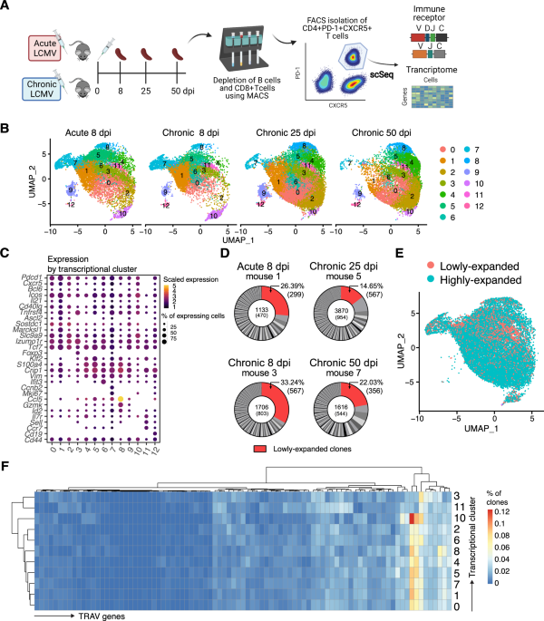


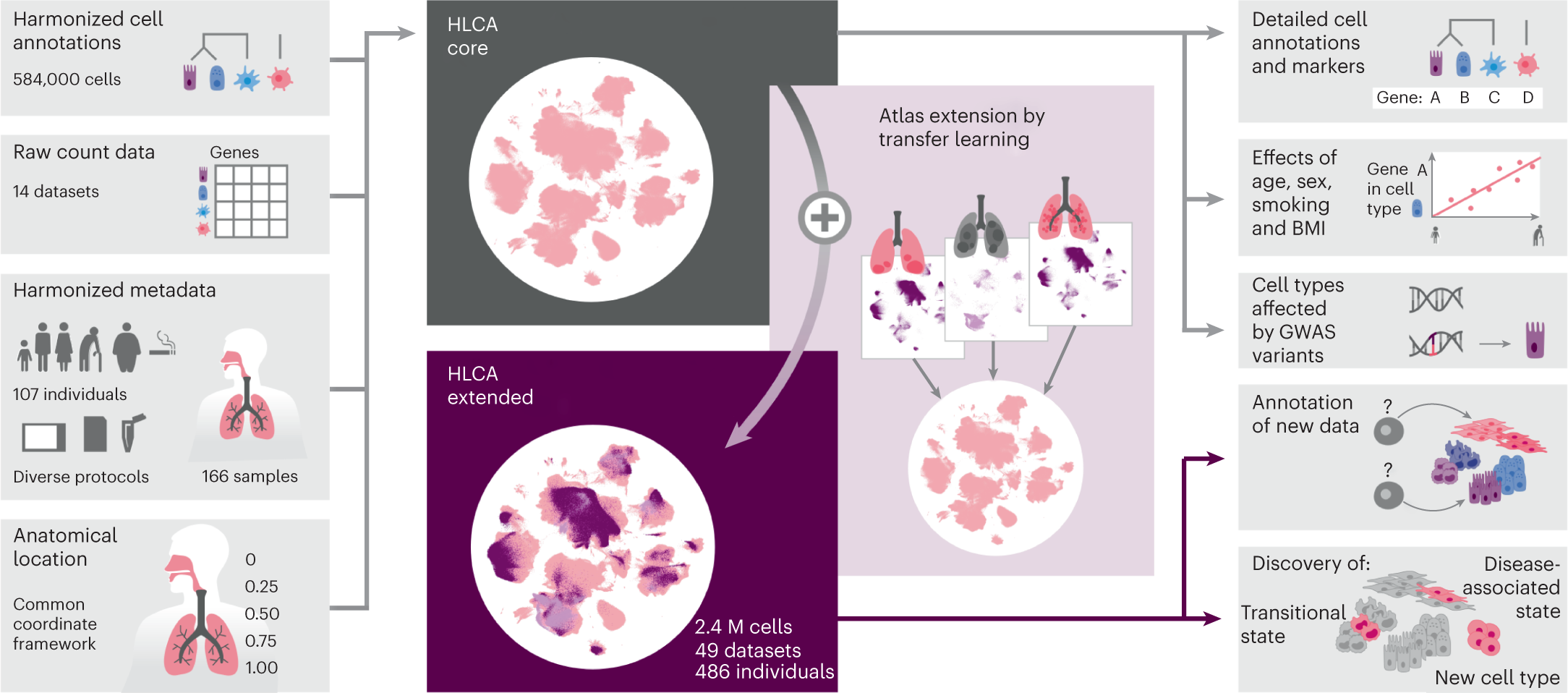


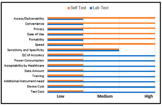


![Anti-ITGAX Armenian Hamster Monoclonal Antibody (PE (Phycoerythrin)) [clone: N418] | VWR](https://fr.vwr.com/stibo/bigweb/std.lang.all/03/25/13150325.jpg)