Streptococcaceae
Carnobacteriaceae
Lactobacillaceae
Gemella
Endocarditis, Bacterial
Gram-Positive Bacterial Infections
Streptococcus
RNA, Ribosomal, 16S
Genes, rRNA
Mouth
DNA, Ribosomal
Bacteremia
Genetic Heterogeneity
Polymerase Chain Reaction
Polymorphism, Restriction Fragment Length
Bacterial Typing Techniques
Molecular Sequence Data
Sequence Homology, Nucleic Acid
Sequence Analysis, DNA
Base Sequence
Abscess
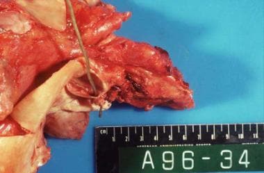
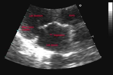
Endocarditis
Venous Insufficiency
Heart Valves
Use of groESL as a target for identification of Abiotrophia, Granulicatella, and Gemella species. (1/3)
(+info)Clonal analysis of the microbiota of severe early childhood caries. (2/3)
(+info)Phylogenetic group- and species-specific oligonucleotide probes for single-cell detection of lactic acid bacteria in oral biofilms. (3/3)
(+info)Abiotrophia is a genus of gram-positive, facultatively anaerobic bacteria that are part of the normal flora in the human mouth and gastrointestinal tract. These bacteria are also known as nutritionally variant streptococci because they have complex growth requirements and need additional factors for growth, such as pyridoxal and hemin. Abiotrophia species can cause various infectious diseases, including endocarditis, bacteremia, and meningitis, especially in individuals with underlying medical conditions or compromised immune systems. The two main species of Abiotrophia are Abiotrophia defectiva and Abiotrophia adiacens.
Streptococcaceae is a family of coccoid gram-positive bacteria, many of which are part of the normal human microbiota. They are facultatively anaerobic and generally non-spore forming. Some species are pathogenic and can cause various infections in humans, such as strep throat, pneumonia, and meningitis. Members of this family are characterized by their ability to form chains during cell division and may be beta-hemolytic, alpha-hemolytic, or non-hemolytic on blood agar plates. The genera in Streptococcaceae include Streptococcus, Enterococcus, Lactococcus, and Vagococcus, among others.
Carnobacteriaceae is a family of gram-positive, facultatively anaerobic bacteria that are commonly found in various environments such as soil, water, and decaying vegetation. Some species within this family can also be found in food products, particularly in refrigerated or processed meats and fish. Members of Carnobacteriaceae are non-spore forming, non-motile rods or cocci that may form pairs or short chains. They are generally considered to be psychrotrophic, meaning they can grow at low temperatures, which contributes to their ability to proliferate in refrigerated foods. Some species of Carnobacteriaceae have been associated with food spoilage and others have been shown to produce bacteriocins, which are protein molecules that inhibit the growth of other bacteria. However, some species within this family have also been investigated for their potential probiotic properties and ability to inhibit the growth of pathogenic bacteria in foods.
Lactobacillaceae is a family of gram-positive, facultatively anaerobic or microaerophilic, rod-shaped bacteria. They are non-spore forming and often occur in pairs or chains. Lactobacillaceae are commonly found in various environments such as the oral cavity, gastrointestinal tract, and vagina of humans and animals, as well as in fermented foods like yogurt, sauerkraut, and sourdough bread.
These bacteria are known for their ability to produce lactic acid as a major end product of carbohydrate metabolism, which gives them the name "lactic acid bacteria." They play an essential role in maintaining a healthy microbiota and have been associated with various health benefits, such as improving digestion, enhancing immune function, and preventing harmful bacterial overgrowth.
Some well-known genera within the family Lactobacillaceae include Lactobacillus, Lactococcus, Leuconostoc, and Weissella. It is important to note that recent taxonomic revisions have led to some changes in the classification of these bacteria, and some genera previously classified within Lactobacillaceae are now placed in other families within the order Lactobacillales.
"Gemella" is a genus of gram-positive, facultatively anaerobic, beta-hemolytic bacteria that are part of the normal flora in the human mouth, upper respiratory tract, and gastrointestinal tract. There are two species within this genus: Gemella haemolysans and Gemella morphilorufinata (previously known as Neisseria morphilorufinata). These bacteria can occasionally cause infections such as endocarditis, bacteremia, and meningitis, particularly in individuals with underlying medical conditions or compromised immune systems. The name "Gemella" is derived from the Latin word for "twin," reflecting the similarity of these organisms to each other.
Bacterial endocarditis is a medical condition characterized by the inflammation and infection of the inner layer of the heart, known as the endocardium. This infection typically occurs when bacteria enter the bloodstream and attach themselves to damaged or abnormal heart valves or other parts of the endocardium. The bacteria can then multiply and cause the formation of vegetations, which are clusters of infected tissue that can further damage the heart valves and lead to serious complications such as heart failure, stroke, or even death if left untreated.
Bacterial endocarditis is a relatively uncommon but potentially life-threatening condition that requires prompt medical attention. Risk factors for developing bacterial endocarditis include pre-existing heart conditions such as congenital heart defects, artificial heart valves, previous history of endocarditis, or other conditions that damage the heart valves. Intravenous drug use is also a significant risk factor for this condition.
Symptoms of bacterial endocarditis may include fever, chills, fatigue, muscle and joint pain, shortness of breath, chest pain, and a new or changing heart murmur. Diagnosis typically involves a combination of medical history, physical examination, blood cultures, and imaging tests such as echocardiography. Treatment usually involves several weeks of intravenous antibiotics to eradicate the infection, and in some cases, surgical intervention may be necessary to repair or replace damaged heart valves.
Gram-positive bacterial infections refer to illnesses or diseases caused by Gram-positive bacteria, which are a group of bacteria that turn purple when stained using the Gram stain method. This staining technique is used in microbiology to differentiate between two main types of bacteria based on their cell wall composition.
Gram-positive bacteria have a thick layer of peptidoglycan in their cell walls, which retains the crystal violet stain used in the Gram staining process. Some common examples of Gram-positive bacteria include Staphylococcus aureus, Streptococcus pyogenes, and Enterococcus faecalis.
Gram-positive bacterial infections can range from mild skin infections to severe and life-threatening conditions such as pneumonia, meningitis, and sepsis. The symptoms of these infections depend on the type of bacteria involved and the location of the infection in the body. Treatment typically involves the use of antibiotics that are effective against Gram-positive bacteria, such as penicillin, vancomycin, or clindamycin. However, the emergence of antibiotic resistance among Gram-positive bacteria is a growing concern and can complicate treatment in some cases.
Streptococcus is a genus of Gram-positive, spherical bacteria that typically form pairs or chains when clustered together. These bacteria are facultative anaerobes, meaning they can grow in the presence or absence of oxygen. They are non-motile and do not produce spores.
Streptococcus species are commonly found on the skin and mucous membranes of humans and animals. Some strains are part of the normal flora of the body, while others can cause a variety of infections, ranging from mild skin infections to severe and life-threatening diseases such as sepsis, meningitis, and toxic shock syndrome.
The pathogenicity of Streptococcus species depends on various virulence factors, including the production of enzymes and toxins that damage tissues and evade the host's immune response. One of the most well-known Streptococcus species is Streptococcus pyogenes, also known as group A streptococcus (GAS), which is responsible for a wide range of clinical manifestations, including pharyngitis (strep throat), impetigo, cellulitis, necrotizing fasciitis, and rheumatic fever.
It's important to note that the classification of Streptococcus species has evolved over time, with many former members now classified as different genera within the family Streptococcaceae. The current classification system is based on a combination of phenotypic characteristics (such as hemolysis patterns and sugar fermentation) and genotypic methods (such as 16S rRNA sequencing and multilocus sequence typing).
Ribosomal RNA (rRNA) is a type of RNA that combines with proteins to form ribosomes, which are complex structures inside cells where protein synthesis occurs. The "16S" refers to the sedimentation coefficient of the rRNA molecule, which is a measure of its size and shape. In particular, 16S rRNA is a component of the smaller subunit of the prokaryotic ribosome (found in bacteria and archaea), and is often used as a molecular marker for identifying and classifying these organisms due to its relative stability and conservation among species. The sequence of 16S rRNA can be compared across different species to determine their evolutionary relationships and taxonomic positions.
rRNA (ribosomal RNA) is not a type of gene itself, but rather a crucial component that is transcribed from genes known as ribosomal DNA (rDNA). In cells, rRNA plays an essential role in protein synthesis by assembling with ribosomal proteins to form ribosomes. Ribosomes are complex structures where the translation of mRNA into proteins occurs. There are multiple types of rRNA molecules, including 5S, 5.8S, 18S, and 28S rRNAs in eukaryotic cells, each with specific functions during protein synthesis.
In summary, 'Genes, rRNA' would refer to the genetic regions (genes) that code for ribosomal RNA molecules, which are vital components of the protein synthesis machinery within cells.
In medical terms, the mouth is officially referred to as the oral cavity. It is the first part of the digestive tract and includes several structures: the lips, vestibule (the space enclosed by the lips and teeth), teeth, gingiva (gums), hard and soft palate, tongue, floor of the mouth, and salivary glands. The mouth is responsible for several functions including speaking, swallowing, breathing, and eating, as it is the initial point of ingestion where food is broken down through mechanical and chemical processes, beginning the digestive process.
Ribosomal DNA (rDNA) refers to the specific regions of DNA in a cell that contain the genes for ribosomal RNA (rRNA). Ribosomes are complex structures composed of proteins and rRNA, which play a crucial role in protein synthesis by translating messenger RNA (mRNA) into proteins.
In humans, there are four types of rRNA molecules: 18S, 5.8S, 28S, and 5S. These rRNAs are encoded by multiple copies of rDNA genes that are organized in clusters on specific chromosomes. In humans, the majority of rDNA genes are located on the short arms of acrocentric chromosomes 13, 14, 15, 21, and 22.
Each cluster of rDNA genes contains both transcribed and non-transcribed spacer regions. The transcribed regions contain the genes for the four types of rRNA, while the non-transcribed spacers contain regulatory elements that control the transcription of the rRNA genes.
The number of rDNA copies varies between species and even within individuals of the same species. The copy number can also change during development and in response to environmental factors. Variations in rDNA copy number have been associated with various diseases, including cancer and neurological disorders.
Bacteremia is the presence of bacteria in the bloodstream. It is a medical condition that occurs when bacteria from another source, such as an infection in another part of the body, enter the bloodstream. Bacteremia can cause symptoms such as fever, chills, and rapid heart rate, and it can lead to serious complications such as sepsis if not treated promptly with antibiotics.
Bacteremia is often a result of an infection elsewhere in the body that allows bacteria to enter the bloodstream. This can happen through various routes, such as during medical procedures, intravenous (IV) drug use, or from infected wounds or devices that come into contact with the bloodstream. In some cases, bacteremia may also occur without any obvious source of infection.
It is important to note that not all bacteria in the bloodstream cause harm, and some people may have bacteria in their blood without showing any symptoms. However, if bacteria in the bloodstream multiply and cause an immune response, it can lead to bacteremia and potentially serious complications.
Phylogeny is the evolutionary history and relationship among biological entities, such as species or genes, based on their shared characteristics. In other words, it refers to the branching pattern of evolution that shows how various organisms have descended from a common ancestor over time. Phylogenetic analysis involves constructing a tree-like diagram called a phylogenetic tree, which depicts the inferred evolutionary relationships among organisms or genes based on molecular sequence data or other types of characters. This information is crucial for understanding the diversity and distribution of life on Earth, as well as for studying the emergence and spread of diseases.
Genetic heterogeneity is a phenomenon in genetics where different genetic variations or mutations in various genes can result in the same or similar phenotypic characteristics, disorders, or diseases. This means that multiple genetic alterations can lead to the same clinical presentation, making it challenging to identify the specific genetic cause based on the observed symptoms alone.
There are two main types of genetic heterogeneity:
1. Allelic heterogeneity: Different mutations in the same gene can cause the same or similar disorders. For example, various mutations in the CFTR gene can lead to cystic fibrosis, a genetic disorder affecting the respiratory and digestive systems.
2. Locus heterogeneity: Mutations in different genes can result in the same or similar disorders. For instance, mutations in several genes, such as BRCA1, BRCA2, and PALB2, are associated with an increased risk of developing breast cancer.
Genetic heterogeneity is essential to consider when diagnosing genetic conditions, evaluating recurrence risks, and providing genetic counseling. It highlights the importance of comprehensive genetic testing and interpretation for accurate diagnosis and appropriate management of genetic disorders.
Streptococcal infections are a type of infection caused by group A Streptococcus bacteria (Streptococcus pyogenes). These bacteria can cause a variety of illnesses, ranging from mild skin infections to serious and potentially life-threatening conditions such as sepsis, pneumonia, and necrotizing fasciitis (flesh-eating disease).
Some common types of streptococcal infections include:
* Streptococcal pharyngitis (strep throat) - an infection of the throat and tonsils that can cause sore throat, fever, and swollen lymph nodes.
* Impetigo - a highly contagious skin infection that causes sores or blisters on the skin.
* Cellulitis - a bacterial infection of the deeper layers of the skin and underlying tissue that can cause redness, swelling, pain, and warmth in the affected area.
* Scarlet fever - a streptococcal infection that causes a bright red rash on the body, high fever, and sore throat.
* Necrotizing fasciitis - a rare but serious bacterial infection that can cause tissue death and destruction of the muscles and fascia (the tissue that covers the muscles).
Treatment for streptococcal infections typically involves antibiotics to kill the bacteria causing the infection. It is important to seek medical attention if you suspect a streptococcal infection, as prompt treatment can help prevent serious complications.
Polymerase Chain Reaction (PCR) is a laboratory technique used to amplify specific regions of DNA. It enables the production of thousands to millions of copies of a particular DNA sequence in a rapid and efficient manner, making it an essential tool in various fields such as molecular biology, medical diagnostics, forensic science, and research.
The PCR process involves repeated cycles of heating and cooling to separate the DNA strands, allow primers (short sequences of single-stranded DNA) to attach to the target regions, and extend these primers using an enzyme called Taq polymerase, resulting in the exponential amplification of the desired DNA segment.
In a medical context, PCR is often used for detecting and quantifying specific pathogens (viruses, bacteria, fungi, or parasites) in clinical samples, identifying genetic mutations or polymorphisms associated with diseases, monitoring disease progression, and evaluating treatment effectiveness.
Restriction Fragment Length Polymorphism (RFLP) is a term used in molecular biology and genetics. It refers to the presence of variations in DNA sequences among individuals, which can be detected by restriction enzymes. These enzymes cut DNA at specific sites, creating fragments of different lengths.
In RFLP analysis, DNA is isolated from an individual and treated with a specific restriction enzyme that cuts the DNA at particular recognition sites. The resulting fragments are then separated by size using gel electrophoresis, creating a pattern unique to that individual's DNA. If there are variations in the DNA sequence between individuals, the restriction enzyme may cut the DNA at different sites, leading to differences in the length of the fragments and thus, a different pattern on the gel.
These variations can be used for various purposes, such as identifying individuals, diagnosing genetic diseases, or studying evolutionary relationships between species. However, RFLP analysis has largely been replaced by more modern techniques like polymerase chain reaction (PCR)-based methods and DNA sequencing, which offer higher resolution and throughput.
Bacterial typing techniques are methods used to identify and differentiate bacterial strains or isolates based on their unique characteristics. These techniques are essential in epidemiological studies, infection control, and research to understand the transmission dynamics, virulence, and antibiotic resistance patterns of bacterial pathogens.
There are various bacterial typing techniques available, including:
1. **Bacteriophage Typing:** This method involves using bacteriophages (viruses that infect bacteria) to identify specific bacterial strains based on their susceptibility or resistance to particular phages.
2. **Serotyping:** It is a technique that differentiates bacterial strains based on the antigenic properties of their cell surface components, such as capsules, flagella, and somatic (O) and flagellar (H) antigens.
3. **Biochemical Testing:** This method uses biochemical reactions to identify specific metabolic pathways or enzymes present in bacterial strains, which can be used for differentiation. Commonly used tests include the catalase test, oxidase test, and various sugar fermentation tests.
4. **Molecular Typing Techniques:** These methods use genetic markers to identify and differentiate bacterial strains at the DNA level. Examples of molecular typing techniques include:
* **Pulsed-Field Gel Electrophoresis (PFGE):** This method uses restriction enzymes to digest bacterial DNA, followed by electrophoresis in an agarose gel under pulsed electrical fields. The resulting banding patterns are analyzed and compared to identify related strains.
* **Multilocus Sequence Typing (MLST):** It involves sequencing specific housekeeping genes to generate unique sequence types that can be used for strain identification and phylogenetic analysis.
* **Whole Genome Sequencing (WGS):** This method sequences the entire genome of a bacterial strain, providing the most detailed information on genetic variation and relatedness between strains. WGS data can be analyzed using various bioinformatics tools to identify single nucleotide polymorphisms (SNPs), gene deletions or insertions, and other genetic changes that can be used for strain differentiation.
These molecular typing techniques provide higher resolution than traditional methods, allowing for more accurate identification and comparison of bacterial strains. They are particularly useful in epidemiological investigations to track the spread of pathogens and identify outbreaks.
Bacterial DNA refers to the genetic material found in bacteria. It is composed of a double-stranded helix containing four nucleotide bases - adenine (A), thymine (T), guanine (G), and cytosine (C) - that are linked together by phosphodiester bonds. The sequence of these bases in the DNA molecule carries the genetic information necessary for the growth, development, and reproduction of bacteria.
Bacterial DNA is circular in most bacterial species, although some have linear chromosomes. In addition to the main chromosome, many bacteria also contain small circular pieces of DNA called plasmids that can carry additional genes and provide resistance to antibiotics or other environmental stressors.
Unlike eukaryotic cells, which have their DNA enclosed within a nucleus, bacterial DNA is present in the cytoplasm of the cell, where it is in direct contact with the cell's metabolic machinery. This allows for rapid gene expression and regulation in response to changing environmental conditions.
Molecular sequence data refers to the specific arrangement of molecules, most commonly nucleotides in DNA or RNA, or amino acids in proteins, that make up a biological macromolecule. This data is generated through laboratory techniques such as sequencing, and provides information about the exact order of the constituent molecules. This data is crucial in various fields of biology, including genetics, evolution, and molecular biology, allowing for comparisons between different organisms, identification of genetic variations, and studies of gene function and regulation.
Sequence homology in nucleic acids refers to the similarity or identity between the nucleotide sequences of two or more DNA or RNA molecules. It is often used as a measure of biological relationship between genes, organisms, or populations. High sequence homology suggests a recent common ancestry or functional constraint, while low sequence homology may indicate a more distant relationship or different functions.
Nucleic acid sequence homology can be determined by various methods such as pairwise alignment, multiple sequence alignment, and statistical analysis. The degree of homology is typically expressed as a percentage of identical or similar nucleotides in a given window of comparison.
It's important to note that the interpretation of sequence homology depends on the biological context and the evolutionary distance between the sequences compared. Therefore, functional and experimental validation is often necessary to confirm the significance of sequence homology.
DNA Sequence Analysis is the systematic determination of the order of nucleotides in a DNA molecule. It is a critical component of modern molecular biology, genetics, and genetic engineering. The process involves determining the exact order of the four nucleotide bases - adenine (A), guanine (G), cytosine (C), and thymine (T) - in a DNA molecule or fragment. This information is used in various applications such as identifying gene mutations, studying evolutionary relationships, developing molecular markers for breeding, and diagnosing genetic diseases.
The process of DNA Sequence Analysis typically involves several steps, including DNA extraction, PCR amplification (if necessary), purification, sequencing reaction, and electrophoresis. The resulting data is then analyzed using specialized software to determine the exact sequence of nucleotides.
In recent years, high-throughput DNA sequencing technologies have revolutionized the field of genomics, enabling the rapid and cost-effective sequencing of entire genomes. This has led to an explosion of genomic data and new insights into the genetic basis of many diseases and traits.
A base sequence in the context of molecular biology refers to the specific order of nucleotides in a DNA or RNA molecule. In DNA, these nucleotides are adenine (A), guanine (G), cytosine (C), and thymine (T). In RNA, uracil (U) takes the place of thymine. The base sequence contains genetic information that is transcribed into RNA and ultimately translated into proteins. It is the exact order of these bases that determines the genetic code and thus the function of the DNA or RNA molecule.
An abscess is a localized collection of pus caused by an infection. It is typically characterized by inflammation, redness, warmth, pain, and swelling in the affected area. Abscesses can form in various parts of the body, including the skin, teeth, lungs, brain, and abdominal organs. They are usually treated with antibiotics to eliminate the infection and may require drainage if they are large or located in a critical area. If left untreated, an abscess can lead to serious complications such as sepsis or organ failure.
Endocarditis is an inflammation of the inner layer of the heart chambers and heart valves, called the endocardium. This inflammation typically results from a bacterial or, less commonly, fungal infection that travels through the bloodstream and attaches to damaged areas of the heart.
There are two main types of endocarditis:
1. Acute Endocarditis: Develops quickly and can be severe, causing fever, chills, shortness of breath, fatigue, and heart murmurs. It may lead to serious complications like heart failure, embolism (blood clots that travel to other parts of the body), and damage to heart valves.
2. Subacute Endocarditis: Develops more slowly, often causing milder symptoms that can be mistaken for a cold or flu. Symptoms may include fatigue, weakness, fever, night sweats, weight loss, joint pain, and heart murmurs. Subacute endocarditis is more likely to affect people with previously damaged heart valves or congenital heart conditions.
Treatment usually involves several weeks of intravenous antibiotics or antifungal medications, depending on the cause of the infection. In some cases, surgery may be required to repair or replace damaged heart valves. Preventive measures include good oral hygiene and prompt treatment of infections, especially in individuals at a higher risk for endocarditis, such as those with congenital heart defects, artificial heart valves, or previous history of endocarditis.
Venous insufficiency is a medical condition that occurs when the veins, particularly in the legs, have difficulty returning blood back to the heart due to impaired valve function or obstruction in the vein. This results in blood pooling in the veins, leading to symptoms such as varicose veins, swelling, skin changes, and ulcers. Prolonged venous insufficiency can cause chronic pain and affect the quality of life if left untreated.
The endocardium is the innermost layer of tissue that lines the chambers of the heart and the valves between them. It is a thin, smooth membrane that is in contact with the blood within the heart. This layer helps to maintain the heart's internal environment, facilitates the smooth movement of blood through the heart, and provides a protective barrier against infection and other harmful substances. The endocardium is composed of simple squamous epithelial cells called endothelial cells, which are supported by a thin layer of connective tissue.
Heart valves are specialized structures in the heart that ensure unidirectional flow of blood through its chambers during the cardiac cycle. There are four heart valves: the tricuspid valve and the mitral (bicuspid) valve, located between the atria and ventricles, and the pulmonic (pulmonary) valve and aortic valve, located between the ventricles and the major blood vessels leaving the heart.
The heart valves are composed of thin flaps of tissue called leaflets or cusps, which are supported by a fibrous ring. The aortic and pulmonic valves have three cusps each, while the tricuspid and mitral valves have three and two cusps, respectively.
The heart valves open and close in response to pressure differences across them, allowing blood to flow forward into the ventricles during diastole (filling phase) and preventing backflow of blood into the atria during systole (contraction phase). A properly functioning heart valve ensures efficient pumping of blood by the heart and maintains normal blood circulation throughout the body.
Heart valve diseases are a group of conditions that affect the function of one or more of the heart's four valves (tricuspid, pulmonic, mitral, and aortic). These valves are responsible for controlling the direction and flow of blood through the heart. Heart valve diseases can cause the valves to become narrowed (stenosis), leaky (regurgitation or insufficiency), or improperly closed (prolapse), leading to disrupted blood flow within the heart and potentially causing symptoms such as shortness of breath, fatigue, chest pain, and irregular heart rhythms. The causes of heart valve diseases can include congenital defects, age-related degenerative changes, infections, rheumatic heart disease, and high blood pressure. Treatment options may include medications, surgical repair or replacement of the affected valve(s), or transcatheter procedures.
 Abiotrophia
Abiotrophia balaenopterae
Infective endocarditis
Granulicatella adiacens
Granulicatella
Globicatella sulfidifaciens
Pyridoxal
Gemella morbillorum
Etest
Lactic acid bacteria
Abiotrophia - Wikipedia
 Infective Endocarditis: Practice Essentials, Background, Pathophysiology
Infective Endocarditis Workup: Approach Considerations, Blood and Urine Studies, Blood Culture
 "Development of a periprosthetic joint infection by Abiotrophia defecti" by Trevor R Tooley, Matthew P Siljander et al.
Clinical characteristics and outcome of infective endocarditis due to Abiotrophia and Granulicatella compared to Viridans group...
 Small Things Considered
 Addisu Mesfin, M.D. | UR Medicine
 References | Isolation Precautions | Guidelines Library | Infection Control | CDC
 Principles and Practice of Pediatric Infectious Diseases, 6th Edition - 9780323756082
HCP/ SDA: Pubblicazioni scientifiche - IT | Unilabs
Name Taxonomy in SILVA v123
 John Greene | Moffitt
 Turkish Journal of Geriatrics
 Risk for Endocarditis in Bacteremia with Streptococcus-Like Bacteria : A Retrospective Population-Based Cohort Study | Lund...
 Vagococcus bubulae sp. nov., isolated from ground beef, and Vagococcus vulneris sp. nov., isolated from a human foot wound |...
 DeCS 2020 - June 23, 2020 version
 KAKEN - Researchers | Ishikawa Taichi (10569247)
 MedPress Publishers LLC | Home
Code System Concept
DeCS 2019 - June 12, 2019 version
DeCS 2018 - July 31, 2018 version
DeCS 2017 - July 04, 2017 version
DeCS 2018 - July 31, 2018 version
Infective Endocarditis: Practice Essentials, Background, Pathophysiology
Miscellaneous gram-positive organisms | Oncohema Key
 Documentos relacionados
 Special Bacteriology Unit - Guide to Services - CNPHI
 Variety of Life: November 2016
 Genomes | ATCC Genome Portal
Granulicatella6
- Abiotrophia elegans was reclassified to Granulicatella elegans. (wikipedia.org)
- Objective: To describe the clinical characteristics and outcome of Abiotrophia and Granulicatella infective endocarditis and compare them with Viridans group streptococci infective endocarditis. (huji.ac.il)
- Data from patients with definitive or possible IE due to Abiotrophia species, Granulicatella species and Viridans group streptococci was analyzed. (huji.ac.il)
- The aim of the study was to analyze this in a cohort of patients with SLB-bacteremia, focusing on Abiotrophia, Aerococcus, Gemella, and Granulicatella. (lu.se)
- Infective endocarditis was most common in bacteremia with Abiotrophia (4 of 19) followed by Granulicatella (9 of 124), Gemella (6 of 87), and Aerococcus (13 of 338). (lu.se)
- Granulicatella and Abiotrophia spp. (firstline.org)
Endocarditis2
- Closing The Brief Case: A Variant on a Classic-Abiotrophia defectiva Endocarditis with Discitis. (bvsalud.org)
- 2012). abiotrophia defectiva endocarditis in a child. (ukm.my)
Species1
- The genus contains 4 species of coccus shaped species, 2 are former members of the genus Streptococcus, which were transferred in 1995 to the newly coined genus Abiotrophia: A. adiacens ( (Bouvet et al. (wikipedia.org)
Streptococcus1
- KAWAMURA (Y.), HOU (X.G.), SULTANA (F.), LIU (S.), YAMAMOTO (H.) and EZAKI (T.): Transfer of Streptococcus adjacens and Streptococcus defectivus to Abiotrophia gen. nov. as Abiotrophia adiacens comb. (wikipedia.org)
Genus1
- Abiotrophia is a genus of lactic acid bacteria, a family in the phylum Bacillota (Bacteria). (wikipedia.org)
Patients1
- Our analysis of BAL fluid from 22 patients with moderate or severe COPD and 10 healthy patients identified a greater proportion of oral bacteria (such as Desulfobulbus , Abiotrophia , and Selenomonas ) in the COPD microbiota than in the healthy lung microbiota. (biomedcentral.com)
Balaenopterae2
- Abiotrophia balaenopterae sp. (wikipedia.org)
- Abiotrophia balaenopterae Lawson et al. (irmng.org)
Genera1
- HIV+ samples were enriched in genera Abiotrophia, Neisseria, Kingella, and unclassified Neisseriaceae and depleted in Leptotrichia and Selenomonas. (bugsigdb.org)
Elegans1
- Abiotrophia elegans Roggenkamp et al. (irmng.org)
Kawamura2
- KAWAMURA (Y.), HOU (X.G.), SULTANA (F.), LIU (S.), YAMAMOTO (H.) and EZAKI (T.): Transfer of Streptococcus adjacens and Streptococcus defectivus to Abiotrophia gen. nov. as Abiotrophia adiacens comb. (wikipedia.org)
- Abiotrophia Kawamura et al. (irmng.org)
Adiacens1
- Abiotrophia adiacens (Bouvet et al. (irmng.org)
Latin1
- Neo-Latin feminine gender noun Abiotrophia, life-nutrition-deficiency. (wikipedia.org)
Human1
- Abiotrophia defectiva CCUG 41537 is a bacterium that was isolated from Human vitreous body,80-yr-old patient. (dsmz.de)