Wheat Germ Agglutinins
Lectins
Agglutinins
Plant Lectins
Peanut Agglutinin
Triticum
Receptors, Mitogen
Concanavalin A
Wheat Germ Agglutinin-Horseradish Peroxidase Conjugate
Receptors, Concanavalin A
Methylmannosides
Carbohydrates
Succinic Anhydrides
Glycoproteins
Chromatography, Affinity
Sialic Acids
Cell Membrane
Datura stramonium
Neuraminidase
Glycoconjugates
Electrophoresis, Polyacrylamide Gel
Agglutination Tests
N-Acetylneuraminic Acid
Histocytochemistry
Glycosylation
Glucosamine
Molecular Sequence Data
Oligosaccharides
Nuclear Envelope
Neoplasms, Germ Cell and Embryonal
Microscopy, Electron
Chitin
Horseradish Peroxidase
Seeds
Binding Sites
Hemagglutination
Carbohydrate Metabolism
Fluorescein-5-isothiocyanate
Glycophorin
Neuronal Tract-Tracers
Membrane Proteins
Carbohydrate Sequence
Freeze Fracturing
Amino Acid Sequence
Mannosyl-Glycoprotein Endo-beta-N-Acetylglucosaminidase
Hemagglutination Tests
Protein Binding
Axonal Transport
Sialoglycoproteins
Plants
A genetic approach to visualization of multisynaptic neural pathways using plant lectin transgene. (1/581)
The wiring patterns among various types of neurons via specific synaptic connections are the basis of functional logic employed by the brain for information processing. This study introduces a powerful method of analyzing the neuronal connectivity patterns by delivering a tracer selectively to specific types of neurons while simultaneously transsynaptically labeling their target neurons. We developed a novel genetic approach introducing cDNA for a plant lectin, wheat germ agglutinin (WGA), as a transgene under the control of specific promoter elements. Using this method, we demonstrate three examples of visualization of specific transsynaptic neural pathways: the mouse cerebellar efferent pathways, the mouse olfactory pathways, and the Drosophila visual pathways. This strategy should greatly facilitate studies on the anatomical and functional organization of the developing and mature nervous system. (+info)Role of surface proteins in Vibrio cholerae attachment to chitin. (2/581)
The role of surface proteins in Vibrio cholerae attachment to chitin particles in vitro was studied. Treatment of V. cholerae O1 ATCC 14034 and ATCC 14035 with pronase E reduced the attachment of bacteria to chitin particles by 57 to 77%. A statistically significant reduction was also observed when the attachment to chitin was evaluated in the presence of homologous Sarkosyl-insoluble membrane proteins (MPs) (67 to 84%), N-acetylglucosamine (GlcNAc) (62%), the sugar that makes up chitin, and wheat germ agglutinin (40 to 56%), a lectin that binds GlcNAc. The soluble oligomers N,N'-diacetylchitobiose or N,N', N"-triacetylchitotriose caused an inhibition of 14 to 23%. Sarkosyl-insoluble MPs able to bind chitin particles were isolated and visualized by sodium dodecyl sulfate-polyacrylamide gel electrophoresis; two of these peptides (molecular sizes, 36 and 53 kDa) specifically bind GlcNAc. (+info)Nucleo-cytoplasmic interactions that control nuclear envelope breakdown and entry into mitosis in the sea urchin zygote. (3/581)
In sea urchin zygotes and mammalian cells nuclear envelope breakdown (NEB) is not driven simply by a rise in cytoplasmic cyclin dependent kinase 1-cyclin B (Cdk1-B) activity; the checkpoint monitoring DNA synthesis can prevent NEB in the face of mitotic levels of Cdk1-B. Using sea urchin zygotes we investigated whether this checkpoint prevents NEB by restricting import of regulatory proteins into the nucleus. We find that cyclin B1-GFP accumulates in nuclei that cannot complete DNA synthesis and do not break down. Thus, this checkpoint limits NEB downstream of both the cytoplasmic activation and nuclear accumulation of Cdk1-B1. In separate experiments we fertilize sea urchin eggs with sperm whose DNA has been covalently cross-linked to inhibit replication. When the pronuclei fuse, the resulting zygote nucleus does not break down for >180 minutes (equivalent to three cell cycles), even though Cdk1-B activity rises to greater than mitotic levels. If pronuclear fusion is prevented, then the female pronucleus breaks down at the normal time (average 68 minutes) and the male pronucleus with cross-linked DNA breaks down 16 minutes later. This male pronucleus has a functional checkpoint because it does not break down for >120 minutes if the female pronucleus is removed just prior to NEB. These results reveal the existence of an activity released by the female pronucleus upon its breakdown, that overrides the checkpoint in the male pronucleus and induces NEB. Microinjecting wheat germ agglutinin into binucleate zygotes reveals that this activity involves molecules that must be actively translocated into the male pronucleus. (+info)Phosphorylation-dependent binding of hepatitis B virus core particles to the nuclear pore complex. (4/581)
Although many viruses replicate in the nucleus, little is known about the processes involved in the nuclear import of viral genomes. We show here that in vitro generated core particles of human hepatitis B virus bind to nuclear pore complexes (NPCs) in digitonin-permeabilized mammalian cells. This only occurred if the cores contained phosphorylated core proteins. Binding was inhibited by wheat germ agglutinin, by antinuclear pore complex antibodies, and by peptides corresponding either to classical nuclear localization signals (NLS) or to COOH-terminal sequences of the core protein. Binding was dependent on the nuclear transport factors importins (karyopherins) alpha and beta. The results suggested that phosphorylation induces exposure of NLS in the COOH-terminal portion of the core protein that allows core binding to the NPCs by the importin- (karyopherin-) mediated pathway. Thus, phosphorylation of the core protein emerged as an important step in the viral replication cycle necessary for transport of the viral genome to the nucleus. (+info)Isolation and characterization of linear polylactosamines containing one and two site-specifically positioned Lewis x determinants: WGA agarose chromatography in fractionation of mixtures generated by random, partial enzymatic alpha3-fucosylation of pure polylactosamines. (5/581)
We report that isomeric monofucosylhexasaccharides, Galbeta1-4GlcNAcbeta1-3Galbeta1-4GlcNAcbeta1- 3Galbeta1-4(Fucalpha1-3) GlcNAc, Galbeta1-4GlcNAcbeta1-3Galbeta1-4(Fucalpha1-3) GlcNAcbeta1-3Galbeta1-4 GlcNAc and Galbeta1-4(Fucalpha1-3)GlcNAcbeta1-3Galbeta1- 4GlcNAcbeta1-3Galbeta1-4 GlcNAc, and bifucosylhexasaccharides Galbeta1-4GlcNAcbeta1-3Galbeta1-4(Fucalpha1-3) GlcNAcbeta1-3Galbeta1-4(Fucalpha1-3)GlcNAc, Galbeta1-4(Fucalpha1-3)GlcNAcbeta1-3Galbeta1- 4GlcNAcbeta1-3Galbeta1-4 (Fucalpha1-3)GlcNAc and Galbeta1-4(Fucalpha1-3)GlcNAcbeta1-3Galbeta1-4( Fucalpha1-3)GlcNAcbeta1-3Galbeta1-4GlcNAc can be isolated in pure form from reaction mixtures of the linear hexasaccharide Galbeta1-4GlcNAcbeta1-3Galbeta1-4GlcNAcbeta1- 3Galbeta1-4GlcNAc with GDP-fucose and alpha1,3-fucosyltransferases of human milk. The pure isomers were characterized in several ways;1H-NMR spectroscopy, for instance, revealed distinct resonances associated with the Lewis x group [Galbeta1-4(Fucalpha1-3)GlcNAc] located at the proximal, middle, and distal positions of the polylactosamine chain. Chromatography on immobilized wheat germ agglutinin was crucial in the separation process used; the isomers carrying the fucose at the reducing end GlcNAc possessed particularly low affinities for the lectin. Isomeric monofucosyl derivatives of the pentasaccharides GlcNAcbeta1-3Galbeta1-4GlcNAcbeta1-3Galbeta1- 4Gl cNAc and Galalpha1-3Galbeta1-4GlcNAcbeta1-3Galbeta1-4G lcN Ac and the tetrasaccharide Galbeta1-4GlcNAcbeta1-3Galbeta1-4GlcNAc were also obtained in pure form, implying that the methods used are widely applicable. The isomeric Lewis x glycans proved to be recognized in highly variable binding modes by polylactosamine-metabolizing enzymes, e.g., the midchain beta1,6-GlcNAc transferase (Leppanen et al., Biochemistry, 36, 13729-13735, 1997). (+info)Binding of human neutrophils to cell-surface anchored Tamm-Horsfall glycoprotein in tubulointerstitial nephritis. (6/581)
BACKGROUND: Human Tamm-Horsfall glycoprotein (T-H) is a glycosylphosphatidylinositol-anchored protein exposed at the surface of distal nephron cells, and urinary T-H is the released soluble counterpart. The latter has been implicated in tubulointerstitial nephritis, and the proinflammatory potential has been related to its ability to bind in vitro human neutrophils (PMNs). We have examined the conditions required for the binding of neutrophils to cell-surface anchored T-H and the consequent effects. METHODS: A HeLa cell-line derivative permanently transformed with human T-H cDNA and expressing T-H at the cell surface was used throughout the study. The adhesion of PMNs to cells expressing T-H was analyzed by immunofluorescence microscopy before and after the opsonization of cells with anti-T-H antibodies. The oxidative burst induced by adhesion of PMNs to the cells was determined by the activation of myeloperoxidase. Quantitative and qualitative changes in the release of T-H under the adhesion of activated PMNs were determined by dot-blot and Western blot analysis. RESULTS: No binding of neutrophils to cell-surface-anchored T-H was observed. On the contrary, the opsonization of cells with anti-T-H antibodies resulted in a dramatic adhesion of neutrophils. Such an adhesion induced the oxidative burst of PMNs and a large increment in the release of T-H, as well as the release of the slightly faster migrating T-H form, which is normally retained intracellularly. CONCLUSIONS: These results support the notion that, after the autoimmune response, the adhesion of neutrophils to cell-surface T-H contributes to the pathogenesis of tubulointerstitial nephritis, favoring a further accumulation of T-H in the interstitium and inducing the loss of cell integrity via reactive oxygen metabolites generated by activated neutrophils. (+info)Wheat germ agglutinin induces NADPH-oxidase activity in human neutrophils by interaction with mobilizable receptors. (7/581)
Wheat germ agglutinin (WGA), a lectin with specificity for N-acetylglucosamine and sialic acid, was investigated with respect to its ability to activate the NADPH-oxidase of in vivo-exudated neutrophils (obtained from a skin chamber), and the activity was compared to that of peripheral blood neutrophils. The exudate cells responded to WGA, by both releasing reactive oxygen species into the extracellular milieu and producing oxygen metabolites intracellularly. The peripheral blood cells were unresponsive. To mimic the in vivo-exuded neutrophils with regards to receptor exposure, peripheral blood neutrophils were induced to mobilize their granules and vesicles to varying degrees (in vitro priming), prior to challenge with WGA. The oxidative response to WGA increased with increasing levels of granule mobilization, and the receptor(s) could be shown to reside in the secretory vesicles and/or the gelatinase granules in resting neutrophils. Several WGA-binding glycoproteins were detected in subcellular fractions containing these organelles. The extra- and intracellular NADPH-oxidase responses showed differences in sialic acid dependency, indicating that these two responses are mediated by different receptor structures. (+info)Interaction of Azospirillum lipoferum with wheat germ agglutinin stimulates nitrogen fixation. (8/581)
In vitro, the nitrogen fixation capability of A. lipoferum is efficiently increased in the presence of wheat germ agglutinin (WGA). A putative WGA-binding receptor, a 32-kDa protein, was detected in the cell capsule. The stimulatory effect required N-acetyl-D-glucosamine dimer (GlcNAcdi) terminated sugar side chains of the receptor and was dependent on the number of GlcNAcdi links involved in receptor-WGA interface. Binding to the primary sugar binding sites on WGA had a larger stimulatory effect than binding to the secondary sites. The WGA-receptor complex generated stimulus led to elevated transcription of the nifH and nifA genes and of the glnBA gene cluster but not of the glnA gene from its own promoter. There may well be a signalling cascade contributing to the regulation of nitrogen fixation. (+info)Wheat germ agglutinins (WGA) are proteins found in wheat germ that have the ability to bind to specific carbohydrate structures, such as N-acetylglucosamine and sialic acid, which are present on the surface of many cells in the human body. WGA is a type of lectin, a group of proteins that can agglutinate, or clump together, red blood cells and bind to specific sugars on cell membranes.
WGA has been studied for its potential effects on various biological processes, including inflammation, immune response, and gut barrier function. Some research suggests that WGA may interact with the gut epithelium and affect intestinal permeability, potentially contributing to the development of gastrointestinal symptoms in some individuals. However, more research is needed to fully understand the clinical significance of these findings.
It's worth noting that while WGA has been studied for its potential biological effects, it is not currently recognized as a major allergen or toxic component of wheat. However, some people may still choose to avoid foods containing WGA due to personal dietary preferences or sensitivities.
Lectins are a type of proteins that bind specifically to carbohydrates and have been found in various plant and animal sources. They play important roles in biological recognition events, such as cell-cell adhesion, and can also be involved in the immune response. Some lectins can agglutinate certain types of cells or precipitate glycoproteins, while others may have a more direct effect on cellular processes. In some cases, lectins from plants can cause adverse effects in humans if ingested, such as digestive discomfort or allergic reactions.
Agglutinins are antibodies that cause the particles (such as red blood cells, bacteria, or viruses) to clump together. They recognize and bind to specific antigens on the surface of these particles, forming a bridge between them and causing them to agglutinate or clump. Agglutinins are an important part of the immune system's response to infection and help to eliminate pathogens from the body.
There are two main types of agglutinins:
1. Naturally occurring agglutinins: These are present in the blood serum of most individuals, even before exposure to an antigen. They can agglutinate some bacteria and red blood cells without prior sensitization. For example, anti-A and anti-B agglutinins are naturally occurring antibodies found in people with different blood groups (A, B, AB, or O).
2. Immune agglutinins: These are produced by the immune system after exposure to an antigen. They develop as part of the adaptive immune response and target specific antigens that the body has encountered before. Immunization with vaccines often leads to the production of immune agglutinins, which can provide protection against future infections.
Agglutination reactions are widely used in laboratory tests for various diagnostic purposes, such as blood typing, detecting bacterial or viral infections, and monitoring immune responses.
Plant lectins are proteins or glycoproteins that are abundantly found in various plant parts such as seeds, leaves, stems, and roots. They have the ability to bind specifically to carbohydrate structures present on cell membranes, known as glycoconjugates. This binding property of lectins is reversible and non-catalytic, meaning it does not involve any enzymatic activity.
Lectins play several roles in plants, including defense against predators, pathogens, and herbivores. They can agglutinate red blood cells, stimulate the immune system, and have been implicated in various biological processes such as cell growth, differentiation, and apoptosis (programmed cell death). Some lectins also exhibit mitogenic activity, which means they can stimulate the proliferation of certain types of cells.
In the medical field, plant lectins have gained attention due to their potential therapeutic applications. For instance, some lectins have been shown to possess anti-cancer properties and are being investigated as potential cancer treatments. However, it is important to note that some lectins can be toxic or allergenic to humans and animals, so they must be used with caution.
Peanut agglutinin (PNA) is a lectin, a type of carbohydrate-binding protein, found in peanuts. It is known to bind specifically to Galβ1-3GalNAc, a disaccharide present on glycoproteins and glycolipids of various cells. PNA has been used in research as a tool for identifying and isolating specific cell types, such as immature red blood cells (reticulocytes) and certain types of cancer cells, due to its affinity for these structures. However, it's important to note that peanut agglutinin may also have potential implications in the development of allergies to peanuts.
"Triticum" is the genus name for a group of cereal grains that includes common wheat (T. aestivum), durum wheat (T. durum), and spelt (T. spelta). These grains are important sources of food for humans, providing carbohydrates, proteins, and various nutrients. They are used to make a variety of foods such as bread, pasta, and breakfast cereals. Triticum species are also known as "wheat" in layman's terms.
Mitogen receptors are a type of cell surface receptor that become activated in response to the binding of mitogens, which are substances that stimulate mitosis (cell division) and therefore promote growth and proliferation of cells. The activation of mitogen receptors triggers a series of intracellular signaling events that ultimately lead to the transcription of genes involved in cell cycle progression and cell division.
Mitogen receptors include receptor tyrosine kinases (RTKs), G protein-coupled receptors (GPCRs), and cytokine receptors, among others. RTKs are transmembrane proteins that have an intracellular tyrosine kinase domain, which becomes activated upon ligand binding and phosphorylates downstream signaling molecules. GPCRs are seven-transmembrane domain proteins that activate heterotrimeric G proteins upon ligand binding, leading to the activation of various intracellular signaling pathways. Cytokine receptors are typically composed of multiple subunits and activate Janus kinases (JAKs) and signal transducer and activator of transcription (STAT) proteins upon ligand binding.
Abnormal activation of mitogen receptors has been implicated in the development and progression of various diseases, including cancer, autoimmune disorders, and inflammatory conditions. Therefore, understanding the mechanisms underlying mitogen receptor signaling is crucial for the development of targeted therapies for these diseases.
Acetylglucosamine is a type of sugar that is commonly found in the body and plays a crucial role in various biological processes. It is a key component of glycoproteins and proteoglycans, which are complex molecules made up of protein and carbohydrate components.
More specifically, acetylglucosamine is an amino sugar that is formed by the addition of an acetyl group to glucosamine. It can be further modified in the body through a process called acetylation, which involves the addition of additional acetyl groups.
Acetylglucosamine is important for maintaining the structure and function of various tissues in the body, including cartilage, tendons, and ligaments. It also plays a role in the immune system and has been studied as a potential therapeutic target for various diseases, including cancer and inflammatory conditions.
In summary, acetylglucosamine is a type of sugar that is involved in many important biological processes in the body, and has potential therapeutic applications in various diseases.
Concanavalin A (Con A) is a type of protein known as a lectin, which is found in the seeds of the plant Canavalia ensiformis, also known as jack bean. It is often used in laboratory settings as a tool to study various biological processes, such as cell division and the immune response, due to its ability to bind specifically to certain sugars on the surface of cells. Con A has been extensively studied for its potential applications in medicine, including as a possible treatment for cancer and viral infections. However, more research is needed before these potential uses can be realized.
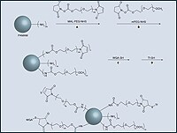
Wheat Germ Agglutinin (WGA) is a lectin protein found in wheat germ, which binds specifically to certain sugars on the surface of cells. Horseradish Peroxidase (HRP) is an enzyme derived from horseradish that catalyzes the conversion of certain substrates, producing a chemiluminescent or colorimetric signal.
A WGA-HRP conjugate refers to the formation of a covalent bond between WGA and HRP, creating an immunoconjugate. This complex is often used as a detection tool in various assays, such as ELISA (Enzyme-Linked Immunosorbent Assay) or Western blotting, where it can bind to specific carbohydrates on the target molecule and catalyze a colorimetric or chemiluminescent reaction, allowing for the visualization of the target.
Agglutination is a medical term that refers to the clumping together of particles, such as cells, bacteria, or precipitates, in a liquid medium. It most commonly occurs due to the presence of antibodies in the fluid that bind to specific antigens on the surface of the particles, causing them to adhere to one another and form visible clumps.
In clinical laboratory testing, agglutination is often used as a diagnostic tool to identify the presence of certain antibodies or antigens in a patient's sample. For example, a common application of agglutination is in blood typing, where the presence of specific antigens on the surface of red blood cells causes them to clump together when mixed with corresponding antibodies.
Agglutination can also occur in response to certain infectious agents, such as bacteria or viruses, that display antigens on their surface. In these cases, the agglutination reaction can help diagnose an infection and guide appropriate treatment.
Concanavalin A (Con A) receptors are not a medical term per se, but rather a term used in the field of immunology and cell biology. Concanavalin A is a type of lectin, a protein that can bind to specific sugars found on the surface of cells. Con A receptors refer to the specific binding sites or proteins on the surface of certain types of cells, such as immune cells, that can recognize and bind to Concanavalin A.
When Con A binds to its receptors, it can activate various cellular responses, including changes in cell shape, movement, and metabolism. In research settings, Con A is often used as a tool to study the behavior of immune cells and other cell types that express Con A receptors. However, it's worth noting that Concanavalin A is not typically used in medical treatments or diagnoses.
Methylmannosides are not a recognized medical term or a specific medical condition. However, in biochemistry, methylmannosides refer to a type of glycosylation pattern where a methyl group (-CH3) is attached to a mannose sugar molecule. Mannose is a type of monosaccharide or simple sugar that is commonly found in various glycoproteins and glycolipids in the human body.
Methylmannosides can be formed through the enzymatic transfer of a methyl group from a donor molecule, such as S-adenosylmethionine (SAM), to the mannose sugar by methyltransferase enzymes. These modifications can play important roles in various biological processes, including protein folding, trafficking, and quality control, as well as cell-cell recognition and signaling.
It's worth noting that while methylmannosides have significant biochemical importance, they are not typically referred to in medical contexts unless discussing specific biochemical or molecular research studies.
Carbohydrates are a major nutrient class consisting of organic compounds that primarily contain carbon, hydrogen, and oxygen atoms. They are classified as saccharides, which include monosaccharides (simple sugars), disaccharides (double sugars), oligosaccharides (short-chain sugars), and polysaccharides (complex carbohydrates).
Monosaccharides, such as glucose, fructose, and galactose, are the simplest form of carbohydrates. They consist of a single sugar molecule that cannot be broken down further by hydrolysis. Disaccharides, like sucrose (table sugar), lactose (milk sugar), and maltose (malt sugar), are formed from two monosaccharide units joined together.
Oligosaccharides contain a small number of monosaccharide units, typically less than 20, while polysaccharides consist of long chains of hundreds to thousands of monosaccharide units. Polysaccharides can be further classified into starch (found in plants), glycogen (found in animals), and non-starchy polysaccharides like cellulose, chitin, and pectin.
Carbohydrates play a crucial role in providing energy to the body, with glucose being the primary source of energy for most cells. They also serve as structural components in plants (cellulose) and animals (chitin), participate in various metabolic processes, and contribute to the taste, texture, and preservation of foods.
I'm sorry for any confusion, but "Succinic Anhydrides" is not a recognized medical term. Succinic anhydride is a chemical compound with the formula (CH2)2(CO)2O. It is used in organic synthesis as a dehydrating agent and acylating agent. If you're asking about a medical application or effect of succinic anhydride or its derivatives, I would need more specific information to provide an accurate and helpful response.
Glycoproteins are complex proteins that contain oligosaccharide chains (glycans) covalently attached to their polypeptide backbone. These glycans are linked to the protein through asparagine residues (N-linked) or serine/threonine residues (O-linked). Glycoproteins play crucial roles in various biological processes, including cell recognition, cell-cell interactions, cell adhesion, and signal transduction. They are widely distributed in nature and can be found on the outer surface of cell membranes, in extracellular fluids, and as components of the extracellular matrix. The structure and composition of glycoproteins can vary significantly depending on their function and location within an organism.
Affinity chromatography is a type of chromatography technique used in biochemistry and molecular biology to separate and purify proteins based on their biological characteristics, such as their ability to bind specifically to certain ligands or molecules. This method utilizes a stationary phase that is coated with a specific ligand (e.g., an antibody, antigen, receptor, or enzyme) that selectively interacts with the target protein in a sample.
The process typically involves the following steps:
1. Preparation of the affinity chromatography column: The stationary phase, usually a solid matrix such as agarose beads or magnetic beads, is modified by covalently attaching the ligand to its surface.
2. Application of the sample: The protein mixture is applied to the top of the affinity chromatography column, allowing it to flow through the stationary phase under gravity or pressure.
3. Binding and washing: As the sample flows through the column, the target protein selectively binds to the ligand on the stationary phase, while other proteins and impurities pass through. The column is then washed with a suitable buffer to remove any unbound proteins and contaminants.
4. Elution of the bound protein: The target protein can be eluted from the column using various methods, such as changing the pH, ionic strength, or polarity of the buffer, or by introducing a competitive ligand that displaces the bound protein.
5. Collection and analysis: The eluted protein fraction is collected and analyzed for purity and identity, often through techniques like SDS-PAGE or mass spectrometry.
Affinity chromatography is a powerful tool in biochemistry and molecular biology due to its high selectivity and specificity, enabling the efficient isolation of target proteins from complex mixtures. However, it requires careful consideration of the binding affinity between the ligand and the protein, as well as optimization of the elution conditions to minimize potential damage or denaturation of the purified protein.
Molecular weight, also known as molecular mass, is the mass of a molecule. It is expressed in units of atomic mass units (amu) or daltons (Da). Molecular weight is calculated by adding up the atomic weights of each atom in a molecule. It is a useful property in chemistry and biology, as it can be used to determine the concentration of a substance in a solution, or to calculate the amount of a substance that will react with another in a chemical reaction.
Sialic acids are a family of nine-carbon sugars that are commonly found on the outermost surface of many cell types, particularly on the glycoconjugates of mucins in various secretions and on the glycoproteins and glycolipids of cell membranes. They play important roles in a variety of biological processes, including cell recognition, immune response, and viral and bacterial infectivity. Sialic acids can exist in different forms, with N-acetylneuraminic acid being the most common one in humans.
A cell membrane, also known as the plasma membrane, is a thin semi-permeable phospholipid bilayer that surrounds all cells in animals, plants, and microorganisms. It functions as a barrier to control the movement of substances in and out of the cell, allowing necessary molecules such as nutrients, oxygen, and signaling molecules to enter while keeping out harmful substances and waste products. The cell membrane is composed mainly of phospholipids, which have hydrophilic (water-loving) heads and hydrophobic (water-fearing) tails. This unique structure allows the membrane to be flexible and fluid, yet selectively permeable. Additionally, various proteins are embedded in the membrane that serve as channels, pumps, receptors, and enzymes, contributing to the cell's overall functionality and communication with its environment.
'Datura stramonium' is a plant species also known as Jimson weed or thorn apple. It belongs to the Solanaceae family, which includes other plants like nightshade and belladonna. All parts of this plant contain dangerous levels of toxic tropane alkaloids, such as scopolamine and atropine.
Here's a brief medical definition of 'Datura stramonium':
A plant species (Solanaceae family) containing toxic tropane alkaloids, including scopolamine and atropine, in all its parts. Common names include Jimson weed or thorn apple. Ingestion can lead to severe anticholinergic symptoms like delirium, tachycardia, dry mouth, blurred vision, and potentially life-threatening complications.
Neuraminidase is an enzyme that occurs on the surface of influenza viruses. It plays a crucial role in the life cycle of the virus by helping it to infect host cells and to spread from cell to cell within the body. Neuraminidase works by cleaving sialic acid residues from glycoproteins, allowing the virus to detach from infected cells and to move through mucus and other bodily fluids. This enzyme is a major target of antiviral drugs used to treat influenza, such as oseltamivir (Tamiflu) and zanamivir (Relenza). Inhibiting the activity of neuraminidase can help to prevent the spread of the virus within the body and reduce the severity of symptoms.
Glycoconjugates are a type of complex molecule that form when a carbohydrate (sugar) becomes chemically linked to a protein or lipid (fat) molecule. This linkage, known as a glycosidic bond, results in the formation of a new molecule that combines the properties and functions of both the carbohydrate and the protein or lipid component.
Glycoconjugates can be classified into several categories based on the type of linkage and the nature of the components involved. For example, glycoproteins are glycoconjugates that consist of a protein backbone with one or more carbohydrate chains attached to it. Similarly, glycolipids are molecules that contain a lipid anchor linked to one or more carbohydrate residues.
Glycoconjugates play important roles in various biological processes, including cell recognition, signaling, and communication. They are also involved in the immune response, inflammation, and the development of certain diseases such as cancer and infectious disorders. As a result, understanding the structure and function of glycoconjugates is an active area of research in biochemistry, cell biology, and medical science.
Electrophoresis, polyacrylamide gel (EPG) is a laboratory technique used to separate and analyze complex mixtures of proteins or nucleic acids (DNA or RNA) based on their size and electrical charge. This technique utilizes a matrix made of cross-linked polyacrylamide, a type of gel, which provides a stable and uniform environment for the separation of molecules.
In this process:
1. The polyacrylamide gel is prepared by mixing acrylamide monomers with a cross-linking agent (bis-acrylamide) and a catalyst (ammonium persulfate) in the presence of a buffer solution.
2. The gel is then poured into a mold and allowed to polymerize, forming a solid matrix with uniform pore sizes that depend on the concentration of acrylamide used. Higher concentrations result in smaller pores, providing better resolution for separating smaller molecules.
3. Once the gel has set, it is placed in an electrophoresis apparatus containing a buffer solution. Samples containing the mixture of proteins or nucleic acids are loaded into wells on the top of the gel.
4. An electric field is applied across the gel, causing the negatively charged molecules to migrate towards the positive electrode (anode) while positively charged molecules move toward the negative electrode (cathode). The rate of migration depends on the size, charge, and shape of the molecules.
5. Smaller molecules move faster through the gel matrix and will migrate farther from the origin compared to larger molecules, resulting in separation based on size. Proteins and nucleic acids can be selectively stained after electrophoresis to visualize the separated bands.
EPG is widely used in various research fields, including molecular biology, genetics, proteomics, and forensic science, for applications such as protein characterization, DNA fragment analysis, cloning, mutation detection, and quality control of nucleic acid or protein samples.
Agglutination tests are laboratory diagnostic procedures used to detect the presence of antibodies or antigens in a sample, such as blood or serum. These tests work by observing the clumping (agglutination) of particles, like red blood cells or bacteriophages, coated with specific antigens or antibodies when mixed with a patient's sample.
In an agglutination test, the sample is typically combined with a reagent containing known antigens or antibodies on the surface of particles, such as latex beads, red blood cells, or bacteriophages. If the sample contains the corresponding antibodies or antigens, they will bind to the particles, forming visible clumps or agglutinates. The presence and strength of agglutination are then assessed visually or with automated equipment to determine the presence and quantity of the target antigen or antibody in the sample.
Agglutination tests are widely used in medical diagnostics for various applications, including:
1. Bacterial and viral infections: To identify specific bacterial or viral antigens in a patient's sample, such as group A Streptococcus, Legionella pneumophila, or HIV.
2. Blood typing: To determine the ABO blood group and Rh type of a donor or recipient before a blood transfusion or organ transplantation.
3. Autoimmune diseases: To detect autoantibodies in patients with suspected autoimmune disorders, such as rheumatoid arthritis, systemic lupus erythematosus, or Hashimoto's thyroiditis.
4. Allergies: To identify specific IgE antibodies in a patient's sample to determine allergic reactions to various substances, such as pollen, food, or venom.
5. Drug monitoring: To detect and quantify the presence of drug-induced antibodies, such as those developed in response to penicillin or hydralazine therapy.
Agglutination tests are simple, rapid, and cost-effective diagnostic tools that provide valuable information for clinical decision-making and patient management. However, they may have limitations, including potential cross-reactivity with other antigens, false-positive results due to rheumatoid factors or heterophile antibodies, and false-negative results due to the prozone effect or insufficient sensitivity. Therefore, it is essential to interpret agglutination test results in conjunction with clinical findings and other laboratory data.
Acetylgalactosamine (also known as N-acetyl-D-galactosamine or GalNAc) is a type of sugar molecule called a hexosamine that is commonly found in glycoproteins and proteoglycans, which are complex carbohydrates that are attached to proteins and lipids. It plays an important role in various biological processes, including cell-cell recognition, signal transduction, and protein folding.
In the context of medical research and biochemistry, Acetylgalactosamine is often used as a building block for synthesizing glycoconjugates, which are molecules that consist of a carbohydrate attached to a protein or lipid. These molecules play important roles in many biological processes, including cell-cell recognition, signaling, and immune response.
Acetylgalactosamine is also used as a target for enzymes called glycosyltransferases, which add sugar molecules to proteins and lipids. In particular, Acetylgalactosamine is the acceptor substrate for a class of glycosyltransferases known as galactosyltransferases, which add galactose molecules to Acetylgalactosamine-containing structures.
Defects in the metabolism of Acetylgalactosamine have been linked to various genetic disorders, including Schindler disease and Kanzaki disease, which are characterized by neurological symptoms and abnormal accumulation of glycoproteins in various tissues.
N-Acetylneuraminic Acid (Neu5Ac) is an organic compound that belongs to the family of sialic acids. It is a common terminal sugar found on many glycoproteins and glycolipids on the surface of animal cells. Neu5Ac plays crucial roles in various biological processes, including cell recognition, signaling, and intercellular interactions. It is also involved in the protection against pathogens by serving as a barrier to prevent their attachment to host cells. Additionally, Neu5Ac has been implicated in several disease conditions, such as cancer and inflammation, due to its altered expression and metabolism.
Histochemistry is the branch of pathology that deals with the microscopic localization of cellular or tissue components using specific chemical reactions. It involves the application of chemical techniques to identify and locate specific biomolecules within tissues, cells, and subcellular structures. This is achieved through the use of various staining methods that react with specific antigens or enzymes in the sample, allowing for their visualization under a microscope. Histochemistry is widely used in diagnostic pathology to identify different types of tissues, cells, and structures, as well as in research to study cellular and molecular processes in health and disease.
Glycosylation is the enzymatic process of adding a sugar group, or glycan, to a protein, lipid, or other organic molecule. This post-translational modification plays a crucial role in modulating various biological functions, such as protein stability, trafficking, and ligand binding. The structure and composition of the attached glycans can significantly influence the functional properties of the modified molecule, contributing to cell-cell recognition, signal transduction, and immune response regulation. Abnormal glycosylation patterns have been implicated in several disease states, including cancer, diabetes, and neurodegenerative disorders.
Organomercury compounds are organic chemical compounds that contain at least one mercury atom bonded to carbon. These compounds can be divided into two main categories: those with a covalent bond between carbon and mercury (carbon-mercury bonds), and those with a coordination bond where mercury acts as a ligand to a metal center.
The carbon-mercury bonds are typically found in organometallic compounds, which contain at least one direct bond between a carbon atom and a metal. Examples of organomercury compounds include methylmercury (CH3Hg+) and phenylmercury (C6H5Hg+). These types of organomercury compounds are often used in industry as catalysts, fungicides, and disinfectants. However, they can be highly toxic to humans and the environment, particularly methylmercury which is a potent neurotoxin that can accumulate in the food chain.
The coordination compounds of mercury are those where mercury acts as a ligand, binding to a metal center through a coordinate covalent bond. These types of organomercury compounds are less common and tend to be less toxic than those with carbon-mercury bonds. They may be used in some chemical reactions or as reagents in laboratory settings.
It is important to note that exposure to organomercury compounds should be avoided, as they can have serious health effects even at low levels of exposure.
Glucosamine is a natural compound found in the body, primarily in the fluid around joints. It is a building block of cartilage, which is the tissue that cushions bones and allows for smooth joint movement. Glucosamine can also be produced in a laboratory and is commonly sold as a dietary supplement.
Medical definitions of glucosamine describe it as a type of amino sugar that plays a crucial role in the formation and maintenance of cartilage, ligaments, tendons, and other connective tissues. It is often used as a supplement to help manage osteoarthritis symptoms, such as pain, stiffness, and swelling in the joints, by potentially reducing inflammation and promoting cartilage repair.
There are different forms of glucosamine available, including glucosamine sulfate, glucosamine hydrochloride, and N-acetyl glucosamine. Glucosamine sulfate is the most commonly used form in supplements and has been studied more extensively than other forms. While some research suggests that glucosamine may provide modest benefits for osteoarthritis symptoms, its effectiveness remains a topic of ongoing debate among medical professionals.
Molecular sequence data refers to the specific arrangement of molecules, most commonly nucleotides in DNA or RNA, or amino acids in proteins, that make up a biological macromolecule. This data is generated through laboratory techniques such as sequencing, and provides information about the exact order of the constituent molecules. This data is crucial in various fields of biology, including genetics, evolution, and molecular biology, allowing for comparisons between different organisms, identification of genetic variations, and studies of gene function and regulation.
In the context of medicine and pharmacology, "kinetics" refers to the study of how a drug moves throughout the body, including its absorption, distribution, metabolism, and excretion (often abbreviated as ADME). This field is called "pharmacokinetics."
1. Absorption: This is the process of a drug moving from its site of administration into the bloodstream. Factors such as the route of administration (e.g., oral, intravenous, etc.), formulation, and individual physiological differences can affect absorption.
2. Distribution: Once a drug is in the bloodstream, it gets distributed throughout the body to various tissues and organs. This process is influenced by factors like blood flow, protein binding, and lipid solubility of the drug.
3. Metabolism: Drugs are often chemically modified in the body, typically in the liver, through processes known as metabolism. These changes can lead to the formation of active or inactive metabolites, which may then be further distributed, excreted, or undergo additional metabolic transformations.
4. Excretion: This is the process by which drugs and their metabolites are eliminated from the body, primarily through the kidneys (urine) and the liver (bile).
Understanding the kinetics of a drug is crucial for determining its optimal dosing regimen, potential interactions with other medications or foods, and any necessary adjustments for special populations like pediatric or geriatric patients, or those with impaired renal or hepatic function.
Oligosaccharides are complex carbohydrates composed of relatively small numbers (3-10) of monosaccharide units joined together by glycosidic linkages. They occur naturally in foods such as milk, fruits, vegetables, and legumes. In the body, oligosaccharides play important roles in various biological processes, including cell recognition, signaling, and protection against pathogens.
There are several types of oligosaccharides, classified based on their structures and functions. Some common examples include:
1. Disaccharides: These consist of two monosaccharide units, such as sucrose (glucose + fructose), lactose (glucose + galactose), and maltose (glucose + glucose).
2. Trisaccharides: These contain three monosaccharide units, like maltotriose (glucose + glucose + glucose) and raffinose (galactose + glucose + fructose).
3. Oligosaccharides found in human milk: Human milk contains unique oligosaccharides that serve as prebiotics, promoting the growth of beneficial bacteria in the gut. These oligosaccharides also help protect infants from pathogens by acting as decoy receptors and inhibiting bacterial adhesion to intestinal cells.
4. N-linked and O-linked glycans: These are oligosaccharides attached to proteins in the body, playing crucial roles in protein folding, stability, and function.
5. Plant-derived oligosaccharides: Fructooligosaccharides (FOS) and galactooligosaccharides (GOS) are examples of plant-derived oligosaccharides that serve as prebiotics, promoting the growth of beneficial gut bacteria.
Overall, oligosaccharides have significant impacts on human health and disease, particularly in relation to gastrointestinal function, immunity, and inflammation.
The nuclear envelope is a complex and double-membrane structure that surrounds the eukaryotic cell's nucleus. It consists of two distinct membranes: the outer nuclear membrane, which is continuous with the endoplasmic reticulum (ER) membrane, and the inner nuclear membrane, which is closely associated with the chromatin and nuclear lamina.
The nuclear envelope serves as a selective barrier between the nucleus and the cytoplasm, controlling the exchange of materials and information between these two cellular compartments. Nuclear pore complexes (NPCs) are embedded in the nuclear envelope at sites where the inner and outer membranes fuse, forming aqueous channels that allow for the passive or active transport of molecules, such as ions, metabolites, and RNA-protein complexes.
The nuclear envelope plays essential roles in various cellular processes, including DNA replication, transcription, RNA processing, and chromosome organization. Additionally, it is dynamically regulated during the cell cycle, undergoing disassembly and reformation during mitosis to facilitate equal distribution of genetic material between daughter cells.
Neoplasms, germ cell and embryonal are types of tumors that originate from the abnormal growth of cells. Here's a brief medical definition for each:
1. Neoplasms: Neoplasms refer to abnormal tissue growths or masses, which can be benign (non-cancerous) or malignant (cancerous). They result from uncontrolled cell division and may invade surrounding tissues or spread to other parts of the body through a process called metastasis.
2. Germ Cell Tumors: These are rare tumors that develop from the germ cells, which give rise to sperm and eggs in the reproductive organs (ovaries and testes). They can be benign or malignant and may occur in both children and adults. Germ cell tumors can also arise outside of the reproductive organs, a condition known as extragonadal germ cell tumors.
3. Embryonal Tumors: These are a type of malignant neoplasm that primarily affects infants and young children. They develop from embryonic cells, which are immature cells present during fetal development. Embryonal tumors can occur in various organs, including the brain (medulloblastomas), nervous system (primitive neuroectodermal tumors or PNETs), and other areas like the kidneys and liver.
It is essential to note that these conditions require professional medical evaluation and treatment by healthcare professionals with expertise in oncology and related fields.
Electron microscopy (EM) is a type of microscopy that uses a beam of electrons to create an image of the sample being examined, resulting in much higher magnification and resolution than light microscopy. There are several types of electron microscopy, including transmission electron microscopy (TEM), scanning electron microscopy (SEM), and reflection electron microscopy (REM).
In TEM, a beam of electrons is transmitted through a thin slice of the sample, and the electrons that pass through the sample are focused to form an image. This technique can provide detailed information about the internal structure of cells, viruses, and other biological specimens, as well as the composition and structure of materials at the atomic level.
In SEM, a beam of electrons is scanned across the surface of the sample, and the electrons that are scattered back from the surface are detected to create an image. This technique can provide information about the topography and composition of surfaces, as well as the structure of materials at the microscopic level.
REM is a variation of SEM in which the beam of electrons is reflected off the surface of the sample, rather than scattered back from it. This technique can provide information about the surface chemistry and composition of materials.
Electron microscopy has a wide range of applications in biology, medicine, and materials science, including the study of cellular structure and function, disease diagnosis, and the development of new materials and technologies.
Chitin is a long-chain polymer of N-acetylglucosamine, which is a derivative of glucose. It is a structural component found in the exoskeletons of arthropods such as insects and crustaceans, as well as in the cell walls of fungi and certain algae. Chitin is similar to cellulose in structure and is one of the most abundant natural biopolymers on Earth. It has a variety of industrial and biomedical applications due to its unique properties, including biocompatibility, biodegradability, and adsorption capacity.
Horseradish peroxidase (HRP) is not a medical term, but a type of enzyme that is derived from the horseradish plant. In biological terms, HRP is defined as a heme-containing enzyme isolated from the roots of the horseradish plant (Armoracia rusticana). It is widely used in molecular biology and diagnostic applications due to its ability to catalyze various oxidative reactions, particularly in immunological techniques such as Western blotting and ELISA.
HRP catalyzes the conversion of hydrogen peroxide into water and oxygen, while simultaneously converting a variety of substrates into colored or fluorescent products that can be easily detected. This enzymatic activity makes HRP a valuable tool in detecting and quantifying specific biomolecules, such as proteins and nucleic acids, in biological samples.
Soybean proteins are the proteins derived from soybeans, a legume native to East Asia. Soybeans contain approximately 40% protein by weight, making them a significant source of plant-based protein. The two major types of soy protein are:
1. Soy protein isolate (SPI): This is a highly refined protein that contains at least 90% protein by weight. It is made by removing carbohydrates and fiber from defatted soy flour, leaving behind a protein-rich powder. SPI is often used as an ingredient in various food products, including meat alternatives, energy bars, and beverages.
2. Soy protein concentrate (SPC): This type of soy protein contains approximately 70% protein by weight. It is made by removing some of the carbohydrates from defatted soy flour, leaving behind a higher concentration of proteins. SPC has applications in food and industrial uses, such as in textured vegetable protein (TVP) for meat alternatives, baked goods, and functional foods.
Soy proteins are considered high-quality proteins due to their complete amino acid profile, containing all nine essential amino acids necessary for human nutrition. They also have various health benefits, such as lowering cholesterol levels, improving bone health, and promoting muscle growth and maintenance. However, it is important to note that soy protein consumption should be balanced with other protein sources to ensure a diverse intake of nutrients.
In medical terms, "seeds" are often referred to as a small amount of a substance, such as a radioactive material or drug, that is inserted into a tissue or placed inside a capsule for the purpose of treating a medical condition. This can include procedures like brachytherapy, where seeds containing radioactive materials are used in the treatment of cancer to kill cancer cells and shrink tumors. Similarly, in some forms of drug delivery, seeds containing medication can be used to gradually release the drug into the body over an extended period of time.
It's important to note that "seeds" have different meanings and applications depending on the medical context. In other cases, "seeds" may simply refer to small particles or structures found in the body, such as those present in the eye's retina.
In the context of medical and biological sciences, a "binding site" refers to a specific location on a protein, molecule, or cell where another molecule can attach or bind. This binding interaction can lead to various functional changes in the original protein or molecule. The other molecule that binds to the binding site is often referred to as a ligand, which can be a small molecule, ion, or even another protein.
The binding between a ligand and its target binding site can be specific and selective, meaning that only certain ligands can bind to particular binding sites with high affinity. This specificity plays a crucial role in various biological processes, such as signal transduction, enzyme catalysis, or drug action.
In the case of drug development, understanding the location and properties of binding sites on target proteins is essential for designing drugs that can selectively bind to these sites and modulate protein function. This knowledge can help create more effective and safer therapeutic options for various diseases.
Hemagglutination is a medical term that refers to the agglutination or clumping together of red blood cells (RBCs) in the presence of an agglutinin, which is typically a protein or a polysaccharide found on the surface of certain viruses, bacteria, or incompatible blood types.
In simpler terms, hemagglutination occurs when the agglutinin binds to specific antigens on the surface of RBCs, causing them to clump together and form visible clumps or aggregates. This reaction is often used in diagnostic tests to identify the presence of certain viruses or bacteria, such as influenza or HIV, by mixing a sample of blood or other bodily fluid with a known agglutinin and observing whether hemagglutination occurs.
Hemagglutination inhibition (HI) assays are also commonly used to measure the titer or concentration of antibodies in a serum sample, by adding serial dilutions of the serum to a fixed amount of agglutinin and observing the highest dilution that still prevents hemagglutination. This can help determine whether a person has been previously exposed to a particular pathogen and has developed immunity to it.
A cell line is a culture of cells that are grown in a laboratory for use in research. These cells are usually taken from a single cell or group of cells, and they are able to divide and grow continuously in the lab. Cell lines can come from many different sources, including animals, plants, and humans. They are often used in scientific research to study cellular processes, disease mechanisms, and to test new drugs or treatments. Some common types of human cell lines include HeLa cells (which come from a cancer patient named Henrietta Lacks), HEK293 cells (which come from embryonic kidney cells), and HUVEC cells (which come from umbilical vein endothelial cells). It is important to note that cell lines are not the same as primary cells, which are cells that are taken directly from a living organism and have not been grown in the lab.
Carbohydrate metabolism is the process by which the body breaks down carbohydrates into glucose, which is then used for energy or stored in the liver and muscles as glycogen. This process involves several enzymes and chemical reactions that convert carbohydrates from food into glucose, fructose, or galactose, which are then absorbed into the bloodstream and transported to cells throughout the body.
The hormones insulin and glucagon regulate carbohydrate metabolism by controlling the uptake and storage of glucose in cells. Insulin is released from the pancreas when blood sugar levels are high, such as after a meal, and promotes the uptake and storage of glucose in cells. Glucagon, on the other hand, is released when blood sugar levels are low and signals the liver to convert stored glycogen back into glucose and release it into the bloodstream.
Disorders of carbohydrate metabolism can result from genetic defects or acquired conditions that affect the enzymes or hormones involved in this process. Examples include diabetes, hypoglycemia, and galactosemia. Proper management of these disorders typically involves dietary modifications, medication, and regular monitoring of blood sugar levels.
Fluorescein-5-isothiocyanate (FITC) is not a medical term per se, but a chemical compound commonly used in biomedical research and clinical diagnostics. Therefore, I will provide a general definition of this term:
Fluorescein-5-isothiocyanate (FITC) is a fluorescent dye with an absorption maximum at approximately 492-495 nm and an emission maximum at around 518-525 nm. It is widely used as a labeling reagent for various biological molecules, such as antibodies, proteins, and nucleic acids, to study their structure, function, and interactions in techniques like flow cytometry, immunofluorescence microscopy, and western blotting. The isothiocyanate group (-N=C=S) in the FITC molecule reacts with primary amines (-NH2) present in biological molecules to form a stable thiourea bond, enabling specific labeling of target molecules for detection and analysis.
Periodic acid is not a medical term per se, but it is a chemical reagent that is used in some laboratory tests and staining procedures in the field of pathology, which is a medical specialty.
Periodic acid is an oxidizing agent with the chemical formula HIO4 or H5IO6. It is often used in histology (the study of the microscopic structure of tissues) to perform a special staining technique called the periodic acid-Schiff (PAS) reaction. This reaction is used to identify certain types of carbohydrates, such as glycogen and some types of mucins, in tissues.
The periodic acid first oxidizes the carbohydrate molecules, creating aldehydes. These aldehydes then react with a Schiff reagent, which results in a pink or magenta color. This reaction can help pathologists identify and diagnose various medical conditions, such as cancer, infection, and inflammation.
Glycophorin is a type of protein found on the surface of red blood cells, also known as erythrocytes. These proteins are heavily glycosylated, meaning they have many carbohydrate chains attached to them. Glycophorins play a crucial role in maintaining the structure and flexibility of the red blood cell membrane, and they also help to mediate interactions between the red blood cells and other cells or molecules in the body.
There are several different types of glycophorin proteins, including glycophorin A, B, C, and D. Glycophorin A is the most abundant type and is often used as a marker for identifying the ABO blood group. Mutations in the genes that encode glycophorin proteins can lead to various blood disorders, such as hereditary spherocytosis and hemolytic anemia.
Neuronal tract-tracers are specialized tools used in neuroscience to map the connections and pathways between neurons (nerve cells) in the brain or other parts of the nervous system. These tracers are typically injected into a specific region of the brain, where they are taken up by nearby nerve terminals. The tracers then travel along the length of the neuron's axon, allowing researchers to visualize and track the connections between different brain regions.
There are several types of tract-tracers available, including radioactive tracers, fluorescent tracers, and biotinylated tracers. Each type has its own advantages and limitations, depending on the specific research question being addressed. For example, radioactive tracers can provide high-resolution images of neuronal connections, but they require specialized equipment to detect and may have safety concerns due to their radioactivity. Fluorescent tracers, on the other hand, are safer and easier to use, but they may not provide as high a resolution as radioactive tracers.
Tract-tracing is an important tool in neuroscience research, as it allows researchers to understand the complex circuitry of the brain and how different regions communicate with each other. This knowledge can help shed light on the neural basis of various cognitive processes, emotions, and behaviors, as well as neurological disorders such as Parkinson's disease, Alzheimer's disease, and stroke.
Membrane proteins are a type of protein that are embedded in the lipid bilayer of biological membranes, such as the plasma membrane of cells or the inner membrane of mitochondria. These proteins play crucial roles in various cellular processes, including:
1. Cell-cell recognition and signaling
2. Transport of molecules across the membrane (selective permeability)
3. Enzymatic reactions at the membrane surface
4. Energy transduction and conversion
5. Mechanosensation and signal transduction
Membrane proteins can be classified into two main categories: integral membrane proteins, which are permanently associated with the lipid bilayer, and peripheral membrane proteins, which are temporarily or loosely attached to the membrane surface. Integral membrane proteins can further be divided into three subcategories based on their topology:
1. Transmembrane proteins, which span the entire width of the lipid bilayer with one or more alpha-helices or beta-barrels.
2. Lipid-anchored proteins, which are covalently attached to lipids in the membrane via a glycosylphosphatidylinositol (GPI) anchor or other lipid modifications.
3. Monotopic proteins, which are partially embedded in the membrane and have one or more domains exposed to either side of the bilayer.
Membrane proteins are essential for maintaining cellular homeostasis and are targets for various therapeutic interventions, including drug development and gene therapy. However, their structural complexity and hydrophobicity make them challenging to study using traditional biochemical methods, requiring specialized techniques such as X-ray crystallography, nuclear magnetic resonance (NMR) spectroscopy, and single-particle cryo-electron microscopy (cryo-EM).
A "carbohydrate sequence" refers to the specific arrangement or order of monosaccharides (simple sugars) that make up a carbohydrate molecule, such as a polysaccharide or an oligosaccharide. Carbohydrates are often composed of repeating units of monosaccharides, and the sequence in which these units are arranged can have important implications for the function and properties of the carbohydrate.
For example, in glycoproteins (proteins that contain carbohydrate chains), the specific carbohydrate sequence can affect how the protein is processed and targeted within the cell, as well as its stability and activity. Similarly, in complex carbohydrates like starch or cellulose, the sequence of glucose units can determine whether the molecule is branched or unbranched, which can have implications for its digestibility and other properties.
Therefore, understanding the carbohydrate sequence is an important aspect of studying carbohydrate structure and function in biology and medicine.
Freeze fracturing is not a medical term itself, but it is a technique used in the field of electron microscopy, which is a type of imaging commonly used in scientific research and medical fields to visualize structures at a very small scale, such as cells and cellular components.
In freeze fracturing, a sample is rapidly frozen to preserve its structure and then fractured or split along a plane of weakness, often along the membrane of a cell. The freshly exposed surface is then shadowed with a thin layer of metal, such as platinum or gold, to create a replica of the surface. This replica can then be examined using an electron microscope to reveal details about the structure and organization of the sample at the molecular level.
Freeze fracturing is particularly useful for studying membrane structures, such as lipid bilayers and protein complexes, because it allows researchers to visualize these structures in their native state, without the need for staining or other chemical treatments that can alter or damage the samples.
An amino acid sequence is the specific order of amino acids in a protein or peptide molecule, formed by the linking of the amino group (-NH2) of one amino acid to the carboxyl group (-COOH) of another amino acid through a peptide bond. The sequence is determined by the genetic code and is unique to each type of protein or peptide. It plays a crucial role in determining the three-dimensional structure and function of proteins.
Mannosyl-glycoprotein endo-beta-N-acetylglucosaminidase (MGNAG) is an enzyme that is involved in the breakdown and recycling of glycoproteins, which are proteins that contain oligosaccharide chains attached to them. The enzyme's primary function is to cleave the beta-N-acetylglucosaminyl linkages in the chitobiose core of N-linked glycans, which are complex carbohydrates that are attached to many proteins in eukaryotic cells.
MGNAG is a lysosomal enzyme, meaning it is located within the lysosomes, which are membrane-bound organelles found in the cytoplasm of eukaryotic cells. Lysosomes contain hydrolytic enzymes that break down various biomolecules, including glycoproteins, lipids, and nucleic acids, into their constituent parts for recycling or disposal.
Deficiency in MGNAG activity can lead to a rare genetic disorder known as alpha-mannosidosis, which is characterized by the accumulation of mannose-rich oligosaccharides in various tissues and organs throughout the body. This condition can result in a range of symptoms, including developmental delays, intellectual disability, coarse facial features, skeletal abnormalities, hearing loss, and immune dysfunction.
Hemagglutination tests are laboratory procedures used to detect the presence of antibodies or antigens in a sample, typically in blood serum. These tests rely on the ability of certain substances, such as viruses or bacteria, to agglutinate (clump together) red blood cells.
In a hemagglutination test, a small amount of the patient's serum is mixed with a known quantity of red blood cells that have been treated with a specific antigen. If the patient has antibodies against that antigen in their serum, they will bind to the antigens on the red blood cells and cause them to agglutinate. This clumping can be observed visually, indicating a positive test result.
Hemagglutination tests are commonly used to diagnose infectious diseases caused by viruses or bacteria that have hemagglutinating properties, such as influenza, parainfluenza, and HIV. They can also be used in blood typing and cross-matching before transfusions.
Protein binding, in the context of medical and biological sciences, refers to the interaction between a protein and another molecule (known as the ligand) that results in a stable complex. This process is often reversible and can be influenced by various factors such as pH, temperature, and concentration of the involved molecules.
In clinical chemistry, protein binding is particularly important when it comes to drugs, as many of them bind to proteins (especially albumin) in the bloodstream. The degree of protein binding can affect a drug's distribution, metabolism, and excretion, which in turn influence its therapeutic effectiveness and potential side effects.
Protein-bound drugs may be less available for interaction with their target tissues, as only the unbound or "free" fraction of the drug is active. Therefore, understanding protein binding can help optimize dosing regimens and minimize adverse reactions.
Axonal transport is the controlled movement of materials and organelles within axons, which are the nerve fibers of neurons (nerve cells). This intracellular transport system is essential for maintaining the structural and functional integrity of axons, particularly in neurons with long axonal processes. There are two types of axonal transport: anterograde transport, which moves materials from the cell body toward the synaptic terminals, and retrograde transport, which transports materials from the synaptic terminals back to the cell body. Anterograde transport is typically slower than retrograde transport and can be divided into fast and slow components based on velocity. Fast anterograde transport moves vesicles containing neurotransmitters and their receptors, as well as mitochondria and other organelles, at speeds of up to 400 mm/day. Slow anterograde transport moves cytoskeletal elements, proteins, and RNA at speeds of 1-10 mm/day. Retrograde transport is primarily responsible for recycling membrane components, removing damaged organelles, and transmitting signals from the axon terminal to the cell body. Dysfunctions in axonal transport have been implicated in various neurodegenerative disorders, such as Alzheimer's disease, Parkinson's disease, and amyotrophic lateral sclerosis (ALS).
Sialglycoproteins are a type of glycoprotein that have sialic acid as the terminal sugar in their oligosaccharide chains. These complex molecules are abundant on the surface of many cell types and play important roles in various biological processes, including cell recognition, cell-cell interactions, and protection against proteolytic degradation.
The presence of sialic acid on the outermost part of these glycoproteins makes them negatively charged, which can affect their interaction with other molecules such as lectins, antibodies, and enzymes. Sialglycoproteins are also involved in the regulation of various physiological functions, including blood coagulation, inflammation, and immune response.
Abnormalities in sialglycoprotein expression or structure have been implicated in several diseases, such as cancer, autoimmune disorders, and neurodegenerative conditions. Therefore, understanding the biology of sialoglycoproteins is important for developing new diagnostic and therapeutic strategies for these diseases.
I apologize for any confusion, but "plants" is not a term that has a medical definition. The term "plants" refers to a large and diverse group of organisms that obtain their energy through photosynthesis, which is the process of converting sunlight into chemical energy. Plants are typically characterized by having cells with cell walls containing cellulose, chloroplasts containing the pigment chlorophyll, and the ability to synthesize their own food through photosynthesis.
In a medical or biological context, you might be thinking of "plant-based" or "phytomedicine," which refer to the use of plants or plant extracts as a form of medicine or treatment. Phytomedicines have been used for thousands of years in many traditional systems of medicine, and some plant-derived compounds have been found to have therapeutic benefits in modern medicine as well. However, "plants" itself does not have a medical definition.
 Wheat germ agglutinin - Wikipedia
 Effect of wheat germ agglutinin on the viscoelastic properties of erythrocyte membrane. | Journal of Cell Biology | Rockefeller...
Differential involvement of cell surface sialic acid residues in wheat germ agglutinin binding to parental and wheat germ...
 O-GlcNAcylation of STAT5 controls tyrosine phosphorylation and oncogenic transcription in STAT5-dependent malignancies |...
Selection of specific wheat germ agglutinin-resistant (Wga(R)) phenotypes from Chinese hamster ovary cell populations...
 Temperature-dependent decay of wheat germ agglutinin activity and its implications for food processing and analysis
 Lectins: Concanavalin A (Con A), Biotinylated
 Microscopic observation of human airway ciliary movement using wheat germ agglutinin. | Methods Cell Biol;175: 33-43, 2023. ...
 Neuronal Tracers - 2B Scientific
 Cell Surface & Membrane Dyes - Biotium
Concanavalina A: diferenças entre revisões - Wikipédia, a enciclopédia livre
 Modulation of immune function by dietary lectins in rheumatoid arthritis | British Journal of Nutrition | Cambridge Core
Plus it
 Molecular Expressions Microscopy Primer: Specialized Microscopy Techniques - Fluorescence Digital Image Gallery - Embryonic...
The Role of Rhodopsin Glycosylation in Protein Folding, Trafficking, and Light-Sensitive Retinal Degeneration | Journal of...
1k7t.1 | SWISS-MODEL Template Library
 Health Prevention - Fat Head
 Blue Excitation: B-2E/C (Bandpass Emission) | Nikon's MicroscopyU
 The homotetramerization of a GPCR transmits the 20-hydroxyecdysone signal and increases its entry into cells for insect...
 Unleash the power of cell painting
 Differential expression of lectin-binding sites defines mouse intestinal M-cells
 Top five reasons why you should remove grains from your diet for good - NaturalNews.com
 WikiGenes - CHEMBL1222017 - N-[(2R,3R,4R,5S,6R)-5-[(2R,3R,4R,5S,6R)-3...
Steven Gundry, MD - Renowned Cardiologist and Author of The Plant Paradox - June 8, 2017 | One Radio Network
 Publication : USDA ARS
 β1 integrin (human) monoclonal antibody (DF7) - BML-IG6061 - Enzo Life Sciences
 3 Reasons Why Gluten Intolerance is Worse Than Celiac Disease
 Membranoproliferative Glomerulonephritis: Practice Essentials, Pathophysiology, Etiology
Lectin11
- Wheat germ agglutinin (WGA) is a lectin that protects wheat (Triticum) from insects, yeast and bacteria. (wikipedia.org)
- Sugar-lectin interactions: how does wheat-germ agglutinin bind sialoglycoconjugates? (wikipedia.org)
- Wheat germ agglutinin (WGA) is a carbohydrate-binding lectin that has high affinity for sialic acid and N-acetylglucosamine moieties of glycoproteins. (gentaur.be)
- Lectin involvement in the development of wheat tolerance to cadmium toxicity. (researchgate.net)
- On that note, we discuss the lectin compound - wheat germ agglutinin (WGA). (glutenfreesociety.org)
- Below the radar of conventional serological testing for antibodies against the various gluten proteins and genetic testing for disease susceptibility, the wheat germ agglutinin "lectin problem" remains almost entirely obscured. (glutenfreesociety.org)
- wheat germ agglutinin lectin is an exceptionally tough adversary as it is formed by the same disulfide bonds that make vulcanized rubber and human hair so strong, flexible and durable. (glutenfreesociety.org)
- Lectins are glycoproteins, and through thousands of years of selectively breeding wheat for increasingly larger quantities of protein, the concentration of wheat germ agglutinin lectin has increased proportionately. (glutenfreesociety.org)
- A glycoform of MUC5AC bound to the plant lectin wheat germ agglutinin (WGA). (nih.gov)
- The lectin wheatgerm agglutinin conjugated to the enzyme HORSERADISH PEROXIDASE . (bvsalud.org)
- 3. Wheat germ agglutinin contains a lectin that can damage intestines. (beingwellagingwell.com)
Lectins6
- Whereas the exoplasmic face of plasma membrane is preferentially labeled by both lectins, the endoplasmic reticulum and nuclear envelope are strongly and uniformly labeled by concanavalin A but not by wheat-germ agglutinin. (rupress.org)
- The unique saccharide-binding properties of plant lectins, such as wheat germ agglutinin (WGA), have made them useful for the study of glycosylated proteins. (technochemical.com)
- It is for this reason that seeds of the grass family, e.g. rice, wheat, spelt, rye, have exceptionally high levels of defensive glycoproteins known as lectins. (glutenfreesociety.org)
- The ones that keep cropping up (no pun intended) in these kinds of discussions are phytates, lectins, gluten, gliadin, and agglutinin, the last three of which for your convenience have been given names that all sound alike. (lesswrong.com)
- 6. The lectins in wheat interfere with leptin receptors, making people leptin resistant and therefore obese. (lesswrong.com)
- As for the constituent(s) of cereals causing leptin resistance as a sign of insufficient adaptation, Jönsson et al (9) propose lectins such as wheat germ agglutinin as a candidate with sufficient properties. (bmj.com)
Isolectin1
- Proteopedia: 2uvo - High resolution crystal structure of Wheat Germ Agglutinin in complex with N-acetyl-D-glucosamine Proteopedia: 2uwg - Crystal structure of Wheat Germ Agglutinin isolectin 1 in complex with glycosylurethan Monsigny M, Roche AC, Sene C, Maget-Dana R, Delmotte F (February 1980). (wikipedia.org)
Triticum2
- In the roots of bread wheat (Triticum aestivum L.) seedlings, the effects of pretreatment with 28 nM wheat germ agglutinin (WGA) and successive action of 1 mM cadmium acetate on growth, phytohormone balance, lignin deposition, and also cadmium accumulation and distribution were studied. (researchgate.net)
- Given that modern bread wheat (Triticum Aestivum) is a hexaploid species containing three distinct sets of chromosomes capable of producing well over 23,000 unique proteins, it is not surprising that we are only now beginning to unravel the complexities of this plant's many secrets. (glutenfreesociety.org)
ConA1
- Subsequently, he also prepared 125I-labeled conA and wheat germ agglutinin and obtained the same results as we have. (nih.gov)
Localization1
- Freeze-fracture cytochemistry: localization of wheat-germ agglutinin and concanavalin A binding sites on freeze-fractured pancreatic cells. (rupress.org)
Protein2
- An agglutinin protein, it binds to N-acetyl-D-glucosamine and Sialic acid. (wikipedia.org)
- An immunoreactive protein to wheat-germ agglutinin antibody is induced in oat roots following invasion of the cereal cyst nematode Heterodera avenae , and by jasmonate. (agri.gov.il)
Interactions1
- The interactions of wheat-germ agglutinin (WGA) with the GlcNAc beta 1,6Gal sequence, a characteristic component of branched poly-N-acetyllactosaminoglycans, were investigated using isothermal titration calorimetry and X-ray crystallography. (rcsb.org)
Worse1
- 5. Wheat has been genetically modified recently in ways that make it much worse for you. (lesswrong.com)
Intestinal1
- 2. Wheat increases intestinal permeability, causing a leaky gut and autoimmune disease. (lesswrong.com)
Inflammation1
- 6. Consumption of wheat promotes visceral fat that is associated with diabetes, heart disease, and inflammation. (beingwellagingwell.com)
Gluten6
- Is Wheat Germ Gluten Free? (glutenfreesociety.org)
- Now that celiac disease has been allowed official entry into the pantheon of established medical conditions, and gluten intolerance is no longer entirely a fringe medical concept, the time has come to draw attention to the powerful little chemical in wheat known as 'wheat germ agglutinin' (WGA) which is largely responsible for many of wheat's pervasive, and difficult to diagnose, ill effects. (glutenfreesociety.org)
- Not only does WGA throw a monkey wrench into our assumptions about the primary causes of gluten intolerance, but due to the fact that wheat germ agglutinin is found in highest concentrations in "whole wheat," including its supposedly superior sprouted form, it also pulls the rug out from under one of the health food industry's favorite poster children. (glutenfreesociety.org)
- Although significant progress has been made in exposing the dark side of wheat over the past decade, gluten receives a disproportionate share of the attention. (glutenfreesociety.org)
- This may explain why chronic inflammatory and degenerative conditions are endemic to wheat-consuming populations even when overt allergies or intolerances to wheat gluten appear exceedingly rare. (glutenfreesociety.org)
- They have severe reactions to the gluten in wheat. (lesswrong.com)
Synthesis1
- Thiol-ene and thiol-yne-based synthesis of glycodendrimers as nanomolar inhibitors of wheat germ agglutinin. (univ-grenoble-alpes.fr)
Toxic2
- The future fate of wheat consumption, and by implication our health, may depend largely on whether or not the toxic qualities of wheat germ come to light in the general population. (glutenfreesociety.org)
- But if humans are insufficiently good at this, maybe because they didn't evolve to eat wheat, some of these chemicals could be toxic to humans. (lesswrong.com)
Consumption1
- Wheat consumption also promotes visceral fat. (beingwellagingwell.com)
High1
- 4. Amylopectin A is the complex carbohydrate wheat responsible for the very high insulin response. (beingwellagingwell.com)
Carbohydrate1
- First, wheat is a carbohydrate, and some people support low carbohydrate diets. (lesswrong.com)
Unique1
- 1] What is unique about the wheat germ agglutinin glycoprotein is that it can do direct damage to the majority of tissues in the human body without requiring a specific set of genetic susceptibilities and/or immune-mediated articulations. (glutenfreesociety.org)
Made1
- I'll try to look at each of those and then turn to the positive claims made about wheat to see if they're strong enough to counteract them. (lesswrong.com)
Blue1
- Alexa Fluor 568 (red emission) conjugated to phalloidin was utilized to target F-actin and Alexa Fluor 350 (blue emission) conjugated to wheat germ agglutinin was used to target sialic acid and N -acetylglucosaminyl residues. (microscopyu.com)
Products1
- We don't eat wheat or wheat products since August of 2010. (beingwellagingwell.com)
Health2
- Peppas' addition of a form of the popular health food, wheat germ agglutinin, converted the hydrogel's stealth quality among stomach acids into a lingering characteristic among the higher pH alkaline intestine. (phys.org)
- For additional information I recommend Wheat Belly, by William Davis, MD, The Diabetes Solution by Richard Bernstein, MD and The Perfect Health Diet by Paul Jaminet, Ph.D. and Shou-Ching Shih Jaminet, Ph.D. All have good bibiliographies for reference to research materials. (beingwellagingwell.com)
Found2
- N-acetyl-D-glucosamine in the natural environment of wheat is found in the chitin of insects, and the cell membrane of yeast & bacteria. (wikipedia.org)
- WGA is found abundantly-but not exclusively-in the wheat kernel, where it got the 'germ' name from. (wikipedia.org)