Ultrasonography
Ultrasonography, Doppler, Color
Ultrasonography, Doppler
Ultrasonography, Prenatal
Endosonography
Ultrasonography, Doppler, Duplex
Ultrasonography, Doppler, Pulsed
Ultrasonography, Mammary
Ultrasonography, Interventional
Ultrasonography, Doppler, Transcranial
Sensitivity and Specificity
Sulfur Hexafluoride
Tomography, X-Ray Computed
Prospective Studies
Pregnancy
Predictive Value of Tests
Reproducibility of Results
Blood Flow Velocity
Fetal Diseases
Imaging, Three-Dimensional
Retrospective Studies
Cysts
Palpation
Carotid Artery Diseases
Preoperative Care
Magnetic Resonance Imaging
Toe Joint
Thyroid Nodule
Carotid Arteries
Gallbladder
Echoencephalography
Gestational Age
Hydronephrosis
Observer Variation
Image Enhancement
Splenic Diseases
Diagnostic Imaging
Follow-Up Studies
Gallbladder Diseases
Treatment Outcome
Carotid Artery, Common
Transducers
Biliary Tract Diseases
Biopsy, Needle
False Positive Reactions
Biopsy, Fine-Needle
Ovarian Follicle
Tunica Media
Prenatal Diagnosis
Tendinopathy
Testicular Hydrocele
Cholangiopancreatography, Endoscopic Retrograde
Polyps
Urinary Tract
Renal Colic
Pulsatile Flow
Intraoperative Care
Carotid Stenosis
Uterus
Pregnancy Trimester, Second
Risk Factors
Cholangiopancreatography, Magnetic Resonance
Synovitis
Lymphangioma, Cystic
Physical Examination
Cholelithiasis
Tunica Intima
Pelvis
Wounds, Nonpenetrating
Pancreatic Neoplasms
Dilatation, Pathologic
Portography
Ovary
Laparoscopy
Echinococcosis, Hepatic
False Negative Reactions
Diagnostic Techniques, Urological
Biopsy
Thyroid Gland
Urolithiasis
Pregnancy Trimester, First
Gallstones
Pregnancy Trimester, Third
Appendicitis
Femoral Vein
Pregnancy, Ectopic
Leiomyoma
Thoracic Wall
Hemangioma
Fetal Heart
Rectal Diseases
Abscess
Radiography, Abdominal
Ultrasonics
Carotid Artery, Internal
Fetus
Genital Diseases, Male
Reference Values
ROC Curve
Fetal Movement
Diagnostic Techniques, Surgical
Intussusception
Vesico-Ureteral Reflux
Abdomen, Acute
Insemination, Artificial
Calculi
Abdominal Neoplasms
Wrist Joint
Tenosynovitis
Severity of Illness Index
Endoscopy
Uterine Hemorrhage
Tendons
Liver Cirrhosis
Arterial Occlusive Diseases
Progesterone
Microbubbles
Image Processing, Computer-Assisted
Ureteral Calculi
Goiter
Estrus Synchronization
Angiography, Digital Subtraction
Point-of-Care Systems
Rheumatology
Arteriosclerosis
Carotid Intima-Media Thickness
Rectal Fistula
Cholangiography
Oligohydramnios
Pancreatitis
Finger Joint
Evaluation Studies as Topic
Venous Insufficiency
Neoplasm Staging
Hepatic Artery
Epididymitis
Gestational Sac
Constriction, Pathologic
Lipoma
Bursa, Synovial
Bursitis
Ampulla of Vater
Carcinoma, Hepatocellular
Image Interpretation, Computer-Assisted
Echocardiography, Four-Dimensional
Urinary Calculi
Feasibility Studies
Angiomyolipoma
Extended field-of-view two-dimensional ultrasonography of the breast: improvement in lesion documentation. (1/486)
The purpose of this study was to evaluate the use of extended field-of-view two-dimensional ultrasonographic imaging for improvement in overall breast lesion documentation. Sonographic images of 59 patients with breast lesions or silicone implants were evaluated by three radiologists retrospectively to compare traditional static linear array images alone with images obtained with the addition of an extended field of view to determine if documentation of lesions was improved. The addition of extended field-of-view imaging improved lesion conspicuity by 21% over traditional images. It provided overall improvement in lesion documentation by including a reference point (nipple) or by more completely imaging large masses in 79% and implants in 69%. The larger field of view of this technique is promising as an adjunct to traditional sonography for breast lesion documentation. (+info)Analysis of normal breast tissue and of solid breast masses using three-dimensional ultrasound mammography. (2/486)
OBJECTIVES: To describe the appearance of normal breast tissue and breast masses with three-dimensional ultrasound mammography, and evaluate the contribution of this to the diagnosis of breast masses. MATERIALS AND METHODS: A total of 186 solid hypoechoic breast masses were analyzed with two- and three-dimensional ultrasonography. Three-dimensional planar reformatted sections were reconstructed along planes either orthogonal or parallel to the skin surface. RESULTS: With parallel planar reformatted sections all the masses tended to be round. Margins were clearly demonstrated. In case of carcinomas, circumferential jagging was present in the equatorial planes. In case of fibroadenomas, complete wall continuity of the mass was readily apparent. The hyperechoic bands of fibrous tissue peripheral to the masses appeared either as distinct from the central image (compressive pattern) or converged towards the hypoechoic central core of the mass, producing a stellar pattern (converging pattern). These two patterns were preferentially associated with benign lesions and carcinomas, respectively. Three-dimensional ultrasound mammography had higher specificity, but lower sensitivity, than two-dimensional ultrasound mammography. CONCLUSIONS: Three-dimensional reconstruction, in particular parallel planar reformatted sections, represents a valuable adjunct to the characterization of breast masses using ultrasongraphy. Further studies are necessary to assess the validity of the present findings, particularly with regard to the rarer, more unusual types of carcinoma. (+info)Women's health issues and nuclear medicine, part II: women and breast cancer. (3/486)
OBJECTIVE: This is the second article of a 4-part series on women's health issues and nuclear medicine. This article reviews women and breast cancer. After reading this article the technologist will be able to: (a) discuss breast cancer statistics and potential risk factors for breast cancer; (b) describe the screening tools and diagnostic procedures used for early detection of breast cancer; and (c) explain the role of radionuclide breast tumor imaging in detecting breast cancer. (+info)Occult breast cancer presenting axillary nodal metastasis: a case report. (4/486)
We report a case of a 42-year-old female with occult breast cancer presenting axillary nodal metastasis. She complained of a swelling of the right axillary lymph node, but no breast mass was palpable. Biopsy of the lymph node was performed and histological examination showed a metastatic ductal carcinoma with papillotubular formation. Estrogen receptor of the lymph node was positive. No pathological findings were obtained by mammography and ultrasonography and systemic examinations revealed no extramammary primary lesion. All these data suggested an occult carcinoma of the breast and modified radical mastectomy was performed. Pathological findings of the removed specimen failed to find the primary breast cancer lesion. The patient has been treated with hormonal therapy and she is well without evidence of disease 5 years after surgery. (+info)Does texture analysis improve breast ultrasound precision? (5/486)
OBJECTIVE: To evaluate the possibility of distinguishing between benign and malignant breast tumors using a computer-aided evaluation of echogenicity and echostructure of ultrasound findings at certain focal points. STUDY DESIGN: The ultrasound images from 89 cases of breast tumor were documented under standardized conditions using a linear array machine and 7.5 MHz transducer. In each sonographic image, the maximum area of the 'region of interest' of the tumor was marked and then subjected to consecutive statistical analysis and correlation with the histological findings. For evaluation of tumor status eight parameters of first and second order texture statistics (gray level histogram, Fourier analysis, co-occurrence matrix) were applied. RESULTS: Benign tumors were clearly distinguished from carcinomas in the evaluation of the co-occurrence matrix and the Fourier analysis on the basis of Wilcoxon and Student t-test (P < 0.05) but not in the gray level histogram. Using logistic regression a sensitivity of 73.8% and a specificity of 54.2% were obtained. A statistically significant difference between benign tumors and moderately differentiated together with poorly differentiated carcinomas could be demonstrated. CONCLUSION: This study concludes that texture analysis appears to distinguish between benign and most malignant tumors. A computer texture analyzing system is able to improve the subjective assessment of ultrasound images of the breast but can not replace it. Where the limits of subjective assessment of a given tumor are reached, computerized texture analysis will provide additional information in the differentiation of benign from malignant findings. (+info)Quantitative vascularity of breast masses by Doppler imaging: regional variations and diagnostic implications. (6/486)
Seventy-four biopsy proven breast masses were imaged by color and power Doppler imaging to evaluate vascular pattern of malignant and benign breast masses. The images were analyzed for vascularity. The measurements were made over the entire mass as well as regionally at its core, at its periphery, and in the tissue surrounding it. The surgical specimens were analyzed for microvessel density. The diagnostic performance of Doppler sonographic vascularity indices was evaluated by receiver operating characteristic analysis. The malignant masses were 14 to 54% more vascular than the benign masses. Both types of masses were more vascular by ultrasonography than the tissue surrounding them. Whereas benign masses were 2.2 times more vascular than the surrounding tissue, the malignant masses were 5.0 times more vascular. In a subset of patients the regional vascularity at the core, periphery, and surrounding tissue by Doppler imaging exhibited a strong correlation (R2 > 0.9) with the corresponding histologic microvessel density measurements. Although the malignant masses exhibited a strong gradient in vascularity, core > periphery > surrounding tissue, the benign masses had relatively uniform distribution of vascularity. The area under the receiver operating characteristic curve (A(Z)) for the Doppler indices ranged from 0.56 +/- 0.07 to 0.65 +/- 0.07. A nonlinear analysis including age-specific values of Doppler indices improved the diagnostic performance to A(Z) = 0.85 +/- 0.06. In conclusion, quantitative Doppler imaging when used in combination with a nonlinear rule-based approach has the potential for differentiating between malignant and benign masses. (+info)The role of enhanced Doppler ultrasound in differentiation of benign vs. malignant scar lesion after breast surgery for malignancy. (7/486)
AIM: To evaluate the benefit of echo-contrast-enhanced Doppler sonography in the differentiation of benign vs. malignant breast lesions after surgical removal of a malignant breast mass. METHODS: Thirty-eight patients referred for biopsy of a palpable, suspicious scar lesion 1-15 years (mean 3.3 years) after surgery for breast cancer were examined. During baseline ultrasound examination a subjective scoring system of the vascularity, the number, the regularity of vessels' course and their Doppler parameters were assessed. After injection of an ultrasound contrast agent (Levovist) the same scoring system was applied to the parameters together with enhancement kinetics, enhancement intensity and enhancement pattern. Any increase in the scoring level of two or more characteristics (vascularity, number of vessels, intensity of enhancement in the tumor or regularity score of vessels in the lesion) was defined as suspicious for malignancy. A marked increase of enhancement in the immediate tumor periphery was also regarded as suspicious for malignancy. The sonographic results were assessed prospectively and correlated with the histology of the lesion. RESULTS: Of the 38 patients with a clinically-suspicious scar lesion, there were 28 true scars and 10 malignant scar lesions. All scar lesions showed no or slight vascularity on baseline sonography. After Echocontrast-enhancement a significant increase in tumor vascularity and the number of tumor vessels could be demonstrated in all 10 malignant lesions but in only one of the 28 benign scars. CONCLUSION: Scars pose inherent technical problems for optimal mammography. Sonographic evaluation of the vascularity of the lesion with contrast enhancing agents showed improved diagnostic accuracy in the hands of an experienced examiner. (+info)Changes in findings of mammography, ultrasonography and contrast-enhanced computed tomography of three histological complete responders with primary breast cancer before and after neoadjuvant chemotherapy: case reports. (8/486)
We report the changes in the findings of imaging examinations (mammography, ultrasonography and contrast-enhanced computed tomography) of three patients with primary breast cancer before and after neoadjuvant chemotherapy, who obtained histologically complete responses after the chemotherapy. The neoadjuvant chemotherapy consisted of four cycles of doxorubicin and docetaxel. All patients were clinically judged as partial responders, because of the remaining tumorous lesions in the imaging examinations. However, these tumorous lesions could be related to the chemotherapy-induced fibrosis and tumor necrosis or the remaining fibrocystic changes. In this study, it was considered very difficult to estimate the extent of residual tumors accurately in patients with primary breast cancer after neoadjuvant chemotherapy by any type of imaging examination. (+info)Ultrasonography, also known as sonography, is a diagnostic medical procedure that uses high-frequency sound waves (ultrasound) to produce dynamic images of organs, tissues, or blood flow inside the body. These images are captured in real-time and can be used to assess the size, shape, and structure of various internal structures, as well as detect any abnormalities such as tumors, cysts, or inflammation.
During an ultrasonography procedure, a small handheld device called a transducer is placed on the patient's skin, which emits and receives sound waves. The transducer sends high-frequency sound waves into the body, and these waves bounce back off internal structures and are recorded by the transducer. The recorded data is then processed and transformed into visual images that can be interpreted by a medical professional.
Ultrasonography is a non-invasive, painless, and safe procedure that does not use radiation like other imaging techniques such as CT scans or X-rays. It is commonly used to diagnose and monitor conditions in various parts of the body, including the abdomen, pelvis, heart, blood vessels, and musculoskeletal system.
Ultrasonography, Doppler, color is a type of diagnostic ultrasound technique that uses the Doppler effect to produce visual images of blood flow in vessels and the heart. The Doppler effect is the change in frequency or wavelength of a wave in relation to an observer who is moving relative to the source of the wave. In this context, it refers to the change in frequency of the ultrasound waves as they reflect off moving red blood cells.
In color Doppler ultrasonography, different colors are used to represent the direction and speed of blood flow. Red typically represents blood flowing toward the transducer (the device that sends and receives sound waves), while blue represents blood flowing away from the transducer. The intensity or brightness of the color is proportional to the velocity of blood flow.
Color Doppler ultrasonography is often used in conjunction with grayscale ultrasound imaging, which provides information about the structure and composition of tissues. Together, these techniques can help diagnose a wide range of conditions, including heart disease, blood clots, and abnormalities in blood flow.
Ultrasonography, Doppler refers to a non-invasive diagnostic medical procedure that uses high-frequency sound waves to create real-time images of the movement of blood flow through vessels, tissues, or heart valves. The Doppler effect is used to measure the frequency shift of the ultrasound waves as they bounce off moving red blood cells, which allows for the calculation of the speed and direction of blood flow. This technique is commonly used to diagnose and monitor various conditions such as deep vein thrombosis, carotid artery stenosis, heart valve abnormalities, and fetal heart development during pregnancy. It does not use radiation or contrast agents and is considered safe with minimal risks.
Prenatal ultrasonography, also known as obstetric ultrasound, is a medical diagnostic procedure that uses high-frequency sound waves to create images of the developing fetus, placenta, and amniotic fluid inside the uterus. It is a non-invasive and painless test that is widely used during pregnancy to monitor the growth and development of the fetus, detect any potential abnormalities or complications, and determine the due date.
During the procedure, a transducer (a small handheld device) is placed on the mother's abdomen and moved around to capture images from different angles. The sound waves travel through the mother's body and bounce back off the fetus, producing echoes that are then converted into electrical signals and displayed as images on a screen.
Prenatal ultrasonography can be performed at various stages of pregnancy, including early pregnancy to confirm the pregnancy and detect the number of fetuses, mid-pregnancy to assess the growth and development of the fetus, and late pregnancy to evaluate the position of the fetus and determine if it is head down or breech. It can also be used to guide invasive procedures such as amniocentesis or chorionic villus sampling.
Overall, prenatal ultrasonography is a valuable tool in modern obstetrics that helps ensure the health and well-being of both the mother and the developing fetus.
Endosonography, also known as endoscopic ultrasound (EUS), is a medical procedure that combines endoscopy and ultrasound to obtain detailed images and information about the digestive tract and surrounding organs. An endoscope, which is a flexible tube with a light and camera at its tip, is inserted through the mouth or rectum to reach the area of interest. A high-frequency ultrasound transducer at the tip of the endoscope generates sound waves that bounce off body tissues and create echoes, which are then translated into detailed images by a computer.
Endosonography allows doctors to visualize structures such as the esophageal, stomach, and intestinal walls, lymph nodes, blood vessels, and organs like the pancreas, liver, and gallbladder. It can help diagnose conditions such as tumors, inflammation, and infections, and it can also be used to guide biopsies or fine-needle aspirations of suspicious lesions.
Overall, endosonography is a valuable tool for the diagnosis and management of various gastrointestinal and related disorders.
Ultrasonography, Doppler, and Duplex are diagnostic medical techniques that use sound waves to create images of internal body structures and assess their function. Here are the definitions for each:
1. Ultrasonography: Also known as ultrasound, this is a non-invasive imaging technique that uses high-frequency sound waves to produce images of internal organs and tissues. A small handheld device called a transducer is placed on the skin surface, which emits and receives sound waves. The returning echoes are then processed to create real-time visual images of the internal structures.
2. Doppler: This is a type of ultrasound that measures the velocity and direction of blood flow in the body by analyzing the frequency shift of the reflected sound waves. It can be used to assess blood flow in various parts of the body, such as the heart, arteries, and veins.
3. Duplex: Duplex ultrasonography is a combination of both gray-scale ultrasound and Doppler ultrasound. It provides detailed images of internal structures, as well as information about blood flow velocity and direction. This technique is often used to evaluate conditions such as deep vein thrombosis, carotid artery stenosis, and peripheral arterial disease.
In summary, ultrasonography is a diagnostic imaging technique that uses sound waves to create images of internal structures, Doppler is a type of ultrasound that measures blood flow velocity and direction, and duplex is a combination of both techniques that provides detailed images and information about blood flow.
Ultrasonography, Doppler, Pulsed is a type of diagnostic ultrasound technique that uses the Doppler effect to measure blood flow in the body. In this technique, short bursts of ultrasound are emitted and then listened for as they bounce back off moving red blood cells. By analyzing the frequency shift of the returning sound waves, the velocity and direction of blood flow can be determined. This information is particularly useful in evaluating conditions such as deep vein thrombosis, carotid artery stenosis, and fetal heart abnormalities. Pulsed Doppler ultrasonography provides more detailed information about blood flow than traditional color Doppler imaging, making it a valuable tool for diagnosing and monitoring various medical conditions.
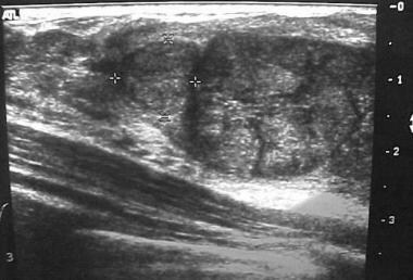
Mammary ultrasonography, also known as breast ultrasound, is a non-invasive diagnostic imaging technique that uses high-frequency sound waves to produce detailed images of the internal structures of the breast tissue. It is often used in conjunction with mammography to help identify and characterize breast abnormalities, such as lumps, cysts, or tumors, and to guide biopsy procedures.
Ultrasonography is particularly useful for evaluating palpable masses, assessing the integrity of breast implants, and distinguishing between solid and fluid-filled lesions. It is also a valuable tool for monitoring treatment response in patients with known breast cancer. Because it does not use radiation like mammography, mammary ultrasonography is considered safe and can be repeated as often as necessary. However, its effectiveness is highly dependent on the skill and experience of the sonographer performing the examination.
Interventional ultrasonography is a medical procedure that involves the use of real-time ultrasound imaging to guide minimally invasive diagnostic and therapeutic interventions. This technique combines the advantages of ultrasound, such as its non-ionizing nature (no radiation exposure), relatively low cost, and portability, with the ability to perform precise and targeted procedures.
In interventional ultrasonography, a specialized physician called an interventional radiologist or an interventional sonographer uses high-frequency sound waves to create detailed images of internal organs and tissues. These images help guide the placement of needles, catheters, or other instruments used during the procedure. Common interventions include biopsies (tissue sampling), fluid drainage, tumor ablation, and targeted drug delivery.
The real-time visualization provided by ultrasonography allows for increased accuracy and safety during these procedures, minimizing complications and reducing recovery time compared to traditional surgical approaches. Additionally, interventional ultrasonography can be performed on an outpatient basis, further contributing to its appeal as a less invasive alternative in many clinical scenarios.
Transcranial Doppler ultrasonography is a non-invasive diagnostic technique that uses high-frequency sound waves to visualize and measure the velocity of blood flow in the cerebral arteries located in the skull. This imaging modality employs the Doppler effect, which describes the change in frequency of sound waves as they reflect off moving red blood cells. By measuring the frequency shift of the reflected ultrasound waves, the velocity and direction of blood flow can be determined.
Transcranial Doppler ultrasonography is primarily used to assess cerebrovascular circulation and detect abnormalities such as stenosis (narrowing), occlusion (blockage), or embolism (obstruction) in the intracranial arteries. It can also help monitor patients with conditions like sickle cell disease, vasospasm following subarachnoid hemorrhage, and evaluate the effectiveness of treatments such as thrombolysis or angioplasty. The procedure is typically performed by placing a transducer on the patient's skull after applying a coupling gel, and it does not involve radiation exposure or contrast agents.
Sensitivity and specificity are statistical measures used to describe the performance of a diagnostic test or screening tool in identifying true positive and true negative results.
* Sensitivity refers to the proportion of people who have a particular condition (true positives) who are correctly identified by the test. It is also known as the "true positive rate" or "recall." A highly sensitive test will identify most or all of the people with the condition, but may also produce more false positives.
* Specificity refers to the proportion of people who do not have a particular condition (true negatives) who are correctly identified by the test. It is also known as the "true negative rate." A highly specific test will identify most or all of the people without the condition, but may also produce more false negatives.
In medical testing, both sensitivity and specificity are important considerations when evaluating a diagnostic test. High sensitivity is desirable for screening tests that aim to identify as many cases of a condition as possible, while high specificity is desirable for confirmatory tests that aim to rule out the condition in people who do not have it.
It's worth noting that sensitivity and specificity are often influenced by factors such as the prevalence of the condition in the population being tested, the threshold used to define a positive result, and the reliability and validity of the test itself. Therefore, it's important to consider these factors when interpreting the results of a diagnostic test.
Sulfur hexafluoride (SF6) is not typically a term used in medical definitions, but it is a colorless, odorless, non-flammable gas that is heavier than air. It is commonly used in the medical field for its magnetic resonance imaging (MRI) properties.
In MRI, SF6 is used as a contrast agent to improve the visualization of blood vessels and flow. When injected into a patient's bloodstream, the gas displaces oxygen in the blood, causing the blood vessels to appear darker on an MRI scan. This allows doctors to better see any abnormalities or blockages in the blood vessels.
It is important to note that sulfur hexafluoride should only be used under medical supervision and with appropriate precautions, as it can have adverse effects if not handled properly.
X-ray computed tomography (CT or CAT scan) is a medical imaging method that uses computer-processed combinations of many X-ray images taken from different angles to produce cross-sectional (tomographic) images (virtual "slices") of the body. These cross-sectional images can then be used to display detailed internal views of organs, bones, and soft tissues in the body.
The term "computed tomography" is used instead of "CT scan" or "CAT scan" because the machines take a series of X-ray measurements from different angles around the body and then use a computer to process these data to create detailed images of internal structures within the body.
CT scanning is a noninvasive, painless medical test that helps physicians diagnose and treat medical conditions. CT imaging provides detailed information about many types of tissue including lung, bone, soft tissue and blood vessels. CT examinations can be performed on every part of the body for a variety of reasons including diagnosis, surgical planning, and monitoring of therapeutic responses.
In computed tomography (CT), an X-ray source and detector rotate around the patient, measuring the X-ray attenuation at many different angles. A computer uses this data to construct a cross-sectional image by the process of reconstruction. This technique is called "tomography". The term "computed" refers to the use of a computer to reconstruct the images.
CT has become an important tool in medical imaging and diagnosis, allowing radiologists and other physicians to view detailed internal images of the body. It can help identify many different medical conditions including cancer, heart disease, lung nodules, liver tumors, and internal injuries from trauma. CT is also commonly used for guiding biopsies and other minimally invasive procedures.
In summary, X-ray computed tomography (CT or CAT scan) is a medical imaging technique that uses computer-processed combinations of many X-ray images taken from different angles to produce cross-sectional images of the body. It provides detailed internal views of organs, bones, and soft tissues in the body, allowing physicians to diagnose and treat medical conditions.
Prospective studies, also known as longitudinal studies, are a type of cohort study in which data is collected forward in time, following a group of individuals who share a common characteristic or exposure over a period of time. The researchers clearly define the study population and exposure of interest at the beginning of the study and follow up with the participants to determine the outcomes that develop over time. This type of study design allows for the investigation of causal relationships between exposures and outcomes, as well as the identification of risk factors and the estimation of disease incidence rates. Prospective studies are particularly useful in epidemiology and medical research when studying diseases with long latency periods or rare outcomes.
Contrast media are substances that are administered to a patient in order to improve the visibility of internal body structures or processes in medical imaging techniques such as X-rays, CT scans, MRI scans, and ultrasounds. These media can be introduced into the body through various routes, including oral, rectal, or intravenous administration.
Contrast media work by altering the appearance of bodily structures in imaging studies. For example, when a patient undergoes an X-ray examination, contrast media can be used to highlight specific organs, tissues, or blood vessels, making them more visible on the resulting images. In CT and MRI scans, contrast media can help to enhance the differences between normal and abnormal tissues, allowing for more accurate diagnosis and treatment planning.
There are several types of contrast media available, each with its own specific properties and uses. Some common examples include barium sulfate, which is used as a contrast medium in X-ray studies of the gastrointestinal tract, and iodinated contrast media, which are commonly used in CT scans to highlight blood vessels and other structures.
While contrast media are generally considered safe, they can sometimes cause adverse reactions, ranging from mild symptoms such as nausea or hives to more serious complications such as anaphylaxis or kidney damage. As a result, it is important for healthcare providers to carefully evaluate each patient's medical history and individual risk factors before administering contrast media.
Pregnancy is a physiological state or condition where a fertilized egg (zygote) successfully implants and grows in the uterus of a woman, leading to the development of an embryo and finally a fetus. This process typically spans approximately 40 weeks, divided into three trimesters, and culminates in childbirth. Throughout this period, numerous hormonal and physical changes occur to support the growing offspring, including uterine enlargement, breast development, and various maternal adaptations to ensure the fetus's optimal growth and well-being.
Urography is a medical imaging technique used to examine the urinary system, which includes the kidneys, ureters, and bladder. It involves the use of a contrast material that is injected into a vein or given orally, which then travels through the bloodstream to the kidneys and gets excreted in the urine. This allows the radiologist to visualize the structures and any abnormalities such as tumors, stones, or blockages. There are different types of urography, including intravenous urography (IVU), CT urography, and retrograde urography.
The Predictive Value of Tests, specifically the Positive Predictive Value (PPV) and Negative Predictive Value (NPV), are measures used in diagnostic tests to determine the probability that a positive or negative test result is correct.
Positive Predictive Value (PPV) is the proportion of patients with a positive test result who actually have the disease. It is calculated as the number of true positives divided by the total number of positive results (true positives + false positives). A higher PPV indicates that a positive test result is more likely to be a true positive, and therefore the disease is more likely to be present.
Negative Predictive Value (NPV) is the proportion of patients with a negative test result who do not have the disease. It is calculated as the number of true negatives divided by the total number of negative results (true negatives + false negatives). A higher NPV indicates that a negative test result is more likely to be a true negative, and therefore the disease is less likely to be present.
The predictive value of tests depends on the prevalence of the disease in the population being tested, as well as the sensitivity and specificity of the test. A test with high sensitivity and specificity will generally have higher predictive values than a test with low sensitivity and specificity. However, even a highly sensitive and specific test can have low predictive values if the prevalence of the disease is low in the population being tested.
Reproducibility of results in a medical context refers to the ability to obtain consistent and comparable findings when a particular experiment or study is repeated, either by the same researcher or by different researchers, following the same experimental protocol. It is an essential principle in scientific research that helps to ensure the validity and reliability of research findings.
In medical research, reproducibility of results is crucial for establishing the effectiveness and safety of new treatments, interventions, or diagnostic tools. It involves conducting well-designed studies with adequate sample sizes, appropriate statistical analyses, and transparent reporting of methods and findings to allow other researchers to replicate the study and confirm or refute the results.
The lack of reproducibility in medical research has become a significant concern in recent years, as several high-profile studies have failed to produce consistent findings when replicated by other researchers. This has led to increased scrutiny of research practices and a call for greater transparency, rigor, and standardization in the conduct and reporting of medical research.
Blood flow velocity is the speed at which blood travels through a specific part of the vascular system. It is typically measured in units of distance per time, such as centimeters per second (cm/s) or meters per second (m/s). Blood flow velocity can be affected by various factors, including cardiac output, vessel diameter, and viscosity of the blood. Measuring blood flow velocity is important in diagnosing and monitoring various medical conditions, such as heart disease, stroke, and peripheral vascular disease.
Fetal diseases are medical conditions or abnormalities that affect a fetus during pregnancy. These diseases can be caused by genetic factors, environmental influences, or a combination of both. They can range from mild to severe and may impact various organ systems in the developing fetus. Examples of fetal diseases include congenital heart defects, neural tube defects, chromosomal abnormalities such as Down syndrome, and infectious diseases such as toxoplasmosis or rubella. Fetal diseases can be diagnosed through prenatal testing, including ultrasound, amniocentesis, and chorionic villus sampling. Treatment options may include medication, surgery, or delivery of the fetus, depending on the nature and severity of the disease.
The scrotum is a part of the external male genitalia. It's a sac-like structure made up of several layers of skin and smooth muscle, which hangs down behind and beneath the penis. The primary function of the scrotum is to maintain the testicles at a temperature slightly lower than the core body temperature, which is optimal for sperm production.
The scrotum contains two compartments, each one housing a testicle. It's located in the pubic region and is usually visible externally. The skin of the scrotum is thin and wrinkled, which allows it to expand and contract depending on the temperature, accommodating the shrinking or swelling of the testicles.
Please note that while I strive to provide accurate information, this definition is intended to be a general overview and should not replace professional medical advice.
Three-dimensional (3D) imaging in medicine refers to the use of technologies and techniques that generate a 3D representation of internal body structures, organs, or tissues. This is achieved by acquiring and processing data from various imaging modalities such as X-ray computed tomography (CT), magnetic resonance imaging (MRI), ultrasound, or confocal microscopy. The resulting 3D images offer a more detailed visualization of the anatomy and pathology compared to traditional 2D imaging techniques, allowing for improved diagnostic accuracy, surgical planning, and minimally invasive interventions.
In 3D imaging, specialized software is used to reconstruct the acquired data into a volumetric model, which can be manipulated and viewed from different angles and perspectives. This enables healthcare professionals to better understand complex anatomical relationships, detect abnormalities, assess disease progression, and monitor treatment response. Common applications of 3D imaging include neuroimaging, orthopedic surgery planning, cancer staging, dental and maxillofacial reconstruction, and interventional radiology procedures.
Retrospective studies, also known as retrospective research or looking back studies, are a type of observational study that examines data from the past to draw conclusions about possible causal relationships between risk factors and outcomes. In these studies, researchers analyze existing records, medical charts, or previously collected data to test a hypothesis or answer a specific research question.
Retrospective studies can be useful for generating hypotheses and identifying trends, but they have limitations compared to prospective studies, which follow participants forward in time from exposure to outcome. Retrospective studies are subject to biases such as recall bias, selection bias, and information bias, which can affect the validity of the results. Therefore, retrospective studies should be interpreted with caution and used primarily to generate hypotheses for further testing in prospective studies.
A cyst is a closed sac, having a distinct membrane and division between the sac and its surrounding tissue, that contains fluid, air, or semisolid material. Cysts can occur in various parts of the body, including the skin, internal organs, and bones. They can be caused by various factors, such as infection, genetic predisposition, or blockage of a duct or gland. Some cysts may cause symptoms, such as pain or discomfort, while others may not cause any symptoms at all. Treatment for cysts depends on the type and location of the cyst, as well as whether it is causing any problems. Some cysts may go away on their own, while others may need to be drained or removed through a surgical procedure.
Palpation is a medical examination technique in which a healthcare professional uses their hands to feel the size, shape, and consistency of body parts, including organs, tissues, and bones. It is used to assess the patient's overall health, identify any abnormalities or areas of pain, monitor healing and disease progression, and guide diagnostic and treatment decisions.
During palpation, the healthcare professional applies gentle pressure with their fingers or hands to specific areas of the body, feeling for any changes in texture, temperature, moisture, or movement. The technique can be used to assess various bodily systems, including the cardiovascular, respiratory, gastrointestinal, musculoskeletal, and nervous systems.
Palpation is a valuable tool in physical examinations because it is non-invasive, relatively quick, and cost-effective. It can provide important information that helps healthcare professionals make accurate diagnoses and develop effective treatment plans for their patients.
Carotid artery diseases refer to conditions that affect the carotid arteries, which are the major blood vessels that supply oxygen-rich blood to the head and neck. The most common type of carotid artery disease is atherosclerosis, which occurs when fatty deposits called plaques build up in the inner lining of the arteries.
These plaques can cause the arteries to narrow or become blocked, reducing blood flow to the brain and increasing the risk of stroke. Other carotid artery diseases include carotid artery dissection, which occurs when there is a tear in the inner lining of the artery, and fibromuscular dysplasia, which is a condition that affects the muscle and tissue in the walls of the artery.
Symptoms of carotid artery disease may include neck pain or pulsations, transient ischemic attacks (TIAs) or "mini-strokes," and strokes. Treatment options for carotid artery disease depend on the severity and type of the condition but may include lifestyle changes, medications, endarterectomy (a surgical procedure to remove plaque from the artery), or angioplasty and stenting (procedures to open blocked arteries using a balloon and stent).
The abdomen refers to the portion of the body that lies between the thorax (chest) and the pelvis. It is a musculo-fascial cavity containing the digestive, urinary, and reproductive organs. The abdominal cavity is divided into several regions and quadrants for medical description and examination purposes. These include the upper and lower abdomen, as well as nine quadrants formed by the intersection of the midline and a horizontal line drawn at the level of the umbilicus (navel).
The major organs located within the abdominal cavity include:
1. Stomach - muscular organ responsible for initial digestion of food
2. Small intestine - long, coiled tube where most nutrient absorption occurs
3. Large intestine - consists of the colon and rectum; absorbs water and stores waste products
4. Liver - largest internal organ, involved in protein synthesis, detoxification, and metabolism
5. Pancreas - secretes digestive enzymes and hormones such as insulin
6. Spleen - filters blood and removes old red blood cells
7. Kidneys - pair of organs responsible for filtering waste products from the blood and producing urine
8. Adrenal glands - sit atop each kidney, produce hormones that regulate metabolism, immune response, and stress response
The abdomen is an essential part of the human body, playing a crucial role in digestion, absorption, and elimination of food and waste materials, as well as various metabolic processes.
Preoperative care refers to the series of procedures, interventions, and preparations that are conducted before a surgical operation. The primary goal of preoperative care is to ensure the patient's well-being, optimize their physical condition, reduce potential risks, and prepare them mentally and emotionally for the upcoming surgery.
Preoperative care typically includes:
1. Preoperative assessment: A thorough evaluation of the patient's overall health status, including medical history, physical examination, laboratory tests, and diagnostic imaging, to identify any potential risk factors or comorbidities that may impact the surgical procedure and postoperative recovery.
2. Informed consent: The process of ensuring the patient understands the nature of the surgery, its purpose, associated risks, benefits, and alternative treatment options. The patient signs a consent form indicating they have been informed and voluntarily agree to undergo the surgery.
3. Preoperative instructions: Guidelines provided to the patient regarding their diet, medication use, and other activities in the days leading up to the surgery. These instructions may include fasting guidelines, discontinuing certain medications, or arranging for transportation after the procedure.
4. Anesthesia consultation: A meeting with the anesthesiologist to discuss the type of anesthesia that will be used during the surgery and address any concerns related to anesthesia risks, side effects, or postoperative pain management.
5. Preparation of the surgical site: Cleaning and shaving the area where the incision will be made, as well as administering appropriate antimicrobial agents to minimize the risk of infection.
6. Medical optimization: Addressing any underlying medical conditions or correcting abnormalities that may negatively impact the surgical outcome. This may involve adjusting medications, treating infections, or managing chronic diseases such as diabetes.
7. Emotional and psychological support: Providing counseling, reassurance, and education to help alleviate anxiety, fear, or emotional distress related to the surgery.
8. Preoperative holding area: The patient is transferred to a designated area near the operating room where they are prepared for surgery by changing into a gown, having intravenous (IV) lines inserted, and receiving monitoring equipment.
By following these preoperative care guidelines, healthcare professionals aim to ensure that patients undergo safe and successful surgical procedures with optimal outcomes.
Medical Definition:
Magnetic Resonance Imaging (MRI) is a non-invasive diagnostic imaging technique that uses a strong magnetic field and radio waves to create detailed cross-sectional or three-dimensional images of the internal structures of the body. The patient lies within a large, cylindrical magnet, and the scanner detects changes in the direction of the magnetic field caused by protons in the body. These changes are then converted into detailed images that help medical professionals to diagnose and monitor various medical conditions, such as tumors, injuries, or diseases affecting the brain, spinal cord, heart, blood vessels, joints, and other internal organs. MRI does not use radiation like computed tomography (CT) scans.
A toe joint, also known as a metatarsophalangeal (MTP) joint, is the articulation between the bones in the foot (metatarsals) and the bones in the toes (phalanges). There are five MTP joints in each foot, one for each toe except for the big toe, which has its own separate joint called the first metatarsophalangeal joint.
The MTP joints allow for movement and flexibility of the toes, enabling activities such as walking, running, and standing. Problems with these joints can lead to pain, stiffness, and difficulty moving, making it important to maintain their health and mobility through proper foot care and exercise.
A thyroid nodule is a growth or lump that forms within the thyroid gland, a small butterfly-shaped endocrine gland located in the front of your neck. Thyroid nodules can be solid or fluid-filled (cystic) and vary in size. Most thyroid nodules are benign (noncancerous) and do not cause symptoms. However, some thyroid nodules may be cancerous or overproduce hormones, leading to hyperthyroidism. The exact cause of thyroid nodules is not always known, but factors such as iodine deficiency, Hashimoto's disease, and family history can increase the risk of developing them. A healthcare professional typically diagnoses a thyroid nodule through physical examination, imaging tests like ultrasound, or fine-needle aspiration biopsy to determine if further treatment is necessary.
The carotid arteries are a pair of vital blood vessels in the human body that supply oxygenated blood to the head and neck. Each person has two common carotid arteries, one on each side of the neck, which branch off from the aorta, the largest artery in the body.
The right common carotid artery originates from the brachiocephalic trunk, while the left common carotid artery arises directly from the aortic arch. As they ascend through the neck, they split into two main branches: the internal and external carotid arteries.
The internal carotid artery supplies oxygenated blood to the brain, eyes, and other structures within the skull, while the external carotid artery provides blood to the face, scalp, and various regions of the neck.
Maintaining healthy carotid arteries is crucial for overall cardiovascular health and preventing serious conditions like stroke, which can occur when the arteries become narrowed or blocked due to the buildup of plaque or fatty deposits (atherosclerosis). Regular check-ups with healthcare professionals may include monitoring carotid artery health through ultrasound or other imaging techniques.
An ovarian cyst is a sac or pouch filled with fluid that forms on the ovary. Ovarian cysts are quite common in women during their childbearing years, and they often cause no symptoms. In most cases, ovarian cysts disappear without treatment over a few months. However, larger or persistent cysts may require medical intervention, including surgical removal.
There are various types of ovarian cysts, such as functional cysts (follicular and corpus luteum cysts), which develop during the menstrual cycle due to hormonal changes, and non-functional cysts (dermoid cysts, endometriomas, and cystadenomas), which can form due to different causes.
While many ovarian cysts are benign, some may have malignant potential or indicate an underlying medical condition like polycystic ovary syndrome (PCOS). Regular gynecological check-ups, including pelvic examinations and ultrasounds, can help detect and monitor ovarian cysts.
The gallbladder is a small, pear-shaped organ located just under the liver in the right upper quadrant of the abdomen. Its primary function is to store and concentrate bile, a digestive enzyme produced by the liver, which helps in the breakdown of fats during the digestion process. When food, particularly fatty foods, enter the stomach and small intestine, the gallbladder contracts and releases bile through the common bile duct into the duodenum, the first part of the small intestine, to aid in fat digestion.
The gallbladder is made up of three main parts: the fundus, body, and neck. It has a muscular wall that allows it to contract and release bile. Gallstones, an inflammation of the gallbladder (cholecystitis), or other gallbladder diseases can cause pain, discomfort, and potentially serious health complications if left untreated.
Echoencephalography (EEG) is a type of neurosonology technique that uses ultrasound to assess the structures of the brain and detect any abnormalities. It is also known as brain ultrasound or transcranial Doppler ultrasound. This non-invasive procedure involves placing a small ultrasound probe on the skull, which emits sound waves that travel through the skull and bounce back (echo) when they reach the brain tissue. The resulting echoes are then analyzed to create images of the brain's structures, including the ventricles, cerebral arteries, and other blood vessels.
EEG is often used in infants and young children, as their skulls are still thin enough to allow for clear ultrasound imaging. It can help diagnose conditions such as hydrocephalus (fluid buildup in the brain), intracranial hemorrhage (bleeding in the brain), stroke, and other neurological disorders. EEG is a safe and painless procedure that does not require any radiation or contrast agents, making it an attractive alternative to other imaging techniques such as CT or MRI scans. However, its use is limited in older children and adults due to the thickening of the skull bones, which can make it difficult to obtain clear images.
The metacarpophalangeal (MCP) joint is the joint that connects the bones of the hand (metacarpals) to the bones of the fingers and thumb (phalanges). It's also commonly referred to as the "knuckle" joint. The MCP joint allows for flexion, extension, abduction, and adduction movements of the fingers and thumb. It is a synovial joint, which means it contains a lubricating fluid called synovial fluid that helps reduce friction during movement.
Gestational age is the length of time that has passed since the first day of the last menstrual period (LMP) in pregnant women. It is the standard unit used to estimate the age of a pregnancy and is typically expressed in weeks. This measure is used because the exact date of conception is often not known, but the start of the last menstrual period is usually easier to recall.
It's important to note that since ovulation typically occurs around two weeks after the start of the LMP, gestational age is approximately two weeks longer than fetal age, which is the actual time elapsed since conception. Medical professionals use both gestational and fetal age to track the development and growth of the fetus during pregnancy.
The vagina is the canal that joins the cervix (the lower part of the uterus) to the outside of the body. It also is known as the birth canal because babies pass through it during childbirth. The vagina is where sexual intercourse occurs and where menstrual blood exits the body. It has a flexible wall that can expand and retract. During sexual arousal, the vaginal walls swell with blood to become more elastic in order to accommodate penetration.
It's important to note that sometimes people use the term "vagina" to refer to the entire female genital area, including the external structures like the labia and clitoris. But technically, these are considered part of the vulva, not the vagina.
Liver neoplasms refer to abnormal growths in the liver that can be benign or malignant. Benign liver neoplasms are non-cancerous tumors that do not spread to other parts of the body, while malignant liver neoplasms are cancerous tumors that can invade and destroy surrounding tissue and spread to other organs.
Liver neoplasms can be primary, meaning they originate in the liver, or secondary, meaning they have metastasized (spread) to the liver from another part of the body. Primary liver neoplasms can be further classified into different types based on their cell of origin and behavior, including hepatocellular carcinoma, cholangiocarcinoma, and hepatic hemangioma.
The diagnosis of liver neoplasms typically involves a combination of imaging studies, such as ultrasound, CT scan, or MRI, and biopsy to confirm the type and stage of the tumor. Treatment options depend on the type and extent of the neoplasm and may include surgery, radiation therapy, chemotherapy, or liver transplantation.
Hydronephrosis is a medical condition characterized by the swelling of one or both kidneys due to the accumulation of urine. This occurs when the flow of urine from the kidney to the bladder is obstructed, causing urine to back up into the kidney. The obstruction can be caused by various factors such as kidney stones, tumors, or congenital abnormalities. If left untreated, hydronephrosis can lead to serious complications including kidney damage and infection. It is typically diagnosed through imaging tests such as ultrasound, CT scan, or MRI.
Observer variation, also known as inter-observer variability or measurement agreement, refers to the difference in observations or measurements made by different observers or raters when evaluating the same subject or phenomenon. It is a common issue in various fields such as medicine, research, and quality control, where subjective assessments are involved.
In medical terms, observer variation can occur in various contexts, including:
1. Diagnostic tests: Different radiologists may interpret the same X-ray or MRI scan differently, leading to variations in diagnosis.
2. Clinical trials: Different researchers may have different interpretations of clinical outcomes or adverse events, affecting the consistency and reliability of trial results.
3. Medical records: Different healthcare providers may document medical histories, physical examinations, or treatment plans differently, leading to inconsistencies in patient care.
4. Pathology: Different pathologists may have varying interpretations of tissue samples or laboratory tests, affecting diagnostic accuracy.
Observer variation can be minimized through various methods, such as standardized assessment tools, training and calibration of observers, and statistical analysis of inter-rater reliability.
Abdominal injuries refer to damages or traumas that occur in the abdomen, an area of the body that is located between the chest and the pelvis. This region contains several vital organs such as the stomach, liver, spleen, pancreas, small intestine, large intestine, kidneys, and reproductive organs. Abdominal injuries can range from minor bruises and cuts to severe internal bleeding and organ damage, depending on the cause and severity of the trauma.
Common causes of abdominal injuries include:
* Blunt force trauma, such as that caused by car accidents, falls, or physical assaults
* Penetrating trauma, such as that caused by gunshot wounds or stabbing
* Deceleration injuries, which occur when the body is moving at a high speed and suddenly stops, causing internal organs to continue moving and collide with each other or the abdominal wall
Symptoms of abdominal injuries may include:
* Pain or tenderness in the abdomen
* Swelling or bruising in the abdomen
* Nausea or vomiting
* Dizziness or lightheadedness
* Blood in the urine or stool
* Difficulty breathing or shortness of breath
* Rapid heartbeat or low blood pressure
Abdominal injuries can be life-threatening if left untreated, and immediate medical attention is necessary to prevent complications such as infection, internal bleeding, organ failure, or even death. Treatment may include surgery, medication, or other interventions depending on the severity and location of the injury.
Image enhancement in the medical context refers to the process of improving the quality and clarity of medical images, such as X-rays, CT scans, MRI scans, or ultrasound images, to aid in the diagnosis and treatment of medical conditions. Image enhancement techniques may include adjusting contrast, brightness, or sharpness; removing noise or artifacts; or applying specialized algorithms to highlight specific features or structures within the image.
The goal of image enhancement is to provide clinicians with more accurate and detailed information about a patient's anatomy or physiology, which can help inform medical decision-making and improve patient outcomes.
Splenic diseases refer to a range of medical conditions that affect the structure, function, or health of the spleen. The spleen is an organ located in the upper left quadrant of the abdomen, which plays a vital role in filtering the blood and fighting infections. Some common splenic diseases include:
1. Splenomegaly: Enlargement of the spleen due to various causes such as infections, liver disease, blood disorders, or cancer.
2. Hypersplenism: Overactivity of the spleen leading to excessive removal of blood cells from circulation, causing anemia, leukopenia, or thrombocytopenia.
3. Splenic infarction: Partial or complete blockage of the splenic artery or its branches, resulting in tissue death and potential organ dysfunction.
4. Splenic rupture: Traumatic or spontaneous tearing of the spleen capsule, causing internal bleeding and potentially life-threatening conditions.
5. Infections: Bacterial (e.g., sepsis, tuberculosis), viral (e.g., mononucleosis, cytomegalovirus), fungal (e.g., histoplasmosis), or parasitic (e.g., malaria) infections can affect the spleen and cause various symptoms.
6. Hematologic disorders: Conditions such as sickle cell disease, thalassemia, hemolytic anemias, lymphomas, leukemias, or myeloproliferative neoplasms can involve the spleen and lead to its enlargement or dysfunction.
7. Autoimmune diseases: Conditions like rheumatoid arthritis, systemic lupus erythematosus, or vasculitis can affect the spleen and cause various symptoms.
8. Cancers: Primary (e.g., splenic tumors) or secondary (e.g., metastatic cancer from other organs) malignancies can involve the spleen and lead to its enlargement, dysfunction, or rupture.
9. Vascular abnormalities: Conditions such as portal hypertension, Budd-Chiari syndrome, or splenic vein thrombosis can affect the spleen and cause various symptoms.
10. Trauma: Accidental or intentional injuries to the spleen can lead to bleeding, infection, or organ dysfunction.
Testicular diseases refer to a range of conditions that affect the testicles, the male reproductive organs located in the scrotum. These diseases can affect either one or both testicles and may cause pain, swelling, or impact fertility. Here are some examples of testicular diseases:
1. Testicular cancer: A malignant tumor that develops in the testicle. It is a relatively rare cancer but is highly treatable if detected early.
2. Testicular torsion: A surgical emergency that occurs when the spermatic cord, which supplies blood to the testicle, becomes twisted, cutting off the blood flow.
3. Epididymitis: An infection or inflammation of the epididymis, a coiled tube that stores and carries sperm from the testicle.
4. Orchitis: An infection or inflammation of the testicle itself. It can occur on its own or as a complication of mumps.
5. Hydrocele: A fluid-filled sac that forms around the testicle, causing swelling.
6. Varicocele: Enlarged veins in the scrotum that can cause pain and affect fertility.
7. Inguinal hernia: A condition where a portion of the intestine or fat protrudes through a weakened area in the abdominal wall, often appearing as a bulge in the groin or scrotum.
8. Testicular trauma: Injury to the testicle, which can result from accidents, sports injuries, or other causes.
9. Undescended testicles: A condition where one or both testicles fail to descend from the abdomen into the scrotum before birth.
It is essential for men to perform regular self-examinations to check for any unusual lumps, swelling, or pain in the testicles and seek medical attention if they notice any changes.
Diagnostic imaging is a medical specialty that uses various technologies to produce visual representations of the internal structures and functioning of the body. These images are used to diagnose injury, disease, or other abnormalities and to monitor the effectiveness of treatment. Common modalities of diagnostic imaging include:
1. Radiography (X-ray): Uses ionizing radiation to produce detailed images of bones, teeth, and some organs.
2. Computed Tomography (CT) Scan: Combines X-ray technology with computer processing to create cross-sectional images of the body.
3. Magnetic Resonance Imaging (MRI): Uses a strong magnetic field and radio waves to generate detailed images of soft tissues, organs, and bones.
4. Ultrasound: Employs high-frequency sound waves to produce real-time images of internal structures, often used for obstetrics and gynecology.
5. Nuclear Medicine: Involves the administration of radioactive tracers to assess organ function or detect abnormalities within the body.
6. Positron Emission Tomography (PET) Scan: Uses a small amount of radioactive material to produce detailed images of metabolic activity in the body, often used for cancer detection and monitoring treatment response.
7. Fluoroscopy: Utilizes continuous X-ray imaging to observe moving structures or processes within the body, such as swallowing studies or angiography.
Diagnostic imaging plays a crucial role in modern medicine, allowing healthcare providers to make informed decisions about patient care and treatment plans.
Auscultation is a medical procedure in which a healthcare professional uses a stethoscope to listen to the internal sounds of the body, such as heart, lung, or abdominal sounds. These sounds can provide important clues about a person's health and help diagnose various medical conditions, such as heart valve problems, lung infections, or digestive issues.
During auscultation, the healthcare professional places the stethoscope on different parts of the body and listens for any abnormal sounds, such as murmurs, rubs, or wheezes. They may also ask the person to perform certain movements, such as breathing deeply or coughing, to help identify any changes in the sounds.
Auscultation is a simple, non-invasive procedure that can provide valuable information about a person's health. It is an essential part of a physical examination and is routinely performed by healthcare professionals during regular checkups and hospital visits.
Follow-up studies are a type of longitudinal research that involve repeated observations or measurements of the same variables over a period of time, in order to understand their long-term effects or outcomes. In medical context, follow-up studies are often used to evaluate the safety and efficacy of medical treatments, interventions, or procedures.
In a typical follow-up study, a group of individuals (called a cohort) who have received a particular treatment or intervention are identified and then followed over time through periodic assessments or data collection. The data collected may include information on clinical outcomes, adverse events, changes in symptoms or functional status, and other relevant measures.
The results of follow-up studies can provide important insights into the long-term benefits and risks of medical interventions, as well as help to identify factors that may influence treatment effectiveness or patient outcomes. However, it is important to note that follow-up studies can be subject to various biases and limitations, such as loss to follow-up, recall bias, and changes in clinical practice over time, which must be carefully considered when interpreting the results.
Gallbladder diseases refer to a range of conditions that affect the function and structure of the gallbladder, a small pear-shaped organ located beneath the liver. The primary role of the gallbladder is to store, concentrate, and release bile into the small intestine to aid in digesting fats. Gallbladder diseases can be chronic or acute and may cause various symptoms, discomfort, or complications if left untreated. Here are some common gallbladder diseases with brief definitions:
1. Cholelithiasis: The presence of gallstones within the gallbladder. Gallstones are small, hard deposits made of cholesterol, bilirubin, or a combination of both, which can vary in size from tiny grains to several centimeters.
2. Cholecystitis: Inflammation of the gallbladder, often caused by obstruction of the cystic duct (the tube connecting the gallbladder and the common bile duct) due to a gallstone. This condition can be acute or chronic and may cause abdominal pain, fever, and tenderness in the right upper quadrant of the abdomen.
3. Choledocholithiasis: The presence of gallstones within the common bile duct, which can lead to obstruction, jaundice, and potential infection of the biliary system (cholangitis).
4. Acalculous gallbladder disease: Gallbladder dysfunction or inflammation without the presence of gallstones. This condition is often seen in critically ill patients and can lead to similar symptoms as cholecystitis.
5. Gallbladder polyps: Small growths attached to the inner wall of the gallbladder. While most polyps are benign, some may have malignant potential, especially if they are larger than 1 cm in size or associated with certain risk factors.
6. Gallbladder cancer: A rare form of cancer that originates in the gallbladder tissue. It is often asymptomatic in its early stages and can be challenging to diagnose. Symptoms may include abdominal pain, jaundice, or a palpable mass in the right upper quadrant of the abdomen.
It is essential to consult with a healthcare professional if experiencing symptoms related to gallbladder disease for proper diagnosis and treatment.
Treatment outcome is a term used to describe the result or effect of medical treatment on a patient's health status. It can be measured in various ways, such as through symptoms improvement, disease remission, reduced disability, improved quality of life, or survival rates. The treatment outcome helps healthcare providers evaluate the effectiveness of a particular treatment plan and make informed decisions about future care. It is also used in clinical research to compare the efficacy of different treatments and improve patient care.
Angiography is a medical procedure in which an x-ray image is taken to visualize the internal structure of blood vessels, arteries, or veins. This is done by injecting a radiopaque contrast agent (dye) into the blood vessel using a thin, flexible catheter. The dye makes the blood vessels visible on an x-ray image, allowing doctors to diagnose and treat various medical conditions such as blockages, narrowing, or malformations of the blood vessels.
There are several types of angiography, including:
* Cardiac angiography (also called coronary angiography) - used to examine the blood vessels of the heart
* Cerebral angiography - used to examine the blood vessels of the brain
* Peripheral angiography - used to examine the blood vessels in the limbs or other parts of the body.
Angiography is typically performed by a radiologist, cardiologist, or vascular surgeon in a hospital setting. It can help diagnose conditions such as coronary artery disease, aneurysms, and peripheral arterial disease, among others.
The common carotid artery is a major blood vessel in the neck that supplies oxygenated blood to the head and neck. It originates from the brachiocephalic trunk or the aortic arch and divides into the internal and external carotid arteries at the level of the upper border of the thyroid cartilage. The common carotid artery is an important structure in the circulatory system, and any damage or blockage to it can have serious consequences, including stroke.
A transducer is a device that converts one form of energy into another. In the context of medicine and biology, transducers often refer to devices that convert a physiological parameter (such as blood pressure, temperature, or sound waves) into an electrical signal that can be measured and analyzed. Examples of medical transducers include:
1. Blood pressure transducer: Converts the mechanical force exerted by blood on the walls of an artery into an electrical signal.
2. Temperature transducer: Converts temperature changes into electrical signals.
3. ECG transducer (electrocardiogram): Converts the electrical activity of the heart into a visual representation called an electrocardiogram.
4. Ultrasound transducer: Uses sound waves to create images of internal organs and structures.
5. Piezoelectric transducer: Generates an electric charge when subjected to pressure or vibration, used in various medical devices such as hearing aids, accelerometers, and pressure sensors.
Biliary tract diseases refer to a group of medical conditions that affect the biliary system, which includes the gallbladder, bile ducts, and liver. Bile is a digestive juice produced by the liver, stored in the gallbladder, and released into the small intestine through the bile ducts to help digest fats.
Biliary tract diseases can cause various symptoms such as abdominal pain, jaundice, fever, nausea, vomiting, and changes in stool color. Some of the common biliary tract diseases include:
1. Gallstones: Small, hard deposits that form in the gallbladder or bile ducts made up of cholesterol or bilirubin.
2. Cholecystitis: Inflammation of the gallbladder, often caused by gallstones.
3. Cholangitis: Infection or inflammation of the bile ducts.
4. Biliary dyskinesia: A motility disorder that affects the contraction and relaxation of the muscles in the biliary system.
5. Primary sclerosing cholangitis: A chronic autoimmune disease that causes scarring and narrowing of the bile ducts.
6. Biliary tract cancer: Rare cancers that affect the gallbladder, bile ducts, or liver.
Treatment for biliary tract diseases varies depending on the specific condition and severity but may include medications, surgery, or a combination of both.
The rectum is the lower end of the digestive tract, located between the sigmoid colon and the anus. It serves as a storage area for feces before they are eliminated from the body. The rectum is about 12 cm long in adults and is surrounded by layers of muscle that help control defecation. The mucous membrane lining the rectum allows for the detection of stool, which triggers the reflex to have a bowel movement.
A newborn infant is a baby who is within the first 28 days of life. This period is also referred to as the neonatal period. Newborns require specialized care and attention due to their immature bodily systems and increased vulnerability to various health issues. They are closely monitored for signs of well-being, growth, and development during this critical time.
A needle biopsy is a medical procedure in which a thin, hollow needle is used to remove a small sample of tissue from a suspicious or abnormal area of the body. The tissue sample is then examined under a microscope to check for cancer cells or other abnormalities. Needle biopsies are often used to diagnose lumps or masses that can be felt through the skin, but they can also be guided by imaging techniques such as ultrasound, CT scan, or MRI to reach areas that cannot be felt. There are several types of needle biopsy procedures, including fine-needle aspiration (FNA) and core needle biopsy. FNA uses a thin needle and gentle suction to remove fluid and cells from the area, while core needle biopsy uses a larger needle to remove a small piece of tissue. The type of needle biopsy used depends on the location and size of the abnormal area, as well as the reason for the procedure.
A "false positive reaction" in medical testing refers to a situation where a diagnostic test incorrectly indicates the presence of a specific condition or disease in an individual who does not actually have it. This occurs when the test results give a positive outcome, while the true health status of the person is negative or free from the condition being tested for.
False positive reactions can be caused by various factors including:
1. Presence of unrelated substances that interfere with the test result (e.g., cross-reactivity between similar molecules).
2. Low specificity of the test, which means it may detect other conditions or irrelevant factors as positive.
3. Contamination during sample collection, storage, or analysis.
4. Human errors in performing or interpreting the test results.
False positive reactions can have significant consequences, such as unnecessary treatments, anxiety, and increased healthcare costs. Therefore, it is essential to confirm any positive test result with additional tests or clinical evaluations before making a definitive diagnosis.
A fine-needle biopsy (FNB) is a medical procedure in which a thin, hollow needle is used to obtain a sample of cells or tissue from a suspicious or abnormal area in the body, such as a lump or mass. The needle is typically smaller than that used in a core needle biopsy, and it is guided into place using imaging techniques such as ultrasound, CT scan, or MRI.
The sample obtained during an FNB can be used to diagnose various medical conditions, including cancer, infection, or inflammation. The procedure is generally considered safe and well-tolerated, with minimal risks of complications such as bleeding, infection, or discomfort. However, the accuracy of the diagnosis depends on the skill and experience of the healthcare provider performing the biopsy, as well as the adequacy of the sample obtained.
Overall, FNB is a valuable diagnostic tool that can help healthcare providers make informed decisions about treatment options and improve patient outcomes.
An ovarian follicle is a fluid-filled sac in the ovary that contains an immature egg or ovum (oocyte). It's a part of the female reproductive system and plays a crucial role in the process of ovulation.
Ovarian follicles start developing in the ovaries during fetal development, but only a small number of them will mature and release an egg during a woman's reproductive years. The maturation process is stimulated by hormones like follicle-stimulating hormone (FSH) and luteinizing hormone (LH).
There are different types of ovarian follicles, including primordial, primary, secondary, and tertiary or Graafian follicles. The Graafian follicle is the mature follicle that ruptures during ovulation to release the egg into the fallopian tube, where it may be fertilized by sperm.
It's important to note that abnormal growth or development of ovarian follicles can lead to conditions like polycystic ovary syndrome (PCOS) and ovarian cancer.
The tunica media is the middle layer of the wall of a blood vessel or hollow organ in the body. It is primarily composed of smooth muscle cells and elastic fibers, which allow the vessel or organ to expand and contract. This layer helps regulate the diameter of the lumen (the inner space) of the vessel or organ, thereby controlling the flow of fluids such as blood or lymph through it. The tunica media plays a crucial role in maintaining proper organ function and blood pressure regulation.
Prenatal diagnosis is the medical testing of fetuses, embryos, or pregnant women to detect the presence or absence of certain genetic disorders or birth defects. These tests can be performed through various methods such as chorionic villus sampling (CVS), amniocentesis, or ultrasound. The goal of prenatal diagnosis is to provide early information about the health of the fetus so that parents and healthcare providers can make informed decisions about pregnancy management and newborn care. It allows for early intervention, treatment, or planning for the child's needs after birth.
Tendinopathy is a general term referring to the degeneration or dysrepair of a tendon, which can result in pain and impaired function. It was previously referred to as tendinitis or tendinosis, but tendinopathy is now preferred because it describes various pathological conditions within the tendon, rather than a specific diagnosis.
Tendinopathy often develops due to overuse, repetitive strain, or age-related wear and tear. The condition typically involves collagen breakdown in the tendon, along with an increase in disorganized tenocytes (tendon cells) and vascular changes. This process can lead to thickening of the tendon, loss of elasticity, and the formation of calcium deposits or nodules.
Commonly affected tendons include the Achilles tendon, patellar tendon, rotator cuff tendons in the shoulder, and the extensor carpi radialis brevis tendon in the elbow (also known as tennis elbow). Treatment for tendinopathy often includes rest, physical therapy, exercise, pain management, and occasionally, surgical intervention.
Cholecystography is a medical procedure that involves the use of X-rays to examine the gallbladder and bile ducts. It is also known as an oral cholecystogram (OCG).
The procedure involves administering a contrast agent, typically a iodine-based dye, which is absorbed by the liver and excreted into the bile ducts and gallbladder. The dye makes the bile ducts and gallbladder visible on X-ray images, allowing doctors to diagnose conditions such as gallstones, tumors, or inflammation of the gallbladder.
Cholecystography is not commonly used today due to the development of more advanced imaging techniques, such as ultrasound and computed tomography (CT) scans, which are non-invasive and do not require the use of contrast agents. However, it may still be used in certain cases where other imaging tests are inconclusive or unavailable.
The portal vein is the large venous trunk that carries blood from the gastrointestinal tract, spleen, pancreas, and gallbladder to the liver. It is formed by the union of the superior mesenteric vein (draining the small intestine and a portion of the large intestine) and the splenic vein (draining the spleen and pancreas). The portal vein then divides into right and left branches within the liver, where the blood flows through the sinusoids and gets enriched with oxygen and nutrients before being drained by the hepatic veins into the inferior vena cava. This unique arrangement allows the liver to process and detoxify the absorbed nutrients, remove waste products, and regulate metabolic homeostasis.
A testicular hydrocele is a type of fluid-filled sac that forms around the testicle (testis), typically in the scrotum. This sac, known as the tunica vaginalis, normally contains a small amount of fluid that helps to lubricate and protect the testicle. However, when an excessive amount of fluid accumulates in this sac, it results in the formation of a hydrocele.
Testicular hydroceles can be congenital (present at birth) or acquired later in life due to various reasons such as injury, inflammation, or infection in the scrotal area. They are usually painless but may cause discomfort or a feeling of heaviness in the scrotum, especially when they become large. In some cases, hydroceles may resolve on their own without treatment, while others may require surgical intervention to drain the fluid and repair the underlying issue.
It is essential to differentiate between hydroceles and other conditions with similar symptoms, such as hernias or tumors, which may require more urgent medical attention. A healthcare professional can perform a physical examination and possibly recommend further testing, like an ultrasound, to confirm the diagnosis of a testicular hydrocele.
Gallbladder neoplasms refer to abnormal growths in the tissue of the gallbladder, which can be benign or malignant. Benign neoplasms are non-cancerous and typically do not spread to other parts of the body. Malignant neoplasms, also known as gallbladder cancer, can invade nearby tissues and organs and may metastasize (spread) to distant parts of the body. Gallbladder neoplasms can cause symptoms such as abdominal pain, jaundice, and nausea, but they are often asymptomatic until they have advanced to an advanced stage. The exact causes of gallbladder neoplasms are not fully understood, but risk factors include gallstones, chronic inflammation of the gallbladder, and certain inherited genetic conditions.
Endoscopic retrograde cholangiopancreatography (ERCP) is a medical procedure that combines upper gastrointestinal (GI) endoscopy and fluoroscopy to diagnose and treat certain problems of the bile ducts and pancreas.
During ERCP, a flexible endoscope (a long, thin, lighted tube with a camera on the end) is passed through the patient's mouth and throat, then through the stomach and into the first part of the small intestine (duodenum). A narrow plastic tube (catheter) is then inserted through the endoscope and into the bile ducts and/or pancreatic duct. Contrast dye is injected through the catheter, and X-rays are taken to visualize the ducts.
ERCP can be used to diagnose a variety of conditions affecting the bile ducts and pancreas, including gallstones, tumors, strictures (narrowing of the ducts), and chronic pancreatitis. It can also be used to treat certain conditions, such as removing gallstones from the bile duct or placing stents to keep the ducts open in cases of stricture.
ERCP is an invasive procedure that carries a risk of complications, including pancreatitis, infection, bleeding, and perforation (a tear in the lining of the GI tract). It should only be performed by experienced medical professionals in a hospital setting.
A polyp is a general term for a small growth that protrudes from a mucous membrane, such as the lining of the nose or the digestive tract. Polyps can vary in size and shape, but they are usually cherry-sized or smaller and have a stalk or a broad base. They are often benign (noncancerous), but some types of polyps, especially those in the colon, can become cancerous over time.
In the digestive tract, polyps can form in the colon, rectum, stomach, or small intestine. Colorectal polyps are the most common type and are usually found during routine colonoscopies. There are several types of colorectal polyps, including:
* Adenomatous polyps (adenomas): These polyps can become cancerous over time and are the most likely to turn into cancer.
* Hyperplastic polyps: These polyps are usually small and benign, but some types may have a higher risk of becoming cancerous.
* Inflammatory polyps: These polyps are caused by chronic inflammation in the digestive tract, such as from inflammatory bowel disease (IBD).
Polyps can also form in other parts of the body, including the nose, sinuses, ears, and uterus. In most cases, polyps are benign and do not cause any symptoms. However, if they become large enough, they may cause problems such as bleeding, obstruction, or discomfort. Treatment typically involves removing the polyp through a surgical procedure.
The urinary tract is a system in the body responsible for producing, storing, and eliminating urine. It includes two kidneys, two ureters, the bladder, and the urethra. The kidneys filter waste and excess fluids from the blood to produce urine, which then travels down the ureters into the bladder. When the bladder is full, urine is released through the urethra during urination. Any part of this system can become infected or inflamed, leading to conditions such as urinary tract infections (UTIs) or kidney stones.
Renal colic is a type of abdominal pain that occurs due to the presence of a kidney stone or other obstruction in the urinary tract. It is typically described as a severe, cramping pain that radiates from the lower back or flank area down to the groin or genitals. The pain may be accompanied by nausea, vomiting, sweating, and frequent urination. Renal colic is caused by the contraction of smooth muscles in the ureter as they attempt to move the stone or obstruction out of the body. This can cause significant discomfort and often requires medical treatment to alleviate the pain and remove the obstruction.
Pulsatile flow is a type of fluid flow that occurs in a rhythmic, wave-like pattern, typically seen in the cardiovascular system. It refers to the periodic variation in the volume or velocity of a fluid (such as blood) that is caused by the regular beating of the heart. In pulsatile flow, there are periods of high flow followed by periods of low or no flow, which creates a distinct pattern on a graph or tracing. This type of flow is important for maintaining proper function and health in organs and tissues throughout the body.
Intraoperative care refers to the medical care and interventions provided to a patient during a surgical procedure. This care is typically administered by a team of healthcare professionals, including anesthesiologists, surgeons, nurses, and other specialists as needed. The goal of intraoperative care is to maintain the patient's physiological stability throughout the surgery, minimize complications, and ensure the best possible outcome.
Intraoperative care may include:
1. Anesthesia management: Administering and monitoring anesthetic drugs to keep the patient unconscious and free from pain during the surgery.
2. Monitoring vital signs: Continuously tracking the patient's heart rate, blood pressure, oxygen saturation, body temperature, and other key physiological parameters to ensure they remain within normal ranges.
3. Fluid and blood product administration: Maintaining adequate intravascular volume and oxygen-carrying capacity through the infusion of fluids and blood products as needed.
4. Intraoperative imaging: Utilizing real-time imaging techniques, such as X-ray, ultrasound, or CT scans, to guide the surgical procedure and ensure accurate placement of implants or other devices.
5. Neuromonitoring: Using electrophysiological methods to monitor the functional integrity of nerves and neural structures during surgery, particularly in procedures involving the brain, spine, or peripheral nerves.
6. Intraoperative medication management: Administering various medications as needed for pain control, infection prophylaxis, or the treatment of medical conditions that may arise during the surgery.
7. Temperature management: Regulating the patient's body temperature to prevent hypothermia or hyperthermia, which can have adverse effects on surgical outcomes and overall patient health.
8. Communication and coordination: Ensuring effective communication among the members of the surgical team to optimize patient care and safety.
Carotid stenosis is a medical condition that refers to the narrowing or constriction of the lumen (inner space) of the carotid artery. The carotid arteries are major blood vessels that supply oxygenated blood to the head and neck. Carotid stenosis usually results from the buildup of plaque, made up of fat, cholesterol, calcium, and other substances, on the inner walls of the artery. This process is called atherosclerosis.
As the plaque accumulates, it causes the artery to narrow, reducing blood flow to the brain. Severe carotid stenosis can increase the risk of stroke, as a clot or debris from the plaque can break off and travel to the brain, blocking a smaller blood vessel and causing tissue damage or death.
Carotid stenosis is typically diagnosed through imaging tests such as ultrasound, CT angiography, or MRI angiography. Treatment options may include lifestyle modifications (such as quitting smoking, controlling blood pressure, and managing cholesterol levels), medications to reduce the risk of clots, or surgical procedures like endarterectomy or stenting to remove or bypass the blockage.
The uterus, also known as the womb, is a hollow, muscular organ located in the female pelvic cavity, between the bladder and the rectum. It has a thick, middle layer called the myometrium, which is composed of smooth muscle tissue, and an inner lining called the endometrium, which provides a nurturing environment for the fertilized egg to develop into a fetus during pregnancy.
The uterus is where the baby grows and develops until it is ready for birth through the cervix, which is the lower, narrow part of the uterus that opens into the vagina. The uterus plays a critical role in the menstrual cycle as well, by shedding its lining each month if pregnancy does not occur.
The second trimester of pregnancy is the period between the completion of 12 weeks (the end of the first trimester) and 26 weeks (the beginning of the third trimester) of gestational age. It is often considered the most comfortable period for many pregnant women as the risk of miscarriage decreases significantly, and the symptoms experienced during the first trimester, such as nausea and fatigue, typically improve.
During this time, the uterus expands above the pubic bone, allowing more space for the growing fetus. The fetal development in the second trimester includes significant growth in size and weight, formation of all major organs, and the beginning of movement sensations that the mother can feel. Additionally, the fetus starts to hear, swallow and kick, and the skin is covered with a protective coating called vernix.
Prenatal care during this period typically includes regular prenatal appointments to monitor the mother's health and the baby's growth and development. These appointments may include measurements of the uterus, fetal heart rate monitoring, and screening tests for genetic disorders or other potential issues.
Medical Definition:
"Risk factors" are any attribute, characteristic or exposure of an individual that increases the likelihood of developing a disease or injury. They can be divided into modifiable and non-modifiable risk factors. Modifiable risk factors are those that can be changed through lifestyle choices or medical treatment, while non-modifiable risk factors are inherent traits such as age, gender, or genetic predisposition. Examples of modifiable risk factors include smoking, alcohol consumption, physical inactivity, and unhealthy diet, while non-modifiable risk factors include age, sex, and family history. It is important to note that having a risk factor does not guarantee that a person will develop the disease, but rather indicates an increased susceptibility.
Magnetic resonance cholangiopancreatography (MRCP) is a non-invasive medical imaging technique that uses magnetic resonance imaging (MRI) to visualize the bile ducts and pancreatic duct. This diagnostic test does not use radiation like other imaging techniques such as computed tomography (CT) scans or endoscopic retrograde cholangiopancreatography (ERCP).
During an MRCP, the patient lies on a table that slides into the MRI machine. Contrast agents may be used to enhance the visibility of the ducts. The MRI machine uses a strong magnetic field and radio waves to produce detailed images of the internal structures, allowing radiologists to assess any abnormalities or blockages in the bile and pancreatic ducts.
MRCP is often used to diagnose conditions such as gallstones, tumors, inflammation, or strictures in the bile or pancreatic ducts. It can also be used to monitor the effectiveness of treatments for these conditions. However, it does not allow for therapeutic interventions like ERCP, which can remove stones or place stents.
Synovitis is a medical condition characterized by inflammation of the synovial membrane, which is the soft tissue that lines the inner surface of joint capsules and tendon sheaths. The synovial membrane produces synovial fluid, which lubricates the joint and allows for smooth movement.
Inflammation of the synovial membrane can cause it to thicken, redden, and become painful and swollen. This can lead to stiffness, limited mobility, and discomfort in the affected joint or tendon sheath. Synovitis may occur as a result of injury, overuse, infection, or autoimmune diseases such as rheumatoid arthritis.
If left untreated, synovitis can cause irreversible damage to the joint and surrounding tissues, including cartilage loss and bone erosion. Treatment typically involves a combination of medications, physical therapy, and lifestyle modifications to reduce inflammation and manage pain.
Cystic lymphangioma is a benign (noncancerous) tumor that develops in the lymphatic system, which is a part of the immune system. It is typically present at birth or appears in early childhood. The tumor is caused by the abnormal development of lymphatic vessels, resulting in the formation of cystic spaces filled with lymph fluid.
Cystic lymphangioma can occur anywhere in the body but are most commonly found in the head and neck region, particularly in the tongue, mouth, and throat. They may also appear in the armpits or groin. The tumor usually grows slowly and can cause various symptoms depending on its location. For example, a cystic lymphangioma in the throat can cause difficulty breathing, swallowing, or speaking.
Treatment options for cystic lymphangioma include surgical removal of the tumor, sclerotherapy (injection of a solution that causes the cysts to harden and shrink), or observation if the tumor is not causing any symptoms. The choice of treatment depends on various factors, including the size and location of the tumor, as well as the patient's age and overall health.
A physical examination is a methodical and systematic process of evaluating a patient's overall health status. It involves inspecting, palpating, percussing, and auscultating different parts of the body to detect any abnormalities or medical conditions. The primary purpose of a physical examination is to gather information about the patient's health, identify potential health risks, diagnose medical conditions, and develop an appropriate plan for prevention, treatment, or further evaluation.
During a physical examination, a healthcare provider may assess various aspects of a patient's health, including their vital signs (such as blood pressure, heart rate, temperature, and respiratory rate), height, weight, body mass index (BMI), and overall appearance. They may also examine different organ systems, such as the cardiovascular, respiratory, gastrointestinal, neurological, musculoskeletal, and genitourinary systems, to identify any signs of disease or abnormalities.
Physical examinations are an essential part of preventive healthcare and are typically performed during routine check-ups, annual physicals, and when patients present with symptoms or concerns about their health. The specific components of a physical examination may vary depending on the patient's age, sex, medical history, and presenting symptoms.
Cholelithiasis is a medical term that refers to the presence of gallstones in the gallbladder. The gallbladder is a small pear-shaped organ located beneath the liver that stores bile, a digestive fluid produced by the liver. Gallstones are hardened deposits that can form in the gallbladder when substances in the bile, such as cholesterol or bilirubin, crystallize.
Gallstones can vary in size and may be as small as a grain of sand or as large as a golf ball. Some people with gallstones may not experience any symptoms, while others may have severe abdominal pain, nausea, vomiting, fever, and jaundice (yellowing of the skin and eyes) if the gallstones block the bile ducts.
Cholelithiasis is a common condition that affects millions of people worldwide, particularly women over the age of 40 and those with certain medical conditions such as obesity, diabetes, and rapid weight loss. If left untreated, gallstones can lead to serious complications such as inflammation of the gallbladder (cholecystitis), infection, or pancreatitis (inflammation of the pancreas). Treatment options for cholelithiasis include medication, shock wave lithotripsy (breaking up the gallstones with sound waves), and surgery to remove the gallbladder (cholecystectomy).
Tunica intima, also known as the intima layer, is the innermost layer of a blood vessel, including arteries and veins. It is in direct contact with the flowing blood and is composed of simple squamous endothelial cells that form a continuous, non-keratinized, stratified epithelium. These cells play a crucial role in maintaining vascular homeostasis by regulating the passage of molecules and immune cells between the blood and the vessel wall, as well as contributing to the maintenance of blood fluidity and preventing coagulation.
The tunica intima is supported by a thin layer of connective tissue called the basement membrane, which provides structural stability and anchorage for the endothelial cells. Beneath the basement membrane lies a loose network of elastic fibers and collagen, known as the internal elastic lamina, that separates the tunica intima from the middle layer, or tunica media.
In summary, the tunica intima is the innermost layer of blood vessels, primarily composed of endothelial cells and a basement membrane, which regulates various functions to maintain vascular homeostasis.
Hysteroscopy is a diagnostic procedure that allows healthcare professionals to examine the interior of the uterus (hyster(o)- and -scopy from Greek "womb" + "examination"). It is performed using a hysteroscope, which is a thin, lighted tube with a camera attached to its end. The hysteroscope is inserted through the vagina and cervix into the uterus, enabling the visualization of the uterine cavity and the detection of any abnormalities, such as polyps, fibroids, or structural issues like a septum.
Hysteroscopy can be performed in a doctor's office or an outpatient surgical center under local, regional, or general anesthesia depending on the situation and patient comfort. The procedure may also be used for minor surgical interventions, such as removing polyps or fibroids, or to assist with other procedures like laparoscopy.
In summary, hysteroscopy is a medical examination of the uterine cavity using a thin, lighted tube called a hysteroscope, which can aid in diagnosing and treating various conditions affecting the uterus.
The pelvis is the lower part of the trunk, located between the abdomen and the lower limbs. It is formed by the fusion of several bones: the ilium, ischium, and pubis (which together form the hip bone on each side), and the sacrum and coccyx in the back. The pelvis has several functions including supporting the weight of the upper body when sitting, protecting the lower abdominal organs, and providing attachment for muscles that enable movement of the lower limbs. In addition, it serves as a bony canal through which the reproductive and digestive tracts pass. The pelvic cavity contains several vital organs such as the bladder, parts of the large intestine, and in females, the uterus, ovaries, and fallopian tubes.
Nonpenetrating wounds are a type of trauma or injury to the body that do not involve a break in the skin or underlying tissues. These wounds can result from blunt force trauma, such as being struck by an object or falling onto a hard surface. They can also result from crushing injuries, where significant force is applied to a body part, causing damage to internal structures without breaking the skin.
Nonpenetrating wounds can cause a range of injuries, including bruising, swelling, and damage to internal organs, muscles, bones, and other tissues. The severity of the injury depends on the force of the trauma, the location of the impact, and the individual's overall health and age.
While nonpenetrating wounds may not involve a break in the skin, they can still be serious and require medical attention. If you have experienced blunt force trauma or suspect a nonpenetrating wound, it is important to seek medical care to assess the extent of the injury and receive appropriate treatment.
Pancreatic neoplasms refer to abnormal growths in the pancreas that can be benign or malignant. The pancreas is a gland located behind the stomach that produces hormones and digestive enzymes. Pancreatic neoplasms can interfere with the normal functioning of the pancreas, leading to various health complications.
Benign pancreatic neoplasms are non-cancerous growths that do not spread to other parts of the body. They are usually removed through surgery to prevent any potential complications, such as blocking the bile duct or causing pain.
Malignant pancreatic neoplasms, also known as pancreatic cancer, are cancerous growths that can invade and destroy surrounding tissues and organs. They can also spread (metastasize) to other parts of the body, such as the liver, lungs, or bones. Pancreatic cancer is often aggressive and difficult to treat, with a poor prognosis.
There are several types of pancreatic neoplasms, including adenocarcinomas, neuroendocrine tumors, solid pseudopapillary neoplasms, and cystic neoplasms. The specific type of neoplasm is determined through various diagnostic tests, such as imaging studies, biopsies, and blood tests. Treatment options depend on the type, stage, and location of the neoplasm, as well as the patient's overall health and preferences.
Urologic diseases refer to a variety of conditions that affect the urinary tract, which includes the kidneys, ureters, bladder, and urethra in both males and females, as well as the male reproductive system. These diseases can range from relatively common conditions such as urinary tract infections (UTIs) and benign prostatic hyperplasia (BPH), to more complex diseases like kidney stones, bladder cancer, and prostate cancer.
Some of the common urologic diseases include:
1. Urinary Tract Infections (UTIs): These are infections that occur in any part of the urinary system, including the kidneys, ureters, bladder, and urethra. UTIs are more common in women than men.
2. Kidney Stones: These are small, hard mineral deposits that form inside the kidneys and can cause pain, nausea, and blood in the urine when passed.
3. Benign Prostatic Hyperplasia (BPH): This is a non-cancerous enlargement of the prostate gland that can cause difficulty urinating, frequent urination, and a weak urine stream.
4. Bladder Cancer: This is a type of cancer that begins in the bladder, usually in the lining of the bladder.
5. Prostate Cancer: This is a type of cancer that occurs in the prostate gland, which is a small walnut-shaped gland in men that produces seminal fluid.
6. Erectile Dysfunction (ED): This is a condition where a man has trouble achieving or maintaining an erection.
7. Overactive Bladder (OAB): This is a condition characterized by the sudden and strong need to urinate frequently, as well as involuntary loss of urine (incontinence).
Urologic diseases can affect people of all ages and genders, although some conditions are more common in certain age groups or among men or women. Treatment for urologic diseases varies depending on the specific condition and its severity, but may include medication, surgery, or lifestyle changes.
Venous thrombosis is a medical condition characterized by the formation of a blood clot (thrombus) in the deep veins, often in the legs (deep vein thrombosis or DVT), but it can also occur in other parts of the body such as the arms, pelvis, or lungs (pulmonary embolism).
The formation of a venous thrombus can be caused by various factors, including injury to the blood vessel wall, changes in blood flow, and alterations in the composition of the blood. These factors can lead to the activation of clotting factors and platelets, which can result in the formation of a clot that blocks the vein.
Symptoms of venous thrombosis may include swelling, pain, warmth, and redness in the affected area. In some cases, the clot can dislodge and travel to other parts of the body, causing potentially life-threatening complications such as pulmonary embolism.
Risk factors for venous thrombosis include advanced age, obesity, smoking, pregnancy, use of hormonal contraceptives or hormone replacement therapy, cancer, recent surgery or trauma, prolonged immobility, and a history of previous venous thromboembolism. Treatment typically involves the use of anticoagulant medications to prevent further clotting and dissolve existing clots.
Abdominal pain is defined as discomfort or painful sensation in the abdomen. The abdomen is the region of the body between the chest and the pelvis, and contains many important organs such as the stomach, small intestine, large intestine, liver, gallbladder, pancreas, and spleen. Abdominal pain can vary in intensity from mild to severe, and can be acute or chronic depending on the underlying cause.
Abdominal pain can have many different causes, ranging from benign conditions such as gastritis, indigestion, or constipation, to more serious conditions such as appendicitis, inflammatory bowel disease, or abdominal aortic aneurysm. The location, quality, and duration of the pain can provide important clues about its cause. For example, sharp, localized pain in the lower right quadrant of the abdomen may indicate appendicitis, while crampy, diffuse pain in the lower abdomen may suggest irritable bowel syndrome.
It is important to seek medical attention if you experience severe or persistent abdominal pain, especially if it is accompanied by other symptoms such as fever, vomiting, or bloody stools. A thorough physical examination, including a careful history and a focused abdominal exam, can help diagnose the underlying cause of the pain and guide appropriate treatment.
Pathologic dilatation refers to an abnormal and excessive widening or enlargement of a body cavity or organ, which can result from various medical conditions. This abnormal dilation can occur in different parts of the body, including the blood vessels, digestive tract, airways, or heart chambers.
In the context of the cardiovascular system, pathologic dilatation may indicate a weakening or thinning of the heart muscle, leading to an enlarged chamber that can no longer pump blood efficiently. This condition is often associated with various heart diseases, such as cardiomyopathy, valvular heart disease, or long-standing high blood pressure.
In the gastrointestinal tract, pathologic dilatation may occur due to mechanical obstruction, neuromuscular disorders, or inflammatory conditions that affect the normal motility of the intestines. Examples include megacolon in Hirschsprung's disease, toxic megacolon in ulcerative colitis, or volvulus (twisting) of the bowel.
Pathologic dilatation can lead to various complications, such as reduced organ function, impaired circulation, and increased risk of infection or perforation. Treatment depends on the underlying cause and may involve medications, surgery, or other interventions to address the root problem and prevent further enlargement.
Portography is a medical term that refers to an X-ray examination of the portal vein, which is the large blood vessel that carries blood from the digestive organs to the liver. In this procedure, a contrast dye is injected into the patient's veins, and then X-rays are taken to visualize the flow of the dye through the portal vein and its branches. This test can help diagnose various conditions that affect the liver and surrounding organs, such as cirrhosis, tumors, or blood clots in the portal vein. It is also known as a portovenogram or hepatic venography.
An ovary is a part of the female reproductive system in which ova or eggs are produced through the process of oogenesis. They are a pair of solid, almond-shaped structures located one on each side of the uterus within the pelvic cavity. Each ovary measures about 3 to 5 centimeters in length and weighs around 14 grams.
The ovaries have two main functions: endocrine (hormonal) function and reproductive function. They produce and release eggs (ovulation) responsible for potential fertilization and development of an embryo/fetus during pregnancy. Additionally, they are essential in the production of female sex hormones, primarily estrogen and progesterone, which regulate menstrual cycles, sexual development, and reproduction.
During each menstrual cycle, a mature egg is released from one of the ovaries into the fallopian tube, where it may be fertilized by sperm. If not fertilized, the egg, along with the uterine lining, will be shed, leading to menstruation.
Laparoscopy is a surgical procedure that involves the insertion of a laparoscope, which is a thin tube with a light and camera attached to it, through small incisions in the abdomen. This allows the surgeon to view the internal organs without making large incisions. It's commonly used to diagnose and treat various conditions such as endometriosis, ovarian cysts, infertility, and appendicitis. The advantages of laparoscopy over traditional open surgery include smaller incisions, less pain, shorter hospital stays, and quicker recovery times.
Echinococcosis, hepatic is a type of parasitic infection caused by the larval stage of the tapeworm Echinococcus granulosus. The infection typically occurs when a person accidentally ingests microscopic eggs of the tapeworm, which can be present in contaminated food, water, or soil.
Once inside the body, the eggs hatch and release larvae that can migrate to various organs, including the liver. In the liver, the larvae form hydatid cysts, which are fluid-filled sacs that can grow slowly over several years, causing symptoms such as abdominal pain, nausea, vomiting, and jaundice.
Hepatic echinococcosis is a serious condition that can lead to complications such as cyst rupture, infection, or organ damage if left untreated. Treatment options include surgery to remove the cysts, medication to kill the parasites, or a combination of both. Prevention measures include good hygiene practices, avoiding contact with contaminated soil or water, and cooking meat thoroughly before eating it.
A "false negative" reaction in medical testing refers to a situation where a diagnostic test incorrectly indicates the absence of a specific condition or disease, when in fact it is present. This can occur due to various reasons such as issues with the sensitivity of the test, improper sample collection, or specimen handling and storage.
False negative results can have serious consequences, as they may lead to delayed treatment, misdiagnosis, or a false sense of security for the patient. Therefore, it is essential to interpret medical test results in conjunction with other clinical findings, patient history, and physical examination. In some cases, repeating the test or using a different diagnostic method may be necessary to confirm the initial result.
A laparotomy is a surgical procedure that involves making an incision in the abdominal wall to gain access to the abdominal cavity. This procedure is typically performed to diagnose and treat various conditions such as abdominal trauma, tumors, infections, or inflammatory diseases. The size of the incision can vary depending on the reason for the surgery and the extent of the condition being treated. Once the procedure is complete, the incision is closed with sutures or staples.
The term "laparotomy" comes from the Greek words "lapara," which means "flank" or "side," and "tome," which means "to cut." Together, they describe the surgical procedure that involves cutting into the abdomen to examine its contents.
Diagnostic techniques in urology are methods used to identify and diagnose various urological conditions affecting the urinary tract and male reproductive system. These techniques include:
1. Urinalysis: A laboratory examination of a urine sample to detect abnormalities such as infection, kidney stones, or other underlying medical conditions.
2. Urine Culture: A test used to identify and grow bacteria from the urine to determine the type of bacterial infection present in the urinary tract.
3. Imaging Studies: Various imaging techniques such as X-rays, ultrasound, CT scans, and MRI scans are used to visualize the internal structures of the urinary tract and identify any abnormalities.
4. Cystoscopy: A procedure that involves inserting a thin tube with a camera into the bladder through the urethra to examine the bladder and urethra for signs of disease or abnormality.
5. Urodynamics: A series of tests used to evaluate bladder function, including measuring bladder pressure and urine flow rate.
6. Biopsy: The removal and examination of tissue from the urinary tract or male reproductive system to diagnose conditions such as cancer.
7. Prostate-Specific Antigen (PSA) Test: A blood test used to screen for prostate cancer by measuring the level of PSA, a protein produced by the prostate gland.
8. Voiding Diary: A record of urinary habits, including the frequency and volume of urination, that can help diagnose conditions such as overactive bladder or urinary incontinence.
In medical terms, the "neck" is defined as the portion of the body that extends from the skull/head to the thorax or chest region. It contains 7 cervical vertebrae, muscles, nerves, blood vessels, lymphatic vessels, and glands (such as the thyroid gland). The neck is responsible for supporting the head, allowing its movement in various directions, and housing vital structures that enable functions like respiration and circulation.
A biopsy is a medical procedure in which a small sample of tissue is taken from the body to be examined under a microscope for the presence of disease. This can help doctors diagnose and monitor various medical conditions, such as cancer, infections, or autoimmune disorders. The type of biopsy performed will depend on the location and nature of the suspected condition. Some common types of biopsies include:
1. Incisional biopsy: In this procedure, a surgeon removes a piece of tissue from an abnormal area using a scalpel or other surgical instrument. This type of biopsy is often used when the lesion is too large to be removed entirely during the initial biopsy.
2. Excisional biopsy: An excisional biopsy involves removing the entire abnormal area, along with a margin of healthy tissue surrounding it. This technique is typically employed for smaller lesions or when cancer is suspected.
3. Needle biopsy: A needle biopsy uses a thin, hollow needle to extract cells or fluid from the body. There are two main types of needle biopsies: fine-needle aspiration (FNA) and core needle biopsy. FNA extracts loose cells, while a core needle biopsy removes a small piece of tissue.
4. Punch biopsy: In a punch biopsy, a round, sharp tool is used to remove a small cylindrical sample of skin tissue. This type of biopsy is often used for evaluating rashes or other skin abnormalities.
5. Shave biopsy: During a shave biopsy, a thin slice of tissue is removed from the surface of the skin using a sharp razor-like instrument. This technique is typically used for superficial lesions or growths on the skin.
After the biopsy sample has been collected, it is sent to a laboratory where a pathologist will examine the tissue under a microscope and provide a diagnosis based on their findings. The results of the biopsy can help guide further treatment decisions and determine the best course of action for managing the patient's condition.
The thyroid gland is a major endocrine gland located in the neck, anterior to the trachea and extends from the lower third of the Adams apple to the suprasternal notch. It has two lateral lobes, connected by an isthmus, and sometimes a pyramidal lobe. This gland plays a crucial role in the metabolism, growth, and development of the human body through the production of thyroid hormones (triiodothyronine/T3 and thyroxine/T4) and calcitonin. The thyroid hormones regulate body temperature, heart rate, and the production of protein, while calcitonin helps in controlling calcium levels in the blood. The function of the thyroid gland is controlled by the hypothalamus and pituitary gland through the thyroid-stimulating hormone (TSH).
Urolithiasis is the formation of stones (calculi) in the urinary system, which includes the kidneys, ureters, bladder, and urethra. These stones can be composed of various substances such as calcium oxalate, calcium phosphate, uric acid, or struvite. The presence of urolithiasis can cause symptoms like severe pain in the back or side, nausea, vomiting, fever, and blood in the urine. The condition can be managed with medications, increased fluid intake, and in some cases, surgical intervention may be required to remove the stones.
The first trimester of pregnancy is defined as the period of gestational development that extends from conception (fertilization of the egg by sperm) to the end of the 13th week. This critical phase marks significant transformations in both the mother's body and the growing embryo/fetus.
During the first trimester, the fertilized egg implants into the uterine lining (implantation), initiating a series of complex interactions leading to the formation of the placenta - an organ essential for providing nutrients and oxygen to the developing fetus while removing waste products. Simultaneously, the embryo undergoes rapid cell division and differentiation, giving rise to various organs and systems. By the end of the first trimester, most major structures are present, although they continue to mature and grow throughout pregnancy.
The mother may experience several physiological changes during this time, including:
- Morning sickness (nausea and vomiting)
- Fatigue
- Breast tenderness
- Frequent urination
- Food aversions or cravings
- Mood swings
Additionally, hormonal shifts can cause various symptoms and prepare the body for potential changes in lactation, posture, and pelvic alignment as pregnancy progresses. Regular prenatal care is crucial during this period to monitor both maternal and fetal wellbeing, identify any potential complications early on, and provide appropriate guidance and support throughout the pregnancy.
Gallstones are small, hard deposits that form in the gallbladder, a small organ located under the liver. They can range in size from as small as a grain of sand to as large as a golf ball. Gallstones can be made of cholesterol, bile pigments, or calcium salts, or a combination of these substances.
There are two main types of gallstones: cholesterol stones and pigment stones. Cholesterol stones are the most common type and are usually yellow-green in color. They form when there is too much cholesterol in the bile, which causes it to become saturated and form crystals that eventually grow into stones. Pigment stones are smaller and darker in color, ranging from brown to black. They form when there is an excess of bilirubin, a waste product produced by the breakdown of red blood cells, in the bile.
Gallstones can cause symptoms such as abdominal pain, nausea, vomiting, and bloating, especially after eating fatty foods. In some cases, gallstones can lead to serious complications, such as inflammation of the gallbladder (cholecystitis), infection, or blockage of the bile ducts, which can cause jaundice, a yellowing of the skin and eyes.
The exact cause of gallstones is not fully understood, but risk factors include being female, older age, obesity, a family history of gallstones, rapid weight loss, diabetes, and certain medical conditions such as cirrhosis or sickle cell anemia. Treatment for gallstones may involve medication to dissolve the stones, shock wave therapy to break them up, or surgery to remove the gallbladder.
The third trimester of pregnancy is the final stage of pregnancy that lasts from week 29 until birth, which typically occurs around the 40th week. During this period, the fetus continues to grow and mature, gaining weight rapidly. The mother's body also prepares for childbirth by dilating the cervix and producing milk in preparation for breastfeeding. Regular prenatal care is crucial during this time to monitor the health of both the mother and the developing fetus, as well as to prepare for delivery.
Ovulation is the medical term for the release of a mature egg from an ovary during a woman's menstrual cycle. The released egg travels through the fallopian tube where it may be fertilized by sperm if sexual intercourse has occurred recently. If the egg is not fertilized, it will break down and leave the body along with the uterine lining during menstruation. Ovulation typically occurs around day 14 of a 28-day menstrual cycle, but the timing can vary widely from woman to woman and even from cycle to cycle in the same woman.
During ovulation, there are several physical changes that may occur in a woman's body, such as an increase in basal body temperature, changes in cervical mucus, and mild cramping or discomfort on one side of the lower abdomen (known as mittelschmerz). These symptoms can be used to help predict ovulation and improve the chances of conception.
It's worth noting that some medical conditions, such as polycystic ovary syndrome (PCOS) or premature ovarian failure, may affect ovulation and make it difficult for a woman to become pregnant. In these cases, medical intervention may be necessary to help promote ovulation and increase the chances of conception.
Uterine diseases refer to a range of medical conditions that affect the uterus, which is the reproductive organ in females where fetal development occurs. These diseases can be categorized into structural abnormalities, infectious diseases, and functional disorders. Here are some examples:
1. Structural abnormalities: These include congenital malformations such as septate uterus or bicornuate uterus, as well as acquired conditions like endometrial polyps, fibroids (benign tumors of the muscular wall), and adenomyosis (where the endometrial tissue grows into the muscular wall).
2. Infectious diseases: The uterus can be affected by various infections, including bacterial, viral, fungal, or parasitic agents. Examples include pelvic inflammatory disease (PID), tuberculosis, and candidiasis.
3. Functional disorders: These are conditions that affect the normal functioning of the uterus without any apparent structural abnormalities or infections. Examples include dysmenorrhea (painful periods), menorrhagia (heavy periods), and endometriosis (where the endometrial tissue grows outside the uterus).
4. Malignant diseases: Uterine cancer, including endometrial cancer and cervical cancer, are significant health concerns for women.
5. Other conditions: Miscarriage, ectopic pregnancy, and infertility can also be considered as uterine diseases since they involve the abnormal functioning or structural issues of the uterus.
Flank pain is defined as discomfort or pain located in the area of the body between the lower ribcage and the pelvis, specifically in the region of the abdomen that lies posterior to the axillary line (the line drawn from the underarm down the side of the body). This region contains several vital organs such as the kidneys, ureters, pancreas, colon, and parts of the reproductive system. Flank pain can be a symptom of various medical conditions affecting these organs, including but not limited to kidney stones, pyelonephritis (kidney infection), musculoskeletal issues, or irritable bowel syndrome. The intensity and character of flank pain may vary depending on the underlying cause, ranging from a dull ache to sharp stabbing sensations.
In the field of medicine, "time factors" refer to the duration of symptoms or time elapsed since the onset of a medical condition, which can have significant implications for diagnosis and treatment. Understanding time factors is crucial in determining the progression of a disease, evaluating the effectiveness of treatments, and making critical decisions regarding patient care.
For example, in stroke management, "time is brain," meaning that rapid intervention within a specific time frame (usually within 4.5 hours) is essential to administering tissue plasminogen activator (tPA), a clot-busting drug that can minimize brain damage and improve patient outcomes. Similarly, in trauma care, the "golden hour" concept emphasizes the importance of providing definitive care within the first 60 minutes after injury to increase survival rates and reduce morbidity.
Time factors also play a role in monitoring the progression of chronic conditions like diabetes or heart disease, where regular follow-ups and assessments help determine appropriate treatment adjustments and prevent complications. In infectious diseases, time factors are crucial for initiating antibiotic therapy and identifying potential outbreaks to control their spread.
Overall, "time factors" encompass the significance of recognizing and acting promptly in various medical scenarios to optimize patient outcomes and provide effective care.
Hematuria is a medical term that refers to the presence of blood in urine. It can be visible to the naked eye, which is called gross hematuria, or detected only under a microscope, known as microscopic hematuria. The blood in urine may come from any site along the urinary tract, including the kidneys, ureters, bladder, or urethra. Hematuria can be a symptom of various medical conditions, such as urinary tract infections, kidney stones, kidney disease, or cancer of the urinary tract. It is essential to consult a healthcare professional if you notice blood in your urine to determine the underlying cause and receive appropriate treatment.
Appendicitis is a medical condition characterized by inflammation of the appendix, a small finger-like structure that projects from the colon located in the lower right abdomen. The appendix doesn't have a known function, and its removal (appendectomy) does not appear to affect a person's health.
The inflammation of the appendix can be caused by various factors, such as obstruction due to hardened stool, foreign bodies, or tumors. The blockage can lead to increased pressure within the appendix, reduced blood flow, and bacterial growth, resulting in infection and inflammation. If left untreated, appendicitis can progress to peritonitis (inflammation of the lining of the abdominal cavity) or even sepsis, a life-threatening condition.
Common symptoms of appendicitis include:
* Sudden onset of pain in the lower right abdomen, which may start around the navel and shift to the lower right side over several hours
* Pain that worsens with movement, coughing, or sneezing
* Nausea and vomiting
* Loss of appetite
* Fever and chills
* Constipation or diarrhea
* Abdominal swelling or bloating
If you suspect appendicitis, it's essential to seek immediate medical attention. The standard treatment for appendicitis is surgical removal of the appendix (appendectomy), which can be performed as an open surgery or laparoscopically. Antibiotics are also administered to treat any existing infection. Delaying treatment can lead to serious complications, so it's crucial not to ignore symptoms and seek medical help promptly.
The femoral vein is the large vein that runs through the thigh and carries oxygen-depleted blood from the lower limbs back to the heart. It is located in the femoral triangle, along with the femoral artery and nerve. The femoral vein begins at the knee as the popliteal vein, which then joins with the deep vein of the thigh to form the femoral vein. As it moves up the leg, it is joined by several other veins, including the great saphenous vein, before it becomes the external iliac vein at the inguinal ligament in the groin.
Ectopic pregnancy is a type of abnormal pregnancy that occurs outside the uterine cavity. The most common site for an ectopic pregnancy is the fallopian tube, accounting for about 95% of cases. This condition is also known as tubal pregnancy. Other less common sites include the ovary, cervix, and abdominal cavity.
In a normal pregnancy, the fertilized egg travels down the fallopian tube and implants itself in the lining of the uterus. However, in an ectopic pregnancy, the fertilized egg implants and starts to develop somewhere other than the uterus. The growing embryo cannot survive outside the uterus, and if left untreated, an ectopic pregnancy can cause life-threatening bleeding due to the rupture of the fallopian tube or other organs.
Symptoms of ectopic pregnancy may include abdominal pain, vaginal bleeding, shoulder pain, lightheadedness, fainting, and in severe cases, shock. Diagnosis is usually made through a combination of medical history, physical examination, ultrasound, and blood tests to measure the levels of human chorionic gonadotropin (hCG), a hormone produced during pregnancy.
Treatment for ectopic pregnancy depends on several factors, including the location, size, and growth rate of the ectopic mass, as well as the patient's overall health and desire for future pregnancies. Treatment options may include medication to stop the growth of the embryo or surgery to remove the ectopic tissue. In some cases, both methods may be used together. Early diagnosis and treatment can help prevent serious complications and improve the chances of preserving fertility in future pregnancies.
Liver diseases refer to a wide range of conditions that affect the normal functioning of the liver. The liver is a vital organ responsible for various critical functions such as detoxification, protein synthesis, and production of biochemicals necessary for digestion.
Liver diseases can be categorized into acute and chronic forms. Acute liver disease comes on rapidly and can be caused by factors like viral infections (hepatitis A, B, C, D, E), drug-induced liver injury, or exposure to toxic substances. Chronic liver disease develops slowly over time, often due to long-term exposure to harmful agents or inherent disorders of the liver.
Common examples of liver diseases include hepatitis, cirrhosis (scarring of the liver tissue), fatty liver disease, alcoholic liver disease, autoimmune liver diseases, genetic/hereditary liver disorders (like Wilson's disease and hemochromatosis), and liver cancers. Symptoms may vary widely depending on the type and stage of the disease but could include jaundice, abdominal pain, fatigue, loss of appetite, nausea, and weight loss.
Early diagnosis and treatment are essential to prevent progression and potential complications associated with liver diseases.
Leiomyoma is a benign (non-cancerous) tumor that originates from the smooth muscle cells. It most commonly occurs in the uterus, where it is also known as a fibroid, but can also develop in other parts of the body such as the skin, gastrointestinal tract, and genitourinary system. Leiomyomas are typically slow-growing and often cause no symptoms, although they can lead to various complications depending on their size and location. Treatment options for leiomyomas include surveillance, medication, or surgical removal.
The thoracic wall refers to the anatomical structure that surrounds and protects the chest cavity or thorax, which contains the lungs, heart, and other vital organs. It is composed of several components:
1. Skeletal framework: This includes the 12 pairs of ribs, the sternum (breastbone) in the front, and the thoracic vertebrae in the back. The upper seven pairs of ribs are directly attached to the sternum in the front through costal cartilages. The lower five pairs of ribs are not directly connected to the sternum but are joined to the ribs above them.
2. Muscles: The thoracic wall contains several muscles, including the intercostal muscles (located between the ribs), the scalene muscles (at the side and back of the neck), and the serratus anterior muscle (on the sides of the chest). These muscles help in breathing by expanding and contracting the ribcage.
3. Soft tissues: The thoracic wall also contains various soft tissues, such as fascia, nerves, blood vessels, and fat. These structures support the functioning of the thoracic organs and contribute to the overall stability and protection of the chest cavity.
The primary function of the thoracic wall is to protect the vital organs within the chest cavity while allowing for adequate movement during respiration. Additionally, it provides a stable base for the attachment of various muscles involved in upper limb movement and posture.
A hemangioma is a benign (noncancerous) vascular tumor or growth that originates from blood vessels. It is characterized by an overgrowth of endothelial cells, which line the interior surface of blood vessels. Hemangiomas can occur in various parts of the body, but they are most commonly found on the skin and mucous membranes.
Hemangiomas can be classified into two main types:
1. Capillary hemangioma (also known as strawberry hemangioma): This type is more common and typically appears during the first few weeks of life. It grows rapidly for several months before gradually involuting (or shrinking) on its own, usually within the first 5 years of life. Capillary hemangiomas can be superficial, appearing as a bright red, raised lesion on the skin, or deep, forming a bluish, compressible mass beneath the skin.
2. Cavernous hemangioma: This type is less common and typically appears during infancy or early childhood. It consists of large, dilated blood vessels and can occur in various organs, including the skin, liver, brain, and gastrointestinal tract. Cavernous hemangiomas on the skin appear as a rubbery, bluish mass that does not typically involute like capillary hemangiomas.
Most hemangiomas do not require treatment, especially if they are small and not causing any significant problems. However, in cases where hemangiomas interfere with vital functions, impair vision or hearing, or become infected, various treatments may be considered, such as medication (e.g., corticosteroids, propranolol), laser therapy, surgical excision, or embolization.
The Musculoskeletal System is a complex system composed of the bones, joints, muscles, tendons, ligaments, and associated tissues that work together to provide form, support, stability, and movement to the body. It serves various functions including:
1. Protection: The musculoskeletal system protects vital organs by encasing them in bones, such as the ribcage protecting the lungs and heart, and the skull protecting the brain.
2. Support and Movement: Muscles and bones work together to enable movement and maintain posture. Muscles contract to pull on bones, causing joint motion and producing movements like walking, running, or jumping.
3. Storage: Bones act as a reservoir for essential minerals like calcium and phosphorus, which can be released into the bloodstream when needed.
4. Hematopoiesis: Within the bone marrow, hematopoietic cells produce blood cells, including red blood cells, white blood cells, and platelets.
5. Endocrine Function: Bone tissue is also an endocrine organ, producing hormones like osteocalcin and FGF23 that regulate various physiological processes, such as energy metabolism and mineral homeostasis.
Dysfunctions or injuries in the musculoskeletal system can result in conditions like arthritis, fractures, muscle strains, tendonitis, and other painful or debilitating ailments that impact an individual's quality of life and mobility.
A rupture, in medical terms, refers to the breaking or tearing of an organ, tissue, or structure in the body. This can occur due to various reasons such as trauma, injury, increased pressure, or degeneration. A ruptured organ or structure can lead to serious complications, including internal bleeding, infection, and even death, if not treated promptly and appropriately. Examples of ruptures include a ruptured appendix, ruptured eardrum, or a ruptured disc in the spine.
Regional blood flow (RBF) refers to the rate at which blood flows through a specific region or organ in the body, typically expressed in milliliters per minute per 100 grams of tissue (ml/min/100g). It is an essential physiological parameter that reflects the delivery of oxygen and nutrients to tissues while removing waste products. RBF can be affected by various factors such as metabolic demands, neural regulation, hormonal influences, and changes in blood pressure or vascular resistance. Measuring RBF is crucial for understanding organ function, diagnosing diseases, and evaluating the effectiveness of treatments.
The fetal heart is the cardiovascular organ that develops in the growing fetus during pregnancy. It starts to form around 22 days after conception and continues to develop throughout the first trimester. By the end of the eighth week of gestation, the fetal heart has developed enough to pump blood throughout the body.
The fetal heart is similar in structure to the adult heart but has some differences. It is smaller and more compact, with a four-chambered structure that includes two atria and two ventricles. The fetal heart also has unique features such as the foramen ovale, which is a hole between the right and left atria that allows blood to bypass the lungs, and the ductus arteriosus, a blood vessel that connects the pulmonary artery to the aorta and diverts blood away from the lungs.
The fetal heart is responsible for pumping oxygenated blood from the placenta to the rest of the body and returning deoxygenated blood back to the placenta for re-oxygenation. The rate of the fetal heartbeat is faster than that of an adult, typically ranging from 120 to 160 beats per minute. Fetal heart rate monitoring is a common method used during pregnancy and childbirth to assess the health and well-being of the developing fetus.
Prostatic diseases refer to a range of medical conditions that affect the prostate gland, a small gland that is part of the male reproductive system. The prostate is located below the bladder and surrounds the urethra, the tube that carries urine and semen out of the body. Some common prostatic diseases include:
1. Benign Prostatic Hyperplasia (BPH): This is a non-cancerous enlargement of the prostate gland that can cause difficulties with urination, such as a weak stream, frequent urination, and a feeling of incomplete bladder emptying.
2. Prostatitis: This is an inflammation or infection of the prostate gland that can cause pain, fever, difficulty urinating, and sexual dysfunction.
3. Prostate Cancer: This is a malignant tumor that develops in the prostate gland and can spread to other parts of the body. It is one of the most common types of cancer in men and can often be treated successfully if detected early.
4. Acute Bacterial Prostatitis: This is a sudden and severe infection of the prostate gland that can cause fever, chills, pain in the lower back and genital area, and difficulty urinating.
5. Chronic Bacterial Prostatitis: This is a recurring or persistent bacterial infection of the prostate gland that can cause symptoms similar to chronic pelvic pain syndrome.
6. Chronic Pelvic Pain Syndrome (CPPS): Also known as chronic nonbacterial prostatitis, this condition is characterized by ongoing pain in the pelvic area, often accompanied by urinary and sexual dysfunction. The exact cause of CPPS is not well understood, but it is thought to be related to inflammation or nerve damage in the prostate gland.
Rectal diseases refer to conditions that affect the structure or function of the rectum, which is the lower end of the large intestine, just above the anus. The rectum serves as a storage area for stool before it is eliminated from the body. Some common rectal diseases include:
1. Hemorrhoids: Swollen veins in the rectum or anus that can cause pain, itching, bleeding, and discomfort.
2. Rectal cancer: Abnormal growth of cells in the rectum that can invade and destroy nearby tissue and spread to other parts of the body.
3. Anal fissures: Small tears in the lining of the anus that can cause pain, bleeding, and itching.
4. Rectal prolapse: A condition where the rectum slips outside the anus, causing discomfort, fecal incontinence, and other symptoms.
5. Inflammatory bowel disease (IBD): A group of chronic inflammatory conditions that affect the digestive tract, including the rectum, such as Crohn's disease and ulcerative colitis.
6. Rectal abscess: A collection of pus in the rectum caused by an infection, which can cause pain, swelling, and fever.
7. Fistula-in-ano: An abnormal connection between the rectum and the skin around the anus, which can cause drainage of pus or stool.
8. Rectal foreign bodies: Objects that are accidentally or intentionally inserted into the rectum and can cause injury, infection, or obstruction.
These are just a few examples of rectal diseases, and there are many other conditions that can affect the rectum. If you experience any symptoms related to the rectum, it is important to seek medical attention from a healthcare professional for proper diagnosis and treatment.
An abscess is a localized collection of pus caused by an infection. It is typically characterized by inflammation, redness, warmth, pain, and swelling in the affected area. Abscesses can form in various parts of the body, including the skin, teeth, lungs, brain, and abdominal organs. They are usually treated with antibiotics to eliminate the infection and may require drainage if they are large or located in a critical area. If left untreated, an abscess can lead to serious complications such as sepsis or organ failure.
Abdominal radiography, also known as a KUB (kidneys, ureters, bladder) X-ray, is a medical imaging technique used to examine the abdominal cavity. It involves using ionizing radiation to produce images of the internal structures of the abdomen, including the bones, organs, and soft tissues.
The procedure typically involves the patient lying down on a table while a specialized X-ray machine captures images of the abdomen from different angles. The images produced can help doctors diagnose and monitor a variety of conditions, such as kidney stones, intestinal obstructions, and abnormalities in the spine or other bones.
Abdominal radiography is a quick, painless, and non-invasive procedure that requires little preparation on the part of the patient. However, it does involve exposure to radiation, so it is typically only used when necessary and when other imaging techniques are not appropriate.
In medical terms, the leg refers to the lower portion of the human body that extends from the knee down to the foot. It includes the thigh (femur), lower leg (tibia and fibula), foot, and ankle. The leg is primarily responsible for supporting the body's weight and enabling movements such as standing, walking, running, and jumping.
The leg contains several important structures, including bones, muscles, tendons, ligaments, blood vessels, nerves, and joints. These structures work together to provide stability, support, and mobility to the lower extremity. Common medical conditions that can affect the leg include fractures, sprains, strains, infections, peripheral artery disease, and neurological disorders.
Ultrasonics is a branch of physics and acoustics that deals with the study and application of sound waves with frequencies higher than the upper limit of human hearing, typically 20 kilohertz or above. In the field of medicine, ultrasonics is commonly used in diagnostic and therapeutic applications through the use of medical ultrasound.
Diagnostic medical ultrasound, also known as sonography, uses high-frequency sound waves to produce images of internal organs, tissues, and bodily structures. A transducer probe emits and receives sound waves that bounce off body structures and reflect back to the probe, creating echoes that are then processed into an image. This technology is widely used in various medical specialties, such as obstetrics and gynecology, cardiology, radiology, and vascular medicine, to diagnose a range of conditions and monitor the health of organs and tissues.
Therapeutic ultrasound, on the other hand, uses lower-frequency sound waves to generate heat within body tissues, promoting healing, increasing local blood flow, and reducing pain and inflammation. This modality is often used in physical therapy and rehabilitation settings to treat soft tissue injuries, joint pain, and musculoskeletal disorders.
In summary, ultrasonics in medicine refers to the use of high-frequency sound waves for diagnostic and therapeutic purposes, providing valuable information about internal body structures and facilitating healing processes.
The internal carotid artery is a major blood vessel that supplies oxygenated blood to the brain. It originates from the common carotid artery and passes through the neck, entering the skull via the carotid canal in the temporal bone. Once inside the skull, it branches into several smaller vessels that supply different parts of the brain with blood.
The internal carotid artery is divided into several segments: cervical, petrous, cavernous, clinoid, and supraclinoid. Each segment has distinct clinical significance in terms of potential injury or disease. The most common conditions affecting the internal carotid artery include atherosclerosis, which can lead to stroke or transient ischemic attack (TIA), and dissection, which can cause severe headache, neck pain, and neurological symptoms.
It's important to note that any blockage or damage to the internal carotid artery can have serious consequences, as it can significantly reduce blood flow to the brain and lead to permanent neurological damage or even death. Therefore, regular check-ups and screening tests are recommended for individuals at high risk of developing vascular diseases.
A fetus is the developing offspring in a mammal, from the end of the embryonic period (approximately 8 weeks after fertilization in humans) until birth. In humans, the fetal stage of development starts from the eleventh week of pregnancy and continues until childbirth, which is termed as full-term pregnancy at around 37 to 40 weeks of gestation. During this time, the organ systems become fully developed and the body grows in size. The fetus is surrounded by the amniotic fluid within the amniotic sac and is connected to the placenta via the umbilical cord, through which it receives nutrients and oxygen from the mother. Regular prenatal care is essential during this period to monitor the growth and development of the fetus and ensure a healthy pregnancy and delivery.
Genital diseases in males refer to various medical conditions that affect the male reproductive and urinary systems, including the penis, testicles, epididymis, vas deferens, seminal vesicles, prostate, and urethra. These conditions can be infectious, inflammatory, degenerative, or neoplastic (cancerous) in nature. Some common examples of male genital diseases include:
1. Balanitis: Inflammation of the foreskin and glans penis, often caused by infection, irritants, or poor hygiene.
2. Prostatitis: Inflammation of the prostate gland, which can be acute or chronic, bacterial or non-bacterial in origin.
3. Epididymitis: Inflammation of the epididymis, a coiled tube at the back of the testicle that stores and carries sperm. It is often caused by infection.
4. Orchitis: Inflammation of the testicle, usually resulting from infection or autoimmune disorders.
5. Testicular torsion: A surgical emergency characterized by twisting of the spermatic cord, leading to reduced blood flow and potential tissue damage in the testicle.
6. Varicocele: Dilated veins in the scrotum that can cause pain, discomfort, or fertility issues.
7. Peyronie's disease: A connective tissue disorder causing scarring and curvature of the penis during erections.
8. Penile cancer: Malignant growths on the penis, often squamous cell carcinomas, which can spread to other parts of the body if left untreated.
9. Benign prostatic hyperplasia (BPH): Non-cancerous enlargement of the prostate gland that can cause lower urinary tract symptoms such as difficulty initiating or maintaining a steady stream of urine.
10. Sexually transmitted infections (STIs): Infectious diseases, like chlamydia, gonorrhea, syphilis, and human papillomavirus (HPV), that can be transmitted through sexual contact and affect the male genital region.
The Achilles tendon, also known as the calcaneal tendon, is a strong band of tissue that connects the calf muscles to the heel bone (calcaneus). It plays a crucial role in enabling activities such as walking, running, and jumping by facilitating the movement of the foot downward, which is called plantar flexion. Injuries to the Achilles tendon, such as tendinitis or ruptures, can be quite painful and impact mobility.
Reference values, also known as reference ranges or reference intervals, are the set of values that are considered normal or typical for a particular population or group of people. These values are often used in laboratory tests to help interpret test results and determine whether a patient's value falls within the expected range.
The process of establishing reference values typically involves measuring a particular biomarker or parameter in a large, healthy population and then calculating the mean and standard deviation of the measurements. Based on these statistics, a range is established that includes a certain percentage of the population (often 95%) and excludes extreme outliers.
It's important to note that reference values can vary depending on factors such as age, sex, race, and other demographic characteristics. Therefore, it's essential to use reference values that are specific to the relevant population when interpreting laboratory test results. Additionally, reference values may change over time due to advances in measurement technology or changes in the population being studied.
A Receiver Operating Characteristic (ROC) curve is a graphical representation used in medical decision-making and statistical analysis to illustrate the performance of a binary classifier system, such as a diagnostic test or a machine learning algorithm. It's a plot that shows the tradeoff between the true positive rate (sensitivity) and the false positive rate (1 - specificity) for different threshold settings.
The x-axis of an ROC curve represents the false positive rate (the proportion of negative cases incorrectly classified as positive), while the y-axis represents the true positive rate (the proportion of positive cases correctly classified as positive). Each point on the curve corresponds to a specific decision threshold, with higher points indicating better performance.
The area under the ROC curve (AUC) is a commonly used summary measure that reflects the overall performance of the classifier. An AUC value of 1 indicates perfect discrimination between positive and negative cases, while an AUC value of 0.5 suggests that the classifier performs no better than chance.
ROC curves are widely used in healthcare to evaluate diagnostic tests, predictive models, and screening tools for various medical conditions, helping clinicians make informed decisions about patient care based on the balance between sensitivity and specificity.
Fetal movement, also known as quickening, refers to the first perceived movements of the fetus in the uterus during pregnancy. These movements are often described as a fluttering sensation in the lower abdomen and are usually felt by pregnant individuals between 18 and 25 weeks of gestation, although they may occur earlier or later depending on various factors such as the position of the placenta and whether it is a first-time pregnancy.
Fetal movements are an important sign of fetal well-being, and pregnant individuals are typically advised to monitor them regularly starting from around 28 weeks of gestation. A decrease in fetal movement or the absence of fetal movement for an extended period may indicate a problem and should be reported to a healthcare provider immediately.
Fetal movements can be described as kicks, rolls, jabs, or turns, and they become stronger and more frequent as the pregnancy progresses. By 32 weeks of gestation, most fetuses move around 10 times per hour, and by 37 weeks, they typically move around 30 times per day. However, it is important to note that every fetus has its own pattern of movements, and what is normal for one may not be normal for another.
Diagnostic techniques, surgical refers to the use of surgical procedures or methods to diagnose and evaluate various medical conditions. These techniques are often used when non-invasive tests are inconclusive or when more detailed information is required. Here are some examples:
1. Biopsy: A small sample of tissue is removed from the body for examination under a microscope. This can help to confirm a diagnosis of cancer, infection, or other diseases.
2. Endoscopy: A flexible tube with a light and camera on the end is inserted into the body through a natural opening (such as the mouth or anus) or a small incision. This allows the doctor to visualize internal organs and tissues, and may also involve taking biopsy samples.
3. Imaging studies: Various imaging techniques such as X-rays, CT scans, MRI scans, and ultrasound can be used to produce detailed images of internal structures. These can help to diagnose a wide range of medical conditions, from broken bones to tumors.
4. Exploratory surgery: In some cases, a surgical incision may be made to directly visualize and examine an organ or tissue. This can help to diagnose conditions that are difficult to detect with non-invasive tests.
5. Functional testing: Some surgical techniques involve stimulating or measuring the function of an organ or system. For example, a cardiac stress test may be performed during surgery to assess heart function.
Overall, diagnostic techniques, surgical play an important role in the diagnosis and management of many medical conditions. They can provide valuable information that helps doctors to make informed decisions about treatment options and improve patient outcomes.
Intussusception is a medical condition in which a part of the intestine telescopes into an adjacent section, leading to bowel obstruction and reduced blood flow. It often affects children under 3 years old but can also occur in adults. If not treated promptly, it can result in serious complications such as perforation, peritonitis, or even death. The exact cause is usually unknown, but it may be associated with infections, intestinal disorders, or tumors.
Uterine neoplasms refer to abnormal growths in the uterus, which can be benign (non-cancerous) or malignant (cancerous). These growths can originate from different types of cells within the uterus, leading to various types of uterine neoplasms. The two main categories of uterine neoplasms are endometrial neoplasms and uterine sarcomas.
Endometrial neoplasms develop from the endometrium, which is the inner lining of the uterus. Most endometrial neoplasms are classified as endometrioid adenocarcinomas, arising from glandular cells in the endometrium. Other types include serous carcinoma, clear cell carcinoma, and mucinous carcinoma.
Uterine sarcomas, on the other hand, are less common and originate from the connective tissue (stroma) or muscle (myometrium) of the uterus. Uterine sarcomas can be further divided into several subtypes, such as leiomyosarcoma, endometrial stromal sarcoma, and undifferentiated uterine sarcoma.
Uterine neoplasms can cause various symptoms, including abnormal vaginal bleeding or discharge, pelvic pain, and difficulty urinating or having bowel movements. The diagnosis typically involves a combination of imaging tests (such as ultrasound, CT, or MRI scans) and tissue biopsies to determine the type and extent of the neoplasm. Treatment options depend on the type, stage, and patient's overall health but may include surgery, radiation therapy, chemotherapy, or hormone therapy.
Vesico-Ureteral Reflux (VUR) is a medical condition that affects the urinary system, specifically the junction where the ureters (tubes that carry urine from the kidneys to the bladder) connect with the bladder. In normal physiology, once the bladder fills up with urine and contracts during micturition (urination), the pressure within the bladder should prevent the backflow of urine into the ureters.
However, in VUR, the valve-like mechanism that prevents this backflow does not function properly, allowing urine to flow backward from the bladder into the ureters and potentially even into the kidneys. This reflux can lead to recurrent urinary tract infections (UTIs), kidney damage, and other complications if left untreated. VUR is more commonly diagnosed in children but can also occur in adults.
"Acute abdomen" is a medical term used to describe a sudden and severe abdominal pain that requires immediate medical attention. This condition can be caused by various factors such as inflammation, infection, obstruction, or perforation of the abdominal organs. Common causes of acute abdomen include appendicitis, cholecystitis, diverticulitis, intestinal obstruction, and perforated ulcers.
The symptoms of acute abdomen may include severe and localized or generalized abdominal pain, tenderness, rigidity, rebound tenderness, fever, nausea, vomiting, and loss of appetite. The diagnosis of acute abdomen is usually made based on the patient's history, physical examination, laboratory tests, and imaging studies such as X-rays, ultrasound, or CT scan.
Treatment of acute abdomen depends on the underlying cause and may include antibiotics, intravenous fluids, pain management, and surgery in severe cases. Delayed diagnosis and treatment of acute abdomen can lead to serious complications such as sepsis, peritonitis, and even death.
Artificial insemination (AI) is a medical procedure that involves the introduction of sperm into a female's cervix or uterus for the purpose of achieving pregnancy. This procedure can be performed using sperm from a partner or a donor. It is often used when there are issues with male fertility, such as low sperm count or poor sperm motility, or in cases where natural conception is not possible due to various medical reasons.
There are two types of artificial insemination: intracervical insemination (ICI) and intrauterine insemination (IUI). ICI involves placing the sperm directly into the cervix, while IUI involves placing the sperm directly into the uterus using a catheter. The choice of procedure depends on various factors, including the cause of infertility and the preferences of the individuals involved.
Artificial insemination is a relatively simple and low-risk procedure that can be performed in a doctor's office or clinic. It may be combined with fertility drugs to increase the chances of pregnancy. The success rate of artificial insemination varies depending on several factors, including the age and fertility of the individuals involved, the cause of infertility, and the type of procedure used.
Pancreatic diseases refer to a group of medical conditions that affect the structure and function of the pancreas, a vital organ located in the abdomen. The pancreas has two main functions: an exocrine function, which involves the production of digestive enzymes that help break down food in the small intestine, and an endocrine function, which involves the production of hormones such as insulin and glucagon that regulate blood sugar levels.
Pancreatic diseases can be broadly classified into two categories: inflammatory and non-inflammatory. Inflammatory pancreatic diseases include conditions such as acute pancreatitis, which is characterized by sudden inflammation of the pancreas, and chronic pancreatitis, which is a long-term inflammation that can lead to scarring and loss of function.
Non-inflammatory pancreatic diseases include conditions such as pancreatic cancer, which is a malignant tumor that can arise from the cells of the pancreas, and benign tumors such as cysts or adenomas. Other non-inflammatory conditions include pancreatic insufficiency, which can occur when the pancreas does not produce enough digestive enzymes, and diabetes mellitus, which can result from impaired insulin production or action.
Overall, pancreatic diseases can have serious consequences on a person's health and quality of life, and early diagnosis and treatment are essential for optimal outcomes.
"Calculi" is a medical term that refers to abnormal concretions or hard masses formed within the body, usually in hollow organs or cavities. These masses are typically composed of minerals such as calcium oxalate, calcium phosphate, or magnesium ammonium phosphate, and can vary in size from tiny granules to large stones. The plural form of the Latin word "calculus" (meaning "pebble"), calculi are commonly known as "stones." They can occur in various locations within the body, including the kidneys, gallbladder, urinary bladder, and prostate gland. The presence of calculi can cause a range of symptoms, such as pain, obstruction, infection, or inflammation, depending on their size, location, and composition.
Abdominal neoplasms refer to abnormal growths or tumors in the abdomen that can be benign (non-cancerous) or malignant (cancerous). These growths can occur in any of the organs within the abdominal cavity, including the stomach, small intestine, large intestine, liver, pancreas, spleen, and kidneys.
Abdominal neoplasms can cause various symptoms depending on their size, location, and type. Some common symptoms include abdominal pain or discomfort, bloating, changes in bowel habits, unexplained weight loss, fatigue, and fever. In some cases, abdominal neoplasms may not cause any symptoms until they have grown quite large or spread to other parts of the body.
The diagnosis of abdominal neoplasms typically involves a combination of physical exam, medical history, imaging studies such as CT scans or MRIs, and sometimes biopsy to confirm the type of tumor. Treatment options depend on the type, stage, and location of the neoplasm but may include surgery, radiation therapy, chemotherapy, targeted therapy, or a combination of these approaches.
The wrist joint, also known as the radiocarpal joint, is a condyloid joint that connects the distal end of the radius bone in the forearm to the proximal row of carpal bones in the hand (scaphoid, lunate, and triquetral bones). It allows for flexion, extension, radial deviation, and ulnar deviation movements of the hand. The wrist joint is surrounded by a capsule and reinforced by several ligaments that provide stability and strength to the joint.
Tenosynovitis is a medical condition characterized by inflammation of the lining (synovium) surrounding a tendon, which is a cord-like structure that attaches muscle to bone. This inflammation can cause pain, swelling, and difficulty moving the affected joint. Tenosynovitis often affects the hands, wrists, feet, and ankles, and it can result from various causes, including infection, injury, overuse, or autoimmune disorders like rheumatoid arthritis. Prompt diagnosis and treatment of tenosynovitis are essential to prevent complications such as tendon rupture or chronic pain.
In medical terms, "heel" generally refers to the posterior and largest part of the foot, specifically the calcaneus bone. The heel is the first part of the foot to make contact with the ground during walking or running, and it plays a crucial role in supporting the body's weight and absorbing shock during movement.
The term "heel" can also be used to describe a structure or device that is attached to the back of a shoe or boot to provide additional height, support, or protection to the wearer's heel. These types of heels are often worn for fashion purposes or to compensate for differences in leg length.
A Severity of Illness Index is a measurement tool used in healthcare to assess the severity of a patient's condition and the risk of mortality or other adverse outcomes. These indices typically take into account various physiological and clinical variables, such as vital signs, laboratory values, and co-morbidities, to generate a score that reflects the patient's overall illness severity.
Examples of Severity of Illness Indices include the Acute Physiology and Chronic Health Evaluation (APACHE) system, the Simplified Acute Physiology Score (SAPS), and the Mortality Probability Model (MPM). These indices are often used in critical care settings to guide clinical decision-making, inform prognosis, and compare outcomes across different patient populations.
It is important to note that while these indices can provide valuable information about a patient's condition, they should not be used as the sole basis for clinical decision-making. Rather, they should be considered in conjunction with other factors, such as the patient's overall clinical presentation, treatment preferences, and goals of care.
Thrombophlebitis is a medical condition characterized by the inflammation and clotting of blood in a vein, usually in the legs. The term thrombophlebitis comes from two words: "thrombo" which means blood clot, and "phlebitis" which refers to inflammation of the vein.
The condition can occur in superficial or deep veins. Superficial thrombophlebitis affects the veins just below the skin's surface, while deep vein thrombophlebitis (DVT) occurs in the deeper veins. DVT is a more serious condition as it can lead to complications such as pulmonary embolism if the blood clot breaks off and travels to the lungs.
Symptoms of thrombophlebitis may include redness, warmth, pain, swelling, or discomfort in the affected area. In some cases, there may be visible surface veins that are hard, tender, or ropy to touch. If left untreated, thrombophlebitis can lead to chronic venous insufficiency and other long-term complications. Treatment typically involves medications such as anticoagulants, antiplatelet agents, or thrombolytics, along with compression stockings and other supportive measures.
Endoscopy is a medical procedure that involves the use of an endoscope, which is a flexible tube with a light and camera at the end, to examine the interior of a body cavity or organ. The endoscope is inserted through a natural opening in the body, such as the mouth or anus, or through a small incision. The images captured by the camera are transmitted to a monitor, allowing the physician to visualize the internal structures and detect any abnormalities, such as inflammation, ulcers, or tumors. Endoscopy can also be used for diagnostic purposes, such as taking tissue samples for biopsy, or for therapeutic purposes, such as removing polyps or performing minimally invasive surgeries.
Uterine hemorrhage, also known as uterine bleeding or gynecological bleeding, is an abnormal loss of blood from the uterus. It can occur in various clinical settings such as menstruation (known as menorrhagia), postpartum period (postpartum hemorrhage), or in non-pregnant women (dysfunctional uterine bleeding). The bleeding may be light to heavy, intermittent or continuous, and can be accompanied by symptoms such as pain, dizziness, or fainting. Uterine hemorrhage is a common gynecological problem that can have various underlying causes, including hormonal imbalances, structural abnormalities, coagulopathies, and malignancies. It is important to seek medical attention if experiencing heavy or prolonged uterine bleeding to determine the cause and receive appropriate treatment.
A tendon is the strong, flexible band of tissue that connects muscle to bone. It helps transfer the force produced by the muscle to allow various movements of our body parts. Tendons are made up of collagen fibers arranged in parallel bundles and have a poor blood supply, making them prone to injuries and slow to heal. Examples include the Achilles tendon, which connects the calf muscle to the heel bone, and the patellar tendon, which connects the kneecap to the shinbone.
Liver cirrhosis is a chronic, progressive disease characterized by the replacement of normal liver tissue with scarred (fibrotic) tissue, leading to loss of function. The scarring is caused by long-term damage from various sources such as hepatitis, alcohol abuse, nonalcoholic fatty liver disease, and other causes. As the disease advances, it can lead to complications like portal hypertension, fluid accumulation in the abdomen (ascites), impaired brain function (hepatic encephalopathy), and increased risk of liver cancer. It is generally irreversible, but early detection and treatment of underlying causes may help slow down its progression.
Arterial occlusive diseases are medical conditions characterized by the blockage or narrowing of the arteries, which can lead to a reduction in blood flow to various parts of the body. This reduction in blood flow can cause tissue damage and may result in serious complications such as tissue death (gangrene), organ dysfunction, or even death.
The most common cause of arterial occlusive diseases is atherosclerosis, which is the buildup of plaque made up of fat, cholesterol, calcium, and other substances in the inner lining of the artery walls. Over time, this plaque can harden and narrow the arteries, restricting blood flow. Other causes of arterial occlusive diseases include blood clots, emboli (tiny particles that travel through the bloodstream and lodge in smaller vessels), inflammation, trauma, and certain inherited conditions.
Symptoms of arterial occlusive diseases depend on the location and severity of the blockage. Common symptoms include:
* Pain, cramping, or fatigue in the affected limb, often triggered by exercise and relieved by rest (claudication)
* Numbness, tingling, or weakness in the affected limb
* Coldness or discoloration of the skin in the affected area
* Slow-healing sores or wounds on the toes, feet, or legs
* Erectile dysfunction in men
Treatment for arterial occlusive diseases may include lifestyle changes such as quitting smoking, exercising regularly, and eating a healthy diet. Medications to lower cholesterol, control blood pressure, prevent blood clots, or manage pain may also be prescribed. In severe cases, surgical procedures such as angioplasty, stenting, or bypass surgery may be necessary to restore blood flow.
Thyroid diseases are a group of conditions that affect the function and structure of the thyroid gland, a small butterfly-shaped endocrine gland located in the base of the neck. The thyroid gland produces hormones that regulate many vital functions in the body, including metabolism, growth, and development.
Thyroid diseases can be classified into two main categories: hypothyroidism and hyperthyroidism. Hypothyroidism occurs when the thyroid gland does not produce enough hormones, leading to symptoms such as fatigue, weight gain, cold intolerance, constipation, and depression. Hyperthyroidism, on the other hand, occurs when the thyroid gland produces too much hormone, resulting in symptoms such as weight loss, heat intolerance, rapid heart rate, tremors, and anxiety.
Other common thyroid diseases include:
1. Goiter: an enlargement of the thyroid gland that can be caused by iodine deficiency or autoimmune disorders.
2. Thyroid nodules: abnormal growths on the thyroid gland that can be benign or malignant.
3. Thyroid cancer: a malignant tumor of the thyroid gland that requires medical treatment.
4. Hashimoto's disease: an autoimmune disorder that causes chronic inflammation of the thyroid gland, leading to hypothyroidism.
5. Graves' disease: an autoimmune disorder that causes hyperthyroidism and can also lead to eye problems and skin changes.
Thyroid diseases are diagnosed through a combination of physical examination, medical history, blood tests, and imaging studies such as ultrasound or CT scan. Treatment options depend on the specific type and severity of the disease and may include medication, surgery, or radioactive iodine therapy.
Diagnostic errors refer to inaccurate or delayed diagnoses of a patient's medical condition, which can lead to improper or unnecessary treatment and potentially serious harm to the patient. These errors can occur due to various factors such as lack of clinical knowledge, failure to consider all possible diagnoses, inadequate communication between healthcare providers and patients, and problems with testing or interpretation of test results. Diagnostic errors are a significant cause of preventable harm in medical care and have been identified as a priority area for quality improvement efforts.
Progesterone is a steroid hormone that is primarily produced in the ovaries during the menstrual cycle and in pregnancy. It plays an essential role in preparing the uterus for implantation of a fertilized egg and maintaining the early stages of pregnancy. Progesterone works to thicken the lining of the uterus, creating a nurturing environment for the developing embryo.
During the menstrual cycle, progesterone is produced by the corpus luteum, a temporary structure formed in the ovary after an egg has been released from a follicle during ovulation. If pregnancy does not occur, the levels of progesterone will decrease, leading to the shedding of the uterine lining and menstruation.
In addition to its reproductive functions, progesterone also has various other effects on the body, such as helping to regulate the immune system, supporting bone health, and potentially influencing mood and cognition. Progesterone can be administered medically in the form of oral pills, intramuscular injections, or vaginal suppositories for various purposes, including hormone replacement therapy, contraception, and managing certain gynecological conditions.
Microbubbles are tiny gas-filled microspheres, typically made up of a gas core (such as air or perfluorocarbon) encapsulated by a stabilizing shell (often a phospholipid or protein). They range in size from 1 to 10 micrometers in diameter and are used in various medical applications.
In diagnostic imaging, microbubbles serve as contrast agents for ultrasound examinations. When injected into the bloodstream, they enhance the echogenicity of blood, improving visualization of vasculature, tissue perfusion, and detection of abnormalities such as tumors or lesions.
In therapeutic applications, microbubbles can be utilized in targeted drug delivery systems, where they are loaded with drugs or genes and then mechanically destroyed using ultrasound to release their cargo locally at the target site. This approach allows for more precise and controlled drug administration while minimizing systemic side effects.
Computer-assisted image processing is a medical term that refers to the use of computer systems and specialized software to improve, analyze, and interpret medical images obtained through various imaging techniques such as X-ray, CT (computed tomography), MRI (magnetic resonance imaging), ultrasound, and others.
The process typically involves several steps, including image acquisition, enhancement, segmentation, restoration, and analysis. Image processing algorithms can be used to enhance the quality of medical images by adjusting contrast, brightness, and sharpness, as well as removing noise and artifacts that may interfere with accurate diagnosis. Segmentation techniques can be used to isolate specific regions or structures of interest within an image, allowing for more detailed analysis.
Computer-assisted image processing has numerous applications in medical imaging, including detection and characterization of lesions, tumors, and other abnormalities; assessment of organ function and morphology; and guidance of interventional procedures such as biopsies and surgeries. By automating and standardizing image analysis tasks, computer-assisted image processing can help to improve diagnostic accuracy, efficiency, and consistency, while reducing the potential for human error.
Ureteral calculi, also known as ureteric stones or ureteral stones, refer to the presence of solid mineral deposits (calculi) within the ureters, the tubes that transport urine from the kidneys to the bladder. These calculi can vary in size and composition, and their formation is often associated with conditions such as dehydration, urinary tract infections, or metabolic disorders. Ureteral calculi may cause symptoms like severe pain, hematuria (blood in the urine), and obstruction of urine flow, potentially leading to serious complications if left untreated.
Goiter is a medical term that refers to an enlarged thyroid gland. The thyroid gland is a small, butterfly-shaped gland located in the front of your neck below the larynx or voice box. It produces hormones that regulate your body's metabolism, growth, and development.
Goiter can vary in size and may be visible as a swelling at the base of the neck. It can be caused by several factors, including iodine deficiency, autoimmune disorders, thyroid cancer, pregnancy, or the use of certain medications. Depending on the underlying cause and the severity of the goiter, treatment options may include medication, surgery, or radioactive iodine therapy.
Estrus synchronization is a veterinary medical procedure used in the management of domestic animals, such as cattle and sheep. It is a process of coordinating the estrous cycles of animals so that they can be bred at the same time or have their fertility treatments performed simultaneously. This is achieved through the use of various hormonal therapies, including progestins, prostaglandins, and gonadotropin-releasing hormones (GnRH).
The goal of estrus synchronization is to improve reproductive efficiency in animal production systems by ensuring that a larger number of animals become pregnant during a shorter breeding season. This can lead to more uniform calf or lamb crops, reduced labor and management costs, and increased profitability for farmers and ranchers.
Estrus synchronization is a complex process that requires careful planning and implementation, as well as ongoing monitoring and evaluation of the animals' reproductive performance. It is typically performed under the guidance of a veterinarian or animal reproduction specialist.
Digital subtraction angiography (DSA) is a medical imaging technique used to visualize the blood vessels and blood flow within the body. It combines the use of X-ray technology with digital image processing to produce detailed images of the vascular system.
In DSA, a contrast agent is injected into the patient's bloodstream through a catheter, which is typically inserted into an artery in the leg and guided to the area of interest using fluoroscopy. As the contrast agent flows through the blood vessels, X-ray images are taken at multiple time points.
The digital subtraction process involves taking a baseline image without contrast and then subtracting it from subsequent images taken with contrast. This allows for the removal of background structures and noise, resulting in clearer images of the blood vessels. DSA can be used to diagnose and evaluate various vascular conditions, such as aneurysms, stenosis, and tumors, and can also guide interventional procedures such as angioplasty and stenting.
Mammography is defined as a specialized medical imaging technique used to create detailed X-ray images of the breast tissue. It's primarily used as a screening tool to detect early signs of breast cancer in women who have no symptoms or complaints, as well as a diagnostic tool for further evaluation of abnormalities detected by other imaging techniques or during a clinical breast exam.
There are two primary types of mammography: film-screen mammography and digital mammography. Film-screen mammography uses traditional X-ray films to capture the images, while digital mammography utilizes digital detectors to convert X-rays into electronic signals, which are then displayed on a computer screen. Digital mammography offers several advantages over film-screen mammography, including lower radiation doses, improved image quality, and the ability to manipulate and enhance the images for better interpretation.
Mammography plays a crucial role in reducing breast cancer mortality by enabling early detection and treatment of this disease. Regular mammography screenings are recommended for women over a certain age (typically starting at age 40 or 50, depending on individual risk factors) to increase the chances of detecting breast cancer at an early stage when it is most treatable.
Cerebrovascular circulation refers to the network of blood vessels that supply oxygenated blood and nutrients to the brain tissue, and remove waste products. It includes the internal carotid arteries, vertebral arteries, circle of Willis, and the intracranial arteries that branch off from them.
The internal carotid arteries and vertebral arteries merge to form the circle of Willis, a polygonal network of vessels located at the base of the brain. The anterior cerebral artery, middle cerebral artery, posterior cerebral artery, and communicating arteries are the major vessels that branch off from the circle of Willis and supply blood to different regions of the brain.
Interruptions or abnormalities in the cerebrovascular circulation can lead to various neurological conditions such as stroke, transient ischemic attack (TIA), and vascular dementia.
Point-of-care (POC) systems refer to medical diagnostic tests or tools that are performed at or near the site where a patient receives care, such as in a doctor's office, clinic, or hospital room. These systems provide rapid and convenient results, allowing healthcare professionals to make immediate decisions regarding diagnosis, treatment, and management of a patient's condition.
POC systems can include various types of diagnostic tests, such as:
1. Lateral flow assays (LFAs): These are paper-based devices that use capillary action to detect the presence or absence of a target analyte in a sample. Examples include pregnancy tests and rapid strep throat tests.
2. Portable analyzers: These are compact devices used for measuring various parameters, such as blood glucose levels, coagulation status, or electrolytes, using small volumes of samples.
3. Imaging systems: Handheld ultrasound machines and portable X-ray devices fall under this category, providing real-time imaging at the point of care.
4. Monitoring devices: These include continuous glucose monitors, pulse oximeters, and blood pressure cuffs that provide real-time data to help manage patient conditions.
POC systems offer several advantages, such as reduced turnaround time for test results, decreased need for sample transportation, and increased patient satisfaction due to faster decision-making and treatment initiation. However, it is essential to ensure the accuracy and reliability of these tests by following proper testing procedures and interpreting results correctly.
Rheumatology is a subspecialty of internal medicine that deals with the diagnosis and management of more than 200 diseases affecting the joints, muscles, and bones. These diseases are often complex, chronic, and systemic, meaning they can affect the whole body. Some common rheumatic diseases include rheumatoid arthritis, osteoarthritis, lupus, gout, osteoporosis, and various forms of vasculitis and connective tissue disorders.
Rheumatologists are medical doctors who have completed additional training in this field, becoming experts in the non-surgical treatment of musculoskeletal diseases. They use a combination of physical examination, patient history, laboratory testing, and imaging to diagnose and manage these conditions. Treatment may involve medications, lifestyle changes, physical therapy, or a combination of these approaches.
Arteriosclerosis is a general term that describes the hardening and stiffening of the artery walls. It's a progressive condition that can occur as a result of aging, or it may be associated with certain risk factors such as high blood pressure, high cholesterol, diabetes, smoking, and a sedentary lifestyle.
The process of arteriosclerosis involves the buildup of plaque, made up of fat, cholesterol, calcium, and other substances, in the inner lining of the artery walls. Over time, this buildup can cause the artery walls to thicken and harden, reducing the flow of oxygen-rich blood to the body's organs and tissues.
Arteriosclerosis can affect any of the body's arteries, but it is most commonly found in the coronary arteries that supply blood to the heart, the cerebral arteries that supply blood to the brain, and the peripheral arteries that supply blood to the limbs. When arteriosclerosis affects the coronary arteries, it can lead to heart disease, angina, or heart attack. When it affects the cerebral arteries, it can lead to stroke or transient ischemic attack (TIA). When it affects the peripheral arteries, it can cause pain, numbness, or weakness in the limbs, and in severe cases, gangrene and amputation.
Gastric emptying is the process by which the stomach empties its contents into the small intestine. In medical terms, it refers to the rate and amount of food that leaves the stomach and enters the duodenum, which is the first part of the small intestine. This process is regulated by several factors, including the volume and composition of the meal, hormonal signals, and neural mechanisms. Abnormalities in gastric emptying can lead to various gastrointestinal symptoms and disorders, such as gastroparesis, where the stomach's ability to empty food is delayed.
Veins are blood vessels that carry deoxygenated blood from the tissues back to the heart. They have a lower pressure than arteries and contain valves to prevent the backflow of blood. Veins have a thin, flexible wall with a larger lumen compared to arteries, allowing them to accommodate more blood volume. The color of veins is often blue or green due to the absorption characteristics of light and the reduced oxygen content in the blood they carry.
Carotid intima-media thickness (CIMT) is a measurement of the thickness of the inner two layers of the carotid artery, which are the intima and media layers. This measurement is used as a marker for assessing cardiovascular disease risk, particularly the risk of atherosclerosis, or the buildup of plaque in the arteries.
CIMT can be measured using ultrasound imaging, and it is typically measured at several points along the length of the common carotid artery, as well as at the bifurcation where the common carotid artery divides into the internal and external carotid arteries. Increased CIMT has been associated with an increased risk of cardiovascular events such as heart attack and stroke.
It is important to note that while CIMT can provide valuable information about a person's cardiovascular health, it should not be used as the sole determinant of cardiovascular disease risk. Other factors, such as age, family history, smoking status, blood pressure, cholesterol levels, and diabetes status, should also be taken into account when assessing cardiovascular disease risk.
A choristoma is a type of growth that occurs when normally functioning tissue is found in an abnormal location within the body. It is not cancerous or harmful, but it can cause problems if it presses on surrounding structures or causes symptoms. Choristomas are typically congenital, meaning they are present at birth, and are thought to occur due to developmental errors during embryonic growth. They can be found in various organs and tissues throughout the body, including the brain, eye, skin, and gastrointestinal tract.
A rectal fistula is an abnormal connection or tunnel that develops between the rectum, which is the lower end of the colon, and another organ or the skin surface surrounding the anus. This condition often results from inflammation, infection, trauma, or surgery in the anal area. The fistula can cause symptoms such as pain, discharge, irritation, and swelling around the anus. In some cases, it may also lead to complications like abscesses or recurrent infections if not treated promptly and effectively. Treatment options typically include surgical intervention to close the fistula and promote healing of the affected tissues.
Cholangiography is a medical procedure that involves taking X-ray images of the bile ducts (the tubes that carry bile from the liver to the small intestine). This is typically done by injecting a contrast dye into the bile ducts through an endoscope or a catheter that has been inserted into the body.
There are several types of cholangiography, including:
* Endoscopic retrograde cholangiopancreatography (ERCP): This procedure involves inserting an endoscope through the mouth and down the throat into the small intestine. A dye is then injected into the bile ducts through a small tube that is passed through the endoscope.
* Percutaneous transhepatic cholangiography (PTC): This procedure involves inserting a needle through the skin and into the liver to inject the contrast dye directly into the bile ducts.
* Operative cholangiography: This procedure is performed during surgery to examine the bile ducts for any abnormalities or blockages.
Cholangiography can help diagnose a variety of conditions that affect the bile ducts, such as gallstones, tumors, or inflammation. It can also be used to guide treatment decisions, such as whether surgery is necessary to remove a blockage.
Thyroid neoplasms refer to abnormal growths or tumors in the thyroid gland, which can be benign (non-cancerous) or malignant (cancerous). These growths can vary in size and may cause a noticeable lump or nodule in the neck. Thyroid neoplasms can also affect the function of the thyroid gland, leading to hormonal imbalances and related symptoms. The exact causes of thyroid neoplasms are not fully understood, but risk factors include radiation exposure, family history, and certain genetic conditions. It is important to note that most thyroid nodules are benign, but a proper medical evaluation is necessary to determine the nature of the growth and develop an appropriate treatment plan.
Oligohydramnios is a medical condition that refers to an abnormally low amount of amniotic fluid surrounding the fetus in the uterus during pregnancy. The amniotic fluid is essential for the protection and development of the fetus, including lung maturation and joint mobility. Oligohydramnios is often diagnosed through ultrasound measurements of the pocket depth of the amniotic fluid and is defined as an amniotic fluid index (AFI) of less than 5 cm or a single deepest pocket (SDP) of less than 2 cm after 24 weeks of gestation.
The condition can be caused by various factors, such as fetal growth restriction, maternal high blood pressure, placental insufficiency, rupture of membranes, and genetic disorders. Oligohydramnios may increase the risk of complications during pregnancy and childbirth, including preterm labor, fetal distress, and stillbirth. The management of oligohydramnios depends on the underlying cause and gestational age, and may include close monitoring, delivery, or treatment of the underlying condition.
Pancreatitis is a medical condition characterized by inflammation of the pancreas, a gland located in the abdomen that plays a crucial role in digestion and regulating blood sugar levels. The inflammation can be acute (sudden and severe) or chronic (persistent and recurring), and it can lead to various complications if left untreated.
Acute pancreatitis often results from gallstones or excessive alcohol consumption, while chronic pancreatitis may be caused by long-term alcohol abuse, genetic factors, autoimmune conditions, or metabolic disorders like high triglyceride levels. Symptoms of acute pancreatitis include severe abdominal pain, nausea, vomiting, fever, and increased heart rate, while chronic pancreatitis may present with ongoing abdominal pain, weight loss, diarrhea, and malabsorption issues due to impaired digestive enzyme production. Treatment typically involves supportive care, such as intravenous fluids, pain management, and addressing the underlying cause. In severe cases, hospitalization and surgery may be necessary.
A finger joint, also known as an articulation, is the point where two bones in a finger connect and allow for movement. The majority of finger joints are classified as hinge joints, permitting flexion and extension movements. These joints consist of several components:
1. Articular cartilage: Smooth tissue that covers the ends of the bones, enabling smooth movement and protecting the bones from friction.
2. Joint capsule: A fibrous sac enclosing the joint, providing stability and producing synovial fluid for lubrication.
3. Synovial membrane: Lines the inner surface of the joint capsule and produces synovial fluid to lubricate the joint.
4. Volar plate (palmar ligament): A strong band of tissue located on the palm side of the joint, preventing excessive extension and maintaining alignment.
5. Collateral ligaments: Two bands of tissue located on each side of the joint, providing lateral stability and limiting radial and ulnar deviation.
6. Flexor tendons: Tendons that attach to the bones on the palmar side of the finger joints, facilitating flexion movements.
7. Extensor tendons: Tendons that attach to the bones on the dorsal side of the finger joints, enabling extension movements.
Finger joints are essential for hand function and enable activities such as grasping, holding, writing, and manipulating objects.
"Evaluation studies" is a broad term that refers to the systematic assessment or examination of a program, project, policy, intervention, or product. The goal of an evaluation study is to determine its merits, worth, and value by measuring its effects, efficiency, and impact. There are different types of evaluation studies, including formative evaluations (conducted during the development or implementation of a program to provide feedback for improvement), summative evaluations (conducted at the end of a program to determine its overall effectiveness), process evaluations (focusing on how a program is implemented and delivered), outcome evaluations (assessing the short-term and intermediate effects of a program), and impact evaluations (measuring the long-term and broad consequences of a program).
In medical contexts, evaluation studies are often used to assess the safety, efficacy, and cost-effectiveness of new treatments, interventions, or technologies. These studies can help healthcare providers make informed decisions about patient care, guide policymakers in developing evidence-based policies, and promote accountability and transparency in healthcare systems. Examples of evaluation studies in medicine include randomized controlled trials (RCTs) that compare the outcomes of a new treatment to those of a standard or placebo treatment, observational studies that examine the real-world effectiveness and safety of interventions, and economic evaluations that assess the costs and benefits of different healthcare options.
Venous insufficiency is a medical condition that occurs when the veins, particularly in the legs, have difficulty returning blood back to the heart due to impaired valve function or obstruction in the vein. This results in blood pooling in the veins, leading to symptoms such as varicose veins, swelling, skin changes, and ulcers. Prolonged venous insufficiency can cause chronic pain and affect the quality of life if left untreated.
Neoplasm staging is a systematic process used in medicine to describe the extent of spread of a cancer, including the size and location of the original (primary) tumor and whether it has metastasized (spread) to other parts of the body. The most widely accepted system for this purpose is the TNM classification system developed by the American Joint Committee on Cancer (AJCC) and the Union for International Cancer Control (UICC).
In this system, T stands for tumor, and it describes the size and extent of the primary tumor. N stands for nodes, and it indicates whether the cancer has spread to nearby lymph nodes. M stands for metastasis, and it shows whether the cancer has spread to distant parts of the body.
Each letter is followed by a number that provides more details about the extent of the disease. For example, a T1N0M0 cancer means that the primary tumor is small and has not spread to nearby lymph nodes or distant sites. The higher the numbers, the more advanced the cancer.
Staging helps doctors determine the most appropriate treatment for each patient and estimate the patient's prognosis. It is an essential tool for communication among members of the healthcare team and for comparing outcomes of treatments in clinical trials.
Cholecystectomy is a medical procedure to remove the gallbladder, a small pear-shaped organ located on the right side of the abdomen, just beneath the liver. The primary function of the gallbladder is to store and concentrate bile, a digestive fluid produced by the liver. During a cholecystectomy, the surgeon removes the gallbladder, usually due to the presence of gallstones or inflammation that can cause pain, infection, or other complications.
There are two primary methods for performing a cholecystectomy:
1. Open Cholecystectomy: In this traditional surgical approach, the surgeon makes an incision in the abdomen to access and remove the gallbladder. This method is typically used when there are complications or unique circumstances that make laparoscopic surgery difficult or risky.
2. Laparoscopic Cholecystectomy: This is a minimally invasive surgical procedure where the surgeon makes several small incisions in the abdomen, through which a thin tube with a camera (laparoscope) and specialized surgical instruments are inserted. The surgeon then guides these tools to remove the gallbladder while viewing the internal structures on a video monitor.
After the gallbladder is removed, bile flows directly from the liver into the small intestine through the common bile duct, and the body continues to function normally without any significant issues.
The hepatic artery is a branch of the celiac trunk or abdominal aorta that supplies oxygenated blood to the liver. It typically divides into two main branches, the right and left hepatic arteries, which further divide into smaller vessels to supply different regions of the liver. The hepatic artery also gives off branches to supply other organs such as the gallbladder, pancreas, and duodenum.
It's worth noting that there is significant variability in the anatomy of the hepatic artery, with some individuals having additional branches or variations in the origin of the vessel. This variability can have implications for surgical procedures involving the liver and surrounding organs.
Epididymitis is defined as the inflammation of the epididymis, a curved tube-like structure located at the back of the testicle that stores and transports sperm. The inflammation can result from infection, trauma, or other causes, and may cause symptoms such as pain, swelling, and tenderness in the scrotum. In some cases, epididymitis may also be associated with urinary tract infections, sexually transmitted infections, or other medical conditions. Treatment typically involves antibiotics to treat any underlying infection, as well as pain relief measures and supportive care to help reduce symptoms and promote healing.
A gestational sac is an early pregnancy structure that develops within the uterus and contains the growing embryo. It is one of the first signs of pregnancy, typically visible on ultrasound around 4-5 weeks after the last menstrual period (LMP). The gestational sac initially appears as a small fluid-filled cavity, which enlarges as the pregnancy progresses.
The sac is formed by the trophoblast, a layer of cells that surrounds the developing embryo and eventually forms the placenta. The gestational sac provides nourishment and protection to the growing embryo during the early stages of pregnancy. It also helps in the exchange of nutrients, gases, and waste products between the mother and the fetus.
The size and appearance of the gestational sac are important indicators of a healthy pregnancy. Abnormalities in the size or shape of the sac may indicate an ectopic pregnancy, a miscarriage, or other complications. Regular prenatal care and ultrasound monitoring can help detect any potential issues early on and ensure the best possible outcome for both the mother and the baby.
Pathological constriction refers to an abnormal narrowing or tightening of a body passage or organ, which can interfere with the normal flow of blood, air, or other substances through the area. This constriction can occur due to various reasons such as inflammation, scarring, or abnormal growths, and can affect different parts of the body, including blood vessels, airways, intestines, and ureters. Pathological constriction can lead to a range of symptoms and complications depending on its location and severity, and may require medical intervention to correct.
The hepatic veins are blood vessels that carry oxygen-depleted blood from the liver back to the heart. There are typically three major hepatic veins - right, middle, and left - that originate from the posterior aspect of the liver and drain into the inferior vena cava just below the diaphragm. These veins are responsible for returning the majority of the blood flow from the gastrointestinal tract and spleen to the heart. It's important to note that the hepatic veins do not have valves, which can make them susceptible to a condition called Budd-Chiari syndrome, where blood clots form in the veins and obstruct the flow of blood from the liver.
A lipoma is a common, benign (non-cancerous) soft tissue growth. It is composed of adipose or fatty tissue and typically found just beneath the skin, but they can also occur deeper within the body. Lipomas are usually round, moveable, and painless, although they may cause discomfort if they grow large enough to put pressure on nearby nerves or if they're located in a sensitive area. They generally grow slowly over time. Surgical removal is an option if the lipoma becomes bothersome or grows significantly in size. It's important to note that while lipomas are typically harmless, any new lumps or bumps should be evaluated by a healthcare professional to confirm the diagnosis and rule out other more serious conditions.
A bursa is a small fluid-filled sac that provides a cushion between bones and other moving parts, such as muscles, tendons, or skin. A synovial bursa is a type of bursa that contains synovial fluid, which is produced by the synovial membrane that lines the inside of the bursa. Synovial bursae are found in various locations throughout the body, particularly near joints that experience a lot of movement or friction. They help to reduce wear and tear on the bones and other tissues, and can become inflamed or irritated due to overuse, injury, or infection, leading to a condition called bursitis.
Congenital abnormalities, also known as birth defects, are structural or functional anomalies that are present at birth. These abnormalities can develop at any point during fetal development, and they can affect any part of the body. They can be caused by genetic factors, environmental influences, or a combination of both.
Congenital abnormalities can range from mild to severe and may include structural defects such as heart defects, neural tube defects, and cleft lip and palate, as well as functional defects such as intellectual disabilities and sensory impairments. Some congenital abnormalities may be visible at birth, while others may not become apparent until later in life.
In some cases, congenital abnormalities may be detected through prenatal testing, such as ultrasound or amniocentesis. In other cases, they may not be diagnosed until after the baby is born. Treatment for congenital abnormalities varies depending on the type and severity of the defect, and may include surgery, therapy, medication, or a combination of these approaches.
Bursitis is the inflammation or irritation of the bursa, a small fluid-filled sac that provides a cushion between bones and muscles, tendons, or skin around a joint. The bursae help to reduce friction and provide smooth movement of the joints. Bursitis can occur in any joint but is most common in the shoulder, elbow, hip, knee, and heel.
The inflammation of the bursa can result from various factors, including repetitive motions, injury or trauma to the joint, bacterial infection, or underlying health conditions such as rheumatoid arthritis or gout. The symptoms of bursitis include pain and tenderness in the affected area, swelling, warmth, and redness. Treatment for bursitis typically involves resting and immobilizing the affected joint, applying ice to reduce swelling, taking anti-inflammatory medications, and undergoing physical therapy exercises to improve strength and flexibility. In severe cases, corticosteroid injections or surgery may be necessary to alleviate symptoms and promote healing.
The ampulla of Vater, also known as hepatopancreatic ampulla, is a dilated portion of the common bile duct where it joins the main pancreatic duct and empties into the second part of the duodenum. It serves as a conduit for both bile from the liver and digestive enzymes from the pancreas to reach the small intestine, facilitating the digestion and absorption of nutrients. The ampulla of Vater is surrounded by a muscular sphincter, the sphincter of Oddi, which controls the flow of these secretions into the duodenum.
Hepatocellular carcinoma (HCC) is the most common type of primary liver cancer in adults. It originates from the hepatocytes, which are the main functional cells of the liver. This type of cancer is often associated with chronic liver diseases such as cirrhosis caused by hepatitis B or C virus infection, alcohol abuse, non-alcoholic fatty liver disease (NAFLD), and aflatoxin exposure.
The symptoms of HCC can vary but may include unexplained weight loss, lack of appetite, abdominal pain or swelling, jaundice, and fatigue. The diagnosis of HCC typically involves imaging tests such as ultrasound, CT scan, or MRI, as well as blood tests to measure alpha-fetoprotein (AFP) levels. Treatment options for Hepatocellular carcinoma depend on the stage and extent of the cancer, as well as the patient's overall health and liver function. Treatment options may include surgery, radiation therapy, chemotherapy, targeted therapy, or liver transplantation.
Urinary bladder diseases refer to a range of conditions that affect the urinary bladder, a muscular sac located in the pelvis that stores urine before it is excreted from the body. These diseases can impair the bladder's ability to store or empty urine properly, leading to various symptoms and complications. Here are some common urinary bladder diseases with their medical definitions:
1. Cystitis: This is an inflammation of the bladder, often caused by bacterial infections (known as UTI - Urinary Tract Infection). However, it can also be triggered by irritants, radiation therapy, or chemical exposure.
2. Overactive Bladder (OAB): A group of symptoms that include urgency, frequency, and, in some cases, urge incontinence. The bladder muscle contracts excessively, causing a strong, sudden desire to urinate.
3. Interstitial Cystitis/Bladder Pain Syndrome (IC/BPS): A chronic bladder condition characterized by pain, pressure, or discomfort in the bladder and pelvic region, often accompanied by urinary frequency and urgency. Unlike cystitis, IC/BPS is not caused by infection, but its exact cause remains unknown.
4. Bladder Cancer: The abnormal growth of cancerous cells within the bladder lining or muscle. It can present as non-muscle-invasive (superficial) or muscle-invasive, depending on whether the tumor has grown into the bladder muscle.
5. Bladder Diverticula: Small sac-like pouches that form in the bladder lining and protrude outward through its wall. These may result from increased bladder pressure due to conditions like OAB or an enlarged prostate.
6. Neurogenic Bladder: A condition where nerve damage or dysfunction affects the bladder's ability to store or empty urine properly. This can lead to symptoms such as incontinence, urgency, and retention.
7. Benign Prostatic Hyperplasia (BPH): Although not a bladder disease itself, BPH is a common condition in older men where the prostate gland enlarges, putting pressure on the bladder and urethra, leading to urinary symptoms like frequency, urgency, and hesitancy.
Understanding these various bladder conditions can help individuals identify potential issues early on and seek appropriate medical attention for proper diagnosis and treatment.
Computer-assisted image interpretation is the use of computer algorithms and software to assist healthcare professionals in analyzing and interpreting medical images. These systems use various techniques such as pattern recognition, machine learning, and artificial intelligence to help identify and highlight abnormalities or patterns within imaging data, such as X-rays, CT scans, MRI, and ultrasound images. The goal is to increase the accuracy, consistency, and efficiency of image interpretation, while also reducing the potential for human error. It's important to note that these systems are intended to assist healthcare professionals in their decision making process and not to replace them.
Parathyroid neoplasms refer to abnormal growths in the parathyroid glands, which are small endocrine glands located in the neck, near or within the thyroid gland. These neoplasms can be benign (non-cancerous) or malignant (cancerous).
Benign parathyroid neoplasms are typically called parathyroid adenomas and are the most common type of parathyroid disorder. They result in overproduction of parathyroid hormone (PTH), leading to a condition known as primary hyperparathyroidism. Symptoms may include kidney stones, osteoporosis, fatigue, depression, and abdominal pain.
Malignant parathyroid neoplasms are called parathyroid carcinomas. They are rare but more aggressive than adenomas, with a higher risk of recurrence and metastasis. Symptoms are similar to those of benign neoplasms but may also include hoarseness, difficulty swallowing, and enlarged lymph nodes in the neck.
It is important to note that parathyroid neoplasms can only be definitively diagnosed through biopsy or surgical removal and subsequent histopathological examination.
Four-dimensional echocardiography (4D echo) is a type of ultrasound imaging that captures the movement and function of the heart in three dimensions over time. It uses advanced software to create a real-time 3D image of the heart, allowing cardiologists to visualize and analyze its structure and motion from various angles. This technique provides detailed information about the size, shape, and function of the heart chambers, valves, and surrounding structures, which can help in the diagnosis and management of various heart conditions.
In 4D echo, the fourth dimension refers to time, as it allows for the analysis of motion and change over time. This technique provides more comprehensive information compared to traditional two-dimensional (2D) echocardiography, which only captures a single plane of the heart at a time. Four-dimensional echocardiography is a valuable tool in the field of cardiology, as it can help clinicians make more informed decisions about patient care and treatment planning.
Urinary calculi, also known as kidney stones or nephrolithiasis, are hard deposits made of minerals and salts that form inside the urinary system. These calculi can develop in any part of the urinary system, which includes the kidneys, ureters, bladder, and urethra.
The formation of urinary calculi typically occurs when there is a concentration of certain substances, such as calcium, oxalate, uric acid, or struvite, in the urine. When these substances become highly concentrated, they can crystallize and form small seeds that gradually grow into larger stones over time.
The size of urinary calculi can vary from tiny, sand-like particles to large stones that can fill the entire renal pelvis. The symptoms associated with urinary calculi depend on the stone's size, location, and whether it is causing a blockage in the urinary tract. Common symptoms include severe pain in the flank, lower abdomen, or groin; nausea and vomiting; blood in the urine (hematuria); fever and chills; and frequent urge to urinate or painful urination.
Treatment for urinary calculi depends on the size and location of the stone, as well as the severity of symptoms. Small stones may pass spontaneously with increased fluid intake and pain management. Larger stones may require medical intervention, such as extracorporeal shock wave lithotripsy (ESWL), ureteroscopy, or percutaneous nephrolithotomy (PCNL) to break up or remove the stone. Preventive measures include maintaining adequate hydration, modifying dietary habits, and taking medications to reduce the risk of stone formation.
A feasibility study is a preliminary investigation or analysis conducted to determine the viability of a proposed project, program, or product. In the medical field, feasibility studies are often conducted before implementing new treatments, procedures, equipment, or facilities. These studies help to assess the practicality and effectiveness of the proposed intervention, as well as its potential benefits and risks.
Feasibility studies in healthcare typically involve several steps:
1. Problem identification: Clearly define the problem that the proposed project, program, or product aims to address.
2. Objectives setting: Establish specific, measurable, achievable, relevant, and time-bound (SMART) objectives for the study.
3. Literature review: Conduct a thorough review of existing research and best practices related to the proposed intervention.
4. Methodology development: Design a methodology for data collection and analysis that will help answer the research questions and achieve the study's objectives.
5. Resource assessment: Evaluate the availability and adequacy of resources, including personnel, time, and finances, required to carry out the proposed intervention.
6. Risk assessment: Identify potential risks and challenges associated with the implementation of the proposed intervention and develop strategies to mitigate them.
7. Cost-benefit analysis: Estimate the costs and benefits of the proposed intervention, including direct and indirect costs, as well as short-term and long-term benefits.
8. Stakeholder engagement: Engage relevant stakeholders, such as patients, healthcare providers, administrators, and policymakers, to gather their input and support for the proposed intervention.
9. Decision-making: Based on the findings of the feasibility study, make an informed decision about whether or not to proceed with the proposed project, program, or product.
Feasibility studies are essential in healthcare as they help ensure that resources are allocated efficiently and effectively, and that interventions are evidence-based, safe, and beneficial for patients.
Lymphatic diseases refer to a group of conditions that affect the lymphatic system, which is an important part of the immune and circulatory systems. The lymphatic system consists of a network of vessels, organs, and tissues that help to transport lymph fluid throughout the body, fight infection, and remove waste products.
Lymphatic diseases can be caused by various factors, including genetics, infections, cancer, and autoimmune disorders. Some common types of lymphatic diseases include:
1. Lymphedema: A condition that causes swelling in the arms or legs due to a blockage or damage in the lymphatic vessels.
2. Lymphoma: A type of cancer that affects the lymphatic system, including Hodgkin's and non-Hodgkin's lymphoma.
3. Infections: Certain bacterial and viral infections can affect the lymphatic system, such as tuberculosis, cat-scratch disease, and HIV/AIDS.
4. Autoimmune disorders: Conditions such as rheumatoid arthritis, lupus, and scleroderma can cause inflammation and damage to the lymphatic system.
5. Congenital abnormalities: Some people are born with abnormalities in their lymphatic system, such as malformations or missing lymph nodes.
Symptoms of lymphatic diseases may vary depending on the specific condition and its severity. Treatment options may include medication, physical therapy, surgery, or radiation therapy. It is important to seek medical attention if you experience symptoms of a lymphatic disease, as early diagnosis and treatment can improve outcomes.
Angiomyolipoma is a type of benign tumor that occurs most commonly in the kidney. It is composed of blood vessels (angio-), smooth muscle cells (myo-), and fat cells (lipo-). Angiomyolipomas are usually associated with the genetic disorder tuberous sclerosis complex, but they can also occur spontaneously or as a result of other genetic conditions.
These tumors can vary in size and may cause symptoms such as pain, blood in the urine, or a palpable mass in the abdomen if they grow large enough. In some cases, angiomyolipomas may also be at risk for rupture and bleeding, particularly if they are larger than 4 cm in size.
Treatment options for angiomyolipomas include surveillance with imaging tests, medication to reduce the risk of bleeding, or surgical removal of the tumor. The choice of treatment depends on factors such as the size and location of the tumor, the presence of symptoms, and the patient's overall health.
An acute disease is a medical condition that has a rapid onset, develops quickly, and tends to be short in duration. Acute diseases can range from minor illnesses such as a common cold or flu, to more severe conditions such as pneumonia, meningitis, or a heart attack. These types of diseases often have clear symptoms that are easy to identify, and they may require immediate medical attention or treatment.
Acute diseases are typically caused by an external agent or factor, such as a bacterial or viral infection, a toxin, or an injury. They can also be the result of a sudden worsening of an existing chronic condition. In general, acute diseases are distinct from chronic diseases, which are long-term medical conditions that develop slowly over time and may require ongoing management and treatment.
Examples of acute diseases include:
* Acute bronchitis: a sudden inflammation of the airways in the lungs, often caused by a viral infection.
* Appendicitis: an inflammation of the appendix that can cause severe pain and requires surgical removal.
* Gastroenteritis: an inflammation of the stomach and intestines, often caused by a viral or bacterial infection.
* Migraine headaches: intense headaches that can last for hours or days, and are often accompanied by nausea, vomiting, and sensitivity to light and sound.
* Myocardial infarction (heart attack): a sudden blockage of blood flow to the heart muscle, often caused by a buildup of plaque in the coronary arteries.
* Pneumonia: an infection of the lungs that can cause coughing, chest pain, and difficulty breathing.
* Sinusitis: an inflammation of the sinuses, often caused by a viral or bacterial infection.
It's important to note that while some acute diseases may resolve on their own with rest and supportive care, others may require medical intervention or treatment to prevent complications and promote recovery. If you are experiencing symptoms of an acute disease, it is always best to seek medical attention to ensure proper diagnosis and treatment.
Impedance plethysmography is a non-invasive method used to measure changes in blood volume or flow in a particular area of the body. It works by passing a small electrical current through the tissue and measuring the opposition (impedance) to that current, which varies with the amount of blood present in the area.
In impedance cardiography, this technique is used to estimate cardiac output, stroke volume, and other hemodynamic parameters. The changes in impedance are measured across the chest wall, which correlate with the ventricular ejection of blood during each heartbeat. This allows for the calculation of various cardiovascular variables, such as the amount of blood pumped by the heart per minute (cardiac output) and the resistance to blood flow in the systemic circulation (systemic vascular resistance).
Impedance plethysmography is a safe and reliable method for assessing cardiovascular function, and it has been widely used in clinical settings to evaluate patients with various cardiovascular disorders, including heart failure, hypertension, and peripheral arterial disease.
Gastroscopy is a medical procedure that involves the insertion of a gastroscope, which is a thin, flexible tube with a camera and light on the end, through the mouth and into the digestive tract. The gastroscope allows the doctor to visually examine the lining of the esophagus, stomach, and duodenum (the first part of the small intestine) for any abnormalities such as inflammation, ulcers, or tumors.
The procedure is usually performed under sedation to minimize discomfort, and it typically takes only a few minutes to complete. Gastroscopy can help diagnose various conditions, including gastroesophageal reflux disease (GERD), gastritis, stomach ulcers, and Barrett's esophagus. It can also be used to take tissue samples for biopsy or to treat certain conditions, such as bleeding or the removal of polyps.
Varicose veins are defined as enlarged, swollen, and twisting veins often appearing blue or dark purple, which usually occur in the legs. They are caused by weakened valves and vein walls that can't effectively push blood back toward the heart. This results in a buildup of blood, causing the veins to bulge and become varicose.
The condition is generally harmless but may cause symptoms like aching, burning, muscle cramp, or a feeling of heaviness in the legs. In some cases, varicose veins can lead to more serious problems, such as skin ulcers, blood clots, or chronic venous insufficiency. Treatment options include lifestyle changes, compression stockings, and medical procedures like sclerotherapy, laser surgery, or endovenous ablation.
Amniocentesis is a medical procedure in which a small amount of amniotic fluid, which contains fetal cells, is withdrawn from the uterus through a hollow needle inserted into the abdomen of a pregnant woman. This procedure is typically performed between the 16th and 20th weeks of pregnancy.
The main purpose of amniocentesis is to diagnose genetic disorders and chromosomal abnormalities in the developing fetus, such as Down syndrome, Edwards syndrome, and neural tube defects. The fetal cells obtained from the amniotic fluid can be cultured and analyzed for various genetic characteristics, including chromosomal structure and number, as well as specific gene mutations.
Amniocentesis carries a small risk of complications, such as miscarriage, infection, or injury to the fetus. Therefore, it is generally offered to women who have an increased risk of having a baby with a genetic disorder or chromosomal abnormality, such as those over the age of 35, those with a family history of genetic disorders, or those who have had a previous pregnancy affected by a genetic condition.
It's important to note that while amniocentesis can provide valuable information about the health of the fetus, it does not guarantee a completely normal baby, and there are some risks associated with the procedure. Therefore, the decision to undergo amniocentesis should be made carefully, in consultation with a healthcare provider, taking into account the individual circumstances and preferences of each woman.
I'm sorry for any confusion, but "Yukon Territory" is not a medical term. It's actually a geographical location, specifically a territory in northwest Canada, known for its natural beauty and wilderness. If you have any medical questions or terms you would like defined, I'd be happy to help!
The intraoperative period is the phase of surgical treatment that refers to the time during which the surgery is being performed. It begins when the anesthesia is administered and the patient is prepared for the operation, and it ends when the surgery is completed, the anesthesia is discontinued, and the patient is transferred to the recovery room or intensive care unit (ICU).
During the intraoperative period, the surgical team, including surgeons, anesthesiologists, nurses, and other healthcare professionals, work together to carry out the surgical procedure safely and effectively. The anesthesiologist monitors the patient's vital signs, such as heart rate, blood pressure, oxygen saturation, and body temperature, throughout the surgery to ensure that the patient remains stable and does not experience any complications.
The surgeon performs the operation, using various surgical techniques and instruments to achieve the desired outcome. The surgical team also takes measures to prevent infection, control bleeding, and manage pain during and after the surgery.
Overall, the intraoperative period is a critical phase of surgical treatment that requires close collaboration and communication among members of the healthcare team to ensure the best possible outcomes for the patient.
Imperforate anus is a congenital condition in which the opening of the anus is absent or abnormally closed or narrowed, preventing the normal passage of stool. This results in a blockage in the digestive tract and can lead to serious health complications if not treated promptly.
The anus is the external opening of the rectum, which is the lower end of the digestive tract. During fetal development, the rectum and anus normally connect through a canal called the anal canal or the recto-anal canal. In imperforate anus, this canal may be completely closed or narrowed, or it may not form properly.
Imperforate anus can occur as an isolated condition or as part of a genetic syndrome or other congenital abnormalities. The exact cause is not fully understood, but it is believed to result from a combination of genetic and environmental factors.
Treatment for imperforate anus typically involves surgery to create an opening in the anus and restore normal bowel function. In some cases, additional procedures may be necessary to correct related abnormalities or complications. The prognosis for individuals with imperforate anus depends on the severity of the condition and any associated abnormalities. With prompt and appropriate treatment, most people with imperforate anus can lead normal lives.
Spiral Computed Tomography (CT), also known as Helical CT, is a type of computed tomography scan in which the X-ray tube and detector rotate around the patient in a spiral path, capturing data as the table moves the patient through the scanner. This continuous spiral motion allows for faster and more detailed volumetric imaging of internal organs and structures, reducing the need for multiple slices and providing improved image reconstruction. It is commonly used to diagnose and monitor various medical conditions, including cancer, heart disease, and trauma injuries.
Extrahepatic cholestasis is a medical condition characterized by the impaired flow of bile outside of the liver. Bile is a digestive fluid produced by the liver that helps in the absorption and digestion of fats. When the flow of bile is obstructed or blocked, it can lead to an accumulation of bile components, such as bilirubin, in the bloodstream, resulting in jaundice, dark urine, light-colored stools, and itching.
Extrahepatic cholestasis can be caused by various factors, including gallstones, tumors, strictures, or inflammation of the bile ducts. It is essential to diagnose and treat extrahepatic cholestasis promptly to prevent further complications, such as liver damage or infection. Treatment options may include medications, endoscopic procedures, or surgery, depending on the underlying cause of the condition.
The inferior vena cava (IVC) is the largest vein in the human body that carries deoxygenated blood from the lower extremities, pelvis, and abdomen to the right atrium of the heart. It is formed by the union of the left and right common iliac veins at the level of the fifth lumbar vertebra. The inferior vena cava is a retroperitoneal structure, meaning it lies behind the peritoneum, the lining that covers the abdominal cavity. It ascends through the posterior abdominal wall and passes through the central tendon of the diaphragm to enter the thoracic cavity.
The inferior vena cava is composed of three parts:
1. The infrarenal portion, which lies below the renal veins
2. The renal portion, which receives blood from the renal veins
3. The suprahepatic portion, which lies above the liver and receives blood from the hepatic veins before draining into the right atrium of the heart.
The inferior vena cava plays a crucial role in maintaining venous return to the heart and contributing to cardiovascular function.
Incidental findings are diagnoses or conditions that are discovered unintentionally while evaluating a patient for a different condition or symptom. These findings are not related to the primary reason for the medical examination, investigation, or procedure. They can occur in various contexts such as radiology studies, laboratory tests, or physical examinations.
Incidental findings can sometimes lead to further evaluation and management, depending on their nature and potential clinical significance. However, they also pose challenges related to communication, informed consent, and potential patient anxiety or harm. Therefore, it is essential to have clear guidelines for managing incidental findings in clinical practice.
Acute cholecystitis is a medical condition characterized by inflammation of the gallbladder (cholecystitis) that develops suddenly (acute). The gallbladder is a small pear-shaped organ located in the upper right part of the abdomen, beneath the liver. It stores bile, a digestive juice produced by the liver, which helps break down fats in the food we eat.
Acute cholecystitis occurs when the gallbladder becomes inflamed and irritated, often due to the presence of gallstones that block the cystic duct, the tube that carries bile from the gallbladder into the small intestine. When the cystic duct is obstructed, bile builds up in the gallbladder, causing it to become swollen, inflamed, and infected.
Symptoms of acute cholecystitis may include sudden and severe abdominal pain, often located in the upper right or middle part of the abdomen, that may radiate to the back or shoulder blade area. Other symptoms may include fever, nausea, vomiting, loss of appetite, and abdominal tenderness or swelling.
Acute cholecystitis is typically diagnosed through a combination of medical history, physical examination, laboratory tests, and imaging studies such as ultrasound or CT scan. Treatment may involve hospitalization, antibiotics to treat infection, pain relief medications, and surgery to remove the gallbladder (cholecystectomy). In some cases, nonsurgical treatments such as endoscopic sphincterotomy or percutaneous cholecystostomy may be used to relieve obstruction and inflammation.
The popliteal vein is the continuation of the tibial and fibular (or anterior and posterior tibial) veins, forming in the lower leg's back portion or popliteal fossa. It carries blood from the leg towards the heart. The popliteal vein is located deep within the body and is accompanied by the popliteal artery, which supplies oxygenated blood to the lower leg. This venous structure is a crucial part of the venous system in the lower extremities and is often assessed during physical examinations for signs of venous insufficiency or deep vein thrombosis (DVT).
Adnexal diseases refer to medical conditions that affect the adnexa of the uterus, which includes the fallopian tubes and ovaries. These diseases can range from benign conditions such as ovarian cysts or ectopic pregnancies, to more serious conditions like ovarian or fallopian tube cancer.
Some common adnexal diseases include:
1. Ovarian cysts: Fluid-filled sacs that develop on the ovary. They are usually benign but can cause symptoms such as pelvic pain, bloating, and irregular menstruation.
2. Ectopic pregnancy: A pregnancy that develops outside of the uterus, usually in the fallopian tube. This condition is a medical emergency and requires prompt treatment.
3. Endometriosis: A condition where tissue similar to the lining of the uterus grows outside of it, often on the ovaries or fallopian tubes. This can cause pain, irregular bleeding, and infertility.
4. Pelvic inflammatory disease (PID): An infection of the reproductive organs, usually caused by sexually transmitted bacteria. PID can affect the ovaries and fallopian tubes and can lead to chronic pain and infertility if left untreated.
5. Ovarian cancer: A malignant tumor that develops in the ovary. This is a serious condition that requires prompt treatment, usually involving surgery and chemotherapy.
6. Fallopian tube cancer: A rare form of cancer that affects the fallopian tubes. Like ovarian cancer, it requires prompt treatment with surgery and chemotherapy.
Lymphangioma is a benign (noncancerous) tumor or malformation that occurs due to the abnormal development of the lymphatic system, a part of the immune system that helps fight infection and eliminate waste products. Lymphangiomas are typically composed of dilated lymphatic vessels filled with clear fluid called lymph. These masses can occur in various parts of the body but are most commonly found in the head, neck, and axilla (armpit) regions.
There are three main types of lymphangiomas:
1. Capillary lymphangioma: Also known as "lymphangiectasia" or "lymphangiomatosis," this is the most superficial and least aggressive type, often presenting as small vesicles or blisters on the skin.
2. Cavernous lymphangioma: This type consists of larger, more dilated lymphatic spaces and can involve deeper tissues. It usually appears as a soft, compressible mass beneath the skin.
3. Cystic hygroma: A subtype of cavernous lymphangioma, cystic hygromas are typically found in the neck or axilla regions and present as large, fluid-filled sacs or cysts.
Lymphangiomas can cause various symptoms depending on their size and location, including swelling, pain, infection, difficulty swallowing, or breathing problems if they compress vital structures such as airways or blood vessels. Treatment options may include surgical excision, sclerotherapy (injection of a substance to shrink the lesion), or observation, depending on the individual case and patient's preferences.
The abdominal aorta is the portion of the aorta, which is the largest artery in the body, that runs through the abdomen. It originates from the thoracic aorta at the level of the diaphragm and descends through the abdomen, where it branches off into several smaller arteries that supply blood to the pelvis, legs, and various abdominal organs. The abdominal aorta is typically divided into four segments: the suprarenal, infrarenal, visceral, and parietal portions. Disorders of the abdominal aorta can include aneurysms, atherosclerosis, and dissections, which can have serious consequences if left untreated.
Adenomyoma is a benign (non-cancerous) growth that occurs when the glands and muscle tissue from the lining of the uterus (endometrium) become embedded in the muscular wall of the uterus (myometrium). This condition most commonly affects women in their 40s and 50s, and it can cause symptoms such as heavy menstrual bleeding, painful periods, and pelvic pain or discomfort.
The term "adenomyoma" is derived from two words: "adeno," which means gland, and "myoma," which refers to a benign muscle tumor. Therefore, an adenomyoma can be thought of as a benign growth that contains both glandular tissue and muscle tissue.
Adenomyomas are typically found in the lower part of the uterus, near the cervix, and they can vary in size from small nodules to larger masses. In some cases, adenomyomas may cause no symptoms at all, while in other cases, they can lead to significant discomfort and pain.
The exact cause of adenomyoma is not fully understood, but it is thought to be related to hormonal factors, as well as trauma or injury to the uterus. Treatment options for adenomyoma may include medication to manage symptoms, such as pain relievers or hormone therapy, or surgical intervention, such as a hysterectomy (removal of the uterus).
Echinococcosis is a parasitic infection caused by the larval stage of tapeworms belonging to the genus Echinococcus. There are several species of Echinococcus that can cause disease in humans, but the most common ones are Echinococcus granulosus (causing cystic echinococcosis) and Echinococcus multilocularis (causing alveolar echinococcosis).
Humans typically become infected with echinococcosis by accidentally ingesting eggs of the tapeworm, which are shed in the feces of infected animals such as dogs, foxes, and wolves. The eggs hatch in the small intestine and release larvae that migrate to various organs in the body, where they form cysts or hydatids.
The symptoms of echinococcosis depend on the location and size of the cysts. Cystic echinococcosis often affects the liver and lungs, causing symptoms such as abdominal pain, cough, and shortness of breath. Alveolar echinococcosis typically involves the liver and can cause chronic liver disease, abdominal pain, and jaundice.
Treatment of echinococcosis may involve surgery to remove the cysts, medication to kill the parasites, or both. Preventive measures include avoiding contact with dogs and other animals that may be infected with Echinococcus, practicing good hygiene, and cooking meat thoroughly before eating it.
Bile duct neoplasms, also known as cholangiocarcinomas, refer to a group of malignancies that arise from the bile ducts. These are the tubes that carry bile from the liver to the gallbladder and small intestine. Bile duct neoplasms can be further classified based on their location as intrahepatic (within the liver), perihilar (at the junction of the left and right hepatic ducts), or distal (in the common bile duct).
These tumors are relatively rare, but their incidence has been increasing in recent years. They can cause a variety of symptoms, including jaundice, abdominal pain, weight loss, and fever. The diagnosis of bile duct neoplasms typically involves imaging studies such as CT or MRI scans, as well as blood tests to assess liver function. In some cases, a biopsy may be necessary to confirm the diagnosis.
Treatment options for bile duct neoplasms depend on several factors, including the location and stage of the tumor, as well as the patient's overall health. Surgical resection is the preferred treatment for early-stage tumors, while chemotherapy and radiation therapy may be used in more advanced cases. For patients who are not candidates for surgery, palliative treatments such as stenting or bypass procedures may be recommended to relieve symptoms and improve quality of life.
Placenta previa is a medical condition that occurs during pregnancy where the placenta partially or fully covers the cervix, which is the lower part of the uterus that opens into the birth canal. This condition can cause severe bleeding during pregnancy and delivery, and it may lead to other complications such as preterm labor and delivery. Placenta previa is typically diagnosed through an ultrasound exam and managed with close monitoring, bed rest, and sometimes cesarean delivery.
Postoperative complications refer to any unfavorable condition or event that occurs during the recovery period after a surgical procedure. These complications can vary in severity and may include, but are not limited to:
1. Infection: This can occur at the site of the incision or inside the body, such as pneumonia or urinary tract infection.
2. Bleeding: Excessive bleeding (hemorrhage) can lead to a drop in blood pressure and may require further surgical intervention.
3. Blood clots: These can form in the deep veins of the legs (deep vein thrombosis) and can potentially travel to the lungs (pulmonary embolism).
4. Wound dehiscence: This is when the surgical wound opens up, which can lead to infection and further complications.
5. Pulmonary issues: These include atelectasis (collapsed lung), pneumonia, or respiratory failure.
6. Cardiovascular problems: These include abnormal heart rhythms (arrhythmias), heart attack, or stroke.
7. Renal failure: This can occur due to various reasons such as dehydration, blood loss, or the use of certain medications.
8. Pain management issues: Inadequate pain control can lead to increased stress, anxiety, and decreased mobility.
9. Nausea and vomiting: These can be caused by anesthesia, opioid pain medication, or other factors.
10. Delirium: This is a state of confusion and disorientation that can occur in the elderly or those with certain medical conditions.
Prompt identification and management of these complications are crucial to ensure the best possible outcome for the patient.
Regression analysis is a statistical technique used in medicine, as well as in other fields, to examine the relationship between one or more independent variables (predictors) and a dependent variable (outcome). It allows for the estimation of the average change in the outcome variable associated with a one-unit change in an independent variable, while controlling for the effects of other independent variables. This technique is often used to identify risk factors for diseases or to evaluate the effectiveness of medical interventions. In medical research, regression analysis can be used to adjust for potential confounding variables and to quantify the relationship between exposures and health outcomes. It can also be used in predictive modeling to estimate the probability of a particular outcome based on multiple predictors.
Ovarian diseases refer to a range of conditions that affect the function and health of the ovaries, which are the female reproductive organs responsible for producing eggs (oocytes) and female hormones estrogen and progesterone. These diseases can be categorized into functional disorders, infectious and inflammatory diseases, neoplastic diseases, and other conditions that impact ovarian function. Here's a brief overview of some common ovarian diseases:
1. Functional Disorders: These are conditions where the ovaries experience hormonal imbalances or abnormal functioning, leading to issues such as:
* Polycystic Ovary Syndrome (PCOS): A condition characterized by hormonal imbalances that can cause irregular periods, cysts in the ovaries, and symptoms like acne, weight gain, and infertility.
* Functional Cysts: Fluid-filled sacs that develop within the ovary, usually as a result of normal ovulation (follicular or corpus luteum cysts). They're typically harmless and resolve on their own within a few weeks or months.
2. Infectious and Inflammatory Diseases: These conditions are caused by infections or inflammation affecting the ovaries, such as:
* Pelvic Inflammatory Disease (PID): An infection that spreads to the reproductive organs, including the ovaries, fallopian tubes, and uterus. It's often caused by sexually transmitted bacteria like Chlamydia trachomatis or Neisseria gonorrhoeae.
* Tuberculosis (TB): A bacterial infection that can spread to the ovaries and cause inflammation, abscesses, or scarring.
3. Neoplastic Diseases: These are conditions where abnormal growths or tumors develop in the ovaries, which can be benign (non-cancerous) or malignant (cancerous). Examples include:
* Ovarian Cysts: While some cysts are functional and harmless, others can be neoplastic. Benign tumors like fibromas, dermoids, or cystadenomas can grow significantly larger and cause symptoms like pain or bloating. Malignant tumors include epithelial ovarian cancer, germ cell tumors, and sex cord-stromal tumors.
4. Other Conditions: Various other conditions can affect the ovaries, such as:
* Polycystic Ovary Syndrome (PCOS): A hormonal disorder that causes enlarged ovaries with small cysts. It's associated with irregular periods, infertility, and increased risk of diabetes, high blood pressure, and heart disease.
* Premature Ovarian Failure (POF): Also known as primary ovarian insufficiency, it occurs when the ovaries stop functioning before age 40, leading to menstrual irregularities, infertility, and early onset of menopause.
It's essential to consult a healthcare professional if you experience any symptoms related to your reproductive system or suspect an issue with your ovaries. Early detection and treatment can significantly improve the prognosis for many conditions affecting the ovaries.
The spermatic cord is a fibrous structure that contains the vas deferens, blood vessels, nerves, and lymphatics, which provide passage for these structures between the abdomen and the scrotum in males. It is covered by several layers of protective sheaths, including the internal spermatic fascia, cremasteric fascia, and external spermatic fascia. The spermatic cord allows the testicles to be located outside the body, which helps maintain a cooler temperature for optimal sperm production.
A "torsion abnormality" is not a standard medical term, but I believe you are asking about torsional deformities or abnormalities related to torsion. Torsion refers to a twisting force or movement that can cause structures to rotate around their long axis. In the context of medical definitions:
Torsional abnormality could refer to a congenital or acquired condition where anatomical structures, such as blood vessels, muscles, tendons, or bones, are twisted or rotated in an abnormal way. This can lead to various complications depending on the structure involved and the degree of torsion.
For instance, in congenital torsional deformities of long bones (like tibia or femur), the rotation of the bone axis can cause issues with gait, posture, and joint function. In some cases, this may require surgical intervention to correct the abnormality.
In the context of vascular torsion abnormalities, such as mesenteric torsion, it could lead to bowel ischemia due to the twisting of blood vessels that supply the intestines. This can be a surgical emergency and requires immediate intervention to restore blood flow and prevent further damage.
It's essential to consult with a medical professional for a precise diagnosis and treatment options if you or someone else experiences symptoms related to torsional abnormalities.
Fatty liver, also known as hepatic steatosis, is a medical condition characterized by the abnormal accumulation of fat in the liver. The liver's primary function is to process nutrients, filter blood, and fight infections, among other tasks. When excess fat builds up in the liver cells, it can impair liver function and lead to inflammation, scarring, and even liver failure if left untreated.
Fatty liver can be caused by various factors, including alcohol consumption, obesity, nonalcoholic fatty liver disease (NAFLD), viral hepatitis, and certain medications or medical conditions. NAFLD is the most common cause of fatty liver in the United States and other developed countries, affecting up to 25% of the population.
Symptoms of fatty liver may include fatigue, weakness, weight loss, loss of appetite, nausea, abdominal pain or discomfort, and jaundice (yellowing of the skin and eyes). However, many people with fatty liver do not experience any symptoms, making it essential to diagnose and manage the condition through regular check-ups and blood tests.
Treatment for fatty liver depends on the underlying cause. Lifestyle changes such as weight loss, exercise, and dietary modifications are often recommended for people with NAFLD or alcohol-related fatty liver disease. Medications may also be prescribed to manage related conditions such as diabetes, high cholesterol, or metabolic syndrome. In severe cases of liver damage, a liver transplant may be necessary.
Endometriosis is a medical condition in which tissue similar to the lining of the uterus (endometrium) grows outside the uterine cavity, most commonly on the ovaries, fallopian tubes, and the pelvic peritoneum. This misplaced endometrial tissue continues to act as it would inside the uterus, thickening, breaking down, and bleeding with each menstrual cycle. However, because it is outside the uterus, this blood and tissue have no way to exit the body and can lead to inflammation, scarring, and the formation of adhesions (tissue bands that bind organs together).
The symptoms of endometriosis may include pelvic pain, heavy menstrual periods, painful intercourse, and infertility. The exact cause of endometriosis is not known, but several theories have been proposed, including retrograde menstruation (the backflow of menstrual blood through the fallopian tubes into the pelvic cavity), genetic factors, and immune system dysfunction.
Endometriosis can be diagnosed through a combination of methods, such as medical history, physical examination, imaging tests like ultrasound or MRI, and laparoscopic surgery with tissue biopsy. Treatment options for endometriosis include pain management, hormonal therapies, and surgical intervention to remove the misplaced endometrial tissue. In severe cases, a hysterectomy (removal of the uterus) may be recommended, but this is typically considered a last resort due to its impact on fertility and quality of life.
Hepatomegaly is a medical term that refers to an enlargement of the liver beyond its normal size. The liver is usually located in the upper right quadrant of the abdomen and can be felt during a physical examination. A healthcare provider may detect hepatomegaly by palpating (examining through touch) the abdomen, noticing that the edge of the liver extends past the lower ribcage.
There are several possible causes for hepatomegaly, including:
- Fatty liver disease (both alcoholic and nonalcoholic)
- Hepatitis (viral or autoimmune)
- Liver cirrhosis
- Cancer (such as primary liver cancer, metastatic cancer, or lymphoma)
- Infections (e.g., bacterial, fungal, or parasitic)
- Heart failure and other cardiovascular conditions
- Genetic disorders (e.g., Gaucher's disease, Niemann-Pick disease, or Hunter syndrome)
- Metabolic disorders (e.g., glycogen storage diseases, hemochromatosis, or Wilson's disease)
Diagnosing the underlying cause of hepatomegaly typically involves a combination of medical history, physical examination, laboratory tests, and imaging studies like ultrasound, CT scan, or MRI. Treatment depends on the specific cause identified and may include medications, lifestyle changes, or, in some cases, surgical intervention.
The rotator cuff is a group of four muscles and their tendons that attach to the shoulder blade (scapula) and help stabilize and move the shoulder joint. These muscles are the supraspinatus, infraspinatus, teres minor, and subscapularis. The rotator cuff helps to keep the head of the humerus (upper arm bone) centered in the glenoid fossa (shoulder socket), providing stability during shoulder movements. It also allows for rotation and elevation of the arm. Rotator cuff injuries or conditions, such as tears or tendinitis, can cause pain and limit shoulder function.
Tendon injuries, also known as tendinopathies, refer to the damage or injury of tendons, which are strong bands of tissue that connect muscles to bones. Tendon injuries typically occur due to overuse or repetitive motion, causing micro-tears in the tendon fibers. The most common types of tendon injuries include tendinitis, which is inflammation of the tendon, and tendinosis, which is degeneration of the tendon's collagen.
Tendon injuries can cause pain, swelling, stiffness, and limited mobility in the affected area. The severity of the injury can vary from mild discomfort to severe pain that makes it difficult to move the affected joint. Treatment for tendon injuries may include rest, ice, compression, elevation (RICE) therapy, physical therapy, medication, or in some cases, surgery. Preventing tendon injuries involves warming up properly before exercise, using proper form and technique during physical activity, gradually increasing the intensity and duration of workouts, and taking regular breaks to rest and recover.
Choledocholithiasis is a medical condition characterized by the presence of one or more gallstones in the common bile duct, which is the tube that carries bile from the liver and gallbladder to the small intestine. Bile is a digestive fluid produced by the liver that helps break down fats in the small intestine. Gallstones are hardened deposits of digestive fluids that can form in the gallbladder or, less commonly, in the bile ducts.
Choledocholithiasis can cause a variety of symptoms, including abdominal pain, jaundice (yellowing of the skin and eyes), nausea, vomiting, and fever. If left untreated, it can lead to serious complications such as infection or inflammation of the bile ducts or pancreas, which can be life-threatening.
The condition is typically diagnosed through imaging tests such as ultrasound, CT scan, or MRI, and may require endoscopic or surgical intervention to remove the gallstones from the common bile duct.
Technetium Tc 99m Dimercaptosuccinic Acid (DMSA) is a radiopharmaceutical agent used in nuclear medicine imaging procedures. The compound is made up of the radioisotope Technetium-99m, which emits gamma rays that can be detected by a gamma camera, and dimercaptosuccinic acid, which binds to certain types of metal ions in the body.
In medical imaging, Technetium Tc 99m DMSA is typically used to visualize the kidneys and detect any abnormalities such as inflammation, infection, or tumors. The compound is taken up by the renal tubules in the kidneys, allowing for detailed images of the kidney structure and function to be obtained.
It's important to note that the use of Technetium Tc 99m DMSA should be under the supervision of a trained medical professional, as with any radiopharmaceutical agent, due to the radiation exposure involved in its use.
Drainage, in medical terms, refers to the removal of excess fluid or accumulated collections of fluids from various body parts or spaces. This is typically accomplished through the use of medical devices such as catheters, tubes, or drains. The purpose of drainage can be to prevent the buildup of fluids that may cause discomfort, infection, or other complications, or to treat existing collections of fluid such as abscesses, hematomas, or pleural effusions. Drainage may also be used as a diagnostic tool to analyze the type and composition of the fluid being removed.
Genital diseases in females refer to various medical conditions that affect the female reproductive system, including the vulva, vagina, cervix, uterus, and ovaries. These conditions can be caused by bacterial, viral, or fungal infections, hormonal imbalances, or structural abnormalities. Some common examples of genital diseases in females include bacterial vaginosis, yeast infections, sexually transmitted infections (STIs) such as chlamydia, gonorrhea, and human papillomavirus (HPV), pelvic inflammatory disease (PID), endometriosis, uterine fibroids, ovarian cysts, and vulvar or vaginal cancer. Symptoms of genital diseases in females can vary widely depending on the specific condition but may include abnormal vaginal discharge, pain or discomfort during sex, irregular menstrual bleeding, painful urination, and pelvic pain. It is important for women to receive regular gynecological care and screenings to detect and treat genital diseases early and prevent complications.
The ejaculatory ducts are a pair of small tubes in the male reproductive system that transport sperm from the vas deferens to the urethra, which runs through the penis and carries both semen and urine. Each duct is formed by the joining of the vas deferens and the seminal vesicle, and they pass through the prostate gland before opening into the urethra. The ejaculatory ducts are important for the proper functioning of the male reproductive system as they allow sperm to mix with other fluids from the seminal vesicles and prostate gland to create semen, which is necessary for fertilization.
Cystic kidney diseases are a group of genetic disorders that cause fluid-filled sacs called cysts to form in the kidneys. These cysts can vary in size and can grow over time, which can lead to damage in the kidneys and affect their function. There are two main types of cystic kidney diseases: autosomal dominant polycystic kidney disease (ADPKD) and autosomal recessive polycystic kidney disease (ARPKD).
ADPKD is the most common type and is characterized by the presence of numerous cysts in both kidneys. It is usually diagnosed in adulthood, but it can also occur in children. The cysts can cause high blood pressure, kidney stones, urinary tract infections, and eventually kidney failure.
ARPKD is a rare, inherited disorder that affects both the kidneys and liver. It is characterized by the presence of numerous cysts in the kidneys and abnormalities in the bile ducts of the liver. ARPKD is usually diagnosed in infancy or early childhood and can cause serious complications such as respiratory distress, kidney failure, and liver fibrosis.
Other types of cystic kidney diseases include nephronophthisis, medullary cystic kidney disease, and glomerulocystic kidney disease. These conditions are also inherited and can cause kidney damage and kidney failure.
Treatment for cystic kidney diseases typically involves managing symptoms such as high blood pressure, pain, and infections. In some cases, surgery may be necessary to remove large cysts or to treat complications such as kidney stones. For individuals with advanced kidney disease, dialysis or a kidney transplant may be necessary.
The liver is a large, solid organ located in the upper right portion of the abdomen, beneath the diaphragm and above the stomach. It plays a vital role in several bodily functions, including:
1. Metabolism: The liver helps to metabolize carbohydrates, fats, and proteins from the food we eat into energy and nutrients that our bodies can use.
2. Detoxification: The liver detoxifies harmful substances in the body by breaking them down into less toxic forms or excreting them through bile.
3. Synthesis: The liver synthesizes important proteins, such as albumin and clotting factors, that are necessary for proper bodily function.
4. Storage: The liver stores glucose, vitamins, and minerals that can be released when the body needs them.
5. Bile production: The liver produces bile, a digestive juice that helps to break down fats in the small intestine.
6. Immune function: The liver plays a role in the immune system by filtering out bacteria and other harmful substances from the blood.
Overall, the liver is an essential organ that plays a critical role in maintaining overall health and well-being.
"Abdominal pregnancy" is a type of ectopic pregnancy, which is a rare and potentially life-threatening condition in which a fertilized egg implants outside the uterus. In an abdominal pregnancy, the embryo implants and grows in the abdominal cavity, rather than in the fallopian tube or uterus.
Abdominal pregnancies can be caused by various factors, including previous abdominal surgeries, pelvic inflammatory disease, or a history of ectopic pregnancies. They are often difficult to diagnose and can pose significant risks to both the mother and the fetus, as they can lead to internal bleeding, infection, and other complications.
Treatment for abdominal pregnancy typically involves surgery to remove the embryo and repair any damage to surrounding organs. In some cases, it may be possible to preserve the fallopian tube and uterus, but in others, removal of one or both may be necessary. It is important for women who have had an ectopic pregnancy to receive appropriate follow-up care and counseling to reduce their risk of future ectopic pregnancies.
The umbilical arteries are a pair of vessels that develop within the umbilical cord during fetal development. They carry oxygenated and nutrient-rich blood from the mother to the developing fetus through the placenta. These arteries arise from the internal iliac arteries in the fetus and pass through the umbilical cord to connect with the two umbilical veins within the placenta. After birth, the umbilical arteries become ligaments (the medial umbilical ligaments) that run along the inner abdominal wall.
Intestinal atresia is a congenital condition characterized by the absence or complete closure of a portion of the intestine, preventing the passage of digested food from the stomach to the remaining part of the intestines. This results in a blockage in the digestive system, which can be life-threatening if not treated promptly after birth. The condition can occur anywhere along the small or large intestine and may affect either a single segment or multiple segments of the intestine.
There are several types of intestinal atresia, including:
1. Jejunal atresia: A closure or absence in the jejunum, a part of the small intestine located between the duodenum and ileum.
2. Ileal atresia: A closure or absence in the ileum, the lower portion of the small intestine that connects to the large intestine (cecum).
3. Colonic atresia: A closure or absence in the colon, a part of the large intestine responsible for storing and eliminating waste.
4. Duodenal atresia: A closure or absence in the duodenum, the uppermost portion of the small intestine that receives chyme (partially digested food) from the stomach.
5. Multiple atresias: When more than one segment of the intestines is affected by atresia.
The exact cause of intestinal atresia remains unclear, but it is believed to be related to disruptions in fetal development during pregnancy. Treatment typically involves surgical correction to reconnect the affected segments of the intestine and restore normal digestive function. The prognosis for infants with intestinal atresia depends on the severity and location of the atresia, as well as any associated conditions or complications.
Thyroid dysgenesis is a developmental disorder that affects the thyroid gland, which is a small butterfly-shaped gland located in the front of the neck. The thyroid gland is responsible for producing hormones that regulate metabolism, growth, and development.
In thyroid dysgenesis, the thyroid gland fails to develop properly during fetal development or early childhood. This can result in a range of abnormalities, including:
* Athyreosis: Complete absence of the thyroid gland.
* Hypoplasia: Underdevelopment of the thyroid gland, resulting in a smaller than normal gland.
* Ectopy: Displacement of the thyroid gland from its normal location in the neck to elsewhere in the body, such as the chest or tongue.
* Heterotopy: Presence of thyroid tissue in abnormal locations, such as within the thymus gland or along the course of the thyroglossal duct.
Thyroid dysgenesis can lead to hypothyroidism, a condition characterized by low levels of thyroid hormones in the body. Symptoms of hypothyroidism may include fatigue, weight gain, cold intolerance, constipation, dry skin, and depression. Treatment typically involves replacement therapy with synthetic thyroid hormones.
Biometry, also known as biometrics, is the scientific study of measurements and statistical analysis of living organisms. In a medical context, biometry is often used to refer to the measurement and analysis of physical characteristics or features of the human body, such as height, weight, blood pressure, heart rate, and other physiological variables. These measurements can be used for a variety of purposes, including diagnosis, treatment planning, monitoring disease progression, and research.
In addition to physical measurements, biometry may also refer to the use of statistical methods to analyze biological data, such as genetic information or medical images. This type of analysis can help researchers and clinicians identify patterns and trends in large datasets, and make predictions about health outcomes or treatment responses.
Overall, biometry is an important tool in modern medicine, as it allows healthcare professionals to make more informed decisions based on data and evidence.
Common bile duct neoplasms refer to abnormal growths that can occur in the common bile duct, which is a tube that carries bile from the liver and gallbladder into the small intestine. These growths can be benign or malignant (cancerous).
Benign neoplasms of the common bile duct include papillomas, adenomas, and leiomyomas. Malignant neoplasms are typically adenocarcinomas, which arise from the glandular cells lining the duct. Other types of malignancies that can affect the common bile duct include cholangiocarcinoma, gallbladder carcinoma, and metastatic cancer from other sites.
Symptoms of common bile duct neoplasms may include jaundice (yellowing of the skin and eyes), abdominal pain, dark urine, and light-colored stools. Diagnosis may involve imaging tests such as CT scans or MRCP (magnetic resonance cholangiopancreatography) and biopsy to confirm the type of neoplasm. Treatment options depend on the type and stage of the neoplasm and may include surgery, radiation therapy, chemotherapy, or a combination of these approaches.
The anal canal is the terminal portion of the digestive tract, located between the rectum and the anus. It is a short tube-like structure that is about 1 to 1.5 inches long in adults. The main function of the anal canal is to provide a seal for the elimination of feces from the body while also preventing the leakage of intestinal contents.
The inner lining of the anal canal is called the mucosa, which is kept moist by the production of mucus. The walls of the anal canal contain specialized muscles that help control the passage of stool during bowel movements. These muscles include the internal and external sphincters, which work together to maintain continence and allow for the voluntary release of feces.
The anal canal is an important part of the digestive system and plays a critical role in maintaining bowel function and overall health.
The external carotid artery is a major blood vessel in the neck that supplies oxygenated blood to the structures of the head and neck, excluding the brain. It originates from the common carotid artery at the level of the upper border of the thyroid cartilage, then divides into several branches that supply various regions of the head and neck, including the face, scalp, ears, and neck muscles.
The external carotid artery has eight branches:
1. Superior thyroid artery: Supplies blood to the thyroid gland, larynx, and surrounding muscles.
2. Ascending pharyngeal artery: Supplies blood to the pharynx, palate, and meninges of the brain.
3. Lingual artery: Supplies blood to the tongue and floor of the mouth.
4. Facial artery: Supplies blood to the face, nose, lips, and palate.
5. Occipital artery: Supplies blood to the scalp and muscles of the neck.
6. Posterior auricular artery: Supplies blood to the ear and surrounding muscles.
7. Maxillary artery: Supplies blood to the lower face, nasal cavity, palate, and meninges of the brain.
8. Superficial temporal artery: Supplies blood to the scalp, face, and temporomandibular joint.
The external carotid artery is an essential structure for maintaining adequate blood flow to the head and neck, and any damage or blockage can lead to serious medical conditions such as stroke or tissue necrosis.
The common bile duct is a duct that results from the union of the cystic duct (which drains bile from the gallbladder) and the common hepatic duct (which drains bile from the liver). The common bile duct transports bile, a digestive enzyme, from the liver and gallbladder to the duodenum, which is the first part of the small intestine.
The common bile duct runs through the head of the pancreas before emptying into the second part of the duodenum, either alone or in conjunction with the pancreatic duct, via a small opening called the ampulla of Vater. The common bile duct plays a crucial role in the digestion of fats by helping to break them down into smaller molecules that can be absorbed by the body.
Prognosis is a medical term that refers to the prediction of the likely outcome or course of a disease, including the chances of recovery or recurrence, based on the patient's symptoms, medical history, physical examination, and diagnostic tests. It is an important aspect of clinical decision-making and patient communication, as it helps doctors and patients make informed decisions about treatment options, set realistic expectations, and plan for future care.
Prognosis can be expressed in various ways, such as percentages, categories (e.g., good, fair, poor), or survival rates, depending on the nature of the disease and the available evidence. However, it is important to note that prognosis is not an exact science and may vary depending on individual factors, such as age, overall health status, and response to treatment. Therefore, it should be used as a guide rather than a definitive forecast.
The femoral artery is the major blood vessel that supplies oxygenated blood to the lower extremity of the human body. It is a continuation of the external iliac artery and becomes the popliteal artery as it passes through the adductor hiatus in the adductor magnus muscle of the thigh.
The femoral artery is located in the femoral triangle, which is bound by the sartorius muscle anteriorly, the adductor longus muscle medially, and the biceps femoris muscle posteriorly. It can be easily palpated in the groin region, making it a common site for taking blood samples, measuring blood pressure, and performing surgical procedures such as femoral artery catheterization and bypass grafting.
The femoral artery gives off several branches that supply blood to the lower limb, including the deep femoral artery, the superficial femoral artery, and the profunda femoris artery. These branches provide blood to the muscles, bones, skin, and other tissues of the leg, ankle, and foot.
I'm sorry for any confusion, but "social planning" is not a term that has a specific medical definition. It is a term that is more commonly used in the fields of sociology and community development, where it refers to the processes and actions taken to improve the social aspects of a community or population, such as through the development and implementation of policies, programs, and services aimed at addressing social determinants of health and promoting social equity.
However, in the context of healthcare and medicine, "social planning" may refer to the process of coordinating and integrating various social services and supports for patients, particularly those with complex medical and social needs. This can include connecting patients with community resources such as housing, food assistance, transportation, and other support services that can help them manage their health and improve their overall well-being.
In summary, while "social planning" is not a term with a specific medical definition, it is a concept that is relevant to healthcare and medicine in terms of addressing the social determinants of health and coordinating care for patients with complex needs.
Ultrasonic therapy, also known as therapeutic ultrasound, is a treatment method used in physical therapy and rehabilitation that utilizes sound waves with frequencies higher than the upper limit of human hearing. In most cases, the frequency ranges from 800,000 to 2,000,000 Hz (cycles per second).
During ultrasonic therapy, a small device called a transducer is placed in direct contact with the patient's skin. The transducer emits ultrasonic waves that are primarily absorbed by soft tissues directly beneath the skin's surface, including muscles, tendons, and ligaments. These sound waves cause microscopic vibrations in the tissue molecules, which can produce various therapeutic effects:
1. Deep heating: The vibration of tissue molecules generates heat within the treated area, increasing local blood flow, reducing muscle tension, and promoting healing. This effect is particularly beneficial for treating chronic pain, muscle spasms, joint stiffness, and soft tissue injuries.
2. Cavitation: High-intensity ultrasonic waves can create tiny gas bubbles in the fluid surrounding the tissue cells. When these bubbles collapse (a process called cavitation), they generate intense localized pressure that may help break down scar tissue, reduce adhesions, and improve tissue mobility.
3. Non-thermal effects: Low-intensity ultrasonic waves can stimulate cellular processes without causing significant heating. These non-thermal effects include enhanced metabolism, increased collagen production, and improved nutrient exchange in the treated tissues, which may contribute to faster healing and tissue regeneration.
Ultrasonic therapy is generally considered safe when performed by a trained healthcare professional. However, it should be avoided in certain situations, such as over areas with malignant tumors, infected tissues, or near metal implants (due to the risk of heating). Pregnant women should also avoid therapeutic ultrasound, especially during the first trimester, due to potential risks to fetal development.
Crown-rump length (CRL) is a medical measurement used in obstetrics to estimate the age of a developing fetus. It refers to the length from the top of the head (crown) to the bottom of the buttocks (rump). This measurement is typically taken during an ultrasound examination in the first trimester of pregnancy, between 8 and 13 weeks of gestation.
The CRL is used to calculate the estimated due date and to monitor fetal growth and development. It can also help identify potential issues or abnormalities in fetal development. As the pregnancy progresses, other measurements such as head circumference, abdominal circumference, and femur length are used to assess fetal growth and development.
Ovulation detection refers to the process of identifying the time period during which an ovary releases an oocyte (mature egg) from its follicle, ready for fertilization. This is a crucial aspect of reproductive health and assisted reproduction technologies (ART), such as in vitro fertilization (IVF).
There are several methods to detect ovulation, including:
1. Ovulation Predictor Kits (OPKs): These are home-use test kits that detect the surge of luteinizing hormone (LH) in urine, which occurs 24-36 hours prior to ovulation.
2. Basal Body Temperature (BBT) Charting: This involves tracking and recording daily basal body temperature (the lowest temperature attained by the body during rest), as it tends to rise slightly after ovulation due to increased progesterone levels.
3. Hormonal Monitoring: Blood tests can be used to measure hormone levels, such as estrogen and progesterone, throughout a menstrual cycle to detect ovulation.
4. Transvaginal Ultrasound: This imaging technique is often used in clinical settings to monitor follicular development and determine the exact time of ovulation by observing changes in the ovarian follicle and endometrial lining.
5. Saliva Ferning Tests: A microscope is used to examine the patterns formed by dried saliva, which can indicate increased estrogen levels prior to ovulation.
Accurate ovulation detection helps individuals or couples trying to conceive optimize their chances of success and provides valuable information for healthcare providers in managing reproductive health issues.
Arteries are blood vessels that carry oxygenated blood away from the heart to the rest of the body. They have thick, muscular walls that can withstand the high pressure of blood being pumped out of the heart. Arteries branch off into smaller vessels called arterioles, which further divide into a vast network of tiny capillaries where the exchange of oxygen, nutrients, and waste occurs between the blood and the body's cells. After passing through the capillary network, deoxygenated blood collects in venules, then merges into veins, which return the blood back to the heart.
The calcaneus is the largest tarsal bone in the human foot, and it is commonly known as the heel bone. It articulates with the cuboid bone anteriorly, the talus bone superiorly, and several tendons and ligaments that help to form the posterior portion of the foot's skeletal structure. The calcaneus plays a crucial role in weight-bearing and movement, as it forms the lower part of the leg's ankle joint and helps to absorb shock during walking or running.
Nonparametric statistics is a branch of statistics that does not rely on assumptions about the distribution of variables in the population from which the sample is drawn. In contrast to parametric methods, nonparametric techniques make fewer assumptions about the data and are therefore more flexible in their application. Nonparametric tests are often used when the data do not meet the assumptions required for parametric tests, such as normality or equal variances.
Nonparametric statistical methods include tests such as the Wilcoxon rank-sum test (also known as the Mann-Whitney U test) for comparing two independent groups, the Wilcoxon signed-rank test for comparing two related groups, and the Kruskal-Wallis test for comparing more than two independent groups. These tests use the ranks of the data rather than the actual values to make comparisons, which allows them to be used with ordinal or continuous data that do not meet the assumptions of parametric tests.
Overall, nonparametric statistics provide a useful set of tools for analyzing data in situations where the assumptions of parametric methods are not met, and can help researchers draw valid conclusions from their data even when the data are not normally distributed or have other characteristics that violate the assumptions of parametric tests.
Magnetic Resonance Angiography (MRA) is a non-invasive medical imaging technique that uses magnetic fields and radio waves to create detailed images of the blood vessels or arteries within the body. It is a type of Magnetic Resonance Imaging (MRI) that focuses specifically on the circulatory system.
MRA can be used to diagnose and evaluate various conditions related to the blood vessels, such as aneurysms, stenosis (narrowing of the vessel), or the presence of plaques or tumors. It can also be used to plan for surgeries or other treatments related to the vascular system. The procedure does not use radiation and is generally considered safe, although people with certain implants like pacemakers may not be able to have an MRA due to safety concerns.
Thoracic diseases refer to a range of medical conditions that affect the thorax, which is the part of the body that includes the chest cage, lungs, pleura (lining of the lungs), mediastinum (the area between the lungs), and diaphragm. Examples of thoracic diseases include:
1. Chronic obstructive pulmonary disease (COPD): A progressive lung disease characterized by difficulty breathing, cough, and sputum production.
2. Asthma: A chronic inflammatory disorder of the airways that causes wheezing, shortness of breath, chest tightness, and coughing.
3. Lung cancer: A malignant tumor that forms in the tissues of the lungs, usually in the cells lining the air passages.
4. Pneumonia: An infection of the lung tissue that can be caused by bacteria, viruses, or fungi.
5. Tuberculosis (TB): A bacterial infection that mainly affects the lungs but can also affect other parts of the body.
6. Pleural effusion: An abnormal accumulation of fluid in the pleural space between the lungs and the chest wall.
7. Pneumothorax: A collection of air in the pleural space that causes the lung to collapse.
8. Lung fibrosis: A condition characterized by scarring and thickening of the lung tissue, leading to difficulty breathing.
9. Esophageal cancer: A malignant tumor that forms in the tissues of the esophagus, the tube that connects the throat to the stomach.
10. Thoracic aortic aneurysm: A bulging or weakened area in the wall of the thoracic aorta, the largest artery in the body.
These are just a few examples of thoracic diseases, and there are many others that can affect the chest and lungs.
A cross-sectional study is a type of observational research design that examines the relationship between variables at one point in time. It provides a snapshot or a "cross-section" of the population at a particular moment, allowing researchers to estimate the prevalence of a disease or condition and identify potential risk factors or associations.
In a cross-sectional study, data is collected from a sample of participants at a single time point, and the variables of interest are measured simultaneously. This design can be used to investigate the association between exposure and outcome, but it cannot establish causality because it does not follow changes over time.
Cross-sectional studies can be conducted using various data collection methods, such as surveys, interviews, or medical examinations. They are often used in epidemiology to estimate the prevalence of a disease or condition in a population and to identify potential risk factors that may contribute to its development. However, because cross-sectional studies only provide a snapshot of the population at one point in time, they cannot account for changes over time or determine whether exposure preceded the outcome.
Therefore, while cross-sectional studies can be useful for generating hypotheses and identifying potential associations between variables, further research using other study designs, such as cohort or case-control studies, is necessary to establish causality and confirm any findings.
The perineum is the region between the anus and the genitals. In anatomical terms, it refers to the diamond-shaped area located in the lower part of the pelvis and extends from the coccyx (tailbone) to the pubic symphysis, which is the joint in the front where the two pubic bones meet. This region contains various muscles that support the pelvic floor and contributes to maintaining urinary and fecal continence. The perineum can be further divided into two triangular regions: the urogenital triangle (anterior) and the anal triangle (posterior).
Submandibular gland diseases refer to a group of disorders that affect the function or structure of the submandibular glands, which are salivary glands located beneath the jaw and produce saliva. These diseases can be categorized into inflammatory, infectious, obstructive, neoplastic (benign or malignant), and autoimmune disorders.
Some common submandibular gland diseases include:
1. Submandibular sialadenitis: Inflammation of the submandibular gland due to bacterial or viral infections, stones, or autoimmune conditions.
2. Salivary gland stones (sialolithiasis): Calcified deposits that obstruct the ducts leading from the submandibular gland, causing swelling and pain, especially during meals.
3. Submandibular gland tumors: Abnormal growths in the submandibular gland, which can be benign or malignant (cancerous). Malignant tumors may invade surrounding tissues and spread to other parts of the body.
4. Sjögren's syndrome: An autoimmune disorder that affects the exocrine glands, including the submandibular gland, leading to dry mouth and eyes.
5. IgG4-related disease: A systemic inflammatory condition characterized by the infiltration of IgG4-positive plasma cells into various organs, including the submandibular gland, causing swelling and damage.
6. Mikulicz's disease: A rare benign lymphoepithelial lesion that affects the salivary and lacrimal glands, including the submandibular gland, leading to enlargement and dryness of the affected glands.
7. Salivary gland dysfunction: Reduced or impaired saliva production due to aging, medications, radiation therapy, or systemic diseases, which can affect the submandibular gland.
Proper diagnosis and treatment of submandibular gland diseases require a thorough clinical evaluation, imaging studies, and sometimes biopsy or surgical intervention.
The iliac veins are a pair of large veins in the human body that carry deoxygenated blood from the lower extremities and the pelvic area back to the heart. They are formed by the union of the common iliac veins, which receive blood from the lower abdomen and legs, at the level of the fifth lumbar vertebra.
The combined iliac vein is called the inferior vena cava, which continues upward to the right atrium of the heart. The iliac veins are located deep within the pelvis, lateral to the corresponding iliac arteries, and are accompanied by the iliac lymphatic vessels.
The left common iliac vein is longer than the right because it must cross the left common iliac artery to join the right common iliac vein. The external and internal iliac veins are the two branches of the common iliac vein, with the external iliac vein carrying blood from the lower limbs and the internal iliac vein carrying blood from the pelvic organs.
It is essential to maintain proper blood flow in the iliac veins to prevent deep vein thrombosis (DVT), a condition that can lead to serious complications such as pulmonary embolism.
Breast diseases refer to a wide range of conditions that affect the breast tissue. These can be broadly categorized into non-cancerous and cancerous conditions.
Non-cancerous breast diseases include:
1. Fibrocystic breast changes: This is a common condition where the breast tissue becomes lumpy, tender, and sometimes painful. It is caused by hormonal changes and is most common in women aged 20 to 50.
2. Mastitis: This is an infection of the breast tissue, usually occurring in breastfeeding women. Symptoms include redness, swelling, warmth, and pain in the affected area.
3. Breast abscess: This is a collection of pus in the breast tissue, often caused by bacterial infection. It can be painful and may require surgical drainage.
4. Fibroadenomas: These are benign tumors made up of glandular and fibrous tissue. They are usually round, firm, and mobile, and can be removed if they cause discomfort.
5. Intraductal papillomas: These are small, wart-like growths that occur in the milk ducts. They may cause nipple discharge, which can be bloody or clear.
Cancerous breast diseases include:
1. Breast cancer: This is a malignant tumor that starts in the breast tissue. It can spread to other parts of the body if left untreated. There are several types of breast cancer, including ductal carcinoma, lobular carcinoma, and inflammatory breast cancer.
2. Paget's disease of the nipple: This is a rare form of breast cancer that affects the skin of the nipple and areola. It can cause symptoms such as redness, itching, burning, and flaking of the nipple skin.
3. Phyllodes tumors: These are rare breast tumors that can be benign or malignant. They usually grow quickly and may require surgical removal.
It is important to note that not all breast lumps are cancerous, and many non-cancerous conditions can cause breast changes. However, any new or unusual breast symptoms should be evaluated by a healthcare professional to rule out serious conditions such as breast cancer.
Bronchopulmonary sequestration is a rare birth defect of the lungs, in which a mass of abnormal lung tissue develops that doesn't function and isn't connected to the tracheobronchial tree (the airways that lead to the lungs). This means that the abnormal tissue receives its blood supply from an anomalous systemic artery instead of the normal pulmonary circulation. The mass may be located within the lung (intralobar sequestration) or outside the lung (extralobar sequestration), and it can occur on either side of the chest.
Intralobar sequestrations are more common than extralobar sequestrations, accounting for about 75% of cases. They are usually found in adults and are located within a normal lung tissue. Extralobar sequestrations, on the other hand, are typically detected earlier in life (often as an incidental finding during prenatal ultrasound) and are surrounded by their own pleural lining, which can make them appear separate from the normal lung tissue.
Symptoms of bronchopulmonary sequestration may include recurrent respiratory infections, coughing up blood (hemoptysis), shortness of breath, or chest pain. Treatment usually involves surgical removal of the abnormal tissue to prevent complications such as infection, bleeding, or the development of malignancy.
Portal hypertension is a medical condition characterized by an increased pressure in the portal vein, which is the large blood vessel that carries blood from the intestines, spleen, and pancreas to the liver. Normal portal venous pressure is approximately 5-10 mmHg. Portal hypertension is defined as a portal venous pressure greater than 10 mmHg.
The most common cause of portal hypertension is cirrhosis of the liver, which leads to scarring and narrowing of the small blood vessels in the liver, resulting in increased resistance to blood flow. Other causes include blood clots in the portal vein, inflammation of the liver or bile ducts, and invasive tumors that block the flow of blood through the liver.
Portal hypertension can lead to a number of complications, including the development of abnormal blood vessels (varices) in the esophagus, stomach, and intestines, which are prone to bleeding. Ascites, or the accumulation of fluid in the abdominal cavity, is another common complication of portal hypertension. Other potential complications include encephalopathy, which is a condition characterized by confusion, disorientation, and other neurological symptoms, and an increased risk of bacterial infections.
Treatment of portal hypertension depends on the underlying cause and the severity of the condition. Medications to reduce pressure in the portal vein, such as beta blockers or nitrates, may be used. Endoscopic procedures to band or inject varices can help prevent bleeding. In severe cases, surgery or liver transplantation may be necessary.
Genital neoplasms in males refer to abnormal growths or tumors that develop in the male reproductive organs. These can be benign (non-cancerous) or malignant (cancerous).
Malignant genital neoplasms are often referred to as genital cancers. The most common types of male genital cancers include:
1. Penile Cancer: This occurs when cancer cells form in the tissues of the penis.
2. Testicular Cancer: This forms in the testicles (testes), which are located inside the scrotum.
3. Prostate Cancer: This is a common cancer in men, forming in the prostate gland, which is part of the male reproductive system that helps make semen.
4. Scrotal Cancer: This is a rare form of cancer that forms in the skin or tissue of the scrotum.
5. Penile Intraepithelial Neoplasia (PeIN): This is not cancer, but it is considered a pre-cancerous condition of the penis.
Early detection and treatment of genital neoplasms can significantly improve the prognosis. Regular self-examinations and medical check-ups are recommended, especially for individuals with risk factors such as smoking, HIV infection, or a family history of these cancers.
Follicle-Stimulating Hormone (FSH) is a glycoprotein hormone secreted and released by the anterior pituitary gland. In females, it promotes the growth and development of ovarian follicles in the ovary, which ultimately leads to the maturation and release of an egg (ovulation). In males, FSH stimulates the testes to produce sperm. It works in conjunction with luteinizing hormone (LH) to regulate reproductive processes. The secretion of FSH is controlled by the hypothalamic-pituitary-gonadal axis and its release is influenced by the levels of gonadotropin-releasing hormone (GnRH), estrogen, inhibin, and androgens.
Retinoscopy is a diagnostic technique used in optometry and ophthalmology to estimate the refractive error of the eye, or in other words, to determine the prescription for eyeglasses or contact lenses. This procedure involves shining a light into the patient's pupil and observing the reflection off the retina while introducing different lenses in front of the patient's eye. The examiner then uses specific movements and observations to determine the amount and type of refractive error, such as myopia (nearsightedness), hyperopia (farsightedness), astigmatism, or presbyopia. Retinoscopy is a fundamental skill for eye care professionals and helps ensure that patients receive accurate prescriptions for corrective lenses.
Pregnancy outcome refers to the final result or status of a pregnancy, including both the health of the mother and the newborn baby. It can be categorized into various types such as:
1. Live birth: The delivery of one or more babies who show signs of life after separation from their mother.
2. Stillbirth: The delivery of a baby who has died in the womb after 20 weeks of pregnancy.
3. Miscarriage: The spontaneous loss of a pregnancy before the 20th week.
4. Abortion: The intentional termination of a pregnancy before the fetus can survive outside the uterus.
5. Ectopic pregnancy: A pregnancy that develops outside the uterus, usually in the fallopian tube, which is not viable and requires medical attention.
6. Preterm birth: The delivery of a baby before 37 weeks of gestation, which can lead to various health issues for the newborn.
7. Full-term birth: The delivery of a baby between 37 and 42 weeks of gestation.
8. Post-term pregnancy: The delivery of a baby after 42 weeks of gestation, which may increase the risk of complications for both mother and baby.
The pregnancy outcome is influenced by various factors such as maternal age, health status, lifestyle habits, genetic factors, and access to quality prenatal care.
A hamartoma is a benign tumor-like growth that is composed of an unusual mixture of cells and tissues that are normally found in the affected area. These growths can occur anywhere in the body, but they are most commonly found in the skin, lungs, and brain. Hamartomas are typically slow growing and do not spread to other parts of the body (metastasize). They are usually harmless, but in some cases, they may cause symptoms or complications depending on their size and location. In general, hamartomas do not require treatment unless they are causing problems.
The renal artery is a pair of blood vessels that originate from the abdominal aorta and supply oxygenated blood to each kidney. These arteries branch into several smaller vessels that provide blood to the various parts of the kidneys, including the renal cortex and medulla. The renal arteries also carry nutrients and other essential components needed for the normal functioning of the kidneys. Any damage or blockage to the renal artery can lead to serious consequences, such as reduced kidney function or even kidney failure.
Prevalence, in medical terms, refers to the total number of people in a given population who have a particular disease or condition at a specific point in time, or over a specified period. It is typically expressed as a percentage or a ratio of the number of cases to the size of the population. Prevalence differs from incidence, which measures the number of new cases that develop during a certain period.
Parathyroidectomy is a surgical procedure for the removal of one or more of the parathyroid glands. These glands are located in the neck and are responsible for producing parathyroid hormone (PTH), which helps regulate the levels of calcium and phosphorus in the body.
Parathyroidectomy is typically performed to treat conditions such as hyperparathyroidism, where one or more of the parathyroid glands become overactive and produce too much PTH. This can lead to high levels of calcium in the blood, which can cause symptoms such as weakness, fatigue, bone pain, kidney stones, and mental confusion.
There are different types of parathyroidectomy procedures, including:
* Partial parathyroidectomy: removal of one or more, but not all, of the parathyroid glands.
* Total parathyroidectomy: removal of all four parathyroid glands.
* Subtotal parathyroidectomy: removal of three and a half of the four parathyroid glands, leaving a small portion of one gland to prevent hypoparathyroidism (a condition where the body produces too little PTH).
The choice of procedure depends on the underlying condition and its severity. After the surgery, patients may need to have their calcium levels monitored and may require calcium and vitamin D supplements to maintain normal calcium levels in the blood.
A kidney, in medical terms, is one of two bean-shaped organs located in the lower back region of the body. They are essential for maintaining homeostasis within the body by performing several crucial functions such as:
1. Regulation of water and electrolyte balance: Kidneys help regulate the amount of water and various electrolytes like sodium, potassium, and calcium in the bloodstream to maintain a stable internal environment.
2. Excretion of waste products: They filter waste products from the blood, including urea (a byproduct of protein metabolism), creatinine (a breakdown product of muscle tissue), and other harmful substances that result from normal cellular functions or external sources like medications and toxins.
3. Endocrine function: Kidneys produce several hormones with important roles in the body, such as erythropoietin (stimulates red blood cell production), renin (regulates blood pressure), and calcitriol (activated form of vitamin D that helps regulate calcium homeostasis).
4. pH balance regulation: Kidneys maintain the proper acid-base balance in the body by excreting either hydrogen ions or bicarbonate ions, depending on whether the blood is too acidic or too alkaline.
5. Blood pressure control: The kidneys play a significant role in regulating blood pressure through the renin-angiotensin-aldosterone system (RAAS), which constricts blood vessels and promotes sodium and water retention to increase blood volume and, consequently, blood pressure.
Anatomically, each kidney is approximately 10-12 cm long, 5-7 cm wide, and 3 cm thick, with a weight of about 120-170 grams. They are surrounded by a protective layer of fat and connected to the urinary system through the renal pelvis, ureters, bladder, and urethra.
Thyroidectomy is a surgical procedure where all or part of the thyroid gland is removed. The thyroid gland is a butterfly-shaped endocrine gland located in the neck, responsible for producing hormones that regulate metabolism, growth, and development.
There are different types of thyroidectomy procedures, including:
1. Total thyroidectomy: Removal of the entire thyroid gland.
2. Partial (or subtotal) thyroidectomy: Removal of a portion of the thyroid gland.
3. Hemithyroidectomy: Removal of one lobe of the thyroid gland, often performed to treat benign solitary nodules or differentiated thyroid cancer.
Thyroidectomy may be recommended for various reasons, such as treating thyroid nodules, goiter, hyperthyroidism (overactive thyroid), or thyroid cancer. Potential risks and complications of the procedure include bleeding, infection, damage to nearby structures like the parathyroid glands and recurrent laryngeal nerve, and hypoparathyroidism or hypothyroidism due to removal of or damage to the parathyroid glands or thyroid gland, respectively. Close postoperative monitoring and management are essential to minimize these risks and ensure optimal patient outcomes.
"Cattle" is a term used in the agricultural and veterinary fields to refer to domesticated animals of the genus *Bos*, primarily *Bos taurus* (European cattle) and *Bos indicus* (Zebu). These animals are often raised for meat, milk, leather, and labor. They are also known as bovines or cows (for females), bulls (intact males), and steers/bullocks (castrated males). However, in a strict medical definition, "cattle" does not apply to humans or other animals.
Myopia, also known as nearsightedness, is a common refractive error of the eye. It occurs when the eye is either too long or the cornea (the clear front part of the eye) is too curved. As a result, light rays focus in front of the retina instead of directly on it, causing distant objects to appear blurry while close objects remain clear.
Myopia typically develops during childhood and can progress gradually or rapidly until early adulthood. It can be corrected with glasses, contact lenses, or refractive surgery such as LASIK. Regular eye examinations are essential for people with myopia to monitor any changes in their prescription and ensure proper correction.
While myopia is generally not a serious condition, high levels of nearsightedness can increase the risk of certain eye diseases, including cataracts, glaucoma, retinal detachment, and myopic degeneration. Therefore, it's crucial to manage myopia effectively and maintain regular follow-ups with an eye care professional.
Phlebography is a medical imaging technique used to visualize and assess the veins, particularly in the legs. It involves the injection of a contrast agent into the veins, followed by X-ray imaging to capture the flow of the contrast material through the veins. This allows doctors to identify any abnormalities such as blood clots, blockages, or malformations in the venous system.
There are different types of phlebography, including ascending phlebography (where the contrast agent is injected into a foot vein and travels up the leg) and descending phlebography (where the contrast agent is injected into a vein in the groin or neck and travels down the leg).
Phlebography is an invasive procedure that requires careful preparation and monitoring, and it is typically performed by radiologists or vascular specialists. It has largely been replaced by non-invasive imaging techniques such as ultrasound and CT angiography in many clinical settings.
Peritoneal lavage is a medical procedure where a sterile fluid is introduced into the peritoneal cavity, which is the space between the lining of the abdominal wall and the organs within it. The fluid is then allowed to mix with any potentially present infectious or inflammatory material in the cavity. Afterward, the fluid is drained out and sent for laboratory analysis to diagnose various conditions such as bacterial peritonitis or other sources of abdominal infection or inflammation.
The procedure can help identify the presence of infection, determine the type of bacteria causing it, and guide appropriate antibiotic therapy. It is an invasive diagnostic test that requires careful monitoring and proper aseptic technique to avoid complications such as infection or bleeding.
Hyperparathyroidism is a condition in which the parathyroid glands produce excessive amounts of parathyroid hormone (PTH). There are four small parathyroid glands located in the neck, near or within the thyroid gland. They release PTH into the bloodstream to help regulate the levels of calcium and phosphorus in the body.
In hyperparathyroidism, overproduction of PTH can lead to an imbalance in these minerals, causing high blood calcium levels (hypercalcemia) and low phosphate levels (hypophosphatemia). This can result in various symptoms such as fatigue, weakness, bone pain, kidney stones, and cognitive issues.
There are two types of hyperparathyroidism: primary and secondary. Primary hyperparathyroidism occurs when there is a problem with one or more of the parathyroid glands, causing them to become overactive and produce too much PTH. Secondary hyperparathyroidism develops as a response to low calcium levels in the body due to conditions like vitamin D deficiency, chronic kidney disease, or malabsorption syndromes.
Treatment for hyperparathyroidism depends on the underlying cause and severity of symptoms. In primary hyperparathyroidism, surgery to remove the overactive parathyroid gland(s) is often recommended. For secondary hyperparathyroidism, treating the underlying condition and managing calcium levels with medications or dietary changes may be sufficient.
The parathyroid glands are four small endocrine glands located in the neck, usually near or behind the thyroid gland. They secrete parathyroid hormone (PTH), which plays a critical role in regulating calcium and phosphate levels in the blood and bones. PTH helps maintain the balance of these minerals by increasing the absorption of calcium from food in the intestines, promoting reabsorption of calcium in the kidneys, and stimulating the release of calcium from bones when needed. Additionally, PTH decreases the excretion of calcium through urine and reduces phosphate reabsorption in the kidneys, leading to increased phosphate excretion. Disorders of the parathyroid glands can result in conditions such as hyperparathyroidism (overactive glands) or hypoparathyroidism (underactive glands), which can have significant impacts on calcium and phosphate homeostasis and overall health.
The eye is the organ of sight, primarily responsible for detecting and focusing on visual stimuli. It is a complex structure composed of various parts that work together to enable vision. Here are some of the main components of the eye:
1. Cornea: The clear front part of the eye that refracts light entering the eye and protects the eye from harmful particles and microorganisms.
2. Iris: The colored part of the eye that controls the amount of light reaching the retina by adjusting the size of the pupil.
3. Pupil: The opening in the center of the iris that allows light to enter the eye.
4. Lens: A biconvex structure located behind the iris that further refracts light and focuses it onto the retina.
5. Retina: A layer of light-sensitive cells (rods and cones) at the back of the eye that convert light into electrical signals, which are then transmitted to the brain via the optic nerve.
6. Optic Nerve: The nerve that carries visual information from the retina to the brain.
7. Vitreous: A clear, gel-like substance that fills the space between the lens and the retina, providing structural support to the eye.
8. Conjunctiva: A thin, transparent membrane that covers the front of the eye and the inner surface of the eyelids.
9. Extraocular Muscles: Six muscles that control the movement of the eye, allowing for proper alignment and focus.
The eye is a remarkable organ that allows us to perceive and interact with our surroundings. Various medical specialties, such as ophthalmology and optometry, are dedicated to the diagnosis, treatment, and management of various eye conditions and diseases.
Radiography is a diagnostic technique that uses X-rays, gamma rays, or similar types of radiation to produce images of the internal structures of the body. It is a non-invasive procedure that can help healthcare professionals diagnose and monitor a wide range of medical conditions, including bone fractures, tumors, infections, and foreign objects lodged in the body.
During a radiography exam, a patient is positioned between an X-ray machine and a special film or digital detector. The machine emits a beam of radiation that passes through the body and strikes the film or detector, creating a shadow image of the internal structures. Denser tissues, such as bones, block more of the radiation and appear white on the image, while less dense tissues, such as muscles and organs, allow more of the radiation to pass through and appear darker.
Radiography is a valuable tool in modern medicine, but it does involve exposure to ionizing radiation, which can carry some risks. Healthcare professionals take steps to minimize these risks by using the lowest possible dose of radiation necessary to produce a diagnostic image, and by shielding sensitive areas of the body with lead aprons or other protective devices.
Gallbladder emptying refers to the process by which the gallbladder releases bile into the small intestine through the bile duct. The gallbladder is a small pear-shaped organ that stores and concentrates bile, a digestive fluid produced by the liver. After eating, especially when fatty or greasy foods are consumed, the hormone cholecystokinin (CCK) is released into the bloodstream, which stimulates the contraction of the gallbladder and relaxation of the sphincter of Oddi, a muscle that controls the opening and closing of the bile duct. This allows the concentrated bile to flow from the gallbladder into the small intestine, where it helps break down fats for absorption.
Gallbladder emptying can be assessed through various diagnostic tests, such as ultrasound or cholescintigraphy (also known as a HIDA scan), which measures the rate and degree of gallbladder emptying in response to CCK stimulation. Abnormalities in gallbladder emptying can contribute to conditions such as gallstones, biliary dyskinesia, and other functional gallbladder disorders.
I'm sorry for any confusion, but "Fluorocarbons" are not typically associated with medical definitions. Fluorocarbons are chemical compounds that contain carbon atoms bonded to fluorine atoms. They are often used in a variety of applications including refrigerants, fire extinguishing agents, and in the manufacturing of Teflon and other non-stick coatings.
If you have any medical terms or concepts you'd like me to define or explain, please let me know!
Fibrin(ogen) degradation products (FDPs) are a group of proteins that result from the breakdown of fibrinogen and fibrin, which are key components of blood clots. This process occurs during the normal physiological process of fibrinolysis, where clots are dissolved to maintain blood flow.
FDPs can be measured in the blood as a marker for the activation of the coagulation and fibrinolytic systems. Elevated levels of FDPs may indicate the presence of a disorder that causes abnormal clotting or bleeding, such as disseminated intravascular coagulation (DIC), deep vein thrombosis (DVT), pulmonary embolism (PE), or certain types of cancer.
It is important to note that FDPs are not specific to any particular disorder and their measurement should be interpreted in conjunction with other clinical and laboratory findings.
Recurrence, in a medical context, refers to the return of symptoms or signs of a disease after a period of improvement or remission. It indicates that the condition has not been fully eradicated and may require further treatment. Recurrence is often used to describe situations where a disease such as cancer comes back after initial treatment, but it can also apply to other medical conditions. The likelihood of recurrence varies depending on the type of disease and individual patient factors.
Vascular resistance is a measure of the opposition to blood flow within a vessel or a group of vessels, typically expressed in units of mmHg/(mL/min) or sometimes as dynes*sec/cm^5. It is determined by the diameter and length of the vessels, as well as the viscosity of the blood flowing through them. In general, a decrease in vessel diameter, an increase in vessel length, or an increase in blood viscosity will result in an increase in vascular resistance, while an increase in vessel diameter, a decrease in vessel length, or a decrease in blood viscosity will result in a decrease in vascular resistance. Vascular resistance is an important concept in the study of circulation and cardiovascular physiology because it plays a key role in determining blood pressure and blood flow within the body.
Ocular refraction is a medical term that refers to the bending of light as it passes through the optical media of the eye, including the cornea and lens. This process allows the eye to focus light onto the retina, creating a clear image. The refractive power of the eye is determined by the curvature and transparency of these structures.
In a normal eye, light rays are bent or refracted in such a way that they converge at a single point on the retina, producing a sharp and focused image. However, if the curvature of the cornea or lens is too steep or too flat, the light rays may not converge properly, resulting in a refractive error such as myopia (nearsightedness), hyperopia (farsightedness), or astigmatism.
Ocular refraction can be measured using a variety of techniques, including retinoscopy, automated refraction, and subjective refraction. These measurements are used to determine the appropriate prescription for corrective lenses such as eyeglasses or contact lenses. In some cases, ocular refractive errors may be corrected surgically through procedures such as LASIK or PRK.
The brachial artery is a major blood vessel in the upper arm. It supplies oxygenated blood to the muscles and tissues of the arm, forearm, and hand. The brachial artery originates from the axillary artery at the level of the shoulder joint and runs down the medial (inner) aspect of the arm, passing through the cubital fossa (the depression on the anterior side of the elbow) where it can be palpated during a routine blood pressure measurement. At the lower end of the forearm, the brachial artery bifurcates into the radial and ulnar arteries, which further divide into smaller vessels to supply the hand and fingers.
Ureteral neoplasms refer to abnormal growths or tumors in the ureters, which are the tubes that carry urine from the kidneys to the bladder. These neoplasms can be benign (non-cancerous) or malignant (cancerous). Benign ureteral neoplasms are rare and usually do not pose a significant health risk, although they may need to be removed if they cause obstructions or other complications.
Malignant ureteral neoplasms, on the other hand, are more serious and can spread to other parts of the body. The most common type of malignant ureteral neoplasm is transitional cell carcinoma (TCC), which arises from the cells that line the inside of the ureters. Other types of malignant ureteral neoplasms include squamous cell carcinoma, adenocarcinoma, and sarcoma.
Symptoms of ureteral neoplasms may include hematuria (blood in the urine), flank pain, weight loss, and fatigue. Diagnosis typically involves imaging tests such as CT scans or MRIs, as well as urine cytology and biopsy to confirm the presence of cancer cells. Treatment options may include surgery, radiation therapy, chemotherapy, or a combination of these approaches.
Bile ducts are tubular structures that carry bile from the liver to the gallbladder for storage or directly to the small intestine to aid in digestion. There are two types of bile ducts: intrahepatic and extrahepatic. Intrahepatic bile ducts are located within the liver and drain bile from liver cells, while extrahepatic bile ducts are outside the liver and include the common hepatic duct, cystic duct, and common bile duct. These ducts can become obstructed or inflamed, leading to various medical conditions such as cholestasis, cholecystitis, and gallstones.
The pancreas is a glandular organ located in the abdomen, posterior to the stomach. It has both exocrine and endocrine functions. The exocrine portion of the pancreas consists of acinar cells that produce and secrete digestive enzymes into the duodenum via the pancreatic duct. These enzymes help in the breakdown of proteins, carbohydrates, and fats in food.
The endocrine portion of the pancreas consists of clusters of cells called islets of Langerhans, which include alpha, beta, delta, and F cells. These cells produce and secrete hormones directly into the bloodstream, including insulin, glucagon, somatostatin, and pancreatic polypeptide. Insulin and glucagon are critical regulators of blood sugar levels, with insulin promoting glucose uptake and storage in tissues and glucagon stimulating glycogenolysis and gluconeogenesis to raise blood glucose when it is low.
I'm sorry for any confusion, but "pilot projects" is not a medical term per se. Instead, it is a general term used in various fields, including healthcare and medicine, to describe a small-scale initiative that is implemented on a temporary basis to evaluate its feasibility, effectiveness, or impact before deciding whether to expand or continue it.
In the context of healthcare, pilot projects might involve testing new treatment protocols, implementing innovative care models, or introducing technology solutions in a limited setting to assess their potential benefits and drawbacks. The results of these projects can help inform decisions about broader implementation and provide valuable insights for improving the quality and efficiency of healthcare services.
Fetal weight is the calculated weight of a fetus during pregnancy, typically estimated through ultrasound measurements. It is a crucial indicator of fetal growth and development throughout pregnancy. The weight is determined by measuring various parameters such as the head circumference, abdominal circumference, and femur length, which are then used in conjunction with specific formulas to estimate the fetal weight. Regular monitoring of fetal weight helps healthcare providers assess fetal health, identify potential growth restrictions or abnormalities, and determine appropriate delivery timing. Low fetal weight can indicate intrauterine growth restriction (IUGR), while high fetal weight might suggest macrosomia, both of which may require specialized care and management.
An appendectomy is a surgical procedure in which the vermiform appendix is removed. This procedure is performed when a patient has appendicitis, which is an inflammation of the appendix that can lead to serious complications such as peritonitis or sepsis if not treated promptly. The surgery can be done as an open procedure, in which a single incision is made in the lower right abdomen, or as a laparoscopic procedure, in which several small incisions are made and specialized instruments are used to remove the appendix. In some cases, if the appendix has burst, a more extensive surgery may be required to clean out the abdominal cavity.
Gastrointestinal endoscopy is a medical procedure that allows direct visualization of the inner lining of the digestive tract, which includes the esophagus, stomach, small intestine, large intestine (colon), and sometimes the upper part of the small intestine (duodenum). This procedure is performed using an endoscope, a long, thin, flexible tube with a light and camera at its tip. The endoscope is inserted through the mouth for upper endoscopy or through the rectum for lower endoscopy (colonoscopy), and the images captured by the camera are transmitted to a monitor for the physician to view.
Gastrointestinal endoscopy can help diagnose various conditions, such as inflammation, ulcers, tumors, polyps, or bleeding in the digestive tract. It can also be used for therapeutic purposes, such as removing polyps, taking tissue samples (biopsies), treating bleeding, and performing other interventions to manage certain digestive diseases.
There are different types of gastrointestinal endoscopy procedures, including:
1. Upper Endoscopy (Esophagogastroduodenoscopy or EGD): This procedure examines the esophagus, stomach, and duodenum.
2. Colonoscopy: This procedure examines the colon and rectum.
3. Sigmoidoscopy: A limited examination of the lower part of the colon (sigmoid colon) using a shorter endoscope.
4. Enteroscopy: An examination of the small intestine, which can be performed using various techniques, such as push enteroscopy, single-balloon enteroscopy, or double-balloon enteroscopy.
5. Capsule Endoscopy: A procedure that involves swallowing a small capsule containing a camera, which captures images of the digestive tract as it passes through.
Gastrointestinal endoscopy is generally considered safe when performed by experienced medical professionals. However, like any medical procedure, there are potential risks and complications, such as bleeding, infection, perforation, or adverse reactions to sedatives used during the procedure. Patients should discuss these risks with their healthcare provider before undergoing gastrointestinal endoscopy.
The metatarsophalangeal (MTP) joint is the joint in the foot where the metatarsal bones of the foot (the long bones behind the toes) connect with the proximal phalanges of the toes. It's a synovial joint, which means it's surrounded by a capsule containing synovial fluid to allow for smooth movement. The MTP joint is responsible for allowing the flexion and extension movements of the toes, and is important for maintaining balance and pushing off during walking and running. Issues with the MTP joint can lead to conditions such as hallux valgus (bunions) or hammertoe.
A dermoid cyst is a type of benign (non-cancerous) growth that typically develops during embryonic development. It is a congenital condition, which means it is present at birth, although it may not become apparent until later in life. Dermoid cysts are most commonly found in the skin or the ovaries of women, but they can also occur in other areas of the body, such as the spine or the brain.
Dermoid cysts form when cells that are destined to develop into skin and its associated structures, such as hair follicles and sweat glands, become trapped during fetal development. These cells continue to grow and multiply, forming a sac-like structure that contains various types of tissue, including skin, fat, hair, and sometimes even teeth or bone.
Dermoid cysts are usually slow-growing and may not cause any symptoms unless they become infected or rupture. In some cases, they may cause pain or discomfort if they press on nearby structures. Treatment typically involves surgical removal of the cyst to prevent complications and alleviate symptoms.
Maxillary sinusitis is a medical condition characterized by inflammation or infection of the maxillary sinuses, which are air-filled cavities located in the upper part of the cheekbones. These sinuses are lined with mucous membranes that produce mucus to help filter and humidify the air we breathe.
When the maxillary sinuses become inflamed or infected, they can fill with fluid and pus, leading to symptoms such as:
* Pain or pressure in the cheeks, upper teeth, or behind the eyes
* Nasal congestion or stuffiness
* Runny nose or postnasal drip
* Reduced sense of smell or taste
* Headache or facial pain
* Fatigue or fever (in cases of bacterial infection)
Maxillary sinusitis can be caused by viruses, bacteria, or fungi, and may also result from allergies, structural abnormalities, or exposure to environmental irritants such as smoke or pollution. Treatment typically involves managing symptoms with over-the-counter remedies or prescription medications, such as decongestants, antihistamines, or antibiotics. In some cases, more invasive treatments such as sinus surgery may be necessary.
A villous adenoma is a type of polyp (a growth that protrudes from the lining of an organ) found in the colon or rectum. It is named for its appearance under a microscope, which reveals finger-like projections called "villi" on the surface of the polyp.
Villous adenomas are typically larger than other types of polyps and can be several centimeters in size. They are also more likely to be cancerous or precancerous, meaning that they have the potential to develop into colon or rectal cancer over time.
Because of this increased risk, it is important for villous adenomas to be removed surgically if they are found during a colonoscopy or other diagnostic procedure. Regular follow-up colonoscopies may also be recommended to monitor for the development of new polyps or recurrence of previous ones.
The estrous cycle is the reproductive cycle in certain mammals, characterized by regular changes in the reproductive tract and behavior, which are regulated by hormonal fluctuations. It is most commonly observed in non-primate mammals such as dogs, cats, cows, pigs, and horses.
The estrous cycle consists of several stages:
1. Proestrus: This stage lasts for a few days and is characterized by the development of follicles in the ovaries and an increase in estrogen levels. During this time, the female may show signs of sexual receptivity, but will not allow mating to occur.
2. Estrus: This is the period of sexual receptivity, during which the female allows mating to take place. It typically lasts for a few days and is marked by a surge in luteinizing hormone (LH) and follicle-stimulating hormone (FSH), which triggers ovulation.
3. Metestrus: This stage follows ovulation and is characterized by the formation of a corpus luteum, a structure that produces progesterone to support pregnancy. If fertilization does not occur, the corpus luteum will eventually regress, leading to the next phase.
4. Diestrus: This is the final stage of the estrous cycle and can last for several weeks or months. During this time, the female's reproductive tract returns to its resting state, and she is not sexually receptive. If pregnancy has occurred, the corpus luteum will continue to produce progesterone until the placenta takes over this function later in pregnancy.
It's important to note that the human menstrual cycle is different from the estrous cycle. While both cycles involve hormonal fluctuations and changes in the reproductive tract, the menstrual cycle includes a shedding of the uterine lining (menstruation) if fertilization does not occur, which is not a feature of the estrous cycle.
Spermatic cord torsion is a urological emergency that refers to the twisting of the spermatic cord, which contains the vas deferens, blood vessels (testicular artery and pampiniform plexus), nerves, and lymphatics. This twisting results in the compromise of the blood supply to the testicle, leading to potential ischemia, necrosis, and loss of the testicle if not promptly diagnosed and treated.
The spermatic cord torsion mainly affects the pediatric population, particularly newborns and adolescents; however, it can also occur in adults, especially those with a history of an undescended testicle or previous episodes of torsion. The most common presenting symptom is sudden onset of severe scrotal pain, often associated with nausea, vomiting, and fever. A physical examination may reveal swelling, tenderness, and elevation of the affected testicle (known as a high-riding or "bell clapper" testicle). Diagnosis typically involves imaging studies such as ultrasound or Doppler ultrasonography, although in some cases, surgical exploration might be necessary for definitive diagnosis and treatment.
Treatment of spermatic cord torsion usually involves prompt surgical intervention to untwist the spermatic cord and secure the affected testicle to the scrotal wall (orchidopexy) to prevent recurrence. Delayed diagnosis and treatment can lead to severe complications, including loss of the testicle, infertility, and chronic pain.
Cholecystitis is a medical condition characterized by inflammation of the gallbladder, a small pear-shaped organ located under the liver that stores and concentrates bile produced by the liver. Bile is a digestive fluid that helps break down fats in the small intestine during digestion.
Acute cholecystitis is a sudden inflammation of the gallbladder, often caused by the presence of gallstones that block the cystic duct, the tube that carries bile from the gallbladder to the common bile duct. This blockage can cause bile to build up in the gallbladder, leading to inflammation, swelling, and pain.
Chronic cholecystitis is a long-term inflammation of the gallbladder, often caused by repeated attacks of acute cholecystitis or the presence of gallstones that cause ongoing irritation and damage to the gallbladder wall. Over time, chronic cholecystitis can lead to thickening and scarring of the gallbladder wall, which can reduce its ability to function properly.
Symptoms of cholecystitis may include sudden and severe abdominal pain, often in the upper right or center of the abdomen, that may worsen after eating fatty foods; fever; nausea and vomiting; bloating and gas; and clay-colored stools. Treatment for cholecystitis typically involves antibiotics to treat any infection present, pain relief, and surgery to remove the gallbladder (cholecystectomy). In some cases, a nonsurgical procedure called endoscopic retrograde cholangiopancreatography (ERCP) may be used to remove gallstones from the bile duct.
Intraoperative monitoring (IOM) is the practice of using specialized techniques to monitor physiological functions or neural structures in real-time during surgical procedures. The primary goal of IOM is to provide continuous information about the patient's status and the effects of surgery on neurological function, allowing surgeons to make informed decisions and minimize potential risks.
IOM can involve various methods such as:
1. Electrophysiological monitoring: This includes techniques like somatosensory evoked potentials (SSEP), motor evoked potentials (MEP), and electroencephalography (EEG) to assess the integrity of neural pathways and brain function during surgery.
2. Neuromonitoring: Direct electrical stimulation of nerves or spinal cord structures can help identify critical neuroanatomical structures, evaluate their functional status, and guide surgical interventions.
3. Hemodynamic monitoring: Measuring blood pressure, heart rate, cardiac output, and oxygen saturation helps assess the patient's overall physiological status during surgery.
4. Imaging modalities: Intraoperative imaging techniques like ultrasound, computed tomography (CT), or magnetic resonance imaging (MRI) can provide real-time visualization of anatomical structures and surgical progress.
The specific IOM methods employed depend on the type of surgery, patient characteristics, and potential risks involved. Intraoperative monitoring is particularly crucial in procedures where there is a risk of neurological injury, such as spinal cord or brain surgeries, vascular interventions, or tumor resections near critical neural structures.
Estradiol is a type of estrogen, which is a female sex hormone. It is the most potent and dominant form of estrogen in humans. Estradiol plays a crucial role in the development and maintenance of secondary sexual characteristics in women, such as breast development and regulation of the menstrual cycle. It also helps maintain bone density, protect the lining of the uterus, and is involved in cognition and mood regulation.
Estradiol is produced primarily by the ovaries, but it can also be synthesized in smaller amounts by the adrenal glands and fat cells. In men, estradiol is produced from testosterone through a process called aromatization. Abnormal levels of estradiol can contribute to various health issues, such as hormonal imbalances, infertility, osteoporosis, and certain types of cancer.
A case-control study is an observational research design used to identify risk factors or causes of a disease or health outcome. In this type of study, individuals with the disease or condition (cases) are compared with similar individuals who do not have the disease or condition (controls). The exposure history or other characteristics of interest are then compared between the two groups to determine if there is an association between the exposure and the disease.
Case-control studies are often used when it is not feasible or ethical to conduct a randomized controlled trial, as they can provide valuable insights into potential causes of diseases or health outcomes in a relatively short period of time and at a lower cost than other study designs. However, because case-control studies rely on retrospective data collection, they are subject to biases such as recall bias and selection bias, which can affect the validity of the results. Therefore, it is important to carefully design and conduct case-control studies to minimize these potential sources of bias.
Polyhydramnios is a medical condition characterized by an excessive accumulation of amniotic fluid in the sac surrounding the fetus during pregnancy, typically defined as an amniotic fluid index (AFI) greater than 24 cm or a single deepest pocket (SDP) measurement of more than 8 cm. It occurs in approximately 1-2% of pregnancies and can be associated with various maternal, fetal, and genetic conditions. If left untreated, polyhydramnios may increase the risk of premature labor, premature rupture of membranes, and other pregnancy complications. Proper diagnosis and management are essential to ensure a healthy pregnancy outcome.
Cerebrovascular disorders are a group of medical conditions that affect the blood vessels of the brain. These disorders can be caused by narrowing, blockage, or rupture of the blood vessels, leading to decreased blood flow and oxygen supply to the brain. The most common types of cerebrovascular disorders include:
1. Stroke: A stroke occurs when a blood vessel in the brain becomes blocked or bursts, causing a lack of oxygen and nutrients to reach brain cells. This can lead to permanent damage or death of brain tissue.
2. Transient ischemic attack (TIA): Also known as a "mini-stroke," a TIA occurs when blood flow to the brain is temporarily blocked, often by a blood clot. Symptoms may last only a few minutes to a few hours and typically resolve on their own. However, a TIA is a serious warning sign that a full-blown stroke may occur in the future.
3. Aneurysm: An aneurysm is a weakened or bulging area in the wall of a blood vessel. If left untreated, an aneurysm can rupture and cause bleeding in the brain.
4. Arteriovenous malformation (AVM): An AVM is a tangled mass of abnormal blood vessels that connect arteries and veins. This can lead to bleeding in the brain or stroke.
5. Carotid stenosis: Carotid stenosis occurs when the carotid arteries, which supply blood to the brain, become narrowed or blocked due to plaque buildup. This can increase the risk of stroke.
6. Vertebrobasilar insufficiency: This condition occurs when the vertebral and basilar arteries, which supply blood to the back of the brain, become narrowed or blocked. This can lead to symptoms such as dizziness, vertigo, and difficulty swallowing.
Cerebrovascular disorders are a leading cause of disability and death worldwide. Risk factors for these conditions include age, high blood pressure, smoking, diabetes, high cholesterol, and family history. Treatment may involve medications, surgery, or lifestyle changes to reduce the risk of further complications.
Elasticity imaging techniques are non-invasive medical diagnostic methods used to evaluate the stiffness or elasticity of various tissues in the body, such as organs, muscles, and breast tissue. These techniques can help detect and diagnose abnormalities, including tumors, lesions, and other conditions that may affect tissue stiffness.
There are several types of elasticity imaging techniques, including:
1. Ultrasound Elastography: This technique uses ultrasound waves to apply pressure to tissues and measure their deformation or strain. The degree of deformation is then used to calculate the stiffness of the tissue.
2. Magnetic Resonance Elastography (MRE): MRE uses magnetic resonance imaging (MRI) to create images of tissue elasticity. A mechanical device is used to apply vibrations to the body, and the resulting motion is measured using MRI to determine tissue stiffness.
3. Shear Wave Elastography: This technique uses acoustic radiation force impulses to generate shear waves in tissues. The speed of these waves is then measured to calculate tissue stiffness.
4. Strain Imaging: This technique measures the amount of deformation or strain that occurs in tissues when they are compressed or stretched. It can be used to detect areas of increased stiffness, such as tumors or scar tissue.
Elasticity imaging techniques have several advantages over traditional diagnostic methods, including their non-invasive nature and ability to provide real-time images of tissue elasticity. They are also useful for monitoring changes in tissue stiffness over time, making them valuable tools for evaluating the effectiveness of treatments and monitoring disease progression.
A portal system in medicine refers to a venous system in which veins from various tissues or organs (known as tributaries) drain into a common large vessel (known as the portal vein), which then carries the blood to a specific organ for filtration and processing before it is returned to the systemic circulation. The most well-known example of a portal system is the hepatic portal system, where veins from the gastrointestinal tract, spleen, pancreas, and stomach merge into the portal vein and then transport blood to the liver for detoxification and nutrient processing. Other examples include the hypophyseal portal system, which connects the hypothalamus to the anterior pituitary gland, and the renal portal system found in some animals.
Lymphatic metastasis is the spread of cancer cells from a primary tumor to distant lymph nodes through the lymphatic system. It occurs when malignant cells break away from the original tumor, enter the lymphatic vessels, and travel to nearby or remote lymph nodes. Once there, these cancer cells can multiply and form new tumors, leading to further progression of the disease. Lymphatic metastasis is a common way for many types of cancer to spread and can have significant implications for prognosis and treatment strategies.
The Chi-square distribution is a continuous probability distribution that is often used in statistical hypothesis testing. It is the distribution of a sum of squares of k independent standard normal random variables. The resulting quantity follows a chi-square distribution with k degrees of freedom, denoted as χ²(k).
The probability density function (pdf) of the Chi-square distribution with k degrees of freedom is given by:
f(x; k) = (1/ (2^(k/2) * Γ(k/2))) \* x^((k/2)-1) \* e^(-x/2), for x > 0 and 0, otherwise.
Where Γ(k/2) is the gamma function evaluated at k/2. The mean and variance of a Chi-square distribution with k degrees of freedom are k and 2k, respectively.
The Chi-square distribution has various applications in statistical inference, including testing goodness-of-fit, homogeneity of variances, and independence in contingency tables.
The biliary tract is a system of ducts that transport bile from the liver to the gallbladder and then to the small intestine. Bile is a digestive fluid produced by the liver that helps in the breakdown and absorption of fats in the small intestine. The main components of the biliary tract are:
1. Intrahepatic bile ducts: These are the smaller branches of bile ducts located within the liver that collect bile from the liver cells or hepatocytes.
2. Gallbladder: A small pear-shaped organ located beneath the liver, which stores and concentrates bile received from the intrahepatic bile ducts. The gallbladder releases bile into the small intestine when food is ingested, particularly fats, to aid digestion.
3. Common hepatic duct: This is a duct that forms by the union of the right and left hepatic ducts, which carry bile from the right and left lobes of the liver, respectively.
4. Cystic duct: A short duct that connects the gallbladder to the common hepatic duct, forming the beginning of the common bile duct.
5. Common bile duct: This is a larger duct formed by the union of the common hepatic duct and the cystic duct. It carries bile from the liver and gallbladder into the small intestine.
6. Pancreatic duct: A separate duct that originates from the pancreas, a gland located near the liver and stomach. The pancreatic duct joins the common bile duct just before they both enter the duodenum, the first part of the small intestine.
7. Ampulla of Vater: This is the dilated portion where the common bile duct and the pancreatic duct join together and empty their contents into the duodenum through a shared opening called the papilla of Vater.
Disorders related to the biliary tract include gallstones, cholecystitis (inflammation of the gallbladder), bile duct stones, bile duct strictures or obstructions, and primary sclerosing cholangitis, among others.
The digestive system is a complex series of organs and glands that process food. Abnormalities in the digestive system can refer to a wide range of conditions that affect any part of the system, including the esophagus, stomach, small intestine, large intestine, liver, pancreas, and gallbladder. These abnormalities can be present at birth (congenital) or acquired later in life due to various factors such as infection, inflammation, injury, or disease.
Some examples of digestive system abnormalities include:
1. Gastroesophageal Reflux Disease (GERD): A condition where the stomach acid flows back into the esophagus, causing heartburn and damage to the esophageal lining.
2. Peptic Ulcers: Open sores that develop on the lining of the stomach or duodenum, often caused by bacterial infections or long-term use of nonsteroidal anti-inflammatory drugs (NSAIDs).
3. Inflammatory Bowel Disease (IBD): A group of chronic inflammatory conditions of the intestine, including Crohn's disease and ulcerative colitis.
4. Irritable Bowel Syndrome (IBS): A functional gastrointestinal disorder characterized by abdominal pain, bloating, and altered bowel habits.
5. Celiac Disease: An autoimmune disorder where the ingestion of gluten leads to damage in the small intestine.
6. Diverticulosis: The presence of small pouches or sacs that form on the lining of the intestine, which can become inflamed or infected (diverticulitis).
7. Hiatal Hernia: A condition where a portion of the stomach protrudes through the diaphragm into the chest cavity.
8. Hepatitis: Inflammation of the liver, often caused by viral infections or toxins.
9. Cirrhosis: A chronic liver disease characterized by scarring and loss of liver function, often due to long-term alcohol abuse or hepatitis.
10. Gallstones: Small, hard deposits that form in the gallbladder and can cause pain and inflammation.
These are just a few examples of gastrointestinal disorders, and there are many others. If you are experiencing symptoms such as abdominal pain, bloating, diarrhea, constipation, or difficulty swallowing, it is important to speak with your healthcare provider to determine the cause and develop an appropriate treatment plan.
Lithiasis is a medical term that refers to the formation of stones or calculi in various organs of the body. These stones can develop in the kidneys (nephrolithiasis), gallbladder (cholelithiasis), urinary bladder (cystolithiasis), or salivary glands (sialolithiasis). The stones are usually composed of minerals and organic substances, and their formation can be influenced by various factors such as diet, dehydration, genetic predisposition, and chronic inflammation. Lithiasis can cause a range of symptoms depending on the location and size of the stone, including pain, obstruction, infection, and damage to surrounding tissues. Treatment may involve medication, shock wave lithotripsy, or surgical removal of the stones.
The sacrococcygeal region is the lower part of the back where the spine ends, specifically referring to the area where the sacrum (a triangular bone at the base of the spine formed by the fusion of several vertebrae) meets the coccyx (also known as the tailbone). This region is located at the very bottom of the spine and is susceptible to injury or trauma due to its position and role in supporting the body's weight. It is also a common site for birth defects, particularly in newborns.
Intrahepatic bile ducts are the small tubular structures inside the liver that collect bile from the liver cells (hepatocytes). Bile is a digestive fluid produced by the liver that helps in the absorption of fats and fat-soluble vitamins from food. The intrahepatic bile ducts merge to form larger ducts, which eventually exit the liver and join with the cystic duct from the gallbladder to form the common bile duct. The common bile duct then empties into the duodenum, the first part of the small intestine, where bile aids in digestion. Intrahepatic bile ducts can become obstructed or damaged due to various conditions such as gallstones, tumors, or inflammation, leading to complications like jaundice, liver damage, and infection.
Spontaneous rupture in medical terms refers to the sudden breaking or tearing of an organ, tissue, or structure within the body without any identifiable trauma or injury. This event can occur due to various reasons such as weakening of the tissue over time because of disease or degeneration, or excessive pressure on the tissue.
For instance, a spontaneous rupture of the appendix is called an "appendiceal rupture," which can lead to peritonitis, a serious inflammation of the abdominal cavity. Similarly, a spontaneous rupture of a blood vessel, like an aortic aneurysm, can result in life-threatening internal bleeding.
Spontaneous ruptures are often medical emergencies and require immediate medical attention for proper diagnosis and treatment.
Estrus is a term used in veterinary medicine to describe the physiological and behavioral state of female mammals that are ready to mate and conceive. It refers to the period of time when the female's reproductive system is most receptive to fertilization.
During estrus, the female's ovaries release one or more mature eggs (ovulation) into the fallopian tubes, where they can be fertilized by sperm from a male. This phase of the estrous cycle is often accompanied by changes in behavior and physical appearance, such as increased vocalization, restlessness, and swelling of the genital area.
The duration and frequency of estrus vary widely among different species of mammals. In some animals, such as dogs and cats, estrus occurs regularly at intervals of several weeks or months, while in others, such as cows and mares, it may only occur once or twice a year.
It's important to note that the term "estrus" is not used to describe human reproductive physiology. In humans, the equivalent phase of the menstrual cycle is called ovulation.
An abdominal aortic aneurysm (AAA) is a localized dilatation or bulging of the abdominal aorta, which is the largest artery in the body that supplies oxygenated blood to the trunk and lower extremities. Normally, the diameter of the abdominal aorta measures about 2 centimeters (cm) in adults. However, when the diameter of the aorta exceeds 3 cm, it is considered an aneurysm.
AAA can occur anywhere along the length of the abdominal aorta, but it most commonly occurs below the renal arteries and above the iliac bifurcation. The exact cause of AAA remains unclear, but several risk factors have been identified, including smoking, hypertension, advanced age, male gender, family history, and certain genetic disorders such as Marfan syndrome and Ehlers-Danlos syndrome.
The main concern with AAA is the risk of rupture, which can lead to life-threatening internal bleeding. The larger the aneurysm, the greater the risk of rupture. Symptoms of AAA may include abdominal or back pain, a pulsating mass in the abdomen, or symptoms related to compression of surrounding structures such as the kidneys, ureters, or nerves. However, many AAAs are asymptomatic and are discovered incidentally during imaging studies performed for other reasons.
Diagnosis of AAA typically involves imaging tests such as ultrasound, computed tomography (CT) scan, or magnetic resonance imaging (MRI). Treatment options depend on the size and location of the aneurysm, as well as the patient's overall health status. Small AAAs that are not causing symptoms may be monitored with regular imaging studies to assess for growth. Larger AAAs or those that are growing rapidly may require surgical repair, either through open surgery or endovascular repair using a stent graft.
Cholestasis is a medical condition characterized by the interruption or reduction of bile flow from the liver to the small intestine. Bile is a digestive fluid produced by the liver that helps in the breakdown and absorption of fats. When the flow of bile is blocked or reduced, it can lead to an accumulation of bile components, such as bilirubin, in the blood, which can cause jaundice, itching, and other symptoms.
Cholestasis can be caused by various factors, including liver diseases (such as hepatitis, cirrhosis, or cancer), gallstones, alcohol abuse, certain medications, pregnancy, and genetic disorders. Depending on the underlying cause, cholestasis may be acute or chronic, and it can range from mild to severe in its symptoms and consequences. Treatment for cholestasis typically involves addressing the underlying cause and managing the symptoms with supportive care.
Intracranial hypertension is a medical condition characterized by an increased pressure within the skull (intracranial space) that contains the brain, cerebrospinal fluid (CSF), and blood. Normally, the pressure inside the skull is carefully regulated to maintain a balance between the formation and absorption of CSF. However, when the production of CSF exceeds its absorption or when there is an obstruction in the flow of CSF, the pressure inside the skull can rise, leading to intracranial hypertension.
The symptoms of intracranial hypertension may include severe headaches, nausea, vomiting, visual disturbances such as blurred vision or double vision, and papilledema (swelling of the optic nerve disc). In some cases, intracranial hypertension can lead to serious complications such as vision loss, brain herniation, and even death if left untreated.
Intracranial hypertension can be idiopathic, meaning that there is no identifiable cause, or secondary to other underlying medical conditions such as brain tumors, meningitis, hydrocephalus, or certain medications. The diagnosis of intracranial hypertension typically involves a combination of clinical evaluation, imaging studies (such as MRI or CT scans), and lumbar puncture to measure the pressure inside the skull and assess the CSF composition. Treatment options may include medications to reduce CSF production, surgery to relieve pressure on the brain, or shunting procedures to drain excess CSF from the intracranial space.
I'm sorry for any confusion, but "Schistosomiasis haematobia" is not a recognized medical term. Schistosomiasis is a parasitic disease caused by several species of blood flukes (schistosomes). The two main forms that affect humans are Schistosoma mansoni and Schistosoma haematobium, but the term "haematobia" itself refers to the presence of blood in urine.
So, when we talk about Schistosoma haematobium, it's a specific species of schistosome that causes an infection known as urogenital schistosomiasis, which is characterized by the presence of blood in the urine (haematuria) and other symptoms.
If you have any questions about tropical diseases or parasitic infections, feel free to ask!
Carpal Tunnel Syndrome (CTS) is a common peripheral nerve disorder that affects the median nerve, which runs from the forearm into the hand through a narrow tunnel-like structure in the wrist called the carpal tunnel. The condition is caused by compression or pinching of the median nerve as it passes through this tunnel, leading to various symptoms such as numbness, tingling, and weakness in the hand and fingers.
The median nerve provides sensation to the thumb, index finger, middle finger, and half of the ring finger. It also controls some small muscles in the hand that allow for fine motor movements. When the median nerve is compressed or damaged due to CTS, it can result in a range of symptoms including:
1. Numbness, tingling, or burning sensations in the fingers (especially the thumb, index finger, middle finger, and half of the ring finger)
2. Pain or discomfort in the hand, wrist, or forearm
3. Weakness in the hand, leading to difficulty gripping objects or making a fist
4. A sensation of swelling or inflammation in the fingers, even if there is no visible swelling present
5. Nighttime symptoms that may disrupt sleep patterns
The exact cause of Carpal Tunnel Syndrome can vary from person to person, but some common risk factors include:
1. Repetitive hand and wrist motions (such as typing, writing, or using tools)
2. Prolonged exposure to vibrations (from machinery or power tools)
3. Wrist trauma or fractures
4. Pregnancy and hormonal changes
5. Certain medical conditions like diabetes, rheumatoid arthritis, and thyroid disorders
6. Obesity
7. Smoking
Diagnosis of Carpal Tunnel Syndrome typically involves a physical examination, medical history review, and sometimes specialized tests like nerve conduction studies or electromyography to confirm the diagnosis and assess the severity of the condition. Treatment options may include splinting, medication, corticosteroid injections, and in severe cases, surgery to relieve pressure on the median nerve.
Primary hyperparathyroidism is a medical condition characterized by excessive secretion of parathyroid hormone (PTH) from one or more of the parathyroid glands in the neck. These glands are normally responsible for regulating calcium levels in the body by releasing PTH, which helps to maintain an appropriate balance of calcium and phosphate in the bloodstream.
In primary hyperparathyroidism, the parathyroid gland(s) become overactive and produce too much PTH, leading to elevated calcium levels (hypercalcemia) in the blood. This can result in a variety of symptoms, such as fatigue, weakness, bone pain, kidney stones, and cognitive impairment, although some individuals may not experience any symptoms at all.
The most common cause of primary hyperparathyroidism is a benign tumor called an adenoma that develops in one or more of the parathyroid glands. In rare cases, primary hyperparathyroidism can be caused by cancer of the parathyroid gland(s) or by enlargement of all four glands (four-gland hyperplasia). Treatment typically involves surgical removal of the affected parathyroid gland(s), which is usually curative.
The Middle Cerebral Artery (MCA) is one of the main blood vessels that supplies oxygenated blood to the brain. It arises from the internal carotid artery and divides into several branches, which supply the lateral surface of the cerebral hemisphere, including the frontal, parietal, and temporal lobes.
The MCA is responsible for providing blood flow to critical areas of the brain, such as the primary motor and sensory cortices, Broca's area (associated with speech production), Wernicke's area (associated with language comprehension), and the visual association cortex.
Damage to the MCA or its branches can result in a variety of neurological deficits, depending on the specific location and extent of the injury. These may include weakness or paralysis on one side of the body, sensory loss, language impairment, and visual field cuts.
Organ size refers to the volume or physical measurement of an organ in the body of an individual. It can be described in terms of length, width, and height or by using specialized techniques such as imaging studies (like CT scans or MRIs) to determine the volume. The size of an organ can vary depending on factors such as age, sex, body size, and overall health status. Changes in organ size may indicate various medical conditions, including growths, inflammation, or atrophy.
Urethral diseases refer to a range of conditions that affect the urethra, which is the tube that carries urine from the bladder out of the body. These diseases can cause various symptoms such as pain or discomfort during urination, difficulty in urinating, blood in urine, and abnormal discharge. Some common urethral diseases include urethritis (inflammation of the urethra), urethral stricture (narrowing of the urethra due to scar tissue or inflammation), and urethral cancer. The causes of urethral diseases can vary, including infections, injuries, congenital abnormalities, and certain medical conditions. Proper diagnosis and treatment are essential for managing urethral diseases and preventing complications.
The endometrium is the innermost layer of the uterus, which lines the uterine cavity and has a critical role in the menstrual cycle and pregnancy. It is composed of glands and blood vessels that undergo cyclic changes under the influence of hormones, primarily estrogen and progesterone. During the menstrual cycle, the endometrium thickens in preparation for a potential pregnancy. If fertilization does not occur, it will break down and be shed, resulting in menstruation. In contrast, if implantation takes place, the endometrium provides essential nutrients to support the developing embryo and placenta throughout pregnancy.
"Foreign bodies" refer to any object or substance that is not normally present in a particular location within the body. These can range from relatively harmless items such as splinters or pieces of food in the skin or gastrointestinal tract, to more serious objects like bullets or sharp instruments that can cause significant damage and infection.
Foreign bodies can enter the body through various routes, including ingestion, inhalation, injection, or penetrating trauma. The location of the foreign body will determine the potential for harm and the necessary treatment. Some foreign bodies may pass through the body without causing harm, while others may require medical intervention such as removal or surgical extraction.
It is important to seek medical attention if a foreign body is suspected, as untreated foreign bodies can lead to complications such as infection, inflammation, and tissue damage.
An adenoma is a benign (noncancerous) tumor that develops from glandular epithelial cells. These types of cells are responsible for producing and releasing fluids, such as hormones or digestive enzymes, into the surrounding tissues. Adenomas can occur in various organs and glands throughout the body, including the thyroid, pituitary, adrenal, and digestive systems.
Depending on their location, adenomas may cause different symptoms or remain asymptomatic. Some common examples of adenomas include:
1. Colorectal adenoma (also known as a polyp): These growths occur in the lining of the colon or rectum and can develop into colorectal cancer if left untreated. Regular screenings, such as colonoscopies, are essential for early detection and removal of these polyps.
2. Thyroid adenoma: This type of adenoma affects the thyroid gland and may result in an overproduction or underproduction of hormones, leading to conditions like hyperthyroidism (overactive thyroid) or hypothyroidism (underactive thyroid).
3. Pituitary adenoma: These growths occur in the pituitary gland, which is located at the base of the brain and controls various hormonal functions. Depending on their size and location, pituitary adenomas can cause vision problems, headaches, or hormonal imbalances that affect growth, reproduction, and metabolism.
4. Liver adenoma: These rare benign tumors develop in the liver and may not cause any symptoms unless they become large enough to press on surrounding organs or structures. In some cases, liver adenomas can rupture and cause internal bleeding.
5. Adrenal adenoma: These growths occur in the adrenal glands, which are located above the kidneys and produce hormones that regulate stress responses, metabolism, and blood pressure. Most adrenal adenomas are nonfunctioning, meaning they do not secrete excess hormones. However, functioning adrenal adenomas can lead to conditions like Cushing's syndrome or Conn's syndrome, depending on the type of hormone being overproduced.
It is essential to monitor and manage benign tumors like adenomas to prevent potential complications, such as rupture, bleeding, or hormonal imbalances. Treatment options may include surveillance with imaging studies, medication to manage hormonal issues, or surgical removal of the tumor in certain cases.
Medical mass screening, also known as population screening, is a public health service that aims to identify and detect asymptomatic individuals in a given population who have or are at risk of a specific disease. The goal is to provide early treatment, reduce morbidity and mortality, and prevent the spread of diseases within the community.
A mass screening program typically involves offering a simple, quick, and non-invasive test to a large number of people in a defined population, regardless of their risk factors or symptoms. Those who test positive are then referred for further diagnostic tests and appropriate medical interventions. Examples of mass screening programs include mammography for breast cancer detection, PSA (prostate-specific antigen) testing for prostate cancer, and fecal occult blood testing for colorectal cancer.
It is important to note that mass screening programs should be evidence-based, cost-effective, and ethically sound, with clear benefits outweighing potential harms. They should also consider factors such as the prevalence of the disease in the population, the accuracy and reliability of the screening test, and the availability and effectiveness of treatment options.
There is no medical definition for "dog diseases" as it is too broad a term. However, dogs can suffer from various health conditions and illnesses that are specific to their species or similar to those found in humans. Some common categories of dog diseases include:
1. Infectious Diseases: These are caused by viruses, bacteria, fungi, or parasites. Examples include distemper, parvovirus, kennel cough, Lyme disease, and heartworms.
2. Hereditary/Genetic Disorders: Some dogs may inherit certain genetic disorders from their parents. Examples include hip dysplasia, elbow dysplasia, progressive retinal atrophy (PRA), and degenerative myelopathy.
3. Age-Related Diseases: As dogs age, they become more susceptible to various health issues. Common age-related diseases in dogs include arthritis, dental disease, cancer, and cognitive dysfunction syndrome (CDS).
4. Nutritional Disorders: Malnutrition or improper feeding can lead to various health problems in dogs. Examples include obesity, malnutrition, and vitamin deficiencies.
5. Environmental Diseases: These are caused by exposure to environmental factors such as toxins, allergens, or extreme temperatures. Examples include heatstroke, frostbite, and toxicities from ingesting harmful substances.
6. Neurological Disorders: Dogs can suffer from various neurological conditions that affect their nervous system. Examples include epilepsy, intervertebral disc disease (IVDD), and vestibular disease.
7. Behavioral Disorders: Some dogs may develop behavioral issues due to various factors such as anxiety, fear, or aggression. Examples include separation anxiety, noise phobias, and resource guarding.
It's important to note that regular veterinary care, proper nutrition, exercise, and preventative measures can help reduce the risk of many dog diseases.
Luteinizing Hormone (LH) is a glycoprotein hormone, which is primarily produced and released by the anterior pituitary gland. In women, a surge of LH triggers ovulation, the release of an egg from the ovaries during the menstrual cycle. During pregnancy, LH stimulates the corpus luteum to produce progesterone. In men, LH stimulates the testes to produce testosterone. It plays a crucial role in sexual development, reproduction, and maintaining the reproductive system.
In anatomical terms, the shoulder refers to the complex joint of the human body that connects the upper limb to the trunk. It is formed by the union of three bones: the clavicle (collarbone), scapula (shoulder blade), and humerus (upper arm bone). The shoulder joint is a ball-and-socket type of synovial joint, allowing for a wide range of movements such as flexion, extension, abduction, adduction, internal rotation, and external rotation.
The shoulder complex includes not only the glenohumeral joint but also other structures that contribute to its movement and stability, including:
1. The acromioclavicular (AC) joint: where the clavicle meets the acromion process of the scapula.
2. The coracoclavicular (CC) ligament: connects the coracoid process of the scapula to the clavicle, providing additional stability to the AC joint.
3. The rotator cuff: a group of four muscles (supraspinatus, infraspinatus, teres minor, and subscapularis) that surround and reinforce the shoulder joint, contributing to its stability and range of motion.
4. The biceps tendon: originates from the supraglenoid tubercle of the scapula and passes through the shoulder joint, helping with flexion, supination, and stability.
5. Various ligaments and capsular structures that provide additional support and limit excessive movement in the shoulder joint.
The shoulder is a remarkable joint due to its wide range of motion, but this also makes it susceptible to injuries and disorders such as dislocations, subluxations, sprains, strains, tendinitis, bursitis, and degenerative conditions like osteoarthritis. Proper care, exercise, and maintenance are essential for maintaining shoulder health and function throughout one's life.
Esophageal varices and gastric varices are abnormal, enlarged veins in the lower part of the esophagus (the tube that connects the throat to the stomach) and in the stomach lining, respectively. They occur as a result of increased pressure in the portal vein, which is the large blood vessel that carries blood from the digestive organs to the liver. This condition is known as portal hypertension.
Esophageal varices are more common than gastric varices and tend to be more symptomatic. They can cause bleeding, which can be life-threatening if not treated promptly. Gastric varices may also bleed, but they are often asymptomatic until they rupture.
The most common causes of esophageal and gastric varices are cirrhosis (scarring of the liver) and portal hypertension due to other liver diseases such as schistosomiasis or Budd-Chiari syndrome. Treatment options for esophageal and gastric varices include medications to reduce bleeding, endoscopic therapies to treat active bleeding or prevent recurrent bleeding, and surgical procedures to relieve portal hypertension.
Prostatic hyperplasia, also known as benign prostatic hyperplasia (BPH), is a noncancerous enlargement of the prostate gland. The prostate gland surrounds the urethra, the tube that carries urine and semen out of the body. When the prostate gland enlarges, it can squeeze or partially block the urethra, causing problems with urination, such as a weak stream, difficulty starting or stopping the flow, and more frequent urination, especially at night. Prostatic hyperplasia is a common condition as men age and does not necessarily lead to cancer. However, it can cause significant discomfort and decreased quality of life if left untreated. Treatment options include medications, minimally invasive procedures, and surgery.
Thoracic radiography is a type of diagnostic imaging that involves using X-rays to produce images of the chest, including the lungs, heart, bronchi, great vessels, and the bones of the spine and chest wall. It is a commonly used tool in the diagnosis and management of various respiratory, cardiovascular, and thoracic disorders such as pneumonia, lung cancer, heart failure, and rib fractures.
During the procedure, the patient is positioned between an X-ray machine and a cassette containing a film or digital detector. The X-ray beam is directed at the chest, and the resulting image is captured on the film or detector. The images produced can help identify any abnormalities in the structure or function of the organs within the chest.
Thoracic radiography may be performed as a routine screening test for certain conditions, such as lung cancer, or it may be ordered when a patient presents with symptoms suggestive of a respiratory or cardiovascular disorder. It is a safe and non-invasive procedure that can provide valuable information to help guide clinical decision making and improve patient outcomes.
Congenital hip dislocation, also known as developmental dysplasia of the hip (DDH), is a condition where the hip joint fails to develop normally in utero or during early infancy. In a healthy hip, the head of the femur (thigh bone) fits snugly into the acetabulum (hip socket). However, in congenital hip dislocation, the femoral head is not held firmly in place within the acetabulum due to abnormal development or laxity of the ligaments that support the joint.
There are two types of congenital hip dislocations:
1. Teratologic dislocation: This type is present at birth and occurs due to abnormalities in the development of the hip joint during fetal growth. The femoral head may be completely outside the acetabulum or partially dislocated.
2. Developmental dysplasia: This type develops after birth, often within the first few months of life, as a result of ligamentous laxity and shallow acetabulum. In some cases, it can progress to a complete hip dislocation if left untreated.
Risk factors for congenital hip dislocation include family history, breech presentation during delivery, and female gender. Early diagnosis and treatment are crucial to prevent long-term complications such as pain, limited mobility, and osteoarthritis. Treatment options may include bracing, closed reduction, or surgical intervention, depending on the severity and age of the child at diagnosis.
Radiology is a medical specialty that uses imaging technologies to diagnose and treat diseases. These imaging technologies include X-rays, computed tomography (CT) scans, magnetic resonance imaging (MRI) scans, positron emission tomography (PET) scans, ultrasound, and mammography. Radiologists are medical doctors who have completed specialized training in interpreting these images to diagnose medical conditions and guide treatment plans. They also perform image-guided procedures such as biopsies and tumor ablations. The goal of radiology is to provide accurate and timely information to help physicians make informed decisions about patient care.
A varicocele is defined as an abnormal dilation and tortuosity (twisting or coiling) of the pampiniform plexus, which is a network of veins that surrounds the spermatic cord in the scrotum. This condition is most commonly found on the left side, and it's more prevalent in men of reproductive age.
The dilation of these veins can cause a decrease in the temperature around the testicle, leading to impaired sperm production, reduced sperm quality, and, in some cases, pain or discomfort. Varicoceles are often asymptomatic but may present as a scrotal mass, discomfort, or infertility issues. In severe cases or when accompanied by symptoms, treatment options include surgical ligation (tying off) or embolization of the affected veins to improve testicular function and alleviate symptoms.
Polysaccharides are complex carbohydrates consisting of long chains of monosaccharide units (simple sugars) bonded together by glycosidic linkages. They can be classified based on the type of monosaccharides and the nature of the bonds that connect them.
Polysaccharides have various functions in living organisms. For example, starch and glycogen serve as energy storage molecules in plants and animals, respectively. Cellulose provides structural support in plants, while chitin is a key component of fungal cell walls and arthropod exoskeletons.
Some polysaccharides also have important roles in the human body, such as being part of the extracellular matrix (e.g., hyaluronic acid) or acting as blood group antigens (e.g., ABO blood group substances).
The nasal bones are a pair of small, thin bones located in the upper part of the face, specifically in the middle of the nose. They articulate with each other at the nasal bridge and with the frontal bone above, the maxillae (upper jaw bones) on either side, and the septal cartilage inside the nose. The main function of the nasal bones is to form the bridge of the nose and protect the nasal cavity. Any damage to these bones can result in a fracture or broken nose.
Intravaginal administration refers to the delivery of medications or other substances directly into the vagina. This route of administration can be used for local treatment of vaginal infections or inflammation, or to deliver systemic medication that is absorbed through the vaginal mucosa.
Medications can be administered intravaginally using a variety of dosage forms, including creams, gels, foams, suppositories, and films. The choice of dosage form depends on several factors, such as the drug's physicochemical properties, the desired duration of action, and patient preference.
Intravaginal administration offers several advantages over other routes of administration. It allows for direct delivery of medication to the site of action, which can result in higher local concentrations and fewer systemic side effects. Additionally, some medications may be more effective when administered intravaginally due to their ability to bypass first-pass metabolism in the liver.
However, there are also potential disadvantages to intravaginal administration. Some women may find it uncomfortable or inconvenient to use this route of administration, and there is a risk of leakage or expulsion of the medication. Additionally, certain medications may cause local irritation or allergic reactions when administered intravaginally.
Overall, intravaginal administration can be a useful route of administration for certain medications and conditions, but it is important to consider the potential benefits and risks when choosing this method.
A therapeutic abortion is the deliberate termination of a pregnancy before viability (the ability of the fetus to survive outside the womb), which is generally considered to be around 24 weeks of gestation. The term "therapeutic" is used to describe abortions that are performed for medical reasons, such as to protect the life or health of the pregnant individual, or in cases where the fetus has a severe abnormality and cannot survive outside the womb.
Therapeutic abortions may be recommended in situations where continuing the pregnancy poses a significant risk to the health or life of the pregnant individual. For example, if a pregnant person has a serious medical condition such as heart disease, cancer, or severe pre-eclampsia, continuing the pregnancy could worsen their condition and put them at risk of serious complications or even death. In these cases, a therapeutic abortion may be necessary to protect the health or life of the pregnant individual.
Therapeutic abortions may also be recommended in cases where the fetus has a severe abnormality that is not compatible with life outside the womb. For example, if the fetus has a condition such as anencephaly (a neural tube defect where the brain and skull do not form properly), or a chromosomal abnormality such as Trisomy 13 or 18, continuing the pregnancy may result in a stillbirth or a short, painful life for the infant after birth. In these cases, a therapeutic abortion may be considered a compassionate option to prevent unnecessary suffering.
It's important to note that the decision to undergo a therapeutic abortion is a deeply personal one, and should be made in consultation with medical professionals and trusted family members or support networks. Ultimately, the decision should be based on what is best for the physical and emotional health of the pregnant individual, taking into account their values, beliefs, and circumstances.
The urinary bladder is a muscular, hollow organ in the pelvis that stores urine before it is released from the body. It expands as it fills with urine and contracts when emptying. The typical adult bladder can hold between 400 to 600 milliliters of urine for about 2-5 hours before the urge to urinate occurs. The wall of the bladder contains several layers, including a mucous membrane, a layer of smooth muscle (detrusor muscle), and an outer fibrous adventitia. The muscles of the bladder neck and urethra remain contracted to prevent leakage of urine during filling, and they relax during voiding to allow the urine to flow out through the urethra.
Radiopharmaceuticals are defined as pharmaceutical preparations that contain radioactive isotopes and are used for diagnosis or therapy in nuclear medicine. These compounds are designed to interact specifically with certain biological targets, such as cells, tissues, or organs, and emit radiation that can be detected and measured to provide diagnostic information or used to destroy abnormal cells or tissue in therapeutic applications.
The radioactive isotopes used in radiopharmaceuticals have carefully controlled half-lives, which determine how long they remain radioactive and how long the pharmaceutical preparation remains effective. The choice of radioisotope depends on the intended use of the radiopharmaceutical, as well as factors such as its energy, range of emission, and chemical properties.
Radiopharmaceuticals are used in a wide range of medical applications, including imaging, cancer therapy, and treatment of other diseases and conditions. Examples of radiopharmaceuticals include technetium-99m for imaging the heart, lungs, and bones; iodine-131 for treating thyroid cancer; and samarium-153 for palliative treatment of bone metastases.
The use of radiopharmaceuticals requires specialized training and expertise in nuclear medicine, as well as strict adherence to safety protocols to minimize radiation exposure to patients and healthcare workers.
In the context of human anatomy, the thigh is the part of the lower limb that extends from the hip to the knee. It is the upper and largest portion of the leg and is primarily composed of the femur bone, which is the longest and strongest bone in the human body, as well as several muscles including the quadriceps femoris (front thigh), hamstrings (back thigh), and adductors (inner thigh). The major blood vessels and nerves that supply the lower limb also pass through the thigh.
Choroid neoplasms are abnormal growths that develop in the choroid, a layer of blood vessels that lies between the retina and the sclera (the white of the eye). These growths can be benign or malignant (cancerous). Benign choroid neoplasms include choroidal hemangiomas and choroidal osteomas. Malignant choroid neoplasms are typically choroidal melanomas, which are the most common primary eye tumors in adults. Other types of malignant choroid neoplasms include metastatic tumors that have spread to the eye from other parts of the body. Symptoms of choroid neoplasms can vary depending on the size and location of the growth, but may include blurred vision, floaters, or a dark spot in the visual field. Treatment options depend on the type, size, and location of the tumor, as well as the patient's overall health and personal preferences.
The omasum is the third compartment of the ruminant stomach, located between the rumen and the abomasum. It is also known as the manyplies because of its structure, which consists of numerous folds or leaves that are arranged in a circular pattern. The main function of the omasum is to absorb water, electrolytes, and volatile fatty acids from the digesta that passes through it, helping to concentrate the solids and prepare them for further digestion in the abomasum.
Pelvic pain is defined as discomfort or unpleasant sensation in the lower abdominal region, below the belly button, and between the hips. It can be acute (sudden and lasting for a short time) or chronic (persisting for months or even years), and it may be steady or intermittent, mild or severe. The pain can have various causes, including musculoskeletal issues, nerve irritation, infection, inflammation, or organic diseases in the reproductive, urinary, or gastrointestinal systems. Accurate diagnosis often requires a thorough medical evaluation to determine the underlying cause and develop an appropriate treatment plan.
The corpus luteum is a temporary endocrine structure that forms in the ovary after an oocyte (egg) has been released from a follicle during ovulation. It's formed by the remaining cells of the ruptured follicle, which transform into large, hormone-secreting cells.
The primary function of the corpus luteum is to produce progesterone and, to a lesser extent, estrogen during the menstrual cycle or pregnancy. Progesterone plays a crucial role in preparing the uterus for potential implantation of a fertilized egg and maintaining the early stages of pregnancy. If pregnancy does not occur, the corpus luteum will typically degenerate and stop producing hormones after approximately 10-14 days, leading to menstruation.
However, if pregnancy occurs, the developing embryo starts to produce human chorionic gonadotropin (hCG), which signals the corpus luteum to continue secreting progesterone and estrogen until the placenta takes over hormonal production, usually around the end of the first trimester.
Xeromammography is not a current or widely used medical term. However, it appears to be a variation of "mammography," which refers to the use of low-energy X-rays to produce detailed images of the breast tissue. In contrast to traditional mammography, which uses a moistened compressible plate to flatten and immobilize the breast during imaging, xeromammography utilizes a special type of photocopying paper that becomes electrically charged when exposed to X-rays. This charged paper then attracts and holds the released electrons from the breast tissue, forming an image.
The term "xero" in xeromammography refers to the Greek word for dry, reflecting the use of dry photocopying paper instead of a moistened compressible plate. However, this technique has largely been replaced by digital mammography and is not commonly used today.
Jaundice is a medical condition characterized by the yellowing of the skin, sclera (whites of the eyes), and mucous membranes due to an excess of bilirubin in the bloodstream. Bilirubin is a yellow-orange pigment produced when hemoglobin from red blood cells is broken down. Normally, bilirubin is processed by the liver and excreted through bile into the digestive system. However, if there's an issue with bilirubin metabolism or elimination, it can accumulate in the body, leading to jaundice.
Jaundice can be a symptom of various underlying conditions, such as liver diseases (hepatitis, cirrhosis), gallbladder issues (gallstones, tumors), or blood disorders (hemolysis). It is essential to consult a healthcare professional if jaundice is observed, as it may indicate a severe health problem requiring prompt medical attention.
Dilatation and Curettage (D&C) is a medical procedure commonly performed on the uterus. The term "dilatation" refers to the widening or opening of the cervix, which is the lower part of the uterus that opens into the vagina. This is achieved using dilators, which are gradually inserted into the cervical canal to stretch it open.
The term "curettage" refers to the scraping or suctioning out of tissue from the lining of the uterus (endometrium). A curette, a long, loop-shaped surgical instrument, is used to scrape the lining, or suction equipment may be used to remove the tissue.
A D&C procedure is typically performed to diagnose and treat various conditions affecting the uterus, such as abnormal uterine bleeding, heavy menstrual periods, endometrial hyperplasia, or to remove residual tissue after a miscarriage or abortion. It's usually a minor surgical procedure that can be done in a hospital, clinic, or doctor's office, and is often performed under local anesthesia, conscious sedation, or general anesthesia depending on the situation and patient preference.
In medical terms, a "lip" refers to the thin edge or border of an organ or other biological structure. However, when people commonly refer to "the lip," they are usually talking about the lips on the face, which are part of the oral cavity. The lips are a pair of soft, fleshy tissues that surround the mouth and play a crucial role in various functions such as speaking, eating, drinking, and expressing emotions.
The lips are made up of several layers, including skin, muscle, blood vessels, nerves, and mucous membrane. The outer surface of the lips is covered by skin, while the inner surface is lined with a moist mucous membrane. The muscles that make up the lips allow for movements such as pursing, puckering, and smiling.
The lips also contain numerous sensory receptors that help detect touch, temperature, pain, and other stimuli. Additionally, they play a vital role in protecting the oral cavity from external irritants and pathogens, helping to keep the mouth clean and healthy.
The prostate is a small gland that is part of the male reproductive system. Its main function is to produce a fluid that, together with sperm cells from the testicles and fluids from other glands, makes up semen. This fluid nourishes and protects the sperm, helping it to survive and facilitating its movement.
The prostate is located below the bladder and in front of the rectum. It surrounds part of the urethra, the tube that carries urine and semen out of the body. This means that prostate problems can affect urination and sexual function. The prostate gland is about the size of a walnut in adult men.
Prostate health is an important aspect of male health, particularly as men age. Common prostate issues include benign prostatic hyperplasia (BPH), which is an enlarged prostate not caused by cancer, and prostate cancer, which is one of the most common types of cancer in men. Regular check-ups with a healthcare provider can help to detect any potential problems early and improve outcomes.
Cerebral arteries refer to the blood vessels that supply oxygenated blood to the brain. These arteries branch off from the internal carotid arteries and the vertebral arteries, which combine to form the basilar artery. The major cerebral arteries include:
1. Anterior cerebral artery (ACA): This artery supplies blood to the frontal lobes of the brain, including the motor and sensory cortices responsible for movement and sensation in the lower limbs.
2. Middle cerebral artery (MCA): The MCA is the largest of the cerebral arteries and supplies blood to the lateral surface of the brain, including the temporal, parietal, and frontal lobes. It is responsible for providing blood to areas involved in motor function, sensory perception, speech, memory, and vision.
3. Posterior cerebral artery (PCA): The PCA supplies blood to the occipital lobe, which is responsible for visual processing, as well as parts of the temporal and parietal lobes.
4. Anterior communicating artery (ACoA) and posterior communicating arteries (PComAs): These are small arteries that connect the major cerebral arteries, forming an important circulatory network called the Circle of Willis. The ACoA connects the two ACAs, while the PComAs connect the ICA with the PCA and the basilar artery.
These cerebral arteries play a crucial role in maintaining proper brain function by delivering oxygenated blood to various regions of the brain. Any damage or obstruction to these arteries can lead to serious neurological conditions, such as strokes or transient ischemic attacks (TIAs).
I'm sorry for any confusion, but "oxides" is not a term that has a specific medical definition. Oxides are a class of chemical compounds that contain at least one oxygen atom and one other element. They can be formed through the process of oxidation, which involves the combination of oxygen with another substance.
In a broader sense, you might encounter the term "oxide" in a medical context when discussing various materials or substances used in medical devices, treatments, or research. For instance, titanium dioxide is a common ingredient in medical-grade sunscreens due to its ability to block and scatter UV light. However, it's important to note that the term "oxides" itself doesn't have a direct connection to medicine or human health.
Fetal development is the process in which a fertilized egg grows and develops into a fetus, which is a developing human being from the end of the eighth week after conception until birth. This complex process involves many different stages, including:
1. Fertilization: The union of a sperm and an egg to form a zygote.
2. Implantation: The movement of the zygote into the lining of the uterus, where it will begin to grow and develop.
3. Formation of the embryo: The development of the basic structures of the body, including the neural tube (which becomes the brain and spinal cord), heart, gastrointestinal tract, and sensory organs.
4. Differentiation of tissues and organs: The process by which different cells and tissues become specialized to perform specific functions.
5. Growth and maturation: The continued growth and development of the fetus, including the formation of bones, muscles, and other tissues.
Fetal development is a complex and highly regulated process that involves the interaction of genetic and environmental factors. Proper nutrition, prenatal care, and avoidance of harmful substances such as tobacco, alcohol, and drugs are important for ensuring healthy fetal development.
Radionuclide imaging, also known as nuclear medicine, is a medical imaging technique that uses small amounts of radioactive material, called radionuclides or radiopharmaceuticals, to diagnose and treat various diseases and conditions. The radionuclides are introduced into the body through injection, inhalation, or ingestion and accumulate in specific organs or tissues. A special camera then detects the gamma rays emitted by these radionuclides and converts them into images that provide information about the structure and function of the organ or tissue being studied.
Radionuclide imaging can be used to evaluate a wide range of medical conditions, including heart disease, cancer, neurological disorders, gastrointestinal disorders, and bone diseases. The technique is non-invasive and generally safe, with minimal exposure to radiation. However, it should only be performed by qualified healthcare professionals in accordance with established guidelines and regulations.
Hydrocolpos is a medical condition that affects female fetuses and newborn girls. It refers to the accumulation of fluid in the vagina, often due to an obstruction in the reproductive tract. This can occur when the vaginal opening (introitus) is blocked by a membrane or mass, preventing the normal flow of fluids.
The fluid accumulation can lead to distention and enlargement of the vagina, which may be noticeable at birth or detected through prenatal ultrasound examinations. Hydrocolpos can sometimes be associated with other congenital anomalies, such as imperforate hymen, vaginal septum, or cloacal malformations.
If left untreated, hydrocolpos may cause complications such as infection, urinary tract problems, and difficulty with menstruation later in life. Treatment typically involves surgical correction of the underlying obstruction to restore normal drainage of fluids from the reproductive tract.
A ureter is a thin, muscular tube that transports urine from the kidney to the bladder. In humans, there are two ureters, one for each kidney, and they are typically about 10-12 inches long. The ureters are lined with a special type of cells called transitional epithelium that can stretch and expand as urine passes through them. They are located in the retroperitoneal space, which is the area behind the peritoneum, the membrane that lines the abdominal cavity. The ureters play a critical role in the urinary system by ensuring that urine flows from the kidneys to the bladder for storage and eventual elimination from the body.
Hypertelorism is a medical term that refers to an ocular condition where the distance between two eyes (interpupillary distance) is abnormally increased. It's typically defined as an interpupillary distance that measures more than 2 standard deviations beyond the mean for a given age, gender, and race.
This condition can be associated with various genetic syndromes or conditions such as craniosynostosis (premature fusion of skull sutures), fetal alcohol syndrome, and certain chromosomal abnormalities like Down syndrome. Hypertelorism may also occur in isolation without any other associated anomalies.
It's important to note that hypertelorism can have cosmetic implications, particularly if the distance between the eyes is significantly increased, as it may affect the overall symmetry and appearance of the face. However, in most cases, this condition does not directly impact vision unless there are other related structural abnormalities of the eye or orbit.
A fatal outcome is a term used in medical context to describe a situation where a disease, injury, or illness results in the death of an individual. It is the most severe and unfortunate possible outcome of any medical condition, and is often used as a measure of the severity and prognosis of various diseases and injuries. In clinical trials and research, fatal outcome may be used as an endpoint to evaluate the effectiveness and safety of different treatments or interventions.
In epidemiology, the incidence of a disease is defined as the number of new cases of that disease within a specific population over a certain period of time. It is typically expressed as a rate, with the number of new cases in the numerator and the size of the population at risk in the denominator. Incidence provides information about the risk of developing a disease during a given time period and can be used to compare disease rates between different populations or to monitor trends in disease occurrence over time.
The kidney pelvis, also known as the renal pelvis, is the funnel-shaped part of the upper end of the ureter in the kidney. It receives urine from the minor and major calyces, which are extensions of the renal collecting tubules, and then drains it into the ureter, which carries it to the bladder for storage and eventual elimination from the body. The kidney pelvis is lined with transitional epithelium, which is designed to stretch and accommodate changes in urine volume.
Fasciitis is a medical condition characterized by inflammation or irritation of the fascia, which are the bands of connective tissue that surround muscles, tendons, and bones in the body. The most common type of fasciitis is plantar fasciitis, which affects the fascia on the bottom of the foot and can cause heel pain. Other types of fasciitis include:
* Achilles tendonitis or Achilles tendinopathy, which affects the fascia that connects the calf muscle to the heel bone
* Shin splints, which affect the fascia that covers the front of the lower leg
* Necrotizing fasciitis, a rare and serious bacterial infection that can cause extensive tissue damage and is potentially life-threatening.
The symptoms of fasciitis may include pain, stiffness, or tenderness in the affected area, especially after prolonged periods of rest or physical activity. Treatment for fasciitis typically involves rest, ice, compression, and elevation (RICE) of the affected area, as well as physical therapy exercises to stretch and strengthen the fascia and surrounding muscles. In some cases, medication or surgery may be necessary to relieve symptoms and promote healing.
Plethysmography is a non-invasive medical technique used to measure changes in volume or blood flow within an organ or body part, typically in the lungs or extremities. There are several types of plethysmography, including:
1. **Whole Body Plethysmography (WBP):** This type of plethysmography is used to assess lung function and volumes by measuring changes in pressure within a sealed chamber that contains the patient's entire body except for their head. The patient breathes normally while wearing a nose clip, allowing technicians to analyze respiratory patterns, airflow, and lung volume changes.
2. **Segmental or Local Plethysmography:** This technique measures volume or blood flow changes in specific body parts, such as the limbs or digits. It can help diagnose and monitor conditions affecting peripheral circulation, like deep vein thrombosis, arterial occlusive disease, or Raynaud's phenomenon.
3. **Impedance Plethysmography (IPG):** This non-invasive method uses electrical impedance to estimate changes in blood volume within an organ or body part. By applying a small electrical current and measuring the opposition to flow (impedance), technicians can determine variations in blood volume, which can help diagnose conditions like deep vein thrombosis or heart failure.
4. **Optical Plethysmography:** This technique uses light to measure changes in blood volume, typically in the skin or mucous membranes. By shining a light on the area and analyzing the reflected or transmitted light, technicians can detect variations in blood volume related to cardiac output, respiration, or other physiological factors.
Overall, plethysmography is an essential tool for diagnosing and monitoring various medical conditions affecting circulation, respiratory function, and organ volumes.
Breast neoplasms refer to abnormal growths in the breast tissue that can be benign or malignant. Benign breast neoplasms are non-cancerous tumors or growths, while malignant breast neoplasms are cancerous tumors that can invade surrounding tissues and spread to other parts of the body.
Breast neoplasms can arise from different types of cells in the breast, including milk ducts, milk sacs (lobules), or connective tissue. The most common type of breast cancer is ductal carcinoma, which starts in the milk ducts and can spread to other parts of the breast and nearby structures.
Breast neoplasms are usually detected through screening methods such as mammography, ultrasound, or MRI, or through self-examination or clinical examination. Treatment options for breast neoplasms depend on several factors, including the type and stage of the tumor, the patient's age and overall health, and personal preferences. Treatment may include surgery, radiation therapy, chemotherapy, hormone therapy, or targeted therapy.
Vitreous detachment, also known as posterior vitreous detachment (PVD), is a common age-related eye condition characterized by the separation of the vitreous gel from the retina. The vitreous is a clear, gel-like substance that fills the space between the lens and the retina in the eye. As we age, the vitreous may change in consistency, becoming more liquefied, leading to the formation of pockets of liquid within the gel.
In vitreous detachment, the posterior part of the vitreous closest to the retina begins to pull away from the retinal surface due to the shrinkage and liquefaction of the vitreous gel. This separation can cause symptoms such as floaters (spots or strands in the field of vision), flashes of light, or a decrease in vision sharpness. While vitreous detachment is typically not a serious condition on its own, it can sometimes lead to complications like retinal tears or retinal detachment, which require immediate medical attention.
Carcinoma is a type of cancer that develops from epithelial cells, which are the cells that line the inner and outer surfaces of the body. These cells cover organs, glands, and other structures within the body. Carcinomas can occur in various parts of the body, including the skin, lungs, breasts, prostate, colon, and pancreas. They are often characterized by the uncontrolled growth and division of abnormal cells that can invade surrounding tissues and spread to other parts of the body through a process called metastasis. Carcinomas can be further classified based on their appearance under a microscope, such as adenocarcinoma, squamous cell carcinoma, and basal cell carcinoma.
Alpha-fetoprotein (AFP) is a protein produced by the yolk sac and the liver during fetal development. In adults, AFP is normally present in very low levels in the blood. However, abnormal production of AFP can occur in certain medical conditions, such as:
* Liver cancer or hepatocellular carcinoma (HCC)
* Germ cell tumors, including non-seminomatous testicular cancer and ovarian cancer
* Hepatitis or liver inflammation
* Certain types of benign liver disease, such as cirrhosis or hepatic adenomas
Elevated levels of AFP in the blood can be detected through a simple blood test. This test is often used as a tumor marker to help diagnose and monitor certain types of cancer, particularly HCC. However, it's important to note that an elevated AFP level alone is not enough to diagnose cancer, and further testing is usually needed to confirm the diagnosis. Additionally, some non-cancerous conditions can also cause elevated AFP levels, so it's important to interpret the test results in the context of the individual's medical history and other diagnostic tests.
Cystoscopy is a medical procedure that involves the insertion of a thin, flexible tube with a camera and light on the end (cystoscope) into the bladder through the urethra. This procedure allows healthcare professionals to examine the lining of the bladder and urethra for any abnormalities such as inflammation, tumors, or stones. Cystoscopy can be used for diagnostic purposes, as well as for therapeutic interventions like removing small bladder tumors or performing biopsies. It is typically performed under local or general anesthesia to minimize discomfort and pain.
The urethra is the tube that carries urine from the bladder out of the body. In males, it also serves as the conduit for semen during ejaculation. The male urethra is longer than the female urethra and is divided into sections: the prostatic, membranous, and spongy (or penile) urethra. The female urethra extends from the bladder to the external urethral orifice, which is located just above the vaginal opening.
Ileal diseases refer to conditions that primarily affect the ileum, which is the final portion of the small intestine. The ileum plays a crucial role in nutrient absorption, particularly vitamin B12 and bile salts. Ileal diseases can cause various symptoms, including diarrhea, abdominal pain, weight loss, and malnutrition, depending on their nature and extent. Some common ileal diseases include:
1. Crohn's disease: A type of inflammatory bowel disease (IBD) that can affect any part of the gastrointestinal tract, including the ileum. Crohn's disease causes chronic inflammation, which can lead to symptoms such as diarrhea, abdominal pain, and fatigue.
2. Celiac disease: An autoimmune disorder triggered by gluten ingestion in genetically susceptible individuals. In celiac disease, the immune system attacks the lining of the small intestine, including the ileum, causing inflammation and impaired nutrient absorption.
3. Intestinal tuberculosis: A bacterial infection caused by Mycobacterium tuberculosis that can affect any part of the gastrointestinal tract, including the ileum. Intestinal tuberculosis can cause symptoms such as abdominal pain, diarrhea, and weight loss.
4. Typhlitis: Also known as neutropenic enterocolitis, typhlitis is an inflammatory condition that affects the cecum and terminal ileum, typically in immunocompromised individuals. It can cause symptoms such as abdominal pain, fever, and diarrhea.
5. Meckel's diverticulum: A congenital condition characterized by a small pouch protruding from the wall of the ileum. While many people with Meckel's diverticulum do not experience symptoms, it can sometimes become inflamed or bleed, causing abdominal pain and rectal bleeding.
6. Lymphoma: A type of cancer that originates in the lymphatic system and can affect any part of the body, including the ileum. Ileal lymphoma can cause symptoms such as abdominal pain, diarrhea, and weight loss.
The common hepatic duct is a medical term that refers to the duct in the liver responsible for carrying bile from the liver. More specifically, it is the duct that results from the convergence of the right and left hepatic ducts, which themselves carry bile from the right and left lobes of the liver, respectively. The common hepatic duct then joins with the cystic duct from the gallbladder to form the common bile duct, which ultimately drains into the duodenum, a part of the small intestine.
The primary function of the common hepatic duct is to transport bile, a digestive juice produced by the liver, to the small intestine. Bile helps break down fats during the digestion process, making it possible for the body to absorb them properly. Any issues or abnormalities in the common hepatic duct can lead to problems with bile flow and potentially cause health complications such as jaundice, gallstones, or liver damage.
Estrus detection in veterinary medicine refers to the process of identifying when a female animal is in heat or estrus, which is the period of time when she is fertile and receptive to mating. This is an important aspect of managing breeding programs for livestock and other animals.
Detection of estrus can be done through various methods, including:
1. Observing behavioral changes: Female animals in heat may show signs of increased interest in males, becoming more vocal or restless, and may adopt a mating stance.
2. Physical examination: A veterinarian may perform a physical exam to check for signs of estrus, such as swelling or reddening of the vulva.
3. Hormonal assays: Blood or vaginal fluid samples can be tested for hormone levels, such as estradiol and progesterone, to determine if an animal is in heat.
4. Use of teaser animals: Intact males can be used to stimulate a response in females, indicating that they are in estrus.
Accurate detection of estrus is critical for successful breeding and management of animal reproduction.
Spinal dysraphism is a broad term used to describe a group of congenital malformations of the spine and spinal cord. These defects occur during embryonic development when the neural tube, which eventually forms the brain and spinal cord, fails to close properly. This results in an incomplete development or formation of the spinal cord and/or vertebral column.
There are two main categories of spinal dysraphism: open (also called exposed or overt) and closed (also called hidden or occult). Open spinal dysraphisms, such as myelomeningocele and myelocele, involve exposure of the spinal cord and/or its coverings through an opening in the back. Closed spinal dysraphisms, such as lipomyelomeningocele, tethered cord syndrome, and diastematomyelia, are more subtle and may not be visibly apparent at birth.
Symptoms of spinal dysraphism can vary widely depending on the type and severity of the defect. They may include motor and sensory impairments, bowel and bladder dysfunction, orthopedic deformities, and increased risk for neurological complications such as hydrocephalus (accumulation of fluid in the brain). Early diagnosis and intervention are crucial to optimize outcomes and minimize potential complications.
The vertebral artery is a major blood vessel that supplies oxygenated blood to the brain and upper spinal cord. It arises from the subclavian artery, then ascends through the transverse processes of several cervical vertebrae before entering the skull through the foramen magnum. Inside the skull, it joins with the opposite vertebral artery to form the basilar artery, which supplies blood to the brainstem and cerebellum. The vertebral artery also gives off several important branches that supply blood to various regions of the brainstem and upper spinal cord.
An islet cell adenoma is a rare, typically benign tumor that develops in the islets of Langerhans, which are clusters of hormone-producing cells in the pancreas. The islets of Langerhans contain several types of cells, including beta cells that produce insulin, alpha cells that produce glucagon, and delta cells that produce somatostatin.
Islet cell adenomas can cause various endocrine disorders depending on the type of hormone-producing cells involved. For example, if the tumor consists mainly of beta cells, it may secrete excessive amounts of insulin, leading to hypoglycemia (low blood sugar). Conversely, if the tumor is composed primarily of alpha cells, it may produce too much glucagon, resulting in hyperglycemia (high blood sugar) and a condition known as glucagonoma.
Islet cell adenomas are usually slow-growing and small but can become quite large in some cases. They are typically diagnosed through imaging tests such as CT scans or MRI, and hormone levels may be measured to determine the type of cells involved. Treatment options include surgical removal of the tumor, medication to manage hormonal imbalances, and, in rare cases, radiofrequency ablation or embolization.
Iodine is an essential trace element that is necessary for the production of thyroid hormones in the body. These hormones play crucial roles in various bodily functions, including growth and development, metabolism, and brain development during pregnancy and infancy. Iodine can be found in various foods such as seaweed, dairy products, and iodized salt. In a medical context, iodine is also used as an antiseptic to disinfect surfaces, wounds, and skin infections due to its ability to kill bacteria, viruses, and fungi.
Disease progression is the worsening or advancement of a medical condition over time. It refers to the natural course of a disease, including its development, the severity of symptoms and complications, and the impact on the patient's overall health and quality of life. Understanding disease progression is important for developing appropriate treatment plans, monitoring response to therapy, and predicting outcomes.
The rate of disease progression can vary widely depending on the type of medical condition, individual patient factors, and the effectiveness of treatment. Some diseases may progress rapidly over a short period of time, while others may progress more slowly over many years. In some cases, disease progression may be slowed or even halted with appropriate medical interventions, while in other cases, the progression may be inevitable and irreversible.
In clinical practice, healthcare providers closely monitor disease progression through regular assessments, imaging studies, and laboratory tests. This information is used to guide treatment decisions and adjust care plans as needed to optimize patient outcomes and improve quality of life.
Stomach neoplasms refer to abnormal growths in the stomach that can be benign or malignant. They include a wide range of conditions such as:
1. Gastric adenomas: These are benign tumors that develop from glandular cells in the stomach lining.
2. Gastrointestinal stromal tumors (GISTs): These are rare tumors that can be found in the stomach and other parts of the digestive tract. They originate from the stem cells in the wall of the digestive tract.
3. Leiomyomas: These are benign tumors that develop from smooth muscle cells in the stomach wall.
4. Lipomas: These are benign tumors that develop from fat cells in the stomach wall.
5. Neuroendocrine tumors (NETs): These are tumors that develop from the neuroendocrine cells in the stomach lining. They can be benign or malignant.
6. Gastric carcinomas: These are malignant tumors that develop from the glandular cells in the stomach lining. They are the most common type of stomach neoplasm and include adenocarcinomas, signet ring cell carcinomas, and others.
7. Lymphomas: These are malignant tumors that develop from the immune cells in the stomach wall.
Stomach neoplasms can cause various symptoms such as abdominal pain, nausea, vomiting, weight loss, and difficulty swallowing. The diagnosis of stomach neoplasms usually involves a combination of imaging tests, endoscopy, and biopsy. Treatment options depend on the type and stage of the neoplasm and may include surgery, chemotherapy, radiation therapy, or targeted therapy.
An epidermal cyst is a common benign skin condition characterized by the growth of a sac-like structure filled with keratin, a protein found in the outermost layer of the skin (epidermis). These cysts typically appear as round, firm bumps just under the surface of the skin, often on the face, neck, trunk, or scalp. They can vary in size from a few millimeters to several centimeters in diameter.
Epidermal cysts usually develop as a result of the accumulation of dead skin cells that become trapped within a hair follicle or a pilosebaceous unit (a structure that contains a hair follicle and an oil gland). The keratin produced by the skin cells then collects inside the sac, causing it to expand gradually.
These cysts are generally slow-growing, painless, and rarely cause any symptoms. However, they may become infected or inflamed, leading to redness, tenderness, pain, or pus formation. In such cases, medical attention might be necessary to drain the cyst or administer antibiotics to treat the infection.
Epidermal cysts can be removed surgically if they cause cosmetic concerns or become frequently infected. The procedure typically involves making an incision in the skin and removing the entire sac along with its contents to prevent recurrence.
Vascular neoplasms are a type of tumor that develops from cells that line the blood vessels or lymphatic vessels. These tumors can be benign (non-cancerous) or malignant (cancerous). Benign vascular neoplasms, such as hemangiomas and lymphangiomas, are usually harmless and may not require treatment unless they cause symptoms or complications. Malignant vascular neoplasms, on the other hand, are known as angiosarcomas and can be aggressive, spreading to other parts of the body and potentially causing serious health problems.
Angiosarcomas can develop in any part of the body but are most commonly found in the skin, particularly in areas exposed to radiation or chronic lymph edema. They can also occur in the breast, liver, spleen, and heart. Treatment for vascular neoplasms depends on the type, location, size, and stage of the tumor, as well as the patient's overall health. Treatment options may include surgery, radiation therapy, chemotherapy, or a combination of these approaches.
A cohort study is a type of observational study in which a group of individuals who share a common characteristic or exposure are followed up over time to determine the incidence of a specific outcome or outcomes. The cohort, or group, is defined based on the exposure status (e.g., exposed vs. unexposed) and then monitored prospectively to assess for the development of new health events or conditions.
Cohort studies can be either prospective or retrospective in design. In a prospective cohort study, participants are enrolled and followed forward in time from the beginning of the study. In contrast, in a retrospective cohort study, researchers identify a cohort that has already been assembled through medical records, insurance claims, or other sources and then look back in time to assess exposure status and health outcomes.
Cohort studies are useful for establishing causality between an exposure and an outcome because they allow researchers to observe the temporal relationship between the two. They can also provide information on the incidence of a disease or condition in different populations, which can be used to inform public health policy and interventions. However, cohort studies can be expensive and time-consuming to conduct, and they may be subject to bias if participants are not representative of the population or if there is loss to follow-up.
Carotid endarterectomy is a surgical procedure to remove plaque buildup (atherosclerosis) from the carotid arteries, which are the major blood vessels that supply oxygen-rich blood to the brain. The surgery involves making an incision in the neck, opening the carotid artery, and removing the plaque from the inside of the artery wall. The goal of the procedure is to restore normal blood flow to the brain and reduce the risk of stroke caused by the narrowing or blockage of the carotid arteries.
A single-blind method in medical research is a study design where the participants are unaware of the group or intervention they have been assigned to, but the researchers conducting the study know which participant belongs to which group. This is done to prevent bias from the participants' expectations or knowledge of their assignment, while still allowing the researchers to control the study conditions and collect data.
In a single-blind trial, the participants do not know whether they are receiving the active treatment or a placebo (a sham treatment that looks like the real thing but has no therapeutic effect), whereas the researcher knows which participant is receiving which intervention. This design helps to ensure that the participants' responses and outcomes are not influenced by their knowledge of the treatment assignment, while still allowing the researchers to assess the effectiveness or safety of the intervention being studied.
Single-blind methods are commonly used in clinical trials and other medical research studies where it is important to minimize bias and control for confounding variables that could affect the study results.
"Age factors" refer to the effects, changes, or differences that age can have on various aspects of health, disease, and medical care. These factors can encompass a wide range of issues, including:
1. Physiological changes: As people age, their bodies undergo numerous physical changes that can affect how they respond to medications, illnesses, and medical procedures. For example, older adults may be more sensitive to certain drugs or have weaker immune systems, making them more susceptible to infections.
2. Chronic conditions: Age is a significant risk factor for many chronic diseases, such as heart disease, diabetes, cancer, and arthritis. As a result, age-related medical issues are common and can impact treatment decisions and outcomes.
3. Cognitive decline: Aging can also lead to cognitive changes, including memory loss and decreased decision-making abilities. These changes can affect a person's ability to understand and comply with medical instructions, leading to potential complications in their care.
4. Functional limitations: Older adults may experience physical limitations that impact their mobility, strength, and balance, increasing the risk of falls and other injuries. These limitations can also make it more challenging for them to perform daily activities, such as bathing, dressing, or cooking.
5. Social determinants: Age-related factors, such as social isolation, poverty, and lack of access to transportation, can impact a person's ability to obtain necessary medical care and affect their overall health outcomes.
Understanding age factors is critical for healthcare providers to deliver high-quality, patient-centered care that addresses the unique needs and challenges of older adults. By taking these factors into account, healthcare providers can develop personalized treatment plans that consider a person's age, physical condition, cognitive abilities, and social circumstances.
Analysis of Variance (ANOVA) is a statistical technique used to compare the means of two or more groups and determine whether there are any significant differences between them. It is a way to analyze the variance in a dataset to determine whether the variability between groups is greater than the variability within groups, which can indicate that the groups are significantly different from one another.
ANOVA is based on the concept of partitioning the total variance in a dataset into two components: variance due to differences between group means (also known as "between-group variance") and variance due to differences within each group (also known as "within-group variance"). By comparing these two sources of variance, ANOVA can help researchers determine whether any observed differences between groups are statistically significant, or whether they could have occurred by chance.
ANOVA is a widely used technique in many areas of research, including biology, psychology, engineering, and business. It is often used to compare the means of two or more experimental groups, such as a treatment group and a control group, to determine whether the treatment had a significant effect. ANOVA can also be used to compare the means of different populations or subgroups within a population, to identify any differences that may exist between them.
The superior mesenteric artery (SMA) is a major artery that supplies oxygenated blood to the intestines, specifically the lower part of the duodenum, jejunum, ileum, cecum, ascending colon, and the first and second parts of the transverse colon. It originates from the abdominal aorta, located just inferior to the pancreas, and passes behind the neck of the pancreas before dividing into several branches to supply the intestines. The SMA is an essential vessel in the digestive system, providing blood flow for nutrient absorption and overall gut function.
An umbilical hernia is a type of hernia that occurs at the umbilicus, or belly button. It results from a protrusion of abdominal contents through a weakened area in the abdominal wall surrounding the navel. This condition is common in newborns and infants, especially premature babies, due to incomplete closure of the abdominal muscles during development.
In most cases, umbilical hernias in children close on their own by age 3-4 or by the time they reach school age. However, if the hernia is still present after this age, surgical intervention may be required to prevent potential complications such as incarceration (where the herniated tissue becomes trapped and cannot be pushed back in) or strangulation (where the blood supply to the herniated tissue is cut off, leading to tissue death).
Adults can also develop umbilical hernias, often as a result of increased pressure in the abdomen due to obesity, pregnancy, heavy lifting, or persistent coughing. Umbilical hernias in adults are generally more likely to require surgical repair due to the higher risk of complications.
Carcinoma, papillary is a type of cancer that begins in the cells that line the glandular structures or the lining of organs. In a papillary carcinoma, the cancerous cells grow and form small finger-like projections, called papillae, within the tumor. This type of cancer most commonly occurs in the thyroid gland, but can also be found in other organs such as the lung, breast, and kidney. Papillary carcinoma of the thyroid gland is usually slow-growing and has a good prognosis, especially when it is diagnosed at an early stage.
Risk assessment in the medical context refers to the process of identifying, evaluating, and prioritizing risks to patients, healthcare workers, or the community related to healthcare delivery. It involves determining the likelihood and potential impact of adverse events or hazards, such as infectious diseases, medication errors, or medical devices failures, and implementing measures to mitigate or manage those risks. The goal of risk assessment is to promote safe and high-quality care by identifying areas for improvement and taking action to minimize harm.
Orchitis is a medical condition characterized by inflammation of one or both testicles, usually caused by an infection. The most common cause of orchitis is a bacterial infection that spreads from the epididymis, resulting in a condition known as epididymo-orchitis. However, viral infections such as mumps can also lead to orchitis. Symptoms may include sudden and severe pain in the testicle(s), swelling, warmth, redness of the overlying skin, nausea, vomiting, and fever. Treatment typically involves antibiotics for bacterial infections and supportive care for symptom relief. If left untreated, orchitis can lead to complications such as infertility or testicular atrophy.
A ranula is a type of mucocele, which is a mucus-containing cyst that forms in the mouth. Specifically, a ranula is a mucocele that develops in the floor of the mouth, usually as a result of a blocked salivary gland duct. It appears as a smooth, dome-shaped swelling that is bluish or transparent in color. Ranulas can cause discomfort, particularly when speaking, eating, or swallowing, and they may interfere with normal oral function if they become large enough. Treatment typically involves surgical removal of the cyst, along with any affected salivary gland tissue.
Intraoperative complications refer to any unforeseen problems or events that occur during the course of a surgical procedure, once it has begun and before it is completed. These complications can range from minor issues, such as bleeding or an adverse reaction to anesthesia, to major complications that can significantly impact the patient's health and prognosis.
Examples of intraoperative complications include:
1. Bleeding (hemorrhage) - This can occur due to various reasons such as injury to blood vessels or organs during surgery.
2. Infection - Surgical site infections can develop if the surgical area becomes contaminated during the procedure.
3. Anesthesia-related complications - These include adverse reactions to anesthesia, difficulty maintaining the patient's airway, or cardiovascular instability.
4. Organ injury - Accidental damage to surrounding organs can occur during surgery, leading to potential long-term consequences.
5. Equipment failure - Malfunctioning surgical equipment can lead to complications and compromise the safety of the procedure.
6. Allergic reactions - Patients may have allergies to certain medications or materials used during surgery, causing an adverse reaction.
7. Prolonged operative time - Complications may arise if a surgical procedure takes longer than expected, leading to increased risk of infection and other issues.
Intraoperative complications require prompt identification and management by the surgical team to minimize their impact on the patient's health and recovery.
The median nerve is one of the major nerves in the human body, providing sensation and motor function to parts of the arm and hand. It originates from the brachial plexus, a network of nerves that arise from the spinal cord in the neck. The median nerve travels down the arm, passing through the cubital tunnel at the elbow, and continues into the forearm and hand.
In the hand, the median nerve supplies sensation to the palm side of the thumb, index finger, middle finger, and half of the ring finger. It also provides motor function to some of the muscles that control finger movements, allowing for flexion of the fingers and opposition of the thumb.
Damage to the median nerve can result in a condition called carpal tunnel syndrome, which is characterized by numbness, tingling, and weakness in the hand and fingers.
A capillary hemangioma is a benign (non-cancerous) vascular tumor that is made up of an overgrowth of small blood vessels called capillaries. These lesions are quite common and usually appear during the first few weeks or months of life, although they can also develop later in childhood or even in adulthood.
Capillary hemangiomas typically appear as a bright red, raised, and rubbery lesion on the skin. They may be small and localized, or they can grow and spread to cover a larger area of the body. In some cases, capillary hemangiomas may also form on internal organs such as the liver, brain, or gastrointestinal tract.
While capillary hemangiomas are generally harmless, they can cause cosmetic concerns if they appear on the face or other visible areas of the body. In some cases, these lesions may also interfere with vision, hearing, or other bodily functions if they grow too large or are located in sensitive areas.
Most capillary hemangiomas will eventually shrink and disappear on their own over time, typically within the first few years of life. However, in some cases, medical treatment may be necessary to help speed up this process or to address any complications that arise. Treatment options for capillary hemangiomas may include medications such as corticosteroids or beta-blockers, laser therapy, or surgical removal.
Thrombosis is the formation of a blood clot (thrombus) inside a blood vessel, obstructing the flow of blood through the circulatory system. When a clot forms in an artery, it can cut off the supply of oxygen and nutrients to the tissues served by that artery, leading to damage or tissue death. If a thrombus forms in the heart, it can cause a heart attack. If a thrombus breaks off and travels through the bloodstream, it can lodge in a smaller vessel, causing blockage and potentially leading to damage in the organ that the vessel supplies. This is known as an embolism.
Thrombosis can occur due to various factors such as injury to the blood vessel wall, abnormalities in blood flow, or changes in the composition of the blood. Certain medical conditions, medications, and lifestyle factors can increase the risk of thrombosis. Treatment typically involves anticoagulant or thrombolytic therapy to dissolve or prevent further growth of the clot, as well as addressing any underlying causes.
I'm sorry for any confusion, but "Linear Models" is actually a term from the field of statistics and machine learning, rather than medicine. A linear model is a type of statistical model that is used to analyze the relationship between two or more variables. In a linear model, the relationship between the dependent variable (the outcome or result) and the independent variable(s) (the factors being studied) is assumed to be linear, meaning that it can be described by a straight line on a graph.
The equation for a simple linear model with one independent variable (x) and one dependent variable (y) looks like this:
y = β0 + β1*x + ε
In this equation, β0 is the y-intercept or the value of y when x equals zero, β1 is the slope or the change in y for each unit increase in x, and ε is the error term or the difference between the actual values of y and the predicted values of y based on the linear model.
Linear models are widely used in medical research to study the relationship between various factors (such as exposure to a risk factor or treatment) and health outcomes (such as disease incidence or mortality). They can also be used to adjust for confounding variables, which are factors that may influence both the independent variable and the dependent variable, and thus affect the observed relationship between them.
High-risk pregnancy is a term used to describe a situation where the mother or the fetus has an increased risk of developing complications during pregnancy, labor, delivery, or in the postpartum period. These risks may be due to pre-existing medical conditions in the mother, such as diabetes, hypertension, heart disease, kidney disease, autoimmune disorders, or infectious diseases like HIV/AIDS. Other factors that can contribute to a high-risk pregnancy include advanced maternal age (35 years and older), obesity, multiple gestations (twins, triplets, etc.), fetal growth restriction, placental issues, and a history of previous pregnancy complications or preterm labor.
High-risk pregnancies require specialized care and monitoring by healthcare professionals, often involving maternal-fetal medicine specialists, obstetricians, perinatologists, and neonatologists. Regular prenatal care, frequent checkups, ultrasound monitoring, and sometimes additional testing and interventions may be necessary to ensure the best possible outcomes for both the mother and the baby.
Liver circulation, also known as hepatic circulation, refers to the blood flow through the liver. The liver receives blood from two sources: the hepatic artery and the portal vein.
The hepatic artery delivers oxygenated blood from the heart to the liver, accounting for about 25% of the liver's blood supply. The remaining 75% comes from the portal vein, which carries nutrient-rich, deoxygenated blood from the gastrointestinal tract, spleen, pancreas, and gallbladder to the liver.
In the liver, these two sources of blood mix in the sinusoids, small vessels with large spaces between the endothelial cells that line them. This allows for efficient exchange of substances between the blood and the hepatocytes (liver cells). The blood then leaves the liver through the hepatic veins, which merge into the inferior vena cava and return the blood to the heart.
The unique dual blood supply and extensive sinusoidal network in the liver enable it to perform various critical functions, such as detoxification, metabolism, synthesis, storage, and secretion of numerous substances, maintaining body homeostasis.
Thoracic neoplasms refer to abnormal growths or tumors that develop in the thorax, which is the area of the body that includes the chest and lungs. These neoplasms can be benign (non-cancerous) or malignant (cancerous). Malignant thoracic neoplasms are often referred to as lung cancer, but they can also include other types of cancer such as mesothelioma, thymoma, and esophageal cancer.
Thoracic neoplasms can cause various symptoms depending on their location and size. Common symptoms include coughing, chest pain, shortness of breath, hoarseness, and difficulty swallowing. Treatment options for thoracic neoplasms depend on the type, stage, and location of the tumor, as well as the patient's overall health. Treatment may include surgery, radiation therapy, chemotherapy, targeted therapy, or a combination of these approaches.
Amniotic fluid is a clear, slightly yellowish liquid that surrounds and protects the developing baby in the uterus. It is enclosed within the amniotic sac, which is a thin-walled sac that forms around the embryo during early pregnancy. The fluid is composed of fetal urine, lung secretions, and fluids that cross over from the mother's bloodstream through the placenta.
Amniotic fluid plays several important roles in pregnancy:
1. It provides a shock-absorbing cushion for the developing baby, protecting it from injury caused by movement or external forces.
2. It helps to maintain a constant temperature around the fetus, keeping it warm and comfortable.
3. It allows the developing baby to move freely within the uterus, promoting normal growth and development of the muscles and bones.
4. It provides a source of nutrients and hydration for the fetus, helping to support its growth and development.
5. It helps to prevent infection by providing a barrier between the fetus and the outside world.
Throughout pregnancy, the volume of amniotic fluid increases as the fetus grows. The amount of fluid typically peaks around 34-36 weeks of gestation, after which it begins to gradually decrease. Abnormalities in the volume of amniotic fluid can indicate problems with the developing baby or the pregnancy itself, and may require medical intervention.
The digestive system, also known as the gastrointestinal (GI) tract, is a series of organs that process food and liquids into nutrients and waste. Digestive system diseases refer to any conditions that affect the normal functioning of this system, leading to impaired digestion, absorption, or elimination of food and fluids.
Some common examples of digestive system diseases include:
1. Gastroesophageal Reflux Disease (GERD): A condition where stomach acid flows back into the esophagus, causing symptoms such as heartburn, chest pain, and difficulty swallowing.
2. Peptic Ulcer Disease: Sores or ulcers that develop in the lining of the stomach or duodenum, often caused by bacterial infection or long-term use of nonsteroidal anti-inflammatory drugs (NSAIDs).
3. Inflammatory Bowel Disease (IBD): A group of chronic inflammatory conditions that affect the intestines, including Crohn's disease and ulcerative colitis.
4. Irritable Bowel Syndrome (IBS): A functional gastrointestinal disorder characterized by abdominal pain, bloating, and changes in bowel habits.
5. Celiac Disease: An autoimmune disorder where the ingestion of gluten leads to damage in the small intestine, impairing nutrient absorption.
6. Diverticular Disease: A condition that affects the colon, characterized by the formation of small pouches or sacs (diverticula) that can become inflamed or infected.
7. Constipation: A common digestive system issue where bowel movements occur less frequently than usual or are difficult to pass.
8. Diarrhea: Loose, watery stools that occur more frequently than normal, often accompanied by cramps and bloating.
9. Gallstones: Small, hard deposits that form in the gallbladder, causing pain, inflammation, and potential blockages of the bile ducts.
10. Hepatitis: Inflammation of the liver, often caused by viral infections or toxins, leading to symptoms such as jaundice, fatigue, and abdominal pain.
These are just a few examples of digestive system disorders that can affect overall health and quality of life. If you experience any persistent or severe digestive symptoms, it is important to seek medical attention from a healthcare professional.
Pregnancy complications refer to any health problems that arise during pregnancy which can put both the mother and the baby at risk. These complications may occur at any point during the pregnancy, from conception until childbirth. Some common pregnancy complications include:
1. Gestational diabetes: a type of diabetes that develops during pregnancy in women who did not have diabetes before becoming pregnant.
2. Preeclampsia: a pregnancy complication characterized by high blood pressure and damage to organs such as the liver or kidneys.
3. Placenta previa: a condition where the placenta covers the cervix, which can cause bleeding and may require delivery via cesarean section.
4. Preterm labor: when labor begins before 37 weeks of gestation, which can lead to premature birth and other complications.
5. Intrauterine growth restriction (IUGR): a condition where the fetus does not grow at a normal rate inside the womb.
6. Multiple pregnancies: carrying more than one baby, such as twins or triplets, which can increase the risk of premature labor and other complications.
7. Rh incompatibility: a condition where the mother's blood type is different from the baby's, which can cause anemia and jaundice in the newborn.
8. Pregnancy loss: including miscarriage, stillbirth, or ectopic pregnancy, which can be emotionally devastating for the parents.
It is important to monitor pregnancy closely and seek medical attention promptly if any concerning symptoms arise. With proper care and management, many pregnancy complications can be treated effectively, reducing the risk of harm to both the mother and the baby.
Ureteroscopy is a medical procedure that involves the use of a ureteroscope, which is a thin, flexible or rigid fiber-optic tube with a light and camera at the end, to visualize the inside of the ureters and kidneys. The ureteroscope is inserted through the urethra and bladder, and then up into the ureter to examine it for any abnormalities such as stones, tumors, or structural issues.
During the procedure, the doctor can also remove any small stones or take a biopsy of any suspicious tissue. Ureteroscopy is typically performed under general or regional anesthesia and may require hospitalization depending on the complexity of the procedure. It is a minimally invasive alternative to traditional open surgery for diagnosing and treating ureteral and kidney conditions.
The pyloric antrum is the distal part of the stomach, which is the last portion that precedes the pylorus and the beginning of the duodenum. It is a thickened, muscular area responsible for grinding and mixing food with gastric juices during digestion. The pyloric antrum also helps regulate the passage of chyme (partially digested food) into the small intestine through the pyloric sphincter, which controls the opening and closing of the pylorus. This region is crucial in the gastrointestinal tract's motor functions and overall digestive process.
Placental circulation refers to the specialized circulatory system that develops during pregnancy to allow for the exchange of nutrients, oxygen, and waste products between the mother's blood and the fetal blood in the placenta. The placenta is a highly vascular organ that grows within the uterus and is connected to the developing fetus via the umbilical cord.
In the maternal side of the placenta, the spiral arteries branch into smaller vessels called the intervillous spaces, where they come in close contact with the fetal blood vessels within the villi (finger-like projections) of the placenta. The intervillous spaces are filled with maternal blood that flows around the villi, allowing for the exchange of gases and nutrients between the two circulations.
On the fetal side, the umbilical cord contains two umbilical arteries that carry oxygen-depleted blood from the fetus to the placenta, and one umbilical vein that returns oxygenated blood back to the fetus. The umbilical arteries branch into smaller vessels within the villi, where they exchange gases and nutrients with the maternal blood in the intervillous spaces.
Overall, the placental circulation is a crucial component of fetal development, allowing for the growing fetus to receive the necessary oxygen and nutrients to support its growth and development.
Endoscopy of the digestive system, also known as gastrointestinal (GI) endoscopy, is a medical procedure that allows healthcare professionals to visually examine the inside lining of the digestive tract using a flexible tube with a light and camera attached to it, called an endoscope. This procedure can help diagnose and treat various conditions affecting the digestive system, including gastroesophageal reflux disease (GERD), ulcers, inflammatory bowel disease (IBD), and cancer.
There are several types of endoscopy procedures that focus on different parts of the digestive tract:
1. Esophagogastroduodenoscopy (EGD): This procedure examines the esophagus, stomach, and duodenum (the first part of the small intestine). It is often used to investigate symptoms such as difficulty swallowing, abdominal pain, or bleeding in the upper GI tract.
2. Colonoscopy: This procedure explores the large intestine (colon) and rectum. It is commonly performed to screen for colon cancer, as well as to diagnose and treat conditions like inflammatory bowel disease, diverticulosis, or polyps.
3. Sigmoidoscopy: Similar to a colonoscopy, this procedure examines the lower part of the colon (sigmoid colon) and rectum. It is often used as a screening tool for colon cancer and to investigate symptoms like rectal bleeding or changes in bowel habits.
4. Upper GI endoscopy: This procedure focuses on the esophagus, stomach, and duodenum, using a thin, flexible tube with a light and camera attached to it. It is used to diagnose and treat conditions such as GERD, ulcers, and difficulty swallowing.
5. Capsule endoscopy: This procedure involves swallowing a small capsule containing a camera that captures images of the digestive tract as it passes through. It can help diagnose conditions in the small intestine that may be difficult to reach with traditional endoscopes.
Endoscopy is typically performed under sedation or anesthesia to ensure patient comfort during the procedure. The images captured by the endoscope are displayed on a monitor, allowing the healthcare provider to assess the condition of the digestive tract and make informed treatment decisions.
CA-125 antigen is a type of protein that is found on the surface of many ovarian cancer cells and is often used as a tumor marker to monitor the effectiveness of treatment and to detect recurrence of ovarian cancer. Elevated levels of CA-125 may also be present in other types of cancer, as well as nonmalignant conditions such as endometriosis, pelvic inflammatory disease, and cirrhosis. It is important to note that while CA-125 can be a useful tool in managing ovarian cancer, it is not specific to this type of cancer and should be used in conjunction with other diagnostic tests and clinical evaluations.
Cross-sectional anatomy refers to the study and visualization of the internal structures of the body as if they were cut along a plane, creating a two-dimensional image. This method allows for a detailed examination of the relationships between various organs, tissues, and structures that may not be as easily appreciated through traditional observation or examination.
In cross-sectional anatomy, different imaging techniques such as computed tomography (CT) scans, magnetic resonance imaging (MRI), and ultrasound are used to create detailed images of the body's internal structures at various depths and planes. These images can help medical professionals diagnose conditions, plan treatments, and assess the effectiveness of interventions.
Cross-sectional anatomy is an important tool in modern medicine, as it provides a more comprehensive understanding of the human body than traditional gross anatomy alone. By allowing for a detailed examination of the internal structures of the body, cross-sectional anatomy can help medical professionals make more informed decisions about patient care.
Splenic neoplasms refer to abnormal growths or tumors in the spleen, which can be benign (non-cancerous) or malignant (cancerous). These growths can arise from various cell types present within the spleen, including hematopoietic cells (red and white blood cells, platelets), stromal cells (supporting tissue), or lymphoid cells (part of the immune system).
There are several types of splenic neoplasms:
1. Hematologic malignancies: These are cancers that affect the blood and bone marrow, such as leukemias, lymphomas, and multiple myeloma. They often involve the spleen, causing enlargement (splenomegaly) and neoplastic infiltration of splenic tissue.
2. Primary splenic tumors: These are rare and include benign lesions like hemangiomas, lymphangiomas, and hamartomas, as well as malignant tumors such as angiosarcoma, littoral cell angiosarcoma, and primary splenic lymphoma.
3. Metastatic splenic tumors: These occur when cancer cells from other primary sites spread (metastasize) to the spleen. Common sources of metastasis include lung, breast, colon, and ovarian cancers, as well as melanomas and sarcomas.
Symptoms of splenic neoplasms may vary depending on the type and extent of the disease but often include abdominal pain or discomfort, fatigue, weight loss, and anemia. Diagnosis typically involves imaging studies (such as ultrasound, CT, or MRI scans) and sometimes requires a biopsy for confirmation. Treatment options depend on the type of neoplasm and may include surgery, chemotherapy, radiation therapy, targeted therapy, or immunotherapy.
Menorrhagia is a medical term used to describe abnormally heavy or prolonged menstrual periods. It's often characterized by the loss of an excessive amount of menstrual blood (usually more than 80 ml) and can last longer than normal, typically over seven days. This condition can have significant impacts on a woman's quality of life, causing fatigue, distress, and restrictions in daily activities due to the need for frequent pad or tampon changes.
The causes of menorrhagia are varied and can include hormonal imbalances, uterine fibroids or polyps, endometrial hyperplasia, pelvic inflammatory disease, pregnancy complications, certain medications, and underlying medical conditions such as coagulopathies or thyroid disorders. In some cases, the cause may remain undetermined even after a thorough evaluation.
Treatment options for menorrhagia depend on the underlying cause and range from medication management with hormonal therapies, nonsteroidal anti-inflammatory drugs (NSAIDs), or tranexamic acid to procedural interventions like endometrial ablation, hysteroscopic resection of polyps or fibroids, or ultimately hysterectomy in severe cases. It is essential for individuals experiencing menorrhagia to consult with their healthcare provider to determine the best course of action based on their specific situation and medical history.
Three-dimensional echocardiography (3DE) is a type of cardiac ultrasound that uses advanced technologies to create a real-time, detailed 3D image of the heart. This imaging technique provides a more comprehensive view of the heart's structure and function compared to traditional 2D echocardiography. By visualizing the heart from multiple angles, 3DE can help physicians better assess complex cardiac conditions, plan treatments, and monitor their effectiveness.
In a 3DE examination, a transducer (a handheld device that emits and receives sound waves) is placed on the chest to capture ultrasound data. This data is then processed by specialized software to create a 3D model of the heart. The procedure is non-invasive and typically takes less than an hour to complete.
Three-dimensional echocardiography has several clinical applications, including:
1. Evaluation of cardiac morphology and function in congenital heart disease
2. Assessment of valvular structure and function, such as mitral or aortic valve regurgitation or stenosis
3. Guidance during interventional procedures like transcatheter aortic valve replacement (TAVR)
4. Quantification of left ventricular volumes, ejection fraction, and mass
5. Assessment of right ventricular size and function
6. Detection and monitoring of cardiac tumors or other masses
7. Pre-surgical planning for complex heart surgeries
Overall, 3DE offers a more accurate and detailed view of the heart, allowing healthcare providers to make informed decisions about patient care and improve outcomes.
Sclerotherapy is a medical procedure used to treat varicose veins and spider veins. It involves the injection of a solution (called a sclerosant) directly into the affected vein, which causes the vein to collapse and eventually fade away. The sclerosant works by irritating the lining of the vein, causing it to swell and stick together, which then leads to clotting and the eventual reabsorption of the vein by the body.
The procedure is typically performed in a doctor's office or outpatient setting and may require multiple sessions depending on the severity and number of veins being treated. Common side effects include bruising, swelling, and discomfort at the injection site, as well as the possibility of developing brownish pigmentation or small ulcers near the treatment area. However, these side effects are usually temporary and resolve on their own within a few weeks.
Sclerotherapy is considered a safe and effective treatment for varicose veins and spider veins, with high success rates and low complication rates. It is important to note that while sclerotherapy can improve the appearance of affected veins, it does not prevent new veins from developing in the future.
Cerebral angiography is a medical procedure that involves taking X-ray images of the blood vessels in the brain after injecting a contrast dye into them. This procedure helps doctors to diagnose and treat various conditions affecting the blood vessels in the brain, such as aneurysms, arteriovenous malformations, and stenosis (narrowing of the blood vessels).
During the procedure, a catheter is inserted into an artery in the leg and threaded through the body to the blood vessels in the neck or brain. The contrast dye is then injected through the catheter, and X-ray images are taken to visualize the blood flow through the brain's blood vessels.
Cerebral angiography provides detailed images of the blood vessels in the brain, allowing doctors to identify any abnormalities or blockages that may be causing symptoms or increasing the risk of stroke. Based on the results of the cerebral angiography, doctors can develop a treatment plan to address these issues and prevent further complications.
Progesterone congeners refer to synthetic or naturally occurring compounds that are structurally similar to progesterone, a steroid hormone involved in the menstrual cycle, pregnancy, and embryogenesis. These compounds have similar chemical structures to progesterone and may exhibit similar physiological activities, although they can also have unique properties and uses. Examples of progesterone congeners include various synthetic progestins used in hormonal contraceptives and other medical treatments.
Ulnar neuropathies refer to conditions that cause damage or dysfunction to the ulnar nerve, which is one of the major nerves in the arm. The ulnar nerve runs down the forearm and through the wrist to the hand, where it provides sensation to the pinky finger and half of the ring finger, as well as motor function to the muscles that control finger movements.
Ulnar neuropathies can result from various causes, including trauma, compression, entrapment, or inflammation. Common symptoms include numbness, tingling, or weakness in the hand and fingers, particularly in the pinky and ring fingers. In more severe cases, muscle wasting and loss of dexterity may occur.
There are several types of ulnar neuropathies, depending on the location and cause of the nerve damage. For example, cubital tunnel syndrome is a type of ulnar neuropathy that results from compression of the ulnar nerve at the elbow, while ulnar nerve entrapment at the wrist (also known as Guyon's canal syndrome) can also cause ulnar neuropathies. Treatment options for ulnar neuropathies may include physical therapy, medication, or surgery, depending on the severity and underlying cause of the condition.
The ankle joint, also known as the talocrural joint, is the articulation between the bones of the lower leg (tibia and fibula) and the talus bone in the foot. It is a synovial hinge joint that allows for dorsiflexion and plantarflexion movements, which are essential for walking, running, and jumping. The ankle joint is reinforced by strong ligaments on both sides to provide stability during these movements.
Vascular diseases are medical conditions that affect the circulatory system, specifically the blood vessels (arteries, veins, and capillaries). These diseases can include conditions such as:
1. Atherosclerosis: The buildup of fats, cholesterol, and other substances in and on the walls of the arteries, which can restrict blood flow.
2. Peripheral Artery Disease (PAD): A condition caused by atherosclerosis where there is narrowing or blockage of the peripheral arteries, most commonly in the legs. This can lead to pain, numbness, and cramping.
3. Coronary Artery Disease (CAD): Atherosclerosis of the coronary arteries that supply blood to the heart muscle. This can lead to chest pain, shortness of breath, or a heart attack.
4. Carotid Artery Disease: Atherosclerosis of the carotid arteries in the neck that supply blood to the brain. This can increase the risk of stroke.
5. Cerebrovascular Disease: Conditions that affect blood flow to the brain, including stroke and transient ischemic attack (TIA or "mini-stroke").
6. Aneurysm: A weakened area in the wall of a blood vessel that causes it to bulge outward and potentially rupture.
7. Deep Vein Thrombosis (DVT): A blood clot that forms in the deep veins, usually in the legs, which can cause pain, swelling, and increased risk of pulmonary embolism if the clot travels to the lungs.
8. Varicose Veins: Swollen, twisted, and often painful veins that have filled with an abnormal collection of blood, usually appearing in the legs.
9. Vasculitis: Inflammation of the blood vessels, which can cause damage and narrowing, leading to reduced blood flow.
10. Raynaud's Phenomenon: A condition where the small arteries that supply blood to the skin become narrowed, causing decreased blood flow, typically in response to cold temperatures or stress.
These are just a few examples of vascular conditions that fall under the umbrella term "cerebrovascular disease." Early diagnosis and treatment can significantly improve outcomes for many of these conditions.
I'm sorry for any confusion, but "Japan" is not a medical term. Japan is the name of a country, officially known as Nippon-koku or Nihon-koku in Japanese, and is located in East Asia. It is an island nation in the Pacific Ocean with a population of about 126 million people.
If you have any medical questions or terms that you would like me to define, please let me know!
Fetal death, also known as stillbirth or intrauterine fetal demise, is defined as the death of a fetus at 20 weeks of gestation or later. The criteria for defining fetal death may vary slightly by country and jurisdiction, but in general, it refers to the loss of a pregnancy after the point at which the fetus is considered viable outside the womb.
Fetal death can occur for a variety of reasons, including chromosomal abnormalities, placental problems, maternal health conditions, infections, and umbilical cord accidents. In some cases, the cause of fetal death may remain unknown.
The diagnosis of fetal death is typically made through ultrasound or other imaging tests, which can confirm the absence of a heartbeat or movement in the fetus. Once fetal death has been diagnosed, medical professionals will work with the parents to determine the best course of action for managing the pregnancy and delivering the fetus. This may involve waiting for labor to begin naturally, inducing labor, or performing a cesarean delivery.
Experiencing a fetal death can be a very difficult and emotional experience for parents, and it is important for them to receive supportive care from their healthcare providers, family members, and friends. Grief counseling and support groups may also be helpful in coping with the loss.
'Abnormalities, Multiple' is a broad term that refers to the presence of two or more structural or functional anomalies in an individual. These abnormalities can be present at birth (congenital) or can develop later in life (acquired). They can affect various organs and systems of the body and can vary greatly in severity and impact on a person's health and well-being.
Multiple abnormalities can occur due to genetic factors, environmental influences, or a combination of both. Chromosomal abnormalities, gene mutations, exposure to teratogens (substances that cause birth defects), and maternal infections during pregnancy are some of the common causes of multiple congenital abnormalities.
Examples of multiple congenital abnormalities include Down syndrome, Turner syndrome, and VATER/VACTERL association. Acquired multiple abnormalities can result from conditions such as trauma, infection, degenerative diseases, or cancer.
The medical evaluation and management of individuals with multiple abnormalities depend on the specific abnormalities present and their impact on the individual's health and functioning. A multidisciplinary team of healthcare professionals is often involved in the care of these individuals to address their complex needs.
The pelvic floor is a group of muscles, ligaments, and connective tissues that form a sling or hammock across the bottom of the pelvis. It supports the organs in the pelvic cavity, including the bladder, rectum, and uterus or prostate. The pelvic floor helps control urination, defecation, and sexual function by relaxing and contracting to allow for the release of waste and during sexual activity. It also contributes to postural stability and balance. Weakness or damage to the pelvic floor can lead to various health issues such as incontinence, pelvic organ prolapse, and sexual dysfunction.
A computer system is a collection of hardware and software components that work together to perform specific tasks. This includes the physical components such as the central processing unit (CPU), memory, storage devices, and input/output devices, as well as the operating system and application software that run on the hardware. Computer systems can range from small, embedded systems found in appliances and devices, to large, complex networks of interconnected computers used for enterprise-level operations.
In a medical context, computer systems are often used for tasks such as storing and retrieving electronic health records (EHRs), managing patient scheduling and billing, performing diagnostic imaging and analysis, and delivering telemedicine services. These systems must adhere to strict regulatory standards, such as the Health Insurance Portability and Accountability Act (HIPAA) in the United States, to ensure the privacy and security of sensitive medical information.
The Valsalva maneuver is a medical procedure that involves forced exhalation against a closed airway, typically by closing one's mouth, pinching the nose shut, and then blowing. This maneuver increases the pressure in the chest and affects the heart's filling and pumping capabilities, as well as the pressures within the ears and eyes.
It is often used during medical examinations to test for conditions such as heart murmurs or to help clear the ears during changes in air pressure (like when scuba diving or flying). It can also be used to help diagnose or monitor conditions related to the autonomic nervous system, such as orthostatic hypotension or dysautonomia.
However, it's important to perform the Valsalva maneuver correctly and under medical supervision, as improper technique or overdoing it can lead to adverse effects like increased heart rate, changes in blood pressure, or even damage to the eardrum.
The postpartum period refers to the time frame immediately following childbirth, typically defined as the first 6-12 weeks. During this time, significant physical and emotional changes occur as the body recovers from pregnancy and delivery. Hormone levels fluctuate dramatically, leading to various symptoms such as mood swings, fatigue, and breast engorgement. The reproductive system also undergoes significant changes, with the uterus returning to its pre-pregnancy size and shape, and the cervix closing.
It is essential to monitor physical and emotional health during this period, as complications such as postpartum depression, infection, or difficulty breastfeeding may arise. Regular check-ups with healthcare providers are recommended to ensure a healthy recovery and address any concerns. Additionally, proper rest, nutrition, and support from family and friends can help facilitate a smooth transition into this new phase of life.
Subclavian Steal Syndrome is a medical condition that occurs when there is a narrowing or blockage (stenosis) in the subclavian artery, usually at or near its origin from the aorta. This stenosis causes reduced blood flow to the ipsilateral upper extremity. The decreased blood supply to the arm leads to reversal of flow in the vertebral artery, which normally supplies blood to the brain and neck structures. As a result, the brain may receive insufficient blood flow, causing symptoms such as dizziness, lightheadedness, syncope (fainting), or transient ischemic attacks (TIAs or "mini-strokes").
The syndrome is called 'subclavian steal' because the vertebral artery essentially "steals" blood from the circle of Willis (the network of arteries at the base of the brain) to compensate for the reduced flow in the subclavian artery. The condition most commonly affects the left subclavian artery, but it can also occur on the right side or both sides.
Subclavian Steal Syndrome is typically diagnosed through a combination of physical examination, medical history, and imaging tests such as Doppler ultrasound, CT angiography (CTA), or magnetic resonance angiography (MRA). Treatment options include surgical bypass, endovascular stenting, or medication to manage symptoms and reduce the risk of stroke.
Thoracic outlet syndrome (TOS) is a group of disorders that occur when the blood vessels or nerves in the thoracic outlet, the space between the collarbone (clavicle) and the first rib, become compressed. This compression can cause pain, numbness, and weakness in the neck, shoulder, arm, and hand.
There are three types of TOS:
1. Neurogenic TOS: This is the most common type and occurs when the nerves (brachial plexus) that pass through the thoracic outlet become compressed, causing symptoms such as pain, numbness, tingling, and weakness in the arm and hand.
2. Venous TOS: This type occurs when the veins that pass through the thoracic outlet become compressed, leading to swelling, pain, and discoloration of the arm.
3. Arterial TOS: This is the least common type and occurs when the arteries that pass through the thoracic outlet become compressed, causing decreased blood flow to the arm, which can result in pain, numbness, and coldness in the arm and hand.
TOS can be caused by a variety of factors, including an extra rib (cervical rib), muscle tightness or spasm, poor posture, repetitive motions, trauma, or tumors. Treatment for TOS may include physical therapy, pain management, and in some cases, surgery.
Fluoroscopy is a type of medical imaging that uses X-rays to obtain real-time moving images of the internal structures of the body. A continuous X-ray beam is passed through the body part being examined, and the resulting fluoroscopic images are transmitted to a monitor, allowing the medical professional to view the structure and movement of the internal organs and bones in real time.
Fluoroscopy is often used to guide minimally invasive procedures such as catheterization, stent placement, or joint injections. It can also be used to diagnose and monitor a variety of medical conditions, including gastrointestinal disorders, musculoskeletal injuries, and cardiovascular diseases.
It is important to note that fluoroscopy involves exposure to ionizing radiation, and the risks associated with this exposure should be carefully weighed against the benefits of the procedure. Medical professionals are trained to use the lowest possible dose of radiation necessary to obtain the desired diagnostic information.
Embryonic and fetal development is the process of growth and development that occurs from fertilization of the egg (conception) to birth. The terms "embryo" and "fetus" are used to describe different stages of this development:
* Embryonic development: This stage begins at fertilization and continues until the end of the 8th week of pregnancy. During this time, the fertilized egg (zygote) divides and forms a blastocyst, which implants in the uterus and begins to develop into a complex structure called an embryo. The embryo consists of three layers of cells that will eventually form all of the organs and tissues of the body. During this stage, the basic structures of the body, including the nervous system, heart, and gastrointestinal tract, begin to form.
* Fetal development: This stage begins at the end of the 8th week of pregnancy and continues until birth. During this time, the embryo is called a fetus, and it grows and develops rapidly. The organs and tissues that were formed during the embryonic stage continue to mature and become more complex. The fetus also begins to move and kick, and it can hear and respond to sounds from outside the womb.
Overall, embryonic and fetal development is a complex and highly regulated process that involves the coordinated growth and differentiation of cells and tissues. It is a critical period of development that lays the foundation for the health and well-being of the individual throughout their life.
Induced abortion is a medical procedure that intentionally terminates a pregnancy before the fetus can survive outside the womb. It can be performed either surgically or medically through the use of medications. The timing of an induced abortion is typically based on the gestational age of the pregnancy, with different methods used at different stages.
The most common surgical procedure for induced abortion is vacuum aspiration, which is usually performed during the first trimester (up to 12-13 weeks of gestation). This procedure involves dilating the cervix and using a vacuum device to remove the pregnancy tissue from the uterus. Other surgical procedures, such as dilation and evacuation (D&E), may be used in later stages of pregnancy.
Medical abortion involves the use of medications to induce the termination of a pregnancy. The most common regimen involves the use of two drugs: mifepristone and misoprostol. Mifepristone works by blocking the action of progesterone, a hormone necessary for maintaining pregnancy. Misoprostol causes the uterus to contract and expel the pregnancy tissue. This method is typically used during the first 10 weeks of gestation.
Induced abortion is a safe and common medical procedure, with low rates of complications when performed by trained healthcare providers in appropriate settings. Access to induced abortion varies widely around the world, with some countries restricting or prohibiting the practice entirely.
Hyperopia, also known as farsightedness, is a refractive error in which the eye does not focus light directly on the retina when looking at a distant object. Instead, light is focused behind the retina, causing close-up objects to appear blurry. This condition usually results from the eyeball being too short or the cornea having too little curvature. It can be corrected with eyeglasses, contact lenses, or refractive surgery.
A varicose ulcer is a type of chronic wound that typically occurs on the lower leg, often as a result of poor circulation and venous insufficiency. These ulcers form when there is increased pressure in the veins, leading to damage and leakage of fluids into the surrounding tissues. Over time, this can cause the skin to break down and form an open sore or ulcer.
Varicose ulcers are often associated with varicose veins, which are swollen and twisted veins that are visible just beneath the surface of the skin. These veins have weakened walls and valves, which can lead to the pooling of blood and fluid in the lower legs. This increased pressure can cause damage to the surrounding tissues, leading to the formation of an ulcer.
Varicose ulcers are typically slow to heal and may require extensive treatment, including compression therapy, wound care, and sometimes surgery. Risk factors for developing varicose ulcers include obesity, smoking, sedentary lifestyle, and a history of deep vein thrombosis or other circulatory problems.
A colonoscopy is a medical procedure used to examine the large intestine, also known as the colon and rectum. It is performed using a flexible tube with a tiny camera on the end, called a colonoscope, which is inserted into the rectum and gently guided through the entire length of the colon.
The procedure allows doctors to visually inspect the lining of the colon for any abnormalities such as polyps, ulcers, inflammation, or cancer. If any polyps are found during the procedure, they can be removed immediately using special tools passed through the colonoscope. Colonoscopy is an important tool in the prevention and early detection of colorectal cancer, which is one of the leading causes of cancer-related deaths worldwide.
Patients are usually given a sedative to help them relax during the procedure, which is typically performed on an outpatient basis in a hospital or clinic setting. The entire procedure usually takes about 30-60 minutes to complete, although patients should plan to spend several hours at the medical facility for preparation and recovery.
New World camelids are a family of mammals (Camelidae) that are native to South America. The family includes four species: the llama (Lama glama), the alpaca (Vicugna pacos), the guanaco (Lama guanicoe), and the vicuña (Vicugna vicugna). These animals are characterized by their long necks, long legs, and a pad on their chest instead of a true knee joint. They are known for their ability to survive in harsh environments with limited water and food resources.
The jugular veins are a pair of large, superficial veins that carry blood from the head and neck to the heart. They are located in the neck and are easily visible when looking at the side of a person's neck. The external jugular vein runs along the surface of the muscles in the neck, while the internal jugular vein runs within the carotid sheath along with the carotid artery and the vagus nerve.
The jugular veins are important in clinical examinations because they can provide information about a person's cardiovascular function and intracranial pressure. For example, distention of the jugular veins may indicate heart failure or increased intracranial pressure, while decreased venous pulsations may suggest a low blood pressure or shock.
It is important to note that medical conditions such as deep vein thrombosis (DVT) can also affect the jugular veins and can lead to serious complications if not treated promptly.
Technetium Tc 99m Sestamibi is a radiopharmaceutical compound used in medical imaging, specifically in myocardial perfusion scintigraphy. It is a technetium-labeled isonitrile chelate that is taken up by mitochondria in cells with high metabolic activity, such as cardiomyocytes (heart muscle cells).
Once injected into the patient's body, Technetium Tc 99m Sestamibi emits gamma rays, which can be detected by a gamma camera. This allows for the creation of images that reflect the distribution and function of the radiopharmaceutical within the heart muscle. The images can help identify areas of reduced blood flow or ischemia, which may indicate coronary artery disease.
The uptake of Technetium Tc 99m Sestamibi in other organs, such as the breast and thyroid, can also be used for imaging purposes, although its primary use remains in cardiac imaging.
A hematoma is defined as a localized accumulation of blood in a tissue, organ, or body space caused by a break in the wall of a blood vessel. This can result from various causes such as trauma, surgery, or certain medical conditions that affect coagulation. The severity and size of a hematoma may vary depending on the location and extent of the bleeding. Symptoms can include swelling, pain, bruising, and decreased mobility in the affected area. Treatment options depend on the size and location of the hematoma but may include observation, compression, ice, elevation, or in some cases, surgical intervention.
Urodynamics is a medical test that measures the function and performance of the lower urinary tract, which includes the bladder, urethra, and sphincters. It involves the use of specialized equipment to record measurements such as bladder pressure, urine flow rate, and residual urine volume. The test can help diagnose various urinary problems, including incontinence, urinary retention, and overactive bladder.
During the test, a small catheter is inserted into the bladder through the urethra to measure bladder pressure while filling it with sterile water or saline solution. Another catheter may be placed in the rectum to record abdominal pressure. The patient is then asked to urinate, and the flow rate and any leaks are recorded.
Urodynamics can help identify the underlying cause of urinary symptoms and guide treatment decisions. It is often recommended for patients with complex or persistent urinary problems that have not responded to initial treatments.
Urinary Tract Infections (UTIs) are defined as the presence of pathogenic microorganisms, typically bacteria, in any part of the urinary system, which includes the kidneys, ureters, bladder, and urethra, resulting in infection and inflammation. The majority of UTIs are caused by Escherichia coli (E. coli) bacteria, but other organisms such as Klebsiella, Proteus, Staphylococcus saprophyticus, and Enterococcus can also cause UTIs.
UTIs can be classified into two types based on the location of the infection:
1. Lower UTI or bladder infection (cystitis): This type of UTI affects the bladder and urethra. Symptoms may include a frequent and urgent need to urinate, pain or burning during urination, cloudy or strong-smelling urine, and discomfort in the lower abdomen or back.
2. Upper UTI or kidney infection (pyelonephritis): This type of UTI affects the kidneys and can be more severe than a bladder infection. Symptoms may include fever, chills, nausea, vomiting, and pain in the flanks or back.
UTIs are more common in women than men due to their shorter urethra, which makes it easier for bacteria to reach the bladder. Other risk factors for UTIs include sexual activity, use of diaphragms or spermicides, urinary catheterization, diabetes, and weakened immune systems.
UTIs are typically diagnosed through a urinalysis and urine culture to identify the causative organism and determine the appropriate antibiotic treatment. In some cases, imaging studies such as ultrasound or CT scan may be necessary to evaluate for any underlying abnormalities in the urinary tract.
Duodenal diseases refer to a range of medical conditions that affect the duodenum, which is the first part of the small intestine. Here are some examples of duodenal diseases:
1. Duodenitis: This is inflammation of the duodenum, which can cause symptoms such as abdominal pain, nausea, vomiting, and bloating. Duodenitis can be caused by bacterial or viral infections, excessive use of nonsteroidal anti-inflammatory drugs (NSAIDs), or chronic inflammation due to conditions like Crohn's disease.
2. Peptic ulcers: These are sores that develop in the lining of the duodenum, usually as a result of infection with Helicobacter pylori bacteria or long-term use of NSAIDs. Symptoms can include abdominal pain, bloating, and heartburn.
3. Duodenal cancer: This is a rare type of cancer that affects the duodenum. Symptoms can include abdominal pain, weight loss, and blood in the stool.
4. Celiac disease: This is an autoimmune disorder that causes the immune system to attack the lining of the small intestine in response to gluten, a protein found in wheat, barley, and rye. This can lead to inflammation and damage to the duodenum.
5. Duodenal diverticulosis: This is a condition in which small pouches form in the lining of the duodenum. While many people with duodenal diverticulosis do not experience symptoms, some may develop complications such as inflammation or infection.
6. Duodenal atresia: This is a congenital condition in which the duodenum does not form properly, leading to blockage of the intestine. This can cause symptoms such as vomiting and difficulty feeding in newborns.
A Popliteal cyst, also known as Baker's cyst, is a fluid-filled sac that develops behind the knee, in the popliteal fossa. It forms when synovial fluid from the knee joint extends through a tear in the joint capsule, creating a visible bulge. The cyst may cause discomfort, swelling, or pain, especially when fully extended or flexed. In some cases, it can rupture and cause further complications, such as increased pain and inflammation in the calf region. Treatment options for Popliteal cysts include physical therapy, corticosteroid injections, and, in severe cases, surgical intervention to repair the underlying joint issue and remove the cyst.
Foot diseases refer to various medical conditions that affect the foot, including its structures such as the bones, joints, muscles, tendons, ligaments, blood vessels, and nerves. These conditions can cause symptoms like pain, swelling, numbness, difficulty walking, and skin changes. Examples of foot diseases include:
1. Plantar fasciitis: inflammation of the band of tissue that connects the heel bone to the toes.
2. Bunions: a bony bump that forms on the joint at the base of the big toe.
3. Hammertoe: a deformity in which the toe is bent at the middle joint, resembling a hammer.
4. Diabetic foot: a group of conditions that can occur in people with diabetes, including nerve damage, poor circulation, and increased risk of infection.
5. Athlete's foot: a fungal infection that affects the skin between the toes and on the soles of the feet.
6. Ingrown toenails: a condition where the corner or side of a toenail grows into the flesh of the toe.
7. Gout: a type of arthritis that causes sudden, severe attacks of pain, swelling, redness, and tenderness in the joints, often starting with the big toe.
8. Foot ulcers: open sores or wounds that can occur on the feet, especially in people with diabetes or poor circulation.
9. Morton's neuroma: a thickening of the tissue around a nerve between the toes, causing pain and numbness.
10. Osteoarthritis: wear and tear of the joints, leading to pain, stiffness, and reduced mobility.
Foot diseases can affect people of all ages and backgrounds, and some may be prevented or managed with proper foot care, hygiene, and appropriate medical treatment.
In anatomical terms, the stomach is a muscular, J-shaped organ located in the upper left portion of the abdomen. It is part of the gastrointestinal tract and plays a crucial role in digestion. The stomach's primary functions include storing food, mixing it with digestive enzymes and hydrochloric acid to break down proteins, and slowly emptying the partially digested food into the small intestine for further absorption of nutrients.
The stomach is divided into several regions, including the cardia (the area nearest the esophagus), the fundus (the upper portion on the left side), the body (the main central part), and the pylorus (the narrowed region leading to the small intestine). The inner lining of the stomach, called the mucosa, is protected by a layer of mucus that prevents the digestive juices from damaging the stomach tissue itself.
In medical contexts, various conditions can affect the stomach, such as gastritis (inflammation of the stomach lining), peptic ulcers (sores in the stomach or duodenum), gastroesophageal reflux disease (GERD), and stomach cancer. Symptoms related to the stomach may include abdominal pain, bloating, nausea, vomiting, heartburn, and difficulty swallowing.
Thyroid function tests (TFTs) are a group of blood tests that assess the functioning of the thyroid gland, which is a small butterfly-shaped gland located in the front of the neck. The thyroid gland produces hormones that regulate metabolism, growth, and development in the body.
TFTs typically include the following tests:
1. Thyroid-stimulating hormone (TSH) test: This test measures the level of TSH, a hormone produced by the pituitary gland that regulates the production of thyroid hormones. High levels of TSH may indicate an underactive thyroid gland (hypothyroidism), while low levels may indicate an overactive thyroid gland (hyperthyroidism).
2. Thyroxine (T4) test: This test measures the level of T4, a hormone produced by the thyroid gland. High levels of T4 may indicate hyperthyroidism, while low levels may indicate hypothyroidism.
3. Triiodothyronine (T3) test: This test measures the level of T3, another hormone produced by the thyroid gland. High levels of T3 may indicate hyperthyroidism, while low levels may indicate hypothyroidism.
4. Thyroid peroxidase antibody (TPOAb) test: This test measures the level of TPOAb, an antibody that attacks the thyroid gland and can cause hypothyroidism.
5. Thyroglobulin (Tg) test: This test measures the level of Tg, a protein produced by the thyroid gland. It is used to monitor the treatment of thyroid cancer.
These tests help diagnose and manage various thyroid disorders, including hypothyroidism, hyperthyroidism, thyroiditis, and thyroid cancer.
I'm sorry for any confusion, but "Turkey" is not a medical term. It is a common name for the country located in Eastern Europe and Western Asia, as well as a type of large bird native to North America that is often eaten as a holiday meal. If you have any questions about medical terminology or health-related topics, I'd be happy to try and help answer them!
A puncture, in medical terms, refers to a small hole or wound that is caused by a sharp object penetrating the skin or other body tissues. This can result in damage to underlying structures such as blood vessels, nerves, or organs, and may lead to complications such as bleeding, infection, or inflammation.
Punctures can occur accidentally, such as from stepping on a nail or getting pricked by a needle, or they can be inflicted intentionally, such as during medical procedures like injections or blood draws. In some cases, puncture wounds may require medical attention to clean and close the wound, prevent infection, and promote healing.
Intestinal perforation is a medical condition that refers to a hole or tear in the lining of the intestine. This can occur anywhere along the gastrointestinal tract, including the small intestine, large intestine (colon), or stomach. Intestinal perforation allows the contents of the intestines, such as digestive enzymes and bacteria, to leak into the abdominal cavity, which can lead to a serious inflammatory response known as peritonitis.
Intestinal perforation can be caused by various factors, including:
* Mechanical trauma (e.g., gunshot wounds, stab wounds)
* Inflammatory bowel disease (e.g., Crohn's disease, ulcerative colitis)
* Diverticulitis
* Appendicitis
* Intestinal obstruction
* Infections (e.g., typhoid fever, tuberculosis)
* Certain medications (e.g., nonsteroidal anti-inflammatory drugs, corticosteroids)
* Radiation therapy
* Ischemic bowel disease (lack of blood flow to the intestines)
Symptoms of intestinal perforation may include sudden abdominal pain, nausea, vomiting, fever, and decreased bowel movements. Treatment typically involves surgery to repair the perforation and remove any damaged tissue. Antibiotics are also administered to prevent infection. In severe cases, a temporary or permanent colostomy or ileostomy may be necessary.
Cellulitis is a medical condition characterized by an infection and inflammation of the deeper layers of the skin (dermis and subcutaneous tissue) and surrounding soft tissues. It's typically caused by bacteria, most commonly group A Streptococcus and Staphylococcus aureus.
The affected area often becomes red, swollen, warm, and painful, and may be accompanied by systemic symptoms such as fever, chills, and fatigue. Cellulitis can spread rapidly and potentially become life-threatening if left untreated, so it's important to seek medical attention promptly if you suspect you have this condition. Treatment typically involves antibiotics, rest, elevation of the affected limb (if applicable), and pain management.
Splenomegaly is a medical term that refers to an enlargement or expansion of the spleen beyond its normal size. The spleen is a vital organ located in the upper left quadrant of the abdomen, behind the stomach and below the diaphragm. It plays a crucial role in filtering the blood, fighting infections, and storing red and white blood cells and platelets.
Splenomegaly can occur due to various underlying medical conditions, including infections, liver diseases, blood disorders, cancer, and inflammatory diseases. The enlarged spleen may put pressure on surrounding organs, causing discomfort or pain in the abdomen, and it may also lead to a decrease in red and white blood cells and platelets, increasing the risk of anemia, infections, and bleeding.
The diagnosis of splenomegaly typically involves a physical examination, medical history, and imaging tests such as ultrasound, CT scan, or MRI. Treatment depends on the underlying cause and may include medications, surgery, or other interventions to manage the underlying condition.
'Digestive System Neoplasms' refer to new and abnormal growths of tissue in the digestive system that can be benign or malignant. These growths are also known as tumors, and they can occur in any part of the digestive system, including the esophagus, stomach, small intestine, large intestine (colon and rectum), liver, bile ducts, pancreas, and gallbladder. Neoplasms in the digestive system can interfere with normal digestion and absorption of nutrients, cause bleeding, obstruct the digestive tract, and spread to other parts of the body (metastasis) if they are malignant.
Benign neoplasms are not cancerous and do not usually spread to other parts of the body. They can often be removed surgically and may not require further treatment. Malignant neoplasms, on the other hand, are cancerous and can invade nearby tissues and organs and spread to other parts of the body. Treatment for malignant neoplasms in the digestive system typically involves a combination of surgery, radiation therapy, and chemotherapy.
The causes of digestive system neoplasms are varied and include genetic factors, environmental exposures, lifestyle factors (such as diet and smoking), and infectious agents. Prevention strategies may include maintaining a healthy diet, avoiding tobacco and excessive alcohol consumption, practicing safe sex, getting vaccinated against certain viral infections, and undergoing regular screenings for certain types of neoplasms (such as colonoscopies for colorectal cancer).
Rheumatoid arthritis (RA) is a systemic autoimmune disease that primarily affects the joints. It is characterized by persistent inflammation, synovial hyperplasia, and subsequent damage to the articular cartilage and bone. The immune system mistakenly attacks the body's own tissues, specifically targeting the synovial membrane lining the joint capsule. This results in swelling, pain, warmth, and stiffness in affected joints, often most severely in the hands and feet.
RA can also have extra-articular manifestations, affecting other organs such as the lungs, heart, skin, eyes, and blood vessels. The exact cause of RA remains unknown, but it is believed to involve a complex interplay between genetic susceptibility and environmental triggers. Early diagnosis and treatment are crucial in managing rheumatoid arthritis to prevent joint damage, disability, and systemic complications.
Chronic pancreatitis is a long-standing inflammation of the pancreas that leads to irreversible structural changes and impaired function of the pancreas. It is characterized by recurrent or persistent abdominal pain, often radiating to the back, and maldigestion with steatorrhea (fatty stools) due to exocrine insufficiency. The pancreatic damage results from repeated episodes of acute pancreatitis, alcohol abuse, genetic predisposition, or autoimmune processes. Over time, the pancreas may lose its ability to produce enough digestive enzymes and hormones like insulin, which can result in diabetes mellitus. Chronic pancreatitis also increases the risk of developing pancreatic cancer.
Bile duct diseases refer to a group of medical conditions that affect the bile ducts, which are tiny tubes that carry bile from the liver to the gallbladder and small intestine. Bile is a digestive juice produced by the liver that helps break down fats in food.
There are several types of bile duct diseases, including:
1. Choledocholithiasis: This occurs when stones form in the common bile duct, causing blockage and leading to symptoms such as abdominal pain, jaundice, and fever.
2. Cholangitis: This is an infection of the bile ducts that can cause inflammation, pain, and fever. It can occur due to obstruction of the bile ducts or as a complication of other medical procedures.
3. Primary Biliary Cirrhosis (PBC): This is a chronic autoimmune disease that affects the bile ducts in the liver, causing inflammation and scarring that can lead to cirrhosis and liver failure.
4. Primary Sclerosing Cholangitis (PSC): This is another autoimmune disease that causes inflammation and scarring of the bile ducts, leading to liver damage and potential liver failure.
5. Bile Duct Cancer: Also known as cholangiocarcinoma, this is a rare form of cancer that affects the bile ducts and can cause jaundice, abdominal pain, and weight loss.
6. Benign Strictures: These are narrowing of the bile ducts that can occur due to injury, inflammation, or surgery, leading to blockage and potential infection.
Symptoms of bile duct diseases may include jaundice, abdominal pain, fever, itching, dark urine, and light-colored stools. Treatment depends on the specific condition and may involve medication, surgery, or other medical interventions.
A pulmonary embolism (PE) is a medical condition that occurs when a blood clot, often formed in the deep veins of the legs (deep vein thrombosis), breaks off and travels to the lungs, blocking one or more pulmonary arteries. This blockage can lead to various symptoms such as shortness of breath, chest pain, rapid heart rate, and coughing up blood. In severe cases, it can cause life-threatening complications like low oxygen levels, hypotension, and even death if not promptly diagnosed and treated with anticoagulant medications or thrombolytic therapy to dissolve the clot.
A percutaneous nephrostomy is a medical procedure in which a tube (catheter) is inserted through the skin into the kidney to drain urine. "Percutaneous" means that the procedure is performed through the skin. The term "nephrostomy" refers specifically to the creation of an opening into the kidney.
This procedure is typically performed under local anesthesia and imaging guidance, such as ultrasound or fluoroscopy, to ensure accurate placement of the catheter. It may be used in cases where there is a blockage in the urinary tract that prevents the normal flow of urine, such as a kidney stone or tumor. By creating a nephrostomy, urine can be drained from the kidney, helping to alleviate pressure and prevent further complications.
Percutaneous nephrostomy is generally a safe procedure, but like any medical intervention, it carries some risks. These may include bleeding, infection, injury to surrounding organs, or failure to properly place the catheter. Patients who undergo this procedure will typically require follow-up care to manage the catheter and monitor their kidney function.
A chronic disease is a long-term medical condition that often progresses slowly over a period of years and requires ongoing management and care. These diseases are typically not fully curable, but symptoms can be managed to improve quality of life. Common chronic diseases include heart disease, stroke, cancer, diabetes, arthritis, and COPD (chronic obstructive pulmonary disease). They are often associated with advanced age, although they can also affect children and younger adults. Chronic diseases can have significant impacts on individuals' physical, emotional, and social well-being, as well as on healthcare systems and society at large.
Extrahepatic bile ducts refer to the portion of the biliary system that lies outside the liver. The biliary system is responsible for producing, storing, and transporting bile, a digestive fluid produced by the liver.
The extrahepatic bile ducts include:
1. The common hepatic duct: This duct is formed by the union of the right and left hepatic ducts, which drain bile from the corresponding lobes of the liver.
2. The cystic duct: This short duct connects the gallbladder to the common hepatic duct, allowing bile to flow into the gallbladder for storage and concentration.
3. The common bile duct: This is the result of the fusion of the common hepatic duct and the cystic duct. It transports bile from the liver and gallbladder to the duodenum, the first part of the small intestine, where it aids in fat digestion.
4. The ampulla of Vater (or hepatopancreatic ampulla): This is a dilated area where the common bile duct and the pancreatic duct join and empty their contents into the duodenum through a shared opening called the major duodenal papilla.
Extrahepatic bile ducts can be affected by various conditions, such as gallstones, inflammation (cholangitis), strictures, or tumors, which may require medical or surgical intervention.
Anencephaly is a serious birth defect that affects the neural tube, which is the structure that develops into the brain and spinal cord. In anencephaly, the neural tube fails to close properly during fetal development, resulting in the absence of a major portion of the brain, skull, and scalp.
Anencephaly is typically diagnosed through prenatal ultrasound or other imaging tests. Unfortunately, it is a fatal condition, and most babies with anencephaly do not survive birth or live for more than a few hours or days after birth.
The exact cause of anencephaly is not fully understood, but it is believed to be related to genetic factors as well as environmental influences such as folic acid deficiency and exposure to certain medications or chemicals during pregnancy. Pregnant women are often advised to take folic acid supplements to reduce the risk of neural tube defects, including anencephaly.
Rectal neoplasms refer to abnormal growths in the tissues of the rectum, which can be benign or malignant. They are characterized by uncontrolled cell division and can invade nearby tissues or spread to other parts of the body (metastasis). The most common type of rectal neoplasm is rectal cancer, which often begins as a small polyp or growth in the lining of the rectum. Other types of rectal neoplasms include adenomas, carcinoids, and gastrointestinal stromal tumors (GISTs). Regular screenings are recommended for early detection and treatment of rectal neoplasms.
Cholangiocarcinoma is a type of cancer that arises from the cells that line the bile ducts, which are small tubes that carry digestive enzymes from the liver to the small intestine. It can occur in different parts of the bile duct system, including the bile ducts inside the liver (intrahepatic), the bile ducts outside the liver (extrahepatic), and the area where the bile ducts join the pancreas and small intestine (ampulla of Vater).
Cholangiocarcinoma is a relatively rare cancer, but its incidence has been increasing in recent years. It can be difficult to diagnose because its symptoms are often nonspecific and similar to those of other conditions, such as gallstones or pancreatitis. Treatment options depend on the location and stage of the cancer, and may include surgery, radiation therapy, chemotherapy, or a combination of these approaches.
Fetal viability is the point in pregnancy at which a fetus is considered capable of surviving outside the uterus, given appropriate medical support. Although there is no precise gestational age that defines fetal viability, it is generally considered to occur between 24 and 28 weeks of gestation. At this stage, the fetus has developed sufficient lung maturity and body weight, and the risk of neonatal mortality and morbidity significantly decreases. However, the exact definition of fetal viability may vary depending on regional standards, medical facilities, and individual clinical assessments.
Calcinosis is a medical condition characterized by the abnormal deposit of calcium salts in various tissues of the body, commonly under the skin or in the muscles and tendons. These calcium deposits can form hard lumps or nodules that can cause pain, inflammation, and restricted mobility. Calcinosis can occur as a complication of other medical conditions, such as autoimmune disorders, kidney disease, and hypercalcemia (high levels of calcium in the blood). In some cases, the cause of calcinosis may be unknown. Treatment for calcinosis depends on the underlying cause and may include medications to manage calcium levels, physical therapy, and surgical removal of large deposits.
Intermittent Pneumatic Compression (IPC) devices are medical devices that use inflatable garments to apply controlled pressure in a rhythmic, intermittent manner to the extremities of the body, most commonly used on the legs. These devices are designed to help improve venous and lymphatic flow, reduce edema (swelling), and prevent the formation of blood clots (deep vein thrombosis) in patients who are at risk.
The IPC device typically consists of a pump, hoses, and an air-filled garment that covers the affected limb. The pump regulates the pressure and inflation pattern, while the garment applies pressure to the limb. The compression cycle usually starts with low pressure and gradually increases to a peak pressure before decreasing again. This process is repeated at regular intervals, providing intermittent compression that mimics natural muscle contractions and helps promote blood flow.
IPC devices are often used in clinical settings such as hospitals and rehabilitation centers, but they can also be prescribed for home use. They are commonly recommended for patients who have undergone surgery, experienced trauma, or have conditions that increase their risk of developing blood clots, such as prolonged immobilization, varicose veins, or certain medical disorders.
It is essential to follow the healthcare provider's instructions when using IPC devices and report any discomfort, pain, or unusual symptoms during treatment.
Fetal growth retardation, also known as intrauterine growth restriction (IUGR), is a condition in which a fetus fails to grow at the expected rate during pregnancy. This can be caused by various factors such as maternal health problems, placental insufficiency, chromosomal abnormalities, and genetic disorders. The fetus may be smaller than expected for its gestational age, have reduced movement, and may be at risk for complications during labor and delivery. It is important to monitor fetal growth and development closely throughout pregnancy to detect any potential issues early on and provide appropriate medical interventions.
The glottis is a medical term that refers to the opening between the vocal cords (the ligaments in the larynx that produce sound when air passes through them during speech) in the human throat or larynx. It is an important structure for breathing, swallowing, and producing sounds or speech. The glottis opens during inhalation to allow air into the lungs and closes during swallowing to prevent food or liquids from entering the trachea (windpipe) and lungs.
Hepatectomy is a surgical procedure that involves the removal of part or all of the liver. This procedure can be performed for various reasons, such as removing cancerous or non-cancerous tumors, treating liver trauma, or donating a portion of the liver to another person in need of a transplant (live donor hepatectomy). The extent of the hepatectomy depends on the medical condition and overall health of the patient. It is a complex procedure that requires significant expertise and experience from the surgical team due to the liver's unique anatomy, blood supply, and regenerative capabilities.
Phospholipids are a major class of lipids that consist of a hydrophilic (water-attracting) head and two hydrophobic (water-repelling) tails. The head is composed of a phosphate group, which is often bound to an organic molecule such as choline, ethanolamine, serine or inositol. The tails are made up of two fatty acid chains.
Phospholipids are a key component of cell membranes and play a crucial role in maintaining the structural integrity and function of the cell. They form a lipid bilayer, with the hydrophilic heads facing outwards and the hydrophobic tails facing inwards, creating a barrier that separates the interior of the cell from the outside environment.
Phospholipids are also involved in various cellular processes such as signal transduction, intracellular trafficking, and protein function regulation. Additionally, they serve as emulsifiers in the digestive system, helping to break down fats in the diet.
The iliac arteries are major branches of the abdominal aorta, the large artery that carries oxygen-rich blood from the heart to the rest of the body. The iliac arteries divide into two branches, the common iliac arteries, which further bifurcate into the internal and external iliac arteries.
The internal iliac artery supplies blood to the lower abdomen, pelvis, and the reproductive organs, while the external iliac artery provides blood to the lower extremities, including the legs and feet. Together, the iliac arteries play a crucial role in circulating blood throughout the body, ensuring that all tissues and organs receive the oxygen and nutrients they need to function properly.
Vascular patency is a term used in medicine to describe the state of a blood vessel (such as an artery or vein) being open, unobstructed, and allowing for the normal flow of blood. It is an important concept in the treatment and management of various cardiovascular conditions, such as peripheral artery disease, coronary artery disease, and deep vein thrombosis.
Maintaining vascular patency can help prevent serious complications like tissue damage, organ dysfunction, or even death. This may involve medical interventions such as administering blood-thinning medications to prevent clots, performing procedures to remove blockages, or using devices like stents to keep vessels open. Regular monitoring of vascular patency is also crucial for evaluating the effectiveness of treatments and adjusting care plans accordingly.
1. Intracranial Embolism: This is a medical condition that occurs when a blood clot or other particle (embolus) formed elsewhere in the body, travels through the bloodstream and lodges itself in the intracranial blood vessels, blocking the flow of blood to a part of the brain. This can lead to various neurological symptoms such as weakness, numbness, speech difficulties, or even loss of consciousness, depending on the severity and location of the blockage.
2. Intracranial Thrombosis: This is a medical condition that occurs when a blood clot (thrombus) forms within the intracranial blood vessels. The clot can partially or completely obstruct the flow of blood, leading to various symptoms such as headache, confusion, seizures, or neurological deficits, depending on the severity and location of the thrombosis. Intracranial thrombosis can occur due to various factors including atherosclerosis, hypertension, diabetes, and other medical conditions that increase the risk of blood clot formation.
Insulinoma is a rare type of neuroendocrine tumor that originates from the beta cells of the pancreatic islets (islets of Langerhans). These tumors produce and secrete excessive amounts of insulin, leading to hypoglycemia (low blood sugar levels) even when the person hasn't eaten for a while. Insulinomas are typically slow-growing and benign (noncancerous), but about 10% of them can be malignant (cancerous) and may spread to other parts of the body. Common symptoms include sweating, confusion, dizziness, and weakness due to low blood sugar levels. The diagnosis is often confirmed through imaging tests like CT scans or MRI, and measuring insulin and C-peptide levels in the blood during a fasting test. Treatment usually involves surgical removal of the tumor.
The accessory nerve, also known as the eleventh cranial nerve (XI), has both a cranial and spinal component. It primarily controls the function of certain muscles in the back of the neck and shoulder.
The cranial part arises from nuclei in the brainstem and innervates some of the muscles that help with head rotation, including the sternocleidomastoid muscle. The spinal root originates from nerve roots in the upper spinal cord (C1-C5), exits the spine, and joins the cranial part to form a single trunk. This trunk then innervates the trapezius muscle, which helps with shoulder movement and stability.
Damage to the accessory nerve can result in weakness or paralysis of the affected muscles, causing symptoms such as difficulty turning the head, weak shoulder shrugging, or winged scapula (a condition where the shoulder blade protrudes from the back).
Antiplatyhelmintic agents are a type of medication used to treat infections caused by parasitic flatworms, also known as platyhelminths. These include tapeworms, flukes, and other types of flatworms that can infect various organs of the body, such as the intestines, liver, lungs, and blood vessels.
Antiplatyhelmintic agents work by disrupting the metabolism or reproductive processes of the parasitic worms, leading to their elimination from the body. Some commonly used antiplatyhelmintic agents include praziquantel, niclosamide, and albendazole.
It is important to note that while these medications can be effective in treating platyhelminth infections, they should only be used under the guidance of a healthcare professional, as improper use or dosage can lead to serious side effects or treatment failures.
Renal artery obstruction is a medical condition that refers to the blockage or restriction of blood flow in the renal artery, which is the main vessel that supplies oxygenated and nutrient-rich blood to the kidneys. This obstruction can be caused by various factors, such as blood clots, atherosclerosis (the buildup of fats, cholesterol, and other substances in and on the artery walls), emboli (tiny particles or air bubbles that travel through the bloodstream and lodge in smaller vessels), or compressive masses like tumors.
The obstruction can lead to reduced kidney function, hypertension, and even kidney failure in severe cases. Symptoms may include high blood pressure, proteinuria (the presence of protein in the urine), hematuria (blood in the urine), and a decrease in kidney function as measured by serum creatinine levels. Diagnosis typically involves imaging studies like Doppler ultrasound, CT angiography, or magnetic resonance angiography to visualize the renal artery and assess the extent of the obstruction. Treatment options may include medications to control blood pressure and reduce kidney damage, as well as invasive procedures like angioplasty and stenting or surgical intervention to remove the obstruction and restore normal blood flow to the kidneys.
Polycystic Kidney Disease (PKD) is a genetic disorder characterized by the growth of multiple cysts in the kidneys. These cysts are fluid-filled sacs that can vary in size and can multiply, leading to enlarged kidneys. The increased size and number of cysts can result in reduced kidney function, high blood pressure, and eventually kidney failure.
There are two main types of PKD: Autosomal Dominant Polycystic Kidney Disease (ADPKD) and Autosomal Recessive Polycystic Kidney Disease (ARPKD). ADPKD is the most common form, affecting approximately 1 in every 500 people. It typically develops in adulthood. On the other hand, ARPKD is a rarer form, affecting about 1 in every 20,000 children, and it often presents in infancy or early childhood.
In addition to kidney problems, PKD can also affect other organs, such as the liver and the heart. It's important to note that while there is no cure for PKD, various treatments can help manage symptoms and slow down the progression of the disease.
A liver abscess is a localized collection of pus within the liver tissue caused by an infection. It can result from various sources such as bacterial or amebic infections that spread through the bloodstream, bile ducts, or directly from nearby organs. The abscess may cause symptoms like fever, pain in the upper right abdomen, nausea, vomiting, and weight loss. If left untreated, a liver abscess can lead to serious complications, including sepsis and organ failure. Diagnosis typically involves imaging tests like ultrasound or CT scan, followed by drainage of the pus and antibiotic treatment.
The pancreatic ducts are a set of tubular structures within the pancreas that play a crucial role in the digestive system. The main pancreatic duct, also known as the duct of Wirsung, is responsible for transporting pancreatic enzymes and bicarbonate-rich fluid from the pancreas to the duodenum, which is the first part of the small intestine.
The exocrine portion of the pancreas contains numerous smaller ducts called interlobular ducts and intralobular ducts that merge and ultimately join the main pancreatic duct. This system ensures that the digestive enzymes and fluids produced by the pancreas are effectively delivered to the small intestine, where they aid in the breakdown and absorption of nutrients from food.
In addition to the main pancreatic duct, there is an accessory pancreatic duct, also known as Santorini's duct, which can sometimes join the common bile duct before emptying into the duodenum through a shared opening called the ampulla of Vater. However, in most individuals, the accessory pancreatic duct usually drains into the main pancreatic duct before entering the duodenum.
Sialography is a medical imaging technique used to examine the ducts (salivary glands) that carry saliva from the salivary glands to the mouth. In this procedure, a radiopaque contrast material is injected into the salivary gland, and then X-rays or other forms of imaging are taken to visualize the shape and any abnormalities in the ducts.
The contrast material outlines the ducts on the images, allowing healthcare professionals to identify any blockages, narrowing, dilations, stones, or other abnormalities that may be present in the salivary glands. Sialography is typically used to diagnose and manage conditions such as salivary gland inflammation, obstruction, or infection.
It's worth noting that sialography has been largely replaced by newer imaging techniques, such as ultrasound, CT scans, and MRI, which do not require the injection of a contrast material and are generally considered safer and more comfortable for patients. However, sialography may still be used in certain cases where these other methods are not sufficient to make an accurate diagnosis.
Laparoscopic cholecystectomy is a surgical procedure to remove the gallbladder using a laparoscope, a thin tube with a camera, which allows the surgeon to view the internal structures on a video monitor. The surgery is performed through several small incisions in the abdomen, rather than a single large incision used in open cholecystectomy. This approach results in less postoperative pain, fewer complications, and shorter recovery time compared to open cholecystectomy.
The procedure is typically indicated for symptomatic gallstones or chronic inflammation of the gallbladder (cholecystitis), which can cause severe abdominal pain, nausea, vomiting, and fever. Laparoscopic cholecystectomy has become the standard of care for gallbladder removal due to its minimally invasive nature and excellent outcomes.
Female infertility is a condition characterized by the inability to conceive after 12 months or more of regular, unprotected sexual intercourse or the inability to carry a pregnancy to a live birth. The causes of female infertility can be multifactorial and may include issues with ovulation, damage to the fallopian tubes or uterus, endometriosis, hormonal imbalances, age-related factors, and other medical conditions.
Some common causes of female infertility include:
1. Ovulation disorders: Conditions such as polycystic ovary syndrome (PCOS), thyroid disorders, premature ovarian failure, and hyperprolactinemia can affect ovulation and lead to infertility.
2. Damage to the fallopian tubes: Pelvic inflammatory disease, endometriosis, or previous surgeries can cause scarring and blockages in the fallopian tubes, preventing the egg and sperm from meeting.
3. Uterine abnormalities: Structural issues with the uterus, such as fibroids, polyps, or congenital defects, can interfere with implantation and pregnancy.
4. Age-related factors: As women age, their fertility declines due to a decrease in the number and quality of eggs.
5. Other medical conditions: Certain medical conditions, such as diabetes, celiac disease, and autoimmune disorders, can contribute to infertility.
In some cases, female infertility can be treated with medications, surgery, or assisted reproductive technologies (ART) like in vitro fertilization (IVF). A thorough evaluation by a healthcare professional is necessary to determine the underlying cause and develop an appropriate treatment plan.
Manometry is a medical test that measures pressure inside various parts of the gastrointestinal tract. It is often used to help diagnose digestive disorders such as achalasia, gastroparesis, and irritable bowel syndrome. During the test, a thin, flexible tube called a manometer is inserted through the mouth or rectum and into the area being tested. The tube is connected to a machine that measures and records pressure readings. These readings can help doctors identify any abnormalities in muscle function or nerve reflexes within the digestive tract.
Periventricular leukomalacia (PVL) is a medical condition that refers to the damage and softening (leukomalacia) of white matter in the brain around the ventricles, which are fluid-filled spaces near the center of the brain. This damage primarily affects the preterm infants, particularly those born before 32 weeks of gestation and weighing less than 1500 grams.
PVL is caused by a decrease in blood flow and oxygen to the periventricular area of the brain, leading to the death of brain cells (infarction) and subsequent scarring (gliosis). The damage to the white matter can result in various neurological problems such as cerebral palsy, developmental delays, visual impairments, and hearing difficulties.
The severity of PVL can vary from mild to severe, with more severe cases resulting in significant neurological deficits. The diagnosis is typically made through imaging techniques like ultrasound, CT, or MRI scans. Currently, there is no specific treatment for PVL, and management focuses on addressing the symptoms and preventing further complications.
Blood pressure is the force exerted by circulating blood on the walls of the blood vessels. It is measured in millimeters of mercury (mmHg) and is given as two figures:
1. Systolic pressure: This is the pressure when the heart pushes blood out into the arteries.
2. Diastolic pressure: This is the pressure when the heart rests between beats, allowing it to fill with blood.
Normal blood pressure for adults is typically around 120/80 mmHg, although this can vary slightly depending on age, sex, and other factors. High blood pressure (hypertension) is generally considered to be a reading of 130/80 mmHg or higher, while low blood pressure (hypotension) is usually defined as a reading below 90/60 mmHg. It's important to note that blood pressure can fluctuate throughout the day and may be affected by factors such as stress, physical activity, and medication use.
"Maternal-Fetal Relations" is not a standard medical term. However, I believe you may be asking for a definition of "Maternal-Fetal Medicine," which is a subspecialty of obstetrics that focuses on the care of pregnant women with high-risk pregnancies and their unborn babies. Maternal-Fetal Medicine specialists provide comprehensive care to these patients, including consultation, diagnosis, treatment, and management of medical complications during pregnancy that may affect the mother, fetus, or both. They work closely with obstetricians, perinatologists, geneticists, and other healthcare professionals to optimize outcomes for both the mother and the baby.
A Choledochal cyst is a congenital dilatation or abnormal enlargement of the bile ducts, which are the tubes that carry bile from the liver to the small intestine. Bile is a digestive juice produced by the liver that helps in the digestion of fats.
Choledochal cysts can be classified into several types based on their location and the anatomy of the biliary tree. The most common type, called Type I, involves dilatation of the common bile duct. Other types include dilatation of the intrahepatic bile ducts (Type II), dilatation of both the intrahepatic and extrahepatic bile ducts (Type III), and multiple cystic dilatations of the bile ducts (Type IV).
Choledochal cysts are more common in females than males, and they can present at any age. Symptoms may include abdominal pain, jaundice, vomiting, and fever. Complications of choledochal cysts can include bile duct stones, infection, and cancer. Treatment typically involves surgical removal of the cyst, followed by reconstruction of the biliary tree.
Adenocarcinoma is a type of cancer that arises from glandular epithelial cells. These cells line the inside of many internal organs, including the breasts, prostate, colon, and lungs. Adenocarcinomas can occur in any of these organs, as well as in other locations where glands are present.
The term "adenocarcinoma" is used to describe a cancer that has features of glandular tissue, such as mucus-secreting cells or cells that produce hormones. These cancers often form glandular structures within the tumor mass and may produce mucus or other substances.
Adenocarcinomas are typically slow-growing and tend to spread (metastasize) to other parts of the body through the lymphatic system or bloodstream. They can be treated with surgery, radiation therapy, chemotherapy, targeted therapy, or a combination of these treatments. The prognosis for adenocarcinoma depends on several factors, including the location and stage of the cancer, as well as the patient's overall health and age.
An aneurysm is a localized, balloon-like bulge in the wall of a blood vessel. It occurs when the pressure inside the vessel causes a weakened area to swell and become enlarged. Aneurysms can develop in any blood vessel, but they are most common in arteries at the base of the brain (cerebral aneurysm) and the main artery carrying blood from the heart to the rest of the body (aortic aneurysm).
Aneurysms can be classified as saccular or fusiform, depending on their shape. A saccular aneurysm is a round or oval bulge that projects from the side of a blood vessel, while a fusiform aneurysm is a dilated segment of a blood vessel that is uniform in width and involves all three layers of the arterial wall.
The size and location of an aneurysm can affect its risk of rupture. Generally, larger aneurysms are more likely to rupture than smaller ones. Aneurysms located in areas with high blood pressure or where the vessel branches are also at higher risk of rupture.
Ruptured aneurysms can cause life-threatening bleeding and require immediate medical attention. Symptoms of a ruptured aneurysm may include sudden severe headache, neck stiffness, nausea, vomiting, blurred vision, or loss of consciousness. Unruptured aneurysms may not cause any symptoms and are often discovered during routine imaging tests for other conditions.
Treatment options for aneurysms depend on their size, location, and risk of rupture. Small, unruptured aneurysms may be monitored with regular imaging tests to check for growth or changes. Larger or symptomatic aneurysms may require surgical intervention, such as clipping or coiling, to prevent rupture and reduce the risk of complications.
A diaphragmatic hernia is a type of hernia that occurs when the abdominal organs (such as the stomach, intestines, or liver) protrude through an opening in the diaphragm, the thin muscle that separates the chest and abdominal cavities. This condition can be present at birth (congenital) or acquired due to injury or surgery.
There are two main types of diaphragmatic hernias:
1. Bochdalek hernia: This is a congenital defect that occurs when the posterior portion of the diaphragm fails to close properly during fetal development, creating an opening through which abdominal organs can move into the chest cavity. It is more common on the left side and can lead to pulmonary hypoplasia (underdevelopment of the lungs) and other complications if not detected and treated early.
2. Morgagni hernia: This is a less common type of congenital diaphragmatic hernia that occurs when there is an opening in the anterior portion of the diaphragm, allowing abdominal organs to move into the chest cavity near the sternum. It tends to be asymptomatic and may not be discovered until adulthood.
Acquired diaphragmatic hernias can result from trauma, such as a car accident or penetrating injury, which causes a tear in the diaphragm. In some cases, surgical procedures involving the abdomen or chest can also lead to a diaphragmatic hernia.
Symptoms of a diaphragmatic hernia may include difficulty breathing, chest pain, vomiting, and bowel obstruction. Treatment typically involves surgery to repair the defect in the diaphragm and return the abdominal organs to their proper position.
Pelvic neoplasms refer to abnormal growths or tumors located in the pelvic region. These growths can be benign (non-cancerous) or malignant (cancerous). They can originate from various tissues within the pelvis, including the reproductive organs (such as ovaries, uterus, cervix, vagina, and vulva in women; and prostate, testicles, and penis in men), the urinary system (kidneys, ureters, bladder, and urethra), the gastrointestinal tract (colon, rectum, and anus), as well as the muscles, nerves, blood vessels, and other connective tissues.
Malignant pelvic neoplasms can invade surrounding tissues and spread to distant parts of the body (metastasize). The symptoms of pelvic neoplasms may vary depending on their location, size, and type but often include abdominal or pelvic pain, bloating, changes in bowel or bladder habits, unusual vaginal bleeding or discharge, and unintentional weight loss. Early detection and prompt treatment are crucial for improving the prognosis of malignant pelvic neoplasms.
A biological marker, often referred to as a biomarker, is a measurable indicator that reflects the presence or severity of a disease state, or a response to a therapeutic intervention. Biomarkers can be found in various materials such as blood, tissues, or bodily fluids, and they can take many forms, including molecular, histologic, radiographic, or physiological measurements.
In the context of medical research and clinical practice, biomarkers are used for a variety of purposes, such as:
1. Diagnosis: Biomarkers can help diagnose a disease by indicating the presence or absence of a particular condition. For example, prostate-specific antigen (PSA) is a biomarker used to detect prostate cancer.
2. Monitoring: Biomarkers can be used to monitor the progression or regression of a disease over time. For instance, hemoglobin A1c (HbA1c) levels are monitored in diabetes patients to assess long-term blood glucose control.
3. Predicting: Biomarkers can help predict the likelihood of developing a particular disease or the risk of a negative outcome. For example, the presence of certain genetic mutations can indicate an increased risk for breast cancer.
4. Response to treatment: Biomarkers can be used to evaluate the effectiveness of a specific treatment by measuring changes in the biomarker levels before and after the intervention. This is particularly useful in personalized medicine, where treatments are tailored to individual patients based on their unique biomarker profiles.
It's important to note that for a biomarker to be considered clinically valid and useful, it must undergo rigorous validation through well-designed studies, including demonstrating sensitivity, specificity, reproducibility, and clinical relevance.
Thyrotropin, also known as thyroid-stimulating hormone (TSH), is a hormone secreted by the anterior pituitary gland. Its primary function is to regulate the production and release of thyroxine (T4) and triiodothyronine (T3) hormones from the thyroid gland. Thyrotropin binds to receptors on the surface of thyroid follicular cells, stimulating the uptake of iodide and the synthesis and release of T4 and T3. The secretion of thyrotropin is controlled by the hypothalamic-pituitary-thyroid axis: thyrotropin-releasing hormone (TRH) from the hypothalamus stimulates the release of thyrotropin, while T3 and T4 inhibit its release through a negative feedback mechanism.
Fetal heart rate (FHR) is the number of times a fetus's heart beats in one minute. It is measured through the use of a fetoscope, Doppler ultrasound device, or cardiotocograph (CTG). A normal FHR ranges from 120 to 160 beats per minute (bpm), although it can vary throughout pregnancy and is usually faster than an adult's heart rate. Changes in the FHR pattern may indicate fetal distress, hypoxia, or other conditions that require medical attention. Regular monitoring of FHR during pregnancy, labor, and delivery helps healthcare providers assess fetal well-being and ensure a safe outcome for both the mother and the baby.
"Animal pregnancy" is not a term that is typically used in medical definitions. However, in biological terms, animal pregnancy refers to the condition where a fertilized egg (or eggs) implants and develops inside the reproductive tract of a female animal, leading to the birth of offspring (live young).
The specific details of animal pregnancy can vary widely between different species, with some animals exhibiting phenomena such as placental development, gestation periods, and hormonal changes that are similar to human pregnancy, while others may have very different reproductive strategies.
It's worth noting that the study of animal pregnancy and reproduction is an important area of biological research, as it can provide insights into fundamental mechanisms of embryonic development, genetics, and evolution.
Fertility agents, also known as fertility drugs or medications, are substances that are used to enhance or restore fertility in individuals who are having difficulty conceiving a child. These agents work by affecting various aspects of the reproductive system, such as stimulating ovulation, enhancing sperm production, or improving the quality and quantity of eggs produced by the ovaries.
There are several types of fertility agents available, including:
1. Ovulation Inducers: These medications are used to stimulate ovulation in women who do not ovulate regularly or at all. Examples include clomiphene citrate (Clomid) and letrozole (Femara).
2. Gonadotropins: These hormones are administered to stimulate the ovaries to produce multiple eggs during a single menstrual cycle. Examples include human menopausal gonadotropin (hMG), follicle-stimulating hormone (FSH), and luteinizing hormone (LH).
3. Inhibins: These medications are used to prevent premature ovulation and improve the quality of eggs produced by the ovaries. Examples include ganirelix acetate and cetrorelix acetate.
4. Sperm Motility Enhancers: These medications are used to improve sperm motility in men with low sperm count or poor sperm movement. Examples include pentoxifylline and caffeine.
5. Fertility Preservation Medications: These medications are used to preserve fertility in individuals who are undergoing treatments that may affect their reproductive system, such as chemotherapy or radiation therapy. Examples include gonadotropin-releasing hormone agonists (GnRH) and cryopreservation of sperm, eggs, or embryos.
It is important to note that fertility agents can have side effects and should only be used under the guidance of a healthcare professional. It is also essential to discuss any underlying medical conditions, allergies, and potential risks before starting any fertility treatment.
Atherosclerosis is a medical condition characterized by the buildup of plaques, made up of fat, cholesterol, calcium, and other substances found in the blood, on the inner walls of the arteries. This process gradually narrows and hardens the arteries, reducing the flow of oxygen-rich blood to various parts of the body. Atherosclerosis can affect any artery in the body, including those that supply blood to the heart (coronary arteries), brain, limbs, and other organs. The progressive narrowing and hardening of the arteries can lead to serious complications such as coronary artery disease, carotid artery disease, peripheral artery disease, and aneurysms, which can result in heart attacks, strokes, or even death if left untreated.
The exact cause of atherosclerosis is not fully understood, but it is believed to be associated with several risk factors, including high blood pressure, high cholesterol levels, smoking, diabetes, obesity, physical inactivity, and a family history of the condition. Atherosclerosis can often progress without any symptoms for many years, but as the disease advances, it can lead to various signs and symptoms depending on which arteries are affected. Treatment typically involves lifestyle changes, medications, and, in some cases, surgical procedures to restore blood flow.
Peripheral catheterization is a medical procedure that involves the insertion of a thin, flexible tube (catheter) into a peripheral vein, which is a blood vessel located outside of the chest and abdomen. This type of catheterization is typically performed to administer medications, fluids, or nutritional support, or to monitor various physiological parameters such as central venous pressure.
Peripheral catheters are usually inserted into veins in the hands or arms, although they can also be placed in other peripheral veins. The procedure is typically performed using aseptic technique to minimize the risk of infection. Once the catheter is in place, it may be secured with a dressing or suture to prevent movement and dislodgement.
Peripheral catheterization is a relatively safe and common procedure that is routinely performed in hospitals, clinics, and other healthcare settings. However, like any medical procedure, it carries a small risk of complications such as infection, bleeding, or damage to the vein or surrounding tissues.
A spermatocele is a type of cyst that develops in the epididymis, which is a small, coiled tube located on the back surface of the testicle. This cyst typically contains sperm and fluid from the epididymis, and it is usually benign and harmless.
Spermatoceles are often asymptomatic and may be discovered during a routine physical examination or self-examination. In some cases, however, they may cause discomfort or pain, particularly if they become large enough to press on the testicle or surrounding structures.
While spermatoceles do not typically require treatment unless they are causing symptoms, it is important to have them evaluated by a healthcare provider to rule out other potential causes of any symptoms and to ensure that appropriate treatment is provided if necessary.
Intracranial pressure (ICP) is the pressure inside the skull and is typically measured in millimeters of mercury (mmHg). It's the measurement of the pressure exerted by the cerebrospinal fluid (CSF), blood, and brain tissue within the confined space of the skull.
Normal ICP ranges from 5 to 15 mmHg in adults when lying down. Intracranial pressure may increase due to various reasons such as bleeding in the brain, swelling of the brain, increased production or decreased absorption of CSF, and brain tumors. Elevated ICP is a serious medical emergency that can lead to brain damage or even death if not promptly treated. Symptoms of high ICP may include severe headache, vomiting, altered consciousness, and visual changes.
Cordocentesis, also known as percutaneous umbilical blood sampling (PUBS), is a medical procedure in which a small amount of fetal blood is withdrawn from the umbilical cord for diagnostic testing. It is typically performed when there is a concern for fetal anemia, chromosomal abnormalities, or other genetic disorders. The procedure involves inserting a thin needle through the mother's abdomen and uterus to reach the umbilical cord, usually during the second trimester of pregnancy. Cordocentesis carries a small risk of complications, including fetal injury, infection, and premature labor.
Cystadenoma is a type of benign tumor (not cancerous), which arises from glandular epithelial cells and is covered by a thin layer of connective tissue. These tumors can develop in various locations within the body, including the ovaries, pancreas, and other organs that contain glands.
There are two main types of cystadenomas: serous and mucinous. Serous cystadenomas are filled with a clear or watery fluid, while mucinous cystadenomas contain a thick, gelatinous material. Although they are generally not harmful, these tumors can grow quite large and cause discomfort or other symptoms due to their size or location. In some cases, cystadenomas may undergo malignant transformation and develop into cancerous tumors, known as cystadenocarcinomas. Regular medical follow-up and monitoring are essential for individuals diagnosed with cystadenomas to ensure early detection and treatment of any potential complications.
The term "lower extremity" is used in the medical field to refer to the portion of the human body that includes the structures below the hip joint. This includes the thigh, lower leg, ankle, and foot. The lower extremities are responsible for weight-bearing and locomotion, allowing individuals to stand, walk, run, and jump. They contain many important structures such as bones, muscles, tendons, ligaments, nerves, and blood vessels.
Prune Belly Syndrome, also known as Eagle-Barrett syndrome, is a rare congenital disorder that primarily affects the urinary and digestive systems, as well as the abdominal wall. The condition is named for its most distinctive feature - a wrinkled, shrunken appearance of the abdomen, similar to a prune.
The medical definition of Prune Belly Syndrome includes the following major characteristics:
1. Absence or severe deficiency of the abdominal muscles: This results in the characteristic "prune belly" appearance and may also lead to respiratory issues due to weakened breathing muscles.
2. Urinary tract abnormalities: These can include dilated urinary tracts, undescended testes, and various kidney defects such as dysplastic (abnormally developed) or hypoplastic (underdeveloped) kidneys. Approximately 1 in 3 patients with Prune Belly Syndrome will develop chronic kidney disease.
3. Gastrointestinal abnormalities: These may include intestinal malrotation, constipation, and a higher risk of developing inguinal hernias.
Prune Belly Syndrome occurs almost exclusively in males, with an estimated incidence of 1 in 30,000 to 40,000 live births. The exact cause of the condition is unknown, but it is believed to result from a combination of genetic and environmental factors during fetal development. Treatment typically involves a multidisciplinary approach, addressing both surgical interventions for urinary tract abnormalities and supportive care for respiratory and gastrointestinal issues.
The cervix uteri, often simply referred to as the cervix, is the lower part of the uterus (womb) that connects to the vagina. It has an opening called the external os through which menstrual blood exits the uterus and sperm enters during sexual intercourse. During childbirth, the cervix dilates or opens to allow for the passage of the baby through the birth canal.
Multiple pregnancy is a type of gestation where more than one fetus is carried simultaneously in the uterus. The most common forms of multiple pregnancies are twins (two fetuses), triplets (three fetuses), and quadruplets (four fetuses). Multiple pregnancies can occur when a single fertilized egg splits into two or more embryos (monozygotic) or when more than one egg is released and gets fertilized during ovulation (dizygotic). The risk of multiple pregnancies increases with the use of assisted reproductive technologies, such as in vitro fertilization. Multiple pregnancies are associated with higher risks for both the mother and the fetuses, including preterm labor, low birth weight, and other complications.
The urachus is a vestigial structure in humans, which is a fibrous cord that connects the umbilicus (navel or belly button) to the dome-shaped top of the bladder. In fetal development, the urachus is the passageway for urine to move from the developing bladder to the allantois, an outpouching of the hindgut that ultimately becomes part of the placenta.
After birth, the urachus usually obliterates and turns into a fibrous cord called the median umbilical ligament. However, in some cases, the urachus may not completely obliterate, leading to various congenital abnormalities such as urachal cysts, urachal sinuses, or urachal fistulas. These conditions can cause symptoms like lower abdominal pain, infection, and sometimes even sepsis if left untreated.
It's worth noting that the urachus is not a commonly discussed structure in routine medical practice, but it does have clinical significance in certain pediatric surgical cases and congenital anomalies.
Ferric compounds are inorganic compounds that contain the iron(III) cation, Fe3+. Iron(III) is a transition metal and can form stable compounds with various anions. Ferric compounds are often colored due to the d-d transitions of the iron ion. Examples of ferric compounds include ferric chloride (FeCl3), ferric sulfate (Fe2(SO4)3), and ferric oxide (Fe2O3). Ferric compounds have a variety of uses, including as catalysts, in dye production, and in medical applications.
'Unnecessary procedures' in a medical context refer to diagnostic or therapeutic interventions that are not indicated based on established guidelines, evidence-based medicine, or the individual patient's needs and preferences. These procedures may not provide any benefit to the patient, or the potential harm may outweigh the expected benefits. They can also include tests, treatments, or surgeries that are performed in excess of what is medically necessary, or when there are less invasive, cheaper, or safer alternatives available.
Unnecessary procedures can result from various factors, including defensive medicine (ordering extra tests or procedures to avoid potential malpractice claims), financial incentives (providers or institutions benefiting financially from performing more procedures), lack of knowledge or awareness of evidence-based guidelines, and patient pressure or anxiety. It is essential to promote evidence-based medicine and shared decision-making between healthcare providers and patients to reduce the frequency of unnecessary procedures.
The abdominal muscles, also known as the abdominals or abs, are a group of muscles in the anterior (front) wall of the abdominopelvic cavity. They play a crucial role in maintaining posture, supporting the trunk, and facilitating movement of the torso. The main abdominal muscles include:
1. Rectus Abdominis: These are the pair of long, flat muscles that run vertically along the middle of the anterior abdominal wall. They are often referred to as the "six-pack" muscles due to their visible, segmented appearance in well-trained individuals. The primary function of the rectus abdominis is to flex the spine, allowing for actions such as sitting up from a lying down position or performing a crunch exercise.
2. External Obliques: These are the largest and most superficial of the oblique muscles, located on the lateral (side) aspects of the abdominal wall. They run diagonally downward and forward from the lower ribs to the iliac crest (the upper part of the pelvis) and the pubic tubercle (a bony prominence at the front of the pelvis). The external obliques help rotate and flex the trunk, as well as assist in side-bending and exhalation.
3. Internal Obliques: These muscles lie deep to the external obliques and run diagonally downward and backward from the lower ribs to the iliac crest, pubic tubercle, and linea alba (the strong band of connective tissue that runs vertically along the midline of the abdomen). The internal obliques help rotate and flex the trunk, as well as assist in forced exhalation and increasing intra-abdominal pressure during actions such as coughing or lifting heavy objects.
4. Transversus Abdominis: This is the deepest of the abdominal muscles, located inner to both the internal obliques and the rectus sheath (a strong, fibrous covering that surrounds the rectus abdominis). The transversus abdominis runs horizontally around the abdomen, attaching to the lower six ribs, the thoracolumbar fascia (a broad sheet of connective tissue spanning from the lower back to the pelvis), and the pubic crest (the front part of the pelvic bone). The transversus abdominis helps maintain core stability by compressing the abdominal contents and increasing intra-abdominal pressure.
Together, these muscles form the muscular "corset" of the abdomen, providing support, stability, and flexibility to the trunk. They also play a crucial role in respiration, posture, and various movements such as bending, twisting, and lifting.
The subclavian vein is a large venous structure that carries deoxygenated blood from the upper limb and part of the thorax back to the heart. It forms when the axillary vein passes through the narrow space between the first rib and the clavicle (collarbone), becoming the subclavian vein.
On the left side, the subclavian vein joins with the internal jugular vein to form the brachiocephalic vein, while on the right side, the subclavian vein directly merges with the internal jugular vein to create the brachiocephalic vein. These brachiocephalic veins then unite to form the superior vena cava, which drains blood into the right atrium of the heart.
The subclavian vein is an essential structure for venous access in various medical procedures and interventions, such as placing central venous catheters or performing blood tests.
A pregnancy test is a medical diagnostic tool used to determine whether or not a woman is pregnant. These tests detect the presence of human chorionic gonadotropin (hCG), a hormone produced by the placenta after fertilization. Pregnancy tests can be performed using a variety of methods, including urine tests and blood tests.
Urine pregnancy tests are typically performed at home and involve either dipping a test strip into a sample of urine or holding the strip under a stream of urine for several seconds. The test strip contains antibodies that react with hCG, producing a visual signal such as a line or plus sign if hCG is present.
Blood pregnancy tests are performed by a healthcare provider and can detect lower levels of hCG than urine tests. There are two types of blood pregnancy tests: qualitative and quantitative. Qualitative tests simply detect the presence or absence of hCG, while quantitative tests measure the exact amount of hCG present in the blood.
Pregnancy tests are generally very accurate when used correctly, but false positives and false negatives can occur. False positives may occur due to certain medical conditions or medications that contain hCG. False negatives may occur if the test is taken too early or if it is not performed correctly. It is important to follow the instructions carefully and consult with a healthcare provider if there is any uncertainty about the results.
Adrenal gland diseases refer to a group of medical conditions that affect the function or structure of the adrenal glands. The adrenal glands are small, triangular-shaped glands located on top of each kidney. They are responsible for producing several essential hormones, including cortisol, aldosterone, and adrenaline (epinephrine).
There are various types of adrenal gland diseases, some of which include:
1. Adrenal Insufficiency: A condition where the adrenal glands do not produce enough hormones, particularly cortisol and aldosterone. This can lead to symptoms such as fatigue, weight loss, low blood pressure, and skin hyperpigmentation.
2. Cushing's Syndrome: A condition characterized by an excess of cortisol in the body. It can be caused by a tumor in the pituitary gland or adrenal glands, or it can result from long-term use of steroid medications.
3. Adrenal Cancer: A rare type of cancer that affects the adrenal glands. Symptoms may include abdominal pain, weight loss, and high blood pressure.
4. Pheochromocytoma: A tumor that develops in the adrenal glands and causes an overproduction of adrenaline (epinephrine) and noradrenaline (norepinephrine). Symptoms may include high blood pressure, headaches, sweating, and anxiety.
5. Adrenal Hemorrhage: A condition where bleeding occurs in the adrenal glands, often as a result of severe trauma or infection. This can lead to adrenal insufficiency and other complications.
6. Congenital Adrenal Hyperplasia: An inherited disorder that affects the production of cortisol and other hormones in the adrenal glands. Symptoms may include ambiguous genitalia, precocious puberty, and short stature.
Treatment for adrenal gland diseases varies depending on the specific condition and its severity. Treatment options may include medication, surgery, or radiation therapy.
Hemodynamics is the study of how blood flows through the cardiovascular system, including the heart and the vascular network. It examines various factors that affect blood flow, such as blood volume, viscosity, vessel length and diameter, and pressure differences between different parts of the circulatory system. Hemodynamics also considers the impact of various physiological and pathological conditions on these variables, and how they in turn influence the function of vital organs and systems in the body. It is a critical area of study in fields such as cardiology, anesthesiology, and critical care medicine.
An algorithm is not a medical term, but rather a concept from computer science and mathematics. In the context of medicine, algorithms are often used to describe step-by-step procedures for diagnosing or managing medical conditions. These procedures typically involve a series of rules or decision points that help healthcare professionals make informed decisions about patient care.
For example, an algorithm for diagnosing a particular type of heart disease might involve taking a patient's medical history, performing a physical exam, ordering certain diagnostic tests, and interpreting the results in a specific way. By following this algorithm, healthcare professionals can ensure that they are using a consistent and evidence-based approach to making a diagnosis.
Algorithms can also be used to guide treatment decisions. For instance, an algorithm for managing diabetes might involve setting target blood sugar levels, recommending certain medications or lifestyle changes based on the patient's individual needs, and monitoring the patient's response to treatment over time.
Overall, algorithms are valuable tools in medicine because they help standardize clinical decision-making and ensure that patients receive high-quality care based on the latest scientific evidence.
I'm sorry for any confusion, but "Romania" is not a medical term. It is a country located in southeastern Europe. If you have any questions about medical terminology or health-related topics, I would be happy to help. Could you please clarify your question?
Peristalsis is an involuntary muscular movement that occurs in the digestive tract, including the esophagus, stomach, and intestines. It is characterized by alternate contraction and relaxation of the smooth muscles in the walls of these organs, which creates a wave-like motion that helps propel food, fluids, and waste through the digestive system.
The process of peristalsis begins with a narrowing or constriction of the muscle in one area of the digestive tract, followed by a relaxation of the muscle in the adjacent area. This creates a localized contraction that moves along the length of the organ, pushing its contents forward. The wave of contractions continues to move along the digestive tract until it reaches the anus, where waste is eliminated from the body.
Peristalsis plays a crucial role in maintaining proper digestion and absorption of nutrients, as well as in the elimination of waste products from the body. Disorders that affect peristalsis, such as gastrointestinal motility disorders, can lead to symptoms such as abdominal pain, bloating, constipation, or diarrhea.
Respiratory aspiration is defined as the entry of foreign materials (such as food, liquids, or vomit) into the lower respiratory tract during swallowing, which includes the trachea and lungs. This can lead to respiratory complications such as pneumonia, bronchitis, or lung abscesses. Aspiration can occur in individuals with impaired swallowing function due to various conditions like neurological disorders, stroke, or anesthesia.
Iatrogenic disease refers to any condition or illness that is caused, directly or indirectly, by medical treatment or intervention. This can include adverse reactions to medications, infections acquired during hospitalization, complications from surgical procedures, or injuries caused by medical equipment. It's important to note that iatrogenic diseases are unintended and often preventable with proper care and precautions.
Cleft lip is a congenital birth defect that affects the upper lip, causing it to develop incompletely or split. This results in an opening or gap in the lip, which can range from a small split to a significant separation that extends into the nose. Cleft lip is often accompanied by cleft palate, which is a similar condition affecting the roof of the mouth.
The medical definition of cleft lip is as follows:
A congenital deformity resulting from failure of fusion of the maxillary and medial nasal processes during embryonic development, leading to a varying degree of separation or split in the upper lip, ranging from a minor notch to a complete cleft extending into the nose. It may occur as an isolated anomaly or in association with other congenital defects, such as cleft palate.
Cleft lip can be surgically corrected through various reconstructive procedures, typically performed during infancy or early childhood. The specific treatment plan depends on the severity and location of the cleft, as well as any associated medical conditions. Early intervention and comprehensive care from a multidisciplinary team of healthcare professionals are crucial for optimal outcomes in cleft lip repair.
I'm sorry for any confusion, but "horses" is not a medical term. It is the common name given to the species *Equus ferus caballus*, which are large domesticated mammals used for transportation, work, and recreation. If you have any questions about horses or a related topic that you would like a medical perspective on, please let me know and I'd be happy to help!
An incomplete abortion is a medical term used to describe a situation where a pregnancy is expelled or terminated spontaneously or induced, but only partially. This means that some of the products of conception (i.e., the fetus, placenta, and associated membranes) are retained within the uterus.
Incomplete abortions can be caused by various factors, including complications during a medical or surgical abortion, miscarriage, ectopic pregnancy, or infection. Symptoms of an incomplete abortion may include vaginal bleeding, abdominal pain, cramping, and the passage of tissue or clots.
Incomplete abortions are considered a medical emergency because they can lead to complications such as infection, hemorrhage, and infertility if left untreated. Treatment typically involves a surgical procedure called dilatation and curettage (D&C) to remove any remaining products of conception from the uterus. In some cases, medication may also be used to help complete the abortion and prevent infection.
The masseter muscle is a strong chewing muscle in the jaw. It is a broad, thick, quadrilateral muscle that extends from the zygomatic arch (cheekbone) to the lower jaw (mandible). The masseter muscle has two distinct parts: the superficial part and the deep part.
The superficial part of the masseter muscle originates from the lower border of the zygomatic process of the maxilla and the anterior two-thirds of the inferior border of the zygomatic arch. The fibers of this part run almost vertically downward to insert on the lateral surface of the ramus of the mandible and the coronoid process.
The deep part of the masseter muscle originates from the deep surface of the zygomatic arch and inserts on the medial surface of the ramus of the mandible, blending with the temporalis tendon.
The primary function of the masseter muscle is to elevate the mandible, helping to close the mouth and clench the teeth together during mastication (chewing). It also plays a role in stabilizing the jaw during biting and speaking. The masseter muscle is one of the most powerful muscles in the human body relative to its size.
Lymphatic abnormalities refer to conditions or defects that affect the lymphatic system, which is a part of the immune and circulatory systems. The lymphatic system includes a network of vessels, tissues, and organs that help rid the body of waste and toxins, fight infections, and maintain fluid balance.
Lymphatic abnormalities can occur due to genetic mutations, infections, inflammation, or cancer. These abnormalities may affect various components of the lymphatic system, including:
1. Lymph vessels: Abnormalities in lymph vessels can lead to a buildup of lymph fluid in certain parts of the body, causing swelling known as lymphedema.
2. Lymph nodes: Enlarged or abnormally shaped lymph nodes (lymphadenopathy) may indicate an infection, inflammation, or cancer.
3. Spleen: An enlarged spleen (splenomegaly) can be a sign of various conditions, such as infections, blood disorders, or cancer.
4. Thymus: Abnormalities in the thymus gland, which is part of the immune system, can lead to immunodeficiency disorders.
5. Tonsils and adenoids: Enlarged tonsils and adenoids can cause breathing and swallowing difficulties, especially in children.
6. Aggregated lymphatic tissue: Abnormalities in aggregated lymphatic tissue, such as Peyer's patches in the small intestine or the appendix, can increase the risk of infections and autoimmune disorders.
Lymphatic abnormalities can present with various symptoms, including swelling, pain, recurrent infections, and fatigue. Treatment depends on the underlying cause and may involve medications, surgery, or lifestyle changes.
Congenital hand deformities refer to physical abnormalities or malformations of the hand, wrist, and/or digits (fingers) that are present at birth. These deformities can result from genetic factors, environmental influences during pregnancy, or a combination of both. They may affect the bones, muscles, tendons, joints, and other structures in the hand, leading to varying degrees of impairment in function and appearance.
There are numerous types of congenital hand deformities, some of which include:
1. Polydactyly: The presence of extra digits on the hand, which can be fully formed or rudimentary.
2. Syndactyly: Webbing or fusion of two or more fingers, which may involve soft tissue only or bone as well.
3. Clinodactyly: A curved finger due to a sideways deviation of the fingertip, often affecting the little finger.
4. Camptodactyly: Permanent flexion or bending of one or more fingers, typically involving the proximal interphalangeal joint.
5. Trigger Finger/Thumb: A condition where a finger or thumb becomes locked in a bent position due to thickening and narrowing of the tendon sheath.
6. Radial Club Hand (Radial Ray Deficiency): Underdevelopment or absence of the radius bone, resulting in a short, curved forearm and hand deformity.
7. Ulnar Club Hand (Ulnar Ray Deficiency): Underdevelopment or absence of the ulna bone, leading to a short, curved forearm and hand deformity.
8. Cleidocranial Dysplasia: A genetic disorder affecting bone growth, resulting in underdeveloped or absent collarbones, dental abnormalities, and occasionally hand deformities.
9. Apert Syndrome: A rare genetic disorder characterized by the fusion of fingers and toes (syndactyly) and other skeletal abnormalities.
10. Holt-Oram Syndrome: A genetic disorder involving heart defects and upper limb deformities, such as radial ray deficiency or thumb anomalies.
Treatment for hand deformities varies depending on the specific condition and severity. Options may include physical therapy, bracing, splinting, medications, or surgical intervention.
In medical terms, ribs are the long, curved bones that make up the ribcage in the human body. They articulate with the thoracic vertebrae posteriorly and connect to the sternum anteriorly via costal cartilages. There are 12 pairs of ribs in total, and they play a crucial role in protecting the lungs and heart, allowing room for expansion and contraction during breathing. Ribs also provide attachment points for various muscles involved in respiration and posture.
The preoperative period is the time period before a surgical procedure during which various preparations are made to ensure the best possible outcome for the surgery. This includes evaluating the patient's overall health status, identifying and managing any underlying medical conditions that could increase the risk of complications, obtaining informed consent from the patient, and providing preoperative instructions regarding medications, food and drink intake, and other aspects of preparation for the surgery.
The specific activities that occur during the preoperative period may vary depending on the type and complexity of the surgical procedure, as well as the individual needs and medical history of the patient. However, some common elements of the preoperative period include:
* A thorough medical history and physical examination to assess the patient's overall health status and identify any potential risk factors for complications
* Diagnostic tests such as blood tests, imaging studies, or electrocardiograms (ECGs) to provide additional information about the patient's health status
* Consultation with anesthesia providers to determine the appropriate type and dosage of anesthesia for the procedure
* Preoperative teaching to help the patient understand what to expect before, during, and after the surgery
* Management of any underlying medical conditions such as diabetes, heart disease, or lung disease to reduce the risk of complications
* Administration of medications such as antibiotics or anti-coagulants to prevent infection or bleeding
* Fasting instructions to ensure that the stomach is empty during the surgery and reduce the risk of aspiration (inhalation of stomach contents into the lungs)
Overall, the preoperative period is a critical time for ensuring the safety and success of surgical procedures. By taking a thorough and systematic approach to preparing patients for surgery, healthcare providers can help to minimize the risks of complications and ensure the best possible outcomes for their patients.
'Echinococcus granulosus' is a species of tapeworm that causes hydatid disease or echinococcosis in humans and other animals. The adult worms are small, typically less than 1 cm in length, and live in the intestines of their definitive hosts, which are usually dogs or other canids.
The life cycle of 'Echinococcus granulosus' involves the shedding of eggs in the feces of the definitive host, which are then ingested by an intermediate host, such as a sheep or a human. Once inside the intermediate host, the eggs hatch and release larvae that migrate to various organs, where they form hydatid cysts. These cysts can grow slowly over several years and may cause significant damage to the affected organ.
Humans can become accidentally infected with 'Echinococcus granulosus' by ingesting contaminated food or water, or through direct contact with infected dogs. The treatment of hydatid disease typically involves surgical removal of the cysts, followed by anti-parasitic medication to kill any remaining parasites. Prevention measures include proper hygiene and sanitation practices, as well as regular deworming of dogs and other definitive hosts.
Dinoprost is a synthetic form of prostaglandin F2α, which is a naturally occurring hormone-like substance in the body. It is used in veterinary medicine as a uterotonic agent to induce labor and abortion in various animals such as cows and pigs. In human medicine, it may be used off-label for similar purposes, but its use must be under the close supervision of a healthcare provider due to potential side effects and risks.
It is important to note that Dinoprost is not approved by the FDA for use in humans, and its availability may vary depending on the country or region. Always consult with a licensed healthcare professional before using any medication, including Dinoprost.
Congenital heart defects (CHDs) are structural abnormalities in the heart that are present at birth. They can affect any part of the heart's structure, including the walls of the heart, the valves inside the heart, and the major blood vessels that lead to and from the heart.
Congenital heart defects can range from mild to severe and can cause various symptoms depending on the type and severity of the defect. Some common symptoms of CHDs include cyanosis (a bluish tint to the skin, lips, and fingernails), shortness of breath, fatigue, poor feeding, and slow growth in infants and children.
There are many different types of congenital heart defects, including:
1. Septal defects: These are holes in the walls that separate the four chambers of the heart. The two most common septal defects are atrial septal defect (ASD) and ventricular septal defect (VSD).
2. Valve abnormalities: These include narrowed or leaky valves, which can affect blood flow through the heart.
3. Obstruction defects: These occur when blood flow is blocked or restricted due to narrowing or absence of a part of the heart's structure. Examples include pulmonary stenosis and coarctation of the aorta.
4. Cyanotic heart defects: These cause a lack of oxygen in the blood, leading to cyanosis. Examples include tetralogy of Fallot and transposition of the great arteries.
The causes of congenital heart defects are not fully understood, but genetic factors and environmental influences during pregnancy may play a role. Some CHDs can be detected before birth through prenatal testing, while others may not be diagnosed until after birth or later in childhood. Treatment for CHDs may include medication, surgery, or other interventions to improve blood flow and oxygenation of the body's tissues.
'Echogenic bowel' is a term used in medical imaging, particularly ultrasonography, to describe the appearance of increased echogenicity or brightness in the bowel or intestines of a fetus. Echogenicity refers to the ability of a structure to reflect sound waves, which are emitted by the ultrasound transducer and bounce back to create an image.
An echogenic bowel appears brighter than usual on an ultrasound scan due to the presence of abnormal amounts of gas or protein-containing material in the intestines. This finding can be associated with a range of conditions, from benign and transient to more serious and potentially harmful.
In some cases, an echogenic bowel may be observed as an isolated finding, without any other abnormalities detected on the ultrasound exam. In these instances, the condition is often considered benign and may resolve on its own without causing any harm to the fetus. However, further monitoring and follow-up may be recommended by the healthcare provider to ensure that the echogenic bowel does not indicate a more significant underlying issue.
In other cases, an echogenic bowel may be associated with chromosomal abnormalities, genetic disorders, infections, or other conditions that can affect fetal development and health. For example, an echogenic bowel is more commonly observed in fetuses with Down syndrome, cystic fibrosis, and certain viral infections.
Therefore, the medical definition of 'echogenic bowel' refers to the appearance of increased brightness or echogenicity in the intestines on an ultrasound exam, which can be associated with a range of conditions and may require further evaluation and monitoring by a healthcare provider.
I'm not able to provide a specific medical definition for the term "Decalcification Technique" as it is not a standard term in medical or scientific literature. However, decalcification is a process that is commonly used in histology (the study of the microscopic structure of tissues) to prepare calcium-containing tissue samples for sectioning and staining.
Decalcification involves removing the calcium salts from the tissue using a weak acid solution, such as formic acid or acetic acid. This process makes it possible to cut thin sections of the tissue with a microtome (a tool used to cut thin slices of tissue for examination under a microscope).
The decalcification technique may refer to the specific method or protocol used to decalcify tissue samples, including the type and concentration of acid used, the duration of decalcification, and the temperature at which the process is carried out. The choice of decalcification technique will depend on the type and size of the tissue sample being prepared, as well as the specific research or diagnostic questions being addressed.
Tarsal Tunnel Syndrome (TTS) is a compressive neuropathy of the tibial nerve as it passes through the tarsal tunnel, a fibro-osseous canal formed by the medial malleolus and the talus bones on the inner ankle. The tibial nerve and its branches provide sensory innervation to the sole of the foot and motor function to several muscles in the lower leg and foot.
In TTS, increased pressure or compression within the tarsal tunnel leads to entrapment of the tibial nerve or its branches, resulting in pain, numbness, tingling, or burning sensations along the distribution of the affected nerves. Common causes include space-occupying lesions (e.g., ganglion cysts, varicosities), trauma, tenosynovitis, or systemic conditions like diabetes and rheumatoid arthritis.
Diagnosis typically involves a thorough clinical examination, including the patient's history, physical examination, and specialized tests such as nerve conduction studies and electromyography (EMG). Treatment options may include conservative measures like immobilization, orthotics, nonsteroidal anti-inflammatory drugs (NSAIDs), or corticosteroid injections. In severe cases or when conservative treatments fail, surgical decompression of the tarsal tunnel might be necessary to alleviate symptoms and prevent further nerve damage.
Shoulder pain is a condition characterized by discomfort or hurt in the shoulder joint, muscles, tendons, ligaments, or surrounding structures. The shoulder is one of the most mobile joints in the body, and this mobility makes it prone to injury and pain. Shoulder pain can result from various causes, including overuse, trauma, degenerative conditions, or referred pain from other areas of the body.
The shoulder joint is a ball-and-socket joint made up of three bones: the humerus (upper arm bone), scapula (shoulder blade), and clavicle (collarbone). The rotator cuff, a group of four muscles that surround and stabilize the shoulder joint, can also be a source of pain if it becomes inflamed or torn.
Shoulder pain can range from mild to severe, and it may be accompanied by stiffness, swelling, bruising, weakness, numbness, tingling, or reduced mobility in the affected arm. The pain may worsen with movement, lifting objects, or performing certain activities, such as reaching overhead or behind the back.
Medical evaluation is necessary to determine the underlying cause of shoulder pain and develop an appropriate treatment plan. Treatment options may include rest, physical therapy, medication, injections, or surgery, depending on the severity and nature of the condition.
The celiac artery, also known as the anterior abdominal aortic trunk, is a major artery that originates from the abdominal aorta and supplies oxygenated blood to the foregut, which includes the stomach, liver, spleen, pancreas, and upper part of the duodenum. It branches into three main branches: the left gastric artery, the splenic artery, and the common hepatic artery. The celiac artery plays a crucial role in providing blood to these vital organs, and any disruption or damage to it can lead to serious health consequences.
Ovarian neoplasms refer to abnormal growths or tumors in the ovary, which can be benign (non-cancerous) or malignant (cancerous). These growths can originate from various cell types within the ovary, including epithelial cells, germ cells, and stromal cells. Ovarian neoplasms are often classified based on their cell type of origin, histological features, and potential for invasive or metastatic behavior.
Epithelial ovarian neoplasms are the most common type and can be further categorized into several subtypes, such as serous, mucinous, endometrioid, clear cell, and Brenner tumors. Some of these epithelial tumors have a higher risk of becoming malignant and spreading to other parts of the body.
Germ cell ovarian neoplasms arise from the cells that give rise to eggs (oocytes) and can include teratomas, dysgerminomas, yolk sac tumors, and embryonal carcinomas. Stromal ovarian neoplasms develop from the connective tissue cells supporting the ovary and can include granulosa cell tumors, thecomas, and fibromas.
It is essential to diagnose and treat ovarian neoplasms promptly, as some malignant forms can be aggressive and potentially life-threatening if not managed appropriately. Regular gynecological exams, imaging studies, and tumor marker tests are often used for early detection and monitoring of ovarian neoplasms. Treatment options may include surgery, chemotherapy, or radiation therapy, depending on the type, stage, and patient's overall health condition.
Amaurosis fugax is a medical term that describes a temporary loss of vision in one eye, which is often described as a "shade or curtain falling over the field of vision." It's usually caused by a temporary interruption of blood flow to the retina or optic nerve. This condition is often associated with conditions such as giant cell arteritis, carotid artery stenosis, and cardiovascular disease.
It's important to note that Amaurosis fugax can be a warning sign for a more serious medical event, such as a stroke, so it's essential to seek medical attention promptly if you experience any symptoms of this condition.
Neoplasm invasiveness is a term used in pathology and oncology to describe the aggressive behavior of cancer cells as they invade surrounding tissues and organs. This process involves the loss of cell-to-cell adhesion, increased motility and migration, and the ability of cancer cells to degrade the extracellular matrix (ECM) through the production of enzymes such as matrix metalloproteinases (MMPs).
Invasive neoplasms are cancers that have spread beyond the original site where they first developed and have infiltrated adjacent tissues or structures. This is in contrast to non-invasive or in situ neoplasms, which are confined to the epithelial layer where they originated and have not yet invaded the underlying basement membrane.
The invasiveness of a neoplasm is an important prognostic factor in cancer diagnosis and treatment, as it can indicate the likelihood of metastasis and the potential effectiveness of various therapies. In general, more invasive cancers are associated with worse outcomes and require more aggressive treatment approaches.
Endoscopic sphincterotomy is a medical procedure that involves the use of an endoscope (a flexible tube with a light and camera) to cut the papilla of Vater, which contains the sphincter of Oddi muscle. This procedure is typically performed to treat gallstones or to manage other conditions related to the bile ducts or pancreatic ducts.
The sphincterotomy helps to widen the opening of the papilla, allowing stones or other obstructions to pass through more easily. It may also be used to relieve pressure and pain caused by spasms of the sphincter of Oddi muscle. The procedure is usually done under sedation or anesthesia and carries a risk of complications such as bleeding, infection, perforation, and pancreatitis.
In medical terms, the foot is the part of the lower limb that is distal to the leg and below the ankle, extending from the tarsus to the toes. It is primarily responsible for supporting body weight and facilitating movement through push-off during walking or running. The foot is a complex structure made up of 26 bones, 33 joints, and numerous muscles, tendons, ligaments, and nerves that work together to provide stability, balance, and flexibility. It can be divided into three main parts: the hindfoot, which contains the talus and calcaneus (heel) bones; the midfoot, which includes the navicular, cuboid, and cuneiform bones; and the forefoot, which consists of the metatarsals and phalanges that form the toes.
Collateral circulation refers to the alternate blood supply routes that bypass an obstructed or narrowed vessel and reconnect with the main vascular system. These collateral vessels can develop over time as a result of the body's natural adaptation to chronic ischemia (reduced blood flow) caused by various conditions such as atherosclerosis, thromboembolism, or vasculitis.
The development of collateral circulation helps maintain adequate blood flow and oxygenation to affected tissues, minimizing the risk of tissue damage and necrosis. In some cases, well-developed collateral circulations can help compensate for significant blockages in major vessels, reducing symptoms and potentially preventing the need for invasive interventions like revascularization procedures. However, the extent and effectiveness of collateral circulation vary from person to person and depend on factors such as age, overall health status, and the presence of comorbidities.
The saphenous vein is a term used in anatomical description to refer to the great or small saphenous veins, which are superficial veins located in the lower extremities of the human body.
The great saphenous vein (GSV) is the longest vein in the body and originates from the medial aspect of the foot, ascending along the medial side of the leg and thigh, and drains into the femoral vein at the saphenofemoral junction, located in the upper third of the thigh.
The small saphenous vein (SSV) is a shorter vein that originates from the lateral aspect of the foot, ascends along the posterior calf, and drains into the popliteal vein at the saphenopopliteal junction, located in the popliteal fossa.
These veins are often used as conduits for coronary artery bypass grafting (CABG) surgery due to their consistent anatomy and length.
Parathyroid diseases refer to conditions that affect the parathyroid glands, which are small endocrine glands located in the neck, near or attached to the back surface of the thyroid gland. The primary function of the parathyroid glands is to produce and secrete parathyroid hormone (PTH), a crucial hormone that helps regulate calcium and phosphorus levels in the blood and bones.
There are four parathyroid glands, and they can develop various diseases, including:
1. Hyperparathyroidism: A condition where one or more parathyroid glands produce excessive amounts of PTH. This can lead to an imbalance in calcium and phosphorus levels, resulting in symptoms such as fatigue, weakness, bone pain, kidney stones, and increased risk of osteoporosis. Hyperparathyroidism can be primary (caused by a benign or malignant tumor in the parathyroid gland), secondary (due to chronic kidney disease or vitamin D deficiency), or tertiary (when secondary hyperparathyroidism becomes autonomous and continues even after correcting the underlying cause).
2. Hypoparathyroidism: A condition where the parathyroid glands do not produce enough PTH, leading to low calcium levels in the blood (hypocalcemia) and high phosphorus levels (hyperphosphatemia). Symptoms of hypoparathyroidism may include muscle spasms, tingling sensations in the fingers, toes, or lips, anxiety, cataracts, and seizures. Hypoparathyroidism can be caused by surgical removal of the parathyroid glands, autoimmune disorders, radiation therapy, or genetic conditions.
3. Parathyroid tumors: Abnormal growths in the parathyroid glands can lead to hyperparathyroidism. Benign tumors (adenomas) are the most common cause of primary hyperparathyroidism. Malignant tumors (carcinomas) are rare but can also occur, leading to more severe symptoms and a worse prognosis.
4. Parathyroid dysfunction in genetic disorders: Some genetic syndromes, such as multiple endocrine neoplasia type 1 (MEN1), multiple endocrine neoplasia type 2A (MEN2A), and hyperparathyroidism-jaw tumor syndrome (HPT-JT), can involve parathyroid gland abnormalities, leading to hyperparathyroidism or other related conditions.
Proper diagnosis and management of parathyroid disorders are crucial for maintaining optimal calcium homeostasis and preventing complications associated with hypocalcemia or hypercalcemia. Treatment options may include surgery, medication, dietary modifications, and monitoring hormone levels.
Anthropometry is the scientific study of measurements and proportions of the human body. It involves the systematic measurement and analysis of various physical characteristics, such as height, weight, blood pressure, waist circumference, and other body measurements. These measurements are used in a variety of fields, including medicine, ergonomics, forensics, and fashion design, to assess health status, fitness level, or to design products and environments that fit the human body. In a medical context, anthropometry is often used to assess growth and development, health status, and disease risk factors in individuals and populations.
Pleural effusion is a medical condition characterized by the abnormal accumulation of fluid in the pleural space, which is the thin, fluid-filled space that surrounds the lungs and lines the inside of the chest wall. This space typically contains a small amount of fluid to allow for smooth movement of the lungs during breathing. However, when an excessive amount of fluid accumulates, it can cause symptoms such as shortness of breath, coughing, and chest pain.
Pleural effusions can be caused by various underlying medical conditions, including pneumonia, heart failure, cancer, pulmonary embolism, and autoimmune disorders. The fluid that accumulates in the pleural space can be transudative or exudative, depending on the cause of the effusion. Transudative effusions are caused by increased pressure in the blood vessels or decreased protein levels in the blood, while exudative effusions are caused by inflammation, infection, or cancer.
Diagnosis of pleural effusion typically involves a physical examination, chest X-ray, and analysis of the fluid in the pleural space. Treatment depends on the underlying cause of the effusion and may include medications, drainage of the fluid, or surgery.
An injection is a medical procedure in which a medication, vaccine, or other substance is introduced into the body using a needle and syringe. The substance can be delivered into various parts of the body, including into a vein (intravenous), muscle (intramuscular), under the skin (subcutaneous), or into the spinal canal (intrathecal or spinal).
Injections are commonly used to administer medications that cannot be taken orally, have poor oral bioavailability, need to reach the site of action quickly, or require direct delivery to a specific organ or tissue. They can also be used for diagnostic purposes, such as drawing blood samples (venipuncture) or injecting contrast agents for imaging studies.
Proper technique and sterile conditions are essential when administering injections to prevent infection, pain, and other complications. The choice of injection site depends on the type and volume of the substance being administered, as well as the patient's age, health status, and personal preferences.
A neurilemmoma, also known as schwannoma or peripheral nerve sheath tumor, is a benign, slow-growing tumor that arises from the Schwann cells, which produce the myelin sheath that surrounds and insulates peripheral nerves. These tumors can occur anywhere along the course of a peripheral nerve, but they most commonly affect the acoustic nerve (vestibulocochlear nerve), leading to a type of tumor called vestibular schwannoma or acoustic neuroma. Neurilemmomas are typically encapsulated and do not invade the surrounding tissue, although larger ones may cause pressure-related symptoms due to compression of nearby structures. Rarely, these tumors can undergo malignant transformation, leading to a condition called malignant peripheral nerve sheath tumor or neurofibrosarcoma.
Fetal monitoring is a procedure used during labor and delivery to assess the well-being of the fetus. It involves the use of electronic devices to measure and record the fetal heart rate and uterine contractions. The information obtained from fetal monitoring can help healthcare providers identify any signs of fetal distress, such as a decreased fetal heart rate, which may indicate the need for interventions or an emergency cesarean delivery.
There are two main types of fetal monitoring: external and internal. External fetal monitoring involves placing sensors on the mother's abdomen to detect the fetal heart rate and uterine contractions. Internal fetal monitoring, which is typically used during high-risk deliveries, involves inserting an electrode into the fetus' scalp to measure the fetal heart rate more accurately.
Fetal monitoring can provide valuable information about the fetus's well-being during labor and delivery, but it is important to note that it has limitations and may not always detect fetal distress in a timely manner. Therefore, healthcare providers must use their clinical judgment and other assessment tools, such as fetal movement counting and visual examination of the fetus, to ensure the safe delivery of the baby.
Ablation techniques are medical procedures that involve the removal or destruction of body tissue or cells. This can be done through various methods, including:
1. Radiofrequency ablation (RFA): This technique uses heat generated by radio waves to destroy targeted tissue. A thin probe is inserted into the body, and the tip of the probe emits high-frequency electrical currents that heat up and destroy the surrounding tissue.
2. Cryoablation: Also known as cryosurgery, this technique uses extreme cold to destroy abnormal tissue. A probe is inserted into the body, and a gas is passed through it to create a ball of ice that freezes and destroys the targeted tissue.
3. Microwave ablation: This technique uses microwaves to heat up and destroy targeted tissue. A probe is inserted into the body, and microwaves are emitted from the tip of the probe to heat up and destroy the surrounding tissue.
4. Laser ablation: This technique uses laser energy to vaporize and destroy targeted tissue. A laser fiber is inserted into the body, and the laser energy is directed at the targeted tissue to destroy it.
5. High-intensity focused ultrasound (HIFU): This technique uses high-frequency sound waves to heat up and destroy targeted tissue. The sound waves are focused on a specific area of the body, and the heat generated by the sound waves destroys the targeted tissue.
Ablation techniques are used in various medical fields, including cardiology, oncology, and neurology, to treat a range of conditions such as arrhythmias, cancer, and chronic pain.
Sclerosing solutions are medications or substances that are used to intentionally cause the scarring and hardening (sclerosis) of tissue, usually in the context of treating various medical conditions. These solutions work by irritating the interior lining of blood vessels or other targeted tissues, leading to the formation of a fibrous scar and the eventual closure of the affected area.
One common use of sclerosing solutions is in the treatment of abnormal veins, such as varicose veins or spider veins. A solution like sodium tetradecyl sulfate or polidocanol is injected directly into the problematic vein, causing inflammation and eventual closure of the vein. The body then gradually absorbs the closed vein, reducing its appearance and associated symptoms.
Other medical applications for sclerosing solutions include the treatment of lymphatic malformations, hydroceles, and certain types of tumors or cysts. It is essential to administer these substances under the supervision of a qualified healthcare professional, as improper use can lead to complications such as infection, tissue damage, or embolism.
Mucinous cystadenoma is a type of benign tumor that arises from the epithelial cells lining the mucous membranes of the body. It is most commonly found in the ovary, but can also occur in other locations such as the pancreas or appendix.
Mucinous cystadenomas are characterized by the production of large amounts of mucin, a slippery, gel-like substance that accumulates inside the tumor and causes it to grow into a cystic mass. These tumors can vary in size, ranging from a few centimeters to over 20 centimeters in diameter.
While mucinous cystadenomas are generally benign, they have the potential to become cancerous (mucinous cystadenocarcinoma) if left untreated. Symptoms of mucinous cystadenoma may include abdominal pain or swelling, bloating, and changes in bowel movements or urinary habits. Treatment typically involves surgical removal of the tumor.
Endometrial hyperplasia is a condition in which the lining of the uterus (endometrium) becomes thickened due to an overgrowth of cells. This occurs as a result of excessive estrogen stimulation without adequate progesterone to balance it. The thickening of the endometrium can range from mild to severe, and in some cases, it may lead to the development of abnormal or precancerous cells.
There are different types of endometrial hyperplasia, including simple hyperplasia, complex hyperplasia, and atypical hyperplasia. Simple hyperplasia has an increased number of glands but no significant architectural distortion, while complex hyperplasia shows crowded glands with architectural complexity. Atypical hyperplasia is a more serious condition characterized by the presence of abnormal cells, which can increase the risk of developing endometrial cancer if left untreated.
The primary symptoms of endometrial hyperplasia include irregular menstrual periods, heavy or prolonged bleeding, and postmenopausal bleeding. The diagnosis typically involves a transvaginal ultrasound and an endometrial biopsy to evaluate the tissue sample for cell changes. Treatment options depend on the type and severity of hyperplasia, as well as the patient's age and overall health. Hormonal therapy, progestin-based medications, or a hysterectomy (surgical removal of the uterus) may be recommended to manage this condition.
Ligaments are bands of dense, fibrous connective tissue that surround joints and provide support, stability, and limits the range of motion. They are made up primarily of collagen fibers arranged in a parallel pattern to withstand tension and stress. Ligaments attach bone to bone, and their function is to prevent excessive movement that could cause injury or dislocation.
There are two main types of ligaments: extracapsular and intracapsular. Extracapsular ligaments are located outside the joint capsule and provide stability to the joint by limiting its range of motion. Intracapsular ligaments, on the other hand, are found inside the joint capsule and help maintain the alignment of the joint surfaces.
Examples of common ligaments in the body include the anterior cruciate ligament (ACL) and posterior cruciate ligament (PCL) in the knee, the medial collateral ligament (MCL) and lateral collateral ligament (LCL) in the elbow, and the coracoacromial ligament in the shoulder.
Injuries to ligaments can occur due to sudden trauma or overuse, leading to sprains, strains, or tears. These injuries can cause pain, swelling, bruising, and limited mobility, and may require medical treatment such as immobilization, physical therapy, or surgery.
A pancreatic cyst is a fluid-filled sac that forms in the pancreas, a gland located behind the stomach that produces enzymes to help with digestion and hormones to regulate blood sugar levels. Pancreatic cysts can be classified into several types, including congenital (present at birth), retention (formed due to blockage of pancreatic ducts), and pseudocysts (formed as a result of injury or inflammation).
While some pancreatic cysts may not cause any symptoms, others can lead to abdominal pain, bloating, nausea, vomiting, or jaundice. Some cysts may also have the potential to become cancerous over time. Therefore, it is essential to monitor and evaluate pancreatic cysts through imaging tests such as ultrasound, CT scan, or MRI, and in some cases, endoscopic ultrasound (EUS) with fine-needle aspiration (FNA) may be necessary for further evaluation.
Treatment options for pancreatic cysts depend on the type, size, location, and symptoms of the cyst, as well as the patient's overall health condition. Some cysts may require surgical removal, while others can be managed with regular monitoring and follow-up care. It is essential to consult a healthcare provider for proper evaluation and management of pancreatic cysts.
The postoperative period is the time following a surgical procedure during which the patient's response to the surgery and anesthesia is monitored, and any complications or adverse effects are managed. This period can vary in length depending on the type of surgery and the individual patient's needs, but it typically includes the immediate recovery phase in the post-anesthesia care unit (PACU) or recovery room, as well as any additional time spent in the hospital for monitoring and management of pain, wound healing, and other aspects of postoperative care.
The goals of postoperative care are to ensure the patient's safety and comfort, promote optimal healing and rehabilitation, and minimize the risk of complications such as infection, bleeding, or other postoperative issues. The specific interventions and treatments provided during this period will depend on a variety of factors, including the type and extent of surgery performed, the patient's overall health and medical history, and any individualized care plans developed in consultation with the patient and their healthcare team.
In medical terms, suction refers to the process of creating and maintaining a partial vacuum in order to remove fluids or gases from a body cavity or wound. This is typically accomplished using specialized medical equipment such as a suction machine, which uses a pump to create the vacuum, and a variety of different suction tips or catheters that can be inserted into the area being treated.
Suction is used in a wide range of medical procedures and treatments, including wound care, surgical procedures, respiratory therapy, and diagnostic tests. It can help to remove excess fluids such as blood or pus from a wound, clear secretions from the airways during mechanical ventilation, or provide a means of visualizing internal structures during endoscopic procedures.
It is important to use proper technique when performing suctioning, as excessive or improperly applied suction can cause tissue damage or bleeding. Medical professionals are trained in the safe and effective use of suction equipment and techniques to minimize risks and ensure optimal patient outcomes.
I believe there might be a misunderstanding in your question. "Dogs" is not a medical term or condition. It is the common name for a domesticated carnivore of the family Canidae, specifically the genus Canis, which includes wolves, foxes, and other extant and extinct species of mammals. Dogs are often kept as pets and companions, and they have been bred in a wide variety of forms and sizes for different purposes, such as hunting, herding, guarding, assisting police and military forces, and providing companionship and emotional support.
If you meant to ask about a specific medical condition or term related to dogs, please provide more context so I can give you an accurate answer.
Focal nodular hyperplasia (FNH) is a benign liver mass, which means it's a non-cancerous growth. It is characterized by the presence of multiple nodules or "lesions" in the liver that are well-circumscribed and have a distinctive "stars in the sky" appearance on histopathology. These lesions are composed of abnormal arrangements of liver cells, bile ducts, and blood vessels. FNH is usually discovered incidentally during imaging studies performed for unrelated reasons. It's more common in women than men, and while its cause remains unclear, it has been linked to hormonal factors. Most cases do not require treatment unless they become symptomatic or complications arise.
Dura Mater is the thickest and outermost of the three membranes (meninges) that cover the brain and spinal cord. It provides protection and support to these delicate structures. The other two layers are called the Arachnoid Mater and the Pia Mater, which are thinner and more delicate than the Dura Mater. Together, these three layers form a protective barrier around the central nervous system.
Ascites is an abnormal accumulation of fluid in the peritoneal cavity, which is the space between the lining of the abdominal wall and the organs within it. This buildup of fluid can cause the belly to swell and become distended. Ascites can be caused by various medical conditions, including liver cirrhosis, cancer, heart failure, and kidney disease. The accumulation of fluid in the peritoneal cavity can lead to complications such as infection, reduced mobility, and difficulty breathing. Treatment for ascites depends on the underlying cause and may include diuretics, paracentesis (a procedure to remove excess fluid from the abdomen), or treatment of the underlying medical condition.
Adenocarcinoma, follicular is a type of cancer that develops in the follicular cells of the thyroid gland. The thyroid gland is a butterfly-shaped endocrine gland located in the neck that produces hormones responsible for regulating various bodily functions such as metabolism and growth.
Follicular adenocarcinoma arises from the follicular cells, which are responsible for producing thyroid hormones. This type of cancer is typically slow-growing and may not cause any symptoms in its early stages. However, as it progresses, it can lead to a variety of symptoms such as a lump or nodule in the neck, difficulty swallowing, hoarseness, or pain in the neck or throat.
Follicular adenocarcinoma is usually treated with surgical removal of the thyroid gland (thyroidectomy), followed by radioactive iodine therapy to destroy any remaining cancer cells. In some cases, additional treatments such as radiation therapy or chemotherapy may be necessary. The prognosis for follicular adenocarcinoma is generally good, with a five-year survival rate of around 90%. However, this can vary depending on the stage and aggressiveness of the cancer at the time of diagnosis.
The vitreous body, also known simply as the vitreous, is the clear, gel-like substance that fills the space between the lens and the retina in the eye. It is composed mainly of water, but also contains collagen fibers, hyaluronic acid, and other proteins. The vitreous helps to maintain the shape of the eye and provides a transparent medium for light to pass through to reach the retina. With age, the vitreous can become more liquefied and may eventually separate from the retina, leading to symptoms such as floaters or flashes of light.
Lymph nodes are small, bean-shaped organs that are part of the immune system. They are found throughout the body, especially in the neck, armpits, groin, and abdomen. Lymph nodes filter lymph fluid, which carries waste and unwanted substances such as bacteria, viruses, and cancer cells. They contain white blood cells called lymphocytes that help fight infections and diseases by attacking and destroying the harmful substances found in the lymph fluid. When an infection or disease is present, lymph nodes may swell due to the increased number of immune cells and fluid accumulation as they work to fight off the invaders.
A syndrome, in medical terms, is a set of symptoms that collectively indicate or characterize a disease, disorder, or underlying pathological process. It's essentially a collection of signs and/or symptoms that frequently occur together and can suggest a particular cause or condition, even though the exact physiological mechanisms might not be fully understood.
For example, Down syndrome is characterized by specific physical features, cognitive delays, and other developmental issues resulting from an extra copy of chromosome 21. Similarly, metabolic syndromes like diabetes mellitus type 2 involve a group of risk factors such as obesity, high blood pressure, high blood sugar, and abnormal cholesterol or triglyceride levels that collectively increase the risk of heart disease, stroke, and diabetes.
It's important to note that a syndrome is not a specific diagnosis; rather, it's a pattern of symptoms that can help guide further diagnostic evaluation and management.
Follicular fluid is the fluid that accumulates within the follicle (a small sac or cyst) in the ovary where an egg matures. This fluid contains various chemicals, hormones, and proteins that support the growth and development of the egg cell. It also contains metabolic waste products and other substances from the granulosa cells (the cells that surround the egg cell within the follicle). Follicular fluid is often analyzed in fertility treatments and studies as it can provide valuable information about the health and viability of the egg cell.
The retroperitoneal space refers to the area within the abdominal cavity that is located behind (retro) the peritoneum, which is the smooth serous membrane that lines the inner wall of the abdomen and covers the abdominal organs. This space is divided into several compartments and contains vital structures such as the kidneys, adrenal glands, pancreas, duodenum, aorta, and vena cava.
The retroperitoneal space can be further categorized into two regions:
1. The posterior pararenal space, which is lateral to the psoas muscle and contains fat tissue.
2. The perirenal space, which surrounds the kidneys and adrenal glands and is filled with fatty connective tissue.
Disorders or conditions affecting the retroperitoneal space may include infections, tumors, hematomas, or inflammation, which can lead to various symptoms depending on the specific structures involved. Imaging techniques such as CT scans or MRI are commonly used to diagnose and assess retroperitoneal pathologies.
Infarction is the term used in medicine to describe the death of tissue (also known as an "area of necrosis") due to the lack of blood supply. This can occur when a blood vessel that supplies oxygen and nutrients to a particular area of the body becomes blocked or obstructed, leading to the deprivation of oxygen and nutrients necessary for the survival of cells in that region.
The blockage in the blood vessel is usually caused by a clot (thrombus) or an embolus, which is a small particle that travels through the bloodstream and lodges in a smaller vessel. The severity and extent of infarction depend on several factors, including the size and location of the affected blood vessel, the duration of the obstruction, and the presence of collateral circulation (alternative blood vessels that can compensate for the blocked one).
Common examples of infarctions include myocardial infarction (heart attack), cerebral infarction (stroke), and pulmonary infarction (lung tissue death due to obstruction in the lung's blood vessels). Infarctions can lead to various symptoms, depending on the affected organ or tissue, and may require medical intervention to manage complications and prevent further damage.
In medicine, elasticity refers to the ability of a tissue or organ to return to its original shape after being stretched or deformed. This property is due to the presence of elastic fibers in the extracellular matrix of the tissue, which can stretch and recoil like rubber bands.
Elasticity is an important characteristic of many tissues, particularly those that are subjected to repeated stretching or compression, such as blood vessels, lungs, and skin. For example, the elasticity of the lungs allows them to expand and contract during breathing, while the elasticity of blood vessels helps maintain normal blood pressure by allowing them to expand and constrict in response to changes in blood flow.
In addition to its role in normal physiology, elasticity is also an important factor in the diagnosis and treatment of various medical conditions. For example, decreased elasticity in the lungs can be a sign of lung disease, while increased elasticity in the skin can be a sign of aging or certain genetic disorders. Medical professionals may use techniques such as pulmonary function tests or skin biopsies to assess elasticity and help diagnose these conditions.
In the context of medicine, "needles" are thin, sharp, and typically hollow instruments used in various medical procedures to introduce or remove fluids from the body, administer medications, or perform diagnostic tests. They consist of a small-gauge metal tube with a sharp point on one end and a hub on the other, where a syringe is attached.
There are different types of needles, including:
1. Hypodermic needles: These are used for injections, such as intramuscular (IM), subcutaneous (SC), or intravenous (IV) injections, to deliver medications directly into the body. They come in various sizes and lengths depending on the type of injection and the patient's age and weight.
2. Blood collection needles: These are used for drawing blood samples for diagnostic tests. They have a special vacuum-assisted design that allows them to easily penetrate veins and collect the required amount of blood.
3. Surgical needles: These are used in surgeries for suturing (stitching) wounds or tissues together. They are typically curved and made from stainless steel, with a triangular or reverse cutting point to facilitate easy penetration through tissues.
4. Acupuncture needles: These are thin, solid needles used in traditional Chinese medicine for acupuncture therapy. They are inserted into specific points on the body to stimulate energy flow and promote healing.
It is essential to follow proper infection control procedures when handling and disposing of needles to prevent the spread of bloodborne pathogens and infectious diseases.
Parasitic liver diseases refer to conditions caused by protozoa or helminths (parasitic worms) that infect and damage the liver. These parasites can enter the body through contaminated food, water, or direct contact with an infected host. Some examples of parasitic liver diseases include:
1. Ascariasis: Caused by the roundworm Ascaris lumbricoides, which can infect the liver and bile ducts, leading to inflammation, obstruction, and abscess formation.
2. Echinococcosis (Hydatid disease): A rare but serious condition caused by the larval stage of tapeworms from the genus Echinococcus. The liver is the most commonly affected organ, with cysts forming in the liver parenchyma that can grow slowly over several years and cause complications such as rupture or secondary bacterial infection.
3. Fascioliasis: A foodborne trematode (fluke) infection caused by Fasciola hepatica or Fasciola gigantica, which affects the liver and bile ducts. The larvae migrate through the liver tissue, causing inflammation, necrosis, and fibrosis.
4. Leishmaniasis: A protozoan infection caused by Leishmania spp., which can affect various organs, including the liver. Visceral leishmaniasis (kala-azar) is the most severe form of the disease, characterized by hepatosplenomegaly, fever, and anemia.
5. Toxoplasmosis: A protozoan infection caused by Toxoplasma gondii, which can affect the liver and other organs. While most immunocompetent individuals remain asymptomatic or experience mild flu-like symptoms, immunocompromised patients are at risk of severe liver damage and disseminated disease.
6. Schistosomiasis: A trematode (fluke) infection caused by Schistosoma spp., which affects the liver and portal venous system. The parasites lay eggs in the liver, causing granulomatous inflammation, fibrosis, and portal hypertension.
7. Fasciolopsiasis: A trematode (fluke) infection caused by Fasciolopsis buski, which affects the small intestine and liver. The larvae migrate through the liver tissue, causing inflammation, necrosis, and fibrosis.
8. Paragonimiasis: A trematode (lung fluke) infection caused by Paragonimus spp., which can affect the lungs, brain, and other organs, including the liver. The larvae migrate through the liver tissue, causing inflammation, necrosis, and fibrosis.
9. Clonorchiasis: A trematode (liver fluke) infection caused by Clonorchis sinensis, which affects the bile ducts and liver. The parasites lay eggs in the bile ducts, causing inflammation, cholangitis, and cholangiocarcinoma.
10. Opisthorchiasis: A trematode (liver fluke) infection caused by Opisthorchis spp., which affects the bile ducts and liver. The parasites lay eggs in the bile ducts, causing inflammation, cholangitis, and cholangiocarcinoma.
11. Heterophyiasis: A trematode (intestinal fluke) infection caused by Heterophyes spp., which affects the small intestine and liver. The larvae migrate through the liver tissue, causing inflammation, necrosis, and fibrosis.
12. Metagonimiasis: A trematode (intestinal fluke) infection caused by Metagonimus spp., which affects the small intestine and liver. The larvae migrate through the liver tissue, causing inflammation, necrosis, and fibrosis.
13. Echinostomiasis: A trematode (intestinal fluke) infection caused by Echinostoma spp., which affects the small intestine and liver. The larvae migrate through the liver tissue, causing inflammation, necrosis, and fibrosis.
14. Gastrodiscoidiasis: A trematode (intestinal fluke) infection caused by Gastrodiscoides spp., which affects the large intestine and liver. The larvae migrate through the liver tissue, causing inflammation, necrosis, and fibrosis.
15. Fascioliasis: A trematode (liver fluke) infection caused by Fasciola spp., which affects the liver and bile ducts. The larvae migrate through the liver tissue, causing inflammation, necrosis, and fibrosis.
16. Paragonimiasis: A trematode (lung fluke) infection caused by Paragonimus spp., which affects the lungs and sometimes the liver. The larvae migrate through the liver tissue, causing inflammation, necrosis, and fibrosis.
17. Schistosomiasis: A trematode (blood fluke) infection caused by Schistosoma spp., which affects the blood vessels and sometimes the liver. The larvae migrate through the liver tissue, causing inflammation, necrosis, and fibrosis.
18. Clonorchiasis: A trematode (liver fluke) infection caused by Clonorchis sinensis, which affects the liver and bile ducts. The larvae migrate through the liver tissue, causing inflammation, necrosis, and fibrosis.
19. Opisthorchiasis: A trematode (liver fluke) infection caused by Opisthorchis spp., which affects the liver and bile ducts. The larvae migrate through the liver tissue, causing inflammation, necrosis, and fibrosis.
20. Metagonimiasis: A trematode (intestinal fluke) infection caused by Metagonimus spp., which affects the small intestine and sometimes the liver. The larvae migrate through the liver tissue, causing inflammation, necrosis, and fibrosis.
21. Heterophyesiasis: A trematode (intestinal fluke) infection caused by Heterophyes spp., which affects the small intestine and sometimes the liver. The larvae migrate through the liver tissue, causing inflammation, necrosis, and fibrosis.
22. Echinostomiasis: A trematode (intestinal fluke) infection caused by Echinostoma spp., which affects the small intestine and sometimes the liver. The larvae migrate through the liver tissue, causing inflammation, necrosis, and fibrosis.
23. Fasciolopsiasis: A trematode (intestinal fluke) infection caused by Fasciolopsis buski, which affects the small intestine and sometimes the liver. The larvae migrate through the liver tissue, causing inflammation, necrosis, and fibrosis.
24. Paragonimiasis: A trematode (lung fluke) infection caused by Paragonimus spp., which affects the lungs and sometimes the liver. The larvae migrate through the liver tissue, causing inflammation, necrosis, and fibrosis.
25. Spirometra mansoni: A trematode (tapeworm) infection caused by Spirometra mansoni, which affects the brain and sometimes the liver. The larvae migrate through the liver tissue, causing inflammation, necrosis, and fibrosis.
26. Taenia solium: A trematode (tapeworm) infection caused by Taenia solium, which affects the brain and sometimes the liver. The larvae migrate through the liver tissue, causing inflammation, necrosis, and fibrosis.
27. Hymenolepis nana: A trematode (tapeworm) infection caused by Hymenolepis nana, which affects the small intestine and sometimes the liver. The larvae migrate through the liver tissue, causing inflammation, necrosis, and fibrosis.
28. Diphyllobothrium latum: A trematode (tapeworm) infection caused by Diphyllobothrium latum, which affects the small intestine and sometimes the liver. The larvae migrate through the liver tissue, causing inflammation, necrosis, and fibrosis.
29. Echinococcus granulosus:
Subacute thyroiditis, also known as de Quervain's thyroiditis or granulomatous thyroiditis, is a inflammatory disorder of the thyroid gland. It is characterized by the presence of granulomas, which are collections of immune cells, within the thyroid tissue. The condition often follows an upper respiratory infection and is more common in women than men.
Subacute thyroiditis typically presents with pain and tenderness in the front of the neck, along with systemic symptoms such as fatigue, weakness, and low-grade fever. The disorder can cause hyperthyroidism (overactive thyroid) initially, followed by hypothyroidism (underactive thyroid) as the gland becomes damaged and inflamed. In some cases, the thyroid function may return to normal on its own after several months. Treatment typically involves anti-inflammatory medications to reduce pain and inflammation, and beta blockers to manage symptoms of hyperthyroidism.
Batroxobin is a serine protease enzyme that is isolated from the venom of Bothrops atrox, also known as the South American fer-de-lance snake. It has thrombin-like activity and can induce fibrinogen to form fibrin, which is an important step in blood clotting. Batroxobin is used medically as a defibrinating agent to treat conditions such as snake envenomation, cerebral infarction, and arterial thrombosis. It may also be used for research purposes to study hemostasis and coagulation.
Tissue adhesions, also known as scar tissue adhesions, are abnormal bands of fibrous tissue that form between two or more internal organs, or between organs and the walls of the chest or abdominal cavity. These adhesions can develop after surgery, infection, injury, radiation, or prolonged inflammation. The fibrous bands can cause pain, restrict movement of the organs, and potentially lead to complications such as bowel obstruction. Treatment options for tissue adhesions may include medication, physical therapy, or surgical intervention to remove the adhesions.
Conjoined twins, also known as Siamese twins, are a rare type of monozygotic (identical) twins who are born physically connected to each other. They develop from a single fertilized egg that fails to fully separate, resulting in various degrees of fusion between their bodies. The point of connection and the extent of sharing body parts can vary greatly between sets of conjoined twins. Some may be connected at the chest, abdomen, or hips, while others may share vital organs such as the heart or brain. Treatment options depend on the type of conjunction and whether separation is possible without causing harm to either twin. Conjoined twins occur in about 1 in every 200,000 live births.
The splenic vein is a large, thin-walled vein that carries oxygenated blood from the spleen and pancreas to the liver. It is formed by the union of several smaller veins that drain the upper part of the stomach, the pancreas, and the left side of the colon (splenic flexure). The splenic vein runs along the top border of the pancreas and merges with the superior mesenteric vein to form the portal vein. This venous system allows for the filtration and detoxification of blood by the liver before it is distributed to the rest of the body.
Skeletal muscle, also known as striated or voluntary muscle, is a type of muscle that is attached to bones by tendons or aponeuroses and functions to produce movements and support the posture of the body. It is composed of long, multinucleated fibers that are arranged in parallel bundles and are characterized by alternating light and dark bands, giving them a striped appearance under a microscope. Skeletal muscle is under voluntary control, meaning that it is consciously activated through signals from the nervous system. It is responsible for activities such as walking, running, jumping, and lifting objects.
Acoustic microscopy is a non-invasive imaging technique that uses sound waves to visualize and analyze the structure and properties of various materials, including biological samples. In the context of medical diagnostics and research, acoustic microscopy can be used to examine tissues, cells, and cellular components with high resolution, providing valuable information about their mechanical and physical properties.
In acoustic microscopy, high-frequency sound waves are focused onto a sample using a transducer. The interaction between the sound waves and the sample generates echoes, which contain information about the sample's internal structure and properties. These echoes are then recorded and processed to create an image of the sample.
Acoustic microscopy offers several advantages over other imaging techniques, such as optical microscopy or electron microscopy. For example, it does not require staining or labeling of samples, which can be time-consuming and potentially damaging. Additionally, acoustic microscopy can provide high-resolution images of samples in their native state, allowing researchers to study the effects of various treatments or interventions on living cells and tissues.
In summary, acoustic microscopy is a non-invasive imaging technique that uses sound waves to visualize and analyze the structure and properties of biological samples with high resolution, providing valuable information for medical diagnostics and research.
The breast is the upper ventral region of the human body in females, which contains the mammary gland. The main function of the breast is to provide nutrition to infants through the production and secretion of milk, a process known as lactation. The breast is composed of fibrous connective tissue, adipose (fatty) tissue, and the mammary gland, which is made up of 15-20 lobes that are arranged in a radial pattern. Each lobe contains many smaller lobules, where milk is produced during lactation. The milk is then transported through a network of ducts to the nipple, where it can be expressed by the infant.
In addition to its role in lactation, the breast also has important endocrine and psychological functions. It contains receptors for hormones such as estrogen and progesterone, which play a key role in sexual development and reproduction. The breast is also a source of sexual pleasure and can be an important symbol of femininity and motherhood.
It's worth noting that males also have breast tissue, although it is usually less developed than in females. Male breast tissue consists mainly of adipose tissue and does not typically contain functional mammary glands. However, some men may develop enlarged breast tissue due to conditions such as gynecomastia, which can be caused by hormonal imbalances or certain medications.
Anticoagulants are a class of medications that work to prevent the formation of blood clots in the body. They do this by inhibiting the coagulation cascade, which is a series of chemical reactions that lead to the formation of a clot. Anticoagulants can be given orally, intravenously, or subcutaneously, depending on the specific drug and the individual patient's needs.
There are several different types of anticoagulants, including:
1. Heparin: This is a naturally occurring anticoagulant that is often used in hospitalized patients who require immediate anticoagulation. It works by activating an enzyme called antithrombin III, which inhibits the formation of clots.
2. Low molecular weight heparin (LMWH): LMWH is a form of heparin that has been broken down into smaller molecules. It has a longer half-life than standard heparin and can be given once or twice daily by subcutaneous injection.
3. Direct oral anticoagulants (DOACs): These are newer oral anticoagulants that work by directly inhibiting specific clotting factors in the coagulation cascade. Examples include apixaban, rivaroxaban, and dabigatran.
4. Vitamin K antagonists: These are older oral anticoagulants that work by inhibiting the action of vitamin K, which is necessary for the formation of clotting factors. Warfarin is an example of a vitamin K antagonist.
Anticoagulants are used to prevent and treat a variety of conditions, including deep vein thrombosis (DVT), pulmonary embolism (PE), atrial fibrillation, and prosthetic heart valve thrombosis. It is important to note that anticoagulants can increase the risk of bleeding, so they must be used with caution and regular monitoring of blood clotting times may be required.
Lithotripsy is a medical procedure that uses shock waves or other high-energy sound waves to break down and remove calculi (stones) in the body, particularly in the kidneys, ureters, or gallbladder. The procedure is typically performed on an outpatient basis and does not require any incisions.
During lithotripsy, the patient lies on a cushioned table while a lithotripter, a device that generates shock waves, is positioned around the area of the stone. As the shock waves pass through the body, they break the stone into tiny fragments that can then be easily passed out of the body in urine.
Lithotripsy is generally a safe and effective procedure, but it may not be suitable for everyone. Patients with certain medical conditions, such as bleeding disorders or pregnancy, may not be able to undergo lithotripsy. Additionally, some stones may be too large or too dense to be effectively treated with lithotripsy. In these cases, other treatment options, such as surgery, may be necessary.
Arthrography is a medical imaging technique used to diagnose problems within joints. It involves the injection of a contrast agent, such as a radiopaque dye or air, into the joint space, followed by the use of fluoroscopy or X-ray imaging to visualize the internal structures of the joint. This can help to identify injuries, tears, or other abnormalities in the cartilage, ligaments, tendons, or bones within the joint.
The procedure is typically performed on an outpatient basis and may be used to diagnose conditions such as shoulder dislocations, rotator cuff tears, meniscal tears in the knee, or hip labral injuries. It is a relatively safe and minimally invasive procedure, although there may be some temporary discomfort or swelling at the injection site. Patients are usually advised to avoid strenuous activity for a day or two following the procedure to allow the contrast agent to fully dissipate from the joint.
Intracranial arteriosclerosis is a medical condition characterized by the thickening and hardening of the walls of the intracranial arteries, which are the blood vessels that supply blood to the brain. This process is caused by the buildup of plaque, made up of fat, cholesterol, and other substances, within the walls of the arteries.
Intracranial arteriosclerosis can lead to a narrowing or blockage of the affected arteries, reducing blood flow to the brain. This can result in various neurological symptoms, such as headaches, dizziness, seizures, and transient ischemic attacks (TIAs) or strokes.
The condition is more common in older adults, particularly those with a history of hypertension, diabetes, smoking, and high cholesterol levels. Intracranial arteriosclerosis can be diagnosed through imaging tests such as magnetic resonance angiography (MRA) or computed tomographic angiography (CTA). Treatment typically involves managing risk factors and may include medications to control blood pressure, cholesterol levels, and prevent blood clots. In severe cases, surgical procedures such as angioplasty and stenting may be necessary to open up the affected arteries.
Musculoskeletal diseases are a group of medical conditions that affect the bones, joints, muscles, tendons, ligaments, and nerves. These diseases can cause pain, stiffness, limited mobility, and decreased function in the affected areas of the body. They include a wide range of conditions such as:
1. Osteoarthritis: A degenerative joint disease characterized by the breakdown of cartilage in joints, leading to pain, stiffness, and loss of mobility.
2. Rheumatoid arthritis: An autoimmune disorder that causes inflammation in the lining of the joints, resulting in swelling, pain, and bone erosion.
3. Gout: A form of arthritis caused by the buildup of uric acid crystals in the joints, leading to severe pain, redness, and swelling.
4. Osteoporosis: A condition characterized by weakened bones that are more susceptible to fractures due to decreased bone density.
5. Fibromyalgia: A disorder that causes widespread muscle pain, fatigue, and tenderness in specific areas of the body.
6. Spinal disorders: Conditions affecting the spine, such as herniated discs, spinal stenosis, or degenerative disc disease, which can cause back pain, numbness, tingling, or weakness.
7. Soft tissue injuries: Damage to muscles, tendons, and ligaments, often caused by overuse, strain, or trauma.
8. Infections: Bone and joint infections (septic arthritis or osteomyelitis) can cause pain, swelling, and fever.
9. Tumors: Benign or malignant growths in bones, muscles, or soft tissues can lead to pain, swelling, and limited mobility.
10. Genetic disorders: Certain genetic conditions, such as Marfan syndrome or Ehlers-Danlos syndrome, can affect the musculoskeletal system and cause various symptoms.
Treatment for musculoskeletal diseases varies depending on the specific condition but may include medications, physical therapy, exercise, surgery, or a combination of these approaches.
The luteal phase is the second half of the menstrual cycle, starting from ovulation (release of an egg from the ovaries) and lasting until the start of the next menstruation. This phase typically lasts around 12-14 days in a regular 28-day menstrual cycle. During this phase, the remains of the dominant follicle that released the egg transform into the corpus luteum, which produces progesterone and some estrogen to support the implantation of a fertilized egg and maintain the early stages of pregnancy. If pregnancy does not occur, the corpus luteum degenerates, leading to a drop in hormone levels and the start of a new menstrual cycle.
Sodium Pertechnetate Tc 99m is a radioactive pharmaceutical preparation used in medical diagnostic imaging. It is a technetium-99m radiopharmaceutical, where technetium-99m is a metastable nuclear isomer of technetium-99, which emits gamma rays and has a half-life of 6 hours. Sodium Pertechnetate Tc 99m is used as a contrast agent in various diagnostic procedures, such as imaging of the thyroid, salivary glands, or the brain, to evaluate conditions like inflammation, tumors, or abnormalities in blood flow. It is typically administered intravenously, and its short half-life ensures that the radiation exposure is limited.
A diaphragm is a thin, dome-shaped muscle that separates the chest cavity from the abdominal cavity. It plays a vital role in the process of breathing as it contracts and flattens to draw air into the lungs (inhalation) and relaxes and returns to its domed shape to expel air out of the lungs (exhalation).
In addition, a diaphragm is also a type of barrier method of birth control. It is a flexible dome-shaped device made of silicone that fits over the cervix inside the vagina. When used correctly and consistently, it prevents sperm from entering the uterus and fertilizing an egg, thereby preventing pregnancy.
A "Plasma Cell Granuloma" is a specific type of granulomatous inflammation that is characterized by the presence of numerous plasma cells. Plasma cells are white blood cells that produce antibodies, which are proteins that help the body fight off infections and diseases. In a Plasma Cell Granuloma, there is an excessive accumulation of these cells, leading to the formation of a nodular lesion or mass.
Plasma Cell Granulomas can occur in various organs, including the skin, lungs, gastrointestinal tract, and oral cavity. They are often associated with chronic inflammation, autoimmune disorders, or malignancies. The exact cause of Plasma Cell Granulomas is not always known, but they may be triggered by infections, foreign bodies, or other stimuli that induce an immune response.
Histologically, a Plasma Cell Granuloma is composed of a central area of plasma cells surrounded by a rim of lymphocytes and macrophages. The lesion may also contain multinucleated giant cells, eosinophils, and other inflammatory cells. Treatment options for Plasma Cell Granulomas depend on the location and extent of the lesion, as well as the underlying cause. Surgical excision is often curative, but medical therapy may be necessary in some cases.
Thyroglobulin is a protein produced and used by the thyroid gland in the production of thyroid hormones, primarily thyroxine (T4) and triiodothyronine (T3). It is composed of two subunits, an alpha and a beta or gamma unit, which bind iodine atoms necessary for the synthesis of the thyroid hormones. Thyroglobulin is exclusively produced by the follicular cells of the thyroid gland.
In clinical practice, measuring thyroglobulin levels in the blood can be useful as a tumor marker for monitoring treatment and detecting recurrence of thyroid cancer, particularly in patients with differentiated thyroid cancer (papillary or follicular) who have had their thyroid gland removed. However, it is important to note that thyroglobulin is not specific to thyroid tissue and can be produced by some non-thyroidal cells under certain conditions, which may lead to false positive results in some cases.
Endarterectomy is a surgical procedure in which the inner lining of an artery (the endothelium) that has become thickened, damaged, or narrowed due to the buildup of fatty deposits, called plaques, is removed. This process helps restore normal blood flow through the artery and reduces the risk of serious complications such as stroke or limb loss.
The procedure typically involves making an incision in the affected artery, carefully removing the plaque and inner lining, and then closing the artery with sutures or a patch graft. Endarterectomy is most commonly performed on the carotid arteries in the neck, but it can also be done on other arteries throughout the body, including the femoral artery in the leg and the iliac artery in the pelvis.
Endarterectomy is usually recommended for patients with significant narrowing of their arteries who are experiencing symptoms such as pain, numbness, or weakness in their limbs, or who have a high risk of stroke due to carotid artery disease. The procedure is generally safe and effective, but like any surgery, it carries risks such as bleeding, infection, and damage to nearby nerves or tissues.
Pyelonephritis is a type of urinary tract infection (UTI) that involves the renal pelvis and the kidney parenchyma. It's typically caused by bacterial invasion, often via the ascending route from the lower urinary tract. The most common causative agent is Escherichia coli (E. coli), but other bacteria such as Klebsiella, Proteus, and Pseudomonas can also be responsible.
Acute pyelonephritis can lead to symptoms like fever, chills, flank pain, nausea, vomiting, and frequent or painful urination. If left untreated, it can potentially cause permanent kidney damage, sepsis, or other complications. Chronic pyelonephritis, on the other hand, is usually associated with underlying structural or functional abnormalities of the urinary tract.
Diagnosis typically involves a combination of clinical evaluation, urinalysis, and imaging studies, while treatment often consists of antibiotics tailored to the identified pathogen and the patient's overall health status.
The myometrium is the middle and thickest layer of the uterine wall, composed mainly of smooth muscle cells. It is responsible for the strong contractions during labor and can also contribute to bleeding during menstruation or childbirth. The myometrium is able to stretch and expand to accommodate a growing fetus and then contract during labor to help push the baby out. It also plays a role in maintaining the structure and shape of the uterus, and in protecting the internal organs within the pelvic cavity.
The placenta is an organ that develops in the uterus during pregnancy and provides oxygen and nutrients to the growing baby through the umbilical cord. It also removes waste products from the baby's blood. The placenta attaches to the wall of the uterus, and the baby's side of the placenta contains many tiny blood vessels that connect to the baby's circulatory system. This allows for the exchange of oxygen, nutrients, and waste between the mother's and baby's blood. After the baby is born, the placenta is usually expelled from the uterus in a process called afterbirth.
Unconsciousness is a state of complete awareness where a person is not responsive to stimuli and cannot be awakened. It is often caused by severe trauma, illness, or lack of oxygen supply to the brain. In medical terms, it is defined as a lack of response to verbal commands, pain, or other stimuli, indicating that the person's brain is not functioning at a level necessary to maintain wakefulness and awareness.
Unconsciousness can be described as having different levels, ranging from drowsiness to deep coma. The causes of unconsciousness can vary widely, including head injury, seizure, stroke, infection, drug overdose, or lack of oxygen supply to the brain. Depending on the cause and severity, unconsciousness may last for a few seconds or continue for an extended period, requiring medical intervention and treatment.
A Mesenteric Cyst is a rare, benign abdominal mass that forms within the mesentery, which is the fold of membrane that attaches the intestine to the abdominal wall and contains blood vessels, lymphatic vessels, and nerves. These cysts can vary in size from a few centimeters to several inches in diameter. They are typically asymptomatic but can cause symptoms such as abdominal pain, bloating, or a palpable mass, depending on their size and location. The exact cause of mesenteric cysts is not well understood, but they may be congenital or acquired due to trauma, inflammation, or surgery. Treatment usually involves surgical removal of the cyst.
The mesenteric veins are a set of blood vessels that are responsible for draining deoxygenated blood from the small and large intestines. There are two main mesenteric veins: the superior mesenteric vein and the inferior mesenteric vein. The superior mesenteric vein drains blood from the majority of the small intestine, as well as the ascending colon and proximal two-thirds of the transverse colon. The inferior mesenteric vein drains blood from the distal third of the transverse colon, descending colon, sigmoid colon, and rectum. These veins ultimately drain into the portal vein, which carries the blood to the liver for further processing.
The sigmoid colon is a part of the large intestine that forms an "S"-shaped curve before it joins the rectum. It gets its name from its unique shape, which resembles the Greek letter sigma (σ). The main function of the sigmoid colon is to store stool temporarily and assist in the absorption of water and electrolytes from digestive waste before it is eliminated from the body.
The follicular phase is a term used in reproductive endocrinology, which refers to the first part of the menstrual cycle. This phase begins on the first day of menstruation and lasts until ovulation. During this phase, several follicles in the ovaries begin to mature under the influence of follicle-stimulating hormone (FSH) released by the pituitary gland.
Typically, one follicle becomes dominant and continues to mature, while the others regress. The dominant follicle produces increasing amounts of estrogen, which causes the lining of the uterus to thicken in preparation for a possible pregnancy. The follicular phase can vary in length, but on average it lasts about 14 days.
It's important to note that the length and characteristics of the follicular phase can provide valuable information in diagnosing various reproductive disorders, such as polycystic ovary syndrome (PCOS) or thyroid dysfunction.
Airway management is a set of procedures and techniques used to maintain or restore the flow of air into and out of the lungs, ensuring adequate ventilation and oxygenation of the body. This is critical in medical emergencies such as respiratory arrest, cardiac arrest, trauma, and other situations where a patient may have difficulty breathing on their own.
Airway management includes various interventions, such as:
1. Basic airway maneuvers: These include chin lift, jaw thrust, and suctioning to clear the airway of obstructions.
2. Use of adjuncts: Devices like oropharyngeal (OPA) and nasopharyngeal airways (NPA) can be used to maintain an open airway.
3. Bag-valve-mask (BVM) ventilation: This is a technique where a mask is placed over the patient's face, and positive pressure is applied to the bag to help move air in and out of the lungs.
4. Endotracheal intubation: A flexible plastic tube is inserted through the mouth or nose and advanced into the trachea (windpipe) to secure the airway and allow for mechanical ventilation.
5. Supraglottic airway devices (SADs): These are alternatives to endotracheal intubation, such as laryngeal mask airways (LMAs), that provide a temporary seal over the upper airway to facilitate ventilation.
6. Surgical airway: In rare cases, when other methods fail or are not possible, a surgical airway may be established by creating an opening through the neck (cricothyrotomy or tracheostomy) to access the trachea directly.
Proper airway management requires knowledge of anatomy, understanding of various techniques and devices, and the ability to quickly assess and respond to changing clinical situations. Healthcare professionals, such as physicians, nurses, respiratory therapists, and paramedics, receive extensive training in airway management to ensure competency in managing this critical aspect of patient care.
Technetium Tc 99m Lidofenin is a radiopharmaceutical used in nuclear medicine imaging procedures, specifically for hepatobiliary scintigraphy. It is a technetium-labeled compound, where the radioisotope technetium-99m (^99m^Tc) is bound to lidofenin, a liver-imaging agent.
The compound is used to assess the function and anatomy of the liver, gallbladder, and biliary system. After intravenous administration, Technetium Tc 99m Lidofenin is taken up by hepatocytes (liver cells) and excreted into the bile ducts and ultimately into the small intestine. The distribution and excretion of this radiopharmaceutical can be monitored using a gamma camera, providing functional information about the liver and biliary system.
It is essential to note that the use of Technetium Tc 99m Lidofenin should be under the guidance and supervision of healthcare professionals trained in nuclear medicine, as its administration and handling require specific expertise and safety measures due to the radioactive nature of the compound.
Urethral obstruction is a medical condition that refers to a blockage in the urethra, which is the tube that carries urine from the bladder out of the body. This blockage can be partial or complete and can be caused by various factors such as scar tissue, stones, tumors, or enlarged prostate gland in men. Symptoms may include difficulty in urinating, painful urination, frequent urination, and urinary retention. If left untreated, urethral obstruction can lead to serious complications such as kidney damage or infection.
Ovulation induction is a medical procedure that involves the stimulation of ovulation (the release of an egg from the ovaries) in women who have difficulties conceiving due to ovulatory disorders. This is typically achieved through the use of medications such as clomiphene citrate or gonadotropins, which promote the development and maturation of follicles in the ovaries containing eggs. The process is closely monitored through regular ultrasounds and hormone tests to ensure appropriate response and minimize the risk of complications like multiple pregnancies. Ovulation induction may be used as a standalone treatment or in conjunction with other assisted reproductive technologies (ART), such as intrauterine insemination (IUI) or in vitro fertilization (IVF).
Common bile duct diseases refer to conditions that affect the common bile duct, a tube that carries bile from the liver and gallbladder into the small intestine. Some common examples of common bile duct diseases include:
1. Choledocholithiasis: This is the presence of stones (calculi) in the common bile duct, which can cause blockage, inflammation, and infection.
2. Cholangitis: This is an infection or inflammation of the common bile duct, often caused by obstruction due to stones, tumors, or strictures.
3. Common bile duct cancer (cholangiocarcinoma): This is a rare but aggressive cancer that arises from the cells lining the common bile duct.
4. Biliary strictures: These are narrowing or scarring of the common bile duct, which can be caused by injury, inflammation, or surgery.
5. Benign tumors: Non-cancerous growths in the common bile duct can also cause blockage and other symptoms.
Symptoms of common bile duct diseases may include abdominal pain, jaundice (yellowing of the skin and eyes), fever, chills, nausea, vomiting, and dark urine or light-colored stools. Treatment depends on the specific condition and severity but may include medications, endoscopic procedures, surgery, or a combination of these approaches.
Early diagnosis refers to the identification and detection of a medical condition or disease in its initial stages, before the appearance of significant symptoms or complications. This is typically accomplished through various screening methods, such as medical history reviews, physical examinations, laboratory tests, and imaging studies. Early diagnosis can allow for more effective treatment interventions, potentially improving outcomes and quality of life for patients, while also reducing the overall burden on healthcare systems.
Neuromuscular agents are drugs or substances that affect the function of the neuromuscular junction, which is the site where nerve impulses are transmitted to muscles. These agents can either enhance or inhibit the transmission of signals across the neuromuscular junction, leading to a variety of effects on muscle tone and activity.
Neuromuscular blocking agents (NMBAs) are a type of neuromuscular agent that is commonly used in anesthesia and critical care settings to induce paralysis during intubation or mechanical ventilation. NMBAs can be classified into two main categories: depolarizing and non-depolarizing agents.
Depolarizing NMBAs, such as succinylcholine, work by activating the nicotinic acetylcholine receptors at the neuromuscular junction, causing muscle contraction followed by paralysis. Non-depolarizing NMBAs, such as rocuronium and vecuronium, block the activation of these receptors, preventing muscle contraction and leading to paralysis.
Other types of neuromuscular agents include cholinesterase inhibitors, which increase the levels of acetylcholine at the neuromuscular junction and can be used to reverse the effects of NMBAs, and botulinum toxin, which is a potent neurotoxin that inhibits the release of acetylcholine from nerve terminals and is used in the treatment of various neurological disorders.
Bronchoscopy is a medical procedure that involves the examination of the inside of the airways and lungs with a flexible or rigid tube called a bronchoscope. This procedure allows healthcare professionals to directly visualize the airways, take tissue samples for biopsy, and remove foreign objects or secretions. Bronchoscopy can be used to diagnose and manage various respiratory conditions such as lung infections, inflammation, cancer, and bleeding. It is usually performed under local or general anesthesia to minimize discomfort and risks associated with the procedure.
Gastrointestinal (GI) neoplasms refer to abnormal growths in the gastrointestinal tract, which can be benign or malignant. The gastrointestinal tract includes the mouth, esophagus, stomach, small intestine, large intestine, rectum, and anus.
Benign neoplasms are non-cancerous growths that do not invade nearby tissues or spread to other parts of the body. They can sometimes be removed completely and may not cause any further health problems.
Malignant neoplasms, on the other hand, are cancerous growths that can invade nearby tissues and organs and spread to other parts of the body through the bloodstream or lymphatic system. These types of neoplasms can be life-threatening if not diagnosed and treated promptly.
GI neoplasms can cause various symptoms, including abdominal pain, bloating, changes in bowel habits, nausea, vomiting, weight loss, and anemia. The specific symptoms may depend on the location and size of the neoplasm.
There are many types of GI neoplasms, including adenocarcinomas, gastrointestinal stromal tumors (GISTs), lymphomas, and neuroendocrine tumors. The diagnosis of GI neoplasms typically involves a combination of medical history, physical examination, imaging studies, and biopsy. Treatment options may include surgery, radiation therapy, chemotherapy, targeted therapy, or immunotherapy.
The esophagus is the muscular tube that connects the throat (pharynx) to the stomach. It is located in the midline of the neck and chest, passing through the diaphragm to enter the abdomen and join the stomach. The main function of the esophagus is to transport food and liquids from the mouth to the stomach for digestion.
The esophagus has a few distinct parts: the upper esophageal sphincter (a ring of muscle that separates the esophagus from the throat), the middle esophagus, and the lower esophageal sphincter (another ring of muscle that separates the esophagus from the stomach). The lower esophageal sphincter relaxes to allow food and liquids to enter the stomach and then contracts to prevent stomach contents from flowing back into the esophagus.
The walls of the esophagus are made up of several layers, including mucosa (a moist tissue that lines the inside of the tube), submucosa (a layer of connective tissue), muscle (both voluntary and involuntary types), and adventitia (an outer layer of connective tissue).
Common conditions affecting the esophagus include gastroesophageal reflux disease (GERD), Barrett's esophagus, esophageal cancer, esophageal strictures, and eosinophilic esophagitis.
Hypothyroidism is a medical condition where the thyroid gland, which is a small butterfly-shaped gland located in the front of your neck, does not produce enough thyroid hormones. This results in a slowing down of the body's metabolic processes, leading to various symptoms such as fatigue, weight gain, constipation, cold intolerance, dry skin, hair loss, muscle weakness, and depression.
The two main thyroid hormones produced by the thyroid gland are triiodothyronine (T3) and thyroxine (T4). These hormones play crucial roles in regulating various bodily functions, including heart rate, body temperature, and energy levels. In hypothyroidism, the production of these hormones is insufficient, leading to a range of symptoms that can affect multiple organ systems.
Hypothyroidism can be caused by several factors, including autoimmune disorders (such as Hashimoto's thyroiditis), surgical removal of the thyroid gland, radiation therapy for neck cancer, certain medications, and congenital defects. Hypothyroidism is typically diagnosed through blood tests that measure levels of TSH (thyroid-stimulating hormone), T3, and T4. Treatment usually involves taking synthetic thyroid hormones to replace the missing hormones and alleviate symptoms.
"Intramuscular injections" refer to a medical procedure where a medication or vaccine is administered directly into the muscle tissue. This is typically done using a hypodermic needle and syringe, and the injection is usually given into one of the large muscles in the body, such as the deltoid (shoulder), vastus lateralis (thigh), or ventrogluteal (buttock) muscles.
Intramuscular injections are used for a variety of reasons, including to deliver medications that need to be absorbed slowly over time, to bypass stomach acid and improve absorption, or to ensure that the medication reaches the bloodstream quickly and directly. Common examples of medications delivered via intramuscular injection include certain vaccines, antibiotics, and pain relievers.
It is important to follow proper technique when administering intramuscular injections to minimize pain and reduce the risk of complications such as infection or injury to surrounding tissues. Proper site selection, needle length and gauge, and injection technique are all critical factors in ensuring a safe and effective intramuscular injection.
Exudates and transudates are two types of bodily fluids that can accumulate in various body cavities or tissues as a result of injury, inflammation, or other medical conditions. Here are the medical definitions:
1. Exudates: These are fluids that accumulate due to an active inflammatory process. Exudates contain high levels of protein, white blood cells (such as neutrophils and macrophages), and sometimes other cells like red blood cells or cellular debris. They can be yellow, green, or brown in color and may have a foul odor due to the presence of dead cells and bacteria. Exudates are often seen in conditions such as abscesses, pneumonia, pleurisy, or wound infections.
Examples of exudative fluids include pus, purulent discharge, or inflammatory effusions.
2. Transudates: These are fluids that accumulate due to increased hydrostatic pressure or decreased oncotic pressure within the blood vessels. Transudates contain low levels of protein and cells compared to exudates. They are typically clear and pale yellow in color, with no odor. Transudates can be found in conditions such as congestive heart failure, liver cirrhosis, or nephrotic syndrome.
Examples of transudative fluids include ascites, pleural effusions, or pericardial effusions.
It is essential to differentiate between exudates and transudates because their underlying causes and treatment approaches may differ significantly. Medical professionals often use various tests, such as fluid analysis, to determine whether a fluid sample is an exudate or transudate.
Urologic surgical procedures in males refer to various surgical operations performed on the male urinary system and reproductive organs. These may include:
1. Transurethral Resection of the Prostate (TURP): A procedure used to treat an enlarged prostate, where excess tissue is removed through the urethra using a specialized instrument.
2. Radical Prostatectomy: The surgical removal of the entire prostate gland and some surrounding tissues, usually performed as a treatment for prostate cancer.
3. Cystectomy: Surgical removal of the bladder, often due to bladder cancer. In males, this procedure may also involve removing the prostate and seminal vesicles.
4. Nephrectomy: The surgical removal of a kidney, usually performed due to kidney disease or cancer.
5. Pyeloplasty: A procedure to correct a blockage in the renal pelvis, the part of the kidney where urine collects before flowing into the ureter.
6. Ureterostomy: A surgical procedure that creates an opening from the ureter to the outside of the body, often performed when a portion of the urinary system needs to be bypassed or drained.
7. Orchiectomy: The surgical removal of one or both testicles, often performed as a treatment for testicular cancer.
8. Vasectomy: A minor surgical procedure for male sterilization, where the vas deferens are cut and sealed to prevent sperm from reaching the semen.
9. Testicular Sperm Extraction (TESE): A surgical procedure used to extract sperm directly from the testicles, often performed as part of assisted reproductive techniques for infertile couples.
These procedures may be performed using open surgery, laparoscopy, or robotic-assisted surgery, depending on the specific circumstances and patient factors.
Non-steroidal abortifacient agents are medications or substances that can cause abortion by interfering with the normal functioning of the hormones in the reproductive system. These agents do not contain steroids and work primarily by preventing the implantation of a fertilized egg in the uterus or by causing the shedding of the uterine lining, leading to the termination of an early pregnancy.
Examples of non-steroidal abortifacient agents include:
1. Mifepristone (RU-486): This medication works by blocking the action of progesterone, a hormone necessary for maintaining pregnancy. When used in combination with another medication called misoprostol, it can cause an abortion during the early stages of pregnancy.
2. Misoprostol: This medication is primarily used to prevent and treat stomach ulcers but can also be used as an abortifacient agent. It causes uterine contractions and cervical dilation, leading to the expulsion of the contents of the uterus.
3. High-dose estrogen and progestin: These hormones can interfere with the normal functioning of the reproductive system and cause an early abortion when taken in high doses.
4. Herbal remedies: Certain herbs, such as pennyroyal, tansy, and savin, have been used traditionally as abortifacient agents. However, their effectiveness and safety are not well-established, and they can cause serious side effects or even death when taken in large quantities.
It is important to note that the use of non-steroidal abortifacient agents for the purpose of inducing an abortion should only be done under the supervision of a licensed healthcare provider, as there are potential risks and complications associated with their use. Additionally, some of these agents may be restricted or illegal in certain jurisdictions, so it is essential to comply with local laws and regulations regarding their use.
Peripheral Vascular Diseases (PVD) refer to a group of medical conditions that affect the blood vessels outside of the heart and brain. These diseases are characterized by a narrowing or blockage of the peripheral arteries, which can lead to reduced blood flow to the limbs, particularly the legs.
The primary cause of PVD is atherosclerosis, a buildup of fats, cholesterol, and other substances in and on the walls of the arteries, forming plaques that restrict blood flow. Other risk factors include smoking, diabetes, hypertension, high cholesterol levels, and a family history of vascular disease.
Symptoms of PVD can vary depending on the severity of the condition but may include leg pain or cramping during exercise (claudication), numbness or tingling in the legs, coldness or discoloration of the feet, sores or wounds that heal slowly or not at all, and in severe cases, gangrene.
PVD can increase the risk of heart attack and stroke, so it is essential to diagnose and treat the condition as early as possible. Treatment options include lifestyle changes such as quitting smoking, exercising regularly, and maintaining a healthy diet, medications to control symptoms and reduce the risk of complications, and surgical procedures such as angioplasty or bypass surgery to restore blood flow.
Positron-Emission Tomography (PET) is a type of nuclear medicine imaging that uses small amounts of radioactive material, called a radiotracer, to produce detailed, three-dimensional images. This technique measures metabolic activity within the body, such as sugar metabolism, to help distinguish between healthy and diseased tissue, identify cancerous cells, or examine the function of organs.
During a PET scan, the patient is injected with a radiotracer, typically a sugar-based compound labeled with a positron-emitting radioisotope, such as fluorine-18 (^18^F). The radiotracer accumulates in cells that are metabolically active, like cancer cells. As the radiotracer decays, it emits positrons, which then collide with electrons in nearby tissue, producing gamma rays. A special camera, called a PET scanner, detects these gamma rays and uses this information to create detailed images of the body's internal structures and processes.
PET is often used in conjunction with computed tomography (CT) or magnetic resonance imaging (MRI) to provide both functional and anatomical information, allowing for more accurate diagnosis and treatment planning. Common applications include detecting cancer recurrence, staging and monitoring cancer, evaluating heart function, and assessing brain function in conditions like dementia and epilepsy.
The subarachnoid space is the area between the arachnoid mater and pia mater, which are two of the three membranes covering the brain and spinal cord (the third one being the dura mater). This space is filled with cerebrospinal fluid (CSF), which provides protection and cushioning to the central nervous system. The subarachnoid space also contains blood vessels that supply the brain and spinal cord with oxygen and nutrients. It's important to note that subarachnoid hemorrhage, a type of stroke, can occur when there is bleeding into this space.
A hand joint, also known as an articulation, is the location at which two or more bones connect. Specifically, in the context of the hand, there are several types of joints:
1. **Metacarpophalangeal (MCP) Joints:** These are the joints located between the metacarpal bones of the hand and the proximal phalanges of the fingers. The MCP joints allow for flexion, extension, abduction, adduction, and circumduction movements.
2. **Proximal Interphalangeal (PIP) Joints:** These are the joints located between the proximal and middle phalanges of the fingers. The PIP joints allow for flexion, extension, and a limited amount of abduction and adduction movements.
3. **Distal Interphalangeal (DIP) Joints:** These are the joints located between the middle and distal phalanges of the fingers. The DIP joints mainly allow for flexion and extension movements.
4. **Carpometacarpal (CMC) Joints:** These are the joints located between the carpal bones of the wrist and the metacarpal bones of the hand. The CMC joints, particularly the first CMC joint at the base of the thumb, allow for a wide range of movements, including flexion, extension, abduction, adduction, and opposition (the ability to touch the tip of the thumb to each of the other fingers).
These hand joints are supported by various structures such as ligaments, tendons, muscles, and cartilage, which provide stability, enable movement, and absorb shock during daily activities.
Multivariate analysis is a statistical method used to examine the relationship between multiple independent variables and a dependent variable. It allows for the simultaneous examination of the effects of two or more independent variables on an outcome, while controlling for the effects of other variables in the model. This technique can be used to identify patterns, associations, and interactions among multiple variables, and is commonly used in medical research to understand complex health outcomes and disease processes. Examples of multivariate analysis methods include multiple regression, factor analysis, cluster analysis, and discriminant analysis.
A decision tree is a graphical representation of possible solutions to a decision based on certain conditions. It is a predictive modeling tool commonly used in statistics, data mining, and machine learning. In the medical field, decision trees can be used for clinical decision-making and predicting patient outcomes based on various factors such as symptoms, test results, or demographic information.
In a decision tree, each internal node represents a feature or attribute, and each branch represents a possible value or outcome of that feature. The leaves of the tree represent the final decisions or predictions. Decision trees are constructed by recursively partitioning the data into subsets based on the most significant attributes until a stopping criterion is met.
Decision trees can be used for both classification and regression tasks, making them versatile tools in medical research and practice. They can help healthcare professionals make informed decisions about patient care, identify high-risk patients, and develop personalized treatment plans. However, it's important to note that decision trees are only as good as the data they are trained on, and their accuracy may be affected by biases or limitations in the data.
I understand you are asking for a medical definition of "Horse Diseases." However, it's important to note that there isn't a specific medical field dedicated to horse diseases as we typically categorize medical fields by human diseases and conditions. Veterinary medicine is the field responsible for studying, diagnosing, and treating diseases in animals, including horses.
Here's a general definition of 'Horse Diseases':
Horse diseases are health issues or medical conditions that affect equine species, particularly horses. These diseases can be caused by various factors such as bacterial, viral, fungal, or parasitic infections; genetic predispositions; environmental factors; and metabolic disorders. Examples of horse diseases include Strangles (Streptococcus equi), Equine Influenza, Equine Herpesvirus, West Nile Virus, Rabies, Potomac Horse Fever, Lyme Disease, and internal or external parasites like worms and ticks. Additionally, horses can suffer from musculoskeletal disorders such as arthritis, laminitis, and various injuries. Regular veterinary care, preventative measures, and proper management are crucial for maintaining horse health and preventing diseases.
Endometrial neoplasms refer to abnormal growths or tumors in the endometrium, which is the innermost lining of the uterus. These neoplasms can be benign (non-cancerous) or malignant (cancerous). The two main types of endometrial cancer are type I, also known as endometrioid adenocarcinoma, and type II, which includes serous carcinoma, clear cell carcinoma, and carcinosarcoma.
Type I endometrial cancers are usually estrogen-dependent and associated with risk factors such as obesity, diabetes, and prolonged exposure to estrogen without progesterone. They tend to grow more slowly and have a better prognosis than type II cancers.
Type II endometrial cancers are less common but more aggressive, often presenting at an advanced stage and having a worse prognosis. They are not typically associated with hormonal factors and may occur in women who have gone through menopause.
Endometrial neoplasms can also include benign growths such as polyps, hyperplasia, and endometriosis. While these conditions are not cancerous, they can increase the risk of developing endometrial cancer and should be monitored closely by a healthcare provider.
A "missed abortion" is a medical term used to describe a pregnancy in which the fetus has died or failed to develop, but the products of conception (i.e., the placenta and gestational sac) remain in the uterus. This condition is also sometimes referred to as a "silent miscarriage" or "delayed miscarriage." In a missed abortion, there may be no symptoms or only very mild ones, such as vaginal bleeding or the passing of tissue. The diagnosis is typically made through an ultrasound exam that shows an empty gestational sac or a non-viable fetus. Treatment options include waiting for the body to expel the products of conception naturally, taking medication to induce expulsion, or undergoing a surgical procedure to remove the products of conception.
Logistic models, specifically logistic regression models, are a type of statistical analysis used in medical and epidemiological research to identify the relationship between the risk of a certain health outcome or disease (dependent variable) and one or more independent variables, such as demographic factors, exposure variables, or other clinical measurements.
In contrast to linear regression models, logistic regression models are used when the dependent variable is binary or dichotomous in nature, meaning it can only take on two values, such as "disease present" or "disease absent." The model uses a logistic function to estimate the probability of the outcome based on the independent variables.
Logistic regression models are useful for identifying risk factors and estimating the strength of associations between exposures and health outcomes, adjusting for potential confounders, and predicting the probability of an outcome given certain values of the independent variables. They can also be used to develop clinical prediction rules or scores that can aid in decision-making and patient care.
In medical terms, the orbit refers to the bony cavity or socket in the skull that contains and protects the eye (eyeball) and its associated structures, including muscles, nerves, blood vessels, fat, and the lacrimal gland. The orbit is made up of several bones: the frontal bone, sphenoid bone, zygomatic bone, maxilla bone, and palatine bone. These bones form a pyramid-like shape that provides protection for the eye while also allowing for a range of movements.
Fetal distress is a term used to describe situations where a fetus is experiencing problems during labor or delivery that are causing significant physiological changes. These changes may include an abnormal heart rate, decreased oxygen levels, or the presence of meconium (the baby's first stool) in the amniotic fluid. Fetal distress can be caused by a variety of factors, such as problems with the umbilical cord, placental abruption, maternal high blood pressure, or prolonged labor. It is important to monitor fetal well-being during labor and delivery to detect and address any signs of fetal distress promptly. Treatment may include changing the mother's position, administering oxygen, giving intravenous fluids, or performing an emergency cesarean section.
Congenital hypothyroidism is a medical condition characterized by the partial or complete absence of thyroid hormone production in the baby's body at birth. The thyroid gland, which is located in the front of the neck, produces hormones that are essential for normal growth and development of the brain and body.
Congenital hypothyroidism can occur due to various reasons such as the absence or abnormal development of the thyroid gland, or a defect in the production or regulation of thyroid hormones. In some cases, it may be caused by genetic mutations that affect the development or function of the thyroid gland.
If left untreated, congenital hypothyroidism can lead to mental and physical retardation, growth problems, and other health issues. Therefore, it is important to diagnose and treat this condition as early as possible, usually within the first few weeks of life. Treatment typically involves replacing the missing thyroid hormones with synthetic medications, which are safe and effective when administered under a doctor's supervision.
Duodenoscopy is a medical procedure that involves the insertion of a duodenoscope, which is a flexible, lighted tube with a camera and tiny tools on the end, through the mouth and down the throat to examine the upper part of the small intestine (duodenum) and the opening of the bile and pancreatic ducts.
During the procedure, the doctor can take tissue samples for biopsy, remove polyps or other abnormal growths, or perform other interventions as needed. Duodenoscopy is commonly used to diagnose and treat conditions such as gastrointestinal bleeding, inflammation, infection, and cancer.
It's important to note that duodenoscopes have been associated with the spread of antibiotic-resistant bacteria in some cases, so healthcare providers must follow strict cleaning and disinfection protocols to minimize this risk.
The pregnancy rate is a measure used in reproductive medicine to determine the frequency or efficiency of conception following certain treatments, interventions, or under specific conditions. It is typically defined as the number of pregnancies per 100 women exposed to the condition being studied over a specified period of time. A pregnancy is confirmed when a woman has a positive result on a pregnancy test or through the detection of a gestational sac on an ultrasound exam.
In clinical trials and research, the pregnancy rate helps healthcare professionals evaluate the effectiveness of various fertility treatments such as in vitro fertilization (IVF), intrauterine insemination (IUI), or ovulation induction medications. The pregnancy rate can also be used to assess the impact of lifestyle factors, environmental exposures, or medical conditions on fertility and conception.
It is important to note that pregnancy rates may vary depending on several factors, including age, the cause of infertility, the type and quality of treatment provided, and individual patient characteristics. Therefore, comparing pregnancy rates between different studies should be done cautiously, considering these potential confounding variables.
Hysterosalpingography (HSG) is a medical diagnostic procedure that involves the use of fluoroscopy and a contrast medium to examine the internal structure of the uterus and fallopian tubes. It is primarily used to diagnose abnormalities related to the shape and size of the uterus, endometrial lining, and fallopian tubes, including blockages or scarring that may affect fertility.
During the procedure, a thin catheter is inserted through the cervix into the uterus, and a contrast medium is injected. The radiologist then takes X-ray images as the contrast fills the uterine cavity and flows through the fallopian tubes. This allows for the visualization of any abnormalities such as blockages, scarring, or structural issues that may be impacting fertility or menstrual function.
HSG is typically performed in a radiology department or outpatient clinic by a trained radiologist or gynecologist. It is usually recommended for women who are experiencing infertility, recurrent miscarriages, or abnormal menstrual bleeding, and may be used as part of an evaluation prior to fertility treatments such as in vitro fertilization (IVF).
Suppuration is the process of forming or discharging pus. It is a condition that results from infection, tissue death (necrosis), or injury, where white blood cells (leukocytes) accumulate to combat the infection and subsequently die, forming pus. The pus consists of dead leukocytes, dead tissue, debris, and microbes (bacteria, fungi, or protozoa). Suppuration can occur in various body parts such as the lungs (empyema), brain (abscess), or skin (carbuncle, furuncle). Treatment typically involves draining the pus and administering appropriate antibiotics to eliminate the infection.
Thyroiditis is a general term that refers to inflammation of the thyroid gland. It can be caused by various factors such as infections, autoimmune disorders, or medications. Depending on the cause and severity, thyroiditis may lead to overproduction (hyperthyroidism) or underproduction (hypothyroidism) of thyroid hormones, or it can result in a temporary or permanent loss of thyroid function.
There are several types of thyroiditis, including:
1. Hashimoto's thyroiditis - an autoimmune disorder where the body attacks and damages the thyroid gland, leading to hypothyroidism.
2. Subacute granulomatous thyroiditis (De Quervain's thyroiditis) - often follows a viral infection and results in painful inflammation of the thyroid gland, causing hyperthyroidism followed by hypothyroidism.
3. Silent thyroiditis - an autoimmune disorder similar to Hashimoto's thyroiditis but without symptoms like pain or tenderness; it can cause temporary hyperthyroidism and later hypothyroidism.
4. Postpartum thyroiditis - occurs in women after childbirth, causing inflammation of the thyroid gland leading to hyperthyroidism followed by hypothyroidism.
5. Acute suppurative thyroiditis - a rare bacterial infection that causes painful swelling and redness of the thyroid gland, usually requiring antibiotics for treatment.
Symptoms of thyroiditis depend on whether it leads to hyperthyroidism or hypothyroidism. Hyperthyroidism symptoms include rapid heartbeat, weight loss, heat intolerance, anxiety, and tremors. Hypothyroidism symptoms include fatigue, weight gain, cold intolerance, constipation, dry skin, and depression. Treatment varies depending on the type of thyroiditis and its severity.
Kidney neoplasms refer to abnormal growths or tumors in the kidney tissues that can be benign (non-cancerous) or malignant (cancerous). These growths can originate from various types of kidney cells, including the renal tubules, glomeruli, and the renal pelvis.
Malignant kidney neoplasms are also known as kidney cancers, with renal cell carcinoma being the most common type. Benign kidney neoplasms include renal adenomas, oncocytomas, and angiomyolipomas. While benign neoplasms are generally not life-threatening, they can still cause problems if they grow large enough to compromise kidney function or if they undergo malignant transformation.
Early detection and appropriate management of kidney neoplasms are crucial for improving patient outcomes and overall prognosis. Regular medical check-ups, imaging studies, and urinalysis can help in the early identification of these growths, allowing for timely intervention and treatment.
An emergency is a sudden, unexpected situation that requires immediate medical attention to prevent serious harm, permanent disability, or death. Emergencies can include severe injuries, trauma, cardiac arrest, stroke, difficulty breathing, severe allergic reactions, and other life-threatening conditions. In such situations, prompt medical intervention is necessary to stabilize the patient's condition, diagnose the underlying problem, and provide appropriate treatment.
Emergency medical services (EMS) are responsible for providing emergency care to patients outside of a hospital setting, such as in the home, workplace, or public place. EMS personnel include emergency medical technicians (EMTs), paramedics, and other first responders who are trained to assess a patient's condition, provide basic life support, and transport the patient to a hospital for further treatment.
In a hospital setting, an emergency department (ED) is a specialized unit that provides immediate care to patients with acute illnesses or injuries. ED staff includes physicians, nurses, and other healthcare professionals who are trained to handle a wide range of medical emergencies. The ED is equipped with advanced medical technology and resources to provide prompt diagnosis and treatment for critically ill or injured patients.
Overall, the goal of emergency medical care is to stabilize the patient's condition, prevent further harm, and provide timely and effective treatment to improve outcomes and save lives.
Multidetector computed tomography (MDCT) is a type of computed tomography (CT) scan that uses multiple rows of detectors to acquire several slices of images simultaneously, thereby reducing the total time required for the scan and improving the spatial resolution. This technology allows for faster scanning of moving organs, such as the heart, and provides high-resolution images with detailed information about various body structures, including bones, soft tissues, and blood vessels. MDCT has numerous applications in diagnostic imaging, interventional procedures, and cancer staging and treatment follow-up.
Embryo transfer is a medical procedure that involves the transfer of an embryo, which is typically created through in vitro fertilization (IVF), into the uterus of a woman with the aim of establishing a pregnancy. The embryo may be created using the intended parent's own sperm and eggs or those from donors. After fertilization and early cell division, the resulting embryo is transferred into the uterus of the recipient mother through a thin catheter that is inserted through the cervix. This procedure is typically performed under ultrasound guidance to ensure proper placement of the embryo. Embryo transfer is a key step in assisted reproductive technology (ART) and is often used as a treatment for infertility.
Fertilization in vitro, also known as in-vitro fertilization (IVF), is a medical procedure where an egg (oocyte) and sperm are combined in a laboratory dish to facilitate fertilization. The fertilized egg (embryo) is then transferred to a uterus with the hope of establishing a successful pregnancy. This procedure is often used when other assisted reproductive technologies have been unsuccessful or are not applicable, such as in cases of blocked fallopian tubes, severe male factor infertility, and unexplained infertility. The process involves ovarian stimulation, egg retrieval, fertilization, embryo culture, and embryo transfer. In some cases, additional techniques such as intracytoplasmic sperm injection (ICSI) or preimplantation genetic testing (PGT) may be used to increase the chances of success.
A skull fracture is a break in one or more of the bones that form the skull. It can occur from a direct blow to the head, penetrating injuries like gunshot wounds, or from strong rotational forces during an accident. There are several types of skull fractures, including:
1. Linear Skull Fracture: This is the most common type, where there's a simple break in the bone without any splintering, depression, or displacement. It often doesn't require treatment unless it's near a sensitive area like an eye or ear.
2. Depressed Skull Fracture: In this type, a piece of the skull is pushed inward toward the brain. Surgery may be needed to relieve pressure on the brain and repair the fracture.
3. Diastatic Skull Fracture: This occurs along the suture lines (the fibrous joints between the skull bones) that haven't fused yet, often seen in infants and young children.
4. Basilar Skull Fracture: This involves fractures at the base of the skull. It can be serious due to potential injury to the cranial nerves and blood vessels located in this area.
5. Comminuted Skull Fracture: In this severe type, the bone is shattered into many pieces. These fractures usually require extensive surgical repair.
Symptoms of a skull fracture can include pain, swelling, bruising, bleeding (if there's an open wound), and in some cases, clear fluid draining from the ears or nose (cerebrospinal fluid leak). Severe fractures may cause brain injury, leading to symptoms like confusion, loss of consciousness, seizures, or neurological deficits. Immediate medical attention is necessary for any suspected skull fracture.
Cranial fontanelles, also known as "soft spots," are the membrane-covered spaces between the bones of a newborn or infant's skull. There are six fontanelles in total: two anterior (frontal) fontanelles, two posterior (occipital) fontanelles, and two smaller sphenoid and mastoid fontanelles.
The anterior fontanelle is the most prominent and is located towards the front of the head. It typically measures about 1 to 2 inches in diameter at birth and closes by around 18-24 months of age as the bones of the skull grow together. The posterior fontanelle is smaller, located towards the back of the head, and usually closes by around 2 months of age.
The fontanelles allow for the baby's brain to grow rapidly during the first few months of life, and they also provide some flexibility during childbirth, allowing the skull bones to overlap and make it easier for the baby to pass through the birth canal. It is important to handle a newborn gently, especially around the fontanelles, as they are still developing and can be injured easily.
Hemorrhage is defined in the medical context as an excessive loss of blood from the circulatory system, which can occur due to various reasons such as injury, surgery, or underlying health conditions that affect blood clotting or the integrity of blood vessels. The bleeding may be internal, external, visible, or concealed, and it can vary in severity from minor to life-threatening, depending on the location and extent of the bleeding. Hemorrhage is a serious medical emergency that requires immediate attention and treatment to prevent further blood loss, organ damage, and potential death.
The knee joint, also known as the tibiofemoral joint, is the largest and one of the most complex joints in the human body. It is a synovial joint that connects the thighbone (femur) to the shinbone (tibia). The patella (kneecap), which is a sesamoid bone, is located in front of the knee joint and helps in the extension of the leg.
The knee joint is made up of three articulations: the femorotibial joint between the femur and tibia, the femoropatellar joint between the femur and patella, and the tibiofibular joint between the tibia and fibula. These articulations are surrounded by a fibrous capsule that encloses the synovial membrane, which secretes synovial fluid to lubricate the joint.
The knee joint is stabilized by several ligaments, including the medial and lateral collateral ligaments, which provide stability to the sides of the joint, and the anterior and posterior cruciate ligaments, which prevent excessive forward and backward movement of the tibia relative to the femur. The menisci, which are C-shaped fibrocartilaginous structures located between the femoral condyles and tibial plateaus, also help to stabilize the joint by absorbing shock and distributing weight evenly across the articular surfaces.
The knee joint allows for flexion, extension, and a small amount of rotation, making it essential for activities such as walking, running, jumping, and sitting.
A Cesarean section, often referred to as a C-section, is a surgical procedure used to deliver a baby. It involves making an incision through the mother's abdomen and uterus to remove the baby. This procedure may be necessary when a vaginal delivery would put the mother or the baby at risk.
There are several reasons why a C-section might be recommended, including:
* The baby is in a breech position (feet first) or a transverse position (sideways) and cannot be turned to a normal head-down position.
* The baby is too large to safely pass through the mother's birth canal.
* The mother has a medical condition, such as heart disease or high blood pressure, that could make vaginal delivery risky.
* The mother has an infection, such as HIV or herpes, that could be passed to the baby during a vaginal delivery.
* The labor is not progressing and there are concerns about the health of the mother or the baby.
C-sections are generally safe for both the mother and the baby, but like any surgery, they do carry some risks. These can include infection, bleeding, blood clots, and injury to nearby organs. In addition, women who have a C-section are more likely to experience complications in future pregnancies, such as placenta previa or uterine rupture.
If you have questions about whether a C-section is necessary for your delivery, it's important to discuss your options with your healthcare provider.
Urogenital abnormalities refer to structural or functional anomalies that affect the urinary and genital systems. These two systems are closely linked during embryonic development, and sometimes they may not develop properly, leading to various types of congenital defects. Urogenital abnormalities can range from minor issues like a bifid scrotum (a condition where the scrotum is split into two parts) to more severe problems such as bladder exstrophy (where the bladder develops outside the body).
These conditions may affect urination, reproduction, and sexual function. They can also increase the risk of infections and other complications. Urogenital abnormalities can be diagnosed through physical examination, imaging tests, or genetic testing. Treatment options depend on the specific condition but may include surgery, medication, or lifestyle changes.
The Circle of Willis is a circulatory arrangement in the brain where the major arteries that supply blood to the brain converge to form an almost circular structure. It is named after Thomas Willis, an English physician who first described it in 1664.
This circle is formed by the joining of the two internal carotid arteries, which divide into the anterior cerebral and middle cerebral arteries, with the basilar artery, which arises from the vertebral arteries. These vessels anastomose, or connect, to form a polygon-like structure at the base of the brain.
The Circle of Willis plays a crucial role in maintaining adequate blood flow to the brain, as it allows for collateral circulation. If one of the arteries that make up the circle becomes blocked or narrowed, blood can still reach the affected area through the other vessels in the circle. This helps to minimize the risk of stroke and other neurological disorders.
The azygos vein is a large, unpaired venous structure in the thoracic cavity of the human body. It begins as the ascending lumbar vein, which receives blood from the lower extremities and abdominal organs. As it enters the thorax through the diaphragm, it becomes the azygos vein and continues to ascend along the vertebral column.
The azygos vein receives blood from various tributaries, including the intercostal veins, esophageal veins, mediastinal veins, and bronchial veins. It then arches over the right mainstem bronchus and empties into the superior vena cava, which returns blood to the right atrium of the heart.
The azygos vein provides an important collateral pathway for venous return in cases where the inferior vena cava is obstructed or occluded. It also plays a role in the spread of certain thoracic diseases, such as tuberculosis and cancer.
A breast cyst is a fluid-filled sac that forms within the breast tissue. It is a common, benign (non-cancerous) condition and can affect people of any age, but it is more commonly found in women between the ages of 35 and 50. Breast cysts can vary in size and may be asymptomatic or cause discomfort or pain, especially just before menstruation.
Breast cysts are usually diagnosed through a physical examination, breast ultrasound, or mammography. In some cases, a fine-needle aspiration (FNA) may be performed to drain the fluid from the cyst and confirm the diagnosis. If the cyst is small, causes no symptoms, and appears benign on imaging studies, then further treatment may not be necessary. However, if the cyst is large, painful, or has concerning features on imaging studies, then additional diagnostic tests or drainage procedures may be recommended.
It's important to note that while breast cysts are generally harmless, they can sometimes mimic the symptoms of breast cancer. Therefore, any new or unusual changes in the breast should be evaluated by a healthcare professional.
Arteriovenous malformations (AVMs) are abnormal tangles of blood vessels that directly connect arteries and veins, bypassing the capillary system. This results in a high-flow and high-pressure circulation in the affected area. AVMs can occur anywhere in the body but are most common in the brain and spine. They can vary in size and may cause symptoms such as headaches, seizures, or bleeding in the brain. In some cases, AVMs may not cause any symptoms and may only be discovered during imaging tests for other conditions. Treatment options include surgery, radiation therapy, or embolization to reduce the flow of blood through the malformation and prevent complications.
Ureterolithiasis is a medical condition characterized by the presence or formation of a stone (calculus) in the ureter, which is the tube that carries urine from the kidney to the bladder. The stone can cause obstruction and/or irritation leading to symptoms such as severe pain, hematuria (blood in the urine), nausea, vomiting, and changes in urinary frequency or urgency. Ureterolithiasis is also known as ureteral stones or ureteric colic.
The umbilicus, also known as the navel, is the scar left on the abdominal wall after the removal of the umbilical cord in a newborn. The umbilical cord connects the developing fetus to the placenta in the uterus during pregnancy, providing essential nutrients and oxygen while removing waste products. After birth, the cord is clamped and cut, leaving behind a small stump that eventually dries up and falls off, leaving the umbilicus. In adults, it typically appears as a slight depression or dimple on the abdomen.
Kidney calculi, also known as kidney stones, are hard deposits made of minerals and salts that form inside your kidneys. They can range in size from a grain of sand to a golf ball. When they're small enough, they can be passed through your urine without causing too much discomfort. However, larger stones may block the flow of urine, causing severe pain and potentially leading to serious complications such as urinary tract infections or kidney damage if left untreated.
The formation of kidney calculi is often associated with factors like dehydration, high levels of certain minerals in your urine, family history, obesity, and certain medical conditions such as gout or inflammatory bowel disease. Symptoms of kidney stones typically include severe pain in the back, side, lower abdomen, or groin; nausea and vomiting; fever and chills if an infection is present; and blood in the urine. Treatment options depend on the size and location of the stone but may include medications to help pass the stone, shock wave lithotripsy to break up the stone, or surgical removal of the stone in severe cases.
Cryptorchidism is a medical condition in which one or both of a male infant's testicles fail to descend from the abdomen into the scrotum before birth or within the first year of life. Normally, the testicles descend from the abdomen into the scrotum during fetal development in the second trimester. If the testicles do not descend on their own, medical intervention may be necessary to correct the condition.
Cryptorchidism is a common birth defect, affecting about 3-5% of full-term and 30% of preterm male infants. In most cases, the testicle will descend on its own within the first six months of life. If it does not, treatment may be necessary to prevent complications such as infertility, testicular cancer, and inguinal hernia.
Treatment for cryptorchidism typically involves surgery to bring the testicle down into the scrotum. This procedure is called orchiopexy and is usually performed before the age of 2. In some cases, hormonal therapy may be used as an alternative to surgery. However, this approach has limited success and is generally only recommended in certain situations.
Overall, cryptorchidism is a treatable condition that can help prevent future health problems if addressed early on. Regular check-ups with a pediatrician or healthcare provider can help ensure timely diagnosis and treatment of this condition.
Vascular malformations are abnormalities in the development and growth of blood vessels and lymphatic vessels that can occur anywhere in the body. They can be present at birth or develop later in life, and they can affect both the form and function of the affected tissues and organs. Vascular malformations can involve arteries, veins, capillaries, and/or lymphatic vessels, and they can range from simple, localized lesions to complex, multifocal disorders.
Vascular malformations are typically classified based on their location, size, flow characteristics, and the type of blood or lymphatic vessels involved. Some common types of vascular malformations include:
1. Capillary malformations (CMs): These are characterized by abnormal dilated capillaries that can cause red or pink discoloration of the skin, typically on the face or neck.
2. Venous malformations (VMs): These involve abnormal veins that can cause swelling, pain, and disfigurement in the affected area.
3. Lymphatic malformations (LMs): These involve abnormal lymphatic vessels that can cause swelling, infection, and other complications.
4. Arteriovenous malformations (AVMs): These involve a tangled mass of arteries and veins that can cause high-flow lesions, bleeding, and other serious complications.
5. Combined vascular malformations: These involve a combination of different types of blood or lymphatic vessels, such as capillary-lymphatic-venous malformations (CLVMs) or arteriovenous-lymphatic malformations (AVLMs).
The exact cause of vascular malformations is not fully understood, but they are believed to result from genetic mutations that affect the development and growth of blood vessels and lymphatic vessels. Treatment options for vascular malformations depend on the type, size, location, and severity of the lesion, as well as the patient's age and overall health. Treatment may include medication, compression garments, sclerotherapy, surgery, or a combination of these approaches.
Osteochondrosis is a group of orthopedic disorders that primarily affect the epiphyseal growth plates (the areas of growing tissue at the ends of long bones) and adjacent articular (joint) cartilage in children and adolescents. These disorders are characterized by abnormal development, degeneration, or fragmentation of the affected bone and/or cartilage, which can lead to pain, stiffness, and, in some cases, restricted mobility.
The term "osteochondrosis" is often used interchangeably with "osteochondritis dissecans," but they are not identical conditions. Osteochondrosis refers to the general category of disorders, while osteochondritis dissecans is a specific type of osteochondrosis that primarily affects the subchondral bone (the layer of bone directly beneath the articular cartilage) and results in the formation of loose fragments or "joint mice."
Examples of osteochondrosis include:
1. Legg-Calvé-Perthes disease, which affects the hip joint
2. Köhler's disease, which affects the navicular bone in the foot
3. Panner's disease, which affects the elbow joint
4. Scheuermann's disease, which affects the vertebral bodies in the spine
5. Freiberg's infarction, which affects the metatarsal heads in the foot
The exact cause of osteochondrosis remains unclear, but it is believed to involve a combination of genetic, biomechanical, and environmental factors that contribute to the abnormal growth and development of the affected bone and cartilage. Treatment typically involves rest, physical therapy, bracing or casting, and, in some cases, surgery to remove loose fragments or promote healing.
A Cathode Ray Tube (CRT) is a vacuum tube that contains one or more electron guns and a fluorescent screen used to view images. In a CRT, the electron gun emits a beam of electrons that are accelerated towards the fluorescent screen. The electrons strike the phosphor-coated screen, causing it to emit light and create images.
CRTs were commonly used in older television sets and computer monitors before being replaced by flat-panel displays such as LCDs and plasmas. CRTs are still used in some medical equipment, such as cathode ray oscilloscopes and X-ray imaging systems.
Angiomatosis is a medical term that refers to a benign condition characterized by the proliferation of blood vessels in various tissues and organs. It is typically composed of small, tangled blood vessels called capillaries, which can form clusters or networks. The condition can affect skin, internal organs, bones, and other tissues.
Angiomatosis is often asymptomatic and may be discovered incidentally during medical imaging or surgical procedures. In some cases, it may cause symptoms such as pain, swelling, or bleeding, depending on the location and extent of the lesions.
While angiomatosis is generally a benign condition, in rare cases, it can be associated with malignant tumors or other medical conditions. Treatment options for angiomatosis depend on the size, location, and symptoms of the lesions and may include observation, medication, or surgical removal.
Temporal arteries are the paired set of arteries that run along the temples on either side of the head. They are branches of the external carotid artery and play a crucial role in supplying oxygenated blood to the scalp and surrounding muscles. One of the most common conditions associated with temporal arteries is Temporal Arteritis (also known as Giant Cell Arteritis), which is an inflammation of these arteries that can lead to serious complications like vision loss if not promptly diagnosed and treated.
Butylscopolammonium Bromide is an anticholinergic drug, which is used as a smooth muscle relaxant and an anti-spasmodic agent. It works by blocking the action of acetylcholine, a neurotransmitter in the body, on certain types of receptors, leading to relaxation of smooth muscles and reduction of spasms.
This medication is commonly used to treat gastrointestinal disorders such as irritable bowel syndrome, intestinal cramps, and spastic constipation. It may also be used in the management of bladder disorders, including neurogenic bladder and urinary incontinence.
The drug is available in various forms, including tablets, suppositories, and solutions for injection. The dosage and route of administration depend on the specific condition being treated and the patient's overall health status. As with any medication, Butylscopolammonium Bromide can cause side effects, such as dry mouth, blurred vision, dizziness, and constipation. It should be used under the guidance of a healthcare professional to ensure safe and effective treatment.
Amniotic Band Syndrome (ABS) is a group of congenital abnormalities that occur when the fetus becomes entangled in fibrous strands or bands of the amnion, the innermost membrane surrounding the developing embryo or fetus in the womb. These strands can constrict the fetal parts, leading to various deformities and limitations of growth, depending on the severity and location of the entanglement.
The medical definition of Amniotic Band Syndrome includes:
1. Constriction bands: These are the most common manifestation of ABS, where fibrous bands encircle a digit, limb, or other body parts, causing varying degrees of constriction and deformity. The constriction can lead to swelling, discoloration, and, in severe cases, amputation of the affected body part.
2. Amniotic band sequence (ABSq): This term is used interchangeably with ABS but emphasizes that multiple congenital abnormalities may result from a single etiology - entanglement in fibrous bands. The abnormalities can include cleft lip and palate, clubfoot, craniofacial deformities, and spinal defects, among others.
3. Limb-body wall complex (LBWC): This is a severe form of ABS where the fetus has extensive deformities involving multiple body parts, including limbs, abdominal wall, and organs. LBWC can be fatal in utero or during early infancy due to its severity.
4. ADAM complex: Acronym for Amniotic Deformation and Adhesion Malformation, this term refers to a specific pattern of deformities seen in ABS, including craniofacial defects, limb deformities, and abdominal wall defects.
The exact cause of amniotic band syndrome remains unknown, but it is not believed to be inherited or genetic. It is thought to occur randomly due to the rupture of the amnion during pregnancy, leading to the formation of fibrous bands that entangle the fetus.
The splenic artery is the largest branch of the celiac trunk, which arises from the abdominal aorta. It supplies blood to the spleen and several other organs in the upper left part of the abdomen. The splenic artery divides into several branches that ultimately form a network of capillaries within the spleen. These capillaries converge to form the main venous outflow, the splenic vein, which drains into the hepatic portal vein.
The splenic artery is a vital structure in the human body, and any damage or blockage can lead to serious complications, including splenic infarction (reduced blood flow to the spleen) or splenic rupture (a surgical emergency that can be life-threatening).
Hydrocephalus is a medical condition characterized by an abnormal accumulation of cerebrospinal fluid (CSF) within the brain, leading to an increase in intracranial pressure and potentially causing damage to the brain tissues. This excessive buildup of CSF can result from either overproduction or impaired absorption of the fluid, which typically causes the ventricles (fluid-filled spaces) inside the brain to expand and put pressure on surrounding brain structures.
The condition can be congenital, present at birth due to genetic factors or abnormalities during fetal development, or acquired later in life as a result of injuries, infections, tumors, or other disorders affecting the brain's ability to regulate CSF flow and absorption. Symptoms may vary depending on age, severity, and duration but often include headaches, vomiting, balance problems, vision issues, cognitive impairment, and changes in behavior or personality.
Treatment for hydrocephalus typically involves surgically implanting a shunt system that diverts the excess CSF from the brain to another part of the body where it can be absorbed, such as the abdominal cavity. In some cases, endoscopic third ventriculostomy (ETV) might be an alternative treatment option, creating a new pathway for CSF flow within the brain. Regular follow-ups with neurosurgeons and other healthcare professionals are essential to monitor the condition and make any necessary adjustments to the treatment plan.
A Thyroglossal cyst is defined as a congenital abnormality, specifically a developmental anomaly of the thyroid gland. It is a cystic mass that forms along the path of the thyroglossal duct, which is a tube-like structure that extends from the tongue to the developing thyroid gland in the neck during embryonic development.
The thyroglossal duct typically disappears before birth, but if it persists, it can result in the formation of a cyst. Thyroglossal cysts are usually midline and located either at or above the level of the hyoid bone in the neck. They may become symptomatic if they become infected or inflamed, leading to pain, swelling, and difficulty swallowing.
Treatment for thyroglossal cyst typically involves surgical removal through a procedure called a Sistrunk operation, which involves removing the cyst as well as a portion of the hyoid bone and the central part of the thyroglossal duct to reduce the risk of recurrence.
Musculoskeletal abnormalities refer to structural and functional disorders that affect the musculoskeletal system, which includes the bones, muscles, cartilages, tendons, ligaments, joints, and other related tissues. These abnormalities can result from genetic factors, trauma, overuse, degenerative processes, infections, or tumors. They may cause pain, stiffness, limited mobility, deformity, weakness, and susceptibility to injuries. Examples of musculoskeletal abnormalities include osteoarthritis, rheumatoid arthritis, scoliosis, kyphosis, lordosis, fractures, dislocations, tendinitis, bursitis, myopathies, and various congenital conditions.
Precocious puberty is a medical condition where the onset of sexual maturation occurs at an unusually early age, typically before the age of 8 in girls and before the age of 9 in boys. It is characterized by the development of secondary sexual characteristics such as breast development or growth of facial hair, as well as the start of menstruation in girls. This condition can be caused by various factors including central nervous system abnormalities, genetic disorders, or exposure to certain hormones. Early diagnosis and treatment are important to prevent potential negative effects on growth, bone health, and psychosocial development.
Intrahepatic cholestasis is a medical condition characterized by the interruption or reduction of bile flow within the liver. Bile is a digestive fluid produced by the liver that helps in the absorption of fats and fat-soluble vitamins. Intrahepatic cholestasis occurs when there is a problem with the transport of bile components inside the liver cells (hepatocytes). This can lead to an accumulation of bile acids, bilirubin, and other substances in the liver, which can cause damage to liver cells and result in symptoms such as jaundice, itching, and dark urine.
Intrahepatic cholestasis can be caused by various factors, including medications, alcohol abuse, hepatitis viruses, autoimmune disorders, genetic defects, and cancer. Depending on the underlying cause, intrahepatic cholestasis can be acute or chronic, and it can range from mild to severe. Treatment typically involves addressing the underlying cause of the condition, as well as providing supportive care to manage symptoms and prevent complications.
A urachal cyst is a rare type of abdominal wall defect that results from the persistent embryonic remnant of the urachus, which is a canal-like structure that connects the bladder to the umbilicus (belly button) during fetal development. This canal normally obliterates and becomes a fibrous cord known as the median umbilical ligament after birth. However, if it fails to do so, it can result in the formation of various urachal anomalies, including a urachal cyst.
A urachal cyst is a fluid-filled sac that forms along any part of the urachus, usually located between the bladder and the umbilicus. These cysts are typically asymptomatic but can become infected or inflamed, leading to symptoms such as abdominal pain, tenderness, fever, and a palpable mass in the lower abdomen. In some cases, urachal cysts may also cause urinary tract infections or bladder irritation. Diagnosis is usually made through imaging studies such as ultrasound, CT scan, or MRI, and treatment typically involves surgical excision of the cyst to prevent complications.
The digestive system is a series of organs that work together to convert food into nutrients and energy. Digestive system surgical procedures involve operations on any part of the digestive system, including the esophagus, stomach, small intestine, large intestine, liver, pancreas, and gallbladder. These procedures can be performed for a variety of reasons, such as to treat diseases, repair damage, or remove cancerous growths.
Some common digestive system surgical procedures include:
1. Gastric bypass surgery: A procedure in which the stomach is divided into two parts and the smaller part is connected directly to the small intestine, bypassing a portion of the stomach and upper small intestine. This procedure is used to treat severe obesity.
2. Colonoscopy: A procedure in which a flexible tube with a camera on the end is inserted into the rectum and colon to examine the lining for polyps, cancer, or other abnormalities.
3. Colectomy: A procedure in which all or part of the colon is removed, often due to cancer, inflammatory bowel disease, or diverticulitis.
4. Gastrostomy: A procedure in which a hole is made through the abdominal wall and into the stomach to create an opening for feeding. This is often done for patients who have difficulty swallowing.
5. Esophagectomy: A procedure in which all or part of the esophagus is removed, often due to cancer. The remaining esophagus is then reconnected to the stomach or small intestine.
6. Liver resection: A procedure in which a portion of the liver is removed, often due to cancer or other diseases.
7. Pancreatectomy: A procedure in which all or part of the pancreas is removed, often due to cancer or chronic pancreatitis.
8. Cholecystectomy: A procedure in which the gallbladder is removed, often due to gallstones or inflammation.
These are just a few examples of digestive system surgical procedures. There are many other types of operations that can be performed on the digestive system depending on the specific needs and condition of each patient.
A hysterectomy is a surgical procedure that involves the removal of the uterus (womb). Depending on the specific medical condition and necessity, a hysterectomy may also include the removal of the ovaries, fallopian tubes, and surrounding tissues. There are different types of hysterectomies, including:
1. Total hysterectomy: The uterus and cervix are removed.
2. Supracervical (or subtotal) hysterectomy: Only the upper part of the uterus is removed, leaving the cervix intact.
3. Radical hysterectomy: This procedure involves removing the uterus, cervix, surrounding tissues, and the upper part of the vagina. It is typically performed in cases of cervical cancer.
4. Oophorectomy: The removal of one or both ovaries can be performed along with a hysterectomy depending on the patient's medical condition and age.
5. Salpingectomy: The removal of one or both fallopian tubes can also be performed along with a hysterectomy if needed.
The reasons for performing a hysterectomy may include but are not limited to: uterine fibroids, heavy menstrual bleeding, endometriosis, adenomyosis, pelvic prolapse, cervical or uterine cancer, and chronic pelvic pain. The choice of the type of hysterectomy depends on the patient's medical condition, age, and personal preferences.
Echocardiography is a medical procedure that uses sound waves to produce detailed images of the heart's structure, function, and motion. It is a non-invasive test that can help diagnose various heart conditions, such as valve problems, heart muscle damage, blood clots, and congenital heart defects.
During an echocardiogram, a transducer (a device that sends and receives sound waves) is placed on the chest or passed through the esophagus to obtain images of the heart. The sound waves produced by the transducer bounce off the heart structures and return to the transducer, which then converts them into electrical signals that are processed to create images of the heart.
There are several types of echocardiograms, including:
* Transthoracic echocardiography (TTE): This is the most common type of echocardiogram and involves placing the transducer on the chest.
* Transesophageal echocardiography (TEE): This type of echocardiogram involves passing a specialized transducer through the esophagus to obtain images of the heart from a closer proximity.
* Stress echocardiography: This type of echocardiogram is performed during exercise or medication-induced stress to assess how the heart functions under stress.
* Doppler echocardiography: This type of echocardiogram uses sound waves to measure blood flow and velocity in the heart and blood vessels.
Echocardiography is a valuable tool for diagnosing and managing various heart conditions, as it provides detailed information about the structure and function of the heart. It is generally safe, non-invasive, and painless, making it a popular choice for doctors and patients alike.
Uterine perforation is a medical condition that refers to the piercing or puncturing of the uterine wall. This can occur during various medical procedures such as dilatation and curettage (D&C), insertion of an intrauterine device (IUD), or during childbirth. It can also be caused by trauma or infection. Uterine perforation can lead to serious complications, such as bleeding, infection, and damage to surrounding organs. If left untreated, it can be life-threatening. Symptoms of uterine perforation may include severe abdominal pain, heavy vaginal bleeding, fever, and signs of shock. Immediate medical attention is required for proper diagnosis and treatment.
Pain is an unpleasant sensory and emotional experience associated with actual or potential tissue damage, or described in terms of such damage. It is a complex phenomenon that can result from various stimuli, such as thermal, mechanical, or chemical irritation, and it can be acute or chronic. The perception of pain involves the activation of specialized nerve cells called nociceptors, which transmit signals to the brain via the spinal cord. These signals are then processed in different regions of the brain, leading to the conscious experience of pain. It's important to note that pain is a highly individual and subjective experience, and its perception can vary widely among individuals.
Xanthogranulomatous pyelonephritis (XPN) is a rare and severe form of chronic pyelonephritis, which is an infection and inflammation of the renal pelvis. In XPN, there is a proliferation of lipid-laden macrophages (also known as xanthoma cells) and other inflammatory cells in the kidney parenchyma, leading to the formation of multiple granulomas.
XPN typically affects middle-aged to older women with underlying urologic abnormalities such as obstructive uropathy, calculi (stones), or chronic urinary tract infections. The condition can be difficult to diagnose and often requires a combination of imaging studies, urinalysis, and histopathological examination of renal tissue.
The clinical presentation of XPN is variable and may include fever, flank pain, weight loss, and symptoms related to urinary tract obstruction or infection. Treatment usually involves antibiotic therapy, surgical removal of the affected kidney (nephrectomy), and management of any underlying urologic abnormalities. If left untreated, XPN can lead to irreversible kidney damage and even sepsis.
Alanine transaminase (ALT) is a type of enzyme found primarily in the cells of the liver and, to a lesser extent, in the cells of other tissues such as the heart, muscles, and kidneys. Its primary function is to catalyze the reversible transfer of an amino group from alanine to another alpha-keto acid, usually pyruvate, to form pyruvate and another amino acid, usually glutamate. This process is known as the transamination reaction.
When liver cells are damaged or destroyed due to various reasons such as hepatitis, alcohol abuse, nonalcoholic fatty liver disease, or drug-induced liver injury, ALT is released into the bloodstream. Therefore, measuring the level of ALT in the blood is a useful diagnostic tool for evaluating liver function and detecting liver damage. Normal ALT levels vary depending on the laboratory, but typically range from 7 to 56 units per liter (U/L) for men and 6 to 45 U/L for women. Elevated ALT levels may indicate liver injury or disease, although other factors such as muscle damage or heart disease can also cause elevations in ALT.
Rheumatic diseases are a group of disorders that cause pain, stiffness, and swelling in the joints, muscles, tendons, ligaments, or bones. They include conditions such as rheumatoid arthritis, osteoarthritis, systemic lupus erythematosus (SLE), gout, ankylosing spondylitis, psoriatic arthritis, and many others. These diseases can also affect other body systems including the skin, eyes, lungs, heart, kidneys, and nervous system. Rheumatic diseases are often chronic and may be progressive, meaning they can worsen over time. They can cause significant pain, disability, and reduced quality of life if not properly diagnosed and managed. The exact causes of rheumatic diseases are not fully understood, but genetics, environmental factors, and immune system dysfunction are believed to play a role in their development.
An "eugenic abortion" is not a medical term, but rather a descriptive phrase that combines two concepts: eugenics and abortion.
Eugenics refers to the belief and practice of improving the human species by encouraging reproduction of individuals with desired traits and preventing reproduction of those with undesired traits. This concept has been widely criticized for its potential to be used as a tool for discrimination and oppression.
Abortion, on the other hand, is the medical procedure to end a pregnancy before the fetus can survive outside the womb.
A "eugenic abortion," therefore, generally refers to the practice of terminating a pregnancy based on the perceived genetic traits or characteristics of the fetus, such as disability, race, or sex. This phrase is often used in discussions about the ethics and morality of selective abortions, and it raises important questions about discrimination, reproductive rights, and medical ethics. It's worth noting that the vast majority of abortions are not performed for eugenic reasons, but rather due to a variety of personal, medical, and socioeconomic factors.
A medical definition of the wrist is the complex joint that connects the forearm to the hand, composed of eight carpal bones arranged in two rows. The wrist allows for movement and flexibility in the hand, enabling us to perform various activities such as grasping, writing, and typing. It also provides stability and support for the hand during these movements. Additionally, numerous ligaments, tendons, and nerves pass through or near the wrist, making it susceptible to injuries and conditions like carpal tunnel syndrome.
A pancreatic pseudocyst is a fluid-filled sac that forms in the abdomen, usually as a result of pancreatitis or trauma to the pancreas. It is composed of cells and tissues from the pancreas, along with enzymes, debris, and fluids. Unlike true cysts, pseudocysts do not have an epithelial lining. They can vary in size and may cause symptoms such as abdominal pain, nausea, vomiting, or fever. In some cases, they may resolve on their own, but larger or symptomatic pseudocysts may require medical intervention, such as drainage or surgery.
The thoracic cavity is the medical term for the chest region that lies between the neck and the diaphragm. It is one of the main body cavities, enclosed by the ribcage and protected by the sternum in front and the vertebral column behind. This cavity contains vital organs such as the heart and lungs, along with the esophagus, trachea, thoracic aorta, and various nerves and blood vessels. The thoracic cavity is lined by a serous membrane called the pleura, which covers the lungs (visceral pleura) and lines the inner surface of the chest wall (parietal pleura). This cavity plays a crucial role in respiration and protection of vital organs.
Patient selection, in the context of medical treatment or clinical research, refers to the process of identifying and choosing appropriate individuals who are most likely to benefit from a particular medical intervention or who meet specific criteria to participate in a study. This decision is based on various factors such as the patient's diagnosis, stage of disease, overall health status, potential risks, and expected benefits. The goal of patient selection is to ensure that the selected individuals will receive the most effective and safe care possible while also contributing to meaningful research outcomes.
An image-guided biopsy is a medical procedure in which imaging technologies, such as ultrasound, CT (computed tomography), MRI (magnetic resonance imaging), or mammography, are used to guide the removal of tissue samples from a suspicious area in the body for further examination and diagnosis. This technique allows healthcare professionals to obtain biopsy specimens precisely and accurately, even from deep-seated or hard-to-reach locations, minimizing injury to surrounding tissues and improving diagnostic confidence. The type of imaging modality used depends on the location, size, and nature of the suspected abnormality.
Spontaneous remission in a medical context refers to the disappearance or significant improvement of symptoms of a disease or condition without any specific treatment being administered. In other words, it's a situation where the disease resolves on its own, without any apparent cause. While spontaneous remission can occur in various conditions, it is relatively rare and not well understood. It's important to note that just because a remission occurs without treatment doesn't mean that medical care should be avoided, as many conditions can worsen or lead to complications if left untreated.
Brain diseases, also known as neurological disorders, refer to a wide range of conditions that affect the brain and nervous system. These diseases can be caused by various factors such as genetics, infections, injuries, degeneration, or structural abnormalities. They can affect different parts of the brain, leading to a variety of symptoms and complications.
Some examples of brain diseases include:
1. Alzheimer's disease - a progressive degenerative disorder that affects memory and cognitive function.
2. Parkinson's disease - a movement disorder characterized by tremors, stiffness, and difficulty with coordination and balance.
3. Multiple sclerosis - a chronic autoimmune disease that affects the nervous system and can cause a range of symptoms such as vision loss, muscle weakness, and cognitive impairment.
4. Epilepsy - a neurological disorder characterized by recurrent seizures.
5. Brain tumors - abnormal growths in the brain that can be benign or malignant.
6. Stroke - a sudden interruption of blood flow to the brain, which can cause paralysis, speech difficulties, and other neurological symptoms.
7. Meningitis - an infection of the membranes surrounding the brain and spinal cord.
8. Encephalitis - an inflammation of the brain that can be caused by viruses, bacteria, or autoimmune disorders.
9. Huntington's disease - a genetic disorder that affects muscle coordination, cognitive function, and mental health.
10. Migraine - a neurological condition characterized by severe headaches, often accompanied by nausea, vomiting, and sensitivity to light and sound.
Brain diseases can range from mild to severe and may be treatable or incurable. They can affect people of all ages and backgrounds, and early diagnosis and treatment are essential for improving outcomes and quality of life.
Adrenal gland neoplasms refer to abnormal growths or tumors in the adrenal glands. These glands are located on top of each kidney and are responsible for producing hormones that regulate various bodily functions such as metabolism, blood pressure, and stress response. Adrenal gland neoplasms can be benign (non-cancerous) or malignant (cancerous).
Benign adrenal tumors are called adenomas and are usually small and asymptomatic. However, some adenomas may produce excessive amounts of hormones, leading to symptoms such as high blood pressure, weight gain, and mood changes.
Malignant adrenal tumors are called adrenocortical carcinomas and are rare but aggressive cancers that can spread to other parts of the body. Symptoms of adrenocortical carcinoma may include abdominal pain, weight loss, and hormonal imbalances.
It is important to diagnose and treat adrenal gland neoplasms early to prevent complications and improve outcomes. Diagnostic tests may include imaging studies such as CT scans or MRIs, as well as hormone level testing and biopsy. Treatment options may include surgery, radiation therapy, chemotherapy, or a combination of these approaches.
A false aneurysm, also known as a pseudoaneurysm, is a type of aneurysm that occurs when there is a leakage or rupture of blood from a blood vessel into the surrounding tissues, creating a pulsating hematoma or collection of blood. Unlike true aneurysms, which involve a localized dilation or bulging of the blood vessel wall, false aneurysms do not have a complete covering of all three layers of the arterial wall (intima, media, and adventitia). Instead, they are typically covered by only one or two layers, such as the intima and adventitia, or by surrounding tissues like connective tissue or fascia.
False aneurysms can result from various factors, including trauma, infection, iatrogenic causes (such as medical procedures), or degenerative changes in the blood vessel wall. They are more common in arteries than veins and can occur in any part of the body. If left untreated, false aneurysms can lead to serious complications such as rupture, thrombosis, distal embolization, or infection. Treatment options for false aneurysms include surgical repair, endovascular procedures, or observation with regular follow-up imaging.
The penis is a part of the male reproductive and urinary systems. It has three parts: the root, the body, and the glans. The root attaches to the pelvic bone and the body makes up the majority of the free-hanging portion. The glans is the cone-shaped end that protects the urethra, the tube inside the penis that carries urine from the bladder and semen from the testicles.
The penis has a dual function - it acts as a conduit for both urine and semen. During sexual arousal, the penis becomes erect when blood fills two chambers inside its shaft. This process is facilitated by the relaxation of the smooth muscles in the arterial walls and the trappping of blood in the corpora cavernosa. The stiffness of the penis enables sexual intercourse. After ejaculation, or when the sexual arousal passes, the muscles contract and the blood flows out of the penis back into the body, causing it to become flaccid again.
The foreskin, a layer of skin that covers the glans, is sometimes removed in a procedure called circumcision. Circumcision is often performed for religious or cultural reasons, or as a matter of family custom. In some countries, it's also done for medical reasons, such as to treat conditions like phimosis (an inability to retract the foreskin) or balanitis (inflammation of the glans).
It's important to note that any changes in appearance, size, or function of the penis should be evaluated by a healthcare professional, as they could indicate an underlying medical condition.
Obstructive Jaundice is a medical condition characterized by the yellowing of the skin, sclera (whites of the eyes), and mucous membranes due to the accumulation of bilirubin in the bloodstream. This occurs when there is an obstruction or blockage in the bile ducts that transport bile from the liver to the small intestine.
Bile, which contains bilirubin, aids in digestion and is usually released from the liver into the small intestine. When the flow of bile is obstructed, bilirubin builds up in the blood, causing jaundice. The obstruction can be caused by various factors, such as gallstones, tumors, or strictures in the bile ducts.
Obstructive jaundice may present with additional symptoms like dark urine, light-colored stools, itching, abdominal pain, and weight loss, depending on the cause and severity of the obstruction. It is essential to seek medical attention if jaundice is observed, as timely diagnosis and management can prevent potential complications, such as liver damage or infection.
Graft occlusion in the context of vascular surgery refers to the complete or partial blockage of a blood vessel that has been surgically replaced or repaired with a graft. The graft can be made from either synthetic materials or autologous tissue (taken from another part of the patient's body).
Graft occlusion can occur due to various reasons, including:
1. Thrombosis: Formation of a blood clot within the graft, which can obstruct blood flow.
2. Intimal hyperplasia: Overgrowth of the inner lining (intima) of the graft or the adjacent native vessel, causing narrowing of the lumen and reducing blood flow.
3. Atherosclerosis: Deposition of cholesterol and other substances in the walls of the graft, leading to hardening and narrowing of the vessel.
4. Infection: Bacterial or fungal infection of the graft can cause inflammation, weakening, and ultimately occlusion of the graft.
5. Mechanical factors: Kinking, twisting, or compression of the graft can lead to obstruction of blood flow.
Graft occlusion is a significant complication following vascular surgery, as it can result in reduced perfusion to downstream tissues and organs, leading to ischemia (lack of oxygen supply) and potential tissue damage or loss.
"Intralesional injection" is a medical term that refers to the administration of a medication directly into a lesion or skin abnormality, such as a tumor, cyst, or blister. This technique is used to deliver the medication directly to the site of action, allowing for higher local concentrations and potentially reducing systemic side effects. Common examples include the injection of corticosteroids into inflamed tissues to reduce swelling and pain, or the injection of chemotherapeutic agents directly into tumors to shrink them.
Refractive errors are a group of vision conditions that include nearsightedness (myopia), farsightedness (hyperopia), astigmatism, and presbyopia. These conditions occur when the shape of the eye prevents light from focusing directly on the retina, causing blurred or distorted vision.
Myopia is a condition where distant objects appear blurry while close-up objects are clear. This occurs when the eye is too long or the cornea is too curved, causing light to focus in front of the retina instead of directly on it.
Hyperopia, on the other hand, is a condition where close-up objects appear blurry while distant objects are clear. This happens when the eye is too short or the cornea is not curved enough, causing light to focus behind the retina.
Astigmatism is a condition that causes blurred vision at all distances due to an irregularly shaped cornea or lens.
Presbyopia is a natural aging process that affects everyone as they get older, usually around the age of 40. It causes difficulty focusing on close-up objects and can be corrected with reading glasses, bifocals, or progressive lenses.
Refractive errors can be diagnosed through a comprehensive eye exam and are typically corrected with eyeglasses, contact lenses, or refractive surgery such as LASIK.
Body Mass Index (BMI) is a measure used to assess whether a person has a healthy weight for their height. It's calculated by dividing a person's weight in kilograms by the square of their height in meters. Here is the medical definition:
Body Mass Index (BMI) = weight(kg) / [height(m)]^2
According to the World Health Organization, BMI categories are defined as follows:
* Less than 18.5: Underweight
* 18.5-24.9: Normal or healthy weight
* 25.0-29.9: Overweight
* 30.0 and above: Obese
It is important to note that while BMI can be a useful tool for identifying weight issues in populations, it does have limitations when applied to individuals. For example, it may not accurately reflect body fat distribution or muscle mass, which can affect health risks associated with excess weight. Therefore, BMI should be used as one of several factors when evaluating an individual's health status and risk for chronic diseases.
The patellar ligament, also known as the patellar tendon, is a strong band of tissue that connects the bottom part of the kneecap (patella) to the top part of the shinbone (tibia). This ligament plays a crucial role in enabling the extension and straightening of the leg during activities such as walking, running, and jumping. Injuries to the patellar ligament, such as tendonitis or tears, can cause pain and difficulty with mobility.
Compression stockings are a specialized type of hosiery that applies pressure to your legs, promoting better blood flow. They are tightest at the ankle and gradually become less constrictive up the leg. This gradient compression helps to counteract the force of gravity and promote venous return, reducing the pooling of blood in the lower extremities.
Compression stockings are often used to help prevent or treat various conditions related to poor circulation, including:
1. Varicose veins: Enlarged, swollen, and twisting veins that are easily visible just under the surface of the skin.
2. Deep vein thrombosis (DVT): A blood clot that forms in the deep veins, usually in the legs. Compression stockings can help reduce the risk of DVT after certain surgeries or during long periods of immobilization.
3. Edema: Swelling in the legs and ankles due to fluid buildup.
4. Chronic venous insufficiency: A condition where the veins have difficulty returning blood from the legs back to the heart, leading to symptoms like leg pain, swelling, and skin changes.
5. Post-thrombotic syndrome (PTS): A long-term complication of DVT characterized by chronic leg pain, swelling, and skin ulcers. Compression stockings can help manage symptoms and prevent further complications.
There are different levels of compression available, ranging from mild (15-20 mmHg) to extra firm (50-60 mmHg). Your healthcare provider will recommend the appropriate level based on your specific condition and needs. It is essential to wear compression stockings correctly for them to be effective and avoid skin irritation or other complications.
Jejunal diseases refer to a range of medical conditions that affect the jejunum, which is the middle section of the small intestine. These diseases can cause various symptoms such as abdominal pain, diarrhea, bloating, nausea, vomiting, and weight loss. Some examples of jejunal diseases include:
1. Jejunal inflammation or infection (jejunitis)
2. Crohn's disease, which can affect any part of the gastrointestinal tract including the jejunum
3. Intestinal lymphoma, a type of cancer that can develop in the small intestine
4. Celiac disease, an autoimmune disorder that causes damage to the small intestine when gluten is consumed
5. Intestinal bacterial overgrowth (SIBO), which can occur due to various reasons including structural abnormalities or motility disorders of the jejunum
6. Meckel's diverticulum, a congenital condition where a small pouch protrudes from the wall of the intestine, usually located in the ileum but can also affect the jejunum
7. Intestinal strictures or obstructions caused by scarring, adhesions, or tumors
8. Radiation enteritis, damage to the small intestine caused by radiation therapy for cancer treatment.
The diagnosis and management of jejunal diseases depend on the specific condition and its severity. Treatment options may include medications, dietary modifications, surgery, or a combination of these approaches.
A splenectomy is a surgical procedure in which the spleen is removed from the body. The spleen is an organ located in the upper left quadrant of the abdomen, near the stomach and behind the ribs. It plays several important roles in the body, including fighting certain types of infections, removing old or damaged red blood cells from the circulation, and storing platelets and white blood cells.
There are several reasons why a splenectomy may be necessary, including:
* Trauma to the spleen that cannot be repaired
* Certain types of cancer, such as Hodgkin's lymphoma or non-Hodgkin's lymphoma
* Sickle cell disease, which can cause the spleen to enlarge and become damaged
* A ruptured spleen, which can be life-threatening if not treated promptly
* Certain blood disorders, such as idiopathic thrombocytopenic purpura (ITP) or hemolytic anemia
A splenectomy is typically performed under general anesthesia and may be done using open surgery or laparoscopically. After the spleen is removed, the incision(s) are closed with sutures or staples. Recovery time varies depending on the individual and the type of surgery performed, but most people are able to return to their normal activities within a few weeks.
It's important to note that following a splenectomy, individuals may be at increased risk for certain types of infections, so it's recommended that they receive vaccinations to help protect against these infections. They should also seek medical attention promptly if they develop fever, chills, or other signs of infection.
Liver function tests (LFTs) are a group of blood tests that are used to assess the functioning and health of the liver. These tests measure the levels of various enzymes, proteins, and waste products that are produced or metabolized by the liver. Some common LFTs include:
1. Alanine aminotransferase (ALT): An enzyme found primarily in the liver, ALT is released into the bloodstream in response to liver cell damage. Elevated levels of ALT may indicate liver injury or disease.
2. Aspartate aminotransferase (AST): Another enzyme found in various tissues, including the liver, heart, and muscles. Like ALT, AST is released into the bloodstream following tissue damage. High AST levels can be a sign of liver damage or other medical conditions.
3. Alkaline phosphatase (ALP): An enzyme found in several organs, including the liver, bile ducts, and bones. Elevated ALP levels may indicate a blockage in the bile ducts, liver disease, or bone disorders.
4. Gamma-glutamyl transferase (GGT): An enzyme found mainly in the liver, pancreas, and biliary system. Increased GGT levels can suggest liver disease, alcohol consumption, or the use of certain medications.
5. Bilirubin: A yellowish pigment produced when hemoglobin from red blood cells is broken down. Bilirubin is processed by the liver and excreted through bile. High bilirubin levels can indicate liver dysfunction, bile duct obstruction, or certain types of anemia.
6. Albumin: A protein produced by the liver that helps maintain fluid balance in the body and transports various substances in the blood. Low albumin levels may suggest liver damage, malnutrition, or kidney disease.
7. Total protein: A measure of all proteins present in the blood, including albumin and other types of proteins produced by the liver. Decreased total protein levels can indicate liver dysfunction or other medical conditions.
These tests are often ordered together as part of a routine health checkup or when evaluating symptoms related to liver function or disease. The results should be interpreted in conjunction with clinical findings, medical history, and other diagnostic tests.
Lip diseases refer to various medical conditions that affect the lips, which can be caused by different factors such as infections, inflammation, allergies, or autoimmune disorders. Some examples of lip diseases include:
1. Cheilitis: It is an inflammation of the lips, which can cause dryness, cracking, and soreness. It can be caused by various factors, including irritants, allergies, or infections.
2. Angular cheilitis: It is a condition that causes inflammation and redness at the corners of the mouth. It can be caused by fungal or bacterial infections, ill-fitting dentures, or vitamin deficiencies.
3. Herpes simplex labialis: Also known as cold sores, it is a viral infection that causes painful blisters on the lips and around the mouth. The virus can be spread through close contact with an infected person.
4. Actinic cheilitis: It is a precancerous condition caused by excessive exposure to the sun, which leads to dry, scaly, or thickened patches on the lips.
5. Fordyce spots: These are small, painless, white or yellowish bumps that appear on the lips and inside the mouth. They are harmless and do not require treatment.
6. Lip cancer: It is a type of skin cancer that affects the lips, usually caused by excessive exposure to the sun. The symptoms include a sore or lump on the lip that does not heal, bleeding, pain, or numbness.
If you experience any symptoms related to lip diseases, it is recommended to consult a healthcare professional for proper diagnosis and treatment.
Vascular surgical procedures are operations that are performed to treat conditions and diseases related to the vascular system, which includes the arteries, veins, and capillaries. These procedures can be invasive or minimally invasive and are often used to treat conditions such as peripheral artery disease, carotid artery stenosis, aortic aneurysms, and venous insufficiency.
Some examples of vascular surgical procedures include:
* Endarterectomy: a procedure to remove plaque buildup from the inside of an artery
* Bypass surgery: creating a new path for blood to flow around a blocked or narrowed artery
* Angioplasty and stenting: using a balloon to open a narrowed artery and placing a stent to keep it open
* Aneurysm repair: surgically repairing an aneurysm, a weakened area in the wall of an artery that has bulged out and filled with blood
* Embolectomy: removing a blood clot from a blood vessel
* Thrombectomy: removing a blood clot from a vein
These procedures are typically performed by vascular surgeons, who are trained in the diagnosis and treatment of vascular diseases.
"Random allocation," also known as "random assignment" or "randomization," is a process used in clinical trials and other research studies to distribute participants into different intervention groups (such as experimental group vs. control group) in a way that minimizes selection bias and ensures the groups are comparable at the start of the study.
In random allocation, each participant has an equal chance of being assigned to any group, and the assignment is typically made using a computer-generated randomization schedule or other objective methods. This process helps to ensure that any differences between the groups are due to the intervention being tested rather than pre-existing differences in the participants' characteristics.
The cerebral ventricles are a system of interconnected fluid-filled cavities within the brain. They are located in the center of the brain and are filled with cerebrospinal fluid (CSF), which provides protection to the brain by cushioning it from impacts and helping to maintain its stability within the skull.
There are four ventricles in total: two lateral ventricles, one third ventricle, and one fourth ventricle. The lateral ventricles are located in each cerebral hemisphere, while the third ventricle is located between the thalami of the two hemispheres. The fourth ventricle is located at the base of the brain, above the spinal cord.
CSF flows from the lateral ventricles into the third ventricle through narrow passageways called the interventricular foramen. From there, it flows into the fourth ventricle through another narrow passageway called the cerebral aqueduct. CSF then leaves the fourth ventricle and enters the subarachnoid space surrounding the brain and spinal cord, where it can be absorbed into the bloodstream.
Abnormalities in the size or shape of the cerebral ventricles can indicate underlying neurological conditions, such as hydrocephalus (excessive accumulation of CSF) or atrophy (shrinkage) of brain tissue. Imaging techniques, such as computed tomography (CT) or magnetic resonance imaging (MRI), are often used to assess the size and shape of the cerebral ventricles in clinical settings.
Sickle cell anemia is a genetic disorder that affects the hemoglobin in red blood cells. Hemoglobin is responsible for carrying oxygen throughout the body. In sickle cell anemia, the hemoglobin is abnormal and causes the red blood cells to take on a sickle shape, rather than the normal disc shape. These sickled cells are stiff and sticky, and they can block blood vessels, causing tissue damage and pain. They also die more quickly than normal red blood cells, leading to anemia.
People with sickle cell anemia often experience fatigue, chronic pain, and jaundice. They may also have a higher risk of infections and complications such as stroke, acute chest syndrome, and priapism. The disease is inherited from both parents, who must both be carriers of the sickle cell gene. It primarily affects people of African descent, but it can also affect people from other ethnic backgrounds.
There is no cure for sickle cell anemia, but treatments such as blood transfusions, medications to manage pain and prevent complications, and bone marrow transplantation can help improve quality of life for affected individuals. Regular medical care and monitoring are essential for managing the disease effectively.
A rare disease, also known as an orphan disease, is a health condition that affects fewer than 200,000 people in the United States or fewer than 1 in 2,000 people in Europe. There are over 7,000 rare diseases identified, and many of them are severe, chronic, and often life-threatening. The causes of rare diseases can be genetic, infectious, environmental, or degenerative. Due to their rarity, research on rare diseases is often underfunded, and treatments may not be available or well-studied. Additionally, the diagnosis of rare diseases can be challenging due to a lack of awareness and understanding among healthcare professionals.
Indwelling catheters, also known as Foley catheters, are medical devices that are inserted into the bladder to drain urine. They have a small balloon at the tip that is inflated with water once the catheter is in the correct position in the bladder, allowing it to remain in place and continuously drain urine. Indwelling catheters are typically used for patients who are unable to empty their bladders on their own, such as those who are bedridden or have nerve damage that affects bladder function. They are also used during and after certain surgical procedures. Prolonged use of indwelling catheters can increase the risk of urinary tract infections and other complications.
A "Parasite Egg Count" is a laboratory measurement used to estimate the number of parasitic eggs present in a fecal sample. It is commonly used in veterinary and human medicine to diagnose and monitor parasitic infections, such as those caused by roundworms, hookworms, tapeworms, and other intestinal helminths (parasitic worms).
The most common method for measuring parasite egg counts is the McMaster technique. This involves mixing a known volume of feces with a flotation solution, which causes the eggs to float to the top of the mixture. A small sample of this mixture is then placed on a special counting chamber and examined under a microscope. The number of eggs present in the sample is then multiplied by a dilution factor to estimate the total number of eggs per gram (EPG) of feces.
Parasite egg counts can provide valuable information about the severity of an infection, as well as the effectiveness of treatment. However, it is important to note that not all parasitic infections produce visible eggs in the feces, and some parasites may only shed eggs intermittently. Therefore, a negative egg count does not always rule out the presence of a parasitic infection.
Placental diseases, also known as placental pathologies, refer to a group of conditions that affect the development and function of the placenta during pregnancy. The placenta is an organ that develops in the uterus during pregnancy and provides oxygen and nutrients to the developing fetus while removing waste products.
Placental diseases can have serious consequences for both the mother and the fetus, including preterm labor, growth restriction, stillbirth, and long-term health problems for the child. Some common placental diseases include:
1. Placental abruption: This occurs when the placenta separates from the uterine wall before delivery, causing bleeding and potentially harming the fetus.
2. Placental previa: This is a condition where the placenta implants in the lower part of the uterus, covering the cervix. It can cause bleeding and may require cesarean delivery.
3. Preeclampsia: This is a pregnancy-related disorder characterized by high blood pressure and damage to organs such as the liver and kidneys. Placental dysfunction is thought to play a role in its development.
4. Intrauterine growth restriction (IUGR): This occurs when the fetus does not grow properly due to poor placental function, leading to low birth weight and potential health problems.
5. Chorioamnionitis: This is an infection of the membranes surrounding the fetus, which can lead to preterm labor and other complications.
6. Placental infarction: This occurs when a portion of the placenta dies due to a lack of blood flow, which can lead to growth restriction or stillbirth.
Prompt diagnosis and treatment of placental diseases are essential for ensuring the best possible outcomes for both the mother and the fetus.
Fallopian tube diseases refer to conditions that affect the function or structure of the Fallopian tubes, which are a pair of narrow tubes that transport the egg from the ovaries to the uterus during ovulation and provide a pathway for sperm to reach the egg for fertilization. Some common Fallopian tube diseases include:
1. Salpingitis: This is an inflammation of the Fallopian tubes, usually caused by an infection. The infection can be bacterial, viral, or fungal in origin and can lead to scarring, blockage, or damage to the Fallopian tubes.
2. Hydrosalpinx: This is a condition where one or both of the Fallopian tubes become filled with fluid, leading to swelling and distension of the tube. The cause of hydrosalpinx can be infection, endometriosis, or previous surgery.
3. Endometriosis: This is a condition where the tissue that lines the inside of the uterus grows outside of it, including on the Fallopian tubes. This can lead to scarring, adhesions, and blockage of the tubes.
4. Ectopic pregnancy: This is a pregnancy that develops outside of the uterus, usually in the Fallopian tube. An ectopic pregnancy can cause the Fallopian tube to rupture, leading to severe bleeding and potentially life-threatening complications.
5. Tubal ligation: This is a surgical procedure that involves blocking or cutting the Fallopian tubes to prevent pregnancy. In some cases, tubal ligation can lead to complications such as ectopic pregnancy or tubal sterilization syndrome, which is a condition where the fallopian tubes reconnect and allow for pregnancy to occur.
These conditions can cause infertility, chronic pain, and other health problems, and may require medical or surgical treatment.
'Echinococcus multilocularis' is a species of tapeworm that causes alveolar echinococcosis, a serious and potentially fatal infection. This tapeworm is most commonly found in foxes and other wild canids, but it can also infect domestic dogs and cats. The life cycle of this parasite involves the ingestion of eggs shed in the feces of an infected animal by another animal, such as a rodent. Once inside the new host, the eggs hatch into larvae that migrate to various organs, particularly the liver, where they form hydatid cysts. These cysts can grow slowly over several years and may eventually cause serious complications if left untreated.
Humans can become accidentally infected with 'Echinococcus multilocularis' by ingesting contaminated food or water, or through direct contact with an infected animal. The infection can be asymptomatic for many years, but it can eventually lead to the formation of hydatid cysts in various organs, particularly the liver and lungs. Treatment typically involves surgical removal of the cysts, followed by anti-parasitic medication to eliminate any remaining parasites. Prevention measures include avoiding contact with foxes or other wild canids, practicing good hygiene, and cooking meat thoroughly before eating it.
The sublingual glands are a pair of salivary glands located in the floor of the mouth, beneath the tongue. They are the smallest of the major salivary glands and produce around 5-10% of the total saliva in the mouth. The sublingual glands secrete saliva containing electrolytes, enzymes (such as amylase), and antibacterial compounds that help in digestion, lubrication, and protection against microorganisms.
The sublingual glands' secretions are released through multiple small ducts called the ducts of Rivinus or minor sublingual ducts, as well as a larger duct called the duct of Wharton, which is a common excretory duct for both sublingual and submandibular glands.
Sublingual gland dysfunction can lead to conditions such as dry mouth (xerostomia), dental caries, or oral infections.
Gonadotropin-Releasing Hormone (GnRH), also known as Luteinizing Hormone-Releasing Hormone (LHRH), is a hormonal peptide consisting of 10 amino acids. It is produced and released by the hypothalamus, an area in the brain that links the nervous system to the endocrine system via the pituitary gland.
GnRH plays a crucial role in regulating reproduction and sexual development through its control of two gonadotropins: follicle-stimulating hormone (FSH) and luteinizing hormone (LH). These gonadotropins, in turn, stimulate the gonads (ovaries or testes) to produce sex steroids and eggs or sperm.
GnRH acts on the anterior pituitary gland by binding to its specific receptors, leading to the release of FSH and LH. The hypothalamic-pituitary-gonadal axis is under negative feedback control, meaning that when sex steroid levels are high, they inhibit the release of GnRH, which subsequently decreases FSH and LH secretion.
GnRH agonists and antagonists have clinical applications in various medical conditions, such as infertility treatments, precocious puberty, endometriosis, uterine fibroids, prostate cancer, and hormone-responsive breast cancer.
Therapeutic embolization is a medical procedure that involves intentionally blocking or obstructing blood vessels to stop excessive bleeding or block the flow of blood to a tumor or abnormal tissue. This is typically accomplished by injecting small particles, such as microspheres or coils, into the targeted blood vessel through a catheter, which is inserted into a larger blood vessel and guided to the desired location using imaging techniques like X-ray or CT scanning. The goal of therapeutic embolization is to reduce the size of a tumor, control bleeding, or block off abnormal blood vessels that are causing problems.
Postmenopause is a stage in a woman's life that follows 12 months after her last menstrual period (menopause) has occurred. During this stage, the ovaries no longer release eggs and produce lower levels of estrogen and progesterone hormones. The reduced levels of these hormones can lead to various physical changes and symptoms, such as hot flashes, vaginal dryness, and mood changes. Postmenopause is also associated with an increased risk of certain health conditions, including osteoporosis and heart disease. It's important for women in postmenopause to maintain a healthy lifestyle, including regular exercise, a balanced diet, and routine medical check-ups to monitor their overall health and manage any potential risks.
Optic disk drusen are small, calcified deposits that form within the optic nerve head, also known as the optic disc. They are made up of protein and calcium salts and can vary in size and number. These deposits can be seen on ophthalmic examination using an instrument called an ophthalmoscope.
Optic disk drusen are typically asymptomatic and are often discovered during routine eye examinations. However, in some cases, they may cause visual disturbances or even vision loss if they compress the optic nerve fibers. They can also increase the risk of developing other eye conditions such as glaucoma.
Optic disk drusen are more commonly found in individuals with a family history of the condition and tend to occur in younger people, typically before the age of 40. While there is no cure for optic disk drusen, regular eye examinations can help monitor any changes in the condition and manage any associated visual symptoms or complications.
Deglutition is the medical term for swallowing. It refers to the process by which food or liquid is transferred from the mouth to the stomach through a series of coordinated muscle movements and neural responses. The deglutition process involves several stages, including oral preparatory, oral transit, pharyngeal, and esophageal phases, each of which plays a critical role in ensuring safe and efficient swallowing.
Dysphagia is the medical term for difficulty with swallowing, which can result from various underlying conditions such as neurological disorders, structural abnormalities, or muscular weakness. Proper evaluation and management of deglutition disorders are essential to prevent complications such as aspiration pneumonia, malnutrition, and dehydration.
Polycyctic Ovary Syndrome (PCOS) is a complex endocrine-metabolic disorder characterized by the presence of hyperandrogenism (excess male hormones), ovulatory dysfunction, and polycystic ovaries. The Rotterdam criteria are commonly used for diagnosis, which require at least two of the following three features:
1. Oligo- or anovulation (irregular menstrual cycles)
2. Clinical and/or biochemical signs of hyperandrogenism (e.g., hirsutism, acne, or high levels of androgens in the blood)
3. Polycystic ovaries on ultrasound examination (presence of 12 or more follicles measuring 2-9 mm in diameter, or increased ovarian volume >10 mL)
The exact cause of PCOS remains unclear, but it is believed to involve a combination of genetic and environmental factors. Insulin resistance and obesity are common findings in women with PCOS, which can contribute to the development of metabolic complications such as type 2 diabetes, dyslipidemia, and cardiovascular disease.
Management of PCOS typically involves a multidisciplinary approach that includes lifestyle modifications (diet, exercise, weight loss), medications to regulate menstrual cycles and reduce hyperandrogenism (e.g., oral contraceptives, metformin, anti-androgens), and fertility treatments if desired. Regular monitoring of metabolic parameters and long-term follow-up are essential for optimal management and prevention of complications.
The descending colon is a part of the large intestine in the human digestive system. It is called "descending" because it is located inferiorly and posteriorly to the transverse colon, and its direction goes downward as it continues toward the rectum. The descending colon receives digested food material from the transverse colon via the splenic flexure, also known as the left colic flexure.
The primary function of the descending colon is to absorb water, electrolytes, and any remaining nutrients from the undigested food materials that have passed through the small intestine. The descending colon also stores this waste material temporarily before it moves into the rectum for eventual elimination from the body.
The descending colon's wall contains a layer of smooth muscle, which helps propel the waste material along the gastrointestinal tract via peristalsis. Additionally, the inner mucosal lining of the descending colon contains numerous goblet cells that produce and secrete mucus to lubricate the passage of stool and protect the intestinal wall from irritation or damage caused by waste materials.
In summary, the medical definition of 'Colon, Descending' refers to a section of the large intestine responsible for absorbing water and electrolytes while storing and eliminating waste materials through peristaltic movements and mucus secretion.
Physiological monitoring is the continuous or intermittent observation and measurement of various body functions or parameters in a patient, with the aim of evaluating their health status, identifying any abnormalities or changes, and guiding clinical decision-making and treatment. This may involve the use of specialized medical equipment, such as cardiac monitors, pulse oximeters, blood pressure monitors, and capnographs, among others. The data collected through physiological monitoring can help healthcare professionals assess the effectiveness of treatments, detect complications early, and make timely adjustments to patient care plans.
The anus is the opening at the end of the digestive tract where feces are eliminated from the body. There are several diseases and conditions that can affect the anus, including:
1. Anal fissure: A small tear in the lining of the anus, which can cause pain and bleeding during bowel movements.
2. Hemorrhoids: Swollen veins in the rectum or anus that can cause discomfort, itching, and bleeding.
3. Perianal abscess: A collection of pus in the tissue surrounding the anus, which can cause pain, swelling, and redness.
4. Anal fistula: An abnormal connection between the anal canal and the skin around the anus, often resulting from a perianal abscess that did not heal properly.
5. Anal cancer: A rare form of cancer that develops in the cells lining the anus, usually affecting people over the age of 50.
6. Inflammatory bowel disease (IBD): A group of chronic inflammatory conditions of the intestine, including Crohn's disease and ulcerative colitis, which can affect the anus and cause symptoms such as pain, bleeding, and diarrhea.
7. Sexually transmitted infections (STIs): Certain STIs, such as herpes simplex virus, chlamydia, gonorrhea, and syphilis, can affect the anus and cause symptoms such as pain, discharge, and sores.
8. Fecal incontinence: The involuntary loss of bowel control, which can be caused by nerve damage, muscle weakness, or other medical conditions affecting the anus.
Holoprosencephaly is a congenital brain malformation that occurs due to the failure of the prosencephalon (the forebrain) to properly divide into the two hemispheres during embryonic development. This condition can vary in severity, from mild anomalies to severe neurological defects and facial abnormalities.
There are four primary types of holoprosencephaly: alobar, semilobar, lobar, and middle interhemispheric variant (MIV). Alobar holoprosencephaly is the most severe form, where the forebrain fails to divide into separate hemispheres, and there is a single ventricle instead of two. Semilobar holoprosencephaly has some separation of the hemispheres but not completely. Lobar holoprosencephaly shows more separation of the hemispheres, with a more typical appearance of the cerebral cortex. MIV is the mildest form and involves an abnormal development of the corpus callosum and third ventricle.
Facial anomalies often accompany holoprosencephaly, such as a single central eye (cyclopia), closely spaced eyes (hypotelorism), a proboscis above the nose, or a flat nasal bridge with a median cleft lip and palate. The severity of these facial abnormalities can correlate with the degree of brain malformation.
Holoprosencephaly is caused by genetic mutations, chromosomal abnormalities, or environmental factors that disrupt normal embryonic development. It affects approximately 1 in 250 conceptuses but has a lower prevalence at birth due to early pregnancy loss. The condition can be diagnosed through prenatal ultrasound, fetal MRI, or postnatal imaging techniques such as CT or MRI scans. Management of holoprosencephaly involves multidisciplinary care, addressing neurological, developmental, and medical needs.
The ophthalmic artery is the first branch of the internal carotid artery, which supplies blood to the eye and its adnexa. It divides into several branches that provide oxygenated blood to various structures within the eye, including the retina, optic nerve, choroid, iris, ciliary body, and cornea. Any blockage or damage to the ophthalmic artery can lead to serious vision problems or even blindness.
A Synovial Cyst is a type of benign cyst that typically develops in the synovium, which is the membrane that lines and lubricates joint capsules. These cysts are filled with synovial fluid, which is the same lubricating fluid found inside joints. They usually form as a result of degenerative changes, trauma, or underlying joint diseases such as osteoarthritis.
Synovial cysts commonly occur in the spine (particularly in the facet joints), but they can also develop in other areas of the body, including the knees, hips, and hands. While synovial cysts are generally not harmful, they may cause discomfort or pain if they press on nearby nerves or restrict movement in the affected joint. Treatment options for synovial cysts range from conservative measures like physical therapy and pain management to surgical intervention in severe cases.
Kidney disease, also known as nephropathy or renal disease, refers to any functional or structural damage to the kidneys that impairs their ability to filter blood, regulate electrolytes, produce hormones, and maintain fluid balance. This damage can result from a wide range of causes, including diabetes, hypertension, glomerulonephritis, polycystic kidney disease, lupus, infections, drugs, toxins, and congenital or inherited disorders.
Depending on the severity and progression of the kidney damage, kidney diseases can be classified into two main categories: acute kidney injury (AKI) and chronic kidney disease (CKD). AKI is a sudden and often reversible loss of kidney function that occurs over hours to days, while CKD is a progressive and irreversible decline in kidney function that develops over months or years.
Symptoms of kidney diseases may include edema, proteinuria, hematuria, hypertension, electrolyte imbalances, metabolic acidosis, anemia, and decreased urine output. Treatment options depend on the underlying cause and severity of the disease and may include medications, dietary modifications, dialysis, or kidney transplantation.
A Transient Ischemic Attack (TIA), also known as a "mini-stroke," is a temporary period of symptoms similar to those you'd get if you were having a stroke. A TIA doesn't cause permanent damage and is often caused by a temporary decrease in blood supply to part of your brain, which may last as little as five minutes.
Like an ischemic stroke, a TIA occurs when a clot or debris blocks blood flow to part of your nervous system. However, unlike a stroke, a TIA doesn't leave lasting damage because the blockage is temporary.
Symptoms of a TIA can include sudden onset of weakness, numbness or paralysis in your face, arm or leg, typically on one side of your body. You could also experience slurred or garbled speech, or difficulty understanding others. Other symptoms can include blindness in one or both eyes, dizziness, or a severe headache with no known cause.
Even though TIAs usually last only a few minutes, they are a serious condition and should not be ignored. If you suspect you or someone else is experiencing a TIA, seek immediate medical attention. TIAs can be a warning sign that a full-blown stroke is imminent.
Budd-Chiari syndrome is a rare condition characterized by the obstruction of the hepatic veins, which are the blood vessels that carry blood from the liver to the heart. This obstruction can be caused by blood clots, tumors, or other abnormalities, and it can lead to a backflow of blood in the liver, resulting in various symptoms such as abdominal pain, swelling, and liver enlargement. In severe cases, Budd-Chiari syndrome can cause liver failure and other complications if left untreated. The diagnosis of this condition typically involves imaging tests such as ultrasound, CT scan, or MRI, and treatment may include anticoagulation therapy, thrombolytic therapy, or surgical intervention to remove the obstruction.
Gastrointestinal tuberculosis (GTB) is a type of tuberculosis that affects the gastrointestinal tract, including the stomach, intestines, and associated organs such as the liver and spleen. It is caused by the bacterium Mycobacterium tuberculosis, which typically infects the lungs (pulmonary TB) but can spread to other parts of the body through the bloodstream or lymphatic system.
In GTB, the bacteria invade the tissues of the gastrointestinal tract and cause inflammation, ulceration, and thickening of the intestinal wall. This can lead to a variety of symptoms, including abdominal pain, diarrhea (which may be bloody), weight loss, fever, and fatigue. GTB can also cause complications such as bowel obstruction, perforation, or fistula formation.
Diagnosis of GTB can be challenging, as the symptoms are non-specific and can mimic those of other gastrointestinal disorders. Diagnostic tests may include endoscopy, biopsy, culture, and molecular testing for the presence of M. tuberculosis. Treatment typically involves a prolonged course of multiple antibiotics, such as isoniazid, rifampin, ethambutol, and pyrazinamide, administered under the guidance of a healthcare provider.
It's worth noting that GTB is relatively rare in developed countries with low rates of tuberculosis, but it is more common in areas where TB is endemic or among populations with weakened immune systems, such as those with HIV/AIDS.
Inhibins are a group of protein hormones that play a crucial role in regulating the function of the reproductive system, specifically by inhibiting the production of follicle-stimulating hormone (FSH) in the pituitary gland. They are produced and secreted primarily by the granulosa cells in the ovaries of females and Sertoli cells in the testes of males.
Inhibins consist of two subunits, an alpha subunit, and a beta subunit, which can be further divided into two types: inhibin A and inhibin B. Inhibin A is primarily produced by the granulosa cells of developing follicles in the ovary, while inhibin B is mainly produced by the Sertoli cells in the testes.
By regulating FSH production, inhibins help control the development and maturation of ovarian follicles in females and spermatogenesis in males. Abnormal levels of inhibins have been associated with various reproductive disorders, including polycystic ovary syndrome (PCOS) and certain types of cancer.
An intracranial embolism is a medical condition that occurs when a blood clot or other foreign material (embolus) forms elsewhere in the body and travels to the blood vessels within the brain. This embolus then blocks the flow of blood in the cerebral arteries, leading to potential damage or death of brain tissue. Common sources of intracranial emboli include heart conditions such as atrial fibrillation, valvular heart disease, or following a heart attack; or from large-vessel atherosclerosis in the carotid arteries. Symptoms can vary depending on the location and size of the obstruction, but may include sudden weakness or numbness, confusion, difficulty speaking, vision loss, severe headache, or even loss of consciousness. Immediate medical attention is required to diagnose and treat intracranial embolism, often involving anticoagulation therapy, endovascular procedures, or surgery.
In medical terms, the face refers to the front part of the head that is distinguished by the presence of the eyes, nose, and mouth. It includes the bones of the skull (frontal bone, maxilla, zygoma, nasal bones, lacrimal bones, palatine bones, inferior nasal conchae, and mandible), muscles, nerves, blood vessels, skin, and other soft tissues. The face plays a crucial role in various functions such as breathing, eating, drinking, speaking, seeing, smelling, and expressing emotions. It also serves as an important identifier for individuals, allowing them to be recognized by others.
Clinical competence is the ability of a healthcare professional to provide safe and effective patient care, demonstrating the knowledge, skills, and attitudes required for the job. It involves the integration of theoretical knowledge with practical skills, judgment, and decision-making abilities in real-world clinical situations. Clinical competence is typically evaluated through various methods such as direct observation, case studies, simulations, and feedback from peers and supervisors.
A clinically competent healthcare professional should be able to:
1. Demonstrate a solid understanding of the relevant medical knowledge and its application in clinical practice.
2. Perform essential clinical skills proficiently and safely.
3. Communicate effectively with patients, families, and other healthcare professionals.
4. Make informed decisions based on critical thinking and problem-solving abilities.
5. Exhibit professionalism, ethical behavior, and cultural sensitivity in patient care.
6. Continuously evaluate and improve their performance through self-reflection and ongoing learning.
Maintaining clinical competence is essential for healthcare professionals to ensure the best possible outcomes for their patients and stay current with advances in medical science and technology.
Chromosome disorders are a group of genetic conditions caused by abnormalities in the number or structure of chromosomes. Chromosomes are thread-like structures located in the nucleus of cells that contain most of the body's genetic material, which is composed of DNA and proteins. Normally, humans have 23 pairs of chromosomes, for a total of 46 chromosomes.
Chromosome disorders can result from changes in the number of chromosomes (aneuploidy) or structural abnormalities in one or more chromosomes. Some common examples of chromosome disorders include:
1. Down syndrome: a condition caused by an extra copy of chromosome 21, resulting in intellectual disability, developmental delays, and distinctive physical features.
2. Turner syndrome: a condition that affects only females and is caused by the absence of all or part of one X chromosome, resulting in short stature, lack of sexual development, and other symptoms.
3. Klinefelter syndrome: a condition that affects only males and is caused by an extra copy of the X chromosome, resulting in tall stature, infertility, and other symptoms.
4. Cri-du-chat syndrome: a condition caused by a deletion of part of the short arm of chromosome 5, resulting in intellectual disability, developmental delays, and a distinctive cat-like cry.
5. Fragile X syndrome: a condition caused by a mutation in the FMR1 gene on the X chromosome, resulting in intellectual disability, behavioral problems, and physical symptoms.
Chromosome disorders can be diagnosed through various genetic tests, such as karyotyping, chromosomal microarray analysis (CMA), or fluorescence in situ hybridization (FISH). Treatment for these conditions depends on the specific disorder and its associated symptoms and may include medical interventions, therapies, and educational support.
 Cancer in cats
Dynamic angiothermography
List of MeSH codes (E01)
Inferior epigastric artery
Papillomatosis of breasts
Pseudopregnancy
Fremitus
Diffuse optical mammography
Ectopic pregnancy
Outline of cardiology
Subclavian steal syndrome
Index of oncology articles
Male breast cancer
Intrauterine growth restriction
Marsupial
 Ultrasonography, Mammary - Medical Dictionary online-medical-dictionary.org
Ultrasonography, Mammary | Profiles RNS
![Summary of the updated stage 3 guideline for early detection of breast cancer in Germany 2008]](data:image/png;base64,iVBORw0KGgoAAAANSUhEUgAAABAAAAAQCAMAAAAoLQ9TAAAARVBMVEVHcEwoU45gYmYAUpQAUpRPYGVgYmZLXnJgYmYAUZUAUpRJXnIAUpQAUpRgYmYAUpRgYmZgYmZhYmYAUpQAUpQAUpRgYmaDiPJuAAAAFXRSTlMADOJ+6QewGO8/uTRqtH7GdFJ11p1bCL3TAAAAZUlEQVQYlV2PVw7AIAxDTeney7n/UcsoldX3E+VJOAboEi7MBpHWMs1ADlG8u7UYWauwyZFeRQVPOhG2o+aiwhByJxUx91Jxhje3iJSqGfHuLKI0+0TpXvY1twCOPlFh5pa/++MB0vIOBm+1zaoAAAAASUVORK5CYII=) Summary of the updated stage 3 guideline for early detection of breast cancer in Germany 2008]
Summary of the updated stage 3 guideline for early detection of breast cancer in Germany 2008]
 MeSH Browser
 ANIMAL REPRODUCTION SCIENCE
 Portal Regional da BVS
 "Effect of Dietary Urea in Gestating Beef Cows: Circulating Metabolite" by Ligia D. Prezotto and Jennifer F. Thorson
Cancer in cats - Wikipedia
 SciELO - Brazil - CA 15-3, CRP, and LDH correlates with prognostic parameters in canine mammary neoplasms CA 15-3, CRP, and...
 DeCS
DeCS
 Pesquisa | Portal Regional da BVS
 Neoplasms.的查詢結果
 Breast feeding. Medical search. Definitions
Goats diagnosed with neoplasia display variability in clinical presentations, treatments, and outcomes in: Journal of the...
Dermatofibrosarcoma protuberans of the breast - Fingerprint - Taipei Medical University
YUHSpace: Diagnostic performance of tomosynthesis and breast ultrasonography in women with dense breasts: a prospective...
Patel, B.<...
Ultrasonographic alterations associated with the dilatation of mammary ducts: Feature analysis and BI-RADS assessment<...
Agreement Between an Automated Volume Breast Scanner and Handheld Ultrasound for Diagnostic Breast Examinations. · NEOMED...
 Ultrasonography - International University Hospital Batumi
Metastatic Disease to the Breast From Extramammary Malignancies: A Multimodality Pictorial Review<...
 Primary Hydatid Cyst in the Axillary Region: A Case Report
| Iranian Journal of Parasitology
Primary Hydatid Cyst in the Axillary Region: A Case Report
| Iranian Journal of Parasitology
 Akantolytický variant skvamózneho karcinómu pr\u2026 | proLékaře.cz
Why should breast surgeons use ultrasound? - Nuffield Department of Surgical Sciences
BVS Brasil
Lymphoid and Hematopoietic Tumors | Basicmedical Key
Body
 Culdoscopy | Harvard Catalyst Profiles | Harvard Catalyst
 Paula Alexandra Martins de Oliveira's LiveDNA Profile
Mammography3
- Purpose: To compare the diagnostic performances of tomosynthesis and ultrasonography as adjunctives to digital mammography in women with dense breasts. (yonsei.ac.kr)
- Methods: A total of 778 women with dense breasts underwent digital mammography with tomosynthesis and ultrasonography for screening and diagnostic purposes. (yonsei.ac.kr)
- Conclusions: Tomosynthesis exhibits comparable performance to ultrasonography as an adjunct to mammography for diagnosis of breast cancer, except among women with extremely dense breasts. (yonsei.ac.kr)
GLANDS10
- An invasive (infiltrating) CARCINOMA of the mammary ductal system (MAMMARY GLANDS) in the human BREAST . (lookformedical.com)
- MicroPurеTM uniquely visualizes the micro-calcifications (markers of the process of transformation of the cells and tissues of mammary glands and other superficial organs into malignant ones). (tomoclinic.ua)
- It can also be used to evaluate heart functions, bladder scans and other areas such as the thyroid glands, testicles and mammary glands. (wcvets.com)
- The symptoms are a big belly and particularly towards the end of gestation, swollen mammary glands. (vetstreet.com)
- Toward the end of pregnancy, the mammary glands enlarge, and a milky discharge from the nipples a day or two before birth is common. (vetstreet.com)
- However, care should be taken not to overexert the dog during the final weeks, when her abdomen and mammary glands are enlarged. (vetstreet.com)
- subcutaneous or ocular infection and infections in male genitalia, female mammary glands, lungs, liver, and mesentery have been described. (cdc.gov)
- Breast ultrasound (USG) - a scan that uses ultrasound to check the condition of the mammary glands and verify whether breasts are in good health. (warsawaesthetic.com)
- It uses ultrasound to determine the condition of the mammary glands. (warsawaesthetic.com)
- During the sixth week of development, the mammary glands first develop as solid downgrowths of the epidermis that extend into the mesenchyme from the axilla to the inguinal regions. (medscape.com)
Ultrasound1
- This narrative review describes the main applications of de la ultrasonografía en ultrasound in anesthesia, ultrasound-guided techniques, and current trends in the perioperative anesthetic management of anestesia the surgical patient. (bvsalud.org)
Breast12
- Surgical-office-based versus radiology-referral-based breast ultrasonography: a comparison of efficiency, cost, and patient satisfaction. (umassmed.edu)
- Mammary gland tumors are the third common type of cancer in older female cats, with the most common symptom manifesting as a lump in the breast tissue. (wikipedia.org)
- A noninvasive (noninfiltrating) carcinoma of the breast characterized by a proliferation of malignant epithelial cells confined to the mammary ducts or lobules, without light-microscopy evidence of invasion through the basement membrane into the surrounding stroma. (lookformedical.com)
- Based on the AUC findings, the non-inferiority of tomosynthesis to ultrasonography was established in the overall group as well as in all subgroups except for that comprising women with extremely dense breast composition. (yonsei.ac.kr)
- Objective: The purpose of this study was to analyse the lesion characteristics and the patterns of dilated ducts on ultrasonography (US) to determine the appropriateness of the Breast Imaging Reporting and Data System (BI-RADS) categories. (tmu.edu.tw)
- The authors present a case of a 57-year-old woman with a large (6 x 7 cm) suspicious lump on ultrasonography in her right breast treated by mastectomy with an ipsilateral axillary lymph node dissection due to histologically verified ASC. (prolekare.cz)
- The diagnosis of primary mammary lymphoma is limited to patients without evidence of systemic lymphoma or leukemia at the time that the breast lesion is detected ( 1 ). (basicmedicalkey.com)
- Lymphoma in the breast typically consists of a dense population of tumor cells that diffusely infiltrate the mammary parenchyma. (basicmedicalkey.com)
- Tuberous breast is a malformation caused by improper development of the mammary gland. (gsdinternational.com)
- In the tuberous breast the mammary gland grows contracted, IV degrees of classification, malprojection and positioning of the CAC (areola-nipple complex), main hypodevelopment of the lower mammary quadrants. (gsdinternational.com)
- Among the possible causes is an anomaly of the superficial fascia that is attached to the dermis and the muscular plane, creating a protrusion towards the front of the mammary growth, another cause of the tuberous breast is a constrictor ring at the base of the breast, which causes the impediment of mammary growth. (gsdinternational.com)
- The indication for men includes gynaecomastia, i.e. hypertrophy of the mammary gland or the presence of a breast tumour. (warsawaesthetic.com)
Ducts1
- Detection of neoplasms of the mammary gland (review of small ducts of the mammary gland). (tomoclinic.ua)
Diagnosis4
- Female canine mammary neoplasms (CMNs) are defined as a heterogeneous group of diseases of complex classification, diagnosis, and prognosis. (scielo.br)
- Consensus regarding the diagnosis, prognosis and treatment of canine and feline mammary tumors-2019. (scielo.br)
- Diagnosis of various teat lesion in dairy cows by ultrasonography. (arccjournals.com)
- Ultrasonography showed a 1-cm, well-defined cyst containing a coiled structure with parallel echogenic walls and movement within the cyst ( Figure , panel A). Subsequent magnetic resonance imaging showed the cyst was located on the testis without signs of infiltration and contained fluid mixed with tubular structures and moving artifacts ( Figure , panel B). The patient was scheduled for surgical excision and histologic diagnosis. (cdc.gov)
Internal mammary vessels2
- Sixteen swine were used for free transfer of a latissimus dorsi myocutaneous flap to the chest that was anastomosed to the internal mammary vessels, and were randomized into controls and study group. (mek-signaling.com)
- likewise, using the internal mammary vessels for a free TRAM flap procedure arguably is a refinement of the traditional use of the thoracodorsal vessels. (medscape.com)
Feline mammary1
- Despite the overall poor prognosis of feline mammary carcinomas, in one study some cats survived >6 months, indicating that adjuvant treatment may be an option to consider in metastatic disease. (wikipedia.org)
Transabdominal ultrasonography1
- After 110 days of gestation, one twin may be reduced by intracardiac injection of potassium chloride or procaine penicillin, guided by transabdominal ultrasonography. (merckvetmanual.com)
Tumors1
- Sorenmo KU, Worley DR, Zappulli V. Tumors of the Mammary Gland. (scielo.br)
Lymph nodes1
- Note the cardiophrenic and internal mammary lymph nodes. (medscape.com)
Doppler1
- Doppler ultrasonography revealed that arterial flow was mainly systolic postoperatively. (mek-signaling.com)
Neoplasms1
- The identification of putative prognostic factors in canine mammary neoplasms (CMNs) has been focused on tissue-specific biomarkers, but the serum biomarkers, including cancer antigen 15-3 (CA 15-3), c-reactive protein (CRP), and lactate dehydrogenase (LDH) have been demonstrated to display clinical application in cases of CMNs. (scielo.br)
Fetal1
- Ultrasonography, if available, may be used to identify fetal heartbeats around the third week of pregnancy. (vetstreet.com)
CARCINOMA1
- The appearance of lymphoma involving mammary stroma that is altered by pseudoangiomatous stromal hyperplasia may also be mistaken for invasive carcinoma ( 13 , 14 ). (basicmedicalkey.com)
Udder2
- B mode ultrasonography of bovine udder and teat. (arccjournals.com)
- Evaluating scrotal temperature has been revealed as an appropriate tool for evaluating semen quality, while the surface temperature of the udder is useful in assessing mammary development in female buffaloes, two aspects of great zootechnical importance. (malque.pub)
Gestation1
- Backfat thickness was measured via ultrasonography on days 28 and 280 of gestation. (unl.edu)
Diagnostic1
- les cliniciens exerçant en milieu peu équipé peuvent donc sans équivoque suivre les recommandations sur la prise en charge des tumeurs classées BIRADS2 et BIRADS3 à l'échographie, bien que sa performance en ce qui concerne le diagnostic différentiel de ces tumeurs soit moindre. (bvsalud.org)
Adipose tissue1
- Unfortunately, the influence of urea supplementation on metabolics, adipose tissue mobilization, and mammary secretions is limited in beef cows. (unl.edu)
Breasts1
- On mammary ultrasonography, both breasts were natural. (ac.ir)
20172
- 2017. Long-term exercise training prevents mammary tumorigenesis-induced muscle wasting in rats through the regulation of TWEAK signalling. (livedna.net)
- LEAG: study conception, de datos PubMed y Cochrane, se incluyeron artículos originales, estudios aleatorizados y de revisión, en español y en inglés, manuscript design, publicados entre 2017-2021. (bvsalud.org)
Descriptor1
- Ultrasonography, Mammary" is a descriptor in the National Library of Medicine's controlled vocabulary thesaurus, MeSH (Medical Subject Headings) . (jefferson.edu)
Proliferation2
- Tamoxifen acts as an anti-estrogen (inhibiting agent) in the mammary tissue, but as an estrogen (stimulating agent) in cholesterol metabolism, bone density, and cell proliferation in the ENDOMETRIUM. (lookformedical.com)
- Soon after birth, the nipples are raised from the shallow mammary pits by proliferation of the surrounding connective tissue. (medscape.com)
Epithelial1
- Calcifications are not usually an intrinsic feature of mammary lymphoma, but they may occur coincidentally in epithelial lesions or in fat necrosis. (basicmedicalkey.com)
Cows2
- Finally, concentration of urea nitrogen increased in mammary secretions of cows fed urea, but total content of urea nitrogen in mammary secretions did not differ between treatments. (unl.edu)
- Ultrasonography of the teat canal in cows and sheep. (arccjournals.com)
Examination1
- Examination of right mammary region showed Peau d’orange skin with hyperemic colour, and a palpable solid mass. (ejournals.ca)
Evident1
- By the last three weeks, however, weight gain around the abdomen and mammary gland growth become evident. (vetstreet.com)
Findings1
- The findings of tomosynthesis and ultrasonography were evaluated independently. (yonsei.ac.kr)
Development1
- Premonitory clinical signs of impending abortion may be only premature mammary gland development. (merckvetmanual.com)
English1
- Up to the present, there have been only 10 cases of mammary ASC described in the English written literature. (prolekare.cz)
System1
- The largest subgroup of primary mammary lymphomas has been described as diffuse histiocytic when classified by the Rappaport system ( 1 , 5 , 6 , 8 ) or as diffuse large cell ( 13 ) ( Fig. 24.1 ). (basicmedicalkey.com)
Written2
- This graph shows the total number of publications written about "Ultrasonography, Mammary" by people in this website by year, and whether "Ultrasonography, Mammary" was a major or minor topic of these publications. (jefferson.edu)
- Below are the most recent publications written about "Ultrasonography, Mammary" by people in Profiles. (jefferson.edu)
Model1
- Studying humane endpoints in a rat model of mammary carcinogenesis. (livedna.net)
Primary1
- Pierre G. Re: Use of Point-of-Care Ultrasonography in Primary Care to Redress Health Inequities. (umassmed.edu)