Down Syndrome
Chromosomes, Human, Pair 18
Chromosome Disorders
Chromosomes, Human, 16-18
Chromosomes, Human, Pair 21
Chromosomes, Human, Pair 13
Prenatal Diagnosis
Mosaicism
Abnormalities, Multiple
Nuchal Translucency Measurement
Chorionic Gonadotropin, beta Subunit, Human
Chromosomes, Human, 13-15
Nondisjunction, Genetic
Chromosomes, Human, 6-12 and X
Chromosome Aberrations
Pregnancy Trimester, First
Ultrasonography, Prenatal
Fetal Diseases
Pregnancy-Associated Plasma Protein-A
Pregnancy Trimester, Second
Pregnancy
Nasal Bone
Chromosomes, Human, 21-22 and Y
Aneuploidy
Chromosome Banding
Dermatoglyphics
Pregnancy, High-Risk
Amniocentesis
Crown-Rump Length
In Situ Hybridization, Fluorescence
Chromosomes, Human, Pair 8
Translocation, Genetic
Monosomy
Chromosomes, Human, Pair 12
Chromosomes, Human, 19-20
Chorionic Villi Sampling
Chromosomes, Human, 4-5
Chromosomes, Human, Pair 7
Gestational Age
Chromosomes, Human, 1-3
Intellectual Disability
Cytogenetic Analysis
Cytogenetics
Partial monosomy and partial trisomy 18 in two offspring of carrier of pericentric inversion of chromosome 18. (1/1109)
A pericentric inversion of chromosome 18 is described in the mother of a patient with clinical diagnosis of 18q--syndrome. The propositus' chromosome complement includes the recombinant 18 with deficiency of the distal one-third of the long arm and duplication of the terminal segment of the short arm. The propositus' sister carrier the recombinant 18 with a duplication of the distal one-third of the long arm and a deficiency of the terminal segment of the short arm. The relative length of the inverted segment represents about 60% of the total chromosome 18 length. The probability of recombinant formation following the occurrence of a chiasma within the inverted segment is predicted to be high. (+info)Der(22) syndrome and velo-cardio-facial syndrome/DiGeorge syndrome share a 1.5-Mb region of overlap on chromosome 22q11. (2/1109)
Derivative 22 (der[22]) syndrome is a rare disorder associated with multiple congenital anomalies, including profound mental retardation, preauricular skin tags or pits, and conotruncal heart defects. It can occur in offspring of carriers of the constitutional t(11;22)(q23;q11) translocation, owing to a 3:1 meiotic malsegregation event resulting in partial trisomy of chromosomes 11 and 22. The trisomic region on chromosome 22 overlaps the region hemizygously deleted in another congenital anomaly disorder, velo-cardio-facial syndrome/DiGeorge syndrome (VCFS/DGS). Most patients with VCFS/DGS have a similar 3-Mb deletion, whereas some have a nested distal deletion endpoint resulting in a 1.5-Mb deletion, and a few rare patients have unique deletions. To define the interval on 22q11 containing the t(11;22) breakpoint, haplotype analysis and FISH mapping were performed for five patients with der(22) syndrome. Analysis of all the patients was consistent with 3:1 meiotic malsegregation in the t(11;22) carrier parent. FISH-mapping studies showed that the t(11;22) breakpoint occurred in the same interval as the 1.5-Mb distal deletion breakpoint for VCFS. The deletion breakpoint of one VCFS patient with an unbalanced t(18;22) translocation also occurred in the same region. Hamster-human somatic hybrid cell lines from a patient with der(22) syndrome and a patient with VCFS showed that the breakpoints occurred in an interval containing low-copy repeats, distal to RANBP1 and proximal to ZNF74. The presence of low-copy repetitive sequences may confer susceptibility to chromosome rearrangements. A 1.5-Mb region of overlap on 22q11 in both syndromes suggests the presence of dosage-dependent genes in this interval. (+info)Trisomies 8 and 20 characterize a subgroup of benign fibrous lesions arising in both soft tissue and bone. (3/1109)
Trisomy 8 and trisomy 20 are nonrandom aberrations in desmoid tumors. The presence of these trisomies in related benign fibrous lesions of bone has not been previously addressed. In this study, 22 specimens from 19 patients diagnosed with desmoid tumor, desmoplastic fibroma, periosteal desmoid tumor, osteofibrous dysplasia, or fibrous dysplasia were examined by cytogenetic analysis of short-term cultures and bi-color fluorescence in situ hybridization of cytological touch preparations or paraffin-embedded tissue with centromeric probes for chromosomes 8 and 20. Trisomy 8 and trisomy 20 were detected by molecular cytogenetic methodologies in 15 specimens, including 10 primary bone lesions. Traditional cytogenetic analysis revealed trisomy 8 in two cases of osteofibrous dysplasia. Our findings demonstrate that trisomy 8 and trisomy 20 are also nonrandom aberrations in histologically similar, but clinically distinct, benign fibrous lesions of bone. (+info)Severe mental retardation in a boy with partial trisomy 10q and partial monosomy 2q. (4/1109)
A severely mentally subnormal child with many physical stigmata was shown to have the karyotype 46,XY,-2,+der(2),t(2;10)(q31;q24)pat. Full evaluation of this patient's karyotype depended on the family studies. It was shown that a balanced translocation t(2,10) was present in 4 normal males in 3 generations. (+info)Partial trisomy D: a diagnostic and cytogenetic dilemma. (5/1109)
An 18-month-old proposita with psychomotor retardation and other congenital abnormalities is presented. Chromosomal analysis of both parents proved normal. However, the karyotype of the proposita contained 47 chromosomes in both lymphocytes and cultured fibroblasts. The marker chromosome proved to be a deleted No. 14 or 15. Comparison of the reported cases of partial trisomy D indicates that a definitive clinical syndrome is not apparent in either case. (+info)Evaluation of trisomy 12 by fluorescence in situ hybridization in peripheral blood, bone marrow and lymph nodes of patients with B-cell chronic lymphocytic leukemia. (6/1109)
BACKGROUND AND OBJECTIVE: Trisomy 12 is the most common numerical chromosomal aberration in patients with B-cell chronic lymphocytic leukemia (B-CLL). Fluorescence in situ hybridization (FISH) has improved the detection of this cytogenetic abnormality and has made detection possible in all phases of the cell cycle. The presence of the trisomy 12 positive (+12) cell population has generally been investigated in leukemic cells obtained from the peripheral blood of CLL patients. To ascertain whether trisomy 12 is expressed homogeneously in cells of different hemopoietic tissues, we applied FISH to lymph node, peripheral blood and bone marrow samples obtained simultaneously from 23 untreated B-CLL patients. DESIGN AND METHODS: Twenty-three newly diagnosed patients with B-CLL, 15 in stage B and 8 in stage C, were included in the present study. Peripheral blood smears, bone marrow aspirate smears and lymph node touch imprints were collected from each patient at diagnosis. Cytologic preparations were examined by light microscopy in order to assess the lymphocyte morphology. Immunophenotyping was performed by cytofluorimetric analysis of the peripheral blood, bone marrow and lymph node mononuclear cell suspensions. The diagnosis was supported in all cases by histologic findings in bone marrow biopsy and lymph node biopsy specimens. Fluorescence in situ hybridization was performed on smears of blood and aspirated bone-marrow and lymph node touch imprints obtained by fresh tissue apposition. RESULTS: In 6 of the 23 cases (26%) trisomy 12 was clearly present in all tissues examined. A comparative analysis of the three different hemopoietic tissues was performed. A higher percentage of leukemic CD5+CD23+ cells was detected in lymph nodes than in peripheral blood and bone marrow. A significantly higher proportion of trisomic cells was observed in lymph nodes samples than in peripheral blood or bone marrow smears of trisomy 12 positive CLL patients. INTERPRETATION AND CONCLUSIONS: Several previous reports show that only a proportion of malignant B-CLL cells carry trisomy 12 when analyzed by interphase FISH. The higher proportion of +12 cells in lymph nodes than in peripheral blood or bone marrow of CLL patients with trisomy 12 could reflect different cell distributions in different tissues, or lymph node specific tropism, or proliferative advantage in selected tissue. At present, the role of trisomy 12 in the pathogenesis of lymphoproliferative disorders is unclear. (+info)Asynchronous replication of alleles in genomes carrying an extra autosome. (7/1109)
Transcriptional activity of genes appears to be highly related to their replication timing; alleles showing the common biallelic mode of expression replicate highly synchronously, whereas those with a monoallelic mode of expression replicate asynchronously. Here we used FISH to determine the level of synchronisation in replication timing of alleles in amniotic fluid cells derived from normal foetuses and from those with either of the trisomies for autosomes 21, 18 or 13, or for sex chromosomes (47,XXX and 47,XXY). Two pairs of alleles, not associated with the extra chromosome, were studied in subjects with each trisomy and three in normal subjects. In cells derived from normal foetuses and from foetuses with sex chromosome trisomies, each pair of alleles replicated synchronously; yet these very same alleles replicated asynchronously in cells derived from foetuses with trisomy for any of the three autosomes studied. The results suggest that the gross phenotypic abnormalities associated with an extra autosome are brought about not only by over-expression of genes present in three doses, but also by modifications in the expression of genes present in the normal two doses. (+info)Trisomy 10: first-trimester features on ultrasound, fetoscopy and postmortem of a case associated with increased nuchal translucency. (8/1109)
We report a case of the prenatal diagnosis of trisomy 10 in a fetus presenting with an increased nuchal translucency thickness (5 mm) on a routine first-trimester anomaly scan at 12 weeks' gestation. Multiple abnormalities were diagnosed by ultrasound and fetoscopy. Karyotyping on chorionic villus sampling led to the diagnosis of homogeneous trisomy 10 which was confirmed by in situ hybridization on fetal tissue samples. Postmortem examination confirmed major anatomical malformations, including facial cleft, arthrogryposis of the upper and lower limbs and bilateral diaphragmatic hernia, and also revealed hypoplastic lungs, right renal agenesis and a complex cardiac malformation. Trisomy 10 is an uncommon chromosomal abnormality that is likely to be associated with increased fetal nuchal translucency. This case also emphasizes the value of a detailed anomaly scan in high-risk patients in the first trimester of pregnancy. (+info)Trisomy is a genetic condition where there is an extra copy of a particular chromosome, resulting in 47 chromosomes instead of the typical 46 in a cell. This usually occurs due to an error in cell division during the development of the egg, sperm, or embryo.
Instead of the normal pair, there are three copies (trisomy) of that chromosome. The most common form of trisomy is Trisomy 21, also known as Down syndrome, where there is an extra copy of chromosome 21. Other forms include Trisomy 13 (Patau syndrome) and Trisomy 18 (Edwards syndrome), which are associated with more severe developmental issues and shorter lifespans.
Trisomy can also occur in a mosaic form, where some cells have the extra chromosome while others do not, leading to varying degrees of symptoms depending on the proportion of affected cells.
Down syndrome is a genetic disorder caused by the presence of all or part of a third copy of chromosome 21. It is characterized by intellectual and developmental disabilities, distinctive facial features, and sometimes physical growth delays and health problems. The condition affects approximately one in every 700 babies born in the United States.
Individuals with Down syndrome have varying degrees of cognitive impairment, ranging from mild to moderate or severe. They may also have delayed development, including late walking and talking, and may require additional support and education services throughout their lives.
People with Down syndrome are at increased risk for certain health conditions, such as congenital heart defects, respiratory infections, hearing loss, vision problems, gastrointestinal issues, and thyroid disorders. However, many individuals with Down syndrome live healthy and fulfilling lives with appropriate medical care and support.
The condition is named after John Langdon Down, an English physician who first described the syndrome in 1866.
Human chromosome pair 18 consists of two rod-shaped structures present in the nucleus of each cell of the human body. Chromosomes are made up of DNA, protein, and RNA, and they carry genetic information that determines an individual's physical characteristics, biochemical processes, and susceptibility to disease.
Chromosome pair 18 is one of the 23 pairs of chromosomes that make up the human genome. Each member of chromosome pair 18 has a length of about 75 million base pairs and contains around 600 genes. Chromosome pair 18 is also known as the "smart chromosome" because it contains many genes involved in brain development, function, and cognition.
Abnormalities in chromosome pair 18 can lead to genetic disorders such as Edwards syndrome (trisomy 18), in which there is an extra copy of chromosome 18, or deletion of a portion of the chromosome, leading to various developmental and cognitive impairments.
Karyotyping is a medical laboratory test used to study the chromosomes in a cell. It involves obtaining a sample of cells from a patient, usually from blood or bone marrow, and then staining the chromosomes so they can be easily seen under a microscope. The chromosomes are then arranged in pairs based on their size, shape, and other features to create a karyotype. This visual representation allows for the identification and analysis of any chromosomal abnormalities, such as extra or missing chromosomes, or structural changes like translocations or inversions. These abnormalities can provide important information about genetic disorders, diseases, and developmental problems.
Chromosome disorders are a group of genetic conditions caused by abnormalities in the number or structure of chromosomes. Chromosomes are thread-like structures located in the nucleus of cells that contain most of the body's genetic material, which is composed of DNA and proteins. Normally, humans have 23 pairs of chromosomes, for a total of 46 chromosomes.
Chromosome disorders can result from changes in the number of chromosomes (aneuploidy) or structural abnormalities in one or more chromosomes. Some common examples of chromosome disorders include:
1. Down syndrome: a condition caused by an extra copy of chromosome 21, resulting in intellectual disability, developmental delays, and distinctive physical features.
2. Turner syndrome: a condition that affects only females and is caused by the absence of all or part of one X chromosome, resulting in short stature, lack of sexual development, and other symptoms.
3. Klinefelter syndrome: a condition that affects only males and is caused by an extra copy of the X chromosome, resulting in tall stature, infertility, and other symptoms.
4. Cri-du-chat syndrome: a condition caused by a deletion of part of the short arm of chromosome 5, resulting in intellectual disability, developmental delays, and a distinctive cat-like cry.
5. Fragile X syndrome: a condition caused by a mutation in the FMR1 gene on the X chromosome, resulting in intellectual disability, behavioral problems, and physical symptoms.
Chromosome disorders can be diagnosed through various genetic tests, such as karyotyping, chromosomal microarray analysis (CMA), or fluorescence in situ hybridization (FISH). Treatment for these conditions depends on the specific disorder and its associated symptoms and may include medical interventions, therapies, and educational support.
Chromosomes are thread-like structures located in the nucleus of cells that contain most of the DNA present in cells. They come in pairs, with one set inherited from each parent. In humans, there are typically 23 pairs of chromosomes, for a total of 46 chromosomes.
Chromosomes 16-18 refer to the specific chromosomes that make up the 16th and 17th pairs in human cells. Chromosome 16 is an acrocentric chromosome, meaning it has a short arm (p arm) and a long arm (q arm), with the centromere located near the middle of the chromosome. It contains around 115 million base pairs of DNA and encodes approximately 1,100 genes.
Chromosome 17 is a metacentric chromosome, meaning it has a centromere located in the middle, dividing the chromosome into two arms of equal length. It contains around 81 million base pairs of DNA and encodes approximately 1,300 genes.
Chromosome 18 is a small acrocentric chromosome with a short arm (p arm) and a long arm (q arm), with the centromere located near the end of the short arm. It contains around 76 million base pairs of DNA and encodes approximately 1,200 genes.
Abnormalities in these chromosomes can lead to various genetic disorders, such as Edwards syndrome (trisomy 18), Patau syndrome (trisomy 13), and some forms of Down syndrome (translocation between chromosomes 14 and 21).
Human chromosome pair 21 consists of two rod-shaped structures present in the nucleus of each cell in the human body. Each member of the pair is a single chromosome, and they are identical to each other. Chromosomes are made up of DNA, which contains genetic information that determines many of an individual's traits and characteristics.
Chromosome pair 21 is one of the 23 pairs of human autosomal chromosomes, meaning they are not sex chromosomes (X or Y). Chromosome pair 21 is the smallest of the human chromosomes, and it contains approximately 48 million base pairs of DNA. It contains around 200-300 genes that provide instructions for making proteins and regulating various cellular processes.
Down syndrome, a genetic disorder characterized by intellectual disability, developmental delays, distinct facial features, and sometimes heart defects, is caused by an extra copy of chromosome pair 21 or a part of it. This additional genetic material can lead to abnormalities in brain development and function, resulting in the characteristic symptoms of Down syndrome.
Human chromosome pair 13 consists of two rod-shaped structures present in the nucleus of each cell in the human body. Each chromosome is made up of DNA tightly coiled around histone proteins, forming a complex structure called a chromatin.
Chromosomes carry genetic information in the form of genes, which are sequences of DNA that code for specific traits and functions. Human cells typically have 23 pairs of chromosomes, for a total of 46 chromosomes. Chromosome pair 13 is one of the autosomal pairs, meaning it is not a sex chromosome (X or Y).
Chromosome pair 13 contains several important genes that are associated with various genetic disorders, such as cri-du-chat syndrome and Phelan-McDermid syndrome. Cri-du-chat syndrome is caused by a deletion of the short arm of chromosome 13 (13p), resulting in distinctive cat-like crying sounds in infants, developmental delays, and intellectual disabilities. Phelan-McDermid syndrome is caused by a deletion or mutation of the terminal end of the long arm of chromosome 13 (13q), leading to developmental delays, intellectual disability, absent or delayed speech, and autistic behaviors.
It's important to note that while some genetic disorders are associated with specific chromosomal abnormalities, many factors can contribute to the development and expression of these conditions, including environmental influences and interactions between multiple genes.
Prenatal diagnosis is the medical testing of fetuses, embryos, or pregnant women to detect the presence or absence of certain genetic disorders or birth defects. These tests can be performed through various methods such as chorionic villus sampling (CVS), amniocentesis, or ultrasound. The goal of prenatal diagnosis is to provide early information about the health of the fetus so that parents and healthcare providers can make informed decisions about pregnancy management and newborn care. It allows for early intervention, treatment, or planning for the child's needs after birth.
Mosaicism, in the context of genetics and medicine, refers to the presence of two or more cell lines with different genetic compositions in an individual who has developed from a single fertilized egg. This means that some cells have one genetic makeup, while others have a different genetic makeup. This condition can occur due to various reasons such as errors during cell division after fertilization.
Mosaicism can involve chromosomes (where whole or parts of chromosomes are present in some cells but not in others) or it can involve single genes (where a particular gene is present in one form in some cells and a different form in others). The symptoms and severity of mosaicism can vary widely, depending on the type and location of the genetic difference and the proportion of cells that are affected. Some individuals with mosaicism may not experience any noticeable effects, while others may have significant health problems.
'Abnormalities, Multiple' is a broad term that refers to the presence of two or more structural or functional anomalies in an individual. These abnormalities can be present at birth (congenital) or can develop later in life (acquired). They can affect various organs and systems of the body and can vary greatly in severity and impact on a person's health and well-being.
Multiple abnormalities can occur due to genetic factors, environmental influences, or a combination of both. Chromosomal abnormalities, gene mutations, exposure to teratogens (substances that cause birth defects), and maternal infections during pregnancy are some of the common causes of multiple congenital abnormalities.
Examples of multiple congenital abnormalities include Down syndrome, Turner syndrome, and VATER/VACTERL association. Acquired multiple abnormalities can result from conditions such as trauma, infection, degenerative diseases, or cancer.
The medical evaluation and management of individuals with multiple abnormalities depend on the specific abnormalities present and their impact on the individual's health and functioning. A multidisciplinary team of healthcare professionals is often involved in the care of these individuals to address their complex needs.
Nuchal translucency measurement (NT) is a prenatal ultrasound assessment used to screen for chromosomal abnormalities, particularly Down syndrome (Trisomy 21), and other fetal abnormalities. The nuchal translucency refers to the sonolucent space or fluid-filled area at the back of the neck of a developing fetus. During the first trimester of pregnancy, an increased nuchal translucency measurement may indicate an increased risk for certain genetic disorders and structural defects.
The procedure involves measuring the thickness of this fluid-filled space using ultrasound imaging, typically between 11 and 14 weeks of gestation. A larger nuchal translucency measurement (usually greater than 3 mm) may suggest an increased risk for chromosomal abnormalities or structural issues in the fetus. The NT measurement is often combined with maternal age, biochemical markers (such as PAPP-A and free beta-hCG), and sometimes first-trimester fetal heart rate assessment to calculate the overall risk of chromosomal abnormalities in the fetus.
It's important to note that while an increased nuchal translucency measurement can indicate a higher risk for genetic disorders, it does not confirm their presence. Further diagnostic testing, such as chorionic villus sampling (CVS) or amniocentesis, may be recommended to obtain a definitive diagnosis.
Chorionic Gonadotropin, beta Subunit, Human (β-hCG) is a protein that is produced by the placenta during pregnancy. It is a component of human chorionic gonadotropin (hCG), which is a hormone that is composed of two subunits: alpha and beta. The β-hCG subunit is specific to hCG and is not found in other hormones, making it a useful marker for pregnancy and certain medical conditions.
During early pregnancy, the levels of β-hCG increase rapidly and can be detected in the blood and urine. This has led to the development of pregnancy tests that detect the presence of β-hCG to confirm pregnancy. In addition to its role in pregnancy, β-hCG is also used as a tumor marker for certain types of cancer, such as germ cell tumors and choriocarcinoma.
Elevated levels of β-hCG may indicate the presence of a molar pregnancy, a condition in which a fertilized egg implants in the uterus but does not develop properly. In some cases, a molar pregnancy can become cancerous and require treatment. Therefore, monitoring β-hCG levels during pregnancy is important for detecting any potential complications.
Human chromosomes 13-15 are part of a set of 23 pairs of chromosomes found in the cells of the human body. Chromosomes are thread-like structures that contain genetic material, or DNA, that is inherited from each parent. They are responsible for the development and function of all the body's organs and systems.
Chromosome 13 is a medium-sized chromosome and contains an estimated 114 million base pairs of DNA. It is associated with several genetic disorders, including cri du chat syndrome, which is caused by a deletion on the short arm of the chromosome. Chromosome 13 also contains several important genes, such as those involved in the production of enzymes and proteins that help regulate growth and development.
Chromosome 14 is a medium-sized chromosome and contains an estimated 107 million base pairs of DNA. It is known to contain many genes that are important for the normal functioning of the brain and nervous system, as well as genes involved in the production of immune system proteins. Chromosome 14 is also associated with a number of genetic disorders, including Wolf-Hirschhorn syndrome, which is caused by a deletion on the short arm of the chromosome.
Chromosome 15 is a medium-sized chromosome and contains an estimated 102 million base pairs of DNA. It is associated with several genetic disorders, including Prader-Willi syndrome and Angelman syndrome, which are caused by abnormalities in the expression of genes on the chromosome. Chromosome 15 also contains important genes involved in the regulation of growth and development, as well as genes that play a role in the production of neurotransmitters, the chemical messengers of the brain.
It is worth noting that while chromosomes 13-15 are important for normal human development and function, abnormalities in these chromosomes can lead to a variety of genetic disorders and developmental issues.
Nondisjunction is a genetic term that refers to the failure of homologous chromosomes or sister chromatids to properly separate during cell division, resulting in an abnormal number of chromosomes in the daughter cells. This can occur during either mitosis (resulting in somatic mutations) or meiosis (leading to gametes with an incorrect number of chromosomes).
In humans, nondisjunction of chromosome 21 during meiosis is the most common cause of Down syndrome, resulting in three copies of chromosome 21 (trisomy 21) in the affected individual. Nondisjunction can also result in other aneuploidies, such as Turner syndrome (X monosomy), Klinefelter syndrome (XXY), and Edwards syndrome (trisomy 18).
Nondisjunction is typically a random event, although maternal age has been identified as a risk factor for nondisjunction during meiosis. In some cases, structural chromosomal abnormalities or genetic factors may predispose an individual to nondisjunction events.
Chromosomes are thread-like structures that contain genetic material, made up of DNA and proteins, in the nucleus of cells. In humans, there are typically 46 chromosomes arranged in 23 pairs, with one member of each pair coming from each parent. The six pairs of chromosomes numbered 6 through 12, along with the X chromosome, are part of these 23 pairs and are referred to as autosomal chromosomes and a sex chromosome.
Human chromosome 6 is one of the autosomal chromosomes and contains an estimated 170 million base pairs and around 1,500 genes. It plays a role in several important functions, including immune response, cell signaling, and nervous system function.
Human chromosome 7 is another autosomal chromosome that contains approximately 159 million base pairs and around 1,200 genes. Chromosome 7 is best known for containing the gene for the cystic fibrosis transmembrane conductance regulator (CFTR) protein, whose mutations can lead to cystic fibrosis.
Human chromosome 8 is an autosomal chromosome that contains around 146 million base pairs and approximately 900 genes. Chromosome 8 has been associated with several genetic disorders, including Smith-Magenis syndrome and 8p deletion syndrome.
Human chromosome 9 is an autosomal chromosome that contains around 139 million base pairs and approximately 950 genes. Chromosome 9 has been linked to several genetic disorders, including Hereditary Spherocytosis and CHARGE syndrome.
Human chromosome 10 is an autosomal chromosome that contains around 135 million base pairs and approximately 800 genes. Chromosome 10 has been associated with several genetic disorders, including Dyschondrosteosis and Melanoma.
Human chromosome 11 is an autosomal chromosome that contains around 135 million base pairs and approximately 800 genes. Chromosome 11 has been linked to several genetic disorders, including Wilms tumor and Beckwith-Wiedemann syndrome.
Human chromosome 12 is an autosomal chromosome that contains around 133 million base pairs and approximately 750 genes. Chromosome 12 has been associated with several genetic disorders, including Charcot-Marie-Tooth disease type 1A and Hereditary Neuropathy with Liability to Pressure Palsies (HNPP).
The X chromosome is one of the two sex chromosomes in humans. Females have two X chromosomes, while males have one X and one Y chromosome. The X chromosome contains around 155 million base pairs and approximately 1,000 genes. It has been linked to several genetic disorders, including Duchenne muscular dystrophy and Fragile X syndrome.
The Y chromosome is the other sex chromosome in humans. Males have one X and one Y chromosome, while females have two X chromosomes. The Y chromosome contains around 59 million base pairs and approximately 70 genes. It is primarily responsible for male sexual development and fertility.
In summary, the human genome consists of 23 pairs of chromosomes, including 22 autosomal pairs and one sex chromosome pair (XX in females and XY in males). The total length of the human genome is approximately 3 billion base pairs, and it contains around 20,000-25,000 protein-coding genes. Chromosomes are made up of DNA and proteins called histones, which help to package the DNA into a compact structure. The chromosomes contain genetic information that is passed down from parents to their offspring through reproduction.
Chromosome aberrations refer to structural and numerical changes in the chromosomes that can occur spontaneously or as a result of exposure to mutagenic agents. These changes can affect the genetic material encoded in the chromosomes, leading to various consequences such as developmental abnormalities, cancer, or infertility.
Structural aberrations include deletions, duplications, inversions, translocations, and rings, which result from breaks and rearrangements of chromosome segments. Numerical aberrations involve changes in the number of chromosomes, such as aneuploidy (extra or missing chromosomes) or polyploidy (multiples of a complete set of chromosomes).
Chromosome aberrations can be detected and analyzed using various cytogenetic techniques, including karyotyping, fluorescence in situ hybridization (FISH), and comparative genomic hybridization (CGH). These methods allow for the identification and characterization of chromosomal changes at the molecular level, providing valuable information for genetic counseling, diagnosis, and research.
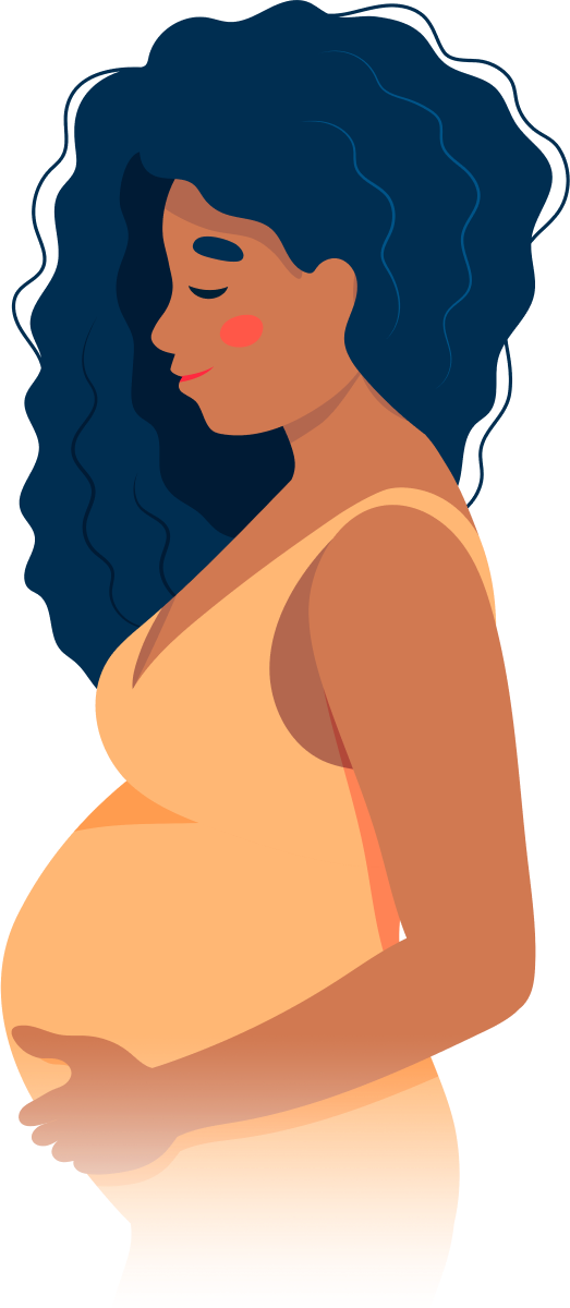
The first trimester of pregnancy is defined as the period of gestational development that extends from conception (fertilization of the egg by sperm) to the end of the 13th week. This critical phase marks significant transformations in both the mother's body and the growing embryo/fetus.
During the first trimester, the fertilized egg implants into the uterine lining (implantation), initiating a series of complex interactions leading to the formation of the placenta - an organ essential for providing nutrients and oxygen to the developing fetus while removing waste products. Simultaneously, the embryo undergoes rapid cell division and differentiation, giving rise to various organs and systems. By the end of the first trimester, most major structures are present, although they continue to mature and grow throughout pregnancy.
The mother may experience several physiological changes during this time, including:
- Morning sickness (nausea and vomiting)
- Fatigue
- Breast tenderness
- Frequent urination
- Food aversions or cravings
- Mood swings
Additionally, hormonal shifts can cause various symptoms and prepare the body for potential changes in lactation, posture, and pelvic alignment as pregnancy progresses. Regular prenatal care is crucial during this period to monitor both maternal and fetal wellbeing, identify any potential complications early on, and provide appropriate guidance and support throughout the pregnancy.
Prenatal ultrasonography, also known as obstetric ultrasound, is a medical diagnostic procedure that uses high-frequency sound waves to create images of the developing fetus, placenta, and amniotic fluid inside the uterus. It is a non-invasive and painless test that is widely used during pregnancy to monitor the growth and development of the fetus, detect any potential abnormalities or complications, and determine the due date.
During the procedure, a transducer (a small handheld device) is placed on the mother's abdomen and moved around to capture images from different angles. The sound waves travel through the mother's body and bounce back off the fetus, producing echoes that are then converted into electrical signals and displayed as images on a screen.
Prenatal ultrasonography can be performed at various stages of pregnancy, including early pregnancy to confirm the pregnancy and detect the number of fetuses, mid-pregnancy to assess the growth and development of the fetus, and late pregnancy to evaluate the position of the fetus and determine if it is head down or breech. It can also be used to guide invasive procedures such as amniocentesis or chorionic villus sampling.
Overall, prenatal ultrasonography is a valuable tool in modern obstetrics that helps ensure the health and well-being of both the mother and the developing fetus.
Maternal age is a term used to describe the age of a woman at the time she becomes pregnant or gives birth. It is often used in medical and epidemiological contexts to discuss the potential risks, complications, and outcomes associated with pregnancy and childbirth at different stages of a woman's reproductive years.
Advanced maternal age typically refers to women who become pregnant or give birth at 35 years of age or older. This group faces an increased risk for certain chromosomal abnormalities, such as Down syndrome, and other pregnancy-related complications, including gestational diabetes, preeclampsia, and cesarean delivery.
On the other end of the spectrum, adolescent pregnancies (those that occur in women under 20 years old) also come with their own set of potential risks and complications, such as preterm birth, low birth weight, and anemia.
It's important to note that while maternal age can influence pregnancy outcomes, many other factors – including genetics, lifestyle choices, and access to quality healthcare – can also play a significant role in determining the health of both mother and baby during pregnancy and childbirth.
Fetal diseases are medical conditions or abnormalities that affect a fetus during pregnancy. These diseases can be caused by genetic factors, environmental influences, or a combination of both. They can range from mild to severe and may impact various organ systems in the developing fetus. Examples of fetal diseases include congenital heart defects, neural tube defects, chromosomal abnormalities such as Down syndrome, and infectious diseases such as toxoplasmosis or rubella. Fetal diseases can be diagnosed through prenatal testing, including ultrasound, amniocentesis, and chorionic villus sampling. Treatment options may include medication, surgery, or delivery of the fetus, depending on the nature and severity of the disease.
Pregnancy-associated plasma protein-A (PAPP-A) is a protease that is often used as a biomarker in early pregnancy. It is a protein that is produced by the placenta and can be detected in the mother's bloodstream during pregnancy.
In early pregnancy, low levels of PAPP-A may indicate an increased risk for certain complications, such as preeclampsia or fetal growth restriction. High levels of PAPP-A, on the other hand, may be associated with an increased risk of chromosomal abnormalities, such as Down syndrome.
It is important to note that while PAPP-A levels can provide valuable information about the health of a pregnancy, they are just one piece of the puzzle and should be considered in conjunction with other factors, such as maternal age, medical history, and ultrasound results. Your healthcare provider will use this information along with other tests to assess your risk for certain complications and develop an appropriate plan of care.
The second trimester of pregnancy is the period between the completion of 12 weeks (the end of the first trimester) and 26 weeks (the beginning of the third trimester) of gestational age. It is often considered the most comfortable period for many pregnant women as the risk of miscarriage decreases significantly, and the symptoms experienced during the first trimester, such as nausea and fatigue, typically improve.
During this time, the uterus expands above the pubic bone, allowing more space for the growing fetus. The fetal development in the second trimester includes significant growth in size and weight, formation of all major organs, and the beginning of movement sensations that the mother can feel. Additionally, the fetus starts to hear, swallow and kick, and the skin is covered with a protective coating called vernix.
Prenatal care during this period typically includes regular prenatal appointments to monitor the mother's health and the baby's growth and development. These appointments may include measurements of the uterus, fetal heart rate monitoring, and screening tests for genetic disorders or other potential issues.
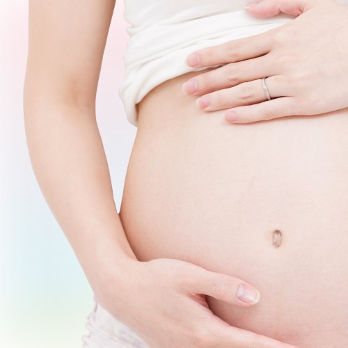
Pregnancy is a physiological state or condition where a fertilized egg (zygote) successfully implants and grows in the uterus of a woman, leading to the development of an embryo and finally a fetus. This process typically spans approximately 40 weeks, divided into three trimesters, and culminates in childbirth. Throughout this period, numerous hormonal and physical changes occur to support the growing offspring, including uterine enlargement, breast development, and various maternal adaptations to ensure the fetus's optimal growth and well-being.
The nasal bones are a pair of small, thin bones located in the upper part of the face, specifically in the middle of the nose. They articulate with each other at the nasal bridge and with the frontal bone above, the maxillae (upper jaw bones) on either side, and the septal cartilage inside the nose. The main function of the nasal bones is to form the bridge of the nose and protect the nasal cavity. Any damage to these bones can result in a fracture or broken nose.
Human chromosomes are thread-like structures that contain genetic material, composed of DNA and proteins, present in the nucleus of human cells. Each chromosome is a single, long DNA molecule that carries hundreds to thousands of genes.
Chromosomes 21, 22, and Y are three of the 23 pairs of human chromosomes. Here's what you need to know about each:
* Chromosome 21 is the smallest human autosomal chromosome, with a total length of about 47 million base pairs. It contains an estimated 200-300 genes and is associated with several genetic disorders, most notably Down syndrome, which occurs when there is an extra copy of this chromosome (trisomy 21).
* Chromosome 22 is the second smallest human autosomal chromosome, with a total length of about 50 million base pairs. It contains an estimated 500-600 genes and is associated with several genetic disorders, including DiGeorge syndrome and cat-eye syndrome.
* The Y chromosome is one of the two sex chromosomes (the other being the X chromosome) and is found only in males. It is much smaller than the X chromosome, with a total length of about 59 million base pairs and an estimated 70-200 genes. The Y chromosome determines maleness by carrying the gene for the testis-determining factor (TDF), which triggers male development in the embryo.
It's worth noting that while we have a standard set of 23 pairs of chromosomes, there can be variations and abnormalities in the number or structure of these chromosomes that can lead to genetic disorders.
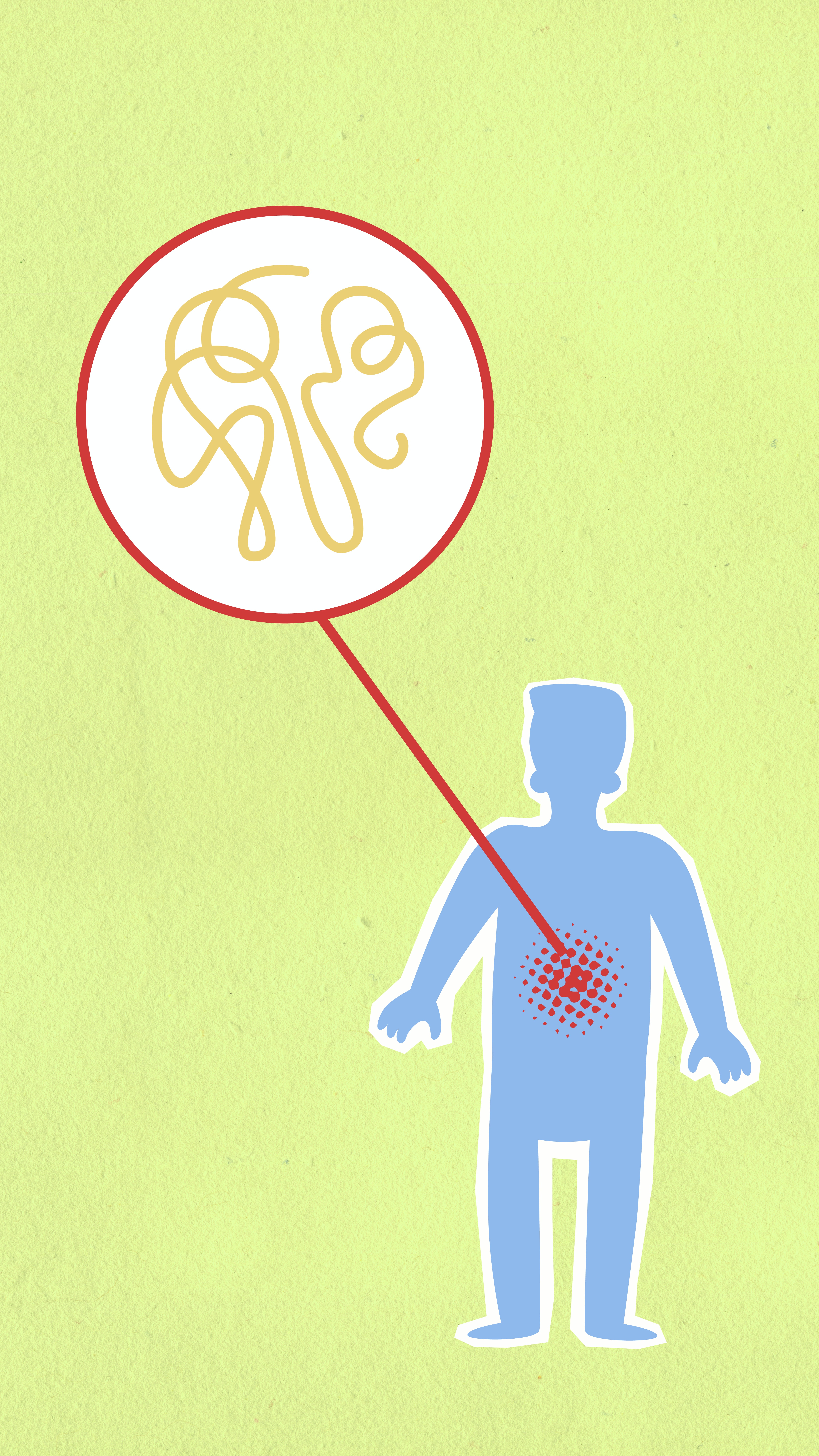
Aneuploidy is a medical term that refers to an abnormal number of chromosomes in a cell. Chromosomes are thread-like structures located inside the nucleus of cells that contain genetic information in the form of genes.
In humans, the normal number of chromosomes in a cell is 46, arranged in 23 pairs. Aneuploidy occurs when there is an extra or missing chromosome in one or more of these pairs. For example, Down syndrome is a condition that results from an extra copy of chromosome 21, also known as trisomy 21.
Aneuploidy can arise during the formation of gametes (sperm or egg cells) due to errors in the process of cell division called meiosis. These errors can result in eggs or sperm with an abnormal number of chromosomes, which can then lead to aneuploidy in the resulting embryo.
Aneuploidy is a significant cause of birth defects and miscarriages. The severity of the condition depends on which chromosomes are affected and the extent of the abnormality. In some cases, aneuploidy may have no noticeable effects, while in others it can lead to serious health problems or developmental delays.
Chromosome banding is a technique used in cytogenetics to identify and describe the physical structure and organization of chromosomes. This method involves staining the chromosomes with specific dyes that bind differently to the DNA and proteins in various regions of the chromosome, resulting in a distinct pattern of light and dark bands when viewed under a microscope.
The most commonly used banding techniques are G-banding (Giemsa banding) and R-banding (reverse banding). In G-banding, the chromosomes are stained with Giemsa dye, which preferentially binds to the AT-rich regions, creating a characteristic banding pattern. The bands are numbered from the centromere (the constriction point where the chromatids join) outwards, with the darker bands (rich in A-T base pairs and histone proteins) labeled as "q" arms and the lighter bands (rich in G-C base pairs and arginine-rich proteins) labeled as "p" arms.
R-banding, on the other hand, uses a different staining procedure that results in a reversed banding pattern compared to G-banding. The darker R-bands correspond to the lighter G-bands, and vice versa. This technique is particularly useful for identifying and analyzing specific regions of chromosomes that may be difficult to visualize with G-banding alone.
Chromosome banding plays a crucial role in diagnosing genetic disorders, identifying chromosomal abnormalities, and studying the structure and function of chromosomes in both clinical and research settings.
Dermatoglyphics is the study of the fingerprints, palm prints, and other skin ridge patterns found on the hands and feet. These patterns are formed during fetal development and are generally considered to be unique to each individual. Dermatoglyphics can provide important clues about a person's genetic makeup and health status, and they are often used in forensic investigations to help identify individuals. In medicine, dermatoglyphics may be used to help diagnose certain genetic disorders or birth defects.
High-risk pregnancy is a term used to describe a situation where the mother or the fetus has an increased risk of developing complications during pregnancy, labor, delivery, or in the postpartum period. These risks may be due to pre-existing medical conditions in the mother, such as diabetes, hypertension, heart disease, kidney disease, autoimmune disorders, or infectious diseases like HIV/AIDS. Other factors that can contribute to a high-risk pregnancy include advanced maternal age (35 years and older), obesity, multiple gestations (twins, triplets, etc.), fetal growth restriction, placental issues, and a history of previous pregnancy complications or preterm labor.
High-risk pregnancies require specialized care and monitoring by healthcare professionals, often involving maternal-fetal medicine specialists, obstetricians, perinatologists, and neonatologists. Regular prenatal care, frequent checkups, ultrasound monitoring, and sometimes additional testing and interventions may be necessary to ensure the best possible outcomes for both the mother and the baby.
Amniocentesis is a medical procedure in which a small amount of amniotic fluid, which contains fetal cells, is withdrawn from the uterus through a hollow needle inserted into the abdomen of a pregnant woman. This procedure is typically performed between the 16th and 20th weeks of pregnancy.
The main purpose of amniocentesis is to diagnose genetic disorders and chromosomal abnormalities in the developing fetus, such as Down syndrome, Edwards syndrome, and neural tube defects. The fetal cells obtained from the amniotic fluid can be cultured and analyzed for various genetic characteristics, including chromosomal structure and number, as well as specific gene mutations.
Amniocentesis carries a small risk of complications, such as miscarriage, infection, or injury to the fetus. Therefore, it is generally offered to women who have an increased risk of having a baby with a genetic disorder or chromosomal abnormality, such as those over the age of 35, those with a family history of genetic disorders, or those who have had a previous pregnancy affected by a genetic condition.
It's important to note that while amniocentesis can provide valuable information about the health of the fetus, it does not guarantee a completely normal baby, and there are some risks associated with the procedure. Therefore, the decision to undergo amniocentesis should be made carefully, in consultation with a healthcare provider, taking into account the individual circumstances and preferences of each woman.
Crown-rump length (CRL) is a medical measurement used in obstetrics to estimate the age of a developing fetus. It refers to the length from the top of the head (crown) to the bottom of the buttocks (rump). This measurement is typically taken during an ultrasound examination in the first trimester of pregnancy, between 8 and 13 weeks of gestation.
The CRL is used to calculate the estimated due date and to monitor fetal growth and development. It can also help identify potential issues or abnormalities in fetal development. As the pregnancy progresses, other measurements such as head circumference, abdominal circumference, and femur length are used to assess fetal growth and development.
In situ hybridization, fluorescence (FISH) is a type of molecular cytogenetic technique used to detect and localize the presence or absence of specific DNA sequences on chromosomes through the use of fluorescent probes. This technique allows for the direct visualization of genetic material at a cellular level, making it possible to identify chromosomal abnormalities such as deletions, duplications, translocations, and other rearrangements.
The process involves denaturing the DNA in the sample to separate the double-stranded molecules into single strands, then adding fluorescently labeled probes that are complementary to the target DNA sequence. The probe hybridizes to the complementary sequence in the sample, and the location of the probe is detected by fluorescence microscopy.
FISH has a wide range of applications in both clinical and research settings, including prenatal diagnosis, cancer diagnosis and monitoring, and the study of gene expression and regulation. It is a powerful tool for identifying genetic abnormalities and understanding their role in human disease.
Human chromosome pair 8 consists of two rod-shaped structures present in the nucleus of each cell of the human body. Each chromosome is made up of DNA tightly coiled around histone proteins, forming a complex structure known as a chromatin.
Human cells have 23 pairs of chromosomes, for a total of 46 chromosomes. Pair 8 is one of the autosomal pairs, meaning that it is not a sex chromosome (X or Y). Each member of chromosome pair 8 has a similar size, shape, and banding pattern, and they are identical in males and females.
Chromosome pair 8 contains several genes that are essential for various cellular functions and human development. Some of the genes located on chromosome pair 8 include those involved in the regulation of metabolism, nerve function, immune response, and cell growth and division.
Abnormalities in chromosome pair 8 can lead to genetic disorders such as Wolf-Hirschhorn syndrome, which is caused by a partial deletion of the short arm of chromosome 4, or partial trisomy 8, which results from an extra copy of all or part of chromosome 8. Both of these conditions are associated with developmental delays, intellectual disability, and various physical abnormalities.
Translocation, genetic, refers to a type of chromosomal abnormality in which a segment of a chromosome is transferred from one chromosome to another, resulting in an altered genome. This can occur between two non-homologous chromosomes (non-reciprocal translocation) or between two homologous chromosomes (reciprocal translocation). Genetic translocations can lead to various clinical consequences, depending on the genes involved and the location of the translocation. Some translocations may result in no apparent effects, while others can cause developmental abnormalities, cancer, or other genetic disorders. In some cases, translocations can also increase the risk of having offspring with genetic conditions.
Monosomy is a type of chromosomal abnormality in which there is only one copy of a particular chromosome instead of the usual pair in a diploid cell. In monosomy, an individual has one less chromosome than the normal diploid number (46 chromosomes) due to the absence of one member of a chromosome pair. This condition arises from the loss of one chromosome in an egg or sperm during gamete formation or at conception.
Examples of monosomy include Turner syndrome, which is characterized by the presence of only one X chromosome (45,X), and Cri du Chat syndrome, which results from a deletion of a portion of the short arm of chromosome 5 (46,del(5)(p15.2)). Monosomy can lead to developmental abnormalities, physical defects, intellectual disabilities, and various health issues depending on the chromosome involved.
Human chromosome pair 12 consists of two rod-shaped structures present in the nucleus of each cell in the human body. Each chromosome is made up of DNA tightly coiled around histone proteins, forming a complex structure called a chromatin.
Chromosomes come in pairs, with one chromosome inherited from each parent. In humans, there are 23 pairs of chromosomes, for a total of 46 chromosomes in each cell. Chromosome pair 12 is the 12th pair of autosomal chromosomes, meaning they are not sex chromosomes (X or Y).
Chromosome 12 is a medium-sized chromosome and contains an estimated 130 million base pairs of DNA. It contains around 1,200 genes that provide instructions for making proteins and regulating various cellular processes. Some of the genes located on chromosome 12 include those involved in metabolism, development, and response to environmental stimuli.
Abnormalities in chromosome 12 can lead to genetic disorders, such as partial trisomy 12q, which is characterized by an extra copy of the long arm of chromosome 12, and Jacobsen syndrome, which is caused by a deletion of the distal end of the long arm of chromosome 12.
Human chromosomes are thread-like structures that contain genetic information in the form of DNA and proteins. Each human cell typically contains 46 chromosomes arranged in 23 pairs, except for the sperm and egg cells which contain only 23 chromosomes (one half of the full set).
Chromosome 19 is one of the autosomal chromosomes, meaning it is not a sex chromosome. It is the fifth smallest human chromosome, spanning about 58 million base pairs and representing approximately 1.9% of the total DNA in cells. Chromosome 19 contains more than 1,200 genes that provide instructions for making proteins and RNA molecules involved in various cellular processes.
Chromosome 20 is also an autosomal chromosome, slightly smaller than chromosome 19. It spans about 54 million base pairs and contains around 800 genes that code for proteins and RNA molecules. Chromosome 20 is known to contain several important genes involved in cancer development, such as the tumor suppressor gene TP53.
Together, chromosomes 19 and 20 carry crucial genetic information necessary for normal human growth, development, and health. Abnormalities in these chromosomes can lead to various genetic disorders and diseases.
Chorionic villi sampling (CVS) is a prenatal testing procedure that involves taking a small sample of the chorionic villi, which are finger-like projections of the placenta that contain fetal cells. The sample is then tested for genetic disorders and chromosomal abnormalities, such as Down syndrome.
CVS is typically performed between the 10th and 12th weeks of pregnancy and carries a small risk of miscarriage (about 1 in 100 to 1 in 200 procedures). The results of CVS can provide important information about the health of the fetus, allowing parents to make informed decisions about their pregnancy. However, it is important to note that CVS does not detect all genetic disorders and may produce false positive or false negative results in some cases. Therefore, follow-up testing may be necessary.
Chromosomes are thread-like structures located in the nucleus of cells that carry genetic information in the form of genes. In humans, there are 23 pairs of chromosomes for a total of 46 chromosomes in every cell of the body, except for the sperm and egg cells which contain only 23 chromosomes.
Human chromosomes are numbered from 1 to 22, based on their size, with chromosome 1 being the largest and chromosome 22 being the smallest. The last two pairs of human chromosomes are known as the sex chromosomes because they determine a person's biological sex. These are labeled X and Y, with females having two X chromosomes (44+XX) and males having one X and one Y chromosome (44+XY).
Therefore, "Chromosomes, Human, 4-5" refers to the fourth and fifth pairs of human chromosomes. Chromosome 4 is an acrocentric chromosome, meaning its centromere is located near one end, resulting in a short arm (p) and a long arm (q). It contains about 190 million base pairs and encodes approximately 700 genes.
Chromosome 5 is a submetacentric chromosome, with the centromere located closer to the middle, creating two arms of roughly equal length: the short arm (p) and the long arm (q). It contains about 182 million base pairs and encodes approximately 900 genes.
Both chromosomes 4 and 5 are involved in various genetic disorders when abnormalities occur, such as deletions, duplications, or translocations. Some of the well-known genetic conditions associated with these chromosomes include:
* Chromosome 4: Wolf-Hirschhorn syndrome (deletion), Charcot-Marie-Tooth disease type 1A (duplication)
* Chromosome 5: Cri du Chat syndrome (deletion), Duchenne muscular dystrophy (deletion or mutation in a gene located on chromosome 5)
Human chromosome pair 7 consists of two rod-shaped structures present in the nucleus of each cell in the human body. Each member of the pair is a single chromosome, and together they contain the genetic material that is inherited from both parents. They are identical in size, shape, and banding pattern and are therefore referred to as homologous chromosomes.
Chromosome 7 is one of the autosomal chromosomes, meaning it is not a sex chromosome (X or Y). It is composed of double-stranded DNA that contains approximately 159 million base pairs and around 1,200 genes. Chromosome 7 contains several important genes associated with human health and disease, including those involved in the development of certain types of cancer, such as colon cancer and lung cancer, as well as genetic disorders such as Williams-Beuren syndrome and Charcot-Marie-Tooth disease.
Abnormalities in chromosome 7 have been linked to various genetic conditions, including deletions, duplications, translocations, and other structural changes. These abnormalities can lead to developmental delays, intellectual disabilities, physical abnormalities, and increased risk of certain types of cancer.
In medical terms, the "neck" is defined as the portion of the body that extends from the skull/head to the thorax or chest region. It contains 7 cervical vertebrae, muscles, nerves, blood vessels, lymphatic vessels, and glands (such as the thyroid gland). The neck is responsible for supporting the head, allowing its movement in various directions, and housing vital structures that enable functions like respiration and circulation.
Gestational age is the length of time that has passed since the first day of the last menstrual period (LMP) in pregnant women. It is the standard unit used to estimate the age of a pregnancy and is typically expressed in weeks. This measure is used because the exact date of conception is often not known, but the start of the last menstrual period is usually easier to recall.
It's important to note that since ovulation typically occurs around two weeks after the start of the LMP, gestational age is approximately two weeks longer than fetal age, which is the actual time elapsed since conception. Medical professionals use both gestational and fetal age to track the development and growth of the fetus during pregnancy.
Human chromosomes are the thread-like structures located in the nucleus of human cells, which carry genetic information in the form of DNA. Humans have a total of 46 chromosomes arranged in 23 pairs. The first 22 pairs are called autosomes, and the last pair are the sex chromosomes, X and Y.
Chromosomes 1-3 are the largest human chromosomes, and they contain a significant portion of the human genome. Here is a brief overview of each:
1. Chromosome 1: This is the largest human chromosome, spanning about 8% of the human genome. It contains approximately 2,800 genes that are responsible for various functions such as cell growth and division, nerve function, and response to stimuli.
2. Chromosome 2: The second largest human chromosome, spanning about 7% of the human genome. It contains approximately 2,300 genes that are involved in various functions such as metabolism, development, and immune response.
3. Chromosome 3: This is the third largest human chromosome, spanning about 6% of the human genome. It contains approximately 1,900 genes that are responsible for various functions such as DNA repair, cell signaling, and response to stress.
It's worth noting that while these chromosomes contain a large number of genes, they also have significant amounts of non-coding DNA, which means that not all of the genetic material on these chromosomes is responsible for encoding proteins or other functional elements.
Intellectual disability (ID) is a term used when there are significant limitations in both intellectual functioning and adaptive behavior, which covers many everyday social and practical skills. This disability originates before the age of 18.
Intellectual functioning, also known as intelligence, refers to general mental capacity, such as learning, reasoning, problem-solving, and other cognitive skills. Adaptive behavior includes skills needed for day-to-day life, such as communication, self-care, social skills, safety judgement, and basic academic skills.
Intellectual disability is characterized by below-average intelligence or mental ability and a lack of skills necessary for day-to-day living. It can be mild, moderate, severe, or profound, depending on the degree of limitation in intellectual functioning and adaptive behavior.
It's important to note that people with intellectual disabilities have unique strengths and limitations, just like everyone else. With appropriate support and education, they can lead fulfilling lives and contribute to their communities in many ways.
A newborn infant is a baby who is within the first 28 days of life. This period is also referred to as the neonatal period. Newborns require specialized care and attention due to their immature bodily systems and increased vulnerability to various health issues. They are closely monitored for signs of well-being, growth, and development during this critical time.
Cytogenetic analysis is a laboratory technique used to identify and study the structure and function of chromosomes, which are the structures in the cell that contain genetic material. This type of analysis involves examining the number, size, shape, and banding pattern of chromosomes in cells, typically during metaphase when they are at their most condensed state.
There are several methods used for cytogenetic analysis, including karyotyping, fluorescence in situ hybridization (FISH), and comparative genomic hybridization (CGH). Karyotyping involves staining the chromosomes with a dye to visualize their banding patterns and then arranging them in pairs based on their size and shape. FISH uses fluorescent probes to label specific DNA sequences, allowing for the detection of genetic abnormalities such as deletions, duplications, or translocations. CGH compares the DNA content of two samples to identify differences in copy number, which can be used to detect chromosomal imbalances.
Cytogenetic analysis is an important tool in medical genetics and is used for a variety of purposes, including prenatal diagnosis, cancer diagnosis and monitoring, and the identification of genetic disorders.
Cytogenetics is a branch of genetics that deals with the study of chromosomes and their structure, function, and abnormalities. It involves the examination of chromosome number and structure in the cells of an organism, usually through microscopic analysis of chromosomes prepared from cell cultures or tissue samples. Cytogenetic techniques can be used to identify chromosomal abnormalities associated with genetic disorders, cancer, and other diseases.
The process of cytogenetics typically involves staining the chromosomes to make them visible under a microscope, and then analyzing their number, size, shape, and banding pattern. Chromosomal abnormalities such as deletions, duplications, inversions, translocations, and aneuploidy (abnormal number of chromosomes) can be detected through cytogenetic analysis.
Cytogenetics is an important tool in medical genetics and has many clinical applications, including prenatal diagnosis, cancer diagnosis and monitoring, and identification of genetic disorders. Advances in molecular cytogenetic techniques, such as fluorescence in situ hybridization (FISH) and comparative genomic hybridization (CGH), have improved the resolution and accuracy of chromosome analysis and expanded its clinical applications.
A forehead, in medical terms, refers to the portion of the human skull that lies immediately above the eyes and serves as an attachment site for the frontal bone. It is a common area for the examination of various clinical signs, such as assessing the level of consciousness (by checking if the patient's eyebrows or eyelids twitch in response to a light touch) or looking for signs of increased intracranial pressure (such as bulging fontanelles in infants). Additionally, the forehead is often used as a site for non-invasive procedures like Botox injections.
An "eugenic abortion" is not a medical term, but rather a descriptive phrase that combines two concepts: eugenics and abortion.
Eugenics refers to the belief and practice of improving the human species by encouraging reproduction of individuals with desired traits and preventing reproduction of those with undesired traits. This concept has been widely criticized for its potential to be used as a tool for discrimination and oppression.
Abortion, on the other hand, is the medical procedure to end a pregnancy before the fetus can survive outside the womb.
A "eugenic abortion," therefore, generally refers to the practice of terminating a pregnancy based on the perceived genetic traits or characteristics of the fetus, such as disability, race, or sex. This phrase is often used in discussions about the ethics and morality of selective abortions, and it raises important questions about discrimination, reproductive rights, and medical ethics. It's worth noting that the vast majority of abortions are not performed for eugenic reasons, but rather due to a variety of personal, medical, and socioeconomic factors.
A fetus is the developing offspring in a mammal, from the end of the embryonic period (approximately 8 weeks after fertilization in humans) until birth. In humans, the fetal stage of development starts from the eleventh week of pregnancy and continues until childbirth, which is termed as full-term pregnancy at around 37 to 40 weeks of gestation. During this time, the organ systems become fully developed and the body grows in size. The fetus is surrounded by the amniotic fluid within the amniotic sac and is connected to the placenta via the umbilical cord, through which it receives nutrients and oxygen from the mother. Regular prenatal care is essential during this period to monitor the growth and development of the fetus and ensure a healthy pregnancy and delivery.
 Trisomy
Trisomy 22
Trisomy 18
Trisomy X
Trisomy 9
Trisomy 16
Trisomy 8
Distal trisomy 10q
Chromosome 15q trisomy
Microcephaly
Confined placental mosaicism
G&T-Seq
Kleeblattschaedel
Pallister-Killian syndrome
Patau syndrome
Obstetrics
Single umbilical artery
Potocki-Lupski syndrome
Small supernumerary marker chromosome
Palpebral fissure
Jérôme Lejeune
Erondu-Cymet syndrome
Cat eye syndrome
XXXYY syndrome
List of conditions with craniosynostosis
X-inactivation
Turricephaly
Chromosome 14
Chromosome 13
Nondisjunction
 Chromosomal Abnormalities: Trisomy 21 (Down Syndrome) | NCBDDD | CDC
Trisomy - Wikipedia
Trisomy 18 - Wikipedia
 Trisomy 18: MedlinePlus Genetics
 Trisomy 18: Practice Essentials, Pathophysiology, Epidemiology
 Trisomy represses ApcMin -mediated tumours in mouse models of Down's syndrome | Nature
 Soft - SOFT - Support Organization For Trisomy
2021 SOFT Trisomy Awareness Month Video Blast
 Trisomy 21 Program Resources | Children's Hospital of Philadelphia
 trisomy | The Biology Corner
 ISSDA | Women's Perception and Knowledge of Prenatal Screening for Fetal Trisomy - 2020
 1 healthy child, 3 miscarriages 1 with trisomy 16 | BabyCenter
 22 or trisomy 22 (solely?)
 Vaccines | Free Full-Text | Adults with Trisomy 21 Have Differential Antibody Responses to Influenza A
 trisomy 21 Archives - Celebrate Life Magazine
 X Chromosome, Trisomy Xp3 - Ontology Report - Rat Genome Database
A case of partial trisomy 15 | Journal of Medical Genetics
The Hidden National Institutes of Health (NIH) Webpage - Duplication/Trisomy of Q42 11 Q42 12
Trisomy 13 - Positive Exposure
 EN
EN
Trisomy 15 mosaicism in an IVF fetus. | Journal of Medical Genetics
Trisomy 21 and baby sign language - Australian Baby Hands
 Trisomy 13 (Patau Syndrome) - Mississippi State Department of Health
 Tetrasomy 21 transient leukemia with a GATA1 mutation in a phenotypically normal trisomy 21 mosaic infant: case report and...
 Down Syndrome (Trisomy 21) - Children's Health Issues - Merck Manuals Consumer Version
Chromosome 17 trisomy - CheckOrphan
 Utilization of Benchtop Next Generation Sequencing Platforms Ion Torrent PGM and MiSeq in Noninvasive Prenatal Testing for...
Trisomy 21 (Down Syndrome) - Mississippi State Department of Health
 Trisomy 13 and Trisomy 18 in Children
 Bergen Open Research Archive: Ten-year survival of children with trisomy 13 or trisomy 18: A multi-registry European cohort...
Syndrome34
- Trisomy 21, also known as Down syndrome, is a condition characterized by a distinctive pattern of minor and major anomalies associated with excess chromosome 21 material. (cdc.gov)
- citation needed] Thus, for example, the presence of an extra chromosome 21, which is found in Down syndrome, is called trisomy 21. (wikipedia.org)
- The most common types of autosomal trisomy that survive to birth in humans are: Trisomy 21 (Down syndrome) Trisomy 18 (Edwards syndrome) Trisomy 13 (Patau syndrome) Trisomy 9 Trisomy 8 (Warkany syndrome 2) Of these, Trisomy 21 and Trisomy 18 are the most common. (wikipedia.org)
- In rare cases, a fetus with Trisomy 13 can survive, giving rise to Patau syndrome. (wikipedia.org)
- Trisomy of sex chromosomes can also occur and include: XXX (Triple X syndrome) XXY (Klinefelter syndrome) XYY (Jacobs syndrome) Compared to trisomy of the autosomal chromosomes, trisomy of the sex chromosomes normally has less severe consequences. (wikipedia.org)
- Trisomy 18 , also known as Edwards syndrome , is a genetic disorder caused by the presence of a third copy of all or part of chromosome 18 . (wikipedia.org)
- [7] Although uncommon in the syndrome, trisomy 18 causes a large portion of prenatally diagnosed cases of Dandy-Walker malformation . (wikipedia.org)
- Trisomy 18, also called Edwards syndrome, is a chromosomal condition associated with abnormalities in many parts of the body. (medlineplus.gov)
- Note the microphthalmia, micrognathia/retrognathia, microstomia, low-set/malformed ears, short sternum, and abnormally clenched fingers in an infant with trisomy 18 (Edwards syndrome). (medscape.com)
- Note the characteristic clenched hand of trisomy 18 (Edwards syndrome) with the index finger overriding the middle finger and the fifth finger overriding the fourth finger. (medscape.com)
- Note the rocker-bottom foot with a prominent calcaneus in an infant with trisomy 18 (Edwards syndrome). (medscape.com)
- This photo shows the hands of a fetus with trisomy 18 (Edwards syndrome). (medscape.com)
- We used mouse models of Down's syndrome and of cancer in a biological approach to investigate the relationship between trisomy and the incidence of intestinal tumours. (nature.com)
- The Trisomy 21 Parent Peer Program provides support and resources to families of individuals with Down syndrome by matching them with fellow caregivers. (chop.edu)
- NEW YORK - The International Society for Prenatal Diagnosis (ISPD) on Wednesday published a set of recommendations on the use of cell-free DNA testing to screen multifetal pregnancies for Down syndrome and other autosomal trisomies, noting that the literature shows such testing to be appropriate for screening twin pregnancies and may also be a viable option for screening triplet pregnancies. (genomeweb.com)
- Although they focused on Down syndrome, they also addressed trisomies 13 and 18. (genomeweb.com)
- In another analysis, which included additional studies, a total of 117 twin pregnancies had at least one fetus with a common autosomal trisomy (84 with Down syndrome, 29 with trisomy 18, and four with trisomy 13), with detection rates for cfDNA screening of 98.8 percent, 93.1 percent, and 75 percent, respectively. (genomeweb.com)
- Trisomy 13, also known as Patau Syndrome, occurs when there is an extra copy of chromosome 13. (positiveexposure.org)
- Occurring in about one per eight hundred births, Down syndrome - or trisomy 21 - is the most frequent genetic cause of intellectual disability. (unige.ch)
- Trisomy 13, known as Patau Syndrome, is a rare condition resulting from genetic errors on the 13th chromosome. (ms.gov)
- Trisomy 21 causes about 95% of the cases of Down syndrome. (merckmanuals.com)
- Trisomy 21, known as Down syndrome, is a genetic disorder resulting from genetic errors on the 21st chromosome. (healthyms.com)
- Down syndrome is the most common Trisomy. (healthyms.com)
- Although most trisomies are due to random errors, mothers over 35 years of age have an increased risk of having a child with Down syndrome. (healthyms.com)
- Some children will have three copies of chromosome 21 in all of their cells (Trisomy 21) while others have three copies in only some of their cells (mosaic Down syndrome) or extra parts of chromosome 21 attached to another chromosome (Translocation Down syndrome). (healthyms.com)
- Unlike other Trisomy disorders, most children with Down syndrome will live into adulthood. (healthyms.com)
- Trisomy can occur with any chromosome, but the most well-known syndromes are: Trisomy 21 (Down Syndrome), Trisomy 18 (Edwards Syndrome) and Trisomy 13 (Patau Syndrome). (personalizedcause.com)
- The genetic architecture of Down syndrome phenotypes revealed by high-resolution analysis of human segmental trisomies. (gersteinlab.org)
- Sixteen-year-old Joshua Allen was born with an extra chromosome--a genetic aberration known as Trisomy XXI, or Down Syndrome. (theroadweveshared.com)
- Joshua was born with Down Syndrome, also called Trisomy XXL due to a third chromosome. (theroadweveshared.com)
- Trisomy 18, also known as Edwards syndrome, is a type of genetic disorder that's mainly caused by the presence of part or all of an extra eighteenth chromosome present in an unborn child's genetic makeup. (cajm.org)
- Trisomy 13 is a serious genetic syndrome, and most babies with Patau syndrome die before birth or within the first week of life. (cajm.org)
- Trisomy 18, also known as Edwards Syndrome, is the next most frequent, followed by Trisomy 13 or Patau Syndrome. (cajm.org)
- National prevalence estimates, adjusted for maternal race/ethnicity and maternal age (trisomy 13, trisomy 18, and Down syndrome only) were determined using data from 14 programs. (cdc.gov)
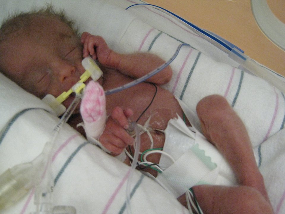
Baby with trisomy 134
- What are the chances of having a second baby with trisomy 13? (richardvigilantebooks.com)
- The risk of having a baby with trisomy 13 increases slightly with the mother's age. (richardvigilantebooks.com)
- However, the average age of the mother at delivery of a baby with trisomy 13 is 32 years. (richardvigilantebooks.com)
- It is difficult to predict the life expectancy of a baby with trisomy 13 if the baby does not have any immediate life-threatening problems. (cajm.org)
Child with trisomy2
- Although women of all ages can have a child with trisomy 18, the chance of having a child with this condition increases as a woman gets older. (medlineplus.gov)
- It is hard to predict how long a child with Trisomy 13 will live. (ms.gov)
Chromosomes9
- Translocation trisomy 21 (2% of cases) is often familial, and commonly involves chromosomes 14 and 21. (cdc.gov)
- Trisomy 21 may be diagnosed through direct analysis of fetal chromosomes, by karyotype or DNA microarray, obtained from amniocentesis, chorionic villus sampling, or percutaneous umbilical blood sampling. (cdc.gov)
- A trisomy is a type of aneuploidy (an abnormal number of chromosomes). (wikipedia.org)
- The number of chromosomes in the cell where trisomy occurs is represented as, for example, 2n+1 if one chromosome shows trisomy, 2n+1+1 if two show trisomy, etc. (wikipedia.org)
- Tertiary trisomy" - the extra chromosome is made up of copies of arms from two other chromosomes. (wikipedia.org)
- Trisomies are sometimes characterised as "autosomal trisomies" (trisomies of the non-sex chromosomes) and "sex-chromosome trisomies. (wikipedia.org)
- The resulting zygote will have three autosomal chromosomes or an autosomal trisomy. (osmosis.org)
- Some people have trisomy conditions, those related to having an extra chromosome in most or all of their cells, for a total of 47 chromosomes. (personalizedcause.com)
- Babies with trisomy have a total of 47 chromosomes. (cajm.org)
Seen in trisomy 182
- Clenched hand and overlapping fingers: index finger overlaps third finger and fifth finger overlaps fourth finger, characteristically seen in trisomy 18. (wikipedia.org)
- If only part of the q arm is present in three copies, the physical signs of partial trisomy 18 may be less severe than those typically seen in trisomy 18. (medlineplus.gov)
Disorders8
- SOFT is a network of families and professionals dedicated to providing support and understanding to families involved in the issues and decisions surrounding the diagnosis and care in Trisomy 18, 13 and other related chromosomal disorders. (trisomy.org)
- Children with trisomy disorders can live happy lives with their families. (trisomy.org)
- Although trisomy disorders can present many challenges, with your help we can be a beacon of hope for families and a voice for our children. (trisomy.org)
- SOFT - Support Organization for Trisomy 13, 18, and Related Disorders is a network of families and professionals that promote parent-professional relationships and provide support to families in the diagnosis and care of Trisomy 13, 18, and other related chromosomal disorders. (ms.gov)
- Trisomy 13 and trisomy 18 are genetic disorders. (stanfordchildrens.org)
- Trisomy Awareness Month , observed in March and sponsored by the Support Organization for Trisomy 18, 13 and Related Disorders, raises awareness about trisomy conditions and the challenges these conditions may pose to individuals and families. (personalizedcause.com)
- Trisomy Awareness Month helps to educate people about trisomy disorders. (personalizedcause.com)
- The files of the Support Organization for Trisomy 18, 13 and Related Disorders (SOFT) indicate a small number of children with trisomy survive much longer than initially expected. (cajm.org)
Partial7
- Partial trisomy" means that there is an extra copy of part of a chromosome. (wikipedia.org)
- People with this genetic change are said to have partial trisomy 18. (medlineplus.gov)
- Partial trisomy 18 can be inherited. (medlineplus.gov)
- In translocations that result in partial trisomy or in cases of mosaic trisomy 18, clinical expression is less severe, and survival is usually longer. (medscape.com)
- The origin of this chromosome is discussed and the clinical findings compared with those of previously reported cases of partial trisomy 15. (bmj.com)
- A partial autosomal trisomy characterized by developmental delay and intellectual disability generalized hypotonia postnatal growth retardation variable brain and heart anomalies and dysmorphic features including frontal bossing round face full cheeks low-set ears broad nasal bridge short nose with anteverted nares long philtrum thin upper lip vermilion and everted thick lower lip. (globalgenes.org)
- Partial trisomy - When only a part of third chromosome is present in the body cells. (cajm.org)
Extra chromosome4
- Full trisomy", also called "primary trisomy", means that an entire extra chromosome has been copied. (wikipedia.org)
- [3] Occasionally, not all cells have the extra chromosome, known as mosaic trisomy , and symptoms in these cases may be less severe. (wikipedia.org)
- The severity of mosaic trisomy 18 depends on the type and number of cells that have the extra chromosome. (medlineplus.gov)
- And since the long arms carry most of the genetic material, cells with the long arm will basically have one extra chromosome, which, when combined with the other parent's again, will result in trisomy, while cells with the short arm are basically missing a chromosome and can result in monosomy. (osmosis.org)
Form of trisomy2
- The development of individuals with this form of trisomy 18 may range from normal to severely affected. (medlineplus.gov)
- This is the only form of trisomy 13 or 18 that may be inherited from a parent. (stanfordchildrens.org)
Mosaic for trisomy2
People with trisomy1
- Approximately 5 percent of people with trisomy 18 have an extra copy of chromosome 18 in only some of the body's cells. (medlineplus.gov)
Patau1
- The trisomy 13 patau sydnrome malformations were done. (cajm.org)
Fetus2
Life expectancy1
- Autosomal trisomy can be associated with birth defects, intellectual disability and shortened life expectancy. (wikipedia.org)
Common trisomy1
- The most common trisomy in a newborn is trisomy 21 (three copies of chromosome 21, which is the smallest human chromosome). (merckmanuals.com)
Autosomal trisomies2
- Autosomal trisomies are described by referencing the specific chromosome that has an extra copy. (wikipedia.org)
- Overall, the researchers concluded, the use of cfDNA screening in the first trimester for common autosomal trisomies is appropriate for twin pregnancies "due to sufficient evidence showing high detection and low false-positive rates with high predictive values. (genomeweb.com)
Constitutional trisomy3
- Infants with constitutional trisomy 21 are at increased risk of developing transient and acute megakaryoblastic leukemia (AMKL). (nih.gov)
- Transient leukemia in trisomy 21 mosaicism recapitulates the condition observed in constitutional trisomy 21 at the biological and clinical levels. (nih.gov)
- Clonal acquired chromosome aberrations were found in 13 patients and a constitutional trisomy 18 as the sole change in 1. (lu.se)
Fetal5
- Objective: Multiple non-invasive tests are available to screen for the risk of fetal trisomy, however, there is no national prenatal screening programme in Ireland. (ucd.ie)
- Fetal ultrasound during pregnancy can also show the possibility of trisomy 13 or 18. (stanfordchildrens.org)
- Presence of the trisomy is rarely observed in blood samples from these cases, but is often confirmed in fetal tissues. (bcchr.ca)
- while in one pregnancy ending in fetal demise, trisomy was found in placenta but not in kidney, skin, or liver. (bcchr.ca)
- 1997) European collaborative research on mosaicism in CVS (EUCROMIC)--fetal and extrafetal cell lineages in 192 gestations with CVS mosaicism involving single autosomal trisomy. (bcchr.ca)
Mosaicism9
- We report a phenotypically normal infant with tetrasomy 21 transient leukemia, GATA1 mutation within exon 2, and trisomy 21 mosaicism restricted to the hematopoietic tissue. (nih.gov)
- Two cases of trisomy 12 were reported in a large study of chromosomal mosaicism detected on CVS. (bcchr.ca)
- 1997) summarized findings in 23 cases of trisomy 12 mosaicism detected in amniotic fluid--20 of which were female and only three were male. (bcchr.ca)
- A bias towards females has been noted in other cases of trisomy mosaicism (e.g. trisomy 16) and suggests either preferential survival of female mosaics or a greater tendency to mosaicism in females. (bcchr.ca)
- 2000) reported a case of trisomy 12 diagnosed in a newborn and summarized results for 4 other cases of trisomy 12 mosaicism diagnosed postnatally. (bcchr.ca)
- 1995) Mosaicism for trisomy 12: four cases with varying outcomes. (bcchr.ca)
- Brosens JJ, Overton C, Lavery SA, Thornton S. (1996) Trisomy 12 mosaicism diagnosed by amniocentesis. (bcchr.ca)
- 2000) Trisomy 12 mosaicism confirmed in multiple organs from a liveborn child. (bcchr.ca)
- English CJ, Goodship JA, Jackson A, Lowry M, Wolstenholme J. (1994) Trisomy 12 mosaicism in a 7 year old girl with dysmorphic features and normal mental development. (bcchr.ca)
Occurs5
- Trisomy 18 occurs in around 1 in 5,000 live births. (wikipedia.org)
- Trisomy 18 occurs in all human populations, but is more prevalent in female offspring. (wikipedia.org)
- Trisomy 13 occurs when children have three copies of chromosome 13 instead of two. (ms.gov)
- Most of the time, trisomy conditions are not passed from one generation to the next, but result from a random error that occurs during cell division very early on in development. (personalizedcause.com)
- If this occurs, trisomy 13 is inherited in the family. (cajm.org)
Chromosome 211
- Trisomy for orthologues of about half of the genes on chromosome 21 (Hsa21) in Ts65Dn mice or just 33 of these genes in Ts1Rhr mice resulted in a significant reduction in the number of intestinal tumours. (nature.com)
Pregnancies3
- only those pregnancies in which some normal cells occur in addition to the trisomic cells, or mosaic trisomy 16, survive. (wikipedia.org)
- Pregnancies at increased risk for Trisomy 13 can be identified through screening tests such as non-invasive prenatal testing (NIPT) and ultrasound examinations. (richardvigilantebooks.com)
- Trisomy 18 is known to occur in around 1 out of 2,500 pregnancies, typically resulting in 1 of 7,000 live births. (cajm.org)
Infants1
- Infants with Trisomy 13 are unusually small and have feeding difficulties which affect growth and development. (ms.gov)
Duplication1
- 7/23/2007: Duplication/trisomy of 1q42 11 q42 12 page was found by Morgellons victim and posted to the public on internet sites. (bibliotecapleyades.net)
Awareness Month2
- Join us for the Trisomy Awareness Month video blast! (trisomy.org)
- The Trisomy Awareness Month video blast is coming and we hope you will join us. (trisomy.org)
Congenital1
- Children with Trisomy 13 often have craniofacial defects such as cleft lip and palate, congenital heart defects, and abnormalities of the brain and spinal cord. (ms.gov)
Translocation1
- Although they do not have signs of trisomy 18, people who carry this type of balanced translocation are at an increased risk of having children with the condition. (medlineplus.gov)
Intellectual2
- In addition, children with Trisomy 13 experience significant developmental and intellectual disabilities. (ms.gov)
- What are the physical symptoms and intellectual delays associated with trisomy 13? (richardvigilantebooks.com)
Prenatally2
- karyotyping is also necessary if the diagnosis is made prenatally to confirm the type of trisomy 18. (medscape.com)
- Currently, the majority of cases of trisomy 12 detected prenatally have had normal outcomes. (bcchr.ca)
Occur3
- Trisomies can occur with any chromosome, but often result in miscarriage, rather than live birth. (wikipedia.org)
- Most cases of trisomy 18 occur due to problems during the formation of the reproductive cells or during early development . (wikipedia.org)
- Most cases of trisomy 18 are not inherited, but occur as random events during the formation of eggs and sperm. (medlineplus.gov)
Edwards1
- Trisomy 18 was independently described by Edwards et al and Smith et al, in 1960. (medscape.com)
Trimester1
- At the time of first trimester screening, the incidence of trisomy 18 is 1 in 400, but due to high spontaneous loss, the birth prevalence is 1 in 6500. (medscape.com)
Liveborn1
- [ 1 ] Among liveborn children, it is the second most common autosomal trisomy after trisomy 21. (medscape.com)
Children8
- Your donation and generosity will impact the lives of Trisomy children and families for years to come. (trisomy.org)
- Every donated dollar goes toward supporting trisomy children and their families. (trisomy.org)
- Although there is no cure for Trisomy 13, children may receive treatment to manage their specific symptoms and health conditions based on the decisions of their medical team and family. (ms.gov)
- Children with Trisomy 13 who are less medically fragile may receive more developmental services including physical therapy, medical, and social services to maximize their functioning and provide the best quality of life possible. (ms.gov)
- Families of children diagnosed with Trisomy 13 may also benefit from genetic counseling. (ms.gov)
- Hope for Trisomy is made up of families of children with Trisomy 13, 18, and related conditions to fund research and promotes education. (ms.gov)
- Objective: To investigate the survival to 10 years of age of children with trisomy 13 (T13) and children with trisomy 18 (T18), born 1995-2014. (uib.no)
- Children with trisomy 13 or 18 generally do not survive beyond their first year of life, and those who do are severely disabled and only live a short life. (cajm.org)
Maternal1
- Both IVF and advanced maternal age would seem to increase the risk of trisomy 15. (bmj.com)
Survival1
- There have been six population surveys of survival in trisomy 18, comprising 430 unselected cases. (cajm.org)
Clinical1
- A chromosome 4 trisomy contributes to increased fluconazole resistance in a clinical isolate of Candida albicans. (bvsalud.org)
Amniotic1
- However, abnormal outcome was observed in 6 of the 23 cases of trisomy 12 detected in amniotic fluid reviewed by Hsu et al. (bcchr.ca)
Karyotype1
- Karyotype of a person with trisomy 18. (wikipedia.org)
Translocations1
- Trisomy 18 is a chromosomal abnormality characterized by the presence of an extra copy of genetic material on the 18th chromosome, either in whole ( trisomy 18) or in part (such as due to translocations ). (wikipedia.org)
Babies4
- Half of babies born with Trisomy 13 live longer than two weeks and fewer than 10% will survive the first year of life. (ms.gov)
- Most babies born with trisomy 13 or 18 die by age 1. (stanfordchildrens.org)
- There are a few reports of babies with trisomy 13 or 18 surviving to their teens. (stanfordchildrens.org)
- Most babies with trisomy 18 have problems that affect all parts of the body. (stanfordchildrens.org)
Cases4
- Based on the initial presentation, all cases seem to have some form of autosomal trisomy. (osmosis.org)
- 1997). In this later case and two additional unpublished cases we have investigated with trisomy 12 detected on CVS and normal outcome, uniparental disomy was excluded. (bcchr.ca)
- Of these, three had postnatal follow-up, at 5 months, 9 months and 5 years and all were reportedly normal (trisomy had been confirmed in urine sediment in one of these cases). (bcchr.ca)
- For example skin was positive for the trisomy in 3 of 7 cases examined all with apparently normal phenotype. (bcchr.ca)
Severity2
- The severity of the impact of Trisomy 13 on development varies. (ms.gov)
- The severity of the impact of Trisomy 21 on development will depend upon the number of cells affected. (healthyms.com)
Organization1
- The Support Organization for Trisomy inspires hope and respects all on their Trisomy journey. (trisomy.org)
Characteristic1
- The extra genetic material disrupts the normal course of development, causing the characteristic features of trisomy 18. (medlineplus.gov)