Tissue Engineering
Tissue Scaffolds
Biocompatible Materials
Protein Engineering
Biomedical Engineering
Genetic Engineering
Hydrogels
Porosity
Bioartificial Organs
Materials Testing
Regenerative Medicine
Polyesters
Nanofibers
Guided Tissue Regeneration
Bone Regeneration
Biomimetic Materials
Metabolic Engineering
Bioreactors
Polymers
Absorbable Implants
Mesenchymal Stromal Cells
Bioengineering
Microscopy, Electron, Scanning
Silk
Elastomers
Chitosan
Microtechnology
Compressive Strength
Extracellular Matrix
Chondrogenesis
Mechanical Phenomena
Nanostructures
Implants, Experimental
Cartilage
Biomimetics
Calcium Phosphates
Hexuronic Acids
Tensile Strength
Glucuronic Acid
Alginates
Bone Substitutes
Bone and Bones
Cell Differentiation
Gelatin
Stem Cells
Cells, Cultured
Surface Properties
Fibroins
Durapatite
Dental Pulp
Nanocomposites
Lactic Acid
Cell Engineering
Elastic Modulus
Collagen
Polyurethanes
Skin, Artificial
Gels
Periodontal Ligament
Nanotechnology
Bone Morphogenetic Protein 2
Biomechanical Phenomena
Osteoblasts
Stress, Mechanical
Fibrocartilage
Ceramics
Cell Survival
Drug Delivery Systems
Chemical Engineering
Glycosaminoglycans
Heart Valves
Cartilage, Articular
Biotechnology
Alkaline Phosphatase
Cell Transplantation
Tissue Culture Techniques
Polyethylene Glycols
Stem Cell Transplantation
Blood Vessels
Polyglactin 910
Nanomedicine
Hyaline Cartilage
Cells, Immobilized
Umbilical Cord
Blood Vessel Prosthesis
Mesenchymal Stem Cell Transplantation
Bioprosthesis
Dental Papilla
Microspheres
Periodontium
Collagen Type II
Fibrin
Tendons
Guided Tissue Regeneration, Periodontal
Calcification, Physiologic
Periosteum
Neovascularization, Physiologic
Prostheses and Implants
Nanoparticles
Dental Cementum
Dimethylpolysiloxanes
Apatites
Hyaluronic Acid
Embryonic Stem Cells
Computer-Aided Design
Polypropylenes
Collagen Type I
Endothelial Cells
Cell- and Tissue-Based Therapy
Spectroscopy, Fourier Transform Infrared
Cattle
Intervertebral Disc
Iridoids
Subcutaneous Tissue
Cryogels
Models, Biological
Transforming Growth Factor beta3
Cross-Linking Reagents
Sepharose
Synthetic Biology
Rheology
NIH 3T3 Cells
Dentistry
Adipose Tissue
Equipment Failure Analysis
Surgery, Plastic
Polyhydroxyethyl Methacrylate
Translational Medical Research
Aggrecans
Electrochemical Techniques
Temporomandibular Joint
Myocytes, Smooth Muscle
Fibrillar Collagens
Iridoid Glycosides
Acrylamides
Nerve Expansion
Rats, Nude
Tissue Transplantation
Fibroblasts
Nanotubes
Intercellular Signaling Peptides and Proteins
Reconstructive Surgical Procedures
Coated Materials, Biocompatible
Dental Sac
Bone Marrow Cells
Cell Separation
Stromal Cells
Mechanotransduction, Cellular
Information Literacy
Freeze Drying
Human Umbilical Vein Endothelial Cells
Nerve Tissue
Microfluidic Analytical Techniques
Intervertebral Disc Degeneration
Extracellular Matrix Proteins
Coculture Techniques
Polypodiaceae
Osteopontin
Multipotent Stem Cells
Osteocalcin
Microfluidics
Awards and Prizes
Artificial Organs
Methacrylates
Liver, Artificial
Nanotubes, Carbon
Elastin
Sus scrofa
Induced Pluripotent Stem Cells
Myocytes, Cardiac
Bone Morphogenetic Proteins
Glass
Polymerization
Born again bone: tissue engineering for bone repair. (1/3513)
Destruction of bone tissue due to disease and inefficient bone healing after traumatic injury may be addressed by tissue engineering techniques. Growth factor, cytokine protein, and gene therapies will be developed, which, in conjunction with suitable carriers, will regenerate missing bone or help in cases of defective healing. (+info)Engineering virtual cardiac tissue. (2/3513)
The kinetics of proteins involved in ion transfer, sequestration and binding in cardiac cells can be modelled to construct a model of the electrical activity of isolated cardiac cells as a system of ordinary differential equations. These cell models may be incorporated into tissue models, which, when combined with histology and anatomy, form virtual tissues. The effects of changes in specific protein expression, or changes in protein kinetics, produced by mutations or pharmacological agents, can be simulated using these tissue models and used to account for the whole organ effects of changes in specific ion-transport protein activity. (+info)Cellular integration of thyrocytes and thyroid folliculogenesis: a perspective for thyroid tissue regeneration and engineering. (3/3513)
Thyroid gland is composed of many spheroid structures called thyroid follicles, in which thyrocytes are integrated in their specific structural and functional polarization. In conventional monolayer and floating cultures, the cells cannot reorganize follicle structures with normal polarity. By contrast, in a 3-D collagen gel culture thyrocytes easily and stably reconstruct follicles with physiological polarity. Integration of thyrocyte growth and differentiation appears to result in eventual thyroid folliculogenesis. 3-D collagen gel culture and subacute thyroiditis, a specific thyroid disorder, are the promising models for addressing the mechanism of thyroid folliculogenesis. Because formation of 3-D follicles actively occurs both in this culture system and at the regenerative stage of the disease. The understanding of the mechanistic basis of folliculogenesis is prerequisite for establishment of an artificial thyroid tissue, which would enable a more physiological approach to the treatment of hypothyroidism caused by various diseases and surgical processes than conventional hormone replacement therapy. In this review, we have discussed thyrocyte integration, and thyroid folliculogenesis and tissue regeneration, to further thyroid biology. Also, we briefly discussed a perspective on thyroid tissue regeneration and engineering. (+info)Oxygen diffusion and consumption of aortic valve cusps. (4/3513)
To maintain tissue oxygenation, normal aortic valves contain a vascular bed where tissue thickness is greatest. Avascular "living" tissue-engineered heart valves have been proposed, yet little information exists regarding the magnitude of valve tissue metabolic activity or oxygen requirements. We therefore set out to measure the oxygen diffusivity (DO(2)) and oxygen consumption (VO(2)) of seven porcine aortic valve cusps in vitro at 37 degrees C using a chamber with a Clark oxygen sensor. Mean DO(2) and VO(2) were 1.06 x 10(-5) cm(2)/s and 3.05 x 10(-5) x ml O(2). ml tissue(-1) x s(-1), respectively. When modeled as a three-layered structure by using these values and a boundary condition of 100 mmHg at both surfaces, the average aortic cusp predicted a central mean PO(2) of 27 mmHg (range of 0-50 mmHg). The DO(2) value obtained was similar to that found for other vascular structures, but because our studies were carried out in vitro, the VO(2) measurements may be lower than that required by the functioning valves. These values provide an initial understanding of the oxygen supply possible from the cusp surfaces and the oxygen needs of the tissue. (+info)Self-assembly and mineralization of peptide-amphiphile nanofibers. (5/3513)
We have used the pH-induced self-assembly of a peptide-amphiphile to make a nanostructured fibrous scaffold reminiscent of extracellular matrix. The design of this peptide-amphiphile allows the nanofibers to be reversibly cross-linked to enhance or decrease their structural integrity. After cross-linking, the fibers are able to direct mineralization of hydroxyapatite to form a composite material in which the crystallographic c axes of hydroxyapatite are aligned with the long axes of the fibers. This alignment is the same as that observed between collagen fibrils and hydroxyapatite crystals in bone. (+info)Contaminants from the transplant contribute to intimal hyperplasia associated with microvascular endothelial cell seeding. (6/3513)
OBJECTIVES: seeding prosthetic grafts with fat-derived microvascular endothelial cells (MVEC) results not only in a non-thrombogenic EC layer, but also in intimal hyperplasia. Here we investigated incidence, composition, progression, and cause of this intimal hyperplasia. DESIGN: EPTFE grafts with MVEC were implanted as carotid interpositions in six dogs with 1 month, and in three dogs with 4, 8 and 12 months follow-up. Grafts seeded without cells, implanted in the contralateral carotid, served as a control. In another three dogs labelled cells were seeded to investigate the contribution of the seeded cells (2-3 weeks). MATERIALS AND METHODS: MVEC were isolated from the falciform ligament. Cells were pressure seeded on ePTFE grafts. Labelling was performed using retroviral gene transduction. The grafts were analysed with immunohistochemical techniques. RESULTS: after 1 month, all patent non-seeded grafts (5/6) showed fibrin and platelet deposition, and all patent seeded grafts (5/6) were covered with a confluent endothelial monolayer on top of a multilayer of myofibroblasts, elastin and collagen. After long term follow-up, all non-seeded grafts were occluded, all patent seeded grafts (4 and 12 months) were covered with an EC-layer with intimal hyperplasia underneath. The thickness of the intima did not progress after 1 month. Transduced cells were found in the endothelial monolayer, hyperplastic intima and luminal part of the prosthesis. CONCLUSIONS: MVEC seeding in dogs results in intimal hyperplasia in all patent grafts, which contains myofibroblasts. Contaminants from the transplant contribute to this intimal hyperplasia. (+info)Replacing and renewing: synthetic materials, biomimetics, and tissue engineering in implant dentistry. (7/3513)
Hundreds of thousands of implantations are performed each year in dental clinical practice. Dental implants are a small fraction of the total number of synthetic materials implanted into the human body in all fields of medicine. Basically, these millions of implants going into humans function adequately. But longevity and complications still are significant issues and provide opportunities for the creation of improved devices. This manuscript briefly reviews the history of dental implant devices and the concepts surrounding the word "biocompatibility." It then contrasts the foreign body reaction with normal healing. Finally, the article describes how ideas gleaned from the study of normal wound healing can be applied to improved dental implants. In a concluding section, three scenarios for dental implants twenty years from now are envisioned. (+info)Expression of renal cell protein markers is dependent on initial mechanical culture conditions. (8/3513)
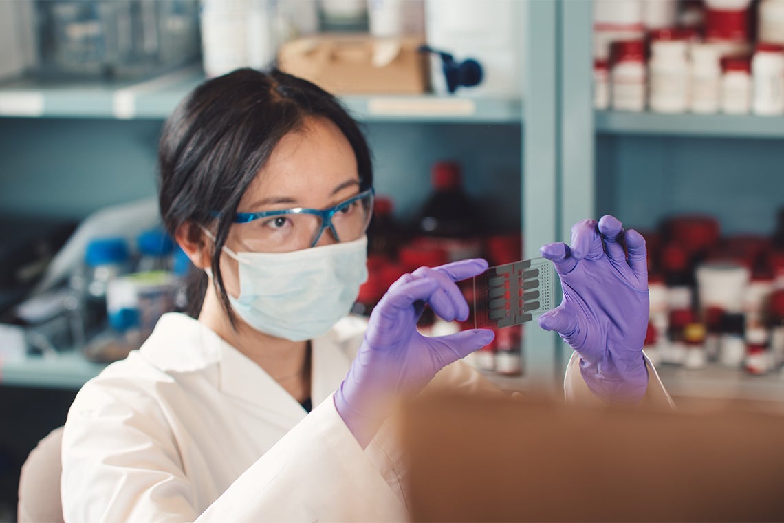
The rotating wall vessel is optimized for suspension culture, with laminar flow and adequate nutrient delivery, but minimal shear. However, higher shears may occur in vivo. During rotating wall vessel cultivation of human renal cells, size and density of glass-coated microcarrier beads were changed to modulate initial shear. Renal-specific proteins were assayed after 2 days. Flow cytometry antibody binding analysis of vitamin D receptor demonstrated peak expression at intermediate shears, with 30% reduction outside this range. Activity of cathepsin C showed the inverse pattern, lowest at midshear, with twofold increases at either extreme. Dipeptidyl-peptidase IV had no shear dependence, suggesting that the other results are specific, not universal, changes in membrane trafficking or protein synthesis. On addition of dextran, which changes medium density and viscosity but not shear, vitamin D receptor assay showed no differences from controls. Neither cell cycle, apoptosis/necrosis indexes, nor lactate dehydrogenase release varied between experiments, confirming that the changes are primary, not secondary to cell cycling or membrane damage. This study provides direct evidence that mechanical culture conditions modulate protein expression in suspension culture. (+info)Tissue engineering is a branch of biomedical engineering that combines the principles of engineering, materials science, and biological sciences to develop functional substitutes for damaged or diseased tissues and organs. It involves the creation of living, three-dimensional structures that can restore, maintain, or improve tissue function. This is typically accomplished through the use of cells, scaffolds (biodegradable matrices), and biologically active molecules. The goal of tissue engineering is to develop biological substitutes that can ultimately restore normal function and structure in damaged tissues or organs.
Tissue scaffolds, also known as bioactive scaffolds or synthetic extracellular matrices, refer to three-dimensional structures that serve as templates for the growth and organization of cells in tissue engineering and regenerative medicine. These scaffolds are designed to mimic the natural extracellular matrix (ECM) found in biological tissues, providing a supportive environment for cell attachment, proliferation, differentiation, and migration.
Tissue scaffolds can be made from various materials, including naturally derived biopolymers (e.g., collagen, alginate, chitosan, hyaluronic acid), synthetic polymers (e.g., polycaprolactone, polylactic acid, poly(lactic-co-glycolic acid)), or a combination of both. The choice of material depends on the specific application and desired properties, such as biocompatibility, biodegradability, mechanical strength, and porosity.
The primary functions of tissue scaffolds include:
1. Cell attachment: Providing surfaces for cells to adhere, spread, and form stable focal adhesions.
2. Mechanical support: Offering a structural framework that maintains the desired shape and mechanical properties of the engineered tissue.
3. Nutrient diffusion: Ensuring adequate transport of nutrients, oxygen, and waste products throughout the scaffold to support cell survival and function.
4. Guided tissue growth: Directing the organization and differentiation of cells through spatial cues and biochemical signals.
5. Biodegradation: Gradually degrading at a rate that matches tissue regeneration, allowing for the replacement of the scaffold with native ECM produced by the cells.
Tissue scaffolds have been used in various applications, such as wound healing, bone and cartilage repair, cardiovascular tissue engineering, and neural tissue regeneration. The design and fabrication of tissue scaffolds are critical aspects of tissue engineering, aiming to create functional substitutes for damaged or diseased tissues and organs.
Biocompatible materials are non-toxic and non-reacting substances that can be used in medical devices, tissue engineering, and drug delivery systems without causing harm or adverse reactions to living tissues or organs. These materials are designed to mimic the properties of natural tissues and are able to integrate with biological systems without being rejected by the body's immune system.
Biocompatible materials can be made from a variety of substances, including metals, ceramics, polymers, and composites. The specific properties of these materials, such as their mechanical strength, flexibility, and biodegradability, are carefully selected to meet the requirements of their intended medical application.
Examples of biocompatible materials include titanium used in dental implants and joint replacements, polyethylene used in artificial hips, and hydrogels used in contact lenses and drug delivery systems. The use of biocompatible materials has revolutionized modern medicine by enabling the development of advanced medical technologies that can improve patient outcomes and quality of life.
Protein engineering is a branch of molecular biology that involves the modification of proteins to achieve desired changes in their structure and function. This can be accomplished through various techniques, including site-directed mutagenesis, gene shuffling, directed evolution, and rational design. The goal of protein engineering may be to improve the stability, activity, specificity, or other properties of a protein for therapeutic, diagnostic, industrial, or research purposes. It is an interdisciplinary field that combines knowledge from genetics, biochemistry, structural biology, and computational modeling.
Biomedical engineering is a field that combines engineering principles and design concepts with medical and biological sciences to develop solutions to healthcare challenges. It involves the application of engineering methods to analyze, understand, and solve problems in biology and medicine, with the goal of improving human health and well-being. Biomedical engineers may work on a wide range of projects, including developing new medical devices, designing artificial organs, creating diagnostic tools, simulating biological systems, and optimizing healthcare delivery systems. They often collaborate with other professionals such as doctors, nurses, and scientists to develop innovative solutions that meet the needs of patients and healthcare providers.
Genetic engineering, also known as genetic modification, is a scientific process where the DNA or genetic material of an organism is manipulated to bring about a change in its characteristics. This is typically done by inserting specific genes into the organism's genome using various molecular biology techniques. These new genes may come from the same species (cisgenesis) or a different species (transgenesis). The goal is to produce a desired trait, such as resistance to pests, improved nutritional content, or increased productivity. It's widely used in research, medicine, and agriculture. However, it's important to note that the use of genetically engineered organisms can raise ethical, environmental, and health concerns.
Hydrogels are defined in the medical and biomedical fields as cross-linked, hydrophilic polymer networks that have the ability to swell and retain a significant amount of water or biological fluids while maintaining their structure. They can be synthesized from natural, synthetic, or hybrid polymers.
Hydrogels are known for their biocompatibility, high water content, and soft consistency, which resemble natural tissues, making them suitable for various medical applications such as contact lenses, drug delivery systems, tissue engineering, wound dressing, and biosensors. The physical and chemical properties of hydrogels can be tailored to specific uses by adjusting the polymer composition, cross-linking density, and network structure.
In the context of medical terminology, "porosity" is not a term that is frequently used to describe human tissues or organs. However, in dermatology and cosmetics, porosity refers to the ability of the skin to absorb and retain moisture or topical treatments.
A skin with high porosity has larger pores and can absorb more products, while a skin with low porosity has smaller pores and may have difficulty absorbing products. It is important to note that this definition of porosity is not a medical one but is instead used in the beauty industry.
Bioartificial organs are hybrid structures that combine living cells, tissues, or biological materials with non-living synthetic materials to replicate the functions of a natural organ. These constructs are designed to mimic the complex architecture and functionality of native organs, providing a viable alternative to traditional organ transplantation.
The bioartificial organ typically consists of three main components:
1. Scaffold: A porous, biocompatible synthetic material that provides structural support and a framework for cell attachment, growth, and organization. The scaffold can be made from various materials such as polymers, ceramics, or composites, and its design considers factors like mechanical strength, degradation rate, and biocompatibility.
2. Cells: Living cells are seeded onto the scaffold, where they proliferate, differentiate, and synthesize extracellular matrix (ECM) proteins to form functional tissue. The choice of cell type depends on the specific organ being replicated; for example, hepatocytes for a liver or cardiomyocytes for a heart.
3. Vascularization: To ensure adequate nutrient and waste exchange, bioartificial organs require an efficient vascular network. This can be achieved through various methods such as co-culturing endothelial cells with the primary cell type, using bioprinting techniques to create patterned vasculature, or incorporating microfluidic channels within the scaffold.
The development of bioartificial organs holds great promise for addressing the current shortage of donor organs and providing personalized treatment options for patients with organ failure. However, several challenges must be overcome before these constructs can be widely adopted in clinical settings, including optimizing vascularization, maintaining long-term functionality, and ensuring biocompatibility and safety.
I'm happy to help! However, I must clarify that "materials testing" is not a medical term per se. It is a term used in various fields, including engineering and materials science, to describe the process of subjecting materials to different tests to evaluate their properties and performance under various conditions.
In the context of medical devices or implants, materials testing may refer to the evaluation of the physical and mechanical properties of materials used in their construction. These tests can include assessments of strength, durability, biocompatibility, and other factors that are critical to ensuring the safety and efficacy of medical devices.
Medical device manufacturers must comply with regulatory standards for materials testing to ensure that their products meet specific requirements for performance, safety, and quality. These standards may vary depending on the type of device, its intended use, and the country or region in which it will be marketed and sold.
Regenerative medicine is a branch of medicine that deals with the repair or replacement of damaged or diseased cells, tissues, and organs using various strategies, including the use of stem cells, tissue engineering, gene therapy, and biomaterials. The goal of regenerative medicine is to restore normal function and structure to tissues and organs, thereby improving the patient's quality of life and potentially curing diseases that were previously considered incurable.
Regenerative medicine has shown promise in a variety of clinical applications, such as the treatment of degenerative diseases like osteoarthritis, spinal cord injuries, heart disease, diabetes, and liver failure. It also holds great potential for use in regenerative therapies for wound healing, tissue reconstruction, and cosmetic surgery.
The field of regenerative medicine is rapidly evolving, with new discoveries and advances being made regularly. As our understanding of the underlying biological mechanisms that drive tissue repair and regeneration continues to grow, so too will the potential clinical applications of this exciting and promising field.
I'm sorry for any confusion, but "Polyesters" is not a medical term. It is a term used in materials science and textile industry to describe a type of synthetic fiber made from polymers characterized by the presence of ester groups in their main chain. If you have any questions related to medical terminology or concepts, I'd be happy to help with those instead!
Nanofibers are defined in the medical field as fibrous structures with extremely small diameters, typically measuring between 100 nanometers to 1 micrometer. They can be made from various materials such as polymers, ceramics, or composites and have a high surface area-to-volume ratio, which makes them useful in a variety of biomedical applications. These include tissue engineering, drug delivery, wound healing, and filtration. Nanofibers can be produced using different techniques such as electrospinning, self-assembly, and phase separation.
Guided Tissue Regeneration (GTR) is a surgical procedure used in periodontics and implant dentistry that aims to regenerate lost periodontal tissues, such as the alveolar bone, cementum, and periodontal ligament, which have been destroyed due to periodontal disease or trauma. The goal of GTR is to restore the architectural and functional relationship between the teeth and their supporting structures.
The procedure involves placing a barrier membrane between the tooth root and the surrounding soft tissues, creating a protected space that allows the periodontal tissues to regenerate. The membrane acts as a physical barrier, preventing the rapid growth of epithelial cells and fibroblasts from the soft tissue into the defect area, while allowing the slower-growing cells derived from the periodontal ligament and bone to repopulate the space.
There are two main types of membranes used in GTR: resorbable and non-resorbable. Resorbable membranes are made of materials that degrade over time, eliminating the need for a second surgical procedure to remove them. Non-resorbable membranes, on the other hand, must be removed after a period of healing.
GTR has been shown to be effective in treating intrabony defects, furcation involvements, and ridge augmentations, among other applications. However, the success of GTR depends on various factors, including the patient's overall health, the size and location of the defect, and the surgeon's skill and experience.
Bone regeneration is the biological process of new bone formation that occurs after an injury or removal of a portion of bone. This complex process involves several stages, including inflammation, migration and proliferation of cells, matrix deposition, and mineralization, leading to the restoration of the bone's structure and function.
The main cells involved in bone regeneration are osteoblasts, which produce new bone matrix, and osteoclasts, which resorb damaged or old bone tissue. The process is tightly regulated by various growth factors, hormones, and signaling molecules that promote the recruitment, differentiation, and activity of these cells.
Bone regeneration can occur naturally in response to injury or surgical intervention, such as fracture repair or dental implant placement. However, in some cases, bone regeneration may be impaired due to factors such as age, disease, or trauma, leading to delayed healing or non-union of the bone. In these situations, various strategies and techniques, including the use of bone grafts, scaffolds, and growth factors, can be employed to enhance and support the bone regeneration process.
Biomimetic materials are synthetic or natural substances that mimic the chemical, physical, and biological properties of living systems or tissues. These materials are designed to interact with cells, tissues, and organs in ways that resemble the body's own structures and processes. They can be used in a variety of medical applications, including tissue engineering, drug delivery, and medical devices.
Biomimetic materials may be composed of polymers, ceramics, metals, or composites, and they can be designed to have specific properties such as mechanical strength, biocompatibility, and degradability. They may also incorporate bioactive molecules, such as growth factors or drugs, to promote healing or prevent infection.
The goal of using biomimetic materials is to create medical solutions that are more effective, safer, and more compatible with the body than traditional synthetic materials. By mimicking the body's own structures and processes, these materials can help to reduce inflammation, promote tissue regeneration, and improve overall patient outcomes.
A hydrogel is a biomaterial that is composed of a three-dimensional network of crosslinked polymers, which are able to absorb and retain a significant amount of water or biological fluids while maintaining their structure. Hydrogels are similar to natural tissues in their water content, making them suitable for various medical applications such as contact lenses, wound dressings, drug delivery systems, tissue engineering, and regenerative medicine.
Hydrogels can be synthesized from a variety of materials, including synthetic polymers like polyethylene glycol (PEG) or natural polymers like collagen, hyaluronic acid, or chitosan. The properties of hydrogels, such as their mechanical strength, degradation rate, and biocompatibility, can be tailored to specific applications by adjusting the type and degree of crosslinking, the molecular weight of the polymers, and the addition of functional groups or drugs.
Hydrogels have shown great potential in medical research and clinical practice due to their ability to mimic the natural environment of cells and tissues, provide sustained drug release, and promote tissue regeneration.
Metabolic engineering is a branch of biotechnology that involves the modification and manipulation of metabolic pathways in organisms to enhance their production of specific metabolites or to alter their flow of energy and carbon. This field combines principles from genetics, molecular biology, biochemistry, and chemical engineering to design and construct novel metabolic pathways or modify existing ones with the goal of optimizing the production of valuable compounds or improving the properties of organisms for various applications.
Examples of metabolic engineering include the modification of microorganisms to produce biofuels, pharmaceuticals, or industrial chemicals; the enhancement of crop yields and nutritional value in agriculture; and the development of novel bioremediation strategies for environmental pollution control. The ultimate goal of metabolic engineering is to create organisms that can efficiently and sustainably produce valuable products while minimizing waste and reducing the impact on the environment.
A bioreactor is a device or system that supports and controls the conditions necessary for biological organisms, cells, or tissues to grow and perform their specific functions. It provides a controlled environment with appropriate temperature, pH, nutrients, and other factors required for the desired biological process to occur. Bioreactors are widely used in various fields such as biotechnology, pharmaceuticals, agriculture, and environmental science for applications like production of therapeutic proteins, vaccines, biofuels, enzymes, and wastewater treatment.
In the context of medical definitions, polymers are large molecules composed of repeating subunits called monomers. These long chains of monomers can have various structures and properties, depending on the type of monomer units and how they are linked together. In medicine, polymers are used in a wide range of applications, including drug delivery systems, medical devices, and tissue engineering scaffolds. Some examples of polymers used in medicine include polyethylene, polypropylene, polystyrene, polyvinyl chloride (PVC), and biodegradable polymers such as polylactic acid (PLA) and polycaprolactone (PCL).
Decanoates are a type of esterified form of certain drugs or medications, particularly in the case of testosterone. The decanoate ester is attached to the testosterone molecule to create a longer-acting formulation. Testosterone decanoate is a slow-release form of testosterone that is used as a replacement therapy for individuals who have low levels of natural testosterone. It is administered through intramuscular injection and has a duration of action of approximately 2-3 weeks.
Other medications may also be available in decanoate ester form, but testosterone decanoate is one of the most commonly used. As with any medication or treatment plan, it's important to consult with a healthcare provider to determine the best course of action based on individual needs and medical history.
Absorbable implants are medical devices that are designed to be placed inside the body during a surgical procedure, where they provide support, stabilization, or other functions, and then gradually break down and are absorbed by the body over time. These implants are typically made from materials such as polymers, proteins, or ceramics that have been engineered to degrade at a controlled rate, allowing them to be resorbed and eliminated from the body without the need for a second surgical procedure to remove them.
Absorbable implants are often used in orthopedic, dental, and plastic surgery applications, where they can help promote healing and support tissue regeneration. For example, absorbable screws or pins may be used to stabilize fractured bones during the healing process, after which they will gradually dissolve and be absorbed by the body. Similarly, absorbable membranes may be used in dental surgery to help guide the growth of new bone and gum tissue around an implant, and then be resorbed over time.
It's important to note that while absorbable implants offer several advantages over non-absorbable materials, such as reduced risk of infection and improved patient comfort, they may also have some limitations. For example, the mechanical properties of absorbable materials may not be as strong as those of non-absorbable materials, which could affect their performance in certain applications. Additionally, the degradation products of absorbable implants may cause local inflammation or other adverse reactions in some patients. As with any medical device, the use of absorbable implants should be carefully considered and discussed with a qualified healthcare professional.
Mesenchymal Stromal Cells (MSCs) are a type of adult stem cells found in various tissues, including bone marrow, adipose tissue, and umbilical cord blood. They have the ability to differentiate into multiple cell types, such as osteoblasts, chondrocytes, and adipocytes, under specific conditions. MSCs also possess immunomodulatory properties, making them a promising tool in regenerative medicine and therapeutic strategies for various diseases, including autoimmune disorders and tissue injuries. It is important to note that the term "Mesenchymal Stem Cells" has been replaced by "Mesenchymal Stromal Cells" in the scientific community to better reflect their biological characteristics and potential functions.
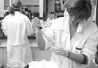
Cell culture is a technique used in scientific research to grow and maintain cells from plants, animals, or humans in a controlled environment outside of their original organism. This environment typically consists of a sterile container called a cell culture flask or plate, and a nutrient-rich liquid medium that provides the necessary components for the cells' growth and survival, such as amino acids, vitamins, minerals, and hormones.
There are several different types of cell culture techniques used in research, including:
1. Adherent cell culture: In this technique, cells are grown on a flat surface, such as the bottom of a tissue culture dish or flask. The cells attach to the surface and spread out, forming a monolayer that can be observed and manipulated under a microscope.
2. Suspension cell culture: In suspension culture, cells are grown in liquid medium without any attachment to a solid surface. These cells remain suspended in the medium and can be agitated or mixed to ensure even distribution of nutrients.
3. Organoid culture: Organoids are three-dimensional structures that resemble miniature organs and are grown from stem cells or other progenitor cells. They can be used to study organ development, disease processes, and drug responses.
4. Co-culture: In co-culture, two or more different types of cells are grown together in the same culture dish or flask. This technique is used to study cell-cell interactions and communication.
5. Conditioned medium culture: In this technique, cells are grown in a medium that has been conditioned by previous cultures of other cells. The conditioned medium contains factors secreted by the previous cells that can influence the growth and behavior of the new cells.
Cell culture techniques are widely used in biomedical research to study cellular processes, develop drugs, test toxicity, and investigate disease mechanisms. However, it is important to note that cell cultures may not always accurately represent the behavior of cells in a living organism, and results from cell culture experiments should be validated using other methods.
Bioengineering, also known as biological engineering, is defined as the application of principles and methods from engineering to study, modify, and control biological systems, often with the goal of creating new technologies or improving existing ones. This field combines knowledge and expertise from various disciplines, including biology, chemistry, physics, mathematics, and computer science, to solve complex problems related to health, medicine, agriculture, and the environment.
Bioengineers may work on a wide range of projects, such as developing new medical devices or therapies, designing synthetic biological systems for industrial applications, creating biosensors for environmental monitoring, or engineering tissues and organs for transplantation. They use a variety of tools and techniques, including genetic engineering, biomaterials, computational modeling, and nanotechnology, to design and build novel biological systems that can perform specific functions or solve practical problems.
Bioengineering has the potential to transform many areas of science and technology, with significant implications for human health, sustainability, and innovation. As such, it is an exciting and rapidly growing field that offers many opportunities for interdisciplinary collaboration and discovery.
Polyglycolic acid (PGA) is a synthetic polymer of glycolic acid, which is commonly used in surgical sutures. It is a biodegradable material that degrades in the body through hydrolysis into glycolic acid, which can be metabolized and eliminated from the body. PGA sutures are often used for approximating tissue during surgical procedures due to their strength, handling properties, and predictable rate of absorption. The degradation time of PGA sutures is typically around 60-90 days, depending on factors such as the size and location of the suture.
Osteogenesis is the process of bone formation or development. It involves the differentiation and maturation of osteoblasts, which are bone-forming cells that synthesize and deposit the organic matrix of bone tissue, composed mainly of type I collagen. This organic matrix later mineralizes to form the inorganic crystalline component of bone, primarily hydroxyapatite.
There are two primary types of osteogenesis: intramembranous and endochondral. Intramembranous osteogenesis occurs directly within connective tissue, where mesenchymal stem cells differentiate into osteoblasts and form bone tissue without an intervening cartilage template. This process is responsible for the formation of flat bones like the skull and clavicles.
Endochondral osteogenesis, on the other hand, involves the initial development of a cartilaginous model or template, which is later replaced by bone tissue. This process forms long bones, such as those in the limbs, and occurs through several stages involving chondrocyte proliferation, hypertrophy, and calcification, followed by invasion of blood vessels and osteoblasts to replace the cartilage with bone tissue.
Abnormalities in osteogenesis can lead to various skeletal disorders and diseases, such as osteogenesis imperfecta (brittle bone disease), achondroplasia (a form of dwarfism), and cleidocranial dysplasia (a disorder affecting skull and collarbone development).
Scanning electron microscopy (SEM) is a type of electron microscopy that uses a focused beam of electrons to scan the surface of a sample and produce a high-resolution image. In SEM, a beam of electrons is scanned across the surface of a specimen, and secondary electrons are emitted from the sample due to interactions between the electrons and the atoms in the sample. These secondary electrons are then detected by a detector and used to create an image of the sample's surface topography. SEM can provide detailed images of the surface of a wide range of materials, including metals, polymers, ceramics, and biological samples. It is commonly used in materials science, biology, and electronics for the examination and analysis of surfaces at the micro- and nanoscale.
I'm sorry for any confusion, but "Silk" is not a medical term. Silk is a natural protein fiber, mainly composed of fibroin and produced by certain insect larvae to form cocoons. It's commonly used in textiles and other industries for its softness, smoothness, and strength. If you have any questions related to medical terminology or health-related topics, I'd be happy to help with those instead!
Elastomers are a type of polymeric material that exhibit elastic behavior when subjected to deforming forces. They have the ability to return to their original shape and size after being stretched or compressed, making them ideal for use in applications where flexibility, resilience, and durability are required.
Elastomers are composed of long chains of repeating molecular units called monomers, which are cross-linked together to form a three-dimensional network. This cross-linking gives elastomers their unique properties, such as high elasticity, low compression set, and resistance to heat, chemicals, and weathering.
Some common examples of elastomers include natural rubber, silicone rubber, neoprene, nitrile rubber, and polyurethane. These materials are used in a wide range of applications, from automotive parts and medical devices to footwear and clothing.
Chitosan is a complex carbohydrate that is derived from the exoskeletons of crustaceans, such as shrimp and crabs. It is made up of chains of N-acetyl-d-glucosamine and d-glucosamine units. Chitosan has been studied for its potential medical and health benefits, including its ability to lower cholesterol levels, promote weight loss, and help control blood sugar levels. It is also used in wound care products due to its antibacterial and absorbent properties. However, more research is needed to confirm these potential benefits and establish recommended dosages and safety guidelines.
I'm sorry for any confusion, but "Microtechnology" is not a term that has a specific medical definition. Microtechnology generally refers to the development and application of technologies on a microscopic or tiny scale. It is used in various fields including engineering, physics, electronics, and materials science.
In the context of medicine, microtechnologies can be used in the development of medical devices, diagnostic tools, drug delivery systems, and other healthcare applications. For example, microfabrication techniques are used to create microfluidic devices for lab-on-a-chip applications, which can perform complex biochemical analyses for disease diagnosis or drug screening.
However, it's important to note that the application of microtechnologies in medicine is constantly evolving, and new developments and techniques are being explored all the time.
Compressive strength is a measure of the maximum compressive load that a material or structure can withstand before failure or deformation. It is typically expressed in units of pressure, such as pounds per square inch (psi) or megapascals (MPa). Compressive strength is an important property in the design and analysis of structures and materials, as it helps to ensure their safety and durability under compressive loads.
In medical terminology, compressive strength may refer to the ability of biological tissues, such as bone or cartilage, to withstand compressive forces without deforming or failing. For example, osteoporosis is a condition characterized by reduced bone density and compressive strength, which can increase the risk of fractures in affected individuals. Similarly, degenerative changes in articular cartilage can lead to decreased compressive strength and joint pain or stiffness.
Regeneration in a medical context refers to the process of renewal, restoration, and growth that replaces damaged or missing cells, tissues, organs, or even whole limbs in some organisms. This complex biological process involves various cellular and molecular mechanisms, such as cell proliferation, differentiation, and migration, which work together to restore the structural and functional integrity of the affected area.
In human medicine, regeneration has attracted significant interest due to its potential therapeutic applications in treating various conditions, including degenerative diseases, trauma, and congenital disorders. Researchers are actively studying the underlying mechanisms of regeneration in various model organisms to develop novel strategies for promoting tissue repair and regeneration in humans.
Examples of regeneration in human medicine include liver regeneration after partial hepatectomy, where the remaining liver lobes can grow back to their original size within weeks, and skin wound healing, where keratinocytes migrate and proliferate to close the wound and restore the epidermal layer. However, the regenerative capacity of humans is limited compared to some other organisms, such as planarians and axolotls, which can regenerate entire body parts or even their central nervous system.
The extracellular matrix (ECM) is a complex network of biomolecules that provides structural and biochemical support to cells in tissues and organs. It is composed of various proteins, glycoproteins, and polysaccharides, such as collagens, elastin, fibronectin, laminin, and proteoglycans. The ECM plays crucial roles in maintaining tissue architecture, regulating cell behavior, and facilitating communication between cells. It provides a scaffold for cell attachment, migration, and differentiation, and helps to maintain the structural integrity of tissues by resisting mechanical stresses. Additionally, the ECM contains various growth factors, cytokines, and chemokines that can influence cellular processes such as proliferation, survival, and differentiation. Overall, the extracellular matrix is essential for the normal functioning of tissues and organs, and its dysregulation can contribute to various pathological conditions, including fibrosis, cancer, and degenerative diseases.
Chondrogenesis is the process of cartilage formation during embryonic development and in the healing of certain types of injuries. It involves the differentiation of mesenchymal stem cells into chondrocytes, which are the specialized cells that produce and maintain the extracellular matrix of cartilage.
During chondrogenesis, the mesenchymal stem cells condense and form a template for the future cartilaginous tissue. These cells then differentiate into chondrocytes, which begin to produce and deposit collagen type II, proteoglycans, and other extracellular matrix components that give cartilage its unique biochemical and mechanical properties.
Chondrogenesis is a critical process for the development of various structures in the body, including the skeletal system, where it plays a role in the formation of articular cartilage, growth plates, and other types of cartilage. Understanding the molecular mechanisms that regulate chondrogenesis is important for developing therapies to treat cartilage injuries and degenerative diseases such as osteoarthritis.
I'm sorry for any confusion, but "mechanical phenomena" is not a standard medical term. The term "mechanical phenomenon" is more commonly used in the fields of physics and engineering to refer to events or processes that involve physical forces and movements, such as the movement of a lever or the flow of a fluid due to pressure differences.
If you have any questions about a specific medical concept or condition, I would be happy to try to help you with that instead!
Nanostructures, in the context of medical and biomedical research, refer to materials or devices with structural features that have at least one dimension ranging between 1-100 nanometers (nm). At this size scale, the properties of these structures can differ significantly from bulk materials, exhibiting unique phenomena that are often influenced by quantum effects.
Nanostructures have attracted considerable interest in biomedicine due to their potential applications in various areas such as drug delivery, diagnostics, regenerative medicine, and tissue engineering. They can be fabricated from a wide range of materials including metals, polymers, ceramics, and carbon-based materials.
Some examples of nanostructures used in biomedicine include:
1. Nanoparticles: These are tiny particles with at least one dimension in the nanoscale range. They can be made from various materials like metals, polymers, or lipids and have applications in drug delivery, imaging, and diagnostics.
2. Quantum dots: These are semiconductor nanocrystals that exhibit unique optical properties due to quantum confinement effects. They are used as fluorescent labels for bioimaging and biosensing applications.
3. Carbon nanotubes: These are hollow, cylindrical structures made of carbon atoms arranged in a hexagonal lattice. They have exceptional mechanical strength, electrical conductivity, and thermal stability, making them suitable for various biomedical applications such as drug delivery, tissue engineering, and biosensors.
4. Nanofibers: These are elongated nanostructures with high aspect ratios (length much greater than width). They can be fabricated from various materials like polymers, ceramics, or composites and have applications in tissue engineering, wound healing, and drug delivery.
5. Dendrimers: These are highly branched, nanoscale polymers with a well-defined structure and narrow size distribution. They can be used as drug carriers, gene delivery vehicles, and diagnostic agents.
6. Nanoshells: These are hollow, spherical nanoparticles consisting of a dielectric core covered by a thin metallic shell. They exhibit unique optical properties that make them suitable for applications such as photothermal therapy, biosensing, and imaging.
Experimental implants refer to medical devices that are not yet approved by regulatory authorities for general use in medical practice. These are typically being tested in clinical trials to evaluate their safety and efficacy. The purpose of experimental implants is to determine whether they can be used as a viable treatment option for various medical conditions. They may include, but are not limited to, devices such as artificial joints, heart valves, or spinal cord stimulators that are still in the developmental or testing stage. Participation in clinical trials involving experimental implants is voluntary and usually requires informed consent from the patient.
Cartilage is a type of connective tissue that is found throughout the body in various forms. It is made up of specialized cells called chondrocytes, which are embedded in a firm, flexible matrix composed of collagen fibers and proteoglycans. This unique structure gives cartilage its characteristic properties of being both strong and flexible.
There are three main types of cartilage in the human body: hyaline cartilage, elastic cartilage, and fibrocartilage.
1. Hyaline cartilage is the most common type and is found in areas such as the articular surfaces of bones (where they meet to form joints), the nose, trachea, and larynx. It has a smooth, glassy appearance and provides a smooth, lubricated surface for joint movement.
2. Elastic cartilage contains more elastin fibers than hyaline cartilage, which gives it greater flexibility and resilience. It is found in structures such as the external ear and parts of the larynx and epiglottis.
3. Fibrocartilage has a higher proportion of collagen fibers and fewer chondrocytes than hyaline or elastic cartilage. It is found in areas that require high tensile strength, such as the intervertebral discs, menisci (found in joints like the knee), and the pubic symphysis.
Cartilage plays a crucial role in supporting and protecting various structures within the body, allowing for smooth movement and providing a cushion between bones to absorb shock and prevent wear and tear. However, cartilage has limited capacity for self-repair and regeneration, making damage or degeneration of cartilage tissue a significant concern in conditions such as osteoarthritis.
Biomimetics, also known as biomimicry, is the process of mimicking or taking inspiration from nature and biological systems to design materials, structures, or processes that solve human problems. It involves studying the models, systems, and elements of nature and then applying the knowledge gained to create new technologies and solutions.
In a medical context, biomimetics can be used to develop new therapies, medical devices, and diagnostic tools. For example, researchers might look to the structure of a spider's web to design a better surgical mesh or take inspiration from the way a gecko sticks to surfaces to create a new type of adhesive bandage.
Biomimetics is an interdisciplinary field that draws on knowledge from biology, chemistry, physics, engineering, and materials science. It has the potential to lead to innovative solutions in healthcare, sustainability, energy, transportation, and other areas.
Calcium phosphates are a group of minerals that are important components of bones and teeth. They are also found in some foods and are used in dietary supplements and medical applications. Chemically, calcium phosphates are salts of calcium and phosphoric acid, and they exist in various forms, including hydroxyapatite, which is the primary mineral component of bone tissue. Other forms of calcium phosphates include monocalcium phosphate, dicalcium phosphate, and tricalcium phosphate, which are used as food additives and dietary supplements. Calcium phosphates are important for maintaining strong bones and teeth, and they also play a role in various physiological processes, such as nerve impulse transmission and muscle contraction.
Hexuronic acids are a type of uronic acid that contains six carbon atoms and is commonly found in various biological tissues and polysaccharides, such as pectins, heparin, and certain glycoproteins. The most common hexuronic acids are glucuronic acid and iduronic acid, which are formed from the oxidation of the corresponding hexoses, glucose and galactose, respectively. Hexuronic acids play important roles in various biological processes, including the detoxification and excretion of xenobiotics, the formation of proteoglycans, and the regulation of cell growth and differentiation.
Chondrocytes are the specialized cells that produce and maintain the extracellular matrix of cartilage tissue. They are responsible for synthesizing and secreting the collagen fibers, proteoglycans, and other components that give cartilage its unique properties, such as elasticity, resiliency, and resistance to compression. Chondrocytes are located within lacunae, or small cavities, in the cartilage matrix, and they receive nutrients and oxygen through diffusion from the surrounding tissue fluid. They are capable of adapting to changes in mechanical stress by modulating the production and organization of the extracellular matrix, which allows cartilage to withstand various loads and maintain its structural integrity. Chondrocytes play a crucial role in the development, maintenance, and repair of cartilaginous tissues throughout the body, including articular cartilage, costal cartilage, and growth plate cartilage.
Tensile strength is a material property that measures the maximum amount of tensile (pulling) stress that a material can withstand before failure, such as breaking or fracturing. It is usually measured in units of force per unit area, such as pounds per square inch (psi) or pascals (Pa). In the context of medical devices or biomaterials, tensile strength may be used to describe the mechanical properties of materials used in implants, surgical tools, or other medical equipment. High tensile strength is often desirable in these applications to ensure that the material can withstand the stresses and forces it will encounter during use.
Glucuronic acid is a physiological important organic acid, which is a derivative of glucose. It is formed by the oxidation of the primary alcohol group of glucose to form a carboxyl group at the sixth position. Glucuronic acid plays a crucial role in the detoxification process in the body as it conjugates with toxic substances, making them water-soluble and facilitating their excretion through urine or bile. This process is known as glucuronidation. It is also a component of various polysaccharides, such as heparan sulfate and chondroitin sulfate, which are found in the extracellular matrix of connective tissues.
Alginates are a type of polysaccharide derived from brown algae or produced synthetically, which have gelling and thickening properties. In medical context, they are commonly used as a component in wound dressings, dental impressions, and bowel cleansing products. The gels formed by alginates can provide a protective barrier to wounds, help maintain a moist environment, and promote healing. They can also be used to create a mold of the mouth or other body parts in dental and medical applications. In bowel cleansing, sodium alginates are often combined with sodium bicarbonate and water to form a solution that expands and stimulates bowel movements, helping to prepare the colon for procedures such as colonoscopy.
Bone substitutes are materials that are used to replace missing or damaged bone in the body. They can be made from a variety of materials, including natural bone from other parts of the body or from animals, synthetic materials, or a combination of both. The goal of using bone substitutes is to provide structural support and promote the growth of new bone tissue.
Bone substitutes are often used in dental, orthopedic, and craniofacial surgery to help repair defects caused by trauma, tumors, or congenital abnormalities. They can also be used to augment bone volume in procedures such as spinal fusion or joint replacement.
There are several types of bone substitutes available, including:
1. Autografts: Bone taken from another part of the patient's body, such as the hip or pelvis.
2. Allografts: Bone taken from a deceased donor and processed to remove any cells and infectious materials.
3. Xenografts: Bone from an animal source, typically bovine or porcine, that has been processed to remove any cells and infectious materials.
4. Synthetic bone substitutes: Materials such as calcium phosphate ceramics, bioactive glass, and polymer-based materials that are designed to mimic the properties of natural bone.
The choice of bone substitute material depends on several factors, including the size and location of the defect, the patient's medical history, and the surgeon's preference. It is important to note that while bone substitutes can provide structural support and promote new bone growth, they may not have the same strength or durability as natural bone. Therefore, they may not be suitable for all applications, particularly those that require high load-bearing capacity.
"Bone" is the hard, dense connective tissue that makes up the skeleton of vertebrate animals. It provides support and protection for the body's internal organs, and serves as a attachment site for muscles, tendons, and ligaments. Bone is composed of cells called osteoblasts and osteoclasts, which are responsible for bone formation and resorption, respectively, and an extracellular matrix made up of collagen fibers and mineral crystals.
Bones can be classified into two main types: compact bone and spongy bone. Compact bone is dense and hard, and makes up the outer layer of all bones and the shafts of long bones. Spongy bone is less dense and contains large spaces, and makes up the ends of long bones and the interior of flat and irregular bones.
The human body has 206 bones in total. They can be further classified into five categories based on their shape: long bones, short bones, flat bones, irregular bones, and sesamoid bones.
Cell differentiation is the process by which a less specialized cell, or stem cell, becomes a more specialized cell type with specific functions and structures. This process involves changes in gene expression, which are regulated by various intracellular signaling pathways and transcription factors. Differentiation results in the development of distinct cell types that make up tissues and organs in multicellular organisms. It is a crucial aspect of embryonic development, tissue repair, and maintenance of homeostasis in the body.
Gelatin is not strictly a medical term, but it is often used in medical contexts. Medically, gelatin is recognized as a protein-rich substance that is derived from collagen, which is found in the skin, bones, and connective tissue of animals. It is commonly used in the production of various medical and pharmaceutical products such as capsules, wound dressings, and drug delivery systems due to its biocompatibility and ability to form gels.
In a broader sense, gelatin is a translucent, colorless, flavorless food ingredient that is derived from collagen through a process called hydrolysis. It is widely used in the food industry as a gelling agent, thickener, stabilizer, and texturizer in various foods such as candies, desserts, marshmallows, and yogurts.
It's worth noting that while gelatin has many uses, it may not be suitable for vegetarians or those with dietary restrictions since it is derived from animal products.
According to the National Institutes of Health (NIH), stem cells are "initial cells" or "precursor cells" that have the ability to differentiate into many different cell types in the body. They can also divide without limit to replenish other cells for as long as the person or animal is still alive.
There are two main types of stem cells: embryonic stem cells, which come from human embryos, and adult stem cells, which are found in various tissues throughout the body. Embryonic stem cells have the ability to differentiate into all cell types in the body, while adult stem cells have more limited differentiation potential.
Stem cells play an essential role in the development and repair of various tissues and organs in the body. They are currently being studied for their potential use in the treatment of a wide range of diseases and conditions, including cancer, diabetes, heart disease, and neurological disorders. However, more research is needed to fully understand the properties and capabilities of these cells before they can be used safely and effectively in clinical settings.
"Cells, cultured" is a medical term that refers to cells that have been removed from an organism and grown in controlled laboratory conditions outside of the body. This process is called cell culture and it allows scientists to study cells in a more controlled and accessible environment than they would have inside the body. Cultured cells can be derived from a variety of sources, including tissues, organs, or fluids from humans, animals, or cell lines that have been previously established in the laboratory.
Cell culture involves several steps, including isolation of the cells from the tissue, purification and characterization of the cells, and maintenance of the cells in appropriate growth conditions. The cells are typically grown in specialized media that contain nutrients, growth factors, and other components necessary for their survival and proliferation. Cultured cells can be used for a variety of purposes, including basic research, drug development and testing, and production of biological products such as vaccines and gene therapies.
It is important to note that cultured cells may behave differently than they do in the body, and results obtained from cell culture studies may not always translate directly to human physiology or disease. Therefore, it is essential to validate findings from cell culture experiments using additional models and ultimately in clinical trials involving human subjects.
Surface properties in the context of medical science refer to the characteristics and features of the outermost layer or surface of a biological material or structure, such as cells, tissues, organs, or medical devices. These properties can include physical attributes like roughness, smoothness, hydrophobicity or hydrophilicity, and electrical conductivity, as well as chemical properties like charge, reactivity, and composition.
In the field of biomaterials science, understanding surface properties is crucial for designing medical implants, devices, and drug delivery systems that can interact safely and effectively with biological tissues and fluids. Surface modifications, such as coatings or chemical treatments, can be used to alter surface properties and enhance biocompatibility, improve lubricity, reduce fouling, or promote specific cellular responses like adhesion, proliferation, or differentiation.
Similarly, in the field of cell biology, understanding surface properties is essential for studying cell-cell interactions, cell signaling, and cell behavior. Cells can sense and respond to changes in their environment, including variations in surface properties, which can influence cell shape, motility, and function. Therefore, characterizing and manipulating surface properties can provide valuable insights into the mechanisms of cellular processes and offer new strategies for developing therapies and treatments for various diseases.
Fibroins are a type of protein that make up the structural component of silk fibers produced by certain insects and arachnids, such as silkworms and spiders. These proteins are characterized by their repetitive amino acid sequences, which give silk its unique properties of strength, flexibility, and toughness. Fibroins have been studied for their potential applications in biomedicine, including tissue engineering, drug delivery, and medical textiles.
Dura Mater: The tough, outer membrane that covers the brain and spinal cord.
Hydroxyapatite: A naturally occurring mineral form of calcium apatite, also known as dahllite, with the formula Ca5(PO4)3(OH), is the primary mineral component of biological apatites found in bones and teeth.
Therefore, "Durapatite" isn't a recognized medical term, but it seems like it might be a combination of "dura mater" and "hydroxyapatite." If you meant to ask about a material used in medical or dental applications that combines properties of both dura mater and hydroxyapatite, please provide more context.
Dental pulp is the soft tissue located in the center of a tooth, surrounded by the dentin. It contains nerves, blood vessels, and connective tissue, and plays a vital role in the development and health of the tooth. The dental pulp helps to form dentin during tooth development and continues to provide nourishment to the tooth throughout its life. It also serves as a sensory organ, allowing the tooth to detect hot and cold temperatures and transmit pain signals to the brain. Injury or infection of the dental pulp can lead to serious dental problems, such as tooth decay or abscesses, and may require root canal treatment to remove the damaged tissue and save the tooth.
I'm sorry for any confusion, but "nanocomposites" is not a term that has a specific medical definition. It is a term used in materials science and engineering to refer to a type of composite material where at least one of the phases has dimensions in the nanoscale (typically less than 100 nanometers). Nanocomposites can have unique properties that make them useful for various applications, including biomedical applications such as drug delivery systems or tissue engineering scaffolds. However, the term itself is not a medical definition.
Lactic acid, also known as 2-hydroxypropanoic acid, is a chemical compound that plays a significant role in various biological processes. In the context of medicine and biochemistry, lactic acid is primarily discussed in relation to muscle metabolism and cellular energy production. Here's a medical definition for lactic acid:
Lactic acid (LA): A carboxylic acid with the molecular formula C3H6O3 that plays a crucial role in anaerobic respiration, particularly during strenuous exercise or conditions of reduced oxygen availability. It is formed through the conversion of pyruvate, catalyzed by the enzyme lactate dehydrogenase (LDH), when there is insufficient oxygen to complete the final step of cellular respiration in the Krebs cycle. The accumulation of lactic acid can lead to acidosis and muscle fatigue. Additionally, lactic acid serves as a vital intermediary in various metabolic pathways and is involved in the production of glucose through gluconeogenesis in the liver.
Cell engineering is a branch of biotechnology that involves the manipulation and modification of cells to achieve desired functions or characteristics. This can be accomplished through various techniques, including genetic engineering, gene editing, cell culturing, and tissue engineering. The goal of cell engineering may be to develop new therapies for diseases, create cells or tissues that can replace damaged ones in the body, or to better understand how cells function.
In genetic engineering, genes are introduced into cells using vectors such as plasmids or viruses. These genes can encode for specific proteins or enzymes that can help the cell perform a particular function, such as producing a therapeutic protein or breaking down a toxic substance. Gene editing techniques, such as CRISPR-Cas9, allow for precise editing of an organism's genome, enabling the correction of genetic mutations or the introduction of new traits.
Cell culturing involves growing cells in controlled conditions outside of the body, allowing researchers to study their behavior and properties. Tissue engineering combines cell engineering with materials science to create functional tissues or organs that can be used for transplantation or other medical applications.
Overall, cell engineering has the potential to revolutionize medicine by enabling the development of personalized therapies, regenerative medicine, and new treatments for a wide range of diseases and conditions.
Cell proliferation is the process by which cells increase in number, typically through the process of cell division. In the context of biology and medicine, it refers to the reproduction of cells that makes up living tissue, allowing growth, maintenance, and repair. It involves several stages including the transition from a phase of quiescence (G0 phase) to an active phase (G1 phase), DNA replication in the S phase, and mitosis or M phase, where the cell divides into two daughter cells.
Abnormal or uncontrolled cell proliferation is a characteristic feature of many diseases, including cancer, where deregulated cell cycle control leads to excessive and unregulated growth of cells, forming tumors that can invade surrounding tissues and metastasize to distant sites in the body.
The Elastic Modulus, also known as Young's modulus, is a measure of the stiffness of a material. It is defined as the ratio of stress (force per unit area) to strain (partial deformation or change in length per unit length) in the elastic range of deformation of a material. In other words, it measures how much a material will deform (change in length or size) when subjected to a given amount of force. A higher elastic modulus indicates that a material is stiffer and less likely to deform, while a lower elastic modulus indicates that a material is more flexible and will deform more easily. The elastic modulus is typically expressed in units of Pascals (Pa) or Gigapascals (GPa).
Collagen is the most abundant protein in the human body, and it is a major component of connective tissues such as tendons, ligaments, skin, and bones. Collagen provides structure and strength to these tissues and helps them to withstand stretching and tension. It is made up of long chains of amino acids, primarily glycine, proline, and hydroxyproline, which are arranged in a triple helix structure. There are at least 16 different types of collagen found in the body, each with slightly different structures and functions. Collagen is important for maintaining the integrity and health of tissues throughout the body, and it has been studied for its potential therapeutic uses in various medical conditions.
I'm sorry for any confusion, but "Polyurethanes" are not a medical term. They are a type of polymer that is used in a variety of industrial and consumer products, including foam cushions, insulation, and packaging materials. Polyurethanes are created through a chemical reaction between diisocyanates and polyols. While they have many applications in the medical field, such as in the production of medical devices and equipment, they are not a medical term themselves.
Artificial Skin is a synthetic substitute or equivalent that is used to replace, support, or enhance the function of damaged or absent skin. It can be made from various materials such as biopolymers, composites, or biosynthetic materials. The main purpose of artificial skin is to provide a temporary or permanent covering for wounds, burns, or ulcers that cannot be healed with conventional treatments. Additionally, it may serve as a platform for the delivery of medications or as a matrix for the growth of cells and tissues during skin grafting procedures. Artificial skin must possess properties such as biocompatibility, durability, flexibility, and permeability to air and water vapor in order to promote optimal healing and minimize scarring.
In medical terms, "gels" are semi-solid colloidal systems in which a solid phase is dispersed in a liquid medium. They have a viscous consistency and can be described as a cross between a solid and a liquid. The solid particles, called the gel network, absorb and swell with the liquid component, creating a system that has properties of both solids and liquids.
Gels are widely used in medical applications such as wound dressings, drug delivery systems, and tissue engineering due to their unique properties. They can provide a moist environment for wounds to heal, control the release of drugs over time, and mimic the mechanical properties of natural tissues.
Cell shape refers to the physical form or configuration of a cell, which is determined by the cytoskeleton (the internal framework of the cell) and the extracellular matrix (the external environment surrounding the cell). The shape of a cell can vary widely depending on its type and function. For example, some cells are spherical, such as red blood cells, while others are elongated or irregularly shaped. Changes in cell shape can be indicative of various physiological or pathological processes, including development, differentiation, migration, and disease.
The periodontal ligament, also known as the "PDL," is the soft tissue that connects the tooth root to the alveolar bone within the dental alveolus (socket). It consists of collagen fibers organized into groups called principal fibers and accessory fibers. These fibers are embedded into both the cementum of the tooth root and the alveolar bone, providing shock absorption during biting and chewing forces, allowing for slight tooth movement, and maintaining the tooth in its position within the socket.
The periodontal ligament plays a crucial role in the health and maintenance of the periodontium, which includes the gingiva (gums), cementum, alveolar bone, and the periodontal ligament itself. Inflammation or infection of the periodontal ligament can lead to periodontal disease, potentially causing tooth loss if not treated promptly and appropriately.
Nanotechnology is not a medical term per se, but it is a field of study with potential applications in medicine. According to the National Nanotechnology Initiative, nanotechnology is defined as "the understanding and control of matter at the nanoscale, at dimensions between approximately 1 and 100 nanometers, where unique phenomena enable novel applications."
In the context of medicine, nanotechnology has the potential to revolutionize the way we diagnose, treat, and prevent diseases. Nanomedicine involves the use of nanoscale materials, devices, or systems for medical applications. These can include drug delivery systems that target specific cells or tissues, diagnostic tools that detect biomarkers at the molecular level, and tissue engineering strategies that promote regeneration and repair.
While nanotechnology holds great promise for medicine, it is still a relatively new field with many challenges to overcome, including issues related to safety, regulation, and scalability.
Bone Morphogenetic Protein 2 (BMP-2) is a growth factor that belongs to the transforming growth factor-beta (TGF-β) superfamily. It plays a crucial role in bone and cartilage formation, as well as in the regulation of wound healing and embryonic development. BMP-2 stimulates the differentiation of mesenchymal stem cells into osteoblasts, which are cells responsible for bone formation.
BMP-2 has been approved by the US Food and Drug Administration (FDA) as a medical device to promote bone growth in certain spinal fusion surgeries and in the treatment of open fractures that have not healed properly. It is usually administered in the form of a collagen sponge soaked with recombinant human BMP-2 protein, which is a laboratory-produced version of the natural protein.
While BMP-2 has shown promising results in some clinical applications, its use is not without risks and controversies. Some studies have reported adverse effects such as inflammation, ectopic bone formation, and increased rates of cancer, which have raised concerns about its safety and efficacy. Therefore, it is essential to weigh the benefits and risks of BMP-2 therapy on a case-by-case basis and under the guidance of a qualified healthcare professional.
Biomechanics is the application of mechanical laws to living structures and systems, particularly in the field of medicine and healthcare. A biomechanical phenomenon refers to a observable event or occurrence that involves the interaction of biological tissues or systems with mechanical forces. These phenomena can be studied at various levels, from the molecular and cellular level to the tissue, organ, and whole-body level.
Examples of biomechanical phenomena include:
1. The way that bones and muscles work together to produce movement (known as joint kinematics).
2. The mechanical behavior of biological tissues such as bone, cartilage, tendons, and ligaments under various loads and stresses.
3. The response of cells and tissues to mechanical stimuli, such as the way that bone tissue adapts to changes in loading conditions (known as Wolff's law).
4. The biomechanics of injury and disease processes, such as the mechanisms of joint injury or the development of osteoarthritis.
5. The use of mechanical devices and interventions to treat medical conditions, such as orthopedic implants or assistive devices for mobility impairments.
Understanding biomechanical phenomena is essential for developing effective treatments and prevention strategies for a wide range of medical conditions, from musculoskeletal injuries to neurological disorders.
Osteoblasts are specialized bone-forming cells that are derived from mesenchymal stem cells. They play a crucial role in the process of bone formation and remodeling. Osteoblasts synthesize, secrete, and mineralize the organic matrix of bones, which is mainly composed of type I collagen.
These cells have receptors for various hormones and growth factors that regulate their activity, such as parathyroid hormone, vitamin D, and transforming growth factor-beta. When osteoblasts are not actively producing bone matrix, they can become trapped within the matrix they produce, where they differentiate into osteocytes, which are mature bone cells that play a role in maintaining bone structure and responding to mechanical stress.
Abnormalities in osteoblast function can lead to various bone diseases, such as osteoporosis, osteogenesis imperfecta, and Paget's disease of bone.
Mechanical stress, in the context of physiology and medicine, refers to any type of force that is applied to body tissues or organs, which can cause deformation or displacement of those structures. Mechanical stress can be either external, such as forces exerted on the body during physical activity or trauma, or internal, such as the pressure changes that occur within blood vessels or other hollow organs.
Mechanical stress can have a variety of effects on the body, depending on the type, duration, and magnitude of the force applied. For example, prolonged exposure to mechanical stress can lead to tissue damage, inflammation, and chronic pain. Additionally, abnormal or excessive mechanical stress can contribute to the development of various musculoskeletal disorders, such as tendinitis, osteoarthritis, and herniated discs.
In order to mitigate the negative effects of mechanical stress, the body has a number of adaptive responses that help to distribute forces more evenly across tissues and maintain structural integrity. These responses include changes in muscle tone, joint positioning, and connective tissue stiffness, as well as the remodeling of bone and other tissues over time. However, when these adaptive mechanisms are overwhelmed or impaired, mechanical stress can become a significant factor in the development of various pathological conditions.
Fibrocartilage is a type of tough, dense connective tissue that contains both collagen fibers and cartilaginous matrix. It is composed of fibroblasts embedded in a extracellular matrix rich in collagen types I and II, proteoglycans and elastin. Fibrocartilage is found in areas of the body where strong, flexible support is required, such as intervertebral discs, menisci (knee cartilage), labrum (shoulder and hip cartilage) and pubic symphysis. It has both the elasticity and flexibility of cartilage and the strength and durability of fibrous tissue. Fibrocartilage can withstand high compressive loads and provides cushioning, shock absorption and stability to the joints and spine.
In the field of medicine, ceramics are commonly referred to as inorganic, non-metallic materials that are made up of compounds such as oxides, carbides, and nitrides. These materials are often used in medical applications due to their biocompatibility, resistance to corrosion, and ability to withstand high temperatures. Some examples of medical ceramics include:
1. Bioceramics: These are ceramic materials that are used in medical devices and implants, such as hip replacements, dental implants, and bone grafts. They are designed to be biocompatible, which means they can be safely implanted into the body without causing an adverse reaction.
2. Ceramic coatings: These are thin layers of ceramic material that are applied to medical devices and implants to improve their performance and durability. For example, ceramic coatings may be used on orthopedic implants to reduce wear and tear, or on cardiovascular implants to prevent blood clots from forming.
3. Ceramic membranes: These are porous ceramic materials that are used in medical filtration systems, such as hemodialysis machines. They are designed to selectively filter out impurities while allowing essential molecules to pass through.
4. Ceramic scaffolds: These are three-dimensional structures made of ceramic material that are used in tissue engineering and regenerative medicine. They provide a framework for cells to grow and multiply, helping to repair or replace damaged tissues.
Overall, medical ceramics play an important role in modern healthcare, providing safe and effective solutions for a wide range of medical applications.
Adult stem cells, also known as somatic stem cells, are undifferentiated cells found in specialized tissues or organs throughout the body of a developed organism. Unlike embryonic stem cells, which are derived from blastocysts and have the ability to differentiate into any cell type in the body (pluripotency), adult stem cells are typically more limited in their differentiation potential, meaning they can only give rise to specific types of cells within the tissue or organ where they reside.
Adult stem cells serve to maintain and repair tissues by replenishing dying or damaged cells. They can divide and self-renew over time, producing one daughter cell that remains a stem cell and another that differentiates into a mature, functional cell type. The most well-known adult stem cells are hematopoietic stem cells, which give rise to all types of blood cells, and mesenchymal stem cells, which can differentiate into various connective tissue cells such as bone, cartilage, fat, and muscle.
The potential therapeutic use of adult stem cells has been explored in various medical fields, including regenerative medicine and cancer therapy. However, their limited differentiation capacity and the challenges associated with isolating and expanding them in culture have hindered their widespread application. Recent advances in stem cell research, such as the development of techniques to reprogram adult cells into induced pluripotent stem cells (iPSCs), have opened new avenues for studying and harnessing the therapeutic potential of these cells.
Cell survival refers to the ability of a cell to continue living and functioning normally, despite being exposed to potentially harmful conditions or treatments. This can include exposure to toxins, radiation, chemotherapeutic drugs, or other stressors that can damage cells or interfere with their normal processes.
In scientific research, measures of cell survival are often used to evaluate the effectiveness of various therapies or treatments. For example, researchers may expose cells to a particular drug or treatment and then measure the percentage of cells that survive to assess its potential therapeutic value. Similarly, in toxicology studies, measures of cell survival can help to determine the safety of various chemicals or substances.
It's important to note that cell survival is not the same as cell proliferation, which refers to the ability of cells to divide and multiply. While some treatments may promote cell survival, they may also inhibit cell proliferation, making them useful for treating diseases such as cancer. Conversely, other treatments may be designed to specifically target and kill cancer cells, even if it means sacrificing some healthy cells in the process.
Cell adhesion refers to the binding of cells to extracellular matrices or to other cells, a process that is fundamental to the development, function, and maintenance of multicellular organisms. Cell adhesion is mediated by various cell surface receptors, such as integrins, cadherins, and immunoglobulin-like cell adhesion molecules (Ig-CAMs), which interact with specific ligands in the extracellular environment. These interactions lead to the formation of specialized junctions, such as tight junctions, adherens junctions, and desmosomes, that help to maintain tissue architecture and regulate various cellular processes, including proliferation, differentiation, migration, and survival. Disruptions in cell adhesion can contribute to a variety of diseases, including cancer, inflammation, and degenerative disorders.
Drug delivery systems (DDS) refer to techniques or technologies that are designed to improve the administration of a pharmaceutical compound in terms of its efficiency, safety, and efficacy. A DDS can modify the drug release profile, target the drug to specific cells or tissues, protect the drug from degradation, and reduce side effects.
The goal of a DDS is to optimize the bioavailability of a drug, which is the amount of the drug that reaches the systemic circulation and is available at the site of action. This can be achieved through various approaches, such as encapsulating the drug in a nanoparticle or attaching it to a biomolecule that targets specific cells or tissues.
Some examples of DDS include:
1. Controlled release systems: These systems are designed to release the drug at a controlled rate over an extended period, reducing the frequency of dosing and improving patient compliance.
2. Targeted delivery systems: These systems use biomolecules such as antibodies or ligands to target the drug to specific cells or tissues, increasing its efficacy and reducing side effects.
3. Nanoparticle-based delivery systems: These systems use nanoparticles made of polymers, lipids, or inorganic materials to encapsulate the drug and protect it from degradation, improve its solubility, and target it to specific cells or tissues.
4. Biodegradable implants: These are small devices that can be implanted under the skin or into body cavities to deliver drugs over an extended period. They can be made of biodegradable materials that gradually break down and release the drug.
5. Inhalation delivery systems: These systems use inhalers or nebulizers to deliver drugs directly to the lungs, bypassing the digestive system and improving bioavailability.
Overall, DDS play a critical role in modern pharmaceutical research and development, enabling the creation of new drugs with improved efficacy, safety, and patient compliance.
Chemical engineering is a branch of engineering that deals with the design, construction, and operation of plants and machinery for the large-scale production or processing of chemicals, fuels, foods, pharmaceuticals, and biologicals, as well as the development of new materials and technologies. It involves the application of principles of chemistry, physics, mathematics, biology, and economics to optimize chemical processes that convert raw materials into valuable products. Chemical engineers are also involved in developing and improving environmental protection methods, such as pollution control and waste management. They work in a variety of industries, including pharmaceuticals, energy, food processing, and environmental protection.
Glycosaminoglycans (GAGs) are long, unbranched polysaccharides composed of repeating disaccharide units. They are a major component of the extracellular matrix and connective tissues in the body. GAGs are negatively charged due to the presence of sulfate and carboxyl groups, which allows them to attract positively charged ions and water molecules, contributing to their ability to retain moisture and maintain tissue hydration and elasticity.
GAGs can be categorized into four main groups: heparin/heparan sulfate, chondroitin sulfate/dermatan sulfate, keratan sulfate, and hyaluronic acid. These different types of GAGs have varying structures and functions in the body, including roles in cell signaling, inflammation, and protection against enzymatic degradation.
Heparin is a highly sulfated form of heparan sulfate that is found in mast cells and has anticoagulant properties. Chondroitin sulfate and dermatan sulfate are commonly found in cartilage and contribute to its resiliency and ability to withstand compressive forces. Keratan sulfate is found in corneas, cartilage, and bone, where it plays a role in maintaining the structure and function of these tissues. Hyaluronic acid is a large, nonsulfated GAG that is widely distributed throughout the body, including in synovial fluid, where it provides lubrication and shock absorption for joints.
Heart valves are specialized structures in the heart that ensure unidirectional flow of blood through its chambers during the cardiac cycle. There are four heart valves: the tricuspid valve and the mitral (bicuspid) valve, located between the atria and ventricles, and the pulmonic (pulmonary) valve and aortic valve, located between the ventricles and the major blood vessels leaving the heart.
The heart valves are composed of thin flaps of tissue called leaflets or cusps, which are supported by a fibrous ring. The aortic and pulmonic valves have three cusps each, while the tricuspid and mitral valves have three and two cusps, respectively.
The heart valves open and close in response to pressure differences across them, allowing blood to flow forward into the ventricles during diastole (filling phase) and preventing backflow of blood into the atria during systole (contraction phase). A properly functioning heart valve ensures efficient pumping of blood by the heart and maintains normal blood circulation throughout the body.
Articular cartilage is the smooth, white tissue that covers the ends of bones where they come together to form joints. It provides a cushion between bones and allows for smooth movement by reducing friction. Articular cartilage also absorbs shock and distributes loads evenly across the joint, protecting the bones from damage. It is avascular, meaning it does not have its own blood supply, and relies on the surrounding synovial fluid for nutrients. Over time, articular cartilage can wear down or become damaged due to injury or disease, leading to conditions such as osteoarthritis.
Dermatologic surgical procedures refer to various types of surgeries performed by dermatologists, which are aimed at treating and managing conditions related to the skin, hair, nails, and mucous membranes. These procedures can be divided into several categories, including:
1. Excisional surgery: This involves removing a lesion or growth by cutting it out with a scalpel. The resulting wound is then closed with stitches, sutures, or left to heal on its own.
2. Incisional biopsy: This is a type of excisional surgery where only a portion of the lesion is removed for diagnostic purposes.
3. Cryosurgery: This involves using extreme cold (usually liquid nitrogen) to destroy abnormal tissue, such as warts or precancerous growths.
4. Electrosurgical procedures: These use heat generated by an electric current to remove or destroy skin lesions. Examples include electrodessication and curettage (ED&C), which involves scraping away the affected tissue with a sharp instrument and then applying heat to seal the wound.
5. Laser surgery: Dermatologic surgeons use various types of lasers to treat a wide range of conditions, such as removing tattoos, reducing wrinkles, or treating vascular lesions.
6. Mohs micrographic surgery: This is a specialized surgical technique used to treat certain types of skin cancer, particularly basal cell carcinomas and squamous cell carcinomas. It involves removing the tumor in thin layers and examining each layer under a microscope until no cancer cells remain.
7. Scar revision surgery: Dermatologic surgeons can perform procedures to improve the appearance of scars, such as excising the scar and reclosing the wound or using laser therapy to minimize redness and thickness.
8. Hair transplantation: This involves removing hair follicles from one area of the body (usually the back of the head) and transplanting them to another area where hair is thinning or absent, such as the scalp or eyebrows.
9. Flap surgery: In this procedure, a piece of tissue with its own blood supply is moved from one part of the body to another and then reattached. This can be used for reconstructive purposes after skin cancer removal or trauma.
10. Liposuction: Dermatologic surgeons may perform liposuction to remove excess fat from various areas of the body, such as the abdomen, thighs, or chin.
Biotechnology is defined in the medical field as a branch of technology that utilizes biological processes, organisms, or systems to create products that are technologically useful. This can include various methods and techniques such as genetic engineering, cell culture, fermentation, and others. The goal of biotechnology is to harness the power of biology to produce drugs, vaccines, diagnostic tests, biofuels, and other industrial products, as well as to advance our understanding of living systems for medical and scientific research.
The use of biotechnology has led to significant advances in medicine, including the development of new treatments for genetic diseases, improved methods for diagnosing illnesses, and the creation of vaccines to prevent infectious diseases. However, it also raises ethical and societal concerns related to issues such as genetic modification of organisms, cloning, and biosecurity.
Alkaline phosphatase (ALP) is an enzyme found in various body tissues, including the liver, bile ducts, digestive system, bones, and kidneys. It plays a role in breaking down proteins and minerals, such as phosphate, in the body.
The medical definition of alkaline phosphatase refers to its function as a hydrolase enzyme that removes phosphate groups from molecules at an alkaline pH level. In clinical settings, ALP is often measured through blood tests as a biomarker for various health conditions.
Elevated levels of ALP in the blood may indicate liver or bone diseases, such as hepatitis, cirrhosis, bone fractures, or cancer. Therefore, physicians may order an alkaline phosphatase test to help diagnose and monitor these conditions. However, it is essential to interpret ALP results in conjunction with other diagnostic tests and clinical findings for accurate diagnosis and treatment.
Cell transplantation is the process of transferring living cells from one part of the body to another or from one individual to another. In medicine, cell transplantation is often used as a treatment for various diseases and conditions, including neurodegenerative disorders, diabetes, and certain types of cancer. The goal of cell transplantation is to replace damaged or dysfunctional cells with healthy ones, thereby restoring normal function to the affected area.
In the context of medical research, cell transplantation may involve the use of stem cells, which are immature cells that have the ability to develop into many different types of specialized cells. Stem cell transplantation has shown promise in the treatment of a variety of conditions, including spinal cord injuries, stroke, and heart disease.
It is important to note that cell transplantation carries certain risks, such as immune rejection and infection. As such, it is typically reserved for cases where other treatments have failed or are unlikely to be effective.
Tissue culture techniques refer to the methods used to maintain and grow cells, tissues or organs from multicellular organisms in an artificial environment outside of the living body, called an in vitro culture. These techniques are widely used in various fields such as biology, medicine, and agriculture for research, diagnostics, and therapeutic purposes.
The basic components of tissue culture include a sterile growth medium that contains nutrients, growth factors, and other essential components to support the growth of cells or tissues. The growth medium is often supplemented with antibiotics to prevent contamination by microorganisms. The cells or tissues are cultured in specialized containers called culture vessels, which can be plates, flasks, or dishes, depending on the type and scale of the culture.
There are several types of tissue culture techniques, including:
1. Monolayer Culture: In this technique, cells are grown as a single layer on a flat surface, allowing for easy observation and manipulation of individual cells.
2. Organoid Culture: This method involves growing three-dimensional structures that resemble the organization and function of an organ in vivo.
3. Co-culture: In co-culture, two or more cell types are grown together to study their interactions and communication.
4. Explant Culture: In this technique, small pieces of tissue are cultured to maintain the original structure and organization of the cells within the tissue.
5. Primary Culture: This refers to the initial culture of cells directly isolated from a living organism. These cells can be further subcultured to generate immortalized cell lines.
Tissue culture techniques have numerous applications, such as studying cell behavior, drug development and testing, gene therapy, tissue engineering, and regenerative medicine.
Polyethylene glycols (PEGs) are a family of synthetic, water-soluble polymers with a wide range of molecular weights. They are commonly used in the medical field as excipients in pharmaceutical formulations due to their ability to improve drug solubility, stability, and bioavailability. PEGs can also be used as laxatives to treat constipation or as bowel cleansing agents prior to colonoscopy examinations. Additionally, some PEG-conjugated drugs have been developed for use in targeted cancer therapies.
In a medical context, PEGs are often referred to by their average molecular weight, such as PEG 300, PEG 400, PEG 1500, and so on. Higher molecular weight PEGs tend to be more viscous and have longer-lasting effects in the body.
It's worth noting that while PEGs are generally considered safe for use in medical applications, some people may experience allergic reactions or hypersensitivity to these compounds. Prolonged exposure to high molecular weight PEGs has also been linked to potential adverse effects, such as decreased fertility and developmental toxicity in animal studies. However, more research is needed to fully understand the long-term safety of PEGs in humans.
Stem cell transplantation is a medical procedure where stem cells, which are immature and unspecialized cells with the ability to differentiate into various specialized cell types, are introduced into a patient. The main purpose of this procedure is to restore the function of damaged or destroyed tissues or organs, particularly in conditions that affect the blood and immune systems, such as leukemia, lymphoma, aplastic anemia, and inherited metabolic disorders.
There are two primary types of stem cell transplantation: autologous and allogeneic. In autologous transplantation, the patient's own stem cells are collected, stored, and then reinfused back into their body after high-dose chemotherapy or radiation therapy to destroy the diseased cells. In allogeneic transplantation, stem cells are obtained from a donor (related or unrelated) whose human leukocyte antigen (HLA) type closely matches that of the recipient.
The process involves several steps: first, the patient undergoes conditioning therapy to suppress their immune system and make space for the new stem cells. Then, the harvested stem cells are infused into the patient's bloodstream, where they migrate to the bone marrow and begin to differentiate and produce new blood cells. This procedure requires close monitoring and supportive care to manage potential complications such as infections, graft-versus-host disease, and organ damage.
Blood vessels are the part of the circulatory system that transport blood throughout the body. They form a network of tubes that carry blood to and from the heart, lungs, and other organs. The main types of blood vessels are arteries, veins, and capillaries. Arteries carry oxygenated blood away from the heart to the rest of the body, while veins return deoxygenated blood back to the heart. Capillaries connect arteries and veins and facilitate the exchange of oxygen, nutrients, and waste materials between the blood and the body's tissues.
Polyglactin 910 is a type of synthetic absorbable suture made from copolymers of lactide and glycolide. It is designed to gradually break down and be absorbed by the body over time, typically within 56 to 70 days after being used in surgical wounds. This property makes it an ideal choice for soft tissue approximation and laceration repairs.
Polyglactin 910 sutures are often used in various surgical procedures, including orthopedic, ophthalmic, cardiovascular, and general surgery. They come in different sizes and forms, such as plain, reverse cutting, and braided, to suit various surgical needs.
The gradual absorption of Polyglactin 910 sutures helps minimize scarring and reduces the need for suture removal procedures. However, it is essential to note that inflammation may occur during the degradation process, which could potentially lead to adverse reactions in some individuals. Proper wound care and follow-up with healthcare professionals are crucial to ensure optimal healing and manage any potential complications.
Nanomedicine is a branch of medicine that utilizes nanotechnology, which deals with materials, devices, or systems at the nanometer scale (typically between 1-100 nm), to prevent and treat diseases. It involves the development of novel therapeutics, diagnostics, and medical devices that can interact with biological systems at the molecular level for improved detection, monitoring, and targeted treatment of various diseases and conditions.
Nanomedicine encompasses several areas, including:
1. Drug delivery: Nanocarriers such as liposomes, polymeric nanoparticles, dendrimers, and inorganic nanoparticles can be used to encapsulate drugs, enhancing their solubility, stability, and targeted delivery to specific cells or tissues, thereby reducing side effects.
2. Diagnostics: Nanoscale biosensors and imaging agents can provide early detection and monitoring of diseases with high sensitivity and specificity, enabling personalized medicine and improved patient outcomes.
3. Regenerative medicine: Nanomaterials can be used to create scaffolds and matrices for tissue engineering, promoting cell growth, differentiation, and vascularization in damaged or diseased tissues.
4. Gene therapy: Nanoparticles can be employed to deliver genetic material such as DNA, RNA, or gene-editing tools (e.g., CRISPR-Cas9) for the targeted correction of genetic disorders or cancer treatment.
5. Medical devices: Nanotechnology can improve the performance and functionality of medical devices by enhancing their biocompatibility, strength, and electrical conductivity, as well as incorporating sensing and drug delivery capabilities.
Overall, nanomedicine holds great promise for addressing unmet medical needs, improving diagnostic accuracy, and developing more effective therapies with reduced side effects. However, it also presents unique challenges related to safety, regulation, and scalability that must be addressed before widespread clinical adoption.
Hyaline cartilage is a type of cartilaginous tissue that is primarily found in the articulating surfaces of bones, ribcage, nose, ears, and trachea. It has a smooth, glassy appearance (hence the name "hyaline," derived from the Greek word "hyalos" meaning glass) due to the presence of type II collagen fibers that are arranged in a precise pattern and embedded in a proteoglycan-rich matrix.
The high concentration of proteoglycans, which are complex molecules made up of a protein core and glycosaminoglycan side chains, gives hyaline cartilage its firm yet flexible properties. This type of cartilage is avascular, meaning it does not contain blood vessels, and receives nutrients through diffusion from the surrounding synovial fluid in joints or adjacent tissues.
Hyaline cartilage plays a crucial role in providing structural support, reducing friction between articulating bones, and facilitating smooth movement in joints. It also serves as a template for endochondral ossification, a process by which long bones grow in length during development.
"Immobilized cells" is a term used in biotechnology and cell biology to describe situations where living cells are confined or restricted in their movement within a defined space. This can be achieved through various methods such as entrapment within a gel, adsorption onto a surface, or encapsulation within a semi-permeable membrane. The immobilization of cells allows for their repeated use in biochemical reactions, such as fermentation or waste treatment, while also providing stability and ease of separation from the reaction products. Additionally, immobilized cells can be used to study cellular processes and functions under controlled conditions.
The umbilical cord is a flexible, tube-like structure that connects the developing fetus to the placenta in the uterus during pregnancy. It arises from the abdomen of the fetus and transports essential nutrients, oxygen, and blood from the mother's circulation to the growing baby. Additionally, it carries waste products, such as carbon dioxide, from the fetus back to the placenta for elimination. The umbilical cord is primarily composed of two arteries (the umbilical arteries) and one vein (the umbilical vein), surrounded by a protective gelatinous substance called Wharton's jelly, and enclosed within a fibrous outer covering known as the umbilical cord coating. Following birth, the umbilical cord is clamped and cut, leaving behind the stump that eventually dries up and falls off, resulting in the baby's belly button.
A blood vessel prosthesis is a medical device that is used as a substitute for a damaged or diseased natural blood vessel. It is typically made of synthetic materials such as polyester, Dacron, or ePTFE (expanded polytetrafluoroethylene) and is designed to mimic the function of a native blood vessel by allowing the flow of blood through it.
Blood vessel prostheses are used in various surgical procedures, including coronary artery bypass grafting, peripheral arterial reconstruction, and the creation of arteriovenous fistulas for dialysis access. The choice of material and size of the prosthesis depends on several factors, such as the location and diameter of the vessel being replaced, the patient's age and overall health status, and the surgeon's preference.
It is important to note that while blood vessel prostheses can be effective in restoring blood flow, they may also carry risks such as infection, thrombosis (blood clot formation), and graft failure over time. Therefore, careful patient selection, surgical technique, and postoperative management are crucial for the success of these procedures.
Mesenchymal Stem Cell Transplantation (MSCT) is a medical procedure that involves the transplantation of mesenchymal stem cells (MSCs), which are multipotent stromal cells that can differentiate into a variety of cell types, including bone, cartilage, fat, and muscle. These cells can be obtained from various sources, such as bone marrow, adipose tissue, umbilical cord blood, or dental pulp.
In MSCT, MSCs are typically harvested from the patient themselves (autologous transplantation) or from a donor (allogeneic transplantation). The cells are then processed and expanded in a laboratory setting before being injected into the patient's body, usually through an intravenous infusion.
MSCT is being investigated as a potential treatment for a wide range of medical conditions, including degenerative diseases, autoimmune disorders, and tissue injuries. The rationale behind this approach is that MSCs have the ability to migrate to sites of injury or inflammation, where they can help to modulate the immune response, reduce inflammation, and promote tissue repair and regeneration.
However, it's important to note that while MSCT holds promise as a therapeutic option, more research is needed to establish its safety and efficacy for specific medical conditions.
A bioprosthesis is a type of medical implant that is made from biological materials, such as heart valves or tendons taken from animals (xenografts) or humans (allografts). These materials are processed and sterilized to be used in surgical procedures to replace damaged or diseased tissues in the body.
Bioprosthetic implants are often used in cardiac surgery, such as heart valve replacement, because they are less likely to cause an immune response than synthetic materials. However, they may have a limited lifespan due to calcification and degeneration of the biological tissue over time. Therefore, bioprosthetic implants may need to be replaced after several years.
Bioprostheses can also be used in other types of surgical procedures, such as ligament or tendon repair, where natural tissue is needed to restore function and mobility. These prostheses are designed to mimic the properties of native tissues and provide a more physiological solution than synthetic materials.
The dental papilla is a type of tissue found in the developing tooth within the jawbone. It is composed of cells that will eventually differentiate into odontoblasts, which are the cells responsible for producing dentin, one of the main hard tissues that make up the tooth. The dental papilla is located in the center of the tooth germ and is surrounded by the dental follicle, another type of tissue that helps to form the tooth. As the tooth develops, the dental papilla becomes smaller and eventually forms the pulp chamber, which contains the blood vessels, nerves, and connective tissue that support and nourish the tooth.
Microspheres are tiny, spherical particles that range in size from 1 to 1000 micrometers in diameter. They are made of biocompatible and biodegradable materials such as polymers, glass, or ceramics. In medical terms, microspheres have various applications, including drug delivery systems, medical imaging, and tissue engineering.
In drug delivery, microspheres can be used to encapsulate drugs and release them slowly over time, improving the efficacy of the treatment while reducing side effects. They can also be used for targeted drug delivery, where the microspheres are designed to accumulate in specific tissues or organs.
In medical imaging, microspheres can be labeled with radioactive isotopes or magnetic materials and used as contrast agents to enhance the visibility of tissues or organs during imaging procedures such as X-ray, CT, MRI, or PET scans.
In tissue engineering, microspheres can serve as a scaffold for cell growth and differentiation, promoting the regeneration of damaged tissues or organs. Overall, microspheres have great potential in various medical applications due to their unique properties and versatility.
The periodontium is a complex structure in the oral cavity that surrounds and supports the teeth. It consists of four main components:
1. Gingiva (gums): The pink, soft tissue that covers the crown of the tooth and extends down to the neck of the tooth, where it meets the cementum.
2. Cementum: A specialized, calcified tissue that covers the root of the tooth and provides a surface for the periodontal ligament fibers to attach.
3. Periodontal ligament (PDL): A highly vascular and cell-rich connective tissue that attaches the cementum of the tooth root to the alveolar bone, allowing for tooth mobility and absorption of forces during chewing.
4. Alveolar bone: The portion of the jawbone that contains the sockets (alveoli) for the teeth. It is a spongy bone with a rich blood supply that responds to mechanical stresses from biting and chewing, undergoing remodeling throughout life.
Periodontal diseases, such as gingivitis and periodontitis, affect the health and integrity of the periodontium, leading to inflammation, bleeding, pocket formation, bone loss, and ultimately tooth loss if left untreated.
Collagen Type II is a specific type of collagen that is a major component of the extracellular matrix in articular cartilage, which is the connective tissue that covers and protects the ends of bones in joints. It is also found in other tissues such as the vitreous humor of the eye and the inner ear.
Collagen Type II is a triple helix molecule composed of three polypeptide chains that contain a high proportion of the amino acids proline and hydroxyproline. This type of collagen provides structural support and elasticity to tissues, and it also plays a role in the regulation of cell behavior and signaling.
Collagen Type II is a target for autoimmune responses in conditions such as rheumatoid arthritis, where the immune system mistakenly attacks the body's own collagen, leading to joint inflammation and damage. It is also a common component of various dietary supplements and therapies used to support joint health and treat osteoarthritis.
Fibrin is defined as a protein that is formed from fibrinogen during the clotting of blood. It plays an essential role in the formation of blood clots, also known as a clotting or coagulation cascade. When an injury occurs and bleeding starts, fibrin threads form a net-like structure that entraps platelets and red blood cells to create a stable clot, preventing further loss of blood.
The process of forming fibrin from fibrinogen is initiated by thrombin, another protein involved in the coagulation cascade. Thrombin cleaves fibrinogen into fibrin monomers, which then polymerize to form long strands of fibrin. These strands cross-link with each other through a process catalyzed by factor XIIIa, forming a stable clot that protects the wound and promotes healing.
It is important to note that abnormalities in fibrin formation or breakdown can lead to bleeding disorders or thrombotic conditions, respectively. Proper regulation of fibrin production and degradation is crucial for maintaining healthy hemostasis and preventing excessive clotting or bleeding.
The menisci are crescent-shaped fibrocartilaginous structures located in the knee joint. There are two menisci in each knee: the medial meniscus and the lateral meniscus. The tibial menisci, also known as the medial and lateral menisci, are named according to their location in the knee joint. They lie on the top surface of the tibia (shin bone) and provide shock absorption, stability, and lubrication to the knee joint.
The tibial menisci have a complex shape, with a wider outer portion called the peripheral rim and a narrower inner portion called the central portion or root attachment. The menisci are attached to the bones of the knee joint by ligaments and have a rich blood supply in their outer portions, which helps in healing after injury. However, the inner two-thirds of the menisci have a poor blood supply, making them more prone to degeneration and less likely to heal after injury.
Damage to the tibial menisci can occur due to trauma or degenerative changes, leading to symptoms such as pain, swelling, stiffness, and limited mobility of the knee joint. Treatment for meniscal injuries may include physical therapy, bracing, or surgery, depending on the severity and location of the injury.
A tendon is the strong, flexible band of tissue that connects muscle to bone. It helps transfer the force produced by the muscle to allow various movements of our body parts. Tendons are made up of collagen fibers arranged in parallel bundles and have a poor blood supply, making them prone to injuries and slow to heal. Examples include the Achilles tendon, which connects the calf muscle to the heel bone, and the patellar tendon, which connects the kneecap to the shinbone.
Guided Tissue Regeneration (GTR) in periodontics is a surgical procedure that aims to regenerate lost periodontal tissues, including the alveolar bone, cementum, and periodontal ligament, which have been destroyed due to periodontal disease. The goal of GTR is to restore the architectural relationship between these supporting structures and the tooth, thereby improving its prognosis and function.
The procedure involves placing a barrier membrane between the tooth root and the surrounding soft tissues, creating a protected space that allows for the selective growth of periodontal cells. The membrane acts as a physical barrier to prevent the ingrowth of epithelial cells and fibroblasts from the oral mucosa, which can interfere with the regeneration process.
The membrane can be either resorbable or non-resorbable, depending on the clinical situation and surgeon's preference. Resorbable membranes are made of materials that degrade over time, while non-resorbable membranes require a second surgical procedure for removal. The choice of membrane material and configuration depends on various factors such as the size and location of the defect, patient's medical history, and surgeon's experience.
GTR has been shown to be effective in treating intrabony defects, furcation involvements, and class II function defects, among others. However, its success depends on various factors such as patient selection, surgical technique, membrane type and placement, and postoperative care.
Physiologic calcification is the normal deposit of calcium salts in body tissues and organs. It is a natural process that occurs as part of the growth and development of the human body, as well as during the repair and remodeling of tissues.
Calcium is an essential mineral that plays a critical role in many bodily functions, including bone formation, muscle contraction, nerve impulse transmission, and blood clotting. In order to maintain proper levels of calcium in the body, excess calcium that is not needed for these functions may be deposited in various tissues as a normal part of the aging process.
Physiologic calcification typically occurs in areas such as the walls of blood vessels, the lungs, and the heart valves. While these calcifications are generally harmless, they can sometimes lead to complications, particularly if they occur in large amounts or in sensitive areas. For example, calcification of the coronary arteries can increase the risk of heart disease, while calcification of the lung tissue can cause respiratory symptoms.
It is important to note that pathologic calcification, on the other hand, refers to the abnormal deposit of calcium salts in tissues and organs, which can be caused by various medical conditions such as chronic kidney disease, hyperparathyroidism, and certain infections. Pathologic calcification is not a normal process and can lead to serious health complications if left untreated.
The periosteum is a highly vascularized and innervated tissue that surrounds the outer surface of bones, except at the articular surfaces. It consists of two layers: an outer fibrous layer containing blood vessels, nerves, and fibroblasts; and an inner cellular layer called the cambium or osteogenic layer, which contains progenitor cells capable of bone formation and repair.
The periosteum plays a crucial role in bone growth, remodeling, and healing by providing a source of osteoprogenitor cells and blood supply. It also contributes to the sensation of pain in response to injury or inflammation of the bone. Additionally, the periosteum can respond to mechanical stress by activating bone formation, making it an essential component in orthopedic treatments such as distraction osteogenesis.
Equipment design, in the medical context, refers to the process of creating and developing medical equipment and devices, such as surgical instruments, diagnostic machines, or assistive technologies. This process involves several stages, including:
1. Identifying user needs and requirements
2. Concept development and brainstorming
3. Prototyping and testing
4. Design for manufacturing and assembly
5. Safety and regulatory compliance
6. Verification and validation
7. Training and support
The goal of equipment design is to create safe, effective, and efficient medical devices that meet the needs of healthcare providers and patients while complying with relevant regulations and standards. The design process typically involves a multidisciplinary team of engineers, clinicians, designers, and researchers who work together to develop innovative solutions that improve patient care and outcomes.
Wound healing is a complex and dynamic process that occurs after tissue injury, aiming to restore the integrity and functionality of the damaged tissue. It involves a series of overlapping phases: hemostasis, inflammation, proliferation, and remodeling.
1. Hemostasis: This initial phase begins immediately after injury and involves the activation of the coagulation cascade to form a clot, which stabilizes the wound and prevents excessive blood loss.
2. Inflammation: Activated inflammatory cells, such as neutrophils and monocytes/macrophages, infiltrate the wound site to eliminate pathogens, remove debris, and release growth factors that promote healing. This phase typically lasts for 2-5 days post-injury.
3. Proliferation: In this phase, various cell types, including fibroblasts, endothelial cells, and keratinocytes, proliferate and migrate to the wound site to synthesize extracellular matrix (ECM) components, form new blood vessels (angiogenesis), and re-epithelialize the wounded area. This phase can last up to several weeks depending on the size and severity of the wound.
4. Remodeling: The final phase of wound healing involves the maturation and realignment of collagen fibers, leading to the restoration of tensile strength in the healed tissue. This process can continue for months to years after injury, although the tissue may never fully regain its original structure and function.
It is important to note that wound healing can be compromised by several factors, including age, nutrition, comorbidities (e.g., diabetes, vascular disease), and infection, which can result in delayed healing or non-healing chronic wounds.
Physiologic neovascularization is the natural and controlled formation of new blood vessels in the body, which occurs as a part of normal growth and development, as well as in response to tissue repair and wound healing. This process involves the activation of endothelial cells, which line the interior surface of blood vessels, and their migration, proliferation, and tube formation to create new capillaries. Physiologic neovascularization is tightly regulated by a balance of pro-angiogenic and anti-angiogenic factors, ensuring that it occurs only when and where it is needed. It plays crucial roles in various physiological processes, such as embryonic development, tissue regeneration, and wound healing.
Ear cartilage, also known as auricular cartilage, refers to the flexible connective tissue that makes up the structural framework of the external ear or pinna. The ear cartilage provides support and shape to the ear, helping to direct sound waves into the ear canal and towards the eardrum.
The ear cartilage is composed of type II collagen fibers and proteoglycans, which give it its flexibility and resiliency. It is covered by a thin layer of skin on both sides and contains no bones. Instead, the ear cartilage is shaped and maintained by the surrounding muscles and connective tissue.
There are three main parts of the ear cartilage: the helix, the antihelix, and the tragus. The helix is the outer rim of the ear, while the antihelix is the curved ridge that runs parallel to the helix. The tragus is the small piece of cartilage that projects from the front of the ear canal.
Ear cartilage can be affected by various conditions, including trauma, infection, and degenerative changes associated with aging. In some cases, surgical procedures may be required to reshape or reconstruct damaged ear cartilage.
Prostheses: Artificial substitutes or replacements for missing body parts, such as limbs, eyes, or teeth. They are designed to restore the function, appearance, or mobility of the lost part. Prosthetic devices can be categorized into several types, including:
1. External prostheses: Devices that are attached to the outside of the body, like artificial arms, legs, hands, and feet. These may be further classified into:
a. Cosmetic or aesthetic prostheses: Primarily designed to improve the appearance of the affected area.
b. Functional prostheses: Designed to help restore the functionality and mobility of the lost limb.
2. Internal prostheses: Implanted artificial parts that replace missing internal organs, bones, or tissues, such as heart valves, hip joints, or intraocular lenses.
Implants: Medical devices or substances that are intentionally placed inside the body to replace or support a missing or damaged biological structure, deliver medication, monitor physiological functions, or enhance bodily functions. Examples of implants include:
1. Orthopedic implants: Devices used to replace or reinforce damaged bones, joints, or cartilage, such as knee or hip replacements.
2. Cardiovascular implants: Devices that help support or regulate heart function, like pacemakers, defibrillators, and artificial heart valves.
3. Dental implants: Artificial tooth roots that are placed into the jawbone to support dental prostheses, such as crowns, bridges, or dentures.
4. Neurological implants: Devices used to stimulate nerves, brain structures, or spinal cord tissues to treat various neurological conditions, like deep brain stimulators for Parkinson's disease or cochlear implants for hearing loss.
5. Ophthalmic implants: Artificial lenses that are placed inside the eye to replace a damaged or removed natural lens, such as intraocular lenses used in cataract surgery.
Nanoparticles are defined in the field of medicine as tiny particles that have at least one dimension between 1 to 100 nanometers (nm). They are increasingly being used in various medical applications such as drug delivery, diagnostics, and therapeutics. Due to their small size, nanoparticles can penetrate cells, tissues, and organs more efficiently than larger particles, making them ideal for targeted drug delivery and imaging.
Nanoparticles can be made from a variety of materials including metals, polymers, lipids, and dendrimers. The physical and chemical properties of nanoparticles, such as size, shape, charge, and surface chemistry, can greatly affect their behavior in biological systems and their potential medical applications.
It is important to note that the use of nanoparticles in medicine is still a relatively new field, and there are ongoing studies to better understand their safety and efficacy.
Dental cementum is a type of hard connective tissue that covers the root of a tooth. It is primarily composed of calcium salts and collagen fibers, and it serves to attach the periodontal ligaments (the fibers that help secure the tooth in its socket) to the tooth's root. Cementum also helps protect the root of the tooth and contributes to the maintenance of tooth stability. It continues to grow and deposit new layers throughout an individual's life, which can be seen as incremental lines called "cementum annulations."
Dimethylpolysiloxanes are a type of silicone-based compound that are often used as lubricants, coatings, and fluid ingredients in various industrial and consumer products. In medical terms, they can be found in some pharmaceutical and medical device formulations as inactive ingredients. They are typically included as anti-foaming agents or to improve the texture and consistency of a product.
Dimethylpolysiloxanes are made up of long chains of silicon and oxygen atoms, with methyl groups (CH3) attached to the silicon atoms. This gives them unique properties such as low toxicity, thermal stability, and resistance to oxidation and water absorption. However, some people may have allergic reactions or sensitivities to dimethylpolysiloxanes, so they should be used with caution in medical applications.
Apatite is a group of phosphate minerals, primarily consisting of fluorapatite, chlorapatite, and hydroxylapatite. They are important constituents of rocks and bones, and they have a wide range of applications in various industries. In the context of medicine, apatites are most notable for their presence in human teeth and bones.
Hydroxylapatite is the primary mineral component of tooth enamel, making up about 97% of its weight. It provides strength and hardness to the enamel, enabling it to withstand the forces of biting and chewing. Fluorapatite, a related mineral that contains fluoride ions instead of hydroxyl ions, is also present in tooth enamel and helps to protect it from acid erosion caused by bacteria and dietary acids.
Chlorapatite has limited medical relevance but can be found in some pathological calcifications in the body.
In addition to their natural occurrence in teeth and bones, apatites have been synthesized for various medical applications, such as bone graft substitutes, drug delivery systems, and tissue engineering scaffolds. These synthetic apatites are biocompatible and can promote bone growth and regeneration, making them useful in dental and orthopedic procedures.
Hyaluronic acid is a glycosaminoglycan, a type of complex carbohydrate, that is naturally found in the human body. It is most abundant in the extracellular matrix of soft connective tissues, including the skin, eyes, and joints. Hyaluronic acid is known for its remarkable capacity to retain water, which helps maintain tissue hydration, lubrication, and elasticity. Its functions include providing structural support, promoting wound healing, and regulating cell growth and differentiation. In the medical field, hyaluronic acid is often used in various forms as a therapeutic agent for conditions like osteoarthritis, dry eye syndrome, and skin rejuvenation.
The Musculoskeletal System is a complex system composed of the bones, joints, muscles, tendons, ligaments, and associated tissues that work together to provide form, support, stability, and movement to the body. It serves various functions including:
1. Protection: The musculoskeletal system protects vital organs by encasing them in bones, such as the ribcage protecting the lungs and heart, and the skull protecting the brain.
2. Support and Movement: Muscles and bones work together to enable movement and maintain posture. Muscles contract to pull on bones, causing joint motion and producing movements like walking, running, or jumping.
3. Storage: Bones act as a reservoir for essential minerals like calcium and phosphorus, which can be released into the bloodstream when needed.
4. Hematopoiesis: Within the bone marrow, hematopoietic cells produce blood cells, including red blood cells, white blood cells, and platelets.
5. Endocrine Function: Bone tissue is also an endocrine organ, producing hormones like osteocalcin and FGF23 that regulate various physiological processes, such as energy metabolism and mineral homeostasis.
Dysfunctions or injuries in the musculoskeletal system can result in conditions like arthritis, fractures, muscle strains, tendonitis, and other painful or debilitating ailments that impact an individual's quality of life and mobility.
Embryonic stem cells are a type of pluripotent stem cell that are derived from the inner cell mass of a blastocyst, which is a very early-stage embryo. These cells have the ability to differentiate into any cell type in the body, making them a promising area of research for regenerative medicine and the study of human development and disease. Embryonic stem cells are typically obtained from surplus embryos created during in vitro fertilization (IVF) procedures, with the consent of the donors. The use of embryonic stem cells is a controversial issue due to ethical concerns surrounding the destruction of human embryos.
X-ray microtomography, often referred to as micro-CT, is a non-destructive imaging technique used to visualize and analyze the internal structure of objects with high spatial resolution. It is based on the principles of computed tomography (CT), where multiple X-ray images are acquired at different angles and then reconstructed into cross-sectional slices using specialized software. These slices can be further processed to create 3D visualizations, allowing researchers and clinicians to examine the internal structure and composition of samples in great detail. Micro-CT is widely used in materials science, biology, medicine, and engineering for various applications such as material characterization, bone analysis, and defect inspection.
Computer-Aided Design (CAD) is the use of computer systems to aid in the creation, modification, analysis, or optimization of a design. CAD software is used to create and manage designs in a variety of fields, such as architecture, engineering, and manufacturing. It allows designers to visualize their ideas in 2D or 3D, simulate how the design will function, and make changes quickly and easily. This can help to improve the efficiency and accuracy of the design process, and can also facilitate collaboration and communication among team members.
I'm sorry for any confusion, but "Polypropylenes" does not have a medical definition as it is not a medical term. Polypropylene is a type of plastic that is used in various medical and non-medical applications. It is a thermoplastic polymer resin that is produced by the polymerization of propylene.
In the medical field, polypropylene is sometimes used to make single-use surgical instruments, sutures, and medical devices due to its resistance to heat, chemicals, and electricity. It is also biocompatible, meaning it can be safely used in the body without causing adverse reactions. However, "Polypropylenes" as a medical term is not recognized or used in the medical community.
Collagen Type I is the most abundant form of collagen in the human body, found in various connective tissues such as tendons, ligaments, skin, and bones. It is a structural protein that provides strength and integrity to these tissues. Collagen Type I is composed of three alpha chains, two alpha-1(I) chains, and one alpha-2(I) chain, arranged in a triple helix structure. This type of collagen is often used in medical research and clinical applications, such as tissue engineering and regenerative medicine, due to its excellent mechanical properties and biocompatibility.
Endothelial cells are the type of cells that line the inner surface of blood vessels, lymphatic vessels, and heart chambers. They play a crucial role in maintaining vascular homeostasis by controlling vasomotor tone, coagulation, platelet activation, and inflammation. Endothelial cells also regulate the transport of molecules between the blood and surrounding tissues, and contribute to the maintenance of the structural integrity of the vasculature. They are flat, elongated cells with a unique morphology that allows them to form a continuous, nonthrombogenic lining inside the vessels. Endothelial cells can be isolated from various tissues and cultured in vitro for research purposes.
Cell-and tissue-based therapy is a type of medical treatment that involves the use of living cells or tissues to repair, replace, or regenerate damaged or diseased cells or tissues in the body. This can include the transplantation of stem cells, which are immature cells that have the ability to develop into different types of cells, as well as the use of fully differentiated cells or tissues that have specific functions in the body.
Cell-and tissue-based therapies may be used to treat a wide variety of medical conditions, including degenerative diseases, injuries, and congenital defects. Some examples of cell-and tissue-based therapies include:
* Bone marrow transplantation: This involves the transplantation of blood-forming stem cells from the bone marrow of a healthy donor to a patient in need of new blood cells due to disease or treatment with chemotherapy or radiation.
* Corneal transplantation: This involves the transplantation of healthy corneal tissue from a deceased donor to a patient with damaged or diseased corneas.
* Articular cartilage repair: This involves the use of cells or tissues to repair damaged articular cartilage, which is the smooth, white tissue that covers the ends of bones where they come together to form joints.
Cell-and tissue-based therapies are a rapidly evolving field of medicine, and researchers are continually exploring new ways to use these treatments to improve patient outcomes. However, it is important to note that cell-and tissue-based therapies also carry some risks, including the possibility of rejection or infection, and they should only be performed by qualified medical professionals in appropriate settings.
Fourier Transform Infrared (FTIR) spectroscopy is a type of infrared spectroscopy that uses the Fourier transform mathematical technique to convert the raw data obtained from an interferometer into a more interpretable spectrum. This technique allows for the simultaneous collection of a wide range of wavelengths, resulting in increased sensitivity and speed compared to traditional dispersive infrared spectroscopy.
FTIR spectroscopy measures the absorption or transmission of infrared radiation by a sample as a function of frequency, providing information about the vibrational modes of the molecules present in the sample. This can be used for identification and quantification of chemical compounds, analysis of molecular structure, and investigation of chemical interactions and reactions.
In summary, FTIR spectroscopy is a powerful analytical technique that uses infrared radiation to study the vibrational properties of molecules, with increased sensitivity and speed due to the use of Fourier transform mathematical techniques and an interferometer.
"Cattle" is a term used in the agricultural and veterinary fields to refer to domesticated animals of the genus *Bos*, primarily *Bos taurus* (European cattle) and *Bos indicus* (Zebu). These animals are often raised for meat, milk, leather, and labor. They are also known as bovines or cows (for females), bulls (intact males), and steers/bullocks (castrated males). However, in a strict medical definition, "cattle" does not apply to humans or other animals.
An intervertebral disc is a fibrocartilaginous structure found between the vertebrae of the spinal column in humans and other animals. It functions as a shock absorber, distributes mechanical stress during weight-bearing activities, and allows for varying degrees of mobility between adjacent vertebrae.
The disc is composed of two parts: the annulus fibrosus, which forms the tough, outer layer; and the nucleus pulposus, which is a gel-like substance in the center that contains proteoglycans and water. The combination of these components provides the disc with its unique ability to distribute forces and allow for movement.
The intervertebral discs are essential for the normal functioning of the spine, providing stability, flexibility, and protection to the spinal cord and nerves. However, they can also be subject to degeneration and injury, which may result in conditions such as herniated discs or degenerative disc disease.
Iridoids are a type of naturally occurring compounds that are widely distributed in the plant kingdom. They are characterized by the presence of a cyclopentanoid structure fused to a monoterpene unit. Iridoids have a wide range of biological activities, including anti-inflammatory, analgesic, and antioxidant effects. Some iridoids also have potential therapeutic benefits in the treatment of various diseases, such as cancer and neurodegenerative disorders.
In a medical context, iridoids may be mentioned in relation to their presence in certain medicinal plants or herbs used in traditional medicine, or in research investigating their potential pharmacological properties. However, it is important to note that the use of iridoid-containing plants or supplements should only be done under the guidance of a qualified healthcare professional, as with any medical treatment.
Subcutaneous tissue, also known as the subcutis or hypodermis, is the layer of fatty connective tissue found beneath the dermis (the inner layer of the skin) and above the muscle fascia. It is composed mainly of adipose tissue, which serves as a energy storage reservoir and provides insulation and cushioning to the body. The subcutaneous tissue also contains blood vessels, nerves, and immune cells that support the skin's functions. This layer varies in thickness depending on the location in the body and can differ significantly between individuals based on factors such as age, genetics, and weight.
Cryogels are a type of hydrogel that is formed under sub-zero temperatures. They are synthesized through the process of cryogenic gelation, where a polymer solution is frozen and then thawed to create a porous, three-dimensional network structure. The pores in cryogels can be tailored to specific sizes and shapes, making them useful for a variety of medical applications such as drug delivery, tissue engineering, and biosensors. Cryogels have high water content, are biocompatible, and can withstand mechanical stress, making them an attractive material for use in the body.
Biological models, also known as physiological models or organismal models, are simplified representations of biological systems, processes, or mechanisms that are used to understand and explain the underlying principles and relationships. These models can be theoretical (conceptual or mathematical) or physical (such as anatomical models, cell cultures, or animal models). They are widely used in biomedical research to study various phenomena, including disease pathophysiology, drug action, and therapeutic interventions.
Examples of biological models include:
1. Mathematical models: These use mathematical equations and formulas to describe complex biological systems or processes, such as population dynamics, metabolic pathways, or gene regulation networks. They can help predict the behavior of these systems under different conditions and test hypotheses about their underlying mechanisms.
2. Cell cultures: These are collections of cells grown in a controlled environment, typically in a laboratory dish or flask. They can be used to study cellular processes, such as signal transduction, gene expression, or metabolism, and to test the effects of drugs or other treatments on these processes.
3. Animal models: These are living organisms, usually vertebrates like mice, rats, or non-human primates, that are used to study various aspects of human biology and disease. They can provide valuable insights into the pathophysiology of diseases, the mechanisms of drug action, and the safety and efficacy of new therapies.
4. Anatomical models: These are physical representations of biological structures or systems, such as plastic models of organs or tissues, that can be used for educational purposes or to plan surgical procedures. They can also serve as a basis for developing more sophisticated models, such as computer simulations or 3D-printed replicas.
Overall, biological models play a crucial role in advancing our understanding of biology and medicine, helping to identify new targets for therapeutic intervention, develop novel drugs and treatments, and improve human health.
In medicine, elasticity refers to the ability of a tissue or organ to return to its original shape after being stretched or deformed. This property is due to the presence of elastic fibers in the extracellular matrix of the tissue, which can stretch and recoil like rubber bands.
Elasticity is an important characteristic of many tissues, particularly those that are subjected to repeated stretching or compression, such as blood vessels, lungs, and skin. For example, the elasticity of the lungs allows them to expand and contract during breathing, while the elasticity of blood vessels helps maintain normal blood pressure by allowing them to expand and constrict in response to changes in blood flow.
In addition to its role in normal physiology, elasticity is also an important factor in the diagnosis and treatment of various medical conditions. For example, decreased elasticity in the lungs can be a sign of lung disease, while increased elasticity in the skin can be a sign of aging or certain genetic disorders. Medical professionals may use techniques such as pulmonary function tests or skin biopsies to assess elasticity and help diagnose these conditions.
I'm sorry for any confusion, but "mechanical processes" is not a specific medical term. However, in a broader scientific context, mechanical processes refer to actions or events that involve forces and the movement or conversion of energy. This could include physical movements or manipulations of the body or bodily systems, such as surgical procedures, or the functioning of mechanical medical devices like pacemakers. If you have a more specific context in mind, I'd be happy to help further!
Transforming Growth Factor-beta 3 (TGF-β3) is a type of cytokine, specifically a growth factor that belongs to the TGF-β family. It plays crucial roles in regulating various cellular processes such as proliferation, differentiation, apoptosis, and extracellular matrix production.
TGF-β3 has been identified to have significant functions during embryonic development and tissue repair. In particular, it is known to be involved in the regulation of wound healing and scar formation. TGF-β3 can influence the behavior of various cell types, including fibroblasts, epithelial cells, and immune cells.
In some cases, TGF-β3 has been investigated for its potential therapeutic use in reducing fibrosis and promoting tissue regeneration. However, more research is needed to fully understand its mechanisms and potential clinical applications.
Cell physiological processes refer to the functional activities and biochemical reactions that occur within a cell to maintain its survival, growth, and reproduction. These processes are essential for the overall functioning of an organism and can be categorized into several key areas:
1. Metabolism: This is the sum total of all chemical reactions that occur within a cell, including catabolic reactions (breaking down molecules to release energy) and anabolic reactions (building up molecules for growth and repair).
2. Homeostasis: Cells maintain a stable internal environment by regulating various factors such as pH, temperature, and ion balance through processes like osmoregulation, buffering systems, and active transport.
3. Signal Transduction: Cells communicate with each other and respond to external stimuli through signal transduction pathways that involve the binding of signaling molecules to receptors, activation of intracellular signaling cascades, and regulation of gene expression.
4. Cell Cycle and Division: Cells grow and divide through a series of coordinated events known as the cell cycle, which includes DNA replication, chromosome segregation, and cytokinesis.
5. Apoptosis: This is a programmed cell death process that eliminates damaged or unnecessary cells to maintain tissue homeostasis and prevent the development of cancer.
6. Motility and Chemotaxis: Some cells have the ability to move and migrate in response to chemical gradients, which is important for processes such as embryonic development, wound healing, and immune responses.
7. Autophagy: This is a process by which cells recycle their own damaged or dysfunctional organelles and proteins through lysosomal degradation.
Overall, cell physiological processes are highly regulated and interconnected, allowing cells to adapt to changing environmental conditions and maintain the health and function of an organism.
Cross-linking reagents are chemical agents that are used to create covalent bonds between two or more molecules, creating a network of interconnected molecules known as a cross-linked structure. In the context of medical and biological research, cross-linking reagents are often used to stabilize protein structures, study protein-protein interactions, and develop therapeutic agents.
Cross-linking reagents work by reacting with functional groups on adjacent molecules, such as amino groups (-NH2) or sulfhydryl groups (-SH), to form a covalent bond between them. This can help to stabilize protein structures and prevent them from unfolding or aggregating.
There are many different types of cross-linking reagents, each with its own specificity and reactivity. Some common examples include glutaraldehyde, formaldehyde, disuccinimidyl suberate (DSS), and bis(sulfosuccinimidyl) suberate (BS3). The choice of cross-linking reagent depends on the specific application and the properties of the molecules being cross-linked.
It is important to note that cross-linking reagents can also have unintended effects, such as modifying or disrupting the function of the proteins they are intended to stabilize. Therefore, it is essential to use them carefully and with appropriate controls to ensure accurate and reliable results.
Sepharose is not a medical term itself, but it is a trade name for a type of gel that is often used in medical and laboratory settings. Sepharose is a type of cross-linked agarose gel, which is derived from seaweed. It is commonly used in chromatography, a technique used to separate and purify different components of a mixture based on their physical or chemical properties.
Sepharose gels are available in various forms, including beads and sheets, and they come in different sizes and degrees of cross-linking. These variations allow for the separation and purification of molecules with different sizes, charges, and other properties. Sepharose is known for its high porosity, mechanical stability, and low non-specific binding, making it a popular choice for many laboratory applications.
Synthetic biology is not a medical term per se, but rather it falls under the broader field of biology and bioengineering. Synthetic biology is an interdisciplinary field that combines principles from biology, engineering, chemistry, physics, and computer science to design and construct new biological parts, devices, and systems that do not exist in nature or re-design existing natural biological systems for useful purposes.
In simpler terms, synthetic biology involves the creation of artificial biological components such as genes, proteins, and cells, or the modification of existing ones to perform specific functions. These engineered biological systems can be used for a wide range of applications, including medical research, diagnostics, therapeutics, and environmental remediation.
Examples of synthetic biology in medicine include the development of synthetic gene circuits that can detect and respond to disease-causing agents or the creation of artificial cells that can produce therapeutic proteins or drugs. However, it's important to note that while synthetic biology holds great promise for improving human health, it also raises ethical, safety, and regulatory concerns that need to be carefully considered and addressed.
The skull is the bony structure that encloses and protects the brain, the eyes, and the ears. It is composed of two main parts: the cranium, which contains the brain, and the facial bones. The cranium is made up of several fused flat bones, while the facial bones include the upper jaw (maxilla), lower jaw (mandible), cheekbones, nose bones, and eye sockets (orbits).
The skull also provides attachment points for various muscles that control chewing, moving the head, and facial expressions. Additionally, it contains openings for blood vessels, nerves, and the spinal cord to pass through. The skull's primary function is to protect the delicate and vital structures within it from injury and trauma.
Rheology is not a term that is specific to medicine, but rather it is a term used in the field of physics to describe the flow and deformation of matter. It specifically refers to the study of how materials flow or deform under various stresses or strains. This concept can be applied to various medical fields such as studying the flow properties of blood (hematology), understanding the movement of tissues and organs during surgical procedures, or analyzing the mechanical behavior of biological materials like bones and cartilages.
NIH 3T3 cells are a type of mouse fibroblast cell line that was developed by the National Institutes of Health (NIH). The "3T3" designation refers to the fact that these cells were derived from embryonic Swiss mouse tissue and were able to be passaged (i.e., subcultured) more than three times in tissue culture.
NIH 3T3 cells are widely used in scientific research, particularly in studies involving cell growth and differentiation, signal transduction, and gene expression. They have also been used as a model system for studying the effects of various chemicals and drugs on cell behavior. NIH 3T3 cells are known to be relatively easy to culture and maintain, and they have a stable, flat morphology that makes them well-suited for use in microscopy studies.
It is important to note that, as with any cell line, it is essential to verify the identity and authenticity of NIH 3T3 cells before using them in research, as contamination or misidentification can lead to erroneous results.
Dentistry is the branch of medicine that is concerned with the examination, diagnosis, prevention, and treatment of diseases, disorders, and conditions of the oral cavity (mouth), including the teeth, gums, and other supporting structures. Dentists use a variety of treatments and procedures to help patients maintain good oral health and prevent dental problems from developing or worsening. These may include:
* Routine cleanings and checkups to remove plaque and tartar and detect any potential issues early on
* Fillings, crowns, and other restorative treatments to repair damaged teeth
* Root canal therapy to treat infected or inflamed tooth pulp
* Extractions of severely decayed or impacted teeth
* Dentures, bridges, and implants to replace missing teeth
* Orthodontic treatment to align crooked or misaligned teeth
* Treatment for temporomandibular joint (TMJ) disorders and other issues affecting the jaw and surrounding muscles
Dental health is an important part of overall health and well-being. Poor oral health has been linked to a variety of systemic conditions, including heart disease, diabetes, and respiratory infections. Regular dental checkups and good oral hygiene practices can help prevent these and other dental problems from developing.
Adipose tissue, also known as fatty tissue, is a type of connective tissue that is composed mainly of adipocytes (fat cells). It is found throughout the body, but is particularly abundant in the abdominal cavity, beneath the skin, and around organs such as the heart and kidneys.
Adipose tissue serves several important functions in the body. One of its primary roles is to store energy in the form of fat, which can be mobilized and used as an energy source during periods of fasting or exercise. Adipose tissue also provides insulation and cushioning for the body, and produces hormones that help regulate metabolism, appetite, and reproductive function.
There are two main types of adipose tissue: white adipose tissue (WAT) and brown adipose tissue (BAT). WAT is the more common form and is responsible for storing energy as fat. BAT, on the other hand, contains a higher number of mitochondria and is involved in heat production and energy expenditure.
Excessive accumulation of adipose tissue can lead to obesity, which is associated with an increased risk of various health problems such as diabetes, heart disease, and certain types of cancer.
Equipment Failure Analysis is a process of identifying the cause of failure in medical equipment or devices. This involves a systematic examination and evaluation of the equipment, its components, and operational history to determine why it failed. The analysis may include physical inspection, chemical testing, and review of maintenance records, as well as assessment of design, manufacturing, and usage factors that may have contributed to the failure.
The goal of Equipment Failure Analysis is to identify the root cause of the failure, so that corrective actions can be taken to prevent similar failures in the future. This is important in medical settings to ensure patient safety and maintain the reliability and effectiveness of medical equipment.
Plastic surgery is a medical specialty that involves the restoration, reconstruction, or alteration of the human body. It can be divided into two main categories: reconstructive surgery and cosmetic surgery.
Reconstructive surgery is performed to correct functional impairments caused by burns, trauma, birth defects, or disease. The goal is to improve function, but may also involve improving appearance.
Cosmetic (or aesthetic) surgery is performed to reshape normal structures of the body in order to improve the patient's appearance and self-esteem. This includes procedures such as breast augmentation, rhinoplasty, facelifts, and tummy tucks.
Plastic surgeons use a variety of techniques, including skin grafts, tissue expansion, flap surgery, and fat grafting, to achieve their goals. They must have a thorough understanding of anatomy, as well as excellent surgical skills and aesthetic judgment.
Polyhydroxyethyl Methacrylate (PHEMA) is not a medical term itself, but a chemical compound that is used in various medical and biomedical applications. Therefore, I will provide you with a chemical definition of PHEMA:
Polyhydroxyethyl Methacrylate (PHEMA) is a type of synthetic hydrogel, which is a cross-linked polymer network with the ability to absorb and retain significant amounts of water or biological fluids. It is made by polymerizing the methacrylate monomer, hydroxyethyl methacrylate (HEMA), in the presence of a crosslinking agent. The resulting PHEMA material has excellent biocompatibility, making it suitable for various medical applications such as contact lenses, drug delivery systems, artificial cartilage, and wound dressings.
Adipogenesis is the process by which precursor cells differentiate into mature adipocytes, or fat cells. This complex biological process involves a series of molecular and cellular events that are regulated by various genetic and epigenetic factors.
During adipogenesis, preadipocytes undergo a series of changes that include cell cycle arrest, morphological alterations, and the expression of specific genes that are involved in lipid metabolism and insulin sensitivity. These changes ultimately result in the formation of mature adipocytes that are capable of storing energy in the form of lipids.
Abnormalities in adipogenesis have been linked to various health conditions, including obesity, type 2 diabetes, and metabolic syndrome. Understanding the molecular mechanisms that regulate adipogenesis is an active area of research, as it may lead to the development of new therapies for these and other related diseases.
Translational medical research, also known as "translational research," refers to the process of turning basic scientific discoveries into clinical interventions that improve human health and well-being. This type of research aims to "translate" findings from laboratory, animal, or cellular studies into practical applications for the prevention, diagnosis, and treatment of human diseases.
Translational medical research typically involves a multidisciplinary approach, bringing together researchers from various fields such as biology, chemistry, engineering, genetics, and medicine to work collaboratively on solving complex health problems. The process often includes several stages, including:
1. Identifying basic scientific discoveries that have the potential to be translated into clinical applications.
2. Developing and optimizing new diagnostic tools, drugs, or therapies based on these discoveries.
3. Conducting preclinical studies in the laboratory or with animal models to evaluate the safety and efficacy of these interventions.
4. Designing and implementing clinical trials to test the effectiveness and safety of the new interventions in human patients.
5. Disseminating research findings to the scientific community, healthcare providers, and the public to facilitate the adoption of new practices or treatments.
Translational medical research is essential for bridging the gap between basic scientific discoveries and clinical applications, ultimately improving patient care and outcomes.
Aggrecan is a large, complex proteoglycan molecule found in the extracellular matrix of articular cartilage and other connective tissues. It is a key component of the structural framework of these tissues, helping to provide resiliency, cushioning, and protection to the cells within. Aggrecan contains numerous glycosaminoglycan (GAG) chains, which are negatively charged molecules that attract water and ions, creating a swelling pressure that contributes to the tissue's load-bearing capacity.
The medical definition of 'Aggrecans' can be described as:
1. A large proteoglycan molecule found in articular cartilage and other connective tissues.
2. Composed of a core protein with attached glycosaminoglycan (GAG) chains, primarily chondroitin sulfate and keratan sulfate.
3. Plays a crucial role in the biomechanical properties of articular cartilage by attracting water and ions, creating a swelling pressure that contributes to the tissue's load-bearing capacity.
4. Aggrecan degradation or loss is associated with various joint diseases, such as osteoarthritis, due to reduced structural integrity and shock-absorbing capabilities of articular cartilage.
Electrochemical techniques are a group of analytical methods used in chemistry and biochemistry that involve the study of chemical processes that cause electrons to move. These techniques use an electrochemical cell, which consists of two electrodes (a working electrode and a counter electrode) immersed in an electrolyte solution. An electrical potential is applied between the electrodes, which drives redox reactions to occur at the electrode surfaces. The resulting current that flows through the cell can be measured and related to the concentration of analytes in the solution.
There are several types of electrochemical techniques, including:
1. Voltammetry: This technique measures the current that flows through the cell as a function of the applied potential. There are several types of voltammetry, including cyclic voltammetry, differential pulse voltammetry, and square wave voltammetry.
2. Amperometry: This technique measures the current that flows through the cell at a constant potential.
3. Potentiometry: This technique measures the potential difference between the working electrode and a reference electrode at zero current flow.
4. Impedance spectroscopy: This technique measures the impedance of the electrical circuit formed by the electrochemical cell as a function of frequency.
Electrochemical techniques are widely used in various fields, such as environmental monitoring, pharmaceuticals, food analysis, and biomedical research. They offer several advantages, including high sensitivity, selectivity, and simplicity, making them a powerful tool for chemical analysis.
The temporomandibular joint (TMJ) is the articulation between the mandible (lower jaw) and the temporal bone of the skull. It's a complex joint that involves the movement of two bones, several muscles, and various ligaments. The TMJ allows for movements like rotation and translation, enabling us to open and close our mouth, chew, speak, and yawn. Dysfunction in this joint can lead to temporomandibular joint disorders (TMD), which can cause pain, discomfort, and limited jaw movement.
Smooth muscle myocytes are specialized cells that make up the contractile portion of non-striated, or smooth, muscles. These muscles are found in various organs and structures throughout the body, including the walls of blood vessels, the digestive system, the respiratory system, and the reproductive system.
Smooth muscle myocytes are smaller than their striated counterparts (skeletal and cardiac muscle cells) and have a single nucleus. They lack the distinctive banding pattern seen in striated muscles and instead have a uniform appearance of actin and myosin filaments. Smooth muscle myocytes are controlled by the autonomic nervous system, which allows them to contract and relax involuntarily.
These cells play an essential role in many physiological processes, such as regulating blood flow, moving food through the digestive tract, and facilitating childbirth. They can also contribute to various pathological conditions, including hypertension, atherosclerosis, and gastrointestinal disorders.
Fibrillar collagens are a type of collagen that form rope-like fibrils in the extracellular matrix of connective tissues. They are composed of three polypeptide chains, called alpha chains, which are coiled together in a triple helix structure. The most common types of fibrillar collagens are Type I, II, III, V, and XI. These collagens provide strength and support to tissues such as tendons, ligaments, skin, and bones. They also play important roles in the regulation of cell behavior and tissue development. Mutations in genes encoding fibrillar collagens can lead to a variety of connective tissue disorders, including osteogenesis imperfecta, Ehlers-Danlos syndrome, and Marfan syndrome.
Fumarates are the salts or esters of fumaric acid, a naturally occurring organic compound with the formula HO2C-CH=CH-CO2H. In the context of medical therapy, fumarates are used as medications for the treatment of psoriasis and multiple sclerosis.
One such medication is dimethyl fumarate (DMF), which is a stable salt of fumaric acid. DMF has anti-inflammatory and immunomodulatory properties, and it's used to treat relapsing forms of multiple sclerosis (MS) and moderate-to-severe plaque psoriasis.
The exact mechanism of action of fumarates in these conditions is not fully understood, but they are thought to modulate the immune system and have antioxidant effects. Common side effects of fumarate therapy include gastrointestinal symptoms such as diarrhea, nausea, and abdominal pain, as well as flushing and skin reactions.
Iridoid glycosides are a type of naturally occurring compounds that are found in various plants, including the Lamiaceae (mint) family and the Plantaginaceae (plantain) family. These compounds consist of an iridoid moiety, which is a cyclic molecule derived from the terpene iridodial, linked to a sugar group. Iridoid glycosides have been studied for their potential medicinal properties, including anti-inflammatory, antispasmodic, and analgesic effects. Some examples of plants that contain iridoid glycosides include gardenia, olive, and valerian. It is important to note that while some iridoid glycosides have been found to have medicinal benefits, others may be toxic in high concentrations, so it is essential to use them under the guidance of a healthcare professional.
Acrylamides are a type of chemical that can form in some foods during high-temperature cooking processes, such as frying, roasting, and baking. They are created when certain amino acids (asparagine) and sugars in the food react together at temperatures above 120°C (248°F). This reaction is known as the Maillard reaction.
Acrylamides have been classified as a probable human carcinogen by the International Agency for Research on Cancer (IARC), based on studies in animals. However, more research is needed to fully understand the potential health risks associated with acrylamide exposure from food.
Public health organizations recommend limiting acrylamide intake by following some cooking practices such as:
* Avoiding overcooking or burning foods
* Soaking potatoes (which are high in asparagine) in water before frying to reduce the formation of acrylamides
* Choosing raw, unprocessed, or minimally processed foods when possible.
I'm sorry for any confusion, but "nerve expansion" is not a widely recognized or established medical term with a specific definition. Nerves are the part of the nervous system that transmits signals between different parts of the body, and they are not something that typically "expands." If you have a specific context or situation in which this term was used, I would be happy to help provide more information if I can!
'Rats, Nude' is not a standard medical term or condition. The term 'nude' in the context of laboratory animals like rats usually refers to a strain of rats that are hairless due to a genetic mutation. This can make them useful for studies involving skin disorders, wound healing, and other conditions where fur might interfere with observations or procedures. However, 'Rats, Nude' is not a recognized or established term in medical literature or taxonomy.
Tissue transplantation is a medical procedure where tissues from one part of the body or from another individual's body are removed and implanted in a recipient to replace damaged, diseased, or missing tissues. The tissues may include skin, bone, tendons, ligaments, heart valves, corneas, or even entire organs such as hearts, lungs, livers, and kidneys.
The donor tissue must be compatible with the recipient's body to reduce the risk of rejection, which is the immune system attacking and destroying the transplanted tissue. This often requires matching certain proteins called human leukocyte antigens (HLAs) found on the surface of most cells in the body.
Tissue transplantation can significantly improve a patient's quality of life or, in some cases, save their life. However, it does carry risks such as infection, bleeding, and rejection, which require careful monitoring and management.
Fibroblasts are specialized cells that play a critical role in the body's immune response and wound healing process. They are responsible for producing and maintaining the extracellular matrix (ECM), which is the non-cellular component present within all tissues and organs, providing structural support and biochemical signals for surrounding cells.
Fibroblasts produce various ECM proteins such as collagens, elastin, fibronectin, and laminins, forming a complex network of fibers that give tissues their strength and flexibility. They also help in the regulation of tissue homeostasis by controlling the turnover of ECM components through the process of remodeling.
In response to injury or infection, fibroblasts become activated and start to proliferate rapidly, migrating towards the site of damage. Here, they participate in the inflammatory response, releasing cytokines and chemokines that attract immune cells to the area. Additionally, they deposit new ECM components to help repair the damaged tissue and restore its functionality.
Dysregulation of fibroblast activity has been implicated in several pathological conditions, including fibrosis (excessive scarring), cancer (where they can contribute to tumor growth and progression), and autoimmune diseases (such as rheumatoid arthritis).
Nanotubes, in the context of nanotechnology and materials science, refer to hollow cylindrical structures with extremely small diameters, measured in nanometers (nm). They are typically composed of carbon atoms arranged in a hexagonal lattice structure, similar to graphene. The most common types of nanotubes are single-walled nanotubes (SWNTs) and multi-walled nanotubes (MWNTs).
In the field of medicine, nanotubes have been studied for their potential applications in drug delivery, tissue engineering, and medical devices. For example, researchers have explored the use of nanotubes as drug carriers, where drugs can be loaded into the hollow interior of the tube and released in a controlled manner at the target site. Additionally, nanotubes have been used to create conductive scaffolds for tissue engineering, which may help promote nerve regeneration or muscle growth.
However, it's important to note that while nanotubes have shown promise in preclinical studies, their potential use in medical applications is still being researched and developed. There are concerns about the potential toxicity of nanotubes, as well as challenges related to their large-scale production and functionalization for specific medical applications.
Intercellular signaling peptides and proteins are molecules that mediate communication and interaction between different cells in living organisms. They play crucial roles in various biological processes, including cell growth, differentiation, migration, and apoptosis (programmed cell death). These signals can be released into the extracellular space, where they bind to specific receptors on the target cell's surface, triggering intracellular signaling cascades that ultimately lead to a response.
Peptides are short chains of amino acids, while proteins are larger molecules made up of one or more polypeptide chains. Both can function as intercellular signaling molecules by acting as ligands for cell surface receptors or by being cleaved from larger precursor proteins and released into the extracellular space. Examples of intercellular signaling peptides and proteins include growth factors, cytokines, chemokines, hormones, neurotransmitters, and their respective receptors.
These molecules contribute to maintaining homeostasis within an organism by coordinating cellular activities across tissues and organs. Dysregulation of intercellular signaling pathways has been implicated in various diseases, such as cancer, autoimmune disorders, and neurodegenerative conditions. Therefore, understanding the mechanisms underlying intercellular signaling is essential for developing targeted therapies to treat these disorders.
Reconstructive surgical procedures are a type of surgery aimed at restoring the form and function of body parts that are defective or damaged due to various reasons such as congenital abnormalities, trauma, infection, tumors, or disease. These procedures can involve the transfer of tissue from one part of the body to another, manipulation of bones, muscles, and tendons, or use of prosthetic materials to reconstruct the affected area. The goal is to improve both the physical appearance and functionality of the body part, thereby enhancing the patient's quality of life. Examples include breast reconstruction after mastectomy, cleft lip and palate repair, and treatment of severe burns.
Biocompatible coated materials refer to surfaces or substances that are treated or engineered with a layer or film designed to interact safely and effectively with living tissues or biological systems, without causing harm or adverse reactions. The coating material is typically composed of biomaterials that can withstand the conditions of the specific application while promoting a positive response from the body.
The purpose of these coatings may vary depending on the medical device or application. For example, they might be used to enhance the lubricity and wear resistance of implantable devices, reduce the risk of infection, promote integration with surrounding tissues, control drug release, or prevent the formation of biofilms.
Biocompatible coated materials must undergo rigorous testing and evaluation to ensure their safety and efficacy in various clinical settings. This includes assessing potential cytotoxicity, genotoxicity, sensitization, hemocompatibility, carcinogenicity, and other factors that could impact the body's response to the material.
Examples of biocompatible coating materials include:
1. Hydrogels: Cross-linked networks of hydrophilic polymers that can be used for drug delivery, tissue engineering, or as lubricious coatings on medical devices.
2. Self-assembling monolayers (SAMs): Organosilane or thiol-based molecules that form a stable, well-ordered film on surfaces, which can be further functionalized to promote specific biological interactions.
3. Poly(ethylene glycol) (PEG): A biocompatible polymer often used as a coating material due to its ability to reduce protein adsorption and cell attachment, making it useful for preventing biofouling or thrombosis on medical devices.
4. Bioactive glass: A type of biomaterial composed of silica-based glasses that can stimulate bone growth and healing when used as a coating material in orthopedic or dental applications.
5. Drug-eluting coatings: Biocompatible polymers impregnated with therapeutic agents, designed to release the drug over time to promote healing, prevent infection, or inhibit restenosis in various medical devices.
The dental sac, also known as the dental follicle, is a soft tissue structure that surrounds the developing tooth crown during odontogenesis, which is the process of tooth development. It is derived from the ectoderm and mesenchyme of the embryonic oral cavity. The dental sac gives rise to several important structures associated with the tooth, including the periodontal ligament, cementum, and the alveolar bone that surrounds and supports the tooth in the jaw.
The dental sac plays a critical role in tooth development by regulating the mineralization of the tooth crown and providing a protective environment for the developing tooth. It also contains cells called odontoblasts, which are responsible for producing dentin, one of the hard tissues that make up the tooth. Abnormalities in the development or growth of the dental sac can lead to various dental anomalies, such as impacted teeth, dilacerated roots, and other developmental disorders.
Bone marrow cells are the types of cells found within the bone marrow, which is the spongy tissue inside certain bones in the body. The main function of bone marrow is to produce blood cells. There are two types of bone marrow: red and yellow. Red bone marrow is where most blood cell production takes place, while yellow bone marrow serves as a fat storage site.
The three main types of bone marrow cells are:
1. Hematopoietic stem cells (HSCs): These are immature cells that can differentiate into any type of blood cell, including red blood cells, white blood cells, and platelets. They have the ability to self-renew, meaning they can divide and create more hematopoietic stem cells.
2. Red blood cell progenitors: These are immature cells that will develop into mature red blood cells, also known as erythrocytes. Red blood cells carry oxygen from the lungs to the body's tissues and carbon dioxide back to the lungs.
3. Myeloid and lymphoid white blood cell progenitors: These are immature cells that will develop into various types of white blood cells, which play a crucial role in the body's immune system by fighting infections and diseases. Myeloid progenitors give rise to granulocytes (neutrophils, eosinophils, and basophils), monocytes, and megakaryocytes (which eventually become platelets). Lymphoid progenitors differentiate into B cells, T cells, and natural killer (NK) cells.
Bone marrow cells are essential for maintaining a healthy blood cell count and immune system function. Abnormalities in bone marrow cells can lead to various medical conditions, such as anemia, leukopenia, leukocytosis, thrombocytopenia, or thrombocytosis, depending on the specific type of blood cell affected. Additionally, bone marrow cells are often used in transplantation procedures to treat patients with certain types of cancer, such as leukemia and lymphoma, or other hematologic disorders.
Cell separation is a process used to separate and isolate specific cell types from a heterogeneous mixture of cells. This can be accomplished through various physical or biological methods, depending on the characteristics of the cells of interest. Some common techniques for cell separation include:
1. Density gradient centrifugation: In this method, a sample containing a mixture of cells is layered onto a density gradient medium and then centrifuged. The cells are separated based on their size, density, and sedimentation rate, with denser cells settling closer to the bottom of the tube and less dense cells remaining near the top.
2. Magnetic-activated cell sorting (MACS): This technique uses magnetic beads coated with antibodies that bind to specific cell surface markers. The labeled cells are then passed through a column placed in a magnetic field, which retains the magnetically labeled cells while allowing unlabeled cells to flow through.
3. Fluorescence-activated cell sorting (FACS): In this method, cells are stained with fluorochrome-conjugated antibodies that recognize specific cell surface or intracellular markers. The stained cells are then passed through a laser beam, which excites the fluorophores and allows for the detection and sorting of individual cells based on their fluorescence profile.
4. Filtration: This simple method relies on the physical size differences between cells to separate them. Cells can be passed through filters with pore sizes that allow smaller cells to pass through while retaining larger cells.
5. Enzymatic digestion: In some cases, cells can be separated by enzymatically dissociating tissues into single-cell suspensions and then using various separation techniques to isolate specific cell types.
These methods are widely used in research and clinical settings for applications such as isolating immune cells, stem cells, or tumor cells from biological samples.
Stromal cells, also known as stromal/stroma cells, are a type of cell found in various tissues and organs throughout the body. They are often referred to as the "connective tissue" or "supporting framework" of an organ because they play a crucial role in maintaining the structure and function of the tissue. Stromal cells include fibroblasts, adipocytes (fat cells), and various types of progenitor/stem cells. They produce and maintain the extracellular matrix, which is the non-cellular component of tissues that provides structural support and biochemical cues for other cells. Stromal cells also interact with immune cells and participate in the regulation of the immune response. In some contexts, "stromal cells" can also refer to cells found in the microenvironment of tumors, which can influence cancer growth and progression.
Cellular mechanotransduction is the process by which cells convert mechanical stimuli into biochemical signals, resulting in changes in cell behavior and function. This complex process involves various molecular components, including transmembrane receptors, ion channels, cytoskeletal proteins, and signaling molecules. Mechanical forces such as tension, compression, or fluid flow can activate these components, leading to alterations in gene expression, protein synthesis, and cell shape or movement. Cellular mechanotransduction plays a crucial role in various physiological processes, including tissue development, homeostasis, and repair, as well as in pathological conditions such as fibrosis and cancer progression.
I'm sorry for any confusion, but "Information Literacy" is not a term that has a specific medical definition. Information literacy is a broader concept that refers to the ability to find, evaluate, and use information effectively and efficiently, regardless of the subject area or context. It is a set of skills that are important in many different fields, including medicine.
In the context of healthcare, information literacy may refer to the ability of healthcare professionals, patients, and caregivers to access, understand, and apply health-related information in a responsible and effective manner. This can include finding reliable sources of health information, evaluating the credibility and relevance of that information, and using it to make informed decisions about healthcare.
Healthcare professionals who are information literate are better able to stay up-to-date with the latest research and best practices in their field, while patients and caregivers who are information literate are better equipped to participate in their own healthcare and make decisions that are based on accurate and reliable information.
Freeze-drying, also known as lyophilization, is a method of preservation that involves the removal of water from a frozen product by sublimation, which is the direct transition of a solid to a gas. This process allows for the preservation of the original shape and structure of the material while significantly extending its shelf life. In medical contexts, freeze-drying can be used for various purposes, including the long-term storage of pharmaceuticals, vaccines, and diagnostic samples. The process helps maintain the efficacy and integrity of these materials until they are ready to be reconstituted with water and used.
Human Umbilical Vein Endothelial Cells (HUVECs) are a type of primary cells that are isolated from the umbilical cord vein of human placenta. These cells are naturally equipped with endothelial properties and functions, making them an essential tool in biomedical research. HUVECs line the interior surface of blood vessels and play a crucial role in the regulation of vascular function, including angiogenesis (the formation of new blood vessels), coagulation, and permeability. Due to their accessibility and high proliferation rate, HUVECs are widely used in various research areas such as vascular biology, toxicology, drug development, and gene therapy.
Nerve tissue, also known as neural tissue, is a type of specialized tissue that is responsible for the transmission of electrical signals and the processing of information in the body. It is a key component of the nervous system, which includes the brain, spinal cord, and peripheral nerves. Nerve tissue is composed of two main types of cells: neurons and glial cells.
Neurons are the primary functional units of nerve tissue. They are specialized cells that are capable of generating and transmitting electrical signals, known as action potentials. Neurons have a unique structure, with a cell body (also called the soma) that contains the nucleus and other organelles, and processes (dendrites and axons) that extend from the cell body and are used to receive and transmit signals.
Glial cells, also known as neuroglia or glia, are non-neuronal cells that provide support and protection for neurons. There are several different types of glial cells, including astrocytes, oligodendrocytes, microglia, and Schwann cells. These cells play a variety of roles in the nervous system, such as providing structural support, maintaining the proper environment for neurons, and helping to repair and regenerate nerve tissue after injury.
Nerve tissue is found throughout the body, but it is most highly concentrated in the brain and spinal cord, which make up the central nervous system (CNS). The peripheral nerves, which are the nerves that extend from the CNS to the rest of the body, also contain nerve tissue. Nerve tissue is responsible for transmitting sensory information from the body to the brain, controlling muscle movements, and regulating various bodily functions such as heart rate, digestion, and respiration.
Microfluidic analytical techniques refer to the use of microfluidics, which is the manipulation of fluids in channels with dimensions of tens to hundreds of micrometers, for analytical measurements and applications. These techniques involve the integration of various functional components such as pumps, valves, mixers, and detectors onto a single chip or platform to perform chemical, biochemical, or biological analyses.
Microfluidic analytical techniques offer several advantages over traditional analytical methods, including reduced sample and reagent consumption, faster analysis times, increased sensitivity and throughput, and improved automation and portability. Examples of microfluidic analytical techniques include lab-on-a-chip devices, digital microfluidics, bead-based assays, and micro total analysis systems (μTAS). These techniques have found applications in various fields such as diagnostics, drug discovery, environmental monitoring, and food safety.
Intervertebral disc degeneration is a physiological and biochemical process that occurs in the spinal discs, which are located between each vertebra in the spine. These discs act as shock absorbers and allow for movement and flexibility of the spine.
The degenerative process involves changes in the structure and composition of the disc, including loss of water content, decreased production of proteoglycans (which help to maintain the disc's elasticity), and disorganization of the collagen fibers that make up the disc's outer layer (annulus fibrosus). These changes can lead to a decrease in the disc's height and mobility, as well as the development of tears or cracks in the annulus fibrosus.
In advanced stages of degeneration, the disc may herniate or bulge outward, causing pressure on nearby nerves and potentially leading to pain, numbness, tingling, or weakness in the affected area. It's worth noting that while intervertebral disc degeneration is a normal part of aging, certain factors such as injury, smoking, obesity, and repetitive stress can accelerate the process.
A tooth is a hard, calcified structure found in the jaws (upper and lower) of many vertebrates and used for biting and chewing food. In humans, a typical tooth has a crown, one or more roots, and three layers: the enamel (the outermost layer, hardest substance in the body), the dentin (the layer beneath the enamel), and the pulp (the innermost layer, containing nerves and blood vessels). Teeth are essential for proper nutrition, speech, and aesthetics. There are different types of teeth, including incisors, canines, premolars, and molars, each designed for specific functions in the mouth.
Extracellular matrix (ECM) proteins are a group of structural and functional molecules that provide support, organization, and regulation to the cells in tissues and organs. The ECM is composed of a complex network of proteins, glycoproteins, and carbohydrates that are secreted by the cells and deposited outside of them.
ECM proteins can be classified into several categories based on their structure and function, including:
1. Collagens: These are the most abundant ECM proteins and provide strength and stability to tissues. They form fibrils that can withstand high tensile forces.
2. Proteoglycans: These are complex molecules made up of a core protein and one or more glycosaminoglycan (GAG) chains. The GAG chains attract water, making proteoglycans important for maintaining tissue hydration and resilience.
3. Elastin: This is an elastic protein that allows tissues to stretch and recoil, such as in the lungs and blood vessels.
4. Fibronectins: These are large glycoproteins that bind to cells and ECM components, providing adhesion, migration, and signaling functions.
5. Laminins: These are large proteins found in basement membranes, which provide structural support for epithelial and endothelial cells.
6. Tenascins: These are large glycoproteins that modulate cell adhesion and migration, and regulate ECM assembly and remodeling.
Together, these ECM proteins create a microenvironment that influences cell behavior, differentiation, and function. Dysregulation of ECM proteins has been implicated in various diseases, including fibrosis, cancer, and degenerative disorders.
In the context of medical and health sciences, particle size generally refers to the diameter or dimension of particles, which can be in the form of solid particles, droplets, or aerosols. These particles may include airborne pollutants, pharmaceutical drugs, or medical devices such as nanoparticles used in drug delivery systems.
Particle size is an important factor to consider in various medical applications because it can affect the behavior and interactions of particles with biological systems. For example, smaller particle sizes can lead to greater absorption and distribution throughout the body, while larger particle sizes may be filtered out by the body's natural defense mechanisms. Therefore, understanding particle size and its implications is crucial for optimizing the safety and efficacy of medical treatments and interventions.
Coculture techniques refer to a type of experimental setup in which two or more different types of cells or organisms are grown and studied together in a shared culture medium. This method allows researchers to examine the interactions between different cell types or species under controlled conditions, and to study how these interactions may influence various biological processes such as growth, gene expression, metabolism, and signal transduction.
Coculture techniques can be used to investigate a wide range of biological phenomena, including the effects of host-microbe interactions on human health and disease, the impact of different cell types on tissue development and homeostasis, and the role of microbial communities in shaping ecosystems. These techniques can also be used to test the efficacy and safety of new drugs or therapies by examining their effects on cells grown in coculture with other relevant cell types.
There are several different ways to establish cocultures, depending on the specific research question and experimental goals. Some common methods include:
1. Mixed cultures: In this approach, two or more cell types are simply mixed together in a culture dish or flask and allowed to grow and interact freely.
2. Cell-layer cultures: Here, one cell type is grown on a porous membrane or other support structure, while the second cell type is grown on top of it, forming a layered coculture.
3. Conditioned media cultures: In this case, one cell type is grown to confluence and its culture medium is collected and then used to grow a second cell type. This allows the second cell type to be exposed to any factors secreted by the first cell type into the medium.
4. Microfluidic cocultures: These involve growing cells in microfabricated channels or chambers, which allow for precise control over the spatial arrangement and flow of nutrients, waste products, and signaling molecules between different cell types.
Overall, coculture techniques provide a powerful tool for studying complex biological systems and gaining insights into the mechanisms that underlie various physiological and pathological processes.
I apologize for any confusion, but "Polypodiaceae" is not a medical term. It is a taxonomic category in botany, specifically a family of ferns known as the "Polypody Family." This family includes various species of leafy ferns that are commonly found growing on trees, rocks, and soil in many parts of the world. If you have any questions about a medical term or concept, I'd be happy to help clarify those for you.
Osteopontin (OPN) is a phosphorylated glycoprotein that is widely distributed in many tissues, including bone, teeth, and mineralized tissues. It plays important roles in various biological processes such as bone remodeling, immune response, wound healing, and tissue repair. In the skeletal system, osteopontin is involved in the regulation of bone formation and resorption by modulating the activity of osteoclasts and osteoblasts. It also plays a role in the development of chronic inflammatory diseases such as rheumatoid arthritis, atherosclerosis, and cancer metastasis to bones. Osteopontin is considered a potential biomarker for various disease states, including bone turnover, cardiovascular disease, and cancer progression.
Multipotent stem cells are a type of stem cell that have the ability to differentiate into multiple cell types, but are more limited than pluripotent stem cells. These stem cells are found in various tissues and organs throughout the body, including bone marrow, adipose tissue, and dental pulp. They can give rise to a number of different cell types within their own germ layer (endoderm, mesoderm, or ectoderm), but cannot cross germ layer boundaries. For example, multipotent stem cells found in bone marrow can differentiate into various blood cells such as red and white blood cells, but they cannot differentiate into nerve cells or liver cells. These stem cells play important roles in tissue repair and regeneration, and have potential therapeutic applications in regenerative medicine.
Osteocalcin is a protein that is produced by osteoblasts, which are the cells responsible for bone formation. It is one of the most abundant non-collagenous proteins found in bones and plays a crucial role in the regulation of bone metabolism. Osteocalcin contains a high affinity for calcium ions, making it essential for the mineralization of the bone matrix.
Once synthesized, osteocalcin is secreted into the extracellular matrix, where it binds to hydroxyapatite crystals, helping to regulate their growth and contributing to the overall strength and integrity of the bones. Osteocalcin also has been found to play a role in other physiological processes outside of bone metabolism, such as modulating insulin sensitivity, energy metabolism, and male fertility.
In summary, osteocalcin is a protein produced by osteoblasts that plays a critical role in bone formation, mineralization, and turnover, and has been implicated in various other physiological processes.
Microfluidics is a multidisciplinary field that involves the study, manipulation, and control of fluids that are geometrically constrained to a small, typically sub-millimeter scale. It combines elements from physics, chemistry, biology, materials science, and engineering to design and fabricate microscale devices that can handle and analyze small volumes of fluids, often in the range of picoliters to microliters.
In medical contexts, microfluidics has numerous applications, including diagnostic testing, drug discovery, and personalized medicine. For example, microfluidic devices can be used to perform rapid and sensitive molecular assays for detecting pathogens or biomarkers in patient samples, as well as to screen drugs and evaluate their efficacy and toxicity in vitro.
Microfluidics also enables the development of organ-on-a-chip platforms that mimic the structure and function of human tissues and organs, allowing researchers to study disease mechanisms and test new therapies in a more physiologically relevant context than traditional cell culture models. Overall, microfluidics offers significant potential for improving healthcare outcomes by enabling faster, more accurate, and more cost-effective diagnostic and therapeutic strategies.
"Awards and prizes" in a medical context generally refer to recognitions given to individuals or organizations for significant achievements, contributions, or advancements in the field of medicine. These can include:
1. Research Awards: Given to researchers who have made significant breakthroughs or discoveries in medical research.
2. Lifetime Achievement Awards: Recognizing individuals who have dedicated their lives to advancing medicine and healthcare.
3. Humanitarian Awards: Presented to those who have provided exceptional service to improving the health and well-being of underserved populations.
4. Innovation Awards: Given to recognize groundbreaking new treatments, technologies, or approaches in medicine.
5. Educator Awards: Honoring medical educators for their contributions to teaching and mentoring future healthcare professionals.
6. Patient Care Awards: Recognizing excellence in patient care and advocacy.
7. Public Health Awards: Given for outstanding work in preventing disease and promoting health at the population level.
8. Global Health Awards: Honoring those who have made significant contributions to improving health outcomes in low-resource settings around the world.
These awards can be given by various organizations, including medical societies, hospitals, universities, pharmaceutical companies, and government agencies.
Artificial organs are medical devices that are implanted in the human body to replace the function of a damaged, diseased, or failing organ. These devices can be made from a variety of materials, including metals, plastics, and synthetic biomaterials. They are designed to mimic the structure and function of natural organs as closely as possible, with the goal of improving the patient's quality of life and extending their lifespan.
Some examples of artificial organs include:
1. Artificial heart: A device that is implanted in the chest to replace the function of a failing heart. It can be used as a temporary or permanent solution for patients with end-stage heart failure.
2. Artificial pancreas: A device that is used to treat type 1 diabetes by regulating blood sugar levels. It consists of an insulin pump and a continuous glucose monitor, which work together to deliver insulin automatically based on the patient's needs.
3. Artificial kidney: A device that filters waste products from the blood, similar to a natural kidney. It can be used as a temporary or permanent solution for patients with end-stage renal disease.
4. Artificial lung: A device that helps patients with respiratory failure breathe by exchanging oxygen and carbon dioxide in the blood.
5. Artificial bladder: A device that is implanted in the body to help patients with bladder dysfunction urinate.
6. Artificial eyes: Prosthetic devices that are used to replace a missing or damaged eye, providing cosmetic and sometimes functional benefits.
It's important to note that while artificial organs can significantly improve the quality of life for many patients, they are not without risks. Complications such as infection, rejection, and device failure can occur, and ongoing medical care is necessary to monitor and manage these risks.
Methacrylates are a group of chemical compounds that contain the methacrylate functional group, which is a vinyl group (CH2=CH-) with a carbonyl group (C=O) at the β-position. This structure gives them unique chemical and physical properties, such as low viscosity, high reactivity, and resistance to heat and chemicals.
In medical terms, methacrylates are used in various biomedical applications, such as dental restorative materials, bone cements, and drug delivery systems. For example, methacrylate-based resins are commonly used in dentistry for fillings, crowns, and bridges due to their excellent mechanical properties and adhesion to tooth structures.
However, there have been concerns about the potential toxicity of methacrylates, particularly their ability to release monomers that can cause allergic reactions, irritation, or even mutagenic effects in some individuals. Therefore, it is essential to use these materials with caution and follow proper handling and safety protocols.
An artificial liver is not a actual organ replacement but a device designed to perform some of the functions of a liver in patients with liver failure. These devices can be divided into two types: bioartificial and non-bioartificial. Non-bioartificial devices, such as hemodialysis machines and molecular adsorbent recirculating system (MARS), use physical and chemical processes to remove toxins from the blood. Bioartificial livers, on the other hand, contain living cells, usually hepatocytes, which can perform more advanced liver functions such as synthesizing proteins and drugs metabolism.
It's important to note that currently there is no FDA approved artificial liver device available for use in clinical practice. However, research and development of these devices are ongoing with the hope that they may provide a bridge to transplantation or recovery for patients with acute liver failure.
Carbon nanotubes (CNTs) are defined in medical literature as hollow, cylindrical structures composed of rolled graphene sheets, with diameters typically measuring on the nanoscale (ranging from 1 to several tens of nanometers) and lengths that can reach several micrometers. They can be single-walled (SWCNTs), consisting of a single layer of graphene, or multi-walled (MWCNTs), composed of multiple concentric layers of graphene.
Carbon nanotubes have unique mechanical, electrical, and thermal properties that make them promising for various biomedical applications, such as drug delivery systems, biosensors, and tissue engineering scaffolds. However, their potential toxicity and long-term effects on human health are still under investigation, particularly concerning their ability to induce oxidative stress, inflammation, and genotoxicity in certain experimental settings.
Elastin is a protein that provides elasticity to tissues and organs, allowing them to resume their shape after stretching or contracting. It is a major component of the extracellular matrix in many tissues, including the skin, lungs, blood vessels, and ligaments. Elastin fibers can stretch up to 1.5 times their original length and then return to their original shape due to the unique properties of this protein. The elastin molecule is made up of cross-linked chains of the protein tropoelastin, which are produced by cells called fibroblasts and then assembled into larger elastin fibers by enzymes called lysyl oxidases. Elastin has a very long half-life, with some estimates suggesting that it can remain in the body for up to 70 years or more.
'Sus scrofa' is the scientific name for the wild boar, a species of suid that is native to much of Eurasia and North Africa. It is not a medical term or concept. If you have any questions related to medical terminology or health-related topics, I would be happy to help with those instead!
Organosilicon compounds are a class of chemical compounds that contain at least one organic group (a group of atoms composed mainly of carbon and hydrogen) bonded to a silicon atom. The organic group can be an alkyl group, aryl group, or any other group that is derived from a hydrocarbon.
The term "organosilicon" is used to describe the covalent bond between carbon and silicon atoms, which is a type of bond known as a "sigma bond." This bond is formed by the overlap of atomic orbitals between the carbon and silicon atoms. The resulting organosilicon compound can have a wide range of physical and chemical properties, depending on the nature of the organic group and the number of such groups attached to the silicon atom.
Organosilicon compounds are widely used in various industries, including electronics, coatings, adhesives, and pharmaceuticals. They are also used as intermediates in the synthesis of other chemical compounds. Some common examples of organosilicon compounds include silicones, which are polymers that contain repeating units of siloxane (Si-O-Si) bonds, and organofunctional silanes, which are used as coupling agents to improve the adhesion of materials to surfaces.
Induced Pluripotent Stem Cells (iPSCs) are a type of pluripotent stem cells that are generated from somatic cells, such as skin or blood cells, through the introduction of specific genes encoding transcription factors. These reprogrammed cells exhibit similar characteristics to embryonic stem cells, including the ability to differentiate into any cell type of the three germ layers (endoderm, mesoderm, and ectoderm). The discovery and development of iPSCs have opened up new possibilities in regenerative medicine, drug testing and development, and disease modeling, while avoiding ethical concerns associated with embryonic stem cells.
Cardiac myocytes are the muscle cells that make up the heart muscle, also known as the myocardium. These specialized cells are responsible for contracting and relaxing in a coordinated manner to pump blood throughout the body. They differ from skeletal muscle cells in several ways, including their ability to generate their own electrical impulses, which allows the heart to function as an independent rhythmical pump. Cardiac myocytes contain sarcomeres, the contractile units of the muscle, and are connected to each other by intercalated discs that help coordinate contraction and ensure the synchronous beating of the heart.
Photochemical processes refer to chemical reactions that are initiated or driven by the absorption of light. In these reactions, photons (light particles) interact with molecules, causing electrons in the molecules to become excited and leading to the formation of new chemical bonds or the breaking of existing ones. This results in the creation of different molecular structures or products.
In the context of human health and medicine, photochemical processes can occur both naturally and artificially. For instance, the body uses light-dependent reactions in the process of vision, where light is absorbed by rhodopsin in the retina, triggering a series of chemical events that ultimately lead to visual perception.
Additionally, photochemotherapy is a medical treatment that utilizes photochemical processes to achieve therapeutic effects. In this approach, a photosensitizing agent is administered to a patient, and then exposed to specific wavelengths of light. The light causes the photosensitizer to react with oxygen, generating reactive oxygen species that can destroy targeted cells or tissues, such as cancer cells or bacteria.
Overall, photochemical processes play an essential role in various biological and medical contexts, enabling critical functions like vision and offering promising therapeutic avenues for a range of conditions.
Bone Morphogenetic Proteins (BMPs) are a group of growth factors that play crucial roles in the development, growth, and repair of bones and other tissues. They belong to the Transforming Growth Factor-β (TGF-β) superfamily and were first discovered when researchers found that certain proteins extracted from demineralized bone matrix had the ability to induce new bone formation.
BMPs stimulate the differentiation of mesenchymal stem cells into osteoblasts, which are the cells responsible for bone formation. They also promote the recruitment and proliferation of these cells, enhancing the overall process of bone regeneration. In addition to their role in bone biology, BMPs have been implicated in various other biological processes, including embryonic development, wound healing, and the regulation of fat metabolism.
There are several types of BMPs (BMP-2, BMP-4, BMP-7, etc.) that exhibit distinct functions and expression patterns. Due to their ability to stimulate bone formation, recombinant human BMPs have been used in clinical applications, such as spinal fusion surgery and non-healing fracture treatment. However, the use of BMPs in medicine has been associated with certain risks and complications, including uncontrolled bone growth, inflammation, and cancer development, which necessitates further research to optimize their therapeutic potential.
I'm sorry for any confusion, but "Glass" is not a medical term. Glass is a non-crystalline amorphous solid that is often transparent and has various uses in everyday life, including medical devices and equipment. If you have any questions related to medical terminology or concepts, please provide them, and I'll be happy to help.
Polymerization is not exclusively a medical term, but it is widely used in the field of medical sciences, particularly in areas such as biochemistry and materials science. In a broad sense, polymerization refers to the process by which small molecules, known as monomers, chemically react and join together to form larger, more complex structures called polymers.
In the context of medical definitions:
Polymerization is the chemical reaction where multiple repeating monomer units bind together covalently (through strong chemical bonds) to create a long, chain-like molecule known as a polymer. This process can occur naturally or be induced artificially through various methods, depending on the type of monomers and desired polymer properties.
In biochemistry, polymerization plays an essential role in forming important biological macromolecules such as DNA, RNA, proteins, and polysaccharides. These natural polymers are built from specific monomer units—nucleotides for nucleic acids (DNA and RNA), amino acids for proteins, and sugars for polysaccharides—that polymerize in a highly regulated manner to create the final functional structures.
In materials science, synthetic polymers are often created through polymerization for various medical applications, such as biocompatible materials, drug delivery systems, and medical devices. These synthetic polymers can be tailored to have specific properties, such as degradation rates, mechanical strength, or hydrophilicity/hydrophobicity, depending on the desired application.
"Caproates" is not a term commonly used in medical terminology. It appears to be a derivative of "caproic acid," which is an organic compound with the formula CH3CH2CH2CH2CO2H. Caproic acid is one of several saturated fatty acids that are abundant in animal fats and have a distinctive rancid odor when they spoil or break down.
However, I was unable to find any specific medical definition or use of the term "caproates" in the context of medicine or healthcare. It is possible that this term may be used in a different field or context, such as chemistry or biochemistry. If you have more information about the context in which you encountered this term, I may be able to provide a more accurate answer.
"Immobilized proteins" refer to proteins that have been fixed or attached to a solid support or matrix, such as beads, resins, membranes, or electrodes. This immobilization can be achieved through various methods including physical adsorption, covalent attachment, cross-linking, or entrapment within the matrix.
Immobilized proteins retain their biological activity and can be used in a variety of applications, such as affinity chromatography, biosensors, enzyme catalysis, and drug delivery systems. The immobilization process allows for the repeated use of the protein, increased stability, and easier separation from reaction mixtures. Additionally, the orientation and density of the immobilized proteins can be controlled to optimize their activity and specificity in these applications.
Bone diseases is a broad term that refers to various medical conditions that affect the bones. These conditions can be categorized into several groups, including:
1. Developmental and congenital bone diseases: These are conditions that affect bone growth and development before or at birth. Examples include osteogenesis imperfecta (brittle bone disease), achondroplasia (dwarfism), and cleidocranial dysostosis.
2. Metabolic bone diseases: These are conditions that affect the body's ability to maintain healthy bones. They are often caused by hormonal imbalances, vitamin deficiencies, or problems with mineral metabolism. Examples include osteoporosis, osteomalacia, and Paget's disease of bone.
3. Inflammatory bone diseases: These are conditions that cause inflammation in the bones. They can be caused by infections, autoimmune disorders, or other medical conditions. Examples include osteomyelitis, rheumatoid arthritis, and ankylosing spondylitis.
4. Degenerative bone diseases: These are conditions that cause the bones to break down over time. They can be caused by aging, injury, or disease. Examples include osteoarthritis, avascular necrosis, and diffuse idiopathic skeletal hyperostosis (DISH).
5. Tumors and cancers of the bone: These are conditions that involve abnormal growths in the bones. They can be benign or malignant. Examples include osteosarcoma, chondrosarcoma, and Ewing sarcoma.
6. Fractures and injuries: While not strictly a "disease," fractures and injuries are common conditions that affect the bones. They can result from trauma, overuse, or weakened bones. Examples include stress fractures, compound fractures, and dislocations.
Overall, bone diseases can cause a wide range of symptoms, including pain, stiffness, deformity, and decreased mobility. Treatment for these conditions varies depending on the specific diagnosis but may include medication, surgery, physical therapy, or lifestyle changes.
Gene transfer techniques, also known as gene therapy, refer to medical procedures where genetic material is introduced into an individual's cells or tissues to treat or prevent diseases. This can be achieved through various methods:
1. **Viral Vectors**: The most common method uses modified viruses, such as adenoviruses, retroviruses, or lentiviruses, to carry the therapeutic gene into the target cells. The virus infects the cell and inserts the new gene into the cell's DNA.
2. **Non-Viral Vectors**: These include methods like electroporation (using electric fields to create pores in the cell membrane), gene guns (shooting gold particles coated with DNA into cells), or liposomes (tiny fatty bubbles that can enclose DNA).
3. **Direct Injection**: In some cases, the therapeutic gene can be directly injected into a specific tissue or organ.
The goal of gene transfer techniques is to supplement or replace a faulty gene with a healthy one, thereby correcting the genetic disorder. However, these techniques are still largely experimental and have their own set of challenges, including potential immune responses, issues with accurate targeting, and risks of mutations or cancer development.
An artificial heart is a mechanical device designed to replace the function of one or both ventricles of the natural human heart. It can be used as a temporary or permanent solution for patients with end-stage heart failure who are not candidates for heart transplantation. There are different types of artificial hearts, such as total artificial hearts and ventricular assist devices (VADs), which can help to pump blood throughout the body. These devices are typically composed of titanium and polyurethane materials and are powered by external electrical systems. They are designed to mimic the natural heart's action, helping to maintain adequate blood flow and oxygenation to vital organs.
Myoblasts are types of cells that are responsible for the development and growth of muscle tissue in the body. They are undifferentiated cells, meaning they have not yet developed into their final form or function. Myoblasts fuse together to form myotubes, which then develop into muscle fibers, also known as myofibers. This process is called myogenesis and it plays a crucial role in the growth, repair, and maintenance of skeletal muscle tissue throughout an individual's life.
Myoblasts can be derived from various sources, including embryonic stem cells, induced pluripotent stem cells, or satellite cells, which are adult stem cells found within mature muscle tissue. Satellite cells are typically quiescent but can be activated in response to muscle damage or injury, proliferate and differentiate into myoblasts, and fuse together to repair and replace damaged muscle fibers.
Dysregulation of myogenesis and impaired myoblast function have been implicated in various muscle-related disorders, including muscular dystrophies, sarcopenia, and cachexia. Therefore, understanding the biology of myoblasts and their role in muscle development and regeneration is an important area of research with potential therapeutic implications for muscle-related diseases.
A cartilage fracture is not a common injury because cartilage itself does not have bones, and it is difficult to fracture something that is not hard. However, there are situations where the term "cartilage fracture" can be used. One such situation is when the articular cartilage, which covers the ends of bones in joints, gets damaged or injured. This type of injury is also known as a chondral fracture or osteochondral fracture (if the bone beneath the cartilage is also involved). These injuries can occur due to trauma, such as a fall or a direct blow to the joint, and can cause pain, swelling, and limited mobility in the affected joint.
Callyspongia is a genus of demosponges, also known as marine sponges or bath sponges. These sponges are found in tropical and subtropical waters around the world. They belong to the family Callyspongiidae and can be recognized by their fibrous skeletons and smooth, often branching shapes. Some species of Callyspongia have been used for medical research due to their potential to produce bioactive compounds with anti-inflammatory, antimicrobial, and other properties. However, there is no specific medical definition associated with the term "Callyspongia."
Molecular sequence data refers to the specific arrangement of molecules, most commonly nucleotides in DNA or RNA, or amino acids in proteins, that make up a biological macromolecule. This data is generated through laboratory techniques such as sequencing, and provides information about the exact order of the constituent molecules. This data is crucial in various fields of biology, including genetics, evolution, and molecular biology, allowing for comparisons between different organisms, identification of genetic variations, and studies of gene function and regulation.
Bone Morphogenetic Protein 7 (BMP-7) is a growth factor belonging to the transforming growth factor-beta (TGF-β) superfamily. It plays crucial roles in the development and maintenance of various tissues, including bones, cartilages, and kidneys. In bones, BMP-7 stimulates the differentiation of mesenchymal stem cells into osteoblasts, which are bone-forming cells, thereby promoting bone formation and regeneration. It also has potential therapeutic applications in the treatment of various musculoskeletal disorders, such as fracture healing, spinal fusion, and osteoporosis.
"Cell count" is a medical term that refers to the process of determining the number of cells present in a given volume or sample of fluid or tissue. This can be done through various laboratory methods, such as counting individual cells under a microscope using a specialized grid called a hemocytometer, or using automated cell counters that use light scattering and electrical impedance techniques to count and classify different types of cells.
Cell counts are used in a variety of medical contexts, including hematology (the study of blood and blood-forming tissues), microbiology (the study of microscopic organisms), and pathology (the study of diseases and their causes). For example, a complete blood count (CBC) is a routine laboratory test that includes a white blood cell (WBC) count, red blood cell (RBC) count, hemoglobin level, hematocrit value, and platelet count. Abnormal cell counts can indicate the presence of various medical conditions, such as infections, anemia, or leukemia.
A cell line is a culture of cells that are grown in a laboratory for use in research. These cells are usually taken from a single cell or group of cells, and they are able to divide and grow continuously in the lab. Cell lines can come from many different sources, including animals, plants, and humans. They are often used in scientific research to study cellular processes, disease mechanisms, and to test new drugs or treatments. Some common types of human cell lines include HeLa cells (which come from a cancer patient named Henrietta Lacks), HEK293 cells (which come from embryonic kidney cells), and HUVEC cells (which come from umbilical vein endothelial cells). It is important to note that cell lines are not the same as primary cells, which are cells that are taken directly from a living organism and have not been grown in the lab.
"Swine" is a common term used to refer to even-toed ungulates of the family Suidae, including domestic pigs and wild boars. However, in a medical context, "swine" often appears in the phrase "swine flu," which is a strain of influenza virus that typically infects pigs but can also cause illness in humans. The 2009 H1N1 pandemic was caused by a new strain of swine-origin influenza A virus, which was commonly referred to as "swine flu." It's important to note that this virus is not transmitted through eating cooked pork products; it spreads from person to person, mainly through respiratory droplets produced when an infected person coughs or sneezes.
An injection is a medical procedure in which a medication, vaccine, or other substance is introduced into the body using a needle and syringe. The substance can be delivered into various parts of the body, including into a vein (intravenous), muscle (intramuscular), under the skin (subcutaneous), or into the spinal canal (intrathecal or spinal).
Injections are commonly used to administer medications that cannot be taken orally, have poor oral bioavailability, need to reach the site of action quickly, or require direct delivery to a specific organ or tissue. They can also be used for diagnostic purposes, such as drawing blood samples (venipuncture) or injecting contrast agents for imaging studies.
Proper technique and sterile conditions are essential when administering injections to prevent infection, pain, and other complications. The choice of injection site depends on the type and volume of the substance being administered, as well as the patient's age, health status, and personal preferences.
Immunohistochemistry (IHC) is a technique used in pathology and laboratory medicine to identify specific proteins or antigens in tissue sections. It combines the principles of immunology and histology to detect the presence and location of these target molecules within cells and tissues. This technique utilizes antibodies that are specific to the protein or antigen of interest, which are then tagged with a detection system such as a chromogen or fluorophore. The stained tissue sections can be examined under a microscope, allowing for the visualization and analysis of the distribution and expression patterns of the target molecule in the context of the tissue architecture. Immunohistochemistry is widely used in diagnostic pathology to help identify various diseases, including cancer, infectious diseases, and immune-mediated disorders.
'Staining and labeling' are techniques commonly used in pathology, histology, cytology, and molecular biology to highlight or identify specific components or structures within tissues, cells, or molecules. These methods enable researchers and medical professionals to visualize and analyze the distribution, localization, and interaction of biological entities, contributing to a better understanding of diseases, cellular processes, and potential therapeutic targets.
Medical definitions for 'staining' and 'labeling' are as follows:
1. Staining: A process that involves applying dyes or stains to tissues, cells, or molecules to enhance their contrast and reveal specific structures or components. Stains can be categorized into basic stains (which highlight acidic structures) and acidic stains (which highlight basic structures). Common staining techniques include Hematoxylin and Eosin (H&E), which differentiates cell nuclei from the surrounding cytoplasm and extracellular matrix; special stains, such as PAS (Periodic Acid-Schiff) for carbohydrates or Masson's trichrome for collagen fibers; and immunostains, which use antibodies to target specific proteins.
2. Labeling: A process that involves attaching a detectable marker or tag to a molecule of interest, allowing its identification, quantification, or tracking within a biological system. Labels can be direct, where the marker is directly conjugated to the targeting molecule, or indirect, where an intermediate linker molecule is used to attach the label to the target. Common labeling techniques include fluorescent labels (such as FITC, TRITC, or Alexa Fluor), enzymatic labels (such as horseradish peroxidase or alkaline phosphatase), and radioactive labels (such as ³²P or ¹⁴C). Labeling is often used in conjunction with staining techniques to enhance the specificity and sensitivity of detection.
Together, staining and labeling provide valuable tools for medical research, diagnostics, and therapeutic development, offering insights into cellular and molecular processes that underlie health and disease.
Osseointegration is a direct structural and functional connection between living bone and the surface of an implant. It's a process where the bone grows in and around the implant, which is typically made of titanium or another biocompatible material. This process provides a solid foundation for dental prosthetics, such as crowns, bridges, or dentures, or for orthopedic devices like artificial limbs. The success of osseointegration depends on various factors, including the patient's overall health, the quality and quantity of available bone, and the surgical technique used for implant placement.
Cementogenesis is the biological process of cementum formation, which is a hard connective tissue that covers the root surface of teeth. Cementum helps to attach the periodontal ligaments, providing stability and support to the teeth within the jawbone. This process involves the differentiation and activity of cementoblasts, which are the cells responsible for producing and mineralizing the cementum matrix.
The medical definition of 'cementogenesis' is:
1. The formation and development of cementum on the roots of teeth.
2. The biological process in which cementoblasts secrete and mineralize the extracellular matrix, leading to the growth and maturation of cementum.
3. A critical component of tooth development and maintenance, ensuring proper attachment and function of the teeth within the oral cavity.
Environmental biodegradation is the breakdown of materials, especially man-made substances such as plastics and industrial chemicals, by microorganisms such as bacteria and fungi in order to use them as a source of energy or nutrients. This process occurs naturally in the environment and helps to break down organic matter into simpler compounds that can be more easily absorbed and assimilated by living organisms.
Biodegradation in the environment is influenced by various factors, including the chemical composition of the substance being degraded, the environmental conditions (such as temperature, moisture, and pH), and the type and abundance of microorganisms present. Some substances are more easily biodegraded than others, and some may even be resistant to biodegradation altogether.
Biodegradation is an important process for maintaining the health and balance of ecosystems, as it helps to prevent the accumulation of harmful substances in the environment. However, some man-made substances, such as certain types of plastics and industrial chemicals, may persist in the environment for long periods of time due to their resistance to biodegradation, leading to negative impacts on wildlife and ecosystems.
In recent years, there has been increasing interest in developing biodegradable materials that can break down more easily in the environment as a way to reduce waste and minimize environmental harm. These efforts have led to the development of various biodegradable plastics, coatings, and other materials that are designed to degrade under specific environmental conditions.
Genetic therapy, also known as gene therapy, is a medical intervention that involves the use of genetic material, such as DNA or RNA, to treat or prevent diseases. It works by introducing functional genes into cells to replace missing or faulty ones caused by genetic disorders or mutations. The introduced gene is incorporated into the recipient's genome, allowing for the production of a therapeutic protein that can help manage the disease symptoms or even cure the condition.
There are several approaches to genetic therapy, including:
1. Replacing a faulty gene with a healthy one
2. Inactivating or "silencing" a dysfunctional gene causing a disease
3. Introducing a new gene into the body to help fight off a disease, such as cancer
Genetic therapy holds great promise for treating various genetic disorders, including cystic fibrosis, muscular dystrophy, hemophilia, and certain types of cancer. However, it is still an evolving field with many challenges, such as efficient gene delivery, potential immune responses, and ensuring the safety and long-term effectiveness of the therapy.
Bone transplantation, also known as bone grafting, is a surgical procedure in which bone or bone-like material is transferred from one part of the body to another or from one person to another. The graft may be composed of cortical (hard outer portion) bone, cancellous (spongy inner portion) bone, or a combination of both. It can be taken from different sites in the same individual (autograft), from another individual of the same species (allograft), or from an animal source (xenograft). The purpose of bone transplantation is to replace missing bone, provide structural support, and stimulate new bone growth. This procedure is commonly used in orthopedic, dental, and maxillofacial surgeries to repair bone defects caused by trauma, tumors, or congenital conditions.
Titanium is not a medical term, but rather a chemical element (symbol Ti, atomic number 22) that is widely used in the medical field due to its unique properties. Medically, it is often referred to as a biocompatible material used in various medical applications such as:
1. Orthopedic implants: Titanium and its alloys are used for making joint replacements (hips, knees, shoulders), bone plates, screws, and rods due to their high strength-to-weight ratio, excellent corrosion resistance, and biocompatibility.
2. Dental implants: Titanium is also commonly used in dental applications like implants, crowns, and bridges because of its ability to osseointegrate, or fuse directly with bone tissue, providing a stable foundation for replacement teeth.
3. Cardiovascular devices: Titanium alloys are used in the construction of heart valves, pacemakers, and other cardiovascular implants due to their non-magnetic properties, which prevent interference with magnetic resonance imaging (MRI) scans.
4. Medical instruments: Due to its resistance to corrosion and high strength, titanium is used in the manufacturing of various medical instruments such as surgical tools, needles, and catheters.
In summary, Titanium is a chemical element with unique properties that make it an ideal material for various medical applications, including orthopedic and dental implants, cardiovascular devices, and medical instruments.
Pluripotent stem cells are a type of undifferentiated stem cell that have the ability to differentiate into any cell type of the three germ layers (endoderm, mesoderm, and ectoderm) of a developing embryo. These cells can give rise to all the cell types that make up the human body, with the exception of those that form the extra-embryonic tissues such as the placenta.
Pluripotent stem cells are characterized by their ability to self-renew, which means they can divide and produce more pluripotent stem cells, and differentiate, which means they can give rise to specialized cell types with specific functions. Pluripotent stem cells can be derived from embryos at the blastocyst stage of development or generated in the lab through a process called induced pluripotency, where adult cells are reprogrammed to have the properties of embryonic stem cells.
Pluripotent stem cells hold great promise for regenerative medicine and tissue engineering because they can be used to generate large numbers of specific cell types that can potentially replace or repair damaged or diseased tissues in the body. However, their use is still a subject of ethical debate due to concerns about the source of embryonic stem cells and the potential risks associated with their use in clinical applications.
"Bombyx" is a genus name that refers to a group of insects in the family Bombycidae, which are known as silk moths. The most well-known species in this genus is "Bombyx mori," which is the domesticated silkworm used for commercial silk production.
The term "Bombyx" itself does not have a specific medical definition, but it is sometimes used in medical or scientific contexts to refer to this group of insects or their characteristics. For example, researchers might study the effects of Bombyx mori silk on wound healing or tissue regeneration.
It's worth noting that while some species of moths and butterflies can be harmful to human health in certain circumstances (such as by acting as vectors for diseases), the Bombyx genus is not typically considered a medical concern.
I believe there may be some confusion in your question. "Rabbits" is a common name used to refer to the Lagomorpha species, particularly members of the family Leporidae. They are small mammals known for their long ears, strong legs, and quick reproduction.
However, if you're referring to "rabbits" in a medical context, there is a term called "rabbit syndrome," which is a rare movement disorder characterized by repetitive, involuntary movements of the fingers, resembling those of a rabbit chewing. It is also known as "finger-chewing chorea." This condition is usually associated with certain medications, particularly antipsychotics, and typically resolves when the medication is stopped or adjusted.
'Cell lineage' is a term used in biology and medicine to describe the developmental history or relationship of a cell or group of cells to other cells, tracing back to the original progenitor or stem cell. It refers to the series of cell divisions and differentiation events that give rise to specific types of cells in an organism over time.
In simpler terms, cell lineage is like a family tree for cells, showing how they are related to each other through a chain of cell division and specialization events. This concept is important in understanding the development, growth, and maintenance of tissues and organs in living beings.
Viscosity is a physical property of a fluid that describes its resistance to flow. In medical terms, viscosity is often discussed in relation to bodily fluids such as blood or synovial fluid (found in joints). The unit of measurement for viscosity is the poise, although it is more commonly expressed in millipascals-second (mPa.s) in SI units. Highly viscous fluids flow more slowly than less viscous fluids. Changes in the viscosity of bodily fluids can have significant implications for health and disease; for example, increased blood viscosity has been associated with cardiovascular diseases, while decreased synovial fluid viscosity can contribute to joint pain and inflammation in conditions like osteoarthritis.
The cellular microenvironment refers to the sum of all physical and biochemical factors in the immediate vicinity of a cell that influence its behavior and function. This includes elements such as:
1. Extracellular matrix (ECM): The non-cellular component that provides structural support, anchorage, and biochemical cues to cells through various molecules like collagens, fibronectin, and laminins.
2. Soluble factors: These include growth factors, hormones, cytokines, and chemokines that bind to cell surface receptors and modulate cellular responses.
3. Neighboring cells: The types and states of nearby cells can significantly impact a cell's behavior through direct contact, paracrine signaling, or competition for resources.
4. Physical conditions: Variables such as temperature, pH, oxygen tension, and mechanical stresses (e.g., stiffness, strain) also contribute to the cellular microenvironment.
5. Biochemical gradients: Concentration gradients of molecules within the ECM or surrounding fluid can guide cell migration, differentiation, and other responses.
Collectively, these factors interact to create a complex and dynamic milieu that regulates various aspects of cellular physiology, including proliferation, differentiation, survival, and motility. Understanding the cellular microenvironment is crucial for developing effective therapies and tissue engineering strategies in regenerative medicine and cancer treatment.
Biodegradable plastics are a type of plastic that can be broken down naturally by microorganisms, such as bacteria and fungi, into water, carbon dioxide, and biomass under specific conditions. This process of breakdown is known as biodegradation. The term "biodegradable" does not necessarily mean that the plastic will break down quickly or safely in all environments, and it is important to note that some plastics marketed as biodegradable may still take a long time to degrade and can still have negative impacts on the environment if not disposed of properly.
Biodegradable plastics are often made from renewable resources such as corn starch, sugarcane, or other plant-based materials, although some may also be made from petroleum-based materials. They are designed to break down more quickly and safely than traditional plastics, which can take hundreds of years to degrade and can persist in the environment, causing harm to wildlife and ecosystems.
Biodegradable plastics have potential applications in a variety of industries, including packaging, agriculture, and medical devices. However, it is important to consider the specific conditions required for biodegradation and the potential impacts on the environment when using these materials.
Artificial kidney, also known as a renal replacement therapy or dialysis, is a device that performs the essential functions of the human kidney when the natural kidneys are unable to do so. The main function of an artificial kidney is to filter and remove waste, excess water, and toxic substances from the blood, helping to maintain the body's chemical balance and regulate blood pressure.
There are two primary types of artificial kidney treatments: hemodialysis and peritoneal dialysis. Hemodialysis involves circulating the patient's blood through an external filter (dialyzer) that contains a semi-permeable membrane, which separates waste products and excess fluids from the blood. The cleaned blood is then returned to the body. This process typically takes place in a clinical setting, such as a hospital or dialysis center, for about 3-5 hours, several times a week.
Peritoneal dialysis, on the other hand, uses the patient's own peritoneum (a membrane lining the abdominal cavity) as a natural filter. A special solution called dialysate is introduced into the peritoneal cavity via a catheter, and waste products and excess fluids pass from the blood vessels in the peritoneum into the dialysate. After a dwell time of several hours, the used dialysate is drained out and replaced with fresh solution. This process can be performed manually (continuous ambulatory peritoneal dialysis) or using a machine (automated peritoneal dialysis), typically at home and during sleep.
Artificial kidneys are life-saving treatments for patients with end-stage renal disease, helping them maintain their quality of life and extend their lifespan until a kidney transplant becomes available.
Shear strength is a property of a material that describes its ability to withstand forces that cause internal friction and sliding of one portion of the material relative to another. In the context of human tissues, shear strength is an important factor in understanding how tissues respond to various stresses and strains, such as those experienced during physical activities or injuries.
For example, in the case of bones, shear strength is a critical factor in determining their ability to resist fractures under different types of loading conditions. Similarly, in soft tissues like ligaments and tendons, shear strength plays a crucial role in maintaining the integrity of these structures during movement and preventing excessive deformation or injury.
It's worth noting that measuring the shear strength of human tissues can be challenging due to their complex structure and anisotropic properties. As such, researchers often use specialized techniques and equipment to quantify these properties under controlled conditions in the lab.
Culture media is a substance that is used to support the growth of microorganisms or cells in an artificial environment, such as a petri dish or test tube. It typically contains nutrients and other factors that are necessary for the growth and survival of the organisms being cultured. There are many different types of culture media, each with its own specific formulation and intended use. Some common examples include blood agar, which is used to culture bacteria; Sabouraud dextrose agar, which is used to culture fungi; and Eagle's minimum essential medium, which is used to culture animal cells.
A heart valve prosthesis is a medical device that is implanted in the heart to replace a damaged or malfunctioning heart valve. The prosthetic valve can be made of biological tissue (such as from a pig or cow) or artificial materials (such as carbon or polyester). Its function is to allow for the proper directional flow of blood through the heart, opening and closing with each heartbeat to prevent backflow of blood.
There are several types of heart valve prostheses, including:
1. Mechanical valves: These are made entirely of artificial materials and have a longer lifespan than biological valves. However, they require the patient to take blood-thinning medication for the rest of their life to prevent blood clots from forming on the valve.
2. Bioprosthetic valves: These are made of biological tissue and typically last 10-15 years before needing replacement. They do not require the patient to take blood-thinning medication, but there is a higher risk of reoperation due to degeneration of the tissue over time.
3. Homografts or allografts: These are human heart valves that have been donated and preserved for transplantation. They have similar longevity to bioprosthetic valves and do not require blood-thinning medication.
4. Autografts: In this case, the patient's own pulmonary valve is removed and used to replace the damaged aortic valve. This procedure is called the Ross procedure and has excellent long-term results, but it requires advanced surgical skills and is not widely available.
The choice of heart valve prosthesis depends on various factors, including the patient's age, overall health, lifestyle, and personal preferences.
I'm sorry for any confusion, but "magnetics" is not a term that is commonly used in medical definitions. It is a term more frequently used in physics and engineering to refer to things related to magnets or magnetic fields. If you have any questions about medical terminology or concepts, I would be happy to try to help with those!
Directed molecular evolution is a laboratory technique used to generate proteins or other molecules with desired properties through an iterative process that mimics natural evolution. This process typically involves the following steps:
1. Generation of a diverse library of variants: A population of molecules is created, usually by introducing random mutations into a parent sequence using techniques such as error-prone PCR or DNA shuffling. The resulting library contains a large number of different sequences, each with potentially unique properties.
2. Screening or selection for desired activity: The library is subjected to a screening or selection process that identifies molecules with the desired activity or property. This could involve an in vitro assay, high-throughput screening, or directed cell sorting.
3. Amplification and reiteration: Molecules that exhibit the desired activity are amplified, either by PCR or through cell growth, and then used as templates for another round of mutagenesis and selection. This process is repeated until the desired level of optimization is achieved.
Directed molecular evolution has been successfully applied to a wide range of molecules, including enzymes, antibodies, and aptamers, enabling the development of improved catalysts, biosensors, and therapeutics.
Atomic Force Microscopy (AFM) is a type of microscopy that allows visualization and measurement of surfaces at the atomic level. It works by using a sharp probe, called a tip, that is mounted on a flexible cantilever. The tip is brought very close to the surface of the sample and as the sample is scanned, the forces between the tip and the sample cause the cantilever to deflect. This deflection is measured and used to generate a topographic map of the surface with extremely high resolution, often on the order of fractions of a nanometer. AFM can be used to study both conductive and non-conductive samples, and can operate in various environments, including air and liquid. It has applications in fields such as materials science, biology, and chemistry.
The temporomandibular joint (TMJ) disc is a small, thin piece of fibrocartilaginous tissue located within the TMJ, which is the joint that connects the mandible (jawbone) to the temporal bone of the skull. The disc acts as a cushion and allows for smooth movement of the jaw during activities such as eating, speaking, and yawning. It divides the joint into two compartments: the upper and lower compartments.
The TMJ disc is composed of several types of tissue, including collagen fibers, elastin fibers, and a small number of cells called fibroblasts. The disc's unique structure allows it to withstand the forces generated during jaw movement and helps to distribute these forces evenly across the joint.
The TMJ disc can become damaged or displaced due to various factors such as trauma, teeth grinding (bruxism), or degenerative joint diseases like osteoarthritis. This can lead to temporomandibular disorders (TMDs) characterized by pain, stiffness, and limited jaw movement.
Recombinant proteins are artificially created proteins produced through the use of recombinant DNA technology. This process involves combining DNA molecules from different sources to create a new set of genes that encode for a specific protein. The resulting recombinant protein can then be expressed, purified, and used for various applications in research, medicine, and industry.
Recombinant proteins are widely used in biomedical research to study protein function, structure, and interactions. They are also used in the development of diagnostic tests, vaccines, and therapeutic drugs. For example, recombinant insulin is a common treatment for diabetes, while recombinant human growth hormone is used to treat growth disorders.
The production of recombinant proteins typically involves the use of host cells, such as bacteria, yeast, or mammalian cells, which are engineered to express the desired protein. The host cells are transformed with a plasmid vector containing the gene of interest, along with regulatory elements that control its expression. Once the host cells are cultured and the protein is expressed, it can be purified using various chromatography techniques.
Overall, recombinant proteins have revolutionized many areas of biology and medicine, enabling researchers to study and manipulate proteins in ways that were previously impossible.
Biofuels are defined as fuels derived from organic materials such as plants, algae, and animal waste. These fuels can be produced through various processes, including fermentation, esterification, and transesterification. The most common types of biofuels include biodiesel, ethanol, and biogas.
Biodiesel is a type of fuel that is produced from vegetable oils or animal fats through a process called transesterification. It can be used in diesel engines with little or no modification and can significantly reduce greenhouse gas emissions compared to traditional fossil fuels.
Ethanol is a type of alcohol that is produced through the fermentation of sugars found in crops such as corn, sugarcane, and switchgrass. It is typically blended with gasoline to create a fuel known as E85, which contains 85% ethanol and 15% gasoline.
Biogas is a type of fuel that is produced through the anaerobic digestion of organic materials such as food waste, sewage sludge, and agricultural waste. It is composed primarily of methane and carbon dioxide and can be used to generate electricity or heat.
Overall, biofuels offer a renewable and more sustainable alternative to traditional fossil fuels, helping to reduce greenhouse gas emissions and decrease dependence on non-renewable resources.
In the field of medicine, "time factors" refer to the duration of symptoms or time elapsed since the onset of a medical condition, which can have significant implications for diagnosis and treatment. Understanding time factors is crucial in determining the progression of a disease, evaluating the effectiveness of treatments, and making critical decisions regarding patient care.
For example, in stroke management, "time is brain," meaning that rapid intervention within a specific time frame (usually within 4.5 hours) is essential to administering tissue plasminogen activator (tPA), a clot-busting drug that can minimize brain damage and improve patient outcomes. Similarly, in trauma care, the "golden hour" concept emphasizes the importance of providing definitive care within the first 60 minutes after injury to increase survival rates and reduce morbidity.
Time factors also play a role in monitoring the progression of chronic conditions like diabetes or heart disease, where regular follow-ups and assessments help determine appropriate treatment adjustments and prevent complications. In infectious diseases, time factors are crucial for initiating antibiotic therapy and identifying potential outbreaks to control their spread.
Overall, "time factors" encompass the significance of recognizing and acting promptly in various medical scenarios to optimize patient outcomes and provide effective care.
Platelet-Rich Plasma (PRP) is a portion of the plasma fraction of autologous blood that has a platelet concentration above baseline. It is often used in the medical field for its growth factor content, which can help to stimulate healing and tissue regeneration in various types of injuries and degenerative conditions. The preparation process involves drawing a patient's own blood, centrifuging it to separate the platelets and plasma from the red and white blood cells, and then extracting the platelet-rich portion of the plasma. This concentrated solution is then injected back into the site of injury or damage to promote healing.
Bone matrix refers to the non-cellular component of bone that provides structural support and functions as a reservoir for minerals, such as calcium and phosphate. It is made up of organic and inorganic components. The organic component consists mainly of type I collagen fibers, which provide flexibility and tensile strength to the bone. The inorganic component is primarily composed of hydroxyapatite crystals, which give bone its hardness and compressive strength. Bone matrix also contains other proteins, growth factors, and signaling molecules that regulate bone formation, remodeling, and repair.
Cartilage diseases refer to conditions that affect the cartilaginous tissues in the body. Cartilage is a firm, flexible connective tissue found in many areas of the body, including the joints, ribcage, ears, and nose. It provides structure and support, allows for smooth movement between bones, and protects the ends of bones from friction.
There are several types of cartilage diseases, including:
1. Osteoarthritis (OA): This is a degenerative joint disease that occurs when the protective cartilage that cushions the ends of your bones wears down over time. It can cause pain, stiffness, and loss of mobility in the affected joints.
2. Rheumatoid arthritis (RA): This is an autoimmune disorder that causes inflammation in the lining of the joints, leading to cartilage damage and bone erosion.
3. Traumatic arthritis: This occurs when a joint is injured, causing damage to the cartilage and resulting in pain, stiffness, and loss of mobility.
4. Infectious arthritis: This occurs when a joint becomes infected, leading to inflammation and potential damage to the cartilage.
5. Chondromalacia patellae: This is a condition that affects the cartilage on the back of the kneecap, causing pain and stiffness in the knee.
6. Costochondritis: This is an inflammation of the cartilage in the ribcage, causing chest pain and discomfort.
7. Nasal septal deviation: This is a condition where the cartilage that separates the nostrils is crooked or off-center, causing difficulty breathing through the nose.
8. Osteochondritis dissecans (OCD): This is a joint condition that occurs when a piece of cartilage and bone in a joint becomes detached, causing pain and stiffness.
9. Synovial chondromatosis: This is a rare condition where nodules made up of cartilage form in the lining of a joint, causing pain, swelling, and limited mobility.
Treatment for cartilage diseases varies depending on the specific condition and severity, but may include medication, physical therapy, surgery, or a combination of these.
A deciduous tooth, also known as a baby tooth or primary tooth, is a type of temporary tooth that humans and some other mammals develop during childhood. They are called "deciduous" because they are eventually shed and replaced by permanent teeth, much like how leaves on a deciduous tree fall off and are replaced by new growth.
Deciduous teeth begin to form in the womb and start to erupt through the gums when a child is around six months old. By the time a child reaches age three, they typically have a full set of 20 deciduous teeth, including incisors, canines, and molars. These teeth are smaller and less durable than permanent teeth, but they serve important functions such as helping children chew food properly, speak clearly, and maintain space in the jaw for the permanent teeth to grow into.
Deciduous teeth usually begin to fall out around age six or seven, starting with the lower central incisors. This process continues until all of the deciduous teeth have been shed, typically by age 12 or 13. At this point, the permanent teeth will have grown in and taken their place, with the exception of the wisdom teeth, which may not erupt until later in adolescence or early adulthood.
A drug carrier, also known as a drug delivery system or vector, is a vehicle that transports a pharmaceutical compound to a specific site in the body. The main purpose of using drug carriers is to improve the efficacy and safety of drugs by enhancing their solubility, stability, bioavailability, and targeted delivery, while minimizing unwanted side effects.
Drug carriers can be made up of various materials, including natural or synthetic polymers, lipids, inorganic nanoparticles, or even cells and viruses. They can encapsulate, adsorb, or conjugate drugs through different mechanisms, such as physical entrapment, electrostatic interaction, or covalent bonding.
Some common types of drug carriers include:
1. Liposomes: spherical vesicles composed of one or more lipid bilayers that can encapsulate hydrophilic and hydrophobic drugs.
2. Polymeric nanoparticles: tiny particles made of biodegradable polymers that can protect drugs from degradation and enhance their accumulation in target tissues.
3. Dendrimers: highly branched macromolecules with a well-defined structure and size that can carry multiple drug molecules and facilitate their release.
4. Micelles: self-assembled structures formed by amphiphilic block copolymers that can solubilize hydrophobic drugs in water.
5. Inorganic nanoparticles: such as gold, silver, or iron oxide nanoparticles, that can be functionalized with drugs and targeting ligands for diagnostic and therapeutic applications.
6. Cell-based carriers: living cells, such as red blood cells, stem cells, or immune cells, that can be loaded with drugs and used to deliver them to specific sites in the body.
7. Viral vectors: modified viruses that can infect cells and introduce genetic material encoding therapeutic proteins or RNA interference molecules.
The choice of drug carrier depends on various factors, such as the physicochemical properties of the drug, the route of administration, the target site, and the desired pharmacokinetics and biodistribution. Therefore, selecting an appropriate drug carrier is crucial for achieving optimal therapeutic outcomes and minimizing side effects.
A foreign-body reaction is an immune response that occurs when a non-native substance, or "foreign body," is introduced into the human body. This can include things like splinters, surgical implants, or even injected medications. The immune system recognizes these substances as foreign and mounts a response to try to eliminate them.
The initial response to a foreign body is often an acute inflammatory reaction, characterized by the release of chemical mediators that cause vasodilation, increased blood flow, and the migration of white blood cells to the site. This can result in symptoms such as redness, swelling, warmth, and pain.
If the foreign body is not eliminated, a chronic inflammatory response may develop, which can lead to the formation of granulation tissue, fibrosis, and encapsulation of the foreign body. In some cases, this reaction can cause significant tissue damage or impede proper healing.
It's worth noting that not all foreign bodies necessarily elicit a strong immune response. The nature and size of the foreign body, as well as its location in the body, can all influence the severity of the reaction.
'Gene expression regulation' refers to the processes that control whether, when, and where a particular gene is expressed, meaning the production of a specific protein or functional RNA encoded by that gene. This complex mechanism can be influenced by various factors such as transcription factors, chromatin remodeling, DNA methylation, non-coding RNAs, and post-transcriptional modifications, among others. Proper regulation of gene expression is crucial for normal cellular function, development, and maintaining homeostasis in living organisms. Dysregulation of gene expression can lead to various diseases, including cancer and genetic disorders.
Growth Differentiation Factor 5 (GDF5) is a member of the transforming growth factor-beta (TGF-β) superfamily of proteins, which are involved in various developmental processes such as cell growth, differentiation, and migration. GDF5 plays crucial roles in skeletal development, joint formation, and cartilage maintenance. It is a secreted signaling molecule that binds to specific receptors on the cell surface, activating intracellular signaling pathways that regulate gene expression and ultimately influence cell behavior.
GDF5 has been associated with several genetic disorders affecting the musculoskeletal system, such as brachydactyly type C (shortened fingers or toes), Grebe's recessive chondrodysplasia (disproportionate short stature and joint deformities), and Hunter-Thompson syndrome (a rare skeletal disorder characterized by abnormal bone growth, joint laxity, and other features). Additionally, GDF5 has been implicated in osteoarthritis, a degenerative joint disease, due to its role in maintaining cartilage homeostasis.
Cardiomyoplasty is a surgical procedure that involves wrapping skeletal muscle around the heart to help it pump more effectively. In this procedure, the surgeon typically uses the latissimus dorsi muscle, which is a large muscle in the back, and connects it to the heart with a special type of suture called a Dacron mesh.
The skeletal muscle used in cardiomyoplasty can be stimulated to contract using an electrical impulse, which helps to augment the contractions of the heart and improve its overall function. This procedure is typically reserved for patients with severe heart failure who are not candidates for other forms of treatment, such as a heart transplant.
While cardiomyoplasty has shown promise in some studies, it is still considered an experimental procedure and is not widely performed due to the risks involved and the limited number of patients who may benefit from it. Some of the potential complications of this procedure include infection, bleeding, muscle weakness, and damage to the heart or surrounding tissues.
Proteoglycans are complex, highly negatively charged macromolecules that are composed of a core protein covalently linked to one or more glycosaminoglycan (GAG) chains. They are a major component of the extracellular matrix (ECM) and play crucial roles in various biological processes, including cell signaling, regulation of growth factor activity, and maintenance of tissue structure and function.
The GAG chains, which can vary in length and composition, are long, unbranched polysaccharides that are composed of repeating disaccharide units containing a hexuronic acid (either glucuronic or iduronic acid) and a hexosamine (either N-acetylglucosamine or N-acetylgalactosamine). These GAG chains can be sulfated to varying degrees, which contributes to the negative charge of proteoglycans.
Proteoglycans are classified into four major groups based on their core protein structure and GAG composition: heparan sulfate/heparin proteoglycans, chondroitin/dermatan sulfate proteoglycans, keratan sulfate proteoglycans, and hyaluronan-binding proteoglycans. Each group has distinct functions and is found in specific tissues and cell types.
In summary, proteoglycans are complex macromolecules composed of a core protein and one or more GAG chains that play important roles in the ECM and various biological processes, including cell signaling, growth factor regulation, and tissue structure maintenance.
A phenotype is the physical or biochemical expression of an organism's genes, or the observable traits and characteristics resulting from the interaction of its genetic constitution (genotype) with environmental factors. These characteristics can include appearance, development, behavior, and resistance to disease, among others. Phenotypes can vary widely, even among individuals with identical genotypes, due to differences in environmental influences, gene expression, and genetic interactions.
'Cellular spheroids' refer to three-dimensional (3D) aggregates of cells that come together to form spherical structures. These spheroids can be formed by various cell types, including cancer cells, stem cells, and primary cells, and they are often used as models to study cell-cell interactions, cell signaling, drug development, and tumor biology in a more physiologically relevant context compared to traditional two-dimensional (2D) cell cultures.
Cellular spheroids can form spontaneously under certain conditions or be induced through various methods such as hanging drop, spinner flask, or microfluidic devices. The formation of spheroids allows cells to interact with each other and the extracellular matrix in a more natural way, leading to the creation of complex structures that mimic the organization and behavior of tissues in vivo.
Studying cellular spheroids has several advantages over traditional 2D cultures, including better preservation of cell-cell interactions, improved modeling of drug penetration and resistance, and enhanced ability to recapitulate the complexity of tumor microenvironments. As a result, cellular spheroids have become an important tool in various areas of biomedical research, including cancer biology, tissue engineering, and regenerative medicine.
Molecular models are three-dimensional representations of molecular structures that are used in the field of molecular biology and chemistry to visualize and understand the spatial arrangement of atoms and bonds within a molecule. These models can be physical or computer-generated and allow researchers to study the shape, size, and behavior of molecules, which is crucial for understanding their function and interactions with other molecules.
Physical molecular models are often made up of balls (representing atoms) connected by rods or sticks (representing bonds). These models can be constructed manually using materials such as plastic or wooden balls and rods, or they can be created using 3D printing technology.
Computer-generated molecular models, on the other hand, are created using specialized software that allows researchers to visualize and manipulate molecular structures in three dimensions. These models can be used to simulate molecular interactions, predict molecular behavior, and design new drugs or chemicals with specific properties. Overall, molecular models play a critical role in advancing our understanding of molecular structures and their functions.
A transplant is a medical procedure where an organ or tissue is removed from one person (the donor) and placed into another person (the recipient) for the purpose of replacing the recipient's damaged or failing organ or tissue with a healthy functioning one. The transplanted organ or tissue can come from a deceased donor, a living donor who is genetically related to the recipient, or a living donor who is not genetically related to the recipient.
Transplantation is an important medical intervention for many patients with end-stage organ failure or severe tissue damage, and it can significantly improve their quality of life and longevity. However, transplantation is a complex and risky procedure that requires careful matching of donor and recipient, rigorous evaluation and preparation of the recipient, and close monitoring and management of the transplanted organ or tissue to prevent rejection and other complications.
Peptides are short chains of amino acid residues linked by covalent bonds, known as peptide bonds. They are formed when two or more amino acids are joined together through a condensation reaction, which results in the elimination of a water molecule and the formation of an amide bond between the carboxyl group of one amino acid and the amino group of another.
Peptides can vary in length from two to about fifty amino acids, and they are often classified based on their size. For example, dipeptides contain two amino acids, tripeptides contain three, and so on. Oligopeptides typically contain up to ten amino acids, while polypeptides can contain dozens or even hundreds of amino acids.
Peptides play many important roles in the body, including serving as hormones, neurotransmitters, enzymes, and antibiotics. They are also used in medical research and therapeutic applications, such as drug delivery and tissue engineering.
An amino acid sequence is the specific order of amino acids in a protein or peptide molecule, formed by the linking of the amino group (-NH2) of one amino acid to the carboxyl group (-COOH) of another amino acid through a peptide bond. The sequence is determined by the genetic code and is unique to each type of protein or peptide. It plays a crucial role in determining the three-dimensional structure and function of proteins.
Cell tracking is a technique used in medical research and clinical applications to monitor the movement, behavior, and fate of cells over time. This process typically involves labeling cells with a marker such as a dye, fluorescent protein, or magnetic nanoparticle, which allows researchers to observe and analyze the cells using various imaging techniques.
The labeled cells can be tracked individually or in groups, enabling the study of cell-cell interactions, migration patterns, proliferation rates, and other biological processes. Cell tracking has numerous applications in fields such as regenerative medicine, cancer research, developmental biology, and drug discovery.
There are different methods for cell tracking, including:
1. Intravital microscopy: This technique involves surgically implanting a microscope into a living organism to directly observe cells in their native environment over time.
2. Two-photon microscopy: Using laser pulses to excite fluorescent markers, this method allows for deep tissue imaging with minimal photodamage.
3. Magnetic resonance imaging (MRI): By labeling cells with magnetic nanoparticles, researchers can use MRI to non-invasively track cell movement and distribution within an organism.
4. Positron emission tomography (PET) and computed tomography (CT) scans: Radioactive tracers can be used to label cells for tracking via PET or CT imaging techniques.
5. Image analysis software: Specialized software can be used to analyze images captured through various imaging techniques, enabling researchers to track cell movement and behavior over time.
Overall, cell tracking is an essential tool in medical research, providing valuable insights into the dynamics of cellular processes and contributing to advancements in diagnostic and therapeutic strategies.
Alveoloplasty is a surgical procedure that involves the reshaping and smoothing of the alveolar ridge, which is the bony ridge in the jaw that contains the tooth sockets. This procedure is typically performed after the removal of teeth, such as during a dental extraction or after wisdom tooth removal, to create a more uniform and aesthetically pleasing shape to the jawbone.
Alveoloplasty may be recommended in cases where there are sharp or jagged bony edges that could irritate the gums or other tissues in the mouth, or where the alveolar ridge is uneven or irregular due to tooth loss or other factors. The procedure can help to improve the fit and comfort of dentures or other dental restorations, as well as enhance the overall appearance of the mouth and jaw.
During an alveoloplasty procedure, a dental surgeon will use specialized tools to carefully remove any excess bone tissue and smooth out the remaining bone. The surgical site may be numbed with local anesthesia or sedation may be used for more complex procedures. After the surgery, patients may experience some swelling, bruising, or discomfort, which can typically be managed with over-the-counter pain medications and cold compresses. It is important to follow all post-operative instructions carefully to ensure proper healing and avoid complications.
Cervicoplasty is a surgical procedure that involves the removal and reconstruction of the cervix, which is the lower part of the uterus. This procedure is typically performed to correct abnormalities or deformities of the cervix, such as those caused by cancer, radiation therapy, or traumatic injury.
During a cervicoplasty, the surgeon removes a portion or all of the cervix and then reconstructs the remaining tissue to restore normal function and appearance. This procedure may be performed using various surgical techniques, including cold knife conization, laser surgery, or loop electrosurgical excision (LEEP).
The specific goals and techniques used in a cervicoplasty will depend on the individual patient's needs and medical history. It is important to discuss the risks and benefits of this procedure with a qualified healthcare provider before making any treatment decisions.
'Escherichia coli' (E. coli) is a type of gram-negative, facultatively anaerobic, rod-shaped bacterium that commonly inhabits the intestinal tract of humans and warm-blooded animals. It is a member of the family Enterobacteriaceae and one of the most well-studied prokaryotic model organisms in molecular biology.
While most E. coli strains are harmless and even beneficial to their hosts, some serotypes can cause various forms of gastrointestinal and extraintestinal illnesses in humans and animals. These pathogenic strains possess virulence factors that enable them to colonize and damage host tissues, leading to diseases such as diarrhea, urinary tract infections, pneumonia, and sepsis.
E. coli is a versatile organism with remarkable genetic diversity, which allows it to adapt to various environmental niches. It can be found in water, soil, food, and various man-made environments, making it an essential indicator of fecal contamination and a common cause of foodborne illnesses. The study of E. coli has contributed significantly to our understanding of fundamental biological processes, including DNA replication, gene regulation, and protein synthesis.
I couldn't find a medical definition specifically for "delayed-action preparations." However, in the context of pharmacology, it may refer to medications or treatments that have a delayed onset of action. These are designed to release the active drug slowly over an extended period, which can help to maintain a consistent level of the medication in the body and reduce the frequency of dosing.
Examples of delayed-action preparations include:
1. Extended-release (ER) or controlled-release (CR) formulations: These are designed to release the drug slowly over several hours, reducing the need for frequent dosing. Examples include extended-release tablets and capsules.
2. Transdermal patches: These deliver medication through the skin and can provide a steady rate of drug delivery over several days. Examples include nicotine patches for smoking cessation or fentanyl patches for pain management.
3. Injectable depots: These are long-acting injectable formulations that slowly release the drug into the body over weeks to months. An example is the use of long-acting antipsychotic injections for the treatment of schizophrenia.
4. Implantable devices: These are small, biocompatible devices placed under the skin or within a body cavity that release a steady dose of medication over an extended period. Examples include hormonal implants for birth control or drug-eluting stents used in cardiovascular procedures.
Delayed-action preparations can improve patient compliance and quality of life by reducing dosing frequency, minimizing side effects, and maintaining consistent therapeutic levels.
I'm sorry for any confusion, but "Finite Element Analysis" (FEA) is not a medical term. It is a computational technique used in engineering and physical sciences. FEA is a computerized method for predicting how a product reacts to real-world forces, vibration, heat, fluid flow, and other physical effects. It's a way that engineers can simulate the performance of a product or system before it is built, which can help reduce costs, improve quality, and shorten the development time.
However, in a medical context, FEA might be used in the field of biomechanical engineering to analyze the mechanical behavior of biological systems, such as bones, joints, or soft tissues, under various loads and conditions. This can help researchers and clinicians better understand the mechanisms of injury, disease, or the effects of treatment, and develop more effective prevention, diagnostic, or therapeutic strategies.
I'm sorry for any confusion, but "Polystyrenes" is not a medical term. Polystyrene is a type of synthetic aromatic hydrocarbon polymer that is used in a variety of commercial and industrial products, such as packaging materials, insulation, and disposable cutlery. It's important to note that some polystyrene products may contain potentially harmful chemicals, such as styrene, which can leach out into food or drink, posing potential health risks. However, the medical community primarily deals with the health effects of exposure to these chemicals rather than defining the material itself.
Fetal research refers to the scientific study of fetal tissues, organs, and fluids for the purpose of advancing our understanding of human development, health, and disease. This may involve the use of fetal tissue from elective abortions, spontaneous miscarriages, or therapeutic abortions performed for medical reasons. The research can provide valuable insights into various aspects of biology and medicine, including genetic disorders, birth defects, infectious diseases, and developmental abnormalities. It has the potential to lead to the development of new treatments and therapies for a wide range of medical conditions. However, fetal research is a highly controversial topic due to ethical considerations and restrictions may vary depending on the jurisdiction.
Chondroitin ABC lyase, also known as chondroitinase ABC or chondroitin sulfate eliminase, is an enzyme that breaks down chondroitin sulfate proteoglycans (CSPGs), which are major components of the extracellular matrix in various tissues including cartilage. CSPGs contain chondroitin sulfate chains, which are long, negatively charged polysaccharides composed of alternating sugars (N-acetylgalactosamine and glucuronic acid) with sulfate groups attached at specific positions.
Chondroitin ABC lyase cleaves chondroitin sulfate chains by removing a disaccharide unit from the polymer, resulting in the formation of unsaturated bonds between the remaining sugars. This enzymatic activity has been used in research to study the structure and function of CSPGs and their role in various biological processes, such as cell migration, tissue repair, and neural plasticity. Additionally, chondroitin ABC lyase has potential therapeutic applications for treating conditions associated with excessive accumulation of CSPGs, such as fibrosis and some neurological disorders.
Prosthesis implantation is a surgical procedure where an artificial device or component, known as a prosthesis, is placed inside the body to replace a missing or damaged body part. The prosthesis can be made from various materials such as metal, plastic, or ceramic and is designed to perform the same function as the original body part.
The implantation procedure involves making an incision in the skin to create a pocket where the prosthesis will be placed. The prosthesis is then carefully positioned and secured in place using screws, cement, or other fixation methods. In some cases, tissue from the patient's own body may be used to help anchor the prosthesis.
Once the prosthesis is in place, the incision is closed with sutures or staples, and the area is bandaged. The patient will typically need to undergo rehabilitation and physical therapy to learn how to use the new prosthesis and regain mobility and strength.
Prosthesis implantation is commonly performed for a variety of reasons, including joint replacement due to arthritis or injury, dental implants to replace missing teeth, and breast reconstruction after mastectomy. The specific procedure and recovery time will depend on the type and location of the prosthesis being implanted.
The urinary bladder is a muscular, hollow organ in the pelvis that stores urine before it is released from the body. It expands as it fills with urine and contracts when emptying. The typical adult bladder can hold between 400 to 600 milliliters of urine for about 2-5 hours before the urge to urinate occurs. The wall of the bladder contains several layers, including a mucous membrane, a layer of smooth muscle (detrusor muscle), and an outer fibrous adventitia. The muscles of the bladder neck and urethra remain contracted to prevent leakage of urine during filling, and they relax during voiding to allow the urine to flow out through the urethra.
 Tissue engineering
Neural tissue engineering
Muscle tissue engineering
Journal of Tissue Engineering
Oral mucosa tissue engineering
Tissue engineering of heart valves
Quality control in tissue engineering
Tissue Engineering and Regenerative Medicine International Society
Human engineered cardiac tissues
Center for Tissue Regeneration and Engineering at Dayton
Tissue clearing
Connective tissue
Tissue remodeling
Tissue expansion
Mineralized tissues
Osteochondroprogenitor cell
Fibrochondrogenesis
Knee cartilage replacement therapy
Sp7 transcription factor
Ewart Carson
Uxbridge, Massachusetts
Anterior cruciate ligament reconstruction
Sarah Cartmell
Milica Radisic
Death regulator Nedd2-like caspase
Sangeeta N. Bhatia
Alain Carpentier
History of genetic engineering
Stem cell
Chandler's disease
 Tissue engineering
Tissue engineering - Wikipedia
 Biomaterials & Tissue Engineering
 Computational staining of unlabelled tissue | Nature Biomedical Engineering
 Tissue Engineering Course | Engineering Research Center in Cellular Metamaterials
 Nina Tandon: Could tissue engineering mean personalized medicine? | TED Talk
 Applied Tissue Engineering | IntechOpen
 Tissue remodeling Archives - GEN - Genetic Engineering and Biotechnology News
 Engineering 3D degradable pliable scaffolds for adipose tissue regeneration | KTH
 The use of advanced biological and tissue-engineered wound products
 News - Cardiff Institute for Tissue Engineering and Repair (CITER) - Cardiff University
 Honoring a Pioneer in Tissue Engineering: Gordana Vunjak-Novakovic Named European Inventor Award 2021 Finalist
 Tissue Engineering and Regenerative Medicine - IEEE Pulse
 SELECTBIO - Tissue Engineering Speaker Biography
 The Future of Joint Replacement Is Tissue Engineering | 3D Cell Culture | Corning
 In Vitro Construction of Scaffold-Free Bilayered Tissue-Engineered Skin Containing Capillary Networks
 Tissue Engineered Skin Substitutes Market Size Report 2020-2027
 Engineering Soft Materials that Mimic Neural Tissue - Tech Briefs
 CRISPR Cas9-engineered 3D Tissue Culture Models of Drug-resistant Melanoma | ATCC
 URINARY BLADDER MATRIX TUBULAR SCAFFOLDS AS A TISSUE ENGINEERED VASCULAR GRAFT | University of Limerick
 Ears Grown From Apples? The Promise of Plants for Engineering Human Tissue
 Searching for Prometheus: Cell therapy and tissue engineering for heart disease - Welt - derStandard.at › Wissenschaft
 Restoring the Nigrostriatal Pathway with Living Micro-Tissue Engineered Axonal Tracts | Parkinson's Disease
 Tissue Analytics wins $50,000 at Beta City - Johns Hopkins Biomedical Engineering
 Esophagus Grown And Transplanted With Tissue Engineering In Regenerative Medicine Breakthrough
 Masters Degrees in Tissue Engineering & Regenerative Medicine
 Tissue engineered cartilage from autologous, dermis-isolated, adult, stem (DIAS) cells - CIRM
 "TISSUE-GUIDED ENGINEERING OF POLYETHYLENE GLYCOL HYDROGELS" by Lauren Jansen
 Chapter 2.2: Mechanics of the Cytoskeleton | Molecular, Cellular, and Tissue Biomechanics | Biological Engineering | MIT...
Biomaterials16
- Scientific advances in biomaterials, stem cells, growth and differentiation factors, and biomimetic environments have created unique opportunities to fabricate or improve existing tissues in the laboratory from combinations of engineered extracellular matrices ("scaffolds"), cells, and biologically active molecules. (wikipedia.org)
- Tissue engineering, which aims at regenerating new tissues, as well as substituting lost organs by making use of autogenic or allogenic cells in combination with biomaterials, is an emerging biomedical engineering field. (intechopen.com)
- In the 1980s, when Vunjak-Novakovic began her career, the mainstream approach was to combine cells and biomaterials, and insert them into the body, with the intention of this transplant finding a way to regenerate tissues. (prweb.com)
- As a biomaterials scientist, his research focuses on developing biomaterials for implantable medical devices and tissue engineering templates and, specifically, on experimental and clinical work aimed at understanding biocompatibility phenomena [2], [3]. (embs.org)
- Regenerative medicine is being driven by multiple factors: a shortage of organs available for transplantation, advances in biomaterials, a greater knowledge of the biological sciences, and a desire to produce functional replacement tissues. (embs.org)
- His work is focused on biomaterials and scaffold development, tissue engineering and regenerative therapies, mostly for musculoskeletal tissues. (selectbiosciences.com)
- Researchers have explored combinations of biomaterials, cells, and bioactive factors, using them to generate new cartilage and bone tissue in vitro and in vivo . (corning.com)
- Osteochondral tissue engineering is the additive technology that 'makes it possible to spatially pattern cells, bioactive factors, and biomaterials in 3D,' Advanced Healthcare Materials asserts. (corning.com)
- Our multidisciplinary Tissue Engineering & Innovation Technology MRes offers you the opportunity to undertake research in a rapidly evolving field of regenerative medicine with new developments in tissue engineering, biomaterials, bone substitutes, 3D printing for customised implants and prostheses, imaging, dental restoratives and drug delivery. (findamasters.com)
- Ultimately, this work can be applied to any tissue or organ, creating a new class of designer biomaterials that can be used to elucidate ECM-driven disease mechanisms that currently lack appropriate in vitro models. (umass.edu)
- A new combination of tissue engineering techniques provides a simple and effective method for building aligned cellular biomaterials. (open.ac.uk)
- Tissue engineers use biomaterials for a variety of applications from drug delivery to supporting the regeneration of damaged or lost tissues to creating in vitro disease models. (leica-microsystems.com)
- Mollie Smoak's research focuses on the synthesis, processing, and evaluation of new biomaterials for use as scaffolds to support the regeneration of musculoskeletal tissues [2] . (leica-microsystems.com)
- Our tissue engineering research focuses on repairing diseased or damaged tissues, incorporating the areas of biomaterials, stem cells, computer modelling and molecular biology. (edu.au)
- Tissue engineering and regenerative medicine can provide a novel treatment regime based on the use of synthetic biomaterials, which may be constructed into three-dimensional implants and combined with biologics (such as cells and/or bioactive molecules). (edu.au)
- In addition to biological tissues, these systems have potential applications for the assessment of bioengineered tissues, biomaterials with fine structures, or some engineering materials. (who.int)
Organs15
- He proposed the joining of the terms tissue (in reference to the fundamental relationship between cells and organs) and engineering (in reference to the field of modification of said tissues). (wikipedia.org)
- Tissue engineer Nina Tandon talks about a possible solution: Using pluripotent stem cells to make personalized models of organs on which to test new drugs and treatments, and storing them on computer chips. (ted.com)
- There are several driving forces that presently make tissue engineering very challenging and important: 1) the limitations in biological functions of current artificial tissues and organs made from man-made materials alone, 2) the shortage of donor tissue and organs for organs transplantation, 3) recent remarkable advances in regeneration mechanisms made by molecular biologists, as well as 4) achievements in modern biotechnology for large-scale tissue culture and growth factor production. (intechopen.com)
- Broadly speaking, both terms refer to generating tissues, components of organs, and whole organs to repair or replace diseased, injured, or malfunctioning tissues. (embs.org)
- In adults, stem cells act as a repair system for the body, replenishing specialised cells, and maintaining the normal turnover of regenerative organs such as blood, skin or intestinal tissues. (derstandard.at)
- Courses in this field focus on strategies to repair, replace and regenerate various tissues and organs to solve major clinical problems. (findamasters.com)
- The course will gather experts from the fields of applied mechanics, biology, and chemistry, in order to give, in an unprecedented transdisciplinary fashion (normally, applied mechanicians are rather missing in the topic, than coordinating a joint educational effort), the cutting-edge view on what the up-to-date ingredients of tissue engineering for load-carrying organs are. (esbiomech.org)
- The field of tissue engineering is constantly evolving as it aims to develop bioengineered and functional tissues and organs for repair or replacement. (mdpi.com)
- Due to their large surface area and ability to interact with proteins and peptides, graphene oxides offer valuable physiochemical and biological features for biomedical applications and have been successfully employed for optimizing scaffold architectures for a wide range of organs, from the skin to cardiac tissue. (mdpi.com)
- The worldwide shortage of donor organs and drawbacks of surgical methods have created significant challenges in repairing and replacing diseased or damaged tissues and organs. (edu.au)
- We aim to gain a better understanding of the relationship between T cell functions and their physical microenvironment by 3D printing hydrogels as a model for the soft tissues and organs in our bodies. (edu.au)
- Cells Tissues Organs (2016) 202 (1-2): 85-101. (karger.com)
- The heart tissue, called MyCardia™, could revolutionize the treatment of cardiovascular disease, and someday might be applied to other failing organs. (tgen.org)
- Tissue engineering is a cross-disciplinary method for creating novel bio-artificial alternatives for damaged tissues and organs. (azooptics.com)
- Generating tissue in vitro for clinical applications, such as replacing wounded tissues or impaired organs. (bvsalud.org)
Regeneration12
- The main goal of Dr. Jayasuriya's research is to develop new approaches for regeneration of damaged or diseased human bone tissues. (utoledo.edu)
- This knowledge can be used to engineer 3D scaffolds with adequate physio-chemical and mechanical properties along with an appropriate design that augments adipose tissue regeneration. (kth.se)
- Cardiff Institute for Tissue Engineering and Repair (CITER) is internationally recognised for its expertise in basic, translational and clinical research in the field of tissue repair, regeneration and rehabilitation. (cardiff.ac.uk)
- The expanded concept of regenerative medicine includes tissue engineering but also incorporates research on the regeneration of tissue directly in vivo, where the body uses its own systems to repair, replace, or regenerate function in damaged or diseased tissue with the help of exogenous cells, scaffolds, or biological factors. (embs.org)
- This means that CSCs represent a logical source for cardiac regeneration therapy because, unlike other adult stem cells, they are intrinsically programmed to generate cardiac tissue in the lab and thus increase cardiac tissue viability in humans. (derstandard.at)
- We then joined the cardiac tissue built from the mesh scaffold onto the diseased structure of the heart to stimulate the regeneration of the optimal cell types for that particular portion of the heart. (derstandard.at)
- PEG is a versatile material that can be used for both cell encapsulation and tissue regeneration applications. (umass.edu)
- Further testing of this device for repair of a critical-sized 15 mm gap showed that, at 8 weeks, engineered neural tissue had supported robust neuronal regeneration across the gap. (open.ac.uk)
- Our work aims to develop optimal tissue engineered constructs for the repair and regeneration of different types of musculoskeletal tissues, including bone, cartilage and tendon. (edu.au)
- Their idea is that, for a heart that is potentially weak, an inserted cardiac membrane could support regeneration of heart tissue. (tgen.org)
- In recent years, tissue engineering has evolved considerably, due to the problems in the biomedical area concerning tissue regeneration therapies. (bvsalud.org)
- Moreover, sterilization strategies of scaffold are a crucial step for its application in tissue regeneration, however, the sterilization process have to maintain the structural and biochemical properties of the scaffold. (bvsalud.org)
Regenerative medicine13
- The term regenerative medicine is often used synonymously with tissue engineering, although those involved in regenerative medicine place more emphasis on the use of stem cells or progenitor cells to produce tissues. (wikipedia.org)
- Dr. A. Champa Jayasuriya has a Tissue Engineering/Regenerative Medicine background and is currently investigating biomimetic strategies for bone tissue engineering. (utoledo.edu)
- Medical researchers increasingly regard tissue engineering and regenerative medicine as potential game changers when it comes to repairing damage from disease or injury and restoring function. (embs.org)
- To understand the progress made and challenges ahead for this combined field, IEEE Pulse sought out two experts: David F. Williams, former global president (2012-2015) of the Tissue Engineering and Regenerative Medicine International Society, and Roderic I. Pettigrew, founding director of the National Institute of Biomedical Imaging and Bioengineering (NIBIB) at the National Institutes of Health (NIH) [1]. (embs.org)
- How would you describe tissue engineering and regenerative medicine today? (embs.org)
- Tissue engineering and regenerative medicine constitute a promising new field of medical research with the goal of generating or repairing human tissues, which in the coming years could transform clinical medicine. (embs.org)
- Their activities led to the creation of the Tissue Engineering Society, which evolved to include international efforts in tissue engineering and in 2005 became the Tissue Engineering and Regenerative Medicine International Society. (embs.org)
- Since that time, the terms tissue engineering and regenerative medicine , which have historical and nuanced distinctions, have become largely interchangeable among the broader scientific community. (embs.org)
- Masters degrees in Tissue Engineering & Regenerative Medicine equip postgraduates with the advanced techniques andthe skills to apply engineering principles to the design and manufacture of replacement tissues for the human body. (findamasters.com)
- Why study a Masters in Tissue Engineering & Regenerative Medicine? (findamasters.com)
- This review critically focuses on opportunities to employ protein-graphene oxide structures either as nanocomposites or as biocomplexes and highlights the effects of carbonaceous nanostructures on protein conformation and structural stability for applications in tissue engineering and regenerative medicine. (mdpi.com)
- This pressing need has led to the rise of 'tissue engineering and regenerative medicine', a multidisclinary field which aims to induce the body's natural regenerative abilities and produce functional substitutes of biological tissue for clinical use. (edu.au)
- Thus, such optical measures could serve as reliable manufacturing release criteria for cell-based tissue-engineered constructs prior to human implantation, thereby addressing a critical regulatory need in regenerative medicine. (materialstoday.com)
Constructs8
- Tissue engineering employs a combination of engineering, biology, and bioactive constructs to improve function by repairing, replacing, or regenerating tissue. (embs.org)
- Combining two or more materials in a layered fashion leads to complex constructs suitable for the treatment of defects at tissue interfaces. (selectbiosciences.com)
- Angiogenesis is an important factor for tissue-engineered skin constructs. (hindawi.com)
- Scaffold choice, immunogenicity, degradation rate, toxicity of degradation products, host inflammatory responses, fibrous tissue formation due to scaffold degradation, and mechanical mismatches with the surrounding tissue are key issues that may affect the long-term behavior of the engineered tissue constructs and directly interfere with their primary biological functions [ 17 ]. (hindawi.com)
- We plan to assess the capabilities of novel micro-tissue engineering neural networks (micro-TENNs), which are preformed constructs consisting of replacement neurons and long axonal tracts, in a model of PD. (michaeljfox.org)
- Milestone 2 of this award consists of examining safety of the engineered constructs in a small animal model. (ca.gov)
- Nonlinear optical molecular imaging and quantitative analytic methods were developed to non-invasively assess the viability of tissue-engineered constructs manufactured from primary human cells. (materialstoday.com)
- Label-free optical measures of local tissue structure and biochemistry characterized morphologic and functional differences between controls and stressed constructs. (materialstoday.com)
Transplantation6
- Among the major challenges now facing tissue engineering is the need for more complex functionality, biomechanical stability, and vascularization in laboratory-grown tissues destined for transplantation. (wikipedia.org)
- She has dedicated her decades-long career to developing an ex vivo tissue engineering technique which offers a safer, more precise way of cultivating skeletal, heart, lung, and vascular tissue for either transplantation, disease modelling, or drug testing. (prweb.com)
- These new transplantation systems enhance the viability of cells and stimulate their outward migration so that they can populate and help regenerate injured tissues. (derstandard.at)
- The ability to precisely regulate and measure the functional characteristics of engineered muscle tissues is an essential component of tissue engineering, enabling a reliable assessment of the tissues before transplantation or modeling for investigating disease alterations. (azooptics.com)
- vi) standards with respect to transplantation of tissues and genetic engineering, including cloning. (who.int)
- as well as diagnostic techniques, drug development and tissue transplantation. (who.int)
Musculoskeletal1
- The proposed model would be useful for the engineers in their efforts to improve the stilts design to reduce musculoskeletal loadings and fall risk. (cdc.gov)
Regenerate2
- Pioneering tissue engineering studies used traditional 2D substrates, but in recent years, research has focused on the development of biomimetic 3D scaffolds and cell culture platforms to repair and regenerate osteoarthritis' osteochondral defects, according to Bio-Design and Manufacturing . (corning.com)
- The team's next step is to see if the MeTro gels will effectively regenerate heart tissue in the artery of a sheep. (harvard.edu)
Soft Tissues1
- In this paper, we introduced two methods of ultrasound elastomicroscopy using water jet and osmosis loading for imaging the elasticity of biological soft tissues with high resolutions. (who.int)
Substitutes6
- Engineering skin substitutes hold promise for advanced treatment of acute and chronic skin wounds [ 1 ]. (hindawi.com)
- Skin tissue substitutes must readily adhere, have good physical and mechanical properties, and be nonantigenic [ 2 ]. (hindawi.com)
- GlobalData uses proprietary data and analytics to provide a comprehensive report on the tissue engineered - skin substitutes market in Chile. (medicaldevice-network.com)
- Tissue Engineered Skin Substitutes can be derived from human tissue (autologous or allogenic), non-human tissue (enographic), a composite, or synthetic materials. (medicaldevice-network.com)
- The tissue engineered - skin substitutes market in Chile can expand or contract due to a variety of reasons including population demographics, disease incidence and prevalence, macroeconomic issues, and geopolitical considerations. (medicaldevice-network.com)
- Accordingly, tissue engineering could overcome these limitations by producing in vitro cartilage substitutes. (bvsalud.org)
Biomedical5
- Tissue engineering is a biomedical engineering discipline that uses a combination of cells, engineering, materials methods, and suitable biochemical and physicochemical factors to restore, maintain, improve, or replace different types of biological tissues. (wikipedia.org)
- MUNICH , May 4, 2021 /PRNewswire-PRWeb/ -- The European Patent Office (EPO) announces that Serbian-American scientist Gordana Vunjak-Novakovic has been nominated as a finalist of the European Inventor Award 2021 for her innovative contribution to biomedical engineering. (prweb.com)
- Replacing damaged or worn-out tissue has long been a goal of scientists working in the biomedical field. (prweb.com)
- The work represents a collaboration between Brown University's Center for Biomedical Engineering, School of Engineering, (with lead author and Ph.D. candidate, Cel Welch from Tripathi Lab ), President Gilda A. Barabino from Olin and the Tapinos Lab from Brown's Department of Neurosurgery, Warren Alpert Medical School. (olin.edu)
- 8,9 3D printing of nanocellulose hydrogels is being used in biomedical applications to support living cell growth for tissue engineering, implants, and cardiovascular devices. (cdc.gov)
Characterization1
- Non-invasive optical characterization of tissue structure. (materialstoday.com)
Mechanical13
- Often, the tissues involved require certain mechanical and structural properties for proper functioning. (wikipedia.org)
- The technology can mimic cartilage's natural mechanical properties, and it can be bioprinted to engineer 3D structures with cells along gradients and more complex biological cues. (corning.com)
- these are cartilage extracellular matrix component important in imparting mechanical function to the tissue. (ca.gov)
- Mechanical characterisation of soft and hard tissues such as cartilage is explored through polymer technology, surface fabrication and nanotechnology. (findamasters.com)
- This is where the field of applied mechanics is expected to be of enormous help - and recent developments in latest years are planned to be disseminated to an interdisciplinary audience from civil and mechanical engineering, material science, biology, and biomedicine. (esbiomech.org)
- In more detail, the lectures of the proposed course will cover topics concerning the key challenges of contemporary tissue engineering strategies, which result from (at least) two competing requirements: (i) the tissue engineering scaffolds must exhibit a sufficient mechanical competence, i.e. stiffness and strength comparable to natural bone. (esbiomech.org)
- The student will be jointly supervised by Dr. Emad Moeendarbary based in the Department of Mechanical Engineering and Dr Umber Cheema based at the Division of Surgery and Interventional Sciences. (imechanica.org)
- Tissue exhibits unique mechanical, biochemical and structural properties, with discrete and continuous changes in cellular and extracellular composition that defines intricate channels, chambers and interfaces. (edu.au)
- Tissue engineering for wound care management is the use of mechanical and chemical processing of materials to manufacture products that may substitute or replace all or some components that make up a normal skin (e.g. , epidermis and/or dermis, cells, and matrix). (medicaldevice-network.com)
- Disruption of cell-cell junctions and digestion of extracellular matrix in tissues requires tissue-specific mechanical and chemical dissociation protocols. (olin.edu)
- The method is compared with standard chemical and mechanical approaches for tissue dissociation. (olin.edu)
- Millimeter-scale tissue rings are conducive to mechanical testing and serve as building blocks for tissue assembly. (jove.com)
- Literature review: The articular cartilage is a highly specialized tissue that reduces joint friction and distributes forces related to high mechanical loads between bone ends. (bvsalud.org)
Skeletal muscle5
- Several technologies have been described over the past 10 years to create functional heart tissue by using stem cells that have been isolated from other tissues such as bone marrow, skeletal muscle, and umbilical cord blood. (derstandard.at)
- Among all tissues, skeletal muscle is the most abundant tissue in the human body, accounting for 40-50% of total weight. (azooptics.com)
- In-vitro skeletal muscle engineering has potential applications in drug screening, tissue replacement, and treating various muscle diseases caused by electrical stimulation or spontaneous contraction. (azooptics.com)
- The noninvasiveness of the embedded sensor in terms of correct tissue development was verified by continuously measuring the contractile force of five 3D-engineered skeletal muscle tissues for five days. (azooptics.com)
- The experiments on five healthy 3D-engineered skeletal muscle tissues revealed that the sensor successfully measured dynamic changes in force responses without damaging muscle tissues. (azooptics.com)
Heart tissue2
- The researchers mimicked the nature of human heart tissue by adding linear patterns to the gels to align heart cells and engineer cardiac tissues. (harvard.edu)
- PHOENIX and TUCSON, Ariz. - Oct. 5, 2020 - Avery Therapeutics Inc. of Tucson has developed an engineered heart tissue, a lab-grown cardiac membrane that can help heal the heart, either following a heart attack or as a result of progressive heart disease. (tgen.org)
Stem10
- This thesis focuses on assessing cell-material interactions between degradable 3D polymer scaffolds with different designs and adipose tissue-derived stem cells. (kth.se)
- British Science Week is a ten day celebration of science, technology, engineering and maths subjects (STEM) and took place between 9 - 18 March 2018. (cardiff.ac.uk)
- Engineering cartilage has typically involved 'encapsulating chondrocytes, or stem cells which can be differentiated along a chondrogenic linage, in a supportive matrix such as a hydrogel or scaffold,' Advanced Healthcare Materials explains. (corning.com)
- Through medical research, stem cells can now be grown and transformed into a variety of specialised cells with characteristics consistent with tissues such as muscles or nerves. (derstandard.at)
- So we decided to use these stem cells in association with an artificial, netlike matrix to support formation of functional tissue-like structures that can be used in a new therapeutic approach to repair the heart. (derstandard.at)
- Using a hybrid therapy that incorporates tissue engineering, we seeded a biodegradable mesh with cardiac stem cells. (derstandard.at)
- The project now has a consistent source of human dermis tissue from which stem cells can be isolated. (ca.gov)
- This directly contrasts the complex protein signature of real tissues and neglects many properties that contribute to the function of the organ via directing cell migration, stem cell fate, and organoid development. (umass.edu)
- Compared to a non-tissue specific hydrogel, the marrow-customized hydrogel provides a better niche for bone marrow-derived mesenchymal stem cell differentiation and proliferation in response to soluble cues. (umass.edu)
- The uses of stem cells in bioengineering have been much investigated and it is showing a promising research line also in tissue engineering, although there are some controversies and discussions. (bvsalud.org)
Hydrogels6
- Polyethylene glycol (PEG) hydrogels are tunable cell culture platforms that recapitulate tissue geometry, water content, and bulk modulus. (umass.edu)
- Here, I developed a new class of tissue-specific PEG-based materials and provided biocompatible strategies to improve the user handling and cell viability post-encapsulation when using these hydrogels. (umass.edu)
- While PEG hydrogels can easily be tuned to mimic tissue mechanics, most PEG platforms contain between 1-3 biofunctional peptide moieties, chosen to maximize cell phenotype, rather than represent features of specific tissues. (umass.edu)
- I also highlighted how tissue-inspired hydrogels can improve in vitro studies of cell-cell and cell-matrix interactions. (umass.edu)
- In a tandem publication in another journal called Advanced Functional Materials, the team recently reported their success using the MeTro hydrogels to successfully engineer cardiac tissue. (harvard.edu)
- Aurand, E.R., K.J. Lampe, K.B. Bjugstad (2012) Defining and designing polymers and hydrogels for neural tissue engineering. (karger.com)
Graft1
- Before she developed her technique, tissue replacement either involved a painful graft from a patient's body or carried the risk of immune rejection in the case of grafts taken from a cadaver. (prweb.com)
Cardiac4
- The mesh acted as a scaffold on which the stems cells were grown into morphologically recognizable cardiac tissue. (derstandard.at)
- Using this technique in animals, the researchers have observed an increase in physiological functions such as the cardiac ejection fraction - a measure of the efficacy of the heart's contraction - and an improvement of cardiac chamber volumes following the placement of these tissue structures on the surface of the animal's damaged hearts. (derstandard.at)
- An international team led by the Wyss Institute recently used microfabrication techniques to design a new micropatterned hydrogel that shows great promise for tissue engineering - cardiac tissue in particular. (harvard.edu)
- Eventually, MeTro gels could be used to engineer new heart valves or in model cardiac diseases, Annabi said. (harvard.edu)
Therapies4
- She studies ways to use electrical signals to grow artificial tissues for transplants and other therapies. (ted.com)
- No long-term solutions exist for cartilage degeneration, but cellular therapies hold promise toward replacing degenerated cartilage with healthy tissue. (ca.gov)
- Toward developing a long-term solution for this vexing problem, cellular therapies hold the promise of replacing degenerated cartilage with healthy tissue. (ca.gov)
- Both are effective at depleting B cells in blood, but these engineered CD19-targeted T cells can reach B cells sitting in tissues in a way that antibody therapies cannot, Konig explained. (medscape.com)
Progenitor cells1
- Human tissue-engineered colon forms from postnatal progenitor cells: an in vivo murine model. (ca.gov)
Vitro1
- Establishing new technologies and platforms for measuring the contractile activities of in-vitro-created tissues is critical for improving tissue development. (azooptics.com)
Mimic3
- A process for engineering next-generation soft materials with embedded chemical networks that mimic the behavior of neural tissue lays the foundation for soft active matter with highly distributed and tightly integrated sensing, actuation, computation, and control. (techbriefs.com)
- A375 drug-resistant melanoma models can be used for screening or study of novel therapeutics in either the traditional 2-D tissue culture format or as 3-D spheroids that more closely mimic the biological tumor micro-environment. (atcc.org)
- Tissue engineering develops materials that mimic biological tissues [1] . (leica-microsystems.com)
Biocompatible materials2
- Its experimental origins date back to the 1970s with cell seeding of biocompatible materials to generate new tissues. (embs.org)
- Scientists have been trying for years to design the "Goldilocks" of biocompatible materials that are "just right" for tissue engineering applications - not too difficult to make, elastic enough to respond to the dynamic nature of the human body, and stable enough to support effective cell growth. (harvard.edu)
Blood vessels3
- While most definitions of tissue engineering cover a broad range of applications, in practice the term is closely associated with applications that repair or replace portions of or whole tissues (i.e. bone, cartilage, blood vessels, bladder, skin, muscle etc. (wikipedia.org)
- Thus, the long-term survival and function of 3D tissues depend on the rapid development of new blood vessels to provide nutrients and oxygen to cells in the center of the tissue grafts. (hindawi.com)
- The reticular dermis consists of a thicker layer of dense connective tissue containing larger blood vessels, closely interlaced elastic fibers, and coarse, branching collagen fibers arranged in layers parallel to the surface. (medscape.com)
Extracellular matrix1
- Self-alignment of Schwann cells within a tethered type-1 collagen matrix, followed by removal of interstitial fluid produces a stable tissue-like biomaterial that recreates the aligned cellular and extracellular matrix architecture associated with nerve grafts. (open.ac.uk)
Characteristics1
- Widefield microscopy can reach its limits when used for tissue engineering due to the thickness and optical characteristics of the tissue. (leica-microsystems.com)
Complex tissues2
- However, the engineering of more complex tissues consisting of large 3D structures remains a critical challenge because the penetration of oxygen, which is required for cell survival, is limited by diffusion to a distance of approximately 150 to 200 mm from the nearest blood vessel. (hindawi.com)
- Placing a tissue biopsy core within a liquid-filled cavity and applying an electric field between two parallel plate electrodes facilitates rapid dissociation of complex tissues into single cells. (olin.edu)
Biochemical4
- Tissue engineering can perhaps be best defined as the use of a combination of cells, engineering materials, and suitable biochemical factors to improve or replace biological functions. (sciencedaily.com)
- Specifically, these materials recapitulate the biochemical cues, physicochemical stimuli, and native architecture found in these tissues. (leica-microsystems.com)
- Unlike traditional histological (found to be generally reliable, but destructive) and biochemical (non-invasive, but found to be unreliable) tissue analyses, label-free optical assessments had the advantages of being both non-invasive and reliable. (materialstoday.com)
- Recent advances in live imaging and genetics of mammalian division, movement and cell differentiation leading to development which integrate observations of biochemical tissue formation [14 ]. (lu.se)
Researchers6
- The course is addressed to all doctoral students, researchers, engineers, physicists, chemists, biologists, and medical doctors interested in a novel, comprehensive approach to the understanding and improvement of the tissue engineering field, with special emphasis on mechanics of tissue engineering scaffolds and on the effect of mechanics on biological systems. (esbiomech.org)
- In this study , researchers created an embedded sensor that uses an optical tracking algorithm to continuously measure the contractility of synthetic muscle tissue. (azooptics.com)
- Researchers, led by Lihong Wang , Bren Professor of Medical Engineering and Electrical Engineering, have made a major step forward in medical imaging by taking inspiration from the field of astronomy. (caltech.edu)
- In a development with implications for better understanding disease, researchers have created a computational system to predict the effect that mutations in non-coding DNA - sections that don't produce proteins - have on tissues and cells in the human body. (princeton.edu)
- In a study published July 16 in the journal Nature Genetics , the researchers reported how they computed the effects of more than 140 million mutations on tissues throughout the body. (princeton.edu)
- Using a single reference genome, the researchers trained the program to understand how DNA controls gene expression across more than 200 different tissues and cell types. (princeton.edu)
Neural5
- By comparing autofluorescence images of unstained tissue slides against ground-truth slides of the corresponding histopathology stains, Ozcan and co-authors' deep-learning model (a deep neural network trained using a generative adversarial approach) statistically 'learns' the histological staining patterns routinely performed by pathologists. (nature.com)
- To engineer a material mimicking the generator, a control device was constructed that produces the same neural activation patterns biologists have observed. (techbriefs.com)
- This could transition the research from artificial material mimicking neural tissue to artificial tissue now mimicking neuromuscular tissue. (techbriefs.com)
- Sheets of this engineered neural tissue supported and directed neuronal growth in a co-culture model, and initial in vivo tests showed that a device containing rods of rolled-up sheets could support neuronal growth during rat sciatic nerve repair (5 mm gap). (open.ac.uk)
- This is, therefore, a useful new approach for generating anisotropic engineered tissues, and it can be used with Schwann cells to fabricate artificial neural tissue for peripheral nerve repair. (open.ac.uk)
Vivo3
- Vunjak-Novakovic's pioneering technique involves creating living biological grafts by growing a new piece of tissue ex vivo from a patient's own cells, entirely eliminating these problems. (prweb.com)
- The Development of an Innovative Embedded Sensor for the Optical Measurement of an Ex-Vivo Engineered Muscle Tissue Contractility. (azooptics.com)
- In conclusion, the results show that among sterilization techniques used in the preset study, the best results were observed with H2O2 sterilization, since it did not significantly modify the surface structure of the PLA fibers and their in vivo response did not cause an unfavorable tissue reaction. (bvsalud.org)
Human9
- A rudimentary understanding of the inner workings of human tissues may date back further than most would expect. (wikipedia.org)
- Over her lifetime, Gordana Vunjak-Novakovic has made a major contribution to tissue engineering, one of the most promising ways to prolong the human lifespan and improve quality of life," said EPO president António Campinos, announcing the 2021 EPO Award finalists. (prweb.com)
- Comparing animal skin and human skin showed notable differences, including morphology, response to enzymatic digestion, and the rate at which cells attach to tissue culture plastic. (ca.gov)
- During the first six months of this reporting period, human DIAS cells were isolated and used to engineer neocartilage. (ca.gov)
- Bioinformatics on human tissue histology identified 20 different cell-instructive peptides that represent the protein signature of bone marrow and can be incorporated into a hydrogel matched to the compressive modulus of marrow. (umass.edu)
- It incorporates an elastic protein called tropoelastin, which is found in all elastic human tissues. (harvard.edu)
- We generated tissue-engineered colon (TEC) from postnatal human organoid units. (ca.gov)
- This project aims to test the hypothesis that World Trade Center (WTC)-related trace elements remained in human tissues years after 9/11/2001 and may be useful in the development of biomarkers of WTC exposure. (cdc.gov)
- The Bourgine Group is seeking a research assistant/engineer who will be involved in routine lab management/organization, as well as in the generaon of human bone/marrow organoids. (lu.se)
Biologically2
- This article aims to reintroduce readers to biologically based and tissue-engineered wound management products. (nih.gov)
- It also examines two well-established wound management interventions, topical negative pressure therapy and larval therapy, which could have a positive effect on the outcome of biologically based and tissue-engineered wound management products. (nih.gov)
Biological tissue2
- ideally, the scaffold eventually is replaced by biological tissue. (corning.com)
- Tissue engineering is the use of cells and bio-chemical substances, in combination with engineering and materials methods, in order to restore, maintain, or improve the functioning of biological tissue. (esbiomech.org)
Structures1
- The use of TISSUE SCAFFOLDING enables the generation of complex multi-layered tissues and tissue structures. (bvsalud.org)
Implantation1
- The shrinkage of cartilage that occurs frequently in other tissue engineering methods, and often renders the replacement tissue wrongly-sized for implantation, did not occur in the study. (medicalxpress.com)
Nanomaterials3
- The techniques for surface engineering will be established to add multifunctionality (biocompatibility, targeting agent and drug loading) that will allow the design of next-generation multifunctional nanomaterials. (edu.au)
- Engineered nanomaterials (ENMs) are materials that are intentionally produced to have at least one primary dimension less than 100 nanometers. (cdc.gov)
- Exposure assessment and control verification can be performed to determine the potential for workplace exposure to engineered nanomaterials. (cdc.gov)
Artificial2
- There are several driving forces that presently make tissue engineering very challenging and important: 1) the limitations in biological functions of current artificial tissues an. (intechopen.com)
- Fabrication techniques were developed for soft materials engineering artificial chemical networks at the nanoscale that would be capable of producing a wide variety of patterns. (techbriefs.com)
Bone tissue3
- This is then fed into a microfabrication device to carve a piece of pig or cattle bone matrix into the same shape, creating a scaffold for the new bone tissue to grow in. (prweb.com)
- Because bone tissue is constantly renewing itself, the animal scaffold will eventually be replaced with the patient's own bone. (prweb.com)
- Thereby, bone tissue will be the key focus, since in this field, great progress has been made, not only in the fields of materials science and biology, but in particular in the field of applied and computational mechanics. (esbiomech.org)
Cartilage tissue4
- A new study examines the use of tissue-engineered scaffolding made of cartilage cells, which have a limited ability to heal naturally, to replace defective cartilage tissue. (medicalxpress.com)
- Cartilage cells are extracted and seeded to the scaffold which is implanted into the body, where new cartilage tissue is grown along the structure. (medicalxpress.com)
- A variety of applications are possible for the engineering of cartilage tissue as the shape and porosity can be altered to suit the type of tissue required. (medicalxpress.com)
- Progress in microsphere-based scaffolds in bone/cartilage tissue engineering. (bvsalud.org)
Cells15
- Tissue engineering often involves the use of cells placed on tissue scaffolds in the formation of new viable tissue for a medical purpose but is not limited to applications involving cells and tissue scaffolds. (wikipedia.org)
- In addition, Langer and Vacanti also state that there are three main types of tissue engineering: cells, tissue-inducing substances, and a cells + matrix approach (often referred to as a scaffold). (wikipedia.org)
- The researcher, herself inspired as a child by the scientific excellence of fellow Serbian Nikola Tesla, pioneered an alternative: growing cells in a laboratory by carefully controlling the external environments - the temperature, pH, nutrients, oxygen, growth factors and physical forces - to influence the type of tissue they develop into, and then implanting this tissue into the body. (prweb.com)
- Could tissue engineering and 3D cells create functional replacement tissue for joints damaged by osteoarthritis? (corning.com)
- Epithelial cells were then seeded on the fibrous sheet to assemble the bilayered tissue. (hindawi.com)
- After injecting these cells into the hearts of animals with damaged myocardial tissue, my colleagues and I have observed an improvement in physiological functions. (derstandard.at)
- The tunability of the material provides simple incorporation of tissue-like features and an avenue to explore how material-properties interface with cells and tissue. (umass.edu)
- The fundamental concept combines various tissue engineering elements, most often a scaffold as a supporting matrix in combination with living cells and/or bioactive molecules, to form a tissue engineering construct that repairs or regenerates the diseased or damaged tissue or organ. (edu.au)
- Dr. Jen Koevary, Avery's Chief Operating and Financial Officer, added: "We created the material based on the idea that you can use healthy cells to treat an unhealthy tissue and make it healthy again. (tgen.org)
- Here, a new approach for dissociating tissues into constituent cells is described. (olin.edu)
- Smooth muscle cells seeded into ring-shaped agarose wells aggregate and contract to form robust three-dimensional (3D) tissues within 7 days. (jove.com)
- In this context, tissue engineering is an emerging and multidisciplinary field, which three main components are responsive cells, scaffolds, and morphogenic agents. (bvsalud.org)
- of the most critical events which lead to tissue patterning involves mechanisms going beyond single cells. (lu.se)
- If you have a patient with myositis, for example, where autoreactive B cells are sitting in the inflamed muscle, or a patient with rheumatoid arthritis , where you have disease-relevant B cells in hard-to-reach tissues like the synovium, those cells are much harder to deplete with an antibody, compared to a T cell that evolved to surveil and effectively kill in all tissues," he explained. (medscape.com)
- In this process, T cells are collected from patients via leukapheresis and then re-engineered to express chimeric antigen receptors. (medscape.com)
Adipose tissue1
- In soft tissue defects that arise due to trauma, tumor resections and complex burns, a significant loss in adipose tissue remains a considerable challenge due to the insufficient regenerative capacity of the tissue. (kth.se)
Multidisciplinary1
- Olin College of Engineering President Gilda A. Barabino has co-authored new research published in the leading multidisciplinary science journal, Nature Scientific Reports . (olin.edu)
Bioengineer1
- The first modern use of the term as recognized today was in 1985 by the researcher, physiologist and bioengineer Yuan-Cheng Fung of the Engineering Research Center. (wikipedia.org)
Formation2
- Acellular or cellular may consist of a synthetic epidermis and a collagen-based dermis to encourage formation of new tissue. (medicaldevice-network.com)
- In this project the candidate will use chick embryos, patient tissue material, and omics data to investigate how hotspot mutations in susceptibility genes affect healthy embryogenesis, organ formation and initation of the endocrine tumor form paraganglioma. (lu.se)
Microscopy2
- Moreover, autofluorescence may also be used to identify other components of the tissue (for example, extracellular vesicles, via label-free nonlinear microscopy of fresh tissue 2 ) that are not visualized via routine histochemical stains. (nature.com)
- Recently, traction force microscopy has been suggested to measure tissue contractility. (azooptics.com)
Contractile force1
- Noninvasive technologies and methods, such as optical approaches, have recently been developed for measuring tissue contractile force to overcome the shortcomings of traditional systems. (azooptics.com)
Patient's1
- The cost of surgery and the time of hospitalization would be reduced, and the standard use of life-long immune suppression medications would be unnecessary in the case of an engineered myocardium using a patient's own tissue. (derstandard.at)
Donor1
- However, for humans, postnatal tissue would be the preferred donor source. (ca.gov)