Spermatozoa
Sperm Motility
Semen Preservation
Acrosome
Epididymis
Sperm Head
Sperm Transport
Sperm Maturation
Acrosome Reaction
Sperm Tail
Semen
Ejaculation
Cryopreservation
Infertility, Male
Fertilization
Testis
Zona Pellucida
Oligospermia
Fertilization in Vitro
Insemination, Artificial
Spermatogenesis
Semen Analysis
Cryoprotective Agents
Spermatids
Fertility
Chlorohydrins
Sperm Injections, Intracytoplasmic
alpha-Chlorohydrin
Fallopian Tubes
Asthenozoospermia
Chromomycin A3
Seminal Plasma Proteins
Oocytes
Povidone
Sperm Retrieval
Swine
Microinjections
Cattle
Protamines
Vasectomy
Y Chromosome
Cervix Mucus
Mesocricetus
Embryo Transfer
Seminal Vesicle Secretory Proteins
Pregnancy
Rete Testis
Sperm Midpiece
Cell Membrane
Oviducts
Microscopy, Electron
Egg Yolk
Cricetinae
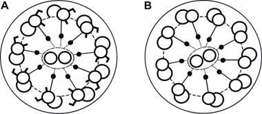
Seminiferous Tubules
Klinefelter Syndrome
Marsupialia
Sheep
Acetylcarnitine
Freeze Drying
Macropodidae
Calcimycin
Genitalia, Male
Ionophores
Micromanipulation
Aneuploidy
Peanut Agglutinin
Sea Urchins
DNA Fragmentation
Vas Deferens
Microscopy, Phase-Contrast
In Situ Hybridization, Fluorescence
Pregnancy Rate
Seminal Vesicles
Horses
Cell Separation
Specimen Handling
Prostatic Secretory Proteins
Epithelial Attachment
Contraceptive Agents, Male
Cell Survival
Microscopy, Fluorescence
Sertoli Cells
Glycerol
Calcium
Follicular Fluid
Insemination, Artificial, Homologous
Ornidazole
Fluorescent Dyes
Tromethamine
Hypotonic Solutions
Buffaloes
Pentoxifylline
Culture Media
Pregnancy Outcome
Tissue Preservation
Antelopes
Diploidy
Spermatocytes
Electrophoresis, Polyacrylamide Gel
Bisbenzimidazole
Cytoplasm
Staining and Labeling
Sex Chromosomes
Reproductive Techniques, Assisted
Suction
Identification of a nuclear localization signal in activin/inhibin betaA subunit; intranuclear betaA in rat spermatogenic cells. (1/9715)
Activin is a dimeric glycoprotein hormone that was initially characterized by its ability to stimulate pituitary FSH secretion and was subsequently recognized as a growth factor with diverse biological functions in a large variety of tissues. In the testis, activin has been implicated in the auto/paracrine regulation of spermatogenesis through its cognate cell membrane receptors on Sertoli and germ cells. In this study we provide evidence for intranuclear activin/inhibin betaA subunit and show its distribution in the rat seminiferous epithelium. We have shown by transient expression in HeLa cells of beta-galactosidase fusion proteins that the betaA subunit precursor contains a functional nuclear localization signal within the lysine-rich sequence corresponding to amino acids 231-244. In all stages of the rat seminiferous epithelial cycle, an intense immunohistochemical staining of nuclear betaA was demonstrated in intermediate or type B spermatogonia or primary spermatocytes in their initial stages of the first meiotic prophase, as well as in pachytene spermatocytes and elongating spermatids primarily in stages IX-XII. In some pachytene spermatocytes, the pattern of betaA immunoreactivity was consistent with the characteristic distribution of pachytene chromosomes. In the nuclei of round spermatids, betaA immunoreactivity was less intense, and in late spermatids it was localized in the residual cytoplasm, suggesting disposal of betaA before spermatozoal maturation. Immunoblot analysis of a protein extract from isolated testicular nuclei revealed a nuclear betaA species with a molecular mass of approximately 24 kDa, which is more than 1.5 times that of the mature activin betaA subunit present in activin dimers. These results suggest that activin/inhibin betaA may elicit its biological functions through two parallel signal transduction pathways, one involving the dimeric molecule and cell surface receptors and the other an alternately processed betaA sequence acting directly within the nucleus. According to our immunohistochemical data, betaA may play a significant role in the regulation of nuclear functions during meiosis and spermiogenesis. (+info)Scrotal heat stress induces altered sperm chromatin structure associated with a decrease in protamine disulfide bonding in the stallion. (2/9715)
A variety of testicular insults can induce changes in the structure of spermatozoal chromatin, resulting in spermatozoal DNA that is more susceptible to acid-induced denaturation. The degree of change in the DNA can be measured using the sperm chromatin structure assay (SCSA). The SCSA measures the relative amounts of single- and double-stranded DNA after staining with the metachromatic dye, acridine orange. Here we used a stallion model (n = 4) to study the effects of scrotal heat stress on spermatozoal DNA. This model was created by insulating stallion testes for 48 h and collecting sperm daily thereafter for 60 days. Changes in the SCSA were then correlated with protamine disulfide content and protamine types and levels. Results of the SCSA indicated that the susceptibility of spermatozoal DNA to denaturation was dependent on the spermatogenic cell stage that the ejaculated sperm was in at the time of the heat stress. Spermatozoa with altered DNA had a decrease in the extent of disulfide bonding that was associated with an increase in the susceptibility of DNA to denaturation. However, there were no detectable changes in either the protamine type or level. Thus, in this model, decreased disulfide bonding is associated with an increased susceptibility of spermatozoal DNA to denaturation in the absence of protamine changes. (+info)An intact sperm nuclear matrix may be necessary for the mouse paternal genome to participate in embryonic development. (3/9715)
We have been interested in determining the minimally required elements in the sperm head that are necessary in order for the paternal genome to participate in embryogenesis. We used an ionic detergent, mixed alkyltrimethylammonium bromide (ATAB), plus dithiothreitol (DTT) to remove the acrosome and almost all of the perinuclear theca, leaving only the sperm nucleus morphologically intact. We also tested the stability of the sperm nuclear matrix by the ability to form nuclear halos. Sperm nuclei washed in freshly prepared 0.5% ATAB + 2 mM DTT completely decondensed when extracted with salt, but nuclei washed in the same buffer that was 1 wk old, and then extracted with salt, produced nuclear halos, indicating stable nuclear matrices. When we treated sperm heads with freshly prepared ATAB+DTT and injected them into oocytes, none of the oocytes developed into live offspring. In contrast, sperm heads treated in the same way but with 1-wk-old ATAB+DTT solution could support development of about 30% of the oocytes to live offspring. Electron microscopy demonstrated that most of the perinuclear theca had been removed in both cases. These data suggest that at least in the mouse, the only component of the spermatozoa that is crucial for participation in embryologic development is the sperm nucleus with a stable nuclear matrix. (+info)A possible role for the pentose phosphate pathway of spermatozoa in gamete fusion in the mouse. (4/9715)
Glucose metabolism is essential for successful gamete fusion in the mouse. Although the metabolic activity of the oocyte does not appear to play a significant role in the fusion step, the metabolic role of the spermatozoon is not known. The aim of this study was therefore to characterize the role of glucose metabolism in mouse spermatozoa. Initially, the high-affinity glucose transporter GLUT3 was identified in mouse sperm. In characterizing the glucose metabolism of mouse sperm, we have shown 1) that mouse epididymal spermatozoa have a functional pentose phosphate pathway (PPP), implying that they produce NADPH, which is required for reducing reactions, and ribose 5-phosphate, which is required for nucleic acid synthesis; and 2) that sperm are able to fuse with the oocyte when NADPH is substituted for glucose, suggesting that sperm need to produce NADPH via the PPP in order to be able to achieve fertilization. The existence of an NADPH-regulated event that influences the ability of the sperm to fuse with the oocyte is envisaged. (+info)A novel trans-complementation assay suggests full mammalian oocyte activation is coordinately initiated by multiple, submembrane sperm components. (5/9715)
To initiate normal embryonic development, an egg must receive a signal to become activated at fertilization. We here report that the ability of demembranated sperm heads to activate is abolished after incubation over the range 20-44 degreesC and is sensitive to reducing agents. On the basis of this observation, we have developed a microinjection-based, trans-complementation assay in order to dissect the heat-inactivated sperm-borne oocyte-activating factor(s) (SOAF). We demonstrate that the failure of heat-inactivated sperm heads to activate an egg is rescued by coinjection with dithiothreitol-solubilized SOAF from demembranated sperm heads. The solubilized SOAF (SOAFs) is trypsin sensitive and is liberated from demembranated heads in a temperature-dependent manner that inversely correlates with the ability of sperm heads to activate. This argues that SOAFs is a proteinaceous molecular species required to initiate activation. Injection of oocytes with mouse or hamster sperm cytosolic factors, but not SOAFs alone, induced resumption of meiosis, further suggesting that these cytosolic factors and SOAF are distinct. Collectively, these data strongly suggest that full mammalian oocyte activation is initiated by the coordinated action of one or more heat-sensitive protein constituents of the perinuclear matrix and at least one heat-stable submembrane component. (+info)Sperm transport in the human female genital tract and its modulation by oxytocin as assessed by hysterosalpingoscintigraphy, hysterotonography, electrohysterography and Doppler sonography. (6/9715)
The transport function of the uterus and oviducts and its modulation by oxytocin has been examined using hysterosalpingoscintigraphy, recording of intrauterine pressure, electrohysterography and Doppler sonography of the Fallopian tubes. After application to the posterior vaginal fornix, a rapid (within minutes) uptake of the labelled particles into the uterus was observed during the follicular and during the luteal phase of the cycle in all patients. Transport into the oviducts, however, could only be demonstrated during the follicular phase. Transport was directed predominantly into the tube ipsilateral to the ovary bearing the dominant follicle; the contralateral oviduct appeared to be functionally closed. The proportion of patients exhibiting ipsilateral transport did increase concomitant with the increase of the diameter of the dominant follicle. That ipsilateral transport has biological significance is suggested by the observation that the pregnancy rate following spontaneous intercourse or insemination was significantly higher in those women in whom ipsilateral transport could be demonstrated than in those who failed to exhibit lateralization. Oxytocin administration was followed by a dramatic increase in the amount of material transported to the ipsilateral tube, as demonstrated by radionuclide imaging and by Doppler sonography following instillation of ultrasound contrast medium. Continuous recording of intrauterine pressure before and after oxytocin administration did show an increase in basal tonus and amplitude of contractions and a reversal of the pressure gradient from a fundo-cervical to a cervico-fundal direction. These actions of oxytocin were accompanied by an increase in amplitude of potentials recorded by electrohysterography. These data support the view that uterus and Fallopian tubes represent a functional unit that is acting as a peristaltic pump and that the increasing activity of this pump during the follicular phase of the menstrual cycle is reflected by an increased transport into the oviduct ipsilateral to the ovary bearing the dominant follicle. In addition, they strongly suggest a critical role of oxytocin in this process. Failure of this mechanism appears to be a cause of subfertility or infertility, as indicated by the low pregnancy rate following intrauterine insemination or normal intercourse in the presence of patent Fallopian tubes. It may be regarded as a new nosological entity for which we propose the term tubal transport disorder (TTD). Since pregnancy rate of such patients is normal when treated with in-vitro fertilization (IVF), hysterosalpingoscintigraphy seems to be useful for the choice of treatment modalities in patients with patent Fallopian tubes suffering from infertility. (+info)Mechanical stimulation of starfish sperm flagella. (7/9715)
1. The responses of starfish sperm flagella to mechanical stimulation with a microneedle were analysed. Flagellar movement was recorded by high-speed microcinematography and by stroboscopic observation. 2. The amplitude of the bending wave of a flagellum was restricted over its entire length when the microneedle was brought near to the flagellum at its proximal region. Beyond the restricted part, the amplitude of the wave, and the bend angle, became smaller than those of a normally beating flagellum, while the curvature was practically unchanged. 3. When the tip of the microneedle was in contact with the flagellum, propagation of the bending wave beyond the microneedle was inhibited. The part of the flagellum between the base and the microneedle continued beating in some cases and stopped beating in other cases. The flagellum beyond the arrested part stopped beating and remained straight. When the microneedle was removed, the bending wave which existed in the part of the flagellum proximal to the microneedle, or the wave which was passively formed de novo at the time of the removal of the microneedle, propagated over the arrested part towards the tip. 4. A flagellum amputated by a microneedle in a medium containing ATP continued beating with a small amplitude, small curvature, small bend angle and low frequency. When the amputated flagellum was passively bent by a microneedle at the region near the point of amputation, this bend propagated towards the tip with a constant bend angle. 5. The beating frequency of the flagellum could be modulated by the application of a rhythmic external force generated by vibrating a microneedle near the flagellum. The beating was completely synchronized with vibration of the microneedle in the frequency range from 23 Hz to 43 Hz. (+info)Incompetence of preovulatory mouse oocytes to undergo cortical granule exocytosis following induced calcium oscillations. (8/9715)
Immature oocytes of many species are incompetent to undergo cortical granule (CG) exocytosis upon fertilization. In mouse eggs, CG exocytosis is dependent primarily on an inositol 1,4,5-trisphosphate (IP3)-mediated elevation of intracellular calcium ([Ca2+]i). While deficiencies upstream of [Ca2+]i release are known, this study examined whether downstream deficiencies also contribute to the incompetence of preovulatory mouse oocytes to release CGs. The experimental strategy was to bypass upstream deficiencies by inducing normal, fertilization-like [Ca2+]i oscillations in fully grown, germinal vesicle (GV) stage oocytes and determine if the extent of CG exocytosis was restored to levels observed in mature, metaphase II (MII)-stage eggs. Because IP3 does not stimulate a normal Ca2+ response in GV-stage oocytes, three alternate methods were used to induce oscillations: thimerosal treatment, electroporation, and sperm factor injection. Long-lasting oscillations from thimerosal treatment resulted in 64 and 10% mean CG release at the MII and GV stages, respectively (P < 0.001). Three electrical pulses induced mean [Ca2+]i elevations of approximately 730 and 650 nM in MII- and GV-stage oocytes, respectively, and 31% CG release in MII-stage eggs and 9% in GV-stage oocytes (P < 0.001). Sperm factor microinjection resulted in 86% CG release in MII-stage eggs, while similarly treated GV-stage oocytes exhibited < 1% CG release (P < 0.001). Taken together, these results demonstrate a deficiency downstream of [Ca2+]i release which is developmentally regulated in the 12 h prior to ovulation. (+info)Spermatozoa are the male reproductive cells, or gametes, that are produced in the testes. They are microscopic, flagellated (tail-equipped) cells that are highly specialized for fertilization. A spermatozoon consists of a head, neck, and tail. The head contains the genetic material within the nucleus, covered by a cap-like structure called the acrosome which contains enzymes to help the sperm penetrate the female's egg (ovum). The long, thin tail propels the sperm forward through fluid, such as semen, enabling its journey towards the egg for fertilization.
Sperm motility is the ability of sperm to move actively and effectively through the female reproductive tract towards the egg for fertilization. It is typically measured as the percentage of moving sperm in a sample, and their progressiveness or velocity. Normal human sperm motility is generally defined as forward progression of at least 25 micrometers per second, with at least 50% of sperm showing progressive motility. Reduced sperm motility, also known as asthenozoospermia, can negatively impact fertility and reproductive outcomes.
Semen preservation is the process of collecting, liquefying, testing, and storing semen samples for future use in assisted reproductive technologies (ART) such as artificial insemination (AI), in vitro fertilization (IVF), or intracytoplasmic sperm injection (ICSI). The semen sample is usually collected through masturbation, and then it is mixed with a cryoprotectant solution to prevent damage during the freezing and thawing process. After that, the sample is divided into straws or vials and frozen in liquid nitrogen tanks at temperatures below -196°C. Properly preserved semen can be stored for many years without significant loss of quality or fertility potential. Semen preservation is often recommended for men who are about to undergo medical treatments that may affect their sperm production or fertility, such as chemotherapy or radiation therapy, or for those who wish to postpone fatherhood for personal or medical reasons.
The acrosome is a specialized structure located on the anterior part of the sperm head in many species of animals, including humans. It contains enzymes that help the sperm penetrate the outer covering of the egg (zona pellucida) during fertilization. The acrosome reaction is the process by which the acrosome releases its enzymes, allowing the sperm to digest a path through the zona pellucida and reach the egg plasma membrane for fusion and fertilization.
The acrosome is formed during spermatogenesis, the process of sperm production in the testis, from the Golgi apparatus, a cellular organelle involved in protein trafficking and modification. The acrosome contains hydrolytic enzymes such as hyaluronidase, acrosin, and proteases that are activated during the acrosome reaction to facilitate sperm-egg fusion.
Abnormalities in acrosome formation or function can lead to infertility in males.
The epididymis is a tightly coiled tube located on the upper and posterior portion of the testicle that serves as the site for sperm maturation and storage. It is an essential component of the male reproductive system. The epididymis can be divided into three parts: the head (where newly produced sperm enter from the testicle), the body, and the tail (where mature sperm exit and are stored). Any abnormalities or inflammation in the epididymis may lead to discomfort, pain, or infertility.
A sperm head is the anterior (front) part of a spermatozoon, which contains the genetic material (DNA). It is covered by a protein layer called the acrosome, which plays a crucial role in fertilization. The sperm head is followed by the midpiece and the tail, which provide mobility to the sperm for its journey towards the egg.
Sperm transport refers to the series of events that occur from the production of sperm in the testes to their release into the female reproductive tract during sexual intercourse. This process involves several stages:
1. Spermatogenesis: The production of sperm cells (spermatozoa) takes place in the seminiferous tubules within the testes.
2. Maturation: The newly produced sperm are immature and incapable of fertilization. They undergo a maturation process as they move through the epididymis, where they acquire motility and the ability to fertilize an egg.
3. Ejaculation: During sexual arousal, sperm are mixed with seminal fluid produced by the seminal vesicles, prostate gland, and bulbourethral glands to form semen. This mixture is propelled through the urethra during orgasm (ejaculation) and released from the penis into the female reproductive tract.
4. Transport within the female reproductive tract: Once inside the female reproductive tract, sperm must travel through the cervix, uterus, and fallopian tubes to reach the site of fertilization, the ampullary-isthmic junction of the fallopian tube. This journey can take several hours to a few days.
5. Capacitation: During their transport within the female reproductive tract, sperm undergo further changes called capacitation, which prepares them for fertilization by increasing their motility and making them more responsive to the egg's chemical signals.
6. Acrosome reaction: The final step in sperm transport is the acrosome reaction, where the sperm releases enzymes from the acrosome (a cap-like structure on the head of the sperm) to penetrate and fertilize the egg.
Sperm maturation is the process by which spermatids, immature sperm cells produced in meiosis, transform into fully developed spermatozoa capable of fertilization. This complex process occurs in the seminiferous tubules of the testes and includes several stages:
1. **Golfi formation:** The first step involves the spermatids reorganizing their cytoplasm and forming a cap-like structure called the acrosome, which contains enzymes that help the sperm penetrate the egg's outer layers during fertilization.
2. **Flagellum development:** The spermatid also develops a tail (flagellum), enabling it to move independently. This is achieved through the assembly of microtubules and other associated proteins.
3. **Nuclear condensation and elongation:** The sperm's DNA undergoes significant compaction, making the nucleus smaller and more compact. Concurrently, the nucleus elongates and aligns with the flagellum.
4. **Mitochondrial positioning:** Mitochondria, which provide energy for sperm motility, migrate to the midpiece of the sperm, close to the base of the flagellum.
5. **Chromatin packaging:** Histones, proteins that help package DNA in non-sperm cells, are replaced by transition proteins and then protamines, which further compact and protect the sperm's DNA.
6. **Sperm release (spermiation):** The mature sperm is finally released from the supporting Sertoli cells into the lumen of the seminiferous tubule, where it mixes with fluid secreted by the testicular tissue to form seminal plasma.
This entire process takes approximately 64 days in humans.
The acrosome reaction is a crucial event in the fertilization process of many species, including humans. It occurs when the sperm makes contact with and binds to the zona pellucida, the glycoprotein-rich extracellular matrix that surrounds the egg. This interaction triggers a series of molecular events leading to the exocytosis of the acrosome, a membrane-bound organelle located at the tip of the sperm head.
The acrosome contains hydrolytic enzymes that help the sperm to penetrate the zona pellucida and reach the egg's plasma membrane. During the acrosome reaction, the outer acrosomal membrane fuses with the sperm plasma membrane, releasing these enzymes and causing the release of the inner acrosomal membrane, which then reorganizes to form a structure called the acrosomal cap.
The acrosome reaction exposes new proteins on the sperm surface that can interact with the egg's plasma membrane, allowing for the fusion of the two membranes and the entry of the sperm into the egg. This event is essential for successful fertilization and subsequent embryonic development.
The "sperm tail" is also known as the flagellum, which is a whip-like structure that enables the sperm to move or swim through fluid. The human sperm tail is made up of nine microtubule doublets and a central pair of microtubules, which are surrounded by a mitochondrial sheath that provides energy for its movement. This complex structure allows the sperm to navigate through the female reproductive tract in order to reach and fertilize an egg.
Semen is a complex, whitish fluid that is released from the male reproductive system during ejaculation. It is produced by several glands, including the seminal vesicles, prostate gland, and bulbourethral glands. Semen contains several components, including sperm (the male reproductive cells), as well as various proteins, enzymes, vitamins, and minerals. Its primary function is to transport sperm through the female reproductive tract during sexual intercourse, providing nutrients and aiding in the protection of the sperm as they travel toward the egg for fertilization.
Ejaculation is the discharge of semen, typically accompanied by orgasm, during sexual activity. It occurs when the male reproductive system releases semen from the penis. This process is usually brought on by sexual arousal and stimulation, which cause the sperm-carrying vas deferens to contract and push the semen into the urethra, from where it is expelled through the tip of the penis.
There are two types of ejaculation:
1. **Reflex ejaculation**: This occurs when there is a high level of sexual excitement or stimulation, leading to an involuntary and automatic response.
2. **Premature ejaculation**: This refers to the condition where ejaculation happens too quickly, often before or shortly after penetration, causing distress and affecting sexual satisfaction for both partners.
It is essential to understand that a healthy male can experience variations in the timing of ejaculation throughout their life, influenced by factors such as age, stress levels, and overall health. If you have concerns about your ejaculation patterns or any related issues, it is recommended to consult a healthcare professional for advice and treatment options.
Cryopreservation is a medical procedure that involves the preservation of cells, tissues, or organs by cooling them to very low temperatures, typically below -150°C. This is usually achieved using liquid nitrogen. The low temperature slows down or stops biological activity, including chemical reactions and cellular metabolism, which helps to prevent damage and decay.
The cells, tissues, or organs that are being cryopreserved must be treated with a cryoprotectant solution before cooling to prevent the formation of ice crystals, which can cause significant damage. Once cooled, the samples are stored in specialized containers or tanks until they are needed for use.
Cryopreservation is commonly used in assisted reproductive technologies, such as the preservation of sperm, eggs, and embryos for fertility treatments. It is also used in research, including the storage of cell lines and stem cells, and in clinical settings, such as the preservation of skin grafts and corneas for transplantation.
Male infertility is a condition characterized by the inability to cause pregnancy in a fertile female. It is typically defined as the failure to achieve a pregnancy after 12 months or more of regular unprotected sexual intercourse.
The causes of male infertility can be varied and include issues with sperm production, such as low sperm count or poor sperm quality, problems with sperm delivery, such as obstructions in the reproductive tract, or hormonal imbalances that affect sperm production. Other factors that may contribute to male infertility include genetic disorders, environmental exposures, lifestyle choices, and certain medical conditions or treatments.
It is important to note that male infertility can often be treated or managed with medical interventions, such as medication, surgery, or assisted reproductive technologies (ART). A healthcare provider can help diagnose the underlying cause of male infertility and recommend appropriate treatment options.
Sperm count, also known as sperm concentration, is the number of sperm present in a given volume of semen. The World Health Organization (WHO) previously defined a normal sperm count as at least 20 million sperm per milliliter of semen. However, more recent studies suggest that fertility may be affected even when sperm counts are slightly lower than this threshold. It's important to note that sperm count is just one factor among many that can influence male fertility. Other factors, such as sperm motility (the ability of sperm to move properly) and morphology (the shape of the sperm), also play crucial roles in successful conception.
Fertilization is the process by which a sperm cell (spermatozoon) penetrates and fuses with an egg cell (ovum), resulting in the formation of a zygote. This fusion of genetic material from both the male and female gametes initiates the development of a new organism. In human biology, fertilization typically occurs in the fallopian tube after sexual intercourse, when a single sperm out of millions is able to reach and penetrate the egg released from the ovary during ovulation. The successful fusion of these two gametes marks the beginning of pregnancy.
The testis, also known as the testicle, is a male reproductive organ that is part of the endocrine system. It is located in the scrotum, outside of the abdominal cavity. The main function of the testis is to produce sperm and testosterone, the primary male sex hormone.
The testis is composed of many tiny tubules called seminiferous tubules, where sperm are produced. These tubules are surrounded by a network of blood vessels, nerves, and supportive tissues. The sperm then travel through a series of ducts to the epididymis, where they mature and become capable of fertilization.
Testosterone is produced in the Leydig cells, which are located in the interstitial tissue between the seminiferous tubules. Testosterone plays a crucial role in the development and maintenance of male secondary sexual characteristics, such as facial hair, deep voice, and muscle mass. It also supports sperm production and sexual function.
Abnormalities in testicular function can lead to infertility, hormonal imbalances, and other health problems. Regular self-examinations and medical check-ups are recommended for early detection and treatment of any potential issues.
Zona pellucida is a term used in the field of reproductive biology and it refers to the glycoprotein membrane that surrounds mammalian oocytes (immature egg cells). This membrane plays a crucial role in the fertilization process. It has receptors for sperm, and upon binding with the sperm, it undergoes changes that prevent other sperm from entering, a process known as the zona reaction. This membrane is also involved in the early development of the embryo.
Oligospermia is a medical term used to describe a condition in which the semen contains a lower than normal number of sperm. Generally, a sperm count of less than 15 million sperm per milliliter (ml) of semen is considered to be below the normal range.
Oligospermia can make it more difficult for a couple to conceive naturally and may require medical intervention such as intracytoplasmic sperm injection (ICSI) or in vitro fertilization (IVF). The condition can result from various factors, including hormonal imbalances, genetic abnormalities, varicocele, environmental factors, and certain medications.
It's important to note that oligospermia is not the same as azoospermia, which is a condition where there is no sperm present in the semen at all.
Sperm agglutination is the clumping or sticking together of sperm cells, which can be caused by the presence of antibodies or other substances in semen. In some cases, sperm agglutination may occur due to an immune response in which the body produces antibodies that attack and bind to sperm cells, leading to their clumping together. This can interfere with the sperm's ability to move and fertilize an egg.
Sperm agglutination can be detected through a semen analysis test, which involves examining a sample of semen under a microscope. If sperm agglutination is present, it may indicate an underlying medical condition or issue that requires further evaluation and treatment. In some cases, sperm agglutination may be treated with medications to reduce the production of antibodies or other substances that are causing the problem.
Fertilization in vitro, also known as in-vitro fertilization (IVF), is a medical procedure where an egg (oocyte) and sperm are combined in a laboratory dish to facilitate fertilization. The fertilized egg (embryo) is then transferred to a uterus with the hope of establishing a successful pregnancy. This procedure is often used when other assisted reproductive technologies have been unsuccessful or are not applicable, such as in cases of blocked fallopian tubes, severe male factor infertility, and unexplained infertility. The process involves ovarian stimulation, egg retrieval, fertilization, embryo culture, and embryo transfer. In some cases, additional techniques such as intracytoplasmic sperm injection (ICSI) or preimplantation genetic testing (PGT) may be used to increase the chances of success.
Artificial insemination (AI) is a medical procedure that involves the introduction of sperm into a female's cervix or uterus for the purpose of achieving pregnancy. This procedure can be performed using sperm from a partner or a donor. It is often used when there are issues with male fertility, such as low sperm count or poor sperm motility, or in cases where natural conception is not possible due to various medical reasons.
There are two types of artificial insemination: intracervical insemination (ICI) and intrauterine insemination (IUI). ICI involves placing the sperm directly into the cervix, while IUI involves placing the sperm directly into the uterus using a catheter. The choice of procedure depends on various factors, including the cause of infertility and the preferences of the individuals involved.
Artificial insemination is a relatively simple and low-risk procedure that can be performed in a doctor's office or clinic. It may be combined with fertility drugs to increase the chances of pregnancy. The success rate of artificial insemination varies depending on several factors, including the age and fertility of the individuals involved, the cause of infertility, and the type of procedure used.
Spermatogenesis is the process by which sperm cells, or spermatozoa, are produced in male organisms. It occurs in the seminiferous tubules of the testes and involves several stages:
1. Spermatocytogenesis: This is the initial stage where diploid spermatogonial stem cells divide mitotically to produce more spermatogonia, some of which will differentiate into primary spermatocytes.
2. Meiosis: The primary spermatocytes undergo meiotic division to form haploid secondary spermatocytes, which then divide again to form haploid spermatids. This process results in the reduction of chromosome number from 46 (diploid) to 23 (haploid).
3. Spermiogenesis: The spermatids differentiate into spermatozoa, undergoing morphological changes such as the formation of a head and tail. During this stage, most of the cytoplasm is discarded, resulting in highly compacted and streamlined sperm cells.
4. Spermation: The final stage where mature sperm are released from the seminiferous tubules into the epididymis for further maturation and storage.
The entire process takes approximately 72-74 days in humans, with continuous production throughout adulthood.
Semen analysis is a laboratory test that evaluates various characteristics of semen, the fluid that is released during ejaculation. These characteristics include:
1. Volume: The amount of semen produced in one ejaculation.
2. Liquefaction time: The time it takes for the semen to change from a gel-like consistency to a liquid state.
3. pH: The acidity or alkalinity of the semen.
4. Sperm concentration: The number of sperm present in each milliliter of semen.
5. Total sperm count: The total number of sperm in the entire ejaculate.
6. Motility: The percentage of sperm that are moving and their forward progression.
7. Morphology: The shape and size of the sperm.
8. Vitality: The percentage of live sperm in the sample.
9. White blood cell count: The presence of white blood cells, which can indicate an infection.
Semen analysis is often used to help diagnose male infertility, as well as to monitor the effectiveness of treatments for infertility. It may also be used to detect abnormalities in the reproductive system or to evaluate the effects of certain medications on sperm production and quality.
Cryoprotective agents are substances that are used to protect biological material from damage during freezing and thawing. These agents work by reducing the amount of ice that forms in the cells, which can help to prevent the formation of damaging ice crystals. Commonly used cryoprotective agents include dimethyl sulfoxide (DMSO), glycerol, and ethylene glycol.
When biological material, such as cells or tissues, is cooled to very low temperatures for storage or transportation, the water in the cells can freeze and form ice crystals. These ice crystals can damage the cell membranes and other structures within the cell, leading to cell death. Cryoprotective agents help to prevent this by lowering the freezing point of the solution that the cells are stored in, which reduces the amount of ice that forms.
Cryoprotective agents are often used in the field of assisted reproductive technology (ART) to protect sperm, eggs, and embryos during freezing and thawing. They are also used in research settings to preserve cells and tissues for later use. It is important to note that while cryoprotective agents can help to reduce the amount of damage that occurs during freezing and thawing, they cannot completely prevent it. Therefore, it is important to carefully control the freezing and thawing process to minimize any potential harm to the biological material.
Acrosin is a proteolytic enzyme that is found in the acrosome, which is a cap-like structure located on the anterior part of the sperm head. This enzyme plays an essential role in the fertilization process by helping the sperm to penetrate the zona pellucida, which is the glycoprotein coat surrounding the egg.
Acrosin is released from the acrosome when the sperm encounters the zona pellucida, and it begins to digest the glycoproteins in the zona pellucida, creating a path for the sperm to reach and fuse with the egg's plasma membrane. This enzyme is synthesized and stored in the acrosome during spermatogenesis and is activated during the acrosome reaction, which is a critical event in fertilization.
Defects in acrosin function or regulation have been implicated in male infertility, making it an important area of research in reproductive biology.
Spermatids are immature sperm cells that are produced during the process of spermatogenesis in the male testes. They are the product of the final stage of meiosis, where a diploid spermatocyte divides into four haploid spermatids. Each spermatid then undergoes a series of changes, including the development of a tail for motility and the condensation of its nucleus to form a head containing the genetic material. Once this process is complete, the spermatids are considered mature spermatozoa and are capable of fertilizing an egg.
Fertility is the natural ability to conceive or to cause conception of offspring. In humans, it is the capacity of a woman and a man to reproduce through sexual reproduction. For women, fertility usually takes place during their reproductive years, which is from adolescence until menopause. A woman's fertility depends on various factors including her age, overall health, and the health of her reproductive system.
For men, fertility can be affected by a variety of factors such as age, genetics, general health, sexual function, and environmental factors that may affect sperm production or quality. Factors that can negatively impact male fertility include exposure to certain chemicals, radiation, smoking, alcohol consumption, drug use, and sexually transmitted infections (STIs).
Infertility is a common medical condition affecting about 10-15% of couples trying to conceive. Infertility can be primary or secondary. Primary infertility refers to the inability to conceive after one year of unprotected sexual intercourse, while secondary infertility refers to the inability to conceive following a previous pregnancy.
Infertility can be treated with various medical and surgical interventions depending on the underlying cause. These may include medications to stimulate ovulation, intrauterine insemination (IUI), in vitro fertilization (IVF), or surgery to correct anatomical abnormalities.
Chlorohydrins are a class of chemical compounds that contain both chlorine and hydroxyl (-OH) groups. They are typically formed by the reaction of an aldehyde or ketone with a hypochlorous acid or chlorine in a process called halogenation. Chlorohydrins can be toxic and have been associated with various health effects, including irritation of the eyes, skin, and respiratory tract, and potential damage to the liver and kidneys. They are used in some industrial applications, such as the production of certain chemicals and pharmaceuticals, but their use is subject to regulations due to their potential hazards.
Intracytoplasmic Sperm Injection (ICSI) is a specialized form of assisted reproductive technology (ART), specifically used in the context of in vitro fertilization (IVF). It involves the direct injection of a single sperm into the cytoplasm of a mature egg (oocyte) to facilitate fertilization. This technique is often used when there are issues with male infertility, such as low sperm count or poor sperm motility, to increase the chances of successful fertilization. The resulting embryos can then be transferred to the uterus in hopes of achieving a pregnancy.
Alpha-chlorohydrin is not typically referred to as a medical term, but it is a chemical compound with the formula HOCH2CHClNH2. It is primarily used in the production of other chemicals and has been used as a reagent in laboratory settings.
Ingestion or exposure to alpha-chlorohydrin can be harmful and may cause symptoms such as nausea, vomiting, abdominal pain, diarrhea, dizziness, and difficulty breathing. It is classified as a possible human carcinogen by the International Agency for Research on Cancer (IARC).
Medical professionals may encounter alpha-chlorohydrin in cases of accidental or intentional ingestion or exposure, or in the context of occupational health and safety for workers who may be exposed to it in industrial settings.
The Fallopian tubes, also known as uterine tubes or oviducts, are a pair of slender tubular structures in the female reproductive system. They play a crucial role in human reproduction by providing a passageway for the egg (ovum) from the ovary to the uterus (womb).
Each Fallopian tube is typically around 7.6 to 10 centimeters long and consists of four parts: the interstitial part, the isthmus, the ampulla, and the infundibulum. The fimbriated end of the infundibulum, which resembles a fringe or frill, surrounds and captures the released egg from the ovary during ovulation.
Fertilization usually occurs in the ampulla when sperm meets the egg after sexual intercourse. Once fertilized, the zygote (fertilized egg) travels through the Fallopian tube toward the uterus for implantation and further development. The cilia lining the inner surface of the Fallopian tubes help propel the egg and the zygote along their journey.
In some cases, abnormalities or blockages in the Fallopian tubes can lead to infertility or ectopic pregnancies, which are pregnancies that develop outside the uterus, typically within the Fallopian tube itself.
Asthenozoospermia is a term used in the field of andrology, which is the study of male reproductive health. It refers to a condition where the majority of sperm in a semen sample have reduced motility, meaning they do not move normally or efficiently. This can make it more difficult for the sperm to reach and fertilize an egg, potentially leading to infertility issues.
To be more specific, asthenozoospermia is defined as having less than 40% of sperm with progressive motility, which means they move forward in a straight line or in a large circle. The condition can be caused by various factors, including genetic abnormalities, environmental toxins, infections, and structural issues with the sperm themselves.
It's worth noting that asthenozoospermia is often diagnosed through a semen analysis, which is a routine test used to assess male fertility. If you or someone you know has been diagnosed with this condition, it may be helpful to consult with a reproductive endocrinologist or andrologist who can provide more information and guidance on potential treatment options.
Chlortetracycline is an antibiotic that belongs to the tetracycline class. It is primarily used to treat a variety of bacterial infections, including respiratory, urinary, and skin infections. Chlortetracycline works by inhibiting the bacteria's ability to produce proteins, which are essential for their survival and growth.
The medical definition of Chlortetracycline is as follows:
Chlortetracycline (CTC): A broad-spectrum antibiotic that is derived from the actinomycete Streptomyces aureofaciens. It is used to treat various bacterial infections, including respiratory, urinary, and skin infections. Chlortetracycline is a colorless crystalline powder that is soluble in water and alcohol. It has a molecular formula of C22H24ClN2O8 and a molecular weight of 476.93 g/mol.
Chlortetracycline is usually administered orally, but it can also be given intravenously or topically. The drug is absorbed well from the gastrointestinal tract and is widely distributed throughout the body. It has a half-life of about 8 hours and is excreted primarily in the urine.
Like other tetracyclines, Chlortetracycline can cause tooth discoloration and enamel hypoplasia in children under the age of 8. It can also cause photosensitivity, nausea, vomiting, and diarrhea. Prolonged use or high doses of Chlortetracycline can lead to bacterial resistance and may increase the risk of superinfection with fungi or other bacteria.
Chlortetracycline is no longer commonly used in human medicine due to the availability of newer antibiotics with fewer side effects. However, it is still used in veterinary medicine to treat infections in animals.
"Sex preselection," also known as "gender selection" or "family balancing," is the process of influencing the sex of an offspring before birth. It can be achieved through various methods, including preimplantation genetic diagnosis (PGD) in conjunction with in vitro fertilization (IVF), sperm sorting techniques, and embryo manipulation.
PGD is a technique where one or more cells are taken from an embryo created through IVF and tested for genetic disorders or chromosomal abnormalities. During this process, the sex of the embryo can also be determined. Only embryos of the desired sex are then transferred to the uterus for implantation.
Sperm sorting techniques involve separating X-chromosome-bearing sperm (which produce female offspring) from Y-chromosome-bearing sperm (which produce male offspring). The sorted sperm can then be used for artificial insemination or IVF.
It's important to note that sex preselection is a controversial topic due to ethical considerations and legal restrictions in some countries.
Chromomycin A3 is an antibiotic and a DNA-binding molecule that is used in research and scientific studies. It is a type of glycosylated anthracycline that can intercalate into DNA and inhibit DNA-dependent RNA synthesis. Chromomycin A3 has been used as a fluorescent stain for microscopy, particularly for the staining of chromosomes during mitosis. It is also used in molecular biology research to study the interactions between drugs and DNA.
It's important to note that Chromomycin A3 is not used as a therapeutic drug in human or veterinary medicine due to its toxicity, it's mainly used for research purposes.
Seminal plasma proteins are a group of proteins that are present in the seminal fluid, which is the liquid component of semen. These proteins originate primarily from the accessory sex glands, including the prostate, seminal vesicles, and bulbourethral glands, and play various roles in the maintenance of sperm function and fertility.
Some of the key functions of seminal plasma proteins include:
1. Nutrition: Seminal plasma proteins provide energy sources and essential nutrients to support sperm survival and motility during their journey through the female reproductive tract.
2. Protection: These proteins help protect sperm from oxidative stress, immune attack, and other environmental factors that could negatively impact sperm function or viability.
3. Lubrication: Seminal plasma proteins contribute to the formation of a fluid medium that facilitates the ejaculation and transport of sperm through the female reproductive tract.
4. Coagulation and liquefaction: Some seminal plasma proteins are involved in the initial coagulation and subsequent liquefaction of semen, which helps ensure proper sperm release and distribution during ejaculation.
5. Interaction with female reproductive system: Seminal plasma proteins can interact with components of the female reproductive tract to modulate immune responses, promote implantation, and support early embryonic development.
Examples of seminal plasma proteins include prostate-specific antigen (PSA), prostate-specific acid phosphatase (PSAP), and semenogelins. Abnormal levels or dysfunctions in these proteins have been associated with various reproductive disorders, such as infertility, prostatitis, and prostate cancer.
An oocyte, also known as an egg cell or female gamete, is a large specialized cell found in the ovary of female organisms. It contains half the number of chromosomes as a normal diploid cell, as it is the product of meiotic division. Oocytes are surrounded by follicle cells and are responsible for the production of female offspring upon fertilization with sperm. The term "oocyte" specifically refers to the immature egg cell before it reaches full maturity and is ready for fertilization, at which point it is referred to as an ovum or egg.
Povidone, also known as PVP or polyvinylpyrrolidone, is not a medication itself but rather a pharmaceutical ingredient used in various medical and healthcare products. It is a water-soluble synthetic polymer that has the ability to bind to and carry other substances, such as drugs or iodine.
In medical applications, povidone is often used as a binder or coating agent in pharmaceutical tablets and capsules. It can also be found in some topical antiseptic solutions, such as those containing iodine, where it helps to stabilize and control the release of the active ingredient.
It's important to note that while povidone is a widely used pharmaceutical ingredient, it is not typically considered a medication on its own.
Sperm retrieval is a medical procedure that involves obtaining sperm from a male patient, usually for the purpose of assisted reproduction. This can be indicated in cases where the man has obstructive or non-obstructive azoospermia (absence of sperm in the semen), ejaculatory dysfunction, or other conditions that prevent the successful collection of sperm through conventional means, such as masturbation.
There are several methods for sperm retrieval, including:
1. Testicular sperm aspiration (TESA): A procedure where a fine needle is inserted into the testicle to aspirate (or draw out) sperm.
2. Percutaneous epididymal sperm aspiration (PESA): Similar to TESA, but the needle is inserted into the epididymis, a small structure that stores and transports sperm from the testicle.
3. Microsurgical epididymal sperm aspiration (MESA): A more invasive procedure where an incision is made in the scrotum to directly visualize the epididymis with a surgical microscope, allowing for the careful removal of sperm.
4. Testicular sperm extraction (TESE): Involves making a small incision in the testicle and removing a piece of tissue containing sperm-producing tubules. The tissue is then processed to extract viable sperm.
5. Microdissection testicular sperm extraction (microTESE): A refined version of TESE, where a surgical microscope is used to identify and isolate individual seminiferous tubules containing sperm in men with non-obstructive azoospermia.
The retrieved sperm can then be used for various assisted reproductive techniques, such as intracytoplasmic sperm injection (ICSI), where a single sperm is injected directly into an egg to facilitate fertilization.
"Swine" is a common term used to refer to even-toed ungulates of the family Suidae, including domestic pigs and wild boars. However, in a medical context, "swine" often appears in the phrase "swine flu," which is a strain of influenza virus that typically infects pigs but can also cause illness in humans. The 2009 H1N1 pandemic was caused by a new strain of swine-origin influenza A virus, which was commonly referred to as "swine flu." It's important to note that this virus is not transmitted through eating cooked pork products; it spreads from person to person, mainly through respiratory droplets produced when an infected person coughs or sneezes.
Microinjection is a medical technique that involves the use of a fine, precise needle to inject small amounts of liquid or chemicals into microscopic structures, cells, or tissues. This procedure is often used in research settings to introduce specific substances into individual cells for study purposes, such as introducing DNA or RNA into cell nuclei to manipulate gene expression.
In clinical settings, microinjections may be used in various medical and cosmetic procedures, including:
1. Intracytoplasmic Sperm Injection (ICSI): A type of assisted reproductive technology where a single sperm is injected directly into an egg to increase the chances of fertilization during in vitro fertilization (IVF) treatments.
2. Botulinum Toxin Injections: Microinjections of botulinum toxin (Botox, Dysport, or Xeomin) are used for cosmetic purposes to reduce wrinkles and fine lines by temporarily paralyzing the muscles responsible for their formation. They can also be used medically to treat various neuromuscular disorders, such as migraines, muscle spasticity, and excessive sweating (hyperhidrosis).
3. Drug Delivery: Microinjections may be used to deliver drugs directly into specific tissues or organs, bypassing the systemic circulation and potentially reducing side effects. This technique can be particularly useful in treating localized pain, delivering growth factors for tissue regeneration, or administering chemotherapy agents directly into tumors.
4. Gene Therapy: Microinjections of genetic material (DNA or RNA) can be used to introduce therapeutic genes into cells to treat various genetic disorders or diseases, such as cystic fibrosis, hemophilia, or cancer.
Overall, microinjection is a highly specialized and precise technique that allows for the targeted delivery of substances into small structures, cells, or tissues, with potential applications in research, medical diagnostics, and therapeutic interventions.
"Cattle" is a term used in the agricultural and veterinary fields to refer to domesticated animals of the genus *Bos*, primarily *Bos taurus* (European cattle) and *Bos indicus* (Zebu). These animals are often raised for meat, milk, leather, and labor. They are also known as bovines or cows (for females), bulls (intact males), and steers/bullocks (castrated males). However, in a strict medical definition, "cattle" does not apply to humans or other animals.
Protamines are small, arginine-rich proteins that are found in the sperm cells of many organisms. They play a crucial role in the process of sperm maturation, also known as spermiogenesis. During this process, the DNA in the sperm cell is tightly packed and compacted by the protamines, which helps to protect the genetic material during its journey to fertilize an egg.
Protamines are typically composed of around 50-100 amino acids and have a high proportion of positively charged arginine residues, which allow them to interact strongly with the negatively charged DNA molecule. This interaction results in the formation of highly condensed chromatin structures that are resistant to enzymatic digestion and other forms of damage.
In addition to their role in sperm maturation, protamines have also been studied for their potential use in drug delivery and gene therapy applications. Their ability to bind strongly to DNA makes them attractive candidates for delivering drugs or genetic material directly to the nucleus of a cell. However, more research is needed to fully understand the potential benefits and risks associated with these applications.
A vasectomy is a surgical procedure for male sterilization or permanent contraception. It involves cutting and sealing the vas deferens, the tubes that carry sperm from the testicles to the prostate gland, to prevent the release of sperm during ejaculation. This procedure is typically performed in an outpatient setting, using local anesthesia, and takes about 20-30 minutes. It is considered a highly effective form of birth control with a low risk of complications. However, it does not protect against sexually transmitted infections (STIs), so additional protection such as condoms may still be necessary.
The Y chromosome is one of the two sex-determining chromosomes in humans and many other animals, along with the X chromosome. The Y chromosome contains the genetic information that helps to determine an individual's sex as male. It is significantly smaller than the X chromosome and contains fewer genes.
The Y chromosome is present in males, who inherit it from their father. Females, on the other hand, have two X chromosomes, one inherited from each parent. The Y chromosome includes a gene called SRY (sex-determining region Y), which initiates the development of male sexual characteristics during embryonic development.
It is worth noting that the Y chromosome has a relatively high rate of genetic mutation and degeneration compared to other chromosomes, leading to concerns about its long-term viability in human evolution. However, current evidence suggests that the Y chromosome has been stable for at least the past 25 million years.
Insemination, in a medical context, refers to the introduction of semen into the reproductive system of a female for the purpose of achieving pregnancy. This can be done through various methods including intracervical insemination (ICI), intrauterine insemination (IUI), and in vitro fertilization (IVF).
Intracervical insemination involves placing the semen at the cervix, the opening to the uterus. Intrauterine insemination involves placing the sperm directly into the uterus using a catheter. In vitro fertilization is a more complex process where the egg and sperm are combined in a laboratory dish and then transferred to the uterus.
Insemination is often used in cases of infertility, either because of male or female factors, or unexplained infertility. It can also be used for those who wish to become pregnant but do not have a partner, such as single women and same-sex female couples.
"Freezing" is a term used in the medical field to describe a phenomenon that can occur in certain neurological conditions, most notably in Parkinson's disease. It refers to a sudden and temporary inability to move or initiate movement, often triggered by environmental factors such as narrow spaces, turning, or approaching a destination. This can increase the risk of falls and make daily activities challenging for affected individuals.
Freezing is also known as "freezing of gait" (FOG) when it specifically affects a person's ability to walk. During FOG episodes, the person may feel like their feet are glued to the ground, making it difficult to take steps forward. This can be very distressing and debilitating for those affected.
It is important to note that "freezing" has different meanings in different medical contexts, such as in the field of orthopedics, where it may refer to a loss of joint motion due to stiffness or inflammation. Always consult with a healthcare professional for accurate information tailored to your specific situation.
The cervix is the lower, narrow part of the uterus that opens into the vagina. Cervical mucus is a clear or cloudy secretion produced by glands in the cervix. The amount and consistency of cervical mucus changes throughout a woman's menstrual cycle, influenced by hormonal fluctuations.
During the fertile window (approximately mid-cycle), estrogen levels rise, causing the cervical mucus to become more abundant, clear, and stretchy (often described as resembling raw egg whites). This "fertile" mucus facilitates the movement of sperm through the cervix and into the uterus, increasing the chances of fertilization.
As the menstrual cycle progresses and progesterone levels rise after ovulation, cervical mucus becomes thicker, cloudier, and less abundant, making it more difficult for sperm to penetrate. This change in cervical mucus helps prevent additional sperm from entering and fertilizing an already-fertilized egg.
Changes in cervical mucus can be used as a method of natural family planning or fertility awareness, with women checking their cervical mucus daily to identify their most fertile days. However, this method should be combined with other tracking methods for increased accuracy and reliability.
"Mesocricetus" is a genus of rodents, more commonly known as hamsters. It includes several species of hamsters that are native to various parts of Europe and Asia. The best-known member of this genus is the Syrian hamster, also known as the golden hamster or Mesocricetus auratus, which is a popular pet due to its small size and relatively easy care. These hamsters are burrowing animals and are typically solitary in the wild.
Embryo transfer is a medical procedure that involves the transfer of an embryo, which is typically created through in vitro fertilization (IVF), into the uterus of a woman with the aim of establishing a pregnancy. The embryo may be created using the intended parent's own sperm and eggs or those from donors. After fertilization and early cell division, the resulting embryo is transferred into the uterus of the recipient mother through a thin catheter that is inserted through the cervix. This procedure is typically performed under ultrasound guidance to ensure proper placement of the embryo. Embryo transfer is a key step in assisted reproductive technology (ART) and is often used as a treatment for infertility.
Seminal vesicle secretory proteins are a group of proteins that are produced and released by the seminal vesicles, which are accessory glands of the male reproductive system in many mammals. These proteins make up a significant portion of the fluid contributed by the seminal vesicles to the ejaculate during sexual activity.
The seminal vesicle secretions contain several types of proteins, including various enzymes, structural proteins, and immunomodulatory proteins. Some of the key proteins found in seminal vesicle secretions include:
1. Semenogelins: These are large, structural proteins that contribute to the formation of a gel-like substance in semen, which helps to prolong the lifespan of sperm and protect them from the acidic environment of the vagina.
2. Prostate-specific antigen (PSA): Although primarily produced by the prostate gland, PSA is also present in seminal vesicle secretions. It is a protease enzyme that helps to liquefy the gel-like substance in semen and facilitate sperm motility.
3. Prostaglandins: These are hormone-like substances that play a role in regulating inflammation, blood flow, and muscle contractions. In the male reproductive system, prostaglandins help to promote sperm motility and capacitation (a process that prepares sperm for fertilization).
4. Immunomodulatory proteins: Seminal vesicle secretions contain several proteins that can modulate the immune response, helping to prevent rejection of sperm by the female's immune system during fertilization.
These proteins play important roles in maintaining the health and function of sperm, as well as facilitating their movement through the female reproductive tract for successful fertilization.
Pregnancy is a physiological state or condition where a fertilized egg (zygote) successfully implants and grows in the uterus of a woman, leading to the development of an embryo and finally a fetus. This process typically spans approximately 40 weeks, divided into three trimesters, and culminates in childbirth. Throughout this period, numerous hormonal and physical changes occur to support the growing offspring, including uterine enlargement, breast development, and various maternal adaptations to ensure the fetus's optimal growth and well-being.
The rete testis is a network of tubules in the male reproductive system that serves as a passageway for sperm to travel from the seminiferous tubules, where sperm are produced, to the epididymis, where they mature. It is located in the mediastinum testis, which is the central part of the testicle.
The rete testis is made up of a series of interconnected tubules that are lined with simple cuboidal epithelial cells. These tubules merge to form larger ducts called efferent ductules, which then connect to the epididymis. The rete testis plays an important role in the transport and maturation of sperm, as well as in the regulation of fluid balance in the male reproductive system.
An ovum is the female reproductive cell, or gamete, produced in the ovaries. It is also known as an egg cell and is released from the ovary during ovulation. When fertilized by a sperm, it becomes a zygote, which can develop into a fetus. The ovum contains half the genetic material necessary to create a new individual.
The vitelline membrane is a thin, transparent, flexible, and protective membrane that surrounds the yolk in bird, reptile, and some insect eggs. It provides nutrition and physical protection to the developing embryo during incubation. In medical terms, it is not directly relevant as it does not have a counterpart or equivalent structure in mammalian embryology.
The sperm midpiece is a part of the sperm flagellum, which is the tail-like structure that enables sperm motility. The midpiece is located between the sperm head and the principal piece, which is the longest part of the flagellum.
The midpiece is characterized by the presence of mitochondria, which provide the energy required for sperm movement through a process called oxidative phosphorylation. The midpiece also contains a ring of nine outer dense fibers that surround the axoneme, which is the core structure of the flagellum. These fibers help to maintain the structural integrity and flexibility of the sperm tail.
Damage or abnormalities in the sperm midpiece can affect sperm motility and fertility.
A cell membrane, also known as the plasma membrane, is a thin semi-permeable phospholipid bilayer that surrounds all cells in animals, plants, and microorganisms. It functions as a barrier to control the movement of substances in and out of the cell, allowing necessary molecules such as nutrients, oxygen, and signaling molecules to enter while keeping out harmful substances and waste products. The cell membrane is composed mainly of phospholipids, which have hydrophilic (water-loving) heads and hydrophobic (water-fearing) tails. This unique structure allows the membrane to be flexible and fluid, yet selectively permeable. Additionally, various proteins are embedded in the membrane that serve as channels, pumps, receptors, and enzymes, contributing to the cell's overall functionality and communication with its environment.
Oviducts, also known as fallopian tubes in humans, are pair of slender tubular structures that serve as the conduit for the ovum (egg) from the ovaries to the uterus. They are an essential part of the female reproductive system, providing a site for fertilization of the egg by sperm and early embryonic development before the embryo moves into the uterus for further growth.
In medical terminology, the term "oviduct" refers to this functional description rather than a specific anatomical structure in all female organisms. The oviducts vary in length and shape across different species, but their primary role remains consistent: to facilitate the transport of the egg and provide a site for fertilization.
Electron microscopy (EM) is a type of microscopy that uses a beam of electrons to create an image of the sample being examined, resulting in much higher magnification and resolution than light microscopy. There are several types of electron microscopy, including transmission electron microscopy (TEM), scanning electron microscopy (SEM), and reflection electron microscopy (REM).
In TEM, a beam of electrons is transmitted through a thin slice of the sample, and the electrons that pass through the sample are focused to form an image. This technique can provide detailed information about the internal structure of cells, viruses, and other biological specimens, as well as the composition and structure of materials at the atomic level.
In SEM, a beam of electrons is scanned across the surface of the sample, and the electrons that are scattered back from the surface are detected to create an image. This technique can provide information about the topography and composition of surfaces, as well as the structure of materials at the microscopic level.
REM is a variation of SEM in which the beam of electrons is reflected off the surface of the sample, rather than scattered back from it. This technique can provide information about the surface chemistry and composition of materials.
Electron microscopy has a wide range of applications in biology, medicine, and materials science, including the study of cellular structure and function, disease diagnosis, and the development of new materials and technologies.
The egg yolk is the nutrient-rich, inner portion of an egg that is surrounded by a protective layer of egg white. It is typically yellowish-orange and has a creamy consistency. The egg yolk contains various essential nutrients such as proteins, fats, vitamins (like A, D, E, and K), minerals (such as calcium, phosphorus, zinc, and iron), and antioxidants (like lutein and zeaxanthin). It is also a significant source of cholesterol. The egg yolk plays an essential role in the development of embryos in birds and reptiles, providing them with necessary nutrients for growth and energy. In culinary applications, egg yolks are often used as emulsifiers, thickeners, and leavening agents in various dishes.
Cricetinae is a subfamily of rodents that includes hamsters, gerbils, and relatives. These small mammals are characterized by having short limbs, compact bodies, and cheek pouches for storing food. They are native to various parts of the world, particularly in Europe, Asia, and Africa. Some species are popular pets due to their small size, easy care, and friendly nature. In a medical context, understanding the biology and behavior of Cricetinae species can be important for individuals who keep them as pets or for researchers studying their physiology.
Seminiferous tubules are the long, convoluted tubes within the testicles that are responsible for producing sperm in males. They are lined with specialized epithelial cells called Sertoli cells, which provide structural support and nourishment to developing sperm cells. The seminiferous tubules also contain germ cells, which divide and differentiate into spermatozoa (sperm) through the process of spermatogenesis.
The seminiferous tubules are surrounded by a thin layer of smooth muscle called the tunica albuginea, which helps to maintain the structure and integrity of the testicle. The tubules are connected to the rete testis, a network of channels that transport sperm to the epididymis for further maturation and storage before ejaculation.
Damage or dysfunction of the seminiferous tubules can lead to male infertility, as well as other reproductive health issues.
Klinefelter Syndrome: A genetic disorder in males, caused by the presence of one or more extra X chromosomes, typically resulting in XXY karyotype. It is characterized by small testes, infertility, gynecomastia (breast enlargement), tall stature, and often mild to moderate intellectual disability. The symptoms can vary greatly among individuals with Klinefelter Syndrome. Some men may not experience any significant health problems and may never be diagnosed, while others may have serious medical or developmental issues that require treatment. It is one of the most common chromosomal disorders, affecting about 1 in every 500-1,000 newborn males.
I'm sorry for any confusion, but "Marsupialia" is not a medical term. It is a taxonomic category in biology, specifically an order that includes mammals known as marsupials. These are mammals that carry their young in a pouch after birth. Examples of marsupials include kangaroos, koalas, and opossums. If you have any medical questions or terms you would like defined, I'd be happy to help!
I apologize, but "sheep" is not a term that has a medical definition. It is a common animal with the scientific name Ovis aries. If you have any medical or health-related questions, I would be happy to try and help answer those for you.
Acetyl-L-carnitine, also known as ALCAR, is a form of the amino acid carnitine. It is a naturally occurring substance in the body that plays a crucial role in energy production in cells, particularly within mitochondria, the "powerhouses" of the cell.
Acetyl-L-carnitine is involved in the transport of fatty acids into the mitochondria, where they can be broken down to produce energy. It also functions as an antioxidant, helping to protect cells from damage caused by free radicals.
This compound has been studied for its potential benefits in various medical conditions, including neurological disorders, cardiovascular diseases, and liver diseases. Some research suggests that Acetyl-L-carnitine may help improve cognitive function, reduce fatigue, and alleviate pain. However, more studies are needed to confirm these findings and establish the optimal dosage and safety profiles for different medical conditions.
It is important to note that while Acetyl-L-carnitine is available as a dietary supplement, its use should be discussed with a healthcare provider before starting any new supplement regimen, especially if you have a medical condition or are taking medication.
Freeze-drying, also known as lyophilization, is a method of preservation that involves the removal of water from a frozen product by sublimation, which is the direct transition of a solid to a gas. This process allows for the preservation of the original shape and structure of the material while significantly extending its shelf life. In medical contexts, freeze-drying can be used for various purposes, including the long-term storage of pharmaceuticals, vaccines, and diagnostic samples. The process helps maintain the efficacy and integrity of these materials until they are ready to be reconstituted with water and used.
Macropodidae is not a medical term, but a taxonomic family in the order Diprotodontia, which includes large marsupials commonly known as kangaroos, wallabies, and tree-kangaroos. These animals are native to Australia and New Guinea. They are characterized by their strong hind legs, large feet adapted for leaping, and a long muscular tail used for balance. Some members of this family, particularly the larger kangaroo species, can pose a risk to humans in certain situations, such as vehicle collisions or aggressive encounters during breeding season. However, they are not typically associated with medical conditions or human health.
Calcimycin is a ionophore compound that is produced by the bacterium Streptomyces chartreusensis. It is also known as Calcineurin A inhibitor because it can bind to and inhibit the activity of calcineurin, a protein phosphatase. In medical research, calcimycin is often used to study calcium signaling in cells.
It has been also used in laboratory studies for its antiproliferative and pro-apoptotic effects on certain types of cancer cells. However, it is not approved for use as a drug in humans.
"Male genitalia" refers to the reproductive and sexual organs that are typically present in male individuals. These structures include:
1. Testes: A pair of oval-shaped glands located in the scrotum that produce sperm and testosterone.
2. Epididymis: A long, coiled tube that lies on the surface of each testicle where sperm matures and is stored.
3. Vas deferens: A pair of muscular tubes that transport sperm from the epididymis to the urethra.
4. Seminal vesicles: Glands that produce a fluid that mixes with sperm to create semen.
5. Prostate gland: A small gland that surrounds the urethra and produces a fluid that also mixes with sperm to create semen.
6. Bulbourethral glands (Cowper's glands): Two pea-sized glands that produce a lubricating fluid that is released into the urethra during sexual arousal.
7. Urethra: A tube that runs through the penis and carries urine from the bladder out of the body, as well as semen during ejaculation.
8. Penis: The external organ that serves as both a reproductive and excretory organ, expelling both semen and urine.
Ionophores are compounds that have the ability to form complexes with ions and facilitate their transportation across biological membranes. They can be either organic or inorganic molecules, and they play important roles in various physiological processes, including ion homeostasis, signal transduction, and antibiotic activity. In medicine and research, ionophores are used as tools to study ion transport, modulate cellular functions, and as therapeutic agents, especially in the treatment of bacterial and fungal infections.
Micromanipulation is a term used in the field of medicine, specifically in assisted reproductive technologies (ARTs) such as in vitro fertilization (IVF). It refers to a technique that involves the manipulation of oocytes (human eggs), sperm, and/or embryos under a microscope using micromanipulative tools and equipment.
The most common form of micromanipulation is intracytoplasmic sperm injection (ICSI), where a single sperm is selected and injected directly into the cytoplasm of an oocyte to facilitate fertilization. Other forms of micromanipulation include assisted hatching (AH), where a small opening is made in the zona pellucida (the protective layer surrounding the embryo) to help the embryo hatch and implant into the uterus, and embryo biopsy, which involves removing one or more cells from an embryo for genetic testing.
Micromanipulation requires specialized training and equipment and is typically performed in IVF laboratories by experienced embryologists. The goal of micromanipulation is to improve the chances of successful fertilization, implantation, and pregnancy, particularly in cases where conventional methods have been unsuccessful or when there are specific fertility issues, such as male factor infertility or genetic disorders.
Aneuploidy is a medical term that refers to an abnormal number of chromosomes in a cell. Chromosomes are thread-like structures located inside the nucleus of cells that contain genetic information in the form of genes.
In humans, the normal number of chromosomes in a cell is 46, arranged in 23 pairs. Aneuploidy occurs when there is an extra or missing chromosome in one or more of these pairs. For example, Down syndrome is a condition that results from an extra copy of chromosome 21, also known as trisomy 21.
Aneuploidy can arise during the formation of gametes (sperm or egg cells) due to errors in the process of cell division called meiosis. These errors can result in eggs or sperm with an abnormal number of chromosomes, which can then lead to aneuploidy in the resulting embryo.
Aneuploidy is a significant cause of birth defects and miscarriages. The severity of the condition depends on which chromosomes are affected and the extent of the abnormality. In some cases, aneuploidy may have no noticeable effects, while in others it can lead to serious health problems or developmental delays.
Peanut agglutinin (PNA) is a lectin, a type of carbohydrate-binding protein, found in peanuts. It is known to bind specifically to Galβ1-3GalNAc, a disaccharide present on glycoproteins and glycolipids of various cells. PNA has been used in research as a tool for identifying and isolating specific cell types, such as immature red blood cells (reticulocytes) and certain types of cancer cells, due to its affinity for these structures. However, it's important to note that peanut agglutinin may also have potential implications in the development of allergies to peanuts.
I'm happy to help! However, it seems there might be a bit of confusion here. "Sea urchins" is a common name that refers to a type of marine invertebrate animal, typically characterized by their round, spiny shells. They belong to the class Echinoidea within the phylum Echinodermata.
Medically speaking, there isn't a specific definition for "sea urchins." However, if you come into contact with sea urchins while swimming or diving and accidentally step on them, their spines can puncture your skin and potentially cause an infection. In this case, medical attention may be necessary to remove the embedded spines and treat any resulting infection.
If you were referring to a specific medical term related to sea urchins, could you please clarify? I'm here to help!
DNA fragmentation is the breaking of DNA strands into smaller pieces. This process can occur naturally during apoptosis, or programmed cell death, where the DNA is broken down and packaged into apoptotic bodies to be safely eliminated from the body. However, excessive or abnormal DNA fragmentation can also occur due to various factors such as oxidative stress, exposure to genotoxic agents, or certain medical conditions. This can lead to genetic instability, cellular dysfunction, and increased risk of diseases such as cancer. In the context of reproductive medicine, high levels of DNA fragmentation in sperm cells have been linked to male infertility and poor assisted reproductive technology outcomes.
The vas deferens is a muscular tube that carries sperm from the epididymis to the urethra during ejaculation in males. It is a part of the male reproductive system and is often targeted in surgical procedures like vasectomy, which is a form of permanent birth control.
Phase-contrast microscopy is a type of optical microscopy that allows visualization of transparent or translucent specimens, such as living cells and their organelles, by increasing the contrast between areas with different refractive indices within the sample. This technique works by converting phase shifts in light passing through the sample into changes in amplitude, which can then be observed as differences in brightness and contrast.
In a phase-contrast microscope, a special condenser and objective are used to create an optical path difference between the direct and diffracted light rays coming from the specimen. The condenser introduces a phase shift for the diffracted light, while the objective contains a phase ring that compensates for this shift in the direct light. This results in the direct light appearing brighter than the diffracted light, creating contrast between areas with different refractive indices within the sample.
Phase-contrast microscopy is particularly useful for observing unstained living cells and their dynamic processes, such as cell division, motility, and secretion, without the need for stains or dyes that might affect their viability or behavior.
In situ hybridization, fluorescence (FISH) is a type of molecular cytogenetic technique used to detect and localize the presence or absence of specific DNA sequences on chromosomes through the use of fluorescent probes. This technique allows for the direct visualization of genetic material at a cellular level, making it possible to identify chromosomal abnormalities such as deletions, duplications, translocations, and other rearrangements.
The process involves denaturing the DNA in the sample to separate the double-stranded molecules into single strands, then adding fluorescently labeled probes that are complementary to the target DNA sequence. The probe hybridizes to the complementary sequence in the sample, and the location of the probe is detected by fluorescence microscopy.
FISH has a wide range of applications in both clinical and research settings, including prenatal diagnosis, cancer diagnosis and monitoring, and the study of gene expression and regulation. It is a powerful tool for identifying genetic abnormalities and understanding their role in human disease.
The pregnancy rate is a measure used in reproductive medicine to determine the frequency or efficiency of conception following certain treatments, interventions, or under specific conditions. It is typically defined as the number of pregnancies per 100 women exposed to the condition being studied over a specified period of time. A pregnancy is confirmed when a woman has a positive result on a pregnancy test or through the detection of a gestational sac on an ultrasound exam.
In clinical trials and research, the pregnancy rate helps healthcare professionals evaluate the effectiveness of various fertility treatments such as in vitro fertilization (IVF), intrauterine insemination (IUI), or ovulation induction medications. The pregnancy rate can also be used to assess the impact of lifestyle factors, environmental exposures, or medical conditions on fertility and conception.
It is important to note that pregnancy rates may vary depending on several factors, including age, the cause of infertility, the type and quality of treatment provided, and individual patient characteristics. Therefore, comparing pregnancy rates between different studies should be done cautiously, considering these potential confounding variables.
The seminal vesicles are a pair of glands located in the male reproductive system, posterior to the urinary bladder and superior to the prostate gland. They are approximately 5 cm long and have a convoluted structure with many finger-like projections called infoldings. The primary function of seminal vesicles is to produce and secrete a significant portion of the seminal fluid, which makes up the bulk of semen along with spermatozoa from the testes and fluids from the prostate gland and bulbourethral glands.
The secretion of the seminal vesicles is rich in fructose, which serves as an energy source for sperm, as well as various proteins, enzymes, vitamins, and minerals that contribute to maintaining the optimal environment for sperm survival, nourishment, and transport. During sexual arousal and ejaculation, the smooth muscles in the walls of the seminal vesicles contract, forcing the stored secretion into the urethra, where it mixes with other fluids before being expelled from the body as semen.
I'm sorry for any confusion, but "horses" is not a medical term. It is the common name given to the species *Equus ferus caballus*, which are large domesticated mammals used for transportation, work, and recreation. If you have any questions about horses or a related topic that you would like a medical perspective on, please let me know and I'd be happy to help!
Cell separation is a process used to separate and isolate specific cell types from a heterogeneous mixture of cells. This can be accomplished through various physical or biological methods, depending on the characteristics of the cells of interest. Some common techniques for cell separation include:
1. Density gradient centrifugation: In this method, a sample containing a mixture of cells is layered onto a density gradient medium and then centrifuged. The cells are separated based on their size, density, and sedimentation rate, with denser cells settling closer to the bottom of the tube and less dense cells remaining near the top.
2. Magnetic-activated cell sorting (MACS): This technique uses magnetic beads coated with antibodies that bind to specific cell surface markers. The labeled cells are then passed through a column placed in a magnetic field, which retains the magnetically labeled cells while allowing unlabeled cells to flow through.
3. Fluorescence-activated cell sorting (FACS): In this method, cells are stained with fluorochrome-conjugated antibodies that recognize specific cell surface or intracellular markers. The stained cells are then passed through a laser beam, which excites the fluorophores and allows for the detection and sorting of individual cells based on their fluorescence profile.
4. Filtration: This simple method relies on the physical size differences between cells to separate them. Cells can be passed through filters with pore sizes that allow smaller cells to pass through while retaining larger cells.
5. Enzymatic digestion: In some cases, cells can be separated by enzymatically dissociating tissues into single-cell suspensions and then using various separation techniques to isolate specific cell types.
These methods are widely used in research and clinical settings for applications such as isolating immune cells, stem cells, or tumor cells from biological samples.
Specimen handling is a set of procedures and practices followed in the collection, storage, transportation, and processing of medical samples or specimens (e.g., blood, tissue, urine, etc.) for laboratory analysis. Proper specimen handling ensures accurate test results, patient safety, and data integrity. It includes:
1. Correct labeling of the specimen container with required patient information.
2. Using appropriate containers and materials to collect, store, and transport the specimen.
3. Following proper collection techniques to avoid contamination or damage to the specimen.
4. Adhering to specific storage conditions (temperature, time, etc.) before testing.
5. Ensuring secure and timely transportation of the specimen to the laboratory.
6. Properly documenting all steps in the handling process for traceability and quality assurance.
A zygote is the initial cell formed when a sperm fertilizes an egg, also known as an oocyte. This occurs in the process of human reproduction and marks the beginning of a new genetic identity, containing 46 chromosomes - 23 from the sperm and 23 from the egg. The zygote starts the journey of cell division and growth, eventually developing into a blastocyst, then an embryo, and finally a fetus over the course of pregnancy.
Copulation is the act of sexual reproduction in animals, achieved through the process of mating and engaging in sexual intercourse. It involves the insertion of the male's reproductive organ (the penis) into the female's reproductive organ (vagina), followed by the ejaculation of semen, which contains sperm. The sperm then travels up through the cervix and into the uterus, where they may fertilize an egg or ovum that has been released from one of the ovaries.
In a broader sense, copulation can also refer to the act of reproduction in other organisms, such as plants, fungi, and protists, which may involve different processes such as pollination, fusion of gametes, or vegetative reproduction.
Prostatic secretory proteins are a group of proteins that are produced and secreted by the prostate gland, which is a small gland that is part of the male reproductive system. These proteins play an important role in maintaining the health and function of the reproductive system.
One of the most well-known prostatic secretory proteins is prostate-specific antigen (PSA), which is often used as a biomarker for the early detection and monitoring of prostate cancer. PSA is a protein that is produced by the cells in the prostate gland and is normally found in low levels in the blood. However, when the prostate gland becomes enlarged or cancerous, the levels of PSA in the blood can increase, making it possible to detect these conditions through a simple blood test.
Other prostatic secretory proteins include prostate-specific acid phosphatase (PSAP), prostatein, and prolactin-inducible protein (PIP). These proteins are also produced by the prostate gland and have various functions, such as helping to liquefy semen and protecting sperm from the immune system.
It is important to note that while these proteins can provide valuable information about the health of the prostate gland, they are not foolproof indicators of disease. Other factors, such as age, inflammation, and benign prostatic hyperplasia (BPH), can also affect the levels of these proteins in the blood. Therefore, it is important to consult with a healthcare professional for proper interpretation and follow-up care.
Epithelial attachment is a general term that refers to the point where epithelial cells, which are the cells that line the outer surfaces of organs and blood vessels, adhere or attach to an underlying structure. In the context of the mouth and teeth, epithelial attachment is often used to describe the connection between the gum tissue (gingiva) and the tooth surface.
In a healthy mouth, the gingival tissue fits tightly around each tooth, forming a protective seal that helps prevent bacteria and other harmful substances from entering the spaces between the teeth and gums. This tight seal is maintained by specialized epithelial cells called junctional epithelial cells, which form a barrier between the oral environment and the underlying connective tissue.
When the gingival tissue becomes inflamed due to factors such as poor oral hygiene or certain medical conditions, the epithelial attachment can become compromised, leading to a condition known as gingivitis. If left untreated, gingivitis can progress to periodontal disease, which is characterized by the destruction of the tissues that support the teeth, including the bone and connective tissue.
In summary, epithelial attachment refers to the point where epithelial cells adhere to an underlying structure, and in the context of oral health, it describes the connection between the gum tissue and the tooth surface.
Contraceptive agents for males are substances or methods that are used to prevent pregnancy by reducing the likelihood of fertilization. These can include:
1. Barrier methods: Condoms, diaphragms, and spermicides create a physical barrier that prevents sperm from reaching the egg.
2. Hormonal methods: Testosterone and progestin hormone therapies can decrease sperm production and reduce fertility.
3. Intrauterine devices (IUDs) for men: These are still in the experimental stage, but they involve placing a device in the male reproductive tract to prevent sperm from reaching the female reproductive system.
4. Withdrawal method: This involves the man withdrawing his penis from the vagina before ejaculation, although this is not a highly reliable form of contraception.
5. Fertility awareness methods: These involve tracking the woman's menstrual cycle and avoiding sexual intercourse during her fertile period.
6. Sterilization: Vasectomy is a surgical procedure that blocks or cuts the vas deferens, preventing sperm from leaving the body. It is a permanent form of contraception for men.
It's important to note that no contraceptive method is 100% effective, and individuals should consult with their healthcare provider to determine which option is best for them based on their personal needs, lifestyle, and medical history.
Cell survival refers to the ability of a cell to continue living and functioning normally, despite being exposed to potentially harmful conditions or treatments. This can include exposure to toxins, radiation, chemotherapeutic drugs, or other stressors that can damage cells or interfere with their normal processes.
In scientific research, measures of cell survival are often used to evaluate the effectiveness of various therapies or treatments. For example, researchers may expose cells to a particular drug or treatment and then measure the percentage of cells that survive to assess its potential therapeutic value. Similarly, in toxicology studies, measures of cell survival can help to determine the safety of various chemicals or substances.
It's important to note that cell survival is not the same as cell proliferation, which refers to the ability of cells to divide and multiply. While some treatments may promote cell survival, they may also inhibit cell proliferation, making them useful for treating diseases such as cancer. Conversely, other treatments may be designed to specifically target and kill cancer cells, even if it means sacrificing some healthy cells in the process.
Body fluids refer to the various liquids that can be found within and circulating throughout the human body. These fluids include, but are not limited to:
1. Blood: A fluid that carries oxygen, nutrients, hormones, and waste products throughout the body via the cardiovascular system. It is composed of red and white blood cells suspended in plasma.
2. Lymph: A clear-to-white fluid that circulates through the lymphatic system, helping to remove waste products, bacteria, and damaged cells from tissues while also playing a crucial role in the immune system.
3. Interstitial fluid: Also known as tissue fluid or extracellular fluid, it is the fluid that surrounds the cells in the body's tissues, allowing for nutrient exchange and waste removal between cells and blood vessels.
4. Cerebrospinal fluid (CSF): A clear, colorless fluid that circulates around the brain and spinal cord, providing protection, cushioning, and nutrients to these delicate structures while also removing waste products.
5. Pleural fluid: A small amount of lubricating fluid found in the pleural space between the lungs and the chest wall, allowing for smooth movement during respiration.
6. Pericardial fluid: A small amount of lubricating fluid found within the pericardial sac surrounding the heart, reducing friction during heart contractions.
7. Synovial fluid: A viscous, lubricating fluid found in joint spaces, allowing for smooth movement and protecting the articular cartilage from wear and tear.
8. Urine: A waste product produced by the kidneys, consisting of water, urea, creatinine, and various ions, which is excreted through the urinary system.
9. Gastrointestinal secretions: Fluids produced by the digestive system, including saliva, gastric juice, bile, pancreatic juice, and intestinal secretions, which aid in digestion, absorption, and elimination of food particles.
10. Reproductive fluids: Secretions from the male (semen) and female (cervical mucus, vaginal lubrication) reproductive systems that facilitate fertilization and reproduction.
In the context of medical definitions, "refrigeration" typically refers to the process of storing or preserving medical supplies, specimens, or pharmaceuticals at controlled low temperatures, usually between 2°C and 8°C (35°F and 46°F). This temperature range is known as the "cold chain" and is critical for maintaining the stability, efficacy, and safety of many medical products.
Refrigeration is used to prevent the growth of bacteria, fungi, and other microorganisms that can cause spoilage or degradation of medical supplies and medications. It also helps to slow down chemical reactions that can lead to the breakdown of active ingredients in pharmaceuticals.
Proper refrigeration practices are essential for healthcare facilities, laboratories, and research institutions to ensure the quality and safety of their medical products and specimens. Regular monitoring and maintenance of refrigeration equipment are necessary to maintain the appropriate temperature range and prevent any deviations that could compromise the integrity of the stored items.
Fluorescence microscopy is a type of microscopy that uses fluorescent dyes or proteins to highlight and visualize specific components within a sample. In this technique, the sample is illuminated with high-energy light, typically ultraviolet (UV) or blue light, which excites the fluorescent molecules causing them to emit lower-energy, longer-wavelength light, usually visible light in the form of various colors. This emitted light is then collected by the microscope and detected to produce an image.
Fluorescence microscopy has several advantages over traditional brightfield microscopy, including the ability to visualize specific structures or molecules within a complex sample, increased sensitivity, and the potential for quantitative analysis. It is widely used in various fields of biology and medicine, such as cell biology, neuroscience, and pathology, to study the structure, function, and interactions of cells and proteins.
There are several types of fluorescence microscopy techniques, including widefield fluorescence microscopy, confocal microscopy, two-photon microscopy, and total internal reflection fluorescence (TIRF) microscopy, each with its own strengths and limitations. These techniques can provide valuable insights into the behavior of cells and proteins in health and disease.
Sertoli cells, also known as sustentacular cells or nurse cells, are specialized cells in the seminiferous tubules of the testis in mammals. They play a crucial role in supporting and nurturing the development of sperm cells (spermatogenesis). Sertoli cells create a microenvironment within the seminiferous tubules that facilitates the differentiation, maturation, and survival of germ cells.
These cells have several essential functions:
1. Blood-testis barrier formation: Sertoli cells form tight junctions with each other, creating a physical barrier called the blood-testis barrier, which separates the seminiferous tubules into basal and adluminal compartments. This barrier protects the developing sperm cells from the immune system and provides an isolated environment for their maturation.
2. Nutrition and support: Sertoli cells provide essential nutrients and growth factors to germ cells, ensuring their proper development and survival. They also engulf and digest residual bodies, which are byproducts of spermatid differentiation.
3. Phagocytosis: Sertoli cells have phagocytic properties, allowing them to remove debris and dead cells within the seminiferous tubules.
4. Hormone metabolism: Sertoli cells express receptors for various hormones, such as follicle-stimulating hormone (FSH), testosterone, and estradiol. They play a role in regulating hormonal signaling within the testis by metabolizing these hormones or producing inhibins, which modulate FSH secretion from the pituitary gland.
5. Regulation of spermatogenesis: Sertoli cells produce and secrete various proteins and growth factors that influence germ cell development and proliferation. They also control the release of mature sperm cells into the epididymis through a process called spermiation.
Egg proteins, also known as egg white proteins or ovalbumin, refer to the proteins found in egg whites. There are several different types of proteins found in egg whites, including:
1. Ovalbumin (54%): This is the major protein found in egg whites and is responsible for their white color. It has various functions such as providing nutrition, maintaining the structural integrity of the egg, and protecting the egg from bacteria.
2. Conalbumin (13%): Also known as ovotransferrin, this protein plays a role in the defense against microorganisms by binding to iron and making it unavailable for bacterial growth.
3. Ovomucoid (11%): This protein is resistant to digestion and helps protect the egg from being broken down by enzymes in the digestive tract of predators.
4. Lysozyme (3.5%): This protein has antibacterial properties and helps protect the egg from bacterial infection.
5. Globulins (4%): These are a group of simple proteins found in egg whites that have various functions such as providing nutrition, maintaining the structural integrity of the egg, and protecting the egg from bacteria.
6. Avidin (0.05%): This protein binds to biotin, a vitamin, making it unavailable for use by the body. However, cooking denatures avidin and makes the biotin available again.
Egg proteins are highly nutritious and contain all nine essential amino acids, making them a complete source of protein. They are also low in fat and cholesterol, making them a popular choice for those following a healthy diet.
Glycerol, also known as glycerine or glycerin, is a simple polyol (a sugar alcohol) with a sweet taste and a thick, syrupy consistency. It is a colorless, odorless, viscous liquid that is slightly soluble in water and freely miscible with ethanol and ether.
In the medical field, glycerol is often used as a medication or supplement. It can be used as a laxative to treat constipation, as a source of calories and energy for people who cannot eat by mouth, and as a way to prevent dehydration in people with certain medical conditions.
Glycerol is also used in the production of various medical products, such as medications, skin care products, and vaccines. It acts as a humectant, which means it helps to keep things moist, and it can also be used as a solvent or preservative.
In addition to its medical uses, glycerol is also widely used in the food industry as a sweetener, thickening agent, and moisture-retaining agent. It is generally recognized as safe (GRAS) by the U.S. Food and Drug Administration (FDA).
Calcium is an essential mineral that is vital for various physiological processes in the human body. The medical definition of calcium is as follows:
Calcium (Ca2+) is a crucial cation and the most abundant mineral in the human body, with approximately 99% of it found in bones and teeth. It plays a vital role in maintaining structural integrity, nerve impulse transmission, muscle contraction, hormonal secretion, blood coagulation, and enzyme activation.
Calcium homeostasis is tightly regulated through the interplay of several hormones, including parathyroid hormone (PTH), calcitonin, and vitamin D. Dietary calcium intake, absorption, and excretion are also critical factors in maintaining optimal calcium levels in the body.
Hypocalcemia refers to low serum calcium levels, while hypercalcemia indicates high serum calcium levels. Both conditions can have detrimental effects on various organ systems and require medical intervention to correct.
Follicular fluid is the fluid that accumulates within the follicle (a small sac or cyst) in the ovary where an egg matures. This fluid contains various chemicals, hormones, and proteins that support the growth and development of the egg cell. It also contains metabolic waste products and other substances from the granulosa cells (the cells that surround the egg cell within the follicle). Follicular fluid is often analyzed in fertility treatments and studies as it can provide valuable information about the health and viability of the egg cell.
Artificial insemination, homologous is a medical procedure where sperm from a woman's partner (the husband or male partner in a heterosexual relationship) is collected, processed and then inserted into the woman's reproductive tract through various methods to achieve fertilization and pregnancy. This method is often used when the male partner has issues with infertility, such as low sperm count or poor sperm motility, or when there are physical barriers that prevent natural conception from occurring. It is a type of artificial insemination that utilizes sperm from a genetically related source, as opposed to artificial insemination with donor (AID) sperm, which uses sperm from an anonymous or known donor.
Ornidazole is an antiprotozoal and antibacterial medication. It is primarily used to treat infections caused by susceptible anaerobic bacteria and protozoan parasites. Ornidazole works by disrupting the DNA of these microorganisms, leading to their death.
Common indications for its use include the treatment of various types of bacterial infections such as skin and soft tissue infections, bone and joint infections, intra-abdominal infections, and gynecological infections. It is also used to treat certain protozoan infections, including amebiasis and giardiasis.
Ornidazole is available in various forms, such as tablets, capsules, and intravenous (IV) solutions, and its use should be based on the specific infection being treated and the patient's individual medical history. As with any medication, it can have side effects, and its use should be monitored by a healthcare professional to ensure its safe and effective use.
Biological preservation is the process of preventing decomposition or decay of biological materials, such as tissues, cells, organs, or organisms, in order to maintain their structural and functional integrity for further studies, research, education, or conservation purposes. This can be achieved through various methods, including fixation, freezing, drying, or the use of chemical preservatives. The goal is to maintain the samples in a stable state so that they can be examined, analyzed, or used in experiments at a later time.
Fluorescent dyes are substances that emit light upon excitation by absorbing light of a shorter wavelength. In a medical context, these dyes are often used in various diagnostic tests and procedures to highlight or mark certain structures or substances within the body. For example, fluorescent dyes may be used in imaging techniques such as fluorescence microscopy or fluorescence angiography to help visualize cells, tissues, or blood vessels. These dyes can also be used in flow cytometry to identify and sort specific types of cells. The choice of fluorescent dye depends on the specific application and the desired properties, such as excitation and emission spectra, quantum yield, and photostability.
Tromethamine is a chemical compound with the formula (CH2OH)3CNH2. It is also known as tris(hydroxymethyl)aminomethane or THAM. Tromethamine is a tertiary amine that acts as a buffer, maintaining a stable pH in various solutions.
In medical terms, tromethamine is used as a medication to correct acid-base imbalances in the body. It works by binding hydrogen ions and converting them into water and carbon dioxide, which can then be eliminated from the body. Tromethamine is often used in critically ill patients who have severe metabolic acidosis, a condition characterized by an excess of acid in the body that can lead to organ dysfunction and failure.
Tromethamine is available as a sterile solution for injection or as a powder to be reconstituted with sterile water for injection. It may also be used as an additive to intravenous fluids to help maintain a stable pH. Common side effects of tromethamine include local irritation at the injection site, nausea, vomiting, and headache.
A hypotonic solution is a type of fluid that has a lower osmotic pressure than another fluid. In the context of medical and physiological terms, it typically refers to a solution that has a lower solute concentration (and therefore lower osmolarity) than the fluids found in the body's cells.
When a hypotonic solution is introduced into the body or comes into contact with body tissues, water molecules tend to move from the area of lower solute concentration (the hypotonic solution) to the area of higher solute concentration (the body's fluids), in an attempt to equalize the osmotic pressure. This movement of water can cause cells to swell and potentially burst if the difference in osmolarity is significant or if the exposure is prolonged.
Hypotonic solutions are sometimes used medically for specific purposes, such as in irrigation solutions or in certain types of intravenous fluids, where careful control of osmotic pressure is required. However, it's important to use them appropriately and under medical supervision to avoid potential adverse effects.
I am not aware of a medical definition for the term "buffaloes." The term generally refers to large, hoofed mammals that are native to Africa and Asia. In English language slang, the term "buffalo" is sometimes used to describe a lie or exaggeration, but this usage is not related to the medical field. If you have more context about where you encountered this term, I may be able to provide a more specific answer.
Pentoxifylline is a medication that belongs to a class of drugs known as xanthines. Medically, it is defined as a methylxanthine derivative that acts as a vasodilator and improves blood flow by reducing the viscosity of blood. It is used in the treatment of intermittent claudication (pain in the legs due to poor circulation) and may also be used for other conditions that benefit from improved blood flow, such as preventing kidney damage in people with diabetes.
Pentoxifylline works by increasing the flexibility of red blood cells, allowing them to move more easily through narrowed blood vessels, improving oxygen supply to tissues and organs. It also has anti-inflammatory effects that may contribute to its therapeutic benefits.
Common side effects of pentoxifylline include gastrointestinal symptoms like nausea, vomiting, and diarrhea. Less commonly, it can cause dizziness, headache, or skin rashes. Rare but serious side effects include decreased blood pressure, irregular heartbeat, and liver damage. It is essential to follow the prescribing physician's instructions carefully when taking pentoxifylline and report any unusual symptoms promptly.
Culture media is a substance that is used to support the growth of microorganisms or cells in an artificial environment, such as a petri dish or test tube. It typically contains nutrients and other factors that are necessary for the growth and survival of the organisms being cultured. There are many different types of culture media, each with its own specific formulation and intended use. Some common examples include blood agar, which is used to culture bacteria; Sabouraud dextrose agar, which is used to culture fungi; and Eagle's minimum essential medium, which is used to culture animal cells.
Pregnancy outcome refers to the final result or status of a pregnancy, including both the health of the mother and the newborn baby. It can be categorized into various types such as:
1. Live birth: The delivery of one or more babies who show signs of life after separation from their mother.
2. Stillbirth: The delivery of a baby who has died in the womb after 20 weeks of pregnancy.
3. Miscarriage: The spontaneous loss of a pregnancy before the 20th week.
4. Abortion: The intentional termination of a pregnancy before the fetus can survive outside the uterus.
5. Ectopic pregnancy: A pregnancy that develops outside the uterus, usually in the fallopian tube, which is not viable and requires medical attention.
6. Preterm birth: The delivery of a baby before 37 weeks of gestation, which can lead to various health issues for the newborn.
7. Full-term birth: The delivery of a baby between 37 and 42 weeks of gestation.
8. Post-term pregnancy: The delivery of a baby after 42 weeks of gestation, which may increase the risk of complications for both mother and baby.
The pregnancy outcome is influenced by various factors such as maternal age, health status, lifestyle habits, genetic factors, and access to quality prenatal care.
Tissue preservation is the process of preventing decomposition or autolysis (self-digestion) of tissues after they have been removed from a living organism. This is typically achieved through the use of fixatives, such as formaldehyde or glutaraldehyde, which stabilize proteins and other cellular structures by creating cross-links between them. Other methods of tissue preservation include freezing, dehydration, and embedding in paraffin or plastic resins. Properly preserved tissues can be stored for long periods of time and used for various research and diagnostic purposes, such as histology, immunohistochemistry, and molecular biology studies.
I'm sorry for any confusion, but "antelopes" is a common name that refers to a group of hoofed mammals in the family Bovidae. They are not a medical term or concept. Antelopes are known for their speed and agility, and they are found primarily in Africa, with a few species in Asia.
If you have any questions related to biology, veterinary medicine, or any other scientific field, I would be happy to help answer them to the best of my ability!
Diploidy is a term used in genetics to describe the state of having two sets of chromosomes in each cell. In diploid organisms, one set of chromosomes is inherited from each parent, resulting in a total of 2 sets of chromosomes.
In humans, for example, most cells are diploid and contain 46 chromosomes arranged in 23 pairs. This includes 22 pairs of autosomal chromosomes and one pair of sex chromosomes (XX in females or XY in males). Diploidy is a characteristic feature of many complex organisms, including animals, plants, and fungi.
Diploid cells can undergo a process called meiosis, which results in the formation of haploid cells that contain only one set of chromosomes. These haploid cells can then combine with other haploid cells during fertilization to form a new diploid organism.
Abnormalities in diploidy can lead to genetic disorders, such as Down syndrome, which occurs when an individual has three copies of chromosome 21 instead of the typical two. This extra copy of the chromosome can result in developmental delays and intellectual disabilities.
Spermatocytes are a type of cell that is involved in the process of spermatogenesis, which is the formation of sperm in the testes. Specifically, spermatocytes are the cells that undergo meiosis, a special type of cell division that results in the production of four haploid daughter cells, each containing half the number of chromosomes as the parent cell.
There are two types of spermatocytes: primary and secondary. Primary spermatocytes are diploid cells that contain 46 chromosomes (23 pairs). During meiosis I, these cells undergo a process called crossing over, in which genetic material is exchanged between homologous chromosomes. After crossing over, the primary spermatocytes divide into two secondary spermatocytes, each containing 23 chromosomes (but still with 23 pairs).
Secondary spermatocytes then undergo meiosis II, which results in the formation of four haploid spermatids. Each spermatid contains 23 single chromosomes and will eventually develop into a mature sperm cell through a process called spermiogenesis.
It's worth noting that spermatocytes are only found in males, as they are specific to the male reproductive system.
Electrophoresis, polyacrylamide gel (EPG) is a laboratory technique used to separate and analyze complex mixtures of proteins or nucleic acids (DNA or RNA) based on their size and electrical charge. This technique utilizes a matrix made of cross-linked polyacrylamide, a type of gel, which provides a stable and uniform environment for the separation of molecules.
In this process:
1. The polyacrylamide gel is prepared by mixing acrylamide monomers with a cross-linking agent (bis-acrylamide) and a catalyst (ammonium persulfate) in the presence of a buffer solution.
2. The gel is then poured into a mold and allowed to polymerize, forming a solid matrix with uniform pore sizes that depend on the concentration of acrylamide used. Higher concentrations result in smaller pores, providing better resolution for separating smaller molecules.
3. Once the gel has set, it is placed in an electrophoresis apparatus containing a buffer solution. Samples containing the mixture of proteins or nucleic acids are loaded into wells on the top of the gel.
4. An electric field is applied across the gel, causing the negatively charged molecules to migrate towards the positive electrode (anode) while positively charged molecules move toward the negative electrode (cathode). The rate of migration depends on the size, charge, and shape of the molecules.
5. Smaller molecules move faster through the gel matrix and will migrate farther from the origin compared to larger molecules, resulting in separation based on size. Proteins and nucleic acids can be selectively stained after electrophoresis to visualize the separated bands.
EPG is widely used in various research fields, including molecular biology, genetics, proteomics, and forensic science, for applications such as protein characterization, DNA fragment analysis, cloning, mutation detection, and quality control of nucleic acid or protein samples.
Bisbenzimidazoles are a class of chemical compounds consisting of two benzimidazole rings joined by a bridge. They are often used in biochemistry and molecular biology as fluorescent dyes for the staining and detection of DNA in various applications, such as DNA sequencing, Southern blotting, and fluorescence in situ hybridization (FISH).
One of the most commonly used bisbenzimidazoles is 4',6-diamidino-2-phenylindole (DAPI), which binds to the minor groove of DNA and emits blue fluorescence upon excitation. This property makes DAPI a useful tool for visualizing nuclei in cells and tissues, as well as for detecting and quantifying DNA in various experimental settings.
It's important to note that while bisbenzimidazoles have many uses in scientific research, they are not typically used as therapeutic agents in medicine.
Cytoplasm is the material within a eukaryotic cell (a cell with a true nucleus) that lies between the nuclear membrane and the cell membrane. It is composed of an aqueous solution called cytosol, in which various organelles such as mitochondria, ribosomes, endoplasmic reticulum, Golgi apparatus, lysosomes, and vacuoles are suspended. Cytoplasm also contains a variety of dissolved nutrients, metabolites, ions, and enzymes that are involved in various cellular processes such as metabolism, signaling, and transport. It is where most of the cell's metabolic activities take place, and it plays a crucial role in maintaining the structure and function of the cell.
'Staining and labeling' are techniques commonly used in pathology, histology, cytology, and molecular biology to highlight or identify specific components or structures within tissues, cells, or molecules. These methods enable researchers and medical professionals to visualize and analyze the distribution, localization, and interaction of biological entities, contributing to a better understanding of diseases, cellular processes, and potential therapeutic targets.
Medical definitions for 'staining' and 'labeling' are as follows:
1. Staining: A process that involves applying dyes or stains to tissues, cells, or molecules to enhance their contrast and reveal specific structures or components. Stains can be categorized into basic stains (which highlight acidic structures) and acidic stains (which highlight basic structures). Common staining techniques include Hematoxylin and Eosin (H&E), which differentiates cell nuclei from the surrounding cytoplasm and extracellular matrix; special stains, such as PAS (Periodic Acid-Schiff) for carbohydrates or Masson's trichrome for collagen fibers; and immunostains, which use antibodies to target specific proteins.
2. Labeling: A process that involves attaching a detectable marker or tag to a molecule of interest, allowing its identification, quantification, or tracking within a biological system. Labels can be direct, where the marker is directly conjugated to the targeting molecule, or indirect, where an intermediate linker molecule is used to attach the label to the target. Common labeling techniques include fluorescent labels (such as FITC, TRITC, or Alexa Fluor), enzymatic labels (such as horseradish peroxidase or alkaline phosphatase), and radioactive labels (such as ³²P or ¹⁴C). Labeling is often used in conjunction with staining techniques to enhance the specificity and sensitivity of detection.
Together, staining and labeling provide valuable tools for medical research, diagnostics, and therapeutic development, offering insights into cellular and molecular processes that underlie health and disease.
Sex chromosomes, often denoted as X and Y, are one of the 23 pairs of human chromosomes found in each cell of the body. Normally, females have two X chromosomes (46,XX), and males have one X and one Y chromosome (46,XY). The sex chromosomes play a significant role in determining the sex of an individual. They contain genes that contribute to physical differences between men and women. Any variations or abnormalities in the number or structure of these chromosomes can lead to various genetic disorders and conditions related to sexual development and reproduction.
Assisted reproductive techniques (ART) are medical procedures that involve the handling of human sperm and ova to establish a pregnancy. These techniques are used when other methods of achieving pregnancy have failed or are not available. Examples of ART include in vitro fertilization (IVF), intracytoplasmic sperm injection (ICSI), gamete intrafallopian transfer (GIFT), and zygote intrafallopian transfer (ZIFT). These procedures may be used to treat infertility, prevent genetic disorders, or to help same-sex couples or single people have children. It is important to note that the use of ART can involve significant physical, emotional, and financial costs, and it may not always result in a successful pregnancy.
In the field of medicine, "time factors" refer to the duration of symptoms or time elapsed since the onset of a medical condition, which can have significant implications for diagnosis and treatment. Understanding time factors is crucial in determining the progression of a disease, evaluating the effectiveness of treatments, and making critical decisions regarding patient care.
For example, in stroke management, "time is brain," meaning that rapid intervention within a specific time frame (usually within 4.5 hours) is essential to administering tissue plasminogen activator (tPA), a clot-busting drug that can minimize brain damage and improve patient outcomes. Similarly, in trauma care, the "golden hour" concept emphasizes the importance of providing definitive care within the first 60 minutes after injury to increase survival rates and reduce morbidity.
Time factors also play a role in monitoring the progression of chronic conditions like diabetes or heart disease, where regular follow-ups and assessments help determine appropriate treatment adjustments and prevent complications. In infectious diseases, time factors are crucial for initiating antibiotic therapy and identifying potential outbreaks to control their spread.
Overall, "time factors" encompass the significance of recognizing and acting promptly in various medical scenarios to optimize patient outcomes and provide effective care.
In medical terms, suction refers to the process of creating and maintaining a partial vacuum in order to remove fluids or gases from a body cavity or wound. This is typically accomplished using specialized medical equipment such as a suction machine, which uses a pump to create the vacuum, and a variety of different suction tips or catheters that can be inserted into the area being treated.
Suction is used in a wide range of medical procedures and treatments, including wound care, surgical procedures, respiratory therapy, and diagnostic tests. It can help to remove excess fluids such as blood or pus from a wound, clear secretions from the airways during mechanical ventilation, or provide a means of visualizing internal structures during endoscopic procedures.
It is important to use proper technique when performing suctioning, as excessive or improperly applied suction can cause tissue damage or bleeding. Medical professionals are trained in the safe and effective use of suction equipment and techniques to minimize risks and ensure optimal patient outcomes.
Embryonic and fetal development is the process of growth and development that occurs from fertilization of the egg (conception) to birth. The terms "embryo" and "fetus" are used to describe different stages of this development:
* Embryonic development: This stage begins at fertilization and continues until the end of the 8th week of pregnancy. During this time, the fertilized egg (zygote) divides and forms a blastocyst, which implants in the uterus and begins to develop into a complex structure called an embryo. The embryo consists of three layers of cells that will eventually form all of the organs and tissues of the body. During this stage, the basic structures of the body, including the nervous system, heart, and gastrointestinal tract, begin to form.
* Fetal development: This stage begins at the end of the 8th week of pregnancy and continues until birth. During this time, the embryo is called a fetus, and it grows and develops rapidly. The organs and tissues that were formed during the embryonic stage continue to mature and become more complex. The fetus also begins to move and kick, and it can hear and respond to sounds from outside the womb.
Overall, embryonic and fetal development is a complex and highly regulated process that involves the coordinated growth and differentiation of cells and tissues. It is a critical period of development that lays the foundation for the health and well-being of the individual throughout their life.
Infertility is a reproductive health disorder defined as the failure to achieve a clinical pregnancy after 12 months or more of regular, unprotected sexual intercourse or due to an impairment of a person's capacity to reproduce either as an individual or with their partner. It can be caused by various factors in both men and women, including hormonal imbalances, structural abnormalities, genetic issues, infections, age, lifestyle factors, and others. Infertility can have significant emotional and psychological impacts on individuals and couples experiencing it, and medical intervention may be necessary to help them conceive.
 Spermatozoon
Tim Birkhead
Estrogen (medication)
Capacitation
Flagellum
Sperm
Epididymis
SLC45A4
CABYR
Bullacta exarata
Cryopreservation
Sex selection
Sperm bank
Progesterone
Sperm sorting
Scorpion
Timeline of biology and organic chemistry
Jerome K. Sherman
Lecithoepitheliata
Centre for Human Reproductive Science
Sardul Singh Guraya
Dusky smooth-hound
Immunocontraception
Semen quality
Rebecca Spindler
Microswimmer
Ashok Agarwal
Audrey Smith
Robotic sperm
List of Cumacea literature
Semen8
- Semen has an alkaline nature and the spermatozoa do not reach full motility (hypermotility) until they reach the vagina, where the alkaline pH is neutralized by acidic vaginal fluids. (wikipedia.org)
- Detection of aneuploidy in human spermatozoa of normal semen donors by fluorescence in situ hybridization. (nih.gov)
- More than 80% enrichment of motile spermatozoa in the cattle semen samples was observed after their separation in the fabricated microfluidic device. (fbk.eu)
- Furthermore, the data result was analyzed by using cluster Randomized Block Design using 3 treatments and 10 replications, to compare each dilution treatment ang continued with Duncan Double distance test and Chi Square test to compare the X and Y spermatozoa from PE Goat fresh semen. (ub.ac.id)
- The conclution from this study were : semen quality includes individual motility descending mean 29,25 percent from fresh semen and percentase of spermatozoa viability descending mean 38,44 percent. (ub.ac.id)
- Clove bud ( Syzygium aromaticum ) extract was added at concentrations of 0, 35, 75, and 115 µg/ml to ovine semen extenders in order to investigate the antioxidant activities of clove bud extract and its effects on semen quality parameters after cryopreservation of ram spermatozoa. (ac.ir)
- This study aims to determine the effect of using young coconut water as a diluent on viability, spermatozoa abnormalities and pH of ram semen. (unimor.ac.id)
- So this study concluded that the addition of young coconut water level of (20%) can still maintain the viability, normality of spermatozoa and pH of ram semen. (unimor.ac.id)
Motile5
- Developing a drug for any purpose is difficult but one that blocks sperm function is a particular challenge as spermatozoa don't divide, transcribe or translate, have specialized organelles, are highly motile and continually produced in large numbers (~100 million per day). (elifesciences.org)
- Three methods can be used for the evaluation of spermatozoa morphology in the in vitro fertilization (IVF) laboratory: (1) light microscopy of stained spermatozoa, (2) motile sperm organelle morphology examination (MSOME) and (3) polarized light microscopy. (intechopen.com)
- Sperm predilution in a non-ionic solution prior to the activation of sperm produced the highest percentages of motile spermatozoa. (sfi-cybium.fr)
- In October and December, fresh ejaculate was fractionated by centrifugation through a discontinuous density gradient of polyvinylpyrrolidone-coated silica particles (Percoll((R))**) to separate motile spermatozoa from other cells and seminal plasma. (cdc.gov)
- The fraction containing motile spermatozoa was washed twice in buffer and introduced into the woman's uterus through a catheter. (cdc.gov)
Cryopreservation of ram spermatozoa1
- A comparative analysis of changes in the phospholipid asymmetry of the plasma membrane (PM) and intracellular structures in in vitro induced capacitation and cryopreservation of ram spermatozoa was carried out. (au-plovdiv.bg)
Ultrastructure4
- Our study contrasted the spermatozoa ultrastructure of 8 species belong to 5 families and 6 genera and added the sperm measurement compare, summarized that three Colubridae snakes are more similar than others momentarily but some specific characteristics in E. schrenckii and proved that the ultrastructure of sperm related to phylogeny in some ways. (folium.ru)
- Jamieson B. G. M. (1995), «The ultrastructure of spermatozoa of the Squamata (Reptilia) with phylogenetic considerations», in: B. G. Jamieson, J. Ausio, and J. L. Justine (eds. (folium.ru)
- Quantitative evaluation of spermatozoa ultrastructure after acupuncture treatment for idiopathic male infertility. (acupuncturepregnancy.com.au)
- In this paper, we describe these two recently discovered species and the ultrastructure of their spermatozoa. (scielo.br)
Motility of spermatozoa2
- In order to gain some understanding of the significance of the morphological features of spermatozoa within the Macropodoidea, the motility of spermatozoa from two macropodids (Petrogale xanthopus and Dendrolagus matschiei) and the motility, number and distribution of spermatozoa from three potoroidids (Aepyprymnus rufescens, Bettongia penicillata and Potorous tridactylus) were examined. (edu.au)
- This study conclude that red dragon fruit has a superior effectivity better than simvastatin in increasing the motility of spermatozoa. (upnvj.ac.id)
Sperm motility2
- Exposure to silver and titanium dioxide nanoparticles at supra-environmental concentrations decreased sperm motility and affected spermatozoa subpopulations in gilthead seabream, Sparus aurata. (bvsalud.org)
- Thus, this work aimed to analyse NP effects in sperm motility in general and considering spermatozoa population structure, using a subpopulation approach. (bvsalud.org)
Mammalian spermatozoa2
- BACKGROUND: A recent study of several compounds, structurally related to amphetamine, provided evidence that mammalian spermatozoa might have adrenergic receptors able to regulate cAMP production. (kcl.ac.uk)
- CONCLUSION: These experiments provide the first good evidence that mammalian spermatozoa have both beta-adrenergic receptors, known to stimulate cAMP production by membrane-associated adenylyl cyclases (mACs), and alpha(2)-adrenergic receptors, known to inhibit cAMP production by mACs. (kcl.ac.uk)
Morphological3
- Therefore the analysis of the morphological parameters of spermatozoa using the light microscopy, MSOME, in combination with precise head birefringence detection using the polarized microscopy, could give the best fertilization rate and embryo quality after IVF and ICSI. (intechopen.com)
- Subsequently, these round spermatids undergo a series of morphological changes to shed excess cytoplast, develop a midpiece and tail, and undergo DNA repackaging to eventually form millions of spermatozoa. (nih.gov)
- Hyperlipidemia is a condition when high level of blood lipids collected of total cholesterol, Low Density Lipoprotein (LDL), and triglycerides and causes morphological changes in spermatozoa. (upnvj.ac.id)
Spermatogenesis2
- Hence, a male contraceptive could work in a number of different ways - it could completely halt spermatogenesis, block an essential function of the spermatozoon (such a motility) or perturb a cellular process that results in a non-productive interaction with the oocyte (such as prematurely triggering the acrosome reaction, and/or by blocking exocytosis or inhibiting receptor binding). (elifesciences.org)
- Spermatogenesis was also divided into five stages: spermatogonia, primary spermatocyte , secondary spermatocyte, spermatids and spermatozoa. (thefreedictionary.com)
Acrosome4
- As the spermatozoon approaches the egg, it undergoes the acrosome reaction in which the membrane surrounding the acrosome fuses with the plasma membrane of the sperm's head, exposing the contents of the acrosome. (wikipedia.org)
- The spermatozoa are then able to undergo a unique exocytotic process called the acrosome reaction (AR) which facilitates penetration of the zona pellucida, the oocyte's outer glycoprotein shell and allows fusion with the oocyte's plasma membrane, delivering its DNA. (elifesciences.org)
- In spermatozoa, several key functions are regulated by cytoplasmic Ca2+ concentration such as sperm capacitation, chemotaxis, hyperactive motility, and acrosome reaction. (123dok.net)
- Spermatozoa from B. penicillata and P. tridactylus were significantly longer and broader and had a smaller acrosome relative to head length, and there was a radial displacement of dense fibres. (edu.au)
Intracellular2
Abnormalities1
- The aim of this work was to review the methods of spermatozoa morphology assessment, features of the normal spermatozoa and the reasons of their several abnormalities. (intechopen.com)
Fertility2
- DNA damages present in spermatozoa in the period after meiosis but before fertilization may be repaired in the fertilized egg, but if not repaired, can have serious deleterious effects on fertility and the developing embryo. (wikipedia.org)
- The examination of oocyte and spermatozoa morphology is currently considered to be a tool devoted to the fertility prognosis. (intechopen.com)
Post thaw1
- 14. Inter-sample variability in post-thaw human spermatozoa. (nih.gov)
Vitro fertilization1
- The development of in vitro produced human embryos is directly dependent on the quality of the oocytes and spermatozoa which are used for in vitro fertilization (IVF) and intracytoplasmic sperm injection (ICSI). (intechopen.com)
Donors1
- This work has been focused on assessing the incidence of de novo 15q11q13 inversions in spermatozoa of control donors and PWS fathers in order to determine the basal rates of inversions and to confirm the intrachromatid mechanism as the main cause of 15q11q13 anomalies. (biomedcentral.com)
Porcine1
- The aim of this preliminary study was to analyze the effects of the Essential Oils extracted by Cuminumcyminum and Coriandrumsativum on the membranes of spermatozoa using porcine ejaculates as model, with the future prospective of possible applications in reproductive medicine. (unimore.it)
Epididymal1
- 1- In a mouse IVF model, pooled oocytes were incubated for 24 h. with LEL-irradiated and non-irradiated epididymal mouse spermatozoa. (biu.ac.il)
Human8
- Electron micrograph of human spermatozoa magnified 3140 times. (wikipedia.org)
- The human spermatozoon contains at least 7500 different proteins. (wikipedia.org)
- Here we describe in detail a method that enables the generation of soluble chromatin samples from mouse and human spermatozoa within 1 d. (nature.com)
- Scientists have now shown that the obesity status of human fathers can dynamically alter the epigenome of their spermatozoa and such changes can be passed on to their children to have profound and lasting effects on the metabolism of future generations. (bioquicknews.com)
- The present study investigated this possibility using physiological and immunochemical analyses of mouse and human spermatozoa. (kcl.ac.uk)
- METHODS: Antibodies specific for different receptor subtypes were used for Western blotting of mouse and human sperm lysates and for immunocytochemical evaluation of whole mouse and human spermatozoa. (kcl.ac.uk)
- immunolocalization indicated their presence on the head, especially acrosomal and neck regions, and flagellum of both mouse and human spermatozoa. (kcl.ac.uk)
- 2- To assess the effects of experimental LEL irradiation on human spermatozoa in the sperm-zona free hamster egg penetration (SPA) model. (biu.ac.il)
Fertilization3
- The quality of spermatozoa has a direct influence on the fertilization and developmental competence of embryos. (intechopen.com)
- Morphology of spermatozoa has been recognized as the best predictor of outcome for natural fertilization, intrauterine insemination and IVF. (intechopen.com)
- The combination of these factors drastically limits to the earliest period of motility the ability for spermatozoa to efficiently progress towards egg for fertilization. (sfi-cybium.fr)
Subpopulation1
- Considering the great heterogeneity traits within a sperm sample, it is possible that NPs affect spermatozoa accordingly, modulating subpopulation profile. (bvsalud.org)
Viability1
- 0.05) on the viability of spermatozoa. (unimor.ac.id)
Morphology3
- The analysis of spermatozoa morphology includes the assessment of head, neck, midpiece and tail. (intechopen.com)
- The spermatozoa morphology also plays a significant role in ICSI outcome [ 1 ]. (intechopen.com)
- The dose that can give the effect of theresult to increase the quality of spermatozoa (motility, morphology and total) is dose 50% 0,13ml/20gBW. (unand.ac.id)
Metabolism1
- The effects of low energy He-Ne 630 laser (LEL) irradiation on various aspects of cell metabolism, including proliferation, respiration, ATP synthesis and Ca 2+ uptake, have been recently recognized in somatic cells, animal spermatozoa and embryonic cells. (biu.ac.il)
Cytoplasm2
- The spermatozoon is characterized by a minimum of cytoplasm and the most densely packed DNA known in eukaryotes. (wikipedia.org)
- Cigarette smoking and OS have been associated with reduced fertilizing capacity through failure to extrude residual spermatozoon cytoplasm and diminished acrosin activity. (medscape.com)
Seminal plasma1
- Effects of seminal plasma ionic content, pH and osmolality on spermatozoa motility in bester (Female Huso huso × Male Acipenser ruthenus) sturgeon', Iranian Journal of Fisheries Sciences , 18(3), pp. 395-404. (ac.ir)
Aneuploidy1
- [13] Increased aneuploidy is often associated with increased DNA damage in spermatozoa. (wikipedia.org)
Physiology1
- The processes through which cotinine alters spermatozoa physiology are not established. (medscape.com)
Structurally1
- The journey begins after sper- matogenesis has been completed culminating in the formation of highly differentiated spermatozoa that are structurally complete but are still functionally immature. (123dok.net)
Mouse spermatozoa1
- Uncapacitated and capacitated mouse spermatozoa were incubated with specific agonists and antagonists for alpha(2)-, beta(1)-, beta(2)- and beta(3)-adrenergic receptors for similar to 35 min and then assessed using chlortetracycline (CTC) fluorescence. (kcl.ac.uk)
Significantly1
- In particular, in a multi-part study, the researchers first showed that spermatozoa from lean and from obese men differed significantly in the expression of many small non-coding RNA (sncRNA) molecules, a subtype of RNAs that is strongly implicated in epigenetic inheritance. (bioquicknews.com)
Membrane4
- [ 17 ] When cotinine concentration in culture media is 400 ng/ml or higher, the percentage of hyperactivated spermatozoa, the sperm membrane function evaluated by the hypo-osmotic swelling test and the capacity of penetrating zona-free hamster oocytes are all negatively affected. (medscape.com)
- In the in vitro infected spermatozoa, only membrane enveloped nucleoids are present, and no process of budding can be found. (unisi.it)
- The Authors conclude considering that the spermatozoa of the AIDS patients can be penetrated by the virus particles in different moments of their life, and show the HIV particles in different stages of their cycle: some of them have freshly penetrated the sperm, and are still contained in a membrane-like coat, others are replicated and are budding through the sperm plasma membrane. (unisi.it)
- On the contrary, in vitro infected spermatozoa have only freshly penetrated virus particles, and lack buddings and membrane-free nucleoids. (unisi.it)
Characteristics1
- Movement characteristics of freely swimming spermatozoa were studied with high-speed cinemicrography. (bioscientifica.com)
Species3
- The survival of the animal species relies almost entirely on the ability of a spermatozoon to fertilize an egg. (123dok.net)
- In general, spermatozoa of all five species differed markedly from one another in head and flagella dimensions. (edu.au)
- Spermatozoa from at least three species were able to alter their motility pattern in vitro as media viscosity increased. (edu.au)
Spermiogenesis2
Cigarette smoke1
- Oxidative stress determined by cigarette smoke was demonstrated to lead to structural alterations in spermatozoa DNA. (medscape.com)
Velocity1
- As in many marine fish, motility is triggered by osmotic pressure (OP) in cod and hake spermatozoa and motility ranges several minutes with a high velocity (above 50 μm/s) period restricted to 100 s. (sfi-cybium.fr)
Approaches1
- Despite decades of research, safe and effective hormonal approaches to prevent spermatozoa production have yet to be realised and viable alternatives remain elusive ( Page and Amory, 2018 ). (elifesciences.org)
Yolk1
- 2- In the enhanced SPA model, with overnight TEST-Yolk preincubation, LEL-irradiated and non-irradiated ejaculated spermatozoa from 43 infertile men were incubated for 3 h with fresh zona-free hamster eggs. (biu.ac.il)
Percentage1
- We have already demonstrated that a significant percentage of Prader-Willi syndrome (PWS) fathers have an increased susceptibility to generate 15q11q13 deletions in spermatozoa, suggesting the participation of intrachromatid exchanges. (biomedcentral.com)
Concentrations1
- Four different concentrations of the oils mix (1:1) were tested on samples of swine spermatozoa alongside with two controls (one with and one without Penicillin). (unimore.it)
Significant1
- The ability to cryopreserve spermatozoa would, therefore, offer significant benefits to the cultured black pearl industry. (alr-journal.org)
CONCLUSION1
- The conclusion of this research is giving the red guava juice toward male adult mice exposed by smoke with dose 50% 0,13ml/20gBW/day in male adult mice wasbetter to prevent in decreasing the spermatozoa quality of male adult mice than other dose. (unand.ac.id)
Content1
- Red dragon fruit (Hylocereus Polyrhizus) is one of the plants as a source of antioxidants and has unsaturated fat content that is needed in the spermatozoa maturation process. (upnvj.ac.id)
METHODS1
- The aim of this study was to know the quality and proportion of spermatozoa X and Y PE Goat after sexing with sentrifugation and inkubation methods by using two gradien of BSA. (ub.ac.id)
Quality3
- Before IVF and intracytoplasmic sperm injection (ICSI), the quality of spermatozoa must be estimated exactly, because this has the high influence on embryo development. (intechopen.com)
- Antioxidant compound can be gotten in red guava fruit.The purpose of this study is to prove whether red guava juice canprotect in decreasing the spermatozoa quality of male adult mice exposed by smoke. (unand.ac.id)
- 17. Effects of cancer on spermatozoa quality after cryopreservation: a 12-year experience. (nih.gov)
Extract1
- The aims of this the study is to determine the benefits of red dragon fruit extract (Hylocereus polyrhizus) on the motility of white rat spermatozoa (Rattus norvegius) induced by high-fat diet. (upnvj.ac.id)
Particles2
Mature1
- The recessive visible mutations spectrum of chromosome II induced by X-rays and ethylmethanesulfonata (EMS) in mature Drosophila melanogaster spermatozoa has been studied. (eurekamag.com)
Process1
- Spermatozoa were cryopreserved with cryoprotectant agent (CPA) 0.7 M trehalose in 0.8 M Me 2 SO and a two-step freezing process was used: straws were first maintained in nitrogen vapour for 10 minutes, then directly plunged into liquid nitrogen and stored for one week before use. (alr-journal.org)
Treatment1
- The highest proportion of X spermatozoa was 55,65 ± 4,14 percent were first layer on inkubation 10 minutes, and the highestproportion Y spermatozoa was 62,19 ± 7,27 percent were fifth layer on inkubation 20 minutes treatment. (ub.ac.id)
Ability1
- [ 22 , 23 ] The concentration of DNA adducts in spermatozoa was also inversely associated with the ability to conceive in 1 year. (medscape.com)
Male2
- In mammals, the sex of the offspring is determined by the sperm cell: a spermatozoon bearing an X chromosome will lead to a female (XX) offspring, while one bearing a Y chromosome will lead to a male (XY) offspring. (wikipedia.org)
- Male Hypogonadism Hypogonadism is defined as testosterone deficiency with associated symptoms or signs, deficiency of spermatozoa production, or both. (msdmanuals.com)
Neck1
- Morphologically abnormal spermatozoa are categorized into subgroups according to the defects of the head, neck, midpiece and/or tail. (intechopen.com)