Skin Tests
Tuberculin Test
Skin
Latent Tuberculosis
Skin Aging
Skin Diseases
Tuberculosis
Hypersensitivity, Delayed
Interferon-gamma Release Tests
Hypersensitivity, Immediate
Tuberculin
BCG Vaccine
Streptodornase and Streptokinase
Drug Hypersensitivity
Immunoglobulin E
Skin Physiological Phenomena
Mycobacterium tuberculosis
Radioallergosorbent Test
Histoplasmin
Hypersensitivity
Allergens
Urticaria
Tuberculosis, Bovine
Basophil Degranulation Test
Anaphylaxis
Erythema
Contact Tracing
Sensitivity and Specificity
Interferon-gamma
Skin, Artificial
Mycobacterium bovis
Mites
Immunity, Cellular
Food Hypersensitivity
Trichophytin
Guinea Pigs
Coccidioidin
Skin Test End-Point Titration
Leishmaniasis, Cutaneous
Leishmania braziliensis
Respiratory Hypersensitivity
Petasites
Reagent Kits, Diagnostic
Transfer Factor
Enzyme-Linked Immunosorbent Assay
Skin Ulcer
Isoniazid
Mass Chest X-Ray
Antitubercular Agents
Desensitization, Immunologic
Asthma
Trichophyton
Epidermis
Leishmaniasis, Mucocutaneous
Dinitrochlorobenzene
Cell Migration Inhibition
Coccidioidomycosis
Infectious Disease Transmission, Patient-to-Professional
Patch Tests
Lepromin
Gambia
Prevalence
Rhinitis, Allergic, Seasonal
Aspergillosis, Allergic Bronchopulmonary
Rhinitis, Allergic, Perennial
Latex Hypersensitivity
Basophils
Veterinary Medicine
Silicotuberculosis
Drug Eruptions
Ethiopia
Lymphocyte Activation
False Positive Reactions
Leprosy
T-Lymphocytes
Bronchial Provocation Tests
Injections, Intradermal
Mass Screening
Health Personnel
Staphylococcal Skin Infections
Peru
Leishmania
Cockroaches
Tinea
Mycobacterium
Endemic Diseases
Leishmaniasis, Visceral
Risk Factors
Triprolidine
Prospective Studies
Brazil
Predictive Value of Tests
Lymphocytes
Eczema
Hair
Passive Cutaneous Anaphylaxis
Arthus Reaction
Cross-Sectional Studies
Mycobacterium leprae
Carcinoma, Basal Cell
Bee Venoms
Ultraviolet Rays
Anti-Allergic Agents
Histamine Release
Wasp Venoms
Dermis
Incidence
Histoplasmosis
Alternaria
Immunoglobulin G
Nasal Provocation Tests
DNA, Kinetoplast
Occupational Exposure
False Negative Reactions
Neuromuscular Blocking Agents
Fusariotoxicosis from barley in British Columbia. II. Analysis and toxicity of syspected barley. (1/2169)
Fusariotoxin T-2, a trichothecene, was tentatively identified in barley samples which caused field outbreaks of mycotoxicosis in British Columbia. Geese died when fed the contaminated barley experimentally but mice were little affected after long term feeding. The methods used in the laboratory for trichothecene extraction and identification of T-2 toxin are described. (+info)Orally exhaled nitric oxide levels are related to the degree of blood eosinophilia in atopic children with mild-intermittent asthma. (2/2169)
Increased levels of nitric oxide have been found in expired air of patients with asthma, and these are thought to be related to the airway inflammatory events that characterize this disorder. Since, in adults, bronchial inflammatory changes are present even in mild disease, the present study was designed to evaluate whether a significant proportion of children with mild-intermittent asthma could have increased exhaled air NO concentrations. Twenty-two atopic children (aged 11.1+/-0.8 yrs) with mild-intermittent asthma, treated only with inhaled beta2-adrenoreceptor agonists on demand and 22 age-matched controls were studied. NO concentrations in orally exhaled air, measured by chemiluminescence, were significantly higher in asthmatics, as compared to controls (19.4+/-3.3 parts per billion (ppb) and 4.0+/-0.5 ppb, respectively; p<0.01). Interestingly, 14 out of 22 asthmatic children had NO levels >8.8 ppb (i.e. >2 standard deviations of the mean in controls). In asthmatic patients, but not in control subjects, statistically significant correlations were found between exhaled NO levels and absolute number or percentage of blood eosinophils (r=0.63 and 0.56, respectively; p<0.01, each comparison). In contrast, exhaled NO levels were not correlated with forced expiratory volume in one second (FEV1) or forced expiratory flows at 25-75% of vital capacity (FEF25-75%) or forced vital capacity (FVC), either in control subjects, or in asthmatic patients (p>0.1, each correlation). These results suggest that a significant proportion of children with mild-intermittent asthma may have airway inflammation, as shown by the presence of elevated levels of nitric oxide in the exhaled air. The clinical relevance of this observation remains to be established. (+info)Skin reaction and antibody responses in guinea-pigs sensitized to human leukaemia cells or their nuclei in combination with Bacillus Calmette-Guerin. (3/2169)
Guinea-pigs sensitized by subcutaneous injection of chronic lymphatic leukaemia (CLL) cells combined with Bacillus Calmette-Guerin (BCG) displayed good skin reacitons 24 and 48 h after challenge with CLL cells. Equally good responses were also demonstrated using nuclei from the leukaemic cells in combination with BCG. These reactions were significantly greater than those produced in the same manner but without BCG. Sera form the animals were examined for the presence of antibodies against CLL cells by cytotoxicity and immunofluorescence techniques. Only samples from guinea-pigs innoculated with CLL cells were found to contain significant antibodies. Histological examination showed that whereas leukaemic cells persisted at the sensitizing injection site leukaemic cell nuclei could not be visualized. It is suggested that because leukaemic cell nuclei in combination with BCG are able to induce good skin reactivity without provoking a vigorous humoral antibody response they may have possible advantages over leukaemic cells when used for immunotherapy. (+info)Cell-mediated immune responses in owl monkeys (Aotus trivirgatus) with trachoma to soluble antigens of Chlamydia trachomatis. (4/2169)
The first temporal study of the cell-mediated immune responses (CMI) following ocular infections with Chlamydia trachomatis is presented. We examined the CMI of owl monkeys infected with trachoma to soluble antigens of C. trachomatis by leucocyte migration inhibition (LIF) and delayed hypersensitivity skin testing. Delayed hypersensitivity of a systemic nature developed after a local eye infection in owl monkeys; clearance of inclusions from conjunctival cells coincided with the onset of this response. The association of eye secretion and circulating antibodies with recovery from primary infection was not so striking. Both cellular and humoral immune responses persisted for at least 2 months, at which time all test animals were completely resistant to re-infection. The elicitation of cell-mediated immune reactions with solubilized chlamydial antigens may permit the isolation of specific antigens involved in the generation of protective immunity in the owl monkey model. (+info)In vitro and skin testing for allergy: comparable clinical utility and costs. (5/2169)
Controversy exists concerning the appropriate use of skin testing and in vitro testing for the diagnosis of allergy, particularly inhalant allergy. Earlier comparisons of skin testing and in vitro testing concluded that skin testing had superior accuracy at lower expense. In light of new developments with in vitro allergy testing, however, this issue should be reconsidered. A review of the recent scientific literature indicates that in vitro and skin testing are highly correlated. However, without the existence of an independent gold standard for inhalant allergy, it is not possible to determine which test is more accurate. The accuracy of either test can be compromised if conducted using different protocols or having insufficient quality control. Given their respective trajectories for technological advancement, quantification, and quality control, in vitro testing may offer the more standardized approach. Although the cost per test of in vitro testing remains greater than that of skin testing, the per-patient costs of the two modalities appear to be comparable, given the greater number of allergens typically used in skin testing. In summary, both skin testing and in vitro testing are acceptable as frontline diagnostic tools. (+info)Immunization with recombinant human granulocyte-macrophage colony-stimulating factor as a vaccine adjuvant elicits both a cellular and humoral response to recombinant human granulocyte-macrophage colony-stimulating factor. (6/2169)
Granulocyte-macrophage colony-stimulating factor (GM-CSF) is an important cytokine for the generation and propagation of antigen-presenting cells and for priming a cellular immune response. We report here that use of recombinant human GM-CSF (rhGM-CSF), administered as an adjuvant in a peptide-based vaccine trial given monthly by intradermal injection, led to the development of a T-cell and antibody response to rhGM-CSF. An antibody response occurred in the majority of patients (72%). This antibody response was not found to be neutralizing. In addition, by 48-hour delayed type hypersensitivity (DTH) skin testing, 17% of patients were shown to have a cellular immune response to the adjuvant rhGM-CSF alone. Thymidine incorporation assays also showed a peripheral blood T-cell response to rhGM-CSF in at least 17% of the patients. The generation of rhGM-CSF-specific T-cell immune responses, elicited in this fashion, is an important observation because rhGM-CSF is being used as a vaccine adjuvant in various vaccine strategies. rhGM-CSF-specific immune responses may be incorrectly interpreted as antigen-specific immunity, particularly when local DTH responses to vaccination are the primary means of immunologic evaluation. We found no evidence of hematologic or infectious complications as a result of the development of rhGM-CSF-specific immune responses. (+info)Clinical and pathologic evaluation of chronic Bartonella henselae or Bartonella clarridgeiae infection in cats. (7/2169)
Human Bartonella infections result in diverse medical presentations, whereas many cats appear to tolerate chronic bacteremia without obvious clinical abnormalities. Eighteen specific-pathogen-free cats were inoculated with Bartonella henselae- and/or Bartonella clarridgeiae-infected cat blood and monitored for 454 days. Relapsing bacteremia did not correlate with changes in protein profiles or differences in antigenic protein recognition. Intradermal skin testing did not induce a delayed type hypersensitivity reaction to cat scratch disease skin test antigen. Thirteen cats were euthanatized at the end of the study. Despite persistent infection, clinical signs were minimal and gross necropsy results were unremarkable. Histopathology revealed peripheral lymph node hyperplasia (in all of the 13 cats), splenic follicular hyperplasia (in 9 cats), lymphocytic cholangitis/pericholangitis (in 9 cats), lymphocytic hepatitis (in 6 cats), lymphoplasmacytic myocarditis (in 8 cats), and interstitial lymphocytic nephritis (in 4 cats). Structures suggestive of Bartonella were visualized in some Warthin-Starry stained sections, and Bartonella DNA was amplified from the lymph node (from 6 of the 13 cats), liver (from 11 cats) heart (from 8 cats), kidney (from 9 cats), lung (from 2 cats), and brain (from 9 cats). This study indicates that B. henselae or B. clarridgeiae can induce chronic infection following blood transfusion in specific-pathogen-free cats and that Bartonella DNA can be detected in blood, brain, lymph node, myocardium, liver, and kidney tissues of both blood culture-positive cats and blood culture-negative cats. Detection of histologic changes in these cats supports a potential etiologic role for Bartonella species in several idiopathic disease processes in cats. (+info)Leishmanin skin test lymphoproliferative responses and cytokine production after symptomatic or asymptomatic Leishmania major infection in Tunisia. (8/2169)
Resistance to Leishmania parasite infection requires the development of a cellular immune response that activates macrophage leishmanicidal activity. In this study we have investigated the lymphoproliferative responses and in vitro cytokine production of peripheral blood mononuclear cells (PBMC) from individuals living in an endemic area for L. major infection in Tunisia. The results were compared with the DTH reaction of the leishmanin skin test (LST). Sixty-seven individuals were included in the study: 22 persons (age range 9-60 years) who developed, 2 years before the present study, a parasitologically confirmed localized cutaneous leishmaniasis (LCL) that healed spontaneously, and 45 individuals (age range 18-20 years) born and living in the same area, with no previous history of LCL. LST was positive (skin induration > or = 5 mm) in 20/22 cured cases of LCL and in 75% of healthy individuals without history of LCL. LST+ individuals expressed vigorous Leishmania-specific lymphoproliferative responses associated with in vitro production of interferon-gamma (IFN-gamma) but not IL-4. Interestingly, IL-10 was detected in parallel with the highest levels of IFN-gamma in PBMC supernatants from 3/20 cured LCL and 8/25 individuals without history of LCL. Our results showed a 98% concordance between the DTH reaction assessed by LST and the in vitro proliferative assay induced by soluble leishmanial antigens. Moreover, proliferative assays as well as cytokine analysis did not show any significant difference of the immune memory to parasite antigens developed by patients who had overt cutaneous leishmaniasis and those who had apparently asymptomatic infection. (+info)Skin tests are medical diagnostic procedures that involve the application of a small amount of a substance to the skin, usually through a scratch, prick, or injection, to determine if the body has an allergic reaction to it. The most common type of skin test is the patch test, which involves applying a patch containing a small amount of the suspected allergen to the skin and observing the area for signs of a reaction, such as redness, swelling, or itching, over a period of several days. Another type of skin test is the intradermal test, in which a small amount of the substance is injected just beneath the surface of the skin. Skin tests are used to help diagnose allergies, including those to pollen, mold, pets, and foods, as well as to identify sensitivities to medications, chemicals, and other substances.
A tuberculin test is a medical procedure used to determine if someone has developed an immune response to the bacterium that causes tuberculosis (TB), Mycobacterium tuberculosis. The test involves injecting a small amount of purified protein derivative (PPD) from the TB bacteria under the skin, usually on the forearm. After 48-72 hours, the area is examined for signs of a reaction, such as swelling, redness, or hardness. A positive result suggests that the person has been infected with TB at some point in the past, although it does not necessarily mean that they have active TB disease. However, individuals who have a positive tuberculin test should be evaluated further to determine if they need treatment for latent TB infection or active TB disease.
In medical terms, the skin is the largest organ of the human body. It consists of two main layers: the epidermis (outer layer) and dermis (inner layer), as well as accessory structures like hair follicles, sweat glands, and oil glands. The skin plays a crucial role in protecting us from external factors such as bacteria, viruses, and environmental hazards, while also regulating body temperature and enabling the sense of touch.
Intradermal tests are a type of allergy test that involves the injection of a small amount of allergen extract directly into the skin, usually the forearm or back. This is different from other types of allergy tests such as scratch tests or blood tests, which measure immune system responses to allergens in other ways.
During an intradermal test, a healthcare professional uses a fine needle to inject a small amount of allergen extract just beneath the surface of the skin. This creates a small wheal or bubble, and the area is then observed for signs of a reaction such as redness, swelling, or itching. These reactions indicate that the person has antibodies to the allergen and may be allergic to it.
Intradermal tests are often used when other types of allergy tests have been inconclusive or when a healthcare professional wants to confirm the results of a previous test. They can be used to diagnose a variety of allergies, including those to insect venom, medications, and environmental allergens such as pollen or mold.
It's important to note that intradermal tests carry a higher risk of causing a severe allergic reaction than other types of allergy tests, so they should only be performed by trained healthcare professionals in a medical setting where appropriate treatments are available.
Latent Tuberculosis (TB) infection is defined as a state of persistent immune response to stimulation by Mycobacterium tuberculosis without evidence of clinically manifest active TB disease. The individuals with latent TB infection do not feel ill and are not infectious. However, they may develop active TB disease later in their lives, typically within the first 2 years after infection. It's estimated that about 5-10% of people with latent TB infection will develop active TB disease during their lifetime. The risk is higher in people who have weakened immune systems due to HIV infection, malnutrition, aging, or use of immunosuppressive medications. Diagnosis of latent TB infection is typically made through a tuberculin skin test or an interferon-gamma release assay (IGRA). Treatment of latent TB infection can reduce the risk of developing active TB disease.
Skin aging, also known as cutaneous aging, is a complex and multifactorial process characterized by various visible changes in the skin's appearance and function. It can be divided into two main types: intrinsic (chronological or natural) aging and extrinsic (environmental) aging.
Intrinsic aging is a genetically determined and time-dependent process that results from internal factors such as cellular metabolism, hormonal changes, and genetic predisposition. The primary features of intrinsic aging include gradual thinning of the epidermis and dermis, decreased collagen and elastin production, reduced skin cell turnover, and impaired wound healing. Clinically, these changes present as fine wrinkles, dryness, loss of elasticity, and increased fragility of the skin.
Extrinsic aging, on the other hand, is caused by external factors such as ultraviolet (UV) radiation, pollution, smoking, alcohol consumption, and poor nutrition. Exposure to these environmental elements leads to oxidative stress, inflammation, and DNA damage, which accelerate the aging process. The main features of extrinsic aging are coarse wrinkles, pigmentary changes (e.g., age spots, melasma), irregular texture, skin laxity, and increased risk of developing skin cancers.
It is important to note that intrinsic and extrinsic aging processes often interact and contribute to the overall appearance of aged skin. A comprehensive approach to skincare should address both types of aging to maintain healthy and youthful-looking skin.
Skin diseases, also known as dermatological conditions, refer to any medical condition that affects the skin, which is the largest organ of the human body. These diseases can affect the skin's function, appearance, or overall health. They can be caused by various factors, including genetics, infections, allergies, environmental factors, and aging.
Skin diseases can present in many different forms, such as rashes, blisters, sores, discolorations, growths, or changes in texture. Some common examples of skin diseases include acne, eczema, psoriasis, dermatitis, fungal infections, viral infections, bacterial infections, and skin cancer.
The symptoms and severity of skin diseases can vary widely depending on the specific condition and individual factors. Some skin diseases are mild and can be treated with over-the-counter medications or topical creams, while others may require more intensive treatments such as prescription medications, light therapy, or even surgery.
It is important to seek medical attention if you experience any unusual or persistent changes in your skin, as some skin diseases can be serious or indicative of other underlying health conditions. A dermatologist is a medical doctor who specializes in the diagnosis and treatment of skin diseases.
Skin neoplasms refer to abnormal growths or tumors in the skin that can be benign (non-cancerous) or malignant (cancerous). They result from uncontrolled multiplication of skin cells, which can form various types of lesions. These growths may appear as lumps, bumps, sores, patches, or discolored areas on the skin.
Benign skin neoplasms include conditions such as moles, warts, and seborrheic keratoses, while malignant skin neoplasms are primarily classified into melanoma, squamous cell carcinoma, and basal cell carcinoma. These three types of cancerous skin growths are collectively known as non-melanoma skin cancers (NMSCs). Melanoma is the most aggressive and dangerous form of skin cancer, while NMSCs tend to be less invasive but more common.
It's essential to monitor any changes in existing skin lesions or the appearance of new growths and consult a healthcare professional for proper evaluation and treatment if needed.
Tuberculosis (TB) is a chronic infectious disease caused by the bacterium Mycobacterium tuberculosis. It primarily affects the lungs but can also involve other organs and tissues in the body. The infection is usually spread through the air when an infected person coughs, sneezes, or talks.
The symptoms of pulmonary TB include persistent cough, chest pain, coughing up blood, fatigue, fever, night sweats, and weight loss. Diagnosis typically involves a combination of medical history, physical examination, chest X-ray, and microbiological tests such as sputum smear microscopy and culture. In some cases, molecular tests like polymerase chain reaction (PCR) may be used for rapid diagnosis.
Treatment usually consists of a standard six-month course of multiple antibiotics, including isoniazid, rifampin, ethambutol, and pyrazinamide. In some cases, longer treatment durations or different drug regimens might be necessary due to drug resistance or other factors. Preventive measures include vaccination with the Bacillus Calmette-Guérin (BCG) vaccine and early detection and treatment of infected individuals to prevent transmission.
Delayed hypersensitivity, also known as type IV hypersensitivity, is a type of immune response that takes place several hours to days after exposure to an antigen. It is characterized by the activation of T cells (a type of white blood cell) and the release of various chemical mediators, leading to inflammation and tissue damage. This reaction is typically associated with chronic inflammatory diseases, such as contact dermatitis, granulomatous disorders (e.g. tuberculosis), and certain autoimmune diseases.
The reaction process involves the following steps:
1. Sensitization: The first time an individual is exposed to an antigen, T cells are activated and become sensitized to it. This process can take several days.
2. Memory: Some of the activated T cells differentiate into memory T cells, which remain in the body and are ready to respond quickly if the same antigen is encountered again.
3. Effector phase: Upon subsequent exposure to the antigen, the memory T cells become activated and release cytokines, which recruit other immune cells (e.g. macrophages) to the site of inflammation. These cells cause tissue damage through various mechanisms, such as phagocytosis, degranulation, and the release of reactive oxygen species.
4. Chronic inflammation: The ongoing immune response can lead to chronic inflammation, which may result in tissue destruction and fibrosis (scarring).
Examples of conditions associated with delayed hypersensitivity include:
* Contact dermatitis (e.g. poison ivy, nickel allergy)
* Tuberculosis
* Leprosy
* Sarcoidosis
* Rheumatoid arthritis
* Type 1 diabetes mellitus
* Multiple sclerosis
* Inflammatory bowel disease (e.g. Crohn's disease, ulcerative colitis)
The Interferon-gamma Release Assay (IGRA) is a type of blood test that measures the immune response to the bacterium Mycobacterium tuberculosis, which causes tuberculosis (TB). Specifically, it detects the release of interferon-gamma (IFN-γ), a signaling molecule produced by T cells when they are stimulated by antigens present in the M. tuberculosis complex.
The IGRA test is used as an aid in diagnosing latent TB infection (LTBI) and active TB disease, particularly in individuals who may have an increased risk of progression to active TB or who cannot provide a reliable sputum sample for conventional acid-fast bacilli (AFB) smear microscopy or culture.
There are two commercially available IGRA tests: the QuantiFERON-TB Gold In-Tube test and the T-SPOT.TB test. Both tests involve incubating a patient's whole blood sample with M. tuberculosis-specific antigens, followed by measurement of IFN-γ release from T cells. The QuantiFERON-TB Gold In-Tube test measures IFN-γ in the plasma using an enzyme-linked immunosorbent assay (ELISA), while the T-SPOT.TB test enumerates antigen-specific T cells using an enzyme-linked immunospot (ELISPOT) assay.
IGRA tests have several advantages over traditional tuberculin skin tests (TSTs), including higher specificity, less cross-reactivity with BCG vaccination or non-tuberculous mycobacteria, and greater ease of administration and interpretation. However, IGRAs may still have limitations in certain populations, such as immunocompromised individuals, and should be interpreted in conjunction with clinical symptoms, radiographic findings, and other diagnostic tests.
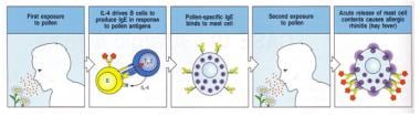
Hypersensitivity, Immediate: Also known as Type I hypersensitivity, it is an exaggerated and abnormal immune response that occurs within minutes to a few hours after exposure to a second dose of an allergen (a substance that triggers an allergic reaction). This type of hypersensitivity is mediated by immunoglobulin E (IgE) antibodies, which are produced by the immune system in response to the first exposure to the allergen. Upon subsequent exposures, these IgE antibodies bind to mast cells and basophils, leading to their degranulation and the release of mediators such as histamine, leukotrienes, and prostaglandins. These mediators cause a variety of symptoms, including itching, swelling, redness, and pain at the site of exposure, as well as systemic symptoms such as difficulty breathing, wheezing, and hypotension (low blood pressure). Examples of immediate hypersensitivity reactions include allergic asthma, hay fever, anaphylaxis, and some forms of food allergy.
Tuberculin is not a medical condition but a diagnostic tool used in the form of a purified protein derivative (PPD) to detect tuberculosis infection. It is prepared from the culture filtrate of Mycobacterium tuberculosis, the bacterium that causes TB. The PPD tuberculin is injected intradermally, and the resulting skin reaction is measured after 48-72 hours to determine if a person has developed an immune response to the bacteria, indicating a past or present infection with TB. It's important to note that a positive tuberculin test does not necessarily mean that active disease is present, but it does indicate that further evaluation is needed.
BCG (Bacillus Calmette-Guérin) vaccine is a type of immunization used primarily to prevent tuberculosis (TB). It contains a live but weakened strain of Mycobacterium bovis, which is related to the bacterium that causes TB in humans (Mycobacterium tuberculosis).
The BCG vaccine works by stimulating an immune response in the body, enabling it to better resist infection with TB bacteria if exposed in the future. It is often given to infants and children in countries where TB is common, and its use varies depending on the national immunization policies. The protection offered by the BCG vaccine is moderate and may not last for a very long time.
In addition to its use against TB, the BCG vaccine has also been investigated for its potential therapeutic role in treating bladder cancer and some other types of cancer. The mechanism of action in these cases is thought to be related to the vaccine's ability to stimulate an immune response against abnormal cells.
Streptodornase: Also known as streptococcal DNase, is an enzyme produced by certain strains of Streptococcus bacteria. It has the ability to degrade DNA, which makes it useful in some medical applications such as reducing the viscosity of purulent exudates (thick pus) in wounds and respiratory secretions, facilitating their removal and promoting tissue healing.
Streptokinase: Is a protein produced by various streptococcus species. It functions as a thrombolytic agent, which means it can dissolve blood clots. Streptokinase does this by binding to plasminogen, an inactive form of the enzyme plasmin, and converting it into its active form. Activated plasmin then breaks down fibrin, a protein that forms the structural framework of blood clots, leading to their dissolution. Streptokinase is used medically as a treatment for conditions associated with blood clots such as deep vein thrombosis, pulmonary embolism, and myocardial infarction (heart attack).
Drug hypersensitivity is an abnormal immune response to a medication or its metabolites. It is a type of adverse drug reaction that occurs in susceptible individuals, characterized by the activation of the immune system leading to inflammation and tissue damage. This reaction can range from mild symptoms such as skin rashes, hives, and itching to more severe reactions like anaphylaxis, which can be life-threatening.
Drug hypersensitivity reactions can be classified into two main types: immediate (or IgE-mediated) and delayed (or non-IgE-mediated). Immediate reactions occur within minutes to a few hours after taking the medication and are mediated by the release of histamine and other inflammatory mediators from mast cells and basophils. Delayed reactions, on the other hand, can take several days to develop and are caused by T-cell activation and subsequent cytokine release.
Common drugs that can cause hypersensitivity reactions include antibiotics (such as penicillins and sulfonamides), nonsteroidal anti-inflammatory drugs (NSAIDs), monoclonal antibodies, and chemotherapeutic agents. It is important to note that previous exposure to a medication does not always guarantee the development of hypersensitivity reactions, as they can also occur after the first administration in some cases.
The diagnosis of drug hypersensitivity involves a thorough medical history, physical examination, and sometimes skin or laboratory tests. Treatment typically includes avoiding the offending medication and managing symptoms with antihistamines, corticosteroids, or other medications as needed. In severe cases, emergency medical care may be required to treat anaphylaxis or other life-threatening reactions.
Immunoglobulin E (IgE) is a type of antibody that plays a key role in the immune response to parasitic infections and allergies. It is produced by B cells in response to stimulation by antigens, such as pollen, pet dander, or certain foods. Once produced, IgE binds to receptors on the surface of mast cells and basophils, which are immune cells found in tissues and blood respectively. When an individual with IgE antibodies encounters the allergen again, the cross-linking of IgE molecules bound to the FcεRI receptor triggers the release of mediators such as histamine, leukotrienes, prostaglandins, and various cytokines from these cells. These mediators cause the symptoms of an allergic reaction, such as itching, swelling, and redness. IgE also plays a role in protecting against certain parasitic infections by activating eosinophils, which can kill the parasites.
In summary, Immunoglobulin E (IgE) is a type of antibody that plays a crucial role in the immune response to allergens and parasitic infections, it binds to receptors on the surface of mast cells and basophils, when an individual with IgE antibodies encounters the allergen again, it triggers the release of mediators from these cells causing the symptoms of an allergic reaction.
"Skin physiological phenomena" is not a standard medical term with a specific definition. However, I can provide some information about the general concepts that might be encompassed by this term.
Physiological phenomena refer to the functions and processes that occur in living organisms. When it comes to the skin, there are many different physiological phenomena that take place, including:
1. Barrier function: The skin acts as a barrier to protect the body from external elements such as bacteria, viruses, chemicals, and UV radiation.
2. Temperature regulation: The skin helps regulate body temperature through sweat production and blood flow.
3. Sensation: The skin contains nerve endings that allow us to feel touch, pressure, pain, and temperature.
4. Vitamin D synthesis: The skin can produce vitamin D when exposed to sunlight.
5. Moisture regulation: The skin helps maintain the body's moisture balance by producing sweat and preventing water loss.
6. Immunological function: The skin plays a role in the immune system by providing a physical barrier and containing immune cells that help fight off infections.
7. Excretion: The skin eliminates waste products through sweat.
8. Wound healing: The skin has the ability to repair itself after injury, through a complex process involving inflammation, tissue regeneration, and remodeling.
Therefore, "skin physiological phenomena" could refer to any or all of these functions and processes that take place in the skin.
'Mycobacterium tuberculosis' is a species of slow-growing, aerobic, gram-positive bacteria that demonstrates acid-fastness. It is the primary causative agent of tuberculosis (TB) in humans. This bacterium has a complex cell wall rich in lipids, including mycolic acids, which provides a hydrophobic barrier and makes it resistant to many conventional antibiotics. The ability of M. tuberculosis to survive within host macrophages and resist the immune response contributes to its pathogenicity and the difficulty in treating TB infections.
M. tuberculosis is typically transmitted through inhalation of infectious droplets containing the bacteria, which primarily targets the lungs but can spread to other parts of the body (extrapulmonary TB). The infection may result in a spectrum of clinical manifestations, ranging from latent TB infection (LTBI) to active disease. LTBI represents a dormant state where individuals are infected with M. tuberculosis but do not show symptoms and cannot transmit the bacteria. However, they remain at risk of developing active TB throughout their lifetime, especially if their immune system becomes compromised.
Effective prevention and control strategies for TB rely on early detection, treatment, and public health interventions to limit transmission. The current first-line treatments for drug-susceptible TB include a combination of isoniazid, rifampin, ethambutol, and pyrazinamide for at least six months. Multidrug-resistant (MDR) and extensively drug-resistant (XDR) strains of M. tuberculosis present significant challenges in TB control and require more complex treatment regimens.
Pulmonary tuberculosis (TB) is an infectious disease caused by the bacterium Mycobacterium tuberculosis. It primarily affects the lungs and can spread to other parts of the body through the bloodstream or lymphatic system. The infection typically enters the body when a person inhales droplets containing the bacteria, which are released into the air when an infected person coughs, sneezes, or talks.
The symptoms of pulmonary TB can vary but often include:
* Persistent cough that lasts for more than three weeks and may produce phlegm or blood-tinged sputum
* Chest pain or discomfort, particularly when breathing deeply or coughing
* Fatigue and weakness
* Unexplained weight loss
* Fever and night sweats
* Loss of appetite
Pulmonary TB can cause serious complications if left untreated, including damage to the lungs, respiratory failure, and spread of the infection to other parts of the body. Treatment typically involves a course of antibiotics that can last several months, and it is essential for patients to complete the full treatment regimen to ensure that the infection is fully eradicated.
Preventive measures include vaccination with the Bacillus Calmette-Guérin (BCG) vaccine, which can provide some protection against severe forms of TB in children, and measures to prevent the spread of the disease, such as covering the mouth and nose when coughing or sneezing, wearing a mask in public places, and avoiding close contact with people who have active TB.
Skin absorption, also known as percutaneous absorption, refers to the process by which substances are taken up by the skin and pass into the systemic circulation. This occurs when a substance is applied topically to the skin and penetrates through the various layers of the epidermis and dermis until it reaches the capillaries, where it can be transported to other parts of the body.
The rate and extent of skin absorption depend on several factors, including the physicochemical properties of the substance (such as its molecular weight, lipophilicity, and charge), the concentration and formulation of the product, the site of application, and the integrity and condition of the skin.
Skin absorption is an important route of exposure for many chemicals, drugs, and cosmetic ingredients, and it can have both therapeutic and toxicological consequences. Therefore, understanding the mechanisms and factors that influence skin absorption is crucial for assessing the safety and efficacy of topical products and for developing strategies to enhance or reduce their absorption as needed.
A Radioallergosorbent Test (RAST) is a type of blood test used in the diagnosis of allergies. It measures the presence and levels of specific antibodies, called immunoglobulin E (IgE), produced by the immune system in response to certain allergens. In this test, a small amount of blood is taken from the patient and then mixed with various allergens. If the patient has developed IgE antibodies against any of these allergens, they will bind to them, forming an antigen-antibody complex.
The mixture is then passed over a solid phase, such as a paper or plastic surface, which has been coated with allergen-specific antibodies. These antibodies will capture the antigen-antibody complexes formed in the previous step. A radioactive label is attached to a different type of antibody (called anti-IgE), which then binds to the IgE antibodies captured on the solid phase. The amount of radioactivity detected is proportional to the quantity of IgE antibodies present, providing an indication of the patient's sensitivity to that specific allergen.
While RAST tests have been largely replaced by more modern and sensitive techniques, such as fluorescence enzyme immunoassays (FEIA), they still provide valuable information in diagnosing allergies and guiding treatment plans.
Histoplasmin is not a medical condition or diagnosis itself, but it's a term related to a skin test used in medicine. Histoplasmin is an antigen extract derived from the histoplasmoma (a form of the fungus Histoplasma capsulatum) used in the histoplasmin skin test. This test is utilized to determine whether a person has been infected with the histoplasmosis fungus, which causes the disease histoplasmosis.
The histoplasmin skin test involves injecting a small amount of histoplasmin under the surface of the skin, usually on the forearm. If the person has previously been exposed to Histoplasma capsulatum, their immune system will recognize the antigen and produce a reaction (a hard, red, swollen area) at the injection site within 24-72 hours. The size of this reaction helps healthcare professionals determine if the person has developed an immune response to the fungus, indicating past or current infection with histoplasmosis.
It's important to note that a positive histoplasmin skin test does not necessarily mean that the person is currently sick with histoplasmosis. Instead, it shows that they have been exposed to the fungus at some point in their life and have developed an immune response to it.
Hypersensitivity is an exaggerated or inappropriate immune response to a substance that is generally harmless to most people. It's also known as an allergic reaction. This abnormal response can be caused by various types of immunological mechanisms, including antibody-mediated reactions (types I, II, and III) and cell-mediated reactions (type IV). The severity of the hypersensitivity reaction can range from mild discomfort to life-threatening conditions. Common examples of hypersensitivity reactions include allergic rhinitis, asthma, atopic dermatitis, food allergies, and anaphylaxis.
An allergen is a substance that can cause an allergic reaction in some people. These substances are typically harmless to most people, but for those with allergies, the immune system mistakenly identifies them as threats and overreacts, leading to the release of histamines and other chemicals that cause symptoms such as itching, sneezing, runny nose, rashes, hives, and difficulty breathing. Common allergens include pollen, dust mites, mold spores, pet dander, insect venom, and certain foods or medications. When a person comes into contact with an allergen, they may experience symptoms that range from mild to severe, depending on the individual's sensitivity to the substance and the amount of exposure.
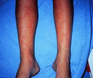
Urticaria, also known as hives, is an allergic reaction that appears on the skin. It is characterized by the rapid appearance of swollen, pale red bumps or plaques (wheals) on the skin, which are often accompanied by itching, stinging, or burning sensations. These wheals can vary in size and shape, and they may change location and appear in different places over a period of hours or days. Urticaria is usually caused by an allergic reaction to food, medication, or other substances, but it can also be triggered by physical factors such as heat, cold, pressure, or exercise. The condition is generally harmless, but severe cases of urticaria may indicate a more serious underlying medical issue and should be evaluated by a healthcare professional.
Skin pigmentation is the coloration of the skin that is primarily determined by two types of melanin pigments, eumelanin and pheomelanin. These pigments are produced by melanocytes, which are specialized cells located in the epidermis. Eumelanin is responsible for brown or black coloration, while pheomelanin produces a red or yellow hue.
The amount and distribution of melanin in the skin can vary depending on genetic factors, age, sun exposure, and various other influences. Increased production of melanin in response to UV radiation from the sun helps protect the skin from damage, leading to darkening or tanning of the skin. However, excessive sun exposure can also cause irregular pigmentation, such as sunspots or freckles.
Abnormalities in skin pigmentation can result from various medical conditions, including albinism (lack of melanin production), vitiligo (loss of melanocytes leading to white patches), and melasma (excessive pigmentation often caused by hormonal changes). These conditions may require medical treatment to manage or improve the pigmentation issues.
Immunologic tests are a type of diagnostic assay that detect and measure the presence or absence of specific immune responses in a sample, such as blood or tissue. These tests can be used to identify antibodies, antigens, immune complexes, or complement components in a sample, which can provide information about the health status of an individual, including the presence of infection, autoimmune disease, or immunodeficiency.
Immunologic tests use various methods to detect these immune components, such as enzyme-linked immunosorbent assays (ELISAs), Western blots, immunofluorescence assays, and radioimmunoassays. The results of these tests can help healthcare providers diagnose and manage medical conditions, monitor treatment effectiveness, and assess immune function.
It's important to note that the interpretation of immunologic test results should be done by a qualified healthcare professional, as false positives or negatives can occur, and the results must be considered in conjunction with other clinical findings and patient history.
Bovine tuberculosis (BTB) is a chronic infectious disease caused by the bacterium Mycobacterium bovis. It primarily affects cattle but can also spread to other mammals including humans, causing a similar disease known as zoonotic tuberculosis. The infection in animals typically occurs through inhalation of infectious droplets or ingestion of contaminated feed and water.
In cattle, the disease often affects the respiratory system, leading to symptoms such as chronic coughing, weight loss, and difficulty breathing. However, it can also affect other organs, including the intestines, lymph nodes, and mammary glands. Diagnosis of BTB typically involves a combination of clinical signs, laboratory tests, and epidemiological data.
Control measures for BTB include regular testing and culling of infected animals, movement restrictions, and vaccination of susceptible populations. In many countries, BTB is a notifiable disease, meaning that cases must be reported to the authorities. Proper cooking and pasteurization of dairy products can help prevent transmission to humans.
The Basophil Degranulation Test is a medical test that measures the degree of degranulation (the release of granules and their contents) in basophils, a type of white blood cell, in response to a stimulus. This test is often used to diagnose allergies or hypersensitivity reactions, as basophils are known to degranulate when exposed to allergens or certain medications.
In this test, basophils are isolated from a patient's blood sample and then exposed to a suspected allergen or other stimuli. After incubation, the cells are stained with a dye that detects the presence of histamine or other mediators released during degranulation. The degree of staining is then measured and used as an indicator of basophil activation and degranulation.
It's important to note that this test is not commonly used in clinical practice due to its complexity, variability, and limited availability. Other tests, such as skin prick tests or blood tests for specific IgE antibodies, are more commonly used to diagnose allergies.
Bacterial antigens are substances found on the surface or produced by bacteria that can stimulate an immune response in a host organism. These antigens can be proteins, polysaccharides, teichoic acids, lipopolysaccharides, or other molecules that are recognized as foreign by the host's immune system.
When a bacterial antigen is encountered by the host's immune system, it triggers a series of responses aimed at eliminating the bacteria and preventing infection. The host's immune system recognizes the antigen as foreign through the use of specialized receptors called pattern recognition receptors (PRRs), which are found on various immune cells such as macrophages, dendritic cells, and neutrophils.
Once a bacterial antigen is recognized by the host's immune system, it can stimulate both the innate and adaptive immune responses. The innate immune response involves the activation of inflammatory pathways, the recruitment of immune cells to the site of infection, and the production of antimicrobial peptides.
The adaptive immune response, on the other hand, involves the activation of T cells and B cells, which are specific to the bacterial antigen. These cells can recognize and remember the antigen, allowing for a more rapid and effective response upon subsequent exposures.
Bacterial antigens are important in the development of vaccines, as they can be used to stimulate an immune response without causing disease. By identifying specific bacterial antigens that are associated with virulence or pathogenicity, researchers can develop vaccines that target these antigens and provide protection against infection.
Anaphylaxis is a severe, life-threatening systemic allergic reaction that occurs suddenly after exposure to an allergen (a substance that triggers an allergic reaction) to which the person has previously been sensitized. The symptoms of anaphylaxis include rapid onset of symptoms such as itching, hives, swelling of the throat and tongue, difficulty breathing, wheezing, cough, chest tightness, rapid heartbeat, hypotension (low blood pressure), shock, and in severe cases, loss of consciousness and death. Anaphylaxis is a medical emergency that requires immediate treatment with epinephrine (adrenaline) and other supportive measures to stabilize the patient's condition.
Erythema is a term used in medicine to describe redness of the skin, which occurs as a result of increased blood flow in the superficial capillaries. This redness can be caused by various factors such as inflammation, infection, trauma, or exposure to heat, cold, or ultraviolet radiation. In some cases, erythema may also be accompanied by other symptoms such as swelling, warmth, pain, or itching. It is a common finding in many medical conditions and can vary in severity from mild to severe.
Contact tracing is a key public health strategy used to control the spread of infectious diseases. It involves identifying and monitoring individuals (contacts) who have come into close contact with an infected person (case), to prevent further transmission of the disease. The process typically includes:
1. Case identification: Identifying and confirming cases of infection through diagnostic testing.
2. Contact identification: Finding people who may have been in close contact with the infected case during their infectious period, which is the time when they can transmit the infection to others. Close contacts are usually defined as individuals who have had face-to-face contact with a confirmed case within a certain distance (often 6 feet or closer) and/or shared confined spaces for prolonged periods (usually more than 15 minutes).
3. Contact listing: Recording the identified contacts' information, including their names, addresses, phone numbers, and potentially other demographic data.
4. Risk assessment: Evaluating the level of risk associated with each contact based on factors such as the type of exposure, duration of contact, and the infectiousness of the case.
5. Notification: Informing contacts about their potential exposure to the infection and providing them with necessary health information, education, and guidance. This may include recommendations for self-quarantine, symptom monitoring, testing, and vaccination if available.
6. Follow-up: Monitoring and supporting contacts during their quarantine or isolation period, which typically lasts 14 days from the last exposure to the case. Public health professionals will check in with contacts regularly to assess their symptoms, provide additional guidance, and ensure they are adhering to the recommended infection prevention measures.
7. Data management: Documenting and reporting contact tracing activities for public health surveillance, evaluation, and future planning purposes.
Contact tracing is a critical component of infectious disease control and has been used effectively in managing various outbreaks, including tuberculosis, HIV/AIDS, Ebola, and more recently, COVID-19.
Sensitivity and specificity are statistical measures used to describe the performance of a diagnostic test or screening tool in identifying true positive and true negative results.
* Sensitivity refers to the proportion of people who have a particular condition (true positives) who are correctly identified by the test. It is also known as the "true positive rate" or "recall." A highly sensitive test will identify most or all of the people with the condition, but may also produce more false positives.
* Specificity refers to the proportion of people who do not have a particular condition (true negatives) who are correctly identified by the test. It is also known as the "true negative rate." A highly specific test will identify most or all of the people without the condition, but may also produce more false negatives.
In medical testing, both sensitivity and specificity are important considerations when evaluating a diagnostic test. High sensitivity is desirable for screening tests that aim to identify as many cases of a condition as possible, while high specificity is desirable for confirmatory tests that aim to rule out the condition in people who do not have it.
It's worth noting that sensitivity and specificity are often influenced by factors such as the prevalence of the condition in the population being tested, the threshold used to define a positive result, and the reliability and validity of the test itself. Therefore, it's important to consider these factors when interpreting the results of a diagnostic test.
Interferon-gamma (IFN-γ) is a soluble cytokine that is primarily produced by the activation of natural killer (NK) cells and T lymphocytes, especially CD4+ Th1 cells and CD8+ cytotoxic T cells. It plays a crucial role in the regulation of the immune response against viral and intracellular bacterial infections, as well as tumor cells. IFN-γ has several functions, including activating macrophages to enhance their microbicidal activity, increasing the presentation of major histocompatibility complex (MHC) class I and II molecules on antigen-presenting cells, stimulating the proliferation and differentiation of T cells and NK cells, and inducing the production of other cytokines and chemokines. Additionally, IFN-γ has direct antiproliferative effects on certain types of tumor cells and can enhance the cytotoxic activity of immune cells against infected or malignant cells.
Artificial Skin is a synthetic substitute or equivalent that is used to replace, support, or enhance the function of damaged or absent skin. It can be made from various materials such as biopolymers, composites, or biosynthetic materials. The main purpose of artificial skin is to provide a temporary or permanent covering for wounds, burns, or ulcers that cannot be healed with conventional treatments. Additionally, it may serve as a platform for the delivery of medications or as a matrix for the growth of cells and tissues during skin grafting procedures. Artificial skin must possess properties such as biocompatibility, durability, flexibility, and permeability to air and water vapor in order to promote optimal healing and minimize scarring.
"Mycobacterium bovis" is a species of slow-growing, aerobic, gram-positive bacteria in the family Mycobacteriaceae. It is the causative agent of tuberculosis in cattle and other animals, and can also cause tuberculosis in humans, particularly in those who come into contact with infected animals or consume unpasteurized dairy products from infected cows. The bacteria are resistant to many common disinfectants and survive for long periods in a dormant state, making them difficult to eradicate from the environment. "Mycobacterium bovis" is closely related to "Mycobacterium tuberculosis," the bacterium that causes tuberculosis in humans, and both species share many genetic and biochemical characteristics.
Mites are tiny arthropods belonging to the class Arachnida, which also includes spiders and ticks. They are characterized by their small size, usually measuring less than 1 mm in length, and their lack of obvious segmentation on their bodies. Many mites are parasitic, feeding on the skin cells, blood, or fluids of plants and animals, including humans. Some common mite infestations in humans include scabies, caused by the itch mite (Sarcoptes scabiei), and dust mites (e.g., Dermatophagoides pteronyssinus and D. farinae), which are commonly found in household dust and can cause allergic reactions in some people. It's worth noting that the majority of mites are not harmful to humans and play important roles in ecosystems as decomposers and predators.
Cellular immunity, also known as cell-mediated immunity, is a type of immune response that involves the activation of immune cells, such as T lymphocytes (T cells), to protect the body against infected or damaged cells. This form of immunity is important for fighting off infections caused by viruses and intracellular bacteria, as well as for recognizing and destroying cancer cells.
Cellular immunity involves a complex series of interactions between various immune cells and molecules. When a pathogen infects a cell, the infected cell displays pieces of the pathogen on its surface in a process called antigen presentation. This attracts T cells, which recognize the antigens and become activated. Activated T cells then release cytokines, chemicals that help coordinate the immune response, and can directly attack and kill infected cells or help activate other immune cells to do so.
Cellular immunity is an important component of the adaptive immune system, which is able to learn and remember specific pathogens in order to mount a faster and more effective response upon subsequent exposure. This form of immunity is also critical for the rejection of transplanted organs, as the immune system recognizes the transplanted tissue as foreign and attacks it.
Food hypersensitivity is an umbrella term that encompasses both immunologic and non-immunologic adverse reactions to food. It is also known as "food allergy" or "food intolerance." Food hypersensitivity occurs when the body's immune system or digestive system reacts negatively to a particular food or food component.
Immunologic food hypersensitivity, commonly referred to as a food allergy, involves an immune response mediated by immunoglobulin E (IgE) antibodies. Upon ingestion of the offending food, IgE antibodies bind to the food antigens and trigger the release of histamine and other chemical mediators from mast cells and basophils, leading to symptoms such as hives, swelling, itching, difficulty breathing, or anaphylaxis.
Non-immunologic food hypersensitivity, on the other hand, does not involve the immune system. Instead, it is caused by various mechanisms, including enzyme deficiencies, pharmacological reactions, and metabolic disorders. Examples of non-immunologic food hypersensitivities include lactose intolerance, gluten sensitivity, and histamine intolerance.
It's important to note that the term "food hypersensitivity" is often used interchangeably with "food allergy," but it has a broader definition that includes both immunologic and non-immunologic reactions.
Fungal antigens are substances found on or produced by fungi that can stimulate an immune response in a host organism. They can be proteins, polysaccharides, or other molecules that are recognized as foreign by the host's immune system. Fungal antigens can be used in diagnostic tests to identify fungal infections, and they can also be targets of immune responses during fungal infections. In some cases, fungal antigens may contribute to the pathogenesis of fungal diseases by inducing inflammatory or allergic reactions. Examples of fungal antigens include the cell wall components of Candida albicans and the extracellular polysaccharide galactomannan produced by Aspergillus fumigatus.
Trichophytin is not a medical condition or diagnosis, but rather a preparation used in skin tests to help identify whether someone has a sensitivity or allergic reaction to the fungus called Trichophyton. This fungus can cause various skin infections such as athlete's foot, ringworm, and jock itch. The trichophytin preparation contains antigens derived from the Trichophyton fungus, which are introduced into the skin to trigger an immune response. If a person is allergic or sensitive to the fungus, their body will mount a reaction, which can be observed and measured as part of the skin test. This information helps healthcare professionals diagnose and manage fungal infections and related allergies.
I must clarify that the term "Guinea Pigs" is not typically used in medical definitions. However, in colloquial or informal language, it may refer to people who are used as the first to try out a new medical treatment or drug. This is known as being a "test subject" or "in a clinical trial."
In the field of scientific research, particularly in studies involving animals, guinea pigs are small rodents that are often used as experimental subjects due to their size, cost-effectiveness, and ease of handling. They are not actually pigs from Guinea, despite their name's origins being unclear. However, they do not exactly fit the description of being used in human medical experiments.
Coccidioidin is a preparation derived from the filtrate of a culture of Coccidioides immitis, a fungus that is the causative agent of coccidioidomycosis, also known as Valley Fever. It is used in skin tests to diagnose coccidioidomycosis infection and determine if a person has developed immunity to the disease.
When Coccidioidin is injected into the skin, a positive reaction (induration or swelling) may indicate a current or past infection with Coccidioides immitis. However, it's important to note that a negative result does not necessarily rule out an infection, and further diagnostic tests may be needed for confirmation.
It's also worth noting that skin testing with coccidioidin can have false-positive results in people who have been vaccinated against other types of fungal infections or have certain medical conditions. Therefore, the test should be interpreted carefully and used in conjunction with other clinical findings and diagnostic tests.
Skin test end-point titration is a method used in allergy testing to determine the minimum concentration of an allergen that will cause a positive skin reaction in a patient. This is done by applying dilutions of the allergen to the patient's skin, usually on the forearm, in a series of increasing concentrations. The skin is then pricked or punctured to allow the allergen to enter the skin.
The response is evaluated after a set period of time, typically 15-20 minutes. A positive reaction is indicated by the development of a wheal and flare response, which is a raised, red, itchy area on the skin. The end-point is defined as the lowest concentration of allergen that produces a positive reaction. This allows for the identification of specific allergens and the determination of the severity of the patient's allergy. It also helps in determining the appropriate dose of allergen immunotherapy, if required.
Cutaneous leishmaniasis is a neglected tropical disease caused by infection with Leishmania parasites, which are transmitted through the bite of infected female sandflies. The disease primarily affects the skin and mucous membranes, causing lesions that can be disfiguring and stigmatizing. There are several clinical forms of cutaneous leishmaniasis, including localized, disseminated, and mucocutaneous.
Localized cutaneous leishmaniasis is the most common form of the disease, characterized by the development of one or more nodular or ulcerative lesions at the site of the sandfly bite, typically appearing within a few weeks to several months after exposure. The lesions may vary in size and appearance, ranging from small papules to large plaques or ulcers, and can be painful or pruritic (itchy).
Disseminated cutaneous leishmaniasis is a more severe form of the disease, characterized by the widespread dissemination of lesions across the body. This form of the disease typically affects people with weakened immune systems, such as those with HIV/AIDS or those receiving immunosuppressive therapy.
Mucocutaneous leishmaniasis is a rare but severe form of the disease, characterized by the spread of infection from the skin to the mucous membranes of the nose, mouth, and throat. This can result in extensive tissue destruction, disfigurement, and functional impairment.
Cutaneous leishmaniasis is diagnosed through a combination of clinical evaluation, epidemiological data, and laboratory tests such as parasite detection using microscopy or molecular techniques, or serological tests to detect antibodies against the Leishmania parasites. Treatment options for cutaneous leishmaniasis include systemic or topical medications, such as antimonial drugs, miltefosine, or pentamidine, as well as physical treatments such as cryotherapy or thermotherapy. The choice of treatment depends on various factors, including the species of Leishmania involved, the clinical form of the disease, and the patient's overall health status.
Leishmania braziliensis is a species of protozoan parasite that causes American cutaneous leishmaniasis, also known as "espundia." This disease is transmitted to humans through the bite of infected female sandflies, primarily from the genus Lutzomyia. The infection can lead to skin lesions, ulcers, and scarring, and in some cases, it can disseminate and affect other organs, causing a more severe form of the disease called mucocutaneous leishmaniasis.
The parasite's life cycle involves two main stages: the promastigote stage, which occurs in the sandfly vector, and the amastigote stage, which takes place inside the mammalian host's macrophages. The infection can be diagnosed through various methods, including microscopic examination of tissue samples, culture isolation, or molecular techniques such as PCR. Treatment typically involves antiparasitic drugs, such as pentavalent antimonials, amphotericin B, or miltefosine, depending on the severity and location of the infection.
Respiratory hypersensitivity, also known as respiratory allergies or hypersensitive pneumonitis, refers to an exaggerated immune response in the lungs to inhaled substances or allergens. This condition occurs when the body's immune system overreacts to harmless particles, leading to inflammation and damage in the airways and alveoli (air sacs) of the lungs.
There are two types of respiratory hypersensitivity: immediate and delayed. Immediate hypersensitivity, also known as type I hypersensitivity, is mediated by immunoglobulin E (IgE) antibodies and results in symptoms such as sneezing, runny nose, and asthma-like symptoms within minutes to hours of exposure to the allergen. Delayed hypersensitivity, also known as type III or type IV hypersensitivity, is mediated by other immune mechanisms and can take several hours to days to develop after exposure to the allergen.
Common causes of respiratory hypersensitivity include mold spores, animal dander, dust mites, pollen, and chemicals found in certain occupations. Symptoms may include coughing, wheezing, shortness of breath, chest tightness, and fatigue. Treatment typically involves avoiding the allergen, if possible, and using medications such as corticosteroids, bronchodilators, or antihistamines to manage symptoms. In severe cases, immunotherapy (allergy shots) may be recommended to help desensitize the immune system to the allergen.
"Petasites" is a genus name in botany, which refers to a group of flowering plants in the family Asteraceae. While it may not have a direct medical definition, some species within this genus have been used in traditional medicine. For instance, Petasites hybridus (also known as butterbur) has been used in herbal medicine for treating migraines, allergies, and asthma. However, it's important to note that the use of these plants should be under the guidance of a healthcare professional, as they can have side effects and interact with certain medications.
Reagent kits, diagnostic are prepackaged sets of chemical reagents and other components designed for performing specific diagnostic tests or assays. These kits are often used in clinical laboratories to detect and measure the presence or absence of various biomarkers, such as proteins, antibodies, antigens, nucleic acids, or small molecules, in biological samples like blood, urine, or tissues.
Diagnostic reagent kits typically contain detailed instructions for their use, along with the necessary reagents, controls, and sometimes specialized equipment or supplies. They are designed to simplify the testing process, reduce human error, and increase standardization, ensuring accurate and reliable results. Examples of diagnostic reagent kits include those used for pregnancy tests, infectious disease screening, drug testing, genetic testing, and cancer biomarker detection.
Transfer factors are natural immune system components that are passed from one individual to another, usually through blood products. They are small proteins called cytokines that are secreted by certain white blood cells (T-lymphocytes or T-cells) and function to regulate the immune system's response to foreign substances.
Transfer factors can be extracted from human blood and given to individuals with weakened immune systems, such as those undergoing chemotherapy or suffering from immune deficiency disorders, to help enhance their immune response. They have also been used in the treatment of chronic fatigue syndrome, allergies, and certain viral infections.
It's important to note that while transfer factors have shown promise in some studies, more research is needed to fully understand their effectiveness and safety.
An Enzyme-Linked Immunosorbent Assay (ELISA) is a type of analytical biochemistry assay used to detect and quantify the presence of a substance, typically a protein or peptide, in a liquid sample. It takes its name from the enzyme-linked antibodies used in the assay.
In an ELISA, the sample is added to a well containing a surface that has been treated to capture the target substance. If the target substance is present in the sample, it will bind to the surface. Next, an enzyme-linked antibody specific to the target substance is added. This antibody will bind to the captured target substance if it is present. After washing away any unbound material, a substrate for the enzyme is added. If the enzyme is present due to its linkage to the antibody, it will catalyze a reaction that produces a detectable signal, such as a color change or fluorescence. The intensity of this signal is proportional to the amount of target substance present in the sample, allowing for quantification.
ELISAs are widely used in research and clinical settings to detect and measure various substances, including hormones, viruses, and bacteria. They offer high sensitivity, specificity, and reproducibility, making them a reliable choice for many applications.
Bacterial skin diseases are a type of infectious skin condition caused by various species of bacteria. These bacteria can multiply rapidly on the skin's surface when given the right conditions, leading to infection and inflammation. Some common bacterial skin diseases include:
1. Impetigo: A highly contagious superficial skin infection that typically affects exposed areas such as the face, hands, and feet. It is commonly caused by Staphylococcus aureus or Streptococcus pyogenes bacteria.
2. Cellulitis: A deep-skin infection that can spread rapidly and involves the inner layers of the skin and underlying tissue. It is often caused by Group A Streptococcus or Staphylococcus aureus bacteria.
3. Folliculitis: An inflammation of hair follicles, usually caused by an infection with Staphylococcus aureus or other bacteria.
4. Furuncles (boils) and carbuncles: Deep infections that develop from folliculitis when the infection spreads to surrounding tissue. A furuncle is a single boil, while a carbuncle is a cluster of boils.
5. Erysipelas: A superficial skin infection characterized by redness, swelling, and warmth in the affected area. It is typically caused by Group A Streptococcus bacteria.
6. MRSA (Methicillin-resistant Staphylococcus aureus) infections: Skin infections caused by a strain of Staphylococcus aureus that has developed resistance to many antibiotics, making it more difficult to treat.
7. Leptospirosis: A bacterial infection transmitted through contact with contaminated water or soil and characterized by flu-like symptoms and skin rashes.
Treatment for bacterial skin diseases usually involves the use of topical or oral antibiotics, depending on the severity and location of the infection. In some cases, drainage of pus-filled abscesses may be necessary to promote healing. Proper hygiene and wound care can help prevent the spread of these infections.
A skin ulcer is a defined as a loss of continuity or disruption of the skin surface, often accompanied by inflammation and/or infection. These lesions can result from various causes including pressure, venous or arterial insufficiency, diabetes, and chronic dermatological conditions. Skin ulcers are typically characterized by their appearance, depth, location, and underlying cause. Common types of skin ulcers include pressure ulcers (also known as bedsores), venous leg ulcers, arterial ulcers, and diabetic foot ulcers. Proper evaluation, wound care, management of underlying conditions, and prevention strategies are crucial in the treatment of skin ulcers to promote healing and prevent complications.
Isoniazid is an antimicrobial medication used for the prevention and treatment of tuberculosis (TB). It is a first-line medication, often used in combination with other TB drugs, to kill the Mycobacterium tuberculosis bacteria that cause TB. Isoniazid works by inhibiting the synthesis of mycolic acids, which are essential components of the bacterial cell wall. This leads to bacterial death and helps to control the spread of TB.
Isoniazid is available in various forms, including tablets, capsules, and liquid solutions. It can be taken orally or given by injection. The medication is generally well-tolerated, but it can cause side effects such as peripheral neuropathy, hepatitis, and skin rashes. Regular monitoring of liver function tests and supplementation with pyridoxine (vitamin B6) may be necessary to prevent or manage these side effects.
It is important to note that Isoniazid is not effective against drug-resistant strains of TB, and its use should be guided by the results of drug susceptibility testing. Additionally, it is essential to complete the full course of treatment as prescribed to ensure the successful eradication of the bacteria and prevent the development of drug-resistant strains.
A "mass chest X-ray" is a term used to describe a radiological screening procedure where a large number of individuals undergo chest X-rays, usually as part of a public health campaign or community screening event. The goal is to identify any early signs of lung diseases such as tuberculosis, lung cancer, or other pulmonary abnormalities. It's important to note that while mass screenings can help detect diseases at an earlier stage, they also raise concerns about radiation exposure and the potential for overdiagnosis. Therefore, such procedures are typically carried out under strict medical guidelines and regulations.
Antitubercular agents, also known as anti-tuberculosis drugs or simply TB drugs, are a category of medications specifically used for the treatment and prevention of tuberculosis (TB), a bacterial infection caused by Mycobacterium tuberculosis. These drugs target various stages of the bacteria's growth and replication process to eradicate it from the body or prevent its spread.
There are several first-line antitubercular agents, including:
1. Isoniazid (INH): This is a bactericidal drug that inhibits the synthesis of mycolic acids, essential components of the mycobacterial cell wall. It is primarily active against actively growing bacilli.
2. Rifampin (RIF) or Rifampicin: A bactericidal drug that inhibits DNA-dependent RNA polymerase, preventing the transcription of genetic information into mRNA. This results in the interruption of protein synthesis and ultimately leads to the death of the bacteria.
3. Ethambutol (EMB): A bacteriostatic drug that inhibits the arabinosyl transferase enzyme, which is responsible for the synthesis of arabinan, a crucial component of the mycobacterial cell wall. It is primarily active against actively growing bacilli.
4. Pyrazinamide (PZA): A bactericidal drug that inhibits the synthesis of fatty acids and mycolic acids in the mycobacterial cell wall, particularly under acidic conditions. PZA is most effective during the initial phase of treatment when the bacteria are in a dormant or slow-growing state.
These first-line antitubercular agents are often used together in a combination therapy to ensure complete eradication of the bacteria and prevent the development of drug-resistant strains. Treatment duration typically lasts for at least six months, with the initial phase consisting of daily doses of INH, RIF, EMB, and PZA for two months, followed by a continuation phase of INH and RIF for four months.
Second-line antitubercular agents are used when patients have drug-resistant TB or cannot tolerate first-line drugs. These include drugs like aminoglycosides (e.g., streptomycin, amikacin), fluoroquinolones (e.g., ofloxacin, moxifloxacin), and injectable bacteriostatic agents (e.g., capreomycin, ethionamide).
It is essential to closely monitor patients undergoing antitubercular therapy for potential side effects and ensure adherence to the treatment regimen to achieve optimal outcomes and prevent the development of drug-resistant strains.
Desensitization, Immunologic is a medical procedure that aims to decrease the immune system's response to an allergen. This is achieved through the controlled exposure of the patient to gradually increasing amounts of the allergen, ultimately leading to a reduction in the severity of allergic reactions upon subsequent exposures. The process typically involves administering carefully measured and incrementally larger doses of the allergen, either orally, sublingually (under the tongue), or by injection, under medical supervision. Over time, this repeated exposure can help the immune system become less sensitive to the allergen, thereby alleviating allergic symptoms.
The specific desensitization protocol and administration method may vary depending on the type of allergen and individual patient factors. Immunologic desensitization is most commonly used for environmental allergens like pollen, dust mites, or pet dander, as well as insect venoms such as bee or wasp stings. It is important to note that this procedure should only be performed under the close supervision of a qualified healthcare professional, as there are potential risks involved, including anaphylaxis (a severe and life-threatening allergic reaction).
Asthma is a chronic respiratory disease characterized by inflammation and narrowing of the airways, leading to symptoms such as wheezing, coughing, shortness of breath, and chest tightness. The airway obstruction in asthma is usually reversible, either spontaneously or with treatment.
The underlying cause of asthma involves a combination of genetic and environmental factors that result in hypersensitivity of the airways to certain triggers, such as allergens, irritants, viruses, exercise, and emotional stress. When these triggers are encountered, the airways constrict due to smooth muscle spasm, swell due to inflammation, and produce excess mucus, leading to the characteristic symptoms of asthma.
Asthma is typically managed with a combination of medications that include bronchodilators to relax the airway muscles, corticosteroids to reduce inflammation, and leukotriene modifiers or mast cell stabilizers to prevent allergic reactions. Avoiding triggers and monitoring symptoms are also important components of asthma management.
There are several types of asthma, including allergic asthma, non-allergic asthma, exercise-induced asthma, occupational asthma, and nocturnal asthma, each with its own set of triggers and treatment approaches. Proper diagnosis and management of asthma can help prevent exacerbations, improve quality of life, and reduce the risk of long-term complications.
Trichophyton is a genus of fungi that are primarily responsible for causing various superficial and cutaneous infections in humans and animals. These infections, known as dermatophytoses or ringworm, typically involve the skin, hair, and nails. Some common examples of diseases caused by Trichophyton species include athlete's foot (T. rubrum), jock itch (T. mentagrophytes), and scalp ringworm (T. tonsurans).
The fungi in the Trichophyton genus are called keratinophilic, meaning they have a preference for keratin, a protein found in high concentrations in skin, hair, and nails. This characteristic allows them to thrive in these environments and cause infection. The specific species of Trichophyton involved in an infection will determine the clinical presentation and severity of the disease.
In summary, Trichophyton is a medical term referring to a group of fungi that can cause various skin, hair, and nail infections in humans and animals.
The epidermis is the outermost layer of the skin, composed mainly of stratified squamous epithelium. It forms a protective barrier that prevents water loss and inhibits the entry of microorganisms. The epidermis contains no blood vessels, and its cells are nourished by diffusion from the underlying dermis. The bottom-most layer of the epidermis, called the stratum basale, is responsible for generating new skin cells that eventually move up to replace dead cells on the surface. This process of cell turnover takes about 28 days in adults.
The most superficial part of the epidermis consists of dead cells called squames, which are constantly shed and replaced. The exact rate at which this happens varies depending on location; for example, it's faster on the palms and soles than elsewhere. Melanocytes, the pigment-producing cells, are also located in the epidermis, specifically within the stratum basale layer.
In summary, the epidermis is a vital part of our integumentary system, providing not only physical protection but also playing a crucial role in immunity and sensory perception through touch receptors called Pacinian corpuscles.
Mucocutaneous Leishmaniasis (MCL) is a chronic, granulomatous disease caused by an infection with Leishmania species, primarily L. braziliensis and L. guyanensis. It affects both the mucous membranes (such as those of the nose, mouth, and throat) and the skin.
The initial infection often occurs through the bite of an infected female sandfly, which transmits the parasitic protozoa into the host's skin. After a variable incubation period, the disease can manifest in different clinical forms, including localized cutaneous leishmaniasis (CL), disseminated cutaneous leishmaniasis, and mucocutaneous leishmaniasis.
MCL is characterized by progressive destruction of the mucous membranes, leading to deformities and functional impairments. The infection typically starts as a cutaneous lesion at the site of the sandfly bite, which heals spontaneously within several months. However, in some cases, the parasites disseminate to the mucous membranes, causing severe inflammation, ulceration, and tissue necrosis.
Symptoms of MCL include:
1. Destruction of nasal septum, leading to a saddle-nose deformity
2. Perforation of the palate or septum
3. Hoarseness or loss of voice due to laryngeal involvement
4. Difficulty swallowing and speaking
5. Chronic rhinitis, sinusitis, or otitis media
6. Severe disfigurement and functional impairments in advanced cases
Diagnosis is usually made by identifying the parasites in tissue samples (such as biopsies) using microscopy, culture, or PCR-based methods. Treatment typically involves systemic antiparasitic drugs, such as pentavalent antimonials, amphotericin B, miltefosine, or combination therapies, along with surgical interventions to reconstruct damaged tissues in advanced cases.
Dinitrochlorobenzene (DNCB) is a chemical compound that is classified as an aromatic organic compound. Its medical definition relates to its use as a topical immunotherapy for the treatment of certain skin conditions. DNCB is a potent sensitizer and hapten, which means that it can cause an immune response when it comes into contact with the skin.
When applied to the skin, DNCB can stimulate the production of antibodies and activate immune cells, leading to an inflammatory reaction. This property has been exploited in the treatment of conditions such as alopecia areata, a type of hair loss that is thought to be caused by an autoimmune response. By sensitizing the patient's immune system to DNCB, it may be possible to modulate the immune response and promote hair growth.
However, the use of DNCB as a therapeutic agent is not without risks. It can cause significant local reactions, including redness, swelling, and blistering, and there is a risk of systemic toxicity if it is absorbed into the bloodstream. As such, its use is generally restricted to specialized medical settings where it can be administered under close supervision.
Rhinitis is a medical condition characterized by inflammation and irritation of the nasal passages, leading to symptoms such as sneezing, runny nose, congestion, and postnasal drip. It can be caused by various factors, including allergies (such as pollen, dust mites, or pet dander), infections (viral or bacterial), environmental irritants (such as smoke or pollution), and hormonal changes. Depending on the cause, rhinitis can be classified as allergic rhinitis, non-allergic rhinitis, infectious rhinitis, or hormonal rhinitis. Treatment options vary depending on the underlying cause but may include medications such as antihistamines, decongestants, nasal sprays, and immunotherapy (allergy shots).
Cell migration inhibition refers to the process or agents that restrict the movement of cells, particularly in the context of cancer metastasis. Cell migration is a critical biological process involved in various physiological and pathological conditions, including embryonic development, wound healing, and tumor cell dissemination. Inhibiting cell migration can help prevent the spread of cancer to distant organs, thereby improving treatment outcomes and patient survival rates.
Various factors and mechanisms contribute to cell migration inhibition, such as:
1. Modulation of signaling pathways: Cell migration is regulated by complex intracellular signaling networks that control cytoskeletal rearrangements, adhesion molecules, and other components required for cell motility. Inhibiting specific signaling proteins or pathways can suppress cell migration.
2. Extracellular matrix (ECM) modifications: The ECM provides structural support and biochemical cues that guide cell migration. Altering the composition or organization of the ECM can hinder cell movement.
3. Inhibition of adhesion molecules: Cell-cell and cell-matrix interactions are mediated by adhesion molecules, such as integrins and cadherins. Blocking these molecules can prevent cells from attaching to their surroundings and migrating.
4. Targeting cytoskeletal components: The cytoskeleton is responsible for the mechanical forces required for cell migration. Inhibiting cytoskeletal proteins, such as actin or tubulin, can impair cell motility.
5. Use of pharmacological agents: Several drugs and compounds have been identified to inhibit cell migration, either by targeting specific molecules or indirectly affecting the overall cellular environment. These agents include chemotherapeutic drugs, natural compounds, and small molecule inhibitors.
Understanding the mechanisms underlying cell migration inhibition can provide valuable insights into developing novel therapeutic strategies for cancer treatment and other diseases involving aberrant cell migration.
An antigen is a substance (usually a protein) that is recognized as foreign by the immune system and stimulates an immune response, leading to the production of antibodies or activation of T-cells. Antigens can be derived from various sources, including bacteria, viruses, fungi, parasites, and tumor cells. They can also come from non-living substances such as pollen, dust mites, or chemicals.
Antigens contain epitopes, which are specific regions on the antigen molecule that are recognized by the immune system. The immune system's response to an antigen depends on several factors, including the type of antigen, its size, and its location in the body.
In general, antigens can be classified into two main categories:
1. T-dependent antigens: These require the help of T-cells to stimulate an immune response. They are typically larger, more complex molecules that contain multiple epitopes capable of binding to both MHC class II molecules on antigen-presenting cells and T-cell receptors on CD4+ T-cells.
2. T-independent antigens: These do not require the help of T-cells to stimulate an immune response. They are usually smaller, simpler molecules that contain repetitive epitopes capable of cross-linking B-cell receptors and activating them directly.
Understanding antigens and their properties is crucial for developing vaccines, diagnostic tests, and immunotherapies.
Asymptomatic infections are those in which an individual carries and may transmit a pathogen, such as a virus or bacteria, but does not develop any symptoms associated with the infection. These individuals are often referred to as being "asymptomatically infected" or "asymptomatic carriers."
Asymptomatic infections can occur with various infectious diseases, including COVID-19, HIV, hepatitis B, and some sexually transmitted infections. In many cases, asymptomatic individuals may not realize they are infected and unknowingly transmit the pathogen to others. This makes identifying and controlling asymptomatic infections crucial for preventing outbreaks and limiting the spread of infectious diseases.
It's important to note that while asymptomatic individuals do not experience symptoms, they can still develop immunity to the infection, similar to those who experienced symptoms. Additionally, some asymptomatic infections may progress to symptomatic illness over time or upon subsequent exposures to the pathogen.
Coccidioidomycosis is a fungal infection caused by the inhalation of spores of the Coccidioides species, mainly C. immitis and C. posadasii. These fungi are commonly found in the soil of dry regions such as the southwestern United States, Mexico, and Central and South America.
The infection often begins when a person inhales the microscopic spores, which can lead to respiratory symptoms resembling a common cold or pneumonia. Some people may develop more severe symptoms, especially those with weakened immune systems. The infection can disseminate to other parts of the body, causing skin lesions, bone and joint inflammation, meningitis, or other complications in rare cases.
Diagnosis typically involves a combination of clinical evaluation, imaging studies, and laboratory tests such as fungal cultures, histopathological examination, or serological tests to detect antibodies against Coccidioides antigens. Treatment depends on the severity of the infection and the patient's immune status. Antifungal medications like fluconazole, itraconazole, or amphotericin B are commonly used for treating coccidioidomycosis. Preventive measures include avoiding inhaling dust in endemic areas, especially during excavation or construction activities.
Patient-to-professional transmission of infectious diseases refers to the spread of an infectious agent or disease from a patient to a healthcare professional. This can occur through various routes, including:
1. Contact transmission: This includes direct contact, such as touching or shaking hands with an infected patient, or indirect contact, such as touching a contaminated surface or object.
2. Droplet transmission: This occurs when an infected person coughs, sneezes, talks, or breathes out droplets containing the infectious agent, which can then be inhaled by a nearby healthcare professional.
3. Airborne transmission: This involves the spread of infectious agents through the air over long distances, usually requiring specialized medical procedures or equipment.
Healthcare professionals are at risk of patient-to-professional transmission of infectious diseases due to their close contact with patients and the potential for exposure to various pathogens. It is essential for healthcare professionals to follow standard precautions, including hand hygiene, personal protective equipment (PPE), and respiratory protection, to minimize the risk of transmission. Additionally, proper vaccination and education on infection prevention and control measures can further reduce the risk of patient-to-professional transmission of infectious diseases.
A patch test is a method used in clinical dermatology to identify whether a specific substance causes allergic inflammation of the skin (contact dermatitis). It involves applying small amounts of potential allergens to patches, which are then placed on the skin and left for a set period of time, usually 48 hours. The skin is then examined for signs of an allergic reaction such as redness, swelling or blistering. This helps in identifying the specific substances that an individual may be allergic to, enabling appropriate avoidance measures and treatment.
Lepromin is not a medical condition but rather a diagnostic test used in the diagnosis and classification of leprosy, a chronic infectious disease caused by the bacterium Mycobacterium leprae. The Lepromin test measures the cell-mediated immunity of an individual to the bacteria that causes leprosy.
The test involves injecting two types of antigens derived from M. leprae, one more soluble and the other less soluble, into the skin of the forearm. The response of the immune system to these antigens is then observed after 24 hours (for the soluble antigen) and 48 hours (for the less soluble antigen).
A positive reaction to the more soluble antigen indicates a strong cell-mediated immunity, which is associated with tuberculoid leprosy, a milder form of the disease. A positive reaction to the less soluble antigen suggests a weaker cell-mediated immunity and is associated with lepromatous leprosy, a more severe form of the disease.
It's important to note that the Lepromin test has limited availability and is not widely used in many parts of the world due to its complexity and the need for specialized laboratory facilities.
I'm sorry for any confusion, but "Gambia" is not a medical term. It is the name of a country located in West Africa, officially known as the Republic of The Gambia. If you have any questions about medical terminology or health-related topics, I would be happy to try and help answer those for you.
Prevalence, in medical terms, refers to the total number of people in a given population who have a particular disease or condition at a specific point in time, or over a specified period. It is typically expressed as a percentage or a ratio of the number of cases to the size of the population. Prevalence differs from incidence, which measures the number of new cases that develop during a certain period.
Allergic rhinitis, seasonal (also known as hay fever) is a type of inflammation in the nose which occurs when an individual breathes in allergens such as pollen or mold spores. The immune system identifies these substances as harmful and releases histamine and other chemicals, causing symptoms such as sneezing, runny or stuffy nose, red, watery, and itchy eyes, cough, and fatigue. Unlike perennial allergic rhinitis, seasonal allergic rhinitis is worse during specific times of the year when certain plants pollinate.
Allergic bronchopulmonary aspergillosis (ABPA) is a medical condition characterized by an hypersensitivity reaction to the fungus Aspergillus species, most commonly A. fumigatus. It primarily affects the airways and lung tissue. The immune system overreacts to the presence of the fungus, leading to inflammation and damage in the lungs.
The main symptoms of ABPA include wheezing, coughing, production of thick mucus, shortness of breath, and chest tightness. These symptoms are similar to those seen in asthma and other respiratory conditions. Some people with ABPA may also experience fever, weight loss, and fatigue.
Diagnosis of ABPA typically involves a combination of clinical evaluation, imaging studies (such as chest X-rays or CT scans), and laboratory tests (such as blood tests or sputum cultures) to detect the presence of Aspergillus species and elevated levels of certain antibodies.
Treatment for ABPA usually involves a combination of corticosteroids to reduce inflammation and antifungal medications to eradicate the Aspergillus infection. In some cases, immunomodulatory therapies may also be used to help regulate the immune system's response to the fungus.
It is important to note that ABPA can lead to serious complications if left untreated, including bronchiectasis (permanent enlargement of the airways), pulmonary fibrosis (scarring of the lung tissue), and respiratory failure. Therefore, prompt diagnosis and treatment are essential for managing this condition.
Allergic rhinitis, perennial type, is a medical condition characterized by inflammation of the nasal passages caused by an allergic response to environmental allergens that are present throughout the year. Unlike seasonal allergic rhinitis, which is triggered by specific pollens or molds during certain times of the year, perennial allergic rhinitis is a persistent condition that occurs year-round.
Common allergens responsible for perennial allergic rhinitis include dust mites, cockroaches, pet dander, and indoor mold spores. Symptoms may include sneezing, runny or stuffy nose, itchy eyes, ears, throat, or roof of the mouth. Treatment options typically involve avoiding exposure to the offending allergens, if possible, as well as medications such as antihistamines, nasal corticosteroids, and leukotriene receptor antagonists to manage symptoms. Immunotherapy (allergy shots) may also be recommended for long-term management in some cases.
Latex hypersensitivity is an immune-mediated reaction to proteins found in natural rubber latex, which can cause allergic symptoms ranging from mild skin irritation to life-threatening anaphylaxis. It is a form of type I (immediate) hypersensitivity, mediated by IgE antibodies that bind to mast cells and basophils, leading to the release of histamine and other mediators of inflammation upon re-exposure to latex proteins.
The symptoms of latex hypersensitivity can include skin rashes, hives, itching, nasal congestion, sneezing, wheezing, shortness of breath, coughing, and in severe cases, anaphylaxis characterized by a rapid heartbeat, low blood pressure, loss of consciousness, and even death.
Healthcare workers, patients with spina bifida, and those who have undergone multiple surgeries are at increased risk for developing latex hypersensitivity due to repeated exposure to latex products. Prevention measures include using non-latex medical supplies and devices, wearing non-powdered latex gloves, and implementing strict hand hygiene practices.
Basophils are a type of white blood cell that are part of the immune system. They are granulocytes, which means they contain granules filled with chemicals that can be released in response to an infection or inflammation. Basophils are relatively rare, making up less than 1% of all white blood cells.
When basophils become activated, they release histamine and other chemical mediators that can contribute to allergic reactions, such as itching, swelling, and redness. They also play a role in inflammation, helping to recruit other immune cells to the site of an infection or injury.
Basophils can be identified under a microscope based on their characteristic staining properties. They are typically smaller than other granulocytes, such as neutrophils and eosinophils, and have a multi-lobed nucleus with dark purple-staining granules in the cytoplasm.
While basophils play an important role in the immune response, abnormal levels of basophils can be associated with various medical conditions, such as allergies, infections, and certain types of leukemia.
Veterinary medicine is the branch of medical science that deals with the prevention, diagnosis, and treatment of diseases, disorders, and injuries in non-human animals. The profession of veterinary medicine is dedicated to the care, health, and welfare of animals, as well as to the promotion of human health through animal research and public health advancements. Veterinarians employ a variety of diagnostic methods including clinical examination, radiography, laboratory testing, and ultrasound imaging. They use a range of treatments, including medication, surgery, and dietary management. In addition, veterinarians may also advise on preventative healthcare measures such as vaccination schedules and parasite control programs.
Silicotuberculosis is not a medical condition that has its own specific definition in the medical literature. However, the term is sometimes used to describe a situation where a patient has both silicosis (a lung disease caused by inhaling crystalline silica dust) and tuberculosis (a bacterial infection that primarily affects the lungs).
Silicosis can increase a person's risk of developing tuberculosis, as the scarring and inflammation caused by silicosis can make the lungs more susceptible to infection. In some cases, the presence of silicosis may also affect the diagnosis and treatment of tuberculosis. Therefore, it is important for healthcare providers to be aware of any history of silica exposure when evaluating a patient with lung symptoms or signs of tuberculosis.
A "drug eruption" is a general term used to describe an adverse skin reaction that occurs as a result of taking a medication. These reactions can vary in severity and appearance, and may include symptoms such as rash, hives, itching, redness, blistering, or peeling of the skin. In some cases, drug eruptions can also cause systemic symptoms such as fever, fatigue, or joint pain.
The exact mechanism by which drugs cause eruptions is not fully understood, but it is thought to involve an abnormal immune response to the medication. There are many different types of drug eruptions, including morphilliform rashes, urticaria (hives), fixed drug eruptions, and Stevens-Johnson syndrome/toxic epidermal necrolysis (SJS/TEN), which is a severe and potentially life-threatening reaction.
If you suspect that you are experiencing a drug eruption, it is important to seek medical attention promptly. Your healthcare provider can help determine the cause of the reaction and recommend appropriate treatment. In some cases, it may be necessary to discontinue the medication causing the reaction and switch to an alternative therapy.
Skin abnormalities refer to any changes in the skin that deviate from its normal structure, function, or color. These can manifest as various conditions such as lesions, growths, discolorations, or textural alterations. Examples include moles, freckles, birthmarks, rashes, hives, acne, eczema, psoriasis, rosacea, skin cancer, and many others. Some skin abnormalities may be harmless and require no treatment, while others might indicate an underlying medical condition that requires further evaluation and management.
Pollen, in a medical context, refers to the fine powder-like substance produced by the male reproductive organ of seed plants. It contains microscopic grains known as pollen grains, which are transported by various means such as wind, water, or insects to the female reproductive organ of the same or another plant species for fertilization.
Pollen can cause allergic reactions in some individuals, particularly during the spring and summer months when plants release large amounts of pollen into the air. These allergies, also known as hay fever or seasonal allergic rhinitis, can result in symptoms such as sneezing, runny nose, congestion, itchy eyes, and coughing.
It is important to note that while all pollen has the potential to cause allergic reactions, certain types of plants, such as ragweed, grasses, and trees, are more likely to trigger symptoms in sensitive individuals.
I must clarify that "Ethiopia" is not a medical term or condition. Ethiopia is a country located in the Horn of Africa, known for its rich history and cultural heritage. It is the second-most populous nation in Africa, with diverse ethnic groups, languages, and religious practices.
If you have any questions related to medical terminology or health-related topics, please feel free to ask! I'm here to help.
Lymphocyte activation is the process by which B-cells and T-cells (types of lymphocytes) become activated to perform effector functions in an immune response. This process involves the recognition of specific antigens presented on the surface of antigen-presenting cells, such as dendritic cells or macrophages.
The activation of B-cells leads to their differentiation into plasma cells that produce antibodies, while the activation of T-cells results in the production of cytotoxic T-cells (CD8+ T-cells) that can directly kill infected cells or helper T-cells (CD4+ T-cells) that assist other immune cells.
Lymphocyte activation involves a series of intracellular signaling events, including the binding of co-stimulatory molecules and the release of cytokines, which ultimately result in the expression of genes involved in cell proliferation, differentiation, and effector functions. The activation process is tightly regulated to prevent excessive or inappropriate immune responses that can lead to autoimmunity or chronic inflammation.
A "false positive reaction" in medical testing refers to a situation where a diagnostic test incorrectly indicates the presence of a specific condition or disease in an individual who does not actually have it. This occurs when the test results give a positive outcome, while the true health status of the person is negative or free from the condition being tested for.
False positive reactions can be caused by various factors including:
1. Presence of unrelated substances that interfere with the test result (e.g., cross-reactivity between similar molecules).
2. Low specificity of the test, which means it may detect other conditions or irrelevant factors as positive.
3. Contamination during sample collection, storage, or analysis.
4. Human errors in performing or interpreting the test results.
False positive reactions can have significant consequences, such as unnecessary treatments, anxiety, and increased healthcare costs. Therefore, it is essential to confirm any positive test result with additional tests or clinical evaluations before making a definitive diagnosis.
Leprosy, also known as Hansen's disease, is a chronic infectious disease caused by the bacterium Mycobacterium leprae. It primarily affects the skin, peripheral nerves, mucosal surfaces of the upper respiratory tract, and the eyes. The disease mainly spreads through droplets from the nose and mouth of infected people.
Leprosy is characterized by granulomatous inflammation, which leads to the formation of distinctive skin lesions and nerve damage. If left untreated, it can cause progressive and permanent damage to the skin, nerves, limbs, and eyes. However, with early diagnosis and multidrug therapy (MDT), the disease can be cured, and disability can be prevented or limited.
The World Health Organization (WHO) classifies leprosy into two types based on the number of skin lesions and bacteriological index: paucibacillary (one to five lesions) and multibacillary (more than five lesions). This classification helps determine the appropriate treatment regimen.
Although leprosy is curable, it remains a public health concern in many developing countries due to its stigmatizing nature and potential for social exclusion of affected individuals.
T-lymphocytes, also known as T-cells, are a type of white blood cell that plays a key role in the adaptive immune system's response to infection. They are produced in the bone marrow and mature in the thymus gland. There are several different types of T-cells, including CD4+ helper T-cells, CD8+ cytotoxic T-cells, and regulatory T-cells (Tregs).
CD4+ helper T-cells assist in activating other immune cells, such as B-lymphocytes and macrophages. They also produce cytokines, which are signaling molecules that help coordinate the immune response. CD8+ cytotoxic T-cells directly kill infected cells by releasing toxic substances. Regulatory T-cells help maintain immune tolerance and prevent autoimmune diseases by suppressing the activity of other immune cells.
T-lymphocytes are important in the immune response to viral infections, cancer, and other diseases. Dysfunction or depletion of T-cells can lead to immunodeficiency and increased susceptibility to infections. On the other hand, an overactive T-cell response can contribute to autoimmune diseases and chronic inflammation.
Bronchial provocation tests are a group of medical tests used to assess the airway responsiveness of the lungs by challenging them with increasing doses of a specific stimulus, such as methacholine or histamine, which can cause bronchoconstriction (narrowing of the airways) in susceptible individuals. These tests are often performed to diagnose and monitor asthma and other respiratory conditions that may be associated with heightened airway responsiveness.
The most common type of bronchial provocation test is the methacholine challenge test, which involves inhaling increasing concentrations of methacholine aerosol via a nebulizer. The dose response is measured by monitoring lung function (usually through spirometry) before and after each exposure. A positive test is indicated when there is a significant decrease in forced expiratory volume in one second (FEV1) or other measures of airflow, which suggests bronchial hyperresponsiveness.
Other types of bronchial provocation tests include histamine challenges, exercise challenges, and mannitol challenges. These tests have specific indications, contraindications, and protocols that should be followed to ensure accurate results and patient safety. Bronchial provocation tests are typically conducted in a controlled clinical setting under the supervision of trained healthcare professionals.
An "injection, intradermal" refers to a type of injection where a small quantity of a substance is introduced into the layer of skin between the epidermis and dermis, using a thin gauge needle. This technique is often used for diagnostic or research purposes, such as conducting allergy tests or administering immunizations in a way that stimulates a strong immune response. The injection site typically produces a small, raised bump (wheal) that disappears within a few hours. It's important to note that intradermal injections should be performed by trained medical professionals to minimize the risk of complications.
Medical mass screening, also known as population screening, is a public health service that aims to identify and detect asymptomatic individuals in a given population who have or are at risk of a specific disease. The goal is to provide early treatment, reduce morbidity and mortality, and prevent the spread of diseases within the community.
A mass screening program typically involves offering a simple, quick, and non-invasive test to a large number of people in a defined population, regardless of their risk factors or symptoms. Those who test positive are then referred for further diagnostic tests and appropriate medical interventions. Examples of mass screening programs include mammography for breast cancer detection, PSA (prostate-specific antigen) testing for prostate cancer, and fecal occult blood testing for colorectal cancer.
It is important to note that mass screening programs should be evidence-based, cost-effective, and ethically sound, with clear benefits outweighing potential harms. They should also consider factors such as the prevalence of the disease in the population, the accuracy and reliability of the screening test, and the availability and effectiveness of treatment options.
"Health personnel" is a broad term that refers to individuals who are involved in maintaining, promoting, and restoring the health of populations or individuals. This can include a wide range of professionals such as:
1. Healthcare providers: These are medical doctors, nurses, midwives, dentists, pharmacists, allied health professionals (like physical therapists, occupational therapists, speech therapists, dietitians, etc.), and other healthcare workers who provide direct patient care.
2. Public health professionals: These are individuals who work in public health agencies, non-governmental organizations, or academia to promote health, prevent diseases, and protect populations from health hazards. They include epidemiologists, biostatisticians, health educators, environmental health specialists, and health services researchers.
3. Health managers and administrators: These are professionals who oversee the operations, finances, and strategic planning of healthcare organizations, such as hospitals, clinics, or public health departments. They may include hospital CEOs, medical directors, practice managers, and healthcare consultants.
4. Health support staff: This group includes various personnel who provide essential services to healthcare organizations, such as medical records technicians, billing specialists, receptionists, and maintenance workers.
5. Health researchers and academics: These are professionals involved in conducting research, teaching, and disseminating knowledge related to health sciences, medicine, public health, or healthcare management in universities, research institutions, or think tanks.
The World Health Organization (WHO) defines "health worker" as "a person who contributes to the promotion, protection, or improvement of health through prevention, treatment, rehabilitation, palliation, health promotion, and health education." This definition encompasses a wide range of professionals working in various capacities to improve health outcomes.
Staphylococcal skin infections are a type of skin infection caused by Staphylococcus aureus (S. aureus) bacteria, which commonly live on the skin and inside the nose without causing harm. However, if they enter the body through a cut or scratch, they can cause an infection.
There are several types of staphylococcal skin infections, including:
1. Impetigo: A highly contagious superficial skin infection that typically affects children and causes red, fluid-filled blisters that burst and leave a yellowish crust.
2. Folliculitis: An inflammation of the hair follicles that causes red, pus-filled bumps or pimples on the skin.
3. Furunculosis: A deeper infection of the hair follicle that forms a large, painful lump or boil under the skin.
4. Cellulitis: A potentially serious bacterial infection that affects the deeper layers of the skin and can cause redness, swelling, warmth, and pain in the affected area.
5. Abscess: A collection of pus that forms in the skin, often caused by a staphylococcal infection.
Treatment for staphylococcal skin infections typically involves antibiotics, either topical or oral, depending on the severity and location of the infection. In some cases, drainage of pus or other fluids may be necessary to promote healing. Preventing the spread of staphylococcal skin infections involves good hygiene practices, such as washing hands frequently, covering wounds and cuts, and avoiding sharing personal items like towels or razors.
I'm sorry for any confusion, but "Peru" is not a medical term. It is a country located in South America, known for its rich history, diverse culture, and beautiful landscapes. If you have any questions about medical terms or concepts, I would be happy to help answer those!
In the field of medicine, "time factors" refer to the duration of symptoms or time elapsed since the onset of a medical condition, which can have significant implications for diagnosis and treatment. Understanding time factors is crucial in determining the progression of a disease, evaluating the effectiveness of treatments, and making critical decisions regarding patient care.
For example, in stroke management, "time is brain," meaning that rapid intervention within a specific time frame (usually within 4.5 hours) is essential to administering tissue plasminogen activator (tPA), a clot-busting drug that can minimize brain damage and improve patient outcomes. Similarly, in trauma care, the "golden hour" concept emphasizes the importance of providing definitive care within the first 60 minutes after injury to increase survival rates and reduce morbidity.
Time factors also play a role in monitoring the progression of chronic conditions like diabetes or heart disease, where regular follow-ups and assessments help determine appropriate treatment adjustments and prevent complications. In infectious diseases, time factors are crucial for initiating antibiotic therapy and identifying potential outbreaks to control their spread.
Overall, "time factors" encompass the significance of recognizing and acting promptly in various medical scenarios to optimize patient outcomes and provide effective care.
Leishmania is a genus of protozoan parasites that are the causative agents of Leishmaniasis, a group of diseases with various clinical manifestations. These parasites are transmitted to humans through the bite of infected female phlebotomine sandflies. The disease has a wide geographic distribution, mainly in tropical and subtropical regions, including parts of Asia, Africa, South America, and Southern Europe.
The Leishmania species have a complex life cycle that involves two main stages: the promastigote stage, which is found in the sandfly vector, and the amastigote stage, which infects mammalian hosts, including humans. The clinical manifestations of Leishmaniasis depend on the specific Leishmania species and the host's immune response to the infection.
The three main forms of Leishmaniasis are:
1. Cutaneous Leishmaniasis (CL): This form is characterized by skin lesions, such as ulcers or nodules, that can take several months to heal and may leave scars. CL is caused by various Leishmania species, including L. major, L. tropica, and L. aethiopica.
2. Visceral Leishmaniasis (VL): Also known as kala-azar, VL affects internal organs such as the spleen, liver, and bone marrow. Symptoms include fever, weight loss, anemia, and enlarged liver and spleen. VL is caused by L. donovani, L. infantum, and L. chagasi species.
3. Mucocutaneous Leishmaniasis (MCL): This form affects the mucous membranes of the nose, mouth, and throat, causing destruction of tissues and severe disfigurement. MCL is caused by L. braziliensis and L. guyanensis species.
Prevention and control measures for Leishmaniasis include vector control, early diagnosis and treatment, and protection against sandfly bites through the use of insect repellents and bed nets.
Hematologic tests, also known as hematology tests, are a group of diagnostic exams that evaluate the health and function of different components of blood, such as red and white blood cells, platelets, and clotting factors. These tests can detect various disorders, including anemia, infection, bleeding problems, and several types of cancer. Common hematologic tests include complete blood count (CBC), coagulation studies, peripheral smear examination, and erythrocyte sedimentation rate (ESR). The specific test or combination of tests ordered will depend on the patient's symptoms, medical history, and physical examination findings.
In medical terms, "dust" is not defined as a specific medical condition or disease. However, generally speaking, dust refers to small particles of solid matter that can be found in the air and can come from various sources, such as soil, pollen, hair, textiles, paper, or plastic.
Exposure to certain types of dust, such as those containing allergens, chemicals, or harmful pathogens, can cause a range of health problems, including respiratory issues like asthma, allergies, and lung diseases. Prolonged exposure to certain types of dust, such as silica or asbestos, can even lead to serious conditions like silicosis or mesothelioma.
Therefore, it is important for individuals who work in environments with high levels of dust to take appropriate precautions, such as wearing masks and respirators, to minimize their exposure and reduce the risk of health problems.
Cockroaches are not a medical condition or disease. They are a type of insect that can be found in many parts of the world. Some species of cockroaches are known to carry diseases and allergens, which can cause health problems for some people. Cockroach allergens can trigger asthma symptoms, especially in children. Additionally, cockroaches can contaminate food and surfaces with bacteria and other germs, which can lead to illnesses such as salmonellosis and gastroenteritis.
If you have a problem with cockroaches in your home or workplace, it is important to take steps to eliminate them to reduce the risk of health problems. This may include cleaning up food and water sources, sealing entry points, and using pesticides or hiring a professional pest control service.
Tinea is a common fungal infection of the skin, also known as ringworm. It's called ringworm because of its characteristic red, circular, and often scaly rash with raised edges that can resemble a worm's shape. However, it has nothing to do with any kind of actual worm.
The fungi responsible for tinea infections belong to the genus Trichophyton, Microsporum, or Epidermophyton. These fungi thrive in warm, damp environments and can be contracted from infected people, animals, or contaminated soil. Common types of tinea infections include athlete's foot (tinea pedis), jock itch (tinea cruris), and ringworm of the scalp (tinea capitis).
Treatment for tinea typically involves antifungal medications, either topical or oral, depending on the location and severity of the infection. Proper hygiene and avoiding sharing personal items can help prevent the spread of this contagious condition.
"Mycobacterium" is a genus of gram-positive, aerobic, rod-shaped bacteria that are characterized by their complex cell walls containing large amounts of lipids. This genus includes several species that are significant in human and animal health, most notably Mycobacterium tuberculosis, which causes tuberculosis, and Mycobacterium leprae, which causes leprosy. Other species of Mycobacterium can cause various diseases in humans, including skin and soft tissue infections, lung infections, and disseminated disease in immunocompromised individuals. These bacteria are often resistant to common disinfectants and antibiotics, making them difficult to treat.
Dermatitis is a general term that describes inflammation of the skin. It is often characterized by redness, swelling, itching, and tenderness. There are many different types of dermatitis, including atopic dermatitis (eczema), contact dermatitis, seborrheic dermatitis, and nummular dermatitis.
Atopic dermatitis is a chronic skin condition that often affects people with a family history of allergies, such as asthma or hay fever. It typically causes dry, scaly patches on the skin that can be extremely itchy.
Contact dermatitis occurs when the skin comes into contact with an irritant or allergen, such as poison ivy or certain chemicals. This type of dermatitis can cause redness, swelling, and blistering.
Seborrheic dermatitis is a common condition that causes a red, itchy rash, often on the scalp, face, or other areas of the body where oil glands are located. It is thought to be related to an overproduction of oil by the skin's sebaceous glands.
Nummular dermatitis is a type of eczema that causes round, coin-shaped patches of dry, scaly skin. It is more common in older adults and often occurs during the winter months.
Treatment for dermatitis depends on the underlying cause and severity of the condition. In some cases, over-the-counter creams or lotions may be sufficient to relieve symptoms. Prescription medications, such as corticosteroids or immunosuppressants, may be necessary in more severe cases. Avoiding triggers and irritants can also help prevent flare-ups of dermatitis.
'Coccidioides' is a genus of fungi that are commonly found in the soil in certain geographical areas, including the southwestern United States and parts of Mexico and Central and South America. The two species of this genus, C. immitis and C. posadasii, can cause a serious infection known as coccidioidomycosis (also called Valley Fever) in humans and animals who inhale the spores of the fungi.
The infection typically begins in the lungs and can cause symptoms such as cough, fever, chest pain, fatigue, and weight loss. In some cases, the infection can spread to other parts of the body, leading to more severe and potentially life-threatening complications. People with weakened immune systems, such as those with HIV/AIDS or who are receiving immunosuppressive therapy, are at higher risk for developing severe coccidioidomycosis.
An endemic disease is a type of disease that is regularly found among particular people or in a certain population, and is spread easily from person to person. The rate of infection is consistently high in these populations, but it is relatively stable and does not change dramatically over time. Endemic diseases are contrasted with epidemic diseases, which suddenly increase in incidence and spread rapidly through a large population.
Endemic diseases are often associated with poverty, poor sanitation, and limited access to healthcare. They can also be influenced by environmental factors such as climate, water quality, and exposure to vectors like mosquitoes or ticks. Examples of endemic diseases include malaria in some tropical countries, tuberculosis (TB) in many parts of the world, and HIV/AIDS in certain populations.
Effective prevention and control measures for endemic diseases typically involve improving access to healthcare, promoting good hygiene and sanitation practices, providing vaccinations when available, and implementing vector control strategies. By addressing the underlying social and environmental factors that contribute to the spread of these diseases, it is possible to reduce their impact on affected populations and improve overall health outcomes.
Visceral leishmaniasis (VL), also known as kala-azar, is a systemic protozoan disease caused by the Leishmania donovani complex. It is the most severe form of leishmaniasis and is characterized by fever, weight loss, anemia, hepatosplenomegaly, and pancytopenia. If left untreated, it can be fatal in over 95% of cases within 2 years of onset of symptoms. It is transmitted to humans through the bite of infected female sandflies (Phlebotomus spp. or Lutzomyia spp.). The parasites enter the skin and are taken up by macrophages, where they transform into amastigotes and spread to internal organs such as the spleen, liver, and bone marrow. Diagnosis is typically made through demonstration of the parasite in tissue samples or through serological tests. Treatment options include antimonial drugs, amphotericin B, miltefosine, and paromomycin. Prevention measures include vector control, early detection and treatment, and protection against sandfly bites.
Medical Definition:
"Risk factors" are any attribute, characteristic or exposure of an individual that increases the likelihood of developing a disease or injury. They can be divided into modifiable and non-modifiable risk factors. Modifiable risk factors are those that can be changed through lifestyle choices or medical treatment, while non-modifiable risk factors are inherent traits such as age, gender, or genetic predisposition. Examples of modifiable risk factors include smoking, alcohol consumption, physical inactivity, and unhealthy diet, while non-modifiable risk factors include age, sex, and family history. It is important to note that having a risk factor does not guarantee that a person will develop the disease, but rather indicates an increased susceptibility.
Triprolidine is an antihistamine medication that is used to relieve symptoms caused by allergies, such as runny nose, sneezing, and itchy or watery eyes. It works by blocking the action of histamine, a substance in the body that causes allergic symptoms. Triprolidine may also be used to help relieve symptoms of motion sickness.
It is important to note that this definition is for informational purposes only and should not be taken as medical advice. If you have any questions about triprolidine or its use, it is best to consult with a healthcare professional.
Prospective studies, also known as longitudinal studies, are a type of cohort study in which data is collected forward in time, following a group of individuals who share a common characteristic or exposure over a period of time. The researchers clearly define the study population and exposure of interest at the beginning of the study and follow up with the participants to determine the outcomes that develop over time. This type of study design allows for the investigation of causal relationships between exposures and outcomes, as well as the identification of risk factors and the estimation of disease incidence rates. Prospective studies are particularly useful in epidemiology and medical research when studying diseases with long latency periods or rare outcomes.
Dermatologic surgical procedures refer to various types of surgeries performed by dermatologists, which are aimed at treating and managing conditions related to the skin, hair, nails, and mucous membranes. These procedures can be divided into several categories, including:
1. Excisional surgery: This involves removing a lesion or growth by cutting it out with a scalpel. The resulting wound is then closed with stitches, sutures, or left to heal on its own.
2. Incisional biopsy: This is a type of excisional surgery where only a portion of the lesion is removed for diagnostic purposes.
3. Cryosurgery: This involves using extreme cold (usually liquid nitrogen) to destroy abnormal tissue, such as warts or precancerous growths.
4. Electrosurgical procedures: These use heat generated by an electric current to remove or destroy skin lesions. Examples include electrodessication and curettage (ED&C), which involves scraping away the affected tissue with a sharp instrument and then applying heat to seal the wound.
5. Laser surgery: Dermatologic surgeons use various types of lasers to treat a wide range of conditions, such as removing tattoos, reducing wrinkles, or treating vascular lesions.
6. Mohs micrographic surgery: This is a specialized surgical technique used to treat certain types of skin cancer, particularly basal cell carcinomas and squamous cell carcinomas. It involves removing the tumor in thin layers and examining each layer under a microscope until no cancer cells remain.
7. Scar revision surgery: Dermatologic surgeons can perform procedures to improve the appearance of scars, such as excising the scar and reclosing the wound or using laser therapy to minimize redness and thickness.
8. Hair transplantation: This involves removing hair follicles from one area of the body (usually the back of the head) and transplanting them to another area where hair is thinning or absent, such as the scalp or eyebrows.
9. Flap surgery: In this procedure, a piece of tissue with its own blood supply is moved from one part of the body to another and then reattached. This can be used for reconstructive purposes after skin cancer removal or trauma.
10. Liposuction: Dermatologic surgeons may perform liposuction to remove excess fat from various areas of the body, such as the abdomen, thighs, or chin.
Skin diseases of viral origin are conditions that affect the skin caused by viral infections. These infections can lead to various symptoms such as rashes, blisters, papules, and skin lesions. Some common examples of viral skin diseases include:
1. Herpes Simplex Virus (HSV) infection: This causes cold sores or genital herpes, which are characterized by small, painful blisters on the skin.
2. Varicella-zoster virus (VZV) infection: This causes chickenpox and shingles, which are characterized by itchy, fluid-filled blisters on the skin.
3. Human Papillomavirus (HPV) infection: This causes warts, which are small, rough growths on the skin.
4. Molluscum contagiosum: This is a viral infection that causes small, raised, and pearly white bumps on the skin.
5. Measles: This is a highly contagious viral disease characterized by fever, cough, runny nose, and a rash that spreads all over the body.
6. Rubella: Also known as German measles, this viral infection causes a red rash on the face and neck that spreads to the rest of the body.
Viral skin diseases can be spread through direct contact with an infected person or contaminated objects, such as towels or bedding. Some viral skin diseases can be prevented through vaccination, while others can be treated with antiviral medications or other therapies.
Mycobacterium infections are a group of infectious diseases caused by various species of the Mycobacterium genus, including but not limited to M. tuberculosis (which causes tuberculosis), M. avium complex (which causes pulmonary and disseminated disease, particularly in immunocompromised individuals), M. leprae (which causes leprosy), and M. ulcerans (which causes Buruli ulcer). These bacteria are known for their ability to resist destruction by normal immune responses and many disinfectants due to the presence of a waxy mycolic acid layer in their cell walls.
Infection typically occurs through inhalation, ingestion, or direct contact with contaminated materials. The severity and manifestations of the disease can vary widely depending on the specific Mycobacterium species involved, the route of infection, and the host's immune status. Symptoms may include cough, fever, night sweats, weight loss, fatigue, skin lesions, or lymphadenitis. Diagnosis often requires specialized laboratory tests, such as culture or PCR-based methods, to identify the specific Mycobacterium species involved. Treatment typically involves a combination of antibiotics and may require long-term therapy.
A skin cream is not a medical term per se, but it generally refers to a topical emollient preparation intended for application to the skin. It contains a mixture of water, oil, and active ingredients, which are formulated to provide various benefits such as moisturizing, protecting, soothing, or treating specific skin conditions. The exact definition and composition may vary depending on the product's intended use and formulation.
Examples of active ingredients in skin creams include:
1. Moisturizers (e.g., glycerin, hyaluronic acid) - help to retain water in the skin, making it feel softer and smoother.
2. Emollients (e.g., shea butter, coconut oil, petrolatum) - provide a protective barrier that helps prevent moisture loss and soften the skin.
3. Humectants (e.g., urea, lactic acid, alpha-hydroxy acids) - attract water from the environment or deeper layers of the skin to hydrate the surface.
4. Anti-inflammatory agents (e.g., hydrocortisone, aloe vera) - help reduce redness, swelling, and itching associated with various skin conditions.
5. Antioxidants (e.g., vitamin C, vitamin E, green tea extract) - protect the skin from free radical damage and environmental stressors that can lead to premature aging.
6. Sunscreen agents (e.g., zinc oxide, titanium dioxide, chemical filters) - provide broad-spectrum protection against UVA and UVB rays.
7. Skin lighteners (e.g., hydroquinone, kojic acid, arbutin) - help reduce the appearance of hyperpigmentation and even out skin tone.
8. Acne treatments (e.g., benzoyl peroxide, salicylic acid, retinoids) - target acne-causing bacteria, unclog pores, and regulate cell turnover to prevent breakouts.
It is essential to choose a skin cream based on your specific skin type and concerns, as well as any medical conditions or allergies you may have. Always consult with a dermatologist or healthcare provider before starting a new skincare regimen.
I apologize, but "Brazil" is not a medical term or condition. It is the largest country in both South America and Latin America by land area and population. If you have any questions related to medical terminology or health concerns, please provide more information and I will do my best to help.
Wuchereria is a genus of parasitic nematode worms that are known to cause lymphatic filariasis, a tropical disease also known as elephantiasis. The two species that are most commonly associated with this disease are Wuchereria bancrofti and Wuchereria malayi.
Wuchereria worms are transmitted to humans through the bite of infected mosquitoes. Once inside the human body, the parasites migrate to the lymphatic system, where they can cause inflammation, blockages, and damage to the lymph vessels and nodes. Over time, this can lead to a range of symptoms, including swelling of the limbs, genitals, and breasts, as well as skin thickening and discoloration.
Lymphatic filariasis is a major public health problem in many tropical and subtropical regions of the world, affecting an estimated 120 million people. The disease can be prevented through the use of insecticide-treated bed nets and mass drug administration programs that target the mosquito vectors and the parasitic worms, respectively.
The Predictive Value of Tests, specifically the Positive Predictive Value (PPV) and Negative Predictive Value (NPV), are measures used in diagnostic tests to determine the probability that a positive or negative test result is correct.
Positive Predictive Value (PPV) is the proportion of patients with a positive test result who actually have the disease. It is calculated as the number of true positives divided by the total number of positive results (true positives + false positives). A higher PPV indicates that a positive test result is more likely to be a true positive, and therefore the disease is more likely to be present.
Negative Predictive Value (NPV) is the proportion of patients with a negative test result who do not have the disease. It is calculated as the number of true negatives divided by the total number of negative results (true negatives + false negatives). A higher NPV indicates that a negative test result is more likely to be a true negative, and therefore the disease is less likely to be present.
The predictive value of tests depends on the prevalence of the disease in the population being tested, as well as the sensitivity and specificity of the test. A test with high sensitivity and specificity will generally have higher predictive values than a test with low sensitivity and specificity. However, even a highly sensitive and specific test can have low predictive values if the prevalence of the disease is low in the population being tested.
Insect bites and stings refer to the penetration of the skin by insects, such as mosquitoes, fleas, ticks, or bees, often resulting in localized symptoms including redness, swelling, itching, and pain. The reaction can vary depending on the individual's sensitivity and the type of insect. In some cases, systemic reactions like anaphylaxis may occur, which requires immediate medical attention. Treatment typically involves relieving symptoms with topical creams, antihistamines, or in severe cases, epinephrine. Prevention measures include using insect repellent and protective clothing.
Lymphocytes are a type of white blood cell that is an essential part of the immune system. They are responsible for recognizing and responding to potentially harmful substances such as viruses, bacteria, and other foreign invaders. There are two main types of lymphocytes: B-lymphocytes (B-cells) and T-lymphocytes (T-cells).
B-lymphocytes produce antibodies, which are proteins that help to neutralize or destroy foreign substances. When a B-cell encounters a foreign substance, it becomes activated and begins to divide and differentiate into plasma cells, which produce and secrete large amounts of antibodies. These antibodies bind to the foreign substance, marking it for destruction by other immune cells.
T-lymphocytes, on the other hand, are involved in cell-mediated immunity. They directly attack and destroy infected cells or cancerous cells. T-cells can also help to regulate the immune response by producing chemical signals that activate or inhibit other immune cells.
Lymphocytes are produced in the bone marrow and mature in either the bone marrow (B-cells) or the thymus gland (T-cells). They circulate throughout the body in the blood and lymphatic system, where they can be found in high concentrations in lymph nodes, the spleen, and other lymphoid organs.
Abnormalities in the number or function of lymphocytes can lead to a variety of immune-related disorders, including immunodeficiency diseases, autoimmune disorders, and cancer.
Skin physiological processes refer to the functions and changes that occur in the skin, which are necessary for its maintenance, repair, and regulation of body homeostasis. These processes include:
1. Barrier Function: The skin forms a physical barrier that protects the body from external factors such as microorganisms, chemicals, and UV radiation. It also helps to prevent water loss from the body.
2. Temperature Regulation: The skin plays a crucial role in regulating body temperature through sweat production and blood flow.
3. Immunological Function: The skin contains immune cells that help to protect the body against infection and disease.
4. Vitamin D Synthesis: The skin is able to synthesize vitamin D when exposed to sunlight.
5. Sensory Perception: The skin contains nerve endings that allow for the perception of touch, pressure, temperature, and pain.
6. Wound Healing: When the skin is injured, a complex series of physiological processes are initiated to repair the damage and restore the barrier function.
7. Excretion: The skin helps to eliminate waste products through sweat.
8. Hydration: The skin maintains hydration by regulating water loss and absorbing moisture from the environment.
9. Pigmentation: The production of melanin in the skin provides protection against UV radiation and determines skin color.
10. Growth and Differentiation: The skin constantly renews itself through a process of cell growth and differentiation, where stem cells in the basal layer divide and differentiate into mature skin cells that migrate to the surface and are eventually shed.
Eczema is a medical condition characterized by inflammation of the skin, which leads to symptoms such as redness, itching, scaling, and blistering. It is often used to describe atopic dermatitis, a chronic relapsing form of eczema, although there are several other types of eczema with different causes and characteristics.
Atopic dermatitis is believed to be caused by a combination of genetic and environmental factors, and it often affects people with a family history of allergic conditions such as asthma or hay fever. The condition typically begins in infancy or childhood and can persist into adulthood, although it may improve over time.
Eczema can affect any part of the body, but it is most commonly found on the hands, feet, behind the knees, inside the elbows, and on the face. The rash of eczema is often accompanied by dry, scaly skin, and people with the condition may experience periods of flare-ups and remissions.
Treatment for eczema typically involves a combination of moisturizers to keep the skin hydrated, topical corticosteroids to reduce inflammation, and antihistamines to relieve itching. In severe cases, systemic immunosuppressive drugs may be necessary. It is also important for people with eczema to avoid triggers that can worsen their symptoms, such as harsh soaps, scratchy fabrics, and stress.
Medically, hair is defined as a threadlike structure that grows from the follicles found in the skin of mammals. It is primarily made up of a protein called keratin and consists of three parts: the medulla (the innermost part or core), the cortex (middle layer containing keratin filaments) and the cuticle (outer layer of overlapping scales).
Hair growth occurs in cycles, with each cycle consisting of a growth phase (anagen), a transitional phase (catagen), and a resting phase (telogen). The length of hair is determined by the duration of the anagen phase.
While hair plays a crucial role in protecting the skin from external factors like UV radiation, temperature changes, and physical damage, it also serves as an essential aspect of human aesthetics and identity.
Passive Cutaneous Anaphylaxis (PCA) is a type of localized or cutaneous hypersensitivity reaction that occurs when an individual who has been sensitized to a particular antigen is injected with the antigen along with a dye (usually Evans blue) and subsequently intravenously administered with a foreign protein, such as horse serum, that contains antibodies (IgG) against the antigen. The IgG antibodies passively transfer to the sensitized individual and bind to the antigen at the site of injection, forming immune complexes. These immune complexes then activate the complement system, leading to the release of mediators such as histamine, which causes localized vasodilation, increased vascular permeability, and extravasation of the dye into the surrounding tissues. As a result, a blue-colored wheal or skin blanching appears at the injection site, indicating a positive PCA reaction. This test is used to detect the presence of IgG antibodies in an individual's serum and to study the mechanisms of immune complex-mediated hypersensitivity reactions.
The Arthus reaction is a type of localized immune complex-mediated hypersensitivity reaction (type III hypersensitivity). It is named after the French scientist Nicolas Maurice Arthus who first described it in 1903. The reaction occurs when an antigen is injected into the skin or tissues of a sensitized individual, leading to the formation of immune complexes composed of antigens and antibodies (usually IgG). These immune complexes deposit in the small blood vessels, causing complement activation, recruitment of inflammatory cells, and release of mediators that result in tissue damage.
Clinically, an Arthus reaction is characterized by localized signs of inflammation, such as redness, swelling, pain, and warmth at the site of antigen injection. In severe cases, it can lead to necrosis and sloughing of the skin. The Arthus reaction typically occurs within 2-8 hours after antigen exposure and is distinct from immediate hypersensitivity reactions (type I), which occur within minutes of antigen exposure.
The Arthus reaction is often seen in laboratory animals used for antibody production, where repeated injections of antigens can lead to sensitization and subsequent Arthus reactions. In humans, it can occur as a complication of immunizations or diagnostic tests that involve the injection of foreign proteins or drugs. To prevent Arthus reactions, healthcare providers may perform skin testing before administering certain medications or vaccines to assess for preexisting sensitization.
Occupational diseases are health conditions or illnesses that occur as a result of exposure to hazards in the workplace. These hazards can include physical, chemical, and biological agents, as well as ergonomic factors and work-related psychosocial stressors. Examples of occupational diseases include respiratory illnesses caused by inhaling dust or fumes, hearing loss due to excessive noise exposure, and musculoskeletal disorders caused by repetitive movements or poor ergonomics. The development of an occupational disease is typically related to the nature of the work being performed and the conditions in which it is carried out. It's important to note that these diseases can be prevented or minimized through proper risk assessment, implementation of control measures, and adherence to safety regulations.
"Hairless mice" is a term used to describe strains of laboratory mice that lack a functional fur coat. This condition is also known as "nude mice." The hairlessness in these mice is caused by a genetic mutation that results in the absence or underdevelopment of hair follicles and a weakened immune system.
Hairless mice are often used in scientific research because their impaired immune systems make them more susceptible to certain diseases, allowing researchers to study the progression and treatment of those conditions in a controlled environment. Additionally, their lack of fur makes it easier to observe and monitor skin conditions and wounds. These mice are also used as models for human diseases such as cancer, AIDS, and autoimmune disorders.
Bacterial proteins are a type of protein that are produced by bacteria as part of their structural or functional components. These proteins can be involved in various cellular processes, such as metabolism, DNA replication, transcription, and translation. They can also play a role in bacterial pathogenesis, helping the bacteria to evade the host's immune system, acquire nutrients, and multiply within the host.
Bacterial proteins can be classified into different categories based on their function, such as:
1. Enzymes: Proteins that catalyze chemical reactions in the bacterial cell.
2. Structural proteins: Proteins that provide structural support and maintain the shape of the bacterial cell.
3. Signaling proteins: Proteins that help bacteria to communicate with each other and coordinate their behavior.
4. Transport proteins: Proteins that facilitate the movement of molecules across the bacterial cell membrane.
5. Toxins: Proteins that are produced by pathogenic bacteria to damage host cells and promote infection.
6. Surface proteins: Proteins that are located on the surface of the bacterial cell and interact with the environment or host cells.
Understanding the structure and function of bacterial proteins is important for developing new antibiotics, vaccines, and other therapeutic strategies to combat bacterial infections.
A cross-sectional study is a type of observational research design that examines the relationship between variables at one point in time. It provides a snapshot or a "cross-section" of the population at a particular moment, allowing researchers to estimate the prevalence of a disease or condition and identify potential risk factors or associations.
In a cross-sectional study, data is collected from a sample of participants at a single time point, and the variables of interest are measured simultaneously. This design can be used to investigate the association between exposure and outcome, but it cannot establish causality because it does not follow changes over time.
Cross-sectional studies can be conducted using various data collection methods, such as surveys, interviews, or medical examinations. They are often used in epidemiology to estimate the prevalence of a disease or condition in a population and to identify potential risk factors that may contribute to its development. However, because cross-sectional studies only provide a snapshot of the population at one point in time, they cannot account for changes over time or determine whether exposure preceded the outcome.
Therefore, while cross-sectional studies can be useful for generating hypotheses and identifying potential associations between variables, further research using other study designs, such as cohort or case-control studies, is necessary to establish causality and confirm any findings.
"Mycobacterium leprae" is a slow-growing, rod-shaped, gram-positive bacterium that is the causative agent of leprosy, a chronic infectious disease that primarily affects the skin, peripheral nerves, and mucosal surfaces of the upper respiratory tract. The bacterium was discovered in 1873 by Gerhard Armauer Hansen, a Norwegian physician, and is named after him as "Hansen's bacillus."
"Mycobacterium leprae" has a unique cell wall that contains high amounts of lipids, which makes it resistant to many common disinfectants and antibiotics. It can survive and multiply within host macrophages, allowing it to evade the immune system and establish a chronic infection.
Leprosy is a treatable disease with multidrug therapy (MDT), which combines several antibiotics such as dapsone, rifampicin, and clofazimine. Early diagnosis and treatment can prevent the progression of the disease and reduce its transmission to others.
Carcinoma, basal cell is a type of skin cancer that arises from the basal cells, which are located in the lower part of the epidermis (the outermost layer of the skin). It is also known as basal cell carcinoma (BCC) and is the most common form of skin cancer.
BCC typically appears as a small, shiny, pearly bump or nodule on the skin, often in sun-exposed areas such as the face, ears, neck, hands, and arms. It may also appear as a scar-like area that is white, yellow, or waxy. BCCs are usually slow growing and rarely spread (metastasize) to other parts of the body. However, they can be locally invasive and destroy surrounding tissue if left untreated.
The exact cause of BCC is not known, but it is thought to be related to a combination of genetic and environmental factors, including exposure to ultraviolet (UV) radiation from the sun or tanning beds. People with fair skin, light hair, and blue or green eyes are at increased risk of developing BCC.
Treatment for BCC typically involves surgical removal of the tumor, along with a margin of healthy tissue. Other treatment options may include radiation therapy, topical chemotherapy, or photodynamic therapy. Prevention measures include protecting your skin from UV radiation by wearing protective clothing, using sunscreen, and avoiding tanning beds.
Bee venom is a poisonous substance that a honeybee (Apis mellifera) injects into the skin of a person or animal when it stings. It's produced in the venom gland and stored in the venom sac of the bee. Bee venom is a complex mixture of proteins, peptides, and other compounds. The main active components of bee venom include melittin, apamin, and phospholipase A2.
Melittin is a toxic peptide that causes pain, redness, and swelling at the site of the sting. It also has hemolytic (red blood cell-destroying) properties. Apamin is a neurotoxin that can affect the nervous system and cause neurological symptoms in severe cases. Phospholipase A2 is an enzyme that can damage cell membranes and contribute to the inflammatory response.
Bee venom has been used in traditional medicine for centuries, particularly in China and other parts of Asia. It's believed to have anti-inflammatory, analgesic (pain-relieving), and immunomodulatory effects. Some studies suggest that bee venom may have therapeutic potential for a variety of medical conditions, including rheumatoid arthritis, multiple sclerosis, and chronic pain. However, more research is needed to confirm these findings and to determine the safety and efficacy of bee venom therapy.
It's important to note that bee stings can cause severe allergic reactions (anaphylaxis) in some people, which can be life-threatening. If you experience symptoms such as difficulty breathing, rapid heartbeat, or hives after being stung by a bee, seek medical attention immediately.
According to the medical definition, ultraviolet (UV) rays are invisible radiations that fall in the range of the electromagnetic spectrum between 100-400 nanometers. UV rays are further divided into three categories: UVA (320-400 nm), UVB (280-320 nm), and UVC (100-280 nm).
UV rays have various sources, including the sun and artificial sources like tanning beds. Prolonged exposure to UV rays can cause damage to the skin, leading to premature aging, eye damage, and an increased risk of skin cancer. UVA rays penetrate deeper into the skin and are associated with skin aging, while UVB rays primarily affect the outer layer of the skin and are linked to sunburns and skin cancer. UVC rays are the most harmful but fortunately, they are absorbed by the Earth's atmosphere and do not reach the surface.
Healthcare professionals recommend limiting exposure to UV rays, wearing protective clothing, using broad-spectrum sunscreen with an SPF of at least 30, and avoiding tanning beds to reduce the risk of UV-related health problems.
I'm sorry for any confusion, but "flour" is not a medical term. It is a powder made by grinding raw grains, roots, beans, nuts, or seeds. The most common type is wheat flour, which is made from grinding wheat grains. Flour is a primary ingredient in many foods such as bread, pasta, and baked goods. I hope this clarifies any confusion! If you have any medical questions, I'd be happy to help.
Anti-allergic agents, also known as antihistamines, are a class of medications used to treat allergies. They work by blocking the action of histamine, a substance in the body that is released during an allergic reaction and causes symptoms such as itching, sneezing, runny nose, and watery eyes.
There are two main types of antihistamines: first-generation and second-generation. First-generation antihistamines, such as diphenhydramine (Benadryl) and chlorpheniramine (Chlor-Trimeton), can cause drowsiness and other side effects, such as dry mouth and blurred vision. They are typically used for the treatment of short-term symptoms, such as those caused by seasonal allergies or a mild reaction to an insect bite.
Second-generation antihistamines, such as loratadine (Claritin) and cetirizine (Zyrtec), are less likely to cause drowsiness and other side effects. They are often used for the long-term treatment of chronic allergies, such as those caused by dust mites or pet dander.
In addition to their use in treating allergies, antihistamines may also be used to treat symptoms of motion sickness, insomnia, and anxiety. It is important to follow the instructions on the label when taking antihistamines and to talk to a healthcare provider if you have any questions or concerns about using these medications.
An antigen is any substance that can stimulate an immune response, leading to the production of antibodies or activation of immune cells. In plants, antigens are typically found on the surface of plant cells and may be derived from various sources such as:
1. Pathogens: Plant pathogens like bacteria, viruses, fungi, and oomycetes have unique molecules on their surfaces that can serve as antigens for the plant's immune system. These antigens are recognized by plant pattern recognition receptors (PRRs) and trigger an immune response.
2. Endogenous proteins: Some plant proteins, when expressed in abnormal locations or quantities, can be recognized as foreign by the plant's immune system and elicit an immune response. These proteins may serve as antigens and are involved in self/non-self recognition.
3. Glycoproteins: Plant cell surface glycoproteins, which contain carbohydrate moieties, can also act as antigens. They play a role in plant-microbe interactions and may be recognized by both the plant's immune system and pathogens.
4. Allergens: Certain plant proteins can cause allergic reactions in humans and animals when ingested or inhaled. These proteins, known as allergens, can also serve as antigens for the human immune system, leading to the production of IgE antibodies and triggering an allergic response.
5. Transgenic proteins: In genetically modified plants, new proteins introduced through genetic engineering may be recognized as foreign by the plant's immune system or even by the human immune system in some cases. These transgenic proteins can serve as antigens and have been a subject of concern in relation to food safety and potential allergies.
Understanding plant antigens is crucial for developing effective strategies for plant disease management, vaccine development, and improving food safety and allergy prevention.
Histamine release is the process by which mast cells and basophils (types of white blood cells) release histamine, a type of chemical messenger or mediator, into the surrounding tissue fluid in response to an antigen-antibody reaction. This process is a key part of the body's immune response to foreign substances, such as allergens, and helps to initiate local inflammation, increase blood flow, and recruit other immune cells to the site of the reaction.
Histamine release can also occur in response to certain medications, physical trauma, or other stimuli. When histamine is released in large amounts, it can cause symptoms such as itching, sneezing, runny nose, watery eyes, and hives. In severe cases, it can lead to anaphylaxis, a life-threatening allergic reaction that requires immediate medical attention.
Wasp venoms are complex mixtures of bioactive molecules produced by wasps (Hymenoptera: Vespidae) to defend themselves and paralyze prey. The main components include:
1. Phospholipases A2 (PLA2): Enzymes that can cause pain, inflammation, and damage to cell membranes.
2. Hyaluronidase: An enzyme that helps spread the venom by breaking down connective tissues.
3. Proteases: Enzymes that break down proteins and contribute to tissue damage and inflammation.
4. Antigen 5: A major allergen that can cause severe allergic reactions (anaphylaxis) in sensitive individuals.
5. Mastoparan: A peptide that induces histamine release, leading to localized inflammation and pain.
6. Neurotoxins: Some wasp venoms contain neurotoxins that can cause paralysis or neurological symptoms.
The composition of wasp venoms may vary among species, and individual sensitivity to the components can result in different reactions ranging from localized pain, swelling, and redness to systemic allergic responses.
The dermis is the layer of skin located beneath the epidermis, which is the outermost layer of the skin. It is composed of connective tissue and provides structure and support to the skin. The dermis contains blood vessels, nerves, hair follicles, sweat glands, and oil glands. It is also responsible for the production of collagen and elastin, which give the skin its strength and flexibility. The dermis can be further divided into two layers: the papillary dermis, which is the upper layer and contains finger-like projections called papillae that extend upwards into the epidermis, and the reticular dermis, which is the lower layer and contains thicker collagen bundles. Together, the epidermis and dermis make up the true skin.
In epidemiology, the incidence of a disease is defined as the number of new cases of that disease within a specific population over a certain period of time. It is typically expressed as a rate, with the number of new cases in the numerator and the size of the population at risk in the denominator. Incidence provides information about the risk of developing a disease during a given time period and can be used to compare disease rates between different populations or to monitor trends in disease occurrence over time.
Histoplasmosis is a pulmonary and systemic disease caused by the dimorphic fungus Histoplasma capsulatum. It is typically acquired through the inhalation of microconidia from contaminated soil, particularly in areas associated with bird or bat droppings. The infection can range from asymptomatic to severe, depending on factors like the individual's immune status and the quantity of inhaled spores.
In acute histoplasmosis, symptoms may include fever, cough, fatigue, chest pain, and headache. Chronic or disseminated forms of the disease can affect various organs, such as the liver, spleen, adrenal glands, and central nervous system, leading to more severe complications. Diagnosis often involves serological tests, cultures, or histopathological examination of tissue samples. Treatment depends on the severity and dissemination of the disease, with antifungal medications like itraconazole or amphotericin B being commonly used for moderate to severe cases.
'Alternaria' is a genus of widely distributed saprophytic fungi that are often found in soil, plant debris, and water. They produce darkly pigmented, septate hyphae and conidia (asexual spores) that are characterized by their distinctive beak-like projections.
Alternaria species can cause various types of plant diseases, including leaf spots, blights, and rots, which can result in significant crop losses. They also produce a variety of mycotoxins, which can have harmful effects on human and animal health.
In humans, Alternaria species can cause allergic reactions, such as hay fever and asthma, as well as skin and respiratory tract infections. Exposure to Alternaria spores is also a known risk factor for the development of allergic bronchopulmonary aspergillosis (ABPA), a condition characterized by inflammation and scarring of the lungs.
It's important to note that medical definitions can vary depending on the context, so it may be helpful to consult a reliable medical or scientific source for more specific information about Alternaria and its potential health effects.
Immunoglobulin G (IgG) is a type of antibody, which is a protective protein produced by the immune system in response to foreign substances like bacteria or viruses. IgG is the most abundant type of antibody in human blood, making up about 75-80% of all antibodies. It is found in all body fluids and plays a crucial role in fighting infections caused by bacteria, viruses, and toxins.
IgG has several important functions:
1. Neutralization: IgG can bind to the surface of bacteria or viruses, preventing them from attaching to and infecting human cells.
2. Opsonization: IgG coats the surface of pathogens, making them more recognizable and easier for immune cells like neutrophils and macrophages to phagocytose (engulf and destroy) them.
3. Complement activation: IgG can activate the complement system, a group of proteins that work together to help eliminate pathogens from the body. Activation of the complement system leads to the formation of the membrane attack complex, which creates holes in the cell membranes of bacteria, leading to their lysis (destruction).
4. Antibody-dependent cellular cytotoxicity (ADCC): IgG can bind to immune cells like natural killer (NK) cells and trigger them to release substances that cause target cells (such as virus-infected or cancerous cells) to undergo apoptosis (programmed cell death).
5. Immune complex formation: IgG can form immune complexes with antigens, which can then be removed from the body through various mechanisms, such as phagocytosis by immune cells or excretion in urine.
IgG is a critical component of adaptive immunity and provides long-lasting protection against reinfection with many pathogens. It has four subclasses (IgG1, IgG2, IgG3, and IgG4) that differ in their structure, function, and distribution in the body.
Nasal provocation tests are a type of diagnostic procedure used in allergy testing to determine the specific allergens that cause a person's nasal symptoms. In this test, a small amount of an allergen is introduced into the patient's nostril using a spray or drops. The patient's response is then observed and measured for any signs of an allergic reaction, such as sneezing, runny nose, or congestion.
The test may be performed with a single allergen or with a series of allergens to identify which specific substances the patient is allergic to. The results of the test can help guide treatment decisions and management strategies for allergies, including immunotherapy (allergy shots) and avoidance measures.
It's important to note that nasal provocation tests should only be performed under the supervision of a trained healthcare professional, as there is a small risk of inducing a severe allergic reaction.
The kinetoplast is a unique structure found in the single, mitochondrion of certain protozoan parasites, including those of the genera Trypanosoma and Leishmania. It consists of a network of circular DNA molecules that are highly concentrated and tightly packed. These DNA molecules contain genetic information necessary for the functioning of the unique mitochondrion in these organisms.
The kinetoplast DNA (kDNA) is organized into thousands of maxicircles and minicircles, which vary in size and number depending on the species. Maxicircles are similar to mammalian mitochondrial DNA and encode proteins involved in oxidative phosphorylation, while minicircles contain sequences that code for guide RNAs involved in the editing of maxicircle transcripts.
The kDNA undergoes dynamic rearrangements during the life cycle of these parasites, which involves different morphological and metabolic forms. The study of kDNA has provided valuable insights into the biology and evolution of these important pathogens and has contributed to the development of novel therapeutic strategies.
Occupational exposure refers to the contact of an individual with potentially harmful chemical, physical, or biological agents as a result of their job or occupation. This can include exposure to hazardous substances such as chemicals, heavy metals, or dusts; physical agents such as noise, radiation, or ergonomic stressors; and biological agents such as viruses, bacteria, or fungi.
Occupational exposure can occur through various routes, including inhalation, skin contact, ingestion, or injection. Prolonged or repeated exposure to these hazards can increase the risk of developing acute or chronic health conditions, such as respiratory diseases, skin disorders, neurological damage, or cancer.
Employers have a legal and ethical responsibility to minimize occupational exposures through the implementation of appropriate control measures, including engineering controls, administrative controls, personal protective equipment, and training programs. Regular monitoring and surveillance of workers' health can also help identify and prevent potential health hazards in the workplace.
A "false negative" reaction in medical testing refers to a situation where a diagnostic test incorrectly indicates the absence of a specific condition or disease, when in fact it is present. This can occur due to various reasons such as issues with the sensitivity of the test, improper sample collection, or specimen handling and storage.
False negative results can have serious consequences, as they may lead to delayed treatment, misdiagnosis, or a false sense of security for the patient. Therefore, it is essential to interpret medical test results in conjunction with other clinical findings, patient history, and physical examination. In some cases, repeating the test or using a different diagnostic method may be necessary to confirm the initial result.
Neuromuscular blocking agents (NMBAs) are a class of drugs that act on the neuromuscular junction, the site where nerve impulses transmit signals to muscles to cause contraction. NMBAs prevent the transmission of these signals, leading to muscle paralysis. They are used in medical settings during surgical procedures and mechanical ventilation to facilitate intubation, control ventilation, and prevent patient movement. It is important to note that NMBAs do not have any effect on consciousness or pain perception; therefore, they are always used in conjunction with anesthetics and analgesics.
NMBAs can be classified into two main categories based on their mechanism of action:
1. Depolarizing Neuromuscular Blocking Agents: These drugs, such as succinylcholine, cause muscle fasciculations (brief, involuntary contractions) before inducing paralysis. They work by binding to the acetylcholine receptors at the neuromuscular junction and depolarizing the membrane, which results in muscle paralysis. However, the continuous depolarization also causes desensitization of the receptors, leading to a loss of effectiveness over time. Depolarizing NMBAs have a relatively short duration of action.
2. Non-depolarizing Neuromuscular Blocking Agents: These drugs, such as rocuronium, vecuronium, and pancuronium, do not cause muscle fasciculations. They work by binding to the acetylcholine receptors at the neuromuscular junction without depolarizing the membrane, which prevents the transmission of nerve impulses to muscles and leads to paralysis. Non-depolarizing NMBAs have a longer duration of action compared to depolarizing NMBAs.
Close monitoring of neuromuscular function is essential when using NMBAs to ensure adequate reversal of their effects before the patient regains consciousness. This can be achieved through the use of nerve stimulators, which assess the degree of blockade and help guide the administration of reversal agents when necessary.
Assay7
- It should be noted that the American Academy of Pediatrics (AAP) recommends that either a TST or TB blood test (interferon-gamma release assay [IGRA]), can be used in children 2 years and older. (cdc.gov)
- Skin tests or radioallergosorbent assay test (RAST) (specific IgE) can be performed to detect hypersensitivity to a limited number of antibiotics. (medscape.com)
- This is the direct peptide reactivity assay (DPRA) which is an in chemico test. (startupguys.net)
- The OECD's methods include the IL-8 Luc Assay, the human Cell Line Activation Test, and U-SENS. (startupguys.net)
- There are some limitations to this testing, and when you add the complexity of skin sensitization at an underlying biology level, this means that no single assay will be enough to predict the potential of skin sensitization. (startupguys.net)
- One of the assays in the test is the Morphometric Imaging assay, which was previously shown to closely correlate skin cell abnormalities with dementia and presence of AD pathology in the brains of patients with AD. (medscape.com)
- Instead of RAST tests, a doctor is more likely to order a different blood test called ELISA, which stands for enzyme-linked immunosorbent assay. (medicalnewstoday.com)
Diagnosis10
- Leprosy diagnosis: a device for testing the thermal sensibility of skin lesions in the field. (who.int)
- What Tests Do Doctors Use to Make a Skin Cancer Diagnosis? (everydayhealth.com)
- Once you get a skin cancer diagnosis, your doctor will want to stage the disease. (everydayhealth.com)
- Not to be used for diagnosis of pheochromocytoma or to test the ability of the gastric mucosa to secrete hydrochloric acid. (nih.gov)
- Diagnosis and cure can often be achieved simultaneously for most small tumors by complete excision that includes a small border of normal skin. (msdmanuals.com)
- Diagnosis Scabies is an infestation of the skin with the mite Sarcoptes scabiei . (msdmanuals.com)
- What is the comparative performance, and cost effectiveness, of QuantiFERON Gold in-tube, T-SPOT.TB and tuberculin skin test, with and without chest x-ray (CXR), in the community-based diagnosis of latent tuberculosis (TB) in immigrants in the UK. (bmj.com)
- Clinical trials testing potential AD therapies typically include patients without a definitive diagnosis for AD dementia. (medscape.com)
- A death and confirming autopsy can take place years after a skin test and clinical diagnosis. (medscape.com)
- According to the Guidelines for the Diagnosis and Management of Food Allergy in the United States, the original RAST test method is now outdated. (medicalnewstoday.com)
Biopsy23
- This is called a skin biopsy . (cancer.org)
- If the biopsy removes the entire tumor, it's often enough to cure basal and squamous cell skin cancers without further treatment. (cancer.org)
- What is a skin biopsy? (medlineplus.gov)
- A skin biopsy is a procedure that removes a small sample of skin for testing. (medlineplus.gov)
- Only certain skin lesions need a biopsy. (medlineplus.gov)
- There are three main ways to do a skin biopsy. (medlineplus.gov)
- A shave biopsy removes a sample from the top layers of skin with a razor blade or scalpel (a small cutting blade used for surgery). (medlineplus.gov)
- Your provider will do a shave biopsy if your condition appears to involve only the top layers of skin. (medlineplus.gov)
- A punch biopsy uses a special tool with a round blade to remove the skin sample. (medlineplus.gov)
- An excisional biopsy uses a scalpel to remove all of the skin lesion, usually with some normal skin around it. (medlineplus.gov)
- If the result is cancer, the biopsy can show what type of skin cancer it is. (medlineplus.gov)
- A skin biopsy can help diagnose skin cancer in the early stages, when it's easier to treat. (medlineplus.gov)
- Why do I need a skin biopsy? (medlineplus.gov)
- What happens during a skin biopsy? (medlineplus.gov)
- A shave biopsy is often used if your provider thinks you may have basal cell or squamous cell skin cancer or if you have a rash appears to affect only the top layer of your skin. (medlineplus.gov)
- If a lesion looks suspicious, your doctor will probably perform a skin biopsy to remove a sample of the tissue and test it for cancer. (everydayhealth.com)
- Shave (Tangential) Biopsy Your doctor shaves off the top layers of the skin with a small surgical blade. (everydayhealth.com)
- Punch Biopsy With this type, a tool that looks like a round cookie cutter is used to remove a deeper sample of skin. (everydayhealth.com)
- Excisional Biopsy The entire tumor is removed with a surgical knife that cuts through the full thickness of the skin. (everydayhealth.com)
- These include Patch testing Biopsy Scrapings Examination. (msdmanuals.com)
- Some require biopsy or other testing. (msdmanuals.com)
- Through immuno-detection of vitellogenin in skin mucus, ovarian development could be detected well in advance prior to observation of oocytes through biopsy," he said. (hatcheryinternational.com)
- In the current study, researchers obtained a small skin sample through a skin punch biopsy from 74 participants. (medscape.com)
Prick test reactivity1
- The aim of this cross-sectional study was to investigate the pattern of skin prick test reactivity to various aeroallergens among allergic rhinitis patients attending outpatient clinics in Amman, Jordan. (who.int)
Lesions3
- Some newer devices allow doctors to spot problematic skin lesions. (everydayhealth.com)
- One approach, called reflectance confocal microscopy (RCM), uses a low-power laser to scan skin lesions and provide important clues about whether they're cancerous or not. (everydayhealth.com)
- Evaluation of the Dermatologic Patient History and physical examination are adequate for diagnosing many skin lesions. (msdmanuals.com)
Tuberculin Skin Testing2
- In 1996, the American Academy of Pediatrics (AAP) recommended targeted tuberculin skin testing (TST) of children while discouraging routine TST of children without risk factors for tuberculosis (TB). (cdc.gov)
- In 1996, the AAP's committee on infectious diseases ( 6 ) issued updated guidelines that called for targeted tuberculin skin testing (TST) of children and discouraged universal testing of children who lack risk factors. (cdc.gov)
RAST17
- Our son had a negative CAP RAST (blood) test 2 years after his reactiona and positive skin test. (peanutallergy.com)
- A RAST test, or radioallergosorbent test, is a blood test that can be used to determine whether a dog is allergic to certain antigens. (spudd64.com)
- RAST test: How can blood tests diagnose allergies? (medicalnewstoday.com)
- Are RAST or skin tests better for allergies? (medicalnewstoday.com)
- A RAST test or radioallergosorbent test is a way of testing a person's blood to see if they have any allergies. (medicalnewstoday.com)
- The RAST test is an alternative to the skin prick test. (medicalnewstoday.com)
- In this article, we look at which allergies a RAST test can test for, the procedure, and interpreting the results. (medicalnewstoday.com)
- We also compare the effectiveness of RAST tests with that of skin tests. (medicalnewstoday.com)
- What is a RAST test? (medicalnewstoday.com)
- The RAST test is a blood test that is used to see if an individual's blood contains antibodies for a specific substance, such as peanuts or pollen. (medicalnewstoday.com)
- The name RAST was originally a brand name, but experts say it is now commonly and incorrectly used to describe any lab test for allergens. (medicalnewstoday.com)
- A skin test can take place in the doctor's office, but in a RAST or ELISA test, the medical professional must send a blood sample to a laboratory for testing. (medicalnewstoday.com)
- Reactions to a skin test typically develop within 15 minutes , whereas it can take between a few days and 2 weeks to get the results of a RAST test. (medicalnewstoday.com)
- There is no risk of this with a blood test, such as RAST or ELISA. (medicalnewstoday.com)
- A skin test costs less than a RAST or ELISA test to process, which may be a consideration for some people. (medicalnewstoday.com)
- What can RAST test for? (medicalnewstoday.com)
- Blood tests like RAST and ELISA can test for a range of allergies, including food allergies , drug allergies, seasonal allergies, and pet allergies. (medicalnewstoday.com)
Reactivity1
- Tricyclic Antidepressants: These exert a potent and sustained decrease of skin reactivity to histamine, which may last for a few weeks. (nih.gov)
Eczema3
- Skin disorders such as psoriasis , eczema , actinic keratosis ("precancers"), and warts . (medlineplus.gov)
- For example, in infants and toddlers who have eczema and suspected food allergy, skin tests often reveal sensitivity to milk or egg. (healthychildren.org)
- Atopic dermatitis, a type of eczema, can make skin dry and extremely itchy. (nih.gov)
Reaction15
- The skin test reaction should be read between 48 and 72 hours after administration by a health care worker trained to read TST results. (cdc.gov)
- This skin reaction usually peaks within 15 to 20 minutes after the test extracts are applied, and then gradually clears up. (healthychildren.org)
- As a rule, the bigger the skin test reaction, the higher the chances are that your child is allergic and will sneeze, itch, or break out in a rash. (healthychildren.org)
- However, in some cases the skin reaction is trivial while the symptoms are overwhelming, and vice versa. (healthychildren.org)
- Determination of allergy using the step reaction to tuberculin (allergometry) based on Pirquet's skin test. (nih.gov)
- It is largely responsible for the immediate skin test reaction of a sensitive patient when challenged with an offending allergen. (nih.gov)
- Our allergist told us that our son's reaction to the peanut skin test was so bad he considers it to be rare. (peanutallergy.com)
- About the rash, as I said, our son never got hives, but his reaction to the skin test was the size of a 50 cent piece, and it stayed on his back for at least a week. (peanutallergy.com)
- A small amount of a suspected allergen is placed on or below the skin to see if a reaction develops. (alberta.ca)
- If you have an allergic reaction from any of the skin tests, you may have some itching, tenderness, and swelling where the allergen solutions were placed on your skin. (alberta.ca)
- A simple test can be used that uses the body's natural reaction against a foreign substance to measure or look for the possibility of a current infection with tuberculosis. (anylabtestnow.com)
- Since the skin test relies on the body's natural reaction against the testing fluid (tuberculin), you will have to wait an appropriate amount of time for that reaction. (anylabtestnow.com)
- In some people who received the BCG, skin tests can potentially cause a positive reaction, even though the same person does not necessarily have latent TB. (anylabtestnow.com)
- You can't be brand cautious here, as even a famous and expensive product might turn out to be ill-suited to your skin, causing a severe reaction. (stepbystep.com)
- Although it is rare, a person can develop a serious reaction to an allergen used in a skin test. (medicalnewstoday.com)
Allergens6
- SPT thresholds with a high probability of food allergy generated from these studies may not be generalizable to other populations, because of highly selective samples and variability in participant's age, test allergens, and food challenge protocol. (nih.gov)
- Use a small device to scratch or prick your skin with drops of possible allergens. (alberta.ca)
- If a person might be allergic to more than one thing - or if it's not clear what's triggering a person's allergy - the allergist will probably skin test for several different allergens at the same time. (kidshealth.org)
- Allergy tests allow a person to find out what substances they are allergic to so that they can plan ahead and avoid those allergens. (medicalnewstoday.com)
- The skin prick test determines how a person's skin reacts to specific allergens. (medicalnewstoday.com)
- In a skin test, small amounts of different allergens are placed on an individual's skin, usually with a pinprick. (medicalnewstoday.com)
Mantoux1
- The Mantoux tuberculin skin test (TST) is one method of determining whether a person is infected with Mycobacterium tuberculosis . (cdc.gov)
Intradermal8
- The tests can also be performed by the deeper, intradermal technique, in which extracts are injected under the skin. (healthychildren.org)
- The intradermal tests, which let the allergen extracts penetrate deeper into the skin, are highlysensitive, but they can occasionally result in false-positive reactions, indicating allergies where none exist. (healthychildren.org)
- Your physician may decide to start with scratch tests, then go on to intradermal testing if further information is needed. (healthychildren.org)
- Although a positive result to scratch or intradermal skin testing strongly suggests that your child has formed lgE antibodies against a specific allergen, it does not follow that your child will definitely develop allergy symptoms when exposed to that particular allergen in the environment. (healthychildren.org)
- The intradermal technique uses a very fine needle to penetrate the surface of the skin. (healthychildren.org)
- A TB Skin Test (TST) is performed through the intradermal injection of 0.1 mL of tuberculin purified protein making a wheal of 6-10 mm in size. (medscape.com)
- For intracutaneous (intradermal) skin testing, the product is a sterile solution that contains 0.1 mg/mL histamine base (0.275 mg/mL Histamine Phosphate) in Water for Injection and Phenol 0.4% as preservative. (nih.gov)
- The results of a skin prick or intradermal test will be available right after the test is done. (alberta.ca)
Tuberculosis3
- Comparison of sensitivities to two commercially available tuberculin skin test reagents in persons with recent tuberculosis. (nih.gov)
- A positive skin test does not mean that you are sick with tuberculosis. (healthunit.com)
- If you have a positive skin test, you will need a medical examination and chest x-ray to be sure you are not sick with tuberculosis. (healthunit.com)
Squamous cell skin1
- Most basal and squamous cell skin cancers are brought to a doctor's attention because of signs or symptoms a person is having. (cancer.org)
Typically4
- Ice cube test findings are typically negative in patients with familial cold autoinflammatory syndrome. (medscape.com)
- The two-step test is typically required for people who will have periodical or regular skin tests, such as healthcare workers, or anyone else in a high-risk situation for exposure to TB. (anylabtestnow.com)
- Skin scraping tests are typically performed to aid in diagnosing certain skin inflammations, fungal infections, and skin cancer, along with being an effective way of determining the presence of mites. (spudd64.com)
- TB Testing at MinuteClinic typically costs $35-$59, while all MinuteClinic® prices in Altamonte Springs range anywhere from $35 to $250 depending on the service. (cvs.com)
Food allergy4
- Selected allergy tests can be performed if food allergy or stinging insect hypersensitivity is suspected. (medscape.com)
- Skin prick tests (SPTs) are able to detect the presence of allergen-specific IgE antibodies (sensitization), but they have low specificity for clinically significant food allergy. (nih.gov)
- To diagnose a food allergy for sure, an allergist may do a blood test in addition to skin testing. (kidshealth.org)
- Doctors often use a combination of skin testing and blood testing to diagnose a food allergy. (kidshealth.org)
Melanoma5
- The most serious type of skin cancer is melanoma. (medlineplus.gov)
- Most deaths from skin cancer are caused by melanoma. (medlineplus.gov)
- About 10 per cent of melanomas, the most deadly form of skin cancer, occur in people with a family history of melanoma. (abc.net.au)
- Moderna, one of the companies behind the COVID-19 vaccine, and Merck, an enterprise focused largely on oncology and preventative medicines, are teaming up to see if they can reduce the public's risk of re-developing the deadliest form of skin cancer: melanoma. (extremetech.com)
- Whether you prefer a mineral or chemical version , a generous layer of SPF will protect your skin from UVA (the ultraviolet rays associated with premature aging and melanoma skin cancer ) and UVB (the rays which cause sunburn ). (refinery29.com)
20231
- The drug is currently being tested for safety in a trial lasting through the end of 2023. (extremetech.com)
Sensitivity2
- The positive predictive value of any test, even one with high sensitivity and specificity, is extremely low in any population with low prevalence of the disease in question. (cdc.gov)
- A minimally invasive skin test can accurately diagnose Alzheimer's Disease (AD) with high sensitivity and specificity, even in the presence of comorbid pathologies, new research suggests. (medscape.com)
Antigen2
- The key takeaway from this is that we need to always use at least a 5 mm wheal with flare greater than the wheal as the minimum criteria for a positive test result, as noted in the FDA-approved package insert for PrePen, the penicillin skin test antigen," said Macy. (medscape.com)
- Wuchereria bancrofti microfilarial antigen was investigated in skin test on: (1) Microfilaria carriers, (2) Amicrofilaraemic cases from endemic villages with and without intestinal helminths, (3) Cases having apparent symptoms and signs of filariasis. (jpgmonline.com)
Fungal infections2
- Bacterial or fungal infections of the skin. (medlineplus.gov)
- Overview of Dermatophytoses Dermatophytoses are fungal infections of keratin in the skin and nails (nail infection is called tinea unguium or onychomycosis). (msdmanuals.com)
Elasticity2
- Other conditions that may cause poor skin turgor include dysautonomia - a dysfunction of the autonomic nervous system that affects about 70% of people with long COVID-19 - in which patients may have low blood volume, a factor in skin elasticity, Cassetty says. (today.com)
- Let's learn about your bounce by examining the strength of your skin elasticity in two areas that usually start loosening with age: upper cheeks (nasolabial fold) and lower contours (lower ptosis). (garnier.co.uk)
Scratch or prick1
- In a typical skin test, an allergist or nurse will place a tiny bit of an allergen (such as pollen or food) on the skin, then make a small scratch or prick on the skin. (kidshealth.org)
Cancers9
- Some skin cancers can spread to lymph nodes. (cancer.org)
- The two most common types of skin cancer are basal cell and squamous cell cancers. (medlineplus.gov)
- It's more likely than other skin cancers to spread to other parts of your body, including to your organs. (medlineplus.gov)
- 1) Skin cancers only appear on skin that's been exposed to sun. (abc.net.au)
- False, skin cancers can appear anywhere on the body. (abc.net.au)
- It's true the sun's ultraviolet light has the power to damage our genes and this is far and away the major cause of skin cancers, says Cancer Council Australia CEO Professor Ian Olver. (abc.net.au)
- skin cancers can appear on body parts that rarely or never see the sun. (abc.net.au)
- Why can skin cancers occur in parts of your body not exposed to the sun? (abc.net.au)
- Most skin cancers that are diagnosed early have a five-year survival rate of 99 percent or higher. (everydayhealth.com)
Clinical7
- C. Special clinical procedures and tests A specially trained health technician carried out the following on examined persons in the designated age ranges: 1. (cdc.gov)
- Based on these findings, investigators at NIH's National Institute of Allergy and Infectious Diseases (NIAID) designed an early stage clinical trial to test the safety and potential benefit of a treatment containing R. mucosa for people with atopic dermatitis. (nih.gov)
- TB skin tests that are required by an educational institution for admission, continuation or clinical placement purposes, and/or volunteering or employment of any kind are NO longer provided at MLHU. (healthunit.com)
- Vancouver, British Columbia - June 11, 2019 - Sirona Biochem Corp . (TSX-V: SBM) (FSE: ZSB) (" Sirona ") is pleased to announce that it will assess its novel skin lightener, TFC-1067, at a concentration 3 times higher than what was used in the previous clinical trial. (sironabiochem.com)
- If the in vitro results of the higher concentrations continue to show no evidence for skin irritation, Sirona will proceed to preparation for clinical testing on humans. (sironabiochem.com)
- While we are pleased with the excellent clinical results achieved so far, being able to test at significantly higher concentrations will likely lead to better clinical effectiveness, potentially faster results and a better range of consumer dosing options. (sironabiochem.com)
- We anticipate we will proceed in the future to test our improved formulation combined with higher concentrations of TFC-1067 in further clinical trials. (sironabiochem.com)
Full thickness3
- The sample may include the full thickness of the skin along with fat below the skin. (medlineplus.gov)
- Full thickness skin models containing sebocytes have reproducible oil production which is increased by linoleic acid and reduced by TOFA, and remarkably this is characterized by significant changes in bioimpedance in both printed tissues and culture supernatants surrounding them. (gattefosse.com)
- We have established a robust protocol to fabricate normal full thickness skin (FTS) and vascularized (VFTS) and AD-like FTS and VFTS tissues by treatment with IL-4. (nih.gov)
Centers3
- A few research centers perform an autologous serum skin test, but it is currently not a well-established procedure. (medscape.com)
- With the exception of testing for dermographism, these tests are usually performed only by specialty centers. (medscape.com)
- Some healthcare centers, schools or workplaces will require TWO separate but consecutive skin tests. (anylabtestnow.com)
Biopsies4
- There are different types of skin biopsies. (cancer.org)
- Most skin biopsies can be done in a health care provider's office or other outpatient facility. (medlineplus.gov)
- Imaging tests and lymph node biopsies are often done to see if cancer has spread. (everydayhealth.com)
- Wedge excision of skin using a scalpel can be done for larger or deeper biopsies. (msdmanuals.com)
Researchers6
- Many diagnostic tests require blood, but NIBIB-funded researchers have developed a skin patch with tiny needles that painlessly collect interstitial fluid (ISF) for testing. (nih.gov)
- Researchers think the ISF patch will simplify diagnostic testing and enable the continuous monitoring of biomarkers. (nih.gov)
- The researchers say the patch can speed diagnostic testing, is designed to take less effort than previous methods, and can be produced in mass quantities at low cost. (nih.gov)
- In December 2020, EPA published final guidance that will allow researchers to forego testing chemicals for acute systemic toxicity on animal skin in certain circumstances. (nih.gov)
- NIH-funded researchers compared giving the vaccine using the skin patch to the usual flu shot. (nih.gov)
- The devices are designed to help researchers study skin texture and perceived characteristics with high accuracy. (cosmeticsandtoiletries.com)
Allergen extracts2
- In performing scratch skin tests, drops of allergen extracts (eg, pollens, dust mites , molds, animal danders , foods ) are allowed to seep through shallow scratches made in the patient's skin. (healthychildren.org)
- Skin prick test with 18 standardized allergen extracts was performed on 538 patients. (who.int)
Substances3
- If your child has formed specific IgE antibodies through earlier exposure to one of the substances being tested, the skin test area will redden and swell into a disk that looks like a mosquito bite around the puncture site. (healthychildren.org)
- The experimental design foresaw the testing of 24 coded test substances in three independent laboratories for the assessment of between-laboratory reproducibility. (europa.eu)
- This test checks their blood for specific lgE antibodies to find out what substances they may be allergic to. (medicalnewstoday.com)
Vitro test2
- Previous maximum dosing levels were limited due to technical issues with the standard regulatory in vitro test used to assess TFC-1067 for skin irritation. (sironabiochem.com)
- Two in vitro test methods have obtained regulatory acceptance for KE2. (startupguys.net)
Serum1
- We wanted to share with you the results of independent lab testing, which has found our Skin Reset concentrated brightening serum outperforms other cosmeceuticals and high-end synthetic brightening creams. (emmalewisham.com)
Darker skin2
- Often, the ingredients that are so brilliant at shielding skin against those damaging UV rays can also leave behind an unsightly white cast, making darker skin looking more ashy and gray than glowy. (refinery29.com)
- It may be more difficult to interpret accurately skin test results on people with darker skin, and skin tests may be affected by medications while blood tests are not. (medicalnewstoday.com)
Involves putting1
- The test involves putting a small needle just under the skin of the forearm and injecting a small amount fluid. (healthunit.com)
Lesion6
- The procedure can help diagnose a skin lesion (an abnormal area of skin). (medlineplus.gov)
- The blade is placed over the abnormal skin area (lesion) and rotated to remove a small piece of skin about the size of a pencil eraser. (medlineplus.gov)
- A doctor will use a scalpel to remove the entire skin lesion, usually with some normal skin around it called "a margin. (medlineplus.gov)
- If the skin lesion is large, your provider may remove only a piece of it. (medlineplus.gov)
- If your physician thinks the lesion looks suspicious, certain tests and procedures may be done to see if it's cancerous. (everydayhealth.com)
- Diagnostic tests are indicated when the cause of a skin lesion or disease is not obvious from history and physical examination alone. (msdmanuals.com)
Atopic7
- In a small study, a treatment using bacteria normally found on healthy skin reduced the severity of a common skin disease called atopic dermatitis. (nih.gov)
- The skin of people with atopic dermatitis can have different types of bacteria than healthy skin. (nih.gov)
- For example, the potentially harmful bacterium Staphylococcus aureus is more common on the skin of people with atopic dermatitis. (nih.gov)
- Animal and lab tests suggest that bacteria known as Roseomonas mucosa collected from healthy skin have qualities that may hold promise for treating people with atopic dermatitis. (nih.gov)
- An analysis showed that R. mucosa from people with atopic dermatitis produced known skin irritants, while R. mucosa from healthy skin made beneficial chemicals. (nih.gov)
- By applying bacteria from a healthy source to the skin of people with atopic dermatitis, we aim to alter the skin microbiome in a way that will relieve symptoms and free people from the burden of constant treatment," Myles says. (nih.gov)
- Atopic dermatitis (AD) is a complex inflammatory skin disease that involves multiple cell types and immune system imbalance. (nih.gov)
Assess4
- They'll assess the size, shape, color, and texture of the spots on your skin. (everydayhealth.com)
- These apps, available via most smartphones, claim to assess skin changes and help people decide whether they should see their dermatologist. (everydayhealth.com)
- The Cosmetic Skin Clinic's medical practitioners assess every patient on an individual basis. (cosmeticskinclinic.com)
- A subset of the test items were selected for additional testing to assess within-laboratory reproducibility. (europa.eu)
Looked at under a microscope2
- If the doctor thinks that a suspicious area might be skin cancer, the area (or part of it) will be removed and sent to a lab to be looked at under a microscope. (cancer.org)
- The skin sample is looked at under a microscope to check for skin cancer , a variety of skin conditions , or skin infections . (medlineplus.gov)
Results8
- This study is the largest series of penicillin skin test results ever reported, Macy pointed out, "and it tells us something very important: you cannot use the wrong criteria for skin testing. (medscape.com)
- Before testing, your doctor will ask you not to give your child any antihistamines for 3 to 5 days, as they will interfere with the results of the tests. (healthychildren.org)
- It is important that tests be conducted and results interpreted by someone trained and experienced in allergy skin testing. (healthychildren.org)
- Since no test can distinguish false positives from true positives, all persons with positive TST results must be evaluated and treated. (cdc.gov)
- Powerful technology, easy to use, for personalised results based on your skin. (garnier.co.uk)
- Loved by A-listers and beauty editors alike, Nip+Fab are known for their cutting-edge ingredients, like Dragon's Blood, that delivers transformative results to your skin. (facethefuture.co.uk)
- The results of the DNA Skin Test give you the knowledge and power to target these internal skin concerns, in combination to the external factors contributing to skin imperfections and aging, allowing you to select the ideal skincare products and professional skin treatments to greatly improve your skin's future. (optiphi.com)
- If the results of the skin and blood tests are still unclear, though, an allergist might do something called a food challenge . (kidshealth.org)
Bacterial1
- Bacterial and yeast infections may be diagnosed this way or by skin cytology, which is a test in which a swab is run firmly over the skin and then over a microscope slide. (spudd64.com)
Hypersensitivity2
- The current reference standard test to confirm the absence of a clinically significant immunologically mediated hypersensitivity to penicillin is tolerance of a therapeutic amoxicillin dose," Macy explained. (medscape.com)
- For use as a positive control in evaluation of allergenic (immediate hypersensitivity or "Type I") skin testing. (nih.gov)
Swab2
Benzene1
- Benzene testing is an important process to ensure the safety of the products. (aurigaresearch.com)
Diagnostic tests1
- Diagnostic tests can measure trace amounts of essential proteins or hormones in the blood called biomarkers. (nih.gov)
Vaccine3
- A TB blood test is the preferred method of testing for people who have received the BCG vaccine in order to prevent false-positive reactions. (cdc.gov)
- Two major pharmaceutical companies are testing a personalized vaccine that might prevent the recurrence of a specific type of skin cancer. (extremetech.com)
- This vaccine is different from one being tested (Opens in a new window) by the University of Washington Cancer Consortium, and Seattle's Fred Hutchinson Cancer Center in collaboration with Amazon. (extremetech.com)
Diagnose skin cancer1
- What Newer Tools Are Available to Diagnose Skin Cancer? (everydayhealth.com)
Search1
- Skin scraping is also utilized in the search for Sarcoptes scabiei, the mite that causes scabies in dogs, cats and sometimes people. (spudd64.com)
Chemicals7
- The "Final Guidance for Waiving Acute Dermal Toxicity Tests for Pesticide Technical Chemicals and Supporting Retrospective Analysis" allows for data waivers for acute dermal systemic toxicity studies for single active ingredients used to develop end-use products other than fumigants and rodenticides. (nih.gov)
- Currently, nonanimal methods of skin sensitization testing for various chemicals, biodegradable polymers, and biomaterials are being developed in the hope of eliminating the use of animals. (hindawi.com)
- The human Cell Line Activation Test (h-CLAT) is used to contribute to the assessment of the skin sensitisation potential of chemicals. (europa.eu)
- collapsed]Skin sensitisation is the regulatory endpoint aiming at the identification of chemicals able to elicit an allergic response in susceptible individuals. (europa.eu)
- The h-CLAT test method is designed to discriminate between sensitising and non-sensitising chemicals whereby chemicals are classified as sensitisers if the relative fluorescence intensity (RFI) of either CD86 and/or CD54 exceeds a defined threshold (i.e. (europa.eu)
- Secondary objectives included a preliminary evaluation of the ability of the method to reliably discriminate skin sensitising and non-sensitising chemicals. (europa.eu)
- Using a skin product for the first time is tricky, because you never know what effect the chemicals in the product will have on your skin. (stepbystep.com)
Interpretation3
- The TB Skin Test Interpretation calculator is created by QxMD. (medscape.com)
- The second skin test is placed at least 7 days, but no more than 27 days, after the reading/interpretation of the first test. (anylabtestnow.com)
- This wall chart illustrates the key steps in administration and reading of the TB skin test, and describes the cutpoints for interpretation of the skin test. (cdc.gov)
Workup1
- This test is often performed as part of the workup for atopy (a skin manifestation of inhalant allergy) in dogs. (spudd64.com)
Scrapings1
- Initial consultations with basic dermatological procedures (cytology and skin scrapings) cost approximately $300 - $350. (spudd64.com)
Melanocytes2
- Vitiligo Vitiligo is a loss of skin melanocytes that causes areas of skin depigmentation of varying sizes. (msdmanuals.com)
- In vitro testing was undertaken by Trinity Bioactives in November 2019 to compare the effects of 10 different products on levels of melanin produced by cultured human skin melanocytes. (emmalewisham.com)
Allergy skin5
- Many parents and children are afraid of having allergy skin testing because they've heard false reports that it is painful and upsetting. (healthychildren.org)
- Allergy skin tests are tests to find out if a substance, called an allergen, may cause an allergic response. (alberta.ca)
- Allergy skin tests are done to find out what things you are allergic to. (alberta.ca)
- You may need to stop taking some medicines such as antihistamines for a few days before you have an allergy skin test. (alberta.ca)
- Allergy skin tests usually take less than an hour. (alberta.ca)
Sensitization18
- The human cell line activation test (h-CLAT) is a skin sensitization assessment that mimics the functions of dendritic cells (DCs). (hindawi.com)
- Phenotypic changes in DCs, such as the production of CD86 and CD54 and internalization of MHC class II molecules, have become focal points of the skin sensitization test. (hindawi.com)
- Until recently, studies of skin sensitization have used the guinea pig maximization test (GPMT) [ 1 - 3 ]. (hindawi.com)
- When skin sensitization develops, DCs immigrate to the secondary lymphoid organs to present naïve T cells [ 17 ]. (hindawi.com)
- Therefore, skin sensitization in in vitro experiments is judged by expressions of CD54 and CD86. (hindawi.com)
- In the present studies, we assessed skin sensitization to biodegradable polymers by in vitro measures. (hindawi.com)
- 2,4-Dinitrochlorobenzene (DNCB, Sigma-Aldrich, St. Louis, Mo, USA) was also dissolved in DMSO and used as a positive control for the skin sensitization test. (hindawi.com)
- Whether to support medical or cosmetic products, skin sensitization testing is vital in ensuring there are no ill effects on the person using them. (startupguys.net)
- However, both regulatory requirements and welfare concerns have led to organizations seeking to test the skin sensitization of their products through other means. (startupguys.net)
- Thankfully, due to advances in understanding the chemical and biological events that lead to sensitization, there has been much development into non-animal test methods for skin sensitization. (startupguys.net)
- Are there challenges involved, and how far are we from seeing an acceptance by the regulators of non-animal testing methods regarding skin sensitization? (startupguys.net)
- We asked the experts Gentronix to further explain the non-animal skin sensitization tests available. (startupguys.net)
- There are, of course, some challenges inaccurate prediction when it comes to skin sensitization. (startupguys.net)
- At the time of posting, there are six such non-animal related methods for in vitro assays, and these address the initial three key skin sensitization events. (startupguys.net)
- However, there is a general assumption that a combination of such methods could replace animal-based testing for skin sensitization. (startupguys.net)
- Further to this, the belief is that data derived from these assays, which align to the initial three vital elements of skin sensitization AOP, should be combined with relevant data such as any structural alert and cross reading from chemical analogs. (startupguys.net)
- This means there must be an integrated approach to the assessment and testing of skin sensitization in accordance with IATA. (startupguys.net)
- While significant advances continue to be made, the long-term goal is still some way off for making accurate skin sensitization predictions without the use of animal testing. (startupguys.net)
Patch8
- On the left, a close-up view of the skin patch shows nine tiny needles that collect ISF. (nih.gov)
- On the right, a microneedle patch is inserted into a rat's skin to produce microscopic holes for ISF to flow through. (nih.gov)
- The next steps for the team are to continue further testing of the patch to ensure its safety for human use and its effectiveness for reliably detecting biomarkers of medical interest. (nih.gov)
- A new study shows that a special skin patch may work as well as a shot with a hypodermic needle. (nih.gov)
- The people who got the skin patch didn't report any serious side effects. (nih.gov)
- More studies will be needed to test the safety and effectiveness of the skin patch. (nih.gov)
- For this reason, it is recommended that whenever you are thinking of purchasing a new skin care product, instead of risking your face, apply a small quantity on your hand or elbows and wait for a day or two to see how your skin reacts to the product - this is called patch testing. (stepbystep.com)
- So, in order to avoid a major complication, it is suggested that you patch test - take only a small quantity of the product and apply it either on the inside of your wrist or your elbow. (stepbystep.com)
Itchy4
- Usually the first step is for your doctor to ask about your symptoms, such as when the mark first appeared on the skin, if it has changed in size or appearance, and if it has been painful, itchy, or bleeding. (cancer.org)
- The skin where the tests were done may feel itchy for a few hours. (healthychildren.org)
- It can make skin dry and extremely itchy. (nih.gov)
- Check your skin after about 15 minutes for red, raised itchy areas called wheals. (alberta.ca)
Detect3
- Bottom line: Skin pinching can detect dehydration, but usually at a later stage, Cassetty says. (today.com)
- To measure evenness, we look at skin uniformity, analyse natural pigmentation like freckles, and detect discoloration on your cheeks - the area most exposed to environmental damage - and brown spots. (garnier.co.uk)
- Several tests to detect AD signs have been developed. (medscape.com)
Assays2
- TB blood tests are also called interferon-gamma release assays or IGRAs. (cdc.gov)
- National screening guidelines have been revised but cost-effectiveness analyses are hampered by the lack of data on the comparative performance of tuberculin skin tests (TSTs) and interferon γ release assays (IGRAs) in immigrants. (bmj.com)
Puncture1
- The effect of added Glycerin (50% v/v) to 1 mg/mL histamine base was studied by puncture testing using a bifurcated needle in twelve volunteer subjects. (nih.gov)
Physicals1
- Some of our services include TB testing , flu shots , and sports physicals . (cvs.com)
Spots4
- The rest of your body may be checked for moles and other spots that could be related to skin cancer (or other skin conditions). (cancer.org)
- Along with a standard physical exam, some dermatologists use a technique called dermoscopy (also known as dermatoscopy , epiluminescence microscopy [ELM] or surface microscopy ) to look at spots on the skin more closely. (cancer.org)
- Our AI examines how much light reflects on your cheeks (where radiance is most visible) and detects dullness caused by dehydrated skin, wrinkles, enlarged pores, redness, and visible spots. (garnier.co.uk)
- When UV light from the sun comes into contact with the enzyme it is usually activated to produce pigment, which travels to the upper layer of the skin and is seen as dark spots or hyperpigmentation. (emmalewisham.com)