Scanning Laser Polarimetry
Birefringence
Lasers
Diagnostic Techniques, Ophthalmological
Nerve Fibers
Microscopy, Polarization
Optic Nerve Diseases
Glaucoma
Retinal Ganglion Cells
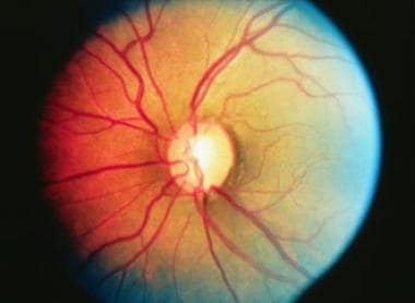
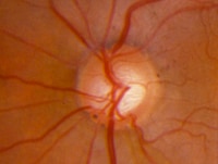
Optic Disk
Visual Field Tests
Refractive Surgical Procedures
Glaucoma, Open-Angle
Tomography, Optical Coherence
Visual Fields
Optic Atrophy
Cornea
Retina
Ocular Hypertension
Gonioscopy
Keratomileusis, Laser In Situ
Optic Nerve
Vision Disorders
Diagnostic Imaging
Microscopy, Confocal
Reproducibility of Results
Sensitivity and Specificity
Lasers, Solid-State
ROC Curve
Polarography
Prospective Studies
Laser Coagulation
Laser Therapy, Low-Level
Follow-Up Studies
Retinal Diseases
Lasers, Excimer
Fourier Analysis
The location of the inferior and superior temporal blood vessels and interindividual variability of the retinal nerve fiber layer thickness. (1/44)
(+info)Scan quality effect on glaucoma discrimination by glaucoma imaging devices. (2/44)
(+info)Determination of foveal location using scanning laser polarimetry. (3/44)
(+info)Scanning laser polarimetry and optical coherence tomography for detection of retinal nerve fiber layer defects. (4/44)
(+info)Structural and functional relationships in glaucoma using standard automated perimetry and the Humphrey Matrix. (5/44)
(+info)Relationship between pattern electroretinogram, standard automated perimetry, and optic nerve structural assessments. (6/44)
(+info)Agreement for detecting glaucoma progression with the GDx guided progression analysis, automated perimetry, and optic disc photography. (7/44)
(+info)Structure-function correlations using scanning laser polarimetry in primary angle-closure glaucoma and primary open-angle glaucoma. (8/44)
(+info)Scanning Laser Polarimetry (SLP) is not primarily a medical term, but a technique that has been applied in medical research and diagnostics. It's a non-invasive method used to analyze the polarization of light as it interacts with biological tissues.
In a simpler sense, SLP uses laser light, which is polarized (meaning all the light waves vibrate in the same plane), to scan tissue. As this light interacts with the tissue, changes in the polarization of the light can occur due to various factors such as the structure and composition of the tissue. By analyzing these changes, SLP can provide information about the tissue's properties, which can be useful in the detection and diagnosis of certain medical conditions.
For example, it has been used in research related to diagnosing diseases like glaucoma and Alzheimer's disease by analyzing the polarization changes in the eye's retina and the brain's cortex, respectively. However, it's important to note that while SLP has shown promise in these areas, it is not yet widely used in clinical settings.
Birefringence is a property of certain materials, such as crystals and some plastics, to split a beam of light into two separate beams with different polarization states and refractive indices when the light passes through the material. This phenomenon arises due to the anisotropic structure of these materials, where their physical properties vary depending on the direction of measurement.
When a unpolarized or partially polarized light beam enters a birefringent material, it gets separated into two orthogonally polarized beams called the ordinary and extraordinary rays. These rays propagate through the material at different speeds due to their distinct refractive indices, resulting in a phase delay between them. Upon exiting the material, the recombination of these two beams can produce various optical effects, such as double refraction or interference patterns, depending on the thickness and orientation of the birefringent material and the polarization state of the incident light.
Birefringence has numerous applications in optics, including waveplates, polarizing filters, stress analysis, and microscopy techniques like phase contrast and differential interference contrast imaging.
A laser is not a medical term per se, but a physical concept that has important applications in medicine. The term "LASER" stands for "Light Amplification by Stimulated Emission of Radiation." It refers to a device that produces and amplifies light with specific characteristics, such as monochromaticity (single wavelength), coherence (all waves moving in the same direction), and high intensity.
In medicine, lasers are used for various therapeutic and diagnostic purposes, including surgery, dermatology, ophthalmology, and dentistry. They can be used to cut, coagulate, or vaporize tissues with great precision, minimizing damage to surrounding structures. Additionally, lasers can be used to detect and measure physiological parameters, such as blood flow and oxygen saturation.
It's important to note that while lasers are powerful tools in medicine, they must be used by trained professionals to ensure safe and effective treatment.
Diagnostic techniques in ophthalmology refer to the various methods and tests used by eye specialists (ophthalmologists) to examine, evaluate, and diagnose conditions related to the eyes and visual system. Here are some commonly used diagnostic techniques:
1. Visual Acuity Testing: This is a basic test to measure the sharpness of a person's vision. It typically involves reading letters or numbers from an eye chart at a specific distance.
2. Refraction Test: This test helps determine the correct lens prescription for glasses or contact lenses by measuring how light is bent as it passes through the cornea and lens.
3. Slit Lamp Examination: A slit lamp is a microscope that allows an ophthalmologist to examine the structures of the eye, including the cornea, iris, lens, and retina, in great detail.
4. Tonometry: This test measures the pressure inside the eye (intraocular pressure) to detect conditions like glaucoma. Common methods include applanation tonometry and non-contact tonometry.
5. Retinal Imaging: Several techniques are used to capture images of the retina, including fundus photography, fluorescein angiography, and optical coherence tomography (OCT). These tests help diagnose conditions like macular degeneration, diabetic retinopathy, and retinal detachments.
6. Color Vision Testing: This test evaluates a person's ability to distinguish between different colors, which can help detect color vision deficiencies or neurological disorders affecting the visual pathway.
7. Visual Field Testing: This test measures a person's peripheral (or side) vision and can help diagnose conditions like glaucoma, optic nerve damage, or brain injuries.
8. Pupillary Reactions Tests: These tests evaluate how the pupils respond to light and near objects, which can provide information about the condition of the eye's internal structures and the nervous system.
9. Ocular Motility Testing: This test assesses eye movements and alignment, helping diagnose conditions like strabismus (crossed eyes) or nystagmus (involuntary eye movement).
10. Corneal Topography: This non-invasive imaging technique maps the curvature of the cornea, which can help detect irregularities, assess the fit of contact lenses, and plan refractive surgery procedures.
Nerve fibers are specialized structures that constitute the long, slender processes (axons) of neurons (nerve cells). They are responsible for conducting electrical impulses, known as action potentials, away from the cell body and transmitting them to other neurons or effector organs such as muscles and glands. Nerve fibers are often surrounded by supportive cells called glial cells and are grouped together to form nerve bundles or nerves. These fibers can be myelinated (covered with a fatty insulating sheath called myelin) or unmyelinated, which influences the speed of impulse transmission.
Ophthalmoscopy is a medical examination technique used by healthcare professionals to observe the interior structures of the eye, including the retina, optic disc, and vitreous humor. This procedure typically involves using an ophthalmoscope, a handheld device that consists of a light and magnifying lenses. The healthcare provider looks through the ophthalmoscope and directly observes the internal structures of the eye by illuminating them.
There are several types of ophthalmoscopy, including direct ophthalmoscopy, indirect ophthalmoscopy, and slit-lamp biomicroscopy. Each type has its own advantages and disadvantages, and they may be used in different situations depending on the specific clinical situation and the information needed.
Ophthalmoscopy is an important diagnostic tool for detecting and monitoring a wide range of eye conditions, including diabetic retinopathy, glaucoma, age-related macular degeneration, and other retinal disorders. It can also provide valuable information about the overall health of the individual, as changes in the appearance of the retina or optic nerve may indicate the presence of systemic diseases such as hypertension or diabetes.
Polarized light microscopy is a type of microscopy that uses polarized light to enhance contrast and reveal unique optical properties in specimens. In this technique, a polarizing filter is placed under the light source, which polarizes the light as it passes through. The specimen is then illuminated with this linearly polarized light. As the light travels through the specimen, its plane of polarization may be altered due to birefringence, a property of certain materials that causes the light to split into two separate rays with different refractive indices.
A second polarizing filter, called an analyzer, is placed in the light path between the objective and the eyepiece. The orientation of this filter can be adjusted to either allow or block the transmission of light through the microscope. When the polarizer and analyzer are aligned perpendicularly, no light will pass through if the specimen does not exhibit birefringence. However, if the specimen has birefringent properties, it will cause the plane of polarization to rotate, allowing some light to pass through the analyzer and create a contrasting image.
Polarized light microscopy is particularly useful for observing structures in minerals, crystals, and certain biological materials like collagen fibers, muscle proteins, and starch granules. It can also be used to study stress patterns in plastics and other synthetic materials.
Optic nerve diseases refer to a group of conditions that affect the optic nerve, which transmits visual information from the eye to the brain. These diseases can cause various symptoms such as vision loss, decreased visual acuity, changes in color vision, and visual field defects. Examples of optic nerve diseases include optic neuritis (inflammation of the optic nerve), glaucoma (damage to the optic nerve due to high eye pressure), optic nerve damage from trauma or injury, ischemic optic neuropathy (lack of blood flow to the optic nerve), and optic nerve tumors. Treatment for optic nerve diseases varies depending on the specific condition and may include medications, surgery, or lifestyle changes.
Glaucoma is a group of eye conditions that damage the optic nerve, often caused by an abnormally high pressure in the eye (intraocular pressure). This damage can lead to permanent vision loss or even blindness if left untreated. The most common type is open-angle glaucoma, which has no warning signs and progresses slowly. Angle-closure glaucoma, on the other hand, can cause sudden eye pain, redness, nausea, and vomiting, as well as rapid vision loss. Other less common types of glaucoma also exist. While there is no cure for glaucoma, early detection and treatment can help slow or prevent further vision loss.
Retinal Ganglion Cells (RGCs) are a type of neuron located in the innermost layer of the retina, the light-sensitive tissue at the back of the eye. These cells receive visual information from photoreceptors (rods and cones) via intermediate cells called bipolar cells. RGCs then send this visual information through their long axons to form the optic nerve, which transmits the signals to the brain for processing and interpretation as vision.
There are several types of RGCs, each with distinct morphological and functional characteristics. Some RGCs are specialized in detecting specific features of the visual scene, such as motion, contrast, color, or brightness. The diversity of RGCs allows for a rich and complex representation of the visual world in the brain.
Damage to RGCs can lead to various visual impairments, including loss of vision, reduced visual acuity, and altered visual fields. Conditions associated with RGC damage or degeneration include glaucoma, optic neuritis, ischemic optic neuropathy, and some inherited retinal diseases.
The optic disk, also known as the optic nerve head, is the point where the optic nerve fibers exit the eye and transmit visual information to the brain. It appears as a pale, circular area in the back of the eye, near the center of the retina. The optic disk has no photoreceptor cells (rods and cones), so it is insensitive to light. It is an important structure to observe during eye examinations because changes in its appearance can indicate various ocular diseases or conditions, such as glaucoma, optic neuritis, or papilledema.
A visual field test is a method used to measure an individual's entire scope of vision, which includes what can be seen straight ahead and in peripheral (or side) vision. During the test, the person being tested is asked to focus on a central point while gradually identifying the appearance of objects moving into their peripheral vision. The visual field test helps detect blind spots (scotomas) or gaps in the visual field, which can be caused by various conditions such as glaucoma, brain injury, optic nerve damage, or retinal disorders. It's an essential tool for diagnosing and monitoring eye-related diseases and conditions.
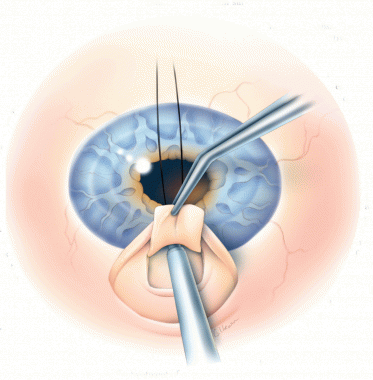
Refractive surgical procedures are a type of ophthalmic surgery aimed at improving the refractive state of the eye and reducing or eliminating the need for corrective eyewear. These procedures reshape the cornea or alter the lens of the eye to correct nearsightedness (myopia), farsightedness (hyperopia), presbyopia, or astigmatism.
Examples of refractive surgical procedures include:
1. Laser-assisted in situ keratomileusis (LASIK): A laser is used to create a thin flap in the cornea, which is then lifted to allow reshaping of the underlying tissue with another laser. The flap is replaced, and the procedure is completed.
2. Photorefractive keratectomy (PRK): This procedure involves removing the outer layer of the cornea (epithelium) and using a laser to reshape the underlying tissue. A bandage contact lens is placed over the eye to protect it during healing.
3. LASEK (laser-assisted subepithelial keratomileusis): Similar to LASIK, but instead of creating a flap, the epithelium is loosened with an alcohol solution and moved aside. The laser treatment is applied, and the epithelium is replaced.
4. Small Incision Lenticule Extraction (SMILE): A femtosecond laser creates a small lenticule within the cornea, which is then removed through a tiny incision. This procedure reshapes the cornea to correct refractive errors.
5. Refractive lens exchange (RLE): The eye's natural lens is removed and replaced with an artificial intraocular lens (IOL) to correct refractive errors, similar to cataract surgery.
6. Implantable contact lenses: A thin, foldable lens is placed between the iris and the natural lens or behind the iris to improve the eye's focusing power.
These procedures are typically performed on an outpatient basis and may require topical anesthesia (eye drops) or local anesthesia. Potential risks and complications include infection, dry eye, visual disturbances, and changes in night vision. It is essential to discuss these potential risks with your ophthalmologist before deciding on a refractive surgery procedure.
Open-angle glaucoma is a chronic, progressive type of glaucoma characterized by the gradual loss of optic nerve fibers and resulting in visual field defects. It is called "open-angle" because the angle where the iris meets the cornea (trabecular meshwork) appears to be normal and open on examination. The exact cause of this condition is not fully understood, but it is associated with increased resistance to the outflow of aqueous humor within the trabecular meshwork, leading to an increase in intraocular pressure (IOP). This elevated IOP can cause damage to the optic nerve and result in vision loss.
The onset of open-angle glaucoma is often asymptomatic, making regular comprehensive eye examinations crucial for early detection and management. Treatment typically involves lowering IOP using medications, laser therapy, or surgery to prevent further optic nerve damage and preserve vision.
Optical coherence tomography (OCT) is a non-invasive imaging technique that uses low-coherence light to capture high-resolution cross-sectional images of biological tissues, particularly the retina and other ocular structures. OCT works by measuring the echo time delay of light scattered back from different depths within the tissue, creating a detailed map of the tissue's structure. This technique is widely used in ophthalmology to diagnose and monitor various eye conditions such as macular degeneration, diabetic retinopathy, and glaucoma.
Visual fields refer to the total area in which objects can be seen while keeping the eyes focused on a central point. It is the entire area that can be observed using peripheral (side) vision while the eye gazes at a fixed point. A visual field test is used to detect blind spots or gaps (scotomas) in a person's vision, which could indicate various medical conditions such as glaucoma, retinal damage, optic nerve disease, brain tumors, or strokes. The test measures both the central and peripheral vision and maps the entire area that can be seen when focusing on a single point.
Optic atrophy is a medical term that refers to the degeneration and shrinkage (atrophy) of the optic nerve, which transmits visual information from the eye to the brain. This condition can result in various vision abnormalities, including loss of visual acuity, color vision deficiencies, and peripheral vision loss.
Optic atrophy can occur due to a variety of causes, such as:
* Traumatic injuries to the eye or optic nerve
* Glaucoma
* Optic neuritis (inflammation of the optic nerve)
* Ischemic optic neuropathy (reduced blood flow to the optic nerve)
* Compression or swelling of the optic nerve
* Hereditary or congenital conditions affecting the optic nerve
* Toxins and certain medications that can damage the optic nerve.
The diagnosis of optic atrophy typically involves a comprehensive eye examination, including visual acuity testing, refraction assessment, slit-lamp examination, and dilated funduscopic examination to evaluate the health of the optic nerve. In some cases, additional diagnostic tests such as visual field testing, optical coherence tomography (OCT), or magnetic resonance imaging (MRI) may be necessary to confirm the diagnosis and determine the underlying cause.
There is no specific treatment for optic atrophy, but addressing the underlying cause can help prevent further damage to the optic nerve. In some cases, vision rehabilitation may be recommended to help patients adapt to their visual impairment.
The cornea is the clear, dome-shaped surface at the front of the eye. It plays a crucial role in focusing vision. The cornea protects the eye from harmful particles and microorganisms, and it also serves as a barrier against UV light. Its transparency allows light to pass through and get focused onto the retina. The cornea does not contain blood vessels, so it relies on tears and the fluid inside the eye (aqueous humor) for nutrition and oxygen. Any damage or disease that affects its clarity and shape can significantly impact vision and potentially lead to blindness if left untreated.
The retina is the innermost, light-sensitive layer of tissue in the eye of many vertebrates and some cephalopods. It receives light that has been focused by the cornea and lens, converts it into neural signals, and sends these to the brain via the optic nerve. The retina contains several types of photoreceptor cells including rods (which handle vision in low light) and cones (which are active in bright light and are capable of color vision).
In medical terms, any pathological changes or diseases affecting the retinal structure and function can lead to visual impairment or blindness. Examples include age-related macular degeneration, diabetic retinopathy, retinal detachment, and retinitis pigmentosa among others.
Ocular hypertension is a medical condition characterized by elevated pressure within the eye (intraocular pressure or IOP), which is higher than normal but not necessarily high enough to cause any visible damage to the optic nerve or visual field loss. It serves as a significant risk factor for developing glaucoma, a sight-threatening disease.
The normal range of intraocular pressure is typically between 10-21 mmHg (millimeters of mercury). Ocular hypertension is often defined as an IOP consistently above 21 mmHg, although some studies suggest that even pressures between 22-30 mmHg may not cause damage in all individuals. Regular monitoring and follow-up with an ophthalmologist are essential for people diagnosed with ocular hypertension to ensure early detection and management of any potential glaucomatous changes. Treatment options include medications, laser therapy, or surgery to lower the IOP and reduce the risk of glaucoma onset.
Gonioscopy is a diagnostic procedure in ophthalmology used to examine the anterior chamber angle, which is the area where the iris and cornea meet. This examination helps to evaluate the drainage pathways of the eye for conditions such as glaucoma. A special contact lens called a goniolens is placed on the cornea during the procedure to allow the healthcare provider to visualize the angle using a biomicroscope. The lens may be coupled with a mirrored or prismatic surface to enhance the view of the angle. Gonioscopy can help detect conditions like narrow angles, closed angles, neovascularization, and other abnormalities that might contribute to glaucoma development or progression.
Intraocular pressure (IOP) is the fluid pressure within the eye, specifically within the anterior chamber, which is the space between the cornea and the iris. It is measured in millimeters of mercury (mmHg). The aqueous humor, a clear fluid that fills the anterior chamber, is constantly produced and drained, maintaining a balance that determines the IOP. Normal IOP ranges from 10-21 mmHg, with average values around 15-16 mmHg. Elevated IOP is a key risk factor for glaucoma, a group of eye conditions that can lead to optic nerve damage and vision loss if not treated promptly and effectively. Regular monitoring of IOP is essential in diagnosing and managing glaucoma and other ocular health issues.
Laser In Situ Keratomileusis (LASIK) is a type of refractive surgery used to correct vision issues such as myopia (nearsightedness), hyperopia (farsightedness), and astigmatism. The procedure involves reshaping the cornea, which is the clear, dome-shaped surface at the front of the eye, using an excimer laser.
In LASIK, a thin flap is created on the surface of the cornea using a femtosecond or microkeratome laser. The flap is then lifted, and the excimer laser is used to reshape the underlying tissue. After the reshaping is complete, the flap is replaced, allowing for quicker healing and visual recovery compared to other refractive surgery procedures.
LASIK is an outpatient procedure that typically takes about 30 minutes or less per eye. Most people can expect to see improved vision within a few days of the procedure, although it may take several weeks for vision to fully stabilize. LASIK has a high success rate and is generally considered safe when performed by a qualified surgeon. However, as with any surgical procedure, there are risks involved, including dry eye, infection, and visual complications such as glare or halos around lights.
The optic nerve, also known as the second cranial nerve, is the nerve that transmits visual information from the retina to the brain. It is composed of approximately one million nerve fibers that carry signals related to vision, such as light intensity and color, from the eye's photoreceptor cells (rods and cones) to the visual cortex in the brain. The optic nerve is responsible for carrying this visual information so that it can be processed and interpreted by the brain, allowing us to see and perceive our surroundings. Damage to the optic nerve can result in vision loss or impairment.
Vision disorders refer to a wide range of conditions that affect the visual system and result in various symptoms, such as blurry vision, double vision, distorted vision, impaired depth perception, and difficulty with visual tracking or focusing. These disorders can be categorized into several types, including:
1. Refractive errors: These occur when the shape of the eye prevents light from focusing directly on the retina, resulting in blurry vision. Examples include myopia (nearsightedness), hyperopia (farsightedness), astigmatism, and presbyopia (age-related loss of near vision).
2. Strabismus: Also known as crossed eyes or walleye, strabismus is a misalignment of the eyes where they point in different directions, which can lead to double vision or loss of depth perception.
3. Amblyopia: Often called lazy eye, amblyopia is a condition where one eye has reduced vision due to lack of proper visual development during childhood. It may be caused by strabismus, refractive errors, or other factors that interfere with normal visual development.
4. Accommodative disorders: These involve problems with the focusing ability of the eyes, such as convergence insufficiency (difficulty focusing on close objects) and accommodative dysfunction (inability to maintain clear vision at different distances).
5. Binocular vision disorders: These affect how the eyes work together as a team, leading to issues like poor depth perception, eye strain, and headaches. Examples include convergence insufficiency, divergence excess, and suppression.
6. Ocular motility disorders: These involve problems with eye movement, such as nystagmus (involuntary eye movements), strabismus, or restricted extraocular muscle function.
7. Visual processing disorders: These affect the brain's ability to interpret and make sense of visual information, even when the eyes themselves are healthy. Symptoms may include difficulty with reading, recognizing shapes and objects, and understanding spatial relationships.
8. Low vision: This term refers to significant visual impairment that cannot be fully corrected with glasses, contact lenses, medication, or surgery. It includes conditions like macular degeneration, diabetic retinopathy, glaucoma, and cataracts.
9. Blindness: Complete loss of sight in both eyes, which can be caused by various factors such as injury, disease, or genetic conditions.
Diagnostic imaging is a medical specialty that uses various technologies to produce visual representations of the internal structures and functioning of the body. These images are used to diagnose injury, disease, or other abnormalities and to monitor the effectiveness of treatment. Common modalities of diagnostic imaging include:
1. Radiography (X-ray): Uses ionizing radiation to produce detailed images of bones, teeth, and some organs.
2. Computed Tomography (CT) Scan: Combines X-ray technology with computer processing to create cross-sectional images of the body.
3. Magnetic Resonance Imaging (MRI): Uses a strong magnetic field and radio waves to generate detailed images of soft tissues, organs, and bones.
4. Ultrasound: Employs high-frequency sound waves to produce real-time images of internal structures, often used for obstetrics and gynecology.
5. Nuclear Medicine: Involves the administration of radioactive tracers to assess organ function or detect abnormalities within the body.
6. Positron Emission Tomography (PET) Scan: Uses a small amount of radioactive material to produce detailed images of metabolic activity in the body, often used for cancer detection and monitoring treatment response.
7. Fluoroscopy: Utilizes continuous X-ray imaging to observe moving structures or processes within the body, such as swallowing studies or angiography.
Diagnostic imaging plays a crucial role in modern medicine, allowing healthcare providers to make informed decisions about patient care and treatment plans.
Confocal microscopy is a powerful imaging technique used in medical and biological research to obtain high-resolution, contrast-rich images of thick samples. This super-resolution technology provides detailed visualization of cellular structures and processes at various depths within a specimen.
In confocal microscopy, a laser beam focused through a pinhole illuminates a small spot within the sample. The emitted fluorescence or reflected light from this spot is then collected by a detector, passing through a second pinhole that ensures only light from the focal plane reaches the detector. This process eliminates out-of-focus light, resulting in sharp images with improved contrast compared to conventional widefield microscopy.
By scanning the laser beam across the sample in a raster pattern and collecting fluorescence at each point, confocal microscopy generates optical sections of the specimen. These sections can be combined to create three-dimensional reconstructions, allowing researchers to study cellular architecture and interactions within complex tissues.
Confocal microscopy has numerous applications in medical research, including studying protein localization, tracking intracellular dynamics, analyzing cell morphology, and investigating disease mechanisms at the cellular level. Additionally, it is widely used in clinical settings for diagnostic purposes, such as analyzing skin lesions or detecting pathogens in patient samples.
Reproducibility of results in a medical context refers to the ability to obtain consistent and comparable findings when a particular experiment or study is repeated, either by the same researcher or by different researchers, following the same experimental protocol. It is an essential principle in scientific research that helps to ensure the validity and reliability of research findings.
In medical research, reproducibility of results is crucial for establishing the effectiveness and safety of new treatments, interventions, or diagnostic tools. It involves conducting well-designed studies with adequate sample sizes, appropriate statistical analyses, and transparent reporting of methods and findings to allow other researchers to replicate the study and confirm or refute the results.
The lack of reproducibility in medical research has become a significant concern in recent years, as several high-profile studies have failed to produce consistent findings when replicated by other researchers. This has led to increased scrutiny of research practices and a call for greater transparency, rigor, and standardization in the conduct and reporting of medical research.
Optical rotation, also known as optical activity, is a property of certain substances to rotate the plane of polarization of linearly polarized light as it passes through the substance. This ability arises from the presence of optically active molecules, most commonly chiral molecules, which have a non-superimposable mirror image.
The angle and direction of rotation (either clockwise or counterclockwise) are specific to each optically active substance and can be used as a characteristic identification property. The measurement of optical rotation is an important tool in the determination of the enantiomeric purity of chiral compounds, such as drugs and natural products, in chemistry and pharmacology.
The optical rotation of a substance can be influenced by factors such as temperature, concentration, wavelength of light, and solvent used. The magnitude of the optical rotation is often reported as the specific rotation, which is the optical rotation per unit length (usually expressed in degrees) and per unit concentration (often given in grams per deciliter or g/dL).
Sensitivity and specificity are statistical measures used to describe the performance of a diagnostic test or screening tool in identifying true positive and true negative results.
* Sensitivity refers to the proportion of people who have a particular condition (true positives) who are correctly identified by the test. It is also known as the "true positive rate" or "recall." A highly sensitive test will identify most or all of the people with the condition, but may also produce more false positives.
* Specificity refers to the proportion of people who do not have a particular condition (true negatives) who are correctly identified by the test. It is also known as the "true negative rate." A highly specific test will identify most or all of the people without the condition, but may also produce more false negatives.
In medical testing, both sensitivity and specificity are important considerations when evaluating a diagnostic test. High sensitivity is desirable for screening tests that aim to identify as many cases of a condition as possible, while high specificity is desirable for confirmatory tests that aim to rule out the condition in people who do not have it.
It's worth noting that sensitivity and specificity are often influenced by factors such as the prevalence of the condition in the population being tested, the threshold used to define a positive result, and the reliability and validity of the test itself. Therefore, it's important to consider these factors when interpreting the results of a diagnostic test.
Solid-state lasers are a type of laser that uses solid materials as the gain medium – the material that amplifies the light energy to produce laser emissions. In contrast to gas or liquid lasers, solid-state lasers use a crystal, ceramic, or glass as the gain medium. The active laser medium in solid-state lasers is typically doped with rare earth ions, such as neodymium (Nd), yttrium (Y), erbium (Er), or thulium (Tm).
The most common type of solid-state laser is the neodymium-doped yttrium aluminum garnet (Nd:YAG) laser. In this laser, neodymium ions are doped into a crystal lattice made up of yttrium, aluminum, and garnet (YAG). The Nd:YAG laser emits light at a wavelength of 1064 nanometers (nm), which can be frequency-doubled to produce emissions at 532 nm.
Solid-state lasers have several advantages over other types of lasers, including high efficiency, long lifetimes, and compact size. They are widely used in various applications, such as material processing, medical treatments, scientific research, and military technology.
A Receiver Operating Characteristic (ROC) curve is a graphical representation used in medical decision-making and statistical analysis to illustrate the performance of a binary classifier system, such as a diagnostic test or a machine learning algorithm. It's a plot that shows the tradeoff between the true positive rate (sensitivity) and the false positive rate (1 - specificity) for different threshold settings.
The x-axis of an ROC curve represents the false positive rate (the proportion of negative cases incorrectly classified as positive), while the y-axis represents the true positive rate (the proportion of positive cases correctly classified as positive). Each point on the curve corresponds to a specific decision threshold, with higher points indicating better performance.
The area under the ROC curve (AUC) is a commonly used summary measure that reflects the overall performance of the classifier. An AUC value of 1 indicates perfect discrimination between positive and negative cases, while an AUC value of 0.5 suggests that the classifier performs no better than chance.
ROC curves are widely used in healthcare to evaluate diagnostic tests, predictive models, and screening tools for various medical conditions, helping clinicians make informed decisions about patient care based on the balance between sensitivity and specificity.
Polarography is a type of electrochemical analysis technique used to determine the concentration of an ion or electron-transferring species in a solution. It involves measuring the current that flows through an electrode as the voltage is varied, which can provide information about the redox potential and the number of electrons transferred during a reaction. The technique is particularly useful for analyzing complex mixtures and for detecting trace amounts of substances.
In polarography, a dropping mercury electrode (DME) is typically used as the working electrode. As the mercury droplets fall from the electrode, they create fresh surfaces for analysis, which helps to minimize interference from surface-adsorbed species. The DME is immersed in a solution containing the analyte along with a supporting electrolyte, and a potential is applied between the DME and a reference electrode.
As the potential is scanned, reduction or oxidation of the analyte occurs at the DME surface, leading to a current that can be measured. The resulting polarogram (a plot of current vs. voltage) shows peaks or waves corresponding to the redox potentials of the analyte, which can be used to identify and quantify the species present in the solution.
Polarography is a sensitive and selective technique that has been widely used in fields such as environmental analysis, pharmaceuticals, and biochemistry. However, it has largely been replaced by more modern electrochemical techniques, such as cyclic voltammetry and differential pulse voltammetry, which offer higher sensitivity and better resolution of complex mixtures.
Prospective studies, also known as longitudinal studies, are a type of cohort study in which data is collected forward in time, following a group of individuals who share a common characteristic or exposure over a period of time. The researchers clearly define the study population and exposure of interest at the beginning of the study and follow up with the participants to determine the outcomes that develop over time. This type of study design allows for the investigation of causal relationships between exposures and outcomes, as well as the identification of risk factors and the estimation of disease incidence rates. Prospective studies are particularly useful in epidemiology and medical research when studying diseases with long latency periods or rare outcomes.
Laser coagulation, also known as laser photocoagulation, is a medical procedure that uses a laser to seal or destroy abnormal blood vessels or tissue. The laser produces a concentrated beam of light that can be precisely focused on the target area. When the laser energy is absorbed by the tissue, it causes the temperature to rise, which leads to coagulation (the formation of a clot) or destruction of the tissue.
In ophthalmology, laser coagulation is commonly used to treat conditions such as diabetic retinopathy, age-related macular degeneration, and retinal tears or holes. The procedure can help to seal leaking blood vessels, reduce fluid leakage, and prevent further vision loss. It is usually performed as an outpatient procedure and may be repeated if necessary.
In other medical specialties, laser coagulation may be used to control bleeding, destroy tumors, or remove unwanted tissue. The specific technique and parameters of the laser treatment will depend on the individual patient's needs and the condition being treated.
Low-level laser therapy (LLLT), also known as cold laser or soft laser, is a form of phototherapy which uses low-intensity lasers or light-emitting diodes to treat various medical conditions. The laser beam is usually applied directly to the skin and penetrates up to several centimeters into the tissue without causing heat damage or pain.
The therapeutic effect of LLLT is believed to be due to the bio-stimulation of cellular processes, including increased ATP production, modulation of reactive oxygen species, and activation of signaling pathways that promote tissue repair and reduce inflammation. The wavelength and power density of the laser light are important factors in determining its biological effects.
LLLT has been used to treat a variety of conditions such as musculoskeletal pain, wound healing, skin rejuvenation, hair growth, and neurological disorders. However, its efficacy is still a subject of ongoing research and debate, with some studies reporting positive results while others showing no significant benefits compared to placebo.
It's important to note that LLLT should only be administered by trained healthcare professionals, as improper use can lead to eye damage or other adverse effects.
Follow-up studies are a type of longitudinal research that involve repeated observations or measurements of the same variables over a period of time, in order to understand their long-term effects or outcomes. In medical context, follow-up studies are often used to evaluate the safety and efficacy of medical treatments, interventions, or procedures.
In a typical follow-up study, a group of individuals (called a cohort) who have received a particular treatment or intervention are identified and then followed over time through periodic assessments or data collection. The data collected may include information on clinical outcomes, adverse events, changes in symptoms or functional status, and other relevant measures.
The results of follow-up studies can provide important insights into the long-term benefits and risks of medical interventions, as well as help to identify factors that may influence treatment effectiveness or patient outcomes. However, it is important to note that follow-up studies can be subject to various biases and limitations, such as loss to follow-up, recall bias, and changes in clinical practice over time, which must be carefully considered when interpreting the results.
Retinal diseases refer to a group of conditions that affect the retina, which is the light-sensitive tissue located at the back of the eye. The retina is responsible for converting light into electrical signals that are sent to the brain and interpreted as visual images. Retinal diseases can cause vision loss or even blindness, depending on their severity and location in the retina.
Some common retinal diseases include:
1. Age-related macular degeneration (AMD): A progressive disease that affects the central part of the retina called the macula, causing blurred or distorted vision.
2. Diabetic retinopathy: A complication of diabetes that can damage the blood vessels in the retina, leading to vision loss.
3. Retinal detachment: A serious condition where the retina becomes separated from its underlying tissue, requiring immediate medical attention.
4. Macular edema: Swelling or thickening of the macula due to fluid accumulation, which can cause blurred vision.
5. Retinitis pigmentosa: A group of inherited eye disorders that affect the retina's ability to respond to light, causing progressive vision loss.
6. Macular hole: A small break in the macula that can cause distorted or blurry vision.
7. Retinal vein occlusion: Blockage of the retinal veins that can lead to bleeding, swelling, and potential vision loss.
Treatment for retinal diseases varies depending on the specific condition and its severity. Some treatments include medication, laser therapy, surgery, or a combination of these options. Regular eye exams are essential for early detection and treatment of retinal diseases.
An excimer laser is a type of laser that is used in various medical procedures, particularly in ophthalmology and dermatology. The term "excimer" is derived from "excited dimer," which refers to a short-lived molecule formed when two atoms combine in an excited state.
Excimer lasers emit light at a specific wavelength that is determined by the type of gas used in the laser. In medical applications, excimer lasers typically use noble gases such as argon, krypton, or xenon, combined with halogens such as fluorine or chlorine. The most commonly used excimer laser in medical procedures is the excimer laser that uses a mixture of argon and fluoride gas to produce light at a wavelength of 193 nanometers (nm).
In ophthalmology, excimer lasers are primarily used for refractive surgery, such as LASIK and PRK, to correct vision problems like myopia, hyperopia, and astigmatism. The laser works by vaporizing tiny amounts of tissue from the cornea, reshaping its curvature to improve the way light is focused onto the retina.
In dermatology, excimer lasers are used for various skin conditions, including psoriasis, vitiligo, and atopic dermatitis. The laser works by emitting high-energy ultraviolet (UV) light that selectively targets and destroys the abnormal cells responsible for these conditions while leaving surrounding healthy tissue intact.
Excimer lasers are known for their precision, accuracy, and minimal side effects, making them a popular choice in medical procedures where fine detail and tissue preservation are critical.
I'm sorry for any confusion, but Fourier Analysis is not a medical term. It is a mathematical concept used in various scientific fields, including physics, engineering, and signal processing.
Fourier Analysis is a method to decompose functions into sinusoidal components (sines and cosines) of different frequencies. This allows for the representation of a function or a signal as a sum of these frequency components. It's particularly useful in analyzing periodic functions, understanding signals, and solving partial differential equations.
If you have any medical terms you would like me to define, please let me know!
 Scanning laser polarimetry - Wikipedia
 Predicting Visual Field Loss in Glaucomatous Patients Using Fast-Fourier Analysis (FFA) of GDx-VCC Scanning Laser Polarimetry |...
 Zeiss GDx Pro 8000 - Scanning Laser Polarimetry
 Primary Open-Angle Glaucoma (POAG): Practice Essentials, Background, Pathophysiology
 Lasers - Annals Singapore
 Browsing Optometry and Vision Science by Title
 Relation of completeness of reporting of health research to journals' endorsement of reporting guidelines: systematic review |...
 Ophthalmic Technologies XVIII | (2008) | Publications | Spie
 Frontiers | The Eye As a Biomarker for Alzheimer's Disease
 Robert Chang, MD's Profile | Stanford Profiles
 Fred Fitzke - Google Scholar
 Classification List
![Schrems WA[au] - Search Results - PubMed](data:image/png;base64,iVBORw0KGgoAAAANSUhEUgAAABAAAAAQCAMAAAAoLQ9TAAAARVBMVEVHcEwoU45gYmYAUpQAUpRPYGVgYmZLXnJgYmYAUZUAUpRJXnIAUpQAUpRgYmYAUpRgYmZgYmZhYmYAUpQAUpQAUpRgYmaDiPJuAAAAFXRSTlMADOJ+6QewGO8/uTRqtH7GdFJ11p1bCL3TAAAAZUlEQVQYlV2PVw7AIAxDTeney7n/UcsoldX3E+VJOAboEi7MBpHWMs1ADlG8u7UYWauwyZFeRQVPOhG2o+aiwhByJxUx91Jxhje3iJSqGfHuLKI0+0TpXvY1twCOPlFh5pa/++MB0vIOBm+1zaoAAAAASUVORK5CYII=) Schrems WA[au] - Search Results - PubMed
Schrems WA[au] - Search Results - PubMed Klinische Forschung Glaukom - Universitätsklinik für Augenheilkunde
 Use of OCT in Neurodegenerative Diseases - EyeWiki
 Equipment | The Rotterdam Ophthalmic Institute
 Renishaw XL80 XC80 Laser Interferometer Calibration System Whitby - BUY
 Risk Factors for Primary Open Angle Glaucoma (POAG) Progression: A Study Ruled in Torino
 Correlation of structure and function in glaucoma - Philippine Journal Of Ophthalmology
 Daily illumination exposure and melatonin: influence of ophthalmic dysfunction and sleep duration | Journal of Circadian...
 Pesquisa | Biblioteca Virtual em Saúde - BRASIL