Taste Buds
Sensory Receptor Cells
Taste
Olfactory Receptor Neurons
Chorda Tympani Nerve
Olfactory Mucosa
Cell Membrane
Receptors, Cell Surface
Tongue
Necturus
Chemoreceptor Cells
Necturus maculosus
Taste Threshold
Salamandridae
Odors
Glossopharyngeal Nerve
Quinine
Olfactory Pathways
Stimulation, Chemical
Cilia
Transducin
Vomeronasal Organ
Phospholipase C beta
Taste Perception
Rana ridibunda
Diptera
Molecular Sequence Data
Electrophysiology
Rana catesbeiana
Signal Transduction
Cells, Cultured
Sucrose
Nephropidae
Endocytosis
Amino Acid Sequence
Cricetinae
Hydrochloric Acid
Salamandra
Phenylthiourea
Receptors, G-Protein-Coupled
Antigens, Surface
Geniculate Ganglion
Calcium
CHO Cells
Citric Acid
TRPM Cation Channels
Monoterpenes
Rana esculenta
Nervous System Physiological Processes
Membrane Proteins
Epithelium
Microscopy, Electron
Amiloride
Protein Binding
Receptors, Odorant
Transfection
Second Messenger Systems
Olfactory Bulb
Taste Disorders
Ambystoma
Lectins
Action Potentials
Cricetulus
Mice, Inbred C57BL
Mechanoreceptors
Immunohistochemistry
Receptors, Immunologic
Cyclohexanols
Sweetening Agents
Ion Channels
Patch-Clamp Techniques
Crustacea
Protein Transport
Tumor Cells, Cultured
Carbohydrates
Hair Cells, Auditory
Cyclic AMP
Quaternary Ammonium Compounds
Membrane Potentials
Ligands
Rana temporaria
RNA, Messenger
Mollusca
Base Sequence
Rats, Sprague-Dawley
Photoreceptor Cells
Microscopy, Confocal
Nucleotides, Cyclic
Microscopy, Electron, Scanning
Olfactory Nerve
Flow Cytometry
Dose-Response Relationship, Drug
Potassium Chloride
Cytoplasm
Potassium
Antigens, CD
Ammonium Chloride
Glycoproteins
Glycosylation
Sodium
Binding, Competitive
Fluorescent Antibody Technique
Models, Biological
Hydrogen-Ion Concentration
Gene Expression Regulation
Biological Transport
Epithelial Cells
Cyclic GMP
Cell Division
Nasal Mucosa
Heparitin Sulfate
Green Fluorescent Proteins
Golgi Apparatus
Recombinant Fusion Proteins
Gene Expression
Adaptation, Physiological
Acids
Mutation
Cyclic Nucleotide-Gated Cation Channels
Neurons
Protein Structure, Tertiary
Hearing
Cloning, Molecular
Fishes
Iodine Radioisotopes
Receptors, Virus
Microscopy, Fluorescence
1-Methyl-3-isobutylxanthine
Cycloheximide
Fluorescent Dyes
Sodium Channels
Mice, Transgenic
Reverse Transcriptase Polymerase Chain Reaction
Mice, Knockout
Cell Adhesion Molecules
Type C Phospholipases
Endosomes
Heparan Sulfate Proteoglycans
Ion Channel Gating
Binding Sites
COS Cells
Nerve Tissue Proteins
Glycosylphosphatidylinositols
T-Lymphocytes
Protein Processing, Post-Translational
Surface Properties
Endoplasmic Reticulum
Cercopithecus aethiops
Cell Compartmentation
Microelectrodes
Fibroblasts
Osmolar Concentration
In Situ Hybridization
Neurotransmitter Agents
Calcium Channels
Electrophoresis, Polyacrylamide Gel
Nerve Fibers
Carrier Proteins
Blotting, Western
Enzyme Inhibitors
Fibronectins
HeLa Cells
Antigens, Neoplasm
Cell Differentiation
Neuraminidase
Down-Regulation
Serotonin
Glycosaminoglycans
Synaptic Transmission
Bacterial Adhesion
Brefeldin A
Precipitin Tests
Heparin
Microscopy, Immunoelectron
Antibodies
Lysosomes
Viral Envelope Proteins
Sonic hedgehog signaling by the patched-smoothened receptor complex. (1/20616)
BACKGROUND: The Hedgehog (Hh) family of secreted proteins is involved in a number of developmental processes as well as in cancer. Genetic and biochemical data suggest that the Sonic hedgehog (Shh) receptor is composed of at least two proteins: the tumor suppressor protein Patched (Ptc) and the seven-transmembrane protein Smoothened (Smo). RESULTS: Using a biochemical assay for activation of the transcription factor Gli, a downstream component of the Hh pathway, we show here that Smo functions as the signaling component of the Shh receptor, and that this activity can be blocked by Ptc. The inhibition of Smo by Ptc can be relieved by the addition of Shh. Furthermore, oncogenic forms of Smo are insensitive to Ptc repression in this assay. Mapping of the Smo domains required for binding to Ptc and for signaling revealed that the Smo-Ptc interaction involves mainly the amino terminus of Smo, and that the third intracellular loop and the seventh transmembrane domain are required for signaling. CONCLUSIONS: These data demonstrate that Smo is the signaling component of a multicomponent Hh receptor complex and that Ptc is a ligand-regulated inhibitor of Smo. Different domains of Smo are involved in Ptc binding and activation of a Gli reporter construct. The latter requires the third intracellular loop and the seventh transmembrane domain of Smo, regions often involved in coupling to G proteins. No changes in the levels of cyclic AMP or calcium associated with such pathways could be detected following receptor activation, however. (+info)Activation of Src in human breast tumor cell lines: elevated levels of phosphotyrosine phosphatase activity that preferentially recognizes the Src carboxy terminal negative regulatory tyrosine 530. (2/20616)
Elevated levels of Src kinase activity have been reported in a number of human cancers, including colon and breast cancer. We have analysed four human breast tumor cell lines that exhibit high levels of Src kinase activity, and have determined that these cell lines also exhibit a high level of a phosphotyrosine phosphatase activity that recognizes the Src carboxy-terminal P-Tyr530 negative regulatory site. Total Src kinase activity in these cell lines is elevated as much as 30-fold over activity in normal control cells and specific activity is elevated as much as 5.6-fold. When the breast tumor cells were grown in the presence of the tyrosine phosphatase inhibitor vanadate, Src kinase activity was reduced in all four breast tumor cell lines, suggesting that Src was being activated by a phosphatase which could recognize the Tyr530 negative regulatory site. In fractionated cell extracts from the breast tumor cells, we found elevated levels of a membrane associated tyrosine phosphatase activity that preferentially dephosphorylated a Src family carboxy-terminal phosphopeptide containing the regulatory tyrosine 530 site. Src was hypophosphorylated in vivo at tyrosine 530 in at least two of the tumor cell lines, further suggesting that Src was being activated by a phosphatase in these cells. In preliminary immunoprecipitation and antibody depletion experiments, we were unable to correlate the major portion of this phosphatase activity with several known phosphatases. (+info)Leptin suppression of insulin secretion and gene expression in human pancreatic islets: implications for the development of adipogenic diabetes mellitus. (3/20616)
Previously we demonstrated the expression of the long form of the leptin receptor in rodent pancreatic beta-cells and an inhibition of insulin secretion by leptin via activation of ATP-sensitive potassium channels. Here we examine pancreatic islets isolated from pancreata of human donors for their responses to leptin. The presence of leptin receptors on islet beta-cells was demonstrated by double fluorescence confocal microscopy after binding of a fluorescent derivative of human leptin (Cy3-leptin). Leptin (6.25 nM) suppressed insulin secretion of normal islets by 20% at 5.6 mM glucose. Intracellular calcium responses to 16.7 mM glucose were rapidly reduced by leptin. Proinsulin messenger ribonucleic acid expression in islets was inhibited by leptin at 11.1 mM, but not at 5.6 mM glucose. Leptin also reduced proinsulin messenger ribonucleic acid levels that were increased in islets by treatment with 10 nM glucagon-like peptide-1 in the presence of either 5.6 or 11.1 mM glucose. These findings demonstrate direct suppressive effects of leptin on insulin-producing beta-cells in human islets at the levels of both stimulus-secretion coupling and gene expression. The findings also further indicate the existence of an adipoinsular axis in humans in which insulin stimulates leptin production in adipocytes and leptin inhibits the production of insulin in beta-cells. We suggest that dysregulation of the adipoinsular axis in obese individuals due to defective leptin reception by beta-cells may result in chronic hyperinsulinemia and may contribute to the pathogenesis of adipogenic diabetes. (+info)p38 mitogen-activated protein kinase can be involved in transforming growth factor beta superfamily signal transduction in Drosophila wing morphogenesis. (4/20616)
p38 mitogen-activated protein kinase (p38) has been extensively studied as a stress-responsive kinase, but its role in development remains unknown. The fruit fly, Drosophila melanogaster, has two p38 genes, D-p38a and D-p38b. To elucidate the developmental function of the Drosophila p38's, we used various genetic and pharmacological manipulations to interfere with their functions: expression of a dominant-negative form of D-p38b, expression of antisense D-p38b RNA, reduction of the D-p38 gene dosage, and treatment with the p38 inhibitor SB203580. Expression of a dominant-negative D-p38b in the wing imaginal disc caused a decapentaplegic (dpp)-like phenotype and enhanced the phenotype of a dpp mutant. Dpp is a secretory ligand belonging to the transforming growth factor beta superfamily which triggers various morphogenetic processes through interaction with the receptor Thick veins (Tkv). Inhibition of D-p38b function also caused the suppression of the wing phenotype induced by constitutively active Tkv (TkvCA). Mosaic analysis revealed that D-p38b regulates the Tkv-dependent transcription of the optomotor-blind (omb) gene in non-Dpp-producing cells, indicating that the site of D-p38b action is downstream of Tkv. Furthermore, forced expression of TkvCA induced an increase in the phosphorylated active form(s) of D-p38(s). These results demonstrate that p38, in addition to its role as a transducer of emergency stress signaling, may function to modulate Dpp signaling. (+info)Ligand substitution of receptor targeted DNA complexes affects gene transfer into hepatoma cells. (5/20616)
We have targeted the serpin enzyme complex receptor for gene transfer in human hepatoma cell lines using peptides < 30 amino acids in length which contain the five amino acid recognition sequence for this receptor, coupled to poly K of average chain length 100 K, using the heterobifunctional coupling reagent sulfo-LC SPDP. The number of sulfo-LC SPDP modified poly-L-lysine residues, as well as the degree of peptide substitution was assessed by nuclear magnetic resonance spectroscopy. Conjugates were prepared in which 3.5%, 7.8% or 26% of the lysine residues contained the sulfo-LC SPDP moiety. Each of these conjugates was then coupled with ligand peptides so that one in 370, one in 1039, or one in 5882 lysines were substituted with receptor ligand. Electron microscopy and atomic force microscopy were used to assess complex structure and size. HuH7 human hepatoma cells were transfected with complexes of these conjugates with the plasmid pGL3 and luciferase expression measured 2 to 16 days after treatment. All the protein conjugates in which 26% of the K residues were modified with sulfo-LC SPDP were poor gene transfer reagents. Complexes containing less substituted poly K, averaged 17 +/- 0.5 nm in diameter and gave peak transgene expression of 3-4 x 10(6) ILU/mg which persisted (> 7 x 10(5) ILU) at 16 days. Of these, more substituted polymers condensed DNA into complexes averaging 20 +/- 0.7 nm in diameter and gave five-fold less luciferase than complexes containing less substituted conjugates. As few as eight to 11 ligands per complex are optimal for DNA delivery via the SEC receptor. The extent of substitution of receptor-mediated gene transfer complexes affects the size of the complexes, as well as the intensity and duration of transgene expression. These observations may permit tailoring of complex construction for the usage required. (+info)Characterization of Moraxella (Branhamella) catarrhalis lbpB, lbpA, and lactoferrin receptor orf3 isogenic mutants. (6/20616)
Pathogenic members of the family Neisseriaceae produce specific receptors to acquire iron from their host's lactoferrin and transferrin. Recently, putative Moraxella catarrhalis lactoferrin receptor genes and a third open reading frame (lbpB, lbpA, and orf3) were cloned and sequenced. We describe the preliminary characterization of isogenic mutants deficient in LbpB, LbpA, or Orf3 protein. (+info)Three receptor genes for plasminogen related growth factors in the genome of the puffer fish Fugu rubripes. (7/20616)
Plasminogen related growth factors (PRGFs) and their receptors play major roles in embryogenesis, tissue regeneration and neoplasia. In order to investigate the complexity and evolution of the PRGF receptor family we have cloned and sequenced three receptors for PRGFs in the teleost fish Fugu rubripes, a model vertebrate with a compact genome. One of the receptor genes isolated encodes the orthologue of mammalian MET, whilst the other two may represent Fugu rubripes orthologues of RON and SEA. This is the first time three PRGF receptors have been identified in a single species. (+info)Cloning, molecular analysis and differential cell localisation of the p36 RACK analogue antigen from the parasite protozoon Crithidia fasciculata. (8/20616)
The family of the RACK molecules (receptors for activated C kinases) are present in all the species studied so far. In the genus Leishmania, these molecules also induce a strong immune reaction against the infection. We have cloned and characterised the gene that encodes the RACK analogue from the parasite trypanosomatid Crithidia fasciculata (CACK). The molecule seems to be encoded by two genes. The sequence analysis of the cloned open reading frame indicates the existence of a high degree of conservation not only with other members of the Trypanosomatidae but also with mammalians. The study of the protein kinase C phosphorylation sites shows the presence of three of them, shared with the mammalian species, additional to those present in the other protozoa suggesting a certain phylogenetic distance between the protozoon Crithidia fasciculata and the rest of the Trypanosomatidae. The CACK-encoded polypeptide shows an additional sequence of four amino acids at the carboxy-terminal end, which produces a different folding of the fragment with the presence of an alpha-helix instead of the beta-sheet usual in all the other species studied. A similar result is elicited at the amino-terminal end by the change of three amino acid residues. The immunolocalisation experiments show that the CACK displays a pattern with a distribution mainly at the plasma membrane, different from that of the related Leishmania species used as control, that displays a distribution close to the nucleus. Altogether, the data suggest that the existence of the structural differences found may have functional consequences. (+info)A taste bud is a cluster of specialized sensory cells found primarily on the tongue, soft palate, and cheek that are responsible for the sense of taste. They contain receptor cells which detect specific tastes: sweet, salty, sour, bitter, and umami (savory). Each taste bud contains supporting cells and 50-100 taste receptor cells. These cells have hair-like projections called microvilli that come into contact with food or drink, transmitting signals to the brain to interpret the taste.
Sensory receptor cells are specialized structures that convert physical stimuli from our environment into electrical signals, which are then transmitted to the brain for interpretation. These receptors can be found in various tissues throughout the body and are responsible for detecting sensations such as touch, pressure, temperature, taste, and smell. They can be classified into two main types: exteroceptors, which respond to stimuli from the external environment, and interoceptors, which react to internal conditions within the body. Examples of sensory receptor cells include hair cells in the inner ear, photoreceptors in the eye, and taste buds on the tongue.
In a medical context, taste is the sensation produced when a substance in the mouth reacts with taste buds, which are specialized sensory cells found primarily on the tongue. The tongue's surface contains papillae, which house the taste buds. These taste buds can identify five basic tastes: salty, sour, bitter, sweet, and umami (savory). Different areas of the tongue are more sensitive to certain tastes, but all taste buds can detect each of the five tastes, although not necessarily equally.
Taste is a crucial part of our sensory experience, helping us identify and differentiate between various types of food and drinks, and playing an essential role in appetite regulation and enjoyment of meals. Abnormalities in taste sensation can be associated with several medical conditions or side effects of certain medications.
Olfactory receptor neurons (ORNs) are specialized sensory nerve cells located in the olfactory epithelium, a patch of tissue inside the nasal cavity. These neurons are responsible for detecting and transmitting information about odors to the brain. Each ORN expresses only one type of olfactory receptor protein, which is specific to certain types of odor molecules. When an odor molecule binds to its corresponding receptor, it triggers a signal transduction pathway that generates an electrical impulse in the neuron. This impulse is then transmitted to the brain via the olfactory nerve, where it is processed and interpreted as a specific smell. ORNs are continuously replaced throughout an individual's lifetime due to their exposure to environmental toxins and other damaging agents.
The chorda tympani nerve is a branch of the facial nerve (cranial nerve VII) that has both sensory and taste functions. It carries taste sensations from the anterior two-thirds of the tongue and sensory information from the oral cavity, including touch, temperature, and pain.
Anatomically, the chorda tympani nerve originates from the facial nerve's intermediate nerve, which is located in the temporal bone of the skull. It then travels through the middle ear, passing near the tympanic membrane (eardrum) before leaving the skull via the petrotympanic fissure. From there, it joins the lingual nerve, a branch of the mandibular division of the trigeminal nerve (cranial nerve V), which carries the taste and sensory information to the brainstem for processing.
Clinically, damage to the chorda tympani nerve can result in loss of taste sensation on the anterior two-thirds of the tongue and altered sensations in the oral cavity. This type of injury can occur during middle ear surgery or as a result of various medical conditions that affect the facial nerve or its branches.
The olfactory mucosa is a specialized mucous membrane that is located in the upper part of the nasal cavity, near the septum and the superior turbinate. It contains the olfactory receptor neurons, which are responsible for the sense of smell. These neurons have hair-like projections called cilia that are covered in a mucus layer, which helps to trap and identify odor molecules present in the air we breathe. The olfactory mucosa also contains supporting cells, blood vessels, and nerve fibers that help to maintain the health and function of the olfactory receptor neurons. Damage to the olfactory mucosa can result in a loss of smell or anosmia.
A cell membrane, also known as the plasma membrane, is a thin semi-permeable phospholipid bilayer that surrounds all cells in animals, plants, and microorganisms. It functions as a barrier to control the movement of substances in and out of the cell, allowing necessary molecules such as nutrients, oxygen, and signaling molecules to enter while keeping out harmful substances and waste products. The cell membrane is composed mainly of phospholipids, which have hydrophilic (water-loving) heads and hydrophobic (water-fearing) tails. This unique structure allows the membrane to be flexible and fluid, yet selectively permeable. Additionally, various proteins are embedded in the membrane that serve as channels, pumps, receptors, and enzymes, contributing to the cell's overall functionality and communication with its environment.
Cell surface receptors, also known as membrane receptors, are proteins located on the cell membrane that bind to specific molecules outside the cell, known as ligands. These receptors play a crucial role in signal transduction, which is the process of converting an extracellular signal into an intracellular response.
Cell surface receptors can be classified into several categories based on their structure and mechanism of action, including:
1. Ion channel receptors: These receptors contain a pore that opens to allow ions to flow across the cell membrane when they bind to their ligands. This ion flux can directly activate or inhibit various cellular processes.
2. G protein-coupled receptors (GPCRs): These receptors consist of seven transmembrane domains and are associated with heterotrimeric G proteins that modulate intracellular signaling pathways upon ligand binding.
3. Enzyme-linked receptors: These receptors possess an intrinsic enzymatic activity or are linked to an enzyme, which becomes activated when the receptor binds to its ligand. This activation can lead to the initiation of various signaling cascades within the cell.
4. Receptor tyrosine kinases (RTKs): These receptors contain intracellular tyrosine kinase domains that become activated upon ligand binding, leading to the phosphorylation and activation of downstream signaling molecules.
5. Integrins: These receptors are transmembrane proteins that mediate cell-cell or cell-matrix interactions by binding to extracellular matrix proteins or counter-receptors on adjacent cells. They play essential roles in cell adhesion, migration, and survival.
Cell surface receptors are involved in various physiological processes, including neurotransmission, hormone signaling, immune response, and cell growth and differentiation. Dysregulation of these receptors can contribute to the development of numerous diseases, such as cancer, diabetes, and neurological disorders.
In medical terms, the tongue is a muscular organ in the oral cavity that plays a crucial role in various functions such as taste, swallowing, and speech. It's covered with a mucous membrane and contains papillae, which are tiny projections that contain taste buds to help us perceive different tastes - sweet, salty, sour, and bitter. The tongue also assists in the initial process of digestion by moving food around in the mouth for chewing and mixing with saliva. Additionally, it helps in forming words and speaking clearly by shaping the sounds produced in the mouth.
"Necturus" is not a term that has a medical definition. It is a genus of aquatic salamanders found in North America, also known as mudpuppies or waterdogs. If you have any confusion regarding a medical or healthcare related term, I would be happy to help clarify!
Chemoreceptor cells are specialized sensory neurons that detect and respond to chemical changes in the internal or external environment. They play a crucial role in maintaining homeostasis within the body by converting chemical signals into electrical impulses, which are then transmitted to the central nervous system for further processing and response.
There are two main types of chemoreceptor cells:
1. Oxygen Chemoreceptors: These cells are located in the carotid bodies near the bifurcation of the common carotid artery and in the aortic bodies close to the aortic arch. They monitor the levels of oxygen, carbon dioxide, and pH in the blood and respond to decreases in oxygen concentration or increases in carbon dioxide and hydrogen ions (indicating acidity) by increasing their firing rate. This signals the brain to increase respiratory rate and depth, thereby restoring normal oxygen levels.
2. Taste Cells: These chemoreceptor cells are found within the taste buds of the tongue and other areas of the oral cavity. They detect specific tastes (salty, sour, sweet, bitter, and umami) by interacting with molecules from food. When a tastant binds to receptors on the surface of a taste cell, it triggers a series of intracellular signaling events that ultimately lead to the generation of an action potential. This information is then relayed to the brain, where it is interpreted as taste sensation.
In summary, chemoreceptor cells are essential for maintaining physiological balance by detecting and responding to chemical stimuli in the body. They play a critical role in regulating vital functions such as respiration and digestion.
"Necturus maculosus" is not a medical term, but a scientific name for a type of salamander. It's commonly known as the mudpuppy or waterdog. While it may not have a direct application in human medicine, studying these animals can contribute to our overall understanding of biology and ecology, which can indirectly inform various medical and health-related fields.
Taste threshold is the minimum concentration of a taste substance that can be detected by the taste buds. It is the point at which a person can just discriminate the presence of a specific taste (sweet, salty, sour, bitter, or umami) from plain water or another tastant. The taste threshold can be measured through various methods, such as whole-mouth tastings or using specialized taste strips, and it can vary among individuals due to factors like age, genetics, and exposure to certain chemicals or medications.
Salamandridae is not a medical term, but a taxonomic designation in the field of biology. It refers to a family of amphibians commonly known as newts and salamanders. These creatures are characterized by their slender bodies, moist skin, and four legs. Some species have the ability to regenerate lost body parts, including limbs, spinal cord, heart, and more.
If you're looking for a medical term, please provide more context or check if you may have made a typo in your question.
In the context of medicine, "odors" refer to smells or scents that are produced by certain medical conditions, substances, or bodily functions. These odors can sometimes provide clues about underlying health issues. For example, sweet-smelling urine could indicate diabetes, while foul-smelling breath might suggest a dental problem or gastrointestinal issue. However, it's important to note that while odors can sometimes be indicative of certain medical conditions, they are not always reliable diagnostic tools and should be considered in conjunction with other symptoms and medical tests.
The glossopharyngeal nerve, also known as the ninth cranial nerve (IX), is a mixed nerve that carries both sensory and motor fibers. It originates from the medulla oblongata in the brainstem and has several functions:
1. Sensory function: The glossopharyngeal nerve provides general sensation to the posterior third of the tongue, the tonsils, the back of the throat (pharynx), and the middle ear. It also carries taste sensations from the back one-third of the tongue.
2. Special visceral afferent function: The nerve transmits information about the stretch of the carotid artery and blood pressure to the brainstem.
3. Motor function: The glossopharyngeal nerve innervates the stylopharyngeus muscle, which helps elevate the pharynx during swallowing. It also provides parasympathetic fibers to the parotid gland, stimulating saliva production.
4. Visceral afferent function: The glossopharyngeal nerve carries information about the condition of the internal organs in the thorax and abdomen to the brainstem.
Overall, the glossopharyngeal nerve plays a crucial role in swallowing, taste, saliva production, and monitoring blood pressure and heart rate.
Quinine is defined as a bitter crystalline alkaloid derived from the bark of the Cinchona tree, primarily used in the treatment of malaria and other parasitic diseases. It works by interfering with the reproduction of the malaria parasite within red blood cells. Quinine has also been used historically as a muscle relaxant and analgesic, but its use for these purposes is now limited due to potential serious side effects. In addition, quinine can be found in some beverages like tonic water, where it is present in very small amounts for flavoring purposes.
A cell line is a culture of cells that are grown in a laboratory for use in research. These cells are usually taken from a single cell or group of cells, and they are able to divide and grow continuously in the lab. Cell lines can come from many different sources, including animals, plants, and humans. They are often used in scientific research to study cellular processes, disease mechanisms, and to test new drugs or treatments. Some common types of human cell lines include HeLa cells (which come from a cancer patient named Henrietta Lacks), HEK293 cells (which come from embryonic kidney cells), and HUVEC cells (which come from umbilical vein endothelial cells). It is important to note that cell lines are not the same as primary cells, which are cells that are taken directly from a living organism and have not been grown in the lab.
In medical terms, the sense of smell is referred to as olfaction. It is the ability to detect and identify different types of chemicals in the air through the use of the olfactory system. The olfactory system includes the nose, nasal passages, and the olfactory bulbs located in the brain.
When a person inhales air containing volatile substances, these substances bind to specialized receptor cells in the nasal passage called olfactory receptors. These receptors then transmit signals to the olfactory bulbs, which process the information and send it to the brain's limbic system, including the hippocampus and amygdala, as well as to the cortex. The brain interprets these signals and identifies the various scents or smells.
Impairment of the sense of smell can occur due to various reasons such as upper respiratory infections, sinusitis, nasal polyps, head trauma, or neurodegenerative disorders like Parkinson's disease and Alzheimer's disease. Loss of smell can significantly impact a person's quality of life, including their ability to taste food, detect dangers such as smoke or gas leaks, and experience emotions associated with certain smells.
The olfactory pathways refer to the neural connections and structures involved in the sense of smell. The process begins with odor molecules that are inhaled through the nostrils, where they bind to specialized receptor cells located in the upper part of the nasal cavity, known as the olfactory epithelium.
These receptor cells then transmit signals via the olfactory nerve (cranial nerve I) to the olfactory bulb, a structure at the base of the brain. Within the olfactory bulb, the signals are processed and relayed through several additional structures, including the olfactory tract, lateral olfactory striae, and the primary olfactory cortex (located within the piriform cortex).
From there, information about odors is further integrated with other sensory systems and cognitive functions in higher-order brain regions, such as the limbic system, thalamus, and hippocampus. This complex network of olfactory pathways allows us to perceive and recognize various scents and plays a role in emotional responses, memory formation, and feeding behaviors.
A chemical stimulation in a medical context refers to the process of activating or enhancing physiological or psychological responses in the body using chemical substances. These chemicals can interact with receptors on cells to trigger specific reactions, such as neurotransmitters and hormones that transmit signals within the nervous system and endocrine system.
Examples of chemical stimulation include the use of medications, drugs, or supplements that affect mood, alertness, pain perception, or other bodily functions. For instance, caffeine can chemically stimulate the central nervous system to increase alertness and decrease feelings of fatigue. Similarly, certain painkillers can chemically stimulate opioid receptors in the brain to reduce the perception of pain.
It's important to note that while chemical stimulation can have therapeutic benefits, it can also have adverse effects if used improperly or in excessive amounts. Therefore, it's essential to follow proper dosing instructions and consult with a healthcare provider before using any chemical substances for stimulation purposes.
Cilia are tiny, hair-like structures that protrude from the surface of many types of cells in the body. They are composed of a core bundle of microtubules surrounded by a protein matrix and are covered with a membrane. Cilia are involved in various cellular functions, including movement of fluid or mucus across the cell surface, detection of external stimuli, and regulation of signaling pathways.
There are two types of cilia: motile and non-motile. Motile cilia are able to move in a coordinated manner to propel fluids or particles across a surface, such as those found in the respiratory tract and reproductive organs. Non-motile cilia, also known as primary cilia, are present on most cells in the body and serve as sensory organelles that detect chemical and mechanical signals from the environment.
Defects in cilia structure or function can lead to a variety of diseases, collectively known as ciliopathies. These conditions can affect multiple organs and systems in the body, including the brain, kidneys, liver, and eyes. Examples of ciliopathies include polycystic kidney disease, Bardet-Biedl syndrome, and Meckel-Gruber syndrome.
Transducin is a G protein found in the rod cells of the retina and plays a crucial role in the visual signal transduction pathway. It is responsible for converting the light-induced isomerization of rhodopsin into a biochemical signal, which ultimately leads to the activation of downstream effectors and the generation of a neural response.
Transducin has three subunits: alpha (Tα), beta (Tβ), and gamma (Tγ). When light activates rhodopsin, it interacts with the Tα subunit, causing it to exchange GDP for GTP and dissociate from the Tβγ complex. The activated Tα then interacts with a downstream effector called phosphodiesterase (PDE), which leads to the hydrolysis of cGMP and the closure of cGMP-gated ion channels in the plasma membrane. This results in the hyperpolarization of the rod cell, which is the initial step in the visual signal transduction pathway.
Overall, transducin is a key player in the conversion of light energy into neural signals, allowing us to see and perceive our visual world.
"Pentanols" is not a recognized medical term. However, in chemistry, pentanols refer to a group of alcohols containing five carbon atoms. The general formula for pentanols is C5H12O, and they have various subcategories such as primary, secondary, and tertiary pentanols, depending on the type of hydroxyl (-OH) group attachment to the carbon chain.
In a medical context, alcohols like methanol and ethanol can be toxic and cause various health issues. However, there is no specific medical relevance associated with "pentanols" as a group. If you have any further questions or need information about a specific chemical compound, please let me know!
The Vomeronasal Organ (VNO) is a chemosensory organ found in many animals, including humans, that is involved in the detection of pheromones and other chemical signals. It's located in the nasal cavity, specifically on the septum, which separates the two nostrils.
In humans, the existence and functionality of the VNO have been a subject of debate among researchers. While it is present in human embryos and some studies suggest that it may play a role in the detection of certain chemicals, its significance in human behavior and physiology is not well understood. In many other animals, however, the VNO plays a crucial role in social behaviors such as mating, aggression, and hierarchy establishment.
Phospholipase C beta (PLCβ) is an enzyme that plays a crucial role in intracellular signaling transduction pathways. It is a subtype of Phospholipase C, which is responsible for cleaving phospholipids into secondary messengers, thereby mediating various cellular responses.
PLCβ is activated by G protein-coupled receptors (GPCRs) and can be found in various tissues throughout the body. Once activated, PLCβ hydrolyzes a specific phospholipid, PIP2 (Phosphatidylinositol 4,5-bisphosphate), into two secondary messengers: IP3 (Inositol 1,4,5-trisphosphate) and DAG (Diacylglycerol). These second messengers then trigger a series of downstream events, such as calcium mobilization and protein kinase C activation, which ultimately lead to changes in cell functions, including gene expression, cell growth, differentiation, and secretion.
There are four isoforms of PLCβ (PLCβ1, PLCβ2, PLCβ3, and PLCβ4) that differ in their tissue distribution, regulation, and substrate specificity. Mutations or dysregulation of PLCβ have been implicated in several diseases, including cancer, cardiovascular disease, and neurological disorders.
Taste perception refers to the ability to recognize and interpret different tastes, such as sweet, salty, sour, bitter, and umami, which are detected by specialized sensory cells called taste buds located on the tongue and other areas in the mouth. These taste signals are then transmitted to the brain, where they are processed and identified as specific tastes. Taste perception is an important sense that helps us to appreciate and enjoy food, and it also plays a role in our ability to detect potentially harmful substances in our diet.
"Rana ridibunda" is the scientific name for the European green frog or marsh frog. It's a species of true frog that is native to parts of Europe and Asia. These frogs are typically green in color, but they can also be brown or gray. They have smooth skin and long, powerful legs that they use to jump long distances. They are semiaquatic animals, living near bodies of water such as ponds, lakes, and rivers.
It is worth noting that the common name for this species may vary based on the region and the specific population of frogs being referred to. In some areas, they may be commonly called "green frogs" or "marsh frogs," while in other regions, these names may refer to different species entirely.
Diptera is an order of insects that includes flies, mosquitoes, and gnats. The name "Diptera" comes from the Greek words "di," meaning two, and "pteron," meaning wing. This refers to the fact that all members of this order have a single pair of functional wings for flying, while the other pair is reduced to small knob-like structures called halteres, which help with balance and maneuverability during flight.
Some common examples of Diptera include houseflies, fruit flies, horseflies, tsetse flies, and midges. Many species in this order are important pollinators, while others can be significant pests or disease vectors. The study of Diptera is called dipterology.
Molecular sequence data refers to the specific arrangement of molecules, most commonly nucleotides in DNA or RNA, or amino acids in proteins, that make up a biological macromolecule. This data is generated through laboratory techniques such as sequencing, and provides information about the exact order of the constituent molecules. This data is crucial in various fields of biology, including genetics, evolution, and molecular biology, allowing for comparisons between different organisms, identification of genetic variations, and studies of gene function and regulation.
Electrophysiology is a branch of medicine that deals with the electrical activities of the body, particularly the heart. In a medical context, electrophysiology studies (EPS) are performed to assess abnormal heart rhythms (arrhythmias) and to evaluate the effectiveness of certain treatments, such as medication or pacemakers.
During an EPS, electrode catheters are inserted into the heart through blood vessels in the groin or neck. These catheters can record the electrical activity of the heart and stimulate it to help identify the source of the arrhythmia. The information gathered during the study can help doctors determine the best course of treatment for each patient.
In addition to cardiac electrophysiology, there are also other subspecialties within electrophysiology, such as neuromuscular electrophysiology, which deals with the electrical activity of the nervous system and muscles.
Sodium glutamate, also known as monosodium glutamate (MSG), is the sodium salt of glutamic acid, which is a naturally occurring amino acid that is widely present in various foods. It is commonly used as a flavor enhancer in the food industry to intensify the savory or umami taste of certain dishes.
Medically speaking, sodium glutamate is generally considered safe for consumption in moderate amounts by the majority of the population. However, some individuals may experience adverse reactions after consuming foods containing MSG, a condition known as "MSG symptom complex." Symptoms can include headache, flushing, sweating, facial pressure or tightness, numbness, tingling or burning in the face, neck and other areas, rapid, fluttering heartbeats (heart palpitations), chest pain, nausea, and weakness.
It is important to note that these symptoms are usually mild and short-term, and not everyone who consumes MSG will experience them. If you suspect that you have an intolerance or sensitivity to MSG, it is best to consult with a healthcare professional for proper evaluation and guidance.
"Rana catesbeiana" is the scientific name for the American bullfrog, which is not a medical term or concept. It belongs to the animal kingdom, specifically in the order Anura and family Ranidae. The American bullfrog is native to North America and is known for its large size and distinctive loud call.
However, if you are looking for a medical definition, I apologize for any confusion. Please provide more context or specify the term you would like me to define.
Signal transduction is the process by which a cell converts an extracellular signal, such as a hormone or neurotransmitter, into an intracellular response. This involves a series of molecular events that transmit the signal from the cell surface to the interior of the cell, ultimately resulting in changes in gene expression, protein activity, or metabolism.
The process typically begins with the binding of the extracellular signal to a receptor located on the cell membrane. This binding event activates the receptor, which then triggers a cascade of intracellular signaling molecules, such as second messengers, protein kinases, and ion channels. These molecules amplify and propagate the signal, ultimately leading to the activation or inhibition of specific cellular responses.
Signal transduction pathways are highly regulated and can be modulated by various factors, including other signaling molecules, post-translational modifications, and feedback mechanisms. Dysregulation of these pathways has been implicated in a variety of diseases, including cancer, diabetes, and neurological disorders.
"Cells, cultured" is a medical term that refers to cells that have been removed from an organism and grown in controlled laboratory conditions outside of the body. This process is called cell culture and it allows scientists to study cells in a more controlled and accessible environment than they would have inside the body. Cultured cells can be derived from a variety of sources, including tissues, organs, or fluids from humans, animals, or cell lines that have been previously established in the laboratory.
Cell culture involves several steps, including isolation of the cells from the tissue, purification and characterization of the cells, and maintenance of the cells in appropriate growth conditions. The cells are typically grown in specialized media that contain nutrients, growth factors, and other components necessary for their survival and proliferation. Cultured cells can be used for a variety of purposes, including basic research, drug development and testing, and production of biological products such as vaccines and gene therapies.
It is important to note that cultured cells may behave differently than they do in the body, and results obtained from cell culture studies may not always translate directly to human physiology or disease. Therefore, it is essential to validate findings from cell culture experiments using additional models and ultimately in clinical trials involving human subjects.
Sucrose is a type of simple sugar, also known as a carbohydrate. It is a disaccharide, which means that it is made up of two monosaccharides: glucose and fructose. Sucrose occurs naturally in many fruits and vegetables and is often extracted and refined for use as a sweetener in food and beverages.
The chemical formula for sucrose is C12H22O11, and it has a molecular weight of 342.3 g/mol. In its pure form, sucrose is a white, odorless, crystalline solid that is highly soluble in water. It is commonly used as a reference compound for determining the sweetness of other substances, with a standard sucrose solution having a sweetness value of 1.0.
Sucrose is absorbed by the body through the small intestine and metabolized into glucose and fructose, which are then used for energy or stored as glycogen in the liver and muscles. While moderate consumption of sucrose is generally considered safe, excessive intake can contribute to weight gain, tooth decay, and other health problems.
I'm sorry for any confusion, but "Nephropidae" is not a medical term. It is actually a taxonomic category in zoology, specifically a family of decapod crustaceans that includes lobsters and crayfish. If you have a question related to biology or veterinary medicine, I'd be happy to try to help with that.
Endocytosis is the process by which cells absorb substances from their external environment by engulfing them in membrane-bound structures, resulting in the formation of intracellular vesicles. This mechanism allows cells to take up large molecules, such as proteins and lipids, as well as small particles, like bacteria and viruses. There are two main types of endocytosis: phagocytosis (cell eating) and pinocytosis (cell drinking). Phagocytosis involves the engulfment of solid particles, while pinocytosis deals with the uptake of fluids and dissolved substances. Other specialized forms of endocytosis include receptor-mediated endocytosis and caveolae-mediated endocytosis, which allow for the specific internalization of molecules through the interaction with cell surface receptors.
An amino acid sequence is the specific order of amino acids in a protein or peptide molecule, formed by the linking of the amino group (-NH2) of one amino acid to the carboxyl group (-COOH) of another amino acid through a peptide bond. The sequence is determined by the genetic code and is unique to each type of protein or peptide. It plays a crucial role in determining the three-dimensional structure and function of proteins.
Cricetinae is a subfamily of rodents that includes hamsters, gerbils, and relatives. These small mammals are characterized by having short limbs, compact bodies, and cheek pouches for storing food. They are native to various parts of the world, particularly in Europe, Asia, and Africa. Some species are popular pets due to their small size, easy care, and friendly nature. In a medical context, understanding the biology and behavior of Cricetinae species can be important for individuals who keep them as pets or for researchers studying their physiology.
Hydrochloric acid, also known as muriatic acid, is not a substance that is typically found within the human body. It is a strong mineral acid with the chemical formula HCl. In a medical context, it might be mentioned in relation to gastric acid, which helps digest food in the stomach. Gastric acid is composed of hydrochloric acid, potassium chloride and sodium chloride dissolved in water. The pH of hydrochloric acid is very low (1-2) due to its high concentration of H+ ions, making it a strong acid. However, it's important to note that the term 'hydrochloric acid' does not directly refer to a component of human bodily fluids or tissues.
Menthol is a compound obtained from the crystals of the mint plant (Mentha arvensis). It is a white, crystalline substance that is solid at room temperature but becomes a clear, colorless, oily liquid when heated. Menthol has a cooling and soothing effect on mucous membranes, which makes it a common ingredient in over-the-counter products used to relieve symptoms of congestion, coughs, and sore throats. It is also used as a topical analgesic for its pain-relieving properties and as a flavoring agent in various products such as toothpaste, mouthwashes, and candies.
"Salamandra" is a term that refers to a genus of amphibians commonly known as fire salamanders. However, in a medical context, it is specifically used to refer to a homeopathic remedy made from the secretions of this animal. The homeopathic preparation is believed to stimulate the body's natural healing abilities and is used to treat various conditions such as skin disorders, respiratory issues, and psychological problems. Please note that the effectiveness and safety of homeopathic remedies are not widely accepted by the mainstream medical community and more research is needed to confirm their benefits.
Phenylthiourea is not typically considered a medical term, but it is a chemical compound that is used in scientific research and has been studied in the context of medicine. Here's a definition from a chemistry perspective:
Phenylthiourea (PTU) is an organic compound with the formula C6H5NCS. It is a derivative of thiourea, where one hydrogen atom is replaced by a phenyl group. PTU is a white crystalline powder that is soluble in water and alcohol.
In medical terms, PTU has been used as a medication to treat hyperthyroidism (overactive thyroid gland) because it can inhibit the production of thyroid hormones. However, its use as a therapeutic agent has declined due to the availability of other medications with fewer side effects. It is still used in research settings to study various biological processes and diseases.
It's important to note that PTU should only be administered under the supervision of a healthcare professional, as it can have adverse effects if not used properly.
G-protein-coupled receptors (GPCRs) are a family of membrane receptors that play an essential role in cellular signaling and communication. These receptors possess seven transmembrane domains, forming a structure that spans the lipid bilayer of the cell membrane. They are called "G-protein-coupled" because they interact with heterotrimeric G proteins upon activation, which in turn modulate various downstream signaling pathways.
When an extracellular ligand binds to a GPCR, it causes a conformational change in the receptor's structure, leading to the exchange of guanosine diphosphate (GDP) for guanosine triphosphate (GTP) on the associated G protein's α subunit. This exchange triggers the dissociation of the G protein into its α and βγ subunits, which then interact with various effector proteins to elicit cellular responses.
There are four main families of GPCRs, classified based on their sequence similarities and downstream signaling pathways:
1. Gq-coupled receptors: These receptors activate phospholipase C (PLC), which leads to the production of inositol trisphosphate (IP3) and diacylglycerol (DAG). IP3 induces calcium release from intracellular stores, while DAG activates protein kinase C (PKC).
2. Gs-coupled receptors: These receptors activate adenylyl cyclase, which increases the production of cyclic adenosine monophosphate (cAMP) and subsequently activates protein kinase A (PKA).
3. Gi/o-coupled receptors: These receptors inhibit adenylyl cyclase, reducing cAMP levels and modulating PKA activity. Additionally, they can activate ion channels or regulate other signaling pathways through the βγ subunits.
4. G12/13-coupled receptors: These receptors primarily activate RhoGEFs, which in turn activate RhoA and modulate cytoskeletal organization and cellular motility.
GPCRs are involved in various physiological processes, including neurotransmission, hormone signaling, immune response, and sensory perception. Dysregulation of GPCR function has been implicated in numerous diseases, making them attractive targets for drug development.
Surface antigens are molecules found on the surface of cells that can be recognized by the immune system as being foreign or different from the host's own cells. Antigens are typically proteins or polysaccharides that are capable of stimulating an immune response, leading to the production of antibodies and activation of immune cells such as T-cells.
Surface antigens are important in the context of infectious diseases because they allow the immune system to identify and target infected cells for destruction. For example, viruses and bacteria often display surface antigens that are distinct from those found on host cells, allowing the immune system to recognize and attack them. In some cases, these surface antigens can also be used as targets for vaccines or other immunotherapies.
In addition to their role in infectious diseases, surface antigens are also important in the context of cancer. Tumor cells often display abnormal surface antigens that differ from those found on normal cells, allowing the immune system to potentially recognize and attack them. However, tumors can also develop mechanisms to evade the immune system, making it difficult to mount an effective response.
Overall, understanding the properties and behavior of surface antigens is crucial for developing effective immunotherapies and vaccines against infectious diseases and cancer.
The geniculate ganglion is a sensory ganglion (a cluster of nerve cell bodies) located in the facial nerve (cranial nerve VII). It is responsible for the special sense of taste for the anterior two-thirds of the tongue and the sensation of skin over the external ear and parts of the face. The term "geniculate" means "knee-shaped," which describes the appearance of this part of the facial nerve.
Calcium is an essential mineral that is vital for various physiological processes in the human body. The medical definition of calcium is as follows:
Calcium (Ca2+) is a crucial cation and the most abundant mineral in the human body, with approximately 99% of it found in bones and teeth. It plays a vital role in maintaining structural integrity, nerve impulse transmission, muscle contraction, hormonal secretion, blood coagulation, and enzyme activation.
Calcium homeostasis is tightly regulated through the interplay of several hormones, including parathyroid hormone (PTH), calcitonin, and vitamin D. Dietary calcium intake, absorption, and excretion are also critical factors in maintaining optimal calcium levels in the body.
Hypocalcemia refers to low serum calcium levels, while hypercalcemia indicates high serum calcium levels. Both conditions can have detrimental effects on various organ systems and require medical intervention to correct.
Sense organs are specialized structures in living organisms that are responsible for receiving and processing various external or internal stimuli, such as light, sound, taste, smell, temperature, and touch. They convert these stimuli into electrical signals that can be interpreted by the nervous system, allowing the organism to interact with and respond to its environment. Examples of sense organs include the eyes, ears, nose, tongue, and skin.
In the context of medicine and pharmacology, "kinetics" refers to the study of how a drug moves throughout the body, including its absorption, distribution, metabolism, and excretion (often abbreviated as ADME). This field is called "pharmacokinetics."
1. Absorption: This is the process of a drug moving from its site of administration into the bloodstream. Factors such as the route of administration (e.g., oral, intravenous, etc.), formulation, and individual physiological differences can affect absorption.
2. Distribution: Once a drug is in the bloodstream, it gets distributed throughout the body to various tissues and organs. This process is influenced by factors like blood flow, protein binding, and lipid solubility of the drug.
3. Metabolism: Drugs are often chemically modified in the body, typically in the liver, through processes known as metabolism. These changes can lead to the formation of active or inactive metabolites, which may then be further distributed, excreted, or undergo additional metabolic transformations.
4. Excretion: This is the process by which drugs and their metabolites are eliminated from the body, primarily through the kidneys (urine) and the liver (bile).
Understanding the kinetics of a drug is crucial for determining its optimal dosing regimen, potential interactions with other medications or foods, and any necessary adjustments for special populations like pediatric or geriatric patients, or those with impaired renal or hepatic function.
CHO cells, or Chinese Hamster Ovary cells, are a type of immortalized cell line that are commonly used in scientific research and biotechnology. They were originally derived from the ovaries of a female Chinese hamster (Cricetulus griseus) in the 1950s.
CHO cells have several characteristics that make them useful for laboratory experiments. They can grow and divide indefinitely under appropriate conditions, which allows researchers to culture large quantities of them for study. Additionally, CHO cells are capable of expressing high levels of recombinant proteins, making them a popular choice for the production of therapeutic drugs, vaccines, and other biologics.
In particular, CHO cells have become a workhorse in the field of biotherapeutics, with many approved monoclonal antibody-based therapies being produced using these cells. The ability to genetically modify CHO cells through various methods has further expanded their utility in research and industrial applications.
It is important to note that while CHO cells are widely used in scientific research, they may not always accurately represent human cell behavior or respond to drugs and other compounds in the same way as human cells do. Therefore, results obtained using CHO cells should be validated in more relevant systems when possible.
Citric acid is a weak organic acid that is widely found in nature, particularly in citrus fruits such as lemons and oranges. Its chemical formula is C6H8O7, and it exists in a form known as a tribasic acid, which means it can donate three protons in chemical reactions.
In the context of medical definitions, citric acid may be mentioned in relation to various physiological processes, such as its role in the Krebs cycle (also known as the citric acid cycle), which is a key metabolic pathway involved in energy production within cells. Additionally, citric acid may be used in certain medical treatments or therapies, such as in the form of citrate salts to help prevent the formation of kidney stones. It may also be used as a flavoring agent or preservative in various pharmaceutical preparations.
Transient Receptor Potential Melastatin (TRPM) cation channels are a subfamily of the transient receptor potential (TRP) channel superfamily, which are non-selective cation channels that play important roles in various cellular processes such as sensory perception, cell proliferation, and migration.
The TRPM subfamily consists of eight members (TRPM1-8), each with distinct functional properties and expression patterns. These channels are permeable to both monovalent and divalent cations, including calcium (Ca^2+^) and magnesium (Mg^2+^).
TRPM channels can be activated by a variety of stimuli, such as changes in temperature, voltage, osmolarity, and chemical ligands. For example, TRPM8 is known to be activated by cold temperatures and menthol, while TRPV1 is activated by heat and capsaicin.
Dysregulation of TRPM channels has been implicated in various pathological conditions, including pain, neurodegenerative diseases, and cancer. Therefore, understanding the structure and function of these channels may provide insights into potential therapeutic targets for these conditions.
Monoterpenes are a class of terpenes that consist of two isoprene units and have the molecular formula C10H16. They are major components of many essential oils found in plants, giving them their characteristic fragrances and flavors. Monoterpenes can be further classified into various subgroups based on their structural features, such as acyclic (e.g., myrcene), monocyclic (e.g., limonene), and bicyclic (e.g., pinene) compounds. In the medical field, monoterpenes have been studied for their potential therapeutic properties, including anti-inflammatory, antimicrobial, and anticancer activities. However, more research is needed to fully understand their mechanisms of action and clinical applications.
"Rana esculenta" is not a medical term. It is the scientific name for a species of frog, also known as the edible frog or the common water frog. This species is native to Europe and has been introduced to other parts of the world. They are often farmed for their meat, which is considered a delicacy in some cultures.
If you have any confusion with a medical term or a topic, please provide it so I can give you an accurate information.
The term "nervous system physiological processes" refers to the various functional activities and mechanisms that occur within the nervous system, which is responsible for controlling and coordinating bodily functions. These processes include:
1. Electrical impulse transmission: The nervous system transmits electrical signals called action potentials through neurons to transmit information between different parts of the body.
2. Neurotransmitter release and reception: Neurons communicate with each other and with other cells by releasing neurotransmitters, which are chemical messengers that bind to receptors on target cells.
3. Sensory perception: Specialized sensory neurons detect changes in the external environment (e.g., light, sound, temperature, touch) or internal environment (e.g., blood pressure, pH, glucose levels) and transmit this information to the brain for processing.
4. Motor control: The nervous system controls voluntary and involuntary movements by sending signals from the brain to muscles and glands.
5. Homeostasis: The nervous system helps maintain internal homeostasis by regulating vital functions such as heart rate, respiratory rate, body temperature, and fluid balance.
6. Cognition: The nervous system is involved in higher cognitive functions such as learning, memory, attention, perception, and language.
7. Emotional regulation: The nervous system plays a crucial role in emotional processing and regulation through its connections with the limbic system and hypothalamus.
8. Sleep-wake cycle: The nervous system regulates the sleep-wake cycle through the interaction of various neurotransmitters and brain regions.
These physiological processes are essential for normal bodily function and are tightly regulated to ensure optimal performance. Dysfunction in any aspect of the nervous system can lead to a wide range of neurological and psychiatric disorders.
Membrane proteins are a type of protein that are embedded in the lipid bilayer of biological membranes, such as the plasma membrane of cells or the inner membrane of mitochondria. These proteins play crucial roles in various cellular processes, including:
1. Cell-cell recognition and signaling
2. Transport of molecules across the membrane (selective permeability)
3. Enzymatic reactions at the membrane surface
4. Energy transduction and conversion
5. Mechanosensation and signal transduction
Membrane proteins can be classified into two main categories: integral membrane proteins, which are permanently associated with the lipid bilayer, and peripheral membrane proteins, which are temporarily or loosely attached to the membrane surface. Integral membrane proteins can further be divided into three subcategories based on their topology:
1. Transmembrane proteins, which span the entire width of the lipid bilayer with one or more alpha-helices or beta-barrels.
2. Lipid-anchored proteins, which are covalently attached to lipids in the membrane via a glycosylphosphatidylinositol (GPI) anchor or other lipid modifications.
3. Monotopic proteins, which are partially embedded in the membrane and have one or more domains exposed to either side of the bilayer.
Membrane proteins are essential for maintaining cellular homeostasis and are targets for various therapeutic interventions, including drug development and gene therapy. However, their structural complexity and hydrophobicity make them challenging to study using traditional biochemical methods, requiring specialized techniques such as X-ray crystallography, nuclear magnetic resonance (NMR) spectroscopy, and single-particle cryo-electron microscopy (cryo-EM).
Epithelium is the tissue that covers the outer surface of the body, lines the internal cavities and organs, and forms various glands. It is composed of one or more layers of tightly packed cells that have a uniform shape and size, and rest on a basement membrane. Epithelial tissues are avascular, meaning they do not contain blood vessels, and are supplied with nutrients by diffusion from the underlying connective tissue.
Epithelial cells perform a variety of functions, including protection, secretion, absorption, excretion, and sensation. They can be classified based on their shape and the number of cell layers they contain. The main types of epithelium are:
1. Squamous epithelium: composed of flat, scalelike cells that fit together like tiles on a roof. It forms the lining of blood vessels, air sacs in the lungs, and the outermost layer of the skin.
2. Cuboidal epithelium: composed of cube-shaped cells with equal height and width. It is found in glands, tubules, and ducts.
3. Columnar epithelium: composed of tall, rectangular cells that are taller than they are wide. It lines the respiratory, digestive, and reproductive tracts.
4. Pseudostratified epithelium: appears stratified or layered but is actually made up of a single layer of cells that vary in height. The nuclei of these cells appear at different levels, giving the tissue a stratified appearance. It lines the respiratory and reproductive tracts.
5. Transitional epithelium: composed of several layers of cells that can stretch and change shape to accommodate changes in volume. It is found in the urinary bladder and ureters.
Epithelial tissue provides a barrier between the internal and external environments, protecting the body from physical, chemical, and biological damage. It also plays a crucial role in maintaining homeostasis by regulating the exchange of substances between the body and its environment.
Electron microscopy (EM) is a type of microscopy that uses a beam of electrons to create an image of the sample being examined, resulting in much higher magnification and resolution than light microscopy. There are several types of electron microscopy, including transmission electron microscopy (TEM), scanning electron microscopy (SEM), and reflection electron microscopy (REM).
In TEM, a beam of electrons is transmitted through a thin slice of the sample, and the electrons that pass through the sample are focused to form an image. This technique can provide detailed information about the internal structure of cells, viruses, and other biological specimens, as well as the composition and structure of materials at the atomic level.
In SEM, a beam of electrons is scanned across the surface of the sample, and the electrons that are scattered back from the surface are detected to create an image. This technique can provide information about the topography and composition of surfaces, as well as the structure of materials at the microscopic level.
REM is a variation of SEM in which the beam of electrons is reflected off the surface of the sample, rather than scattered back from it. This technique can provide information about the surface chemistry and composition of materials.
Electron microscopy has a wide range of applications in biology, medicine, and materials science, including the study of cellular structure and function, disease diagnosis, and the development of new materials and technologies.
Amiloride is a medication that belongs to a class of drugs called potassium-sparing diuretics. It works by preventing the reabsorption of salt and water in the kidneys, which helps to increase urine output and decrease fluid buildup in the body. At the same time, amiloride also helps to preserve the level of potassium in the body, which is why it is known as a potassium-sparing diuretic.
Amiloride is commonly used to treat high blood pressure, heart failure, and edema (fluid buildup) in the body. It is available in tablet form and is typically taken once or twice a day, with or without food. Common side effects of amiloride include headache, dizziness, and stomach upset.
It's important to note that amiloride can interact with other medications, including some over-the-counter products, so it's essential to inform your healthcare provider of all the medications you are taking before starting amiloride therapy. Additionally, regular monitoring of blood pressure, kidney function, and electrolyte levels is necessary while taking this medication.
Protein binding, in the context of medical and biological sciences, refers to the interaction between a protein and another molecule (known as the ligand) that results in a stable complex. This process is often reversible and can be influenced by various factors such as pH, temperature, and concentration of the involved molecules.
In clinical chemistry, protein binding is particularly important when it comes to drugs, as many of them bind to proteins (especially albumin) in the bloodstream. The degree of protein binding can affect a drug's distribution, metabolism, and excretion, which in turn influence its therapeutic effectiveness and potential side effects.
Protein-bound drugs may be less available for interaction with their target tissues, as only the unbound or "free" fraction of the drug is active. Therefore, understanding protein binding can help optimize dosing regimens and minimize adverse reactions.
Odorant receptors are a type of G protein-coupled receptor (GPCR) that are primarily found in the cilia of olfactory sensory neurons in the nose. These receptors are responsible for detecting and transmitting information about odorants, or volatile molecules that we perceive as smells.
Each odorant receptor can bind to a specific set of odorant molecules, and when an odorant binds to its corresponding receptor, it triggers a signaling cascade that ultimately leads to the generation of an electrical signal in the olfactory sensory neuron. This signal is then transmitted to the brain, where it is processed and interpreted as a particular smell.
There are thought to be around 400 different types of odorant receptors in humans, each with its own unique binding profile. The combinatorial coding of these receptors allows for the detection and discrimination of a vast array of different smells, from sweet to sour, floral to fruity, and everything in between.
Overall, the ability to detect and respond to odorants is critical for many important functions, including the identification of food, mates, and potential dangers in the environment.
Sodium Chloride is defined as the inorganic compound with the chemical formula NaCl, representing a 1:1 ratio of sodium and chloride ions. It is commonly known as table salt or halite, and it is used extensively in food seasoning and preservation due to its ability to enhance flavor and inhibit bacterial growth. In medicine, sodium chloride is used as a balanced electrolyte solution for rehydration and as a topical wound irrigant and antiseptic. It is also an essential component of the human body's fluid balance and nerve impulse transmission.
Transfection is a term used in molecular biology that refers to the process of deliberately introducing foreign genetic material (DNA, RNA or artificial gene constructs) into cells. This is typically done using chemical or physical methods, such as lipofection or electroporation. Transfection is widely used in research and medical settings for various purposes, including studying gene function, producing proteins, developing gene therapies, and creating genetically modified organisms. It's important to note that transfection is different from transduction, which is the process of introducing genetic material into cells using viruses as vectors.
Second messenger systems are a type of intracellular signaling pathway that allows cells to respond to external signals, such as hormones and neurotransmitters. When an extracellular signal binds to a specific receptor on the cell membrane, it activates a G-protein or an enzyme associated with the receptor. This activation leads to the production of a second messenger molecule inside the cell, which then propagates the signal and triggers various intracellular responses.
Examples of second messengers include cyclic adenosine monophosphate (cAMP), cyclic guanosine monophosphate (cGMP), inositol trisphosphate (IP3), diacylglycerol (DAG), and calcium ions (Ca2+). These second messengers activate or inhibit various downstream effectors, such as protein kinases, ion channels, and gene transcription factors, leading to changes in cellular functions, such as metabolism, gene expression, cell growth, differentiation, and apoptosis.
Second messenger systems play crucial roles in many physiological processes, including sensory perception, neurotransmission, hormonal regulation, immune response, and development. Dysregulation of these systems can contribute to various diseases, such as cancer, diabetes, cardiovascular disease, and neurological disorders.
The olfactory bulb is the primary center for the sense of smell in the brain. It's a structure located in the frontal part of the brain, specifically in the anterior cranial fossa, and is connected to the nasal cavity through tiny holes called the cribriform plates. The olfactory bulb receives signals from olfactory receptors in the nose that detect different smells, processes this information, and then sends it to other areas of the brain for further interpretation and perception of smell.
Taste disorders, also known as dysgeusia, refer to conditions that affect a person's ability to taste or distinguish between different tastes. These tastes include sweet, sour, salty, bitter, and umami (savory). Taste disorders can result from damage to the taste buds, nerves that transmit taste signals to the brain, or areas of the brain responsible for processing taste information.
Taste disorders can manifest in several ways, including:
1. Hypogeusia: Reduced ability to taste
2. Ageusia: Complete loss of taste
3. Dysgeusia: Distorted or altered taste perception
4. Phantogeusia: Tasting something that is not present
5. Parageusia: Unpleasant or metallic tastes in the mouth
Taste disorders can be caused by various factors, including damage to the tongue or other areas of the mouth, certain medications, infections, exposure to chemicals or radiation, and neurological conditions such as Bell's palsy or multiple sclerosis. In some cases, taste disorders may be a symptom of an underlying medical condition, such as diabetes or kidney disease.
Treatment for taste disorders depends on the underlying cause. If a medication is causing the disorder, adjusting the dosage or switching to a different medication may help. In other cases, treating the underlying medical condition may resolve the taste disorder. If the cause cannot be identified or treated, various therapies and strategies can be used to manage the symptoms of taste disorders.
"Ambystoma" is a genus of salamanders, also known as the mole salamanders. These amphibians are characterized by their fossorial (burrowing) habits and typically have four limbs, a tail, and moist skin. They are found primarily in North America, with a few species in Asia and Europe. Some well-known members of this genus include the axolotl (A. mexicanum), which is famous for its ability to regenerate lost body parts, and the spotted salamander (A. maculatum). The name "Ambystoma" comes from the Greek words "amblys," meaning blunt, and "stoma," meaning mouth, in reference to the wide, blunt snout of these animals.
Lectins are a type of proteins that bind specifically to carbohydrates and have been found in various plant and animal sources. They play important roles in biological recognition events, such as cell-cell adhesion, and can also be involved in the immune response. Some lectins can agglutinate certain types of cells or precipitate glycoproteins, while others may have a more direct effect on cellular processes. In some cases, lectins from plants can cause adverse effects in humans if ingested, such as digestive discomfort or allergic reactions.
Membrane glycoproteins are proteins that contain oligosaccharide chains (glycans) covalently attached to their polypeptide backbone. They are integral components of biological membranes, spanning the lipid bilayer and playing crucial roles in various cellular processes.
The glycosylation of these proteins occurs in the endoplasmic reticulum (ER) and Golgi apparatus during protein folding and trafficking. The attached glycans can vary in structure, length, and composition, which contributes to the diversity of membrane glycoproteins.
Membrane glycoproteins can be classified into two main types based on their orientation within the lipid bilayer:
1. Type I (N-linked): These glycoproteins have a single transmembrane domain and an extracellular N-terminus, where the oligosaccharides are predominantly attached via asparagine residues (Asn-X-Ser/Thr sequon).
2. Type II (C-linked): These glycoproteins possess two transmembrane domains and an intracellular C-terminus, with the oligosaccharides linked to tryptophan residues via a mannose moiety.
Membrane glycoproteins are involved in various cellular functions, such as:
* Cell adhesion and recognition
* Receptor-mediated signal transduction
* Enzymatic catalysis
* Transport of molecules across membranes
* Cell-cell communication
* Immunological responses
Some examples of membrane glycoproteins include cell surface receptors (e.g., growth factor receptors, cytokine receptors), adhesion molecules (e.g., integrins, cadherins), and transporters (e.g., ion channels, ABC transporters).
Electric conductivity, also known as electrical conductance, is a measure of a material's ability to allow the flow of electric current through it. It is usually measured in units of Siemens per meter (S/m) or ohm-meters (Ω-m).
In medical terms, electric conductivity can refer to the body's ability to conduct electrical signals, which is important for various physiological processes such as nerve impulse transmission and muscle contraction. Abnormalities in electrical conductivity can be associated with various medical conditions, including neurological disorders and heart diseases.
For example, in electrocardiography (ECG), the electric conductivity of the heart is measured to assess its electrical activity and identify any abnormalities that may indicate heart disease. Similarly, in electromyography (EMG), the electric conductivity of muscles is measured to diagnose neuromuscular disorders.
An action potential is a brief electrical signal that travels along the membrane of a nerve cell (neuron) or muscle cell. It is initiated by a rapid, localized change in the permeability of the cell membrane to specific ions, such as sodium and potassium, resulting in a rapid influx of sodium ions and a subsequent efflux of potassium ions. This ion movement causes a brief reversal of the electrical potential across the membrane, which is known as depolarization. The action potential then propagates along the cell membrane as a wave, allowing the electrical signal to be transmitted over long distances within the body. Action potentials play a crucial role in the communication and functioning of the nervous system and muscle tissue.
"Cricetulus" is a genus of rodents that includes several species of hamsters. These small, burrowing animals are native to Asia and have a body length of about 8-15 centimeters, with a tail that is usually shorter than the body. They are characterized by their large cheek pouches, which they use to store food. Some common species in this genus include the Chinese hamster (Cricetulus griseus) and the Daurian hamster (Cricetulus dauuricus). These animals are often kept as pets or used in laboratory research.
C57BL/6 (C57 Black 6) is an inbred strain of laboratory mouse that is widely used in biomedical research. The term "inbred" refers to a strain of animals where matings have been carried out between siblings or other closely related individuals for many generations, resulting in a population that is highly homozygous at most genetic loci.
The C57BL/6 strain was established in 1920 by crossing a female mouse from the dilute brown (DBA) strain with a male mouse from the black strain. The resulting offspring were then interbred for many generations to create the inbred C57BL/6 strain.
C57BL/6 mice are known for their robust health, longevity, and ease of handling, making them a popular choice for researchers. They have been used in a wide range of biomedical research areas, including studies of cancer, immunology, neuroscience, cardiovascular disease, and metabolism.
One of the most notable features of the C57BL/6 strain is its sensitivity to certain genetic modifications, such as the introduction of mutations that lead to obesity or impaired glucose tolerance. This has made it a valuable tool for studying the genetic basis of complex diseases and traits.
Overall, the C57BL/6 inbred mouse strain is an important model organism in biomedical research, providing a valuable resource for understanding the genetic and molecular mechanisms underlying human health and disease.
Mechanoreceptors are specialized sensory receptor cells that convert mechanical stimuli such as pressure, tension, or deformation into electrical signals that can be processed and interpreted by the nervous system. They are found in various tissues throughout the body, including the skin, muscles, tendons, joints, and internal organs. Mechanoreceptors can detect different types of mechanical stimuli depending on their specific structure and location. For example, Pacinian corpuscles in the skin respond to vibrations, while Ruffini endings in the joints detect changes in joint angle and pressure. Overall, mechanoreceptors play a crucial role in our ability to perceive and interact with our environment through touch, proprioception (the sense of the position and movement of body parts), and visceral sensation (awareness of internal organ activity).
Immunohistochemistry (IHC) is a technique used in pathology and laboratory medicine to identify specific proteins or antigens in tissue sections. It combines the principles of immunology and histology to detect the presence and location of these target molecules within cells and tissues. This technique utilizes antibodies that are specific to the protein or antigen of interest, which are then tagged with a detection system such as a chromogen or fluorophore. The stained tissue sections can be examined under a microscope, allowing for the visualization and analysis of the distribution and expression patterns of the target molecule in the context of the tissue architecture. Immunohistochemistry is widely used in diagnostic pathology to help identify various diseases, including cancer, infectious diseases, and immune-mediated disorders.
Immunologic receptors are specialized proteins found on the surface of immune cells that recognize and bind to specific molecules, known as antigens, on the surface of pathogens or infected cells. This binding triggers a series of intracellular signaling events that activate the immune cell and initiate an immune response.
There are several types of immunologic receptors, including:
1. T-cell receptors (TCRs): These receptors are found on the surface of T cells and recognize antigens presented in the context of major histocompatibility complex (MHC) molecules.
2. B-cell receptors (BCRs): These receptors are found on the surface of B cells and recognize free antigens in solution.
3. Pattern recognition receptors (PRRs): These receptors are found inside immune cells and recognize conserved molecular patterns associated with pathogens, such as lipopolysaccharides and flagellin.
4. Fc receptors: These receptors are found on the surface of various immune cells and bind to the constant region of antibodies, mediating effector functions such as phagocytosis and antibody-dependent cellular cytotoxicity (ADCC).
Immunologic receptors play a critical role in the recognition and elimination of pathogens and infected cells, and dysregulation of these receptors can lead to immune disorders and diseases.
Cyclohexanols are a class of organic compounds that contain a cyclohexane ring (a six-carbon saturated ring) with a hydroxyl group (-OH) attached to it. The hydroxyl group makes these compounds alcohols, and the cyclohexane ring provides a unique structure that can adopt different conformations.
The presence of the hydroxyl group in cyclohexanols allows them to act as solvents, intermediates in chemical synthesis, and starting materials for the production of other chemicals. They are used in various industries, including pharmaceuticals, agrochemicals, and polymers.
Cyclohexanols can exist in different forms, such as cis- and trans-isomers, depending on the orientation of the hydroxyl group relative to the cyclohexane ring. The physical and chemical properties of these isomers can differ significantly due to their distinct structures and conformations.
Examples of cyclohexanols include cyclohexanol itself (C6H11OH), as well as its derivatives, such as methylcyclohexanol (C7H13OH) and phenylcyclohexanol (C12H15OH).
Monoclonal antibodies are a type of antibody that are identical because they are produced by a single clone of cells. They are laboratory-produced molecules that act like human antibodies in the immune system. They can be designed to attach to specific proteins found on the surface of cancer cells, making them useful for targeting and treating cancer. Monoclonal antibodies can also be used as a therapy for other diseases, such as autoimmune disorders and inflammatory conditions.
Monoclonal antibodies are produced by fusing a single type of immune cell, called a B cell, with a tumor cell to create a hybrid cell, or hybridoma. This hybrid cell is then able to replicate indefinitely, producing a large number of identical copies of the original antibody. These antibodies can be further modified and engineered to enhance their ability to bind to specific targets, increase their stability, and improve their effectiveness as therapeutic agents.
Monoclonal antibodies have several mechanisms of action in cancer therapy. They can directly kill cancer cells by binding to them and triggering an immune response. They can also block the signals that promote cancer growth and survival. Additionally, monoclonal antibodies can be used to deliver drugs or radiation directly to cancer cells, increasing the effectiveness of these treatments while minimizing their side effects on healthy tissues.
Monoclonal antibodies have become an important tool in modern medicine, with several approved for use in cancer therapy and other diseases. They are continuing to be studied and developed as a promising approach to treating a wide range of medical conditions.
Sweetening agents are substances that are added to foods or drinks to give them a sweet taste. They can be natural, like sugar (sucrose), honey, and maple syrup, or artificial, like saccharin, aspartame, and sucralose. Artificial sweeteners are often used by people who want to reduce their calorie intake or control their blood sugar levels. However, it's important to note that some sweetening agents may have potential health concerns when consumed in large amounts.
Ion channels are specialized transmembrane proteins that form hydrophilic pores or gaps in the lipid bilayer of cell membranes. They regulate the movement of ions (such as sodium, potassium, calcium, and chloride) across the cell membrane by allowing these charged particles to pass through selectively in response to various stimuli, including voltage changes, ligand binding, mechanical stress, or temperature changes. This ion movement is essential for many physiological processes, including electrical signaling, neurotransmission, muscle contraction, and maintenance of resting membrane potential. Ion channels can be categorized based on their activation mechanisms, ion selectivity, and structural features. Dysfunction of ion channels can lead to various diseases, making them important targets for drug development.
Patch-clamp techniques are a group of electrophysiological methods used to study ion channels and other electrical properties of cells. These techniques were developed by Erwin Neher and Bert Sakmann, who were awarded the Nobel Prize in Physiology or Medicine in 1991 for their work. The basic principle of patch-clamp techniques involves creating a high resistance seal between a glass micropipette and the cell membrane, allowing for the measurement of current flowing through individual ion channels or groups of channels.
There are several different configurations of patch-clamp techniques, including:
1. Cell-attached configuration: In this configuration, the micropipette is attached to the outer surface of the cell membrane, and the current flowing across a single ion channel can be measured. This configuration allows for the study of the properties of individual channels in their native environment.
2. Whole-cell configuration: Here, the micropipette breaks through the cell membrane, creating a low resistance electrical connection between the pipette and the inside of the cell. This configuration allows for the measurement of the total current flowing across all ion channels in the cell membrane.
3. Inside-out configuration: In this configuration, the micropipette is pulled away from the cell after establishing a seal, resulting in the exposure of the inner surface of the cell membrane to the solution in the pipette. This configuration allows for the study of the properties of ion channels in isolation from other cellular components.
4. Outside-out configuration: Here, the micropipette is pulled away from the cell after establishing a seal, resulting in the exposure of the outer surface of the cell membrane to the solution in the pipette. This configuration allows for the study of the properties of ion channels in their native environment, but with the ability to control the composition of the extracellular solution.
Patch-clamp techniques have been instrumental in advancing our understanding of ion channel function and have contributed to numerous breakthroughs in neuroscience, pharmacology, and physiology.
Crustacea is a subphylum of Arthropoda, which is a phylum that includes animals without backbones and with jointed appendages. Crustaceans are characterized by their segmented bodies, usually covered with a hard exoskeleton made of chitin, and paired, jointed limbs.
Examples of crustaceans include crabs, lobsters, shrimps, crayfish, krill, barnacles, and copepods. Many crustaceans are aquatic, living in both freshwater and marine environments, while some are terrestrial. They can vary greatly in size, from tiny planktonic organisms to large crabs and lobsters.
Crustaceans have a complex life cycle that typically involves several distinct stages, including larval and adult forms. They are an important part of many aquatic ecosystems, serving as both predators and prey. Crustaceans also have economic importance as a source of food for humans, with crabs, lobsters, and shrimps being among the most commonly consumed.
Protein transport, in the context of cellular biology, refers to the process by which proteins are actively moved from one location to another within or between cells. This is a crucial mechanism for maintaining proper cell function and regulation.
Intracellular protein transport involves the movement of proteins within a single cell. Proteins can be transported across membranes (such as the nuclear envelope, endoplasmic reticulum, Golgi apparatus, or plasma membrane) via specialized transport systems like vesicles and transport channels.
Intercellular protein transport refers to the movement of proteins from one cell to another, often facilitated by exocytosis (release of proteins in vesicles) and endocytosis (uptake of extracellular substances via membrane-bound vesicles). This is essential for communication between cells, immune response, and other physiological processes.
It's important to note that any disruption in protein transport can lead to various diseases, including neurological disorders, cancer, and metabolic conditions.
'Tumor cells, cultured' refers to the process of removing cancerous cells from a tumor and growing them in controlled laboratory conditions. This is typically done by isolating the tumor cells from a patient's tissue sample, then placing them in a nutrient-rich environment that promotes their growth and multiplication.
The resulting cultured tumor cells can be used for various research purposes, including the study of cancer biology, drug development, and toxicity testing. They provide a valuable tool for researchers to better understand the behavior and characteristics of cancer cells outside of the human body, which can lead to the development of more effective cancer treatments.
It is important to note that cultured tumor cells may not always behave exactly the same way as they do in the human body, so findings from cell culture studies must be validated through further research, such as animal models or clinical trials.
Cell adhesion refers to the binding of cells to extracellular matrices or to other cells, a process that is fundamental to the development, function, and maintenance of multicellular organisms. Cell adhesion is mediated by various cell surface receptors, such as integrins, cadherins, and immunoglobulin-like cell adhesion molecules (Ig-CAMs), which interact with specific ligands in the extracellular environment. These interactions lead to the formation of specialized junctions, such as tight junctions, adherens junctions, and desmosomes, that help to maintain tissue architecture and regulate various cellular processes, including proliferation, differentiation, migration, and survival. Disruptions in cell adhesion can contribute to a variety of diseases, including cancer, inflammation, and degenerative disorders.
Carbohydrates are a major nutrient class consisting of organic compounds that primarily contain carbon, hydrogen, and oxygen atoms. They are classified as saccharides, which include monosaccharides (simple sugars), disaccharides (double sugars), oligosaccharides (short-chain sugars), and polysaccharides (complex carbohydrates).
Monosaccharides, such as glucose, fructose, and galactose, are the simplest form of carbohydrates. They consist of a single sugar molecule that cannot be broken down further by hydrolysis. Disaccharides, like sucrose (table sugar), lactose (milk sugar), and maltose (malt sugar), are formed from two monosaccharide units joined together.
Oligosaccharides contain a small number of monosaccharide units, typically less than 20, while polysaccharides consist of long chains of hundreds to thousands of monosaccharide units. Polysaccharides can be further classified into starch (found in plants), glycogen (found in animals), and non-starchy polysaccharides like cellulose, chitin, and pectin.
Carbohydrates play a crucial role in providing energy to the body, with glucose being the primary source of energy for most cells. They also serve as structural components in plants (cellulose) and animals (chitin), participate in various metabolic processes, and contribute to the taste, texture, and preservation of foods.
Recombinant proteins are artificially created proteins produced through the use of recombinant DNA technology. This process involves combining DNA molecules from different sources to create a new set of genes that encode for a specific protein. The resulting recombinant protein can then be expressed, purified, and used for various applications in research, medicine, and industry.
Recombinant proteins are widely used in biomedical research to study protein function, structure, and interactions. They are also used in the development of diagnostic tests, vaccines, and therapeutic drugs. For example, recombinant insulin is a common treatment for diabetes, while recombinant human growth hormone is used to treat growth disorders.
The production of recombinant proteins typically involves the use of host cells, such as bacteria, yeast, or mammalian cells, which are engineered to express the desired protein. The host cells are transformed with a plasmid vector containing the gene of interest, along with regulatory elements that control its expression. Once the host cells are cultured and the protein is expressed, it can be purified using various chromatography techniques.
Overall, recombinant proteins have revolutionized many areas of biology and medicine, enabling researchers to study and manipulate proteins in ways that were previously impossible.
Biotinyllation is a process of introducing biotin (a vitamin) into a molecule, such as a protein or nucleic acid (DNA or RNA), through chemical reaction. This modification allows the labeled molecule to be easily detected and isolated using streptavidin-biotin interaction, which has one of the strongest non-covalent bonds in nature. Biotinylated molecules are widely used in various research applications such as protein-protein interaction studies, immunohistochemistry, and blotting techniques.
Auditory hair cells are specialized sensory receptor cells located in the inner ear, more specifically in the organ of Corti within the cochlea. They play a crucial role in hearing by converting sound vibrations into electrical signals that can be interpreted by the brain.
These hair cells have hair-like projections called stereocilia on their apical surface, which are embedded in a gelatinous matrix. When sound waves reach the inner ear, they cause the fluid within the cochlea to move, which in turn causes the stereocilia to bend. This bending motion opens ion channels at the tips of the stereocilia, allowing positively charged ions (such as potassium) to flow into the hair cells and trigger a receptor potential.
The receptor potential then leads to the release of neurotransmitters at the base of the hair cells, which activate afferent nerve fibers that synapse with these cells. The electrical signals generated by this process are transmitted to the brain via the auditory nerve, where they are interpreted as sound.
There are two types of auditory hair cells: inner hair cells and outer hair cells. Inner hair cells are the primary sensory receptors responsible for transmitting information about sound to the brain. They make direct contact with afferent nerve fibers and are more sensitive to mechanical stimulation than outer hair cells.
Outer hair cells, on the other hand, are involved in amplifying and fine-tuning the mechanical response of the inner ear to sound. They have a unique ability to contract and relax in response to electrical signals, which allows them to adjust the stiffness of their stereocilia and enhance the sensitivity of the cochlea to different frequencies.
Damage or loss of auditory hair cells can lead to hearing impairment or deafness, as these cells cannot regenerate spontaneously in mammals. Therefore, understanding the structure and function of hair cells is essential for developing therapies aimed at treating hearing disorders.
Cyclic adenosine monophosphate (cAMP) is a key secondary messenger in many biological processes, including the regulation of metabolism, gene expression, and cellular excitability. It is synthesized from adenosine triphosphate (ATP) by the enzyme adenylyl cyclase and is degraded by the enzyme phosphodiesterase.
In the body, cAMP plays a crucial role in mediating the effects of hormones and neurotransmitters on target cells. For example, when a hormone binds to its receptor on the surface of a cell, it can activate a G protein, which in turn activates adenylyl cyclase to produce cAMP. The increased levels of cAMP then activate various effector proteins, such as protein kinases, which go on to regulate various cellular processes.
Overall, the regulation of cAMP levels is critical for maintaining proper cellular function and homeostasis, and abnormalities in cAMP signaling have been implicated in a variety of diseases, including cancer, diabetes, and neurological disorders.
Quaternary ammonium compounds (QACs) are a group of disinfectants and antiseptics that contain a nitrogen atom surrounded by four organic groups, resulting in a charged "quat" structure. They are widely used in healthcare settings due to their broad-spectrum activity against bacteria, viruses, fungi, and spores. QACs work by disrupting the cell membrane of microorganisms, leading to their death. Common examples include benzalkonium chloride and cetyltrimethylammonium bromide. It is important to note that some microorganisms have developed resistance to QACs, and they may not be effective against all types of pathogens.
Membrane potential is the electrical potential difference across a cell membrane, typically for excitable cells such as nerve and muscle cells. It is the difference in electric charge between the inside and outside of a cell, created by the selective permeability of the cell membrane to different ions. The resting membrane potential of a typical animal cell is around -70 mV, with the interior being negative relative to the exterior. This potential is generated and maintained by the active transport of ions across the membrane, primarily through the action of the sodium-potassium pump. Membrane potentials play a crucial role in many physiological processes, including the transmission of nerve impulses and the contraction of muscle cells.
A ligand, in the context of biochemistry and medicine, is a molecule that binds to a specific site on a protein or a larger biomolecule, such as an enzyme or a receptor. This binding interaction can modify the function or activity of the target protein, either activating it or inhibiting it. Ligands can be small molecules, like hormones or neurotransmitters, or larger structures, like antibodies. The study of ligand-protein interactions is crucial for understanding cellular processes and developing drugs, as many therapeutic compounds function by binding to specific targets within the body.
"Rana temporaria" is the scientific name for the common European frog, also known as the grass frog. It's a widespread species found throughout Europe and into western Asia. These frogs are typically brown or green in color with darker spots, and they can change their color to some extent based on their environment. They are semi-aquatic, spending time both in water and on land, and are known for their distinctive mating call.
However, if you're looking for a medical definition, there isn't one for "Rana temporaria." The term is strictly biological and refers to this specific species of frog.
Messenger RNA (mRNA) is a type of RNA (ribonucleic acid) that carries genetic information copied from DNA in the form of a series of three-base code "words," each of which specifies a particular amino acid. This information is used by the cell's machinery to construct proteins, a process known as translation. After being transcribed from DNA, mRNA travels out of the nucleus to the ribosomes in the cytoplasm where protein synthesis occurs. Once the protein has been synthesized, the mRNA may be degraded and recycled. Post-transcriptional modifications can also occur to mRNA, such as alternative splicing and addition of a 5' cap and a poly(A) tail, which can affect its stability, localization, and translation efficiency.
Mollusca is not a medical term per se, but a major group of invertebrate animals that includes snails, clams, octopuses, and squids. However, medically, some mollusks can be relevant as they can act as vectors for various diseases, such as schistosomiasis (transmitted by freshwater snails) and fascioliasis (transmitted by aquatic snails). Therefore, a medical definition might describe Mollusca as a phylum of mostly marine invertebrates that can sometimes play a role in the transmission of certain infectious diseases.
A base sequence in the context of molecular biology refers to the specific order of nucleotides in a DNA or RNA molecule. In DNA, these nucleotides are adenine (A), guanine (G), cytosine (C), and thymine (T). In RNA, uracil (U) takes the place of thymine. The base sequence contains genetic information that is transcribed into RNA and ultimately translated into proteins. It is the exact order of these bases that determines the genetic code and thus the function of the DNA or RNA molecule.
Sprague-Dawley rats are a strain of albino laboratory rats that are widely used in scientific research. They were first developed by researchers H.H. Sprague and R.C. Dawley in the early 20th century, and have since become one of the most commonly used rat strains in biomedical research due to their relatively large size, ease of handling, and consistent genetic background.
Sprague-Dawley rats are outbred, which means that they are genetically diverse and do not suffer from the same limitations as inbred strains, which can have reduced fertility and increased susceptibility to certain diseases. They are also characterized by their docile nature and low levels of aggression, making them easier to handle and study than some other rat strains.
These rats are used in a wide variety of research areas, including toxicology, pharmacology, nutrition, cancer, and behavioral studies. Because they are genetically diverse, Sprague-Dawley rats can be used to model a range of human diseases and conditions, making them an important tool in the development of new drugs and therapies.
Photoreceptor cells are specialized neurons in the retina of the eye that convert light into electrical signals. These cells consist of two types: rods and cones. Rods are responsible for vision at low light levels and provide black-and-white, peripheral, and motion sensitivity. Cones are active at higher light levels and are capable of color discrimination and fine detail vision. Both types of photoreceptor cells contain light-sensitive pigments that undergo chemical changes when exposed to light, triggering a series of electrical signals that ultimately reach the brain and contribute to visual perception.
Confocal microscopy is a powerful imaging technique used in medical and biological research to obtain high-resolution, contrast-rich images of thick samples. This super-resolution technology provides detailed visualization of cellular structures and processes at various depths within a specimen.
In confocal microscopy, a laser beam focused through a pinhole illuminates a small spot within the sample. The emitted fluorescence or reflected light from this spot is then collected by a detector, passing through a second pinhole that ensures only light from the focal plane reaches the detector. This process eliminates out-of-focus light, resulting in sharp images with improved contrast compared to conventional widefield microscopy.
By scanning the laser beam across the sample in a raster pattern and collecting fluorescence at each point, confocal microscopy generates optical sections of the specimen. These sections can be combined to create three-dimensional reconstructions, allowing researchers to study cellular architecture and interactions within complex tissues.
Confocal microscopy has numerous applications in medical research, including studying protein localization, tracking intracellular dynamics, analyzing cell morphology, and investigating disease mechanisms at the cellular level. Additionally, it is widely used in clinical settings for diagnostic purposes, such as analyzing skin lesions or detecting pathogens in patient samples.
Cyclic nucleotides are formed by the intramolecular phosphoester bond between the phosphate group and the hydroxyl group at the 3'-carbon atom of the ribose sugar in a nucleotide. This creates a cyclic structure, specifically a cyclic phosphate. The most common cyclic nucleotides are cyclic adenosine monophosphate (cAMP) and cyclic guanosine monophosphate (cGMP). These molecules function as second messengers in cells, playing crucial roles in various cellular signaling pathways related to metabolism, gene expression, and cell differentiation. The levels of cAMP and cGMP are tightly regulated by the activities of enzymes such as adenylate cyclase and guanylate cyclase for their synthesis, and phosphodiesterases for their degradation.
Scanning electron microscopy (SEM) is a type of electron microscopy that uses a focused beam of electrons to scan the surface of a sample and produce a high-resolution image. In SEM, a beam of electrons is scanned across the surface of a specimen, and secondary electrons are emitted from the sample due to interactions between the electrons and the atoms in the sample. These secondary electrons are then detected by a detector and used to create an image of the sample's surface topography. SEM can provide detailed images of the surface of a wide range of materials, including metals, polymers, ceramics, and biological samples. It is commonly used in materials science, biology, and electronics for the examination and analysis of surfaces at the micro- and nanoscale.
The olfactory nerve, also known as the first cranial nerve (I), is a specialized sensory nerve that is responsible for the sense of smell. It consists of thin, delicate fibers called olfactory neurons that are located in the upper part of the nasal cavity. These neurons have hair-like structures called cilia that detect and transmit information about odors to the brain.
The olfactory nerve has two main parts: the peripheral process and the central process. The peripheral process extends from the olfactory neuron to the nasal cavity, where it picks up odor molecules. These molecules bind to receptors on the cilia, which triggers an electrical signal that travels along the nerve fiber to the brain.
The central process of the olfactory nerve extends from the olfactory bulb, a structure at the base of the brain, to several areas in the brain involved in smell and memory, including the amygdala, hippocampus, and thalamus. Damage to the olfactory nerve can result in a loss of smell (anosmia) or distorted smells (parosmia).
Flow cytometry is a medical and research technique used to measure physical and chemical characteristics of cells or particles, one cell at a time, as they flow in a fluid stream through a beam of light. The properties measured include:
* Cell size (light scatter)
* Cell internal complexity (granularity, also light scatter)
* Presence or absence of specific proteins or other molecules on the cell surface or inside the cell (using fluorescent antibodies or other fluorescent probes)
The technique is widely used in cell counting, cell sorting, protein engineering, biomarker discovery and monitoring disease progression, particularly in hematology, immunology, and cancer research.
Afferent neurons, also known as sensory neurons, are a type of nerve cell that conducts impulses or signals from peripheral receptors towards the central nervous system (CNS), which includes the brain and spinal cord. These neurons are responsible for transmitting sensory information such as touch, temperature, pain, sound, and light to the CNS for processing and interpretation. Afferent neurons have specialized receptor endings that detect changes in the environment and convert them into electrical signals, which are then transmitted to the CNS via synapses with other neurons. Once the signals reach the CNS, they are processed and integrated with other information to produce a response or reaction to the stimulus.
A dose-response relationship in the context of drugs refers to the changes in the effects or symptoms that occur as the dose of a drug is increased or decreased. Generally, as the dose of a drug is increased, the severity or intensity of its effects also increases. Conversely, as the dose is decreased, the effects of the drug become less severe or may disappear altogether.
The dose-response relationship is an important concept in pharmacology and toxicology because it helps to establish the safe and effective dosage range for a drug. By understanding how changes in the dose of a drug affect its therapeutic and adverse effects, healthcare providers can optimize treatment plans for their patients while minimizing the risk of harm.
The dose-response relationship is typically depicted as a curve that shows the relationship between the dose of a drug and its effect. The shape of the curve may vary depending on the drug and the specific effect being measured. Some drugs may have a steep dose-response curve, meaning that small changes in the dose can result in large differences in the effect. Other drugs may have a more gradual dose-response curve, where larger changes in the dose are needed to produce significant effects.
In addition to helping establish safe and effective dosages, the dose-response relationship is also used to evaluate the potential therapeutic benefits and risks of new drugs during clinical trials. By systematically testing different doses of a drug in controlled studies, researchers can identify the optimal dosage range for the drug and assess its safety and efficacy.
Potassium chloride is an essential electrolyte that is often used in medical settings as a medication. It's a white, crystalline salt that is highly soluble in water and has a salty taste. In the body, potassium chloride plays a crucial role in maintaining fluid and electrolyte balance, nerve function, and muscle contraction.
Medically, potassium chloride is commonly used to treat or prevent low potassium levels (hypokalemia) in the blood. Hypokalemia can occur due to various reasons such as certain medications, kidney diseases, vomiting, diarrhea, or excessive sweating. Potassium chloride is available in various forms, including tablets, capsules, and liquids, and it's usually taken by mouth.
It's important to note that potassium chloride should be used with caution and under the supervision of a healthcare provider, as high levels of potassium (hyperkalemia) can be harmful and even life-threatening. Hyperkalemia can cause symptoms such as muscle weakness, irregular heartbeat, and cardiac arrest.
Cytoplasm is the material within a eukaryotic cell (a cell with a true nucleus) that lies between the nuclear membrane and the cell membrane. It is composed of an aqueous solution called cytosol, in which various organelles such as mitochondria, ribosomes, endoplasmic reticulum, Golgi apparatus, lysosomes, and vacuoles are suspended. Cytoplasm also contains a variety of dissolved nutrients, metabolites, ions, and enzymes that are involved in various cellular processes such as metabolism, signaling, and transport. It is where most of the cell's metabolic activities take place, and it plays a crucial role in maintaining the structure and function of the cell.
Potassium is a essential mineral and an important electrolyte that is widely distributed in the human body. The majority of potassium in the body (approximately 98%) is found within cells, with the remaining 2% present in blood serum and other bodily fluids. Potassium plays a crucial role in various physiological processes, including:
1. Regulation of fluid balance and maintenance of normal blood pressure through its effects on vascular tone and sodium excretion.
2. Facilitation of nerve impulse transmission and muscle contraction by participating in the generation and propagation of action potentials.
3. Protein synthesis, enzyme activation, and glycogen metabolism.
4. Regulation of acid-base balance through its role in buffering systems.
The normal serum potassium concentration ranges from 3.5 to 5.0 mEq/L (milliequivalents per liter) or mmol/L (millimoles per liter). Potassium levels outside this range can have significant clinical consequences, with both hypokalemia (low potassium levels) and hyperkalemia (high potassium levels) potentially leading to serious complications such as cardiac arrhythmias, muscle weakness, and respiratory failure.
Potassium is primarily obtained through the diet, with rich sources including fruits (e.g., bananas, oranges, and apricots), vegetables (e.g., leafy greens, potatoes, and tomatoes), legumes, nuts, dairy products, and meat. In cases of deficiency or increased needs, potassium supplements may be recommended under the guidance of a healthcare professional.
CD (cluster of differentiation) antigens are cell-surface proteins that are expressed on leukocytes (white blood cells) and can be used to identify and distinguish different subsets of these cells. They are important markers in the field of immunology and hematology, and are commonly used to diagnose and monitor various diseases, including cancer, autoimmune disorders, and infectious diseases.
CD antigens are designated by numbers, such as CD4, CD8, CD19, etc., which refer to specific proteins found on the surface of different types of leukocytes. For example, CD4 is a protein found on the surface of helper T cells, while CD8 is found on cytotoxic T cells.
CD antigens can be used as targets for immunotherapy, such as monoclonal antibody therapy, in which antibodies are designed to bind to specific CD antigens and trigger an immune response against cancer cells or infected cells. They can also be used as markers to monitor the effectiveness of treatments and to detect minimal residual disease (MRD) after treatment.
It's important to note that not all CD antigens are exclusive to leukocytes, some can be found on other cell types as well, and their expression can vary depending on the activation state or differentiation stage of the cells.
Ammonium chloride is an inorganic compound with the formula NH4Cl. It is a white crystalline salt that is highly soluble in water and can be produced by combining ammonia (NH3) with hydrochloric acid (HCl). Ammonium chloride is commonly used as a source of hydrogen ions in chemical reactions, and it has a variety of industrial and medical applications.
In the medical field, ammonium chloride is sometimes used as a expectorant to help thin and loosen mucus in the respiratory tract, making it easier to cough up and clear from the lungs. It may also be used to treat conditions such as metabolic alkalosis, a condition characterized by an excess of base in the body that can lead to symptoms such as confusion, muscle twitching, and irregular heartbeat.
However, it is important to note that ammonium chloride can have side effects, including stomach upset, nausea, vomiting, and diarrhea. It should be used under the guidance of a healthcare professional and should not be taken in large amounts or for extended periods of time without medical supervision.
Glycoproteins are complex proteins that contain oligosaccharide chains (glycans) covalently attached to their polypeptide backbone. These glycans are linked to the protein through asparagine residues (N-linked) or serine/threonine residues (O-linked). Glycoproteins play crucial roles in various biological processes, including cell recognition, cell-cell interactions, cell adhesion, and signal transduction. They are widely distributed in nature and can be found on the outer surface of cell membranes, in extracellular fluids, and as components of the extracellular matrix. The structure and composition of glycoproteins can vary significantly depending on their function and location within an organism.
Glycosylation is the enzymatic process of adding a sugar group, or glycan, to a protein, lipid, or other organic molecule. This post-translational modification plays a crucial role in modulating various biological functions, such as protein stability, trafficking, and ligand binding. The structure and composition of the attached glycans can significantly influence the functional properties of the modified molecule, contributing to cell-cell recognition, signal transduction, and immune response regulation. Abnormal glycosylation patterns have been implicated in several disease states, including cancer, diabetes, and neurodegenerative disorders.
Sodium is an essential mineral and electrolyte that is necessary for human health. In a medical context, sodium is often discussed in terms of its concentration in the blood, as measured by serum sodium levels. The normal range for serum sodium is typically between 135 and 145 milliequivalents per liter (mEq/L).
Sodium plays a number of important roles in the body, including:
* Regulating fluid balance: Sodium helps to regulate the amount of water in and around your cells, which is important for maintaining normal blood pressure and preventing dehydration.
* Facilitating nerve impulse transmission: Sodium is involved in the generation and transmission of electrical signals in the nervous system, which is necessary for proper muscle function and coordination.
* Assisting with muscle contraction: Sodium helps to regulate muscle contractions by interacting with other minerals such as calcium and potassium.
Low sodium levels (hyponatremia) can cause symptoms such as confusion, seizures, and coma, while high sodium levels (hypernatremia) can lead to symptoms such as weakness, muscle cramps, and seizures. Both conditions require medical treatment to correct.
I believe there may be some confusion in your question. "Moths" are not a medical term, but rather they are a group of insects closely related to butterflies. They belong to the order Lepidoptera and are characterized by their scales covering their wings and body. If you have any questions about moths or if you meant to ask something else, please let me know!
"Competitive binding" is a term used in pharmacology and biochemistry to describe the behavior of two or more molecules (ligands) competing for the same binding site on a target protein or receptor. In this context, "binding" refers to the physical interaction between a ligand and its target.
When a ligand binds to a receptor, it can alter the receptor's function, either activating or inhibiting it. If multiple ligands compete for the same binding site, they will compete to bind to the receptor. The ability of each ligand to bind to the receptor is influenced by its affinity for the receptor, which is a measure of how strongly and specifically the ligand binds to the receptor.
In competitive binding, if one ligand is present in high concentrations, it can prevent other ligands with lower affinity from binding to the receptor. This is because the higher-affinity ligand will have a greater probability of occupying the binding site and blocking access to the other ligands. The competition between ligands can be described mathematically using equations such as the Langmuir isotherm, which describes the relationship between the concentration of ligand and the fraction of receptors that are occupied by the ligand.
Competitive binding is an important concept in drug development, as it can be used to predict how different drugs will interact with their targets and how they may affect each other's activity. By understanding the competitive binding properties of a drug, researchers can optimize its dosage and delivery to maximize its therapeutic effect while minimizing unwanted side effects.
The Fluorescent Antibody Technique (FAT) is a type of immunofluorescence assay used in laboratory medicine and pathology for the detection and localization of specific antigens or antibodies in tissues, cells, or microorganisms. In this technique, a fluorescein-labeled antibody is used to selectively bind to the target antigen or antibody, forming an immune complex. When excited by light of a specific wavelength, the fluorescein label emits light at a longer wavelength, typically visualized as green fluorescence under a fluorescence microscope.
The FAT is widely used in diagnostic microbiology for the identification and characterization of various bacteria, viruses, fungi, and parasites. It has also been applied in the diagnosis of autoimmune diseases and certain cancers by detecting specific antibodies or antigens in patient samples. The main advantage of FAT is its high sensitivity and specificity, allowing for accurate detection and differentiation of various pathogens and disease markers. However, it requires specialized equipment and trained personnel to perform and interpret the results.
Biological models, also known as physiological models or organismal models, are simplified representations of biological systems, processes, or mechanisms that are used to understand and explain the underlying principles and relationships. These models can be theoretical (conceptual or mathematical) or physical (such as anatomical models, cell cultures, or animal models). They are widely used in biomedical research to study various phenomena, including disease pathophysiology, drug action, and therapeutic interventions.
Examples of biological models include:
1. Mathematical models: These use mathematical equations and formulas to describe complex biological systems or processes, such as population dynamics, metabolic pathways, or gene regulation networks. They can help predict the behavior of these systems under different conditions and test hypotheses about their underlying mechanisms.
2. Cell cultures: These are collections of cells grown in a controlled environment, typically in a laboratory dish or flask. They can be used to study cellular processes, such as signal transduction, gene expression, or metabolism, and to test the effects of drugs or other treatments on these processes.
3. Animal models: These are living organisms, usually vertebrates like mice, rats, or non-human primates, that are used to study various aspects of human biology and disease. They can provide valuable insights into the pathophysiology of diseases, the mechanisms of drug action, and the safety and efficacy of new therapies.
4. Anatomical models: These are physical representations of biological structures or systems, such as plastic models of organs or tissues, that can be used for educational purposes or to plan surgical procedures. They can also serve as a basis for developing more sophisticated models, such as computer simulations or 3D-printed replicas.
Overall, biological models play a crucial role in advancing our understanding of biology and medicine, helping to identify new targets for therapeutic intervention, develop novel drugs and treatments, and improve human health.
Hydrogen-ion concentration, also known as pH, is a measure of the acidity or basicity of a solution. It is defined as the negative logarithm (to the base 10) of the hydrogen ion activity in a solution. The standard unit of measurement is the pH unit. A pH of 7 is neutral, less than 7 is acidic, and greater than 7 is basic.
In medical terms, hydrogen-ion concentration is important for maintaining homeostasis within the body. For example, in the stomach, a high hydrogen-ion concentration (low pH) is necessary for the digestion of food. However, in other parts of the body such as blood, a high hydrogen-ion concentration can be harmful and lead to acidosis. Conversely, a low hydrogen-ion concentration (high pH) in the blood can lead to alkalosis. Both acidosis and alkalosis can have serious consequences on various organ systems if not corrected.
'Gene expression regulation' refers to the processes that control whether, when, and where a particular gene is expressed, meaning the production of a specific protein or functional RNA encoded by that gene. This complex mechanism can be influenced by various factors such as transcription factors, chromatin remodeling, DNA methylation, non-coding RNAs, and post-transcriptional modifications, among others. Proper regulation of gene expression is crucial for normal cellular function, development, and maintaining homeostasis in living organisms. Dysregulation of gene expression can lead to various diseases, including cancer and genetic disorders.
Biological transport refers to the movement of molecules, ions, or solutes across biological membranes or through cells in living organisms. This process is essential for maintaining homeostasis, regulating cellular functions, and enabling communication between cells. There are two main types of biological transport: passive transport and active transport.
Passive transport does not require the input of energy and includes:
1. Diffusion: The random movement of molecules from an area of high concentration to an area of low concentration until equilibrium is reached.
2. Osmosis: The diffusion of solvent molecules (usually water) across a semi-permeable membrane from an area of lower solute concentration to an area of higher solute concentration.
3. Facilitated diffusion: The assisted passage of polar or charged substances through protein channels or carriers in the cell membrane, which increases the rate of diffusion without consuming energy.
Active transport requires the input of energy (in the form of ATP) and includes:
1. Primary active transport: The direct use of ATP to move molecules against their concentration gradient, often driven by specific transport proteins called pumps.
2. Secondary active transport: The coupling of the movement of one substance down its electrochemical gradient with the uphill transport of another substance, mediated by a shared transport protein. This process is also known as co-transport or counter-transport.
Epithelial cells are types of cells that cover the outer surfaces of the body, line the inner surfaces of organs and glands, and form the lining of blood vessels and body cavities. They provide a protective barrier against the external environment, regulate the movement of materials between the internal and external environments, and are involved in the sense of touch, temperature, and pain. Epithelial cells can be squamous (flat and thin), cuboidal (square-shaped and of equal height), or columnar (tall and narrow) in shape and are classified based on their location and function.
Cyclic guanosine monophosphate (cGMP) is a important second messenger molecule that plays a crucial role in various biological processes within the human body. It is synthesized from guanosine triphosphate (GTP) by the enzyme guanylyl cyclase.
Cyclic GMP is involved in regulating diverse physiological functions, such as smooth muscle relaxation, cardiovascular function, and neurotransmission. It also plays a role in modulating immune responses and cellular growth and differentiation.
In the medical field, changes in cGMP levels or dysregulation of cGMP-dependent pathways have been implicated in various disease states, including pulmonary hypertension, heart failure, erectile dysfunction, and glaucoma. Therefore, pharmacological agents that target cGMP signaling are being developed as potential therapeutic options for these conditions.
Cell division is the process by which a single eukaryotic cell (a cell with a true nucleus) divides into two identical daughter cells. This complex process involves several stages, including replication of DNA, separation of chromosomes, and division of the cytoplasm. There are two main types of cell division: mitosis and meiosis.
Mitosis is the type of cell division that results in two genetically identical daughter cells. It is a fundamental process for growth, development, and tissue repair in multicellular organisms. The stages of mitosis include prophase, prometaphase, metaphase, anaphase, and telophase, followed by cytokinesis, which divides the cytoplasm.
Meiosis, on the other hand, is a type of cell division that occurs in the gonads (ovaries and testes) during the production of gametes (sex cells). Meiosis results in four genetically unique daughter cells, each with half the number of chromosomes as the parent cell. This process is essential for sexual reproduction and genetic diversity. The stages of meiosis include meiosis I and meiosis II, which are further divided into prophase, prometaphase, metaphase, anaphase, and telophase.
In summary, cell division is the process by which a single cell divides into two daughter cells, either through mitosis or meiosis. This process is critical for growth, development, tissue repair, and sexual reproduction in multicellular organisms.
Nasal mucosa refers to the mucous membrane that lines the nasal cavity. It is a delicate, moist, and specialized tissue that contains various types of cells including epithelial cells, goblet cells, and glands. The primary function of the nasal mucosa is to warm, humidify, and filter incoming air before it reaches the lungs.
The nasal mucosa produces mucus, which traps dust, allergens, and microorganisms, preventing them from entering the respiratory system. The cilia, tiny hair-like structures on the surface of the epithelial cells, help move the mucus towards the back of the throat, where it can be swallowed or expelled.
The nasal mucosa also contains a rich supply of blood vessels and immune cells that help protect against infections and inflammation. It plays an essential role in the body's defense system by producing antibodies, secreting antimicrobial substances, and initiating local immune responses.
Heparin sulfate is not exactly referred to as "heparitin sulfate" in medical terminology. The correct term is heparan sulfate, which is a type of glycosaminoglycan (GAG), a long unbranched chain of repeating disaccharide units composed of a hexuronic acid and a hexosamine.
Heparan sulfate is found on the cell surface and in the extracellular matrix, where it plays crucial roles in various biological processes, including cell signaling, regulation of growth factor activity, and control of blood coagulation. It is also an important component of the proteoglycans, which are complex molecules that help to maintain the structural integrity and function of tissues and organs.
Like heparin, heparan sulfate has a high negative charge due to the presence of sulfate groups, which allows it to bind to and interact with various proteins and growth factors. However, heparan sulfate has a more diverse structure than heparin, with variations in the pattern of sulfation along the chain, which leads to specificity in its interactions with different proteins.
Defects in heparan sulfate biosynthesis or function have been implicated in various human diseases, including certain forms of cancer, developmental disorders, and infectious diseases.
Green Fluorescent Protein (GFP) is not a medical term per se, but a scientific term used in the field of molecular biology. GFP is a protein that exhibits bright green fluorescence when exposed to light, particularly blue or ultraviolet light. It was originally discovered in the jellyfish Aequorea victoria.
In medical and biological research, scientists often use recombinant DNA technology to introduce the gene for GFP into other organisms, including bacteria, plants, and animals, including humans. This allows them to track the expression and localization of specific genes or proteins of interest in living cells, tissues, or even whole organisms.
The ability to visualize specific cellular structures or processes in real-time has proven invaluable for a wide range of research areas, from studying the development and function of organs and organ systems to understanding the mechanisms of diseases and the effects of therapeutic interventions.
In the field of medicine, "time factors" refer to the duration of symptoms or time elapsed since the onset of a medical condition, which can have significant implications for diagnosis and treatment. Understanding time factors is crucial in determining the progression of a disease, evaluating the effectiveness of treatments, and making critical decisions regarding patient care.
For example, in stroke management, "time is brain," meaning that rapid intervention within a specific time frame (usually within 4.5 hours) is essential to administering tissue plasminogen activator (tPA), a clot-busting drug that can minimize brain damage and improve patient outcomes. Similarly, in trauma care, the "golden hour" concept emphasizes the importance of providing definitive care within the first 60 minutes after injury to increase survival rates and reduce morbidity.
Time factors also play a role in monitoring the progression of chronic conditions like diabetes or heart disease, where regular follow-ups and assessments help determine appropriate treatment adjustments and prevent complications. In infectious diseases, time factors are crucial for initiating antibiotic therapy and identifying potential outbreaks to control their spread.
Overall, "time factors" encompass the significance of recognizing and acting promptly in various medical scenarios to optimize patient outcomes and provide effective care.
The Golgi apparatus, also known as the Golgi complex or simply the Golgi, is a membrane-bound organelle found in the cytoplasm of most eukaryotic cells. It plays a crucial role in the processing, sorting, and packaging of proteins and lipids for transport to their final destinations within the cell or for secretion outside the cell.
The Golgi apparatus consists of a series of flattened, disc-shaped sacs called cisternae, which are stacked together in a parallel arrangement. These stacks are often interconnected by tubular structures called tubules or vesicles. The Golgi apparatus has two main faces: the cis face, which is closest to the endoplasmic reticulum (ER) and receives proteins and lipids directly from the ER; and the trans face, which is responsible for sorting and dispatching these molecules to their final destinations.
The Golgi apparatus performs several essential functions in the cell:
1. Protein processing: After proteins are synthesized in the ER, they are transported to the cis face of the Golgi apparatus, where they undergo various post-translational modifications, such as glycosylation (the addition of sugar molecules) and sulfation. These modifications help determine the protein's final structure, function, and targeting.
2. Lipid modification: The Golgi apparatus also modifies lipids by adding or removing different functional groups, which can influence their properties and localization within the cell.
3. Protein sorting and packaging: Once proteins and lipids have been processed, they are sorted and packaged into vesicles at the trans face of the Golgi apparatus. These vesicles then transport their cargo to various destinations, such as lysosomes, plasma membrane, or extracellular space.
4. Intracellular transport: The Golgi apparatus serves as a central hub for intracellular trafficking, coordinating the movement of vesicles and other transport carriers between different organelles and cellular compartments.
5. Cell-cell communication: Some proteins that are processed and packaged in the Golgi apparatus are destined for secretion, playing crucial roles in cell-cell communication and maintaining tissue homeostasis.
In summary, the Golgi apparatus is a vital organelle involved in various cellular processes, including post-translational modification, sorting, packaging, and intracellular transport of proteins and lipids. Its proper functioning is essential for maintaining cellular homeostasis and overall organismal health.
Recombinant fusion proteins are artificially created biomolecules that combine the functional domains or properties of two or more different proteins into a single protein entity. They are generated through recombinant DNA technology, where the genes encoding the desired protein domains are linked together and expressed as a single, chimeric gene in a host organism, such as bacteria, yeast, or mammalian cells.
The resulting fusion protein retains the functional properties of its individual constituent proteins, allowing for novel applications in research, diagnostics, and therapeutics. For instance, recombinant fusion proteins can be designed to enhance protein stability, solubility, or immunogenicity, making them valuable tools for studying protein-protein interactions, developing targeted therapies, or generating vaccines against infectious diseases or cancer.
Examples of recombinant fusion proteins include:
1. Etaglunatide (ABT-523): A soluble Fc fusion protein that combines the heavy chain fragment crystallizable region (Fc) of an immunoglobulin with the extracellular domain of the human interleukin-6 receptor (IL-6R). This fusion protein functions as a decoy receptor, neutralizing IL-6 and its downstream signaling pathways in rheumatoid arthritis.
2. Etanercept (Enbrel): A soluble TNF receptor p75 Fc fusion protein that binds to tumor necrosis factor-alpha (TNF-α) and inhibits its proinflammatory activity, making it a valuable therapeutic option for treating autoimmune diseases like rheumatoid arthritis, ankylosing spondylitis, and psoriasis.
3. Abatacept (Orencia): A fusion protein consisting of the extracellular domain of cytotoxic T-lymphocyte antigen 4 (CTLA-4) linked to the Fc region of an immunoglobulin, which downregulates T-cell activation and proliferation in autoimmune diseases like rheumatoid arthritis.
4. Belimumab (Benlysta): A monoclonal antibody that targets B-lymphocyte stimulator (BLyS) protein, preventing its interaction with the B-cell surface receptor and inhibiting B-cell activation in systemic lupus erythematosus (SLE).
5. Romiplostim (Nplate): A fusion protein consisting of a thrombopoietin receptor agonist peptide linked to an immunoglobulin Fc region, which stimulates platelet production in patients with chronic immune thrombocytopenia (ITP).
6. Darbepoetin alfa (Aranesp): A hyperglycosylated erythropoiesis-stimulating protein that functions as a longer-acting form of recombinant human erythropoietin, used to treat anemia in patients with chronic kidney disease or cancer.
7. Palivizumab (Synagis): A monoclonal antibody directed against the F protein of respiratory syncytial virus (RSV), which prevents RSV infection and is administered prophylactically to high-risk infants during the RSV season.
8. Ranibizumab (Lucentis): A recombinant humanized monoclonal antibody fragment that binds and inhibits vascular endothelial growth factor A (VEGF-A), used in the treatment of age-related macular degeneration, diabetic retinopathy, and other ocular disorders.
9. Cetuximab (Erbitux): A chimeric monoclonal antibody that binds to epidermal growth factor receptor (EGFR), used in the treatment of colorectal cancer and head and neck squamous cell carcinoma.
10. Adalimumab (Humira): A fully humanized monoclonal antibody that targets tumor necrosis factor-alpha (TNF-α), used in the treatment of various inflammatory diseases, including rheumatoid arthritis, psoriasis, and Crohn's disease.
11. Bevacizumab (Avastin): A recombinant humanized monoclonal antibody that binds to VEGF-A, used in the treatment of various cancers, including colorectal, lung, breast, and kidney cancer.
12. Trastuzumab (Herceptin): A humanized monoclonal antibody that targets HER2/neu receptor, used in the treatment of breast cancer.
13. Rituximab (Rituxan): A chimeric monoclonal antibody that binds to CD20 antigen on B cells, used in the treatment of non-Hodgkin's lymphoma and rheumatoid arthritis.
14. Palivizumab (Synagis): A humanized monoclonal antibody that binds to the F protein of respiratory syncytial virus, used in the prevention of respiratory syncytial virus infection in high-risk infants.
15. Infliximab (Remicade): A chimeric monoclonal antibody that targets TNF-α, used in the treatment of various inflammatory diseases, including Crohn's disease, ulcerative colitis, rheumatoid arthritis, and ankylosing spondylitis.
16. Natalizumab (Tysabri): A humanized monoclonal antibody that binds to α4β1 integrin, used in the treatment of multiple sclerosis and Crohn's disease.
17. Adalimumab (Humira): A fully human monoclonal antibody that targets TNF-α, used in the treatment of various inflammatory diseases, including rheumatoid arthritis, psoriatic arthritis, ankylosing spondylitis, Crohn's disease, and ulcerative colitis.
18. Golimumab (Simponi): A fully human monoclonal antibody that targets TNF-α, used in the treatment of rheumatoid arthritis, psoriatic arthritis, ankylosing spondylitis, and ulcerative colitis.
19. Certolizumab pegol (Cimzia): A PEGylated Fab' fragment of a humanized monoclonal antibody that targets TNF-α, used in the treatment of rheumatoid arthritis, psoriatic arthritis, ankylosing spondylitis, and Crohn's disease.
20. Ustekinumab (Stelara): A fully human monoclonal antibody that targets IL-12 and IL-23, used in the treatment of psoriasis, psoriatic arthritis, and Crohn's disease.
21. Secukinumab (Cosentyx): A fully human monoclonal antibody that targets IL-17A, used in the treatment of psoriasis, psoriatic arthritis, and ankylosing spondylitis.
22. Ixekizumab (Taltz): A fully human monoclonal antibody that targets IL-17A, used in the treatment of psoriasis and psoriatic arthritis.
23. Brodalumab (Siliq): A fully human monoclonal antibody that targets IL-17 receptor A, used in the treatment of psoriasis.
24. Sarilumab (Kevzara): A fully human monoclonal antibody that targets the IL-6 receptor, used in the treatment of rheumatoid arthritis.
25. Tocilizumab (Actemra): A humanized monoclonal antibody that targets the IL-6 receptor, used in the treatment of rheumatoid arthritis, systemic juvenile idiopathic arthritis, polyarticular juvenile idiopathic arthritis, giant cell arteritis, and chimeric antigen receptor T-cell-induced cytokine release syndrome.
26. Siltuximab (Sylvant): A chimeric monoclonal antibody that targets IL-6, used in the treatment of multicentric Castleman disease.
27. Satralizumab (Enspryng): A humanized monoclonal antibody that targets IL-6 receptor alpha, used in the treatment of neuromyelitis optica spectrum disorder.
28. Sirukumab (Plivensia): A human monoclonal antibody that targets IL-6, used in the treatment
Gene expression is the process by which the information encoded in a gene is used to synthesize a functional gene product, such as a protein or RNA molecule. This process involves several steps: transcription, RNA processing, and translation. During transcription, the genetic information in DNA is copied into a complementary RNA molecule, known as messenger RNA (mRNA). The mRNA then undergoes RNA processing, which includes adding a cap and tail to the mRNA and splicing out non-coding regions called introns. The resulting mature mRNA is then translated into a protein on ribosomes in the cytoplasm through the process of translation.
The regulation of gene expression is a complex and highly controlled process that allows cells to respond to changes in their environment, such as growth factors, hormones, and stress signals. This regulation can occur at various stages of gene expression, including transcriptional activation or repression, RNA processing, mRNA stability, and translation. Dysregulation of gene expression has been implicated in many diseases, including cancer, genetic disorders, and neurological conditions.
Physiological adaptation refers to the changes or modifications that occur in an organism's biological functions or structures as a result of environmental pressures or changes. These adaptations enable the organism to survive and reproduce more successfully in its environment. They can be short-term, such as the constriction of blood vessels in response to cold temperatures, or long-term, such as the evolution of longer limbs in animals that live in open environments.
In the context of human physiology, examples of physiological adaptation include:
1. Acclimatization: The process by which the body adjusts to changes in environmental conditions, such as altitude or temperature. For example, when a person moves to a high-altitude location, their body may produce more red blood cells to compensate for the lower oxygen levels, leading to improved oxygen delivery to tissues.
2. Exercise adaptation: Regular physical activity can lead to various physiological adaptations, such as increased muscle strength and endurance, enhanced cardiovascular function, and improved insulin sensitivity.
3. Hormonal adaptation: The body can adjust hormone levels in response to changes in the environment or internal conditions. For instance, during prolonged fasting, the body releases stress hormones like cortisol and adrenaline to help maintain energy levels and prevent muscle wasting.
4. Sensory adaptation: Our senses can adapt to different stimuli over time. For example, when we enter a dark room after being in bright sunlight, it takes some time for our eyes to adjust to the new light level. This process is known as dark adaptation.
5. Aging-related adaptations: As we age, various physiological changes occur that help us adapt to the changing environment and maintain homeostasis. These include changes in body composition, immune function, and cognitive abilities.
In medical terms, acids refer to a class of chemicals that have a pH less than 7 and can donate protons (hydrogen ions) in chemical reactions. In the context of human health, acids are an important part of various bodily functions, such as digestion. However, an imbalance in acid levels can lead to medical conditions. For example, an excess of hydrochloric acid in the stomach can cause gastritis or peptic ulcers, while an accumulation of lactic acid due to strenuous exercise or decreased blood flow can lead to muscle fatigue and pain.
Additionally, in clinical laboratory tests, certain substances may be tested for their "acidity" or "alkalinity," which is measured using a pH scale. This information can help diagnose various medical conditions, such as kidney disease or diabetes.
A mutation is a permanent change in the DNA sequence of an organism's genome. Mutations can occur spontaneously or be caused by environmental factors such as exposure to radiation, chemicals, or viruses. They may have various effects on the organism, ranging from benign to harmful, depending on where they occur and whether they alter the function of essential proteins. In some cases, mutations can increase an individual's susceptibility to certain diseases or disorders, while in others, they may confer a survival advantage. Mutations are the driving force behind evolution, as they introduce new genetic variability into populations, which can then be acted upon by natural selection.
Cyclic nucleotide-gated (CNG) channels are a type of ion channel found in the membranes of certain cells, particularly in the sensory neurons of the visual and olfactory systems. They are called cyclic nucleotide-gated because they can be activated or regulated by the binding of cyclic nucleotides, such as cyclic adenosine monophosphate (cAMP) or cyclic guanosine monophosphate (cGMP), to the intracellular domain of the channel.
CNG channels are permeable to cations, including sodium (Na+) and calcium (Ca2+) ions, and their activation allows these ions to flow into the cell. This influx of cations can trigger a variety of cellular responses, such as the initiation of visual or olfactory signaling pathways.
CNG channels are composed of four subunits that form a functional channel. Each subunit has a cyclic nucleotide-binding domain (CNBD) in its intracellular region, which can bind to cyclic nucleotides and regulate the opening and closing of the channel. The CNBD is connected to the pore-forming region of the channel by a flexible linker, allowing for conformational changes in the CNBD to be transmitted to the pore and modulate ion conductance.
CNG channels play important roles in various physiological processes, including sensory perception, neurotransmission, and cellular signaling. Dysfunction of CNG channels has been implicated in several human diseases, such as retinitis pigmentosa, congenital stationary night blindness, and cystic fibrosis.
Neurons, also known as nerve cells or neurocytes, are specialized cells that constitute the basic unit of the nervous system. They are responsible for receiving, processing, and transmitting information and signals within the body. Neurons have three main parts: the dendrites, the cell body (soma), and the axon. The dendrites receive signals from other neurons or sensory receptors, while the axon transmits these signals to other neurons, muscles, or glands. The junction between two neurons is called a synapse, where neurotransmitters are released to transmit the signal across the gap (synaptic cleft) to the next neuron. Neurons vary in size, shape, and structure depending on their function and location within the nervous system.
Tertiary protein structure refers to the three-dimensional arrangement of all the elements (polypeptide chains) of a single protein molecule. It is the highest level of structural organization and results from interactions between various side chains (R groups) of the amino acids that make up the protein. These interactions, which include hydrogen bonds, ionic bonds, van der Waals forces, and disulfide bridges, give the protein its unique shape and stability, which in turn determines its function. The tertiary structure of a protein can be stabilized by various factors such as temperature, pH, and the presence of certain ions. Any changes in these factors can lead to denaturation, where the protein loses its tertiary structure and thus its function.
Hearing is the ability to perceive sounds by detecting vibrations in the air or other mediums and translating them into nerve impulses that are sent to the brain for interpretation. In medical terms, hearing is defined as the sense of sound perception, which is mediated by the ear and interpreted by the brain. It involves a complex series of processes, including the conduction of sound waves through the outer ear to the eardrum, the vibration of the middle ear bones, and the movement of fluid in the inner ear, which stimulates hair cells to send electrical signals to the auditory nerve and ultimately to the brain. Hearing allows us to communicate with others, appreciate music and sounds, and detect danger or important events in our environment.
Molecular cloning is a laboratory technique used to create multiple copies of a specific DNA sequence. This process involves several steps:
1. Isolation: The first step in molecular cloning is to isolate the DNA sequence of interest from the rest of the genomic DNA. This can be done using various methods such as PCR (polymerase chain reaction), restriction enzymes, or hybridization.
2. Vector construction: Once the DNA sequence of interest has been isolated, it must be inserted into a vector, which is a small circular DNA molecule that can replicate independently in a host cell. Common vectors used in molecular cloning include plasmids and phages.
3. Transformation: The constructed vector is then introduced into a host cell, usually a bacterial or yeast cell, through a process called transformation. This can be done using various methods such as electroporation or chemical transformation.
4. Selection: After transformation, the host cells are grown in selective media that allow only those cells containing the vector to grow. This ensures that the DNA sequence of interest has been successfully cloned into the vector.
5. Amplification: Once the host cells have been selected, they can be grown in large quantities to amplify the number of copies of the cloned DNA sequence.
Molecular cloning is a powerful tool in molecular biology and has numerous applications, including the production of recombinant proteins, gene therapy, functional analysis of genes, and genetic engineering.
I believe there may be a misunderstanding in your question. The term "fishes" is not typically used in a medical context. "Fish" or "fishes" refers to any aquatic organism belonging to the taxonomic class Actinopterygii (bony fish), Chondrichthyes (sharks and rays), or Agnatha (jawless fish).
However, if you are referring to a condition related to fish or consuming fish, there is a medical issue called scombroid fish poisoning. It's a foodborne illness caused by eating spoiled or improperly stored fish from the Scombridae family, which includes tuna, mackerel, and bonito, among others. The bacteria present in these fish can produce histamine, which can cause symptoms like skin flushing, headache, diarrhea, and itchy rash. But again, this is not related to the term "fishes" itself but rather a condition associated with consuming certain types of fish.
Iodine radioisotopes are radioactive isotopes of the element iodine, which decays and emits radiation in the form of gamma rays. Some commonly used iodine radioisotopes include I-123, I-125, I-131. These radioisotopes have various medical applications such as in diagnostic imaging, therapy for thyroid disorders, and cancer treatment.
For example, I-131 is commonly used to treat hyperthyroidism and differentiated thyroid cancer due to its ability to destroy thyroid tissue. On the other hand, I-123 is often used in nuclear medicine scans of the thyroid gland because it emits gamma rays that can be detected by a gamma camera, allowing for detailed images of the gland's structure and function.
It is important to note that handling and administering radioisotopes require specialized training and safety precautions due to their radiation-emitting properties.
Virus receptors are specific molecules (commonly proteins) on the surface of host cells that viruses bind to in order to enter and infect those cells. This interaction between the virus and its receptor is a critical step in the infection process. Different types of viruses have different receptor requirements, and identifying these receptors can provide important insights into the biology of the virus and potential targets for antiviral therapies.
Fluorescence microscopy is a type of microscopy that uses fluorescent dyes or proteins to highlight and visualize specific components within a sample. In this technique, the sample is illuminated with high-energy light, typically ultraviolet (UV) or blue light, which excites the fluorescent molecules causing them to emit lower-energy, longer-wavelength light, usually visible light in the form of various colors. This emitted light is then collected by the microscope and detected to produce an image.
Fluorescence microscopy has several advantages over traditional brightfield microscopy, including the ability to visualize specific structures or molecules within a complex sample, increased sensitivity, and the potential for quantitative analysis. It is widely used in various fields of biology and medicine, such as cell biology, neuroscience, and pathology, to study the structure, function, and interactions of cells and proteins.
There are several types of fluorescence microscopy techniques, including widefield fluorescence microscopy, confocal microscopy, two-photon microscopy, and total internal reflection fluorescence (TIRF) microscopy, each with its own strengths and limitations. These techniques can provide valuable insights into the behavior of cells and proteins in health and disease.
Terpenes are a large and diverse class of organic compounds produced by a variety of plants, including cannabis. They are responsible for the distinctive aromas and flavors found in different strains of cannabis. Terpenes have been found to have various therapeutic benefits, such as anti-inflammatory, analgesic, and antimicrobial properties. Some terpenes may also enhance the psychoactive effects of THC, the main psychoactive compound in cannabis. It's important to note that more research is needed to fully understand the potential medical benefits and risks associated with terpenes.
1-Methyl-3-isobutylxanthine is a chemical compound that belongs to the class of xanthines. It is a methylated derivative of xanthine and is commonly found in some types of tea, coffee, and chocolate. This compound acts as a non-selective phosphodiesterase inhibitor, which means it can increase the levels of intracellular cyclic AMP (cAMP) by preventing its breakdown.
In medical terms, 1-Methyl-3-isobutylxanthine is often used as a bronchodilator and a stimulant of central nervous system. It is also known to have diuretic properties. This compound is sometimes used in the treatment of asthma, COPD (chronic obstructive pulmonary disease), and other respiratory disorders.
It's important to note that 1-Methyl-3-isobutylxanthine can have side effects, including increased heart rate, blood pressure, and anxiety. It should be used under the supervision of a medical professional and its use should be carefully monitored to avoid potential adverse reactions.
Cycloheximide is an antibiotic that is primarily used in laboratory settings to inhibit protein synthesis in eukaryotic cells. It is derived from the actinobacteria species Streptomyces griseus. In medical terms, it is not used as a therapeutic drug in humans due to its significant side effects, including liver toxicity and potential neurotoxicity. However, it remains a valuable tool in research for studying protein function and cellular processes.
The antibiotic works by binding to the 60S subunit of the ribosome, thereby preventing the transfer RNA (tRNA) from delivering amino acids to the growing polypeptide chain during translation. This inhibition of protein synthesis can be lethal to cells, making cycloheximide a useful tool in studying cellular responses to protein depletion or misregulation.
In summary, while cycloheximide has significant research applications due to its ability to inhibit protein synthesis in eukaryotic cells, it is not used as a therapeutic drug in humans because of its toxic side effects.
Fluorescent dyes are substances that emit light upon excitation by absorbing light of a shorter wavelength. In a medical context, these dyes are often used in various diagnostic tests and procedures to highlight or mark certain structures or substances within the body. For example, fluorescent dyes may be used in imaging techniques such as fluorescence microscopy or fluorescence angiography to help visualize cells, tissues, or blood vessels. These dyes can also be used in flow cytometry to identify and sort specific types of cells. The choice of fluorescent dye depends on the specific application and the desired properties, such as excitation and emission spectra, quantum yield, and photostability.
Sodium channels are specialized protein structures that are embedded in the membranes of excitable cells, such as nerve and muscle cells. They play a crucial role in the generation and transmission of electrical signals in these cells. Sodium channels are responsible for the rapid influx of sodium ions into the cell during the initial phase of an action potential, which is the electrical signal that travels along the membrane of a neuron or muscle fiber. This sudden influx of sodium ions causes the membrane potential to rapidly reverse, leading to the depolarization of the cell. After the action potential, the sodium channels close and become inactivated, preventing further entry of sodium ions and helping to restore the resting membrane potential.
Sodium channels are composed of a large alpha subunit and one or two smaller beta subunits. The alpha subunit forms the ion-conducting pore, while the beta subunits play a role in modulating the function and stability of the channel. Mutations in sodium channel genes have been associated with various inherited diseases, including certain forms of epilepsy, cardiac arrhythmias, and muscle disorders.
Transgenic mice are genetically modified rodents that have incorporated foreign DNA (exogenous DNA) into their own genome. This is typically done through the use of recombinant DNA technology, where a specific gene or genetic sequence of interest is isolated and then introduced into the mouse embryo. The resulting transgenic mice can then express the protein encoded by the foreign gene, allowing researchers to study its function in a living organism.
The process of creating transgenic mice usually involves microinjecting the exogenous DNA into the pronucleus of a fertilized egg, which is then implanted into a surrogate mother. The offspring that result from this procedure are screened for the presence of the foreign DNA, and those that carry the desired genetic modification are used to establish a transgenic mouse line.
Transgenic mice have been widely used in biomedical research to model human diseases, study gene function, and test new therapies. They provide a valuable tool for understanding complex biological processes and developing new treatments for a variety of medical conditions.
Reverse Transcriptase Polymerase Chain Reaction (RT-PCR) is a laboratory technique used in molecular biology to amplify and detect specific DNA sequences. This technique is particularly useful for the detection and quantification of RNA viruses, as well as for the analysis of gene expression.
The process involves two main steps: reverse transcription and polymerase chain reaction (PCR). In the first step, reverse transcriptase enzyme is used to convert RNA into complementary DNA (cDNA) by reading the template provided by the RNA molecule. This cDNA then serves as a template for the PCR amplification step.
In the second step, the PCR reaction uses two primers that flank the target DNA sequence and a thermostable polymerase enzyme to repeatedly copy the targeted cDNA sequence. The reaction mixture is heated and cooled in cycles, allowing the primers to anneal to the template, and the polymerase to extend the new strand. This results in exponential amplification of the target DNA sequence, making it possible to detect even small amounts of RNA or cDNA.
RT-PCR is a sensitive and specific technique that has many applications in medical research and diagnostics, including the detection of viruses such as HIV, hepatitis C virus, and SARS-CoV-2 (the virus that causes COVID-19). It can also be used to study gene expression, identify genetic mutations, and diagnose genetic disorders.
Molecular weight, also known as molecular mass, is the mass of a molecule. It is expressed in units of atomic mass units (amu) or daltons (Da). Molecular weight is calculated by adding up the atomic weights of each atom in a molecule. It is a useful property in chemistry and biology, as it can be used to determine the concentration of a substance in a solution, or to calculate the amount of a substance that will react with another in a chemical reaction.
A "knockout" mouse is a genetically engineered mouse in which one or more genes have been deleted or "knocked out" using molecular biology techniques. This allows researchers to study the function of specific genes and their role in various biological processes, as well as potential associations with human diseases. The mice are generated by introducing targeted DNA modifications into embryonic stem cells, which are then used to create a live animal. Knockout mice have been widely used in biomedical research to investigate gene function, disease mechanisms, and potential therapeutic targets.
Cell adhesion molecules (CAMs) are a type of protein found on the surface of cells that mediate the attachment or adhesion of cells to either other cells or to the extracellular matrix (ECM), which is the network of proteins and carbohydrates that provides structural and biochemical support to surrounding cells.
CAMs play crucial roles in various biological processes, including tissue development, differentiation, repair, and maintenance of tissue architecture and function. They are also involved in cell signaling, migration, and regulation of the immune response.
There are several types of CAMs, classified based on their structure and function, such as immunoglobulin-like CAMs (IgCAMs), cadherins, integrins, and selectins. Dysregulation of CAMs has been implicated in various diseases, including cancer, inflammation, and neurological disorders.
Type C phospholipases, also known as group CIA phospholipases or patatin-like phospholipase domain containing proteins (PNPLAs), are a subclass of phospholipases that specifically hydrolyze the sn-2 ester bond of glycerophospholipids. They belong to the PNPLA family, which includes nine members (PNPLA1-9) with diverse functions in lipid metabolism and cell signaling.
Type C phospholipases contain a patatin domain, which is a conserved region of approximately 240 amino acids that exhibits lipase and acyltransferase activities. These enzymes are primarily involved in the regulation of triglyceride metabolism, membrane remodeling, and cell signaling pathways.
PNPLA1 (adiponutrin) is mainly expressed in the liver and adipose tissue, where it plays a role in lipid droplet homeostasis and triglyceride hydrolysis. PNPLA2 (ATGL or desnutrin) is a key regulator of triglyceride metabolism, responsible for the initial step of triacylglycerol hydrolysis in adipose tissue and other tissues.
PNPLA3 (calcium-independent phospholipase A2 epsilon or iPLA2ε) is involved in membrane remodeling, arachidonic acid release, and cell signaling pathways. Mutations in PNPLA3 have been associated with an increased risk of developing nonalcoholic fatty liver disease (NAFLD), alcoholic liver disease, and hepatic steatosis.
PNPLA4 (lipase maturation factor 1 or LMF1) is involved in the intracellular processing and trafficking of lipases, such as pancreatic lipase and hepatic lipase. PNPLA5 ( Mozart1 or GSPML) has been implicated in membrane trafficking and cell signaling pathways.
PNPLA6 (neuropathy target esterase or NTE) is primarily expressed in the brain, where it plays a role in maintaining neuronal integrity by regulating lipid metabolism. Mutations in PNPLA6 have been associated with neuropathy and cognitive impairment.
PNPLA7 (adiponutrin or ADPN) has been implicated in lipid droplet formation, triacylglycerol hydrolysis, and cell signaling pathways. Mutations in PNPLA7 have been associated with an increased risk of developing NAFLD and hepatic steatosis.
PNPLA8 (diglyceride lipase or DGLα) is involved in the regulation of intracellular triacylglycerol metabolism, particularly in adipocytes and muscle cells. PNPLA9 (calcium-independent phospholipase A2 gamma or iPLA2γ) has been implicated in membrane remodeling, arachidonic acid release, and cell signaling pathways.
PNPLA10 (calcium-independent phospholipase A2 delta or iPLA2δ) is involved in the regulation of intracellular triacylglycerol metabolism, particularly in adipocytes and muscle cells. PNPLA11 (calcium-independent phospholipase A2 epsilon or iPLA2ε) has been implicated in membrane remodeling, arachidonic acid release, and cell signaling pathways.
PNPLA12 (calcium-independent phospholipase A2 zeta or iPLA2ζ) is involved in the regulation of intracellular triacylglycerol metabolism, particularly in adipocytes and muscle cells. PNPLA13 (calcium-independent phospholipase A2 eta or iPLA2η) has been implicated in membrane remodeling, arachidonic acid release, and cell signaling pathways.
PNPLA14 (calcium-independent phospholipase A2 theta or iPLA2θ) is involved in the regulation of intracellular triacylglycerol metabolism, particularly in adipocytes and muscle cells. PNPLA15 (calcium-independent phospholipase A2 iota or iPLA2ι) has been implicated in membrane remodeling, arachidonic acid release, and cell signaling pathways.
PNPLA16 (calcium-independent phospholipase A2 kappa or iPLA2κ) is involved in the regulation of intracellular triacylglycerol metabolism, particularly in adipocytes and muscle cells. PNPLA17 (calcium-independent phospholipase A2 lambda or iPLA2λ) has been implicated in membrane remodeling, arachidonic acid release, and cell signaling pathways.
PNPLA18 (calcium-independent phospholipase A2 mu or iPLA2μ) is involved in the regulation of intracellular triacylglycerol metabolism, particularly in adipocytes and muscle cells. PNPLA19 (calcium-independent phospholipase A2 nu or iPLA2ν) has been implicated in membrane remodeling, arachidonic acid release, and cell signaling pathways.
PNPLA20 (calcium-independent phospholipase A2 xi or iPLA2ξ) is involved in the regulation of intracellular triacylglycerol metabolism, particularly in adipocytes and muscle cells. PNPLA21 (calcium-independent phospholipase A2 omicron or iPLA2ο) has been implicated in membrane remodeling, arachidonic acid release, and cell signaling pathways.
PNPLA22 (calcium-independent phospholipase A2 pi or iPLA2π) is involved in the regulation of intracellular triacylglycerol metabolism, particularly in adipocytes and muscle cells. PNPLA23 (calcium-independent phospholipase A2 rho or iPLA2ρ) has been implicated in membrane remodeling, arachidonic acid release, and cell signaling pathways.
PNPLA24 (calcium-independent phospholipase A2 sigma or iPLA2σ) is involved in the regulation of intracellular triacylglycerol metabolism, particularly in adipocytes and muscle cells. PNPLA25 (calcium-independent phospholipase A2 tau or iPLA2τ) has been implicated in membrane remodeling, arachidonic acid release, and cell signaling pathways.
PNPLA26 (calcium-independent phospholipase A2 upsilon or iPLA2υ) is involved in the regulation of intracellular triacylglycerol metabolism, particularly in adipocytes and muscle cells. PNPLA27 (calcium-independent phospholipase A2 phi or iPLA2φ) has been implicated in membrane remodeling, arachidonic acid release, and cell signaling pathways.
PNPLA28 (calcium-independent phospholipase A2 chi or iPLA2χ) is involved in the regulation of intracellular triacylglycerol metabolism, particularly in adipocytes and muscle cells. PNPLA29 (calcium-independent phospholipase A2 psi or iPLA2ψ) has been implicated in membrane remodeling, arachidonic acid release, and cell signaling pathways.
PNPLA30 (calcium-independent phospholipase A2 omega or iPLA2ω) is involved in the regulation of intracellular triacylglycerol metabolism, particularly in adipocytes and muscle cells. PNPLA31 (calcium-independent phospholipase A2 pi or iPLA2π) has been implicated in membrane remodeling, arachidonic acid release, and cell signaling pathways.
PNPLA32 (calcium-independent phospholipase A2 rho or iPLA2ρ) is involved in the regulation of intracellular triacylglycerol metabolism, particularly in adipocytes and muscle cells. PNPLA33 (calcium-independent phospholipase A2 sigma or iPLA2σ) has been implicated in membrane remodeling, ar
Endosomes are membrane-bound compartments within eukaryotic cells that play a critical role in intracellular trafficking and sorting of various cargoes, including proteins and lipids. They are formed by the invagination of the plasma membrane during endocytosis, resulting in the internalization of extracellular material and cell surface receptors.
Endosomes can be classified into early endosomes, late endosomes, and recycling endosomes based on their morphology, molecular markers, and functional properties. Early endosomes are the initial sorting stations for internalized cargoes, where they undergo sorting and processing before being directed to their final destinations. Late endosomes are more acidic compartments that mature from early endosomes and are responsible for the transport of cargoes to lysosomes for degradation.
Recycling endosomes, on the other hand, are involved in the recycling of internalized cargoes back to the plasma membrane or to other cellular compartments. Endosomal sorting and trafficking are regulated by a complex network of molecular interactions involving various proteins, lipids, and intracellular signaling pathways.
Defects in endosomal function have been implicated in various human diseases, including neurodegenerative disorders, developmental abnormalities, and cancer. Therefore, understanding the mechanisms underlying endosomal trafficking and sorting is of great importance for developing therapeutic strategies to treat these conditions.
Heparan sulfate proteoglycans (HSPGs) are complex molecules composed of a core protein to which one or more heparan sulfate (HS) glycosaminoglycan chains are covalently attached. They are widely distributed in animal tissues and play crucial roles in various biological processes, including cell-cell communication, growth factor signaling, viral infection, and cancer metastasis.
The HS chains are long, linear polysaccharides composed of repeating disaccharide units of glucosamine and uronic acid (either glucuronic or iduronic acid). These chains contain sulfate groups at various positions, which give them a negative charge and allow them to interact with numerous proteins, growth factors, and enzymes.
HSPGs can be found on the cell surface (syndecans and glypicans) or in the extracellular matrix (perlecans and agrin). They act as co-receptors for many signaling molecules, such as fibroblast growth factors (FGFs), wingless-type MMTV integration site family members (WNTs), and hedgehog proteins. By modulating the activity of these signaling pathways, HSPGs help regulate various cellular functions, including proliferation, differentiation, migration, and adhesion.
Dysregulation of HSPGs has been implicated in several diseases, such as cancer, fibrosis, and viral infections (e.g., HIV and herpes simplex virus). Therefore, understanding the structure and function of HSPGs is essential for developing new therapeutic strategies to target these diseases.
Ion channel gating refers to the process by which ion channels in cell membranes open and close in response to various stimuli, allowing ions such as sodium, potassium, and calcium to flow into or out of the cell. This movement of ions is crucial for many physiological processes, including the generation and transmission of electrical signals in nerve cells, muscle contraction, and the regulation of hormone secretion.
Ion channel gating can be regulated by various factors, including voltage changes across the membrane (voltage-gated channels), ligand binding (ligand-gated channels), mechanical stress (mechanosensitive channels), or other intracellular signals (second messenger-gated channels). The opening and closing of ion channels are highly regulated and coordinated processes that play a critical role in maintaining the proper functioning of cells and organ systems.
In the context of medical and biological sciences, a "binding site" refers to a specific location on a protein, molecule, or cell where another molecule can attach or bind. This binding interaction can lead to various functional changes in the original protein or molecule. The other molecule that binds to the binding site is often referred to as a ligand, which can be a small molecule, ion, or even another protein.
The binding between a ligand and its target binding site can be specific and selective, meaning that only certain ligands can bind to particular binding sites with high affinity. This specificity plays a crucial role in various biological processes, such as signal transduction, enzyme catalysis, or drug action.
In the case of drug development, understanding the location and properties of binding sites on target proteins is essential for designing drugs that can selectively bind to these sites and modulate protein function. This knowledge can help create more effective and safer therapeutic options for various diseases.
COS cells are a type of cell line that are commonly used in molecular biology and genetic research. The name "COS" is an acronym for "CV-1 in Origin," as these cells were originally derived from the African green monkey kidney cell line CV-1. COS cells have been modified through genetic engineering to express high levels of a protein called SV40 large T antigen, which allows them to efficiently take up and replicate exogenous DNA.
There are several different types of COS cells that are commonly used in research, including COS-1, COS-3, and COS-7 cells. These cells are widely used for the production of recombinant proteins, as well as for studies of gene expression, protein localization, and signal transduction.
It is important to note that while COS cells have been a valuable tool in scientific research, they are not without their limitations. For example, because they are derived from monkey kidney cells, there may be differences in the way that human genes are expressed or regulated in these cells compared to human cells. Additionally, because COS cells express SV40 large T antigen, they may have altered cell cycle regulation and other phenotypic changes that could affect experimental results. Therefore, it is important to carefully consider the choice of cell line when designing experiments and interpreting results.
Nerve tissue proteins are specialized proteins found in the nervous system that provide structural and functional support to nerve cells, also known as neurons. These proteins include:
1. Neurofilaments: These are type IV intermediate filaments that provide structural support to neurons and help maintain their shape and size. They are composed of three subunits - NFL (light), NFM (medium), and NFH (heavy).
2. Neuronal Cytoskeletal Proteins: These include tubulins, actins, and spectrins that provide structural support to the neuronal cytoskeleton and help maintain its integrity.
3. Neurotransmitter Receptors: These are specialized proteins located on the postsynaptic membrane of neurons that bind neurotransmitters released by presynaptic neurons, triggering a response in the target cell.
4. Ion Channels: These are transmembrane proteins that regulate the flow of ions across the neuronal membrane and play a crucial role in generating and transmitting electrical signals in neurons.
5. Signaling Proteins: These include enzymes, receptors, and adaptor proteins that mediate intracellular signaling pathways involved in neuronal development, differentiation, survival, and death.
6. Adhesion Proteins: These are cell surface proteins that mediate cell-cell and cell-matrix interactions, playing a crucial role in the formation and maintenance of neural circuits.
7. Extracellular Matrix Proteins: These include proteoglycans, laminins, and collagens that provide structural support to nerve tissue and regulate neuronal migration, differentiation, and survival.
Glycosylphosphatidylinositols (GPIs) are complex glycolipids that are attached to the outer leaflet of the cell membrane. They play a role in anchoring proteins to the cell surface by serving as a post-translational modification site for certain proteins, known as GPI-anchored proteins.
The structure of GPIs consists of a core glycan backbone made up of three mannose and one glucosamine residue, which is linked to a phosphatidylinositol (PI) anchor via a glycosylphosphatidylinositol anchor addition site. The PI anchor is composed of a diacylglycerol moiety and a phosphatidylinositol headgroup.
GPIs are involved in various cellular processes, including signal transduction, protein targeting, and cell adhesion. They have also been implicated in several diseases, such as cancer and neurodegenerative disorders.
T-lymphocytes, also known as T-cells, are a type of white blood cell that plays a key role in the adaptive immune system's response to infection. They are produced in the bone marrow and mature in the thymus gland. There are several different types of T-cells, including CD4+ helper T-cells, CD8+ cytotoxic T-cells, and regulatory T-cells (Tregs).
CD4+ helper T-cells assist in activating other immune cells, such as B-lymphocytes and macrophages. They also produce cytokines, which are signaling molecules that help coordinate the immune response. CD8+ cytotoxic T-cells directly kill infected cells by releasing toxic substances. Regulatory T-cells help maintain immune tolerance and prevent autoimmune diseases by suppressing the activity of other immune cells.
T-lymphocytes are important in the immune response to viral infections, cancer, and other diseases. Dysfunction or depletion of T-cells can lead to immunodeficiency and increased susceptibility to infections. On the other hand, an overactive T-cell response can contribute to autoimmune diseases and chronic inflammation.
Post-translational protein processing refers to the modifications and changes that proteins undergo after their synthesis on ribosomes, which are complex molecular machines responsible for protein synthesis. These modifications occur through various biochemical processes and play a crucial role in determining the final structure, function, and stability of the protein.
The process begins with the translation of messenger RNA (mRNA) into a linear polypeptide chain, which is then subjected to several post-translational modifications. These modifications can include:
1. Proteolytic cleavage: The removal of specific segments or domains from the polypeptide chain by proteases, resulting in the formation of mature, functional protein subunits.
2. Chemical modifications: Addition or modification of chemical groups to the side chains of amino acids, such as phosphorylation (addition of a phosphate group), glycosylation (addition of sugar moieties), methylation (addition of a methyl group), acetylation (addition of an acetyl group), and ubiquitination (addition of a ubiquitin protein).
3. Disulfide bond formation: The oxidation of specific cysteine residues within the polypeptide chain, leading to the formation of disulfide bonds between them. This process helps stabilize the three-dimensional structure of proteins, particularly in extracellular environments.
4. Folding and assembly: The acquisition of a specific three-dimensional conformation by the polypeptide chain, which is essential for its function. Chaperone proteins assist in this process to ensure proper folding and prevent aggregation.
5. Protein targeting: The directed transport of proteins to their appropriate cellular locations, such as the nucleus, mitochondria, endoplasmic reticulum, or plasma membrane. This is often facilitated by specific signal sequences within the protein that are recognized and bound by transport machinery.
Collectively, these post-translational modifications contribute to the functional diversity of proteins in living organisms, allowing them to perform a wide range of cellular processes, including signaling, catalysis, regulation, and structural support.
Surface properties in the context of medical science refer to the characteristics and features of the outermost layer or surface of a biological material or structure, such as cells, tissues, organs, or medical devices. These properties can include physical attributes like roughness, smoothness, hydrophobicity or hydrophilicity, and electrical conductivity, as well as chemical properties like charge, reactivity, and composition.
In the field of biomaterials science, understanding surface properties is crucial for designing medical implants, devices, and drug delivery systems that can interact safely and effectively with biological tissues and fluids. Surface modifications, such as coatings or chemical treatments, can be used to alter surface properties and enhance biocompatibility, improve lubricity, reduce fouling, or promote specific cellular responses like adhesion, proliferation, or differentiation.
Similarly, in the field of cell biology, understanding surface properties is essential for studying cell-cell interactions, cell signaling, and cell behavior. Cells can sense and respond to changes in their environment, including variations in surface properties, which can influence cell shape, motility, and function. Therefore, characterizing and manipulating surface properties can provide valuable insights into the mechanisms of cellular processes and offer new strategies for developing therapies and treatments for various diseases.
An epitope is a specific region on the surface of an antigen (a molecule that can trigger an immune response) that is recognized by an antibody, B-cell receptor, or T-cell receptor. It is also commonly referred to as an antigenic determinant. Epitopes are typically composed of linear amino acid sequences or conformational structures made up of discontinuous amino acids in the antigen. They play a crucial role in the immune system's ability to differentiate between self and non-self molecules, leading to the targeted destruction of foreign substances like viruses and bacteria. Understanding epitopes is essential for developing vaccines, diagnostic tests, and immunotherapies.
The endoplasmic reticulum (ER) is a network of interconnected tubules and sacs that are present in the cytoplasm of eukaryotic cells. It is a continuous membranous organelle that plays a crucial role in the synthesis, folding, modification, and transport of proteins and lipids.
The ER has two main types: rough endoplasmic reticulum (RER) and smooth endoplasmic reticulum (SER). RER is covered with ribosomes, which give it a rough appearance, and is responsible for protein synthesis. On the other hand, SER lacks ribosomes and is involved in lipid synthesis, drug detoxification, calcium homeostasis, and steroid hormone production.
In summary, the endoplasmic reticulum is a vital organelle that functions in various cellular processes, including protein and lipid metabolism, calcium regulation, and detoxification.
'Cercopithecus aethiops' is the scientific name for the monkey species more commonly known as the green monkey. It belongs to the family Cercopithecidae and is native to western Africa. The green monkey is omnivorous, with a diet that includes fruits, nuts, seeds, insects, and small vertebrates. They are known for their distinctive greenish-brown fur and long tail. Green monkeys are also important animal models in biomedical research due to their susceptibility to certain diseases, such as SIV (simian immunodeficiency virus), which is closely related to HIV.
Cell compartmentation, also known as intracellular compartmentalization, refers to the organization of cells into distinct functional and spatial domains. This is achieved through the separation of cellular components and biochemical reactions into membrane-bound organelles or compartments. Each compartment has its unique chemical composition and environment, allowing for specific biochemical reactions to occur efficiently and effectively without interfering with other processes in the cell.
Some examples of membrane-bound organelles include the nucleus, mitochondria, chloroplasts, endoplasmic reticulum, Golgi apparatus, lysosomes, peroxisomes, and vacuoles. These organelles have specific functions, such as energy production (mitochondria), protein synthesis and folding (endoplasmic reticulum and Golgi apparatus), waste management (lysosomes), and lipid metabolism (peroxisomes).
Cell compartmentation is essential for maintaining cellular homeostasis, regulating metabolic pathways, protecting the cell from potentially harmful substances, and enabling complex biochemical reactions to occur in a controlled manner. Dysfunction of cell compartmentation can lead to various diseases, including neurodegenerative disorders, cancer, and metabolic disorders.
A microelectrode is a small electrode with dimensions ranging from several micrometers to a few tens of micrometers in diameter. They are used in various biomedical applications, such as neurophysiological studies, neuromodulation, and brain-computer interfaces. In these applications, microelectrodes serve to record electrical activity from individual or small groups of neurons or deliver electrical stimuli to specific neural structures with high spatial resolution.
Microelectrodes can be fabricated using various materials, including metals (e.g., tungsten, stainless steel, platinum), metal alloys, carbon fibers, and semiconductor materials like silicon. The design of microelectrodes may vary depending on the specific application, with some common types being sharpened metal wires, glass-insulated metal microwires, and silicon-based probes with multiple recording sites.
The development and use of microelectrodes have significantly contributed to our understanding of neural function in health and disease, enabling researchers and clinicians to investigate the underlying mechanisms of neurological disorders and develop novel therapies for conditions such as Parkinson's disease, epilepsy, and hearing loss.
Fibroblasts are specialized cells that play a critical role in the body's immune response and wound healing process. They are responsible for producing and maintaining the extracellular matrix (ECM), which is the non-cellular component present within all tissues and organs, providing structural support and biochemical signals for surrounding cells.
Fibroblasts produce various ECM proteins such as collagens, elastin, fibronectin, and laminins, forming a complex network of fibers that give tissues their strength and flexibility. They also help in the regulation of tissue homeostasis by controlling the turnover of ECM components through the process of remodeling.
In response to injury or infection, fibroblasts become activated and start to proliferate rapidly, migrating towards the site of damage. Here, they participate in the inflammatory response, releasing cytokines and chemokines that attract immune cells to the area. Additionally, they deposit new ECM components to help repair the damaged tissue and restore its functionality.
Dysregulation of fibroblast activity has been implicated in several pathological conditions, including fibrosis (excessive scarring), cancer (where they can contribute to tumor growth and progression), and autoimmune diseases (such as rheumatoid arthritis).
Osmolar concentration is a measure of the total number of solute particles (such as ions or molecules) dissolved in a solution per liter of solvent (usually water), which affects the osmotic pressure. It is expressed in units of osmoles per liter (osmol/L). Osmolarity and osmolality are related concepts, with osmolarity referring to the number of osmoles per unit volume of solution, typically measured in liters, while osmolality refers to the number of osmoles per kilogram of solvent. In clinical contexts, osmolar concentration is often used to describe the solute concentration of bodily fluids such as blood or urine.
In situ hybridization (ISH) is a molecular biology technique used to detect and localize specific nucleic acid sequences, such as DNA or RNA, within cells or tissues. This technique involves the use of a labeled probe that is complementary to the target nucleic acid sequence. The probe can be labeled with various types of markers, including radioisotopes, fluorescent dyes, or enzymes.
During the ISH procedure, the labeled probe is hybridized to the target nucleic acid sequence in situ, meaning that the hybridization occurs within the intact cells or tissues. After washing away unbound probe, the location of the labeled probe can be visualized using various methods depending on the type of label used.
In situ hybridization has a wide range of applications in both research and diagnostic settings, including the detection of gene expression patterns, identification of viral infections, and diagnosis of genetic disorders.
Neurotransmitter agents are substances that affect the synthesis, storage, release, uptake, degradation, or reuptake of neurotransmitters, which are chemical messengers that transmit signals across a chemical synapse from one neuron to another. These agents can be either agonists, which mimic the action of a neurotransmitter and bind to its receptor, or antagonists, which block the action of a neurotransmitter by binding to its receptor without activating it. They are used in medicine to treat various neurological and psychiatric disorders, such as depression, anxiety, and Parkinson's disease.
Proteoglycans are complex, highly negatively charged macromolecules that are composed of a core protein covalently linked to one or more glycosaminoglycan (GAG) chains. They are a major component of the extracellular matrix (ECM) and play crucial roles in various biological processes, including cell signaling, regulation of growth factor activity, and maintenance of tissue structure and function.
The GAG chains, which can vary in length and composition, are long, unbranched polysaccharides that are composed of repeating disaccharide units containing a hexuronic acid (either glucuronic or iduronic acid) and a hexosamine (either N-acetylglucosamine or N-acetylgalactosamine). These GAG chains can be sulfated to varying degrees, which contributes to the negative charge of proteoglycans.
Proteoglycans are classified into four major groups based on their core protein structure and GAG composition: heparan sulfate/heparin proteoglycans, chondroitin/dermatan sulfate proteoglycans, keratan sulfate proteoglycans, and hyaluronan-binding proteoglycans. Each group has distinct functions and is found in specific tissues and cell types.
In summary, proteoglycans are complex macromolecules composed of a core protein and one or more GAG chains that play important roles in the ECM and various biological processes, including cell signaling, growth factor regulation, and tissue structure maintenance.
Calcium channels are specialized proteins that span the membrane of cells and allow calcium ions (Ca²+) to flow in and out of the cell. They are crucial for many physiological processes, including muscle contraction, neurotransmitter release, hormone secretion, and gene expression.
There are several types of calcium channels, classified based on their biophysical and pharmacological properties. The most well-known are:
1. Voltage-gated calcium channels (VGCCs): These channels are activated by changes in the membrane potential. They are further divided into several subtypes, including L-type, P/Q-type, N-type, R-type, and T-type. VGCCs play a critical role in excitation-contraction coupling in muscle cells and neurotransmitter release in neurons.
2. Receptor-operated calcium channels (ROCCs): These channels are activated by the binding of an extracellular ligand, such as a hormone or neurotransmitter, to a specific receptor on the cell surface. ROCCs are involved in various physiological processes, including smooth muscle contraction and platelet activation.
3. Store-operated calcium channels (SOCCs): These channels are activated by the depletion of intracellular calcium stores, such as those found in the endoplasmic reticulum. SOCCs play a critical role in maintaining calcium homeostasis and signaling within cells.
Dysregulation of calcium channel function has been implicated in various diseases, including hypertension, arrhythmias, migraine, epilepsy, and neurodegenerative disorders. Therefore, calcium channels are an important target for drug development and therapy.
Electrophoresis, polyacrylamide gel (EPG) is a laboratory technique used to separate and analyze complex mixtures of proteins or nucleic acids (DNA or RNA) based on their size and electrical charge. This technique utilizes a matrix made of cross-linked polyacrylamide, a type of gel, which provides a stable and uniform environment for the separation of molecules.
In this process:
1. The polyacrylamide gel is prepared by mixing acrylamide monomers with a cross-linking agent (bis-acrylamide) and a catalyst (ammonium persulfate) in the presence of a buffer solution.
2. The gel is then poured into a mold and allowed to polymerize, forming a solid matrix with uniform pore sizes that depend on the concentration of acrylamide used. Higher concentrations result in smaller pores, providing better resolution for separating smaller molecules.
3. Once the gel has set, it is placed in an electrophoresis apparatus containing a buffer solution. Samples containing the mixture of proteins or nucleic acids are loaded into wells on the top of the gel.
4. An electric field is applied across the gel, causing the negatively charged molecules to migrate towards the positive electrode (anode) while positively charged molecules move toward the negative electrode (cathode). The rate of migration depends on the size, charge, and shape of the molecules.
5. Smaller molecules move faster through the gel matrix and will migrate farther from the origin compared to larger molecules, resulting in separation based on size. Proteins and nucleic acids can be selectively stained after electrophoresis to visualize the separated bands.
EPG is widely used in various research fields, including molecular biology, genetics, proteomics, and forensic science, for applications such as protein characterization, DNA fragment analysis, cloning, mutation detection, and quality control of nucleic acid or protein samples.
Nerve fibers are specialized structures that constitute the long, slender processes (axons) of neurons (nerve cells). They are responsible for conducting electrical impulses, known as action potentials, away from the cell body and transmitting them to other neurons or effector organs such as muscles and glands. Nerve fibers are often surrounded by supportive cells called glial cells and are grouped together to form nerve bundles or nerves. These fibers can be myelinated (covered with a fatty insulating sheath called myelin) or unmyelinated, which influences the speed of impulse transmission.
Carrier proteins, also known as transport proteins, are a type of protein that facilitates the movement of molecules across cell membranes. They are responsible for the selective and active transport of ions, sugars, amino acids, and other molecules from one side of the membrane to the other, against their concentration gradient. This process requires energy, usually in the form of ATP (adenosine triphosphate).
Carrier proteins have a specific binding site for the molecule they transport, and undergo conformational changes upon binding, which allows them to move the molecule across the membrane. Once the molecule has been transported, the carrier protein returns to its original conformation, ready to bind and transport another molecule.
Carrier proteins play a crucial role in maintaining the balance of ions and other molecules inside and outside of cells, and are essential for many physiological processes, including nerve impulse transmission, muscle contraction, and nutrient uptake.
Western blotting is a laboratory technique used in molecular biology to detect and quantify specific proteins in a mixture of many different proteins. This technique is commonly used to confirm the expression of a protein of interest, determine its size, and investigate its post-translational modifications. The name "Western" blotting distinguishes this technique from Southern blotting (for DNA) and Northern blotting (for RNA).
The Western blotting procedure involves several steps:
1. Protein extraction: The sample containing the proteins of interest is first extracted, often by breaking open cells or tissues and using a buffer to extract the proteins.
2. Separation of proteins by electrophoresis: The extracted proteins are then separated based on their size by loading them onto a polyacrylamide gel and running an electric current through the gel (a process called sodium dodecyl sulfate-polyacrylamide gel electrophoresis or SDS-PAGE). This separates the proteins according to their molecular weight, with smaller proteins migrating faster than larger ones.
3. Transfer of proteins to a membrane: After separation, the proteins are transferred from the gel onto a nitrocellulose or polyvinylidene fluoride (PVDF) membrane using an electric current in a process called blotting. This creates a replica of the protein pattern on the gel but now immobilized on the membrane for further analysis.
4. Blocking: The membrane is then blocked with a blocking agent, such as non-fat dry milk or bovine serum albumin (BSA), to prevent non-specific binding of antibodies in subsequent steps.
5. Primary antibody incubation: A primary antibody that specifically recognizes the protein of interest is added and allowed to bind to its target protein on the membrane. This step may be performed at room temperature or 4°C overnight, depending on the antibody's properties.
6. Washing: The membrane is washed with a buffer to remove unbound primary antibodies.
7. Secondary antibody incubation: A secondary antibody that recognizes the primary antibody (often coupled to an enzyme or fluorophore) is added and allowed to bind to the primary antibody. This step may involve using a horseradish peroxidase (HRP)-conjugated or alkaline phosphatase (AP)-conjugated secondary antibody, depending on the detection method used later.
8. Washing: The membrane is washed again to remove unbound secondary antibodies.
9. Detection: A detection reagent is added to visualize the protein of interest by detecting the signal generated from the enzyme-conjugated or fluorophore-conjugated secondary antibody. This can be done using chemiluminescent, colorimetric, or fluorescent methods.
10. Analysis: The resulting image is analyzed to determine the presence and quantity of the protein of interest in the sample.
Western blotting is a powerful technique for identifying and quantifying specific proteins within complex mixtures. It can be used to study protein expression, post-translational modifications, protein-protein interactions, and more. However, it requires careful optimization and validation to ensure accurate and reproducible results.
Enzyme inhibitors are substances that bind to an enzyme and decrease its activity, preventing it from catalyzing a chemical reaction in the body. They can work by several mechanisms, including blocking the active site where the substrate binds, or binding to another site on the enzyme to change its shape and prevent substrate binding. Enzyme inhibitors are often used as drugs to treat various medical conditions, such as high blood pressure, abnormal heart rhythms, and bacterial infections. They can also be found naturally in some foods and plants, and can be used in research to understand enzyme function and regulation.
Fibronectin is a high molecular weight glycoprotein that is found in many tissues and body fluids, including plasma, connective tissue, and the extracellular matrix. It is composed of two similar subunits that are held together by disulfide bonds. Fibronectin plays an important role in cell adhesion, migration, and differentiation by binding to various cell surface receptors, such as integrins, and other extracellular matrix components, such as collagen and heparan sulfate proteoglycans.
Fibronectin has several isoforms that are produced by alternative splicing of a single gene transcript. These isoforms differ in their biological activities and can be found in different tissues and developmental stages. Fibronectin is involved in various physiological processes, such as wound healing, tissue repair, and embryonic development, and has been implicated in several pathological conditions, including fibrosis, tumor metastasis, and thrombosis.
HeLa cells are a type of immortalized cell line used in scientific research. They are derived from a cancer that developed in the cervical tissue of Henrietta Lacks, an African-American woman, in 1951. After her death, cells taken from her tumor were found to be capable of continuous division and growth in a laboratory setting, making them an invaluable resource for medical research.
HeLa cells have been used in a wide range of scientific studies, including research on cancer, viruses, genetics, and drug development. They were the first human cell line to be successfully cloned and are able to grow rapidly in culture, doubling their population every 20-24 hours. This has made them an essential tool for many areas of biomedical research.
It is important to note that while HeLa cells have been instrumental in numerous scientific breakthroughs, the story of their origin raises ethical questions about informed consent and the use of human tissue in research.
An axon is a long, slender extension of a neuron (a type of nerve cell) that conducts electrical impulses (nerve impulses) away from the cell body to target cells, such as other neurons or muscle cells. Axons can vary in length from a few micrometers to over a meter long and are typically surrounded by a myelin sheath, which helps to insulate and protect the axon and allows for faster transmission of nerve impulses.
Axons play a critical role in the functioning of the nervous system, as they provide the means by which neurons communicate with one another and with other cells in the body. Damage to axons can result in serious neurological problems, such as those seen in spinal cord injuries or neurodegenerative diseases like multiple sclerosis.
Neoplasm antigens, also known as tumor antigens, are substances that are produced by cancer cells (neoplasms) and can stimulate an immune response. These antigens can be proteins, carbohydrates, or other molecules that are either unique to the cancer cells or are overexpressed or mutated versions of normal cellular proteins.
Neoplasm antigens can be classified into two main categories: tumor-specific antigens (TSAs) and tumor-associated antigens (TAAs). TSAs are unique to cancer cells and are not expressed by normal cells, while TAAs are present at low levels in normal cells but are overexpressed or altered in cancer cells.
TSAs can be further divided into viral antigens and mutated antigens. Viral antigens are produced when cancer is caused by a virus, such as human papillomavirus (HPV) in cervical cancer. Mutated antigens are the result of genetic mutations that occur during cancer development and are unique to each patient's tumor.
Neoplasm antigens play an important role in the immune response against cancer. They can be recognized by the immune system, leading to the activation of immune cells such as T cells and natural killer (NK) cells, which can then attack and destroy cancer cells. However, cancer cells often develop mechanisms to evade the immune response, allowing them to continue growing and spreading.
Understanding neoplasm antigens is important for the development of cancer immunotherapies, which aim to enhance the body's natural immune response against cancer. These therapies include checkpoint inhibitors, which block proteins that inhibit T cell activation, and therapeutic vaccines, which stimulate an immune response against specific tumor antigens.
Cell differentiation is the process by which a less specialized cell, or stem cell, becomes a more specialized cell type with specific functions and structures. This process involves changes in gene expression, which are regulated by various intracellular signaling pathways and transcription factors. Differentiation results in the development of distinct cell types that make up tissues and organs in multicellular organisms. It is a crucial aspect of embryonic development, tissue repair, and maintenance of homeostasis in the body.
Nerve regeneration is the process of regrowth and restoration of functional nerve connections following damage or injury to the nervous system. This complex process involves various cellular and molecular events, such as the activation of support cells called glia, the sprouting of surviving nerve fibers (axons), and the reformation of neural circuits. The goal of nerve regeneration is to enable the restoration of normal sensory, motor, and autonomic functions impaired due to nerve damage or injury.
Neuraminidase is an enzyme that occurs on the surface of influenza viruses. It plays a crucial role in the life cycle of the virus by helping it to infect host cells and to spread from cell to cell within the body. Neuraminidase works by cleaving sialic acid residues from glycoproteins, allowing the virus to detach from infected cells and to move through mucus and other bodily fluids. This enzyme is a major target of antiviral drugs used to treat influenza, such as oseltamivir (Tamiflu) and zanamivir (Relenza). Inhibiting the activity of neuraminidase can help to prevent the spread of the virus within the body and reduce the severity of symptoms.
Down-regulation is a process that occurs in response to various stimuli, where the number or sensitivity of cell surface receptors or the expression of specific genes is decreased. This process helps maintain homeostasis within cells and tissues by reducing the ability of cells to respond to certain signals or molecules.
In the context of cell surface receptors, down-regulation can occur through several mechanisms:
1. Receptor internalization: After binding to their ligands, receptors can be internalized into the cell through endocytosis. Once inside the cell, these receptors may be degraded or recycled back to the cell surface in smaller numbers.
2. Reduced receptor synthesis: Down-regulation can also occur at the transcriptional level, where the expression of genes encoding for specific receptors is decreased, leading to fewer receptors being produced.
3. Receptor desensitization: Prolonged exposure to a ligand can lead to a decrease in receptor sensitivity or affinity, making it more difficult for the cell to respond to the signal.
In the context of gene expression, down-regulation refers to the decreased transcription and/or stability of specific mRNAs, leading to reduced protein levels. This process can be induced by various factors, including microRNA (miRNA)-mediated regulation, histone modification, or DNA methylation.
Down-regulation is an essential mechanism in many physiological processes and can also contribute to the development of several diseases, such as cancer and neurodegenerative disorders.
Serotonin, also known as 5-hydroxytryptamine (5-HT), is a monoamine neurotransmitter that is found primarily in the gastrointestinal (GI) tract, blood platelets, and the central nervous system (CNS) of humans and other animals. It is produced by the conversion of the amino acid tryptophan to 5-hydroxytryptophan (5-HTP), and then to serotonin.
In the CNS, serotonin plays a role in regulating mood, appetite, sleep, memory, learning, and behavior, among other functions. It also acts as a vasoconstrictor, helping to regulate blood flow and blood pressure. In the GI tract, it is involved in peristalsis, the contraction and relaxation of muscles that moves food through the digestive system.
Serotonin is synthesized and stored in serotonergic neurons, which are nerve cells that use serotonin as their primary neurotransmitter. These neurons are found throughout the brain and spinal cord, and they communicate with other neurons by releasing serotonin into the synapse, the small gap between two neurons.
Abnormal levels of serotonin have been linked to a variety of disorders, including depression, anxiety, schizophrenia, and migraines. Medications that affect serotonin levels, such as selective serotonin reuptake inhibitors (SSRIs), are commonly used to treat these conditions.
Glycosaminoglycans (GAGs) are long, unbranched polysaccharides composed of repeating disaccharide units. They are a major component of the extracellular matrix and connective tissues in the body. GAGs are negatively charged due to the presence of sulfate and carboxyl groups, which allows them to attract positively charged ions and water molecules, contributing to their ability to retain moisture and maintain tissue hydration and elasticity.
GAGs can be categorized into four main groups: heparin/heparan sulfate, chondroitin sulfate/dermatan sulfate, keratan sulfate, and hyaluronic acid. These different types of GAGs have varying structures and functions in the body, including roles in cell signaling, inflammation, and protection against enzymatic degradation.
Heparin is a highly sulfated form of heparan sulfate that is found in mast cells and has anticoagulant properties. Chondroitin sulfate and dermatan sulfate are commonly found in cartilage and contribute to its resiliency and ability to withstand compressive forces. Keratan sulfate is found in corneas, cartilage, and bone, where it plays a role in maintaining the structure and function of these tissues. Hyaluronic acid is a large, nonsulfated GAG that is widely distributed throughout the body, including in synovial fluid, where it provides lubrication and shock absorption for joints.
Synaptic transmission is the process by which a neuron communicates with another cell, such as another neuron or a muscle cell, across a junction called a synapse. It involves the release of neurotransmitters from the presynaptic terminal of the neuron, which then cross the synaptic cleft and bind to receptors on the postsynaptic cell, leading to changes in the electrical or chemical properties of the target cell. This process is critical for the transmission of signals within the nervous system and for controlling various physiological functions in the body.
Bacterial adhesion is the initial and crucial step in the process of bacterial colonization, where bacteria attach themselves to a surface or tissue. This process involves specific interactions between bacterial adhesins (proteins, fimbriae, or pili) and host receptors (glycoproteins, glycolipids, or extracellular matrix components). The attachment can be either reversible or irreversible, depending on the strength of interaction. Bacterial adhesion is a significant factor in initiating biofilm formation, which can lead to various infectious diseases and medical device-associated infections.
Brefeldin A is a fungal metabolite that inhibits protein transport from the endoplasmic reticulum to the Golgi apparatus. It disrupts the organization of the Golgi complex and causes the redistribution of its proteins to the endoplasmic reticulum. Brefeldin A is used in research to study various cellular processes, including vesicular transport, protein trafficking, and signal transduction pathways. In medicine, it has been studied as a potential anticancer agent due to its ability to induce apoptosis (programmed cell death) in certain types of cancer cells. However, its clinical use is not yet approved.
A cell line that is derived from tumor cells and has been adapted to grow in culture. These cell lines are often used in research to study the characteristics of cancer cells, including their growth patterns, genetic changes, and responses to various treatments. They can be established from many different types of tumors, such as carcinomas, sarcomas, and leukemias. Once established, these cell lines can be grown and maintained indefinitely in the laboratory, allowing researchers to conduct experiments and studies that would not be feasible using primary tumor cells. It is important to note that tumor cell lines may not always accurately represent the behavior of the original tumor, as they can undergo genetic changes during their time in culture.
A precipitin test is a type of immunodiagnostic test used to detect and measure the presence of specific antibodies or antigens in a patient's serum. The test is based on the principle of antigen-antibody interaction, where the addition of an antigen to a solution containing its corresponding antibody results in the formation of an insoluble immune complex known as a precipitin.
In this test, a small amount of the patient's serum is added to a solution containing a known antigen or antibody. If the patient has antibodies or antigens that correspond to the added reagent, they will bind and form a visible precipitate. The size and density of the precipitate can be used to quantify the amount of antibody or antigen present in the sample.
Precipitin tests are commonly used in the diagnosis of various infectious diseases, autoimmune disorders, and allergies. They can also be used in forensic science to identify biological samples. However, they have largely been replaced by more modern immunological techniques such as enzyme-linked immunosorbent assays (ELISAs) and radioimmunoassays (RIAs).
Heparin is defined as a highly sulfated glycosaminoglycan (a type of polysaccharide) that is widely present in many tissues, but is most commonly derived from the mucosal tissues of mammalian lungs or intestinal mucosa. It is an anticoagulant that acts as an inhibitor of several enzymes involved in the blood coagulation cascade, primarily by activating antithrombin III which then neutralizes thrombin and other clotting factors.
Heparin is used medically to prevent and treat thromboembolic disorders such as deep vein thrombosis, pulmonary embolism, and certain types of heart attacks. It can also be used during hemodialysis, cardiac bypass surgery, and other medical procedures to prevent the formation of blood clots.
It's important to note that while heparin is a powerful anticoagulant, it does not have any fibrinolytic activity, meaning it cannot dissolve existing blood clots. Instead, it prevents new clots from forming and stops existing clots from growing larger.
Immunoelectron microscopy (IEM) is a specialized type of electron microscopy that combines the principles of immunochemistry and electron microscopy to detect and localize specific antigens within cells or tissues at the ultrastructural level. This technique allows for the visualization and identification of specific proteins, viruses, or other antigenic structures with a high degree of resolution and specificity.
In IEM, samples are first fixed, embedded, and sectioned to prepare them for electron microscopy. The sections are then treated with specific antibodies that have been labeled with electron-dense markers, such as gold particles or ferritin. These labeled antibodies bind to the target antigens in the sample, allowing for their visualization under an electron microscope.
There are several different methods of IEM, including pre-embedding and post-embedding techniques. Pre-embedding involves labeling the antigens before embedding the sample in resin, while post-embedding involves labeling the antigens after embedding. Post-embedding techniques are generally more commonly used because they allow for better preservation of ultrastructure and higher resolution.
IEM is a valuable tool in many areas of research, including virology, bacteriology, immunology, and cell biology. It can be used to study the structure and function of viruses, bacteria, and other microorganisms, as well as the distribution and localization of specific proteins and antigens within cells and tissues.
Electric stimulation, also known as electrical nerve stimulation or neuromuscular electrical stimulation, is a therapeutic treatment that uses low-voltage electrical currents to stimulate nerves and muscles. It is often used to help manage pain, promote healing, and improve muscle strength and mobility. The electrical impulses can be delivered through electrodes placed on the skin or directly implanted into the body.
In a medical context, electric stimulation may be used for various purposes such as:
1. Pain management: Electric stimulation can help to block pain signals from reaching the brain and promote the release of endorphins, which are natural painkillers produced by the body.
2. Muscle rehabilitation: Electric stimulation can help to strengthen muscles that have become weak due to injury, illness, or surgery. It can also help to prevent muscle atrophy and improve range of motion.
3. Wound healing: Electric stimulation can promote tissue growth and help to speed up the healing process in wounds, ulcers, and other types of injuries.
4. Urinary incontinence: Electric stimulation can be used to strengthen the muscles that control urination and reduce symptoms of urinary incontinence.
5. Migraine prevention: Electric stimulation can be used as a preventive treatment for migraines by applying electrical impulses to specific nerves in the head and neck.
It is important to note that electric stimulation should only be administered under the guidance of a qualified healthcare professional, as improper use can cause harm or discomfort.
Antibodies are proteins produced by the immune system in response to the presence of a foreign substance, such as a bacterium or virus. They are capable of identifying and binding to specific antigens (foreign substances) on the surface of these invaders, marking them for destruction by other immune cells. Antibodies are also known as immunoglobulins and come in several different types, including IgA, IgD, IgE, IgG, and IgM, each with a unique function in the immune response. They are composed of four polypeptide chains, two heavy chains and two light chains, that are held together by disulfide bonds. The variable regions of the heavy and light chains form the antigen-binding site, which is specific to a particular antigen.
Lysosomes are membrane-bound organelles found in the cytoplasm of eukaryotic cells. They are responsible for breaking down and recycling various materials, such as waste products, foreign substances, and damaged cellular components, through a process called autophagy or phagocytosis. Lysosomes contain hydrolytic enzymes that can break down biomolecules like proteins, nucleic acids, lipids, and carbohydrates into their basic building blocks, which can then be reused by the cell. They play a crucial role in maintaining cellular homeostasis and are often referred to as the "garbage disposal system" of the cell.
Viral envelope proteins are structural proteins found in the envelope that surrounds many types of viruses. These proteins play a crucial role in the virus's life cycle, including attachment to host cells, fusion with the cell membrane, and entry into the host cell. They are typically made up of glycoproteins and are often responsible for eliciting an immune response in the host organism. The exact structure and function of viral envelope proteins vary between different types of viruses.
Transferrin receptors are membrane-bound proteins found on the surface of many cell types, including red and white blood cells, as well as various tissues such as the liver, brain, and placenta. These receptors play a crucial role in iron homeostasis by regulating the uptake of transferrin, an iron-binding protein, into the cells.
Transferrin binds to two ferric ions (Fe3+) in the bloodstream, forming a complex known as holo-transferrin. This complex then interacts with the transferrin receptors on the cell surface, leading to endocytosis of the transferrin-receptor complex into the cell. Once inside the cell, the acidic environment within the endosome causes the release of iron ions from the transferrin molecule, which can then be transported into the cytoplasm for use in various metabolic processes.
After releasing the iron, the apo-transferrin (iron-free transferrin) is recycled back to the cell surface and released back into the bloodstream, where it can bind to more ferric ions and repeat the cycle. This process helps maintain appropriate iron levels within the body and ensures that cells have access to the iron they need for essential functions such as DNA synthesis, energy production, and oxygen transport.
In summary, transferrin receptors are membrane-bound proteins responsible for recognizing and facilitating the uptake of transferrin-bound iron into cells, playing a critical role in maintaining iron homeostasis within the body.
Syndecans are a group of transmembrane proteoglycans that play important roles in various cellular functions, such as cell adhesion, migration, and growth regulation. They consist of a core protein with one or more covalently attached glycosaminoglycan (GAG) chains. These GAG chains can interact with extracellular matrix components, growth factors, and cytokines, thereby mediating various cell-matrix and cell-cell interactions. Syndecans have been implicated in several biological processes, including embryonic development, angiogenesis, wound healing, and tumor progression.
Peptides are short chains of amino acid residues linked by covalent bonds, known as peptide bonds. They are formed when two or more amino acids are joined together through a condensation reaction, which results in the elimination of a water molecule and the formation of an amide bond between the carboxyl group of one amino acid and the amino group of another.
Peptides can vary in length from two to about fifty amino acids, and they are often classified based on their size. For example, dipeptides contain two amino acids, tripeptides contain three, and so on. Oligopeptides typically contain up to ten amino acids, while polypeptides can contain dozens or even hundreds of amino acids.
Peptides play many important roles in the body, including serving as hormones, neurotransmitters, enzymes, and antibiotics. They are also used in medical research and therapeutic applications, such as drug delivery and tissue engineering.
A synapse is a structure in the nervous system that allows for the transmission of signals from one neuron (nerve cell) to another. It is the point where the axon terminal of one neuron meets the dendrite or cell body of another, and it is here that neurotransmitters are released and received. The synapse includes both the presynaptic and postsynaptic elements, as well as the cleft between them.
At the presynaptic side, an action potential travels down the axon and triggers the release of neurotransmitters into the synaptic cleft through exocytosis. These neurotransmitters then bind to receptors on the postsynaptic side, which can either excite or inhibit the receiving neuron. The strength of the signal between two neurons is determined by the number and efficiency of these synapses.
Synapses play a crucial role in the functioning of the nervous system, allowing for the integration and processing of information from various sources. They are also dynamic structures that can undergo changes in response to experience or injury, which has important implications for learning, memory, and recovery from neurological disorders.
The retina is the innermost, light-sensitive layer of tissue in the eye of many vertebrates and some cephalopods. It receives light that has been focused by the cornea and lens, converts it into neural signals, and sends these to the brain via the optic nerve. The retina contains several types of photoreceptor cells including rods (which handle vision in low light) and cones (which are active in bright light and are capable of color vision).
In medical terms, any pathological changes or diseases affecting the retinal structure and function can lead to visual impairment or blindness. Examples include age-related macular degeneration, diabetic retinopathy, retinal detachment, and retinitis pigmentosa among others.
Sialic acids are a family of nine-carbon sugars that are commonly found on the outermost surface of many cell types, particularly on the glycoconjugates of mucins in various secretions and on the glycoproteins and glycolipids of cell membranes. They play important roles in a variety of biological processes, including cell recognition, immune response, and viral and bacterial infectivity. Sialic acids can exist in different forms, with N-acetylneuraminic acid being the most common one in humans.
BALB/c is an inbred strain of laboratory mouse that is widely used in biomedical research. The strain was developed at the Institute of Cancer Research in London by Henry Baldwin and his colleagues in the 1920s, and it has since become one of the most commonly used inbred strains in the world.
BALB/c mice are characterized by their black coat color, which is determined by a recessive allele at the tyrosinase locus. They are also known for their docile and friendly temperament, making them easy to handle and work with in the laboratory.
One of the key features of BALB/c mice that makes them useful for research is their susceptibility to certain types of tumors and immune responses. For example, they are highly susceptible to developing mammary tumors, which can be induced by chemical carcinogens or viral infection. They also have a strong Th2-biased immune response, which makes them useful models for studying allergic diseases and asthma.
BALB/c mice are also commonly used in studies of genetics, neuroscience, behavior, and infectious diseases. Because they are an inbred strain, they have a uniform genetic background, which makes it easier to control for genetic factors in experiments. Additionally, because they have been bred in the laboratory for many generations, they are highly standardized and reproducible, making them ideal subjects for scientific research.
Cell movement, also known as cell motility, refers to the ability of cells to move independently and change their location within tissue or inside the body. This process is essential for various biological functions, including embryonic development, wound healing, immune responses, and cancer metastasis.
There are several types of cell movement, including:
1. **Crawling or mesenchymal migration:** Cells move by extending and retracting protrusions called pseudopodia or filopodia, which contain actin filaments. This type of movement is common in fibroblasts, immune cells, and cancer cells during tissue invasion and metastasis.
2. **Amoeboid migration:** Cells move by changing their shape and squeezing through tight spaces without forming protrusions. This type of movement is often observed in white blood cells (leukocytes) as they migrate through the body to fight infections.
3. **Pseudopodial extension:** Cells extend pseudopodia, which are temporary cytoplasmic projections containing actin filaments. These protrusions help the cell explore its environment and move forward.
4. **Bacterial flagellar motion:** Bacteria use a whip-like structure called a flagellum to propel themselves through their environment. The rotation of the flagellum is driven by a molecular motor in the bacterial cell membrane.
5. **Ciliary and ependymal movement:** Ciliated cells, such as those lining the respiratory tract and fallopian tubes, have hair-like structures called cilia that beat in coordinated waves to move fluids or mucus across the cell surface.
Cell movement is regulated by a complex interplay of signaling pathways, cytoskeletal rearrangements, and adhesion molecules, which enable cells to respond to environmental cues and navigate through tissues.
Polysaccharides are complex carbohydrates consisting of long chains of monosaccharide units (simple sugars) bonded together by glycosidic linkages. They can be classified based on the type of monosaccharides and the nature of the bonds that connect them.
Polysaccharides have various functions in living organisms. For example, starch and glycogen serve as energy storage molecules in plants and animals, respectively. Cellulose provides structural support in plants, while chitin is a key component of fungal cell walls and arthropod exoskeletons.
Some polysaccharides also have important roles in the human body, such as being part of the extracellular matrix (e.g., hyaluronic acid) or acting as blood group antigens (e.g., ABO blood group substances).
CD44 is a type of protein found on the surface of some cells in the human body. It is a cell adhesion molecule and is involved in various biological processes such as cell-cell interaction, lymphocyte activation, and migration of cells. CD44 also acts as a receptor for hyaluronic acid, a component of the extracellular matrix.
As an antigen, CD44 can be recognized by certain immune cells, including T cells and B cells, and can play a role in the immune response. There are several isoforms of CD44 that exist due to alternative splicing of its mRNA, leading to differences in its structure and function.
CD44 has been studied in the context of cancer, where it can contribute to tumor growth, progression, and metastasis. In some cases, high levels of CD44 have been associated with poor prognosis in certain types of cancer. However, CD44 also has potential roles in tumor suppression and immune surveillance, making its overall role in cancer complex and context-dependent.
N-Acetylneuraminic Acid (Neu5Ac) is an organic compound that belongs to the family of sialic acids. It is a common terminal sugar found on many glycoproteins and glycolipids on the surface of animal cells. Neu5Ac plays crucial roles in various biological processes, including cell recognition, signaling, and intercellular interactions. It is also involved in the protection against pathogens by serving as a barrier to prevent their attachment to host cells. Additionally, Neu5Ac has been implicated in several disease conditions, such as cancer and inflammation, due to its altered expression and metabolism.
GPI-linked proteins are a type of cell surface protein that are attached to the plasma membrane via a glycosylphosphatidylinositol (GPI) anchor. The GPI anchor is a complex glycolipid molecule that acts as a molecular tether, connecting the protein to the outer leaflet of the lipid bilayer of the cell membrane.
The GPI anchor is synthesized in the endoplasmic reticulum (ER) and added to proteins in the ER or Golgi apparatus during protein trafficking. The addition of the GPI anchor to a protein occurs in a post-translational modification process called GPI anchoring, which involves the transfer of the GPI moiety from a lipid carrier to the carboxyl terminus of the protein.
GPI-linked proteins are found on the surface of many different types of cells, including red blood cells, immune cells, and nerve cells. They play important roles in various cellular processes, such as cell signaling, cell adhesion, and enzyme function. Some GPI-linked proteins also serve as receptors for bacterial toxins and viruses, making them potential targets for therapeutic intervention.
Concanavalin A (Con A) receptors are not a medical term per se, but rather a term used in the field of immunology and cell biology. Concanavalin A is a type of lectin, a protein that can bind to specific sugars found on the surface of cells. Con A receptors refer to the specific binding sites or proteins on the surface of certain types of cells, such as immune cells, that can recognize and bind to Concanavalin A.
When Con A binds to its receptors, it can activate various cellular responses, including changes in cell shape, movement, and metabolism. In research settings, Con A is often used as a tool to study the behavior of immune cells and other cell types that express Con A receptors. However, it's worth noting that Concanavalin A is not typically used in medical treatments or diagnoses.
Histocompatibility antigens, class I are proteins found on the surface of most cells in the body. They play a critical role in the immune system's ability to differentiate between "self" and "non-self." These antigens are composed of three polypeptides - two heavy chains and one light chain - and are encoded by genes in the major histocompatibility complex (MHC) on chromosome 6 in humans.
Class I MHC molecules present peptide fragments from inside the cell to CD8+ T cells, also known as cytotoxic T cells. This presentation allows the immune system to detect and destroy cells that have been infected by viruses or other intracellular pathogens, or that have become cancerous.
There are three main types of class I MHC molecules in humans: HLA-A, HLA-B, and HLA-C. The term "HLA" stands for human leukocyte antigen, which reflects the original identification of these proteins on white blood cells (leukocytes). The genes encoding these molecules are highly polymorphic, meaning there are many different variants in the population, and matching HLA types is essential for successful organ transplantation to minimize the risk of rejection.
Bacterial proteins are a type of protein that are produced by bacteria as part of their structural or functional components. These proteins can be involved in various cellular processes, such as metabolism, DNA replication, transcription, and translation. They can also play a role in bacterial pathogenesis, helping the bacteria to evade the host's immune system, acquire nutrients, and multiply within the host.
Bacterial proteins can be classified into different categories based on their function, such as:
1. Enzymes: Proteins that catalyze chemical reactions in the bacterial cell.
2. Structural proteins: Proteins that provide structural support and maintain the shape of the bacterial cell.
3. Signaling proteins: Proteins that help bacteria to communicate with each other and coordinate their behavior.
4. Transport proteins: Proteins that facilitate the movement of molecules across the bacterial cell membrane.
5. Toxins: Proteins that are produced by pathogenic bacteria to damage host cells and promote infection.
6. Surface proteins: Proteins that are located on the surface of the bacterial cell and interact with the environment or host cells.
Understanding the structure and function of bacterial proteins is important for developing new antibiotics, vaccines, and other therapeutic strategies to combat bacterial infections.
Integrins are a type of cell-adhesion molecule that play a crucial role in cell-cell and cell-extracellular matrix (ECM) interactions. They are heterodimeric transmembrane receptors composed of non-covalently associated α and β subunits, which form more than 24 distinct integrin heterodimers in humans.
Integrins bind to specific ligands, such as ECM proteins (e.g., collagen, fibronectin, laminin), cell surface molecules, and soluble factors, through their extracellular domains. The intracellular domains of integrins interact with the cytoskeleton and various signaling proteins, allowing them to transduce signals from the ECM into the cell (outside-in signaling) and vice versa (inside-out signaling).
These molecular interactions are essential for numerous biological processes, including cell adhesion, migration, proliferation, differentiation, survival, and angiogenesis. Dysregulation of integrin function has been implicated in various pathological conditions, such as cancer, fibrosis, inflammation, and autoimmune diseases.
Solubility is a fundamental concept in pharmaceutical sciences and medicine, which refers to the maximum amount of a substance (solute) that can be dissolved in a given quantity of solvent (usually water) at a specific temperature and pressure. Solubility is typically expressed as mass of solute per volume or mass of solvent (e.g., grams per liter, milligrams per milliliter). The process of dissolving a solute in a solvent results in a homogeneous solution where the solute particles are dispersed uniformly throughout the solvent.
Understanding the solubility of drugs is crucial for their formulation, administration, and therapeutic effectiveness. Drugs with low solubility may not dissolve sufficiently to produce the desired pharmacological effect, while those with high solubility might lead to rapid absorption and short duration of action. Therefore, optimizing drug solubility through various techniques like particle size reduction, salt formation, or solubilization is an essential aspect of drug development and delivery.
Immunoblotting, also known as western blotting, is a laboratory technique used in molecular biology and immunogenetics to detect and quantify specific proteins in a complex mixture. This technique combines the electrophoretic separation of proteins by gel electrophoresis with their detection using antibodies that recognize specific epitopes (protein fragments) on the target protein.
The process involves several steps: first, the protein sample is separated based on size through sodium dodecyl sulfate-polyacrylamide gel electrophoresis (SDS-PAGE). Next, the separated proteins are transferred onto a nitrocellulose or polyvinylidene fluoride (PVDF) membrane using an electric field. The membrane is then blocked with a blocking agent to prevent non-specific binding of antibodies.
After blocking, the membrane is incubated with a primary antibody that specifically recognizes the target protein. Following this, the membrane is washed to remove unbound primary antibodies and then incubated with a secondary antibody conjugated to an enzyme such as horseradish peroxidase (HRP) or alkaline phosphatase (AP). The enzyme catalyzes a colorimetric or chemiluminescent reaction that allows for the detection of the target protein.
Immunoblotting is widely used in research and clinical settings to study protein expression, post-translational modifications, protein-protein interactions, and disease biomarkers. It provides high specificity and sensitivity, making it a valuable tool for identifying and quantifying proteins in various biological samples.
In the context of medical terminology, "light" doesn't have a specific or standardized definition on its own. However, it can be used in various medical terms and phrases. For example, it could refer to:
1. Visible light: The range of electromagnetic radiation that can be detected by the human eye, typically between wavelengths of 400-700 nanometers. This is relevant in fields such as ophthalmology and optometry.
2. Therapeutic use of light: In some therapies, light is used to treat certain conditions. An example is phototherapy, which uses various wavelengths of ultraviolet (UV) or visible light for conditions like newborn jaundice, skin disorders, or seasonal affective disorder.
3. Light anesthesia: A state of reduced consciousness in which the patient remains responsive to verbal commands and physical stimulation. This is different from general anesthesia where the patient is completely unconscious.
4. Pain relief using light: Certain devices like transcutaneous electrical nerve stimulation (TENS) units have a 'light' setting, indicating lower intensity or frequency of electrical impulses used for pain management.
Without more context, it's hard to provide a precise medical definition of 'light'.
Membrane microdomains, also known as lipid rafts, are specialized microenvironments within the cell membrane. They are characterized by the presence of sphingolipids, cholesterol, and specific proteins that cluster together, forming dynamic, heterogeneous, and highly organized domains. These microdomains are involved in various cellular processes such as signal transduction, membrane trafficking, and pathogen entry. However, it's important to note that the existence and function of membrane microdomains are still subjects of ongoing research and debate within the scientific community.
B-lymphocytes, also known as B-cells, are a type of white blood cell that plays a key role in the immune system's response to infection. They are responsible for producing antibodies, which are proteins that help to neutralize or destroy pathogens such as bacteria and viruses.
When a B-lymphocyte encounters a pathogen, it becomes activated and begins to divide and differentiate into plasma cells, which produce and secrete large amounts of antibodies specific to the antigens on the surface of the pathogen. These antibodies bind to the pathogen, marking it for destruction by other immune cells such as neutrophils and macrophages.
B-lymphocytes also have a role in presenting antigens to T-lymphocytes, another type of white blood cell involved in the immune response. This helps to stimulate the activation and proliferation of T-lymphocytes, which can then go on to destroy infected cells or help to coordinate the overall immune response.
Overall, B-lymphocytes are an essential part of the adaptive immune system, providing long-lasting immunity to previously encountered pathogens and helping to protect against future infections.
Lymphocyte activation is the process by which B-cells and T-cells (types of lymphocytes) become activated to perform effector functions in an immune response. This process involves the recognition of specific antigens presented on the surface of antigen-presenting cells, such as dendritic cells or macrophages.
The activation of B-cells leads to their differentiation into plasma cells that produce antibodies, while the activation of T-cells results in the production of cytotoxic T-cells (CD8+ T-cells) that can directly kill infected cells or helper T-cells (CD4+ T-cells) that assist other immune cells.
Lymphocyte activation involves a series of intracellular signaling events, including the binding of co-stimulatory molecules and the release of cytokines, which ultimately result in the expression of genes involved in cell proliferation, differentiation, and effector functions. The activation process is tightly regulated to prevent excessive or inappropriate immune responses that can lead to autoimmunity or chronic inflammation.
The extracellular matrix (ECM) is a complex network of biomolecules that provides structural and biochemical support to cells in tissues and organs. It is composed of various proteins, glycoproteins, and polysaccharides, such as collagens, elastin, fibronectin, laminin, and proteoglycans. The ECM plays crucial roles in maintaining tissue architecture, regulating cell behavior, and facilitating communication between cells. It provides a scaffold for cell attachment, migration, and differentiation, and helps to maintain the structural integrity of tissues by resisting mechanical stresses. Additionally, the ECM contains various growth factors, cytokines, and chemokines that can influence cellular processes such as proliferation, survival, and differentiation. Overall, the extracellular matrix is essential for the normal functioning of tissues and organs, and its dysregulation can contribute to various pathological conditions, including fibrosis, cancer, and degenerative diseases.
Sequence homology, amino acid, refers to the similarity in the order of amino acids in a protein or a portion of a protein between two or more species. This similarity can be used to infer evolutionary relationships and functional similarities between proteins. The higher the degree of sequence homology, the more likely it is that the proteins are related and have similar functions. Sequence homology can be determined through various methods such as pairwise alignment or multiple sequence alignment, which compare the sequences and calculate a score based on the number and type of matching amino acids.
A cell wall is a rigid layer found surrounding the plasma membrane of plant cells, fungi, and many types of bacteria. It provides structural support and protection to the cell, maintains cell shape, and acts as a barrier against external factors such as chemicals and mechanical stress. The composition of the cell wall varies among different species; for example, in plants, it is primarily made up of cellulose, hemicellulose, and pectin, while in bacteria, it is composed of peptidoglycan.
Glycoconjugates are a type of complex molecule that form when a carbohydrate (sugar) becomes chemically linked to a protein or lipid (fat) molecule. This linkage, known as a glycosidic bond, results in the formation of a new molecule that combines the properties and functions of both the carbohydrate and the protein or lipid component.
Glycoconjugates can be classified into several categories based on the type of linkage and the nature of the components involved. For example, glycoproteins are glycoconjugates that consist of a protein backbone with one or more carbohydrate chains attached to it. Similarly, glycolipids are molecules that contain a lipid anchor linked to one or more carbohydrate residues.
Glycoconjugates play important roles in various biological processes, including cell recognition, signaling, and communication. They are also involved in the immune response, inflammation, and the development of certain diseases such as cancer and infectious disorders. As a result, understanding the structure and function of glycoconjugates is an active area of research in biochemistry, cell biology, and medical science.
Oligosaccharides are complex carbohydrates composed of relatively small numbers (3-10) of monosaccharide units joined together by glycosidic linkages. They occur naturally in foods such as milk, fruits, vegetables, and legumes. In the body, oligosaccharides play important roles in various biological processes, including cell recognition, signaling, and protection against pathogens.
There are several types of oligosaccharides, classified based on their structures and functions. Some common examples include:
1. Disaccharides: These consist of two monosaccharide units, such as sucrose (glucose + fructose), lactose (glucose + galactose), and maltose (glucose + glucose).
2. Trisaccharides: These contain three monosaccharide units, like maltotriose (glucose + glucose + glucose) and raffinose (galactose + glucose + fructose).
3. Oligosaccharides found in human milk: Human milk contains unique oligosaccharides that serve as prebiotics, promoting the growth of beneficial bacteria in the gut. These oligosaccharides also help protect infants from pathogens by acting as decoy receptors and inhibiting bacterial adhesion to intestinal cells.
4. N-linked and O-linked glycans: These are oligosaccharides attached to proteins in the body, playing crucial roles in protein folding, stability, and function.
5. Plant-derived oligosaccharides: Fructooligosaccharides (FOS) and galactooligosaccharides (GOS) are examples of plant-derived oligosaccharides that serve as prebiotics, promoting the growth of beneficial gut bacteria.
Overall, oligosaccharides have significant impacts on human health and disease, particularly in relation to gastrointestinal function, immunity, and inflammation.
HEK293 cells, also known as human embryonic kidney 293 cells, are a line of cells used in scientific research. They were originally derived from human embryonic kidney cells and have been adapted to grow in a lab setting. HEK293 cells are widely used in molecular biology and biochemistry because they can be easily transfected (a process by which DNA is introduced into cells) and highly express foreign genes. As a result, they are often used to produce proteins for structural and functional studies. It's important to note that while HEK293 cells are derived from human tissue, they have been grown in the lab for many generations and do not retain the characteristics of the original embryonic kidney cells.
A peptide fragment is a short chain of amino acids that is derived from a larger peptide or protein through various biological or chemical processes. These fragments can result from the natural breakdown of proteins in the body during regular physiological processes, such as digestion, or they can be produced experimentally in a laboratory setting for research or therapeutic purposes.
Peptide fragments are often used in research to map the structure and function of larger peptides and proteins, as well as to study their interactions with other molecules. In some cases, peptide fragments may also have biological activity of their own and can be developed into drugs or diagnostic tools. For example, certain peptide fragments derived from hormones or neurotransmitters may bind to receptors in the body and mimic or block the effects of the full-length molecule.
Lymphocytes are a type of white blood cell that is an essential part of the immune system. They are responsible for recognizing and responding to potentially harmful substances such as viruses, bacteria, and other foreign invaders. There are two main types of lymphocytes: B-lymphocytes (B-cells) and T-lymphocytes (T-cells).
B-lymphocytes produce antibodies, which are proteins that help to neutralize or destroy foreign substances. When a B-cell encounters a foreign substance, it becomes activated and begins to divide and differentiate into plasma cells, which produce and secrete large amounts of antibodies. These antibodies bind to the foreign substance, marking it for destruction by other immune cells.
T-lymphocytes, on the other hand, are involved in cell-mediated immunity. They directly attack and destroy infected cells or cancerous cells. T-cells can also help to regulate the immune response by producing chemical signals that activate or inhibit other immune cells.
Lymphocytes are produced in the bone marrow and mature in either the bone marrow (B-cells) or the thymus gland (T-cells). They circulate throughout the body in the blood and lymphatic system, where they can be found in high concentrations in lymph nodes, the spleen, and other lymphoid organs.
Abnormalities in the number or function of lymphocytes can lead to a variety of immune-related disorders, including immunodeficiency diseases, autoimmune disorders, and cancer.
Phosphorylation is the process of adding a phosphate group (a molecule consisting of one phosphorus atom and four oxygen atoms) to a protein or other organic molecule, which is usually done by enzymes called kinases. This post-translational modification can change the function, localization, or activity of the target molecule, playing a crucial role in various cellular processes such as signal transduction, metabolism, and regulation of gene expression. Phosphorylation is reversible, and the removal of the phosphate group is facilitated by enzymes called phosphatases.
Site-directed mutagenesis is a molecular biology technique used to introduce specific and targeted changes to a specific DNA sequence. This process involves creating a new variant of a gene or a specific region of interest within a DNA molecule by introducing a planned, deliberate change, or mutation, at a predetermined site within the DNA sequence.
The methodology typically involves the use of molecular tools such as PCR (polymerase chain reaction), restriction enzymes, and/or ligases to introduce the desired mutation(s) into a plasmid or other vector containing the target DNA sequence. The resulting modified DNA molecule can then be used to transform host cells, allowing for the production of large quantities of the mutated gene or protein for further study.
Site-directed mutagenesis is a valuable tool in basic research, drug discovery, and biotechnology applications where specific changes to a DNA sequence are required to understand gene function, investigate protein structure/function relationships, or engineer novel biological properties into existing genes or proteins.
Clathrin is a type of protein that plays a crucial role in the formation of coated vesicles within cells. These vesicles are responsible for transporting materials between different cellular compartments, such as from the plasma membrane to the endoplasmic reticulum or Golgi apparatus. Clathrin molecules form a lattice-like structure that curves around the vesicle, providing stability and shape to the coated vesicle. This process is known as clathrin-mediated endocytosis.
The formation of clathrin-coated vesicles begins with the recruitment of clathrin proteins to specific sites on the membrane, where they assemble into a polygonal lattice structure. As more clathrin molecules join the assembly, the lattice curves and eventually pinches off from the membrane, forming a closed vesicle. The clathrin coat then disassembles, releasing the vesicle to continue with its intracellular transport mission.
Disruptions in clathrin-mediated endocytosis can lead to various cellular dysfunctions and diseases, including neurodegenerative disorders and certain types of cancer.
Cell surface display techniques refer to a group of molecular biology methods that involve the presentation of recombinant proteins or peptides on the outer surface of a cell, typically a bacterial or yeast cell. This is achieved by fusing the protein or peptide of interest to a cell surface anchor protein, which helps tether the fusion protein to the cell membrane.
The displayed protein can then be used for various applications such as antigen presentation for vaccine development, enzyme immobilization, bioremediation, and biosensing. The most commonly used cell surface anchor proteins include ice nucleation protein (INP) in Gram-negative bacteria, autotransporter proteins in Gram-negative bacteria, and the alpha-agglutinin protein in yeast.
Cell surface display techniques offer several advantages, including high expression levels, ease of genetic manipulation, and the ability to screen large libraries of displayed proteins for specific functions or interactions. However, they also have some limitations, such as potential interference from the anchor protein with the function of the displayed protein and the difficulty of recovering the displayed protein from the cell surface.
Complementary DNA (cDNA) is a type of DNA that is synthesized from a single-stranded RNA molecule through the process of reverse transcription. In this process, the enzyme reverse transcriptase uses an RNA molecule as a template to synthesize a complementary DNA strand. The resulting cDNA is therefore complementary to the original RNA molecule and is a copy of its coding sequence, but it does not contain non-coding regions such as introns that are present in genomic DNA.
Complementary DNA is often used in molecular biology research to study gene expression, protein function, and other genetic phenomena. For example, cDNA can be used to create cDNA libraries, which are collections of cloned cDNA fragments that represent the expressed genes in a particular cell type or tissue. These libraries can then be screened for specific genes or gene products of interest. Additionally, cDNA can be used to produce recombinant proteins in heterologous expression systems, allowing researchers to study the structure and function of proteins that may be difficult to express or purify from their native sources.
Cell aggregation is the process by which individual cells come together and adhere to each other to form a group or cluster. This phenomenon can occur naturally during embryonic development, tissue repair, and wound healing, as well as in the formation of multicellular organisms such as slime molds. In some cases, cell aggregation may also be induced in the laboratory setting through the use of various techniques, including the use of cell culture surfaces that promote cell-to-cell adhesion or the addition of factors that stimulate the expression of adhesion molecules on the cell surface.
Cell aggregation can be influenced by a variety of factors, including the type and properties of the cells involved, as well as environmental conditions such as pH, temperature, and nutrient availability. The ability of cells to aggregate is often mediated by the presence of adhesion molecules on the cell surface, such as cadherins, integrins, and immunoglobulin-like cell adhesion molecules (Ig-CAMs). These molecules interact with each other and with extracellular matrix components to promote cell-to-cell adhesion and maintain the stability of the aggregate.
In some contexts, abnormal or excessive cell aggregation can contribute to the development of diseases such as cancer, fibrosis, and inflammatory disorders. For example, the aggregation of cancer cells can facilitate their invasion and metastasis, while the accumulation of fibrotic cells in tissues can lead to organ dysfunction and failure. Understanding the mechanisms that regulate cell aggregation is therefore an important area of research with potential implications for the development of new therapies and treatments for a variety of diseases.
A phenotype is the physical or biochemical expression of an organism's genes, or the observable traits and characteristics resulting from the interaction of its genetic constitution (genotype) with environmental factors. These characteristics can include appearance, development, behavior, and resistance to disease, among others. Phenotypes can vary widely, even among individuals with identical genotypes, due to differences in environmental influences, gene expression, and genetic interactions.
CD4 antigens, also known as CD4 proteins or CD4 molecules, are a type of cell surface receptor found on certain immune cells, including T-helper cells and monocytes. They play a critical role in the immune response by binding to class II major histocompatibility complex (MHC) molecules on the surface of antigen-presenting cells and helping to activate T-cells. CD4 antigens are also the primary target of the human immunodeficiency virus (HIV), which causes AIDS, leading to the destruction of CD4-positive T-cells and a weakened immune system.
Jurkat cells are a type of human immortalized T lymphocyte (a type of white blood cell) cell line that is commonly used in scientific research. They were originally isolated from the peripheral blood of a patient with acute T-cell leukemia. Jurkat cells are widely used as a model system to study T-cell activation, signal transduction, and apoptosis (programmed cell death). They are also used in the study of HIV infection and replication, as they can be infected with the virus and used to investigate viral replication and host cell responses.
Cell separation is a process used to separate and isolate specific cell types from a heterogeneous mixture of cells. This can be accomplished through various physical or biological methods, depending on the characteristics of the cells of interest. Some common techniques for cell separation include:
1. Density gradient centrifugation: In this method, a sample containing a mixture of cells is layered onto a density gradient medium and then centrifuged. The cells are separated based on their size, density, and sedimentation rate, with denser cells settling closer to the bottom of the tube and less dense cells remaining near the top.
2. Magnetic-activated cell sorting (MACS): This technique uses magnetic beads coated with antibodies that bind to specific cell surface markers. The labeled cells are then passed through a column placed in a magnetic field, which retains the magnetically labeled cells while allowing unlabeled cells to flow through.
3. Fluorescence-activated cell sorting (FACS): In this method, cells are stained with fluorochrome-conjugated antibodies that recognize specific cell surface or intracellular markers. The stained cells are then passed through a laser beam, which excites the fluorophores and allows for the detection and sorting of individual cells based on their fluorescence profile.
4. Filtration: This simple method relies on the physical size differences between cells to separate them. Cells can be passed through filters with pore sizes that allow smaller cells to pass through while retaining larger cells.
5. Enzymatic digestion: In some cases, cells can be separated by enzymatically dissociating tissues into single-cell suspensions and then using various separation techniques to isolate specific cell types.
These methods are widely used in research and clinical settings for applications such as isolating immune cells, stem cells, or tumor cells from biological samples.
Dimerization is a process in which two molecules, usually proteins or similar structures, bind together to form a larger complex. This can occur through various mechanisms, such as the formation of disulfide bonds, hydrogen bonding, or other non-covalent interactions. Dimerization can play important roles in cell signaling, enzyme function, and the regulation of gene expression.
In the context of medical research and therapy, dimerization is often studied in relation to specific proteins that are involved in diseases such as cancer. For example, some drugs have been developed to target and inhibit the dimerization of certain proteins, with the goal of disrupting their function and slowing or stopping the progression of the disease.
Tunicamycin is not a medical condition or disease, but rather a bacterial antibiotic and a research tool used in biochemistry and cell biology. It is produced by certain species of bacteria, including Streptomyces lysosuperificus and Streptomyces chartreusis.
Tunicamycin works by inhibiting the enzyme that catalyzes the first step in the biosynthesis of N-linked glycoproteins, which are complex carbohydrates that are attached to proteins during their synthesis. This leads to the accumulation of misfolded proteins and endoplasmic reticulum (ER) stress, which can ultimately result in cell death.
In medical research, tunicamycin is often used to study the role of N-linked glycoproteins in various biological processes, including protein folding, quality control, and trafficking. It has also been explored as a potential therapeutic agent for cancer and other diseases, although its use as a drug is limited by its toxicity to normal cells.
Heparin Lyase, also known as Heparan Sulfate Lyase or Heparanase, is an enzyme that cleaves heparan sulfate proteoglycans (HSPGs), which are complex sugar-protein molecules found on the surface of many cells and in the extracellular matrix. These molecules play important roles in various biological processes such as cell growth, differentiation, and migration.
Heparin Lyase specifically cleaves heparan sulfate chains at a specific site, forming two unsaturated sugar residues. This enzyme is involved in the degradation of HSPGs during physiological processes like tissue remodeling and pathological conditions such as cancer metastasis, inflammation, and diabetic complications.
It's important to note that there are two main types of heparin lyases (heparin lyase I, II, and III) that differ in their substrate specificity and tissue distribution. Heparin Lyase I primarily acts on highly sulfated regions of heparan sulfate chains, while Heparin Lyase III prefers less sulfated domains. Heparin Lyase II has intermediate properties between the other two isoforms.
Macrophages are a type of white blood cell that are an essential part of the immune system. They are large, specialized cells that engulf and destroy foreign substances, such as bacteria, viruses, parasites, and fungi, as well as damaged or dead cells. Macrophages are found throughout the body, including in the bloodstream, lymph nodes, spleen, liver, lungs, and connective tissues. They play a critical role in inflammation, immune response, and tissue repair and remodeling.
Macrophages originate from monocytes, which are a type of white blood cell produced in the bone marrow. When monocytes enter the tissues, they differentiate into macrophages, which have a larger size and more specialized functions than monocytes. Macrophages can change their shape and move through tissues to reach sites of infection or injury. They also produce cytokines, chemokines, and other signaling molecules that help coordinate the immune response and recruit other immune cells to the site of infection or injury.
Macrophages have a variety of surface receptors that allow them to recognize and respond to different types of foreign substances and signals from other cells. They can engulf and digest foreign particles, bacteria, and viruses through a process called phagocytosis. Macrophages also play a role in presenting antigens to T cells, which are another type of immune cell that helps coordinate the immune response.
Overall, macrophages are crucial for maintaining tissue homeostasis, defending against infection, and promoting wound healing and tissue repair. Dysregulation of macrophage function has been implicated in a variety of diseases, including cancer, autoimmune disorders, and chronic inflammatory conditions.
Monocytes are a type of white blood cell that are part of the immune system. They are large cells with a round or oval shape and a nucleus that is typically indented or horseshoe-shaped. Monocytes are produced in the bone marrow and then circulate in the bloodstream, where they can differentiate into other types of immune cells such as macrophages and dendritic cells.
Monocytes play an important role in the body's defense against infection and tissue damage. They are able to engulf and digest foreign particles, microorganisms, and dead or damaged cells, which helps to clear them from the body. Monocytes also produce cytokines, which are signaling molecules that help to coordinate the immune response.
Elevated levels of monocytes in the bloodstream can be a sign of an ongoing infection, inflammation, or other medical conditions such as cancer or autoimmune disorders.
'Animal behavior' refers to the actions or responses of animals to various stimuli, including their interactions with the environment and other individuals. It is the study of the actions of animals, whether they are instinctual, learned, or a combination of both. Animal behavior includes communication, mating, foraging, predator avoidance, and social organization, among other things. The scientific study of animal behavior is called ethology. This field seeks to understand the evolutionary basis for behaviors as well as their physiological and psychological mechanisms.
Amino acid motifs are recurring patterns or sequences of amino acids in a protein molecule. These motifs can be identified through various sequence analysis techniques and often have functional or structural significance. They can be as short as two amino acids in length, but typically contain at least three to five residues.
Some common examples of amino acid motifs include:
1. Active site motifs: These are specific sequences of amino acids that form the active site of an enzyme and participate in catalyzing chemical reactions. For example, the catalytic triad in serine proteases consists of three residues (serine, histidine, and aspartate) that work together to hydrolyze peptide bonds.
2. Signal peptide motifs: These are sequences of amino acids that target proteins for secretion or localization to specific organelles within the cell. For example, a typical signal peptide consists of a positively charged n-region, a hydrophobic h-region, and a polar c-region that directs the protein to the endoplasmic reticulum membrane for translocation.
3. Zinc finger motifs: These are structural domains that contain conserved sequences of amino acids that bind zinc ions and play important roles in DNA recognition and regulation of gene expression.
4. Transmembrane motifs: These are sequences of hydrophobic amino acids that span the lipid bilayer of cell membranes and anchor transmembrane proteins in place.
5. Phosphorylation sites: These are specific serine, threonine, or tyrosine residues that can be phosphorylated by protein kinases to regulate protein function.
Understanding amino acid motifs is important for predicting protein structure and function, as well as for identifying potential drug targets in disease-associated proteins.
Antibody specificity refers to the ability of an antibody to bind to a specific epitope or antigenic determinant on an antigen. Each antibody has a unique structure that allows it to recognize and bind to a specific region of an antigen, typically a small portion of the antigen's surface made up of amino acids or sugar residues. This highly specific binding is mediated by the variable regions of the antibody's heavy and light chains, which form a pocket that recognizes and binds to the epitope.
The specificity of an antibody is determined by its unique complementarity-determining regions (CDRs), which are loops of amino acids located in the variable domains of both the heavy and light chains. The CDRs form a binding site that recognizes and interacts with the epitope on the antigen. The precise fit between the antibody's binding site and the epitope is critical for specificity, as even small changes in the structure of either can prevent binding.
Antibody specificity is important in immune responses because it allows the immune system to distinguish between self and non-self antigens. This helps to prevent autoimmune reactions where the immune system attacks the body's own cells and tissues. Antibody specificity also plays a crucial role in diagnostic tests, such as ELISA assays, where antibodies are used to detect the presence of specific antigens in biological samples.
Monensin is a type of antibiotic known as a polyether ionophore, which is used primarily in the veterinary field for the prevention and treatment of coccidiosis, a parasitic disease caused by protozoa in animals. It works by selectively increasing the permeability of cell membranes to sodium ions, leading to disruption of the ion balance within the cells of the parasite and ultimately causing its death.
In addition to its use as an animal antibiotic, monensin has also been studied for its potential effects on human health, including its ability to lower cholesterol levels and improve insulin sensitivity in type 2 diabetes. However, it is not currently approved for use in humans due to concerns about toxicity and potential side effects.
CD29, also known as integrin β1, is a type of cell surface protein called an integrin that forms heterodimers with various α subunits to form different integrin receptors. These integrin receptors play important roles in various biological processes such as cell adhesion, migration, and signaling.
CD29/integrin β1 is widely expressed on many types of cells including leukocytes, endothelial cells, epithelial cells, and fibroblasts. It can bind to several extracellular matrix proteins such as collagen, laminin, and fibronectin, and mediate cell-matrix interactions. CD29/integrin β1 also participates in intracellular signaling pathways that regulate cell survival, proliferation, differentiation, and migration.
CD29/integrin β1 can function as an antigen, which is a molecule capable of inducing an immune response. Antibodies against CD29/integrin β1 have been found in some autoimmune diseases such as rheumatoid arthritis and systemic lupus erythematosus (SLE). These antibodies can contribute to the pathogenesis of these diseases by activating complement, inducing inflammation, and damaging tissues.
Therefore, CD29/integrin β1 is an important molecule in both physiological and pathological processes, and its functions as an antigen have been implicated in some autoimmune disorders.
3T3 cells are a type of cell line that is commonly used in scientific research. The name "3T3" is derived from the fact that these cells were developed by treating mouse embryo cells with a chemical called trypsin and then culturing them in a flask at a temperature of 37 degrees Celsius.
Specifically, 3T3 cells are a type of fibroblast, which is a type of cell that is responsible for producing connective tissue in the body. They are often used in studies involving cell growth and proliferation, as well as in toxicity tests and drug screening assays.
One particularly well-known use of 3T3 cells is in the 3T3-L1 cell line, which is a subtype of 3T3 cells that can be differentiated into adipocytes (fat cells) under certain conditions. These cells are often used in studies of adipose tissue biology and obesity.
It's important to note that because 3T3 cells are a type of immortalized cell line, they do not always behave exactly the same way as primary cells (cells that are taken directly from a living organism). As such, researchers must be careful when interpreting results obtained using 3T3 cells and consider any potential limitations or artifacts that may arise due to their use.
The cytoskeleton is a complex network of various protein filaments that provides structural support, shape, and stability to the cell. It plays a crucial role in maintaining cellular integrity, intracellular organization, and enabling cell movement. The cytoskeleton is composed of three major types of protein fibers: microfilaments (actin filaments), intermediate filaments, and microtubules. These filaments work together to provide mechanical support, participate in cell division, intracellular transport, and help maintain the cell's architecture. The dynamic nature of the cytoskeleton allows cells to adapt to changing environmental conditions and respond to various stimuli.
Concanavalin A (Con A) is a type of protein known as a lectin, which is found in the seeds of the plant Canavalia ensiformis, also known as jack bean. It is often used in laboratory settings as a tool to study various biological processes, such as cell division and the immune response, due to its ability to bind specifically to certain sugars on the surface of cells. Con A has been extensively studied for its potential applications in medicine, including as a possible treatment for cancer and viral infections. However, more research is needed before these potential uses can be realized.
Urokinase Plasminogen Activator Receptors (uPAR) are a type of cell surface receptor that play a role in several biological processes including cell migration, tissue remodeling, and angiogenesis. They bind to urokinase plasminogen activator (uPA), a serine protease that converts plasminogen to plasmin, leading to the degradation of extracellular matrix components.
The interaction between uPAR and uPA plays a crucial role in various physiological processes such as wound healing and tissue repair, but it has also been implicated in several pathological conditions, including cancer, where it contributes to tumor cell invasion and metastasis. The regulation of uPAR expression and activity is therefore an important area of research for the development of new therapeutic strategies.
I believe there may be some confusion in your question. "Rabbits" is a common name used to refer to the Lagomorpha species, particularly members of the family Leporidae. They are small mammals known for their long ears, strong legs, and quick reproduction.
However, if you're referring to "rabbits" in a medical context, there is a term called "rabbit syndrome," which is a rare movement disorder characterized by repetitive, involuntary movements of the fingers, resembling those of a rabbit chewing. It is also known as "finger-chewing chorea." This condition is usually associated with certain medications, particularly antipsychotics, and typically resolves when the medication is stopped or adjusted.
1. Receptors: In the context of physiology and medicine, receptors are specialized proteins found on the surface of cells or inside cells that detect and respond to specific molecules, known as ligands. These interactions can trigger a variety of responses within the cell, such as starting a signaling cascade or changing the cell's metabolism. Receptors play crucial roles in various biological processes, including communication between cells, regulation of immune responses, and perception of senses.
2. Antigen: An antigen is any substance (usually a protein) that can be recognized by the adaptive immune system, specifically by B-cells and T-cells. Antigens can be derived from various sources, such as microorganisms (like bacteria, viruses, or fungi), pollen, dust mites, or even components of our own cells (for instance, in autoimmune diseases). An antigen's ability to stimulate an immune response is determined by its molecular structure and whether it can be recognized by the receptors on immune cells.
3. B-Cell: B-cells are a type of white blood cell that plays a critical role in the adaptive immune system, particularly in humoral immunity. They originate from hematopoietic stem cells in the bone marrow and are responsible for producing antibodies, which are proteins that recognize and bind to specific antigens. Each B-cell has receptors on its surface called B-cell receptors (BCRs) that can recognize a unique antigen. When a B-cell encounters its specific antigen, it becomes activated, undergoes proliferation, and differentiates into plasma cells that secrete large amounts of antibodies to neutralize or eliminate the antigen.
The endothelium is a thin layer of simple squamous epithelial cells that lines the interior surface of blood vessels, lymphatic vessels, and heart chambers. The vascular endothelium, specifically, refers to the endothelial cells that line the blood vessels. These cells play a crucial role in maintaining vascular homeostasis by regulating vasomotor tone, coagulation, platelet activation, inflammation, and permeability of the vessel wall. They also contribute to the growth and repair of the vascular system and are involved in various pathological processes such as atherosclerosis, hypertension, and diabetes.
DNA primers are short single-stranded DNA molecules that serve as a starting point for DNA synthesis. They are typically used in laboratory techniques such as the polymerase chain reaction (PCR) and DNA sequencing. The primer binds to a complementary sequence on the DNA template through base pairing, providing a free 3'-hydroxyl group for the DNA polymerase enzyme to add nucleotides and synthesize a new strand of DNA. This allows for specific and targeted amplification or analysis of a particular region of interest within a larger DNA molecule.
Metalloendopeptidases are a type of enzymes that cleave peptide bonds in proteins, specifically at interior positions within the polypeptide chain. They require metal ions as cofactors for their catalytic activity, typically zinc (Zn2+) or cobalt (Co2+). These enzymes play important roles in various biological processes such as protein degradation, processing, and signaling. Examples of metalloendopeptidases include thermolysin, matrix metalloproteinases (MMPs), and neutrophil elastase.
Syndecan-1 is a type of transmembrane heparan sulfate proteoglycan that is widely expressed in various tissues, including epithelial cells and platelets. It plays a crucial role in cell proliferation, differentiation, migration, and angiogenesis by interacting with extracellular matrix components, growth factors, and cytokines. Syndecan-1 is also known as CD138 or Leu-19 and can be used as a marker for plasma cells in the diagnosis of certain diseases such as multiple myeloma.
A kidney, in medical terms, is one of two bean-shaped organs located in the lower back region of the body. They are essential for maintaining homeostasis within the body by performing several crucial functions such as:
1. Regulation of water and electrolyte balance: Kidneys help regulate the amount of water and various electrolytes like sodium, potassium, and calcium in the bloodstream to maintain a stable internal environment.
2. Excretion of waste products: They filter waste products from the blood, including urea (a byproduct of protein metabolism), creatinine (a breakdown product of muscle tissue), and other harmful substances that result from normal cellular functions or external sources like medications and toxins.
3. Endocrine function: Kidneys produce several hormones with important roles in the body, such as erythropoietin (stimulates red blood cell production), renin (regulates blood pressure), and calcitriol (activated form of vitamin D that helps regulate calcium homeostasis).
4. pH balance regulation: Kidneys maintain the proper acid-base balance in the body by excreting either hydrogen ions or bicarbonate ions, depending on whether the blood is too acidic or too alkaline.
5. Blood pressure control: The kidneys play a significant role in regulating blood pressure through the renin-angiotensin-aldosterone system (RAAS), which constricts blood vessels and promotes sodium and water retention to increase blood volume and, consequently, blood pressure.
Anatomically, each kidney is approximately 10-12 cm long, 5-7 cm wide, and 3 cm thick, with a weight of about 120-170 grams. They are surrounded by a protective layer of fat and connected to the urinary system through the renal pelvis, ureters, bladder, and urethra.
A Structure-Activity Relationship (SAR) in the context of medicinal chemistry and pharmacology refers to the relationship between the chemical structure of a drug or molecule and its biological activity or effect on a target protein, cell, or organism. SAR studies aim to identify patterns and correlations between structural features of a compound and its ability to interact with a specific biological target, leading to a desired therapeutic response or undesired side effects.
By analyzing the SAR, researchers can optimize the chemical structure of lead compounds to enhance their potency, selectivity, safety, and pharmacokinetic properties, ultimately guiding the design and development of novel drugs with improved efficacy and reduced toxicity.
Immunoprecipitation (IP) is a research technique used in molecular biology and immunology to isolate specific antigens or antibodies from a mixture. It involves the use of an antibody that recognizes and binds to a specific antigen, which is then precipitated out of solution using various methods, such as centrifugation or chemical cross-linking.
In this technique, an antibody is first incubated with a sample containing the antigen of interest. The antibody specifically binds to the antigen, forming an immune complex. This complex can then be captured by adding protein A or G agarose beads, which bind to the constant region of the antibody. The beads are then washed to remove any unbound proteins, leaving behind the precipitated antigen-antibody complex.
Immunoprecipitation is a powerful tool for studying protein-protein interactions, post-translational modifications, and signal transduction pathways. It can also be used to detect and quantify specific proteins in biological samples, such as cells or tissues, and to identify potential biomarkers of disease.
Temperature, in a medical context, is a measure of the degree of hotness or coldness of a body or environment. It is usually measured using a thermometer and reported in degrees Celsius (°C), degrees Fahrenheit (°F), or kelvin (K). In the human body, normal core temperature ranges from about 36.5-37.5°C (97.7-99.5°F) when measured rectally, and can vary slightly depending on factors such as time of day, physical activity, and menstrual cycle. Elevated body temperature is a common sign of infection or inflammation, while abnormally low body temperature can indicate hypothermia or other medical conditions.
Protein conformation refers to the specific three-dimensional shape that a protein molecule assumes due to the spatial arrangement of its constituent amino acid residues and their associated chemical groups. This complex structure is determined by several factors, including covalent bonds (disulfide bridges), hydrogen bonds, van der Waals forces, and ionic bonds, which help stabilize the protein's unique conformation.
Protein conformations can be broadly classified into two categories: primary, secondary, tertiary, and quaternary structures. The primary structure represents the linear sequence of amino acids in a polypeptide chain. The secondary structure arises from local interactions between adjacent amino acid residues, leading to the formation of recurring motifs such as α-helices and β-sheets. Tertiary structure refers to the overall three-dimensional folding pattern of a single polypeptide chain, while quaternary structure describes the spatial arrangement of multiple folded polypeptide chains (subunits) that interact to form a functional protein complex.
Understanding protein conformation is crucial for elucidating protein function, as the specific three-dimensional shape of a protein directly influences its ability to interact with other molecules, such as ligands, nucleic acids, or other proteins. Any alterations in protein conformation due to genetic mutations, environmental factors, or chemical modifications can lead to loss of function, misfolding, aggregation, and disease states like neurodegenerative disorders and cancer.
'Immune sera' refers to the serum fraction of blood that contains antibodies produced in response to an antigenic stimulus, such as a vaccine or an infection. These antibodies are proteins known as immunoglobulins, which are secreted by B cells (a type of white blood cell) and can recognize and bind to specific antigens. Immune sera can be collected from an immunized individual and used as a source of passive immunity to protect against infection or disease. It is often used in research and diagnostic settings to identify or measure the presence of specific antigens or antibodies.
Mitogen receptors are a type of cell surface receptor that become activated in response to the binding of mitogens, which are substances that stimulate mitosis (cell division) and therefore promote growth and proliferation of cells. The activation of mitogen receptors triggers a series of intracellular signaling events that ultimately lead to the transcription of genes involved in cell cycle progression and cell division.
Mitogen receptors include receptor tyrosine kinases (RTKs), G protein-coupled receptors (GPCRs), and cytokine receptors, among others. RTKs are transmembrane proteins that have an intracellular tyrosine kinase domain, which becomes activated upon ligand binding and phosphorylates downstream signaling molecules. GPCRs are seven-transmembrane domain proteins that activate heterotrimeric G proteins upon ligand binding, leading to the activation of various intracellular signaling pathways. Cytokine receptors are typically composed of multiple subunits and activate Janus kinases (JAKs) and signal transducer and activator of transcription (STAT) proteins upon ligand binding.
Abnormal activation of mitogen receptors has been implicated in the development and progression of various diseases, including cancer, autoimmune disorders, and inflammatory conditions. Therefore, understanding the mechanisms underlying mitogen receptor signaling is crucial for the development of targeted therapies for these diseases.
Laminin is a family of proteins that are an essential component of the basement membrane, which is a specialized type of extracellular matrix. Laminins are large trimeric molecules composed of three different chains: α, β, and γ. There are five different α chains, three different β chains, and three different γ chains that can combine to form at least 15 different laminin isoforms.
Laminins play a crucial role in maintaining the structure and integrity of basement membranes by interacting with other components of the extracellular matrix, such as collagen IV, and cell surface receptors, such as integrins. They are involved in various biological processes, including cell adhesion, differentiation, migration, and survival.
Laminin dysfunction has been implicated in several human diseases, including cancer, diabetic nephropathy, and muscular dystrophy.
Glycoside hydrolases are a class of enzymes that catalyze the hydrolysis of glycosidic bonds found in various substrates such as polysaccharides, oligosaccharides, and glycoproteins. These enzymes break down complex carbohydrates into simpler sugars by cleaving the glycosidic linkages that connect monosaccharide units.
Glycoside hydrolases are classified based on their mechanism of action and the type of glycosidic bond they hydrolyze. The classification system is maintained by the International Union of Biochemistry and Molecular Biology (IUBMB). Each enzyme in this class is assigned a unique Enzyme Commission (EC) number, which reflects its specificity towards the substrate and the type of reaction it catalyzes.
These enzymes have various applications in different industries, including food processing, biofuel production, pulp and paper manufacturing, and biomedical research. In medicine, glycoside hydrolases are used to diagnose and monitor certain medical conditions, such as carbohydrate-deficient glycoprotein syndrome, a rare inherited disorder affecting the structure of glycoproteins.
A "cell line, transformed" is a type of cell culture that has undergone a stable genetic alteration, which confers the ability to grow indefinitely in vitro, outside of the organism from which it was derived. These cells have typically been immortalized through exposure to chemical or viral carcinogens, or by introducing specific oncogenes that disrupt normal cell growth regulation pathways.
Transformed cell lines are widely used in scientific research because they offer a consistent and renewable source of biological material for experimentation. They can be used to study various aspects of cell biology, including signal transduction, gene expression, drug discovery, and toxicity testing. However, it is important to note that transformed cells may not always behave identically to their normal counterparts, and results obtained using these cells should be validated in more physiologically relevant systems when possible.
Up-regulation is a term used in molecular biology and medicine to describe an increase in the expression or activity of a gene, protein, or receptor in response to a stimulus. This can occur through various mechanisms such as increased transcription, translation, or reduced degradation of the molecule. Up-regulation can have important functional consequences, for example, enhancing the sensitivity or response of a cell to a hormone, neurotransmitter, or drug. It is a normal physiological process that can also be induced by disease or pharmacological interventions.
I believe there might be a misunderstanding in your question. "Dogs" is not a medical term or condition. It is the common name for a domesticated carnivore of the family Canidae, specifically the genus Canis, which includes wolves, foxes, and other extant and extinct species of mammals. Dogs are often kept as pets and companions, and they have been bred in a wide variety of forms and sizes for different purposes, such as hunting, herding, guarding, assisting police and military forces, and providing companionship and emotional support.
If you meant to ask about a specific medical condition or term related to dogs, please provide more context so I can give you an accurate answer.
Subcellular fractions refer to the separation and collection of specific parts or components of a cell, including organelles, membranes, and other structures, through various laboratory techniques such as centrifugation and ultracentrifugation. These fractions can be used in further biochemical and molecular analyses to study the structure, function, and interactions of individual cellular components. Examples of subcellular fractions include nuclear extracts, mitochondrial fractions, microsomal fractions (membrane vesicles), and cytosolic fractions (cytoplasmic extracts).
Transferrin is a glycoprotein that plays a crucial role in the transport and homeostasis of iron in the body. It's produced mainly in the liver and has the ability to bind two ferric (Fe3+) ions in its N-lobe and C-lobe, thus creating transferrin saturation.
This protein is essential for delivering iron to cells while preventing the harmful effects of free iron, which can catalyze the formation of reactive oxygen species through Fenton reactions. Transferrin interacts with specific transferrin receptors on the surface of cells, particularly in erythroid precursors and brain endothelial cells, to facilitate iron uptake via receptor-mediated endocytosis.
In addition to its role in iron transport, transferrin also has antimicrobial properties due to its ability to sequester free iron, making it less available for bacterial growth and survival. Transferrin levels can be used as a clinical marker of iron status, with decreased levels indicating iron deficiency anemia and increased levels potentially signaling inflammation or liver disease.
Antigens are substances that can stimulate an immune response, particularly the production of antibodies by B-lymphocytes. Differentiation refers to the process by which cells mature and become more specialized in their functions. In the context of B-lymphocytes, differentiation involves the maturation of naive B-cells into plasma cells that are capable of producing large amounts of antibodies in response to an antigenic stimulus.
B-lymphocytes, also known as B-cells, are a type of white blood cell that plays a critical role in the adaptive immune system. They are responsible for producing antibodies, which are proteins that recognize and bind to specific antigens, marking them for destruction by other immune cells.
When a B-lymphocyte encounters an antigen, it becomes activated and begins to differentiate into a plasma cell. During this process, the B-cell undergoes several changes, including an increase in size, the expression of new surface receptors, and the production of large amounts of antibodies specific to the antigen. These antibodies are then released into the bloodstream, where they can bind to the antigen and help to neutralize or eliminate it.
Overall, the differentiation of B-lymphocytes in response to antigens is a critical component of the adaptive immune system, allowing the body to mount targeted responses to specific pathogens and other foreign substances.
Coated pits are specialized regions on the cell membrane that are involved in the process of endocytosis. They are called "coated" pits because they are covered or coated with a layer of proteins and clathrin molecules, which form a lattice-like structure that helps to shape and invaginate the membrane inward, forming a vesicle.
Coated pits play an important role in regulating cellular uptake of various substances, such as nutrients, hormones, and receptors. Once the coated pit has pinched off from the cell membrane, it becomes a coated vesicle, which can then fuse with other intracellular compartments to deliver its contents.
The formation of coated pits is a highly regulated process that involves the recruitment of specific proteins and adaptors to the site of endocytosis. Defects in this process have been implicated in various diseases, including neurodevelopmental disorders and cancer.
A viral attachment, in the context of virology, refers to the initial step in the infection process of a host cell by a virus. This involves the binding or adsorption of the viral particle to specific receptors on the surface of the host cell. The viral attachment proteins, often located on the viral envelope or capsid, recognize and interact with these receptors, leading to a close association between the virus and the host cell. This interaction is highly specific, as different viruses may target various cell types based on their unique receptor-binding preferences. Following attachment, the virus can enter the host cell and initiate the replication cycle, ultimately leading to the production of new viral particles and potential disease manifestations.
Cell fusion is the process by which two or more cells combine to form a single cell with a single nucleus, containing the genetic material from all of the original cells. This can occur naturally in certain biological processes, such as fertilization (when a sperm and egg cell fuse to form a zygote), muscle development (where multiple muscle precursor cells fuse together to create multinucleated muscle fibers), and during the formation of bone (where osteoclasts, the cells responsible for breaking down bone tissue, are multinucleated).
Cell fusion can also be induced artificially in laboratory settings through various methods, including chemical treatments, electrical stimulation, or viral vectors. Induced cell fusion is often used in research to create hybrid cells with unique properties, such as cybrid cells (cytoplasmic hybrids) and heterokaryons (nuclear hybrids). These hybrid cells can help scientists study various aspects of cell biology, genetics, and disease mechanisms.
In summary, cell fusion is the merging of two or more cells into one, resulting in a single cell with combined genetic material. This process occurs naturally during certain biological processes and can be induced artificially for research purposes.
Virus internalization, also known as viral entry, is the process by which a virus enters a host cell to infect it and replicate its genetic material. This process typically involves several steps:
1. Attachment: The viral envelope proteins bind to specific receptors on the surface of the host cell.
2. Entry: The virus then enters the host cell through endocytosis or membrane fusion, depending on the type of virus.
3. Uncoating: Once inside the host cell, the viral capsid is removed, releasing the viral genome into the cytoplasm.
4. Replication: The viral genome then uses the host cell's machinery to replicate itself and produce new viral particles.
It's important to note that the specific mechanisms of virus internalization can vary widely between different types of viruses, and are an active area of research in virology and infectious disease.
Exocytosis is the process by which cells release molecules, such as hormones or neurotransmitters, to the extracellular space. This process involves the transport of these molecules inside vesicles (membrane-bound sacs) to the cell membrane, where they fuse and release their contents to the outside of the cell. It is a crucial mechanism for intercellular communication and the regulation of various physiological processes in the body.
Bacterial outer membrane proteins (OMPs) are a type of protein found in the outer membrane of gram-negative bacteria. The outer membrane is a unique characteristic of gram-negative bacteria, and it serves as a barrier that helps protect the bacterium from hostile environments. OMPs play a crucial role in maintaining the structural integrity and selective permeability of the outer membrane. They are involved in various functions such as nutrient uptake, transport, adhesion, and virulence factor secretion.
OMPs are typically composed of beta-barrel structures that span the bacterial outer membrane. These proteins can be classified into several groups based on their size, function, and structure. Some of the well-known OMP families include porins, autotransporters, and two-partner secretion systems.
Porins are the most abundant type of OMPs and form water-filled channels that allow the passive diffusion of small molecules, ions, and nutrients across the outer membrane. Autotransporters are a diverse group of OMPs that play a role in bacterial pathogenesis by secreting virulence factors or acting as adhesins. Two-partner secretion systems involve the cooperation between two proteins to transport effector molecules across the outer membrane.
Understanding the structure and function of bacterial OMPs is essential for developing new antibiotics and therapies that target gram-negative bacteria, which are often resistant to conventional treatments.
An antigen-antibody reaction is a specific immune response that occurs when an antigen (a foreign substance, such as a protein or polysaccharide on the surface of a bacterium or virus) comes into contact with a corresponding antibody (a protective protein produced by the immune system in response to the antigen). The antigen and antibody bind together, forming an antigen-antibody complex. This interaction can neutralize the harmful effects of the antigen, mark it for destruction by other immune cells, or activate complement proteins to help eliminate the antigen from the body. Antigen-antibody reactions are a crucial part of the adaptive immune response and play a key role in the body's defense against infection and disease.
"Cattle" is a term used in the agricultural and veterinary fields to refer to domesticated animals of the genus *Bos*, primarily *Bos taurus* (European cattle) and *Bos indicus* (Zebu). These animals are often raised for meat, milk, leather, and labor. They are also known as bovines or cows (for females), bulls (intact males), and steers/bullocks (castrated males). However, in a strict medical definition, "cattle" does not apply to humans or other animals.
Asialoglycoproteins are glycoproteins that have lost their terminal sialic acid residues. In the body, these molecules are typically recognized and removed from circulation by hepatic lectins, such as the Ashwell-Morrell receptor, found on liver cells. This process is a part of the normal turnover and clearance of glycoproteins in the body.
Antigens are substances (usually proteins) on the surface of cells, viruses, fungi, or bacteria that can be recognized by the immune system and provoke an immune response. In the context of differentiation, antigens refer to specific markers that identify the developmental stage or lineage of a cell.
Differentiation antigens are proteins or carbohydrates expressed on the surface of cells during various stages of differentiation, which can be used to distinguish between cells at different maturation stages or of different cell types. These antigens play an essential role in the immune system's ability to recognize and respond to abnormal or infected cells while sparing healthy cells.
Examples of differentiation antigens include:
1. CD (cluster of differentiation) molecules: A group of membrane proteins used to identify and define various cell types, such as T cells, B cells, natural killer cells, monocytes, and granulocytes.
2. Lineage-specific antigens: Antigens that are specific to certain cell lineages, such as CD3 for T cells or CD19 for B cells.
3. Maturation markers: Antigens that indicate the maturation stage of a cell, like CD34 and CD38 on hematopoietic stem cells.
Understanding differentiation antigens is crucial in immunology, cancer research, transplantation medicine, and vaccine development.
Trypsin is a proteolytic enzyme, specifically a serine protease, that is secreted by the pancreas as an inactive precursor, trypsinogen. Trypsinogen is converted into its active form, trypsin, in the small intestine by enterokinase, which is produced by the intestinal mucosa.
Trypsin plays a crucial role in digestion by cleaving proteins into smaller peptides at specific arginine and lysine residues. This enzyme helps to break down dietary proteins into amino acids, allowing for their absorption and utilization by the body. Additionally, trypsin can activate other zymogenic pancreatic enzymes, such as chymotrypsinogen and procarboxypeptidases, thereby contributing to overall protein digestion.
Agglutination is a medical term that refers to the clumping together of particles, such as cells, bacteria, or precipitates, in a liquid medium. It most commonly occurs due to the presence of antibodies in the fluid that bind to specific antigens on the surface of the particles, causing them to adhere to one another and form visible clumps.
In clinical laboratory testing, agglutination is often used as a diagnostic tool to identify the presence of certain antibodies or antigens in a patient's sample. For example, a common application of agglutination is in blood typing, where the presence of specific antigens on the surface of red blood cells causes them to clump together when mixed with corresponding antibodies.
Agglutination can also occur in response to certain infectious agents, such as bacteria or viruses, that display antigens on their surface. In these cases, the agglutination reaction can help diagnose an infection and guide appropriate treatment.
A binding site on an antibody refers to the specific region on the surface of the antibody molecule that can recognize and bind to a specific antigen. Antibodies are proteins produced by the immune system in response to the presence of foreign substances called antigens. They have two main functions: to neutralize the harmful effects of antigens and to help eliminate them from the body.
The binding site of an antibody is located at the tips of its Y-shaped structure, formed by the variable regions of the heavy and light chains of the antibody molecule. These regions contain unique amino acid sequences that determine the specificity of the antibody for a particular antigen. The binding site can recognize and bind to a specific epitope or region on the antigen, forming an antigen-antibody complex.
The binding between the antibody and antigen is highly specific and depends on non-covalent interactions such as hydrogen bonds, van der Waals forces, and electrostatic attractions. This interaction plays a crucial role in the immune response, as it allows the immune system to recognize and eliminate pathogens and other foreign substances from the body.
Cell polarity refers to the asymmetric distribution of membrane components, cytoskeleton, and organelles in a cell. This asymmetry is crucial for various cellular functions such as directed transport, cell division, and signal transduction. The plasma membrane of polarized cells exhibits distinct domains with unique protein and lipid compositions that define apical, basal, and lateral surfaces of the cell.
In epithelial cells, for example, the apical surface faces the lumen or external environment, while the basolateral surface interacts with other cells or the extracellular matrix. The establishment and maintenance of cell polarity are regulated by various factors including protein complexes, lipids, and small GTPases. Loss of cell polarity has been implicated in several diseases, including cancer and neurological disorders.
The trans-Golgi network (TGN) is a structure in the cell's endomembrane system that is involved in the sorting and distribution of proteins and lipids to their final destinations within the cell or for secretion. It is a part of the Golgi apparatus, which consists of a series of flattened, membrane-bound sacs called cisternae. The TGN is located at the trans face (or "exit" side) of the Golgi complex and is the final stop for proteins that have been modified as they pass through the Golgi stacks.
At the TGN, proteins are sorted into different transport vesicles based on their specific targeting signals. These vesicles then bud off from the TGN and move to their respective destinations, such as endosomes, lysosomes, the plasma membrane, or secretory vesicles for exocytosis. The TGN also plays a role in the modification of lipids and the formation of primary lysosomes.
In summary, the trans-Golgi network is a crucial sorting and distribution center within the cell that ensures proteins and lipids reach their correct destinations to maintain proper cellular function.
Oligopeptides are defined in medicine and biochemistry as short chains of amino acids, typically containing fewer than 20 amino acid residues. These small peptides are important components in various biological processes, such as serving as signaling molecules, enzyme inhibitors, or structural elements in some proteins. They can be found naturally in foods and may also be synthesized for use in medical research and therapeutic applications.
Apoptosis is a programmed and controlled cell death process that occurs in multicellular organisms. It is a natural process that helps maintain tissue homeostasis by eliminating damaged, infected, or unwanted cells. During apoptosis, the cell undergoes a series of morphological changes, including cell shrinkage, chromatin condensation, and fragmentation into membrane-bound vesicles called apoptotic bodies. These bodies are then recognized and engulfed by neighboring cells or phagocytic cells, preventing an inflammatory response. Apoptosis is regulated by a complex network of intracellular signaling pathways that involve proteins such as caspases, Bcl-2 family members, and inhibitors of apoptosis (IAPs).
Enzyme activation refers to the process by which an enzyme becomes biologically active and capable of carrying out its specific chemical or biological reaction. This is often achieved through various post-translational modifications, such as proteolytic cleavage, phosphorylation, or addition of cofactors or prosthetic groups to the enzyme molecule. These modifications can change the conformation or structure of the enzyme, exposing or creating a binding site for the substrate and allowing the enzymatic reaction to occur.
For example, in the case of proteolytic cleavage, an inactive precursor enzyme, known as a zymogen, is cleaved into its active form by a specific protease. This is seen in enzymes such as trypsin and chymotrypsin, which are initially produced in the pancreas as inactive precursors called trypsinogen and chymotrypsinogen, respectively. Once they reach the small intestine, they are activated by enteropeptidase, a protease that cleaves a specific peptide bond, releasing the active enzyme.
Phosphorylation is another common mechanism of enzyme activation, where a phosphate group is added to a specific serine, threonine, or tyrosine residue on the enzyme by a protein kinase. This modification can alter the conformation of the enzyme and create a binding site for the substrate, allowing the enzymatic reaction to occur.
Enzyme activation is a crucial process in many biological pathways, as it allows for precise control over when and where specific reactions take place. It also provides a mechanism for regulating enzyme activity in response to various signals and stimuli, such as hormones, neurotransmitters, or changes in the intracellular environment.
A neoplasm is a tumor or growth that is formed by an abnormal and excessive proliferation of cells, which can be benign or malignant. Neoplasm proteins are therefore any proteins that are expressed or produced in these neoplastic cells. These proteins can play various roles in the development, progression, and maintenance of neoplasms.
Some neoplasm proteins may contribute to the uncontrolled cell growth and division seen in cancer, such as oncogenic proteins that promote cell cycle progression or inhibit apoptosis (programmed cell death). Others may help the neoplastic cells evade the immune system, allowing them to proliferate undetected. Still others may be involved in angiogenesis, the formation of new blood vessels that supply the tumor with nutrients and oxygen.
Neoplasm proteins can also serve as biomarkers for cancer diagnosis, prognosis, or treatment response. For example, the presence or level of certain neoplasm proteins in biological samples such as blood or tissue may indicate the presence of a specific type of cancer, help predict the likelihood of cancer recurrence, or suggest whether a particular therapy will be effective.
Overall, understanding the roles and behaviors of neoplasm proteins can provide valuable insights into the biology of cancer and inform the development of new diagnostic and therapeutic strategies.
Fibronectin receptors are a type of cell surface adhesion molecule that bind to the extracellular matrix protein fibronectin. These receptors are composed of transmembrane glycoproteins called integrins, which consist of non-covalently associated α and β subunits. The binding of fibronectin to its receptor triggers a range of intracellular signaling events that regulate various cellular functions, including cell adhesion, migration, proliferation, differentiation, and survival.
Fibronectin receptors play critical roles in many physiological processes, such as embryonic development, tissue repair, and hemostasis. They also contribute to the pathogenesis of various diseases, including fibrosis, cancer, and cardiovascular disease. In cancer, for example, increased expression of fibronectin receptors has been associated with tumor progression, metastasis, and drug resistance. Therefore, targeting fibronectin receptors has emerged as a promising therapeutic strategy for treating various diseases.
Gangliosides are a type of complex lipid molecule known as sialic acid-containing glycosphingolipids. They are predominantly found in the outer leaflet of the cell membrane, particularly in the nervous system. Gangliosides play crucial roles in various biological processes, including cell recognition, signal transduction, and cell adhesion. They are especially abundant in the ganglia (nerve cell clusters) of the peripheral and central nervous systems, hence their name.
Gangliosides consist of a hydrophobic ceramide portion and a hydrophilic oligosaccharide chain that contains one or more sialic acid residues. The composition and structure of these oligosaccharide chains can vary significantly among different gangliosides, leading to the classification of various subtypes, such as GM1, GD1a, GD1b, GT1b, and GQ1b.
Abnormalities in ganglioside metabolism or expression have been implicated in several neurological disorders, including Parkinson's disease, Alzheimer's disease, and various lysosomal storage diseases like Tay-Sachs and Gaucher's diseases. Additionally, certain bacterial toxins, such as botulinum neurotoxin and tetanus toxin, target gangliosides to gain entry into neuronal cells, causing their toxic effects.
Glucose Transporter Type 4 (GLUT4) is a type of glucose transporter protein that plays a crucial role in regulating insulin-mediated glucose uptake into cells, particularly in muscle and fat tissues. GLUT4 is primarily located in intracellular vesicles within these cell types and moves to the plasma membrane upon stimulation by insulin or muscle contraction, facilitating the influx of glucose into the cell. Dysfunction in GLUT4 regulation has been implicated in various metabolic disorders, including type 2 diabetes and insulin resistance.
Affinity chromatography is a type of chromatography technique used in biochemistry and molecular biology to separate and purify proteins based on their biological characteristics, such as their ability to bind specifically to certain ligands or molecules. This method utilizes a stationary phase that is coated with a specific ligand (e.g., an antibody, antigen, receptor, or enzyme) that selectively interacts with the target protein in a sample.
The process typically involves the following steps:
1. Preparation of the affinity chromatography column: The stationary phase, usually a solid matrix such as agarose beads or magnetic beads, is modified by covalently attaching the ligand to its surface.
2. Application of the sample: The protein mixture is applied to the top of the affinity chromatography column, allowing it to flow through the stationary phase under gravity or pressure.
3. Binding and washing: As the sample flows through the column, the target protein selectively binds to the ligand on the stationary phase, while other proteins and impurities pass through. The column is then washed with a suitable buffer to remove any unbound proteins and contaminants.
4. Elution of the bound protein: The target protein can be eluted from the column using various methods, such as changing the pH, ionic strength, or polarity of the buffer, or by introducing a competitive ligand that displaces the bound protein.
5. Collection and analysis: The eluted protein fraction is collected and analyzed for purity and identity, often through techniques like SDS-PAGE or mass spectrometry.
Affinity chromatography is a powerful tool in biochemistry and molecular biology due to its high selectivity and specificity, enabling the efficient isolation of target proteins from complex mixtures. However, it requires careful consideration of the binding affinity between the ligand and the protein, as well as optimization of the elution conditions to minimize potential damage or denaturation of the purified protein.
Biotin is a water-soluble vitamin, also known as Vitamin B7 or Vitamin H. It is a cofactor for several enzymes involved in metabolism, particularly in the synthesis and breakdown of fatty acids, amino acids, and carbohydrates. Biotin plays a crucial role in maintaining healthy skin, hair, nails, nerves, and liver function. It is found in various foods such as nuts, seeds, whole grains, milk, and vegetables. Biotin deficiency is rare but can occur in people with malnutrition, alcoholism, pregnancy, or certain genetic disorders.
CD55, also known as Decay-accelerating factor (DAF), is a protein that acts as an inhibitor of the complement system, which is a part of the immune system. It prevents the formation of the membrane attack complex (MAC) on host cells and tissues, thereby protecting them from damage caused by the complement activation. CD55 is found on the surface of many types of cells in the body, including red blood cells, white blood cells, and cells lining the blood vessels.
As an antigen, CD55 is a molecule that can be recognized by the immune system and stimulate an immune response. However, unlike some other antigens, CD55 does not typically elicit a strong immune response because it is a self-antigen, meaning it is normally present in the body and should not be targeted by the immune system.
In certain medical conditions, such as autoimmune disorders or transplant rejection, the immune system may mistakenly attack cells expressing CD55. In these cases, measuring the levels of CD55 antigens can provide valuable diagnostic information and help guide treatment decisions.
Beta-2 microglobulin (β2M) is a small protein that is a component of the major histocompatibility complex class I molecule, which plays a crucial role in the immune system. It is found on the surface of almost all nucleated cells in the body and is involved in presenting intracellular peptides to T-cells for immune surveillance.
β2M is produced at a relatively constant rate by cells throughout the body and is freely filtered by the glomeruli in the kidneys. Under normal circumstances, most of the filtrated β2M is reabsorbed and catabolized in the proximal tubules of the nephrons. However, when the glomerular filtration rate (GFR) is decreased, as in chronic kidney disease (CKD), the reabsorption capacity of the proximal tubules becomes overwhelmed, leading to increased levels of β2M in the blood and its subsequent appearance in the urine.
Elevated serum and urinary β2M levels have been associated with various clinical conditions, such as CKD, multiple myeloma, autoimmune disorders, and certain infectious diseases. Measuring β2M concentrations can provide valuable information for diagnostic, prognostic, and monitoring purposes in these contexts.
A "carbohydrate sequence" refers to the specific arrangement or order of monosaccharides (simple sugars) that make up a carbohydrate molecule, such as a polysaccharide or an oligosaccharide. Carbohydrates are often composed of repeating units of monosaccharides, and the sequence in which these units are arranged can have important implications for the function and properties of the carbohydrate.
For example, in glycoproteins (proteins that contain carbohydrate chains), the specific carbohydrate sequence can affect how the protein is processed and targeted within the cell, as well as its stability and activity. Similarly, in complex carbohydrates like starch or cellulose, the sequence of glucose units can determine whether the molecule is branched or unbranched, which can have implications for its digestibility and other properties.
Therefore, understanding the carbohydrate sequence is an important aspect of studying carbohydrate structure and function in biology and medicine.
Histocompatibility antigens Class II are a group of cell surface proteins that play a crucial role in the immune system's response to foreign substances. They are expressed on the surface of various cells, including immune cells such as B lymphocytes, macrophages, dendritic cells, and activated T lymphocytes.
Class II histocompatibility antigens are encoded by the major histocompatibility complex (MHC) class II genes, which are located on chromosome 6 in humans. These antigens are composed of two non-covalently associated polypeptide chains, an alpha (α) and a beta (β) chain, which form a heterodimer. There are three main types of Class II histocompatibility antigens, known as HLA-DP, HLA-DQ, and HLA-DR.
Class II histocompatibility antigens present peptide antigens to CD4+ T helper cells, which then activate other immune cells, such as B cells and macrophages, to mount an immune response against the presented antigen. Because of their role in initiating an immune response, Class II histocompatibility antigens are important in transplantation medicine, where mismatches between donor and recipient can lead to rejection of the transplanted organ or tissue.
A chick embryo refers to the developing organism that arises from a fertilized chicken egg. It is often used as a model system in biological research, particularly during the stages of development when many of its organs and systems are forming and can be easily observed and manipulated. The study of chick embryos has contributed significantly to our understanding of various aspects of developmental biology, including gastrulation, neurulation, organogenesis, and pattern formation. Researchers may use various techniques to observe and manipulate the chick embryo, such as surgical alterations, cell labeling, and exposure to drugs or other agents.
Phosphatidylinositol Diacylglycerol-Lyase is an enzyme that plays a crucial role in the breakdown and metabolism of certain lipids known as phosphoinositides. These are important components of cell membranes and are involved in various cellular processes such as signal transduction.
The systematic name for this enzyme is 1-phosphatidyl-1D-myo-inositol-3,4-bisphosphate D-3-phosphoinositide phospholipase C. Its function is to cleave 1,2-diacylglycerol and inositol 1,3,4,5-tetrakisphosphate from 1-phosphatidyl-1D-myo-inositol-3,4-bisphosphate. This reaction is a key step in the phosphoinositide signaling pathway, which is involved in regulating various cellular functions such as cell growth, differentiation, and metabolism.
Defects in this enzyme have been associated with certain diseases, including neurological disorders and cancer. Therefore, understanding its function and regulation is an important area of research in biology and medicine.
Membrane fusion is a fundamental biological process that involves the merging of two initially separate lipid bilayers, such as those surrounding cells or organelles, to form a single continuous membrane. This process plays a crucial role in various physiological events including neurotransmitter release, hormone secretion, fertilization, viral infection, and intracellular trafficking of proteins and lipids. Membrane fusion is tightly regulated and requires the participation of specific proteins called SNAREs (Soluble NSF Attachment Protein REceptors) and other accessory factors that facilitate the recognition, approximation, and merger of the membranes. The energy required to overcome the repulsive forces between the negatively charged lipid headgroups is provided by these proteins, which undergo conformational changes during the fusion process. Membrane fusion is a highly specific and coordinated event, ensuring that the correct membranes fuse at the right time and place within the cell.
A clone is a group of cells that are genetically identical to each other because they are derived from a common ancestor cell through processes such as mitosis or asexual reproduction. Therefore, the term "clone cells" refers to a population of cells that are genetic copies of a single parent cell.
In the context of laboratory research, cells can be cloned by isolating a single cell and allowing it to divide in culture, creating a population of genetically identical cells. This is useful for studying the behavior and characteristics of individual cell types, as well as for generating large quantities of cells for use in experiments.
It's important to note that while clone cells are genetically identical, they may still exhibit differences in their phenotype (physical traits) due to epigenetic factors or environmental influences.
Polysaccharide-lyases are a class of enzymes that cleave polysaccharides through a β-elimination mechanism, leading to the formation of unsaturated sugars. These enzymes are also known as depolymerizing enzymes and play an essential role in the breakdown and modification of complex carbohydrates found in nature. They have important applications in various industries such as food, pharmaceuticals, and biofuels.
Polysaccharide-lyases specifically target polysaccharides containing uronic acid residues, such as pectins, alginates, and heparin sulfate. The enzymes cleave the glycosidic bond between two sugar residues by breaking the alpha configuration at carbon 4 of the uronic acid residue, resulting in a double bond between carbons 4 and 5 of the non-reducing end of the polysaccharide chain.
Polysaccharide-lyases are classified into several subclasses based on their substrate specificity and reaction mechanism. These enzymes have potential therapeutic applications, such as in the treatment of bacterial infections, cancer, and other diseases associated with abnormal glycosylation.
Hyaluronic acid is a glycosaminoglycan, a type of complex carbohydrate, that is naturally found in the human body. It is most abundant in the extracellular matrix of soft connective tissues, including the skin, eyes, and joints. Hyaluronic acid is known for its remarkable capacity to retain water, which helps maintain tissue hydration, lubrication, and elasticity. Its functions include providing structural support, promoting wound healing, and regulating cell growth and differentiation. In the medical field, hyaluronic acid is often used in various forms as a therapeutic agent for conditions like osteoarthritis, dry eye syndrome, and skin rejuvenation.
Actin is a type of protein that forms part of the contractile apparatus in muscle cells, and is also found in various other cell types. It is a globular protein that polymerizes to form long filaments, which are important for many cellular processes such as cell division, cell motility, and the maintenance of cell shape. In muscle cells, actin filaments interact with another type of protein called myosin to enable muscle contraction. Actins can be further divided into different subtypes, including alpha-actin, beta-actin, and gamma-actin, which have distinct functions and expression patterns in the body.
The Fluorescent Antibody Technique (FAT), Indirect is a type of immunofluorescence assay used to detect the presence of specific antigens in a sample. In this method, the sample is first incubated with a primary antibody that binds to the target antigen. After washing to remove unbound primary antibodies, a secondary fluorescently labeled antibody is added, which recognizes and binds to the primary antibody. This indirect labeling approach allows for amplification of the signal, making it more sensitive than direct methods. The sample is then examined under a fluorescence microscope to visualize the location and amount of antigen based on the emitted light from the fluorescent secondary antibody. It's commonly used in diagnostic laboratories for detection of various bacteria, viruses, and other antigens in clinical specimens.
Molecular models are three-dimensional representations of molecular structures that are used in the field of molecular biology and chemistry to visualize and understand the spatial arrangement of atoms and bonds within a molecule. These models can be physical or computer-generated and allow researchers to study the shape, size, and behavior of molecules, which is crucial for understanding their function and interactions with other molecules.
Physical molecular models are often made up of balls (representing atoms) connected by rods or sticks (representing bonds). These models can be constructed manually using materials such as plastic or wooden balls and rods, or they can be created using 3D printing technology.
Computer-generated molecular models, on the other hand, are created using specialized software that allows researchers to visualize and manipulate molecular structures in three dimensions. These models can be used to simulate molecular interactions, predict molecular behavior, and design new drugs or chemicals with specific properties. Overall, molecular models play a critical role in advancing our understanding of molecular structures and their functions.
Immunoglobulin G (IgG) is a type of antibody, which is a protective protein produced by the immune system in response to foreign substances like bacteria or viruses. IgG is the most abundant type of antibody in human blood, making up about 75-80% of all antibodies. It is found in all body fluids and plays a crucial role in fighting infections caused by bacteria, viruses, and toxins.
IgG has several important functions:
1. Neutralization: IgG can bind to the surface of bacteria or viruses, preventing them from attaching to and infecting human cells.
2. Opsonization: IgG coats the surface of pathogens, making them more recognizable and easier for immune cells like neutrophils and macrophages to phagocytose (engulf and destroy) them.
3. Complement activation: IgG can activate the complement system, a group of proteins that work together to help eliminate pathogens from the body. Activation of the complement system leads to the formation of the membrane attack complex, which creates holes in the cell membranes of bacteria, leading to their lysis (destruction).
4. Antibody-dependent cellular cytotoxicity (ADCC): IgG can bind to immune cells like natural killer (NK) cells and trigger them to release substances that cause target cells (such as virus-infected or cancerous cells) to undergo apoptosis (programmed cell death).
5. Immune complex formation: IgG can form immune complexes with antigens, which can then be removed from the body through various mechanisms, such as phagocytosis by immune cells or excretion in urine.
IgG is a critical component of adaptive immunity and provides long-lasting protection against reinfection with many pathogens. It has four subclasses (IgG1, IgG2, IgG3, and IgG4) that differ in their structure, function, and distribution in the body.
Viral proteins are the proteins that are encoded by the viral genome and are essential for the viral life cycle. These proteins can be structural or non-structural and play various roles in the virus's replication, infection, and assembly process. Structural proteins make up the physical structure of the virus, including the capsid (the protein shell that surrounds the viral genome) and any envelope proteins (that may be present on enveloped viruses). Non-structural proteins are involved in the replication of the viral genome and modulation of the host cell environment to favor viral replication. Overall, a thorough understanding of viral proteins is crucial for developing antiviral therapies and vaccines.
Lysosome-Associated Membrane Glycoproteins (LAMPs) are a group of proteins found in the membrane of lysosomes, which are cellular organelles responsible for breaking down and recycling various biomolecules. LAMPs play a crucial role in maintaining the integrity and function of the lysosomal membrane.
There are two major types of LAMPs: LAMP-1 and LAMP-2. Both proteins share structural similarities, including a large heavily glycosylated domain that faces the lumen of the lysosome and a short hydrophobic region that anchors them to the membrane.
The primary function of LAMPs is to protect the lysosomal membrane from degradation by hydrolytic enzymes present inside the lysosome. They also participate in the process of autophagy, a cellular recycling mechanism, by fusing with autophagosomes (double-membraned vesicles formed during autophagy) to form autolysosomes, where the contents are degraded.
Moreover, LAMPs have been implicated in several cellular processes, such as antigen presentation, cholesterol homeostasis, and intracellular signaling. Mutations in LAMP-2 have been associated with certain genetic disorders, including Danon disease, a rare X-linked dominant disorder characterized by heart problems, muscle weakness, and intellectual disability.
Bacterial adhesins are proteins or structures on the surface of bacterial cells that allow them to attach to other cells or surfaces. This ability to adhere to host tissues is an important first step in the process of bacterial infection and colonization. Adhesins can recognize and bind to specific receptors on host cells, such as proteins or sugars, enabling the bacteria to establish a close relationship with the host and evade immune responses.
There are several types of bacterial adhesins, including fimbriae, pili, and non-fimbrial adhesins. Fimbriae and pili are thin, hair-like structures that extend from the bacterial surface and can bind to a variety of host cell receptors. Non-fimbrial adhesins are proteins that are directly embedded in the bacterial cell wall and can also mediate attachment to host cells.
Bacterial adhesins play a crucial role in the pathogenesis of many bacterial infections, including urinary tract infections, respiratory tract infections, and gastrointestinal infections. Understanding the mechanisms of bacterial adhesion is important for developing new strategies to prevent and treat bacterial infections.
Macromolecular substances, also known as macromolecules, are large, complex molecules made up of repeating subunits called monomers. These substances are formed through polymerization, a process in which many small molecules combine to form a larger one. Macromolecular substances can be naturally occurring, such as proteins, DNA, and carbohydrates, or synthetic, such as plastics and synthetic fibers.
In the context of medicine, macromolecular substances are often used in the development of drugs and medical devices. For example, some drugs are designed to bind to specific macromolecules in the body, such as proteins or DNA, in order to alter their function and produce a therapeutic effect. Additionally, macromolecular substances may be used in the creation of medical implants, such as artificial joints and heart valves, due to their strength and durability.
It is important for healthcare professionals to have an understanding of macromolecular substances and how they function in the body, as this knowledge can inform the development and use of medical treatments.
An amino acid substitution is a type of mutation in which one amino acid in a protein is replaced by another. This occurs when there is a change in the DNA sequence that codes for a particular amino acid in a protein. The genetic code is redundant, meaning that most amino acids are encoded by more than one codon (a sequence of three nucleotides). As a result, a single base pair change in the DNA sequence may not necessarily lead to an amino acid substitution. However, if a change does occur, it can have a variety of effects on the protein's structure and function, depending on the nature of the substituted amino acids. Some substitutions may be harmless, while others may alter the protein's activity or stability, leading to disease.
Tetradecanoylphorbol acetate (TPA) is defined as a pharmacological agent that is a derivative of the phorbol ester family. It is a potent tumor promoter and activator of protein kinase C (PKC), a group of enzymes that play a role in various cellular processes such as signal transduction, proliferation, and differentiation. TPA has been widely used in research to study PKC-mediated signaling pathways and its role in cancer development and progression. It is also used in topical treatments for skin conditions such as psoriasis.
Calreticulin is a multifunctional protein found in the endoplasmic reticulum (ER) of eukaryotic cells. Its primary function is as a calcium-binding chaperone, helping to ensure proper folding and quality control of newly synthesized glycoproteins in the ER. Calreticulin also plays roles in ER-to-Golgi transport, regulation of ER calcium homeostasis, and acts as a sensor for ER stress. Additionally, it has been implicated in various cellular processes such as adhesion, migration, phagocytosis, and immune response. Defects in calreticulin have been linked to several diseases, including neurodegenerative disorders and cancer.
Glycolipids are a type of lipid (fat) molecule that contain one or more sugar molecules attached to them. They are important components of cell membranes, where they play a role in cell recognition and signaling. Glycolipids are also found on the surface of some viruses and bacteria, where they can be recognized by the immune system as foreign invaders.
There are several different types of glycolipids, including cerebrosides, gangliosides, and globosides. These molecules differ in the number and type of sugar molecules they contain, as well as the structure of their lipid tails. Glycolipids are synthesized in the endoplasmic reticulum and Golgi apparatus of cells, and they are transported to the cell membrane through vesicles.
Abnormalities in glycolipid metabolism or structure have been implicated in a number of diseases, including certain types of cancer, neurological disorders, and autoimmune diseases. For example, mutations in genes involved in the synthesis of glycolipids can lead to conditions such as Tay-Sachs disease and Gaucher's disease, which are characterized by the accumulation of abnormal glycolipids in cells.
Immunoglobulins (Igs), also known as antibodies, are glycoprotein molecules produced by the immune system's B cells in response to the presence of foreign substances, such as bacteria, viruses, and toxins. These Y-shaped proteins play a crucial role in identifying and neutralizing pathogens and other antigens, thereby protecting the body against infection and disease.
Immunoglobulins are composed of four polypeptide chains: two identical heavy chains and two identical light chains, held together by disulfide bonds. The variable regions of these chains form the antigen-binding sites, which recognize and bind to specific epitopes on antigens. Based on their heavy chain type, immunoglobulins are classified into five main isotypes or classes: IgA, IgD, IgE, IgG, and IgM. Each class has distinct functions in the immune response, such as providing protection in different body fluids and tissues, mediating hypersensitivity reactions, and aiding in the development of immunological memory.
In medical settings, immunoglobulins can be administered therapeutically to provide passive immunity against certain diseases or to treat immune deficiencies, autoimmune disorders, and other conditions that may benefit from immunomodulation.
Sialglycoproteins are a type of glycoprotein that have sialic acid as the terminal sugar in their oligosaccharide chains. These complex molecules are abundant on the surface of many cell types and play important roles in various biological processes, including cell recognition, cell-cell interactions, and protection against proteolytic degradation.
The presence of sialic acid on the outermost part of these glycoproteins makes them negatively charged, which can affect their interaction with other molecules such as lectins, antibodies, and enzymes. Sialglycoproteins are also involved in the regulation of various physiological functions, including blood coagulation, inflammation, and immune response.
Abnormalities in sialglycoprotein expression or structure have been implicated in several diseases, such as cancer, autoimmune disorders, and neurodegenerative conditions. Therefore, understanding the biology of sialoglycoproteins is important for developing new diagnostic and therapeutic strategies for these diseases.
Mannose is a simple sugar (monosaccharide) that is similar in structure to glucose. It is a hexose, meaning it contains six carbon atoms. Mannose is a stereoisomer of glucose, meaning it has the same chemical formula but a different structural arrangement of its atoms.
Mannose is not as commonly found in foods as other simple sugars, but it can be found in some fruits, such as cranberries, blueberries, and peaches, as well as in certain vegetables, like sweet potatoes and turnips. It is also found in some dietary fibers, such as those found in beans and whole grains.
In the body, mannose can be metabolized and used for energy, but it is also an important component of various glycoproteins and glycolipids, which are molecules that play critical roles in many biological processes, including cell recognition, signaling, and adhesion.
Mannose has been studied as a potential therapeutic agent for various medical conditions, including urinary tract infections (UTIs), because it can inhibit the attachment of certain bacteria to the cells lining the urinary tract. Additionally, mannose-binding lectins have been investigated for their potential role in the immune response to viral and bacterial infections.
Phagocytosis is the process by which certain cells in the body, known as phagocytes, engulf and destroy foreign particles, bacteria, or dead cells. This mechanism plays a crucial role in the immune system's response to infection and inflammation. Phagocytes, such as neutrophils, monocytes, and macrophages, have receptors on their surface that recognize and bind to specific molecules (known as antigens) on the target particles or microorganisms.
Once attached, the phagocyte extends pseudopodia (cell extensions) around the particle, forming a vesicle called a phagosome that completely encloses it. The phagosome then fuses with a lysosome, an intracellular organelle containing digestive enzymes and other chemicals. This fusion results in the formation of a phagolysosome, where the engulfed particle is broken down by the action of these enzymes, neutralizing its harmful effects and allowing for the removal of cellular debris or pathogens.
Phagocytosis not only serves as a crucial defense mechanism against infections but also contributes to tissue homeostasis by removing dead cells and debris.
Luminescent proteins are a type of protein that emit light through a chemical reaction, rather than by absorbing and re-emitting light like fluorescent proteins. This process is called bioluminescence. The light emitted by luminescent proteins is often used in scientific research as a way to visualize and track biological processes within cells and organisms.
One of the most well-known luminescent proteins is Green Fluorescent Protein (GFP), which was originally isolated from jellyfish. However, GFP is actually a fluorescent protein, not a luminescent one. A true example of a luminescent protein is the enzyme luciferase, which is found in fireflies and other bioluminescent organisms. When luciferase reacts with its substrate, luciferin, it produces light through a process called oxidation.
Luminescent proteins have many applications in research, including as reporters for gene expression, as markers for protein-protein interactions, and as tools for studying the dynamics of cellular processes. They are also used in medical imaging and diagnostics, as well as in the development of new therapies.
Immunologic cytotoxicity refers to the damage or destruction of cells that occurs as a result of an immune response. This process involves the activation of immune cells, such as cytotoxic T cells and natural killer (NK) cells, which release toxic substances, such as perforins and granzymes, that can kill target cells.
In addition, antibodies produced by B cells can also contribute to immunologic cytotoxicity by binding to antigens on the surface of target cells and triggering complement-mediated lysis or antibody-dependent cellular cytotoxicity (ADCC) by activating immune effector cells.
Immunologic cytotoxicity plays an important role in the body's defense against viral infections, cancer cells, and other foreign substances. However, it can also contribute to tissue damage and autoimmune diseases if the immune system mistakenly targets healthy cells or tissues.
Protein isoforms are different forms or variants of a protein that are produced from a single gene through the process of alternative splicing, where different exons (or parts of exons) are included in the mature mRNA molecule. This results in the production of multiple, slightly different proteins that share a common core structure but have distinct sequences and functions. Protein isoforms can also arise from genetic variations such as single nucleotide polymorphisms or mutations that alter the protein-coding sequence of a gene. These differences in protein sequence can affect the stability, localization, activity, or interaction partners of the protein isoform, leading to functional diversity and specialization within cells and organisms.
Cross-linking reagents are chemical agents that are used to create covalent bonds between two or more molecules, creating a network of interconnected molecules known as a cross-linked structure. In the context of medical and biological research, cross-linking reagents are often used to stabilize protein structures, study protein-protein interactions, and develop therapeutic agents.
Cross-linking reagents work by reacting with functional groups on adjacent molecules, such as amino groups (-NH2) or sulfhydryl groups (-SH), to form a covalent bond between them. This can help to stabilize protein structures and prevent them from unfolding or aggregating.
There are many different types of cross-linking reagents, each with its own specificity and reactivity. Some common examples include glutaraldehyde, formaldehyde, disuccinimidyl suberate (DSS), and bis(sulfosuccinimidyl) suberate (BS3). The choice of cross-linking reagent depends on the specific application and the properties of the molecules being cross-linked.
It is important to note that cross-linking reagents can also have unintended effects, such as modifying or disrupting the function of the proteins they are intended to stabilize. Therefore, it is essential to use them carefully and with appropriate controls to ensure accurate and reliable results.
Monosaccharide transport proteins are a type of membrane transport protein that facilitate the passive or active transport of monosaccharides, such as glucose, fructose, and galactose, across cell membranes. These proteins play a crucial role in the absorption, distribution, and metabolism of carbohydrates in the body.
There are two main types of monosaccharide transport proteins: facilitated diffusion transporters and active transporters. Facilitated diffusion transporters, also known as glucose transporters (GLUTs), passively transport monosaccharides down their concentration gradient without the need for energy. In contrast, active transporters, such as the sodium-glucose cotransporter (SGLT), use energy in the form of ATP to actively transport monosaccharides against their concentration gradient.
Monosaccharide transport proteins are found in various tissues throughout the body, including the intestines, kidneys, liver, and brain. They play a critical role in maintaining glucose homeostasis by regulating the uptake and release of glucose into and out of cells. Dysfunction of these transporters has been implicated in several diseases, such as diabetes, cancer, and neurological disorders.
Lipopolysaccharides (LPS) are large molecules found in the outer membrane of Gram-negative bacteria. They consist of a hydrophilic polysaccharide called the O-antigen, a core oligosaccharide, and a lipid portion known as Lipid A. The Lipid A component is responsible for the endotoxic activity of LPS, which can trigger a powerful immune response in animals, including humans. This response can lead to symptoms such as fever, inflammation, and septic shock, especially when large amounts of LPS are introduced into the bloodstream.
An Enzyme-Linked Immunosorbent Assay (ELISA) is a type of analytical biochemistry assay used to detect and quantify the presence of a substance, typically a protein or peptide, in a liquid sample. It takes its name from the enzyme-linked antibodies used in the assay.
In an ELISA, the sample is added to a well containing a surface that has been treated to capture the target substance. If the target substance is present in the sample, it will bind to the surface. Next, an enzyme-linked antibody specific to the target substance is added. This antibody will bind to the captured target substance if it is present. After washing away any unbound material, a substrate for the enzyme is added. If the enzyme is present due to its linkage to the antibody, it will catalyze a reaction that produces a detectable signal, such as a color change or fluorescence. The intensity of this signal is proportional to the amount of target substance present in the sample, allowing for quantification.
ELISAs are widely used in research and clinical settings to detect and measure various substances, including hormones, viruses, and bacteria. They offer high sensitivity, specificity, and reproducibility, making them a reliable choice for many applications.
Antigen presentation is the process by which certain cells in the immune system, known as antigen presenting cells (APCs), display foreign or abnormal proteins (antigens) on their surface to other immune cells, such as T-cells. This process allows the immune system to recognize and mount a response against harmful pathogens, infected or damaged cells.
There are two main types of antigen presentation: major histocompatibility complex (MHC) class I and MHC class II presentation.
1. MHC class I presentation: APCs, such as dendritic cells, macrophages, and B-cells, process and load antigens onto MHC class I molecules, which are expressed on the surface of almost all nucleated cells in the body. The MHC class I-antigen complex is then recognized by CD8+ T-cells (cytotoxic T-cells), leading to the destruction of infected or damaged cells.
2. MHC class II presentation: APCs, particularly dendritic cells and B-cells, process and load antigens onto MHC class II molecules, which are mainly expressed on the surface of professional APCs. The MHC class II-antigen complex is then recognized by CD4+ T-cells (helper T-cells), leading to the activation of other immune cells, such as B-cells and macrophages, to eliminate the pathogen or damaged cells.
In summary, antigen presentation is a crucial step in the adaptive immune response, allowing for the recognition and elimination of foreign or abnormal substances that could potentially harm the body.
Furin is not a medical condition or disease, but rather it is a type of enzyme that belongs to the group of proteases. It's also known as paired basic amino acid cleaving enzyme (PACE) or convertase 6.
Furin plays an essential role in processing and activating various proteins in the body, particularly those involved in cell signaling, growth regulation, and viral infectivity. Furin works by cutting or cleaving specific amino acid sequences in proteins, allowing them to become active and perform their functions.
In a medical context, furin is often discussed in relation to its role in activating certain viruses, such as HIV, influenza, and coronaviruses (including SARS-CoV-2). Inhibiting furin activity has been explored as a potential therapeutic strategy for treating these viral infections.
A plasmid is a small, circular, double-stranded DNA molecule that is separate from the chromosomal DNA of a bacterium or other organism. Plasmids are typically not essential for the survival of the organism, but they can confer beneficial traits such as antibiotic resistance or the ability to degrade certain types of pollutants.
Plasmids are capable of replicating independently of the chromosomal DNA and can be transferred between bacteria through a process called conjugation. They often contain genes that provide resistance to antibiotics, heavy metals, and other environmental stressors. Plasmids have also been engineered for use in molecular biology as cloning vectors, allowing scientists to replicate and manipulate specific DNA sequences.
Plasmids are important tools in genetic engineering and biotechnology because they can be easily manipulated and transferred between organisms. They have been used to produce vaccines, diagnostic tests, and genetically modified organisms (GMOs) for various applications, including agriculture, medicine, and industry.
Wheat germ agglutinins (WGA) are proteins found in wheat germ that have the ability to bind to specific carbohydrate structures, such as N-acetylglucosamine and sialic acid, which are present on the surface of many cells in the human body. WGA is a type of lectin, a group of proteins that can agglutinate, or clump together, red blood cells and bind to specific sugars on cell membranes.
WGA has been studied for its potential effects on various biological processes, including inflammation, immune response, and gut barrier function. Some research suggests that WGA may interact with the gut epithelium and affect intestinal permeability, potentially contributing to the development of gastrointestinal symptoms in some individuals. However, more research is needed to fully understand the clinical significance of these findings.
It's worth noting that while WGA has been studied for its potential biological effects, it is not currently recognized as a major allergen or toxic component of wheat. However, some people may still choose to avoid foods containing WGA due to personal dietary preferences or sensitivities.
A virion is the complete, infectious form of a virus outside its host cell. It consists of the viral genome (DNA or RNA) enclosed within a protein coat called the capsid, which is often surrounded by a lipid membrane called the envelope. The envelope may contain viral proteins and glycoproteins that aid in attachment to and entry into host cells during infection. The term "virion" emphasizes the infectious nature of the virus particle, as opposed to non-infectious components like individual capsid proteins or naked viral genome.
A genetic vector is a vehicle, often a plasmid or a virus, that is used to introduce foreign DNA into a host cell as part of genetic engineering or gene therapy techniques. The vector contains the desired gene or genes, along with regulatory elements such as promoters and enhancers, which are needed for the expression of the gene in the target cells.
The choice of vector depends on several factors, including the size of the DNA to be inserted, the type of cell to be targeted, and the efficiency of uptake and expression required. Commonly used vectors include plasmids, adenoviruses, retroviruses, and lentiviruses.
Plasmids are small circular DNA molecules that can replicate independently in bacteria. They are often used as cloning vectors to amplify and manipulate DNA fragments. Adenoviruses are double-stranded DNA viruses that infect a wide range of host cells, including human cells. They are commonly used as gene therapy vectors because they can efficiently transfer genes into both dividing and non-dividing cells.
Retroviruses and lentiviruses are RNA viruses that integrate their genetic material into the host cell's genome. This allows for stable expression of the transgene over time. Lentiviruses, a subclass of retroviruses, have the advantage of being able to infect non-dividing cells, making them useful for gene therapy applications in post-mitotic tissues such as neurons and muscle cells.
Overall, genetic vectors play a crucial role in modern molecular biology and medicine, enabling researchers to study gene function, develop new therapies, and modify organisms for various purposes.
Urokinase-type plasminogen activator (uPA) is a serine protease enzyme that plays a crucial role in the degradation of the extracellular matrix and cell migration. It catalyzes the conversion of plasminogen to plasmin, which then breaks down various proteins in the extracellular matrix, leading to tissue remodeling and repair.
uPA is synthesized as a single-chain molecule, pro-uPA, which is activated by cleavage into two chains, forming the mature and active enzyme. uPA binds to its specific receptor, uPAR, on the cell surface, where it exerts its proteolytic activity.
Abnormal regulation of uPA and uPAR has been implicated in various pathological conditions, including cancer, where they contribute to tumor invasion and metastasis. Therefore, uPA is a potential target for therapeutic intervention in cancer and other diseases associated with excessive extracellular matrix degradation.
1. Receptors: In the context of physiology and medicine, receptors are specialized proteins found on the surface of cells or inside cells that detect and respond to specific molecules, known as ligands. These interactions can trigger a range of responses within the cell, such as starting a signaling pathway or changing the cell's behavior. There are various types of receptors, including ion channels, G protein-coupled receptors, and enzyme-linked receptors.
2. Antigen: An antigen is any substance (usually a protein) that can be recognized by the immune system, specifically by antibodies or T-cells, as foreign and potentially harmful. Antigens can be derived from various sources, such as bacteria, viruses, fungi, parasites, or even non-living substances like pollen, chemicals, or toxins. An antigen typically contains epitopes, which are the specific regions that antibodies or T-cell receptors recognize and bind to.
3. T-Cell: Also known as T lymphocytes, T-cells are a type of white blood cell that plays a crucial role in cell-mediated immunity, a part of the adaptive immune system. They are produced in the bone marrow and mature in the thymus gland. There are several types of T-cells, including CD4+ helper T-cells, CD8+ cytotoxic T-cells, and regulatory T-cells (Tregs). T-cells recognize antigens presented to them by antigen-presenting cells (APCs) via their surface receptors called the T-cell receptor (TCR). Once activated, T-cells can proliferate and differentiate into various effector cells that help eliminate infected or damaged cells.
Cadherins are a type of cell adhesion molecule that play a crucial role in the development and maintenance of intercellular junctions. They are transmembrane proteins that mediate calcium-dependent homophilic binding between adjacent cells, meaning that they bind to identical cadherin molecules on neighboring cells.
There are several types of cadherins, including classical cadherins, desmosomal cadherins, and protocadherins, each with distinct functions and localization in tissues. Classical cadherins, also known as type I cadherins, are the most well-studied and are essential for the formation of adherens junctions, which help to maintain cell-to-cell contact and tissue architecture.
Desmosomal cadherins, on the other hand, are critical for the formation and maintenance of desmosomes, which are specialized intercellular junctions that provide mechanical strength and stability to tissues. Protocadherins are a diverse family of cadherin-related proteins that have been implicated in various developmental processes, including neuronal connectivity and tissue patterning.
Mutations in cadherin genes have been associated with several human diseases, including cancer, neurological disorders, and heart defects. Therefore, understanding the structure, function, and regulation of cadherins is essential for elucidating their roles in health and disease.
Cell adhesion molecules (CAMs) are a type of protein that mediates the attachment or binding of cells to their surrounding extracellular matrix or to other cells. Neuronal cell adhesion molecules (NCAMs) are a specific subtype of CAMs that are primarily expressed on neurons and play crucial roles in the development, maintenance, and function of the nervous system.
NCAMs are involved in various processes such as cell recognition, migration, differentiation, synaptic plasticity, and neural circuit formation. They can interact with other NCAMs or other types of CAMs to form homophilic or heterophilic bonds, respectively. The binding of NCAMs can activate intracellular signaling pathways that regulate various cellular responses.
NCAMs are classified into three major families based on their molecular structure: the immunoglobulin superfamily (Ig-CAMs), the cadherin family, and the integrin family. The Ig-CAMs include NCAM1 (also known as CD56), which is a glycoprotein with multiple extracellular Ig-like domains and intracellular signaling motifs. The cadherin family includes N-cadherin, which mediates calcium-dependent cell-cell adhesion. The integrin family includes integrins such as α5β1 and αVβ3, which mediate cell-matrix adhesion.
Abnormalities in NCAMs have been implicated in various neurological disorders, including schizophrenia, Alzheimer's disease, and autism spectrum disorder. Therefore, understanding the structure and function of NCAMs is essential for developing therapeutic strategies to treat these conditions.
Fc receptors (FcRs) are specialized proteins found on the surface of various immune cells, including neutrophils, monocytes, macrophages, eosinophils, basophils, mast cells, and B lymphocytes. They play a crucial role in the immune response by recognizing and binding to the Fc region of antibodies (IgG, IgA, and IgE) after they have interacted with their specific antigens.
FcRs can be classified into several types based on the class of antibody they bind:
1. FcγRs - bind to the Fc region of IgG antibodies
2. FcαRs - bind to the Fc region of IgA antibodies
3. FcεRs - bind to the Fc region of IgE antibodies
The binding of antibodies to Fc receptors triggers various cellular responses, such as phagocytosis, degranulation, and antibody-dependent cellular cytotoxicity (ADCC), which contribute to the elimination of pathogens, immune complexes, and other foreign substances. Dysregulation of Fc receptor function has been implicated in several diseases, including autoimmune disorders and allergies.
Cell communication, also known as cell signaling, is the process by which cells exchange and transmit signals between each other and their environment. This complex system allows cells to coordinate their functions and maintain tissue homeostasis. Cell communication can occur through various mechanisms including:
1. Autocrine signaling: When a cell releases a signal that binds to receptors on the same cell, leading to changes in its behavior or function.
2. Paracrine signaling: When a cell releases a signal that binds to receptors on nearby cells, influencing their behavior or function.
3. Endocrine signaling: When a cell releases a hormone into the bloodstream, which then travels to distant target cells and binds to specific receptors, triggering a response.
4. Synaptic signaling: In neurons, communication occurs through the release of neurotransmitters that cross the synapse and bind to receptors on the postsynaptic cell, transmitting electrical or chemical signals.
5. Contact-dependent signaling: When cells physically interact with each other, allowing for the direct exchange of signals and information.
Cell communication is essential for various physiological processes such as growth, development, differentiation, metabolism, immune response, and tissue repair. Dysregulation in cell communication can contribute to diseases, including cancer, diabetes, and neurological disorders.
Intracellular fluid (ICF) refers to the fluid that is contained within the cells of the body. It makes up about two-thirds of the total body water and is found in the cytosol, which is the liquid inside the cell's membrane. The intracellular fluid contains various ions, nutrients, waste products, and other molecules that are necessary for the proper functioning of the cell.
The main ions present in the ICF include potassium (K+), magnesium (Mg2+), and phosphate (HPO42-). The concentration of these ions inside the cell is different from their concentration outside the cell, which creates an electrochemical gradient that plays a crucial role in various physiological processes such as nerve impulse transmission, muscle contraction, and cell volume regulation.
Maintaining the balance of intracellular fluid is essential for normal cell function, and any disruption in this balance can lead to various health issues. Factors that can affect the ICF balance include changes in hydration status, electrolyte imbalances, and certain medical conditions such as kidney disease or heart failure.
Immunologic capping is a biological process that occurs in immune cells, particularly B lymphocytes and neutrophils. It refers to the redistribution and clustering of immunoglobulin receptors or antibodies on the cell surface upon engagement with their specific antigens. This phenomenon leads to the formation of a cap-like structure at one pole of the cell, which is then internalized by endocytosis, followed by the degradation of the antigen-antibody complex in lysosomes. Immunologic capping helps regulate immune responses and contributes to the elimination of antigens from the cell surface.
Chondroitin sulfate proteoglycans (CSPGs) are complex molecules found in the extracellular matrix of various connective tissues, including cartilage. They are composed of a core protein covalently linked to one or more glycosaminoglycan (GAG) chains, such as chondroitin sulfate and dermatan sulfate.
CSPGs play important roles in the structure and function of tissues, including:
1. Regulating water content and providing resilience to tissues due to their high negative charge, which attracts cations and bound water molecules.
2. Interacting with other matrix components, such as collagen and elastin, to form a highly organized network that provides tensile strength and elasticity.
3. Modulating cell behavior by interacting with various growth factors, cytokines, and cell surface receptors, thereby influencing processes like cell adhesion, proliferation, differentiation, and migration.
4. Contributing to the maintenance of the extracellular matrix homeostasis through their involvement in matrix turnover and remodeling.
In articular cartilage, CSPGs are particularly abundant and contribute significantly to its load-bearing capacity and overall health. Dysregulation of CSPGs has been implicated in various pathological conditions, such as osteoarthritis, where altered proteoglycan composition and content can lead to cartilage degradation and joint dysfunction.
Cell surface extensions, also known as cellular processes or protrusions, are specialized structures that extend from the plasma membrane of a eukaryotic cell. These extensions include various types of projections such as cilia, flagella, and filopodia, as well as larger and more complex structures like lamellipodia and pseudopodia.
Cilia and flagella are hair-like structures that are involved in cell movement and the sensation of external stimuli. They are composed of a core of microtubules surrounded by the plasma membrane.
Filopodia are thin, finger-like protrusions that contain bundles of actin filaments and are involved in cell motility, sensing the environment, and establishing cell-cell contacts.
Lamellipodia are sheet-like extensions composed of a branched network of actin filaments and are involved in cell migration.
Pseudopodia are large, irregularly shaped protrusions that contain a mixture of actin filaments and other cytoskeletal elements, and are involved in phagocytosis and cell motility.
These cell surface extensions play important roles in various biological processes, including cell motility, sensing the environment, establishing cell-cell contacts, and the uptake of extracellular material.
Annexin A2 is a protein found in various types of cells, including those that line the inside of blood vessels. It is a member of the annexin family of proteins, which are characterized by their ability to bind to calcium ions and membranes. Annexin A2 is involved in several cellular processes, including the regulation of ion channels, the modulation of enzyme activity, and the promotion of cell adhesion and migration. It also plays a role in the coagulation of blood, and has been implicated in the development and progression of various diseases, including cancer and cardiovascular disease.
Bacterial antigens are substances found on the surface or produced by bacteria that can stimulate an immune response in a host organism. These antigens can be proteins, polysaccharides, teichoic acids, lipopolysaccharides, or other molecules that are recognized as foreign by the host's immune system.
When a bacterial antigen is encountered by the host's immune system, it triggers a series of responses aimed at eliminating the bacteria and preventing infection. The host's immune system recognizes the antigen as foreign through the use of specialized receptors called pattern recognition receptors (PRRs), which are found on various immune cells such as macrophages, dendritic cells, and neutrophils.
Once a bacterial antigen is recognized by the host's immune system, it can stimulate both the innate and adaptive immune responses. The innate immune response involves the activation of inflammatory pathways, the recruitment of immune cells to the site of infection, and the production of antimicrobial peptides.
The adaptive immune response, on the other hand, involves the activation of T cells and B cells, which are specific to the bacterial antigen. These cells can recognize and remember the antigen, allowing for a more rapid and effective response upon subsequent exposures.
Bacterial antigens are important in the development of vaccines, as they can be used to stimulate an immune response without causing disease. By identifying specific bacterial antigens that are associated with virulence or pathogenicity, researchers can develop vaccines that target these antigens and provide protection against infection.
Cell survival refers to the ability of a cell to continue living and functioning normally, despite being exposed to potentially harmful conditions or treatments. This can include exposure to toxins, radiation, chemotherapeutic drugs, or other stressors that can damage cells or interfere with their normal processes.
In scientific research, measures of cell survival are often used to evaluate the effectiveness of various therapies or treatments. For example, researchers may expose cells to a particular drug or treatment and then measure the percentage of cells that survive to assess its potential therapeutic value. Similarly, in toxicology studies, measures of cell survival can help to determine the safety of various chemicals or substances.
It's important to note that cell survival is not the same as cell proliferation, which refers to the ability of cells to divide and multiply. While some treatments may promote cell survival, they may also inhibit cell proliferation, making them useful for treating diseases such as cancer. Conversely, other treatments may be designed to specifically target and kill cancer cells, even if it means sacrificing some healthy cells in the process.
Melanoma is defined as a type of cancer that develops from the pigment-containing cells known as melanocytes. It typically occurs in the skin but can rarely occur in other parts of the body, including the eyes and internal organs. Melanoma is characterized by the uncontrolled growth and multiplication of melanocytes, which can form malignant tumors that invade and destroy surrounding tissue.
Melanoma is often caused by exposure to ultraviolet (UV) radiation from the sun or tanning beds, but it can also occur in areas of the body not exposed to the sun. It is more likely to develop in people with fair skin, light hair, and blue or green eyes, but it can affect anyone, regardless of their skin type.
Melanoma can be treated effectively if detected early, but if left untreated, it can spread to other parts of the body and become life-threatening. Treatment options for melanoma include surgery, radiation therapy, chemotherapy, immunotherapy, and targeted therapy, depending on the stage and location of the cancer. Regular skin examinations and self-checks are recommended to detect any changes or abnormalities in moles or other pigmented lesions that may indicate melanoma.
Natural Killer (NK) cells are a type of lymphocyte, which are large granular innate immune cells that play a crucial role in the host's defense against viral infections and malignant transformations. They do not require prior sensitization to target and destroy abnormal cells, such as virus-infected cells or tumor cells. NK cells recognize their targets through an array of germline-encoded activating and inhibitory receptors that detect the alterations in the cell surface molecules of potential targets. Upon activation, NK cells release cytotoxic granules containing perforins and granzymes to induce target cell apoptosis, and they also produce a variety of cytokines and chemokines to modulate immune responses. Overall, natural killer cells serve as a critical component of the innate immune system, providing rapid and effective responses against infected or malignant cells.
Antigens are substances (usually proteins) on the surface of cells, viruses, fungi, or bacteria that the immune system recognizes as foreign and mounts a response against.
Differentiation in the context of T-lymphocytes refers to the process by which immature T-cells mature and develop into different types of T-cells with specific functions, such as CD4+ helper T-cells or CD8+ cytotoxic T-cells.
T-lymphocytes, also known as T-cells, are a type of white blood cell that plays a central role in cell-mediated immunity. They are produced in the bone marrow and mature in the thymus gland. Once mature, they circulate throughout the body in search of foreign antigens to attack and destroy.
Therefore, 'Antigens, Differentiation, T-Lymphocyte' refers to the process by which T-lymphocytes mature and develop the ability to recognize and respond to specific foreign antigens.
Erythrocytes, also known as red blood cells (RBCs), are the most common type of blood cell in circulating blood in mammals. They are responsible for transporting oxygen from the lungs to the body's tissues and carbon dioxide from the tissues to the lungs.
Erythrocytes are formed in the bone marrow and have a biconcave shape, which allows them to fold and bend easily as they pass through narrow blood vessels. They do not have a nucleus or mitochondria, which makes them more flexible but also limits their ability to reproduce or repair themselves.
In humans, erythrocytes are typically disc-shaped and measure about 7 micrometers in diameter. They contain the protein hemoglobin, which binds to oxygen and gives blood its red color. The lifespan of an erythrocyte is approximately 120 days, after which it is broken down in the liver and spleen.
Abnormalities in erythrocyte count or function can lead to various medical conditions, such as anemia, polycythemia, and sickle cell disease.
Mutagenesis is the process by which the genetic material (DNA or RNA) of an organism is changed in a way that can alter its phenotype, or observable traits. These changes, known as mutations, can be caused by various factors such as chemicals, radiation, or viruses. Some mutations may have no effect on the organism, while others can cause harm, including diseases and cancer. Mutagenesis is a crucial area of study in genetics and molecular biology, with implications for understanding evolution, genetic disorders, and the development of new medical treatments.
HIV-1 (Human Immunodeficiency Virus type 1) is a species of the retrovirus genus that causes acquired immunodeficiency syndrome (AIDS). It is primarily transmitted through sexual contact, exposure to infected blood or blood products, and from mother to child during pregnancy, childbirth, or breastfeeding. HIV-1 infects vital cells in the human immune system, such as CD4+ T cells, macrophages, and dendritic cells, leading to a decline in their numbers and weakening of the immune response over time. This results in the individual becoming susceptible to various opportunistic infections and cancers that ultimately cause death if left untreated. HIV-1 is the most prevalent form of HIV worldwide and has been identified as the causative agent of the global AIDS pandemic.
C-type lectins are a family of proteins that contain one or more carbohydrate recognition domains (CRDs) with a characteristic pattern of conserved sequence motifs. These proteins are capable of binding to specific carbohydrate structures in a calcium-dependent manner, making them important in various biological processes such as cell adhesion, immune recognition, and initiation of inflammatory responses.
C-type lectins can be further classified into several subfamilies based on their structure and function, including selectins, collectins, and immunoglobulin-like receptors. They play a crucial role in the immune system by recognizing and binding to carbohydrate structures on the surface of pathogens, facilitating their clearance by phagocytic cells. Additionally, C-type lectins are involved in various physiological processes such as cell development, tissue repair, and cancer progression.
It is important to note that some C-type lectins can also bind to self-antigens and contribute to autoimmune diseases. Therefore, understanding the structure and function of these proteins has important implications for developing new therapeutic strategies for various diseases.
Protein sorting signals, also known as sorting motifs or sorting determinants, are specific sequences or domains within a protein that determine its intracellular trafficking and localization. These signals can be found in the amino acid sequence of a protein and are recognized by various sorting machinery such as receptors, coat proteins, and transport vesicles. They play a crucial role in directing newly synthesized proteins to their correct destinations within the cell, including the endoplasmic reticulum (ER), Golgi apparatus, lysosomes, plasma membrane, or extracellular space.
There are several types of protein sorting signals, such as:
1. Signal peptides: These are short sequences of amino acids found at the N-terminus of a protein that direct it to the ER for translocation across the membrane and subsequent processing in the secretory pathway.
2. Transmembrane domains: Hydrophobic regions within a protein that span the lipid bilayer, often serving as anchors to tether proteins to specific organelle membranes or the plasma membrane.
3. Glycosylphosphatidylinositol (GPI) anchors: These are post-translational modifications added to the C-terminus of a protein, allowing it to be attached to the outer leaflet of the plasma membrane.
4. Endoplasmic reticulum retrieval signals: KDEL or KKXX-like sequences found at the C-terminus of proteins that direct their retrieval from the Golgi apparatus back to the ER.
5. Lysosomal targeting signals: Sequences within a protein, such as mannose 6-phosphate (M6P) residues or tyrosine-based motifs, that facilitate its recognition and transport to lysosomes.
6. Nuclear localization signals (NLS): Short sequences of basic amino acids that direct a protein to the nuclear pore complex for import into the nucleus.
7. Nuclear export signals (NES): Sequences rich in leucine residues that facilitate the export of proteins from the nucleus to the cytoplasm.
These various targeting and localization signals help ensure that proteins are delivered to their proper destinations within the cell, allowing for the coordinated regulation of cellular processes and functions.
Glucosamine is a natural compound found in the body, primarily in the fluid around joints. It is a building block of cartilage, which is the tissue that cushions bones and allows for smooth joint movement. Glucosamine can also be produced in a laboratory and is commonly sold as a dietary supplement.
Medical definitions of glucosamine describe it as a type of amino sugar that plays a crucial role in the formation and maintenance of cartilage, ligaments, tendons, and other connective tissues. It is often used as a supplement to help manage osteoarthritis symptoms, such as pain, stiffness, and swelling in the joints, by potentially reducing inflammation and promoting cartilage repair.
There are different forms of glucosamine available, including glucosamine sulfate, glucosamine hydrochloride, and N-acetyl glucosamine. Glucosamine sulfate is the most commonly used form in supplements and has been studied more extensively than other forms. While some research suggests that glucosamine may provide modest benefits for osteoarthritis symptoms, its effectiveness remains a topic of ongoing debate among medical professionals.
Neutrophils are a type of white blood cell that are part of the immune system's response to infection. They are produced in the bone marrow and released into the bloodstream where they circulate and are able to move quickly to sites of infection or inflammation in the body. Neutrophils are capable of engulfing and destroying bacteria, viruses, and other foreign substances through a process called phagocytosis. They are also involved in the release of inflammatory mediators, which can contribute to tissue damage in some cases. Neutrophils are characterized by the presence of granules in their cytoplasm, which contain enzymes and other proteins that help them carry out their immune functions.
'Escherichia coli' (E. coli) is a type of gram-negative, facultatively anaerobic, rod-shaped bacterium that commonly inhabits the intestinal tract of humans and warm-blooded animals. It is a member of the family Enterobacteriaceae and one of the most well-studied prokaryotic model organisms in molecular biology.
While most E. coli strains are harmless and even beneficial to their hosts, some serotypes can cause various forms of gastrointestinal and extraintestinal illnesses in humans and animals. These pathogenic strains possess virulence factors that enable them to colonize and damage host tissues, leading to diseases such as diarrhea, urinary tract infections, pneumonia, and sepsis.
E. coli is a versatile organism with remarkable genetic diversity, which allows it to adapt to various environmental niches. It can be found in water, soil, food, and various man-made environments, making it an essential indicator of fecal contamination and a common cause of foodborne illnesses. The study of E. coli has contributed significantly to our understanding of fundamental biological processes, including DNA replication, gene regulation, and protein synthesis.
Plasminogen is a glycoprotein that is present in human plasma, and it is the inactive precursor of the enzyme plasmin. Plasmin is a serine protease that plays a crucial role in the dissolution of blood clots by degrading fibrin, one of the major components of a blood clot.
Plasminogen can be activated to form plasmin through the action of various activators, such as tissue plasminogen activator (tPA) and urokinase-type plasminogen activator (uPA). Once activated, plasmin can break down fibrin and other proteins, helping to prevent excessive clotting and promoting the normal turnover of extracellular matrix components.
Abnormalities in plasminogen activation have been implicated in various diseases, including thrombosis, fibrosis, and cancer. Therefore, understanding the regulation and function of plasminogen is important for developing therapies to treat these conditions.
An antigen-antibody complex is a type of immune complex that forms when an antibody binds to a specific antigen. An antigen is any substance that triggers an immune response, while an antibody is a protein produced by the immune system to neutralize or destroy foreign substances like antigens.
When an antibody binds to an antigen, it forms a complex that can be either soluble or insoluble. Soluble complexes are formed when the antigen is small and can move freely through the bloodstream. Insoluble complexes, on the other hand, are formed when the antigen is too large to move freely, such as when it is part of a bacterium or virus.
The formation of antigen-antibody complexes plays an important role in the immune response. Once formed, these complexes can be recognized and cleared by other components of the immune system, such as phagocytes, which help to prevent further damage to the body. However, in some cases, the formation of large numbers of antigen-antibody complexes can lead to inflammation and tissue damage, contributing to the development of certain autoimmune diseases.
A sequence deletion in a genetic context refers to the removal or absence of one or more nucleotides (the building blocks of DNA or RNA) from a specific region in a DNA or RNA molecule. This type of mutation can lead to the loss of genetic information, potentially resulting in changes in the function or expression of a gene. If the deletion involves a critical portion of the gene, it can cause diseases, depending on the role of that gene in the body. The size of the deleted sequence can vary, ranging from a single nucleotide to a large segment of DNA.
Tyrosine is an non-essential amino acid, which means that it can be synthesized by the human body from another amino acid called phenylalanine. Its name is derived from the Greek word "tyros," which means cheese, as it was first isolated from casein, a protein found in cheese.
Tyrosine plays a crucial role in the production of several important substances in the body, including neurotransmitters such as dopamine, norepinephrine, and epinephrine, which are involved in various physiological processes, including mood regulation, stress response, and cognitive functions. It also serves as a precursor to melanin, the pigment responsible for skin, hair, and eye color.
In addition, tyrosine is involved in the structure of proteins and is essential for normal growth and development. Some individuals may require tyrosine supplementation if they have a genetic disorder that affects tyrosine metabolism or if they are phenylketonurics (PKU), who cannot metabolize phenylalanine, which can lead to elevated tyrosine levels in the blood. However, it is important to consult with a healthcare professional before starting any supplementation regimen.
Transport vesicles are membrane-bound sacs or containers within cells that are responsible for the intracellular transport of proteins, lipids, and other cargo. These vesicles form when a portion of a donor membrane buds off, enclosing the cargo inside. There are different types of transport vesicles, including:
1. Endoplasmic reticulum (ER) vesicles: These vesicles form from the ER and transport proteins to the Golgi apparatus for further processing.
2. Golgi-derived vesicles: After proteins have been processed in the Golgi, they are packaged into transport vesicles that can deliver them to their final destinations within the cell or to the plasma membrane for secretion.
3. Endocytic vesicles: These vesicles form when a portion of the plasma membrane invaginates and pinches off, engulfing extracellular material or fluid. Examples include clathrin-coated vesicles and caveolae.
4. Lysosomal vesicles: These vesicles transport materials to lysosomes for degradation.
5. Secretory vesicles: These vesicles store proteins and other molecules that will be secreted from the cell. When stimulated, these vesicles fuse with the plasma membrane, releasing their contents to the extracellular space.
'Antibodies, Neoplasm' is a medical term that refers to abnormal antibodies produced by neoplastic cells, which are cells that have undergone uncontrolled division and form a tumor or malignancy. These antibodies can be produced in large quantities and may have altered structures or functions compared to normal antibodies.
Neoplastic antibodies can arise from various types of malignancies, including leukemias, lymphomas, and multiple myeloma. In some cases, these abnormal antibodies can interfere with the normal functioning of the immune system and contribute to the progression of the disease.
In addition, neoplastic antibodies can also be used as tumor markers for diagnostic purposes. For example, certain types of monoclonal gammopathy, such as multiple myeloma, are characterized by the overproduction of a single type of immunoglobulin, which can be detected in the blood or urine and used to monitor the disease.
Overall, 'Antibodies, Neoplasm' is a term that encompasses a wide range of abnormal antibodies produced by neoplastic cells, which can have significant implications for both the diagnosis and treatment of malignancies.
Acetylgalactosamine (also known as N-acetyl-D-galactosamine or GalNAc) is a type of sugar molecule called a hexosamine that is commonly found in glycoproteins and proteoglycans, which are complex carbohydrates that are attached to proteins and lipids. It plays an important role in various biological processes, including cell-cell recognition, signal transduction, and protein folding.
In the context of medical research and biochemistry, Acetylgalactosamine is often used as a building block for synthesizing glycoconjugates, which are molecules that consist of a carbohydrate attached to a protein or lipid. These molecules play important roles in many biological processes, including cell-cell recognition, signaling, and immune response.
Acetylgalactosamine is also used as a target for enzymes called glycosyltransferases, which add sugar molecules to proteins and lipids. In particular, Acetylgalactosamine is the acceptor substrate for a class of glycosyltransferases known as galactosyltransferases, which add galactose molecules to Acetylgalactosamine-containing structures.
Defects in the metabolism of Acetylgalactosamine have been linked to various genetic disorders, including Schindler disease and Kanzaki disease, which are characterized by neurological symptoms and abnormal accumulation of glycoproteins in various tissues.
CD95 (also known as Fas or APO-1) is a type of cell surface receptor that can bind to specific proteins and trigger programmed cell death, also known as apoptosis. It is an important regulator of the immune system and helps to control the activation and deletion of immune cells. CD95 ligand (CD95L), the protein that binds to CD95, is expressed on activated T-cells and can induce apoptosis in other cells that express CD95, including other T-cells and tumor cells.
An antigen is any substance that can stimulate an immune response, leading to the production of antibodies or activation of immune cells. In the context of CD95, antigens may refer to substances that can induce the expression of CD95 on the surface of cells, making them susceptible to CD95L-mediated apoptosis. These antigens could include viral proteins, tumor antigens, or other substances that trigger an immune response.
Therefore, the medical definition of 'antigens, CD95' may refer to substances that can induce the expression of CD95 on the surface of cells and make them targets for CD95L-mediated apoptosis.
Detergents are cleaning agents that are often used to remove dirt, grease, and stains from various surfaces. They contain one or more surfactants, which are compounds that lower the surface tension between two substances, such as water and oil, allowing them to mix more easily. This makes it possible for detergents to lift and suspend dirt particles in water so they can be rinsed away.
Detergents may also contain other ingredients, such as builders, which help to enhance the cleaning power of the surfactants by softening hard water or removing mineral deposits. Some detergents may also include fragrances, colorants, and other additives to improve their appearance or performance.
In a medical context, detergents are sometimes used as disinfectants or antiseptics, as they can help to kill bacteria, viruses, and other microorganisms on surfaces. However, it is important to note that not all detergents are effective against all types of microorganisms, and some may even be toxic or harmful if used improperly.
It is always important to follow the manufacturer's instructions when using any cleaning product, including detergents, to ensure that they are used safely and effectively.
Disaccharides are a type of carbohydrate that is made up of two monosaccharide units bonded together. Monosaccharides are simple sugars, such as glucose, fructose, or galactose. When two monosaccharides are joined together through a condensation reaction, they form a disaccharide.
The most common disaccharides include:
* Sucrose (table sugar), which is composed of one glucose molecule and one fructose molecule.
* Lactose (milk sugar), which is composed of one glucose molecule and one galactose molecule.
* Maltose (malt sugar), which is composed of two glucose molecules.
Disaccharides are broken down into their component monosaccharides during digestion by enzymes called disaccharidases, which are located in the brush border of the small intestine. These enzymes catalyze the hydrolysis of the glycosidic bond that links the two monosaccharides together, releasing them to be absorbed into the bloodstream and used for energy.
Disorders of disaccharide digestion and absorption can lead to various symptoms, such as bloating, diarrhea, and abdominal pain. For example, lactose intolerance is a common condition in which individuals lack sufficient levels of the enzyme lactase, leading to an inability to properly digest lactose and resulting in gastrointestinal symptoms.
The spleen is an organ in the upper left side of the abdomen, next to the stomach and behind the ribs. It plays multiple supporting roles in the body:
1. It fights infection by acting as a filter for the blood. Old red blood cells are recycled in the spleen, and platelets and white blood cells are stored there.
2. The spleen also helps to control the amount of blood in the body by removing excess red blood cells and storing platelets.
3. It has an important role in immune function, producing antibodies and removing microorganisms and damaged red blood cells from the bloodstream.
The spleen can be removed without causing any significant problems, as other organs take over its functions. This is known as a splenectomy and may be necessary if the spleen is damaged or diseased.
Species specificity is a term used in the field of biology, including medicine, to refer to the characteristic of a biological entity (such as a virus, bacterium, or other microorganism) that allows it to interact exclusively or preferentially with a particular species. This means that the biological entity has a strong affinity for, or is only able to infect, a specific host species.
For example, HIV is specifically adapted to infect human cells and does not typically infect other animal species. Similarly, some bacterial toxins are species-specific and can only affect certain types of animals or humans. This concept is important in understanding the transmission dynamics and host range of various pathogens, as well as in developing targeted therapies and vaccines.
In genetics, sequence alignment is the process of arranging two or more DNA, RNA, or protein sequences to identify regions of similarity or homology between them. This is often done using computational methods to compare the nucleotide or amino acid sequences and identify matching patterns, which can provide insight into evolutionary relationships, functional domains, or potential genetic disorders. The alignment process typically involves adjusting gaps and mismatches in the sequences to maximize the similarity between them, resulting in an aligned sequence that can be visually represented and analyzed.
Low-Density Lipoprotein Receptor-Related Protein 1 (LRP1) is a large transmembrane receptor protein that belongs to the low-density lipoprotein receptor family. It plays a crucial role in various biological processes, including cellular signaling, endocytosis, and intracellular trafficking of ligands. LRP1 is widely expressed in many tissues, particularly in the brain, liver, and vascular endothelial cells.
LRP1 interacts with a diverse array of ligands, such as extracellular matrix proteins, apolipoproteins, proteinases, proteinase inhibitors, and various pathogen-associated molecules. The receptor is involved in the clearance of these ligands from the extracellular space through endocytosis, followed by intracellular degradation or recycling.
In the context of lipid metabolism, LRP1 has been implicated in the cellular uptake and degradation of Apolipoprotein E (ApoE)-containing lipoproteins, which are involved in the reverse transport of cholesterol from peripheral tissues to the liver. Dysregulation of LRP1 function has been linked to several diseases, including atherosclerosis, Alzheimer's disease, and various neurological disorders.
In summary, Low-Density Lipoprotein Receptor-Related Protein 1 (LRP1) is a multifunctional transmembrane receptor that plays essential roles in cellular signaling, endocytosis, and intracellular trafficking of various ligands. Its dysfunction has been implicated in several diseases related to lipid metabolism, neurodegeneration, and neurological disorders.
Chondroitin sulfates are a type of complex carbohydrate molecules known as glycosaminoglycans (GAGs). They are a major component of cartilage, the tissue that cushions and protects the ends of bones in joints. Chondroitin sulfates are composed of repeating disaccharide units made up of glucuronic acid and N-acetylgalactosamine, which can be sulfated at various positions.
Chondroitin sulfates play a crucial role in the biomechanical properties of cartilage by attracting water and maintaining the resiliency and elasticity of the tissue. They also interact with other molecules in the extracellular matrix, such as collagen and proteoglycans, to form a complex network that provides structural support and regulates cell behavior.
Chondroitin sulfates have been studied for their potential therapeutic benefits in osteoarthritis, a degenerative joint disease characterized by the breakdown of cartilage. Supplementation with chondroitin sulfate has been shown to reduce pain and improve joint function in some studies, although the evidence is not consistent across all trials. The mechanism of action is thought to involve inhibition of enzymes that break down cartilage, as well as stimulation of cartilage repair and synthesis.
Histocompatibility antigens, also known as human leukocyte antigens (HLAs), are proteins found on the surface of most cells in the body. They play a critical role in the immune system's ability to differentiate between "self" and "non-self" cells. Histocompatibility antigens are encoded by a group of genes called the major histocompatibility complex (MHC).
There are two main types of histocompatibility antigens: class I and class II. Class I antigens are found on almost all nucleated cells, while class II antigens are primarily expressed on immune cells such as B cells, macrophages, and dendritic cells. These antigens present pieces of proteins (peptides) from both inside and outside the cell to T-cells, a type of white blood cell that plays a central role in the immune response.
When foreign peptides are presented to T-cells by histocompatibility antigens, it triggers an immune response aimed at eliminating the threat. This is why histocompatibility antigens are so important in organ transplantation - if the donor's and recipient's antigens do not match closely enough, the recipient's immune system may recognize the transplanted organ as foreign and attack it.
Understanding the role of histocompatibility antigens has been crucial in developing techniques for matching donors and recipients in organ transplantation, as well as in diagnosing and treating various autoimmune diseases and cancers.
CD9 is a type of protein found on the surface of certain cells in the human body. It is part of a group of proteins known as tetraspanins, which are involved in various cellular processes such as cell adhesion, motility, and activation. CD9 has been found to be expressed on the surface of immune cells, including T cells, B cells, and platelets.
As an antigen, CD9 is a molecule that can stimulate an immune response when it is recognized by the immune system as foreign or different from normal self-tissue. However, CD9 is not typically considered a foreign substance, so it does not usually elicit an immune response in healthy individuals.
In some cases, CD9 may be targeted by autoantibodies in certain medical conditions such as autoimmune diseases. For example, anti-CD9 antibodies have been found in patients with systemic lupus erythematosus (SLE) and other autoimmune disorders. These autoantibodies can contribute to the development of tissue damage and inflammation in these conditions.
It's worth noting that while CD9 is an important protein involved in various cellular functions, its role as an antigen is not well-studied or well-understood, particularly in the context of autoimmune diseases.
Epidermal Growth Factor (EGF) is a small polypeptide that plays a significant role in various biological processes, including cell growth, proliferation, differentiation, and survival. It primarily binds to the Epidermal Growth Factor Receptor (EGFR) on the surface of target cells, leading to the activation of intracellular signaling pathways that regulate these functions.
EGF is naturally produced in various tissues, such as the skin, and is involved in wound healing, tissue regeneration, and maintaining the integrity of epithelial tissues. In addition to its physiological roles, EGF has been implicated in several pathological conditions, including cancer, where it can contribute to tumor growth and progression by promoting cell proliferation and survival.
As a result, EGF and its signaling pathways have become targets for therapeutic interventions in various diseases, particularly cancer. Inhibitors of EGFR or downstream signaling components are used in the treatment of several types of malignancies, such as non-small cell lung cancer, colorectal cancer, and head and neck cancer.
Vero cells are a line of cultured kidney epithelial cells that were isolated from an African green monkey (Cercopithecus aethiops) in the 1960s. They are named after the location where they were initially developed, the Vervet Research Institute in Japan.
Vero cells have the ability to divide indefinitely under certain laboratory conditions and are often used in scientific research, including virology, as a host cell for viruses to replicate. This allows researchers to study the characteristics of various viruses, such as their growth patterns and interactions with host cells. Vero cells are also used in the production of some vaccines, including those for rabies, polio, and Japanese encephalitis.
It is important to note that while Vero cells have been widely used in research and vaccine production, they can still have variations between different cell lines due to factors like passage number or culture conditions. Therefore, it's essential to specify the exact source and condition of Vero cells when reporting experimental results.
An antigen is a substance (usually a protein) that is recognized as foreign by the immune system and stimulates an immune response, leading to the production of antibodies or activation of T-cells. Antigens can be derived from various sources, including bacteria, viruses, fungi, parasites, and tumor cells. They can also come from non-living substances such as pollen, dust mites, or chemicals.
Antigens contain epitopes, which are specific regions on the antigen molecule that are recognized by the immune system. The immune system's response to an antigen depends on several factors, including the type of antigen, its size, and its location in the body.
In general, antigens can be classified into two main categories:
1. T-dependent antigens: These require the help of T-cells to stimulate an immune response. They are typically larger, more complex molecules that contain multiple epitopes capable of binding to both MHC class II molecules on antigen-presenting cells and T-cell receptors on CD4+ T-cells.
2. T-independent antigens: These do not require the help of T-cells to stimulate an immune response. They are usually smaller, simpler molecules that contain repetitive epitopes capable of cross-linking B-cell receptors and activating them directly.
Understanding antigens and their properties is crucial for developing vaccines, diagnostic tests, and immunotherapies.
Galectin-1 is a protein that belongs to the galectin family, which are carbohydrate-binding proteins with diverse functions in various biological processes. Galectin-1 is found in both intracellular and extracellular environments and has been implicated in several physiological and pathological conditions.
Galectin-1 is a homodimeric protein composed of two identical subunits, each containing a carbohydrate recognition domain (CRD) that binds to beta-galactoside sugars found on glycoproteins and glycolipids. The CRDs are connected by a linker peptide, which allows the protein to adopt different conformations and interact with various ligands.
Galectin-1 has been shown to regulate cell adhesion, migration, proliferation, apoptosis, and immune responses. In the immune system, Galectin-1 can modulate T-cell activation and differentiation, promote regulatory T-cell function, and induce apoptosis of activated T cells. These properties make Galectin-1 a potential target for immunotherapy in cancer and autoimmune diseases.
In summary, Galectin-1 is a multifunctional protein involved in various biological processes, including immune regulation, cell adhesion, and migration. Its role in disease pathogenesis and potential therapeutic applications are currently under investigation.
CD63 is a type of protein found on the surface of certain cells, including platelets and some immune cells. It is also known as granulophysin and is a member of the tetraspanin family of proteins. CD63 is often used as a marker for activated immune cells, particularly those involved in the immune response to viruses and other pathogens.
In the context of antigens, CD63 may be referred to as a target antigen, which is a molecule on the surface of a cell that can be recognized by the immune system. In this case, CD63 may be targeted by antibodies produced by the immune system in response to an infection or other stimulus.
It's important to note that while CD63 is often used as a marker for activated immune cells, it is not itself an antigen in the sense of being a foreign molecule that can elicit an immune response. Rather, it is a protein that can be targeted by the immune system in certain contexts.
An asialoglycoprotein receptor (ASGPR) is a type of cell surface receptor found primarily on hepatocytes, which are the main cell type in the liver. These receptors are responsible for recognizing and removing glycoproteins (proteins with attached carbohydrate molecules) from circulation, particularly those that have lost their terminal sialic acid residues through a process called desialylation.
ASGPRs play an essential role in the liver's clearance function by identifying and removing various substances, such as bacteria, viruses, and abnormal or damaged cells, from the bloodstream. There are two main types of ASGPRs, known as ASGPR1 and ASGPR2, which have different structures and functions but work together to mediate the endocytosis and degradation of desialylated glycoproteins.
Understanding the role of ASGPRs in liver function has important implications for developing targeted therapies and diagnostic tools for various liver-related diseases, including hepatitis, cirrhosis, and liver cancer.
Deoxyribonucleic acid (DNA) is the genetic material present in the cells of organisms where it is responsible for the storage and transmission of hereditary information. DNA is a long molecule that consists of two strands coiled together to form a double helix. Each strand is made up of a series of four nucleotide bases - adenine (A), guanine (G), cytosine (C), and thymine (T) - that are linked together by phosphate and sugar groups. The sequence of these bases along the length of the molecule encodes genetic information, with A always pairing with T and C always pairing with G. This base-pairing allows for the replication and transcription of DNA, which are essential processes in the functioning and reproduction of all living organisms.
Agglutinins are antibodies that cause the particles (such as red blood cells, bacteria, or viruses) to clump together. They recognize and bind to specific antigens on the surface of these particles, forming a bridge between them and causing them to agglutinate or clump. Agglutinins are an important part of the immune system's response to infection and help to eliminate pathogens from the body.
There are two main types of agglutinins:
1. Naturally occurring agglutinins: These are present in the blood serum of most individuals, even before exposure to an antigen. They can agglutinate some bacteria and red blood cells without prior sensitization. For example, anti-A and anti-B agglutinins are naturally occurring antibodies found in people with different blood groups (A, B, AB, or O).
2. Immune agglutinins: These are produced by the immune system after exposure to an antigen. They develop as part of the adaptive immune response and target specific antigens that the body has encountered before. Immunization with vaccines often leads to the production of immune agglutinins, which can provide protection against future infections.
Agglutination reactions are widely used in laboratory tests for various diagnostic purposes, such as blood typing, detecting bacterial or viral infections, and monitoring immune responses.
Culture media is a substance that is used to support the growth of microorganisms or cells in an artificial environment, such as a petri dish or test tube. It typically contains nutrients and other factors that are necessary for the growth and survival of the organisms being cultured. There are many different types of culture media, each with its own specific formulation and intended use. Some common examples include blood agar, which is used to culture bacteria; Sabouraud dextrose agar, which is used to culture fungi; and Eagle's minimum essential medium, which is used to culture animal cells.
Human Immunodeficiency Virus (HIV) Proteins refer to the different structural and non-structural proteins that are encoded by the HIV genome. These proteins play crucial roles in various stages of the viral life cycle, such as virus entry, replication, assembly, and release from infected host cells.
The major HIV proteins include:
1. Group-specific antigen (gag): A structural protein that forms the matrix, capsid, and nucleocapsid of the virion. It is involved in virus particle assembly and release.
2. Polymerase (pol): A multi-functional enzyme responsible for HIV replication, including reverse transcriptase activity, RNase H activity, and integrase activity. Reverse transcriptase converts the single-stranded viral RNA into double-stranded DNA, while integrase inserts this viral DNA into the host cell genome.
3. Envelope (env): A glycoprotein on the surface of the virion that mediates virus entry into host cells by binding to specific receptors and co-receptors on the target cell membrane, followed by fusion of the viral and host cell membranes. The envelope protein consists of two subunits: gp120 (the exterior domain) and gp41 (the transmembrane domain).
4. Accessory proteins: HIV encodes several accessory proteins that regulate various aspects of the viral life cycle, modulate host cell functions, and counteract the host immune response. These include Vif (viral infectivity factor), Vpr (viral protein R), Vpu (virion-associated protein unique for HIV-1), and Nef (negative regulatory factor).
5. Regulatory proteins: HIV encodes two regulatory proteins, Tat (transactivator of transcription) and Rev (regulator of expression of viral genes), that control the expression of viral genes during different stages of the viral life cycle. Tat is essential for efficient transcription of the viral genome, while Rev facilitates the export of fully spliced and partially spliced viral mRNAs from the nucleus to the cytoplasm.
Dipeptidyl peptidase 4 (DPP-4) is a serine protease enzyme that is widely distributed in various tissues and organs, including the kidney, liver, intestines, and immune cells. It plays a crucial role in regulating several biological processes, such as glucose metabolism, immune function, and cell signaling.
In terms of glucose metabolism, DPP-4 is responsible for breaking down incretin hormones, including glucagon-like peptide-1 (GLP-1) and glucose-dependent insulinotropic polypeptide (GIP), which are released from the gut in response to food intake. These hormones stimulate insulin secretion from pancreatic beta cells, suppress glucagon release, and promote satiety, thereby helping to regulate blood sugar levels. By degrading GLP-1 and GIP, DPP-4 reduces their activity and contributes to the development of type 2 diabetes.
DPP-4 inhibitors are a class of drugs used to treat type 2 diabetes by blocking the action of DPP-4 and increasing incretin hormone levels, leading to improved insulin secretion and glucose control.
H-2 antigens are a group of cell surface proteins found in mice that play a critical role in the immune system. They are similar to the human leukocyte antigen (HLA) complex in humans and are involved in the presentation of peptide antigens to T cells, which is a crucial step in the adaptive immune response.
The H-2 antigens are encoded by a cluster of genes located on chromosome 17 in mice. They are highly polymorphic, meaning that there are many different variations of these proteins circulating in the population. This genetic diversity allows for a wide range of potential peptide antigens to be presented to T cells, thereby enhancing the ability of the immune system to recognize and respond to a variety of pathogens.
The H-2 antigens are divided into two classes based on their function and structure. Class I H-2 antigens are found on almost all nucleated cells and consist of a heavy chain, a light chain, and a peptide fragment. They present endogenous peptides, such as those derived from viruses that infect the cell, to CD8+ T cells.
Class II H-2 antigens, on the other hand, are found primarily on professional antigen-presenting cells, such as dendritic cells and macrophages. They consist of an alpha chain and a beta chain and present exogenous peptides, such as those derived from bacteria that have been engulfed by the cell, to CD4+ T cells.
Overall, H-2 antigens are essential components of the mouse immune system, allowing for the recognition and elimination of pathogens and infected cells.
Syndecan-4 is a type of cell surface proteoglycan, which is a type of protein that contains covalently attached glycosaminoglycans (GAGs). It is a member of the syndecan family, which includes four members (syndecan-1, -2, -3, and -4) that are involved in various cellular functions such as cell adhesion, migration, and growth regulation.
Syndecan-4 is widely expressed in many tissues, including the vascular endothelium, fibroblasts, and epithelial cells. It has a single transmembrane domain and a short cytoplasmic tail that interacts with intracellular signaling molecules, making it a key player in signal transduction pathways.
Syndecan-4 is involved in various biological processes such as wound healing, inflammation, and angiogenesis. It has been implicated in the regulation of cell proliferation, differentiation, and survival, as well as in the modulation of extracellular matrix (ECM) organization and turnover. Dysregulation of syndecan-4 expression or function has been associated with various pathological conditions such as cancer, fibrosis, and cardiovascular diseases.
Plant lectins are proteins or glycoproteins that are abundantly found in various plant parts such as seeds, leaves, stems, and roots. They have the ability to bind specifically to carbohydrate structures present on cell membranes, known as glycoconjugates. This binding property of lectins is reversible and non-catalytic, meaning it does not involve any enzymatic activity.
Lectins play several roles in plants, including defense against predators, pathogens, and herbivores. They can agglutinate red blood cells, stimulate the immune system, and have been implicated in various biological processes such as cell growth, differentiation, and apoptosis (programmed cell death). Some lectins also exhibit mitogenic activity, which means they can stimulate the proliferation of certain types of cells.
In the medical field, plant lectins have gained attention due to their potential therapeutic applications. For instance, some lectins have been shown to possess anti-cancer properties and are being investigated as potential cancer treatments. However, it is important to note that some lectins can be toxic or allergenic to humans and animals, so they must be used with caution.
Dynamins are a family of large GTPase proteins that play important roles in membrane trafficking processes, such as endocytosis and vesicle budding. They are involved in the constriction and separation of membranes during these events by forming helical structures around the necks of budding vesicles and hydrolyzing GTP to provide the mechanical force required for membrane fission. Dynamins have also been implicated in other cellular processes, including cytokinesis, actin dynamics, and maintenance of mitochondrial morphology. There are three main isoforms of dynamin in mammals: dynamin 1, dynamin 2, and dynamin 3, which differ in their expression patterns, subcellular localization, and functions.
ADAM (A Disintegrin And Metalloprotease) proteins are a family of type I transmembrane proteins that contain several distinct domains, including a prodomain, a metalloprotease domain, a disintegrin-like domain, a cysteine-rich domain, a transmembrane domain, and a cytoplasmic tail. These proteins are involved in various biological processes such as cell adhesion, migration, proteolysis, and signal transduction.
ADAM proteins have been found to play important roles in many physiological and pathological conditions, including fertilization, neurodevelopment, inflammation, and cancer metastasis. For example, ADAM12 is involved in the fusion of myoblasts during muscle development, while ADAM17 (also known as TACE) plays a crucial role in the shedding of membrane-bound proteins such as tumor necrosis factor-alpha and epidermal growth factor receptor ligands.
Abnormalities in ADAM protein function have been implicated in various diseases, including cancer, Alzheimer's disease, and arthritis. Therefore, understanding the structure and function of these proteins has important implications for the development of novel therapeutic strategies.
A hybridoma is a type of hybrid cell that is created in a laboratory by fusing a cancer cell (usually a B cell) with a normal immune cell. The resulting hybrid cell combines the ability of the cancer cell to grow and divide indefinitely with the ability of the immune cell to produce antibodies, which are proteins that help the body fight infection.
Hybridomas are commonly used to produce monoclonal antibodies, which are identical copies of a single antibody produced by a single clone of cells. These antibodies can be used for a variety of purposes, including diagnostic tests and treatments for diseases such as cancer and autoimmune disorders.
To create hybridomas, B cells are first isolated from the spleen or blood of an animal that has been immunized with a specific antigen (a substance that triggers an immune response). The B cells are then fused with cancer cells using a chemical agent such as polyethylene glycol. The resulting hybrid cells are called hybridomas and are grown in culture medium, where they can be selected for their ability to produce antibodies specific to the antigen of interest. These antibody-producing hybridomas can then be cloned to produce large quantities of monoclonal antibodies.
Hemagglutinins are proteins found on the surface of some viruses, including influenza viruses. They have the ability to bind to specific receptors on the surface of red blood cells, causing them to clump together (a process known as hemagglutination). This property is what allows certain viruses to infect host cells and cause disease. Hemagglutinins play a crucial role in the infection process of influenza viruses, as they facilitate the virus's entry into host cells by binding to sialic acid receptors on the surface of respiratory epithelial cells. There are 18 different subtypes of hemagglutinin (H1-H18) found in various influenza A viruses, and they are a major target of the immune response to influenza infection. Vaccines against influenza contain hemagglutinins from the specific strains of virus that are predicted to be most prevalent in a given season, and induce immunity by stimulating the production of antibodies that can neutralize the virus.
Intracellular membranes refer to the membrane structures that exist within a eukaryotic cell (excluding bacteria and archaea, which are prokaryotic and do not have intracellular membranes). These membranes compartmentalize the cell, creating distinct organelles or functional regions with specific roles in various cellular processes.
Major types of intracellular membranes include:
1. Nuclear membrane (nuclear envelope): A double-membraned structure that surrounds and protects the genetic material within the nucleus. It consists of an outer and inner membrane, perforated by nuclear pores that regulate the transport of molecules between the nucleus and cytoplasm.
2. Endoplasmic reticulum (ER): An extensive network of interconnected tubules and sacs that serve as a major site for protein folding, modification, and lipid synthesis. The ER has two types: rough ER (with ribosomes on its surface) and smooth ER (without ribosomes).
3. Golgi apparatus/Golgi complex: A series of stacked membrane-bound compartments that process, sort, and modify proteins and lipids before they are transported to their final destinations within the cell or secreted out of the cell.
4. Lysosomes: Membrane-bound organelles containing hydrolytic enzymes for breaking down various biomolecules (proteins, carbohydrates, lipids, and nucleic acids) in the process called autophagy or from outside the cell via endocytosis.
5. Peroxisomes: Single-membrane organelles involved in various metabolic processes, such as fatty acid oxidation and detoxification of harmful substances like hydrogen peroxide.
6. Vacuoles: Membrane-bound compartments that store and transport various molecules, including nutrients, waste products, and enzymes. Plant cells have a large central vacuole for maintaining turgor pressure and storing metabolites.
7. Mitochondria: Double-membraned organelles responsible for generating energy (ATP) through oxidative phosphorylation and other metabolic processes, such as the citric acid cycle and fatty acid synthesis.
8. Chloroplasts: Double-membraned organelles found in plant cells that convert light energy into chemical energy during photosynthesis, producing oxygen and organic compounds (glucose) from carbon dioxide and water.
9. Endoplasmic reticulum (ER): A network of interconnected membrane-bound tubules involved in protein folding, modification, and transport; it is divided into two types: rough ER (with ribosomes on the surface) and smooth ER (without ribosomes).
10. Nucleus: Double-membraned organelle containing genetic material (DNA) and associated proteins involved in replication, transcription, RNA processing, and DNA repair. The nuclear membrane separates the nucleoplasm from the cytoplasm and contains nuclear pores for transporting molecules between the two compartments.
Serine endopeptidases are a type of enzymes that cleave peptide bonds within proteins (endopeptidases) and utilize serine as the nucleophilic amino acid in their active site for catalysis. These enzymes play crucial roles in various biological processes, including digestion, blood coagulation, and programmed cell death (apoptosis). Examples of serine endopeptidases include trypsin, chymotrypsin, thrombin, and elastase.
Inbred strains of mice are defined as lines of mice that have been brother-sister mated for at least 20 consecutive generations. This results in a high degree of homozygosity, where the mice of an inbred strain are genetically identical to one another, with the exception of spontaneous mutations.
Inbred strains of mice are widely used in biomedical research due to their genetic uniformity and stability, which makes them useful for studying the genetic basis of various traits, diseases, and biological processes. They also provide a consistent and reproducible experimental system, as compared to outbred or genetically heterogeneous populations.
Some commonly used inbred strains of mice include C57BL/6J, BALB/cByJ, DBA/2J, and 129SvEv. Each strain has its own unique genetic background and phenotypic characteristics, which can influence the results of experiments. Therefore, it is important to choose the appropriate inbred strain for a given research question.
Intercellular junctions are specialized areas of contact between two or more adjacent cells in multicellular organisms. They play crucial roles in maintaining tissue structure and function by regulating the movement of ions, molecules, and even larger cellular structures from one cell to another. There are several types of intercellular junctions, including:
1. Tight Junctions (Zonulae Occludentes): These are the most apical structures in epithelial and endothelial cells, forming a virtually impermeable barrier to prevent the paracellular passage of solutes and water between the cells. They create a tight seal by connecting the transmembrane proteins of adjacent cells, such as occludin and claudins.
2. Adherens Junctions: These are located just below the tight junctions and help maintain cell-to-cell adhesion and tissue integrity. Adherens junctions consist of cadherin proteins that form homophilic interactions with cadherins on adjacent cells, as well as intracellular adaptor proteins like catenins, which connect to the actin cytoskeleton.
3. Desmosomes: These are another type of cell-to-cell adhesion structure, primarily found in tissues that experience mechanical stress, such as the skin and heart. Desmosomes consist of cadherin proteins (desmocadherins) that interact with each other and connect to intermediate filaments (keratin in epithelial cells) via plakoglobin and desmoplakin.
4. Gap Junctions: These are specialized channels that directly connect the cytoplasm of adjacent cells, allowing for the exchange of small molecules, ions, and second messengers. Gap junctions consist of connexin proteins that form hexameric structures called connexons in the plasma membrane of each cell. When two connexons align, they create a continuous pore or channel between the cells.
In summary, intercellular junctions are essential for maintaining tissue structure and function by regulating paracellular transport, cell-to-cell adhesion, and intercellular communication.
Peanut agglutinin (PNA) is a lectin, a type of carbohydrate-binding protein, found in peanuts. It is known to bind specifically to Galβ1-3GalNAc, a disaccharide present on glycoproteins and glycolipids of various cells. PNA has been used in research as a tool for identifying and isolating specific cell types, such as immature red blood cells (reticulocytes) and certain types of cancer cells, due to its affinity for these structures. However, it's important to note that peanut agglutinin may also have potential implications in the development of allergies to peanuts.
Immunosorbent techniques are a group of laboratory methods used in immunology and clinical chemistry to isolate or detect specific proteins, antibodies, or antigens from a complex mixture. These techniques utilize the specific binding properties of antibodies or antigens to capture and concentrate target molecules.
The most common immunosorbent technique is the Enzyme-Linked Immunosorbent Assay (ELISA), which involves coating a solid surface with a capture antibody, allowing the sample to bind, washing away unbound material, and then detecting bound antigens or antibodies using an enzyme-conjugated detection reagent. The enzyme catalyzes a colorimetric reaction that can be measured and quantified, providing a sensitive and specific assay for the target molecule.
Other immunosorbent techniques include Radioimmunoassay (RIA), Immunofluorescence Assay (IFA), and Lateral Flow Immunoassay (LFIA). These methods have wide-ranging applications in research, diagnostics, and drug development.
Caveolins are a group of proteins that are the main structural components of caveolae, which are small invaginations or "caves" found in the plasma membrane of many cell types. These proteins play important roles in various cellular processes such as endocytosis, cholesterol homeostasis, and signal transduction.
There are three main caveolin isoforms: caveolin-1, caveolin-2, and caveolin-3. Caveolin-1 is the most well-studied and is expressed in many cell types, while caveolin-2 and caveolin-3 have more restricted expression patterns. Caveolin-1 and caveolin-2 are co-expressed in many cells and can form hetero-oligomers, while caveolin-3 primarily forms homo-oligomers.
Caveolins have a number of functional domains that allow them to interact with various proteins and lipids. For example, the C-terminal domain of caveolin-1 contains a binding site for cholesterol, which helps to regulate the formation and stability of caveolae. Additionally, the N-terminal domain of caveolin-1 contains a binding site for various signaling proteins, allowing it to act as a scaffolding protein that organizes signaling complexes within caveolae.
Mutations in caveolin genes have been associated with several human diseases, including muscular dystrophy, cardiovascular disease, and cancer.
Protein precursors, also known as proproteins or prohormones, are inactive forms of proteins that undergo post-translational modification to become active. These modifications typically include cleavage of the precursor protein by specific enzymes, resulting in the release of the active protein. This process allows for the regulation and control of protein activity within the body. Protein precursors can be found in various biological processes, including the endocrine system where they serve as inactive hormones that can be converted into their active forms when needed.
Glycosphingolipids are a type of complex lipid molecule found in animal cell membranes, particularly in the outer leaflet of the plasma membrane. They consist of a hydrophobic ceramide backbone, which is composed of sphingosine and fatty acids, linked to one or more hydrophilic sugar residues, such as glucose or galactose.
Glycosphingolipids can be further classified into two main groups: neutral glycosphingolipids (which include cerebrosides and gangliosides) and acidic glycosphingolipids (which are primarily gangliosides). Glycosphingolipids play important roles in various cellular processes, including cell recognition, signal transduction, and cell adhesion.
Abnormalities in the metabolism or structure of glycosphingolipids have been implicated in several diseases, such as lysosomal storage disorders (e.g., Gaucher's disease, Fabry's disease) and certain types of cancer (e.g., ganglioside-expressing neuroblastoma).
Sialyltransferases are a group of enzymes that play a crucial role in the biosynthesis of sialic acids, which are a type of sugar molecule found on the surface of many cell types. These enzymes catalyze the transfer of sialic acid from a donor molecule (usually CMP-sialic acid) to an acceptor molecule, such as a glycoprotein or glycolipid.
The addition of sialic acids to these molecules can affect their function and properties, including their recognition by other cells and their susceptibility to degradation. Sialyltransferases are involved in various biological processes, including cell-cell recognition, inflammation, and cancer metastasis.
There are several different types of sialyltransferases, each with specific substrate preferences and functions. For example, some sialyltransferases add sialic acids to the ends of N-linked glycans, while others add them to O-linked glycans or glycolipids.
Abnormalities in sialyltransferase activity have been implicated in various diseases, including cancer, inflammatory disorders, and neurological conditions. Therefore, understanding the function and regulation of these enzymes is an important area of research with potential implications for disease diagnosis and treatment.
The extracellular space is the region outside of cells within a tissue or organ, where various biological molecules and ions exist in a fluid medium. This space is filled with extracellular matrix (ECM), which includes proteins like collagen and elastin, glycoproteins, and proteoglycans that provide structural support and biochemical cues to surrounding cells. The ECM also contains various ions, nutrients, waste products, signaling molecules, and growth factors that play crucial roles in cell-cell communication, tissue homeostasis, and regulation of cell behavior. Additionally, the extracellular space includes the interstitial fluid, which is the fluid component of the ECM, and the lymphatic and vascular systems, through which cells exchange nutrients, waste products, and signaling molecules with the rest of the body. Overall, the extracellular space is a complex and dynamic microenvironment that plays essential roles in maintaining tissue structure, function, and homeostasis.
Galactose is a simple sugar or monosaccharide that is a constituent of lactose, the disaccharide found in milk and dairy products. It's structurally similar to glucose but with a different chemical structure, and it plays a crucial role in various biological processes.
Galactose can be metabolized in the body through the action of enzymes such as galactokinase, galactose-1-phosphate uridylyltransferase, and UDP-galactose 4'-epimerase. Inherited deficiencies in these enzymes can lead to metabolic disorders like galactosemia, which can cause serious health issues if not diagnosed and treated promptly.
In summary, Galactose is a simple sugar that plays an essential role in lactose metabolism and other biological processes.
Galactosyltransferases are a group of enzymes that play a crucial role in the biosynthesis of glycoconjugates, which are complex carbohydrate structures found on the surface of many cell types. These enzymes catalyze the transfer of galactose, a type of sugar, to another molecule, such as another sugar or a lipid, to form a glycosidic bond.
Galactosyltransferases are classified based on the type of donor substrate they use and the type of acceptor substrate they act upon. For example, some galactosyltransferases use UDP-galactose as a donor substrate and transfer galactose to an N-acetylglucosamine (GlcNAc) residue on a protein or lipid, forming a lactosamine unit. Others may use different donor and acceptor substrates to form different types of glycosidic linkages.
These enzymes are involved in various biological processes, including cell recognition, signaling, and adhesion. Abnormalities in the activity of galactosyltransferases have been implicated in several diseases, such as congenital disorders of glycosylation, cancer, and inflammatory conditions. Therefore, understanding the function and regulation of these enzymes is important for developing potential therapeutic strategies for these diseases.
Surface Plasmon Resonance (SPR) is a physical phenomenon that occurs at the interface between a metal and a dielectric material, when electromagnetic radiation (usually light) is shone on it. It involves the collective oscillation of free electrons in the metal, known as surface plasmons, which are excited by the incident light. The resonance condition is met when the momentum and energy of the photons match those of the surface plasmons, leading to a strong absorption of light and an evanescent wave that extends into the dielectric material.
In the context of medical diagnostics and research, SPR is often used as a sensitive and label-free detection technique for biomolecular interactions. By immobilizing one binding partner (e.g., a receptor or antibody) onto the metal surface and flowing the other partner (e.g., a ligand or antigen) over it, changes in the refractive index at the interface can be measured in real-time as the plasmons are disturbed by the presence of bound molecules. This allows for the quantification of binding affinities, kinetics, and specificity with high sensitivity and selectivity.
The endothelium is the thin, delicate tissue that lines the interior surface of blood vessels and lymphatic vessels. It is a single layer of cells called endothelial cells that are in contact with the blood or lymph fluid. The endothelium plays an essential role in maintaining vascular homeostasis by regulating blood flow, coagulation, platelet activation, immune function, and angiogenesis (the formation of new blood vessels). It also acts as a barrier between the vessel wall and the circulating blood or lymph fluid. Dysfunction of the endothelium has been implicated in various cardiovascular diseases, diabetes, inflammation, and cancer.
A protein subunit refers to a distinct and independently folding polypeptide chain that makes up a larger protein complex. Proteins are often composed of multiple subunits, which can be identical or different, that come together to form the functional unit of the protein. These subunits can interact with each other through non-covalent interactions such as hydrogen bonds, ionic bonds, and van der Waals forces, as well as covalent bonds like disulfide bridges. The arrangement and interaction of these subunits contribute to the overall structure and function of the protein.
Protein-Tyrosine Kinases (PTKs) are a type of enzyme that plays a crucial role in various cellular functions, including signal transduction, cell growth, differentiation, and metabolism. They catalyze the transfer of a phosphate group from ATP to the tyrosine residues of proteins, thereby modifying their activity, localization, or interaction with other molecules.
PTKs can be divided into two main categories: receptor tyrosine kinases (RTKs) and non-receptor tyrosine kinases (NRTKs). RTKs are transmembrane proteins that become activated upon binding to specific ligands, such as growth factors or hormones. NRTKs, on the other hand, are intracellular enzymes that can be activated by various signals, including receptor-mediated signaling and intracellular messengers.
Dysregulation of PTK activity has been implicated in several diseases, such as cancer, diabetes, and inflammatory disorders. Therefore, PTKs are important targets for drug development and therapy.
CD3 antigens are a group of proteins found on the surface of T-cells, which are a type of white blood cell that plays a central role in the immune response. The CD3 antigens are composed of several different subunits (ε, δ, γ, and α) that associate to form the CD3 complex, which is involved in T-cell activation and signal transduction.
The CD3 complex is associated with the T-cell receptor (TCR), which recognizes and binds to specific antigens presented by antigen-presenting cells. When the TCR binds to an antigen, it triggers a series of intracellular signaling events that lead to T-cell activation and the initiation of an immune response.
CD3 antigens are important targets for immunotherapy in some diseases, such as certain types of cancer. For example, monoclonal antibodies that target CD3 have been developed to activate T-cells and enhance their ability to recognize and destroy tumor cells. However, CD3-targeted therapies can also cause side effects, such as cytokine release syndrome, which can be serious or life-threatening in some cases.
N-Acetyllactosamine Synthase (Galβ1,3GlcNAc-T) is an enzyme that catalyzes the transfer of N-acetylglucosamine (GlcNAc) from UDP-N-acetylglucosamine to a terminal β-D-galactose residue of glycoproteins or glycolipids, forming β1,3 linkages and creating the disaccharide N-acetyllactosamine (Galβ1-3GlcNAc). This enzyme plays a crucial role in the biosynthesis of complex carbohydrates called mucin-type O-glycans and some types of A, B, H, Le^a^, and Le^b^ blood group antigens. There are two major isoforms of this enzyme, β3GnT1 and β3GnT2, which differ in their substrate specificities and tissue distributions.
Genetic transcription is the process by which the information in a strand of DNA is used to create a complementary RNA molecule. This process is the first step in gene expression, where the genetic code in DNA is converted into a form that can be used to produce proteins or functional RNAs.
During transcription, an enzyme called RNA polymerase binds to the DNA template strand and reads the sequence of nucleotide bases. As it moves along the template, it adds complementary RNA nucleotides to the growing RNA chain, creating a single-stranded RNA molecule that is complementary to the DNA template strand. Once transcription is complete, the RNA molecule may undergo further processing before it can be translated into protein or perform its functional role in the cell.
Transcription can be either "constitutive" or "regulated." Constitutive transcription occurs at a relatively constant rate and produces essential proteins that are required for basic cellular functions. Regulated transcription, on the other hand, is subject to control by various intracellular and extracellular signals, allowing cells to respond to changing environmental conditions or developmental cues.
Immunologic techniques are a group of laboratory methods that utilize the immune system's ability to recognize and respond to specific molecules, known as antigens. These techniques are widely used in medicine, biology, and research to detect, measure, or identify various substances, including proteins, hormones, viruses, bacteria, and other antigens.
Some common immunologic techniques include:
1. Enzyme-linked Immunosorbent Assay (ELISA): A sensitive assay used to detect and quantify antigens or antibodies in a sample. This technique uses an enzyme linked to an antibody or antigen, which reacts with a substrate to produce a colored product that can be measured and quantified.
2. Immunofluorescence: A microscopic technique used to visualize the location of antigens or antibodies in tissues or cells. This technique uses fluorescent dyes conjugated to antibodies, which bind to specific antigens and emit light when excited by a specific wavelength of light.
3. Western Blotting: A laboratory technique used to detect and identify specific proteins in a sample. This technique involves separating proteins based on their size using electrophoresis, transferring them to a membrane, and then probing the membrane with antibodies that recognize the protein of interest.
4. Immunoprecipitation: A laboratory technique used to isolate and purify specific antigens or antibodies from a complex mixture. This technique involves incubating the mixture with an antibody that recognizes the antigen or antibody of interest, followed by precipitation of the antigen-antibody complex using a variety of methods.
5. Radioimmunoassay (RIA): A sensitive assay used to detect and quantify antigens or antibodies in a sample. This technique uses radioactively labeled antigens or antibodies, which bind to specific antigens or antibodies in the sample, allowing for detection and quantification using a scintillation counter.
These techniques are important tools in medical diagnosis, research, and forensic science.
Proteins are complex, large molecules that play critical roles in the body's functions. They are made up of amino acids, which are organic compounds that are the building blocks of proteins. Proteins are required for the structure, function, and regulation of the body's tissues and organs. They are essential for the growth, repair, and maintenance of body tissues, and they play a crucial role in many biological processes, including metabolism, immune response, and cellular signaling. Proteins can be classified into different types based on their structure and function, such as enzymes, hormones, antibodies, and structural proteins. They are found in various foods, especially animal-derived products like meat, dairy, and eggs, as well as plant-based sources like beans, nuts, and grains.
Caveolin 1 is a protein that is a key component of caveolae, which are specialized invaginations of the plasma membrane found in many cell types. Caveolae play important roles in various cellular processes, including endocytosis, cholesterol homeostasis, and signal transduction.
Caveolin 1 is a structural protein that helps to form and maintain the shape of caveolae. It also plays a role in regulating the activity of various signaling molecules that are associated with caveolae, including G proteins, receptor tyrosine kinases, and Src family kinases.
Mutations in the gene that encodes caveolin 1 have been linked to several genetic disorders, including muscular dystrophy, cardiac arrhythmias, and cancer. Additionally, changes in the expression or localization of caveolin 1 have been implicated in a variety of diseases, including diabetes, neurodegenerative disorders, and infectious diseases.
Cytochalasins are a group of fungal metabolites that have the ability to disrupt the organization and dynamics of the cytoskeleton in eukaryotic cells. They bind to the barbed end of actin filaments, preventing the addition or loss of actin subunits, which results in the inhibition of actin polymerization and depolymerization. This can lead to changes in cell shape, motility, and cytokinesis (the process by which a cell divides into two daughter cells).
There are several different types of cytochalasins, including cytochalasin A, B, C, D, and E, among others. Each type has slightly different effects on the actin cytoskeleton and may also have other cellular targets. Cytochalasins have been widely used in research to study the role of the actin cytoskeleton in various cellular processes.
In addition to their use in research, cytochalasins have also been investigated for their potential therapeutic applications. For example, some studies have suggested that cytochalasins may have anti-cancer properties by inhibiting the proliferation and migration of cancer cells. However, more research is needed before these compounds can be developed into effective treatments for human diseases.
Immunoglobulin (Ig) Fab fragments are the antigen-binding portions of an antibody that result from the digestion of the whole antibody molecule by enzymes such as papain. An antibody, also known as an immunoglobulin, is a Y-shaped protein produced by the immune system to identify and neutralize foreign substances like bacteria, viruses, or toxins. The antibody has two identical antigen-binding sites, located at the tips of the two shorter arms, which can bind specifically to a target antigen.
Fab fragments are formed when an antibody is cleaved by papain, resulting in two Fab fragments and one Fc fragment. Each Fab fragment contains one antigen-binding site, composed of a variable region (Fv) and a constant region (C). The Fv region is responsible for the specificity and affinity of the antigen binding, while the C region contributes to the effector functions of the antibody.
Fab fragments are often used in various medical applications, such as immunodiagnostics and targeted therapies, due to their ability to bind specifically to target antigens without triggering an immune response or other effector functions associated with the Fc region.
Tumor Necrosis Factor-alpha (TNF-α) is a cytokine, a type of small signaling protein involved in immune response and inflammation. It is primarily produced by activated macrophages, although other cell types such as T-cells, natural killer cells, and mast cells can also produce it.
TNF-α plays a crucial role in the body's defense against infection and tissue injury by mediating inflammatory responses, activating immune cells, and inducing apoptosis (programmed cell death) in certain types of cells. It does this by binding to its receptors, TNFR1 and TNFR2, which are found on the surface of many cell types.
In addition to its role in the immune response, TNF-α has been implicated in the pathogenesis of several diseases, including autoimmune disorders such as rheumatoid arthritis, inflammatory bowel disease, and psoriasis, as well as cancer, where it can promote tumor growth and metastasis.
Therapeutic agents that target TNF-α, such as infliximab, adalimumab, and etanercept, have been developed to treat these conditions. However, these drugs can also increase the risk of infections and other side effects, so their use must be carefully monitored.
Dendritic cells (DCs) are a type of immune cell that play a critical role in the body's defense against infection and cancer. They are named for their dendrite-like projections, which they use to interact with and sample their environment. DCs are responsible for processing antigens (foreign substances that trigger an immune response) and presenting them to T cells, a type of white blood cell that plays a central role in the immune system's response to infection and cancer.
DCs can be found throughout the body, including in the skin, mucous membranes, and lymphoid organs. They are able to recognize and respond to a wide variety of antigens, including those from bacteria, viruses, fungi, and parasites. Once they have processed an antigen, DCs migrate to the lymph nodes, where they present the antigen to T cells. This interaction activates the T cells, which then go on to mount a targeted immune response against the invading pathogen or cancerous cells.
DCs are a diverse group of cells that can be divided into several subsets based on their surface markers and function. Some DCs, such as Langerhans cells and dermal DCs, are found in the skin and mucous membranes, where they serve as sentinels for invading pathogens. Other DCs, such as plasmacytoid DCs and conventional DCs, are found in the lymphoid organs, where they play a role in activating T cells and initiating an immune response.
Overall, dendritic cells are essential for the proper functioning of the immune system, and dysregulation of these cells has been implicated in a variety of diseases, including autoimmune disorders and cancer.
Protein synthesis inhibitors are a class of medications or chemical substances that interfere with the process of protein synthesis in cells. Protein synthesis is the biological process by which cells create proteins, essential components for the structure, function, and regulation of tissues and organs. This process involves two main stages: transcription and translation.
Translation is the stage where the genetic information encoded in messenger RNA (mRNA) is translated into a specific sequence of amino acids, resulting in a protein molecule. Protein synthesis inhibitors work by targeting various components of the translation machinery, such as ribosomes, transfer RNAs (tRNAs), or translation factors, thereby preventing or disrupting the formation of new proteins.
These inhibitors have clinical applications in treating various conditions, including bacterial and viral infections, cancer, and autoimmune disorders. Some examples of protein synthesis inhibitors include:
1. Antibiotics: Certain antibiotics, like tetracyclines, macrolides, aminoglycosides, and chloramphenicol, target bacterial ribosomes and inhibit their ability to synthesize proteins, thereby killing or inhibiting the growth of bacteria.
2. Antiviral drugs: Protein synthesis inhibitors are used to treat viral infections by targeting various stages of the viral replication cycle, including protein synthesis. For example, ribavirin is an antiviral drug that can inhibit viral RNA-dependent RNA polymerase and mRNA capping, which are essential for viral protein synthesis.
3. Cancer therapeutics: Some chemotherapeutic agents target rapidly dividing cancer cells by interfering with their protein synthesis machinery. For instance, puromycin is an aminonucleoside antibiotic that can be incorporated into elongating polypeptide chains during translation, causing premature termination and inhibiting overall protein synthesis in cancer cells.
4. Immunosuppressive drugs: Protein synthesis inhibitors are also used as immunosuppressants to treat autoimmune disorders and prevent organ rejection after transplantation. For example, tacrolimus and cyclosporine bind to and inhibit the activity of calcineurin, a protein phosphatase that plays a crucial role in T-cell activation and cytokine production.
In summary, protein synthesis inhibitors are valuable tools for treating various diseases, including bacterial and viral infections, cancer, and autoimmune disorders. By targeting the protein synthesis machinery of pathogens or abnormal cells, these drugs can selectively inhibit their growth and proliferation while minimizing harm to normal cells.
Gene deletion is a type of mutation where a segment of DNA, containing one or more genes, is permanently lost or removed from a chromosome. This can occur due to various genetic mechanisms such as homologous recombination, non-homologous end joining, or other types of genomic rearrangements.
The deletion of a gene can have varying effects on the organism, depending on the function of the deleted gene and its importance for normal physiological processes. If the deleted gene is essential for survival, the deletion may result in embryonic lethality or developmental abnormalities. However, if the gene is non-essential or has redundant functions, the deletion may not have any noticeable effects on the organism's phenotype.
Gene deletions can also be used as a tool in genetic research to study the function of specific genes and their role in various biological processes. For example, researchers may use gene deletion techniques to create genetically modified animal models to investigate the impact of gene deletion on disease progression or development.
An azide is a chemical compound that contains the functional group -N=N+=N-, which consists of three nitrogen atoms joined by covalent bonds. In organic chemistry, azides are often used as reagents in various chemical reactions, such as the azide-alkyne cycloaddition (also known as the "click reaction").
In medical terminology, azides may refer to a class of drugs that contain an azido group and are used for their pharmacological effects. For example, sodium nitroprusside is a vasodilator drug that contains an azido group and is used to treat hypertensive emergencies.
However, it's worth noting that azides can also be toxic and potentially explosive under certain conditions, so they must be handled with care in laboratory settings.
Collagen is the most abundant protein in the human body, and it is a major component of connective tissues such as tendons, ligaments, skin, and bones. Collagen provides structure and strength to these tissues and helps them to withstand stretching and tension. It is made up of long chains of amino acids, primarily glycine, proline, and hydroxyproline, which are arranged in a triple helix structure. There are at least 16 different types of collagen found in the body, each with slightly different structures and functions. Collagen is important for maintaining the integrity and health of tissues throughout the body, and it has been studied for its potential therapeutic uses in various medical conditions.
Small interfering RNA (siRNA) is a type of short, double-stranded RNA molecule that plays a role in the RNA interference (RNAi) pathway. The RNAi pathway is a natural cellular process that regulates gene expression by targeting and destroying specific messenger RNA (mRNA) molecules, thereby preventing the translation of those mRNAs into proteins.
SiRNAs are typically 20-25 base pairs in length and are generated from longer double-stranded RNA precursors called hairpin RNAs or dsRNAs by an enzyme called Dicer. Once generated, siRNAs associate with a protein complex called the RNA-induced silencing complex (RISC), which uses one strand of the siRNA (the guide strand) to recognize and bind to complementary sequences in the target mRNA. The RISC then cleaves the target mRNA, leading to its degradation and the inhibition of protein synthesis.
SiRNAs have emerged as a powerful tool for studying gene function and have shown promise as therapeutic agents for a variety of diseases, including viral infections, cancer, and genetic disorders. However, their use as therapeutics is still in the early stages of development, and there are challenges associated with delivering siRNAs to specific cells and tissues in the body.
The complement system is a group of proteins found in the blood and on the surface of cells that when activated, work together to help eliminate pathogens such as bacteria, viruses, and fungi from the body. The proteins are normally inactive in the bloodstream. When they encounter an invading microorganism or foreign substance, a series of reactions take place leading to the activation of the complement system. Activation results in the production of effector molecules that can punch holes in the cell membranes of pathogens, recruit and activate immune cells, and help remove debris and dead cells from the body.
There are three main pathways that can lead to complement activation: the classical pathway, the lectin pathway, and the alternative pathway. Each pathway involves a series of proteins that work together in a cascade-like manner to amplify the response and generate effector molecules. The three main effector molecules produced by the complement system are C3b, C4b, and C5b. These molecules can bind to the surface of pathogens, marking them for destruction by other immune cells.
Complement proteins also play a role in the regulation of the immune response. They help to prevent excessive activation of the complement system, which could damage host tissues. Dysregulation of the complement system has been implicated in a number of diseases, including autoimmune disorders and inflammatory conditions.
In summary, Complement System Proteins are a group of proteins that play a crucial role in the immune response by helping to eliminate pathogens and regulate the immune response. They can be activated through three different pathways, leading to the production of effector molecules that mark pathogens for destruction. Dysregulation of the complement system has been linked to various diseases.
Fucose is a type of sugar molecule that is often found in complex carbohydrates known as glycans, which are attached to many proteins and lipids in the body. It is a hexose sugar, meaning it contains six carbon atoms, and is a type of L-sugar, which means that it rotates plane-polarized light in a counterclockwise direction.
Fucose is often found at the ends of glycan chains and plays important roles in various biological processes, including cell recognition, signaling, and interaction. It is also a component of some blood group antigens and is involved in the development and function of the immune system. Abnormalities in fucosylation (the addition of fucose to glycans) have been implicated in various diseases, including cancer, inflammation, and neurological disorders.
Cell proliferation is the process by which cells increase in number, typically through the process of cell division. In the context of biology and medicine, it refers to the reproduction of cells that makes up living tissue, allowing growth, maintenance, and repair. It involves several stages including the transition from a phase of quiescence (G0 phase) to an active phase (G1 phase), DNA replication in the S phase, and mitosis or M phase, where the cell divides into two daughter cells.
Abnormal or uncontrolled cell proliferation is a characteristic feature of many diseases, including cancer, where deregulated cell cycle control leads to excessive and unregulated growth of cells, forming tumors that can invade surrounding tissues and metastasize to distant sites in the body.
Phosphatidylserines are a type of phospholipids that are essential components of the cell membrane, particularly in the brain. They play a crucial role in maintaining the fluidity and permeability of the cell membrane, and are involved in various cellular processes such as signal transduction, protein anchorage, and apoptosis (programmed cell death). Phosphatidylserines contain a polar head group made up of serine amino acids and two non-polar fatty acid tails. They are abundant in the inner layer of the cell membrane but can be externalized to the outer layer during apoptosis, where they serve as signals for recognition and removal of dying cells by the immune system. Phosphatidylserines have been studied for their potential benefits in various medical conditions, including cognitive decline, Alzheimer's disease, and depression.
Pinocytosis is a type of cellular process involving the ingestion and absorption of extracellular fluid and dissolved substances into a cell. It is a form of endocytosis, where the cell membrane surrounds and engulfs the extracellular fluid to form a vesicle containing the fluid and its contents within the cell cytoplasm.
In pinocytosis, the cell membrane invaginates and forms small vesicles (pinocytotic vesicles) that contain extracellular fluid and dissolved substances. These vesicles then detach from the cell membrane and move into the cytoplasm, where they fuse with endosomes or lysosomes to break down and digest the contents of the vesicle.
Pinocytosis is a non-selective process that allows cells to take up small amounts of extracellular fluid and dissolved substances from their environment. It plays an important role in various physiological processes, including nutrient uptake, cell signaling, and the regulation of extracellular matrix composition.
Galectin-3 is a type of protein belonging to the galectin family, which binds to carbohydrates (sugars) and plays a role in various biological processes such as inflammation, immune response, and cancer. It is also known as Mac-2 binding protein or LGALS3.
Galectin-3 is unique among galectins because it can form oligomers (complexes of multiple subunits) and has a wide range of functions in the body. It is involved in cell adhesion, proliferation, differentiation, apoptosis (programmed cell death), and angiogenesis (formation of new blood vessels).
In the context of disease, Galectin-3 has been implicated in several pathological conditions such as fibrosis, heart failure, and cancer. High levels of Galectin-3 have been associated with poor prognosis in patients with heart failure, and it is considered a potential biomarker for this condition. In addition, Galectin-3 has been shown to promote tumor growth, angiogenesis, and metastasis, making it a target for cancer therapy.
CD4-positive T-lymphocytes, also known as CD4+ T cells or helper T cells, are a type of white blood cell that plays a crucial role in the immune response. They express the CD4 receptor on their surface and help coordinate the immune system's response to infectious agents such as viruses and bacteria.
CD4+ T cells recognize and bind to specific antigens presented by antigen-presenting cells, such as dendritic cells or macrophages. Once activated, they can differentiate into various subsets of effector cells, including Th1, Th2, Th17, and Treg cells, each with distinct functions in the immune response.
CD4+ T cells are particularly important in the immune response to HIV (human immunodeficiency virus), which targets and destroys these cells, leading to a weakened immune system and increased susceptibility to opportunistic infections. The number of CD4+ T cells is often used as a marker of disease progression in HIV infection, with lower counts indicating more advanced disease.
Tissue distribution, in the context of pharmacology and toxicology, refers to the way that a drug or xenobiotic (a chemical substance found within an organism that is not naturally produced by or expected to be present within that organism) is distributed throughout the body's tissues after administration. It describes how much of the drug or xenobiotic can be found in various tissues and organs, and is influenced by factors such as blood flow, lipid solubility, protein binding, and the permeability of cell membranes. Understanding tissue distribution is important for predicting the potential effects of a drug or toxin on different parts of the body, and for designing drugs with improved safety and efficacy profiles.
Endothelial cells are the type of cells that line the inner surface of blood vessels, lymphatic vessels, and heart chambers. They play a crucial role in maintaining vascular homeostasis by controlling vasomotor tone, coagulation, platelet activation, and inflammation. Endothelial cells also regulate the transport of molecules between the blood and surrounding tissues, and contribute to the maintenance of the structural integrity of the vasculature. They are flat, elongated cells with a unique morphology that allows them to form a continuous, nonthrombogenic lining inside the vessels. Endothelial cells can be isolated from various tissues and cultured in vitro for research purposes.
Caveolae are small, flask-shaped invaginations of the plasma membrane that are abundant in many cell types, including endothelial cells, adipocytes, and muscle cells. They are characterized by the presence of caveolin proteins, which play a crucial role in their formation and function.
Caveolae have been implicated in various cellular processes, such as endocytosis, signal transduction, cholesterol homeostasis, and mechanoprotection. They can also serve as platforms for the assembly of signaling complexes and the regulation of various enzymatic activities.
The invaginated structure of caveolae allows them to interact with extracellular molecules and intracellular proteins, facilitating the exchange of materials between the plasma membrane and the cytosol. Dysregulation of caveolae function has been linked to several diseases, including cardiovascular disorders, cancer, and neurological conditions.
Intercellular Adhesion Molecule-1 (ICAM-1), also known as CD54, is a transmembrane glycoprotein expressed on the surface of various cell types including endothelial cells, fibroblasts, and immune cells. ICAM-1 plays a crucial role in the inflammatory response and the immune system by mediating the adhesion of leukocytes (white blood cells) to the endothelium, allowing them to migrate into surrounding tissues during an immune response or inflammation.
ICAM-1 contains five immunoglobulin-like domains in its extracellular region and binds to several integrins present on leukocytes, such as LFA-1 (lymphocyte function-associated antigen 1) and Mac-1 (macrophage-1 antigen). This interaction facilitates the firm adhesion of leukocytes to the endothelium, which is a critical step in the extravasation process.
In addition to its role in inflammation and immunity, ICAM-1 has been implicated in several pathological conditions, including atherosclerosis, cancer, and autoimmune diseases. Increased expression of ICAM-1 on endothelial cells is associated with the recruitment of immune cells to sites of injury or infection, making it an important target for therapeutic interventions in various inflammatory disorders.
Protein multimerization refers to the process where multiple protein subunits assemble together to form a complex, repetitive structure called a multimer or oligomer. This can involve the association of identical or similar protein subunits through non-covalent interactions such as hydrogen bonding, ionic bonding, and van der Waals forces. The resulting multimeric structures can have various shapes, sizes, and functions, including enzymatic activity, transport, or structural support. Protein multimerization plays a crucial role in many biological processes and is often necessary for the proper functioning of proteins within cells.
Matrix metalloproteinases (MMPs) are a group of enzymes that can degrade various components of the extracellular matrix (ECM). Membrane-associated matrix metalloproteinases (MT-MMPs) are a subgroup of MMPs that are bound to the cell membrane through a transmembrane domain. They play important roles in ECM remodeling, tissue repair and regeneration, as well as in various pathological processes such as cancer invasion and metastasis.
MT-MMPs can activate other MMPs and convert pro-MMPs into their active forms. They also have the ability to cleave cell surface receptors, adhesion molecules, and growth factors, thereby regulating various cellular processes such as cell migration, proliferation, and apoptosis.
The membrane-associated matrix metalloproteinases include MMP-14 (MT1-MMP), MMP-15 (MT2-MMP), MMP-16 (MT3-MMP), MMP-17 (MT4-MMP), and MMP-24 (MT5-MMP). Dysregulation of MT-MMPs has been implicated in various diseases, including cancer, fibrosis, and neurodegenerative disorders.
HLA (Human Leukocyte Antigen) antigens are a group of proteins found on the surface of cells in our body. They play a crucial role in the immune system's ability to differentiate between "self" and "non-self." HLA antigens are encoded by a group of genes located on chromosome 6, known as the major histocompatibility complex (MHC).
There are three types of HLA antigens: HLA class I, HLA class II, and HLA class III. HLA class I antigens are found on the surface of almost all cells in the body and help the immune system recognize and destroy virus-infected or cancerous cells. They consist of three components: HLA-A, HLA-B, and HLA-C.
HLA class II antigens are primarily found on the surface of immune cells, such as macrophages, B cells, and dendritic cells. They assist in the presentation of foreign particles (like bacteria and viruses) to CD4+ T cells, which then activate other parts of the immune system. HLA class II antigens include HLA-DP, HLA-DQ, and HLA-DR.
HLA class III antigens consist of various molecules involved in immune responses, such as cytokines and complement components. They are not directly related to antigen presentation.
The genetic diversity of HLA antigens is extensive, with thousands of variations or alleles. This diversity allows for a better ability to recognize and respond to a wide range of pathogens. However, this variation can also lead to compatibility issues in organ transplantation, as the recipient's immune system may recognize the donor's HLA antigens as foreign and attack the transplanted organ.
Polymerase Chain Reaction (PCR) is a laboratory technique used to amplify specific regions of DNA. It enables the production of thousands to millions of copies of a particular DNA sequence in a rapid and efficient manner, making it an essential tool in various fields such as molecular biology, medical diagnostics, forensic science, and research.
The PCR process involves repeated cycles of heating and cooling to separate the DNA strands, allow primers (short sequences of single-stranded DNA) to attach to the target regions, and extend these primers using an enzyme called Taq polymerase, resulting in the exponential amplification of the desired DNA segment.
In a medical context, PCR is often used for detecting and quantifying specific pathogens (viruses, bacteria, fungi, or parasites) in clinical samples, identifying genetic mutations or polymorphisms associated with diseases, monitoring disease progression, and evaluating treatment effectiveness.
The Epidermal Growth Factor Receptor (EGFR) is a type of receptor found on the surface of many cells in the body, including those of the epidermis or outer layer of the skin. It is a transmembrane protein that has an extracellular ligand-binding domain and an intracellular tyrosine kinase domain.
EGFR plays a crucial role in various cellular processes such as proliferation, differentiation, migration, and survival. When EGF (Epidermal Growth Factor) or other ligands bind to the extracellular domain of EGFR, it causes the receptor to dimerize and activate its intrinsic tyrosine kinase activity. This leads to the autophosphorylation of specific tyrosine residues on the receptor, which in turn recruits and activates various downstream signaling molecules, resulting in a cascade of intracellular signaling events that ultimately regulate gene expression and cell behavior.
Abnormal activation of EGFR has been implicated in several human diseases, including cancer. Overexpression or mutation of EGFR can lead to uncontrolled cell growth and division, angiogenesis, and metastasis, making it an important target for cancer therapy.
LDL receptors (Low-Density Lipoprotein Receptors) are cell surface receptors that play a crucial role in the regulation of cholesterol homeostasis within the body. They are responsible for recognizing and binding to LDL particles, also known as "bad cholesterol," which are then internalized by the cell through endocytosis.
Once inside the cell, the LDL particles are broken down, releasing their cholesterol content, which can be used for various cellular processes such as membrane synthesis and hormone production. The LDL receptors themselves are recycled back to the cell surface, allowing for continued uptake of LDL particles.
Mutations in the LDL receptor gene can lead to a condition called familial hypercholesterolemia, which is characterized by high levels of LDL cholesterol in the blood and an increased risk of premature cardiovascular disease.
The umbilical veins are blood vessels in the umbilical cord that carry oxygenated and nutrient-rich blood from the mother to the developing fetus during pregnancy. There are typically two umbilical veins, one of which usually degenerates and becomes obliterated, leaving a single functional vein. This remaining vein is known as the larger umbilical vein or the venous duct. It enters the fetal abdomen through the umbilicus and passes through the liver, where it branches off to form the portal sinus. Ultimately, the blood from the umbilical vein mixes with the blood from the inferior vena cava and is pumped to the heart through the right atrium.
It's important to note that after birth, the umbilical veins are no longer needed and undergo involution, becoming the ligamentum teres in the adult.
Endopeptidases are a type of enzyme that breaks down proteins by cleaving peptide bonds inside the polypeptide chain. They are also known as proteinases or endoproteinases. These enzymes work within the interior of the protein molecule, cutting it at specific points along its length, as opposed to exopeptidases, which remove individual amino acids from the ends of the protein chain.
Endopeptidases play a crucial role in various biological processes, such as digestion, blood coagulation, and programmed cell death (apoptosis). They are classified based on their catalytic mechanism and the structure of their active site. Some examples of endopeptidase families include serine proteases, cysteine proteases, aspartic proteases, and metalloproteases.
It is important to note that while endopeptidases are essential for normal physiological functions, they can also contribute to disease processes when their activity is unregulated or misdirected. For instance, excessive endopeptidase activity has been implicated in the pathogenesis of neurodegenerative disorders, cancer, and inflammatory conditions.
Syndecan-2 is a type of transmembrane heparan sulfate proteoglycan that is widely expressed in various cell types, including endothelial cells and fibroblasts. It plays a crucial role in the regulation of cellular signaling, adhesion, and migration by interacting with extracellular matrix components, growth factors, and cytokines. Syndecan-2 has been implicated in several biological processes, including angiogenesis, wound healing, and tumor progression.
In medical terms, Syndecan-2 is defined as a cell surface proteoglycan that belongs to the syndecan family of four members (Syndecan-1, -2, -3, and -4). It has a molecular weight of approximately 25-30 kDa and consists of a core protein with attached heparan sulfate chains. The cytoplasmic domain of Syndecan-2 interacts with various intracellular signaling molecules, such as kinases, adaptor proteins, and cytoskeletal components, thereby mediating cellular responses to extracellular stimuli.
Syndecan-2 has been shown to be involved in the regulation of several signaling pathways, including the Wnt/β-catenin, fibroblast growth factor (FGF), and vascular endothelial growth factor (VEGF) pathways. Dysregulation of Syndecan-2 expression or function has been associated with various pathological conditions, such as cancer, fibrosis, and inflammation.
In summary, Syndecan-2 is a crucial regulator of cellular signaling, adhesion, and migration, and its dysregulation has been implicated in several diseases.
'Staining and labeling' are techniques commonly used in pathology, histology, cytology, and molecular biology to highlight or identify specific components or structures within tissues, cells, or molecules. These methods enable researchers and medical professionals to visualize and analyze the distribution, localization, and interaction of biological entities, contributing to a better understanding of diseases, cellular processes, and potential therapeutic targets.
Medical definitions for 'staining' and 'labeling' are as follows:
1. Staining: A process that involves applying dyes or stains to tissues, cells, or molecules to enhance their contrast and reveal specific structures or components. Stains can be categorized into basic stains (which highlight acidic structures) and acidic stains (which highlight basic structures). Common staining techniques include Hematoxylin and Eosin (H&E), which differentiates cell nuclei from the surrounding cytoplasm and extracellular matrix; special stains, such as PAS (Periodic Acid-Schiff) for carbohydrates or Masson's trichrome for collagen fibers; and immunostains, which use antibodies to target specific proteins.
2. Labeling: A process that involves attaching a detectable marker or tag to a molecule of interest, allowing its identification, quantification, or tracking within a biological system. Labels can be direct, where the marker is directly conjugated to the targeting molecule, or indirect, where an intermediate linker molecule is used to attach the label to the target. Common labeling techniques include fluorescent labels (such as FITC, TRITC, or Alexa Fluor), enzymatic labels (such as horseradish peroxidase or alkaline phosphatase), and radioactive labels (such as ³²P or ¹⁴C). Labeling is often used in conjunction with staining techniques to enhance the specificity and sensitivity of detection.
Together, staining and labeling provide valuable tools for medical research, diagnostics, and therapeutic development, offering insights into cellular and molecular processes that underlie health and disease.
Molecular chaperones are a group of proteins that assist in the proper folding and assembly of other protein molecules, helping them achieve their native conformation. They play a crucial role in preventing protein misfolding and aggregation, which can lead to the formation of toxic species associated with various neurodegenerative diseases. Molecular chaperones are also involved in protein transport across membranes, degradation of misfolded proteins, and protection of cells under stress conditions. Their function is generally non-catalytic and ATP-dependent, and they often interact with their client proteins in a transient manner.
Complement receptors are proteins found on the surface of various cells in the human body, including immune cells and some non-immune cells. They play a crucial role in the complement system, which is a part of the innate immune response that helps to eliminate pathogens and damaged cells from the body. Complement receptors bind to complement proteins or fragments that are generated during the activation of the complement system. This binding triggers various intracellular signaling events that can lead to diverse cellular responses, such as phagocytosis, inflammation, and immune regulation.
There are several types of complement receptors, including:
1. CR1 (CD35): A receptor found on erythrocytes, B cells, neutrophils, monocytes, macrophages, and glomerular podocytes. It functions in the clearance of immune complexes and regulates complement activation.
2. CR2 (CD21): Expressed mainly on B cells and follicular dendritic cells. It facilitates antigen presentation, B-cell activation, and immune regulation.
3. CR3 (CD11b/CD18, Mac-1): Present on neutrophils, monocytes, macrophages, and some T cells. It mediates cell adhesion, phagocytosis, and intracellular signaling.
4. CR4 (CD11c/CD18, p150,95): Expressed on neutrophils, monocytes, macrophages, and dendritic cells. It is involved in cell adhesion, phagocytosis, and intracellular signaling.
5. C5aR (CD88): Found on various immune cells, including neutrophils, monocytes, macrophages, mast cells, eosinophils, and dendritic cells. It binds to the complement protein C5a and mediates chemotaxis, degranulation, and inflammation.
6. C5L2 (GPR77): Present on various cell types, including immune cells. Its function is not well understood but may involve regulating C5a-mediated responses or acting as a receptor for other ligands.
These receptors play crucial roles in the immune response and inflammation by mediating various functions such as chemotaxis, phagocytosis, cell adhesion, and intracellular signaling. Dysregulation of these receptors has been implicated in several diseases, including autoimmune disorders, infections, and cancer.
Cell fractionation is a laboratory technique used to separate different cellular components or organelles based on their size, density, and other physical properties. This process involves breaking open the cell (usually through homogenization), and then separating the various components using various methods such as centrifugation, filtration, and ultracentrifugation.
The resulting fractions can include the cytoplasm, mitochondria, nuclei, endoplasmic reticulum, Golgi apparatus, lysosomes, peroxisomes, and other organelles. Each fraction can then be analyzed separately to study the biochemical and functional properties of the individual components.
Cell fractionation is a valuable tool in cell biology research, allowing scientists to study the structure, function, and interactions of various cellular components in a more detailed and precise manner.
Fluorescein-5-isothiocyanate (FITC) is not a medical term per se, but a chemical compound commonly used in biomedical research and clinical diagnostics. Therefore, I will provide a general definition of this term:
Fluorescein-5-isothiocyanate (FITC) is a fluorescent dye with an absorption maximum at approximately 492-495 nm and an emission maximum at around 518-525 nm. It is widely used as a labeling reagent for various biological molecules, such as antibodies, proteins, and nucleic acids, to study their structure, function, and interactions in techniques like flow cytometry, immunofluorescence microscopy, and western blotting. The isothiocyanate group (-N=C=S) in the FITC molecule reacts with primary amines (-NH2) present in biological molecules to form a stable thiourea bond, enabling specific labeling of target molecules for detection and analysis.
Bacterial polysaccharides are complex carbohydrates that consist of long chains of sugar molecules (monosaccharides) linked together by glycosidic bonds. They are produced and used by bacteria for various purposes such as:
1. Structural components: Bacterial polysaccharides, such as peptidoglycan and lipopolysaccharide (LPS), play a crucial role in maintaining the structural integrity of bacterial cells. Peptidoglycan is a major component of the bacterial cell wall, while LPS forms the outer layer of the outer membrane in gram-negative bacteria.
2. Nutrient storage: Some bacteria synthesize and store polysaccharides as an energy reserve, similar to how plants store starch. These polysaccharides can be broken down and utilized by the bacterium when needed.
3. Virulence factors: Bacterial polysaccharides can also function as virulence factors, contributing to the pathogenesis of bacterial infections. For example, certain bacteria produce capsular polysaccharides (CPS) that surround and protect the bacterial cells from host immune defenses, allowing them to evade phagocytosis and persist within the host.
4. Adhesins: Some polysaccharides act as adhesins, facilitating the attachment of bacteria to surfaces or host cells. This is important for biofilm formation, which helps bacteria resist environmental stresses and antibiotic treatments.
5. Antigenic properties: Bacterial polysaccharides can be highly antigenic, eliciting an immune response in the host. The antigenicity of these molecules can vary between different bacterial species or even strains within a species, making them useful as targets for vaccines and diagnostic tests.
In summary, bacterial polysaccharides are complex carbohydrates that serve various functions in bacteria, including structural support, nutrient storage, virulence factor production, adhesion, and antigenicity.
The thymus gland is an essential organ of the immune system, located in the upper chest, behind the sternum and surrounding the heart. It's primarily active until puberty and begins to shrink in size and activity thereafter. The main function of the thymus gland is the production and maturation of T-lymphocytes (T-cells), which are crucial for cell-mediated immunity, helping to protect the body from infection and cancer.
The thymus gland provides a protected environment where immune cells called pre-T cells develop into mature T cells. During this process, they learn to recognize and respond appropriately to foreign substances while remaining tolerant to self-tissues, which is crucial for preventing autoimmune diseases.
Additionally, the thymus gland produces hormones like thymosin that regulate immune cell activities and contribute to the overall immune response.
Mannosyl-glycoprotein endo-beta-N-acetylglucosaminidase (MGNAG) is an enzyme that is involved in the breakdown and recycling of glycoproteins, which are proteins that contain oligosaccharide chains attached to them. The enzyme's primary function is to cleave the beta-N-acetylglucosaminyl linkages in the chitobiose core of N-linked glycans, which are complex carbohydrates that are attached to many proteins in eukaryotic cells.
MGNAG is a lysosomal enzyme, meaning it is located within the lysosomes, which are membrane-bound organelles found in the cytoplasm of eukaryotic cells. Lysosomes contain hydrolytic enzymes that break down various biomolecules, including glycoproteins, lipids, and nucleic acids, into their constituent parts for recycling or disposal.
Deficiency in MGNAG activity can lead to a rare genetic disorder known as alpha-mannosidosis, which is characterized by the accumulation of mannose-rich oligosaccharides in various tissues and organs throughout the body. This condition can result in a range of symptoms, including developmental delays, intellectual disability, coarse facial features, skeletal abnormalities, hearing loss, and immune dysfunction.
Cytotoxicity tests, immunologic are a group of laboratory assays used to measure the immune-mediated damage or destruction (cytotoxicity) of cells. These tests are often used in medical research and clinical settings to evaluate the potential toxicity of drugs, biological agents, or environmental factors on specific types of cells.
Immunologic cytotoxicity tests typically involve the use of immune effector cells, such as cytotoxic T lymphocytes (CTLs) or natural killer (NK) cells, which can recognize and kill target cells that express specific antigens on their surface. The tests may also involve the use of antibodies or other immune molecules that can bind to target cells and trigger complement-mediated cytotoxicity.
There are several types of immunologic cytotoxicity tests, including:
1. Cytotoxic T lymphocyte (CTL) assays: These tests measure the ability of CTLs to recognize and kill target cells that express specific antigens. The test involves incubating target cells with CTLs and then measuring the amount of cell death or damage.
2. Natural killer (NK) cell assays: These tests measure the ability of NK cells to recognize and kill target cells that lack self-antigens or express stress-induced antigens. The test involves incubating target cells with NK cells and then measuring the amount of cell death or damage.
3. Antibody-dependent cellular cytotoxicity (ADCC) assays: These tests measure the ability of antibodies to bind to target cells and recruit immune effector cells, such as NK cells or macrophages, to mediate cell lysis. The test involves incubating target cells with antibodies and then measuring the amount of cell death or damage.
4. Complement-dependent cytotoxicity (CDC) assays: These tests measure the ability of complement proteins to bind to target cells and form a membrane attack complex that leads to cell lysis. The test involves incubating target cells with complement proteins and then measuring the amount of cell death or damage.
Immunologic cytotoxicity tests are important tools in immunology, cancer research, and drug development. They can help researchers understand how immune cells recognize and kill infected or damaged cells, as well as how to develop new therapies that enhance or inhibit these processes.
Sulfur radioisotopes are unstable forms of the element sulfur that emit radiation as they decay into more stable forms. These isotopes can be used in medical imaging and treatment, such as in the detection and treatment of certain cancers. Common sulfur radioisotopes used in medicine include sulfur-35 and sulfur-32. Sulfur-35 is used in research and diagnostic applications, while sulfur-32 is used in brachytherapy, a type of internal radiation therapy. It's important to note that handling and usage of radioisotopes should be done by trained professionals due to the potential radiation hazards they pose.
Thromboplastin is a substance that activates the coagulation cascade, leading to the formation of a clot (thrombus). It's primarily found in damaged or injured tissues and blood vessels, as well as in platelets (thrombocytes). There are two types of thromboplastin:
1. Extrinsic thromboplastin (also known as tissue factor): This is a transmembrane glycoprotein that is primarily found in subendothelial cells and released upon injury to the blood vessels. It initiates the extrinsic pathway of coagulation by binding to and activating Factor VII, ultimately leading to the formation of thrombin and fibrin clots.
2. Intrinsic thromboplastin (also known as plasma thromboplastin or factor III): This term is used less frequently and refers to a labile phospholipid component present in platelet membranes, which plays a role in the intrinsic pathway of coagulation.
In clinical settings, the term "thromboplastin" often refers to reagents used in laboratory tests like the prothrombin time (PT) and activated partial thromboplastin time (aPTT). These reagents contain a source of tissue factor and calcium ions to initiate and monitor the coagulation process.
'Adhesiveness' is a term used in medicine and biology to describe the ability of two surfaces to stick or adhere to each other. In medical terms, it often refers to the property of tissues or cells to adhere to one another, as in the case of scar tissue formation where healing tissue adheres to adjacent structures.
In the context of microbiology, adhesiveness can refer to the ability of bacteria or other microorganisms to attach themselves to surfaces, such as medical devices or human tissues, which can lead to infection and other health problems. Adhesives used in medical devices, such as bandages or wound dressings, also have adhesiveness properties that allow them to stick to the skin or other surfaces.
Overall, adhesiveness is an important property in many areas of medicine and biology, with implications for wound healing, infection control, and the design and function of medical devices.
Peptide receptors are a type of cell surface receptor that bind to peptide hormones and neurotransmitters. These receptors play crucial roles in various physiological processes, including regulation of appetite, pain perception, immune function, and cardiovascular homeostasis. Peptide receptors belong to the G protein-coupled receptor (GPCR) superfamily or the tyrosine kinase receptor family. Upon binding of a peptide ligand, these receptors activate intracellular signaling cascades that ultimately lead to changes in cell behavior and communication with other cells.
Peptide receptors can be classified into two main categories: metabotropic and ionotropic. Metabotropic peptide receptors are GPCRs, which activate intracellular signaling pathways through coupling with heterotrimeric G proteins. These receptors typically have seven transmembrane domains and undergo conformational changes upon ligand binding, leading to the activation of downstream effectors such as adenylyl cyclase, phospholipase C, or ion channels.
Ionotropic peptide receptors are ligand-gated ion channels that directly modulate ion fluxes across the cell membrane upon ligand binding. These receptors contain four or five subunits arranged around a central pore and undergo conformational changes to allow ion flow through the channel.
Examples of peptide receptors include:
1. Opioid receptors (μ, δ, κ) - bind endogenous opioid peptides such as enkephalins, endorphins, and dynorphins to modulate pain perception and reward processing.
2. Somatostatin receptors (SSTR1-5) - bind somatostatin and cortistatin to regulate hormone secretion, cell proliferation, and angiogenesis.
3. Neuropeptide Y receptors (Y1-Y5) - bind neuropeptide Y to modulate feeding behavior, energy metabolism, and cardiovascular function.
4. Calcitonin gene-related peptide receptor (CGRP-R) - binds calcitonin gene-related peptide to mediate vasodilation and neurogenic inflammation.
5. Bradykinin B2 receptor (B2R) - binds bradykinin to induce pain, inflammation, and vasodilation.
6. Vasoactive intestinal polypeptide receptors (VPAC1, VPAC2) - bind vasoactive intestinal peptide to regulate neurotransmission, hormone secretion, and smooth muscle contraction.
7. Oxytocin receptor (OXTR) - binds oxytocin to mediate social bonding, maternal behavior, and uterine contractions during childbirth.
8. Angiotensin II type 1 receptor (AT1R) - binds angiotensin II to regulate blood pressure, fluid balance, and cell growth.
Cysteine is a semi-essential amino acid, which means that it can be produced by the human body under normal circumstances, but may need to be obtained from external sources in certain conditions such as illness or stress. Its chemical formula is HO2CCH(NH2)CH2SH, and it contains a sulfhydryl group (-SH), which allows it to act as a powerful antioxidant and participate in various cellular processes.
Cysteine plays important roles in protein structure and function, detoxification, and the synthesis of other molecules such as glutathione, taurine, and coenzyme A. It is also involved in wound healing, immune response, and the maintenance of healthy skin, hair, and nails.
Cysteine can be found in a variety of foods, including meat, poultry, fish, dairy products, eggs, legumes, nuts, seeds, and some grains. It is also available as a dietary supplement and can be used in the treatment of various medical conditions such as liver disease, bronchitis, and heavy metal toxicity. However, excessive intake of cysteine may have adverse effects on health, including gastrointestinal disturbances, nausea, vomiting, and headaches.
'Dictyostelium' is a genus of social amoebae that are commonly found in soil and decaying organic matter. These microscopic organisms have a unique life cycle, starting as individual cells that feed on bacteria. When food becomes scarce, the cells undergo a developmental process where they aggregate together to form a multicellular slug-like structure called a pseudoplasmodium or grex. This grex then moves and differentiates into a fruiting body that can release spores for further reproduction.
Dictyostelium discoideum is the most well-studied species in this genus, serving as a valuable model organism for research in various fields such as cell biology, developmental biology, and evolutionary biology. The study of Dictyostelium has contributed significantly to our understanding of fundamental biological processes like chemotaxis, signal transduction, and cell differentiation.
Viral fusion proteins are specialized surface proteins found on the envelope of enveloped viruses. These proteins play a crucial role in the viral infection process by mediating the fusion of the viral membrane with the target cell membrane, allowing the viral genetic material to enter the host cell and initiate replication.
The fusion protein is often synthesized as an inactive precursor, which undergoes a series of conformational changes upon interaction with specific receptors on the host cell surface. This results in the exposure of hydrophobic fusion peptides or domains that insert into the target cell membrane, bringing the two membranes into close proximity and facilitating their merger.
A well-known example of a viral fusion protein is the gp120/gp41 complex found on the Human Immunodeficiency Virus (HIV). The gp120 subunit binds to CD4 receptors and chemokine coreceptors on the host cell surface, triggering conformational changes in the gp41 subunit that expose the fusion peptide and enable membrane fusion. Understanding the structure and function of viral fusion proteins is important for developing antiviral strategies and vaccines.
CD2 is a type of cell surface protein known as a glycoprotein that is found on the surface of T cells, natural killer (NK) cells, and thymocytes in humans. It plays a role in the activation and regulation of the immune response. CD2 can also function as an adhesion molecule, helping to bind T cells to other cells during an immune response.
An antigen is any substance that can stimulate an immune response, leading to the production of antibodies or the activation of immune cells such as T cells. In the context of CD2, an "antigen" may refer to a specific molecule or structure that interacts with CD2 and triggers a response from T cells or other immune cells.
It's worth noting that while CD2 can interact with certain antigens, it is not itself an antigen in the traditional sense. However, the term "antigen" is sometimes used more broadly to refer to any molecule that interacts with the immune system and triggers a response, so it is possible for CD2 to be referred to as an "antigen" in this context.
Northern blotting is a laboratory technique used in molecular biology to detect and analyze specific RNA molecules (such as mRNA) in a mixture of total RNA extracted from cells or tissues. This technique is called "Northern" blotting because it is analogous to the Southern blotting method, which is used for DNA detection.
The Northern blotting procedure involves several steps:
1. Electrophoresis: The total RNA mixture is first separated based on size by running it through an agarose gel using electrical current. This separates the RNA molecules according to their length, with smaller RNA fragments migrating faster than larger ones.
2. Transfer: After electrophoresis, the RNA bands are denatured (made single-stranded) and transferred from the gel onto a nitrocellulose or nylon membrane using a technique called capillary transfer or vacuum blotting. This step ensures that the order and relative positions of the RNA fragments are preserved on the membrane, similar to how they appear in the gel.
3. Cross-linking: The RNA is then chemically cross-linked to the membrane using UV light or heat treatment, which helps to immobilize the RNA onto the membrane and prevent it from washing off during subsequent steps.
4. Prehybridization: Before adding the labeled probe, the membrane is prehybridized in a solution containing blocking agents (such as salmon sperm DNA or yeast tRNA) to minimize non-specific binding of the probe to the membrane.
5. Hybridization: A labeled nucleic acid probe, specific to the RNA of interest, is added to the prehybridization solution and allowed to hybridize (form base pairs) with its complementary RNA sequence on the membrane. The probe can be either a DNA or an RNA molecule, and it is typically labeled with a radioactive isotope (such as ³²P) or a non-radioactive label (such as digoxigenin).
6. Washing: After hybridization, the membrane is washed to remove unbound probe and reduce background noise. The washing conditions (temperature, salt concentration, and detergent concentration) are optimized based on the stringency required for specific hybridization.
7. Detection: The presence of the labeled probe is then detected using an appropriate method, depending on the type of label used. For radioactive probes, this typically involves exposing the membrane to X-ray film or a phosphorimager screen and analyzing the resulting image. For non-radioactive probes, detection can be performed using colorimetric, chemiluminescent, or fluorescent methods.
8. Data analysis: The intensity of the signal is quantified and compared to controls (such as housekeeping genes) to determine the relative expression level of the RNA of interest. This information can be used for various purposes, such as identifying differentially expressed genes in response to a specific treatment or comparing gene expression levels across different samples or conditions.
Thy-1, also known as Thy-1 antigen or CD90, is a glycosylphosphatidylinositol (GPI)-anchored protein found on the surface of various cells in the body. It was first discovered as a cell surface antigen on thymocytes, hence the name Thy-1.
Thy-1 is a member of the immunoglobulin superfamily and is widely expressed in different tissues, including the brain, where it is found on the surface of neurons and glial cells. In the immune system, Thy-1 is expressed on the surface of T lymphocytes, natural killer (NK) cells, and some subsets of dendritic cells.
The function of Thy-1 is not fully understood, but it has been implicated in various biological processes, including cell adhesion, signal transduction, and regulation of immune responses. Thy-1 has also been shown to play a role in the development and maintenance of the nervous system, as well as in the pathogenesis of certain neurological disorders.
As an antigen, Thy-1 can be recognized by specific antibodies, which can be used in various research and clinical applications, such as immunohistochemistry, flow cytometry, and cell sorting.
Cytokines are a broad and diverse category of small signaling proteins that are secreted by various cells, including immune cells, in response to different stimuli. They play crucial roles in regulating the immune response, inflammation, hematopoiesis, and cellular communication.
Cytokines mediate their effects by binding to specific receptors on the surface of target cells, which triggers intracellular signaling pathways that ultimately result in changes in gene expression, cell behavior, and function. Some key functions of cytokines include:
1. Regulating the activation, differentiation, and proliferation of immune cells such as T cells, B cells, natural killer (NK) cells, and macrophages.
2. Coordinating the inflammatory response by recruiting immune cells to sites of infection or tissue damage and modulating their effector functions.
3. Regulating hematopoiesis, the process of blood cell formation in the bone marrow, by controlling the proliferation, differentiation, and survival of hematopoietic stem and progenitor cells.
4. Modulating the development and function of the nervous system, including neuroinflammation, neuroprotection, and neuroregeneration.
Cytokines can be classified into several categories based on their structure, function, or cellular origin. Some common types of cytokines include interleukins (ILs), interferons (IFNs), tumor necrosis factors (TNFs), chemokines, colony-stimulating factors (CSFs), and transforming growth factors (TGFs). Dysregulation of cytokine production and signaling has been implicated in various pathological conditions, such as autoimmune diseases, chronic inflammation, cancer, and neurodegenerative disorders.
Extracellular matrix (ECM) proteins are a group of structural and functional molecules that provide support, organization, and regulation to the cells in tissues and organs. The ECM is composed of a complex network of proteins, glycoproteins, and carbohydrates that are secreted by the cells and deposited outside of them.
ECM proteins can be classified into several categories based on their structure and function, including:
1. Collagens: These are the most abundant ECM proteins and provide strength and stability to tissues. They form fibrils that can withstand high tensile forces.
2. Proteoglycans: These are complex molecules made up of a core protein and one or more glycosaminoglycan (GAG) chains. The GAG chains attract water, making proteoglycans important for maintaining tissue hydration and resilience.
3. Elastin: This is an elastic protein that allows tissues to stretch and recoil, such as in the lungs and blood vessels.
4. Fibronectins: These are large glycoproteins that bind to cells and ECM components, providing adhesion, migration, and signaling functions.
5. Laminins: These are large proteins found in basement membranes, which provide structural support for epithelial and endothelial cells.
6. Tenascins: These are large glycoproteins that modulate cell adhesion and migration, and regulate ECM assembly and remodeling.
Together, these ECM proteins create a microenvironment that influences cell behavior, differentiation, and function. Dysregulation of ECM proteins has been implicated in various diseases, including fibrosis, cancer, and degenerative disorders.
Ricin is defined as a highly toxic protein that is derived from the seeds of the castor oil plant (Ricinus communis). It can be produced as a white, powdery substance or a mistable aerosol. Ricin works by getting inside cells and preventing them from making the proteins they need. Without protein, cells die. Eventually, this can cause organ failure and death.
It is not easily inhaled or absorbed through the skin, but if ingested or injected, it can be lethal in very small amounts. There is no antidote for ricin poisoning - treatment consists of supportive care. Ricin has been used as a bioterrorism agent in the past and continues to be a concern due to its relative ease of production and potential high toxicity.
Platelet membrane glycoproteins are specialized proteins found on the surface of platelets, which are small blood cells responsible for clotting. These glycoproteins play crucial roles in various processes related to hemostasis and thrombosis, including platelet adhesion, activation, and aggregation.
There are several key platelet membrane glycoproteins, such as:
1. Glycoprotein (GP) Ia/IIa (also known as integrin α2β1): This glycoprotein mediates the binding of platelets to collagen fibers in the extracellular matrix, facilitating platelet adhesion and activation.
2. GP IIb/IIIa (also known as integrin αIIbβ3): This is the most abundant glycoprotein on the platelet surface and functions as a receptor for fibrinogen, von Willebrand factor, and other adhesive proteins. Upon activation, GP IIb/IIIa undergoes conformational changes that enable it to bind these ligands, leading to platelet aggregation and clot formation.
3. GPIb-IX-V: This glycoprotein complex is involved in the initial tethering and adhesion of platelets to von Willebrand factor (vWF) in damaged blood vessels. It consists of four subunits: GPIbα, GPIbβ, GPIX, and GPV.
4. GPVI: This glycoprotein is essential for platelet activation upon contact with collagen. It associates with the Fc receptor γ-chain (FcRγ) to form a signaling complex that triggers intracellular signaling pathways, leading to platelet activation and aggregation.
Abnormalities in these platelet membrane glycoproteins can lead to bleeding disorders or thrombotic conditions. For example, mutations in GPIIb/IIIa can result in Glanzmann's thrombasthenia, a severe bleeding disorder characterized by impaired platelet aggregation. On the other hand, increased expression or activation of these glycoproteins may contribute to the development of arterial thrombosis and cardiovascular diseases.
Muscle proteins are a type of protein that are found in muscle tissue and are responsible for providing structure, strength, and functionality to muscles. The two major types of muscle proteins are:
1. Contractile proteins: These include actin and myosin, which are responsible for the contraction and relaxation of muscles. They work together to cause muscle movement by sliding along each other and shortening the muscle fibers.
2. Structural proteins: These include titin, nebulin, and desmin, which provide structural support and stability to muscle fibers. Titin is the largest protein in the human body and acts as a molecular spring that helps maintain the integrity of the sarcomere (the basic unit of muscle contraction). Nebulin helps regulate the length of the sarcomere, while desmin forms a network of filaments that connects adjacent muscle fibers together.
Overall, muscle proteins play a critical role in maintaining muscle health and function, and their dysregulation can lead to various muscle-related disorders such as muscular dystrophy, myopathies, and sarcopenia.
Chlorates are salts or esters of chloric acid (HClO3). They contain the chlorate ion (ClO3-) in their chemical structure. Chlorates are strong oxidizing agents and can be hazardous if mishandled. They have various uses, including in matches, explosives, and disinfectants, but they can also pose health risks if ingested or come into contact with the skin or eyes. Exposure to chlorates can cause irritation, burns, and other harmful effects. It is important to handle chlorates with care and follow proper safety precautions when using them.
ATP-binding cassette (ABC) transporters are a family of membrane proteins that utilize the energy from ATP hydrolysis to transport various substrates across extra- and intracellular membranes. These transporters play crucial roles in several biological processes, including detoxification, drug resistance, nutrient uptake, and regulation of cellular cholesterol homeostasis.
The structure of ABC transporters consists of two nucleotide-binding domains (NBDs) that bind and hydrolyze ATP, and two transmembrane domains (TMDs) that form the substrate-translocation pathway. The NBDs are typically located adjacent to each other in the cytoplasm, while the TMDs can be either integral membrane domains or separate structures associated with the membrane.
The human genome encodes 48 distinct ABC transporters, which are classified into seven subfamilies (ABCA-ABCG) based on their sequence similarity and domain organization. Some well-known examples of ABC transporters include P-glycoprotein (ABCB1), multidrug resistance protein 1 (ABCC1), and breast cancer resistance protein (ABCG2).
Dysregulation or mutations in ABC transporters have been implicated in various diseases, such as cystic fibrosis, neurological disorders, and cancer. In cancer, overexpression of certain ABC transporters can contribute to drug resistance by actively effluxing chemotherapeutic agents from cancer cells, making them less susceptible to treatment.
The nef gene in the Human Immunodeficiency Virus (HIV) encodes for the nef protein, which is a key regulatory protein for the virus. The nef gene products, which include the nef protein and its cleavage fragments, play several crucial roles in the viral life cycle and the pathogenesis of HIV infection.
The nef protein is a myristoylated, multifunctional type I transmembrane protein that localizes to the plasma membrane and endosomal compartments. It has been shown to have several effects on both viral replication and host cell functions:
1. Downregulation of CD4 receptor and major histocompatibility complex class I (MHC-I) molecules from the cell surface: By reducing the expression of these molecules, nef helps HIV to evade the immune response and enhances viral infectivity.
2. Enhancement of virion infectivity: Nef can increase the incorporation of viral envelope proteins into virions and promote their fusogenic activity, leading to more efficient infection of target cells.
3. Augmentation of viral replication: Nef contributes to the activation of signaling pathways that stimulate viral gene expression and support the establishment of viral reservoirs in infected cells.
4. Modulation of host cell signal transduction: Nef can interact with various host cell proteins, affecting their functions and contributing to HIV-induced immune dysfunction and disease progression.
The nef gene products are essential for efficient HIV replication and pathogenesis, making them potential targets for antiretroviral therapy and vaccine development.
Virus replication is the process by which a virus produces copies or reproduces itself inside a host cell. This involves several steps:
1. Attachment: The virus attaches to a specific receptor on the surface of the host cell.
2. Penetration: The viral genetic material enters the host cell, either by invagination of the cell membrane or endocytosis.
3. Uncoating: The viral genetic material is released from its protective coat (capsid) inside the host cell.
4. Replication: The viral genetic material uses the host cell's machinery to produce new viral components, such as proteins and nucleic acids.
5. Assembly: The newly synthesized viral components are assembled into new virus particles.
6. Release: The newly formed viruses are released from the host cell, often through lysis (breaking) of the cell membrane or by budding off the cell membrane.
The specific mechanisms and details of virus replication can vary depending on the type of virus. Some viruses, such as DNA viruses, use the host cell's DNA polymerase to replicate their genetic material, while others, such as RNA viruses, use their own RNA-dependent RNA polymerase or reverse transcriptase enzymes. Understanding the process of virus replication is important for developing antiviral therapies and vaccines.
Monosaccharides are simple sugars that cannot be broken down into simpler units by hydrolysis. They are the most basic unit of carbohydrates and are often referred to as "simple sugars." Monosaccharides typically contain three to seven atoms of carbon, but the most common monosaccharides contain five or six carbon atoms.
The general formula for a monosaccharide is (CH2O)n, where n is the number of carbon atoms in the molecule. The majority of monosaccharides have a carbonyl group (aldehyde or ketone) and multiple hydroxyl groups. These functional groups give monosaccharides their characteristic sweet taste and chemical properties.
The most common monosaccharides include glucose, fructose, and galactose, all of which contain six carbon atoms and are known as hexoses. Other important monosaccharides include pentoses (five-carbon sugars) such as ribose and deoxyribose, which play crucial roles in the structure and function of nucleic acids (DNA and RNA).
Monosaccharides can exist in various forms, including linear and cyclic structures. In aqueous solutions, monosaccharides often form cyclic structures through a reaction between the carbonyl group and a hydroxyl group, creating a hemiacetal or hemiketal linkage. These cyclic structures can adopt different conformations, known as anomers, depending on the orientation of the hydroxyl group attached to the anomeric carbon atom.
Monosaccharides serve as essential building blocks for complex carbohydrates, such as disaccharides (e.g., sucrose, lactose, and maltose) and polysaccharides (e.g., starch, cellulose, and glycogen). They also participate in various biological processes, including energy metabolism, cell recognition, and protein glycosylation.
Cytoplasmic vesicles are membrane-bound sacs or compartments within the cytoplasm of a cell. They are formed by the pinching off of a portion of the cell membrane (a process called budding) or by the breakdown of larger organelles within the cell. These vesicles can contain various substances, such as proteins, lipids, carbohydrates, and enzymes, and they play a crucial role in many cellular processes, including intracellular transport, membrane trafficking, and waste disposal.
There are several types of cytoplasmic vesicles, including:
1. Endosomes: Vesicles that form when endocytic vesicles fuse with early endosomes, which then mature into late endosomes. These vesicles are involved in the transport and degradation of extracellular molecules that have been taken up by the cell through endocytosis.
2. Lysosomes: Membrane-bound organelles that contain hydrolytic enzymes for breaking down and recycling various biomolecules, such as proteins, carbohydrates, and lipids.
3. Transport vesicles: Small, membrane-bound sacs that transport proteins and other molecules between different cellular compartments. These vesicles can be classified based on their function, such as COPI (coat protein complex I) vesicles, which are involved in retrograde transport from the Golgi apparatus to the endoplasmic reticulum, or COPII (coat protein complex II) vesicles, which are involved in anterograde transport from the endoplasmic reticulum to the Golgi apparatus.
4. Secretory vesicles: Membrane-bound sacs that store proteins and other molecules destined for secretion from the cell. These vesicles fuse with the plasma membrane, releasing their contents into the extracellular space through a process called exocytosis.
5. Autophagosomes: Double-membraned vesicles that form around cytoplasmic components during the process of autophagy, a cellular mechanism for degrading and recycling damaged organelles and protein aggregates. The autophagosome fuses with a lysosome, forming an autolysosome, where the contents are broken down and recycled.
6. Peroxisomes: Membrane-bound organelles that contain enzymes for oxidizing and detoxifying various molecules, such as fatty acids and amino acids. They also play a role in the synthesis of bile acids and plasmalogens, a type of lipid found in cell membranes.
7. Lysosomes: Membrane-bound organelles that contain hydrolytic enzymes for breaking down various biomolecules, such as proteins, carbohydrates, and lipids. They are involved in the degradation of materials delivered to them through endocytosis, phagocytosis, or autophagy.
8. Endosomes: Membrane-bound organelles that form during the process of endocytosis, where extracellular material is internalized into the cell. Early endosomes are involved in sorting and trafficking of internalized molecules, while late endosomes are acidic compartments that mature into lysosomes for degradation of their contents.
9. Golgi apparatus: Membrane-bound organelles that function as a central hub for the processing, modification, and sorting of proteins and lipids. They receive newly synthesized proteins from the endoplasmic reticulum and modify them through various enzymatic reactions before packaging them into vesicles for transport to their final destinations.
10. Endoplasmic reticulum (ER): Membrane-bound organelles that function as a site for protein synthesis, folding, and modification. The ER is continuous with the nuclear membrane and consists of two distinct domains: the rough ER, which contains ribosomes on its surface for protein synthesis, and the smooth ER, which lacks ribosomes and functions in lipid metabolism and detoxification of xenobiotics.
11. Mitochondria: Membrane-bound organelles that function as the powerhouse of the cell, generating ATP through oxidative phosphorylation. They contain their own DNA and are believed to have originated from free-living bacteria that were engulfed by a eukaryotic host cell in an ancient endosymbiotic event.
12. Nucleus: Membrane-bound organelle that contains the genetic material of the cell, including DNA and histone proteins. The nucleus is surrounded by a double membrane called the nuclear envelope, which is perforated by nuclear pores that allow for the selective transport of molecules between the nucleus and the cytoplasm.
13. Cytoskeleton: A network of protein filaments that provide structural support and organization to the cell. The cytoskeleton consists of three main types of filaments: microtubules, intermediate filaments, and actin filaments, which differ in their composition, structure, and function.
14. Plasma membrane: Membrane-bound organelle that surrounds the cell and separates it from its external environment. The plasma membrane is composed of a phospholipid bilayer with embedded proteins and carbohydrate chains, and functions as a selective barrier that regulates the exchange of molecules between the cell and its surroundings.
15. Endoplasmic reticulum (ER): Membrane-bound organelle that consists of an interconnected network of tubules and sacs that extend throughout the cytoplasm. The ER is involved in various cellular processes, including protein synthesis, lipid metabolism, and calcium homeostasis.
16. Golgi apparatus: Membrane-bound organelle that consists of a series of flattened sacs called cisternae, which are arranged in a stack-like structure. The Golgi apparatus is involved in the modification and sorting of proteins and lipids, and plays a key role in the formation of lysosomes, secretory vesicles, and the plasma membrane.
17. Lysosomes: Membrane-bound organelles that contain hydrolytic enzymes that can break down various biomolecules, including proteins, carbohydrates, lipids, and nucleic acids. Lysosomes are involved in the degradation of cellular waste, damaged organelles, and foreign particles, and play a crucial role in the maintenance of cellular homeostasis.
18. Peroxisomes: Membrane-bound organelles that contain various enzymes that are involved in oxidative metabolism, including the breakdown of fatty acids and the detoxification of harmful substances. Peroxisomes also play a role in the biosynthesis of certain lipids and hormones.
19. Mitochondria: Membrane-bound organelles that are involved in energy production, metabolism, and signaling. Mitochondria contain their own DNA and are believed to have originated from ancient bacteria that were engulfed by eukaryotic cells. They consist of an outer membrane, an inner membrane, and a matrix, and are involved in various cellular processes, including oxidative phosphorylation, the citric acid cycle, and the regulation of calcium homeostasis.
20. Nucleus: Membrane-bound organelle that contains the genetic material of the cell, including DNA and histone proteins. The nucleus is involved in various cellular processes, including gene expression, DNA replication, and RNA processing. It is surrounded by a double membrane called the nuclear envelope, which is pierced by numerous pores that allow for the exchange of molecules between the nucleus and the cytoplasm.
21. Endoplasmic reticulum (ER): Membranous network that is involved in protein synthesis, folding, and modification. The ER consists of a system of interconnected tubules and sacs that are continuous with the nuclear envelope. It is divided into two main regions: the rough ER, which is studded with ribosomes and is involved in protein synthesis, and the smooth ER, which lacks ribosomes and is involved in lipid metabolism and detoxification.
22. Golgi apparatus: Membranous organelle that is involved in the sorting, modification, and transport of proteins and lipids. The Golgi apparatus consists of a stack of flattened sacs called cisternae, which are surrounded by vesicles and tubules. It receives proteins and lipids from the ER and modifies them by adding sugar molecules or other modifications before sending them to their final destinations.
23. Lysosomes: Membrane-bound organelles that contain hydrolytic enzymes that break down and recycle cellular waste and foreign materials. Lysosomes are formed by the fusion of vesicles derived
Microvilli are small, finger-like projections that line the apical surface (the side facing the lumen) of many types of cells, including epithelial and absorptive cells. They serve to increase the surface area of the cell membrane, which in turn enhances the cell's ability to absorb nutrients, transport ions, and secrete molecules.
Microvilli are typically found in high density and are arranged in a brush-like border called the "brush border." They contain a core of actin filaments that provide structural support and allow for their movement and flexibility. The membrane surrounding microvilli contains various transporters, channels, and enzymes that facilitate specific functions related to absorption and secretion.
In summary, microvilli are specialized structures on the surface of cells that enhance their ability to interact with their environment by increasing the surface area for transport and secretory processes.
IgE receptors, also known as Fc epsilon RI receptors, are membrane-bound proteins found on the surface of mast cells and basophils. They play a crucial role in the immune response to parasitic infections and allergies. IgE receptors bind to the Fc region of immunoglobulin E (IgE) antibodies, which are produced by B cells in response to certain antigens. When an allergen cross-links two adjacent IgE molecules bound to the same IgE receptor, it triggers a signaling cascade that leads to the release of mediators such as histamine, leukotrienes, and prostaglandins. These mediators cause the symptoms associated with allergic reactions, including inflammation, itching, and vasodilation. IgE receptors are also involved in the activation of the adaptive immune response by promoting the presentation of antigens to T cells.
Interferon-gamma (IFN-γ) is a soluble cytokine that is primarily produced by the activation of natural killer (NK) cells and T lymphocytes, especially CD4+ Th1 cells and CD8+ cytotoxic T cells. It plays a crucial role in the regulation of the immune response against viral and intracellular bacterial infections, as well as tumor cells. IFN-γ has several functions, including activating macrophages to enhance their microbicidal activity, increasing the presentation of major histocompatibility complex (MHC) class I and II molecules on antigen-presenting cells, stimulating the proliferation and differentiation of T cells and NK cells, and inducing the production of other cytokines and chemokines. Additionally, IFN-γ has direct antiproliferative effects on certain types of tumor cells and can enhance the cytotoxic activity of immune cells against infected or malignant cells.
Phosphoproteins are proteins that have been post-translationally modified by the addition of a phosphate group (-PO3H2) onto specific amino acid residues, most commonly serine, threonine, or tyrosine. This process is known as phosphorylation and is mediated by enzymes called kinases. Phosphoproteins play crucial roles in various cellular processes such as signal transduction, cell cycle regulation, metabolism, and gene expression. The addition or removal of a phosphate group can activate or inhibit the function of a protein, thereby serving as a switch to control its activity. Phosphoproteins can be detected and quantified using techniques such as Western blotting, mass spectrometry, and immunofluorescence.
Fucosyltransferases (FUTs) are a group of enzymes that catalyze the transfer of fucose, a type of sugar, to specific acceptor molecules, such as proteins and lipids. This transfer results in the addition of a fucose residue to these molecules, creating structures known as fucosylated glycans. These structures play important roles in various biological processes, including cell-cell recognition, inflammation, and cancer metastasis.
There are several different types of FUTs, each with its own specificity for acceptor molecules and the linkage type of fucose it adds. For example, FUT1 and FUT2 add fucose to the terminal position of glycans in a alpha-1,2 linkage, while FUT3 adds fucose in an alpha-1,3 or alpha-1,4 linkage. Mutations in genes encoding FUTs have been associated with various diseases, including congenital disorders of glycosylation and cancer.
In summary, Fucosyltransferases are enzymes that add fucose to acceptor molecules, creating fucosylated glycans that play important roles in various biological processes.
HL-60 cells are a type of human promyelocytic leukemia cell line that is commonly used in scientific research. They are named after the hospital where they were first isolated, the Hospital of the University of Pennsylvania (HUP) and the 60th culture attempt to grow these cells.
HL-60 cells have the ability to differentiate into various types of blood cells, such as granulocytes, monocytes, and macrophages, when exposed to certain chemical compounds or under specific culturing conditions. This makes them a valuable tool for studying the mechanisms of cell differentiation, proliferation, and apoptosis (programmed cell death).
HL-60 cells are also often used in toxicity studies, drug discovery and development, and research on cancer, inflammation, and infectious diseases. They can be easily grown in the lab and have a stable genotype, making them ideal for use in standardized experiments and comparisons between different studies.
Leukocytes, also known as white blood cells (WBCs), are a crucial component of the human immune system. They are responsible for protecting the body against infections and foreign substances. Leukocytes are produced in the bone marrow and circulate throughout the body in the bloodstream and lymphatic system.
There are several types of leukocytes, including:
1. Neutrophils - These are the most abundant type of leukocyte and are primarily responsible for fighting bacterial infections. They contain enzymes that can destroy bacteria.
2. Lymphocytes - These are responsible for producing antibodies and destroying virus-infected cells, as well as cancer cells. There are two main types of lymphocytes: B-lymphocytes and T-lymphocytes.
3. Monocytes - These are the largest type of leukocyte and help to break down and remove dead or damaged tissues, as well as microorganisms.
4. Eosinophils - These play a role in fighting parasitic infections and are also involved in allergic reactions and inflammation.
5. Basophils - These release histamine and other chemicals that cause inflammation in response to allergens or irritants.
An abnormal increase or decrease in the number of leukocytes can indicate an underlying medical condition, such as an infection, inflammation, or a blood disorder.
Cytochalasin D is a toxin produced by certain fungi that inhibits the polymerization and elongation of actin filaments, which are crucial components of the cytoskeleton in cells. This results in the disruption of various cellular processes such as cell division, motility, and shape maintenance. It is often used in research to study actin dynamics and cellular structure.
Bacterial fimbriae are thin, hair-like protein appendages that extend from the surface of many types of bacteria. They are involved in the attachment of bacteria to surfaces, other cells, or extracellular structures. Fimbriae enable bacteria to adhere to host tissues and form biofilms, which contribute to bacterial pathogenicity and survival in various environments. These protein structures are composed of several thousand subunits of a specific protein called pilin. Some fimbriae can recognize and bind to specific receptors on host cells, initiating the process of infection and colonization.
Hydrolysis is a chemical process, not a medical one. However, it is relevant to medicine and biology.
Hydrolysis is the breakdown of a chemical compound due to its reaction with water, often resulting in the formation of two or more simpler compounds. In the context of physiology and medicine, hydrolysis is a crucial process in various biological reactions, such as the digestion of food molecules like proteins, carbohydrates, and fats. Enzymes called hydrolases catalyze these hydrolysis reactions to speed up the breakdown process in the body.
Fibrinolysin is defined as a proteolytic enzyme that dissolves or breaks down fibrin, a protein involved in the clotting of blood. This enzyme is produced by certain cells, such as endothelial cells that line the interior surface of blood vessels, and is an important component of the body's natural mechanism for preventing excessive blood clotting and maintaining blood flow.
Fibrinolysin works by cleaving specific bonds in the fibrin molecule, converting it into soluble degradation products that can be safely removed from the body. This process is known as fibrinolysis, and it helps to maintain the balance between clotting and bleeding in the body.
In medical contexts, fibrinolysin may be used as a therapeutic agent to dissolve blood clots that have formed in the blood vessels, such as those that can occur in deep vein thrombosis or pulmonary embolism. It is often administered in combination with other medications that help to enhance its activity and specificity for fibrin.
A "gene product" is a general term that refers to the biochemical material or molecule produced by a gene after it has been transcribed and translated. This can include proteins, RNA molecules, or other types of functional genetic material.
In the context of "nef," this refers to a specific protein encoded by the nef gene found in the human immunodeficiency virus (HIV), which causes AIDS. The nef gene is one of the nine genes present in the HIV genome, and it encodes for a protein that plays a crucial role in the viral replication cycle and the pathogenesis of HIV infection.
The nef protein has multiple functions, including downregulation of CD4 receptors on the surface of infected cells, which helps the virus evade the immune response. It also enhances viral infectivity and modulates various cell signaling pathways to promote viral replication and survival. The nef gene product is an important target for HIV research and potential therapeutic interventions.
CD43, also known as leukosialin or sialophorin, is a protein found on the surface of various types of immune cells, including T cells, B cells, and natural killer (NK) cells. It is a type of transmembrane glycoprotein that is involved in cell-cell interactions, adhesion, and signaling.
CD43 is not typically considered an antigen in the traditional sense, as it does not elicit an immune response on its own. However, it can be used as a marker for identifying certain types of cells, particularly those of hematopoietic origin (i.e., cells that give rise to blood cells).
CD43 is also a target for some immunotherapy approaches, such as monoclonal antibody therapy, in the treatment of certain types of cancer. By binding to CD43 on the surface of cancer cells, these therapies aim to trigger an immune response against the cancer cells and promote their destruction.
An antigen is any substance that can stimulate an immune response, particularly the production of antibodies. Viral antigens are antigens that are found on or produced by viruses. They can be proteins, glycoproteins, or carbohydrates present on the surface or inside the viral particle.
Viral antigens play a crucial role in the immune system's recognition and response to viral infections. When a virus infects a host cell, it may display its antigens on the surface of the infected cell. This allows the immune system to recognize and target the infected cells for destruction, thereby limiting the spread of the virus.
Viral antigens are also important targets for vaccines. Vaccines typically work by introducing a harmless form of a viral antigen to the body, which then stimulates the production of antibodies and memory T-cells that can recognize and respond quickly and effectively to future infections with the actual virus.
It's worth noting that different types of viruses have different antigens, and these antigens can vary between strains of the same virus. This is why there are often different vaccines available for different viral diseases, and why flu vaccines need to be updated every year to account for changes in the circulating influenza virus strains.
Galectins are a family of animal lectins (carbohydrate-binding proteins) that bind specifically to beta-galactosides. They play important roles in various biological processes, including inflammation, immune response, cancer progression, and development. Galectins are widely distributed in various tissues and organ systems, and they can be found both intracellularly and extracellularly.
There are 15 known mammalian galectins, which are classified into three groups based on their structure: prototype (Gal-1, -2, -5, -7, -10, -13, -14, and -16), chimera-type (Gal-3), and tandem-repeat type (Gal-4, -6, -8, -9, and -12). Each galectin has a unique set of functions, but they often work together to regulate cellular processes.
Abnormal expression or function of galectins has been implicated in various diseases, including cancer, fibrosis, and autoimmune disorders. Therefore, galectins are considered potential targets for the development of new therapeutic strategies.
Adsorption is a process in which atoms, ions, or molecules from a gas, liquid, or dissolved solid accumulate on the surface of a material. This occurs because the particles in the adsorbate (the substance being adsorbed) have forces that attract them to the surface of the adsorbent (the material that the adsorbate is adhering to).
In medical terms, adsorption can refer to the use of materials with adsorptive properties to remove harmful substances from the body. For example, activated charcoal is sometimes used in the treatment of poisoning because it can adsorb a variety of toxic substances and prevent them from being absorbed into the bloodstream.
It's important to note that adsorption is different from absorption, which refers to the process by which a substance is taken up and distributed throughout a material or tissue.
Rab GTP-binding proteins, also known as Rab GTPases or simply Rabs, are a large family of small GTP-binding proteins that play a crucial role in regulating intracellular vesicle trafficking. They function as molecular switches that cycle between an active GTP-bound state and an inactive GDP-bound state.
In the active state, Rab proteins interact with various effector molecules to mediate specific membrane trafficking events such as vesicle budding, transport, tethering, and fusion. Each Rab protein is thought to have a unique function and localize to specific intracellular compartments or membranes, where they regulate the transport of vesicles and organelles within the cell.
Rab proteins are involved in several important cellular processes, including endocytosis, exocytosis, Golgi apparatus function, autophagy, and intracellular signaling. Dysregulation of Rab GTP-binding proteins has been implicated in various human diseases, such as cancer, neurodegenerative disorders, and infectious diseases.
'Candida albicans' is a species of yeast that is commonly found in the human body, particularly in warm and moist areas such as the mouth, gut, and genital region. It is a part of the normal microbiota and usually does not cause any harm. However, under certain conditions like a weakened immune system, prolonged use of antibiotics or steroids, poor oral hygiene, or diabetes, it can overgrow and cause infections known as candidiasis. These infections can affect various parts of the body including the skin, nails, mouth (thrush), and genital area (yeast infection).
The medical definition of 'Candida albicans' is:
A species of yeast belonging to the genus Candida, which is commonly found as a commensal organism in humans. It can cause opportunistic infections when there is a disruption in the normal microbiota or when the immune system is compromised. The overgrowth of C. albicans can lead to various forms of candidiasis, such as oral thrush, vaginal yeast infection, and invasive candidiasis.
Enzyme precursors are typically referred to as zymogens or proenzymes. These are inactive forms of enzymes that can be activated under specific conditions. When the need for the enzyme's function arises, the proenzyme is converted into its active form through a process called proteolysis, where it is cleaved by another enzyme. This mechanism helps control and regulate the activation of certain enzymes in the body, preventing unwanted or premature reactions. A well-known example of an enzyme precursor is trypsinogen, which is converted into its active form, trypsin, in the digestive system.
Immunophenotyping is a medical laboratory technique used to identify and classify cells, usually in the context of hematologic (blood) disorders and malignancies (cancers), based on their surface or intracellular expression of various proteins and antigens. This technique utilizes specific antibodies tagged with fluorochromes, which bind to the target antigens on the cell surface or within the cells. The labeled cells are then analyzed using flow cytometry, allowing for the detection and quantification of multiple antigenic markers simultaneously.
Immunophenotyping helps in understanding the distribution of different cell types, their subsets, and activation status, which can be crucial in diagnosing various hematological disorders, immunodeficiencies, and distinguishing between different types of leukemias, lymphomas, and other malignancies. Additionally, it can also be used to monitor the progression of diseases, evaluate the effectiveness of treatments, and detect minimal residual disease (MRD) during follow-up care.
Experimental leukemia refers to the stage of research or clinical trials where new therapies, treatments, or diagnostic methods are being studied for leukemia. Leukemia is a type of cancer that affects the blood and bone marrow, leading to an overproduction of abnormal white blood cells.
In the experimental stage, researchers investigate various aspects of leukemia, such as its causes, progression, and potential treatments. They may conduct laboratory studies using cell cultures or animal models to understand the disease better and test new therapeutic approaches. Additionally, clinical trials may be conducted to evaluate the safety and efficacy of novel treatments in human patients with leukemia.
Experimental research in leukemia is crucial for advancing our understanding of the disease and developing more effective treatment strategies. It involves a rigorous and systematic process that adheres to ethical guidelines and scientific standards to ensure the validity and reliability of the findings.
Hemagglutinins are glycoprotein spikes found on the surface of influenza viruses. They play a crucial role in the viral infection process by binding to sialic acid receptors on host cells, primarily in the respiratory tract. After attachment, hemagglutinins mediate the fusion of the viral and host cell membranes, allowing the viral genome to enter the host cell and initiate replication.
There are 18 different subtypes of hemagglutinin (H1-H18) identified in influenza A viruses, which naturally infect various animal species, including birds, pigs, and humans. The specificity of hemagglutinins for particular sialic acid receptors can influence host range and tissue tropism, contributing to the zoonotic potential of certain influenza A virus subtypes.
Hemagglutination inhibition (HI) assays are commonly used in virology and epidemiology to measure the antibody response to influenza viruses and determine vaccine effectiveness. In these assays, hemagglutinins bind to red blood cells coated with sialic acid receptors, forming a diffuse mat of cells that can be observed visually. The addition of specific antisera containing antibodies against the hemagglutinin prevents this binding and results in the formation of discrete buttons of red blood cells, indicating a positive HI titer and the presence of neutralizing antibodies.
IGF-2 (Insulin-like Growth Factor 2) receptor is a type of transmembrane protein that plays a role in cell growth, differentiation, and survival. Unlike other receptors in the insulin and IGF family, IGF-2 receptor does not mediate the activation of intracellular signaling pathways upon binding to its ligand (IGF-2). Instead, it acts as a clearance receptor that facilitates the removal of IGF-2 from circulation by transporting it to lysosomes for degradation.
The IGF-2 receptor is also known as cation-independent mannose-6-phosphate receptor (CI-M6PR) because it can also bind and transport mannose-6-phosphate-containing enzymes to lysosomes for degradation.
Mutations in the IGF-2 receptor gene have been associated with certain types of cancer, as well as developmental disorders such as Beckwith-Wiedemann syndrome.
The liver is a large, solid organ located in the upper right portion of the abdomen, beneath the diaphragm and above the stomach. It plays a vital role in several bodily functions, including:
1. Metabolism: The liver helps to metabolize carbohydrates, fats, and proteins from the food we eat into energy and nutrients that our bodies can use.
2. Detoxification: The liver detoxifies harmful substances in the body by breaking them down into less toxic forms or excreting them through bile.
3. Synthesis: The liver synthesizes important proteins, such as albumin and clotting factors, that are necessary for proper bodily function.
4. Storage: The liver stores glucose, vitamins, and minerals that can be released when the body needs them.
5. Bile production: The liver produces bile, a digestive juice that helps to break down fats in the small intestine.
6. Immune function: The liver plays a role in the immune system by filtering out bacteria and other harmful substances from the blood.
Overall, the liver is an essential organ that plays a critical role in maintaining overall health and well-being.
Vitronectin receptors, also known as integrin αvβ3 or integrin avb3, are a type of cell surface receptor that bind to the protein vitronectin. These receptors are heterodimeric transmembrane proteins composed of αv and β3 subunits. They play important roles in various biological processes including cell adhesion, migration, proliferation, and survival. Vitronectin receptors are widely expressed in many different cell types, including endothelial cells, smooth muscle cells, and platelets. In addition to vitronectin, these receptors can also bind to other extracellular matrix proteins such as fibronectin, von Willebrand factor, and osteopontin. They are also involved in the regulation of angiogenesis, wound healing, and bone metabolism.
Vesicular stomatitis Indiana virus (VSIV) is a single-stranded, negative-sense RNA virus that belongs to the family Rhabdoviridae and genus Vesiculovirus. It is the causative agent of vesicular stomatitis (VS), a viral disease that primarily affects horses and cattle, but can also infect other species including swine, sheep, goats, and humans.
The virus is transmitted through direct contact with infected animals or their saliva, as well as through insect vectors such as black flies and sandflies. The incubation period for VS ranges from 2 to 8 days, after which infected animals develop fever, lethargy, and vesicular lesions in the mouth, nose, and feet. These lesions can be painful and may cause difficulty eating or walking.
In humans, VSIV infection is typically asymptomatic or causes mild flu-like symptoms such as fever, muscle aches, and headache. Occasionally, individuals may develop vesicular lesions on their skin or mucous membranes, particularly if they have had contact with infected animals.
Diagnosis of VSIV infection is typically made through virus isolation from lesion exudates or blood, as well as through serological testing. Treatment is generally supportive and aimed at relieving symptoms, as there are no specific antiviral therapies available for VS. Prevention measures include vaccination of susceptible animals, vector control, and biosecurity measures to prevent the spread of infection between animals.
Glycosides are organic compounds that consist of a glycone (a sugar component) linked to a non-sugar component, known as an aglycone, via a glycosidic bond. They can be found in various plants, microorganisms, and some animals. Depending on the nature of the aglycone, glycosides can be classified into different types, such as anthraquinone glycosides, cardiac glycosides, and saponin glycosides.
These compounds have diverse biological activities and pharmacological effects. For instance:
* Cardiac glycosides, like digoxin and digitoxin, are used in the treatment of heart failure and certain cardiac arrhythmias due to their positive inotropic (contractility-enhancing) and negative chronotropic (heart rate-slowing) effects on the heart.
* Saponin glycosides have potent detergent properties and can cause hemolysis (rupture of red blood cells). They are used in various industries, including cosmetics and food processing, and have potential applications in drug delivery systems.
* Some glycosides, like amygdalin found in apricot kernels and bitter almonds, can release cyanide upon hydrolysis, making them potentially toxic.
It is important to note that while some glycosides have therapeutic uses, others can be harmful or even lethal if ingested or otherwise introduced into the body in large quantities.
Fungal proteins are a type of protein that is specifically produced and present in fungi, which are a group of eukaryotic organisms that include microorganisms such as yeasts and molds. These proteins play various roles in the growth, development, and survival of fungi. They can be involved in the structure and function of fungal cells, metabolism, pathogenesis, and other cellular processes. Some fungal proteins can also have important implications for human health, both in terms of their potential use as therapeutic targets and as allergens or toxins that can cause disease.
Fungal proteins can be classified into different categories based on their functions, such as enzymes, structural proteins, signaling proteins, and toxins. Enzymes are proteins that catalyze chemical reactions in fungal cells, while structural proteins provide support and protection for the cell. Signaling proteins are involved in communication between cells and regulation of various cellular processes, and toxins are proteins that can cause harm to other organisms, including humans.
Understanding the structure and function of fungal proteins is important for developing new treatments for fungal infections, as well as for understanding the basic biology of fungi. Research on fungal proteins has led to the development of several antifungal drugs that target specific fungal enzymes or other proteins, providing effective treatment options for a range of fungal diseases. Additionally, further study of fungal proteins may reveal new targets for drug development and help improve our ability to diagnose and treat fungal infections.
N-Acetylglucosaminyltransferases (GlcNAc transferases) are a group of enzymes that play a crucial role in the post-translational modification of proteins by adding N-acetylglucosamine (GlcNAc) to specific amino acids in a protein sequence. These enzymes catalyze the transfer of GlcNAc from a donor molecule, typically UDP-GlcNAc, to acceptor proteins, which can be other glycoproteins or proteins without any prior glycosylation.
The addition of N-acetylglucosamine by these enzymes is an essential step in the formation of complex carbohydrate structures called N-linked glycans, which are attached to asparagine residues within the protein sequence. The process of adding GlcNAc can occur in different ways, leading to various types of N-glycan structures, such as oligomannose, hybrid, and complex types.
There are several classes of N-Acetylglucosaminyltransferases (GnTs) based on their substrate specificity and the type of glycosidic linkage they form:
1. GnT I (MGAT1): Transfers GlcNAc to the α1,6 position of the mannose residue in the chitobiose core of N-linked glycans, initiating the formation of complex-type structures.
2. GnT II (MGAT2): Adds a second GlcNAc residue to the β1,4 position of the mannose residue at the non-reducing end of the chitobiose core, forming bi-antennary N-glycans.
3. GnT III (MGAT3): Transfers GlcNAc to the β1,4 position of the mannose residue in the chitobiose core, creating a branching point for further glycosylation and leading to tri- or tetra-antennary N-glycans.
4. GnT IV (MGAT4): Adds GlcNAc to the β1,4 position of the mannose residue at the non-reducing end of antennae, forming multi-branched complex-type structures.
5. GnT V (MGAT5): Transfers GlcNAc to the β1,6 position of the mannose residue in the chitobiose core, leading to hybrid and complex-type N-glycans with bisecting GlcNAc.
6. GnT VI (MGAT6): Adds GlcNAc to the α1,3 position of the mannose residue at the non-reducing end of antennae, forming a-linked poly-N-acetyllactosamine structures.
7. GnT VII (MGAT7): Transfers GlcNAc to the β1,6 position of the N-acetylglucosamine residue in complex-type N-glycans, forming i-antigen structures.
8. GnT VIII (MGAT8): Adds GlcNAc to the α1,3 position of the mannose residue at the non-reducing end of antennae, forming a-linked poly-N-acetyllactosamine structures.
9. GnT IX (MGAT9): Transfers GlcNAc to the β1,6 position of the N-acetylglucosamine residue in complex-type N-glycans, forming i-antigen structures.
10. GnT X (MGAT10): Adds GlcNAc to the α1,3 position of the mannose residue at the non-reducing end of antennae, forming a-linked poly-N-acetyllactosamine structures.
11. GnT XI (MGAT11): Transfers GlcNAc to the β1,6 position of the N-acetylglucosamine residue in complex-type N-glycans, forming i-antigen structures.
12. GnT XII (MGAT12): Adds GlcNAc to the α1,3 position of the mannose residue at the non-reducing end of antennae, forming a-linked poly-N-acetyllactosamine structures.
13. GnT XIII (MGAT13): Transfers GlcNAc to the β1,6 position of the N-acetylglucosamine residue in complex-type N-glycans, forming i-antigen structures.
14. GnT XIV (MGAT14): Adds GlcNAc to the α1,3 position of the mannose residue at the non-reducing end of antennae, forming a-linked poly-N-acetyllactosamine structures.
15. GnT XV (MGAT15): Transfers GlcNAc to the β1,6 position of the N-acetylglucosamine residue in complex-type N-glycans, forming i-antigen structures.
16. GnT XVI (MGAT16): Adds GlcNAc to the α1,3 position of the mannose residue at the non-reducing end of antennae, forming a-linked poly-N-acetyllactosamine structures.
17. GnT XVII (MGAT17): Transfers GlcNAc to the β1,6 position of the N-acetylglucosamine residue in complex-type N-glycans, forming i-antigen structures.
18. GnT XVIII (MGAT18): Adds GlcNAc to the α1,3 position of the mannose residue at the non-reducing end of antennae, forming a-linked poly-N-acetyllactosamine structures.
19. GnT XIX (MGAT19): Transfers GlcNAc to the β1,6 position of the N-acetylglucosamine residue in complex-type N-glycans, forming i-antigen structures.
20. GnT XX (MGAT20): Adds GlcNAc to the α1,3 position of the mannose residue at the non-reducing end of antennae, forming a-linked poly-N-acetyllactosamine structures.
21. GnT XXI (MGAT21): Transfers GlcNAc to the β1,6 position of the N-acetylglucosamine residue in complex-type N-glycans, forming i-antigen structures.
22. GnT XXII (MGAT22): Adds GlcNAc to the α1,3 position of the mannose residue at the non-reducing end of antennae, forming a-linked poly-N-acetyllactosamine structures.
23. GnT XXIII (MGAT23): Transfers GlcNAc to the β1,6 position of the N-acetylglucosamine residue in complex-type N-glycans, forming i-antigen structures.
24. GnT XXIV (MGAT24): Adds GlcNAc to the α1,3 position of the mannose residue at the non-reducing end of antennae, forming a-linked poly-N-acetyllactosamine structures.
25. GnT XXV (MGAT25): Transfers GlcNAc to the β1,6 position of the N-acetylglucosamine residue in complex-type N-glycans, forming i-antigen structures.
26. GnT XXVI (MGAT26): Adds GlcNAc to the α1,3 position of the mannose residue at the non-reducing end of antennae, forming a-linked poly-N-acetyllactosamine structures.
27. GnT XXVII (MGAT27): Transfers GlcNAc to the β1,6 position of the N-acetylglucosamine residue in complex-type N-glycans, forming i-antigen structures.
28. GnT XXVIII (MGAT28): Adds GlcNAc to the α1,3 position of the mannose residue at the non-reducing end of antennae, forming a-linked poly-N-acetyllactosamine structures.
29. GnT XXIX (MGAT29): Transfers GlcNAc to the β1,6 position of the N-acetylglucosamine residue in complex-type N-glycans, forming i-antigen structures.
30. GnT XXX (MG
Octoxynol is a type of surfactant, which is a compound that lowers the surface tension between two substances, such as oil and water. It is a synthetic chemical that is composed of repeating units of octylphenoxy polyethoxy ethanol.
Octoxynol is commonly used in medical applications as a spermicide, as it is able to disrupt the membrane of sperm cells and prevent them from fertilizing an egg. It is found in some contraceptive creams, gels, and films, and is also used as an ingredient in some personal care products such as shampoos and toothpastes.
In addition to its use as a spermicide, octoxynol has been studied for its potential antimicrobial properties, and has been shown to have activity against certain viruses, bacteria, and fungi. However, its use as an antimicrobial agent is not widely established.
It's important to note that octoxynol can cause irritation and allergic reactions in some people, and should be used with caution. Additionally, there is some concern about the potential for octoxynol to have harmful effects on the environment, as it has been shown to be toxic to aquatic organisms at high concentrations.
Insulin is a hormone produced by the beta cells of the pancreatic islets, primarily in response to elevated levels of glucose in the circulating blood. It plays a crucial role in regulating blood glucose levels and facilitating the uptake and utilization of glucose by peripheral tissues, such as muscle and adipose tissue, for energy production and storage. Insulin also inhibits glucose production in the liver and promotes the storage of excess glucose as glycogen or triglycerides.
Deficiency in insulin secretion or action leads to impaired glucose regulation and can result in conditions such as diabetes mellitus, characterized by chronic hyperglycemia and associated complications. Exogenous insulin is used as a replacement therapy in individuals with diabetes to help manage their blood glucose levels and prevent long-term complications.
Protease inhibitors are a class of antiviral drugs that are used to treat infections caused by retroviruses, such as the human immunodeficiency virus (HIV), which is responsible for causing AIDS. These drugs work by blocking the activity of protease enzymes, which are necessary for the replication and multiplication of the virus within infected cells.
Protease enzymes play a crucial role in the life cycle of retroviruses by cleaving viral polyproteins into functional units that are required for the assembly of new viral particles. By inhibiting the activity of these enzymes, protease inhibitors prevent the virus from replicating and spreading to other cells, thereby slowing down the progression of the infection.
Protease inhibitors are often used in combination with other antiretroviral drugs as part of highly active antiretroviral therapy (HAART) for the treatment of HIV/AIDS. Common examples of protease inhibitors include saquinavir, ritonavir, indinavir, and atazanavir. While these drugs have been successful in improving the outcomes of people living with HIV/AIDS, they can also cause side effects such as nausea, diarrhea, headaches, and lipodystrophy (changes in body fat distribution).
Glypicans are a type of heparan sulfate proteoglycan (HSPG) that are attached to the cell membrane via a glycosylphosphatidylinositol (GPI) anchor. They are involved in various biological processes, such as cell growth, differentiation, and migration, by regulating the distribution and activity of various signaling molecules, including morphogens, growth factors, and Wnt proteins. There are six distinct glypican genes (GPC1-6) identified in humans, each encoding a unique protein isoform with a conserved core structure but varying in their specific functions and expression patterns. Abnormal glypican expression or function has been implicated in several diseases, including cancer, developmental disorders, and neurodegenerative diseases.
Tumor Necrosis Factor (TNF) Receptors are cell surface receptors that bind to tumor necrosis factor cytokines. They play crucial roles in the regulation of a variety of immune cell functions, including inflammation, immunity, and cell survival or death (apoptosis).
There are two major types of TNF receptors: TNFR1 (also known as p55 or CD120a) and TNFR2 (also known as p75 or CD120b). TNFR1 is widely expressed in most tissues, while TNFR2 has a more restricted expression pattern and is mainly found on immune cells.
TNF receptors have an intracellular domain called the death domain, which can trigger signaling pathways leading to apoptosis when activated by TNF ligands. However, they can also activate other signaling pathways that promote cell survival, differentiation, and inflammation. Dysregulation of TNF receptor signaling has been implicated in various diseases, including cancer, autoimmune disorders, and neurodegenerative conditions.
Contactins are a family of glycosylphosphatidylinositol (GPI)-anchored neuronal cell adhesion molecules that play important roles in the nervous system. They are involved in the formation and maintenance of neural connections, including axon guidance, fasciculation, and synaptogenesis. Contactins have immunoglobulin-like domains and fibronectin type III repeats, which mediate their homophilic or heterophilic interactions with other molecules on the cell surface. There are six known members of the contactin family: contactin-1 (also known as F3), contactin-2 (TAG-1), contactin-3 (BIG-1), contactin-4 (BIG-2), contactin-5, and contactin-6. Mutations in some contactin genes have been associated with neurological disorders such as X-linked mental retardation and epilepsy.
Lymphoma is a type of cancer that originates from the white blood cells called lymphocytes, which are part of the immune system. These cells are found in various parts of the body such as the lymph nodes, spleen, bone marrow, and other organs. Lymphoma can be classified into two main types: Hodgkin lymphoma (HL) and non-Hodgkin lymphoma (NHL).
HL is characterized by the presence of a specific type of abnormal lymphocyte called Reed-Sternberg cells, while NHL includes a diverse group of lymphomas that lack these cells. The symptoms of lymphoma may include swollen lymph nodes, fever, night sweats, weight loss, and fatigue.
The exact cause of lymphoma is not known, but it is believed to result from genetic mutations in the lymphocytes that lead to uncontrolled cell growth and division. Exposure to certain viruses, chemicals, and radiation may increase the risk of developing lymphoma. Treatment options for lymphoma depend on various factors such as the type and stage of the disease, age, and overall health of the patient. Common treatments include chemotherapy, radiation therapy, immunotherapy, and stem cell transplantation.
Neoplastic cell transformation is a process in which a normal cell undergoes genetic alterations that cause it to become cancerous or malignant. This process involves changes in the cell's DNA that result in uncontrolled cell growth and division, loss of contact inhibition, and the ability to invade surrounding tissues and metastasize (spread) to other parts of the body.
Neoplastic transformation can occur as a result of various factors, including genetic mutations, exposure to carcinogens, viral infections, chronic inflammation, and aging. These changes can lead to the activation of oncogenes or the inactivation of tumor suppressor genes, which regulate cell growth and division.
The transformation of normal cells into cancerous cells is a complex and multi-step process that involves multiple genetic and epigenetic alterations. It is characterized by several hallmarks, including sustained proliferative signaling, evasion of growth suppressors, resistance to cell death, enabling replicative immortality, induction of angiogenesis, activation of invasion and metastasis, reprogramming of energy metabolism, and evading immune destruction.
Neoplastic cell transformation is a fundamental concept in cancer biology and is critical for understanding the molecular mechanisms underlying cancer development and progression. It also has important implications for cancer diagnosis, prognosis, and treatment, as identifying the specific genetic alterations that underlie neoplastic transformation can help guide targeted therapies and personalized medicine approaches.
Peptide hydrolases, also known as proteases or peptidases, are a group of enzymes that catalyze the hydrolysis of peptide bonds in proteins and peptides. They play a crucial role in various biological processes such as protein degradation, digestion, cell signaling, and regulation of various physiological functions. Based on their catalytic mechanism and the specificity for the peptide bond, they are classified into several types, including serine proteases, cysteine proteases, aspartic proteases, and metalloproteases. These enzymes have important clinical applications in the diagnosis and treatment of various diseases, such as cancer, viral infections, and inflammatory disorders.
CD45 is a protein that is found on the surface of many types of white blood cells, including T-cells, B-cells, and natural killer (NK) cells. It is also known as leukocyte common antigen because it is present on almost all leukocytes. CD45 is a tyrosine phosphatase that plays a role in regulating the activity of various proteins involved in cell signaling pathways.
As an antigen, CD45 is used as a marker to identify and distinguish different types of white blood cells. It has several isoforms that are generated by alternative splicing of its mRNA, resulting in different molecular weights. The size of the CD45 isoform can be used to distinguish between different subsets of T-cells and B-cells.
CD45 is an important molecule in the immune system, and abnormalities in its expression or function have been implicated in various diseases, including autoimmune disorders and cancer.
Experimental neoplasms refer to abnormal growths or tumors that are induced and studied in a controlled laboratory setting, typically in animals or cell cultures. These studies are conducted to understand the fundamental mechanisms of cancer development, progression, and potential treatment strategies. By manipulating various factors such as genetic mutations, environmental exposures, and pharmacological interventions, researchers can gain valuable insights into the complex processes underlying neoplasm formation and identify novel targets for cancer therapy. It is important to note that experimental neoplasms may not always accurately represent human cancers, and further research is needed to translate these findings into clinically relevant applications.
Leucine is an essential amino acid, meaning it cannot be produced by the human body and must be obtained through the diet. It is one of the three branched-chain amino acids (BCAAs), along with isoleucine and valine. Leucine is critical for protein synthesis and muscle growth, and it helps to regulate blood sugar levels, promote wound healing, and produce growth hormones.
Leucine is found in various food sources such as meat, dairy products, eggs, and certain plant-based proteins like soy and beans. It is also available as a dietary supplement for those looking to increase their intake for athletic performance or muscle recovery purposes. However, it's important to consult with a healthcare professional before starting any new supplement regimen.
I'm sorry for any confusion, but "Hyaluronoglucosaminidase" appears to be a made-up term or a typographical error. The correct term related to hyaluronic acid metabolism is "hyaluronidase," which is an enzyme that degrades hyaluronic acid, a component of the extracellular matrix in various tissues. If you meant to ask about this enzyme or its functions, I'd be happy to provide more information on that. However, if "Hyaluronoglucosaminidase" is intended to represent another medical term, could you please clarify so I can provide an accurate and helpful response?
Protein Kinase C (PKC) is a family of serine-threonine kinases that play crucial roles in various cellular signaling pathways. These enzymes are activated by second messengers such as diacylglycerol (DAG) and calcium ions (Ca2+), which result from the activation of cell surface receptors like G protein-coupled receptors (GPCRs) and receptor tyrosine kinases (RTKs).
Once activated, PKC proteins phosphorylate downstream target proteins, thereby modulating their activities. This regulation is involved in numerous cellular processes, including cell growth, differentiation, apoptosis, and membrane trafficking. There are at least 10 isoforms of PKC, classified into three subfamilies based on their second messenger requirements and structural features: conventional (cPKC; α, βI, βII, and γ), novel (nPKC; δ, ε, η, and θ), and atypical (aPKC; ζ and ι/λ). Dysregulation of PKC signaling has been implicated in several diseases, such as cancer, diabetes, and neurological disorders.
Laminin receptors are a type of cell-surface receptor that bind to laminins, which are extracellular matrix proteins. These receptors play a crucial role in the attachment, migration, and differentiation of cells during development, tissue repair, and disease processes. Laminin receptors include integrins, dystroglycans, and non-integrin receptors such as syndecans and Lutheran proteins. These receptors interact with laminins through specific binding sites, which activate intracellular signaling pathways that regulate various cellular functions, including gene expression, cell survival, and cytoskeletal organization. Abnormalities in laminin receptor function have been implicated in several diseases, such as cancer, muscular dystrophy, and neurodegenerative disorders.
Bone marrow cells are the types of cells found within the bone marrow, which is the spongy tissue inside certain bones in the body. The main function of bone marrow is to produce blood cells. There are two types of bone marrow: red and yellow. Red bone marrow is where most blood cell production takes place, while yellow bone marrow serves as a fat storage site.
The three main types of bone marrow cells are:
1. Hematopoietic stem cells (HSCs): These are immature cells that can differentiate into any type of blood cell, including red blood cells, white blood cells, and platelets. They have the ability to self-renew, meaning they can divide and create more hematopoietic stem cells.
2. Red blood cell progenitors: These are immature cells that will develop into mature red blood cells, also known as erythrocytes. Red blood cells carry oxygen from the lungs to the body's tissues and carbon dioxide back to the lungs.
3. Myeloid and lymphoid white blood cell progenitors: These are immature cells that will develop into various types of white blood cells, which play a crucial role in the body's immune system by fighting infections and diseases. Myeloid progenitors give rise to granulocytes (neutrophils, eosinophils, and basophils), monocytes, and megakaryocytes (which eventually become platelets). Lymphoid progenitors differentiate into B cells, T cells, and natural killer (NK) cells.
Bone marrow cells are essential for maintaining a healthy blood cell count and immune system function. Abnormalities in bone marrow cells can lead to various medical conditions, such as anemia, leukopenia, leukocytosis, thrombocytopenia, or thrombocytosis, depending on the specific type of blood cell affected. Additionally, bone marrow cells are often used in transplantation procedures to treat patients with certain types of cancer, such as leukemia and lymphoma, or other hematologic disorders.
Cytoplasmic granules are small, membrane-bound organelles or inclusions found within the cytoplasm of cells. They contain various substances such as proteins, lipids, carbohydrates, and genetic material. Cytoplasmic granules have diverse functions depending on their specific composition and cellular location. Some examples include:
1. Secretory granules: These are found in secretory cells and store hormones, neurotransmitters, or enzymes before they are released by exocytosis.
2. Lysosomes: These are membrane-bound organelles that contain hydrolytic enzymes for intracellular digestion of waste materials, foreign substances, and damaged organelles.
3. Melanosomes: Found in melanocytes, these granules produce and store the pigment melanin, which is responsible for skin, hair, and eye color.
4. Weibel-Palade bodies: These are found in endothelial cells and store von Willebrand factor and P-selectin, which play roles in hemostasis and inflammation.
5. Peroxisomes: These are single-membrane organelles that contain enzymes for various metabolic processes, such as β-oxidation of fatty acids and detoxification of harmful substances.
6. Lipid bodies (also called lipid droplets): These are cytoplasmic granules that store neutral lipids, such as triglycerides and cholesteryl esters. They play a role in energy metabolism and intracellular signaling.
7. Glycogen granules: These are cytoplasmic inclusions that store glycogen, a polysaccharide used for energy storage in animals.
8. Protein bodies: Found in plants, these granules store excess proteins and help regulate protein homeostasis within the cell.
9. Electron-dense granules: These are found in certain immune cells, such as mast cells and basophils, and release mediators like histamine during an allergic response.
10. Granules of unknown composition or function may also be present in various cell types.
Lymphocyte Antigen 96 (LY96), also known as MD-1 or myeloid differentiation factor 1, is a protein that is primarily expressed on the surface of B cells and some types of antigen-presenting cells. It associates with CD14/TLR4/MD-2 complex and plays an important role in the recognition and response to lipopolysaccharides (LPS) found on gram-negative bacteria. LY96 is involved in the activation of signaling pathways that lead to the production of pro-inflammatory cytokines, which are crucial for the immune response against bacterial infections.
CD59 is a type of protein found on the surface of many cells in the human body, including red and white blood cells, that functions as an inhibitor of the complement system. The complement system is a part of the immune system that helps to eliminate pathogens such as bacteria and viruses from the body.
CD59 specifically inhibits the formation of the membrane attack complex (MAC), which is a protein structure that forms pores in the cell membrane and can lead to cell lysis or death. By preventing the formation of the MAC, CD59 helps to protect cells from complement-mediated damage.
As an antigen, CD59 is a molecule that can be recognized by the immune system and stimulate an immune response. However, because it is a self-protein found on normal human cells, CD59 is not typically targeted by the immune system unless there is some kind of dysregulation or abnormality.
In certain medical conditions, such as autoimmune disorders or transplant rejection, the immune system may mistakenly target CD59 or other self-proteins, leading to damage to healthy cells and tissues. In these cases, treatments may be necessary to modulate or suppress the immune response and prevent further harm.
IgG receptors, also known as Fcγ receptors (Fc gamma receptors), are specialized protein molecules found on the surface of various immune cells, such as neutrophils, monocytes, macrophages, and some lymphocytes. These receptors recognize and bind to the Fc region of IgG antibodies, one of the five classes of immunoglobulins in the human body.
IgG receptors play a crucial role in immune responses by mediating different effector functions, including:
1. Antibody-dependent cellular cytotoxicity (ADCC): IgG receptors on natural killer (NK) cells and other immune cells bind to IgG antibodies coated on the surface of virus-infected or cancer cells, leading to their destruction.
2. Phagocytosis: When IgG antibodies tag pathogens or foreign particles, phagocytes like neutrophils and macrophages recognize and bind to these immune complexes via IgG receptors, facilitating the engulfment and removal of the targeted particles.
3. Antigen presentation: IgG receptors on antigen-presenting cells (APCs) can internalize immune complexes, process the antigens, and present them to T cells, thereby initiating adaptive immune responses.
4. Inflammatory response regulation: IgG receptors can modulate inflammation by activating or inhibiting downstream signaling pathways in immune cells, depending on the specific type of Fcγ receptor and its activation state.
There are several types of IgG receptors (FcγRI, FcγRII, FcγRIII, and FcγRIV) with varying affinities for different subclasses of IgG antibodies (IgG1, IgG2, IgG3, and IgG4). The distinct functions and expression patterns of these receptors contribute to the complexity and fine-tuning of immune responses in the human body.
Membrane lipids are the main component of biological membranes, forming a lipid bilayer in which various cellular processes take place. These lipids include phospholipids, glycolipids, and cholesterol. Phospholipids are the most abundant type, consisting of a hydrophilic head (containing a phosphate group) and two hydrophobic tails (composed of fatty acid chains). Glycolipids contain a sugar group attached to the lipid molecule. Cholesterol helps regulate membrane fluidity and permeability. Together, these lipids create a selectively permeable barrier that separates cells from their environment and organelles within cells.
Hemagglutination is a medical term that refers to the agglutination or clumping together of red blood cells (RBCs) in the presence of an agglutinin, which is typically a protein or a polysaccharide found on the surface of certain viruses, bacteria, or incompatible blood types.
In simpler terms, hemagglutination occurs when the agglutinin binds to specific antigens on the surface of RBCs, causing them to clump together and form visible clumps or aggregates. This reaction is often used in diagnostic tests to identify the presence of certain viruses or bacteria, such as influenza or HIV, by mixing a sample of blood or other bodily fluid with a known agglutinin and observing whether hemagglutination occurs.
Hemagglutination inhibition (HI) assays are also commonly used to measure the titer or concentration of antibodies in a serum sample, by adding serial dilutions of the serum to a fixed amount of agglutinin and observing the highest dilution that still prevents hemagglutination. This can help determine whether a person has been previously exposed to a particular pathogen and has developed immunity to it.
Beta-cyclodextrins are cyclic, oligosaccharide structures made up of 6-8 glucose units linked by α-1,4 glycosidic bonds. They have a hydrophilic outer surface and a hydrophobic central cavity, making them useful for forming inclusion complexes with various hydrophobic molecules in aqueous solutions. This property is exploited in pharmaceutical applications to improve drug solubility, stability, and bioavailability. Additionally, beta-cyclodextrins can be chemically modified to enhance their properties and expand their uses.
Clathrin-coated vesicles are small, membrane-bound structures that play a crucial role in intracellular transport within eukaryotic cells. They are formed by the coating of the plasma membrane or the membranes of other organelles with a lattice-like structure made up of clathrin proteins.
The formation of clathrin-coated vesicles is initiated when adaptor proteins recognize and bind to specific signals on the cytoplasmic side of the membrane. These adaptor proteins then recruit clathrin molecules, which assemble into a cage-like structure that deforms the membrane into a spherical shape. The vesicle then pinches off from the membrane, enclosed in its clathrin coat.
Once formed, clathrin-coated vesicles can transport proteins and other molecules between different cellular compartments, such as from the plasma membrane to endosomes or from the Golgi apparatus to the endoplasmic reticulum. The clathrin coat is subsequently disassembled, allowing the vesicle to fuse with its target membrane and release its contents.
Defects in clathrin-coated vesicle function have been implicated in a variety of human diseases, including neurodegenerative disorders and certain forms of cancer.
Cell membrane permeability refers to the ability of various substances, such as molecules and ions, to pass through the cell membrane. The cell membrane, also known as the plasma membrane, is a thin, flexible barrier that surrounds all cells, controlling what enters and leaves the cell. Its primary function is to protect the cell's internal environment and maintain homeostasis.
The permeability of the cell membrane depends on its structure, which consists of a phospholipid bilayer interspersed with proteins. The hydrophilic (water-loving) heads of the phospholipids face outward, while the hydrophobic (water-fearing) tails face inward, creating a barrier that is generally impermeable to large, polar, or charged molecules.
However, specific proteins within the membrane, called channels and transporters, allow certain substances to cross the membrane. Channels are protein structures that span the membrane and provide a pore for ions or small uncharged molecules to pass through. Transporters, on the other hand, are proteins that bind to specific molecules and facilitate their movement across the membrane, often using energy in the form of ATP.
The permeability of the cell membrane can be influenced by various factors, such as temperature, pH, and the presence of certain chemicals or drugs. Changes in permeability can have significant consequences for the cell's function and survival, as they can disrupt ion balances, nutrient uptake, waste removal, and signal transduction.
Receptor aggregation, also known as receptor clustering or patching, is a process that occurs when multiple receptor proteins, which are typically found dispersed on the cell membrane, come together and form a cluster or aggregate in response to a stimulus. This can occur through various mechanisms such as ligand-induced dimerization, conformational changes, or interactions with intracellular signaling molecules.
Receptor aggregation can lead to changes in receptor function, including increased sensitivity, altered signaling properties, and internalization of the receptors. This process plays an important role in various physiological processes such as cell signaling, immune response, and neuronal communication. However, abnormal receptor aggregation has also been implicated in several diseases, including cancer and neurological disorders.
Disulfides are a type of organic compound that contains a sulfur-sulfur bond. In the context of biochemistry and medicine, disulfide bonds are often found in proteins, where they play a crucial role in maintaining their three-dimensional structure and function. These bonds form when two sulfhydryl groups (-SH) on cysteine residues within a protein molecule react with each other, releasing a molecule of water and creating a disulfide bond (-S-S-) between the two cysteines. Disulfide bonds can be reduced back to sulfhydryl groups by various reducing agents, which is an important process in many biological reactions. The formation and reduction of disulfide bonds are critical for the proper folding, stability, and activity of many proteins, including those involved in various physiological processes and diseases.
Phosphoinositide Phospholipase C (PI-PLC) is an enzyme that plays a crucial role in intracellular signaling pathways. It catalyzes the hydrolysis of phosphatidylinositol 4,5-bisphosphate (PIP2), a phospholipid component of the cell membrane, into two second messengers: inositol 1,4,5-trisphosphate (IP3) and diacylglycerol (DAG).
IP3 is responsible for triggering the release of calcium ions from intracellular stores, while DAG remains in the membrane and activates certain protein kinase C (PKC) isoforms. These second messengers then go on to modulate various cellular processes such as gene expression, metabolism, secretion, and cell growth or differentiation. PI-PLC exists in multiple isoforms, which are classified based on their structure and activation mechanisms. They can be activated by a variety of extracellular signals, including hormones, neurotransmitters, and growth factors, making them important components in signal transduction cascades.
Drug receptors are specific protein molecules found on the surface of cells, to which drugs can bind. These receptors are part of the cell's communication system and are responsible for responding to neurotransmitters, hormones, and other signaling molecules in the body. When a drug binds to its corresponding receptor, it can alter the receptor's function and trigger a cascade of intracellular events that ultimately lead to a biological response.
Drug receptors can be classified into several types based on their function, including:
1. G protein-coupled receptors (GPCRs): These are the largest family of drug receptors and are involved in various physiological processes such as vision, olfaction, neurotransmission, and hormone signaling. They activate intracellular signaling pathways through heterotrimeric G proteins.
2. Ion channel receptors: These receptors form ion channels that allow the flow of ions across the cell membrane when activated. They are involved in rapid signal transduction and can be directly gated by ligands or indirectly through G protein-coupled receptors.
3. Enzyme-linked receptors: These receptors have an intracellular domain that functions as an enzyme, activating intracellular signaling pathways when bound to a ligand. Examples include receptor tyrosine kinases and receptor serine/threonine kinases.
4. Nuclear receptors: These receptors are located in the nucleus and function as transcription factors, regulating gene expression upon binding to their ligands.
Understanding drug receptors is crucial for developing new drugs and predicting their potential therapeutic and adverse effects. By targeting specific receptors, drugs can modulate cellular responses and produce desired pharmacological actions.
Biological transport, active is the process by which cells use energy to move materials across their membranes from an area of lower concentration to an area of higher concentration. This type of transport is facilitated by specialized proteins called transporters or pumps that are located in the cell membrane. These proteins undergo conformational changes to physically carry the molecules through the lipid bilayer of the membrane, often against their concentration gradient.
Active transport requires energy because it works against the natural tendency of molecules to move from an area of higher concentration to an area of lower concentration, a process known as diffusion. Cells obtain this energy in the form of ATP (adenosine triphosphate), which is produced through cellular respiration.
Examples of active transport include the uptake of glucose and amino acids into cells, as well as the secretion of hormones and neurotransmitters. The sodium-potassium pump, which helps maintain resting membrane potential in nerve and muscle cells, is a classic example of an active transporter.
Cytoskeletal proteins are a type of structural proteins that form the cytoskeleton, which is the internal framework of cells. The cytoskeleton provides shape, support, and structure to the cell, and plays important roles in cell division, intracellular transport, and maintenance of cell shape and integrity.
There are three main types of cytoskeletal proteins: actin filaments, intermediate filaments, and microtubules. Actin filaments are thin, rod-like structures that are involved in muscle contraction, cell motility, and cell division. Intermediate filaments are thicker than actin filaments and provide structural support to the cell. Microtubules are hollow tubes that are involved in intracellular transport, cell division, and maintenance of cell shape.
Cytoskeletal proteins are composed of different subunits that polymerize to form filamentous structures. These proteins can be dynamically assembled and disassembled, allowing cells to change their shape and move. Mutations in cytoskeletal proteins have been linked to various human diseases, including cancer, neurological disorders, and muscular dystrophies.
Atomic Force Microscopy (AFM) is a type of microscopy that allows visualization and measurement of surfaces at the atomic level. It works by using a sharp probe, called a tip, that is mounted on a flexible cantilever. The tip is brought very close to the surface of the sample and as the sample is scanned, the forces between the tip and the sample cause the cantilever to deflect. This deflection is measured and used to generate a topographic map of the surface with extremely high resolution, often on the order of fractions of a nanometer. AFM can be used to study both conductive and non-conductive samples, and can operate in various environments, including air and liquid. It has applications in fields such as materials science, biology, and chemistry.
Immunoglobulin M (IgM) is a type of antibody that is primarily found in the blood and lymph fluid. It is the first antibody to be produced in response to an initial exposure to an antigen, making it an important part of the body's primary immune response. IgM antibodies are large molecules that are composed of five basic units, giving them a pentameric structure. They are primarily found on the surface of B cells as membrane-bound immunoglobulins (mlgM), where they function as receptors for antigens. Once an mlgM receptor binds to an antigen, it triggers the activation and differentiation of the B cell into a plasma cell that produces and secretes large amounts of soluble IgM antibodies.
IgM antibodies are particularly effective at agglutination (clumping) and complement activation, which makes them important in the early stages of an immune response to help clear pathogens from the bloodstream. However, they are not as stable or long-lived as other types of antibodies, such as IgG, and their levels tend to decline after the initial immune response has occurred.
In summary, Immunoglobulin M (IgM) is a type of antibody that plays a crucial role in the primary immune response to antigens by agglutination and complement activation. It is primarily found in the blood and lymph fluid, and it is produced by B cells after they are activated by an antigen.
In the context of medicine and biology, sulfates are ions or compounds that contain the sulfate group (SO4−2). Sulfate is a polyatomic anion with the structure of a sphere. It consists of a central sulfur atom surrounded by four oxygen atoms in a tetrahedral arrangement.
Sulfates can be found in various biological molecules, such as glycosaminoglycans and proteoglycans, which are important components of connective tissue and the extracellular matrix. Sulfate groups play a crucial role in these molecules by providing negative charges that help maintain the structural integrity and hydration of tissues.
In addition to their biological roles, sulfates can also be found in various medications and pharmaceutical compounds. For example, some laxatives contain sulfate salts, such as magnesium sulfate (Epsom salt) or sodium sulfate, which work by increasing the water content in the intestines and promoting bowel movements.
It is important to note that exposure to high levels of sulfates can be harmful to human health, particularly in the form of sulfur dioxide (SO2), a common air pollutant produced by burning fossil fuels. Prolonged exposure to SO2 can cause respiratory problems and exacerbate existing lung conditions.
Chloroquine is an antimalarial and autoimmune disease drug. It works by increasing the pH or making the environment less acidic in the digestive vacuoles of malaria parasites, which inhibits the polymerization of heme and the formation of hemozoin. This results in the accumulation of toxic levels of heme that are harmful to the parasite. Chloroquine is also used as an anti-inflammatory agent in the treatment of rheumatoid arthritis, discoid or systemic lupus erythematosus, and photodermatitis.
The chemical name for chloroquine is 7-chloro-4-(4-diethylamino-1-methylbutylamino)quinoline, and it has a molecular formula of C18H26ClN3. It is available in the form of phosphate or sulfate salts for oral administration as tablets or solution.
Chloroquine was first synthesized in 1934 by Bayer scientists, and it has been widely used since the 1940s as a safe and effective antimalarial drug. However, the emergence of chloroquine-resistant strains of malaria parasites has limited its use in some areas. Chloroquine is also being investigated for its potential therapeutic effects on various viral infections, including COVID-19.
Organelles are specialized structures within cells that perform specific functions essential for the cell's survival and proper functioning. They can be thought of as the "organs" of the cell, and they are typically membrane-bound to separate them from the rest of the cellular cytoplasm. Examples of organelles include the nucleus (which contains the genetic material), mitochondria (which generate energy for the cell), ribosomes (which synthesize proteins), endoplasmic reticulum (which is involved in protein and lipid synthesis), Golgi apparatus (which modifies, sorts, and packages proteins and lipids for transport), lysosomes (which break down waste materials and cellular debris), peroxisomes (which detoxify harmful substances and produce certain organic compounds), and vacuoles (which store nutrients and waste products). The specific organelles present in a cell can vary depending on the type of cell and its function.
Vesicular transport proteins are specialized proteins that play a crucial role in the intracellular trafficking and transportation of various biomolecules, such as proteins and lipids, within eukaryotic cells. These proteins facilitate the formation, movement, and fusion of membrane-bound vesicles, which are small, spherical structures that carry cargo between different cellular compartments or organelles.
There are several types of vesicular transport proteins involved in this process:
1. Coat Proteins (COPs): These proteins form a coat around the vesicle membrane and help shape it into its spherical form during the budding process. They also participate in selecting and sorting cargo for transportation. Two main types of COPs exist: COPI, which is involved in transport between the Golgi apparatus and the endoplasmic reticulum (ER), and COPII, which mediates transport from the ER to the Golgi apparatus.
2. SNARE Proteins: These proteins are responsible for the specific recognition and docking of vesicles with their target membranes. They form complexes that bring the vesicle and target membranes close together, allowing for fusion and the release of cargo into the target organelle. There are two types of SNARE proteins: v-SNAREs (vesicle SNAREs) and t-SNAREs (target SNAREs), which interact to form a stable complex during membrane fusion.
3. Rab GTPases: These proteins act as molecular switches that regulate the recruitment of coat proteins, motor proteins, and SNAREs during vesicle transport. They cycle between an active GTP-bound state and an inactive GDP-bound state, controlling the various stages of vesicular trafficking, such as budding, transport, tethering, and fusion.
4. Tethering Proteins: These proteins help to bridge the gap between vesicles and their target membranes before SNARE-mediated fusion occurs. They play a role in ensuring specificity during vesicle docking and may also contribute to regulating the timing of membrane fusion events.
5. Soluble N-ethylmaleimide-sensitive factor Attachment Protein Receptors (SNAREs): These proteins are involved in intracellular transport, particularly in the trafficking of vesicles between organelles. They consist of a family of coiled-coil domain-containing proteins that form complexes to mediate membrane fusion events.
Overall, these various classes of proteins work together to ensure the specificity and efficiency of vesicular transport in eukaryotic cells. Dysregulation or mutation of these proteins can lead to various diseases, including neurodegenerative disorders and cancer.
C-X-C chemokine receptor type 4 (CXCR4) is a type of protein found on the surface of some cells, including white blood cells, and is a type of G protein-coupled receptor (GPCR). CXCR4 binds specifically to the chemokine ligand CXCL12 (also known as stromal cell-derived factor 1, or SDF-1), which plays a crucial role in the trafficking and homing of immune cells, particularly hematopoietic stem cells and lymphocytes. The binding of CXCL12 to CXCR4 triggers various intracellular signaling pathways that regulate cell migration, proliferation, survival, and differentiation.
In addition to its role in the immune system, CXCR4 has been implicated in several physiological and pathological processes, such as embryonic development, neurogenesis, angiogenesis, cancer metastasis, and HIV infection. In cancer, the overexpression of CXCR4 or increased levels of its ligand CXCL12 have been associated with poor prognosis, tumor growth, and metastasis in various types of malignancies, including breast, lung, prostate, colon, and ovarian cancers. In HIV infection, the CXCR4 coreceptor, together with CD4, facilitates viral entry into host cells, particularly during the later stages of the disease when the virus shifts its preference from CCR5 to CXCR4 as a coreceptor.
In summary, CXCR4 is a cell-surface receptor that binds specifically to the chemokine ligand CXCL12 and plays essential roles in immune cell trafficking, hematopoiesis, cancer metastasis, and HIV infection.
CD46, also known as membrane cofactor protein (MCP), is a regulatory protein that plays a role in the immune system and helps to protect cells from complement activation. It is found on the surface of many different types of cells in the body, including cells of the immune system such as T cells and B cells, as well as cells of various other tissues such as epithelial cells and endothelial cells.
As an antigen, CD46 is a molecule that can be recognized by the immune system and stimulate an immune response. It is a type I transmembrane protein that consists of four distinct domains: two short cytoplasmic domains, a transmembrane domain, and a large extracellular domain. The extracellular domain contains several binding sites for complement proteins, which helps to regulate the activation of the complement system and prevent it from damaging host cells.
CD46 has been shown to play a role in protecting cells from complement-mediated damage, modulating immune responses, and promoting the survival and proliferation of certain types of immune cells. It is also thought to be involved in the development of some autoimmune diseases and may be a target for immunotherapy in the treatment of cancer.
A radioligand assay is a type of in vitro binding assay used in molecular biology and pharmacology to measure the affinity and quantity of a ligand (such as a drug or hormone) to its specific receptor. In this technique, a small amount of a radioactively labeled ligand, also known as a radioligand, is introduced to a sample containing the receptor of interest. The radioligand binds competitively with other unlabeled ligands present in the sample for the same binding site on the receptor. After allowing sufficient time for binding, the reaction is stopped, and the amount of bound radioligand is measured using a technique such as scintillation counting. The data obtained from this assay can be used to determine the dissociation constant (Kd) and maximum binding capacity (Bmax) of the receptor-ligand interaction, which are important parameters in understanding the pharmacological properties of drugs and other ligands.
Membrane transport proteins are specialized biological molecules, specifically integral membrane proteins, that facilitate the movement of various substances across the lipid bilayer of cell membranes. They are responsible for the selective and regulated transport of ions, sugars, amino acids, nucleotides, and other molecules into and out of cells, as well as within different cellular compartments. These proteins can be categorized into two main types: channels and carriers (or pumps). Channels provide a passive transport mechanism, allowing ions or small molecules to move down their electrochemical gradient, while carriers actively transport substances against their concentration gradient, requiring energy usually in the form of ATP. Membrane transport proteins play a crucial role in maintaining cell homeostasis, signaling processes, and many other physiological functions.
The intracellular space refers to the interior of a cell, specifically the area enclosed by the plasma membrane that is occupied by organelles, cytoplasm, and other cellular structures. It excludes the extracellular space, which is the area outside the cell surrounded by the plasma membrane. The intracellular space is where various metabolic processes, such as protein synthesis, energy production, and waste removal, occur. It is essential for maintaining the cell's structure, function, and survival.
A mutant protein is a protein that has undergone a genetic mutation, resulting in an altered amino acid sequence and potentially changed structure and function. These changes can occur due to various reasons such as errors during DNA replication, exposure to mutagenic substances, or inherited genetic disorders. The alterations in the protein's structure and function may have no significant effects, lead to benign phenotypic variations, or cause diseases, depending on the type and location of the mutation. Some well-known examples of diseases caused by mutant proteins include cystic fibrosis, sickle cell anemia, and certain types of cancer.
Histochemistry is the branch of pathology that deals with the microscopic localization of cellular or tissue components using specific chemical reactions. It involves the application of chemical techniques to identify and locate specific biomolecules within tissues, cells, and subcellular structures. This is achieved through the use of various staining methods that react with specific antigens or enzymes in the sample, allowing for their visualization under a microscope. Histochemistry is widely used in diagnostic pathology to identify different types of tissues, cells, and structures, as well as in research to study cellular and molecular processes in health and disease.
Lymphocyte homing receptors are specialized molecules found on the surface of lymphocytes (white blood cells that include T-cells and B-cells), which play a crucial role in the immune system's response to infection and disease. These receptors facilitate the targeted migration and trafficking of lymphocytes from the bloodstream to specific secondary lymphoid organs, such as lymph nodes, spleen, and Peyer's patches in the intestines, where they can encounter antigens and mount an immune response.
The homing receptors consist of two main components: adhesion molecules and chemokine receptors. Adhesion molecules, such as selectins and integrins, mediate the initial attachment and rolling of lymphocytes along the endothelial cells that line the blood vessels in lymphoid organs. Chemokine receptors, on the other hand, interact with chemokines (a type of cytokine) that are secreted by the endothelial cells and stromal cells within the lymphoid organs. This interaction triggers a signaling cascade that activates integrins, leading to their firm adhesion to the endothelium and subsequent transmigration into the lymphoid tissue.
The specificity of this homing process is determined by the unique combination of adhesion molecules and chemokine receptors expressed on different subsets of lymphocytes, which allows them to home to distinct anatomical locations in response to various chemokine gradients. This targeted migration ensures that the immune system can effectively mount a rapid and localized response against pathogens while minimizing unnecessary inflammation in other parts of the body.
Hexosamines are amino sugars that are formed by the substitution of an amino group (-NH2) for a hydroxyl group (-OH) in a hexose sugar. The most common hexosamine is N-acetylglucosamine (GlcNAc), which is derived from glucose. Other hexosamines include galactosamine, mannosamine, and fucosamine.
Hexosamines play important roles in various biological processes, including the formation of glycosaminoglycans, proteoglycans, and glycoproteins. These molecules are involved in many cellular functions, such as cell signaling, cell adhesion, and protein folding. Abnormalities in hexosamine metabolism have been implicated in several diseases, including diabetes, cancer, and neurodegenerative disorders.
"Saccharomyces cerevisiae" is not typically considered a medical term, but it is a scientific name used in the field of microbiology. It refers to a species of yeast that is commonly used in various industrial processes, such as baking and brewing. It's also widely used in scientific research due to its genetic tractability and eukaryotic cellular organization.
However, it does have some relevance to medical fields like medicine and nutrition. For example, certain strains of S. cerevisiae are used as probiotics, which can provide health benefits when consumed. They may help support gut health, enhance the immune system, and even assist in the digestion of certain nutrients.
In summary, "Saccharomyces cerevisiae" is a species of yeast with various industrial and potential medical applications.
Cytochalasin B is a fungal metabolite that inhibits actin polymerization in cells, which can disrupt the cytoskeleton and affect various cellular processes such as cell division and motility. It is often used in research to study actin dynamics and cell shape.
CD15 is a type of antigen that is found on the surface of certain types of white blood cells called neutrophils and monocytes. It is also expressed on some types of cancer cells, including myeloid leukemia cells and some lymphomas. CD15 antigens are part of a group of molecules known as carbohydrate antigens because they contain sugar-like substances called carbohydrates.
CD15 antigens play a role in the immune system's response to infection and disease. They can be recognized by certain types of immune cells, such as natural killer (NK) cells and cytotoxic T cells, which can then target and destroy cells that express CD15 antigens. In cancer, the presence of CD15 antigens on the surface of cancer cells can make them more visible to the immune system, potentially triggering an immune response against the cancer.
CD15 antigens are also used as a marker in laboratory tests to help identify and classify different types of white blood cells and cancer cells. For example, CD15 staining is often used in the diagnosis of acute myeloid leukemia (AML) to distinguish it from other types of leukemia.
Major Histocompatibility Complex (MHC) class I genes are a group of genes that encode proteins found on the surface of most nucleated cells in the body. These proteins play a crucial role in the immune system by presenting pieces of protein from inside the cell to T-cells, which are a type of white blood cell. This process allows the immune system to detect and respond to cells that have been infected by viruses or become cancerous.
MHC class I genes are highly polymorphic, meaning there are many different variations of these genes in the population. This diversity is important for the immune system's ability to recognize and respond to a wide variety of pathogens. The MHC class I proteins are composed of three main regions: the heavy chain, which is encoded by the MHC class I gene; a short peptide, which is derived from inside the cell; and a light chain called beta-2 microglobulin, which is not encoded by an MHC gene.
There are three major types of MHC class I genes in humans, known as HLA-A, HLA-B, and HLA-C. These genes are located on chromosome 6 and are among the most polymorphic genes in the human genome. The products of these genes are critical for the immune system's ability to distinguish between self and non-self, and play a key role in organ transplant rejection.
Neoplasms are abnormal growths of cells or tissues in the body that serve no physiological function. They can be benign (non-cancerous) or malignant (cancerous). Benign neoplasms are typically slow growing and do not spread to other parts of the body, while malignant neoplasms are aggressive, invasive, and can metastasize to distant sites.
Neoplasms occur when there is a dysregulation in the normal process of cell division and differentiation, leading to uncontrolled growth and accumulation of cells. This can result from genetic mutations or other factors such as viral infections, environmental exposures, or hormonal imbalances.
Neoplasms can develop in any organ or tissue of the body and can cause various symptoms depending on their size, location, and type. Treatment options for neoplasms include surgery, radiation therapy, chemotherapy, immunotherapy, and targeted therapy, among others.
Calnexin is a type I transmembrane protein found in the endoplasmic reticulum (ER) of eukaryotic cells. It is a chaperone protein involved in the folding and quality control of newly synthesized glycoproteins. Calnexin binds to monoglucosylated oligosaccharides on unfolded or misfolded proteins, facilitating their correct folding and preventing their aggregation. Once the protein is correctly folded, calnexin dissociates from it and it can proceed through the ER for further processing and transport to its final destination in the cell. Calnexin also plays a role in the degradation of misfolded proteins by targeting them for ER-associated degradation (ERAD).
Lipoprotein receptors are specialized proteins found on the surface of cells that play a crucial role in the metabolism of lipoproteins, which are complex particles composed of lipids and proteins. These receptors bind to specific lipoproteins in the bloodstream, facilitating their uptake into the cell for further processing.
There are several types of lipoprotein receptors, including:
1. LDL (Low-Density Lipoprotein) Receptor: This receptor is responsible for recognizing and internalizing LDL particles, which are rich in cholesterol. Once inside the cell, LDL particles release their cholesterol, which can then be used for various cellular functions or stored for later use. Defects in the LDL receptor can lead to elevated levels of LDL cholesterol in the blood and an increased risk of developing cardiovascular disease.
2. HDL (High-Density Lipoprotein) Receptor: This receptor is involved in the clearance of HDL particles from the bloodstream. HDL particles are responsible for transporting excess cholesterol from peripheral tissues to the liver, where it can be processed and eliminated from the body.
3. VLDL (Very Low-Density Lipoprotein) Receptor: This receptor recognizes and internalizes VLDL particles, which are produced by the liver and carry triglycerides and cholesterol to peripheral tissues. VLDL particles are subsequently converted into LDL particles in the bloodstream.
4. LRP (Low-Density Lipoprotein Receptor-Related Protein) Family: This family of receptors includes several members, such as LRP1 and LRP2, that play roles in various cellular processes, including lipid metabolism, protein trafficking, and cell signaling. They can bind to a variety of ligands, including lipoproteins, proteases, and extracellular matrix components.
In summary, lipoprotein receptors are essential for maintaining proper lipid metabolism and homeostasis by facilitating the uptake, processing, and elimination of lipoproteins in the body.
Pronase is not a medical term itself, but it is a proteolytic enzyme mixture derived from the bacterium Streptomyces griseus. The term "pronase" refers to a group of enzymes that can break down proteins into smaller peptides and individual amino acids by hydrolyzing their peptide bonds.
Pronase is used in various laboratory applications, including protein degradation, DNA and RNA isolation, and the removal of contaminating proteins from nucleic acid samples. It has also been used in some medical research contexts to study protein function and structure, as well as in certain therapeutic settings for its ability to break down proteins.
It is important to note that pronase is not a drug or a medical treatment itself but rather a laboratory reagent with potential applications in medical research and diagnostics.
Coculture techniques refer to a type of experimental setup in which two or more different types of cells or organisms are grown and studied together in a shared culture medium. This method allows researchers to examine the interactions between different cell types or species under controlled conditions, and to study how these interactions may influence various biological processes such as growth, gene expression, metabolism, and signal transduction.
Coculture techniques can be used to investigate a wide range of biological phenomena, including the effects of host-microbe interactions on human health and disease, the impact of different cell types on tissue development and homeostasis, and the role of microbial communities in shaping ecosystems. These techniques can also be used to test the efficacy and safety of new drugs or therapies by examining their effects on cells grown in coculture with other relevant cell types.
There are several different ways to establish cocultures, depending on the specific research question and experimental goals. Some common methods include:
1. Mixed cultures: In this approach, two or more cell types are simply mixed together in a culture dish or flask and allowed to grow and interact freely.
2. Cell-layer cultures: Here, one cell type is grown on a porous membrane or other support structure, while the second cell type is grown on top of it, forming a layered coculture.
3. Conditioned media cultures: In this case, one cell type is grown to confluence and its culture medium is collected and then used to grow a second cell type. This allows the second cell type to be exposed to any factors secreted by the first cell type into the medium.
4. Microfluidic cocultures: These involve growing cells in microfabricated channels or chambers, which allow for precise control over the spatial arrangement and flow of nutrients, waste products, and signaling molecules between different cell types.
Overall, coculture techniques provide a powerful tool for studying complex biological systems and gaining insights into the mechanisms that underlie various physiological and pathological processes.
Intracellular signaling peptides and proteins are molecules that play a crucial role in transmitting signals within cells, which ultimately lead to changes in cell behavior or function. These signals can originate from outside the cell (extracellular) or within the cell itself. Intracellular signaling molecules include various types of peptides and proteins, such as:
1. G-protein coupled receptors (GPCRs): These are seven-transmembrane domain receptors that bind to extracellular signaling molecules like hormones, neurotransmitters, or chemokines. Upon activation, they initiate a cascade of intracellular signals through G proteins and secondary messengers.
2. Receptor tyrosine kinases (RTKs): These are transmembrane receptors that bind to growth factors, cytokines, or hormones. Activation of RTKs leads to autophosphorylation of specific tyrosine residues, creating binding sites for intracellular signaling proteins such as adapter proteins, phosphatases, and enzymes like Ras, PI3K, and Src family kinases.
3. Second messenger systems: Intracellular second messengers are small molecules that amplify and propagate signals within the cell. Examples include cyclic adenosine monophosphate (cAMP), cyclic guanosine monophosphate (cGMP), diacylglycerol (DAG), inositol triphosphate (IP3), calcium ions (Ca2+), and nitric oxide (NO). These second messengers activate or inhibit various downstream effectors, leading to changes in cellular responses.
4. Signal transduction cascades: Intracellular signaling proteins often form complex networks of interacting molecules that relay signals from the plasma membrane to the nucleus. These cascades involve kinases (protein kinases A, B, C, etc.), phosphatases, and adapter proteins, which ultimately regulate gene expression, cell cycle progression, metabolism, and other cellular processes.
5. Ubiquitination and proteasome degradation: Intracellular signaling pathways can also control protein stability by modulating ubiquitin-proteasome degradation. E3 ubiquitin ligases recognize specific substrates and conjugate them with ubiquitin molecules, targeting them for proteasomal degradation. This process regulates the abundance of key signaling proteins and contributes to signal termination or amplification.
In summary, intracellular signaling pathways involve a complex network of interacting proteins that relay signals from the plasma membrane to various cellular compartments, ultimately regulating gene expression, metabolism, and other cellular processes. Dysregulation of these pathways can contribute to disease development and progression, making them attractive targets for therapeutic intervention.
Methylmannosides are not a recognized medical term or a specific medical condition. However, in biochemistry, methylmannosides refer to a type of glycosylation pattern where a methyl group (-CH3) is attached to a mannose sugar molecule. Mannose is a type of monosaccharide or simple sugar that is commonly found in various glycoproteins and glycolipids in the human body.
Methylmannosides can be formed through the enzymatic transfer of a methyl group from a donor molecule, such as S-adenosylmethionine (SAM), to the mannose sugar by methyltransferase enzymes. These modifications can play important roles in various biological processes, including protein folding, trafficking, and quality control, as well as cell-cell recognition and signaling.
It's worth noting that while methylmannosides have significant biochemical importance, they are not typically referred to in medical contexts unless discussing specific biochemical or molecular research studies.
Hexosaminidases are a group of enzymes that play a crucial role in the breakdown of complex carbohydrates, specifically glycoproteins and glycolipids, in the human body. These enzymes are responsible for cleaving the terminal N-acetyl-D-glucosamine (GlcNAc) residues from these molecules during the process of glycosidase digestion.
There are several types of hexosaminidases, including Hexosaminidase A and Hexosaminidase B, which are encoded by different genes and have distinct functions. Deficiencies in these enzymes can lead to serious genetic disorders, such as Tay-Sachs disease and Sandhoff disease, respectively. These conditions are characterized by the accumulation of undigested glycolipids and glycoproteins in various tissues, leading to progressive neurological deterioration and other symptoms.
I'm sorry for any confusion, but "Mannosephosphates" is not a widely recognized or established term in medicine or biochemistry. It seems that this term may be a combination of "mannose," which is a type of sugar (monosaccharide), and "phosphates," which are compounds containing phosphorus. However, without more context, it's difficult to provide an accurate medical definition for this term.
In biochemistry, mannose can be linked to phosphate groups in various ways, such as in the context of mannose-1-phosphate or mannose-6-phosphate, which are involved in different metabolic pathways. If you could provide more information about where you encountered this term, I might be able to give a more precise definition or explanation.
Vitronectin is a glycoprotein found in various biological fluids, including blood plasma. It has multiple functions in the body, such as participating in blood clotting (as a cofactor for the protease thrombin), inhibiting the complement system, and binding to cell surfaces and the extracellular matrix. Vitronectin can also interact with several other molecules, including heparin, collagen, and the cytoskeleton. It is involved in various biological processes, such as cell adhesion, migration, and protection against apoptosis (programmed cell death).
Immunoenzyme techniques are a group of laboratory methods used in immunology and clinical chemistry that combine the specificity of antibody-antigen reactions with the sensitivity and amplification capabilities of enzyme reactions. These techniques are primarily used for the detection, quantitation, or identification of various analytes (such as proteins, hormones, drugs, viruses, or bacteria) in biological samples.
In immunoenzyme techniques, an enzyme is linked to an antibody or antigen, creating a conjugate. This conjugate then interacts with the target analyte in the sample, forming an immune complex. The presence and amount of this immune complex can be visualized or measured by detecting the enzymatic activity associated with it.
There are several types of immunoenzyme techniques, including:
1. Enzyme-linked Immunosorbent Assay (ELISA): A widely used method for detecting and quantifying various analytes in a sample. In ELISA, an enzyme is attached to either the capture antibody or the detection antibody. After the immune complex formation, a substrate is added that reacts with the enzyme, producing a colored product that can be measured spectrophotometrically.
2. Immunoblotting (Western blot): A method used for detecting specific proteins in a complex mixture, such as a protein extract from cells or tissues. In this technique, proteins are separated by gel electrophoresis and transferred to a membrane, where they are probed with an enzyme-conjugated antibody directed against the target protein.
3. Immunohistochemistry (IHC): A method used for detecting specific antigens in tissue sections or cells. In IHC, an enzyme-conjugated primary or secondary antibody is applied to the sample, and the presence of the antigen is visualized using a chromogenic substrate that produces a colored product at the site of the antigen-antibody interaction.
4. Immunofluorescence (IF): A method used for detecting specific antigens in cells or tissues by employing fluorophore-conjugated antibodies. The presence of the antigen is visualized using a fluorescence microscope.
5. Enzyme-linked immunosorbent assay (ELISA): A method used for detecting and quantifying specific antigens or antibodies in liquid samples, such as serum or culture supernatants. In ELISA, an enzyme-conjugated detection antibody is added after the immune complex formation, and a substrate is added that reacts with the enzyme to produce a colored product that can be measured spectrophotometrically.
These techniques are widely used in research and diagnostic laboratories for various applications, including protein characterization, disease diagnosis, and monitoring treatment responses.
CD8 antigens are a type of protein found on the surface of certain immune cells called cytotoxic T lymphocytes or cytotoxic T cells. These cells play a critical role in the adaptive immune response, which is the specific and targeted response of the immune system to foreign substances (antigens) that invade the body.
CD8 antigens help cytotoxic T cells recognize and respond to infected or abnormal cells, such as those that have been infected by a virus or have become cancerous. When a cytotoxic T cell encounters a cell displaying a specific antigen bound to a CD8 molecule, it becomes activated and releases toxic substances that can kill the target cell.
CD8 antigens are also known as cluster of differentiation 8 antigens or CD8 receptors. They belong to a larger family of proteins called major histocompatibility complex class I (MHC class I) molecules, which present antigens to T cells and play a crucial role in the immune system's ability to distinguish between self and non-self.
In medical terms, the skin is the largest organ of the human body. It consists of two main layers: the epidermis (outer layer) and dermis (inner layer), as well as accessory structures like hair follicles, sweat glands, and oil glands. The skin plays a crucial role in protecting us from external factors such as bacteria, viruses, and environmental hazards, while also regulating body temperature and enabling the sense of touch.
Sialomucins are a type of glycoprotein mucins that contain high amounts of sialic acid, which is a family of negatively charged sugars found on the surface of many cell types. These mucins are produced by the major salivary glands and are a major component of saliva. They play an important role in lubricating and protecting the oral cavity, as well as contributing to the mouth's ability to resist infection and damage.
Sialomucins have also been shown to have various biological functions, such as regulating cell adhesion, modulating immune responses, and serving as receptors for certain viruses and bacteria. Abnormalities in sialomucin expression or structure have been implicated in several diseases, including cancer, autoimmune disorders, and infectious diseases.
Cell transformation, viral refers to the process by which a virus causes normal cells to become cancerous or tumorigenic. This occurs when the genetic material of the virus integrates into the DNA of the host cell and alters its regulation, leading to uncontrolled cell growth and division. Some viruses known to cause cell transformation include human papillomavirus (HPV), hepatitis B virus (HBV), and certain types of herpesviruses.
Leukemia, lymphoid is a type of cancer that affects the lymphoid cells, which are a vital part of the body's immune system. It is characterized by the uncontrolled production of abnormal white blood cells (leukocytes or WBCs) in the bone marrow, specifically the lymphocytes. These abnormal lymphocytes accumulate and interfere with the production of normal blood cells, leading to a decrease in red blood cells (anemia), platelets (thrombocytopenia), and healthy white blood cells (leukopenia).
There are two main types of lymphoid leukemia: acute lymphoblastic leukemia (ALL) and chronic lymphocytic leukemia (CLL). Acute lymphoblastic leukemia progresses rapidly, while chronic lymphocytic leukemia has a slower onset and progression.
Symptoms of lymphoid leukemia may include fatigue, frequent infections, easy bruising or bleeding, weight loss, swollen lymph nodes, and bone pain. Treatment options depend on the type, stage, and individual patient factors but often involve chemotherapy, radiation therapy, targeted therapy, immunotherapy, or stem cell transplantation.
Morphogenesis is a term used in developmental biology and refers to the process by which cells give rise to tissues and organs with specific shapes, structures, and patterns during embryonic development. This process involves complex interactions between genes, cells, and the extracellular environment that result in the coordinated movement and differentiation of cells into specialized functional units.
Morphogenesis is a dynamic and highly regulated process that involves several mechanisms, including cell proliferation, death, migration, adhesion, and differentiation. These processes are controlled by genetic programs and signaling pathways that respond to environmental cues and regulate the behavior of individual cells within a developing tissue or organ.
The study of morphogenesis is important for understanding how complex biological structures form during development and how these processes can go awry in disease states such as cancer, birth defects, and degenerative disorders.
Immunoglobulin Fc fragments are the crystallizable fragment of an antibody that is responsible for effector functions such as engagement with Fc receptors on immune cells, activation of the complement system, and neutralization of toxins. The Fc region is located at the tail end of the Y-shaped immunoglobulin molecule, and it is made up of constant regions of the heavy chains of the antibody.
When an antibody binds to its target antigen, the Fc region can interact with other proteins in the immune system, leading to a variety of responses such as phagocytosis, antibody-dependent cellular cytotoxicity (ADCC), and complement activation. These effector functions help to eliminate pathogens and infected cells from the body.
Immunoglobulin Fc fragments can be produced artificially through enzymatic digestion of intact antibodies, resulting in a fragment that retains the ability to interact with Fc receptors and other proteins involved in immune responses. These fragments have potential therapeutic applications in a variety of diseases, including autoimmune disorders, inflammatory conditions, and cancer.
Autoradiography is a medical imaging technique used to visualize and localize the distribution of radioactively labeled compounds within tissues or organisms. In this process, the subject is first exposed to a radioactive tracer that binds to specific molecules or structures of interest. The tissue is then placed in close contact with a radiation-sensitive film or detector, such as X-ray film or an imaging plate.
As the radioactive atoms decay, they emit particles (such as beta particles) that interact with the film or detector, causing chemical changes and leaving behind a visible image of the distribution of the labeled compound. The resulting autoradiogram provides information about the location, quantity, and sometimes even the identity of the molecules or structures that have taken up the radioactive tracer.
Autoradiography has been widely used in various fields of biology and medical research, including pharmacology, neuroscience, genetics, and cell biology, to study processes such as protein-DNA interactions, gene expression, drug metabolism, and neuronal connectivity. However, due to the use of radioactive materials and potential hazards associated with them, this technique has been gradually replaced by non-radioactive alternatives like fluorescence in situ hybridization (FISH) or immunofluorescence techniques.
Fas Ligand Protein (FasL or CD95L) is a type II transmembrane protein belonging to the tumor necrosis factor (TNF) superfamily. It plays a crucial role in programmed cell death, also known as apoptosis. The FasL protein binds to its receptor, Fas (CD95 or APO-1), which is found on the surface of various cells including immune cells. This binding triggers a signaling cascade that leads to apoptosis, helping to regulate the immune response and maintain homeostasis in tissues.
FasL can also be produced as a soluble protein (sFasL) through alternative splicing or proteolytic cleavage of the membrane-bound form. Soluble FasL may have different functions compared to its membrane-bound counterpart, and its role in physiology and disease is still under investigation.
Dysregulation of the Fas/FasL system has been implicated in various pathological conditions, including autoimmune diseases, neurodegenerative disorders, and cancer.
Affinity labels are chemical probes or reagents that can selectively and covalently bind to a specific protein or biomolecule based on its biological function or activity. These labels contain a functional group that interacts with the target molecule, often through non-covalent interactions such as hydrogen bonding, van der Waals forces, or ionic bonds. Once bound, the label then forms a covalent bond with the target molecule, allowing for its isolation and further study.
Affinity labels are commonly used in biochemistry and molecular biology research to identify and characterize specific proteins, enzymes, or receptors. They can be designed to bind to specific active sites, binding pockets, or other functional regions of a protein, allowing researchers to study the structure-function relationships of these molecules.
One example of an affinity label is a substrate analogue that contains a chemically reactive group. This type of affinity label can be used to identify and characterize enzymes by binding to their active sites and forming a covalent bond with the enzyme. The labeled enzyme can then be purified and analyzed to determine its structure, function, and mechanism of action.
Overall, affinity labels are valuable tools for studying the properties and functions of biological molecules in vitro and in vivo.
Virulence, in the context of medicine and microbiology, refers to the degree or severity of damage or harm that a pathogen (like a bacterium, virus, fungus, or parasite) can cause to its host. It is often associated with the ability of the pathogen to invade and damage host tissues, evade or suppress the host's immune response, replicate within the host, and spread between hosts.
Virulence factors are the specific components or mechanisms that contribute to a pathogen's virulence, such as toxins, enzymes, adhesins, and capsules. These factors enable the pathogen to establish an infection, cause tissue damage, and facilitate its transmission between hosts. The overall virulence of a pathogen can be influenced by various factors, including host susceptibility, environmental conditions, and the specific strain or species of the pathogen.
Transmission electron microscopy (TEM) is a type of microscopy in which an electron beam is transmitted through a ultra-thin specimen, interacting with it as it passes through. An image is formed from the interaction of the electrons with the specimen; the image is then magnified and visualized on a fluorescent screen or recorded on an electronic detector (or photographic film in older models).
TEM can provide high-resolution, high-magnification images that can reveal the internal structure of specimens including cells, viruses, and even molecules. It is widely used in biological and materials science research to investigate the ultrastructure of cells, tissues and materials. In medicine, TEM is used for diagnostic purposes in fields such as virology and bacteriology.
It's important to note that preparing a sample for TEM is a complex process, requiring specialized techniques to create thin (50-100 nm) specimens. These include cutting ultrathin sections of embedded samples using an ultramicrotome, staining with heavy metal salts, and positive staining or negative staining methods.
U937 cells are a type of human histiocytic lymphoma cell line that is commonly used in scientific research and studies. They are derived from the peripheral blood of a patient with histiocytic lymphoma, which is a rare type of cancer that affects the immune system's cells called histiocytes.
U937 cells have a variety of uses in research, including studying the mechanisms of cancer cell growth and proliferation, testing the effects of various drugs and treatments on cancer cells, and investigating the role of different genes and proteins in cancer development and progression. These cells are easy to culture and maintain in the laboratory, making them a popular choice for researchers in many fields.
It is important to note that while U937 cells can provide valuable insights into the behavior of cancer cells, they do not necessarily reflect the complexity and diversity of human cancers. Therefore, findings from studies using these cells should be validated in more complex models or clinical trials before being applied to patient care.
Cytotoxic T-lymphocytes, also known as CD8+ T cells, are a type of white blood cell that plays a central role in the cell-mediated immune system. They are responsible for identifying and destroying virus-infected cells and cancer cells. When a cytotoxic T-lymphocyte recognizes a specific antigen presented on the surface of an infected or malignant cell, it becomes activated and releases toxic substances such as perforins and granzymes, which can create pores in the target cell's membrane and induce apoptosis (programmed cell death). This process helps to eliminate the infected or malignant cells and prevent the spread of infection or cancer.
Tritium is not a medical term, but it is a term used in the field of nuclear physics and chemistry. Tritium (symbol: T or 3H) is a radioactive isotope of hydrogen with two neutrons and one proton in its nucleus. It is also known as heavy hydrogen or superheavy hydrogen.
Tritium has a half-life of about 12.3 years, which means that it decays by emitting a low-energy beta particle (an electron) to become helium-3. Due to its radioactive nature and relatively short half-life, tritium is used in various applications, including nuclear weapons, fusion reactors, luminous paints, and medical research.
In the context of medicine, tritium may be used as a radioactive tracer in some scientific studies or medical research, but it is not a term commonly used to describe a medical condition or treatment.
Staphylococcal Protein A (SpA) is a cell wall-associated protein found on many strains of the bacterium Staphylococcus aureus. It plays an important role in the pathogenesis of staphylococcal infections. SpA has several domains that allow it to bind to various host proteins, including immunoglobulins (Igs), complement components, and fibrinogen.
The protein A's ability to bind to the Fc region of Igs, particularly IgG, enables it to inhibit phagocytosis by masking the antibodies' binding sites, thus helping the bacterium evade the host immune system. Additionally, SpA can activate complement component C1 and initiate the classical complement pathway, leading to the release of anaphylatoxins and the formation of the membrane attack complex, which can cause tissue damage.
Furthermore, SpA's binding to fibrinogen promotes bacterial adherence and colonization of host tissues, contributing to the establishment of infection. Overall, Staphylococcal Protein A is a crucial virulence factor in S. aureus infections, making it an important target for the development of novel therapeutic strategies.
Protein folding is the process by which a protein molecule naturally folds into its three-dimensional structure, following the synthesis of its amino acid chain. This complex process is determined by the sequence and properties of the amino acids, as well as various environmental factors such as temperature, pH, and the presence of molecular chaperones. The final folded conformation of a protein is crucial for its proper function, as it enables the formation of specific interactions between different parts of the molecule, which in turn define its biological activity. Protein misfolding can lead to various diseases, including neurodegenerative disorders such as Alzheimer's and Parkinson's disease.
Intercellular signaling peptides and proteins are molecules that mediate communication and interaction between different cells in living organisms. They play crucial roles in various biological processes, including cell growth, differentiation, migration, and apoptosis (programmed cell death). These signals can be released into the extracellular space, where they bind to specific receptors on the target cell's surface, triggering intracellular signaling cascades that ultimately lead to a response.
Peptides are short chains of amino acids, while proteins are larger molecules made up of one or more polypeptide chains. Both can function as intercellular signaling molecules by acting as ligands for cell surface receptors or by being cleaved from larger precursor proteins and released into the extracellular space. Examples of intercellular signaling peptides and proteins include growth factors, cytokines, chemokines, hormones, neurotransmitters, and their respective receptors.
These molecules contribute to maintaining homeostasis within an organism by coordinating cellular activities across tissues and organs. Dysregulation of intercellular signaling pathways has been implicated in various diseases, such as cancer, autoimmune disorders, and neurodegenerative conditions. Therefore, understanding the mechanisms underlying intercellular signaling is essential for developing targeted therapies to treat these disorders.
"Chickens" is a common term used to refer to the domesticated bird, Gallus gallus domesticus, which is widely raised for its eggs and meat. However, in medical terms, "chickens" is not a standard term with a specific definition. If you have any specific medical concern or question related to chickens, such as food safety or allergies, please provide more details so I can give a more accurate answer.
Chemotaxis is a term used in biology and medicine to describe the movement of an organism or cell towards or away from a chemical stimulus. This process plays a crucial role in various biological phenomena, including immune responses, wound healing, and the development and progression of diseases such as cancer.
In chemotaxis, cells can detect and respond to changes in the concentration of specific chemicals, known as chemoattractants or chemorepellents, in their environment. These chemicals bind to receptors on the cell surface, triggering a series of intracellular signaling events that ultimately lead to changes in the cytoskeleton and directed movement of the cell towards or away from the chemical gradient.
For example, during an immune response, white blood cells called neutrophils use chemotaxis to migrate towards sites of infection or inflammation, where they can attack and destroy invading pathogens. Similarly, cancer cells can use chemotaxis to migrate towards blood vessels and metastasize to other parts of the body.
Understanding chemotaxis is important for developing new therapies and treatments for a variety of diseases, including cancer, infectious diseases, and inflammatory disorders.
Glucuronidase is an enzyme that catalyzes the hydrolysis of glucuronic acid from various substrates, including molecules that have been conjugated with glucuronic acid as part of the detoxification process in the body. This enzyme plays a role in the breakdown and elimination of certain drugs, toxins, and endogenous compounds, such as bilirubin. It is found in various tissues and organisms, including humans, bacteria, and insects. In clinical contexts, glucuronidase activity may be measured to assess liver function or to identify the presence of certain bacterial infections.
HLA-DR antigens are a type of human leukocyte antigen (HLA) class II molecule that plays a crucial role in the immune system. They are found on the surface of antigen-presenting cells, such as dendritic cells, macrophages, and B lymphocytes. HLA-DR molecules present peptide antigens to CD4+ T cells, also known as helper T cells, thereby initiating an immune response.
HLA-DR antigens are highly polymorphic, meaning that there are many different variants of these molecules in the human population. This diversity allows for a wide range of potential peptide antigens to be presented and recognized by the immune system. HLA-DR antigens are encoded by genes located on chromosome 6 in the major histocompatibility complex (MHC) region.
In transplantation, HLA-DR compatibility between donor and recipient is an important factor in determining the success of the transplant. Incompatibility can lead to a heightened immune response against the transplanted organ or tissue, resulting in rejection. Additionally, certain HLA-DR types have been associated with increased susceptibility to autoimmune diseases, such as rheumatoid arthritis and multiple sclerosis.
Secondary protein structure refers to the local spatial arrangement of amino acid chains in a protein, typically described as regular repeating patterns held together by hydrogen bonds. The two most common types of secondary structures are the alpha-helix (α-helix) and the beta-pleated sheet (β-sheet). In an α-helix, the polypeptide chain twists around itself in a helical shape, with each backbone atom forming a hydrogen bond with the fourth amino acid residue along the chain. This forms a rigid rod-like structure that is resistant to bending or twisting forces. In β-sheets, adjacent segments of the polypeptide chain run parallel or antiparallel to each other and are connected by hydrogen bonds, forming a pleated sheet-like arrangement. These secondary structures provide the foundation for the formation of tertiary and quaternary protein structures, which determine the overall three-dimensional shape and function of the protein.
The oncogene proteins v-sis are derived from the simian sarcoma virus (SSV). The v-sis gene in SSV is derived from a cellular gene called c-sis, which encodes for the platelet-derived growth factor B (PDGFB) protein. The v-sis oncogene protein is a truncated and altered version of the PDGFB protein, which has lost its regulatory mechanisms and can lead to uncontrolled cell growth and division, contributing to the development of cancer.
In normal cells, the c-sis gene produces a precursor protein that is cleaved into two identical subunits, forming the functional PDGFB homodimer. This growth factor plays an essential role in the regulation of cell growth, proliferation, and survival, particularly in mesenchymal cells such as fibroblasts and smooth muscle cells.
However, in SSV-infected cells, the v-sis oncogene encodes a fusion protein that includes the viral gag protein and a truncated version of the c-sis gene product. This fusion protein can form homodimers or heterodimers with cellular PDGFB, leading to unregulated activation of PDGF receptors and subsequent intracellular signaling pathways, promoting tumor growth and progression.
In summary, v-sis oncogene proteins are aberrant forms of the platelet-derived growth factor B (PDGFB) that lack proper regulation and contribute to uncontrolled cell growth and division, potentially leading to cancer development.
Proteolysis is the biological process of breaking down proteins into smaller polypeptides or individual amino acids by the action of enzymes called proteases. This process is essential for various physiological functions, including digestion, protein catabolism, cell signaling, and regulation of numerous biological activities. Dysregulation of proteolysis can contribute to several pathological conditions, such as cancer, neurodegenerative diseases, and inflammatory disorders.
CD13, also known as aminopeptidase N, is a type of protein found on the surface of some cells in the human body. It is a type of antigen, which is a molecule that can trigger an immune response when recognized by the immune system. CD13 is found on the surface of various cell types, including certain white blood cells and cells that line the blood vessels. It plays a role in several biological processes, such as breaking down proteins and regulating inflammation.
CD13 is also a target for some cancer therapies because it is overexpressed in certain types of cancer cells. For example, CD13-targeted therapies have been developed to treat acute myeloid leukemia (AML), a type of blood cancer that affects the bone marrow. These therapies work by binding to CD13 on the surface of AML cells and triggering an immune response that helps to destroy the cancer cells.
It's important to note that while CD13 is an antigen, it is not typically associated with infectious diseases or foreign invaders, as other antigens might be. Instead, it is a normal component of human cells that can play a role in various physiological processes and disease states.
'Virus release' in a medical context typically refers to the point at which a virus that has infected a host cell causes that cell to rupture or disintegrate, releasing new viruses into the surrounding tissue or bodily fluids. This is a key step in the replication cycle of many viruses and can lead to the spread of infection throughout the body.
The process of virus release often follows a phase of viral replication inside the host cell, where the virus uses the cell's machinery to produce multiple copies of its genetic material and proteins. Once enough new viruses have been produced, they can cause the host cell membrane to break down, allowing the viruses to exit and infect other cells.
It is important to note that not all viruses follow this pattern of replication, and some may use alternative mechanisms such as budding or exocytosis to release new viruses from infected cells.
I'm assuming you are asking for information about "Ly" antigens in the context of human immune system and immunology.
Ly (Lymphocyte) antigens are a group of cell surface markers found on human leukocytes, including T cells, NK cells, and some B cells. These antigens were originally identified through serological analysis and were historically used to distinguish different subsets of lymphocytes based on their surface phenotype.
The "Ly" nomenclature has been largely replaced by the CD (Cluster of Differentiation) system, which is a more standardized and internationally recognized classification system for cell surface markers. However, some Ly antigens are still commonly referred to by their historical names, such as:
* Ly-1 or CD5: A marker found on mature T cells, including both CD4+ and CD8+ subsets.
* Ly-2 or CD8: A marker found on cytotoxic T cells, which are a subset of CD8+ T cells that can directly kill infected or damaged cells.
* Ly-3 or CD56: A marker found on natural killer (NK) cells, which are a type of immune cell that can recognize and destroy virus-infected or cancerous cells without the need for prior activation.
It's worth noting that while these antigens were originally identified through serological analysis, they are now more commonly detected using flow cytometry, which allows for the simultaneous measurement of multiple surface markers on individual cells. This has greatly expanded our ability to identify and characterize different subsets of immune cells and has led to a better understanding of their roles in health and disease.
Cyclopentanes are a class of hydrocarbons that contain a cycloalkane ring of five carbon atoms. The chemical formula for cyclopentane is C5H10. It is a volatile, flammable liquid that is used as a solvent and in the production of polymers. Cyclopentanes are also found naturally in petroleum and coal tar.
Cyclopentanes have a unique structure in which the carbon atoms are arranged in a pentagonal shape, with each carbon atom bonded to two other carbon atoms and one or two hydrogen atoms. This structure gives cyclopentane its characteristic "bowl-shaped" geometry, which allows it to undergo various chemical reactions, such as ring-opening reactions, that can lead to the formation of other chemicals.
Cyclopentanes have a variety of industrial and commercial applications. For example, they are used in the production of plastics, resins, and synthetic rubbers. They also have potential uses in the development of new drugs and medical technologies, as their unique structure and reactivity make them useful building blocks for the synthesis of complex molecules.
Caco-2 cells are a type of human epithelial colorectal adenocarcinoma cell line that is commonly used in scientific research, particularly in the field of drug development and toxicology. These cells are capable of forming a monolayer with tight junctions, which makes them an excellent model for studying intestinal absorption, transport, and metabolism of drugs and other xenobiotic compounds.
Caco-2 cells express many of the transporters and enzymes that are found in the human small intestine, making them a valuable tool for predicting drug absorption and bioavailability in humans. They are also used to study the mechanisms of drug transport across the intestinal epithelium, including passive diffusion and active transport by various transporters.
In addition to their use in drug development, Caco-2 cells are also used to study the toxicological effects of various compounds on human intestinal cells. They can be used to investigate the mechanisms of toxicity, as well as to evaluate the potential for drugs and other compounds to induce intestinal damage or inflammation.
Overall, Caco-2 cells are a widely used and valuable tool in both drug development and toxicology research, providing important insights into the absorption, transport, metabolism, and toxicity of various compounds in the human body.
Mucins are high molecular weight, heavily glycosylated proteins that are the major components of mucus. They are produced and secreted by specialized epithelial cells in various organs, including the respiratory, gastrointestinal, and urogenital tracts, as well as the eyes and ears.
Mucins have a characteristic structure consisting of a protein backbone with numerous attached oligosaccharide side chains, which give them their gel-forming properties and provide a protective barrier against pathogens, environmental insults, and digestive enzymes. They also play important roles in lubrication, hydration, and cell signaling.
Mucins can be classified into two main groups based on their structure and function: secreted mucins and membrane-bound mucins. Secreted mucins are released from cells and form a physical barrier on the surface of mucosal tissues, while membrane-bound mucins are integrated into the cell membrane and participate in cell adhesion and signaling processes.
Abnormalities in mucin production or function have been implicated in various diseases, including chronic inflammation, cancer, and cystic fibrosis.
Cross reactions, in the context of medical diagnostics and immunology, refer to a situation where an antibody or a immune response directed against one antigen also reacts with a different antigen due to similarities in their molecular structure. This can occur in allergy testing, where a person who is allergic to a particular substance may have a positive test result for a different but related substance because of cross-reactivity between them. For example, some individuals who are allergic to birch pollen may also have symptoms when eating certain fruits, such as apples, due to cross-reactive proteins present in both.
HLA-D antigens, also known as HLA class II antigens, are a group of proteins found on the surface of cells that play an important role in the immune system. "HLA" stands for Human Leukocyte Antigen, which is a part of the major histocompatibility complex (MHC) in humans.
HLA-D antigens are primarily expressed by immune cells such as B lymphocytes, macrophages, and dendritic cells, but they can also be found on other cell types under certain conditions. These antigens help the immune system distinguish between "self" and "non-self" by presenting pieces of proteins (peptides) from both inside and outside the cell to T lymphocytes, a type of white blood cell that is crucial for mounting an immune response.
HLA-D antigens are divided into three subtypes: HLA-DP, HLA-DQ, and HLA-DR. Each subtype has a specific function in presenting peptides to T lymphocytes. The genes that encode HLA-D antigens are highly polymorphic, meaning there are many different variations of these genes in the population. This genetic diversity allows for a better match between an individual's immune system and the wide variety of pathogens they may encounter.
Abnormalities in HLA-D antigens have been associated with several autoimmune diseases, such as rheumatoid arthritis, type 1 diabetes, and multiple sclerosis. Additionally, certain variations in HLA-D genes can influence the severity of infectious diseases, such as HIV/AIDS and hepatitis C.
Hydrophobic interactions: These are the interactions that occur between non-polar molecules or groups of atoms in an aqueous environment, leading to their association or aggregation. The term "hydrophobic" means "water-fearing" and describes the tendency of non-polar substances to repel water. When non-polar molecules or groups are placed in water, they tend to clump together to minimize contact with the polar water molecules. These interactions are primarily driven by the entropy increase of the system as a whole, rather than energy minimization. Hydrophobic interactions play crucial roles in various biological processes, such as protein folding, membrane formation, and molecular self-assembly.
Hydrophilic interactions: These are the interactions that occur between polar molecules or groups of atoms and water molecules. The term "hydrophilic" means "water-loving" and describes the attraction of polar substances to water. When polar molecules or groups are placed in water, they can form hydrogen bonds with the surrounding water molecules, which helps solvate them. Hydrophilic interactions contribute to the stability and functionality of various biological systems, such as protein structure, ion transport across membranes, and enzyme catalysis.
CCR5 (C-C chemokine receptor type 5) is a type of protein found on the surface of certain white blood cells, including T-cells, macrophages, and dendritic cells. It belongs to the family of G protein-coupled receptors, which are involved in various cellular responses.
CCR5 acts as a co-receptor for HIV (Human Immunodeficiency Virus) entry into host cells, along with CD4. The virus binds to both CCR5 and CD4, leading to fusion of the viral and cell membranes and subsequent infection of the cell.
Individuals who have a genetic mutation that prevents CCR5 from functioning are resistant to HIV infection, highlighting its importance in the viral life cycle. Additionally, CCR5 antagonists have been developed as potential therapeutic agents for the treatment of HIV infection.
Mannose-binding lectins (MBLs) are a group of proteins that belong to the collectin family and play a crucial role in the innate immune system. They are primarily produced by the liver and secreted into the bloodstream. MBLs have a specific affinity for mannose sugar residues found on the surface of various microorganisms, including bacteria, viruses, fungi, and parasites.
The primary function of MBLs is to recognize and bind to these mannose-rich structures, which triggers the complement system's activation through the lectin pathway. This process leads to the destruction of the microorganism by opsonization (coating the microbe to enhance phagocytosis) or direct lysis. MBLs also have the ability to neutralize certain viruses and inhibit the replication of others, further contributing to their antimicrobial activity.
Deficiencies in MBL levels or function have been associated with an increased susceptibility to infections, particularly in children and older adults. However, the clinical significance of MBL deficiency remains a subject of ongoing research.
'C3H' is the name of an inbred strain of laboratory mice that was developed at the Jackson Laboratory in Bar Harbor, Maine. The mice are characterized by their uniform genetic background and have been widely used in biomedical research for many decades.
The C3H strain is particularly notable for its susceptibility to certain types of cancer, including mammary tumors and lymphomas. It also has a high incidence of age-related macular degeneration and other eye diseases. The strain is often used in studies of immunology, genetics, and carcinogenesis.
Like all inbred strains, the C3H mice are the result of many generations of brother-sister matings, which leads to a high degree of genetic uniformity within the strain. This makes them useful for studying the effects of specific genes or environmental factors on disease susceptibility and other traits. However, it also means that they may not always be representative of the genetic diversity found in outbred populations, including humans.
Alpha-macroglobulins are a type of large protein molecule found in blood plasma, which play a crucial role in the human body's immune system. They are called "macro" globulins because of their large size, and "alpha" refers to their electrophoretic mobility, which is a laboratory technique used to separate proteins based on their electrical charge.
Alpha-macroglobulins function as protease inhibitors, which means they help regulate the activity of enzymes called proteases that can break down other proteins in the body. By inhibiting these proteases, alpha-macroglobulins help protect tissues and organs from excessive protein degradation and also help maintain the balance of various biological processes.
One of the most well-known alpha-macroglobulins is alpha-1-antitrypsin, which helps protect the lungs from damage caused by inflammation and protease activity. Deficiencies in this protein have been linked to lung diseases such as emphysema and chronic obstructive pulmonary disease (COPD).
Overall, alpha-macroglobulins are an essential component of the human immune system and play a critical role in maintaining homeostasis and preventing excessive tissue damage.
CD47 is a cell surface protein that acts as a type of "marker" on certain cells in the body, including red blood cells and immune cells. It is sometimes referred to as an "antigen" because it can be recognized by other proteins called receptors, which can trigger various responses in the body.
CD47 plays a role in regulating the immune response and protecting healthy cells from being attacked by the immune system. It does this by binding to a receptor called SIRPα on certain immune cells, such as macrophages and dendritic cells. This interaction sends a "don't eat me" signal that helps prevent the immune cells from attacking and destroying the CD47-expressing cells.
CD47 has been studied in the context of various diseases, including cancer, because some cancer cells may overexpress CD47 as a way to evade the immune system. Inhibiting the interaction between CD47 and SIRPα has emerged as a potential strategy for enhancing the body's ability to fight off cancer cells.
Calcium-binding proteins (CaBPs) are a diverse group of proteins that have the ability to bind calcium ions (Ca^2+^) with high affinity and specificity. They play crucial roles in various cellular processes, including signal transduction, muscle contraction, neurotransmitter release, and protection against oxidative stress.
The binding of calcium ions to these proteins induces conformational changes that can either activate or inhibit their functions. Some well-known CaBPs include calmodulin, troponin C, S100 proteins, and parvalbumins. These proteins are essential for maintaining calcium homeostasis within cells and for mediating the effects of calcium as a second messenger in various cellular signaling pathways.
Phase-contrast microscopy is a type of optical microscopy that allows visualization of transparent or translucent specimens, such as living cells and their organelles, by increasing the contrast between areas with different refractive indices within the sample. This technique works by converting phase shifts in light passing through the sample into changes in amplitude, which can then be observed as differences in brightness and contrast.
In a phase-contrast microscope, a special condenser and objective are used to create an optical path difference between the direct and diffracted light rays coming from the specimen. The condenser introduces a phase shift for the diffracted light, while the objective contains a phase ring that compensates for this shift in the direct light. This results in the direct light appearing brighter than the diffracted light, creating contrast between areas with different refractive indices within the sample.
Phase-contrast microscopy is particularly useful for observing unstained living cells and their dynamic processes, such as cell division, motility, and secretion, without the need for stains or dyes that might affect their viability or behavior.
Colchicine is a medication that is primarily used to treat gout, a type of arthritis characterized by sudden and severe attacks of pain, swelling, redness, and tenderness in the joints. It works by reducing inflammation and preventing the formation of uric acid crystals that cause gout symptoms.
Colchicine is also used to treat familial Mediterranean fever (FMF), a genetic disorder that causes recurrent fevers and inflammation in the abdomen, chest, and joints. It can help prevent FMF attacks and reduce their severity.
The medication comes in the form of tablets or capsules that are taken by mouth. Common side effects of colchicine include diarrhea, nausea, vomiting, and abdominal pain. In rare cases, it can cause more serious side effects such as muscle weakness, nerve damage, and bone marrow suppression.
It is important to follow the dosage instructions carefully when taking colchicine, as taking too much of the medication can be toxic. People with certain health conditions, such as liver or kidney disease, may need to take a lower dose or avoid using colchicine altogether.
Hematopoietic stem cells (HSCs) are immature, self-renewing cells that give rise to all the mature blood and immune cells in the body. They are capable of both producing more hematopoietic stem cells (self-renewal) and differentiating into early progenitor cells that eventually develop into red blood cells, white blood cells, and platelets. HSCs are found in the bone marrow, umbilical cord blood, and peripheral blood. They have the ability to repair damaged tissues and offer significant therapeutic potential for treating various diseases, including hematological disorders, genetic diseases, and cancer.
Endosomal Sorting Complexes Required for Transport (ESCRT) are a set of protein complexes found in the endosomal membrane of eukaryotic cells. They play a crucial role in the sorting and trafficking of proteins and lipids between various cellular compartments, particularly in the formation of vesicles and the budding of viruses.
The ESCRT system is composed of several distinct complexes (ESCRT-0, -I, -II, and -III) that work together in a coordinated manner to carry out their functions. ESCRT-0 recognizes and binds to ubiquitinated proteins on the endosomal membrane, initiating the sorting process. ESCRT-I and -II then help to deform the membrane and recruit ESCRT-III, which forms a tight spiral around the neck of the budding vesicle. Finally, the AAA+ ATPase Vps4 disassembles the ESCRT-III complex, allowing for the release of the vesicle into the lumen of the endosome or extracellular space.
Defects in the ESCRT system have been linked to a variety of human diseases, including neurological disorders, cancer, and viral infections.
Gelatinases are a group of matrix metalloproteinases (MMPs) that have the ability to degrade gelatin, which is denatured collagen. There are two main types of gelatinases: MMP-2 (gelatinase A) and MMP-9 (gelatinase B). These enzymes play important roles in various physiological processes such as tissue remodeling and wound healing, but they have also been implicated in several pathological conditions, including cancer, cardiovascular diseases, and neurological disorders.
MMP-2 is produced by a variety of cells, including fibroblasts, endothelial cells, and immune cells. It plays a crucial role in angiogenesis (the formation of new blood vessels) and tumor cell invasion and metastasis. MMP-9 is primarily produced by inflammatory cells such as neutrophils and macrophages, and it has been associated with the degradation of the extracellular matrix during inflammation and tissue injury.
Both MMP-2 and MMP-9 are synthesized as inactive zymogens and require activation by other proteases or physicochemical factors before they can exert their enzymatic activity. The regulation of gelatinase activity is tightly controlled at multiple levels, including gene expression, protein synthesis, secretion, activation, and inhibition. Dysregulation of gelatinase activity has been linked to various diseases, making them attractive targets for therapeutic intervention.
Carbohydrate conformation refers to the three-dimensional shape and structure of a carbohydrate molecule. Carbohydrates, also known as sugars, can exist in various conformational states, which are determined by the rotation of their component bonds and the spatial arrangement of their functional groups.
The conformation of a carbohydrate molecule can have significant implications for its biological activity and recognition by other molecules, such as enzymes or antibodies. Factors that can influence carbohydrate conformation include the presence of intramolecular hydrogen bonds, steric effects, and intermolecular interactions with solvent molecules or other solutes.
In some cases, the conformation of a carbohydrate may be stabilized by the formation of cyclic structures, in which the hydroxyl group at one end of the molecule forms a covalent bond with the carbonyl carbon at the other end, creating a ring structure. The most common cyclic carbohydrates are monosaccharides, such as glucose and fructose, which can exist in various conformational isomers known as anomers.
Understanding the conformation of carbohydrate molecules is important for elucidating their biological functions and developing strategies for targeting them with drugs or other therapeutic agents.
A missense mutation is a type of point mutation in which a single nucleotide change results in the substitution of a different amino acid in the protein that is encoded by the affected gene. This occurs when the altered codon (a sequence of three nucleotides that corresponds to a specific amino acid) specifies a different amino acid than the original one. The function and/or stability of the resulting protein may be affected, depending on the type and location of the missense mutation. Missense mutations can have various effects, ranging from benign to severe, depending on the importance of the changed amino acid for the protein's structure or function.
Siglec-2, also known as CD22, is a type of cell surface protein that belongs to the sialic acid-binding immunoglobulin-like lectins (Siglecs) family. It is primarily expressed on mature B cells and plays a crucial role in regulating B cell activation and function. Siglec-2 recognizes and binds to sialic acid residues on glycoproteins and gangliosides, which are sugars that are attached to proteins and lipids on the surface of cells. This binding can lead to inhibitory signals that dampen B cell activation and help prevent autoimmunity. Siglec-2 has also been implicated in the regulation of B cell migration and adhesion.
Lymphocyte Function-Associated Antigen-1 (LFA-1) is a type of integrin, which is a family of cell surface proteins that are important for cell-cell adhesion and signal transduction. LFA-1 is composed of two subunits, called alpha-L (CD11a) and beta-2 (CD18), and it is widely expressed on various leukocytes, including T cells, B cells, and natural killer cells.
LFA-1 plays a crucial role in the immune system by mediating the adhesion of leukocytes to other cells, such as endothelial cells that line blood vessels, and extracellular matrix components. This adhesion is necessary for leukocyte migration from the bloodstream into tissues during inflammation or immune responses. LFA-1 also contributes to the activation of T cells and their interaction with antigen-presenting cells, such as dendritic cells and macrophages.
The binding of LFA-1 to its ligands, including intercellular adhesion molecule 1 (ICAM-1) and ICAM-2, triggers intracellular signaling pathways that regulate various cellular functions, such as cytoskeletal reorganization, gene expression, and cell survival. Dysregulation of LFA-1 function has been implicated in several immune-related diseases, including autoimmune disorders, inflammatory diseases, and cancer.
Vaccinia virus is a large, complex DNA virus that belongs to the Poxviridae family. It is the virus used in the production of the smallpox vaccine. The vaccinia virus is not identical to the variola virus, which causes smallpox, but it is closely related and provides cross-protection against smallpox infection.
The vaccinia virus has a unique replication cycle that occurs entirely in the cytoplasm of infected cells, rather than in the nucleus like many other DNA viruses. This allows the virus to evade host cell defenses and efficiently produce new virions. The virus causes the formation of pocks or lesions on the skin, which contain large numbers of virus particles that can be transmitted to others through close contact.
Vaccinia virus has also been used as a vector for the delivery of genes encoding therapeutic proteins, vaccines against other infectious diseases, and cancer therapies. However, the use of vaccinia virus as a vector is limited by its potential to cause adverse reactions in some individuals, particularly those with weakened immune systems or certain skin conditions.
A point mutation is a type of genetic mutation where a single nucleotide base (A, T, C, or G) in DNA is altered, deleted, or substituted with another nucleotide. Point mutations can have various effects on the organism, depending on the location of the mutation and whether it affects the function of any genes. Some point mutations may not have any noticeable effect, while others might lead to changes in the amino acids that make up proteins, potentially causing diseases or altering traits. Point mutations can occur spontaneously due to errors during DNA replication or be inherited from parents.
RNA interference (RNAi) is a biological process in which RNA molecules inhibit the expression of specific genes. This process is mediated by small RNA molecules, including microRNAs (miRNAs) and small interfering RNAs (siRNAs), that bind to complementary sequences on messenger RNA (mRNA) molecules, leading to their degradation or translation inhibition.
RNAi plays a crucial role in regulating gene expression and defending against foreign genetic elements, such as viruses and transposons. It has also emerged as an important tool for studying gene function and developing therapeutic strategies for various diseases, including cancer and viral infections.
CD81 is a type of protein that is found on the surface of certain cells in the human body. It is a member of the tetraspanin family of proteins, which are involved in various cellular processes including cell adhesion, motility, and activation. CD81 has been shown to be important in the function of the immune system, particularly in the regulation of T cells.
CD81 is also known as a potential antigen, which means that it can stimulate an immune response when introduced into the body. Specifically, CD81 can bind to another protein called CD19, and this interaction has been shown to be important for the activation and survival of B cells, which are a type of white blood cell involved in the production of antibodies.
In some cases, CD81 may be targeted by the immune system in certain autoimmune diseases or during rejection of transplanted organs. Additionally, CD81 has been identified as a potential target for cancer immunotherapy, as it is overexpressed on some types of cancer cells and can help to inhibit the anti-tumor immune response.
Alternative splicing is a process in molecular biology that occurs during the post-transcriptional modification of pre-messenger RNA (pre-mRNA) molecules. It involves the removal of non-coding sequences, known as introns, and the joining together of coding sequences, or exons, to form a mature messenger RNA (mRNA) molecule that can be translated into a protein.
In alternative splicing, different combinations of exons are selected and joined together to create multiple distinct mRNA transcripts from a single pre-mRNA template. This process increases the diversity of proteins that can be produced from a limited number of genes, allowing for greater functional complexity in organisms.
Alternative splicing is regulated by various cis-acting elements and trans-acting factors that bind to specific sequences in the pre-mRNA molecule and influence which exons are included or excluded during splicing. Abnormal alternative splicing has been implicated in several human diseases, including cancer, neurological disorders, and cardiovascular disease.
Cholesterol is a type of lipid (fat) molecule that is an essential component of cell membranes and is also used to make certain hormones and vitamins in the body. It is produced by the liver and is also obtained from animal-derived foods such as meat, dairy products, and eggs.
Cholesterol does not mix with blood, so it is transported through the bloodstream by lipoproteins, which are particles made up of both lipids and proteins. There are two main types of lipoproteins that carry cholesterol: low-density lipoproteins (LDL), also known as "bad" cholesterol, and high-density lipoproteins (HDL), also known as "good" cholesterol.
High levels of LDL cholesterol in the blood can lead to a buildup of cholesterol in the walls of the arteries, increasing the risk of heart disease and stroke. On the other hand, high levels of HDL cholesterol are associated with a lower risk of these conditions because HDL helps remove LDL cholesterol from the bloodstream and transport it back to the liver for disposal.
It is important to maintain healthy levels of cholesterol through a balanced diet, regular exercise, and sometimes medication if necessary. Regular screening is also recommended to monitor cholesterol levels and prevent health complications.
Cytosol refers to the liquid portion of the cytoplasm found within a eukaryotic cell, excluding the organelles and structures suspended in it. It is the site of various metabolic activities and contains a variety of ions, small molecules, and enzymes. The cytosol is where many biochemical reactions take place, including glycolysis, protein synthesis, and the regulation of cellular pH. It is also where some organelles, such as ribosomes and vesicles, are located. In contrast to the cytosol, the term "cytoplasm" refers to the entire contents of a cell, including both the cytosol and the organelles suspended within it.
CD14 is a type of protein found on the surface of certain cells in the human body, including monocytes, macrophages, and some types of dendritic cells. These cells are part of the immune system and play a crucial role in detecting and responding to infections and other threats.
CD14 is not an antigen itself, but it can bind to certain types of antigens, such as lipopolysaccharides (LPS) found on the surface of gram-negative bacteria. When CD14 binds to an LPS molecule, it helps to activate the immune response and trigger the production of cytokines and other inflammatory mediators.
CD14 can also be found in soluble form in the bloodstream, where it can help to neutralize LPS and prevent it from causing damage to tissues and organs.
It's worth noting that while CD14 plays an important role in the immune response, it is not typically used as a target for vaccines or other immunotherapies. Instead, it is often studied as a marker of immune activation and inflammation in various diseases, including sepsis, atherosclerosis, and Alzheimer's disease.
L-Selectin, also known as LECAM-1 (Leukocyte Cell Adhesion Molecule 1), is a type of cell adhesion molecule that is found on the surface of leukocytes (white blood cells). It plays an important role in the immune system by mediating the initial attachment and rolling of leukocytes along the endothelial lining of blood vessels, which is a critical step in the process of inflammation and immune response.
L-Selectin recognizes specific sugar structures called sialyl Lewis x (sLeX) and related structures on the surface of endothelial cells, allowing leukocytes to bind to them. This interaction helps to slow down the leukocytes and facilitate their extravasation from the blood vessels into the surrounding tissues, where they can carry out their immune functions.
L-Selectin is involved in a variety of immunological processes, including the recruitment of leukocytes to sites of infection or injury, the homing of lymphocytes to lymphoid organs, and the regulation of immune cell trafficking under homeostatic conditions.
CD147 (also known as basigin or EMMPRIN) is a transmembrane protein that belongs to the immunoglobulin superfamily. It is widely expressed on various cell types including immune cells, epithelial cells, and endothelial cells. CD147 plays important roles in several biological processes such as cell adhesion, migration, and activation of matrix metalloproteinases (MMPs), which are enzymes involved in extracellular matrix remodeling.
CD147 can also function as an antigen, a molecule that is recognized by the immune system and can stimulate an immune response. CD147 has been identified as a receptor for the cyclophilin A protein of several enveloped viruses, including HIV-1, dengue virus, and hepatitis C virus. The interaction between CD147 and these viral proteins is important for viral entry into host cells and can also modulate the immune response to infection.
In addition, CD147 has been implicated in various pathological conditions such as cancer, inflammation, and autoimmune diseases. It has been shown to promote tumor growth, invasion, and metastasis, and its expression is often upregulated in various types of cancer. CD147 has also been found to contribute to the pathogenesis of several inflammatory and autoimmune diseases, including rheumatoid arthritis, multiple sclerosis, and lupus erythematosus.
Overall, CD147 is a multifunctional protein that can act as an antigen and play important roles in various biological processes, pathological conditions, and infectious diseases.
Matrix metalloproteinase 14 (MMP-14), also known as membrane-type 1 matrix metalloproteinase (MT1-MMP), is a type of enzyme that belongs to the matrix metalloproteinase (MMP) family. MMPs are involved in the breakdown and remodeling of extracellular matrix (ECM) components, such as collagens, elastins, and proteoglycans.
MMP-14 is unique among MMPs because it is membrane-bound and can be found on the cell surface. It plays a crucial role in the activation of other MMPs, including proMMP-2, by cleaving their prodomains. Additionally, MMP-14 can degrade various ECM components directly, such as collagen types I, II, III, and IV, gelatin, fibronectin, and laminin.
The regulation of MMP-14 is complex and involves transcriptional, post-transcriptional, and post-translational mechanisms. Its expression can be induced by various growth factors, cytokines, and oncogenes, and it can be regulated by tissue inhibitors of metalloproteinases (TIMPs).
MMP-14 has been implicated in several physiological processes, including wound healing, angiogenesis, and cell migration. However, its overexpression or dysregulation has also been associated with various pathological conditions, such as cancer, arthritis, and cardiovascular diseases.
Filipin is not a medical term itself, but it is the name given to a group of compounds that are used in medicine and research. Medically, Filipin is often referred to as Filipin III or Filipin stain, which is a fluorescent polyene antibiotic used in the study of lipids, particularly in diagnosing certain types of lipid storage diseases such as Niemann-Pick disease type C. The Filipin stain binds to unesterified cholesterol and forms complexes that exhibit blue fluorescence under ultraviolet light. This property is used to detect the accumulation of free cholesterol in various tissues and cells, which can be indicative of certain diseases or conditions.
Adaptor proteins are a type of protein that play a crucial role in intracellular signaling pathways by serving as a link between different components of the signaling complex. Specifically, "signal transducing adaptor proteins" refer to those adaptor proteins that are involved in signal transduction processes, where they help to transmit signals from the cell surface receptors to various intracellular effectors. These proteins typically contain modular domains that allow them to interact with multiple partners, thereby facilitating the formation of large signaling complexes and enabling the integration of signals from different pathways.
Signal transducing adaptor proteins can be classified into several families based on their structural features, including the Src homology 2 (SH2) domain, the Src homology 3 (SH3) domain, and the phosphotyrosine-binding (PTB) domain. These domains enable the adaptor proteins to recognize and bind to specific motifs on other signaling molecules, such as receptor tyrosine kinases, G protein-coupled receptors, and cytokine receptors.
One well-known example of a signal transducing adaptor protein is the growth factor receptor-bound protein 2 (Grb2), which contains an SH2 domain that binds to phosphotyrosine residues on activated receptor tyrosine kinases. Grb2 also contains an SH3 domain that interacts with proline-rich motifs on other signaling proteins, such as the guanine nucleotide exchange factor SOS. This interaction facilitates the activation of the Ras small GTPase and downstream signaling pathways involved in cell growth, differentiation, and survival.
Overall, signal transducing adaptor proteins play a critical role in regulating various cellular processes by modulating intracellular signaling pathways in response to extracellular stimuli. Dysregulation of these proteins has been implicated in various diseases, including cancer and inflammatory disorders.
Gel chromatography is a type of liquid chromatography that separates molecules based on their size or molecular weight. It uses a stationary phase that consists of a gel matrix made up of cross-linked polymers, such as dextran, agarose, or polyacrylamide. The gel matrix contains pores of various sizes, which allow smaller molecules to penetrate deeper into the matrix while larger molecules are excluded.
In gel chromatography, a mixture of molecules is loaded onto the top of the gel column and eluted with a solvent that moves down the column by gravity or pressure. As the sample components move down the column, they interact with the gel matrix and get separated based on their size. Smaller molecules can enter the pores of the gel and take longer to elute, while larger molecules are excluded from the pores and elute more quickly.
Gel chromatography is commonly used to separate and purify proteins, nucleic acids, and other biomolecules based on their size and molecular weight. It is also used in the analysis of polymers, colloids, and other materials with a wide range of applications in chemistry, biology, and medicine.
Viral regulatory and accessory proteins are a type of viral protein that play a role in the regulation of viral replication, gene expression, and host immune response. These proteins are not directly involved in the structural components of the virus but instead help to modulate the environment inside the host cell to facilitate viral replication and evade the host's immune system.
Regulatory proteins control various stages of the viral life cycle, such as transcription, translation, and genome replication. They may also interact with host cell regulatory proteins to alter their function and promote viral replication. Accessory proteins, on the other hand, are non-essential for viral replication but can enhance viral pathogenesis or modulate the host's immune response.
The specific functions of viral regulatory and accessory proteins vary widely among different viruses. For example, in human immunodeficiency virus (HIV), the Tat protein is a regulatory protein that activates transcription of the viral genome, while the Vpu protein is an accessory protein that downregulates the expression of CD4 receptors on host cells to prevent superinfection.
Understanding the functions of viral regulatory and accessory proteins is important for developing antiviral therapies and vaccines, as these proteins can be potential targets for inhibiting viral replication or modulating the host's immune response.
Avidin is a protein found in the white of eggs (egg whites) and some other animal tissues. It has a high binding affinity for biotin, also known as vitamin B7 or vitamin H, which is an essential nutrient for humans and other organisms. This property makes avidin useful in various biochemical and medical applications, such as immunohistochemistry, blotting techniques, and drug delivery systems.
Biotin-avidin interactions are among the strongest non-covalent interactions known in nature, with a dissociation constant (Kd) of approximately 10^-15 M. This means that once biotin is bound to avidin, it is very difficult to separate them. In some cases, this property can be exploited to create stable and specific complexes for various applications.
However, it's worth noting that the high affinity of avidin for biotin can also have negative effects in certain contexts. For example, raw egg whites contain large amounts of avidin, which can bind to biotin in the gut and prevent its absorption if consumed in sufficient quantities. This can lead to biotin deficiency, which can cause various health problems. Cooking egg whites denatures avidin and reduces its ability to bind to biotin, making cooked eggs a safe source of biotin.
Medical Definition:
Murine leukemia virus (MLV) is a type of retrovirus that primarily infects and causes various types of malignancies such as leukemias and lymphomas in mice. It is a complex genus of viruses, with many strains showing different pathogenic properties.
MLV contains two identical single-stranded RNA genomes and has the ability to reverse transcribe its RNA into DNA upon infection, integrating this proviral DNA into the host cell's genome. This is facilitated by an enzyme called reverse transcriptase, which MLV carries within its viral particle.
The virus can be horizontally transmitted between mice through close contact with infected saliva, urine, or milk. Vertical transmission from mother to offspring can also occur either in-utero or through the ingestion of infected breast milk.
MLV has been extensively studied as a model system for retroviral pathogenesis and tumorigenesis, contributing significantly to our understanding of oncogenes and their role in cancer development. It's important to note that Murine Leukemia Virus does not infect humans.
Proto-oncogene proteins are normal cellular proteins that play crucial roles in various cellular processes, such as signal transduction, cell cycle regulation, and apoptosis (programmed cell death). They are involved in the regulation of cell growth, differentiation, and survival under physiological conditions.
When proto-oncogene proteins undergo mutations or aberrations in their expression levels, they can transform into oncogenic forms, leading to uncontrolled cell growth and division. These altered proteins are then referred to as oncogene products or oncoproteins. Oncogenic mutations can occur due to various factors, including genetic predisposition, environmental exposures, and aging.
Examples of proto-oncogene proteins include:
1. Ras proteins: Involved in signal transduction pathways that regulate cell growth and differentiation. Activating mutations in Ras genes are found in various human cancers.
2. Myc proteins: Regulate gene expression related to cell cycle progression, apoptosis, and metabolism. Overexpression of Myc proteins is associated with several types of cancer.
3. EGFR (Epidermal Growth Factor Receptor): A transmembrane receptor tyrosine kinase that regulates cell proliferation, survival, and differentiation. Mutations or overexpression of EGFR are linked to various malignancies, such as lung cancer and glioblastoma.
4. Src family kinases: Intracellular tyrosine kinases that regulate signal transduction pathways involved in cell proliferation, survival, and migration. Dysregulation of Src family kinases is implicated in several types of cancer.
5. Abl kinases: Cytoplasmic tyrosine kinases that regulate various cellular processes, including cell growth, differentiation, and stress responses. Aberrant activation of Abl kinases, as seen in chronic myelogenous leukemia (CML), leads to uncontrolled cell proliferation.
Understanding the roles of proto-oncogene proteins and their dysregulation in cancer development is essential for developing targeted cancer therapies that aim to inhibit or modulate these aberrant signaling pathways.
The cell nucleus is a membrane-bound organelle found in the eukaryotic cells (cells with a true nucleus). It contains most of the cell's genetic material, organized as DNA molecules in complex with proteins, RNA molecules, and histones to form chromosomes.
The primary function of the cell nucleus is to regulate and control the activities of the cell, including growth, metabolism, protein synthesis, and reproduction. It also plays a crucial role in the process of mitosis (cell division) by separating and protecting the genetic material during this process. The nuclear membrane, or nuclear envelope, surrounding the nucleus is composed of two lipid bilayers with numerous pores that allow for the selective transport of molecules between the nucleoplasm (nucleus interior) and the cytoplasm (cell exterior).
The cell nucleus is a vital structure in eukaryotic cells, and its dysfunction can lead to various diseases, including cancer and genetic disorders.
Asparagine is an organic compound that is classified as a naturally occurring amino acid. It contains an amino group, a carboxylic acid group, and a side chain consisting of a single carbon atom bonded to a nitrogen atom, making it a neutral amino acid. Asparagine is encoded by the genetic codon AAU or AAC in the DNA sequence.
In the human body, asparagine plays important roles in various biological processes, including serving as a building block for proteins and participating in the synthesis of other amino acids. It can also act as a neurotransmitter and is involved in the regulation of cellular metabolism. Asparagine can be found in many foods, particularly in high-protein sources such as meat, fish, eggs, and dairy products.
The Macrophage-1 Antigen (also known as Macrophage Antigen-1 or CD14) is a glycoprotein found on the surface of various cells, including monocytes, macrophages, and some dendritic cells. It functions as a receptor for complexes formed by lipopolysaccharides (LPS) and LPS-binding protein (LBP), which are involved in the immune response to gram-negative bacteria. CD14 plays a crucial role in activating immune cells and initiating the release of proinflammatory cytokines upon recognizing bacterial components.
In summary, Macrophage-1 Antigen is a cell surface receptor that contributes to the recognition and response against gram-negative bacteria by interacting with LPS-LBP complexes.
Receptor Protein-Tyrosine Kinases (RTKs) are a type of transmembrane receptors found on the cell surface that play a crucial role in signal transduction and regulation of various cellular processes, including cell growth, differentiation, metabolism, and survival. They are called "tyrosine kinases" because they possess an intrinsic enzymatic activity that catalyzes the transfer of a phosphate group from ATP to tyrosine residues on target proteins, thereby modulating their function.
RTKs are composed of three main domains: an extracellular domain that binds to specific ligands (growth factors, hormones, or cytokines), a transmembrane domain that spans the cell membrane, and an intracellular domain with tyrosine kinase activity. Upon ligand binding, RTKs undergo conformational changes that lead to their dimerization or oligomerization, which in turn activates their tyrosine kinase activity. Activated RTKs then phosphorylate specific tyrosine residues on downstream signaling proteins, initiating a cascade of intracellular signaling events that ultimately result in the appropriate cellular response.
Dysregulation of RTK signaling has been implicated in various human diseases, including cancer, diabetes, and developmental disorders. As such, RTKs are important targets for therapeutic intervention in these conditions.
Growth substances, in the context of medical terminology, typically refer to natural hormones or chemically synthesized agents that play crucial roles in controlling and regulating cell growth, differentiation, and division. They are also known as "growth factors" or "mitogens." These substances include:
1. Proteins: Examples include insulin-like growth factors (IGFs), transforming growth factor-beta (TGF-β), platelet-derived growth factor (PDGF), and fibroblast growth factors (FGFs). They bind to specific receptors on the cell surface, activating intracellular signaling pathways that promote cell proliferation, differentiation, and survival.
2. Steroids: Certain steroid hormones, such as androgens and estrogens, can also act as growth substances by binding to nuclear receptors and influencing gene expression related to cell growth and division.
3. Cytokines: Some cytokines, like interleukins (ILs) and hematopoietic growth factors (HGFs), contribute to the regulation of hematopoiesis, immune responses, and inflammation, thus indirectly affecting cell growth and differentiation.
These growth substances have essential roles in various physiological processes, such as embryonic development, tissue repair, and wound healing. However, abnormal or excessive production or response to these growth substances can lead to pathological conditions, including cancer, benign tumors, and other proliferative disorders.
Substrate specificity in the context of medical biochemistry and enzymology refers to the ability of an enzyme to selectively bind and catalyze a chemical reaction with a particular substrate (or a group of similar substrates) while discriminating against other molecules that are not substrates. This specificity arises from the three-dimensional structure of the enzyme, which has evolved to match the shape, charge distribution, and functional groups of its physiological substrate(s).
Substrate specificity is a fundamental property of enzymes that enables them to carry out highly selective chemical transformations in the complex cellular environment. The active site of an enzyme, where the catalysis takes place, has a unique conformation that complements the shape and charge distribution of its substrate(s). This ensures efficient recognition, binding, and conversion of the substrate into the desired product while minimizing unwanted side reactions with other molecules.
Substrate specificity can be categorized as:
1. Absolute specificity: An enzyme that can only act on a single substrate or a very narrow group of structurally related substrates, showing no activity towards any other molecule.
2. Group specificity: An enzyme that prefers to act on a particular functional group or class of compounds but can still accommodate minor structural variations within the substrate.
3. Broad or promiscuous specificity: An enzyme that can act on a wide range of structurally diverse substrates, albeit with varying catalytic efficiencies.
Understanding substrate specificity is crucial for elucidating enzymatic mechanisms, designing drugs that target specific enzymes or pathways, and developing biotechnological applications that rely on the controlled manipulation of enzyme activities.
A peptide library is a collection of a large number of peptides, which are short chains of amino acids. Each peptide in the library is typically composed of a defined length and sequence, and may contain a variety of different amino acids. Peptide libraries can be synthesized using automated techniques and are often used in scientific research to identify potential ligands (molecules that bind to specific targets) or to study the interactions between peptides and other molecules.
In a peptide library, each peptide is usually attached to a solid support, such as a resin bead, and the entire library can be created using split-and-pool synthesis techniques. This allows for the rapid and efficient synthesis of a large number of unique peptides, which can then be screened for specific activities or properties.
Peptide libraries are used in various fields such as drug discovery, proteomics, and molecular biology to identify potential therapeutic targets, understand protein-protein interactions, and develop new diagnostic tools.
CD24 is a cell surface glycoprotein that serves as a marker for B cells at various stages of development and differentiation. It is also expressed on the surface of certain other cell types, including neutrophils and some cancer cells. Antigens are substances that can stimulate an immune response and are recognized as foreign by the body's immune system. CD24 is not typically referred to as an antigen itself, but it can be used as a target for immunotherapy in certain types of cancer. In this context, monoclonal antibodies or other immune-based therapies may be developed to specifically recognize and bind to CD24 on the surface of cancer cells, with the goal of triggering an immune response against the cancer cells.
Coated vesicles are membrane-bound compartments found within cells that are characterized by a coat of proteins on their cytoplasmic surface. These vesicles play a crucial role in intracellular transport and membrane trafficking, particularly in the process of endocytosis and exocytosis.
Endocytosis is the process by which cells engulf extracellular material, such as nutrients or molecules like receptors, into vesicles that are formed from the plasma membrane. During this process, coated vesicles called clathrin-coated vesicles form around the region of the plasma membrane where endocytosis is taking place. Clathrin, a protein involved in the formation of these vesicles, polymerizes to form a lattice-like structure that curves the membrane into a spherical shape and pinches it off from the plasma membrane.
Exocytosis, on the other hand, is the process by which cells release molecules or vesicles containing molecules to the extracellular space. In this case, coated vesicles called COP-coated vesicles are involved. These vesicles have a different protein coat, composed of coatomer proteins (COP), and they mediate the transport of proteins and lipids between the endoplasmic reticulum, Golgi apparatus, and the plasma membrane.
Coated vesicles are essential for maintaining cellular homeostasis by controlling the movement of molecules in and out of the cell, as well as the proper sorting and targeting of proteins within the cell. Dysfunctions in coated vesicle formation or trafficking have been implicated in various diseases, including neurodegenerative disorders and cancer.
Colonic neoplasms refer to abnormal growths in the large intestine, also known as the colon. These growths can be benign (non-cancerous) or malignant (cancerous). The two most common types of colonic neoplasms are adenomas and carcinomas.
Adenomas are benign tumors that can develop into cancer over time if left untreated. They are often found during routine colonoscopies and can be removed during the procedure.
Carcinomas, on the other hand, are malignant tumors that invade surrounding tissues and can spread to other parts of the body. Colorectal cancer is the third leading cause of cancer-related deaths in the United States, and colonic neoplasms are a significant risk factor for developing this type of cancer.
Regular screenings for colonic neoplasms are recommended for individuals over the age of 50 or those with a family history of colorectal cancer or other risk factors. Early detection and removal of colonic neoplasms can significantly reduce the risk of developing colorectal cancer.
Vacuoles are membrane-bound organelles found in the cells of most eukaryotic organisms. They are essentially fluid-filled sacs that store various substances, such as enzymes, waste products, and nutrients. In plants, vacuoles often contain water, ions, and various organic compounds, while in fungi, they may store lipids or pigments. Vacuoles can also play a role in maintaining the turgor pressure of cells, which is critical for cell shape and function.
In animal cells, vacuoles are typically smaller and less numerous than in plant cells. Animal cells have lysosomes, which are membrane-bound organelles that contain digestive enzymes and break down waste materials, cellular debris, and foreign substances. Lysosomes can be considered a type of vacuole, but they are more specialized in their function.
Overall, vacuoles are essential for maintaining the health and functioning of cells by providing a means to store and dispose of various substances.
The Major Histocompatibility Complex (MHC) is a group of cell surface proteins in vertebrates that play a central role in the adaptive immune system. They are responsible for presenting peptide antigens to T-cells, which helps the immune system distinguish between self and non-self. The MHC is divided into two classes:
1. MHC Class I: These proteins present endogenous (intracellular) peptides to CD8+ T-cells (cytotoxic T-cells). The MHC class I molecule consists of a heavy chain and a light chain, together with an antigenic peptide.
2. MHC Class II: These proteins present exogenous (extracellular) peptides to CD4+ T-cells (helper T-cells). The MHC class II molecule is composed of two heavy chains and two light chains, together with an antigenic peptide.
MHC genes are highly polymorphic, meaning there are many different alleles within a population. This diversity allows for better recognition and presentation of various pathogens, leading to a more robust immune response. The term "histocompatibility" refers to the compatibility between donor and recipient MHC molecules in tissue transplantation. Incompatible MHC molecules can lead to rejection of the transplanted tissue due to an activated immune response against the foreign MHC antigens.
Mannans are a type of complex carbohydrate, specifically a heteropolysaccharide, that are found in the cell walls of certain plants, algae, and fungi. They consist of chains of mannose sugars linked together, often with other sugar molecules such as glucose or galactose.
Mannans have various biological functions, including serving as a source of energy for microorganisms that can break them down. In some cases, mannans can also play a role in the immune response and are used as a component of vaccines to stimulate an immune response.
In the context of medicine, mannans may be relevant in certain conditions such as gut dysbiosis or allergic reactions to foods containing mannans. Additionally, some research has explored the potential use of mannans as a delivery vehicle for drugs or other therapeutic agents.
Mucin-1, also known as MUC1, is a type of protein called a transmembrane mucin. It is heavily glycosylated and found on the surface of many types of epithelial cells, including those that line the respiratory, gastrointestinal, and urogenital tracts.
Mucin-1 has several functions, including:
* Protecting the underlying epithelial cells from damage caused by friction, chemicals, and microorganisms
* Helping to maintain the integrity of the mucosal barrier
* Acting as a receptor for various signaling molecules
* Participating in immune responses
In cancer, MUC1 can be overexpressed or aberrantly glycosylated, which can contribute to tumor growth and metastasis. As a result, MUC1 has been studied as a potential target for cancer immunotherapy.
Metalloproteases are a group of enzymes that require a metal ion as a cofactor for their enzymatic activity. They are also known as matrix metalloproteinases (MMPs) or extracellular proteinases, and they play important roles in various biological processes such as tissue remodeling, wound healing, and cell migration. These enzymes are capable of degrading various types of extracellular matrix proteins, including collagens, gelatins, and proteoglycans. The metal ion cofactor is usually zinc, although other ions such as calcium or cobalt can also be involved. Metalloproteases are implicated in several diseases, including cancer, cardiovascular disease, and neurodegenerative disorders. Inhibitors of metalloproteases have been developed for therapeutic purposes.
Adaptor proteins play a crucial role in vesicular transport, which is the process by which materials are transported within cells in membrane-bound sacs called vesicles. These adaptor proteins serve as a bridge between vesicle membranes and cytoskeletal elements or other cellular structures, facilitating the movement of vesicles throughout the cell.
There are several different types of adaptor proteins involved in vesicular transport, each with specific functions and localizations within the cell. Some examples include:
1. Clathrin Adaptor Protein Complex (AP-1, AP-2, AP-3, AP-4): These complexes are responsible for recruiting clathrin to membranes during vesicle formation, which helps to shape and stabilize the vesicle. They also play a role in sorting cargo into specific vesicles.
2. Coat Protein Complex I (COPI): This complex is involved in the transport of proteins between the endoplasmic reticulum (ER) and the Golgi apparatus, as well as within the Golgi itself. COPI-coated vesicles are formed by the assembly of coatomer proteins around the membrane, which helps to deform the membrane into a vesicle shape.
3. Coat Protein Complex II (COPII): This complex is involved in the transport of proteins from the ER to the Golgi apparatus. COPII-coated vesicles are formed by the assembly of Sar1, Sec23/24, and Sec13/31 proteins around the membrane, which helps to select cargo and form a vesicle.
4. BAR (Bin/Amphiphysin/Rvs) Domain Proteins: These proteins are involved in shaping and stabilizing membranes during vesicle formation. They can sense and curve membranes, recruiting other proteins to help form the vesicle.
5. SNARE Proteins: While not strictly adaptor proteins, SNAREs play a critical role in vesicle fusion by forming complexes that bring the vesicle and target membrane together. These complexes provide the energy required for membrane fusion, allowing for the release of cargo into the target compartment.
Overall, adaptor proteins are essential components of the cellular machinery that regulates intracellular trafficking. They help to select cargo, deform membranes, and facilitate vesicle formation, ensuring that proteins and lipids reach their correct destinations within the cell.
Integrin α6 (also known as CD49f) is a type of integrin, which is a heterodimeric transmembrane receptor that mediates cell-cell and cell-extracellular matrix (ECM) interactions. Integrins play crucial roles in various biological processes such as cell adhesion, migration, proliferation, differentiation, and survival.
Integrin α6 is a 130 kDa glycoprotein that pairs with integrin β1, β4 or β5 to form three distinct heterodimeric complexes: α6β1, α6β4, and α6β5. Among these, the α6β4 integrin is the most extensively studied. It specifically binds to laminins in the basement membrane and plays essential roles in maintaining epithelial tissue architecture and function.
The α6β4 integrin has a unique structure with an extended cytoplasmic domain of β4 that can interact with intracellular signaling molecules, cytoskeletal proteins, and other adhesion receptors. This interaction allows the formation of stable adhesion complexes called hemidesmosomes, which anchor epithelial cells to the basement membrane and provide mechanical stability to tissues.
Mutations in integrin α6 or its partners can lead to various human diseases, including epidermolysis bullosa, a group of inherited skin disorders characterized by fragile skin and mucous membranes that blister and tear easily.
Neural Cell Adhesion Molecules (NCAMs) are a group of glycoproteins that play crucial roles in the development, function, and repair of the nervous system. They are located on the surface of neurons and other cells in the nervous system and mediate cell-cell recognition and adhesion. NCAMs are involved in various processes such as neuronal migration, axon guidance, synaptic plasticity, and nerve regeneration. They exist in different isoforms generated by alternative splicing, and their functions can be modulated by post-translational modifications like glycosylation. NCAMs have been implicated in several neurological disorders, including schizophrenia, Alzheimer's disease, and multiple sclerosis.
K562 cells are a type of human cancer cell that are commonly used in scientific research. They are derived from a patient with chronic myelogenous leukemia (CML), a type of cancer that affects the blood and bone marrow.
K562 cells are often used as a model system to study various biological processes, including cell signaling, gene expression, differentiation, and apoptosis (programmed cell death). They are also commonly used in drug discovery and development, as they can be used to test the effectiveness of potential new therapies against cancer.
K562 cells have several characteristics that make them useful for research purposes. They are easy to grow and maintain in culture, and they can be manipulated genetically to express or knock down specific genes. Additionally, K562 cells are capable of differentiating into various cell types, such as red blood cells and megakaryocytes, which allows researchers to study the mechanisms of cell differentiation.
It's important to note that while K562 cells are a valuable tool for research, they do not fully recapitulate the complexity of human CML or other cancers. Therefore, findings from studies using K562 cells should be validated in more complex model systems or in clinical trials before they can be translated into treatments for patients.
Fibrosarcoma is a type of soft tissue cancer that develops in the fibrous (or connective) tissue found throughout the body, including tendons, ligaments, and muscles. It is characterized by the malignant proliferation of fibroblasts, which are the cells responsible for producing collagen, a structural protein found in connective tissue.
The tumor typically presents as a painless, firm mass that grows slowly over time. Fibrosarcomas can occur at any age but are more common in adults between 30 and 60 years old. The exact cause of fibrosarcoma is not well understood, but it has been linked to radiation exposure, certain chemicals, and genetic factors.
There are several subtypes of fibrosarcoma, including adult-type fibrosarcoma, infantile fibrosarcoma, and dedifferentiated fibrosarcoma. Treatment usually involves surgical removal of the tumor, often followed by radiation therapy and/or chemotherapy to reduce the risk of recurrence. The prognosis for patients with fibrosarcoma depends on several factors, including the size and location of the tumor, the patient's age and overall health, and the presence or absence of metastasis (spread of cancer to other parts of the body).
 Cell surface receptor
Toll-like receptor 1
Killer-cell immunoglobulin-like receptor
T-cell receptor
5-HT2C receptor
Mannose 6-phosphate receptor
G protein-coupled bile acid receptor
Killer activation receptor
TGF-beta receptor family
Fibroblast growth factor receptor 1
Vitamin A receptor
D1-D2 dopamine receptor heteromer
Receptor antagonist
Scavenger receptor (immunology)
Transferrin receptor 1
Platelet-activating factor receptor
Coagulation factor II receptor
Interleukin-7 receptor-α
Asialoglycoprotein receptor 1
Receptor theory
5-HT2A receptor
Platelet-derived growth factor receptor A
Bombesin-like receptor 3
Interleukin 1 receptor, type I
CD28 family receptor
RAGE (receptor)
GABAA receptor
B-cell receptor
Low-density lipoprotein receptor-related protein 8
Alpha-1B adrenergic receptor
Cell surface receptor - Wikipedia
 cell surface receptor Archives - ScienceBlog.com
 Allosteric binding sites on cell-surface receptors: novel targets for drug discovery
 Identification of a Cell-Surface Co-Receptor that Mediates the Uptake and Immunostimulatory Activity of 'D' Type CpG...
 Growth Factors & cytokines initiate or affect signaling through cell surface receptors | PeproTech
 Adherent and suspension baby hamster kidney cells have a different cytoskeleton and surface receptor repertoire | PLOS ONE
 Oligomerization Dynamics of Cell Surface Receptors in Living Cells by Total Internal Reflection Fluorescence Microscopy...
Cell surface targeting of the Melanocortin 5 Receptor (MC5R) requires serine-rich terminal motifs | www.ibmc.up.pt
 Low density lipoprotein receptor and cation-independent mannose 6-phosphate receptor are transported from the cell surface to...
Molecular immunology of T cell surface receptors - Dunn School
![Tlr3 toll-like receptor 3 [Mus musculus (house mouse)] - Gene - NCBI](data:image/png;base64,iVBORw0KGgoAAAANSUhEUgAAABAAAAAQCAYAAAAf8/9hAAAB1ElEQVQ4jaWSPWgTcRjGf/ehuWhobO2JxGJRY3taTTRV2yoqSpW6iIWO4iAoUsRBioNDKUWKLU7i4KA4OfhVREQnETRia03k7IdiS0LaQYKJQg3mLtfc30GySNUDn/V5nx/vy/vAf0pqad3db2xquiBJku93s2Tb2eEHdw1rTcsxol23sObTjN7oIp9KVmaU9kMdTxcLAyiqGtA0bfms+XKQULSdQG2EmnUx0q9ughAA8p/CFW0IN3Sv0vUI5p2zIMpUrd5JeP/Jii//80ZJUlrb9lyV8qn3zI5dB8A4MoBWtcITAKBmZe3eRmPzccYf9uIUsyzx6zQd7fMMAIjFdgxpkuPy4clFANbu6qa6fouybXtznxeAoqoBn0/zz5kvBqVQ5DBasJ5gXaPnDQAWFpwCkiwLZekyAMp2wTPAsqy5d8nEZcIHThPQo7jlIua9854BibdvekqKX8PouARAOn6F+c8pT4Bc7svz6U8f77O1cwDVV439PcPU4yHw8AUhhDPyOn4OfWOMuuZfBZp41INTLACorhC2/Jc2zsxMX8vl8lMcPBUHFL5mnpEZGa748sS42esKYS8WLtl2NjE22s/6fScIhtr48W2S5O0zIFwvp3vST6Z+myCvkaonAAAAAElFTkSuQmCC) Tlr3 toll-like receptor 3 [Mus musculus (house mouse)] - Gene - NCBI
Tlr3 toll-like receptor 3 [Mus musculus (house mouse)] - Gene - NCBI
Excitatory effects of ACPD receptor activation in the hippocampus are mediated by direct effects on pyramidal cells and...
Developmental Changes in Synaptic AMPA and NMDA Receptor Distribution and AMPA Receptor Subunit Composition in Living...
 TRAIL induces apoptosis in oral squamous carcinoma cells: a crosstalk with oncogenic Ras regulated cell surface expression of...
Pharmacologic characterization of novel adenosine A2A receptor agonists in equine neutrophils in: American Journal of...
 Cell-Surface Antigens - Transferrin Receptors - Ian Trowbridge
 Structural basis of steroid hormone perception by the receptor kinase BRI1 | Nature
 Interleukin-5 Receptor alpha Subunit | Harvard Catalyst Profiles | Harvard Catalyst
Revealing the Stoichiometry of G Protein-Coupled Receptors (GPCRs) at the Cell Surface using Single Molecule Imaging - MRC...
 Researchers visualize antigen-bound T-cell receptor at atomic resolution
JCI - The multiligand receptor RAGE as a progression factor amplifying immune and inflammatory responses
 Frontiers | Delineating the Association Between Soluble CD26 and Autoantibodies Against G-Protein Coupled Receptors,...
 Molecules | Free Full-Text | Methods for Studying Endocytotic Pathways of Herpesvirus Encoded G Protein-Coupled Receptors
Medical Science Monitor | High-Level Expression of Toll-Like Receptors on Dendritic Cells in Adult Patients with Burns on ≥90%...
 Breast Cancer Diagnosis | Questions to Ask Your Doctor
Highly Pathogenic Influenza A(H5Nx) Viruses with Altered H5 Receptor-Binding Specificity - Volume 23, Number 2-February 2017 -...
 Characterization and isolation of a β-cell surface receptor for diabetogenic EMC-D virus and studies on the relationship...
 Human Metabolome Database: Showing Protein Vasopressin V2 receptor (HMDBP01725)
 RCSB PDB - 2RR2: Structure of O-fucosylated epidermal growth factor-like repeat 12 of mouse Notch-1 receptor
Proteins27
- They are specialized integral membrane proteins that allow communication between the cell and the extracellular space. (wikipedia.org)
- Many membrane receptors are transmembrane proteins. (wikipedia.org)
- The rotation alters which parts of the receptor are exposed on the intracellular side of the membrane, altering how the receptor can interact with other proteins within the cell. (wikipedia.org)
- Two most abundant classes of transmembrane receptors are GPCR and single-pass transmembrane proteins. (wikipedia.org)
- Receptor protein: cells must have cell surface receptor proteins which bind to the signaling molecule and communicate inward into the cell. (wikipedia.org)
- Intracellular signaling proteins: these pass the signal to the organelles of the cell. (wikipedia.org)
- Binding of the signal molecule to the receptor protein will activate intracellular signaling proteins that initiate a signaling cascade. (wikipedia.org)
- Target proteins: the conformations or other properties of the target proteins are altered when a signaling pathway is active and changes the behavior of the cell. (wikipedia.org)
- This product category contains extracellular proteins of human, murine or rat sequence that, for the most part, initiate or otherwise affect signaling through cell surface receptors. (peprotech.com)
- Efficient transport of cell surface glycoproteins to the Golgi apparatus has been previously demonstrated for a limited number of proteins, and has been proposed to require selective sorting in the endocytic pathway after internalization. (rupress.org)
- Since each of the transmembrane proteins studied showed the same t1/2 for transport to the Golgi apparatus, we conclude that transport of these proteins from the cell surface to the Golgi apparatus does not require sorting information specific to any one of these proteins. (rupress.org)
- These results suggest that one of the functions of late endosomes is constitutive recycling of cell surface receptors through the Golgi apparatus if they fail to recycle to the cell surface directly from early endosomes, and that the late endosome recycling pathway is followed frequently by many rapidly internalized proteins. (rupress.org)
- Altered receptor-binding specificity of virus clade 2.3.4.4 H5 proteins might have contributed to emergence and spread of H5Nx viruses. (cdc.gov)
- HA proteins bind to sialoside receptors on the host cell surface. (cdc.gov)
- Although a recent study ( 22 ) reported enhanced avidity of H5N6 viruses for human-type receptors, recombinant clade 2.3.4.4 highly pathogenic influenza A virus H5 proteins from virus isolates in North America show a strict avian receptor-binding preference ( 23 ). (cdc.gov)
- The Hill analysis result is supported by the detection of two affinity purified putative receptor proteins (M.W. 97,000 & 70,000 daltons) from BMP cells. (mun.ca)
- Chromatofocusing studies on these proteins confirmed that the putative virus receptor contains Neu5Ac. (mun.ca)
- Chimeric antigen receptors (CARs) are synthetic proteins expressed on the surface of T cells. (medscape.com)
- Some drugs need to attach to proteins on the surface of cells called receptors in order to work properly. (cdc.gov)
- Cell lysates were then loaded (40 μg/lane) in a 4-12% SDS-PAGE gel to separate proteins and transferred to nitrocellulose membrane. (bio-cavagnou.info)
- These receptor proteins are delineated by structure and bind to a variety of substances responsible for creating a reaction or lack thereof. (timelynursingwriters.com)
- Tir acts as a cell-surface receptor in host cells, rewiring intracellular processes to assist infection by targeting multiple host proteins. (biorxiv.org)
- This work proposes that other bacterial secreted pathogenic proteins exploit intrinsic disorder to hijack human cells, suggesting a widespread host subversion mechanism. (biorxiv.org)
- Receptors are proteins that attach to certain substances. (medlineplus.gov)
- Receptor-activated G proteins are bound to the inner surface of the cell membrane. (meltingpointathens.com)
- Specific molecular sites or proteins on or in cells to which VASOPRESSINS bind or interact in order to modify the function of the cells. (bvsalud.org)
- Therefore, we aimed to identify cell surface proteins specifically expressed on NPM1)-mutated AML cells, allowing for potential targeting with antibody-based therapies. (lu.se)
Intracellular15
- In other receptors, the transmembrane domains undergo a conformational change upon binding, which affects intracellular conditions. (wikipedia.org)
- The intracellular (or cytoplasmic) domain of the receptor interacts with the interior of the cell or organelle, relaying the signal. (wikipedia.org)
- With enzyme-linked receptors, the intracellular domain has enzymatic activity. (wikipedia.org)
- Signal transduction processes through membrane receptors involve the external reactions, in which the ligand binds to a membrane receptor, and the internal reactions, in which intracellular response is triggered. (wikipedia.org)
- The decision to respond is made by the integration of signals from a large number of different T cell surface receptors using many intracellular signalling molecules. (ox.ac.uk)
- One possible mechanism to explain this observation is that a pool of pre-assembled AMPA receptors can be moved from an intracellular compartment to the postsynaptic membrane. (jneurosci.org)
- In metazoans, intracellular receptors recognize these molecules. (nature.com)
- The co-receptor CD8 is known to approach the T-cell receptor after antigen binding and to stimulate the transfer of phosphate groups to its intracellular part. (news-medical.net)
- These receptors have both extracellular and intracellular components. (medscape.com)
- The intracellular domains ensure intracellular signaling necessary to activate the effector functions of the CAR T cell. (medscape.com)
- Our cells are filled with intracellular and surface cell receptors (Berg & Clarke, 2018). (timelynursingwriters.com)
- Collectively, our findings provide an updated picture of Tir's intracellular side, highlighting its ability to mimic host disordered membrane receptors' versatility as a molecular strategy for host evasion. (biorxiv.org)
- Upon delivery into host cells, Tir inserts the host plasma membrane providing a means for these extracellular pathogens to control host intracellular processes. (biorxiv.org)
- This paper provides a coherent picture of the intracellular side of Tir, highlighting its ability to copycat the interactions of disordered intracellular domains of host immune receptors. (biorxiv.org)
- Briefly, following receptor stimulation, activated Gs leads to stimulation of adenylyl cyclase and the generation of intracellular cAMP which induces further downstream signaling responsible for the physiological actions of beta1 receptor activation. (meltingpointathens.com)
Protein-Coupled Receptors6
- In the simplest receptors, polypeptide chains cross the lipid bilayer once, while others, such as the G-protein coupled receptors, cross as many as seven times. (wikipedia.org)
- These antibodies belong to a network of natural antibodies against adrenergic, cholinergic and other G-protein coupled receptors (GPCR) which were shown to be dysregulated and dysfunctional in various autoimmune diseases ( 9 ). (frontiersin.org)
- G-protein coupled receptors (GPCRs) target 30-50% of psychotropic medications (Stahl, 2013). (timelynursingwriters.com)
- His experience from research into another type of receptors, G protein-coupled receptors, supports his belief that there are significant similarities between these two groups. (ki.se)
- The beta-adrenergic receptors belong to the family of G-protein coupled receptors. (meltingpointathens.com)
- These receptors classify as G-protein coupled receptors with either inhibitory or excitatory effects and different binding affinities to norepinephrine. (meltingpointathens.com)
Signal transduction6
- In the process of signal transduction, ligand binding affects a cascading chemical change through the cell membrane. (wikipedia.org)
- Signal transduction through membrane receptors requires four parts: Extracellular signaling molecule: an extracellular signaling molecule is produced by one cell and is at least capable of traveling to neighboring cells. (wikipedia.org)
- Li, J. & Chory, J. A putative leucine-rich repeat receptor kinase involved in brassinosteroid signal transduction. (nature.com)
- The subsequent images collected at the cryo-electron microscope delivered groundbreaking insights into how the T-cell receptor works, as Tampé summarises: 'On the basis of our structural analysis, we were able to show how the T-cell receptor assembles and recognizes antigens and hypothesize how signal transduction is triggered after antigen binding. (news-medical.net)
- When a ligand binds to the appropriate receptor, signal transduction activates the receptor and produces a biological response ( Berg & Clarke, 2018). (timelynursingwriters.com)
- Ion gated channel linked receptors open and close in response to a chemical message changing signal transduction in the synaptic cleft. (timelynursingwriters.com)
Binds7
- AMPA receptor variants were identified with a polyclonal antibody recognizing the conserved extracellular loop region of all four AMPA receptor subunits (GluR1-4, both flip and flop ), whereas NMDA receptors were immunolabeled with a polyclonal antibody that binds to an extracellular N-terminal epitope of the NR1 subunit, common to all splice variants. (jneurosci.org)
- For the structural analysis, the researchers chose a T-cell receptor used in immunotherapy to treat melanoma and which had been optimized for this purpose in several steps in such a way that it binds its antigen as tightly as possible. (news-medical.net)
- Epinephrine binds both α and β adrenergic receptors to cause vasoconstriction and vasodilation. (meltingpointathens.com)
- When [BT-ON014 trade name] binds to HER2 it stops the growth of such cells and causes them to die. (who.int)
- The most extensively studied and used somatostatin analogue, octreotide, binds to the somatostatin receptor subtypes II and V, inhibiting GH secretion. (medscape.com)
- Pasireotide is a cyclohexapeptide somatostatin analog that binds to human somatostatin receptors 1, 2, 3, 4 and 5. (medscape.com)
- It selectively binds to GH receptors on cell surfaces, blocking endogenous GH binding. (medscape.com)
Antagonists6
- Values for the control samples were obtained without addition of receptor antagonists. (avma.org)
- Antagonists block the standard action of ligands, preventing a response from the receptor (Berg & Clarke, 2018). (timelynursingwriters.com)
- Competitive antagonists bind to receptors and prevent ligands from attaching to its preferred receptor, inhibiting stimulation, and leaving the receptor unchanged (Berg & Clarke, 2018). (timelynursingwriters.com)
- The agents used include antacids, H2 receptor antagonists, proton pump inhibitors, and prokinetic agents. (medscape.com)
- H2 receptor antagonists are the first-line agents for patients with mild to moderate symptoms and grades I-II esophagitis. (medscape.com)
- The H2 receptor antagonists are reversible competitive blockers of histamine at the H2 receptors, particularly those in the gastric parietal cells, where they inhibit acid secretion. (medscape.com)
Molecules8
- They act in cell signaling by receiving (binding to) extracellular molecules. (wikipedia.org)
- The term "oncotarget" encompasses all molecules, pathways, cellular functions, cell types, and even tissues that can be viewed as targets relevant to cancer as well as other diseases. (oncotarget.com)
- Other unusual features of the receptor include its ability to engage classes of molecules, rather than individual ligands, and its enhanced surface expression in environments rich in RAGE ligands. (jci.org)
- Endocytosis is a fundamental process involved in trafficking of various extracellular and transmembrane molecules from the cell surface to its interior. (mdpi.com)
- Cytokines transmit their message by attaching to specific molecules called receptors on the surface of another cell. (msdmanuals.com)
- Toll-like receptors are surface molecules that are present in the host's defence cells. (bvsalud.org)
- Illustration of membrane-embedded plant cell-surface receptors (white cartoons) that establish communication with symbiotic bacteria via secreted carbohydrate signaling molecules. (lu.se)
- Cell-surface receptors in plants and animals are found within cells' plasma membrane where they enable a cell to receive, process, and transmit signals from its environment by binding to extracellular signaling molecules such as growth factors, hormones or neurotransmitters. (lu.se)
Plasma membrane2
- Cell surface receptors (membrane receptors, transmembrane receptors) are receptors that are embedded in the plasma membrane of cells. (wikipedia.org)
- Transmembrane receptors in plasma membrane can usually be divided into three parts. (wikipedia.org)
Mannose 6-phosph1
- The cation-independent mannose 6-phosphate receptor and the low density lipoprotein receptor, both rapidly internalized from the cell surface, and the synaptic vesicle membrane protein synaptophysin, were transported to the Golgi apparatus with equivalent, nonlinear kinetics. (rupress.org)
Tumors4
- Our findings could have implications for the use of TRAIL receptor targeted therapies in the treatment of human OSCC tumors particularly the ones harboring constitutively active Ras mutant. (oncotarget.com)
- Tumors take in estrogen through structures on the surfaces of their cells. (cancercare.org)
- North WG, Fay MJ, Longo KA, Du J: Expression of all known vasopressin receptor subtypes by small cell tumors implies a multifaceted role for this neuropeptide. (hmdb.ca)
- Approximately 70% of all breast cancers are considered HR positive, HER2 negative, meaning tumors test positive for estrogen and/or progesterone hormone receptors and negative for HER2. (biospace.com)
Antigens6
- These cells patrol the body continuously scanning the surfaces of other cells for protein fragments called antigens. (ox.ac.uk)
- How do T cells avoid activation when their TCR bind abundant self-antigens, yet respond when they bind even a single foreign-antigen? (ox.ac.uk)
- On their surface, these special white blood cells carry a receptor that recognizes antigens . (news-medical.net)
- They carry a special receptor called the T-cell receptor on their surface that recognizes antigens - small protein fragments of bacteria, viruses and infected or cancerous body cells - which are presented by specialized immune complexes. (news-medical.net)
- A KIR receptor that has specificity for HLA-C ANTIGENS. (umassmed.edu)
- Tumor antigens After a cell becomes cancerous, the immune system is often able to recognize it as abnormal and destroy it before it replicates or spreads. (msdmanuals.com)
Subtypes1
- Octreotide acts primarily on somatostatin receptor subtypes II and V, inhibiting GH secretion. (medscape.com)
Cytokine5
- The inventors' work established that CXCL16, a membranebound scavenger receptor, influences the uptake, subcellular localization, and cytokine profile induced by D oligonucleotides. (fda.gov)
- A low affinity interleukin-5 receptor subunit that combines with the CYTOKINE RECEPTOR COMMON BETA SUBUNIT to form a high affinity receptor for INTERLEUKIN-5. (harvard.edu)
- When given to a person, the artificial cytokine or cytokine receptor can be used to modify the person's natural immune response and treat many diseases. (msdmanuals.com)
- An example of a fusion protein is the drug etanercept , which fuses a cytokine receptor with an antibody. (msdmanuals.com)
- In this study, side-by-side comparisons show that provision of CD137 costimulation in-cis with regard to the TCR-CD3-ligating cell is superior to that provided in-trans in terms of T cell activation, proliferation, survival, cytokine secretion and mitochondrial fitness in mouse and human. (lu.se)
Antidiuretic hormone receptor1
- Seibold A, Brabet P, Rosenthal W, Birnbaumer M: Structure and chromosomal localization of the human antidiuretic hormone receptor gene. (hmdb.ca)
Antigen receptors2
Bind6
- For example, a neurotransmitter, hormone, or atomic ions may each bind to the extracellular domain as a ligand coupled to receptor. (wikipedia.org)
- Although the recognition of CpG DNA by B cells and plasmacytoid dendritic cells is mediated by TLR 9, these cell types differ in their ability to bind and respond to structurally distinct classes of CpG oligonucleotides. (fda.gov)
- Once infused, the cells continue to expand in number and bind to cancer cells via the engineered receptor, resulting in immunologic cancer cell death. (medscape.com)
- What receptor does norepinephrine bind to? (meltingpointathens.com)
- Norepinephrine can then go on to bind three main receptors: alpha1 (alpha-1), alpha-2, and beta receptors. (meltingpointathens.com)
- They specifically bind iron-bearing transferrin, are endocytosed with its ligand and then returned to the cell surface where transferrin without its iron is released. (umassmed.edu)
Vasopressin V2 receptor1
- Fay MJ, Du J, Yu X, North WG: Evidence for expression of vasopressin V2 receptor mRNA in human lung. (hmdb.ca)
Kinase3
- Plants, however, perceive steroids at membranes, using the membrane-integral receptor kinase BRASSINOSTEROID INSENSITIVE 1 (BRI1). (nature.com)
- sCD26 concentrations in infection-triggered ME/CFS were found to be associated with activated T cells, liver enzymes, creatin kinase (CK) and lactate dehydrogenase (LDH) and inversely with Interleukin-1 beta (IL-1b). (frontiersin.org)
- Furthermore, the expression of the G-protein receptor kinase is elevated. (meltingpointathens.com)
Inhibitory1
- It is an inhibitory receptor that contains D1 and D2 extracellular immunoglobulin-like domains and a long cytoplasmic tail. (umassmed.edu)
Substances2
- Antisera to activated equine endothelial cells, which detected surface area substances of 116 kD, 97 kD, 42 kD and 38 kD, were designed to investigate the function of endothelial adhesion substances in equid herpes simplex virus 1 infection. (tam-receptor.com)
- The suspended leucocytes had been circulated with a peristaltic pump through the … Mediators The next mediators had been looked into either on the foundation that these were feasible applicant mediators of endothelial activation at sites where EHV1 endothelial cell an infection sometimes appears [5C7], or because they're set up mitogens that activate adhesion molecule appearance, or, as detrimental controls, cytokines which were improbable to activate adhesion substances. (tam-receptor.com)
Ligands5
- Binding of these ligands to RAGE does not accelerate clearance or degradation but rather begins a sustained period of cellular activation mediated by receptor-dependent signaling. (jci.org)
- Taken together, these features of RAGE allow the receptor to propagate cellular dysfunction in a number of pathophysiologically relevant situations, most often dictated by the formation and persistence of ligands in the tissues. (jci.org)
- Agonists act like ligands, binding to receptors and causing action (Berg & Clarke, 2018). (timelynursingwriters.com)
- Ligands and agonists jump in and out of receptors, increasing signaling or changes in the cell. (timelynursingwriters.com)
- No direct ligands have been identified so far for Toll-like receptor 1 (TLR1) which seems to act as a coreceptor for TLR2. (invivogen.com)
Extracellular3
- Rotation: Ligand binding to the extracellular part of the receptor induces a rotation (conformational change) of part of the receptor's transmembrane helices. (wikipedia.org)
- The extracellular domain is just externally from the cell or organelle. (wikipedia.org)
- Antibodies that recognize extracellular epitopes and can thus label iGluRs on living neurons are important tools for the study of receptor localization and dynamics. (jneurosci.org)
Type of receptors1
- Your DNA determines what type of receptors you have and how many, which can affect your response to the drug. (cdc.gov)
Interleukin-5 recept4
- Interleukin-5 Receptor alpha Subunit" is a descriptor in the National Library of Medicine's controlled vocabulary thesaurus, MeSH (Medical Subject Headings) . (harvard.edu)
- Several isoforms of the interleukin-5 receptor alpha subunit exist due to multiple ALTERNATIVE SPLICING. (harvard.edu)
- This graph shows the total number of publications written about "Interleukin-5 Receptor alpha Subunit" by people in Harvard Catalyst Profiles by year, and whether "Interleukin-5 Receptor alpha Subunit" was a major or minor topic of these publication. (harvard.edu)
- Below are the most recent publications written about "Interleukin-5 Receptor alpha Subunit" by people in Profiles. (harvard.edu)
Affinity2
- The naloxone has a stronger affinity for the receptor, making it more desirable. (timelynursingwriters.com)
- Lanreotide elicits high affinity for human somatostatin receptors 2, 3, and 5. (medscape.com)
Pathway4
- TLR3 stimulation improves the migratory potency of adipose-derived mesenchymal stem cells through the stress response pathway in the melanoma mouse model. (nih.gov)
- After binding of a suitable antigen to the receptor, a signaling pathway is triggered inside the T cell that 'arms' the cell for the respective task. (news-medical.net)
- However, how this signaling pathway is activated has remained a mystery until now - despite the fact that the T-cell receptor is one of the most extensively studied receptor protein complexes. (news-medical.net)
- Using real-time, live cell imaging, we found that MPA-QDs interact with the cell surface within minutes and progress through the endocytic pathway to the lysosomes upon internalization. (cdc.gov)
GPCR1
- The Beta1 Receptor is a generally excitatory GPCR connected to the Gs G-protein. (meltingpointathens.com)
Virus receptors3
- Type and number of internal monosaccharides and their linkages determine fine specificity of virus receptors ( 19 , 20 ). (cdc.gov)
- FACS analyses of virus binding and anti-EMC virus anti-idiotypic antibody binding to β-cells, showed that cells from SJL/J and C57BL/6J mice contain about the same number of EMC-D virus receptors. (mun.ca)
- It is concluded that the inability of EMC-D virus to cause IDDM in C57BL/6J may not be due to lack of, or a reduction in the number of virus receptors on β-cells from C57BL/6J mice. (mun.ca)
Metastatic breast cancer1
- BT-ON014 trade name] is also used in combination with medicines called aromatase inhibitors with patients with high levels of HER2 and hormone receptor-positive metastatic breast cancer (cancer that is sensitive to the presence of female sex hormones). (who.int)
Monoclonal3
- In this study, we tested the apoptotic responses of a panel of seven human OSCC cell lines (HN31, HN30, HN12, HN6, HN4, Cal27, and OSCC3) to rhTRAIL and monoclonal antibodies against DR4 or DR5. (oncotarget.com)
- Monoclonal antibodies raised against the putative receptor blocked EMC-D virus infection of BMP cells and virus binding to β-cells from SJL/J mice. (mun.ca)
- METHODS: Expression of HLA-B27 heavy chains by mononuclear cells was analyzed by fluorescence-activated cell sorter staining, Western blotting with the monoclonal antibody HC-10, and 2-dimensional isoelectric focusing. (ox.ac.uk)
20174
- Naloxone is a competitive antagonist to opiate receptors London, 2017). (timelynursingwriters.com)
- Due to the excess amount of continued competition for receptors, some patients require multiple doses of naloxone before regaining the ability to breath or regain consciousness (London, 2017). (timelynursingwriters.com)
- G-protein receptors tend to have a delay in effect due to a requirement for the accumulation of changed cellular function (London, 2017). (timelynursingwriters.com)
- On 1 February 2017, Gunnar Schulte was appointed Professor of Receptor Pharmacology at Karolinska Institutet. (ki.se)
Molecular8
- We use a combination of quantitative experiments and mathematical modelling to uncover the operational and molecular mechanisms underlying signal integration by T cell surface receptors. (ox.ac.uk)
- How the molecular reach of T cell surface receptors and their enzymes impact signalling (e.g. here ). (ox.ac.uk)
- It is speculated that anti-idiotypic antibodies may play a role in the initiation of β-cell destruction in virus-infected SJL/J mice, possibly by molecular mimicry. (mun.ca)
- Therefore a better understanding of the molecular mechanisms of immune responses in epithelial cells including cholangiocytes (epithelial cells lining the biliary tract) is critical to the development of new therapeutic strategies for contamination. (bio-cavagnou.info)
- A more molecular-pharmacological angle of attack will, I believe, help focus more attention on the similarities between these types of receptors," he says. (ki.se)
- However, the molecular mechanisms underlying how cells use this adhesin (a hypothetical fusogen) to recognize kin and transition towards a multicellular lifestyle remain largely unexplored. (nature.com)
- Although there is still a long way to go to understand the complex interplay between the approximately 230 key food-related odorants and human olfactory receptors, a start has been made, the molecular biologist adds. (leibniz-gemeinschaft.de)
- Veronika Somoza, Director of the Leibniz Institute adds: "In the future, we will continue to use our extensive odorant and receptor collections at the Institute to help elucidate the molecular basis of human olfactory perception. (leibniz-gemeinschaft.de)
Agonists5
- Inhibition of LPS-induced neutrophil ROS production by various adenosine receptor agonists. (avma.org)
- Concentration of cAMP in equine neutrophils induced by 5 adenosine A 2A receptor agonists (A) and effects of ZM241385 and MRS1706 on ATL313-induced accumulation of cAMP (B). Each point represents the mean ± SEM of 3 replicates. (avma.org)
- Prolactin levels are often adequately suppressed with dopamine-receptor agonists. (medscape.com)
- Dopamine-receptor agonists make up another pharmacologic option. (medscape.com)
- GH-receptor agonists are the newest class of drugs used to decrease excessive GH effect. (medscape.com)
Selectively2
- 1. Phosphoinositide hydrolysis-linked excitatory amino acid (EAA) receptors (ACPD receptors) are selectively activated by the glutamate analogue trans-1-amino-1,3-cyclopentanedicarboxylic acid (trans-ACPD). (nih.gov)
- Expression of a constitutively active Ras mutant (RasV12) in OSCC3 cells selectively upregulated surface expression of DR5, but not DR4, and restored TRAIL sensitivity. (oncotarget.com)
Recognizes2
- it recognizes a specific protein on the surface of malignant cells (eg, CD19 on B-cells). (medscape.com)
- The receptor recognizes terminal galactose and N-acetylgalactosamine units. (joplink.net)
Gene7
- Shiu, S. H. & Bleecker, A. B. Receptor-like kinases from Arabidopsis form a monophyletic gene family related to animal receptor kinases. (nature.com)
- In approved products, a patient's own T lymphocytes are collected by apheresis and transduced with a gene that encodes for a CAR to direct the T cells against cancer cells. (medscape.com)
- The beta-1 adrenergic receptor (β 1 adrenoceptor), also known as ADRB1, is a beta-adrenergic receptor, and also denotes the human gene encoding it. (meltingpointathens.com)
- TLR1-GFP fusion gene was generated by fusing the green fluorescent protein (GFP) gene to the C terminus of human Toll-like receptor 1 (TLR1) gene. (invivogen.com)
- GFP fusion gene has been fully sequenced, its fluorescence confirmed and its function tested in HEK293 cells coexpressing an NF-κB reporter plasmid ( pNiFty-SEAP ). (invivogen.com)
- Cis ligation of CD137 relative to the TCR-CD3 complex results in more intense canonical and non-canonical NF-κB signaling and provides a more robust induction of cell cycle and DNA damage repair gene expression programs. (lu.se)
- By flow cytometry and single cell RNA-sequencing, we further show that normal hematopoietic stem and progenitor cells lack detectable C3AR gene and protein expression, making it particularly suitable as a target for antibody therapy. (lu.se)
Activate5
- None of these effects were mimicked by an ACPD analogue that does not activate the ACPD receptor (trans-methanoglutamate), nor were they blocked by kynurenate, a nonselective EAA receptor antagonist that does not block the ACPD receptor. (nih.gov)
- It was previously shown that K562-mb15-41BBL (K562D2) cells, which express IL-15 and 4-1BBL on the K562 cell surface, were able to expand and activate human primary NK cells of normal peripheral blood mononuclear cells (PBMC). (fda.gov)
- Through various cellular signaling mechanisms, hormones and medications activate the beta-1 receptor. (meltingpointathens.com)
- Of these, however, only homofuraneol was able to significantly activate the receptor. (leibniz-gemeinschaft.de)
- Key food furanones furaneol and sotolone specifically activate distinct odorant receptors , J Agric Food Chem. (leibniz-gemeinschaft.de)
Specificity2
Progesterone-receptor3
- Tumor cells that have many of these receptors on their surfaces are said to be estrogen- or progesterone-receptor positive. (cancercare.org)
- What are estrogen receptor/progesterone receptor (ER/PR) tests? (medlineplus.gov)
- Estrogen receptor/progesterone receptor (ER/PR) tests are used to help guide breast cancer treatment. (medlineplus.gov)
Humans4
- The effect seen in cell and animal models can be recapitulated in humans. (snmjournals.org)
- A new study shows which types of olfactory receptors humans use to perceive this odorant. (leibniz-gemeinschaft.de)
- Nevertheless, until now it was unknown which of the approximately 400 different types of olfactory receptors humans use to perceive this odorant. (leibniz-gemeinschaft.de)
- In humans, ten Toll-like receptors have been identified and designated (TLR1-10) with different functions specific to particular microbial components. (bvsalud.org)
Mechanism6
- Two models have been proposed to explain transmembrane receptors' mechanism of action. (wikipedia.org)
- Our findings provide insight into the activation mechanism of this highly expanded family of plant receptors that have essential roles in hormone, developmental and innate immunity signalling. (nature.com)
- This mechanism was so far assumed to also pertain to the T-cell receptor. (news-medical.net)
- A comparison of the antigen-bound structure captured using cryo-electron microscopy with that of a receptor without antigen provides the first clues to the activation mechanism. (news-medical.net)
- What is the mechanism of action of the beta1 receptor? (meltingpointathens.com)
- In a study combining structural biology, biochemical and genetic approaches, scientists showed that plant cell-surface receptors employ a mechanism for error correction responsible for the control of receptor activation and signaling select bacterial symbionts. (lu.se)
Membrane surface2
- Receptors are often clustered on the membrane surface, rather than evenly distributed. (wikipedia.org)
- The receptor then returns to the cell membrane surface. (joplink.net)
Hormone7
- A 70-amino-acid island domain between LRRs 21 and 22 folds back into the interior of the superhelix to create a surface pocket for binding the plant hormone brassinolide. (nature.com)
- The steroid hormone binding site maps to the C-terminal inner surface of the superhelix. (nature.com)
- Belkhadir, Y. & Chory, J. Brassinosteroid signaling: a paradigm for steroid hormone signaling from the cell surface. (nature.com)
- What Is My Hormone Receptor Status? (cancercare.org)
- Test results are frequently referred to as the hormone receptor status. (medlineplus.gov)
- If your hormone receptor status shows you have one or both of these receptors on your cancer cells, you may respond well to certain types of treatments. (medlineplus.gov)
- Knowing your hormone receptor status will help your health care provider decide how to treat it. (medlineplus.gov)
Tumor cells3
- T cells are our immune system's customized tools for fighting infectious diseases and tumor cells. (news-medical.net)
- in mice bearing PSMA-expressing xenografts demonstrated that PSMA expression is increased with both orchiectomy and enzalutamide ( 11 ), a finding that has recently been reproduced in multiple prostate cancer cell lines and circulating tumor cells ( 13 , 14 ). (snmjournals.org)
- These treatments target specific genetic characteristics of the tumor cells. (msdmanuals.com)
Excitatory4
- 4. trans-ACPD had a number of direct excitatory effects on CA1 pyramidal cells. (nih.gov)
- trans-ACPD also had effects on CA1 pyramidal cells that were not excitatory in nature. (nih.gov)
- AMPA and NMDA receptors mediate most excitatory synaptic transmission in the CNS. (jneurosci.org)
- Ionotropic glutamate receptors (iGluRs) are the principal excitatory neurotransmitter receptors in the CNS. (jneurosci.org)
Human16
- Boston) - Researchers from the Boston University School of Medicine (BUSM) have demonstrated that commensal species of the genus Neisseriae are low inducers of human airway epithelial cell responses as compared to the pathogenic species. (scienceblog.com)
- Orthologous to human TLR3 (toll like receptor 3). (nih.gov)
- The selectivity of TRAIL towards cancer cells has promoted clinical evaluation of recombinant human TRAIL (rhTRAIL) and its agonistic antibodies in treating several major human cancers including colon and non-Hodgkin's lymphoma. (oncotarget.com)
- Epithelial cells along human gastrointestinal mucosal surface express pathogen-recognizing receptors and actively participate in the regulation of inflammatory reactions in response to microbial infection. (bio-cavagnou.info)
- Thus our data suggest that regulates SIRT1 expression in human biliary epithelial cells in response to microbial challenge suggesting a new role of in the regulation of NF-κB-mediated epithelial innate immune response. (bio-cavagnou.info)
- Furthermore SIRT1 has been identified as a target for more than 16 miRNAs in both malignant and non-malignant human cell types [17]. (bio-cavagnou.info)
- H69 cells (a gift of Dr. D. Jefferson Tufts University) are SV40-transformed human biliary epithelial cells originally derived from normal liver harvested for transplant [14 19 Before infecting cells oocysts were treated with 1% sodium hypochlorite on ice for 20 min followed by extensive washing with Dulbecco's modified Eagle medium (DMEM)-F12. (bio-cavagnou.info)
- Anti-West Nile virus activity of in vitro expanded human primary natural killer cells. (fda.gov)
- This composite promoter comprises the Elongation Factor-1α (EF-1α) core promoter [3] and the R segment and part of the U5 sequence (R-U5') of the Human T-Cell Leukemia Virus (HTLV) Type 1 Long Terminal Repeat [4]. (invivogen.com)
- 1988). SR alpha promoter: an efficient and versatile mammalian cDNA expression system composed of the simian virus 40 early promoter and the R-U5 segment of human T-cell leukemia virus type 1 long terminal repeat. (invivogen.com)
- Despite intensive research, it is still only known for about 20 percent of human olfactory receptors which odorant spectrum they recognize. (leibniz-gemeinschaft.de)
- To help elucidate the recognition spectra, the team led by Dietmar Krautwurst at LSB is using a collection of all human olfactory receptor genes and their most common genetic variants to decipher their function using a test cell system. (leibniz-gemeinschaft.de)
- In the present study, the researchers examined a total of 391 human odorant receptor types and 225 of their most common variants. (leibniz-gemeinschaft.de)
- Heparin prevents programmed cell death in human trophoblast. (mdx.ac.uk)
- Trastuzumab is designed to attach to an antigen called human epidermal growth factor receptor 2 (HER2). (who.int)
- Pegvisomant is a recombinant deoxyribonucleic acid (DNA) analogue of human GH that has been structurally altered to act as a GH receptor antagonist. (medscape.com)
HER23
- Some breast cancers make too much HER2, a receptor, and this extra HER2 helps the cancer develop and spread. (cdc.gov)
- The drug T-DM1 can be used to treat this type of breast cancer and works by attaching to HER2 on cancerous cells and killing them. (cdc.gov)
- HER2 is found in large amounts on the surface of some cancer cells where it stimulates their growth. (who.int)
Cytotoxicity3
- and positive regulation of natural killer cell mediated cytotoxicity. (mcw.edu)
- The expanded NK (D2NK) cell has strong natural killing activity against both K562 and Vero cells, and killed the WNV infected Vero cells through antibody-dependent cellular cytotoxicity (ADCC). (fda.gov)
- This article compares the cytotoxicity and cellular interactions of positively and negatively charged CdSe/CdS/ZnS QDs prepared by a microwave method using a murine alveolar macrophage-like cell culture model. (cdc.gov)
Immune receptor2
- Once they had successfully achieved this, the researchers used a trick to fish those receptors out of the preparation that had survived the process and were still functional: due to the strong interaction between the receptor complex and the antigen, they were able to 'fish' one of the most medically important immune receptor complexes. (news-medical.net)
- Cell-surface expression and immune receptor recognition of HLA-B27 homodimers. (ox.ac.uk)
Cytokines3
- Cytokines and their receptors are a bit like a key and lock. (msdmanuals.com)
- Different cytokines have different receptors. (msdmanuals.com)
- Cytokines or their receptors can be produced in a laboratory. (msdmanuals.com)
Expression9
- Using a quantitative cell ELISA, a dramatic increase was observed in the surface expression of AMPA receptors, but not NMDA receptors, between 3 and 10 d in culture. (jneurosci.org)
- In epithelial cells miR-200a suppresses SIRT1 expression and subsequently SB225002 regulates epithelial to mesenchymal transition-like transformation [18]. (bio-cavagnou.info)
- It was shown that in these patients the expression of the beta1-adrenergic receptor is reduced on the mRNA and protein level. (meltingpointathens.com)
- In fact, cellular PSMA expression is regulated by the androgen receptor (AR), which is frequently targeted during prostate cancer treatments and can therefore confound interpretation. (snmjournals.org)
- As opposed to prostate-specific antigen levels, PSMA expression on the cell surface increases with AR inhibition ( 10 - 12 ). (snmjournals.org)
- Transfected cells can be analyzed for GFP expression by flow cytometry and by Western-blotting using GFP antibodies. (invivogen.com)
- BT-474 cells, obtained expression of the receptor has important implications for their biology from American Type Culture Collection, were maintained in RPMI 1640 and therapy (1). (lu.se)
- Cells which lack ER expression at diagnosis arise from an ER compart- were harvested at 60 - 80% confluency and used as a reference in all hybrid- ment within the mammary epithelium or represent evolution from an izations. (lu.se)
- Comparing the cell surface expression on NPM1-mutated AML cells to primitive (CD34+ CD38-) normal bone marrow cells, we identified the complement receptor C3AR as specifically expressed in NPM1-mutated AML. (lu.se)
Recognize4
- Klotho is an enzyme which effects a receptor to recognize the ligand (FGF23). (wikipedia.org)
- We have developed antibodies that recognize all AMPA or all NMDA receptor variants on the surface of living neurons. (jneurosci.org)
- This work provides a conceptual framework for how microbes use a fluid outer membrane receptor to recognize and assemble kin cells into a cooperative multicellular community that resembles a tissue. (nature.com)
- Understanding how cells recognize kin and make lifestyle transitions can provide valuable insights into the emergence of complex multicellular life. (nature.com)
Uptake1
- The purpose of this work was to evaluate the effect of androgen receptor (AR) inhibition on prostate-specific membrane antigen (PSMA) uptake imaged using 68 Ga-PSMA-11 PET in a mouse xenograft model and in a patient with castration-sensitive prostate cancer. (snmjournals.org)
Adrenergic7
- In patients with infection-triggered onset, the associations of low sCD26 with elevated autoantibodies (AAB) against alpha1 adrenergic (AR) and M3 muscarinic acetylcholine receptors (mAChR) point to a pathomechanism of infection-triggered autoimmune-mediated vascular and immunological dysregulation. (frontiersin.org)
- Autoantibodies (AAB) against nuclear and membrane structures as well as neurotransmitter receptors including muscarinic cholinergic receptor M3/M4-antibodies (M3-mAChR/M4-mAChR) and beta-1 and -2-adrenergic receptor (beta1-AR/beta2-AR) have been described in patients with ME/CFS ( 3 , 6 - 8 ). (frontiersin.org)
- Background: The beta-adrenergic receptors of the myocardium play an important role in the regulation of heart function. (meltingpointathens.com)
- Alpha1 adrenergic receptors are a type of adrenergic receptors that play a central role in the sympathetic nervous system-the part of the nervous system that increases heart rate, blood pressure, breathing rate, and eye pupil size. (meltingpointathens.com)
- These studies demonstrate that beta(1)-adrenergic receptors play a dominant role in the murine vascular system to mediate vasodilation. (meltingpointathens.com)
- What is beta 1 adrenergic receptor? (meltingpointathens.com)
- How does beta-1-adrenergic dysfunction affect the G-protein receptor? (meltingpointathens.com)
Activation9
- Early activation of Toll-like receptor-3 reduces the pathological progression of Alzheimer's disease in APP/PS1 mouse. (nih.gov)
- Therefore we designed a series of studies aimed at testing the hypothesis that activation of this receptor modulates synaptic function in the hippocampal region. (nih.gov)
- We propose that steroid binding to BRI1 generates a docking platform for a co-receptor that is required for receptor activation. (nature.com)
- The remaining question is how antigen binding could instead lead to T-cell activation. (news-medical.net)
- Our structure is a blueprint for future studies on T-cell activation,' Tampé is convinced. (news-medical.net)
- Targeted activation of the beta-1 receptor increases heart rate, renin release, and lipolysis. (meltingpointathens.com)
- TNFSR9) is an activation-induced surface receptor that through costimulation effects provide antigen-primed T cells with augmented survival, proliferation and effector functions as well as metabolic advantages. (lu.se)
- Although Toll-like receptors play a key role in protecting the host against infectious and inflammatory processes, and there must be a balance between the activation and inactivation of these receptors to avoid an excessive inflammatory or immune response, as it occurs in systemic autoimmune and chronic inflammatory diseases, such as systemic lupus erythematosus and rheumatoid arthritis. (bvsalud.org)
- Mechanistically, stimulation of C3AR-expressing cells with C3a, the ligand of C3AR, leads to activation of ERK1/2 and increased survival of AML cells, suggesting that this is an important signaling axis in this subtype of AML. (lu.se)
Antibody2
- We also showed that DR5 was expressed on the surface of the tested cell lines which correlated with the cellular susceptibility to apoptosis induced by rhTRAIL and anti-DR5 antibody. (oncotarget.com)
- By contrast, little or no DR4 was detected on the surface of OSCC3 and HN6 cells rendering cellular resistance to DR4 antibody and a reduced sensitivity to rhTRAIL. (oncotarget.com)
Induces1
- TNF-related apoptosis inducing ligand (TRAIL) induces apoptosis through its death receptors (DRs) 4 and/or 5 expressed on the surface of target cells. (oncotarget.com)
Stimulation1
- Ranitidine inhibits histamine stimulation of the H2 receptor in the gastric parietal cells, which, in turn, reduces gastric acid secretion, gastric volume, and hydrogen concentrations. (medscape.com)
Epithelial cells2
- A layer of cells that separate the epithelial cells and other tissue cells. (ucsfhealth.org)
- Among these features are the peculiar tropism of the virus for Vero cells (a continuous cell line established from monkey kidney epithelial cells), its capacity for growth at 37°C (while other respiratory coronaviruses grow at lower temperatures), and its ability to infect lower respiratory tract tissues ( 13 ). (cdc.gov)
Endothelial Cells1
- Positive settings were spot slip monolayers of EHV-1 infected equine embryo kidney cells (EEK), and cryostat sections of LPS triggered endothelial cells taken after 7 h exposure to LPS. (tam-receptor.com)


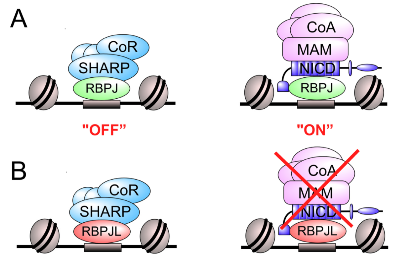


![Anti-ITGAX Armenian Hamster Monoclonal Antibody (PE (Phycoerythrin)) [clone: N418] | VWR](https://fr.vwr.com/stibo/bigweb/std.lang.all/03/25/13150325.jpg)