Radiographic Magnification
Lenses
Quantitative methods in phase-contrast x-ray imaging. (1/37)
A new method for extracting quantitative information from phase-contrast x-ray images obtained with microfocus x-ray sources is presented. The proposed technique allows rapid noninvasive characterization of the internal structure of thick optically opaque organic samples. The method does not generally involve any sample preparation and does not need any x-ray optical elements (such as monochromators, zone plates, or interferometers). As a consequence, samples can be imaged in vivo or in vitro, and the images are free from optical aberrations. While alternative techniques of x-ray phase-contrast imaging usually require expensive synchrotron radiation sources, our method can be implemented with conventional, albeit microfocus, x-ray tubes, which greatly enhances its practicality. In the present work, we develop the theoretical framework, perform numerical simulations, and present the first experimental results, demonstrating the viability of the proposed approach. We believe that this method should find wide-ranging applications in clinical radiology and medical research. (+info)Extraction of microcalcifications in digital mammograms using regional watershed. (2/37)
In this report, a novel technique is proposed for computer-aided automatic extraction of microcalcifications in a digital mammogram. First, the microcalcifications are detected by morphological filtering, followed by entropy-based thresholding. Next, the microcalcifications are segmented by computing regional watershed. The proposed automatic technique is designed to serve as a visual aid to radiologists. Its efficacy is demonstrated through experimental results. (+info)A multiscale algorithm for segmenting calcifications from high-resolution mammographic specimen radiographs. (3/37)
We have developed a multiscale algorithm for segmenting breast calcifications from high-resolution specimen radiographs. The algorithm was evaluated using 152 mammographic regions of interest digitized at a 15-microm spatial resolution. The true-positive detection rate was approximately 97.4% with 0.67 false-positives per image, and the segmentation error of individual calcification particles was approximately 5%. The performance of the algorithm is highly satisfactory. (+info)Detection of microcalcifications by means of multiscale methods and statistical techniques. (4/37)
The detection of clustered microcalcifications can help the radiologist to detect early breast cancer. Microcalcifications exhibit some important characteristics, such as small size and high luminosity. Use of a computer-aided diagnosis (CAD) method can prevent them being overlooked. In this report, a multiresolution analysis is performed based on a multilevel wavelet transformation. Decomposition produces sub-band images which become visible only as details of the different scales. Thereafter, all the images will be combined in a final image, in order to obtain an image that contains all the interest details at the scale where microcalcifications tend to appear. Once the image, called detail image, is obtained, it is necessary to determine which details correspond with microcalcifications. Statistical analysis of the histogram permits classification of the zones likely to contain microcalcifications. Applying this statistical techniques over the whole image and representing the results in a two-dimensional map, clustered microcalcification regions are clearly distinguishable. (+info)Intrarenal arterial collateral circulation. (5/37)
In three cases of intrarenal arterial collateral circulation the collateral channels developed between interlobar arteries in diseased kidneys. Probably these originated in hypertrophied spiral vessels that had arisen from the interlobar arteries in the area of the minor calyces. This form of collateral circulation will undoubtedly be recognized more frequently with the increased use of magnification radiography. (+info)The radiographic localization of impacted maxillary canines: a comparison of methods. (6/37)
This study compared two different radiographic techniques for localization of impacted maxillary canines: vertical parallax (from a panoramic and a maxillary anterior occlusal radiograph) and magnification (from a single panoramic radiograph). The radiographs and the information regarding the impacted canines were obtained retrospectively from records of patients treated in the Day Stay Unit of the Eastman Dental Hospital. The two different radiographic techniques were tested blind and compared for localization of the impacted canine by six examiners. The 'gold standard' used for the radiographic comparisons was the true position of the canine as recorded at operation. The results showed a wide variation between the six examiners in the prediction of the canine position with the two different techniques. Localization with vertical parallax was more successful overall than with magnification, although the difference failed to reach significance. Seventy-six per cent of the impacted canines could be successfully located with vertical parallax and 66 per cent with magnification. Further analysis showed that, while almost 90 per cent of the palatally impacted canines could be correctly detected with both techniques, less than half of the buccal canines could be detected with parallax and only one in 10 buccal canines could be detected with magnification. If a canine is suspected to be buccally placed from its appearance on a panoramic film and cannot be palpated, further views are justified. (+info)An accurate three-dimensional cephalometric system: a solution for the correction of cephalic malpositioning. (7/37)
In recent years, methods have been developed that calculate three-dimensional (3D) co-ordinates of orthodontic landmarks from lateral and frontal cephalograms. However, precise measurement has been impossible with these methods because, although they corrected the magnification of the image, they did not correct 3D cephalic malpositioning that occurs during the measurement of human subjects. In this study, we developed a 3D cephalometric system that corrected not only for magnification of the image, but also 3D cephalic malpositioning during cephalogram exposure. Magnification of the image was corrected for first. Cephalic revolution was then sequentially corrected and divided into elements of x-, y-, and z-axes. The origin was parallelly translated to the mid-point of bilateral porion. In order to examine the accuracy of this system, seven human dry skulls were measured. The accuracy unaffected by the cephalic revolution in any direction and standard errors was within 0.8 mm in any orthodontic landmarks. It was suggested that this measurement system would have sufficient accuracy for clinical application. The results indicated that precise cephalometric measurement was possible with this system and it was suggested that its clinical application would be possible. (+info)An evaluation of the reproducibility of landmark identification using scanned cephalometric images. (8/37)
OBJECTIVE: A method of cephalometric analysis is described in which cephalometric x-rays were scanned using a flat-bed scanner and transparency hood. Then the image was displayed on a computer monitor for point identification and subsequent cephalometric analysis using dedicated software. The reproducibility of point identification using this method was compared with two other, commonly used, methods. MATERIAL AND METHODS: The study material comprised 25 lateral skull x-rays taken as part of routine orthodontic assessment. Repeat cephalometric point identification was carried out on each x-ray using 3 methods: 1. On-screen digitization of the scanned bitmap image (Screenceph method). 2. Tracing followed by digitization of the identified points and 3. Direct digitization. RESULTS: For the 8 angular and 4 linear cephalometric measurements examined the Screenceph method compared favourably with the two conventional methods. The median difference between methods was 0.5 degrees and 0.2 mm. Using constructed Cartesian axes to examine the x, y discrepancy between repeat measurements and comparing Screenceph to tracing followed by digitization, there were significant differences in 3 instances at the 5% level and 2 instances at the 1% level. These differences represented median scores of 0.14 to 0.32 mm greater for Screenceph. Comparing Screenceph to direct digitization 15 significant differences out of the 28 measurements were noted: six at the 5% level and 9 at the 1% level. The actual difference in median scores ranged from 0.2 mm to 0.53 mm. CONCLUSION: The results demonstrated that Screenceph is sufficiently accurate to use in a clinical setting but is not yet sufficiently exact for use in research projects owing to hardware limitations. (+info)Radiographic magnification is a phenomenon that occurs during radiographic imaging where the image produced appears larger than the actual size of the object being imaged. This can occur due to several reasons, including the use of a focal distance that is shorter than the object-to-image receptor distance (SID), or when using a grid that is misaligned with the X-ray beam.
In some cases, radiographic magnification may be intentionally used as a technique to improve image quality for small structures or to enhance visualization of certain details in an image. However, it can also lead to distortion and decreased image sharpness if not properly controlled. Therefore, it is important to carefully consider the benefits and potential drawbacks of radiographic magnification when using this technique in medical imaging.
In the context of medical terminology, "lenses" generally refers to optical lenses used in various medical devices and instruments. These lenses are typically made of glass or plastic and are designed to refract (bend) light in specific ways to help magnify, focus, or redirect images. Here are some examples:
1. In ophthalmology and optometry, lenses are used in eyeglasses, contact lenses, and ophthalmic instruments to correct vision problems like myopia (nearsightedness), hypermetropia (farsightedness), astigmatism, or presbyopia.
2. In surgical microscopes, lenses are used to provide a magnified and clear view of the operating field during microsurgical procedures like ophthalmic, neurosurgical, or ENT (Ear, Nose, Throat) surgeries.
3. In endoscopes and laparoscopes, lenses are used to transmit light and images from inside the body during minimally invasive surgical procedures.
4. In ophthalmic diagnostic instruments like slit lamps, lenses are used to examine various structures of the eye in detail.
In summary, "lenses" in medical terminology refer to optical components that help manipulate light to aid in diagnosis, treatment, or visual correction.
Indigo Carmine is not a medical term, but it is a chemical compound that is sometimes used in medical settings. Indigo Carmine is a type of dye that is often used as a marker in various medical tests and procedures. It can be used during surgeries to help identify structures or tissues within the body, such as the urinary tract or the gastrointestinal tract.
Indigo Carmine is also sometimes used as a diagnostic aid in urological procedures, such as cystoscopy, to help visualize the flow of urine and detect any abnormalities in the urinary tract. The dye is usually introduced into the body through a catheter or other medical device, and it is excreted in the urine, turning it blue or green.
It's important to note that Indigo Carmine should only be used under the supervision of a healthcare professional, as improper use can lead to adverse effects.
Radiographic Magnification | Profiles RNS
 Projectional radiography - Wikipedia
 Pediatric Splenomegaly: Practice Essentials, Pathophysiology, Epidemiology
Figure - Chlamydia abortus in Pregnant Woman with Acute Respiratory Distress Syndrome - Volume 26, Number 3-March 2020 -...
 Thieme E-Journals - Journal of Child Science / Full Text
Effect of cardiac and respiratory cycles on vertebral heart score measured on fluoroscopic images of healthy dogs in: Journal...
 Magnification tips to help make you more comfortable and reduce stress | Dental Economics
 Influence of Preclinical Training on Root Canal Treatment Technical Quality and Confidence Level of Undergraduate Dental...
 Significance of Cephalometric Radiograph in Orthodontic Treatment Plan Decision
 Pelvimetry to Diagnose Dystocia in the Bitch - WSAVA 2003 Congress - VIN
 2020-2021 BCSC Basic and Clinical Science Course™
Osteitis | Profiles RNS
 Primary Hyperparathyroidism | Radiology
 Comparison between electronic and radiographic method for the determination of root canal length in primary teeth
 RADIOGRAPHIC FILM INTERPRETATION - Quality Group
 Radiography abdominal. Medical search. Definitions
 Leborgne, Raul
 Table Angle Attribute - DICOM Standard Browser
Body Part Thickness Attribute - DICOM Standard Browser
 Diagnostic and Interventional Radiology
 Rickets - history, re-occurrence and association with breathing problems - Nov 2016 | VitaminDWiki
Using digital radiography<...
Analysing the clinical relevance and impact of subsidence seen with standalone cages for cervical disc surgeries - A study of...
 Radiographical characteristics and traction duration of impacted maxillary canine requiring surgical exposure and orthodontic...
 radiographic survey in pediatric dentistry
Coronary angiography quantitative angiography - wikidoc
 Pesquisa | Portal Regional da BVS
 Correlation of clinical and radiographic diagnosis of carious lesions in posterior teeth
| Revista Facultad de...
Correlation of clinical and radiographic diagnosis of carious lesions in posterior teeth
| Revista Facultad de...
 Relationship between Tibial conformation, cage size and advancement achieved in TTA procedure | BMC Veterinary Research | Full...
Affect the radiographic image2
- 3. Explain the factors affecting the quality and quantity of the x-ray beam and how these factors affect the radiographic image. (santarosa.edu)
- The two methods affect the radiographic image differently because the magnification and distortion changes are different in each and this precluded accurate comparison. (massey.ac.nz)
Radiography3
- 4 ] Bitewing radiography is the most efficient dental radiographic modality for the detection of interproximal caries in teeth with closed contacts such that 75% of interproximal caries cannot be detected without bitewing radiography. (thieme-connect.com)
- 1 ] Panoramic radiography is an extraoral radiographic modality used as a diagnostic tool for developmental disorders such as missing teeth, supernumeraries, ectopic tooth eruption, delayed primary root resorption, and detection of cysts, tumors, and some genetic disorders. (thieme-connect.com)
- This course teaches the principles of digital radiography, equipment standards for CR systems, radiographic techniques with CR, and calibration of equipment (use of phantom). (twitraining.com)
Evaluate radiographic2
- 14. Critically evaluate radiographic images to assess errors and indicate proper methods for correction. (santarosa.edu)
- 1 Leverage full-body EOS images and reports to establish unbiased preoperative objectives and evaluate radiographic outcomes along the continuum of care. (atecspine.com)
Clinical11
- Clinical manifestations, radiographic findings, and bone mineral measurements of 65 patients with primary hyperparathyroidism are reviewed. (rsna.org)
- These are determined by a careful clinical examination of the patient before ordering the radiographic survey. (trnds.co)
- There are different techniques to diagnose dental caries, including clinical and radiographic evaluation. (udea.edu.co)
- the objective of this study was to establish correlation between the clinical and radiographic caries diagnosis suggested by ICCMS TM , in deciduous and permanent molars of a school population. (udea.edu.co)
- there is low correlation between the clinical diagnosis of caries and the radiographic examination, in relation to ICCMS TM standards. (udea.edu.co)
- Clinical and radiographic diagnosis of occlusal caries: a study in vitro. (udea.edu.co)
- DMFT index was calculated by clinical and radiographic examination. (who.int)
- Objective: the present report describes the clinical, radiographic, and histopathological features of an ameloblastic fibro-odontoma (AFO) lesion. (bvsalud.org)
- AFO was suspected based on the radiographic and clinical characteristics. (bvsalud.org)
- Final considerations: the evaluation of the clinical, radiographic, and histopathologic findings needs to be accurate for a correct diagnosis and appropriate treatment for case of AFO since the presentation is often asymptomatic. (bvsalud.org)
- Clinical and radiographic outcome [Time Frame: Immediately, 1, 3, 6 and 12 months. (who.int)
400x magnification2
- The cells were then inspected under a light microscope at 400x magnification for cytogenetic changes. (thieme-connect.com)
- They were digitalized as 600 dpi with a flatbed scanner and analyzed by software for image analysis at 400x magnification utilizing a personal computer. (hacettepe.edu.tr)
Radiograph2
- Correction for magnification of the pelvis on the radiograph was considered unnecessary after investigation. (vin.com)
- The technique chosen was radiographic absorptiometry which determines bone mineral density from a radiograph that includes a control (usually a wedge) of known photodensity. (massey.ac.nz)
Original magnification2
- D) H&E stained section of placenta showing necrotizing mural arteritis (original magnification ×100). (cdc.gov)
- original magnification, 40) show sheets of mononuclear cells interspersed with multinucleated giant cells. (conferencedequebec.org)
Examination1
- In vitro evaluation of ICDAS and radiographic examination of occlusal surfaces and their association with treatment decisions. (udea.edu.co)
Endodontics1
- Many radiographic images in endodontics are taken in two-dimensional planes, thereby having inherent distortion created by the relative positions of the object (tooth), the X-ray film, and the tube head. (org.in)
Interpretation4
- Use of optic and geometric techniques to enhance radiographic image quality and interpretation. (rush.edu)
- This is a course suitable for all who require an in-depth knowledge of the radiographic interpretation of welds. (qualitygroup.sg)
- Course candidates must be qualified to a minimum of Level 2 Radiographic Interpretation conforming to BS EN ISO 9712. (twitraining.com)
- The student should, at the end of the course, be able to competently recognize the normal radiographic anatomy of cats and dogs, know the basic principles of how the images are created and know the basics of image interpretation. (sgu.edu)
Lateral2
- Closed-mouth lateral radiographic view of both condyles. (medscape.com)
- Open-mouth lateral radiographic view of both condyles. (medscape.com)
Calibration1
- You will also learn about image processing using phosphor screens, software specific parameters, calibration of measuring tools, effects of magnification, and definition of accuracy constraints and capabilities of CR system. (twitraining.com)
Diagnostic1
- 9. Produce radiographic images of acceptable diagnostic quality with proper contrast, density, definition and minimal magnification or anatomic distortion. (santarosa.edu)
Density3
- Radiographic density is the measure of overall darkening of the image. (wikipedia.org)
- A higher radiographic density represents more opaque areas of the film, and lower density more transparent areas of the film. (wikipedia.org)
- Contrast is defined as the difference in radiographic density between adjacent portions of the image. (wikipedia.org)
Panoramic2
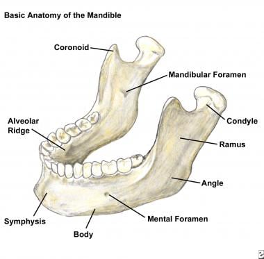
- With computed tomography (CT) scanning and panoramic imaging in addition to the basic mandibular radiographic study, a comprehensive evaluation and subsequent identification of all but the most subtle fractures can be achieved. (medscape.com)
- Panoramic radiographic image shows a left angle fracture extending to and dislodging the molar. (medscape.com)
Assessment2
- The course concentrates on identification of weld defects, assessment of radiographic quality and sentencing to example specifications. (qualitygroup.sg)
- Past two decades have seen many advancements in treatment of spine deformities right from initial radiographic assessment, surgical planning to postoperative care. (backbonejournal.com)
Findings1
- symptoms and radiographic findings do not differentiate multidrug-resistant TB (MDR-TB) from fully susceptible TB. (medscape.com)
Periapical1
- 1 ] The three most commonly prescribed dental radiographic modalities include the bitewing, periapical, and extraoral dental radiographies. (thieme-connect.com)
Radiology2
- Direct radiographic magnification for skeletal radiology. (uchicago.edu)
- Includes chapters on physics of radiology and normal radiographic anatomy of Canine, Feline and Equine species. (sgu.edu)
Positioning5
- Small Animal Radiographic Techniques and Positioning , Wiley& Blackwell, 2012. (sgu.edu)
- Six lectures of basic positioning and normal radiographic anatomy. (sgu.edu)
- For each anatomic region, students will explore the topics of patient positioning, normal radiographic anatomy, image quality and comparative anatomy. (sgu.edu)
- Bontrager's Textbook of Radiographic Positioning and Related Anatomy. (radiopaedia.org)
- Merrill's Atlas of Radiographic Positioning and Procedures. (radiopaedia.org)
Visualization2
- But I know that magnification is not the only factor involved in better visualization. (dentaleconomics.com)
- Radiographic visualization of the body between the thorax and the pelvis, i.e., within the peritoneal cavity. (lookformedical.com)
Apex1
- Common methods in WL determination include tactile, radiographic, and electronic apex locators (EAL). (org.in)
Exposure2
- Closely related to radiographic contrast is the concept of exposure latitude. (wikipedia.org)
- 3 ] It should be noted that despite the widespread use of dental radiographic modalities, there is no definite safety level for X-ray exposure. (thieme-connect.com)
Caries2
- Determinación de la especificidad y sensibilidad del ICDAS y fluorescencia Láser en la detección de caries in vitro. (udea.edu.co)
- Wenzel A, Hintze H, Mikkelsen L, Mouyen F. Radiographic detection of occlusal caries in non-cavitated teeth. (udea.edu.co)
Mandibular1
- If only an isolated mandibular fracture is suspected, radiographic evaluation should begin with the acquisition of a posteroanterior (PA) view, a Towne view (anteroposterior [AP] axial view), and bilateral oblique views. (medscape.com)
Parameters1
- or third, the fit is checked preoperatively through the use of radiographic templates with known magnification parameters. (teachmeorthopedics.info)
Bone2
- therefore, a proximal tibal en bloc resection was performed in August 2010 (Fig.?2C, D). The histologic features of the specimen were consistent with a benign giant cell tumor of bone (Fig.?2E). (conferencedequebec.org)
- Determined with a ruler and taking into consideration radiographic magnification, the inner diameter of the isthmus of the intramedullary canal should be at least 2.5mm to ensure that the 2.5mm arrowhead tip can pass through the narrowest point of the intramedullary canal in order to engage in the subchondral bone at the base of the proximal phalanx. (arrowheaddevices.com)
Cervical1
- Turning the patient's head creates a radiographic keyhole between the cervical spine and ramus of the mandible. (trnds.co)
Techniques2
- 15. Adapt radiographic techniques to meet patient needs. (santarosa.edu)
- [3] Different techniques have been described under the radiographic method. (org.in)
Differences1
- He went on to describe the radiographic differences between benign and malignant calcifications. (scienceheroes.com)
Anatomy1
- The course offers the theoretical and practical basis for learning and understanding radiographic anatomy of the cat and dog. (sgu.edu)
Angle2
- Posteroanterior radiographic view showing a left angle fracture. (medscape.com)
- Posteroanterior radiographic view of a fracture of the left body and angle. (medscape.com)
Procedures1
- Present day intraoral and extraoral procedures, used individually or in combination, suffer from the same inherent limitations of all planar two-dimensional projections: magnification, distortions, superimpositions, and misrepresentation of structures. (journalcra.com)
Teeth1
- EAL accurately predicted the AC in 31 (96.9%) teeth, while the digital radiographic and tactile sensation methods accurately predicted the constriction in 19 (59.4%) and 8 (25%) teeth, respectively, in the study population. (org.in)
Determination1
- Precise working length determination continues to challenge many dentists relying solely on tactile and radiographic methods. (org.in)
Methods1
- These include the following: body plethysmography (using various methodologies), nitrogen washout, gas dilution, and radiographic imaging methods. (ersjournals.com)
Images3
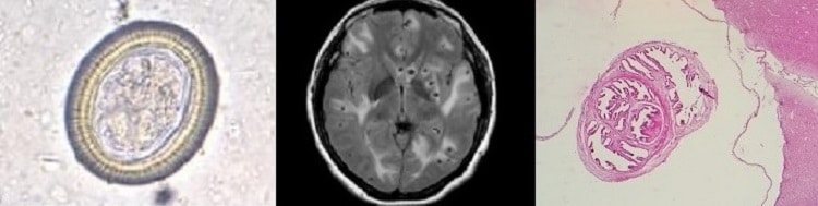
- Radiographic and histologic images from a pregnant woman in rural France infected with Chlamydia abortus . (cdc.gov)
- Standardized images with best-in-class radiographic technology. (atecspine.com)
- The lesion involved the impacted third mo- recognized as unilocular masses on radiographic lar that was mesio-angled, undergoing inductive images with defined limits, presenting with changes, and was located near the base of the variable amounts of calcified material in mandible. (bvsalud.org)
Film3
- The focus film distance was 90 cm and in order to minimize the object-film distance and thereby magnification, no grid was used. (vin.com)
- This course is specifically designed for candidates wishing to become certificated as Radiographic film interpreter under the ASNT requirements. (qualitygroup.sg)
- A rapid, low-dose, digital imaging system using a small intraoral sensor instead of radiographic film, an intensifying screen, and a charge-coupled device. (lookformedical.com)
Structures1
- Using it, he was better able to see small structures and use spot/magnification. (scienceheroes.com)
Method1
- The purpose of this research was to compare the radiographic and the electronic method to obtain the working length in deciduous molars. (bvsalud.org)
Cells1
- This study aimed to assess the genotoxic and cytotoxic effects of dental radiographic modalities on buccal mucosal cells in children. (thieme-connect.com)
Patients3
- Magnification is one of the most important and underutilized technologies that doctors should have in their arsenals of tools to deliver their best possible dental care to patients. (dentaleconomics.com)
- A radiographic survey of dental anomalies in Black pediatric patients. (trnds.co)
- The Selection of Patients for X-ray Examinations: Dental Radiographic Examinations. (trnds.co)
Image analysis1
- Radiographic Image Analysis. (radiopaedia.org)
Digital2
- Optical, or less preferred digital, magnification results in an effective pixel matrix up to 2458 × 2458. (wikidoc.org)
- This course is ideal if you are looking to expand your current knowledge of radiographic systems and/or are looking at the possibility of introducing a digital system into your place of work/company. (twitraining.com)
Dental1
- Available at: ada.org/~/media/ADA/Member%20Center/FIles/Dental_Radiographic_Examinations_2012.ashx. (trnds.co)
Level1
- The MagnaVu features a fully adjustable custom lens system that allows the user to choose the desired optical magnification level from 1X to 23X. (dentaleconomics.com)
Shows1
- This graph shows the total number of publications written about "Radiographic Magnification" by people in this website by year, and whether "Radiographic Magnification" was a major or minor topic of these publications. (rush.edu)
Standards1
- The geometric unsharpness is between 0.02 to O.lmm, because of this, geometric unsharpness is seen much more than the standards on the photographic card after magnification. (itu.edu.tr)
View4
- I tested various magnification scopes, and decided on the MagnaVu as the best fit for my practice because of its ease of operation and field of view. (dentaleconomics.com)
- Posteroanterior radiographic view of a left condylar fracture. (medscape.com)
- Towne radiographic view of the left condylar fracture. (medscape.com)
- Posteroanterior radiographic view of the left condyle and right parasymphyseal fracture. (medscape.com)
Studies1
- Plain radiographic studies show a ground-glass appearance with lytic foci. (aao.org)