Protein Phosphatase 2
Phosphoprotein Phosphatases
Protein Phosphatase 1
Okadaic Acid
Protein Tyrosine Phosphatases
Acid Phosphatase
Phosphorylation
Oxazoles
Cantharidin
Phosphoric Monoester Hydrolases
Microcystins
Calcineurin
Dual-Specificity Phosphatases
Glucose-6-Phosphatase
Phosphorylase Phosphatase
Molecular Sequence Data
Amino Acid Sequence
Enzyme Inhibitors
cdc25 Phosphatases
Myosin-Light-Chain Phosphatase
Enzyme Activation
Signal Transduction
Protein Tyrosine Phosphatase, Non-Receptor Type 1
Protein Tyrosine Phosphatase, Non-Receptor Type 11
Protein Kinases
Substrate Specificity
Protein Tyrosine Phosphatase, Non-Receptor Type 6
Protein Binding
Isoenzymes
Protein Tyrosine Phosphatases, Non-Receptor
Calmodulin-Binding Proteins
Catalytic Domain
Protein-Serine-Threonine Kinases
Phosphorylase a
Base Sequence
Mutation
Dual Specificity Phosphatase 1
Mitogen-Activated Protein Kinase Phosphatases
Sequence Homology, Amino Acid
Pyrans
Protein Subunits
Intracellular Signaling Peptides and Proteins
Phosphoproteins
Serine
Saccharomyces cerevisiae
Phosphatidate Phosphatase
Saccharomyces cerevisiae Proteins
Receptor-Like Protein Tyrosine Phosphatases, Class 2
Threonine
Rabbits
Glycogen-Synthase-D Phosphatase
Cells, Cultured
Spiro Compounds
Peptides, Cyclic
Vanadates
Cloning, Molecular
Cyclic AMP-Dependent Protein Kinases
Mitosis
Dopamine and cAMP-Regulated Phosphoprotein 32
4-Nitrophenylphosphatase
Proteins
Recombinant Fusion Proteins
PTEN Phosphohydrolase
Catalysis
Gene Expression Regulation, Enzymologic
Binding Sites
Calcium
Cell Cycle Proteins
Nuclear Proteins
Protein Structure, Tertiary
Phosphothreonine
Glycogen
Electrophoresis, Polyacrylamide Gel
Carrier Proteins
Dual Specificity Phosphatase 6
Receptor-Like Protein Tyrosine Phosphatases, Class 3
Two-Hybrid System Techniques
Blotting, Western
Receptor-Like Protein Tyrosine Phosphatases, Class 4
Protein Kinase C
Precipitin Tests
Liver
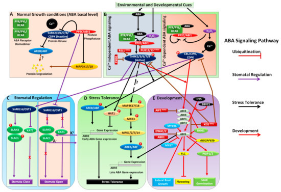
Abscisic Acid
Nitrophenols
Cell Nucleus
Calcium-Calmodulin-Dependent Protein Kinases
Cattle
RNA, Messenger
Dual Specificity Phosphatase 3
Chromatography, Gel
Immunoprecipitation
Phosphopeptides
Holoenzymes
Immunoblotting
Models, Biological
Organophosphorus Compounds
Protein Tyrosine Phosphatase, Non-Receptor Type 2
Transfection
Sequence Alignment
Tyrosine
Subcellular Fractions
Cell Cycle
COS Cells
Proto-Oncogene Proteins c-akt
Protein Processing, Post-Translational
SH2 Domain-Containing Protein Tyrosine Phosphatases
Amino Acid Motifs
Cytosol
Phosphorus Radioisotopes
Arabidopsis
Tartrates
Magnesium
Arabidopsis Proteins
Macromolecular Substances
DNA, Complementary
Alkenes
3T3 Cells
Mutagenesis, Site-Directed
Chromatography, Ion Exchange
CDC2 Protein Kinase
Protein O-Methyltransferase
Phosphotyrosine
Mitogen-Activated Protein Kinases
Membrane Proteins
DNA Primers
Tumor Cells, Cultured
Cell Membrane
Glycogen Synthase Kinase 3
Down-Regulation
Protein-Tyrosine Kinases
Transcription Factors
Casein Kinase I
14-3-3 Proteins
Tacrolimus
Rats, Sprague-Dawley
Brain
RNA, Small Interfering
Glycogen Synthase
Ceramides
Dose-Response Relationship, Drug
Transcription, Genetic
Adenosine Triphosphate
A Kinase Anchor Proteins
Glutathione Transferase
Manganese
HEK293 Cells
Receptor-Like Protein Tyrosine Phosphatases, Class 5
Gene Expression Regulation, Fungal
Pyruvate Dehydrogenase (Lipoamide)-Phosphatase
Phosphorylase Kinase
Protein Transport
Gene Expression Regulation
Protein Tyrosine Phosphatase, Non-Receptor Type 12
Phenotype
tau Proteins
Apoptosis
Cyclosporine
RNA Interference
Escherichia coli
Cytoplasm
Peptide Mapping
Nerve Tissue Proteins
Vicia faba
Receptor-Like Protein Tyrosine Phosphatases
HeLa Cells
Calmodulin
Cyclic AMP
Gene Deletion
Chromatography, DEAE-Cellulose
Hydrogen-Ion Concentration
Protein Tyrosine Phosphatase, Non-Receptor Type 13
Adaptor Proteins, Signal Transducing
Models, Molecular
DNA-Binding Proteins
Peptides
Peptide Fragments
Staurosporine
Protein Tyrosine Phosphatase, Non-Receptor Type 3
Gene Expression
Antigens, Polyomavirus Transforming
Chromatography, Affinity
Cell Division
Calcium-Binding Proteins
Microtubules
Tetramisole
All-trans-retinoic acid inhibits Jun N-terminal kinase by increasing dual-specificity phosphatase activity. (1/1591)
Jun N-terminal kinases (JNKs) are serine-threonine kinases that play a critical role in the regulation of cell growth and differentiation. We previously observed that JNK activity is suppressed by all-trans-retinoic acid (t-RA), a ligand for retinoic acid nuclear receptors (RARs), in normal human bronchial epithelial cells, which are growth inhibited by t-RA. In this study, we investigated the mechanism by which t-RA inhibits JNK and the possibility that this signaling event is blocked in non-small cell lung cancer (NSCLC) cells. Virtually all NSCLC cell lines are resistant to the growth-inhibitory effects of t-RA, and a subset of them have a transcriptional defect specific to retinoid nuclear receptors. We found that in NSCLC cells expressing functional retinoid receptors, serum-induced JNK phosphorylation and activity were inhibited by t-RA in a bimodal pattern, transiently within 30 min and in a sustained fashion beginning at 12 h. Retinoid receptor transcriptional activation was required for the late, but not the early, suppression of JNK activity. t-RA inhibited serum-induced JNK activity by blocking mitogen-activated protein (MAP) kinase kinase 4-induced signaling events. This effect of t-RA was phosphatase dependent and involved an increase in the expression of the dual-specificity MAP kinase phosphatase 1 (MKP-1). t-RA did not activate MKP-1 expression or inhibit JNK activity in a NSCLC cell line with retinoid receptors that are refractory to ligand-induced transcriptional activation. These findings provide the first evidence that t-RA suppresses JNK activity by inhibiting JNK phosphorylation. Retinoid receptor transcriptional activation was necessary for the sustained inhibition of JNK activity by t-RA, and this signaling event was disrupted in NSCLC cells with retinoid receptors that are refractory to ligand-induced transcriptional activation. (+info)gp49B1 inhibits IgE-initiated mast cell activation through both immunoreceptor tyrosine-based inhibitory motifs, recruitment of src homology 2 domain-containing phosphatase-1, and suppression of early and late calcium mobilization. (2/1591)
We define by molecular, pharmacologic, and physiologic approaches the proximal mechanism by which the immunoglobulin superfamily member gp49B1 inhibits mast cell activation mediated by the high affinity Fc receptor for IgE (FcepsilonRI). In rat basophilic leukemia-2H3 cells expressing transfected mouse gp49B1, mutation of tyrosine to phenylalanine in either of the two immunoreceptor tyrosine-based inhibitory motifs of the gp49B1 cytoplasmic domain partially suppressed gp49B1-mediated inhibition of exocytosis, whereas mutation of both abolished inhibitory capacity. Sodium pervanadate elicited tyrosine phosphorylation of native gp49B1 and association of the tyrosine phosphatases src homology 2 domain-containing phosphatase-1 (SHP-1) and SHP-2 in mouse bone marrow-derived mast cells (mBMMCs). SHP-1 associated transiently with gp49B1 within 1 min after coligation of gp49B1 with cross-linked FcepsilonRI in mBMMCs. SHP-1-deficient mBMMCs exhibited a partial loss of gp49B1-mediated inhibition of FcepsilonRI-induced exocytosis at concentrations of IgE providing optimal exocytosis, revealing a central, but not exclusive, SHP-1 requirement in the counter-regulatory pathway. Coligation of gp49B1 with cross-linked FcepsilonRI on mBMMCs inhibited early release of calcium from intracellular stores and subsequent influx of extracellular calcium, consistent with SHP-1 participation. Because exocytosis is complete within 2 min in mBMMCs, our studies establish a role for SHP-1 in the initial counter-regulatory cellular responses whereby gp49B1 immunoreceptor tyrosine-based inhibition motifs rapidly transmit inhibition of FcepsilonRI-mediated exocytosis. (+info)Expression of dominant-negative src-homology domain 2-containing protein tyrosine phosphatase-1 results in increased Syk tyrosine kinase activity and B cell activation. (3/1591)
The Src-homology domain 2 (SH2)-containing cytoplasmic tyrosine phosphatase, SHP-1 (SH2-containing protein tyrosine phosphatase-1), interacts with several B cell surface and intracellular signal transduction molecules through its SH2 domains. Mice with the motheaten and viable motheaten mutations are deficient in SHP-1 and lack most mature B cells. To define the role of SHP-1 in mature B cells, we expressed phosphatase-inactive SHP-1 (C453S) in a mature B cell lymphoma line. SHP-1 (C453S) retains the ability to bind to both substrates and appropriate tyrosine-phosphorylated proteins and therefore can compete with the endogenous wild-type enzyme. We found that B cells expressing SHP-1 (C453S) demonstrated enhanced and prolonged tyrosine phosphorylation of proteins with molecular masses of 110, 70, and 55-60 kDa after stimulation with anti-mouse IgG. The tyrosine kinase Syk was hyperphosphorylated and hyperactive in B cells expressing SHP-1 (C453S). SHP-1 and Syk were coimmunoprecipitated from wild-type K46 cells, K46 SHP-1 (C453S) cells, and splenic B cells, and SHP-1 dephosphorylated Syk. Cells expressing SHP-1 (C453S) showed increased Ca2+ mobilization, extracellular signal-regulated kinase activation, and homotypic adhesion after B cell Ag receptor engagement. Thus, SHP-1 regulates multiple early and late events in B lymphocyte activation. (+info)A herpesvirus ribosome-associated, RNA-binding protein confers a growth advantage upon mutants deficient in a GADD34-related function. (4/1591)
The herpes simplex virus type 1 gamma34.5 gene product and the cellular GADD34 protein both contain similar domains that can regulate the activity of eukaryotic initiation factor 2 (eIF2), a critical translation initiation factor. Viral mutants that lack the GADD34-related function grow poorly on a variety of malignant human cells, as activation of the cellular PKR kinase leads to the accumulation of inactive, phosphorylated eIF2 at late times postinfection. Termination of translation prior to the completion of the viral reproductive cycle leads to impaired growth. Extragenic suppressors that regain the ability to synthesize proteins efficiently in the absence of the viral GADD34-related function have been isolated. These suppressor alleles are dominant in trans and affect the steady-state accumulation of several viral mRNA species. We demonstrate that deregulated expression of Us11, a virus-encoded RNA-binding, ribosome-associated protein is necessary and sufficient to confer a growth advantage upon viral mutants that lack a GADD34-related function. Ectopic expression of Us11 reduces the accumulation of the activated cellular PKR kinase and allows for sustained protein synthesis. Thus, an RNA-binding, ribosome-associated protein (Us11) and a GADD34-related protein (gamma34.5) both function in a signal pathway that regulates translation by modulating eIF2 phosphorylation. (+info)Characterization of the inhibition of protein phosphatase-1 by DARPP-32 and inhibitor-2. (5/1591)
Phospho-DARPP-32 (where DARPP-32 is dopamine- and cAMP-regulated phosphoprotein, Mr 32,000), its homolog, phospho-inhibitor-1, and inhibitor-2 are potent inhibitors (IC50 approximately 1 nM) of the catalytic subunit of protein phosphatase-1 (PP1). Our previous studies have indicated that a region encompassing residues 6-11 (RKKIQF) and phospho-Thr-34, of phospho-DARPP-32, interacts with PP1. However, little is known about specific regions of inhibitor-2 that interact with PP1. We have now characterized in detail the interaction of phospho-DARPP-32 and inhibitor-2 with PP1. Mutagenesis studies indicate that within DARPP-32 Phe-11 and Ile-9 play critical roles, with Lys-7 playing a lesser role in inhibition of PP1. Pro-33 and Pro-35 are also important, as is the number of amino acids between residues 7 and 11 and phospho-Thr-34. For inhibitor-2, deletion of amino acids 1-8 (I2-(9-204)) or 100-204 (I2-(1-99)) had little effect on the ability of the mutant proteins to inhibit PP1. Further deletion of residues 9-13 (I2-(14-204)) resulted in a large decrease in inhibitory potency (IC50 approximately 800 nM), whereas further COOH-terminal deletion (I2-(1-84)) caused a moderate decrease in inhibitory potency (IC50 approximately 10 nM). Within residues 9-13 (PIKGI), mutagenesis indicated that Ile-10, Lys-11, and Ile-13 play critical roles. The peptide I2-(6-20) antagonized the inhibition of PP-1 by inhibitor-2 but had no effect on inhibition by phospho-DARPP-32. In contrast, the peptide D32-(6-38) antagonized the inhibition of PP1 by phospho-DARPP-32, inhibitor-2, and I2-(1-120) but not I2-(85-204). These results indicate that distinct amino acid motifs contained within the NH2 termini of phospho-DARPP-32 (KKIQF, where italics indicate important residues) and inhibitor-2 (IKGI) are critical for inhibition of PP1. Moreover, residues 14-84 of inhibitor-2 and residues 6-38 of phospho-DARPP-32 share elements that are important for interaction with PP1. (+info)Transcriptional activation of the glucose transporter GLUT1 in ventricular cardiac myocytes by hypertrophic agonists. (6/1591)
Myocardial hypertrophy is associated with increased basal glucose metabolism. Basal glucose transport into cardiac myocytes is mediated by the GLUT1 isoform of glucose transporters, whereas the GLUT4 isoform is responsible for regulatable glucose transport. Treatment of neonatal cardiac myocytes with the hypertrophic agonist 12-O-tetradecanoylphorbol-13-acetate or phenylephrine increased expression of Glut1 mRNA relative to Glut4 mRNA. To study the transcriptional regulation of GLUT1 expression, myocytes were transfected with luciferase reporter constructs under the control of the Glut1 promoter. Stimulation of the cells with 12-O-tetradecanoylphorbol-13-acetate or phenylephrine induced transcription from the Glut1 promoter, which was inhibited by cotransfection with the mitogen-activated protein kinase phosphatases CL100 and MKP-3. Cotransfection of the myocytes with constitutively active versions of Ras and MEK1 or an estrogen-inducible version of Raf1 also stimulated transcription from the Glut1 promoter. Hypertrophic induction of the Glut1 promoter was also partially sensitive to inhibition of the phosphatidylinositol 3-kinase pathway and was strongly inhibited by cotransfection with dominant-negative Ras. Thus, Ras activation and pathways downstream of Ras mediate induction of the Glut1 promoter during myocardial hypertrophy. (+info)Negative regulation of myeloid cell proliferation and function by the SH2 domain-containing tyrosine phosphatase-1. (7/1591)
The SH2 domain containing tyrosine phosphatase SHP-1 has been implicated in the regulation of a multiplicity of signaling pathways involved in hemopoietic cell growth, differentiation, and activation. A pivotal contribution of SHP-1 in the modulation of myeloid cell signaling cascades has been revealed by the demonstration that SHP-1 gene mutation is responsible for the overexpansion and inappropriate activation of myelomonocytic populations in motheaten mice. To investigate the role of SHP-1 in regulation of myeloid leukocytes, an HA epitope-tagged dominant negative (interfering) SHP-1 (SHP-1C453S) was expressed in the myelo-monocytic cell line U937 using the pcDNA3 vector. Overexpression of this protein in SHP-1C453S transfectants was demonstrated by Western blot analysis and by detection of decreased specific activity. Growth, proliferation, and IL-3-induced proliferative responses were substantially increased in the SHP-1C453S-overexpressing cells relative to those in control cells. The results of cell cycle analysis also revealed that the proportion of cells overexpressing SHP-1C453S in S phase was greater than that of control cells. The SHP-1C453S-expressing cells also displayed diminished rates of apoptosis as detected by flow cytometric analysis of propidium iodide-stained cells and terminal deoxynucleotidyltransferase-mediated fluorescein-dUTP nick end-labeling assay. While motility and phagocytosis were not affected by SHP-1C453S overexpression, adhesion and the oxidative burst in response to PMA were enhanced in the SHP-1C453S compared with those in the vector alone transfectants. Taken together, these results suggest that SHP-1 exerts an important negative regulatory influence on cell proliferation and activation while promoting spontaneous cell death in myeloid cells. (+info)Protein phosphatase 1 is involved in the dissociation of Ca2+/calmodulin-dependent protein kinase II from postsynaptic densities. (8/1591)
Autophosphorylation-dependent translocation of Ca2+/calmodulin-dependent protein kinase II (CaM kinase II) to postsynaptic densities (PSDs) from cytosol may be a physiologically important process during synaptic activation. We investigated a protein phosphatase responsible for dephosphorylation of the kinase. CaM kinase II was shown to be targeted to two sites using the gel overlay method in two-dimensional gel electrophoresis. Protein phosphatase 1 (PP1) was identified to dephosphorylate CaM kinase II from its complex with PSDs using phosphatase inhibitors and activators, and purified phosphatases. The kinase was released from PSDs after its dephosphorylation by PP1. (+info)Protein Phosphatase 2 (PP2A) is a type of serine/threonine protein phosphatase that plays a crucial role in the regulation of various cellular processes, including signal transduction, cell cycle progression, and metabolism. PP2A is a heterotrimeric enzyme composed of a catalytic subunit (C), a regulatory subunit A (A), and a variable regulatory subunit B (B). The different combinations of the B subunits confer specificity to PP2A, allowing it to regulate a diverse array of cellular targets.
PP2A is responsible for dephosphorylating many proteins that have been previously phosphorylated by protein kinases. This function is essential for maintaining the balance of phosphorylation and dephosphorylation in cells, which is necessary for proper protein function and cell signaling. Dysregulation of PP2A has been implicated in various diseases, including cancer, neurodegenerative disorders, and cardiovascular disease.
Phosphoprotein phosphatases (PPPs) are a family of enzymes that play a crucial role in the regulation of various cellular processes by removing phosphate groups from serine, threonine, and tyrosine residues on proteins. Phosphorylation is a post-translational modification that regulates protein function, localization, and stability, and dephosphorylation by PPPs is essential for maintaining the balance of this regulation.
The PPP family includes several subfamilies, such as PP1, PP2A, PP2B (also known as calcineurin), PP4, PP5, and PP6. Each subfamily has distinct substrate specificities and regulatory mechanisms. For example, PP1 and PP2A are involved in the regulation of metabolism, signal transduction, and cell cycle progression, while PP2B is involved in immune response and calcium signaling.
Dysregulation of PPPs has been implicated in various diseases, including cancer, neurodegenerative disorders, and cardiovascular disease. Therefore, understanding the function and regulation of PPPs is important for developing therapeutic strategies to target these diseases.
Protein Phosphatase 1 (PP1) is a type of serine/threonine protein phosphatase that plays a crucial role in the regulation of various cellular processes, including metabolism, signal transduction, and cell cycle progression. PP1 functions by removing phosphate groups from specific serine and threonine residues on target proteins, thereby reversing the effects of protein kinases and controlling protein activity, localization, and stability.
PP1 is a highly conserved enzyme found in eukaryotic cells and is composed of a catalytic subunit associated with one or more regulatory subunits that determine its substrate specificity, subcellular localization, and regulation. The human genome encodes several isoforms of the PP1 catalytic subunit, including PP1α, PP1β/δ, and PP1γ, which share a high degree of sequence similarity and functional redundancy.
PP1 has been implicated in various physiological processes, such as muscle contraction, glycogen metabolism, DNA replication, transcription, and RNA processing. Dysregulation of PP1 activity has been associated with several pathological conditions, including neurodegenerative diseases, cancer, and diabetes. Therefore, understanding the molecular mechanisms that regulate PP1 function is essential for developing novel therapeutic strategies to treat these disorders.
Okadaic acid is a type of toxin that is produced by certain species of marine algae, including Dinophysis and Prorocentrum. It is a potent inhibitor of protein phosphatases 1 and 2A, which are important enzymes that help regulate cellular processes in the body.
Okadaic acid can accumulate in shellfish that feed on these algae, and consumption of contaminated seafood can lead to a serious illness known as diarrhetic shellfish poisoning (DSP). Symptoms of DSP include nausea, vomiting, diarrhea, and abdominal cramps. In severe cases, it can also cause neurological symptoms such as dizziness, disorientation, and tingling or numbness in the lips, tongue, and fingers.
It is important to note that okadaic acid is not only a marine toxin but also used in scientific research as a tool to study the role of protein phosphatases in cellular processes. However, exposure to this compound should be avoided due to its toxic effects.
Protein Tyrosine Phosphatases (PTPs) are a group of enzymes that play a crucial role in the regulation of various cellular processes, including cell growth, differentiation, and signal transduction. PTPs function by removing phosphate groups from tyrosine residues on proteins, thereby counteracting the effects of tyrosine kinases, which add phosphate groups to tyrosine residues to activate proteins.
PTPs are classified into several subfamilies based on their structure and function, including classical PTPs, dual-specificity PTPs (DSPs), and low molecular weight PTPs (LMW-PTPs). Each subfamily has distinct substrate specificities and regulatory mechanisms.
Classical PTPs are further divided into receptor-like PTPs (RPTPs) and non-receptor PTPs (NRPTPs). RPTPs contain a transmembrane domain and extracellular regions that mediate cell-cell interactions, while NRPTPs are soluble enzymes located in the cytoplasm.
DSPs can dephosphorylate both tyrosine and serine/threonine residues on proteins and play a critical role in regulating various signaling pathways, including the mitogen-activated protein kinase (MAPK) pathway.
LMW-PTPs are a group of small molecular weight PTPs that localize to different cellular compartments, such as the endoplasmic reticulum and mitochondria, and regulate various cellular processes, including protein folding and apoptosis.
Overall, PTPs play a critical role in maintaining the balance of phosphorylation and dephosphorylation events in cells, and dysregulation of PTP activity has been implicated in various diseases, including cancer, diabetes, and neurological disorders.
Cyclic ethers are a type of organic compound that contain an ether functional group (-O-) within a cyclic (ring-shaped) structure. In a cyclic ether, one or more oxygen atoms are part of the ring, which can consist of various numbers of carbon atoms. The simplest example of a cyclic ether is oxirane, also known as ethylene oxide, which contains a three-membered ring with two carbon atoms and one oxygen atom.
Cyclic ethers have diverse applications in the chemical industry, including their use as building blocks for the synthesis of other chemicals, pharmaceuticals, and materials. Some cyclic ethers, like tetrahydrofuran (THF), are common solvents due to their ability to dissolve a wide range of organic compounds. However, some cyclic ethers can be hazardous or toxic, so they must be handled with care during laboratory work and industrial processes.
Acid phosphatase is a type of enzyme that is found in various tissues and organs throughout the body, including the prostate gland, red blood cells, bone, liver, spleen, and kidneys. This enzyme plays a role in several biological processes, such as bone metabolism and the breakdown of molecules like nucleotides and proteins.
Acid phosphatase is classified based on its optimum pH level for activity. Acid phosphatases have an optimal activity at acidic pH levels (below 7.0), while alkaline phosphatases have an optimal activity at basic or alkaline pH levels (above 7.0).
In clinical settings, measuring the level of acid phosphatase in the blood can be useful as a tumor marker for prostate cancer. Elevated acid phosphatase levels may indicate the presence of metastatic prostate cancer or disease progression. However, it is important to note that acid phosphatase is not specific to prostate cancer and can also be elevated in other conditions, such as bone diseases, liver disorders, and some benign conditions. Therefore, acid phosphatase should be interpreted in conjunction with other diagnostic tests and clinical findings for a more accurate diagnosis.
Phosphorylation is the process of adding a phosphate group (a molecule consisting of one phosphorus atom and four oxygen atoms) to a protein or other organic molecule, which is usually done by enzymes called kinases. This post-translational modification can change the function, localization, or activity of the target molecule, playing a crucial role in various cellular processes such as signal transduction, metabolism, and regulation of gene expression. Phosphorylation is reversible, and the removal of the phosphate group is facilitated by enzymes called phosphatases.
I'm sorry for any confusion, but "Oxazoles" is not a medical term, it is a chemical term. Oxazoles are heterocyclic aromatic organic compounds that contain a five-membered ring made up of one nitrogen atom, one oxygen atom, and three carbon atoms. They have the molecular formula C4H4NO.
Oxazoles do not have specific medical relevance, but they can be found in some natural and synthetic substances, including certain drugs and bioactive molecules. Some oxazole-containing compounds have been studied for their potential medicinal properties, such as anti-inflammatory, antimicrobial, and anticancer activities. However, these studies are primarily within the field of chemistry and pharmacology, not medicine itself.
Cantharidin is a toxic substance that is produced by several species of beetles, including the blister beetle. It has been used in medicine as a topical vesicant or blistering agent to treat warts and other skin conditions. Cantharidin works by causing irritation and inflammation of the skin, which leads to the formation of a blister. This can help to remove the affected skin and promote healing.
It is important to note that cantharidin is a potent toxic substance and should only be used under the supervision of a qualified healthcare professional. It can cause serious side effects if it is not used properly, including severe burns, scarring, and allergic reactions. Cantharidin is not approved for use in the United States, and its use is generally discouraged due to the risks associated with it.
Phosphoric monoester hydrolases are a class of enzymes that catalyze the hydrolysis of phosphoric monoesters into alcohol and phosphate. This class of enzymes includes several specific enzymes, such as phosphatases and nucleotidases, which play important roles in various biological processes, including metabolism, signal transduction, and regulation of cellular processes.
Phosphoric monoester hydrolases are classified under the EC number 3.1.3 by the Nomenclature Committee of the International Union of Biochemistry and Molecular Biology (IUBMB). The enzymes in this class share a common mechanism of action, which involves the nucleophilic attack on the phosphorus atom of the substrate by a serine or cysteine residue in the active site of the enzyme. This results in the formation of a covalent intermediate, which is then hydrolyzed to release the products.
Phosphoric monoester hydrolases are important therapeutic targets for the development of drugs that can modulate their activity. For example, inhibitors of phosphoric monoester hydrolases have been developed as potential treatments for various diseases, including cancer, neurodegenerative disorders, and infectious diseases.
Microcystins are a type of toxin produced by certain species of blue-green algae (cyanobacteria) that can contaminate freshwater bodies. They are cyclic peptides consisting of seven amino acids, and their structure varies among different microcystin variants. These toxins can have negative effects on the liver and other organs in humans and animals upon exposure through ingestion, inhalation, or skin contact with contaminated water. They are a concern for both public health and environmental safety, particularly in relation to drinking water supplies, recreational water use, and aquatic ecosystems.
Calcineurin is a calcium-calmodulin-activated serine/threonine protein phosphatase that plays a crucial role in signal transduction pathways involved in immune response and neuronal development. It consists of two subunits: the catalytic A subunit (calcineurin A) and the regulatory B subunit (calcineurin B). Calcineurin is responsible for dephosphorylating various substrates, including transcription factors, which leads to changes in their activity and ultimately affects gene expression. In the immune system, calcineurin plays a critical role in T-cell activation by dephosphorylating the nuclear factor of activated T-cells (NFAT), allowing it to translocate into the nucleus and induce the expression of cytokines and other genes involved in the immune response. Inhibitors of calcineurin, such as cyclosporine A and tacrolimus, are commonly used as immunosuppressive drugs to prevent organ rejection after transplantation.
Dual-specificity phosphatases (DUSPs) are a group of enzymes that regulate various cellular processes by removing phosphate groups from specific proteins. They are called "dual-specificity" because they can remove phosphates from both tyrosine and serine/threonine residues on their target proteins, whereas most other protein phosphatases can only remove phosphates from one or the other.
DUSPs play important roles in regulating signal transduction pathways that are involved in various cellular functions such as proliferation, differentiation, survival, and apoptosis. They act as negative regulators of these pathways by dephosphorylating and inactivating key signaling molecules, including mitogen-activated protein kinases (MAPKs) and extracellular signal-regulated kinases (ERKs).
There are several subfamilies of DUSPs, each with distinct substrate specificities and cellular localizations. Some DUSPs are primarily cytoplasmic, while others are nuclear or associated with the plasma membrane. Dysregulation of DUSP activity has been implicated in various diseases, including cancer, diabetes, and neurodegenerative disorders. Therefore, understanding the function and regulation of DUSPs is important for developing new therapeutic strategies for these diseases.
Glucose-6-phosphatase is an enzyme that plays a crucial role in the regulation of glucose metabolism. It is primarily located in the endoplasmic reticulum of cells in liver, kidney, and intestinal mucosa. The main function of this enzyme is to remove the phosphate group from glucose-6-phosphate (G6P), converting it into free glucose, which can then be released into the bloodstream and used as a source of energy by cells throughout the body.
The reaction catalyzed by glucose-6-phosphatase is as follows:
Glucose-6-phosphate + H2O → Glucose + Pi (inorganic phosphate)
This enzyme is essential for maintaining normal blood glucose levels, particularly during periods of fasting or starvation. In these situations, the body needs to break down stored glycogen in the liver and convert it into glucose to supply energy to the brain and other vital organs. Glucose-6-phosphatase is a key enzyme in this process, allowing for the release of free glucose into the bloodstream.
Deficiencies or mutations in the gene encoding glucose-6-phosphatase can lead to several metabolic disorders, such as glycogen storage disease type I (von Gierke's disease) and other related conditions. These disorders are characterized by an accumulation of glycogen and/or fat in various organs, leading to impaired glucose metabolism, growth retardation, and increased risk of infection and liver dysfunction.
Phosphorylase phosphatase is an enzyme that plays a role in the regulation of glycogen metabolism. It works by removing phosphate groups from glycogen phosphorylase, which is an enzyme that breaks down glycogen into glucose-1-phosphate. The dephosphorylation of glycogen phosphorylase by phosphorylase phosphatase leads to the inactivation of the enzyme and therefore slows down the breakdown of glycogen. Phosphorylase phosphatase is itself regulated by various hormones and signaling molecules, allowing for fine-tuning of glycogen metabolism in response to changes in energy demand.
Molecular sequence data refers to the specific arrangement of molecules, most commonly nucleotides in DNA or RNA, or amino acids in proteins, that make up a biological macromolecule. This data is generated through laboratory techniques such as sequencing, and provides information about the exact order of the constituent molecules. This data is crucial in various fields of biology, including genetics, evolution, and molecular biology, allowing for comparisons between different organisms, identification of genetic variations, and studies of gene function and regulation.
An amino acid sequence is the specific order of amino acids in a protein or peptide molecule, formed by the linking of the amino group (-NH2) of one amino acid to the carboxyl group (-COOH) of another amino acid through a peptide bond. The sequence is determined by the genetic code and is unique to each type of protein or peptide. It plays a crucial role in determining the three-dimensional structure and function of proteins.
Enzyme inhibitors are substances that bind to an enzyme and decrease its activity, preventing it from catalyzing a chemical reaction in the body. They can work by several mechanisms, including blocking the active site where the substrate binds, or binding to another site on the enzyme to change its shape and prevent substrate binding. Enzyme inhibitors are often used as drugs to treat various medical conditions, such as high blood pressure, abnormal heart rhythms, and bacterial infections. They can also be found naturally in some foods and plants, and can be used in research to understand enzyme function and regulation.
CDC25 phosphatases are a group of enzymes that play crucial roles in the regulation of the cell cycle, which is the series of events that cells undergo as they grow and divide. Specifically, CDC25 phosphatases function to remove inhibitory phosphates from certain cyclin-dependent kinases (CDKs), thereby activating them and allowing the cell cycle to progress.
There are three main types of CDC25 phosphatases in humans, known as CDC25A, CDC25B, and CDC25C. These enzymes are named after the original yeast homolog, called Cdc25, which was discovered to be essential for cell cycle progression.
CDC25 phosphatases are tightly regulated during the cell cycle, with their activity being controlled by various mechanisms such as phosphorylation, protein-protein interactions, and subcellular localization. Dysregulation of CDC25 phosphatases has been implicated in several human diseases, including cancer, where they can contribute to uncontrolled cell growth and division. Therefore, understanding the functions and regulation of CDC25 phosphatases is an important area of research in molecular biology and medicine.
Myosin-Light-Chain Phosphatase (MLCP) is an enzyme complex that plays a crucial role in the regulation of muscle contraction and relaxation. It is responsible for dephosphorylating the myosin light chains, which are key regulatory components of the contractile apparatus in muscles.
The phosphorylation state of the myosin light chains regulates the interaction between actin and myosin filaments, which is necessary for muscle contraction. When the myosin light chains are phosphorylated, they bind more strongly to actin, leading to increased contractile force. Conversely, when the myosin light chains are dephosphorylated by MLCP, the interaction between actin and myosin is weakened, allowing for muscle relaxation.
MLCP is composed of three subunits: a catalytic subunit (PP1cδ), a regulatory subunit (MYPT1), and a small subunit (M20). The regulatory subunit contains binding sites for various signaling molecules that can modulate the activity of MLCP, such as calcium/calmodulin, protein kinase C, and Rho-associated protein kinase (ROCK). Dysregulation of MLCP has been implicated in various muscle disorders, including hypertrophic cardiomyopathy, dilated cardiomyopathy, and muscle atrophy.
Enzyme activation refers to the process by which an enzyme becomes biologically active and capable of carrying out its specific chemical or biological reaction. This is often achieved through various post-translational modifications, such as proteolytic cleavage, phosphorylation, or addition of cofactors or prosthetic groups to the enzyme molecule. These modifications can change the conformation or structure of the enzyme, exposing or creating a binding site for the substrate and allowing the enzymatic reaction to occur.
For example, in the case of proteolytic cleavage, an inactive precursor enzyme, known as a zymogen, is cleaved into its active form by a specific protease. This is seen in enzymes such as trypsin and chymotrypsin, which are initially produced in the pancreas as inactive precursors called trypsinogen and chymotrypsinogen, respectively. Once they reach the small intestine, they are activated by enteropeptidase, a protease that cleaves a specific peptide bond, releasing the active enzyme.
Phosphorylation is another common mechanism of enzyme activation, where a phosphate group is added to a specific serine, threonine, or tyrosine residue on the enzyme by a protein kinase. This modification can alter the conformation of the enzyme and create a binding site for the substrate, allowing the enzymatic reaction to occur.
Enzyme activation is a crucial process in many biological pathways, as it allows for precise control over when and where specific reactions take place. It also provides a mechanism for regulating enzyme activity in response to various signals and stimuli, such as hormones, neurotransmitters, or changes in the intracellular environment.
Signal transduction is the process by which a cell converts an extracellular signal, such as a hormone or neurotransmitter, into an intracellular response. This involves a series of molecular events that transmit the signal from the cell surface to the interior of the cell, ultimately resulting in changes in gene expression, protein activity, or metabolism.
The process typically begins with the binding of the extracellular signal to a receptor located on the cell membrane. This binding event activates the receptor, which then triggers a cascade of intracellular signaling molecules, such as second messengers, protein kinases, and ion channels. These molecules amplify and propagate the signal, ultimately leading to the activation or inhibition of specific cellular responses.
Signal transduction pathways are highly regulated and can be modulated by various factors, including other signaling molecules, post-translational modifications, and feedback mechanisms. Dysregulation of these pathways has been implicated in a variety of diseases, including cancer, diabetes, and neurological disorders.
Protein Tyrosine Phosphatase, Non-Receptor Type 1 (PTPN1) is a type of enzyme that belongs to the protein tyrosine phosphatase (PTP) family. PTPs play crucial roles in regulating various cellular processes by removing phosphate groups from phosphorylated tyrosine residues on proteins, thereby controlling the activity of many proteins involved in signal transduction pathways.
PTPN1, also known as PTP1B, is a non-receptor type PTP that is localized to the endoplasmic reticulum and cytosol of cells. It has been extensively studied due to its important role in regulating various cellular signaling pathways, including those involved in metabolism, cell growth, differentiation, and survival.
PTPN1 dephosphorylates several key signaling molecules, such as the insulin receptor, epidermal growth factor receptor (EGFR), and Janus kinase 2 (JAK2). By negatively regulating these signaling pathways, PTPN1 acts as a tumor suppressor and plays a role in preventing excessive cell growth and survival. However, dysregulation of PTPN1 has been implicated in various diseases, including diabetes, obesity, and cancer.
Protein Tyrosine Phosphatase, Non-Receptor Type 11 (PTPN11) is a gene that encodes for the protein tyrosine phosphatase SHP-2. This enzyme regulates various cellular processes, including cell growth, differentiation, and migration, by controlling the balance of phosphorylation and dephosphorylation of proteins involved in signal transduction pathways. Mutations in PTPN11 have been associated with several human diseases, most notably Noonan syndrome and its related disorders, as well as certain types of leukemia.
Protein kinases are a group of enzymes that play a crucial role in many cellular processes by adding phosphate groups to other proteins, a process known as phosphorylation. This modification can activate or deactivate the target protein's function, thereby regulating various signaling pathways within the cell. Protein kinases are essential for numerous biological functions, including metabolism, signal transduction, cell cycle progression, and apoptosis (programmed cell death). Abnormal regulation of protein kinases has been implicated in several diseases, such as cancer, diabetes, and neurological disorders.
Substrate specificity in the context of medical biochemistry and enzymology refers to the ability of an enzyme to selectively bind and catalyze a chemical reaction with a particular substrate (or a group of similar substrates) while discriminating against other molecules that are not substrates. This specificity arises from the three-dimensional structure of the enzyme, which has evolved to match the shape, charge distribution, and functional groups of its physiological substrate(s).
Substrate specificity is a fundamental property of enzymes that enables them to carry out highly selective chemical transformations in the complex cellular environment. The active site of an enzyme, where the catalysis takes place, has a unique conformation that complements the shape and charge distribution of its substrate(s). This ensures efficient recognition, binding, and conversion of the substrate into the desired product while minimizing unwanted side reactions with other molecules.
Substrate specificity can be categorized as:
1. Absolute specificity: An enzyme that can only act on a single substrate or a very narrow group of structurally related substrates, showing no activity towards any other molecule.
2. Group specificity: An enzyme that prefers to act on a particular functional group or class of compounds but can still accommodate minor structural variations within the substrate.
3. Broad or promiscuous specificity: An enzyme that can act on a wide range of structurally diverse substrates, albeit with varying catalytic efficiencies.
Understanding substrate specificity is crucial for elucidating enzymatic mechanisms, designing drugs that target specific enzymes or pathways, and developing biotechnological applications that rely on the controlled manipulation of enzyme activities.
Protein Tyrosine Phosphatase, Non-Receptor Type 6 (PTPN6) is a protein encoded by the PTPN6 gene in humans. It belongs to the family of protein tyrosine phosphatases (PTPs), which are enzymes that remove phosphate groups from phosphorylated tyrosine residues on proteins. This regulation of protein phosphorylation is critical for various cellular processes, including signal transduction, cell growth, and differentiation.
PTPN6, also known as SHP-1 (Src Homology 2 domain-containing Protein Tyrosine Phosphatase-1), is a non-receptor type PTP, meaning it does not have a transmembrane domain and is found in the cytosol. It contains two SH2 domains at its N-terminus, which allow it to bind to specific phosphotyrosine-containing motifs on target proteins, and a catalytic PTP domain at its C-terminus, responsible for its enzymatic activity.
PTPN6 plays essential roles in hematopoiesis, immune responses, and cancer. It negatively regulates various signaling pathways, including those downstream of cytokine receptors, growth factor receptors, and T-cell receptors. Dysregulation of PTPN6 has been implicated in several diseases, such as leukemia, lymphoma, and autoimmune disorders.
In the context of medicine and pharmacology, "kinetics" refers to the study of how a drug moves throughout the body, including its absorption, distribution, metabolism, and excretion (often abbreviated as ADME). This field is called "pharmacokinetics."
1. Absorption: This is the process of a drug moving from its site of administration into the bloodstream. Factors such as the route of administration (e.g., oral, intravenous, etc.), formulation, and individual physiological differences can affect absorption.
2. Distribution: Once a drug is in the bloodstream, it gets distributed throughout the body to various tissues and organs. This process is influenced by factors like blood flow, protein binding, and lipid solubility of the drug.
3. Metabolism: Drugs are often chemically modified in the body, typically in the liver, through processes known as metabolism. These changes can lead to the formation of active or inactive metabolites, which may then be further distributed, excreted, or undergo additional metabolic transformations.
4. Excretion: This is the process by which drugs and their metabolites are eliminated from the body, primarily through the kidneys (urine) and the liver (bile).
Understanding the kinetics of a drug is crucial for determining its optimal dosing regimen, potential interactions with other medications or foods, and any necessary adjustments for special populations like pediatric or geriatric patients, or those with impaired renal or hepatic function.
Protein binding, in the context of medical and biological sciences, refers to the interaction between a protein and another molecule (known as the ligand) that results in a stable complex. This process is often reversible and can be influenced by various factors such as pH, temperature, and concentration of the involved molecules.
In clinical chemistry, protein binding is particularly important when it comes to drugs, as many of them bind to proteins (especially albumin) in the bloodstream. The degree of protein binding can affect a drug's distribution, metabolism, and excretion, which in turn influence its therapeutic effectiveness and potential side effects.
Protein-bound drugs may be less available for interaction with their target tissues, as only the unbound or "free" fraction of the drug is active. Therefore, understanding protein binding can help optimize dosing regimens and minimize adverse reactions.
Isoenzymes, also known as isoforms, are multiple forms of an enzyme that catalyze the same chemical reaction but differ in their amino acid sequence, structure, and/or kinetic properties. They are encoded by different genes or alternative splicing of the same gene. Isoenzymes can be found in various tissues and organs, and they play a crucial role in biological processes such as metabolism, detoxification, and cell signaling. Measurement of isoenzyme levels in body fluids (such as blood) can provide valuable diagnostic information for certain medical conditions, including tissue damage, inflammation, and various diseases.
Protein Tyrosine Phosphatases, Non-Receptor (PTPNs) are a type of enzymes that play a crucial role in the regulation of various cellular processes by removing phosphate groups from tyrosine residues of proteins. Unlike receptor protein tyrosine phosphatases, PTPNs do not have a transmembrane domain and are located in the cytoplasm. They are involved in several signaling pathways that control cell growth, differentiation, migration, and survival. Dysregulation of PTPN function has been implicated in various diseases, including cancer, diabetes, and neurological disorders.
Calmodulin-binding proteins are a diverse group of proteins that have the ability to bind to calmodulin, a ubiquitous calcium-binding protein found in eukaryotic cells. Calmodulin plays a critical role in various cellular processes by regulating the activity of its target proteins in a calcium-dependent manner.
Calmodulin-binding proteins contain specific domains or motifs that enable them to interact with calmodulin. These domains can be classified into two main categories: IQ motifs and CaM motifs. The IQ motif is a short amino acid sequence that contains the consensus sequence IQXXXRGXXR, where X represents any amino acid. This motif binds to the C-lobe of calmodulin in a calcium-dependent manner. On the other hand, CaM motifs are longer sequences that can bind to both lobes of calmodulin with high affinity and in a calcium-dependent manner.
Calmodulin-binding proteins play crucial roles in various cellular functions, including signal transduction, gene regulation, cytoskeleton organization, and ion channel regulation. For example, calmodulin-binding proteins such as calcineurin and CaM kinases are involved in the regulation of immune responses, learning, and memory. Similarly, myosin regulatory light chains, which contain IQ motifs, play a critical role in muscle contraction by regulating the interaction between actin and myosin filaments.
In summary, calmodulin-binding proteins are a diverse group of proteins that interact with calmodulin to regulate various cellular processes. They contain specific domains or motifs that enable them to bind to calmodulin in a calcium-dependent manner, thereby modulating the activity of their target proteins.
A catalytic domain is a portion or region within a protein that contains the active site, where the chemical reactions necessary for the protein's function are carried out. This domain is responsible for the catalysis of biological reactions, hence the name "catalytic domain." The catalytic domain is often composed of specific amino acid residues that come together to form the active site, creating a unique three-dimensional structure that enables the protein to perform its specific function.
In enzymes, for example, the catalytic domain contains the residues that bind and convert substrates into products through chemical reactions. In receptors, the catalytic domain may be involved in signal transduction or other regulatory functions. Understanding the structure and function of catalytic domains is crucial to understanding the mechanisms of protein function and can provide valuable insights for drug design and therapeutic interventions.
Protein-Serine-Threonine Kinases (PSTKs) are a type of protein kinase that catalyzes the transfer of a phosphate group from ATP to the hydroxyl side chains of serine or threonine residues on target proteins. This phosphorylation process plays a crucial role in various cellular signaling pathways, including regulation of metabolism, gene expression, cell cycle progression, and apoptosis. PSTKs are involved in many physiological and pathological processes, and their dysregulation has been implicated in several diseases, such as cancer, diabetes, and neurodegenerative disorders.
A cell line is a culture of cells that are grown in a laboratory for use in research. These cells are usually taken from a single cell or group of cells, and they are able to divide and grow continuously in the lab. Cell lines can come from many different sources, including animals, plants, and humans. They are often used in scientific research to study cellular processes, disease mechanisms, and to test new drugs or treatments. Some common types of human cell lines include HeLa cells (which come from a cancer patient named Henrietta Lacks), HEK293 cells (which come from embryonic kidney cells), and HUVEC cells (which come from umbilical vein endothelial cells). It is important to note that cell lines are not the same as primary cells, which are cells that are taken directly from a living organism and have not been grown in the lab.
Phosphorylase a is an enzyme that plays a crucial role in the breakdown and metabolism of glycogen, a complex carbohydrate stored primarily in the liver and muscles. It is a phosphorylated form of the enzyme glycogen phosphorylase, which is activated by the addition of a phosphate group.
Phosphorylase a catalyzes the rate-limiting step in glycogenolysis, the process of breaking down glycogen into glucose-1-phosphate, which can then be converted into glucose and used for energy production. The activation of phosphorylase a is mediated by hormones such as adrenaline (epinephrine) and glucagon, which stimulate the enzyme phosphorylase kinase to add a phosphate group to inactive phosphorylase b, converting it to active phosphorylase a.
Phosphorylase a is composed of two identical subunits, each containing a catalytic site and a regulatory site that binds to ATP, glucose, and other molecules. The enzyme's activity is regulated by several factors, including the concentration of glucose, the presence of calcium ions, and the phosphorylation state of the enzyme.
In summary, Phosphorylase a is a key enzyme in glycogen metabolism that catalyzes the breakdown of glycogen into glucose-1-phosphate, providing energy for the body's cells. Its activity is regulated by hormones and other factors, making it an important component of the body's energy homeostasis.
A base sequence in the context of molecular biology refers to the specific order of nucleotides in a DNA or RNA molecule. In DNA, these nucleotides are adenine (A), guanine (G), cytosine (C), and thymine (T). In RNA, uracil (U) takes the place of thymine. The base sequence contains genetic information that is transcribed into RNA and ultimately translated into proteins. It is the exact order of these bases that determines the genetic code and thus the function of the DNA or RNA molecule.
A mutation is a permanent change in the DNA sequence of an organism's genome. Mutations can occur spontaneously or be caused by environmental factors such as exposure to radiation, chemicals, or viruses. They may have various effects on the organism, ranging from benign to harmful, depending on where they occur and whether they alter the function of essential proteins. In some cases, mutations can increase an individual's susceptibility to certain diseases or disorders, while in others, they may confer a survival advantage. Mutations are the driving force behind evolution, as they introduce new genetic variability into populations, which can then be acted upon by natural selection.
Dual Specificity Phosphatase 1 (DUSP1), also known as MAP Kinase Phosphatase 1 (MKP-1), is a protein that plays a crucial role in the negative regulation of cell signaling pathways. It is a member of the dual specificity phosphatase family, which can dephosphorylate both tyrosine and serine/threonine residues on its target proteins.
DUSP1 specifically dephosphorylates and inactivates members of the mitogen-activated protein kinase (MAPK) family, including extracellular signal-regulated kinases (ERKs), c-Jun N-terminal kinases (JNKs), and p38 MAPKs. These MAPK signaling pathways are involved in various cellular processes such as proliferation, differentiation, survival, and apoptosis.
DUSP1 is rapidly induced in response to various stimuli, including growth factors, cytokines, and stress signals. Its expression helps maintain the balance of MAPK signaling, preventing excessive or prolonged activation that could lead to cellular dysfunction and diseases such as cancer, inflammation, and neurodegeneration.
In summary, Dual Specificity Phosphatase 1 (DUSP1) is a protein that negatively regulates MAPK signaling pathways by dephosphorylating and inactivating ERKs, JNKs, and p38 MAPKs. Its expression is critical for maintaining the proper balance of cell signaling and preventing the development of various diseases.
Mitogen-Activated Protein Kinase Phosphatases (MAPK Phosphatases or MAPKPs) are a group of enzymes that play a crucial role in the regulation of Mitogen-Activated Protein Kinase (MAPK) signaling pathways. MAPKs are serine/threonine protein kinases involved in various cellular processes, including proliferation, differentiation, and apoptosis.
MAPK Phosphatases dephosphorylate and inactivate both the threonine and tyrosine residues of MAPKs, thereby acting as negative regulators of MAPK signaling cascades. There are three major subfamilies of MAPK Phosphatases:
1. DUSPs (Dual Specificity Phosphatases) - also known as MKPs (MAP Kinase Phosphatases)
2. CDC14s
3. PTENs (Phosphatase and Tensin Homologs)
Each subfamily has distinct substrate specificities, cellular localizations, and regulatory mechanisms. Dysregulation of MAPK Phosphatases can lead to various pathological conditions, such as cancer, inflammation, and neurodegenerative diseases. Therefore, understanding the function and regulation of MAPK Phosphatases is essential for developing novel therapeutic strategies targeting MAPK signaling pathways.
Sequence homology, amino acid, refers to the similarity in the order of amino acids in a protein or a portion of a protein between two or more species. This similarity can be used to infer evolutionary relationships and functional similarities between proteins. The higher the degree of sequence homology, the more likely it is that the proteins are related and have similar functions. Sequence homology can be determined through various methods such as pairwise alignment or multiple sequence alignment, which compare the sequences and calculate a score based on the number and type of matching amino acids.
"Pyrans" is not a term commonly used in medical definitions. It is a chemical term that refers to a class of heterocyclic compounds containing a six-membered ring with one oxygen atom and five carbon atoms. The name "pyran" comes from the fact that it contains a pyroline unit (two double-bonded carbons) and a ketone group (a carbon double-bonded to an oxygen).
While pyrans are not directly related to medical definitions, some of their derivatives have been studied for potential medicinal applications. For example, certain pyran derivatives have shown anti-inflammatory, antiviral, and anticancer activities in laboratory experiments. However, more research is needed before these compounds can be considered as potential therapeutic agents.
A protein subunit refers to a distinct and independently folding polypeptide chain that makes up a larger protein complex. Proteins are often composed of multiple subunits, which can be identical or different, that come together to form the functional unit of the protein. These subunits can interact with each other through non-covalent interactions such as hydrogen bonds, ionic bonds, and van der Waals forces, as well as covalent bonds like disulfide bridges. The arrangement and interaction of these subunits contribute to the overall structure and function of the protein.
Intracellular signaling peptides and proteins are molecules that play a crucial role in transmitting signals within cells, which ultimately lead to changes in cell behavior or function. These signals can originate from outside the cell (extracellular) or within the cell itself. Intracellular signaling molecules include various types of peptides and proteins, such as:
1. G-protein coupled receptors (GPCRs): These are seven-transmembrane domain receptors that bind to extracellular signaling molecules like hormones, neurotransmitters, or chemokines. Upon activation, they initiate a cascade of intracellular signals through G proteins and secondary messengers.
2. Receptor tyrosine kinases (RTKs): These are transmembrane receptors that bind to growth factors, cytokines, or hormones. Activation of RTKs leads to autophosphorylation of specific tyrosine residues, creating binding sites for intracellular signaling proteins such as adapter proteins, phosphatases, and enzymes like Ras, PI3K, and Src family kinases.
3. Second messenger systems: Intracellular second messengers are small molecules that amplify and propagate signals within the cell. Examples include cyclic adenosine monophosphate (cAMP), cyclic guanosine monophosphate (cGMP), diacylglycerol (DAG), inositol triphosphate (IP3), calcium ions (Ca2+), and nitric oxide (NO). These second messengers activate or inhibit various downstream effectors, leading to changes in cellular responses.
4. Signal transduction cascades: Intracellular signaling proteins often form complex networks of interacting molecules that relay signals from the plasma membrane to the nucleus. These cascades involve kinases (protein kinases A, B, C, etc.), phosphatases, and adapter proteins, which ultimately regulate gene expression, cell cycle progression, metabolism, and other cellular processes.
5. Ubiquitination and proteasome degradation: Intracellular signaling pathways can also control protein stability by modulating ubiquitin-proteasome degradation. E3 ubiquitin ligases recognize specific substrates and conjugate them with ubiquitin molecules, targeting them for proteasomal degradation. This process regulates the abundance of key signaling proteins and contributes to signal termination or amplification.
In summary, intracellular signaling pathways involve a complex network of interacting proteins that relay signals from the plasma membrane to various cellular compartments, ultimately regulating gene expression, metabolism, and other cellular processes. Dysregulation of these pathways can contribute to disease development and progression, making them attractive targets for therapeutic intervention.
Phosphoproteins are proteins that have been post-translationally modified by the addition of a phosphate group (-PO3H2) onto specific amino acid residues, most commonly serine, threonine, or tyrosine. This process is known as phosphorylation and is mediated by enzymes called kinases. Phosphoproteins play crucial roles in various cellular processes such as signal transduction, cell cycle regulation, metabolism, and gene expression. The addition or removal of a phosphate group can activate or inhibit the function of a protein, thereby serving as a switch to control its activity. Phosphoproteins can be detected and quantified using techniques such as Western blotting, mass spectrometry, and immunofluorescence.
Serine is an amino acid, which is a building block of proteins. More specifically, it is a non-essential amino acid, meaning that the body can produce it from other compounds, and it does not need to be obtained through diet. Serine plays important roles in the body, such as contributing to the formation of the protective covering of nerve fibers (myelin sheath), helping to synthesize another amino acid called tryptophan, and taking part in the metabolism of fatty acids. It is also involved in the production of muscle tissues, the immune system, and the forming of cell structures. Serine can be found in various foods such as soy, eggs, cheese, meat, peanuts, lentils, and many others.
"Saccharomyces cerevisiae" is not typically considered a medical term, but it is a scientific name used in the field of microbiology. It refers to a species of yeast that is commonly used in various industrial processes, such as baking and brewing. It's also widely used in scientific research due to its genetic tractability and eukaryotic cellular organization.
However, it does have some relevance to medical fields like medicine and nutrition. For example, certain strains of S. cerevisiae are used as probiotics, which can provide health benefits when consumed. They may help support gut health, enhance the immune system, and even assist in the digestion of certain nutrients.
In summary, "Saccharomyces cerevisiae" is a species of yeast with various industrial and potential medical applications.
Phosphatidate phosphatase is an enzyme that plays a crucial role in the metabolism of lipids, particularly in the synthesis of glycerophospholipids, which are key components of cell membranes.
The term "phosphatidate" refers to a type of lipid molecule known as a diacylglycerol phosphate. This molecule contains two fatty acid chains attached to a glycerol backbone, with a phosphate group also attached to the glycerol.
Phosphatidate phosphatase functions to remove the phosphate group from phosphatidate, converting it into diacylglycerol (DAG). This reaction is an important step in the biosynthesis of glycerophospholipids, as DAG can be further metabolized to produce various types of these lipids, including phosphatidylcholine, phosphatidylethanolamine, and phosphatidylinositol.
There are two main types of phosphatidate phosphatase enzymes: type 1 and type 2. Type 1 phosphatidate phosphatase is primarily located in the cytosol and is involved in the synthesis of triacylglycerols, which are stored as energy reserves in cells. Type 2 phosphatidate phosphatase, on the other hand, is found on the endoplasmic reticulum membrane and plays a key role in the biosynthesis of glycerophospholipids.
Deficiencies or mutations in phosphatidate phosphatase enzymes can lead to various metabolic disorders, including some forms of lipodystrophy, which are characterized by abnormalities in fat metabolism and distribution.
Saccharomyces cerevisiae proteins are the proteins that are produced by the budding yeast, Saccharomyces cerevisiae. This organism is a single-celled eukaryote that has been widely used as a model organism in scientific research for many years due to its relatively simple genetic makeup and its similarity to higher eukaryotic cells.
The genome of Saccharomyces cerevisiae has been fully sequenced, and it is estimated to contain approximately 6,000 genes that encode proteins. These proteins play a wide variety of roles in the cell, including catalyzing metabolic reactions, regulating gene expression, maintaining the structure of the cell, and responding to environmental stimuli.
Many Saccharomyces cerevisiae proteins have human homologs and are involved in similar biological processes, making this organism a valuable tool for studying human disease. For example, many of the proteins involved in DNA replication, repair, and recombination in yeast have human counterparts that are associated with cancer and other diseases. By studying these proteins in yeast, researchers can gain insights into their function and regulation in humans, which may lead to new treatments for disease.
Receptor-like protein tyrosine phosphatases, class 2 (RPTPs-Class 2) are a subfamily of receptor-like protein tyrosine phosphatases that play crucial roles in various cellular processes, including cell growth, differentiation, and migration. These transmembrane enzymes are characterized by the presence of two extracellular fibronectin type III domains, a single membrane-spanning region, and one or two intracellular protein tyrosine phosphatase (PTP) domains.
RPTPs-Class 2 include four members in humans: PTPRD, PTPRF, PTPRG, and PTPRH. These enzymes can dephosphorylate and modulate the activity of various proteins involved in signal transduction pathways by removing phosphate groups from tyrosine residues. By doing so, RPTPs-Class 2 help regulate the balance between kinase-mediated phosphorylation and phosphatase-mediated dephosphorylation events, which is essential for proper cellular function.
Mutations in RPTPs-Class 2 genes have been associated with various human diseases, including cancer, neurological disorders, and developmental abnormalities. Therefore, understanding the structure, regulation, and functions of these enzymes can provide valuable insights into disease mechanisms and potential therapeutic strategies.
Threonine is an essential amino acid, meaning it cannot be synthesized by the human body and must be obtained through the diet. Its chemical formula is HO2CCH(NH2)CH(OH)CH3. Threonine plays a crucial role in various biological processes, including protein synthesis, immune function, and fat metabolism. It is particularly important for maintaining the structural integrity of proteins, as it is often found in their hydroxyl-containing regions. Foods rich in threonine include animal proteins such as meat, dairy products, and eggs, as well as plant-based sources like lentils and soybeans.
I believe there may be some confusion in your question. "Rabbits" is a common name used to refer to the Lagomorpha species, particularly members of the family Leporidae. They are small mammals known for their long ears, strong legs, and quick reproduction.
However, if you're referring to "rabbits" in a medical context, there is a term called "rabbit syndrome," which is a rare movement disorder characterized by repetitive, involuntary movements of the fingers, resembling those of a rabbit chewing. It is also known as "finger-chewing chorea." This condition is usually associated with certain medications, particularly antipsychotics, and typically resolves when the medication is stopped or adjusted.
Glycogen Synthase-D Phosphatase is not a commonly used medical term, but I can provide you with some information about Glycogen Synthase and Phosphatases that might help.
Glycogen synthase is an enzyme that plays a crucial role in the synthesis of glycogen, which is a form of energy storage in the body, mainly in the liver and muscles. The activity of this enzyme is regulated by phosphorylation and dephosphorylation, which are chemical reactions that add or remove phosphate groups to/from the enzyme, respectively.
Phosphatases are a group of enzymes that catalyze the removal of phosphate groups from various substrates, including proteins like glycogen synthase. Specifically, Glycogen Synthase-D Phosphatase refers to a type of phosphatase that dephosphorylates and activates glycogen synthase by removing phosphate groups from it. This activation leads to increased glycogen synthesis in the body.
Therefore, Glycogen Synthase-D Phosphatase is an enzyme responsible for dephosphorylating and activating glycogen synthase, thereby promoting glycogen storage in the body.
"Cells, cultured" is a medical term that refers to cells that have been removed from an organism and grown in controlled laboratory conditions outside of the body. This process is called cell culture and it allows scientists to study cells in a more controlled and accessible environment than they would have inside the body. Cultured cells can be derived from a variety of sources, including tissues, organs, or fluids from humans, animals, or cell lines that have been previously established in the laboratory.
Cell culture involves several steps, including isolation of the cells from the tissue, purification and characterization of the cells, and maintenance of the cells in appropriate growth conditions. The cells are typically grown in specialized media that contain nutrients, growth factors, and other components necessary for their survival and proliferation. Cultured cells can be used for a variety of purposes, including basic research, drug development and testing, and production of biological products such as vaccines and gene therapies.
It is important to note that cultured cells may behave differently than they do in the body, and results obtained from cell culture studies may not always translate directly to human physiology or disease. Therefore, it is essential to validate findings from cell culture experiments using additional models and ultimately in clinical trials involving human subjects.
"Spiro compounds" are not specifically classified as medical terms, but they are a concept in organic chemistry. However, I can provide a general definition:
Spiro compounds are a type of organic compound that contains two or more rings, which share a single common atom, known as the "spiro center." The name "spiro" comes from the Greek word for "spiral" or "coiled," reflecting the three-dimensional structure of these molecules.
The unique feature of spiro compounds is that they have at least one spiro atom, typically carbon, which is bonded to four other atoms, two of which belong to each ring. This arrangement creates a specific geometry where the rings are positioned at right angles to each other, giving spiro compounds distinctive structural and chemical properties.
While not directly related to medical terminology, understanding spiro compounds can be essential in medicinal chemistry and pharmaceutical research since these molecules often exhibit unique biological activities due to their intricate structures.
Cyclic peptides are a type of peptides in which the N-terminus and C-terminus of the peptide chain are linked to form a circular structure. This is in contrast to linear peptides, which have a straight peptide backbone with a free N-terminus and C-terminus. The cyclization of peptides can occur through various mechanisms, including the formation of an amide bond between the N-terminal amino group and the C-terminal carboxylic acid group (head-to-tail cyclization), or through the formation of a bond between side chain functional groups.
Cyclic peptides have unique structural and chemical properties that make them valuable in medical and therapeutic applications. For example, they are more resistant to degradation by enzymes compared to linear peptides, which can increase their stability and half-life in the body. Additionally, the cyclic structure allows for greater conformational rigidity, which can enhance their binding affinity and specificity to target molecules.
Cyclic peptides have been explored as potential therapeutics for a variety of diseases, including cancer, infectious diseases, and neurological disorders. They have also been used as tools in basic research to study protein-protein interactions and cell signaling pathways.
Vanadates are salts or esters of vanadic acid (HVO3), which contains the vanadium(V) ion. They contain the vanadate ion (VO3-), which consists of one vanadium atom and three oxygen atoms. Vanadates have been studied for their potential insulin-mimetic and antidiabetic effects, as well as their possible cardiovascular benefits. However, more research is needed to fully understand their mechanisms of action and potential therapeutic uses in medicine.
Molecular cloning is a laboratory technique used to create multiple copies of a specific DNA sequence. This process involves several steps:
1. Isolation: The first step in molecular cloning is to isolate the DNA sequence of interest from the rest of the genomic DNA. This can be done using various methods such as PCR (polymerase chain reaction), restriction enzymes, or hybridization.
2. Vector construction: Once the DNA sequence of interest has been isolated, it must be inserted into a vector, which is a small circular DNA molecule that can replicate independently in a host cell. Common vectors used in molecular cloning include plasmids and phages.
3. Transformation: The constructed vector is then introduced into a host cell, usually a bacterial or yeast cell, through a process called transformation. This can be done using various methods such as electroporation or chemical transformation.
4. Selection: After transformation, the host cells are grown in selective media that allow only those cells containing the vector to grow. This ensures that the DNA sequence of interest has been successfully cloned into the vector.
5. Amplification: Once the host cells have been selected, they can be grown in large quantities to amplify the number of copies of the cloned DNA sequence.
Molecular cloning is a powerful tool in molecular biology and has numerous applications, including the production of recombinant proteins, gene therapy, functional analysis of genes, and genetic engineering.
Cyclic AMP (cAMP)-dependent protein kinases, also known as protein kinase A (PKA), are a family of enzymes that play a crucial role in intracellular signaling pathways. These enzymes are responsible for the regulation of various cellular processes, including metabolism, gene expression, and cell growth and differentiation.
PKA is composed of two regulatory subunits and two catalytic subunits. When cAMP binds to the regulatory subunits, it causes a conformational change that leads to the dissociation of the catalytic subunits. The freed catalytic subunits then phosphorylate specific serine and threonine residues on target proteins, thereby modulating their activity.
The cAMP-dependent protein kinases are activated in response to a variety of extracellular signals, such as hormones and neurotransmitters, that bind to G protein-coupled receptors (GPCRs) or receptor tyrosine kinases (RTKs). These signals lead to the activation of adenylyl cyclase, which catalyzes the conversion of ATP to cAMP. The resulting increase in intracellular cAMP levels triggers the activation of PKA and the downstream phosphorylation of target proteins.
Overall, cAMP-dependent protein kinases are essential regulators of many fundamental cellular processes and play a critical role in maintaining normal physiology and homeostasis. Dysregulation of these enzymes has been implicated in various diseases, including cancer, diabetes, and neurological disorders.
Molecular weight, also known as molecular mass, is the mass of a molecule. It is expressed in units of atomic mass units (amu) or daltons (Da). Molecular weight is calculated by adding up the atomic weights of each atom in a molecule. It is a useful property in chemistry and biology, as it can be used to determine the concentration of a substance in a solution, or to calculate the amount of a substance that will react with another in a chemical reaction.
Mitosis is a type of cell division in which the genetic material of a single cell, called the mother cell, is equally distributed into two identical daughter cells. It's a fundamental process that occurs in multicellular organisms for growth, maintenance, and repair, as well as in unicellular organisms for reproduction.
The process of mitosis can be broken down into several stages: prophase, prometaphase, metaphase, anaphase, and telophase. During prophase, the chromosomes condense and become visible, and the nuclear envelope breaks down. In prometaphase, the nuclear membrane is completely disassembled, and the mitotic spindle fibers attach to the chromosomes at their centromeres.
During metaphase, the chromosomes align at the metaphase plate, an imaginary line equidistant from the two spindle poles. In anaphase, sister chromatids are pulled apart by the spindle fibers and move toward opposite poles of the cell. Finally, in telophase, new nuclear envelopes form around each set of chromosomes, and the chromosomes decondense and become less visible.
Mitosis is followed by cytokinesis, a process that divides the cytoplasm of the mother cell into two separate daughter cells. The result of mitosis and cytokinesis is two genetically identical cells, each with the same number and kind of chromosomes as the original parent cell.
Dopamine and cAMP-regulated phosphoprotein 32 (DARPP-32) is a protein that plays a crucial role in the regulation of signal transduction pathways in the brain. It is primarily expressed in neurons of the striatum, a region involved in movement control, motivation, and reward processing.
DARPP-32 acts as a molecular switch in response to various neurotransmitters, including dopamine and glutamate. When phosphorylated by protein kinase A (PKA), DARPP-32 inhibits protein phosphatase-1 (PP-1), thereby enhancing the effects of PKA and promoting long-term changes in synaptic plasticity. Conversely, when phosphorylated by other kinases such as cyclin-dependent kinase 5 (Cdk5) or protein kinase C (PKC), DARPP-32 inhibits PKA, counteracting its effects.
Dysregulation of DARPP-32 has been implicated in several neurological and psychiatric disorders, including drug addiction, Parkinson's disease, and schizophrenia. Therefore, understanding the molecular mechanisms underlying DARPP-32 function is essential for developing novel therapeutic strategies to treat these conditions.
4-Nitrophenylphosphatase is an enzyme that catalyzes the hydrolysis of 4-nitrophenyl phosphate, producing 4-nitrophenol and phosphate. This enzyme is commonly used in laboratory assays to measure enzyme activity or to determine the presence of certain metals, such as aluminum and lead, which can inhibit its activity. The hydrolysis reaction results in the formation of yellow 4-nitrophenol, which can be easily measured spectrophotometrically at a wavelength of 405 nm. The activity of 4-nitrophenylphosphatase is often used as an indicator of the functional status of certain organelles, such as lysosomes, in biological systems.
Recombinant proteins are artificially created proteins produced through the use of recombinant DNA technology. This process involves combining DNA molecules from different sources to create a new set of genes that encode for a specific protein. The resulting recombinant protein can then be expressed, purified, and used for various applications in research, medicine, and industry.
Recombinant proteins are widely used in biomedical research to study protein function, structure, and interactions. They are also used in the development of diagnostic tests, vaccines, and therapeutic drugs. For example, recombinant insulin is a common treatment for diabetes, while recombinant human growth hormone is used to treat growth disorders.
The production of recombinant proteins typically involves the use of host cells, such as bacteria, yeast, or mammalian cells, which are engineered to express the desired protein. The host cells are transformed with a plasmid vector containing the gene of interest, along with regulatory elements that control its expression. Once the host cells are cultured and the protein is expressed, it can be purified using various chromatography techniques.
Overall, recombinant proteins have revolutionized many areas of biology and medicine, enabling researchers to study and manipulate proteins in ways that were previously impossible.
Proteins are complex, large molecules that play critical roles in the body's functions. They are made up of amino acids, which are organic compounds that are the building blocks of proteins. Proteins are required for the structure, function, and regulation of the body's tissues and organs. They are essential for the growth, repair, and maintenance of body tissues, and they play a crucial role in many biological processes, including metabolism, immune response, and cellular signaling. Proteins can be classified into different types based on their structure and function, such as enzymes, hormones, antibodies, and structural proteins. They are found in various foods, especially animal-derived products like meat, dairy, and eggs, as well as plant-based sources like beans, nuts, and grains.
Recombinant fusion proteins are artificially created biomolecules that combine the functional domains or properties of two or more different proteins into a single protein entity. They are generated through recombinant DNA technology, where the genes encoding the desired protein domains are linked together and expressed as a single, chimeric gene in a host organism, such as bacteria, yeast, or mammalian cells.
The resulting fusion protein retains the functional properties of its individual constituent proteins, allowing for novel applications in research, diagnostics, and therapeutics. For instance, recombinant fusion proteins can be designed to enhance protein stability, solubility, or immunogenicity, making them valuable tools for studying protein-protein interactions, developing targeted therapies, or generating vaccines against infectious diseases or cancer.
Examples of recombinant fusion proteins include:
1. Etaglunatide (ABT-523): A soluble Fc fusion protein that combines the heavy chain fragment crystallizable region (Fc) of an immunoglobulin with the extracellular domain of the human interleukin-6 receptor (IL-6R). This fusion protein functions as a decoy receptor, neutralizing IL-6 and its downstream signaling pathways in rheumatoid arthritis.
2. Etanercept (Enbrel): A soluble TNF receptor p75 Fc fusion protein that binds to tumor necrosis factor-alpha (TNF-α) and inhibits its proinflammatory activity, making it a valuable therapeutic option for treating autoimmune diseases like rheumatoid arthritis, ankylosing spondylitis, and psoriasis.
3. Abatacept (Orencia): A fusion protein consisting of the extracellular domain of cytotoxic T-lymphocyte antigen 4 (CTLA-4) linked to the Fc region of an immunoglobulin, which downregulates T-cell activation and proliferation in autoimmune diseases like rheumatoid arthritis.
4. Belimumab (Benlysta): A monoclonal antibody that targets B-lymphocyte stimulator (BLyS) protein, preventing its interaction with the B-cell surface receptor and inhibiting B-cell activation in systemic lupus erythematosus (SLE).
5. Romiplostim (Nplate): A fusion protein consisting of a thrombopoietin receptor agonist peptide linked to an immunoglobulin Fc region, which stimulates platelet production in patients with chronic immune thrombocytopenia (ITP).
6. Darbepoetin alfa (Aranesp): A hyperglycosylated erythropoiesis-stimulating protein that functions as a longer-acting form of recombinant human erythropoietin, used to treat anemia in patients with chronic kidney disease or cancer.
7. Palivizumab (Synagis): A monoclonal antibody directed against the F protein of respiratory syncytial virus (RSV), which prevents RSV infection and is administered prophylactically to high-risk infants during the RSV season.
8. Ranibizumab (Lucentis): A recombinant humanized monoclonal antibody fragment that binds and inhibits vascular endothelial growth factor A (VEGF-A), used in the treatment of age-related macular degeneration, diabetic retinopathy, and other ocular disorders.
9. Cetuximab (Erbitux): A chimeric monoclonal antibody that binds to epidermal growth factor receptor (EGFR), used in the treatment of colorectal cancer and head and neck squamous cell carcinoma.
10. Adalimumab (Humira): A fully humanized monoclonal antibody that targets tumor necrosis factor-alpha (TNF-α), used in the treatment of various inflammatory diseases, including rheumatoid arthritis, psoriasis, and Crohn's disease.
11. Bevacizumab (Avastin): A recombinant humanized monoclonal antibody that binds to VEGF-A, used in the treatment of various cancers, including colorectal, lung, breast, and kidney cancer.
12. Trastuzumab (Herceptin): A humanized monoclonal antibody that targets HER2/neu receptor, used in the treatment of breast cancer.
13. Rituximab (Rituxan): A chimeric monoclonal antibody that binds to CD20 antigen on B cells, used in the treatment of non-Hodgkin's lymphoma and rheumatoid arthritis.
14. Palivizumab (Synagis): A humanized monoclonal antibody that binds to the F protein of respiratory syncytial virus, used in the prevention of respiratory syncytial virus infection in high-risk infants.
15. Infliximab (Remicade): A chimeric monoclonal antibody that targets TNF-α, used in the treatment of various inflammatory diseases, including Crohn's disease, ulcerative colitis, rheumatoid arthritis, and ankylosing spondylitis.
16. Natalizumab (Tysabri): A humanized monoclonal antibody that binds to α4β1 integrin, used in the treatment of multiple sclerosis and Crohn's disease.
17. Adalimumab (Humira): A fully human monoclonal antibody that targets TNF-α, used in the treatment of various inflammatory diseases, including rheumatoid arthritis, psoriatic arthritis, ankylosing spondylitis, Crohn's disease, and ulcerative colitis.
18. Golimumab (Simponi): A fully human monoclonal antibody that targets TNF-α, used in the treatment of rheumatoid arthritis, psoriatic arthritis, ankylosing spondylitis, and ulcerative colitis.
19. Certolizumab pegol (Cimzia): A PEGylated Fab' fragment of a humanized monoclonal antibody that targets TNF-α, used in the treatment of rheumatoid arthritis, psoriatic arthritis, ankylosing spondylitis, and Crohn's disease.
20. Ustekinumab (Stelara): A fully human monoclonal antibody that targets IL-12 and IL-23, used in the treatment of psoriasis, psoriatic arthritis, and Crohn's disease.
21. Secukinumab (Cosentyx): A fully human monoclonal antibody that targets IL-17A, used in the treatment of psoriasis, psoriatic arthritis, and ankylosing spondylitis.
22. Ixekizumab (Taltz): A fully human monoclonal antibody that targets IL-17A, used in the treatment of psoriasis and psoriatic arthritis.
23. Brodalumab (Siliq): A fully human monoclonal antibody that targets IL-17 receptor A, used in the treatment of psoriasis.
24. Sarilumab (Kevzara): A fully human monoclonal antibody that targets the IL-6 receptor, used in the treatment of rheumatoid arthritis.
25. Tocilizumab (Actemra): A humanized monoclonal antibody that targets the IL-6 receptor, used in the treatment of rheumatoid arthritis, systemic juvenile idiopathic arthritis, polyarticular juvenile idiopathic arthritis, giant cell arteritis, and chimeric antigen receptor T-cell-induced cytokine release syndrome.
26. Siltuximab (Sylvant): A chimeric monoclonal antibody that targets IL-6, used in the treatment of multicentric Castleman disease.
27. Satralizumab (Enspryng): A humanized monoclonal antibody that targets IL-6 receptor alpha, used in the treatment of neuromyelitis optica spectrum disorder.
28. Sirukumab (Plivensia): A human monoclonal antibody that targets IL-6, used in the treatment
PTEN phosphohydrolase, also known as PTEN protein or phosphatase and tensin homolog deleted on chromosome ten, is a tumor suppressor protein that plays a crucial role in regulating cell growth and division. It works by dephosphorylating (removing a phosphate group from) the lipid second messenger PIP3, which is involved in signaling pathways that promote cell proliferation and survival. By negatively regulating these pathways, PTEN helps to prevent uncontrolled cell growth and tumor formation. Mutations in the PTEN gene can lead to a variety of cancer types, including breast, prostate, and endometrial cancer.
Catalysis is the process of increasing the rate of a chemical reaction by adding a substance known as a catalyst, which remains unchanged at the end of the reaction. A catalyst lowers the activation energy required for the reaction to occur, thereby allowing the reaction to proceed more quickly and efficiently. This can be particularly important in biological systems, where enzymes act as catalysts to speed up metabolic reactions that are essential for life.
Gene expression regulation, enzymologic refers to the biochemical processes and mechanisms that control the transcription and translation of specific genes into functional proteins or enzymes. This regulation is achieved through various enzymatic activities that can either activate or repress gene expression at different levels, such as chromatin remodeling, transcription factor activation, mRNA processing, and protein degradation.
Enzymologic regulation of gene expression involves the action of specific enzymes that catalyze chemical reactions involved in these processes. For example, histone-modifying enzymes can alter the structure of chromatin to make genes more or less accessible for transcription, while RNA polymerase and its associated factors are responsible for transcribing DNA into mRNA. Additionally, various enzymes are involved in post-transcriptional modifications of mRNA, such as splicing, capping, and tailing, which can affect the stability and translation of the transcript.
Overall, the enzymologic regulation of gene expression is a complex and dynamic process that allows cells to respond to changes in their environment and maintain proper physiological function.
In the context of medical and biological sciences, a "binding site" refers to a specific location on a protein, molecule, or cell where another molecule can attach or bind. This binding interaction can lead to various functional changes in the original protein or molecule. The other molecule that binds to the binding site is often referred to as a ligand, which can be a small molecule, ion, or even another protein.
The binding between a ligand and its target binding site can be specific and selective, meaning that only certain ligands can bind to particular binding sites with high affinity. This specificity plays a crucial role in various biological processes, such as signal transduction, enzyme catalysis, or drug action.
In the case of drug development, understanding the location and properties of binding sites on target proteins is essential for designing drugs that can selectively bind to these sites and modulate protein function. This knowledge can help create more effective and safer therapeutic options for various diseases.
Calcium is an essential mineral that is vital for various physiological processes in the human body. The medical definition of calcium is as follows:
Calcium (Ca2+) is a crucial cation and the most abundant mineral in the human body, with approximately 99% of it found in bones and teeth. It plays a vital role in maintaining structural integrity, nerve impulse transmission, muscle contraction, hormonal secretion, blood coagulation, and enzyme activation.
Calcium homeostasis is tightly regulated through the interplay of several hormones, including parathyroid hormone (PTH), calcitonin, and vitamin D. Dietary calcium intake, absorption, and excretion are also critical factors in maintaining optimal calcium levels in the body.
Hypocalcemia refers to low serum calcium levels, while hypercalcemia indicates high serum calcium levels. Both conditions can have detrimental effects on various organ systems and require medical intervention to correct.
Cell cycle proteins are a group of regulatory proteins that control the progression of the cell cycle, which is the series of events that take place in a eukaryotic cell leading to its division and duplication. These proteins can be classified into several categories based on their functions during different stages of the cell cycle.
The major groups of cell cycle proteins include:
1. Cyclin-dependent kinases (CDKs): CDKs are serine/threonine protein kinases that regulate key transitions in the cell cycle. They require binding to a regulatory subunit called cyclin to become active. Different CDK-cyclin complexes are activated at different stages of the cell cycle.
2. Cyclins: Cyclins are a family of regulatory proteins that bind and activate CDKs. Their levels fluctuate throughout the cell cycle, with specific cyclins expressed during particular phases. For example, cyclin D is important for the G1 to S phase transition, while cyclin B is required for the G2 to M phase transition.
3. CDK inhibitors (CKIs): CKIs are regulatory proteins that bind to and inhibit CDKs, thereby preventing their activation. CKIs can be divided into two main families: the INK4 family and the Cip/Kip family. INK4 family members specifically inhibit CDK4 and CDK6, while Cip/Kip family members inhibit a broader range of CDKs.
4. Anaphase-promoting complex/cyclosome (APC/C): APC/C is an E3 ubiquitin ligase that targets specific proteins for degradation by the 26S proteasome. During the cell cycle, APC/C regulates the metaphase to anaphase transition and the exit from mitosis by targeting securin and cyclin B for degradation.
5. Other regulatory proteins: Several other proteins play crucial roles in regulating the cell cycle, such as p53, a transcription factor that responds to DNA damage and arrests the cell cycle, and the polo-like kinases (PLKs), which are involved in various aspects of mitosis.
Overall, cell cycle proteins work together to ensure the proper progression of the cell cycle, maintain genomic stability, and prevent uncontrolled cell growth, which can lead to cancer.
Nuclear proteins are a category of proteins that are primarily found in the nucleus of a eukaryotic cell. They play crucial roles in various nuclear functions, such as DNA replication, transcription, repair, and RNA processing. This group includes structural proteins like lamins, which form the nuclear lamina, and regulatory proteins, such as histones and transcription factors, that are involved in gene expression. Nuclear localization signals (NLS) often help target these proteins to the nucleus by interacting with importin proteins during active transport across the nuclear membrane.
Tertiary protein structure refers to the three-dimensional arrangement of all the elements (polypeptide chains) of a single protein molecule. It is the highest level of structural organization and results from interactions between various side chains (R groups) of the amino acids that make up the protein. These interactions, which include hydrogen bonds, ionic bonds, van der Waals forces, and disulfide bridges, give the protein its unique shape and stability, which in turn determines its function. The tertiary structure of a protein can be stabilized by various factors such as temperature, pH, and the presence of certain ions. Any changes in these factors can lead to denaturation, where the protein loses its tertiary structure and thus its function.
Phosphothreonine is not a medical term per se, but rather a biochemical term that refers to a specific post-translational modification of the amino acid threonine. In this modification, a phosphate group is added to the hydroxyl side chain of threonine, which can affect the function and regulation of proteins in which it occurs.
In medical or clinical contexts, phosphothreonine may be mentioned in relation to various disease processes or signaling pathways that involve protein kinases, enzymes that add phosphate groups to specific amino acids (including threonine) in proteins. For example, abnormal regulation of protein kinases and phosphatases (enzymes that remove phosphate groups) can contribute to the development of cancer, neurological disorders, and other diseases.
Glycogen is a complex carbohydrate that serves as the primary form of energy storage in animals, fungi, and bacteria. It is a polysaccharide consisting of long, branched chains of glucose molecules linked together by glycosidic bonds. Glycogen is stored primarily in the liver and muscles, where it can be quickly broken down to release glucose into the bloodstream during periods of fasting or increased metabolic demand.
In the liver, glycogen plays a crucial role in maintaining blood glucose levels by releasing glucose when needed, such as between meals or during exercise. In muscles, glycogen serves as an immediate energy source for muscle contractions during intense physical activity. The ability to store and mobilize glycogen is essential for the proper functioning of various physiological processes, including athletic performance, glucose homeostasis, and overall metabolic health.
Electrophoresis, polyacrylamide gel (EPG) is a laboratory technique used to separate and analyze complex mixtures of proteins or nucleic acids (DNA or RNA) based on their size and electrical charge. This technique utilizes a matrix made of cross-linked polyacrylamide, a type of gel, which provides a stable and uniform environment for the separation of molecules.
In this process:
1. The polyacrylamide gel is prepared by mixing acrylamide monomers with a cross-linking agent (bis-acrylamide) and a catalyst (ammonium persulfate) in the presence of a buffer solution.
2. The gel is then poured into a mold and allowed to polymerize, forming a solid matrix with uniform pore sizes that depend on the concentration of acrylamide used. Higher concentrations result in smaller pores, providing better resolution for separating smaller molecules.
3. Once the gel has set, it is placed in an electrophoresis apparatus containing a buffer solution. Samples containing the mixture of proteins or nucleic acids are loaded into wells on the top of the gel.
4. An electric field is applied across the gel, causing the negatively charged molecules to migrate towards the positive electrode (anode) while positively charged molecules move toward the negative electrode (cathode). The rate of migration depends on the size, charge, and shape of the molecules.
5. Smaller molecules move faster through the gel matrix and will migrate farther from the origin compared to larger molecules, resulting in separation based on size. Proteins and nucleic acids can be selectively stained after electrophoresis to visualize the separated bands.
EPG is widely used in various research fields, including molecular biology, genetics, proteomics, and forensic science, for applications such as protein characterization, DNA fragment analysis, cloning, mutation detection, and quality control of nucleic acid or protein samples.
Carrier proteins, also known as transport proteins, are a type of protein that facilitates the movement of molecules across cell membranes. They are responsible for the selective and active transport of ions, sugars, amino acids, and other molecules from one side of the membrane to the other, against their concentration gradient. This process requires energy, usually in the form of ATP (adenosine triphosphate).
Carrier proteins have a specific binding site for the molecule they transport, and undergo conformational changes upon binding, which allows them to move the molecule across the membrane. Once the molecule has been transported, the carrier protein returns to its original conformation, ready to bind and transport another molecule.
Carrier proteins play a crucial role in maintaining the balance of ions and other molecules inside and outside of cells, and are essential for many physiological processes, including nerve impulse transmission, muscle contraction, and nutrient uptake.
Phosphoserine is not a medical term per se, but rather a biochemical term. It refers to a post-translationally modified amino acid called serine that has a phosphate group attached to its side chain. This modification plays a crucial role in various cellular processes, including signal transduction and regulation of protein function. In medical contexts, abnormalities in the regulation of phosphorylation (the addition of a phosphate group) and dephosphorylation (the removal of a phosphate group) have been implicated in several diseases, such as cancer and neurological disorders.
Dual specificity phosphatase 6 (DUSP6), also known as MAP kinase phosphatase 3 (MKP3), is a type of enzyme that belongs to the dual specificity phosphatase family. These enzymes are capable of removing phosphate groups from both tyrosine and threonine/serine residues on their target proteins, including mitogen-activated protein kinases (MAPKs).
DUSP6 specifically dephosphorylates and inactivates extracellular signal-regulated kinase 1 and 2 (ERK1/2), which are MAPKs that play crucial roles in various cellular processes such as proliferation, differentiation, and survival. By negatively regulating ERK1/2 signaling, DUSP6 helps maintain the balance of this pathway and prevents excessive or aberrant activation, which can contribute to diseases like cancer.
DUSP6 is primarily localized in the nucleus and is involved in various cellular responses, including the negative feedback regulation of ERK1/2 signaling upon growth factor stimulation. Dysregulation of DUSP6 has been implicated in several pathological conditions, including cancer and neurological disorders.
Receptor-like protein tyrosine phosphatases, class 3 (RPTPs, Class 3) are a subfamily of receptor-like protein tyrosine phosphatases that play crucial roles in various cellular processes, including cell growth, differentiation, and migration. These transmembrane enzymes are characterized by the presence of two extracellular carbonic anhydrase-like domains (CA domains), a single transmembrane region, and one or two intracellular protein tyrosine phosphatase (PTP) domains.
The RPTPs, Class 3 subfamily includes three members: PTPRG (also known as RPTPγ), PTPRD (RPTPδ), and PTPRS (RPTPσ). These proteins have been implicated in the regulation of neuronal development, synaptic plasticity, and tumorigenesis. They are involved in cell-cell adhesion and signaling through homophilic interactions between their extracellular CA domains and heterophilic interactions with various ligands, such as semaphorins, plexins, and collapsin response mediator proteins (CRMPs).
Upon activation, the intracellular PTP domains of RPTPs, Class 3 dephosphorylate specific tyrosine residues on their target proteins, thereby modulating various signaling pathways. Dysregulation of these phosphatases has been associated with several neurological disorders and cancers.
A two-hybrid system technique is a type of genetic screening method used in molecular biology to identify protein-protein interactions within an organism, most commonly baker's yeast (Saccharomyces cerevisiae) or Escherichia coli. The name "two-hybrid" refers to the fact that two separate proteins are being examined for their ability to interact with each other.
The technique is based on the modular nature of transcription factors, which typically consist of two distinct domains: a DNA-binding domain (DBD) and an activation domain (AD). In a two-hybrid system, one protein of interest is fused to the DBD, while the second protein of interest is fused to the AD. If the two proteins interact, the DBD and AD are brought in close proximity, allowing for transcriptional activation of a reporter gene that is linked to a specific promoter sequence recognized by the DBD.
The main components of a two-hybrid system include:
1. Bait protein (fused to the DNA-binding domain)
2. Prey protein (fused to the activation domain)
3. Reporter gene (transcribed upon interaction between bait and prey proteins)
4. Promoter sequence (recognized by the DBD when brought in proximity due to interaction)
The two-hybrid system technique has several advantages, including:
1. Ability to screen large libraries of potential interacting partners
2. High sensitivity for detecting weak or transient interactions
3. Applicability to various organisms and protein types
4. Potential for high-throughput analysis
However, there are also limitations to the technique, such as false positives (interactions that do not occur in vivo) and false negatives (lack of detection of true interactions). Additionally, the fusion proteins may not always fold or localize correctly, leading to potential artifacts. Despite these limitations, two-hybrid system techniques remain a valuable tool for studying protein-protein interactions and have contributed significantly to our understanding of various cellular processes.
Western blotting is a laboratory technique used in molecular biology to detect and quantify specific proteins in a mixture of many different proteins. This technique is commonly used to confirm the expression of a protein of interest, determine its size, and investigate its post-translational modifications. The name "Western" blotting distinguishes this technique from Southern blotting (for DNA) and Northern blotting (for RNA).
The Western blotting procedure involves several steps:
1. Protein extraction: The sample containing the proteins of interest is first extracted, often by breaking open cells or tissues and using a buffer to extract the proteins.
2. Separation of proteins by electrophoresis: The extracted proteins are then separated based on their size by loading them onto a polyacrylamide gel and running an electric current through the gel (a process called sodium dodecyl sulfate-polyacrylamide gel electrophoresis or SDS-PAGE). This separates the proteins according to their molecular weight, with smaller proteins migrating faster than larger ones.
3. Transfer of proteins to a membrane: After separation, the proteins are transferred from the gel onto a nitrocellulose or polyvinylidene fluoride (PVDF) membrane using an electric current in a process called blotting. This creates a replica of the protein pattern on the gel but now immobilized on the membrane for further analysis.
4. Blocking: The membrane is then blocked with a blocking agent, such as non-fat dry milk or bovine serum albumin (BSA), to prevent non-specific binding of antibodies in subsequent steps.
5. Primary antibody incubation: A primary antibody that specifically recognizes the protein of interest is added and allowed to bind to its target protein on the membrane. This step may be performed at room temperature or 4°C overnight, depending on the antibody's properties.
6. Washing: The membrane is washed with a buffer to remove unbound primary antibodies.
7. Secondary antibody incubation: A secondary antibody that recognizes the primary antibody (often coupled to an enzyme or fluorophore) is added and allowed to bind to the primary antibody. This step may involve using a horseradish peroxidase (HRP)-conjugated or alkaline phosphatase (AP)-conjugated secondary antibody, depending on the detection method used later.
8. Washing: The membrane is washed again to remove unbound secondary antibodies.
9. Detection: A detection reagent is added to visualize the protein of interest by detecting the signal generated from the enzyme-conjugated or fluorophore-conjugated secondary antibody. This can be done using chemiluminescent, colorimetric, or fluorescent methods.
10. Analysis: The resulting image is analyzed to determine the presence and quantity of the protein of interest in the sample.
Western blotting is a powerful technique for identifying and quantifying specific proteins within complex mixtures. It can be used to study protein expression, post-translational modifications, protein-protein interactions, and more. However, it requires careful optimization and validation to ensure accurate and reproducible results.
Receptor-like protein tyrosine phosphatases, class 4 (RPTPs, Class 4) are a subfamily of transmembrane receptor proteins that possess tyrosine-specific phosphatase activity. They play crucial roles in various cellular processes, including cell growth, differentiation, and migration, by regulating the balance of protein tyrosine phosphorylation.
Class 4 RPTPs are characterized by the presence of two extracellular carbonic anhydrase-like domains (CA domains), a single transmembrane region, and one intracellular catalytic domain with tyrosine phosphatase activity. The extracellular CA domains are involved in mediating protein-protein interactions, while the intracellular domain regulates signaling pathways through dephosphorylation of specific tyrosine residues on target proteins.
There are four members in this class: RPTP-μ (PTPRM), RPTP-π (PTPRS), RPTP-ε (PTPRE), and RPTP-δ (PTPRD). Mutations in these genes have been associated with various human diseases, including neurological disorders, cancer, and immune dysfunction.
In summary, Receptor-like protein tyrosine phosphatases, class 4 are a group of transmembrane receptors that regulate cellular signaling through tyrosine dephosphorylation, with important roles in various physiological processes and disease states.
Protein Kinase C (PKC) is a family of serine-threonine kinases that play crucial roles in various cellular signaling pathways. These enzymes are activated by second messengers such as diacylglycerol (DAG) and calcium ions (Ca2+), which result from the activation of cell surface receptors like G protein-coupled receptors (GPCRs) and receptor tyrosine kinases (RTKs).
Once activated, PKC proteins phosphorylate downstream target proteins, thereby modulating their activities. This regulation is involved in numerous cellular processes, including cell growth, differentiation, apoptosis, and membrane trafficking. There are at least 10 isoforms of PKC, classified into three subfamilies based on their second messenger requirements and structural features: conventional (cPKC; α, βI, βII, and γ), novel (nPKC; δ, ε, η, and θ), and atypical (aPKC; ζ and ι/λ). Dysregulation of PKC signaling has been implicated in several diseases, such as cancer, diabetes, and neurological disorders.
A precipitin test is a type of immunodiagnostic test used to detect and measure the presence of specific antibodies or antigens in a patient's serum. The test is based on the principle of antigen-antibody interaction, where the addition of an antigen to a solution containing its corresponding antibody results in the formation of an insoluble immune complex known as a precipitin.
In this test, a small amount of the patient's serum is added to a solution containing a known antigen or antibody. If the patient has antibodies or antigens that correspond to the added reagent, they will bind and form a visible precipitate. The size and density of the precipitate can be used to quantify the amount of antibody or antigen present in the sample.
Precipitin tests are commonly used in the diagnosis of various infectious diseases, autoimmune disorders, and allergies. They can also be used in forensic science to identify biological samples. However, they have largely been replaced by more modern immunological techniques such as enzyme-linked immunosorbent assays (ELISAs) and radioimmunoassays (RIAs).
I believe there might be a misunderstanding in your question. "Pyrones" is not a medical term, but rather a chemical term used to describe a class of organic compounds known as lactones with a characteristic eight-membered ring. These compounds are found in various natural sources such as plants and fungi, and some have been studied for their potential biological activities.
However, if you meant "pyrexia" instead of "pyrones," then I can provide the medical definition:
Pyrexia is a term used to describe an abnormally elevated body temperature, also known as fever. In adults, a core body temperature of 100.4°F (38°C) or higher is generally considered indicative of pyrexia. Fever is often a response to an infection or inflammation in the body and can be part of the immune system's effort to combat pathogens.
The liver is a large, solid organ located in the upper right portion of the abdomen, beneath the diaphragm and above the stomach. It plays a vital role in several bodily functions, including:
1. Metabolism: The liver helps to metabolize carbohydrates, fats, and proteins from the food we eat into energy and nutrients that our bodies can use.
2. Detoxification: The liver detoxifies harmful substances in the body by breaking them down into less toxic forms or excreting them through bile.
3. Synthesis: The liver synthesizes important proteins, such as albumin and clotting factors, that are necessary for proper bodily function.
4. Storage: The liver stores glucose, vitamins, and minerals that can be released when the body needs them.
5. Bile production: The liver produces bile, a digestive juice that helps to break down fats in the small intestine.
6. Immune function: The liver plays a role in the immune system by filtering out bacteria and other harmful substances from the blood.
Overall, the liver is an essential organ that plays a critical role in maintaining overall health and well-being.
Abscisic acid (ABA) is a plant hormone that plays a crucial role in the regulation of various physiological processes, including seed dormancy, bud dormancy, leaf senescence, and response to abiotic stresses such as drought, salinity, and cold temperatures. It is a sesquiterpene compound that is synthesized in plants primarily in response to environmental stimuli that trigger the onset of stress responses.
ABA functions by regulating gene expression, cell growth and development, and stomatal closure, which helps prevent water loss from plants under drought conditions. It also plays a role in the regulation of plant metabolism and the activation of defense mechanisms against pathogens and other environmental stressors. Overall, abscisic acid is an essential hormone that enables plants to adapt to changing environmental conditions and optimize their growth and development.
Nitrophenols are organic compounds that contain a hydroxyl group (-OH) attached to a phenyl ring (aromatic hydrocarbon) and one or more nitro groups (-NO2). They have the general structure R-C6H4-NO2, where R represents the hydroxyl group.
Nitrophenols are known for their distinctive yellow to brown color and can be found in various natural sources such as plants and microorganisms. Some common nitrophenols include:
* p-Nitrophenol (4-nitrophenol)
* o-Nitrophenol (2-nitrophenol)
* m-Nitrophenol (3-nitrophenol)
These compounds are used in various industrial applications, including dyes, pharmaceuticals, and agrochemicals. However, they can also be harmful to human health and the environment, as some nitrophenols have been identified as potential environmental pollutants and may pose risks to human health upon exposure.
A muscle is a soft tissue in our body that contracts to produce force and motion. It is composed mainly of specialized cells called muscle fibers, which are bound together by connective tissue. There are three types of muscles: skeletal (voluntary), smooth (involuntary), and cardiac. Skeletal muscles attach to bones and help in movement, while smooth muscles are found within the walls of organs and blood vessels, helping with functions like digestion and circulation. Cardiac muscle is the specific type that makes up the heart, allowing it to pump blood throughout the body.
The cell nucleus is a membrane-bound organelle found in the eukaryotic cells (cells with a true nucleus). It contains most of the cell's genetic material, organized as DNA molecules in complex with proteins, RNA molecules, and histones to form chromosomes.
The primary function of the cell nucleus is to regulate and control the activities of the cell, including growth, metabolism, protein synthesis, and reproduction. It also plays a crucial role in the process of mitosis (cell division) by separating and protecting the genetic material during this process. The nuclear membrane, or nuclear envelope, surrounding the nucleus is composed of two lipid bilayers with numerous pores that allow for the selective transport of molecules between the nucleoplasm (nucleus interior) and the cytoplasm (cell exterior).
The cell nucleus is a vital structure in eukaryotic cells, and its dysfunction can lead to various diseases, including cancer and genetic disorders.
Calcium-calmodulin-dependent protein kinases (CAMKs) are a family of enzymes that play a crucial role in intracellular signaling pathways. They are activated by the binding of calcium ions and calmodulin, a ubiquitous calcium-binding protein, to their regulatory domain.
Once activated, CAMKs phosphorylate specific serine or threonine residues on target proteins, thereby modulating their activity, localization, or stability. This post-translational modification is essential for various cellular processes, including synaptic plasticity, gene expression, metabolism, and cell cycle regulation.
There are several subfamilies of CAMKs, including CaMKI, CaMKII, CaMKIII (also known as CaMKIV), and CaMK kinase (CaMKK). Each subfamily has distinct structural features, substrate specificity, and regulatory mechanisms. Dysregulation of CAMK signaling has been implicated in various pathological conditions, such as neurodegenerative diseases, cancer, and cardiovascular disorders.
"Cattle" is a term used in the agricultural and veterinary fields to refer to domesticated animals of the genus *Bos*, primarily *Bos taurus* (European cattle) and *Bos indicus* (Zebu). These animals are often raised for meat, milk, leather, and labor. They are also known as bovines or cows (for females), bulls (intact males), and steers/bullocks (castrated males). However, in a strict medical definition, "cattle" does not apply to humans or other animals.
Messenger RNA (mRNA) is a type of RNA (ribonucleic acid) that carries genetic information copied from DNA in the form of a series of three-base code "words," each of which specifies a particular amino acid. This information is used by the cell's machinery to construct proteins, a process known as translation. After being transcribed from DNA, mRNA travels out of the nucleus to the ribosomes in the cytoplasm where protein synthesis occurs. Once the protein has been synthesized, the mRNA may be degraded and recycled. Post-transcriptional modifications can also occur to mRNA, such as alternative splicing and addition of a 5' cap and a poly(A) tail, which can affect its stability, localization, and translation efficiency.
Polyenes are a group of antibiotics that contain a long, unsaturated hydrocarbon chain with alternating double and single bonds. They are characterized by their ability to bind to ergosterol, a steroid found in fungal cell membranes, forming pores that increase the permeability of the membrane and lead to fungal cell death.
The most well-known polyene antibiotic is amphotericin B, which is used to treat serious systemic fungal infections such as candidiasis, aspergillosis, and cryptococcosis. Other polyenes include nystatin and natamycin, which are primarily used to treat topical fungal infections of the skin or mucous membranes.
While polyenes are effective antifungal agents, they can also cause significant side effects, particularly when used systemically. These may include kidney damage, infusion reactions, and electrolyte imbalances. Therefore, their use is typically reserved for severe fungal infections that are unresponsive to other treatments.
Dual Specificity Phosphatase 3 (DUSP3), also known as Phosphatase of Regenerating Liver-3 (PRL-3), is a protein that belongs to the dual specificity phosphatase family. These enzymes are capable of dephosphorylating both tyrosine and serine/threonine residues on their target proteins, thereby regulating various cellular processes such as signal transduction, cell growth, differentiation, and survival.
DUSP3 specifically dephosphorylates and inactivates members of the mitogen-activated protein kinase (MAPK) family, including extracellular signal-regulated kinases (ERKs), c-Jun N-terminal kinases (JNKs), and p38 MAPKs. These MAPKs play crucial roles in various cellular responses to external stimuli, such as growth factors, hormones, and stress. By negatively regulating MAPK signaling, DUSP3 helps maintain the balance of these pathways and prevents excessive or aberrant activation.
Dysregulation of DUSP3 has been implicated in several diseases, including cancer. Overexpression of DUSP3 has been observed in various tumor types, where it may contribute to tumor progression by promoting cell proliferation, survival, and metastasis. On the other hand, loss or downregulation of DUSP3 has also been associated with tumorigenesis, suggesting a complex role for this phosphatase in cancer development and progression.
Gel chromatography is a type of liquid chromatography that separates molecules based on their size or molecular weight. It uses a stationary phase that consists of a gel matrix made up of cross-linked polymers, such as dextran, agarose, or polyacrylamide. The gel matrix contains pores of various sizes, which allow smaller molecules to penetrate deeper into the matrix while larger molecules are excluded.
In gel chromatography, a mixture of molecules is loaded onto the top of the gel column and eluted with a solvent that moves down the column by gravity or pressure. As the sample components move down the column, they interact with the gel matrix and get separated based on their size. Smaller molecules can enter the pores of the gel and take longer to elute, while larger molecules are excluded from the pores and elute more quickly.
Gel chromatography is commonly used to separate and purify proteins, nucleic acids, and other biomolecules based on their size and molecular weight. It is also used in the analysis of polymers, colloids, and other materials with a wide range of applications in chemistry, biology, and medicine.
Immunoprecipitation (IP) is a research technique used in molecular biology and immunology to isolate specific antigens or antibodies from a mixture. It involves the use of an antibody that recognizes and binds to a specific antigen, which is then precipitated out of solution using various methods, such as centrifugation or chemical cross-linking.
In this technique, an antibody is first incubated with a sample containing the antigen of interest. The antibody specifically binds to the antigen, forming an immune complex. This complex can then be captured by adding protein A or G agarose beads, which bind to the constant region of the antibody. The beads are then washed to remove any unbound proteins, leaving behind the precipitated antigen-antibody complex.
Immunoprecipitation is a powerful tool for studying protein-protein interactions, post-translational modifications, and signal transduction pathways. It can also be used to detect and quantify specific proteins in biological samples, such as cells or tissues, and to identify potential biomarkers of disease.
Phosphopeptides are short peptide sequences that contain one or more phosphorylated amino acid residues, most commonly serine, threonine, or tyrosine. Phosphorylation is a post-translational modification that plays a crucial role in regulating various cellular processes such as signal transduction, protein-protein interactions, enzyme activity, and protein degradation. The addition of a phosphate group to a peptide can alter its charge, conformation, stability, and interaction with other molecules, thereby modulating its function in the cell. Phosphopeptides are often generated by proteolytic digestion of phosphorylated proteins and are used as biomarkers or probes to study protein phosphorylation and signaling pathways in various biological systems.
A holozyme is not a specific medical term, but rather a term used in biochemistry to refer to the complete, active form of an enzyme. An enzyme is a biological molecule that catalyzes chemical reactions in the body, and it is often made up of several different subunits or components.
The term "holozyme" comes from the Greek words "holos," meaning whole, and "enzyma," meaning in yeast. It was originally used to describe the active form of enzymes found in yeast cells, but it is now used more broadly to refer to any complete, active enzyme complex.
A holozyme typically consists of two types of subunits: a catalytic subunit, which contains the active site where the substrate binds and the reaction takes place, and one or more regulatory subunits, which control the activity of the enzyme under different conditions. The regulatory subunits may be activated or inhibited by various signals, such as hormones, metabolites, or other molecules, allowing the enzyme to respond to changes in the cellular environment.
In summary, a holozyme is the fully assembled and functional form of an enzyme, consisting of one or more catalytic subunits and one or more regulatory subunits that work together to carry out specific biochemical reactions in the body.
Immunoblotting, also known as western blotting, is a laboratory technique used in molecular biology and immunogenetics to detect and quantify specific proteins in a complex mixture. This technique combines the electrophoretic separation of proteins by gel electrophoresis with their detection using antibodies that recognize specific epitopes (protein fragments) on the target protein.
The process involves several steps: first, the protein sample is separated based on size through sodium dodecyl sulfate-polyacrylamide gel electrophoresis (SDS-PAGE). Next, the separated proteins are transferred onto a nitrocellulose or polyvinylidene fluoride (PVDF) membrane using an electric field. The membrane is then blocked with a blocking agent to prevent non-specific binding of antibodies.
After blocking, the membrane is incubated with a primary antibody that specifically recognizes the target protein. Following this, the membrane is washed to remove unbound primary antibodies and then incubated with a secondary antibody conjugated to an enzyme such as horseradish peroxidase (HRP) or alkaline phosphatase (AP). The enzyme catalyzes a colorimetric or chemiluminescent reaction that allows for the detection of the target protein.
Immunoblotting is widely used in research and clinical settings to study protein expression, post-translational modifications, protein-protein interactions, and disease biomarkers. It provides high specificity and sensitivity, making it a valuable tool for identifying and quantifying proteins in various biological samples.
Histone chaperones are a group of proteins that play a crucial role in the process of nucleosome assembly and disassembly. They facilitate the transfer of histones, the protein components of nucleosomes, to and from DNA during various cellular processes such as DNA replication, repair, transcription, and chromatin remodeling.
Histone chaperones bind to histones and prevent their nonspecific aggregation or association with DNA. They help in the ordered deposition of histone proteins onto DNA, forming nucleosomes, which are the fundamental units of chromatin structure. Additionally, they assist in the removal of histones from DNA during transcription, DNA repair, and replication. Histone chaperones contribute to the dynamic regulation of chromatin structure and function, thereby playing an essential role in epigenetic regulation and gene expression.
Biological models, also known as physiological models or organismal models, are simplified representations of biological systems, processes, or mechanisms that are used to understand and explain the underlying principles and relationships. These models can be theoretical (conceptual or mathematical) or physical (such as anatomical models, cell cultures, or animal models). They are widely used in biomedical research to study various phenomena, including disease pathophysiology, drug action, and therapeutic interventions.
Examples of biological models include:
1. Mathematical models: These use mathematical equations and formulas to describe complex biological systems or processes, such as population dynamics, metabolic pathways, or gene regulation networks. They can help predict the behavior of these systems under different conditions and test hypotheses about their underlying mechanisms.
2. Cell cultures: These are collections of cells grown in a controlled environment, typically in a laboratory dish or flask. They can be used to study cellular processes, such as signal transduction, gene expression, or metabolism, and to test the effects of drugs or other treatments on these processes.
3. Animal models: These are living organisms, usually vertebrates like mice, rats, or non-human primates, that are used to study various aspects of human biology and disease. They can provide valuable insights into the pathophysiology of diseases, the mechanisms of drug action, and the safety and efficacy of new therapies.
4. Anatomical models: These are physical representations of biological structures or systems, such as plastic models of organs or tissues, that can be used for educational purposes or to plan surgical procedures. They can also serve as a basis for developing more sophisticated models, such as computer simulations or 3D-printed replicas.
Overall, biological models play a crucial role in advancing our understanding of biology and medicine, helping to identify new targets for therapeutic intervention, develop novel drugs and treatments, and improve human health.
Organophosphorus compounds are a class of chemical substances that contain phosphorus bonded to organic compounds. They are used in various applications, including as plasticizers, flame retardants, pesticides (insecticides, herbicides, and nerve gases), and solvents. In medicine, they are also used in the treatment of certain conditions such as glaucoma. However, organophosphorus compounds can be toxic to humans and animals, particularly those that affect the nervous system by inhibiting acetylcholinesterase, an enzyme that breaks down the neurotransmitter acetylcholine. Exposure to these compounds can cause symptoms such as nausea, vomiting, muscle weakness, and in severe cases, respiratory failure and death.
Protein Tyrosine Phosphatase, Non-Receptor Type 2 (PTPN2) is a type of enzyme that belongs to the protein tyrosine phosphatase (PTP) family. PTPs play a crucial role in regulating various cellular processes by removing phosphate groups from phosphorylated tyrosine residues on proteins, thereby controlling their activity.
PTPN2 is a non-receptor type of PTP, meaning it does not have a transmembrane domain and is found in the cytoplasm of cells. It specifically dephosphorylates and regulates the activity of various signaling proteins, including receptor tyrosine kinases (RTKs), JAK kinases, and STAT transcription factors.
PTPN2 has been implicated in several cellular processes, such as regulation of immune responses, insulin signaling, and cell growth and differentiation. Mutations in the PTPN2 gene have been associated with various diseases, including autoimmune disorders, cancer, and diabetes.
Transfection is a term used in molecular biology that refers to the process of deliberately introducing foreign genetic material (DNA, RNA or artificial gene constructs) into cells. This is typically done using chemical or physical methods, such as lipofection or electroporation. Transfection is widely used in research and medical settings for various purposes, including studying gene function, producing proteins, developing gene therapies, and creating genetically modified organisms. It's important to note that transfection is different from transduction, which is the process of introducing genetic material into cells using viruses as vectors.
In genetics, sequence alignment is the process of arranging two or more DNA, RNA, or protein sequences to identify regions of similarity or homology between them. This is often done using computational methods to compare the nucleotide or amino acid sequences and identify matching patterns, which can provide insight into evolutionary relationships, functional domains, or potential genetic disorders. The alignment process typically involves adjusting gaps and mismatches in the sequences to maximize the similarity between them, resulting in an aligned sequence that can be visually represented and analyzed.
Tyrosine is an non-essential amino acid, which means that it can be synthesized by the human body from another amino acid called phenylalanine. Its name is derived from the Greek word "tyros," which means cheese, as it was first isolated from casein, a protein found in cheese.
Tyrosine plays a crucial role in the production of several important substances in the body, including neurotransmitters such as dopamine, norepinephrine, and epinephrine, which are involved in various physiological processes, including mood regulation, stress response, and cognitive functions. It also serves as a precursor to melanin, the pigment responsible for skin, hair, and eye color.
In addition, tyrosine is involved in the structure of proteins and is essential for normal growth and development. Some individuals may require tyrosine supplementation if they have a genetic disorder that affects tyrosine metabolism or if they are phenylketonurics (PKU), who cannot metabolize phenylalanine, which can lead to elevated tyrosine levels in the blood. However, it is important to consult with a healthcare professional before starting any supplementation regimen.
Phosphates, in a medical context, refer to the salts or esters of phosphoric acid. Phosphates play crucial roles in various biological processes within the human body. They are essential components of bones and teeth, where they combine with calcium to form hydroxyapatite crystals. Phosphates also participate in energy transfer reactions as phosphate groups attached to adenosine diphosphate (ADP) and adenosine triphosphate (ATP). Additionally, they contribute to buffer systems that help maintain normal pH levels in the body.
Abnormal levels of phosphates in the blood can indicate certain medical conditions. High phosphate levels (hyperphosphatemia) may be associated with kidney dysfunction, hyperparathyroidism, or excessive intake of phosphate-containing products. Low phosphate levels (hypophosphatemia) might result from malnutrition, vitamin D deficiency, or certain diseases affecting the small intestine or kidneys. Both hypophosphatemia and hyperphosphatemia can have significant impacts on various organ systems and may require medical intervention.
Fungal proteins are a type of protein that is specifically produced and present in fungi, which are a group of eukaryotic organisms that include microorganisms such as yeasts and molds. These proteins play various roles in the growth, development, and survival of fungi. They can be involved in the structure and function of fungal cells, metabolism, pathogenesis, and other cellular processes. Some fungal proteins can also have important implications for human health, both in terms of their potential use as therapeutic targets and as allergens or toxins that can cause disease.
Fungal proteins can be classified into different categories based on their functions, such as enzymes, structural proteins, signaling proteins, and toxins. Enzymes are proteins that catalyze chemical reactions in fungal cells, while structural proteins provide support and protection for the cell. Signaling proteins are involved in communication between cells and regulation of various cellular processes, and toxins are proteins that can cause harm to other organisms, including humans.
Understanding the structure and function of fungal proteins is important for developing new treatments for fungal infections, as well as for understanding the basic biology of fungi. Research on fungal proteins has led to the development of several antifungal drugs that target specific fungal enzymes or other proteins, providing effective treatment options for a range of fungal diseases. Additionally, further study of fungal proteins may reveal new targets for drug development and help improve our ability to diagnose and treat fungal infections.
Subcellular fractions refer to the separation and collection of specific parts or components of a cell, including organelles, membranes, and other structures, through various laboratory techniques such as centrifugation and ultracentrifugation. These fractions can be used in further biochemical and molecular analyses to study the structure, function, and interactions of individual cellular components. Examples of subcellular fractions include nuclear extracts, mitochondrial fractions, microsomal fractions (membrane vesicles), and cytosolic fractions (cytoplasmic extracts).
The cell cycle is a series of events that take place in a cell leading to its division and duplication. It consists of four main phases: G1 phase, S phase, G2 phase, and M phase.
During the G1 phase, the cell grows in size and synthesizes mRNA and proteins in preparation for DNA replication. In the S phase, the cell's DNA is copied, resulting in two complete sets of chromosomes. During the G2 phase, the cell continues to grow and produces more proteins and organelles necessary for cell division.
The M phase is the final stage of the cell cycle and consists of mitosis (nuclear division) and cytokinesis (cytoplasmic division). Mitosis results in two genetically identical daughter nuclei, while cytokinesis divides the cytoplasm and creates two separate daughter cells.
The cell cycle is regulated by various checkpoints that ensure the proper completion of each phase before progressing to the next. These checkpoints help prevent errors in DNA replication and division, which can lead to mutations and cancer.
COS cells are a type of cell line that are commonly used in molecular biology and genetic research. The name "COS" is an acronym for "CV-1 in Origin," as these cells were originally derived from the African green monkey kidney cell line CV-1. COS cells have been modified through genetic engineering to express high levels of a protein called SV40 large T antigen, which allows them to efficiently take up and replicate exogenous DNA.
There are several different types of COS cells that are commonly used in research, including COS-1, COS-3, and COS-7 cells. These cells are widely used for the production of recombinant proteins, as well as for studies of gene expression, protein localization, and signal transduction.
It is important to note that while COS cells have been a valuable tool in scientific research, they are not without their limitations. For example, because they are derived from monkey kidney cells, there may be differences in the way that human genes are expressed or regulated in these cells compared to human cells. Additionally, because COS cells express SV40 large T antigen, they may have altered cell cycle regulation and other phenotypic changes that could affect experimental results. Therefore, it is important to carefully consider the choice of cell line when designing experiments and interpreting results.
Protein-kinase B, also known as AKT, is a group of intracellular proteins that play a crucial role in various cellular processes such as glucose metabolism, apoptosis, cell proliferation, transcription, and cell migration. The AKT family includes three isoforms: AKT1, AKT2, and AKT3, which are encoded by the genes PKBalpha, PKBbeta, and PKBgamma, respectively.
Proto-oncogene proteins c-AKT refer to the normal, non-mutated forms of these proteins that are involved in the regulation of cell growth and survival under physiological conditions. However, when these genes are mutated or overexpressed, they can become oncogenes, leading to uncontrolled cell growth and cancer development.
Activation of c-AKT occurs through a signaling cascade that begins with the binding of extracellular ligands such as insulin-like growth factor 1 (IGF-1) or epidermal growth factor (EGF) to their respective receptors on the cell surface. This triggers a series of phosphorylation events that ultimately lead to the activation of c-AKT, which then phosphorylates downstream targets involved in various cellular processes.
In summary, proto-oncogene proteins c-AKT are normal intracellular proteins that play essential roles in regulating cell growth and survival under physiological conditions. However, their dysregulation can contribute to cancer development and progression.
Post-translational protein processing refers to the modifications and changes that proteins undergo after their synthesis on ribosomes, which are complex molecular machines responsible for protein synthesis. These modifications occur through various biochemical processes and play a crucial role in determining the final structure, function, and stability of the protein.
The process begins with the translation of messenger RNA (mRNA) into a linear polypeptide chain, which is then subjected to several post-translational modifications. These modifications can include:
1. Proteolytic cleavage: The removal of specific segments or domains from the polypeptide chain by proteases, resulting in the formation of mature, functional protein subunits.
2. Chemical modifications: Addition or modification of chemical groups to the side chains of amino acids, such as phosphorylation (addition of a phosphate group), glycosylation (addition of sugar moieties), methylation (addition of a methyl group), acetylation (addition of an acetyl group), and ubiquitination (addition of a ubiquitin protein).
3. Disulfide bond formation: The oxidation of specific cysteine residues within the polypeptide chain, leading to the formation of disulfide bonds between them. This process helps stabilize the three-dimensional structure of proteins, particularly in extracellular environments.
4. Folding and assembly: The acquisition of a specific three-dimensional conformation by the polypeptide chain, which is essential for its function. Chaperone proteins assist in this process to ensure proper folding and prevent aggregation.
5. Protein targeting: The directed transport of proteins to their appropriate cellular locations, such as the nucleus, mitochondria, endoplasmic reticulum, or plasma membrane. This is often facilitated by specific signal sequences within the protein that are recognized and bound by transport machinery.
Collectively, these post-translational modifications contribute to the functional diversity of proteins in living organisms, allowing them to perform a wide range of cellular processes, including signaling, catalysis, regulation, and structural support.
SH2 (Src homology 2) domain-containing protein tyrosine phosphatases (PTPs) are a family of enzymes that play crucial roles in regulating various cellular processes, including cell growth, differentiation, and survival. These enzymes are characterized by the presence of SH2 domains, which bind to specific phosphorylated tyrosine residues on other proteins, and protein tyrosine phosphatase (PTP) domains, which catalyze the removal of phosphate groups from tyrosine residues.
SH2 domain-containing PTPs can be further divided into two subfamilies: the SH2 domain-containing PTP1 (PTP1B) family and the SH2 domain-containing PTP2 (SHP) family. The PTP1B family includes PTP1B, TCPTP (T-cell protein tyrosine phosphatase), and MEG2 (Megakaryocyte-associated tyrosine phosphatase). These enzymes primarily function as negative regulators of various signaling pathways by dephosphorylating activated receptor tyrosine kinases, such as the insulin receptor and epidermal growth factor receptor.
The SHP family includes SHP1 (PTPN6) and SHP2 (PTPN11). These enzymes contain two SH2 domains and a PTP domain. They play essential roles in regulating various signaling pathways, including those involved in hematopoiesis, immune cell function, and cancer. Unlike the PTP1B family members, SHP1 and SHP2 can act as both positive and negative regulators of signaling pathways, depending on the context.
Dysregulation of SH2 domain-containing PTPs has been implicated in various diseases, including diabetes, cancer, and immune disorders. Therefore, these enzymes represent potential targets for therapeutic intervention.
Amino acid motifs are recurring patterns or sequences of amino acids in a protein molecule. These motifs can be identified through various sequence analysis techniques and often have functional or structural significance. They can be as short as two amino acids in length, but typically contain at least three to five residues.
Some common examples of amino acid motifs include:
1. Active site motifs: These are specific sequences of amino acids that form the active site of an enzyme and participate in catalyzing chemical reactions. For example, the catalytic triad in serine proteases consists of three residues (serine, histidine, and aspartate) that work together to hydrolyze peptide bonds.
2. Signal peptide motifs: These are sequences of amino acids that target proteins for secretion or localization to specific organelles within the cell. For example, a typical signal peptide consists of a positively charged n-region, a hydrophobic h-region, and a polar c-region that directs the protein to the endoplasmic reticulum membrane for translocation.
3. Zinc finger motifs: These are structural domains that contain conserved sequences of amino acids that bind zinc ions and play important roles in DNA recognition and regulation of gene expression.
4. Transmembrane motifs: These are sequences of hydrophobic amino acids that span the lipid bilayer of cell membranes and anchor transmembrane proteins in place.
5. Phosphorylation sites: These are specific serine, threonine, or tyrosine residues that can be phosphorylated by protein kinases to regulate protein function.
Understanding amino acid motifs is important for predicting protein structure and function, as well as for identifying potential drug targets in disease-associated proteins.
Cytosol refers to the liquid portion of the cytoplasm found within a eukaryotic cell, excluding the organelles and structures suspended in it. It is the site of various metabolic activities and contains a variety of ions, small molecules, and enzymes. The cytosol is where many biochemical reactions take place, including glycolysis, protein synthesis, and the regulation of cellular pH. It is also where some organelles, such as ribosomes and vesicles, are located. In contrast to the cytosol, the term "cytoplasm" refers to the entire contents of a cell, including both the cytosol and the organelles suspended within it.
Phosphorus radioisotopes are radioactive isotopes or variants of the element phosphorus that emit radiation. Phosphorus has several radioisotopes, with the most common ones being phosphorus-32 (^32P) and phosphorus-33 (^33P). These radioisotopes are used in various medical applications such as cancer treatment and diagnostic procedures.
Phosphorus-32 has a half-life of approximately 14.3 days and emits beta particles, making it useful for treating certain types of cancer, such as leukemia and lymphoma. It can also be used in brachytherapy, a type of radiation therapy that involves placing a radioactive source close to the tumor.
Phosphorus-33 has a shorter half-life of approximately 25.4 days and emits both beta particles and gamma rays. This makes it useful for diagnostic procedures, such as positron emission tomography (PET) scans, where the gamma rays can be detected and used to create images of the body's internal structures.
It is important to note that handling and using radioisotopes requires specialized training and equipment to ensure safety and prevent radiation exposure.
'Arabidopsis' is a genus of small flowering plants that are part of the mustard family (Brassicaceae). The most commonly studied species within this genus is 'Arabidopsis thaliana', which is often used as a model organism in plant biology and genetics research. This plant is native to Eurasia and Africa, and it has a small genome that has been fully sequenced. It is known for its short life cycle, self-fertilization, and ease of growth, making it an ideal subject for studying various aspects of plant biology, including development, metabolism, and response to environmental stresses.
Tartrates are salts or esters of tartaric acid, a naturally occurring organic acid found in many fruits, particularly grapes. In a medical context, potassium bitartrate (also known as cream of tartar) is sometimes used as a mild laxative or to treat acidosis by helping to restore the body's normal pH balance. Additionally, sodium tartrate has been historically used as an antidote for lead poisoning. However, these uses are not common in modern medicine.
Magnesium is an essential mineral that plays a crucial role in various biological processes in the human body. It is the fourth most abundant cation in the body and is involved in over 300 enzymatic reactions, including protein synthesis, muscle and nerve function, blood glucose control, and blood pressure regulation. Magnesium also contributes to the structural development of bones and teeth.
In medical terms, magnesium deficiency can lead to several health issues, such as muscle cramps, weakness, heart arrhythmias, and seizures. On the other hand, excessive magnesium levels can cause symptoms like diarrhea, nausea, and muscle weakness. Magnesium supplements or magnesium-rich foods are often recommended to maintain optimal magnesium levels in the body.
Some common dietary sources of magnesium include leafy green vegetables, nuts, seeds, legumes, whole grains, and dairy products. Magnesium is also available in various forms as a dietary supplement, including magnesium oxide, magnesium citrate, magnesium chloride, and magnesium glycinate.
Arabidopsis proteins refer to the proteins that are encoded by the genes in the Arabidopsis thaliana plant, which is a model organism commonly used in plant biology research. This small flowering plant has a compact genome and a short life cycle, making it an ideal subject for studying various biological processes in plants.
Arabidopsis proteins play crucial roles in many cellular functions, such as metabolism, signaling, regulation of gene expression, response to environmental stresses, and developmental processes. Research on Arabidopsis proteins has contributed significantly to our understanding of plant biology and has provided valuable insights into the molecular mechanisms underlying various agronomic traits.
Some examples of Arabidopsis proteins include transcription factors, kinases, phosphatases, receptors, enzymes, and structural proteins. These proteins can be studied using a variety of techniques, such as biochemical assays, protein-protein interaction studies, and genetic approaches, to understand their functions and regulatory mechanisms in plants.
Macromolecular substances, also known as macromolecules, are large, complex molecules made up of repeating subunits called monomers. These substances are formed through polymerization, a process in which many small molecules combine to form a larger one. Macromolecular substances can be naturally occurring, such as proteins, DNA, and carbohydrates, or synthetic, such as plastics and synthetic fibers.
In the context of medicine, macromolecular substances are often used in the development of drugs and medical devices. For example, some drugs are designed to bind to specific macromolecules in the body, such as proteins or DNA, in order to alter their function and produce a therapeutic effect. Additionally, macromolecular substances may be used in the creation of medical implants, such as artificial joints and heart valves, due to their strength and durability.
It is important for healthcare professionals to have an understanding of macromolecular substances and how they function in the body, as this knowledge can inform the development and use of medical treatments.
Complementary DNA (cDNA) is a type of DNA that is synthesized from a single-stranded RNA molecule through the process of reverse transcription. In this process, the enzyme reverse transcriptase uses an RNA molecule as a template to synthesize a complementary DNA strand. The resulting cDNA is therefore complementary to the original RNA molecule and is a copy of its coding sequence, but it does not contain non-coding regions such as introns that are present in genomic DNA.
Complementary DNA is often used in molecular biology research to study gene expression, protein function, and other genetic phenomena. For example, cDNA can be used to create cDNA libraries, which are collections of cloned cDNA fragments that represent the expressed genes in a particular cell type or tissue. These libraries can then be screened for specific genes or gene products of interest. Additionally, cDNA can be used to produce recombinant proteins in heterologous expression systems, allowing researchers to study the structure and function of proteins that may be difficult to express or purify from their native sources.
In the field of medicine, "time factors" refer to the duration of symptoms or time elapsed since the onset of a medical condition, which can have significant implications for diagnosis and treatment. Understanding time factors is crucial in determining the progression of a disease, evaluating the effectiveness of treatments, and making critical decisions regarding patient care.
For example, in stroke management, "time is brain," meaning that rapid intervention within a specific time frame (usually within 4.5 hours) is essential to administering tissue plasminogen activator (tPA), a clot-busting drug that can minimize brain damage and improve patient outcomes. Similarly, in trauma care, the "golden hour" concept emphasizes the importance of providing definitive care within the first 60 minutes after injury to increase survival rates and reduce morbidity.
Time factors also play a role in monitoring the progression of chronic conditions like diabetes or heart disease, where regular follow-ups and assessments help determine appropriate treatment adjustments and prevent complications. In infectious diseases, time factors are crucial for initiating antibiotic therapy and identifying potential outbreaks to control their spread.
Overall, "time factors" encompass the significance of recognizing and acting promptly in various medical scenarios to optimize patient outcomes and provide effective care.
Alkenes are unsaturated hydrocarbons that contain at least one carbon-carbon double bond in their molecular structure. The general chemical formula for alkenes is CnH2n, where n represents the number of carbon atoms in the molecule.
The double bond in alkenes can undergo various reactions, such as addition reactions, where different types of molecules can add across the double bond to form new compounds. The relative position of the double bond in the carbon chain and the presence of substituents on the carbon atoms can affect the physical and chemical properties of alkenes.
Alkenes are important industrial chemicals and are used as starting materials for the synthesis of a wide range of products, including plastics, resins, fibers, and other chemicals. They are also found in nature, occurring in some plants and animals, and can be produced by certain types of bacteria through fermentation processes.
3T3 cells are a type of cell line that is commonly used in scientific research. The name "3T3" is derived from the fact that these cells were developed by treating mouse embryo cells with a chemical called trypsin and then culturing them in a flask at a temperature of 37 degrees Celsius.
Specifically, 3T3 cells are a type of fibroblast, which is a type of cell that is responsible for producing connective tissue in the body. They are often used in studies involving cell growth and proliferation, as well as in toxicity tests and drug screening assays.
One particularly well-known use of 3T3 cells is in the 3T3-L1 cell line, which is a subtype of 3T3 cells that can be differentiated into adipocytes (fat cells) under certain conditions. These cells are often used in studies of adipose tissue biology and obesity.
It's important to note that because 3T3 cells are a type of immortalized cell line, they do not always behave exactly the same way as primary cells (cells that are taken directly from a living organism). As such, researchers must be careful when interpreting results obtained using 3T3 cells and consider any potential limitations or artifacts that may arise due to their use.
Site-directed mutagenesis is a molecular biology technique used to introduce specific and targeted changes to a specific DNA sequence. This process involves creating a new variant of a gene or a specific region of interest within a DNA molecule by introducing a planned, deliberate change, or mutation, at a predetermined site within the DNA sequence.
The methodology typically involves the use of molecular tools such as PCR (polymerase chain reaction), restriction enzymes, and/or ligases to introduce the desired mutation(s) into a plasmid or other vector containing the target DNA sequence. The resulting modified DNA molecule can then be used to transform host cells, allowing for the production of large quantities of the mutated gene or protein for further study.
Site-directed mutagenesis is a valuable tool in basic research, drug discovery, and biotechnology applications where specific changes to a DNA sequence are required to understand gene function, investigate protein structure/function relationships, or engineer novel biological properties into existing genes or proteins.
Ion exchange chromatography is a type of chromatography technique used to separate and analyze charged molecules (ions) based on their ability to exchange bound ions in a solid resin or gel with ions of similar charge in the mobile phase. The stationary phase, often called an ion exchanger, contains fixed ated functional groups that can attract counter-ions of opposite charge from the sample mixture.
In this technique, the sample is loaded onto an ion exchange column containing the charged resin or gel. As the sample moves through the column, ions in the sample compete for binding sites on the stationary phase with ions already present in the column. The ions that bind most strongly to the stationary phase will elute (come off) slower than those that bind more weakly.
Ion exchange chromatography can be performed using either cation exchangers, which exchange positive ions (cations), or anion exchangers, which exchange negative ions (anions). The pH and ionic strength of the mobile phase can be adjusted to control the binding and elution of specific ions.
Ion exchange chromatography is widely used in various applications such as water treatment, protein purification, and chemical analysis.
CDC2 protein kinase, also known as cell division cycle 2 or CDK1, is a type of enzyme that plays a crucial role in the regulation of the cell cycle. The cell cycle is the series of events that cells undergo as they grow, replicate their DNA, and divide into two daughter cells.
CDC2 protein kinase is a member of the cyclin-dependent kinase (CDK) family, which are serine/threonine protein kinases that are activated by binding to regulatory subunits called cyclins. CDC2 protein kinase is primarily associated with the regulation of the G2 phase and the entry into mitosis, the stage of the cell cycle where nuclear and cytoplasmic division occur.
CDC2 protein kinase functions by phosphorylating various target proteins, which alters their activity and contributes to the coordination of the different events that occur during the cell cycle. The activity of CDC2 protein kinase is tightly regulated through a variety of mechanisms, including phosphorylation and dephosphorylation, as well as the binding and destruction of cyclin subunits.
Dysregulation of CDC2 protein kinase has been implicated in various human diseases, including cancer, where uncontrolled cell division can lead to the formation of tumors. Therefore, understanding the regulation and function of CDC2 protein kinase is an important area of research in molecular biology and medicine.
Protein O-Methyltransferases (also known as Protein OMTs) are a class of enzymes that catalyze the transfer of methyl groups from a donor molecule, such as S-adenosylmethionine (SAM), to the oxygen atom of specific amino acid residues in proteins. This post-translational modification plays a crucial role in various cellular processes, including epigenetic regulation, signal transduction, and protein stability.
The reaction catalyzed by Protein O-Methyltransferases can be represented as follows:
Protein + SAM → Protein (O-methylated) + S-adenosylhomocysteine
These enzymes specifically recognize their target proteins and methylate particular residues, such as lysine, arginine, serine, threonine, or tyrosine. The methylation of these residues can alter protein function, localization, or interaction with other molecules, thereby regulating various cellular pathways. Dysregulation of Protein O-Methyltransferases has been implicated in several diseases, including cancer and neurological disorders.
Phosphotyrosine is not a medical term per se, but rather a biochemical term used in the field of medicine and life sciences.
Phosphotyrosine is a post-translational modification of tyrosine residues in proteins, where a phosphate group is added to the hydroxyl side chain of tyrosine by protein kinases. This modification plays a crucial role in intracellular signaling pathways and regulates various cellular processes such as cell growth, differentiation, and apoptosis. Abnormalities in phosphotyrosine-mediated signaling have been implicated in several diseases, including cancer and diabetes.
Mitogen-Activated Protein Kinases (MAPKs) are a family of serine/threonine protein kinases that play crucial roles in various cellular processes, including proliferation, differentiation, transformation, and apoptosis, in response to diverse stimuli such as mitogens, growth factors, hormones, cytokines, and environmental stresses. They are highly conserved across eukaryotes and consist of a three-tiered kinase module composed of MAPK kinase kinases (MAP3Ks), MAPK kinases (MKKs or MAP2Ks), and MAPKs.
Activation of MAPKs occurs through a sequential phosphorylation and activation cascade, where MAP3Ks phosphorylate and activate MKKs, which in turn phosphorylate and activate MAPKs at specific residues (Thr-X-Tyr or Ser-Pro motifs). Once activated, MAPKs can further phosphorylate and regulate various downstream targets, including transcription factors and other protein kinases.
There are four major groups of MAPKs in mammals: extracellular signal-regulated kinases (ERK1/2), c-Jun N-terminal kinases (JNK1/2/3), p38 MAPKs (p38α/β/γ/δ), and ERK5/BMK1. Each group of MAPKs has distinct upstream activators, downstream targets, and cellular functions, allowing for a high degree of specificity in signal transduction and cellular responses. Dysregulation of MAPK signaling pathways has been implicated in various human diseases, including cancer, diabetes, neurodegenerative disorders, and inflammatory diseases.
Membrane proteins are a type of protein that are embedded in the lipid bilayer of biological membranes, such as the plasma membrane of cells or the inner membrane of mitochondria. These proteins play crucial roles in various cellular processes, including:
1. Cell-cell recognition and signaling
2. Transport of molecules across the membrane (selective permeability)
3. Enzymatic reactions at the membrane surface
4. Energy transduction and conversion
5. Mechanosensation and signal transduction
Membrane proteins can be classified into two main categories: integral membrane proteins, which are permanently associated with the lipid bilayer, and peripheral membrane proteins, which are temporarily or loosely attached to the membrane surface. Integral membrane proteins can further be divided into three subcategories based on their topology:
1. Transmembrane proteins, which span the entire width of the lipid bilayer with one or more alpha-helices or beta-barrels.
2. Lipid-anchored proteins, which are covalently attached to lipids in the membrane via a glycosylphosphatidylinositol (GPI) anchor or other lipid modifications.
3. Monotopic proteins, which are partially embedded in the membrane and have one or more domains exposed to either side of the bilayer.
Membrane proteins are essential for maintaining cellular homeostasis and are targets for various therapeutic interventions, including drug development and gene therapy. However, their structural complexity and hydrophobicity make them challenging to study using traditional biochemical methods, requiring specialized techniques such as X-ray crystallography, nuclear magnetic resonance (NMR) spectroscopy, and single-particle cryo-electron microscopy (cryo-EM).
DNA primers are short single-stranded DNA molecules that serve as a starting point for DNA synthesis. They are typically used in laboratory techniques such as the polymerase chain reaction (PCR) and DNA sequencing. The primer binds to a complementary sequence on the DNA template through base pairing, providing a free 3'-hydroxyl group for the DNA polymerase enzyme to add nucleotides and synthesize a new strand of DNA. This allows for specific and targeted amplification or analysis of a particular region of interest within a larger DNA molecule.
'Tumor cells, cultured' refers to the process of removing cancerous cells from a tumor and growing them in controlled laboratory conditions. This is typically done by isolating the tumor cells from a patient's tissue sample, then placing them in a nutrient-rich environment that promotes their growth and multiplication.
The resulting cultured tumor cells can be used for various research purposes, including the study of cancer biology, drug development, and toxicity testing. They provide a valuable tool for researchers to better understand the behavior and characteristics of cancer cells outside of the human body, which can lead to the development of more effective cancer treatments.
It is important to note that cultured tumor cells may not always behave exactly the same way as they do in the human body, so findings from cell culture studies must be validated through further research, such as animal models or clinical trials.
A cell membrane, also known as the plasma membrane, is a thin semi-permeable phospholipid bilayer that surrounds all cells in animals, plants, and microorganisms. It functions as a barrier to control the movement of substances in and out of the cell, allowing necessary molecules such as nutrients, oxygen, and signaling molecules to enter while keeping out harmful substances and waste products. The cell membrane is composed mainly of phospholipids, which have hydrophilic (water-loving) heads and hydrophobic (water-fearing) tails. This unique structure allows the membrane to be flexible and fluid, yet selectively permeable. Additionally, various proteins are embedded in the membrane that serve as channels, pumps, receptors, and enzymes, contributing to the cell's overall functionality and communication with its environment.
Glycogen Synthase Kinase 3 (GSK-3) is a serine/threonine protein kinase that plays a crucial role in the regulation of several cellular processes, including glycogen metabolism, cell signaling, gene transcription, and apoptosis. It was initially discovered as a key enzyme involved in glycogen metabolism due to its ability to phosphorylate and inhibit glycogen synthase, an enzyme responsible for the synthesis of glycogen from glucose.
GSK-3 exists in two isoforms, GSK-3α and GSK-3β, which share a high degree of sequence similarity and are widely expressed in various tissues. Both isoforms are constitutively active under normal conditions and are regulated through inhibitory phosphorylation by several upstream signaling pathways, such as insulin, Wnt, and Hedgehog signaling.
Dysregulation of GSK-3 has been implicated in the pathogenesis of various diseases, including diabetes, neurodegenerative disorders, and cancer. In recent years, GSK-3 has emerged as an attractive therapeutic target for the development of novel drugs to treat these conditions.
Down-regulation is a process that occurs in response to various stimuli, where the number or sensitivity of cell surface receptors or the expression of specific genes is decreased. This process helps maintain homeostasis within cells and tissues by reducing the ability of cells to respond to certain signals or molecules.
In the context of cell surface receptors, down-regulation can occur through several mechanisms:
1. Receptor internalization: After binding to their ligands, receptors can be internalized into the cell through endocytosis. Once inside the cell, these receptors may be degraded or recycled back to the cell surface in smaller numbers.
2. Reduced receptor synthesis: Down-regulation can also occur at the transcriptional level, where the expression of genes encoding for specific receptors is decreased, leading to fewer receptors being produced.
3. Receptor desensitization: Prolonged exposure to a ligand can lead to a decrease in receptor sensitivity or affinity, making it more difficult for the cell to respond to the signal.
In the context of gene expression, down-regulation refers to the decreased transcription and/or stability of specific mRNAs, leading to reduced protein levels. This process can be induced by various factors, including microRNA (miRNA)-mediated regulation, histone modification, or DNA methylation.
Down-regulation is an essential mechanism in many physiological processes and can also contribute to the development of several diseases, such as cancer and neurodegenerative disorders.
Protein-Tyrosine Kinases (PTKs) are a type of enzyme that plays a crucial role in various cellular functions, including signal transduction, cell growth, differentiation, and metabolism. They catalyze the transfer of a phosphate group from ATP to the tyrosine residues of proteins, thereby modifying their activity, localization, or interaction with other molecules.
PTKs can be divided into two main categories: receptor tyrosine kinases (RTKs) and non-receptor tyrosine kinases (NRTKs). RTKs are transmembrane proteins that become activated upon binding to specific ligands, such as growth factors or hormones. NRTKs, on the other hand, are intracellular enzymes that can be activated by various signals, including receptor-mediated signaling and intracellular messengers.
Dysregulation of PTK activity has been implicated in several diseases, such as cancer, diabetes, and inflammatory disorders. Therefore, PTKs are important targets for drug development and therapy.
Transcription factors are proteins that play a crucial role in regulating gene expression by controlling the transcription of DNA to messenger RNA (mRNA). They function by binding to specific DNA sequences, known as response elements, located in the promoter region or enhancer regions of target genes. This binding can either activate or repress the initiation of transcription, depending on the properties and interactions of the particular transcription factor. Transcription factors often act as part of a complex network of regulatory proteins that determine the precise spatiotemporal patterns of gene expression during development, differentiation, and homeostasis in an organism.
Casein Kinase 1 (CK1) is a type of serine/threonine protein kinase that plays a crucial role in various cellular processes, including the regulation of circadian rhythms, signal transduction, and DNA damage response. CK1 phosphorylates specific serine or threonine residues on its target proteins, thereby modulating their activity, localization, or stability.
There are several isoforms of CK1, including CK1α, CK1δ, CK1ε, and CK1γ, which exhibit distinct subcellular distributions and functions. Dysregulation of CK1 has been implicated in several human diseases, such as cancer, neurodegenerative disorders, and metabolic syndromes. Therefore, understanding the molecular mechanisms underlying CK1 function is essential for developing novel therapeutic strategies to treat these conditions.
14-3-3 proteins are a family of conserved regulatory molecules found in eukaryotic cells. They are involved in various cellular processes, such as signal transduction, cell cycle regulation, and apoptosis (programmed cell death). These proteins bind to specific phosphoserine-containing motifs on their target proteins, thereby modulating their activity, localization, or stability. Dysregulation of 14-3-3 proteins has been implicated in several human diseases, including cancer, neurodegenerative disorders, and diabetes.
A cell line that is derived from tumor cells and has been adapted to grow in culture. These cell lines are often used in research to study the characteristics of cancer cells, including their growth patterns, genetic changes, and responses to various treatments. They can be established from many different types of tumors, such as carcinomas, sarcomas, and leukemias. Once established, these cell lines can be grown and maintained indefinitely in the laboratory, allowing researchers to conduct experiments and studies that would not be feasible using primary tumor cells. It is important to note that tumor cell lines may not always accurately represent the behavior of the original tumor, as they can undergo genetic changes during their time in culture.
Tacrolimus is an immunosuppressant drug that is primarily used to prevent the rejection of transplanted organs. It works by inhibiting the activity of T-cells, which are a type of white blood cell that plays a central role in the body's immune response. By suppressing the activity of these cells, tacrolimus helps to reduce the risk of an immune response being mounted against the transplanted organ.
Tacrolimus is often used in combination with other immunosuppressive drugs, such as corticosteroids and mycophenolate mofetil, to provide a comprehensive approach to preventing organ rejection. It is available in various forms, including capsules, oral solution, and intravenous injection.
The drug was first approved for use in the United States in 1994 and has since become a widely used immunosuppressant in transplant medicine. Tacrolimus is also being studied as a potential treatment for a variety of other conditions, including autoimmune diseases and cancer.
Sprague-Dawley rats are a strain of albino laboratory rats that are widely used in scientific research. They were first developed by researchers H.H. Sprague and R.C. Dawley in the early 20th century, and have since become one of the most commonly used rat strains in biomedical research due to their relatively large size, ease of handling, and consistent genetic background.
Sprague-Dawley rats are outbred, which means that they are genetically diverse and do not suffer from the same limitations as inbred strains, which can have reduced fertility and increased susceptibility to certain diseases. They are also characterized by their docile nature and low levels of aggression, making them easier to handle and study than some other rat strains.
These rats are used in a wide variety of research areas, including toxicology, pharmacology, nutrition, cancer, and behavioral studies. Because they are genetically diverse, Sprague-Dawley rats can be used to model a range of human diseases and conditions, making them an important tool in the development of new drugs and therapies.
The brain is the central organ of the nervous system, responsible for receiving and processing sensory information, regulating vital functions, and controlling behavior, movement, and cognition. It is divided into several distinct regions, each with specific functions:
1. Cerebrum: The largest part of the brain, responsible for higher cognitive functions such as thinking, learning, memory, language, and perception. It is divided into two hemispheres, each controlling the opposite side of the body.
2. Cerebellum: Located at the back of the brain, it is responsible for coordinating muscle movements, maintaining balance, and fine-tuning motor skills.
3. Brainstem: Connects the cerebrum and cerebellum to the spinal cord, controlling vital functions such as breathing, heart rate, and blood pressure. It also serves as a relay center for sensory information and motor commands between the brain and the rest of the body.
4. Diencephalon: A region that includes the thalamus (a major sensory relay station) and hypothalamus (regulates hormones, temperature, hunger, thirst, and sleep).
5. Limbic system: A group of structures involved in emotional processing, memory formation, and motivation, including the hippocampus, amygdala, and cingulate gyrus.
The brain is composed of billions of interconnected neurons that communicate through electrical and chemical signals. It is protected by the skull and surrounded by three layers of membranes called meninges, as well as cerebrospinal fluid that provides cushioning and nutrients.
Small interfering RNA (siRNA) is a type of short, double-stranded RNA molecule that plays a role in the RNA interference (RNAi) pathway. The RNAi pathway is a natural cellular process that regulates gene expression by targeting and destroying specific messenger RNA (mRNA) molecules, thereby preventing the translation of those mRNAs into proteins.
SiRNAs are typically 20-25 base pairs in length and are generated from longer double-stranded RNA precursors called hairpin RNAs or dsRNAs by an enzyme called Dicer. Once generated, siRNAs associate with a protein complex called the RNA-induced silencing complex (RISC), which uses one strand of the siRNA (the guide strand) to recognize and bind to complementary sequences in the target mRNA. The RISC then cleaves the target mRNA, leading to its degradation and the inhibition of protein synthesis.
SiRNAs have emerged as a powerful tool for studying gene function and have shown promise as therapeutic agents for a variety of diseases, including viral infections, cancer, and genetic disorders. However, their use as therapeutics is still in the early stages of development, and there are challenges associated with delivering siRNAs to specific cells and tissues in the body.
Glycogen synthase is an enzyme (EC 2.4.1.11) that plays a crucial role in the synthesis of glycogen, a polysaccharide that serves as the primary storage form of glucose in animals, fungi, and bacteria. This enzyme catalyzes the transfer of glucosyl residues from uridine diphosphate glucose (UDP-glucose) to the non-reducing end of an growing glycogen chain, thereby elongating it.
Glycogen synthase is regulated by several mechanisms, including allosteric regulation and covalent modification. The activity of this enzyme is inhibited by high levels of intracellular glucose-6-phosphate (G6P) and activated by the binding of glycogen or proteins that bind to glycogen, such as glycogenin. Phosphorylation of glycogen synthase by protein kinases, like glycogen synthase kinase-3 (GSK3), also reduces its activity, while dephosphorylation by protein phosphatases enhances it.
The regulation of glycogen synthase is critical for maintaining glucose homeostasis and energy balance in the body. Dysregulation of this enzyme has been implicated in several metabolic disorders, including type 2 diabetes and non-alcoholic fatty liver disease (NAFLD).
Ceramides are a type of lipid molecule that are found naturally in the outer layer of the skin (the stratum corneum). They play a crucial role in maintaining the barrier function and hydration of the skin. Ceramides help to seal in moisture, support the structure of the skin, and protect against environmental stressors such as pollution and bacteria.
In addition to their role in the skin, ceramides have also been studied for their potential therapeutic benefits in various medical conditions. For example, abnormal levels of ceramides have been implicated in several diseases, including diabetes, cardiovascular disease, and cancer. As a result, ceramide-based therapies are being investigated as potential treatments for these conditions.
Medically, ceramides may be mentioned in the context of skin disorders or diseases where there is a disruption in the skin's barrier function, such as eczema, psoriasis, and ichthyosis. In these cases, ceramide-based therapies may be used to help restore the skin's natural barrier and improve its overall health and appearance.
A dose-response relationship in the context of drugs refers to the changes in the effects or symptoms that occur as the dose of a drug is increased or decreased. Generally, as the dose of a drug is increased, the severity or intensity of its effects also increases. Conversely, as the dose is decreased, the effects of the drug become less severe or may disappear altogether.
The dose-response relationship is an important concept in pharmacology and toxicology because it helps to establish the safe and effective dosage range for a drug. By understanding how changes in the dose of a drug affect its therapeutic and adverse effects, healthcare providers can optimize treatment plans for their patients while minimizing the risk of harm.
The dose-response relationship is typically depicted as a curve that shows the relationship between the dose of a drug and its effect. The shape of the curve may vary depending on the drug and the specific effect being measured. Some drugs may have a steep dose-response curve, meaning that small changes in the dose can result in large differences in the effect. Other drugs may have a more gradual dose-response curve, where larger changes in the dose are needed to produce significant effects.
In addition to helping establish safe and effective dosages, the dose-response relationship is also used to evaluate the potential therapeutic benefits and risks of new drugs during clinical trials. By systematically testing different doses of a drug in controlled studies, researchers can identify the optimal dosage range for the drug and assess its safety and efficacy.
Genetic transcription is the process by which the information in a strand of DNA is used to create a complementary RNA molecule. This process is the first step in gene expression, where the genetic code in DNA is converted into a form that can be used to produce proteins or functional RNAs.
During transcription, an enzyme called RNA polymerase binds to the DNA template strand and reads the sequence of nucleotide bases. As it moves along the template, it adds complementary RNA nucleotides to the growing RNA chain, creating a single-stranded RNA molecule that is complementary to the DNA template strand. Once transcription is complete, the RNA molecule may undergo further processing before it can be translated into protein or perform its functional role in the cell.
Transcription can be either "constitutive" or "regulated." Constitutive transcription occurs at a relatively constant rate and produces essential proteins that are required for basic cellular functions. Regulated transcription, on the other hand, is subject to control by various intracellular and extracellular signals, allowing cells to respond to changing environmental conditions or developmental cues.
Adenosine Triphosphate (ATP) is a high-energy molecule that stores and transports energy within cells. It is the main source of energy for most cellular processes, including muscle contraction, nerve impulse transmission, and protein synthesis. ATP is composed of a base (adenine), a sugar (ribose), and three phosphate groups. The bonds between these phosphate groups contain a significant amount of energy, which can be released when the bond between the second and third phosphate group is broken, resulting in the formation of adenosine diphosphate (ADP) and inorganic phosphate. This process is known as hydrolysis and can be catalyzed by various enzymes to drive a wide range of cellular functions. ATP can also be regenerated from ADP through various metabolic pathways, such as oxidative phosphorylation or substrate-level phosphorylation, allowing for the continuous supply of energy to cells.
A kinase anchor protein (AKAP) is a type of scaffolding protein that plays a role in organizing and targeting various signaling molecules within cells. AKAPs are so named because they can bind to and anchor protein kinases, enzymes that add phosphate groups to other proteins, thereby modulating their activity. This allows for the localized regulation of signaling pathways and helps ensure that specific cellular responses occur in the correct location and at the right time. AKAPs can also bind to other signaling molecules, such as phosphatases, ion channels, and second messenger systems, forming large complexes that facilitate efficient communication between different parts of the cell.
There are many different AKAPs identified in various organisms, and they play crucial roles in a wide range of cellular processes, including cell division, signal transduction, and gene expression. Mutations or dysregulation of AKAPs have been implicated in several diseases, including cancer, cardiovascular disease, and neurological disorders. Therefore, understanding the structure, function, and regulation of AKAPs is an important area of research with potential therapeutic implications.
Glutathione transferases (GSTs) are a group of enzymes involved in the detoxification of xenobiotics and endogenous compounds. They facilitate the conjugation of these compounds with glutathione, a tripeptide consisting of cysteine, glutamic acid, and glycine, which results in more water-soluble products that can be easily excreted from the body.
GSTs play a crucial role in protecting cells against oxidative stress and chemical injury by neutralizing reactive electrophilic species and peroxides. They are found in various tissues, including the liver, kidneys, lungs, and intestines, and are classified into several families based on their structure and function.
Abnormalities in GST activity have been associated with increased susceptibility to certain diseases, such as cancer, neurological disorders, and respiratory diseases. Therefore, GSTs have become a subject of interest in toxicology, pharmacology, and clinical research.
Manganese is not a medical condition, but it's an essential trace element that is vital for human health. Here is the medical definition of Manganese:
Manganese (Mn) is a trace mineral that is present in tiny amounts in the body. It is found mainly in bones, the liver, kidneys, and pancreas. Manganese helps the body form connective tissue, bones, blood clotting factors, and sex hormones. It also plays a role in fat and carbohydrate metabolism, calcium absorption, and blood sugar regulation. Manganese is also necessary for normal brain and nerve function.
The recommended dietary allowance (RDA) for manganese is 2.3 mg per day for adult men and 1.8 mg per day for adult women. Good food sources of manganese include nuts, seeds, legumes, whole grains, green leafy vegetables, and tea.
In some cases, exposure to high levels of manganese can cause neurological symptoms similar to Parkinson's disease, a condition known as manganism. However, this is rare and usually occurs in people who are occupationally exposed to manganese dust or fumes, such as welders.
HEK293 cells, also known as human embryonic kidney 293 cells, are a line of cells used in scientific research. They were originally derived from human embryonic kidney cells and have been adapted to grow in a lab setting. HEK293 cells are widely used in molecular biology and biochemistry because they can be easily transfected (a process by which DNA is introduced into cells) and highly express foreign genes. As a result, they are often used to produce proteins for structural and functional studies. It's important to note that while HEK293 cells are derived from human tissue, they have been grown in the lab for many generations and do not retain the characteristics of the original embryonic kidney cells.
Receptor-like protein tyrosine phosphatases, class 5 (RPTPs-Class 5), also known as R7 family or PTP receptor type R, are a subfamily of receptor-like protein tyrosine phosphatases (RPTPs) that play crucial roles in various cellular processes, including cell growth, differentiation, and migration. These transmembrane enzymes are characterized by the presence of two extracellular carbonic anhydrase-like domains (CA domains), a single membrane-spanning region, and one intracellular protein tyrosine phosphatase domain.
The RPTPs-Class 5 includes four members in humans: PTPRF (also known as LAR), PTPRF-B (or LAR2), PTPRJ (or PTP receptor type J), and PTPRK (or PTP receptor type K). These phosphatases have the ability to dephosphorylate tyrosine residues on their target proteins, thereby regulating various signaling pathways. Dysregulation of RPTPs-Class 5 has been implicated in several diseases, including cancer and neurological disorders.
In summary, Receptor-like protein tyrosine phosphatases, class 5 are a group of transmembrane enzymes that regulate cellular processes by dephosphorylating tyrosine residues on target proteins, playing essential roles in maintaining proper cell function and homeostasis.
Gene expression regulation in fungi refers to the complex cellular processes that control the production of proteins and other functional gene products in response to various internal and external stimuli. This regulation is crucial for normal growth, development, and adaptation of fungal cells to changing environmental conditions.
In fungi, gene expression is regulated at multiple levels, including transcriptional, post-transcriptional, translational, and post-translational modifications. Key regulatory mechanisms include:
1. Transcription factors (TFs): These proteins bind to specific DNA sequences in the promoter regions of target genes and either activate or repress their transcription. Fungi have a diverse array of TFs that respond to various signals, such as nutrient availability, stress, developmental cues, and quorum sensing.
2. Chromatin remodeling: The organization and compaction of DNA into chromatin can influence gene expression. Fungi utilize ATP-dependent chromatin remodeling complexes and histone modifying enzymes to alter chromatin structure, thereby facilitating or inhibiting the access of transcriptional machinery to genes.
3. Non-coding RNAs: Small non-coding RNAs (sncRNAs) play a role in post-transcriptional regulation of gene expression in fungi. These sncRNAs can guide RNA-induced transcriptional silencing (RITS) complexes to specific target loci, leading to the repression of gene expression through histone modifications and DNA methylation.
4. Alternative splicing: Fungi employ alternative splicing mechanisms to generate multiple mRNA isoforms from a single gene, thereby increasing proteome diversity. This process can be regulated by RNA-binding proteins that recognize specific sequence motifs in pre-mRNAs and promote or inhibit splicing events.
5. Protein stability and activity: Post-translational modifications (PTMs) of proteins, such as phosphorylation, ubiquitination, and sumoylation, can influence their stability, localization, and activity. These PTMs play a crucial role in regulating various cellular processes, including signal transduction, stress response, and cell cycle progression.
Understanding the complex interplay between these regulatory mechanisms is essential for elucidating the molecular basis of fungal development, pathogenesis, and drug resistance. This knowledge can be harnessed to develop novel strategies for combating fungal infections and improving agricultural productivity.
Phosphorylase Kinase (PhK) is a key enzyme in the regulation of glycogen metabolism, primarily involved in the breakdown of glycogen to glucose-1-phosphate. It is a serine/threonine protein kinase that catalyzes the phosphorylation of glycogen phosphorylase b, an isoform of glycogen phosphorylase, converting it into its active form, glycogen phosphorylase a.
PhK is composed of four different subunits: α, β, γ, and δ. The γ subunit contains the catalytic site, while the other subunits play regulatory roles. PhK itself can be activated by calcium ions (Ca2+) and protein kinase A (PKA)-mediated phosphorylation.
Phosphorylase Kinase is primarily located in the sarcoplasmic reticulum of muscle cells, where it plays a crucial role in regulating energy production during muscle contraction and relaxation. Dysregulation or mutations in PhK have been implicated in several genetic disorders, such as Debré-akaki syndrome, which is characterized by muscle weakness and cardiac abnormalities.
Protein transport, in the context of cellular biology, refers to the process by which proteins are actively moved from one location to another within or between cells. This is a crucial mechanism for maintaining proper cell function and regulation.
Intracellular protein transport involves the movement of proteins within a single cell. Proteins can be transported across membranes (such as the nuclear envelope, endoplasmic reticulum, Golgi apparatus, or plasma membrane) via specialized transport systems like vesicles and transport channels.
Intercellular protein transport refers to the movement of proteins from one cell to another, often facilitated by exocytosis (release of proteins in vesicles) and endocytosis (uptake of extracellular substances via membrane-bound vesicles). This is essential for communication between cells, immune response, and other physiological processes.
It's important to note that any disruption in protein transport can lead to various diseases, including neurological disorders, cancer, and metabolic conditions.
'Gene expression regulation' refers to the processes that control whether, when, and where a particular gene is expressed, meaning the production of a specific protein or functional RNA encoded by that gene. This complex mechanism can be influenced by various factors such as transcription factors, chromatin remodeling, DNA methylation, non-coding RNAs, and post-transcriptional modifications, among others. Proper regulation of gene expression is crucial for normal cellular function, development, and maintaining homeostasis in living organisms. Dysregulation of gene expression can lead to various diseases, including cancer and genetic disorders.
Protein Tyrosine Phosphatase, Non-Receptor Type 12 (PTPN12) is a protein belonging to the family of protein tyrosine phosphatases (PTPs), which are enzymes that regulate various cellular processes by removing phosphate groups from phosphorylated tyrosine residues on proteins. PTPN12, specifically, is a non-receptor type PTP, meaning it does not have a transmembrane domain and is found in the cytosol of the cell.
PTPN12 plays crucial roles in several signaling pathways that regulate cell growth, differentiation, migration, and survival. It has been shown to dephosphorylate and negatively regulate various proteins, including Src family kinases (SFKs), receptor tyrosine kinases (RTKs), and adaptor proteins. Dysregulation of PTPN12 has been implicated in several diseases, such as cancer, where its expression is often reduced or lost, leading to increased activation of oncogenic signaling pathways.
A phenotype is the physical or biochemical expression of an organism's genes, or the observable traits and characteristics resulting from the interaction of its genetic constitution (genotype) with environmental factors. These characteristics can include appearance, development, behavior, and resistance to disease, among others. Phenotypes can vary widely, even among individuals with identical genotypes, due to differences in environmental influences, gene expression, and genetic interactions.
Tau proteins are a type of microtubule-associated protein (MAP) found primarily in neurons of the central nervous system. They play a crucial role in maintaining the stability and structure of microtubules, which are essential components of the cell's cytoskeleton. Tau proteins bind to and stabilize microtubules, helping to regulate their assembly and disassembly.
In Alzheimer's disease and other neurodegenerative disorders known as tauopathies, tau proteins can become abnormally hyperphosphorylated, leading to the formation of insoluble aggregates called neurofibrillary tangles (NFTs) within neurons. These aggregates disrupt the normal function of microtubules and contribute to the degeneration and death of nerve cells, ultimately leading to cognitive decline and other symptoms associated with these disorders.
Sodium fluoride is an inorganic compound with the chemical formula NaF. Medically, it is commonly used as a dental treatment to prevent tooth decay, as it is absorbed into the structure of teeth and helps to harden the enamel, making it more resistant to acid attacks from bacteria. It can also reduce the ability of bacteria to produce acid. Sodium fluoride is often found in toothpastes, mouth rinses, and various dental treatments. However, excessive consumption can lead to dental fluorosis and skeletal fluorosis, which cause changes in bone structure and might negatively affect health.
Apoptosis is a programmed and controlled cell death process that occurs in multicellular organisms. It is a natural process that helps maintain tissue homeostasis by eliminating damaged, infected, or unwanted cells. During apoptosis, the cell undergoes a series of morphological changes, including cell shrinkage, chromatin condensation, and fragmentation into membrane-bound vesicles called apoptotic bodies. These bodies are then recognized and engulfed by neighboring cells or phagocytic cells, preventing an inflammatory response. Apoptosis is regulated by a complex network of intracellular signaling pathways that involve proteins such as caspases, Bcl-2 family members, and inhibitors of apoptosis (IAPs).
Cyclosporine is a medication that belongs to a class of drugs called immunosuppressants. It is primarily used to prevent the rejection of transplanted organs, such as kidneys, livers, and hearts. Cyclosporine works by suppressing the activity of the immune system, which helps to reduce the risk of the body attacking the transplanted organ.
In addition to its use in organ transplantation, cyclosporine may also be used to treat certain autoimmune diseases, such as rheumatoid arthritis and psoriasis. It does this by suppressing the overactive immune response that contributes to these conditions.
Cyclosporine is available in capsule, oral solution, and injectable forms. Common side effects of the medication include kidney problems, high blood pressure, tremors, headache, and nausea. Long-term use of cyclosporine can also increase the risk of certain types of cancer and infections.
It is important to note that cyclosporine should only be used under the close supervision of a healthcare provider, as it requires regular monitoring of blood levels and kidney function.
RNA interference (RNAi) is a biological process in which RNA molecules inhibit the expression of specific genes. This process is mediated by small RNA molecules, including microRNAs (miRNAs) and small interfering RNAs (siRNAs), that bind to complementary sequences on messenger RNA (mRNA) molecules, leading to their degradation or translation inhibition.
RNAi plays a crucial role in regulating gene expression and defending against foreign genetic elements, such as viruses and transposons. It has also emerged as an important tool for studying gene function and developing therapeutic strategies for various diseases, including cancer and viral infections.
'Escherichia coli' (E. coli) is a type of gram-negative, facultatively anaerobic, rod-shaped bacterium that commonly inhabits the intestinal tract of humans and warm-blooded animals. It is a member of the family Enterobacteriaceae and one of the most well-studied prokaryotic model organisms in molecular biology.
While most E. coli strains are harmless and even beneficial to their hosts, some serotypes can cause various forms of gastrointestinal and extraintestinal illnesses in humans and animals. These pathogenic strains possess virulence factors that enable them to colonize and damage host tissues, leading to diseases such as diarrhea, urinary tract infections, pneumonia, and sepsis.
E. coli is a versatile organism with remarkable genetic diversity, which allows it to adapt to various environmental niches. It can be found in water, soil, food, and various man-made environments, making it an essential indicator of fecal contamination and a common cause of foodborne illnesses. The study of E. coli has contributed significantly to our understanding of fundamental biological processes, including DNA replication, gene regulation, and protein synthesis.
Cytoplasm is the material within a eukaryotic cell (a cell with a true nucleus) that lies between the nuclear membrane and the cell membrane. It is composed of an aqueous solution called cytosol, in which various organelles such as mitochondria, ribosomes, endoplasmic reticulum, Golgi apparatus, lysosomes, and vacuoles are suspended. Cytoplasm also contains a variety of dissolved nutrients, metabolites, ions, and enzymes that are involved in various cellular processes such as metabolism, signaling, and transport. It is where most of the cell's metabolic activities take place, and it plays a crucial role in maintaining the structure and function of the cell.
Peptide mapping is a technique used in proteomics and analytical chemistry to analyze and identify the sequence and structure of peptides or proteins. This method involves breaking down a protein into smaller peptide fragments using enzymatic or chemical digestion, followed by separation and identification of these fragments through various analytical techniques such as liquid chromatography (LC) and mass spectrometry (MS).
The resulting peptide map serves as a "fingerprint" of the protein, providing information about its sequence, modifications, and structure. Peptide mapping can be used for a variety of applications, including protein identification, characterization of post-translational modifications, and monitoring of protein degradation or cleavage.
In summary, peptide mapping is a powerful tool in proteomics that enables the analysis and identification of proteins and their modifications at the peptide level.
Nerve tissue proteins are specialized proteins found in the nervous system that provide structural and functional support to nerve cells, also known as neurons. These proteins include:
1. Neurofilaments: These are type IV intermediate filaments that provide structural support to neurons and help maintain their shape and size. They are composed of three subunits - NFL (light), NFM (medium), and NFH (heavy).
2. Neuronal Cytoskeletal Proteins: These include tubulins, actins, and spectrins that provide structural support to the neuronal cytoskeleton and help maintain its integrity.
3. Neurotransmitter Receptors: These are specialized proteins located on the postsynaptic membrane of neurons that bind neurotransmitters released by presynaptic neurons, triggering a response in the target cell.
4. Ion Channels: These are transmembrane proteins that regulate the flow of ions across the neuronal membrane and play a crucial role in generating and transmitting electrical signals in neurons.
5. Signaling Proteins: These include enzymes, receptors, and adaptor proteins that mediate intracellular signaling pathways involved in neuronal development, differentiation, survival, and death.
6. Adhesion Proteins: These are cell surface proteins that mediate cell-cell and cell-matrix interactions, playing a crucial role in the formation and maintenance of neural circuits.
7. Extracellular Matrix Proteins: These include proteoglycans, laminins, and collagens that provide structural support to nerve tissue and regulate neuronal migration, differentiation, and survival.
'Vicia faba' is the scientific name for the fava bean plant, which belongs to the legume family (Fabaceae). It is also known as broad bean or horse bean. The plant is widely cultivated as a vegetable crop, and its seeds, pods, and young leaves are all edible. Fava beans are rich in proteins, dietary fiber, vitamins, and minerals, making them an essential component of many diets around the world. However, some people may have an adverse reaction to fava beans due to a genetic disorder called favism, which can cause hemolytic anemia.
Receptor-like protein tyrosine phosphatases (RPTPs) are a subclass of protein tyrosine phosphatases that possess an extracellular domain, a transmembrane domain, and an intracellular domain with tyrosine phosphatase activity. They play crucial roles in various cellular processes, including cell growth, differentiation, migration, and survival, by regulating the balance of protein tyrosine phosphorylation. RPTPs can act as receptors, interacting with extracellular ligands to initiate intracellular signaling cascades. Dysregulation of RPTPs has been implicated in several human diseases, including cancer and neurological disorders.
Protein kinase inhibitors (PKIs) are a class of drugs that work by interfering with the function of protein kinases. Protein kinases are enzymes that play a crucial role in many cellular processes by adding a phosphate group to specific proteins, thereby modifying their activity, localization, or interaction with other molecules. This process of adding a phosphate group is known as phosphorylation and is a key mechanism for regulating various cellular functions, including signal transduction, metabolism, and cell division.
In some diseases, such as cancer, protein kinases can become overactive or mutated, leading to uncontrolled cell growth and division. Protein kinase inhibitors are designed to block the activity of these dysregulated kinases, thereby preventing or slowing down the progression of the disease. These drugs can be highly specific, targeting individual protein kinases or families of kinases, making them valuable tools for targeted therapy in cancer and other diseases.
Protein kinase inhibitors can work in various ways to block the activity of protein kinases. Some bind directly to the active site of the enzyme, preventing it from interacting with its substrates. Others bind to allosteric sites, changing the conformation of the enzyme and making it inactive. Still, others target upstream regulators of protein kinases or interfere with their ability to form functional complexes.
Examples of protein kinase inhibitors include imatinib (Gleevec), which targets the BCR-ABL kinase in chronic myeloid leukemia, and gefitinib (Iressa), which inhibits the EGFR kinase in non-small cell lung cancer. These drugs have shown significant clinical benefits in treating these diseases and have become important components of modern cancer therapy.
HeLa cells are a type of immortalized cell line used in scientific research. They are derived from a cancer that developed in the cervical tissue of Henrietta Lacks, an African-American woman, in 1951. After her death, cells taken from her tumor were found to be capable of continuous division and growth in a laboratory setting, making them an invaluable resource for medical research.
HeLa cells have been used in a wide range of scientific studies, including research on cancer, viruses, genetics, and drug development. They were the first human cell line to be successfully cloned and are able to grow rapidly in culture, doubling their population every 20-24 hours. This has made them an essential tool for many areas of biomedical research.
It is important to note that while HeLa cells have been instrumental in numerous scientific breakthroughs, the story of their origin raises ethical questions about informed consent and the use of human tissue in research.
Calmodulin is a small, ubiquitous calcium-binding protein that plays a critical role in various intracellular signaling pathways. It functions as a calcium sensor, binding to and regulating the activity of numerous target proteins upon calcium ion (Ca^2+^) binding. Calmodulin is expressed in all eukaryotic cells and participates in many cellular processes, including muscle contraction, neurotransmitter release, gene expression, metabolism, and cell cycle progression.
The protein contains four EF-hand motifs that can bind Ca^2+^ ions. Upon calcium binding, conformational changes occur in the calmodulin structure, exposing hydrophobic surfaces that facilitate its interaction with target proteins. Calmodulin's targets include enzymes (such as protein kinases and phosphatases), ion channels, transporters, and cytoskeletal components. By modulating the activity of these proteins, calmodulin helps regulate essential cellular functions in response to changes in intracellular Ca^2+^ concentrations.
Calmodulin's molecular weight is approximately 17 kDa, and it consists of a single polypeptide chain with 148-150 amino acid residues. The protein can be found in both the cytoplasm and the nucleus of cells. In addition to its role as a calcium sensor, calmodulin has been implicated in various pathological conditions, including cancer, neurodegenerative diseases, and cardiovascular disorders.
Cyclic adenosine monophosphate (cAMP) is a key secondary messenger in many biological processes, including the regulation of metabolism, gene expression, and cellular excitability. It is synthesized from adenosine triphosphate (ATP) by the enzyme adenylyl cyclase and is degraded by the enzyme phosphodiesterase.
In the body, cAMP plays a crucial role in mediating the effects of hormones and neurotransmitters on target cells. For example, when a hormone binds to its receptor on the surface of a cell, it can activate a G protein, which in turn activates adenylyl cyclase to produce cAMP. The increased levels of cAMP then activate various effector proteins, such as protein kinases, which go on to regulate various cellular processes.
Overall, the regulation of cAMP levels is critical for maintaining proper cellular function and homeostasis, and abnormalities in cAMP signaling have been implicated in a variety of diseases, including cancer, diabetes, and neurological disorders.
Gene deletion is a type of mutation where a segment of DNA, containing one or more genes, is permanently lost or removed from a chromosome. This can occur due to various genetic mechanisms such as homologous recombination, non-homologous end joining, or other types of genomic rearrangements.
The deletion of a gene can have varying effects on the organism, depending on the function of the deleted gene and its importance for normal physiological processes. If the deleted gene is essential for survival, the deletion may result in embryonic lethality or developmental abnormalities. However, if the gene is non-essential or has redundant functions, the deletion may not have any noticeable effects on the organism's phenotype.
Gene deletions can also be used as a tool in genetic research to study the function of specific genes and their role in various biological processes. For example, researchers may use gene deletion techniques to create genetically modified animal models to investigate the impact of gene deletion on disease progression or development.
DEAE-cellulose chromatography is a method of purification and separation of biological molecules such as proteins, nucleic acids, and enzymes. DEAE stands for diethylaminoethyl, which is a type of charged functional group that is covalently bound to cellulose, creating a matrix with positive charges.
In this method, the mixture of biological molecules is applied to a column packed with DEAE-cellulose. The positively charged DEAE groups attract and bind negatively charged molecules in the mixture, such as nucleic acids and proteins, while allowing uncharged or neutrally charged molecules to pass through.
By adjusting the pH, ionic strength, or concentration of salt in the buffer solution used to elute the bound molecules from the column, it is possible to selectively elute specific molecules based on their charge and binding affinity to the DEAE-cellulose matrix. This makes DEAE-cellulose chromatography a powerful tool for purifying and separating biological molecules with high resolution and efficiency.
Hydrogen-ion concentration, also known as pH, is a measure of the acidity or basicity of a solution. It is defined as the negative logarithm (to the base 10) of the hydrogen ion activity in a solution. The standard unit of measurement is the pH unit. A pH of 7 is neutral, less than 7 is acidic, and greater than 7 is basic.
In medical terms, hydrogen-ion concentration is important for maintaining homeostasis within the body. For example, in the stomach, a high hydrogen-ion concentration (low pH) is necessary for the digestion of food. However, in other parts of the body such as blood, a high hydrogen-ion concentration can be harmful and lead to acidosis. Conversely, a low hydrogen-ion concentration (high pH) in the blood can lead to alkalosis. Both acidosis and alkalosis can have serious consequences on various organ systems if not corrected.
Protein Tyrosine Phosphatase, Non-Receptor Type 13 (PTPN13), also known as PTP Delta or PTPD, is a protein tyrosine phosphatase enzyme that plays a crucial role in regulating various cellular processes, including cell growth, differentiation, and migration. It is a non-receptor type phosphatase, meaning it does not have a transmembrane domain and is localized in the cytoplasm.
PTPN13 contains several functional domains, including a catalytic domain that dephosphorylates tyrosine residues on target proteins, a protein-protein interaction domain, and a focal adhesion targeting (FAT) domain that localizes the enzyme to focal adhesions, which are sites of cell-matrix contact.
PTPN13 has been shown to interact with and dephosphorylate several signaling molecules, including receptor tyrosine kinases, adaptor proteins, and small GTPases, thereby regulating various downstream signaling pathways involved in cell survival, proliferation, and migration. Dysregulation of PTPN13 has been implicated in the development and progression of several diseases, including cancer, cardiovascular disease, and neurological disorders.
Fungal genes refer to the genetic material present in fungi, which are eukaryotic organisms that include microorganisms such as yeasts and molds, as well as larger organisms like mushrooms. The genetic material of fungi is composed of DNA, just like in other eukaryotes, and is organized into chromosomes located in the nucleus of the cell.
Fungal genes are segments of DNA that contain the information necessary to produce proteins and RNA molecules required for various cellular functions. These genes are transcribed into messenger RNA (mRNA) molecules, which are then translated into proteins by ribosomes in the cytoplasm.
Fungal genomes have been sequenced for many species, revealing a diverse range of genes that encode proteins involved in various cellular processes such as metabolism, signaling, and regulation. Comparative genomic analyses have also provided insights into the evolutionary relationships among different fungal lineages and have helped to identify unique genetic features that distinguish fungi from other eukaryotes.
Understanding fungal genes and their functions is essential for advancing our knowledge of fungal biology, as well as for developing new strategies to control fungal pathogens that can cause diseases in humans, animals, and plants.
Adaptor proteins are a type of protein that play a crucial role in intracellular signaling pathways by serving as a link between different components of the signaling complex. Specifically, "signal transducing adaptor proteins" refer to those adaptor proteins that are involved in signal transduction processes, where they help to transmit signals from the cell surface receptors to various intracellular effectors. These proteins typically contain modular domains that allow them to interact with multiple partners, thereby facilitating the formation of large signaling complexes and enabling the integration of signals from different pathways.
Signal transducing adaptor proteins can be classified into several families based on their structural features, including the Src homology 2 (SH2) domain, the Src homology 3 (SH3) domain, and the phosphotyrosine-binding (PTB) domain. These domains enable the adaptor proteins to recognize and bind to specific motifs on other signaling molecules, such as receptor tyrosine kinases, G protein-coupled receptors, and cytokine receptors.
One well-known example of a signal transducing adaptor protein is the growth factor receptor-bound protein 2 (Grb2), which contains an SH2 domain that binds to phosphotyrosine residues on activated receptor tyrosine kinases. Grb2 also contains an SH3 domain that interacts with proline-rich motifs on other signaling proteins, such as the guanine nucleotide exchange factor SOS. This interaction facilitates the activation of the Ras small GTPase and downstream signaling pathways involved in cell growth, differentiation, and survival.
Overall, signal transducing adaptor proteins play a critical role in regulating various cellular processes by modulating intracellular signaling pathways in response to extracellular stimuli. Dysregulation of these proteins has been implicated in various diseases, including cancer and inflammatory disorders.
Molecular models are three-dimensional representations of molecular structures that are used in the field of molecular biology and chemistry to visualize and understand the spatial arrangement of atoms and bonds within a molecule. These models can be physical or computer-generated and allow researchers to study the shape, size, and behavior of molecules, which is crucial for understanding their function and interactions with other molecules.
Physical molecular models are often made up of balls (representing atoms) connected by rods or sticks (representing bonds). These models can be constructed manually using materials such as plastic or wooden balls and rods, or they can be created using 3D printing technology.
Computer-generated molecular models, on the other hand, are created using specialized software that allows researchers to visualize and manipulate molecular structures in three dimensions. These models can be used to simulate molecular interactions, predict molecular behavior, and design new drugs or chemicals with specific properties. Overall, molecular models play a critical role in advancing our understanding of molecular structures and their functions.
DNA-binding proteins are a type of protein that have the ability to bind to DNA (deoxyribonucleic acid), the genetic material of organisms. These proteins play crucial roles in various biological processes, such as regulation of gene expression, DNA replication, repair and recombination.
The binding of DNA-binding proteins to specific DNA sequences is mediated by non-covalent interactions, including electrostatic, hydrogen bonding, and van der Waals forces. The specificity of binding is determined by the recognition of particular nucleotide sequences or structural features of the DNA molecule.
DNA-binding proteins can be classified into several categories based on their structure and function, such as transcription factors, histones, and restriction enzymes. Transcription factors are a major class of DNA-binding proteins that regulate gene expression by binding to specific DNA sequences in the promoter region of genes and recruiting other proteins to modulate transcription. Histones are DNA-binding proteins that package DNA into nucleosomes, the basic unit of chromatin structure. Restriction enzymes are DNA-binding proteins that recognize and cleave specific DNA sequences, and are widely used in molecular biology research and biotechnology applications.
Peptides are short chains of amino acid residues linked by covalent bonds, known as peptide bonds. They are formed when two or more amino acids are joined together through a condensation reaction, which results in the elimination of a water molecule and the formation of an amide bond between the carboxyl group of one amino acid and the amino group of another.
Peptides can vary in length from two to about fifty amino acids, and they are often classified based on their size. For example, dipeptides contain two amino acids, tripeptides contain three, and so on. Oligopeptides typically contain up to ten amino acids, while polypeptides can contain dozens or even hundreds of amino acids.
Peptides play many important roles in the body, including serving as hormones, neurotransmitters, enzymes, and antibiotics. They are also used in medical research and therapeutic applications, such as drug delivery and tissue engineering.
A peptide fragment is a short chain of amino acids that is derived from a larger peptide or protein through various biological or chemical processes. These fragments can result from the natural breakdown of proteins in the body during regular physiological processes, such as digestion, or they can be produced experimentally in a laboratory setting for research or therapeutic purposes.
Peptide fragments are often used in research to map the structure and function of larger peptides and proteins, as well as to study their interactions with other molecules. In some cases, peptide fragments may also have biological activity of their own and can be developed into drugs or diagnostic tools. For example, certain peptide fragments derived from hormones or neurotransmitters may bind to receptors in the body and mimic or block the effects of the full-length molecule.
Staurosporine is an alkaloid compound that is derived from the bacterium Streptomyces staurosporeus. It is a potent and broad-spectrum protein kinase inhibitor, which means it can bind to and inhibit various types of protein kinases, including protein kinase C (PKC), cyclin-dependent kinases (CDKs), and tyrosine kinases.
Protein kinases are enzymes that play a crucial role in cell signaling by adding phosphate groups to other proteins, thereby modulating their activity. The inhibition of protein kinases by staurosporine can disrupt these signaling pathways and lead to various biological effects, such as the induction of apoptosis (programmed cell death) and the inhibition of cell proliferation.
Staurosporine has been widely used in research as a tool to study the roles of protein kinases in various cellular processes and diseases, including cancer, neurodegenerative disorders, and inflammation. However, its use as a therapeutic agent is limited due to its lack of specificity and high toxicity.
Protein Tyrosine Phosphatase, Non-Receptor Type 3 (PTPN3) is a type of enzyme that belongs to the protein tyrosine phosphatase (PTP) family. PTPs are responsible for regulating various cellular processes by removing phosphate groups from phosphorylated tyrosine residues on proteins, thereby controlling the activity of proteins involved in intracellular signaling pathways.
PTPN3, also known as HePTP (hematopoietic protein tyrosine phosphatase), is a non-receptor type PTP that contains a single catalytic domain and is widely expressed in various tissues, including the brain, lung, liver, muscle, and hematopoietic cells. It has been shown to dephosphorylate several signaling molecules, such as receptor tyrosine kinases (RTKs), non-receptor tyrosine kinases (NRTKs), and adaptor proteins, thereby regulating their activity and downstream signaling pathways.
PTPN3 has been implicated in several cellular processes, including cell growth, differentiation, survival, and migration. Dysregulation of PTPN3 has been associated with various human diseases, such as cancer, inflammation, and neurodegenerative disorders. Therefore, understanding the function and regulation of PTPN3 is crucial for developing novel therapeutic strategies for these diseases.
Gene expression is the process by which the information encoded in a gene is used to synthesize a functional gene product, such as a protein or RNA molecule. This process involves several steps: transcription, RNA processing, and translation. During transcription, the genetic information in DNA is copied into a complementary RNA molecule, known as messenger RNA (mRNA). The mRNA then undergoes RNA processing, which includes adding a cap and tail to the mRNA and splicing out non-coding regions called introns. The resulting mature mRNA is then translated into a protein on ribosomes in the cytoplasm through the process of translation.
The regulation of gene expression is a complex and highly controlled process that allows cells to respond to changes in their environment, such as growth factors, hormones, and stress signals. This regulation can occur at various stages of gene expression, including transcriptional activation or repression, RNA processing, mRNA stability, and translation. Dysregulation of gene expression has been implicated in many diseases, including cancer, genetic disorders, and neurological conditions.
Polyomavirus transforming antigens refer to specific proteins expressed by polyomaviruses that can induce cellular transformation and lead to the development of cancer. These antigens are called large T antigen (T-Ag) and small t antigen (t-Ag). They manipulate key cellular processes, such as cell cycle regulation and DNA damage response, leading to uncontrolled cell growth and malignant transformation.
The large T antigen is a multifunctional protein that plays a crucial role in viral replication and transformation. It has several domains with different functions:
1. Origin binding domain (OBD): Binds to the viral origin of replication, initiating DNA synthesis.
2. Helicase domain: Unwinds double-stranded DNA during replication.
3. DNA binding domain: Binds to specific DNA sequences and acts as a transcriptional regulator.
4. Protein phosphatase 1 (PP1) binding domain: Recruits PP1 to promote viral DNA replication and inhibit host cell defense mechanisms.
5. p53-binding domain: Binds and inactivates the tumor suppressor protein p53, promoting cell cycle progression and preventing apoptosis.
6. Rb-binding domain: Binds to and inactivates the retinoblastoma protein (pRb), leading to deregulation of the cell cycle and uncontrolled cell growth.
The small t antigen shares a common N-terminal region with large T antigen but lacks some functional domains, such as the OBD and helicase domain. Small t antigen can also bind to and inactivate PP1 and pRb, contributing to transformation. However, its primary role is to stabilize large T antigen by preventing its proteasomal degradation.
Polyomavirus transforming antigens are associated with various human cancers, such as Merkel cell carcinoma (caused by Merkel cell polyomavirus) and some forms of brain tumors, sarcomas, and lymphomas (associated with simian virus 40).
Affinity chromatography is a type of chromatography technique used in biochemistry and molecular biology to separate and purify proteins based on their biological characteristics, such as their ability to bind specifically to certain ligands or molecules. This method utilizes a stationary phase that is coated with a specific ligand (e.g., an antibody, antigen, receptor, or enzyme) that selectively interacts with the target protein in a sample.
The process typically involves the following steps:
1. Preparation of the affinity chromatography column: The stationary phase, usually a solid matrix such as agarose beads or magnetic beads, is modified by covalently attaching the ligand to its surface.
2. Application of the sample: The protein mixture is applied to the top of the affinity chromatography column, allowing it to flow through the stationary phase under gravity or pressure.
3. Binding and washing: As the sample flows through the column, the target protein selectively binds to the ligand on the stationary phase, while other proteins and impurities pass through. The column is then washed with a suitable buffer to remove any unbound proteins and contaminants.
4. Elution of the bound protein: The target protein can be eluted from the column using various methods, such as changing the pH, ionic strength, or polarity of the buffer, or by introducing a competitive ligand that displaces the bound protein.
5. Collection and analysis: The eluted protein fraction is collected and analyzed for purity and identity, often through techniques like SDS-PAGE or mass spectrometry.
Affinity chromatography is a powerful tool in biochemistry and molecular biology due to its high selectivity and specificity, enabling the efficient isolation of target proteins from complex mixtures. However, it requires careful consideration of the binding affinity between the ligand and the protein, as well as optimization of the elution conditions to minimize potential damage or denaturation of the purified protein.
Cell division is the process by which a single eukaryotic cell (a cell with a true nucleus) divides into two identical daughter cells. This complex process involves several stages, including replication of DNA, separation of chromosomes, and division of the cytoplasm. There are two main types of cell division: mitosis and meiosis.
Mitosis is the type of cell division that results in two genetically identical daughter cells. It is a fundamental process for growth, development, and tissue repair in multicellular organisms. The stages of mitosis include prophase, prometaphase, metaphase, anaphase, and telophase, followed by cytokinesis, which divides the cytoplasm.
Meiosis, on the other hand, is a type of cell division that occurs in the gonads (ovaries and testes) during the production of gametes (sex cells). Meiosis results in four genetically unique daughter cells, each with half the number of chromosomes as the parent cell. This process is essential for sexual reproduction and genetic diversity. The stages of meiosis include meiosis I and meiosis II, which are further divided into prophase, prometaphase, metaphase, anaphase, and telophase.
In summary, cell division is the process by which a single cell divides into two daughter cells, either through mitosis or meiosis. This process is critical for growth, development, tissue repair, and sexual reproduction in multicellular organisms.
Calcium-binding proteins (CaBPs) are a diverse group of proteins that have the ability to bind calcium ions (Ca^2+^) with high affinity and specificity. They play crucial roles in various cellular processes, including signal transduction, muscle contraction, neurotransmitter release, and protection against oxidative stress.
The binding of calcium ions to these proteins induces conformational changes that can either activate or inhibit their functions. Some well-known CaBPs include calmodulin, troponin C, S100 proteins, and parvalbumins. These proteins are essential for maintaining calcium homeostasis within cells and for mediating the effects of calcium as a second messenger in various cellular signaling pathways.
Microtubules are hollow, cylindrical structures composed of tubulin proteins in the cytoskeleton of eukaryotic cells. They play crucial roles in various cellular processes such as maintaining cell shape, intracellular transport, and cell division (mitosis and meiosis). Microtubules are dynamic, undergoing continuous assembly and disassembly, which allows them to rapidly reorganize in response to cellular needs. They also form part of important cellular structures like centrioles, basal bodies, and cilia/flagella.
Tetramisole is an anthelmintic drug, which is used to treat infections caused by certain parasitic worms. It is an equimolar mixture of two stereoisomers, levamisole and dexamisole. Levamisole is the active ingredient that has antiparasitic properties.
Levamisole works by paralyzing the worms in the gut, which leads to their expulsion from the body. It is used to treat a variety of parasitic worm infestations, including roundworm, hookworm, and whipworm infections. In addition to its anthelmintic properties, levamisole has also been found to have immunomodulatory effects and has been used off-label in the treatment of some cancers and autoimmune disorders.
It is important to note that tetramisole/levamisole should only be used under the supervision of a healthcare provider, as it can have serious side effects if not used properly.
Phosphatidylinositol 3-Kinases (PI3Ks) are a family of enzymes that play a crucial role in intracellular signal transduction. They phosphorylate the 3-hydroxyl group of the inositol ring in phosphatidylinositol and its derivatives, which results in the production of second messengers that regulate various cellular processes such as cell growth, proliferation, differentiation, motility, and survival.
PI3Ks are divided into three classes based on their structure and substrate specificity. Class I PI3Ks are further subdivided into two categories: class IA and class IB. Class IA PI3Ks are heterodimers consisting of a catalytic subunit (p110α, p110β, or p110δ) and a regulatory subunit (p85α, p85β, p55γ, or p50γ). They are primarily activated by receptor tyrosine kinases and G protein-coupled receptors. Class IB PI3Ks consist of a catalytic subunit (p110γ) and a regulatory subunit (p101 or p84/87). They are mainly activated by G protein-coupled receptors.
Dysregulation of PI3K signaling has been implicated in various human diseases, including cancer, diabetes, and autoimmune disorders. Therefore, PI3Ks have emerged as important targets for drug development in these areas.
"Tulipa" is not a medical term. It is the genus name for Tulips, which are a type of spring-flowering bulb in the family Liliaceae. They are native to Southern Europe and Central Asia, but have been cultivated all over the world for their beautiful flowers.
If you meant to ask about a different term, please let me know and I will be happy to help you find a definition.
Proto-oncogene proteins are normal cellular proteins that play crucial roles in various cellular processes, such as signal transduction, cell cycle regulation, and apoptosis (programmed cell death). They are involved in the regulation of cell growth, differentiation, and survival under physiological conditions.
When proto-oncogene proteins undergo mutations or aberrations in their expression levels, they can transform into oncogenic forms, leading to uncontrolled cell growth and division. These altered proteins are then referred to as oncogene products or oncoproteins. Oncogenic mutations can occur due to various factors, including genetic predisposition, environmental exposures, and aging.
Examples of proto-oncogene proteins include:
1. Ras proteins: Involved in signal transduction pathways that regulate cell growth and differentiation. Activating mutations in Ras genes are found in various human cancers.
2. Myc proteins: Regulate gene expression related to cell cycle progression, apoptosis, and metabolism. Overexpression of Myc proteins is associated with several types of cancer.
3. EGFR (Epidermal Growth Factor Receptor): A transmembrane receptor tyrosine kinase that regulates cell proliferation, survival, and differentiation. Mutations or overexpression of EGFR are linked to various malignancies, such as lung cancer and glioblastoma.
4. Src family kinases: Intracellular tyrosine kinases that regulate signal transduction pathways involved in cell proliferation, survival, and migration. Dysregulation of Src family kinases is implicated in several types of cancer.
5. Abl kinases: Cytoplasmic tyrosine kinases that regulate various cellular processes, including cell growth, differentiation, and stress responses. Aberrant activation of Abl kinases, as seen in chronic myelogenous leukemia (CML), leads to uncontrolled cell proliferation.
Understanding the roles of proto-oncogene proteins and their dysregulation in cancer development is essential for developing targeted cancer therapies that aim to inhibit or modulate these aberrant signaling pathways.
'Cercopithecus aethiops' is the scientific name for the monkey species more commonly known as the green monkey. It belongs to the family Cercopithecidae and is native to western Africa. The green monkey is omnivorous, with a diet that includes fruits, nuts, seeds, insects, and small vertebrates. They are known for their distinctive greenish-brown fur and long tail. Green monkeys are also important animal models in biomedical research due to their susceptibility to certain diseases, such as SIV (simian immunodeficiency virus), which is closely related to HIV.
Calcium-calmodulin-dependent protein kinase type 2 (CAMK2) is a type of serine/threonine protein kinase that plays a crucial role in signal transduction pathways related to synaptic plasticity, learning, and memory. It is composed of four subunits, each with a catalytic domain and a regulatory domain that contains an autoinhibitory region and a calmodulin-binding site.
The activation of CAMK2 requires the binding of calcium ions (Ca^2+^) to calmodulin, which then binds to the regulatory domain of CAMK2, relieving the autoinhibition and allowing the kinase to phosphorylate its substrates. Once activated, CAMK2 can also undergo a process called autophosphorylation, which results in a persistent activation state that can last for hours or even days.
CAMK2 has many downstream targets, including ion channels, transcription factors, and other protein kinases. Dysregulation of CAMK2 signaling has been implicated in various neurological disorders, such as Alzheimer's disease, Parkinson's disease, and epilepsy.
Insulin is a hormone produced by the beta cells of the pancreatic islets, primarily in response to elevated levels of glucose in the circulating blood. It plays a crucial role in regulating blood glucose levels and facilitating the uptake and utilization of glucose by peripheral tissues, such as muscle and adipose tissue, for energy production and storage. Insulin also inhibits glucose production in the liver and promotes the storage of excess glucose as glycogen or triglycerides.
Deficiency in insulin secretion or action leads to impaired glucose regulation and can result in conditions such as diabetes mellitus, characterized by chronic hyperglycemia and associated complications. Exogenous insulin is used as a replacement therapy in individuals with diabetes to help manage their blood glucose levels and prevent long-term complications.
Fluorescence microscopy is a type of microscopy that uses fluorescent dyes or proteins to highlight and visualize specific components within a sample. In this technique, the sample is illuminated with high-energy light, typically ultraviolet (UV) or blue light, which excites the fluorescent molecules causing them to emit lower-energy, longer-wavelength light, usually visible light in the form of various colors. This emitted light is then collected by the microscope and detected to produce an image.
Fluorescence microscopy has several advantages over traditional brightfield microscopy, including the ability to visualize specific structures or molecules within a complex sample, increased sensitivity, and the potential for quantitative analysis. It is widely used in various fields of biology and medicine, such as cell biology, neuroscience, and pathology, to study the structure, function, and interactions of cells and proteins.
There are several types of fluorescence microscopy techniques, including widefield fluorescence microscopy, confocal microscopy, two-photon microscopy, and total internal reflection fluorescence (TIRF) microscopy, each with its own strengths and limitations. These techniques can provide valuable insights into the behavior of cells and proteins in health and disease.
The myocardium is the middle layer of the heart wall, composed of specialized cardiac muscle cells that are responsible for pumping blood throughout the body. It forms the thickest part of the heart wall and is divided into two sections: the left ventricle, which pumps oxygenated blood to the rest of the body, and the right ventricle, which pumps deoxygenated blood to the lungs.
The myocardium contains several types of cells, including cardiac muscle fibers, connective tissue, nerves, and blood vessels. The muscle fibers are arranged in a highly organized pattern that allows them to contract in a coordinated manner, generating the force necessary to pump blood through the heart and circulatory system.
Damage to the myocardium can occur due to various factors such as ischemia (reduced blood flow), infection, inflammation, or genetic disorders. This damage can lead to several cardiac conditions, including heart failure, arrhythmias, and cardiomyopathy.
Protein isoforms are different forms or variants of a protein that are produced from a single gene through the process of alternative splicing, where different exons (or parts of exons) are included in the mature mRNA molecule. This results in the production of multiple, slightly different proteins that share a common core structure but have distinct sequences and functions. Protein isoforms can also arise from genetic variations such as single nucleotide polymorphisms or mutations that alter the protein-coding sequence of a gene. These differences in protein sequence can affect the stability, localization, activity, or interaction partners of the protein isoform, leading to functional diversity and specialization within cells and organisms.
Deoxyribonucleic acid (DNA) is the genetic material present in the cells of organisms where it is responsible for the storage and transmission of hereditary information. DNA is a long molecule that consists of two strands coiled together to form a double helix. Each strand is made up of a series of four nucleotide bases - adenine (A), guanine (G), cytosine (C), and thymine (T) - that are linked together by phosphate and sugar groups. The sequence of these bases along the length of the molecule encodes genetic information, with A always pairing with T and C always pairing with G. This base-pairing allows for the replication and transcription of DNA, which are essential processes in the functioning and reproduction of all living organisms.
Glycogen phosphorylase is an enzyme that plays a crucial role in the breakdown of glycogen, a stored form of glucose, to provide energy for the body's needs. This enzyme is primarily located in the liver and muscles.
In the process of glycogenolysis, glycogen phosphorylase catalyzes the phosphorolytic cleavage of the α-1,4-glycosidic bonds between glucose units in glycogen, releasing glucose-1-phosphate. This reaction does not involve water, unlike hydrolysis, making it more energy efficient. The glucose-1-phosphate produced can then be further metabolized to yield ATP and other energy-rich compounds through the glycolytic pathway.
Glycogen phosphorylase exists in two interconvertible forms: the active a form and the less active b form. The conversion between these forms is regulated by various factors, including hormones (such as insulin, glucagon, and epinephrine), enzymes, and second messengers (like cyclic AMP). Phosphorylation and dephosphorylation of the enzyme are critical in this regulation process. When glycogen phosphorylase is phosphorylated, it becomes activated, leading to increased glycogen breakdown; when it's dephosphorylated, it becomes less active or inactive, slowing down glycogenolysis.
Understanding the function and regulation of glycogen phosphorylase is essential for comprehending energy metabolism, particularly during periods of fasting, exercise, and stress when glucose availability from glycogen stores becomes crucial.
Phosphatidylinositol phosphates (PIPs) are a family of lipid molecules that play crucial roles as secondary messengers in intracellular signaling pathways. They are formed by the phosphorylation of the hydroxyl group on the inositol ring of phosphatidylinositol (PI), a fundamental component of cell membranes.
There are seven main types of PIPs, classified based on the number and position of phosphate groups attached to the inositol ring:
1. Phosphatidylinositol 4-monophosphate (PI4P) - one phosphate group at the 4th position
2. Phosphatidylinositol 5-monophosphate (PI5P) - one phosphate group at the 5th position
3. Phosphatidylinositol 3,4-bisphosphate (PI(3,4)P2) - two phosphate groups at the 3rd and 4th positions
4. Phosphatidylinositol 3,5-bisphosphate (PI(3,5)P2) - two phosphate groups at the 3rd and 5th positions
5. Phosphatidylinositol 4,5-bisphosphate [PI(4,5)P2] - two phosphate groups at the 4th and 5th positions
6. Phosphatidylinositol 3,4,5-trisphosphate [PI(3,4,5)P3] - three phosphate groups at the 3rd, 4th, and 5th positions
7. Phosphatidylinositol 3-phosphate (PI3P) - one phosphate group at the 3rd position
These PIPs are involved in various cellular processes such as membrane trafficking, cytoskeleton organization, cell survival, and metabolism. Dysregulation of PIP metabolism has been implicated in several diseases, including cancer, diabetes, and neurological disorders.
Mitogen-Activated Protein Kinase 1 (MAPK1), also known as Extracellular Signal-Regulated Kinase 2 (ERK2), is a protein kinase that plays a crucial role in intracellular signal transduction pathways. It is a member of the MAPK family, which regulates various cellular processes such as proliferation, differentiation, apoptosis, and stress response.
MAPK1 is activated by a cascade of phosphorylation events initiated by upstream activators like MAPKK (Mitogen-Activated Protein Kinase Kinase) in response to various extracellular signals such as growth factors, hormones, and mitogens. Once activated, MAPK1 phosphorylates downstream targets, including transcription factors and other protein kinases, thereby modulating their activities and ultimately influencing gene expression and cellular responses.
MAPK1 is widely expressed in various tissues and cells, and its dysregulation has been implicated in several pathological conditions, including cancer, inflammation, and neurodegenerative diseases. Therefore, understanding the regulation and function of MAPK1 signaling pathways has important implications for developing therapeutic strategies to treat these disorders.
Protein conformation refers to the specific three-dimensional shape that a protein molecule assumes due to the spatial arrangement of its constituent amino acid residues and their associated chemical groups. This complex structure is determined by several factors, including covalent bonds (disulfide bridges), hydrogen bonds, van der Waals forces, and ionic bonds, which help stabilize the protein's unique conformation.
Protein conformations can be broadly classified into two categories: primary, secondary, tertiary, and quaternary structures. The primary structure represents the linear sequence of amino acids in a polypeptide chain. The secondary structure arises from local interactions between adjacent amino acid residues, leading to the formation of recurring motifs such as α-helices and β-sheets. Tertiary structure refers to the overall three-dimensional folding pattern of a single polypeptide chain, while quaternary structure describes the spatial arrangement of multiple folded polypeptide chains (subunits) that interact to form a functional protein complex.
Understanding protein conformation is crucial for elucidating protein function, as the specific three-dimensional shape of a protein directly influences its ability to interact with other molecules, such as ligands, nucleic acids, or other proteins. Any alterations in protein conformation due to genetic mutations, environmental factors, or chemical modifications can lead to loss of function, misfolding, aggregation, and disease states like neurodegenerative disorders and cancer.
Neurons, also known as nerve cells or neurocytes, are specialized cells that constitute the basic unit of the nervous system. They are responsible for receiving, processing, and transmitting information and signals within the body. Neurons have three main parts: the dendrites, the cell body (soma), and the axon. The dendrites receive signals from other neurons or sensory receptors, while the axon transmits these signals to other neurons, muscles, or glands. The junction between two neurons is called a synapse, where neurotransmitters are released to transmit the signal across the gap (synaptic cleft) to the next neuron. Neurons vary in size, shape, and structure depending on their function and location within the nervous system.
Mitogen-activated protein kinase (MAPK) signaling system is a crucial pathway for the transmission and regulation of various cellular responses in eukaryotic cells. It plays a significant role in several biological processes, including proliferation, differentiation, apoptosis, inflammation, and stress response. The MAPK cascade consists of three main components: MAP kinase kinase kinase (MAP3K or MEKK), MAP kinase kinase (MAP2K or MEK), and MAP kinase (MAPK).
The signaling system is activated by various extracellular stimuli, such as growth factors, cytokines, hormones, and stress signals. These stimuli initiate a phosphorylation cascade that ultimately leads to the activation of MAPKs. The activated MAPKs then translocate into the nucleus and regulate gene expression by phosphorylating various transcription factors and other regulatory proteins.
There are four major MAPK families: extracellular signal-regulated kinases (ERK1/2), c-Jun N-terminal kinases (JNK1/2/3), p38 MAPKs (p38α/β/γ/δ), and ERK5. Each family has distinct functions, substrates, and upstream activators. Dysregulation of the MAPK signaling system can lead to various diseases, including cancer, diabetes, cardiovascular diseases, and neurological disorders. Therefore, understanding the molecular mechanisms underlying this pathway is crucial for developing novel therapeutic strategies.
A plasmid is a small, circular, double-stranded DNA molecule that is separate from the chromosomal DNA of a bacterium or other organism. Plasmids are typically not essential for the survival of the organism, but they can confer beneficial traits such as antibiotic resistance or the ability to degrade certain types of pollutants.
Plasmids are capable of replicating independently of the chromosomal DNA and can be transferred between bacteria through a process called conjugation. They often contain genes that provide resistance to antibiotics, heavy metals, and other environmental stressors. Plasmids have also been engineered for use in molecular biology as cloning vectors, allowing scientists to replicate and manipulate specific DNA sequences.
Plasmids are important tools in genetic engineering and biotechnology because they can be easily manipulated and transferred between organisms. They have been used to produce vaccines, diagnostic tests, and genetically modified organisms (GMOs) for various applications, including agriculture, medicine, and industry.
Cell differentiation is the process by which a less specialized cell, or stem cell, becomes a more specialized cell type with specific functions and structures. This process involves changes in gene expression, which are regulated by various intracellular signaling pathways and transcription factors. Differentiation results in the development of distinct cell types that make up tissues and organs in multicellular organisms. It is a crucial aspect of embryonic development, tissue repair, and maintenance of homeostasis in the body.
A conserved sequence in the context of molecular biology refers to a pattern of nucleotides (in DNA or RNA) or amino acids (in proteins) that has remained relatively unchanged over evolutionary time. These sequences are often functionally important and are highly conserved across different species, indicating strong selection pressure against changes in these regions.
In the case of protein-coding genes, the corresponding amino acid sequence is deduced from the DNA sequence through the genetic code. Conserved sequences in proteins may indicate structurally or functionally important regions, such as active sites or binding sites, that are critical for the protein's activity. Similarly, conserved non-coding sequences in DNA may represent regulatory elements that control gene expression.
Identifying conserved sequences can be useful for inferring evolutionary relationships between species and for predicting the function of unknown genes or proteins.
The cytoskeleton is a complex network of various protein filaments that provides structural support, shape, and stability to the cell. It plays a crucial role in maintaining cellular integrity, intracellular organization, and enabling cell movement. The cytoskeleton is composed of three major types of protein fibers: microfilaments (actin filaments), intermediate filaments, and microtubules. These filaments work together to provide mechanical support, participate in cell division, intracellular transport, and help maintain the cell's architecture. The dynamic nature of the cytoskeleton allows cells to adapt to changing environmental conditions and respond to various stimuli.
Gene expression regulation in plants refers to the processes that control the production of proteins and RNA from the genes present in the plant's DNA. This regulation is crucial for normal growth, development, and response to environmental stimuli in plants. It can occur at various levels, including transcription (the first step in gene expression, where the DNA sequence is copied into RNA), RNA processing (such as alternative splicing, which generates different mRNA molecules from a single gene), translation (where the information in the mRNA is used to produce a protein), and post-translational modification (where proteins are chemically modified after they have been synthesized).
In plants, gene expression regulation can be influenced by various factors such as hormones, light, temperature, and stress. Plants use complex networks of transcription factors, chromatin remodeling complexes, and small RNAs to regulate gene expression in response to these signals. Understanding the mechanisms of gene expression regulation in plants is important for basic research, as well as for developing crops with improved traits such as increased yield, stress tolerance, and disease resistance.
Tetradecanoylphorbol acetate (TPA) is defined as a pharmacological agent that is a derivative of the phorbol ester family. It is a potent tumor promoter and activator of protein kinase C (PKC), a group of enzymes that play a role in various cellular processes such as signal transduction, proliferation, and differentiation. TPA has been widely used in research to study PKC-mediated signaling pathways and its role in cancer development and progression. It is also used in topical treatments for skin conditions such as psoriasis.
Promoter regions in genetics refer to specific DNA sequences located near the transcription start site of a gene. They serve as binding sites for RNA polymerase and various transcription factors that regulate the initiation of gene transcription. These regulatory elements help control the rate of transcription and, therefore, the level of gene expression. Promoter regions can be composed of different types of sequences, such as the TATA box and CAAT box, and their organization and composition can vary between different genes and species.
Phosphotransferases are a group of enzymes that catalyze the transfer of a phosphate group from a donor molecule to an acceptor molecule. This reaction is essential for various cellular processes, including energy metabolism, signal transduction, and biosynthesis.
The systematic name for this group of enzymes is phosphotransferase, which is derived from the general reaction they catalyze: D-donor + A-acceptor = D-donor minus phosphate + A-phosphate. The donor molecule can be a variety of compounds, such as ATP or a phosphorylated protein, while the acceptor molecule is typically a compound that becomes phosphorylated during the reaction.
Phosphotransferases are classified into several subgroups based on the type of donor and acceptor molecules they act upon. For example, kinases are a subgroup of phosphotransferases that transfer a phosphate group from ATP to a protein or other organic compound. Phosphatases, another subgroup, remove phosphate groups from molecules by transferring them to water.
Overall, phosphotransferases play a critical role in regulating many cellular functions and are important targets for drug development in various diseases, including cancer and neurological disorders.
A "knockout" mouse is a genetically engineered mouse in which one or more genes have been deleted or "knocked out" using molecular biology techniques. This allows researchers to study the function of specific genes and their role in various biological processes, as well as potential associations with human diseases. The mice are generated by introducing targeted DNA modifications into embryonic stem cells, which are then used to create a live animal. Knockout mice have been widely used in biomedical research to investigate gene function, disease mechanisms, and potential therapeutic targets.
Mitogen-Activated Protein Kinase Kinases (MAP2K or MEK) are a group of protein kinases that play a crucial role in intracellular signal transduction pathways. They are so named because they are activated by mitogens, which are substances that stimulate cell division, and other extracellular signals.
MAP2Ks are positioned upstream of the Mitogen-Activated Protein Kinases (MAPK) in a three-tiered kinase cascade. Once activated, MAP2Ks phosphorylate and activate MAPKs, which then go on to regulate various cellular processes such as proliferation, differentiation, survival, and apoptosis.
There are several subfamilies of MAP2Ks, including MEK1/2, MEK3/6 (also known as MKK3/6), MEK4/7 (also known as MKK4/7), and MEK5. Each MAP2K is specific to activating a particular MAPK, and they are activated by different MAP3Ks (MAP kinase kinase kinases) in response to various extracellular signals.
Dysregulation of the MAPK/MAP2K signaling pathways has been implicated in numerous diseases, including cancer, cardiovascular disease, and neurological disorders. Therefore, targeting these pathways with therapeutic agents has emerged as a promising strategy for treating various diseases.
A Structure-Activity Relationship (SAR) in the context of medicinal chemistry and pharmacology refers to the relationship between the chemical structure of a drug or molecule and its biological activity or effect on a target protein, cell, or organism. SAR studies aim to identify patterns and correlations between structural features of a compound and its ability to interact with a specific biological target, leading to a desired therapeutic response or undesired side effects.
By analyzing the SAR, researchers can optimize the chemical structure of lead compounds to enhance their potency, selectivity, safety, and pharmacokinetic properties, ultimately guiding the design and development of novel drugs with improved efficacy and reduced toxicity.
I apologize for any confusion, but "Schizosaccharomyces" is not a medical term. It is a genus name in the field of microbiology and genetics, referring to a group of budding, tear-shaped yeasts that are widely used as model organisms in scientific research. The most well-known species within this genus is Schizosaccharomyces pombe, which has been extensively studied for its cell cycle regulation, DNA repair mechanisms, and other fundamental biological processes.
If you have any questions related to medical terminology or health-related topics, I would be happy to help.
Medical Definition of "Multiprotein Complexes" :
Multiprotein complexes are large molecular assemblies composed of two or more proteins that interact with each other to carry out specific cellular functions. These complexes can range from relatively simple dimers or trimers to massive structures containing hundreds of individual protein subunits. They are formed through a process known as protein-protein interaction, which is mediated by specialized regions on the protein surface called domains or motifs.
Multiprotein complexes play critical roles in many cellular processes, including signal transduction, gene regulation, DNA replication and repair, protein folding and degradation, and intracellular transport. The formation of these complexes is often dynamic and regulated in response to various stimuli, allowing for precise control of their function.
Disruption of multiprotein complexes can lead to a variety of diseases, including cancer, neurodegenerative disorders, and infectious diseases. Therefore, understanding the structure, composition, and regulation of these complexes is an important area of research in molecular biology and medicine.
C57BL/6 (C57 Black 6) is an inbred strain of laboratory mouse that is widely used in biomedical research. The term "inbred" refers to a strain of animals where matings have been carried out between siblings or other closely related individuals for many generations, resulting in a population that is highly homozygous at most genetic loci.
The C57BL/6 strain was established in 1920 by crossing a female mouse from the dilute brown (DBA) strain with a male mouse from the black strain. The resulting offspring were then interbred for many generations to create the inbred C57BL/6 strain.
C57BL/6 mice are known for their robust health, longevity, and ease of handling, making them a popular choice for researchers. They have been used in a wide range of biomedical research areas, including studies of cancer, immunology, neuroscience, cardiovascular disease, and metabolism.
One of the most notable features of the C57BL/6 strain is its sensitivity to certain genetic modifications, such as the introduction of mutations that lead to obesity or impaired glucose tolerance. This has made it a valuable tool for studying the genetic basis of complex diseases and traits.
Overall, the C57BL/6 inbred mouse strain is an important model organism in biomedical research, providing a valuable resource for understanding the genetic and molecular mechanisms underlying human health and disease.
Northern blotting is a laboratory technique used in molecular biology to detect and analyze specific RNA molecules (such as mRNA) in a mixture of total RNA extracted from cells or tissues. This technique is called "Northern" blotting because it is analogous to the Southern blotting method, which is used for DNA detection.
The Northern blotting procedure involves several steps:
1. Electrophoresis: The total RNA mixture is first separated based on size by running it through an agarose gel using electrical current. This separates the RNA molecules according to their length, with smaller RNA fragments migrating faster than larger ones.
2. Transfer: After electrophoresis, the RNA bands are denatured (made single-stranded) and transferred from the gel onto a nitrocellulose or nylon membrane using a technique called capillary transfer or vacuum blotting. This step ensures that the order and relative positions of the RNA fragments are preserved on the membrane, similar to how they appear in the gel.
3. Cross-linking: The RNA is then chemically cross-linked to the membrane using UV light or heat treatment, which helps to immobilize the RNA onto the membrane and prevent it from washing off during subsequent steps.
4. Prehybridization: Before adding the labeled probe, the membrane is prehybridized in a solution containing blocking agents (such as salmon sperm DNA or yeast tRNA) to minimize non-specific binding of the probe to the membrane.
5. Hybridization: A labeled nucleic acid probe, specific to the RNA of interest, is added to the prehybridization solution and allowed to hybridize (form base pairs) with its complementary RNA sequence on the membrane. The probe can be either a DNA or an RNA molecule, and it is typically labeled with a radioactive isotope (such as ³²P) or a non-radioactive label (such as digoxigenin).
6. Washing: After hybridization, the membrane is washed to remove unbound probe and reduce background noise. The washing conditions (temperature, salt concentration, and detergent concentration) are optimized based on the stringency required for specific hybridization.
7. Detection: The presence of the labeled probe is then detected using an appropriate method, depending on the type of label used. For radioactive probes, this typically involves exposing the membrane to X-ray film or a phosphorimager screen and analyzing the resulting image. For non-radioactive probes, detection can be performed using colorimetric, chemiluminescent, or fluorescent methods.
8. Data analysis: The intensity of the signal is quantified and compared to controls (such as housekeeping genes) to determine the relative expression level of the RNA of interest. This information can be used for various purposes, such as identifying differentially expressed genes in response to a specific treatment or comparing gene expression levels across different samples or conditions.
Divalent cations are ions that carry a positive charge of +2. They are called divalent because they have two positive charges. Common examples of divalent cations include calcium (Ca²+), magnesium (Mg²+), and iron (Fe²+). These ions play important roles in various biological processes, such as muscle contraction, nerve impulse transmission, and bone metabolism. They can also interact with certain drugs and affect their absorption, distribution, and elimination in the body.
Extracellular signal-regulated mitogen-activated protein kinases (ERKs or Extracellular signal-regulated kinases) are a subfamily of the MAPK (mitogen-activated protein kinase) family, which are serine/threonine protein kinases that regulate various cellular processes such as proliferation, differentiation, migration, and survival in response to extracellular signals.
ERKs are activated by a cascade of phosphorylation events initiated by the binding of growth factors, hormones, or other extracellular molecules to their respective receptors. This activation results in the formation of a complex signaling pathway that involves the sequential activation of several protein kinases, including Ras, Raf, MEK (MAPK/ERK kinase), and ERK.
Once activated, ERKs translocate to the nucleus where they phosphorylate and activate various transcription factors, leading to changes in gene expression that ultimately result in the appropriate cellular response. Dysregulation of the ERK signaling pathway has been implicated in a variety of diseases, including cancer, diabetes, and neurological disorders.
Restriction mapping is a technique used in molecular biology to identify the location and arrangement of specific restriction endonuclease recognition sites within a DNA molecule. Restriction endonucleases are enzymes that cut double-stranded DNA at specific sequences, producing fragments of various lengths. By digesting the DNA with different combinations of these enzymes and analyzing the resulting fragment sizes through techniques such as agarose gel electrophoresis, researchers can generate a restriction map - a visual representation of the locations and distances between recognition sites on the DNA molecule. This information is crucial for various applications, including cloning, genome analysis, and genetic engineering.
Marine toxins are toxic compounds that are produced by certain marine organisms, including algae, bacteria, and various marine animals such as shellfish, jellyfish, and snails. These toxins can cause a range of illnesses and symptoms in humans who consume contaminated seafood or come into direct contact with the toxin-producing organisms. Some of the most well-known marine toxins include:
1. Saxitoxin: Produced by certain types of algae, saxitoxin can cause paralytic shellfish poisoning (PSP) in humans who consume contaminated shellfish. Symptoms of PSP include tingling and numbness of the lips, tongue, and fingers, followed by muscle weakness, paralysis, and in severe cases, respiratory failure.
2. Domoic acid: Produced by certain types of algae, domoic acid can cause amnesic shellfish poisoning (ASP) in humans who consume contaminated shellfish. Symptoms of ASP include nausea, vomiting, diarrhea, abdominal cramps, headache, and memory loss.
3. Okadaic acid: Produced by certain types of algae, okadaic acid can cause diarrhetic shellfish poisoning (DSP) in humans who consume contaminated shellfish. Symptoms of DSP include nausea, vomiting, diarrhea, abdominal cramps, and fever.
4. Ciguatoxin: Produced by certain types of dinoflagellates, ciguatoxin can cause ciguatera fish poisoning (CFP) in humans who consume contaminated fish. Symptoms of CFP include nausea, vomiting, diarrhea, abdominal pain, and neurological symptoms such as tingling and numbness of the lips, tongue, and fingers, as well as reversal of hot and cold sensations.
5. Tetrodotoxin: Found in certain types of pufferfish, tetrodotoxin can cause a severe form of food poisoning known as pufferfish poisoning or fugu poisoning. Symptoms of tetrodotoxin poisoning include numbness of the lips and tongue, difficulty speaking, muscle weakness, paralysis, and respiratory failure.
Prevention measures for these types of seafood poisoning include avoiding consumption of fish and shellfish that are known to be associated with these toxins, as well as cooking and preparing seafood properly before eating it. Additionally, monitoring programs have been established in many countries to monitor the levels of these toxins in seafood and issue warnings when necessary.
Casein kinases are a family of protein kinases that play important roles in various cellular processes, including signal transduction, cell cycle regulation, and DNA damage repair. These enzymes phosphorylate serine and threonine residues on their target proteins by transferring a phosphate group from ATP to the hydroxyl side chain of these amino acids.
There are several isoforms of casein kinases, including Casein Kinase 1 (CK1) and Casein Kinase 2 (CK2), which differ in their structure, substrate specificity, and cellular functions. CK1 is involved in various signaling pathways, such as the Wnt signaling pathway, and regulates processes such as gene transcription, DNA repair, and circadian rhythm. CK2, on the other hand, is a highly conserved serine/threonine protein kinase that plays a role in many cellular processes, including cell division, apoptosis, and transcriptional regulation.
Dysregulation of casein kinases has been implicated in various diseases, including cancer, neurodegenerative disorders, and cardiovascular disease. Therefore, these enzymes are considered important targets for the development of new therapeutic strategies.
Myosin light chains are regulatory proteins that bind to the myosin head region of myosin molecules, which are involved in muscle contraction. There are two types of myosin light chains, essential and regulatory, that have different functions. The essential light chains are necessary for the assembly and stability of the myosin filaments, while the regulatory light chains control the calcium-sensitive activation of the myosin ATPase activity during muscle contraction. Phosphorylation of the regulatory light chains plays a critical role in regulating muscle contraction and relaxation.
I'm sorry for any confusion, but "Protamine Kinase" is not a widely recognized or established term in medical or biological sciences. Protamines are small, arginine-rich proteins found in the sperm cells of many organisms, and they play a crucial role in the packaging and protection of DNA during spermatogenesis.
Kinases, on the other hand, are enzymes that catalyze the transfer of phosphate groups from ATP to specific amino acids in proteins, thereby modulating their function, localization, or stability.
A search of scientific literature reveals only a few instances where "protamine kinase" is mentioned, usually in the context of potential regulatory mechanisms during sperm maturation or fertilization. However, there is no widely accepted or well-characterized enzyme known as "protamine kinase." Therefore, it would be challenging to provide a concise and accurate medical definition for this term.
Fibroblasts are specialized cells that play a critical role in the body's immune response and wound healing process. They are responsible for producing and maintaining the extracellular matrix (ECM), which is the non-cellular component present within all tissues and organs, providing structural support and biochemical signals for surrounding cells.
Fibroblasts produce various ECM proteins such as collagens, elastin, fibronectin, and laminins, forming a complex network of fibers that give tissues their strength and flexibility. They also help in the regulation of tissue homeostasis by controlling the turnover of ECM components through the process of remodeling.
In response to injury or infection, fibroblasts become activated and start to proliferate rapidly, migrating towards the site of damage. Here, they participate in the inflammatory response, releasing cytokines and chemokines that attract immune cells to the area. Additionally, they deposit new ECM components to help repair the damaged tissue and restore its functionality.
Dysregulation of fibroblast activity has been implicated in several pathological conditions, including fibrosis (excessive scarring), cancer (where they can contribute to tumor growth and progression), and autoimmune diseases (such as rheumatoid arthritis).
Glucose is a simple monosaccharide (or single sugar) that serves as the primary source of energy for living organisms. It's a fundamental molecule in biology, often referred to as "dextrose" or "grape sugar." Glucose has the molecular formula C6H12O6 and is vital to the functioning of cells, especially those in the brain and nervous system.
In the body, glucose is derived from the digestion of carbohydrates in food, and it's transported around the body via the bloodstream to cells where it can be used for energy. Cells convert glucose into a usable form through a process called cellular respiration, which involves a series of metabolic reactions that generate adenosine triphosphate (ATP)—the main currency of energy in cells.
Glucose is also stored in the liver and muscles as glycogen, a polysaccharide (multiple sugar) that can be broken down back into glucose when needed for energy between meals or during physical activity. Maintaining appropriate blood glucose levels is crucial for overall health, and imbalances can lead to conditions such as diabetes mellitus.
"Wistar rats" are a strain of albino rats that are widely used in laboratory research. They were developed at the Wistar Institute in Philadelphia, USA, and were first introduced in 1906. Wistar rats are outbred, which means that they are genetically diverse and do not have a fixed set of genetic characteristics like inbred strains.
Wistar rats are commonly used as animal models in biomedical research because of their size, ease of handling, and relatively low cost. They are used in a wide range of research areas, including toxicology, pharmacology, nutrition, cancer, cardiovascular disease, and behavioral studies. Wistar rats are also used in safety testing of drugs, medical devices, and other products.
Wistar rats are typically larger than many other rat strains, with males weighing between 500-700 grams and females weighing between 250-350 grams. They have a lifespan of approximately 2-3 years. Wistar rats are also known for their docile and friendly nature, making them easy to handle and work with in the laboratory setting.
Alkaloids are a type of naturally occurring organic compounds that contain mostly basic nitrogen atoms. They are often found in plants, and are known for their complex ring structures and diverse pharmacological activities. Many alkaloids have been used in medicine for their analgesic, anti-inflammatory, and therapeutic properties. Examples of alkaloids include morphine, quinine, nicotine, and caffeine.
Hydrolysis is a chemical process, not a medical one. However, it is relevant to medicine and biology.
Hydrolysis is the breakdown of a chemical compound due to its reaction with water, often resulting in the formation of two or more simpler compounds. In the context of physiology and medicine, hydrolysis is a crucial process in various biological reactions, such as the digestion of food molecules like proteins, carbohydrates, and fats. Enzymes called hydrolases catalyze these hydrolysis reactions to speed up the breakdown process in the body.
Mutagenesis is the process by which the genetic material (DNA or RNA) of an organism is changed in a way that can alter its phenotype, or observable traits. These changes, known as mutations, can be caused by various factors such as chemicals, radiation, or viruses. Some mutations may have no effect on the organism, while others can cause harm, including diseases and cancer. Mutagenesis is a crucial area of study in genetics and molecular biology, with implications for understanding evolution, genetic disorders, and the development of new medical treatments.
Cricetinae is a subfamily of rodents that includes hamsters, gerbils, and relatives. These small mammals are characterized by having short limbs, compact bodies, and cheek pouches for storing food. They are native to various parts of the world, particularly in Europe, Asia, and Africa. Some species are popular pets due to their small size, easy care, and friendly nature. In a medical context, understanding the biology and behavior of Cricetinae species can be important for individuals who keep them as pets or for researchers studying their physiology.
A genetic complementation test is a laboratory procedure used in molecular genetics to determine whether two mutated genes can complement each other's function, indicating that they are located at different loci and represent separate alleles. This test involves introducing a normal or wild-type copy of one gene into a cell containing a mutant version of the same gene, and then observing whether the presence of the normal gene restores the normal function of the mutated gene. If the introduction of the normal gene results in the restoration of the normal phenotype, it suggests that the two genes are located at different loci and can complement each other's function. However, if the introduction of the normal gene does not restore the normal phenotype, it suggests that the two genes are located at the same locus and represent different alleles of the same gene. This test is commonly used to map genes and identify genetic interactions in a variety of organisms, including bacteria, yeast, and animals.
Tumor suppressor proteins are a type of regulatory protein that helps control the cell cycle and prevent cells from dividing and growing in an uncontrolled manner. They work to inhibit tumor growth by preventing the formation of tumors or slowing down their progression. These proteins can repair damaged DNA, regulate gene expression, and initiate programmed cell death (apoptosis) if the damage is too severe for repair.
Mutations in tumor suppressor genes, which provide the code for these proteins, can lead to a decrease or loss of function in the resulting protein. This can result in uncontrolled cell growth and division, leading to the formation of tumors and cancer. Examples of tumor suppressor proteins include p53, Rb (retinoblastoma), and BRCA1/2.
Ionophores are compounds that have the ability to form complexes with ions and facilitate their transportation across biological membranes. They can be either organic or inorganic molecules, and they play important roles in various physiological processes, including ion homeostasis, signal transduction, and antibiotic activity. In medicine and research, ionophores are used as tools to study ion transport, modulate cellular functions, and as therapeutic agents, especially in the treatment of bacterial and fungal infections.
The G2 phase, also known as the "gap 2 phase," is a stage in the cell cycle that occurs after DNA replication (S phase) and before cell division (mitosis). During this phase, the cell prepares for mitosis by completing the synthesis of proteins and organelles needed for chromosome separation. The cell also checks for any errors or damage to the DNA before entering mitosis. This phase is a critical point in the cell cycle where proper regulation ensures the faithful transmission of genetic information from one generation of cells to the next. If significant DNA damage is detected during G2, the cell may undergo programmed cell death (apoptosis) instead of dividing.
Temperature, in a medical context, is a measure of the degree of hotness or coldness of a body or environment. It is usually measured using a thermometer and reported in degrees Celsius (°C), degrees Fahrenheit (°F), or kelvin (K). In the human body, normal core temperature ranges from about 36.5-37.5°C (97.7-99.5°F) when measured rectally, and can vary slightly depending on factors such as time of day, physical activity, and menstrual cycle. Elevated body temperature is a common sign of infection or inflammation, while abnormally low body temperature can indicate hypothermia or other medical conditions.
Protamines are small, arginine-rich proteins that are found in the sperm cells of many organisms. They play a crucial role in the process of sperm maturation, also known as spermiogenesis. During this process, the DNA in the sperm cell is tightly packed and compacted by the protamines, which helps to protect the genetic material during its journey to fertilize an egg.
Protamines are typically composed of around 50-100 amino acids and have a high proportion of positively charged arginine residues, which allow them to interact strongly with the negatively charged DNA molecule. This interaction results in the formation of highly condensed chromatin structures that are resistant to enzymatic digestion and other forms of damage.
In addition to their role in sperm maturation, protamines have also been studied for their potential use in drug delivery and gene therapy applications. Their ability to bind strongly to DNA makes them attractive candidates for delivering drugs or genetic material directly to the nucleus of a cell. However, more research is needed to fully understand the potential benefits and risks associated with these applications.
Peptidylprolyl Isomerase (PPIase) is an enzyme that catalyzes the cis-trans isomerization of peptidyl-prolyl bonds in proteins. This isomerization process, which involves the rotation around a proline bond, is a rate-limiting step in protein folding and can be a significant factor in the development of various diseases, including neurodegenerative disorders and cancer.
PPIases are classified into three families: cyclophilins, FK506-binding proteins (FKBPs), and parvulins. These enzymes play important roles in protein folding, trafficking, and degradation, as well as in signal transduction pathways and the regulation of gene expression.
Inhibitors of PPIases have been developed as potential therapeutic agents for various diseases, including transplant rejection, autoimmune disorders, and cancer. For example, cyclosporine A and FK506 are immunosuppressive drugs that inhibit cyclophilins and FKBPs, respectively, and are used to prevent transplant rejection.
SRC-family kinases (SFKs) are a group of non-receptor tyrosine kinases that play important roles in various cellular processes, including cell proliferation, differentiation, survival, and migration. They are named after the founding member, SRC, which was first identified as an oncogene in Rous sarcoma virus.
SFKs share a common structure, consisting of an N-terminal unique domain, a SH3 domain, a SH2 domain, a catalytic kinase domain, and a C-terminal regulatory tail with a negative regulatory tyrosine residue (Y527 in human SRC). In their inactive state, SFKs are maintained in a closed conformation through intramolecular interactions between the SH3 domain, SH2 domain, and the phosphorylated C-terminal tyrosine.
Upon activation by various signals, such as growth factors, cytokines, or integrin engagement, SFKs are activated through a series of events that involve dephosphorylation of the regulatory tyrosine residue, recruitment to membrane receptors via their SH2 and SH3 domains, and trans-autophosphorylation of the activation loop in the kinase domain.
Once activated, SFKs can phosphorylate a wide range of downstream substrates, including other protein kinases, adaptor proteins, and cytoskeletal components, thereby regulating various signaling pathways that control cell behavior. Dysregulation of SFK activity has been implicated in various diseases, including cancer, inflammation, and neurological disorders.
Cell survival refers to the ability of a cell to continue living and functioning normally, despite being exposed to potentially harmful conditions or treatments. This can include exposure to toxins, radiation, chemotherapeutic drugs, or other stressors that can damage cells or interfere with their normal processes.
In scientific research, measures of cell survival are often used to evaluate the effectiveness of various therapies or treatments. For example, researchers may expose cells to a particular drug or treatment and then measure the percentage of cells that survive to assess its potential therapeutic value. Similarly, in toxicology studies, measures of cell survival can help to determine the safety of various chemicals or substances.
It's important to note that cell survival is not the same as cell proliferation, which refers to the ability of cells to divide and multiply. While some treatments may promote cell survival, they may also inhibit cell proliferation, making them useful for treating diseases such as cancer. Conversely, other treatments may be designed to specifically target and kill cancer cells, even if it means sacrificing some healthy cells in the process.
A "gene library" is not a recognized term in medical genetics or molecular biology. However, the closest concept that might be referred to by this term is a "genomic library," which is a collection of DNA clones that represent the entire genetic material of an organism. These libraries are used for various research purposes, such as identifying and studying specific genes or gene functions.
Indole alkaloids are a type of naturally occurring organic compound that contain an indole structural unit, which is a heterocyclic aromatic ring system consisting of a benzene ring fused to a pyrrole ring. These compounds are produced by various plants and animals as secondary metabolites, and they have diverse biological activities. Some indole alkaloids have important pharmacological properties and are used in medicine as drugs or lead compounds for drug discovery. Examples of medically relevant indole alkaloids include reserpine, which is used to treat hypertension, and vinblastine and vincristine, which are used to treat various types of cancer.
Organophosphates are a group of chemicals that include insecticides, herbicides, and nerve gases. They work by inhibiting an enzyme called acetylcholinesterase, which normally breaks down the neurotransmitter acetylcholine in the synapse between nerves. This leads to an overaccumulation of acetylcholine, causing overstimulation of the nervous system and resulting in a wide range of symptoms such as muscle twitching, nausea, vomiting, diarrhea, sweating, confusion, and potentially death due to respiratory failure. Organophosphates are highly toxic and their use is regulated due to the risks they pose to human health and the environment.
Actin is a type of protein that forms part of the contractile apparatus in muscle cells, and is also found in various other cell types. It is a globular protein that polymerizes to form long filaments, which are important for many cellular processes such as cell division, cell motility, and the maintenance of cell shape. In muscle cells, actin filaments interact with another type of protein called myosin to enable muscle contraction. Actins can be further divided into different subtypes, including alpha-actin, beta-actin, and gamma-actin, which have distinct functions and expression patterns in the body.
Reverse Transcriptase Polymerase Chain Reaction (RT-PCR) is a laboratory technique used in molecular biology to amplify and detect specific DNA sequences. This technique is particularly useful for the detection and quantification of RNA viruses, as well as for the analysis of gene expression.
The process involves two main steps: reverse transcription and polymerase chain reaction (PCR). In the first step, reverse transcriptase enzyme is used to convert RNA into complementary DNA (cDNA) by reading the template provided by the RNA molecule. This cDNA then serves as a template for the PCR amplification step.
In the second step, the PCR reaction uses two primers that flank the target DNA sequence and a thermostable polymerase enzyme to repeatedly copy the targeted cDNA sequence. The reaction mixture is heated and cooled in cycles, allowing the primers to anneal to the template, and the polymerase to extend the new strand. This results in exponential amplification of the target DNA sequence, making it possible to detect even small amounts of RNA or cDNA.
RT-PCR is a sensitive and specific technique that has many applications in medical research and diagnostics, including the detection of viruses such as HIV, hepatitis C virus, and SARS-CoV-2 (the virus that causes COVID-19). It can also be used to study gene expression, identify genetic mutations, and diagnose genetic disorders.
Multienzyme complexes are specialized protein structures that consist of multiple enzymes closely associated or bound together, often with other cofactors and regulatory subunits. These complexes facilitate the sequential transfer of substrates along a series of enzymatic reactions, also known as a metabolic pathway. By keeping the enzymes in close proximity, multienzyme complexes enhance reaction efficiency, improve substrate specificity, and maintain proper stoichiometry between different enzymes involved in the pathway. Examples of multienzyme complexes include the pyruvate dehydrogenase complex, the citrate synthase complex, and the fatty acid synthetase complex.
An oocyte, also known as an egg cell or female gamete, is a large specialized cell found in the ovary of female organisms. It contains half the number of chromosomes as a normal diploid cell, as it is the product of meiotic division. Oocytes are surrounded by follicle cells and are responsible for the production of female offspring upon fertilization with sperm. The term "oocyte" specifically refers to the immature egg cell before it reaches full maturity and is ready for fertilization, at which point it is referred to as an ovum or egg.
Enzyme activators, also known as allosteric activators or positive allosteric modulators, are molecules that bind to an enzyme at a site other than the active site, which is the site where the substrate typically binds. This separate binding site is called the allosteric site. When an enzyme activator binds to this site, it changes the shape or conformation of the enzyme, which in turn alters the shape of the active site. As a result, the affinity of the substrate for the active site increases, leading to an increase in the rate of the enzymatic reaction.
Enzyme activators play important roles in regulating various biological processes within the body. They can be used to enhance the activity of enzymes that are involved in the production of certain hormones or neurotransmitters, for example. Additionally, enzyme activators may be useful as therapeutic agents for treating diseases caused by deficiencies in enzyme activity.
It's worth noting that there are also molecules called enzyme inhibitors, which bind to an enzyme and decrease its activity. These can be either competitive or non-competitive, depending on whether they bind to the active site or an allosteric site, respectively. Understanding the mechanisms of both enzyme activators and inhibitors is crucial for developing drugs and therapies that target specific enzymes involved in various diseases and conditions.
Receptor-like protein tyrosine phosphatases, class 7 (RPTPs, Class 7), also known as R7 family or PTP receptor type R, are a subclass of the receptor-like protein tyrosine phosphatase (RPTP) superfamily. These transmembrane proteins play crucial roles in various cellular processes, including cell growth, differentiation, and migration, by regulating tyrosine phosphorylation levels through their enzymatic activity.
Class 7 RPTPs are characterized by the presence of two extracellular carbonic anhydrase-like (CA) domains, a single transmembrane domain, and one intracellular protein tyrosine phosphatase (PTP) domain. The CA domains are involved in mediating protein-protein interactions and have been implicated in cell adhesion and recognition processes.
There are two members of the R7 family in mammals: PTPRG (also known as DEP-1 or HEPTP) and PTPRF (also known as LAR). Mutations in these genes can lead to various human diseases, including cancer and neurological disorders. The regulation of Class 7 RPTPs is complex and involves multiple mechanisms, such as alternative splicing, proteolytic cleavage, and interactions with intracellular signaling molecules.
In summary, Receptor-like protein tyrosine phosphatases, class 7, are a subfamily of RPTPs that play essential roles in regulating cellular processes through their enzymatic activity and extracellular interactions. They consist of two members in mammals: PTPRG and PTPRF, which have been implicated in various human diseases when mutated or dysregulated.
The spindle apparatus is a microtubule-based structure that plays a crucial role in the process of cell division, specifically during mitosis and meiosis. It consists of three main components:
1. The spindle poles: These are organized structures composed of microtubules and associated proteins that serve as the anchoring points for the spindle fibers. In animal cells, these poles are typically formed by centrosomes, while in plant cells, they form around nucleation sites called microtubule-organizing centers (MTOCs).
2. The spindle fibers: These are dynamic arrays of microtubules that extend between the two spindle poles. They can be categorized into three types: kinetochore fibers, which connect to the kinetochores on chromosomes; astral fibers, which radiate from the spindle poles and help position the spindle within the cell; and interpolar fibers, which lie between the two spindle poles and contribute to their separation during anaphase.
3. Regulatory proteins: Various motor proteins, such as dynein and kinesin, as well as non-motor proteins like tubulin and septins, are involved in the assembly, maintenance, and dynamics of the spindle apparatus. These proteins help to generate forces that move chromosomes, position the spindle, and ultimately segregate genetic material between two daughter cells during cell division.
The spindle apparatus is essential for ensuring accurate chromosome separation and maintaining genomic stability during cell division. Dysfunction of the spindle apparatus can lead to various abnormalities, including aneuploidy (abnormal number of chromosomes) and chromosomal instability, which have been implicated in several diseases, such as cancer and developmental disorders.
Jurkat cells are a type of human immortalized T lymphocyte (a type of white blood cell) cell line that is commonly used in scientific research. They were originally isolated from the peripheral blood of a patient with acute T-cell leukemia. Jurkat cells are widely used as a model system to study T-cell activation, signal transduction, and apoptosis (programmed cell death). They are also used in the study of HIV infection and replication, as they can be infected with the virus and used to investigate viral replication and host cell responses.
Aurora kinases are a family of serine/threonine protein kinases that play crucial roles in the regulation of cell division. There are three members of the Aurora kinase family, designated as Aurora A, Aurora B, and Aurora C. These kinases are involved in the proper separation of chromosomes during mitosis and meiosis, and their dysregulation has been implicated in various types of cancer.
Aurora A is primarily located at the centrosomes and spindle poles during cell division, where it regulates centrosome maturation, bipolar spindle formation, and chromosome segregation. Aurora B, on the other hand, is a component of the chromosomal passenger complex (CPC) that localizes to the centromeres during prophase and moves to the spindle midzone during anaphase. It plays essential roles in kinetochore-microtubule attachment, chromosome alignment, and cytokinesis. Aurora C is most similar to Aurora B and appears to have overlapping functions with it, although its specific roles are less well understood.
Dysregulation of Aurora kinases has been associated with various types of cancer, including breast, ovarian, colon, and lung cancers. Overexpression or amplification of Aurora A is observed in many cancers, leading to chromosomal instability and aneuploidy. Inhibition of Aurora kinases has emerged as a potential therapeutic strategy for cancer treatment, with several small molecule inhibitors currently under investigation in clinical trials.
p38 Mitogen-Activated Protein Kinases (p38 MAPKs) are a family of conserved serine-threonine protein kinases that play crucial roles in various cellular processes, including inflammation, immune response, differentiation, apoptosis, and stress responses. They are activated by diverse stimuli such as cytokines, ultraviolet radiation, heat shock, osmotic stress, and lipopolysaccharides (LPS).
Once activated, p38 MAPKs phosphorylate and regulate several downstream targets, including transcription factors and other protein kinases. This regulation leads to the expression of genes involved in inflammation, cell cycle arrest, and apoptosis. Dysregulation of p38 MAPK signaling has been implicated in various diseases, such as cancer, neurodegenerative disorders, and autoimmune diseases. Therefore, p38 MAPKs are considered promising targets for developing new therapeutic strategies to treat these conditions.
Microfilament proteins are a type of structural protein that form part of the cytoskeleton in eukaryotic cells. They are made up of actin monomers, which polymerize to form long, thin filaments. These filaments are involved in various cellular processes such as muscle contraction, cell division, and cell motility. Microfilament proteins also interact with other cytoskeletal components like intermediate filaments and microtubules to maintain the overall shape and integrity of the cell. Additionally, they play a crucial role in the formation of cell-cell junctions and cell-matrix adhesions, which are essential for tissue structure and function.
Green Fluorescent Protein (GFP) is not a medical term per se, but a scientific term used in the field of molecular biology. GFP is a protein that exhibits bright green fluorescence when exposed to light, particularly blue or ultraviolet light. It was originally discovered in the jellyfish Aequorea victoria.
In medical and biological research, scientists often use recombinant DNA technology to introduce the gene for GFP into other organisms, including bacteria, plants, and animals, including humans. This allows them to track the expression and localization of specific genes or proteins of interest in living cells, tissues, or even whole organisms.
The ability to visualize specific cellular structures or processes in real-time has proven invaluable for a wide range of research areas, from studying the development and function of organs and organ systems to understanding the mechanisms of diseases and the effects of therapeutic interventions.
"Bone" is the hard, dense connective tissue that makes up the skeleton of vertebrate animals. It provides support and protection for the body's internal organs, and serves as a attachment site for muscles, tendons, and ligaments. Bone is composed of cells called osteoblasts and osteoclasts, which are responsible for bone formation and resorption, respectively, and an extracellular matrix made up of collagen fibers and mineral crystals.
Bones can be classified into two main types: compact bone and spongy bone. Compact bone is dense and hard, and makes up the outer layer of all bones and the shafts of long bones. Spongy bone is less dense and contains large spaces, and makes up the ends of long bones and the interior of flat and irregular bones.
The human body has 206 bones in total. They can be further classified into five categories based on their shape: long bones, short bones, flat bones, irregular bones, and sesamoid bones.
Mitogen-Activated Protein Kinase 3 (MAPK3), also known as extracellular signal-regulated kinase 1 (ERK1), is a serine/threonine protein kinase that plays a crucial role in intracellular signal transduction pathways. It is involved in the regulation of various cellular processes, including proliferation, differentiation, and survival, in response to extracellular stimuli such as growth factors, hormones, and stress.
MAPK3 is activated through a phosphorylation cascade that involves the activation of upstream MAPK kinases (MKK or MEK). Once activated, MAPK3 can phosphorylate and activate various downstream targets, including transcription factors, to regulate gene expression. Dysregulation of MAPK3 signaling has been implicated in several diseases, including cancer and neurological disorders.
Trypsin is a proteolytic enzyme, specifically a serine protease, that is secreted by the pancreas as an inactive precursor, trypsinogen. Trypsinogen is converted into its active form, trypsin, in the small intestine by enterokinase, which is produced by the intestinal mucosa.
Trypsin plays a crucial role in digestion by cleaving proteins into smaller peptides at specific arginine and lysine residues. This enzyme helps to break down dietary proteins into amino acids, allowing for their absorption and utilization by the body. Additionally, trypsin can activate other zymogenic pancreatic enzymes, such as chymotrypsinogen and procarboxypeptidases, thereby contributing to overall protein digestion.
A sequence deletion in a genetic context refers to the removal or absence of one or more nucleotides (the building blocks of DNA or RNA) from a specific region in a DNA or RNA molecule. This type of mutation can lead to the loss of genetic information, potentially resulting in changes in the function or expression of a gene. If the deletion involves a critical portion of the gene, it can cause diseases, depending on the role of that gene in the body. The size of the deleted sequence can vary, ranging from a single nucleotide to a large segment of DNA.
Bacterial proteins are a type of protein that are produced by bacteria as part of their structural or functional components. These proteins can be involved in various cellular processes, such as metabolism, DNA replication, transcription, and translation. They can also play a role in bacterial pathogenesis, helping the bacteria to evade the host's immune system, acquire nutrients, and multiply within the host.
Bacterial proteins can be classified into different categories based on their function, such as:
1. Enzymes: Proteins that catalyze chemical reactions in the bacterial cell.
2. Structural proteins: Proteins that provide structural support and maintain the shape of the bacterial cell.
3. Signaling proteins: Proteins that help bacteria to communicate with each other and coordinate their behavior.
4. Transport proteins: Proteins that facilitate the movement of molecules across the bacterial cell membrane.
5. Toxins: Proteins that are produced by pathogenic bacteria to damage host cells and promote infection.
6. Surface proteins: Proteins that are located on the surface of the bacterial cell and interact with the environment or host cells.
Understanding the structure and function of bacterial proteins is important for developing new antibiotics, vaccines, and other therapeutic strategies to combat bacterial infections.
Biological transport refers to the movement of molecules, ions, or solutes across biological membranes or through cells in living organisms. This process is essential for maintaining homeostasis, regulating cellular functions, and enabling communication between cells. There are two main types of biological transport: passive transport and active transport.
Passive transport does not require the input of energy and includes:
1. Diffusion: The random movement of molecules from an area of high concentration to an area of low concentration until equilibrium is reached.
2. Osmosis: The diffusion of solvent molecules (usually water) across a semi-permeable membrane from an area of lower solute concentration to an area of higher solute concentration.
3. Facilitated diffusion: The assisted passage of polar or charged substances through protein channels or carriers in the cell membrane, which increases the rate of diffusion without consuming energy.
Active transport requires the input of energy (in the form of ATP) and includes:
1. Primary active transport: The direct use of ATP to move molecules against their concentration gradient, often driven by specific transport proteins called pumps.
2. Secondary active transport: The coupling of the movement of one substance down its electrochemical gradient with the uphill transport of another substance, mediated by a shared transport protein. This process is also known as co-transport or counter-transport.
In a medical context, "hot temperature" is not a standard medical term with a specific definition. However, it is often used in relation to fever, which is a common symptom of illness. A fever is typically defined as a body temperature that is higher than normal, usually above 38°C (100.4°F) for adults and above 37.5-38°C (99.5-101.3°F) for children, depending on the source.
Therefore, when a medical professional talks about "hot temperature," they may be referring to a body temperature that is higher than normal due to fever or other causes. It's important to note that a high environmental temperature can also contribute to an elevated body temperature, so it's essential to consider both the body temperature and the environmental temperature when assessing a patient's condition.
Carbaryl is a carbamate pesticide that is used to control a wide variety of insects, including fleas, ticks, and mosquitoes. It works by inhibiting the action of an enzyme called cholinesterase, which is necessary for the proper functioning of the nervous system in insects. This leads to paralysis and death of the pests. Carbaryl is also used in some veterinary products to treat parasitic infestations. It can be found in various forms, such as powders, granules, and solutions, and can be applied to plants, animals, and indoor/outdoor surfaces. However, it can be harmful to non-target organisms, including humans, if not used properly. Therefore, it is important to follow the label instructions carefully when using carbaryl products.
PC12 cells are a type of rat pheochromocytoma cell line, which are commonly used in scientific research. Pheochromocytomas are tumors that develop from the chromaffin cells of the adrenal gland, and PC12 cells are a subtype of these cells.
PC12 cells have several characteristics that make them useful for research purposes. They can be grown in culture and can be differentiated into a neuron-like phenotype when treated with nerve growth factor (NGF). This makes them a popular choice for studies involving neuroscience, neurotoxicity, and neurodegenerative disorders.
PC12 cells are also known to express various neurotransmitter receptors, ion channels, and other proteins that are relevant to neuronal function, making them useful for studying the mechanisms of drug action and toxicity. Additionally, PC12 cells can be used to study the regulation of cell growth and differentiation, as well as the molecular basis of cancer.
Cytoskeletal proteins are a type of structural proteins that form the cytoskeleton, which is the internal framework of cells. The cytoskeleton provides shape, support, and structure to the cell, and plays important roles in cell division, intracellular transport, and maintenance of cell shape and integrity.
There are three main types of cytoskeletal proteins: actin filaments, intermediate filaments, and microtubules. Actin filaments are thin, rod-like structures that are involved in muscle contraction, cell motility, and cell division. Intermediate filaments are thicker than actin filaments and provide structural support to the cell. Microtubules are hollow tubes that are involved in intracellular transport, cell division, and maintenance of cell shape.
Cytoskeletal proteins are composed of different subunits that polymerize to form filamentous structures. These proteins can be dynamically assembled and disassembled, allowing cells to change their shape and move. Mutations in cytoskeletal proteins have been linked to various human diseases, including cancer, neurological disorders, and muscular dystrophies.
Phosvitin is not a medical term, but it is a protein found in egg yolk. It is a highly phosphorylated protein, meaning that many of its amino acids are bound to phosphate groups. This gives phosvitin a high negative charge and makes it an excellent chelator of positively charged ions such as calcium and iron.
Phosvitin is known for its ability to bind and store minerals, particularly iron, in the egg yolk. It plays a role in the development and nutrition of growing embryos in birds. In addition to its nutritional role, phosvitin has been studied for its potential health benefits due to its antioxidant properties and ability to bind heavy metals.
While not a medical term itself, phosvitin may be relevant to certain medical fields such as nutrition, biochemistry, and food science.
Enzyme induction is a process by which the activity or expression of an enzyme is increased in response to some stimulus, such as a drug, hormone, or other environmental factor. This can occur through several mechanisms, including increasing the transcription of the enzyme's gene, stabilizing the mRNA that encodes the enzyme, or increasing the translation of the mRNA into protein.
In some cases, enzyme induction can be a beneficial process, such as when it helps the body to metabolize and clear drugs more quickly. However, in other cases, enzyme induction can have negative consequences, such as when it leads to the increased metabolism of important endogenous compounds or the activation of harmful procarcinogens.
Enzyme induction is an important concept in pharmacology and toxicology, as it can affect the efficacy and safety of drugs and other xenobiotics. It is also relevant to the study of drug interactions, as the induction of one enzyme by a drug can lead to altered metabolism and effects of another drug that is metabolized by the same enzyme.
Enzyme reactivators are substances or compounds that restore the activity of an enzyme that has been inhibited or inactivated. This can occur due to various reasons such as exposure to certain chemicals, oxidation, or heavy metal ions. Enzyme reactivators work by binding to the enzyme and reversing the effects of the inhibitor or promoting the repair of any damage caused.
One example of an enzyme reactivator is methionine sulfoxide reductase (Msr), which can reduce oxidized methionine residues in proteins, thereby restoring their function. Another example is 2-phenylethynesulfonamide (PESNA), which has been shown to reactivate the enzyme parkinsonism-associated deglycase (DJ-1) that is mutated in some cases of familial Parkinson's disease.
It is important to note that not all enzyme inhibitors can be reversed by reactivators, and the development of specific reactivators for particular enzymes is an active area of research with potential therapeutic applications.
"Xenopus" is not a medical term, but it is a genus of highly invasive aquatic frogs native to sub-Saharan Africa. They are often used in scientific research, particularly in developmental biology and genetics. The most commonly studied species is Xenopus laevis, also known as the African clawed frog.
In a medical context, Xenopus might be mentioned when discussing their use in research or as a model organism to study various biological processes or diseases.
Microtubule proteins are a class of structural proteins that make up the microtubules, which are key components of the cytoskeleton in eukaryotic cells. The main microtubule protein is tubulin, which exists in two forms: alpha-tubulin and beta-tubulin. These tubulins polymerize to form heterodimers, which then assemble into protofilaments, which in turn aggregate to form hollow microtubules. Microtubules are dynamic structures that undergo continuous assembly and disassembly, and they play crucial roles in various cellular processes, including intracellular transport, cell division, and maintenance of cell shape. Other microtubule-associated proteins (MAPs) also bind to microtubules and regulate their stability, dynamics, and interactions with other cellular structures.
Antigens are substances that trigger an immune response in the body, leading to the production of antibodies. Antigens can be proteins, polysaccharides, or other molecules found on the surface of cells or viruses.
Viral antigens are antigens that are present on the surface of viruses. When a virus infects a cell, it may display viral antigens on the surface of the infected cell. This can alert the immune system to the presence of the virus and trigger an immune response.
Tumor antigens are antigens that are present on the surface of cancer cells. These antigens may be unique to the cancer cells, or they may be similar to antigens found on normal cells. Tumor antigens can be recognized by the immune system as foreign, leading to an immune response against the cancer cells.
It is important to note that not all viral infections lead to cancer, and not all tumors are caused by viruses. However, some viruses have been linked to an increased risk of certain types of cancer. For example, human papillomavirus (HPV) has been associated with an increased risk of cervical, anal, and oral cancers. In these cases, the virus may introduce viral antigens into the cells it infects, leading to an altered presentation of tumor antigens on the surface of the infected cells. This can potentially trigger an immune response against both the viral antigens and the tumor antigens, which may help to prevent or slow the growth of the cancer.
Up-regulation is a term used in molecular biology and medicine to describe an increase in the expression or activity of a gene, protein, or receptor in response to a stimulus. This can occur through various mechanisms such as increased transcription, translation, or reduced degradation of the molecule. Up-regulation can have important functional consequences, for example, enhancing the sensitivity or response of a cell to a hormone, neurotransmitter, or drug. It is a normal physiological process that can also be induced by disease or pharmacological interventions.
Histochemistry is the branch of pathology that deals with the microscopic localization of cellular or tissue components using specific chemical reactions. It involves the application of chemical techniques to identify and locate specific biomolecules within tissues, cells, and subcellular structures. This is achieved through the use of various staining methods that react with specific antigens or enzymes in the sample, allowing for their visualization under a microscope. Histochemistry is widely used in diagnostic pathology to identify different types of tissues, cells, and structures, as well as in research to study cellular and molecular processes in health and disease.
I'm sorry for any confusion, but "Schizosaccharomyces pombe proteins" is not a medical term or concept. Schizosaccharomyces pombe is a type of single-celled microorganism called a yeast, which is often used as a model organism in scientific research. Proteins are complex molecules that do most of the work in cells and are necessary for the structure, function, and regulation of the body's tissues and organs.
In the context of scientific research, "Schizosaccharomyces pombe proteins" would refer to the specific proteins found in or studied using this particular type of yeast. These proteins may have similarities to human proteins and can be used to help understand basic biological processes, as well as diseases that occur in humans. However, it is important to note that while research using model organisms like Schizosaccharomyces pombe has led to many important discoveries, the findings may not always translate directly to humans.
Ribosomal Protein S6 Kinases (RSKs) are a family of serine/threonine protein kinases that play a crucial role in the regulation of cell growth, proliferation, and survival. They are so named because they phosphorylate and regulate the function of the ribosomal protein S6, which is a component of the 40S ribosomal subunit involved in protein synthesis.
RSKs are activated by various signals, including growth factors, hormones, and mitogens, through a cascade of phosphorylation events involving several upstream kinases such as MAPK/ERK kinase (MEK) and extracellular signal-regulated kinase (ERK). Once activated, RSKs phosphorylate a wide range of downstream targets, including transcription factors, regulators of translation, and cytoskeletal proteins, thereby modulating their activities and functions.
There are four isoforms of RSKs in humans, namely RSK1, RSK2, RSK3, and RSK4, which share a common structural organization and functional domains, including an N-terminal kinase domain, a C-terminal kinase domain, and a linker region that contains several regulatory motifs. Dysregulation of RSKs has been implicated in various pathological conditions, including cancer, cardiovascular diseases, neurological disorders, and diabetes, making them attractive targets for therapeutic intervention.
Fungal DNA refers to the genetic material present in fungi, which are a group of eukaryotic organisms that include microorganisms such as yeasts and molds, as well as larger organisms like mushrooms. The DNA of fungi, like that of all living organisms, is made up of nucleotides that are arranged in a double helix structure.
Fungal DNA contains the genetic information necessary for the growth, development, and reproduction of fungi. This includes the instructions for making proteins, which are essential for the structure and function of cells, as well as other important molecules such as enzymes and nucleic acids.
Studying fungal DNA can provide valuable insights into the biology and evolution of fungi, as well as their potential uses in medicine, agriculture, and industry. For example, researchers have used genetic engineering techniques to modify the DNA of fungi to produce drugs, biofuels, and other useful products. Additionally, understanding the genetic makeup of pathogenic fungi can help scientists develop new strategies for preventing and treating fungal infections.
Gene knockdown techniques are methods used to reduce the expression or function of specific genes in order to study their role in biological processes. These techniques typically involve the use of small RNA molecules, such as siRNAs (small interfering RNAs) or shRNAs (short hairpin RNAs), which bind to and promote the degradation of complementary mRNA transcripts. This results in a decrease in the production of the protein encoded by the targeted gene.
Gene knockdown techniques are often used as an alternative to traditional gene knockout methods, which involve completely removing or disrupting the function of a gene. Knockdown techniques allow for more subtle and reversible manipulation of gene expression, making them useful for studying genes that are essential for cell survival or have redundant functions.
These techniques are widely used in molecular biology research to investigate gene function, genetic interactions, and disease mechanisms. However, it is important to note that gene knockdown can have off-target effects and may not completely eliminate the expression of the targeted gene, so results should be interpreted with caution.
Hypophosphatasia is a rare inherited metabolic disorder characterized by defective bone mineralization due to deficiency of alkaline phosphatase, an enzyme that is crucial for the formation of strong and healthy bones. This results in skeletal abnormalities, including softening and weakening of the bones (rickets in children and osteomalacia in adults), premature loss of teeth, and an increased risk of fractures.
The disorder can vary widely in severity, from mild cases with few symptoms to severe forms that can lead to disability or even be life-threatening in infancy. Hypophosphatasia is caused by mutations in the ALPL gene, which provides instructions for making the tissue non-specific alkaline phosphatase (TNSALP) enzyme. Inheritance is autosomal recessive, meaning an individual must inherit two copies of the mutated gene (one from each parent) to have the condition.
Protein biosynthesis is the process by which cells generate new proteins. It involves two major steps: transcription and translation. Transcription is the process of creating a complementary RNA copy of a sequence of DNA. This RNA copy, or messenger RNA (mRNA), carries the genetic information to the site of protein synthesis, the ribosome. During translation, the mRNA is read by transfer RNA (tRNA) molecules, which bring specific amino acids to the ribosome based on the sequence of nucleotides in the mRNA. The ribosome then links these amino acids together in the correct order to form a polypeptide chain, which may then fold into a functional protein. Protein biosynthesis is essential for the growth and maintenance of all living organisms.
'Drosophila proteins' refer to the proteins that are expressed in the fruit fly, Drosophila melanogaster. This organism is a widely used model system in genetics, developmental biology, and molecular biology research. The study of Drosophila proteins has contributed significantly to our understanding of various biological processes, including gene regulation, cell signaling, development, and aging.
Some examples of well-studied Drosophila proteins include:
1. HSP70 (Heat Shock Protein 70): A chaperone protein involved in protein folding and protection from stress conditions.
2. TUBULIN: A structural protein that forms microtubules, important for cell division and intracellular transport.
3. ACTIN: A cytoskeletal protein involved in muscle contraction, cell motility, and maintenance of cell shape.
4. BETA-GALACTOSIDASE (LACZ): A reporter protein often used to monitor gene expression patterns in transgenic flies.
5. ENDOGLIN: A protein involved in the development of blood vessels during embryogenesis.
6. P53: A tumor suppressor protein that plays a crucial role in preventing cancer by regulating cell growth and division.
7. JUN-KINASE (JNK): A signaling protein involved in stress response, apoptosis, and developmental processes.
8. DECAPENTAPLEGIC (DPP): A member of the TGF-β (Transforming Growth Factor Beta) superfamily, playing essential roles in embryonic development and tissue homeostasis.
These proteins are often studied using various techniques such as biochemistry, genetics, molecular biology, and structural biology to understand their functions, interactions, and regulation within the cell.
Nocodazole is not a medical condition or disease, but rather a pharmacological agent used in medical research and clinical settings. It's a synthetic chemical compound that belongs to the class of drugs known as microtubule inhibitors. Nocodazole works by binding to and disrupting the dynamic assembly and disassembly of microtubules, which are important components of the cell's cytoskeleton and play a critical role in cell division.
Nocodazole is primarily used in research settings as a tool for studying cell biology and mitosis, the process by which cells divide. It can be used to synchronize cells in the cell cycle or to induce mitotic arrest, making it useful for investigating various aspects of cell division and chromosome behavior.
In clinical settings, nocodazole has been used off-label as a component of some cancer treatment regimens, particularly in combination with other chemotherapeutic agents. Its ability to disrupt microtubules can interfere with the proliferation of cancer cells and enhance the effectiveness of certain anti-cancer drugs. However, its use is not widespread due to potential side effects and the availability of alternative treatments.
CHO cells, or Chinese Hamster Ovary cells, are a type of immortalized cell line that are commonly used in scientific research and biotechnology. They were originally derived from the ovaries of a female Chinese hamster (Cricetulus griseus) in the 1950s.
CHO cells have several characteristics that make them useful for laboratory experiments. They can grow and divide indefinitely under appropriate conditions, which allows researchers to culture large quantities of them for study. Additionally, CHO cells are capable of expressing high levels of recombinant proteins, making them a popular choice for the production of therapeutic drugs, vaccines, and other biologics.
In particular, CHO cells have become a workhorse in the field of biotherapeutics, with many approved monoclonal antibody-based therapies being produced using these cells. The ability to genetically modify CHO cells through various methods has further expanded their utility in research and industrial applications.
It is important to note that while CHO cells are widely used in scientific research, they may not always accurately represent human cell behavior or respond to drugs and other compounds in the same way as human cells do. Therefore, results obtained using CHO cells should be validated in more relevant systems when possible.
Anaphase is a stage in the cell division process called mitosis, where sister chromatids (the two copies of each chromosome formed during DNA replication) separate at the centromeres and move toward opposite poles of the cell. This separation is facilitated by the attachment of microtubules from the spindle apparatus to the kinetochores, protein structures located on the centromeres of each sister chromatid. Anaphase is followed by telophase, during which the nuclear membrane reforms around each set of separated chromosomes, and cytokinesis, the division of the cytoplasm to form two separate daughter cells.
JNK (c-Jun N-terminal kinase) Mitogen-Activated Protein Kinases are a subgroup of the Ser/Thr protein kinases that are activated by stress stimuli and play important roles in various cellular processes, including inflammation, apoptosis, and differentiation. They are involved in the regulation of gene expression through phosphorylation of transcription factors such as c-Jun. JNKs are activated by a variety of upstream kinases, including MAP2Ks (MKK4/SEK1 and MKK7), which are in turn activated by MAP3Ks (such as ASK1, MEKK1, MLKs, and TAK1). JNK signaling pathways have been implicated in various diseases, including cancer, neurodegenerative disorders, and inflammatory diseases.
Mass spectrometry (MS) is an analytical technique used to identify and quantify the chemical components of a mixture or compound. It works by ionizing the sample, generating charged molecules or fragments, and then measuring their mass-to-charge ratio in a vacuum. The resulting mass spectrum provides information about the molecular weight and structure of the analytes, allowing for identification and characterization.
In simpler terms, mass spectrometry is a method used to determine what chemicals are present in a sample and in what quantities, by converting the chemicals into ions, measuring their masses, and generating a spectrum that shows the relative abundances of each ion type.
Protein Kinase C-alpha (PKC-α) is a specific isoform of the Protein Kinase C (PKC) family, which are serine/threonine protein kinases that play crucial roles in various cellular processes such as proliferation, differentiation, and apoptosis. PKC-α is activated by diacylglycerol (DAG) and calcium ions (Ca2+). It is involved in signal transduction pathways related to cell growth, differentiation, and oncogenic transformation. Mutations or dysregulation of PKC-alpha have been implicated in several diseases including cancer, diabetes, and neurological disorders.
A kidney, in medical terms, is one of two bean-shaped organs located in the lower back region of the body. They are essential for maintaining homeostasis within the body by performing several crucial functions such as:
1. Regulation of water and electrolyte balance: Kidneys help regulate the amount of water and various electrolytes like sodium, potassium, and calcium in the bloodstream to maintain a stable internal environment.
2. Excretion of waste products: They filter waste products from the blood, including urea (a byproduct of protein metabolism), creatinine (a breakdown product of muscle tissue), and other harmful substances that result from normal cellular functions or external sources like medications and toxins.
3. Endocrine function: Kidneys produce several hormones with important roles in the body, such as erythropoietin (stimulates red blood cell production), renin (regulates blood pressure), and calcitriol (activated form of vitamin D that helps regulate calcium homeostasis).
4. pH balance regulation: Kidneys maintain the proper acid-base balance in the body by excreting either hydrogen ions or bicarbonate ions, depending on whether the blood is too acidic or too alkaline.
5. Blood pressure control: The kidneys play a significant role in regulating blood pressure through the renin-angiotensin-aldosterone system (RAAS), which constricts blood vessels and promotes sodium and water retention to increase blood volume and, consequently, blood pressure.
Anatomically, each kidney is approximately 10-12 cm long, 5-7 cm wide, and 3 cm thick, with a weight of about 120-170 grams. They are surrounded by a protective layer of fat and connected to the urinary system through the renal pelvis, ureters, bladder, and urethra.
Cell proliferation is the process by which cells increase in number, typically through the process of cell division. In the context of biology and medicine, it refers to the reproduction of cells that makes up living tissue, allowing growth, maintenance, and repair. It involves several stages including the transition from a phase of quiescence (G0 phase) to an active phase (G1 phase), DNA replication in the S phase, and mitosis or M phase, where the cell divides into two daughter cells.
Abnormal or uncontrolled cell proliferation is a characteristic feature of many diseases, including cancer, where deregulated cell cycle control leads to excessive and unregulated growth of cells, forming tumors that can invade surrounding tissues and metastasize to distant sites in the body.
Tissue distribution, in the context of pharmacology and toxicology, refers to the way that a drug or xenobiotic (a chemical substance found within an organism that is not naturally produced by or expected to be present within that organism) is distributed throughout the body's tissues after administration. It describes how much of the drug or xenobiotic can be found in various tissues and organs, and is influenced by factors such as blood flow, lipid solubility, protein binding, and the permeability of cell membranes. Understanding tissue distribution is important for predicting the potential effects of a drug or toxin on different parts of the body, and for designing drugs with improved safety and efficacy profiles.
Tacrolimus binding proteins, also known as FK506 binding proteins (FKBPs), are a group of intracellular proteins that bind to the immunosuppressive drug tacrolimus (also known as FK506) and play a crucial role in its mechanism of action. Tacrolimus is primarily used in organ transplantation to prevent rejection of the transplanted organ.
FKBPs are a family of peptidyl-prolyl cis-trans isomerases (PPIases) that catalyze the conversion of proline residues from their cis to trans conformations in proteins, thereby regulating protein folding and function. FKBP12, a member of this family, has a high affinity for tacrolimus and forms a complex with it upon entry into the cell.
The formation of the tacrolimus-FKBP12 complex inhibits calcineurin, a serine/threonine phosphatase that plays a critical role in T-cell activation. Calcineurin inhibition prevents the dephosphorylation and nuclear translocation of the transcription factor NFAT (nuclear factor of activated T-cells), thereby blocking the expression of genes involved in T-cell activation, proliferation, and cytokine production.
In summary, tacrolimus binding proteins are intracellular proteins that bind to tacrolimus and inhibit calcineurin, leading to the suppression of T-cell activation and immune response, which is essential in organ transplantation and other immunological disorders.
Protein Tyrosine Phosphatase, Non-Receptor Type 22 (PTPN22) is a gene that encodes a protein tyrosine phosphatase, which is an enzyme that regulates various cellular processes by removing phosphate groups from tyrosine residues on proteins. This particular phosphatase is a non-receptor type, meaning it does not have a transmembrane domain and is found in the cytoplasm.
The PTPN22 protein plays a crucial role in regulating immune cell function, particularly T cells, by modulating signaling pathways that are important for their activation and differentiation. Variations in the PTPN22 gene have been associated with an increased risk of developing several autoimmune diseases, including rheumatoid arthritis, type 1 diabetes, and systemic lupus erythematosus. These genetic variations may lead to altered enzymatic activity or expression levels of the PTPN22 protein, resulting in dysregulated immune responses and increased susceptibility to autoimmune diseases.
Nuclear factor of activated T-cells (NFAT) transcription factors are a group of proteins that play a crucial role in the regulation of gene transcription in various cells, including immune cells. They are involved in the activation of genes responsible for immune responses, cell survival, differentiation, and development.
NFAT transcription factors can be divided into five main members: NFATC1 (also known as NFAT2 or NFATp), NFATC2 (or NFAT1), NFATC3 (or NFATc), NFATC4 (or NFAT3), and NFAT5 (or TonEBP). These proteins share a highly conserved DNA-binding domain, known as the Rel homology region, which allows them to bind to specific sequences in the promoter or enhancer regions of target genes.
NFATC transcription factors are primarily located in the cytoplasm in their inactive form, bound to inhibitory proteins. Upon stimulation of the cell, typically through calcium-dependent signaling pathways, NFAT proteins get dephosphorylated by calcineurin phosphatase, leading to their nuclear translocation and activation. Once in the nucleus, NFATC transcription factors can form homodimers or heterodimers with other transcription factors, such as AP-1, to regulate gene expression.
In summary, NFATC transcription factors are a family of proteins involved in the regulation of gene transcription, primarily in immune cells, and play critical roles in various cellular processes, including immune responses, differentiation, and development.
DNA damage refers to any alteration in the structure or composition of deoxyribonucleic acid (DNA), which is the genetic material present in cells. DNA damage can result from various internal and external factors, including environmental exposures such as ultraviolet radiation, tobacco smoke, and certain chemicals, as well as normal cellular processes such as replication and oxidative metabolism.
Examples of DNA damage include base modifications, base deletions or insertions, single-strand breaks, double-strand breaks, and crosslinks between the two strands of the DNA helix. These types of damage can lead to mutations, genomic instability, and chromosomal aberrations, which can contribute to the development of diseases such as cancer, neurodegenerative disorders, and aging-related conditions.
The body has several mechanisms for repairing DNA damage, including base excision repair, nucleotide excision repair, mismatch repair, and double-strand break repair. However, if the damage is too extensive or the repair mechanisms are impaired, the cell may undergo apoptosis (programmed cell death) to prevent the propagation of potentially harmful mutations.
Sirolimus is a medication that belongs to a class of drugs called immunosuppressants. It is also known as rapamycin. Sirolimus works by inhibiting the mammalian target of rapamycin (mTOR), which is a protein that plays a key role in cell growth and division.
Sirolimus is primarily used to prevent rejection of transplanted organs, such as kidneys, livers, and hearts. It works by suppressing the activity of the immune system, which can help to reduce the risk of the body rejecting the transplanted organ. Sirolimus is often used in combination with other immunosuppressive drugs, such as corticosteroids and calcineurin inhibitors.
Sirolimus is also being studied for its potential therapeutic benefits in a variety of other conditions, including cancer, tuberous sclerosis complex, and lymphangioleiomyomatosis. However, more research is needed to fully understand the safety and efficacy of sirolimus in these contexts.
It's important to note that sirolimus can have significant side effects, including increased risk of infections, mouth sores, high blood pressure, and kidney damage. Therefore, it should only be used under the close supervision of a healthcare provider.
Cyclin G2 is a type of protein that belongs to the cyclin family, which are involved in the regulation of the cell cycle. The cell cycle is the series of events that cells undergo as they grow and divide. Specifically, Cyclin G2 regulates the G1 phase of the cell cycle, which is the phase where the cell prepares to divide.
Cyclin G2 has been found to play a role in several important cellular processes, including DNA damage response, apoptosis (programmed cell death), and differentiation. It has also been implicated in the development of certain diseases, such as cancer. For example, Cyclin G2 has been shown to have tumor-suppressive functions, and its expression is often reduced in cancer cells.
In summary, Cyclin G2 is a regulatory protein that plays a critical role in controlling the cell cycle and maintaining genomic stability. Its dysregulation has been associated with various diseases, including cancer.
Receptor-like protein tyrosine phosphatases, class 8 (RPTPs μ/β) are a subfamily of the receptor-like protein tyrosine phosphatase superfamily. These transmembrane proteins contain two extracellular carbonic anhydrase-like domains, a single membrane-spanning region, and one intracellular protein tyrosine phosphatase domain. They are involved in the regulation of various cellular processes, including cell growth, differentiation, and migration, by dephosphorylating specific tyrosine residues on target proteins. RPTPs μ/β have been implicated in the development and function of the nervous system, and their dysregulation has been associated with several neurological disorders and cancers.
A cation is a type of ion, which is a charged particle, that has a positive charge. In chemistry and biology, cations are formed when a neutral atom loses one or more electrons during chemical reactions. The removal of electrons results in the atom having more protons than electrons, giving it a net positive charge.
Cations are important in many biological processes, including nerve impulse transmission, muscle contraction, and enzyme function. For example, sodium (Na+), potassium (K+), calcium (Ca2+), and magnesium (Mg2+) are all essential cations that play critical roles in various physiological functions.
In medical contexts, cations can also be relevant in the diagnosis and treatment of various conditions. For instance, abnormal levels of certain cations, such as potassium or calcium, can indicate specific diseases or disorders. Additionally, medications used to treat various conditions may work by altering cation concentrations or activity within the body.
NIH 3T3 cells are a type of mouse fibroblast cell line that was developed by the National Institutes of Health (NIH). The "3T3" designation refers to the fact that these cells were derived from embryonic Swiss mouse tissue and were able to be passaged (i.e., subcultured) more than three times in tissue culture.
NIH 3T3 cells are widely used in scientific research, particularly in studies involving cell growth and differentiation, signal transduction, and gene expression. They have also been used as a model system for studying the effects of various chemicals and drugs on cell behavior. NIH 3T3 cells are known to be relatively easy to culture and maintain, and they have a stable, flat morphology that makes them well-suited for use in microscopy studies.
It is important to note that, as with any cell line, it is essential to verify the identity and authenticity of NIH 3T3 cells before using them in research, as contamination or misidentification can lead to erroneous results.
I'm sorry for any confusion, but "Active Transport, Cell Nucleus" is not a widely recognized or established medical term. Active transport typically refers to the energy-dependent process by which cells move molecules across their membranes against their concentration gradient. This process is facilitated by transport proteins and requires ATP as an energy source. However, this process primarily occurs in the cell membrane and not in the cell nucleus.
The cell nucleus, on the other hand, contains genetic material (DNA) and is responsible for controlling various cellular activities such as gene expression, replication, and repair. While there are transport processes that occur within the nucleus, they do not typically involve active transport in the same way that it occurs at the cell membrane.
Therefore, a medical definition of "Active Transport, Cell Nucleus" would not be applicable or informative in this context.
"Plant proteins" refer to the proteins that are derived from plant sources. These can include proteins from legumes such as beans, lentils, and peas, as well as proteins from grains like wheat, rice, and corn. Other sources of plant proteins include nuts, seeds, and vegetables.
Plant proteins are made up of individual amino acids, which are the building blocks of protein. While animal-based proteins typically contain all of the essential amino acids that the body needs to function properly, many plant-based proteins may be lacking in one or more of these essential amino acids. However, by consuming a variety of plant-based foods throughout the day, it is possible to get all of the essential amino acids that the body needs from plant sources alone.
Plant proteins are often lower in calories and saturated fat than animal proteins, making them a popular choice for those following a vegetarian or vegan diet, as well as those looking to maintain a healthy weight or reduce their risk of chronic diseases such as heart disease and cancer. Additionally, plant proteins have been shown to have a number of health benefits, including improving gut health, reducing inflammation, and supporting muscle growth and repair.
Eukaryotic Initiation Factor-2 (eIF-2) is a crucial protein complex in the process of protein synthesis, also known as translation, in eukaryotic cells. It plays a role in the initiation phase of translation, where it helps to recruit and position the initiator tRNA (tRNAiMet) at the start codon on the mRNA molecule.
The eIF-2 complex is made up of three subunits: α, β, and γ. Phosphorylation of the α subunit (eIF-2α) plays a regulatory role in protein synthesis. When eIF-2α is phosphorylated by one of several eIF-2 kinases in response to various stress signals, it leads to a decrease in global protein synthesis, allowing the cell to conserve resources and survive during times of stress. This process is known as the integrated stress response (ISR).
In summary, Eukaryotic Initiation Factor-2 (eIF-2) is a protein complex that plays a critical role in the initiation phase of protein synthesis in eukaryotic cells, and its activity can be regulated by phosphorylation of the α subunit.
Dicarboxylic acids are organic compounds containing two carboxyl groups (-COOH) in their molecular structure. The general formula for dicarboxylic acids is HOOC-R-COOH, where R represents a hydrocarbon chain or a functional group.
The presence of two carboxyl groups makes dicarboxylic acids stronger acids than monocarboxylic acids (compounds containing only one -COOH group). This is because the second carboxyl group contributes to the acidity of the molecule, allowing it to donate two protons in solution.
Examples of dicarboxylic acids include oxalic acid (HOOC-COOH), malonic acid (CH2(COOH)2), succinic acid (HOOC-CH2-CH2-COOH), glutaric acid (HOOC-(CH2)3-COOH), and adipic acid (HOOC-(CH2)4-COOH). These acids have various industrial applications, such as in the production of polymers, dyes, and pharmaceuticals.
An amino acid substitution is a type of mutation in which one amino acid in a protein is replaced by another. This occurs when there is a change in the DNA sequence that codes for a particular amino acid in a protein. The genetic code is redundant, meaning that most amino acids are encoded by more than one codon (a sequence of three nucleotides). As a result, a single base pair change in the DNA sequence may not necessarily lead to an amino acid substitution. However, if a change does occur, it can have a variety of effects on the protein's structure and function, depending on the nature of the substituted amino acids. Some substitutions may be harmless, while others may alter the protein's activity or stability, leading to disease.
Transgenic mice are genetically modified rodents that have incorporated foreign DNA (exogenous DNA) into their own genome. This is typically done through the use of recombinant DNA technology, where a specific gene or genetic sequence of interest is isolated and then introduced into the mouse embryo. The resulting transgenic mice can then express the protein encoded by the foreign gene, allowing researchers to study its function in a living organism.
The process of creating transgenic mice usually involves microinjecting the exogenous DNA into the pronucleus of a fertilized egg, which is then implanted into a surrogate mother. The offspring that result from this procedure are screened for the presence of the foreign DNA, and those that carry the desired genetic modification are used to establish a transgenic mouse line.
Transgenic mice have been widely used in biomedical research to model human diseases, study gene function, and test new therapies. They provide a valuable tool for understanding complex biological processes and developing new treatments for a variety of medical conditions.
Lithium Chloride (LiCl) is not typically defined in a medical context as it is not a medication or a clinical condition. However, it can be defined chemically as an inorganic compound consisting of lithium and chlorine. Its chemical formula is LiCl, and it is commonly used in laboratory settings for various purposes such as a drying agent or a component in certain chemical reactions.
It's worth noting that while lithium salts like lithium carbonate (Li2CO3) are used medically to treat bipolar disorder, lithium chloride is not used for this purpose due to its higher toxicity compared to other lithium salts.
Hydrogen peroxide (H2O2) is a colorless, odorless, clear liquid with a slightly sweet taste, although drinking it is harmful and can cause poisoning. It is a weak oxidizing agent and is used as an antiseptic and a bleaching agent. In diluted form, it is used to disinfect wounds and kill bacteria and viruses on the skin; in higher concentrations, it can be used to bleach hair or remove stains from clothing. It is also used as a propellant in rocketry and in certain industrial processes. Chemically, hydrogen peroxide is composed of two hydrogen atoms and two oxygen atoms, and it is structurally similar to water (H2O), with an extra oxygen atom. This gives it its oxidizing properties, as the additional oxygen can be released and used to react with other substances.
Trifluoperazine is an antipsychotic medication that belongs to the class of drugs called phenothiazines. It works by blocking the action of dopamine, a neurotransmitter in the brain, and helps to reduce symptoms of schizophrenia such as hallucinations, delusions, paranoia, and disordered thought. Trifluoperazine may also be used to manage anxiety or agitation in certain medical conditions. It is available in the form of tablets for oral administration. As with any medication, trifluoperazine should be taken under the supervision of a healthcare provider due to potential side effects and risks associated with its use.
Histones are highly alkaline proteins found in the chromatin of eukaryotic cells. They are rich in basic amino acid residues, such as arginine and lysine, which give them their positive charge. Histones play a crucial role in packaging DNA into a more compact structure within the nucleus by forming a complex with it called a nucleosome. Each nucleosome contains about 146 base pairs of DNA wrapped around an octamer of eight histone proteins (two each of H2A, H2B, H3, and H4). The N-terminal tails of these histones are subject to various post-translational modifications, such as methylation, acetylation, and phosphorylation, which can influence chromatin structure and gene expression. Histone variants also exist, which can contribute to the regulation of specific genes and other nuclear processes.
"Swine" is a common term used to refer to even-toed ungulates of the family Suidae, including domestic pigs and wild boars. However, in a medical context, "swine" often appears in the phrase "swine flu," which is a strain of influenza virus that typically infects pigs but can also cause illness in humans. The 2009 H1N1 pandemic was caused by a new strain of swine-origin influenza A virus, which was commonly referred to as "swine flu." It's important to note that this virus is not transmitted through eating cooked pork products; it spreads from person to person, mainly through respiratory droplets produced when an infected person coughs or sneezes.
Cyclin-dependent kinases (CDKs) are a family of serine/threonine protein kinases that play crucial roles in regulating the cell cycle, transcription, and other cellular processes. They are activated by binding to cyclin proteins, which accumulate and degrade at specific stages of the cell cycle. The activation of CDKs leads to phosphorylation of various downstream target proteins, resulting in the promotion or inhibition of different cell cycle events. Dysregulation of CDKs has been implicated in several human diseases, including cancer, and they are considered important targets for drug development.
Lafora Disease is a rare, inherited, progressive myoclonus epilepsy (PME) disorder. It is characterized by the accumulation of abnormal glycogen particles called Lafora Bodies in nerve cells (neurons) throughout the body, most prominently in the brain and muscle tissue.
The disease typically begins in late childhood or early adolescence with symptoms such as:
- Seizures (myoclonic jerks, tonic-clonic seizures, absence seizures)
- Visual hallucinations
- Dementia
- Speech difficulties
- Muscle stiffness and rigidity
- Difficulty walking and coordinating movements
Lafora Disease is caused by mutations in either the EPM2A or NHLRC1 gene, which play a role in regulating glycogen metabolism. The disease is inherited in an autosomal recessive manner, meaning that an individual must inherit two copies of the mutated gene (one from each parent) to develop the condition.
There is currently no cure for Lafora Disease and treatment is focused on managing symptoms with anti-epileptic drugs and supportive care. The prognosis for individuals with Lafora Disease is poor, with most individuals not surviving beyond their mid-20s.
Osteoblasts are specialized bone-forming cells that are derived from mesenchymal stem cells. They play a crucial role in the process of bone formation and remodeling. Osteoblasts synthesize, secrete, and mineralize the organic matrix of bones, which is mainly composed of type I collagen.
These cells have receptors for various hormones and growth factors that regulate their activity, such as parathyroid hormone, vitamin D, and transforming growth factor-beta. When osteoblasts are not actively producing bone matrix, they can become trapped within the matrix they produce, where they differentiate into osteocytes, which are mature bone cells that play a role in maintaining bone structure and responding to mechanical stress.
Abnormalities in osteoblast function can lead to various bone diseases, such as osteoporosis, osteogenesis imperfecta, and Paget's disease of bone.
X-ray crystallography is a technique used in structural biology to determine the three-dimensional arrangement of atoms in a crystal lattice. In this method, a beam of X-rays is directed at a crystal and diffracts, or spreads out, into a pattern of spots called reflections. The intensity and angle of each reflection are measured and used to create an electron density map, which reveals the position and type of atoms in the crystal. This information can be used to determine the molecular structure of a compound, including its shape, size, and chemical bonds. X-ray crystallography is a powerful tool for understanding the structure and function of biological macromolecules such as proteins and nucleic acids.
"Inbred strains of rats" are genetically identical rodents that have been produced through many generations of brother-sister mating. This results in a high degree of homozygosity, where the genes at any particular locus in the genome are identical in all members of the strain.
Inbred strains of rats are widely used in biomedical research because they provide a consistent and reproducible genetic background for studying various biological phenomena, including the effects of drugs, environmental factors, and genetic mutations on health and disease. Additionally, inbred strains can be used to create genetically modified models of human diseases by introducing specific mutations into their genomes.
Some commonly used inbred strains of rats include the Wistar Kyoto (WKY), Sprague-Dawley (SD), and Fischer 344 (F344) rat strains. Each strain has its own unique genetic characteristics, making them suitable for different types of research.
Sequence homology in nucleic acids refers to the similarity or identity between the nucleotide sequences of two or more DNA or RNA molecules. It is often used as a measure of biological relationship between genes, organisms, or populations. High sequence homology suggests a recent common ancestry or functional constraint, while low sequence homology may indicate a more distant relationship or different functions.
Nucleic acid sequence homology can be determined by various methods such as pairwise alignment, multiple sequence alignment, and statistical analysis. The degree of homology is typically expressed as a percentage of identical or similar nucleotides in a given window of comparison.
It's important to note that the interpretation of sequence homology depends on the biological context and the evolutionary distance between the sequences compared. Therefore, functional and experimental validation is often necessary to confirm the significance of sequence homology.
Glycogen synthase kinases (GSKs) are a family of enzymes that play a crucial role in the regulation of glycogen metabolism. Glycogen is a complex carbohydrate that serves as a primary energy storage form in animals, fungi, and bacteria.
GSKs function as serine/threonine protein kinases, which means they add phosphate groups to specific serine or threonine residues on their target proteins. In the case of glycogen synthase kinases, their primary target is glycogen synthase, an enzyme responsible for synthesizing glycogen from glucose-1-phosphate during the process of glycogenesis (glycogen synthesis).
There are several isoforms of GSKs identified in humans, including GSK3α and GSK3β. These kinases are involved in various cellular processes, such as:
1. Regulation of glycogen metabolism: By phosphorylating and inhibiting glycogen synthase, GSKs help control the balance between glycogen storage and glucose utilization.
2. Cell signaling pathways: GSKs participate in several intracellular signaling cascades, including the Wnt signaling pathway, insulin signaling pathway, and the PI3K/AKT pathway, which regulate various cellular functions such as proliferation, differentiation, survival, and metabolism.
3. Regulation of gene expression: GSKs can modulate transcription factors' activity, thereby influencing gene expression and contributing to various cellular responses.
4. Neuronal function: In the brain, GSKs are involved in regulating synaptic plasticity, learning, and memory processes.
5. Disease pathogenesis: Dysregulation of GSKs has been implicated in several diseases, such as diabetes, neurodegenerative disorders (e.g., Alzheimer's disease), and cancer.
In summary, glycogen synthase kinases are a family of protein kinases that regulate glycogen metabolism and participate in various cell signaling pathways, influencing numerous cellular functions and being implicated in several diseases.
Sepharose is not a medical term itself, but it is a trade name for a type of gel that is often used in medical and laboratory settings. Sepharose is a type of cross-linked agarose gel, which is derived from seaweed. It is commonly used in chromatography, a technique used to separate and purify different components of a mixture based on their physical or chemical properties.
Sepharose gels are available in various forms, including beads and sheets, and they come in different sizes and degrees of cross-linking. These variations allow for the separation and purification of molecules with different sizes, charges, and other properties. Sepharose is known for its high porosity, mechanical stability, and low non-specific binding, making it a popular choice for many laboratory applications.
Immunohistochemistry (IHC) is a technique used in pathology and laboratory medicine to identify specific proteins or antigens in tissue sections. It combines the principles of immunology and histology to detect the presence and location of these target molecules within cells and tissues. This technique utilizes antibodies that are specific to the protein or antigen of interest, which are then tagged with a detection system such as a chromogen or fluorophore. The stained tissue sections can be examined under a microscope, allowing for the visualization and analysis of the distribution and expression patterns of the target molecule in the context of the tissue architecture. Immunohistochemistry is widely used in diagnostic pathology to help identify various diseases, including cancer, infectious diseases, and immune-mediated disorders.
Glucose-6-phosphate (G6P) is a vital intermediate compound in the metabolism of glucose, which is a simple sugar that serves as a primary source of energy for living organisms. G6P plays a critical role in both glycolysis and gluconeogenesis pathways, contributing to the regulation of blood glucose levels and energy production within cells.
In biochemistry, glucose-6-phosphate is defined as:
A hexose sugar phosphate ester formed by the phosphorylation of glucose at the 6th carbon atom by ATP in a reaction catalyzed by the enzyme hexokinase or glucokinase. This reaction is the first step in both glycolysis and glucose storage (glycogen synthesis) processes, ensuring that glucose can be effectively utilized for energy production or stored for later use.
G6P serves as a crucial metabolic branch point, leading to various pathways such as:
1. Glycolysis: In the presence of sufficient ATP and NAD+ levels, G6P is further metabolized through glycolysis to generate pyruvate, which enters the citric acid cycle for additional energy production in the form of ATP, NADH, and FADH2.
2. Gluconeogenesis: During periods of low blood glucose levels, G6P can be synthesized back into glucose through the gluconeogenesis pathway, primarily occurring in the liver and kidneys. This process helps maintain stable blood glucose concentrations and provides energy to cells when dietary intake is insufficient.
3. Pentose phosphate pathway (PPP): A portion of G6P can be shunted into the PPP, an alternative metabolic route that generates NADPH, ribose-5-phosphate for nucleotide synthesis, and erythrose-4-phosphate for aromatic amino acid production. The PPP is essential in maintaining redox balance within cells and supporting biosynthetic processes.
Overall, glucose-6-phosphate plays a critical role as a central metabolic intermediate, connecting various pathways to regulate energy homeostasis, redox balance, and biosynthesis in response to cellular demands and environmental cues.
Muscle proteins are a type of protein that are found in muscle tissue and are responsible for providing structure, strength, and functionality to muscles. The two major types of muscle proteins are:
1. Contractile proteins: These include actin and myosin, which are responsible for the contraction and relaxation of muscles. They work together to cause muscle movement by sliding along each other and shortening the muscle fibers.
2. Structural proteins: These include titin, nebulin, and desmin, which provide structural support and stability to muscle fibers. Titin is the largest protein in the human body and acts as a molecular spring that helps maintain the integrity of the sarcomere (the basic unit of muscle contraction). Nebulin helps regulate the length of the sarcomere, while desmin forms a network of filaments that connects adjacent muscle fibers together.
Overall, muscle proteins play a critical role in maintaining muscle health and function, and their dysregulation can lead to various muscle-related disorders such as muscular dystrophy, myopathies, and sarcopenia.
The hippocampus is a complex, curved formation in the brain that resembles a seahorse (hence its name, from the Greek word "hippos" meaning horse and "kampos" meaning sea monster). It's part of the limbic system and plays crucial roles in the formation of memories, particularly long-term ones.
This region is involved in spatial navigation and cognitive maps, allowing us to recognize locations and remember how to get to them. Additionally, it's one of the first areas affected by Alzheimer's disease, which often results in memory loss as an early symptom.
Anatomically, it consists of two main parts: the Ammon's horn (or cornu ammonis) and the dentate gyrus. These structures are made up of distinct types of neurons that contribute to different aspects of learning and memory.
A gene suppressor, also known as a tumor suppressor gene, is a type of gene that regulates cell growth and division by producing proteins to prevent uncontrolled cell proliferation. When these genes are mutated or deleted, they can lose their ability to regulate cell growth, leading to the development of cancer.
Tumor suppressor genes work to repair damaged DNA, regulate the cell cycle, and promote programmed cell death (apoptosis) when necessary. Some examples of tumor suppressor genes include TP53, BRCA1, and BRCA2. Mutations in these genes have been linked to an increased risk of developing various types of cancer, such as breast, ovarian, and colon cancer.
In contrast to oncogenes, which promote cell growth and division when mutated, tumor suppressor genes typically act to inhibit or slow down cell growth and division. Both types of genes play crucial roles in maintaining the proper functioning of cells and preventing the development of cancer.
The G1 phase, or Gap 1 phase, is the first phase of the cell cycle, during which the cell grows in size and synthesizes mRNA and proteins in preparation for subsequent steps leading to mitosis. During this phase, the cell also checks its growth and makes sure that it is large enough to proceed through the cell cycle. If the cell is not large enough, it will arrest in the G1 phase until it has grown sufficiently. The G1 phase is followed by the S phase, during which DNA replication occurs.
Skeletal muscle, also known as striated or voluntary muscle, is a type of muscle that is attached to bones by tendons or aponeuroses and functions to produce movements and support the posture of the body. It is composed of long, multinucleated fibers that are arranged in parallel bundles and are characterized by alternating light and dark bands, giving them a striped appearance under a microscope. Skeletal muscle is under voluntary control, meaning that it is consciously activated through signals from the nervous system. It is responsible for activities such as walking, running, jumping, and lifting objects.
Polymerase Chain Reaction (PCR) is a laboratory technique used to amplify specific regions of DNA. It enables the production of thousands to millions of copies of a particular DNA sequence in a rapid and efficient manner, making it an essential tool in various fields such as molecular biology, medical diagnostics, forensic science, and research.
The PCR process involves repeated cycles of heating and cooling to separate the DNA strands, allow primers (short sequences of single-stranded DNA) to attach to the target regions, and extend these primers using an enzyme called Taq polymerase, resulting in the exponential amplification of the desired DNA segment.
In a medical context, PCR is often used for detecting and quantifying specific pathogens (viruses, bacteria, fungi, or parasites) in clinical samples, identifying genetic mutations or polymorphisms associated with diseases, monitoring disease progression, and evaluating treatment effectiveness.
Casein Kinase II (CK2) is a serine/threonine protein kinase that is widely expressed in eukaryotic cells and is involved in the regulation of various cellular processes. It is a heterotetrameric enzyme, consisting of two catalytic subunits (alpha and alpha') and two regulatory subunits (beta).
CK2 phosphorylates a wide range of substrates, including transcription factors, signaling proteins, and other kinases. It is known to play roles in cell cycle regulation, apoptosis, DNA damage response, and protein stability, among others. CK2 activity is often found to be elevated in various types of cancer, making it a potential target for cancer therapy.
Phylogeny is the evolutionary history and relationship among biological entities, such as species or genes, based on their shared characteristics. In other words, it refers to the branching pattern of evolution that shows how various organisms have descended from a common ancestor over time. Phylogenetic analysis involves constructing a tree-like diagram called a phylogenetic tree, which depicts the inferred evolutionary relationships among organisms or genes based on molecular sequence data or other types of characters. This information is crucial for understanding the diversity and distribution of life on Earth, as well as for studying the emergence and spread of diseases.
Protein interaction mapping is a research approach used to identify and characterize the physical interactions between different proteins within a cell or organism. This process often involves the use of high-throughput experimental techniques, such as yeast two-hybrid screening, mass spectrometry-based approaches, or protein fragment complementation assays, to detect and quantify the binding affinities of protein pairs. The resulting data is then used to construct a protein interaction network, which can provide insights into functional relationships between proteins, help elucidate cellular pathways, and inform our understanding of biological processes in health and disease.
'Drosophila melanogaster' is the scientific name for a species of fruit fly that is commonly used as a model organism in various fields of biological research, including genetics, developmental biology, and evolutionary biology. Its small size, short generation time, large number of offspring, and ease of cultivation make it an ideal subject for laboratory studies. The fruit fly's genome has been fully sequenced, and many of its genes have counterparts in the human genome, which facilitates the understanding of genetic mechanisms and their role in human health and disease.
Here is a brief medical definition:
Drosophila melanogaster (droh-suh-fih-luh meh-lon-guh-ster): A species of fruit fly used extensively as a model organism in genetic, developmental, and evolutionary research. Its genome has been sequenced, revealing many genes with human counterparts, making it valuable for understanding genetic mechanisms and their role in human health and disease.
SRC homology domains, often abbreviated as SH domains, are conserved protein modules that were first identified in the SRC family of non-receptor tyrosine kinases. These domains are involved in various intracellular signaling processes and mediate protein-protein interactions. There are several types of SH domains, including:
1. SH2 domain: This domain is approximately 100 amino acids long and binds to specific phosphotyrosine-containing motifs in other proteins, thereby mediating signal transduction.
2. SH3 domain: This domain is about 60 amino acids long and recognizes proline-rich sequences in target proteins, playing a role in protein-protein interactions and intracellular signaling.
3. SH1 domain: Also known as the tyrosine kinase catalytic domain, this region contains the active site responsible for transferring a phosphate group from ATP to specific tyrosine residues on target proteins.
4. SH4 domain: This domain is present in some SRC family members and serves as a membrane-targeting module by interacting with lipids or transmembrane proteins.
These SH domains allow SRC kinases and other proteins containing them to participate in complex signaling networks that regulate various cellular processes, such as proliferation, differentiation, survival, and migration.
RNA-binding proteins (RBPs) are a class of proteins that selectively interact with RNA molecules to form ribonucleoprotein complexes. These proteins play crucial roles in the post-transcriptional regulation of gene expression, including pre-mRNA processing, mRNA stability, transport, localization, and translation. RBPs recognize specific RNA sequences or structures through their modular RNA-binding domains, which can be highly degenerate and allow for the recognition of a wide range of RNA targets. The interaction between RBPs and RNA is often dynamic and can be regulated by various post-translational modifications of the proteins or by environmental stimuli, allowing for fine-tuning of gene expression in response to changing cellular needs. Dysregulation of RBP function has been implicated in various human diseases, including neurological disorders and cancer.
Cell extracts refer to the mixture of cellular components that result from disrupting or breaking open cells. The process of obtaining cell extracts is called cell lysis. Cell extracts can contain various types of molecules, such as proteins, nucleic acids (DNA and RNA), carbohydrates, lipids, and metabolites, depending on the methods used for cell disruption and extraction.
Cell extracts are widely used in biochemical and molecular biology research to study various cellular processes and pathways. For example, cell extracts can be used to measure enzyme activities, analyze protein-protein interactions, characterize gene expression patterns, and investigate metabolic pathways. In some cases, specific cellular components can be purified from the cell extracts for further analysis or application, such as isolating pure proteins or nucleic acids.
It is important to note that the composition of cell extracts may vary depending on the type of cells, the growth conditions, and the methods used for cell disruption and extraction. Therefore, it is essential to optimize the experimental conditions to obtain representative and meaningful results from cell extract studies.
Liver glycogen is the reserve form of glucose stored in hepatocytes (liver cells) for the maintenance of normal blood sugar levels. It is a polysaccharide, a complex carbohydrate, that is broken down into glucose molecules when blood glucose levels are low. This process helps to maintain the body's energy needs between meals and during periods of fasting or exercise. The amount of glycogen stored in the liver can vary depending on factors such as meal consumption, activity level, and insulin regulation.
The placenta is an organ that develops in the uterus during pregnancy and provides oxygen and nutrients to the growing baby through the umbilical cord. It also removes waste products from the baby's blood. The placenta attaches to the wall of the uterus, and the baby's side of the placenta contains many tiny blood vessels that connect to the baby's circulatory system. This allows for the exchange of oxygen, nutrients, and waste between the mother's and baby's blood. After the baby is born, the placenta is usually expelled from the uterus in a process called afterbirth.
Homeostasis is a fundamental concept in the field of medicine and physiology, referring to the body's ability to maintain a stable internal environment, despite changes in external conditions. It is the process by which biological systems regulate their internal environment to remain in a state of dynamic equilibrium. This is achieved through various feedback mechanisms that involve sensors, control centers, and effectors, working together to detect, interpret, and respond to disturbances in the system.
For example, the body maintains homeostasis through mechanisms such as temperature regulation (through sweating or shivering), fluid balance (through kidney function and thirst), and blood glucose levels (through insulin and glucagon secretion). When homeostasis is disrupted, it can lead to disease or dysfunction in the body.
In summary, homeostasis is the maintenance of a stable internal environment within biological systems, through various regulatory mechanisms that respond to changes in external conditions.
Cyclin G1 is a type of protein that belongs to the cyclin family, which are involved in the regulation of the cell cycle. The cell cycle is the series of events that take place as a cell grows, copies its DNA, and divides into two daughter cells.
Cyclin G1 regulates the cell cycle by interacting with various cyclin-dependent kinases (CDKs), which are enzymes that help control the progression of the cell cycle. Specifically, Cyclin G1 has been shown to inhibit the activity of CDK1 and CDK2, which play important roles in regulating the transition from the G1 phase to the S phase of the cell cycle.
Cyclin G1 has also been implicated in other cellular processes, including DNA damage repair, apoptosis (programmed cell death), and tumor suppression. Dysregulation of Cyclin G1 has been linked to various types of cancer, making it a potential target for cancer therapy.
Nucleotidases are a class of enzymes that catalyze the hydrolysis of nucleotides into nucleosides and phosphate groups. Nucleotidases play important roles in various biological processes, including the regulation of nucleotide concentrations within cells, the salvage pathways for nucleotide synthesis, and the breakdown of nucleic acids during programmed cell death (apoptosis).
There are several types of nucleotidases that differ in their substrate specificity and subcellular localization. These include:
1. Nucleoside monophosphatases (NMPs): These enzymes hydrolyze nucleoside monophosphates (NMPs) into nucleosides and inorganic phosphate.
2. Nucleoside diphosphatases (NDPs): These enzymes hydrolyze nucleoside diphosphates (NDPs) into nucleoside monophosphates (NMPs) and inorganic phosphate.
3. Nucleoside triphosphatases (NTPs): These enzymes hydrolyze nucleoside triphosphates (NTPs) into nucleoside diphosphates (NDPs) and inorganic phosphate.
4. 5'-Nucleotidase: This enzyme specifically hydrolyzes the phosphate group from the 5' position of nucleoside monophosphates, producing nucleosides.
5. Pyrophosphatases: These enzymes hydrolyze pyrophosphates into two phosphate groups and play a role in regulating nucleotide metabolism.
Nucleotidases are widely distributed in nature and can be found in various tissues, organs, and biological fluids, including blood, urine, and cerebrospinal fluid. Dysregulation of nucleotidase activity has been implicated in several diseases, such as cancer, neurodegenerative disorders, and infectious diseases.
Isoquinolines are not a medical term per se, but a chemical classification. They refer to a class of organic compounds that consist of a benzene ring fused to a piperidine ring. This structure is similar to that of quinoline, but with the nitrogen atom located at a different position in the ring.
Isoquinolines have various biological activities and can be found in some natural products, including certain alkaloids. Some isoquinoline derivatives have been developed as drugs for the treatment of various conditions, such as cardiovascular diseases, neurological disorders, and cancer. However, specific medical definitions related to isoquinolines typically refer to the use or effects of these specific drugs rather than the broader class of compounds.
Osteocalcin is a protein that is produced by osteoblasts, which are the cells responsible for bone formation. It is one of the most abundant non-collagenous proteins found in bones and plays a crucial role in the regulation of bone metabolism. Osteocalcin contains a high affinity for calcium ions, making it essential for the mineralization of the bone matrix.
Once synthesized, osteocalcin is secreted into the extracellular matrix, where it binds to hydroxyapatite crystals, helping to regulate their growth and contributing to the overall strength and integrity of the bones. Osteocalcin also has been found to play a role in other physiological processes outside of bone metabolism, such as modulating insulin sensitivity, energy metabolism, and male fertility.
In summary, osteocalcin is a protein produced by osteoblasts that plays a critical role in bone formation, mineralization, and turnover, and has been implicated in various other physiological processes.
Carboxylic ester hydrolases are a class of enzymes that catalyze the hydrolysis of ester bonds in carboxylic acid esters, producing alcohols and carboxylates. This group includes several subclasses of enzymes such as esterases, lipases, and thioesterases. These enzymes play important roles in various biological processes, including metabolism, detoxification, and signal transduction. They are widely used in industrial applications, such as the production of biodiesel, pharmaceuticals, and food ingredients.
Phosphorylases are enzymes that catalyze the phosphorolytic cleavage of a bond, often a glycosidic bond, in a carbohydrate molecule, releasing a sugar moiety and a phosphate group. This reaction is important in metabolic pathways such as glycogenolysis, where glycogen is broken down into glucose-1-phosphate by the action of glycogen phosphorylase. The resulting glucose-1-phosphate can then be further metabolized to produce energy. Phosphorylases are widely found in nature and play a crucial role in various biological processes, including energy metabolism and signal transduction.
Colforsin is a drug that belongs to a class of medications called phosphodiesterase inhibitors. It works by increasing the levels of a chemical called cyclic AMP (cyclic adenosine monophosphate) in the body, which helps to relax and widen blood vessels.
Colforsin is not approved for use in humans in many countries, including the United States. However, it has been used in research settings to study its potential effects on heart function and other physiological processes. In animals, colforsin has been shown to have positive inotropic (contractility-enhancing) and lusitropic (relaxation-enhancing) effects on the heart, making it a potential therapeutic option for heart failure and other cardiovascular conditions.
It is important to note that while colforsin has shown promise in preclinical studies, more research is needed to establish its safety and efficacy in humans. Therefore, it should only be used under the supervision of a qualified healthcare professional and in the context of a clinical trial or research study.
AMP-activated protein kinases (AMPK) are a group of heterotrimeric enzymes that play a crucial role in cellular energy homeostasis. They are composed of a catalytic subunit (α) and two regulatory subunits (β and γ). AMPK is activated under conditions of low energy charge, such as ATP depletion, hypoxia, or exercise, through an increase in the AMP:ATP ratio.
Once activated, AMPK phosphorylates and regulates various downstream targets involved in metabolic pathways, including glycolysis, fatty acid oxidation, and protein synthesis. This results in the inhibition of energy-consuming processes and the promotion of energy-producing processes, ultimately helping to restore cellular energy balance.
AMPK has been implicated in a variety of physiological processes, including glucose and lipid metabolism, autophagy, mitochondrial biogenesis, and inflammation. Dysregulation of AMPK activity has been linked to several diseases, such as diabetes, obesity, cancer, and neurodegenerative disorders. Therefore, AMPK is an attractive target for therapeutic interventions in these conditions.
Cyclin B is a type of cyclin protein that regulates the cell cycle, specifically the transition from G2 phase to mitosis (M phase) in eukaryotic cells. Cyclin B binds and activates cyclin-dependent kinase 1 (CDK1), forming the complex known as M-phase promoting factor (MPF). This complex triggers the events leading to cell division, such as chromosome condensation, nuclear envelope breakdown, and spindle formation. The levels of cyclin B increase during the G2 phase and are degraded by the anaphase-promoting complex/cyclosome (APC/C) at the onset of anaphase, allowing the cell cycle to progress into the next phase.
Proto-oncogene proteins c-RAF, also known as RAF kinases, are serine/threonine protein kinases that play crucial roles in regulating cell growth, differentiation, and survival. They are part of the RAS/RAF/MEK/ERK signaling pathway, which is a key intracellular signaling cascade that conveys signals from various extracellular stimuli, such as growth factors and hormones, to the nucleus.
The c-RAF protein exists in three isoforms: A-RAF, B-RAF, and C-RAF (also known as RAF-1). These isoforms share a common structure, consisting of several functional domains, including an N-terminal regulatory region, a central kinase domain, and a C-terminal autoinhibitory region. In their inactive state, c-RAF proteins are bound to the cell membrane through interactions with RAS GTPases and other regulatory proteins.
Upon activation of RAS GTPases by upstream signals, c-RAF becomes recruited to the plasma membrane, where it undergoes a conformational change that leads to its activation. Activated c-RAF then phosphorylates and activates MEK (MAPK/ERK kinase) proteins, which in turn phosphorylate and activate ERK (Extracellular Signal-Regulated Kinase) proteins. Activated ERK proteins can translocate to the nucleus and regulate the expression of various genes involved in cell growth, differentiation, and survival.
Mutations in c-RAF proto-oncogenes can lead to their constitutive activation, resulting in uncontrolled cell growth and division, which can contribute to the development of various types of cancer. In particular, B-RAF mutations have been identified in several human malignancies, including melanoma, colorectal cancer, and thyroid cancer.
Physiologic calcification is the normal deposit of calcium salts in body tissues and organs. It is a natural process that occurs as part of the growth and development of the human body, as well as during the repair and remodeling of tissues.
Calcium is an essential mineral that plays a critical role in many bodily functions, including bone formation, muscle contraction, nerve impulse transmission, and blood clotting. In order to maintain proper levels of calcium in the body, excess calcium that is not needed for these functions may be deposited in various tissues as a normal part of the aging process.
Physiologic calcification typically occurs in areas such as the walls of blood vessels, the lungs, and the heart valves. While these calcifications are generally harmless, they can sometimes lead to complications, particularly if they occur in large amounts or in sensitive areas. For example, calcification of the coronary arteries can increase the risk of heart disease, while calcification of the lung tissue can cause respiratory symptoms.
It is important to note that pathologic calcification, on the other hand, refers to the abnormal deposit of calcium salts in tissues and organs, which can be caused by various medical conditions such as chronic kidney disease, hyperparathyroidism, and certain infections. Pathologic calcification is not a normal process and can lead to serious health complications if left untreated.
Protein interaction domains and motifs refer to specific regions or sequences within proteins that are involved in mediating interactions between two or more proteins. These elements can be classified into two main categories: domains and motifs.
Domains are structurally conserved regions of a protein that can fold independently and perform specific functions, such as binding to other molecules like DNA, RNA, or other proteins. They typically range from 25 to 500 amino acids in length and can be found in multiple copies within a single protein or shared among different proteins.
Motifs, on the other hand, are shorter sequences of 3-10 amino acids that mediate more localized interactions with other molecules. Unlike domains, motifs may not have well-defined structures and can be found in various contexts within a protein.
Together, these protein interaction domains and motifs play crucial roles in many biological processes, including signal transduction, gene regulation, enzyme function, and protein complex formation. Understanding the specificity and dynamics of these interactions is essential for elucidating cellular functions and developing therapeutic strategies.
A "reporter gene" is a type of gene that is linked to a gene of interest in order to make the expression or activity of that gene detectable. The reporter gene encodes for a protein that can be easily measured and serves as an indicator of the presence and activity of the gene of interest. Commonly used reporter genes include those that encode for fluorescent proteins, enzymes that catalyze colorimetric reactions, or proteins that bind to specific molecules.
In the context of genetics and genomics research, a reporter gene is often used in studies involving gene expression, regulation, and function. By introducing the reporter gene into an organism or cell, researchers can monitor the activity of the gene of interest in real-time or after various experimental treatments. The information obtained from these studies can help elucidate the role of specific genes in biological processes and diseases, providing valuable insights for basic research and therapeutic development.
Furans are not a medical term, but a class of organic compounds that contain a four-membered ring with four atoms, usually carbon and oxygen. They can be found in some foods and have been used in the production of certain industrial chemicals. Some furan derivatives have been identified as potentially toxic or carcinogenic, but the effects of exposure to these substances depend on various factors such as the level and duration of exposure.
In a medical context, furans may be mentioned in relation to environmental exposures, food safety, or occupational health. For example, some studies have suggested that high levels of exposure to certain furan compounds may increase the risk of liver damage or cancer. However, more research is needed to fully understand the potential health effects of these substances.
It's worth noting that furans are not a specific medical condition or diagnosis, but rather a class of chemical compounds with potential health implications. If you have concerns about exposure to furans or other environmental chemicals, it's best to consult with a healthcare professional for personalized advice and recommendations.
Econazole is an antifungal medication used to treat various fungal infections of the skin, nails, and mucous membranes. It works by inhibiting the synthesis of ergosterol, a key component of fungal cell membranes, thereby weakening the cell membrane and increasing permeability, ultimately leading to fungal cell death.
Econazole is available in various formulations, including creams, lotions, powders, and tablets. It is commonly used to treat conditions such as athlete's foot, jock itch, ringworm, candidiasis (yeast infection), and other fungal skin infections.
It is important to follow the instructions of a healthcare provider when using econazole or any medication, and to report any side effects or concerns promptly.
Sphingosine is not a medical term per se, but rather a biological compound with importance in the field of medicine. It is a type of sphingolipid, a class of lipids that are crucial components of cell membranes. Sphingosine itself is a secondary alcohol with an amino group and two long-chain hydrocarbons.
Medically, sphingosine is significant due to its role as a precursor in the synthesis of other sphingolipids, such as ceramides, sphingomyelins, and gangliosides, which are involved in various cellular processes like signal transduction, cell growth, differentiation, and apoptosis (programmed cell death).
Moreover, sphingosine-1-phosphate (S1P), a derivative of sphingosine, is an important bioactive lipid mediator that regulates various physiological functions, including immune response, vascular maturation, and neuronal development. Dysregulation of S1P signaling has been implicated in several diseases, such as cancer, inflammation, and cardiovascular disorders.
In summary, sphingosine is a crucial biological compound with medical relevance due to its role as a precursor for various sphingolipids involved in cellular processes and as a precursor for the bioactive lipid mediator S1P.
Immunophilins are a group of intracellular proteins that have peptidyl-prolyl isomerase (PPIase) activity, which enables them to catalyze the cis-trans isomerization of proline imidic peptide bonds in oligopeptides. They play crucial roles in protein folding, trafficking, and assembly, as well as in immunoregulation and signal transduction processes.
Two major classes of immunophilins are FK506-binding proteins (FKBPs) and cyclophilins. These proteins can bind to immunosuppressive drugs like FK506 (tacrolimus) and cyclosporin A, respectively, forming complexes that inhibit the activity of calcineurin, a phosphatase involved in T-cell activation. This interaction leads to an inhibition of immune responses and is exploited in transplantation medicine to prevent graft rejection.
Immunophilins also participate in various cellular processes, such as protein trafficking, neuroprotection, and regulation of gene expression, by interacting with other proteins or acting as chaperones during protein folding. Dysregulation of immunophilin function has been implicated in several diseases, including cancer, neurological disorders, and viral infections.
"Xenopus laevis" is not a medical term itself, but it refers to a specific species of African clawed frog that is often used in scientific research, including biomedical and developmental studies. Therefore, its relevance to medicine comes from its role as a model organism in laboratories.
In a broader sense, Xenopus laevis has contributed significantly to various medical discoveries, such as the understanding of embryonic development, cell cycle regulation, and genetic research. For instance, the Nobel Prize in Physiology or Medicine was awarded in 1963 to John R. B. Gurdon and Sir Michael J. Bishop for their discoveries concerning the genetic mechanisms of organism development using Xenopus laevis as a model system.
Adenosine triphosphatases (ATPases) are a group of enzymes that catalyze the conversion of adenosine triphosphate (ATP) into adenosine diphosphate (ADP) and inorganic phosphate. This reaction releases energy, which is used to drive various cellular processes such as muscle contraction, transport of ions across membranes, and synthesis of proteins and nucleic acids.
ATPases are classified into several types based on their structure, function, and mechanism of action. Some examples include:
1. P-type ATPases: These ATPases form a phosphorylated intermediate during the reaction cycle and are involved in the transport of ions across membranes, such as the sodium-potassium pump and calcium pumps.
2. F-type ATPases: These ATPases are found in mitochondria, chloroplasts, and bacteria, and are responsible for generating a proton gradient across the membrane, which is used to synthesize ATP.
3. V-type ATPases: These ATPases are found in vacuolar membranes and endomembranes, and are involved in acidification of intracellular compartments.
4. A-type ATPases: These ATPases are found in the plasma membrane and are involved in various functions such as cell signaling and ion transport.
Overall, ATPases play a crucial role in maintaining the energy balance of cells and regulating various physiological processes.
Sodium-Potassium-Exchanging ATPase (also known as Na+/K+ ATPase) is a type of active transporter found in the cell membrane of many types of cells. It plays a crucial role in maintaining the electrochemical gradient and membrane potential of animal cells by pumping sodium ions (Na+) out of the cell and potassium ions (K+) into the cell, using energy derived from ATP hydrolysis.
This transporter is composed of two main subunits: a catalytic α-subunit that contains the binding sites for Na+, K+, and ATP, and a regulatory β-subunit that helps in the proper targeting and functioning of the pump. The Na+/K+ ATPase plays a critical role in various physiological processes, including nerve impulse transmission, muscle contraction, and kidney function.
In summary, Sodium-Potassium-Exchanging ATPase is an essential membrane protein that uses energy from ATP to transport sodium and potassium ions across the cell membrane, thereby maintaining ionic gradients and membrane potentials necessary for normal cellular function.
MAPKKK1 or Mitogen-Activated Protein Kinase Kinase Kinase 1 is a serine/threonine protein kinase that belongs to the MAP3K family. It plays a crucial role in intracellular signal transduction pathways, particularly in the MAPK/ERK cascade, which is involved in various cellular processes such as proliferation, differentiation, and survival.
MAPKKK1 activates MAPKKs (Mitogen-Activated Protein Kinase Kinases) through phosphorylation of specific serine and threonine residues. In turn, activated MAPKKs phosphorylate and activate MAPKs (Mitogen-Activated Protein Kinases), which then regulate the activity of various transcription factors and other downstream targets to elicit appropriate cellular responses.
Mutations in MAPKKK1 have been implicated in several human diseases, including cancer and developmental disorders. Therefore, understanding its function and regulation is essential for developing novel therapeutic strategies to treat these conditions.
Two-dimensional (2D) gel electrophoresis is a type of electrophoretic technique used in the separation and analysis of complex protein mixtures. This method combines two types of electrophoresis – isoelectric focusing (IEF) and sodium dodecyl sulfate polyacrylamide gel electrophoresis (SDS-PAGE) – to separate proteins based on their unique physical and chemical properties in two dimensions.
In the first dimension, IEF separates proteins according to their isoelectric points (pI), which is the pH at which a protein carries no net electrical charge. The proteins are focused into narrow zones along a pH gradient established within a gel strip. In the second dimension, SDS-PAGE separates the proteins based on their molecular weights by applying an electric field perpendicular to the first dimension.
The separated proteins form distinct spots on the 2D gel, which can be visualized using various staining techniques. The resulting protein pattern provides valuable information about the composition and modifications of the protein mixture, enabling researchers to identify and compare different proteins in various samples. Two-dimensional gel electrophoresis is widely used in proteomics research, biomarker discovery, and quality control in protein production.
Developmental gene expression regulation refers to the processes that control the activation or repression of specific genes during embryonic and fetal development. These regulatory mechanisms ensure that genes are expressed at the right time, in the right cells, and at appropriate levels to guide proper growth, differentiation, and morphogenesis of an organism.
Developmental gene expression regulation is a complex and dynamic process involving various molecular players, such as transcription factors, chromatin modifiers, non-coding RNAs, and signaling molecules. These regulators can interact with cis-regulatory elements, like enhancers and promoters, to fine-tune the spatiotemporal patterns of gene expression during development.
Dysregulation of developmental gene expression can lead to various congenital disorders and developmental abnormalities. Therefore, understanding the principles and mechanisms governing developmental gene expression regulation is crucial for uncovering the etiology of developmental diseases and devising potential therapeutic strategies.
Calcium signaling is the process by which cells regulate various functions through changes in intracellular calcium ion concentrations. Calcium ions (Ca^2+^) are crucial second messengers that play a critical role in many cellular processes, including muscle contraction, neurotransmitter release, gene expression, and programmed cell death (apoptosis).
Intracellular calcium levels are tightly regulated by a complex network of channels, pumps, and exchangers located on the plasma membrane and intracellular organelles such as the endoplasmic reticulum (ER) and mitochondria. These proteins control the influx, efflux, and storage of calcium ions within the cell.
Calcium signaling is initiated when an external signal, such as a hormone or neurotransmitter, binds to a specific receptor on the plasma membrane. This interaction triggers the opening of ion channels, allowing extracellular Ca^2+^ to flow into the cytoplasm. In some cases, this influx of calcium ions is sufficient to activate downstream targets directly. However, in most instances, the increase in intracellular Ca^2+^ serves as a trigger for the release of additional calcium from internal stores, such as the ER.
The release of calcium from the ER is mediated by ryanodine receptors (RyRs) and inositol trisphosphate receptors (IP3Rs), which are activated by specific second messengers generated in response to the initial external signal. The activation of these channels leads to a rapid increase in cytoplasmic Ca^2+^, creating a transient intracellular calcium signal known as a "calcium spark" or "calcium puff."
These localized increases in calcium concentration can then propagate throughout the cell as waves of elevated calcium, allowing for the spatial and temporal coordination of various cellular responses. The duration and amplitude of these calcium signals are finely tuned by the interplay between calcium-binding proteins, pumps, and exchangers, ensuring that appropriate responses are elicited in a controlled manner.
Dysregulation of intracellular calcium signaling has been implicated in numerous pathological conditions, including neurodegenerative diseases, cardiovascular disorders, and cancer. Therefore, understanding the molecular mechanisms governing calcium homeostasis and signaling is crucial for the development of novel therapeutic strategies targeting these diseases.
"Drosophila" is a genus of small flies, also known as fruit flies. The most common species used in scientific research is "Drosophila melanogaster," which has been a valuable model organism for many areas of biological and medical research, including genetics, developmental biology, neurobiology, and aging.
The use of Drosophila as a model organism has led to numerous important discoveries in genetics and molecular biology, such as the identification of genes that are associated with human diseases like cancer, Parkinson's disease, and obesity. The short reproductive cycle, large number of offspring, and ease of genetic manipulation make Drosophila a powerful tool for studying complex biological processes.
Lithium is not a medical term per se, but it is a chemical element with symbol Li and atomic number 3. In the field of medicine, lithium is most commonly referred to as a medication, specifically as "lithium carbonate" or "lithium citrate," which are used primarily to treat bipolar disorder. These medications work by stabilizing mood and reducing the severity and frequency of manic episodes.
Lithium is a naturally occurring substance, and it is an alkali metal. In its elemental form, lithium is highly reactive and flammable. However, when combined with carbonate or citrate ions to form lithium salts, it becomes more stable and safe for medical use.
It's important to note that lithium levels in the body must be closely monitored while taking this medication because too much lithium can lead to toxicity, causing symptoms such as tremors, nausea, diarrhea, and in severe cases, seizures, coma, or even death. Regular blood tests are necessary to ensure that lithium levels remain within the therapeutic range.
Carbazoles are aromatic organic compounds that consist of a tricyclic structure with two benzene rings fused to a five-membered ring containing two nitrogen atoms. The chemical formula for carbazole is C12H9N. Carbazoles are found in various natural sources, including coal tar and certain plants. They also have various industrial applications, such as in the production of dyes, pigments, and pharmaceuticals. In a medical context, carbazoles are not typically referred to as a single entity but rather as a class of compounds with potential therapeutic activity. Some carbazole derivatives have been studied for their anti-cancer, anti-inflammatory, and anti-microbial properties.
Rho-associated kinases (ROCKs) are serine/threonine kinases that are involved in the regulation of various cellular processes, including actin cytoskeleton organization, cell migration, and gene expression. They are named after their association with the small GTPase RhoA, which activates them upon binding.
ROCKs exist as two isoforms, ROCK1 and ROCK2, which share a high degree of sequence homology and have similar functions. They contain several functional domains, including a kinase domain, a coiled-coil region that mediates protein-protein interactions, and a Rho-binding domain (RBD) that binds to active RhoA.
Once activated by RhoA, ROCKs phosphorylate a variety of downstream targets, including myosin light chain (MLC), LIM kinase (LIMK), and moesin, leading to the regulation of actomyosin contractility, stress fiber formation, and focal adhesion turnover. Dysregulation of ROCK signaling has been implicated in various pathological conditions, such as cancer, cardiovascular diseases, neurological disorders, and fibrosis. Therefore, ROCKs have emerged as promising therapeutic targets for the treatment of these diseases.
 Protein phosphatase 1
Protein phosphatase methylesterase-1
Protein phosphatase 1 regulatory subunit 12c
Protein phosphatase
Protein phosphatase 2
Protein tyrosine phosphatase
Ceramide-activated protein phosphatase
Phosphatase 2A protein)-leucine-carboxy methyltransferase
Protein arginine phosphatase
Myosin-light-chain phosphatase
Polynucleotide 5'-phosphatase
Phosphatase and actin regulator 1
TRPV6
Para-Nitrophenylphosphate
Receptor tyrosine phosphatase
MAPK phosphatase
CENTG3
FERM domain
Kohzoh Imai
Trodusquemine
BK channel
PTPMT1
Amy M. Barrios
PPP1R27
Trypsin 1
FHL3
ETV6
PPP1R3G
Calponin 1
NOM1
Protein phosphatase 1 - Wikipedia
![PPP1R10 protein phosphatase 1 regulatory subunit 10 [Homo sapiens (human)] - Gene - NCBI](data:image/png;base64,iVBORw0KGgoAAAANSUhEUgAAABAAAAAQCAYAAAAf8/9hAAAB1ElEQVQ4jaWSPWgTcRjGf/ehuWhobO2JxGJRY3taTTRV2yoqSpW6iIWO4iAoUsRBioNDKUWKLU7i4KA4OfhVREQnETRia03k7IdiS0LaQYKJQg3mLtfc30GySNUDn/V5nx/vy/vAf0pqad3db2xquiBJku93s2Tb2eEHdw1rTcsxol23sObTjN7oIp9KVmaU9kMdTxcLAyiqGtA0bfms+XKQULSdQG2EmnUx0q9ughAA8p/CFW0IN3Sv0vUI5p2zIMpUrd5JeP/Jii//80ZJUlrb9lyV8qn3zI5dB8A4MoBWtcITAKBmZe3eRmPzccYf9uIUsyzx6zQd7fMMAIjFdgxpkuPy4clFANbu6qa6fouybXtznxeAoqoBn0/zz5kvBqVQ5DBasJ5gXaPnDQAWFpwCkiwLZekyAMp2wTPAsqy5d8nEZcIHThPQo7jlIua9854BibdvekqKX8PouARAOn6F+c8pT4Bc7svz6U8f77O1cwDVV439PcPU4yHw8AUhhDPyOn4OfWOMuuZfBZp41INTLACorhC2/Jc2zsxMX8vl8lMcPBUHFL5mnpEZGa748sS42esKYS8WLtl2NjE22s/6fScIhtr48W2S5O0zIFwvp3vST6Z+myCvkaonAAAAAElFTkSuQmCC) PPP1R10 protein phosphatase 1 regulatory subunit 10 [Homo sapiens (human)] - Gene - NCBI
PPP1R10 protein phosphatase 1 regulatory subunit 10 [Homo sapiens (human)] - Gene - NCBI
Orphanet: protein phosphatase 1 catalytic subunit beta
Ppp1r15a MGI Mouse Gene Detail - MGI:1927072 - protein phosphatase 1, regulatory subunit 15A
Ppp1r1a MGI Mouse Gene Detail - MGI:1889595 - protein phosphatase 1, regulatory inhibitor subunit 1A
 Receptor-Like Protein Tyrosine Phosphatases, Class 3 - Medical Dictionary online-medical-dictionary.org
 Protein phosphatase 1 regulates assembly and function of the beta-catenin degradation complex.
 RCSB PDB - 1C88: CRYSTAL STRUCTURE OF PROTEIN TYROSINE PHOSPHATASE 1B COMPLEXED WITH 2-(OXALYL-AMINO)-4,5,6,7-TETRAHYDRO-THIENO...
 Transforming growth factor {beta} (TGF-{beta})-Smad target gene protein tyrosine phosphatase receptor type kappa is required...
 phosphatase 1 - Protein Phosphatases
 Particles - Protein phosphatase 1 (PP1)
Cell transformation by FLT3 ITD in acute myeloid leukemia involves oxidative inactivation of the tumor suppressor protein...
dna genetics - Protein phosphatase 1 (PP1)
 Drugging the undruggable: new approaches to exploiting the protein tyrosine phosphatase PTP1B as a therapeutic target |...
T3DB: Protein phosphatase 1 regulatory subunit 1B
Domains Necessary for Gα12 Binding and Stimulation of Protein Phosphatase-2A (PP2A): Is Gα12 a Novel Regulatory Subunit of PP2A...
 SCOPe 2.07: Protein: Autoinducer 2 sensor kinase/phosphatase LuxQ
 Salicylic acid targets protein phosphatase 2A to attenuate growth in plants
1244-P: The Protein Phosphatase PPm1k Regulates Ribosomal Protein S6 Phosphorylation in Beta Cells | Diabetes | American...
 Crystal structure of the catalytic domain of protein-tyrosine phosphatase SHP-1
 Mouse PPP1R15A(Protein Phosphatase 1, Regulatory Subunit 15A) ELISA Kit - Hiv Pharmacogenomics
Recombinant Protein phosphatase 1 regulatory subunit 12B (PPP1R12B) partial | Technique alternative | 03025099026 - PP1B
 protein phosphatase methylesterase 1 | 3.1.1.- Carboxylic Ester Hydrolases | IUPHAR Guide to IMMUNOPHARMACOLOGY
Regulation of protein phosphatase inhibitor-1 by cyclin-dependent kinase 5<...
Determination of the molecular reach of the protein tyrosine phosphatase SHP-1 - Oxford Neuroscience
 Regulation of neurabin I interaction with protein phosphatase 1 by phosphorylation. | Department of Anesthesiology
Vanadium in PDB 6phs: Protein Tyrosine Phosphatase 1B (1-301), P185A Mutant, Vanadate Bound State
 Frontiers | Phosphatase PTPN22 Regulates Dendritic Cell Homeostasis and cDC2 Dependent T Cell Responses
Protein phosphatase 1 modulation of neostriatal AMPA channels: Regulation by DARPP-32 and spinophilin<...
 Expression, purification and preliminary crystal analysis of the human low Mr phosphotyrosine protein phosphatase isoform 1.
Subunit17
- The catalytic subunit consists of a 30-kD single-domain protein that can form complexes with other regulatory subunits. (wikipedia.org)
- Also, when other substrates become phosphorylated by protein kinase A, they can bind to the catalytic subunit of PP1 and directly inhibit it. (wikipedia.org)
- Upregulation of Phosphatase 1 Nuclear-Targeting Subunit (PNUTS) Is an Independent Predictor of Poor Prognosis in Prostate Cancer. (nih.gov)
- Phosphatase 1 Nuclear Targeting Subunit (PNUTS) Regulates Aurora Kinases and Mitotic Progression. (nih.gov)
- Biophysical Analysis of the N-Terminal Domain from the Human Protein Phosphatase 1 Nuclear Targeting Subunit PNUTS Suggests an Extended Transcription Factor TFIIS-Like Fold. (nih.gov)
- Domains Necessary for Gα12 Binding and Stimulation of Protein Phosphatase-2A (PP2A): Is Gα12 a Novel Regulatory Subunit of PP2A? (aspetjournals.org)
- Description: This is Double-antibody Sandwich Enzyme-linked immunosorbent assay for detection of Rat Protein Phosphatase 1, Regulatory Subunit 15A (PPP1R15A) in Tissue homogenates, cell lysates and other biological fluids. (hiv-pharmacogenomics.org)
- Description: Enzyme-linked immunosorbent assay based on the Double-antibody Sandwich method for detection of Rat Protein Phosphatase 1, Regulatory Subunit 15A (PPP1R15A) in samples from Tissue homogenates, cell lysates and other biological fluids with no significant corss-reactivity with analogues from other species. (hiv-pharmacogenomics.org)
- Description: A sandwich ELISA kit for detection of Protein Phosphatase 1, Regulatory Subunit 15A from Rat in samples from blood, serum, plasma, cell culture fluid and other biological fluids. (hiv-pharmacogenomics.org)
- Using recombinant proteins and complementary in vitro binding studies, we identified a multi-protein complex centered on protein phosphatase-1 that includes its muscle specific glycogen-targeting subunit GM and substrate phospholamban. (uoa.gr)
- The glycogen‐associated form of protein phosphatase‐1 (PP‐1 G ) is a heterodimer comprising a 37‐kDa catalytic (C) subunit and a 161‐kDa glycogen‐binding (G) subunit, the latter being phosphorylated by cAMP‐dependent protein kinase at two serine residues (site 1 and site 2). (dundee.ac.uk)
- The released activity was free C subunit, and not PP‐1 G , while the phospho‐G subunit remained bound to glycogen. (dundee.ac.uk)
- Release of C subunit was also induced by the binding of anti‐site‐1 Fab fragments to glycogen‐bound PP‐1 G . At near physiological ionic strength, PP‐1 G and glycogen concentration, site 2 was autodephosphorylated by PP‐1 G with a t 0.5 of 2.6 min at 30°C, ∼ 100‐fold slower than the t 0.5 for dephosphorylation of glycogen phosphorylase under the same conditions. (dundee.ac.uk)
- Site 2 was a good substrate for all three type‐2 phosphatases (2A, 2B and 2C) with t 0.5 values less than those toward the α subunit of phosphorylase kinase. (dundee.ac.uk)
- In conjunction with information presented in the following paper in this issue of this journal, the results substantiate the hypothesis that PP‐1 activity towards the glycogen‐metabolising enzymes is regulated in vivo by reversible phosphorylation of a targetting subunit (G) that directs the C subunit to glycogen-protein particles. (dundee.ac.uk)
- Both Northern and Western blot analyses confirmed the increased expression of one of the cDNA species identified as coding for the catalytic subunit of protein phosphatase 2A (PP2A), an observation not previously reported during the response to a growth factor. (edu.au)
- The immunogen recognized by this antibody maps to a region between residue 1 and 50 of human Protein Phosphatase 4, Regulatory Subunit 1 using the numbering given in entry NP_005125.1 (GeneID 9989). (novusbio.com)
Phosphorylation24
- Phosphorylation of ribosomal protein S6 (RPS6) on ser235/236 and ser240/244 in the pancreatic beta cell increases cell size and insulin content and influences systemic glucose homeostasis. (diabetesjournals.org)
- Our phosphoproteomic analysis identified the protein phosphatase, PPm1K, as a novel regulator of RPS6 phosphorylation. (diabetesjournals.org)
- The effects of siRNA-mediated PPm1K knockdown on RPS6 phosphorylation were accompanied by a 1.5 fold increase in protein translation and the induction of an unfolded protein response as determined by an increase in XBP-1 splicing, and BiP expression. (diabetesjournals.org)
- Together our data identify a new role for the phosphatase PPm1K as a glucose sensitive modulator of RPS6 phosphorylation and protein translation that impacts beta cell insulin content and secretion. (diabetesjournals.org)
- The JNK protein kinase is a member of the MAP kinase group that is activated in response to dual phosphorylation on threonine and tyrosine. (umassmed.edu)
- Basal levels of phospho-Ser 6 inhibitor-1, as detected by a phosphorylation state-specific antibody against the site, existed in specific regions of the brain and varied with age. (elsevierpure.com)
- In the striatum, basal in vivo phosphorylation and dephosphorylation of Ser 6 were mediated by Cdk5, PP-2A, and PP-1, respectively. (elsevierpure.com)
- Regulation of neurabin I interaction with protein phosphatase 1 by phosphorylation. (cornell.edu)
- Importantly, the protein phosphatase-1/GM/phospholamban complex dissociates upon protein kinase A phosphorylation, indicating its significance in the beta-adrenergic signalling axis. (uoa.gr)
- Phosphorylation of translation initiation factor 2α (eIF2α) attenuates global protein synthesis but enhances translation of activating transcription factor 4 (ATF4) and is a crucial evolutionarily conserved adaptive pathway during cellular stresses. (kcl.ac.uk)
- The serine-threonine protein phosphatase 1 (PP1) deactivates this pathway whereas prolonging eIF2α phosphorylation enhances cell survival. (kcl.ac.uk)
- The reversible phosphorylation of proteins on serine, threonine, and tyrosine residues represents a fundamental strategy used by eukaryotic organisms to regulate a host of biological functions, including DNA replication, cell cycle progression, energy metabolism, and cell growth and differentiation. (rupress.org)
- Levels of cellular protein phosphorylation are modulated both by protein kinases and phosphatases. (rupress.org)
- Protein phosphorylation can regulate enzyme function, mediate protein-protein interactions, alter subcellular localization, and control protein stability. (rupress.org)
- To fully understand this complex and essential regulatory process, the kinases and phosphatases mediating the changes in cellular phosphorylation must be identified and characterized. (rupress.org)
- However, disruption of ligand binding of CD22 enhances CD22 phosphorylation , a process required for CD22-mediated signal inhibition, upon BCR ligation in CD45-/- as well as wild-type mouse B cells but not in mouse B cells expressing a loss-of-function mutant of SHP-1. (bvsalud.org)
- We also found that inhibition of ROCK by Y-27632 or H-1152 selectively diminished U-46619-induced CPI-17 phosphorylation, whereas it did not affect PHI-1 phosphorylation. (uky.edu)
- Activation of ROCK by expressing V14RhoA selectively induced CPI-17 phosphorylation without affecting PHI-1 phosphorylation. (uky.edu)
- In contrast, inhibition of PKC by GF-109203X or by PKC downregulation selectively diminished U-46619-induced PHI-1 phosphorylation without significantly affecting U-46619-induced CPI-17 phosphorylation. (uky.edu)
- Activating PKC by PMA induced PHI-1 phosphorylation. (uky.edu)
- Together, our results show for the first time that agonist induces PHI-1 phosphorylation in VSMCs and divergent kinase signaling couples agonist stimulation to PHI-1 and CPI-17 phosphorylation. (uky.edu)
- At physiological ionic strength, phosphorylation of glycogen‐bound PP‐1 G was found to release all the phosphatase activity from glycogen. (dundee.ac.uk)
- Regulation of protein phosphorylation in β-cells has been extensively investigated, but less is known about protein dephosphorylation. (lu.se)
- Thus, protein de-phosphorylation via PPM1E abrogates GSIS. (lu.se)
Kinases10
- These protein kinases correspond to alternatively spliced isoforms derived from the JNK1, JNK2 and JNK3 genes. (umassmed.edu)
- Although the importance of kinases in this process has long been recognized, an appreciation for the complex and fundamental role of phosphatases is more recent. (rupress.org)
- Through extensive biochemical and genetic analysis, we now know that pathways are not simply switched on with kinases and off with phosphatases. (rupress.org)
- Furthermore, kinases and phosphatases may work together to modulate the strength of a signal. (rupress.org)
- Adding further complexity to this picture is the fact that both kinases and phosphatases can function in signaling networks where multiple kinases and phosphatases contribute to the outcome of a pathway. (rupress.org)
- A variety of approaches, including biochemical purification, gene isolation by homology, and genetic screens, have been successfully used for the identification of putative protein kinases and phosphatases. (rupress.org)
- In isolated enzyme systems, several kinases, including PKC and rho kinase (ROCK), have been shown to phosphorylate PHI-1. (uky.edu)
- These phosphatases inactivate their target kinases by dephosphorylating both the phosphoserine/threonine and phosphotyrosine residues. (antibodies-online.com)
- Different members of the family of dual specificity phosphatases show distinct substrate specificities for various MAP kinases, different tissue distribution and subcellular localization, and different modes of inducibility of their expression by extracellular stimuli. (antibodies-online.com)
- Protein phosphatases reverse the covalent modifications of numerous cellular proteins imposed by the activation of protein kinases and, therefore, play key role in cell signaling metabolism, growth and differentiation. (bvsalud.org)
Serine-threonine protein phosphatase1
- STP, serine/threonine protein phosphatase. (rupress.org)
Regulation14
- Regulation of HIV-1 transcription by Protein Phosphatase 1 (PP1). (wikipedia.org)
- SIRT2 is involved in cisplatin-induced acute kidney injury through regulation of mitogen-activated protein kinase phosphatase - 1 . (pp1a.com)
- This study addressed the role of DEP-1 for regulation of the acute myeloid leukemia (AML)-related mutant FLT3 internal tandem duplication (ITD) protein. (nih.gov)
- Here we provide physiological evidence that the activity of AMPA channels is regulated by protein phosphatase 1 (PP-1) in neostriatal neurons and identify two distinct molecular mechanisms of this regulation. (elsevierpure.com)
- The results suggest that regulation of anchored PP-1 is important for AMPA-receptor-mediated synaptic transmission and plasticity. (elsevierpure.com)
- Indeed, human genetic variants of inhibitor-1 (G147D) or Hsp20 (P20L) result in reduced binding and inhibition of protein phosphatase-1, suggesting aberrant enzymatic regulation in human carriers. (uoa.gr)
- These findings provide insights into the mechanisms underlying fine-tuned regulation of protein phosphatase-1 and its impact on the SERCA2/phospholamban interactome in cardiac function. (uoa.gr)
- The Protein Tyrosine Phosphatase SHP-1 (PTPN6) but Not CD45 (PTPRC) Is Essential for the Ligand-Mediated Regulation of CD22 in BCR-Ligated B Cells. (bvsalud.org)
- This result indicates that SHP-1 but not CD45 is required for ligand -mediated regulation of CD22. (bvsalud.org)
- Regulation of protein phosphatase‐1G from rabbit/skeletal muscle: 1. (dundee.ac.uk)
- Glucocorticoids induce rapid up-regulation of mitogen-activated protein kinase phosphatase-1 and dephosphorylation of extracellular signal-regulated kinase and impair proliferation in human and mouse osteoblast cell lines. (ox.ac.uk)
- Therefore, MKP-1 up-regulation provides a novel and rapid mechanism, whereby GCs inhibit osteoblast proliferation. (ox.ac.uk)
- The AAA-ATPase (ATPases associated with diverse cellular activities) valosin-containing protein (VCP), is essential for many cellular pathways including but not limited to endoplasmic reticulum-associated degradation (ERAD), DNA damage responses, and cell cycle regulation. (portlandpress.com)
- Due to its abundance and versatile function, VCP participates in many cellular pathways including ERAD, endolysosomal trafficking, selective autophagy, cell cycle regulation, and DNA damage signaling [ 1 ]. (portlandpress.com)
Dual-specificity protein phosphatase2
- Activation of mitogen-activated protein kinase phosphatase-1 (MKP-1), dual-specificity protein phosphatase, set the mitogen-activated protein kinase signaling. (pp1a.com)
- The protein encoded by this gene is a member of the dual specificity protein phosphatase subfamily. (antibodies-online.com)
Genes4
- The genes of the human low Mr phosphotyrosine protein phosphatase (PTPase) isoforms 1 (IF1) and 2 (IF2) were isolated by screening a human placenta cDNA library, cloned in pGEX and expressed in E. coli as fusion proteins with glutathione S-transferase. (unifi.it)
- To investigate which genes are regulated in response to CSF-1-stimulation in murine bone-marrow-derived macrophages (BMM), we employed mRNA differential display reverse transcriptase-mediated PCR to identify cDNA species induced by CSF-1. (edu.au)
- This protein helps regulate the process of transcription, which is a key step in using the information carried by genes to direct the production (synthesis) of proteins. (medlineplus.gov)
- The term RASopathies includes disorders with mutations in the genes that code for the proteins of the RAS/MAPK pathway, such as neurofibromatosis type 1, Noonan syndrome, Legius syndrome, LEOPARD syndrome, Costello syndrome, and cardiofaciocutaneous syndrome. (medscape.com)
Dephosphorylation8
- MYC dephosphorylation by the PP1/PNUTS phosphatase complex regulates chromatin binding and protein stability. (nih.gov)
- Dephosphorylation of inhibitor-1 at Thr 35 is equivalent to inactivation of the protein, as inhibitor-1 only serves as an inhibitor of PP-1 when phosphorylated by cAMP-dependent kinase (PKA) at Thr 35 . (elsevierpure.com)
- In human and experimental heart failure, the activity of the type 1 phosphatase is significantly increased, associated with dephosphorylation of phospholamban, inhibition of the sarco(endo)plasmic reticulum Ca(2+) transport ATPase (SERCA2a) and depressed function. (uoa.gr)
- CD22 dephosphorylation by SHP-1 appears to be augmented by homotypic CD22 clustering mediated by recognition of CD22 as a ligand of CD22 because CD22 clustering increases the number of nearby CD22. (bvsalud.org)
- Our results suggest that CD22 but not CD45 is an endogenous ligand of CD22 that enhances BCR ligation -induced signaling through SHP-1-mediated dephosphorylation of CD22 in CD22 clusters. (bvsalud.org)
- The efficient dephosphorylation of site 2 by the Ca 2+ /calmodulin‐stimulated protein phosphatase (2B) provides a potential mechanism for regulating PP‐1 activity in response to Ca 2+ , and represents an example of a protein phosphatase cascade. (dundee.ac.uk)
- MKP-1 protein was also markedly up-regulated following 1-8 h of Dex treatment, and this correlated precisely with dephosphorylation of ERK. (ox.ac.uk)
- To understand the role of protein dephosphorylation in β-cells and type 2 diabetes (T2D), we first examined mRNA expression of the type 2C family (PP2C) of protein phosphatases in islets from T2D donors. (lu.se)
Signal Transduction1
- Signal transduction of FMS-like tyrosine kinase 3 (FLT3) is regulated by protein-tyrosine phosphatases (PTPs). (nih.gov)
Catalytic6
- This was caused by enzymatic inactivation of DEP-1 through oxidation of the DEP-1 catalytic cysteine. (nih.gov)
- The crystal structures of the protein-tyrosine phosphatase SHP-1 catalytic domain and the complex it forms with the substrate analogue tungstate have been determined and refined to crystallographic R values of 0.209 at 2.5 A resolution and 0.207 at 2.8 A resolution, respectively. (umassmed.edu)
- Despite low sequence similarity, the catalytic domain of SHP-1 shows high similarity in secondary and tertiary structures with other protein-tyrosine phosphatases (PTPs). (umassmed.edu)
- In contrast to the conformational changes observed in the crystal structures of PTP1B and Yersinia PTP, the WPD loop (Trp419-Pro428) in the catalytic domain of SHP-1 moves away from the substrate binding pocket after binding the tungstate ion. (umassmed.edu)
- Uncovering the Molecular Interactions in the Catalytic Loop That Modulate the Conformational Dynamics in Protein Tyrosine Phosphatase 1B. (atomistry.com)
- One mechanism involves control of PP-1 catalytic activity by DARPP-32, a dopamine- and cAMP-regulated phosphoprotein highly enriched in neostriatum. (elsevierpure.com)
Gene5
- This gene encodes a protein phosphatase 1 binding protein. (nih.gov)
- Coupled with the rapidity of Dex action, this indicates immediate-early gene phosphatase involvement, and we therefore used quantitative, real-time PCR to examine expression profiles of the dual-specificity MAPK phosphatases, MKP-1 and MKP-3. (ox.ac.uk)
- The CTDP1 gene provides instructions for making a protein called carboxy-terminal domain phosphatase 1. (medlineplus.gov)
- LEOPARD syndrome, also known as Noonan syndrome with multiple lentigines, is a rare autosomal dominant disorder most often caused by missense mutations in the PTPN11 gene, which encodes the protein tyrosine phosphatase SHP2. (medscape.com)
- [ 4 ] Molecular studies have proven that LEOPARD syndrome and Noonan syndrome are allelic disorders caused by different missense mutations in PTPN11, a gene encoding the protein tyrosine phosphatase SHP-2 located at band 12q24.1. (medscape.com)
Regulates4
- Scholars@Duke publication: Protein phosphatase 1 regulates assembly and function of the beta-catenin degradation complex. (duke.edu)
- Endocytic trafficking dynamically regulates neuronal plasma membrane protein presentation and activity, and plays a central role in excitability and plasticity. (umassmed.edu)
- CD22 is an inhibitory B cell coreceptor that regulates B cell development and activation by downregulating BCR signaling through activation of SH2-containing protein tyrosine phosphatase -1 (SHP-1). (bvsalud.org)
- Cryopyrin (also called NALP3, PYPAF1, or NACHT, leucine-rich repeat [LRR], and PYD domains-containing protein 3) is a member of the APAF-1/NOD1-like protein family, which regulates apoptosis and inflammation. (medscape.com)
MAPK1
- They negatively regulate members of the mitogen-activated protein (MAP) kinase superfamily (MAPK/ERK, SAPK/JNK, p38), which are associated with cellular proliferation and differentiation. (antibodies-online.com)
PTPs1
- Several protein-tyrosine phosphatases (PTPs) have been proposed to act as negative regulators of insulin signaling. (rcsb.org)
Isoform1
- Expression, purification and preliminary crystal analysis of the human low Mr phosphotyrosine protein phosphatase isoform 1. (unifi.it)
Mitogen-activa1
- [ 11 ] They revealed that whereas Noonan syndrome is caused by gain-of-function PTPN11 mutations, LEOPARD syndrome mutants are catalytically defective and act as dominant negative mutations that interfere with growth factor/Erk-mitogen-activated protein kinase-mediated signaling. (medscape.com)
Inhibitor proteins2
Inhibition of protein1
- PP1 inhibition involves metal center oxidation rather than the thiol oxidation that underlies redox inhibition of protein tyrosine phosphatases. (kcl.ac.uk)
Alkaline3
- Alkaline phosphatase increased. (cdc.gov)
- Using two-dimensional polyacrylamide gel electrophoresis and immunoblot, we found that there are two major PHI-1 spots under resting conditions: a minor spot with an acidic isoelectric point (pI) and a major spot with a more alkaline pI. (uky.edu)
- The purpose of this study was to investigate the activities of the total acid phosphatase (TAP), tartrate-resistant acid phosphatase (TRAP), low molecular weight protein tyrosine phosphatase (LMW-PTP) and alkaline phosphatase (ALP) enzymes, as well as the possible correlation in the serum and in unstimulated whole saliva of children. (bvsalud.org)
Regulator4
- It has been recognized that protein phosphatase-1 (PP1) serves as an important regulator of HIV-1 transcription. (wikipedia.org)
- Using a directed RNAi screen we find that protein phosphatase 1 (PP1), a ubiquitous serine/threonine phosphatase, is a novel potent positive physiologic regulator of the Wnt/beta-catenin signaling pathway. (duke.edu)
- We recently identified the PTP DEP-1/CD148/PTPRJ as a novel negative regulator of FLT3. (nih.gov)
- Using Ptpn22 −/− mice we demonstrate that the phosphatase PTPN22 is a highly selective, negative regulator of cDC2 homeostasis, preventing excessive population expansion from as early as 3 weeks of age. (frontiersin.org)
Substrate3
- Inhibitor-1, the first identified endogenous inhibitor of protein phosphatase 1 (PP-1), was previously reported to be a substrate for cyclin-dependent kinase 5 (Cdk5) at Ser 67 . (elsevierpure.com)
- We further demonstrate that CD22 is a substrate of SHP-1, suggesting that SHP-1 recruited to CD22 dephosphorylates nearby CD22 as well as other substrates . (bvsalud.org)
- Site 1 was at least 10‐fold less effective than site 2 as a substrate for all four phosphatases. (dundee.ac.uk)
Acid phosphatase2
- Acid phosphatase. (bvsalud.org)
- Semen acid- phosphatase activity was increased in the 45mg/kg group. (cdc.gov)
Recombinant proteins1
- The recombinant proteins were purified by a rapid one-step procedure allowing each enzyme to purify with high final yield and specific activity. (unifi.it)
Adaptor4
- Desensitization of p21(ras) after stimulation of cells by growth factors and phorbol 12-myristate 13-acetate (PMA) correlates with hyperphosphorylation of the guanine nucleotide exchange factor Son-of-sevenless (Sos) and its dissociation from the adaptor protein Grb2 (Cherniack, A., Klarlund, J. K., Conway, B. R., and Czech, M. P. (1995) J. Biol. (umassmed.edu)
- Receptor dimerization and autophosphorylation attracts proteins containing Src homology 2 (SH2) or phosphotyrosine binding (PTB) domains including adaptor proteins like FRS2 and GRB2. (springer.com)
- VCP identifies ubiquitylated substrates through numerous dedicated adaptor proteins and unfolds substrates by threading them through a central pore in the hexamer ( Figure 1 ). (portlandpress.com)
- VCP interacts with adaptor proteins to identify ubiquitylated substrates for degradation by the proteasome. (portlandpress.com)
Activation6
- My results indicate that KCNK3 internalizes in response to Protein Kinase C (PKC) activation, using a novel pathway that requires the phosphoserine binding protein, 14-3-3β, and demonstrates for the first time regulated KCNK3 channel trafficking in neurons. (umassmed.edu)
- Treatment of cells with interleukin-1 (IL-1) caused activation of the JNK isoforms. (umassmed.edu)
- This activation was blocked by expression of the MAP kinase phosphatase MKP-1. (umassmed.edu)
- Colony-stimulating factor 1 (CSF-1) is required for the development of monocytes/macrophages from progenitor cells and for the survival and activation of mature macrophages. (edu.au)
- Surprisingly, in the light of previous studies with other cells, the PP2A-dependent proliferation could be dissociated from activation by extracellular signal-regulated protein kinase (ERK) in macrophages because OA did not affect either the basal or CSF-1-induced ERK activity in BMM. (edu.au)
- NLRP3 ( CIAS1 ) mutations are gain-of-function mutations that cause constitutive activation of the inflammasome, a complex with pro-IL-1-processing activity, and macrophage necrosis. (medscape.com)
PP2A5
- Protein phosphatase 2A (PP2A) regulate critical cell signaling and the human tumor suppressor. (pp1a.com)
- SA directly binds to A subunits of protein phosphatase 2A (PP2A), inhibiting activity of this complex. (ugent.be)
- To determine the significance of the increased expression of PP2A in response to CSF-1, the PP2A inhibitor okadaic acid (OA) was added to CSF-1-treated BMM and found to inhibit DNA synthesis in a dose-dependent manner. (edu.au)
- Further analysis with flow cytometry in the presence of OA led to the novel conclusion that PP2A activity is critical for CSF-1-driven BMM cell cycle progression in both early G1 and S phases. (edu.au)
- Two-dimensional SDS/PAGE analysis of lysates of 32P-labelled BMM, which had been treated with CSF-1 in the presence or absence of OA, identified candidate substrates for PP2A. (edu.au)
Type5
- This type of phosphatase includes metal-dependent protein phosphatases (PPMs) and aspartate-based phosphatases. (wikipedia.org)
- Researchers at the New York State Institute for Basic Research in Developmental Disabilities showed that there is significantly lower type 1 phosphatase activity in both gray and white matters in Alzheimer disease brains. (wikipedia.org)
- In TGF-beta-treated MCF10A mammary epithelial cells overexpressing HER2 and by chromatin immunoprecipitation, we identified novel Smad targets including protein tyrosine phosphatase receptor type kappa (PTPRK). (nih.gov)
- At the levels present in skeletal muscle, the type‐2A and type‐2B phosphatases are potentially capable of dephosphorylating site 2 in vivo within seconds. (dundee.ac.uk)
- In the late 1960s, neurophysiologic testing allowed the classification of CMT into 2 groups, one with slow nerve conduction velocities and histologic features of a hypertrophic demyelinating neuropathy (hereditary motor and sensory neuropathy type 1 or CMT1) and another with relatively normal velocities and axonal and neuronal degeneration (hereditary motor and sensory neuropathy type 2 or CMT2). (medscape.com)
Pathway1
- American Diabetes Association/Pathway to Stop Diabetes (1-16-INI-17 to P.J.W. (diabetesjournals.org)
MRNA2
- MKP-1, but not MKP-3, mRNA expression was 10-fold up-regulated in both mouse and human osteoblast cell lines within 30 min of Dex treatment and remained elevated for 24 h. (ox.ac.uk)
- Myelinating Schwann cells form a myelin sheath around a single axon and express high levels of myelin-related proteins and messenger RNA (mRNA). (medscape.com)
Synthesis1
- This inhibition is reversed by cotreatment with the protein synthesis inhibitor, cycloheximide, and by the GC receptor antagonist, RU486, suggesting a classical transcriptional mechanism. (ox.ac.uk)
Kinase activity1
- The protein kinase activity of these JNK isoforms was measured using the transcription factors ATF2, Elk-1 and members of the Jun family as substrates. (umassmed.edu)
Homo1
- Valosin-containing protein (VCP, also p97, or Cdc48p in yeast) is an evolutionarily conserved, homo-hexameric, ubiquitin-selective, AAA-ATPase that functions in numerous ubiquitin-dependent protein quality control pathways. (portlandpress.com)
Glutathione S-transfera1
- A glutathione S-transferase (GST)-neurabin I fusion protein (residues 318-661) containing the putative PP1 binding domain (residues 456-460) inhibited PP1 activity (K(i) = 2.7 +/- 1.2 nM). (cornell.edu)
Inhibitory1
- Interference with ROS production in 32D cells inhibited cell transformation by FLT3 ITD in a DEP-1-dependent manner, because RNAi-mediated depletion of DEP-1 partially abrogated the inhibitory effect of ROS quenching. (nih.gov)
Mice2
- Reactivation of DEP-1 by stable overexpression of Prx-1 extended survival of mice in the 32D cell/C3H/HeJ mouse model of FLT3 ITD-driven myeloproliferative disease. (nih.gov)
- In mice, cDCs (CD11c + MHCII + ) are sub-divided into functionally distinct phenotypes defined as cDC1 (CD8 + IRF8 + XCR1 + Clec9a + CD24 + ) and cDC2 (IRF4 + CD11b + SIRPα + ) whilst in humans the equivalent DC subsets are defined by expression of CD8 + IRF8 + XCR1 + Clec9a + CD141 + (cDC1) and IRF4 + CD1c + (cDC2) ( 1 - 4 ). (frontiersin.org)
Mutation1
- This mutation alters the way the gene's instructions are pieced together to produce the carboxy-terminal domain phosphatase 1 protein. (medlineplus.gov)
Activity9
- This binding to phosphorylase a prevents any phosphatase activity of PP1 and maintains the glycogen phosphorylase in its active phosphorylated configuration. (wikipedia.org)
- Protein kinase A can reduce the activity of PP1. (wikipedia.org)
- The encoded protein plays a role in many cellular processes including cell cycle progression, DNA repair and apoptosis by regulating the activity of protein phosphatase 1. (nih.gov)
- Sequence alignment and structural analysis suggest that the residues in the WPD loop, especially the amino acid following Asp421, are critical for the movement of WPD loop on binding substrates and the specific activity of protein-tyrosine phosphatases. (umassmed.edu)
- Comparison of the binding activity of the JNK isoforms demonstrated that the JNK proteins differ in their interaction with ATF2, Elk-1 and Jun transcription factors. (umassmed.edu)
- Here we show that neurabin I binds protein phosphatase 1 (PP1) and inhibits PP1 activity. (cornell.edu)
- In the current study, we investigated the molecular mechanisms controlling protein phosphatase-1 activity. (uoa.gr)
- Moreover, protein phosphatase-1 activity is regulated by two binding partners, inhibitor-1 and the small heat shock protein 20, Hsp20. (uoa.gr)
- Phosphatase activity was up-regulated by Dex treatment, and inhibition of ERK activity by Dex was also reversed by the protein tyrosine phosphatase inhibitor, vanadate. (ox.ac.uk)
Neuronal3
- Over the course of my dissertation research I investigated endocytic mechanisms regulating two neuronal membrane proteins: the anesthetic-activated potassium leak channel, KCNK3, as well as the psychostimulant-sensitive dopamine transporter (DAT). (umassmed.edu)
- Among them are the Sprouty proteins which mainly act as inhibitors of growth factor-dependent neuronal and glial signaling pathways. (springer.com)
- On the contrary, knockdown of Sprouty proteins increases proliferation of activated astrocytes and, consequently, reduces secondary brain damage in neuronal lesion models such as kainic acid-induced epilepsy or endothelin-induced ischemia. (springer.com)
Receptors1
- The other involves binding of PP-1 to spinophilin, a protein that colocalizes PP-1 with AMPA receptors in postsynaptic densities. (elsevierpure.com)
Residues1
- Here the amino acid sequence surrounding site 2 has been determined and this phosphoserine shown to lie 19 residues C‐terminal to site 1 in the primary structure. (dundee.ac.uk)
Pathways3
- Thus, inhibitor-1 serves as a critical junction between kinase- and phosphatase-signaling pathways, linking PP-1 to not only PKA and calcineurin but also Cdk5. (elsevierpure.com)
- RTK-dependent signaling pathways provide a variety of targets for the treatment of neurological and neuropsychiatric disorders in which neurotrophins and other growth factors are released [ 8 , 9 ] (Fig. 1 ). (springer.com)
- VCP primarily identifies ubiquitylated proteins in these pathways and mediates their unfolding and degradation by the 26S proteasome. (portlandpress.com)
Regulate1
- The altered instructions introduce a premature stop signal, resulting in an abnormally short, nonfunctional protein that cannot regulate transcription. (medlineplus.gov)
CDNA1
- Human proteins, cDNA and human recombinants are used in human reactive ELISA kits and to produce anti-human mono and polyclonal antibodies. (pp1b.com)
PTP1B2
- Structure-based design, guided by PTP mutants and x-ray protein crystallography, was used to optimize a relatively weak, nonphosphorus, nonpeptide general PTP inhibitor (2-(oxalyl-amino)-benzoic acid) into a highly selective PTP1B inhibitor. (rcsb.org)
- 2018). Here, we have performed two RT crystallographic screens of PTP1B using many of the same fragments, representing the largest RT crystallographic screens of a diverse library of ligands to date, and enabling a direct interrogation of the effect of data collection temperature on protein-ligand interactions. (rcsb.org)
Tyrosine phosphate1
- A subclass of receptor-like protein tryosine phosphatases that contain a single cytosolic protein tyrosine phosphate domain and multiple extracellular fibronectin III-like domains. (online-medical-dictionary.org)
Cellular3
- Furthermore, pyrin interacts with proline-serine-threonine phosphatase-interacting protein (PSTPIP1), also known as CD2-binding protein 1 (CD2BP1), which is a tyrosine-phosphorylated protein involved in cytoskeletal organization and thereby involved in immunologic cellular interactions. (medscape.com)
- Now a large and ever increasing number of genetic subtypes has been described, and major advances in molecular and cellular biology have clarified the understanding of the role of different proteins in the physiology of peripheral nerve conduction in health and in disease. (medscape.com)
- The protein composition of saliva also reflects cellular signal processing that results from day-to-day environmental influences, as well as from acute or chronic stress 9 . (bvsalud.org)
Neurabin1
- Neurabin I is a brain-specific actin-binding protein. (cornell.edu)
Membrane2
- Protein crowding induces membrane curvatures through an entropic mechanism. (portlandpress.com)
- Crowding of asymmetric proteins results in an asymmetric lateral pressure across the membrane which can be used by cells in a number of biological processes involving membrane remodeling. (portlandpress.com)
Enzymatic1
- Since the protein lacks enzymatic activities, it will be difficult to develop chemical compounds capable to directly and specifically modulate Sprouty functions. (springer.com)
Cytosolic1
- GM interacts directly with phospholamban and this association is mediated by the cytosolic regions of the proteins. (uoa.gr)
Neurons1
- In Alzheimer's, hyperphosphorylation of the microtubule-associated protein inhibits the assembly of microtubules in neurons. (wikipedia.org)