Perfusion Imaging
Myocardial Perfusion Imaging
Tomography, Emission-Computed, Single-Photon
Chemotherapy, Cancer, Regional Perfusion
Technetium Tc 99m Sestamibi
Organotechnetium Compounds
Ventilation-Perfusion Ratio
Magnetic Resonance Angiography
Laser-Doppler Flowmetry
Radiopharmaceuticals
Blood Flow Velocity
Thallium Radioisotopes
Dipyridamole
Magnetic Resonance Imaging
Oximes
Nitrogen Radioisotopes
Hemodynamics
Myocardial Ischemia
Sensitivity and Specificity
Tomography, X-Ray Computed
Image Processing, Computer-Assisted
Dogs
Adenosine
Technetium Tc 99m Exametazime
Image Enhancement
Ischemia
Gated Blood-Pool Imaging
Brain
Organ Preservation
Reproducibility of Results
Exercise Test
Gadolinium DTPA
Myocardium
Tomography, Emission-Computed
Technetium Tc 99m Aggregated Albumin
Coronary Angiography
Microspheres
Rats, Sprague-Dawley
Oxygen
Image Interpretation, Computer-Assisted
Coronary Disease
Spin Labels
Pressure
Collateral Circulation
Swine
Extracorporeal Circulation
Cardiac-Gated Single-Photon Emission Computer-Assisted Tomography
Myocardial Reperfusion
Hindlimb
Rubidium Radioisotopes
Vascular Resistance
Rats, Wistar
Oxygen Consumption
Predictive Value of Tests
Coronary Artery Disease
Organophosphorus Compounds
Liver
Artifacts
Technetium
Feasibility Studies
Positron-Emission Tomography
Ventricular Function, Left
Myocardial Infarction
Xenon Radioisotopes
Intracranial Pressure
Disease Models, Animal
Hyperemia
Oxygen Radioisotopes
Brain Ischemia
Prospective Studies
Rats, Inbred Strains
Hypothermia, Induced
Ventriculography, First-Pass
Krypton
Rabbits
Radioisotopes
Lung
Microvessels
Kidney
Organ Preservation Solutions
Dobutamine
Fluorocarbons
Splanchnic Circulation
Jejunum
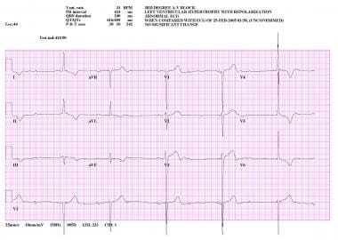
Electrocardiography
Vasodilation
Cardiopulmonary Bypass
Treatment Outcome
Radionuclide Imaging
Echocardiography
Carbon Dioxide
Vasoconstriction
Magnetic Resonance Imaging, Cine
Models, Cardiovascular
Heart Arrest, Induced
Diffusion Magnetic Resonance Imaging
Myocardial Stunning
Microdialysis
Fractional Flow Reserve, Myocardial
Algorithms
Capillary Permeability
Acetazolamide
Hyperthermia, Induced
Nitric Oxide
Microbubbles
Echo-Planar Imaging
Models, Animal
Observer Variation
Krypton Radioisotopes
Ammonia
Cerebral Infarction
Melphalan
Pulmonary Gas Exchange
Ventricular Dysfunction, Left
Dose-Response Relationship, Drug
Iohexol
Radionuclide Angiography
Imaging, Three-Dimensional
Partial Pressure
Neovascularization, Physiologic
Warm Ischemia
Stroke
Arterioles
Norepinephrine
Glucose
Mannitol
Middle Cerebral Artery
Albumins
Sulfur Hexafluoride
Isotonic Solutions
Indocyanine Green
Xenon
Organometallic Compounds
Endothelium, Vascular
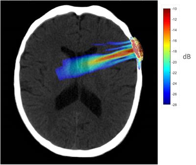
Cerebral Angiography
Cardiac Output
Severity of Illness Index
Subtraction Technique
Stroke Volume
Pulsatile Flow
Aqueous Humor
Bioreactors
Follow-Up Studies
Noble Gases
Reference Values
Iodobenzenes
Lactic Acid
Hepatic Artery
Retrospective Studies
Arterial Occlusive Diseases
Phantoms, Imaging
ROC Curve
Angina Pectoris
Iofetamine
Heart Ventricles
Blood Volume Determination
Prognosis
Circulatory Arrest, Deep Hypothermia Induced
Calcium
Analysis of Variance
Raffinose
Tomography, Spiral Computed
Sturge-Weber Syndrome
Echocardiography, Stress
Tissue Preservation
Infarction, Middle Cerebral Artery
Prognostic importance of quantitative echocardiographic evaluation in patients suspected of first non-massive pulmonary embolism. (1/275)
(+info)Which CT perfusion parameter best reflects cerebrovascular reserve?: correlation of acetazolamide-challenged CT perfusion with single-photon emission CT in Moyamoya patients. (2/275)
(+info)Changes in ventilatory capacity, exercise capacity, and pulmonary blood flow after lobectomy in patients with lung cancer--which lobectomy has the most loss in exercise capacity? (3/275)
(+info)Clinical parameters affecting prediction accuracy of postoperative lung function in non-small cell lung cancer. (4/275)
(+info)Computed tomography perfusion in evaluating the therapeutic effect of transarterial chemoembolization for hepatocellular carcinoma. (5/275)
AIM: To prospectively assess the changes in parameters of computed tomography (CT) perfusion pre- and post-transarterial chemoembolization (TACE) of hepatocellular carcinoma (HCC) in different treatment response groups, and to correlate the changes with various responses of HCC to TACE. METHODS: Thirty-nine HCC patients underwent CT perfusion examinations pre-(1 d before TACE) and post-treatment (4 wk after TACE). The response evaluation criteria for solid tumors (RECIST) were referred to when treatment responses were distributed. Wilcoxon-signed ranks test was used to compare the differences in CT perfusion parameters pre- and post-TACE for different response groups. RESULTS: Only one case had treatment response to CR and the CT perfusion maps of post-treatment lesion displayed complete absence of signals. In the PR treatment response group, hepatic artery perfusion (HAP), hepatic arterial fracture (HAF) and hepatic blood volume (HBV) of viable tumors post-TACE were reduced compared with pre-TACE (P = 0.001, 0.030 and 0.001, respectively). In the SD group, all CT perfusion parameters were not significantly different pre- and post-TACE. In the PD group, HAP, HAF, portal vein perfusion (PVP) and hepatic blood flow (HBF) of viable tumors post-TACE were significantly increased compared with pre-TACE (P = 0.005, 0.012, 0.035 and 0.005, respectively). CONCLUSION: Changes in CT perfusion parameters of viable tumors are correlated with different responses of HCC to TACE. Therefore, CT perfusion imaging is a feasible technique for monitoring response of HCC to TACE. (+info)Use of adrenomedullin derivatives for molecular imaging of pulmonary circulation. (6/275)
(+info)Sensitivity and specificity of perfusion scintigraphy combined with chest radiography for acute pulmonary embolism in PIOPED II. (7/275)
(+info)Neural substrates of visuospatial processing in distinct reference frames: evidence from unilateral spatial neglect. (8/275)
(+info)Perfusion, in medical terms, refers to the process of circulating blood through the body's organs and tissues to deliver oxygen and nutrients and remove waste products. It is a measure of the delivery of adequate blood flow to specific areas or tissues in the body. Perfusion can be assessed using various methods, including imaging techniques like computed tomography (CT) scans, magnetic resonance imaging (MRI), and perfusion scintigraphy.
Perfusion is critical for maintaining proper organ function and overall health. When perfusion is impaired or inadequate, it can lead to tissue hypoxia, acidosis, and cell death, which can result in organ dysfunction or failure. Conditions that can affect perfusion include cardiovascular disease, shock, trauma, and certain surgical procedures.
Perfusion imaging is a medical imaging technique used to evaluate the blood flow or perfusion in various organs and tissues of the body. It is often utilized in conjunction with computed tomography (CT), magnetic resonance imaging (MRI), or single-photon emission computed tomography (SPECT) scans.
During a perfusion imaging procedure, a contrast agent is introduced into the patient's bloodstream, and a series of images are captured to track the flow and distribution of the contrast agent over time. This information helps medical professionals assess tissue viability, identify areas of reduced or blocked blood flow, and detect various pathological conditions such as stroke, heart attack, pulmonary embolism, and tumors.
In summary, perfusion imaging is a valuable diagnostic tool for evaluating the circulatory function of different organs and tissues in the body.
Myocardial perfusion imaging (MPI) is a non-invasive nuclear medicine test used to assess the blood flow to the heart muscle (myocardium). It typically involves the injection of a radioactive tracer, such as thallium-201 or technetium-99m sestamibi, into a vein. The tracer is taken up by healthy heart muscle in proportion to blood flow. A special camera then takes images of the distribution of the tracer within the heart, providing information about areas of reduced or blocked blood flow (ischemia) or scarred tissue (infarction). MPI can help diagnose coronary artery disease, assess the effectiveness of treatments, and determine prognosis.
Emission-Computed Tomography, Single-Photon (SPECT) is a type of nuclear medicine imaging procedure that generates detailed, three-dimensional images of the distribution of radioactive pharmaceuticals within the body. It uses gamma rays emitted by a radiopharmaceutical that is introduced into the patient's body, and a specialized gamma camera to detect these gamma rays and create tomographic images. The data obtained from the SPECT imaging can be used to diagnose various medical conditions, evaluate organ function, and guide treatment decisions. It is commonly used to image the heart, brain, and bones, among other organs and systems.
Regional perfusion chemotherapy for cancer is a medical treatment in which a specific area or region of the body is infused with high concentrations of cancer-killing (cytotoxic) drugs via a temporary isolation and perfusion of that region. This technique is typically used to treat isolated areas of cancer that are locally advanced, recurrent, or cannot be removed surgically.
The procedure involves isolating the regional blood circulation by cannulating the artery and vein that supply blood to the target area, often the limbs (such as in melanoma or sarcoma) or the liver (for liver tumors). The chemotherapeutic drugs are then introduced into the isolated arterial circulation, allowing for a high concentration of the drug to be delivered directly to the cancerous tissue while minimizing systemic exposure and toxicity.
After the infusion, the region is rinsed with a blood-substitute solution to remove any residual chemotherapeutic agents before reconnecting the circulation. This procedure can be repeated multiple times if necessary.
Regional perfusion chemotherapy has been shown to improve local control and potentially increase survival rates in certain types of cancer, while reducing systemic side effects compared to traditional intravenous chemotherapy. However, it is a complex and invasive procedure that requires specialized medical expertise and facilities.
Coronary circulation refers to the circulation of blood in the coronary vessels, which supply oxygenated blood to the heart muscle (myocardium) and drain deoxygenated blood from it. The coronary circulation system includes two main coronary arteries - the left main coronary artery and the right coronary artery - that branch off from the aorta just above the aortic valve. These arteries further divide into smaller branches, which supply blood to different regions of the heart muscle.
The left main coronary artery divides into two branches: the left anterior descending (LAD) artery and the left circumflex (LCx) artery. The LAD supplies blood to the front and sides of the heart, while the LCx supplies blood to the back and sides of the heart. The right coronary artery supplies blood to the lower part of the heart, including the right ventricle and the bottom portion of the left ventricle.
The veins that drain the heart muscle include the great cardiac vein, the middle cardiac vein, and the small cardiac vein, which merge to form the coronary sinus. The coronary sinus empties into the right atrium, allowing deoxygenated blood to enter the right side of the heart and be pumped to the lungs for oxygenation.
Coronary circulation is essential for maintaining the health and function of the heart muscle, as it provides the necessary oxygen and nutrients required for proper contraction and relaxation of the myocardium. Any disruption or blockage in the coronary circulation system can lead to serious consequences, such as angina, heart attack, or even death.
Cerebrovascular circulation refers to the network of blood vessels that supply oxygenated blood and nutrients to the brain tissue, and remove waste products. It includes the internal carotid arteries, vertebral arteries, circle of Willis, and the intracranial arteries that branch off from them.
The internal carotid arteries and vertebral arteries merge to form the circle of Willis, a polygonal network of vessels located at the base of the brain. The anterior cerebral artery, middle cerebral artery, posterior cerebral artery, and communicating arteries are the major vessels that branch off from the circle of Willis and supply blood to different regions of the brain.
Interruptions or abnormalities in the cerebrovascular circulation can lead to various neurological conditions such as stroke, transient ischemic attack (TIA), and vascular dementia.
Regional blood flow (RBF) refers to the rate at which blood flows through a specific region or organ in the body, typically expressed in milliliters per minute per 100 grams of tissue (ml/min/100g). It is an essential physiological parameter that reflects the delivery of oxygen and nutrients to tissues while removing waste products. RBF can be affected by various factors such as metabolic demands, neural regulation, hormonal influences, and changes in blood pressure or vascular resistance. Measuring RBF is crucial for understanding organ function, diagnosing diseases, and evaluating the effectiveness of treatments.
Technetium Tc 99m Sestamibi is a radiopharmaceutical compound used in medical imaging, specifically in myocardial perfusion scintigraphy. It is a technetium-labeled isonitrile chelate that is taken up by mitochondria in cells with high metabolic activity, such as cardiomyocytes (heart muscle cells).
Once injected into the patient's body, Technetium Tc 99m Sestamibi emits gamma rays, which can be detected by a gamma camera. This allows for the creation of images that reflect the distribution and function of the radiopharmaceutical within the heart muscle. The images can help identify areas of reduced blood flow or ischemia, which may indicate coronary artery disease.
The uptake of Technetium Tc 99m Sestamibi in other organs, such as the breast and thyroid, can also be used for imaging purposes, although its primary use remains in cardiac imaging.
Organotechnetium compounds are chemical substances that contain carbon-technetium bonds, where technetium is an element with the symbol Tc and atomic number 43. These types of compounds are primarily used in medical imaging as radioactive tracers due to the ability of technetium-99m to emit gamma rays. The organotechnetium compounds help in localizing specific organs, tissues, or functions within the body, making them useful for diagnostic purposes in nuclear medicine.
It is important to note that most organotechnetium compounds are synthesized from technetium-99m, which is generated from the decay of molybdenum-99. The use of these compounds requires proper handling and administration by trained medical professionals due to their radioactive nature.
Contrast media are substances that are administered to a patient in order to improve the visibility of internal body structures or processes in medical imaging techniques such as X-rays, CT scans, MRI scans, and ultrasounds. These media can be introduced into the body through various routes, including oral, rectal, or intravenous administration.
Contrast media work by altering the appearance of bodily structures in imaging studies. For example, when a patient undergoes an X-ray examination, contrast media can be used to highlight specific organs, tissues, or blood vessels, making them more visible on the resulting images. In CT and MRI scans, contrast media can help to enhance the differences between normal and abnormal tissues, allowing for more accurate diagnosis and treatment planning.
There are several types of contrast media available, each with its own specific properties and uses. Some common examples include barium sulfate, which is used as a contrast medium in X-ray studies of the gastrointestinal tract, and iodinated contrast media, which are commonly used in CT scans to highlight blood vessels and other structures.
While contrast media are generally considered safe, they can sometimes cause adverse reactions, ranging from mild symptoms such as nausea or hives to more serious complications such as anaphylaxis or kidney damage. As a result, it is important for healthcare providers to carefully evaluate each patient's medical history and individual risk factors before administering contrast media.
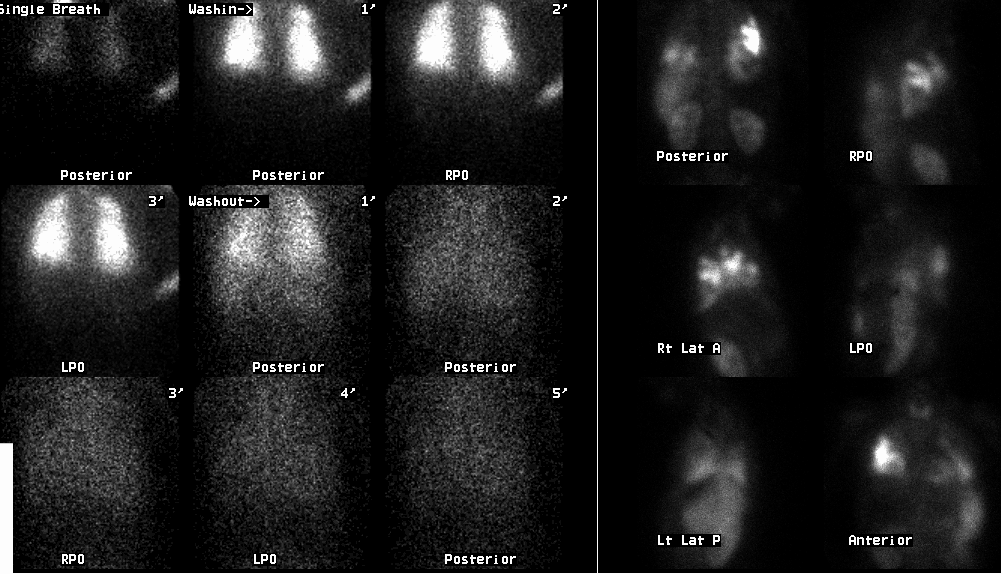
The Ventilation-Perfusion (V/Q) ratio is a measure used in respiratory physiology to describe the relationship between the amount of air that enters the alveoli (ventilation) and the amount of blood that reaches the alveoli to pick up oxygen (perfusion).
In a healthy lung, these two processes are well-matched, meaning that well-ventilated areas of the lung also have good blood flow. This results in a V/Q ratio close to 1.0.
However, certain lung conditions such as emphysema or pulmonary embolism can cause ventilation and perfusion to become mismatched, leading to a V/Q ratio that is either higher (ventilation exceeds perfusion) or lower (perfusion exceeds ventilation) than normal. This mismatch can result in impaired gas exchange and lead to hypoxemia (low oxygen levels in the blood).
The V/Q ratio is often used in clinical settings to assess lung function and diagnose respiratory disorders.
In medical terms, the heart is a muscular organ located in the thoracic cavity that functions as a pump to circulate blood throughout the body. It's responsible for delivering oxygen and nutrients to the tissues and removing carbon dioxide and other wastes. The human heart is divided into four chambers: two atria on the top and two ventricles on the bottom. The right side of the heart receives deoxygenated blood from the body and pumps it to the lungs, while the left side receives oxygenated blood from the lungs and pumps it out to the rest of the body. The heart's rhythmic contractions and relaxations are regulated by a complex electrical conduction system.
Magnetic Resonance Angiography (MRA) is a non-invasive medical imaging technique that uses magnetic fields and radio waves to create detailed images of the blood vessels or arteries within the body. It is a type of Magnetic Resonance Imaging (MRI) that focuses specifically on the circulatory system.
MRA can be used to diagnose and evaluate various conditions related to the blood vessels, such as aneurysms, stenosis (narrowing of the vessel), or the presence of plaques or tumors. It can also be used to plan for surgeries or other treatments related to the vascular system. The procedure does not use radiation and is generally considered safe, although people with certain implants like pacemakers may not be able to have an MRA due to safety concerns.
Microcirculation is the circulation of blood in the smallest blood vessels, including arterioles, venules, and capillaries. It's responsible for the delivery of oxygen and nutrients to the tissues and the removal of waste products. The microcirculation plays a crucial role in maintaining tissue homeostasis and is regulated by various physiological mechanisms such as autonomic nervous system activity, local metabolic factors, and hormones.
Impairment of microcirculation can lead to tissue hypoxia, inflammation, and organ dysfunction, which are common features in several diseases, including diabetes, hypertension, sepsis, and ischemia-reperfusion injury. Therefore, understanding the structure and function of the microcirculation is essential for developing new therapeutic strategies to treat these conditions.
Laser-Doppler flowmetry (LDF) is a non-invasive, investigative technique used to measure microcirculatory blood flow in real time. It is based on the principle of the Doppler effect, which describes the change in frequency or wavelength of light or sound waves as they encounter a moving object or reflect off a moving surface.
In LDF, a low-power laser beam is directed at the skin or other transparent tissue. The light penetrates the tissue and scatters off the moving red blood cells within the microvasculature. As the light scatters, it undergoes a slight frequency shift due to the movement of the red blood cells. This frequency shift is then detected by a photodetector, which converts it into an electrical signal. The magnitude of this signal is directly proportional to the speed and concentration of the moving red blood cells, providing a measure of microcirculatory blood flow.
LDF has various clinical applications, including the assessment of skin perfusion in patients with peripheral arterial disease, burn injuries, and flaps used in reconstructive surgery. It can also be used to study the effects of drugs or other interventions on microcirculation in research settings.
Radiopharmaceuticals are defined as pharmaceutical preparations that contain radioactive isotopes and are used for diagnosis or therapy in nuclear medicine. These compounds are designed to interact specifically with certain biological targets, such as cells, tissues, or organs, and emit radiation that can be detected and measured to provide diagnostic information or used to destroy abnormal cells or tissue in therapeutic applications.
The radioactive isotopes used in radiopharmaceuticals have carefully controlled half-lives, which determine how long they remain radioactive and how long the pharmaceutical preparation remains effective. The choice of radioisotope depends on the intended use of the radiopharmaceutical, as well as factors such as its energy, range of emission, and chemical properties.
Radiopharmaceuticals are used in a wide range of medical applications, including imaging, cancer therapy, and treatment of other diseases and conditions. Examples of radiopharmaceuticals include technetium-99m for imaging the heart, lungs, and bones; iodine-131 for treating thyroid cancer; and samarium-153 for palliative treatment of bone metastases.
The use of radiopharmaceuticals requires specialized training and expertise in nuclear medicine, as well as strict adherence to safety protocols to minimize radiation exposure to patients and healthcare workers.
Blood flow velocity is the speed at which blood travels through a specific part of the vascular system. It is typically measured in units of distance per time, such as centimeters per second (cm/s) or meters per second (m/s). Blood flow velocity can be affected by various factors, including cardiac output, vessel diameter, and viscosity of the blood. Measuring blood flow velocity is important in diagnosing and monitoring various medical conditions, such as heart disease, stroke, and peripheral vascular disease.
Thallium radioisotopes are radioactive isotopes or variants of the element thallium (Tl), which decays and emits radiation. Thallium has several radioisotopes, with the most commonly used being thallium-201 (^201Tl). This radioisotope is used in medical imaging, specifically in myocardial perfusion scintigraphy, to evaluate blood flow to the heart muscle. It decays by electron capture and emits gamma radiation with a half-life of 73 hours, making it suitable for diagnostic procedures.
It's important to note that handling and using radioisotopes require proper training and safety measures due to their ionizing radiation properties.
Dipyridamole is a medication that belongs to a class of drugs called antiplatelet agents. It works by preventing platelets in your blood from sticking together to form clots. Dipyridamole is often used in combination with aspirin to prevent stroke and other complications in people who have had a heart valve replacement or a type of irregular heartbeat called atrial fibrillation.
Dipyridamole can also be used as a stress agent in myocardial perfusion imaging studies, which are tests used to evaluate blood flow to the heart. When used for this purpose, dipyridamole is given intravenously and works by dilating the blood vessels in the heart, allowing more blood to flow through them and making it easier to detect areas of reduced blood flow.
The most common side effects of dipyridamole include headache, dizziness, and gastrointestinal symptoms such as diarrhea, nausea, and vomiting. In rare cases, dipyridamole can cause more serious side effects, such as allergic reactions, abnormal heart rhythms, or low blood pressure. It is important to take dipyridamole exactly as directed by your healthcare provider and to report any unusual symptoms or side effects promptly.
Blood volume refers to the total amount of blood present in an individual's circulatory system at any given time. It is the combined volume of both the plasma (the liquid component of blood) and the formed elements (such as red and white blood cells and platelets) in the blood. In a healthy adult human, the average blood volume is approximately 5 liters (or about 1 gallon). However, blood volume can vary depending on several factors, including age, sex, body weight, and overall health status.
Blood volume plays a critical role in maintaining proper cardiovascular function, as it affects blood pressure, heart rate, and the delivery of oxygen and nutrients to tissues throughout the body. Changes in blood volume can have significant impacts on an individual's health and may be associated with various medical conditions, such as dehydration, hemorrhage, heart failure, and liver disease. Accurate measurement of blood volume is essential for diagnosing and managing these conditions, as well as for guiding treatment decisions in clinical settings.
Medical Definition:
Magnetic Resonance Imaging (MRI) is a non-invasive diagnostic imaging technique that uses a strong magnetic field and radio waves to create detailed cross-sectional or three-dimensional images of the internal structures of the body. The patient lies within a large, cylindrical magnet, and the scanner detects changes in the direction of the magnetic field caused by protons in the body. These changes are then converted into detailed images that help medical professionals to diagnose and monitor various medical conditions, such as tumors, injuries, or diseases affecting the brain, spinal cord, heart, blood vessels, joints, and other internal organs. MRI does not use radiation like computed tomography (CT) scans.
Oximes are a class of chemical compounds that contain the functional group =N-O-, where two organic groups are attached to the nitrogen atom. In a clinical context, oximes are used as antidotes for nerve agent and pesticide poisoning. The most commonly used oxime in medicine is pralidoxime (2-PAM), which is used to reactivate acetylcholinesterase that has been inhibited by organophosphorus compounds, such as nerve agents and certain pesticides. These compounds work by forming a bond with the phosphoryl group of the inhibited enzyme, allowing for its reactivation and restoration of normal neuromuscular function.
Nitrogen radioisotopes are unstable isotopes of the element nitrogen that emit radiation as they decay into more stable forms. Nitrogen has several radioisotopes, with the most common being nitrogen-13 and nitrogen-15. These isotopes have 7 protons in their nucleus, but differ in the number of neutrons.
Nitrogen-13 has a half-life of about 10 minutes, making it useful for medical imaging studies such as positron emission tomography (PET) scans. When nitrogen-13 decays, it emits a positron, which then collides with an electron and produces gamma rays that can be detected by a PET scanner.
Nitrogen-15, on the other hand, has a half-life of about 3 minutes and is not typically used for medical imaging. However, it is widely used in research settings as a stable isotope tracer to study metabolic processes in the body.
It's important to note that handling and using radioisotopes requires specialized training and equipment due to their potential radiation hazards.
In the field of medicine, "time factors" refer to the duration of symptoms or time elapsed since the onset of a medical condition, which can have significant implications for diagnosis and treatment. Understanding time factors is crucial in determining the progression of a disease, evaluating the effectiveness of treatments, and making critical decisions regarding patient care.
For example, in stroke management, "time is brain," meaning that rapid intervention within a specific time frame (usually within 4.5 hours) is essential to administering tissue plasminogen activator (tPA), a clot-busting drug that can minimize brain damage and improve patient outcomes. Similarly, in trauma care, the "golden hour" concept emphasizes the importance of providing definitive care within the first 60 minutes after injury to increase survival rates and reduce morbidity.
Time factors also play a role in monitoring the progression of chronic conditions like diabetes or heart disease, where regular follow-ups and assessments help determine appropriate treatment adjustments and prevent complications. In infectious diseases, time factors are crucial for initiating antibiotic therapy and identifying potential outbreaks to control their spread.
Overall, "time factors" encompass the significance of recognizing and acting promptly in various medical scenarios to optimize patient outcomes and provide effective care.
Hemodynamics is the study of how blood flows through the cardiovascular system, including the heart and the vascular network. It examines various factors that affect blood flow, such as blood volume, viscosity, vessel length and diameter, and pressure differences between different parts of the circulatory system. Hemodynamics also considers the impact of various physiological and pathological conditions on these variables, and how they in turn influence the function of vital organs and systems in the body. It is a critical area of study in fields such as cardiology, anesthesiology, and critical care medicine.
Myocardial ischemia is a condition in which the blood supply to the heart muscle (myocardium) is reduced or blocked, leading to insufficient oxygen delivery and potential damage to the heart tissue. This reduction in blood flow typically results from the buildup of fatty deposits, called plaques, in the coronary arteries that supply the heart with oxygen-rich blood. The plaques can rupture or become unstable, causing the formation of blood clots that obstruct the artery and limit blood flow.
Myocardial ischemia may manifest as chest pain (angina pectoris), shortness of breath, fatigue, or irregular heartbeats (arrhythmias). In severe cases, it can lead to myocardial infarction (heart attack) if the oxygen supply is significantly reduced or cut off completely, causing permanent damage or death of the heart muscle. Early diagnosis and treatment of myocardial ischemia are crucial for preventing further complications and improving patient outcomes.
Sensitivity and specificity are statistical measures used to describe the performance of a diagnostic test or screening tool in identifying true positive and true negative results.
* Sensitivity refers to the proportion of people who have a particular condition (true positives) who are correctly identified by the test. It is also known as the "true positive rate" or "recall." A highly sensitive test will identify most or all of the people with the condition, but may also produce more false positives.
* Specificity refers to the proportion of people who do not have a particular condition (true negatives) who are correctly identified by the test. It is also known as the "true negative rate." A highly specific test will identify most or all of the people without the condition, but may also produce more false negatives.
In medical testing, both sensitivity and specificity are important considerations when evaluating a diagnostic test. High sensitivity is desirable for screening tests that aim to identify as many cases of a condition as possible, while high specificity is desirable for confirmatory tests that aim to rule out the condition in people who do not have it.
It's worth noting that sensitivity and specificity are often influenced by factors such as the prevalence of the condition in the population being tested, the threshold used to define a positive result, and the reliability and validity of the test itself. Therefore, it's important to consider these factors when interpreting the results of a diagnostic test.
X-ray computed tomography (CT or CAT scan) is a medical imaging method that uses computer-processed combinations of many X-ray images taken from different angles to produce cross-sectional (tomographic) images (virtual "slices") of the body. These cross-sectional images can then be used to display detailed internal views of organs, bones, and soft tissues in the body.
The term "computed tomography" is used instead of "CT scan" or "CAT scan" because the machines take a series of X-ray measurements from different angles around the body and then use a computer to process these data to create detailed images of internal structures within the body.
CT scanning is a noninvasive, painless medical test that helps physicians diagnose and treat medical conditions. CT imaging provides detailed information about many types of tissue including lung, bone, soft tissue and blood vessels. CT examinations can be performed on every part of the body for a variety of reasons including diagnosis, surgical planning, and monitoring of therapeutic responses.
In computed tomography (CT), an X-ray source and detector rotate around the patient, measuring the X-ray attenuation at many different angles. A computer uses this data to construct a cross-sectional image by the process of reconstruction. This technique is called "tomography". The term "computed" refers to the use of a computer to reconstruct the images.
CT has become an important tool in medical imaging and diagnosis, allowing radiologists and other physicians to view detailed internal images of the body. It can help identify many different medical conditions including cancer, heart disease, lung nodules, liver tumors, and internal injuries from trauma. CT is also commonly used for guiding biopsies and other minimally invasive procedures.
In summary, X-ray computed tomography (CT or CAT scan) is a medical imaging technique that uses computer-processed combinations of many X-ray images taken from different angles to produce cross-sectional images of the body. It provides detailed internal views of organs, bones, and soft tissues in the body, allowing physicians to diagnose and treat medical conditions.
Computer-assisted image processing is a medical term that refers to the use of computer systems and specialized software to improve, analyze, and interpret medical images obtained through various imaging techniques such as X-ray, CT (computed tomography), MRI (magnetic resonance imaging), ultrasound, and others.
The process typically involves several steps, including image acquisition, enhancement, segmentation, restoration, and analysis. Image processing algorithms can be used to enhance the quality of medical images by adjusting contrast, brightness, and sharpness, as well as removing noise and artifacts that may interfere with accurate diagnosis. Segmentation techniques can be used to isolate specific regions or structures of interest within an image, allowing for more detailed analysis.
Computer-assisted image processing has numerous applications in medical imaging, including detection and characterization of lesions, tumors, and other abnormalities; assessment of organ function and morphology; and guidance of interventional procedures such as biopsies and surgeries. By automating and standardizing image analysis tasks, computer-assisted image processing can help to improve diagnostic accuracy, efficiency, and consistency, while reducing the potential for human error.
I believe there might be a misunderstanding in your question. "Dogs" is not a medical term or condition. It is the common name for a domesticated carnivore of the family Canidae, specifically the genus Canis, which includes wolves, foxes, and other extant and extinct species of mammals. Dogs are often kept as pets and companions, and they have been bred in a wide variety of forms and sizes for different purposes, such as hunting, herding, guarding, assisting police and military forces, and providing companionship and emotional support.
If you meant to ask about a specific medical condition or term related to dogs, please provide more context so I can give you an accurate answer.
Adenosine is a purine nucleoside that is composed of a sugar (ribose) and the base adenine. It plays several important roles in the body, including serving as a precursor for the synthesis of other molecules such as ATP, NAD+, and RNA.
In the medical context, adenosine is perhaps best known for its use as a pharmaceutical agent to treat certain cardiac arrhythmias. When administered intravenously, it can help restore normal sinus rhythm in patients with paroxysmal supraventricular tachycardia (PSVT) by slowing conduction through the atrioventricular node and interrupting the reentry circuit responsible for the arrhythmia.
Adenosine can also be used as a diagnostic tool to help differentiate between narrow-complex tachycardias of supraventricular origin and those that originate from below the ventricles (such as ventricular tachycardia). This is because adenosine will typically terminate PSVT but not affect the rhythm of VT.
It's worth noting that adenosine has a very short half-life, lasting only a few seconds in the bloodstream. This means that its effects are rapidly reversible and generally well-tolerated, although some patients may experience transient symptoms such as flushing, chest pain, or shortness of breath.
Technetium Tc 99m Exametazime is a radiopharmaceutical agent used in nuclear medicine imaging procedures. The compound consists of the radioisotope Technetium-99m (^99m^Tc) bonded to Exametazime, also known as HMPAO (hexamethylpropyleneamine oxime).
Once injected into the patient's bloodstream, Technetium Tc 99m Exametazime distributes evenly throughout the brain, crossing the blood-brain barrier and entering cells. The radioactive decay of Technetium-99m emits gamma rays that can be detected by a gamma camera, creating images of the brain's blood flow and distribution of the tracer.
This imaging technique is often used in cerebral perfusion studies to assess conditions such as stroke, epilepsy, or dementia, providing valuable information about regional cerebral blood flow and potential areas of injury or abnormality.
Pulmonary circulation refers to the process of blood flow through the lungs, where blood picks up oxygen and releases carbon dioxide. This is a vital part of the overall circulatory system, which delivers nutrients and oxygen to the body's cells while removing waste products like carbon dioxide.
In pulmonary circulation, deoxygenated blood from the systemic circulation returns to the right atrium of the heart via the superior and inferior vena cava. The blood then moves into the right ventricle through the tricuspid valve and gets pumped into the pulmonary artery when the right ventricle contracts.
The pulmonary artery divides into smaller vessels called arterioles, which further branch into a vast network of tiny capillaries in the lungs. Here, oxygen from the alveoli diffuses into the blood, binding to hemoglobin in red blood cells, while carbon dioxide leaves the blood and is exhaled through the nose or mouth.
The now oxygenated blood collects in venules, which merge to form pulmonary veins. These veins transport the oxygen-rich blood back to the left atrium of the heart, where it enters the systemic circulation once again. This continuous cycle enables the body's cells to receive the necessary oxygen and nutrients for proper functioning while disposing of waste products.
Image enhancement in the medical context refers to the process of improving the quality and clarity of medical images, such as X-rays, CT scans, MRI scans, or ultrasound images, to aid in the diagnosis and treatment of medical conditions. Image enhancement techniques may include adjusting contrast, brightness, or sharpness; removing noise or artifacts; or applying specialized algorithms to highlight specific features or structures within the image.
The goal of image enhancement is to provide clinicians with more accurate and detailed information about a patient's anatomy or physiology, which can help inform medical decision-making and improve patient outcomes.
Ischemia is the medical term used to describe a lack of blood flow to a part of the body, often due to blocked or narrowed blood vessels. This can lead to a shortage of oxygen and nutrients in the tissues, which can cause them to become damaged or die. Ischemia can affect many different parts of the body, including the heart, brain, legs, and intestines. Symptoms of ischemia depend on the location and severity of the blockage, but they may include pain, cramping, numbness, weakness, or coldness in the affected area. In severe cases, ischemia can lead to tissue death (gangrene) or organ failure. Treatment for ischemia typically involves addressing the underlying cause of the blocked blood flow, such as through medication, surgery, or lifestyle changes.
Gated Blood-Pool Imaging (GBPI) is a type of nuclear medicine test that uses radioactive material and a specialized camera to create detailed images of the heart and its function. In this procedure, a small amount of radioactive tracer is injected into the patient's bloodstream, which then accumulates in the heart muscle and the blood pool within the heart chambers.
The term "gated" refers to the use of an electrocardiogram (ECG) signal to synchronize the image acquisition with the heart's contractions. This allows for the visualization of the heart's motion during different phases of the cardiac cycle, providing valuable information about the size, shape, and contraction of the heart chambers, as well as the movement of the walls of the heart.
GBPI is often used to assess patients with known or suspected heart disease, such as valvular abnormalities, cardiomyopathies, or congenital heart defects. It can help diagnose and evaluate the severity of these conditions, guide treatment decisions, and monitor the effectiveness of therapy.
The brain is the central organ of the nervous system, responsible for receiving and processing sensory information, regulating vital functions, and controlling behavior, movement, and cognition. It is divided into several distinct regions, each with specific functions:
1. Cerebrum: The largest part of the brain, responsible for higher cognitive functions such as thinking, learning, memory, language, and perception. It is divided into two hemispheres, each controlling the opposite side of the body.
2. Cerebellum: Located at the back of the brain, it is responsible for coordinating muscle movements, maintaining balance, and fine-tuning motor skills.
3. Brainstem: Connects the cerebrum and cerebellum to the spinal cord, controlling vital functions such as breathing, heart rate, and blood pressure. It also serves as a relay center for sensory information and motor commands between the brain and the rest of the body.
4. Diencephalon: A region that includes the thalamus (a major sensory relay station) and hypothalamus (regulates hormones, temperature, hunger, thirst, and sleep).
5. Limbic system: A group of structures involved in emotional processing, memory formation, and motivation, including the hippocampus, amygdala, and cingulate gyrus.
The brain is composed of billions of interconnected neurons that communicate through electrical and chemical signals. It is protected by the skull and surrounded by three layers of membranes called meninges, as well as cerebrospinal fluid that provides cushioning and nutrients.
Organ preservation is a medical technique used to maintain the viability and functionality of an organ outside the body for a certain period, typically for transplantation purposes. This process involves cooling the organ to slow down its metabolic activity and prevent tissue damage, while using specialized solutions that help preserve the organ's structure and function. Commonly preserved organs include hearts, livers, kidneys, lungs, and pancreases. The goal of organ preservation is to ensure that the transplanted organ remains in optimal condition until it can be successfully implanted into a recipient.
Blood pressure is the force exerted by circulating blood on the walls of the blood vessels. It is measured in millimeters of mercury (mmHg) and is given as two figures:
1. Systolic pressure: This is the pressure when the heart pushes blood out into the arteries.
2. Diastolic pressure: This is the pressure when the heart rests between beats, allowing it to fill with blood.
Normal blood pressure for adults is typically around 120/80 mmHg, although this can vary slightly depending on age, sex, and other factors. High blood pressure (hypertension) is generally considered to be a reading of 130/80 mmHg or higher, while low blood pressure (hypotension) is usually defined as a reading below 90/60 mmHg. It's important to note that blood pressure can fluctuate throughout the day and may be affected by factors such as stress, physical activity, and medication use.
Reproducibility of results in a medical context refers to the ability to obtain consistent and comparable findings when a particular experiment or study is repeated, either by the same researcher or by different researchers, following the same experimental protocol. It is an essential principle in scientific research that helps to ensure the validity and reliability of research findings.
In medical research, reproducibility of results is crucial for establishing the effectiveness and safety of new treatments, interventions, or diagnostic tools. It involves conducting well-designed studies with adequate sample sizes, appropriate statistical analyses, and transparent reporting of methods and findings to allow other researchers to replicate the study and confirm or refute the results.
The lack of reproducibility in medical research has become a significant concern in recent years, as several high-profile studies have failed to produce consistent findings when replicated by other researchers. This has led to increased scrutiny of research practices and a call for greater transparency, rigor, and standardization in the conduct and reporting of medical research.
An exercise test, also known as a stress test or an exercise stress test, is a medical procedure used to evaluate the heart's function and response to physical exertion. It typically involves walking on a treadmill or pedaling a stationary bike while being monitored for changes in heart rate, blood pressure, electrocardiogram (ECG), and sometimes other variables such as oxygen consumption or gas exchange.
During the test, the patient's symptoms, such as chest pain or shortness of breath, are also closely monitored. The exercise test can help diagnose coronary artery disease, assess the severity of heart-related symptoms, and evaluate the effectiveness of treatments for heart conditions. It may also be used to determine a person's safe level of physical activity and fitness.
There are different types of exercise tests, including treadmill stress testing, stationary bike stress testing, nuclear stress testing, and stress echocardiography. The specific type of test used depends on the patient's medical history, symptoms, and overall health status.
Gadolinium DTPA (Diethylenetriaminepentaacetic acid) is a type of gadolinium-based contrast agent (GBCA) used in medical imaging, particularly magnetic resonance imaging (MRI) and magnetic resonance angiography (MRA). It functions as a paramagnetic substance that enhances the visibility of internal body structures during these imaging techniques.
The compound Gadolinium DTPA is formed when gadolinium ions are bound to diethylenetriaminepentaacetic acid, a chelating agent. This binding helps to make the gadolinium ion safer for use in medical imaging by reducing its toxicity and improving its stability in the body.
Gadolinium DTPA is eliminated from the body primarily through the kidneys, making it important to monitor renal function before administering this contrast agent. In some cases, Gadolinium DTPA may cause adverse reactions, including allergic-like responses and nephrogenic systemic fibrosis (NSF) in patients with impaired kidney function.
The myocardium is the middle layer of the heart wall, composed of specialized cardiac muscle cells that are responsible for pumping blood throughout the body. It forms the thickest part of the heart wall and is divided into two sections: the left ventricle, which pumps oxygenated blood to the rest of the body, and the right ventricle, which pumps deoxygenated blood to the lungs.
The myocardium contains several types of cells, including cardiac muscle fibers, connective tissue, nerves, and blood vessels. The muscle fibers are arranged in a highly organized pattern that allows them to contract in a coordinated manner, generating the force necessary to pump blood through the heart and circulatory system.
Damage to the myocardium can occur due to various factors such as ischemia (reduced blood flow), infection, inflammation, or genetic disorders. This damage can lead to several cardiac conditions, including heart failure, arrhythmias, and cardiomyopathy.
Emission computed tomography (ECT) is a type of tomographic imaging technique in which an emission signal from within the body is detected to create cross-sectional images of that signal's distribution. In Emission-Computed Tomography (ECT), a radionuclide is introduced into the body, usually through injection, inhalation or ingestion. The radionuclide emits gamma rays that are then detected by external gamma cameras.
The data collected from these cameras is then used to create cross-sectional images of the distribution of the radiopharmaceutical within the body. This allows for the identification and quantification of functional information about specific organs or systems within the body, such as blood flow, metabolic activity, or receptor density.
One common type of Emission-Computed Tomography is Single Photon Emission Computed Tomography (SPECT), which uses a single gamma camera that rotates around the patient to collect data from multiple angles. Another type is Positron Emission Tomography (PET), which uses positron-emitting radionuclides and detects the coincident gamma rays emitted by the annihilation of positrons and electrons.
Overall, ECT is a valuable tool in medical imaging for diagnosing and monitoring various diseases, including cancer, heart disease, and neurological disorders.
Vasodilator agents are pharmacological substances that cause the relaxation or widening of blood vessels by relaxing the smooth muscle in the vessel walls. This results in an increase in the diameter of the blood vessels, which decreases vascular resistance and ultimately reduces blood pressure. Vasodilators can be further classified based on their site of action:
1. Systemic vasodilators: These agents cause a generalized relaxation of the smooth muscle in the walls of both arteries and veins, resulting in a decrease in peripheral vascular resistance and preload (the volume of blood returning to the heart). Examples include nitroglycerin, hydralazine, and calcium channel blockers.
2. Arterial vasodilators: These agents primarily affect the smooth muscle in arterial vessel walls, leading to a reduction in afterload (the pressure against which the heart pumps blood). Examples include angiotensin-converting enzyme (ACE) inhibitors, angiotensin receptor blockers (ARBs), and direct vasodilators like sodium nitroprusside.
3. Venous vasodilators: These agents primarily affect the smooth muscle in venous vessel walls, increasing venous capacitance and reducing preload. Examples include nitroglycerin and other organic nitrates.
Vasodilator agents are used to treat various cardiovascular conditions such as hypertension, heart failure, angina, and pulmonary arterial hypertension. It is essential to monitor their use carefully, as excessive vasodilation can lead to orthostatic hypotension, reflex tachycardia, or fluid retention.
Technetium Tc 99m Aggregated Albumin is a radiopharmaceutical preparation used in diagnostic imaging. It consists of radioactive technetium-99m (^99m^Tc) chemically bonded to human serum albumin, which has been aggregated to increase its size and alter its clearance from the body.
The resulting compound is injected into the patient's bloodstream, where it accumulates in the reticuloendothelial system (RES), including the liver, spleen, and bone marrow. The radioactive emission of technetium-99m can then be detected by a gamma camera, producing images that reflect the distribution and function of the RES.
This imaging technique is used to diagnose and monitor various conditions, such as liver disease, inflammation, or tumors. It provides valuable information about the patient's health status and helps guide medical decision-making.
Coronary angiography is a medical procedure that uses X-ray imaging to visualize the coronary arteries, which supply blood to the heart muscle. During the procedure, a thin, flexible catheter is inserted into an artery in the arm or groin and threaded through the blood vessels to the heart. A contrast dye is then injected through the catheter, and X-ray images are taken as the dye flows through the coronary arteries. These images can help doctors diagnose and treat various heart conditions, such as blockages or narrowing of the arteries, that can lead to chest pain or heart attacks. It is also known as coronary arteriography or cardiac catheterization.
Microspheres are tiny, spherical particles that range in size from 1 to 1000 micrometers in diameter. They are made of biocompatible and biodegradable materials such as polymers, glass, or ceramics. In medical terms, microspheres have various applications, including drug delivery systems, medical imaging, and tissue engineering.
In drug delivery, microspheres can be used to encapsulate drugs and release them slowly over time, improving the efficacy of the treatment while reducing side effects. They can also be used for targeted drug delivery, where the microspheres are designed to accumulate in specific tissues or organs.
In medical imaging, microspheres can be labeled with radioactive isotopes or magnetic materials and used as contrast agents to enhance the visibility of tissues or organs during imaging procedures such as X-ray, CT, MRI, or PET scans.
In tissue engineering, microspheres can serve as a scaffold for cell growth and differentiation, promoting the regeneration of damaged tissues or organs. Overall, microspheres have great potential in various medical applications due to their unique properties and versatility.
Sprague-Dawley rats are a strain of albino laboratory rats that are widely used in scientific research. They were first developed by researchers H.H. Sprague and R.C. Dawley in the early 20th century, and have since become one of the most commonly used rat strains in biomedical research due to their relatively large size, ease of handling, and consistent genetic background.
Sprague-Dawley rats are outbred, which means that they are genetically diverse and do not suffer from the same limitations as inbred strains, which can have reduced fertility and increased susceptibility to certain diseases. They are also characterized by their docile nature and low levels of aggression, making them easier to handle and study than some other rat strains.
These rats are used in a wide variety of research areas, including toxicology, pharmacology, nutrition, cancer, and behavioral studies. Because they are genetically diverse, Sprague-Dawley rats can be used to model a range of human diseases and conditions, making them an important tool in the development of new drugs and therapies.
Capillaries are the smallest blood vessels in the body, with diameters that range from 5 to 10 micrometers. They form a network of tiny tubes that connect the arterioles (small branches of arteries) and venules (small branches of veins), allowing for the exchange of oxygen, carbon dioxide, nutrients, and waste products between the blood and the surrounding tissues.
Capillaries are composed of a single layer of endothelial cells that surround a hollow lumen through which blood flows. The walls of capillaries are extremely thin, allowing for easy diffusion of molecules between the blood and the surrounding tissue. This is essential for maintaining the health and function of all body tissues.
Capillaries can be classified into three types based on their structure and function: continuous, fenestrated, and sinusoidal. Continuous capillaries have a continuous layer of endothelial cells with tight junctions that restrict the passage of large molecules. Fenestrated capillaries have small pores or "fenestrae" in the endothelial cell walls that allow for the passage of larger molecules, such as proteins and lipids. Sinusoidal capillaries are found in organs with high metabolic activity, such as the liver and spleen, and have large, irregular spaces between the endothelial cells that allow for the exchange of even larger molecules.
Overall, capillaries play a critical role in maintaining the health and function of all body tissues by allowing for the exchange of nutrients, oxygen, and waste products between the blood and surrounding tissues.
Oxygen is a colorless, odorless, tasteless gas that constitutes about 21% of the earth's atmosphere. It is a crucial element for human and most living organisms as it is vital for respiration. Inhaled oxygen enters the lungs and binds to hemoglobin in red blood cells, which carries it to tissues throughout the body where it is used to convert nutrients into energy and carbon dioxide, a waste product that is exhaled.
Medically, supplemental oxygen therapy may be provided to patients with conditions such as chronic obstructive pulmonary disease (COPD), pneumonia, heart failure, or other medical conditions that impair the body's ability to extract sufficient oxygen from the air. Oxygen can be administered through various devices, including nasal cannulas, face masks, and ventilators.
Computer-assisted image interpretation is the use of computer algorithms and software to assist healthcare professionals in analyzing and interpreting medical images. These systems use various techniques such as pattern recognition, machine learning, and artificial intelligence to help identify and highlight abnormalities or patterns within imaging data, such as X-rays, CT scans, MRI, and ultrasound images. The goal is to increase the accuracy, consistency, and efficiency of image interpretation, while also reducing the potential for human error. It's important to note that these systems are intended to assist healthcare professionals in their decision making process and not to replace them.
Liver circulation, also known as hepatic circulation, refers to the blood flow through the liver. The liver receives blood from two sources: the hepatic artery and the portal vein.
The hepatic artery delivers oxygenated blood from the heart to the liver, accounting for about 25% of the liver's blood supply. The remaining 75% comes from the portal vein, which carries nutrient-rich, deoxygenated blood from the gastrointestinal tract, spleen, pancreas, and gallbladder to the liver.
In the liver, these two sources of blood mix in the sinusoids, small vessels with large spaces between the endothelial cells that line them. This allows for efficient exchange of substances between the blood and the hepatocytes (liver cells). The blood then leaves the liver through the hepatic veins, which merge into the inferior vena cava and return the blood to the heart.
The unique dual blood supply and extensive sinusoidal network in the liver enable it to perform various critical functions, such as detoxification, metabolism, synthesis, storage, and secretion of numerous substances, maintaining body homeostasis.
Coronary vessels refer to the network of blood vessels that supply oxygenated blood and nutrients to the heart muscle, also known as the myocardium. The two main coronary arteries are the left main coronary artery and the right coronary artery.
The left main coronary artery branches off into the left anterior descending artery (LAD) and the left circumflex artery (LCx). The LAD supplies blood to the front of the heart, while the LCx supplies blood to the side and back of the heart.
The right coronary artery supplies blood to the right lower part of the heart, including the right atrium and ventricle, as well as the back of the heart.
Coronary vessel disease (CVD) occurs when these vessels become narrowed or blocked due to the buildup of plaque, leading to reduced blood flow to the heart muscle. This can result in chest pain, shortness of breath, or a heart attack.
Coronary artery disease, often simply referred to as coronary disease, is a condition in which the blood vessels that supply oxygen-rich blood to the heart become narrowed or blocked due to the buildup of fatty deposits called plaques. This can lead to chest pain (angina), shortness of breath, or in severe cases, a heart attack.
The medical definition of coronary artery disease is:
A condition characterized by the accumulation of atheromatous plaques in the walls of the coronary arteries, leading to decreased blood flow and oxygen supply to the myocardium (heart muscle). This can result in symptoms such as angina pectoris, shortness of breath, or arrhythmias, and may ultimately lead to myocardial infarction (heart attack) or heart failure.
Risk factors for coronary artery disease include age, smoking, high blood pressure, high cholesterol, diabetes, obesity, physical inactivity, and a family history of the condition. Lifestyle changes such as quitting smoking, exercising regularly, eating a healthy diet, and managing stress can help reduce the risk of developing coronary artery disease. Medical treatments may include medications to control blood pressure, cholesterol levels, or irregular heart rhythms, as well as procedures such as angioplasty or bypass surgery to improve blood flow to the heart.
"Spin labels" are a term used in the field of magnetic resonance, including nuclear magnetic resonance (NMR) and electron paramagnetic resonance (EPR). They refer to molecules or atoms that have been chemically attached to a system of interest and possess a stable, unpaired electron. This unpaired electron behaves like a tiny magnet and can be manipulated using magnetic fields and radiofrequency pulses in EPR experiments. The resulting changes in the electron's spin state can provide information about the local environment, dynamics, and structure of the system to which it is attached. Spin labels are often used in biochemistry and materials science to study complex biological systems or materials at the molecular level.
In medical terms, pressure is defined as the force applied per unit area on an object or body surface. It is often measured in millimeters of mercury (mmHg) in clinical settings. For example, blood pressure is the force exerted by circulating blood on the walls of the arteries and is recorded as two numbers: systolic pressure (when the heart beats and pushes blood out) and diastolic pressure (when the heart rests between beats).
Pressure can also refer to the pressure exerted on a wound or incision to help control bleeding, or the pressure inside the skull or spinal canal. High or low pressure in different body systems can indicate various medical conditions and require appropriate treatment.
Collateral circulation refers to the alternate blood supply routes that bypass an obstructed or narrowed vessel and reconnect with the main vascular system. These collateral vessels can develop over time as a result of the body's natural adaptation to chronic ischemia (reduced blood flow) caused by various conditions such as atherosclerosis, thromboembolism, or vasculitis.
The development of collateral circulation helps maintain adequate blood flow and oxygenation to affected tissues, minimizing the risk of tissue damage and necrosis. In some cases, well-developed collateral circulations can help compensate for significant blockages in major vessels, reducing symptoms and potentially preventing the need for invasive interventions like revascularization procedures. However, the extent and effectiveness of collateral circulation vary from person to person and depend on factors such as age, overall health status, and the presence of comorbidities.
"Swine" is a common term used to refer to even-toed ungulates of the family Suidae, including domestic pigs and wild boars. However, in a medical context, "swine" often appears in the phrase "swine flu," which is a strain of influenza virus that typically infects pigs but can also cause illness in humans. The 2009 H1N1 pandemic was caused by a new strain of swine-origin influenza A virus, which was commonly referred to as "swine flu." It's important to note that this virus is not transmitted through eating cooked pork products; it spreads from person to person, mainly through respiratory droplets produced when an infected person coughs or sneezes.
Extracorporeal circulation (ECC) is a term used in medicine to describe the process of temporarily taking over the functions of the heart and lungs by using a machine. This allows the surgeon to perform certain types of surgery, such as open-heart surgery, on a still and bloodless operating field.
During ECC, the patient's blood is circulated outside the body through a pump and oxygenator. The pump helps to maintain blood flow and pressure, while the oxygenator adds oxygen to the blood and removes carbon dioxide. This allows the surgeon to stop the heart and arrest its motion, making it easier to perform delicate procedures on the heart and surrounding structures.
Extracorporeal circulation is a complex and high-risk procedure that requires careful monitoring and management by a team of healthcare professionals. It carries risks such as bleeding, infection, and injury to blood vessels or organs. However, when performed correctly, it can be a life-saving measure for patients undergoing certain types of surgery.
Cardiac-gated single-photon emission computer-assisted tomography (SPECT) is a medical imaging technique used to evaluate the function and perfusion of the heart. This technique combines the principles of cardiac gating, SPECT imaging, and computed tomography (CT) to produce detailed, three-dimensional images of the heart.
In this procedure, a small amount of radioactive tracer is injected into the patient's bloodstream. The tracer accumulates in the heart muscle according to its blood flow, allowing for the assessment of regional myocardial perfusion and function. A specialized gamma camera then captures images of the distribution of this tracer within the heart.
Cardiac gating is a technique that synchronizes image acquisition with the patient's electrocardiogram (ECG) signal, ensuring that data are collected only during specific phases of the cardiac cycle. This allows for the accurate assessment of regional wall motion and thickening, which can help identify areas of the heart affected by ischemia or scarring.
The acquired images are then reconstructed using computer algorithms to create detailed, three-dimensional representations of the heart's structure and function. These images can aid in the diagnosis and management of various cardiac conditions, such as coronary artery disease, cardiomyopathies, and heart failure.
Myocardial reperfusion is the restoration of blood flow to the heart muscle (myocardium), usually after a period of ischemia or reduced oxygen supply, such as during a myocardial infarction (heart attack). This can be achieved through various medical interventions, including thrombolytic therapy, percutaneous coronary intervention (PCI), or coronary artery bypass surgery (CABG). The goal of myocardial reperfusion is to salvage the jeopardized myocardium, preserve cardiac function, and reduce the risk of complications like heart failure or arrhythmias. However, it's important to note that while reperfusion is crucial for treating ischemic heart disease, it can also lead to additional injury to the heart muscle, known as reperfusion injury.
A hindlimb, also known as a posterior limb, is one of the pair of extremities that are located distally to the trunk in tetrapods (four-legged vertebrates) and include mammals, birds, reptiles, and amphibians. In humans and other primates, hindlimbs are equivalent to the lower limbs, which consist of the thigh, leg, foot, and toes.
The primary function of hindlimbs is locomotion, allowing animals to move from one place to another. However, they also play a role in other activities such as balance, support, and communication. In humans, the hindlimbs are responsible for weight-bearing, standing, walking, running, and jumping.
In medical terminology, the term "hindlimb" is not commonly used to describe human anatomy. Instead, healthcare professionals use terms like lower limbs or lower extremities to refer to the same region of the body. However, in comparative anatomy and veterinary medicine, the term hindlimb is still widely used to describe the corresponding structures in non-human animals.
Rubidium radioisotopes are unstable isotopes of the element rubidium that emit radiation as they decay towards a stable state. This means that rubidium atoms with an excess of neutrons in their nuclei will emit subatomic particles (such as beta particles) and/or gamma rays to transform into a more stable form, often resulting in a different element.
Rubidium has two common radioisotopes: Rubidium-82 and Rubidium-87.
* Rubidium-82 (^82Rb) is a positron emitter with a half-life of 1.25 minutes, which is commonly used in medical imaging for myocardial perfusion studies to assess blood flow to the heart muscle. It is produced by the decay of Strontium-82 (^82Sr), typically via a generator system in the hospital's radiopharmacy.
* Rubidium-87 (^87Rb) has a half-life of 48.8 billion years, which is much longer than the age of the universe. It occurs naturally and decays into Strontium-87 (^87Sr) through beta decay. This process can be used for geological dating purposes in rocks and minerals.
It's important to note that radioisotopes, including rubidium isotopes, should only be handled by trained professionals in controlled environments due to their radiation hazards.
Vascular resistance is a measure of the opposition to blood flow within a vessel or a group of vessels, typically expressed in units of mmHg/(mL/min) or sometimes as dynes*sec/cm^5. It is determined by the diameter and length of the vessels, as well as the viscosity of the blood flowing through them. In general, a decrease in vessel diameter, an increase in vessel length, or an increase in blood viscosity will result in an increase in vascular resistance, while an increase in vessel diameter, a decrease in vessel length, or a decrease in blood viscosity will result in a decrease in vascular resistance. Vascular resistance is an important concept in the study of circulation and cardiovascular physiology because it plays a key role in determining blood pressure and blood flow within the body.
Thallium is a chemical element with the symbol Tl and atomic number 81. It is a soft, malleable, silver-like metal that is highly toxic. In the context of medicine, thallium may be used as a component in medical imaging tests, such as thallium stress tests, which are used to evaluate blood flow to the heart and detect coronary artery disease. Thallium-201 is a radioactive isotope of thallium that is used as a radiopharmaceutical in these tests. When administered to a patient, it is taken up by heart muscle tissue in proportion to its blood flow, allowing doctors to identify areas of the heart that may not be receiving enough oxygen-rich blood. However, due to concerns about its potential toxicity and the availability of safer alternatives, thallium stress tests are less commonly used today than they were in the past.
Renal circulation refers to the blood flow specifically dedicated to the kidneys. The main function of the kidneys is to filter waste and excess fluids from the blood, which then get excreted as urine. To perform this function efficiently, the kidneys receive a substantial amount of the body's total blood supply - about 20-25% in a resting state.
The renal circulation process begins when deoxygenated blood from the rest of the body returns to the right side of the heart and is pumped into the lungs for oxygenation. Oxygen-rich blood then leaves the left side of the heart through the aorta, the largest artery in the body.
A portion of this oxygen-rich blood moves into the renal arteries, which branch directly from the aorta and supply each kidney with blood. Within the kidneys, these arteries divide further into smaller vessels called afferent arterioles, which feed into a network of tiny capillaries called the glomerulus within each nephron (the functional unit of the kidney).
The filtration process occurs in the glomeruli, where waste materials and excess fluids are separated from the blood. The resulting filtrate then moves through another set of capillaries, the peritubular capillaries, which surround the renal tubules (the part of the nephron that reabsorbs necessary substances back into the bloodstream).
The now-deoxygenated blood from the kidneys' capillary network coalesces into venules and then merges into the renal veins, which ultimately drain into the inferior vena cava and return the blood to the right side of the heart. This highly specialized circulation system allows the kidneys to efficiently filter waste while maintaining appropriate blood volume and composition.
"Wistar rats" are a strain of albino rats that are widely used in laboratory research. They were developed at the Wistar Institute in Philadelphia, USA, and were first introduced in 1906. Wistar rats are outbred, which means that they are genetically diverse and do not have a fixed set of genetic characteristics like inbred strains.
Wistar rats are commonly used as animal models in biomedical research because of their size, ease of handling, and relatively low cost. They are used in a wide range of research areas, including toxicology, pharmacology, nutrition, cancer, cardiovascular disease, and behavioral studies. Wistar rats are also used in safety testing of drugs, medical devices, and other products.
Wistar rats are typically larger than many other rat strains, with males weighing between 500-700 grams and females weighing between 250-350 grams. They have a lifespan of approximately 2-3 years. Wistar rats are also known for their docile and friendly nature, making them easy to handle and work with in the laboratory setting.
Oxygen consumption, also known as oxygen uptake, is the amount of oxygen that is consumed or utilized by the body during a specific period of time, usually measured in liters per minute (L/min). It is a common measurement used in exercise physiology and critical care medicine to assess an individual's aerobic metabolism and overall health status.
In clinical settings, oxygen consumption is often measured during cardiopulmonary exercise testing (CPET) to evaluate cardiovascular function, pulmonary function, and exercise capacity in patients with various medical conditions such as heart failure, chronic obstructive pulmonary disease (COPD), and other respiratory or cardiac disorders.
During exercise, oxygen is consumed by the muscles to generate energy through a process called oxidative phosphorylation. The amount of oxygen consumed during exercise can provide important information about an individual's fitness level, exercise capacity, and overall health status. Additionally, measuring oxygen consumption can help healthcare providers assess the effectiveness of treatments and rehabilitation programs in patients with various medical conditions.
The Predictive Value of Tests, specifically the Positive Predictive Value (PPV) and Negative Predictive Value (NPV), are measures used in diagnostic tests to determine the probability that a positive or negative test result is correct.
Positive Predictive Value (PPV) is the proportion of patients with a positive test result who actually have the disease. It is calculated as the number of true positives divided by the total number of positive results (true positives + false positives). A higher PPV indicates that a positive test result is more likely to be a true positive, and therefore the disease is more likely to be present.
Negative Predictive Value (NPV) is the proportion of patients with a negative test result who do not have the disease. It is calculated as the number of true negatives divided by the total number of negative results (true negatives + false negatives). A higher NPV indicates that a negative test result is more likely to be a true negative, and therefore the disease is less likely to be present.
The predictive value of tests depends on the prevalence of the disease in the population being tested, as well as the sensitivity and specificity of the test. A test with high sensitivity and specificity will generally have higher predictive values than a test with low sensitivity and specificity. However, even a highly sensitive and specific test can have low predictive values if the prevalence of the disease is low in the population being tested.
Coronary artery disease (CAD) is a medical condition in which the coronary arteries, which supply oxygen-rich blood to the heart muscle, become narrowed or blocked due to the buildup of cholesterol, fatty deposits, and other substances, known as plaque. Over time, this buildup can cause the arteries to harden and narrow (a process called atherosclerosis), reducing blood flow to the heart muscle.
The reduction in blood flow can lead to various symptoms and complications, including:
1. Angina (chest pain or discomfort) - This occurs when the heart muscle doesn't receive enough oxygen-rich blood, causing pain, pressure, or discomfort in the chest, arms, neck, jaw, or back.
2. Shortness of breath - When the heart isn't receiving adequate blood flow, it can't pump blood efficiently to meet the body's demands, leading to shortness of breath during physical activities or at rest.
3. Heart attack - If a piece of plaque ruptures or breaks off in a coronary artery, a blood clot can form and block the artery, causing a heart attack (myocardial infarction). This can damage or destroy part of the heart muscle.
4. Heart failure - Chronic reduced blood flow to the heart muscle can weaken it over time, leading to heart failure, a condition in which the heart can't pump blood efficiently to meet the body's needs.
5. Arrhythmias - Reduced blood flow and damage to the heart muscle can lead to abnormal heart rhythms (arrhythmias), which can be life-threatening if not treated promptly.
Coronary artery disease is typically diagnosed through a combination of medical history, physical examination, and diagnostic tests such as electrocardiograms (ECGs), stress testing, cardiac catheterization, and imaging studies like coronary computed tomography angiography (CCTA). Treatment options for CAD include lifestyle modifications, medications, medical procedures, and surgery.
Organophosphorus compounds are a class of chemical substances that contain phosphorus bonded to organic compounds. They are used in various applications, including as plasticizers, flame retardants, pesticides (insecticides, herbicides, and nerve gases), and solvents. In medicine, they are also used in the treatment of certain conditions such as glaucoma. However, organophosphorus compounds can be toxic to humans and animals, particularly those that affect the nervous system by inhibiting acetylcholinesterase, an enzyme that breaks down the neurotransmitter acetylcholine. Exposure to these compounds can cause symptoms such as nausea, vomiting, muscle weakness, and in severe cases, respiratory failure and death.
The liver is a large, solid organ located in the upper right portion of the abdomen, beneath the diaphragm and above the stomach. It plays a vital role in several bodily functions, including:
1. Metabolism: The liver helps to metabolize carbohydrates, fats, and proteins from the food we eat into energy and nutrients that our bodies can use.
2. Detoxification: The liver detoxifies harmful substances in the body by breaking them down into less toxic forms or excreting them through bile.
3. Synthesis: The liver synthesizes important proteins, such as albumin and clotting factors, that are necessary for proper bodily function.
4. Storage: The liver stores glucose, vitamins, and minerals that can be released when the body needs them.
5. Bile production: The liver produces bile, a digestive juice that helps to break down fats in the small intestine.
6. Immune function: The liver plays a role in the immune system by filtering out bacteria and other harmful substances from the blood.
Overall, the liver is an essential organ that plays a critical role in maintaining overall health and well-being.
An artifact, in the context of medical terminology, refers to something that is created or introduced during a scientific procedure or examination that does not naturally occur in the patient or specimen being studied. Artifacts can take many forms and can be caused by various factors, including contamination, damage, degradation, or interference from equipment or external sources.
In medical imaging, for example, an artifact might appear as a distortion or anomaly on an X-ray, MRI, or CT scan that is not actually present in the patient's body. This can be caused by factors such as patient movement during the scan, metal implants or other foreign objects in the body, or issues with the imaging equipment itself.
Similarly, in laboratory testing, an artifact might refer to a substance or characteristic that is introduced into a sample during collection, storage, or analysis that can interfere with accurate results. This could include things like contamination from other samples, degradation of the sample over time, or interference from chemicals used in the testing process.
In general, artifacts are considered to be sources of error or uncertainty in medical research and diagnosis, and it is important to identify and account for them in order to ensure accurate and reliable results.
Technetium is not a medical term itself, but it is a chemical element with the symbol Tc and atomic number 43. However, in the field of nuclear medicine, which is a branch of medicine that uses small amounts of radioactive material to diagnose or treat diseases, Technetium-99m (a radioisotope of technetium) is commonly used for various diagnostic procedures.
Technetium-99m is a metastable nuclear isomer of technetium-99, and it emits gamma rays that can be detected outside the body to create images of internal organs or tissues. It has a short half-life of about 6 hours, which makes it ideal for diagnostic imaging since it decays quickly and reduces the patient's exposure to radiation.
Technetium-99m is used in a variety of medical procedures, such as bone scans, lung scans, heart scans, liver-spleen scans, brain scans, and kidney scans, among others. It can be attached to different pharmaceuticals or molecules that target specific organs or tissues, allowing healthcare professionals to assess their function or identify any abnormalities.
A feasibility study is a preliminary investigation or analysis conducted to determine the viability of a proposed project, program, or product. In the medical field, feasibility studies are often conducted before implementing new treatments, procedures, equipment, or facilities. These studies help to assess the practicality and effectiveness of the proposed intervention, as well as its potential benefits and risks.
Feasibility studies in healthcare typically involve several steps:
1. Problem identification: Clearly define the problem that the proposed project, program, or product aims to address.
2. Objectives setting: Establish specific, measurable, achievable, relevant, and time-bound (SMART) objectives for the study.
3. Literature review: Conduct a thorough review of existing research and best practices related to the proposed intervention.
4. Methodology development: Design a methodology for data collection and analysis that will help answer the research questions and achieve the study's objectives.
5. Resource assessment: Evaluate the availability and adequacy of resources, including personnel, time, and finances, required to carry out the proposed intervention.
6. Risk assessment: Identify potential risks and challenges associated with the implementation of the proposed intervention and develop strategies to mitigate them.
7. Cost-benefit analysis: Estimate the costs and benefits of the proposed intervention, including direct and indirect costs, as well as short-term and long-term benefits.
8. Stakeholder engagement: Engage relevant stakeholders, such as patients, healthcare providers, administrators, and policymakers, to gather their input and support for the proposed intervention.
9. Decision-making: Based on the findings of the feasibility study, make an informed decision about whether or not to proceed with the proposed project, program, or product.
Feasibility studies are essential in healthcare as they help ensure that resources are allocated efficiently and effectively, and that interventions are evidence-based, safe, and beneficial for patients.
Myocardial contraction refers to the rhythmic and forceful shortening of heart muscle cells (myocytes) in the myocardium, which is the muscular wall of the heart. This process is initiated by electrical signals generated by the sinoatrial node, causing a wave of depolarization that spreads throughout the heart.
During myocardial contraction, calcium ions flow into the myocytes, triggering the interaction between actin and myosin filaments, which are the contractile proteins in the muscle cells. This interaction causes the myofilaments to slide past each other, resulting in the shortening of the sarcomeres (the functional units of muscle contraction) and ultimately leading to the contraction of the heart muscle.
Myocardial contraction is essential for pumping blood throughout the body and maintaining adequate circulation to vital organs. Any impairment in myocardial contractility can lead to various cardiac disorders, such as heart failure, cardiomyopathy, and arrhythmias.
Positron-Emission Tomography (PET) is a type of nuclear medicine imaging that uses small amounts of radioactive material, called a radiotracer, to produce detailed, three-dimensional images. This technique measures metabolic activity within the body, such as sugar metabolism, to help distinguish between healthy and diseased tissue, identify cancerous cells, or examine the function of organs.
During a PET scan, the patient is injected with a radiotracer, typically a sugar-based compound labeled with a positron-emitting radioisotope, such as fluorine-18 (^18^F). The radiotracer accumulates in cells that are metabolically active, like cancer cells. As the radiotracer decays, it emits positrons, which then collide with electrons in nearby tissue, producing gamma rays. A special camera, called a PET scanner, detects these gamma rays and uses this information to create detailed images of the body's internal structures and processes.
PET is often used in conjunction with computed tomography (CT) or magnetic resonance imaging (MRI) to provide both functional and anatomical information, allowing for more accurate diagnosis and treatment planning. Common applications include detecting cancer recurrence, staging and monitoring cancer, evaluating heart function, and assessing brain function in conditions like dementia and epilepsy.
Left ventricular function refers to the ability of the left ventricle (the heart's lower-left chamber) to contract and relax, thereby filling with and ejecting blood. The left ventricle is responsible for pumping oxygenated blood to the rest of the body. Its function is evaluated by measuring several parameters, including:
1. Ejection fraction (EF): This is the percentage of blood that is pumped out of the left ventricle with each heartbeat. A normal ejection fraction ranges from 55% to 70%.
2. Stroke volume (SV): The amount of blood pumped by the left ventricle in one contraction. A typical SV is about 70 mL/beat.
3. Cardiac output (CO): The total volume of blood that the left ventricle pumps per minute, calculated as the product of stroke volume and heart rate. Normal CO ranges from 4 to 8 L/minute.
Assessment of left ventricular function is crucial in diagnosing and monitoring various cardiovascular conditions such as heart failure, coronary artery disease, valvular heart diseases, and cardiomyopathies.
Myocardial infarction (MI), also known as a heart attack, is a medical condition characterized by the death of a segment of heart muscle (myocardium) due to the interruption of its blood supply. This interruption is most commonly caused by the blockage of a coronary artery by a blood clot formed on the top of an atherosclerotic plaque, which is a buildup of cholesterol and other substances in the inner lining of the artery.
The lack of oxygen and nutrients supply to the heart muscle tissue results in damage or death of the cardiac cells, causing the affected area to become necrotic. The extent and severity of the MI depend on the size of the affected area, the duration of the occlusion, and the presence of collateral circulation.
Symptoms of a myocardial infarction may include chest pain or discomfort, shortness of breath, nausea, lightheadedness, and sweating. Immediate medical attention is necessary to restore blood flow to the affected area and prevent further damage to the heart muscle. Treatment options for MI include medications, such as thrombolytics, antiplatelet agents, and pain relievers, as well as procedures such as percutaneous coronary intervention (PCI) or coronary artery bypass grafting (CABG).
Blood circulation, also known as cardiovascular circulation, refers to the process by which blood is pumped by the heart and circulated throughout the body through a network of blood vessels, including arteries, veins, and capillaries. This process ensures that oxygen and nutrients are delivered to cells and tissues, while waste products and carbon dioxide are removed.
The circulation of blood can be divided into two main parts: the pulmonary circulation and the systemic circulation. The pulmonary circulation involves the movement of blood between the heart and the lungs, where it picks up oxygen and releases carbon dioxide. The systemic circulation refers to the movement of blood between the heart and the rest of the body, delivering oxygen and nutrients to cells and tissues while picking up waste products for removal.
The heart plays a central role in blood circulation, acting as a pump that contracts and relaxes to move blood through the body. The contraction of the heart's left ventricle pushes oxygenated blood into the aorta, which then branches off into smaller arteries that carry blood throughout the body. The blood then flows through capillaries, where it exchanges oxygen and nutrients for waste products and carbon dioxide with surrounding cells and tissues. The deoxygenated blood is then collected in veins, which merge together to form larger vessels that eventually return the blood back to the heart's right atrium. From there, the blood is pumped into the lungs to pick up oxygen and release carbon dioxide, completing the cycle of blood circulation.
Xenon radioisotopes are unstable isotopes of the element xenon that emit radiation as they decay into more stable forms. These isotopes can be produced through various nuclear reactions and have a wide range of applications, including medical imaging and cancer treatment. Examples of commonly used xenon radioisotopes include xenon-127, xenon-131m, xenon-133, and xenon-135.
It's important to note that the use of radioisotopes in medical settings must be carefully regulated and monitored to ensure safety and minimize potential risks to patients and healthcare workers.
Intracranial pressure (ICP) is the pressure inside the skull and is typically measured in millimeters of mercury (mmHg). It's the measurement of the pressure exerted by the cerebrospinal fluid (CSF), blood, and brain tissue within the confined space of the skull.
Normal ICP ranges from 5 to 15 mmHg in adults when lying down. Intracranial pressure may increase due to various reasons such as bleeding in the brain, swelling of the brain, increased production or decreased absorption of CSF, and brain tumors. Elevated ICP is a serious medical emergency that can lead to brain damage or even death if not promptly treated. Symptoms of high ICP may include severe headache, vomiting, altered consciousness, and visual changes.
Animal disease models are specialized animals, typically rodents such as mice or rats, that have been genetically engineered or exposed to certain conditions to develop symptoms and physiological changes similar to those seen in human diseases. These models are used in medical research to study the pathophysiology of diseases, identify potential therapeutic targets, test drug efficacy and safety, and understand disease mechanisms.
The genetic modifications can include knockout or knock-in mutations, transgenic expression of specific genes, or RNA interference techniques. The animals may also be exposed to environmental factors such as chemicals, radiation, or infectious agents to induce the disease state.
Examples of animal disease models include:
1. Mouse models of cancer: Genetically engineered mice that develop various types of tumors, allowing researchers to study cancer initiation, progression, and metastasis.
2. Alzheimer's disease models: Transgenic mice expressing mutant human genes associated with Alzheimer's disease, which exhibit amyloid plaque formation and cognitive decline.
3. Diabetes models: Obese and diabetic mouse strains like the NOD (non-obese diabetic) or db/db mice, used to study the development of type 1 and type 2 diabetes, respectively.
4. Cardiovascular disease models: Atherosclerosis-prone mice, such as ApoE-deficient or LDLR-deficient mice, that develop plaque buildup in their arteries when fed a high-fat diet.
5. Inflammatory bowel disease models: Mice with genetic mutations affecting intestinal barrier function and immune response, such as IL-10 knockout or SAMP1/YitFc mice, which develop colitis.
Animal disease models are essential tools in preclinical research, but it is important to recognize their limitations. Differences between species can affect the translatability of results from animal studies to human patients. Therefore, researchers must carefully consider the choice of model and interpret findings cautiously when applying them to human diseases.
Hyperemia is a medical term that refers to an increased flow or accumulation of blood in certain capillaries or vessels within an organ or tissue, resulting in its redness and warmth. This can occur due to various reasons such as physical exertion, emotional excitement, local injury, or specific medical conditions.
There are two types of hyperemia: active and passive. Active hyperemia is a physiological response where the blood flow increases as a result of the metabolic demands of the organ or tissue. For example, during exercise, muscles require more oxygen and nutrients, leading to an increase in blood flow. Passive hyperemia, on the other hand, occurs when there is a blockage in the venous outflow, causing the blood to accumulate in the affected area. This can result from conditions like thrombosis or vasoconstriction.
It's important to note that while hyperemia itself is not a disease, it can be a symptom of various underlying medical conditions and should be evaluated by a healthcare professional if it persists or is accompanied by other symptoms.
Oxygen radioisotopes are unstable isotopes of the element oxygen that emit radiation as they decay to a more stable form. These isotopes can be used in medical imaging and treatment, such as positron emission tomography (PET) scans. Common oxygen radioisotopes used in medicine include oxygen-15 and oxygen-18. Oxygen-15 has a very short half-life of about 2 minutes, while oxygen-18 has a longer half-life of about 2 hours. These isotopes can be incorporated into molecules such as water or carbon dioxide, which can then be used to study blood flow, metabolism and other physiological processes in the body.
Brain ischemia is the medical term used to describe a reduction or interruption of blood flow to the brain, leading to a lack of oxygen and glucose delivery to brain tissue. This can result in brain damage or death of brain cells, known as infarction. Brain ischemia can be caused by various conditions such as thrombosis (blood clot formation), embolism (obstruction of a blood vessel by a foreign material), or hypoperfusion (reduced blood flow). The severity and duration of the ischemia determine the extent of brain damage. Symptoms can range from mild, such as transient ischemic attacks (TIAs or "mini-strokes"), to severe, including paralysis, speech difficulties, loss of consciousness, and even death. Immediate medical attention is required for proper diagnosis and treatment to prevent further damage and potential long-term complications.
Prospective studies, also known as longitudinal studies, are a type of cohort study in which data is collected forward in time, following a group of individuals who share a common characteristic or exposure over a period of time. The researchers clearly define the study population and exposure of interest at the beginning of the study and follow up with the participants to determine the outcomes that develop over time. This type of study design allows for the investigation of causal relationships between exposures and outcomes, as well as the identification of risk factors and the estimation of disease incidence rates. Prospective studies are particularly useful in epidemiology and medical research when studying diseases with long latency periods or rare outcomes.
"Inbred strains of rats" are genetically identical rodents that have been produced through many generations of brother-sister mating. This results in a high degree of homozygosity, where the genes at any particular locus in the genome are identical in all members of the strain.
Inbred strains of rats are widely used in biomedical research because they provide a consistent and reproducible genetic background for studying various biological phenomena, including the effects of drugs, environmental factors, and genetic mutations on health and disease. Additionally, inbred strains can be used to create genetically modified models of human diseases by introducing specific mutations into their genomes.
Some commonly used inbred strains of rats include the Wistar Kyoto (WKY), Sprague-Dawley (SD), and Fischer 344 (F344) rat strains. Each strain has its own unique genetic characteristics, making them suitable for different types of research.
Induced hypothermia is a medically controlled lowering of the core body temperature to around 89.6-93.2°F (32-34°C) for therapeutic purposes. It is intentionally induced to reduce the metabolic rate and oxygen demand of organs, thereby offering protection during periods of low blood flow or inadequate oxygenation, such as during cardiac bypass surgery, severe trauma, or after a cardiac arrest. The deliberate induction and maintenance of hypothermia can help minimize tissue damage and improve outcomes in specific clinical scenarios. Once the risk has passed, the body temperature is gradually rewarmed to normal levels under controlled conditions.
First-pass ventriculography is a type of cardiac diagnostic procedure that involves the injection of a contrast material into the heart's chamber (left ventricle) during cardiac catheterization. The term "first-pass" refers to the initial circulation of the contrast agent through the heart and great vessels, allowing for real-time imaging of the left ventricular chamber as it contracts and relaxes. This procedure is used to assess the size, shape, and function of the left ventricle, including its wall motion abnormalities, ejection fraction, and overall contractility. The information obtained from first-pass ventriculography can help in the diagnosis and management of various cardiovascular conditions such as heart failure, valvular heart disease, and myocardial ischemia or infarction.
Tissue survival, in the context of medical and surgical sciences, refers to the ability of tissues to maintain their structural and functional integrity after being subjected to various stressors such as injury, surgery, ischemia (restriction in blood supply), or disease. The maintenance of tissue survival is crucial for ensuring proper healing, reducing the risk of complications, and preserving organ function.
Factors that contribute to tissue survival include adequate blood flow, sufficient oxygen and nutrient supply, removal of waste products, maintenance of a healthy cellular environment (pH, temperature, etc.), and minimal exposure to harmful substances or damaging agents. In some cases, therapeutic interventions such as hypothermia, pharmacological treatments, or tissue engineering strategies may be employed to enhance tissue survival in challenging clinical scenarios.
I'm sorry for any confusion, but "Krypton" is not a medical term. It is a chemical element with the symbol Kr and atomic number 36. It's a noble gas, colorless, odorless, tasteless, and nonreactive. It's commonly used in lighting products like flash lamps and high-powered gas lasers. The misconception might arise from its use in popular culture, notably as the element that gives Superman his powers in comic books, movies, and television shows.
I believe there may be some confusion in your question. "Rabbits" is a common name used to refer to the Lagomorpha species, particularly members of the family Leporidae. They are small mammals known for their long ears, strong legs, and quick reproduction.
However, if you're referring to "rabbits" in a medical context, there is a term called "rabbit syndrome," which is a rare movement disorder characterized by repetitive, involuntary movements of the fingers, resembling those of a rabbit chewing. It is also known as "finger-chewing chorea." This condition is usually associated with certain medications, particularly antipsychotics, and typically resolves when the medication is stopped or adjusted.
Radioisotopes, also known as radioactive isotopes or radionuclides, are variants of chemical elements that have unstable nuclei and emit radiation in the form of alpha particles, beta particles, gamma rays, or conversion electrons. These isotopes are formed when an element's nucleus undergoes natural or artificial radioactive decay.
Radioisotopes can be produced through various processes, including nuclear fission, nuclear fusion, and particle bombardment in a cyclotron or other types of particle accelerators. They have a wide range of applications in medicine, industry, agriculture, research, and energy production. In the medical field, radioisotopes are used for diagnostic imaging, radiation therapy, and in the labeling of molecules for research purposes.
It is important to note that handling and using radioisotopes requires proper training, safety measures, and regulatory compliance due to their ionizing radiation properties, which can pose potential health risks if not handled correctly.
A lung is a pair of spongy, elastic organs in the chest that work together to enable breathing. They are responsible for taking in oxygen and expelling carbon dioxide through the process of respiration. The left lung has two lobes, while the right lung has three lobes. The lungs are protected by the ribcage and are covered by a double-layered membrane called the pleura. The trachea divides into two bronchi, which further divide into smaller bronchioles, leading to millions of tiny air sacs called alveoli, where the exchange of gases occurs.
Microvessels are the smallest blood vessels in the body, including capillaries, venules, and arterioles. They form a crucial part of the circulatory system, responsible for delivering oxygen and nutrients to tissues and organs while removing waste products. Capillaries, the tiniest microvessels, facilitate the exchange of substances between blood and tissue cells through their thin walls. Overall, microvessels play a vital role in maintaining proper organ function and overall health.
A kidney, in medical terms, is one of two bean-shaped organs located in the lower back region of the body. They are essential for maintaining homeostasis within the body by performing several crucial functions such as:
1. Regulation of water and electrolyte balance: Kidneys help regulate the amount of water and various electrolytes like sodium, potassium, and calcium in the bloodstream to maintain a stable internal environment.
2. Excretion of waste products: They filter waste products from the blood, including urea (a byproduct of protein metabolism), creatinine (a breakdown product of muscle tissue), and other harmful substances that result from normal cellular functions or external sources like medications and toxins.
3. Endocrine function: Kidneys produce several hormones with important roles in the body, such as erythropoietin (stimulates red blood cell production), renin (regulates blood pressure), and calcitriol (activated form of vitamin D that helps regulate calcium homeostasis).
4. pH balance regulation: Kidneys maintain the proper acid-base balance in the body by excreting either hydrogen ions or bicarbonate ions, depending on whether the blood is too acidic or too alkaline.
5. Blood pressure control: The kidneys play a significant role in regulating blood pressure through the renin-angiotensin-aldosterone system (RAAS), which constricts blood vessels and promotes sodium and water retention to increase blood volume and, consequently, blood pressure.
Anatomically, each kidney is approximately 10-12 cm long, 5-7 cm wide, and 3 cm thick, with a weight of about 120-170 grams. They are surrounded by a protective layer of fat and connected to the urinary system through the renal pelvis, ureters, bladder, and urethra.
Organ preservation solutions are specialized fluids used to maintain the viability and functionality of organs ex vivo (outside the body) during the process of transplantation. These solutions are designed to provide optimal conditions for the organ by preventing tissue damage, reducing metabolic activity, and minimizing ischemic injuries that may occur during the time between organ removal from the donor and implantation into the recipient.
The composition of organ preservation solutions typically includes various ingredients such as:
1. Cryoprotectants: These help prevent ice crystal formation and damage to cell membranes during freezing and thawing processes, especially for organs like the heart and lungs that require deep hypothermia for preservation.
2. Buffers: They maintain physiological pH levels and counteract acidosis caused by anaerobic metabolism in the absence of oxygen supply.
3. Colloids: These substances, such as hydroxyethyl starch or dextran, help preserve oncotic pressure and prevent cellular edema.
4. Electrolytes: Balanced concentrations of ions like sodium, potassium, calcium, magnesium, chloride, and bicarbonate are essential for maintaining physiological osmolarity and membrane potentials.
5. Energy substrates: Glucose, lactate, or other energy-rich compounds can serve as fuel sources to support the metabolic needs of the organ during preservation.
6. Antioxidants: These agents protect against oxidative stress and lipid peroxidation induced by ischemia-reperfusion injuries.
7. Anti-inflammatory agents and immunosuppressants: Some solutions may contain substances that mitigate the inflammatory response and reduce immune activation in the transplanted organ.
Examples of commonly used organ preservation solutions include University of Wisconsin (UW) solution, Histidine-Tryptophan-Ketoglutarate (HTK) solution, Custodiol HTK solution, and Euro-Collins solution. The choice of preservation solution depends on the specific organ being transplanted and the duration of preservation required.
Dobutamine is a synthetic catecholamine used in medical treatment, specifically as a positive inotrope and vasodilator. It works by stimulating the beta-1 adrenergic receptors of the heart, thereby increasing its contractility and stroke volume. This results in an improved cardiac output, making dobutamine beneficial in treating heart failure, cardiogenic shock, and other conditions where heart function is compromised.
It's important to note that dobutamine should be administered under strict medical supervision due to its potential to cause adverse effects such as arrhythmias, hypotension, or hypertension. The dosage, frequency, and duration of administration are determined by the patient's specific condition and response to treatment.
I'm sorry for any confusion, but "Fluorocarbons" are not typically associated with medical definitions. Fluorocarbons are chemical compounds that contain carbon atoms bonded to fluorine atoms. They are often used in a variety of applications including refrigerants, fire extinguishing agents, and in the manufacturing of Teflon and other non-stick coatings.
If you have any medical terms or concepts you'd like me to define or explain, please let me know!
Splanchnic circulation refers to the blood flow to the visceral organs, including the gastrointestinal tract, pancreas, spleen, and liver. These organs receive a significant portion of the cardiac output, with approximately 25-30% of the total restingly going to the splanchnic circulation. The splanchnic circulation is regulated by a complex interplay of neural and hormonal mechanisms that help maintain adequate blood flow to these vital organs while also allowing for the distribution of blood to other parts of the body as needed.
The splanchnic circulation is unique in its ability to vasodilate and increase blood flow significantly in response to meals or other stimuli, such as stress or hormonal changes. This increased blood flow helps support the digestive process and absorption of nutrients. At the same time, the body must carefully regulate this blood flow to prevent a significant drop in blood pressure or overloading the heart with too much work.
Overall, the splanchnic circulation plays a critical role in maintaining the health and function of the body's vital organs, and dysregulation of this system can contribute to various diseases, including digestive disorders, liver disease, and cardiovascular disease.
The jejunum is the middle section of the small intestine, located between the duodenum and the ileum. It is responsible for the majority of nutrient absorption that occurs in the small intestine, particularly carbohydrates, proteins, and some fats. The jejunum is characterized by its smooth muscle structure, which allows it to contract and mix food with digestive enzymes and absorb nutrients through its extensive network of finger-like projections called villi.
The jejunum is also lined with microvilli, which further increase the surface area available for absorption. Additionally, the jejunum contains numerous lymphatic vessels called lacteals, which help to absorb fats and fat-soluble vitamins into the bloodstream. Overall, the jejunum plays a critical role in the digestion and absorption of nutrients from food.
Electrocardiography (ECG or EKG) is a medical procedure that records the electrical activity of the heart. It provides a graphic representation of the electrical changes that occur during each heartbeat. The resulting tracing, called an electrocardiogram, can reveal information about the heart's rate and rhythm, as well as any damage to its cells or abnormalities in its conduction system.
During an ECG, small electrodes are placed on the skin of the chest, arms, and legs. These electrodes detect the electrical signals produced by the heart and transmit them to a machine that amplifies and records them. The procedure is non-invasive, painless, and quick, usually taking only a few minutes.
ECGs are commonly used to diagnose and monitor various heart conditions, including arrhythmias, coronary artery disease, heart attacks, and electrolyte imbalances. They can also be used to evaluate the effectiveness of certain medications or treatments.
Coronary stenosis is a medical condition that refers to the narrowing of the coronary arteries, which supply oxygen-rich blood to the heart muscle. This narrowing is typically caused by the buildup of plaque, made up of fat, cholesterol, and other substances, on the inner walls of the arteries. Over time, as the plaque hardens and calcifies, it can cause the artery to become narrowed or blocked, reducing blood flow to the heart muscle.
Coronary stenosis can lead to various symptoms and complications, including chest pain (angina), shortness of breath, irregular heart rhythms (arrhythmias), and heart attacks. Treatment options for coronary stenosis may include lifestyle changes, medications, medical procedures such as angioplasty or bypass surgery, or a combination of these approaches. Regular check-ups and diagnostic tests, such as stress testing or coronary angiography, can help detect and monitor coronary stenosis over time.
Vasodilation is the widening or increase in diameter of blood vessels, particularly the involuntary relaxation of the smooth muscle in the tunica media (middle layer) of the arteriole walls. This results in an increase in blood flow and a decrease in vascular resistance. Vasodilation can occur due to various physiological and pathophysiological stimuli, such as local metabolic demands, neural signals, or pharmacological agents. It plays a crucial role in regulating blood pressure, tissue perfusion, and thermoregulation.
Cardiopulmonary bypass (CPB) is a medical procedure that temporarily takes over the functions of the heart and lungs during major heart surgery. It allows the surgeon to operate on a still, bloodless heart.
During CPB, the patient's blood is circulated outside the body with the help of a heart-lung machine. The machine pumps the blood through a oxygenator, where it is oxygenated and then returned to the body. This bypasses the heart and lungs, hence the name "cardiopulmonary bypass."
CPB involves several components, including a pump, oxygenator, heat exchanger, and tubing. The patient's blood is drained from the heart through cannulas (tubes) and passed through the oxygenator, where it is oxygenated and carbon dioxide is removed. The oxygenated blood is then warmed to body temperature in a heat exchanger before being pumped back into the body.
While on CPB, the patient's heart is stopped with the help of cardioplegia solution, which is infused directly into the coronary arteries. This helps to protect the heart muscle during surgery. The surgeon can then operate on a still and bloodless heart, allowing for more precise surgical repair.
After the surgery is complete, the patient is gradually weaned off CPB, and the heart is restarted with the help of electrical stimulation or medication. The patient's condition is closely monitored during this time to ensure that their heart and lungs are functioning properly.
While CPB has revolutionized heart surgery and allowed for more complex procedures to be performed, it is not without risks. These include bleeding, infection, stroke, kidney damage, and inflammation. However, with advances in technology and technique, the risks associated with CPB have been significantly reduced over time.
Treatment outcome is a term used to describe the result or effect of medical treatment on a patient's health status. It can be measured in various ways, such as through symptoms improvement, disease remission, reduced disability, improved quality of life, or survival rates. The treatment outcome helps healthcare providers evaluate the effectiveness of a particular treatment plan and make informed decisions about future care. It is also used in clinical research to compare the efficacy of different treatments and improve patient care.
Radionuclide imaging, also known as nuclear medicine, is a medical imaging technique that uses small amounts of radioactive material, called radionuclides or radiopharmaceuticals, to diagnose and treat various diseases and conditions. The radionuclides are introduced into the body through injection, inhalation, or ingestion and accumulate in specific organs or tissues. A special camera then detects the gamma rays emitted by these radionuclides and converts them into images that provide information about the structure and function of the organ or tissue being studied.
Radionuclide imaging can be used to evaluate a wide range of medical conditions, including heart disease, cancer, neurological disorders, gastrointestinal disorders, and bone diseases. The technique is non-invasive and generally safe, with minimal exposure to radiation. However, it should only be performed by qualified healthcare professionals in accordance with established guidelines and regulations.
Echocardiography is a medical procedure that uses sound waves to produce detailed images of the heart's structure, function, and motion. It is a non-invasive test that can help diagnose various heart conditions, such as valve problems, heart muscle damage, blood clots, and congenital heart defects.
During an echocardiogram, a transducer (a device that sends and receives sound waves) is placed on the chest or passed through the esophagus to obtain images of the heart. The sound waves produced by the transducer bounce off the heart structures and return to the transducer, which then converts them into electrical signals that are processed to create images of the heart.
There are several types of echocardiograms, including:
* Transthoracic echocardiography (TTE): This is the most common type of echocardiogram and involves placing the transducer on the chest.
* Transesophageal echocardiography (TEE): This type of echocardiogram involves passing a specialized transducer through the esophagus to obtain images of the heart from a closer proximity.
* Stress echocardiography: This type of echocardiogram is performed during exercise or medication-induced stress to assess how the heart functions under stress.
* Doppler echocardiography: This type of echocardiogram uses sound waves to measure blood flow and velocity in the heart and blood vessels.
Echocardiography is a valuable tool for diagnosing and managing various heart conditions, as it provides detailed information about the structure and function of the heart. It is generally safe, non-invasive, and painless, making it a popular choice for doctors and patients alike.
Carbon dioxide (CO2) is a colorless, odorless gas that is naturally present in the Earth's atmosphere. It is a normal byproduct of cellular respiration in humans, animals, and plants, and is also produced through the combustion of fossil fuels such as coal, oil, and natural gas.
In medical terms, carbon dioxide is often used as a respiratory stimulant and to maintain the pH balance of blood. It is also used during certain medical procedures, such as laparoscopic surgery, to insufflate (inflate) the abdominal cavity and create a working space for the surgeon.
Elevated levels of carbon dioxide in the body can lead to respiratory acidosis, a condition characterized by an increased concentration of carbon dioxide in the blood and a decrease in pH. This can occur in conditions such as chronic obstructive pulmonary disease (COPD), asthma, or other lung diseases that impair breathing and gas exchange. Symptoms of respiratory acidosis may include shortness of breath, confusion, headache, and in severe cases, coma or death.
In medical terms, constriction refers to the narrowing or tightening of a body part or passageway. This can occur due to various reasons such as spasms of muscles, inflammation, or abnormal growths. It can lead to symptoms like difficulty in breathing, swallowing, or blood flow, depending on where it occurs. For example, constriction of the airways in asthma, constriction of blood vessels in hypertension, or constriction of the esophagus in certain digestive disorders.
Intestinal absorption refers to the process by which the small intestine absorbs water, nutrients, and electrolytes from food into the bloodstream. This is a critical part of the digestive process, allowing the body to utilize the nutrients it needs and eliminate waste products. The inner wall of the small intestine contains tiny finger-like projections called villi, which increase the surface area for absorption. Nutrients are absorbed into the bloodstream through the walls of the capillaries in these villi, and then transported to other parts of the body for use or storage.
Vasoconstriction is a medical term that refers to the narrowing of blood vessels due to the contraction of the smooth muscle in their walls. This process decreases the diameter of the lumen (the inner space of the blood vessel) and reduces blood flow through the affected vessels. Vasoconstriction can occur throughout the body, but it is most noticeable in the arterioles and precapillary sphincters, which control the amount of blood that flows into the capillary network.
The autonomic nervous system, specifically the sympathetic division, plays a significant role in regulating vasoconstriction through the release of neurotransmitters like norepinephrine (noradrenaline). Various hormones and chemical mediators, such as angiotensin II, endothelin-1, and serotonin, can also induce vasoconstriction.
Vasoconstriction is a vital physiological response that helps maintain blood pressure and regulate blood flow distribution in the body. However, excessive or prolonged vasoconstriction may contribute to several pathological conditions, including hypertension, stroke, and peripheral vascular diseases.
Magnetic Resonance Imaging (MRI) is a non-invasive diagnostic technique that uses a strong magnetic field and radio waves to create detailed cross-sectional images of the body's internal structures. In MRI, Cine is a specific mode of imaging that allows for the evaluation of moving structures, such as the heart, by acquiring and displaying a series of images in rapid succession. This technique is particularly useful in cardiac imaging, where it can help assess heart function, valve function, and blood flow. The term "Cine" refers to the continuous playback of these images, similar to watching a movie, allowing doctors to evaluate motion and timing within the heart.
Cardiovascular models are simplified representations or simulations of the human cardiovascular system used in medical research, education, and training. These models can be physical, computational, or mathematical and are designed to replicate various aspects of the heart, blood vessels, and blood flow. They can help researchers study the structure and function of the cardiovascular system, test new treatments and interventions, and train healthcare professionals in diagnostic and therapeutic techniques.
Physical cardiovascular models may include artificial hearts, blood vessels, or circulation systems made from materials such as plastic, rubber, or silicone. These models can be used to study the mechanics of heart valves, the effects of different surgical procedures, or the impact of various medical devices on blood flow.
Computational and mathematical cardiovascular models use algorithms and equations to simulate the behavior of the cardiovascular system. These models may range from simple representations of a single heart chamber to complex simulations of the entire circulatory system. They can be used to study the electrical activity of the heart, the biomechanics of blood flow, or the distribution of drugs in the body.
Overall, cardiovascular models play an essential role in advancing our understanding of the human body and improving patient care.
A pulmonary embolism (PE) is a medical condition that occurs when a blood clot, often formed in the deep veins of the legs (deep vein thrombosis), breaks off and travels to the lungs, blocking one or more pulmonary arteries. This blockage can lead to various symptoms such as shortness of breath, chest pain, rapid heart rate, and coughing up blood. In severe cases, it can cause life-threatening complications like low oxygen levels, hypotension, and even death if not promptly diagnosed and treated with anticoagulant medications or thrombolytic therapy to dissolve the clot.
Induced heart arrest, also known as controlled cardiac arrest or planned cardiac arrest, is a deliberate medical intervention where cardiac activity is temporarily stopped through the use of medications or electrical disruption. This procedure is typically carried out during a surgical procedure, such as open-heart surgery, where the heart needs to be stilled to allow surgeons to work on it safely.
The most common method used to induce heart arrest is by administering a medication called potassium chloride, which stops the heart's electrical activity. Alternatively, an electrical shock may be delivered to the heart to achieve the same effect. Once the procedure is complete, the heart can be restarted using various resuscitation techniques, such as defibrillation or medication administration.
It's important to note that induced heart arrest is a carefully monitored and controlled medical procedure carried out by trained healthcare professionals in a hospital setting. It should not be confused with sudden cardiac arrest, which is an unexpected and often unpredictable event that occurs outside of a medical setting.
Diffusion Magnetic Resonance Imaging (MRI) is a non-invasive medical imaging technique that uses magnetic fields and radio waves to produce detailed images of the body's internal structures, particularly the brain and nervous system. In diffusion MRI, the movement of water molecules in biological tissues is measured and analyzed to generate contrast in the images based on the microstructural properties of the tissue.
Diffusion MRI is unique because it allows for the measurement of water diffusion in various directions, which can reveal important information about the organization and integrity of nerve fibers in the brain. This technique has been widely used in research and clinical settings to study a variety of neurological conditions, including stroke, traumatic brain injury, multiple sclerosis, and neurodegenerative diseases such as Alzheimer's disease.
In summary, diffusion MRI is a specialized type of MRI that measures the movement of water molecules in biological tissues to generate detailed images of the body's internal structures, particularly the brain and nervous system. It provides valuable information about the microstructural properties of tissues and has important applications in both research and clinical settings.
Myocardial stunning is a condition in cardiovascular medicine where the heart muscle (myocardium) temporarily loses its ability to contract effectively after being exposed to a brief, severe episode of ischemia (restriction of blood supply) or reperfusion injury (damage that occurs when blood flow is restored to an organ or tissue after a period of ischemia). This results in a reduction in the heart's pumping function, which can be detected using imaging techniques such as echocardiography.
The stunning phenomenon is believed to be caused by complex biochemical and cellular processes that occur during ischemia-reperfusion injury, including the generation of free radicals, calcium overload, inflammation, and activation of various signaling pathways. These changes can lead to the dysfunction of contractile proteins, mitochondrial damage, and altered gene expression in cardiomyocytes (heart muscle cells).
Myocardial stunning is often observed following procedures such as coronary angioplasty or bypass surgery, where blood flow is temporarily interrupted and then restored to the heart. It can also occur during episodes of unstable angina, acute myocardial infarction, or cardiac arrest. Although the stunning itself is usually reversible within a few days to several weeks, it may contribute to short-term hemodynamic instability and increased risk of adverse events such as heart failure, arrhythmias, or even death.
Management of myocardial stunning typically involves supportive care, optimizing hemodynamics, and addressing any underlying conditions that may have contributed to the ischemic episode. In some cases, medications like inotropes or vasopressors might be used to support cardiac function temporarily. Preventive strategies, such as maintaining adequate blood pressure, heart rate, and oxygenation during procedures, can help reduce the risk of myocardial stunning.
Arteries are blood vessels that carry oxygenated blood away from the heart to the rest of the body. They have thick, muscular walls that can withstand the high pressure of blood being pumped out of the heart. Arteries branch off into smaller vessels called arterioles, which further divide into a vast network of tiny capillaries where the exchange of oxygen, nutrients, and waste occurs between the blood and the body's cells. After passing through the capillary network, deoxygenated blood collects in venules, then merges into veins, which return the blood back to the heart.
Microdialysis is a minimally invasive technique used in clinical and research settings to continuously monitor the concentration of various chemicals, such as neurotransmitters, drugs, or metabolites, in biological fluids (e.g., extracellular fluid of tissues, blood, or cerebrospinal fluid). This method involves inserting a small, flexible catheter with a semipermeable membrane into the region of interest. A physiological solution is continuously perfused through the catheter, allowing molecules to diffuse across the membrane based on their concentration gradient. The dialysate that exits the catheter is then collected and analyzed for target compounds using various analytical techniques (e.g., high-performance liquid chromatography, mass spectrometry).
In summary, microdialysis is a valuable tool for monitoring real-time changes in chemical concentrations within biological systems, enabling better understanding of physiological processes or pharmacokinetic properties of drugs.
Heart rate is the number of heartbeats per unit of time, often expressed as beats per minute (bpm). It can vary significantly depending on factors such as age, physical fitness, emotions, and overall health status. A resting heart rate between 60-100 bpm is generally considered normal for adults, but athletes and individuals with high levels of physical fitness may have a resting heart rate below 60 bpm due to their enhanced cardiovascular efficiency. Monitoring heart rate can provide valuable insights into an individual's health status, exercise intensity, and response to various treatments or interventions.
Fractional Flow Reserve (Myocardial) is a medical term used to describe the ratio of maximum blood flow through a stenosed (narrowed) coronary artery to the maximum flow that could be achieved if the artery were completely normal. It is a pressure-based index, which is measured during cardiac catheterization using a special wire that can measure pressure differences across a stenosis.
The FFR value ranges from 0 (no flow) to 1 (normal flow). An FFR value less than or equal to 0.80 is generally considered indicative of functionally significant coronary artery disease, which may benefit from revascularization (such as angioplasty or bypass surgery).
FFR is used in clinical practice to help guide decisions regarding the management of patients with coronary artery disease and has been shown to improve patient outcomes.
An algorithm is not a medical term, but rather a concept from computer science and mathematics. In the context of medicine, algorithms are often used to describe step-by-step procedures for diagnosing or managing medical conditions. These procedures typically involve a series of rules or decision points that help healthcare professionals make informed decisions about patient care.
For example, an algorithm for diagnosing a particular type of heart disease might involve taking a patient's medical history, performing a physical exam, ordering certain diagnostic tests, and interpreting the results in a specific way. By following this algorithm, healthcare professionals can ensure that they are using a consistent and evidence-based approach to making a diagnosis.
Algorithms can also be used to guide treatment decisions. For instance, an algorithm for managing diabetes might involve setting target blood sugar levels, recommending certain medications or lifestyle changes based on the patient's individual needs, and monitoring the patient's response to treatment over time.
Overall, algorithms are valuable tools in medicine because they help standardize clinical decision-making and ensure that patients receive high-quality care based on the latest scientific evidence.
Capillary permeability refers to the ability of substances to pass through the walls of capillaries, which are the smallest blood vessels in the body. These tiny vessels connect the arterioles and venules, allowing for the exchange of nutrients, waste products, and gases between the blood and the surrounding tissues.
The capillary wall is composed of a single layer of endothelial cells that are held together by tight junctions. The permeability of these walls varies depending on the size and charge of the molecules attempting to pass through. Small, uncharged molecules such as water, oxygen, and carbon dioxide can easily diffuse through the capillary wall, while larger or charged molecules such as proteins and large ions have more difficulty passing through.
Increased capillary permeability can occur in response to inflammation, infection, or injury, allowing larger molecules and immune cells to enter the surrounding tissues. This can lead to swelling (edema) and tissue damage if not controlled. Decreased capillary permeability, on the other hand, can lead to impaired nutrient exchange and tissue hypoxia.
Overall, the permeability of capillaries is a critical factor in maintaining the health and function of tissues throughout the body.
Acetazolamide is a medication that belongs to a class of drugs called carbonic anhydrase inhibitors. It works by decreasing the production of bicarbonate in the body, which helps to reduce the amount of fluid in the eye and brain, making it useful for treating conditions such as glaucoma and epilepsy.
In medical terms, acetazolamide can be defined as: "A carbonic anhydrase inhibitor that is used to treat glaucoma, epilepsy, altitude sickness, and other conditions. It works by decreasing the production of bicarbonate in the body, which helps to reduce the amount of fluid in the eye and brain."
Acetazolamide may also be used for other purposes not listed here, so it is important to consult with a healthcare provider for specific medical advice.
Angiography is a medical procedure in which an x-ray image is taken to visualize the internal structure of blood vessels, arteries, or veins. This is done by injecting a radiopaque contrast agent (dye) into the blood vessel using a thin, flexible catheter. The dye makes the blood vessels visible on an x-ray image, allowing doctors to diagnose and treat various medical conditions such as blockages, narrowing, or malformations of the blood vessels.
There are several types of angiography, including:
* Cardiac angiography (also called coronary angiography) - used to examine the blood vessels of the heart
* Cerebral angiography - used to examine the blood vessels of the brain
* Peripheral angiography - used to examine the blood vessels in the limbs or other parts of the body.
Angiography is typically performed by a radiologist, cardiologist, or vascular surgeon in a hospital setting. It can help diagnose conditions such as coronary artery disease, aneurysms, and peripheral arterial disease, among others.
Hyperthermia, induced, is a medically controlled increase in core body temperature beyond the normal range (36.5-37.5°C or 97.7-99.5°F) to a target temperature typically between 38-42°C (100.4-107.6°F). This therapeutic intervention is used in various medical fields, including oncology and critical care medicine. Induced hyperthermia can be achieved through different methods such as whole-body heating or localized heat application, often combined with chemotherapy or radiation therapy to enhance treatment efficacy.
In the context of oncology, hyperthermia is used as a sensitizer for cancer treatments by increasing blood flow to tumors, enhancing drug delivery, and directly damaging cancer cells through protein denaturation and apoptosis at higher temperatures. In critical care settings, induced hyperthermia may be applied in therapeutic hypothermia protocols to protect the brain after cardiac arrest or other neurological injuries by decreasing metabolic demand and reducing oxidative stress.
It is essential to closely monitor patients undergoing induced hyperthermia for potential adverse effects, including cardiovascular instability, electrolyte imbalances, and infections, and manage these complications promptly to ensure patient safety during the procedure.
Nitric oxide (NO) is a molecule made up of one nitrogen atom and one oxygen atom. In the body, it is a crucial signaling molecule involved in various physiological processes such as vasodilation, immune response, neurotransmission, and inhibition of platelet aggregation. It is produced naturally by the enzyme nitric oxide synthase (NOS) from the amino acid L-arginine. Inhaled nitric oxide is used medically to treat pulmonary hypertension in newborns and adults, as it helps to relax and widen blood vessels, improving oxygenation and blood flow.
Microbubbles are tiny gas-filled microspheres, typically made up of a gas core (such as air or perfluorocarbon) encapsulated by a stabilizing shell (often a phospholipid or protein). They range in size from 1 to 10 micrometers in diameter and are used in various medical applications.
In diagnostic imaging, microbubbles serve as contrast agents for ultrasound examinations. When injected into the bloodstream, they enhance the echogenicity of blood, improving visualization of vasculature, tissue perfusion, and detection of abnormalities such as tumors or lesions.
In therapeutic applications, microbubbles can be utilized in targeted drug delivery systems, where they are loaded with drugs or genes and then mechanically destroyed using ultrasound to release their cargo locally at the target site. This approach allows for more precise and controlled drug administration while minimizing systemic side effects.
Echo-Planar Imaging (EPI) is a type of magnetic resonance imaging (MRI) technique that uses rapidly alternating magnetic field gradients and radiofrequency pulses to acquire multiple images in a very short period of time. This technique allows for the rapid acquisition of images, making it useful for functional MRI (fMRI) studies, diffusion-weighted imaging, and other applications where motion artifacts can be a problem.
In EPI, a single excitation pulse is followed by a series of gradient echoes that are acquired in a rapid succession, with each echo providing information about a different slice or plane of the object being imaged. The resulting images can then be combined to create a 3D representation of the object.
One of the key advantages of EPI is its speed, as it can acquire an entire brain volume in as little as 50 milliseconds. This makes it possible to capture rapid changes in the brain, such as those that occur during cognitive tasks or in response to neural activation. However, the technique can be susceptible to distortions and artifacts, particularly at higher field strengths, which can affect image quality and accuracy.
An animal model in medicine refers to the use of non-human animals in experiments to understand, predict, and test responses and effects of various biological and chemical interactions that may also occur in humans. These models are used when studying complex systems or processes that cannot be easily replicated or studied in human subjects, such as genetic manipulation or exposure to harmful substances. The choice of animal model depends on the specific research question being asked and the similarities between the animal's and human's biological and physiological responses. Examples of commonly used animal models include mice, rats, rabbits, guinea pigs, and non-human primates.
Observer variation, also known as inter-observer variability or measurement agreement, refers to the difference in observations or measurements made by different observers or raters when evaluating the same subject or phenomenon. It is a common issue in various fields such as medicine, research, and quality control, where subjective assessments are involved.
In medical terms, observer variation can occur in various contexts, including:
1. Diagnostic tests: Different radiologists may interpret the same X-ray or MRI scan differently, leading to variations in diagnosis.
2. Clinical trials: Different researchers may have different interpretations of clinical outcomes or adverse events, affecting the consistency and reliability of trial results.
3. Medical records: Different healthcare providers may document medical histories, physical examinations, or treatment plans differently, leading to inconsistencies in patient care.
4. Pathology: Different pathologists may have varying interpretations of tissue samples or laboratory tests, affecting diagnostic accuracy.
Observer variation can be minimized through various methods, such as standardized assessment tools, training and calibration of observers, and statistical analysis of inter-rater reliability.
An acute disease is a medical condition that has a rapid onset, develops quickly, and tends to be short in duration. Acute diseases can range from minor illnesses such as a common cold or flu, to more severe conditions such as pneumonia, meningitis, or a heart attack. These types of diseases often have clear symptoms that are easy to identify, and they may require immediate medical attention or treatment.
Acute diseases are typically caused by an external agent or factor, such as a bacterial or viral infection, a toxin, or an injury. They can also be the result of a sudden worsening of an existing chronic condition. In general, acute diseases are distinct from chronic diseases, which are long-term medical conditions that develop slowly over time and may require ongoing management and treatment.
Examples of acute diseases include:
* Acute bronchitis: a sudden inflammation of the airways in the lungs, often caused by a viral infection.
* Appendicitis: an inflammation of the appendix that can cause severe pain and requires surgical removal.
* Gastroenteritis: an inflammation of the stomach and intestines, often caused by a viral or bacterial infection.
* Migraine headaches: intense headaches that can last for hours or days, and are often accompanied by nausea, vomiting, and sensitivity to light and sound.
* Myocardial infarction (heart attack): a sudden blockage of blood flow to the heart muscle, often caused by a buildup of plaque in the coronary arteries.
* Pneumonia: an infection of the lungs that can cause coughing, chest pain, and difficulty breathing.
* Sinusitis: an inflammation of the sinuses, often caused by a viral or bacterial infection.
It's important to note that while some acute diseases may resolve on their own with rest and supportive care, others may require medical intervention or treatment to prevent complications and promote recovery. If you are experiencing symptoms of an acute disease, it is always best to seek medical attention to ensure proper diagnosis and treatment.
Intra-arterial injection is a type of medical procedure where a medication or contrast agent is delivered directly into an artery. This technique is used for various therapeutic and diagnostic purposes.
For instance, intra-arterial chemotherapy may be used to deliver cancer drugs directly to the site of a tumor, while intra-arterial thrombolysis involves the administration of clot-busting medications to treat arterial blockages caused by blood clots. Intra-arterial injections are also used in diagnostic imaging procedures such as angiography, where a contrast agent is injected into an artery to visualize the blood vessels and identify any abnormalities.
It's important to note that intra-arterial injections require precise placement of the needle or catheter into the artery, and are typically performed by trained medical professionals using specialized equipment.
Krypton is a noble gas with the symbol Kr and atomic number 36. It exists in various radioisotopes, which are unstable isotopes of krypton that undergo radioactive decay. A few examples include:
1. Krypton-81: This radioisotope has a half-life of about 2.1 x 10^5 years and decays via electron capture to rubidium-81. It is produced naturally in the atmosphere by cosmic rays.
2. Krypton-83: With a half-life of approximately 85.7 days, this radioisotope decays via beta decay to bromine-83. It can be used in medical imaging for lung ventilation studies.
3. Krypton-85: This radioisotope has a half-life of about 10.7 years and decays via beta decay to rubidium-85. It is produced as a byproduct of nuclear fission and can be found in trace amounts in the atmosphere.
4. Krypton-87: With a half-life of approximately 76.3 minutes, this radioisotope decays via beta decay to rubidium-87. It is not found naturally on Earth but can be produced artificially.
It's important to note that while krypton radioisotopes have medical applications, they are also associated with potential health risks due to their radioactivity. Proper handling and safety precautions must be taken when working with these substances.
Ammonia is a colorless, pungent-smelling gas with the chemical formula NH3. It is a compound of nitrogen and hydrogen and is a basic compound, meaning it has a pH greater than 7. Ammonia is naturally found in the environment and is produced by the breakdown of organic matter, such as animal waste and decomposing plants. In the medical field, ammonia is most commonly discussed in relation to its role in human metabolism and its potential toxicity.
In the body, ammonia is produced as a byproduct of protein metabolism and is typically converted to urea in the liver and excreted in the urine. However, if the liver is not functioning properly or if there is an excess of protein in the diet, ammonia can accumulate in the blood and cause a condition called hyperammonemia. Hyperammonemia can lead to serious neurological symptoms, such as confusion, seizures, and coma, and is treated by lowering the level of ammonia in the blood through medications, dietary changes, and dialysis.
Cerebral infarction, also known as a "stroke" or "brain attack," is the sudden death of brain cells caused by the interruption of their blood supply. It is most commonly caused by a blockage in one of the blood vessels supplying the brain (an ischemic stroke), but can also result from a hemorrhage in or around the brain (a hemorrhagic stroke).
Ischemic strokes occur when a blood clot or other particle blocks a cerebral artery, cutting off blood flow to a part of the brain. The lack of oxygen and nutrients causes nearby brain cells to die. Hemorrhagic strokes occur when a weakened blood vessel ruptures, causing bleeding within or around the brain. This bleeding can put pressure on surrounding brain tissues, leading to cell death.
Symptoms of cerebral infarction depend on the location and extent of the affected brain tissue but may include sudden weakness or numbness in the face, arm, or leg; difficulty speaking or understanding speech; vision problems; loss of balance or coordination; and severe headache with no known cause. Immediate medical attention is crucial for proper diagnosis and treatment to minimize potential long-term damage or disability.
Melphalan is an antineoplastic agent, specifically an alkylating agent. It is used in the treatment of multiple myeloma and other types of cancer. The medical definition of Melphalan is:
A nitrogen mustard derivative that is used as an alkylating agent in the treatment of cancer, particularly multiple myeloma and ovarian cancer. Melphalan works by forming covalent bonds with DNA, resulting in cross-linking of the double helix and inhibition of DNA replication and transcription. This ultimately leads to cell cycle arrest and apoptosis (programmed cell death) in rapidly dividing cells, such as cancer cells.
Melphalan is administered orally or intravenously, and its use is often accompanied by other anticancer therapies, such as radiation therapy or chemotherapy. Common side effects of Melphalan include nausea, vomiting, diarrhea, and bone marrow suppression, which can lead to anemia, neutropenia, and thrombocytopenia. Other potential side effects include hair loss, mucositis, and secondary malignancies.
It is important to note that Melphalan should be used under the close supervision of a healthcare professional, as it can cause serious adverse reactions if not administered correctly.
Pulmonary gas exchange is the process by which oxygen (O2) from inhaled air is transferred to the blood, and carbon dioxide (CO2), a waste product of metabolism, is removed from the blood and exhaled. This process occurs in the lungs, primarily in the alveoli, where the thin walls of the alveoli and capillaries allow for the rapid diffusion of gases between them. The partial pressure gradient between the alveolar air and the blood in the pulmonary capillaries drives this diffusion process. Oxygen-rich blood is then transported to the body's tissues, while CO2-rich blood returns to the lungs to be exhaled.
Left ventricular dysfunction (LVD) is a condition characterized by the impaired ability of the left ventricle of the heart to pump blood efficiently during contraction. The left ventricle is one of the four chambers of the heart and is responsible for pumping oxygenated blood to the rest of the body.
LVD can be caused by various underlying conditions, such as coronary artery disease, cardiomyopathy, valvular heart disease, or hypertension. These conditions can lead to structural changes in the left ventricle, including remodeling, hypertrophy, and dilation, which ultimately impair its contractile function.
The severity of LVD is often assessed by measuring the ejection fraction (EF), which is the percentage of blood that is pumped out of the left ventricle during each contraction. A normal EF ranges from 55% to 70%, while an EF below 40% is indicative of LVD.
LVD can lead to various symptoms, such as shortness of breath, fatigue, fluid retention, and decreased exercise tolerance. It can also increase the risk of complications, such as heart failure, arrhythmias, and cardiac arrest. Treatment for LVD typically involves managing the underlying cause, along with medications to improve contractility, reduce fluid buildup, and control heart rate. In severe cases, devices such as implantable cardioverter-defibrillators (ICDs) or left ventricular assist devices (LVADs) may be required.
A dose-response relationship in the context of drugs refers to the changes in the effects or symptoms that occur as the dose of a drug is increased or decreased. Generally, as the dose of a drug is increased, the severity or intensity of its effects also increases. Conversely, as the dose is decreased, the effects of the drug become less severe or may disappear altogether.
The dose-response relationship is an important concept in pharmacology and toxicology because it helps to establish the safe and effective dosage range for a drug. By understanding how changes in the dose of a drug affect its therapeutic and adverse effects, healthcare providers can optimize treatment plans for their patients while minimizing the risk of harm.
The dose-response relationship is typically depicted as a curve that shows the relationship between the dose of a drug and its effect. The shape of the curve may vary depending on the drug and the specific effect being measured. Some drugs may have a steep dose-response curve, meaning that small changes in the dose can result in large differences in the effect. Other drugs may have a more gradual dose-response curve, where larger changes in the dose are needed to produce significant effects.
In addition to helping establish safe and effective dosages, the dose-response relationship is also used to evaluate the potential therapeutic benefits and risks of new drugs during clinical trials. By systematically testing different doses of a drug in controlled studies, researchers can identify the optimal dosage range for the drug and assess its safety and efficacy.
Iohexol is a non-ionic, water-soluble contrast medium primarily used in radiographic imaging procedures such as computed tomography (CT) scans and angiography. It belongs to a class of medications known as radiocontrast agents. Iohexol works by increasing the X-ray absorption of body tissues, making them more visible on X-ray images. This helps healthcare professionals to better diagnose and assess various medical conditions, including injuries, tumors, and vascular diseases.
The chemical structure of iohexol consists of an iodine atom surrounded by organic molecules, which makes it safe for intravenous administration. It is eliminatted from the body primarily through urinary excretion. Iohexol has a low risk of allergic reactions compared to ionic contrast media and is generally well-tolerated in patients with normal renal function. However, its use should be avoided or closely monitored in individuals with impaired kidney function, as it may increase the risk of nephrotoxicity.
Radionuclide angiography (RNA) is a type of nuclear medicine imaging procedure used to evaluate the heart's function, specifically the pumping ability of the lower chambers of the heart (the ventricles). It involves the use of radioactive material (radionuclide or radiopharmaceutical) that is injected into the patient's bloodstream. A special camera then captures images of the distribution and accumulation of this radioactive material within the heart, providing information about blood flow, ventricular function, and any potential abnormalities in the heart muscle.
During a RNA procedure, the radiopharmaceutical is usually injected into a vein in the patient's arm. As the tracer circulates through the bloodstream, it accumulates in the heart tissue. The gamma camera captures images of the distribution and accumulation of the radionuclide within the heart at different time points. These images are then used to assess various aspects of heart function, such as ejection fraction (the percentage of blood that is pumped out of the ventricles with each beat), wall motion abnormalities, and any potential areas of reduced blood flow or damage in the heart muscle.
Radionuclide angiography can be used to diagnose and monitor various cardiac conditions, including coronary artery disease, heart failure, cardiomyopathy, and valvular heart disease. It is a non-invasive procedure that does not require catheterization or the use of contrast agents, making it a safer alternative for patients with kidney problems or allergies to contrast materials. However, as with any medical procedure involving radiation exposure, the benefits of RNA must be weighed against the potential risks.
Three-dimensional (3D) imaging in medicine refers to the use of technologies and techniques that generate a 3D representation of internal body structures, organs, or tissues. This is achieved by acquiring and processing data from various imaging modalities such as X-ray computed tomography (CT), magnetic resonance imaging (MRI), ultrasound, or confocal microscopy. The resulting 3D images offer a more detailed visualization of the anatomy and pathology compared to traditional 2D imaging techniques, allowing for improved diagnostic accuracy, surgical planning, and minimally invasive interventions.
In 3D imaging, specialized software is used to reconstruct the acquired data into a volumetric model, which can be manipulated and viewed from different angles and perspectives. This enables healthcare professionals to better understand complex anatomical relationships, detect abnormalities, assess disease progression, and monitor treatment response. Common applications of 3D imaging include neuroimaging, orthopedic surgery planning, cancer staging, dental and maxillofacial reconstruction, and interventional radiology procedures.
In the context of medicine, and specifically in physiology and respiratory therapy, partial pressure (P or p) is a measure of the pressure exerted by an individual gas in a mixture of gases. It's commonly used to describe the concentrations of gases in the body, such as oxygen (PO2), carbon dioxide (PCO2), and nitrogen (PN2).
The partial pressure of a specific gas is calculated as the fraction of that gas in the total mixture multiplied by the total pressure of the mixture. This concept is based on Dalton's law, which states that the total pressure exerted by a mixture of gases is equal to the sum of the pressures exerted by each individual gas.
For example, in room air at sea level, the partial pressure of oxygen (PO2) is approximately 160 mmHg (mm of mercury), which represents about 21% of the total barometric pressure (760 mmHg). This concept is crucial for understanding gas exchange in the lungs and how gases move across membranes, such as from alveoli to blood and vice versa.
Physiologic neovascularization is the natural and controlled formation of new blood vessels in the body, which occurs as a part of normal growth and development, as well as in response to tissue repair and wound healing. This process involves the activation of endothelial cells, which line the interior surface of blood vessels, and their migration, proliferation, and tube formation to create new capillaries. Physiologic neovascularization is tightly regulated by a balance of pro-angiogenic and anti-angiogenic factors, ensuring that it occurs only when and where it is needed. It plays crucial roles in various physiological processes, such as embryonic development, tissue regeneration, and wound healing.
Warm ischemia, also known as warm injury or warm hypoxia, refers to the damage that occurs to tissues when there is an inadequate blood supply at body temperature. This can happen during surgical procedures, trauma, or other medical conditions that restrict blood flow to a specific area of the body. The lack of oxygen and nutrients, combined with the buildup of waste products, can cause cells to become damaged or die, leading to tissue dysfunction or failure.
The term "warm" is used to distinguish this type of ischemia from cold ischemia, which occurs when tissues are cooled and blood flow is restricted. Cold ischemia is often used in organ transplantation to preserve the organ until it can be transplanted. Warm ischemia is generally more damaging to tissues than cold ischemia because the metabolic demands of the cells are not being met, leading to a more rapid onset of cellular damage.
The severity and duration of warm ischemia can affect the extent of tissue damage and the likelihood of recovery. In some cases, warm ischemia may be reversible if blood flow is restored quickly enough, but in other cases it may lead to permanent tissue damage or loss of function. Treatment for warm ischemia typically involves restoring blood flow to the affected area as soon as possible, along with supportive care to manage any complications that may arise.
Vasoconstrictor agents are substances that cause the narrowing of blood vessels by constricting the smooth muscle in their walls. This leads to an increase in blood pressure and a decrease in blood flow. They work by activating the sympathetic nervous system, which triggers the release of neurotransmitters such as norepinephrine and epinephrine that bind to alpha-adrenergic receptors on the smooth muscle cells of the blood vessel walls, causing them to contract.
Vasoconstrictor agents are used medically for a variety of purposes, including:
* Treating hypotension (low blood pressure)
* Controlling bleeding during surgery or childbirth
* Relieving symptoms of nasal congestion in conditions such as the common cold or allergies
Examples of vasoconstrictor agents include phenylephrine, oxymetazoline, and epinephrine. It's important to note that prolonged use or excessive doses of vasoconstrictor agents can lead to rebound congestion and other adverse effects, so they should be used with caution and under the guidance of a healthcare professional.
A stroke, also known as cerebrovascular accident (CVA), is a serious medical condition that occurs when the blood supply to part of the brain is interrupted or reduced, leading to deprivation of oxygen and nutrients to brain cells. This can result in the death of brain tissue and cause permanent damage or temporary impairment to cognitive functions, speech, memory, movement, and other body functions controlled by the affected area of the brain.
Strokes can be caused by either a blockage in an artery that supplies blood to the brain (ischemic stroke) or the rupture of a blood vessel in the brain (hemorrhagic stroke). A transient ischemic attack (TIA), also known as a "mini-stroke," is a temporary disruption of blood flow to the brain that lasts only a few minutes and does not cause permanent damage.
Symptoms of a stroke may include sudden weakness or numbness in the face, arm, or leg; difficulty speaking or understanding speech; vision problems; loss of balance or coordination; severe headache with no known cause; and confusion or disorientation. Immediate medical attention is crucial for stroke patients to receive appropriate treatment and prevent long-term complications.
Cerebral arteries refer to the blood vessels that supply oxygenated blood to the brain. These arteries branch off from the internal carotid arteries and the vertebral arteries, which combine to form the basilar artery. The major cerebral arteries include:
1. Anterior cerebral artery (ACA): This artery supplies blood to the frontal lobes of the brain, including the motor and sensory cortices responsible for movement and sensation in the lower limbs.
2. Middle cerebral artery (MCA): The MCA is the largest of the cerebral arteries and supplies blood to the lateral surface of the brain, including the temporal, parietal, and frontal lobes. It is responsible for providing blood to areas involved in motor function, sensory perception, speech, memory, and vision.
3. Posterior cerebral artery (PCA): The PCA supplies blood to the occipital lobe, which is responsible for visual processing, as well as parts of the temporal and parietal lobes.
4. Anterior communicating artery (ACoA) and posterior communicating arteries (PComAs): These are small arteries that connect the major cerebral arteries, forming an important circulatory network called the Circle of Willis. The ACoA connects the two ACAs, while the PComAs connect the ICA with the PCA and the basilar artery.
These cerebral arteries play a crucial role in maintaining proper brain function by delivering oxygenated blood to various regions of the brain. Any damage or obstruction to these arteries can lead to serious neurological conditions, such as strokes or transient ischemic attacks (TIAs).
Arterioles are small branches of arteries that play a crucial role in regulating blood flow and blood pressure within the body's circulatory system. They are the smallest type of blood vessels that have muscular walls, which allow them to contract or dilate in response to various physiological signals.
Arterioles receive blood from upstream arteries and deliver it to downstream capillaries, where the exchange of oxygen, nutrients, and waste products occurs between the blood and surrounding tissues. The contraction of arteriolar muscles can reduce the diameter of these vessels, causing increased resistance to blood flow and leading to a rise in blood pressure upstream. Conversely, dilation of arterioles reduces resistance and allows for greater blood flow at a lower pressure.
The regulation of arteriolar tone is primarily controlled by the autonomic nervous system, local metabolic factors, and various hormones. This fine-tuning of arteriolar diameter enables the body to maintain adequate blood perfusion to vital organs while also controlling overall blood pressure and distribution.
Norepinephrine, also known as noradrenaline, is a neurotransmitter and a hormone that is primarily produced in the adrenal glands and is released into the bloodstream in response to stress or physical activity. It plays a crucial role in the "fight-or-flight" response by preparing the body for action through increasing heart rate, blood pressure, respiratory rate, and glucose availability.
As a neurotransmitter, norepinephrine is involved in regulating various functions of the nervous system, including attention, perception, motivation, and arousal. It also plays a role in modulating pain perception and responding to stressful or emotional situations.
In medical settings, norepinephrine is used as a vasopressor medication to treat hypotension (low blood pressure) that can occur during septic shock, anesthesia, or other critical illnesses. It works by constricting blood vessels and increasing heart rate, which helps to improve blood pressure and perfusion of vital organs.
Anoxia is a medical condition that refers to the absence or complete lack of oxygen supply in the body or a specific organ, tissue, or cell. This can lead to serious health consequences, including damage or death of cells and tissues, due to the vital role that oxygen plays in supporting cellular metabolism and energy production.
Anoxia can occur due to various reasons, such as respiratory failure, cardiac arrest, severe blood loss, carbon monoxide poisoning, or high altitude exposure. Prolonged anoxia can result in hypoxic-ischemic encephalopathy, a serious condition that can cause brain damage and long-term neurological impairments.
Medical professionals use various diagnostic tests, such as blood gas analysis, pulse oximetry, and electroencephalography (EEG), to assess oxygen levels in the body and diagnose anoxia. Treatment for anoxia typically involves addressing the underlying cause, providing supplemental oxygen, and supporting vital functions, such as breathing and circulation, to prevent further damage.
Glucose is a simple monosaccharide (or single sugar) that serves as the primary source of energy for living organisms. It's a fundamental molecule in biology, often referred to as "dextrose" or "grape sugar." Glucose has the molecular formula C6H12O6 and is vital to the functioning of cells, especially those in the brain and nervous system.
In the body, glucose is derived from the digestion of carbohydrates in food, and it's transported around the body via the bloodstream to cells where it can be used for energy. Cells convert glucose into a usable form through a process called cellular respiration, which involves a series of metabolic reactions that generate adenosine triphosphate (ATP)—the main currency of energy in cells.
Glucose is also stored in the liver and muscles as glycogen, a polysaccharide (multiple sugar) that can be broken down back into glucose when needed for energy between meals or during physical activity. Maintaining appropriate blood glucose levels is crucial for overall health, and imbalances can lead to conditions such as diabetes mellitus.
Mannitol is a type of sugar alcohol (a sugar substitute) used primarily as a diuretic to reduce brain swelling caused by traumatic brain injury or other causes that induce increased pressure in the brain. It works by drawing water out of the body through the urine. It's also used before surgeries in the heart, lungs, and kidneys to prevent fluid buildup.
In addition, mannitol is used in medical laboratories as a medium for growing bacteria and other microorganisms, and in some types of chemical research. In the clinic, it is also used as an osmotic agent in eye drops to reduce the pressure inside the eye in conditions such as glaucoma.
It's important to note that mannitol should be used with caution in patients with heart or kidney disease, as well as those who are dehydrated, because it can lead to electrolyte imbalances and other complications.
The Middle Cerebral Artery (MCA) is one of the main blood vessels that supplies oxygenated blood to the brain. It arises from the internal carotid artery and divides into several branches, which supply the lateral surface of the cerebral hemisphere, including the frontal, parietal, and temporal lobes.
The MCA is responsible for providing blood flow to critical areas of the brain, such as the primary motor and sensory cortices, Broca's area (associated with speech production), Wernicke's area (associated with language comprehension), and the visual association cortex.
Damage to the MCA or its branches can result in a variety of neurological deficits, depending on the specific location and extent of the injury. These may include weakness or paralysis on one side of the body, sensory loss, language impairment, and visual field cuts.
Albumins are a type of protein found in various biological fluids, including blood plasma. The most well-known albumin is serum albumin, which is produced by the liver and is the most abundant protein in blood plasma. Serum albumin plays several important roles in the body, such as maintaining oncotic pressure (which helps to regulate fluid balance in the body), transporting various substances (such as hormones, fatty acids, and drugs), and acting as an antioxidant.
Albumins are soluble in water and have a molecular weight ranging from 65,000 to 69,000 daltons. They are composed of a single polypeptide chain that contains approximately 585 amino acid residues. The structure of albumin is characterized by a high proportion of alpha-helices and beta-sheets, which give it a stable, folded conformation.
In addition to their role in human physiology, albumins are also used as diagnostic markers in medicine. For example, low serum albumin levels may indicate liver disease, malnutrition, or inflammation, while high levels may be seen in dehydration or certain types of kidney disease. Albumins may also be used as a replacement therapy in patients with severe protein loss, such as those with nephrotic syndrome or burn injuries.
Sulfur hexafluoride (SF6) is not typically a term used in medical definitions, but it is a colorless, odorless, non-flammable gas that is heavier than air. It is commonly used in the medical field for its magnetic resonance imaging (MRI) properties.
In MRI, SF6 is used as a contrast agent to improve the visualization of blood vessels and flow. When injected into a patient's bloodstream, the gas displaces oxygen in the blood, causing the blood vessels to appear darker on an MRI scan. This allows doctors to better see any abnormalities or blockages in the blood vessels.
It is important to note that sulfur hexafluoride should only be used under medical supervision and with appropriate precautions, as it can have adverse effects if not handled properly.
Isotonic solutions are defined in the context of medical and physiological sciences as solutions that contain the same concentration of solutes (dissolved particles) as another solution, usually the bodily fluids like blood. This means that if you compare the concentration of solute particles in two isotonic solutions, they will be equal.
A common example is a 0.9% sodium chloride (NaCl) solution, also known as normal saline. The concentration of NaCl in this solution is approximately equal to the concentration found in the fluid portion of human blood, making it isotonic with blood.
Isotonic solutions are crucial in medical settings for various purposes, such as intravenous (IV) fluids replacement, wound care, and irrigation solutions. They help maintain fluid balance, prevent excessive water movement across cell membranes, and reduce the risk of damaging cells due to osmotic pressure differences between the solution and bodily fluids.
Indocyanine green (ICG) is a sterile, water-soluble, tricarbocyanine dye that is used as a diagnostic agent in medical imaging. It is primarily used in ophthalmology for fluorescein angiography to examine blood flow in the retina and choroid, and in cardiac surgery to assess cardiac output and perfusion. When injected into the body, ICG binds to plasma proteins and fluoresces when exposed to near-infrared light, allowing for visualization of various tissues and structures. It is excreted primarily by the liver and has a half-life of approximately 3-4 minutes in the bloodstream.
Xenon is a noble gas with symbol Xe and atomic number 54. It's a colorless, heavy, odorless, and chemically inert gas. In the field of medicine, xenon has been used as a general anesthetic due to its ability to produce unconsciousness while preserving physiological reflexes and cardiovascular stability. Its use is limited due to high cost compared to other anesthetics.
Organometallic compounds are a type of chemical compound that contain at least one metal-carbon bond. This means that the metal is directly attached to carbon atom(s) from an organic molecule. These compounds can be synthesized through various methods, and they have found widespread use in industrial and medicinal applications, including catalysis, polymerization, and pharmaceuticals.
It's worth noting that while organometallic compounds contain metal-carbon bonds, not all compounds with metal-carbon bonds are considered organometallic. For example, in classical inorganic chemistry, simple salts of metal carbonyls (M(CO)n) are not typically classified as organometallic, but rather as metal carbonyl complexes. The distinction between these classes of compounds can sometimes be subtle and is a matter of ongoing debate among chemists.
The endothelium is a thin layer of simple squamous epithelial cells that lines the interior surface of blood vessels, lymphatic vessels, and heart chambers. The vascular endothelium, specifically, refers to the endothelial cells that line the blood vessels. These cells play a crucial role in maintaining vascular homeostasis by regulating vasomotor tone, coagulation, platelet activation, inflammation, and permeability of the vessel wall. They also contribute to the growth and repair of the vascular system and are involved in various pathological processes such as atherosclerosis, hypertension, and diabetes.
Cerebral angiography is a medical procedure that involves taking X-ray images of the blood vessels in the brain after injecting a contrast dye into them. This procedure helps doctors to diagnose and treat various conditions affecting the blood vessels in the brain, such as aneurysms, arteriovenous malformations, and stenosis (narrowing of the blood vessels).
During the procedure, a catheter is inserted into an artery in the leg and threaded through the body to the blood vessels in the neck or brain. The contrast dye is then injected through the catheter, and X-ray images are taken to visualize the blood flow through the brain's blood vessels.
Cerebral angiography provides detailed images of the blood vessels in the brain, allowing doctors to identify any abnormalities or blockages that may be causing symptoms or increasing the risk of stroke. Based on the results of the cerebral angiography, doctors can develop a treatment plan to address these issues and prevent further complications.
Cardiac output is a measure of the amount of blood that is pumped by the heart in one minute. It is defined as the product of stroke volume (the amount of blood pumped by the left ventricle during each contraction) and heart rate (the number of contractions per minute). Normal cardiac output at rest for an average-sized adult is about 5 to 6 liters per minute. Cardiac output can be increased during exercise or other conditions that require more blood flow, such as during illness or injury. It can be measured noninvasively using techniques such as echocardiography or invasively through a catheter placed in the heart.
A Severity of Illness Index is a measurement tool used in healthcare to assess the severity of a patient's condition and the risk of mortality or other adverse outcomes. These indices typically take into account various physiological and clinical variables, such as vital signs, laboratory values, and co-morbidities, to generate a score that reflects the patient's overall illness severity.
Examples of Severity of Illness Indices include the Acute Physiology and Chronic Health Evaluation (APACHE) system, the Simplified Acute Physiology Score (SAPS), and the Mortality Probability Model (MPM). These indices are often used in critical care settings to guide clinical decision-making, inform prognosis, and compare outcomes across different patient populations.
It is important to note that while these indices can provide valuable information about a patient's condition, they should not be used as the sole basis for clinical decision-making. Rather, they should be considered in conjunction with other factors, such as the patient's overall clinical presentation, treatment preferences, and goals of care.
The "subtraction technique" is not a widely recognized or established term in medical terminology. It may refer to various methods used in different medical contexts that involve subtracting or comparing measurements, values, or observations to diagnose, monitor, or treat medical conditions. However, without more specific context, it's difficult to provide an accurate medical definition of the term.
In radiology, for example, the subtraction technique is a method used in imaging to enhance the visibility of certain structures by digitally subtracting one image from another. This technique is often used in angiography to visualize blood vessels more clearly.
Therefore, it's essential to provide more context or specify the medical field when using the term "subtraction technique" to ensure accurate communication and understanding.
Stroke volume is a term used in cardiovascular physiology and medicine. It refers to the amount of blood that is pumped out of the left ventricle of the heart during each contraction (systole). Specifically, it is the difference between the volume of blood in the left ventricle at the end of diastole (when the ventricle is filled with blood) and the volume at the end of systole (when the ventricle has contracted and ejected its contents into the aorta).
Stroke volume is an important measure of heart function, as it reflects the ability of the heart to pump blood effectively to the rest of the body. A low stroke volume may indicate that the heart is not pumping efficiently, while a high stroke volume may suggest that the heart is working too hard. Stroke volume can be affected by various factors, including heart disease, high blood pressure, and physical fitness level.
The formula for calculating stroke volume is:
Stroke Volume = End-Diastolic Volume - End-Systolic Volume
Where end-diastolic volume (EDV) is the volume of blood in the left ventricle at the end of diastole, and end-systolic volume (ESV) is the volume of blood in the left ventricle at the end of systole.
Pulsatile flow is a type of fluid flow that occurs in a rhythmic, wave-like pattern, typically seen in the cardiovascular system. It refers to the periodic variation in the volume or velocity of a fluid (such as blood) that is caused by the regular beating of the heart. In pulsatile flow, there are periods of high flow followed by periods of low or no flow, which creates a distinct pattern on a graph or tracing. This type of flow is important for maintaining proper function and health in organs and tissues throughout the body.
Aqueous humor is a clear, watery fluid that fills the anterior and posterior chambers of the eye. It is produced by the ciliary processes in the posterior chamber and circulates through the pupil into the anterior chamber, where it provides nutrients to the cornea and lens, maintains intraocular pressure, and helps to shape the eye. The aqueous humor then drains out of the eye through the trabecular meshwork and into the canal of Schlemm, eventually reaching the venous system.
A bioreactor is a device or system that supports and controls the conditions necessary for biological organisms, cells, or tissues to grow and perform their specific functions. It provides a controlled environment with appropriate temperature, pH, nutrients, and other factors required for the desired biological process to occur. Bioreactors are widely used in various fields such as biotechnology, pharmaceuticals, agriculture, and environmental science for applications like production of therapeutic proteins, vaccines, biofuels, enzymes, and wastewater treatment.
Follow-up studies are a type of longitudinal research that involve repeated observations or measurements of the same variables over a period of time, in order to understand their long-term effects or outcomes. In medical context, follow-up studies are often used to evaluate the safety and efficacy of medical treatments, interventions, or procedures.
In a typical follow-up study, a group of individuals (called a cohort) who have received a particular treatment or intervention are identified and then followed over time through periodic assessments or data collection. The data collected may include information on clinical outcomes, adverse events, changes in symptoms or functional status, and other relevant measures.
The results of follow-up studies can provide important insights into the long-term benefits and risks of medical interventions, as well as help to identify factors that may influence treatment effectiveness or patient outcomes. However, it is important to note that follow-up studies can be subject to various biases and limitations, such as loss to follow-up, recall bias, and changes in clinical practice over time, which must be carefully considered when interpreting the results.
The Noble gases are a group of elements in the periodic table, specifically helium (He), neon (Ne), argon (Ar), krypton (Kr), xenon (Xe), and radon (Rn). They are called "noble" because they are very unreactive due to having a full complement of electrons in their outer atomic shell, which makes them stable and non-reactive with other elements. This property also means that they do not form compounds under normal conditions. Noble gases are colorless, odorless, tasteless, and nontoxic gases. They are used in various applications such as lighting, medical imaging, and scientific research.
Reference values, also known as reference ranges or reference intervals, are the set of values that are considered normal or typical for a particular population or group of people. These values are often used in laboratory tests to help interpret test results and determine whether a patient's value falls within the expected range.
The process of establishing reference values typically involves measuring a particular biomarker or parameter in a large, healthy population and then calculating the mean and standard deviation of the measurements. Based on these statistics, a range is established that includes a certain percentage of the population (often 95%) and excludes extreme outliers.
It's important to note that reference values can vary depending on factors such as age, sex, race, and other demographic characteristics. Therefore, it's essential to use reference values that are specific to the relevant population when interpreting laboratory test results. Additionally, reference values may change over time due to advances in measurement technology or changes in the population being studied.
Iodobenzenes are organic compounds that contain a iodine atom (I) attached to a benzene ring. The general formula for iodobenzenes is C6H5I. They can be considered as aryl halides and can undergo various chemical reactions such as nucleophilic substitution, electrophilic aromatic substitution, and reduction. Iodobenzenes are less reactive than other aryl halides due to the larger size and lower electronegativity of iodine compared to other halogens. They are used in organic synthesis as building blocks or reagents for various chemical transformations.
Lactic acid, also known as 2-hydroxypropanoic acid, is a chemical compound that plays a significant role in various biological processes. In the context of medicine and biochemistry, lactic acid is primarily discussed in relation to muscle metabolism and cellular energy production. Here's a medical definition for lactic acid:
Lactic acid (LA): A carboxylic acid with the molecular formula C3H6O3 that plays a crucial role in anaerobic respiration, particularly during strenuous exercise or conditions of reduced oxygen availability. It is formed through the conversion of pyruvate, catalyzed by the enzyme lactate dehydrogenase (LDH), when there is insufficient oxygen to complete the final step of cellular respiration in the Krebs cycle. The accumulation of lactic acid can lead to acidosis and muscle fatigue. Additionally, lactic acid serves as a vital intermediary in various metabolic pathways and is involved in the production of glucose through gluconeogenesis in the liver.
The hepatic artery is a branch of the celiac trunk or abdominal aorta that supplies oxygenated blood to the liver. It typically divides into two main branches, the right and left hepatic arteries, which further divide into smaller vessels to supply different regions of the liver. The hepatic artery also gives off branches to supply other organs such as the gallbladder, pancreas, and duodenum.
It's worth noting that there is significant variability in the anatomy of the hepatic artery, with some individuals having additional branches or variations in the origin of the vessel. This variability can have implications for surgical procedures involving the liver and surrounding organs.
Retrospective studies, also known as retrospective research or looking back studies, are a type of observational study that examines data from the past to draw conclusions about possible causal relationships between risk factors and outcomes. In these studies, researchers analyze existing records, medical charts, or previously collected data to test a hypothesis or answer a specific research question.
Retrospective studies can be useful for generating hypotheses and identifying trends, but they have limitations compared to prospective studies, which follow participants forward in time from exposure to outcome. Retrospective studies are subject to biases such as recall bias, selection bias, and information bias, which can affect the validity of the results. Therefore, retrospective studies should be interpreted with caution and used primarily to generate hypotheses for further testing in prospective studies.
Arterial occlusive diseases are medical conditions characterized by the blockage or narrowing of the arteries, which can lead to a reduction in blood flow to various parts of the body. This reduction in blood flow can cause tissue damage and may result in serious complications such as tissue death (gangrene), organ dysfunction, or even death.
The most common cause of arterial occlusive diseases is atherosclerosis, which is the buildup of plaque made up of fat, cholesterol, calcium, and other substances in the inner lining of the artery walls. Over time, this plaque can harden and narrow the arteries, restricting blood flow. Other causes of arterial occlusive diseases include blood clots, emboli (tiny particles that travel through the bloodstream and lodge in smaller vessels), inflammation, trauma, and certain inherited conditions.
Symptoms of arterial occlusive diseases depend on the location and severity of the blockage. Common symptoms include:
* Pain, cramping, or fatigue in the affected limb, often triggered by exercise and relieved by rest (claudication)
* Numbness, tingling, or weakness in the affected limb
* Coldness or discoloration of the skin in the affected area
* Slow-healing sores or wounds on the toes, feet, or legs
* Erectile dysfunction in men
Treatment for arterial occlusive diseases may include lifestyle changes such as quitting smoking, exercising regularly, and eating a healthy diet. Medications to lower cholesterol, control blood pressure, prevent blood clots, or manage pain may also be prescribed. In severe cases, surgical procedures such as angioplasty, stenting, or bypass surgery may be necessary to restore blood flow.
In the field of medical imaging, "phantoms" refer to physical objects that are specially designed and used for calibration, quality control, and evaluation of imaging systems. These phantoms contain materials with known properties, such as attenuation coefficients or spatial resolution, which allow for standardized measurement and comparison of imaging parameters across different machines and settings.
Imaging phantoms can take various forms depending on the modality of imaging. For example, in computed tomography (CT), a common type of phantom is the "water-equivalent phantom," which contains materials with similar X-ray attenuation properties as water. This allows for consistent measurement of CT dose and image quality. In magnetic resonance imaging (MRI), phantoms may contain materials with specific relaxation times or magnetic susceptibilities, enabling assessment of signal-to-noise ratio, spatial resolution, and other imaging parameters.
By using these standardized objects, healthcare professionals can ensure the accuracy, consistency, and reliability of medical images, ultimately contributing to improved patient care and safety.
Blood gas analysis is a medical test that measures the levels of oxygen and carbon dioxide in the blood, as well as the pH level, which indicates the acidity or alkalinity of the blood. This test is often used to evaluate lung function, respiratory disorders, and acid-base balance in the body. It can also be used to monitor the effectiveness of treatments for conditions such as chronic obstructive pulmonary disease (COPD), asthma, and other respiratory illnesses. The analysis is typically performed on a sample of arterial blood, although venous blood may also be used in some cases.
A Receiver Operating Characteristic (ROC) curve is a graphical representation used in medical decision-making and statistical analysis to illustrate the performance of a binary classifier system, such as a diagnostic test or a machine learning algorithm. It's a plot that shows the tradeoff between the true positive rate (sensitivity) and the false positive rate (1 - specificity) for different threshold settings.
The x-axis of an ROC curve represents the false positive rate (the proportion of negative cases incorrectly classified as positive), while the y-axis represents the true positive rate (the proportion of positive cases correctly classified as positive). Each point on the curve corresponds to a specific decision threshold, with higher points indicating better performance.
The area under the ROC curve (AUC) is a commonly used summary measure that reflects the overall performance of the classifier. An AUC value of 1 indicates perfect discrimination between positive and negative cases, while an AUC value of 0.5 suggests that the classifier performs no better than chance.
ROC curves are widely used in healthcare to evaluate diagnostic tests, predictive models, and screening tools for various medical conditions, helping clinicians make informed decisions about patient care based on the balance between sensitivity and specificity.
The supine position is a term used in medicine to describe a body posture where an individual is lying down on their back, with their face and torso facing upwards. This position is often adopted during various medical procedures, examinations, or when resting, as it allows for easy access to the front of the body. It is also the position automatically assumed by most people who are falling asleep.
It's important to note that in the supine position, the head can be flat on the surface or raised with the use of pillows or specialized medical equipment like a hospital bed. This can help to alleviate potential issues such as breathing difficulties or swelling in the face and head.
Angina pectoris is a medical term that describes chest pain or discomfort caused by an inadequate supply of oxygen-rich blood to the heart muscle. This condition often occurs due to coronary artery disease, where the coronary arteries become narrowed or blocked by the buildup of cholesterol, fatty deposits, and other substances, known as plaques. These blockages can reduce blood flow to the heart, causing ischemia (lack of oxygen) and leading to angina symptoms.
There are two primary types of angina: stable and unstable. Stable angina is predictable and usually occurs during physical exertion or emotional stress when the heart needs more oxygen-rich blood. The pain typically subsides with rest or after taking prescribed nitroglycerin medication, which helps widen the blood vessels and improve blood flow to the heart.
Unstable angina, on the other hand, is more severe and unpredictable. It can occur at rest, during sleep, or with minimal physical activity and may not be relieved by rest or nitroglycerin. Unstable angina is considered a medical emergency, as it could indicate an imminent heart attack.
Symptoms of angina pectoris include chest pain, pressure, tightness, or heaviness that typically radiates to the left arm, neck, jaw, or back. Shortness of breath, nausea, sweating, and fatigue may also accompany angina symptoms. Immediate medical attention is necessary if you experience chest pain or discomfort, especially if it's new, severe, or persistent, as it could be a sign of a more serious condition like a heart attack.
Iofetamine is a radiopharmaceutical agent used in myocardial perfusion imaging, a type of nuclear stress test. It is a derivative of the amphetamine family and functions as a vasoconstrictor when administered. Iofetamine is labeled with technetium-99m (^99mTc) before use, which allows for the detection and imaging of the heart's blood flow and function during rest and stress conditions. This information helps physicians diagnose and assess coronary artery disease and evaluate the effectiveness of treatments.
The medical definition of Iofetamine is:
A radiopharmaceutical agent, (^99mTc)Tc-sestamibi or (^99mTc)Tc-MIBI, used in myocardial perfusion imaging for the assessment of coronary artery disease. Iofetamine is a lipophilic cation that accumulates in myocardial cells in proportion to regional blood flow. The technetium-99m label enables gamma camera detection and imaging, providing information about the heart's blood flow and function during rest and stress conditions.
The heart ventricles are the two lower chambers of the heart that receive blood from the atria and pump it to the lungs or the rest of the body. The right ventricle pumps deoxygenated blood to the lungs, while the left ventricle pumps oxygenated blood to the rest of the body. Both ventricles have thick, muscular walls to generate the pressure necessary to pump blood through the circulatory system.
Blood volume determination is a medical procedure that involves measuring the total amount of blood present in an individual's circulatory system. This measurement is typically expressed in milliliters (mL) or liters (L) and provides important information about the person's overall cardiovascular health and fluid status.
There are several methods for determining blood volume, including:
1. Direct measurement: This involves withdrawing a known volume of blood from the body, labeling the red blood cells with a radioactive or dye marker, reinfusing the cells back into the body, and then measuring the amount of marked cells that appear in subsequent blood samples over time.
2. Indirect measurement: This method uses formulas based on the person's height, weight, sex, and other factors to estimate their blood volume. One common indirect method is the "hemodynamic" calculation, which takes into account the individual's heart rate, stroke volume (the amount of blood pumped by the heart with each beat), and the concentration of hemoglobin in their red blood cells.
3. Bioimpedance analysis: This non-invasive technique uses electrical signals to measure the body's fluid volumes, including blood volume. By analyzing changes in the body's electrical conductivity in response to a small current, bioimpedance analysis can provide an estimate of blood volume.
Accurate determination of blood volume is important for assessing various medical conditions, such as heart failure, shock, anemia, and dehydration. It can also help guide treatment decisions, including the need for fluid replacement or blood transfusions.
Prognosis is a medical term that refers to the prediction of the likely outcome or course of a disease, including the chances of recovery or recurrence, based on the patient's symptoms, medical history, physical examination, and diagnostic tests. It is an important aspect of clinical decision-making and patient communication, as it helps doctors and patients make informed decisions about treatment options, set realistic expectations, and plan for future care.
Prognosis can be expressed in various ways, such as percentages, categories (e.g., good, fair, poor), or survival rates, depending on the nature of the disease and the available evidence. However, it is important to note that prognosis is not an exact science and may vary depending on individual factors, such as age, overall health status, and response to treatment. Therefore, it should be used as a guide rather than a definitive forecast.
Medical Definition of Rest:
1. A state of motionless, inactivity, or repose of the body.
2. A period during which such a state is experienced, usually as a result of sleep or relaxation.
3. The cessation of mental or physical activity; a pause or interval of rest is a period of time in which one does not engage in work or exertion.
4. In medical contexts, rest may also refer to the treatment or management strategy that involves limiting physical activity or exertion in order to allow an injury or illness to heal, reduce pain or prevent further harm. This can include bed rest, where a person is advised to stay in bed for a certain period of time.
5. In physiology, rest refers to the state of the body when it is not engaged in physical activity and the muscles are at their resting length and tension. During rest, the body's systems have an opportunity to recover from the demands placed on them during activity, allowing for optimal functioning and overall health.
Deep hypothermic circulatory arrest (DHCA) is a medical procedure in which the body temperature is lowered to around 15-20°C (59-68°F), and the circulation of blood is temporarily stopped. This technique is often used during complex cardiac surgeries, such as aortic arch reconstruction or repair of congenital heart defects, to reduce the body's metabolic demand for oxygen and allow surgeons to operate in a still and bloodless field.
During DHCA, the patient is connected to a heart-lung machine that takes over the function of pumping blood and oxygenating it. The blood is then cooled down using a cooling device before being returned to the body. Once the body temperature reaches the desired level, the circulation is stopped for a short period, usually no more than 30 minutes, during which time the surgeon can work on the heart or great vessels.
After the surgical procedure is complete, the patient is gradually rewarmed, and the circulation is restarted. DHCA carries some risks, including neurological complications such as stroke, cognitive impairment, or delirium, but it remains an important tool in complex cardiac surgery.
Calcium is an essential mineral that is vital for various physiological processes in the human body. The medical definition of calcium is as follows:
Calcium (Ca2+) is a crucial cation and the most abundant mineral in the human body, with approximately 99% of it found in bones and teeth. It plays a vital role in maintaining structural integrity, nerve impulse transmission, muscle contraction, hormonal secretion, blood coagulation, and enzyme activation.
Calcium homeostasis is tightly regulated through the interplay of several hormones, including parathyroid hormone (PTH), calcitonin, and vitamin D. Dietary calcium intake, absorption, and excretion are also critical factors in maintaining optimal calcium levels in the body.
Hypocalcemia refers to low serum calcium levels, while hypercalcemia indicates high serum calcium levels. Both conditions can have detrimental effects on various organ systems and require medical intervention to correct.
Analysis of Variance (ANOVA) is a statistical technique used to compare the means of two or more groups and determine whether there are any significant differences between them. It is a way to analyze the variance in a dataset to determine whether the variability between groups is greater than the variability within groups, which can indicate that the groups are significantly different from one another.
ANOVA is based on the concept of partitioning the total variance in a dataset into two components: variance due to differences between group means (also known as "between-group variance") and variance due to differences within each group (also known as "within-group variance"). By comparing these two sources of variance, ANOVA can help researchers determine whether any observed differences between groups are statistically significant, or whether they could have occurred by chance.
ANOVA is a widely used technique in many areas of research, including biology, psychology, engineering, and business. It is often used to compare the means of two or more experimental groups, such as a treatment group and a control group, to determine whether the treatment had a significant effect. ANOVA can also be used to compare the means of different populations or subgroups within a population, to identify any differences that may exist between them.
Intravenous injections are a type of medical procedure where medication or fluids are administered directly into a vein using a needle and syringe. This route of administration is also known as an IV injection. The solution injected enters the patient's bloodstream immediately, allowing for rapid absorption and onset of action. Intravenous injections are commonly used to provide quick relief from symptoms, deliver medications that are not easily absorbed by other routes, or administer fluids and electrolytes in cases of dehydration or severe illness. It is important that intravenous injections are performed using aseptic technique to minimize the risk of infection.
Raffinose is a complex carbohydrate, specifically an oligosaccharide, that is composed of three sugars: galactose, fructose, and glucose. It is a non-reducing sugar, which means it does not undergo oxidation reactions like reducing sugars do.
Raffinose is found in various plants, including beans, cabbage, brussels sprouts, broccoli, and whole grains. It is a member of the class of carbohydrates known as alpha-galactosides.
In humans, raffinose cannot be digested because we lack the enzyme alpha-galactosidase, which is necessary to break down the bond between galactose and glucose in raffinose. As a result, it passes through the small intestine intact and enters the large intestine, where it is fermented by gut bacteria. This fermentation process can lead to the production of gases such as methane and hydrogen, which can cause digestive discomfort, bloating, and flatulence in some individuals.
It's worth noting that raffinose has been studied for its potential prebiotic properties, as it can promote the growth of beneficial gut bacteria. However, excessive consumption may lead to digestive issues in sensitive individuals.
Spiral Computed Tomography (CT), also known as Helical CT, is a type of computed tomography scan in which the X-ray tube and detector rotate around the patient in a spiral path, capturing data as the table moves the patient through the scanner. This continuous spiral motion allows for faster and more detailed volumetric imaging of internal organs and structures, reducing the need for multiple slices and providing improved image reconstruction. It is commonly used to diagnose and monitor various medical conditions, including cancer, heart disease, and trauma injuries.
Sturge-Weber syndrome is a rare neurocutaneous disorder characterized by the combination of a facial port-wine birthmark and neurological abnormalities. The facial birthmark, which is typically located on one side of the face, occurs due to the malformation of small blood vessels (capillaries) in the skin and eye.
Neurological features often include seizures that begin in infancy, muscle weakness or paralysis on one side of the body (hemiparesis), developmental delay, and intellectual disability. These neurological symptoms are caused by abnormal blood vessel formation in the brain (leptomeningeal angiomatosis) leading to increased pressure, reduced blood flow, and potential damage to the brain tissue.
Sturge-Weber syndrome can also affect the eyes, with glaucoma being a common occurrence due to increased pressure within the eye. Early diagnosis and appropriate management of this condition are crucial for improving the quality of life and reducing potential complications.
Stress echocardiography is a medical test that uses ultrasound imaging to assess how well your heart muscles are pumping blood and how well they respond to stress. It can help diagnose and evaluate coronary artery disease, valvular heart disease, and other cardiac conditions.
During the test, you will be asked to exercise on a treadmill or stationary bike while your heart rate and blood pressure are monitored. At peak exercise, a healthcare professional will take ultrasound images of your heart to evaluate its structure and function. If you are unable to exercise, medication may be given to simulate the effects of exercise on your heart.
The test can help identify areas of your heart that aren't receiving enough oxygen-rich blood due to blocked or narrowed arteries. It can also assess how well your heart valves are functioning and whether there are any structural abnormalities in your heart. Your healthcare provider will use the results of the test to develop a treatment plan tailored to your individual needs.
Tissue preservation is the process of preventing decomposition or autolysis (self-digestion) of tissues after they have been removed from a living organism. This is typically achieved through the use of fixatives, such as formaldehyde or glutaraldehyde, which stabilize proteins and other cellular structures by creating cross-links between them. Other methods of tissue preservation include freezing, dehydration, and embedding in paraffin or plastic resins. Properly preserved tissues can be stored for long periods of time and used for various research and diagnostic purposes, such as histology, immunohistochemistry, and molecular biology studies.
Middle Cerebral Artery (MCA) infarction is a type of ischemic stroke that occurs when there is an obstruction in the blood supply to the middle cerebral artery, which is one of the major blood vessels that supplies oxygenated blood to the brain. The MCA supplies blood to a large portion of the brain, including the motor and sensory cortex, parts of the temporal and parietal lobes, and the basal ganglia.
An infarction is the death of tissue due to the lack of blood supply, which can lead to damage or loss of function in the affected areas of the brain. Symptoms of MCA infarction may include weakness or numbness on one side of the body, difficulty speaking or understanding speech, vision problems, and altered levels of consciousness.
MCA infarctions can be caused by various factors, including embolism (a blood clot that travels to the brain from another part of the body), thrombosis (a blood clot that forms in the MCA itself), or stenosis (narrowing of the artery due to atherosclerosis or other conditions). Treatment for MCA infarction may include medications to dissolve blood clots, surgery to remove the obstruction, or rehabilitation to help regain lost function.
The prone position is a body posture in which an individual lies on their stomach, with their face down and chest facing the floor or bed. This position is often used in medical settings for various purposes, such as during certain surgical procedures, respiratory support, or to alleviate pressure ulcers. It's also important to note that the prone position can have implications for patient safety, particularly in critically ill patients, and should be carefully monitored.
Skeletal muscle, also known as striated or voluntary muscle, is a type of muscle that is attached to bones by tendons or aponeuroses and functions to produce movements and support the posture of the body. It is composed of long, multinucleated fibers that are arranged in parallel bundles and are characterized by alternating light and dark bands, giving them a striped appearance under a microscope. Skeletal muscle is under voluntary control, meaning that it is consciously activated through signals from the nervous system. It is responsible for activities such as walking, running, jumping, and lifting objects.
Acetylcholine is a neurotransmitter, a type of chemical messenger that transmits signals across a chemical synapse from one neuron (nerve cell) to another "target" neuron, muscle cell, or gland cell. It is involved in both peripheral and central nervous system functions.
In the peripheral nervous system, acetylcholine acts as a neurotransmitter at the neuromuscular junction, where it transmits signals from motor neurons to activate muscles. Acetylcholine also acts as a neurotransmitter in the autonomic nervous system, where it is involved in both the sympathetic and parasympathetic systems.
In the central nervous system, acetylcholine plays a role in learning, memory, attention, and arousal. Disruptions in cholinergic neurotransmission have been implicated in several neurological disorders, including Alzheimer's disease, Parkinson's disease, and myasthenia gravis.
Acetylcholine is synthesized from choline and acetyl-CoA by the enzyme choline acetyltransferase and is stored in vesicles at the presynaptic terminal of the neuron. When a nerve impulse arrives, the vesicles fuse with the presynaptic membrane, releasing acetylcholine into the synapse. The acetylcholine then binds to receptors on the postsynaptic membrane, triggering a response in the target cell. Acetylcholine is subsequently degraded by the enzyme acetylcholinesterase, which terminates its action and allows for signal transduction to be repeated.
Reperfusion injury is a complex pathophysiological process that occurs when blood flow is restored to previously ischemic tissues, leading to further tissue damage. This phenomenon can occur in various clinical settings such as myocardial infarction (heart attack), stroke, or peripheral artery disease after an intervention aimed at restoring perfusion.
The restoration of blood flow leads to the generation of reactive oxygen species (ROS) and inflammatory mediators, which can cause oxidative stress, cellular damage, and activation of the immune system. This results in a cascade of events that may lead to microvascular dysfunction, capillary leakage, and tissue edema, further exacerbating the injury.
Reperfusion injury is an important consideration in the management of ischemic events, as interventions aimed at restoring blood flow must be carefully balanced with potential harm from reperfusion injury. Strategies to mitigate reperfusion injury include ischemic preconditioning (exposing the tissue to short periods of ischemia before a prolonged ischemic event), ischemic postconditioning (applying brief periods of ischemia and reperfusion after restoring blood flow), remote ischemic preconditioning (ischemia applied to a distant organ or tissue to protect the target organ), and pharmacological interventions that scavenge ROS, reduce inflammation, or improve microvascular function.
Blood vessels are the part of the circulatory system that transport blood throughout the body. They form a network of tubes that carry blood to and from the heart, lungs, and other organs. The main types of blood vessels are arteries, veins, and capillaries. Arteries carry oxygenated blood away from the heart to the rest of the body, while veins return deoxygenated blood back to the heart. Capillaries connect arteries and veins and facilitate the exchange of oxygen, nutrients, and waste materials between the blood and the body's tissues.
Ultrasonography, Doppler refers to a non-invasive diagnostic medical procedure that uses high-frequency sound waves to create real-time images of the movement of blood flow through vessels, tissues, or heart valves. The Doppler effect is used to measure the frequency shift of the ultrasound waves as they bounce off moving red blood cells, which allows for the calculation of the speed and direction of blood flow. This technique is commonly used to diagnose and monitor various conditions such as deep vein thrombosis, carotid artery stenosis, heart valve abnormalities, and fetal heart development during pregnancy. It does not use radiation or contrast agents and is considered safe with minimal risks.
Sodium is an essential mineral and electrolyte that is necessary for human health. In a medical context, sodium is often discussed in terms of its concentration in the blood, as measured by serum sodium levels. The normal range for serum sodium is typically between 135 and 145 milliequivalents per liter (mEq/L).
Sodium plays a number of important roles in the body, including:
* Regulating fluid balance: Sodium helps to regulate the amount of water in and around your cells, which is important for maintaining normal blood pressure and preventing dehydration.
* Facilitating nerve impulse transmission: Sodium is involved in the generation and transmission of electrical signals in the nervous system, which is necessary for proper muscle function and coordination.
* Assisting with muscle contraction: Sodium helps to regulate muscle contractions by interacting with other minerals such as calcium and potassium.
Low sodium levels (hyponatremia) can cause symptoms such as confusion, seizures, and coma, while high sodium levels (hypernatremia) can lead to symptoms such as weakness, muscle cramps, and seizures. Both conditions require medical treatment to correct.
Lactates, also known as lactic acid, are compounds that are produced by muscles during intense exercise or other conditions of low oxygen supply. They are formed from the breakdown of glucose in the absence of adequate oxygen to complete the full process of cellular respiration. This results in the production of lactate and a hydrogen ion, which can lead to a decrease in pH and muscle fatigue.
In a medical context, lactates may be measured in the blood as an indicator of tissue oxygenation and metabolic status. Elevated levels of lactate in the blood, known as lactic acidosis, can indicate poor tissue perfusion or hypoxia, and may be seen in conditions such as sepsis, cardiac arrest, and severe shock. It is important to note that lactates are not the primary cause of acidemia (low pH) in lactic acidosis, but rather a marker of the underlying process.
Computer-assisted radiographic image interpretation is the use of computer algorithms and software to assist and enhance the interpretation and analysis of medical images produced by radiography, such as X-rays, CT scans, and MRI scans. The computer-assisted system can help identify and highlight certain features or anomalies in the image, such as tumors, fractures, or other abnormalities, which may be difficult for the human eye to detect. This technology can improve the accuracy and speed of diagnosis, and may also reduce the risk of human error. It's important to note that the final interpretation and diagnosis is always made by a qualified healthcare professional, such as a radiologist, who takes into account the computer-assisted analysis in conjunction with their clinical expertise and knowledge.
Myocardial reperfusion injury is a pathological process that occurs when blood flow is restored to the heart muscle (myocardium) after a period of ischemia or reduced oxygen supply, such as during a myocardial infarction (heart attack). The restoration of blood flow, although necessary to salvage the dying tissue, can itself cause further damage to the heart muscle. This paradoxical phenomenon is known as myocardial reperfusion injury.
The mechanisms behind myocardial reperfusion injury are complex and involve several processes, including:
1. Oxidative stress: The sudden influx of oxygen into the previously ischemic tissue leads to an overproduction of reactive oxygen species (ROS), which can damage cellular structures, such as proteins, lipids, and DNA.
2. Calcium overload: During reperfusion, there is an increase in calcium influx into the cardiomyocytes (heart muscle cells). This elevated intracellular calcium level can disrupt normal cellular functions, leading to further damage.
3. Inflammation: Reperfusion triggers an immune response, with the recruitment of inflammatory cells, such as neutrophils and monocytes, to the site of injury. These cells release cytokines and other mediators that can exacerbate tissue damage.
4. Mitochondrial dysfunction: The restoration of blood flow can cause mitochondria, the powerhouses of the cell, to malfunction, leading to the release of pro-apoptotic factors and contributing to cell death.
5. Vasoconstriction and microvascular obstruction: During reperfusion, there may be vasoconstriction of the small blood vessels (microvasculature) in the heart, which can further limit blood flow and contribute to tissue damage.
Myocardial reperfusion injury is a significant concern because it can negate some of the benefits of early reperfusion therapy, such as thrombolysis or primary percutaneous coronary intervention (PCI), used to treat acute myocardial infarction. Strategies to minimize myocardial reperfusion injury are an area of active research and include pharmacological interventions, ischemic preconditioning, and remote ischemic conditioning.
Fluorine radioisotopes are radioactive isotopes or variants of the chemical element Fluorine (F, atomic number 9). These radioisotopes have an unstable nucleus that emits radiation in the form of alpha particles, beta particles, or gamma rays. Examples of Fluorine radioisotopes include Fluorine-18 and Fluorine-19.
Fluorine-18 is a positron-emitting radionuclide with a half-life of approximately 110 minutes, making it useful for medical imaging techniques such as Positron Emission Tomography (PET) scans. It is commonly used in the production of fluorodeoxyglucose (FDG), a radiopharmaceutical that can be used to detect cancer and other metabolic disorders.
Fluorine-19, on the other hand, is a stable isotope of Fluorine and does not emit radiation. However, it can be enriched and used as a non-radioactive tracer in medical research and diagnostic applications.
'Sus scrofa' is the scientific name for the wild boar, a species of suid that is native to much of Eurasia and North Africa. It is not a medical term or concept. If you have any questions related to medical terminology or health-related topics, I would be happy to help with those instead!
The pulmonary artery is a large blood vessel that carries deoxygenated blood from the right ventricle of the heart to the lungs for oxygenation. It divides into two main branches, the right and left pulmonary arteries, which further divide into smaller vessels called arterioles, and then into a vast network of capillaries in the lungs where gas exchange occurs. The thin walls of these capillaries allow oxygen to diffuse into the blood and carbon dioxide to diffuse out, making the blood oxygen-rich before it is pumped back to the left side of the heart through the pulmonary veins. This process is crucial for maintaining proper oxygenation of the body's tissues and organs.
Catheterization is a medical procedure in which a catheter (a flexible tube) is inserted into the body to treat various medical conditions or for diagnostic purposes. The specific definition can vary depending on the area of medicine and the particular procedure being discussed. Here are some common types of catheterization:
1. Urinary catheterization: This involves inserting a catheter through the urethra into the bladder to drain urine. It is often performed to manage urinary retention, monitor urine output in critically ill patients, or assist with surgical procedures.
2. Cardiac catheterization: A procedure where a catheter is inserted into a blood vessel, usually in the groin or arm, and guided to the heart. This allows for various diagnostic tests and treatments, such as measuring pressures within the heart chambers, assessing blood flow, or performing angioplasty and stenting of narrowed coronary arteries.
3. Central venous catheterization: A catheter is inserted into a large vein, typically in the neck, chest, or groin, to administer medications, fluids, or nutrition, or to monitor central venous pressure.
4. Peritoneal dialysis catheterization: A catheter is placed into the abdominal cavity for individuals undergoing peritoneal dialysis, a type of kidney replacement therapy.
5. Neurological catheterization: In some cases, a catheter may be inserted into the cerebrospinal fluid space (lumbar puncture) or the brain's ventricular system (ventriculostomy) to diagnose or treat various neurological conditions.
These are just a few examples of catheterization procedures in medicine. The specific definition and purpose will depend on the medical context and the particular organ or body system involved.
Chest pain is a discomfort or pain that you feel in the chest area. The pain can be sharp, dull, burning, crushing, heaviness, or tightness. It may be accompanied by other symptoms such as shortness of breath, sweating, nausea, dizziness, or pain that radiates to the arm, neck, jaw, or back.
Chest pain can have many possible causes, including heart-related conditions such as angina or a heart attack, lung conditions such as pneumonia or pleurisy, gastrointestinal problems such as acid reflux or gastritis, musculoskeletal issues such as costochondritis or muscle strain, and anxiety or panic attacks.
It is important to seek immediate medical attention if you experience chest pain that is severe, persistent, or accompanied by other concerning symptoms, as it may be a sign of a serious medical condition. A healthcare professional can evaluate your symptoms, perform tests, and provide appropriate treatment.
Necrosis is the premature death of cells or tissues due to damage or injury, such as from infection, trauma, infarction (lack of blood supply), or toxic substances. It's a pathological process that results in the uncontrolled and passive degradation of cellular components, ultimately leading to the release of intracellular contents into the extracellular space. This can cause local inflammation and may lead to further tissue damage if not treated promptly.
There are different types of necrosis, including coagulative, liquefactive, caseous, fat, fibrinoid, and gangrenous necrosis, each with distinct histological features depending on the underlying cause and the affected tissues or organs.
Intraocular pressure (IOP) is the fluid pressure within the eye, specifically within the anterior chamber, which is the space between the cornea and the iris. It is measured in millimeters of mercury (mmHg). The aqueous humor, a clear fluid that fills the anterior chamber, is constantly produced and drained, maintaining a balance that determines the IOP. Normal IOP ranges from 10-21 mmHg, with average values around 15-16 mmHg. Elevated IOP is a key risk factor for glaucoma, a group of eye conditions that can lead to optic nerve damage and vision loss if not treated promptly and effectively. Regular monitoring of IOP is essential in diagnosing and managing glaucoma and other ocular health issues.
Equipment design, in the medical context, refers to the process of creating and developing medical equipment and devices, such as surgical instruments, diagnostic machines, or assistive technologies. This process involves several stages, including:
1. Identifying user needs and requirements
2. Concept development and brainstorming
3. Prototyping and testing
4. Design for manufacturing and assembly
5. Safety and regulatory compliance
6. Verification and validation
7. Training and support
The goal of equipment design is to create safe, effective, and efficient medical devices that meet the needs of healthcare providers and patients while complying with relevant regulations and standards. The design process typically involves a multidisciplinary team of engineers, clinicians, designers, and researchers who work together to develop innovative solutions that improve patient care and outcomes.
The portal vein is the large venous trunk that carries blood from the gastrointestinal tract, spleen, pancreas, and gallbladder to the liver. It is formed by the union of the superior mesenteric vein (draining the small intestine and a portion of the large intestine) and the splenic vein (draining the spleen and pancreas). The portal vein then divides into right and left branches within the liver, where the blood flows through the sinusoids and gets enriched with oxygen and nutrients before being drained by the hepatic veins into the inferior vena cava. This unique arrangement allows the liver to process and detoxify the absorbed nutrients, remove waste products, and regulate metabolic homeostasis.
The trabecular meshwork is a specialized tissue located in the anterior chamber angle of the eye, near the iris and cornea. It is composed of a network of interconnected beams or trabeculae that provide support and structure to the eye. The primary function of the trabecular meshwork is to regulate the outflow of aqueous humor, the fluid that fills the anterior chamber of the eye, and maintain intraocular pressure within normal ranges.
The aqueous humor flows from the ciliary processes in the posterior chamber of the eye through the pupil and into the anterior chamber. From there, it drains out of the eye through the trabecular meshwork and into the canal of Schlemm, which leads to the venous system. Any obstruction or damage to the trabecular meshwork can lead to an increase in intraocular pressure and potentially contribute to the development of glaucoma, a leading cause of irreversible blindness worldwide.
The vasomotor system is a part of the autonomic nervous system that controls the diameter of blood vessels, particularly the smooth muscle in the walls of arterioles and precapillary sphincters. It regulates blood flow to different parts of the body by constricting or dilating these vessels. The vasomotor center located in the medulla oblongata of the brainstem controls the system, receiving input from various sensory receptors and modulating the sympathetic and parasympathetic nervous systems' activity. Vasoconstriction decreases blood flow, while vasodilation increases it.
Myocardial revascularization is a medical term that refers to the restoration of blood flow to the heart muscle (myocardium), typically through a surgical or interventional procedure. This is often performed in patients with coronary artery disease, where the buildup of plaque in the coronary arteries restricts blood flow to the heart muscle, causing symptoms such as chest pain (angina) or shortness of breath, and increasing the risk of a heart attack (myocardial infarction).
There are two main types of myocardial revascularization:
1. Coronary artery bypass grafting (CABG): This is a surgical procedure in which a healthy blood vessel from another part of the body is used to create a detour around the blocked or narrowed coronary artery, allowing blood to flow more freely to the heart muscle.
2. Percutaneous coronary intervention (PCI), also known as angioplasty and stenting: This is a minimally invasive procedure in which a thin catheter is inserted into an artery in the groin or arm and threaded up to the blocked or narrowed coronary artery. A balloon is then inflated to widen the artery, and a stent may be placed to keep it open.
Both procedures aim to improve symptoms, reduce the risk of heart attack, and prolong survival in appropriately selected patients with coronary artery disease.
Potassium is a essential mineral and an important electrolyte that is widely distributed in the human body. The majority of potassium in the body (approximately 98%) is found within cells, with the remaining 2% present in blood serum and other bodily fluids. Potassium plays a crucial role in various physiological processes, including:
1. Regulation of fluid balance and maintenance of normal blood pressure through its effects on vascular tone and sodium excretion.
2. Facilitation of nerve impulse transmission and muscle contraction by participating in the generation and propagation of action potentials.
3. Protein synthesis, enzyme activation, and glycogen metabolism.
4. Regulation of acid-base balance through its role in buffering systems.
The normal serum potassium concentration ranges from 3.5 to 5.0 mEq/L (milliequivalents per liter) or mmol/L (millimoles per liter). Potassium levels outside this range can have significant clinical consequences, with both hypokalemia (low potassium levels) and hyperkalemia (high potassium levels) potentially leading to serious complications such as cardiac arrhythmias, muscle weakness, and respiratory failure.
Potassium is primarily obtained through the diet, with rich sources including fruits (e.g., bananas, oranges, and apricots), vegetables (e.g., leafy greens, potatoes, and tomatoes), legumes, nuts, dairy products, and meat. In cases of deficiency or increased needs, potassium supplements may be recommended under the guidance of a healthcare professional.
Technetium Tc 99m Pentetate is a radioactive pharmaceutical preparation used as a radiopharmaceutical agent in medical imaging. It is a salt of technetium-99m, a metastable nuclear isomer of technetium-99, which emits gamma rays and has a half-life of 6 hours.
Technetium Tc 99m Pentetate is used in various diagnostic procedures, including renal imaging, brain scans, lung perfusion studies, and bone scans. It is distributed throughout the body after intravenous injection and is excreted primarily by the kidneys, making it useful for evaluating renal function and detecting abnormalities in the urinary tract.
The compound itself is a colorless, sterile, pyrogen-free solution that is typically supplied in a lead shielded container to protect against radiation exposure. It should be used promptly after preparation and handled with care to minimize radiation exposure to healthcare workers and patients.
A thoracic aortic aneurysm is a localized dilatation or bulging of the thoracic aorta, which is the part of the aorta that runs through the chest cavity. The aorta is the largest artery in the body, and it carries oxygenated blood from the heart to the rest of the body.
Thoracic aortic aneurysms can occur anywhere along the thoracic aorta, but they are most commonly found in the aortic arch or the descending thoracic aorta. These aneurysms can vary in size, and they are considered significant when they are 50% larger than the expected normal diameter of the aorta.
The exact cause of thoracic aortic aneurysms is not fully understood, but several factors can contribute to their development, including:
* Atherosclerosis (hardening and narrowing of the arteries)
* High blood pressure
* Genetic disorders such as Marfan syndrome or Ehlers-Danlos syndrome
* Infections or inflammation of the aorta
* Trauma to the chest
Thoracic aortic aneurysms can be asymptomatic and found incidentally on imaging studies, or they may present with symptoms such as chest pain, cough, difficulty swallowing, or hoarseness. If left untreated, thoracic aortic aneurysms can lead to serious complications, including aortic dissection (tearing of the inner layer of the aorta) or rupture, which can be life-threatening.
Treatment options for thoracic aortic aneurysms include medical management with blood pressure control and cholesterol-lowering medications, as well as surgical repair or endovascular stenting, depending on the size, location, and growth rate of the aneurysm. Regular follow-up imaging is necessary to monitor the size and progression of the aneurysm over time.
Transcranial Doppler ultrasonography is a non-invasive diagnostic technique that uses high-frequency sound waves to visualize and measure the velocity of blood flow in the cerebral arteries located in the skull. This imaging modality employs the Doppler effect, which describes the change in frequency of sound waves as they reflect off moving red blood cells. By measuring the frequency shift of the reflected ultrasound waves, the velocity and direction of blood flow can be determined.
Transcranial Doppler ultrasonography is primarily used to assess cerebrovascular circulation and detect abnormalities such as stenosis (narrowing), occlusion (blockage), or embolism (obstruction) in the intracranial arteries. It can also help monitor patients with conditions like sickle cell disease, vasospasm following subarachnoid hemorrhage, and evaluate the effectiveness of treatments such as thrombolysis or angioplasty. The procedure is typically performed by placing a transducer on the patient's skull after applying a coupling gel, and it does not involve radiation exposure or contrast agents.
Near-infrared spectroscopy (NIRS) is a non-invasive optical technique that uses the near-infrared region of the electromagnetic spectrum (approximately 700-2500 nanometers) to analyze various chemical and physical properties of materials, primarily in the fields of biomedical research and industry. In medicine, NIRS is often used to measure tissue oxygenation, hemodynamics, and metabolism, providing valuable information about organ function and physiology. This technique is based on the principle that different molecules absorb and scatter near-infrared light differently, allowing for the identification and quantification of specific chromophores, such as oxyhemoglobin, deoxyhemoglobin, and cytochrome c oxidase. NIRS can be employed in a variety of clinical settings, including monitoring cerebral or muscle oxygenation during surgery, assessing tissue viability in wound healing, and studying brain function in neuroscience research.
Cerebrovascular disorders are a group of medical conditions that affect the blood vessels of the brain. These disorders can be caused by narrowing, blockage, or rupture of the blood vessels, leading to decreased blood flow and oxygen supply to the brain. The most common types of cerebrovascular disorders include:
1. Stroke: A stroke occurs when a blood vessel in the brain becomes blocked or bursts, causing a lack of oxygen and nutrients to reach brain cells. This can lead to permanent damage or death of brain tissue.
2. Transient ischemic attack (TIA): Also known as a "mini-stroke," a TIA occurs when blood flow to the brain is temporarily blocked, often by a blood clot. Symptoms may last only a few minutes to a few hours and typically resolve on their own. However, a TIA is a serious warning sign that a full-blown stroke may occur in the future.
3. Aneurysm: An aneurysm is a weakened or bulging area in the wall of a blood vessel. If left untreated, an aneurysm can rupture and cause bleeding in the brain.
4. Arteriovenous malformation (AVM): An AVM is a tangled mass of abnormal blood vessels that connect arteries and veins. This can lead to bleeding in the brain or stroke.
5. Carotid stenosis: Carotid stenosis occurs when the carotid arteries, which supply blood to the brain, become narrowed or blocked due to plaque buildup. This can increase the risk of stroke.
6. Vertebrobasilar insufficiency: This condition occurs when the vertebral and basilar arteries, which supply blood to the back of the brain, become narrowed or blocked. This can lead to symptoms such as dizziness, vertigo, and difficulty swallowing.
Cerebrovascular disorders are a leading cause of disability and death worldwide. Risk factors for these conditions include age, high blood pressure, smoking, diabetes, high cholesterol, and family history. Treatment may involve medications, surgery, or lifestyle changes to reduce the risk of further complications.
NG-Nitroarginine Methyl Ester (L-NAME) is not a medication, but rather a research chemical used in scientific studies. It is an inhibitor of nitric oxide synthase, an enzyme that synthesizes nitric oxide, a molecule involved in the relaxation of blood vessels.
Therefore, L-NAME is often used in experiments to investigate the role of nitric oxide in various physiological and pathophysiological processes. It is important to note that the use of L-NAME in humans is not approved for therapeutic purposes due to its potential side effects, which can include hypertension, decreased renal function, and decreased cerebral blood flow.
Homeostasis is a fundamental concept in the field of medicine and physiology, referring to the body's ability to maintain a stable internal environment, despite changes in external conditions. It is the process by which biological systems regulate their internal environment to remain in a state of dynamic equilibrium. This is achieved through various feedback mechanisms that involve sensors, control centers, and effectors, working together to detect, interpret, and respond to disturbances in the system.
For example, the body maintains homeostasis through mechanisms such as temperature regulation (through sweating or shivering), fluid balance (through kidney function and thirst), and blood glucose levels (through insulin and glucagon secretion). When homeostasis is disrupted, it can lead to disease or dysfunction in the body.
In summary, homeostasis is the maintenance of a stable internal environment within biological systems, through various regulatory mechanisms that respond to changes in external conditions.
The Blood-Brain Barrier (BBB) is a highly specialized, selective interface between the central nervous system (CNS) and the circulating blood. It is formed by unique endothelial cells that line the brain's capillaries, along with tight junctions, astrocytic foot processes, and pericytes, which together restrict the passage of substances from the bloodstream into the CNS. This barrier serves to protect the brain from harmful agents and maintain a stable environment for proper neural function. However, it also poses a challenge in delivering therapeutics to the CNS, as most large and hydrophilic molecules cannot cross the BBB.
Hydrochloric acid, also known as muriatic acid, is not a substance that is typically found within the human body. It is a strong mineral acid with the chemical formula HCl. In a medical context, it might be mentioned in relation to gastric acid, which helps digest food in the stomach. Gastric acid is composed of hydrochloric acid, potassium chloride and sodium chloride dissolved in water. The pH of hydrochloric acid is very low (1-2) due to its high concentration of H+ ions, making it a strong acid. However, it's important to note that the term 'hydrochloric acid' does not directly refer to a component of human bodily fluids or tissues.
Gadolinium is a rare earth metal that is used as a contrast agent in medical imaging techniques such as Magnetic Resonance Imaging (MRI) and Magnetic Resonance Angiography (MRA). It works by shortening the relaxation time of protons in tissues, which enhances the visibility of internal body structures on the images. Gadolinium-based contrast agents are injected into the patient's bloodstream during the imaging procedure.
It is important to note that in some individuals, gadolinium-based contrast agents can cause a condition called nephrogenic systemic fibrosis (NSF), which is a rare but serious disorder that affects people with severe kidney disease. NSF causes thickening and hardening of the skin, joints, eyes, and internal organs. Therefore, it is essential to evaluate a patient's renal function before administering gadolinium-based contrast agents.
The femoral artery is the major blood vessel that supplies oxygenated blood to the lower extremity of the human body. It is a continuation of the external iliac artery and becomes the popliteal artery as it passes through the adductor hiatus in the adductor magnus muscle of the thigh.
The femoral artery is located in the femoral triangle, which is bound by the sartorius muscle anteriorly, the adductor longus muscle medially, and the biceps femoris muscle posteriorly. It can be easily palpated in the groin region, making it a common site for taking blood samples, measuring blood pressure, and performing surgical procedures such as femoral artery catheterization and bypass grafting.
The femoral artery gives off several branches that supply blood to the lower limb, including the deep femoral artery, the superficial femoral artery, and the profunda femoris artery. These branches provide blood to the muscles, bones, skin, and other tissues of the leg, ankle, and foot.
Cardiotonic agents are a type of medication that have a positive inotropic effect on the heart, meaning they help to improve the contractility and strength of heart muscle contractions. These medications are often used to treat heart failure, as they can help to improve the efficiency of the heart's pumping ability and increase cardiac output.
Cardiotonic agents work by increasing the levels of calcium ions inside heart muscle cells during each heartbeat, which in turn enhances the force of contraction. Some common examples of cardiotonic agents include digitalis glycosides (such as digoxin), which are derived from the foxglove plant, and synthetic medications such as dobutamine and milrinone.
While cardiotonic agents can be effective in improving heart function, they can also have potentially serious side effects, including arrhythmias, electrolyte imbalances, and digestive symptoms. As a result, they are typically used under close medical supervision and their dosages may need to be carefully monitored to minimize the risk of adverse effects.
Pathologic neovascularization is the abnormal growth of new blood vessels in previously avascular tissue or excessive growth within existing vasculature, which occurs as a result of hypoxia, inflammation, or angiogenic stimuli. These newly formed vessels are often disorganized, fragile, and lack proper vessel hierarchy, leading to impaired blood flow and increased vascular permeability. Pathologic neovascularization can be observed in various diseases such as cancer, diabetic retinopathy, age-related macular degeneration, and chronic inflammation. This process contributes to disease progression by promoting tumor growth, metastasis, and edema formation, ultimately leading to tissue damage and organ dysfunction.
Ligation, in the context of medical terminology, refers to the process of tying off a part of the body, usually blood vessels or tissue, with a surgical suture or another device. The goal is to stop the flow of fluids such as blood or other substances within the body. It is commonly used during surgeries to control bleeding or to block the passage of fluids, gases, or solids in various parts of the body.
The choroid is a layer of the eye that contains blood vessels that supply oxygen and nutrients to the outer layers of the retina. It lies between the sclera (the white, protective coat of the eye) and the retina (the light-sensitive tissue at the back of the eye). The choroid is essential for maintaining the health and function of the retina, particularly the photoreceptor cells that detect light and transmit visual signals to the brain. Damage to the choroid can lead to vision loss or impairment.
I must clarify that the term "Guinea Pigs" is not typically used in medical definitions. However, in colloquial or informal language, it may refer to people who are used as the first to try out a new medical treatment or drug. This is known as being a "test subject" or "in a clinical trial."
In the field of scientific research, particularly in studies involving animals, guinea pigs are small rodents that are often used as experimental subjects due to their size, cost-effectiveness, and ease of handling. They are not actually pigs from Guinea, despite their name's origins being unclear. However, they do not exactly fit the description of being used in human medical experiments.
Fluorodeoxyglucose F18 (FDG-18) is not a medical condition, but a radiopharmaceutical used in medical imaging. It is a type of glucose (a simple sugar) that has been chemically combined with a small amount of a radioactive isotope called fluorine-18.
FDG-18 is used in positron emission tomography (PET) scans to help identify areas of the body where cells are using more energy than normal, such as cancerous tumors. The FDG-18 is injected into the patient's vein and travels throughout the body. Because cancer cells often use more glucose than normal cells, they tend to absorb more FDG-18.
Once inside the body, the FDG-18 emits positrons, which interact with electrons in nearby tissue, producing gamma rays that can be detected by a PET scanner. The resulting images can help doctors locate and assess the size and activity of cancerous tumors, as well as monitor the effectiveness of treatment.
Medical definitions of water generally describe it as a colorless, odorless, tasteless liquid that is essential for all forms of life. It is a universal solvent, making it an excellent medium for transporting nutrients and waste products within the body. Water constitutes about 50-70% of an individual's body weight, depending on factors such as age, sex, and muscle mass.
In medical terms, water has several important functions in the human body:
1. Regulation of body temperature through perspiration and respiration.
2. Acting as a lubricant for joints and tissues.
3. Facilitating digestion by helping to break down food particles.
4. Transporting nutrients, oxygen, and waste products throughout the body.
5. Helping to maintain healthy skin and mucous membranes.
6. Assisting in the regulation of various bodily functions, such as blood pressure and heart rate.
Dehydration can occur when an individual does not consume enough water or loses too much fluid due to illness, exercise, or other factors. This can lead to a variety of symptoms, including dry mouth, fatigue, dizziness, and confusion. Severe dehydration can be life-threatening if left untreated.
"Cat" is a common name that refers to various species of small carnivorous mammals that belong to the family Felidae. The domestic cat, also known as Felis catus or Felis silvestris catus, is a popular pet and companion animal. It is a subspecies of the wildcat, which is found in Europe, Africa, and Asia.
Domestic cats are often kept as pets because of their companionship, playful behavior, and ability to hunt vermin. They are also valued for their ability to provide emotional support and therapy to people. Cats are obligate carnivores, which means that they require a diet that consists mainly of meat to meet their nutritional needs.
Cats are known for their agility, sharp senses, and predatory instincts. They have retractable claws, which they use for hunting and self-defense. Cats also have a keen sense of smell, hearing, and vision, which allow them to detect prey and navigate their environment.
In medical terms, cats can be hosts to various parasites and diseases that can affect humans and other animals. Some common feline diseases include rabies, feline leukemia virus (FeLV), feline immunodeficiency virus (FIV), and toxoplasmosis. It is important for cat owners to keep their pets healthy and up-to-date on vaccinations and preventative treatments to protect both the cats and their human companions.
Physiological monitoring is the continuous or intermittent observation and measurement of various body functions or parameters in a patient, with the aim of evaluating their health status, identifying any abnormalities or changes, and guiding clinical decision-making and treatment. This may involve the use of specialized medical equipment, such as cardiac monitors, pulse oximeters, blood pressure monitors, and capnographs, among others. The data collected through physiological monitoring can help healthcare professionals assess the effectiveness of treatments, detect complications early, and make timely adjustments to patient care plans.
Biological models, also known as physiological models or organismal models, are simplified representations of biological systems, processes, or mechanisms that are used to understand and explain the underlying principles and relationships. These models can be theoretical (conceptual or mathematical) or physical (such as anatomical models, cell cultures, or animal models). They are widely used in biomedical research to study various phenomena, including disease pathophysiology, drug action, and therapeutic interventions.
Examples of biological models include:
1. Mathematical models: These use mathematical equations and formulas to describe complex biological systems or processes, such as population dynamics, metabolic pathways, or gene regulation networks. They can help predict the behavior of these systems under different conditions and test hypotheses about their underlying mechanisms.
2. Cell cultures: These are collections of cells grown in a controlled environment, typically in a laboratory dish or flask. They can be used to study cellular processes, such as signal transduction, gene expression, or metabolism, and to test the effects of drugs or other treatments on these processes.
3. Animal models: These are living organisms, usually vertebrates like mice, rats, or non-human primates, that are used to study various aspects of human biology and disease. They can provide valuable insights into the pathophysiology of diseases, the mechanisms of drug action, and the safety and efficacy of new therapies.
4. Anatomical models: These are physical representations of biological structures or systems, such as plastic models of organs or tissues, that can be used for educational purposes or to plan surgical procedures. They can also serve as a basis for developing more sophisticated models, such as computer simulations or 3D-printed replicas.
Overall, biological models play a crucial role in advancing our understanding of biology and medicine, helping to identify new targets for therapeutic intervention, develop novel drugs and treatments, and improve human health.
Hydrogen-ion concentration, also known as pH, is a measure of the acidity or basicity of a solution. It is defined as the negative logarithm (to the base 10) of the hydrogen ion activity in a solution. The standard unit of measurement is the pH unit. A pH of 7 is neutral, less than 7 is acidic, and greater than 7 is basic.
In medical terms, hydrogen-ion concentration is important for maintaining homeostasis within the body. For example, in the stomach, a high hydrogen-ion concentration (low pH) is necessary for the digestion of food. However, in other parts of the body such as blood, a high hydrogen-ion concentration can be harmful and lead to acidosis. Conversely, a low hydrogen-ion concentration (high pH) in the blood can lead to alkalosis. Both acidosis and alkalosis can have serious consequences on various organ systems if not corrected.
In medical terms, the leg refers to the lower portion of the human body that extends from the knee down to the foot. It includes the thigh (femur), lower leg (tibia and fibula), foot, and ankle. The leg is primarily responsible for supporting the body's weight and enabling movements such as standing, walking, running, and jumping.
The leg contains several important structures, including bones, muscles, tendons, ligaments, blood vessels, nerves, and joints. These structures work together to provide stability, support, and mobility to the lower extremity. Common medical conditions that can affect the leg include fractures, sprains, strains, infections, peripheral artery disease, and neurological disorders.
Radiographic image enhancement refers to the process of improving the quality and clarity of radiographic images, such as X-rays, CT scans, or MRI images, through various digital techniques. These techniques may include adjusting contrast, brightness, and sharpness, as well as removing noise and artifacts that can interfere with image interpretation.
The goal of radiographic image enhancement is to provide medical professionals with clearer and more detailed images, which can help in the diagnosis and treatment of medical conditions. This process may be performed using specialized software or hardware tools, and it requires a strong understanding of imaging techniques and the specific needs of medical professionals.
Sodium Chloride is defined as the inorganic compound with the chemical formula NaCl, representing a 1:1 ratio of sodium and chloride ions. It is commonly known as table salt or halite, and it is used extensively in food seasoning and preservation due to its ability to enhance flavor and inhibit bacterial growth. In medicine, sodium chloride is used as a balanced electrolyte solution for rehydration and as a topical wound irrigant and antiseptic. It is also an essential component of the human body's fluid balance and nerve impulse transmission.
Intravenous (IV) infusion is a medical procedure in which liquids, such as medications, nutrients, or fluids, are delivered directly into a patient's vein through a needle or a catheter. This route of administration allows for rapid absorption and distribution of the infused substance throughout the body. IV infusions can be used for various purposes, including resuscitation, hydration, nutrition support, medication delivery, and blood product transfusion. The rate and volume of the infusion are carefully controlled to ensure patient safety and efficacy of treatment.
Iopamidol is a non-ionic, low-osmolar contrast media (LOCM) used in diagnostic imaging procedures such as X-rays, CT scans, and angiography. It is a type of radiocontrast agent that contains iodine atoms, which absorb X-rays and make the internal structures of the body visible on X-ray images. Iopamidol has a low osmolarity, which means it has fewer particles per unit volume compared to high-osmolar contrast media (HOCM). This makes it safer and more comfortable for patients as it reduces the risk of adverse reactions such as pain, vasodilation, and kidney damage. Iopamidol is elimated from the body primarily through the kidneys and excreted in the urine.
The thoracic aorta is the segment of the largest artery in the human body (the aorta) that runs through the chest region (thorax). The thoracic aorta begins at the aortic arch, where it branches off from the ascending aorta, and extends down to the diaphragm, where it becomes the abdominal aorta.
The thoracic aorta is divided into three parts: the ascending aorta, the aortic arch, and the descending aorta. The ascending aorta rises from the left ventricle of the heart and is about 2 inches (5 centimeters) long. The aortic arch curves backward and to the left, giving rise to the brachiocephalic trunk, the left common carotid artery, and the left subclavian artery. The descending thoracic aorta runs downward through the chest, passing through the diaphragm to become the abdominal aorta.
The thoracic aorta supplies oxygenated blood to the upper body, including the head, neck, arms, and chest. It plays a critical role in maintaining blood flow and pressure throughout the body.
In medical terms, the skin is the largest organ of the human body. It consists of two main layers: the epidermis (outer layer) and dermis (inner layer), as well as accessory structures like hair follicles, sweat glands, and oil glands. The skin plays a crucial role in protecting us from external factors such as bacteria, viruses, and environmental hazards, while also regulating body temperature and enabling the sense of touch.
Bile is a digestive fluid that is produced by the liver and stored in the gallbladder. It plays an essential role in the digestion and absorption of fats and fat-soluble vitamins in the small intestine. Bile consists of bile salts, bilirubin, cholesterol, phospholipids, electrolytes, and water.
Bile salts are amphipathic molecules that help to emulsify fats into smaller droplets, increasing their surface area and allowing for more efficient digestion by enzymes such as lipase. Bilirubin is a breakdown product of hemoglobin from red blood cells and gives bile its characteristic greenish-brown color.
Bile is released into the small intestine in response to food, particularly fats, entering the digestive tract. It helps to break down large fat molecules into smaller ones that can be absorbed through the walls of the intestines and transported to other parts of the body for energy or storage.
The kidney cortex is the outer region of the kidney where most of the functional units called nephrons are located. It plays a crucial role in filtering blood and regulating water, electrolyte, and acid-base balance in the body. The kidney cortex contains the glomeruli, proximal tubules, loop of Henle, and distal tubules, which work together to reabsorb necessary substances and excrete waste products into the urine.
The term "mouth floor" is not a standard medical terminology. However, it might refer to the floor of the mouth, which is the part of the oral cavity located beneath the tongue and above the hyoid bone, which is a U-shaped bone in the front of the neck that helps support the tongue. The mouth floor contains several salivary glands, muscles, and nerves that are important for functions such as swallowing and speaking.
Intraoperative monitoring (IOM) is the practice of using specialized techniques to monitor physiological functions or neural structures in real-time during surgical procedures. The primary goal of IOM is to provide continuous information about the patient's status and the effects of surgery on neurological function, allowing surgeons to make informed decisions and minimize potential risks.
IOM can involve various methods such as:
1. Electrophysiological monitoring: This includes techniques like somatosensory evoked potentials (SSEP), motor evoked potentials (MEP), and electroencephalography (EEG) to assess the integrity of neural pathways and brain function during surgery.
2. Neuromonitoring: Direct electrical stimulation of nerves or spinal cord structures can help identify critical neuroanatomical structures, evaluate their functional status, and guide surgical interventions.
3. Hemodynamic monitoring: Measuring blood pressure, heart rate, cardiac output, and oxygen saturation helps assess the patient's overall physiological status during surgery.
4. Imaging modalities: Intraoperative imaging techniques like ultrasound, computed tomography (CT), or magnetic resonance imaging (MRI) can provide real-time visualization of anatomical structures and surgical progress.
The specific IOM methods employed depend on the type of surgery, patient characteristics, and potential risks involved. Intraoperative monitoring is particularly crucial in procedures where there is a risk of neurological injury, such as spinal cord or brain surgeries, vascular interventions, or tumor resections near critical neural structures.
"Miniature Swine" is not a medical term per se, but it is commonly used in the field of biomedical research to refer to certain breeds or types of pigs that are smaller in size compared to traditional farm pigs. These miniature swine are often used as animal models for human diseases due to their similarities with humans in terms of anatomy, genetics, and physiology. Examples of commonly used miniature swine include the Yucatan, Sinclair, and Göttingen breeds. It is important to note that while these animals are often called "miniature," they can still weigh between 50-200 pounds depending on the specific breed or age.
Cardiac-gated imaging techniques are medical diagnostic procedures that involve synchronizing the acquisition of data with the electrical activity of the heart, typically the R-wave of the electrocardiogram (ECG). This allows for the capture of images during specific phases of the cardiac cycle, reducing motion artifacts and improving image quality. These techniques are commonly used in various imaging modalities such as echocardiography, cardiac magnetic resonance imaging (MRI), and nuclear medicine studies like myocardial perfusion imaging. By obtaining images at specific points in the cardiac cycle, these techniques help assess heart function, wall motion abnormalities, valve function, and myocardial perfusion, ultimately aiding in the diagnosis and management of various cardiovascular diseases.
Heart disease is a broad term for a class of diseases that involve the heart or blood vessels. It's often used to refer to conditions that include:
1. Coronary artery disease (CAD): This is the most common type of heart disease. It occurs when the arteries that supply blood to the heart become hardened and narrowed due to the buildup of cholesterol and other substances, which can lead to chest pain (angina), shortness of breath, or a heart attack.
2. Heart failure: This condition occurs when the heart is unable to pump blood efficiently to meet the body's needs. It can be caused by various conditions, including coronary artery disease, high blood pressure, and cardiomyopathy.
3. Arrhythmias: These are abnormal heart rhythms, which can be too fast, too slow, or irregular. They can lead to symptoms such as palpitations, dizziness, and fainting.
4. Valvular heart disease: This involves damage to one or more of the heart's four valves, which control blood flow through the heart. Damage can be caused by various conditions, including infection, rheumatic fever, and aging.
5. Cardiomyopathy: This is a disease of the heart muscle that makes it harder for the heart to pump blood efficiently. It can be caused by various factors, including genetics, viral infections, and drug abuse.
6. Pericardial disease: This involves inflammation or other problems with the sac surrounding the heart (pericardium). It can cause chest pain and other symptoms.
7. Congenital heart defects: These are heart conditions that are present at birth, such as a hole in the heart or abnormal blood vessels. They can range from mild to severe and may require medical intervention.
8. Heart infections: The heart can become infected by bacteria, viruses, or parasites, leading to various symptoms and complications.
It's important to note that many factors can contribute to the development of heart disease, including genetics, lifestyle choices, and certain medical conditions. Regular check-ups and a healthy lifestyle can help reduce the risk of developing heart disease.
Bicarbonates, also known as sodium bicarbonate or baking soda, is a chemical compound with the formula NaHCO3. In the context of medical definitions, bicarbonates refer to the bicarbonate ion (HCO3-), which is an important buffer in the body that helps maintain normal pH levels in blood and other bodily fluids.
The balance of bicarbonate and carbonic acid in the body helps regulate the acidity or alkalinity of the blood, a condition known as pH balance. Bicarbonates are produced by the body and are also found in some foods and drinking water. They work to neutralize excess acid in the body and help maintain the normal pH range of 7.35 to 7.45.
In medical testing, bicarbonate levels may be measured as part of an electrolyte panel or as a component of arterial blood gas (ABG) analysis. Low bicarbonate levels can indicate metabolic acidosis, while high levels can indicate metabolic alkalosis. Both conditions can have serious consequences if not treated promptly and appropriately.
Enzyme inhibitors are substances that bind to an enzyme and decrease its activity, preventing it from catalyzing a chemical reaction in the body. They can work by several mechanisms, including blocking the active site where the substrate binds, or binding to another site on the enzyme to change its shape and prevent substrate binding. Enzyme inhibitors are often used as drugs to treat various medical conditions, such as high blood pressure, abnormal heart rhythms, and bacterial infections. They can also be found naturally in some foods and plants, and can be used in research to understand enzyme function and regulation.
Blood substitutes, also known as artificial blood or blood surrogates, are fluids that are designed to mimic some of the properties and functions of human blood. They are used as a replacement for blood transfusions in situations where blood is not available or when it is not safe to use. Blood substitutes can be divided into two main categories: oxygen-carrying and non-oxygen-carrying.
Oxygen-carrying blood substitutes contain artificial molecules called hemoglobin-based oxygen carriers (HBOCs) that are designed to carry oxygen from the lungs to the body's tissues. These HBOCs can be derived from human or animal hemoglobin, or they can be synthetically produced.
Non-oxygen-carrying blood substitutes, on the other hand, do not contain hemoglobin and are used primarily to restore intravascular volume and maintain blood pressure in cases of hypovolemia (low blood volume) caused by bleeding or dehydration. These products include crystalloids, such as saline solution and lactated Ringer's solution, and colloids, such as albumin and hydroxyethyl starch solutions.
It is important to note that while blood substitutes can be useful in certain situations, they are not a perfect substitute for human blood. They do not provide all of the functions of blood, such as immune defense and clotting, and their use is associated with some risks, including allergic reactions, kidney damage, and increased oxygen free radical production. Therefore, they should only be used when there is no suitable alternative available.
The small intestine is the portion of the gastrointestinal tract that extends from the pylorus of the stomach to the beginning of the large intestine (cecum). It plays a crucial role in the digestion and absorption of nutrients from food. The small intestine is divided into three parts: the duodenum, jejunum, and ileum.
1. Duodenum: This is the shortest and widest part of the small intestine, approximately 10 inches long. It receives chyme (partially digested food) from the stomach and begins the process of further digestion with the help of various enzymes and bile from the liver and pancreas.
2. Jejunum: The jejunum is the middle section, which measures about 8 feet in length. It has a large surface area due to the presence of circular folds (plicae circulares), finger-like projections called villi, and microvilli on the surface of the absorptive cells (enterocytes). These structures increase the intestinal surface area for efficient absorption of nutrients, electrolytes, and water.
3. Ileum: The ileum is the longest and final section of the small intestine, spanning about 12 feet. It continues the absorption process, mainly of vitamin B12, bile salts, and any remaining nutrients. At the end of the ileum, there is a valve called the ileocecal valve that prevents backflow of contents from the large intestine into the small intestine.
The primary function of the small intestine is to absorb the majority of nutrients, electrolytes, and water from ingested food. The mucosal lining of the small intestine contains numerous goblet cells that secrete mucus, which protects the epithelial surface and facilitates the movement of chyme through peristalsis. Additionally, the small intestine hosts a diverse community of microbiota, which contributes to various physiological functions, including digestion, immunity, and protection against pathogens.
Multidetector computed tomography (MDCT) is a type of computed tomography (CT) scan that uses multiple rows of detectors to acquire several slices of images simultaneously, thereby reducing the total time required for the scan and improving the spatial resolution. This technology allows for faster scanning of moving organs, such as the heart, and provides high-resolution images with detailed information about various body structures, including bones, soft tissues, and blood vessels. MDCT has numerous applications in diagnostic imaging, interventional procedures, and cancer staging and treatment follow-up.
Magnetic Resonance Spectroscopy (MRS) is a non-invasive diagnostic technique that provides information about the biochemical composition of tissues, including their metabolic state. It is often used in conjunction with Magnetic Resonance Imaging (MRI) to analyze various metabolites within body tissues, such as the brain, heart, liver, and muscles.
During MRS, a strong magnetic field, radio waves, and a computer are used to produce detailed images and data about the concentration of specific metabolites in the targeted tissue or organ. This technique can help detect abnormalities related to energy metabolism, neurotransmitter levels, pH balance, and other biochemical processes, which can be useful for diagnosing and monitoring various medical conditions, including cancer, neurological disorders, and metabolic diseases.
There are different types of MRS, such as Proton (^1^H) MRS, Phosphorus-31 (^31^P) MRS, and Carbon-13 (^13^C) MRS, each focusing on specific elements or metabolites within the body. The choice of MRS technique depends on the clinical question being addressed and the type of information needed for diagnosis or monitoring purposes.
Diffusion, in the context of medicine and physiology, refers to the process by which molecules move from an area of high concentration to an area of low concentration until they are evenly distributed throughout a space or solution. This passive transport mechanism does not require energy and relies solely on the random motion of particles. Diffusion is a vital process in many biological systems, including the exchange of gases in the lungs, the movement of nutrients and waste products across cell membranes, and the spread of drugs and other substances throughout tissues.
Nitric Oxide Synthase (NOS) is a group of enzymes that catalyze the production of nitric oxide (NO) from L-arginine. There are three distinct isoforms of NOS, each with different expression patterns and functions:
1. Neuronal Nitric Oxide Synthase (nNOS or NOS1): This isoform is primarily expressed in the nervous system and plays a role in neurotransmission, synaptic plasticity, and learning and memory processes.
2. Inducible Nitric Oxide Synthase (iNOS or NOS2): This isoform is induced by various stimuli such as cytokines, lipopolysaccharides, and hypoxia in a variety of cells including immune cells, endothelial cells, and smooth muscle cells. iNOS produces large amounts of NO, which functions as a potent effector molecule in the immune response, particularly in the defense against microbial pathogens.
3. Endothelial Nitric Oxide Synthase (eNOS or NOS3): This isoform is constitutively expressed in endothelial cells and produces low levels of NO that play a crucial role in maintaining vascular homeostasis by regulating vasodilation, inhibiting platelet aggregation, and preventing smooth muscle cell proliferation.
Overall, NOS plays an essential role in various physiological processes, including neurotransmission, immune response, cardiovascular function, and respiratory regulation. Dysregulation of NOS activity has been implicated in several pathological conditions such as hypertension, atherosclerosis, neurodegenerative diseases, and inflammatory disorders.
"Random allocation," also known as "random assignment" or "randomization," is a process used in clinical trials and other research studies to distribute participants into different intervention groups (such as experimental group vs. control group) in a way that minimizes selection bias and ensures the groups are comparable at the start of the study.
In random allocation, each participant has an equal chance of being assigned to any group, and the assignment is typically made using a computer-generated randomization schedule or other objective methods. This process helps to ensure that any differences between the groups are due to the intervention being tested rather than pre-existing differences in the participants' characteristics.
A Transient Ischemic Attack (TIA), also known as a "mini-stroke," is a temporary period of symptoms similar to those you'd get if you were having a stroke. A TIA doesn't cause permanent damage and is often caused by a temporary decrease in blood supply to part of your brain, which may last as little as five minutes.
Like an ischemic stroke, a TIA occurs when a clot or debris blocks blood flow to part of your nervous system. However, unlike a stroke, a TIA doesn't leave lasting damage because the blockage is temporary.
Symptoms of a TIA can include sudden onset of weakness, numbness or paralysis in your face, arm or leg, typically on one side of your body. You could also experience slurred or garbled speech, or difficulty understanding others. Other symptoms can include blindness in one or both eyes, dizziness, or a severe headache with no known cause.
Even though TIAs usually last only a few minutes, they are a serious condition and should not be ignored. If you suspect you or someone else is experiencing a TIA, seek immediate medical attention. TIAs can be a warning sign that a full-blown stroke is imminent.
Iodine radioisotopes are radioactive isotopes of the element iodine, which decays and emits radiation in the form of gamma rays. Some commonly used iodine radioisotopes include I-123, I-125, I-131. These radioisotopes have various medical applications such as in diagnostic imaging, therapy for thyroid disorders, and cancer treatment.
For example, I-131 is commonly used to treat hyperthyroidism and differentiated thyroid cancer due to its ability to destroy thyroid tissue. On the other hand, I-123 is often used in nuclear medicine scans of the thyroid gland because it emits gamma rays that can be detected by a gamma camera, allowing for detailed images of the gland's structure and function.
It is important to note that handling and administering radioisotopes require specialized training and safety precautions due to their radiation-emitting properties.
Moyamoya Disease is a rare, progressive cerebrovascular disorder characterized by the narrowing or occlusion (blockage) of the internal carotid artery and its main branches. The name "moyamoya" means "puff of smoke" in Japanese and describes the look of the tangle of tiny vessels formed to compensate for the blockage. Over time, these fragile vessels can become less effective or rupture, leading to transient ischemic attacks (mini-strokes), strokes, bleeding in the brain, or cognitive decline. The exact cause of moyamoya disease is unknown, but it may be associated with genetic factors and certain medical conditions such as Down syndrome, neurofibromatosis type 1, and sickle cell anemia. Treatment options include surgical procedures to improve blood flow to the brain.
The cerebrum is the largest part of the brain, located in the frontal part of the skull. It is divided into two hemispheres, right and left, which are connected by a band of nerve fibers called the corpus callosum. The cerebrum is responsible for higher cognitive functions such as thinking, learning, memory, language, perception, and consciousness.
The outer layer of the cerebrum is called the cerebral cortex, which is made up of gray matter containing billions of neurons. This region is responsible for processing sensory information, generating motor commands, and performing higher-level cognitive functions. The cerebrum also contains several subcortical structures such as the thalamus, hypothalamus, hippocampus, and amygdala, which play important roles in various brain functions.
Damage to different parts of the cerebrum can result in a range of neurological symptoms, depending on the location and severity of the injury. For example, damage to the left hemisphere may affect language function, while damage to the right hemisphere may affect spatial perception and visual-spatial skills.
Biological transport refers to the movement of molecules, ions, or solutes across biological membranes or through cells in living organisms. This process is essential for maintaining homeostasis, regulating cellular functions, and enabling communication between cells. There are two main types of biological transport: passive transport and active transport.
Passive transport does not require the input of energy and includes:
1. Diffusion: The random movement of molecules from an area of high concentration to an area of low concentration until equilibrium is reached.
2. Osmosis: The diffusion of solvent molecules (usually water) across a semi-permeable membrane from an area of lower solute concentration to an area of higher solute concentration.
3. Facilitated diffusion: The assisted passage of polar or charged substances through protein channels or carriers in the cell membrane, which increases the rate of diffusion without consuming energy.
Active transport requires the input of energy (in the form of ATP) and includes:
1. Primary active transport: The direct use of ATP to move molecules against their concentration gradient, often driven by specific transport proteins called pumps.
2. Secondary active transport: The coupling of the movement of one substance down its electrochemical gradient with the uphill transport of another substance, mediated by a shared transport protein. This process is also known as co-transport or counter-transport.
In the context of medicine and pharmacology, "kinetics" refers to the study of how a drug moves throughout the body, including its absorption, distribution, metabolism, and excretion (often abbreviated as ADME). This field is called "pharmacokinetics."
1. Absorption: This is the process of a drug moving from its site of administration into the bloodstream. Factors such as the route of administration (e.g., oral, intravenous, etc.), formulation, and individual physiological differences can affect absorption.
2. Distribution: Once a drug is in the bloodstream, it gets distributed throughout the body to various tissues and organs. This process is influenced by factors like blood flow, protein binding, and lipid solubility of the drug.
3. Metabolism: Drugs are often chemically modified in the body, typically in the liver, through processes known as metabolism. These changes can lead to the formation of active or inactive metabolites, which may then be further distributed, excreted, or undergo additional metabolic transformations.
4. Excretion: This is the process by which drugs and their metabolites are eliminated from the body, primarily through the kidneys (urine) and the liver (bile).
Understanding the kinetics of a drug is crucial for determining its optimal dosing regimen, potential interactions with other medications or foods, and any necessary adjustments for special populations like pediatric or geriatric patients, or those with impaired renal or hepatic function.
Adenosine Triphosphate (ATP) is a high-energy molecule that stores and transports energy within cells. It is the main source of energy for most cellular processes, including muscle contraction, nerve impulse transmission, and protein synthesis. ATP is composed of a base (adenine), a sugar (ribose), and three phosphate groups. The bonds between these phosphate groups contain a significant amount of energy, which can be released when the bond between the second and third phosphate group is broken, resulting in the formation of adenosine diphosphate (ADP) and inorganic phosphate. This process is known as hydrolysis and can be catalyzed by various enzymes to drive a wide range of cellular functions. ATP can also be regenerated from ADP through various metabolic pathways, such as oxidative phosphorylation or substrate-level phosphorylation, allowing for the continuous supply of energy to cells.
The kidney medulla is the inner portion of the renal pyramids in the kidney, consisting of multiple conical structures found within the kidney. It is composed of loops of Henle and collecting ducts responsible for concentrating urine by reabsorbing water and producing a hyperosmotic environment. The kidney medulla has a unique blood supply and is divided into an inner and outer zone, with the inner zone having a higher osmolarity than the outer zone. This region of the kidney helps regulate electrolyte and fluid balance in the body.
Echoencephalography (EEG) is a type of neurosonology technique that uses ultrasound to assess the structures of the brain and detect any abnormalities. It is also known as brain ultrasound or transcranial Doppler ultrasound. This non-invasive procedure involves placing a small ultrasound probe on the skull, which emits sound waves that travel through the skull and bounce back (echo) when they reach the brain tissue. The resulting echoes are then analyzed to create images of the brain's structures, including the ventricles, cerebral arteries, and other blood vessels.
EEG is often used in infants and young children, as their skulls are still thin enough to allow for clear ultrasound imaging. It can help diagnose conditions such as hydrocephalus (fluid buildup in the brain), intracranial hemorrhage (bleeding in the brain), stroke, and other neurological disorders. EEG is a safe and painless procedure that does not require any radiation or contrast agents, making it an attractive alternative to other imaging techniques such as CT or MRI scans. However, its use is limited in older children and adults due to the thickening of the skull bones, which can make it difficult to obtain clear images.
nitroprusside (ni-troe-rus-ide)
A rapid-acting vasodilator used in the management of severe hypertension, acute heart failure, and to reduce afterload in patients undergoing cardiac surgery. It is a potent arterial and venous dilator that decreases preload and afterload, thereby reducing myocardial oxygen demand. Nitroprusside is metabolized to cyanide, which must be monitored closely during therapy to prevent toxicity.
Pharmacologic class: Peripheral vasodilators
Therapeutic class: Antihypertensives, Vasodilators
Medical Categories: Cardiovascular Drugs, Hypertension Agents
A gamma camera, also known as a scintillation camera, is a device used in nuclear medicine to image gamma-emitting radionuclides in the body. It detects gamma radiation emitted by radioisotopes that have been introduced into the body, usually through injection or ingestion. The camera consists of a large flat crystal (often sodium iodide) that scintillates when struck by gamma rays, producing light flashes that are detected by an array of photomultiplier tubes.
The resulting signals are then processed by a computer to generate images that reflect the distribution and concentration of the radionuclide in the body. Gamma cameras are used in a variety of medical imaging procedures, including bone scans, lung scans, heart scans (such as myocardial perfusion imaging), and brain scans. They can help diagnose conditions such as cancer, heart disease, and neurological disorders.
Intra-arterial infusion is a medical procedure in which a liquid medication or fluid is delivered directly into an artery. This technique is used to deliver drugs directly to a specific organ or region of the body, bypassing the usual systemic circulation and allowing for higher concentrations of the drug to reach the target area. It is often used in cancer treatment to deliver chemotherapeutic agents directly to tumors, as well as in other conditions such as severe infections or inflammation.
Intra-arterial infusions are typically administered through a catheter that is inserted into an artery, usually under the guidance of imaging techniques such as fluoroscopy, CT, or MRI. The procedure requires careful monitoring and precise control to ensure proper placement of the catheter and accurate delivery of the medication.
It's important to note that intra-arterial infusions are different from intra venous (IV) infusions, where medications are delivered into a vein instead of an artery. The choice between intra-arterial and intra-venous infusion depends on various factors such as the type of medication being used, the location of the target area, and the patient's overall medical condition.
The duodenum is the first part of the small intestine, immediately following the stomach. It is a C-shaped structure that is about 10-12 inches long and is responsible for continuing the digestion process that begins in the stomach. The duodenum receives partially digested food from the stomach through the pyloric valve and mixes it with digestive enzymes and bile produced by the pancreas and liver, respectively. These enzymes help break down proteins, fats, and carbohydrates into smaller molecules, allowing for efficient absorption in the remaining sections of the small intestine.
The term "extremities" in a medical context refers to the most distant parts of the body, including the hands and feet (both fingers and toes), as well as the arms and legs. These are the farthest parts from the torso and head. Medical professionals may examine a patient's extremities for various reasons, such as checking circulation, assessing nerve function, or looking for injuries or abnormalities.
Thrombolytic therapy, also known as thrombolysis, is a medical treatment that uses medications called thrombolytics or fibrinolytics to dissolve or break down blood clots (thrombi) in blood vessels. These clots can obstruct the flow of blood to vital organs such as the heart, lungs, or brain, leading to serious conditions like myocardial infarction (heart attack), pulmonary embolism, or ischemic stroke.
The goal of thrombolytic therapy is to restore blood flow as quickly and efficiently as possible to prevent further damage to the affected organ and potentially save lives. Commonly used thrombolytic drugs include alteplase (tPA), reteplase, and tenecteplase. It's essential to administer these medications as soon as possible after the onset of symptoms for optimal treatment outcomes. However, there are risks associated with thrombolytic therapy, such as an increased chance of bleeding complications, which must be carefully weighed against its benefits in each individual case.
Epinephrine, also known as adrenaline, is a hormone and a neurotransmitter that is produced in the body. It is released by the adrenal glands in response to stress or excitement, and it prepares the body for the "fight or flight" response. Epinephrine works by binding to specific receptors in the body, which causes a variety of physiological effects, including increased heart rate and blood pressure, improved muscle strength and alertness, and narrowing of the blood vessels in the skin and intestines. It is also used as a medication to treat various medical conditions, such as anaphylaxis (a severe allergic reaction), cardiac arrest, and low blood pressure.
Medical Definition of Respiration:
Respiration, in physiology, is the process by which an organism takes in oxygen and gives out carbon dioxide. It's also known as breathing. This process is essential for most forms of life because it provides the necessary oxygen for cellular respiration, where the cells convert biochemical energy from nutrients into adenosine triphosphate (ATP), and releases waste products, primarily carbon dioxide.
In humans and other mammals, respiration is a two-stage process:
1. Breathing (or external respiration): This involves the exchange of gases with the environment. Air enters the lungs through the mouth or nose, then passes through the pharynx, larynx, trachea, and bronchi, finally reaching the alveoli where the actual gas exchange occurs. Oxygen from the inhaled air diffuses into the blood, while carbon dioxide, a waste product of metabolism, diffuses from the blood into the alveoli to be exhaled.
2. Cellular respiration (or internal respiration): This is the process by which cells convert glucose and other nutrients into ATP, water, and carbon dioxide in the presence of oxygen. The carbon dioxide produced during this process then diffuses out of the cells and into the bloodstream to be exhaled during breathing.
In summary, respiration is a vital physiological function that enables organisms to obtain the necessary oxygen for cellular metabolism while eliminating waste products like carbon dioxide.
Diatrizoate Meglumine is a type of contrast medium that is used during X-ray examinations, such as CT scans and angiography. It is a radiopaque substance, which means that it contains atoms that absorb X-rays, making it possible to visualize the internal structures of the body on an X-ray image.
Diatrizoate Meglumine is a salt of diatrizoic acid, which is a type of ionic contrast medium. It works by increasing the contrast between different tissues and organs in the body, making them easier to distinguish on an X-ray image. This can help doctors to diagnose a wide range of medical conditions, including injuries, tumors, and vascular diseases.
Like all medications, Diatrizoate Meglumine can have side effects, including allergic reactions, kidney damage, and thyroid problems. It is important for patients to discuss any potential risks and benefits with their doctor before undergoing an X-ray examination that involves the use of this contrast medium.
Brain neoplasms, also known as brain tumors, are abnormal growths of cells within the brain. These growths can be benign (non-cancerous) or malignant (cancerous). Benign brain tumors typically grow slowly and do not spread to other parts of the body. However, they can still cause serious problems if they press on sensitive areas of the brain. Malignant brain tumors, on the other hand, are cancerous and can grow quickly, invading surrounding brain tissue and spreading to other parts of the brain or spinal cord.
Brain neoplasms can arise from various types of cells within the brain, including glial cells (which provide support and insulation for nerve cells), neurons (nerve cells that transmit signals in the brain), and meninges (the membranes that cover the brain and spinal cord). They can also result from the spread of cancer cells from other parts of the body, known as metastatic brain tumors.
Symptoms of brain neoplasms may vary depending on their size, location, and growth rate. Common symptoms include headaches, seizures, weakness or paralysis in the limbs, difficulty with balance and coordination, changes in speech or vision, confusion, memory loss, and changes in behavior or personality.
Treatment for brain neoplasms depends on several factors, including the type, size, location, and grade of the tumor, as well as the patient's age and overall health. Treatment options may include surgery, radiation therapy, chemotherapy, targeted therapy, or a combination of these approaches. Regular follow-up care is essential to monitor for recurrence and manage any long-term effects of treatment.
Cardioplegic solutions are specially formulated liquids used in medical procedures to induce cardiac arrest and protect the heart muscle during open-heart surgery. These solutions typically contain a combination of electrolytes, such as potassium and magnesium, which stop the heart from beating by interrupting its electrical activity. They may also include energy substrates, buffers, and other components to maintain the health and function of the heart cells during the period of arrest. The specific formulation of cardioplegic solutions can vary depending on the needs of the patient and the preferences of the medical team.
A heart bypass surgery, also known as coronary artery bypass grafting (CABG), is a surgical procedure performed to improve blood flow to the heart in patients with severe coronary artery disease. In a left heart bypass, specifically, the left internal thoracic artery or the left great saphenous vein is used to create a bypass graft for the left anterior descending (LAD) coronary artery, which is the primary blood supplier to the front part of the heart. This surgical procedure helps to restore adequate oxygenated blood supply to the heart muscle and alleviate symptoms such as angina and shortness of breath, thereby reducing the risk of heart attacks and improving overall cardiac function.
The internal carotid artery is a major blood vessel that supplies oxygenated blood to the brain. It originates from the common carotid artery and passes through the neck, entering the skull via the carotid canal in the temporal bone. Once inside the skull, it branches into several smaller vessels that supply different parts of the brain with blood.
The internal carotid artery is divided into several segments: cervical, petrous, cavernous, clinoid, and supraclinoid. Each segment has distinct clinical significance in terms of potential injury or disease. The most common conditions affecting the internal carotid artery include atherosclerosis, which can lead to stroke or transient ischemic attack (TIA), and dissection, which can cause severe headache, neck pain, and neurological symptoms.
It's important to note that any blockage or damage to the internal carotid artery can have serious consequences, as it can significantly reduce blood flow to the brain and lead to permanent neurological damage or even death. Therefore, regular check-ups and screening tests are recommended for individuals at high risk of developing vascular diseases.
Coronary balloon angioplasty is a minimally invasive medical procedure used to widen narrowed or obstructed coronary arteries (the blood vessels that supply oxygen-rich blood to the heart muscle) and improve blood flow to the heart. This procedure is typically performed in conjunction with the insertion of a stent, a small mesh tube that helps keep the artery open.
During coronary balloon angioplasty, a thin, flexible catheter with a deflated balloon at its tip is inserted into a blood vessel, usually through a small incision in the groin or arm. The catheter is then guided to the narrowed or obstructed section of the coronary artery. Once in position, the balloon is inflated to compress the plaque against the artery wall and widen the lumen (the inner space) of the artery. This helps restore blood flow to the heart muscle.
The procedure is typically performed under local anesthesia and conscious sedation to minimize discomfort. Coronary balloon angioplasty is a relatively safe and effective treatment for many people with coronary artery disease, although complications such as bleeding, infection, or re-narrowing of the artery (restenosis) can occur in some cases.
Brain diseases, also known as neurological disorders, refer to a wide range of conditions that affect the brain and nervous system. These diseases can be caused by various factors such as genetics, infections, injuries, degeneration, or structural abnormalities. They can affect different parts of the brain, leading to a variety of symptoms and complications.
Some examples of brain diseases include:
1. Alzheimer's disease - a progressive degenerative disorder that affects memory and cognitive function.
2. Parkinson's disease - a movement disorder characterized by tremors, stiffness, and difficulty with coordination and balance.
3. Multiple sclerosis - a chronic autoimmune disease that affects the nervous system and can cause a range of symptoms such as vision loss, muscle weakness, and cognitive impairment.
4. Epilepsy - a neurological disorder characterized by recurrent seizures.
5. Brain tumors - abnormal growths in the brain that can be benign or malignant.
6. Stroke - a sudden interruption of blood flow to the brain, which can cause paralysis, speech difficulties, and other neurological symptoms.
7. Meningitis - an infection of the membranes surrounding the brain and spinal cord.
8. Encephalitis - an inflammation of the brain that can be caused by viruses, bacteria, or autoimmune disorders.
9. Huntington's disease - a genetic disorder that affects muscle coordination, cognitive function, and mental health.
10. Migraine - a neurological condition characterized by severe headaches, often accompanied by nausea, vomiting, and sensitivity to light and sound.
Brain diseases can range from mild to severe and may be treatable or incurable. They can affect people of all ages and backgrounds, and early diagnosis and treatment are essential for improving outcomes and quality of life.
The placenta is an organ that develops in the uterus during pregnancy and provides oxygen and nutrients to the growing baby through the umbilical cord. It also removes waste products from the baby's blood. The placenta attaches to the wall of the uterus, and the baby's side of the placenta contains many tiny blood vessels that connect to the baby's circulatory system. This allows for the exchange of oxygen, nutrients, and waste between the mother's and baby's blood. After the baby is born, the placenta is usually expelled from the uterus in a process called afterbirth.
Cardiac catheterization is a medical procedure used to diagnose and treat cardiovascular conditions. In this procedure, a thin, flexible tube called a catheter is inserted into a blood vessel in the arm or leg and threaded up to the heart. The catheter can be used to perform various diagnostic tests, such as measuring the pressure inside the heart chambers and assessing the function of the heart valves.
Cardiac catheterization can also be used to treat certain cardiovascular conditions, such as narrowed or blocked arteries. In these cases, a balloon or stent may be inserted through the catheter to open up the blood vessel and improve blood flow. This procedure is known as angioplasty or percutaneous coronary intervention (PCI).
Cardiac catheterization is typically performed in a hospital cardiac catheterization laboratory by a team of healthcare professionals, including cardiologists, radiologists, and nurses. The procedure may be done under local anesthesia with sedation or general anesthesia, depending on the individual patient's needs and preferences.
Overall, cardiac catheterization is a valuable tool in the diagnosis and treatment of various heart conditions, and it can help improve symptoms, reduce complications, and prolong life for many patients.
The sympathetic nervous system (SNS) is a part of the autonomic nervous system that operates largely below the level of consciousness, and it functions to produce appropriate physiological responses to perceived danger. It's often associated with the "fight or flight" response. The SNS uses nerve impulses to stimulate target organs, causing them to speed up (e.g., increased heart rate), prepare for action, or otherwise respond to stressful situations.
The sympathetic nervous system is activated due to stressful emotional or physical situations and it prepares the body for immediate actions. It dilates the pupils, increases heart rate and blood pressure, accelerates breathing, and slows down digestion. The primary neurotransmitter involved in this system is norepinephrine (also known as noradrenaline).
Allopurinol is a medication used to treat chronic gout and certain types of kidney stones. It works by reducing the production of uric acid in the body, which is the substance that can cause these conditions when it builds up in high levels. Allopurinol is a xanthine oxidase inhibitor, meaning it blocks an enzyme called xanthine oxidase from converting purines into uric acid. By doing this, allopurinol helps to lower the levels of uric acid in the body and prevent the formation of new kidney stones or gout attacks.
It is important to note that allopurinol can have side effects, including rash, stomach upset, and liver or kidney problems. It may also interact with other medications, so it is essential to inform your healthcare provider of any other drugs you are taking before starting allopurinol. Your healthcare provider will determine the appropriate dosage and monitoring schedule based on your individual needs and medical history.
Electric stimulation, also known as electrical nerve stimulation or neuromuscular electrical stimulation, is a therapeutic treatment that uses low-voltage electrical currents to stimulate nerves and muscles. It is often used to help manage pain, promote healing, and improve muscle strength and mobility. The electrical impulses can be delivered through electrodes placed on the skin or directly implanted into the body.
In a medical context, electric stimulation may be used for various purposes such as:
1. Pain management: Electric stimulation can help to block pain signals from reaching the brain and promote the release of endorphins, which are natural painkillers produced by the body.
2. Muscle rehabilitation: Electric stimulation can help to strengthen muscles that have become weak due to injury, illness, or surgery. It can also help to prevent muscle atrophy and improve range of motion.
3. Wound healing: Electric stimulation can promote tissue growth and help to speed up the healing process in wounds, ulcers, and other types of injuries.
4. Urinary incontinence: Electric stimulation can be used to strengthen the muscles that control urination and reduce symptoms of urinary incontinence.
5. Migraine prevention: Electric stimulation can be used as a preventive treatment for migraines by applying electrical impulses to specific nerves in the head and neck.
It is important to note that electric stimulation should only be administered under the guidance of a qualified healthcare professional, as improper use can cause harm or discomfort.
"Cold temperature" is a relative term and its definition can vary depending on the context. In general, it refers to temperatures that are lower than those normally experienced or preferred by humans and other warm-blooded animals. In a medical context, cold temperature is often defined as an environmental temperature that is below 16°C (60.8°F).
Exposure to cold temperatures can have various physiological effects on the human body, such as vasoconstriction of blood vessels near the skin surface, increased heart rate and metabolic rate, and shivering, which helps to generate heat and maintain body temperature. Prolonged exposure to extreme cold temperatures can lead to hypothermia, a potentially life-threatening condition characterized by a drop in core body temperature below 35°C (95°F).
It's worth noting that some people may have different sensitivities to cold temperatures due to factors such as age, health status, and certain medical conditions. For example, older adults, young children, and individuals with circulatory or neurological disorders may be more susceptible to the effects of cold temperatures.
Blood circulation time is the duration it takes for blood to travel throughout the body and return to the point of origin. It is typically measured from the time a substance is injected into the bloodstream until it can be detected at the original injection site after circulating through the body. This measurement can provide valuable information about an individual's cardiovascular health, as any delays in circulation time may indicate issues with the heart or blood vessels.
In medical terms, blood circulation time is often divided into two components: the pulmonary circulation time and the systemic circulation time. The pulmonary circulation time refers to the time it takes for blood to travel from the heart to the lungs and back again, while the systemic circulation time refers to the time it takes for blood to travel from the heart to the rest of the body and back again.
There are several methods for measuring blood circulation time, including injecting a dye or other substance into the bloodstream and using specialized equipment to detect its presence at various points in the body. This information can be used to diagnose and monitor conditions such as heart disease, shock, and other circulatory disorders.
Hematocrit is a medical term that refers to the percentage of total blood volume that is made up of red blood cells. It is typically measured as part of a complete blood count (CBC) test. A high hematocrit may indicate conditions such as dehydration, polycythemia, or living at high altitudes, while a low hematocrit may be a sign of anemia, bleeding, or overhydration. It is important to note that hematocrit values can vary depending on factors such as age, gender, and pregnancy status.
Antipyrine is a chemical compound that was commonly used as a fever reducer and pain reliever in the past. It is a type of phenylpyrazole antipyretic and analgesic. However, due to its potential for causing liver damage and other side effects, it has largely been replaced by other medications and is not widely used in modern medicine.
The medical definition of Antipyrine refers to this specific chemical compound with the formula C11H13N3O2, and not to any broader category of drugs or substances. It is a white crystalline powder that is soluble in alcohol, chloroform, and ether, but insoluble in water.
Antipyrine was first synthesized in 1883 and was widely used as a fever reducer and pain reliever until the mid-20th century. However, its use declined due to concerns about its safety profile, including the potential for liver damage, skin reactions, and other side effects.
Today, Antipyrine is still used in some medical applications, such as in the measurement of earwax conductivity as a way to assess hearing function. It may also be used in some topical creams and ointments for pain relief. However, its use as a systemic medication is generally not recommended due to its potential for causing harm.
Indomethacin is a non-steroidal anti-inflammatory drug (NSAID) that is commonly used to reduce pain, inflammation, and fever. It works by inhibiting the activity of certain enzymes in the body, including cyclooxygenase (COX), which plays a role in producing prostaglandins, chemicals involved in the inflammatory response.
Indomethacin is available in various forms, such as capsules, suppositories, and injectable solutions, and is used to treat a wide range of conditions, including rheumatoid arthritis, osteoarthritis, ankylosing spondylitis, gout, and bursitis. It may also be used to relieve pain and reduce fever in other conditions, such as dental procedures or after surgery.
Like all NSAIDs, indomethacin can have side effects, including stomach ulcers, bleeding, and kidney damage, especially when taken at high doses or for long periods of time. It may also increase the risk of heart attack and stroke. Therefore, it is important to use indomethacin only as directed by a healthcare provider and to report any unusual symptoms or side effects promptly.
Cardiac arrest, also known as heart arrest, is a medical condition where the heart suddenly stops beating or functioning properly. This results in the cessation of blood flow to the rest of the body, including the brain, leading to loss of consciousness and pulse. Cardiac arrest is often caused by electrical disturbances in the heart that disrupt its normal rhythm, known as arrhythmias. If not treated immediately with cardiopulmonary resuscitation (CPR) and defibrillation, it can lead to death or permanent brain damage due to lack of oxygen supply. It's important to note that a heart attack is different from cardiac arrest; a heart attack occurs when blood flow to a part of the heart is blocked, often by a clot, causing damage to the heart muscle, but the heart continues to beat. However, a heart attack can sometimes trigger a cardiac arrest.
Anesthesia is a medical term that refers to the loss of sensation or awareness, usually induced by the administration of various drugs. It is commonly used during surgical procedures to prevent pain and discomfort. There are several types of anesthesia, including:
1. General anesthesia: This type of anesthesia causes a complete loss of consciousness and is typically used for major surgeries.
2. Regional anesthesia: This type of anesthesia numbs a specific area of the body, such as an arm or leg, while the patient remains conscious.
3. Local anesthesia: This type of anesthesia numbs a small area of the body, such as a cut or wound, and is typically used for minor procedures.
Anesthesia can be administered through various routes, including injection, inhalation, or topical application. The choice of anesthesia depends on several factors, including the type and duration of the procedure, the patient's medical history, and their overall health. Anesthesiologists are medical professionals who specialize in administering anesthesia and monitoring patients during surgical procedures to ensure their safety and comfort.
Carbon radioisotopes are radioactive isotopes of carbon, which is an naturally occurring chemical element with the atomic number 6. The most common and stable isotope of carbon is carbon-12 (^12C), but there are also several radioactive isotopes, including carbon-11 (^11C), carbon-14 (^14C), and carbon-13 (^13C). These radioisotopes have different numbers of neutrons in their nuclei, which makes them unstable and causes them to emit radiation.
Carbon-11 has a half-life of about 20 minutes and is used in medical imaging techniques such as positron emission tomography (PET) scans. It is produced by bombarding nitrogen-14 with protons in a cyclotron.
Carbon-14, also known as radiocarbon, has a half-life of about 5730 years and is used in archaeology and geology to date organic materials. It is produced naturally in the atmosphere by cosmic rays.
Carbon-13 is stable and has a natural abundance of about 1.1% in carbon. It is not radioactive, but it can be used as a tracer in medical research and in the study of metabolic processes.
Cerium is a naturally occurring element found in the Earth's crust, and it has several radioisotopes, which are radioactive isotopes or forms of cerium. These isotopes have unstable nuclei that emit radiation as they decay into more stable forms. Here are some examples of cerium radioisotopes:
* Cerium-134: This radioisotope has a half-life of about 3.12 days, which means that half of its atoms will decay into other elements in this time period. It decays by emitting beta particles and gamma rays.
* Cerium-137: This is a long-lived radioisotope with a half-life of about 2.5 years. It decays by emitting beta particles and gamma rays, and it can be used in medical imaging and cancer treatment. However, if released into the environment, it can pose a radiation hazard to humans and other organisms.
* Cerium-141: This radioisotope has a half-life of about 32.5 days, and it decays by emitting beta particles and gamma rays. It is produced in nuclear reactors and can be used for research purposes.
* Cerium-144: This radioisotope has a half-life of about 284 days, and it decays by emitting beta particles and gamma rays. It is produced in nuclear reactors and can be used for research purposes.
It's important to note that handling and working with radioisotopes requires proper training and protective measures to ensure safety and prevent radiation exposure.
Coloring agents, also known as food dyes or color additives, are substances that are added to foods, medications, and cosmetics to improve their appearance by giving them a specific color. These agents can be made from both synthetic and natural sources. They must be approved by regulatory agencies such as the U.S. Food and Drug Administration (FDA) before they can be used in products intended for human consumption.
Coloring agents are used for various reasons, including:
* To replace color lost during food processing or preparation
* To make foods more visually appealing
* To help consumers easily identify certain types of food
* To indicate the flavor of a product (e.g., fruit-flavored candies)
It's important to note that while coloring agents can enhance the appearance of products, they do not affect their taste or nutritional value. Some people may have allergic reactions to certain coloring agents, so it's essential to check product labels if you have any known allergies. Additionally, excessive consumption of some synthetic coloring agents has been linked to health concerns, so moderation is key.
The axillary artery is a major blood vessel in the upper limb. It is the continuation of the subclavian artery and begins at the lateral border of the first rib, where it becomes the brachial artery. The axillary artery supplies oxygenated blood to the upper extremity, chest wall, and breast.
The axillary artery is divided into three parts based on the surrounding structures:
1. First part: From its origin at the lateral border of the first rib to the medial border of the pectoralis minor muscle. It lies deep to the clavicle and is covered by the scalene muscles, the anterior and middle scalene being the most important. The branches arising from this portion are the superior thoracic artery and the thyrocervical trunk.
2. Second part: Behind the pectoralis minor muscle. The branches arising from this portion are the lateral thoracic artery and the subscapular artery.
3. Third part: After leaving the lower border of the pectoralis minor muscle, it becomes the brachial artery. The branches arising from this portion are the anterior circumflex humeral artery and the posterior circumflex humeral artery.
The axillary artery is a common site for surgical interventions such as angioplasty and stenting to treat peripheral arterial disease, as well as for bypass grafting in cases of severe atherosclerosis or occlusion.
Intracranial hypertension is a medical condition characterized by an increased pressure within the skull (intracranial space) that contains the brain, cerebrospinal fluid (CSF), and blood. Normally, the pressure inside the skull is carefully regulated to maintain a balance between the formation and absorption of CSF. However, when the production of CSF exceeds its absorption or when there is an obstruction in the flow of CSF, the pressure inside the skull can rise, leading to intracranial hypertension.
The symptoms of intracranial hypertension may include severe headaches, nausea, vomiting, visual disturbances such as blurred vision or double vision, and papilledema (swelling of the optic nerve disc). In some cases, intracranial hypertension can lead to serious complications such as vision loss, brain herniation, and even death if left untreated.
Intracranial hypertension can be idiopathic, meaning that there is no identifiable cause, or secondary to other underlying medical conditions such as brain tumors, meningitis, hydrocephalus, or certain medications. The diagnosis of intracranial hypertension typically involves a combination of clinical evaluation, imaging studies (such as MRI or CT scans), and lumbar puncture to measure the pressure inside the skull and assess the CSF composition. Treatment options may include medications to reduce CSF production, surgery to relieve pressure on the brain, or shunting procedures to drain excess CSF from the intracranial space.
The mesenteric arteries are the arteries that supply oxygenated blood to the intestines. There are three main mesenteric arteries: the superior mesenteric artery, which supplies blood to the small intestine (duodenum to two-thirds of the transverse colon) and large intestine (cecum, ascending colon, and the first part of the transverse colon); the inferior mesenteric artery, which supplies blood to the distal third of the transverse colon, descending colon, sigmoid colon, and rectum; and the middle colic artery, which is a branch of the superior mesenteric artery that supplies blood to the transverse colon. These arteries are important in maintaining adequate blood flow to the intestines to support digestion and absorption of nutrients.
Risk assessment in the medical context refers to the process of identifying, evaluating, and prioritizing risks to patients, healthcare workers, or the community related to healthcare delivery. It involves determining the likelihood and potential impact of adverse events or hazards, such as infectious diseases, medication errors, or medical devices failures, and implementing measures to mitigate or manage those risks. The goal of risk assessment is to promote safe and high-quality care by identifying areas for improvement and taking action to minimize harm.
An aortic aneurysm is a medical condition characterized by the abnormal widening or bulging of the wall of the aorta, which is the largest artery in the body. The aorta carries oxygenated blood from the heart to the rest of the body. When the aortic wall weakens, it can stretch and balloon out, forming an aneurysm.
Aortic aneurysms can occur anywhere along the aorta but are most commonly found in the abdominal section (abdominal aortic aneurysm) or the chest area (thoracic aortic aneurysm). The size and location of the aneurysm, as well as the patient's overall health, determine the risk of rupture and associated complications.
Aneurysms often do not cause symptoms until they become large or rupture. Symptoms may include:
* Pain in the chest, back, or abdomen
* Pulsating sensation in the abdomen
* Difficulty breathing
* Hoarseness
* Coughing or vomiting
Risk factors for aortic aneurysms include age, smoking, high blood pressure, family history, and certain genetic conditions. Treatment options depend on the size and location of the aneurysm and may include monitoring, medication, or surgical repair.
I'm sorry for any confusion, but "gravitation" is not a term that is typically used in the context of medical definitions. Gravitation is a fundamental force that attracts two objects with mass towards each other. It is the force that causes objects to fall towards the earth and keeps the planets in orbit around the sun.
In the field of medicine, the concepts of gravity or gravitational forces are not directly relevant to the diagnosis or treatment of medical conditions. However, there may be some indirect applications related to physiology and human health, such as the effects of microgravity on the human body during space travel.
The cerebral cortex is the outermost layer of the brain, characterized by its intricate folded structure and wrinkled appearance. It is a region of great importance as it plays a key role in higher cognitive functions such as perception, consciousness, thought, memory, language, and attention. The cerebral cortex is divided into two hemispheres, each containing four lobes: the frontal, parietal, temporal, and occipital lobes. These areas are responsible for different functions, with some regions specializing in sensory processing while others are involved in motor control or associative functions. The cerebral cortex is composed of gray matter, which contains neuronal cell bodies, and is covered by a layer of white matter that consists mainly of myelinated nerve fibers.
Digital subtraction angiography (DSA) is a medical imaging technique used to visualize the blood vessels and blood flow within the body. It combines the use of X-ray technology with digital image processing to produce detailed images of the vascular system.
In DSA, a contrast agent is injected into the patient's bloodstream through a catheter, which is typically inserted into an artery in the leg and guided to the area of interest using fluoroscopy. As the contrast agent flows through the blood vessels, X-ray images are taken at multiple time points.
The digital subtraction process involves taking a baseline image without contrast and then subtracting it from subsequent images taken with contrast. This allows for the removal of background structures and noise, resulting in clearer images of the blood vessels. DSA can be used to diagnose and evaluate various vascular conditions, such as aneurysms, stenosis, and tumors, and can also guide interventional procedures such as angioplasty and stenting.
Secretory rate refers to the amount or volume of a secretion produced by a gland or an organ over a given period of time. It is a measure of the productivity or activity level of the secreting structure. The secretory rate can be quantified for various bodily fluids, such as saliva, sweat, digestive enzymes, hormones, or milk, depending on the context and the specific gland or organ being studied.
In clinical settings, measuring the secretory rate might involve collecting and analyzing samples over a certain duration to estimate the production rate of the substance in question. This information can be helpful in diagnosing conditions related to impaired secretion, monitoring treatment responses, or understanding the physiological adaptations of the body under different circumstances.
Radionuclide generators, also known as "radiogenic sources" or "generator systems," are devices that contain a parent radionuclide that decays into a daughter radionuclide. The parent nuclide has a longer half-life than the daughter nuclide and remains within the generator system, while the daughter nuclide is produced continuously through the decay process and can be extracted for use in medical imaging or therapeutic procedures.
The most common type of radionuclide generator used in nuclear medicine is the technetium-99m (^99m Tc) generator, which contains molybdenum-99 (^99 Mo) as the parent nuclide. Molybdenum-99 has a half-life of 66 hours and decays into technetium-99m, which has a half-life of only 6 hours. Technetium-99m emits gamma rays that can be detected by gamma cameras, making it useful for a wide range of diagnostic procedures such as bone scans, lung perfusion imaging, and myocardial perfusion imaging.
Radionuclide generators are typically used in hospitals and medical centers where they can provide a convenient and cost-effective way to produce small quantities of radionuclides for diagnostic or therapeutic purposes. The extracted daughter nuclide can be used immediately, reducing the need for complex shipping and handling procedures associated with other radioactive materials.
A brain injury is defined as damage to the brain that occurs following an external force or trauma, such as a blow to the head, a fall, or a motor vehicle accident. Brain injuries can also result from internal conditions, such as lack of oxygen or a stroke. There are two main types of brain injuries: traumatic and acquired.
Traumatic brain injury (TBI) is caused by an external force that results in the brain moving within the skull or the skull being fractured. Mild TBIs may result in temporary symptoms such as headaches, confusion, and memory loss, while severe TBIs can cause long-term complications, including physical, cognitive, and emotional impairments.
Acquired brain injury (ABI) is any injury to the brain that occurs after birth and is not hereditary, congenital, or degenerative. ABIs are often caused by medical conditions such as strokes, tumors, anoxia (lack of oxygen), or infections.
Both TBIs and ABIs can range from mild to severe and may result in a variety of physical, cognitive, and emotional symptoms that can impact a person's ability to perform daily activities and function independently. Treatment for brain injuries typically involves a multidisciplinary approach, including medical management, rehabilitation, and supportive care.
Parenteral infusions refer to the administration of fluids or medications directly into a patient's vein or subcutaneous tissue using a needle or catheter. This route bypasses the gastrointestinal tract and allows for rapid absorption and onset of action. Parenteral infusions can be used to correct fluid and electrolyte imbalances, administer medications that cannot be given orally, provide nutritional support, and deliver blood products. Common types of parenteral infusions include intravenous (IV) drips, IV push, and subcutaneous infusions. It is important that parenteral infusions are administered using aseptic technique to reduce the risk of infection.
Microvascular angina, also known as cardiac syndrome X or microvascular ischemia, is a type of angina (chest pain) that results from reduced blood flow to the heart muscle due to dysfunction in the small coronary arteries (microvasculature). These vessels are too small to be visualized during conventional diagnostic tests like coronary angiography.
The medical definition of microvascular angina is:
A clinical syndrome characterized by the presence of anginal chest pain, often accompanied by evidence of myocardial ischemia (insufficient blood flow to the heart muscle), in the absence of obstructive coronary artery disease on conventional diagnostic imaging. The underlying mechanism involves dysfunction and impaired regulation of the microvasculature, leading to reduced vasodilatory capacity, increased vasoconstriction, and ultimately, inadequate oxygen supply to meet the metabolic demands of the heart muscle.
Microvascular angina is more prevalent in women, especially those with risk factors such as hypertension, diabetes, hyperlipidemia, and smoking. Diagnosis often requires specialized testing like coronary flow reserve assessment using positron emission tomography (PET) or cardiac magnetic resonance imaging (MRI). Treatment typically involves a combination of lifestyle modifications, medications to improve blood vessel function and reduce chest pain, and sometimes, invasive treatments such as transmyocardial laser revascularization.
The cerebral ventricles are a system of interconnected fluid-filled cavities within the brain. They are located in the center of the brain and are filled with cerebrospinal fluid (CSF), which provides protection to the brain by cushioning it from impacts and helping to maintain its stability within the skull.
There are four ventricles in total: two lateral ventricles, one third ventricle, and one fourth ventricle. The lateral ventricles are located in each cerebral hemisphere, while the third ventricle is located between the thalami of the two hemispheres. The fourth ventricle is located at the base of the brain, above the spinal cord.
CSF flows from the lateral ventricles into the third ventricle through narrow passageways called the interventricular foramen. From there, it flows into the fourth ventricle through another narrow passageway called the cerebral aqueduct. CSF then leaves the fourth ventricle and enters the subarachnoid space surrounding the brain and spinal cord, where it can be absorbed into the bloodstream.
Abnormalities in the size or shape of the cerebral ventricles can indicate underlying neurological conditions, such as hydrocephalus (excessive accumulation of CSF) or atrophy (shrinkage) of brain tissue. Imaging techniques, such as computed tomography (CT) or magnetic resonance imaging (MRI), are often used to assess the size and shape of the cerebral ventricles in clinical settings.
Interventional Magnetic Resonance Imaging (MRI) is a medical imaging technique that combines the diagnostic capabilities of MRI with minimally invasive image-guided procedures. It uses a strong magnetic field, radio waves, and computer software to produce detailed images of the body's internal structures and soft tissues.
In interventional MRI, the technology is used in real-time to guide the placement of needles, catheters, or other medical instruments for diagnostic or therapeutic purposes. This can include biopsies, tumor ablations, or targeted drug deliveries. The primary advantage of interventional MRI over traditional interventional radiology techniques is its ability to provide high-resolution imaging without the use of radiation, making it a safer option for certain patients. However, it requires specialized equipment and trained personnel to perform these procedures.
The term "cisterna magna" is derived from Latin, where "cisterna" means "reservoir" or "receptacle," and "magna" means "large." In medical anatomy, the cisterna magna refers to a large, sac-like space located near the lower part of the brainstem. It is a subarachnoid cistern, which means it is a space that contains cerebrospinal fluid (CSF) between the arachnoid and pia mater membranes covering the brain and spinal cord.
More specifically, the cisterna magna is situated between the cerebellum (the lower part of the brain responsible for coordinating muscle movements and maintaining balance) and the occipital bone (the bone at the back of the skull). This space contains a significant amount of CSF, which serves as a protective cushion for the brain and spinal cord, helps regulate intracranial pressure, and facilitates the circulation of nutrients and waste products.
The cisterna magna is an essential structure in neurosurgical procedures and diagnostic imaging techniques like lumbar puncture (spinal tap) or myelograms, where contrast agents are introduced into the CSF to visualize the spinal cord and surrounding structures. Additionally, it serves as a crucial landmark for various surgical approaches to the posterior fossa (the lower part of the skull that houses the cerebellum and brainstem).
Medical Definition:
"Risk factors" are any attribute, characteristic or exposure of an individual that increases the likelihood of developing a disease or injury. They can be divided into modifiable and non-modifiable risk factors. Modifiable risk factors are those that can be changed through lifestyle choices or medical treatment, while non-modifiable risk factors are inherent traits such as age, gender, or genetic predisposition. Examples of modifiable risk factors include smoking, alcohol consumption, physical inactivity, and unhealthy diet, while non-modifiable risk factors include age, sex, and family history. It is important to note that having a risk factor does not guarantee that a person will develop the disease, but rather indicates an increased susceptibility.
Resuscitation is a medical term that refers to the process of reversing cardiopulmonary arrest or preventing further deterioration of someone in cardiac or respiratory arrest. It involves a series of interventions aimed at restoring spontaneous blood circulation and breathing, thereby preventing or minimizing tissue damage due to lack of oxygen.
The most common form of resuscitation is cardiopulmonary resuscitation (CPR), which combines chest compressions to manually pump blood through the body with rescue breaths to provide oxygen to the lungs. In a hospital setting, more advanced techniques such as defibrillation, medication administration, and intubation may also be used as part of the resuscitation process.
The goal of resuscitation is to stabilize the patient's condition and prevent further harm while treating the underlying cause of the arrest. Successful resuscitation can lead to a full recovery or, in some cases, result in varying degrees of neurological impairment depending on the severity and duration of the cardiac or respiratory arrest.
Rheology is not a term that is specific to medicine, but rather it is a term used in the field of physics to describe the flow and deformation of matter. It specifically refers to the study of how materials flow or deform under various stresses or strains. This concept can be applied to various medical fields such as studying the flow properties of blood (hematology), understanding the movement of tissues and organs during surgical procedures, or analyzing the mechanical behavior of biological materials like bones and cartilages.
Platelet adhesiveness refers to the ability of platelets, which are small blood cells that help your body form clots to prevent excessive bleeding, to stick to other cells or surfaces. This process is crucial in hemostasis, the process of stopping bleeding after injury to a blood vessel.
When the endothelium (the lining of blood vessels) is damaged, subendothelial structures are exposed, which can trigger platelet adhesion. Platelets then change shape and release chemical signals that cause other platelets to clump together, forming a platelet plug. This plug helps to seal the damaged vessel and prevent further bleeding.
Platelet adhesiveness is influenced by several factors, including the presence of von Willebrand factor (vWF), a protein in the blood that helps platelets bind to damaged vessels, and the expression of glycoprotein receptors on the surface of platelets. Abnormalities in platelet adhesiveness can lead to bleeding disorders or thrombotic conditions.
"Recovery of function" is a term used in medical rehabilitation to describe the process in which an individual regains the ability to perform activities or tasks that were previously difficult or impossible due to injury, illness, or disability. This can involve both physical and cognitive functions. The goal of recovery of function is to help the person return to their prior level of independence and participation in daily activities, work, and social roles as much as possible.
Recovery of function may be achieved through various interventions such as physical therapy, occupational therapy, speech-language therapy, and other rehabilitation strategies. The specific approach used will depend on the individual's needs and the nature of their impairment. Recovery of function can occur spontaneously as the body heals, or it may require targeted interventions to help facilitate the process.
It is important to note that recovery of function does not always mean a full return to pre-injury or pre-illness levels of ability. Instead, it often refers to the person's ability to adapt and compensate for any remaining impairments, allowing them to achieve their maximum level of functional independence and quality of life.
Intestinal secretions refer to the fluids and electrolytes that are released by the cells lining the small intestine in response to various stimuli. These secretions play a crucial role in the digestion and absorption of nutrients from food. The major components of intestinal secretions include water, electrolytes (such as sodium, chloride, bicarbonate, and potassium), and enzymes that help break down carbohydrates, proteins, and fats.
The small intestine secretes these substances in response to hormonal signals, neural stimulation, and the presence of food in the lumen of the intestine. The secretion of water and electrolytes helps maintain the proper hydration and pH of the intestinal contents, while the enzymes facilitate the breakdown of nutrients into smaller molecules that can be absorbed across the intestinal wall.
Abnormalities in intestinal secretions can lead to various gastrointestinal disorders, such as diarrhea, malabsorption, and inflammatory bowel disease.
I'm sorry for any confusion, but "pilot projects" is not a medical term per se. Instead, it is a general term used in various fields, including healthcare and medicine, to describe a small-scale initiative that is implemented on a temporary basis to evaluate its feasibility, effectiveness, or impact before deciding whether to expand or continue it.
In the context of healthcare, pilot projects might involve testing new treatment protocols, implementing innovative care models, or introducing technology solutions in a limited setting to assess their potential benefits and drawbacks. The results of these projects can help inform decisions about broader implementation and provide valuable insights for improving the quality and efficiency of healthcare services.
Nitro-L-arginine or Nitroarginine is not a medical term per se, but it is a chemical compound that is sometimes used in medical research and experiments. It is a salt of nitric acid and L-arginine, an amino acid that is important for the functioning of the body.
Nitroarginine is known to inhibit the production of nitric oxide, a molecule that plays a role in various physiological processes such as blood flow regulation, immune response, and neurotransmission. As a result, nitroarginine has been used in research to study the effects of reduced nitric oxide levels on different systems in the body.
It's worth noting that nitroarginine is not approved for use as a medication in humans, and its use is generally limited to laboratory settings.
Cold ischemia is a medical term that refers to the loss of blood flow and subsequent lack of oxygen delivery to an organ or tissue, which is then cooled and stored in a solution at temperatures between 0-4°C (32-39°F) for the purpose of transplantation. The term "cold" indicates the temperature range, while "ischemia" refers to the lack of blood flow and oxygen delivery to the tissue.
During cold ischemia, the metabolic activity of the organ or tissue slows down significantly, which helps to reduce the rate of cellular damage that would otherwise occur due to the absence of oxygen and nutrients. However, even with cold storage, there is still some degree of injury to the organ or tissue, and this can affect its function after transplantation.
The duration of cold ischemia time is an important factor in determining the success of a transplant procedure. Prolonged cold ischemia times are associated with increased risk of poor organ function and rejection, as well as decreased graft survival rates. Therefore, it is essential to minimize the cold ischemia time as much as possible during organ transplantation to ensure optimal outcomes for the recipient.
Nuclear medicine is a branch of medical imaging that uses small amounts of radioactive material, called radiopharmaceuticals, to diagnose and treat various diseases. The radiopharmaceuticals are taken internally, usually through injection or oral administration, and accumulate in specific organs or tissues. A special camera then detects the radiation emitted by these substances, which helps create detailed images of the body's internal structures and functions.
The images produced in nuclear medicine can help doctors identify abnormalities such as tumors, fractures, infection, or inflammation. Additionally, some radiopharmaceuticals can be used to treat certain conditions, like hyperthyroidism or cancer, by delivering targeted doses of radiation directly to the affected area. Overall, nuclear medicine provides valuable information for the diagnosis, treatment planning, and monitoring of many medical conditions.
Postoperative complications refer to any unfavorable condition or event that occurs during the recovery period after a surgical procedure. These complications can vary in severity and may include, but are not limited to:
1. Infection: This can occur at the site of the incision or inside the body, such as pneumonia or urinary tract infection.
2. Bleeding: Excessive bleeding (hemorrhage) can lead to a drop in blood pressure and may require further surgical intervention.
3. Blood clots: These can form in the deep veins of the legs (deep vein thrombosis) and can potentially travel to the lungs (pulmonary embolism).
4. Wound dehiscence: This is when the surgical wound opens up, which can lead to infection and further complications.
5. Pulmonary issues: These include atelectasis (collapsed lung), pneumonia, or respiratory failure.
6. Cardiovascular problems: These include abnormal heart rhythms (arrhythmias), heart attack, or stroke.
7. Renal failure: This can occur due to various reasons such as dehydration, blood loss, or the use of certain medications.
8. Pain management issues: Inadequate pain control can lead to increased stress, anxiety, and decreased mobility.
9. Nausea and vomiting: These can be caused by anesthesia, opioid pain medication, or other factors.
10. Delirium: This is a state of confusion and disorientation that can occur in the elderly or those with certain medical conditions.
Prompt identification and management of these complications are crucial to ensure the best possible outcome for the patient.
The uterus, also known as the womb, is a hollow, muscular organ located in the female pelvic cavity, between the bladder and the rectum. It has a thick, middle layer called the myometrium, which is composed of smooth muscle tissue, and an inner lining called the endometrium, which provides a nurturing environment for the fertilized egg to develop into a fetus during pregnancy.
The uterus is where the baby grows and develops until it is ready for birth through the cervix, which is the lower, narrow part of the uterus that opens into the vagina. The uterus plays a critical role in the menstrual cycle as well, by shedding its lining each month if pregnancy does not occur.
Video microscopy is a medical technique that involves the use of a microscope equipped with a video camera to capture and display real-time images of specimens on a monitor. This allows for the observation and documentation of dynamic processes, such as cell movement or chemical reactions, at a level of detail that would be difficult or impossible to achieve with the naked eye. Video microscopy can also be used in conjunction with image analysis software to measure various parameters, such as size, shape, and motion, of individual cells or structures within the specimen.
There are several types of video microscopy, including brightfield, darkfield, phase contrast, fluorescence, and differential interference contrast (DIC) microscopy. Each type uses different optical techniques to enhance contrast and reveal specific features of the specimen. For example, fluorescence microscopy uses fluorescent dyes or proteins to label specific structures within the specimen, allowing them to be visualized against a dark background.
Video microscopy is used in various fields of medicine, including pathology, microbiology, and neuroscience. It can help researchers and clinicians diagnose diseases, study disease mechanisms, develop new therapies, and understand fundamental biological processes at the cellular and molecular level.
Arginine is an α-amino acid that is classified as a semi-essential or conditionally essential amino acid, depending on the developmental stage and health status of the individual. The adult human body can normally synthesize sufficient amounts of arginine to meet its needs, but there are certain circumstances, such as periods of rapid growth or injury, where the dietary intake of arginine may become necessary.
The chemical formula for arginine is C6H14N4O2. It has a molecular weight of 174.20 g/mol and a pKa value of 12.48. Arginine is a basic amino acid, which means that it contains a side chain with a positive charge at physiological pH levels. The side chain of arginine is composed of a guanidino group, which is a functional group consisting of a nitrogen atom bonded to three methyl groups.
In the body, arginine plays several important roles. It is a precursor for the synthesis of nitric oxide, a molecule that helps regulate blood flow and immune function. Arginine is also involved in the detoxification of ammonia, a waste product produced by the breakdown of proteins. Additionally, arginine can be converted into other amino acids, such as ornithine and citrulline, which are involved in various metabolic processes.
Foods that are good sources of arginine include meat, poultry, fish, dairy products, nuts, seeds, and legumes. Arginine supplements are available and may be used for a variety of purposes, such as improving exercise performance, enhancing wound healing, and boosting immune function. However, it is important to consult with a healthcare provider before taking arginine supplements, as they can interact with certain medications and have potential side effects.
Hypotension is a medical term that refers to abnormally low blood pressure, usually defined as a systolic blood pressure less than 90 millimeters of mercury (mm Hg) or a diastolic blood pressure less than 60 mm Hg. Blood pressure is the force exerted by the blood against the walls of the blood vessels as the heart pumps blood.
Hypotension can cause symptoms such as dizziness, lightheadedness, weakness, and fainting, especially when standing up suddenly. In severe cases, hypotension can lead to shock, which is a life-threatening condition characterized by multiple organ failure due to inadequate blood flow.
Hypotension can be caused by various factors, including certain medications, medical conditions such as heart disease, endocrine disorders, and dehydration. It is important to seek medical attention if you experience symptoms of hypotension, as it can indicate an underlying health issue that requires treatment.
The endothelium is the thin, delicate tissue that lines the interior surface of blood vessels and lymphatic vessels. It is a single layer of cells called endothelial cells that are in contact with the blood or lymph fluid. The endothelium plays an essential role in maintaining vascular homeostasis by regulating blood flow, coagulation, platelet activation, immune function, and angiogenesis (the formation of new blood vessels). It also acts as a barrier between the vessel wall and the circulating blood or lymph fluid. Dysfunction of the endothelium has been implicated in various cardiovascular diseases, diabetes, inflammation, and cancer.
Isoproterenol is a medication that belongs to a class of drugs called beta-adrenergic agonists. Medically, it is defined as a synthetic catecholamine with both alpha and beta adrenergic receptor stimulating properties. It is primarily used as a bronchodilator to treat conditions such as asthma and chronic obstructive pulmonary disease (COPD) by relaxing the smooth muscles in the airways, thereby improving breathing.
Isoproterenol can also be used in the treatment of bradycardia (abnormally slow heart rate), cardiac arrest, and heart blocks by increasing the heart rate and contractility. However, due to its non-selective beta-agonist activity, it may cause various side effects such as tremors, palpitations, and increased blood pressure. Its use is now limited due to the availability of more selective and safer medications.
The extracellular space is the region outside of cells within a tissue or organ, where various biological molecules and ions exist in a fluid medium. This space is filled with extracellular matrix (ECM), which includes proteins like collagen and elastin, glycoproteins, and proteoglycans that provide structural support and biochemical cues to surrounding cells. The ECM also contains various ions, nutrients, waste products, signaling molecules, and growth factors that play crucial roles in cell-cell communication, tissue homeostasis, and regulation of cell behavior. Additionally, the extracellular space includes the interstitial fluid, which is the fluid component of the ECM, and the lymphatic and vascular systems, through which cells exchange nutrients, waste products, and signaling molecules with the rest of the body. Overall, the extracellular space is a complex and dynamic microenvironment that plays essential roles in maintaining tissue structure, function, and homeostasis.
Electrolytes are substances that, when dissolved in water, break down into ions that can conduct electricity. In the body, electrolytes are responsible for regulating various important physiological functions, including nerve and muscle function, maintaining proper hydration and acid-base balance, and helping to repair tissue damage.
The major electrolytes found in the human body include sodium, potassium, chloride, bicarbonate, calcium, magnesium, and phosphate. These electrolytes are tightly regulated by various mechanisms, including the kidneys, which help to maintain their proper balance in the body.
When there is an imbalance of electrolytes in the body, it can lead to a range of symptoms and health problems. For example, low levels of sodium (hyponatremia) can cause confusion, seizures, and even coma, while high levels of potassium (hyperkalemia) can lead to heart arrhythmias and muscle weakness.
Electrolytes are also lost through sweat during exercise or illness, so it's important to replace them through a healthy diet or by drinking fluids that contain electrolytes, such as sports drinks or coconut water. In some cases, electrolyte imbalances may require medical treatment, such as intravenous (IV) fluids or medication.
Tissue distribution, in the context of pharmacology and toxicology, refers to the way that a drug or xenobiotic (a chemical substance found within an organism that is not naturally produced by or expected to be present within that organism) is distributed throughout the body's tissues after administration. It describes how much of the drug or xenobiotic can be found in various tissues and organs, and is influenced by factors such as blood flow, lipid solubility, protein binding, and the permeability of cell membranes. Understanding tissue distribution is important for predicting the potential effects of a drug or toxin on different parts of the body, and for designing drugs with improved safety and efficacy profiles.
The ileum is the third and final segment of the small intestine, located between the jejunum and the cecum (the beginning of the large intestine). It plays a crucial role in nutrient absorption, particularly for vitamin B12 and bile salts. The ileum is characterized by its thin, lined walls and the presence of Peyer's patches, which are part of the immune system and help surveil for pathogens.
I'm sorry for any confusion, but "Scandium" is not a medical term. It is a chemical element with the symbol Sc and atomic number 21. It is a silvery-white metal that is soft, workable, and highly resistant to corrosion. In medicine, scandium and its compounds are not used in therapy or diagnosis.
Sodium Pertechnetate Tc 99m is a radioactive pharmaceutical preparation used in medical diagnostic imaging. It is a technetium-99m radiopharmaceutical, where technetium-99m is a metastable nuclear isomer of technetium-99, which emits gamma rays and has a half-life of 6 hours. Sodium Pertechnetate Tc 99m is used as a contrast agent in various diagnostic procedures, such as imaging of the thyroid, salivary glands, or the brain, to evaluate conditions like inflammation, tumors, or abnormalities in blood flow. It is typically administered intravenously, and its short half-life ensures that the radiation exposure is limited.
A subarachnoid hemorrhage is a type of stroke that results from bleeding into the space surrounding the brain, specifically within the subarachnoid space which contains cerebrospinal fluid (CSF). This space is located between the arachnoid membrane and the pia mater, two of the three layers that make up the meninges, the protective covering of the brain and spinal cord.
The bleeding typically originates from a ruptured aneurysm, a weakened area in the wall of a cerebral artery, or less commonly from arteriovenous malformations (AVMs) or head trauma. The sudden influx of blood into the CSF-filled space can cause increased intracranial pressure, irritation to the brain, and vasospasms, leading to further ischemia and potential additional neurological damage.
Symptoms of a subarachnoid hemorrhage may include sudden onset of severe headache (often described as "the worst headache of my life"), neck stiffness, altered mental status, nausea, vomiting, photophobia, and focal neurological deficits. Rapid diagnosis and treatment are crucial to prevent further complications and improve the chances of recovery.
Electron microscopy (EM) is a type of microscopy that uses a beam of electrons to create an image of the sample being examined, resulting in much higher magnification and resolution than light microscopy. There are several types of electron microscopy, including transmission electron microscopy (TEM), scanning electron microscopy (SEM), and reflection electron microscopy (REM).
In TEM, a beam of electrons is transmitted through a thin slice of the sample, and the electrons that pass through the sample are focused to form an image. This technique can provide detailed information about the internal structure of cells, viruses, and other biological specimens, as well as the composition and structure of materials at the atomic level.
In SEM, a beam of electrons is scanned across the surface of the sample, and the electrons that are scattered back from the surface are detected to create an image. This technique can provide information about the topography and composition of surfaces, as well as the structure of materials at the microscopic level.
REM is a variation of SEM in which the beam of electrons is reflected off the surface of the sample, rather than scattered back from it. This technique can provide information about the surface chemistry and composition of materials.
Electron microscopy has a wide range of applications in biology, medicine, and materials science, including the study of cellular structure and function, disease diagnosis, and the development of new materials and technologies.
Cardiopulmonary resuscitation (CPR) is a lifesaving procedure that is performed when someone's breathing or heartbeat has stopped. It involves a series of steps that are designed to manually pump blood through the body and maintain the flow of oxygen to the brain until advanced medical treatment can be provided.
CPR typically involves a combination of chest compressions and rescue breaths, which are delivered in a specific rhythm and frequency. The goal is to maintain circulation and oxygenation of vital organs, particularly the brain, until advanced life support measures such as defibrillation or medication can be administered.
Chest compressions are used to manually pump blood through the heart and into the rest of the body. This is typically done by placing both hands on the lower half of the chest and pressing down with enough force to compress the chest by about 2 inches. The compressions should be delivered at a rate of at least 100-120 compressions per minute.
Rescue breaths are used to provide oxygen to the lungs and maintain oxygenation of the body's tissues. This is typically done by pinching the nose shut, creating a seal around the person's mouth with your own, and blowing in enough air to make the chest rise. The breath should be delivered over about one second, and this process should be repeated until the person begins to breathe on their own or advanced medical help arrives.
CPR can be performed by trained laypeople as well as healthcare professionals. It is an important skill that can help save lives in emergency situations where a person's breathing or heartbeat has stopped.
C57BL/6 (C57 Black 6) is an inbred strain of laboratory mouse that is widely used in biomedical research. The term "inbred" refers to a strain of animals where matings have been carried out between siblings or other closely related individuals for many generations, resulting in a population that is highly homozygous at most genetic loci.
The C57BL/6 strain was established in 1920 by crossing a female mouse from the dilute brown (DBA) strain with a male mouse from the black strain. The resulting offspring were then interbred for many generations to create the inbred C57BL/6 strain.
C57BL/6 mice are known for their robust health, longevity, and ease of handling, making them a popular choice for researchers. They have been used in a wide range of biomedical research areas, including studies of cancer, immunology, neuroscience, cardiovascular disease, and metabolism.
One of the most notable features of the C57BL/6 strain is its sensitivity to certain genetic modifications, such as the introduction of mutations that lead to obesity or impaired glucose tolerance. This has made it a valuable tool for studying the genetic basis of complex diseases and traits.
Overall, the C57BL/6 inbred mouse strain is an important model organism in biomedical research, providing a valuable resource for understanding the genetic and molecular mechanisms underlying human health and disease.
Pregnancy is a physiological state or condition where a fertilized egg (zygote) successfully implants and grows in the uterus of a woman, leading to the development of an embryo and finally a fetus. This process typically spans approximately 40 weeks, divided into three trimesters, and culminates in childbirth. Throughout this period, numerous hormonal and physical changes occur to support the growing offspring, including uterine enlargement, breast development, and various maternal adaptations to ensure the fetus's optimal growth and well-being.
A dissecting aneurysm is a serious and potentially life-threatening condition that occurs when there is a tear in the inner layer of the artery wall, allowing blood to flow between the layers of the artery wall. This can cause the artery to bulge or balloon out, leading to a dissection aneurysm.
Dissecting aneurysms can occur in any artery, but they are most commonly found in the aorta, which is the largest artery in the body. When a dissecting aneurysm occurs in the aorta, it is often referred to as a "dissecting aortic aneurysm."
Dissecting aneurysms can be caused by various factors, including high blood pressure, atherosclerosis (hardening and narrowing of the arteries), genetic disorders that affect the connective tissue, trauma, or illegal drug use (such as cocaine).
Symptoms of a dissecting aneurysm may include sudden severe chest or back pain, which can feel like ripping or tearing, shortness of breath, sweating, lightheadedness, or loss of consciousness. If left untreated, a dissecting aneurysm can lead to serious complications, such as rupture of the artery, stroke, or even death.
Treatment for a dissecting aneurysm typically involves surgery or endovascular repair to prevent further damage and reduce the risk of rupture. The specific treatment approach will depend on various factors, including the location and size of the aneurysm, the patient's overall health, and their medical history.
Organ culture techniques refer to the methods used to maintain or grow intact organs or pieces of organs under controlled conditions in vitro, while preserving their structural and functional characteristics. These techniques are widely used in biomedical research to study organ physiology, pathophysiology, drug development, and toxicity testing.
Organ culture can be performed using a variety of methods, including:
1. Static organ culture: In this method, the organs or tissue pieces are placed on a porous support in a culture dish and maintained in a nutrient-rich medium. The medium is replaced periodically to ensure adequate nutrition and removal of waste products.
2. Perfusion organ culture: This method involves perfusing the organ with nutrient-rich media, allowing for better distribution of nutrients and oxygen throughout the tissue. This technique is particularly useful for studying larger organs such as the liver or kidney.
3. Microfluidic organ culture: In this approach, microfluidic devices are used to create a controlled microenvironment for organ cultures. These devices allow for precise control over the flow of nutrients and waste products, as well as the application of mechanical forces.
Organ culture techniques can be used to study various aspects of organ function, including metabolism, secretion, and response to drugs or toxins. Additionally, these methods can be used to generate three-dimensional tissue models that better recapitulate the structure and function of intact organs compared to traditional two-dimensional cell cultures.
Ventricular function, in the context of cardiac medicine, refers to the ability of the heart's ventricles (the lower chambers) to fill with blood during the diastole phase and eject blood during the systole phase. The ventricles are primarily responsible for pumping oxygenated blood out to the body (left ventricle) and deoxygenated blood to the lungs (right ventricle).
There are several ways to assess ventricular function, including:
1. Ejection Fraction (EF): This is the most commonly used measure of ventricular function. It represents the percentage of blood that is ejected from the ventricle during each heartbeat. A normal left ventricular ejection fraction is typically between 55% and 70%.
2. Fractional Shortening (FS): This is another measure of ventricular function, which calculates the change in size of the ventricle during contraction as a percentage of the original size. A normal FS for the left ventricle is typically between 25% and 45%.
3. Stroke Volume (SV): This refers to the amount of blood that is pumped out of the ventricle with each heartbeat. SV is calculated by multiplying the ejection fraction by the end-diastolic volume (the amount of blood in the ventricle at the end of diastole).
4. Cardiac Output (CO): This is the total amount of blood that the heart pumps in one minute. It is calculated by multiplying the stroke volume by the heart rate.
Impaired ventricular function can lead to various cardiovascular conditions, such as heart failure, cardiomyopathy, and valvular heart disease. Assessing ventricular function is crucial for diagnosing these conditions, monitoring treatment response, and guiding clinical decision-making.
Antineoplastic agents, alkylating, are a class of chemotherapeutic drugs that work by alkylating (adding alkyl groups) to DNA, which can lead to the death or dysfunction of cancer cells. These agents can form cross-links between strands of DNA, preventing DNA replication and transcription, ultimately leading to cell cycle arrest and apoptosis (programmed cell death). Examples of alkylating agents include cyclophosphamide, melphalan, and cisplatin. While these drugs are designed to target rapidly dividing cancer cells, they can also affect normal cells that divide quickly, such as those in the bone marrow and digestive tract, leading to side effects like anemia, neutropenia, thrombocytopenia, and nausea/vomiting.
Liver neoplasms refer to abnormal growths in the liver that can be benign or malignant. Benign liver neoplasms are non-cancerous tumors that do not spread to other parts of the body, while malignant liver neoplasms are cancerous tumors that can invade and destroy surrounding tissue and spread to other organs.
Liver neoplasms can be primary, meaning they originate in the liver, or secondary, meaning they have metastasized (spread) to the liver from another part of the body. Primary liver neoplasms can be further classified into different types based on their cell of origin and behavior, including hepatocellular carcinoma, cholangiocarcinoma, and hepatic hemangioma.
The diagnosis of liver neoplasms typically involves a combination of imaging studies, such as ultrasound, CT scan, or MRI, and biopsy to confirm the type and stage of the tumor. Treatment options depend on the type and extent of the neoplasm and may include surgery, radiation therapy, chemotherapy, or liver transplantation.
Osmolar concentration is a measure of the total number of solute particles (such as ions or molecules) dissolved in a solution per liter of solvent (usually water), which affects the osmotic pressure. It is expressed in units of osmoles per liter (osmol/L). Osmolarity and osmolality are related concepts, with osmolarity referring to the number of osmoles per unit volume of solution, typically measured in liters, while osmolality refers to the number of osmoles per kilogram of solvent. In clinical contexts, osmolar concentration is often used to describe the solute concentration of bodily fluids such as blood or urine.
Vascular Endothelial Growth Factor A (VEGFA) is a specific isoform of the vascular endothelial growth factor (VEGF) family. It is a well-characterized signaling protein that plays a crucial role in angiogenesis, the process of new blood vessel formation from pre-existing vessels. VEGFA stimulates the proliferation and migration of endothelial cells, which line the interior surface of blood vessels, thereby contributing to the growth and development of new vasculature. This protein is essential for physiological processes such as embryonic development and wound healing, but it has also been implicated in various pathological conditions, including cancer, age-related macular degeneration, and diabetic retinopathy. The regulation of VEGFA expression and activity is critical to maintaining proper vascular function and homeostasis.
Hemorheology is the study of the flow properties of blood and its components, including red blood cells, white blood cells, platelets, and plasma. Specifically, it examines how these components interact with each other and with the walls of blood vessels to affect the flow characteristics of blood under different conditions. Hemorheological factors can influence blood viscosity, which is a major determinant of peripheral vascular resistance and cardiac workload. Abnormalities in hemorheology have been implicated in various diseases such as atherosclerosis, hypertension, diabetes, and sickle cell disease.
The postoperative period is the time following a surgical procedure during which the patient's response to the surgery and anesthesia is monitored, and any complications or adverse effects are managed. This period can vary in length depending on the type of surgery and the individual patient's needs, but it typically includes the immediate recovery phase in the post-anesthesia care unit (PACU) or recovery room, as well as any additional time spent in the hospital for monitoring and management of pain, wound healing, and other aspects of postoperative care.
The goals of postoperative care are to ensure the patient's safety and comfort, promote optimal healing and rehabilitation, and minimize the risk of complications such as infection, bleeding, or other postoperative issues. The specific interventions and treatments provided during this period will depend on a variety of factors, including the type and extent of surgery performed, the patient's overall health and medical history, and any individualized care plans developed in consultation with the patient and their healthcare team.
Cerebrospinal fluid (CSF) is a clear, colorless fluid that surrounds and protects the brain and spinal cord. It acts as a shock absorber for the central nervous system and provides nutrients to the brain while removing waste products. CSF is produced by specialized cells called ependymal cells in the choroid plexus of the ventricles (fluid-filled spaces) inside the brain. From there, it circulates through the ventricular system and around the outside of the brain and spinal cord before being absorbed back into the bloodstream. CSF analysis is an important diagnostic tool for various neurological conditions, including infections, inflammation, and cancer.
Atropine is an anticholinergic drug that blocks the action of the neurotransmitter acetylcholine in the central and peripheral nervous system. It is derived from the belladonna alkaloids, which are found in plants such as deadly nightshade (Atropa belladonna), Jimson weed (Datura stramonium), and Duboisia spp.
In clinical medicine, atropine is used to reduce secretions, increase heart rate, and dilate the pupils. It is often used before surgery to dry up secretions in the mouth, throat, and lungs, and to reduce salivation during the procedure. Atropine is also used to treat certain types of nerve agent and pesticide poisoning, as well as to manage bradycardia (slow heart rate) and hypotension (low blood pressure) caused by beta-blockers or calcium channel blockers.
Atropine can have several side effects, including dry mouth, blurred vision, dizziness, confusion, and difficulty urinating. In high doses, it can cause delirium, hallucinations, and seizures. Atropine should be used with caution in patients with glaucoma, prostatic hypertrophy, or other conditions that may be exacerbated by its anticholinergic effects.
Serum albumin is the most abundant protein in human blood plasma, synthesized by the liver. It plays a crucial role in maintaining the oncotic pressure or colloid osmotic pressure of blood, which helps to regulate the fluid balance between the intravascular and extravascular spaces.
Serum albumin has a molecular weight of around 66 kDa and is composed of a single polypeptide chain. It contains several binding sites for various endogenous and exogenous substances, such as bilirubin, fatty acids, hormones, and drugs, facilitating their transport throughout the body. Additionally, albumin possesses antioxidant properties, protecting against oxidative damage.
Albumin levels in the blood are often used as a clinical indicator of liver function, nutritional status, and overall health. Low serum albumin levels may suggest liver disease, malnutrition, inflammation, or kidney dysfunction.
Cerebral revascularization is a surgical procedure aimed at restoring blood flow to the brain. This is often performed in cases where there is narrowing or blockage of the cerebral arteries, a condition known as cerebrovascular disease. The most common type of cerebral revascularization is called carotid endarterectomy, which involves removing plaque buildup from the carotid artery in the neck to improve blood flow to the brain. Another type is extracranial-intracranial bypass, where a new connection is created between an external carotid artery and an intracranial artery to bypass a blockage.
Pulse oximetry is a noninvasive method for monitoring a person's oxygen saturation (SO2) and pulse rate. It uses a device called a pulse oximeter, which measures the amount of oxygen-carrying hemoglobin in the blood compared to the amount of hemoglobin that is not carrying oxygen. This measurement is expressed as a percentage, known as oxygen saturation (SpO2). Normal oxygen saturation levels are generally 95% or above at sea level. Lower levels may indicate hypoxemia, a condition where there is not enough oxygen in the blood to meet the body's needs. Pulse oximetry is commonly used in hospitals and other healthcare settings to monitor patients during surgery, in intensive care units, and in sleep studies to detect conditions such as sleep apnea. It can also be used by individuals with certain medical conditions, such as chronic obstructive pulmonary disease (COPD), to monitor their oxygen levels at home.
"Cells, cultured" is a medical term that refers to cells that have been removed from an organism and grown in controlled laboratory conditions outside of the body. This process is called cell culture and it allows scientists to study cells in a more controlled and accessible environment than they would have inside the body. Cultured cells can be derived from a variety of sources, including tissues, organs, or fluids from humans, animals, or cell lines that have been previously established in the laboratory.
Cell culture involves several steps, including isolation of the cells from the tissue, purification and characterization of the cells, and maintenance of the cells in appropriate growth conditions. The cells are typically grown in specialized media that contain nutrients, growth factors, and other components necessary for their survival and proliferation. Cultured cells can be used for a variety of purposes, including basic research, drug development and testing, and production of biological products such as vaccines and gene therapies.
It is important to note that cultured cells may behave differently than they do in the body, and results obtained from cell culture studies may not always translate directly to human physiology or disease. Therefore, it is essential to validate findings from cell culture experiments using additional models and ultimately in clinical trials involving human subjects.
Membrane potential is the electrical potential difference across a cell membrane, typically for excitable cells such as nerve and muscle cells. It is the difference in electric charge between the inside and outside of a cell, created by the selective permeability of the cell membrane to different ions. The resting membrane potential of a typical animal cell is around -70 mV, with the interior being negative relative to the exterior. This potential is generated and maintained by the active transport of ions across the membrane, primarily through the action of the sodium-potassium pump. Membrane potentials play a crucial role in many physiological processes, including the transmission of nerve impulses and the contraction of muscle cells.
Plasma substitutes are fluids that are used to replace the plasma volume in conditions such as hypovolemia (low blood volume) or plasma loss, for example due to severe burns, trauma, or major surgery. They do not contain cells or clotting factors, but they help to maintain intravascular volume and tissue perfusion. Plasma substitutes can be divided into two main categories: crystalloids and colloids.
Crystalloid solutions contain small molecules that can easily move between intracellular and extracellular spaces. Examples include normal saline (0.9% sodium chloride) and lactated Ringer's solution. They are less expensive and have a lower risk of allergic reactions compared to colloids, but they may require larger volumes to achieve the same effect due to their rapid distribution in the body.
Colloid solutions contain larger molecules that tend to stay within the intravascular space for longer periods, thus increasing the oncotic pressure and helping to maintain fluid balance. Examples include albumin, fresh frozen plasma, and synthetic colloids such as hydroxyethyl starch (HES) and gelatin. Colloids may be more effective in restoring intravascular volume, but they carry a higher risk of allergic reactions and anaphylaxis, and some types have been associated with adverse effects such as kidney injury and coagulopathy.
The choice of plasma substitute depends on various factors, including the patient's clinical condition, the underlying cause of plasma loss, and any contraindications or potential side effects of the available products. It is important to monitor the patient's hemodynamic status, electrolyte balance, and coagulation profile during and after the administration of plasma substitutes to ensure appropriate resuscitation and avoid complications.
Phosphocreatine (PCr) is a high-energy phosphate compound found in the skeletal muscles, cardiac muscle, and brain. It plays a crucial role in energy metabolism and storage within cells. Phosphocreatine serves as an immediate energy reserve that helps regenerate ATP (adenosine triphosphate), the primary source of cellular energy, during short bursts of intense activity or stress. This process is facilitated by the enzyme creatine kinase, which catalyzes the transfer of a phosphate group from phosphocreatine to ADP (adenosine diphosphate) to form ATP.
In a medical context, phosphocreatine levels may be assessed in muscle biopsies or magnetic resonance spectroscopy (MRS) imaging to evaluate muscle energy metabolism and potential mitochondrial dysfunction in conditions such as muscular dystrophies, mitochondrial disorders, and neuromuscular diseases. Additionally, phosphocreatine depletion has been implicated in various pathological processes, including ischemia-reperfusion injury, neurodegenerative disorders, and heart failure.
Arterial pressure is the pressure exerted by the blood on the walls of the arteries during its flow through them. It is usually measured in millimeters of mercury (mmHg) and is expressed as two numbers: systolic and diastolic pressures. Systolic pressure is the higher value, representing the pressure when the heart contracts and pushes blood into the arteries. Diastolic pressure is the lower value, representing the pressure when the heart relaxes and fills with blood. A normal resting blood pressure for adults is typically around 120/80 mmHg.
Catecholamines are a group of hormones and neurotransmitters that are derived from the amino acid tyrosine. The most well-known catecholamines are dopamine, norepinephrine (also known as noradrenaline), and epinephrine (also known as adrenaline). These hormones are produced by the adrenal glands and are released into the bloodstream in response to stress. They play important roles in the "fight or flight" response, increasing heart rate, blood pressure, and alertness. In addition to their role as hormones, catecholamines also function as neurotransmitters, transmitting signals in the nervous system. Disorders of catecholamine regulation can lead to a variety of medical conditions, including hypertension, mood disorders, and neurological disorders.
Body temperature is the measure of heat produced by the body. In humans, the normal body temperature range is typically between 97.8°F (36.5°C) and 99°F (37.2°C), with an average oral temperature of 98.6°F (37°C). Body temperature can be measured in various ways, including orally, rectally, axillary (under the arm), and temporally (on the forehead).
Maintaining a stable body temperature is crucial for proper bodily functions, as enzymes and other biological processes depend on specific temperature ranges. The hypothalamus region of the brain regulates body temperature through feedback mechanisms that involve shivering to produce heat and sweating to release heat. Fever is a common medical sign characterized by an elevated body temperature above the normal range, often as a response to infection or inflammation.
Vascular surgical procedures are operations that are performed to treat conditions and diseases related to the vascular system, which includes the arteries, veins, and capillaries. These procedures can be invasive or minimally invasive and are often used to treat conditions such as peripheral artery disease, carotid artery stenosis, aortic aneurysms, and venous insufficiency.
Some examples of vascular surgical procedures include:
* Endarterectomy: a procedure to remove plaque buildup from the inside of an artery
* Bypass surgery: creating a new path for blood to flow around a blocked or narrowed artery
* Angioplasty and stenting: using a balloon to open a narrowed artery and placing a stent to keep it open
* Aneurysm repair: surgically repairing an aneurysm, a weakened area in the wall of an artery that has bulged out and filled with blood
* Embolectomy: removing a blood clot from a blood vessel
* Thrombectomy: removing a blood clot from a vein
These procedures are typically performed by vascular surgeons, who are trained in the diagnosis and treatment of vascular diseases.
The "no-reflow" phenomenon is a term used in the medical field, particularly in interventional cardiology and neurology. It refers to the inability to restore blood flow to an organ or tissue despite successful removal of the obstruction in the blood vessel that supplies it. This can occur during procedures such as angioplasty and stenting, where the opening of a narrowed or blocked artery is attempted.
The no-reflow phenomenon is thought to be caused by several factors, including damage to the blood vessel walls, formation of microthrombi (small blood clots), and spasm of the blood vessels. This can lead to further tissue damage and poor clinical outcomes, such as reduced organ function or even death of the tissue in extreme cases.
In the context of cardiology, the no-reflow phenomenon is often seen during percutaneous coronary intervention (PCI) procedures, where the goal is to open up a blocked artery in the heart (coronary artery) to improve blood flow to the heart muscle. Despite successful restoration of blood flow through the use of balloons and stents, some areas of the heart muscle may not receive adequate blood flow due to the no-reflow phenomenon.
In neurology, the no-reflow phenomenon can occur during procedures aimed at restoring blood flow to the brain, such as mechanical thrombectomy for acute ischemic stroke. The presence of the no-reflow phenomenon in this context has been associated with worse clinical outcomes and increased risk of disability or death.
Diatrizoate is a type of contrast medium that is used during X-ray examinations, such as CT scans and urography, to help improve the visibility of internal body structures. It is a type of iodinated compound, which means it contains iodine atoms. Diatrizoate works by blocking the absorption of X-rays, causing the areas where it is injected or introduced to appear white on X-ray images. This can help doctors to diagnose a variety of medical conditions, including problems with the urinary system and digestive tract.
Like all medications and contrast agents, diatrizoate can have side effects, including allergic reactions, kidney damage, and thyroid problems. It is important for patients to discuss any potential risks and benefits of using this agent with their healthcare provider before undergoing an X-ray examination.
Fibrinolytic agents are medications that dissolve or break down blood clots by activating plasminogen, which is converted into plasmin. Plasmin is a proteolytic enzyme that degrades fibrin, the structural protein in blood clots. Fibrinolytic agents are used medically to treat conditions such as acute ischemic stroke, deep vein thrombosis, pulmonary embolism, and myocardial infarction (heart attack) by restoring blood flow in occluded vessels. Examples of fibrinolytic agents include alteplase, reteplase, and tenecteplase. It is important to note that these medications carry a risk of bleeding complications and should be administered with caution.
A portal system in medicine refers to a venous system in which veins from various tissues or organs (known as tributaries) drain into a common large vessel (known as the portal vein), which then carries the blood to a specific organ for filtration and processing before it is returned to the systemic circulation. The most well-known example of a portal system is the hepatic portal system, where veins from the gastrointestinal tract, spleen, pancreas, and stomach merge into the portal vein and then transport blood to the liver for detoxification and nutrient processing. Other examples include the hypophyseal portal system, which connects the hypothalamus to the anterior pituitary gland, and the renal portal system found in some animals.
Inulin is a soluble fiber that is not digestible by human enzymes. It is a fructan, a type of carbohydrate made up of chains of fructose molecules, and is found in various plants such as chicory root, Jerusalem artichokes, and onions.
Inulin has a number of potential health benefits, including promoting the growth of beneficial bacteria in the gut (prebiotic effect), slowing down the absorption of sugar to help regulate blood glucose levels, and increasing feelings of fullness to aid in weight management. It is often used as a functional food ingredient or dietary supplement for these purposes.
Inulin can also be used as a diagnostic tool in medical testing to measure kidney function, as it is excreted unchanged in the urine.
Blood viscosity is a measure of the thickness or flow resistance of blood. It is defined as the ratio of shear stress to shear rate within the flowing blood, which reflects the internal friction or resistance to flow. Blood viscosity is primarily determined by the concentration and size of red blood cells (hematocrit), plasma proteins, and other blood constituents. An increase in any of these components can raise blood viscosity, leading to impaired blood flow, reduced oxygen delivery to tissues, and potential cardiovascular complications if not managed appropriately.
Scanning electron microscopy (SEM) is a type of electron microscopy that uses a focused beam of electrons to scan the surface of a sample and produce a high-resolution image. In SEM, a beam of electrons is scanned across the surface of a specimen, and secondary electrons are emitted from the sample due to interactions between the electrons and the atoms in the sample. These secondary electrons are then detected by a detector and used to create an image of the sample's surface topography. SEM can provide detailed images of the surface of a wide range of materials, including metals, polymers, ceramics, and biological samples. It is commonly used in materials science, biology, and electronics for the examination and analysis of surfaces at the micro- and nanoscale.
Thiosemicarbazones are a class of organic compounds that contain the functional group R-NH-CS-N=CNR', where R and R' are organic radicals. These compounds have been widely studied due to their various biological activities, including antiviral, antibacterial, and anticancer properties. They can form complexes with metal ions, which can also exhibit interesting biological activity. Thiosemicarbazones have the ability to act as chelating agents, forming stable coordination compounds with many metal ions. This property has been exploited in the development of new drugs and diagnostic agents.
In the context of medical research, "methods" refers to the specific procedures or techniques used in conducting a study or experiment. This includes details on how data was collected, what measurements were taken, and what statistical analyses were performed. The methods section of a medical paper allows other researchers to replicate the study if they choose to do so. It is considered one of the key components of a well-written research article, as it provides transparency and helps establish the validity of the findings.
The aorta is the largest artery in the human body, which originates from the left ventricle of the heart and carries oxygenated blood to the rest of the body. It can be divided into several parts, including the ascending aorta, aortic arch, and descending aorta. The ascending aorta gives rise to the coronary arteries that supply blood to the heart muscle. The aortic arch gives rise to the brachiocephalic, left common carotid, and left subclavian arteries, which supply blood to the head, neck, and upper extremities. The descending aorta travels through the thorax and abdomen, giving rise to various intercostal, visceral, and renal arteries that supply blood to the chest wall, organs, and kidneys.
Pathological constriction refers to an abnormal narrowing or tightening of a body passage or organ, which can interfere with the normal flow of blood, air, or other substances through the area. This constriction can occur due to various reasons such as inflammation, scarring, or abnormal growths, and can affect different parts of the body, including blood vessels, airways, intestines, and ureters. Pathological constriction can lead to a range of symptoms and complications depending on its location and severity, and may require medical intervention to correct.
Body water refers to the total amount of water present in the human body. It is an essential component of life and makes up about 60-70% of an adult's body weight. Body water is distributed throughout various fluid compartments within the body, including intracellular fluid (water inside cells), extracellular fluid (water outside cells), and transcellular fluid (water found in specific bodily spaces such as the digestive tract, eyes, and joints). Maintaining proper hydration and balance of body water is crucial for various physiological processes, including temperature regulation, nutrient transportation, waste elimination, and overall health.
Cortical Spreading Depression (CSD) is a wave of neuronal and glial depolarization that spreads across the cerebral cortex, characterized by the near-complete suppression of neural activity, followed by a period of depressed excitability. It is often accompanied by profound changes in blood flow and metabolism.
CSD is associated with several neurological conditions, including migraine with aura, traumatic brain injury, and subarachnoid hemorrhage. In migraine, it is believed to underlie the visual aura that precedes the headache phase of the attack. CSD can also have harmful effects on the brain, contributing to the development of secondary injuries after trauma or stroke.
The underlying mechanisms of CSD involve the activation of various ion channels and neurotransmitter receptors, leading to a massive efflux of potassium ions (K+) from neurons and glial cells. This K+ efflux triggers a cascade of events that result in the depolarization of surrounding neurons and glia, ultimately leading to the suppression of neural activity and the characteristic hemodynamic and metabolic changes associated with CSD.
Hypertension is a medical term used to describe abnormally high blood pressure in the arteries, often defined as consistently having systolic blood pressure (the top number in a blood pressure reading) over 130 mmHg and/or diastolic blood pressure (the bottom number) over 80 mmHg. It is also commonly referred to as high blood pressure.
Hypertension can be classified into two types: primary or essential hypertension, which has no identifiable cause and accounts for about 95% of cases, and secondary hypertension, which is caused by underlying medical conditions such as kidney disease, hormonal disorders, or use of certain medications.
If left untreated, hypertension can lead to serious health complications such as heart attack, stroke, heart failure, and chronic kidney disease. Therefore, it is important for individuals with hypertension to manage their condition through lifestyle modifications (such as healthy diet, regular exercise, stress management) and medication if necessary, under the guidance of a healthcare professional.
Ultrasonography, Doppler, color is a type of diagnostic ultrasound technique that uses the Doppler effect to produce visual images of blood flow in vessels and the heart. The Doppler effect is the change in frequency or wavelength of a wave in relation to an observer who is moving relative to the source of the wave. In this context, it refers to the change in frequency of the ultrasound waves as they reflect off moving red blood cells.
In color Doppler ultrasonography, different colors are used to represent the direction and speed of blood flow. Red typically represents blood flowing toward the transducer (the device that sends and receives sound waves), while blue represents blood flowing away from the transducer. The intensity or brightness of the color is proportional to the velocity of blood flow.
Color Doppler ultrasonography is often used in conjunction with grayscale ultrasound imaging, which provides information about the structure and composition of tissues. Together, these techniques can help diagnose a wide range of conditions, including heart disease, blood clots, and abnormalities in blood flow.
Manometry is a medical test that measures pressure inside various parts of the gastrointestinal tract. It is often used to help diagnose digestive disorders such as achalasia, gastroparesis, and irritable bowel syndrome. During the test, a thin, flexible tube called a manometer is inserted through the mouth or rectum and into the area being tested. The tube is connected to a machine that measures and records pressure readings. These readings can help doctors identify any abnormalities in muscle function or nerve reflexes within the digestive tract.
Dextrans are a type of complex glucose polymers that are formed by the action of certain bacteria on sucrose. They are branched polysaccharides consisting of linear chains of α-1,6 linked D-glucopyranosyl units with occasional α-1,3 branches.
Dextrans have a wide range of applications in medicine and industry. In medicine, dextrans are used as plasma substitutes, volume expanders, and anticoagulants. They are also used as carriers for drugs and diagnostic agents, and in the manufacture of immunoadsorbents for the removal of toxins and pathogens from blood.
Dextrans can be derived from various bacterial sources, but the most common commercial source is Leuconostoc mesenteroides B-512(F) or L. dextranicum. The molecular weight of dextrans can vary widely, ranging from a few thousand to several million Daltons, depending on the method of preparation and purification.
Dextrans are generally biocompatible and non-toxic, but they can cause allergic reactions in some individuals. Therefore, their use as medical products requires careful monitoring and testing for safety and efficacy.
Venous pressure is the pressure exerted on the walls of a vein, which varies depending on several factors such as the volume and flow of blood within the vein, the contractile state of the surrounding muscles, and the position of the body. In clinical settings, venous pressure is often measured in the extremities (e.g., arms or legs) to assess the functioning of the cardiovascular system.
Central venous pressure (CVP) is a specific type of venous pressure that refers to the pressure within the large veins that enter the right atrium of the heart. CVP is an important indicator of right heart function and fluid status, as it reflects the amount of blood returning to the heart and the ability of the heart to pump it forward. Normal CVP ranges from 0 to 8 mmHg (millimeters of mercury) in adults.
Elevated venous pressure can be caused by various conditions such as heart failure, obstruction of blood flow, or fluid overload, while low venous pressure may indicate dehydration or blood loss. Accurate measurement and interpretation of venous pressure require specialized equipment and knowledge, and are typically performed by healthcare professionals in a clinical setting.
Ultrasonography, Doppler, Pulsed is a type of diagnostic ultrasound technique that uses the Doppler effect to measure blood flow in the body. In this technique, short bursts of ultrasound are emitted and then listened for as they bounce back off moving red blood cells. By analyzing the frequency shift of the returning sound waves, the velocity and direction of blood flow can be determined. This information is particularly useful in evaluating conditions such as deep vein thrombosis, carotid artery stenosis, and fetal heart abnormalities. Pulsed Doppler ultrasonography provides more detailed information about blood flow than traditional color Doppler imaging, making it a valuable tool for diagnosing and monitoring various medical conditions.
Phenylephrine is a medication that belongs to the class of drugs known as sympathomimetic amines. It primarily acts as an alpha-1 adrenergic receptor agonist, which means it stimulates these receptors, leading to vasoconstriction (constriction of blood vessels). This effect can be useful in various medical situations, such as:
1. Nasal decongestion: When applied topically in the nose, phenylephrine causes constriction of the blood vessels in the nasal passages, which helps to relieve congestion and swelling. It is often found in over-the-counter (OTC) cold and allergy products.
2. Ocular circulation: In ophthalmology, phenylephrine is used to dilate the pupils before eye examinations. The increased pressure from vasoconstriction helps to open up the pupil, allowing for a better view of the internal structures of the eye.
3. Hypotension management: In some cases, phenylephrine may be given intravenously to treat low blood pressure (hypotension) during medical procedures like spinal anesthesia or septic shock. The vasoconstriction helps to increase blood pressure and improve perfusion of vital organs.
It is essential to use phenylephrine as directed, as improper usage can lead to adverse effects such as increased heart rate, hypertension, arrhythmias, and rebound congestion (when used as a nasal decongestant). Always consult with a healthcare professional for appropriate guidance on using this medication.
Nitric Oxide Synthase Type III (NOS-III), also known as endothelial Nitric Oxide Synthase (eNOS), is an enzyme responsible for the production of nitric oxide (NO) in the endothelium, the lining of blood vessels. This enzyme catalyzes the conversion of L-arginine to L-citrulline, producing NO as a byproduct. The release of NO from eNOS plays an important role in regulating vascular tone and homeostasis, including the relaxation of smooth muscle cells in the blood vessel walls, inhibition of platelet aggregation, and modulation of immune function. Mutations or dysfunction in NOS-III can contribute to various cardiovascular diseases such as hypertension, atherosclerosis, and erectile dysfunction.
 Myocardial perfusion imaging - Wikipedia
 Heart perfusion imaging scan: Preparation, risks, and procedure
 CTA shows promise for myocardial perfusion imaging
 Imaging Perfusion Restrictions from Extracellular Solid Stress | ImPRESS | Project | Fact sheet | H2020 | CORDIS | European...
 Perfusimaging - A novel physical standard for multi-modality perfusion imaging (WP1)
Perfusion CT Imaging of Brain Tumors: An Overview | American Journal of Neuroradiology
 MR perfusion imaging using encapsulated laser-polarized 3He. | CREATIS
 In Vivo MR Imaging of Pulmonary Perfusion and Gas Exchange in Rats via Continuous Extracorporeal Infusion of Hyperpolarized...
Ultrasonic evaluation of pathological brain perfusion in acute stroke using second harmonic imaging | Journal of Neurology,...
Liver perfusion MR imaging: A non invasive method to assess liver fibrosis | CREATIS
Automated interpretation of myocardial SPECT perfusion images using artificial neural networks
 SINAPSE Image of the Month: COVID-19 CT perfusion findings - SINAPSE
 Imaging brain structure and white matter connections and its perfusion
 64-Slice spiral CT perfusion combined with vascular imaging of acute ischemic stroke for assessment of infarct core and penumbra
Laser speckle contrast imaging enables perfusion monitoring of the anterior segment during strabismus surgery: a study on the...
 Efficient radiation dose reduction in whole-brain CT perfusion imaging using a 3D GAN: Performance and clinical feasibility. -...
 Perfusion and diffusion magnetic resonance imaging studies of cerebral ischaemia
-
UCL Discovery
Perfusion and diffusion magnetic resonance imaging studies of cerebral ischaemia
-
UCL Discovery
 Arterial Spin Labeled Perfusion Magnetic resonance imaging (MRI) data processing toolbox (ASLtbx)
 News for Medical Independent Sales Representatives and Medical Distributors: Hitachi Demonstrates Cardiovascular Imaging...
 Artifacts and Pitfalls in Myocardial Perfusion Imaging | Journal of Nuclear Medicine Technology
 Combined evaluation of blood flow and tissue perfusion in diabetic feet by intra-arterial dynamic 4DCT imaging ...
CT perfusion imaging in cerebral ischemia [2] (multiple letters)<...
 Cardiac MRI, Technical Aspects Primer: Overview, Imaging Planes, Electrocardiographic Gating
 Twin Reversed Arterial Perfusion (TRAP) | Boston Children's Hospital
 SPECT: myocardial perfusion imaging | Open Medscience
 ISMRM 2017) Diffusion & Perfusion Weighted Imaging
Recent Advances in MRI and Ultrasound Perfusion Imaging
What is medium size severe reversible perfusion defect mid inferior wall to apex? - Heart Disease - MedHelp
 Blood vessel, perfusion, and barrier function | Optiscan Imaging Ltd
Brain perfusion2
- Based on CT data of dynamic brain perfusion, a number of early studies proposed standards for determining the necrosis or penumbra of the brain tissue. (spandidos-publications.com)
- Visual assessment of brain perfusion MRI scans in dementia: a pilot study. (lu.se)
SPECT10
- With multihead SPECT systems, imaging can often be completed in less than 10 minutes. (wikipedia.org)
- Other applications, such as evaluation of myocardial perfusion and of valvular and ventricular function, are very accurately performed with MRI, but competing modalities, such as single-photon emission computed tomography (SPECT) scanning and echocardiography, are more commonly used in clinical practice. (medscape.com)
- SPECT (Single Photon Emission Computed Tomography) is used in myocardial perfusion imaging (MPI) and is currently the most common imaging modality used in nuclear cardiology. (openmedscience.com)
- During the past three decades, cardiac SPECT imaging has advanced in several areas, including diagnostic accuracy, optimising image quality and reducing radiation exposure. (openmedscience.com)
- However, cadmium zinc telluride (CZT) solid-state detectors are used with a different collimation setup for high-sensitivity SPECT imaging. (openmedscience.com)
- Myocardial perfusion imaging with technetium-99m-labelled methoxyisobutyl isonitrile single photon emission computed tomography (99mTc-MIBI SPECT) has proven to be an important clinical procedure in assessing the severity of myocardial ischaemia. (qxmd.com)
- 99mTc-MIBI SPECT imaging is therefore helpful in providing additional diagnostic information in patients with Coxsackie viral myocarditis. (qxmd.com)
- SPECT Myocardial Perfusion Imaging-Based Ischemia-Guided Early Coronar" by Srikanth Yandrapalli, Aaqib Malik et al. (touro.edu)
- Methods: Consecutive patients who underwent clinically indicated CCTA and SPECT myocardial imaging for suspected CAD were included. (houstonmethodist.org)
- Second-tier imaging with molecular methods, preferably with fluorodeoxyglucose positron emission tomography (FDG-PET) (or single-photon emission CT [SPECT] if PET is unavailable), can provide greater diagnostic specificity. (medscape.com)
Angiography6
- CT pulmonary angiography is a recommended imaging investigation for patients with suspected COVID-19 as well as a high clinical suspicion for pulmonary embolism if the D-dimer level is elevated. (sinapse.ac.uk)
- Intra-arterial digital subtraction angiography (IADSA) has been the reference standard but has some shortcomings including the two-dimensional projection and the lack of tissue perfusion information. (diabeticfootonline.com)
- The aim of this exploratory study is to examine four-dimensional computed tomography (4DCT) angiography and perfusion imaging using low-volume intra-arterial contrast injections for an improved anatomic and hemodynamic assessment in patients with foot ulcers. (diabeticfootonline.com)
- Voreloxin class="kwd-title">Keywords: Coronary circulation reserve (CFR) Myocardial circulation reserve (MFR) Fractional circulation reserve (FFR) Positron emission tomography (PET) Computed tomography angiography (CTA) Intro Positron emission tomography (PET) myocardial perfusion imaging (MPI) allows accurate measurement of myocardial hypoperfusion and function at stress and rest. (biongenex.com)
- Recently, optical coherence tomography angiography (OCT‐A) has emerged as a promising new technology for imaging the microvasculature of the retina . (ed.ac.uk)
- METHODS: A new stroke imaging protocol was initiated at Emory University Hospital including computed tomographic angiography (CTA) and computed tomographic perfusion (CTP). (lvhn.org)
Stress myocardial perfusion1
- We assessed the incremental prognostic value of heart rate reserve (HRR) over stress myocardial perfusion single-photon emission computed tomography (MPS) findings in patients with suspected coronary artery disease (CAD). (unina.it)
Tissue12
- 3 - 5 However, PCT, which has also been used recently for glioma grading, 6 , 7 provides a linear relationship between tissue attenuation and tissue concentration of a contrast agent, unlike perfusion MR imaging, and, hence, probably provides a more robust and less biased estimation of hemodynamic (tumor blood volume) and physiologic (tumor vascular leakiness) parameters. (ajnr.org)
- Cerebral perfusion refers to the delivery of oxygen and nutrients to brain tissue through blood vessels. (alliedacademies.org)
- Perfusion in the adjacent paralimbal and iris tissue was monitored with LSCI. (bmj.com)
- The perfusion images for CBF, CBV, MTT and TP of brain tissue are obtained following pseudo-color processing. (spandidos-publications.com)
- The diffusive mobility of water is a sensitive indicator of tissue energy status, and perfusion imaging provides quantitative measurements of cerebral blood flow (CBF). (ucl.ac.uk)
- An automated assessment of blood flow and tissue perfusion from the 4DCT data was performed. (diabeticfootonline.com)
- Perfusion values of the affected tissue were significantly higher compared to the unaffected tissue. (diabeticfootonline.com)
- Besides its superiority in determining mass and volumes of the heart, structural imaging of myocardial tissue has become the domain of cardiac MRI. (medscape.com)
- Perfusion imaging is an important diagnostic tool used mostly in oncology, neurology and cardiology, to assess the perfusion status of the tissue on a capillary level, e.g. assessment of angiogenesis, ischemic regions and inflammation. (tib.eu)
- Lung tissue samples were fixed by vascular perfusion with 2.5% glutaraldehyde , 2% paraformaldehyde in 0.1M HEPES buffer. (cdc.gov)
- method that visualizes tissue blood perfusion in real time. (lu.se)
- High-quality images of small animals and excised tissue can be obtained. (lu.se)
Tomography10
- The use of automated detection of large vessel occlusion on computed tomography (CT) through artificial intelligence (AI) software reportedly led to an 11.2-minute reduction in triage time from the completion of imaging to initiation of endovascular therapy, according to newly published research. (diagnosticimaging.com)
- Imaging techniques such as functional MRI (fMRI) and positron emission tomography (PET) can provide insights into cerebral perfusion [ 3 ]. (alliedacademies.org)
- The aim of this study was to determine the value of computed tomography perfusion (CTP) parameters, including cerebral blood flow (CBF), cerebral blood volume (CBV), mean transit time (MTT) and time-to-peak (TP), in a clinical study of patients with stroke. (spandidos-publications.com)
- Brain computed tomography perfusion imaging (CTPI) is also performed for continuous dynamic scanning of the layer in the region of interest (ROI) within a certain time after the intravenous injection of the contrast agent. (spandidos-publications.com)
- We perform detailed anatomic, functional, and physiologic imaging of the coronary arteries, myocardium, cardiac chambers, valves, aorta, pulmonary arteries, and pericardium using cardiac magnetic resonance imaging (MRI) and computed tomography (CT) in adults and pediatric patients suffering from a broad range of congenital and acquired cardiac diseases. (ohsu.edu)
- A variety of imaging modalities, including structural and functional magnetic resonance imaging (MRI) and positron emission tomography (PET) studies of cerebral metabolism, have shown characteristic changes in the brain of patients with Alzheimer disease in prodromal and even presymptomatic states. (medscape.com)
- Structural imaging, preferably with MRI when possible and with computed tomography (CT) when not, should be performed as a first-tier approach. (medscape.com)
- Computed tomography (CT) scanning and magnetic resonance imaging (MRI) are making inroads, but diagnostic features are seen in only a minority of patients, although the cross-sectional images are superior and are vital in planning intervention. (medscape.com)
- Computed Tomography (CT) Computed tomography (CT) is a type of medical imaging that combines a series of x-rays to create cross-sectional, detailed images of internal structures. (msdmanuals.com)
- Cardiovascular Imaging, Naval Medical Center Portsmouth, Board of Di- go to the ACR website at www.acr.org/ac to confirm that you are accessing rectors, Society of Cardiovascular Computed Tomography. (bvsalud.org)
Arterial8
- Courtesy of Department of Radiology, Haaglanden Medical Centre (The Hague, the Netherlands), this image shows chest CT findings from a patient in the early phase of COVID-19, in selected slices ( A , D ), with perfusion index ( B , E ) and pulmonary arterial flow ( C , F ) in corresponding slices. (sinapse.ac.uk)
- The proposed 4DCT protocol combined with the minimal usage of contrast agent (2 mL) provides superior images compared to IADSA as three phases (arterial, perfusion, and venous) are captured. (diabeticfootonline.com)
- Intra-arterial 4DCT allows for assessing three phases (arterial, perfusion and venous) using minimal contrast (2 mL). (diabeticfootonline.com)
- Twin reversed arterial perfusion (TRAP) occurs in identical twin pregnancies when one twin has an absent or non-functioning heart and receives all of its blood from the normally functioning and developing "pump twin. (childrenshospital.org)
- We investigated pulsed continuous arterial spin labeling combined with background suppression for renal perfusion imaging at 3 Tesla using single shot fast spin echo acquisition. (ismrm.org)
- Initial experience with upfront arterial and perfusion imaging among i" by Ali Reza Noorian, Katja Bryant et al. (lvhn.org)
- Initial experience with upfront arterial and perfusion imaging among ischemic stroke patients presenting within the 4.5-hour time window. (lvhn.org)
- Voxel-level comparison of arterial spin-labeled perfusion magnetic resonance imaging in adolescents with internet gaming addiction. (bvsalud.org)
Defect4
- What is medium size severe reversible perfusion defect mid inferior wall to apex? (medhelp.org)
- QUALITATIVE: There is a medium size severe reversible perfusion defect mid inferior wall to apex which persisted on prone stress imaging. (medhelp.org)
- When the perfusion findings were compared with other parameters, it was shown that myocardial enzyme levels, ST-T segment changes and left ventricular function correlated well with the 99mTc-MIBI-established perfusion defect severity. (qxmd.com)
- A high-probability scan is one that shows a peripherally based perfusion defect with normal ventilation (mismatch). (medscape.com)
Vascular perfusion1
- Tumor vascular perfusion parameters obtained by using CT or MR perfusion have been used for tumor grading, prognosis, and treatment response in addition to differentiating treatment/radiation effects and non-neoplastic lesions from neoplasms. (ajnr.org)
Diagnostic6
- The diagnostic information is generated by provoking controlled regional ischemia in the heart with variable perfusion. (wikipedia.org)
- A heart perfusion imaging scan is a noninvasive diagnostic procedure to assess blood flow to the heart muscle. (medicalnewstoday.com)
- Whereas MPI is a valuable diagnostic tool, it is also a complex physiologic imaging process, which exposes it to several potential pitfalls and artifacts that can limit the utility of the study. (snmjournals.org)
- The chest radiograph remains the mainstay of our diagnostic imaging division, sufficient to evaluate most thoracic problems. (ohsu.edu)
- The Cardiac Imaging Section of Diagnostic Radiology is a multidisciplinary team of Radiologists and Cardiologists. (ohsu.edu)
- Digital diagnostic imaging is rapidly being adopted world-wide, including by many developing countries. (cdc.gov)
Tumor8
- Developing biomarkers that can measure how mechanical forces in a solid tumor impair perfusion and promotes therapy resistance is essential for treatment of disease. (europa.eu)
- Using RPI, ImPRESS will conduct a comprehensive series of innovative studies in brain cancer patients to answer three key questions: (Q1) Can we image vascular restrictions in human cancers and map how the vasculature changes with tumor growth or treatment? (europa.eu)
- In vivo perfusion imaging techniques provide additional information regarding tumor physiology and hemodynamics, which may help in better characterizing glioma malignancy and may also overcome some of the limitations of histologic grading and conventional morphologic imaging. (ajnr.org)
- Perfusion imaging has been used to assess tumor grade and prognosis and, recently, to assess treatment response, which has attracted more attention due to the advent of newer therapeutic options, including antiangiogenic agents. (ajnr.org)
- Traditionally, perfusion imaging of brain tumors has been performed with MR imaging, by using various perfusion imaging techniques and estimating tumor blood volume, blood flow, and permeability. (ajnr.org)
- Hence, there is a need for noninvasive in vivo clinical imaging tools that can study perfusion in the entire tumor, can be used to assess much larger volumes than small biopsy samples, and can probably guide biopsy and excision sites for better results. (ajnr.org)
- Perfusion was heterogeneous in tumors and varied considerably across patients, most likely depending upon the tumor histopathologic subtypes. (ismrm.org)
- Since tumor perfusion is one of the critical factors in oncology, we evaluated the value of HSI in quantifying perfusion changes during interventions in clinical oncology through a systematic review of the literature. (radioloncol.com)
Clinical17
- However, these surgical protocols are based on empirical observations of clinical outcome, rather than objective perfusion measurements. (bmj.com)
- Efficient radiation dose reduction in whole-brain CT perfusion imaging using a 3D GAN: Performance and clinical feasibility. (physiciansweekly.com)
- Dose reduction in cerebral CT perfusion (CTP) imaging is desirable but is accompanied by an increase in noise that can compromise the image quality and the accuracy of image-based haemodynamic modelling used for clinical decision support in acute ischaemic stroke. (physiciansweekly.com)
- Accuracy of the method was evaluated using standard image quality metrics and the extent to which the clinical content and lesion characteristics of the denoised CTP data were preserved. (physiciansweekly.com)
- Its non-invasive nature and the ability to obtain images with a high degree of spatial resolution, have been exploited in this research as well as in routine clinical investigations. (ucl.ac.uk)
- ImagenMD software offers a quality control suite that allows the clinician unparalleled access to information needed to provide a superior perfusion test, while AVIA simplifies workflow and networking by providing a workstation that centralizes and manages the abundance of clinical data confronting today's imaging departments. (salesandmarketingnetwork.com)
- In the last decade, several randomized clinical trials demonstrated the role of CT perfusion (CTP) in evaluating anterior circulation large vessel occlusion (LVO) patients eligible for endovascular thrombectomy (EVT). (rapidai.com)
- Image data were compared to ischemic time and allograft function to assess potential clinical relevance. (usuhs.edu)
- Cardiothoracic Imaging works closely with many clinical departments, such as the Knight Cardiovascular Institute, Pulmonary Medicine and the Knight Lung Cancer team. (ohsu.edu)
- OHSU is home to numerous research centers and institutes, including one of the first Clinical and Translational Science Award-funded programs in the country ( Oregon Clinical and Translational Research Institute ), the Advanced Imaging Research Center , the Dotter Interventional Institute and both the Knight Cancer and Knight Cardiovascular Institute . (ohsu.edu)
- HSI is a potentially attractive imaging modality for clinical application in oncology, with assessment of mastectomy skin flap perfusion after reconstructive breast surgery and anastomotic perfusion during reconstruction of gastrointenstinal conduit as the most promising at present. (radioloncol.com)
- PET Tracers Amongst several available myocardial perfusion PET tracers those most widely used in clinical practice include Rubidium-82 (82Rb) and Nitrogen-13-Ammonia (13N-ammonia). (biongenex.com)
- Nevertheless unlike 82Rb or 13N-ammonia 15 is neither reimbursed nor approved for clinical imaging in america. (biongenex.com)
- NIOSH continues to have an extensive impact on national and international practices in using chest imaging for workforce medical screening, research, hazard evaluations, clinical practice, compensation, and standard setting in occupational respiratory disease . (cdc.gov)
- It has become the leading cross-sectional imaging method in clinical practice and continuous technical improvements have significantly broadened the scope of applications. (lu.se)
- The Clinical Magnetic Resonance Imaging facility is housed within the Centre for Medical Imaging and Function at Skåne University Hospital in Lund. (lu.se)
- Method and Grading of Recommendations Assessment, Development, and Evaluation or GRADE) to rate the appropriateness of imaging and treatment procedures for specific clinical scenarios. (bvsalud.org)
Diffusion7
- Diffusion tensor imaging (DTI) is a specialized MRI technique that enables the visualization and mapping of white matter tracts. (alliedacademies.org)
- The relatively recent development of the MRI techniques of diffusion and perfusion imaging, described in this thesis, have the capability to fill this gap. (ucl.ac.uk)
- High time-resolution sequences for rapid, quantitative diffusion imaging were implemented on both high-field and low-field MRI systems. (ucl.ac.uk)
- On the low-field system, fast imaging of the trace of the diffusion tensor (trace(D)) was optimised and employed during a study of repeated ischaemic episodes that is a model of transient ischaemic attacks. (ucl.ac.uk)
- This lecture is devoted to the basic technological aspects of diffusion-weighted imaging (DWI) and perfusion-weighted imaging (PWI), using neuroimaging applications as examples, and with the concepts explained with minimal use of equations. (gitlab.io)
- While diffusion-weighted imaging revealed nonsignificant high signals, the apparent diffusion coefficient map demonstrated high signal intensities in these regions ( Fig. 1A - D ). There was no history of cytotoxic immunosuppressive therapy. (e-jnc.org)
- Numerous studies are under way to identify specific imaging markers for different types of dementia, including cerebral volumetric measurements, diffusion imaging, spectroscopy, very-high-field MRI scans of senile plaques, and PET scan markers of senile plaques. (medscape.com)
Microvasculature1
- The obtained parameters could allow for an improved diagnosis of critical limb ischemia as both the proximal vasculature and the extent of the perfusion deficit in the microvasculature can be assessed. (diabeticfootonline.com)
Methods4
- Different image data reduction methods, including pixel averaging and 2-dimensional Fourier transform, were applied to the bull's-eye scintigrams. (lu.se)
- The few reported methods aimed at denoising low-dose CTP images lack practicality by considering only small sections of the brain or being computationally expensive. (physiciansweekly.com)
- We compared 99mTc-MIBI myocardial perfusion imaging with other heart monitoring methods in order to assess its value in the diagnosis of children with Coxsackie viral myocarditis. (qxmd.com)
- Methods: Twenty-three patients with suspected MVA (66 ± 11 years), 19 CAD patients (69 ± 5 years) with stress-induced ischaemia and 24 healthy controls (61 ± 10 years) underwent cardiac magnetic resonance (CMR) including coronary sinus flow measurements and first-pass perfusion at rest and during adenosine stress. (lu.se)
Tracers2
- The creation and display of functional images showing where the blood flow reaches by following the distribution of tracers injected into the blood stream. (bvsalud.org)
- Imaging blood flow to specific organs or areas of the body by following the distribution of radioactively labeled tracers injected in to the blood stream. (bvsalud.org)
Assess3
- Doctors may use a heart perfusion scan to assess the presence and severity of CAD. (medicalnewstoday.com)
- After a heart attack , a perfusion scan can assess damage to the heart muscle and help doctors identify areas with reduced blood flow, which can guide treatment decisions and rehabilitation plans. (medicalnewstoday.com)
- With its unique ability to evaluate perfusion at the cellular level and to assess perfusion at peak exercise stress, MPI plays an important role in diagnosing cardiovascular disease, establishing prognosis, assessing the effectiveness of therapy, and evaluating viability. (snmjournals.org)
Ischemia3
- Doctors may use a heart perfusion imaging scan to determine whether ischemia is causing this reduced heart strength. (medicalnewstoday.com)
- This method could lead to an improved diagnosis of critical limb ischemia as both proximal vasculature and the extent of the perfusion deficit are assessed. (diabeticfootonline.com)
- Perfusion imaging allows detection of ischemia and measurement of blood flow quantifies cardiac shunts and valve disorders. (medscape.com)
Diagnosis2
- Practice parameters for diagnosis and evaluation of dementia, as published by the American Academy of Neurology (AAN), consider structural brain imaging to be optimal. (medscape.com)
- Disclaimer: The ACR Committee on Appropriateness Criteria and its expert panels have developed criteria for determining appropriate imaging examinations for diagnosis and treatment of specified medical condition(s). (bvsalud.org)
Parameters4
- In view of the wider availability, faster scanning times, and low cost compared with MR perfusion, PCT is potentially well-suited to study brain tumors 6 , 7 and could potentially be useful as an easy tool for quantitative estimates of perfusion parameters and their use as imaging biomarkers. (ajnr.org)
- Pseudo-color images of multiple parameters are obtained within minutes of processing the images with perfusion software, in a similar manner to magnetic resonance perfusion imaging (MRPI). (spandidos-publications.com)
- Combination of myocardial perfusion imaging, left ventricle function parameters and coronary artery calcium score in risk evaluation in patients with diabetes mellitus. (bvsalud.org)
- Our aim was to examine specific group of patients with diabetes mellitus (DM), who did not have significant reversible ischaemia diagnosed on perfusion study itself, and asses additional value of functional parameters obtained from gated acquisition and added information from coronary artery calcium score (CACS). (bvsalud.org)
Findings5
- Though larger multicenter randomized trials are needed to validate the findings, Lee's work strongly suggests that perfusion CT will eventually challenge cardiac MR for this application. (diagnosticimaging.com)
- We examined 46 patients (age, 3-12 years) with Coxsackie viral myocarditis using 99mTc-MIBI myocardial perfusion imaging and compared the perfusion data with myocardial enzymes, electrocardiographic findings and echocardiography. (qxmd.com)
- Earlier reports have demonstrated that PRES can occur in cases of atypical distributions, and features of imaging findings and normotensive settings. (e-jnc.org)
- All findings from cross-sectional imaging are nonspecific. (medscape.com)
- Peer-reviewed research findings indicate that digital images can be used in the classification of work-related parenchymal disorders, providing results equivalent to conventional film-based radiographs. (cdc.gov)
Hypoperfusion2
- Perfusion magnetic resonance imaging (MRP) showed no hyper- or hypoperfusion at blood pressure levels of 140/50 mmHg. (e-jnc.org)
- Perfusion MRI (MRP) showed no hyperor hypoperfusion at blood pressure levels of 140/50 mmHg ( Fig. 2A - D ). After close monitoring of blood pressure, her symptoms were resolved. (e-jnc.org)
Microvascular1
- Multiple bilateral perfusion deficits are visible due to microvascular obstruction, with increased blood flow in or adjacent to areas of ground glass opacities. (sinapse.ac.uk)
Tumors2
- Perfusion imaging of brain tumors has been performed by using various tracer and nontracer modalities and can provide additional physiologic and hemodynamic information, which is not available with routine morphologic imaging. (ajnr.org)
- High image quality was achieved in normal volunteers and five patients with renal tumors. (ismrm.org)
Adenosine1
- The aim of this study was to determine if patients with suspected MVA have lower global myocardial perfusion (global MP) during adenosine stress compared with healthy controls and coronary artery disease (CAD) patients and to determine if there are sex differences in global MP. (lu.se)
Quantify3
- An infrared (IR) method utilizing a focal plane array detector camera was developed for real-time intraoperative IR imaging of renal allografts, and evaluated in a pilot trial to quantify perfusion in recipients of live (n=8) and cadaveric donor (n=5) allografts. (usuhs.edu)
- Hepatobiliary imaging is increasingly used by pharmacologists to quantify liver concentrations of transporter-dependent drugs. (aspetjournals.org)
- However, liver imaging does not quantify concentrations in extracellular space, hepatocytes, and bile canaliculi. (aspetjournals.org)
Abnormal1
- PET imaging has been particularly useful in studying neurodegenerative disorders, such as Alzheimer's disease, by identifying abnormal patterns of brain metabolism and perfusion [ 4 ]. (alliedacademies.org)
Magnetic resonance16
- A robotic guidance and placement system, the IGAR system can be utilized inside of a magnetic resonance imaging (MRI) bore. (diagnosticimaging.com)
- Structural imaging techniques, such as magnetic resonance imaging (MRI), provide detailed information about the anatomy and morphology of the brain. (alliedacademies.org)
- Magnetic resonance imaging (MRI) has opened up new avenues of anatomical and physiological research. (ucl.ac.uk)
- This technology is a collection of batch scripts which implement a pipeline for processing ASL Magnetic Resonance Imaging (MRI) data for resting or functional studies. (va.gov)
- Hitachi Medical Systems America, Inc. markets premium and high performance Magnetic Resonance Imaging systems of Hitachi Medical Corporation, Tokyo, Japan. (salesandmarketingnetwork.com)
- The company has 1,500 Magnetic Resonance Imaging systems installed throughout the United States and over 3,100 Open MRI systems installed worldwide. (salesandmarketingnetwork.com)
- Starting from an almost experimental level in the 1990s, magnetic resonance imaging (MRI) of the heart has now become a routine tool within cardiac diagnostics. (medscape.com)
- This contribution is a review of recent advances in dynamic contrast-enhanced magnetic-resonance and ultrasound imaging. (tib.eu)
- A follow-up magnetic resonance imaging at 3 weeks later demonstrated complete resolution of previous lesions. (e-jnc.org)
- Intravenous lorazepam 4 mg was given for acute control of seizure, after which she underwent brain magnetic resonance imaging (MRI). (e-jnc.org)
- Magnetic Resonance Imaging (MRI) is occasionally used for more advanced problem solving and mediastinal diseases or in younger patients with radiation exposure issues. (ohsu.edu)
- Coronal, T1-weighted magnetic resonance imaging (MRI) scan in a patient with moderate Alzheimer disease. (medscape.com)
- Axial, T2-weighted magnetic resonance imaging (MRI) scan reveals atrophic changes within bilateral temporal lobes with prominence of the sylvian fissures. (medscape.com)
- Magnetic resonance imaging (MRI) is a type of medical imaging that uses a strong magnetic field and very high frequency radio waves to produce highly detailed images. (msdmanuals.com)
- Paramagnetic Contrast Agents Radiographic contrast agents are substances used to distinguish between internal structures in medical imaging, such as various types of x-rays and magnetic resonance imaging (MRI). (msdmanuals.com)
- Magnetic Resonance Imaging (MRI) is a non-invasive medical imaging technique that uses a magnetic field and computer-generated radio waves to create detailed images. (lu.se)
Scans2
- Doctors may recommend periodic heart perfusion scans if a person has known heart conditions or if they are at risk of heart disease. (medicalnewstoday.com)
- For examples of cardiac MRI scans, see the images below. (medscape.com)
Modality2
- Myocardial perfusion imaging (MPI) is an important imaging modality in the management of patients with cardiovascular disease. (snmjournals.org)
- Hyperspectral imaging (HSI) is a promising imaging modality that uses visible light to obtain information about blood flow. (radioloncol.com)
Myocardium1
- Myocardial perfusion imaging or scanning (also referred to as MPI or MPS) is a nuclear medicine procedure that illustrates the function of the heart muscle (myocardium). (wikipedia.org)
Acute ischemic stroke1
- BACKGROUND: Although perfusion imaging is being evaluated as a tool to select acute ischemic stroke patients who are most likely to benefit from reperfusion therapies beyond the standard time windows, there are limited data on the utility of perfusion imaging within the intravenous (IV) thrombolytic time window. (lvhn.org)
Quantitative1
- We developed an automatic quantitative algorithm for the measurement of regional myocardial wall motion and wall thickening from three-dimensional gated technetium-99m sestamibi myocardial perfusion single-photon emission computed tomographic images. (nih.gov)
Patients with suspected1
- Results: Global perfusion was lower during stress in patients with suspected MVA (2.9 ± 1.0 ml/min/g) compared with healthy volunteers (3.7 ± 1.1 ml/min/g, p = 0.018), but higher compared with CAD patients (2.0 ± 0.9 ml/min/g, p = 0.019). (lu.se)
Cross-sectional1
- During an MRI, a computer records changes in the magnetic field around a person's body to create cross-sectional, detailed images. (msdmanuals.com)
Objective1
- The ImPRESS project main objective is to develop a novel imaging paradigm coined Restricted Perfusion Imaging (RPI) to reveal - for the first time in humans - vascular restrictions in solid cancers caused by mechanical solid stress, and use RPI to demonstrate that alleviating this force will repair the cancerous microenvironment and improve therapeutic response. (europa.eu)
Study8
- Among the various imaging techniques available, the combination of structural imaging and white matter connectivity mapping, coupled with the study of perfusion, has emerged as a powerful tool for unraveling the mysteries of the brain [ 1 ]. (alliedacademies.org)
- While structural imaging and white matter connectivity mapping reveal the physical architecture of the brain, the study of perfusion focuses on understanding the brain's blood flow dynamics. (alliedacademies.org)
- The aims of this study were to investigate whether laser speckle contrast imaging (LSCI) could be used for such measurements, and to monitor perfusion during strabismus surgery on one or two horizontal rectus muscles. (bmj.com)
- Conclusion This is the first study showing that perfusion of the anterior segment can be monitored non-invasively with LSCI during strabismus surgery. (bmj.com)
- This retrospective study included 103 BAO patients who had perfusion imaging before EVT. (rapidai.com)
- Complete myocardial blood flow (MBF) in milliliter/minute/gram can be quantified using PET imaging in the same study. (biongenex.com)
- Recent data suggest that the total-body effective dose from a stress-rest myocardial perfusion study performed with 82Rb is approximately 4 mSv [2 3 Nitrogen-13-Ammonia 13 is a cyclotron product and has a physical half-life of 9.96 min [4]. (biongenex.com)
- RESULTS: During the study period, 87 patients met eligibility criteria for the CTA/CTP protocol, of which 83 (95%) underwent this upfront comprehensive imaging protocol and 30 (34%) received IV thrombolytics. (lvhn.org)
Contrast3
- Impaired glymphatic perfusion after strokes revealed by contrast-enhanced MRI: a new target for fibrinolysis? (crossref.org)
- In contrast, the three compartments contributed to the low liver concentrations obtained during BOPTA perfusion. (aspetjournals.org)
- Phase‐angle images are acquired dynamically during bouls paramagnetic contrast injection and demonstrate a phase‐enhancement effect in perfused cerebral tissues. (johnshopkins.edu)
Reconstruction2
- Offered in conjunction with the AVIA workstation, the ImagenPRO system's state-of-the-art reconstruction technology optimizes image quality. (salesandmarketingnetwork.com)
- In this work, we applied a dynamic compressed sensing (CS) with patch-based non-convex low-rank penalty1 for 3D DCE-MRI to exploit temporal redundancy of the dynamic images to improve the spatiotemporal resolution of the conventional GRAPPA2 reconstruction. (gitlab.io)
Cortex1
- Averaged across all subjects, mean perfusion was measured as 23572 mL/100g/min in cortex and 13425 mL/100g/min in medulla and in agreement with values previously reported in the literature. (ismrm.org)
Conventional1
- Conventional screen-film chest radiographic imaging has been an indispensable tool for monitoring the lung health of miners and other dust-exposed workers. (cdc.gov)
Vivo2
- Q2) Can we use medical engineering to image solid stress in vivo? (europa.eu)
- Our aims were to investigate in vivo the integrity of cerebral cortical GABA(A)/BZD receptors in subjects with amnestic mild cognitive impairment (MCI) and to compare possible receptor changes to those in cerebral perfusion. (cnr.it)
Stroke2
- It is a multi-parameter imaging method that is the preferred examination method for ultra-early and early stroke patients. (spandidos-publications.com)
- This has become an urgent requirement for acute stroke imaging. (spandidos-publications.com)
Feasibility1
- Establishing the feasibility of perfusion CT to do what cardiac MRA does for delayed-enhancement myocardial imaging could eliminate the need for cardiac MRA in this important function and help speed therapy decision-making for surgical candidates. (diagnosticimaging.com)
Myocardial infarction2
- It can also detect regions of myocardial infarction by showing areas of decreased resting perfusion. (wikipedia.org)
- Patients were followed for major adverse cardiovascular events (MACE, inclusive of all-cause death, non-fatal myocardial infarction, and percutaneous coronary intervention or coronary artery bypass grafting 90-days after imaging test. (houstonmethodist.org)
Reversible1
- Posterior reversible encephalopathy syndrome (PRES) is classically characterized by symmetric vasogenic edema in the parietooccipital areas, but may occur at other sites with varying imaging appearances. (e-jnc.org)
Myocarditis3
- 99mTc-MIBI myocardial perfusion imaging in myocarditis. (qxmd.com)
- Consequently, infectious diseases, such as myocarditis, may also affect myocardial perfusion by inducing local inflammation and necrosis. (qxmd.com)
- All 46 patients with Coxsackie viral myocarditis showed a certain degree of reduced perfusion. (qxmd.com)
Pulmonary1
- These data indicate that decreased lung compliance and regional ventilation-perfusion mismatch caused by pulmonary vascular and parenchymal abnormalities would play an important role in the excessive exercise ventilation in chronic heart failure. (nih.gov)
Modalities1
- Some applications, such as coronary artery imaging, are currently more accurately performed with other modalities. (medscape.com)
Measurements2
- There is no method available for perioperative, real-time perfusion measurements during ocular muscle surgery. (bmj.com)
- The high-field environment sensitises the images to various artefacts and a novel enhancement of a standard rapid imaging technique was required in order to obtain accurate measurements. (ucl.ac.uk)
Data2
- The theoretical basis is derived from the use of data-processing technology to generate cerebral perfusion images with radionuclides. (spandidos-publications.com)
- Results showed an average improvement of 5.15-5.32 dB PSNR and 0.025-0.033 SSIM for CTP images and 2.66-3.95 dB PSNR and 0.036-0.067 SSIM for functional maps at 50% and 25% of normal dose using GAN model in conjunction with a stacked data regime for image synthesis. (physiciansweekly.com)
Functional2
- therefere, it is a functional imaging technique. (spandidos-publications.com)
- They suggest that in MCI patients functional changes precede neuronal/synaptic loss in neocortical posterior regions and that [(99m)Tc]HMPAO rCBF imaging is more sensitive than [(123)I]iomazenil GABA(A)/BZD receptor imaging in detecting prodromal AD. (cnr.it)
Cardiac Imaging5
- The main cardiac imaging planes are oblique to one another. (medscape.com)
- As cardiac imaging planes are also at arbitrary angles with respect to the scanner, they are called "double oblique" planes. (medscape.com)
- The Division of Cardiac Imaging serves children and adults with congenital heart disease. (childrenshospital.org)
- However, for cardiac imaging, the large field-of-view detectors are set at 90° and 180° for a broad range of nuclear medicine procedures. (openmedscience.com)
- in addition, they have completed subspecialty training in the area of both thoracic and cardiac imaging. (ohsu.edu)
Lung1
- NIOSH is in the process of adopting and enabling the application of digital chest imaging to surveillance for occupational lung disorders. (cdc.gov)
Single-shot fast spin-echo2
- A sagittal single-shot fast spin-echo image is used as an initial localizer for coronal imaging, as shown. (medscape.com)
- Coronal single-shot fast spin-echo image obtained off the sagittal plane. (medscape.com)
LSCI1
- LSCI is a non-invasive technique to measure blood perfusion. (lu.se)
Vasculature2
- It affects the vasculature of the lungs and other organs and has a high thrombosis risk with acute life-threatening events that require adequate treatment with anticoagulants based on laboratory monitoring with appropriate imaging tests as required. (sinapse.ac.uk)
- One promising approach is retinal imaging , as there is homology between retinal & cerebral vasculature. (ed.ac.uk)