Pepsinogens
Pepsinogen A
Pepsinogen C
Pepsin A
Gastritis, Atrophic
Pepstatins
Gastric Mucosa
Chief Cells, Gastric
Chymosin
Gastrins
Helicobacter pylori
Helicobacter Infections
Stomach
Gastritis, Hypertrophic
Influences of Helicobacter pylori on serum pepsinogen concentrations in dialysis patients. (1/123)
BACKGROUND: Patients with impaired renal function have been known to have elevated concentrations of serum pepsinogens, which are raised by Helicobacter pylori infection of the stomach. The present study was performed to examine the effect of H. pylori infection on serum pepsinogen concentrations in dialysis patients. METHODS: Forty nine patients on dialysis and 48 subjects with no known kidney disease were examined for upper gastroduodenal endoscopy, H. pylori infection and serum concentrations of pepsinogen I and II. The status of H. pylori infection was evaluated from results of a urease test, histology and culture of biopsy specimens of the gastric mucosa. Serum pepsinogen levels were measured by radioimmunoassay. RESULTS: Serum concentrations of pepsinogen I and II were elevated in the dialysis patients in comparison with those in the controls (277.4+/-24.2 vs 52.6+/-4.0 pg/ml, P<0.01 for pepsinogen I, and 30.2+/-2.9 vs 14.9+/-1.3 pg/ml, P<0.01 for pepsinogen II). In both the dialysis patients and controls, those with H. pylori infection had significantly higher concentrations of serum pepsinogen I and II and a lower ratio of pepsinogen I to pepsinogen II than those without infection. Among the controls, 15 of 25 subjects with atrophic gastritis had a pepsinogen I/pepsinogen II ratio < or = 3.0, while only two out of 17 patients on dialysis fell into this range. CONCLUSIONS: We conclude that H. pylori status should be taken into account when serum pepsinogen concentrations are evaluated in dialysis patients. (+info)Accuracy of screening for gastric cancer using serum pepsinogen concentrations. (2/123)
BACKGROUND/AIMS: The characteristics of pepsinogen screening for gastric cancer were investigated to establish a suitable cut off point for identifying gastric cancer, using endoscopic diagnosis as the yardstick. SUBJECTS/METHODS: Serum pepsinogen concentrations were measured in 5113 subjects who were also screened for gastric cancer by endoscopy. The cut off point for pepsinogen was determined using receiver operator characteristics curves. RESULTS: The most suitable cut off point was a pepsinogen I concentration of less than 70 ng/ml and a ratio of pepsinogen I to pepsinogen II of less than 3. 0. Using this cut off point, the sensitivity and specificity of pepsinogen screening for gastric cancer were 84.6% and 73.5% respectively. All cases of gastric cancer in patients with severe atrophic gastritis were detected. However, two of four cases of gastric cancer in patients with mild atrophic gastritis were overlooked. In subjects with mild atrophic gastritis, when gastric cancer arises within the fundic gland region, the size of the lesion determines whether it is possible to detect cancer by serum pepsinogen screening. CONCLUSION: Pepsinogen screening has many advantages, including its suitability for combination with other screening methods because it is simple and inexpensive. (+info)Contribution of a prosegment lysine residue to the function and structure of porcine pepsinogen A and its active form pepsin A. (3/123)
A conserved lysine residue, Lys36p, on the prosegment of pepsinogen was replaced with a positively charged arginine (K36pR), a negatively charged glutamic acid (K36pE), and a neutral side chain methionine (K36pM). K36pM and K36pE mutants were extremely unstable and degraded rapidly, especially K36pE, which was inactivated during purification. This instability was confirmed by microcalorimetry where the denaturing temperatures for K36pM and K36pE were 6 degrees C and 10 degrees C lower than the wild-type, respectively. As a function of pH, K36pM and K36pR were activated over a broader range of pH as compared with wild-type. The mutant pepsinogens were activated faster than wild-type with K36pM being activated approximately 10 times faster. The activated pepsins from the various mutant pepsinogens showed lower kinetic efficiency than wild-type enzyme. Catalytic rate constants were reduced by half. The results suggested Lys36p is important for the correct folding of the active-centre residues. The molecular modeling calculation suggested that the position of Asp215 was substantially altered. In conclusion, the above results would suggest that Lys36p was important not only for stability of the prosegment and pepsinogen, but also for the correct alignment of the active-centre residues. (+info)Binding of pepsinogen to the 78 kDa gastrin binding protein. (4/123)
An endogenous ligand of the 78 kDa gastrin-binding protein (GBP) has been purified from detergent extracts of porcine gastric mucosal membranes by ion exchange chromatography and preparative gel electrophoresis. The ligand bound to the GBP with high affinity (mean IC50 value of 0.31+/-0.09 microgram/ml, or 8 nM), as assessed by inhibition of cross-linking of iodinated gastrin2,17 to the GBP. Both the N- and C-terminal halves of the GBP, which had been expressed individually as glutathione-S-transferase fusion proteins in Escherichia coli, and purified on glutathione-agarose beads, bound the ligand. Two peptides derived from the ligand were purified by reversed-phase high-performance liquid chromatography (HPLC), and characterised by mass spectrometry and Edman sequencing. The peptides were 97% and 100% identical, respectively, to amino acids 119-157 and 199-219 of porcine pepsinogen A. Commercial samples of pepsinogen also bound to the GBP, with a mean IC50 value of 3.9+/-1. 2 micrograms/ml (100 nM). We conclude that the ligand is closely related, but not identical, to pepsinogen A. (+info)Significance of serum pepsinogens and their relationship to Helicobacter pylori infection and histological gastritis in dialysis patients. (5/123)
BACKGROUND: Previous investigations reported that patients undergoing dialysis therapy had significantly higher serum pepsinogen (PG) levels than patients with normal renal function. However, in dialysis patients, the relationship between serum PG levels and Helicobacter pylori infection remains unknown. METHODS: Sixty three maintenance dialysis patients (54 haemodialysis and nine continuous ambulatory peritoneal dialysis) who required endoscopic examination were enrolled in the study. Sixty four age- and sex-matched patients with normal renal function served as controls. We performed endoscopic examination and obtained both the gastric antral and corpus mucosa for histopathological evaluation and H. pylori identification. Twenty three patients on dialysis underwent H. pylori eradication therapy. RESULTS: In dialysis patients, H. pylori-positives had significantly higher serum PG II levels than H. pylori-negatives (26.6+/-21.5 vs 14.1+/-7.1 ng/ml, P<0.05), but no significant difference was found in serum PG I between H. pylori-positives and H. pylori-negatives (228.8+/-158.5 vs 179. 4+/-113.5 ng/ml). There was no significant difference in serum PG II between dialysis patients and controls (19.9+/-16.5 vs 18.6+/-14.9 ng/ml), while serum PG I levels were significantly higher in dialysis patients than in controls (201.7+/-136.8 vs 77.6+/-85.8 ng/ml, P<0.05). Serum PG II levels, but not those of PG I, significantly correlated with the inflammation and activity scores of antrum in dialysis patients, and these scores were highly influenced by H. pylori infection. Dialysis patients in whom H. pylori was eradicated successfully showed significant reductions of serum PG II levels but not of PG I. CONCLUSIONS: In dialysis patients, high serum levels of PG II, but not PG I, are significantly related to H. pylori infection and mucosal inflammation. A significant decrease in serum PG II levels could be used as a predictor of the eradication of H. pylori infection in dialysis patients. (+info)A structural comparison of lipopolysaccharides from two strains of Helicobacter pylori, of which one strain (442) does and the other strain (471) does not stimulate pepsinogen secretion. (6/123)
Lipopolysaccharides (LPSs) from strains of Helicobacter pylori (442 and 471), which differed in stimulation of pepsinogen secretion, were isolated as water-soluble material of high-M(r), and as water-insoluble gels of low-M(r). Chemical and spectroscopic analyses of soluble LPS and oligosaccharides liberated from the gels led to proposed structures with Lewis (Le) antigen termini connected to N -acetyllacto-saminoglycans of alternating 3-linked beta-D-Gal and 4-linked beta-D-GlcNAc residues with various laterally attached glycosyl substituents. The LPS of H.pylori 442 was similar to previously examined strains (NCTC 11637 and P466) in having partially glycosylated chains with alpha-L-Fuc units attached to O-3 of the majority of GlcNAc residues in Le(x)units, and in chain termination with Le(x)or Le(y)determinants. In contrast, terminal Le(y)units occurred in LPS of H.pylori 471 and glycosaminoglycan chains carried a smaller proportion of alpha-L-Fuc units, but at O-6 of a majority of nonfucosylated GlcNAc residues, there was a novel type of branching with alpha-D-Gal substituents. Evidence for the branched regions was obtained from(1)H-NMR spectra and from characterization of oligosaccharides formed by the action of endo-beta-galactosidase. Examination of oligosaccharides liberated from water-insoluble LPS gels of H.pylori 442 and 471 provided evidence for similar core OS structures to those from other H.pylori strains but interesting differences were observed. (+info)Atrophic gastritis and Helicobacter pylori infection in outpatients referred for gastroscopy. (7/123)
BACKGROUND: Atrophic gastritis has been shown to be one of the long term sequelae of Helicobacter pylori infection. AIMS: To determine the prevalence of atrophic gastritis in outpatients, to study the accuracy of serological methods for revealing atrophy, and to define the association of H pylori infection with atrophic gastritis in these patients. PATIENTS/METHODS: A total of 207 consecutive outpatients referred for gastroscopy were included. Biopsy specimens from the antrum and corpus were assessed histologically according to the Sydney system. Serum samples were studied for H pylori IgG and IgA antibodies by enzyme immunoassay, CagA antibodies by immunoblot, pepsinogen I by an immunoenzymometric assay, gastrin by radioimmunoassay, and parietal cell antibodies by indirect immunofluorescence. RESULTS: Histological examination revealed atrophic gastritis in 52 (25%) of 207 patients. H pylori and CagA antibodies were strongly associated with atrophic antral gastritis but poorly associated with atrophic corpus gastritis. Low serum pepsinogen I was the most sensitive and specific indicator of moderate and severe atrophic corpus gastritis. All six patients with moderate atrophic corpus gastritis had H pylori infection but eight of 10 patients with severe atrophic corpus had increased parietal cell antibodies and nine had no signs of H pylori infection. CONCLUSIONS: Atrophic antral gastritis was strongly associated with CagA positive H pylori infection. Severe atrophic corpus gastritis was not determined by H pylori tests but low serum pepsinogen I, high gastrin, and parietal cell antibodies may be valuable in detecting these changes. (+info)Stomachs of mice lacking the gastric H,K-ATPase alpha -subunit have achlorhydria, abnormal parietal cells, and ciliated metaplasia. (8/123)
The H,K-ATPase of the gastric parietal cell is the most critical component of the ion transport system mediating acid secretion in the stomach. To study the requirement of this enzyme in the development, maintenance, and function of the gastric mucosa, we used gene targeting to prepare mice lacking the alpha-subunit. Homozygous mutant (Atp4a(-/-)) mice appeared healthy and exhibited normal systemic electrolyte and acid-base status but were achlorhydric and hypergastrinemic. Immunocytochemical, histological, and ultrastructural analyses of Atp4a(-/-) stomachs revealed the presence of chief cells, demonstrating that the lack of acid secretion does not interfere with their differentiation. Parietal cells were also present in normal numbers, and despite the absence of alpha-subunit mRNA and protein, the beta-subunit was expressed. However, Atp4a(-/-) parietal cells had dilated canaliculi and lacked typical canalicular microvilli and tubulovesicles, and subsets of these cells contained abnormal mitochondria and/or massive glycogen stores. Stomachs of adult Atp4a(-/-) mice exhibited metaplasia, which included the presence of ciliated cells. We conclude that ablation of the H,K-ATPase alpha-subunit causes achlorhydria and hypergastrinemia, severe perturbations in the secretory membranes of the parietal cell, and metaplasia of the gastric mucosa; however, the absence of the pump appears not to perturb parietal cell viability or chief cell differentiation. (+info)Pepsinogens are inactive precursor forms of the enzyme pepsin, which is produced in the stomach. They are composed of two types: Pepsinogen I (or gastric intrinsic factor) and Pepsinogen II. When exposed to acid in the stomach, these pepsinogens get converted into their active form, pepsin, which helps digest proteins in food. Measurement of pepsinogens in blood can be used as a diagnostic marker for certain stomach conditions, such as atrophic gastritis and gastric cancer.
Pepsinogen A is the inactive precursor form of the enzyme pepsin, which is produced in the stomach chief cells. Once exposed to acidic environment in the stomach, pepsinogen A is converted into its active form, pepsin. Pepsin plays a crucial role in digestion by breaking down proteins into smaller peptides. An elevated level of pepsinogen A in the blood may indicate damage to the stomach lining, such as that seen in gastritis or gastric cancer.
Pepsinogen C is not typically referred to as a medical term. However, pepsinogens are proenzymes, or inactive forms, of the enzyme pepsin, which plays a crucial role in digesting proteins in the stomach. Pepsinogen C is one of the three types of pepsinogens (A, C, and F) found in the gastric mucosa.
Pepsinogen C is produced mainly by the chief cells in the fundic region of the stomach. Its primary function is to protect the gastric mucosa from self-digestion by remaining in an inactive state until it is converted into pepsin upon exposure to hydrochloric acid in the stomach.
While pepsinogen C has been studied in relation to gastric diseases, such as atrophic gastritis and gastric cancer, it is not commonly used as a clinical marker or diagnostic tool compared to pepsinogen I and pepsinogen II.
Pepsin A is defined as a digestive enzyme that is primarily secreted by the chief cells in the stomach's fundic glands. It plays a crucial role in protein catabolism, helping to break down food proteins into smaller peptides during the digestive process. Pepsin A has an optimal pH range of 1.5-2.5 for its enzymatic activity and is activated from its inactive precursor, pepsinogen, upon exposure to acidic conditions in the stomach.
Atrophic gastritis is a condition characterized by the inflammation and atrophy (wasting away) of the stomach lining, specifically the mucous membrane called the gastric mucosa. This process involves the loss of glandular cells in the stomach, which can result in decreased acid production and potential vitamin B12 deficiency due to reduced intrinsic factor production. Atrophic gastritis can be caused by various factors, including autoimmune disorders, chronic bacterial infection (usually with Helicobacter pylori), and the use of certain medications such as proton pump inhibitors. It can increase the risk of developing stomach cancer, so regular monitoring is often recommended.
Pepstatins are a group of naturally occurring cyclic peptides that inhibit aspartic proteases, a type of enzyme that breaks down proteins. They are isolated from various actinomycete species of Streptomyces and Actinosynnema. Pepstatins are often used in laboratory research to study the function of aspartic proteases and as tools to probe the mechanism of action of these enzymes. In addition, pepstatins have been explored for their potential therapeutic use in various diseases, including cancer, viral infections, and cardiovascular disease. However, they have not yet been approved for clinical use.
Gastric mucosa refers to the innermost lining of the stomach, which is in contact with the gastric lumen. It is a specialized mucous membrane that consists of epithelial cells, lamina propria, and a thin layer of smooth muscle. The surface epithelium is primarily made up of mucus-secreting cells (goblet cells) and parietal cells, which secrete hydrochloric acid and intrinsic factor, and chief cells, which produce pepsinogen.
The gastric mucosa has several important functions, including protection against self-digestion by the stomach's own digestive enzymes and hydrochloric acid. The mucus layer secreted by the epithelial cells forms a physical barrier that prevents the acidic contents of the stomach from damaging the underlying tissues. Additionally, the bicarbonate ions secreted by the surface epithelial cells help neutralize the acidity in the immediate vicinity of the mucosa.
The gastric mucosa is also responsible for the initial digestion of food through the action of hydrochloric acid and pepsin, an enzyme that breaks down proteins into smaller peptides. The intrinsic factor secreted by parietal cells plays a crucial role in the absorption of vitamin B12 in the small intestine.
The gastric mucosa is constantly exposed to potential damage from various factors, including acid, pepsin, and other digestive enzymes, as well as mechanical stress due to muscle contractions during digestion. To maintain its integrity, the gastric mucosa has a remarkable capacity for self-repair and regeneration. However, chronic exposure to noxious stimuli or certain medical conditions can lead to inflammation, erosions, ulcers, or even cancer of the gastric mucosa.
Chief cells, also known as zymogenic cells or peptic cells, are a type of cell located in the gastric glands of the stomach. They are responsible for producing and secreting pepsinogen, a precursor to the enzyme pepsin, which plays a crucial role in digesting proteins in the stomach.
The gastric glands are tubular structures that extend deep into the lamina propria of the stomach mucosa. They consist of several types of cells, including chief cells, parietal cells, mucous neck cells, and enteroendocrine cells. Chief cells are located in the base of the gastric glands, and they are characterized by their large, basophilic cytoplasm and apical secretory granules.
When stimulated by gastrin, a hormone produced by the G cells in the antrum of the stomach, chief cells release pepsinogen into the stomach lumen. Once in the acidic environment of the stomach, pepsinogen is converted to pepsin, which begins the process of protein digestion.
It's worth noting that chronic inflammation or damage to the stomach lining, such as that caused by gastritis or Helicobacter pylori infection, can lead to decreased numbers of chief cells and reduced production of pepsinogen, which can impair protein digestion and contribute to malnutrition.
Chymosin, also known as rennin or rennet, is a proteolytic enzyme that is naturally present in the stomachs of ruminant animals such as cows, goats, and sheep. It plays an essential role in the digestion of milk in these animals by curdling or coagulating the milk protein casein, which helps in the separation of solid curds from liquid whey during the process of stomach digestion.
In the context of food production, chymosin is often used as a coagulant in the manufacturing of cheese and other dairy products. Traditionally, rennet was obtained by extracting it from the fourth stomach chamber (abomasum) of young calves, but nowadays, most commercial chymosin is produced through microbial fermentation using genetically modified bacteria or yeast that have been engineered to produce this enzyme. This method of production allows for a more consistent and animal-friendly source of chymosin for industrial applications.
The primary function of chymosin in cheese making is to catalyze the coagulation of casein, leading to the formation of a curd that can be further processed into various types of cheese. The enzyme specifically cleaves a bond in the casein protein called Phe105-Met106, resulting in the formation of para-κ-casein and paracaseinompholine, which then interact to form the curd. This reaction is crucial for initiating the cheese making process, as it allows for the separation of solid curds from liquid whey, which can then be pressed, aged, and transformed into a wide variety of cheese styles.
Gastrins are a group of hormones that are produced by G cells in the stomach lining. These hormones play an essential role in regulating gastric acid secretion and motor functions of the gastrointestinal tract. The most well-known gastrin is known as "gastrin-17," which is released into the bloodstream and stimulates the release of hydrochloric acid from parietal cells in the stomach lining.
Gastrins are stored in secretory granules within G cells, and their release is triggered by several factors, including the presence of food in the stomach, gastrin-releasing peptide (GRP), and vagus nerve stimulation. Once released, gastrins bind to specific receptors on parietal cells, leading to an increase in intracellular calcium levels and the activation of enzymes that promote hydrochloric acid secretion.
Abnormalities in gastrin production can lead to several gastrointestinal disorders, including gastrinomas (tumors that produce excessive amounts of gastrin), which can cause severe gastric acid hypersecretion and ulcers. Conversely, a deficiency in gastrin production can result in hypochlorhydria (low stomach acid levels) and impaired digestion.
The proventriculus is not typically referred to in human anatomy, but it is a term used in veterinary medicine and physiology. It is the first chamber of the stomach in some animals, including birds and reptiles. The proventriculus is responsible for secreting digestive enzymes and hydrochloric acid to help break down food before it enters the gizzard (the second chamber of the stomach) for mechanical grinding.
In human anatomy, the equivalent structure would be the cardiac portion of the stomach, which is the upper part of the stomach near the esophagus. This region contains glands that secrete gastric juices, including hydrochloric acid and digestive enzymes, to initiate the digestion process.
Helicobacter pylori (H. pylori) is a gram-negative, microaerophilic bacterium that colonizes the stomach of approximately 50% of the global population. It is closely associated with gastritis and peptic ulcer disease, and is implicated in the pathogenesis of gastric adenocarcinoma and mucosa-associated lymphoid tissue (MALT) lymphoma. H. pylori infection is usually acquired in childhood and can persist for life if not treated. The bacterium's spiral shape and flagella allow it to penetrate the mucus layer and adhere to the gastric epithelium, where it releases virulence factors that cause inflammation and tissue damage. Diagnosis of H. pylori infection can be made through various tests, including urea breath test, stool antigen test, or histological examination of a gastric biopsy. Treatment typically involves a combination of antibiotics and proton pump inhibitors to eradicate the bacteria and promote healing of the stomach lining.
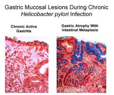
Helicobacter infections are caused by the bacterium Helicobacter pylori (H. pylori), which colonizes the stomach lining and is associated with various gastrointestinal diseases. The infection can lead to chronic active gastritis, peptic ulcers, gastric mucosa-associated lymphoid tissue (MALT) lymphoma, and gastric cancer.
The spiral-shaped H. pylori bacteria are able to survive in the harsh acidic environment of the stomach by producing urease, an enzyme that neutralizes gastric acid in their immediate vicinity. This allows them to adhere to and colonize the epithelial lining of the stomach, where they can cause inflammation (gastritis) and disrupt the normal functioning of the stomach.
Transmission of H. pylori typically occurs through oral-oral or fecal-oral routes, and infection is more common in developing countries and in populations with lower socioeconomic status. The diagnosis of Helicobacter infections can be confirmed through various tests, including urea breath tests, stool antigen tests, or gastric biopsy with histology and culture. Treatment usually involves a combination of antibiotics and proton pump inhibitors to eradicate the bacteria and reduce stomach acidity.
Gastritis is a medical condition characterized by inflammation of the lining of the stomach. It can be caused by various factors, including bacterial infections (such as Helicobacter pylori), regular use of nonsteroidal anti-inflammatory drugs (NSAIDs), excessive alcohol consumption, and stress.
Gastritis can present with a range of symptoms, such as abdominal pain or discomfort, nausea, vomiting, loss of appetite, and bloating. In some cases, gastritis may not cause any noticeable symptoms. Depending on the severity and duration of inflammation, gastritis can lead to complications like stomach ulcers or even stomach cancer if left untreated.
There are two main types of gastritis: acute and chronic. Acute gastritis develops suddenly and may last for a short period, while chronic gastritis persists over time, often leading to atrophy of the stomach lining. Diagnosis typically involves endoscopy and tissue biopsy to assess the extent of inflammation and rule out other potential causes of symptoms. Treatment options depend on the underlying cause but may include antibiotics, proton pump inhibitors, or lifestyle modifications.
In anatomical terms, the stomach is a muscular, J-shaped organ located in the upper left portion of the abdomen. It is part of the gastrointestinal tract and plays a crucial role in digestion. The stomach's primary functions include storing food, mixing it with digestive enzymes and hydrochloric acid to break down proteins, and slowly emptying the partially digested food into the small intestine for further absorption of nutrients.
The stomach is divided into several regions, including the cardia (the area nearest the esophagus), the fundus (the upper portion on the left side), the body (the main central part), and the pylorus (the narrowed region leading to the small intestine). The inner lining of the stomach, called the mucosa, is protected by a layer of mucus that prevents the digestive juices from damaging the stomach tissue itself.
In medical contexts, various conditions can affect the stomach, such as gastritis (inflammation of the stomach lining), peptic ulcers (sores in the stomach or duodenum), gastroesophageal reflux disease (GERD), and stomach cancer. Symptoms related to the stomach may include abdominal pain, bloating, nausea, vomiting, heartburn, and difficulty swallowing.
I'm sorry for any confusion, but the term "Tuna" is not typically used in medical definitions. It is most commonly known as a type of large, ocean-dwelling fish that belongs to the family Scombridae and the genus Thunnus. Some species of tuna, like bluefin tuna, are highly prized for their meat, which is often consumed raw as sushi or sashimi.
If you have any questions related to medical terminology or health concerns, please don't hesitate to ask!
Enzyme precursors are typically referred to as zymogens or proenzymes. These are inactive forms of enzymes that can be activated under specific conditions. When the need for the enzyme's function arises, the proenzyme is converted into its active form through a process called proteolysis, where it is cleaved by another enzyme. This mechanism helps control and regulate the activation of certain enzymes in the body, preventing unwanted or premature reactions. A well-known example of an enzyme precursor is trypsinogen, which is converted into its active form, trypsin, in the digestive system.
Hypertrophic gastritis is a relatively uncommon condition characterized by thickened folds in the stomach lining (gastric mucosa) due to an increase in the number of cells and/or the size of the cells. This chronic inflammatory condition can lead to atrophy of the glands, intestinal metaplasia, and an increased risk of developing gastric cancer. It is often associated with autoimmune disorders, such as Hashimoto's thyroiditis, pernicious anemia, or type A atrophic gastritis.
The condition can be asymptomatic or may present with symptoms like abdominal pain, nausea, vomiting, bloating, and loss of appetite. Diagnosis typically involves endoscopy with biopsy to assess the extent of inflammation and cellular changes in the stomach lining. Treatment usually includes proton pump inhibitors to reduce acid secretion, as well as addressing any underlying conditions that may be contributing to the development or progression of hypertrophic gastritis.
Stomach neoplasms refer to abnormal growths in the stomach that can be benign or malignant. They include a wide range of conditions such as:
1. Gastric adenomas: These are benign tumors that develop from glandular cells in the stomach lining.
2. Gastrointestinal stromal tumors (GISTs): These are rare tumors that can be found in the stomach and other parts of the digestive tract. They originate from the stem cells in the wall of the digestive tract.
3. Leiomyomas: These are benign tumors that develop from smooth muscle cells in the stomach wall.
4. Lipomas: These are benign tumors that develop from fat cells in the stomach wall.
5. Neuroendocrine tumors (NETs): These are tumors that develop from the neuroendocrine cells in the stomach lining. They can be benign or malignant.
6. Gastric carcinomas: These are malignant tumors that develop from the glandular cells in the stomach lining. They are the most common type of stomach neoplasm and include adenocarcinomas, signet ring cell carcinomas, and others.
7. Lymphomas: These are malignant tumors that develop from the immune cells in the stomach wall.
Stomach neoplasms can cause various symptoms such as abdominal pain, nausea, vomiting, weight loss, and difficulty swallowing. The diagnosis of stomach neoplasms usually involves a combination of imaging tests, endoscopy, and biopsy. Treatment options depend on the type and stage of the neoplasm and may include surgery, chemotherapy, radiation therapy, or targeted therapy.
 Pepsinogen 3, group I (pepsinogen A)
Progastricsin
Digestive enzyme
PGA5
Gertrude Perlmann
Cholecystokinin antagonist
Gastrointestinal physiology
Proventriculus
Stomach
Bacardi
Aspartic protease
Richard Serra
Pepsin B
Pepsin A
SOX2
Chief cell
S. Narasinga Rao
Ostertagia ostertagi
Vasoactive intestinal peptide
Gastric pits
Pepsin
Zymogen
AKAP5
John Howard Northrop
Foveolar cell
Gastrin
Chymosin
Gastric chief cell
List of Italian inventions and discoveries
Intestinal gland
Pepsinogen 3, group I (pepsinogen A) - Wikipedia
 SRUC farm animal diagnostics tests | Pepsinogen
 Helicobacter pylori infection and serum pepsinogen A, pepsinogen C, and gastrin in gastritis and peptic ulcer: significance of...
![Pepsinogen II (PGC) Mouse Monoclonal Antibody [Clone ID: OTI5C9], TA809596 | AMSBIO](data:image/png;base64,iVBORw0KGgoAAAANSUhEUgAAABAAAAAQCAMAAAAoLQ9TAAAAMFBMVEVHcEweRYwANIUeRYwWQov////v8vi1wNfi6PH09/sGPYlkfa3a4O05XJvG0OKXp8giSqfSAAAAAnRSTlMApz4h+3MAAABzSURBVBiVbY/hEoAgCINtQCphvf/bBmT9qDjPu33nZCtloRjOm5aSWkFDwQCcuMbWRKRyawZygENyVj8BdHPRau0yAfx9dbvaBXiIdMSeMYE79gCE/g+IXpbPp9+1T7B+A49uGd1sV8pyjCg4y836dNc/AYpQBMbK5mhgAAAAAElFTkSuQmCC) Pepsinogen II (PGC) Mouse Monoclonal Antibody [Clone ID: OTI5C9], TA809596 | AMSBIO
Pepsinogen II (PGC) Mouse Monoclonal Antibody [Clone ID: OTI5C9], TA809596 | AMSBIO
What is needed to activate pepsinogen?
 Stomach (Gastric) Cancer Screening (PDQ®) (Health professionals) | OncoLink
 Rat PGC(Pepsinogen C) ELISA Kit - UNICO Microscopes and Spectrophotometers
 SNU Open Repository and Archive: The role of serum pepsinogen and gastrin test for the detection of gastric cancer in Korea
 Cells of the stomach: Types, purpose, and location
 Vagotomy in duodenal ulcer disease : a study of gastric acidity, serum pepsinogen I, gastric mucosal histology and Helicobacter...
 Assessing GastroPanel serum markers as a non-invasive method for the diagnosis of atrophic gastritis and Helicobacter pylori...
 OER Commons
 Smoking history and severe atrophic gastritis assessed by pepsinogen are risk factors for the prevalence of synchronous gastric...
 Microbiology Elisa | Gentaur Elisa Kits | US - UK & Europe Supply
 Gastrointestinal Disorders Quiz Questions And Answers - ProProfs Quiz
 Frontiers | Oncolytic virus: A catalyst for the treatment of gastric cancer
 Digestive Function of the Stomach | Digestive Diseases | Karger Publishers
 Current Oncology | Free Full-Text | Epidemiology, Risk Factors for Gastric Cancer and Surveillance of Premalignant Gastric...
 DADAMOwiki: Serum gastrin concentrations and ABO blood group
 Questions & Answers
Chronic Gastritis Differential Diagnoses
 Deglycyrrhizinated Licorice for Gastrointestinal Ulcers | Natural Medicine Journal
 Journal of Jilin University Medicine Edition
 British Medical Journal: 1 (5388) | The BMJ
 Area
Area Table of contents | Journal of Clinical Pathology
Model Details
 digestive system | Memory
 Specialty Supplements
Hydrochloric acid4
- Hydrochloric acid inactivates salivary amylase and catalyzes the conversion of pepsinogen to pepsin. (github.io)
- Gastric juice: the gastric juice consists of the following: water 90%, hydrochloric acid 0.4% prorennin and pepsinogen, gastric lipase. (preservearticles.com)
- In the stomach, hydrochloric acid activates pepsinogen to pepsin, which breaks down protein, and gastric lipase begins the hydrolysis of fats. (metagenics.com)
- In the stomach, hydrochloric acid converts pepsinogen to pepsin, an enzyme that breaks down protein. (vitaminlife.com)
Pepsin4
- Gastric chief cells secrete pepsin as an inactive zymogen called pepsinogen. (github.io)
- Acid in the stomach changes pepsinogen to pepsin, which breaks down proteins in food during digestion. (github.io)
- Samloff IM: Pepsinogens, pepsins, and pepsin inhibitors. (karger.com)
- Gastric pepsin is a proteolytic enzyme secreted by gastric chief cells and mucus neck cells as inactive pepsinogen. (ersjournals.com)
Gastrin and pepsinogen5
- To study the relationship between Helicobacter pylori infection, gastric inflammatory scores, and fasting gastrin and pepsinogen A and C concentrations, and to evaluate the effect of treatment on these parameters. (nih.gov)
- Gastrin and pepsinogen A and C concentrations were measured in 36 patients with gastritis, 10 gastric ulcer patients, 12 duodenal ulcer patients, and in 15 subjects with normal gastric mucosa, by standard radioimmunoassay techniques. (nih.gov)
- Fasting gastrin and pepsinogen A and C concentrations were significantly higher in H. pylori-positive gastritis and peptic ulcer patients than in subjects with normal mucosa and in patients with H. pylori-negative gastritis. (nih.gov)
- Neither sex nor age affected basal gastrin and pepsinogen concentrations. (nih.gov)
- Gastric colonization by Helicobacter pylori increases the risk of gastric disorders, including atrophic gastritis which can be diagnosed based on levels of serum biomarkers like Gastrin and Pepsinogen. (scirp.org)
Stomach1
- Samloff IM, Liebman WM: Cellular localization of the group II pepsinogens in human stomach and duodenum by immunofluorescence. (karger.com)
Serum gastrin1
- Eradication of H. pylori infection was successful in 12 patients and resulted in a significant fall in serum gastrin and in pepsinogen A and C concentrations, and in a concomitant improvement of the inflammatory scores. (nih.gov)
Secrete1
- The chief cells secrete pepsinogen. (medicalnewstoday.com)
Secretion1
- Ileal protein which stimulates gastric acid and pepsinogen secretion. (hmdb.ca)
Concentrations1
- Accuracy of screening for gastric cancer using serum pepsinogen concentrations. (oncolink.org)
Antigen1
- The role of Prostate Specific Membrane Antigen (PSMA) and Pepsinogen C (PGC) gene. (usp.br)
Atrophic2
- Pepsinogen levels in serum may serve as a biomarker for atrophic gastritis and gastric cancer. (wikipedia.org)
- Smoking history and severe atrophic gastritis assessed by pepsinogen are risk factors for the prevalence of synchronous gastric cancers in patients with gastric endoscopic submucosal dissection: a multicenter prospective cohort study. (bvsalud.org)
ELISA1
- Description: A sandwich ELISA kit for detection of Pepsinogen C from Rat in samples from blood, serum, plasma, cell culture fluid and other biological fluids. (unicoupi.com)
Inactive1
- Prorennin and pepsinogen are protein digesting proenzymes secreted in inactive form. (preservearticles.com)
Stimulates1
- Which hormone stimulates the chief cells to produce pepsinogen? (proprofs.com)
Gene3
- Pepsinogen 3, group I (pepsinogen A) is a protein that in humans is encoded by the PGA3 gene. (wikipedia.org)
- This gene is found in a cluster of related genes on chromosome 11, each of which encodes one of multiple pepsinogens. (wikipedia.org)
- Entrez Gene: Pepsinogen 3, group I (pepsinogen A)". Retrieved 2017-08-25. (wikipedia.org)
Enzyme2
- Description: This is Double-antibody Sandwich Enzyme-linked immunosorbent assay for detection of Rat Pepsinogen C (PGC) in serum, plasma, tissue homogenates and other biological fluids. (unicoupi.com)
- Description: Enzyme-linked immunosorbent assay based on the Double-antibody Sandwich method for detection of Rat Pepsinogen C (PGC) in samples from Serum, plasma, tissue homogenates and other biological fluids with no significant corss-reactivity with analogues from other species. (unicoupi.com)
Levels1
- The degree of atrophy was classified according to levels in patient serum of pepsinogens I and II (PGI and PGII) and Gastrin 17 (G17) and compared with histological profiles as reference method. (scirp.org)
Cancer3
- Review the evidence on the benefits and harms of screening for gastric cancer using barium-meal photofluorography, gastric endoscopy, or serum pepsinogen in this expert-reviewed summary. (oncolink.org)
- Based on fair evidence, screening with barium-meal photofluorography or serum pepsinogen would not result in a decrease in mortality from gastric cancer in areas with relatively low incidence of the disease, such as the United States. (oncolink.org)
- BACKGROUND AND AIM: This study was performed to determine whether serum pepsinogen (PG) and gastrin testing can be used to detect gastric cancer in Korea. (snu.ac.kr)
Human2
- Samloff IM: Cellular localization of group I pepsinogens in human gastric mucosa by immunofluorescence. (karger.com)
- Recombinant human pepsinogen I protein. (medixbiochemica.com)
Progastricsin3
- Dorland, 28th ed) In humans there are 2 related pepsinogen systems: PEPSINOGEN A (formerly pepsinogen I or pepsinogen) and PEPSINOGEN C (formerly pepsinogen II or progastricsin). (childrensmercy.org)
- Pepsinogens are mainly grouped in 5 different groups based on their primary structure: pepsinogen A (also called pepsinogen I), pepsinogen B, progastricsin (also called pepsinogen II and pepsinogen C), prochymosin (also called prorennin) and pepsinogen F (also called pregnancy-associated glycoprotein). (wikipedia.org)
- Hence the structures of these two proteins indicate that aspartic proteinase zymogens keep themselves inactive at neutral pH by a very similar mechanism in human progastricsin and porcine pepsinogen. (rcsb.org)
Related pepsinogen systems in humans1
- This is one of 2 related pepsinogen systems in humans and is also known as pepsinogen. (nih.gov)
Gastrin-173
- 3. Serum levels of amidated gastrin-17 and pepsinogen I in atrophic gastritis: an observational case-control study. (nih.gov)
- A panel of stomach-specific serum biomarkers: pepsinogen (PG) I, pepsinogen (PG) II, gastrin-17 (G-17), and IgG antibodies to H. pylori (HP-Ab) detects the extent and grade of AG. (iiarjournals.org)
- Pepsinogen I Pepsinogen II ndi Gastrin-17 Comb. (baysenrapidtest.com)
Atrophic gastritis6
- Pepsinogen levels in serum may serve as a biomarker for atrophic gastritis and gastric cancer. (wikipedia.org)
- Since the 1990's, the test for serum pepsinogen as a marker for chronic atrophic gastritis has been incorporated into gastric cancer screening programs, on a trial basis, to identify people at high risk for gastric cancer. (go.jp)
- The clinical applications of measuring pepsinogen I and II are a useful aid in the diagnosis of severe atrophic gastritis and stomach cancer. (epitopediagnostics.com)
- It has been suggested that the measurement of serum pepsinogens serve as a "serological biopsy" for predicting the presence of atrophic gastritis or superficial gastritis. (epitopediagnostics.com)
- It has been found the serum pepsinogen I levels falling lower than 20 ng/ml was highly specific for severe atrophic gastritis. (epitopediagnostics.com)
- On the other hand, the decrease in serum pepsinogen I levels in patients with pernicious anemia and atrophic gastritis was found to be associated with normal or raised pepsinogen II levels. (epitopediagnostics.com)
PGII1
- Human gastric mucosa contains two abundant and distinguishable aspartic proteinases, namely pepsinogen I (PGI or PGA) and pepsinogen II (PGII or PGC). (medscape.com)
Aspartic2
- Human Pepsinogens are aspartic proteases produced in the gastric mucosa and secreted into the gastric lumen that play a major role in the digestion of proteins after activation of acidic pH. (fiapoct.com)
- This similarity likely carries over to all members of both the pepsinogen A and C families of aspartic proteinase zymogens. (rcsb.org)
Cheif cells1
- Pepsinogen I is synthesized at gastric cheif cells and mucous neck cells, while pepsinogen II is also produced by clear mucous cells of antrum, etc. (epitopediagnostics.com)
Gastric mucosa2
- The addition of the serum test to the cancer screening program has been shown to improve the detection rate of cancer and pepsinogen testing is useful in detecting early-stage gastric cancers arising from atrophic gastric mucosa, which macroscopically tend to be elevated and histologically differentiated. (go.jp)
- evaluation of gastric mucosa by serum pepsinogen levels. (nih.gov)
Helicobacter1
- Interaction between an insertion/deletion polymorphism in pepsinogen C and Helicobacter pylori infection in the development of gastric cancer]. (nih.gov)
Protein1
- Pepsinogen 3, group I (pepsinogen A) is a protein that in humans is encoded by the PGA3 gene. (wikipedia.org)
Duodenal ulcer1
- A low serum pepsinogen I level can exclude a diagnosis of duodenal ulcer. (epitopediagnostics.com)
Encodes1
- This gene is found in a cluster of related genes on chromosome 11, each of which encodes one of multiple pepsinogens. (wikipedia.org)
Mucous2
- It also stimulates secretion of HCL and pepsinogen and stimulates mucous production to protect against autodigestion of your stomach wall. (jonbarron.org)
- Glands in the mucous-membrane lining of the stomach, called peptic chief cells, are responsible for making pepsinogen. (draxe.com)
Mucosal2
- It was also observed that serum pepsinogen I levels fell with increasing severity of mucosal damage in such cases. (epitopediagnostics.com)
- Free acidity, total acidity, basal acid output, serum pepsinogen I, gastric mucosal blood flow (GMBF) and gastrin were significantly lower in group II, whereas serum gastrin and somatostatin staining were significantly higher. (who.int)
Residues2
- Pepsinogen consists of a single polypeptide chain of 375 amino acid residues with an average molecular weight of 42 kDa. (epitopediagnostics.com)
- The first 37 amino acid residues of the prosegment are similar in conformation to the equivalent residues of porcine pepsinogen (pPGN). (rcsb.org)
Ratio3
- Determination of human serum pepsinogen I/II ratio has been reported to be a useful tool in aiding the diagnosis of the functional states of acid-secreting gastric muscosa. (epitopediagnostics.com)
- Therefore, a pepsinogen I/pepsinogen II ratio is significantly lower than those with superficial gastritis or normal remnant mucosa. (epitopediagnostics.com)
- Third, achlorhydria has been defined as a ratio of serum pepsinogen I/pepsinogen II of less than 2.9. (medscape.com)
Vagus nerve1
- The hormone gastrin and the vagus nerve trigger the release of both pepsinogen and HCl from the stomach lining when food is ingested. (wikipedia.org)
Detection2
- Description: A sandwich ELISA kit for detection of Pepsinogen C from Mouse in samples from blood, serum, plasma, cell culture fluid and other biological fluids. (taleffectors.com)
- Description: A sandwich quantitative ELISA assay kit for detection of Rat Pepsinogen C (PGC) in samples from serum, plasma, tissue homogenates or other biological fluids. (taleffectors.com)
Gastritis1
- The diagnostic sensitivity and specificity of serum pepsinogen I levels for advanced atrophic corpus gastritis are about 92% and 90% respectively. (epitopediagnostics.com)
Stomach cancer1
- Other studies have concluded that low serum pepsinogen I levels may identify persons at increased risk for intestinal types of stomach cancer. (epitopediagnostics.com)
Human1
- In this study, we used the antimicrobial peptide PAP3 (A3 isoform of human pepsinogen) and the phage 1 (isolated by Salmonella rissen). (unina.it)
Clinical1
- Although a high pepsinogen I level has less clinical use for establishing this diagnosis, the combination of hypergastinemia and a highly evelated serum pepsinogen I level strongly suggests the possibility of Zollinger-Ellison syndrome. (epitopediagnostics.com)
Found2
Diagnosis1
- Objectives: Complete en-bloc resection of pedunculated colorectal carcinoma is necessary for a proper pathological diagnosis. (bvsalud.org)
Group1
- The other is PEPSINOGEN C .) This includes isozymogens Pg1-Pg5 (pepsinogens 1-5, group I or products of PGA1-PGA5 genes). (nih.gov)
Cells1
- In the stomach, gastric chief cells release pepsinogen. (wikipedia.org)
Groups2
- It is synthesized as isoymogens and is classified into two groups (Pepsinogen I and Pepsinogen II). (fiapoct.com)
- There were significant differences between the PCM and CM groups in en bloc resection (100% vs. 48%, p = 0.013) and R0 resection rates (71% vs. 31%, p = 0.049). (bvsalud.org)