Pectoralis Muscles
Cockatoos
Melopsittacus
Mesterolone
Muscle Development
Chickens
Muscle, Skeletal
Mitochondria, Muscle
Biomechanical Phenomena
Surgical Flaps
Myosins
Muscle Proteins
Chick Embryo
Poland Syndrome
Muscle, Smooth
Muscle Fibers, Skeletal
Muscle Contraction
Pectoral myoplasty for recurrent pneumothorax: an extrathoracic solution to an intrathoracic problem. (1/214)
The recurrence rate of spontaneous pneumothorax in patients with underlying lung disease can be as high as 50%. We present a novel method of treatment for recurrent pneumothorax based on the intrathoracic transfer of an extrathoracic muscle flap. (+info)Intramuscular desmoid tumor (musculoaponeurotic fibromatosis) in two horses. (2/214)
Intramuscular desmoid tumors (musculoaponeurotic fibromatosis) were discovered in two young adult horses. The tumor in one horse was in the lateral cervical musculature, and that in the second horse occurred in the pectoral musculature. Histopathologic features were similar in both horses and included proliferation of fibroblasts and cells expressing muscle actin (myofibroblasts), with extensive dissecting fibrosis within muscle. These features are similar to those of desmoid tumors in humans, particularly those also known as musculoaponeurotic fibromatosis. Dissection of these lesions revealed a single central (horse No. 1) or multiple central (horse No. 2) fluid-filled cavities with associated sterile inflammation. The presence of these cavities supports the hypothesis that equine desmoid tumors are traumatic in origin, possibly occurring at sites of injections or bursal rupture. Surgical excision of the tumor in horse No. 1 was apparently curative, but the extent of the tumor in horse No. 2 precluded surgical excision. (+info)Lymphangioma of the chest wall. (3/214)
A lymphangioma of the chest wall, hitherto unreported is described here. (+info)Retrospective analysis of pectoralis major myocutaneous flap surgeries performed under war conditions. (4/214)
AIM: To present our experiences in using a pectoralis major myocutaneous flap in the reconstruction of surgically created defects of the neck and lower part of the head during the war in Bosnia and Herzegovina. METHODS: Retrospective analysis of medical records from 15 patients treated at the ENT Department, Tuzla University Hospital, between January 1992 and December 1996. RESULTS: Ten flaps were prepared during basic operation ("one step reconstruction of defect") and five flaps three weeks after the removal of tumor (postoperative pharyngocutaneous fistula was the reason for secondary flap preparation). The necessary time for flap preparation and its accommodation in the defect was 2 hours. The most frequent complications included seroma of donor site (6/13), fistula (3/13), partial necrosis of the flap (2/13), and total necrosis of the flap (2/13). Three patients died in the postoperative period because of a cardiac arrest but the flaps accepted correctly. CONCLUSION: The pectoralis major myocutaneous flap is a good solution for covering defects of the neck and lower region of the head. Military blockade with extremely difficult conditions and lack of experience were the reason of the higher complication rate than those from literature. (+info)Pectoralis muscle performance during ascending and slow level flight in mallards (Anas platyrhynchos). (5/214)
In vivo measurements of pectoralis muscle length change and force production were obtained using sonomicrometry and delto-pectoral bone strain recordings during ascending and slow level flight in mallards (Anas platyrhynchos). These measurements provide a description of the force/length properties of the pectoralis under dynamic conditions during two discrete flight behaviors and allow an examination of the effects of differences in body size and morphology on pectoralis performance by comparing the results with those of a recent similar study of slow level flight in pigeons (Columbia livia). In the present study, the mallard pectoralis showed a distinct pattern of active lengthening during the upstroke. This probably enhances the rate of force generation and the magnitude of the force generated and, thus, the amount of work and power produced during the downstroke. The power output of the pectoralis averaged 17.0 W kg(-)(1 )body mass (131 W kg(-)(1 )muscle mass) during slow level flight (3 m s(-)(1)) and 23.3 W kg(-)(1 )body mass (174 W kg(-)(1 )muscle mass) during ascending flight. This increase in power was achieved principally via an increase in muscle strain (29 % versus 36 %), rather than an increase in peak force (107 N versus 113 N) or cycle frequency (8.4 Hz versus 8.9 Hz). Body-mass-specific power output of mallards during slow level flight (17.0 W kg(-)(1)), measured in terms of pectoralis mechanical power, was similar to that measured recently in pigeons (16.1 W kg(-)(1)). Mallards compensate for their greater body mass and proportionately smaller wing area and pectoralis muscle volume by operating with a high myofibrillar stress to elevate mechanical power output. (+info)The ultrastructure of skeletal and smooth muscle in experimental protein malnutrition in rats fed a low protein diet. (6/214)
Light microscopy of the pectoralis muscle of rats on a low protein diet did not show such morphological alterations as atrophy, degeneration, or sarcoplasmic edema, but electron microscopy occasionally demonstrated ultrastructural changes only in the sarcomeres of myofibrils. In the affected sarcomeres, the Z-line was disrupted and often showed a jagged structure. The Z-substance with electron opacity was frequently present flowing along the long axis of myofibrils, here referred to as the streaming of Z-lines. In addition, regular striations formed by the reciprocal arrangement of thick and thin filaments disappeared from the affected sarcomeres, though these filaments were still discernible. Two or more consecutive sarcomeres in a single myofibril were occasionally involved in these changes. A further two or more neighboring sarcomeres at the same level of myofibrils were affected transversely by these structural alterations. On the other hand, the ultrastructure of the intestinal smooth muscle was not affected by protein deficiency. The study suggests that the ultrastructural damage induced by a low protein diet is attributed to the activation of endogenous protease by the excess leaking of Ca2+ into the cytosol as a result of lipid peroxidation of cell membrane by raised free radicals, owing to the depletion of glutathione production by protein deficiency. It also suggests that the smooth muscle cells differ in their susceptibility to protein deficiency from the skeletal muscle cells. (+info)Extra-anatomic bypass graft for management of axillary artery occlusion in pitchers. (7/214)
OBJECTIVE: Our goal was to evaluate the long-term results of vein bypass grafts for axillary artery occlusion, specifically those placed extra-anatomically to prevent arterial injury in pitchers. METHODS: With the greater saphenous veins used as the selected conduit, arterial bypass grafts were routed anterior to the pectoralis minor muscle in four baseball pitchers who had occlusion of the axillary artery. We performed a follow-up in excess of 10 years with evaluations of the bypass grafts by ultrasonic duplex scan and magnetic resonance angiography. RESULTS: All four pitchers treated in this manner returned to the game and played for several seasons without a recurrence of the arterial injury. Long-term evaluation of the bypass grafts did not reveal any structural or functional disorder. CONCLUSIONS: Axillary artery occlusion in an athlete can be effectively treated with a vein bypass graft placed extra-anatomically, anterior to the pectoralis minor muscle. The greater saphenous vein should be considered the conduit of choice. (+info)Pectoralis major tears: comparison of surgical and conservative treatment. (8/214)
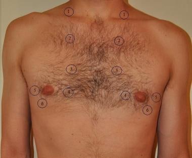
OBJECTIVES: To compare objective measures of strength and subjective functional outcomes in complete distal pectoralis major tears treated either surgically or non-surgically. METHODS: Twenty two pectoralis major tears were included in 21 patients. Ten were surgically repaired and 12 were managed non-surgically. Patients completed a standard questionnaire, and clinical examination and isokinetic dynamometry were carried out. RESULTS: In patients who had surgical repair, peak torque returned to 99% of that of the uninjured side and work performed returned to 97%. For those managed conservatively, peak torque and work performed returned to only 56% of that of the uninjured side (p = 0.003 for the difference in peak torque, and p = 0.01 for work performed). Findings were independent of the strength of the patient, whether or not the dominant arm was involved, the age of the patient, and the length of time from injury or surgery to testing. Patients were grouped into one of three subjective functional outcome groups, and those who had a surgical repair had a better functional outcome. CONCLUSIONS: Surgical repair results in greater recovery of peak torque and work performed than conservative management of patients with rupture of the pectoralis major. (+info)The pectoralis muscles are a group of chest muscles that are primarily involved in the movement and stabilization of the shoulder joint. They consist of two individual muscles: the pectoralis major and the pectoralis minor.
1. Pectoralis Major: This is the larger and more superficial of the two muscles, lying just under the skin and fat of the chest wall. It has two heads of origin - the clavicular head arises from the medial half of the clavicle (collarbone), while the sternocostal head arises from the anterior surface of the sternum (breastbone) and the upper six costal cartilages. Both heads insert onto the lateral lip of the bicipital groove of the humerus (upper arm bone). The primary actions of the pectoralis major include flexion, adduction, and internal rotation of the shoulder joint.
2. Pectoralis Minor: This is a smaller, triangular muscle that lies deep to the pectoralis major. It originates from the third, fourth, and fifth ribs near their costal cartilages and inserts onto the coracoid process of the scapula (shoulder blade). The main function of the pectoralis minor is to pull the scapula forward and downward, helping to stabilize the shoulder joint and aiding in deep inspiration during breathing.
Together, these muscles play essential roles in various movements such as pushing, pulling, and hugging, making them crucial for daily activities and athletic performance.
Cockatoos are a group of parrots that make up the family Cacatuidae. They are characterized by their distinctive crest on top of their heads, which they can raise or lower depending on their mood. Cockatoos come in a variety of sizes and colors, with some species having black, white, pink, or gray feathers.
Cockatoos are known for their intelligence and ability to mimic human speech, although not all species have this ability. They are also known for being social birds that form strong bonds with their mates and families. Many cockatoo species are popular as pets due to their friendly and affectionate personalities.
In terms of medical concerns, cockatoos can suffer from a variety of health issues, including feather-plucking, obesity, and behavioral problems. They require a balanced diet, plenty of mental and physical stimulation, and regular veterinary care to maintain their health and well-being.
"Melopsittacus" is the genus name for the species of bird commonly known as the Budgerigar or Parakeet. It is the only species in its genus and belongs to the Psittacidae family, which includes parrots. The Melopsittacus undulatus is a small, long-tailed parrot native to Australia, known for its bright green, yellow, or blue feathers and sociable behavior. They are popular pets due to their ease of care, playful personalities, and ability to mimic human speech.
"Animal Flight" is not a medical term per se, but it is a concept that is studied in the field of comparative physiology and biomechanics, which are disciplines related to medicine. Animal flight refers to the ability of certain animal species to move through the air by flapping their wings or other appendages. This mode of locomotion is most commonly associated with birds, bats, and insects, but some mammals such as flying squirrels and sugar gliders are also capable of gliding through the air.
The study of animal flight involves understanding the biomechanics of how animals generate lift and propulsion, as well as the physiological adaptations that allow them to sustain flight. For example, birds have lightweight skeletons and powerful chest muscles that enable them to flap their wings rapidly and generate lift. Bats, on the other hand, use a more complex system of membranes and joints to manipulate their wings and achieve maneuverability in flight.
Understanding animal flight has important implications for the design of aircraft and other engineering systems, as well as for our broader understanding of how animals have evolved to adapt to their environments.
A muscle is a soft tissue in our body that contracts to produce force and motion. It is composed mainly of specialized cells called muscle fibers, which are bound together by connective tissue. There are three types of muscles: skeletal (voluntary), smooth (involuntary), and cardiac. Skeletal muscles attach to bones and help in movement, while smooth muscles are found within the walls of organs and blood vessels, helping with functions like digestion and circulation. Cardiac muscle is the specific type that makes up the heart, allowing it to pump blood throughout the body.
Mesterolone is an androgen and anabolic steroid (AAS) medication, which is primarily used in the treatment of low testosterone levels in men. It is also known by its brand name Proviron. Mesterolone works by promoting the development and maintenance of male sexual characteristics and can help to increase muscle mass and strength.
Mesterolone is a synthetic derivative of dihydrotestosterone (DHT), which is a more potent androgen than testosterone. It has both anabolic and androgenic effects, but its androgenic effects are more pronounced. Mesterolone is not aromatized to estrogen, so it does not have the estrogenic side effects that can occur with some other AAS medications.
Mesterolone is taken orally, and its typical dosage ranges from 25 to 75 milligrams per day. It is important to note that Mesterolone and other AAS medications are controlled substances in many countries and should only be used under the supervision of a healthcare provider.
Muscle development, also known as muscle hypertrophy, refers to the increase in size and mass of the muscles through a process called myofiber growth. This is primarily achieved through resistance or strength training exercises that cause micro-tears in the muscle fibers, leading to an inflammatory response and the release of hormones that promote muscle growth. As the muscles repair themselves, they become larger and stronger than before. Proper nutrition, including adequate protein intake, and rest are also essential components of muscle development.
It is important to note that while muscle development can lead to an increase in strength and muscular endurance, it does not necessarily result in improved athletic performance or overall fitness. A well-rounded exercise program that includes cardiovascular activity, flexibility training, and resistance exercises is recommended for optimal health and fitness outcomes.
"Chickens" is a common term used to refer to the domesticated bird, Gallus gallus domesticus, which is widely raised for its eggs and meat. However, in medical terms, "chickens" is not a standard term with a specific definition. If you have any specific medical concern or question related to chickens, such as food safety or allergies, please provide more details so I can give a more accurate answer.
Skeletal muscle, also known as striated or voluntary muscle, is a type of muscle that is attached to bones by tendons or aponeuroses and functions to produce movements and support the posture of the body. It is composed of long, multinucleated fibers that are arranged in parallel bundles and are characterized by alternating light and dark bands, giving them a striped appearance under a microscope. Skeletal muscle is under voluntary control, meaning that it is consciously activated through signals from the nervous system. It is responsible for activities such as walking, running, jumping, and lifting objects.
Mitochondria in muscle, also known as the "powerhouses" of the cell, are organelles that play a crucial role in generating energy for muscle cells through a process called cellular respiration. They convert the chemical energy found in glucose and oxygen into ATP (adenosine triphosphate), which is the main source of energy used by cells.
Muscle cells contain a high number of mitochondria due to their high energy demands for muscle contraction and relaxation. The number and size of mitochondria in muscle fibers can vary depending on the type of muscle fiber, with slow-twitch, aerobic fibers having more numerous and larger mitochondria than fast-twitch, anaerobic fibers.
Mitochondrial dysfunction has been linked to various muscle disorders, including mitochondrial myopathies, which are characterized by muscle weakness, exercise intolerance, and other symptoms related to impaired energy production in the muscle cells.
Biomechanics is the application of mechanical laws to living structures and systems, particularly in the field of medicine and healthcare. A biomechanical phenomenon refers to a observable event or occurrence that involves the interaction of biological tissues or systems with mechanical forces. These phenomena can be studied at various levels, from the molecular and cellular level to the tissue, organ, and whole-body level.
Examples of biomechanical phenomena include:
1. The way that bones and muscles work together to produce movement (known as joint kinematics).
2. The mechanical behavior of biological tissues such as bone, cartilage, tendons, and ligaments under various loads and stresses.
3. The response of cells and tissues to mechanical stimuli, such as the way that bone tissue adapts to changes in loading conditions (known as Wolff's law).
4. The biomechanics of injury and disease processes, such as the mechanisms of joint injury or the development of osteoarthritis.
5. The use of mechanical devices and interventions to treat medical conditions, such as orthopedic implants or assistive devices for mobility impairments.
Understanding biomechanical phenomena is essential for developing effective treatments and prevention strategies for a wide range of medical conditions, from musculoskeletal injuries to neurological disorders.
A surgical flap is a specialized type of surgical procedure where a section of living tissue (including skin, fat, muscle, and/or blood vessels) is lifted from its original site and moved to another location, while still maintaining a blood supply through its attached pedicle. This technique allows the surgeon to cover and reconstruct defects or wounds that cannot be closed easily with simple suturing or stapling.
Surgical flaps can be classified based on their vascularity, type of tissue involved, or method of transfer. The choice of using a specific type of surgical flap depends on the location and size of the defect, the patient's overall health, and the surgeon's expertise. Some common types of surgical flaps include:
1. Random-pattern flaps: These flaps are based on random blood vessels within the tissue and are typically used for smaller defects in areas with good vascularity, such as the face or scalp.
2. Axial pattern flaps: These flaps are designed based on a known major blood vessel and its branches, allowing them to cover larger defects or reach distant sites. Examples include the radial forearm flap and the anterolateral thigh flap.
3. Local flaps: These flaps involve tissue adjacent to the wound and can be further classified into advancement, rotation, transposition, and interpolation flaps based on their movement and orientation.
4. Distant flaps: These flaps are harvested from a distant site and then transferred to the defect after being tunneled beneath the skin or through a separate incision. Examples include the groin flap and the latissimus dorsi flap.
5. Free flaps: In these flaps, the tissue is completely detached from its original blood supply and then reattached at the new site using microvascular surgical techniques. This allows for greater flexibility in terms of reach and placement but requires specialized expertise and equipment.
Surgical flaps play a crucial role in reconstructive surgery, helping to restore form and function after trauma, tumor removal, or other conditions that result in tissue loss.
Myosins are a large family of motor proteins that play a crucial role in various cellular processes, including muscle contraction and intracellular transport. They consist of heavy chains, which contain the motor domain responsible for generating force and motion, and light chains, which regulate the activity of the myosin. Based on their structural and functional differences, myosins are classified into over 35 classes, with classes II, V, and VI being the most well-studied.
Class II myosins, also known as conventional myosins, are responsible for muscle contraction in skeletal, cardiac, and smooth muscles. They form filaments called thick filaments, which interact with actin filaments to generate force and movement during muscle contraction.
Class V myosins, also known as unconventional myosins, are involved in intracellular transport and organelle positioning. They have a long tail that can bind to various cargoes, such as vesicles, mitochondria, and nuclei, and a motor domain that moves along actin filaments to transport the cargoes to their destinations.
Class VI myosins are also unconventional myosins involved in intracellular transport and organelle positioning. They have two heads connected by a coiled-coil tail, which can bind to various cargoes. Class VI myosins move along actin filaments in a unique hand-over-hand motion, allowing them to transport their cargoes efficiently.
Overall, myosins are essential for many cellular functions and have been implicated in various diseases, including cardiovascular diseases, neurological disorders, and cancer.
Muscle proteins are a type of protein that are found in muscle tissue and are responsible for providing structure, strength, and functionality to muscles. The two major types of muscle proteins are:
1. Contractile proteins: These include actin and myosin, which are responsible for the contraction and relaxation of muscles. They work together to cause muscle movement by sliding along each other and shortening the muscle fibers.
2. Structural proteins: These include titin, nebulin, and desmin, which provide structural support and stability to muscle fibers. Titin is the largest protein in the human body and acts as a molecular spring that helps maintain the integrity of the sarcomere (the basic unit of muscle contraction). Nebulin helps regulate the length of the sarcomere, while desmin forms a network of filaments that connects adjacent muscle fibers together.
Overall, muscle proteins play a critical role in maintaining muscle health and function, and their dysregulation can lead to various muscle-related disorders such as muscular dystrophy, myopathies, and sarcopenia.
A chick embryo refers to the developing organism that arises from a fertilized chicken egg. It is often used as a model system in biological research, particularly during the stages of development when many of its organs and systems are forming and can be easily observed and manipulated. The study of chick embryos has contributed significantly to our understanding of various aspects of developmental biology, including gastrulation, neurulation, organogenesis, and pattern formation. Researchers may use various techniques to observe and manipulate the chick embryo, such as surgical alterations, cell labeling, and exposure to drugs or other agents.
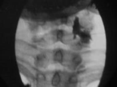
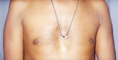
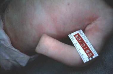
Poland Syndrome is a rare congenital anomaly characterized by the absence or underdevelopment of the chest muscle (pectoralis major) on one side of the body, often associated with webbing or absence of the fingers (cutaneous syndactyly) and shortening of the arm on the same side. It was first described by Alfred Poland, a British surgeon, in 1841. The exact cause of this condition is not known, but it is believed to be due to an interruption of blood flow to the developing fetus during early pregnancy. Treatment typically involves reconstructive surgery and physical therapy.
Smooth muscle, also known as involuntary muscle, is a type of muscle that is controlled by the autonomic nervous system and functions without conscious effort. These muscles are found in the walls of hollow organs such as the stomach, intestines, bladder, and blood vessels, as well as in the eyes, skin, and other areas of the body.
Smooth muscle fibers are shorter and narrower than skeletal muscle fibers and do not have striations or sarcomeres, which give skeletal muscle its striped appearance. Smooth muscle is controlled by the autonomic nervous system through the release of neurotransmitters such as acetylcholine and norepinephrine, which bind to receptors on the smooth muscle cells and cause them to contract or relax.
Smooth muscle plays an important role in many physiological processes, including digestion, circulation, respiration, and elimination. It can also contribute to various medical conditions, such as hypertension, gastrointestinal disorders, and genitourinary dysfunction, when it becomes overactive or underactive.
Skeletal muscle fibers, also known as striated muscle fibers, are the type of muscle cells that make up skeletal muscles, which are responsible for voluntary movements of the body. These muscle fibers are long, cylindrical, and multinucleated, meaning they contain multiple nuclei. They are surrounded by a connective tissue layer called the endomysium, and many fibers are bundled together into fascicles, which are then surrounded by another layer of connective tissue called the perimysium.
Skeletal muscle fibers are composed of myofibrils, which are long, thread-like structures that run the length of the fiber. Myofibrils contain repeating units called sarcomeres, which are responsible for the striated appearance of skeletal muscle fibers. Sarcomeres are composed of thick and thin filaments, which slide past each other during muscle contraction to shorten the sarcomere and generate force.
Skeletal muscle fibers can be further classified into two main types based on their contractile properties: slow-twitch (type I) and fast-twitch (type II). Slow-twitch fibers have a high endurance capacity and are used for sustained, low-intensity activities such as maintaining posture. Fast-twitch fibers, on the other hand, have a higher contractile speed and force generation capacity but fatigue more quickly and are used for powerful, explosive movements.
Muscle contraction is the physiological process in which muscle fibers shorten and generate force, leading to movement or stability of a body part. This process involves the sliding filament theory where thick and thin filaments within the sarcomeres (the functional units of muscles) slide past each other, facilitated by the interaction between myosin heads and actin filaments. The energy required for this action is provided by the hydrolysis of adenosine triphosphate (ATP). Muscle contractions can be voluntary or involuntary, and they play a crucial role in various bodily functions such as locomotion, circulation, respiration, and posture maintenance.
A smooth muscle within the vascular system refers to the involuntary, innervated muscle that is found in the walls of blood vessels. These muscles are responsible for controlling the diameter of the blood vessels, which in turn regulates blood flow and blood pressure. They are called "smooth" muscles because their individual muscle cells do not have the striations, or cross-striped patterns, that are observed in skeletal and cardiac muscle cells. Smooth muscle in the vascular system is controlled by the autonomic nervous system and by hormones, and can contract or relax slowly over a period of time.
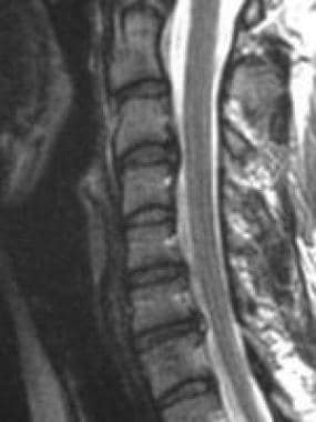
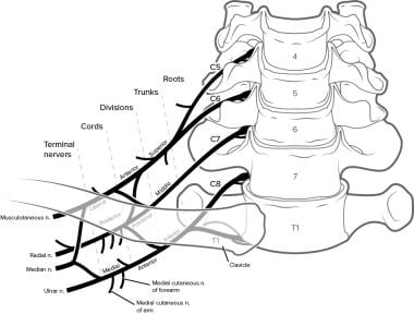
Thoracic nerves are the 12 paired nerves that originate from the thoracic segment (T1-T12) of the spinal cord. These nerves provide motor and sensory innervation to the trunk and abdomen, specifically to the muscles of the chest wall, the skin over the back and chest, and some parts of the abdomen. They also contribute to the formation of the sympathetic trunk, which is a part of the autonomic nervous system that regulates unconscious bodily functions such as heart rate and digestion. Each thoracic nerve emerges from the intervertebral foramen, a small opening between each vertebra, and splits into anterior and posterior branches to innervate the corresponding dermatomes and myotomes.
 Pectoralis major
Kākāpō
Pectus excavatum
Cat anatomy
Lactate shuttle hypothesis
Sternalis muscle
Upper-limb surgery in tetraplegia
Coracoid process
Pectoral muscles
Superior thoracic artery
Bench press
Myositis ossificans
Lance Ringnald
Medial pectoral nerve
List of lymph nodes of the human body
Aponeurosis
Amastia
Lateral pectoral nerve
Pectoralis minor
Axillary dissection
Tra Telligman
Arm
Breast cancer classification
Activin type 2 receptors
Breast implant
Serratus punch
Breast augmentation
Deltoid muscle
Rotter's lymph nodes
Serratus anterior muscle
 Effects of electrical stimulation with precisely controlled square wave voltages on texture of broiler pectoralis muscles
 Faculty Collaboration Database - Publications indexed to the term Pectoralis Muscles
 Inspiratory muscle fatigue affects Latissimus Dorsi but not Pectoralis major activity during arms only front crawl sprinting<...
 Learn Your Muscles: Pectoralis Major
 Pectoralis Minor Muscle | Keywords | MRCEM Success
Pectoralis Minor - Bony Landmarks | 3D Muscle Lab
 Breast Cancer Pectoralis Muscle Invasion - BreastCancerTalk.net
 Muscle Group of the Week: Pectoralis | Moyer Total Wellness
Classification of unusual insertion of the pectoralis minor muscle<...
 Don't Tie Yourself in Knots - CHEK INSTITUTE
Nandrolone decanoate increases satellite cell numbers in the chicken pectoralis muscle<...
 Pectoralis major tendinitis, Shoulder muscles and tendons injuries and damage | Videoreha - medical and sports rehabilitation
Pectoralis major - Wikipedia
 Surgical technique and results of tracheal and carinal replacement with aortic allografts for salivary gland-type carcinoma
 Detection of early changes in the muscle properties of the pectoralis major in breast cancer patients treated with radiotherapy...
Secondary reconstruction with a transverse colon covered with a pectoralis major muscle flap and split thickness skin grafts...
 Puolanne | Influence of Woody Breast Myopathy on Sarcomere Length and Tensile Strength in Commercial Broiler Pectoralis major...
What are the advantages & disadvantages of placing breast implants below the pectoralis muscle? - Pelosi Medical Center -...
 Asthma in Pregnancy: Risks and Prevalence, Pathophysiologic Mechanisms, Asthma Differentials
 Breast cancer awatif | PPT
 Volume 61 Issue 4 | Avian Diseases
 Surveillance, Epidemiology, And End Results, 1973-1989
 I Hurt My Stomach Doing Push-Ups | livestrong
 Free Medical Flashcards about ch6 muscles
 Infraclavicular Block
Breast augmentation - Wikipedia
 Wheelchair Exercises: A Routine for Strength
 Essential Orthopaedics, 2nd Edition - 9780323568944
What Muscles Does the Pec Fly Machine Work? | livestrong
Axillary artery
Deltoids2
- The deltoids , trapezius, and rhomboids are most often overstretched and weakened by a locked-short pectoralis. (moyerwellness.com)
- A bench press uses the pectoralis muscles, deltoids, and triceps. (nifs.org)
Below the pectoralis muscle2
- What are the advantages & disadvantages of placing breast implants below the pectoralis muscle? (pelosimedicalcenter.com)
- and submuscular - placed below the pectoralis muscle. (botonics.co.uk)
Broiler pectoralis1
- Puolanne, T. J. & Costandache, C. G. & Ertbjerg, P., (2021) "Influence of Woody Breast Myopathy on Sarcomere Length and Tensile Strength in Commercial Broiler Pectoralis major Muscle", Meat and Muscle Biology 5(1), 12, 1-11. (iastatedigitalpress.com)
Clavicular3
- Below the clavicular head of the pectoralis major is a much larger section called the sternocostal head. (moyerwellness.com)
- Depending on their goals, bench pressers utilize these variations for achieving total development of both the clavicular (upper) and sternocostal (lower) heads of the pectoralis major. (thesportjournal.org)
- The clavicular head of the pectoralis major has a proximal attachment at the anterior surface of the medial half of the clavicle and distal attachments at the lateral lip of the bicipital groove of the humerus and the anterior lip of the of the deltoid tuberosity. (thesportjournal.org)
Rotator cuff muscles1
- It is important for a heavy bencher to work on shoulder mobility and strengthening the rotator cuff muscles. (nfpt.com)
Latissimus dorsi1
- Collaborating with the latissimus dorsi muscle, the pectoralis major muscle also plays an important role in activities such as climbing. (moyerwellness.com)
Trapezius3
- The primary aim was to investigate serial changes in the mechanical properties of the pectoralis major (PM), upper trapezius (UT), and sternoclavicular mastoid muscle (SCM) in breast cancer patients undergoing radiotherapy (RT) using a hand-held myotonometer. (myoton.com)
- The activations of anterior deltoid, upper trapezius, infraspinatus and pectoralis major were recorded by surface EMG during one-handed transferring of a cylinder from a home shelve to six spatially distributed target shelves. (cdc.gov)
- EMG analysis showed that the injured groups required more upper trapezius and anterior deltoid activation during load transfer tasks, which may predispose them to muscle overexertion. (cdc.gov)
Minor Muscle8
- Purpose: The pectoralis minor muscle (PMi) generally originates from the third, fourth, and fifth ribs and inserts on the medial and superior margins of the anterior portion of the coracoid process. (elsevierpure.com)
- Beneath the pectoralis major is the pectoralis minor muscle. (wikipedia.org)
- These parts are identified by their location relative to the pectoralis minor muscle, which is located in front of the axillary artery. (healthline.com)
- The first part of the artery is located medial (near the middle) to the pectoralis minor muscle. (healthline.com)
- The second part of the artery is behind the pectoralis minor muscle. (healthline.com)
- The third part of the artery is lateral (to the side of, or farther away from the middle) to the pectoralis minor muscle. (healthline.com)
- The left pectoralis minor muscle (18,27) has been cut near its origin and reflected laterally. (stanford.edu)
- One of the factors that can contribute to the scapular kinematics alterations is the presence of a short pectoralis minor muscle that originates from third to fifth ribs and inserts on coracoids process. (fapesp.br)
Pecs3
- The pectoral major may colloquially be referred to as "pecs", "pectoral muscle", or "chest muscle", because it is the largest and most superficial muscle in the chest area. (wikipedia.org)
- The three main muscles are the quads (quadriceps), pecs (pectoralis major) and lats (latissimum dorsi). (swimming.org)
- The pectoralis major muscle (the major muscle in the chest, sometimes called the "pecs" in adults) can be partly developed or missing altogether. (msdmanuals.com)
Sternocostal1
- The sternocostal head of the pectoralis major attaches to the anterior surface of the sternum and the costal cartilages of ribs 1-6. (moyerwellness.com)
Anterior10
- The most superficial and largest muscle of the anterior chest wall, the pectoralis major is known as the main chest muscle. (moyerwellness.com)
- The pectoralis major receives dual motor innervation by the medial pectoral nerve and the lateral pectoral nerve, also known as the lateral anterior thoracic nerve. (wikipedia.org)
- The roots combine above the first rib to form the superior, middle, and inferior trunks of the brachial plexus, between the anterior and middle scalene muscles, in the interscalene groove. (asra.com)
- The anterior deltoid muscle helps bring the arms of the pec fly machine together. (livestrong.com)
- The lateral femoral circumflex artery supplies oxygenated blood to the anterior (front) and middle portions of the thigh muscles. (healthline.com)
- The primary muscle used to create the movement for the dumbbell press are: Pectoralis major, all Triceps and Anterior Deltoid. (nfpt.com)
- The secondary muscles used are Pectoralis minor, Latissimis Dorsi, Coracobrachialis and Serratus Anterior. (nfpt.com)
- Body/hand position, execution, width of grip, trunk inclination, dumbbells and barbells are all variables that affect the prime movers (pectoralis major, anterior deltoid and triceps brachii) of the bench press. (thesportjournal.org)
- The prime movers involved in the bench press are the pectoralis major, anterior deltoid, and the triceps brachii. (thesportjournal.org)
- They occupy the space between the anterior and middle scalene muscles. (medscape.com)
Originates1
- Pectoralis major originates from the medial proximal part of the humerus , and it descends across the chest diagonally toward the sternum along whose lenght it connects. (videoreha.com)
Bench Press4
- The bench press is considered the top exercise when it comes to training your pectoralis major, but the pec deck or chest fly machine is a close second. (livestrong.com)
- The research has shown that the horizontal barbell bench press done with a grip between 165% to 190% biacromial width produces maximum EMG activity in the pectoralis major. (thesportjournal.org)
- Owing to these several sites of attachments the pectoralis major can be targeted using different variations of the bench press. (thesportjournal.org)
- Algra, B. (1982) illustrates the muscles worked and the starting and finishing positions for the free weight barbell bench press as shown below. (thesportjournal.org)
Pectoral muscle3
- All grafts were wrapped with bulky and well-vascularized flaps (pectoral muscle flap all patients, with an additional "thymopericardial fat flap" in the last 2) to promote revascularization and to prevent erosion of adjacent large vessels or fistulas. (nih.gov)
- [ 7 ] Brachial deformities and pectoral muscle hypoplasia have also been described. (medscape.com)
- A syndrome of congenital facial paralysis, frequently associated with abducens palsy and other congenital abnormalities including lingual palsy, clubfeet, brachial disorders, cognitive deficits, and pectoral muscle defects. (bvsalud.org)
Shoulder14
- More specifically than just moving the shoulder, what does the pectoralis major do? (moyerwellness.com)
- Both sections of the pectoralis major insert into the greater tubercle of the humerus, making this thick fan-shaped muscle crucial to shoulder movement. (moyerwellness.com)
- As a whole, the main function of the pectoralis major is to adduct and medially rotate the humerus at the shoulder joint. (moyerwellness.com)
- The muscles situated at the front of the chest strengthen the chest cavity, serve as a strong protective shield and they are also a big part of securing shoulder and arm mobility and functionality. (videoreha.com)
- Pectoralis major performs internal rotation of shoulder and arm, it pulls the arm horizontally to the front and across the chest and pulls the arm from an above shoulder level position diagonally downwards. (videoreha.com)
- Strengthening of pectoralis musculature should also be implemented, in both concentric and ecccentric work conditions, so that they could bear the loads put on them and to regain full shoulder and arm functionality. (videoreha.com)
- Increased tone and stiffness were noted in the affected UT immediately after RT, and decreased 4 months later, indicating that sustaining an abducted and externally rotated shoulder posture during the RT session may have temporally influenced the muscle properties of the UT. (myoton.com)
- This activity provides a great stretch to the shoulder and chest muscles. (healthline.com)
- The rear deltoid muscles run along the back of the shoulder and are necessary for shoulder stability and movement during pulling and lifting activities. (healthline.com)
- This muscle lies underneath your pec major and is responsible for stabilization of your shoulder blade. (livestrong.com)
- To investigate muscle activation of the shoulder extensors and trunk stabilizers by surface electromyography (sEMG) activity during the isometric Ab Wheel Rollout exercise in different shoulder joint positions. (scielo.br)
- It also looked at whether subacromial impingement syndrome athletes differ in volleyball spiking sequence and mobilization and recruitment of muscle power during swing spikes compared to athletes with normal shoulder function in the full kinetic chain. (hindawi.com)
- This study aimed to compare the activity of four shoulder muscles in individuals with low back pain (LBP), spinal cord injuries (SCI) and a control group, during one-handed load transfer trials. (cdc.gov)
- Practitioner Summary: This study aimed to compare the activation of four shoulder muscles in individuals with low back pain , spinal cord injuries and healthy condition. (cdc.gov)
Sternocleidomastoid1
- We present a unique case of an 84-year-old female who presented with neck swelling and upper airway obstruction due to metastatic breast cancer invading the sternocleidomastoid muscles. (breastcancertalk.net)
Medial1
- After the synapse in the posterior horn of the spinal cord, sensory information concerning movement of the muscle, proprioception, and pressure then travels through a second-order neuron in the dorsal column medial lemniscus tract to the medulla. (wikipedia.org)
Barbells1
- Barbells and dumbbells are the most commonly used, and the different types of bench presses are all effective ways to address the main chest muscle. (moyerwellness.com)
Fascia1
- The layer of coracoclavicular fascia (19) deep to this muscle is continuous laterally with the pectoral (22) and axillary fascia (21). (stanford.edu)
Ribs2
- from the cartilages of all the true ribs, with the exception, frequently, of the first or seventh, and from the aponeurosis of the abdominal external oblique muscle. (wikipedia.org)
- The boundaries of the infraclavicular fossa are the pectoralis minor and major anteriorly, ribs medially, clavicle and coracoid process superiorly, and humerus laterally. (medscape.com)
Quadriceps1
- The squat movement pattern is a compound movement that works the muscles of the upper legs including the quadriceps and glutes. (nifs.org)
Major muscle3
- The machine emphasizes your pectoralis major muscle, the broad muscle of the chest, as well as a few helper muscles. (livestrong.com)
- There was a significant increase in Pectoralis Major muscle activity between neutral x 150º. (scielo.br)
- 9. Inadequate chest wall tissue due to damage caused by radiotherapy, tight skin grafts, or radical resection of the pectoralis major muscle. (who.int)
Sternoclavicular1
- When treating women, adhesions on the sternoclavicular portion of the pectoralis major can be loosened with heat application either through use of a hotpack or a heating pad. (moyerwellness.com)
Secondary muscles1
- The secondary muscles stressed are the shoulders. (nfpt.com)
Exercises11
- Choose mobility exercises that improve blood flow to restricted muscles. (elitefitness.com)
- Do some activation exercises to prep and prime the stabilizing muscles. (elitefitness.com)
- Mobility exercises are carried out by using self-massage therapy methods, such as applying pressure to the muscle using a foam roller or lacrosse ball. (elitefitness.com)
- My advice is to choose one to three of the exercises below, apply pressure to the muscle, and then perform five to ten strokes for 60 to 90 seconds. (elitefitness.com)
- Your gym programme should include exercises that will overload the muscles that you will be using during swimming. (swimming.org)
- The superficial electromyographic (sEMG) technique is often used to identify the activation of each muscle in different exercises. (scielo.br)
- Analysis of muscle activation during different leg press exercises at submaximum effort levels. (scielo.br)
- 2009. This article seeks to determine optimum body/hand position and the best exercises for development of the pectoralis major. (thesportjournal.org)
- Exercises that isolate a single muscle group are called isolation exercises. (nifs.org)
- Through the electromyographic signal it is possible to study the response to the therapeutic exercises commonly used in rehabilitation regarding the beginning and end of the activity, type of muscle contraction and joint position. (bvsalud.org)
- Four analyzed muscle activation in isometric exercises, six used isotonic exercises and only one article used isokinetic exercises. (bvsalud.org)
Lateral1
- The lateral pectoral nerve is distributed over the deep surface of the pectoralis major. (wikipedia.org)
Shoulders4
- When the arm is bearing weight, for example when using crutches, the pectoralis major and latissimus dorsi elevate the body while keeping the shoulders stable. (moyerwellness.com)
- A review in Topics in Geriatric Rehabilitation reports that for people who use a manual wheelchair, exercise programs should include training of the larger muscles of the trunk as well as stretching for the shoulders and chest. (healthline.com)
- Most wheelchair users consistently use their upper body, particularly the triceps and the shoulders, for movement, which puts extra strain on the joints and muscles. (healthline.com)
- The pec machine targets the pectoralis major and minor muscles in your chest, as well as muscles in the front of your shoulders. (livestrong.com)
Targets2
- Chest dips are another workout that targets the pectoralis major. (moyerwellness.com)
- The spatial position of the targets also significantly influenced demands for these two muscles. (cdc.gov)
Upper5
- The muscle group is also heavily impacted by poor posture , finding itself locked short and tight in postural disorders such as upper cross syndrome . (moyerwellness.com)
- The pushup is an exercise that works most of the muscles of the upper body in some way. (livestrong.com)
- This exercise strengthens the muscles of the mid back, upper arms, and core. (healthline.com)
- The pec fly machine also activate your ab muscles as they contract to help stabilize your upper body as your arms move. (livestrong.com)
- This will help to isolate the upper and central muscles on the chest. (nfpt.com)
Contraction2
- This leads to a reduction in airway diameter caused by smooth muscle contraction, vascular congestion, bronchial wall edema, and thick secretions. (medscape.com)
- Through the EMGs it is possible to observe the degree, duration, type of muscle contraction, alteration of the composition of the motor units resulting from muscle training programs, recruitment neural strategies, as well as allowing inferences related to muscle fatigue. (bvsalud.org)
Additionally1
- Additionally, the portability, convenient usage, noninvasiveness, and objectivity of myotonometer allow physicians to easily check muscle conditions of irradiated patients in busy clinical settings. (myoton.com)
Strengthen3
- The next step in the treatment is to strengthen the antagonist muscle groups weakened by a tight pectoralis major. (moyerwellness.com)
- If your clients have poor posture, they must attempt to lengthen their short muscles and strengthen or tighten any long or weak phasic muscles to bring their bodies back into balance. (chekinstitute.com)
- on the other hand, after a mild injury occurs, timely targeted training should be taken to find and correct wrong actions, and strengthen the weak part of muscle strength, so as to reduce the probability of repeated injury and improve sports performance and athletic ability. (hindawi.com)
Imbalances4
- Sitting for extended periods day in and day out, without adequate stretching and movement, leads to decreased flexibility and muscle imbalances. (chekinstitute.com)
- To better explain to your clients how muscle imbalances affect their bodies, imagine a bicycle wheel out of balance. (chekinstitute.com)
- In order to prevent injury, it's important to avoid imbalances by strengthening the muscles of the back and stretching the muscles of the chest. (healthline.com)
- If you want to sculpt your body in a particular way, and are focused on correcting muscle imbalances or injury rehabilitation, each may require the use of specific isolation movements to build up specific muscle groups. (nifs.org)
Internal rotation1
- The pectoralis major's primary functions are flexion, adduction, and internal rotation of the humerus. (wikipedia.org)
Posterior3
- For each eligible patient, clinical history and surgical pathology details were also recorded, including whether the posterior margins were clear, close or positive for tumor involvement, as well as whether tumor invasion of the pectoralis muscle was seen on surgical pathology. (breastcancertalk.net)
- If posterior margins were clear and no skeletal muscle was present on surgical pathology, the case was recorded as having no muscle invasion. (breastcancertalk.net)
- The sensory feedback from the pectoralis major follows the reverse path, returning via first-order neurons to the spinal nerves at C5, C6, C8, and T1 through the posterior rami. (wikipedia.org)
Fibers1
- Woody breast syndrome is characterized by degenerative changes at the muscle fiber level and accumulation of connective tissue between the fibers. (iastatedigitalpress.com)
Tighten1
- Poor posture always indicates the need to follow a stretching program to lengthen short muscles and an exercise program to tighten your clients' weak or loose muscles. (chekinstitute.com)
Trunk1
- When activated together, both muscles pull the trunk forward and upward when their humeral attachments are fixed in an overhead position. (moyerwellness.com)
Scapular6
- Pectoralis major tendon transfer for the treatment of scapular winging due to long thoracic nerve palsy. (mcw.edu)
- Because of an increase in passive tension, this muscle can limit the normal scapular motion. (fapesp.br)
- Previous study demonstrate that healthy subjects with short pectoralis minor show scapular kinematics similar to that presented by individuals with subacromial impingement syndrome. (fapesp.br)
- However, there are no data in the literature that demonstrate the isolated effect of the pectoralis minor stretching on scapular kinematics. (fapesp.br)
- Thus, the objective of this study is to evaluate the influence of the pectoralis minor on scapular kinematics. (fapesp.br)
- For this, thirty healthy individuals with short pectoralis minor will be submitted to a scapular kinematics evaluation before and after a stretching programme. (fapesp.br)
Commonly2
- People commonly make the mistake of stretching muscles that don't need stretching and not stretching the ones that do need it . (chekinstitute.com)
- Treatment is commonly done through a conservative, nonsurgical approach.The inflammatory process is reduced in acute phase by use of RICE method (rest, ice, compression, elevation) and in the following functional rehabilitation the chest muscles should get stretched and relaxed. (videoreha.com)
Breast9
- Breast cancer rarely metastasizes to the muscles, and it is even more unusual for this phenomenon to result in airway compromise. (breastcancertalk.net)
- However, the pectoralis major can be difficult to fully massage on women, as breast tissue will be present unless the woman has undergone a mastectomy . (moyerwellness.com)
- The pectoralis major (from Latin pectus 'breast') is a thick, fan-shaped or triangular convergent muscle of the human chest. (wikipedia.org)
- It makes up the bulk of the chest muscles and lies under the breast. (wikipedia.org)
- This study explored effects of the syndrome on muscle properties by focusing on a comparison of the sarcomere lengths between normal and woody breast muscles, including cranial and middle parts, surface and deeper layers, electrically stimulated and nonstimulated muscles, and their combinations. (iastatedigitalpress.com)
- Tensile strength was much greater in diffuse woody breast muscles when extended longitudinally or transversely to the fiber direction. (iastatedigitalpress.com)
- In conclusion, although this study did not show sarcomere lengths in living muscle, it suggests an imbalance in sarcomere lengths in different parts of the breast muscle, which may induce a reduction in the functionality and strength of the muscle. (iastatedigitalpress.com)
- The operational aims were to study the sarcomere lengths in different breast muscle locations and the tensile strength of muscle tissue, longitudinally and transversely to the fiber direction. (iastatedigitalpress.com)
- A congenital condition called the Poland sequence, characterized by ipsilateral hand malformations and by partial or complete absence of the pectoralis muscles and breast, is concurrent with Möbius syndrome in approximately 15% of patients. (medscape.com)
Investigate1
- The aim of the current study was to investigate the EMG activity of pectoralis major and latissimus dorsi muscles during the pullover exercise. (humankinetics.com)
Oblique1
- This exercise is great to target oblique muscles and slim down your waistline. (projectswole.com)
Affects2
- The purpose of this study was to determine whether inspiratory muscle fatigue (IMF) affects the muscle activity of the latissimus dorsi and pectoralis major during maximal arms only front crawl swimming. (port.ac.uk)
- This is a non-final version of an article published in final form in Lomax M, Tasker L, Bostanci O. Inspiratory muscle fatigue affects latissimus dorsi but not pectoralis major activity during arms only front crawl sprinting. (port.ac.uk)
Concentric1
- The EMG activity of the pectoralis major and that of the latissimus dorsi of the right side were acquired simultaneously during the pullover exercise with a free-weight barbell during both the concentric and eccentric phases of the movement. (humankinetics.com)
Abdominal2
- The abdominal pain you're feeling after doing pushups is likely due to a strained abdominal muscle. (livestrong.com)
- One or more layers of the abdominal muscles may be missing at birth, as in prune-belly syndrome. (msdmanuals.com)
Lengthen1
- Before lifting, dynamic stretching will assist you lengthen the muscle and enhance its performance. (elitefitness.com)
Movement6
- You need to train your muscles to perform the movement correctly before you begin overloading them. (swimming.org)
- In addition, it can influence the level of muscle activation that will be used in the movement, 2 2 Brennecke A, Guimarães TM, Leone R, Cadarci M, Mochizuki L, Simão R, et al. (scielo.br)
- The primary muscles stressed in this movement are the chest muscles (pectoralis major and minor). (nfpt.com)
- In this blog series, I have discussed the four movement patterns for building muscle and broken them down further with exercise examples you can add to your workouts. (nifs.org)
- The most efficient way to structure a workout utilizing both methods is to perform a compound movement first in your workout followed by isolation movements to complement the muscles used in the compound movement. (nifs.org)
- It also means that the electromyographic signal becomes a useful tool for analyzing the outcome of physiotherapeutic treatments because it provides easy access to the physiological processes that make the muscle generate strength, produce movement, and perform numerous functions that allow us to make relevant inferences regarding biomechanics of human movements. (bvsalud.org)
Mechanical1
- Because the choice of a specific exercise can generate mechanical and physiological muscle stress, it is essential to define the exercise order during resistance training. (scielo.br)
Abstract1
- abstract = "The anabolic androgenic steroid nandrolone decanoate has minimal androgenic effects and, thus, is widely used to induce muscle hypertrophy in both patients and athletes. (uaeu.ac.ae)
Mobility2
- You can improve the blood supply to your muscles and regain motion by performing mobility activities. (elitefitness.com)
- The mobility of the joint is restricted when a muscle is tight. (elitefitness.com)
Chest muscle2
- Although uncomfortable, a strained chest muscle is usually a minor injury that tends to heal within days or weeks. (breastcancertalk.net)
- Chest muscle tendinitis in most cases reffers to an inflammation of the pectoralis major tendon. (videoreha.com)
Significantly3
- All satellite cell indices that were quantified increased significantly in chicken pectoralis with administration of nandrolone. (uaeu.ac.ae)
- However, an overall increase in myonuclear numbers was revealed by a significantly greater mean number of myonuclei per millimeter of fiber in nandrolone injected muscle. (uaeu.ac.ae)
- Early changes in muscle properties immediately after RT preceded changes in subjective symptom scale scores, which worsened significantly 4 months post-RT compared with baseline. (myoton.com)
Fibres2
- Electromyography suggests that it consists of at least six groups of muscle fibres that can be independently coordinated by the central nervous system. (wikipedia.org)
- Electromyography is a technique used for recording changes in electrical potential of muscle fibres that are associated with their contractions Payton, C. J., Bartlett, R. M. (Eds. (thesportjournal.org)
Syndrome1
- Birth defects of the muscles can occur alone or as part of a syndrome. (msdmanuals.com)
Exercise4
- It has minimal reach when it comes to muscle activation, though, as it's an isolation exercise, activating just one joint. (livestrong.com)
- The present findings demonstrated that the barbell pullover exercise emphasized the muscle action of the pectoralis major more than that of the latissimus dorsi, and the higher activation depended on the external force lever arm produced. (humankinetics.com)
- Another excellent exercise to build core stability as well as the supporting muscles below the core, to fortify the aesthetics. (projectswole.com)
- This requires more energy to be expended than if you used only one of those muscle groups on a single joint exercise. (nifs.org)
Nerve1
- Pathologic findings are variable and include brain stem nuclear aplasia, facial nerve aplasia, and facial muscle aplasia, consistent with a multifactorial etiology. (bvsalud.org)
Position1
- Squeeze your back muscles for a second, then return to starting position. (healthline.com)
Isolate1
- This will help isolate the chest muscles. (nfpt.com)
Erector spinae1
- The major muscles in your core include the abs (transverse abdominis and rectus abdominis), obliques (internal and external) and the erector spinae. (swimming.org)