Patellofemoral Joint
Patellofemoral Pain Syndrome
Patellar Ligament
Osteoarthritis, Knee
Chondromalacia Patellae
Biomechanical Phenomena
Cartilage, Articular
Joint Diseases
Quadriceps Muscle
Athletic Tape
Joint Instability
Joints
Range of Motion, Articular
Weight-Bearing
Pain
Dislocations
Magnetic Resonance Imaging
Tibia
Stress, Mechanical
Osteoarthritis
Anterior Cruciate Ligament
Finite Element Analysis
Hip Joint
Braces
Bone Malalignment
Treatment Outcome
Finger Joint
Prospective Studies
Pain Measurement
Orthotic Devices
Hip
Ankle Joint
Posterior Cruciate Ligament
Bone Diseases, Developmental
Does isolated patellofemoral osteoarthritis matter? (1/76)
(+info)Q-angle and J-sign: indicative of maltracking subgroups in patellofemoral pain. (2/76)
(+info)The regenerative effect of platelet-rich plasma on healing in large osteochondral defects. (3/76)
(+info)Tibiofemoral and patellofemoral kinematics after reconstruction of an isolated posterior cruciate ligament injury: in vivo analysis during lunge. (4/76)
(+info)Patellar tendon orientation and patellar tracking in male and female knees. (5/76)
(+info)Pain and hip lateral rotator muscle strength contribute to functional status in females with patellofemoral pain. (6/76)
(+info)Tantalum is a good bone graft substitute in tibial tubercle advancement. (7/76)
(+info)Denuded subchondral bone and knee pain in persons with knee osteoarthritis. (8/76)
(+info)The patellofemoral joint is the articulation between the patella (kneecap) and the femur (thigh bone). It is a synovial joint, which means it is surrounded by a joint capsule containing synovial fluid to lubricate the joint. This joint is responsible for providing stability to the knee extensor mechanism and allows for smooth movement of the patella during activities like walking, running, and jumping. Pain or dysfunction in this joint can result in various conditions such as patellofemoral pain syndrome, chondromalacia patella, or patellar dislocation.
Patellofemoral Pain Syndrome (PFPS) is a broad term used to describe pain arising from the front of the knee, specifically where the patella (kneecap) meets the femur (thigh bone). It is often described as a diffuse, aching pain in the anterior knee, typically worsening with activities that load the patellofemoral joint such as climbing stairs, running, jumping or prolonged sitting.
PFPS can be caused by various factors including overuse, muscle imbalances, poor biomechanics, or abnormal tracking of the patella. Treatment usually involves a combination of physical therapy to improve strength and flexibility, activity modification, and sometimes bracing or orthotics for better alignment.
The patella, also known as the kneecap, is a sesamoid bone located at the front of the knee joint. It is embedded in the tendon of the quadriceps muscle and serves to protect the knee joint and increase the leverage of the extensor mechanism, allowing for greater extension force of the lower leg. The patella moves within a groove on the femur called the trochlea during flexion and extension of the knee.
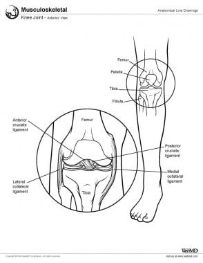
The knee joint, also known as the tibiofemoral joint, is the largest and one of the most complex joints in the human body. It is a synovial joint that connects the thighbone (femur) to the shinbone (tibia). The patella (kneecap), which is a sesamoid bone, is located in front of the knee joint and helps in the extension of the leg.
The knee joint is made up of three articulations: the femorotibial joint between the femur and tibia, the femoropatellar joint between the femur and patella, and the tibiofibular joint between the tibia and fibula. These articulations are surrounded by a fibrous capsule that encloses the synovial membrane, which secretes synovial fluid to lubricate the joint.
The knee joint is stabilized by several ligaments, including the medial and lateral collateral ligaments, which provide stability to the sides of the joint, and the anterior and posterior cruciate ligaments, which prevent excessive forward and backward movement of the tibia relative to the femur. The menisci, which are C-shaped fibrocartilaginous structures located between the femoral condyles and tibial plateaus, also help to stabilize the joint by absorbing shock and distributing weight evenly across the articular surfaces.
The knee joint allows for flexion, extension, and a small amount of rotation, making it essential for activities such as walking, running, jumping, and sitting.
Patellar dislocation is a medical condition characterized by the displacement of the patella (kneecap) from its normal position in the femoral groove, which is a part of the femur (thighbone). This displacement usually occurs laterally, meaning that the patella moves toward the outer side of the knee.
Patellar dislocation can happen as a result of direct trauma or due to various factors that increase the laxity of the medial patellofemoral ligament and tightness of the lateral structures, leading to abnormal tracking of the patella. These factors include anatomical variations, muscle imbalances, genetic predisposition, or degenerative changes in the knee joint.
Dislocation of the patella can cause pain, swelling, and difficulty in moving the knee. In some cases, it might be associated with other injuries such as fractures or damage to the articular cartilage and surrounding soft tissues. Immediate medical attention is required for proper diagnosis and treatment, which may involve reduction, immobilization, physical therapy, bracing, or even surgery in severe cases.
The femur is the medical term for the thigh bone, which is the longest and strongest bone in the human body. It connects the hip bone to the knee joint and plays a crucial role in supporting the weight of the body and allowing movement during activities such as walking, running, and jumping. The femur is composed of a rounded head, a long shaft, and two condyles at the lower end that articulate with the tibia and patella to form the knee joint.
The patellar ligament, also known as the patellar tendon, is a strong band of tissue that connects the bottom part of the kneecap (patella) to the top part of the shinbone (tibia). This ligament plays a crucial role in enabling the extension and straightening of the leg during activities such as walking, running, and jumping. Injuries to the patellar ligament, such as tendonitis or tears, can cause pain and difficulty with mobility.
Osteoarthritis (OA) of the knee is a degenerative joint disease that affects the articular cartilage and subchondral bone in the knee joint. It is characterized by the breakdown and eventual loss of the smooth, cushioning cartilage that covers the ends of bones and allows for easy movement within joints. As the cartilage wears away, the bones rub against each other, causing pain, stiffness, and limited mobility. Osteoarthritis of the knee can also lead to the formation of bone spurs (osteophytes) and cysts in the joint. This condition is most commonly found in older adults, but it can also occur in younger people as a result of injury or overuse. Risk factors include obesity, family history, previous joint injuries, and repetitive stress on the knee joint. Treatment options typically include pain management, physical therapy, and in some cases, surgery.
Chondromalacia patellae is a medical condition that refers to the softening and degeneration of the articular cartilage on the undersurface of the patella, or kneecap. This cartilage, which provides a smooth, lubricated surface for joint movement, can become damaged due to various reasons such as overuse, misalignment of the patella, or direct trauma. The resulting damage can cause pain and inflammation in the knee, particularly during activities that involve bending or straightening the leg. In some cases, chondromalacia patellae may also lead to the formation of bone spurs or osteophytes, which can further exacerbate the symptoms and limit joint mobility. Treatment for chondromalacia patellae typically involves a combination of rest, physical therapy, and pain management strategies, such as anti-inflammatory medications or corticosteroid injections. In severe cases, surgery may be required to repair or replace the damaged cartilage.
Cartilage diseases refer to conditions that affect the cartilaginous tissues in the body. Cartilage is a firm, flexible connective tissue found in many areas of the body, including the joints, ribcage, ears, and nose. It provides structure and support, allows for smooth movement between bones, and protects the ends of bones from friction.
There are several types of cartilage diseases, including:
1. Osteoarthritis (OA): This is a degenerative joint disease that occurs when the protective cartilage that cushions the ends of your bones wears down over time. It can cause pain, stiffness, and loss of mobility in the affected joints.
2. Rheumatoid arthritis (RA): This is an autoimmune disorder that causes inflammation in the lining of the joints, leading to cartilage damage and bone erosion.
3. Traumatic arthritis: This occurs when a joint is injured, causing damage to the cartilage and resulting in pain, stiffness, and loss of mobility.
4. Infectious arthritis: This occurs when a joint becomes infected, leading to inflammation and potential damage to the cartilage.
5. Chondromalacia patellae: This is a condition that affects the cartilage on the back of the kneecap, causing pain and stiffness in the knee.
6. Costochondritis: This is an inflammation of the cartilage in the ribcage, causing chest pain and discomfort.
7. Nasal septal deviation: This is a condition where the cartilage that separates the nostrils is crooked or off-center, causing difficulty breathing through the nose.
8. Osteochondritis dissecans (OCD): This is a joint condition that occurs when a piece of cartilage and bone in a joint becomes detached, causing pain and stiffness.
9. Synovial chondromatosis: This is a rare condition where nodules made up of cartilage form in the lining of a joint, causing pain, swelling, and limited mobility.
Treatment for cartilage diseases varies depending on the specific condition and severity, but may include medication, physical therapy, surgery, or a combination of these.
Biomechanics is the application of mechanical laws to living structures and systems, particularly in the field of medicine and healthcare. A biomechanical phenomenon refers to a observable event or occurrence that involves the interaction of biological tissues or systems with mechanical forces. These phenomena can be studied at various levels, from the molecular and cellular level to the tissue, organ, and whole-body level.
Examples of biomechanical phenomena include:
1. The way that bones and muscles work together to produce movement (known as joint kinematics).
2. The mechanical behavior of biological tissues such as bone, cartilage, tendons, and ligaments under various loads and stresses.
3. The response of cells and tissues to mechanical stimuli, such as the way that bone tissue adapts to changes in loading conditions (known as Wolff's law).
4. The biomechanics of injury and disease processes, such as the mechanisms of joint injury or the development of osteoarthritis.
5. The use of mechanical devices and interventions to treat medical conditions, such as orthopedic implants or assistive devices for mobility impairments.
Understanding biomechanical phenomena is essential for developing effective treatments and prevention strategies for a wide range of medical conditions, from musculoskeletal injuries to neurological disorders.
Articular cartilage is the smooth, white tissue that covers the ends of bones where they come together to form joints. It provides a cushion between bones and allows for smooth movement by reducing friction. Articular cartilage also absorbs shock and distributes loads evenly across the joint, protecting the bones from damage. It is avascular, meaning it does not have its own blood supply, and relies on the surrounding synovial fluid for nutrients. Over time, articular cartilage can wear down or become damaged due to injury or disease, leading to conditions such as osteoarthritis.
Joint diseases is a broad term that refers to various conditions affecting the joints, including but not limited to:
1. Osteoarthritis (OA): A degenerative joint disease characterized by the breakdown of cartilage and underlying bone, leading to pain, stiffness, and potential loss of function.
2. Rheumatoid Arthritis (RA): An autoimmune disorder causing inflammation in the synovial membrane lining the joints, resulting in swelling, pain, and joint damage if left untreated.
3. Infectious Arthritis: Joint inflammation caused by bacterial, viral, or fungal infections that spread through the bloodstream or directly enter the joint space.
4. Gout: A type of arthritis resulting from the buildup of uric acid crystals in the joints, typically affecting the big toe and characterized by sudden attacks of severe pain, redness, and swelling.
5. Psoriatic Arthritis (PsA): An inflammatory joint disease associated with psoriasis, causing symptoms such as pain, stiffness, and swelling in the joints and surrounding tissues.
6. Juvenile Idiopathic Arthritis (JIA): A group of chronic arthritis conditions affecting children, characterized by joint inflammation, pain, and stiffness.
7. Ankylosing Spondylitis: A form of arthritis primarily affecting the spine, causing inflammation, pain, and potential fusion of spinal vertebrae.
8. Bursitis: Inflammation of the fluid-filled sacs (bursae) that cushion joints, leading to pain and swelling.
9. Tendinitis: Inflammation or degeneration of tendons, which connect muscles to bones, often resulting in pain and stiffness near joints.
These conditions can impact the function and mobility of affected joints, causing discomfort and limiting daily activities. Proper diagnosis and treatment are essential for managing joint diseases and preserving joint health.
The Quadriceps muscle, also known as the Quadriceps Femoris, is a large muscle group located in the front of the thigh. It consists of four individual muscles - the Rectus Femoris, Vastus Lateralis, Vastus Intermedius, and Vastus Medialis. These muscles work together to extend the leg at the knee joint and flex the thigh at the hip joint. The Quadriceps muscle is crucial for activities such as walking, running, jumping, and kicking.
Athletic tape, also known as sports tape or physiotherapy tape, is a type of adhesive tape that is commonly used in the field of sports medicine and physical therapy to provide support and stability to joints, muscles, and tendons during athletic activities. It is typically made from a cotton or synthetic fabric material with a strong adhesive backing.
The main purpose of athletic tape is to limit excessive movement or provide compression to an injured area, which can help to reduce pain, swelling, and the risk of further injury. Athletic tape can be used to support a wide variety of body parts, including the ankles, knees, wrists, elbows, and fingers.
There are several different types of athletic tape available, including rigid and flexible options. Rigid tapes, such as zinc oxide tape, are designed to provide maximum support and stability to joints and muscles, while flexible tapes, such as cohesive bandage or kinesiology tape, allow for a greater range of motion and can be used to provide more gentle support or to help facilitate muscle activation and movement.
It is important to note that athletic tape should only be applied by trained professionals, as improper application can lead to further injury or skin irritation. Additionally, athletes should always consult with their healthcare provider before using athletic tape to treat an injury, as it may not be appropriate for all types of injuries or medical conditions.
Joint instability is a condition characterized by the loss of normal joint function and increased risk of joint injury due to impaired integrity of the supporting structures, such as ligaments, muscles, or cartilage. This can result in excessive movement or laxity within the joint, leading to decreased stability and increased susceptibility to dislocations or subluxations. Joint instability may cause pain, swelling, and limited range of motion, and it can significantly impact a person's mobility and quality of life. It is often caused by trauma, degenerative conditions, or congenital abnormalities and may require medical intervention, such as physical therapy, bracing, or surgery, to restore joint stability.
Arthroplasty, replacement, knee is a surgical procedure where the damaged or diseased joint surface of the knee is removed and replaced with an artificial joint or prosthesis. The procedure involves resurfacing the worn-out ends of the femur (thigh bone) and tibia (shin bone) with metal components, and the back of the kneecap with a plastic button. This surgery is usually performed to relieve pain and restore function in patients with severe knee osteoarthritis, rheumatoid arthritis, or traumatic injuries that have damaged the joint beyond repair. The goal of knee replacement surgery is to improve mobility, reduce pain, and enhance the quality of life for the patient.
A joint is the location at which two or more bones make contact. They are constructed to allow movement and provide support and stability to the body during motion. Joints can be classified in several ways, including structure, function, and the type of tissue that forms them. The three main types of joints based on structure are fibrous (or fixed), cartilaginous, and synovial (or diarthrosis). Fibrous joints do not have a cavity and have limited movement, while cartilaginous joints allow for some movement and are connected by cartilage. Synovial joints, the most common and most movable type, have a space between the articular surfaces containing synovial fluid, which reduces friction and wear. Examples of synovial joints include hinge, pivot, ball-and-socket, saddle, and condyloid joints.
Articular Range of Motion (AROM) is a term used in physiotherapy and orthopedics to describe the amount of movement available in a joint, measured in degrees of a circle. It refers to the range through which synovial joints can actively move without causing pain or injury. AROM is assessed by measuring the degree of motion achieved by active muscle contraction, as opposed to passive range of motion (PROM), where the movement is generated by an external force.
Assessment of AROM is important in evaluating a patient's functional ability and progress, planning treatment interventions, and determining return to normal activities or sports participation. It is also used to identify any restrictions in joint mobility that may be due to injury, disease, or surgery, and to monitor the effectiveness of rehabilitation programs.
"Weight-bearing" is a term used in the medical field to describe the ability of a body part or limb to support the weight or pressure exerted upon it, typically while standing, walking, or performing other physical activities. In a clinical setting, healthcare professionals often use the term "weight-bearing exercise" to refer to physical activities that involve supporting one's own body weight, such as walking, jogging, or climbing stairs. These exercises can help improve bone density, muscle strength, and overall physical function, particularly in individuals with conditions affecting the bones, joints, or muscles.
In addition, "weight-bearing" is also used to describe the positioning of a body part during medical imaging studies, such as X-rays or MRIs. For example, a weight-bearing X-ray of the foot or ankle involves taking an image while the patient stands on the affected limb, allowing healthcare providers to assess any alignment or stability issues that may not be apparent in a non-weight-bearing position.
Pain is an unpleasant sensory and emotional experience associated with actual or potential tissue damage, or described in terms of such damage. It is a complex phenomenon that can result from various stimuli, such as thermal, mechanical, or chemical irritation, and it can be acute or chronic. The perception of pain involves the activation of specialized nerve cells called nociceptors, which transmit signals to the brain via the spinal cord. These signals are then processed in different regions of the brain, leading to the conscious experience of pain. It's important to note that pain is a highly individual and subjective experience, and its perception can vary widely among individuals.
A dislocation is a condition in which a bone slips out of its normal position in a joint. This can happen as a result of trauma or injury, such as a fall or direct blow to the body. Dislocations can cause pain, swelling, and limited mobility in the affected area. In some cases, a dislocation may also damage surrounding tissues, such as ligaments, tendons, and nerves.
Dislocations are typically treated by reducing the dislocation, which means putting the bone back into its normal position. This is usually done with the help of medication to relieve pain and relaxation techniques to help the person stay still during the reduction. In some cases, surgery may be necessary to repair damaged tissues or if the dislocation cannot be reduced through other methods. After the dislocation has been reduced, the joint may be immobilized with a splint or sling to allow it to heal properly.
It is important to seek medical attention promptly if you suspect that you have a dislocation. If left untreated, a dislocation can lead to further complications, such as joint instability and chronic pain.
Medical Definition:
Magnetic Resonance Imaging (MRI) is a non-invasive diagnostic imaging technique that uses a strong magnetic field and radio waves to create detailed cross-sectional or three-dimensional images of the internal structures of the body. The patient lies within a large, cylindrical magnet, and the scanner detects changes in the direction of the magnetic field caused by protons in the body. These changes are then converted into detailed images that help medical professionals to diagnose and monitor various medical conditions, such as tumors, injuries, or diseases affecting the brain, spinal cord, heart, blood vessels, joints, and other internal organs. MRI does not use radiation like computed tomography (CT) scans.
The tibia, also known as the shin bone, is the larger of the two bones in the lower leg and part of the knee joint. It supports most of the body's weight and is a major insertion point for muscles that flex the foot and bend the leg. The tibia articulates with the femur at the knee joint and with the fibula and talus bone at the ankle joint. Injuries to the tibia, such as fractures, are common in sports and other activities that put stress on the lower leg.
Fluoroscopy is a type of medical imaging that uses X-rays to obtain real-time moving images of the internal structures of the body. A continuous X-ray beam is passed through the body part being examined, and the resulting fluoroscopic images are transmitted to a monitor, allowing the medical professional to view the structure and movement of the internal organs and bones in real time.
Fluoroscopy is often used to guide minimally invasive procedures such as catheterization, stent placement, or joint injections. It can also be used to diagnose and monitor a variety of medical conditions, including gastrointestinal disorders, musculoskeletal injuries, and cardiovascular diseases.
It is important to note that fluoroscopy involves exposure to ionizing radiation, and the risks associated with this exposure should be carefully weighed against the benefits of the procedure. Medical professionals are trained to use the lowest possible dose of radiation necessary to obtain the desired diagnostic information.
Mechanical stress, in the context of physiology and medicine, refers to any type of force that is applied to body tissues or organs, which can cause deformation or displacement of those structures. Mechanical stress can be either external, such as forces exerted on the body during physical activity or trauma, or internal, such as the pressure changes that occur within blood vessels or other hollow organs.
Mechanical stress can have a variety of effects on the body, depending on the type, duration, and magnitude of the force applied. For example, prolonged exposure to mechanical stress can lead to tissue damage, inflammation, and chronic pain. Additionally, abnormal or excessive mechanical stress can contribute to the development of various musculoskeletal disorders, such as tendinitis, osteoarthritis, and herniated discs.
In order to mitigate the negative effects of mechanical stress, the body has a number of adaptive responses that help to distribute forces more evenly across tissues and maintain structural integrity. These responses include changes in muscle tone, joint positioning, and connective tissue stiffness, as well as the remodeling of bone and other tissues over time. However, when these adaptive mechanisms are overwhelmed or impaired, mechanical stress can become a significant factor in the development of various pathological conditions.
Osteoarthritis (OA) is a type of joint disease that is characterized by the breakdown and eventual loss of cartilage - the tissue that cushions the ends of bones where they meet in the joints. This breakdown can cause the bones to rub against each other, causing pain, stiffness, and loss of mobility. OA can occur in any joint, but it most commonly affects the hands, knees, hips, and spine. It is often associated with aging and can be caused or worsened by obesity, injury, or overuse.
The medical definition of osteoarthritis is: "a degenerative, non-inflammatory joint disease characterized by the loss of articular cartilage, bone remodeling, and the formation of osteophytes (bone spurs). It is often associated with pain, stiffness, and decreased range of motion in the affected joint."
The Anterior Cruciate Ligament (ACL) is a major stabilizing ligament in the knee. It is one of the four strong bands of tissue that connect the bones of the knee joint together. The ACL runs diagonally through the middle of the knee and helps to control the back and forth motion of the knee, as well as provide stability to the knee joint. Injuries to the ACL often occur during sports or physical activities that involve sudden stops, changes in direction, or awkward landings.
Arthralgia is a medical term that refers to pain in the joints. It does not involve inflammation, which would be referred to as arthritis. The pain can range from mild to severe and may occur in one or multiple joints. Arthralgia can have various causes, including injuries, infections, degenerative conditions, or systemic diseases. In some cases, the underlying cause of arthralgia remains unknown. Treatment typically focuses on managing the pain and addressing the underlying condition if it can be identified.
I'm sorry for any confusion, but "Finite Element Analysis" (FEA) is not a medical term. It is a computational technique used in engineering and physical sciences. FEA is a computerized method for predicting how a product reacts to real-world forces, vibration, heat, fluid flow, and other physical effects. It's a way that engineers can simulate the performance of a product or system before it is built, which can help reduce costs, improve quality, and shorten the development time.
However, in a medical context, FEA might be used in the field of biomechanical engineering to analyze the mechanical behavior of biological systems, such as bones, joints, or soft tissues, under various loads and conditions. This can help researchers and clinicians better understand the mechanisms of injury, disease, or the effects of treatment, and develop more effective prevention, diagnostic, or therapeutic strategies.
Knee injuries refer to damages or harm caused to the structures surrounding or within the knee joint, which may include the bones (femur, tibia, and patella), cartilage (meniscus and articular cartilage), ligaments (ACL, PCL, MCL, and LCL), tendons (patellar and quadriceps), muscles, bursae, and other soft tissues. These injuries can result from various causes, such as trauma, overuse, degeneration, or sports-related activities. Symptoms may include pain, swelling, stiffness, instability, reduced range of motion, and difficulty walking or bearing weight on the affected knee. Common knee injuries include fractures, dislocations, meniscal tears, ligament sprains or ruptures, and tendonitis. Proper diagnosis and treatment are crucial to ensure optimal recovery and prevent long-term complications.
Surgical tape, also known as surgical adhesive tape or hypoallergenic tape, is a type of adhesive tape that is specifically designed for use in surgical settings. It is typically made from a thin, porous material such as rayon, cotton, or polyester, which allows air to circulate and moisture to escape. The adhesive used in surgical tape is designed to be gentle on the skin and to minimize the risk of allergic reactions or irritation.
Surgical tape is used to hold dressings or bandages in place, to close wounds or incisions, or to secure IV lines or other medical devices to the skin. It is available in a variety of sizes, shapes, and colors, and can be cut or shaped to fit the specific needs of the patient.
When applied properly, surgical tape can provide a secure and comfortable hold, while also minimizing the risk of damage to the skin or infection. It is important to follow proper technique when applying and removing surgical tape, as improper use can lead to discomfort, irritation, or other complications.
A knee prosthesis, also known as a knee replacement or artificial knee joint, is a medical device used to replace the damaged or diseased weight-bearing surfaces of the knee joint. It typically consists of three components: the femoral component (made of metal) that fits over the end of the thighbone (femur), the tibial component (often made of metal and plastic) that fits into the top of the shinbone (tibia), and a patellar component (usually made of plastic) that replaces the damaged surface of the kneecap.
The primary goal of knee prosthesis is to relieve pain, restore function, and improve quality of life for individuals with advanced knee joint damage due to conditions such as osteoarthritis, rheumatoid arthritis, or traumatic injuries. The procedure to implant a knee prosthesis is called knee replacement surgery or total knee arthroplasty (TKA).
The hip joint, also known as the coxal joint, is a ball-and-socket type synovial joint that connects the femur (thigh bone) to the pelvis. The "ball" is the head of the femur, while the "socket" is the acetabulum, a concave surface on the pelvic bone.
The hip joint is surrounded by a strong fibrous capsule and is reinforced by several ligaments, including the iliofemoral, ischiofemoral, and pubofemoral ligaments. The joint allows for flexion, extension, abduction, adduction, medial and lateral rotation, and circumduction movements, making it one of the most mobile joints in the body.
The hip joint is also supported by various muscles, including the gluteus maximus, gluteus medius, gluteus minimus, iliopsoas, and other hip flexors and extensors. These muscles provide stability and strength to the joint, allowing for weight-bearing activities such as walking, running, and jumping.
In the field of dentistry, braces are devices used to align and straighten teeth and improve jaw position. They are typically made of metal or ceramic brackets that are bonded to the teeth, along with wires and rubber bands that apply pressure and move the teeth into proper alignment over time. The length of treatment with braces can vary but typically lasts from 1-3 years. Regular adjustments are necessary to ensure effective movement of the teeth.
The purpose of wearing braces is to correct malocclusions, such as overbites, underbites, crossbites, and open bites, as well as crowded or crooked teeth. This can lead to improved dental health, better oral function, and a more aesthetically pleasing smile. It's important to maintain good oral hygiene while wearing braces to prevent issues like tooth decay and gum disease. After the braces are removed, retainers may be used to maintain the new alignment of the teeth.
Bone malalignment is a term used to describe the abnormal alignment or positioning of bones in relation to each other. This condition can occur as a result of injury, deformity, surgery, or disease processes that affect the bones and joints. Bone malalignment can cause pain, stiffness, limited mobility, and an increased risk of further injury. In some cases, bone malalignment may require treatment such as bracing, physical therapy, or surgery to correct the alignment and improve function.
Treatment outcome is a term used to describe the result or effect of medical treatment on a patient's health status. It can be measured in various ways, such as through symptoms improvement, disease remission, reduced disability, improved quality of life, or survival rates. The treatment outcome helps healthcare providers evaluate the effectiveness of a particular treatment plan and make informed decisions about future care. It is also used in clinical research to compare the efficacy of different treatments and improve patient care.
A finger joint, also known as an articulation, is the point where two bones in a finger connect and allow for movement. The majority of finger joints are classified as hinge joints, permitting flexion and extension movements. These joints consist of several components:
1. Articular cartilage: Smooth tissue that covers the ends of the bones, enabling smooth movement and protecting the bones from friction.
2. Joint capsule: A fibrous sac enclosing the joint, providing stability and producing synovial fluid for lubrication.
3. Synovial membrane: Lines the inner surface of the joint capsule and produces synovial fluid to lubricate the joint.
4. Volar plate (palmar ligament): A strong band of tissue located on the palm side of the joint, preventing excessive extension and maintaining alignment.
5. Collateral ligaments: Two bands of tissue located on each side of the joint, providing lateral stability and limiting radial and ulnar deviation.
6. Flexor tendons: Tendons that attach to the bones on the palmar side of the finger joints, facilitating flexion movements.
7. Extensor tendons: Tendons that attach to the bones on the dorsal side of the finger joints, enabling extension movements.
Finger joints are essential for hand function and enable activities such as grasping, holding, writing, and manipulating objects.
Arthroplasty is a surgical procedure to restore the integrity and function of a joint. The term is derived from two Greek words: "arthro" meaning joint, and "plasty" meaning to mold or form. There are several types of arthroplasty, but most involve resurfacing the damaged joint cartilage with artificial materials such as metal, plastic, or ceramic.
The goal of arthroplasty is to relieve pain, improve mobility, and restore function in a joint that has been damaged by arthritis, injury, or other conditions. The most common types of arthroplasty are total joint replacement (TJR) and partial joint replacement (PJR).
In TJR, the surgeon removes the damaged ends of the bones in the joint and replaces them with artificial components called prostheses. These prostheses can be made of metal, plastic, or ceramic materials, and are designed to mimic the natural movement and function of the joint.
In PJR, only one side of the joint is resurfaced, typically because the damage is less extensive. This procedure is less invasive than TJR and may be recommended for younger patients who are still active or have a higher risk of complications from a full joint replacement.
Other types of arthroplasty include osteotomy, in which the surgeon cuts and reshapes the bone to realign the joint; arthrodesis, in which the surgeon fuses two bones together to create a stable joint; and resurfacing, in which the damaged cartilage is removed and replaced with a smooth, artificial surface.
Arthroplasty is typically recommended for patients who have tried other treatments, such as physical therapy, medication, or injections, but have not found relief from their symptoms. While arthroplasty can be highly effective in relieving pain and improving mobility, it is not without risks, including infection, blood clots, and implant failure. Patients should discuss the benefits and risks of arthroplasty with their healthcare provider to determine if it is the right treatment option for them.
Prospective studies, also known as longitudinal studies, are a type of cohort study in which data is collected forward in time, following a group of individuals who share a common characteristic or exposure over a period of time. The researchers clearly define the study population and exposure of interest at the beginning of the study and follow up with the participants to determine the outcomes that develop over time. This type of study design allows for the investigation of causal relationships between exposures and outcomes, as well as the identification of risk factors and the estimation of disease incidence rates. Prospective studies are particularly useful in epidemiology and medical research when studying diseases with long latency periods or rare outcomes.
Pain measurement, in a medical context, refers to the quantification or evaluation of the intensity and/or unpleasantness of a patient's subjective pain experience. This is typically accomplished through the use of standardized self-report measures such as numerical rating scales (NRS), visual analog scales (VAS), or categorical scales (mild, moderate, severe). In some cases, physiological measures like heart rate, blood pressure, and facial expressions may also be used to supplement self-reported pain ratings. The goal of pain measurement is to help healthcare providers better understand the nature and severity of a patient's pain in order to develop an effective treatment plan.
Orthotic devices are custom-made or prefabricated appliances designed to align, support, prevent deformity, or improve the function of movable body parts. They are frequently used in the treatment of various musculoskeletal disorders, such as foot and ankle conditions, knee problems, spinal alignment issues, and hand or wrist ailments. These devices can be adjustable or non-adjustable and are typically made from materials like plastic, metal, leather, or fabric. They work by redistributing forces across joints, correcting alignment, preventing unwanted movements, or accommodating existing deformities. Examples of orthotic devices include ankle-foot orthoses, knee braces, back braces, wrist splints, and custom-made foot insoles.
In medical terms, the hip is a ball-and-socket joint where the rounded head of the femur (thigh bone) fits into the cup-shaped socket, also known as the acetabulum, of the pelvis. This joint allows for a wide range of movement in the lower extremities and supports the weight of the upper body during activities such as walking, running, and jumping. The hip joint is surrounded by strong ligaments, muscles, and tendons that provide stability and enable proper functioning.
The ankle joint, also known as the talocrural joint, is the articulation between the bones of the lower leg (tibia and fibula) and the talus bone in the foot. It is a synovial hinge joint that allows for dorsiflexion and plantarflexion movements, which are essential for walking, running, and jumping. The ankle joint is reinforced by strong ligaments on both sides to provide stability during these movements.
In medical terms, the knee is referred to as the largest and one of the most complex joints in the human body. It is a hinge joint that connects the thigh bone (femur) to the shin bones (tibia and fibula), enabling movements like flexion, extension, and a small amount of rotation. The knee also contains several other components such as menisci, ligaments, tendons, and bursae, which provide stability, cushioning, and protection during movement.
The torso refers to the central part of the human body, which is composed of the spine, ribcage, and the abdomen. It does not include the head, neck, arms, or legs. In anatomical terms, it is often used to describe the area between the neck and the pelvis.
The Posterior Cruciate Ligament (PCL) is one of the major ligaments in the knee, providing stability to the joint. It is a strong band of tissue located in the back of the knee, connecting the thighbone (femur) to the shinbone (tibia). The PCL limits the backward motion of the tibia relative to the femur and provides resistance to forces that tend to push the tibia backwards. It also assists in maintaining the overall alignment and function of the knee joint during various movements and activities. Injuries to the PCL are less common compared to injuries to the Anterior Cruciate Ligament (ACL) but can still occur due to high-energy trauma, such as motor vehicle accidents or sports incidents involving direct impact to the front of the knee.
Developmental bone diseases are a group of medical conditions that affect the growth and development of bones. These diseases are present at birth or develop during childhood and adolescence, when bones are growing rapidly. They can result from genetic mutations, hormonal imbalances, or environmental factors such as poor nutrition.
Some examples of developmental bone diseases include:
1. Osteogenesis imperfecta (OI): Also known as brittle bone disease, OI is a genetic disorder that affects the body's production of collagen, a protein necessary for healthy bones. People with OI have fragile bones that break easily and may also experience other symptoms such as blue sclerae (whites of the eyes), hearing loss, and joint laxity.
2. Achondroplasia: This is the most common form of dwarfism, caused by a genetic mutation that affects bone growth. People with achondroplasia have short limbs and a large head relative to their body size.
3. Rickets: A condition caused by vitamin D deficiency or an inability to absorb or use vitamin D properly. This leads to weak, soft bones that can bow or bend easily, particularly in children.
4. Fibrous dysplasia: A rare bone disorder where normal bone is replaced with fibrous tissue, leading to weakened bones and deformities.
5. Scoliosis: An abnormal curvature of the spine that can develop during childhood or adolescence. While not strictly a developmental bone disease, scoliosis can be caused by various underlying conditions such as cerebral palsy, muscular dystrophy, or spina bifida.
Treatment for developmental bone diseases varies depending on the specific condition and its severity. Treatment may include medication, physical therapy, bracing, or surgery to correct deformities and improve function. Regular follow-up with a healthcare provider is essential to monitor growth, manage symptoms, and prevent complications.
 Patellofemoral pain syndrome
Chondromalacia patellae
Crepitus
Tibia
Backward running
Arthrogram
Medial patellofemoral ligament
High-heeled shoe
Sports injury
Alberto Gobbi
Patellofemoral
Merv Cross
Knee pain
Patellar dislocation
Posterior cruciate ligament
Posterior cruciate ligament injury
Patellar subluxation syndrome
Movement assessment
Vastus medialis
Mattias Öhlund
Squat (exercise)
Medial knee injuries
Munjed Al Muderis
St. Francis Hospital (Columbus, Georgia)
Adult stem cell
Running injuries
Cartilage
Patella
Blumensaat's line
Knee replacement
Osteoarthritis19
- Patellofemoral osteoarthritis is common clinically and often independent of tibiofemoral disease. (medscape.com)
- Osteoarthritis is a joint disease currently affecting 14 million Americans, a similar number of Europeans, and approximately half as many Japanese. (medscape.com)
- [ 12 ] Joints most often affected by osteoarthritis include the hip, knee, and metatarsophalangeal joints of the lower limb. (medscape.com)
- [ 30 ] The most common signs and symptoms of osteoarthritis include joint soreness, stiffness, and pain especially after periods of overuse or inactivity. (medscape.com)
- Knee Osteoarthritis - What Happened to the Patellofemoral Compartment? (medscape.com)
- One of the joints most often affected by osteoarthritis is the knee. (medscape.com)
- Despite the considerable contribution of all three compartments to knee articulation and force transfer to the lower limb, most studies investigating knee osteoarthritis have focused on the tibiofemoral compartments with only limited reporting on the patellofemoral compartment. (medscape.com)
- In large clinical surveys of knee osteoarthritis, there is a lack of data addressing patellofemoral disease and its correlation to tibiofemoral disease. (medscape.com)
- [ 20 ] As with clinical studies, research involving animal models of osteoarthritis has paid much more attention to the tibiofemoral compartments compared with the patellofemoral compartment. (medscape.com)
- Basing from these studies, I suggest that the patellofemoral joint should no longer be disregarded in studies of knee joint osteoarthritis and that, in fact, the disparity of the disease across the articulating surfaces of this joint may hold crucial insights into the etiology of osteoarthritis. (medscape.com)
- To examine whether joint line tenderness and patellofemoral grind from physical examination were associated with cartilage volume loss, worsening of radiographic osteoarthritis, and the risk of total knee replacement. (nih.gov)
- Assessing progression of patellofemoral osteoarthritis: a comparison between two radiographic methods. (bmj.com)
- OBJECTIVE: To compare two plain radiographic methods for sensitivity to detect progression of patellofemoral osteoarthritis. (bmj.com)
- The skyline view should be the method of choice to detect progression of patellofemoral osteoarthritis. (bmj.com)
- This full color stock medcial exhibit illustrates the stages of Patellofemoral osteoarthritis of the knee. (doctorstock.com)
- Computational finite element (FE) models of the knee joint are able to offer a quantitative estimation about risks for the onset and development of knee osteoarthritis (OA) based on mechanical signals experienced by tissues. (springer.com)
- For early knee osteoarthritis I perform joint-preserving surgery such as osteotomy and focal resurfacing. (spirehealthcare.com)
- 3. Pre-existing knee conditions: Individuals with pre-existing knee conditions like osteoarthritis or patellofemoral syndrome may experience heightened pain when sitting cross-legged. (dossia.org)
- In knee osteoarthritis, inflammation in and surrounding the joint stimulates peripheral nociceptors in and around the synovium, bone, muscles, and tendons. (nih.gov)
Patellar12
- Therapeutic patellar taping changes the timing of vasti muscle activation in people with patellofemoral pain syndrome. (medscape.com)
- Balachandar V, Barton C, Morrissey D. The efficacy of patellar taping in individuals with patellofemoral pain syndrome: a systematic review. (medscape.com)
- Patellar motion is further constrained by the patellofemoral ligament, the patellotibial ligament, and the retinaculum. (medscape.com)
- In this study, we investigated the effect of flexion of the femoral component on patellar tendon moment arm, patellofemoral forces and kinematics in posterior-referencing CR-TKA. (researchgate.net)
- Our hypothesis was that flexion of the femoral component increases the patellar tendon moment arm, reduces the patellofemoral forces and provides stable kinematics. (researchgate.net)
- With the smaller size, the patellar tendon moment arm decreased by 6%, the quadriceps muscle force and patellofemoral contact force increased by 8 and 12%, and the patellar shifted 5 mm more posteriorly. (researchgate.net)
- A previous study has established abnormalities in gait pattern and joint congruence in patients with a history of patellar instability. (researchgate.net)
- Patellar dislocation occurs when the patella moves out of the patellofemoral groove, (trochlea) onto the bony head of the femur. (markchowardmd.com)
- Carlson VR, Boden BP, Shen A, Jackson JN, Alter KE, Sheehan FT. Patellar maltracking persists in adolescent females with patellofemoral pain: A longitudinal study. (nih.gov)
- Patellofemoral compartment height restoration versus patellar height alone does not appear to significantly reduce pain or improve function. (utmb.edu)
- Patellar dislocations occur most often in adolescent females who have an underlying chronic patellofemoral abnormality. (msdmanuals.com)
- The athletes' young ages and hours of practice make pain in the front of the knee common (Osgood-Schlatter disease, patellar tendinosis, and patellofemoral pain syndrome). (healthychildren.org)
Syndrome23
- Anterior knee pain: the challenge of patellofemoral syndrome. (medscape.com)
- Patellofemoral pain syndrome. (medscape.com)
- Hudson Z, Darthuy E. Iliotibial band tightness and patellofemoral pain syndrome: a case-control study. (medscape.com)
- Ferrari D, Kuriki HU, Silva CR, Alves N, Mícolis de Azevedo F. Diagnostic accuracy of the electromyography parameters associated with anterior knee pain in the diagnosis of patellofemoral pain syndrome. (medscape.com)
- The effect of patellofemoral bracing on walking in individuals with patellofemoral pain syndrome. (medscape.com)
- Syme G, Rowe P, Martin D, Daly G. Disability in patients with chronic patellofemoral pain syndrome: a randomised controlled trial of VMO selective training versus general quadriceps strengthening. (medscape.com)
- LaBotz M. Patellofemoral syndrome: diagnostic pointers and individualized treatment. (medscape.com)
- Radiographic imaging is not necessary to make the diagnosis of patellofemoral joint syndrome, but these studies can be helpful to the physician when excluding other potential causes (see Differentials and Other Problems to Be Considered). (medscape.com)
- A study reported that the use of surface electromyography signals (B2-frequency band of 45-96 Hz) of the vastus lateralis and vastus medialis muscles with referred anterior knee pain can provide diagnostic accuracy of patellofemoral pain syndrome. (medscape.com)
- The etiology of patellofemoral joint syndrome is multifactorial and results from a combination of intrinsic and extrinsic factors. (medscape.com)
- Patellofemoral joint syndrome may affect as many as 25% of all athletes. (medscape.com)
- Patellofemoral pain syndrome is the most common cause of knee pain, affecting more than 20% of young adults. (wikipedia.org)
- Patellofemoral pain syndrome (PFPS) is the name for a knee condition where you get pain at the front of your knee. (bupa.co.uk)
- Patellofemoral pain syndrome is one of the most common causes of knee pain. (bupa.co.uk)
- Patellofemoral pain syndrome is sometimes called 'runner's knee' because it's particularly common in people who run . (bupa.co.uk)
- The exact reasons why you can develop patellofemoral pain syndrome aren't known. (bupa.co.uk)
- You're more likely to get patellofemoral pain syndrome if there's a structural problem that affects the joint between your kneecap and thigh bone. (bupa.co.uk)
- Patellofemoral pain syndrome can affect one or both of your knees. (bupa.co.uk)
- The main patellofemoral pain syndrome symptom is a dull, aching pain, which you feel around and in front of your knee or at the back of your kneecap. (bupa.co.uk)
- Patellofemoral pain syndrome self-care is important. (bupa.co.uk)
- The initial patellofemoral pain syndrome treatment is to tackle your pain. (bupa.co.uk)
- The patellofemoral pain syndrome recovery time for most people is about four to six weeks. (bupa.co.uk)
- In the running community, patellofemoral pain syndrome ("PFPS"), or Runner's Knee, has a reputation for being painful and frustrating. (orthocarolina.com)
Arthroplasty3
- Patellofemoral knee arthroplasty surgery will not alter your ability to eventually move to a total knee replacement in the future should that become necessary. (uncortho.net)
- Purpose Although largely successful, patellofemoral joint arthroplasty (PFA) has a less than satisfactory outcome in some patients. (researchgate.net)
- Dr. Wheeless enjoys and performs all types of orthopaedic surgery but is renowned for his expertise in total joint arthroplasty (Hip and Knee replacement) as well as complex joint infections. (wheelessonline.com)
Form the patellofemoral joint2
- The patella is a dynamic sesamoid bone embedded in the quadriceps tendon articulating directly with the femoral groove to form the patellofemoral joint. (medscape.com)
- It articulates with the femur bone to form the patellofemoral joint. (markchowardmd.com)
Syndromes2
- Laboratory studies are not indicated for patellofemoral joint syndromes, unless there is a need to rule out other potential causes, such as systemic, inflammatory, or metabolic disease. (medscape.com)
- Magnetic resonance imaging (MRI), computed tomography (CT) scanning, and bone scanning are not needed in the evaluation of patellofemoral joint syndromes, but these imaging modalities may be used to evaluate other pathologic conditions of the knee if the diagnosis is in doubt. (medscape.com)
Compartment7
- [ 3 ] It is perhaps intuitive that investigators using these models would focus their attention on the tibiofemoral compartments as opposed to the patellofemoral compartment because of proximity to the disrupted soft tissue. (medscape.com)
- Young and active individuals who have arthritis affecting only the patellofemoral compartment greatly benefit from patellofemoral knee replacement surgery, which preserves the knee parts not damaged by arthritis. (uncortho.net)
- Patellofemoral knee replacement is a minimally invasive surgical option performed in the patellofemoral compartment only, preserving the knee parts not damaged by arthritis as well as the stabilizing anterior and posterior cruciate ligaments (ACL and PCL). (uncortho.net)
- The knee can be divided into three compartments: patellofemoral, the compartment in front of the knee between the kneecap and thighbone, the medial compartment, on the inside portion of the knee, and lateral compartment which is the area on the outside portion of the knee joint. (uncortho.net)
- With the assistance of the robotic arm, the patellofemoral compartment is prepared for the artificial components by removing the damaged part of the patella and trochlea, the groove at the end of the femur. (uncortho.net)
- Patellofemoral compartment: Partial thickness chondral defects with areas of fissuring about the femoral trochlea. (cancer.org)
- For example, in patients with focal cartilage damage of the medial knee joint compartment, registry data indicate that leg axis correction is indicated even in cases of mild deviation of the mechanical leg axis. (bvsalud.org)
Cartilage6
- Osteoarthritic joints may be inflamed and have a thickened capsule, bone spurs, and/or damage to other soft tissues including the articular cartilage. (medscape.com)
- With severe disease, there may be complete loss of articular cartilage resulting in bone-on-bone articulation in the joint. (medscape.com)
- Therefore, the objective of this study was to develop a template-based modeling method for rapid prediction of knee joint cartilage degeneration. (springer.com)
- When one or both pads of cartilage that cushion each of your knee joints deteriorates or tears, you may feel pain and a sticking or locking sensation. (harvard.edu)
- Years of wear and tear can break down the cartilage in the knees, leading to chronic joint inflammation. (harvard.edu)
- The German Cartilage Registry (KnorpelRegister DGOU) was established in 2013 as an instrument for quality assurance after surgical cartilage regenerative procedures on hip, knee and ankle joints. (bvsalud.org)
Articulation2
- The patellofemoral joint is composed of the articulation of the patella with the femoral condyles of the femur. (medscape.com)
- The ankle encompasses the ankle joint , an articulation between the tibia and fibula of the leg and the talus of the foot. (physio-pedia.com)
Instability6
- Return to Overview of Patellofemoral Joint Instability . (physio-pedia.com)
- Many Radiographic and Magnetic Resonance Imaging Assessments for Surgical Decision Making in Pediatric Patellofemoral Instability Patients Demonstrate Poor Interrater Reliability. (cincinnatichildrens.org)
- I treat patellofemoral pain and instability. (spirehealthcare.com)
- Anatomic risk factors for instability include a shallow trochlea, an abnormally lateral tibial tubercle position, patella alta, hypermobility, or a secondary injury to the medial patellofemoral ligament (MPFL). (researchgate.net)
- In Sports Medicine Joint Repair (SMJR) we market products for shoulder repair, including Rotator Cuff Repair and instability repair, two of the most common sports medicine procedures, and provide surgeons with extensive options for knee repair. (smith-nephew.com)
- At the knee, gross deformities such as swelling (eg, joint effusion, popliteal cysts), quadriceps muscle atrophy, and joint instability may be obvious when the patient stands and walks. (msdmanuals.com)
Individuals with patellofemoral1
- Thomeer LT, Sheehan FT, Jackson JN (2017) Normalized patellofemoral joint reaction force is greater in individuals with patellofemoral pain. (nih.gov)
Ligament5
- ACL or anterior cruciate ligament is one of four knee ligaments that are critical to the stability of the knee joint. (myhealth.gov.my)
- One of the most common problems involving the knee joint is an anterior cruciate ligament injury or ACL tear or sprain. (myhealth.gov.my)
- The ligament has been split into two pieces, and the knee joint is unstable. (myhealth.gov.my)
- The patella is protected by a ligament called the medial patellofemoral ligament (MPFL), which prevents the kneecap from gliding out. (markchowardmd.com)
- 1. Any clinical signs of a knee impairment in the joint being studied, including abnormal range of motion, muscle weakness, malalignment, and ligament damage. (nih.gov)
Increase patellofemoral2
- To gradually increase patellofemoral loads, perform forward lunge in the following sequence: (1) minimal knee flexion (0°-30°), (2) moderate knee flexion (0°-60°), (3) long step and deep knee flexion (0°-100°) up to a 10-cm platform, and (4) long step and deep knee flexion (0°-100°) at ground level. (humankinetics.com)
- 3 PFP is characterised by diffuse pain over the anterior aspect of the knee and aggravated by activities that increase patellofemoral joint (PFJ) compressive forces, such as squatting, ascending and descending stairs and prolonged sitting, as well as repetitive activities such as running. (bmj.com)
Symptoms5
- If any of these symptoms or risk factors sound familiar, you may be a candidate for SI joint injections. (healthline.com)
- The study is not designed to evaluate the underlying cause of the joint changes in OA but does support that more than only mechanical change [is] involved in the symptoms of pain. (medscape.com)
- The common symptoms include pain, tenderness, swelling around the knee joint, restricted movement of the knee, numbness below the knee, and discoloration of the area where the injury has occurred. (markchowardmd.com)
- See also Evaluation of the Patient With Joint Symptoms. (msdmanuals.com)
- Evaluation of the Patient With Joint Symptoms Some musculoskeletal disorders affect primarily the joints, causing arthritis. (msdmanuals.com)
Pain37
- Wertheimer C. Patellofemoral mechanics as a cause of anterior knee pain. (medscape.com)
- Effect of oral glucosamine on joint structure in individuals with chronic knee pain: a randomized, placebo-controlled clinical trial. (medscape.com)
- citation needed] The cause of pain and dysfunction often results from either abnormal forces (e.g. increased pull of the lateral quadriceps retinaculum with acute or chronic lateral PF subluxation/dislocation) or prolonged repetitive compressive or shearing forces (running or jumping) on the PF joint. (wikipedia.org)
- Patellofemoral pain (PFP) is often seen in physically active individuals and may account for 25-40% of all knee problems seen in a sports injury clinic. (bmj.com)
- A sacroiliac (SI) joint injection both treats and diagnoses lower back pain. (healthline.com)
- Factors such as aging, injury, or misalignment in the SI joint can disrupt proper function and cause pain. (healthline.com)
- In approximately 25% of people living with lower back pain, the SI joint is the culprit. (healthline.com)
- An SI joint injection has two purposes: to help verify SI joint involvement and to provide pain relief. (healthline.com)
- Some methods are sensitive enough to help identify arthritic conditions or bone degeneration, but many people with SI joint pain may have unclear X-ray images . (healthline.com)
- If your pain immediately improves with the injection, it's a good indicator that the SI joint is to blame. (healthline.com)
- SI joint injections are used to diagnose and treat pain in the lower back related to the SI joint. (healthline.com)
- There's no single test that can diagnose SI joint pain, but a doctor can put you through specific range-of-motion movements that help pinpoint the source of pain. (healthline.com)
- SI joint pain feels deep, and it isn't superficial. (healthline.com)
- Unlike many other lower back conditions, SI joint pain doesn't happen gradually. (healthline.com)
- The most common side effect of an SI joint injection is an increase in pain or soreness. (healthline.com)
- These problems can affect the way your knee joint moves, and can cause pain. (bupa.co.uk)
- These may provide short-term pain relief of patellofemoral pain. (bupa.co.uk)
- This type of knee pain is typically caused by wear and tear of your knee joints over time, or by an accident or injury that takes effect over several years. (circlehealthgroup.co.uk)
- We use our knee joints to perform almost every movement, and it can be almost impossible to escape from knee pain. (circlehealthgroup.co.uk)
- Knee joint pain can significantly impact many areas of your life. (circlehealthgroup.co.uk)
- Our Joint Pain Matters report found that 50% of people with joint pain feel it significantly reduces their quality of life and 39% rated their quality of life as 'poor' as a result of joint pain. (circlehealthgroup.co.uk)
- The same report found that 44% of people had missed work because of their joint pain, 37% of people felt it affected their sleep every night, and 49% had felt the impact of joint pain on their romantic relationships. (circlehealthgroup.co.uk)
- 69% of our respondents felt that joint pain had negatively affected their mental health, with women tending to report a greater impact. (circlehealthgroup.co.uk)
- Physical therapy for patellofemoral pain: a randomized, double-blinded, placebo-controlled trial. (wheelessonline.com)
- The right combination of strengthening and stretching exercises can relieve pain by helping to improve the way the joint moves and functions. (harvard.edu)
- Purpose: To examine the effects of increased cadence and minimalist footwear on lower-limb variability in runners with patellofemoral pain (PFP). (edu.au)
- It can be easier to understand if you know that the patella, or knee cap and femur, or upper leg bone, are connected -- so PFPS is essentially pain caused by the joint where the knee cap and upper leg bone meet. (orthocarolina.com)
- Patellofemoral Kinematics and Tibial Tuberosity-Trochlear Groove Distances in Female Adolescents With Patellofemoral Pain. (nih.gov)
- The Tibial Tubercle-Trochlear Groove Distance Is Greater in Patients With Patellofemoral Pain: Implications for the Origin of Pain and Clinical Interventions. (nih.gov)
- Poster 119 Pathological Patellofemoral Kinematics Contribute to Idiopathic Patellofemoral Pain in Adolescence and Persist at Four Year Follow-up. (nih.gov)
- Vasti Control of Patellofemoral Kinematics in Patients with Chronic Patellofemoral Pain. (nih.gov)
- Knee pain is a common complaint among fitness enthusiasts and athletes who engage in activities that involve repetitive bending of the knee joint. (soft2share.com)
- 1. Knee joint pressure: Sitting cross-legged places excessive pressure on the knee joints, which can lead to discomfort or pain. (dossia.org)
- This reduced blood flow can cause the muscles and soft tissues around the knee joint to become stiff and tense, leading to discomfort or pain. (dossia.org)
- If your joints lack the necessary range of motion, sitting cross-legged can result in discomfort or pain due to the strain placed on the joints. (dossia.org)
- Age-related factors, such as decreased joint flexibility or the presence of degenerative conditions, can contribute to knee pain while sitting cross-legged. (dossia.org)
- Radiographic (osteophytes and joint space narrowing) and symptomatic OA (pain, function). (nih.gov)
Kinematics5
- A chair-rising trial was analysed using the model, while simultaneously estimating quadriceps muscle force, patellofemoral contact force, tibiofemoral and patellofemoral kinematics. (researchgate.net)
- The peak quadriceps muscle force and patellofemoral contact force decreased by 2%, the patella shifted 0.8 mm more anteriorly and the remaining kinematics remained stable, with knee flexion. (researchgate.net)
- patellofemoral forces and pr ovides stable kinematics. (researchgate.net)
- force, patellofemoral contact f orce, tibiofemoral and patellof emoral kinematics. (researchgate.net)
- Inter-joint coordination analysis of reach-to-grasp kinematics in children and adolescents with obstetrical brachial plexus palsy. (nih.gov)
Lunge ascent2
- 0.001) knee angles during lunge ascent patellofemoral joint force and stress were greater in forward lunge than side lunge. (ijspt.org)
- At 60°(p=0.009) knee angle during lunge descent and 40°(p=0.008), 50°(p=0.009), and 60°(p=0.007) knee angles during lunge ascent patellofemoral joint force and stress were greater lunging at ground level than up a 10cm platform. (ijspt.org)
Partial2
- For established arthritis I perform joint replacement, including partial and total knee replacement. (spirehealthcare.com)
- In Knee Implants, Smith+Nephew's specialised systems include leading products for total primary replacement and revision, as well as partial and patellofemoral joint resurfacing procedures. (smith-nephew.com)
Lower Extremity1
- [4] The unique patterns of these peaks illustrate the load forces at the joints and muscles of the lower extremity. (physio-pedia.com)
Reconstruction1
- Knee reconstruction is a surgical procedure involving the replacement of damaged or diseased parts of the knee joint. (medicaldevice-network.com)
Femur2
- The thighbone, or femur, makes up the top part of the joint. (webmd.com)
- It's thought to happen because of stress on the joint between your kneecap and your thigh bone (femur). (bupa.co.uk)
Lateral6
- The knee is a complex joint consisting of three compartments: the patellofemoral and the medial and lateral tibiofemoral compartments. (medscape.com)
- On the lateral view measured joint space decreased in 51% of knees but increased in 43%, with overall no significant mean group change with time (-0.2 mm, 95% confidence interval, 0.1 to -0.5). (bmj.com)
- By contrast on the skyline view joint space decreased in at least one facet in 71% of knees, with significant decrease in mean joint space for both lateral facets (-0.4 mm, 95% CI, -0.2 to -0.6) and medial facets (-0.5 mm, 95% CI, -0.1 to -0.8). (bmj.com)
- CONCLUSIONS: It is possible to detect significant joint space loss with time on the skyline view that is not apparent on the lateral view. (bmj.com)
- No lateral or skyline views of knee to assess patellofemoral joint. (nih.gov)
- The medial and lateral joint lines correspond to locations of the medial and lateral menisci and can be located by palpation while slowly flexing and extending the knee. (msdmanuals.com)
Compressive1
- Patellofemoral compressive force and stress were greater while lunging at ground level compared to lunging up to a 10 cm platform between 40° - 60° knee angles, and greater while performing the side lunge compared to the forward lunge between 40° - 100° knee angles. (ijspt.org)
Tibiofemoral joint1
- In contrast, the tibiofemoral joint is noncongruent and uses menisci, ligaments, and muscles to improve stability. (medscape.com)
Alignment5
- The kneecap is in an abnormal position (also called poor alignment of the patellofemoral joint). (medlineplus.gov)
- This can include minor problems in the alignment of your knee joint and weakness in the muscles around your hip or thigh. (bupa.co.uk)
- For each measurable joint in the body, this resource provides consistent, easy-to-follow content that depict range of motion and alignment, making it easy for you to visualize the examination and technique for each joint motion and muscle length test. (credoreference.com)
- Improve patellofemoral (knee cap) alignment. (myhealth.gov.my)
- If the muscles surrounding the hip joints are tight or inflexible, it can alter the alignment of the legs, placing additional stress on the knees. (dossia.org)
Femoral1
- Conclusion: Flexing the femoral component with posterior referencing reduced the patellofemoral contact forces during a simulated chair-rising trial with a patient-specific musculoskeletal model of CR-TKA. (researchgate.net)
Dislocation2
- There are non-surgical and surgical ways of treating patellofemoral dislocation. (markchowardmd.com)
- Overview of Dislocations A dislocation is complete separation of the 2 bones that form a joint. (msdmanuals.com)
PFPS1
- The medical cause of PFPS is thought to be increased pressure on the patellofemoral joint. (wikipedia.org)
Forward lunge2
- The purpose was to quantify, via calculated estimates, patellofemoral force and stress between two lunge type variations (forward lunge versus side lunge) and between two step height variations (ground level versus 10 cm platform). (ijspt.org)
- The hypotheses were that patellofemoral force and stress would be greater at all knee angles performing the bodyweight side lunge compared to the bodyweight forward lunge, and greater when performing the forward and side lunge at ground level compared to up a 10cm platform. (ijspt.org)
Chronic1
- Most patients are adolescent females and have an underlying chronic patellofemoral abnormality. (msdmanuals.com)
Muscles5
- Strengthening the muscles around the hip joint can help relieve it, says Dr. Elson. (harvard.edu)
- If the muscles aren't flexible, the knee joint sometimes won't move properly, says Dr. Elson. (harvard.edu)
- While surgery is sometimes necessary, doctors usually first recommend physical therapy to help build up the muscles around the knee to take the pressure off the joint and reduce discomfort. (harvard.edu)
- Strong muscles act like scaffolding, taking some of the pressure off the joints. (harvard.edu)
- It can be caused by various factors, including incorrect squatting technique, overuse or overtraining, weak muscles around the knee joint, and injuries to the meniscus or ligaments. (soft2share.com)
Diagnosis2
- UNC School of Medicine Orthopaedics provides diagnosis and minimally-invasive patellofemoral knee replacement surgery in Chapel Hill, North Carolina . (uncortho.net)
- The diagnosis of acute pseudogout is made by performing compensated polarized microscopy after aspiration of fluid from the involved joint. (medscape.com)
Replacement6
- Ultimately, patients undergo joint replacement surgery. (medscape.com)
- What is Patellofemoral Knee Replacement? (uncortho.net)
- Patellofemoral knee replacement surgery may be recommended by your surgeon if you have not obtained adequate relief with conservative treatment options. (uncortho.net)
- Patellofemoral knee replacement surgery is performed on an outpatient basis as day surgery, under general anesthesia or spinal anesthesia with sedation. (uncortho.net)
- As with any major surgery, there are potential risks involved with patellofemoral knee replacement. (uncortho.net)
- If you would like to have additional information on knee treatments or would like to learn more about patellofemoral knee replacement, please contact UNC School of Medicine Orthopaedics, serving the communities of Chapel Hill, North Carolina. (uncortho.net)
Patients2
- Patients may feel grating or catching when they move an affected joint. (medscape.com)
- We understand that replacing joints is a path of no return and therefore before defining the definitive treatment we routinely use teraupeutic methods in order to improve the quality of life of our patients such as viscosupplementation with hyaluronic acids, physical therapy and stimulus for muscle strengthening as well as stretches. (cartilage.org)
Elbow2
- While they're not hinging joints, such as your elbow or knee, SI joints still allow movement through multidirectional gliding. (healthline.com)
- Gymnasts are at risk of these injuries from the repetitive forces placed on the elbow joints. (healthychildren.org)
20221
- According to their database, the average cost in 2022 for an SI joint injection was between $328 and $648 . (healthline.com)
Surgical1
- From the Surgical point of view, we started to correct the mechanical axis in order to provide joint harmonization through the redistribution of loads during gait, then we use AMIC by videoarthroscopy and in some cases Lipogems. (cartilage.org)
Forces3
- Forces up to eight times the body weight can be transferred across the congruent patellofemoral joint during everyday activities. (medscape.com)
- In the unstable patellofemoral joint (PFJ), the patella will articulate in an abnormal manner, producing an uneven distribution of forces. (researchgate.net)
- Our results showed that, in a deep squatting posture (knee flexion 120+ degrees), the posterior thigh/shank contact helps reduce the patellofemoral (PF) and tibiofemoral (TF) normal contact forces by 42% and 57%, respectively. (cdc.gov)
Kneecap4
- Your kneecap (patella) sits over the front of your knee joint. (medlineplus.gov)
- Kneecap may be realigned to allow for better joint movement. (medlineplus.gov)
- In some knee injuries, you can use compression to keep your kneecap aligned and keep the joint working as it should. (webmd.com)
- These help the kneecap move smoothly over the knee joint. (aafp.org)
Posterior1
- The current study was aimed at analyzing the effects of the posterior thigh/shank contact on the joint loading during deep knee flexion in a natural knee. (cdc.gov)
Deep knee flexion1
- Excessive deep knee flexion postures may cause excessive loading in the knee joint. (cdc.gov)
Thigh1
- Forward and side lunge exercises strengthen hip and thigh musculature, enhance patellofemoral joint stability, and are commonly used during patellofemoral rehabilitation and training for sport. (ijspt.org)
Arthritis1
- Knee Joint Arthritis. (doctorstock.com)
Grind2
- For each measurement of joint line tenderness and patellofemoral grind, the patterns were defined as no (none at baseline and at 1 year), fluctuating (present at either time point), and persistent (present at both time points). (nih.gov)
- A total of 35.0% of participants had joint line tenderness, and 15.8% had patellofemoral grind. (nih.gov)
Musculoskeletal1
- Patellofemoral joint complaints are one of the most common musculoskeletal complaints in all age groups. (medscape.com)
Assess1
- The objective was to assess how patellofemoral loads (joint force and stress) change while lunging with step length and step height variations. (humankinetics.com)
Clinic1
- The Patellofemoral Clinic. (medscape.com)
Bone1
- Smith+Nephew's Orthopaedics franchise includes an innovative range of hip and knee implants used to replace diseased, damaged or worn joints, robotics-assisted enabling technologies that empower surgeons, and trauma products used to stabilise fractures and correct bone deformities. (smith-nephew.com)
Rehabilitation1
- Rehabilitation of the patellofemoral joint. (medscape.com)
Stability2
- The stability of the patellofemoral joint relies on the tenuous interplay of soft tissue and bony factors. (researchgate.net)
- Exercises will restore the movement and maintain the adequate strength and stability of the knee joint. (myhealth.gov.my)
Diagnostic1
- Because your SI joint is hidden deep inside your body, a clear diagnostic view of the joint through imaging isn't easy to get. (healthline.com)
Soft tissue1
- We have a rich history of product development, and our technologies, instruments and implants enable surgeons to perform minimally invasive surgery of the joints, including the repair of soft tissue injuries and degenerative conditions of the shoulder, knee, hip and small joints. (smith-nephew.com)