Panuveitis
Uveitis
Uveitis, Posterior
Fluocinolone Acetonide
Retinal Vasculitis
Anellovirus
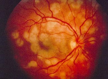
Chorioretinitis
Uveitis, Anterior
Uveomeningoencephalitic Syndrome
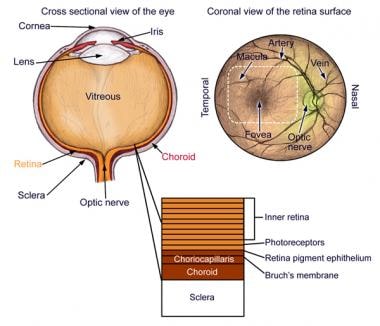
Fundus Oculi
Behcet Syndrome
High incidence of glucose intolerance in Vogt-Koyanagi-Harada disease. (1/45)
AIMS: To evaluate glucose tolerance of patients with Vogt-Koyanagi-Harada (VKH) disease before systemic corticosteroid therapy, and to assess changes brought on by treatment. METHODS: 20 VKH patients with acute bilateral panuveitis were studied. 20 healthy adults and 11 Behcet's disease patients with active uveoretinitis served as controls. A 75 g oral glucose tolerance test (OGTT) was given in the acute stage of ocular inflammation before systemic corticosteroid therapy. The OGTT was repeated in the convalescent stage of VKH disease in the patients with glucose intolerance before treatment. Insulin response was examined at the same time as the OGTT when possible. RESULTS: 55% of VKH patients (11/20) showed glucose intolerance but no apparent insulin secretion deficiency was detected. Four of seven patients in the convalescent stage showed improvement of glucose tolerance. None of the normal controls or disease controls showed glucose intolerance. CONCLUSION: A high incidence of glucose intolerance was found in the acute stage of VKH disease. However, glucose intolerance improved in most cases after systemic corticosteroid therapy. It is possible that glucose intolerance seen in VKH patients may be related to the autoimmune inflammatory process of this disease. (+info)Presumed ocular bartonellosis. (2/45)
BACKGROUND: The spectrum of diseases caused by Bartonella henselae continues to expand and ocular involvement during this infection is being diagnosed with increasing frequency. METHODS: The clinical features and visual prognosis for 13 patients with intraocular inflammatory disease and laboratory evidence of bartonellosis were investigated. There were nine patients with neuroretinitis and four with panuveitis with positive antibody titres against B henselae determined by an enzyme immunoassay (IgG exceeding 1:900 and/or IgM exceeding 1:250). RESULTS: Positive IgG levels were found for eight patients and positive IgM levels for five. Despite animal exposure of 10 patients, only two (IgG positive) cases had systemic symptoms consistent with the diagnosis of cat scratch disease. Pathological fluorescein leakage of the optic disc was observed in all affected eyes. At 6 months' follow up, 3/18 (17%) affected eyes had a visual acuity of less than 20/100, owing to optic disc atrophy and cystoid macular oedema. 12 patients (17 eyes) were treated with antibiotics; visual acuity improved two or more Snellen lines for 9/17 (53%) eyes. CONCLUSIONS: The possibility of B henselae infection should be considered in patients with neuroretinitis and panuveitis (especially in cases with associated optic nerve involvement) even in the absence of systemic symptoms typical for cat scratch disease. (+info)Molecular analysis of resolving immune responses in uveitis. (3/45)
To identify the cellular immune processes underlying intra-ocular inflammation, aqueous humour was obtained at cataract surgery from 22 patients with clinically inactive uveitis and 24 patients with age-related cataract. mRNA expression for the cytokines IL-1beta, IL-2, IL-4, IL-6, IL-10, IL-12, interferon-gamma (IFN-gamma), transforming growth factor-beta (TGF-beta); T cell subsets CD3, CD4, CD8; monocytes and macrophages (CD14); and B cells (CD19) was measured using reverse transcriptase-polymerase chain reaction (RT-PCR) and radiometric analysis. The majority of uveitis patients demonstrated a T cell-mediated inflammatory response, predominately involving a Th1-like cytokine profile with expression of IL-2 and IFN-gamma in 16/22 and 18/22 samples, respectively. These cytokines were present in only a small number of patients with age-related cataract. This Th1-like polarization was supported by an increased expression of CD8 in a number of patients. IL-1beta was expressed in only six uveitic eyes. Only four patients expressed either IL-4 or IL-10 and no patient expressed both. TGF-beta mRNA could be detected in 18/22 uveitis patients and 15/24 controls. IL-12, the paradigmatic Th1-inducing cytokine, was absent in all samples but CD14 was expressed in the majority of patients and controls. CD19 could not be detected in any sample. The cellular infiltrate in the uveitic eyes showed clear evidence of low IL-1 and absent IL-12 expression despite a Th1-like profile and high expression of macrophages. This strongly suggests that the systemic immunosuppressive therapy used prior to surgery in some patients and/or the chronicity of the uveitis had actively suppressed/switched off macrophage function, leading to resolution of T cell activity. (+info)HLA-B27 typing in the categorisation of uveitis in a HLA-B27 rich population. (4/45)
AIMS: To determine whether HLA-B27 typing helps the clinician in the diagnostic examination of uveitis in a HLA-B27 rich population and also whether the clinical picture of HLA-B27 positive unilateral acute or recurrent anterior uveitis (AAU) is distinguishable from the idiopathic negative form. METHODS: During a 3 year period 220 consecutive patients with undetermined uveitis at onset were examined in the Helsinki University Eye Clinic. HLA-B27 antigen was tested for 85% of the patients. Other laboratory or x ray examinations were performed on the basis of the anatomical classification of uveitis and the biomicroscopic features characteristic of uveitis associated systemic diseases. RESULTS: HLA-B27 antigen was found significantly more often in patients with anterior (71%) and acute/recurrent unilateral (79%) uveitis than in patients with intermediate, posterior panuveitis (7%), and chronic (7%) or bilateral (12%) forms. Of the 16 cases of HLA-B27 negative unilateral AAU, five showed biomicroscopic features representing uveitis entities. The remaining 11 cases did not differ in any respect from the cases of HLA-B27 positive unilateral AAU. CONCLUSION: HLA-B27 antigen helps the clinician in the diagnostic examination of unilateral AAU. Positive test results serve as a clue to search for spondyloarthropathies, and negative results indicate the need to look for specific uveitis entities and other systemic diseases. The occurrence of HLA-B27 positivity in conjunction with uveitis entities other than unilateral AAU is of the same level or less than in the population of Finland in general. (+info)Bartonella henselae associated uveitis and HLA-B27. (5/45)
AIM: To investigate the frequency of HLA-B27 in patients with presumed Bartonella henselae associated uveitis and to describe the clinical characteristics of HLA-B27 positive patients with uveitis and presumed ocular bartonellosis (POB). METHODS: The diagnosis of POB was considered in 19 patients with unexplained uveitis (except for the HLA-B27 association) and high positive IgG (titre >/=1:900) and/or IgM (titre >/=1:250) antibodies against B henselae. In addition to B henselae serology and HLA-B27 typing, all patients underwent an extensive standard diagnostic screening procedure for uveitis and in all cases the results were within the normal limits. The control group consisted of 25 consecutive patients with panuveitis and negative B henselae serology. RESULTS: HLA-B27 was positive in six of the 19 patients (32%) with POB in contrast to the 4% frequency of HLA-B27 in the control group (p=0.03) and 8% prevalence of HLA-B27 in the Dutch population (p=0.003). At the time of positive Bartonella serological testing five of six HLA-B27 positive patients with POB had severe posterior segment involvement with papillitis, macular oedema, and vitreitis. The duration of intraocular inflammatory activity was more than 6 months in five HLA-B27 positive patients. Although four of the six HLA-B27 positive patients had previous recurrent attacks of acute anterior uveitis, the clinical presentation at the time of positive Bartonella serology differed, as illustrated by the involvement of the posterior segment and chronic course of the ocular disease. CONCLUSIONS: The frequency of HLA-B27 in patients with uveitis and serological characteristics of acute infection with B henselae is higher than in the general Dutch population. The findings of this study also suggest a relation between infection with Bartonella species and HLA-B27. (+info)Acute panuveitis with haemorrhagic hypopyon as a presenting feature of acquired immunodeficiency syndrome (AIDS). (6/45)
Anterior uveitis is a known clinical entity in herpes zoster ophthalmicus associated with AIDS. However, reports of acute haemorrhagic hypopyon uveitis in such cases are lacking. Herein we describe a young male patient presenting with acute panuveitis with haemorrhagic hypopyon, who was found HIV positive on investigation. (+info)Immune recovery vitritis presenting as panuveitis following therapy with protease inhibitors. (7/45)
Immune reconstitution in acquired immunodeficiency syndrome (AIDS) patients on highly active anti-retroviral therapy (HAART) with cytomegalovirus (CMV) retinitis manifested as posterior segment intraocular inflammation has been reported. We report an adult HIV-positive Indian male with clinically inactive CMV retinitis who developed panuveitis with hypopyon. This was related to immune recovery mediated by combination anti-retroviral treatment, including protease inhibitors. (+info)Intravenous immunoglobulin therapy for resistant ocular Behcet's disease. (8/45)
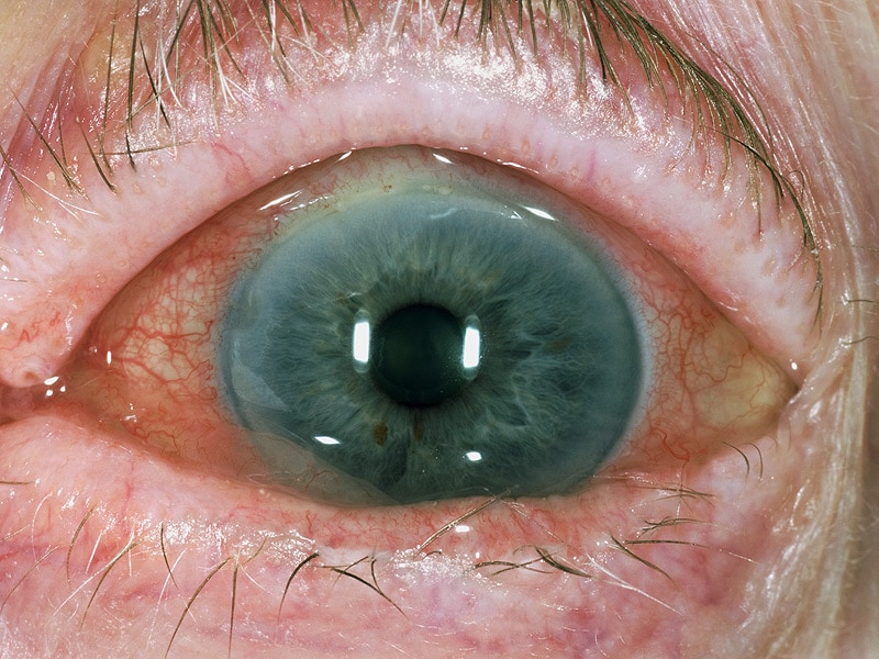
AIMS: The present report was aimed at finding out whether gammaglobulin could have a role in treating ocular Behcet's disease (BD) refractory to accepted medical therapy. METHODS: Six eyes of four patients with ocular BD refractory to steroids and cyclosporin A were treated with a course of intravenous gammaglobulin and followed up for their response to treatment. RESULTS: All six eyes of all four patients showed good response to gammaglobulin therapy. CONCLUSION: Gamma globulin may have a role in treating refractory ocular BD. A wide range of controlled studies with longer follow up is needed to substantiate this impression. (+info)Panuveitis is a medical term that refers to inflammation that affects the entire uveal tract, including the iris, ciliary body, and choroid. The uveal tract is the middle layer of the eye between the inner retina and the outer fibrous tunic (sclera). Panuveitis can also affect other parts of the eye, such as the vitreous, retina, and optic nerve.
The symptoms of panuveitis may include redness, pain, light sensitivity, blurred vision, floaters, and decreased visual acuity. The condition can be caused by various factors, including infections, autoimmune diseases, trauma, or unknown causes (idiopathic). Treatment typically involves the use of corticosteroids to reduce inflammation, as well as addressing any underlying cause if identified. If left untreated, panuveitis can lead to complications such as cataracts, glaucoma, and retinal damage, which can result in permanent vision loss.
Choroiditis is an inflammatory condition that affects the choroid, a layer of blood vessels in the eye located between the retina (the light-sensitive tissue at the back of the eye) and the sclera (the white outer coat of the eye). The choroid provides oxygen and nutrients to the outer layers of the retina.
Choroiditis is characterized by spots or patches of inflammation in the choroid, which can lead to damage and scarring of the tissue. This can result in vision loss if it affects the macula (the central part of the retina responsible for sharp, detailed vision). Symptoms of choroiditis may include blurred vision, floaters, sensitivity to light, and decreased color perception.
There are several types of choroiditis, including:
1. Multifocal choroiditis: This type is characterized by multiple, small areas of inflammation in the choroid, often accompanied by scarring. It can affect both eyes and may cause vision loss if it involves the macula.
2. Serpiginous choroiditis: This is a chronic, relapsing form of choroiditis that affects the outer layers of the retina and the choroid. It typically causes well-defined, wavy or serpentine-shaped lesions in the posterior pole (the back part) of the eye.
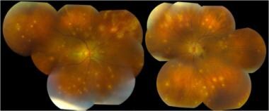
3. Birdshot chorioretinopathy: This is a rare form of choroiditis that primarily affects the peripheral retina and choroid. It is characterized by multiple, cream-colored or yellowish spots throughout the fundus (the interior surface of the eye).
4. Sympathetic ophthalmia: This is a rare condition that occurs when one eye is injured, leading to inflammation in both eyes. The choroid and other structures in the uninjured eye become inflamed due to an autoimmune response.
5. Vogt-Koyanagi-Harada (VKH) disease: This is a multisystemic autoimmune disorder that affects the eyes, skin, hair, and inner ear. In the eye, it causes choroiditis, retinal inflammation, and sometimes optic nerve swelling.
Treatment for choroiditis depends on the underlying cause and may include corticosteroids, immunosuppressive medications, or biologic agents to control inflammation. In some cases, laser therapy or surgery might be necessary to address complications such as retinal detachment or cataracts.
Uveitis is the inflammation of the uvea, the middle layer of the eye between the retina and the white of the eye (sclera). The uvea consists of the iris, ciliary body, and choroid. Uveitis can cause redness, pain, and vision loss. It can be caused by various systemic diseases, infections, or trauma. Depending on the part of the uvea that's affected, uveitis can be classified as anterior (iritis), intermediate (cyclitis), posterior (choroiditis), or pan-uveitis (affecting all layers). Treatment typically includes corticosteroids and other immunosuppressive drugs to control inflammation.
Posterior uveitis is a type of uveitis that specifically affects the back portion of the uvea, which includes the choroid (a layer of blood vessels that provides nutrients to the outer layers of the retina), the retina (the light-sensitive tissue at the back of the eye), and the optic nerve (which carries visual information from the eye to the brain).
Posterior uveitis can cause symptoms such as blurred vision, floaters, sensitivity to light, and decreased vision. It may also lead to complications such as retinal scarring, cataracts, glaucoma, and retinal detachment if left untreated. The condition can be caused by a variety of factors, including infections, autoimmune diseases, and trauma. Treatment typically involves the use of corticosteroids or other immunosuppressive medications to reduce inflammation and prevent complications.
Fluocinolone acetonide is a synthetic corticosteroid, which is a type of medication that reduces inflammation and suppresses the immune system. It is used to treat various skin conditions such as eczema, psoriasis, and dermatitis. Fluocinolone acetonide works by reducing the production of chemicals in the body that cause inflammation.
Fluocinolone acetonide is available in several forms, including creams, ointments, solutions, and tape. It is usually applied to the affected area of the skin one to three times a day, depending on the severity of the condition and the specific formulation being used.
Like all corticosteroids, fluocinolone acetonide can have side effects, particularly with long-term use or if used in large amounts. These may include thinning of the skin, easy bruising, stretch marks, increased hair growth, and acne. It is important to follow the instructions of a healthcare provider carefully when using this medication to minimize the risk of side effects.
Intermediate uveitis is a type of uveitis that affects the vitreous cavity and peripheral retina. It is characterized by the presence of inflammatory cells in the vitreous, called vitritis, and sometimes also by snowbanking or peripheral lesions in the retina. Intermediate uveitis can cause vision loss due to cystoid macular edema, epiretinal membrane formation, or complications such as glaucoma or cataract. The onset of intermediate uveitis is often insidious and the course can be chronic, with recurrent episodes of inflammation. The exact cause of intermediate uveitis is often unknown, but it can be associated with systemic diseases such as sarcoidosis, multiple sclerosis, or Lyme disease.
Retinal vasculitis is a medical condition characterized by inflammation of the blood vessels in the retina, which is the light-sensitive tissue located at the back of the eye. This condition can cause damage to the retina and may lead to vision loss if not treated promptly. The inflammation can affect both the small and large blood vessels in the retina and can occur as a result of various systemic diseases or infections, including autoimmune disorders, tuberculosis, syphilis, and toxoplasmosis. In some cases, retinal vasculitis may also be associated with uveitis, which is inflammation of the middle layer of the eye. Treatment typically involves addressing the underlying cause of the inflammation and may include corticosteroids or other immunosuppressive therapies to reduce inflammation and prevent further damage to the retina.
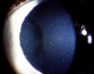
Hyphema is defined as the presence of blood in the anterior chamber of the eye, which is the space between the cornea and the iris. This condition usually results from trauma or injury to the eye, but it can also occur due to various medical conditions such as severe eye inflammation, retinal surgery, or blood disorders that affect clotting.
The blood in the anterior chamber can vary in amount, ranging from a few drops to a complete fill, which is called an "eight-ball hyphema." Hyphema can be painful and cause sensitivity to light (photophobia), blurred vision, or even loss of vision if not treated promptly.
Immediate medical attention is necessary for hyphema to prevent complications such as increased intraocular pressure, corneal blood staining, glaucoma, or cataracts. Treatment options may include bed rest, eye drops to reduce inflammation and control intraocular pressure, and sometimes surgery to remove the blood from the anterior chamber.
Anellovirus is a type of small, single-stranded DNA virus that infects humans and animals. These viruses are classified in the family Anelloviridae and include several genera such as Torque teno virus (TTV), Torque teno mini virus (TTMV), and Torque teno midi virus (TTMDV).
Anelloviruses are widespread and found in various body fluids, including blood, saliva, and stool. They are believed to be transmitted through close contact with infected individuals or through contaminated food and water. Although anelloviruses have been detected in people with various diseases, their role in human health and disease remains unclear.
In immunocompromised individuals, such as those with HIV/AIDS or organ transplant recipients, anellovirus infections can become persistent and may be associated with adverse health outcomes. However, more research is needed to fully understand the clinical significance of these viruses.
Chorioretinitis is a medical term that refers to the inflammation of the choroid and the retina, which are both important structures in the eye. The choroid is a layer of blood vessels that supplies oxygen and nutrients to the retina, while the retina is a light-sensitive tissue that converts light into electrical signals that are sent to the brain and interpreted as visual images.
Chorioretinitis can be caused by various infectious and non-infectious conditions, such as bacterial, viral, fungal, or parasitic infections, autoimmune diseases, or cancer. The symptoms of chorioretinitis may include decreased vision, floaters, blurry vision, sensitivity to light, and eye pain. Treatment for chorioretinitis depends on the underlying cause and may include antibiotics, antiviral medications, corticosteroids, or other immunosuppressive therapies. It is important to seek medical attention promptly if you experience any symptoms of chorioretinitis, as timely diagnosis and treatment can help prevent permanent vision loss.
Anterior uveitis is a medical term that refers to the inflammation of the front portion of the uvea, which is the middle layer of the eye. The uvea includes the iris (the colored part of the eye), the ciliary body (a structure behind the iris that helps focus light onto the retina), and the choroid (a layer of blood vessels that supplies oxygen and nutrients to the retina).
Anterior uveitis is characterized by inflammation of the iris and/or the ciliary body, leading to symptoms such as redness, pain, sensitivity to light, blurred vision, and a small pupil. The condition can be caused by various factors, including infections, autoimmune diseases, trauma, or unknown causes (idiopathic).
Treatment of anterior uveitis typically involves the use of topical corticosteroids to reduce inflammation and cycloplegics to relieve pain and prevent spasms of the ciliary muscle. In some cases, oral medications may be necessary to control the inflammation. Prompt treatment is important to prevent complications such as glaucoma, cataracts, or permanent vision loss.
I'm sorry for any confusion, but "Uveomeningoencephalitic Syndrome" is not a widely recognized or established medical term. It seems to be a combination of different terms from various medical conditions.
1. Uveitis refers to inflammation of the uvea, the middle layer of the eye between the retina and the white of the eye.
2. Meningoencephalitis refers to inflammation of both the meninges (the protective membranes covering the brain and spinal cord) and the brain itself (encephalitis).
If you're looking for information on a specific medical condition that you think might be related to these terms, I would recommend consulting a healthcare professional or searching for information on conditions like uveitis or meningoencephalitis.
"Fundus Oculi" is a medical term that refers to the back part of the interior of the eye, including the optic disc, macula, fovea, retinal vasculature, and peripheral retina. It is the area where light is focused and then transmitted to the brain via the optic nerve, forming visual images. Examinations of the fundus oculi are crucial for detecting various eye conditions such as diabetic retinopathy, macular degeneration, glaucoma, and other retinal diseases. The examination is typically performed using an ophthalmoscope or a specialized camera called a retinal camera.
Behçet syndrome is a rare inflammatory disease that can cause symptoms in various parts of the body. It's characterized by recurrent mouth sores (aphthous ulcers), genital sores, and inflammation of the eyes (uveitis). The condition may also cause skin lesions, joint pain and swelling, and inflammation of the digestive tract, brain, or spinal cord.
The exact cause of Behçet syndrome is not known, but it's thought to be an autoimmune disorder, in which the body's immune system mistakenly attacks its own healthy cells and tissues. The condition tends to affect men more often than women and typically develops during a person's 20s or 30s.
There is no cure for Behçet syndrome, but treatments can help manage symptoms and prevent complications. Treatment options may include medications such as corticosteroids, immunosuppressants, and biologics to reduce inflammation, as well as pain relievers and other supportive therapies.
Fluorescein angiography is a medical diagnostic procedure used in ophthalmology to examine the blood flow in the retina and choroid, which are the inner layers of the eye. This test involves injecting a fluorescent dye, Fluorescein, into a patient's arm vein. As the dye reaches the blood vessels in the eye, a specialized camera takes rapid sequences of photographs to capture the dye's circulation through the retina and choroid.
The images produced by fluorescein angiography can help doctors identify any damage to the blood vessels, leakage, or abnormal growth of new blood vessels. This information is crucial in diagnosing and managing various eye conditions such as age-related macular degeneration, diabetic retinopathy, retinal vein occlusions, and inflammatory eye diseases.
It's important to note that while fluorescein angiography is a valuable diagnostic tool, it does carry some risks, including temporary side effects like nausea, vomiting, or allergic reactions to the dye. In rare cases, severe adverse reactions can occur, so patients should discuss these potential risks with their healthcare provider before undergoing the procedure.
 Panuveitis - Wikipedia
 Panuveitis (Diffuse Uveitis): Causes, Symptoms, Diagnosis, and Treatment
 Uveitis Classification: Classification Systems, Classification Parameters, Patient Demographics
 2020-2021 BCSC Basic and Clinical Science Course™
 Humira (adalimumab): Side effects, cost, uses, and more
 Panuveitis after covishield vaccination in an undiagnosed immunocompromised patient: A rare case report from Nepal
|...
Panuveitis after covishield vaccination in an undiagnosed immunocompromised patient: A rare case report from Nepal
|... Two commonly used uveitis drugs perform similarly in NIH-funded clinical trial | National Institutes of Health (NIH)
 Abby Abelson, MD, FACR | Cleveland Clinic
 2004 - Gorgas Course | UAB
 Tattoo-associated uveitis | Eye
 Amgen Secures Approval for Two Humira Biosimilars in the EU through Duplicate MAAs | 2017-03-28 | FDANews | FDAnews
 Anterior Uveitis: Symptoms, Treatment and More
 Zika virus may persist in eyes: Disease may spread from infected eyes | ScienceDaily
 CDC - Clinical Advisory: Ocular Syphilis in the United States
 Uveitis (Ocular Inflammation) - Ophthalmology | UCLA Health
 Uveitis | What it is, causes, symptoms and treatment
 Immunomodulatory Therapy (IMT) for Ocular Inflammation - EyeWiki
 Pharmacotherapy for uveitis: current management and emerging therapy | OPTH
 Pharmacovigilance in ophthalmology in Switzerland: an analysis of the most frequently reported ocular adverse drug reactions...