Optic Nerve
Optic Disk
Photic Stimulation
Optic Neuritis
Diptera
Optic Chiasm
Visual Pathways
Distance Perception
Optic Atrophy
Rotation
Psychophysics
Nystagmus, Optokinetic
Visual Fields
Vestibule, Labyrinth
Optic Nerve Injuries
Cues
Vision, Ocular
Head Movements
Optics and Photonics
Kinesthesis
Macaca mulatta
Blood Flow Velocity
Visual Perception
Motion
Optic Lobe, Nonmammalian
Optic Neuropathy, Ischemic
Flow Cytometry
Temporal Lobe
Locomotion
Parietal Lobe
Retina
Models, Neurological
Movement
Perceptual Disorders
Optic Nerve Glioma
Optic Atrophies, Hereditary
Psychomotor Performance
Perceptual Distortion
Optic Atrophy, Hereditary, Leber
Bees
Visual Cortex
Fixation, Ocular
Vision Disparity
Neurons
Normal Distribution
Pattern Recognition, Visual
Retinal Ganglion Cells
Columbidae
Pursuit, Smooth
Optic Atrophy, Autosomal Dominant
Evoked Potentials, Visual
Adaptation, Physiological
Voltage-Sensitive Dye Imaging
Pulsatile Flow
Figural Aftereffect
Spatial Behavior
Head
Gene Flow
Functional Laterality
Contrast Sensitivity
Optic Disk Drusen
Automobile Driving
Sensory Receptor Cells
Walking
Selective attention modulates electrical responses to reversals of optic-flow direction. (1/57)
(+info)Goal seeking in honeybees: matching of optic flow snapshots? (2/57)
(+info)A Bayesian model for estimating observer translation and rotation from optic flow and extra-retinal input. (3/57)
(+info)Relative contributions of optic flow, bearing, and splay angle information to lane keeping. (4/57)
(+info)Optimal combination of form and motion cues in human heading perception. (5/57)
(+info)The effect of active selection in human path integration. (6/57)
(+info)Neural action fields for optic flow based navigation: a simulation study of the fly lobula plate network. (7/57)
(+info)Impact of visual motion adaptation on neural responses to objects and its dependence on the temporal characteristics of optic flow. (8/57)
(+info)Optic flow is not a medical term per se, but rather a term used in the field of visual perception and neuroscience. It refers to the pattern of motion of objects in the visual field that occurs as an observer moves through the environment. This pattern of motion is important for the perception of self-motion and the estimation of egocentric distance (the distance of objects in the environment relative to the observer). Optic flow has been studied in relation to various clinical populations, such as individuals with vestibular disorders or visual impairments, who may have difficulty processing optic flow information.
The optic nerve, also known as the second cranial nerve, is the nerve that transmits visual information from the retina to the brain. It is composed of approximately one million nerve fibers that carry signals related to vision, such as light intensity and color, from the eye's photoreceptor cells (rods and cones) to the visual cortex in the brain. The optic nerve is responsible for carrying this visual information so that it can be processed and interpreted by the brain, allowing us to see and perceive our surroundings. Damage to the optic nerve can result in vision loss or impairment.
Motion perception is the ability to interpret and understand the movement of objects in our environment. It is a complex process that involves multiple areas of the brain and the visual system. In medical terms, motion perception refers to the specific function of the visual system to detect and analyze the movement of visual stimuli. This allows us to perceive and respond to moving objects in our environment, which is crucial for activities such as driving, sports, and even maintaining balance. Disorders in motion perception can lead to conditions like motion sickness or difficulty with depth perception.
The optic disk, also known as the optic nerve head, is the point where the optic nerve fibers exit the eye and transmit visual information to the brain. It appears as a pale, circular area in the back of the eye, near the center of the retina. The optic disk has no photoreceptor cells (rods and cones), so it is insensitive to light. It is an important structure to observe during eye examinations because changes in its appearance can indicate various ocular diseases or conditions, such as glaucoma, optic neuritis, or papilledema.
Photic stimulation is a medical term that refers to the exposure of the eyes to light, specifically repetitive pulses of light, which is used as a method in various research and clinical settings. In neuroscience, it's often used in studies related to vision, circadian rhythms, and brain function.
In a clinical context, photic stimulation is sometimes used in the diagnosis of certain medical conditions such as seizure disorders (like epilepsy). By observing the response of the brain to this light stimulus, doctors can gain valuable insights into the functioning of the brain and the presence of any neurological disorders.
However, it's important to note that photic stimulation should be conducted under the supervision of a trained healthcare professional, as improper use can potentially trigger seizures in individuals who are susceptible to them.
Optic neuritis is a medical condition characterized by inflammation and damage to the optic nerve, which transmits visual information from the eye to the brain. This condition can result in various symptoms such as vision loss, pain with eye movement, color vision disturbances, and pupillary abnormalities. Optic neuritis may occur in isolation or be associated with other underlying medical conditions, including multiple sclerosis, neuromyelitis optica, and autoimmune disorders. The diagnosis typically involves a comprehensive eye examination, including visual acuity testing, dilated funduscopic examination, and possibly imaging studies like MRI to evaluate the optic nerve and brain. Treatment options may include corticosteroids or other immunomodulatory therapies to reduce inflammation and prevent further damage to the optic nerve.
Diptera is an order of insects that includes flies, mosquitoes, and gnats. The name "Diptera" comes from the Greek words "di," meaning two, and "pteron," meaning wing. This refers to the fact that all members of this order have a single pair of functional wings for flying, while the other pair is reduced to small knob-like structures called halteres, which help with balance and maneuverability during flight.
Some common examples of Diptera include houseflies, fruit flies, horseflies, tsetse flies, and midges. Many species in this order are important pollinators, while others can be significant pests or disease vectors. The study of Diptera is called dipterology.
"Animal Flight" is not a medical term per se, but it is a concept that is studied in the field of comparative physiology and biomechanics, which are disciplines related to medicine. Animal flight refers to the ability of certain animal species to move through the air by flapping their wings or other appendages. This mode of locomotion is most commonly associated with birds, bats, and insects, but some mammals such as flying squirrels and sugar gliders are also capable of gliding through the air.
The study of animal flight involves understanding the biomechanics of how animals generate lift and propulsion, as well as the physiological adaptations that allow them to sustain flight. For example, birds have lightweight skeletons and powerful chest muscles that enable them to flap their wings rapidly and generate lift. Bats, on the other hand, use a more complex system of membranes and joints to manipulate their wings and achieve maneuverability in flight.
Understanding animal flight has important implications for the design of aircraft and other engineering systems, as well as for our broader understanding of how animals have evolved to adapt to their environments.
The optic chiasm is a structure in the brain where the optic nerves from each eye meet and cross. This allows for the integration of visual information from both eyes into the brain's visual cortex, creating a single, combined image of the visual world. The optic chiasm plays an important role in the processing of visual information and helps to facilitate depth perception and other complex visual tasks. Damage to the optic chiasm can result in various visual field deficits, such as bitemporal hemianopsia, where there is a loss of vision in the outer halves (temporal fields) of both eyes' visual fields.
Visual pathways, also known as the visual system or the optic pathway, refer to the series of specialized neurons in the nervous system that transmit visual information from the eyes to the brain. This complex network includes the retina, optic nerve, optic chiasma, optic tract, lateral geniculate nucleus, pulvinar, and the primary and secondary visual cortices located in the occipital lobe of the brain.
The process begins when light enters the eye and strikes the photoreceptor cells (rods and cones) in the retina, converting the light energy into electrical signals. These signals are then transmitted to bipolar cells and subsequently to ganglion cells, whose axons form the optic nerve. The fibers from each eye's nasal hemiretina cross at the optic chiasma, while those from the temporal hemiretina continue without crossing. This results in the formation of the optic tract, which carries visual information from both eyes to the opposite side of the brain.
The majority of fibers in the optic tract synapse with neurons in the lateral geniculate nucleus (LGN), a part of the thalamus. The LGN sends this information to the primary visual cortex, also known as V1 or Brodmann area 17, located in the occipital lobe. Here, simple features like lines and edges are initially processed. Further processing occurs in secondary (V2) and tertiary (V3-V5) visual cortices, where more complex features such as shape, motion, and depth are analyzed. Ultimately, this information is integrated to form our perception of the visual world.
Distance perception refers to the ability to accurately judge the distance or depth of an object in relation to oneself or other objects. It is a complex process that involves both visual and non-visual cues, such as perspective, size, texture, motion parallax, binocular disparity, and familiarity with the object or scene.
In the visual system, distance perception is primarily mediated by the convergence of the two eyes on an object, which provides information about its depth and location in three-dimensional space. The brain then integrates this information with other sensory inputs and prior knowledge to create a coherent perception of the environment.
Disorders of distance perception can result from various conditions that affect the visual system, such as amblyopia, strabismus, or traumatic brain injury. These disorders can cause difficulties in tasks that require accurate depth perception, such as driving, sports, or manual work.
Optic atrophy is a medical term that refers to the degeneration and shrinkage (atrophy) of the optic nerve, which transmits visual information from the eye to the brain. This condition can result in various vision abnormalities, including loss of visual acuity, color vision deficiencies, and peripheral vision loss.
Optic atrophy can occur due to a variety of causes, such as:
* Traumatic injuries to the eye or optic nerve
* Glaucoma
* Optic neuritis (inflammation of the optic nerve)
* Ischemic optic neuropathy (reduced blood flow to the optic nerve)
* Compression or swelling of the optic nerve
* Hereditary or congenital conditions affecting the optic nerve
* Toxins and certain medications that can damage the optic nerve.
The diagnosis of optic atrophy typically involves a comprehensive eye examination, including visual acuity testing, refraction assessment, slit-lamp examination, and dilated funduscopic examination to evaluate the health of the optic nerve. In some cases, additional diagnostic tests such as visual field testing, optical coherence tomography (OCT), or magnetic resonance imaging (MRI) may be necessary to confirm the diagnosis and determine the underlying cause.
There is no specific treatment for optic atrophy, but addressing the underlying cause can help prevent further damage to the optic nerve. In some cases, vision rehabilitation may be recommended to help patients adapt to their visual impairment.
In the context of medicine, particularly in anatomy and physiology, "rotation" refers to the movement of a body part around its own axis or the long axis of another structure. This type of motion is three-dimensional and can occur in various planes. A common example of rotation is the movement of the forearm bones (radius and ulna) around each other during pronation and supination, which allows the hand to be turned palm up or down. Another example is the rotation of the head during mastication (chewing), where the mandible moves in a circular motion around the temporomandibular joint.
Psychophysics is not a medical term per se, but rather a subfield of psychology and neuroscience that studies the relationship between physical stimuli and the sensations and perceptions they produce. It involves the quantitative investigation of psychological functions, such as how brightness or loudness is perceived relative to the physical intensity of light or sound.
In medical contexts, psychophysical methods may be used in research or clinical settings to understand how patients with neurological conditions or sensory impairments perceive and respond to different stimuli. This information can inform diagnostic assessments, treatment planning, and rehabilitation strategies.
Optokinetic nystagmus (OKN) is a type of involuntary eye movement that occurs in response to large moving visual patterns. It serves as a mechanism for stabilizing the image on the retina during head movement and helps in maintaining visual fixation.
In OKN, there are two phases of eye movement: a slow phase where the eyes follow or track the moving pattern, and a fast phase where the eyes quickly reset to the starting position. This results in a back-and-forth or "to-and-fro" motion of the eyes.
Optokinetic nystagmus can be elicited by observing a large moving object or a series of alternating visual stimuli, such as stripes on a rotating drum. It is often used in clinical settings to assess various aspects of the visual system, including oculomotor function and visual acuity.
Abnormalities in OKN can indicate problems with the vestibular system, brainstem, or cerebellum, and may be associated with conditions such as brain injury, multiple sclerosis, or cerebral palsy.
In a medical context, "orientation" typically refers to an individual's awareness and understanding of their personal identity, place, time, and situation. It is a critical component of cognitive functioning and mental status. Healthcare professionals often assess a person's orientation during clinical evaluations, using tests that inquire about their name, location, the current date, and the circumstances of their hospitalization or visit.
There are different levels of orientation:
1. Person (or self): The individual knows their own identity, including their name, age, and other personal details.
2. Place: The individual is aware of where they are, such as the name of the city, hospital, or healthcare facility.
3. Time: The individual can accurately state the current date, day of the week, month, and year.
4. Situation or event: The individual understands why they are in the healthcare setting, what happened leading to their hospitalization or visit, and the nature of any treatments or procedures they are undergoing.
Impairments in orientation can be indicative of various neurological or psychiatric conditions, such as delirium, dementia, or substance intoxication or withdrawal. It is essential for healthcare providers to monitor and address orientation issues to ensure appropriate diagnosis, treatment, and patient safety.
Visual fields refer to the total area in which objects can be seen while keeping the eyes focused on a central point. It is the entire area that can be observed using peripheral (side) vision while the eye gazes at a fixed point. A visual field test is used to detect blind spots or gaps (scotomas) in a person's vision, which could indicate various medical conditions such as glaucoma, retinal damage, optic nerve disease, brain tumors, or strokes. The test measures both the central and peripheral vision and maps the entire area that can be seen when focusing on a single point.
The vestibular system is a part of the inner ear that contributes to our sense of balance and spatial orientation. It is made up of two main components: the vestibule and the labyrinth.
The vestibule is a bony chamber in the inner ear that contains two important structures called the utricle and saccule. These structures contain hair cells and fluid-filled sacs that help detect changes in head position and movement, allowing us to maintain our balance and orientation in space.
The labyrinth, on the other hand, is a more complex structure that includes the vestibule as well as three semicircular canals. These canals are also filled with fluid and contain hair cells that detect rotational movements of the head. Together, the vestibule and labyrinth work together to provide us with information about our body's position and movement in space.
Overall, the vestibular system plays a crucial role in maintaining our balance, coordinating our movements, and helping us navigate through our environment.
Optic nerve injuries refer to damages or trauma inflicted on the optic nerve, which is a crucial component of the visual system. The optic nerve transmits visual information from the retina to the brain, enabling us to see. Injuries to the optic nerve can result in various visual impairments, including partial or complete vision loss, decreased visual acuity, changes in color perception, and reduced field of view.
These injuries may occur due to several reasons, such as:
1. Direct trauma to the eye or head
2. Increased pressure inside the eye (glaucoma)
3. Optic neuritis, an inflammation of the optic nerve
4. Ischemia, or insufficient blood supply to the optic nerve
5. Compression from tumors or other space-occupying lesions
6. Intrinsic degenerative conditions affecting the optic nerve
7. Toxic exposure to certain chemicals or medications
Optic nerve injuries are diagnosed through a comprehensive eye examination, including visual acuity testing, slit-lamp examination, dilated fundus exam, and additional diagnostic tests like optical coherence tomography (OCT) and visual field testing. Treatment options vary depending on the cause and severity of the injury but may include medications, surgery, or vision rehabilitation.
Regional blood flow (RBF) refers to the rate at which blood flows through a specific region or organ in the body, typically expressed in milliliters per minute per 100 grams of tissue (ml/min/100g). It is an essential physiological parameter that reflects the delivery of oxygen and nutrients to tissues while removing waste products. RBF can be affected by various factors such as metabolic demands, neural regulation, hormonal influences, and changes in blood pressure or vascular resistance. Measuring RBF is crucial for understanding organ function, diagnosing diseases, and evaluating the effectiveness of treatments.
Eye movements, also known as ocular motility, refer to the voluntary or involuntary motion of the eyes that allows for visual exploration of our environment. There are several types of eye movements, including:
1. Saccades: rapid, ballistic movements that quickly shift the gaze from one point to another.
2. Pursuits: smooth, slow movements that allow the eyes to follow a moving object.
3. Vergences: coordinated movements of both eyes in opposite directions, usually in response to a three-dimensional stimulus.
4. Vestibulo-ocular reflex (VOR): automatic eye movements that help stabilize the gaze during head movement.
5. Optokinetic nystagmus (OKN): rhythmic eye movements that occur in response to large moving visual patterns, such as when looking out of a moving vehicle.
Abnormalities in eye movements can indicate neurological or ophthalmological disorders and are often assessed during clinical examinations.
In the context of medicine, "cues" generally refer to specific pieces of information or signals that can help healthcare professionals recognize and respond to a particular situation or condition. These cues can come in various forms, such as:
1. Physical examination findings: For example, a patient's abnormal heart rate or blood pressure reading during a physical exam may serve as a cue for the healthcare professional to investigate further.
2. Patient symptoms: A patient reporting chest pain, shortness of breath, or other concerning symptoms can act as a cue for a healthcare provider to consider potential diagnoses and develop an appropriate treatment plan.
3. Laboratory test results: Abnormal findings on laboratory tests, such as elevated blood glucose levels or abnormal liver function tests, may serve as cues for further evaluation and diagnosis.
4. Medical history information: A patient's medical history can provide valuable cues for healthcare professionals when assessing their current health status. For example, a history of smoking may increase the suspicion for chronic obstructive pulmonary disease (COPD) in a patient presenting with respiratory symptoms.
5. Behavioral or environmental cues: In some cases, behavioral or environmental factors can serve as cues for healthcare professionals to consider potential health risks. For instance, exposure to secondhand smoke or living in an area with high air pollution levels may increase the risk of developing respiratory conditions.
Overall, "cues" in a medical context are essential pieces of information that help healthcare professionals make informed decisions about patient care and treatment.
Ocular vision refers to the ability to process and interpret visual information that is received by the eyes. This includes the ability to see clearly and make sense of the shapes, colors, and movements of objects in the environment. The ocular system, which includes the eye and related structures such as the optic nerve and visual cortex of the brain, works together to enable vision.
There are several components of ocular vision, including:
* Visual acuity: the clarity or sharpness of vision
* Field of vision: the extent of the visual world that is visible at any given moment
* Color vision: the ability to distinguish different colors
* Depth perception: the ability to judge the distance of objects in three-dimensional space
* Contrast sensitivity: the ability to distinguish an object from its background based on differences in contrast
Disorders of ocular vision can include refractive errors such as nearsightedness or farsightedness, as well as more serious conditions such as cataracts, glaucoma, and macular degeneration. These conditions can affect one or more aspects of ocular vision and may require medical treatment to prevent further vision loss.
Depth perception is the ability to accurately judge the distance or separation of an object in three-dimensional space. It is a complex visual process that allows us to perceive the world in three dimensions and to understand the spatial relationships between objects.
Depth perception is achieved through a combination of monocular cues, which are visual cues that can be perceived with one eye, and binocular cues, which require input from both eyes. Monocular cues include perspective (the relative size of objects), texture gradients (finer details become smaller as distance increases), and atmospheric perspective (colors become less saturated and lighter in value as distance increases). Binocular cues include convergence (the degree to which the eyes must turn inward to focus on an object) and retinal disparity (the slight difference in the images projected onto the two retinas due to the slightly different positions of the eyes).
Deficits in depth perception can occur due to a variety of factors, including eye disorders, brain injuries, or developmental delays. These deficits can result in difficulties with tasks such as driving, sports, or navigating complex environments. Treatment for depth perception deficits may include vision therapy, corrective lenses, or surgery.
Head movements refer to the voluntary or involuntary motion of the head in various directions. These movements can occur in different planes, including flexion (moving the head forward), extension (moving the head backward), rotation (turning the head to the side), and lateral bending (leaning the head to one side).
Head movements can be a result of normal physiological processes, such as when nodding in agreement or shaking the head to indicate disagreement. They can also be caused by neurological conditions, such as abnormal head movements in patients with Parkinson's disease or cerebellar disorders. Additionally, head movements may occur in response to sensory stimuli, such as turning the head toward a sound.
In a medical context, an examination of head movements can provide important clues about a person's neurological function and help diagnose various conditions affecting the brain and nervous system.
Space perception, in the context of neuroscience and psychology, refers to the ability to perceive and understand the spatial arrangement of objects and their relationship to oneself. It involves integrating various sensory inputs such as visual, auditory, tactile, and proprioceptive information to create a coherent three-dimensional representation of our environment.
This cognitive process enables us to judge distances, sizes, shapes, and movements of objects around us. It also helps us navigate through space, reach for objects, avoid obstacles, and maintain balance. Disorders in space perception can lead to difficulties in performing everyday activities and may be associated with neurological conditions such as stroke, brain injury, or neurodevelopmental disorders like autism.
I'm sorry for any confusion, but "Optics and Photonics" is a broad field that encompasses various scientific and engineering disciplines. It doesn't have a specific medical definition, as it's not a term typically used in a medical context.
However, I can provide a general definition:
Optics is the study of light and its interactions with matter. This includes how light is produced, controlled, transmitted, and detected. It involves phenomena such as reflection, refraction, diffraction, and interference.
Photonics, on the other hand, is a branch of optics that deals with the generation, detection, and manipulation of individual photons, the basic units of light. Photonics is often applied to technologies such as lasers, fiber optics, and optical communications.
In a medical context, these fields might be used in various diagnostic and therapeutic applications, such as endoscopes, ophthalmic devices, laser surgery, and imaging technologies like MRI and CT scans. But the terms "Optics" and "Photonics" themselves are not medical conditions or treatments.
Kinesthesia, also known as proprioception, refers to the perception or awareness of the position and movement of the body parts in space. It is a type of sensory information that comes from receptors located in muscles, tendons, ligaments, and joints, which detect changes in tension, length, and pressure of these tissues during movement. This information is then sent to the brain, where it is integrated with visual and vestibular (inner ear) inputs to create a sense of body position and movement.
Kinesthesia allows us to perform complex movements and maintain balance without having to consciously think about each movement. It helps us to coordinate our movements, adjust our posture, and navigate through our environment with ease. Deficits in kinesthetic perception can lead to difficulties with motor coordination, balance, and mobility.
"Macaca mulatta" is the scientific name for the Rhesus macaque, a species of monkey that is native to South, Central, and Southeast Asia. They are often used in biomedical research due to their genetic similarity to humans.
Blood flow velocity is the speed at which blood travels through a specific part of the vascular system. It is typically measured in units of distance per time, such as centimeters per second (cm/s) or meters per second (m/s). Blood flow velocity can be affected by various factors, including cardiac output, vessel diameter, and viscosity of the blood. Measuring blood flow velocity is important in diagnosing and monitoring various medical conditions, such as heart disease, stroke, and peripheral vascular disease.
Visual perception refers to the ability to interpret and organize information that comes from our eyes to recognize and understand what we are seeing. It involves several cognitive processes such as pattern recognition, size estimation, movement detection, and depth perception. Visual perception allows us to identify objects, navigate through space, and interact with our environment. Deficits in visual perception can lead to learning difficulties and disabilities.
In the context of medical terminology, "motion" generally refers to the act or process of moving or changing position. It can also refer to the range of movement of a body part or joint. However, there is no single specific medical definition for the term "motion." The meaning may vary depending on the context in which it is used.
The optic lobe in non-mammals refers to a specific region of the brain that is responsible for processing visual information. It is a part of the protocerebrum in the insect brain and is analogous to the mammalian visual cortex. The optic lobes receive input directly from the eyes via the optic nerves and are involved in the interpretation and integration of visual stimuli, enabling non-mammals to perceive and respond to their environment. In some invertebrates, like insects, the optic lobe is further divided into subregions, including the lamina, medulla, and lobula, each with distinct functions in visual processing.
Ischemic optic neuropathy (ION) is a medical condition that refers to the damage or death of the optic nerve due to insufficient blood supply. The optic nerve is responsible for transmitting visual information from the eye to the brain.
In ION, the blood vessels that supply the optic nerve become blocked or narrowed, leading to decreased blood flow and oxygen delivery to the nerve fibers. This results in inflammation, swelling, and ultimately, damage to the optic nerve. The damage can cause sudden, painless vision loss, often noticed upon waking up in the morning.
There are two types of ION: anterior ischemic optic neuropathy (AION) and posterior ischemic optic neuropathy (PION). AION affects the front part of the optic nerve, while PION affects the back part of the nerve. AION is further classified into arteritic and non-arteritic types, depending on whether it is caused by giant cell arteritis or not.
Risk factors for ION include age (most commonly occurring in people over 50), hypertension, diabetes, smoking, sleep apnea, and other cardiovascular diseases. Treatment options depend on the type and cause of ION and may include controlling underlying medical conditions, administering corticosteroids, or undergoing surgical procedures to improve blood flow.
Flow cytometry is a medical and research technique used to measure physical and chemical characteristics of cells or particles, one cell at a time, as they flow in a fluid stream through a beam of light. The properties measured include:
* Cell size (light scatter)
* Cell internal complexity (granularity, also light scatter)
* Presence or absence of specific proteins or other molecules on the cell surface or inside the cell (using fluorescent antibodies or other fluorescent probes)
The technique is widely used in cell counting, cell sorting, protein engineering, biomarker discovery and monitoring disease progression, particularly in hematology, immunology, and cancer research.
Optical illusions are visual phenomena that occur when the brain perceives an image or scene differently from the actual physical properties of that image or scene. They often result from the brain's attempt to interpret and make sense of ambiguous, contradictory, or incomplete information provided by the eyes. This can lead to visually perceived images that are different from the objective reality. Optical illusions can be categorized into different types such as literal illusions, physiological illusions, and cognitive illusions, based on the nature of the illusion and the underlying cause.
The temporal lobe is one of the four main lobes of the cerebral cortex in the brain, located on each side of the head roughly level with the ears. It plays a major role in auditory processing, memory, and emotion. The temporal lobe contains several key structures including the primary auditory cortex, which is responsible for analyzing sounds, and the hippocampus, which is crucial for forming new memories. Damage to the temporal lobe can result in various neurological symptoms such as hearing loss, memory impairment, and changes in emotional behavior.
Locomotion, in a medical context, refers to the ability to move independently and change location. It involves the coordinated movement of the muscles, bones, and nervous system that enables an individual to move from one place to another. This can include walking, running, jumping, or using assistive devices such as wheelchairs or crutches. Locomotion is a fundamental aspect of human mobility and is often assessed in medical evaluations to determine overall health and functioning.
The parietal lobe is a region of the brain that is located in the posterior part of the cerebral cortex, covering the upper and rear portions of the brain. It is involved in processing sensory information from the body, such as touch, temperature, and pain, as well as spatial awareness and perception, visual-spatial cognition, and the integration of different senses.
The parietal lobe can be divided into several functional areas, including the primary somatosensory cortex (which receives tactile information from the body), the secondary somatosensory cortex (which processes more complex tactile information), and the posterior parietal cortex (which is involved in spatial attention, perception, and motor planning).
Damage to the parietal lobe can result in various neurological symptoms, such as neglect of one side of the body, difficulty with spatial orientation, problems with hand-eye coordination, and impaired mathematical and language abilities.
The retina is the innermost, light-sensitive layer of tissue in the eye of many vertebrates and some cephalopods. It receives light that has been focused by the cornea and lens, converts it into neural signals, and sends these to the brain via the optic nerve. The retina contains several types of photoreceptor cells including rods (which handle vision in low light) and cones (which are active in bright light and are capable of color vision).
In medical terms, any pathological changes or diseases affecting the retinal structure and function can lead to visual impairment or blindness. Examples include age-related macular degeneration, diabetic retinopathy, retinal detachment, and retinitis pigmentosa among others.
Neurological models are simplified representations or simulations of various aspects of the nervous system, including its structure, function, and processes. These models can be theoretical, computational, or physical and are used to understand, explain, and predict neurological phenomena. They may focus on specific neurological diseases, disorders, or functions, such as memory, learning, or movement. The goal of these models is to provide insights into the complex workings of the nervous system that cannot be easily observed or understood through direct examination alone.
In the context of medicine and healthcare, "movement" refers to the act or process of changing physical location or position. It involves the contraction and relaxation of muscles, which allows for the joints to move and the body to be in motion. Movement can also refer to the ability of a patient to move a specific body part or limb, which is assessed during physical examinations. Additionally, "movement" can describe the progression or spread of a disease within the body.
Perceptual disorders are conditions that affect the way a person perceives or interprets sensory information from their environment. These disorders can involve any of the senses, including sight, sound, touch, taste, and smell. They can cause a person to have difficulty recognizing, interpreting, or responding appropriately to sensory stimuli.
Perceptual disorders can result from damage to the brain or nervous system, such as from a head injury, stroke, or degenerative neurological condition. They can also be caused by certain mental health conditions, such as schizophrenia or severe depression.
Symptoms of perceptual disorders may include:
* Misinterpretations of sensory information, such as seeing things that are not there or hearing voices that are not present
* Difficulty recognizing familiar objects or people
* Problems with depth perception or spatial awareness
* Difficulty judging the size, shape, or distance of objects
* Trouble distinguishing between similar sounds or colors
* Impaired sense of smell or taste
Perceptual disorders can have a significant impact on a person's daily life and functioning. Treatment may involve medication, therapy, or rehabilitation to help the person better cope with their symptoms and improve their ability to interact with their environment.
An Optic Nerve Glioma is a type of brain tumor that arises from the glial cells (supportive tissue) within the optic nerve. It is most commonly seen in children, particularly those with neurofibromatosis type 1 (NF1). These tumors are typically slow-growing and may not cause any symptoms, especially if they are small. However, as they grow larger, they can put pressure on the optic nerve, leading to vision loss or other visual disturbances. In some cases, these tumors can also affect nearby structures in the brain, causing additional neurological symptoms. Treatment options may include observation, chemotherapy, radiation therapy, or surgery, depending on the size and location of the tumor, as well as the patient's age and overall health.
Hereditary optic atrophies (HOAs) are a group of genetic disorders that cause degeneration of the optic nerve, leading to vision loss. The optic nerve is responsible for transmitting visual information from the eye to the brain. In HOAs, this nerve degenerates over time, resulting in decreased visual acuity, color vision deficits, and sometimes visual field defects.
There are several types of HOAs, including dominant optic atrophy (DOA), Leber hereditary optic neuropathy (LHON), autosomal recessive optic atrophy (AROA), and Wolfram syndrome. Each type has a different inheritance pattern and is caused by mutations in different genes.
DOA is the most common form of HOA and is characterized by progressive vision loss that typically begins in childhood or early adulthood. It is inherited in an autosomal dominant manner, meaning that a child has a 50% chance of inheriting the disease-causing mutation from an affected parent.
LHON is a mitochondrial disorder that primarily affects males and is characterized by sudden, severe vision loss that typically occurs in young adulthood. It is caused by mutations in the mitochondrial DNA and is inherited maternally.
AROA is a rare form of HOA that is inherited in an autosomal recessive manner, meaning that both copies of the gene must be mutated to cause the disease. It typically presents in infancy or early childhood with progressive vision loss.
Wolfram syndrome is a rare genetic disorder that affects multiple organs, including the eyes, ears, and endocrine system. It is characterized by diabetes insipidus, diabetes mellitus, optic atrophy, and hearing loss. It is inherited in an autosomal recessive manner.
There is currently no cure for HOAs, but treatments such as low-vision aids and rehabilitation may help to manage the symptoms. Research is ongoing to develop new therapies for these disorders.
Psychomotor performance refers to the integration and coordination of mental processes (cognitive functions) with physical movements. It involves the ability to perform complex tasks that require both cognitive skills, such as thinking, remembering, and perceiving, and motor skills, such as gross and fine motor movements. Examples of psychomotor performances include driving a car, playing a musical instrument, or performing surgical procedures.
In a medical context, psychomotor performance is often used to assess an individual's ability to perform activities of daily living (ADLs) and instrumental activities of daily living (IADLs), such as bathing, dressing, cooking, cleaning, and managing medications. Deficits in psychomotor performance can be a sign of neurological or psychiatric disorders, such as dementia, Parkinson's disease, or depression.
Assessment of psychomotor performance may involve tests that measure reaction time, coordination, speed, precision, and accuracy of movements, as well as cognitive functions such as attention, memory, and problem-solving skills. These assessments can help healthcare professionals develop appropriate treatment plans and monitor the progression of diseases or the effectiveness of interventions.
Perceptual distortion is not explicitly defined within the realm of medicine, but it does fall under the broader category of cognitive impairments and abnormalities. It generally refers to the incorrect interpretation or misrepresentation of sensory information by the brain. This can result in various experiences such as hallucinations, illusions, or distorted perceptions of reality. Perceptual distortions are often associated with certain medical conditions like mental disorders (e.g., schizophrenia, bipolar disorder), neurological disorders (e.g., migraines, epilepsy), and substance use disorders.
Hereditary Optic Atrophy, Leber type (LOA) is a mitochondrial DNA-associated inherited condition that primarily affects the optic nerve and leads to vision loss. It is characterized by the degeneration of retinal ganglion cells and their axons, which make up the optic nerve. This results in bilateral, painless, and progressive visual deterioration, typically beginning in young adulthood (14-35 years).
Leber's hereditary optic atrophy is caused by mutations in the mitochondrial DNA (mtDNA) gene MT-ND4 or MT-ND6. The condition follows a maternal pattern of inheritance, meaning that it is passed down through the mother's lineage.
The onset of LOA usually occurs in one eye first, followed by the second eye within weeks to months. Central vision is initially affected, leading to blurriness and loss of visual acuity. Color vision may also be impaired. The progression of the condition generally stabilizes after a few months, but complete recovery of vision is unlikely.
Currently, there is no cure for Leber's hereditary optic atrophy. Treatment focuses on managing symptoms and providing visual rehabilitation to help affected individuals adapt to their visual impairment.
Binocular vision refers to the ability to use both eyes together to create a single, three-dimensional image of our surroundings. This is achieved through a process called binocular fusion, where the images from each eye are aligned and combined in the brain to form a unified perception.
The term "binocular vision" specifically refers to the way that our visual system integrates information from both eyes to create depth perception and enhance visual clarity. When we view an object with both eyes, they focus on the same point in space and send slightly different images to the brain due to their slightly different positions. The brain then combines these images to create a single, three-dimensional image that allows us to perceive depth and distance.
Binocular vision is important for many everyday activities, such as driving, reading, and playing sports. Disorders of binocular vision can lead to symptoms such as double vision, eye strain, and difficulty with depth perception.
Monocular vision refers to the ability to see and process visual information using only one eye. It is the type of vision that an individual has when they are using only one eye to look at something, while the other eye may be covered or not functioning. This can be contrasted with binocular vision, which involves the use of both eyes working together to provide depth perception and a single, combined visual field.
Monocular vision is important for tasks that only require the use of one eye, such as when looking through a microscope or using a telescope. However, it does not provide the same level of depth perception and spatial awareness as binocular vision. In some cases, individuals may have reduced visual acuity or other visual impairments in one eye, leading to limited monocular vision in that eye. It is important for individuals with monocular vision to have regular eye exams to monitor their eye health and ensure that any visual impairments are detected and treated promptly.
"Bees" are not a medical term, as they refer to various flying insects belonging to the Apidae family in the Apoidea superfamily. They are known for their role in pollination and honey production. If you're looking for medical definitions or information, please provide relevant terms.
The visual cortex is the part of the brain that processes visual information. It is located in the occipital lobe, which is at the back of the brain. The visual cortex is responsible for receiving and interpreting signals from the retina, which are then transmitted through the optic nerve and optic tract.
The visual cortex contains several areas that are involved in different aspects of visual processing, such as identifying shapes, colors, and movements. These areas work together to help us recognize and understand what we see. Damage to the visual cortex can result in various visual impairments, such as blindness or difficulty with visual perception.
Ocular fixation is a term used in ophthalmology and optometry to refer to the ability of the eyes to maintain steady gaze or visual focus on an object. It involves the coordinated movement of the extraocular muscles that control eye movements, allowing for clear and stable vision.
In medical terminology, fixation specifically refers to the state in which the eyes are aligned and focused on a single point in space. This is important for maintaining visual perception and preventing blurring or double vision. Ocular fixation can be affected by various factors such as muscle weakness, nerve damage, or visual processing disorders.
Assessment of ocular fixation is often used in eye examinations to evaluate visual acuity, eye alignment, and muscle function. Abnormalities in fixation may indicate the presence of underlying eye conditions or developmental delays that require further investigation and treatment.
Vision disparity, also known as binocular vision disparity, refers to the difference in the image that is perceived by each eye. This can occur due to a variety of reasons such as misalignment of the eyes (strabismus), unequal refractive power in each eye (anisometropia), or abnormalities in the shape of the eye (astigmatism).
When there is a significant difference in the image that is perceived by each eye, the brain may have difficulty combining the two images into a single, three-dimensional perception. This can result in visual symptoms such as double vision (diplopia), eyestrain, headaches, and difficulty with depth perception.
Vision disparity can be detected through a comprehensive eye examination and may be treated with corrective lenses, prism lenses, vision therapy, or surgery, depending on the underlying cause and severity of the condition.
Sensory thresholds are the minimum levels of stimulation that are required to produce a sensation in an individual, as determined through psychophysical testing. These tests measure the point at which a person can just barely detect the presence of a stimulus, such as a sound, light, touch, or smell.
There are two types of sensory thresholds: absolute and difference. Absolute threshold is the minimum level of intensity required to detect a stimulus 50% of the time. Difference threshold, also known as just noticeable difference (JND), is the smallest change in intensity that can be detected between two stimuli.
Sensory thresholds can vary between individuals and are influenced by factors such as age, attention, motivation, and expectations. They are often used in clinical settings to assess sensory function and diagnose conditions such as hearing or vision loss.
Neurons, also known as nerve cells or neurocytes, are specialized cells that constitute the basic unit of the nervous system. They are responsible for receiving, processing, and transmitting information and signals within the body. Neurons have three main parts: the dendrites, the cell body (soma), and the axon. The dendrites receive signals from other neurons or sensory receptors, while the axon transmits these signals to other neurons, muscles, or glands. The junction between two neurons is called a synapse, where neurotransmitters are released to transmit the signal across the gap (synaptic cleft) to the next neuron. Neurons vary in size, shape, and structure depending on their function and location within the nervous system.
To the best of my knowledge, "Normal Distribution" is not a term that has a specific medical definition. It is a statistical concept that describes a distribution of data points in which the majority of the data falls around a central value, with fewer and fewer data points appearing as you move further away from the center in either direction. This type of distribution is also known as a "bell curve" because of its characteristic shape.
In medical research, normal distribution may be used to describe the distribution of various types of data, such as the results of laboratory tests or patient outcomes. For example, if a large number of people are given a particular laboratory test, their test results might form a normal distribution, with most people having results close to the average and fewer people having results that are much higher or lower than the average.
It's worth noting that in some cases, data may not follow a normal distribution, and other types of statistical analyses may be needed to accurately describe and analyze the data.
Visual pattern recognition is the ability to identify and interpret patterns in visual information. In a medical context, it often refers to the process by which healthcare professionals recognize and diagnose medical conditions based on visible signs or symptoms. This can involve recognizing the characteristic appearance of a rash, wound, or other physical feature associated with a particular disease or condition. It may also involve recognizing patterns in medical images such as X-rays, CT scans, or MRIs.
In the field of radiology, for example, visual pattern recognition is a critical skill. Radiologists are trained to recognize the typical appearances of various diseases and conditions in medical images. This allows them to make accurate diagnoses based on the patterns they see. Similarly, dermatologists use visual pattern recognition to identify skin abnormalities and diseases based on the appearance of rashes, lesions, or other skin changes.
Overall, visual pattern recognition is an essential skill in many areas of medicine, allowing healthcare professionals to quickly and accurately diagnose medical conditions based on visible signs and symptoms.
Retinal Ganglion Cells (RGCs) are a type of neuron located in the innermost layer of the retina, the light-sensitive tissue at the back of the eye. These cells receive visual information from photoreceptors (rods and cones) via intermediate cells called bipolar cells. RGCs then send this visual information through their long axons to form the optic nerve, which transmits the signals to the brain for processing and interpretation as vision.
There are several types of RGCs, each with distinct morphological and functional characteristics. Some RGCs are specialized in detecting specific features of the visual scene, such as motion, contrast, color, or brightness. The diversity of RGCs allows for a rich and complex representation of the visual world in the brain.
Damage to RGCs can lead to various visual impairments, including loss of vision, reduced visual acuity, and altered visual fields. Conditions associated with RGC damage or degeneration include glaucoma, optic neuritis, ischemic optic neuropathy, and some inherited retinal diseases.
Columbidae is the family that includes all pigeons and doves. According to the medical literature, there are no specific medical definitions associated with Columbidae. However, it's worth noting that some species of pigeons and doves are commonly kept as pets or used in research, and may be mentioned in medical contexts related to avian medicine, zoonoses (diseases transmissible from animals to humans), or public health concerns such as bird-related allergies.
In the context of medical terminology, I believe you may be referring to "pursuit" as it relates to neurological tests. A smooth pursuit is a type of eye movement in which the eyes smoothly and slowly follow a moving object. It requires coordination between the extraocular muscles, vestibular system, and visual system. If there are issues with any of these systems, smooth pursuit can be affected, leading to abnormalities such as jerky or saccadic movements.
Therefore, "smooth pursuit" is a medical term used to describe the normal, coordinated movement of the eyes that allows for the tracking of moving objects in a smooth and continuous manner.
Form perception, also known as shape perception, is not a term that has a specific medical definition. However, in the field of neuropsychology and sensory perception, form perception refers to the ability to recognize and interpret different shapes and forms of objects through visual processing. This ability is largely dependent on the integrity of the visual cortex and its ability to process and interpret information received from the retina.
Damage to certain areas of the brain, particularly in the occipital and parietal lobes, can result in deficits in form perception, leading to difficulties in recognizing and identifying objects based on their shape or form. This condition is known as visual agnosia and can be a symptom of various neurological disorders such as stroke, brain injury, or degenerative diseases like Alzheimer's disease.
Autosomal dominant optic atrophy (ADOA) is a genetic disorder that affects the optic nerve, which transmits visual information from the eye to the brain. The term "optic atrophy" refers to degeneration or damage to the optic nerve. In ADOA, this condition is inherited in an autosomal dominant manner, meaning that only one copy of the mutated gene, located on one of the autosomal chromosomes (not a sex chromosome), needs to be present for the individual to develop the disorder.
The most common form of ADOA is caused by mutations in the OPA1 gene, which provides instructions for making a protein involved in the maintenance of mitochondria, the energy-producing structures in cells. The exact role of this protein in optic nerve function is not fully understood, but it is thought to play a critical role in maintaining the health and function of retinal ganglion cells, which are the neurons that make up the optic nerve.
In ADOA, mutations in the OPA1 gene lead to progressive degeneration of retinal ganglion cells and their axons (nerve fibers) within the optic nerve. This results in decreased visual acuity, color vision deficits, and a characteristic visual field defect called centrocecal scotoma, which is an area of blindness near the center of the visual field. The onset and severity of these symptoms can vary widely among individuals with ADOA.
It's important to note that medical definitions may contain complex terminology. In simpler terms, autosomal dominant optic atrophy (ADOA) is a genetic condition affecting the optic nerve, leading to decreased visual acuity and other vision problems due to degeneration of retinal ganglion cells. The disorder is inherited in an autosomal dominant manner, meaning only one copy of the mutated gene is needed for the individual to develop ADOA.
Evoked potentials, visual, also known as visually evoked potentials (VEPs), are electrical responses recorded from the brain following the presentation of a visual stimulus. These responses are typically measured using electroencephalography (EEG) and can provide information about the functioning of the visual pathways in the brain.
There are several types of VEPs, including pattern-reversal VEPs and flash VEPs. Pattern-reversal VEPs are elicited by presenting alternating checkerboard patterns, while flash VEPs are elicited by flashing a light. The responses are typically analyzed in terms of their latency (the time it takes for the response to occur) and amplitude (the size of the response).
VEPs are often used in clinical settings to help diagnose and monitor conditions that affect the visual system, such as multiple sclerosis, optic neuritis, and brainstem tumors. They can also be used in research to study the neural mechanisms underlying visual perception.
Physiological adaptation refers to the changes or modifications that occur in an organism's biological functions or structures as a result of environmental pressures or changes. These adaptations enable the organism to survive and reproduce more successfully in its environment. They can be short-term, such as the constriction of blood vessels in response to cold temperatures, or long-term, such as the evolution of longer limbs in animals that live in open environments.
In the context of human physiology, examples of physiological adaptation include:
1. Acclimatization: The process by which the body adjusts to changes in environmental conditions, such as altitude or temperature. For example, when a person moves to a high-altitude location, their body may produce more red blood cells to compensate for the lower oxygen levels, leading to improved oxygen delivery to tissues.
2. Exercise adaptation: Regular physical activity can lead to various physiological adaptations, such as increased muscle strength and endurance, enhanced cardiovascular function, and improved insulin sensitivity.
3. Hormonal adaptation: The body can adjust hormone levels in response to changes in the environment or internal conditions. For instance, during prolonged fasting, the body releases stress hormones like cortisol and adrenaline to help maintain energy levels and prevent muscle wasting.
4. Sensory adaptation: Our senses can adapt to different stimuli over time. For example, when we enter a dark room after being in bright sunlight, it takes some time for our eyes to adjust to the new light level. This process is known as dark adaptation.
5. Aging-related adaptations: As we age, various physiological changes occur that help us adapt to the changing environment and maintain homeostasis. These include changes in body composition, immune function, and cognitive abilities.
Size perception in a medical context typically refers to the way an individual's brain interprets and perceives the size or volume of various stimuli. This can include visual stimuli, such as objects or distances, as well as tactile stimuli, like the size of an object being held or touched.
Disorders in size perception can occur due to neurological conditions, brain injuries, or certain developmental disorders. For example, individuals with visual agnosia may have difficulty recognizing or perceiving the size of objects they see, even though their eyes are functioning normally. Similarly, those with somatoparaphrenia may not recognize the size of their own limbs due to damage in specific areas of the brain.
It's important to note that while 'size perception' is not a medical term per se, it can still be used in a medical or clinical context to describe these types of symptoms and conditions.
Voltage-sensitive dye imaging (VSDI) is not a medical definition itself, but it is a technique used in the field of physiology and neuroscience to measure the electrical activity of cells, particularly excitable cells such as neurons and cardiac myocytes. Here's a brief explanation:
Voltage-sensitive dyes are fluorescent or luminescent molecules that change their optical properties in response to changes in membrane potential. When these dyes bind to the cell membrane, they can report on the electrical activity of the cell by changing their emission intensity, polarization, or lifetime depending on the voltage across the membrane.
VSDI is a technique that uses these voltage-sensitive dyes to measure changes in membrane potential in a population of cells or even in an entire organ. By illuminating the sample with light and measuring the emitted fluorescence or luminescence, researchers can visualize and quantify the electrical activity of cells in real-time.
VSDI has many applications in basic research, including studying the electrical properties of neurons, mapping neural circuits, investigating the mechanisms of excitation-contraction coupling in cardiac myocytes, and developing new drugs that target ion channels. However, it is not a commonly used clinical technique due to its limitations, such as the need for specialized equipment, the potential for phototoxicity, and the difficulty of interpreting signals from complex tissues.
Reaction time, in the context of medicine and physiology, refers to the time period between the presentation of a stimulus and the subsequent initiation of a response. This complex process involves the central nervous system, particularly the brain, which perceives the stimulus, processes it, and then sends signals to the appropriate muscles or glands to react.
There are different types of reaction times, including simple reaction time (responding to a single, expected stimulus) and choice reaction time (choosing an appropriate response from multiple possibilities). These measures can be used in clinical settings to assess various aspects of neurological function, such as cognitive processing speed, motor control, and alertness.
However, it is important to note that reaction times can be influenced by several factors, including age, fatigue, attention, and the use of certain medications or substances.
Pulsatile flow is a type of fluid flow that occurs in a rhythmic, wave-like pattern, typically seen in the cardiovascular system. It refers to the periodic variation in the volume or velocity of a fluid (such as blood) that is caused by the regular beating of the heart. In pulsatile flow, there are periods of high flow followed by periods of low or no flow, which creates a distinct pattern on a graph or tracing. This type of flow is important for maintaining proper function and health in organs and tissues throughout the body.
'Animal behavior' refers to the actions or responses of animals to various stimuli, including their interactions with the environment and other individuals. It is the study of the actions of animals, whether they are instinctual, learned, or a combination of both. Animal behavior includes communication, mating, foraging, predator avoidance, and social organization, among other things. The scientific study of animal behavior is called ethology. This field seeks to understand the evolutionary basis for behaviors as well as their physiological and psychological mechanisms.
In the field of medicine, "time factors" refer to the duration of symptoms or time elapsed since the onset of a medical condition, which can have significant implications for diagnosis and treatment. Understanding time factors is crucial in determining the progression of a disease, evaluating the effectiveness of treatments, and making critical decisions regarding patient care.
For example, in stroke management, "time is brain," meaning that rapid intervention within a specific time frame (usually within 4.5 hours) is essential to administering tissue plasminogen activator (tPA), a clot-busting drug that can minimize brain damage and improve patient outcomes. Similarly, in trauma care, the "golden hour" concept emphasizes the importance of providing definitive care within the first 60 minutes after injury to increase survival rates and reduce morbidity.
Time factors also play a role in monitoring the progression of chronic conditions like diabetes or heart disease, where regular follow-ups and assessments help determine appropriate treatment adjustments and prevent complications. In infectious diseases, time factors are crucial for initiating antibiotic therapy and identifying potential outbreaks to control their spread.
Overall, "time factors" encompass the significance of recognizing and acting promptly in various medical scenarios to optimize patient outcomes and provide effective care.
"Figural aftereffect" is not a widely recognized or established term in medical or clinical neuroscience literature. However, it seems to be related to the concept of "perceptual aftereffects," which are well-documented phenomena in visual and other sensory perception. Here's a definition that may help you understand figural aftereffects within this context:
Perceptual aftereffect is a phenomenon where exposure to a specific stimulus for a certain period can temporarily alter the perception of subsequent stimuli, making them appear different from what they would have been without the initial exposure. This effect arises due to neural adaptation in response to the prolonged exposure.
In the case of "figural aftereffect," it likely refers to a specific type of perceptual aftereffect where the perception of figures or shapes is affected by prior exposure. For example, if someone stares at a curved line for a while and then looks at a straight line, they might initially perceive the straight line as being more curved than it actually is due to the lingering influence of the initial stimulus.
However, since "figural aftereffect" isn't a standard term in medical or neuroscience literature, I would recommend consulting original research articles or experts in visual perception for a more precise definition and context.
I'm sorry for any confusion, but "spatial behavior" is not a term that has a specific medical definition. It is a concept that is used in various fields, including psychology, neuroscience, and robotics, to refer to the way that organisms or machines navigate and interact with their environment based on spatial relationships and coordinates.
In a broader context, "spatial behavior" can sometimes be used to describe certain aspects of human behavior related to how people move and interact within spaces, such as in architecture, urban planning, or ergonomics. However, it is not a term that is typically used in medical diagnoses or treatments.
If you have any specific questions about a concept related to medicine or health, I would be happy to try to help answer them for you!
In medical terms, the "head" is the uppermost part of the human body that contains the brain, skull, face, eyes, nose, mouth, and ears. It is connected to the rest of the body by the neck and is responsible for many vital functions such as sight, hearing, smell, taste, touch, and thought processing. The head also plays a crucial role in maintaining balance, speech, and eating.
Gene flow, also known as genetic migration or gene admixture, refers to the transfer of genetic variation from one population to another. It occurs when individuals reproduce and exchange genes with members of other populations through processes such as migration and interbreeding. This can result in an alteration of the genetic composition of both populations, increasing genetic diversity and reducing the differences between them. Gene flow is an important mechanism in evolutionary biology and population genetics, contributing to the distribution and frequency of alleles (versions of a gene) within and across populations.
Functional laterality, in a medical context, refers to the preferential use or performance of one side of the body over the other for specific functions. This is often demonstrated in hand dominance, where an individual may be right-handed or left-handed, meaning they primarily use their right or left hand for tasks such as writing, eating, or throwing.
However, functional laterality can also apply to other bodily functions and structures, including the eyes (ocular dominance), ears (auditory dominance), or legs. It's important to note that functional laterality is not a strict binary concept; some individuals may exhibit mixed dominance or no strong preference for one side over the other.
In clinical settings, assessing functional laterality can be useful in diagnosing and treating various neurological conditions, such as stroke or traumatic brain injury, where understanding any resulting lateralized impairments can inform rehabilitation strategies.
Contrast sensitivity is a measure of the ability to distinguish between an object and its background based on differences in contrast, rather than differences in luminance. Contrast refers to the difference in light intensity between an object and its immediate surroundings. Contrast sensitivity is typically measured using specially designed charts that have patterns of parallel lines with varying widths and contrast levels.
In clinical settings, contrast sensitivity is often assessed as part of a comprehensive visual examination. Poor contrast sensitivity can affect a person's ability to perform tasks such as reading, driving, or distinguishing objects from their background, especially in low-light conditions. Reduced contrast sensitivity is a common symptom of various eye conditions, including cataracts, glaucoma, and age-related macular degeneration.
Optic disk drusen are small, calcified deposits that form within the optic nerve head, also known as the optic disc. They are made up of protein and calcium salts and can vary in size and number. These deposits can be seen on ophthalmic examination using an instrument called an ophthalmoscope.
Optic disk drusen are typically asymptomatic and are often discovered during routine eye examinations. However, in some cases, they may cause visual disturbances or even vision loss if they compress the optic nerve fibers. They can also increase the risk of developing other eye conditions such as glaucoma.
Optic disk drusen are more commonly found in individuals with a family history of the condition and tend to occur in younger people, typically before the age of 40. While there is no cure for optic disk drusen, regular eye examinations can help monitor any changes in the condition and manage any associated visual symptoms or complications.
The medical definition of 'Automobile Driving' is the act of operating a motor vehicle, typically a car, on public roads or highways. This requires a set of cognitive, physical, and sensory skills to safely control the vehicle, navigate through traffic, and respond to various situations that may arise while driving.
Cognitive skills include attention, memory, decision-making, problem-solving, and judgment. Physical abilities encompass fine motor coordination, reaction time, strength, and flexibility. Sensory functions such as vision, hearing, and touch are also essential for safe driving.
Various medical conditions or medications can impair these skills and affect a person's ability to drive safely. Therefore, it is crucial for individuals to consult with their healthcare providers about any potential risks associated with driving and follow any recommended restrictions or guidelines.
Sensory receptor cells are specialized structures that convert physical stimuli from our environment into electrical signals, which are then transmitted to the brain for interpretation. These receptors can be found in various tissues throughout the body and are responsible for detecting sensations such as touch, pressure, temperature, taste, and smell. They can be classified into two main types: exteroceptors, which respond to stimuli from the external environment, and interoceptors, which react to internal conditions within the body. Examples of sensory receptor cells include hair cells in the inner ear, photoreceptors in the eye, and taste buds on the tongue.
Medical science often defines and describes "walking" as a form of locomotion or mobility where an individual repeatedly lifts and sets down each foot to move forward, usually bearing weight on both legs. It is a complex motor activity that requires the integration and coordination of various systems in the human body, including the musculoskeletal, neurological, and cardiovascular systems.
Walking involves several components such as balance, coordination, strength, and endurance. The ability to walk independently is often used as a measure of functional mobility and overall health status. However, it's important to note that the specific definition of walking may vary depending on the context and the medical or scientific field in question.
I'm sorry for any confusion, but "Macaca" is not a medical term. It is the name of a genus that includes several species of monkeys, commonly known as macaques. These primates are often used in biomedical research due to their similarities with humans in terms of genetics and physiology. If you have any questions related to medicine or health, I would be happy to try to help answer them.
 Ambient optic array
Michael von Grünau
Ralph Siegel (scientist)
G. C. Grindley
Optic neuropathy
Visual acuity
Stereoscopic motion
Optical flow
Idiothetic
Looming
Ophthalmic artery
Fluorescent glucose biosensor
Motion perception
H1 neuron
Alon Harris
Census transform
Visual impairment due to intracranial pressure
Optomotor response
David Marr (neuroscientist)
Nickolas Mohanna
Medial superior temporal area
Tabanidae
Julie M. Harris
Sensory cue
Constant bearing, decreasing range
Perceptual learning
Ant
Diffuse correlation spectrometry
Light tube
Gekko (optimization software)
Sensory Evidence Accumulation Using Optic Flow in a Naturalistic Navigation Task | Journal of Neuroscience
 Create A Laminar Flow Jet Without Pesky Fiber Optics. | Hackaday
 Head orientation and stabilisation strategies across age, tasks and optic flow conditions: dynamic stimuli improve head...
 Role of Nitric Oxide in Optic Nerve Head Blood Flow Regulation during Isometric Exercise in Healthy Humans | IOVS | ARVO...
Enhancement of prominent texture cues in fly optic flow processing
 A history of the optic nerve and its diseases | Eye
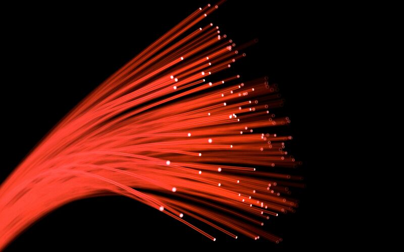
 Flow Series Fiber Optic Interior Ambient Lighting Kit
 KTP Associate - Fibre Optic Flow Sensing - The UK Acoustics Network
Ambient optic array - Wikipedia
 Virtuoso interoperability - Circuit Design Flows using INTERCONNECT - Ansys Optics
 New Concepts in Glaucoma » Optic nerve blood flow in primary open-angle glaucoma
 Night Running: Why It Feels Harder - Outside Online
 Mouse Serioux Flow 207 Wireless,reincarcabil USB-C, Negru, senzor: Optic, - ITVolks - Magazin Online
 Spectral flow, Magnus force, and mutual friction via the geometric optics limit of Andreev reflection<...
Pupil responses associated with the perception of gravitational vertical under directional optic flows - Fingerprint -...
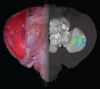
 Compromised Blood Flow in the Optic Nerve Head after Systemic Administration of Aldosterone in Rats: A Possible Rat Model of...
Effects of adaptation to Glass pattern structure and to path of optic flow<...
 Page 1 | Search Results | SPE Annual Technical Conference and Exhibition | OnePetro
 High-resolution fibre-optic temperature sensing: A new tool to study the two-dimensional structure of atmospheric surface-layer...
 Temporal dynamics and magnitude of the blood flow response at the optic
disk in normal subjects during functional retinal...
Temporal dynamics and magnitude of the blood flow response at the optic
disk in normal subjects during functional retinal...
 Fast Response, Fiber-Optic Micromachined Five-Hole Probe For Three-Dimensional Flow Measurements In Harsh Environments - SBIR...
Detailed Table of Contents
 Biotechnology Expert Witnesses
 Optical Products - ECPlaza
 Giant Cell Arteritis and Arteritic Anterior Ischemic Optic Neuropathies | IntechOpen
 OF-MSRN: Optical Flow-Auxiliary Multi-Task Regression Network for Direct Quantitative Measurement, Segmentation and Motion...
 Equity Trader Alert #2013 - 5 QView Latency Optics Launching February 4, 2013
![Burlingham CS[au] - Search Results - PubMed](data:image/png;base64,iVBORw0KGgoAAAANSUhEUgAAABAAAAAQCAMAAAAoLQ9TAAAARVBMVEVHcEwoU45gYmYAUpQAUpRPYGVgYmZLXnJgYmYAUZUAUpRJXnIAUpQAUpRgYmYAUpRgYmZgYmZhYmYAUpQAUpQAUpRgYmaDiPJuAAAAFXRSTlMADOJ+6QewGO8/uTRqtH7GdFJ11p1bCL3TAAAAZUlEQVQYlV2PVw7AIAxDTeney7n/UcsoldX3E+VJOAboEi7MBpHWMs1ADlG8u7UYWauwyZFeRQVPOhG2o+aiwhByJxUx91Jxhje3iJSqGfHuLKI0+0TpXvY1twCOPlFh5pa/++MB0vIOBm+1zaoAAAAASUVORK5CYII=) Burlingham CS[au] - Search Results - PubMed
Burlingham CS[au] - Search Results - PubMed
 Glaucoma - Symptoms and causes - Mayo Clinic
 Visual motion sensitivity in descending neurons in the hoverfly | Journal of Comparative Physiology A
Glaucoma6
- Glaucoma is a group of eye conditions that damage the optic nerve. (mayoclinic.org)
- Glaucoma develops when the optic nerve becomes damaged. (mayoclinic.org)
- This effect helps to both ward off and slow the progress of glaucoma - a disease characterized by damage to the optic nerve. (viteyes.com)
- Glaucoma leads to the loss of retinal ganglion cells and their axons but also to tissue remodelling which involves both the optic nerve head and the retina. (molvis.org)
- On average, ocular blood flow (OBF) is reduced in glaucoma patients in various tissues of the eye. (molvis.org)
- Glaucoma usually happens when the fluid pressure inside the eyes slowly rises and damages the optic nerve. (medlineplus.gov)
Intraocular pressure3
- If that system is blocked or isn't functioning well, the pressure inside the eye (intraocular pressure) builds, which in turn damages the optic nerve. (mayoclinic.org)
- Glaucomas are a group of eye disorders characterized by progressive optic nerve damage in which an important part is a relative increase in intraocular pressure (IOP) that can lead to irreversible loss of vision. (msdmanuals.com)
- Elevated intraocular pressure (IOP) plays a role in axonal damage, either by direct nerve compression or diminution of blood flow. (msdmanuals.com)
Stimuli1
- We describe how the neurons can be identified based on their receptive field properties, and how they respond to moving targets, looming stimuli and to widefield optic flow. (springer.com)
Indicated optic nerve1
- Medieval physicians understood that the presence of a fixed dilated pupil indicated optic nerve obstruction, preventing the passage of visual spirit, and that cataract surgery in such cases would not restore sight. (nature.com)
Retina1
- The acuity of central vision (at the optic disc) was explained by the concentration of visual spirit where the optic nerve met the retina. (nature.com)
Ganglion6
- Spatial and temporal integration properties of units in first optic ganglion of dipterans. (uni-bielefeld.de)
- Compromised Blood Flow in the Optic Nerve Head after Systemic Administration of Aldosterone in Rats: A Possible Rat Model of Retinal Ganglion Cell Loss. (bvsalud.org)
- To investigate the optic nerve head (ONH) blood flow , retinal vessel diameters, and retinal ganglion cell (RGC) loss after systemic administration of aldosterone in rats . (bvsalud.org)
- In addition, the optic nerve gets thinner and the cells of the lateral geniculate ganglion disappear partially. (molvis.org)
- Axons of retinal ganglion cells travel through the optic nerve carrying visual information from the eye to the brain. (msdmanuals.com)
- Damage to these axons causes ganglion cell death with resultant optic nerve atrophy and patchy vision loss. (msdmanuals.com)
Lobes5
- The neural mechanisms underlying the computation of visual motion have been particularly well investigated in the fly optic lobes. (springer.com)
- However, the descending neurons, which connect the optic lobes with the motor command centers of the ventral nerve cord, remain less studied. (springer.com)
- We discuss their similarities with previously published visual neurons, in the optic lobes and ventral nerve cord, and suggest that they can be classified as target-selective, looming sensitive and optic flow sensitive, based on these similarities. (springer.com)
- In fly motion vision most of the attention has been given to the neurons in the optic lobes (for review, see, e.g. (springer.com)
- Exposure to sulfoxaflor led to sparse increases in neuronal apoptosis, localized primarily in the optic lobes, however there was no effect of imidacloprid. (frontiersin.org)
Neuropathies1
- Ischemic optic neuropathies (IONs) are a major cause of blindness or seriously impaired vision in the middle-aged and elderly population, although they can occur at any age. (intechopen.com)
Retinal4
- Several studies have shown that retinal blood flow is autoregulated in response to experimental changes in ocular perfusion pressure (OPP). (arvojournals.org)
- Near-infrared laser Doppler flowmetry was applied in 15 normal volunteers to record the time course and magnitude of changes in the velocity (Vel), volume (Vol) and flow (F) of blood and tissue reflectance (R) at the optic disk in response to 40 and 50 s of increased retinal neural activity. (unicatt.it)
- However, in one subject, 15 recordings from the same site of the optic disk showed a significant increase in R of 8%, with a time course similar to that of Vol. Our findings show that, in the human optic nerve, a white matter tissue, the temporal dynamics and magnitude of the response of blood flow to an increase in retinal neural activity are similar to those reported for brain gray matter. (unicatt.it)
- ONH blood flow decreased following retinal vessel constriction without changes in IOP or blood pressure in a possible rat model of RGC loss by systemic administration of aldosterone . (bvsalud.org)
Alters1
- We determined whether administration of a nitric oxide synthase (NOS) inhibitor alters optic nerve head blood flow (ONHBF) regulation during isometric exercise in healthy subjects. (arvojournals.org)
Nerve diseases1
- We will trace the history of ideas about optic nerve anatomy and function in the Western world from the ancient Greeks to the early 20th century and show how these influenced causal theories of optic nerve diseases. (nature.com)
Cues2
- R. Kurtz, "Enhancement of prominent texture cues in fly optic flow processing", Frontiers in neural circuits , vol. 6, 2012, pp. 78. (uni-bielefeld.de)
- In addition to landmark cues, honeybees rely on two primary modes of visual information for navigation: the orientation of polarized sunlight, and optic flow that results from self-motion during flight. (frontiersin.org)
Directional1
- We conclude that the mechanisms that detect radial and rotational Glass patterns are independent and independently adaptable, but that the mechanisms that detect the path of optic flow, when directional effects are cancelled out, are linked in an opponent, push-pull fashion. (edu.au)
Sensing1
- Among various technologies available for pipeline leak detection, the report encapsulates four technologies that include infra-red detection, pressure monitoring, fiber optic sensing and flow monitoring. (prnewswire.com)
Blood flow15
- 1 - 6 There also is evidence that the choroid comprises regulatory capacity in vivo and neural mechanisms may have a role in keeping blood flow constant during changes in perfusion pressure. (arvojournals.org)
- have shown, in a series of rabbit experiments, that choroidal blood flow remains relatively stable over a wide range of perfusion pressures. (arvojournals.org)
- 8 - 10 Studies done in healthy humans also indicated that choroidal blood flow is autoregulated when OPP either was increased during isometric exercise or decreased during an artificial IOP increase. (arvojournals.org)
- In contrast, only few data on optic nerve head (ONH) blood flow regulation during changes in OPP are available. (arvojournals.org)
- 11 ONH blood flow (ONHBF) seems to be regulated in response to an acute experimental increase and decrease in OPP. (arvojournals.org)
- Magnesium is paramount in improving blood flow to the optic nerve, with studies showing how the mineral enhanced ocular circulation . (viteyes.com)
- Because blood flow to the skin can be modulated by multiple other physiological systems, the PPG can also be used to monitor breathing, hypovolemia , and other circulatory conditions. (wikipedia.org)
- However, in cases of shock, hypothermia , etc., blood flow to the periphery can be reduced, resulting in a PPG without a discernible cardiac pulse. (wikipedia.org)
- All of these work by relaxing your muscles and boosting blood flow to your penis , making erections easier to get and maintain. (webmd.com)
- The condition causes a sudden loss of eyesight because blood flow is blocked to the optic nerve. (webmd.com)
- Arteritis refers to inflammation of your arteries that damages your blood vessel walls and reduces blood flow to your organs. (healthline.com)
- As your blood vessels become increasingly inflamed, blood flow to your facial nerves, optic nerves, or jaw can become disrupted. (healthline.com)
- The immune bodies inside your blood vessels form nodules called granulomas that block blood flow to other parts of your body. (healthline.com)
- Impaired blood flow to your body organs can cause blindness, heart failure, or kidney failure, depending on which arteries are involved. (healthline.com)
- Causes include poor blood flow to the eye, disease, trauma, or exposure to toxic substances. (medlineplus.gov)
Nerves2
- 3 He admitted that cobweb-thin nerves might not possess a lumen but those in the optic nerves ( poroi optikoi -optic channels) were large enough to be visible and to be probed with a hog bristle. (nature.com)
- The optic nerves came together at the chiasma (from the Greek letter × -chi) in order to produce a single impression in binocular vision but did not interchange. (nature.com)
Eyesight1
- As the link between your eyes and brain, maintaining optic nerve health is essential in protecting your eyesight, warding off optic concerns and boosting your overall eye health. (viteyes.com)
Ischemia1
- AION represents an acute ischemic disorder (a segmental infarction) of the ONH supplied by the posterior ciliary arteries (PCAs), while PION has no specific location in the posterior part of the optic nerve and does not represent ischemia in a specific artery [ 1 ]. (intechopen.com)
Sensor2
- Sensirion is launching a flow sensor for flow rates below 2 cc/sec (120 ml/min) with hydrocarbon-based liquids such as photoresists and solvents. (ien.eu)
- Designed for use in high-purity fluid handling applications in the semiconductor, PV and flat panel display industries, the new SLQ-QT105 sensor features a short measurement response time of approximately 40 ms and high sensitivity for flow rates down to 0.05 cc/sec (3 ml/min). (ien.eu)
Temporal1
- Impact of visual motion adaptation on neural responses to objects and its dependence on the temporal characteristics of optic flow. (uni-bielefeld.de)
Visual9
- Recurrent network interactions underlying flow-field selectivity of visual interneurons. (uni-bielefeld.de)
- Greek and Roman humoral physiology needed a hollow optic nerve, the obstruction of which prevented the flow of visual spirit to and from the brain and resulted in blindness. (nature.com)
- The optic nerve sends visual information from your eye to your brain and is vital for good vision. (mayoclinic.org)
- While polarized light from the sun is used as a compass ( 10 ), the information provided from optic flow (wide-field visual motion) allows bees to maintain a steady flight heading, altitude, and speed, and importantly it functions as a visual odometer with the integration of image velocity over time ( 11 ). (frontiersin.org)
- Jassik-Gerschenfeld, D. & Guichard, J. Visual receptive fields of single cells in the pigeon's optic tectum. (nature.com)
- Frost, B.J. Subcortical analysis of visual motion: Relative motion, figure-ground discrimination and self-induced optic flow. (nature.com)
- The optic nerve is a bundle of more than 1 million nerve fibers that carry visual messages. (medlineplus.gov)
- Enhanced optic flow speed discrimination while walking: Contextual tuning of visual coding. (bvsalud.org)
- The aim ers after reviewing the relevant visual field due to damage to the optic of the current study was to evaluate the literature. (who.int)
Disorders2
- Tests for optic nerve disorders may include eye exams, ophthalmoscopy (an examination of the back of your eye), and imaging tests . (medlineplus.gov)
- With some optic nerve disorders, you may get your vision back. (medlineplus.gov)
Damage8
- Damage to the optic nerve is often related to high pressure in your eye. (mayoclinic.org)
- As a powerful antioxidant, vitamin C is thought to protect the optic nerve from oxidative damage caused by free radicals. (viteyes.com)
- These properties help protect the optic nerve against oxidative damage while contributing to healthy circulation that allows essential blood and nutrients to reach the optic nerve. (viteyes.com)
- Our cells use CoQ10 for growth and maintenance and it's a vital component in reducing oxidative damage to the optic nerve. (viteyes.com)
- Vitamin E is a neuroprotective antioxidant that protects our optic nerve from damage caused by harmful free radicals and oxidative stress. (viteyes.com)
- This plant-derived compound facilitates a steady supply of blood and nutrients to the optic nerve - this is important as fluctuations in blood supply and pressure can damage the optic nerve. (viteyes.com)
- Damage to an optic nerve can cause vision loss . (medlineplus.gov)
- Optic nerve atrophy is damage to the optic nerve. (medlineplus.gov)
Fiber optics3
- Create A Laminar Flow Jet Without Pesky Fiber Optics. (hackaday.com)
- If light is applied to this stream of water it tends to act similarly to fiber optics. (hackaday.com)
- Isn't laminar flow jet entirely water based and has nothing to do with fiber optics? (hackaday.com)
Blindness1
- Though it's not totally clear why folate and vitamin B12 are so important for the health of the optic nerve, studies have shown that deficiencies in these essential nutrients can lead to serious eye problems such as optic neuropathy and can even result in blindness. (viteyes.com)
Biophotonics1
- This expert has been issued 19 patents in optics, lasers, microfluidics, and biophotonics (17 more pending), as well as professional awards. (roundtablegroup.com)
Head1
- ION is of two types: anterior (AION) and posterior (PION), the first involving the anterior part of the optic nerve (also called the optic nerve head, ONH) and the second, the rest of the optic nerve. (intechopen.com)
Measurement1
- Measurement of volume flow at standard conditions, proven by billions of metering hours in the field. (ien.eu)
Detection1
- While acoustic and pressure monitoring leak detection are most widely used techniques, newer technologies such as fiber optic leak detection and Real Time Transient Model (RTTM) are increasingly gaining popularity among pipeline operators. (prnewswire.com)
Perception3
- Our results suggest that both macaques (male) and humans (female/male) relied heavily on optic flow, thereby demonstrating a critical role for sensory evidence accumulation during naturalistic action-perception closed-loop tasks. (jneurosci.org)
- For Gibson, perception is a bottom-up process, whereby the agent accesses information about the environment directly from invariant structures in the ambient optic array, rather than recovering it by means of complex cognitive processes. (wikipedia.org)
- Heading perception depends on time-varying evolution of optic flow. (nih.gov)
Cytometry5
- This sample expert has over 25 years of experience and specialization in optics, with specific knowledge in flow cytometry, cell sorting, and hematology analysis. (roundtablegroup.com)
- Published on subjects from flow cytometry to optical design, and I teach courses on these subjects. (roundtablegroup.com)
- The process of measuring the percentage of CD4+ T-lymphocytes in the whole blood sample is referred to as ``immunophenotyping by flow cytometry'' (9-14). (cdc.gov)
- The fluorochrome-labeled cells are analyzed by flow cytometry, which categorizes individual cells according to size, granularity, fluorochrome, and fluorochrome intensity. (cdc.gov)
- This document provides CDC recommendations for immunophenotyping by flow cytometry. (cdc.gov)
Anterior2
- Usually, fluid (aqueous humor) in the eye flows freely through the anterior chamber and exits through the drainage system (trabecular meshwork). (mayoclinic.org)
- A rare vision problem called NAION -- short for "nonarteritic anterior ischemic optic neuropathy " -- has been reported by a few men using these drugs. (webmd.com)
Movements1
- We designed a sensory-based sequential decision task in virtual reality in which humans and monkeys navigated to a memorized location by integrating optic flow generated by their own joystick movements. (jneurosci.org)
Type1
- and (2) adaptation to Glass patterns decreases detectability of patterns of the same type, but adaptation to optic flow has little effect on the detectability of patterns of any type. (edu.au)
Bloch1
- The discrete nature of the states, however, leads to Bloch oscillations which thwart the spectral flow. (illinois.edu)
Experimental1
- In contrast, inhibition of NOS did not alter the pressure-flow relationship in the ONH during an experimental increase in OPP compared to phenylephrine and placebo ( P = 0.37 between groups). (arvojournals.org)
Vision3
- During the Renaissance, the organ of vision was transferred from the lens to the optic nerve, which was generally believed to be on the axis of the eye. (nature.com)
- Their size allowed psychic pneuma to flow in abundance, emerging from the eyes to unite with the incoming light, a process essential for vision. (nature.com)
- Ross, J & Dickinson, E 2007, ' Effects of adaptation to Glass pattern structure and to path of optic flow ', Vision Research , vol. 47, no. 16, pp. 2150-2155. (edu.au)
Magnitude1
- E ) Visualization of optic flow using arrow lengths to indicate the direction and magnitude flow. (elifesciences.org)
Time1
- Humidified high-flow oxygen was administered for the first 5 h, noninvasive positive pressure ventilation by oronasal mask for the next 18.25 h, and invasive mechanical ventilation for the subsequent time (7.5 h). (cdc.gov)
Reduces1
- Studies have shown that this neuroprotective vitamin is essential to allow the optic nerve to function properly and reduces the degeneration of optic nerve cells. (viteyes.com)
Physics1
- Stone, M 1996, ' Spectral flow, Magnus force, and mutual friction via the geometric optics limit of Andreev reflection ', Physical Review B - Condensed Matter and Materials Physics , vol. 54, no. 18, pp. 13222-13229. (illinois.edu)
Interpretation1
- Both the microscope and the ophthalmoscope allowed visualisation of the optic nerve, but problems of interpretation persisted until conceptual transformations in medical science were made. (nature.com)
Systems1
- Conductor element for use in integrated electro-optic systems that measures current flowing through it. (photonics.com)
Patterns2
- He also noticed that "optical flow patterns", or optical flow invariants, are produced in the array as the agent moves about the environment. (wikipedia.org)
- The effects of adaptation to radial and rotational polar optic flow on the classification of noisy test patterns are shown to be very similar to the effects of adaptation to polar Glass patterns on the classification of noisy Glass patterns (Clifford, C. W. G. & Weston, E. (2005). (edu.au)
Cells2
- Certain vitamins and minerals are essential in maintaining the cells and nerve fibers that comprise our optic nerve. (viteyes.com)
- This is especially important to our optic nerve, as communication between nerve cells in this area facilitates the nervous communication that allows us to see. (viteyes.com)
Units1
- Jassik-Gerschenfeld, D., Minois, F. & Conde-Courtine, F. Receptive field properties of directionally selective units in the pigeon's optic tectum. (nature.com)
Pressure1
- Elevated eye pressure happens as the result of a buildup of fluid that flows throughout the inside of the eye. (mayoclinic.org)
Optical flow8
- As a powerful alternative to traditional methods, deep learning based approaches take the research of optical flow into a new level. (researchgate.net)
- Current optical flow methods have made great advances in: i) developing powerful data-driven, endto-end learning paradigms (Dosovitskiy et al. (researchgate.net)
- To improve results on optical flow, many recent works introduce stronger learning paradigms that can enable iterative refinement (Ranjan and Black 2017;Sun et al. (researchgate.net)
- Estimating per-pixel motion between video frames, known as optical flow, is a long-standing problem in video understanding and analysis. (researchgate.net)
- Most contemporary optical flow techniques largely focus on addressing the cross-image matching with feature similarity, with few methods considering how to explicitly reason over the given scene for achieving a holistic motion understanding. (researchgate.net)
- In this work, taking a fresh perspective, we introduce a novel graph-based approach, called adaptive graph reasoning for optical flow (AGFlow), to emphasize the value of scene/context information in optical flow. (researchgate.net)
- Here he will talk about the optical Flow + CNN aproach he used for solving the problem. (mathworks.com)
- In this code, I use optical flow to pre-process the video frames and feed these images into a basic regression model, using a pre-trained CNN. (mathworks.com)
Studies have shown1
- Another powerful antioxidant, studies have shown that grape seed extract has a neuroprotective effect that extends especially to our optic nerve, in addition to being beneficial for general circulation, reducing inflammation as well as promoting wound healing. (viteyes.com)
Motion1
- The notion of spectral flow has given new insight into the motion of vortices in superfluids and superconductors. (illinois.edu)
Effect1
- If the core spectrum were continuous, spectral flow would convert the momentum flowing into the core via the Magnus effect into unbound quasiparticles-thus allowing the vortex to remain stationary without a pinning potential or other sink for the inflowing momentum. (illinois.edu)