Odontoblasts
Dentinogenesis
Dentin
Dental Pulp
Odontogenesis
Ameloblasts
Tooth Germ
Molar
Dental Papilla
Tooth Calcification
Incisor
Tooth Permeability
Sialoglycoproteins
Amelogenesis
Extracellular Matrix Proteins
Tooth Root
Dental Enamel
Calcification, Physiologic
Dental Pulp Exposure
Cell Differentiation
Amelogenin
Microscopy, Electrochemical, Scanning
Dental Pulp Calcification
Odontoma
In Situ Hybridization
Phosphoproteins
Enamel Organ
Dental Cementum
Phalloidine
Dental Cavity Preparation
Expression of the Fanconi anemia group A gene (Fanca) during mouse embryogenesis. (1/240)
About 80% of all cases of Fanconi anemia (FA) can be accounted for by complementation groups A and C. To understand the relationship between these groups, we analyzed the expression pattern of the mouse FA group-A gene (Fanca) during embryogenesis and compared it with the known pattern of the group-C gene (Fancc). Northern analysis of RNA from mouse embryos at embryonic days 7, 11, 15, and 17 showed a predominant 4.5 kb band in all stages. By in situ hybridization, Fanca transcripts were found in the whisker follicles, teeth, brain, retina, kidney, liver, and limbs. There was also stage-specific variation in Fanca expression, particularly within the developing whiskers and the brain. Some tissues known to express Fancc (eg, gut) failed to show Fanca expression. These observations show that (1) Fanca is under both tissue- and stage-specific regulation in several tissues; (2) the expression pattern of Fanca is consistent with the phenotype of the human disease; and (3) Fanca expression is not necessarily coupled to that of Fancc. The presence of distinct tissue targets for FA genes suggests that some of the variability in the clinical phenotype can be attributed to the complementation group assignment. (+info)Spatial and temporal activity of the dentin sialophosphoprotein gene promoter: differential regulation in odontoblasts and ameloblasts. (2/240)
Dentin sialoprotein and dentin phosphoprotein are non-collagenous proteins that are cleavage products of dentin sialophosphoprotein (DSPP). Although these two protein products are believed to have a crucial role in the process of tooth mineralization, their precise biological functions and the molecular mechanisms of gene regulation are not clearly understood. To understand such functions, we have developed a transgenic mouse model expressing a reporter gene (lacZ) under the control of approximately 6 kb upstream sequences of Dspp. The transgenic fusion protein was designed to reside within the cells to facilitate the precise identification of cell type and developmental stages at which the Dspp-lacZ gene is expressed. The results presented in this report demonstrate: (a) the 6 kb upstream sequences of Dspp have the necessary regulatory elements to direct the tissue specific expression of the transgene similar to endogenous Dspp, (b) both odontoblasts and ameloblasts exhibit transgene expression in a differentiation dependent manner, and (c) a differential regulation of the transgene in odontoblasts and ameloblasts occurs during tooth development and mineralization. (+info)Subtilisin-like proprotein convertase PACE4 (SPC4) is a candidate processing enzyme of bone morphogenetic proteins during tooth formation. (3/240)
The temporospatial expression of PACE4, a member of the mammalian subtilisin-like proprotein convertase family, in the developing rat molar tooth was determined by in situ hybridization. At the initiation stage of tooth development, PACE4 mRNA was weakly expressed in the dental lamina, whereas the mesenchymal cells intensely expressed the PACE4 transcript. At the bud stage, high-level expression of PACE4 mRNA was found in the dental epithelium and condensed dental mesenchyme. Its expression became more localized in the differentiating ameloblasts during cap and early bell stages. In the newborn rats, PACE4 mRNA was localized in the ameloblasts and odontoblasts, but its expression became weaker with advancing development, showing apparent association with the differentiation and establishment of functional ameloblasts and odontoblasts. These expression patterns of PACE4 were very similar to those of several bone morphogenetic proteins (BMPs) reported previously. Because BMPs, which are primarily involved in the morphogenesis in tooth formation, are synthesized as inactive precursors and activated by limited proteolysis at the consensus Arg-X-X-Arg maturation site, the present observations suggest that PACE4 is possibly a candidate proBMP convertase that acts during tooth formation. (+info)Characterization and gene expression of high conductance calcium-activated potassium channels displaying mechanosensitivity in human odontoblasts. (4/240)
Odontoblasts form a layer of cells responsible for the dentin formation and possibly mediate early stages of sensory processing in teeth. Several classes of ion channels have previously been identified in the odontoblast or pulp cell membrane, and it is suspected that these channels assist in these events. This study was carried out to characterize the K(Ca) channels on odontoblasts fully differentiated in vitro using the patch clamp technique and to investigate the HSLO gene expression encoding the alpha-subunit of these channels on odontoblasts in vivo. In inside-out patches, K(Ca) channels were identified on the basis of their K(+) selectivity, conductance, voltage, and Ca(2+) dependence. In cell-attached patches, these channels were found to be activated by application of a negative pressure as well as an osmotic shock. By reverse transcription-polymerase chain reaction, a probe complementary to K(Ca) alpha-subunit mRNA was constructed and used for in situ hybridization on human dental pulp samples. Transcripts were expressed in the odontoblast layer. The use of antibodies showed that the K(Ca) channels were preferentially detected at the apical pole of the odontoblasts. These channels could be involved in mineralization processes. Their mechanosensitivity suggests that the fluid displacement within dentinal tubules could be transduced into electrical cell signals. (+info)Widespread expression of tartrate-resistant acid phosphatase (Acp 5) in the mouse embryo. (5/240)
Tartrate-resistant acid phosphatase (TRAP, Acp 5) is considered to be a marker of the osteoclast and studies using 'knockout' mice have demonstrated that TRAP is critical for normal development of the skeleton. To investigate the distribution of TRAP in the mammalian embryo, cryostat sections of 18 d murine fetuses were examined by in situ hybridisation, immunohistochemistry and histochemical reactions in situ. Abundant expression of TRAP mRNA was observed in the skin and epithelial surfaces of the tongue, oropharynx and gastrointestinal tract including the colon, as well as the thymus, ossifying skeleton and dental papillae. TRAP protein was identified at the same sites, but the level of expression in the different tissues did not always correlate with apparent enzyme activity. The findings indicate that abundant TRAP expression is not confined to osteoclasts in bone, but occurs in diverse tissues harbouring cells of bone marrow origin, including dendritic cells and other cells belonging to the osteoclast/macrophage lineage. (+info)Dissection of the odontoblast differentiation process in vitro by a combination of FGF1, FGF2, and TGFbeta1. (6/240)
Dental papillae (DP) isolated from first lower molars of 17-day-old mouse embryos were cultured in the presence of combinations of the following growth factors: FGF1, FGF2, and TGFbeta1. After 6 days in culture, only the DP treated with FGF1+TGFbeta1 contained differentiated odontoblast-like cells at the periphery of the explants, and these cells secreted extracellular matrix similar to predentin. Surprisingly, treatments with FGF2+TGFbeta1 induced cell polarization at the surface of the explants but no matrix secretion was observed. Electron microscopy and histochemical analysis of odontoblast markers showed that differentiated cells induced by FGF1+TGFbeta1 exhibited cytological features of functional odontoblasts with matrix vesicle secretion and mineral formation, positive alkaline-phosphatase activity, and type-I collagen production. DP cultured in the presence of FGF2+TGFbeta1 showed cell polarization and long and thin cell processes containing matrix vesicles; however, type-I collagen secretion was not detected and alkaline-phosphatase activity was completely inhibited. Our results indicate that, in our culture system, exogenous combinations of FGF1, FGF2, and TGFbeta1 interact with preodontoblasts and induce cell polarization or differentiation, which can be studied separately in vitro. Thus, FGF1 and TGFbeta1 do have a synergic effect to promote morphological and functional features of differentiated odontoblasts whereas FGF2 seems to modulate TGFbeta1 action, causing morphological polarization of preodontoblasts but limiting the functional activity of these cells in terms of type-I collagen secretion and alkaline-phosphatase activity. (+info)BMP4 rescues a non-cell-autonomous function of Msx1 in tooth development. (7/240)
The development of many organs depends on sequential epithelial-mesenchymal interactions, and the developing tooth germ provides a powerful model for elucidating the nature of these inductive tissue interactions. In Msx1-deficient mice, tooth development arrests at the bud stage when Msx1 is required for the expression of Bmp4 and Fgf3 in the dental mesenchyme (Bei, M. and Maas, R. (1998) Development 125, 4325-4333). To define the tissue requirements for Msx1 function, we performed tissue recombinations between wild-type and Msx1 mutant dental epithelium and mesenchyme. We show that through the E14.5 cap stage of tooth development, Msx1 is required in the dental mesenchyme for tooth formation. After the cap stage, however, tooth development becomes Msx1 independent, although our experiments identify a further late function of Msx1 in odontoblast and dental pulp survival. These results suggest that prior to the cap stage, the dental epithelium receives an Msx1-dependent signal from the dental mesenchyme that is necessary for tooth formation. To further test this hypothesis, Msx1 mutant tooth germs were first cultured with either BMP4 or with various FGFs for two days in vitro and then grown under the kidney capsule of syngeneic mice to permit completion of organogenesis and terminal differentiation. Previously, using an in vitro culture system, we showed that BMP4 stimulated the growth of Msx1 mutant dental epithelium (Chen, Y., Bei, M. Woo, I., Satokata, I. and Maas, R. (1996). Development 122, 3035-3044). Using the more powerful kidney capsule grafting procedure, we now show that when added to explanted Msx1-deficient tooth germs prior to grafting, BMP4 rescues Msx1 mutant tooth germs all the way to definitive stages of enamel and dentin formation. Collectively, these results establish a transient functional requirement for Msx1 in the dental mesenchyme that is almost fully supplied by BMP4 alone, and not by FGFs. In addition, they formally prove the postulated downstream relationship of BMP4 with respect to Msx1, establish the non-cell-autonomous nature of Msx1 during odontogenesis, and disclose an additional late survival function for Msx1 in odontoblasts and dental pulp. (+info)Transient expression of heat shock protein (Hsp)25 in the dental pulp and enamel organ during odontogenesis in the rat incisor. (8/240)
The expression of heat shock protein (Hsp) 25 during odontogenesis in the dental pulp and enamel organ of rat incisors was investigated by immunocytochemistry and confocal microscopy. In the process of dentin formation, immature odontoblasts first exhibited Hsp 25-immunoreactivity, and increased in immunointensity with the advance of their differentiation. In the dental pulp, in contrast, intense immunoreaction in the mesenchymal cells became weak or negative in parallel with the progress of cell differentiation. The immunoreaction for Hsp 25 in the enamel organ revealed a characteristic stage-related alteration during amelogenesis. In secretory ameloblasts, the immunoreaction for Hsp 25 was found throughout their cell bodies, intense reactivity being located near the proximal and distal terminal webs. At the maturation stage, ruffle-ended ameloblasts (RA) consistently showed Hsp 25-immunoreactivity throughout the cell bodies, whereas smooth-ended ameloblasts (SA) lacking a ruffled border were weak in immunoreaction at the distal cytoplasm. Other cellular elements of the enamel organ were negative. The subcellular localization of Hsp 25-immunoreactivity in this study appeared essentially identical to that of actin filaments as demonstrated by confocal microscopy using rhodamine-labeled phalloidin. These immunocytochemical data suggest that the Hsp 25 molecule is involved in reinforcement of the cell layer following cell movement during odontogenesis and in the formation and maintenance of the ruffled border of RA. (+info)Odontoblasts are defined as columnar-shaped cells that are located in the pulp tissue of teeth, specifically within the predentin region. They are responsible for the formation of dentin, one of the main components of a tooth, by synthesizing and depositing collagenous and non-collagenous proteins, as well as the mineral hydroxyapatite.
Odontoblasts have a single process that extends into the dentinal tubules, which are microscopic channels within the dentin matrix. These cells play a crucial role in sensing external stimuli, such as heat, cold, or pressure, and transmitting signals to the nerves located in the pulp tissue, thereby contributing to the tooth's sensitivity.
In summary, odontoblasts are specialized dental cells that produce dentin, provide structural support for teeth, and contribute to their sensory functions.
Dentinogenesis is the process of dentin formation, which is one of the main components of teeth. Dentin is a hard, calcified tissue that lies beneath the tooth's enamel and cementum layers, providing structural support and protection to the pulp tissue containing nerves and blood vessels. The process of dentinogenesis involves the differentiation and activation of odontoblasts, which are specialized cells that synthesize and secrete the organic and inorganic components of dentin matrix. These components include collagenous proteins and hydroxyapatite crystals, which form a highly mineralized tissue that is both strong and flexible. Dentinogenesis continues throughout life as new layers of dentin are formed in response to various stimuli such as tooth wear, dental caries, or injury.
Dentin is the hard, calcified tissue that lies beneath the enamel and cementum of a tooth. It forms the majority of the tooth's structure and is composed primarily of mineral salts (hydroxyapatite), collagenous proteins, and water. Dentin has a tubular structure, with microscopic channels called dentinal tubules that radiate outward from the pulp chamber (the center of the tooth containing nerves and blood vessels) to the exterior of the tooth. These tubules contain fluid and nerve endings that are responsible for the tooth's sensitivity to various stimuli such as temperature changes, pressure, or decay. Dentin plays a crucial role in protecting the dental pulp while also providing support and structure to the overlying enamel and cementum.
Dental pulp is the soft tissue located in the center of a tooth, surrounded by the dentin. It contains nerves, blood vessels, and connective tissue, and plays a vital role in the development and health of the tooth. The dental pulp helps to form dentin during tooth development and continues to provide nourishment to the tooth throughout its life. It also serves as a sensory organ, allowing the tooth to detect hot and cold temperatures and transmit pain signals to the brain. Injury or infection of the dental pulp can lead to serious dental problems, such as tooth decay or abscesses, and may require root canal treatment to remove the damaged tissue and save the tooth.
Odontogenesis is the process of tooth development that involves the formation and calcification of teeth. It is a complex process that requires the interaction of several types of cells, including epithelial cells, mesenchymal cells, and odontoblasts. The process begins during embryonic development with the formation of dental lamina, which gives rise to the tooth bud. As the tooth bud grows and differentiates, it forms the various structures of the tooth, including the enamel, dentin, cementum, and pulp. Odontogenesis is completed when the tooth erupts into the oral cavity. Abnormalities in odontogenesis can result in developmental dental anomalies such as tooth agenesis, microdontia, or odontomas.
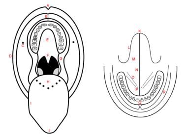
A tooth is a hard, calcified structure found in the jaws (upper and lower) of many vertebrates and used for biting and chewing food. In humans, a typical tooth has a crown, one or more roots, and three layers: the enamel (the outermost layer, hardest substance in the body), the dentin (the layer beneath the enamel), and the pulp (the innermost layer, containing nerves and blood vessels). Teeth are essential for proper nutrition, speech, and aesthetics. There are different types of teeth, including incisors, canines, premolars, and molars, each designed for specific functions in the mouth.
Ameloblasts are the specialized epithelial cells that are responsible for the formation of enamel, which is the hard, outermost layer of a tooth. These cells are a part of the dental lamina and are present in the developing tooth's crown region. They align themselves along the surface of the developing tooth and secrete enamel proteins and minerals to form the enamel rods and interrod enamel. Once the enamel formation is complete, ameloblasts undergo programmed cell death, leaving behind the hard, mineralized enamel matrix. Any damage or abnormality in the functioning of ameloblasts can lead to developmental defects in the enamel, such as hypoplasia or hypocalcification, which may affect the tooth's structure and function.
A tooth germ is a small cluster of cells that eventually develop into a tooth. It contains the dental papilla, which will become the dentin and pulp of the tooth, and the dental follicle, which will form the periodontal ligament, cementum, and alveolar bone. The tooth germ starts as an epithelial thickening called the dental lamina, which then forms a bud, cap, and bell stage before calcification occurs and the tooth begins to erupt through the gums. It is during the bell stage that the enamel organ, which will form the enamel of the tooth, is formed.
In the context of dentistry, a molar is a type of tooth found in the back of the mouth. They are larger and wider than other types of teeth, such as incisors or canines, and have a flat biting surface with multiple cusps. Molars are primarily used for grinding and chewing food into smaller pieces that are easier to swallow. Humans typically have twelve molars in total, including the four wisdom teeth.
In medical terminology outside of dentistry, "molar" can also refer to a unit of mass in the apothecaries' system of measurement, which is equivalent to 4.08 grams. However, this usage is less common and not related to dental or medical anatomy.
The dental papilla is a type of tissue found in the developing tooth within the jawbone. It is composed of cells that will eventually differentiate into odontoblasts, which are the cells responsible for producing dentin, one of the main hard tissues that make up the tooth. The dental papilla is located in the center of the tooth germ and is surrounded by the dental follicle, another type of tissue that helps to form the tooth. As the tooth develops, the dental papilla becomes smaller and eventually forms the pulp chamber, which contains the blood vessels, nerves, and connective tissue that support and nourish the tooth.
Tooth calcification, also known as dental calculus or tartar formation, refers to the hardening of plaque on the surface of teeth. This process occurs when minerals from saliva combine with bacterial deposits and dental plaque, resulting in a hard, calcified substance that adheres to the tooth surface. Calcification can occur both above and below the gum line, and if not removed through professional dental cleanings, it can lead to periodontal disease, tooth decay, and other oral health issues.
An incisor is a type of tooth that is primarily designed for biting off food pieces rather than chewing or grinding. They are typically chisel-shaped, flat, and have a sharp cutting edge. In humans, there are eight incisors - four on the upper jaw and four on the lower jaw, located at the front of the mouth. Other animals such as dogs, cats, and rodents also have incisors that they use for different purposes like tearing or gnawing.
Secondary dentin is a type of dentin that is formed after the initial development of the tooth. It is produced in response to stimuli such as tooth wear or injury and continues to form throughout an individual's life. Unlike primary dentin, which is laid down during tooth development and has a more uniform structure, secondary dentin is often deposited in a less organized manner and can vary in thickness. The formation of secondary dentin can help to protect the pulp tissue within the tooth from further damage or infection.
I am not able to find a single medical definition for "tooth permeability" as it is not a widely used or recognized term in dental or medical literature. However, I can provide some context that might help you understand the concept.
In general, permeability refers to the ability of a material or tissue to allow fluids or gases to pass through it. In the case of teeth, they are composed of several layers: the outer enamel, dentin, and the inner pulp. Enamel is highly mineralized and does not have living cells, making it relatively impermeable to substances. Dentin, on the other hand, contains microscopic tubules filled with fluid and nerve endings, which can potentially allow for some permeability.
However, when people talk about tooth permeability in a dental context, they might be referring to the ability of various substances (such as acids, bacteria, or chemicals) to penetrate through the enamel and dentin layers and reach the inner pulp chamber. This can lead to dental issues like tooth decay, sensitivity, or infection.
In summary, while "tooth permeability" is not a standard medical term, it may refer to the ability of substances to pass through the various layers of the tooth and potentially cause damage.
Sialglycoproteins are a type of glycoprotein that have sialic acid as the terminal sugar in their oligosaccharide chains. These complex molecules are abundant on the surface of many cell types and play important roles in various biological processes, including cell recognition, cell-cell interactions, and protection against proteolytic degradation.
The presence of sialic acid on the outermost part of these glycoproteins makes them negatively charged, which can affect their interaction with other molecules such as lectins, antibodies, and enzymes. Sialglycoproteins are also involved in the regulation of various physiological functions, including blood coagulation, inflammation, and immune response.
Abnormalities in sialglycoprotein expression or structure have been implicated in several diseases, such as cancer, autoimmune disorders, and neurodegenerative conditions. Therefore, understanding the biology of sialoglycoproteins is important for developing new diagnostic and therapeutic strategies for these diseases.
Amelogenesis is the biological process of forming enamel, which is the hard and highly mineralized outer layer of teeth. Enamel is primarily made up of calcium and phosphate minerals and is the toughest substance in the human body. Amelogenesis involves the synthesis, secretion, and maturation of enamel proteins by specialized cells called ameloblasts.
The medical definition of 'Amelogenesis' refers to a genetic disorder that affects the development and formation of tooth enamel. This condition is also known as Amelogenesis Imperfecta (AI) and can result in teeth that are discolored, sensitive, and prone to decay. There are several types of Amelogenesis Imperfecta, each with its own set of symptoms and genetic causes.
In summary, 'Amelogenesis' is the biological process of enamel formation, while 'Amelogenesis Imperfecta' is a genetic disorder that affects this process, leading to abnormal tooth enamel development.
Extracellular matrix (ECM) proteins are a group of structural and functional molecules that provide support, organization, and regulation to the cells in tissues and organs. The ECM is composed of a complex network of proteins, glycoproteins, and carbohydrates that are secreted by the cells and deposited outside of them.
ECM proteins can be classified into several categories based on their structure and function, including:
1. Collagens: These are the most abundant ECM proteins and provide strength and stability to tissues. They form fibrils that can withstand high tensile forces.
2. Proteoglycans: These are complex molecules made up of a core protein and one or more glycosaminoglycan (GAG) chains. The GAG chains attract water, making proteoglycans important for maintaining tissue hydration and resilience.
3. Elastin: This is an elastic protein that allows tissues to stretch and recoil, such as in the lungs and blood vessels.
4. Fibronectins: These are large glycoproteins that bind to cells and ECM components, providing adhesion, migration, and signaling functions.
5. Laminins: These are large proteins found in basement membranes, which provide structural support for epithelial and endothelial cells.
6. Tenascins: These are large glycoproteins that modulate cell adhesion and migration, and regulate ECM assembly and remodeling.
Together, these ECM proteins create a microenvironment that influences cell behavior, differentiation, and function. Dysregulation of ECM proteins has been implicated in various diseases, including fibrosis, cancer, and degenerative disorders.
A tooth root is the part of a tooth that is embedded in the jawbone and cannot be seen when looking at a person's smile. It is the lower portion of a tooth that typically has a conical shape and anchors the tooth to the jawbone through a periodontal ligament. The tooth root is covered by cementum, a specialized bone-like tissue, and contains nerve endings and blood vessels within its pulp chamber.
The number of roots in a tooth can vary depending on the type of tooth. For example, incisors typically have one root, canines may have one or two roots, premolars usually have one or two roots, and molars often have two to four roots. The primary function of the tooth root is to provide stability and support for the crown of the tooth, allowing it to withstand the forces of biting and chewing.
Dental enamel is the hard, white, outermost layer of a tooth. It is a highly mineralized and avascular tissue, meaning it contains no living cells or blood vessels. Enamel is primarily composed of calcium and phosphate minerals and serves as the protective covering for the crown of a tooth, which is the portion visible above the gum line.
Enamel is the hardest substance in the human body, and its primary function is to provide structural support and protection to the underlying dentin and pulp tissues of the tooth. It also plays a crucial role in chewing and biting by helping to distribute forces evenly across the tooth surface during these activities.
Despite its hardness, dental enamel can still be susceptible to damage from factors such as tooth decay, erosion, and abrasion. Once damaged or lost, enamel cannot regenerate or repair itself, making it essential to maintain good oral hygiene practices and seek regular dental checkups to prevent enamel damage and protect overall oral health.
Tooth abnormalities refer to any variations or irregularities in the size, shape, number, structure, or development of teeth that deviate from the typical or normal anatomy. These abnormalities can occur in primary (deciduous) or permanent teeth and can be caused by genetic factors, environmental influences, systemic diseases, or localized dental conditions during tooth formation.
Some examples of tooth abnormalities include:
1. Microdontia - teeth that are smaller than normal in size.
2. Macrodontia - teeth that are larger than normal in size.
3. Peg-shaped teeth - teeth with a narrow, conical shape.
4. Talon cusps - additional cusps or points on the biting surface of a tooth.
5. Dens invaginatus - an abnormal development where the tooth crown has an extra fold or pouch that can trap bacteria and cause dental problems.
6. Taurodontism - teeth with large pulp chambers and short roots.
7. Supernumerary teeth - having more teeth than the typical number (20 primary and 32 permanent teeth).
8. Hypodontia - missing one or more teeth due to a failure of development.
9. Germination - two adjacent teeth fused together, usually occurring in the front teeth.
10. Fusion - two separate teeth that have grown together during development.
Tooth abnormalities may not always require treatment unless they cause functional, aesthetic, or dental health issues. A dentist can diagnose and manage tooth abnormalities through various treatments, such as fillings, extractions, orthodontic care, or restorative procedures.
A toothache is defined as pain or discomfort in or around a tooth, usually caused by dental cavities, gum disease, tooth fracture, or exposed tooth roots. The pain may be sharp and stabbing, throbbing, or constant and dull. It can also be aggravated by hot, cold, sweet, or sour foods and drinks, or by biting or chewing. Toothaches are serious and should not be ignored as they can be a sign of more significant dental issues that require immediate professional attention from a dentist.
Physiologic calcification is the normal deposit of calcium salts in body tissues and organs. It is a natural process that occurs as part of the growth and development of the human body, as well as during the repair and remodeling of tissues.
Calcium is an essential mineral that plays a critical role in many bodily functions, including bone formation, muscle contraction, nerve impulse transmission, and blood clotting. In order to maintain proper levels of calcium in the body, excess calcium that is not needed for these functions may be deposited in various tissues as a normal part of the aging process.
Physiologic calcification typically occurs in areas such as the walls of blood vessels, the lungs, and the heart valves. While these calcifications are generally harmless, they can sometimes lead to complications, particularly if they occur in large amounts or in sensitive areas. For example, calcification of the coronary arteries can increase the risk of heart disease, while calcification of the lung tissue can cause respiratory symptoms.
It is important to note that pathologic calcification, on the other hand, refers to the abnormal deposit of calcium salts in tissues and organs, which can be caused by various medical conditions such as chronic kidney disease, hyperparathyroidism, and certain infections. Pathologic calcification is not a normal process and can lead to serious health complications if left untreated.
Dental pulp exposure is a condition in which the soft, living tissue inside a tooth (the dental pulp) becomes exposed due to damage or injury to the tooth. This can occur as a result of tooth decay that has progressed deeply into the tooth, trauma or fracture that exposes the pulp, or recession of the gums due to periodontal disease.
Exposure of the dental pulp can lead to infection, inflammation, and severe pain. If left untreated, it may result in the need for a root canal procedure or even extraction of the tooth. Therefore, prompt dental treatment is necessary to prevent further complications and preserve the tooth.
Cell differentiation is the process by which a less specialized cell, or stem cell, becomes a more specialized cell type with specific functions and structures. This process involves changes in gene expression, which are regulated by various intracellular signaling pathways and transcription factors. Differentiation results in the development of distinct cell types that make up tissues and organs in multicellular organisms. It is a crucial aspect of embryonic development, tissue repair, and maintenance of homeostasis in the body.
Amelogenin is a protein that plays a crucial role in the formation and mineralization of enamel, which is the hard, calcified tissue that covers the outer surface of teeth. It is expressed during tooth development and is secreted by ameloblasts, the cells responsible for producing enamel.
Amelogenin makes up approximately 90% of the organic matrix of developing enamel and guides the growth and organization of hydroxyapatite crystals, which are the primary mineral component of enamel. The protein is subsequently degraded and removed as the enamel matures and becomes fully mineralized.
Mutations in the gene that encodes amelogenin (AMELX on the X chromosome) can lead to various inherited enamel defects, such as amelogenesis imperfecta, which is characterized by thin, soft, or poorly formed enamel. Additionally, because of its high expression in developing teeth and unique size and structure, amelogenin has been widely used as a marker in forensic dentistry for human identification and sex determination.
Electrochemical Scanning Microscopy (ESCM) is not a specific type of microscopy on its own, but rather refers to various techniques that combine scanning probe microscopy with electrochemistry. These techniques use a sharp probe to scan the surface of a sample while simultaneously measuring or applying an electrical potential. This allows for the visualization and manipulation of electrochemical processes at the nanoscale.
There are several types of ESCM, including:
1. Scanning Electrochemical Microscopy (SECM): A technique that measures the local electrochemical activity of a sample by scanning a microelectrode over its surface while monitoring changes in current. This can be used to map out the distribution of redox-active species, measure local pH or potential, and study corrosion processes.
2. Scanning Ion Conductance Microscopy (SICM): A technique that measures the ion conductance between a nanopipette and a sample surface to create topographic images with high resolution. SICM can be used to investigate biological samples, such as cells and tissues, in their native environment without causing damage.
3. Scanning Kelvin Probe Microscopy (SKPM): A technique that measures the contact potential difference between a conductive probe and a sample surface. This allows for the mapping of work function differences, which can provide information about chemical composition and electronic properties.
4. Piezoresponse Force Microscopy (PFM): A technique that uses an electric field to induce mechanical deformation in ferroelectric or piezoelectric materials. By monitoring these deformations, PFM can be used to map the local polarization and investigate nanoscale electromechanical properties.
5. Scanning Electrochemical Strain Microscopy (SESM): A technique that combines scanning probe microscopy with electrochemical strain measurements to study mechanical deformations in materials under an applied potential. SESM can be used to investigate the relationship between electrochemical processes and mechanical properties at the nanoscale.
In summary, Electrochemical Scanning Microscopy (ESCM) encompasses various techniques that combine scanning probe microscopy with electrochemical measurements or manipulations. These methods provide valuable insights into the structure, composition, and properties of materials at the nanoscale, enabling advancements in fields such as energy storage, electronics, biology, and materials science.
Dental pulp calcification, also known as pulp stones or denticles, refers to the formation of hard tissue within the pulp chamber of a tooth. The pulp chamber is the central part of a tooth that contains its nerves, blood vessels, and connective tissues.
Pulp calcification occurs when the soft tissue of the pulp gradually transforms into a harder, calcified substance. This can happen as a result of aging, injury, or inflammation in the pulp chamber. Over time, these calcifications can build up and make the pulp chamber smaller, which can potentially lead to problems with the tooth's nerve and blood supply.
While dental pulp calcification is not usually harmful on its own, it can cause issues if it becomes severe enough to compress the tooth's nerve or restrict blood flow. In some cases, calcifications may also make root canal treatment more difficult, as there may be less space to work within the pulp chamber.
Odontoma is a type of odontogenic tumor, which means it arises from the tissues that form teeth. It is considered a benign or non-cancerous tumor and is typically slow-growing. Odontomas are usually asymptomatic and are often discovered on routine dental X-rays or during procedures such as wisdom tooth removal.
Odontomas can be classified into two types: complex and compound. Complex odontomas are composed of a haphazard mixture of dental tissue, including enamel, dentin, and cementum, while compound odontomas contain small tooth-like structures called denticles.
These tumors typically occur in the posterior region of the jaw, and while they are usually asymptomatic, some patients may experience symptoms such as swelling, pain, or displacement of teeth. Treatment for odontomas typically involves surgical removal of the tumor.
In situ hybridization (ISH) is a molecular biology technique used to detect and localize specific nucleic acid sequences, such as DNA or RNA, within cells or tissues. This technique involves the use of a labeled probe that is complementary to the target nucleic acid sequence. The probe can be labeled with various types of markers, including radioisotopes, fluorescent dyes, or enzymes.
During the ISH procedure, the labeled probe is hybridized to the target nucleic acid sequence in situ, meaning that the hybridization occurs within the intact cells or tissues. After washing away unbound probe, the location of the labeled probe can be visualized using various methods depending on the type of label used.
In situ hybridization has a wide range of applications in both research and diagnostic settings, including the detection of gene expression patterns, identification of viral infections, and diagnosis of genetic disorders.
Phosphoproteins are proteins that have been post-translationally modified by the addition of a phosphate group (-PO3H2) onto specific amino acid residues, most commonly serine, threonine, or tyrosine. This process is known as phosphorylation and is mediated by enzymes called kinases. Phosphoproteins play crucial roles in various cellular processes such as signal transduction, cell cycle regulation, metabolism, and gene expression. The addition or removal of a phosphate group can activate or inhibit the function of a protein, thereby serving as a switch to control its activity. Phosphoproteins can be detected and quantified using techniques such as Western blotting, mass spectrometry, and immunofluorescence.
The enamel organ is a structure found in the developing teeth of vertebrates. It is responsible for the formation of enamel, which is the hard, outermost layer of the tooth crown. The enamel organ is derived from the dental papilla and is composed of several layers: the outer enamel epithelium, the stellate reticulum, the stratum intermedium, and the inner enamel epithelium. These layers work together to produce the enamel matrix, which is then mineralized to form the hard tissue that covers the tooth's crown. The enamel organ disappears after the formation of enamel is complete, leaving only the hardened enamel layer behind.
Dental cementum is a type of hard connective tissue that covers the root of a tooth. It is primarily composed of calcium salts and collagen fibers, and it serves to attach the periodontal ligaments (the fibers that help secure the tooth in its socket) to the tooth's root. Cementum also helps protect the root of the tooth and contributes to the maintenance of tooth stability. It continues to grow and deposit new layers throughout an individual's life, which can be seen as incremental lines called "cementum annulations."
Phalloidine is not a medical term, but it is often referenced in the field of toxicology and mycology. Phalloidine is a toxin found in certain species of mushrooms, including the death cap (Amanita phalloides) and the destroying angel (Amanita virosa). It is one of the most potent and deadly toxins known to affect humans.
Phalloidine is a cyclic peptide that inhibits the function of actin, a protein involved in cell movement and division. By interfering with actin's normal functioning, phalloidine causes severe damage to the liver, kidneys, and other organs, leading to symptoms such as vomiting, diarrhea, dehydration, electrolyte imbalances, and potentially fatal organ failure.
It is important to note that phalloidine poisoning can be difficult to diagnose and treat, and it often requires prompt medical attention and supportive care to manage the symptoms and prevent long-term damage or death.
Dental cavity preparation is the process of removing decayed and damaged tissue from a tooth and shaping the remaining healthy structure in order to prepare it for the placement of a filling or a crown. The goal of cavity preparation is to remove all traces of decay and create a clean, stable surface for the restoration to bond with, while also maintaining as much of the natural tooth structure as possible.
The process typically involves the use of dental drills and other tools to remove the decayed tissue and shape the tooth. The size and depth of the preparation will depend on the extent of the decay and the type of restoration that will be used. After the preparation is complete, the dentist will place the filling or crown, restoring the function and integrity of the tooth.
 Odontoblast
Odontoblast process
Pulpitis
Index of oral health and dental articles
Cranial neural crest
Reelin
Regenerative endodontics
Dentinogenesis
Dentin
Ameloblast
FNDC3A
Human tooth
Human tooth development
Pulp (tooth)
TRPC5
Alveolar nerve
Radula
Tooth decay
Pulp necrosis
Osteomodulin
Enamel organ
Dental anatomy
Dentin sialophosphoprotein
SEMA7A
Dental pulp stem cell
Pulp stone
Hydrodynamic theory (dentistry)
Vertical root fracture
Enamel spindles
FAM20C
Odontoblast - Wikipedia
 Cell Type | Odontoblast
 Deferoxamine-Induced Migration and Odontoblast Differentiation via ROS-Dependent Autophagy in Dental Pulp Stem Cells
 Browsing by Subject "Odontoblasts"
 Odontoblast - WikiLectures
 odontoblast meaning in English, odontoblast का अर्थ अंग्रेजी में
 Odontoblasts in the dental pulp immune response - Institut de Génomique Fonctionnelle de Lyon
 The odontoblast - a multifunctional cell - Dental Implants: How-To, Tips
 IJMS | Free Full-Text | Ion Channels Involved in Tooth Pain
Calbindin D-28k distribution in odontoblasts underneath tertiary dentine in human carious teeth<...
 S100A4 is expressed in human odontoblasts and odontoblast-like cells. | Tissue Cell;79: 101959, 2022 Dec. | MEDLINE
 IRUCAA@TDC : High pH-sensitive store-operated Ca2+ entry mediated by Ca2+ release-activated Ca2+ channels in rat odontoblasts
12:33 am
![MATN4 matrilin 4 [Homo sapiens (human)] - Gene - NCBI](data:image/png;base64,iVBORw0KGgoAAAANSUhEUgAAABAAAAAQCAYAAAAf8/9hAAAB1ElEQVQ4jaWSPWgTcRjGf/ehuWhobO2JxGJRY3taTTRV2yoqSpW6iIWO4iAoUsRBioNDKUWKLU7i4KA4OfhVREQnETRia03k7IdiS0LaQYKJQg3mLtfc30GySNUDn/V5nx/vy/vAf0pqad3db2xquiBJku93s2Tb2eEHdw1rTcsxol23sObTjN7oIp9KVmaU9kMdTxcLAyiqGtA0bfms+XKQULSdQG2EmnUx0q9ughAA8p/CFW0IN3Sv0vUI5p2zIMpUrd5JeP/Jii//80ZJUlrb9lyV8qn3zI5dB8A4MoBWtcITAKBmZe3eRmPzccYf9uIUsyzx6zQd7fMMAIjFdgxpkuPy4clFANbu6qa6fouybXtznxeAoqoBn0/zz5kvBqVQ5DBasJ5gXaPnDQAWFpwCkiwLZekyAMp2wTPAsqy5d8nEZcIHThPQo7jlIua9854BibdvekqKX8PouARAOn6F+c8pT4Bc7svz6U8f77O1cwDVV439PcPU4yHw8AUhhDPyOn4OfWOMuuZfBZp41INTLACorhC2/Jc2zsxMX8vl8lMcPBUHFL5mnpEZGa748sS42esKYS8WLtl2NjE22s/6fScIhtr48W2S5O0zIFwvp3vST6Z+myCvkaonAAAAAElFTkSuQmCC) MATN4 matrilin 4 [Homo sapiens (human)] - Gene - NCBI
MATN4 matrilin 4 [Homo sapiens (human)] - Gene - NCBI
Internet Scientific Publications
 SciELO - Brazil - |i|In vivo|/i| performance of different scaffolds for dental pulp stem cells induced for odontogenic...
 Weiping Tian - Search Results - PubMed
 Pulp biology | Den norske tannlegeforenings Tidende
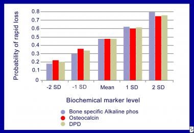
 Bone Markers in Osteoporosis: Bone Turnover Markers, Bone Formation Markers, Bone Resorption Markers
Carlos Alberto Jurado - Search Results - PubMed
The Tohoku Journal of Experimental Medicine
![Atlas of Human Embryos [by: RF Gasser, PhD.] - Ch.8](data:image/png;base64,iVBORw0KGgoAAAANSUhEUgAAABAAAAAQCAYAAAAf8/9hAAAAh0lEQVQ4jd2Suw2EMBBEHxZV+C7dPkDUQXWuA9l9OAXaWCIjn7GOwAESLx3NaPYDT9OpqrYEmNYGPUCMO+MwX8R1WwD4fqaL5oNDxGJysw/uNNVYtwUfHADjMBPjjsnNIva2soj9CemTkILyBrXqJWdArcG/HSRMOdMd5c46VdWWKzz/SC/gACJUSba7VMpqAAAAAElFTkSuQmCC) Atlas of Human Embryos [by: RF Gasser, PhD.] - Ch.8
Atlas of Human Embryos [by: RF Gasser, PhD.] - Ch.8
 New Study Shows MRI can Perform Forensic Age Estimation on Human Teeth | Bruker
 Paper: <span>Ectonucleotide Pyrophosphatase/Phosphodiesterase (NPP)1-3 May Regulate Periodontal Homeostasis. </span> (AADR...
 PDF) The Effect of Stevia Rebaudiana on Serum Omentin and Visfatin Level in STZ-Induced Diabetic Rats
 Real-Time Analysis of Temperature Changes in Composite Increments and Pulp Chamber during Photopolymerization
 Parts of a Tooth - What is it Made of?
 Histology of tooth and periodontal tissues - Histology and Embryology for Dental Hygiene
4:41 am
 Current Approaches of Bone Morphogenetic Proteins in Dentistry | Journal of Oral Implantology
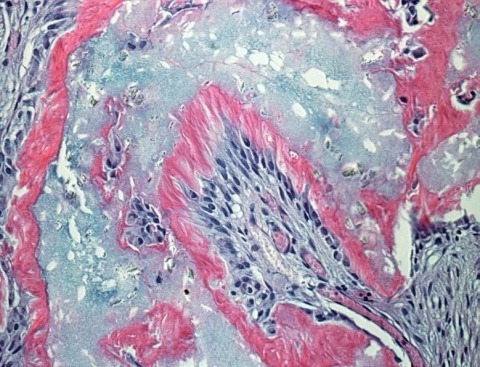
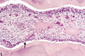
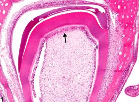
Dentin18
- In vertebrates, an odontoblast is a cell of neural crest origin that is part of the outer surface of the dental pulp, and whose biological function is dentinogenesis, which is the formation of dentin, the substance beneath the tooth enamel on the crown and the cementum on the root. (wikipedia.org)
- Odontoblasts are large columnar cells, whose cell bodies are arranged along the interface between dentin and pulp, from the crown to cervix to the root apex in a mature tooth. (wikipedia.org)
- Nutrition for odontoblasts within the dentin comes through the dentinal tubules from tissue fluid that originally traveled from the blood vessels located in the adjacent pulp tissue. (wikipedia.org)
- In primates enamel spindles were observed where the odontoblast process reaches until the border between dentin and enamel. (wikipedia.org)
- The odontoblasts secrete dentin throughout life, unlike enamel, which is considered secondary dentin once root formation is complete, which may be an attempt to compensate for natural wear of the enamel. (wikipedia.org)
- Odontoblasts also secrete tertiary dentin when irritated. (wikipedia.org)
- The most recognized function odontoblasts is the formation and maintenance of the dentin. (dent-wiki.com)
- Dentin -forming cells, odontoblasts, which originate from the ectomesenchyme, form a single layer of cells between the dentin and pulp. (intelligentdental.com)
- Odontoblasts synthesize dentin matrix during their entire lifetime. (intelligentdental.com)
- The unmineralized zone between the odontoblasts and mineralized dentin is called predentin. (intelligentdental.com)
- specifically, enamel is deposited by ameloblasts derived from the epithelium, while dentin is deposited by odontoblasts derived from mesenchymal cells. (ispub.com)
- Major part of dentin is intertubular, formed by the dentin-forming odontoblasts at the dentin-pulp border. (tannlegetidende.no)
- There are two kinds of tertiary dentin, namely reactionary dentin, formed by original odontoblasts, and reparative dentin, formed by newly differentiated replacement odontoblasts [ 4 ]. (tannlegetidende.no)
- Odontoblasts are found between the soft dental pulp and hard dentin and produce dentin. (bruker.com)
- Between the dentin and the nerve, there are odontoblasts (this part starts the formation of dentin). (healthyteeth.org)
- Osteoblasts are bone-building cells that also form callus required for bone repair (see also dentin-producing odontoblasts ). (learninggnm.com)
- This particular channel was tracked to the odontoblast cell found between the pulp of the tooth and the dentin layer that surrounds it. (trustedhealthproducts.com)
- The beginning of the crown phase is characterized by odontoblast maturation and deposition of dentin, followed by enamel secretion by ameloblasts to form the mineralized tissues of the future crown 15 . (bvsalud.org)
Differentiation6
- The differentiation of the odontoblast is done by signaling molecules and growth factors in the cells of the inner enamel epithelium. (wikipedia.org)
- Nevertheless, the effect of GDF-5 on odontoblast differentiation from dental pulp tissues remains unknown. (ispub.com)
- In this study, we assayed the effect of exogenous mouse recombinant GDF-5 on cell proliferation of dental pulp cells and determined the expression levels of odontoblast differentiation marker genes in the cells of isolated mouse dental pulp tissues in the presence of GDF-5. (ispub.com)
- These results indicate that, although GDF-5 may have no effect on cell proliferation in isolated dental pulp tissue, it could promote odontoblast differentiation. (ispub.com)
- Here, we investigated the effect of GDF-5 on the proliferation of dental pulp cells and their differentiation into odontoblasts by assaying the expression levels of odontoblast marker genes. (ispub.com)
- Natural Bioactive Epigallocatechin-Gallate Promote Bond Strength and Differentiation of Odontoblast-like Cells. (nih.gov)
Known as odontoblasts2
- Researchers at the Friedrick-Alexander University Erlangen-Numberg have found that the molecular and cellular effects of cold that create the sensations within the teeth - known as odontoblasts - house proteins that sense temperature changes and cause the signals to arrive in the brain that alert it to the sense of pain. (trustedhealthproducts.com)
- 3. The two layers of cells which are thus brought in contact, namely, the epithelial cells lining the concavity of the adamant organ, and the superficial cells of the tooth papilla, become elongated or columnar, and undergo other changes, preliminary to the production of the adamant by the former-which are now called adamant cells or ameloblasts-and the ivory by the latter, which are known as odontoblasts. (co.ma)
Columnar4
- On the other hand, columnar odontoblasts were seen at the peripheral site, and their cell bodies and processes showed strong immunoreactivity. (elsevierpure.com)
- Underneath the tubule-rich tertiary dentine, columnar odontoblasts were abundantly distributed, and the strong immunoreactivity was observed in their cell bodies and processes. (elsevierpure.com)
- Histologically, secretory odontoblasts are columnar in shape. (intelligentdental.com)
- A large number of cytoplasmic organelles are identifiable in young odontoblasts, whereas, aged odontoblasts lose their columnar shape and contain a small number of Golgi apparatus and a small-sized rough endoblasmic reticulum. (intelligentdental.com)
Osteoblasts2
- Osteocalcin is a small protein (49 amino acids) synthesized by mature osteoblasts, odontoblasts, and hypertrophic chondrocytes. (medscape.com)
- Results: In WT mice, NPP1 staining was observed in cementoblasts, osteoblasts, and odontoblasts, whereas NPP2 and NPP3 were localized to pulp and PDL tissues. (umich.edu)
Cells15
- Odontoblasts were originally the outer cells of the dental papilla. (wikipedia.org)
- Odontoblasts' are slender cylindrical cells of epithelioid character. (wikilectures.eu)
- Like many other fabrics-supporting cells, odontoblasts also contribute to the host defense. (dent-wiki.com)
- Answer leads to activation of specific receptors on the adjacent cells, blood vessels, nerves and on odontoblast itself (Advanced concept 2.1). (dent-wiki.com)
- Thus, odontoblasts, together with a local resident protection of cells and blood-borne invasion cells have a wide repertoire of response patterns and play an important role in the activation of both the innate and adaptive immune response pulp (Fig. 2.5). (dent-wiki.com)
- Odontoblasts in addition to respond to proinflammatory cytokines, allocated adjacent resident of the cells and the invasion of the cells of the immune system . (dent-wiki.com)
- S100A4 is expressed in human odontoblasts and odontoblast-like cells. (bvsalud.org)
- In both the crown and root, S100A4 staining was observed in the odontoblast layer and in odontoblast -like cells , but not in other pulp cells . (bvsalud.org)
- S100A4 is expressed in the odontoblast layer and in odontoblast -like cells in mature human pulp tissue . (bvsalud.org)
- Young odontoblasts are secretorily active and produce predentin at a faster rate than the older cells. (intelligentdental.com)
- To date, various studies have reported that BMP-family growth factors can induce cells isolated from dental pulp tissues to differentiate into odontoblasts. (ispub.com)
- The cells in the dental pulp comprise a heterogeneous mixed population, made up of odontoblasts, fibroblasts, stem cells, and macrophages and other immunocompetent cells. (ispub.com)
- 2002). It has also been reported that the GDF-5 gene is expressed in dental sac, periodontal ligament, dental pulp cells, and odontoblasts during tooth development (Morotome et al. (ispub.com)
- 4. The odontoblasts, that is the layer of columnarshaped connective tissue cells lying on the surface of the dental papilla, begin to form at their outer ends a layer of ivory (Fig. 971, IV. (co.ma)
- and in each case the two layers of cells-odontoblasts and adamant cells -which produced the deposits, retiring gradually from one another, as the space between them becomes occupied by the newly formed tissues (Fig. 971, V. (co.ma)
Fibroblasts1
- Recent observations show that odontoblasts more powerful attractant than the tooth pulp fibroblasts in this respect (67). (dent-wiki.com)
Immunoreactivity3
- Results: In intact teeth, numerous odontoblasts were aligned underneath the secondary dentine and their cell bodies showed the immunoreactivity. (elsevierpure.com)
- On the other hand, the intensity of the immunoreactivity in odontoblasts was similar underneath the secondary dentine in intact and carious teeth. (elsevierpure.com)
- Immunoreactivity of the cell membrane of the odontoblast including the odontoblastic process was also confirmed by immuno-transmission microscopic observation. (go.jp)
Teeth2
- To visualize the activity, the avidin-biotin-peroxidase complex immunostaining was performed on the odontoblasts of fresh teeth followed by embedding. (go.jp)
- The results of these experiments indicate that the odontoblasts are one of potent sources of blood group antigenicity for blood grouping of the human teeth. (go.jp)
Extracellular2
- It has been shown that odontoblasts secrete the extracellular matrix protein reelin. (wikipedia.org)
- During the phase of collagen aggregation, the odontoblasts regulate the order of collagen fibers and modify the composition of the extracellular matrix. (intelligentdental.com)
Cavity1
- The remains of the dental papilla persist as the pulp of the tooth, which is covered even in the adult by the odontoblasts, and occupies the tooth cavity, i.e. the central part of the tooth to which the formation of ivory has not extended. (co.ma)
Tissue3
- The dentinal fluid in the tubule presumably also includes the tissue fluid surrounding the cell membrane of the odontoblast, which is continuous from the cell body in the pulp. (wikipedia.org)
- Comparative gene expression profile analysis between native human odontoblasts and pulp tissue. (nih.gov)
- The odontoblasts are differentiated from the dental papilla, which remains a soft connective tissue in the tooth interior also known as the dental pulp, even after development is complete. (ispub.com)
Fibers1
- Odontoblasts are connected to each other with interodontoblastic collagen, the so-called von Korff fibers. (intelligentdental.com)
Cell4
- Figure 177 from Chapter 5 (Endoplasmic Reticulum) of 'The Cell, 2nd Ed.' by Don W. Fawcett M.D. This electron micrograph shows the supranuclear region of an odontoblast from a rat incisor, as this ce. (cellimagelibrary.org)
- Consequently, strategic peripheral position odontoblast and its varied range of models of response make the cell tractor pulp defense to both externally and internally derived adverse effects. (dent-wiki.com)
- Frequent bundles of collagen fibrils enter the odontoblast layer from predentin and are present between odontoblast cell bodies. (intelligentdental.com)
- Group specific colorization was successfully developed on the cell surface of the odontoblasts. (go.jp)
Receptors2
- With the discovery of TRPC5 as cold transducer the odontoblast transduction theory has become a likely explanation of dentinal hypersensivity The contribution of TRPC5 channels to the sensory function in odontoblasts is still controversial It has been shown that odontoblast-neuron signal communication via Piezo1/TRPA1 channels and pannexin-1 in odontoblasts and P2X3 receptors in A-delta neuron is involved in the generation of dentinal sensitivity/hypersensitivity. (wikipedia.org)
- Odontoblasts come with a lot of receptors that allow you to recognize and respond to the microbial items and thus prevent the immune system. (dent-wiki.com)
TRPA11
- Functional expression of TRPA1 channel, TRPV1 channel and TMEM100 in human odontoblasts. (nih.gov)
Crown1
- The root is composed chiefly of ivory, continuous above with that of the crown, and like it formed by the odontoblasts of the dental papilla. (co.ma)
Tooth development2
- Odontoblasts form approximately 4 μm of predentin daily during tooth development. (wikipedia.org)
- Odontoblasts first appear at sites of tooth development at 17-18 weeks in utero and remain present until death unless killed by bacterial or chemical attack, or indirectly through other means such as heat or trauma (e.g. during dental procedures). (wikipedia.org)
Epithelium1
- This electron micrograph highlights a darkly-stained glycocalyx rim of the brush border of the intestinal epithelium of the cat, stained en bloc with colloidal thorium. (cellimagelibrary.org)
Layer1
- Ultimately they pass through the odontoblast layer into pulp. (intelligentdental.com)
Nerve1
- A pulpal A-delta (noxious, short sharp pain) nerve fibre is either wrapped around the base of this process, or travels a short way into the dentinal tubule with the odontoblast process (max ~0.1 mm) This process lies in the dentinal tubule. (wikipedia.org)
English1
- odontoblast meaning in English, odontoblast का अर्थ अंग्रेजी में, odontoblast definition in English, odontoblast की परिभाषा अंग्रेजी में, odontoblast का मतलब, odontoblast माने क्या, odontoblast mane kya, odontoblast ka matlab This page is showing answer of : What is meaning of odontoblast in English? (kodand.com)
Levels1
- Odontoblast marker gene levels, on the other hand, were significantly elevated after seven days of culture in the presence of GDF-5. (ispub.com)
Wall1
- This is because of the retention of the odontoblasts within the tooth, along the outer pulpal wall. (wikipedia.org)
Create1
- To call odontoblasts create and issue a lot of molecules, which can help to defeat the invading microorganisms. (dent-wiki.com)
Issue1
- Odontoblasts may also issue of antimicrobial peptides with the possibility of direct killing of Gram-positive and Gram-negative bacteria (18). (dent-wiki.com)