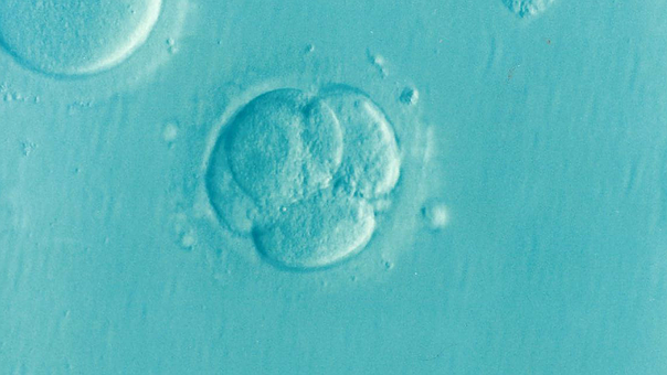
Morula
Blastocyst
Embryo, Mammalian
Cleavage Stage, Ovum
Embryo Culture Techniques
Cloning, Organism
Oocytes
Fertilization in Vitro
Embryo Transfer
Blastomeres
Parthenogenesis
Vitrification
Fetal Viability
Pregnancy
Fallopian Tubes
Culture Techniques
Cattle
Gene Expression Regulation, Developmental
Cryopreservation
Culture Media
Octamer Transcription Factor-3
Zona Pellucida
Fertilization
Mice, Inbred Strains
Oogenesis
Trophoblasts
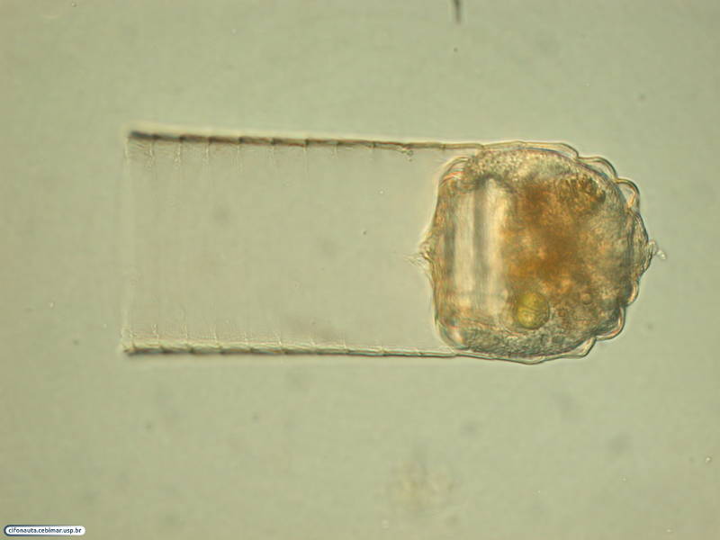
Antibodies with the cell-type specificity to the morula cells of the solitary ascidians Styela rustica and Boltenia echinata. (1/365)
The separation of the blood cells of Styela rustica (Styelidae, Stolidobranchiae) in discontinuous Percoll gradient showed 4 fractions. The 4th and bottom most fraction contained 90-100% of morula cells. The protein composition of the morula cell fraction revealed on SDS-PAGE showed two major proteins with m.w. 47 and 26 kDa. These proteins were heavily positively charged. Polyclonal antiserums against these proteins were raised. Each antiserum reacted with both proteins only in morula cells on the blot after SDS-PAGE and stained the proper protein without crossreaction on the blot after AU-PAGE. The only type of cells stained with antibodies in circulating blood, in the tunic and on the tunic wound surface in paraffin sections of another species Boltenia echinata (Pyuridae, Stolidobranchiae) were morula cells. The morula-type specific antibodies obtained recognized major positively charged proteins which were apparently structural substrates for the phenoloxidase tanning. (+info)Mammalian transgenesis by intracytoplasmic sperm injection. (2/365)
Coinjection of unfertilized mouse oocytes with sperm heads and exogenous DNA encoding either a green fluorescent protein (GFP) or beta-galactosidase reporter produced 64 to 94 percent transgene-expressing embryos, reflecting DNA-sperm head association before coinjection. Nonselective transfer to surrogate mothers of embryos in the GFP series generated about 20 percent offspring expressing the integrated transgene. These data indicate that exogenous DNA can reproducibly be delivered into an oocyte by microinjected spermatozoa and suggest an adaptable method of transgenesis. (+info)Developmental competence and metabolism of bovine embryos cultured in semi-defined and defined culture media. (3/365)
Development of in vitro-produced bovine embryos was studied in 3 two-step culture media: synthetic oviduct fluid (SOF), Gardner's G1/G2, and control (hamster embryo culture medium with 11 amino acids [HECM-6] followed by tissue culture medium 199 + 10% bovine calf serum). Modifications were made to reduce or eliminate protein. Glycolysis and Krebs cycle activity of morulae and blastocysts developed from selected immature oocytes were measured. There were no differences in development to the morula and blastocyst stages between SOF, G1/G2, or control (41%, 36%, and 46%, respectively), although more blastocysts developed in control medium than in G1/G2 (46%, 30%, respectively). Reducing or removing BSA during the initial culture period did not significantly reduce development to blastocyst (31%, 33%, respectively), although development was reduced in SOF with BSA removed from the final culture period (19%). There were no differences in development to the blastocyst stage between SOF, SOF with BSA removed during the initial culture period, and control (44%, 32%, 49%, respectively), but development was reduced in chemically defined protein-free medium throughout the culture period (21%). Krebs cycle activity did not differ between treatments; however, glycolysis was highest in the control embryos and lowest in embryos cultured in protein-free medium. Embryos that developed in the presence of serum appeared dark and granular and had elevated glycolytic rates compared to embryos developed in completely defined medium. This study shows that both metabolism and blastocyst development of embryos are altered by different culture media, implying a functional linkage between these two indicators of successful embryogenesis. (+info)Microelectrophoretic analysis of changes in protein expression patterns in mouse oocytes and preimplantation embryos. (4/365)
One- and two-dimensional polyacrylamide microslab gel electrophoresis followed by silver staining was devised to visualize picogram to nanogram levels of proteins and was applied to the analysis of 1-20 mouse oocytes and embryos (approximately 16.5-330 ng of protein) during preimplantation development. Compared with values in embryos, more bands in the higher molecular weight range were found only for unfertilized oocytes in one-dimensional microelectrophoresis. A marked decrease in the number of protein spots occurred after fertilization in two-dimensional microelectrophoresis. Both findings indicate a decrease in maternal proteins caused by fertilization. Silver-staining densities were almost invariable for 8 major spots, but increased, decreased, or varied for 32 minor spots in developing embryos from the 1-cell to the morula stage, signifying spot-specific changes in the expression of zygotic proteins during development. The protein patterns in cumulus cells and blastocysts were different from those in oocytes and embryos. Even in a single 1-cell embryo, major spots and some minor spots were detectable by our two-dimensional microelectrophoretic technique, but many more minor spots were visualized in five 1-cell embryos, exemplifying the limit of our microelectrophoretic technique. As a preliminary result, a two-dimensional immunoblot pattern is shown for glucose transporter 1 expressed in morulae. (+info)Retinol administration to superovulated ewes improves in vitro embryonic viability. (5/365)
Retinol and its metabolites, all-trans retinoic acid and 9-cis retinoid acid, are regulators of cellular growth, differentiation, and development and have been implicated in reproductive processes including folliculogenesis and embryonic survival. Three experiments were conducted to identify effects of retinoid treatment of superovulated ewes upon subsequent in vitro embryonic development. Ewes were treated with all-trans retinol (ROH), all-trans retinoic acid (RA), 9-cis retinoic acid (CIS), or vehicle (Control) on the first and last day of FSH treatment. Embryos were recovered at the morula stage, cultured in vitro for 96 h, and observed for blastocyst formation. Embryos from ROH-treated animals had a higher (p < 0.01) incidence of blastocyst formation than RA-, CIS-, or vehicle-treated animals (72% vs. 27%, 33% and 32%, respectively). In experiment 2, ewes were given ROH or vehicle and treated as above. ROH treatment resulted in an increased percentage of embryos forming blastocysts (70% vs. 22%, p < 0.05). In experiment 3, ewes were treated with ROH or vehicle, and embryos were collected at the 1- to 4-cell stage and cultured for 7 days. ROH treatment resulted in increased blastocyst formation (79% vs. 5%, p < 0.05). The majority of embryos (60% vs. 6%; p < 0.01)) from vehicle-treated animals failed to develop beyond the 8-cell stage in comparison with those from ROH animals. ROH treatment of superovulated ewes increased embryonic viability and positively impacted embryonic development. (+info)Developmentally regulated loss and reappearance of immunoreactive somatic histone H1 on chromatin of bovine morula-stage nuclei following transplantation into oocytes. (6/365)
One difference between chromatin of bovine oocytes and blastomeres is that somatic subtypes of histone H1 are undetectable in oocytes and are assembled onto embryonic chromatin during the fourth cell cycle. We investigated whether this chromatin modification is reversed when nuclei containing somatic H1 are transplanted into ooplasts. Donor nuclei obtained from morula-stage bovine embryos were fused to ooplasts at different times before and after parthenogenetic activation of the ooplasts. After fusion, immunoreactive H1 became undetectable, and the loss occurred more rapidly when fusion was performed near the time of ooplast activation compared with several hours after activation, when the host oocytes were at a stage corresponding to interphase. Although the loss of immunoreactive H1 occurred independently of DNA replication and transcription, exposure of reconstructed oocytes to cycloheximide or 6-dymethylaminopurine (6-DMAP) delayed the loss of immunoreactive H1 from transplanted nuclei. During further development of nuclear-transplant embryos, somatic H1 remained undetectable at the 2- and 4-cell stages, and it reappeared on the chromatin at the 8- to 16-cell stage, as previously observed in unmanipulated embryos. We conclude that factors in oocyte cytoplasm are able to modify morula chromatin so that somatic H1 becomes undetectable, and that the amount or activity of these factors declines over time in activated ooplasts. (+info)Transforming growth factor-beta signalling in extraembryonic mesoderm is required for yolk sac vasculogenesis in mice. (7/365)
We have analysed the function of transforming growth factor beta (TGF-beta) in yolk sac development in mice by generating somatic chimaeras in which the extraembryonic mesoderm, which gives rise to the endothelial and haematopoietic cells of the yolk sac vasculature, is derived from embryonic stem (ES) cells. The ES cells were stably transfected and express either the full-length type II binding receptor or a kinase-deficient mutant of this receptor. Examination of yolk sacs from chimaeras between E8.5 and 9.5, and analysis of marker expression in embryoid bodies from these mutant ES cell lines in prolonged suspension culture demonstrated that (1) a major function of TGF-beta in yolk sac mesoderm is to regulate production and deposition of fibronectin in the extracellular matrix that maintains yolk sac integrity, (2) TGF-beta signalling is not required for differentiation of extraembryonic mesoderm into endothelial cells but is necessary for their subsequent organisation into robust vessels, and (3) TGF-beta signalling must be tightly regulated for the differentiation of primitive haematopoietic cells to take place normally. Together, these results show that defective TGF-beta signalling in the extraembryonic mesoderm alone is sufficient to account for the extraembryonic phenotype reported previously in TGF-beta1(-/-) mice (Dickson, M. C., Martin, J. S., Cousins, F. M., Kulkarni, A. B., Karlsson, S. and Akhurst, R. J. (1995) Development 121, 1845-1854). (+info)Stage-specific expression of estrogen receptor subtypes and estrogen responsive finger protein in preimplantational mouse embryos. (8/365)
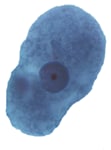
In hope of understanding possible roles of estrogen during early embryogenesis, we examined the expression of both estrogen receptor alpha (ER alpha) and ER beta, a recently cloned novel subtype, in mouse oocytes and preimplantation embryos by means of reverse transcription polymerase chain reaction (RT-PCR). To investigate whether estrogen actually exerts its action, we further determined the expression of efp (estrogen-responsive finger protein), a newly characterized estrogen responsive gene belonging to the RING finger family. ER alpha mRNA was detected in whole ovaries, cumulus-oocyte complexes, denuded oocytes, 2-cell and 4-cell embryos, whereas it was undetected in 8-cell embryos. Interestingly it reappeared in morulae and blastocysts. ER beta mRNA was detected similarly to ER alpha except for the absence of ER beta mRNA in morulae. The efp mRNA was detected in whole ovaries, cumulus-oocyte complexes, 4-cell embryos, morulae and blastocysts. The stage specific expression of ER alpha and ER beta along with detection of the product of the estrogen responsive gene in early preimplantation embryos may indicate the possible physiological roles of estrogen in early embryogenesis. (+info)A morula is a term used in embryology, which refers to the early stage of development in mammalian embryos. It is formed after fertilization when the zygote (a single cell resulting from the fusion of sperm and egg) undergoes several rounds of mitotic divisions to form a solid mass of 16 or more cells called blastomeres. At this stage, the cells are tightly packed together and have a compact, mulberry-like appearance, hence the name "morula" which is derived from the Latin word for "mulberry."
The morula stage typically occurs about 4-5 days after fertilization in humans and is marked by the beginning of blastulation, where the cells start to differentiate and become organized into an outer layer (trophoblast) and an inner cell mass. The trophoblast will eventually form the placenta, while the inner cell mass will give rise to the embryo proper.
It's important to note that the morula stage is a transient phase in embryonic development, and it represents a critical period of growth and differentiation as the embryo prepares for implantation into the uterine wall.
A blastocyst is a stage in the early development of a fertilized egg, or embryo, in mammals. It occurs about 5-6 days after fertilization and consists of an outer layer of cells called trophoblasts, which will eventually form the placenta, and an inner cell mass, which will give rise to the fetus. The blastocyst is characterized by a fluid-filled cavity called the blastocoel. This stage is critical for the implantation of the embryo into the uterine lining.
Embryonic development is the series of growth and developmental stages that occur during the formation and early growth of the embryo. In humans, this stage begins at fertilization (when the sperm and egg cell combine) and continues until the end of the 8th week of pregnancy. During this time, the fertilized egg (now called a zygote) divides and forms a blastocyst, which then implants into the uterus. The cells in the blastocyst begin to differentiate and form the three germ layers: the ectoderm, mesoderm, and endoderm. These germ layers will eventually give rise to all of the different tissues and organs in the body.
Embryonic development is a complex and highly regulated process that involves the coordinated interaction of genetic and environmental factors. It is characterized by rapid cell division, migration, and differentiation, as well as programmed cell death (apoptosis) and tissue remodeling. Abnormalities in embryonic development can lead to birth defects or other developmental disorders.
It's important to note that the term "embryo" is used to describe the developing organism from fertilization until the end of the 8th week of pregnancy in humans, after which it is called a fetus.
Embryonic and fetal development is the process of growth and development that occurs from fertilization of the egg (conception) to birth. The terms "embryo" and "fetus" are used to describe different stages of this development:
* Embryonic development: This stage begins at fertilization and continues until the end of the 8th week of pregnancy. During this time, the fertilized egg (zygote) divides and forms a blastocyst, which implants in the uterus and begins to develop into a complex structure called an embryo. The embryo consists of three layers of cells that will eventually form all of the organs and tissues of the body. During this stage, the basic structures of the body, including the nervous system, heart, and gastrointestinal tract, begin to form.
* Fetal development: This stage begins at the end of the 8th week of pregnancy and continues until birth. During this time, the embryo is called a fetus, and it grows and develops rapidly. The organs and tissues that were formed during the embryonic stage continue to mature and become more complex. The fetus also begins to move and kick, and it can hear and respond to sounds from outside the womb.
Overall, embryonic and fetal development is a complex and highly regulated process that involves the coordinated growth and differentiation of cells and tissues. It is a critical period of development that lays the foundation for the health and well-being of the individual throughout their life.
A mammalian embryo is the developing offspring of a mammal, from the time of implantation of the fertilized egg (blastocyst) in the uterus until the end of the eighth week of gestation. During this period, the embryo undergoes rapid cell division and organ differentiation to form a complex structure with all the major organs and systems in place. This stage is followed by fetal development, which continues until birth. The study of mammalian embryos is important for understanding human development, evolution, and reproductive biology.
The cleavage stage of an ovum, also known as a fertilized egg, refers to the series of rapid cell divisions that occur after fertilization. During this stage, the single cell (zygote) divides into multiple cells, forming a blastomere. This process occurs in the fallopian tube and continues until the blastocyst reaches the uterus, typically around 5-6 days after fertilization. The cleavage stage is a critical period in early embryonic development, as any abnormalities during this time can lead to implantation failure or developmental defects.
Embryo culture techniques refer to the methods and procedures used to maintain and support the growth and development of an embryo outside of the womb, typically in a laboratory setting. These techniques are often used in the context of assisted reproductive technologies (ART), such as in vitro fertilization (IVF).
The process typically involves fertilizing an egg with sperm in a laboratory dish and then carefully monitoring and maintaining the resulting embryo in a specialized culture medium that provides the necessary nutrients, hormones, and other factors to support its development. The culture medium is usually contained within an incubator that maintains optimal temperature, humidity, and gas concentrations to mimic the environment inside the body.
Embryologists may use various embryo culture techniques depending on the stage of development and the specific needs of the embryo. For example, some techniques involve culturing the embryo in a single layer, while others may use a technique called "co-culture" that involves growing the embryo on a layer of cells to provide additional support and nutrients.
The goal of embryo culture techniques is to promote the healthy growth and development of the embryo, increasing the chances of a successful pregnancy and live birth. However, it's important to note that these techniques are not without risk, and there are potential ethical considerations surrounding the use of ART and embryo culture.
Cloning of an organism is the process of creating a genetically identical copy of an entire living organism, including all of its DNA. This is achieved through a variety of laboratory techniques that can vary depending on the type of organism being cloned. In the case of animals, one common method is called somatic cell nuclear transfer (SCNT).
In SCNT, the nucleus of a donor animal's cell (which contains its DNA) is removed and transferred into an egg cell that has had its own nucleus removed. The egg cell is then stimulated to divide and grow, resulting in an embryo that is genetically identical to the donor animal. This embryo can be implanted into a surrogate mother, where it will continue to develop until birth.
Cloning of organisms has raised ethical concerns and debates, particularly in the case of animals, due to questions about the welfare of cloned animals and the potential implications for human cloning. However, cloning is also seen as having potential benefits, such as the ability to produce genetically identical animals for research or agricultural purposes.
It's important to note that while cloning can create genetically identical organisms, it does not necessarily mean that they will be identical in every way, as environmental factors and random genetic mutations can still result in differences between clones.
A zygote is the initial cell formed when a sperm fertilizes an egg, also known as an oocyte. This occurs in the process of human reproduction and marks the beginning of a new genetic identity, containing 46 chromosomes - 23 from the sperm and 23 from the egg. The zygote starts the journey of cell division and growth, eventually developing into a blastocyst, then an embryo, and finally a fetus over the course of pregnancy.
An oocyte, also known as an egg cell or female gamete, is a large specialized cell found in the ovary of female organisms. It contains half the number of chromosomes as a normal diploid cell, as it is the product of meiotic division. Oocytes are surrounded by follicle cells and are responsible for the production of female offspring upon fertilization with sperm. The term "oocyte" specifically refers to the immature egg cell before it reaches full maturity and is ready for fertilization, at which point it is referred to as an ovum or egg.
Fertilization in vitro, also known as in-vitro fertilization (IVF), is a medical procedure where an egg (oocyte) and sperm are combined in a laboratory dish to facilitate fertilization. The fertilized egg (embryo) is then transferred to a uterus with the hope of establishing a successful pregnancy. This procedure is often used when other assisted reproductive technologies have been unsuccessful or are not applicable, such as in cases of blocked fallopian tubes, severe male factor infertility, and unexplained infertility. The process involves ovarian stimulation, egg retrieval, fertilization, embryo culture, and embryo transfer. In some cases, additional techniques such as intracytoplasmic sperm injection (ICSI) or preimplantation genetic testing (PGT) may be used to increase the chances of success.
Embryo transfer is a medical procedure that involves the transfer of an embryo, which is typically created through in vitro fertilization (IVF), into the uterus of a woman with the aim of establishing a pregnancy. The embryo may be created using the intended parent's own sperm and eggs or those from donors. After fertilization and early cell division, the resulting embryo is transferred into the uterus of the recipient mother through a thin catheter that is inserted through the cervix. This procedure is typically performed under ultrasound guidance to ensure proper placement of the embryo. Embryo transfer is a key step in assisted reproductive technology (ART) and is often used as a treatment for infertility.
Blastomeres are early stage embryonic cells that result from the initial rounds of cell division in a fertilized egg, also known as a zygote. These cells are typically smaller and have a more simple organization compared to more mature cells. They are important for the normal development of the embryo and contribute to the formation of the blastocyst, which is an early stage embryonic structure that will eventually give rise to the fetus. The process of cell division that produces blastomeres is called cleavage.
Parthenogenesis is a form of asexual reproduction in which offspring develop from unfertilized eggs or ovums. It occurs naturally in some plant and insect species, as well as a few vertebrates such as reptiles and fish. Parthenogenesis does not involve the fusion of sperm and egg cells; instead, the development of offspring is initiated by some other trigger, such as a chemical or physical stimulus. This type of reproduction results in offspring that are genetically identical to the parent organism. In humans and other mammals, parthenogenesis is not a natural occurrence and would require scientific intervention to induce.
Vitrification is a process used in cryopreservation, where a liquid or semi-liquid biological material is transformed into a glass-like solid state by cooling it to extremely low temperatures at a rate that suppresses the formation of ice crystals. This technique is often used in assisted reproductive technology (ART) for preserving oocytes (human eggs), embryos, and ovarian or testicular tissues.
During vitrification, the biological material is exposed to high concentrations of cryoprotectants, which help prevent ice crystal formation and minimize cellular damage during cooling. The sample is then rapidly cooled using liquid nitrogen, achieving temperatures below -150°C (-238°F) in a matter of seconds or minutes.
The primary advantage of vitrification over traditional slow-freezing methods is the elimination of ice crystal formation, which can cause significant damage to cellular structures and organelles. Vitrified samples maintain their structural integrity and have higher survival rates upon thawing, making them more suitable for use in ART procedures.
However, it's important to note that vitrification also has potential risks, such as the toxicity of high cryoprotectant concentrations and the possibility of cracking during cooling or warming due to thermal stress. Proper technique and careful handling are crucial to ensure successful vitrification and subsequent use in clinical applications.
Fetal viability is the point in pregnancy at which a fetus is considered capable of surviving outside the uterus, given appropriate medical support. Although there is no precise gestational age that defines fetal viability, it is generally considered to occur between 24 and 28 weeks of gestation. At this stage, the fetus has developed sufficient lung maturity and body weight, and the risk of neonatal mortality and morbidity significantly decreases. However, the exact definition of fetal viability may vary depending on regional standards, medical facilities, and individual clinical assessments.
Pregnancy is a physiological state or condition where a fertilized egg (zygote) successfully implants and grows in the uterus of a woman, leading to the development of an embryo and finally a fetus. This process typically spans approximately 40 weeks, divided into three trimesters, and culminates in childbirth. Throughout this period, numerous hormonal and physical changes occur to support the growing offspring, including uterine enlargement, breast development, and various maternal adaptations to ensure the fetus's optimal growth and well-being.
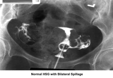
The Fallopian tubes, also known as uterine tubes or oviducts, are a pair of slender tubular structures in the female reproductive system. They play a crucial role in human reproduction by providing a passageway for the egg (ovum) from the ovary to the uterus (womb).
Each Fallopian tube is typically around 7.6 to 10 centimeters long and consists of four parts: the interstitial part, the isthmus, the ampulla, and the infundibulum. The fimbriated end of the infundibulum, which resembles a fringe or frill, surrounds and captures the released egg from the ovary during ovulation.
Fertilization usually occurs in the ampulla when sperm meets the egg after sexual intercourse. Once fertilized, the zygote (fertilized egg) travels through the Fallopian tube toward the uterus for implantation and further development. The cilia lining the inner surface of the Fallopian tubes help propel the egg and the zygote along their journey.
In some cases, abnormalities or blockages in the Fallopian tubes can lead to infertility or ectopic pregnancies, which are pregnancies that develop outside the uterus, typically within the Fallopian tube itself.
Culture techniques are methods used in microbiology to grow and multiply microorganisms, such as bacteria, fungi, or viruses, in a controlled laboratory environment. These techniques allow for the isolation, identification, and study of specific microorganisms, which is essential for diagnostic purposes, research, and development of medical treatments.
The most common culture technique involves inoculating a sterile growth medium with a sample suspected to contain microorganisms. The growth medium can be solid or liquid and contains nutrients that support the growth of the microorganisms. Common solid growth media include agar plates, while liquid growth media are used for broth cultures.
Once inoculated, the growth medium is incubated at a temperature that favors the growth of the microorganisms being studied. During incubation, the microorganisms multiply and form visible colonies on the solid growth medium or turbid growth in the liquid growth medium. The size, shape, color, and other characteristics of the colonies can provide important clues about the identity of the microorganism.
Other culture techniques include selective and differential media, which are designed to inhibit the growth of certain types of microorganisms while promoting the growth of others, allowing for the isolation and identification of specific pathogens. Enrichment cultures involve adding specific nutrients or factors to a sample to promote the growth of a particular type of microorganism.
Overall, culture techniques are essential tools in microbiology and play a critical role in medical diagnostics, research, and public health.
Embryo implantation is the process by which a fertilized egg, or embryo, becomes attached to the wall of the uterus (endometrium) and begins to receive nutrients from the mother's blood supply. This process typically occurs about 6-10 days after fertilization and is a critical step in the establishment of a successful pregnancy.
During implantation, the embryo secretes enzymes that help it to burrow into the endometrium, while the endometrium responds by producing receptors for the embryo's enzymes and increasing blood flow to the area. The embryo then begins to grow and develop, eventually forming the placenta, which will provide nutrients and oxygen to the developing fetus throughout pregnancy.
Implantation is a complex process that requires precise timing and coordination between the embryo and the mother's body. Factors such as age, hormonal imbalances, and uterine abnormalities can affect implantation and increase the risk of miscarriage or difficulty becoming pregnant.
"Cattle" is a term used in the agricultural and veterinary fields to refer to domesticated animals of the genus *Bos*, primarily *Bos taurus* (European cattle) and *Bos indicus* (Zebu). These animals are often raised for meat, milk, leather, and labor. They are also known as bovines or cows (for females), bulls (intact males), and steers/bullocks (castrated males). However, in a strict medical definition, "cattle" does not apply to humans or other animals.
Developmental gene expression regulation refers to the processes that control the activation or repression of specific genes during embryonic and fetal development. These regulatory mechanisms ensure that genes are expressed at the right time, in the right cells, and at appropriate levels to guide proper growth, differentiation, and morphogenesis of an organism.
Developmental gene expression regulation is a complex and dynamic process involving various molecular players, such as transcription factors, chromatin modifiers, non-coding RNAs, and signaling molecules. These regulators can interact with cis-regulatory elements, like enhancers and promoters, to fine-tune the spatiotemporal patterns of gene expression during development.
Dysregulation of developmental gene expression can lead to various congenital disorders and developmental abnormalities. Therefore, understanding the principles and mechanisms governing developmental gene expression regulation is crucial for uncovering the etiology of developmental diseases and devising potential therapeutic strategies.
Cryopreservation is a medical procedure that involves the preservation of cells, tissues, or organs by cooling them to very low temperatures, typically below -150°C. This is usually achieved using liquid nitrogen. The low temperature slows down or stops biological activity, including chemical reactions and cellular metabolism, which helps to prevent damage and decay.
The cells, tissues, or organs that are being cryopreserved must be treated with a cryoprotectant solution before cooling to prevent the formation of ice crystals, which can cause significant damage. Once cooled, the samples are stored in specialized containers or tanks until they are needed for use.
Cryopreservation is commonly used in assisted reproductive technologies, such as the preservation of sperm, eggs, and embryos for fertility treatments. It is also used in research, including the storage of cell lines and stem cells, and in clinical settings, such as the preservation of skin grafts and corneas for transplantation.
Culture media is a substance that is used to support the growth of microorganisms or cells in an artificial environment, such as a petri dish or test tube. It typically contains nutrients and other factors that are necessary for the growth and survival of the organisms being cultured. There are many different types of culture media, each with its own specific formulation and intended use. Some common examples include blood agar, which is used to culture bacteria; Sabouraud dextrose agar, which is used to culture fungi; and Eagle's minimum essential medium, which is used to culture animal cells.
Octamer Transcription Factor-3 (OTF-3 or Oct3) is a specific protein that belongs to the class of POU domain transcription factors. These proteins play crucial roles in the regulation of gene expression during cell growth, development, and differentiation. The "POU" name refers to the presence of two conserved domains - a POU-specific domain and a POU homeodomain - that recognize and bind to specific DNA sequences called octamer motifs, which are involved in controlling the transcription of target genes.
Oct3, encoded by the Pou2f1 gene, is widely expressed in various tissues, including lymphoid cells, neurons, and embryonic stem cells. It has been shown to regulate the expression of several genes that are essential for cell survival, proliferation, and differentiation. Dysregulation of Oct3 has been implicated in several diseases, such as cancers and neurological disorders.
In summary, Octamer Transcription Factor-3 (Oct3) is a POU domain transcription factor that binds to octamer motifs in DNA and regulates the expression of target genes involved in cell growth, development, and differentiation.
An ovum is the female reproductive cell, or gamete, produced in the ovaries. It is also known as an egg cell and is released from the ovary during ovulation. When fertilized by a sperm, it becomes a zygote, which can develop into a fetus. The ovum contains half the genetic material necessary to create a new individual.
Zona pellucida is a term used in the field of reproductive biology and it refers to the glycoprotein membrane that surrounds mammalian oocytes (immature egg cells). This membrane plays a crucial role in the fertilization process. It has receptors for sperm, and upon binding with the sperm, it undergoes changes that prevent other sperm from entering, a process known as the zona reaction. This membrane is also involved in the early development of the embryo.
Fertilization is the process by which a sperm cell (spermatozoon) penetrates and fuses with an egg cell (ovum), resulting in the formation of a zygote. This fusion of genetic material from both the male and female gametes initiates the development of a new organism. In human biology, fertilization typically occurs in the fallopian tube after sexual intercourse, when a single sperm out of millions is able to reach and penetrate the egg released from the ovary during ovulation. The successful fusion of these two gametes marks the beginning of pregnancy.
Inbred strains of mice are defined as lines of mice that have been brother-sister mated for at least 20 consecutive generations. This results in a high degree of homozygosity, where the mice of an inbred strain are genetically identical to one another, with the exception of spontaneous mutations.
Inbred strains of mice are widely used in biomedical research due to their genetic uniformity and stability, which makes them useful for studying the genetic basis of various traits, diseases, and biological processes. They also provide a consistent and reproducible experimental system, as compared to outbred or genetically heterogeneous populations.
Some commonly used inbred strains of mice include C57BL/6J, BALB/cByJ, DBA/2J, and 129SvEv. Each strain has its own unique genetic background and phenotypic characteristics, which can influence the results of experiments. Therefore, it is important to choose the appropriate inbred strain for a given research question.
Oogenesis is the biological process of formation and maturation of female gametes, or ova or egg cells, in the ovary. It begins during fetal development and continues throughout a woman's reproductive years. The process involves the division and differentiation of a germ cell (oogonium) into an immature ovum (oocyte), which then undergoes meiotic division to form a mature ovum capable of being fertilized by sperm.
The main steps in oogenesis include:
1. Multiplication phase: The oogonia divide mitotically to increase their number.
2. Growth phase: One of the oogonia becomes primary oocyte and starts to grow, accumulating nutrients and organelles required for future development.
3. First meiotic division: The primary oocyte undergoes an incomplete first meiotic division, resulting in two haploid cells - a secondary oocyte and a smaller cell called the first polar body. This division is arrested in prophase I until puberty.
4. Second meiotic division: At ovulation or just before fertilization, the secondary oocyte completes the second meiotic division, producing another small cell, the second polar body, and a mature ovum (egg) with 23 chromosomes.
5. Fertilization: The mature ovum can be fertilized by a sperm, restoring the normal diploid number of chromosomes in the resulting zygote.
Oogenesis is a complex and highly regulated process that involves various hormonal signals and cellular interactions to ensure proper development and maturation of female gametes for successful reproduction.
Trophoblasts are specialized cells that make up the outer layer of a blastocyst, which is a hollow ball of cells that forms in the earliest stages of embryonic development. In humans, this process occurs about 5-6 days after fertilization. The blastocyst consists of an inner cell mass (which will eventually become the embryo) and an outer layer of trophoblasts.
Trophoblasts play a crucial role in implantation, which is the process by which the blastocyst attaches to and invades the lining of the uterus. Once implanted, the trophoblasts differentiate into two main layers: the cytotrophoblasts (which are closer to the inner cell mass) and the syncytiotrophoblasts (which form a multinucleated layer that is in direct contact with the maternal tissues).
The cytotrophoblasts proliferate and fuse to form the syncytiotrophoblasts, which have several important functions. They secrete enzymes that help to degrade and remodel the extracellular matrix of the uterine lining, allowing the blastocyst to implant more deeply. They also form a barrier between the maternal and fetal tissues, helping to protect the developing embryo from the mother's immune system.
Additionally, trophoblasts are responsible for the formation of the placenta, which provides nutrients and oxygen to the developing fetus and removes waste products. The syncytiotrophoblasts in particular play a key role in this process by secreting hormones such as human chorionic gonadotropin (hCG), which helps to maintain pregnancy, and by forming blood vessels that allow for the exchange of nutrients and waste between the mother and fetus.
Abnormalities in trophoblast development or function can lead to a variety of pregnancy-related complications, including preeclampsia, intrauterine growth restriction, and gestational trophoblastic diseases such as hydatidiform moles and choriocarcinomas.
 Morula striata
 The Morula Solution?
 logo-morula
 Morula rodgersi
 Morula Capital Partners
 Makassar - Morula IVF
 Microshells: Morula granulata レイシダマシ
 Integrated Solutions - Morula Health
 Web Design Companies Morula View
 rnav - Morula IVF Research - Fertility Lab
 Unscramble MORULAE - 107 Words You Can Make
 Anatomy and Physiology of Animals/Reproductive System - Wikibooks, open books for an open world
 featured poem: Survive on Poetry potion! by Morula Wa Kutukgolo - Poetry Potion
 Embryogenesis: Cleavage and Blastulation | Biology | JoVE
 De novo DNA methylation during monkey pre-implantation embryogenesis | Cell Research
 2023 All Shows - National Arts Festival
 Fertilization: Your pregnancy week by week
 Baby’s Pregnancy Calendar
 Diagnosis and Management of Tickborne Rickettsial Diseases: Rocky Mountain Spotted Fever and Other Spotted Fever Group...
 RIA: Development and quality of bovine morulae cultured in serum-free medium with retinoic receptor specific agonists