Molecular Probe Techniques
Molecular Probes
DNA Probes
Molecular Imaging
SELEX Aptamer Technique
Fluorescent Dyes
Optical Phenomena
Oligonucleotide Probes
Tomography, Optical
Aptamers, Nucleotide
Carbocyanines
Spectroscopy, Near-Infrared
Diagnostic Imaging
Nucleic Acid Hybridization
Molecular Sequence Data
Microscopy, Fluorescence
Base Sequence
Molecular Structure
Fluorescence
Fluoresceins
Tissue Distribution
Organometallic Compounds
Amino Acid Sequence
Structure-Activity Relationship
Small Molecule Libraries
Drug Design
Fluorescence Resonance Energy Transfer
DNA
Statistical analysis of array expression data as applied to the problem of tamoxifen resistance. (1/849)
BACKGROUND: Although the emerging complementary DNA (cDNA) array technology holds great promise to discern complex patterns of gene expression, its novelty means that there are no well-established standards to guide analysis and interpretation of the data that it produces. We have used preliminary data generated with the CLONTECH Atlas human cDNA array to develop a practical approach to the statistical analysis of these data by studying changes in gene expression during the development of acquired tamoxifen resistance in breast cancer. METHODS: For hybridization to the array, we prepared RNA from MCF-7 human breast cell tumors, isolated from our athymic nude mouse xenograft model of acquired tamoxifen resistance during estrogen-stimulated, tamoxifen-sensitive, and tamoxifen-resistant growth. Principal components analysis was used to identify genes with altered expression. RESULTS AND CONCLUSIONS: Principal components analysis yielded three principal components that are interpreted as 1) the average level of gene expression, 2) the difference between estrogen-stimulated gene expression and the average of tamoxifen-sensitive and tamoxifen-resistant gene expression, and 3) the difference between tamoxifen-sensitive and tamoxifen-resistant gene expression. A bivariate (second and third principal components) 99% prediction region was used to identify outlier genes that exhibit altered expression. Two representative outlier genes, erk-2 and HSF-1 (heat shock transcription factor-1), were chosen for confirmatory study, and their predicted relative expression levels were confirmed in western blot analysis, suggesting that semiquantitative estimates are possible with array technology. IMPLICATIONS: Principal components analysis provides a useful and practical method to analyze gene expression data from a cDNA array. The method can identify broad patterns of expression alteration and, based on a small simulation study, will likely provide reasonable power to detect moderate-sized alterations in clinically relevant genes. (+info)Identification of Mycobacterium kansasii by using a DNA probe (AccuProbe) and molecular techniques. (2/849)
The newly formulated Mycobacterium kansasii AccuProbe was evaluated, and the results obtained with the new version were compared to the results obtained with the old version of this test by using 116 M. kansasii strains, 1 Mycobacterium gastri strain, and 19 strains of several mycobacterial species. The sensitivity of this new formulation was 97.4% and the specificity was 100%. Still, three M. kansasii strains were missed by this probe. To evaluate the variability within the species, genetic analyses of the hsp65 gene, the spacer sequence between the 16S and 23S rRNA genes, and the 16S rRNA gene of several M. kansasii AccuProbe-positive strains as well as all AccuProbe-negative strains were performed. Genetic analyses of the one M. gastri strain from the comparative assay and of two further M. gastri strains were included because of the identity of the 16S rRNA gene in M. gastri to that in M. kansasii. The data confirmed the genetic heterogeneity of M. kansasii. Furthermore, a subspecies with an unpublished hsp65 restriction pattern and spacer sequence was described. The genetic data indicate that all M. kansasii strains missed by the AccuProbe test belong to one subspecies, the newly described subspecies VI, as determined by the hsp65 restriction pattern and the spacer sequence. Since the M. kansasii strains that are missed are rare and all M. gastri strains are correctly negative, the new formulated AccuProbe provides a useful tool for the identification of M. kansasii. (+info)Molecular probes for muscarinic receptors: functionalized congeners of selective muscarinic antagonists. (3/849)
The muscarinic agonist oxotremorine and the tricyclic muscarinic antagonists pirenzepine and telenzepine have been derivatized using a functionalized congener approach for the purpose of synthesizing high affinity ligand probes that are suitable for conjugation with prosthetic groups, for receptor cross-linking, fluorescent and radioactive detection, etc. A novel fluorescent conjugate of TAC (telenzepine amine congener), an n-decylamino derivative of the m1-selective antagonist, with the fluorescent trisulfonated pyrene dye Cascade Blue may be useful for assaying the receptor as an alternative to radiotracers. In a rat m3 receptor mutant containing a single amino acid substitution in the sixth transmembrane domain (Asn507 to Ala) the parent telenzepine lost 636-fold in affinity, while TAC lost only 27-fold. Thus, the decylamino group of TAC stabilizes the bound state and thus enhances potency by acting as a distal anchor in the receptor binding site. We have built a computer-assisted molecular model of the transmembrane regions of muscarinic receptors based on homology with the G-protein coupled receptor rhodopsin, for which a low resolution structure is known. We have coordinated the antagonist pharmacophore (tricyclic and piperazine moieties) with residues of the third and seventh helices of the rat m3 receptor. Although the decylamino chain of TAC is likely to be highly flexible and may adopt many conformations, we located one possible site for a salt bridge formation with the positively charged -NH3+ group, i.e. Asp113 in helix II. (+info)Silver/silver chloride electrodes for measurement of potential difference in human bronchi. (4/849)
BACKGROUND: An easy and reliable method to measure potential difference (PD) in the lower airways would be of interest in the field of cystic fibrosis. We have developed silver/silver chloride (Ag/AgCl) electrodes to measure PD in the lower airways. METHODS: To validate this technique the nasal PD measured with Ag/AgCl electrodes and with conventional perfused electrodes was compared in 16 patients. The range of PD measured with Ag/AgCl electrodes in the lower airways during fibreoptic bronchoscopy was determined in 14 adult patients and in nine the reproducibility of this technique was examined. RESULTS: Nasal PD values measured with Ag/AgCl and perfused electrodes were highly correlated (r = 0.985, p < 0.0001) and the limits of agreement (mean +/- 2SD of the difference) between the two methods were -1.91 mV and 1.53 mV. In the lower airways a progressive and slight decrease in PD values with decreasing airway diameter was observed in most patients. The mean (2SD) of the differences between the two tracheal measurements was 0.21 (1.73) mV. CONCLUSIONS: The use of Ag/AgCl electrodes gives a reliable and reproducible measurement of PD in the lower airways in humans. (+info)Identification of protease and rpoN-associated genes of uropathogenic Proteus mirabilis by negative selection in a mouse model of ascending urinary tract infection. (5/849)
Proteus mirabilis, a motile gram-negative bacterium, is a principal cause of urinary tract infections in patients with functional or anatomical abnormalities of the urinary tract or those with urinary catheters in place. Thus far, virulence factors including urease, flagella, haemolysin, various fimbriae, IgA protease and a deaminase have been characterized based on the phenotypic traits conferred by these proteins. In this study, an attempt was made to identify new virulence genes of P. mirabilis that may not have identifiable phenotypes using the recently described technique of signature-tagged mutagenesis. A pool of chromosomal transposon mutants was made through conjugation and kanamycin/tetracycline selection; random insertion was confirmed by Southern blotting of chromosomal DNA isolated from 16 mutants using the aphA gene as a probe. From the total pool, 2.3% (9/397) auxotrophic mutants and 3.5% (14/397) swarming mutants were identified by screening on minimal salts agar and Luria agar plates, respectively. Thirty per cent of the mutants, found to have either no tag or an unamplifiable tag, were removed from the input pool. Then 10(7) c.f.u. from a 96-mutant pool (approximately 10(5) c.f.u. of each mutant) were used as an input pool to transurethrally inoculate seven CBA mice. After 2 d infection, bacteria were recovered from the bladders and kidneys and yielded about 10(5) c.f.u. as an output pool. Dot blot analysis showed that two of the 96 mutants, designated B2 and B5, could not be hybridized by signature tags amplified from the bladder output pool. Interrupted genes from these two mutants were cloned and sequenced. The interrupted gene in B2 predicts a polypeptide of 37.3 kDa that shares amino acid similarity with a putative protease or collagenase precursor. The gene in B5 predicts a polypeptide of 32.6 kDa that is very similar to that encoded by ORF284 of the rpoN operon controlling expression of nitrogen-regulated genes from several bacterial species. The virulence of the two mutants was tested further by co-challenging CBA mice with each mutant and the parental strain. After 1 week of infection, the B2 and B5 mutants were recovered in numbers 100-fold and 1000-fold less than the parental strain, respectively. Using an in vitro assay, it was shown that the B2 mutant had significantly less (P = 0.0001) extracellular protease activity than the wild-type strain. These findings demonstrate that signature-tagged mutagenesis is a viable approach to identify bacterial genes associated with the ability to infect the urinary tract. (+info)Cellular delivery of peptide nucleic acids and inhibition of human telomerase. (6/849)
BACKGROUND: Human telomerase has an essential RNA component and is an ideal target for developing rules correlating oligonucleotide chemistry with disruption of biological function. Similarly, peptide nucleic acids (PNAs), DNA analogs that bind complementary sequences with high affinity, are outstanding candidates for inducing phenotypic changes through hybridization. RESULTS: We identify PNAs directed to nontemplate regions of the telomerase RNA that can overcome RNA secondary structure and inhibit telomerase by intercepting the RNA component prior to holoenzyme assembly. Relative potencies of inhibition delineate putative structural domains. We describe a novel protocol for introducing PNAs into eukaryotic cells and report efficient inhibition of cellular telomerase by PNAs. CONCLUSIONS: PNAs directed to nontemplate regions are a new class of telomerase inhibitor and may contribute to the development of novel antiproliferative agents. The dependence of inhibition by nontemplate-directed PNAs on target sequence suggests that PNAs have great potential for mapping nucleic acid structure and predictably regulating biological processes. Our simple method for introducing PNAs into cells will not only be useful for probing the complex biology surrounding telomere length maintenance but can be broadly applied for controlling gene expression and functional genomics. (+info)General properties and phylogenetic utilities of nuclear ribosomal DNA and mitochondrial DNA commonly used in molecular systematics. (7/849)
To choose one or more appropriate molecular markers or gene regions for resolving a particular systematic question among the organisms at a certain categorical level is still a very difficult process. The primary goal of this review, therefore, is to provide a theoretical information in choosing one or more molecular markers or gene regions by illustrating general properties and phylogenetic utilities of nuclear ribosomal DNA (rDNA) and mitochondrial DNA (mtDNA) that have been most commonly used for phylogenetic researches. The highly conserved molecular markers and/or gene regions are useful for investigating phylogenetic relationships at higher categorical levels (deep branches of evolutionary history). On the other hand, the hypervariable molecular markers and/or gene regions are useful for elucidating phylogenetic relationships at lower categorical levels (recently diverged branches). In summary, different selective forces have led to the evolution of various molecular markers or gene regions with varying degrees of sequence conservation. Thus, appropriate molecular markers or gene regions should be chosen with even greater caution to deduce true phylogenetic relationships over a broad taxonomic spectrum. (+info)Calculation of the relative geometry of tRNAs in the ribosome from directed hydroxyl-radical probing data. (8/849)
The many interactions of tRNA with the ribosome are fundamental to protein synthesis. During the peptidyl transferase reaction, the acceptor ends of the aminoacyl and peptidyl tRNAs must be in close proximity to allow peptide bond formation, and their respective anticodons must base pair simultaneously with adjacent trinucleotide codons on the mRNA. The two tRNAs in this state can be arranged in two nonequivalent general configurations called the R and S orientations, many versions of which have been proposed for the geometry of tRNAs in the ribosome. Here, we report the combined use of computational analysis and tethered hydroxyl-radical probing to constrain their arrangement. We used Fe(II) tethered to the 5' end of anticodon stem-loop analogs (ASLs) of tRNA and to the 5' end of deacylated tRNA(Phe) to generate hydroxyl radicals that probe proximal positions in the backbone of adjacent tRNAs in the 70S ribosome. We inferred probe-target distances from the resulting RNA strand cleavage intensities and used these to calculate the mutual arrangement of A-site and P-site tRNAs in the ribosome, using three different structure estimation algorithms. The two tRNAs are constrained to the S configuration with an angle of about 45 degrees between the respective planes of the molecules. The terminal phosphates of 3'CCA are separated by 23 A when using the tRNA crystal conformations, and the anticodon arms of the two tRNAs are sufficiently close to interact with adjacent codons in mRNA. (+info)Molecular probe techniques are analytical methods used in molecular biology and medicine to detect, analyze, and visualize specific biological molecules or cellular structures within cells, tissues, or bodily fluids. These techniques typically involve the use of labeled probes that bind selectively to target molecules, allowing for their detection and quantification.
A molecular probe is a small molecule or biomacromolecule (such as DNA, RNA, peptide, or antibody) that has been tagged with a detectable label, such as a fluorescent dye, radioisotope, enzyme, or magnetic particle. The probe is designed to recognize and bind to a specific target molecule, such as a gene, protein, or metabolite, through complementary base pairing, antigen-antibody interactions, or other forms of molecular recognition.
Molecular probe techniques can be broadly classified into two categories:
1. In situ hybridization (ISH): This technique involves the use of labeled DNA or RNA probes to detect specific nucleic acid sequences within cells or tissues. The probes are designed to complement the target sequence and, upon hybridization, allow for the visualization of the location and quantity of the target molecule using various detection methods, such as fluorescence microscopy, brightfield microscopy, or radioisotopic imaging.
2. Immunohistochemistry (IHC) and immunofluorescence (IF): These techniques utilize antibodies as probes to detect specific proteins within cells or tissues. Primary antibodies are raised against a target protein and, upon binding, can be detected using various methods, such as enzyme-linked secondary antibodies, fluorescent dyes, or gold nanoparticles. IHC is typically used for brightfield microscopy, while IF is used for fluorescence microscopy.
Molecular probe techniques have numerous applications in basic research, diagnostics, and therapeutics, including gene expression analysis, protein localization, disease diagnosis, drug development, and targeted therapy.
Molecular probes, also known as bioprobes or molecular tracers, are molecules that are used to detect and visualize specific biological targets or processes within cells, tissues, or organisms. These probes can be labeled with a variety of detection methods such as fluorescence, radioactivity, or enzymatic activity. They can bind to specific biomolecules such as DNA, RNA, proteins, or lipids and are used in various fields including molecular biology, cell biology, diagnostic medicine, and medical research.
For example, a fluorescent molecular probe may be designed to bind specifically to a certain protein in a living cell. When the probe binds to its target, it emits a detectable signal that can be observed under a microscope, allowing researchers to track the location and behavior of the protein within the cell.
Molecular probes are valuable tools for understanding biological systems at the molecular level, enabling researchers to study complex processes such as gene expression, signal transduction, and metabolism in real-time. They can also be used in clinical settings for diagnostic purposes, such as detecting specific biomarkers of disease or monitoring the effectiveness of therapies.
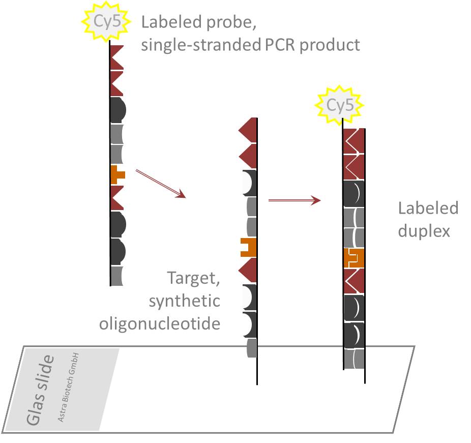
A DNA probe is a single-stranded DNA molecule that contains a specific sequence of nucleotides, and is labeled with a detectable marker such as a radioisotope or a fluorescent dye. It is used in molecular biology to identify and locate a complementary sequence within a sample of DNA. The probe hybridizes (forms a stable double-stranded structure) with its complementary sequence through base pairing, allowing for the detection and analysis of the target DNA. This technique is widely used in various applications such as genetic testing, diagnosis of infectious diseases, and forensic science.
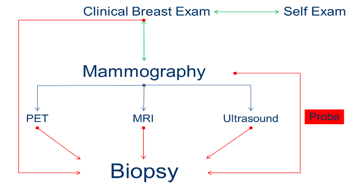
Molecular imaging is a type of medical imaging that provides detailed pictures of what is happening at the molecular and cellular level in the body. It involves the use of specialized imaging devices and radiopharmaceuticals (radiotracers) to visualize and measure biological processes, such as gene expression, protein expression, or metabolic activity, within cells and tissues. This information can be used to detect disease at its earliest stages, monitor response to therapy, and guide the development of new treatments.
Molecular imaging techniques include positron emission tomography (PET), single-photon emission computed tomography (SPECT), magnetic resonance imaging (MRI), and computed tomography (CT). These techniques differ in their ability to provide functional, anatomical, or molecular information about the body.
Overall, molecular imaging is a powerful tool for non-invasively visualizing and understanding biological processes at the molecular level, which can lead to improved diagnosis, treatment planning, and patient outcomes.
Systematic Evolution of Ligands by EXponential enrichment (SELEX) is a laboratory technique used to select and amplify high-affinity nucleic acid ligands, such as DNA or RNA aptamers, that bind specifically to a target molecule. The process involves repeated rounds of in vitro selection and amplification, where large libraries of randomized oligonucleotides are exposed to the target molecule, and those that bind are separated from unbound sequences.
The bound sequences are then amplified using PCR (for DNA) or reverse transcription-PCR (for RNA), followed by re-exposure to the target in subsequent rounds of selection. Over time, this process enriches for a population of nucleic acid sequences that bind tightly and specifically to the target molecule.
SELEX aptamer technique has been widely used to generate aptamers against various targets, including small molecules, proteins, cells, and even viruses. These aptamers have potential applications in diagnostic, therapeutic, and research settings.
Fluorescent dyes are substances that emit light upon excitation by absorbing light of a shorter wavelength. In a medical context, these dyes are often used in various diagnostic tests and procedures to highlight or mark certain structures or substances within the body. For example, fluorescent dyes may be used in imaging techniques such as fluorescence microscopy or fluorescence angiography to help visualize cells, tissues, or blood vessels. These dyes can also be used in flow cytometry to identify and sort specific types of cells. The choice of fluorescent dye depends on the specific application and the desired properties, such as excitation and emission spectra, quantum yield, and photostability.
Optical phenomena refer to the various observable patterns and effects that occur due to the interaction of light with the environment or with structures in our eye. These can include natural phenomena such as rainbows, mirages, and halos around the sun or moon, as well as visual artifacts created by the eye itself, such as afterimages, floaters, and flashes of light. Some optical phenomena are caused by the refraction, reflection, or interference of light waves, while others may result from abnormalities in the eye's structure or function. Understanding these phenomena can provide insight into the properties of light and the functioning of the visual system.
An oligonucleotide probe is a short, single-stranded DNA or RNA molecule that contains a specific sequence of nucleotides designed to hybridize with a complementary sequence in a target nucleic acid (DNA or RNA). These probes are typically 15-50 nucleotides long and are used in various molecular biology techniques, such as polymerase chain reaction (PCR), DNA sequencing, microarray analysis, and blotting methods.
Oligonucleotide probes can be labeled with various reporter molecules, like fluorescent dyes or radioactive isotopes, to enable the detection of hybridized targets. The high specificity of oligonucleotide probes allows for the precise identification and quantification of target nucleic acids in complex biological samples, making them valuable tools in diagnostic, research, and forensic applications.
Optical Tomography (OT) is a non-invasive imaging technique that uses light to visualize and measure the optical properties of tissue, such as absorption and scattering coefficients. This modality can be used to produce cross-sectional or three-dimensional images of internal structures, providing functional information about tissue physiology. It has applications in various fields including biomedical research, dermatology, and oncology for the detection and monitoring of diseases. There are different types of optical tomography, such as diffuse optical tomography (DOT) and near-infrared spectroscopy (NIRS), which differ in their light sources, detection schemes, and data analysis methods.
Aptamers are short, single-stranded oligonucleotides (DNA or RNA) that bind to specific target molecules with high affinity and specificity. They are generated through an iterative process called Systematic Evolution of Ligands by EXponential enrichment (SELEX), where large libraries of randomized oligonucleotides are subjected to repeated rounds of selection and amplification until sequences with the desired binding properties are identified. Nucleotide aptamers have potential applications in various fields, including diagnostics, therapeutics, and research tools.
The term "nucleotide" refers to the basic building blocks of nucleic acids (DNA and RNA). A nucleotide consists of a pentose sugar (ribose for RNA and deoxyribose for DNA), a phosphate group, and a nitrogenous base. The nitrogenous bases in nucleotides are adenine, guanine, cytosine, thymine (in DNA) or uracil (in RNA). In aptamers, the nucleotide sequences form specific three-dimensional structures that enable them to recognize and bind to their target molecules.
Carbocyanines are a class of organic compounds that contain a polymethine chain, which is a type of carbon-based structure with alternating single and double bonds, and one or more cyanine groups. A cyanine group is a functional group consisting of a nitrogen atom connected to two carbon atoms by double bonds, with the remaining valences on the carbon atoms being satisfied by other groups.
Carbocyanines are known for their strong absorption and fluorescence properties in the visible and near-infrared regions of the electromagnetic spectrum. These properties make them useful as dyes and fluorescent labels in various applications, including biomedical research, clinical diagnostics, and material science.
In medicine, carbocyanines are sometimes used as fluorescent contrast agents for imaging purposes. They can be injected into the body and accumulate in certain tissues or organs, where they emit light when excited by a specific wavelength of light. This allows doctors to visualize the distribution of the agent and potentially detect abnormalities such as tumors or inflammation.
It is important to note that while carbocyanines have potential medical applications, they are not themselves medications or drugs. They are tools used in various medical procedures and research.
Near-infrared spectroscopy (NIRS) is a non-invasive optical technique that uses the near-infrared region of the electromagnetic spectrum (approximately 700-2500 nanometers) to analyze various chemical and physical properties of materials, primarily in the fields of biomedical research and industry. In medicine, NIRS is often used to measure tissue oxygenation, hemodynamics, and metabolism, providing valuable information about organ function and physiology. This technique is based on the principle that different molecules absorb and scatter near-infrared light differently, allowing for the identification and quantification of specific chromophores, such as oxyhemoglobin, deoxyhemoglobin, and cytochrome c oxidase. NIRS can be employed in a variety of clinical settings, including monitoring cerebral or muscle oxygenation during surgery, assessing tissue viability in wound healing, and studying brain function in neuroscience research.
Diagnostic imaging is a medical specialty that uses various technologies to produce visual representations of the internal structures and functioning of the body. These images are used to diagnose injury, disease, or other abnormalities and to monitor the effectiveness of treatment. Common modalities of diagnostic imaging include:
1. Radiography (X-ray): Uses ionizing radiation to produce detailed images of bones, teeth, and some organs.
2. Computed Tomography (CT) Scan: Combines X-ray technology with computer processing to create cross-sectional images of the body.
3. Magnetic Resonance Imaging (MRI): Uses a strong magnetic field and radio waves to generate detailed images of soft tissues, organs, and bones.
4. Ultrasound: Employs high-frequency sound waves to produce real-time images of internal structures, often used for obstetrics and gynecology.
5. Nuclear Medicine: Involves the administration of radioactive tracers to assess organ function or detect abnormalities within the body.
6. Positron Emission Tomography (PET) Scan: Uses a small amount of radioactive material to produce detailed images of metabolic activity in the body, often used for cancer detection and monitoring treatment response.
7. Fluoroscopy: Utilizes continuous X-ray imaging to observe moving structures or processes within the body, such as swallowing studies or angiography.
Diagnostic imaging plays a crucial role in modern medicine, allowing healthcare providers to make informed decisions about patient care and treatment plans.
Nucleic acid hybridization is a process in molecular biology where two single-stranded nucleic acids (DNA, RNA) with complementary sequences pair together to form a double-stranded molecule through hydrogen bonding. The strands can be from the same type of nucleic acid or different types (i.e., DNA-RNA or DNA-cDNA). This process is commonly used in various laboratory techniques, such as Southern blotting, Northern blotting, polymerase chain reaction (PCR), and microarray analysis, to detect, isolate, and analyze specific nucleic acid sequences. The hybridization temperature and conditions are critical to ensure the specificity of the interaction between the two strands.
Molecular sequence data refers to the specific arrangement of molecules, most commonly nucleotides in DNA or RNA, or amino acids in proteins, that make up a biological macromolecule. This data is generated through laboratory techniques such as sequencing, and provides information about the exact order of the constituent molecules. This data is crucial in various fields of biology, including genetics, evolution, and molecular biology, allowing for comparisons between different organisms, identification of genetic variations, and studies of gene function and regulation.
Fluorescence microscopy is a type of microscopy that uses fluorescent dyes or proteins to highlight and visualize specific components within a sample. In this technique, the sample is illuminated with high-energy light, typically ultraviolet (UV) or blue light, which excites the fluorescent molecules causing them to emit lower-energy, longer-wavelength light, usually visible light in the form of various colors. This emitted light is then collected by the microscope and detected to produce an image.
Fluorescence microscopy has several advantages over traditional brightfield microscopy, including the ability to visualize specific structures or molecules within a complex sample, increased sensitivity, and the potential for quantitative analysis. It is widely used in various fields of biology and medicine, such as cell biology, neuroscience, and pathology, to study the structure, function, and interactions of cells and proteins.
There are several types of fluorescence microscopy techniques, including widefield fluorescence microscopy, confocal microscopy, two-photon microscopy, and total internal reflection fluorescence (TIRF) microscopy, each with its own strengths and limitations. These techniques can provide valuable insights into the behavior of cells and proteins in health and disease.
A base sequence in the context of molecular biology refers to the specific order of nucleotides in a DNA or RNA molecule. In DNA, these nucleotides are adenine (A), guanine (G), cytosine (C), and thymine (T). In RNA, uracil (U) takes the place of thymine. The base sequence contains genetic information that is transcribed into RNA and ultimately translated into proteins. It is the exact order of these bases that determines the genetic code and thus the function of the DNA or RNA molecule.
Fluorescence spectrometry is a type of analytical technique used to investigate the fluorescent properties of a sample. It involves the measurement of the intensity of light emitted by a substance when it absorbs light at a specific wavelength and then re-emits it at a longer wavelength. This process, known as fluorescence, occurs because the absorbed energy excites electrons in the molecules of the substance to higher energy states, and when these electrons return to their ground state, they release the excess energy as light.
Fluorescence spectrometry typically measures the emission spectrum of a sample, which is a plot of the intensity of emitted light versus the wavelength of emission. This technique can be used to identify and quantify the presence of specific fluorescent molecules in a sample, as well as to study their photophysical properties.
Fluorescence spectrometry has many applications in fields such as biochemistry, environmental science, and materials science. For example, it can be used to detect and measure the concentration of pollutants in water samples, to analyze the composition of complex biological mixtures, or to study the properties of fluorescent nanomaterials.
Molecular structure, in the context of biochemistry and molecular biology, refers to the arrangement and organization of atoms and chemical bonds within a molecule. It describes the three-dimensional layout of the constituent elements, including their spatial relationships, bond lengths, and angles. Understanding molecular structure is crucial for elucidating the functions and reactivities of biological macromolecules such as proteins, nucleic acids, lipids, and carbohydrates. Various experimental techniques, like X-ray crystallography, nuclear magnetic resonance (NMR) spectroscopy, and cryo-electron microscopy (cryo-EM), are employed to determine molecular structures at atomic resolution, providing valuable insights into their biological roles and potential therapeutic targets.
Fluorescence is not a medical term per se, but it is widely used in the medical field, particularly in diagnostic tests, medical devices, and research. Fluorescence is a physical phenomenon where a substance absorbs light at a specific wavelength and then emits light at a longer wavelength. This process, often referred to as fluorescing, results in the emission of visible light that can be detected and measured.
In medical terms, fluorescence is used in various applications such as:
1. In-vivo imaging: Fluorescent dyes or probes are introduced into the body to highlight specific structures, cells, or molecules during imaging procedures. This technique can help doctors detect and diagnose diseases such as cancer, inflammation, or infection.
2. Microscopy: Fluorescence microscopy is a powerful tool for visualizing biological samples at the cellular and molecular level. By labeling specific proteins, nucleic acids, or other molecules with fluorescent dyes, researchers can observe their distribution, interactions, and dynamics within cells and tissues.
3. Surgical guidance: Fluorescence-guided surgery is a technique where surgeons use fluorescent markers to identify critical structures such as blood vessels, nerves, or tumors during surgical procedures. This helps ensure precise and safe surgical interventions.
4. Diagnostic tests: Fluorescence-based assays are used in various diagnostic tests to detect and quantify specific biomarkers or analytes. These assays can be performed using techniques such as enzyme-linked immunosorbent assay (ELISA), polymerase chain reaction (PCR), or flow cytometry.
In summary, fluorescence is a physical process where a substance absorbs and emits light at different wavelengths. In the medical field, this phenomenon is harnessed for various applications such as in-vivo imaging, microscopy, surgical guidance, and diagnostic tests.
Fluorescein is not a medical condition, but rather a diagnostic dye that is used in various medical tests and procedures. It is a fluorescent compound that absorbs light at one wavelength and emits light at another wavelength, which makes it useful for imaging and detecting various conditions.
In ophthalmology, fluorescein is commonly used in eye examinations to evaluate the health of the cornea, conjunctiva, and anterior chamber of the eye. A fluorescein dye is applied to the surface of the eye, and then the eye is examined under a blue light. The dye highlights any damage or abnormalities on the surface of the eye, such as scratches, ulcers, or inflammation.
Fluorescein is also used in angiography, a medical imaging technique used to examine blood vessels in the body. A fluorescein dye is injected into a vein, and then a special camera takes pictures of the dye as it flows through the blood vessels. This can help doctors diagnose and monitor conditions such as cancer, diabetes, and macular degeneration.
Overall, fluorescein is a valuable diagnostic tool that helps medical professionals detect and monitor various conditions in the body.
Tissue distribution, in the context of pharmacology and toxicology, refers to the way that a drug or xenobiotic (a chemical substance found within an organism that is not naturally produced by or expected to be present within that organism) is distributed throughout the body's tissues after administration. It describes how much of the drug or xenobiotic can be found in various tissues and organs, and is influenced by factors such as blood flow, lipid solubility, protein binding, and the permeability of cell membranes. Understanding tissue distribution is important for predicting the potential effects of a drug or toxin on different parts of the body, and for designing drugs with improved safety and efficacy profiles.
Organometallic compounds are a type of chemical compound that contain at least one metal-carbon bond. This means that the metal is directly attached to carbon atom(s) from an organic molecule. These compounds can be synthesized through various methods, and they have found widespread use in industrial and medicinal applications, including catalysis, polymerization, and pharmaceuticals.
It's worth noting that while organometallic compounds contain metal-carbon bonds, not all compounds with metal-carbon bonds are considered organometallic. For example, in classical inorganic chemistry, simple salts of metal carbonyls (M(CO)n) are not typically classified as organometallic, but rather as metal carbonyl complexes. The distinction between these classes of compounds can sometimes be subtle and is a matter of ongoing debate among chemists.
An amino acid sequence is the specific order of amino acids in a protein or peptide molecule, formed by the linking of the amino group (-NH2) of one amino acid to the carboxyl group (-COOH) of another amino acid through a peptide bond. The sequence is determined by the genetic code and is unique to each type of protein or peptide. It plays a crucial role in determining the three-dimensional structure and function of proteins.
A Structure-Activity Relationship (SAR) in the context of medicinal chemistry and pharmacology refers to the relationship between the chemical structure of a drug or molecule and its biological activity or effect on a target protein, cell, or organism. SAR studies aim to identify patterns and correlations between structural features of a compound and its ability to interact with a specific biological target, leading to a desired therapeutic response or undesired side effects.
By analyzing the SAR, researchers can optimize the chemical structure of lead compounds to enhance their potency, selectivity, safety, and pharmacokinetic properties, ultimately guiding the design and development of novel drugs with improved efficacy and reduced toxicity.
A Small Molecule Library is a collection of a large number of chemically synthesized, low molecular weight (typically under 900 daltons) compounds, which are used in drug discovery and development research. These libraries contain diverse structures and chemical properties, allowing researchers to screen them against specific targets, such as proteins or genes, to identify potential lead compounds that can be further optimized for therapeutic use. The use of small molecule libraries enables high-throughput screening, which is a rapid and efficient method to identify potential drug candidates.
"Drug design" is the process of creating and developing a new medication or therapeutic agent to treat or prevent a specific disease or condition. It involves identifying potential targets within the body, such as proteins or enzymes that are involved in the disease process, and then designing small molecules or biologics that can interact with these targets to produce a desired effect.
The drug design process typically involves several stages, including:
1. Target identification: Researchers identify a specific molecular target that is involved in the disease process.
2. Lead identification: Using computational methods and high-throughput screening techniques, researchers identify small molecules or biologics that can interact with the target.
3. Lead optimization: Researchers modify the chemical structure of the lead compound to improve its ability to interact with the target, as well as its safety and pharmacokinetic properties.
4. Preclinical testing: The optimized lead compound is tested in vitro (in a test tube or petri dish) and in vivo (in animals) to evaluate its safety and efficacy.
5. Clinical trials: If the preclinical testing is successful, the drug moves on to clinical trials in humans to further evaluate its safety and efficacy.
The ultimate goal of drug design is to create a new medication that is safe, effective, and can be used to improve the lives of patients with a specific disease or condition.
Fluorescence Resonance Energy Transfer (FRET) is not strictly a medical term, but it is a fundamental concept in biophysical and molecular biology research, which can have medical applications. Here's the definition of FRET:
Fluorescence Resonance Energy Transfer (FRET) is a distance-dependent energy transfer process between two fluorophores, often referred to as a donor and an acceptor. The process occurs when the emission spectrum of the donor fluorophore overlaps with the excitation spectrum of the acceptor fluorophore. When the donor fluorophore is excited, it can transfer its energy to the acceptor fluorophore through non-radiative dipole-dipole coupling, resulting in the emission of light from the acceptor at a longer wavelength than that of the donor.
FRET efficiency depends on several factors, including the distance between the two fluorophores, their relative orientation, and the spectral overlap between their excitation and emission spectra. FRET is typically efficient when the distance between the donor and acceptor is less than 10 nm (nanometers), making it a powerful tool for measuring molecular interactions, conformational changes, and distances at the molecular level.
In medical research, FRET has been used to study various biological processes, such as protein-protein interactions, enzyme kinetics, and gene regulation. It can also be used in developing biosensors for detecting specific molecules or analytes in clinical samples, such as blood or tissue.
Deoxyribonucleic acid (DNA) is the genetic material present in the cells of organisms where it is responsible for the storage and transmission of hereditary information. DNA is a long molecule that consists of two strands coiled together to form a double helix. Each strand is made up of a series of four nucleotide bases - adenine (A), guanine (G), cytosine (C), and thymine (T) - that are linked together by phosphate and sugar groups. The sequence of these bases along the length of the molecule encodes genetic information, with A always pairing with T and C always pairing with G. This base-pairing allows for the replication and transcription of DNA, which are essential processes in the functioning and reproduction of all living organisms.
 Cycling probe technology
Mycobacterium bohemicum
Force spectroscopy
Cell biomechanics
Mostafa Ronaghi
Clathrate hydrate
Alexander Kovarski
Chemical imaging
Molecular Inversion Probe
TRIUMF
Functional cloning
Fluxional molecule
Transition state
Molecular and Cellular Probes
Hybridization probe
Bioelectricity
Molecular probe
Chemical physics
Thermal shift assay
Hoechst stain
Small interfering RNA
Dual-polarization interferometry
Bai Chunli
Biointerface
Second-harmonic generation
Lyotropic liquid crystal
Leroy Hood
Long-range restriction mapping
Stable-isotope probing
Molecular beacon
Alloprotein
Resazurin
Molecular Probe Techniques - Immunoblotting | CU Experts | CU Boulder
 IJMS | Free Full-Text | Thermodynamic and Dynamic Transitions and Interaction Aspects in Reorientation Dynamics of Molecular...
 Optical probes and techniques for molecular contrast enhancement in coherence imaging | Biophotonics Imaging Laboratory | UIUC
 A systematic approach to the development of fluorescent contrast agents for optical imaging of mouse cancer models
 Building optical molecular probes with nanotubes
Cycling probe technology - Wikipedia
![PAR-07-368: Preapplication for the Molecular Libraries Probe Production Centers Network (MLPCN) [X02]](data:image/png;base64,iVBORw0KGgoAAAANSUhEUgAAABAAAAAQCAYAAAAf8/9hAAABDElEQVQ4jaXTPy9DURiA8V+lBith8xGupYnRJhJfQFOLRlSRSEiaaCNikNChE4Oli0QNdovEZHQX/QoWIrEZpNTg3LCoe+Nd3pznvOc5ec+fXL/f95/IrVTWSzj/Zb6DctyLiqjiBXXcJwVDAxZDCQtxu3oWxvO4wcRPwV/RCnkz5DHsZBGMV1Y3GrjDSWBbmEorgP1CvjuKJl4Dq2cRDPtq5QGHgRUxl1YAS4V8dwYHvm+hmUUAHyHnEpBF0I570S12EQVWSyt4Qw2TaAR2geu0gr24FyWvcCSwI9K18Bj3oiamsRZYSzjINILtuF2F4zB+SnZPBIsDFndwWVg+LeMdV5jFc1KQ++93/gTPiD9F5tDWMgAAAABJRU5ErkJggg==) PAR-07-368: Preapplication for the Molecular Libraries Probe Production Centers Network (MLPCN) [X02]
PAR-07-368: Preapplication for the Molecular Libraries Probe Production Centers Network (MLPCN) [X02]
 Respiratory Syncytial Virus Infection: Practice Essentials, Background, Pathophysiology
 Advanced Search Results - Public Health Image Library(PHIL)
Search Results - - 20,839 Results - UNT Digital Library
Chemical Physics authors/titles May 2015
 State of the art and opportunities in probing photoinduced phase transitions in molecular materials by conventional and...
PAR-08-034: Solicitation of Assays for High Throughput Screening (HTS) in the Molecular Libraries Probe Production Centers...
 An interstellar synthesis of phosphorus oxoacids | Nature Communications
Mycobacterium bohemicum and Cervical Lymphadenitis in Children - Volume 14, Number 7-July 2008 - Emerging Infectious Diseases...
 Sinead Ni Chadhain's Research Area | Biology Department
BIOLOGICKÉ CENTRUM AV ČR, v. v. i. | Zaměstnanec
 Palmetto Draft LCD of Cancer Genomic Profiling in NSCLC is Good First Step, Say NGS Test Providers | GenomeWeb
 Molecular Vision: Application of atomic force microscopy in
morphological observation of antisense probe labeled with magnetism
Molecular Vision: Application of atomic force microscopy in
morphological observation of antisense probe labeled with magnetism
 Chemistry Professor Published in Journal of Chemical Education | Harvey Mudd College News
 Kommentare Wintersemester 2020/21 - Physikalisches Institut
 All Faculty (A-Z): Department of Medicine: Feinberg School of Medicine: Northwestern University
 Multipurpose Molecule for Cancer Surgery | MIT Technology Review
 Roberts' Lab Home Page
 Tracing Magnetic Field Morphology Using the Velocity Gradient Technique in the Presence of CO Self-absorption - NASA/ADS
 Chemistry BSc Hons | University of Nottingham
 Affiliate Investigators | Living Systems Institute | University of Exeter
Respiratory Syncytial Virus Infection Workup: Laboratory Studies, Other Studies
 What are the Differences Between DNA and RNA Probes? - Enzo Life Sciences
Dynamics9
- By playing on different physical parameters and developing the techniques and analysis, one can investigate new out of equilibrium physics through light-driven cooperative dynamics and transformations in materials. (degruyter.com)
- The applications to living systems I am interested in include biophysical modelling (active transport and organelle dynamics in cell biology), cognition (perceptual rivalry, computational aspects of networks), molecular networks, functional dynamics in neural and biomedical systems and tipping points in nonautonomous systems. (exeter.ac.uk)
- Molecular dynamics (MD) was explored as a complementary computational technique to probe deeper into the experimental data. (ens-lyon.fr)
- Carbon Nanotubes in water, investigation of heat and mass transfer phenomena with Molecular Dynamics and Pump and Probe spectroscopy. (polito.it)
- In this work, it is reported an investigation of the thermal behavior of Carbon Nanotubes in an aqueous solution by means of Molecular Dynamics simulations and the early results of Pump and Probe spectroscopy. (polito.it)
- In particular, Molecular Dynamics simulations are performed to investigate the role of the interface in heat and mass transfer phenomena. (polito.it)
- There he broadened his repertoire with application of attosecond pulses to small molecules and started up an experimental free electron laser (FEL) program at FLASH, aiming at studying molecular dynamics through pump-probe experiments, using the Velocity Map Imaging technique. (lu.se)
- Following his postdoctoral period, in 2009 he acquired a position as a junior researcher at the Atomic Physics Division at Lund University, working on the application of attosecond and FEL pulses to study molecular dynamics. (lu.se)
- This has been established by experiments on simple model interfaces1,2 and by molecular dynamics simulations.8-10 Given an estimate of the number (nH) of water molecules in the hydration layer, e.g. from a simulation, we can obtain, in an essentially model-free way, the rotational correlation time htHi averaged over all sites in the hydration layer. (lu.se)
Assays5
- CPT requires specialized chimeric probes, making CPT assays more expensive than PCR. (wikipedia.org)
- The goal of the MLP is to screen compounds in the Small Molecule Repository in target-based and phenotypic assays to identify and subsequently optimize small molecule s as research probes. (nih.gov)
- Molecular probes for revealing RSV in clinical specimens may be more sensitive than the aforementioned assays and are becoming clinically available, but they presently are more expensive. (medscape.com)
- DNA/RNA probe assays are faster and sensitive so that many conventional diagnostic tests for viruses and bacteria involving culturing of the organisms are being fast replaced by molecular probe assays. (enzolifesciences.com)
- While culture tests can take days, molecular probe assays can be performed within a few hours or minutes. (enzolifesciences.com)
Polymerase7
- Secretions can be analyzed for virus in the laboratory by means of culture, antigen-revealing techniques, or polymerase chain reaction (PCR). (medscape.com)
- Most enzyme-mediated labeling techniques are very much dependent on polymerase activity, which is responsible for incorporation of the labeled nucleotides. (enzolifesciences.com)
- The annealed primers ultimately become part of the probe itself, because the Klenow fragment of DNA polymerase I extends the primers in the 3′ direction and, in so doing, incorporates the label. (enzolifesciences.com)
- Next, the enzyme DNA polymerase I removes the native nucleotides from the probe molecules in the 5′→3′ direction (exonuclease activity) while replacing them with labeled dNTP precursors by virtue of its 5′→3′ polymerase activity. (enzolifesciences.com)
- This technique sought to simplify gene synthesis by using artificial primers and templates that help DNA polymerase to copy the desired gene segments. (news-medical.net)
- Although this technique used DNA polymerase repeatedly, similar to PCR, it employed only a single primer template complex, and exponential amplification was not possible using this technique. (news-medical.net)
- Use of EIAs, tissue culture, molecular probes, and the polymerase chain reaction has improved the diagnosis of diarrhea caused by bacteria, and special concentrating and staining techniques have improved the process of detecting parasites such as Cryptosporidium and I. belli. (cdc.gov)
Characterization3
- The team has extended it to include characterization of the distribution of molecular clusters in fluids with important energy, chemical, and biological applications. (newswise.com)
- With the help of design, fabrication, and characterization techniques defining the state of the art, We develop optical multilayers for wide-band light waveform synthesis all the way from the infrared to the ultraviolet. (attoworld.de)
- Occurrence and molecular characterization of Acanthamoeba, Naegleria fowleri and Blastocystis in water samples from various sources in Egypt. (cdc.gov)
High-throughput3
- This initiative is one of the integrated components of the NIH Molecular Libraries Roadmap initiative that offers biomedical researchers access to large-scale automated high throughput screening (HTS) centers in the MLPCN, diverse compound libraries in the Small Molecule Repository (MLSMR) and information on biological activities of small molecules in the PubChem BioAssay public database. (nih.gov)
- In high-throughput labs, the speed advantages of isothermal amplification techniques can be made up for by batch operation. (ttp.com)
- Capitalizing on the femtosecond field-resolving metrologies, we devise electric-field molecular fingerprinting (EMF) as a new, high-throughput analytical technique. (attoworld.de)
Atomic6
- To explore the possibility of the c-erbB2 oncogene antisense probe labeled with superparamagnetic iron oxide (SPIO) nanoparticles as a target contrast agent for magnetic resonance (MR) imaging whose morphology was observed with atomic force microscopy (AFM), and its efficiency was examined by MR imaging. (molvis.org)
- With atomic force microscopy (AFM), we observed the morphology of antisense probes labeled with SPIO to explore its potential as a target contrast agent for magnetic resonance (MR) imaging. (molvis.org)
- Newswise - Materials science explores how materials behave, including at the atomic and molecular scales. (newswise.com)
- The technique visualizes and quantifies the atomic and molecular structures in three-dimensional samples in real time. (newswise.com)
- These atomic- and molecular-scale "microstructures" include features such as grain size and pores. (newswise.com)
- The following two years he spent as a postdoctoral fellow with Marc Vrakking at the FOM Institute for Atomic and Molecular Physics in Amsterdam. (lu.se)
Primers4
- I use primers and probes to key genes in the degradation pathways of various environmental contaminants to determine how the microbial community as a whole responds to that contaminant. (southalabama.edu)
- Specific molecular probes and primers are designed for this purpose. (enzolifesciences.com)
- Kary Mullis of Cetus worked on oligonucleotides synthesis for use as probes, primers, and building blocks for various molecular biology techniques. (news-medical.net)
- Meanwhile, the team used PCR for other applications by designing new primers and probes, which made the reaction more specific until the results were evident on agarose gel electrophoresis. (news-medical.net)
Gene7
- With our probe' says Baik, 'the target gene within the chromosome can be localized with the ssDNA-SWNT conjugates, and this method can be used for the in situ detection of the specific genome in cells, tissue sections or infectious agents with the possibility of long term storage and re-evaluation of the results in the future. (nanowerk.com)
- Molecular tools such as PCR, gene probing, mRNA transcript analysis, and DNA sequencing can aid in this endeavour. (southalabama.edu)
- In the present study, we prepared the ASODN of complementary c-erbB2 oncogene using the gene synthesis technique and labeled the superparamagnetic iron oxide (SPIO) nanometer using the chemical cross-linking method to produce the antisense probe. (molvis.org)
- Gene probes are used in various blotting and in situ hybridization (ISH) techniques for the detection of nucleic acid sequences in food industry, environmental, medical, and veterinary applications to improve the specificity of the analyses. (enzolifesciences.com)
- A DNA probe is a labeled fragment of DNA that contains a nucleotide sequence specific for the gene or chromosomal region of interest. (enzolifesciences.com)
- Probes labeled by nick translation can be used in many different hybridization techniques including: chromogenic in situ hybridization (CISH), fluorescent in situ hybridization (FISH), screening gene banks by colony or plaque hybridization, DNA or RNA transfer hybridization, and re-association kinetic studies. (enzolifesciences.com)
- Her research will focus on new immunotherapy approaches to treating acute myeloid leukemia using gene- and cell-based therapy techniques. (wustl.edu)
Physics4
- This molecular state features rich physics, including a first-order transition to the polaron state and a negative effective mass at small interactions. (arxiv.org)
- Central elements in the course are thereby interaction between radiation and matter, lasers and their properties, optics, optical measuring technique, molecular physics and combustion. (lu.se)
- The fields that are treated are molecular spectroscopy, statistical physics, combustion and experimental equipment for laser-based combustion diagnostics. (lu.se)
- The basic physics and engineering of each imaging technique are emphasized. (lu.se)
Aggregates1
- At the molecular level, most of biology happens at interfaces where water makes contact with macromolecules or molecular aggregates. (lu.se)
Fluorescent probes3
- Scott Hilderbrand , a chemist at Massachusetts General Hospital's Center for Molecular Imaging Research, is also developing targeted fluorescent probes. (technologyreview.com)
- The mice they tested this in have only normal tissue and cancer tissue, but the human body is not so simple" says Hisataka Kobayashi , a molecular imaging specialist at the National Cancer Institute in Bethesda, MA, and another person working on the targeting cancer with fluorescent probes. (technologyreview.com)
- Use of fluorescent probes such as HyBeacons [4] and Molecular Beacons [5] has subsequently been demonstrated. (ttp.com)
Spectroscopy3
- In this work we show theoretically how the molecular state can be directly prepared experimentally even in its excited state using state-of-the-art cold atom Raman spectroscopy techniques. (arxiv.org)
- We can use this to observe processes that are too fast for traditional pump-probe spectroscopy. (attoworld.de)
- To conclude, it is provided an outline of the early results of Carbon Nanotube in aqueous solution investigated with Pump and Probe spectroscopy with testing developed at the Institut Lumière Matière of Lyon. (polito.it)
Diagnostic5
- Although the insurance coverage process in the US for new molecular diagnostic approaches is not straightforward or easy, we are very excited about this important development in setting the stage for increased access to CGPs and supporting our efforts to obtain broad reimbursement for FoundationOne. (genomeweb.com)
- As early as 1990, a review of target amplification systems for diagnostic applications concluded that techniques enjoying advantages such as "isothermal reaction … may eventually gain widespread use after further development" [1]. (ttp.com)
- These potential biomarkers of CD may elucidate tumor biology and suggest possible diagnostic molecular imaging probes as well as therapeutic targets in patients with recurrent disease after surgery. (broadinstitute.org)
- Investigating the associated dosimetry estimations help to decrease the risk of diagnostic and therapy techniques. (nih.gov)
- New diagnostic laboratory techniques as well as modifications of standard ones have been used by investigators to identify viral, bacterial, and parasitic agents of outbreaks of gastroenteritis. (cdc.gov)
Beacons1
- CPT can be used in conjunction with other technologies, like molecular beacons and qPCR. (wikipedia.org)
Structures3
- Electrospray ionization mass spectrometry (ESI-MS) is a new, powerful technique for probing molecular structures, especially those of biological interest. (hmc.edu)
- This program supports the research and development of technologies and techniques that create images of internal structures, contrast agents, or molecular probes using x-rays transmitted through the body (CT, mammography) or x-ray stimulation of secondary emissions (x-ray fluorescence tomography). (nih.gov)
- This machine learning technique can quickly identify, track, and quantify complex structures in materials. (newswise.com)
Assay4
- In a recent paper, titled 'The DNA hybridization assay using single-walled carbon nanotubes as ultrasensitive, long-term optical labels' the Korean scientists present single- stranded DNA (ssDNA)-coated SWNT probes to locate a particular sequence of DNA. (nanowerk.com)
- Because CPT probes are so specific, a new probe must be designed for each unique assay, further increasing cost. (wikipedia.org)
- The NIH Molecular Libraries Roadmap Initiative wishes to solicit HTS assay applications from the scientific community from investigators who have the interest and capability to work with the Molecular Libraries Probe Production Centers Network (MLPCN) in support of chemical probe development. (nih.gov)
- Initially, LAMP was a pH-dependent precipitation assay, but the technique was later adapted for use with intercalating dye, allowing researchers to confirm specificity by post-reaction melting curve analysis. (ttp.com)
Simulations1
- This thesis expands our understanding and provides insight into the tools available to investigate solvent behaviour of ionic liquids at a molecular level using both experiments and computational simulations. (ens-lyon.fr)
Specificity2
- Southern blotting, which uses photostable Raman signals of nanotubes instead of fluorescent dyes, demonstrates excellent sensitivity and specificity of the probes. (nanowerk.com)
- Both increasing RNase H concentrations and use of a probe that isn't prone to inter-probe and intra-probe interactions has been shown to increase specificity. (wikipedia.org)
Researchers4
- The molecular label, developed by researchers at the University of California at San Diego (UCSD), works in two ways. (technologyreview.com)
- He notes that the individual pieces of the technique developed by Tsien's team have been shown to work by other researchers, "but to be able to do this with one agent is one of the more major advances of this approach. (technologyreview.com)
- Over time, the technique has evolved beyond the confines of its simple initial design and has opened incredible avenues for researchers. (news-medical.net)
- The technique will also help researchers analyze 3D data on how atoms in materials are arranged. (newswise.com)
Laboratory6
- Gross developed an open-ended laboratory experiment that illustrates the power of ESI-MS, and Van Ryswyk helped adapt it to other techniques. (hmc.edu)
- Research in my laboratory explores the molecular mechanism of amoeboid movement with focus on a simple, specialized system, the crawling sperm of nematodes. (fsu.edu)
- Working in collaboration with colleagues at the MRC Laboratory of Molecular Biology in Cambridge, England, we have used high-resolution electron microscopy and computer image processing to examine the detailed anatomy of MSP filaments and x-ray crystallography to determine the structure of the MSP molecule. (fsu.edu)
- however, because of technical complexity, cost and the requirement for sophisticated laboratory infrastructure, use of these techniques has been limited in many resource-constrained settings. (bvsalud.org)
- Laboratory team performance is monitored using several techniques. (cdc.gov)
- Molecular genetic epidemiology : a laboratory perspective / Ian N.M. Day, ed. (who.int)
Chimeric5
- CPT uses a sequence specific chimeric probe which hybridizes to a complementary target DNA sequence and becomes a substrate for RNase H. Cleavage occurs at the RNA internucleotide linkages and results in dissociation of the probe from the target, thereby making it available for the next probe molecule. (wikipedia.org)
- Cycling probe technology makes use of a chimeric nucleic acid probe to detect the presence of a particular DNA sequence. (wikipedia.org)
- The chimeric probe consists of an RNA segment sandwiched between two DNA segments. (wikipedia.org)
- Cycling probe technology utilizes a cyclic, isothermal process that begins with the hybridization of the chimeric probe with the target DNA. (wikipedia.org)
- Once hybridized, the probe becomes a suitable substrate for RNase H. RNase H, an endonuclease, cleaves the RNA portion of the probe, resulting in two chimeric fragments. (wikipedia.org)
Pathways2
- We are engaged in a multidisciplinary study of fundamental aspects of the crystallization of organic molecular materials from solution, focusing on polymorphic systems under the recognition that such systems represent an ideal opportunity for obtaining a systematic understanding of competing pathways in crystallization processes. (rsc.org)
- Her research will focus on exploring the molecular pathways of potential pathogens involved in sepsis, a common and often deadly disease. (wustl.edu)
Transitions1
- State of the art and opportunities in probing photoinduced phase transitions in molecular materials by conventional and picosecond X-ray diffraction" Zeitschrift für Kristallographie - Crystalline Materials , vol. 223, no. 4-5, 2008, pp. 272-282. (degruyter.com)
Contrast1
- Daphnnia), ciliates, and some bacteria exhibit good contrast by this technique. (canadiannaturephotographer.com)
Biology techniques1
- The second approach uses molecular biology techniques to study microbial communities. (southalabama.edu)
Biological3
- Cycling probe technology (CPT) is a molecular biological technique for detecting specific DNA sequences. (wikipedia.org)
- This FOA promotes discovery and development of new chemical probes as research tools for use by scientists in both the public and private sector to advance the understanding of biological functions and disease mechanisms. (nih.gov)
- After the fundamentals of photon transport in biological tissues are established, various optical imaging techniques for biological tissues are covered. (lu.se)
Optical1
- Comparison is made between probe methods and optical methods of measurement. (lu.se)
Nucleic acid2
- Nucleic acid probes are either a single stranded DNA or RNA with a strong affinity towards a specific DNA or RNA target sequence. (enzolifesciences.com)
- In theory, any nucleic acid can be used as a probe provided it can be labeled to permit detection and quantitation of the hybrid molecules formed between the probe and sequence to be identified. (enzolifesciences.com)
QPCR1
- In the case of qPCR, the IP on the simpler hydrolysis probes and use of reference dyes has also expired, so qPCR can be performed licence-free. (ttp.com)
Tissue3
- Slides for microscopic examination were prepared directly from minced lymph nodes before decontamination, and the tissue was stained according to the Ziehl-Neelsen technique. (cdc.gov)
- In lung cancer, obtaining sufficient tissue samples through invasive biopsies is difficult and often limits the molecular analysis that can be run. (genomeweb.com)
- Even though MRI-based techniques may have the ability to probe the microscopic composition, other methods are needed to directly visualize the microstructure of the tissue. (lu.se)
Novel technique2
- Harvey Mudd Professor of Chemistry Hal Van Ryswyk has teamed with Carleton College Professor of Chemistry Deborah Gross to publish a paper in the Journal of Chemical Education exploring a novel technique for probing the structure of large molecules. (hmc.edu)
- Machine learning, which allows for the input of experimental data from which extrapolations can be made about new ILs, is a novel technique which has sparked great interest within our field. (ens-lyon.fr)
Research5
- A Feature Paper should be a substantial original Article that involves several techniques or approaches, provides an outlook for future research directions and describes possible research applications. (mdpi.com)
- Van Ryswyk, whose research focuses on solar energy conversion, and Gross, whose expertise is in atmospheric chemistry, had mutual interest in the novel ESI-MS technique and decided to team up on the research. (hmc.edu)
- However, it was not until 1983 that, in an effort to fix some issues in his research work, Mullis used Sanger's DNA sequencing method as a basis to devise a new technique. (news-medical.net)
- His research will aim to develop a technique to assess peripheral nerve damage in badly injured limbs. (wustl.edu)
- Scientists at the Center for Nanoscale Materials have developed a machine learning technique for materials research. (newswise.com)
Tumor1
- As such, profiling the tumor at one time for multiple molecular characteristics would be the best use of limited resources. (genomeweb.com)
Clinical2
- Through comprehensive clinical and molecular studies, we harness the power of EMF and data analytics to pave the way for a new era of medical probing. (attoworld.de)
- CONCLUSION: Our results provide insight into the role of OPN3 in LCH which may become a molecular target for the clinical treatment of LCH. (bvsalud.org)
Diagnosis1
- He also plans to tap into his expertise in microsurgery, with plans to create custom-made, molecular imaging probes to improve diagnosis and treatment of peripheral nerve damage. (wustl.edu)
Nucleotides2
- The probes should be less than 30 nucleotides in length and designed to minimize intra-probe and inter-probe interactions. (wikipedia.org)
- In developing a probe, a sequence of nucleotides must be identified, isolated, reproduced in sufficient quantity, and tagged with a label that can be detected. (enzolifesciences.com)
Amplification4
- Because cycling probe technology does not involve the amplification of target DNA, CPT has a lower risk of cross contamination than PCR. (wikipedia.org)
- PCR is still the go-to method in the molecular diagnostics industry but, with the rise of close-to-patient testing, isothermal amplification methods with their low power and potentially easier detection schemes look increasingly attractive. (ttp.com)
- Now, with the progression of diagnostics out of the lab and closer to the patient, the potential advantages of isothermal amplification techniques offer an increasingly interesting alternative. (ttp.com)
- BioHelix, purchased by Quidel in 2013, offer a hand-held molecular test based in its proprietary Helicase-Dependent Amplification (HDA) while Abbott's Alere-I offer a panel of CLIA-waiver tests based on isothermal Nicking Enzyme Amplification Reaction (NEAR) technology. (ttp.com)
Methods4
- Metagenomics: Methods and Protocols, Methods in Molecular Biology. (cas.cz)
- When coupled with methods to reduce sample size, the technique requires only seconds to process large 3D samples and obtain precise microstructure information. (newswise.com)
- Magnetic resonance imaging (MRI) is suitable for imaging of cartilage and meniscus, and quantitative imaging methods have the potential to probe the molecular composition and microstructure. (lu.se)
- describe advantages and disadvantages with these laser-based methods compared with probe methods. (lu.se)
Pulses1
- A technique in which electric pulses, in kilovolts per centimeter and of microsecond-to-millisecond duration, cause a loss of the semipermeability of CELL MEMBRANES, thus leading to ion leakage, escape of metabolites, and increased uptake by cells of drugs, molecular probes, and DNA. (bvsalud.org)
Biomedical2
- His mentor is Samuel Achilefu, PhD , the Michel M. Ter-Pogossian Professor of Radiology, as well as a professor of medicine, of biochemistry and molecular biophysics, and of biomedical engineering. (wustl.edu)
- Biomedical Engineering: Health Care Systems, Technology and Techniques is an edited volume with contributions from world experts. (lu.se)
Conventional1
- Thus, nematode sperm have become a valuable complement to more conventional systems for dissecting the molecular interactions responsible for amoeboid movement. (fsu.edu)
Computational1
- To solve the conundrum regarding why such similar PILs led to vastly different results, it was decided computational techniques are necessary. (ens-lyon.fr)
Investigate2
- This in vitro system provides us with an unparalleled opportunity to identify and characterize the components of the MSP motile apparatus, study its key regulatory mechanisms, and investigate the molecular interactions that produce the force for locomotion. (fsu.edu)
- Due to their significance, it is needed to investigate their properties accurately with modeling and experimental techniques. (polito.it)
Protein2
- In their paper, titled "Examination and Manipulation of Protein Surface Charge in Solution with Electrospray Ionization Mass Spectrometry," the chemists describe SNAPP-MS-selective non-covalent adduct protein probing mass spectrometry-a technique that allows for the detailed examination of surface charges on proteins in solution. (hmc.edu)
- 1998. Amoeboid motility without actin: insights into the molecular mechanism of locomotion using the major sperm protein (MSP) of nematodes. (fsu.edu)
Molecules1
- However, currently there is insufficient knowledge about the interaction of these IL-molecular solvents with solutes and interfaces, particularly regarding which ions or molecular solvent molecules are involved in solvating various solutes. (ens-lyon.fr)
Microscopy5
- We use techniques ranging from computer-enhanced video light microscopy to molecular biology to probe the motile machinery of sperm. (fsu.edu)
- I show pictures using each technique, discuss some of their pros and cons and describe how DIC microscopy works. (canadiannaturephotographer.com)
- Use of direct electron microscopy (EM), coupled with immunologic techniques (e.g., immune EM and enzyme immunoassays (EIAs)) and serologic studies, has enhanced understanding of viruses as a major cause of gastroenteritis. (cdc.gov)
- Numerous SPM (scanning probe microscopy) modes including AFM, force curves measurements, conductive and electrical modes (cAFM, KPFM), STM, liquid cell and electrochemical environment, together with chemical mapping through TERS/TEPL. (horiba.com)
- High resolution electron microscopy on the other hand, is a technique whereby individual pores are imaged and then measured directly from the micrographs. (gla.ac.uk)
Synthesis2
- Furthermore, the use of Taq or other thermostable DNA polymerases permits labeling reactions to be performed at higher temperatures via PCR, thereby reducing the incidence of enzyme-mediated point mutations during probe synthesis. (enzolifesciences.com)
- PCR is an excellent method for probe synthesis, requiring very small quantities of template material. (enzolifesciences.com)
Protocols1
- Within 20 years of its discovery, this sensational technique became the basis for several molecular biology protocols and formed the foundation of the Human Genome Project. (news-medical.net)
Imaging2
- Strong, low signals of the probes in transfected cells were observed by MR cellular imaging. (molvis.org)
- The development of molecular imaging at the end of the 20th century has provided a novel visualization for early and non-invasive diagnoses of diseases. (molvis.org)
Stable1
- The degree of homology between target and probe results in stable hybridization. (enzolifesciences.com)
Synthetic2
- Our study tests the applicability of VGT in various molecular tracers, e.g., 12 CO, 13 CO, and C 18 O. By inspecting synthetic molecular-line maps of CO isotopologs generated through radiative transfer calculations, we show that the VGT can be successfully applied in probing the magnetic field direction in the diffuse interstellar medium, as well as in self-gravitating molecular clouds. (harvard.edu)
- Molecular probes can be broadly categorized into DNA probes and RNA probes, cDNA probes, and synthetic oligonucleotide probes. (enzolifesciences.com)
Clouds1
- Probing magnetic fields in self-gravitating molecular clouds is generally difficult, even with the use of polarimetry. (harvard.edu)
Magnetic1
- Based on the properties of magnetohydrodynamic turbulence and turbulent reconnection, the velocity gradient technique (VGT) provides a new way of tracing magnetic field orientation and strength based on spectroscopic data. (harvard.edu)
Reaction1
- Because the CPT reaction is isothermally kept just above the melting point of the original probe, the cleaved fragments dissociate from the target DNA. (wikipedia.org)
Systems2
- Within the vanguard of probing living systems, we develop electric field molecular fingerprinting for detecting human disease. (attoworld.de)
- Mihaela Žigman - As the molecular composition of living systems underlies their physiology, simultaneous probing of multi-molecular changes in complex organic media has the potential to phenotype any living system. (attoworld.de)
Raman1
- Fully integrated system based on our OmegaScope scanning probe microscope and LabRAM Soleil Raman microscope. (horiba.com)
Isothermal1
- The advantages of isothermal techniques do not apply everywhere. (ttp.com)
Successfully1
- The team successfully tested the technique by comparing data obtained from analyses of several different metals (aluminum, iron, silicon, and titanium) and soft materials (polymers and micelles). (newswise.com)
Interactions1
- These studies have paved the way for structure-based, site-directed mutagensis to produce assembly-defective MSP mutants that will be used to examine in detail the molecular interactions involved in filament formation. (fsu.edu)
Target2
- Once dissociated, the target DNA is free to hybridize with a new probe, beginning the cycle again. (wikipedia.org)
- But because cleaved probes accumulate following linear rate kinetics, the amount of target DNA can be quantified. (wikipedia.org)
Libraries1
- Interested investigators must first submit the pre-application for a Molecular Libraries Probe Production Center under this announcement. (nih.gov)
Analysis1
- The technique by which M. bohemicum can clearly be identified is sequence analysis ( 8 , 9 ). (cdc.gov)
Approaches1
- My group uses a number of biophysical approaches to elicit growth responses from fungal hyphae at the molecular and whole-cell levels. (exeter.ac.uk)