Microglia
Astrocytes
Gliosis
Brain
Cells, Cultured
Encephalitis
Mice, Inbred C57BL
Neuroglia
Lipopolysaccharides
Minocycline
Neurons
Antigens, CD11b
Nerve Degeneration
Amyloid beta-Peptides
Central Nervous System
Macrophages
Glial Fibrillary Acidic Protein
Inflammation
Disease Models, Animal
Rats, Sprague-Dawley
Cell Death
Coculture Techniques
Mice, Transgenic
Receptors, Purinergic P2X4
Phagocytosis
Spinal Cord
Mice, Knockout
Oligodendroglia
Alzheimer Disease
Tumor Necrosis Factor-alpha
Inflammation Mediators
Neuroprotective Agents
Interleukin-1beta
Nitric Oxide Synthase Type II
Chemokine CX3CL1
Neuralgia
Trauma, Nervous System
Signal Transduction
Neurodegenerative Diseases
Cerebral Cortex
Cell Count
Microfilament Proteins
Neurotoxins
Hippocampus
Gene Expression Regulation
Calcium-Binding Proteins
RNA, Messenger
Macrophage Activation
Rats, Wistar
Nitric Oxide
Antigens, Differentiation, Myelomonocytic
Analysis of Variance
Purinergic P2X Receptor Antagonists
Up-Regulation
Cell Communication
Activated macrophages and microglia induce dopaminergic sprouting in the injured striatum and express brain-derived neurotrophic factor and glial cell line-derived neurotrophic factor. (1/3431)
Nigrostriatal dopaminergic neurons undergo sprouting around the margins of a striatal wound. The mechanism of this periwound sprouting has been unclear. In this study, we have examined the role played by the macrophage and microglial response that follows striatal injury. Macrophages and activated microglia quickly accumulate after injury and reach their greatest numbers in the first week. Subsequently, the number of both cell types declines rapidly in the first month and thereafter more slowly. Macrophage numbers eventually cease to decline, and a sizable group of these cells remains at the wound site and forms a long-term, highly activated resident population. This population of macrophages expresses increasing amounts of glial cell line-derived neurotrophic factor mRNA with time. Brain-derived neurotrophic factor mRNA is also expressed in and around the wound site. Production of this factor is by both activated microglia and, to a lesser extent, macrophages. The production of these potent dopaminergic neurotrophic factors occurs in a similar spatial distribution to sprouting dopaminergic fibers. Moreover, dopamine transporter-positive dopaminergic neurites can be seen growing toward and embracing hemosiderin-filled wound macrophages. The dopaminergic sprouting that accompanies striatal injury thus appears to result from neurotrophic factor secretion by activated macrophages and microglia at the wound site. (+info)The central cannabinoid receptor (CB1) mediates inhibition of nitric oxide production by rat microglial cells. (2/3431)
Upon activation, brain microglial cells release proinflammatory mediators, such as nitric oxide (NO), which may play an important role in the central nervous system antibacterial, antiviral, and antitumor activities. However, excessive release of NO has been postulated to elicit immune-mediated neurodegenerative inflammatory processes and to cause brain injury. In the present study, the effect of cannabinoids on the release of NO from endotoxin/cytokine-activated rat cortical microglial cells was evaluated. A drug dose-dependent (0.1 microM-8 microM) inhibition of NO release from rat microglial cells was exerted by the cannabinoid receptor high-affinity binding enantiomer (-)-CP55940. In contrast, a minimal inhibitory effect was exerted by the lower affinity binding paired enantiomer (+)-CP56667. Pretreatment of microglial cells with the Galphai/Galphao protein inactivator pertussis toxin, cyclic AMP reconstitution with the cell-permeable analog dibutyryl-cAMP, or treatment of cells with the Galphas activator cholera toxin, resulted in reversal of the (-)-CP55940-mediated inhibition of NO release. A similar reversal in (-)-CP55940-mediated inhibition of NO release was effected when microglial cells were pretreated with the central cannabinoid receptor (CB1) selective antagonist SR141716A. Mutagenic reverse transcription-polymerase chain reaction, Western immunoblot assay using a CB1 receptor amine terminal domain-specific antibody, and cellular colocalization of CB1 and the microglial marker Griffonia simplicifolia isolectin B4 confirmed the expression of the CB1 receptor in rat microglial cells. Collectively, these results indicate a functional linkage between the CB1 receptor and cannabinoid-mediated inhibition of NO production by rat microglial cells. (+info)CNS wound healing is severely depressed in metallothionein I- and II-deficient mice. (3/3431)
To characterize the physiological role of metallothioneins I and II (MT-I+II) in the brain, we have examined the chronological effects of a freeze injury to the cortex in normal and MT-I+II null mice. In normal mice, microglia/macrophage activation and astrocytosis were observed in the areas surrounding the lesion site, peaking at approximately 1 and 3 d postlesion (dpl), respectively. At 20 dpl, the parenchyma had regenerated. Both brain macrophages and astrocytes surrounding the lesion increased the MT-I+II immunoreactivity, peaking at approximately 3 dpl, and at 20 dpl it was similar to that of unlesioned mice. In situ hybridization analysis indicates that MT-I+II immunoreactivity reflects changes in the messenger levels. In MT-I+II null mice, microglia/macrophages infiltrated the lesion heavily, and at 20 dpl they were still present. Reactive astrocytosis was delayed and persisted at 20 dpl. In contrast to normal mice, at 20 dpl no wound healing had occurred. The rate of apoptosis, as determined by using terminal deoxynucleotidyl transferase-mediated dUTP-biotin nick end labeling, was drastically increased in neurons of ipsilateral cortex of the MT-I+II null mice. Our results demonstrate that MT-I+II are essential for a normal wound repair in the CNS, and that their deficiency impairs neuronal survival. (+info)Thromboembolic events predispose the brain to widespread cerebral infarction after delayed transient global ischemia in rats. (4/3431)
BACKGROUND AND PURPOSE: Transient distal platelet accumulation after common carotid artery thrombosis (CCAT) leads to hemodynamic, metabolic, and molecular events that may influence the response of the postthrombotic brain to secondary insults. We investigated how a thromboembolic insult would affect histopathological outcome when combined with an ischemic insult induced 24 hours later. METHODS: Three groups of rats underwent either (1) CCAT+10 minutes of normothermic 2-vessel occlusion (n=6), (2) CCAT+sham ischemia procedures (n=6), or (3) sham CCAT procedures+10 minutes of 2-vessel occlusion (n=6). At 7 days, rats were perfused for quantitative histopathological and immunocytochemical analysis. RESULTS: Rats undergoing combined insults (group 1) had significantly larger areas of ischemic injury (P<0.05) within the cerebral cortex, striatum, and thalamus compared with the other, single-injury groups. Increased ischemic damage included selective neuronal necrosis, infarction, and focal hemorrhage. By means of glial fibrillary acidic protein immunocytochemistry and lectin histochemistry, reactive astrocytes and microglia were found to be associated with widespread tissue necrosis. In contrast, infrequent infarction or CA1 hippocampal neuronal necrosis was observed in groups 2 and 3, respectively. CONCLUSIONS: A prior thromboembolic event is a risk factor for widespread cerebral infarction and hemorrhage when combined with a delayed ischemic insult. The understanding of what factors enhance the susceptibility of the postthrombotic brain to secondary insults may aid in the development of neuroprotective strategies to be applied after transient ischemic attacks to prevent the initiation of stroke. (+info)Cyclopentenone prostaglandins suppress activation of microglia: down-regulation of inducible nitric-oxide synthase by 15-deoxy-Delta12,14-prostaglandin J2. (5/3431)
Mechanisms leading to down-regulation of activated microglia and astrocytes are poorly understood, in spite of the potentially detrimental role of activated glia in neurodegeneration. Prostaglandins, produced both by neurons and glia, may serve as mediators of glial and neuronal functions. We examined the influence of cyclopentenone prostaglandins and their precursors on activated glia. As models of glial activation, production of inducible nitric-oxide synthase (iNOS) was studied in lipopolysaccharide-stimulated rat microglia, a murine microglial cell line BV-2, and IL-1beta-stimulated rat astrocytes. Cyclopentenone prostaglandins were potent inhibitors of iNOS induction and were more effective than their precursors, prostaglandins E2 and D2. 15-Deoxy-Delta12,14-prostaglandin J2 (15d-PGJ2) was the most potent prostaglandin among those tested. In activated microglia, 15d-PGJ2 suppressed iNOS promoter activity, iNOS mRNA, and protein levels. The action of 15d-PGJ2 does not appear to involve its nuclear receptor peroxisome proliferator-activated receptor gamma (PPARgamma) because troglitazone, a specific ligand of PPARgamma, was unable to inhibit iNOS induction, and neither troglitazone nor 15d-PGJ2 could stimulate the activity of a PPAR-dependent promoter in the absence of cotransfected PPARgamma. 15d-PGJ2 did not block nuclear translocation or DNA-binding activity of the transcription factor NFkappaB, but it did inhibit the activity of an NFkappaB reporter construct, suggesting that the mechanism of suppression of microglial iNOS by 15d-PGJ2 may involve interference with NFkappaB transcriptional activity in the nucleus. Thus, our data suggest the existence of a novel pathway mediated by cyclopentenone prostaglandins, which may represent part of a feedback mechanism leading to the cessation of inflammatory glial responses in the brain. (+info)T cell immunity induced by allogeneic microglia in relation to neuronal retina transplantation. (6/3431)
Microglia share a lineage relationship with bone marrow-derived monocytes/macrophages and dendritic cells, and their inclusion in retinal and brain transplants may function as "passenger leukocytes. " In other solid allografts, passenger leukocytes are the primary sources of immunogenicity, triggering alloimmune rejection. We have conducted a series of in vitro and in vivo studies examining the capacity of microglia cultured from forebrain to activate alloreactive T cells and to induce and elicit alloimmunity. Cultured microglia expressed class II MHC molecules and costimulatory molecules (B7-1, B7-2, and CD40), and they secreted IL-12. Cultured microglia injected s.c. into naive recipients induced allospecific delayed hypersensitivity and elicited delayed hypersensitivity directed at alloantigens. Cultured microglia differed from conventional APCs by secreting significant amounts of mature TGF-beta2, but smaller amounts of IL-12. Moreover, while both cultured microglia and conventional APC stimulated T cell proliferation in vitro, microglia directed the responding T cells toward the Th2 pathway in which IL-4, but not IL-2 and IFN-gamma, was secreted. The abilities of microglia to secrete TGF-beta2, to stimulate alloreactive Th2 cells, and to induce anterior chamber associated immune deviation when injected into the eye of naive allogeneic mice suggest that they are not typical passenger leukocytes. The unique functional properties of cultured microglia may account for the capacity of neonatal retinal tissue transplanted into the eye to alter the systemic alloimmune response in a manner that delays, but does not prevent, graft rejection. (+info)The origin and function of soluble CD14 in experimental bacterial meningitis. (7/3431)
Murine experimental meningitis models induced by either Escherichia coli LPS, live Streptococcus pneumoniae, or Listeria monocytogenes were used to study the origin and potential function of soluble CD14 (sCD14) in the brain during bacterial meningitis. Whereas intracerebral infection caused only a minor and/or transient increase of sCD14 levels in the serum, dramatically elevated concentrations of sCD14 were detected in the cerebrospinal fluid. Reverse-transcriptase PCR and FACS analysis of the leukocytes invading the subarachnoid compartment revealed an active amplification of CD14 transcription and concomitant surface expression. These findings were confirmed by in situ hybridization and immunohistochemical analysis. In contrast, parenchymal astrocytes and microglial cells were shown not to significantly contribute to the elevated levels of sCD14. Simultaneous intracerebral inoculation of rsCD14 and S. pneumoniae resulted in a markedly increased local cytokine response. Taken together, these data provide the first evidence that sCD14 can act as an inflammatory co-ligand in vivo. Thus, during bacterial meningitis, sCD14 is massively released by intrathecal leukocytes, and the sCD14 found in the cerebrospinal fluid can play an important role in the pathogenesis of this disease. (+info)Down-regulation of microglial cyclo-oxygenase-2 and inducible nitric oxide synthase expression by lipocortin 1. (8/3431)
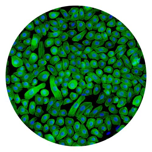
1. Activated microglial cells are believed to play an active role in most brain pathologies, during which they can contribute to host defence and repair but also to the establishment of tissue damage. These actions are largely mediated by microglial secretory products, among which are prostaglandins (PGs) and nitric oxide (NO). 2. The anti-inflammatory protein, lipocortin 1 (LC1) was reported to have neuroprotective action and to be induced by glucocorticoids in several brain structures, with a preferential expression in microglia. In this paper we tested whether the neuroprotective effect of LC1 could be explained by an inhibitory effect on microglial activation. 3. We have previously shown that bacterial endotoxin (LPS) strongly stimulates PGE2 and NO production in rat primary microglial cultures, by inducing the expression of the key enzymes cyclo-oxygenase-2 (COX-2) and inducible nitric oxide synthase (iNOS), respectively. 4. Dexamethasone (DEX, 1-100 nM) and LC1-derived N-terminus peptide (peptide Ac2-26, 1-100 microg ml(-1)) dose-dependently inhibited the production of both PGE2 and NO from LPS-stimulated microglia. The inhibitory effects of DEX on NO and of the peptide on NO and PGE2 synthesis were partially abrogated by a specific antiserum, raised against the N-terminus of human LC1. The peptide Ac2-26 did not affect arachidonic acid release from control and LPS-stimulated microglial cultures. 5. Western blot experiments showed that the LPS-induced expression of COX-2 and iNOS was effectively down-regulated by DEX (100 nM) and peptide Ac2-26 (100 microg ml(-1)). 6. In conclusion, our findings support the hypothesis that LC1 may foster neuroprotection by limiting microglial activation, through autocrine and paracrine mechanisms. (+info)Microglia are a type of specialized immune cell found in the brain and spinal cord. They are part of the glial family, which provide support and protection to the neurons in the central nervous system (CNS). Microglia account for about 10-15% of all cells found in the CNS.
The primary role of microglia is to constantly survey their environment and eliminate any potentially harmful agents, such as pathogens, dead cells, or protein aggregates. They do this through a process called phagocytosis, where they engulf and digest foreign particles or cellular debris. In addition to their phagocytic function, microglia also release various cytokines, chemokines, and growth factors that help regulate the immune response in the CNS, promote neuronal survival, and contribute to synaptic plasticity.
Microglia can exist in different activation states depending on the nature of the stimuli they encounter. In a resting state, microglia have a small cell body with numerous branches that are constantly monitoring their surroundings. When activated by an injury, infection, or neurodegenerative process, microglia change their morphology and phenotype, retracting their processes and adopting an amoeboid shape to migrate towards the site of damage or inflammation. Based on the type of activation, microglia can release both pro-inflammatory and anti-inflammatory factors that contribute to either neuroprotection or neurotoxicity.
Dysregulation of microglial function has been implicated in several neurological disorders, including Alzheimer's disease, Parkinson's disease, multiple sclerosis, and Amyotrophic Lateral Sclerosis (ALS). Therefore, understanding the role of microglia in health and disease is crucial for developing novel therapeutic strategies to treat these conditions.
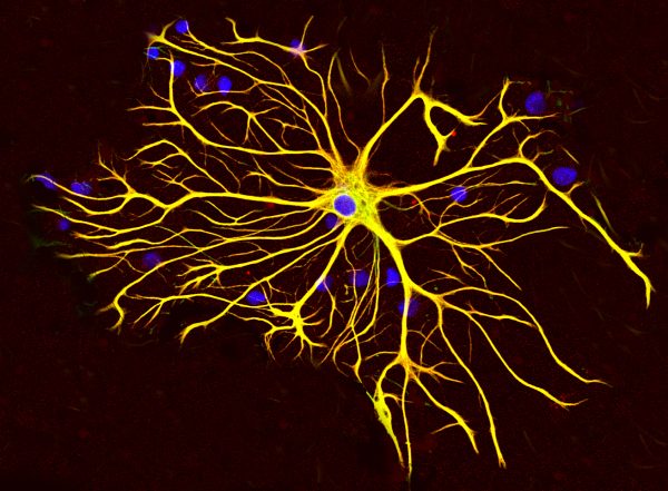
Astrocytes are a type of star-shaped glial cell found in the central nervous system (CNS), including the brain and spinal cord. They play crucial roles in supporting and maintaining the health and function of neurons, which are the primary cells responsible for transmitting information in the CNS.
Some of the essential functions of astrocytes include:
1. Supporting neuronal structure and function: Astrocytes provide structural support to neurons by ensheathing them and maintaining the integrity of the blood-brain barrier, which helps regulate the entry and exit of substances into the CNS.
2. Regulating neurotransmitter levels: Astrocytes help control the levels of neurotransmitters in the synaptic cleft (the space between two neurons) by taking up excess neurotransmitters and breaking them down, thus preventing excessive or prolonged activation of neuronal receptors.
3. Providing nutrients to neurons: Astrocytes help supply energy metabolites, such as lactate, to neurons, which are essential for their survival and function.
4. Modulating synaptic activity: Through the release of various signaling molecules, astrocytes can modulate synaptic strength and plasticity, contributing to learning and memory processes.
5. Participating in immune responses: Astrocytes can respond to CNS injuries or infections by releasing pro-inflammatory cytokines and chemokines, which help recruit immune cells to the site of injury or infection.
6. Promoting neuronal survival and repair: In response to injury or disease, astrocytes can become reactive and undergo morphological changes that aid in forming a glial scar, which helps contain damage and promote tissue repair. Additionally, they release growth factors and other molecules that support the survival and regeneration of injured neurons.
Dysfunction or damage to astrocytes has been implicated in several neurological disorders, including Alzheimer's disease, Parkinson's disease, amyotrophic lateral sclerosis (ALS), and multiple sclerosis (MS).
Gliosis is a term used in histopathology and neuroscience to describe the reaction of support cells in the brain, called glial cells, to injury or disease. This response includes an increase in the number and size of glial cells, as well as changes in their shape and function. The most common types of glial cells involved in gliosis are astrocytes and microglia.
Gliosis can be triggered by a variety of factors, including trauma, infection, inflammation, neurodegenerative diseases, and stroke. In response to injury or disease, astrocytes become hypertrophied (enlarged) and undergo changes in their gene expression profile that can lead to the production of various proteins, such as glial fibrillary acidic protein (GFAP). These changes can result in the formation of a dense network of astrocytic processes, which can contribute to the formation of a glial scar.
Microglia, another type of glial cell, become activated during gliosis and play a role in the immune response in the central nervous system (CNS). They can release pro-inflammatory cytokines, chemokines, and reactive oxygen species that contribute to the inflammatory response.
While gliosis is a protective response aimed at containing damage and promoting tissue repair, it can also have negative consequences. For example, the formation of glial scars can impede axonal regeneration and contribute to neurological deficits. Additionally, chronic activation of microglia has been implicated in various neurodegenerative diseases, such as Alzheimer's disease and Parkinson's disease.
The brain is the central organ of the nervous system, responsible for receiving and processing sensory information, regulating vital functions, and controlling behavior, movement, and cognition. It is divided into several distinct regions, each with specific functions:
1. Cerebrum: The largest part of the brain, responsible for higher cognitive functions such as thinking, learning, memory, language, and perception. It is divided into two hemispheres, each controlling the opposite side of the body.
2. Cerebellum: Located at the back of the brain, it is responsible for coordinating muscle movements, maintaining balance, and fine-tuning motor skills.
3. Brainstem: Connects the cerebrum and cerebellum to the spinal cord, controlling vital functions such as breathing, heart rate, and blood pressure. It also serves as a relay center for sensory information and motor commands between the brain and the rest of the body.
4. Diencephalon: A region that includes the thalamus (a major sensory relay station) and hypothalamus (regulates hormones, temperature, hunger, thirst, and sleep).
5. Limbic system: A group of structures involved in emotional processing, memory formation, and motivation, including the hippocampus, amygdala, and cingulate gyrus.
The brain is composed of billions of interconnected neurons that communicate through electrical and chemical signals. It is protected by the skull and surrounded by three layers of membranes called meninges, as well as cerebrospinal fluid that provides cushioning and nutrients.
"Cells, cultured" is a medical term that refers to cells that have been removed from an organism and grown in controlled laboratory conditions outside of the body. This process is called cell culture and it allows scientists to study cells in a more controlled and accessible environment than they would have inside the body. Cultured cells can be derived from a variety of sources, including tissues, organs, or fluids from humans, animals, or cell lines that have been previously established in the laboratory.
Cell culture involves several steps, including isolation of the cells from the tissue, purification and characterization of the cells, and maintenance of the cells in appropriate growth conditions. The cells are typically grown in specialized media that contain nutrients, growth factors, and other components necessary for their survival and proliferation. Cultured cells can be used for a variety of purposes, including basic research, drug development and testing, and production of biological products such as vaccines and gene therapies.
It is important to note that cultured cells may behave differently than they do in the body, and results obtained from cell culture studies may not always translate directly to human physiology or disease. Therefore, it is essential to validate findings from cell culture experiments using additional models and ultimately in clinical trials involving human subjects.
Encephalitis is defined as inflammation of the brain parenchyma, which is often caused by viral infections but can also be due to bacterial, fungal, or parasitic infections, autoimmune disorders, or exposure to toxins. The infection or inflammation can cause various symptoms such as headache, fever, confusion, seizures, and altered consciousness, ranging from mild symptoms to severe cases that can lead to brain damage, long-term disabilities, or even death.
The diagnosis of encephalitis typically involves a combination of clinical evaluation, imaging studies (such as MRI or CT scans), and laboratory tests (such as cerebrospinal fluid analysis). Treatment may include antiviral medications, corticosteroids, immunoglobulins, and supportive care to manage symptoms and prevent complications.
C57BL/6 (C57 Black 6) is an inbred strain of laboratory mouse that is widely used in biomedical research. The term "inbred" refers to a strain of animals where matings have been carried out between siblings or other closely related individuals for many generations, resulting in a population that is highly homozygous at most genetic loci.
The C57BL/6 strain was established in 1920 by crossing a female mouse from the dilute brown (DBA) strain with a male mouse from the black strain. The resulting offspring were then interbred for many generations to create the inbred C57BL/6 strain.
C57BL/6 mice are known for their robust health, longevity, and ease of handling, making them a popular choice for researchers. They have been used in a wide range of biomedical research areas, including studies of cancer, immunology, neuroscience, cardiovascular disease, and metabolism.
One of the most notable features of the C57BL/6 strain is its sensitivity to certain genetic modifications, such as the introduction of mutations that lead to obesity or impaired glucose tolerance. This has made it a valuable tool for studying the genetic basis of complex diseases and traits.
Overall, the C57BL/6 inbred mouse strain is an important model organism in biomedical research, providing a valuable resource for understanding the genetic and molecular mechanisms underlying human health and disease.
Neuroglia, also known as glial cells or simply glia, are non-neuronal cells that provide support and protection for neurons in the nervous system. They maintain homeostasis, form myelin sheaths around nerve fibers, and provide structural support. They also play a role in the immune response of the central nervous system. Some types of neuroglia include astrocytes, oligodendrocytes, microglia, and ependymal cells.
Lipopolysaccharides (LPS) are large molecules found in the outer membrane of Gram-negative bacteria. They consist of a hydrophilic polysaccharide called the O-antigen, a core oligosaccharide, and a lipid portion known as Lipid A. The Lipid A component is responsible for the endotoxic activity of LPS, which can trigger a powerful immune response in animals, including humans. This response can lead to symptoms such as fever, inflammation, and septic shock, especially when large amounts of LPS are introduced into the bloodstream.
Minocycline is an antibiotic medication that belongs to the tetracycline class. Medically, it is defined as a semisynthetic derivative of tetracycline and has a broader spectrum of activity compared to other tetracyclines. It is bacteriostatic, meaning it inhibits bacterial growth rather than killing them outright.
Minocycline is commonly used to treat various infections caused by susceptible bacteria, including acne, respiratory infections, urinary tract infections, skin and soft tissue infections, and sexually transmitted diseases. Additionally, it has been found to have anti-inflammatory properties and is being investigated for its potential use in treating neurological disorders such as multiple sclerosis and Alzheimer's disease.
As with all antibiotics, minocycline should be taken under the guidance of a healthcare professional, and its usage should be based on the results of bacterial culture and sensitivity testing to ensure its effectiveness against the specific bacteria causing the infection.
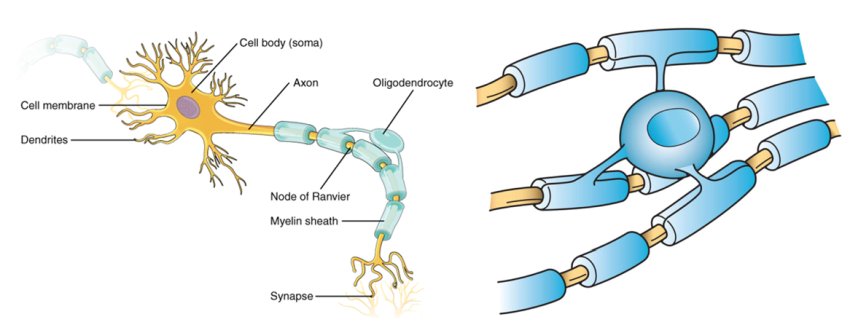
Neurons, also known as nerve cells or neurocytes, are specialized cells that constitute the basic unit of the nervous system. They are responsible for receiving, processing, and transmitting information and signals within the body. Neurons have three main parts: the dendrites, the cell body (soma), and the axon. The dendrites receive signals from other neurons or sensory receptors, while the axon transmits these signals to other neurons, muscles, or glands. The junction between two neurons is called a synapse, where neurotransmitters are released to transmit the signal across the gap (synaptic cleft) to the next neuron. Neurons vary in size, shape, and structure depending on their function and location within the nervous system.
CD11b, also known as integrin αM or Mac-1, is not an antigen itself but a protein that forms part of a family of cell surface receptors called integrins. These integrins play a crucial role in various biological processes, including cell adhesion, migration, and signaling.
CD11b combines with CD18 (integrin β2) to form the heterodimeric integrin αMβ2, also known as Mac-1 or CR3 (complement receptor 3). This integrin is primarily expressed on the surface of myeloid cells, such as monocytes, macrophages, and neutrophils.
As an integral part of the immune system, CD11b/CD18 recognizes and binds to various ligands, including:
1. Icosahedral bacterial components like lipopolysaccharides (LPS) and peptidoglycans
2. Fragments of complement component C3b (iC3b)
3. Fibrinogen and other extracellular matrix proteins
4. Certain immune cell receptors, such as ICAM-1 (intercellular adhesion molecule 1)
The binding of CD11b/CD18 to these ligands triggers various intracellular signaling pathways that regulate the immune response and inflammation. In this context, antigens are substances (usually proteins or polysaccharides) found on the surface of cells, viruses, or bacteria that can be recognized by the immune system. CD11b/CD18 plays a role in recognizing and responding to these antigens during an immune response.
"Newborn animals" refers to the very young offspring of animals that have recently been born. In medical terminology, newborns are often referred to as "neonates," and they are classified as such from birth until about 28 days of age. During this time period, newborn animals are particularly vulnerable and require close monitoring and care to ensure their survival and healthy development.
The specific needs of newborn animals can vary widely depending on the species, but generally, they require warmth, nutrition, hydration, and protection from harm. In many cases, newborns are unable to regulate their own body temperature or feed themselves, so they rely heavily on their mothers for care and support.
In medical settings, newborn animals may be examined and treated by veterinarians to ensure that they are healthy and receiving the care they need. This can include providing medical interventions such as feeding tubes, antibiotics, or other treatments as needed to address any health issues that arise. Overall, the care and support of newborn animals is an important aspect of animal medicine and conservation efforts.
Nerve degeneration, also known as neurodegeneration, is the progressive loss of structure and function of neurons, which can lead to cognitive decline, motor impairment, and various other symptoms. This process occurs due to a variety of factors, including genetics, environmental influences, and aging. It is a key feature in several neurological disorders such as Alzheimer's disease, Parkinson's disease, Huntington's disease, and multiple sclerosis. The degeneration can affect any part of the nervous system, leading to different symptoms depending on the location and extent of the damage.
Amyloid beta-peptides (Aβ) are small protein fragments that are crucially involved in the pathogenesis of Alzheimer's disease. They are derived from a larger transmembrane protein called the amyloid precursor protein (APP) through a series of proteolytic cleavage events.
The two primary forms of Aβ peptides are Aβ40 and Aβ42, which differ in length by two amino acids. While both forms can be harmful, Aβ42 is more prone to aggregation and is considered to be the more pathogenic form. These peptides have the tendency to misfold and accumulate into oligomers, fibrils, and eventually insoluble plaques that deposit in various areas of the brain, most notably the cerebral cortex and hippocampus.
The accumulation of Aβ peptides is believed to initiate a cascade of events leading to neuroinflammation, oxidative stress, synaptic dysfunction, and neuronal death, which are all hallmarks of Alzheimer's disease. Although the exact role of Aβ in the onset and progression of Alzheimer's is still under investigation, it is widely accepted that they play a central part in the development of this debilitating neurodegenerative disorder.
The Central Nervous System (CNS) is the part of the nervous system that consists of the brain and spinal cord. It is called the "central" system because it receives information from, and sends information to, the rest of the body through peripheral nerves, which make up the Peripheral Nervous System (PNS).
The CNS is responsible for processing sensory information, controlling motor functions, and regulating various autonomic processes like heart rate, respiration, and digestion. The brain, as the command center of the CNS, interprets sensory stimuli, formulates thoughts, and initiates actions. The spinal cord serves as a conduit for nerve impulses traveling to and from the brain and the rest of the body.
The CNS is protected by several structures, including the skull (which houses the brain) and the vertebral column (which surrounds and protects the spinal cord). Despite these protective measures, the CNS remains vulnerable to injury and disease, which can have severe consequences due to its crucial role in controlling essential bodily functions.
Macrophages are a type of white blood cell that are an essential part of the immune system. They are large, specialized cells that engulf and destroy foreign substances, such as bacteria, viruses, parasites, and fungi, as well as damaged or dead cells. Macrophages are found throughout the body, including in the bloodstream, lymph nodes, spleen, liver, lungs, and connective tissues. They play a critical role in inflammation, immune response, and tissue repair and remodeling.
Macrophages originate from monocytes, which are a type of white blood cell produced in the bone marrow. When monocytes enter the tissues, they differentiate into macrophages, which have a larger size and more specialized functions than monocytes. Macrophages can change their shape and move through tissues to reach sites of infection or injury. They also produce cytokines, chemokines, and other signaling molecules that help coordinate the immune response and recruit other immune cells to the site of infection or injury.
Macrophages have a variety of surface receptors that allow them to recognize and respond to different types of foreign substances and signals from other cells. They can engulf and digest foreign particles, bacteria, and viruses through a process called phagocytosis. Macrophages also play a role in presenting antigens to T cells, which are another type of immune cell that helps coordinate the immune response.
Overall, macrophages are crucial for maintaining tissue homeostasis, defending against infection, and promoting wound healing and tissue repair. Dysregulation of macrophage function has been implicated in a variety of diseases, including cancer, autoimmune disorders, and chronic inflammatory conditions.
Glial Fibrillary Acidic Protein (GFAP) is a type of intermediate filament protein that is primarily found in astrocytes, which are a type of star-shaped glial cells in the central nervous system (CNS). These proteins play an essential role in maintaining the structural integrity and stability of astrocytes. They also participate in various cellular processes such as responding to injury, providing support to neurons, and regulating the extracellular environment.
GFAP is often used as a marker for astrocytic activation or reactivity, which can occur in response to CNS injuries, neuroinflammation, or neurodegenerative diseases. Elevated GFAP levels in cerebrospinal fluid (CSF) or blood can indicate astrocyte damage or dysfunction and are associated with several neurological conditions, including traumatic brain injury, stroke, multiple sclerosis, Alzheimer's disease, and Alexander's disease.
Inflammation is a complex biological response of tissues to harmful stimuli, such as pathogens, damaged cells, or irritants. It is characterized by the following signs: rubor (redness), tumor (swelling), calor (heat), dolor (pain), and functio laesa (loss of function). The process involves the activation of the immune system, recruitment of white blood cells, and release of inflammatory mediators, which contribute to the elimination of the injurious stimuli and initiation of the healing process. However, uncontrolled or chronic inflammation can also lead to tissue damage and diseases.
Animal disease models are specialized animals, typically rodents such as mice or rats, that have been genetically engineered or exposed to certain conditions to develop symptoms and physiological changes similar to those seen in human diseases. These models are used in medical research to study the pathophysiology of diseases, identify potential therapeutic targets, test drug efficacy and safety, and understand disease mechanisms.
The genetic modifications can include knockout or knock-in mutations, transgenic expression of specific genes, or RNA interference techniques. The animals may also be exposed to environmental factors such as chemicals, radiation, or infectious agents to induce the disease state.
Examples of animal disease models include:
1. Mouse models of cancer: Genetically engineered mice that develop various types of tumors, allowing researchers to study cancer initiation, progression, and metastasis.
2. Alzheimer's disease models: Transgenic mice expressing mutant human genes associated with Alzheimer's disease, which exhibit amyloid plaque formation and cognitive decline.
3. Diabetes models: Obese and diabetic mouse strains like the NOD (non-obese diabetic) or db/db mice, used to study the development of type 1 and type 2 diabetes, respectively.
4. Cardiovascular disease models: Atherosclerosis-prone mice, such as ApoE-deficient or LDLR-deficient mice, that develop plaque buildup in their arteries when fed a high-fat diet.
5. Inflammatory bowel disease models: Mice with genetic mutations affecting intestinal barrier function and immune response, such as IL-10 knockout or SAMP1/YitFc mice, which develop colitis.
Animal disease models are essential tools in preclinical research, but it is important to recognize their limitations. Differences between species can affect the translatability of results from animal studies to human patients. Therefore, researchers must carefully consider the choice of model and interpret findings cautiously when applying them to human diseases.
Sprague-Dawley rats are a strain of albino laboratory rats that are widely used in scientific research. They were first developed by researchers H.H. Sprague and R.C. Dawley in the early 20th century, and have since become one of the most commonly used rat strains in biomedical research due to their relatively large size, ease of handling, and consistent genetic background.
Sprague-Dawley rats are outbred, which means that they are genetically diverse and do not suffer from the same limitations as inbred strains, which can have reduced fertility and increased susceptibility to certain diseases. They are also characterized by their docile nature and low levels of aggression, making them easier to handle and study than some other rat strains.
These rats are used in a wide variety of research areas, including toxicology, pharmacology, nutrition, cancer, and behavioral studies. Because they are genetically diverse, Sprague-Dawley rats can be used to model a range of human diseases and conditions, making them an important tool in the development of new drugs and therapies.
Cell death is the process by which cells cease to function and eventually die. There are several ways that cells can die, but the two most well-known and well-studied forms of cell death are apoptosis and necrosis.
Apoptosis is a programmed form of cell death that occurs as a normal and necessary process in the development and maintenance of healthy tissues. During apoptosis, the cell's DNA is broken down into small fragments, the cell shrinks, and the membrane around the cell becomes fragmented, allowing the cell to be easily removed by phagocytic cells without causing an inflammatory response.
Necrosis, on the other hand, is a form of cell death that occurs as a result of acute tissue injury or overwhelming stress. During necrosis, the cell's membrane becomes damaged and the contents of the cell are released into the surrounding tissue, causing an inflammatory response.
There are also other forms of cell death, such as autophagy, which is a process by which cells break down their own organelles and proteins to recycle nutrients and maintain energy homeostasis, and pyroptosis, which is a form of programmed cell death that occurs in response to infection and involves the activation of inflammatory caspases.
Cell death is an important process in many physiological and pathological processes, including development, tissue homeostasis, and disease. Dysregulation of cell death can contribute to the development of various diseases, including cancer, neurodegenerative disorders, and autoimmune diseases.
Research personnel, in the context of medical and scientific research, refers to individuals who are involved in the design, conduct, or reporting of research studies. This can include, but is not limited to, principal investigators, co-investigators, research assistants, research coordinators, data managers, biostatisticians, and laboratory technicians. These individuals may have various levels of education, training, and expertise, and their roles and responsibilities will depend on the specific research study and their individual qualifications. It is important for research personnel to adhere to ethical guidelines and regulations in order to ensure the integrity and validity of research findings.
Coculture techniques refer to a type of experimental setup in which two or more different types of cells or organisms are grown and studied together in a shared culture medium. This method allows researchers to examine the interactions between different cell types or species under controlled conditions, and to study how these interactions may influence various biological processes such as growth, gene expression, metabolism, and signal transduction.
Coculture techniques can be used to investigate a wide range of biological phenomena, including the effects of host-microbe interactions on human health and disease, the impact of different cell types on tissue development and homeostasis, and the role of microbial communities in shaping ecosystems. These techniques can also be used to test the efficacy and safety of new drugs or therapies by examining their effects on cells grown in coculture with other relevant cell types.
There are several different ways to establish cocultures, depending on the specific research question and experimental goals. Some common methods include:
1. Mixed cultures: In this approach, two or more cell types are simply mixed together in a culture dish or flask and allowed to grow and interact freely.
2. Cell-layer cultures: Here, one cell type is grown on a porous membrane or other support structure, while the second cell type is grown on top of it, forming a layered coculture.
3. Conditioned media cultures: In this case, one cell type is grown to confluence and its culture medium is collected and then used to grow a second cell type. This allows the second cell type to be exposed to any factors secreted by the first cell type into the medium.
4. Microfluidic cocultures: These involve growing cells in microfabricated channels or chambers, which allow for precise control over the spatial arrangement and flow of nutrients, waste products, and signaling molecules between different cell types.
Overall, coculture techniques provide a powerful tool for studying complex biological systems and gaining insights into the mechanisms that underlie various physiological and pathological processes.
Transgenic mice are genetically modified rodents that have incorporated foreign DNA (exogenous DNA) into their own genome. This is typically done through the use of recombinant DNA technology, where a specific gene or genetic sequence of interest is isolated and then introduced into the mouse embryo. The resulting transgenic mice can then express the protein encoded by the foreign gene, allowing researchers to study its function in a living organism.
The process of creating transgenic mice usually involves microinjecting the exogenous DNA into the pronucleus of a fertilized egg, which is then implanted into a surrogate mother. The offspring that result from this procedure are screened for the presence of the foreign DNA, and those that carry the desired genetic modification are used to establish a transgenic mouse line.
Transgenic mice have been widely used in biomedical research to model human diseases, study gene function, and test new therapies. They provide a valuable tool for understanding complex biological processes and developing new treatments for a variety of medical conditions.
Purinergic P2X4 receptors are a type of ionotropic purinergic receptor that are activated by adenosine triphosphate (ATP) and related nucleotides. They belong to the P2X receptor family, which includes seven subtypes (P2X1-7) that form trimeric channels permeable to cations such as calcium, sodium, and potassium.
The P2X4 receptor is widely expressed in various tissues, including the central and peripheral nervous systems, immune cells, and epithelial cells. It plays a role in several physiological processes, including neurotransmission, inflammation, and pain perception. Activation of P2X4 receptors leads to an increase in intracellular calcium concentration and membrane depolarization, which can modulate synaptic transmission and cell excitability.
P2X4 receptors have also been implicated in various pathological conditions, such as neuropathic pain, neuroinflammation, and ischemic injury. They are involved in the release of pro-inflammatory cytokines and chemokines from immune cells, contributing to the development of chronic inflammation and tissue damage.
In summary, purinergic P2X4 receptors are a type of ATP-gated ion channel that play important roles in physiological and pathological processes, including neurotransmission, inflammation, and pain perception.
Phagocytosis is the process by which certain cells in the body, known as phagocytes, engulf and destroy foreign particles, bacteria, or dead cells. This mechanism plays a crucial role in the immune system's response to infection and inflammation. Phagocytes, such as neutrophils, monocytes, and macrophages, have receptors on their surface that recognize and bind to specific molecules (known as antigens) on the target particles or microorganisms.
Once attached, the phagocyte extends pseudopodia (cell extensions) around the particle, forming a vesicle called a phagosome that completely encloses it. The phagosome then fuses with a lysosome, an intracellular organelle containing digestive enzymes and other chemicals. This fusion results in the formation of a phagolysosome, where the engulfed particle is broken down by the action of these enzymes, neutralizing its harmful effects and allowing for the removal of cellular debris or pathogens.
Phagocytosis not only serves as a crucial defense mechanism against infections but also contributes to tissue homeostasis by removing dead cells and debris.
The ulna is one of the two long bones in the forearm, the other being the radius. It runs from the elbow to the wrist and is located on the medial side of the forearm, next to the bone called the humerus in the upper arm. The ulna plays a crucial role in the movement of the forearm and also serves as an attachment site for various muscles.
The spinal cord is a major part of the nervous system, extending from the brainstem and continuing down to the lower back. It is a slender, tubular bundle of nerve fibers (axons) and support cells (glial cells) that carries signals between the brain and the rest of the body. The spinal cord primarily serves as a conduit for motor information, which travels from the brain to the muscles, and sensory information, which travels from the body to the brain. It also contains neurons that can independently process and respond to information within the spinal cord without direct input from the brain.
The spinal cord is protected by the bony vertebral column (spine) and is divided into 31 segments: 8 cervical, 12 thoracic, 5 lumbar, 5 sacral, and 1 coccygeal. Each segment corresponds to a specific region of the body and gives rise to pairs of spinal nerves that exit through the intervertebral foramina at each level.
The spinal cord is responsible for several vital functions, including:
1. Reflexes: Simple reflex actions, such as the withdrawal reflex when touching a hot surface, are mediated by the spinal cord without involving the brain.
2. Muscle control: The spinal cord carries motor signals from the brain to the muscles, enabling voluntary movement and muscle tone regulation.
3. Sensory perception: The spinal cord transmits sensory information, such as touch, temperature, pain, and vibration, from the body to the brain for processing and awareness.
4. Autonomic functions: The sympathetic and parasympathetic divisions of the autonomic nervous system originate in the thoracolumbar and sacral regions of the spinal cord, respectively, controlling involuntary physiological responses like heart rate, blood pressure, digestion, and respiration.
Damage to the spinal cord can result in various degrees of paralysis or loss of sensation below the level of injury, depending on the severity and location of the damage.
A "knockout" mouse is a genetically engineered mouse in which one or more genes have been deleted or "knocked out" using molecular biology techniques. This allows researchers to study the function of specific genes and their role in various biological processes, as well as potential associations with human diseases. The mice are generated by introducing targeted DNA modifications into embryonic stem cells, which are then used to create a live animal. Knockout mice have been widely used in biomedical research to investigate gene function, disease mechanisms, and potential therapeutic targets.
Oligodendroglia are a type of neuroglial cell found in the central nervous system (CNS) of vertebrates, including humans. These cells play a crucial role in providing support and insulation to nerve fibers (axons) in the CNS, which includes the brain and spinal cord.
More specifically, oligodendroglia produce a fatty substance called myelin that wraps around axons, forming myelin sheaths. This myelination process helps to increase the speed of electrical impulse transmission (nerve impulses) along the axons, allowing for efficient communication between different neurons.
In addition to their role in myelination, oligodendroglia also contribute to the overall health and maintenance of the CNS by providing essential nutrients and supporting factors to neurons. Dysfunction or damage to oligodendroglia has been implicated in various neurological disorders, such as multiple sclerosis (MS), where demyelination of axons leads to impaired nerve function and neurodegeneration.
Alzheimer's disease is a progressive disorder that causes brain cells to waste away (degenerate) and die. It's the most common cause of dementia — a continuous decline in thinking, behavioral and social skills that disrupts a person's ability to function independently.
The early signs of the disease include forgetting recent events or conversations. As the disease progresses, a person with Alzheimer's disease will develop severe memory impairment and lose the ability to carry out everyday tasks.
Currently, there's no cure for Alzheimer's disease. However, treatments can temporarily slow the worsening of dementia symptoms and improve quality of life.
Cuprizone is not a medical condition or disease, but rather a chemical compound that is used in laboratory settings for research purposes. Cuprizone, also known as bis-cyclohexanone oxaldihydrazone, is a copper chelator, which means it can bind to and remove copper ions from various substances.
In research, cuprizone is often used to induce demyelination in animal models of multiple sclerosis (MS) and other neurological disorders. Demyelination refers to the loss or damage of the myelin sheath, which is a fatty substance that surrounds and protects nerve fibers in the brain and spinal cord. When cuprizone is added to the diet of laboratory animals such as mice, it can cause demyelination in specific areas of the brain, making it a useful tool for studying the mechanisms underlying MS and other demyelinating diseases.
It's important to note that while cuprizone is a valuable research tool, it is not used as a medical treatment or therapy for any human conditions.
Tumor Necrosis Factor-alpha (TNF-α) is a cytokine, a type of small signaling protein involved in immune response and inflammation. It is primarily produced by activated macrophages, although other cell types such as T-cells, natural killer cells, and mast cells can also produce it.
TNF-α plays a crucial role in the body's defense against infection and tissue injury by mediating inflammatory responses, activating immune cells, and inducing apoptosis (programmed cell death) in certain types of cells. It does this by binding to its receptors, TNFR1 and TNFR2, which are found on the surface of many cell types.
In addition to its role in the immune response, TNF-α has been implicated in the pathogenesis of several diseases, including autoimmune disorders such as rheumatoid arthritis, inflammatory bowel disease, and psoriasis, as well as cancer, where it can promote tumor growth and metastasis.
Therapeutic agents that target TNF-α, such as infliximab, adalimumab, and etanercept, have been developed to treat these conditions. However, these drugs can also increase the risk of infections and other side effects, so their use must be carefully monitored.
Inflammation mediators are substances that are released by the body in response to injury or infection, which contribute to the inflammatory response. These mediators include various chemical factors such as cytokines, chemokines, prostaglandins, leukotrienes, and histamine, among others. They play a crucial role in regulating the inflammatory process by attracting immune cells to the site of injury or infection, increasing blood flow to the area, and promoting the repair and healing of damaged tissues. However, an overactive or chronic inflammatory response can also contribute to the development of various diseases and conditions, such as autoimmune disorders, cardiovascular disease, and cancer.
Neuroprotective agents are substances that protect neurons or nerve cells from damage, degeneration, or death caused by various factors such as trauma, inflammation, oxidative stress, or excitotoxicity. These agents work through different mechanisms, including reducing the production of free radicals, inhibiting the release of glutamate (a neurotransmitter that can cause cell damage in high concentrations), promoting the growth and survival of neurons, and preventing apoptosis (programmed cell death). Neuroprotective agents have been studied for their potential to treat various neurological disorders, including stroke, traumatic brain injury, Parkinson's disease, Alzheimer's disease, and multiple sclerosis. However, more research is needed to fully understand their mechanisms of action and to develop effective therapies.
Interleukin-1 beta (IL-1β) is a member of the interleukin-1 cytokine family and is primarily produced by activated macrophages in response to inflammatory stimuli. It is a crucial mediator of the innate immune response and plays a key role in the regulation of various biological processes, including cell proliferation, differentiation, and apoptosis. IL-1β is involved in the pathogenesis of several inflammatory diseases, such as rheumatoid arthritis, inflammatory bowel disease, and atherosclerosis. It exerts its effects by binding to the interleukin-1 receptor, which triggers a signaling cascade that leads to the activation of various transcription factors and the expression of target genes.
Nitric Oxide Synthase Type II (NOS2), also known as Inducible Nitric Oxide Synthase (iNOS), is an enzyme that catalyzes the production of nitric oxide (NO) from L-arginine. Unlike other isoforms of NOS, NOS2 is not constitutively expressed and its expression can be induced by various stimuli such as cytokines, lipopolysaccharides, and bacterial products. Once induced, NOS2 produces large amounts of NO, which plays a crucial role in the immune response against invading pathogens. However, excessive or prolonged production of NO by NOS2 has been implicated in various pathological conditions such as inflammation, septic shock, and neurodegenerative disorders.
Chemokine (C-X-C motif) ligand 1 (CX3CL1), also known as fractalkine, is a protein that belongs to the chemokine family. Chemokines are a group of small signaling proteins involved in immune responses and inflammation. CX3CL1 is unique among chemokines because it exists both as a soluble protein and as a membrane-bound protein on the surface of certain cells.
As a chemoattractant, CX3CL1 plays a crucial role in recruiting immune cells, particularly T cells and monocytes/macrophages, to sites of infection or injury. The interaction between CX3CL1 and its receptor, CX3CR1, expressed on the surface of these immune cells, mediates their migration and activation.
In addition to its role in immunity and inflammation, CX3CL1 has been implicated in various physiological and pathological processes, such as neuronal development, neuroinflammation, and neurodegenerative disorders like Alzheimer's disease and Parkinson's disease.
Neuralgia is a type of pain that occurs along the pathway of a nerve, often caused by damage or irritation to the nerve. It is typically described as a sharp, stabbing, burning, or electric-shock like pain that can be severe and debilitating. Neuralgia can affect any nerve in the body, but it most commonly occurs in the facial area (trigeminal neuralgia) or in the nerves related to the spine (postherpetic neuralgia). The pain associated with neuralgia can be intermittent or constant and may be worsened by certain triggers such as touch, temperature changes, or movement. Treatment for neuralgia typically involves medications to manage pain, as well as other therapies such as nerve blocks, surgery, or lifestyle modifications.
Nervous system trauma, also known as neurotrauma, refers to damage or injury to the nervous system, including the brain and spinal cord. This type of trauma can result from various causes, such as vehicular accidents, sports injuries, falls, violence, or penetrating traumas. Nervous system trauma can lead to temporary or permanent impairments in sensory, motor, or cognitive functions, depending on the severity and location of the injury.
Traumatic brain injury (TBI) is a common form of nervous system trauma that occurs when an external force causes brain dysfunction. TBIs can be classified as mild, moderate, or severe, based on factors such as loss of consciousness, memory loss, and neurological deficits. Mild TBIs, also known as concussions, may not cause long-term damage but still require medical attention to ensure proper healing and prevent further complications.
Spinal cord injuries (SCI) are another form of nervous system trauma that can have severe consequences. SCI occurs when the spinal cord is damaged due to a sudden, traumatic blow or cut, causing loss of motor function, sensation, or autonomic function below the level of injury. The severity and location of the injury determine the extent of impairment, which can range from partial to complete paralysis.
Immediate medical intervention is crucial in cases of nervous system trauma to minimize secondary damage, prevent complications, and optimize recovery outcomes. Treatment options may include surgery, medication, rehabilitation, or a combination of these approaches.
Amyloid plaque is a pathological hallmark of several degenerative diseases, including Alzheimer's disease. It refers to extracellular deposits of misfolded proteins that accumulate in various tissues and organs, but are most commonly found in the brain. The main component of these plaques is an abnormally folded form of a protein called amyloid-beta (Aβ). This protein is produced through the normal processing of the amyloid precursor protein (APP), but in amyloid plaques, it aggregates into insoluble fibrils that form the core of the plaque.
The accumulation of amyloid plaques is thought to contribute to neurodegeneration and cognitive decline in Alzheimer's disease and other related disorders. The exact mechanisms by which this occurs are not fully understood, but it is believed that the aggregation of Aβ into plaques leads to the disruption of neuronal function and viability, as well as the activation of inflammatory responses that can further damage brain tissue.
It's important to note that while amyloid plaques are a key feature of Alzheimer's disease, they are not exclusive to this condition. Amyloid plaques have also been found in other neurodegenerative disorders, as well as in some normal aging brains, although their significance in these contexts is less clear.
Signal transduction is the process by which a cell converts an extracellular signal, such as a hormone or neurotransmitter, into an intracellular response. This involves a series of molecular events that transmit the signal from the cell surface to the interior of the cell, ultimately resulting in changes in gene expression, protein activity, or metabolism.
The process typically begins with the binding of the extracellular signal to a receptor located on the cell membrane. This binding event activates the receptor, which then triggers a cascade of intracellular signaling molecules, such as second messengers, protein kinases, and ion channels. These molecules amplify and propagate the signal, ultimately leading to the activation or inhibition of specific cellular responses.
Signal transduction pathways are highly regulated and can be modulated by various factors, including other signaling molecules, post-translational modifications, and feedback mechanisms. Dysregulation of these pathways has been implicated in a variety of diseases, including cancer, diabetes, and neurological disorders.
Neurodegenerative diseases are a group of disorders characterized by progressive and persistent loss of neuronal structure and function, often leading to cognitive decline, functional impairment, and ultimately death. These conditions are associated with the accumulation of abnormal protein aggregates, mitochondrial dysfunction, oxidative stress, chronic inflammation, and genetic mutations in the brain. Examples of neurodegenerative diseases include Alzheimer's disease, Parkinson's disease, Huntington's disease, Amyotrophic Lateral Sclerosis (ALS), and Spinal Muscular Atrophy (SMA). The underlying causes and mechanisms of these diseases are not fully understood, and there is currently no cure for most neurodegenerative disorders. Treatment typically focuses on managing symptoms and slowing disease progression.
A primary cell culture is the very first cell culture generation that is established by directly isolating cells from an original tissue or organ source. These cells are removed from the body and then cultured in controlled conditions in a laboratory setting, allowing them to grow and multiply. Primary cell cultures maintain many of the characteristics of the cells in their original tissue environment, making them valuable for research purposes. However, they can only be passaged (subcultured) a limited number of times before they undergo senescence or change into a different type of cell.
The cerebral cortex is the outermost layer of the brain, characterized by its intricate folded structure and wrinkled appearance. It is a region of great importance as it plays a key role in higher cognitive functions such as perception, consciousness, thought, memory, language, and attention. The cerebral cortex is divided into two hemispheres, each containing four lobes: the frontal, parietal, temporal, and occipital lobes. These areas are responsible for different functions, with some regions specializing in sensory processing while others are involved in motor control or associative functions. The cerebral cortex is composed of gray matter, which contains neuronal cell bodies, and is covered by a layer of white matter that consists mainly of myelinated nerve fibers.
"Cell count" is a medical term that refers to the process of determining the number of cells present in a given volume or sample of fluid or tissue. This can be done through various laboratory methods, such as counting individual cells under a microscope using a specialized grid called a hemocytometer, or using automated cell counters that use light scattering and electrical impedance techniques to count and classify different types of cells.
Cell counts are used in a variety of medical contexts, including hematology (the study of blood and blood-forming tissues), microbiology (the study of microscopic organisms), and pathology (the study of diseases and their causes). For example, a complete blood count (CBC) is a routine laboratory test that includes a white blood cell (WBC) count, red blood cell (RBC) count, hemoglobin level, hematocrit value, and platelet count. Abnormal cell counts can indicate the presence of various medical conditions, such as infections, anemia, or leukemia.
Microfilament proteins are a type of structural protein that form part of the cytoskeleton in eukaryotic cells. They are made up of actin monomers, which polymerize to form long, thin filaments. These filaments are involved in various cellular processes such as muscle contraction, cell division, and cell motility. Microfilament proteins also interact with other cytoskeletal components like intermediate filaments and microtubules to maintain the overall shape and integrity of the cell. Additionally, they play a crucial role in the formation of cell-cell junctions and cell-matrix adhesions, which are essential for tissue structure and function.
Neurotoxins are substances that are poisonous or destructive to nerve cells (neurons) and the nervous system. They can cause damage by destroying neurons, disrupting communication between neurons, or interfering with the normal functioning of the nervous system. Neurotoxins can be produced naturally by certain organisms, such as bacteria, plants, and animals, or they can be synthetic compounds created in a laboratory. Examples of neurotoxins include botulinum toxin (found in botulism), tetrodotoxin (found in pufferfish), and heavy metals like lead and mercury. Neurotoxic effects can range from mild symptoms such as headaches, muscle weakness, and tremors, to more severe symptoms such as paralysis, seizures, and cognitive impairment. Long-term exposure to neurotoxins can lead to chronic neurological conditions and other health problems.
The hippocampus is a complex, curved formation in the brain that resembles a seahorse (hence its name, from the Greek word "hippos" meaning horse and "kampos" meaning sea monster). It's part of the limbic system and plays crucial roles in the formation of memories, particularly long-term ones.
This region is involved in spatial navigation and cognitive maps, allowing us to recognize locations and remember how to get to them. Additionally, it's one of the first areas affected by Alzheimer's disease, which often results in memory loss as an early symptom.
Anatomically, it consists of two main parts: the Ammon's horn (or cornu ammonis) and the dentate gyrus. These structures are made up of distinct types of neurons that contribute to different aspects of learning and memory.
'Gene expression regulation' refers to the processes that control whether, when, and where a particular gene is expressed, meaning the production of a specific protein or functional RNA encoded by that gene. This complex mechanism can be influenced by various factors such as transcription factors, chromatin remodeling, DNA methylation, non-coding RNAs, and post-transcriptional modifications, among others. Proper regulation of gene expression is crucial for normal cellular function, development, and maintaining homeostasis in living organisms. Dysregulation of gene expression can lead to various diseases, including cancer and genetic disorders.
Calcium-binding proteins (CaBPs) are a diverse group of proteins that have the ability to bind calcium ions (Ca^2+^) with high affinity and specificity. They play crucial roles in various cellular processes, including signal transduction, muscle contraction, neurotransmitter release, and protection against oxidative stress.
The binding of calcium ions to these proteins induces conformational changes that can either activate or inhibit their functions. Some well-known CaBPs include calmodulin, troponin C, S100 proteins, and parvalbumins. These proteins are essential for maintaining calcium homeostasis within cells and for mediating the effects of calcium as a second messenger in various cellular signaling pathways.
Messenger RNA (mRNA) is a type of RNA (ribonucleic acid) that carries genetic information copied from DNA in the form of a series of three-base code "words," each of which specifies a particular amino acid. This information is used by the cell's machinery to construct proteins, a process known as translation. After being transcribed from DNA, mRNA travels out of the nucleus to the ribosomes in the cytoplasm where protein synthesis occurs. Once the protein has been synthesized, the mRNA may be degraded and recycled. Post-transcriptional modifications can also occur to mRNA, such as alternative splicing and addition of a 5' cap and a poly(A) tail, which can affect its stability, localization, and translation efficiency.
Macrophage activation is a process in which these immune cells become increasingly active and responsive to various stimuli, such as pathogens or inflammatory signals. This activation triggers a series of changes within the macrophages, allowing them to perform important functions like phagocytosis (ingesting and destroying foreign particles or microorganisms), antigen presentation (presenting microbial fragments to T-cells to stimulate an immune response), and production of cytokines and chemokines (signaling molecules that help coordinate the immune response).
There are two main types of macrophage activation: classical (or M1) activation and alternative (or M2) activation. Classical activation is typically induced by interferon-gamma (IFN-γ) and lipopolysaccharide (LPS), leading to a proinflammatory response, enhanced microbicidal activity, and the production of reactive oxygen and nitrogen species. Alternative activation, on the other hand, is triggered by cytokines like interleukin-4 (IL-4) and IL-13, resulting in an anti-inflammatory response, tissue repair, and the promotion of wound healing.
It's important to note that macrophage activation plays a crucial role in various physiological and pathological processes, including immune defense, inflammation, tissue remodeling, and even cancer progression. Dysregulation of macrophage activation has been implicated in several diseases, such as autoimmune disorders, chronic infections, and cancer.
"Wistar rats" are a strain of albino rats that are widely used in laboratory research. They were developed at the Wistar Institute in Philadelphia, USA, and were first introduced in 1906. Wistar rats are outbred, which means that they are genetically diverse and do not have a fixed set of genetic characteristics like inbred strains.
Wistar rats are commonly used as animal models in biomedical research because of their size, ease of handling, and relatively low cost. They are used in a wide range of research areas, including toxicology, pharmacology, nutrition, cancer, cardiovascular disease, and behavioral studies. Wistar rats are also used in safety testing of drugs, medical devices, and other products.
Wistar rats are typically larger than many other rat strains, with males weighing between 500-700 grams and females weighing between 250-350 grams. They have a lifespan of approximately 2-3 years. Wistar rats are also known for their docile and friendly nature, making them easy to handle and work with in the laboratory setting.
Nitric oxide (NO) is a molecule made up of one nitrogen atom and one oxygen atom. In the body, it is a crucial signaling molecule involved in various physiological processes such as vasodilation, immune response, neurotransmission, and inhibition of platelet aggregation. It is produced naturally by the enzyme nitric oxide synthase (NOS) from the amino acid L-arginine. Inhaled nitric oxide is used medically to treat pulmonary hypertension in newborns and adults, as it helps to relax and widen blood vessels, improving oxygenation and blood flow.
Antigens are substances (usually proteins) on the surface of cells, or viruses, bacteria, and other microorganisms, that can stimulate an immune response.
Differentiation in the context of myelomonocytic cells refers to the process by which these cells mature and develop into specific types of immune cells, such as monocytes, macrophages, and neutrophils.
Myelomonocytic cells are a type of white blood cell that originate from stem cells in the bone marrow. They give rise to two main types of immune cells: monocytes and granulocytes (which include neutrophils, eosinophils, and basophils).
Therefore, 'Antigens, Differentiation, Myelomonocytic' refers to the study or examination of how antigens affect the differentiation process of myelomonocytic cells into specific types of immune cells. This is an important area of research in immunology and hematology as it relates to understanding how the body responds to infections, inflammation, and cancer.
Analysis of Variance (ANOVA) is a statistical technique used to compare the means of two or more groups and determine whether there are any significant differences between them. It is a way to analyze the variance in a dataset to determine whether the variability between groups is greater than the variability within groups, which can indicate that the groups are significantly different from one another.
ANOVA is based on the concept of partitioning the total variance in a dataset into two components: variance due to differences between group means (also known as "between-group variance") and variance due to differences within each group (also known as "within-group variance"). By comparing these two sources of variance, ANOVA can help researchers determine whether any observed differences between groups are statistically significant, or whether they could have occurred by chance.
ANOVA is a widely used technique in many areas of research, including biology, psychology, engineering, and business. It is often used to compare the means of two or more experimental groups, such as a treatment group and a control group, to determine whether the treatment had a significant effect. ANOVA can also be used to compare the means of different populations or subgroups within a population, to identify any differences that may exist between them.
Purinergic P2X receptor antagonists are pharmaceutical agents that block the activation of P2X receptors, which are ligand-gated ion channels found in the cell membranes of various cell types, including excitable cells such as neurons and muscle cells. These receptors are activated by extracellular adenosine triphosphate (ATP) and play important roles in a variety of physiological processes, including neurotransmission, pain perception, and inflammation.
P2X receptor antagonists work by binding to the receptor and preventing ATP from activating it, thereby blocking its downstream effects. These drugs have potential therapeutic uses in various medical conditions, such as chronic pain, urinary incontinence, and ischemia-reperfusion injury. However, their development and use are still in the early stages of research, and more studies are needed to fully understand their mechanisms of action and safety profiles.
Up-regulation is a term used in molecular biology and medicine to describe an increase in the expression or activity of a gene, protein, or receptor in response to a stimulus. This can occur through various mechanisms such as increased transcription, translation, or reduced degradation of the molecule. Up-regulation can have important functional consequences, for example, enhancing the sensitivity or response of a cell to a hormone, neurotransmitter, or drug. It is a normal physiological process that can also be induced by disease or pharmacological interventions.
Cell communication, also known as cell signaling, is the process by which cells exchange and transmit signals between each other and their environment. This complex system allows cells to coordinate their functions and maintain tissue homeostasis. Cell communication can occur through various mechanisms including:
1. Autocrine signaling: When a cell releases a signal that binds to receptors on the same cell, leading to changes in its behavior or function.
2. Paracrine signaling: When a cell releases a signal that binds to receptors on nearby cells, influencing their behavior or function.
3. Endocrine signaling: When a cell releases a hormone into the bloodstream, which then travels to distant target cells and binds to specific receptors, triggering a response.
4. Synaptic signaling: In neurons, communication occurs through the release of neurotransmitters that cross the synapse and bind to receptors on the postsynaptic cell, transmitting electrical or chemical signals.
5. Contact-dependent signaling: When cells physically interact with each other, allowing for the direct exchange of signals and information.
Cell communication is essential for various physiological processes such as growth, development, differentiation, metabolism, immune response, and tissue repair. Dysregulation in cell communication can contribute to diseases, including cancer, diabetes, and neurological disorders.
Peripheral nerve injuries refer to damage or trauma to the peripheral nerves, which are the nerves outside the brain and spinal cord. These nerves transmit information between the central nervous system (CNS) and the rest of the body, including sensory, motor, and autonomic functions. Peripheral nerve injuries can result in various symptoms, depending on the type and severity of the injury, such as numbness, tingling, weakness, or paralysis in the affected area.
Peripheral nerve injuries are classified into three main categories based on the degree of damage:
1. Neuropraxia: This is the mildest form of nerve injury, where the nerve remains intact but its function is disrupted due to a local conduction block. The nerve fiber is damaged, but the supporting structures remain intact. Recovery usually occurs within 6-12 weeks without any residual deficits.
2. Axonotmesis: In this type of injury, there is damage to both the axons and the supporting structures (endoneurium, perineurium). The nerve fibers are disrupted, but the connective tissue sheaths remain intact. Recovery can take several months or even up to a year, and it may be incomplete, with some residual deficits possible.
3. Neurotmesis: This is the most severe form of nerve injury, where there is complete disruption of the nerve fibers and supporting structures (endoneurium, perineurium, epineurium). Recovery is unlikely without surgical intervention, which may involve nerve grafting or repair.
Peripheral nerve injuries can be caused by various factors, including trauma, compression, stretching, lacerations, or chemical exposure. Treatment options depend on the type and severity of the injury and may include conservative management, such as physical therapy and pain management, or surgical intervention for more severe cases.
 Microglia
Role of microglia in disease
CD68
Alexei Verkhratsky
LBT-3627
Staci Bilbo
Berta Scharrer
René Kahn
Neuroinflammation
Free fatty acid receptor 2
Excitatory amino acid transporter 1
Beth Stevens
Rostral migratory stream
Pathophysiology of multiple sclerosis
Multiple sclerosis research
Valladolid Science Museum
Pathology of multiple sclerosis
List of investigational antidepressants
Melanie Greter
Dendrimer
Glutamate hypothesis of schizophrenia
Allograft inflammatory factor 1
TREM2
Cellular senescence
Alzheimer's disease
Effector cell
Ben Barres
Astrocyte
Lacunarity
Thomas Reh
Neurons13
- Microglia are key cells in overall brain maintenance-they are constantly scavenging the CNS for plaques, damaged or unnecessary neurons and synapses, and infectious agents. (wikipedia.org)
- mechanistically, rotenone-induced destruction of dopaminergic neurons has been attributed to its inhibition of the activity of neuronal mitochondrial complex I. However, the role of microglia, the resident brain immune cells in rotenone-induced neurodegeneration, has not been reported. (jneurosci.org)
- This is the first report demonstrating that microglia play a pivotal role in rotenone-induced degeneration of dopaminergic neurons. (jneurosci.org)
- In healthy brains, microglia act as humble housekeepers, removing dead cells and bathing neurons in protective factors. (scienceandnonduality.com)
- A team led by Rosa Chiara Paolicelli, from the Department of Biomedical Sciences at the University of Lausanne, describes how lactate promotes lysosome function in microglia, while having various consequences on other brain cells, such as neurons. (myscience.ch)
- Funded in part by NIA and published in Nature , this latest research suggests there is a microglia-driven negative feedback mechanism that is modifying how neurons act. (nih.gov)
- For their job as the brain's immune cells, microglia remove dying neurons, prune synapses, and generally support the survival of neurons. (nih.gov)
- When the microglia were removed, at first the neurons were fine, but then went haywire, showing a seizure response to insults. (nih.gov)
- The findings show a circuit between neurons and microglia that involve the molecule adenosine triphosphate (ATP). (nih.gov)
- When neurons are active, they release ATP, which attracts microglia. (nih.gov)
- [1] [2] Microglia are constantly scavenging the CNS for damaged neurons, plaques, and infectious agents. (anti-agingfirewalls.com)
- Microglia spend a lot of their time cultivating neurons, helping them grow and connect properly, and cleaning up natural waste products that accumulate with normal 'everyday' activity. (iu.edu)
- Recently, more and more researches focus on brain network components other than neurons , among which microglia have attracted much attention for their diverse biological functions. (bvsalud.org)
Alzheimer's24
- Activated microglia represent a common pathological feature of several neurodegenerative diseases, including Alzheimer's disease (AD). (jci.org)
- Pathogenic activation of microglia contributes to oxidative stress, inflammation and neurodegeneration in both Alzheimer's disease (AD) and multiple sclerosis (MS) 6 . (nature.com)
- Microglia turnover with aging and in an Alzheimer's model via long-term in vivo single-cell imaging. (nature.com)
- In Alzheimer's disease, microglia have a Jekyll-and-Hyde reputation. (alzforum.org)
- In addition, microglia phagocytoses brain-specific cargo, such as axonal and myelin debris in spinal cord injury or multiple sclerosis, amyloid-β deposits in Alzheimer's disease, and supernumerary synapses in postnatal development. (frontiersin.org)
- As the brain ages, microglia adopt dysfunctional states that increase the risk of developing neurodegenerative diseases such as Alzheimer's disease. (neurosciencenews.com)
- Microglia activation is the brain's major immune response to amyloid plaques in Alzheimer's disease (AD). (nih.gov)
- Amanda McQuade from Dr. Mathew Blurton-Jones's lab discusses her protocol for differentiating microglia from induced pluripotent stem cells (iPSCs) and the use of these microglia in vivo and in vitro to uncover the mechanisms of immune activation and neurodegeneration in Alzheimer's disease. (stemcell.com)
- This buildup of tau sends microglia and other mechanisms into overdrive, leading to the inflammatory immune response that many researchers believe harms brain vitality in Alzheimer's disease. (medscape.com)
- Long-term stress may contribute to the role microglia in development of Alzheimer's disease. (medscape.com)
- Genome-wide association studies have found that of the genes identified as being associated with Alzheimer's disease, 60.5% are expressed in microglia," the authors noted. (medscape.com)
- To connect the roles of chronic stress and brain inflammation in Alzheimer's disease, the researchers proposed a "two-hit" hypothesis: Early or mid-life exposure to stress primes the microglia to enter an inflammatory state in response to a secondary stimulus later in life. (medscape.com)
- Are microglia the hero or the villain in Alzheimer's disease? (harvard.edu)
- However, recent studies have highlighted the crucial roles of microglia, the resident immune cells of our brain, in the development and progression of Alzheimer's disease. (harvard.edu)
- The role of microglia in β-amyloid (Aβ) deposition or clearance in the Alzheimer's disease (AD) brain remains unclear. (open.ac.uk)
- Microglia are a minority among brain cells, but a new study in mice is adding to the evidence that these "trash collector" cells may have a mightier role in brain health and disease, and could provide clues to new treatment targets for neurodegenerative diseases such as Alzheimer's. (nih.gov)
- They hypothesize that this type of microglia-induced neuromodulation may go awry in neurodegenerative diseases, including Alzheimer's and Huntington's, and contribute to increases in neuron excitability and behavior changes that are associated with those disorders. (nih.gov)
- Microglia and complement are already known to be involved in Alzheimer's disease, but they have been largely regarded as a secondary event related to plaque-related neuroinflammation, a prominent feature in progressed stages of Alzheimer's," notes Soyon Hong, the Science paper's first author. (neurodegenerationresearch.eu)
- The researchers also found that the beta-amyloid protein, C1q and microglia work together to cause synapse loss in the early stages of Alzheimer's. (neurodegenerationresearch.eu)
- Previously I have written about the roles of glia and microglia in Alzheimer's disease and in spinal cord injury. (anti-agingfirewalls.com)
- In the March 2011 blog entry Alzheimer's Disease Update I included a discussion on this hypothesis and on how with aging microglia lose their capability to get rid of beta amyloid via phagocytosis. (anti-agingfirewalls.com)
- Researchers have found that eradication of microglia improves cognition in mice genetically engineered to resemble Alzheimer's symptoms. (iu.edu)
- Some of the prime suspects are genes that control immune cells called microglia, now the focus of intense research in developing new Alzheimer's drugs. (medscape.com)
- The presence of tau sends microglia and other immune mechanisms into overdrive, resulting in the inflammatory immune response that many experts believe ultimately saps brain vitality in Alzheimer's. (medscape.com)
Macrophages18
- The cell staining techniques in the 1880s showed that microglia are related to macrophages. (wikipedia.org)
- The ability to transform distinguishes microglia from macrophages, which must be replaced on a regular basis, and provides them the ability to defend the CNS on extremely short notice without causing immunological disturbance. (wikipedia.org)
- Comparative functional multiomics showed that blood proteins induce distinct receptor-mediated transcriptional programs in microglia and macrophages, such as redox, type I interferon and lymphocyte recruitment. (nature.com)
- Microglia are resident macrophages of the central nervous system that have key functions in its development, homeostasis and response to damage and infection. (nature.com)
- Fate mapping analysis reveals that adult microglia derive from primitive macrophages. (nature.com)
- Researchers have isolated macrophages from blood samples of affected individuals which can be converted into microglia-like cells. (genengnews.com)
- Microglia are the resident brain macrophages and they have been traditionally studied as orchestrators of the brain inflammatory response during infections and disease. (frontiersin.org)
- Microglia are the brain resident macrophages. (frontiersin.org)
- In this review we directly extrapolate basic phagocytic mechanisms from bone marrow-derived tissue macrophages to the less-known processes of microglia, but would like to point out that these assumed similarities might not fully hold true to the yolk sac-derived microglia. (frontiersin.org)
- Particularly, yolk sac-derived macrophages including microglia may possibly have different tasks since they are confronted with different target structures, mainly apoptotic cells during developmental tissue remodeling, while bone marrow-derived macrophages have a higher chance to be confronted with pathogens, mainly during defense against invading microbes. (frontiersin.org)
- Microglia, as the tissue-resident macrophages and primary innate immune cells of the central nervous system, play an important role in neuropathic pain. (iasp-pain.org)
- Furthermore, ATP promoted the migration of microglia but not macrophages across the MBEC monolayer. (portlandpress.com)
- As the resident macrophages of the brain and spinal cord, microglia are crucial for the phagocytosis of infectious agents, apoptotic cells and synapses. (elifesciences.org)
- During brain injury or infection, bone-marrow derived macrophages invade neural tissue, making it difficult to distinguish between invading macrophages and resident microglia. (elifesciences.org)
- Microglia, the resident macrophages of the neural parenchyma, regulate a wide variety of processes in the brain, from development and synapse remodeling, to inflammatory insult and antigen presentation. (elifesciences.org)
- In a recent review published in Acta Neuropathologica , Matuleviciute, Akinluyi & co-authors focus on the evidence obtained from humans to examine how microglia, the brain's macrophages, participate in brain development. (biologists.com)
- Microglia are a type of glial cell that are the resident macrophages of the brain and spinal cord , and thus act as the first and main form of active immune defense in the central nervous system (CNS). (anti-agingfirewalls.com)
- Analysis of parabiotic mice and fate mapping showed that responding retinal myeloid cells were not derived from circulating macrophages and that GFPhi myeloid cells could be derived from GFPlo microglia. (umn.edu)
Immune cells20
- We report a blood-induced microglia gene network and show that blood proteins elicit distinct receptor-mediated transcriptional changes and signaling programs in innate immune cells. (nature.com)
- Multiple sclerosis genomic map implicates peripheral immune cells and microglia in susceptibility. (nature.com)
- The immune cells of the central nervous system, known as microglia, recognize misfolded proteins in the brain. (genengnews.com)
- In this week's Journal of Neuroscience, Jun Tan, University of South Florida, Tampa, and colleagues propose that the tendency of microglia to lean one way or the other depends on CD45, a transmembrane phosphatase found on microglia and some other immune cells. (alzforum.org)
- This project will use cutting-edge transgenic rat and microbubble drug delivery technologies in conjunction with sophisticated metabolic phenotyping to assess the role of microglia, the brain's immune cells, in the development and progression of obesity and insulin resistance. (edu.au)
- Researchers discovered the essential role of microglia, brain immune cells, in early human brain development by incorporating them into lab-grown brain organoids. (neurosciencenews.com)
- Changes in the immune system during pregnancy, specifically the decrease of immune cells known as microglia, may contribute to the onset of maternal behavior. (neurosciencenews.com)
- Researchers made a significant discovery regarding microglia, the immune cells of the central nervous system, and their role in neurodegenerative diseases. (neurosciencenews.com)
- Researchers have developed human brain organoids that contain microglia, the immune cells of the brain. (neurosciencenews.com)
- Researchers were able to transplant the donor microglia immune cells into humanized rodent models while administering an FDA-approved cancer drug called pexidartinib. (neurosciencenews.com)
- Microglia are innate immune cells that possess the unique ability to migrate in the mature brain. (uvic.ca)
- Microglia cells are the primary immune cells of the central nervous system [ 10 ]. (springer.com)
- Immune cells in the brain called microglia then "eat" the synapses - similar to what occurs during normal brain development. (neurodegenerationresearch.eu)
- Glioma cells recruit and exploit microglia (the resident immune cells of the brain) for their proliferation and invasion ability. (cam.ac.uk)
- Microglia are the principal immune cells of the central nervous system, and have several important functions in immune surveillance and neuronal homeostasis. (arvojournals.org)
- After a pain causing peripheral injury, spinal cord microglia, the resident immune cells of the CNS, become acutely activated. (stanford.edu)
- In the last couple of decades, the growing field of neuroimmunology has highlighted the importance of a certain kind of glia: microglia, the brain's resident immune cells. (iu.edu)
- As the resident immune cells of the central nervous system , microglia have highly plastic transcription, morphology and functional characteristics, which can change dynamically in a context-dependent manner during the progression of epilepsy . (bvsalud.org)
- This project will grow brain immune cells call microglia from blood cells of people with ALS. (cdc.gov)
- The KCNH2 protein is also produced in nerve cells and certain immune cells (microglia) in the brain and spinal cord (central nervous system). (medlineplus.gov)
Microglial12
- The activation of microglia and formation of ramified microglial clusters was first noted by Victor Babeş while studying a rabies case in 1897. (wikipedia.org)
- He went on to characterize microglial response to brain lesions in 1927 and note the "fountains of microglia" present in the corpus callosum and other perinatal white matter areas in 1932. (wikipedia.org)
- To see whether these defects stemmed from faulty microglia, the authors stained brain sections with anti-Aβ antibodies and looked to see whether the amyloid clumps also lit up with microglial activation markers (Iba1, CD11b, or CD40), as this would indicate that the phagocytes were reaching their desired target. (alzforum.org)
- Transformation of microglia to reactive states in response to pathology has been known for decades as microglial activation, but seems to be more diverse and dynamic than ever anticipated--in both transcriptional and nontranscriptional features and functional consequences. (nih.gov)
- We first investigated the influence of microglia or ATP, a microglial chemotactic factor, on MBEC barrier integrity. (portlandpress.com)
- An inhibitor of matrix metalloproteinases (MMPs) decreased the transmigration of microglia in our system, indicating that MMPs play a role in microglial chemotaxis. (portlandpress.com)
- Given the established role of microglia in neuropathologies, an understanding of the mechanism of microglial modulation by lactate could prove relevant for targeting this in different brain diseases. (myscience.ch)
- We found that glioma-induced microglia conversion was coupled to a reduction in the basal activity of microglial caspase-3 and increased S-nitrosylation of mitochondria-associated caspase-3 through inhibition of thioredoxin-2 activity, and that inhibition of caspase-3 regulated microglial tumor-supporting function. (cam.ac.uk)
- led to a reduction in both microglia recruitment and tumor expansion, whereas depletion of microglial caspase-3 gene promoted tumor growth. (cam.ac.uk)
- We show for the first time that retinal microglia express the AT1-R, and that increases in AngII cause microglial activation within 24 hours. (arvojournals.org)
- In order to study the role of FcγRs in the interactions of α-SYN and microglia, we treated the primary microglial cultures from wild-type (WT) and FcγR −/− mice with aggregated human α-SYN in vitro . (biomedcentral.com)
- Conclusions: We show that α-synuclein aggregates induce microglial activation and demonstrate for the first time that galectin-3 plays a significant role in microglia activation induced by α-synuclein. (lu.se)
Spinal cord6
- Microglia are a type of neuroglia (glial cell) located throughout the brain and spinal cord. (wikipedia.org)
- Figure at right: Microglia depletion in the lumbar region of the spinal cord after intrathecal injections of Mac-1-SAP (Cat. (atsbio.com)
- Microglia are small glial cells scattered throughout the central nervous system (brain, spinal cord and retina). (myscience.ch)
- Microglia and reactive astrocytes accumulate in the spinal cord of rats expressing the Amyotrophic lateral sclerosis (ALS)-linked SOD1 (G93A) mutation. (oregonstate.edu)
- Cultures of spinal cord prepared from symptomatic SOD1 (G93A) rats yielded large numbers of microglia expressing Iba1, CD11b, and CD68. (oregonstate.edu)
- Microglia (and astrocytes , another type of glial cells) are distributed in large non-overlapping regions throughout the brain and spinal cord. (anti-agingfirewalls.com)
Neurodegenerative6
- Deletion of the blood coagulation factor fibrinogen largely reversed blood-induced microglia neurodegenerative signatures. (nature.com)
- In this Review, we discuss recent work that reveals the changes in white matter with ageing and neurodegenerative disease, how this relates to microglia dynamics during myelin damage and regeneration, and factors that influence the regenerative functions of microglia. (nature.com)
- The findings open the door for new methods of using microglia to treat a range of neurodegenerative disorders. (neurosciencenews.com)
- The greatly enhanced neurodegenerative ability of rotenone was attributed to the presence of glia, especially microglia, because the addition of microglia to neuron-enriched cultures markedly increased their susceptibility to rotenone. (jneurosci.org)
- Targeting the microglia system could potentially be a therapeutic protecting the brain from abnormal activation seen in neurodegenerative diseases. (nih.gov)
- As a next step, Dr. Schaefer and colleagues are starting to investigate the epigenetic mechanisms of microglia activation during aging and its contributions to neurodegenerative diseases. (nih.gov)
Synaptic9
- Local externalization of phosphatidylserine mediates developmental synaptic pruning by microglia. (alzforum.org)
- In 2012, Beth Stevens, a young researcher at Children's Hospital and Harvard Medical School, and her then-postdoc, Dori Schafer, discovered that microglia also determine synaptic health , for good or ill, from cradle to grave - a discovery for which Stevens was named a 2015 MacArthur "genius award" winner . (scienceandnonduality.com)
- The microglia in turn break down ATP into forms of adenosine, which suppress synaptic transmission and neuron activity. (nih.gov)
- However, it is un-clear whether gut microbiota influences microglia functional properties and crosstalk with neu-rons, known to shape and modulate synaptic development and function. (preprints.org)
- The effect of dysbiosis on neuronal functions are mediated by microglia-neuron cross-talk through the CX3CL1-CX3CR1 axis, as antibiotics treatment of CX3CR1 deficient mice, mod-ulates microglia density and processes rearrangement leaving unaltered synaptic function. (preprints.org)
- To-gether, our findings show that the antibiotics alteration of gut microbiota impairs synaptic effi-cacy, probably through CX3CL1-CX3CR1 signaling supporting microglia as a major player in in the gut-brain axis, and in particular in the gut microbiota-to-neuron communication pathway. (preprints.org)
- Our study challenges this view and provides evidence that complement and microglia are involved much earlier in the disease process, when synapses are already vulnerable, and could potentially be targeted to preserve synaptic health. (neurodegenerationresearch.eu)
- As little neuronal gardeners, microglia participate in synaptic pruning, a process whereby unneeded connections get trimmed from a neuron's dendrites. (iu.edu)
- 37.Andoh M, Koyama R. Microglia regulate synaptic development and plasticity. (bvsalud.org)
Beta-amyloid3
- In healthy brains, a protein called beta-amyloid is cleared away by microglia . (medscape.com)
- The converse was also true: microglia engulfed synapses only when oligomeric beta-amyloid was present. (neurodegenerationresearch.eu)
- In a normal brain, a protein called beta-amyloid is cleared away through our lymphatic system by microglia as molecular junk. (medscape.com)
Neurodegeneration3
- Long-term activation of microglia drives proliferation as well as a chronic inflammatory state that causes neurotoxicity and neurodegeneration. (jci.org)
- Eric Huang과 연구진들은 Progranulin이 없을 때 미세아교세포(Microglia)가 엔도리소좀 기능 장애(Endolysosomal dysfunction)와 신경 퇴행(Neurodegeneration)을 일으키는 질병-특정 상태(Disease-specific state)에 들어간다고 발표하였다. (natureasia.com)
- We also have observed that Fcγ receptors (FcγR), proteins present on the surface of microglia that bind immunoglobulin G (IgG) and other ligands, are key modulators of α-SYN-induced neurodegeneration. (biomedcentral.com)
Specific microglia populations1
- A new study shows that, like buttons on a game controller, specific microglia populations activate anxiety and OCSD behaviors while others dampen them. (utah.edu)
Astrocytes4
- Microglia (and other neuroglia including astrocytes) are distributed in large non-overlapping regions throughout the CNS. (wikipedia.org)
- I hypothesize that at the acute-to-chronic transition microglia are necessary and sufficient to activate astrocytes and that microglia effects in chronic pain are entirely dependent on astrocyte activation. (stanford.edu)
- In Aim 2 I will determine which signals from microglia are important for astrocyte activation in the induction of chronic pain using cell-cell interaction analyses of single nuclei RNA-Sequencing data from astrocytes and microglia. (stanford.edu)
- Using the interdisciplinary experiments in this research proposal, I will uncover the relative roles and contributions of microglia and astrocytes to chronic pain and generate new targets for pain therapeutics. (stanford.edu)
Cells30
- Microglia account for about 10-15% of cells found within the brain. (wikipedia.org)
- Due to the lack of antibodies from the rest of the body (few antibodies are small enough to cross the blood-brain barrier), microglia must be able to recognize foreign bodies, swallow them, and act as antigen-presenting cells activating T-cells. (wikipedia.org)
- The ability to view and characterize different neural cells including microglia began in 1880 when Nissl staining was developed by Franz Nissl. (wikipedia.org)
- Babeş noted the cells were found in a variety of viral brain infections but did not know what the clusters of microglia he saw were. (wikipedia.org)
- Pío del Río Hortega, a student of Santiago Ramón y Cajal, first called the cells "microglia" around 1920. (wikipedia.org)
- Unlike activated or ameboid microglia, ramified microglia do not phagocytose cells and secrete fewer immunomolecules (including the MHC class I/II proteins). (wikipedia.org)
- Microglia are cells that are central to both brain health as well as disease progression in many neurological conditions. (news-medical.net)
- Microglia are brain-resident myeloid cells that mediate key functions to support the CNS. (jci.org)
- The findings held up in vitro, where CD45-sufficient primary microglia gobbled Aβ peptides faster than did their CD45-deficient counterparts, also in confocal imaging studies, which revealed fluorescent Aβ within the cytoplasm of wild-type microglia but remaining on the surface of CD45-deficient cells. (alzforum.org)
- Recent data, however, has shown that unchallenged microglia phagocytose apoptotic cells during development and in adult neurogenic niches, suggesting an overlooked role in brain remodeling throughout the normal lifespan. (frontiersin.org)
- A unique subset of these cells, the ARG1+microglia, important for proper cognitive functions, were identified in mice, with evidence suggesting a similar subset exists in humans. (neurosciencenews.com)
- CRISPR gene editing created the G795A amino acid which was introduced to microglia derived from human stem cells. (neurosciencenews.com)
- Much of the revelatory science fueling this reversal in how we see brain health was due to a radically new understanding of tiny cells called microglia. (scienceandnonduality.com)
- On activation, microglia cells upregulate TSPO expression [ 11 ]. (springer.com)
- Microglia cells play important roles not only in the survey of the brain parenchyma for danger-associated patterns and the neuroimmune system's response to these patterns, but also in physiological conditions related to cognition such as shaping neural circuit activity [ 12 ]. (springer.com)
- Thus, convulsant and/or excitotoxic stimuli increase the production of IL-1β in microglia-like cells in the hippocampus. (jneurosci.org)
- To study the migration of microglia across the BBB, we developed an in vitro co-culture system of mouse brain endothelial cells (MBECs) and Ra2 microglia using Transwell inserts. (portlandpress.com)
- Anti-inflammatory effects of the cannabidiol derivative dimethylheptyl-cannabidiol - studies in BV-2 microglia and encephalitogenic T cells. (unboundmedicine.com)
- TY - JOUR T1 - Anti-inflammatory effects of the cannabidiol derivative dimethylheptyl-cannabidiol - studies in BV-2 microglia and encephalitogenic T cells. (unboundmedicine.com)
- Meanwhile microglia, representing only about 10% of brain cells, have been seen as the clean-up crew. (nih.gov)
- Phenotypic transition of microglia into astrocyte-like cells associated with disease onset in a model of inherited ALS. (oregonstate.edu)
- The underlying molecular mechanism used by glioma cells to transform microglia into a tumor-supporting phenotype has remained elusive. (cam.ac.uk)
- In a new study, U scientists discovered a new lineage of specialized brain cells, called Hoxb8-lineage microglia, and established a link between the lineage and OCD and anxiety in mice. (utah.edu)
- Comparison of optic nerve to retina following an ONC showed a much greater concentration of GFPhi cells and GFPlo microglia in the optic nerve. (umn.edu)
- Comparison of the retinal myeloid cell response after full versus partial ONT revealed fewer GFPhi cells and GFPlo microglia in the retina following a full ONT despite it being a more severe injury, suggesting that full transection of the optic nerve can block the migration of responding myeloid cells to the retina. (umn.edu)
- Our results suggest that the optic nerve can be a reservoir for activated microglia and other retinal myeloid cells in the retina following optic nerve injury. (umn.edu)
- Using immunocytochemistry, we found that α-SYN was taken up by both WT and FcγR −/− microglia, however, their patterns of internalization were different, with aggregation in autophagosomes in WT cells and more diffuse localization in FcγR −/− microglia. (biomedcentral.com)
- Fc gamma receptors (FcγR) are proteins expressed on the surface of microglia as well as other cell types, including natural killer cells, neutrophils, and mast cells. (biomedcentral.com)
- Microglia are amoeba-like cells that scour the brain for injuries and invaders. (medscape.com)
- Mice (10-day) inoculated ic show "rod cell" formation (elongation of microglia cells), perivascular cuffing of blood vessels and swelling of the vascular epithelium of the brain cortex. (cdc.gov)
Reactive3
- Reactive microglia was enhanced with a pattern similar to IL-1β immunoreactivity. (jneurosci.org)
- The review specifies that we are far from identifying mechanisms because human studies are descriptive and though, microglia are part of the pathological signature of these disorders, it remains challenging to concur whether they are reactive to an initial insult or causative of altered neurodevelopment. (biologists.com)
- In the pathogenesis of epilepsy , highly reactive microglia interact with other components in the epileptogenic network by performing crucial functions such as secretion of soluble factors and phagocytosis , thus continuously reshaping the landscape of the epileptic brain microenvironment. (bvsalud.org)
Mice6
- The immunohistochemical studies showed Iba1-positive microglia cozying up near plaques in PSAPP mice, but spread out more randomly, farther away from plaques, in CD45-deficient PSAPP animals. (alzforum.org)
- We report here the identification of Aβ-positive processes throughout the cortex and hippocampus of TgCRND8 mice expressing the human Swedish (KM670/671NL) and Indiana (V717F) amyloid precursor protein mutations, which localized to ionized calcium binding protein-1-positive resident microglia that were not associated with extracellular plaques. (open.ac.uk)
- A research team at the Icahn School of Medicine at Mount Sinai performed a series of experiments in mice, including the removal of microglia from specific brain regions. (nih.gov)
- Hippocampal microglia from adult mice treated with oral antibiotics exhibited increased microglia density, altered basal patrolling activity, and impaired process rearrangement in response to damage. (preprints.org)
- I will further use DREADDs to exogenously activate microglia in uninjured mice to determine the effect of microglia activation on astrocyte state. (stanford.edu)
- Finally, we performed injections of α-synuclein in the olfactory bulb of wild type mice and observed that some of the α-synuclein was taken up by activated microglia that were immunopositive for galectin-3. (lu.se)
Found that microglia2
- They found that microglia exposed to α-syn establish a network of intercellular connections which transfer α-syn from overloaded microglia to neighboring naive microglia. (genengnews.com)
- The researchers found that microglia are influenced by the environment in which they develop, and that they play a role in both development and disease. (neurosciencenews.com)
Shows that microglia2
- Recent evidence shows that microglia are also key players in the sustainment of normal brain functions under healthy conditions. (wikipedia.org)
- This paper shows that microglia play an important role in removal of soluble and insoluble Aβ, and that CD45 is a key molecule in these pathways," commented Haruhiko Akiyama of Tokyo Institute of Psychiatry. (alzforum.org)
Addition of microglia1
- The addition of microglia or ATP led to the disruption of the MBEC monolayer and significantly decreased barrier function as measured by trans-endothelial electrical resistance (TEER) and electric cell-substrate impedance sensing (ECIS). (portlandpress.com)
Evidence that microglia2
- In conclusion, we offer evidence that microglia migration across the brain endothelial cell monolayer is increased in the presence of ATP in a manner that involves MMP secretion. (portlandpress.com)
- However, there is increasing evidence that microglia can produce and release angiogenic factors and may be involved in neovascularisation. (arvojournals.org)
Depletion3
- Depletion of microglia in female rats, who typically dislike being around offspring, led to accelerated care for newborn rats placed in their cages. (neurosciencenews.com)
- Previous research from our lab has shown that depletion of microglia at the time of transition from acute pain to chronic pain prevents chronic pain. (stanford.edu)
- In Aim 1, I will characterize astrocyte activation in a mouse model of pain-producing peripheral injury after selective depletion of microglia at the acute-to-chronic pain transition. (stanford.edu)
Inflammation5
- DAMPs that arise from these processes further activate microglia, leading to compromised Aβ phagocytosis and propagating chronic inflammation. (jci.org)
- Researchers suggest that these variations may prime microglia to cause inflammation in the brain. (medscape.com)
- But just as the body's immune system can rev into overdrive, causing inflammation and devastating physical symptoms, microglia can also become overexcited. (scienceandnonduality.com)
- Of particular interest is how inflammation, which can signal microglia to become overactive and destroy synapses in areas of the brain related to symptoms of depression and bipolar disorder, might limit someone's response to treatment. (scienceandnonduality.com)
- The results suggest that AngII may directly activate AT1-Rs on microglia to induce an activation response and a morphological phenotype suggestive of retinal inflammation. (arvojournals.org)
Therapeutic4
- In this Review, the role of microglia in the pathogenesis of AD and the modulation of microglia activity as a therapeutic modality will be discussed. (jci.org)
- Our data provide an interactive resource for investigation of the immunology of blood proteins that could support therapeutic targeting of microglia activation by immune and vascular signals. (nature.com)
- Given the lack of approved therapies targeting myelin maintenance or regeneration, it is imperative to understand the mechanisms by which microglia support and restore myelin health to identify potential therapeutic approaches. (nature.com)
- This comprehensive review critically summarizes the pathophysiological role of microglia in epileptic brain homeostasis alterations and explores potential therapeutic or modulatory targets for epilepsy targeting microglia . (bvsalud.org)
Behavior2
- Investigators have determined that the genes CD33 and TREM2 are largely responsible for microglia behavior . (medscape.com)
- This affects their behavior in adulthood, increasing their anxiety levels and innate fear', explains Katia Monsorno , first author of the study and researcher at the Microglia Biology Laboratory, part of the Department of Biomedical Sciences at the Faculty of Biology and Medicine, University of Lausanne. (myscience.ch)
Branching processes1
- This "resting" form of microglia is composed of long branching processes and a small cellular body. (wikipedia.org)
Synapses1
- Di Angelantonio, S. Antibiotics Treatment Modulates Microglia-Synapses Interaction. (preprints.org)
Phagocytosis3
- Acutely activated microglia express cytokines, which drive enhanced phagocytosis, uptake, and clearance of Aβ. (jci.org)
- Common mechanisms of recognition, engulfment, and degradation of the different types of cargo are assumed, but very little is known about the shared and specific molecules involved in the phagocytosis of each target by microglia. (frontiersin.org)
- Phagocytosis has been traditionally studied in pathological conditions, leading to the assumption that microglia have to be activated in order to become efficient phagocytes. (frontiersin.org)
Regulate2
- However, the mechanisms by which microglia regulate myelin loss or integrity are still being uncovered. (nature.com)
- A new study reveals how microglia can regulate anxiety and obsessive-compulsive spectrum disorder (OCSD) behaviors. (neurosciencenews.com)
Extracellular2
- However, how microglia integrate extracellular signals at sites of cerebrovascular damage and the specificity of blood proteins controlling innate immune cell polarization in disease remain poorly understood. (nature.com)
- Small extracellular vesicles induced gene expression changes in microglia. (iasp-pain.org)
Transgenic1
- We will employ transgenic approaches to specifically manipulate microglia and determine how this alters whole-body metabolic homeostasis and insulin sensitivity in both peripheral organs and the brain before weight gain and during established obesity. (edu.au)
Role8
- Here, we established an unbiased blood-innate immunity multiomic and genetic loss-of-function pipeline to define the transcriptome and global phosphoproteome of blood-induced innate immune polarization and its role in microglia neurotoxicity. (nature.com)
- The data indicate that microglia play a critical role in clearing soluble and oligomeric Aβ from the brain, suggesting that compounds promoting these activities via the CD45 pathway could offer new avenues for AD treatment. (alzforum.org)
- In addition, microglia has a more benign, less explored role as the brain professional phagocytes. (frontiersin.org)
- Further, it is not known if microglia play a direct role in mediating Aβ uptake following anti-aggregant treatment. (open.ac.uk)
- These results reveal a novel, beneficial role for non-plaque associated microglia in the regulation of cerebral Aβ levels in a mouse model of AD. (open.ac.uk)
- We specifically identify a role for microglia-derived MMP-2. (portlandpress.com)
- We examined the role of the proangiogenic factor Angiotensin II (AngII) in activation of microglia in the retina. (arvojournals.org)
- Recently, neuroscientists that study almost every nervous system disorder have found that activated microglia play some role in what they study. (iu.edu)
Roles1
- Although microglia have been increasingly implicated in contributing to the pathology that underpins neurological dysfunction and disease, they also have crucial roles in neurological homeostasis and regeneration. (nature.com)
Populations1
- However, microglia populations are able to spring back a day or so after the drug is cleared from the body. (iu.edu)
Vivo studies2
- Recent in vivo studies have shown that microglia carry out active tissue scanning, which challenges the traditional notion of 'resting' microglia in the normal brain. (nih.gov)
- Previous in vivo studies have focused primarily on the association of microglia with Aβ-positive parenchymal plaques, but have given little consideration to the possible interaction between Aβ and non-plaque associated microglia. (open.ac.uk)
Phenotype2
- Microglia adopt a specific form, or phenotype, in response to the local conditions and chemical signals they have detected. (wikipedia.org)
- During aging, microglia seem to shift further toward the harmful phenotype. (alzforum.org)
Pathological2
- Since these processes must be efficient to prevent potentially fatal damage, microglia are extremely sensitive to even small pathological changes in the CNS. (wikipedia.org)
- indicates that morphine-induced hyperalgesia, as neuropathic pain, is a pathological alteration of pain sensitivity whose expression is gated by spinal microglia. (atsbio.com)
Circulating monocytes1
- Fate-mapping and developmental analyses have revealed that microglia are derived from the embryonic yolk sac, unlike circulating monocytes, which are derived from the adult bone marrow. (elifesciences.org)
Neuronal functions1
- Microglia also constantly monitor neuronal functions through direct somatic contacts and exert neuroprotective effects when needed. (wikipedia.org)
Receptors1
- Microglia express a wide range of receptors that act as molecular sensors, which recognize exogenous or endogenous CNS insults and initiate an immune response. (jci.org)
TREM22
- Neurologist David Holtzman of Washington University in St. Louis, who studies TREM2, agrees that where you find amyloid, tau, or dead braincells, there are microglia, raring to go and ready to scavenge. (medscape.com)
- It was the discovery of TREM2 on the heels of CD33 that really shifted the thinking, in part because it produces a protein that in the brain is only found in microglia. (medscape.com)
Hippocampus1
- Next, we studied whether EEG seizures per se induced IL-1β and microglia changes in the hippocampus using bicuculline as a nonexcitotoxic convulsant agent. (jneurosci.org)
Neuroinflammation1
- We use this new tool to track microglia during embryonic development and in the context of ischemic injury and neuroinflammation. (elifesciences.org)
Inflammatory3
- In turn, the α-syn is rapidly and effectively degraded, the α-syn burden is lowered, the inflammatory profile of microglia is attenuated, and survival is improved. (genengnews.com)
- Furthermore, manipulating an inflammatory cytokine called interferon gamma had much stronger impact on microglia movements in males than in females. (uvic.ca)
- In WT microglia, α-SYN induced the nuclear accumulation of NF-κB p65 protein and downstream chemokine expression while in FcγR −/− mouse microglia, α-SYN failed to trigger the enhancement of nuclear NF-κB p65, and the pro-inflammatory signaling was reduced. (biomedcentral.com)
Homeostasis4
- Microglia in this state are able to search for and identify immune threats while maintaining homeostasis in the CNS. (wikipedia.org)
- In addition to their classical immune cell function, microglia act as guardians of the brain by promoting phagocytic clearance and providing trophic support to ensure tissue repair and maintain cerebral homeostasis. (jci.org)
- Therefore, we anticipate that the outcomes of this work will reveal microglia as key mediators of obesity and thus whole-body metabolic homeostasis. (edu.au)
- Morphine hyperalgesia gated through microglia-mediated disruption of neuronal Cl- homeostasis. (atsbio.com)
Central nervou1
- In the mammalian central nervous system (CNS), the innate immune response is orchestrated by microglia. (frontiersin.org)
Migration2
- In their new paper published in the Proceedings of the National Academy of Science , Dr. Craig Brown and his team-former post-doctoral fellow and first author Roobina Boghozian, post-doctoral fellow Sorabh Sharma, master's student Kamal Narayan, and research assistant Manjinder Cheema-show that microglia migration in the mature brain is dependent on sex and interferon signalling. (uvic.ca)
- Microglia show a specific affinity for migration into the CNS, and this migration appears to occur independently of BBB integrity. (portlandpress.com)
Glial cell1
- Microglia constitute 20% of the total glial cell population within the brain. (anti-agingfirewalls.com)
Protein3
- We will use our established experimental models of obesity to determine how microglia in specific regions of the brain change with high fat diet, including interrogation of protein pathways and biological sex. (edu.au)
- When Stevens and colleagues blocked C1q, a downstream protein called C3, or the C3 receptor on microglia, synapse loss did not occur. (neurodegenerationresearch.eu)
- Cx3cr1 +/GFP animals are normal with the exception that microglia are labeled with Enhanced Green Fluorescent Protein (eGFP). (arvojournals.org)
Disruption1
- Overall, these results demonstrate that selective disruption of the MCT4 monocarboxylate transporter in microglia prevents lactate-induced lysosomal modulation. (myscience.ch)
Demyelination2
- Fig. 2: Microglia dynamics during demyelination and remyelination: transcriptional state and function. (nature.com)
- Multiple Sclerosis: Microglia, Monocytes, and Macrophage-Mediated Demyelination. (cdc.gov)
Cortical1
- Here, we investigated alterations in the composition of serum sEVs from a mouse model of neuropathic pain and assessed the functional consequences of sEV uptake by primary cortical microglia. (iasp-pain.org)
Researchers1
- These organoids allow researchers to study how microglia develop and function in a more realistic setting than previous models. (neurosciencenews.com)
Morphology1
- Labelling and microglia morphology were assessed in both vertical sections and wholemount retinae. (arvojournals.org)
Activation7
- Acute versus chronic activation of microglia in AD. (jci.org)
- Aβ binds to PRRs, leading to activation of resting microglia. (jci.org)
- In conclusion, higher microglia activation as determined by CSF sTREM2 or microglia PET shows protective effects on subsequent amyloid accumulation. (nih.gov)
- Microglia manipulation may provide a tool to prevent or alter astrocyte activation, which in turn may prevent pain from becoming chronic. (stanford.edu)
- However, the signaling pathways involved in α-synuclein-mediated microglia activation are poorly understood. (lu.se)
- Therefore, we investigated whether galectin-3 is involved in the microglia activation. (lu.se)
- Therefore, we investigated whether galectin-3 is involved in the microglia activation triggered by α-synuclein. (lu.se)