Mesenteric Veins
Mesenteric Vascular Occlusion
Portal System
Splenic Vein
Femoral Vein
Iliac Vein
Splanchnic Circulation
Viscera
Pulmonary Veins
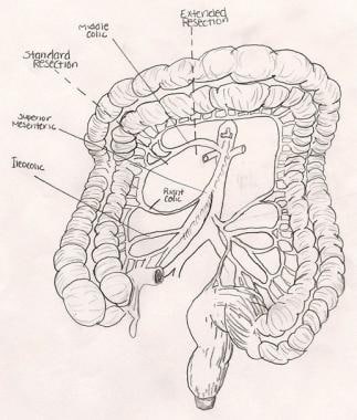
Pancreaticoduodenectomy
Iofetamine
Vena Cava, Inferior
Jugular Veins
Hypertension, Portal
Norepinephrine
Mesenteric Arteries
Portasystemic Shunt, Surgical
Mesenteric Artery, Superior
Adrenergic alpha-2 Receptor Antagonists
Esophageal and Gastric Varices
Amphetamines
Umbilical Veins
Tomography, X-Ray Computed
Dogs
Budd-Chiari Syndrome
Two populations of sympathetic neurons project selectively to mesenteric artery or vein. (1/420)
The objective of this study was to determine whether sympathetic neurons of the inferior mesenteric ganglion (IMG) projecting to mesenteric arteries could be distinguished by their localization, neurochemical phenotype, and electrophysiological properties from neurons projecting to mesenteric veins. In an in vitro intact vasculature-IMG preparation, neurons were labeled following intraluminal injection of Fluoro-Gold or rhodamine beads into the inferior mesenteric artery (IMA) or vein (IMV). The somata of neurons projecting to IMA were localized in the central part of the IMG, whereas those projecting to IMV were localized more peripherally. None of the labeled neurons was doubly labeled. Neuropeptide Y immunoreactivity was found in 18.9% of neurons innervating the IMA, but not in neurons innervating the IMV. Identified neurons were dissociated and characterized using whole cell patch-clamp recording. After direct soma depolarization, all of the labeled arterial and venous neurons were classified as tonic firing, compared with only 40% of unlabeled neurons; the remaining 60% of unlabeled neurons were phasic firing. The results indicate that IMG neurons projecting to mesenteric arteries are distinct from neurons projecting to mesenteric veins. (+info)Human eotaxin induces eosinophil extravasation through rat mesenteric venules: role of alpha4 integrins and vascular cell adhesion molecule-1. (2/420)
Eotaxin is a potent eosinophil-specific CC-chemokine, which has been shown to play a role in the selective induction of eosinophil accumulation in a number of allergic models of inflammation. Many aspects of the mechanism by which eotaxin induces eosinophil accumulation in vivo remain unresolved. In the present study, we investigated the direct effect of synthetic human eotaxin on leucocyte/endothelial cell interactions within rat mesenteric venules, as quantified by intravital microscopy. Topical eotaxin (30 pmol) induced rapid firm adhesion and extravasation of leucocytes within the rat mesentery, the extravasated leucocytes all being eosinophils, as determined by histological analysis. Whilst eotaxin was unable to stimulate the interaction of rat eosinophils with vascular cell adhesion molecule-1 (VCAM-1) under static conditions in vitro, eotaxin-induced responses in vivo were significantly suppressed by anti-alpha4 integrin and anti-VCAM-1 monoclonal antibodies (mAbs). The anti-alpha4 integrin mAb, HP2/1 (3.5 mg/kg), inhibited the eotaxin-induced firm adhesion and extravasation, 60 min postapplication of the chemokine, by 89% and 84%, respectively. In the same set of experiments, the anti-VCAM-1 mAb, 5F10 (3.5 mg/kg), inhibited leucocyte adhesion and extravasation by 61% and 63%, respectively. These results demonstrate that eotaxin-induced migration of eosinophils through rat mesenteric venules in vivo is dependent on an alpha4 integrin/VCAM-1 adhesion pathway, the significance of which may only be evident under flow conditions and/or following the ligation of other adhesion molecules expressed on eosinophils. (+info)Segmental differentiations of cell junctions in the vascular endothelium. Arteries and veins. (3/420)
A systematic survey of endothelial junctions in elastic (aorta) and muscular (mesenteric) arteries and in medium (renal and mesenteric) and large (cava inferior) size veins has been carried out in the rat using freeze-cleaved preparations. The arterial endothelium is provided with a complex of occluding and communicating junctions (gap junctions) comparable to, though less elaborate than, that described in arterioles. The particles of the occluding junctions behave like "single unit" particles and have the tendency to remain on B faces upon membrane cleavage. In the venous endothelium the junctions take the form of long occluding junctions with few associated communicating junctions (maculae communicantes). As in arterial endothelium, the junctional particles appear preferentially on B faces in cleaved preparations. These structures, although continuous over long distances, are interrupted focally by areas in which the junctional elements are similar to those found in venules: the ridges and grooves are short, discontinuous, randomly distributed along the general line of cell contact, and often particle-free. In muscular arteries two unusual types of junctions are encountered. Both are disposed in loops over short distances along the perimeter of the cell. One type appears to be a strectched-out version of the usual combination of occluding and communcating junctions of the arterial endothelium (this type is also occasionally encountered in the venous endothelium). The other type is reminiscent of the septate junctions found in the epithelia of invertebrates but the apparent similarity remains to be checked by further work. (+info)Phlebosclerosis of the colon with positive anti-centromere antibody. (4/420)
A 56-year-old woman with symptoms of chronic bowel disease presented a peculiar calcification of the mesenteric vein of the ascending to transverse colon on barium enema study. The resected colon was hard and black. Histo-pathologic examinations demonstrated fibrous change of the colon with a calcified and hyaline-deposited mesenteric vein. No cell infiltration was observed. These findings were compatible with phlebosclerosis and also with systemic sclerosis. Positive anti-centromere antibody and Raynaud's phenomenon, hallmarks of a variant systemic sclerosis, the CREST syndrome were observed. We therefore speculated that the pathogenesis of the phlebosclerosis of the colon is related to the CREST syndrome. (+info)P-selectin expression by endothelial cells is decreased in neonatal rats and human premature infants. (5/420)
Decreased adhesion of neutrophils to endothelial cells and delayed transendothelial cell migration of neutrophils have been consistently reported in neonatal animals and humans and contribute to their susceptibility to infection. The delayed transmigration of neutrophils is especially prevalent in premature neonates. To define the nature of this defect, we used an in vivo animal model of inflammation and found that radiolabeled leukocytes from adult rats transmigrated into the peritoneum of other adult rats 5 times more efficiently than they did in neonatal rats (P =.05). This indicated that defects in neonatal neutrophils could not completely account for the delayed transmigration. Delayed transmigration in the neonatal rats correlated with a defect in the expression of P-selectin on the surface of their endothelial cells. We found a similar P-selectin deficiency in endothelial cells lining mesenteric venules and umbilical veins of human premature infants when compared with term human infants. The decreased P-selectin in premature infants was associated with decreased numbers of P-selectin storage granules and decreased P-selectin transcription. Decreased P-selectin expression on the surface of endothelial cells in preterm infants may contribute to delayed neutrophil transmigration and increased susceptibility to infection. (+info)Resistance to praziquantel: direct evidence from Schistosoma mansoni isolated from Egyptian villagers. (6/420)
Recent evidence suggest that resistance to praziquantel (PZQ) may be developing. This would not be surprising in countries like Egypt where the drug has been used aggressively for more that 10 years. The classic phenotype of drug resistance is a significant increase in the 50% effective dose value of isolates retrieved from patients not responding to the drug. In a previous publication, we reported that such phenotypes have been isolated from humans infected with Schistosoma mansoni. Since the action of PZQ may be dependent upon the drug and host factors, most notably the immune system, we analyzed the quantitative effects of PZQ on single worms that differed in their response to PZQ when maintained in mice. Our hypothesis was that the in vitro action of the drug would correlate with it in vivo action. We confirmed this hypothesis and conclude that the in vitro action of the drug is related to its in vivo action. Knowing this relationship will assist in our ability to detect or survey for the PZQ resistant phenotype in human populations. (+info)Superior mesenteric vein stenosis complicating Crohn's disease. (7/420)
BACKGROUND: Superior mesenteric vein stenosis as a consequence of mesenteric fibrosis, causing the development of small bowel varices, is an unrecognised association of Crohn's disease. CASE REPORTS: Two cases of gastrointestinal bleeding occurring in patients with Crohn's disease, and a third case, presenting with pain and diarrhoea, are described. In all three patients, visceral angiography showed superior mesenteric vein stenosis with dilatation of draining collateral veins in the small bowel. Overt gastrointestinal bleeding or iron deficiency anaemia resulting from mucosal ulceration is common in Crohn's disease, but acute or chronic bleeding from small bowel varices as a result of superior mesenteric vein stenosis due to fibrosis has not previously been reported. (+info)Mesenteric and portal vein thrombosis in a young patient with protein S deficiency treated with urokinase via the superior mesenteric artery. (8/420)
A 32-year-old man, who was previously healthy, had acute abdominal pain without peritonitis. Diffuse mesenteric and portal vein thrombosis were shown by means of a computed tomography scan. A protein s deficiency was found by means of an extensive workup for hypercoagulable state. Successful treatment was achieved with urokinase infusion via the superior mesenteric artery without an operation. This represents an attractive alternative approach to treating patients with this disease. The previous standard of operative intervention(1) can now be reserved for complications, such as bowel infarction with peritonitis, or for those patients with absolute contraindications to thrombolytic therapy. (+info)The mesenteric veins are a set of blood vessels that are responsible for draining deoxygenated blood from the small and large intestines. There are two main mesenteric veins: the superior mesenteric vein and the inferior mesenteric vein. The superior mesenteric vein drains blood from the majority of the small intestine, as well as the ascending colon and proximal two-thirds of the transverse colon. The inferior mesenteric vein drains blood from the distal third of the transverse colon, descending colon, sigmoid colon, and rectum. These veins ultimately drain into the portal vein, which carries the blood to the liver for further processing.
The portal vein is the large venous trunk that carries blood from the gastrointestinal tract, spleen, pancreas, and gallbladder to the liver. It is formed by the union of the superior mesenteric vein (draining the small intestine and a portion of the large intestine) and the splenic vein (draining the spleen and pancreas). The portal vein then divides into right and left branches within the liver, where the blood flows through the sinusoids and gets enriched with oxygen and nutrients before being drained by the hepatic veins into the inferior vena cava. This unique arrangement allows the liver to process and detoxify the absorbed nutrients, remove waste products, and regulate metabolic homeostasis.
Mesenteric vascular occlusion refers to the blockage or obstruction of the blood vessels that supply the intestines, specifically the mesenteric arteries and veins. This condition can result in insufficient blood flow to the intestines, leading to ischemia (inadequate oxygen supply) and potential necrosis (tissue death).
There are two primary types of mesenteric vascular occlusion:
1. Mesenteric arterial occlusion: This occurs when the mesenteric artery, which carries oxygenated blood from the heart to the intestines, becomes blocked. The most common causes include atherosclerosis (plaque buildup in the arteries), embolism (a clot or particle that travels from another part of the body and lodges in the artery), and thrombosis (a blood clot forming directly in the artery).
2. Mesenteric venous occlusion: This happens when the mesenteric vein, which returns deoxygenated blood from the intestines to the heart, becomes obstructed. The most common causes include thrombophlebitis (inflammation and clot formation in the vein), tumors, or abdominal trauma.
Symptoms of mesenteric vascular occlusion may include severe abdominal pain, nausea, vomiting, diarrhea, and bloody stools. Rapid diagnosis and treatment are crucial to prevent intestinal tissue damage and potential life-threatening complications such as sepsis or shock. Treatment options typically involve surgical intervention, anticoagulation therapy, or endovascular procedures to restore blood flow.
Veins are blood vessels that carry deoxygenated blood from the tissues back to the heart. They have a lower pressure than arteries and contain valves to prevent the backflow of blood. Veins have a thin, flexible wall with a larger lumen compared to arteries, allowing them to accommodate more blood volume. The color of veins is often blue or green due to the absorption characteristics of light and the reduced oxygen content in the blood they carry.
A portal system in medicine refers to a venous system in which veins from various tissues or organs (known as tributaries) drain into a common large vessel (known as the portal vein), which then carries the blood to a specific organ for filtration and processing before it is returned to the systemic circulation. The most well-known example of a portal system is the hepatic portal system, where veins from the gastrointestinal tract, spleen, pancreas, and stomach merge into the portal vein and then transport blood to the liver for detoxification and nutrient processing. Other examples include the hypophyseal portal system, which connects the hypothalamus to the anterior pituitary gland, and the renal portal system found in some animals.
The splenic vein is a large, thin-walled vein that carries oxygenated blood from the spleen and pancreas to the liver. It is formed by the union of several smaller veins that drain the upper part of the stomach, the pancreas, and the left side of the colon (splenic flexure). The splenic vein runs along the top border of the pancreas and merges with the superior mesenteric vein to form the portal vein. This venous system allows for the filtration and detoxification of blood by the liver before it is distributed to the rest of the body.
The femoral vein is the large vein that runs through the thigh and carries oxygen-depleted blood from the lower limbs back to the heart. It is located in the femoral triangle, along with the femoral artery and nerve. The femoral vein begins at the knee as the popliteal vein, which then joins with the deep vein of the thigh to form the femoral vein. As it moves up the leg, it is joined by several other veins, including the great saphenous vein, before it becomes the external iliac vein at the inguinal ligament in the groin.
The renal veins are a pair of large veins that carry oxygen-depleted blood and waste products from the kidneys to the inferior vena cava, which is the largest vein in the body that returns blood to the heart. The renal veins are formed by the union of several smaller veins that drain blood from different parts of the kidney.
In humans, the right renal vein is shorter and passes directly into the inferior vena cava, while the left renal vein is longer and passes in front of the aorta before entering the inferior vena cava. The left renal vein also receives blood from the gonadal (testicular or ovarian) veins, suprarenal (adrenal) veins, and the lumbar veins.
It is important to note that the renal veins are vulnerable to compression by surrounding structures, such as the overlying artery or a tumor, which can lead to renal vein thrombosis, a serious condition that requires prompt medical attention.
The saphenous vein is a term used in anatomical description to refer to the great or small saphenous veins, which are superficial veins located in the lower extremities of the human body.
The great saphenous vein (GSV) is the longest vein in the body and originates from the medial aspect of the foot, ascending along the medial side of the leg and thigh, and drains into the femoral vein at the saphenofemoral junction, located in the upper third of the thigh.
The small saphenous vein (SSV) is a shorter vein that originates from the lateral aspect of the foot, ascends along the posterior calf, and drains into the popliteal vein at the saphenopopliteal junction, located in the popliteal fossa.
These veins are often used as conduits for coronary artery bypass grafting (CABG) surgery due to their consistent anatomy and length.
The iliac veins are a pair of large veins in the human body that carry deoxygenated blood from the lower extremities and the pelvic area back to the heart. They are formed by the union of the common iliac veins, which receive blood from the lower abdomen and legs, at the level of the fifth lumbar vertebra.
The combined iliac vein is called the inferior vena cava, which continues upward to the right atrium of the heart. The iliac veins are located deep within the pelvis, lateral to the corresponding iliac arteries, and are accompanied by the iliac lymphatic vessels.
The left common iliac vein is longer than the right because it must cross the left common iliac artery to join the right common iliac vein. The external and internal iliac veins are the two branches of the common iliac vein, with the external iliac vein carrying blood from the lower limbs and the internal iliac vein carrying blood from the pelvic organs.
It is essential to maintain proper blood flow in the iliac veins to prevent deep vein thrombosis (DVT), a condition that can lead to serious complications such as pulmonary embolism.
Splanchnic circulation refers to the blood flow to the visceral organs, including the gastrointestinal tract, pancreas, spleen, and liver. These organs receive a significant portion of the cardiac output, with approximately 25-30% of the total restingly going to the splanchnic circulation. The splanchnic circulation is regulated by a complex interplay of neural and hormonal mechanisms that help maintain adequate blood flow to these vital organs while also allowing for the distribution of blood to other parts of the body as needed.
The splanchnic circulation is unique in its ability to vasodilate and increase blood flow significantly in response to meals or other stimuli, such as stress or hormonal changes. This increased blood flow helps support the digestive process and absorption of nutrients. At the same time, the body must carefully regulate this blood flow to prevent a significant drop in blood pressure or overloading the heart with too much work.
Overall, the splanchnic circulation plays a critical role in maintaining the health and function of the body's vital organs, and dysregulation of this system can contribute to various diseases, including digestive disorders, liver disease, and cardiovascular disease.
Varicose veins are defined as enlarged, swollen, and twisting veins often appearing blue or dark purple, which usually occur in the legs. They are caused by weakened valves and vein walls that can't effectively push blood back toward the heart. This results in a buildup of blood, causing the veins to bulge and become varicose.
The condition is generally harmless but may cause symptoms like aching, burning, muscle cramp, or a feeling of heaviness in the legs. In some cases, varicose veins can lead to more serious problems, such as skin ulcers, blood clots, or chronic venous insufficiency. Treatment options include lifestyle changes, compression stockings, and medical procedures like sclerotherapy, laser surgery, or endovenous ablation.
Venous thrombosis is a medical condition characterized by the formation of a blood clot (thrombus) in the deep veins, often in the legs (deep vein thrombosis or DVT), but it can also occur in other parts of the body such as the arms, pelvis, or lungs (pulmonary embolism).
The formation of a venous thrombus can be caused by various factors, including injury to the blood vessel wall, changes in blood flow, and alterations in the composition of the blood. These factors can lead to the activation of clotting factors and platelets, which can result in the formation of a clot that blocks the vein.
Symptoms of venous thrombosis may include swelling, pain, warmth, and redness in the affected area. In some cases, the clot can dislodge and travel to other parts of the body, causing potentially life-threatening complications such as pulmonary embolism.
Risk factors for venous thrombosis include advanced age, obesity, smoking, pregnancy, use of hormonal contraceptives or hormone replacement therapy, cancer, recent surgery or trauma, prolonged immobility, and a history of previous venous thromboembolism. Treatment typically involves the use of anticoagulant medications to prevent further clotting and dissolve existing clots.
The hepatic veins are blood vessels that carry oxygen-depleted blood from the liver back to the heart. There are typically three major hepatic veins - right, middle, and left - that originate from the posterior aspect of the liver and drain into the inferior vena cava just below the diaphragm. These veins are responsible for returning the majority of the blood flow from the gastrointestinal tract and spleen to the heart. It's important to note that the hepatic veins do not have valves, which can make them susceptible to a condition called Budd-Chiari syndrome, where blood clots form in the veins and obstruct the flow of blood from the liver.
Viscera is a medical term that refers to the internal organs of the body, specifically those contained within the chest and abdominal cavities. These include the heart, lungs, liver, pancreas, spleen, kidneys, and intestines. In some contexts, it may also refer to the reproductive organs. The term viscera is often used in anatomical or surgical descriptions, and is derived from the Latin word "viscus," meaning "an internal organ."
Pulmonary veins are blood vessels that carry oxygenated blood from the lungs to the left atrium of the heart. There are four pulmonary veins in total, two from each lung, and they are the only veins in the body that carry oxygen-rich blood. The oxygenated blood from the pulmonary veins is then pumped by the left ventricle to the rest of the body through the aorta. Any blockage or damage to the pulmonary veins can lead to various cardiopulmonary conditions, such as pulmonary hypertension and congestive heart failure.
Pancreaticoduodenectomy, also known as the Whipple procedure, is a complex surgical operation that involves the removal of the head of the pancreas, the duodenum (the first part of the small intestine), the gallbladder, and the distal common bile duct. In some cases, a portion of the stomach may also be removed. The remaining parts of the pancreas, bile duct, and intestines are then reconnected to allow for the digestion of food and drainage of bile.
This procedure is typically performed as a treatment for various conditions affecting the pancreas, such as tumors (including pancreatic cancer), chronic pancreatitis, or traumatic injuries. It is a major surgical operation that requires significant expertise and experience to perform safely and effectively.
Iofetamine is a radiopharmaceutical agent used in myocardial perfusion imaging, a type of nuclear stress test. It is a derivative of the amphetamine family and functions as a vasoconstrictor when administered. Iofetamine is labeled with technetium-99m (^99mTc) before use, which allows for the detection and imaging of the heart's blood flow and function during rest and stress conditions. This information helps physicians diagnose and assess coronary artery disease and evaluate the effectiveness of treatments.
The medical definition of Iofetamine is:
A radiopharmaceutical agent, (^99mTc)Tc-sestamibi or (^99mTc)Tc-MIBI, used in myocardial perfusion imaging for the assessment of coronary artery disease. Iofetamine is a lipophilic cation that accumulates in myocardial cells in proportion to regional blood flow. The technetium-99m label enables gamma camera detection and imaging, providing information about the heart's blood flow and function during rest and stress conditions.
The inferior vena cava (IVC) is the largest vein in the human body that carries deoxygenated blood from the lower extremities, pelvis, and abdomen to the right atrium of the heart. It is formed by the union of the left and right common iliac veins at the level of the fifth lumbar vertebra. The inferior vena cava is a retroperitoneal structure, meaning it lies behind the peritoneum, the lining that covers the abdominal cavity. It ascends through the posterior abdominal wall and passes through the central tendon of the diaphragm to enter the thoracic cavity.
The inferior vena cava is composed of three parts:
1. The infrarenal portion, which lies below the renal veins
2. The renal portion, which receives blood from the renal veins
3. The suprahepatic portion, which lies above the liver and receives blood from the hepatic veins before draining into the right atrium of the heart.
The inferior vena cava plays a crucial role in maintaining venous return to the heart and contributing to cardiovascular function.
The jugular veins are a pair of large, superficial veins that carry blood from the head and neck to the heart. They are located in the neck and are easily visible when looking at the side of a person's neck. The external jugular vein runs along the surface of the muscles in the neck, while the internal jugular vein runs within the carotid sheath along with the carotid artery and the vagus nerve.
The jugular veins are important in clinical examinations because they can provide information about a person's cardiovascular function and intracranial pressure. For example, distention of the jugular veins may indicate heart failure or increased intracranial pressure, while decreased venous pulsations may suggest a low blood pressure or shock.
It is important to note that medical conditions such as deep vein thrombosis (DVT) can also affect the jugular veins and can lead to serious complications if not treated promptly.
Portal hypertension is a medical condition characterized by an increased pressure in the portal vein, which is the large blood vessel that carries blood from the intestines, spleen, and pancreas to the liver. Normal portal venous pressure is approximately 5-10 mmHg. Portal hypertension is defined as a portal venous pressure greater than 10 mmHg.
The most common cause of portal hypertension is cirrhosis of the liver, which leads to scarring and narrowing of the small blood vessels in the liver, resulting in increased resistance to blood flow. Other causes include blood clots in the portal vein, inflammation of the liver or bile ducts, and invasive tumors that block the flow of blood through the liver.
Portal hypertension can lead to a number of complications, including the development of abnormal blood vessels (varices) in the esophagus, stomach, and intestines, which are prone to bleeding. Ascites, or the accumulation of fluid in the abdominal cavity, is another common complication of portal hypertension. Other potential complications include encephalopathy, which is a condition characterized by confusion, disorientation, and other neurological symptoms, and an increased risk of bacterial infections.
Treatment of portal hypertension depends on the underlying cause and the severity of the condition. Medications to reduce pressure in the portal vein, such as beta blockers or nitrates, may be used. Endoscopic procedures to band or inject varices can help prevent bleeding. In severe cases, surgery or liver transplantation may be necessary.
Norepinephrine, also known as noradrenaline, is a neurotransmitter and a hormone that is primarily produced in the adrenal glands and is released into the bloodstream in response to stress or physical activity. It plays a crucial role in the "fight-or-flight" response by preparing the body for action through increasing heart rate, blood pressure, respiratory rate, and glucose availability.
As a neurotransmitter, norepinephrine is involved in regulating various functions of the nervous system, including attention, perception, motivation, and arousal. It also plays a role in modulating pain perception and responding to stressful or emotional situations.
In medical settings, norepinephrine is used as a vasopressor medication to treat hypotension (low blood pressure) that can occur during septic shock, anesthesia, or other critical illnesses. It works by constricting blood vessels and increasing heart rate, which helps to improve blood pressure and perfusion of vital organs.
The mesenteric arteries are the arteries that supply oxygenated blood to the intestines. There are three main mesenteric arteries: the superior mesenteric artery, which supplies blood to the small intestine (duodenum to two-thirds of the transverse colon) and large intestine (cecum, ascending colon, and the first part of the transverse colon); the inferior mesenteric artery, which supplies blood to the distal third of the transverse colon, descending colon, sigmoid colon, and rectum; and the middle colic artery, which is a branch of the superior mesenteric artery that supplies blood to the transverse colon. These arteries are important in maintaining adequate blood flow to the intestines to support digestion and absorption of nutrients.
Liver circulation, also known as hepatic circulation, refers to the blood flow through the liver. The liver receives blood from two sources: the hepatic artery and the portal vein.
The hepatic artery delivers oxygenated blood from the heart to the liver, accounting for about 25% of the liver's blood supply. The remaining 75% comes from the portal vein, which carries nutrient-rich, deoxygenated blood from the gastrointestinal tract, spleen, pancreas, and gallbladder to the liver.
In the liver, these two sources of blood mix in the sinusoids, small vessels with large spaces between the endothelial cells that line them. This allows for efficient exchange of substances between the blood and the hepatocytes (liver cells). The blood then leaves the liver through the hepatic veins, which merge into the inferior vena cava and return the blood to the heart.
The unique dual blood supply and extensive sinusoidal network in the liver enable it to perform various critical functions, such as detoxification, metabolism, synthesis, storage, and secretion of numerous substances, maintaining body homeostasis.
A portosystemic shunt is a surgical procedure that creates a connection between the portal vein (the blood vessel that carries blood from the digestive organs to the liver) and another systemic vein (a vein that carries blood away from the liver). This procedure is typically performed in animals, particularly dogs, to treat conditions such as portal hypertension or liver disease.
In a surgical portosystemic shunt, the surgeon creates a connection between the portal vein and a systemic vein, allowing blood from the digestive organs to bypass the liver. This can help to reduce the pressure in the portal vein and improve blood flow to the liver. The specific type of shunt created and the surgical approach used may vary depending on the individual patient's needs and the surgeon's preference.
It is important to note that while a surgical portosystemic shunt can be an effective treatment for certain conditions, it is not without risks and potential complications. As with any surgical procedure, there is always a risk of infection, bleeding, or other complications. Additionally, the creation of a portosystemic shunt can have long-term effects on the liver and overall health of the patient. It is important for pet owners to carefully consider the risks and benefits of this procedure and to discuss any questions or concerns they may have with their veterinarian.
The superior mesenteric artery (SMA) is a major artery that supplies oxygenated blood to the intestines, specifically the lower part of the duodenum, jejunum, ileum, cecum, ascending colon, and the first and second parts of the transverse colon. It originates from the abdominal aorta, located just inferior to the pancreas, and passes behind the neck of the pancreas before dividing into several branches to supply the intestines. The SMA is an essential vessel in the digestive system, providing blood flow for nutrient absorption and overall gut function.
Adrenergic alpha-2 receptor antagonists are a class of medications that block the action of norepinephrine, a neurotransmitter and hormone, at adrenergic alpha-2 receptors. These receptors are found in the central and peripheral nervous system and play a role in regulating various physiological functions such as blood pressure, heart rate, and insulin secretion.
By blocking the action of norepinephrine at these receptors, adrenergic alpha-2 receptor antagonists can increase sympathetic nervous system activity, leading to vasodilation, increased heart rate, and increased insulin secretion. These effects make them useful in the treatment of conditions such as hypotension (low blood pressure), opioid-induced sedation and respiratory depression, and diagnostic procedures that require vasodilation.
Examples of adrenergic alpha-2 receptor antagonists include yohimbine, idazoxan, and atipamezole. It's important to note that these medications can have significant side effects, including hypertension, tachycardia, and agitation, and should be used under the close supervision of a healthcare provider.
Esophageal varices and gastric varices are abnormal, enlarged veins in the lower part of the esophagus (the tube that connects the throat to the stomach) and in the stomach lining, respectively. They occur as a result of increased pressure in the portal vein, which is the large blood vessel that carries blood from the digestive organs to the liver. This condition is known as portal hypertension.
Esophageal varices are more common than gastric varices and tend to be more symptomatic. They can cause bleeding, which can be life-threatening if not treated promptly. Gastric varices may also bleed, but they are often asymptomatic until they rupture.
The most common causes of esophageal and gastric varices are cirrhosis (scarring of the liver) and portal hypertension due to other liver diseases such as schistosomiasis or Budd-Chiari syndrome. Treatment options for esophageal and gastric varices include medications to reduce bleeding, endoscopic therapies to treat active bleeding or prevent recurrent bleeding, and surgical procedures to relieve portal hypertension.
Amphetamines are a type of central nervous system stimulant drug that increases alertness, wakefulness, and energy levels. They work by increasing the activity of certain neurotransmitters (chemical messengers) in the brain, such as dopamine and norepinephrine. Amphetamines can be prescribed for medical conditions such as attention deficit hyperactivity disorder (ADHD) and narcolepsy, but they are also commonly abused for their ability to produce euphoria, increase confidence, and improve performance in tasks that require sustained attention.
Some common examples of amphetamines include:
* Adderall: a combination of amphetamine and dextroamphetamine, used to treat ADHD and narcolepsy
* Dexedrine: a brand name for dextroamphetamine, used to treat ADHD and narcolepsy
* Vyvanse: a long-acting formulation of lisdexamfetamine, a prodrug that is converted to dextroamphetamine in the body, used to treat ADHD
Amphetamines can be taken orally, snorted, smoked, or injected. Long-term use or abuse of amphetamines can lead to a number of negative health consequences, including addiction, cardiovascular problems, malnutrition, mental health disorders, and memory loss.
Thrombosis is the formation of a blood clot (thrombus) inside a blood vessel, obstructing the flow of blood through the circulatory system. When a clot forms in an artery, it can cut off the supply of oxygen and nutrients to the tissues served by that artery, leading to damage or tissue death. If a thrombus forms in the heart, it can cause a heart attack. If a thrombus breaks off and travels through the bloodstream, it can lodge in a smaller vessel, causing blockage and potentially leading to damage in the organ that the vessel supplies. This is known as an embolism.
Thrombosis can occur due to various factors such as injury to the blood vessel wall, abnormalities in blood flow, or changes in the composition of the blood. Certain medical conditions, medications, and lifestyle factors can increase the risk of thrombosis. Treatment typically involves anticoagulant or thrombolytic therapy to dissolve or prevent further growth of the clot, as well as addressing any underlying causes.
A smooth muscle within the vascular system refers to the involuntary, innervated muscle that is found in the walls of blood vessels. These muscles are responsible for controlling the diameter of the blood vessels, which in turn regulates blood flow and blood pressure. They are called "smooth" muscles because their individual muscle cells do not have the striations, or cross-striped patterns, that are observed in skeletal and cardiac muscle cells. Smooth muscle in the vascular system is controlled by the autonomic nervous system and by hormones, and can contract or relax slowly over a period of time.
The umbilical veins are blood vessels in the umbilical cord that carry oxygenated and nutrient-rich blood from the mother to the developing fetus during pregnancy. There are typically two umbilical veins, one of which usually degenerates and becomes obliterated, leaving a single functional vein. This remaining vein is known as the larger umbilical vein or the venous duct. It enters the fetal abdomen through the umbilicus and passes through the liver, where it branches off to form the portal sinus. Ultimately, the blood from the umbilical vein mixes with the blood from the inferior vena cava and is pumped to the heart through the right atrium.
It's important to note that after birth, the umbilical veins are no longer needed and undergo involution, becoming the ligamentum teres in the adult.
X-ray computed tomography (CT or CAT scan) is a medical imaging method that uses computer-processed combinations of many X-ray images taken from different angles to produce cross-sectional (tomographic) images (virtual "slices") of the body. These cross-sectional images can then be used to display detailed internal views of organs, bones, and soft tissues in the body.
The term "computed tomography" is used instead of "CT scan" or "CAT scan" because the machines take a series of X-ray measurements from different angles around the body and then use a computer to process these data to create detailed images of internal structures within the body.
CT scanning is a noninvasive, painless medical test that helps physicians diagnose and treat medical conditions. CT imaging provides detailed information about many types of tissue including lung, bone, soft tissue and blood vessels. CT examinations can be performed on every part of the body for a variety of reasons including diagnosis, surgical planning, and monitoring of therapeutic responses.
In computed tomography (CT), an X-ray source and detector rotate around the patient, measuring the X-ray attenuation at many different angles. A computer uses this data to construct a cross-sectional image by the process of reconstruction. This technique is called "tomography". The term "computed" refers to the use of a computer to reconstruct the images.
CT has become an important tool in medical imaging and diagnosis, allowing radiologists and other physicians to view detailed internal images of the body. It can help identify many different medical conditions including cancer, heart disease, lung nodules, liver tumors, and internal injuries from trauma. CT is also commonly used for guiding biopsies and other minimally invasive procedures.
In summary, X-ray computed tomography (CT or CAT scan) is a medical imaging technique that uses computer-processed combinations of many X-ray images taken from different angles to produce cross-sectional images of the body. It provides detailed internal views of organs, bones, and soft tissues in the body, allowing physicians to diagnose and treat medical conditions.
I believe there might be a misunderstanding in your question. "Dogs" is not a medical term or condition. It is the common name for a domesticated carnivore of the family Canidae, specifically the genus Canis, which includes wolves, foxes, and other extant and extinct species of mammals. Dogs are often kept as pets and companions, and they have been bred in a wide variety of forms and sizes for different purposes, such as hunting, herding, guarding, assisting police and military forces, and providing companionship and emotional support.
If you meant to ask about a specific medical condition or term related to dogs, please provide more context so I can give you an accurate answer.
Budd-Chiari syndrome is a rare condition characterized by the obstruction of the hepatic veins, which are the blood vessels that carry blood from the liver to the heart. This obstruction can be caused by blood clots, tumors, or other abnormalities, and it can lead to a backflow of blood in the liver, resulting in various symptoms such as abdominal pain, swelling, and liver enlargement. In severe cases, Budd-Chiari syndrome can cause liver failure and other complications if left untreated. The diagnosis of this condition typically involves imaging tests such as ultrasound, CT scan, or MRI, and treatment may include anticoagulation therapy, thrombolytic therapy, or surgical intervention to remove the obstruction.
 Mesenteric vein
Inferior mesenteric vein
Superior mesenteric vein
Left colic vein
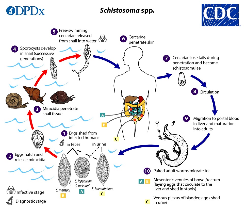
Schistosoma mansoni
Surgical Outcomes Analysis and Research
Vitelline veins
Kristaps Keggi
Left colic artery
Left gastroepiploic vein
Right colic vein
Schistosoma
Right gastroepiploic vein
Portal vein
Pancreas
Duodenum
May-Thurner syndrome
Superior mesenteric artery
Schistosoma japonicum
Superior mesenteric vessels
Peritoneal recesses
Portal vein thrombosis
Schistosoma bovis
Nutcracker syndrome
Schistosoma intercalatum
Middle colic vein
Periaortic lymph nodes
Pancreaticoduodenectomy
Neurokinin B
Acute abdomen
Thrombosis28
- Mesenteric vein thrombosis (MVT) is a distinct clinical cause of intestinal ischemia representing 5-15% of all ischemic events. (wustl.edu)
- We report a case of mesenteric vein thrombosis associated with intravaginal hormonal contraception. (wustl.edu)
- Voora, D & Vijayan, A 2003, ' Mesenteric vein thrombosis associated with intravaginal contraceptives: A case report and review of the literature ', Journal of Thrombosis and Thrombolysis , vol. 15, no. 2, pp. 105-108. (wustl.edu)
- The lienal vein has hypoechoic centre creating target appearance due to partial thrombosis. (mudr.org)
- We recommend anticoagulation for all noncirrhotic patients with acute symptomatic portal or mesenteric vein thrombosis in the absence of any contraindication. (mdcalc.com)
- We suggest at least 6 months of anticoagulation in patients with portal or mesenteric vein thrombosis without a demonstrable thrombophilia and when the etiology of the thrombosis is reversible. (mdcalc.com)
- Indefinite anticoagulation is recommended in patients with portal or mesenteric vein thrombosis and thrombophilia. (mdcalc.com)
- We recommend anticoagulation for patients with (i) acute complete main PVT, (ii) MVT, or (iii) extension of portal venous thrombosis into mesenteric veins. (mdcalc.com)
- Anticoagulation is continued beyond this period in patients with portal or mesenteric vein thrombosis who are on the waiting list for liver transplant. (mdcalc.com)
- We recommend nonselective beta-blockers for prevention of variceal bleeding in patients with high-risk varices and portal and/or mesenteric vein thrombosis requiring anticoagulation. (mdcalc.com)
- Inferior mesenteric vein thrombosis was excluded. (sages.org)
- Ovarian vein thrombosis (OVT) is an uncommon but potentially serious disorder that is associated with a variety of pelvic conditions-most notably, recent childbirth, but also pelvic inflammatory disease , malignancies, and pelvic surgery. (medscape.com)
- Ultrasound (US), magnetic resonance imaging (MRI), and CT scanning with contrast are the best radiologic modalities for making the diagnosis of ovarian vein thrombosis. (medscape.com)
- Angiography can help make the diagnosis of ovarian vein thrombosis, but this technique is not usually performed because of the availability of noninvasive, cross-sectional imaging methods. (medscape.com)
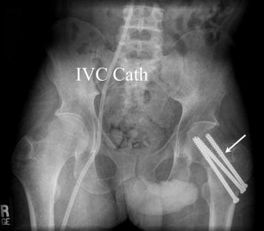
- Contrast-enhanced computed tomography scan in a postpartum patient with fever that demonstrates bilateral ovarian vein thrombosis. (medscape.com)
- Ovarian vein thrombosis arises out of the coincident conditions of venous stasis and hypercoagulability, which are commonly present in the recently postpartum patient. (medscape.com)
- [ 21 ] also increase the patient's risk for ovarian vein thrombosis. (medscape.com)
- Both ovarian vein thrombosis and septic pelvic thrombophlebitis are influenced by the Virchow triad of vessel wall injury, stasis, and hypercoagulability. (medscape.com)
- Ovarian vein thrombosis occurs in 0.05-0.18% of vaginal births and in 2% of cesarean deliveries. (medscape.com)
- The typical patient with ovarian vein thrombosis (ie, thrombophlebitis) presents with pelvic pain, fever, and a right-sided abdominal mass. (medscape.com)
- Note that patients who have undergone total abdominal hysterectomy and bilateral salpingo-oophorectomy with retroperitoneal lymph node dissection can incidentally demonstrate ovarian vein thrombosis on contrast-enhanced computed tomography [CT] scanning. (medscape.com)
- Antiphospholipid Syndrome presenting as mesenteric vein thrombosis and gangrenous small bowel. (iium.edu.my)
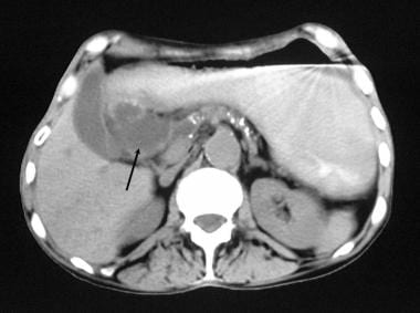
- C-4: Splenic vein thrombosis of the same patient is marked by arrows. (medscape.com)
- There is thrombosis of the right and left portal veins, main portal vein, splenic vein and superior mesenteric vein. (uab.edu)
- Chronic SMV thrombosis is characterized by mild enlargement of the vein with central low denstiy surrounded by higher density wall. (uab.edu)
- One-year survival has been reported following transplantation of a living-related segment of a donor intestine [ 4 ] and in a 41-year-old woman with short gut syndrome (SGS) secondary to superior mesenteric artery thrombosis. (medscape.com)
- Deep vein thrombosis, or DVT, is a blood clot that forms in a vein deep in the body. (medlineplus.gov)
- A deep vein thrombosis can break loose and cause a serious problem in the lung, called a pulmonary embolism . (medlineplus.gov)
Thrombus5
- Tail of the thrombus extends into portal vein. (mudr.org)
- Contrast-enhanced CT or MRI scan is recommended to assess the extension of thrombus into the mesenteric veins and to exclude tumor thrombus among patients with cirrhosis who develop new portal and/or mesenteric vein thrombus. (mdcalc.com)
- We suggest anticoagulation for patients with chronic PVT if there is (i) evidence of inherited or acquired thrombophilia, (ii) progression of thrombus into the mesenteric veins, or (iii) current or previous evidence of bowel ischemia. (mdcalc.com)
- PMVT was defined as thrombus within the portal, hepatic or superior mesenteric veins. (sages.org)
- Transjugular portography demonstrates extensive portal vein thrombus in the whole-liver allograft of a 40-year-old woman whose clinical condition rapidly deteriorated on postoperative day 39. (medscape.com)
Occlusion1
- Nonthrombotic occlusion or stenosis of the mesenteric veins is a rare cause of intestinal ischemia that usually occurs in association with systemic vasculitis. (elsevierpure.com)
Hepatic8
- The hepatic portal vein is a vessel that moves blood from the spleen and gastrointestinal tract to the liver. (healthline.com)
- The hepatic veins carry oxygen-depleted blood from the liver to the inferior vena cava. (healthline.com)
- The liver is divided into two lobes by the middle hepatic vein: the right lobe of liver and the left lobe of liver. (healthline.com)
- The scans also revealed he had hepatic ischemia, also called 'shock liver', a type of injury caused by low oxygen supply to the organ, which doctors believe was related to the presence of gas in the portal vein. (dailymail.co.uk)
- He explained that the portal vein carries material from the gut for the liver to process, as opposed to a different blood vessel, the hepatic artery, which carries oxygen to the organ. (dailymail.co.uk)
- To conclude, although type I classification which describes the textbook pattern of hepatic artery distribution was significantly detected among the Sudanese population, other variants were to be considered since they are related to major arteries like aorta and superior mesenteric. (bvsalud.org)
- accessory pancreatic or splenic arteries.Objective: To present three cases of accessory right hepatic artery originating from the superior mesenteric artery in black African cadavers as found during routine cadaveric dissections.Materials and Method: The abdomens of 8 adult male black African cadavers were dissected according to the description and guidance by Romanes (1996). (bvsalud.org)
- portal veins and gastroduodenal arteries were exposed.Results: Three cadaveric cases of the accessory right hepatic arteries arising from the superior mesenteric arteries were observed. (bvsalud.org)
Ischemia1
- Associated findings may include increased attenuatiuon of the mesenteric fat due to mesenteric edema and bowel wall thickening due to stasis and mesenteric ischemia. (uab.edu)
Arteries2
- Of the visceral branches, the celiac artery and the superior and inferior mesenteric arteries are unpaired, while the suprarenals, renals, internal spermatics, and ovarian are paired. (theodora.com)
- mesenteric arteries (MA) and veins (MV) were mounted on glass cannulas, intravascularly filled with fluorescent dextran and incrementally pressurized above their in vivo physiological values. (bvsalud.org)
Arterial2
- Sudeck's critical point at the rectosigmoid junction is described as the point of origin of the last sigmoid arterial branch, originating from the inferior mesenteric artery (IMA) [4]. (researchsquare.com)
- The purpose of the present study as to evaluate the effect of changes in intravascular pressure and the inflammatory mediator bradykinin on rat mesenteric arterial and venous vascular permeability. (bvsalud.org)
Inferior mesent1
- Xiu WL, Liu J, Zhang JL, Su N, Wang FJ, Hao XW, Wang FF, Dong Q. Computer-assisted rescue of the inferior mesenteric artery in a child with a giant ganglioneuroblastoma: A case report. (wjgnet.com)
Splenic vein2
- Splenic vein isolated. (vesalius.com)
- Two centimeters of distal pancreas was mobilized off the splenic vein. (vesalius.com)
Lienal vein1
- Below, it is in relation to the upper border of the pancreas, and the lienal vein. (theodora.com)
Vessel1
- The intima of the mesenteric and intestinal mural veins was focally thickened by a marked increase in cells and matrix between the endothelium and internal elastic lamina, whereas the vessel walls external to the thickened intima appeared normal. (elsevierpure.com)
Liver4
- In most people, the portal vein splits into left and right veins before entering the liver. (healthline.com)
- The portal vein supplies approximately 75 percent of blood flow to the liver. (healthline.com)
- Professor Davies also added that gas in the portal vein would not explain the lack of oxygen the man's liver was receiving. (dailymail.co.uk)
- The cercariae can invade the skin or mucous membranes of the definitive host (dogs or wild mammals) and gain access to the bloodstream, migrate to the liver, mature, and migrate to the mesenteric vessels to complete the life cycle. (tamu.edu)
Intramural1
- The intramural arteriocapillaries first lose part of their volume as the earlier entered blood flows out from the veins, even though some blood may seep back from the veins. (ajronline.org)
Vessels3
- Adult schistosomes typically live in mesenteric blood vessels of birds or mammals and produce eggs that pass from the host in feces. (cdc.gov)
- Chugging the beverage so quickly led to gas gathering in his intestines, which due to the pressure then leaked into his portal vein, one of the liver's main blood vessels. (dailymail.co.uk)
- The inferior mesenteric vessels are identified at their takeoff from the aorta. (medscape.com)
Anterior2
- The right vein then branches off into anterior and superior veins. (healthline.com)
- Posteriorly, it is separated from the lumbar vertebræ and intervertebral fibrocartilages by the anterior longitudinal ligament and left lumbar veins. (theodora.com)
Clot3
- A positive finding on venography is the presence of a filling defect that is consistent with a clot within the ovarian vein. (medscape.com)
- So a splanchnic vein clot or a mesenteric clot in an otherwise young patient. (reachmd.com)
- I remember a young patient I had during the first period of time when I was training who had a mesenteric vein clot and had to actually get a small bowel resection and then transplant because of this disease. (reachmd.com)
Hernia3
- Internal hernia after laparoscopic sigmoidectomy with splenic flexural mobilization and high ligation of the inferior mesenteric vein: A case report. (bvsalud.org)
- However, there have been no reports on the indications for closing mesenteric defects to prevent the development of an internal hernia . (bvsalud.org)
- This study reports a case of an internal hernia of the proximal jejunum near the ligament of Treitz in a patient who underwent laparoscopic sigmoidectomy with splenic flexural mobilization and high ligation of the inferior mesenteric vein . (bvsalud.org)
Branches1
- The small branches draining the head of the pancreas into the right side of the portal vein were carefully ligated and divided, freeing the specimen. (vesalius.com)
Thrombophlebitis1
- If the vein swells, the condition is called thrombophlebitis. (medlineplus.gov)
Upper2
- It is approximately three to four inches in length and is usually formed by the merging of the superior mesenteric and splenic veins behind the upper edge of the head of the pancreas. (healthline.com)
- Upper chambers of the heart that receive blood from the veins. (encyclopedia.com)
Portal hypertension1
- Abnormally high blood pressure in the portal vein is known as portal hypertension . (healthline.com)
Intestine1
- CT scans found he had pneumatosis, an abnormal presence of gas, in the wall of his intestine and his portal vein. (dailymail.co.uk)
Blood1
- The adults are blood parasites and live in the mesenteric veins where mating occurs. (tamu.edu)
Doppler1
- Doppler ultrasound increases sensitivity because permeable ovarian veins can be clearly identified. (medscape.com)
Colon1
- Diffuse strandy inflammatory changes are also apparent in the region of ileum and ascending colon indicating mesenteric congestion. (uab.edu)
Artery2
- The bulky metastatic lymph nodes at the head of the pancreas were dissected after ligating the right colic artery and vein. (spandidos-publications.com)
- During the operation, an aberrant middle colic artery was found to be originating from the gastroduodenal artery instead of its usual origin at the superior mesenteric artery. (hindawi.com)
Fever1
- These include fever, enlarged veins on the belly that can be seen through the skin, and abnormal bruising or bleeding. (medtronic.com)
Common1
- For part of its course, it runs near the portal vein and the common bile duct. (healthline.com)
Research1
- The mesenteric-portal vein in research. (aspetjournals.org)
Drain1
- The portal vein is not a true vein, which means it does not drain into the heart. (healthline.com)
Shown1
- The exposed superior mesenteric/portal vein is shown. (vesalius.com)
Middle1
- The middle colic vein was traced to the superior mesenteric vein. (hindawi.com)
Wall1
- As bacteria proliferate and more gas is produced, the intraluminal gas may dissect into the necrotic wall (pneumatosis intestinalis), spread through the mesenteric veins, and finally flow into the portal veins (Fig. 7A , 7B ). (ajronline.org)