Maxillary Artery
Carotid Artery, External
Maxillary Sinus
Masticatory Muscles
Meninges
Trigeminal Nerve
Dissection
Maxilla
Maxillary Sinusitis
Maxillary Sinus Neoplasms
Maxillary Nerve
Maxillary Diseases
Neurogenic vasodilatation of canine isolated small labial arteries. (1/31)
Mechanisms underlying vasodilatation to nerve stimulation by electrical pulses and nicotine were analyzed in isolated canine small labial arteries. Transmural electrical stimulation (5 and 20 Hz) produced a contraction followed by a relaxation in labial arterial strips denuded of the endothelium, partially contracted with prostaglandin F2alpha. The contraction was abolished by prazosin or combined treatment with alpha, beta-methylene ATP. In the treated strips, neurogenic relaxation was abolished by NG-nitro-L-arginine (L-NA), a nitric oxide (NO) synthase inhibitor, and restored by L-arginine. The D-enantiomers were without effect. Nicotine (10(-4) M) also relaxed the arteries, in which the contractile response was abolished by prazosin and alpha, beta-methylene ATP. The relaxant response was attenuated but not abolished by L-NA; the inhibition was reversed by L-arginine. The remaining relaxation by nicotine was abolished by calcitonin gene-related peptide (CGRP)-[8 to 37], a CGRP1 receptor antagonist. Relaxations elicited by a lower concentration of nicotine (2 x 10(-5) M) sufficient to produce similar magnitudes of response to those induced by 5-Hz electrical nerve stimulation were also inhibited partially by L-NA. Histochemical study with the NADPH-diaphorase method demonstrated positively stained nerve fibers and bundles in the arterial wall, suggesting the presence of neuronal NO synthase. It is concluded that the relaxation induced by electrical nerve stimulation of small labial arteries is mediated exclusively by NO synthesized from L-arginine in nerve terminals, whereas nicotine in the concentrations used evokes relaxations by a mediation of nerve-derived NO and also CGRP, possibly from sensory nerves. The reason why nicotine but not electrical pulses stimulates sensory nerves and elicits vasorelaxation remains unsolved. (+info)Radiologic and histopathologic evaluation of canine artery occlusion after collagen-coated platinum microcoil delivery. (2/31)
BACKGROUND AND PURPOSE: Platinum coil embolization is one of the significant advances in interventional neuroradiologic techniques that has been introduced this decade. Our purpose was to evaluate the angiographic and histologic effects of collagen-coated platinum microcoil delivery in the canine artery. METHODS: We embolized the bilateral internal maxillary arteries of 18 dogs; one uncoated and one collagen-primed coil was used in each dog. We evaluated all coils by angiography, macroscopy, and scanning electron microscopy within 30 minutes of embolization. We then studied a proportional number of coated and collagen-primed coils at either 1 or 3 days, or 1, 2, 3, 4, 8, 12, or 16 weeks postoperatively. RESULTS: Six (33%) of 18 arteries embolized with uncoated coils were occluded 30 minutes after delivery, whereas 11 (61%) of 18 arteries treated with collagen-primed coils were occluded within 30 minutes of embolization. Late occlusion (3 weeks after embolization) occurred in 2 (25%) of 8 arteries embolized with untreated coils, and 6 (75%) of 8 arteries embolized with collagen-primed coils. We calculated differences in late occlusion rates by the chi2 (chi-square) test, and found these differences were significant (P=.04). Histologic findings of arteries embolized with unprimed coils revealed endothelial cell growth was limited to the organized thrombi 4 weeks after coil delivery. In contrast, endothelial cells grew directly on the collagen-primed coils 3 days postoperatively, and coils were completely covered by endothelial cells within 2 weeks. We found an organized thrombus in the inner space of coils in angiographically occluded arteries, a finding that was not evident in angiographically patent arteries. CONCLUSION: Collagen-coated platinum coils can produce rapid and stable occlusion of embolized vessels. (+info)In situ beta radiation to prevent recanalization after coil embolization of cerebral aneurysms. (3/31)
BACKGROUND AND PURPOSE: Endovascular treatment of cerebral aneurysms, a minimally invasive alternative to surgery, is too often followed by recanalization and recurrences. The purpose of this work was to assess if in situ beta radiation can inhibit recanalization after coil embolization. METHODS: Radioactive platinum coils (32P-coils) were produced by ion implantation of 32P. A single-coil arterial occlusion model was used to compare angiographic and pathological results at 1 to 12 weeks after nonradioactive and 32P-coil embolization of maxillary, cervical, and vertebral arteries in 26 dogs. Coils of varying activities were used and results compared to define the minimal activity required to inhibit recanalization. Similar experiments were performed in 16 porcine maxillary and lingual and 8 rabbit axillary arteries. Results of 32P-coil embolization of bifurcation aneurysms were then compared with embolization with nonradioactive coils in 12 dogs at 3 months. RESULTS: Nonradioactive coil embolization of canine arteries led to occlusion at 1 week, followed by recanalization at 2 weeks, which persisted at 3 months in all cases. 32P-coils, ion-implanted with activities above 0.13 microCi/cm, led to persistent occlusion at 3 months in 80% of arteries. 32P-coils ion-implanted with the same activity inhibited recanalization in porcine and rabbit arteries. Bifurcation aneurysms treated with 32P-coils had better angiographic results at 3 months (P=0.006) than aneurysms treated with nonradioactive coils. Arteries occluded were filled with fibrous tissue at 3 months. Aneurysms embolized with 32P-coils showed more complete neointimal coverage of the neck, without recanalization, as compared with aneurysms treated with nonradioactive coils. CONCLUSION: In situ low-dose beta radiation inhibits recanalization after coil embolization and may improve long-term results of endovascular treatment of aneurysms. (+info)Beta radiation and inhibition of recanalization after coil embolization of canine arteries and experimental aneurysms: how should radiation be delivered? (4/31)
BACKGROUND AND PURPOSE: Beta radiation prevents recanalization after coil embolization. We sought to determine the effects of varying coil caliber, length, activity of 32P per centimeter of coil or per volume, and spatial distribution of coils on recanalization. METHODS: We studied the angiographic evolution of 81 canine maxillary, cervical, and vertebral arteries implanted with a variety of nonradioactive (n=29 arteries) or radioactive (n=52) devices. We compared 1- or 2-caliber 0.015 or 0.010 coils ion-implanted or not with 3 different activity levels (0.05 to 0.08, 0.06 to 0.12, 0.18 to 0.32 microCi/cm) of 32P and totaling 4, 8, and 16 cm in length for the same arterial volume. We also compared inhibition of recanalization by beta radiation delivered by stents, after coil occlusion proximal to or within the stent, with that delivered by coils placed within nonradioactive stents. We finally studied the angiographic evolution of canine lateral wall carotid aneurysms treated with 1 or 2 stents of various activity levels positioned inside the parent artery across the neck. Animals were killed at 4 and 12 weeks for macroscopic photography and pathological examination. RESULTS: All arteries (29 of 29) occluded with nonradioactive devices were recanalized, while 49 of 52 arteries (94%) implanted with 32P devices were occluded at 4 weeks. All aneurysms treated with stents, radioactive or not, demonstrated residual filling of the sac or of channels leading to the aneurysms at follow-up angiography at 4 weeks. CONCLUSIONS: The recanalization process found in the canine arterial occlusion model is minimally affected by coil caliber, number, and length or packing density. Beta radiation reliably inhibits this process, but thrombosis is an essential condition for the efficacy of a radioactive coil strategy. (+info)Pathological changes in temporal arteries removed from unselected cadavers. (5/31)
Progressive degenerative changes in ageing temporal arteries are described. These changes are usually severe in elderly patients, hence their presence in diagnostic biopsies provides no evidence of temporal arteritis past or present. In two patients in the series of random necropsies examined there were changes in the temporal arteries identical with those seen in patients known to have suffered from temporal arteritis in the past; in both these cases there was, in addition a giant cell aortitis. The distinction between healed arteritis and degenerative changes due to age in a single biopsy specimen may be difficult. Furthermore, it is possible that steroid treatment may arrest the progress of the disease so that gross intimal thickening and vascularization of the artery wall do not occur. Giant cell arteritis is not a rare disease. Two examples were discovered in 39 random necropsies on patients over 60 years of age. (+info)Development of gold stents for the treatment of intracranial aneurysms: an experimental study in a canine model. (6/31)
BACKGROUND AND PURPOSE: Gold has often been used in medicine because of its radiopacity and flexibility. To perform stent-supported coil embolization of intracranial aneurysms, we prepared a gold stent and examined its flexibility, radiopacity, and thrombogenic properties in comparison with a stainless steel device implanted in vitro and in vivo. METHODS: Gold stents were prepared by plating gold on stainless steel stents as a template. Their mechanical properties and trackability in vitro were determined and compared with those of stainless steel stents of the same design. Twenty gold stents and two stainless steel stents were implanted in canine external carotid, vertebral, and renal arteries, as a muscle branch of the maxillary arteries, to examine their performance in vivo. RESULTS: The gold stent exhibited much less radial force and greater flexibility than the stainless steel stent. It also demonstrated superior trackability and radiopacity in the experimental endovascular procedures in canines. Histologic examination showed good patency of the stented artery with slight endothelial hypertrophy. CONCLUSION: Although there is still room for more radial strength, less influence on intimal hypertrophy, a more suitable flexibility, and a smoother surface, the superior trackability and radiopacity of gold stents seem to support use of this device for the endovascular treatment of intracranial aneurysms. (+info)Surgical outcome of radical maxillectomy in advanced maxillary sinus cancers. (7/31)
We investigated the surgical outcome of radical maxillectomy in advanced maxillary sinus cancers invading through the posterior wall and into the infratemporal fossa. Twenty-eight patients with maxillary sinus squamous cell carcinoma, who visited the Otorhinolaryngology Department at Severance Hospital from March, 1993 to February, 2001 and underwent the surgery, were analyzed retrospectively by reviewing clinical medical records and radiologic test results. The mean follow- up period was 78.8 months.(26-162 months) Local recurrence, sites of local recurrence, and the 2-year disease-free survival rate were analyzed. Of the total 28 cases, 9 cases were T3, and 19 cases were T4. Total maxillectomy was performed in 12 cases (42.9%) and radical maxillectomy in 16 cases (57.1%). Regardless of staging, radical maxillectomy was performed only when cancers invaded through the posterior wall and into the infratemporal fossa. When cancers only maginally or did not invade the posterior wall, total maxillectomy was performed. The 2-year disease-free survival rate was 75% for both total and radical maxillectomy, and the local recurrence rates were 8.3% and 18.7% respectively. All recurrence occurred at the posterior resection margin of the maxillectomy. We strongly recommend the use of radical maxillectomy in the cases of advanced maxillary sinus cancers invading the infratemporal fossa. Radical maxillectomy can provide sufficient safety margins and lower the local recurrence rate. (+info)Spontaneous resolution of traumatic pseudoaneurysm of the middle meningeal artery. (8/31)
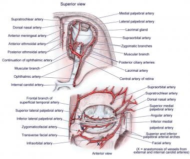
We describe a case of traumatic pseudoaneurysm of the middle meningeal artery in a patient after a head trauma. The aneurysm was found incidentally and resolved spontaneously without any intervention; this outcome suggests that middle meningeal artery aneurysm may not require treatment in all cases and can be followed conservatively with follow-up conventional angiography. (+info)The maxillary artery is a branch of the external carotid artery that supplies the deep structures of the face and head. It originates from the external carotid artery just below the neck of the mandible and passes laterally to enter the parotid gland. Within the gland, it gives off several branches, including the deep auricular, anterior tympanic, and middle meningeal arteries.
After leaving the parotid gland, the maxillary artery travels through the infratemporal fossa, where it gives off several more branches, including the inferior alveolar, buccinator, and masseteric arteries. These vessels supply blood to the teeth, gums, and muscles of mastication.
The maxillary artery also gives off the sphenopalatine artery, which supplies the nasal cavity, nasopharynx, and palate. Additionally, it provides branches that supply the meninges, dura mater, and brain. Overall, the maxillary artery plays a critical role in providing blood flow to many structures in the head and neck region.
The external carotid artery is a major blood vessel in the neck that supplies oxygenated blood to the structures of the head and neck, excluding the brain. It originates from the common carotid artery at the level of the upper border of the thyroid cartilage, then divides into several branches that supply various regions of the head and neck, including the face, scalp, ears, and neck muscles.
The external carotid artery has eight branches:
1. Superior thyroid artery: Supplies blood to the thyroid gland, larynx, and surrounding muscles.
2. Ascending pharyngeal artery: Supplies blood to the pharynx, palate, and meninges of the brain.
3. Lingual artery: Supplies blood to the tongue and floor of the mouth.
4. Facial artery: Supplies blood to the face, nose, lips, and palate.
5. Occipital artery: Supplies blood to the scalp and muscles of the neck.
6. Posterior auricular artery: Supplies blood to the ear and surrounding muscles.
7. Maxillary artery: Supplies blood to the lower face, nasal cavity, palate, and meninges of the brain.
8. Superficial temporal artery: Supplies blood to the scalp, face, and temporomandibular joint.
The external carotid artery is an essential structure for maintaining adequate blood flow to the head and neck, and any damage or blockage can lead to serious medical conditions such as stroke or tissue necrosis.
In medical terms, the jaw is referred to as the mandible (in humans and some other animals), which is the lower part of the face that holds the lower teeth in place. It's a large, horseshoe-shaped bone that forms the lower jaw and serves as a attachment point for several muscles that are involved in chewing and moving the lower jaw.
In addition to the mandible, the upper jaw is composed of two bones known as the maxillae, which fuse together at the midline of the face to form the upper jaw. The upper jaw holds the upper teeth in place and forms the roof of the mouth, as well as a portion of the eye sockets and nasal cavity.
Together, the mandible and maxillae allow for various functions such as speaking, eating, and breathing.
The maxillary sinuses, also known as the antrums of Highmore, are the largest of the four pairs of paranasal sinuses located in the maxilla bones. They are air-filled cavities that surround the nasolacrimal duct and are situated superior to the upper teeth and lateral to the nasal cavity. Each maxillary sinus is lined with a mucous membrane, which helps to warm, humidify, and filter the air we breathe. Inflammation or infection of the maxillary sinuses can result in conditions such as sinusitis, leading to symptoms like facial pain, headaches, and nasal congestion.
A cadaver is a deceased body that is used for medical research or education. In the field of medicine, cadavers are often used in anatomy lessons, surgical training, and other forms of medical research. The use of cadavers allows medical professionals to gain a deeper understanding of the human body and its various systems without causing harm to living subjects. Cadavers may be donated to medical schools or obtained through other means, such as through consent of the deceased or their next of kin. It is important to handle and treat cadavers with respect and dignity, as they were once living individuals who deserve to be treated with care even in death.
Masticatory muscles are a group of skeletal muscles responsible for the mastication (chewing) process in humans and other animals. They include:
1. Masseter muscle: This is the primary muscle for chewing and is located on the sides of the face, running from the lower jawbone (mandible) to the cheekbone (zygomatic arch). It helps close the mouth and elevate the mandible during chewing.
2. Temporalis muscle: This muscle is situated in the temporal region of the skull, covering the temple area. It assists in closing the jaw, retracting the mandible, and moving it sideways during chewing.
3. Medial pterygoid muscle: Located deep within the cheek, near the angle of the lower jaw, this muscle helps move the mandible forward and grind food during chewing. It also contributes to closing the mouth.
4. Lateral pterygoid muscle: Found inside the ramus (the vertical part) of the mandible, this muscle has two heads - superior and inferior. The superior head helps open the mouth by pulling the temporomandibular joint (TMJ) downwards, while the inferior head assists in moving the mandible sideways during chewing.
These muscles work together to enable efficient chewing and food breakdown, preparing it for swallowing and digestion.
The meninges are the protective membranes that cover the brain and spinal cord. They consist of three layers: the dura mater (the outermost, toughest layer), the arachnoid mater (middle layer), and the pia mater (the innermost, delicate layer). These membranes provide protection and support to the central nervous system, and contain blood vessels that supply nutrients and remove waste products. Inflammation or infection of the meninges is called meningitis, which can be a serious medical condition requiring prompt treatment.
The trigeminal nerve, also known as the fifth cranial nerve or CNV, is a paired nerve that carries both sensory and motor information. It has three major branches: ophthalmic (V1), maxillary (V2), and mandibular (V3). The ophthalmic branch provides sensation to the forehead, eyes, and upper portion of the nose; the maxillary branch supplies sensation to the lower eyelid, cheek, nasal cavity, and upper lip; and the mandibular branch is responsible for sensation in the lower lip, chin, and parts of the oral cavity, as well as motor function to the muscles involved in chewing. The trigeminal nerve plays a crucial role in sensations of touch, pain, temperature, and pressure in the face and mouth, and it also contributes to biting, chewing, and swallowing functions.
In medical terms, dissection refers to the separation of the layers of a biological tissue or structure by cutting or splitting. It is often used to describe the process of surgically cutting through tissues, such as during an operation to separate organs or examine their internal structures.
However, "dissection" can also refer to a pathological condition in which there is a separation of the layers of a blood vessel wall by blood, creating a false lumen or aneurysm. This type of dissection is most commonly seen in the aorta and can be life-threatening if not promptly diagnosed and treated.
In summary, "dissection" has both surgical and pathological meanings related to the separation of tissue layers, and it's essential to consider the context in which the term is used.
The maxilla is a paired bone that forms the upper jaw in vertebrates. In humans, it is a major bone in the face and plays several important roles in the craniofacial complex. Each maxilla consists of a body and four processes: frontal process, zygomatic process, alveolar process, and palatine process.
The maxillae contribute to the formation of the eye sockets (orbits), nasal cavity, and the hard palate of the mouth. They also contain the upper teeth sockets (alveoli) and help form the lower part of the orbit and the cheekbones (zygomatic arches).
Here's a quick rundown of its key functions:
1. Supports the upper teeth and forms the upper jaw.
2. Contributes to the formation of the eye sockets, nasal cavity, and hard palate.
3. Helps shape the lower part of the orbit and cheekbones.
4. Partakes in the creation of important sinuses, such as the maxillary sinus, which is located within the body of the maxilla.
Maxillary sinusitis is a medical condition characterized by inflammation or infection of the maxillary sinuses, which are air-filled cavities located in the upper part of the cheekbones. These sinuses are lined with mucous membranes that produce mucus to help filter and humidify the air we breathe.
When the maxillary sinuses become inflamed or infected, they can fill with fluid and pus, leading to symptoms such as:
* Pain or pressure in the cheeks, upper teeth, or behind the eyes
* Nasal congestion or stuffiness
* Runny nose or postnasal drip
* Reduced sense of smell or taste
* Headache or facial pain
* Fatigue or fever (in cases of bacterial infection)
Maxillary sinusitis can be caused by viruses, bacteria, or fungi, and may also result from allergies, structural abnormalities, or exposure to environmental irritants such as smoke or pollution. Treatment typically involves managing symptoms with over-the-counter remedies or prescription medications, such as decongestants, antihistamines, or antibiotics. In some cases, more invasive treatments such as sinus surgery may be necessary.
Arteries are blood vessels that carry oxygenated blood away from the heart to the rest of the body. They have thick, muscular walls that can withstand the high pressure of blood being pumped out of the heart. Arteries branch off into smaller vessels called arterioles, which further divide into a vast network of tiny capillaries where the exchange of oxygen, nutrients, and waste occurs between the blood and the body's cells. After passing through the capillary network, deoxygenated blood collects in venules, then merges into veins, which return the blood back to the heart.
Maxillary sinus neoplasms refer to abnormal growths or tumors that develop in the maxillary sinuses, which are located in the upper part of your cheekbones, below your eyes. These growths can be benign (non-cancerous) or malignant (cancerous).
Benign neoplasms may include conditions such as an osteoma (a benign bone tumor), a papilloma (a benign growth of the lining of the sinus), or a fibrous dysplasia (a condition where bone is replaced by fibrous tissue).
Malignant neoplasms, on the other hand, can be primary (originating in the maxillary sinuses) or secondary (spreading to the maxillary sinuses from another site in the body). Common types of malignant tumors that arise in the maxillary sinus include squamous cell carcinoma, adenocarcinoma, and mucoepidermoid carcinoma.
Symptoms of maxillary sinus neoplasms may include nasal congestion, nosebleeds, facial pain or numbness, vision changes, and difficulty swallowing or speaking. Treatment options depend on the type, size, and location of the tumor but may include surgery, radiation therapy, chemotherapy, or a combination of these approaches.
The maxillary nerve, also known as the second division of the trigeminal nerve (cranial nerve V2), is a primary sensory nerve that provides innervation to the skin of the lower eyelid, side of the nose, part of the cheek, upper lip, and roof of the mouth. It also supplies sensory fibers to the mucous membranes of the nasal cavity, maxillary sinus, palate, and upper teeth. Furthermore, it contributes motor innervation to the muscles involved in chewing (muscles of mastication), specifically the tensor veli palatini and tensor tympani. The maxillary nerve originates from the trigeminal ganglion and passes through the foramen rotundum in the skull before reaching its target areas.
Maxillary neoplasms refer to abnormal growths or tumors in the maxilla, which is the upper jaw bone. These growths can be benign (non-cancerous) or malignant (cancerous). Benign neoplasms are slow-growing and do not spread to other parts of the body, while malignant neoplasms can invade surrounding tissues and spread to distant sites.
Maxillary neoplasms can cause various symptoms such as swelling, pain, numbness, loose teeth, or difficulty in chewing or swallowing. They may also cause nasal congestion, nosebleeds, or visual changes if they affect the eye or orbit. The diagnosis of maxillary neoplasms usually involves a combination of clinical examination, imaging studies such as CT or MRI scans, and biopsy to determine the type and extent of the tumor.
Treatment options for maxillary neoplasms depend on several factors, including the type, size, location, and stage of the tumor, as well as the patient's overall health and preferences. Treatment may include surgery, radiation therapy, chemotherapy, or a combination of these modalities. Regular follow-up care is essential to monitor for recurrence or metastasis and ensure optimal outcomes.
Maxillary diseases refer to conditions that affect the maxilla, which is the upper bone of the jaw. This bone plays an essential role in functions such as biting, chewing, and speaking, and also forms the upper part of the oral cavity, houses the upper teeth, and supports the nose and the eyes.
Maxillary diseases can be caused by various factors, including infections, trauma, tumors, congenital abnormalities, or systemic conditions. Some common maxillary diseases include:
1. Maxillary sinusitis: Inflammation of the maxillary sinuses, which are air-filled cavities located within the maxilla, can cause symptoms such as nasal congestion, facial pain, and headaches.
2. Periodontal disease: Infection and inflammation of the tissues surrounding the teeth, including the gums and the alveolar bone (which is part of the maxilla), can lead to tooth loss and other complications.
3. Maxillary fractures: Trauma to the face can result in fractures of the maxilla, which can cause pain, swelling, and difficulty breathing or speaking.
4. Maxillary cysts and tumors: Abnormal growths in the maxilla can be benign or malignant and may require surgical intervention.
5. Oral cancer: Cancerous lesions in the oral cavity, including the maxilla, can cause pain, swelling, and difficulty swallowing or speaking.
Treatment for maxillary diseases depends on the specific condition and its severity. Treatment options may include antibiotics, surgery, radiation therapy, or chemotherapy. Regular dental check-ups and good oral hygiene practices can help prevent many maxillary diseases.
An incisor is a type of tooth that is primarily designed for biting off food pieces rather than chewing or grinding. They are typically chisel-shaped, flat, and have a sharp cutting edge. In humans, there are eight incisors - four on the upper jaw and four on the lower jaw, located at the front of the mouth. Other animals such as dogs, cats, and rodents also have incisors that they use for different purposes like tearing or gnawing.
 Maxillary artery - Wikipedia
 Branches of maxillary artery (mnemonic) | Radiology Reference Article | Radiopaedia.org
 Maxillary Artery Anatomy - pediagenosis
 Table of Contents 2011 | Case Reports in Otolaryngology | Hindawi
 Management of maxillary artery pseudoaneurysm in Emergency Department: a narrative review. | Clin Ter;173(5): 496-499, 2022. ...
 Carotid Artery Stenosis Imaging: Practice Essentials, Radiography, Computed Tomography
 Anatomy, Head and Neck, Nasal Cavity - StatPearls - NCBI Bookshelf
 Bassett Collection - Lane Medical Library - Stanford University School of Medicine
 5 year old Saddlebred with recurrent epistaxis - American College of Veterinary Radiology
 Occipital Neuralgia - Articles - Scientific Research Publishing
 What does internal jugular vein mean?
 Pharynx: Function And Definition - Science Trends
 Head And Neck Pt. 1 - ProProfs Quiz
 Trigeminal Neuralgia: Safest First Line Treatment may be Most Effective Treatment - Sleep and Health Journal Chicago
 PDF) Periodontal Disease in Dogs
 Stomach 7 : Xia Guan : Below the Joint : Acupuncture Points : Rootdown.us
 Misaligned - Intuitive Fish
 Complex Case Discussion Focusing on Navigation and Image Fusion in Sinonasal Tumors - Brainlab
 Top 120 + Research paper animation - Lifewithvernonhoward.com