Masticatory Muscles
Temporal Muscle
Pterygoid Muscles
Zygoma
Vertical Dimension
Stomatognathic System
Muscle Fibers, Skeletal
Trigeminal Nuclei
Muscle Contraction
Electromyography
Sucking Behavior
Facial Muscles
Muscle, Skeletal
Mandible
Dental Occlusion
Facial Pain
Muscle Rigidity
Muscle Fibers, Slow-Twitch
Muscle Proteins
Muscle Spindles
Muscle, Smooth
Myosin Heavy Chains
Temporomandibular Joint
Chin
Muscle Fibers, Fast-Twitch
Sella Turcica
Muscle Development
Nasal Bone
Trigeminal Nerve
Succinylcholine
Temporomandibular Joint Dysfunction Syndrome
Overbite
Maxilla
Facial Bones
Dental Arch
Diagnostic Techniques, Otological
Anatomic Landmarks
Neck Muscles
Hypertrophy
Lingual Nerve
Temporomandibular Joint Disorders
Mandibular Nerve
Open Bite
Muscle Fatigue
Trigeminal Ganglion
Malocclusion, Angle Class II
Malocclusion, Angle Class I
Activation of peripheral GABAA receptors inhibits temporomandibular joint-evoked jaw muscle activity. (1/310)
We have previously shown that injection of mustard oil or glutamate into rat temporomandibular joint (TMJ) tissues, an experimental model of acute TMJ injury, can reflexly induce a prolonged increase in the activity of both digastric (jaw-opener) and masseter (jaw-closer) muscles. In this study, GABA was applied to the TMJ region by itself or in combination with glutamate, and the magnitude of evoked jaw muscle electromyographic (EMG) activity was measured. Application of GABA alone to the TMJ region did not evoke significant jaw muscle EMG activity when compared with normal saline controls. In contrast, co-application of GABA and glutamate into the TMJ region decreased the magnitude of glutamate-evoked EMG activity. This GABA-mediated inhibition of glutamate-evoked EMG activity followed an inverse dose-response relationship with an estimated median inhibitory dose (ID50) of 0.17 +/- 0.05 (SE) micromol and 0.031 +/- 0.006 micromol for the digastric and masseter muscles, respectively. Co-administration of the GABAA receptor antagonist bicuculline (0.05 micromol) but not the GABAB receptor antagonist phaclofen (0.05 or 0. 15 micromol) reversed the suppressive actions of GABA, indicating that this action of GABA may be mediated by peripheral GABAA receptors located within the TMJ region. Our results suggest that activation of peripheral GABAA receptors located within the TMJ region could act to decrease the transmission of nociceptive information. (+info)Halothane induces calcium release from human skinned masseter muscle fibers. (2/310)
BACKGROUND: An increase in masseter muscle tone in response to halothane or succinylcholine anesthesia (or both) can be observed in healthy persons. Thus the authors compared the fiber-type halothane and succinylcholine sensitivities in human masseter and vastus lateralis muscles. METHODS: Masseter and vastus lateralis muscle segments were obtained from 13 and 9 healthy persons, respectively. After chemical skinning of a single fiber and loading the sarcoplasmic reticulum with Ca++ 0.16 microM solution, halothane (0.5-4 vol% bubbled in the incubating solution), succinylcholine (0.1 microM to 10 mM), or both sensitivities were defined as the concentration inducing more than 10% of the maximum tension obtained by application of 16 microM Ca++ solution. The myofilament response to Ca++ was studied with and without halothane by observing the isometric tension of skinned masseter fibers challenged with increasing concentrations of Ca++. Muscle fiber type was determined by the difference in strontium-induced tension measurements. RESULTS: A significant difference in halothane sensitivity was found between type 1 masseter fibers (0.6+/-0.2 vol%; mean +/- SD) versus type 1 (2.7+/-0.6 vol%) and type 2 vastus lateralis muscle (2.5+/-0.4 vol%). Succinylcholine did not induce Ca++ release by the sarcoplasmic reticulum. In the masseter muscle, 0.75 vol% halothane decreased the maximal activated tension by 40% but did not change the Ca++ concentration that yields 50% of the maximal tension. CONCLUSIONS: The very low halothane threshold for Ca++ release from the masseter muscle usually could be counteracted by a direct negative inotropic effect on contractile proteins. However, halothane may increase the sensitivity of the sarcoplasmic reticulum Ca++ release to succinylcholine-induced depolarization, leading to an increase in masseter muscle tone. (+info)Jaw reflexes evoked by mechanical stimulation of teeth in humans. (3/310)
Jaw reflexes evoked by mechanical stimulation of teeth in humans. The reflex response of jaw muscles to mechanical stimulation of an upper incisor tooth was investigated using the surface electromyogram (SEMG) of the masseter muscle and the bite force. With a slowly rising stimulus, the reflex response obtained on the masseter SEMG showed three different patterns of reflex responses; sole excitation, sole inhibition, and inhibition followed by excitation. Simultaneously recorded bite force, however, exhibited mainly one reflex response pattern, a decrease followed by an increase in the net closing force. A rapidly rising stimulus also induced several different patterns of reflex responses in the masseter SEMG. When the simultaneously recorded bite force was analyzed, however, there was only one reflex response pattern, a decrease in the net closing force. Therefore, the reflex change in the masseter muscle is not a good representative of the net reflex response of all jaw muscles to mechanical tooth stimulation. The net response is best expressed by the averaged bite force. The averaged bite force records showed that when the stimulus force was developing rapidly, the periodontal reflex could reduce the bite force and hence protect the teeth and supporting tissues from damaging forces. It also can increase the bite force; this might help keep food between the teeth if the change in force rate is slow, especially when the initial bite force is low. (+info)Muscle spindle afferent input to motoneurons in human masseter. (4/310)
The H-reflex response in large and small single motor units in human deep anterior masseter was studied to investigate the distribution of muscle spindle afferents onto masseter motoneurons. We found that only the larger units displayed H-reflex responses. This indicates preferential distribution of muscle spindle input onto large motoneurons or a skewed distribution of tonic presynaptic inhibitory mechanisms. (+info)Jaw reflexes in healthy old people. (5/310)
OBJECTIVE: to investigate variations in the masseteric myotatic reflex (jaw-jerk) and the silent period from the 5th to the 9th decades of life. SUBJECTS AND METHODS: electromyographic data were recorded from the masseter muscle of the preferred chewing side by surface electrodes, using a computerized recording and analysis system. Chin taps were applied with a neurologist's hammer during mandibular rest and at 40% intercuspal clenching in 30 healthy people aged from 49 to 87 years. The influence of age, gender and silent period type were analysed by multiple regression analysis (P < or = 0.05). RESULTS: even in the very old subjects all reflexes were elicited, at least once. However, with increasing age the overall occurrence of the jaw-jerk reflex at rest (%) and its amplitude, at rest and at clench, were reduced, while its latency at rest was significantly increased (P < or = 0.05). No age effects were recorded in most parameters of the jaw-jerk reflex at clench and in the silent period. Women showed a tendency for reduced latencies of the jaw-jerk and the early silent period and increased silent period duration (P < or = 0.05). They also had a steeper decline in myotatic reflex activity, particularly at rest. CONCLUSION: simple masseteric reflex activity is maintained until very old age, particularly when elicited during contraction of the jaw elevators. (+info)Behavior of jaw muscle spindle afferents during cortically induced rhythmic jaw movements in the anesthetized rabbit. (6/310)
The regulation by muscle spindles of jaw-closing muscle activity during mastication was evaluated in anesthetized rabbits. Simultaneous records were made of the discharges of muscle spindle units in the mesencephalic trigeminal nucleus, masseter and digastric muscle activity (electromyogram [EMG]), and jaw-movement parameters during cortically induced rhythmic jaw movements. One of three test strips of polyurethane foam, each of a different hardness, was inserted between the opposing molars during the jaw movements. The induced rhythmic jaw movements were crescent shaped and were divided into three phases: jaw-opening, jaw-closing, and power. The firing rate of muscle spindle units during each phase increased after strip application, with a tendency for the spindle discharge to be continuous throughout the entire chewing cycle. However, although the firing rate did not change during the jaw-opening and jaw-closing phases when the strip hardness was altered, the firing rate during the power phase increased in a hardness-dependent manner. In addition, the integrated EMG activity, the duration of the masseteric bursts, and the minimum gape increased with strip hardness. Spindle discharge during the power phase correlated with jaw-closing muscle activity, implying that the change in jaw-closing muscle activity associated with strip hardness was caused by increased spindle discharge produced through insertion of a test strip. The increased firing rate during the other two phases may be involved in a long-latency spindle feedback. This could contribute to matching the spatiotemporal pattern of the central pattern generator to that of the moving jaw. (+info)Global field power helps separate respiratory-related evoked potentials from EMG contamination. (7/310)
Respiratory-related evoked potentials (RREPs) were stimulated by brief (200-ms) oral pressure pulses (-10 cmH(2)O) applied at the onset of inspiration in 12 subjects. Scalp potentials were measured at 30 sites on a rectangular grid that encompassed the right side of the scalp overlying the somatosensory cortex (SSC). Concurrent and significant masseter EMG (mEMG) activity was evoked by the pressure pulse, and we found correlational evidence for contamination of the RREP by the mEMG. The global field power (GFP) was used to provide a robust, reference-independent measure of SSC activation that provided partial insulation from mEMG contamination. The mean GFP from all subjects, reflective of afferent information from respiratory mechanoreceptors, showed a latency to onset of significant afferent SSC activity of approximately 25 ms. Scalp GFP activity during control experiments (absence of applied pressure) was significant and may reflect ongoing afferent activity from inspiration. (+info)Contribution of supraglottal mechanoreceptor afferents to respiratory-related evoked potentials in humans. (8/310)
We used the global field power (GFP) to estimate the magnitude and timing of activation of the somatosensory cortex by respiratory mechanoreceptor afferents in normal humans in response to brief, negative oral pressure pulses applied at the onset of inspiration. We compared responses before (test) and after insertion of a laryngeal mask airway (LMA) that prevented supraglottal airway receptors from sensing the applied stimulus. Evoked potential responses without supraglottic stimulation were smaller, with delayed or missing features, than those with all receptors stimulated. Supraglottic receptors contribute about one-half of the GFP summed over the 100 ms poststimulus, and subglottal receptors, including those in the larynx, provide a GFP response approximately 38% above baseline. The most obvious difference between test and LMA responses occurred at 55 ms on average, when the LMA GFP lacked activation features seen in the test condition. We conclude that mechanoreceptors above the larynx are responsible for a major portion of the midlatency afferent information arriving at the somatosensory cortex in response to applied pressure pulses. (+info)The masseter muscle is a strong chewing muscle in the jaw. It is a broad, thick, quadrilateral muscle that extends from the zygomatic arch (cheekbone) to the lower jaw (mandible). The masseter muscle has two distinct parts: the superficial part and the deep part.
The superficial part of the masseter muscle originates from the lower border of the zygomatic process of the maxilla and the anterior two-thirds of the inferior border of the zygomatic arch. The fibers of this part run almost vertically downward to insert on the lateral surface of the ramus of the mandible and the coronoid process.
The deep part of the masseter muscle originates from the deep surface of the zygomatic arch and inserts on the medial surface of the ramus of the mandible, blending with the temporalis tendon.
The primary function of the masseter muscle is to elevate the mandible, helping to close the mouth and clench the teeth together during mastication (chewing). It also plays a role in stabilizing the jaw during biting and speaking. The masseter muscle is one of the most powerful muscles in the human body relative to its size.
Masticatory muscles are a group of skeletal muscles responsible for the mastication (chewing) process in humans and other animals. They include:
1. Masseter muscle: This is the primary muscle for chewing and is located on the sides of the face, running from the lower jawbone (mandible) to the cheekbone (zygomatic arch). It helps close the mouth and elevate the mandible during chewing.
2. Temporalis muscle: This muscle is situated in the temporal region of the skull, covering the temple area. It assists in closing the jaw, retracting the mandible, and moving it sideways during chewing.
3. Medial pterygoid muscle: Located deep within the cheek, near the angle of the lower jaw, this muscle helps move the mandible forward and grind food during chewing. It also contributes to closing the mouth.
4. Lateral pterygoid muscle: Found inside the ramus (the vertical part) of the mandible, this muscle has two heads - superior and inferior. The superior head helps open the mouth by pulling the temporomandibular joint (TMJ) downwards, while the inferior head assists in moving the mandible sideways during chewing.
These muscles work together to enable efficient chewing and food breakdown, preparing it for swallowing and digestion.
The temporalis muscle is a fan-shaped muscle located in the lateral aspect of the head, in the temporal fossa region. It belongs to the group of muscles known as muscles of mastication, responsible for chewing movements. The temporalis muscle has its origin at the temporal fossa and inserts into the coronoid process and ramus of the mandible. Its main function is to retract the mandible and assist in closing the jaw.
Mastication is the medical term for the process of chewing food. It's the first step in digestion, where food is broken down into smaller pieces by the teeth, making it easier to swallow and further digest. The act of mastication involves not only the physical grinding and tearing of food by the teeth but also the mixing of the food with saliva, which contains enzymes that begin to break down carbohydrates. This process helps to enhance the efficiency of digestion and nutrient absorption in the subsequent stages of the digestive process.
Bite force refers to the amount of force or pressure that can be exerted by the teeth and jaw when biting down or clenching together. It is a measure of an individual's maximum biting strength, typically expressed in units such as pounds (lb) or newtons (N). Bite force is an important factor in various biological and medical contexts, including oral health, nutrition, and the study of animal behavior and evolution.
In humans, bite force can vary widely depending on factors such as age, sex, muscle strength, and dental health. On average, a healthy adult human male may have a maximum bite force of around 150-200 pounds (670-890 newtons), while an adult female may have a bite force of around 100-130 pounds (445-578 newtons). However, these values can vary significantly from person to person.
Abnormalities in bite force can be indicative of various medical conditions or injuries, such as temporomandibular joint disorders (TMD), muscle weakness, or neurological disorders affecting the facial muscles. Assessing and measuring bite force may also be useful in evaluating the effectiveness of dental treatments or appliances, such as dentures or orthodontic devices.
In medical terms, the jaw is referred to as the mandible (in humans and some other animals), which is the lower part of the face that holds the lower teeth in place. It's a large, horseshoe-shaped bone that forms the lower jaw and serves as a attachment point for several muscles that are involved in chewing and moving the lower jaw.
In addition to the mandible, the upper jaw is composed of two bones known as the maxillae, which fuse together at the midline of the face to form the upper jaw. The upper jaw holds the upper teeth in place and forms the roof of the mouth, as well as a portion of the eye sockets and nasal cavity.
Together, the mandible and maxillae allow for various functions such as speaking, eating, and breathing.
The pterygoid muscles are a pair of muscles located in the deep part of the lateral aspect of the nasopharynx, in the human head. They are part of the group of muscles known as the muscles of mastication, which are involved in the chewing process.
There are two sets of pterygoid muscles: the medial and lateral pterygoids. The medial pterygoids are located deep within the jaw, near the temporomandibular joint (TMJ). They originate from the medial surface of the lateral pterygoid plate of the sphenoid bone and insert onto the inner aspect of the angle of the mandible (lower jawbone). The main function of the medial pterygoids is to assist in closing the jaw and moving it forward during chewing.
The lateral pterygoids, on the other hand, are located more superficially than the medial pterygoids and are situated near the TMJ. They have two heads: the upper head originates from the greater wing of the sphenoid bone, while the lower head arises from the lateral surface of the lateral pterygoid plate. The lateral pterygoids insert onto the front part of the neck of the mandible and the disc of the TMJ. Their main function is to assist in opening the jaw and moving it sideways during chewing.
Together, the pterygoid muscles play a crucial role in the movement and function of the jaw, allowing us to chew food effectively and speak clearly.
A muscle is a soft tissue in our body that contracts to produce force and motion. It is composed mainly of specialized cells called muscle fibers, which are bound together by connective tissue. There are three types of muscles: skeletal (voluntary), smooth (involuntary), and cardiac. Skeletal muscles attach to bones and help in movement, while smooth muscles are found within the walls of organs and blood vessels, helping with functions like digestion and circulation. Cardiac muscle is the specific type that makes up the heart, allowing it to pump blood throughout the body.
The zygoma is the scientific name for the cheekbone. It is a part of the facial skeleton that forms the prominence of the cheek and houses the maxillary sinus, one of the pairs of paranasal sinuses. The zygomatic bone, also known as the malar bone, contributes to the formation of the zygoma.
The term "vertical dimension" is used in dentistry, specifically in the field of prosthodontics, to refer to the measurement of the distance between two specific points in the vertical direction when the jaw is closed. The most common measurement is the "vertical dimension of occlusion," which is the distance between the upper and lower teeth when the jaw is in a balanced and comfortable position during resting closure.
The vertical dimension is an important consideration in the design and fabrication of dental restorations, such as dentures or dental crowns, to ensure proper function, comfort, and aesthetics. Changes in the vertical dimension can occur due to various factors, including tooth loss, jaw joint disorders, or muscle imbalances, which may require correction through dental treatment.
The stomatognathic system is a term used in medicine and dentistry to refer to the coordinated functions of the mouth, jaw, and related structures. It includes the teeth, gums, tongue, palate, lips, cheeks, salivary glands, as well as the muscles of mastication (chewing), swallowing, and speech. The stomatognathic system also involves the temporomandibular joint (TMJ) and associated structures that allow for movement of the jaw. This complex system works together to enable functions such as eating, speaking, and breathing. Dysfunction in the stomatognathic system can lead to various oral health issues, including temporomandibular disorders, occlusal problems, and orofacial pain.
Skeletal muscle fibers, also known as striated muscle fibers, are the type of muscle cells that make up skeletal muscles, which are responsible for voluntary movements of the body. These muscle fibers are long, cylindrical, and multinucleated, meaning they contain multiple nuclei. They are surrounded by a connective tissue layer called the endomysium, and many fibers are bundled together into fascicles, which are then surrounded by another layer of connective tissue called the perimysium.
Skeletal muscle fibers are composed of myofibrils, which are long, thread-like structures that run the length of the fiber. Myofibrils contain repeating units called sarcomeres, which are responsible for the striated appearance of skeletal muscle fibers. Sarcomeres are composed of thick and thin filaments, which slide past each other during muscle contraction to shorten the sarcomere and generate force.
Skeletal muscle fibers can be further classified into two main types based on their contractile properties: slow-twitch (type I) and fast-twitch (type II). Slow-twitch fibers have a high endurance capacity and are used for sustained, low-intensity activities such as maintaining posture. Fast-twitch fibers, on the other hand, have a higher contractile speed and force generation capacity but fatigue more quickly and are used for powerful, explosive movements.
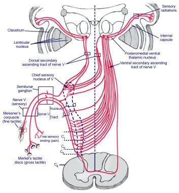
The trigeminal nuclei are a collection of sensory nerve cell bodies (nuclei) located in the brainstem that receive and process sensory information from the face and head, including pain, temperature, touch, and proprioception. There are four main trigeminal nuclei: the ophthalmic, maxillary, mandibular, and mesencephalic nuclei. Each nucleus is responsible for processing sensory information from specific areas of the face and head. The trigeminal nerve (cranial nerve V) carries these sensory signals to the brainstem, where they synapse with neurons in the trigeminal nuclei before being relayed to higher brain centers for further processing.
Muscle contraction is the physiological process in which muscle fibers shorten and generate force, leading to movement or stability of a body part. This process involves the sliding filament theory where thick and thin filaments within the sarcomeres (the functional units of muscles) slide past each other, facilitated by the interaction between myosin heads and actin filaments. The energy required for this action is provided by the hydrolysis of adenosine triphosphate (ATP). Muscle contractions can be voluntary or involuntary, and they play a crucial role in various bodily functions such as locomotion, circulation, respiration, and posture maintenance.
Electromyography (EMG) is a medical diagnostic procedure that measures the electrical activity of skeletal muscles during contraction and at rest. It involves inserting a thin needle electrode into the muscle to record the electrical signals generated by the muscle fibers. These signals are then displayed on an oscilloscope and may be heard through a speaker.
EMG can help diagnose various neuromuscular disorders, such as muscle weakness, numbness, or pain, and can distinguish between muscle and nerve disorders. It is often used in conjunction with other diagnostic tests, such as nerve conduction studies, to provide a comprehensive evaluation of the nervous system.
EMG is typically performed by a neurologist or a physiatrist, and the procedure may cause some discomfort or pain, although this is usually minimal. The results of an EMG can help guide treatment decisions and monitor the progression of neuromuscular conditions over time.
"Sucking behavior" is not a term typically used in medical terminology. However, in the context of early childhood development and behavior, "non-nutritive sucking" is a term that may be used to describe an infant or young child's habitual sucking on their thumb, fingers, or pacifiers, beyond what is necessary for feeding. This type of sucking behavior can provide a sense of security, comfort, or help to self-soothe and manage stress or anxiety.
It's important to note that while non-nutritive sucking is generally considered a normal part of early childhood development, persistent sucking habits beyond the age of 2-4 years may lead to dental or orthodontic problems such as an overbite or open bite. Therefore, it's recommended to monitor and address these behaviors if they persist beyond this age range.
Facial muscles, also known as facial nerves or cranial nerve VII, are a group of muscles responsible for various expressions and movements of the face. These muscles include:
1. Orbicularis oculi: muscle that closes the eyelid and raises the upper eyelid
2. Corrugator supercilii: muscle that pulls the eyebrows down and inward, forming wrinkles on the forehead
3. Frontalis: muscle that raises the eyebrows and forms horizontal wrinkles on the forehead
4. Procerus: muscle that pulls the medial ends of the eyebrows downward, forming vertical wrinkles between the eyebrows
5. Nasalis: muscle that compresses or dilates the nostrils
6. Depressor septi: muscle that pulls down the tip of the nose
7. Levator labii superioris alaeque nasi: muscle that raises the upper lip and flares the nostrils
8. Levator labii superioris: muscle that raises the upper lip
9. Zygomaticus major: muscle that raises the corner of the mouth, producing a smile
10. Zygomaticus minor: muscle that raises the nasolabial fold and corner of the mouth
11. Risorius: muscle that pulls the angle of the mouth laterally, producing a smile
12. Depressor anguli oris: muscle that pulls down the angle of the mouth
13. Mentalis: muscle that raises the lower lip and forms wrinkles on the chin
14. Buccinator: muscle that retracts the cheek and helps with chewing
15. Platysma: muscle that depresses the corner of the mouth and wrinkles the skin of the neck.
These muscles are innervated by the facial nerve, which arises from the brainstem and exits the skull through the stylomastoid foramen. Damage to the facial nerve can result in facial paralysis or weakness on one or both sides of the face.
Skeletal muscle, also known as striated or voluntary muscle, is a type of muscle that is attached to bones by tendons or aponeuroses and functions to produce movements and support the posture of the body. It is composed of long, multinucleated fibers that are arranged in parallel bundles and are characterized by alternating light and dark bands, giving them a striped appearance under a microscope. Skeletal muscle is under voluntary control, meaning that it is consciously activated through signals from the nervous system. It is responsible for activities such as walking, running, jumping, and lifting objects.
The mandible, also known as the lower jaw, is the largest and strongest bone in the human face. It forms the lower portion of the oral cavity and plays a crucial role in various functions such as mastication (chewing), speaking, and swallowing. The mandible is a U-shaped bone that consists of a horizontal part called the body and two vertical parts called rami.
The mandible articulates with the skull at the temporomandibular joints (TMJs) located in front of each ear, allowing for movements like opening and closing the mouth, protrusion, retraction, and side-to-side movement. The mandible contains the lower teeth sockets called alveolar processes, which hold the lower teeth in place.
In medical terminology, the term "mandible" refers specifically to this bone and its associated structures.
Dental occlusion refers to the alignment and contact between the upper and lower teeth when the jaws are closed. It is the relationship between the maxillary (upper) and mandibular (lower) teeth when they approach each other, as occurs during chewing or biting.
A proper dental occlusion, also known as a balanced occlusion, ensures that the teeth and jaw joints function harmoniously, reducing the risk of tooth wear, damage, and temporomandibular disorders (TMD). Malocclusion, on the other hand, refers to improper alignment or contact between the upper and lower teeth, which may require orthodontic treatment or dental restorations to correct.
Facial pain is a condition characterized by discomfort or pain felt in any part of the face. It can result from various causes, including nerve damage or irritation, injuries, infections, dental problems, migraines, or sinus congestion. The pain can range from mild to severe and may be sharp, dull, constant, or intermittent. In some cases, facial pain can also be associated with other symptoms such as headaches, redness, swelling, or changes in sensation. Accurate diagnosis and treatment of the underlying cause are essential for effective management of facial pain.
Muscle rigidity is a term used to describe an increased resistance to passive movement or muscle tone that is present at rest, which cannot be overcome by the person. It is a common finding in various neurological conditions such as Parkinson's disease, stiff-person syndrome, and tetanus. In these conditions, muscle rigidity can result from hyperexcitability of the stretch reflex arc or abnormalities in the basal ganglia circuitry.
Muscle rigidity should be distinguished from spasticity, which is a velocity-dependent increase in muscle tone that occurs during voluntary movement or passive stretching. Spasticity is often seen in upper motor neuron lesions such as stroke or spinal cord injury.
It's important to note that the assessment of muscle rigidity requires a careful physical examination and may need to be evaluated in conjunction with other signs and symptoms to determine an underlying cause.
Slow-twitch muscle fibers, also known as type I muscle fibers, are specialized skeletal muscle cells that contract relatively slowly and generate less force than fast-twitch fibers. However, they can maintain contraction for longer periods of time and have a higher resistance to fatigue. These fibers primarily use oxygen and aerobic metabolism to produce energy, making them highly efficient during prolonged, lower-intensity activities such as long-distance running or cycling. Slow-twitch muscle fibers also have an abundant blood supply, which allows for efficient delivery of oxygen and removal of waste products.
Muscle proteins are a type of protein that are found in muscle tissue and are responsible for providing structure, strength, and functionality to muscles. The two major types of muscle proteins are:
1. Contractile proteins: These include actin and myosin, which are responsible for the contraction and relaxation of muscles. They work together to cause muscle movement by sliding along each other and shortening the muscle fibers.
2. Structural proteins: These include titin, nebulin, and desmin, which provide structural support and stability to muscle fibers. Titin is the largest protein in the human body and acts as a molecular spring that helps maintain the integrity of the sarcomere (the basic unit of muscle contraction). Nebulin helps regulate the length of the sarcomere, while desmin forms a network of filaments that connects adjacent muscle fibers together.
Overall, muscle proteins play a critical role in maintaining muscle health and function, and their dysregulation can lead to various muscle-related disorders such as muscular dystrophy, myopathies, and sarcopenia.
Skeletal muscle myosin, also known as myosin II, is a type of motor protein that plays a crucial role in muscle contraction. It is a hexameric protein composed of two heavy chains and four light chains. The heavy chains have a head region, which contains the ATPase activity and binds to actin filaments, and a tail region, which forms a coiled-coil structure that allows myosin molecules to self-associate into thick filaments.
During muscle contraction, the myosin heads bind to actin filaments in the sarcomere and undergo a power stroke, which results in the sliding of the actin filaments relative to the myosin filaments and thus shortening of the sarcomere. The ATP hydrolysis provides the energy for this power stroke.
Skeletal muscle myosin is essential for generating force and movement in skeletal muscles, and its dysfunction can lead to various muscle diseases and disorders.
Muscle spindles are specialized sensory organs found within the muscle belly, which primarily function as proprioceptors, providing information about the length and rate of change in muscle length. They consist of small, encapsulated bundles of intrafusal muscle fibers that are interspersed among the extrafusal muscle fibers (the ones responsible for force generation).
Muscle spindles have two types of sensory receptors called primary and secondary endings. Primary endings are located near the equatorial region of the intrafusal fiber, while secondary endings are situated more distally. These endings detect changes in muscle length and transmit this information to the central nervous system (CNS) through afferent nerve fibers.
The activation of muscle spindles plays a crucial role in reflexive responses, such as the stretch reflex (myotatic reflex), which helps maintain muscle tone and joint stability. Additionally, they contribute to our sense of body position and movement awareness, known as kinesthesia.
Smooth muscle, also known as involuntary muscle, is a type of muscle that is controlled by the autonomic nervous system and functions without conscious effort. These muscles are found in the walls of hollow organs such as the stomach, intestines, bladder, and blood vessels, as well as in the eyes, skin, and other areas of the body.
Smooth muscle fibers are shorter and narrower than skeletal muscle fibers and do not have striations or sarcomeres, which give skeletal muscle its striped appearance. Smooth muscle is controlled by the autonomic nervous system through the release of neurotransmitters such as acetylcholine and norepinephrine, which bind to receptors on the smooth muscle cells and cause them to contract or relax.
Smooth muscle plays an important role in many physiological processes, including digestion, circulation, respiration, and elimination. It can also contribute to various medical conditions, such as hypertension, gastrointestinal disorders, and genitourinary dysfunction, when it becomes overactive or underactive.
Myosin Heavy Chains are the large, essential components of myosin molecules, which are responsible for the molecular motility in muscle cells. These heavy chains have a molecular weight of approximately 200 kDa and form the motor domain of myosin, which binds to actin filaments and hydrolyzes ATP to generate force and movement during muscle contraction. There are several different types of myosin heavy chains, each with specific roles in various tissues and cellular functions. In skeletal and cardiac muscles, for example, myosin heavy chains have distinct isoforms that contribute to the contractile properties of these tissues.
The temporomandibular joint (TMJ) is the articulation between the mandible (lower jaw) and the temporal bone of the skull. It's a complex joint that involves the movement of two bones, several muscles, and various ligaments. The TMJ allows for movements like rotation and translation, enabling us to open and close our mouth, chew, speak, and yawn. Dysfunction in this joint can lead to temporomandibular joint disorders (TMD), which can cause pain, discomfort, and limited jaw movement.
The "chin" is the lower, prominent part of the front portion of the jaw in humans and other animals. In medical terms, it is often referred to as the mentum or the symphysis of the mandible. The chin helps in protecting the soft tissues of the mouth and throat during activities such as eating, speaking, and swallowing. It also plays a role in shaping the overall appearance of the face. Anatomically, the chin is formed by the fusion of the two halves of the mandible (lower jawbone) at the symphysis menti.
Fast-twitch muscle fibers, also known as type II fibers, are a type of skeletal muscle fiber that are characterized by their rapid contraction and relaxation rates. These fibers have a larger diameter and contain a higher concentration of glycogen, which serves as a quick source of energy for muscle contractions. Fast-twitch fibers are further divided into two subcategories: type IIa and type IIb (or type IIx). Type IIa fibers have a moderate amount of mitochondria and can utilize both aerobic and anaerobic metabolic pathways, making them fatigue-resistant. Type IIb fibers, on the other hand, have fewer mitochondria and primarily use anaerobic metabolism, leading to faster fatigue. Fast-twitch fibers are typically used in activities that require quick, powerful movements such as sprinting or weightlifting.
The Sella Turcica, also known as the Turkish saddle, is a depression or fossa in the sphenoid bone located at the base of the skull. It forms a housing for the pituitary gland, which is a small endocrine gland often referred to as the "master gland" because it controls other glands and makes several essential hormones. The Sella Turcica has a saddle-like shape, with its anterior and posterior clinoids forming the front and back of the saddle, respectively. This region is of significant interest in neuroimaging and clinical settings, as various conditions such as pituitary tumors or other abnormalities may affect the size, shape, and integrity of the Sella Turcica.
Muscle development, also known as muscle hypertrophy, refers to the increase in size and mass of the muscles through a process called myofiber growth. This is primarily achieved through resistance or strength training exercises that cause micro-tears in the muscle fibers, leading to an inflammatory response and the release of hormones that promote muscle growth. As the muscles repair themselves, they become larger and stronger than before. Proper nutrition, including adequate protein intake, and rest are also essential components of muscle development.
It is important to note that while muscle development can lead to an increase in strength and muscular endurance, it does not necessarily result in improved athletic performance or overall fitness. A well-rounded exercise program that includes cardiovascular activity, flexibility training, and resistance exercises is recommended for optimal health and fitness outcomes.
The nasal bones are a pair of small, thin bones located in the upper part of the face, specifically in the middle of the nose. They articulate with each other at the nasal bridge and with the frontal bone above, the maxillae (upper jaw bones) on either side, and the septal cartilage inside the nose. The main function of the nasal bones is to form the bridge of the nose and protect the nasal cavity. Any damage to these bones can result in a fracture or broken nose.
A smooth muscle within the vascular system refers to the involuntary, innervated muscle that is found in the walls of blood vessels. These muscles are responsible for controlling the diameter of the blood vessels, which in turn regulates blood flow and blood pressure. They are called "smooth" muscles because their individual muscle cells do not have the striations, or cross-striped patterns, that are observed in skeletal and cardiac muscle cells. Smooth muscle in the vascular system is controlled by the autonomic nervous system and by hormones, and can contract or relax slowly over a period of time.
The trigeminal nerve, also known as the fifth cranial nerve or CNV, is a paired nerve that carries both sensory and motor information. It has three major branches: ophthalmic (V1), maxillary (V2), and mandibular (V3). The ophthalmic branch provides sensation to the forehead, eyes, and upper portion of the nose; the maxillary branch supplies sensation to the lower eyelid, cheek, nasal cavity, and upper lip; and the mandibular branch is responsible for sensation in the lower lip, chin, and parts of the oral cavity, as well as motor function to the muscles involved in chewing. The trigeminal nerve plays a crucial role in sensations of touch, pain, temperature, and pressure in the face and mouth, and it also contributes to biting, chewing, and swallowing functions.
Succinylcholine is a neuromuscular blocking agent, a type of muscle relaxant used in anesthesia during surgical procedures. It works by inhibiting the transmission of nerve impulses at the neuromuscular junction, leading to temporary paralysis of skeletal muscles. This facilitates endotracheal intubation and mechanical ventilation during surgery. Succinylcholine has a rapid onset of action and is metabolized quickly, making it useful for short surgical procedures. However, its use may be associated with certain adverse effects, such as increased heart rate, muscle fasciculations, and potentially life-threatening hyperkalemia in susceptible individuals.
Temporomandibular Joint Dysfunction Syndrome, often abbreviated as TMJD or TMD, is a group of conditions that cause pain and dysfunction in the temporomandibular joint (TMJ) - the joint that connects the jawbone to the skull. Here's a more detailed medical definition:
Temporomandibular Joint Dysfunction Syndrome is a complex disorder characterized by pain, clicking, popping, or grating sounds in the TMJ; limited movement or locking of the jaw; and/or painful chewing movements. The condition may be caused by a variety of factors, including muscle tension, joint inflammation, structural problems with the joint itself, or injury to the head, neck, or jaw.
Symptoms of TMJD can include:
- Pain or tenderness in the face, jaw joint area, neck, and/or shoulders
- Limited ability to open the mouth wide
- Jaw locking, making it difficult to close or open the mouth
- Clicking, popping, or grating sounds in the TMJ when opening or closing the mouth
- A significant change in the way the upper and lower teeth fit together
- Headaches, earaches, dizziness, and hearing problems
Treatment for TMJD can vary depending on the severity of the condition and its underlying cause. It may include self-care practices such as eating soft foods, avoiding extreme jaw movements, and practicing relaxation techniques; physical therapy; medication to reduce pain and inflammation; dental treatments such as mouthguards or bite adjustments; and, in rare cases, surgery.
Cephalometry is a medical term that refers to the measurement and analysis of the skull, particularly the head face relations. It is commonly used in orthodontics and maxillofacial surgery to assess and plan treatment for abnormalities related to the teeth, jaws, and facial structures. The process typically involves taking X-ray images called cephalograms, which provide a lateral view of the head, and then using various landmarks and reference lines to make measurements and evaluate skeletal and dental relationships. This information can help clinicians diagnose problems, plan treatment, and assess treatment outcomes.
An overbite, also known as "malocclusion of class II division 1" in dental terminology, is an orthodontic condition where the upper front teeth excessively overlap the lower front teeth when biting down. This means that the upper incisors are positioned too far forward or the lower incisors are too far back. A slight overbite is considered normal and healthy, as it allows the front teeth to perform their functions properly, such as biting and tearing food. However, a significant overbite can lead to various problems like difficulty in chewing, speaking, and maintaining good oral hygiene. It may also cause wear and tear on the teeth, jaw pain, or even contribute to temporomandibular joint disorders (TMD). Orthodontic treatment, such as braces or aligners, is often recommended to correct a severe overbite and restore proper bite alignment.
The maxilla is a paired bone that forms the upper jaw in vertebrates. In humans, it is a major bone in the face and plays several important roles in the craniofacial complex. Each maxilla consists of a body and four processes: frontal process, zygomatic process, alveolar process, and palatine process.
The maxillae contribute to the formation of the eye sockets (orbits), nasal cavity, and the hard palate of the mouth. They also contain the upper teeth sockets (alveoli) and help form the lower part of the orbit and the cheekbones (zygomatic arches).
Here's a quick rundown of its key functions:
1. Supports the upper teeth and forms the upper jaw.
2. Contributes to the formation of the eye sockets, nasal cavity, and hard palate.
3. Helps shape the lower part of the orbit and cheekbones.
4. Partakes in the creation of important sinuses, such as the maxillary sinus, which is located within the body of the maxilla.
The facial bones, also known as the facial skeleton, are a series of bones that make up the framework of the face. They include:
1. Frontal bone: This bone forms the forehead and the upper part of the eye sockets.
2. Nasal bones: These two thin bones form the bridge of the nose.
3. Maxilla bones: These are the largest bones in the facial skeleton, forming the upper jaw, the bottom of the eye sockets, and the sides of the nose. They also contain the upper teeth.
4. Zygomatic bones (cheekbones): These bones form the cheekbones and the outer part of the eye sockets.
5. Palatine bones: These bones form the back part of the roof of the mouth, the side walls of the nasal cavity, and contribute to the formation of the eye socket.
6. Inferior nasal conchae: These are thin, curved bones that form the lateral walls of the nasal cavity and help to filter and humidify air as it passes through the nose.
7. Lacrimal bones: These are the smallest bones in the skull, located at the inner corner of the eye socket, and help to form the tear duct.
8. Mandible (lower jaw): This is the only bone in the facial skeleton that can move. It holds the lower teeth and forms the chin.
These bones work together to protect vital structures such as the eyes, brain, and nasal passages, while also providing attachment points for muscles that control chewing, expression, and other facial movements.
Maxillofacial development refers to the growth and formation of the bones, muscles, and soft tissues that make up the face and jaw (maxillofacial region). This process begins in utero and continues throughout childhood and adolescence. It involves the coordinated growth and development of multiple structures, including the upper and lower jaws (maxilla and mandible), facial bones, teeth, muscles, and nerves.
Abnormalities in maxillofacial development can result in a range of conditions, such as cleft lip and palate, jaw deformities, and craniofacial syndromes. These conditions may affect a person's appearance, speech, chewing, and breathing, and may require medical or surgical intervention to correct.
Healthcare professionals involved in the diagnosis and treatment of maxillofacial developmental disorders include oral and maxillofacial surgeons, orthodontists, pediatricians, geneticists, and other specialists.
The dental arch refers to the curved shape formed by the upper or lower teeth when they come together. The dental arch follows the curve of the jaw and is important for proper bite alignment and overall oral health. The dental arches are typically described as having a U-shaped appearance, with the front teeth forming a narrower section and the back teeth forming a wider section. The shape and size of the dental arch can vary from person to person, and any significant deviations from the typical shape or size may indicate an underlying orthodontic issue that requires treatment.
Diagnostic techniques in otology refer to the methods and tests used by healthcare professionals to identify and diagnose various conditions related to the ear. These techniques can include:
1. Otoscopy: A visual examination of the external auditory canal and eardrum using an otoscope. This helps to identify any physical abnormalities, such as wax buildup, inflammation, or foreign objects in the ear.
2. Audiometry: A hearing test that measures a person's ability to hear different sounds, pitches, and volumes. This can help to identify any hearing loss or auditory processing issues.
3. Tympanometry: A test that measures the function of the middle ear by creating variations in air pressure in the ear canal. This can help to identify any issues with the eardrum or middle ear bones.
4. Acoustic reflex testing: A test that measures the body's involuntary response to loud sounds. This can help to identify any damage to the hearing nerves or brainstem.
5. Otoacoustic emissions (OAE) testing: A test that measures the sound waves produced by the inner ear in response to stimuli. This can help to identify any issues with the cochlea or hair cells in the inner ear.
6. Auditory brainstem response (ABR) testing: A test that measures the electrical activity of the hearing nerve and brainstem in response to sound. This can help to identify any issues with the auditory nervous system.
7. Vestibular testing: A series of tests that measure a person's balance and equilibrium. This can help to identify any issues with the vestibular system, which is responsible for maintaining balance.
These diagnostic techniques are used to diagnose various otological conditions such as hearing loss, tinnitus, vertigo, ear infections, and tumors of the ear.
Anatomic landmarks are specific, identifiable structures or features on the body that are used as references in medicine and surgery. These landmarks can include bones, muscles, joints, or other visible or palpable features that help healthcare professionals identify specific locations, orient themselves during procedures, or measure changes in the body.
Examples of anatomic landmarks include:
* The anterior iliac spine, a bony prominence on the front of the pelvis that can be used to locate the hip joint.
* The cubital fossa, a depression at the elbow where the median nerve and brachial artery can be palpated.
* The navel (umbilicus), which serves as a reference point for measuring distances in the abdomen.
* The xiphoid process, a small piece of cartilage at the bottom of the breastbone that can be used to locate the heart and other structures in the chest.
Anatomic landmarks are important for accurate diagnosis, treatment planning, and surgical procedures, as they provide reliable and consistent reference points that can help ensure safe and effective care.
Facial asymmetry refers to a condition in which the facial features are not identical or proportionate on both sides of a vertical line drawn down the middle of the face. This can include differences in the size, shape, or positioning of facial features such as the eyes, ears, nose, cheeks, and jaw. Facial asymmetry can be mild and barely noticeable, or it can be more severe and affect a person's appearance and/or functionality of the mouth and jaw.
Facial asymmetry can be present at birth (congenital) or can develop later in life due to various factors such as injury, surgery, growth disorders, nerve damage, or tumors. In some cases, facial asymmetry may not cause any medical problems and may only be of cosmetic concern. However, in other cases, it may indicate an underlying medical condition that requires treatment.
Depending on the severity and cause of the facial asymmetry, treatment options may include cosmetic procedures such as fillers or surgery, orthodontic treatment, physical therapy, or medication to address any underlying conditions.
Neck muscles, also known as cervical muscles, are a group of muscles that provide movement, support, and stability to the neck region. They are responsible for various functions such as flexion, extension, rotation, and lateral bending of the head and neck. The main neck muscles include:
1. Sternocleidomastoid: This muscle is located on either side of the neck and is responsible for rotating and flexing the head. It also helps in tilting the head to the same side.
2. Trapezius: This large, flat muscle covers the back of the neck, shoulders, and upper back. It is involved in movements like shrugging the shoulders, rotating and extending the head, and stabilizing the scapula (shoulder blade).
3. Scalenes: These three pairs of muscles are located on the side of the neck and assist in flexing, rotating, and laterally bending the neck. They also help with breathing by elevating the first two ribs during inspiration.
4. Suboccipitals: These four small muscles are located at the base of the skull and are responsible for fine movements of the head, such as tilting and rotating.
5. Longus Colli and Longus Capitis: These muscles are deep neck flexors that help with flexing the head and neck forward.
6. Splenius Capitis and Splenius Cervicis: These muscles are located at the back of the neck and assist in extending, rotating, and laterally bending the head and neck.
7. Levator Scapulae: This muscle is located at the side and back of the neck, connecting the cervical vertebrae to the scapula. It helps with rotation, extension, and elevation of the head and scapula.
Hypertrophy, in the context of physiology and pathology, refers to an increase in the size of an organ or tissue due to an enlargement of its constituent cells. It is often used to describe the growth of muscle cells (myocytes) in response to increased workload or hormonal stimulation, resulting in an increase in muscle mass. However, hypertrophy can also occur in other organs such as the heart (cardiac hypertrophy) in response to high blood pressure or valvular heart disease.
It is important to note that while hypertrophy involves an increase in cell size, hyperplasia refers to an increase in cell number. In some cases, both hypertrophy and hyperplasia can occur together, leading to a significant increase in the overall size and function of the organ or tissue.
The lingual nerve is a branch of the mandibular division of the trigeminal nerve (cranial nerve V). It provides general sensory innervation to the anterior two-thirds of the tongue, including taste sensation from the same region. It also supplies sensory innervation to the floor of the mouth and the lingual gingiva (gum tissue). The lingual nerve is closely associated with the submandibular and sublingual salivary glands and their ducts.
Temporomandibular Joint Disorders (TMD) refer to a group of conditions that cause pain and dysfunction in the temporomandibular joint (TMJ) and the muscles that control jaw movement. The TMJ is the hinge joint that connects the lower jaw (mandible) to the skull (temporal bone) in front of the ear. It allows for movements required for activities such as eating, speaking, and yawning.
TMD can result from various causes, including:
1. Muscle tension or spasm due to clenching or grinding teeth (bruxism), stress, or jaw misalignment
2. Dislocation or injury of the TMJ disc, which is a small piece of cartilage that acts as a cushion between the bones in the joint
3. Arthritis or other degenerative conditions affecting the TMJ
4. Bite problems (malocclusion) leading to abnormal stress on the TMJ and its surrounding muscles
5. Stress, which can exacerbate existing TMD symptoms by causing muscle tension
Symptoms of Temporomandibular Joint Disorders may include:
- Pain or tenderness in the jaw, face, neck, or shoulders
- Limited jaw movement or locking of the jaw
- Clicking, popping, or grating sounds when moving the jaw
- Headaches, earaches, or dizziness
- Difficulty chewing or biting
- Swelling on the side of the face
Treatment for TMD varies depending on the severity and cause of the condition. It may include self-care measures (like eating soft foods, avoiding extreme jaw movements, and applying heat or cold packs), physical therapy, medications (such as muscle relaxants, pain relievers, or anti-inflammatory drugs), dental work (including bite adjustments or orthodontic treatment), or even surgery in severe cases.
The mandibular nerve is a branch of the trigeminal nerve (the fifth cranial nerve), which is responsible for sensations in the face and motor functions such as biting and chewing. The mandibular nerve provides both sensory and motor innervation to the lower third of the face, below the eye and nose down to the chin.
More specifically, it carries sensory information from the lower teeth, lower lip, and parts of the oral cavity, as well as the skin over the jaw and chin. It also provides motor innervation to the muscles of mastication (chewing), which include the masseter, temporalis, medial pterygoid, and lateral pterygoid muscles.
Damage to the mandibular nerve can result in numbness or loss of sensation in the lower face and mouth, as well as weakness or difficulty with chewing and biting.
An open bite, in dental terminology, refers to a type of malocclusion (or misalignment) where the upper and lower teeth do not make contact with each other when the jaw is closed. More specifically, the front teeth of both the upper and lower jaws fail to meet or overlap normally, creating an opening in the bite. This condition can lead to various problems such as difficulty in biting, chewing, speaking clearly, and even cause temporomandibular joint disorders (TMD). Open bite can be caused by several factors including thumb sucking, tongue thrusting, genetic factors, or abnormal jaw development. Treatment usually involves orthodontic intervention, possibly with the use of appliances or even surgery in severe cases.
Muscle fatigue is a condition characterized by a reduction in the ability of a muscle to generate force or power, typically after prolonged or strenuous exercise. It is often accompanied by sensations of tiredness, weakness, and discomfort in the affected muscle(s). The underlying mechanisms of muscle fatigue are complex and involve both peripheral factors (such as changes in muscle metabolism, ion handling, and neuromuscular transmission) and central factors (such as changes in the nervous system's ability to activate muscles). Muscle fatigue can also occur as a result of various medical conditions or medications that impair muscle function.
The trigeminal ganglion, also known as the semilunar or Gasserian ganglion, is a sensory ganglion (a cluster of nerve cell bodies) located near the base of the skull. It is a part of the trigeminal nerve (the fifth cranial nerve), which is responsible for sensation in the face and motor functions such as biting and chewing.
The trigeminal ganglion contains the cell bodies of sensory neurons that carry information from three major branches of the trigeminal nerve: the ophthalmic, maxillary, and mandibular divisions. These divisions provide sensation to different areas of the face, head, and oral cavity, including the skin, mucous membranes, muscles, and teeth.
Damage to the trigeminal ganglion or its nerve branches can result in various sensory disturbances, such as pain, numbness, or tingling in the affected areas. Conditions like trigeminal neuralgia, a disorder characterized by intense, stabbing facial pain, may involve the trigeminal ganglion and its associated nerves.
Malocclusion, Angle Class II is a type of dental malocclusion where the relationship between the maxilla (upper jaw) and mandible (lower jaw) is such that the lower molar teeth are positioned posteriorly relative to the upper molar teeth. This results in an overbite, which means that the upper front teeth overlap the lower front teeth excessively. The classification was proposed by Edward Angle, an American orthodontist who is considered the father of modern orthodontics. In this classification system, Class II malocclusion is further divided into three subclasses (I, II, and III) based on the position of the lower incisors relative to the upper incisors.
Muscle denervation is a medical term that refers to the loss of nerve supply to a muscle or group of muscles. This can occur due to various reasons, such as injury to the nerves, nerve compression, or certain medical conditions like neuromuscular disorders. When the nerve supply to the muscle is interrupted, it can lead to muscle weakness, atrophy (wasting), and ultimately, paralysis.
In denervation, the communication between the nervous system and the muscle is disrupted, which means that the muscle no longer receives signals from the brain to contract and move. Over time, this can result in significant muscle wasting and disability, depending on the severity and extent of the denervation.
Denervation may be treated with various therapies, including physical therapy, medication, or surgical intervention, such as nerve grafting or muscle transfers, to restore function and prevent further muscle wasting. The specific treatment approach will depend on the underlying cause and severity of the denervation.
Malocclusion, Angle Class I is a type of dental malocclusion where the misalignment of teeth is not severe enough to affect the overall function or appearance of the bite significantly. Named after Edward Angle, the founder of modern orthodontics, this classification indicates that the mesiobuccal cusp of the upper first molar is aligned with the buccal groove of the lower first molar. Although the bite appears normal, there might be crowding, spacing, or rotations present in the teeth, which can lead to aesthetic concerns and potential periodontal issues if left untreated.
Motor neurons are specialized nerve cells in the brain and spinal cord that play a crucial role in controlling voluntary muscle movements. They transmit electrical signals from the brain to the muscles, enabling us to perform actions such as walking, talking, and swallowing. There are two types of motor neurons: upper motor neurons, which originate in the brain's motor cortex and travel down to the brainstem and spinal cord; and lower motor neurons, which extend from the brainstem and spinal cord to the muscles. Damage or degeneration of these motor neurons can lead to various neurological disorders, such as amyotrophic lateral sclerosis (ALS) and spinal muscular atrophy (SMA).
In medical terms, the face refers to the front part of the head that is distinguished by the presence of the eyes, nose, and mouth. It includes the bones of the skull (frontal bone, maxilla, zygoma, nasal bones, lacrimal bones, palatine bones, inferior nasal conchae, and mandible), muscles, nerves, blood vessels, skin, and other soft tissues. The face plays a crucial role in various functions such as breathing, eating, drinking, speaking, seeing, smelling, and expressing emotions. It also serves as an important identifier for individuals, allowing them to be recognized by others.
 Masseter muscle
Orofacial myofunctional disorders
Masseteric nerve
Mandibular setback surgery
Jaw jerk reflex
Risorius
Platycraniellus
Masticatory muscle myositis
Geomyoidea
Lonchocyon
Macrostomia
Carnassial
Tenomodulin
Biofeedback
Short face
Brow ridge
Forehand (horse)
Startle response
Coronoid process of the mandible
Mountain beaver
Durophagy
Glossary of medicine
Nematode infection in dogs
Mandible
Macroraptorial sperm whale
Bruxism
Thalassocnus
Medistylus
Parotid duct
Audio-visual entrainment
Masseter muscle - Wikipedia
 Reduction of masseter muscle
 Unilateral Masseter Muscle Hypertrophy: Case Repor
 "Changes in Tension of the Masseter and Digastricus Muscles of a Decere" by Wesley H. Ardoin
 Masseter Muscle Reduction
 masseter muscle reduction Archives - Elite Doc Health and Beauty
 Benefit of Masseter Muscle Botox Over Anti-Wrinkle Treatment
 TMJ Pain Can Be Treated With Masseter Muscle Massage
 Myofascial syndrome of the masseter muscle.
 Masseter Muscle Reduction Procedures
 Correlation between MRI and Biomodelling Analysis in Masseter Muscle Following Orthognathic Surgery - Clitrofa
 DIFFERENTIAL EXPRESSION OF miRNA 328 IN MASSETER MUSCLES OF SUBJECTS WITH FACIAL ASYMMETRY
 May 1977 - Volume 59 - Issue 5 : Plastic and Reconstructive Surgery
 Modulation of exteroceptive suppression periods in human jaw-closing muscles by local and remote experimental muscle pain
 Use of a Questionnaire for Evaluation of Surgical Treatment of Masseter Muscle Hypertrophy: A Case Report
| Prague Medical...
Use of a Questionnaire for Evaluation of Surgical Treatment of Masseter Muscle Hypertrophy: A Case Report
| Prague Medical...
 Electromyographic analysis of the masseter and temporal muscles in oralized deaf individuals. | Electromyogr Clin Neurophysiol...
 View of Sarcoglycan sub-complex and Satellite cells in masseter muscle of crossbite patients
| Italian Journal of...
View of Sarcoglycan sub-complex and Satellite cells in masseter muscle of crossbite patients
| Italian Journal of...
Macaca mulatta. Cephalometric analysis of adaptations after lengthening of the masseter muscle in adult rhesus monkeys - Texas...
 Masticatory Muscle Pain and Disorders | SpringerLink
Masseteric nerve - Wikipedia
 Tetanus (Lockjaw): Photos of Bacteria and People Affected | CDC
 Abductor Pollicis Brevis Muscle Anatomy, Function & Diagram | Body Maps
Trapezius Muscle Origin, Function & Area | Body Maps
 3 Tips to Stop Grinding Your Teeth and Relieve Jaw Pain | livestrong
 Sabina head symptoms - ABC Homeopathy
Temporomandibular Joint Disorders | Encyclopedia.com
 Scientists discover new part of the body | Live Science
 Static Suspension for Facial Paralysis: Background, History of the Procedure, Etiology
Muscles of mastication | Intelligent Dental
 Michael Kirifides | College of Nursing and Health Professions | Drexel University
Left masseter muscle3
- In random order 5% hypertonic or 0.9% isotonic saline was infused into the left masseter muscle for 15 min. (nih.gov)
- Locations of the left temporalis muscle (asterisk) and left masseter muscle (arrow) are shown. (avma.org)
- At the same time, the electrical activity of their left masseter muscle was recorded to confirm clenching. (improbable.com)
Role of the masseter muscle2
- The essential role of the masseter muscle is for adequate mastication. (elegantplasticsurgery.com)
- The role of the masseter muscle in craniofacial adaptations to altered vertical dimension was determined by detaching and re-attaching the insertion of the masseter muscle in one group of experimental animals. (tamu.edu)
Contraction5
- The action of the muscle during bilateral contraction of the entire muscle is to elevate the mandible, raising the lower jaw. (wikipedia.org)
- Sudden contraction of temporalis muscle will result in coronoid fracture, which is rare. (intelligentdental.com)
- Muscle contraction happens when nerves release a neurotransmitter (acetylcholine) onto muscle cells, causing those cells to contract. (glowday.com)
- This patient presented with facial tetany, involving contraction of the masseter and his neck muscles. (cdc.gov)
- Lower motor neurons transmit impulses to the neuromuscular junction to initiate muscle contraction. (msdmanuals.com)
Botox14
- It can usually be accomplished with 3 to 5 small injections of Botox into the masseter muscle. (theparkerclinic.com)
- Botox can help shrink the muscle and both reduce its appearance and lessen the amount of discomfort the muscle exerts on the surrounding tissues. (theparkerclinic.com)
- For more information on Botox for Masseter Muscle Reduction or to schedule a consultation for a personalized treatment plan with one of our providers, please contact us . (theparkerclinic.com)
- Injection of botulinum toxin type A (Botox) into the masseter muscle was considered as a less invasive modality for the treatment of muscle hypertrophy. (elegantplasticsurgery.com)
- Local injection of tiny doses of the Botox into a muscle produces local paralysis, and therefore, specific muscles can be selectively weakened, and atrophy of the muscle occurs. (elegantplasticsurgery.com)
- Botox® is commonly used to temporarily paralyse muscles of the face to soften wrinkles, particularly those that occur due to facial expressions. (glowday.com)
- When Botox® is injected into a muscle, the release of acetylcholine from the nerves stimulating that muscle is prevented. (glowday.com)
- If you are self-conscious about having an overly square jaw, masseter muscle Botox® may be a potential treatment to narrow your jawline and produce a more v-shaped lower face. (glowday.com)
- How should I prepare for a masseter Botox® treatment? (glowday.com)
- What happens during a masseter Botox® treatment? (glowday.com)
- Botox causes paralysis or limited movement of the muscles around the jaws. (harcourthealth.com)
- The procedure involves the injection of a small portion of Botox into the masseter muscle. (harcourthealth.com)
- Botox relaxes muscles in the face, therefore reducing the prominence of wrinkles. (michelegreenmd.com)
- Male Botox patients generally require a slightly higher dose of Botox than female patients in this area, since their muscles are generally stronger. (michelegreenmd.com)
Muscular4
- Deaf individuals showed a lower muscular activity for clinical activities that demanded a greater masseter and temporal muscular activity such as mastication , mouth opening and closing, and dental compression. (bvsalud.org)
- The newly discovered muscle layer runs from the back of the cheekbone to the anterior muscular process of the lower jaw. (livescience.com)
- Tetanus is characterized by painful muscular contractions, primarily of the masseter, and other large muscles. (cdc.gov)
- Weakness of intercostal and diaphragmatic and improving the quality of life in Duchenne muscular muscles with spinal deformity affects respiratory dystrophy children. (who.int)
Bilateral6
- Even if the hypertrophy is bilateral, asymmetry of the face may still occur due to unequal enlargement of the muscles. (wikipedia.org)
- Masseter muscle hypertrophy is a unilateral or bilateral enlargement of the masseter muscle, of undefined etiology, which in most cases generates an aesthetic discomfort, and in some cases a functional one as well. (bvsalud.org)
- In most cases of masseter hypertrophy, it is bilateral and symmetric, but asymmetry is not unusual. (elegantplasticsurgery.com)
- This study aimed to assess, by means of computerized bilateral electromyography (EMG), masseter and temporal muscles of 12 oralized deaf individuals in clinical activities that involve part of this masticatory musculature and compare this system's functionality with that of 12 normal listening individuals, performing the same activities. (bvsalud.org)
- Intraoperative photographs of the dog in Figure 1 showing full-thickness incision of the temporalis fascia on the left side in a caudal to rostral direction (A) and bilateral fasciotomies of the temporalis muscles (B). (avma.org)
- On the basis of the history and examination findings, a clinical provisional diagnosis of myofascial pain was made and the patient was put on the treatment of muscle relaxants Tab Myospaz forte two times a day and topical application of voveran emuigel along with hot fomentation, soft diet, and bilateral chewing. (hindawi.com)
Bruxism5
- The masseter muscle can become enlarged in patients who habitually clench or grind (with bruxism) their teeth and even in those who constantly chew gum. (wikipedia.org)
- Aside from genetics, enlargement of the masseter muscle is caused by asymmetric chewing caused by dental problems, temporomandibular joint dysfunction, food intakes and personal habits (ie: frequent gum chewing, jaw clenching or grinding and bruxism. (theparkerclinic.com)
- The muscle function may also be impaired, thus resulting in conditions such as trismus, protrusion, and bruxism. (elegantplasticsurgery.com)
- Some people have overdeveloped masseter muscles, which can present as a wide, square jawline, teeth grinding (bruxism) and jaw pain. (glowday.com)
- Involuntary masseter muscle activity such as bruxism and jaw clenching may be linked to. (dentistrytoday.com)
Reduction3
- These alterations produced a shortening of the lengthened masseter muscle, i.e. a reduction in the amount of muscle stretch brought about by the appliance. (tamu.edu)
- Patients in this group were operated by reduction angleplasty with/without resection of masseter muscle. (koreamed.org)
- In this case, we applied reduction angleplasty, contouring of body, resection of buccal fat pad and/or masseter muscle. (koreamed.org)
Mastication20
- In human anatomy, the masseter is one of the muscles of mastication. (wikipedia.org)
- The most obvious muscle of mastication is the masseter muscle, since it is the most superficial and one of the strongest. (wikipedia.org)
- Along with the other three muscles of mastication (temporalis, medial pterygoid, and lateral pterygoid), the masseter is innervated by the anterior division of the mandibular division (V3) of the trigeminal nerve. (wikipedia.org)
- The Masseter muscle is a thick and rectangular muscle of mastication found in the jawline. (theparkerclinic.com)
- The masseter muscle (muscle of the jaw, of mastication), ( orange area ) is located in front of the ear, from the lower jaw to the outer side of the eye. (post-operative-chronic-pain.com)
- The stomatognathic system muscles play important roles in functions such as mastication , deglutition , and phonation . (bvsalud.org)
- This relationship between the function of the muscles of mastication and craniofacial form was investigated in young adult monkeys by increasing the functional length of the elevator muscles of the mandible non-invasively by a bite-opening splint cemented to the maxillary dentition. (tamu.edu)
- During mastication, four muscles of mastication (or musculi masticatorii ) are responsible for adduction and lateral motion of the jaw. (intelligentdental.com)
- Each of these primary muscles of mastication is paired, with each side of the mandible possessing one of the four. (intelligentdental.com)
- It is one of the main muscle which helps in the process of mastication. (intelligentdental.com)
- This is the muscle which helps in elevation of the mandible, It is one of the muscles of mastication. (intelligentdental.com)
- This is a small muscle which also helps in the mastication process. (intelligentdental.com)
- It is a thick muscle of mastication. (intelligentdental.com)
- All 8 patients showed changes in muscles of mastication, including superficial temporalis and deep temporalis, and in masseter muscles. (cdc.gov)
- They are also found in oral and perioral tissues, particularly in the muscles of mastication, facial expression, the suprahyoid muscles, and the postcervical musculature as well as in the tongue, buccal mucosa, and lip [ 2 - 4 ]. (hindawi.com)
- Examination of the TMJ and other muscles of mastication revealed normal findings. (hindawi.com)
- It is the motor nerve for the muscles of mastication and contains proprioceptive fibers. (medscape.com)
- The mesencephalic nucleus is in the midbrain and receives proprioceptive fibers from all muscles of mastication. (medscape.com)
- The proprioceptive fibers of CN V arise from the muscles of mastication and the extraocular muscles. (medscape.com)
- The motor nucleus of CN V receives cortical fibers for voluntary control of the muscles of mastication. (medscape.com)
Mandible5
- The coronoid head of the masseter's tendon and muscle fibers run posterolaterally from the coronoid process of the mandible towards the posterior third of the zygomatic arch. (wikipedia.org)
- Dissection, showing salivary glands of right side (Masseter visible at center) Left temporal bone, outer surface Left temporal bone, inferior surface Left zygomatic bone, temporal surface Mandible, outer surface, side view The arteries of the face and scalp. (wikipedia.org)
- The surgery to access the masseter muscles and the mandible (jawbone) is performed through incisions on the inside of the mouth, between the gum and cheek (lower buccal sulcus area), leaving no visible scarring. (elegantplasticsurgery.com)
- The masseter muscle extends from the zygomatic arch to the ramus and body of the mandible. (intelligentdental.com)
- A-On initial examination, the dog had bilaterally elevated nictitating membranes, an inability to adduct the mandible, and severely swollen masticatory muscles. (avma.org)
Atrophy3
- The use of muscle transfer is particularly useful with atrophy of the mimetic muscles and/or facial fat. (medscape.com)
- Following the first treatment, the masseter muscle will atrophy (waste away slightly). (glowday.com)
- with time, muscles atrophy. (msdmanuals.com)
Temporalis muscles1
- Electromyographic (EMG) activity was recorded bilaterally from the masseter and temporalis muscles during the pre-infusion, early phase of infusion (from 120 to 480 s), late phase of infusion (from 540 to 900 s) and post-infusion. (nih.gov)
Mandibular angle3
- The surgical approach involves intervetions performed in either the compromised muscle or the mandibular angle bone structure, or in both. (bvsalud.org)
- This article describes a case of unilateral masseter muscle hypertrophy, where the surgical resection of the muscle fibers in an intraoral approach and a osteotomy with a reciprocating saw were performed in the mandibular angle region. (bvsalud.org)
- After a two-month follow-up, it was observed that the surgical approach for the treatment of masseter hypertrophy is well advised, since the aesthetic results are excellent, with similarity to the contralateral side of mandibular angle. (bvsalud.org)
Hypertrophy includes1
- Conservative management of the idiopathic masseter hypertrophy includes psychological counselling, use of mouth guards, muscle relaxant, anxiolytic drugs, analgesics, physical therapy, dental restorations, and occlusal adjustments to correct premature contacts. (elegantplasticsurgery.com)
Discomfort as a result2
- The benefits of the injections can be twofold: patients can expect improvement in facial balance and change the shape of an overly square face and secondly some patients will experience pain and discomfort as a result of an overly large masseter muscle. (theparkerclinic.com)
- The treatment aims to improve facial balance, change the shape of an overly square face and relief pain or discomfort as a result of an excessively large masseter muscle. (elegantplasticsurgery.com)
Musculus2
- musculus masseter. (wikipedia.org)
- In their paper, the team proposed that the newfound muscle layer be named "Musculus masseter pars coronidea," meaning "coronoid part of the masseter. (livescience.com)
Lateral pterygoid1
- The masseteric nerve is a branch of (the anterior division of) the mandibular nerve (CN V3) (itself a branch of the trigeminal nerve (CN V)). It passes laterally superior to the lateral pterygoid muscle, anterior to the temporomandibular joint, and posterior to the tendon of the temporalis muscle. (wikipedia.org)
Bilaterally2
- To perform an extraoral examination, stand near the patient and visually inspect and bilaterally palpate the muscle. (wikipedia.org)
- The temporalis (star), masseter (polygon), and pterygoid (black arrow) muscles appear bilaterally and symmetrically enlarged. (avma.org)
Neck muscles1
- A careful history and clinical examination of the jaw and neck muscles will facilitate a tentative diagnosis that may be followed by the need for additional examinations and investigations. (springer.com)
Masseteric5
- Patients with square lower jawline have a masseteric muscle hypertrophy or enlargement of one or both masseter muscles. (theparkerclinic.com)
- It crosses (the posterior portion of) the mandibular notch alongside the masseteric artery before branching out upon the surface of the masseter muscle, then entering the muscle. (wikipedia.org)
- The masseteric nerve provides motor innervation the masseter muscle. (wikipedia.org)
- Several different regional transfers have been described, but the masseteric and temporalis transfers remain the workhorses in muscle transfer techniques. (medscape.com)
- The masseteric transfer provides reanimation of the lower face with separate muscle slips sutured to the upper lip, oral commissure, and lower lip. (medscape.com)
Affecting the masticatory muscles1
- Temporomandibular disorders (TMD) are defined as a set of conditions affecting the masticatory muscles or joints and exhibiting pain as their primary characteristic 1 1. (scielo.br)
Masticatory muscle3
- This chapter will aid in the diagnostic process and institution of appropriate therapy for masticatory muscle pain. (springer.com)
- In humans, the masseter is the second most efficient masticatory muscle. (intelligentdental.com)
- B-By 24 hours after fasciotomy of the temporalis and masseter muscles, the nictitating membranes were no longer elevated and the masticatory muscle swelling was less severe. (avma.org)
Medial pterygoid1
- The masseter parallels the medial pterygoid muscle, but it is stronger and superficial fibres can cause protrusion. (wikipedia.org)
Triceps1
- Attribution-NonCommercial- deltoid, gluteus maximus, triceps and masseter. (who.int)
Jawbone3
- The jawbone is held in place by a combination of ligaments, tendons, and muscles. (encyclopedia.com)
- And in fact, the newfound muscle layer is the only part of the masseter that can pull the jawbone backward, Mezey said. (livescience.com)
- The discovery could be important in a clinical context, because knowing about the muscle layer could help doctors to both better perform surgeries in that region of the jaw and better treat conditions involving the joint that joins the jawbone to the skull, they noted. (livescience.com)
Asymmetric2
- Generally, if you perceive that your face is asymmetric, a "quite pronounced asymmetry" as you say, the masseter muscles are not the only asymmetric structures. (sylvainchamberland.com)
- Muscle affected showed asymmetric high signal intensities on T2-weighted short T1 inversion recovery, consistent with inflammatory edema. (cdc.gov)
Abnormalities2
- TMJ syndrome, which is also sometimes called TMJ disorder, results from pressure on the facial nerves due to muscle tension or abnormalities of the bones in the area of the hinge joint between the lower jaw and the temporal bone. (encyclopedia.com)
- Abnormalities were also observed in back muscles in 4 patients and in calf muscles in 2 patients. (cdc.gov)
Tension2
- Singers often experience various kinds of masseter tension, which is often treated with transdermal massages or stretches as a vocal warm-up. (wikipedia.org)
- Muscle tension. (encyclopedia.com)
Spasm1
- Finally, the muscle undergoes spasm with malignant hyperthermia as do other skeletal muscles, but this one is easily noted, since it is on the face. (wikipedia.org)
Superficial4
- The deep head of the muscle is partly concealed, anteriorly, by the superficial portion. (wikipedia.org)
- The trapezius is the most superficial (nearest to the skin) of the back, neck, and upper trunk muscles. (healthline.com)
- Modern anatomy textbooks describe the masseter muscle as having two layers, one deep and one superficial. (livescience.com)
- An irregular cap-like contour of the contrast-enhanced portion of the temporalis muscle (white arrow) is superficial to the edematous deeper portion of the temporalis muscle. (avma.org)
Craniofacial1
- Differential expression of microRNA 328 seen in Class III females and males suggests a potential influence on muscle target genes, craniofacial development and pain. (temple.edu)
Fibers1
- Based on the arrangement of the muscle fibers, the muscle layer likely helps stabilize the lower jaw by "elevating and retracting" the coronoid process, the team wrote in their report. (livescience.com)
Teeth4
- Place the fingers of each hand over the muscle and ask the patient to clench his or her teeth several times. (wikipedia.org)
- Horizontal MRI scan of a head, at the level of the mandibular teeth, showing the masseter muscle and neighbors. (wikipedia.org)
- The masseter is the primary muscle that brings your teeth together when you're chewing. (healthline.com)
- The examination might include pressing on (palpating) the jaw muscles for soreness or asking the patient to open and close the jaw in order to check for misalignment of the teeth in the upper and lower jaw. (encyclopedia.com)
Jawline1
- The masseter muscle is the muscle which runs along your jawline near the ear. (glowday.com)
Contractions1
- The muscle contractions are weakened, preventing the masseter muscle from contracting so much. (glowday.com)
Headaches3
- After treatment, you will notice less headaches, facial pain and enlarged masseter muscles. (theparkerclinic.com)
- The symptoms include headaches, soreness in the chewing muscles, and clicking or stiffness of the joints. (encyclopedia.com)
- You would also expect to experience mild headaches caused by frequent engagement of your head muscles. (harcourthealth.com)
Strongest muscle2
- What is the strongest muscle in the body? (jeopardylabs.com)
- But the strongest muscle of all , given its actual size, is the jaw muscle known as the masseter. (lifehack.org)
Enlargement2
- However, no other signs are present except those involved in changes in occlusion intraorally such as pain, and the enlargement corresponds with the outline of the muscle. (wikipedia.org)
- Masseter hypertrophy is recognised as an asymptomatic enlargement of one or both masseter muscles. (elegantplasticsurgery.com)
Lower jaw1
- Scientists recently uncovered a part of the body that had never been described before: a deep layer of muscle in the masseter, which raises the lower jaw and is critical for chewing. (livescience.com)
Surgical5
- Unlike surgical excision of muscle tissue that reduces the actual number of muscle cells, botulinum toxin type A only reduces muscle volume temporarily. (elegantplasticsurgery.com)
- This surgical treatment consists of removal of 3/4 to 2/3 of all muscle mass via intraoral approach. (elegantplasticsurgery.com)
- RNA was isolated from muscle of patients undergoing mandibular sagittal-split surgical procedures for non-syndromic skeletal discrepancies. (temple.edu)
- We suggest that a questionnaire applied exclusively to masseter hypertrophy should be developed, as well as studies for the development of muscle volume measurement protocols, aiming at a more specific evaluation of the surgical outcomes. (cuni.cz)
- Three general categories for surgical techniques in facial reanimation exist: neural suturing and grafting, muscle transfer, and static techniques. (medscape.com)
Anatomy1
- Your cat's skeleton and muscles form the basis of feline anatomy. (vetinfo.com)
Electromyographic2
- Electromyographic analysis of the masseter and temporal muscles in oralized deaf individuals. (bvsalud.org)
- Significant differences (p muscles were found among the clinical conditions, with deaf individuals showing greater electromyographic activity for both muscles for the clinical activity protrusion. (bvsalud.org)
Respiratory1
- Weakness of specific muscle groups can cause disorders of eye movement, dysarthria, dysphagia, or respiratory weakness. (msdmanuals.com)
Temporomandibular3
- It provides motor innervation the masseter muscle, and sensory innervation to the temporomandibular joint. (wikipedia.org)
- Muscle tightness in the temporomandibular joint usually results from overuse of muscles. (encyclopedia.com)
- Temporomandibular Dysfunction (TMD) is a set of disorders in- volving the masticatory muscles in the TMJ and associated structures. (bvsalud.org)
Trismus1
- We present two cases who presented to us with pain, swelling, and tenderness involving the temporalis muscle along with trismus. (hindawi.com)
Anterior2
- Nine healthy subjects participated experiment 3 where remote muscle pain was induced in the left tibialis anterior muscle. (nih.gov)
- The myotomized monkeys experienced significantly less anterior displacement of the maxilla than the non-myotomized monkeys, indicating that the surgery may have lessened some of the anteriorly-directed distracting forces of the lengthened masseter. (tamu.edu)
Grew2
Back muscles1
- Along with the latissimus dorsi, rhomboids, and levator scapula, the trapezius muscle is one of the widest back muscles. (healthline.com)
Proximal2
- Peripheral polyneuropathies tend to be most noticeable in the longest nerves (ie, weakness is more prominent in the distal limb than the proximal and in legs more than arms) and produce signs of lower motor neuron dysfunction (eg, decreased reflexes and muscle tone). (msdmanuals.com)
- Diffuse muscle dysfunction (eg, in myopathies) tends to be most noticeable in the largest muscle groups (proximal muscles). (msdmanuals.com)
Skeletal muscle4
- Agamanolis DP, Dasu S, Krill CE Jr. Tumors of skeletal muscle. (springer.com)
- Malignant hyperthermia (MH) is a rare, inherited disorder of skeletal muscle that presents as a hypermetabolic response triggered by halogenated anesthetics, succinylcholine, or both. (medscape.com)
- MH is an inherited disorder of skeletal muscle that classically presents itself as a hypermetabolic response to halogenated anesthetic agents, succinylcholine, or both. (medscape.com)
- The direct cause of MH when it is triggered is uncontrolled release of intracellular calcium from the skeletal muscle sarcoplasmic reticulum. (medscape.com)
Patients4
- A hypertrophied masseter will alter facial lines, generating discomfort, and create negative cosmetic impacts in many patients. (elegantplasticsurgery.com)
- Investigation by using magnetic resonance imaging (MRI) was prompted by development of visible swelling of the face in 9 patients and swelling of the calf muscles in 4 patients. (cdc.gov)
- A biopsy specimen was obtained from the temporalis muscle of 1 of these patients. (cdc.gov)
- Weakness is loss of muscle strength, although many patients also use the term when they feel generally fatigued or have functional limitations (eg, due to pain or limited joint motion) even though muscle strength is normal. (msdmanuals.com)
Pain8
- Masseter muscle massage (MMM, which is almost as tasty as an alliterative, consonant whole as it is an acronym) is a home care technique that you might try for TMJ and may reduce muscle-related jaw pain . (tmjtreatmentsc.com)
- Referred pain ( red area ) of contracture in the muscle ( green area ) is in the suborbital, or maxillary area, associated with dental pain in the upper molars that become hypersensitive to pressure and temperature variations. (post-operative-chronic-pain.com)
- The aim of this study was to examine the effects of tonic experimental jaw-muscle pain versus remote muscle pain on the short-latency (ES1) and long-latency (ES2) reflex in the jaw-closing muscles. (nih.gov)
- Twelve healthy subjects participated in the first experiment with jaw-muscle pain. (nih.gov)
- These results indicate that the effects of tonic jaw-muscle pain on ES2 can be distinguished from a generalized effect of muscle pain. (nih.gov)
- Furthermore, there seems to be a differential and lateralized effect of jaw-muscle pain on the brain stem reflex circuits involved in the generation of ES1 and ES2 probably through a presynaptic mechanism. (nih.gov)
- This chapter represents a concerted attempt to provide the clinician with a comprehensive overview on the basic neurobiological mechanisms underlying the many different manifestations of jaw muscle pain and disorders. (springer.com)
- The range of conditions varies from muscle pain being secondary to other diseases and conditions, to being the primary pain condition, i.e., there are no obvious causes or known etiological factors for the pain or dysfunction. (springer.com)
Contralateral1
- Ten healthy subjects participated in experiment 2 which was as identical to experiment 1 except that the electrical stimulus was delivered to the right mental nerve (contralateral to the painful muscle). (nih.gov)
Large1
- The Deep Temporal artery supplies the large muscle. (intelligentdental.com)
Intraoral1
- An intraoral incision is made and the fat is then teased from between the masseter muscle and removed. (drvitenas.com)
Group of muscles2
- The quadriceps femoris is a group of muscles located in the front of the thigh. (healthline.com)
- Accepted: 24-01-2023 weakness of a selective group of muscles without involvement of nervous system. (who.int)