Laser Capture Microdissection
Lasers
Dissection
Gene Expression Profiling
Cryoultramicrotomy
Oligonucleotide Array Sequence Analysis
Reverse Transcriptase Polymerase Chain Reaction
Histocytological Preparation Techniques
RNA, Messenger
Micromanipulation
Pyronine
Immunohistochemistry
Hematoxylin
Tissue Fixation
Paraffin Embedding
RNA
Polymerase Chain Reaction
Neuromuscular Junction Diseases
Nucleic Acid Amplification Techniques
Gene Expression Regulation, Neoplastic
Loss of Heterozygosity
Epithelial Cells
Gene Expression
Staining and Labeling
Laser Therapy
Reproducibility of Results
In Situ Hybridization
Gene Expression Regulation
Stromal Cells
Cluster Analysis
Protein Array Analysis
Tumor Markers, Biological
Rats, Sprague-Dawley
Up-Regulation
DNA Primers
Biopsy
Cell Separation
Precancerous Conditions
Microarray Analysis
Prostate
Hyperplasia
Microscopy, Confocal
Disease Progression
Gene Expression Regulation, Developmental
Base Sequence
Sensitivity and Specificity
Neoplasm Proteins
Spectrometry, Mass, Matrix-Assisted Laser Desorption-Ionization
Real-Time Polymerase Chain Reaction
Microsatellite Repeats
Mice, Inbred C57BL
Clone Cells
Lasers, Solid-State
Mutation
Disease Models, Animal
Molecular Sequence Data
Down-Regulation
Nerve Tissue Proteins
Sequence Analysis, DNA
Brain
Intestinal Mucosa
Tissue Embedding
Carcinoma, Squamous Cell
Neurons
Laser Coagulation
Proteins
Fluorescent Antibody Technique
Immunoenzyme Techniques
Mice, Transgenic
Laser Therapy, Low-Level
Transcription, Genetic
Blotting, Western
Comparison of aryl hydrocarbon receptor gene expression in laser dissected granulosa cell layers of immature rat ovaries. (1/177)
In order to understand ovarian toxicity of aryl hydrocarbon receptor (AhR) agonists, in situ gene expression of the AhR was examined during follicle development in immature rats. In situ hybridization on frozen sections of ovaries from 24-day-old Sprague-Dawley rats showed that the AhR mRNA was localized in the granulosa cells and occasionally in the theca cells of the follicles irrespective of the developmental stage. In situ gene quantification on granulosa cell layers collected by laser microdissection further revealed that the granulosa cells expressed less AhR mRNA according to development of belonging follicles, but more beta-subunit of inhibin A mRNA, a quality control gene. These results may help to elucidate vulnerable developmental stages of follicles to toxicities of the AhR agonists. (+info)Isolation of disseminated neuroblastoma cells from bone marrow aspirates for pretreatment risk assessment by array comparative genomic hybridization. (2/177)
(+info)Hippocampal CA1 region shows differential regulation of gene expression in mice displaying extremes in behavioral sensitization to amphetamine: relevance for psychosis susceptibility? (3/177)
(+info)Glutamatergic gene expression is specifically reduced in thalamocortical projecting relay neurons in schizophrenia. (4/177)
(+info)Clonally related histiocytic/dendritic cell sarcoma and chronic lymphocytic leukemia/small lymphocytic lymphoma: a study of seven cases. (5/177)
(+info)A mouse model of heterogeneous, c-MYC-initiated prostate cancer with loss of Pten and p53. (6/177)
(+info)Differential vitamin D 24-hydroxylase/CYP24A1 gene promoter methylation in endothelium from benign and malignant human prostate. (7/177)
(+info)Gene expression in accumbens GABA neurons from inbred rats with different drug-taking behavior. (8/177)
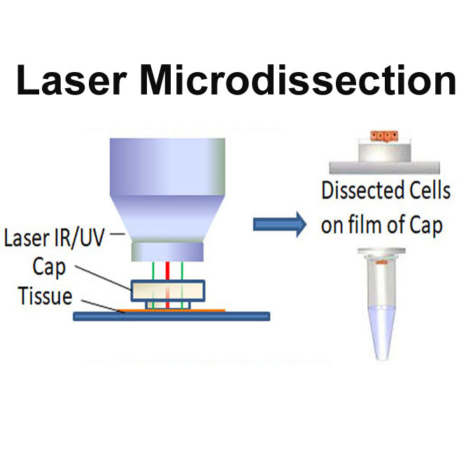
(+info)Microdissection is a surgical technique that involves the use of a microscope to allow for precise, minimalistic dissection of tissue. It is often used in research and clinical settings to isolate specific cells, tissues or structures while minimizing damage to surrounding areas. This technique can be performed using various methods such as laser capture microdissection (LCM) or manual microdissection with microsurgical tools. The size and scale of the dissection required will determine the specific method used. In general, microdissection allows for the examination and analysis of very small and delicate structures that would otherwise be difficult to access and study.
Laser capture microdissection (LCM) is a specialized technique used in pathology and molecular biology to isolate specific cells or cell types from heterogeneous tissue sections for further analysis. This method employs a laser beam to precisely cut and capture the cells of interest, which are then collected for downstream applications such as genetic or protein analysis.
The process typically involves the following steps:
1. Tissue preparation: The tissue sample is embedded in a supporting matrix, like a polymer or wax, and cut into thin sections using a microtome. These sections are mounted on special slides designed for LCM.
2. Staining: To visualize the cells of interest, the tissue sections are stained with various dyes or immunohistochemical markers that selectively bind to specific cell types or structures.
3. Laser microdissection: Under a microscope equipped with a laser system, the researcher identifies and outlines the cells or regions of interest. The laser beam is then focused and directed to cut along the outlined borders, separating the desired cells from the surrounding tissue.
4. Cell collection: A specialized cap containing an adhesive surface is positioned over the dissected cells, which are subsequently lifted and captured onto the cap when brought into contact with it.
5. Downstream analysis: The isolated cells can now be extracted for various downstream applications, such as genomic DNA analysis (e.g., PCR, sequencing), transcriptomic analysis (e.g., RNA sequencing, gene expression profiling), or proteomic analysis (e.g., mass spectrometry).
LCM enables the study of specific cell populations within complex tissues, providing valuable insights into their molecular characteristics and functions. This technique has broad applications in research areas such as cancer biology, neuroscience, developmental biology, and toxicology.
A laser is not a medical term per se, but a physical concept that has important applications in medicine. The term "LASER" stands for "Light Amplification by Stimulated Emission of Radiation." It refers to a device that produces and amplifies light with specific characteristics, such as monochromaticity (single wavelength), coherence (all waves moving in the same direction), and high intensity.
In medicine, lasers are used for various therapeutic and diagnostic purposes, including surgery, dermatology, ophthalmology, and dentistry. They can be used to cut, coagulate, or vaporize tissues with great precision, minimizing damage to surrounding structures. Additionally, lasers can be used to detect and measure physiological parameters, such as blood flow and oxygen saturation.
It's important to note that while lasers are powerful tools in medicine, they must be used by trained professionals to ensure safe and effective treatment.
In medical terms, dissection refers to the separation of the layers of a biological tissue or structure by cutting or splitting. It is often used to describe the process of surgically cutting through tissues, such as during an operation to separate organs or examine their internal structures.
However, "dissection" can also refer to a pathological condition in which there is a separation of the layers of a blood vessel wall by blood, creating a false lumen or aneurysm. This type of dissection is most commonly seen in the aorta and can be life-threatening if not promptly diagnosed and treated.
In summary, "dissection" has both surgical and pathological meanings related to the separation of tissue layers, and it's essential to consider the context in which the term is used.
Gene expression profiling is a laboratory technique used to measure the activity (expression) of thousands of genes at once. This technique allows researchers and clinicians to identify which genes are turned on or off in a particular cell, tissue, or organism under specific conditions, such as during health, disease, development, or in response to various treatments.
The process typically involves isolating RNA from the cells or tissues of interest, converting it into complementary DNA (cDNA), and then using microarray or high-throughput sequencing technologies to determine which genes are expressed and at what levels. The resulting data can be used to identify patterns of gene expression that are associated with specific biological states or processes, providing valuable insights into the underlying molecular mechanisms of diseases and potential targets for therapeutic intervention.
In recent years, gene expression profiling has become an essential tool in various fields, including cancer research, drug discovery, and personalized medicine, where it is used to identify biomarkers of disease, predict patient outcomes, and guide treatment decisions.
Cryoultramicrotomy is a specialized microscopy technique used in the field of pathology and biology. It involves cutting extremely thin sections (typically less than 100 nanometers thick) of biological samples that have been frozen and hardened at very low temperatures, often using liquid nitrogen or helium.
The process begins by embedding the sample in a suitable medium, such as a cryoprotectant or a low-temperature wax, to prevent ice crystal formation during freezing. The embedded sample is then mounted on a specimen holder and cooled to a temperature below its glass transition point, typically around -150°C to -196°C.
Once the sample is frozen and hardened, it is cut using an ultramicrotome, a precision instrument that uses a diamond knife to slice the sample into thin sections. These sections are then collected on a grid or other support and can be stained with various dyes or stains to enhance contrast and visualization under an electron microscope.
Cryoultramicrotomy is particularly useful for studying the ultrastructure of biological samples, such as cells, tissues, and organelles, that may be sensitive to heat or chemical fixation methods commonly used in traditional histology techniques. It allows researchers to visualize details at the molecular level, providing valuable insights into cellular processes and disease mechanisms.
Oligonucleotide Array Sequence Analysis is a type of microarray analysis that allows for the simultaneous measurement of the expression levels of thousands of genes in a single sample. In this technique, oligonucleotides (short DNA sequences) are attached to a solid support, such as a glass slide, in a specific pattern. These oligonucleotides are designed to be complementary to specific target mRNA sequences from the sample being analyzed.
During the analysis, labeled RNA or cDNA from the sample is hybridized to the oligonucleotide array. The level of hybridization is then measured and used to determine the relative abundance of each target sequence in the sample. This information can be used to identify differences in gene expression between samples, which can help researchers understand the underlying biological processes involved in various diseases or developmental stages.
It's important to note that this technique requires specialized equipment and bioinformatics tools for data analysis, as well as careful experimental design and validation to ensure accurate and reproducible results.
Reverse Transcriptase Polymerase Chain Reaction (RT-PCR) is a laboratory technique used in molecular biology to amplify and detect specific DNA sequences. This technique is particularly useful for the detection and quantification of RNA viruses, as well as for the analysis of gene expression.
The process involves two main steps: reverse transcription and polymerase chain reaction (PCR). In the first step, reverse transcriptase enzyme is used to convert RNA into complementary DNA (cDNA) by reading the template provided by the RNA molecule. This cDNA then serves as a template for the PCR amplification step.
In the second step, the PCR reaction uses two primers that flank the target DNA sequence and a thermostable polymerase enzyme to repeatedly copy the targeted cDNA sequence. The reaction mixture is heated and cooled in cycles, allowing the primers to anneal to the template, and the polymerase to extend the new strand. This results in exponential amplification of the target DNA sequence, making it possible to detect even small amounts of RNA or cDNA.
RT-PCR is a sensitive and specific technique that has many applications in medical research and diagnostics, including the detection of viruses such as HIV, hepatitis C virus, and SARS-CoV-2 (the virus that causes COVID-19). It can also be used to study gene expression, identify genetic mutations, and diagnose genetic disorders.
Histocytoлогиcal preparation techniques are methods used to prepare tissue samples for examination under a microscope in order to study the structure and function of cells, specifically histiocytes. These techniques involve fixing, processing, embedding, sectioning, and staining the tissue samples to preserve their cellular details and enhance the visibility of various cellular components.
The process typically begins with fixing the tissue sample in a fixative solution, such as formalin or alcohol, to preserve its structure and prevent decomposition. The fixed tissue is then dehydrated using a series of increasing concentrations of ethanol and cleared with a clearing agent, such as xylene, to remove the ethanol and make the tissue more transparent.
Next, the tissue is infiltrated with a liquid embedding material, such as paraffin or plastic, and solidified into a block. The block is then cut into thin sections using a microtome, and the sections are mounted onto glass slides.
Finally, the sections are stained with various dyes to highlight different cellular components, such as the nucleus, cytoplasm, or specific organelles. Common staining techniques used in histocytoлогиcal preparation include hematoxylin and eosin (H&E), immunohistochemistry (IHC), and special stains for specific cell types or structures.
These techniques allow pathologists to examine the tissue sample at a microscopic level, identify any abnormalities or diseases, and make an accurate diagnosis.
Messenger RNA (mRNA) is a type of RNA (ribonucleic acid) that carries genetic information copied from DNA in the form of a series of three-base code "words," each of which specifies a particular amino acid. This information is used by the cell's machinery to construct proteins, a process known as translation. After being transcribed from DNA, mRNA travels out of the nucleus to the ribosomes in the cytoplasm where protein synthesis occurs. Once the protein has been synthesized, the mRNA may be degraded and recycled. Post-transcriptional modifications can also occur to mRNA, such as alternative splicing and addition of a 5' cap and a poly(A) tail, which can affect its stability, localization, and translation efficiency.
Micromanipulation is a term used in the field of medicine, specifically in assisted reproductive technologies (ARTs) such as in vitro fertilization (IVF). It refers to a technique that involves the manipulation of oocytes (human eggs), sperm, and/or embryos under a microscope using micromanipulative tools and equipment.
The most common form of micromanipulation is intracytoplasmic sperm injection (ICSI), where a single sperm is selected and injected directly into the cytoplasm of an oocyte to facilitate fertilization. Other forms of micromanipulation include assisted hatching (AH), where a small opening is made in the zona pellucida (the protective layer surrounding the embryo) to help the embryo hatch and implant into the uterus, and embryo biopsy, which involves removing one or more cells from an embryo for genetic testing.
Micromanipulation requires specialized training and equipment and is typically performed in IVF laboratories by experienced embryologists. The goal of micromanipulation is to improve the chances of successful fertilization, implantation, and pregnancy, particularly in cases where conventional methods have been unsuccessful or when there are specific fertility issues, such as male factor infertility or genetic disorders.
I'm sorry for any confusion, but "Pyronine" is not a medical term. It is a type of basic dye that is often used in histology (the study of the microscopic structure of tissues) and cytology (the study of individual cells). Pyronin Y, a specific type of pyronine dye, is sometimes used to stain acidic components within cells, such as DNA and RNA. However, it is not a term that is typically used in clinical medicine to describe diseases or conditions.
Immunohistochemistry (IHC) is a technique used in pathology and laboratory medicine to identify specific proteins or antigens in tissue sections. It combines the principles of immunology and histology to detect the presence and location of these target molecules within cells and tissues. This technique utilizes antibodies that are specific to the protein or antigen of interest, which are then tagged with a detection system such as a chromogen or fluorophore. The stained tissue sections can be examined under a microscope, allowing for the visualization and analysis of the distribution and expression patterns of the target molecule in the context of the tissue architecture. Immunohistochemistry is widely used in diagnostic pathology to help identify various diseases, including cancer, infectious diseases, and immune-mediated disorders.
Hematoxylin is not a medical term per se, but it is widely used in the field of histology and pathology, which are subspecialties within medicine. Hematoxylin is a natural dye that is commonly used in histological staining procedures to highlight cell nuclei in tissue samples. It is often combined with eosin, another dye, to create the well-known hematoxylin and eosin (H&E) stain, which is routinely used to examine tissue architecture and diagnose various medical conditions.
In essence, hematoxylin is a histological stain that selectively binds to the acidic components of nuclear chromatin, imparting a blue-purple color to the cell nuclei when visualized under a microscope. This staining technique helps pathologists and researchers identify and analyze various cellular structures and abnormalities within tissue samples.
Tissue fixation is a process in histology (the study of the microscopic structure of tissues) where fixed tissue samples are prepared for further examination, typically through microscopy. The goal of tissue fixation is to preserve the original three-dimensional structure and biochemical composition of tissues and cells as much as possible, making them stable and suitable for various analyses.
The most common method for tissue fixation involves immersing the sample in a chemical fixative, such as formaldehyde or glutaraldehyde. These fixatives cross-link proteins within the tissue, creating a stable matrix that maintains the original structure and prevents decay. Other methods of tissue fixation may include freezing or embedding samples in various media to preserve their integrity.
Properly fixed tissue samples can be sectioned, stained, and examined under a microscope, allowing pathologists and researchers to study cellular structures, diagnose diseases, and understand biological processes at the molecular level.
Paraffin embedding is a process in histology (the study of the microscopic structure of tissues) where tissue samples are impregnated with paraffin wax to create a solid, stable block. This allows for thin, uniform sections of the tissue to be cut and mounted on slides for further examination under a microscope.
The process involves fixing the tissue sample with a chemical fixative to preserve its structure, dehydrating it through a series of increasing concentrations of alcohol, clearing it in a solvent such as xylene to remove the alcohol, and then impregnating it with melted paraffin wax. The tissue is then cooled and hardened into a block, which can be stored, transported, and sectioned as needed.
Paraffin embedding is a commonly used technique in histology due to its relative simplicity, low cost, and ability to produce high-quality sections for microscopic examination.
RNA (Ribonucleic Acid) is a single-stranded, linear polymer of ribonucleotides. It is a nucleic acid present in the cells of all living organisms and some viruses. RNAs play crucial roles in various biological processes such as protein synthesis, gene regulation, and cellular signaling. There are several types of RNA including messenger RNA (mRNA), ribosomal RNA (rRNA), transfer RNA (tRNA), small nuclear RNA (snRNA), microRNA (miRNA), and long non-coding RNA (lncRNA). These RNAs differ in their structure, function, and location within the cell.
Polymerase Chain Reaction (PCR) is a laboratory technique used to amplify specific regions of DNA. It enables the production of thousands to millions of copies of a particular DNA sequence in a rapid and efficient manner, making it an essential tool in various fields such as molecular biology, medical diagnostics, forensic science, and research.
The PCR process involves repeated cycles of heating and cooling to separate the DNA strands, allow primers (short sequences of single-stranded DNA) to attach to the target regions, and extend these primers using an enzyme called Taq polymerase, resulting in the exponential amplification of the desired DNA segment.
In a medical context, PCR is often used for detecting and quantifying specific pathogens (viruses, bacteria, fungi, or parasites) in clinical samples, identifying genetic mutations or polymorphisms associated with diseases, monitoring disease progression, and evaluating treatment effectiveness.
Neuromuscular junction diseases are a group of disorders that affect the functioning of the neuromuscular junction, which is the site where nerve impulses are transmitted to muscles. These diseases are characterized by muscle weakness and fatigue, and can be caused by various factors such as autoimmune disorders, genetic mutations, or toxins.
Examples of neuromuscular junction diseases include myasthenia gravis, Lambert-Eaton myasthenic syndrome (LEMS), congenital myasthenic syndromes (CMS), and botulism. Myasthenia gravis is an autoimmune disorder that causes the immune system to attack the receptors in the neuromuscular junction, leading to muscle weakness and fatigue. LEMS is a rare autoimmune disorder that affects the nerve endings at the neuromuscular junction, causing muscle weakness and decreased reflexes.
Congenital myasthenic syndromes are genetic disorders that affect the functioning of the neuromuscular junction from birth, leading to muscle weakness and fatigue. Botulism is a rare but serious condition caused by the ingestion of botulinum toxin, which can lead to paralysis of the muscles due to interference with nerve impulse transmission at the neuromuscular junction.
Treatment for neuromuscular junction diseases may include medications such as cholinesterase inhibitors, immunosuppressive drugs, or plasma exchange therapy, depending on the specific diagnosis and severity of the condition.
Nucleic acid amplification techniques (NAATs) are medical laboratory methods used to increase the number of copies of a specific DNA or RNA sequence. These techniques are widely used in molecular biology and diagnostics, including the detection and diagnosis of infectious diseases, genetic disorders, and cancer.
The most commonly used NAAT is the polymerase chain reaction (PCR), which involves repeated cycles of heating and cooling to separate and replicate DNA strands. Other NAATs include loop-mediated isothermal amplification (LAMP), nucleic acid sequence-based amplification (NASBA), and transcription-mediated amplification (TMA).
NAATs offer several advantages over traditional culture methods for detecting pathogens, including faster turnaround times, increased sensitivity and specificity, and the ability to detect viable but non-culturable organisms. However, they also require specialized equipment and trained personnel, and there is a risk of contamination and false positive results if proper precautions are not taken.
Proteomics is the large-scale study and analysis of proteins, including their structures, functions, interactions, modifications, and abundance, in a given cell, tissue, or organism. It involves the identification and quantification of all expressed proteins in a biological sample, as well as the characterization of post-translational modifications, protein-protein interactions, and functional pathways. Proteomics can provide valuable insights into various biological processes, diseases, and drug responses, and has applications in basic research, biomedicine, and clinical diagnostics. The field combines various techniques from molecular biology, chemistry, physics, and bioinformatics to study proteins at a systems level.
Neoplastic gene expression regulation refers to the processes that control the production of proteins and other molecules from genes in neoplastic cells, or cells that are part of a tumor or cancer. In a normal cell, gene expression is tightly regulated to ensure that the right genes are turned on or off at the right time. However, in cancer cells, this regulation can be disrupted, leading to the overexpression or underexpression of certain genes.
Neoplastic gene expression regulation can be affected by a variety of factors, including genetic mutations, epigenetic changes, and signals from the tumor microenvironment. These changes can lead to the activation of oncogenes (genes that promote cancer growth and development) or the inactivation of tumor suppressor genes (genes that prevent cancer).
Understanding neoplastic gene expression regulation is important for developing new therapies for cancer, as targeting specific genes or pathways involved in this process can help to inhibit cancer growth and progression.
Loss of Heterozygosity (LOH) is a term used in genetics to describe the loss of one copy of a gene or a segment of a chromosome, where there was previously a pair of different genes or chromosomal segments (heterozygous). This can occur due to various genetic events such as mutation, deletion, or mitotic recombination.
LOH is often associated with the development of cancer, as it can lead to the loss of tumor suppressor genes, which normally help to regulate cell growth and division. When both copies of a tumor suppressor gene are lost or inactivated, it can result in uncontrolled cell growth and the formation of a tumor.
In medical terms, LOH is used as a biomarker for cancer susceptibility, progression, and prognosis. It can also be used to identify individuals who may be at increased risk for certain types of cancer, or to monitor patients for signs of cancer recurrence.
Epithelial cells are types of cells that cover the outer surfaces of the body, line the inner surfaces of organs and glands, and form the lining of blood vessels and body cavities. They provide a protective barrier against the external environment, regulate the movement of materials between the internal and external environments, and are involved in the sense of touch, temperature, and pain. Epithelial cells can be squamous (flat and thin), cuboidal (square-shaped and of equal height), or columnar (tall and narrow) in shape and are classified based on their location and function.
Gene expression is the process by which the information encoded in a gene is used to synthesize a functional gene product, such as a protein or RNA molecule. This process involves several steps: transcription, RNA processing, and translation. During transcription, the genetic information in DNA is copied into a complementary RNA molecule, known as messenger RNA (mRNA). The mRNA then undergoes RNA processing, which includes adding a cap and tail to the mRNA and splicing out non-coding regions called introns. The resulting mature mRNA is then translated into a protein on ribosomes in the cytoplasm through the process of translation.
The regulation of gene expression is a complex and highly controlled process that allows cells to respond to changes in their environment, such as growth factors, hormones, and stress signals. This regulation can occur at various stages of gene expression, including transcriptional activation or repression, RNA processing, mRNA stability, and translation. Dysregulation of gene expression has been implicated in many diseases, including cancer, genetic disorders, and neurological conditions.
'Staining and labeling' are techniques commonly used in pathology, histology, cytology, and molecular biology to highlight or identify specific components or structures within tissues, cells, or molecules. These methods enable researchers and medical professionals to visualize and analyze the distribution, localization, and interaction of biological entities, contributing to a better understanding of diseases, cellular processes, and potential therapeutic targets.
Medical definitions for 'staining' and 'labeling' are as follows:
1. Staining: A process that involves applying dyes or stains to tissues, cells, or molecules to enhance their contrast and reveal specific structures or components. Stains can be categorized into basic stains (which highlight acidic structures) and acidic stains (which highlight basic structures). Common staining techniques include Hematoxylin and Eosin (H&E), which differentiates cell nuclei from the surrounding cytoplasm and extracellular matrix; special stains, such as PAS (Periodic Acid-Schiff) for carbohydrates or Masson's trichrome for collagen fibers; and immunostains, which use antibodies to target specific proteins.
2. Labeling: A process that involves attaching a detectable marker or tag to a molecule of interest, allowing its identification, quantification, or tracking within a biological system. Labels can be direct, where the marker is directly conjugated to the targeting molecule, or indirect, where an intermediate linker molecule is used to attach the label to the target. Common labeling techniques include fluorescent labels (such as FITC, TRITC, or Alexa Fluor), enzymatic labels (such as horseradish peroxidase or alkaline phosphatase), and radioactive labels (such as ³²P or ¹⁴C). Labeling is often used in conjunction with staining techniques to enhance the specificity and sensitivity of detection.
Together, staining and labeling provide valuable tools for medical research, diagnostics, and therapeutic development, offering insights into cellular and molecular processes that underlie health and disease.
Laser therapy, also known as phototherapy or laser photobiomodulation, is a medical treatment that uses low-intensity lasers or light-emitting diodes (LEDs) to stimulate healing, reduce pain, and decrease inflammation. It works by promoting the increase of cellular metabolism, blood flow, and tissue regeneration through the process of photobiomodulation.
The therapy can be used on patients suffering from a variety of acute and chronic conditions, including musculoskeletal injuries, arthritis, neuropathic pain, and wound healing complications. The wavelength and intensity of the laser light are precisely controlled to ensure a safe and effective treatment.
During the procedure, the laser or LED device is placed directly on the skin over the area of injury or discomfort. The non-ionizing light penetrates the tissue without causing heat or damage, interacting with chromophores in the cells to initiate a series of photochemical reactions. This results in increased ATP production, modulation of reactive oxygen species, and activation of transcription factors that lead to improved cellular function and reduced pain.
In summary, laser therapy is a non-invasive, drug-free treatment option for various medical conditions, providing patients with an alternative or complementary approach to traditional therapies.
RNA (Ribonucleic acid) is a single-stranded molecule similar in structure to DNA, involved in the process of protein synthesis in the cell. It acts as a messenger carrying genetic information from DNA to the ribosomes, where proteins are produced.
A neoplasm, on the other hand, is an abnormal growth of cells, which can be benign or malignant. Benign neoplasms are not cancerous and do not invade nearby tissues or spread to other parts of the body. Malignant neoplasms, however, are cancerous and have the potential to invade surrounding tissues and spread to distant sites in the body through a process called metastasis.
Therefore, an 'RNA neoplasm' is not a recognized medical term as RNA is not a type of growth or tumor. However, there are certain types of cancer-causing viruses known as oncoviruses that contain RNA as their genetic material and can cause neoplasms. For example, human T-cell leukemia virus (HTLV-1) and hepatitis C virus (HCV) are RNA viruses that can cause certain types of cancer in humans.
Reproducibility of results in a medical context refers to the ability to obtain consistent and comparable findings when a particular experiment or study is repeated, either by the same researcher or by different researchers, following the same experimental protocol. It is an essential principle in scientific research that helps to ensure the validity and reliability of research findings.
In medical research, reproducibility of results is crucial for establishing the effectiveness and safety of new treatments, interventions, or diagnostic tools. It involves conducting well-designed studies with adequate sample sizes, appropriate statistical analyses, and transparent reporting of methods and findings to allow other researchers to replicate the study and confirm or refute the results.
The lack of reproducibility in medical research has become a significant concern in recent years, as several high-profile studies have failed to produce consistent findings when replicated by other researchers. This has led to increased scrutiny of research practices and a call for greater transparency, rigor, and standardization in the conduct and reporting of medical research.
In situ hybridization (ISH) is a molecular biology technique used to detect and localize specific nucleic acid sequences, such as DNA or RNA, within cells or tissues. This technique involves the use of a labeled probe that is complementary to the target nucleic acid sequence. The probe can be labeled with various types of markers, including radioisotopes, fluorescent dyes, or enzymes.
During the ISH procedure, the labeled probe is hybridized to the target nucleic acid sequence in situ, meaning that the hybridization occurs within the intact cells or tissues. After washing away unbound probe, the location of the labeled probe can be visualized using various methods depending on the type of label used.
In situ hybridization has a wide range of applications in both research and diagnostic settings, including the detection of gene expression patterns, identification of viral infections, and diagnosis of genetic disorders.
'Gene expression regulation' refers to the processes that control whether, when, and where a particular gene is expressed, meaning the production of a specific protein or functional RNA encoded by that gene. This complex mechanism can be influenced by various factors such as transcription factors, chromatin remodeling, DNA methylation, non-coding RNAs, and post-transcriptional modifications, among others. Proper regulation of gene expression is crucial for normal cellular function, development, and maintaining homeostasis in living organisms. Dysregulation of gene expression can lead to various diseases, including cancer and genetic disorders.
Stromal cells, also known as stromal/stroma cells, are a type of cell found in various tissues and organs throughout the body. They are often referred to as the "connective tissue" or "supporting framework" of an organ because they play a crucial role in maintaining the structure and function of the tissue. Stromal cells include fibroblasts, adipocytes (fat cells), and various types of progenitor/stem cells. They produce and maintain the extracellular matrix, which is the non-cellular component of tissues that provides structural support and biochemical cues for other cells. Stromal cells also interact with immune cells and participate in the regulation of the immune response. In some contexts, "stromal cells" can also refer to cells found in the microenvironment of tumors, which can influence cancer growth and progression.
The proteome is the entire set of proteins produced or present in an organism, system, organ, or cell at a certain time under specific conditions. It is a dynamic collection of protein species that changes over time, responding to various internal and external stimuli such as disease, stress, or environmental factors. The study of the proteome, known as proteomics, involves the identification and quantification of these protein components and their post-translational modifications, providing valuable insights into biological processes, functional pathways, and disease mechanisms.
Cluster analysis is a statistical method used to group similar objects or data points together based on their characteristics or features. In medical and healthcare research, cluster analysis can be used to identify patterns or relationships within complex datasets, such as patient records or genetic information. This technique can help researchers to classify patients into distinct subgroups based on their symptoms, diagnoses, or other variables, which can inform more personalized treatment plans or public health interventions.
Cluster analysis involves several steps, including:
1. Data preparation: The researcher must first collect and clean the data, ensuring that it is complete and free from errors. This may involve removing outlier values or missing data points.
2. Distance measurement: Next, the researcher must determine how to measure the distance between each pair of data points. Common methods include Euclidean distance (the straight-line distance between two points) or Manhattan distance (the distance between two points along a grid).
3. Clustering algorithm: The researcher then applies a clustering algorithm, which groups similar data points together based on their distances from one another. Common algorithms include hierarchical clustering (which creates a tree-like structure of clusters) or k-means clustering (which assigns each data point to the nearest centroid).
4. Validation: Finally, the researcher must validate the results of the cluster analysis by evaluating the stability and robustness of the clusters. This may involve re-running the analysis with different distance measures or clustering algorithms, or comparing the results to external criteria.
Cluster analysis is a powerful tool for identifying patterns and relationships within complex datasets, but it requires careful consideration of the data preparation, distance measurement, and validation steps to ensure accurate and meaningful results.
Protein array analysis is a high-throughput technology used to detect and measure the presence and activity of specific proteins in biological samples. This technique utilizes arrays or chips containing various capture agents, such as antibodies or aptamers, that are designed to bind to specific target proteins. The sample is then added to the array, allowing the target proteins to bind to their corresponding capture agents. After washing away unbound materials, a detection system is used to identify and quantify the bound proteins. This method can be used for various applications, including protein-protein interaction studies, biomarker discovery, and drug development. The results of protein array analysis provide valuable information about the expression levels, post-translational modifications, and functional states of proteins in complex biological systems.
Tumor markers are substances that can be found in the body and their presence can indicate the presence of certain types of cancer or other conditions. Biological tumor markers refer to those substances that are produced by cancer cells or by other cells in response to cancer or certain benign (non-cancerous) conditions. These markers can be found in various bodily fluids such as blood, urine, or tissue samples.
Examples of biological tumor markers include:
1. Proteins: Some tumor markers are proteins that are produced by cancer cells or by other cells in response to the presence of cancer. For example, prostate-specific antigen (PSA) is a protein produced by normal prostate cells and in higher amounts by prostate cancer cells.
2. Genetic material: Tumor markers can also include genetic material such as DNA, RNA, or microRNA that are shed by cancer cells into bodily fluids. For example, circulating tumor DNA (ctDNA) is genetic material from cancer cells that can be found in the bloodstream.
3. Metabolites: Tumor markers can also include metabolic products produced by cancer cells or by other cells in response to cancer. For example, lactate dehydrogenase (LDH) is an enzyme that is released into the bloodstream when cancer cells break down glucose for energy.
It's important to note that tumor markers are not specific to cancer and can be elevated in non-cancerous conditions as well. Therefore, they should not be used alone to diagnose cancer but rather as a tool in conjunction with other diagnostic tests and clinical evaluations.
Sprague-Dawley rats are a strain of albino laboratory rats that are widely used in scientific research. They were first developed by researchers H.H. Sprague and R.C. Dawley in the early 20th century, and have since become one of the most commonly used rat strains in biomedical research due to their relatively large size, ease of handling, and consistent genetic background.
Sprague-Dawley rats are outbred, which means that they are genetically diverse and do not suffer from the same limitations as inbred strains, which can have reduced fertility and increased susceptibility to certain diseases. They are also characterized by their docile nature and low levels of aggression, making them easier to handle and study than some other rat strains.
These rats are used in a wide variety of research areas, including toxicology, pharmacology, nutrition, cancer, and behavioral studies. Because they are genetically diverse, Sprague-Dawley rats can be used to model a range of human diseases and conditions, making them an important tool in the development of new drugs and therapies.
Prostatic neoplasms refer to abnormal growths in the prostate gland, which can be benign or malignant. The term "neoplasm" simply means new or abnormal tissue growth. When it comes to the prostate, neoplasms are often referred to as tumors.
Benign prostatic neoplasms, such as prostate adenomas, are non-cancerous overgrowths of prostate tissue. They usually grow slowly and do not spread to other parts of the body. While they can cause uncomfortable symptoms like difficulty urinating, they are generally not life-threatening.
Malignant prostatic neoplasms, on the other hand, are cancerous growths. The most common type of prostate cancer is adenocarcinoma, which arises from the glandular cells in the prostate. Prostate cancer often grows slowly and may not cause any symptoms for many years. However, some types of prostate cancer can be aggressive and spread quickly to other parts of the body, such as the bones or lymph nodes.
It's important to note that while prostate neoplasms can be concerning, early detection and treatment can significantly improve outcomes for many men. Regular check-ups with a healthcare provider are key to monitoring prostate health and catching any potential issues early on.
Up-regulation is a term used in molecular biology and medicine to describe an increase in the expression or activity of a gene, protein, or receptor in response to a stimulus. This can occur through various mechanisms such as increased transcription, translation, or reduced degradation of the molecule. Up-regulation can have important functional consequences, for example, enhancing the sensitivity or response of a cell to a hormone, neurotransmitter, or drug. It is a normal physiological process that can also be induced by disease or pharmacological interventions.
DNA primers are short single-stranded DNA molecules that serve as a starting point for DNA synthesis. They are typically used in laboratory techniques such as the polymerase chain reaction (PCR) and DNA sequencing. The primer binds to a complementary sequence on the DNA template through base pairing, providing a free 3'-hydroxyl group for the DNA polymerase enzyme to add nucleotides and synthesize a new strand of DNA. This allows for specific and targeted amplification or analysis of a particular region of interest within a larger DNA molecule.
A biopsy is a medical procedure in which a small sample of tissue is taken from the body to be examined under a microscope for the presence of disease. This can help doctors diagnose and monitor various medical conditions, such as cancer, infections, or autoimmune disorders. The type of biopsy performed will depend on the location and nature of the suspected condition. Some common types of biopsies include:
1. Incisional biopsy: In this procedure, a surgeon removes a piece of tissue from an abnormal area using a scalpel or other surgical instrument. This type of biopsy is often used when the lesion is too large to be removed entirely during the initial biopsy.
2. Excisional biopsy: An excisional biopsy involves removing the entire abnormal area, along with a margin of healthy tissue surrounding it. This technique is typically employed for smaller lesions or when cancer is suspected.
3. Needle biopsy: A needle biopsy uses a thin, hollow needle to extract cells or fluid from the body. There are two main types of needle biopsies: fine-needle aspiration (FNA) and core needle biopsy. FNA extracts loose cells, while a core needle biopsy removes a small piece of tissue.
4. Punch biopsy: In a punch biopsy, a round, sharp tool is used to remove a small cylindrical sample of skin tissue. This type of biopsy is often used for evaluating rashes or other skin abnormalities.
5. Shave biopsy: During a shave biopsy, a thin slice of tissue is removed from the surface of the skin using a sharp razor-like instrument. This technique is typically used for superficial lesions or growths on the skin.
After the biopsy sample has been collected, it is sent to a laboratory where a pathologist will examine the tissue under a microscope and provide a diagnosis based on their findings. The results of the biopsy can help guide further treatment decisions and determine the best course of action for managing the patient's condition.
Adenocarcinoma is a type of cancer that arises from glandular epithelial cells. These cells line the inside of many internal organs, including the breasts, prostate, colon, and lungs. Adenocarcinomas can occur in any of these organs, as well as in other locations where glands are present.
The term "adenocarcinoma" is used to describe a cancer that has features of glandular tissue, such as mucus-secreting cells or cells that produce hormones. These cancers often form glandular structures within the tumor mass and may produce mucus or other substances.
Adenocarcinomas are typically slow-growing and tend to spread (metastasize) to other parts of the body through the lymphatic system or bloodstream. They can be treated with surgery, radiation therapy, chemotherapy, targeted therapy, or a combination of these treatments. The prognosis for adenocarcinoma depends on several factors, including the location and stage of the cancer, as well as the patient's overall health and age.
The term "DNA, neoplasm" is not a standard medical term or concept. DNA refers to deoxyribonucleic acid, which is the genetic material present in the cells of living organisms. A neoplasm, on the other hand, is a tumor or growth of abnormal tissue that can be benign (non-cancerous) or malignant (cancerous).
In some contexts, "DNA, neoplasm" may refer to genetic alterations found in cancer cells. These genetic changes can include mutations, amplifications, deletions, or rearrangements of DNA sequences that contribute to the development and progression of cancer. Identifying these genetic abnormalities can help doctors diagnose and treat certain types of cancer more effectively.
However, it's important to note that "DNA, neoplasm" is not a term that would typically be used in medical reports or research papers without further clarification. If you have any specific questions about DNA changes in cancer cells or neoplasms, I would recommend consulting with a healthcare professional or conducting further research on the topic.
Neoplasm invasiveness is a term used in pathology and oncology to describe the aggressive behavior of cancer cells as they invade surrounding tissues and organs. This process involves the loss of cell-to-cell adhesion, increased motility and migration, and the ability of cancer cells to degrade the extracellular matrix (ECM) through the production of enzymes such as matrix metalloproteinases (MMPs).
Invasive neoplasms are cancers that have spread beyond the original site where they first developed and have infiltrated adjacent tissues or structures. This is in contrast to non-invasive or in situ neoplasms, which are confined to the epithelial layer where they originated and have not yet invaded the underlying basement membrane.
The invasiveness of a neoplasm is an important prognostic factor in cancer diagnosis and treatment, as it can indicate the likelihood of metastasis and the potential effectiveness of various therapies. In general, more invasive cancers are associated with worse outcomes and require more aggressive treatment approaches.
Cell separation is a process used to separate and isolate specific cell types from a heterogeneous mixture of cells. This can be accomplished through various physical or biological methods, depending on the characteristics of the cells of interest. Some common techniques for cell separation include:
1. Density gradient centrifugation: In this method, a sample containing a mixture of cells is layered onto a density gradient medium and then centrifuged. The cells are separated based on their size, density, and sedimentation rate, with denser cells settling closer to the bottom of the tube and less dense cells remaining near the top.
2. Magnetic-activated cell sorting (MACS): This technique uses magnetic beads coated with antibodies that bind to specific cell surface markers. The labeled cells are then passed through a column placed in a magnetic field, which retains the magnetically labeled cells while allowing unlabeled cells to flow through.
3. Fluorescence-activated cell sorting (FACS): In this method, cells are stained with fluorochrome-conjugated antibodies that recognize specific cell surface or intracellular markers. The stained cells are then passed through a laser beam, which excites the fluorophores and allows for the detection and sorting of individual cells based on their fluorescence profile.
4. Filtration: This simple method relies on the physical size differences between cells to separate them. Cells can be passed through filters with pore sizes that allow smaller cells to pass through while retaining larger cells.
5. Enzymatic digestion: In some cases, cells can be separated by enzymatically dissociating tissues into single-cell suspensions and then using various separation techniques to isolate specific cell types.
These methods are widely used in research and clinical settings for applications such as isolating immune cells, stem cells, or tumor cells from biological samples.
A precancerous condition, also known as a premalignant condition, is a state of abnormal cellular growth and development that has a higher-than-normal potential to progress into cancer. These conditions are characterized by the presence of certain anomalies in the cells, such as dysplasia (abnormal changes in cell shape or size), which can indicate an increased risk for malignant transformation.
It is important to note that not all precancerous conditions will eventually develop into cancer, and some may even regress on their own. However, individuals with precancerous conditions are often at a higher risk of developing cancer compared to the general population. Regular monitoring and appropriate medical interventions, if necessary, can help manage this risk and potentially prevent or detect cancer at an early stage when it is more treatable.
Examples of precancerous conditions include:
1. Dysplasia in the cervix (cervical intraepithelial neoplasia or CIN)
2. Atypical ductal hyperplasia or lobular hyperplasia in the breast
3. Actinic keratosis on the skin
4. Leukoplakia in the mouth
5. Barrett's esophagus in the digestive tract
Regular medical check-ups, screenings, and lifestyle modifications are crucial for individuals with precancerous conditions to monitor their health and reduce the risk of cancer development.
Microarray analysis is a laboratory technique used to measure the expression levels of large numbers of genes (or other types of DNA sequences) simultaneously. This technology allows researchers to monitor the expression of thousands of genes in a single experiment, providing valuable information about which genes are turned on or off in response to various stimuli or diseases.
In microarray analysis, samples of RNA from cells or tissues are labeled with fluorescent dyes and then hybridized to a solid surface (such as a glass slide) onto which thousands of known DNA sequences have been spotted in an organized array. The intensity of the fluorescence at each spot on the array is proportional to the amount of RNA that has bound to it, indicating the level of expression of the corresponding gene.
Microarray analysis can be used for a variety of applications, including identifying genes that are differentially expressed between healthy and diseased tissues, studying genetic variations in populations, and monitoring gene expression changes over time or in response to environmental factors. However, it is important to note that microarray data must be analyzed carefully using appropriate statistical methods to ensure the accuracy and reliability of the results.
The prostate is a small gland that is part of the male reproductive system. Its main function is to produce a fluid that, together with sperm cells from the testicles and fluids from other glands, makes up semen. This fluid nourishes and protects the sperm, helping it to survive and facilitating its movement.
The prostate is located below the bladder and in front of the rectum. It surrounds part of the urethra, the tube that carries urine and semen out of the body. This means that prostate problems can affect urination and sexual function. The prostate gland is about the size of a walnut in adult men.
Prostate health is an important aspect of male health, particularly as men age. Common prostate issues include benign prostatic hyperplasia (BPH), which is an enlarged prostate not caused by cancer, and prostate cancer, which is one of the most common types of cancer in men. Regular check-ups with a healthcare provider can help to detect any potential problems early and improve outcomes.
Hyperplasia is a medical term that refers to an abnormal increase in the number of cells in an organ or tissue, leading to an enlargement of the affected area. It's a response to various stimuli such as hormones, chronic irritation, or inflammation. Hyperplasia can be physiological, like the growth of breast tissue during pregnancy, or pathological, like in the case of benign or malignant tumors. The process is generally reversible if the stimulus is removed. It's important to note that hyperplasia itself is not cancerous, but some forms of hyperplasia can increase the risk of developing cancer over time.
Confocal microscopy is a powerful imaging technique used in medical and biological research to obtain high-resolution, contrast-rich images of thick samples. This super-resolution technology provides detailed visualization of cellular structures and processes at various depths within a specimen.
In confocal microscopy, a laser beam focused through a pinhole illuminates a small spot within the sample. The emitted fluorescence or reflected light from this spot is then collected by a detector, passing through a second pinhole that ensures only light from the focal plane reaches the detector. This process eliminates out-of-focus light, resulting in sharp images with improved contrast compared to conventional widefield microscopy.
By scanning the laser beam across the sample in a raster pattern and collecting fluorescence at each point, confocal microscopy generates optical sections of the specimen. These sections can be combined to create three-dimensional reconstructions, allowing researchers to study cellular architecture and interactions within complex tissues.
Confocal microscopy has numerous applications in medical research, including studying protein localization, tracking intracellular dynamics, analyzing cell morphology, and investigating disease mechanisms at the cellular level. Additionally, it is widely used in clinical settings for diagnostic purposes, such as analyzing skin lesions or detecting pathogens in patient samples.
Disease progression is the worsening or advancement of a medical condition over time. It refers to the natural course of a disease, including its development, the severity of symptoms and complications, and the impact on the patient's overall health and quality of life. Understanding disease progression is important for developing appropriate treatment plans, monitoring response to therapy, and predicting outcomes.
The rate of disease progression can vary widely depending on the type of medical condition, individual patient factors, and the effectiveness of treatment. Some diseases may progress rapidly over a short period of time, while others may progress more slowly over many years. In some cases, disease progression may be slowed or even halted with appropriate medical interventions, while in other cases, the progression may be inevitable and irreversible.
In clinical practice, healthcare providers closely monitor disease progression through regular assessments, imaging studies, and laboratory tests. This information is used to guide treatment decisions and adjust care plans as needed to optimize patient outcomes and improve quality of life.
Developmental gene expression regulation refers to the processes that control the activation or repression of specific genes during embryonic and fetal development. These regulatory mechanisms ensure that genes are expressed at the right time, in the right cells, and at appropriate levels to guide proper growth, differentiation, and morphogenesis of an organism.
Developmental gene expression regulation is a complex and dynamic process involving various molecular players, such as transcription factors, chromatin modifiers, non-coding RNAs, and signaling molecules. These regulators can interact with cis-regulatory elements, like enhancers and promoters, to fine-tune the spatiotemporal patterns of gene expression during development.
Dysregulation of developmental gene expression can lead to various congenital disorders and developmental abnormalities. Therefore, understanding the principles and mechanisms governing developmental gene expression regulation is crucial for uncovering the etiology of developmental diseases and devising potential therapeutic strategies.
A base sequence in the context of molecular biology refers to the specific order of nucleotides in a DNA or RNA molecule. In DNA, these nucleotides are adenine (A), guanine (G), cytosine (C), and thymine (T). In RNA, uracil (U) takes the place of thymine. The base sequence contains genetic information that is transcribed into RNA and ultimately translated into proteins. It is the exact order of these bases that determines the genetic code and thus the function of the DNA or RNA molecule.
Sensitivity and specificity are statistical measures used to describe the performance of a diagnostic test or screening tool in identifying true positive and true negative results.
* Sensitivity refers to the proportion of people who have a particular condition (true positives) who are correctly identified by the test. It is also known as the "true positive rate" or "recall." A highly sensitive test will identify most or all of the people with the condition, but may also produce more false positives.
* Specificity refers to the proportion of people who do not have a particular condition (true negatives) who are correctly identified by the test. It is also known as the "true negative rate." A highly specific test will identify most or all of the people without the condition, but may also produce more false negatives.
In medical testing, both sensitivity and specificity are important considerations when evaluating a diagnostic test. High sensitivity is desirable for screening tests that aim to identify as many cases of a condition as possible, while high specificity is desirable for confirmatory tests that aim to rule out the condition in people who do not have it.
It's worth noting that sensitivity and specificity are often influenced by factors such as the prevalence of the condition in the population being tested, the threshold used to define a positive result, and the reliability and validity of the test itself. Therefore, it's important to consider these factors when interpreting the results of a diagnostic test.
Liquid chromatography (LC) is a type of chromatography technique used to separate, identify, and quantify the components in a mixture. In this method, the sample mixture is dissolved in a liquid solvent (the mobile phase) and then passed through a stationary phase, which can be a solid or a liquid that is held in place by a solid support.
The components of the mixture interact differently with the stationary phase and the mobile phase, causing them to separate as they move through the system. The separated components are then detected and measured using various detection techniques, such as ultraviolet (UV) absorbance or mass spectrometry.
Liquid chromatography is widely used in many areas of science and medicine, including drug development, environmental analysis, food safety testing, and clinical diagnostics. It can be used to separate and analyze a wide range of compounds, from small molecules like drugs and metabolites to large biomolecules like proteins and nucleic acids.
A neoplasm is a tumor or growth that is formed by an abnormal and excessive proliferation of cells, which can be benign or malignant. Neoplasm proteins are therefore any proteins that are expressed or produced in these neoplastic cells. These proteins can play various roles in the development, progression, and maintenance of neoplasms.
Some neoplasm proteins may contribute to the uncontrolled cell growth and division seen in cancer, such as oncogenic proteins that promote cell cycle progression or inhibit apoptosis (programmed cell death). Others may help the neoplastic cells evade the immune system, allowing them to proliferate undetected. Still others may be involved in angiogenesis, the formation of new blood vessels that supply the tumor with nutrients and oxygen.
Neoplasm proteins can also serve as biomarkers for cancer diagnosis, prognosis, or treatment response. For example, the presence or level of certain neoplasm proteins in biological samples such as blood or tissue may indicate the presence of a specific type of cancer, help predict the likelihood of cancer recurrence, or suggest whether a particular therapy will be effective.
Overall, understanding the roles and behaviors of neoplasm proteins can provide valuable insights into the biology of cancer and inform the development of new diagnostic and therapeutic strategies.
Matrix-Assisted Laser Desorption/Ionization Mass Spectrometry (MALDI-MS) is a type of mass spectrometry that is used to analyze large biomolecules such as proteins and peptides. In this technique, the sample is mixed with a matrix compound, which absorbs laser energy and helps to vaporize and ionize the analyte molecules.
The matrix-analyte mixture is then placed on a target plate and hit with a laser beam, causing the matrix and analyte molecules to desorb from the plate and become ionized. The ions are then accelerated through an electric field and into a mass analyzer, which separates them based on their mass-to-charge ratio.
The separated ions are then detected and recorded as a mass spectrum, which can be used to identify and quantify the analyte molecules present in the sample. MALDI-MS is particularly useful for the analysis of complex biological samples, such as tissue extracts or biological fluids, because it allows for the detection and identification of individual components within those mixtures.
Real-Time Polymerase Chain Reaction (RT-PCR) is a laboratory technique used in molecular biology to amplify and detect specific DNA sequences in real-time. It is a sensitive and specific method that allows for the quantification of target nucleic acids, such as DNA or RNA, through the use of fluorescent reporter molecules.
The RT-PCR process involves several steps: first, the template DNA is denatured to separate the double-stranded DNA into single strands. Then, primers (short sequences of DNA) specific to the target sequence are added and allowed to anneal to the template DNA. Next, a heat-stable enzyme called Taq polymerase adds nucleotides to the annealed primers, extending them along the template DNA until a new double-stranded DNA molecule is formed.
During each amplification cycle, fluorescent reporter molecules are added that bind specifically to the newly synthesized DNA. As more and more copies of the target sequence are generated, the amount of fluorescence increases in proportion to the number of copies present. This allows for real-time monitoring of the PCR reaction and quantification of the target nucleic acid.
RT-PCR is commonly used in medical diagnostics, research, and forensics to detect and quantify specific DNA or RNA sequences. It has been widely used in the diagnosis of infectious diseases, genetic disorders, and cancer, as well as in the identification of microbial pathogens and the detection of gene expression.
Microsatellite repeats, also known as short tandem repeats (STRs), are repetitive DNA sequences made up of units of 1-6 base pairs that are repeated in a head-to-tail manner. These repeats are spread throughout the human genome and are highly polymorphic, meaning they can have different numbers of repeat units in different individuals.
Microsatellites are useful as genetic markers because of their high degree of variability. They are commonly used in forensic science to identify individuals, in genealogy to trace ancestry, and in medical research to study genetic diseases and disorders. Mutations in microsatellite repeats have been associated with various neurological conditions, including Huntington's disease and fragile X syndrome.
C57BL/6 (C57 Black 6) is an inbred strain of laboratory mouse that is widely used in biomedical research. The term "inbred" refers to a strain of animals where matings have been carried out between siblings or other closely related individuals for many generations, resulting in a population that is highly homozygous at most genetic loci.
The C57BL/6 strain was established in 1920 by crossing a female mouse from the dilute brown (DBA) strain with a male mouse from the black strain. The resulting offspring were then interbred for many generations to create the inbred C57BL/6 strain.
C57BL/6 mice are known for their robust health, longevity, and ease of handling, making them a popular choice for researchers. They have been used in a wide range of biomedical research areas, including studies of cancer, immunology, neuroscience, cardiovascular disease, and metabolism.
One of the most notable features of the C57BL/6 strain is its sensitivity to certain genetic modifications, such as the introduction of mutations that lead to obesity or impaired glucose tolerance. This has made it a valuable tool for studying the genetic basis of complex diseases and traits.
Overall, the C57BL/6 inbred mouse strain is an important model organism in biomedical research, providing a valuable resource for understanding the genetic and molecular mechanisms underlying human health and disease.
A clone is a group of cells that are genetically identical to each other because they are derived from a common ancestor cell through processes such as mitosis or asexual reproduction. Therefore, the term "clone cells" refers to a population of cells that are genetic copies of a single parent cell.
In the context of laboratory research, cells can be cloned by isolating a single cell and allowing it to divide in culture, creating a population of genetically identical cells. This is useful for studying the behavior and characteristics of individual cell types, as well as for generating large quantities of cells for use in experiments.
It's important to note that while clone cells are genetically identical, they may still exhibit differences in their phenotype (physical traits) due to epigenetic factors or environmental influences.
A cell line that is derived from tumor cells and has been adapted to grow in culture. These cell lines are often used in research to study the characteristics of cancer cells, including their growth patterns, genetic changes, and responses to various treatments. They can be established from many different types of tumors, such as carcinomas, sarcomas, and leukemias. Once established, these cell lines can be grown and maintained indefinitely in the laboratory, allowing researchers to conduct experiments and studies that would not be feasible using primary tumor cells. It is important to note that tumor cell lines may not always accurately represent the behavior of the original tumor, as they can undergo genetic changes during their time in culture.
Solid-state lasers are a type of laser that uses solid materials as the gain medium – the material that amplifies the light energy to produce laser emissions. In contrast to gas or liquid lasers, solid-state lasers use a crystal, ceramic, or glass as the gain medium. The active laser medium in solid-state lasers is typically doped with rare earth ions, such as neodymium (Nd), yttrium (Y), erbium (Er), or thulium (Tm).
The most common type of solid-state laser is the neodymium-doped yttrium aluminum garnet (Nd:YAG) laser. In this laser, neodymium ions are doped into a crystal lattice made up of yttrium, aluminum, and garnet (YAG). The Nd:YAG laser emits light at a wavelength of 1064 nanometers (nm), which can be frequency-doubled to produce emissions at 532 nm.
Solid-state lasers have several advantages over other types of lasers, including high efficiency, long lifetimes, and compact size. They are widely used in various applications, such as material processing, medical treatments, scientific research, and military technology.
A mutation is a permanent change in the DNA sequence of an organism's genome. Mutations can occur spontaneously or be caused by environmental factors such as exposure to radiation, chemicals, or viruses. They may have various effects on the organism, ranging from benign to harmful, depending on where they occur and whether they alter the function of essential proteins. In some cases, mutations can increase an individual's susceptibility to certain diseases or disorders, while in others, they may confer a survival advantage. Mutations are the driving force behind evolution, as they introduce new genetic variability into populations, which can then be acted upon by natural selection.
Breast neoplasms refer to abnormal growths in the breast tissue that can be benign or malignant. Benign breast neoplasms are non-cancerous tumors or growths, while malignant breast neoplasms are cancerous tumors that can invade surrounding tissues and spread to other parts of the body.
Breast neoplasms can arise from different types of cells in the breast, including milk ducts, milk sacs (lobules), or connective tissue. The most common type of breast cancer is ductal carcinoma, which starts in the milk ducts and can spread to other parts of the breast and nearby structures.
Breast neoplasms are usually detected through screening methods such as mammography, ultrasound, or MRI, or through self-examination or clinical examination. Treatment options for breast neoplasms depend on several factors, including the type and stage of the tumor, the patient's age and overall health, and personal preferences. Treatment may include surgery, radiation therapy, chemotherapy, hormone therapy, or targeted therapy.
Animal disease models are specialized animals, typically rodents such as mice or rats, that have been genetically engineered or exposed to certain conditions to develop symptoms and physiological changes similar to those seen in human diseases. These models are used in medical research to study the pathophysiology of diseases, identify potential therapeutic targets, test drug efficacy and safety, and understand disease mechanisms.
The genetic modifications can include knockout or knock-in mutations, transgenic expression of specific genes, or RNA interference techniques. The animals may also be exposed to environmental factors such as chemicals, radiation, or infectious agents to induce the disease state.
Examples of animal disease models include:
1. Mouse models of cancer: Genetically engineered mice that develop various types of tumors, allowing researchers to study cancer initiation, progression, and metastasis.
2. Alzheimer's disease models: Transgenic mice expressing mutant human genes associated with Alzheimer's disease, which exhibit amyloid plaque formation and cognitive decline.
3. Diabetes models: Obese and diabetic mouse strains like the NOD (non-obese diabetic) or db/db mice, used to study the development of type 1 and type 2 diabetes, respectively.
4. Cardiovascular disease models: Atherosclerosis-prone mice, such as ApoE-deficient or LDLR-deficient mice, that develop plaque buildup in their arteries when fed a high-fat diet.
5. Inflammatory bowel disease models: Mice with genetic mutations affecting intestinal barrier function and immune response, such as IL-10 knockout or SAMP1/YitFc mice, which develop colitis.
Animal disease models are essential tools in preclinical research, but it is important to recognize their limitations. Differences between species can affect the translatability of results from animal studies to human patients. Therefore, researchers must carefully consider the choice of model and interpret findings cautiously when applying them to human diseases.
Molecular sequence data refers to the specific arrangement of molecules, most commonly nucleotides in DNA or RNA, or amino acids in proteins, that make up a biological macromolecule. This data is generated through laboratory techniques such as sequencing, and provides information about the exact order of the constituent molecules. This data is crucial in various fields of biology, including genetics, evolution, and molecular biology, allowing for comparisons between different organisms, identification of genetic variations, and studies of gene function and regulation.
Down-regulation is a process that occurs in response to various stimuli, where the number or sensitivity of cell surface receptors or the expression of specific genes is decreased. This process helps maintain homeostasis within cells and tissues by reducing the ability of cells to respond to certain signals or molecules.
In the context of cell surface receptors, down-regulation can occur through several mechanisms:
1. Receptor internalization: After binding to their ligands, receptors can be internalized into the cell through endocytosis. Once inside the cell, these receptors may be degraded or recycled back to the cell surface in smaller numbers.
2. Reduced receptor synthesis: Down-regulation can also occur at the transcriptional level, where the expression of genes encoding for specific receptors is decreased, leading to fewer receptors being produced.
3. Receptor desensitization: Prolonged exposure to a ligand can lead to a decrease in receptor sensitivity or affinity, making it more difficult for the cell to respond to the signal.
In the context of gene expression, down-regulation refers to the decreased transcription and/or stability of specific mRNAs, leading to reduced protein levels. This process can be induced by various factors, including microRNA (miRNA)-mediated regulation, histone modification, or DNA methylation.
Down-regulation is an essential mechanism in many physiological processes and can also contribute to the development of several diseases, such as cancer and neurodegenerative disorders.
Nerve tissue proteins are specialized proteins found in the nervous system that provide structural and functional support to nerve cells, also known as neurons. These proteins include:
1. Neurofilaments: These are type IV intermediate filaments that provide structural support to neurons and help maintain their shape and size. They are composed of three subunits - NFL (light), NFM (medium), and NFH (heavy).
2. Neuronal Cytoskeletal Proteins: These include tubulins, actins, and spectrins that provide structural support to the neuronal cytoskeleton and help maintain its integrity.
3. Neurotransmitter Receptors: These are specialized proteins located on the postsynaptic membrane of neurons that bind neurotransmitters released by presynaptic neurons, triggering a response in the target cell.
4. Ion Channels: These are transmembrane proteins that regulate the flow of ions across the neuronal membrane and play a crucial role in generating and transmitting electrical signals in neurons.
5. Signaling Proteins: These include enzymes, receptors, and adaptor proteins that mediate intracellular signaling pathways involved in neuronal development, differentiation, survival, and death.
6. Adhesion Proteins: These are cell surface proteins that mediate cell-cell and cell-matrix interactions, playing a crucial role in the formation and maintenance of neural circuits.
7. Extracellular Matrix Proteins: These include proteoglycans, laminins, and collagens that provide structural support to nerve tissue and regulate neuronal migration, differentiation, and survival.
DNA Sequence Analysis is the systematic determination of the order of nucleotides in a DNA molecule. It is a critical component of modern molecular biology, genetics, and genetic engineering. The process involves determining the exact order of the four nucleotide bases - adenine (A), guanine (G), cytosine (C), and thymine (T) - in a DNA molecule or fragment. This information is used in various applications such as identifying gene mutations, studying evolutionary relationships, developing molecular markers for breeding, and diagnosing genetic diseases.
The process of DNA Sequence Analysis typically involves several steps, including DNA extraction, PCR amplification (if necessary), purification, sequencing reaction, and electrophoresis. The resulting data is then analyzed using specialized software to determine the exact sequence of nucleotides.
In recent years, high-throughput DNA sequencing technologies have revolutionized the field of genomics, enabling the rapid and cost-effective sequencing of entire genomes. This has led to an explosion of genomic data and new insights into the genetic basis of many diseases and traits.
The brain is the central organ of the nervous system, responsible for receiving and processing sensory information, regulating vital functions, and controlling behavior, movement, and cognition. It is divided into several distinct regions, each with specific functions:
1. Cerebrum: The largest part of the brain, responsible for higher cognitive functions such as thinking, learning, memory, language, and perception. It is divided into two hemispheres, each controlling the opposite side of the body.
2. Cerebellum: Located at the back of the brain, it is responsible for coordinating muscle movements, maintaining balance, and fine-tuning motor skills.
3. Brainstem: Connects the cerebrum and cerebellum to the spinal cord, controlling vital functions such as breathing, heart rate, and blood pressure. It also serves as a relay center for sensory information and motor commands between the brain and the rest of the body.
4. Diencephalon: A region that includes the thalamus (a major sensory relay station) and hypothalamus (regulates hormones, temperature, hunger, thirst, and sleep).
5. Limbic system: A group of structures involved in emotional processing, memory formation, and motivation, including the hippocampus, amygdala, and cingulate gyrus.
The brain is composed of billions of interconnected neurons that communicate through electrical and chemical signals. It is protected by the skull and surrounded by three layers of membranes called meninges, as well as cerebrospinal fluid that provides cushioning and nutrients.
The intestinal mucosa is the innermost layer of the intestines, which comes into direct contact with digested food and microbes. It is a specialized epithelial tissue that plays crucial roles in nutrient absorption, barrier function, and immune defense. The intestinal mucosa is composed of several cell types, including absorptive enterocytes, mucus-secreting goblet cells, hormone-producing enteroendocrine cells, and immune cells such as lymphocytes and macrophages.
The surface of the intestinal mucosa is covered by a single layer of epithelial cells, which are joined together by tight junctions to form a protective barrier against harmful substances and microorganisms. This barrier also allows for the selective absorption of nutrients into the bloodstream. The intestinal mucosa also contains numerous lymphoid follicles, known as Peyer's patches, which are involved in immune surveillance and defense against pathogens.
In addition to its role in absorption and immunity, the intestinal mucosa is also capable of producing hormones that regulate digestion and metabolism. Dysfunction of the intestinal mucosa can lead to various gastrointestinal disorders, such as inflammatory bowel disease, celiac disease, and food allergies.
Tissue embedding is a process in histology (the study of the microscopic structure of tissues) where biological tissue samples are encased in a supporting medium, typically paraffin wax or plastic resins, to maintain their shape and structural integrity during sectioning. This allows for thin slices of the embedded tissue to be cut using a microtome, mounted on slides, and then stained for further examination under a microscope. The embedding process ensures that the tissue remains intact and does not tear or compress during sectioning, providing clear and consistent samples for analysis.
Squamous cell carcinoma is a type of skin cancer that begins in the squamous cells, which are flat, thin cells that form the outer layer of the skin (epidermis). It commonly occurs on sun-exposed areas such as the face, ears, lips, and backs of the hands. Squamous cell carcinoma can also develop in other areas of the body including the mouth, lungs, and cervix.
This type of cancer usually develops slowly and may appear as a rough or scaly patch of skin, a red, firm nodule, or a sore or ulcer that doesn't heal. While squamous cell carcinoma is not as aggressive as some other types of cancer, it can metastasize (spread) to other parts of the body if left untreated, making early detection and treatment important.
Risk factors for developing squamous cell carcinoma include prolonged exposure to ultraviolet (UV) radiation from the sun or tanning beds, fair skin, a history of sunburns, a weakened immune system, and older age. Prevention measures include protecting your skin from the sun by wearing protective clothing, using a broad-spectrum sunscreen with an SPF of at least 30, avoiding tanning beds, and getting regular skin examinations.
Neurons, also known as nerve cells or neurocytes, are specialized cells that constitute the basic unit of the nervous system. They are responsible for receiving, processing, and transmitting information and signals within the body. Neurons have three main parts: the dendrites, the cell body (soma), and the axon. The dendrites receive signals from other neurons or sensory receptors, while the axon transmits these signals to other neurons, muscles, or glands. The junction between two neurons is called a synapse, where neurotransmitters are released to transmit the signal across the gap (synaptic cleft) to the next neuron. Neurons vary in size, shape, and structure depending on their function and location within the nervous system.
In the field of medicine, "time factors" refer to the duration of symptoms or time elapsed since the onset of a medical condition, which can have significant implications for diagnosis and treatment. Understanding time factors is crucial in determining the progression of a disease, evaluating the effectiveness of treatments, and making critical decisions regarding patient care.
For example, in stroke management, "time is brain," meaning that rapid intervention within a specific time frame (usually within 4.5 hours) is essential to administering tissue plasminogen activator (tPA), a clot-busting drug that can minimize brain damage and improve patient outcomes. Similarly, in trauma care, the "golden hour" concept emphasizes the importance of providing definitive care within the first 60 minutes after injury to increase survival rates and reduce morbidity.
Time factors also play a role in monitoring the progression of chronic conditions like diabetes or heart disease, where regular follow-ups and assessments help determine appropriate treatment adjustments and prevent complications. In infectious diseases, time factors are crucial for initiating antibiotic therapy and identifying potential outbreaks to control their spread.
Overall, "time factors" encompass the significance of recognizing and acting promptly in various medical scenarios to optimize patient outcomes and provide effective care.
Laser coagulation, also known as laser photocoagulation, is a medical procedure that uses a laser to seal or destroy abnormal blood vessels or tissue. The laser produces a concentrated beam of light that can be precisely focused on the target area. When the laser energy is absorbed by the tissue, it causes the temperature to rise, which leads to coagulation (the formation of a clot) or destruction of the tissue.
In ophthalmology, laser coagulation is commonly used to treat conditions such as diabetic retinopathy, age-related macular degeneration, and retinal tears or holes. The procedure can help to seal leaking blood vessels, reduce fluid leakage, and prevent further vision loss. It is usually performed as an outpatient procedure and may be repeated if necessary.
In other medical specialties, laser coagulation may be used to control bleeding, destroy tumors, or remove unwanted tissue. The specific technique and parameters of the laser treatment will depend on the individual patient's needs and the condition being treated.
Proteins are complex, large molecules that play critical roles in the body's functions. They are made up of amino acids, which are organic compounds that are the building blocks of proteins. Proteins are required for the structure, function, and regulation of the body's tissues and organs. They are essential for the growth, repair, and maintenance of body tissues, and they play a crucial role in many biological processes, including metabolism, immune response, and cellular signaling. Proteins can be classified into different types based on their structure and function, such as enzymes, hormones, antibodies, and structural proteins. They are found in various foods, especially animal-derived products like meat, dairy, and eggs, as well as plant-based sources like beans, nuts, and grains.
The Fluorescent Antibody Technique (FAT) is a type of immunofluorescence assay used in laboratory medicine and pathology for the detection and localization of specific antigens or antibodies in tissues, cells, or microorganisms. In this technique, a fluorescein-labeled antibody is used to selectively bind to the target antigen or antibody, forming an immune complex. When excited by light of a specific wavelength, the fluorescein label emits light at a longer wavelength, typically visualized as green fluorescence under a fluorescence microscope.
The FAT is widely used in diagnostic microbiology for the identification and characterization of various bacteria, viruses, fungi, and parasites. It has also been applied in the diagnosis of autoimmune diseases and certain cancers by detecting specific antibodies or antigens in patient samples. The main advantage of FAT is its high sensitivity and specificity, allowing for accurate detection and differentiation of various pathogens and disease markers. However, it requires specialized equipment and trained personnel to perform and interpret the results.
Immunoenzyme techniques are a group of laboratory methods used in immunology and clinical chemistry that combine the specificity of antibody-antigen reactions with the sensitivity and amplification capabilities of enzyme reactions. These techniques are primarily used for the detection, quantitation, or identification of various analytes (such as proteins, hormones, drugs, viruses, or bacteria) in biological samples.
In immunoenzyme techniques, an enzyme is linked to an antibody or antigen, creating a conjugate. This conjugate then interacts with the target analyte in the sample, forming an immune complex. The presence and amount of this immune complex can be visualized or measured by detecting the enzymatic activity associated with it.
There are several types of immunoenzyme techniques, including:
1. Enzyme-linked Immunosorbent Assay (ELISA): A widely used method for detecting and quantifying various analytes in a sample. In ELISA, an enzyme is attached to either the capture antibody or the detection antibody. After the immune complex formation, a substrate is added that reacts with the enzyme, producing a colored product that can be measured spectrophotometrically.
2. Immunoblotting (Western blot): A method used for detecting specific proteins in a complex mixture, such as a protein extract from cells or tissues. In this technique, proteins are separated by gel electrophoresis and transferred to a membrane, where they are probed with an enzyme-conjugated antibody directed against the target protein.
3. Immunohistochemistry (IHC): A method used for detecting specific antigens in tissue sections or cells. In IHC, an enzyme-conjugated primary or secondary antibody is applied to the sample, and the presence of the antigen is visualized using a chromogenic substrate that produces a colored product at the site of the antigen-antibody interaction.
4. Immunofluorescence (IF): A method used for detecting specific antigens in cells or tissues by employing fluorophore-conjugated antibodies. The presence of the antigen is visualized using a fluorescence microscope.
5. Enzyme-linked immunosorbent assay (ELISA): A method used for detecting and quantifying specific antigens or antibodies in liquid samples, such as serum or culture supernatants. In ELISA, an enzyme-conjugated detection antibody is added after the immune complex formation, and a substrate is added that reacts with the enzyme to produce a colored product that can be measured spectrophotometrically.
These techniques are widely used in research and diagnostic laboratories for various applications, including protein characterization, disease diagnosis, and monitoring treatment responses.
Transgenic mice are genetically modified rodents that have incorporated foreign DNA (exogenous DNA) into their own genome. This is typically done through the use of recombinant DNA technology, where a specific gene or genetic sequence of interest is isolated and then introduced into the mouse embryo. The resulting transgenic mice can then express the protein encoded by the foreign gene, allowing researchers to study its function in a living organism.
The process of creating transgenic mice usually involves microinjecting the exogenous DNA into the pronucleus of a fertilized egg, which is then implanted into a surrogate mother. The offspring that result from this procedure are screened for the presence of the foreign DNA, and those that carry the desired genetic modification are used to establish a transgenic mouse line.
Transgenic mice have been widely used in biomedical research to model human diseases, study gene function, and test new therapies. They provide a valuable tool for understanding complex biological processes and developing new treatments for a variety of medical conditions.
Low-level laser therapy (LLLT), also known as cold laser or soft laser, is a form of phototherapy which uses low-intensity lasers or light-emitting diodes to treat various medical conditions. The laser beam is usually applied directly to the skin and penetrates up to several centimeters into the tissue without causing heat damage or pain.
The therapeutic effect of LLLT is believed to be due to the bio-stimulation of cellular processes, including increased ATP production, modulation of reactive oxygen species, and activation of signaling pathways that promote tissue repair and reduce inflammation. The wavelength and power density of the laser light are important factors in determining its biological effects.
LLLT has been used to treat a variety of conditions such as musculoskeletal pain, wound healing, skin rejuvenation, hair growth, and neurological disorders. However, its efficacy is still a subject of ongoing research and debate, with some studies reporting positive results while others showing no significant benefits compared to placebo.
It's important to note that LLLT should only be administered by trained healthcare professionals, as improper use can lead to eye damage or other adverse effects.
Genetic transcription is the process by which the information in a strand of DNA is used to create a complementary RNA molecule. This process is the first step in gene expression, where the genetic code in DNA is converted into a form that can be used to produce proteins or functional RNAs.
During transcription, an enzyme called RNA polymerase binds to the DNA template strand and reads the sequence of nucleotide bases. As it moves along the template, it adds complementary RNA nucleotides to the growing RNA chain, creating a single-stranded RNA molecule that is complementary to the DNA template strand. Once transcription is complete, the RNA molecule may undergo further processing before it can be translated into protein or perform its functional role in the cell.
Transcription can be either "constitutive" or "regulated." Constitutive transcription occurs at a relatively constant rate and produces essential proteins that are required for basic cellular functions. Regulated transcription, on the other hand, is subject to control by various intracellular and extracellular signals, allowing cells to respond to changing environmental conditions or developmental cues.
Western blotting is a laboratory technique used in molecular biology to detect and quantify specific proteins in a mixture of many different proteins. This technique is commonly used to confirm the expression of a protein of interest, determine its size, and investigate its post-translational modifications. The name "Western" blotting distinguishes this technique from Southern blotting (for DNA) and Northern blotting (for RNA).
The Western blotting procedure involves several steps:
1. Protein extraction: The sample containing the proteins of interest is first extracted, often by breaking open cells or tissues and using a buffer to extract the proteins.
2. Separation of proteins by electrophoresis: The extracted proteins are then separated based on their size by loading them onto a polyacrylamide gel and running an electric current through the gel (a process called sodium dodecyl sulfate-polyacrylamide gel electrophoresis or SDS-PAGE). This separates the proteins according to their molecular weight, with smaller proteins migrating faster than larger ones.
3. Transfer of proteins to a membrane: After separation, the proteins are transferred from the gel onto a nitrocellulose or polyvinylidene fluoride (PVDF) membrane using an electric current in a process called blotting. This creates a replica of the protein pattern on the gel but now immobilized on the membrane for further analysis.
4. Blocking: The membrane is then blocked with a blocking agent, such as non-fat dry milk or bovine serum albumin (BSA), to prevent non-specific binding of antibodies in subsequent steps.
5. Primary antibody incubation: A primary antibody that specifically recognizes the protein of interest is added and allowed to bind to its target protein on the membrane. This step may be performed at room temperature or 4°C overnight, depending on the antibody's properties.
6. Washing: The membrane is washed with a buffer to remove unbound primary antibodies.
7. Secondary antibody incubation: A secondary antibody that recognizes the primary antibody (often coupled to an enzyme or fluorophore) is added and allowed to bind to the primary antibody. This step may involve using a horseradish peroxidase (HRP)-conjugated or alkaline phosphatase (AP)-conjugated secondary antibody, depending on the detection method used later.
8. Washing: The membrane is washed again to remove unbound secondary antibodies.
9. Detection: A detection reagent is added to visualize the protein of interest by detecting the signal generated from the enzyme-conjugated or fluorophore-conjugated secondary antibody. This can be done using chemiluminescent, colorimetric, or fluorescent methods.
10. Analysis: The resulting image is analyzed to determine the presence and quantity of the protein of interest in the sample.
Western blotting is a powerful technique for identifying and quantifying specific proteins within complex mixtures. It can be used to study protein expression, post-translational modifications, protein-protein interactions, and more. However, it requires careful optimization and validation to ensure accurate and reproducible results.
Sperm retrieval is a medical procedure that involves obtaining sperm from a male patient, usually for the purpose of assisted reproduction. This can be indicated in cases where the man has obstructive or non-obstructive azoospermia (absence of sperm in the semen), ejaculatory dysfunction, or other conditions that prevent the successful collection of sperm through conventional means, such as masturbation.
There are several methods for sperm retrieval, including:
1. Testicular sperm aspiration (TESA): A procedure where a fine needle is inserted into the testicle to aspirate (or draw out) sperm.
2. Percutaneous epididymal sperm aspiration (PESA): Similar to TESA, but the needle is inserted into the epididymis, a small structure that stores and transports sperm from the testicle.
3. Microsurgical epididymal sperm aspiration (MESA): A more invasive procedure where an incision is made in the scrotum to directly visualize the epididymis with a surgical microscope, allowing for the careful removal of sperm.
4. Testicular sperm extraction (TESE): Involves making a small incision in the testicle and removing a piece of tissue containing sperm-producing tubules. The tissue is then processed to extract viable sperm.
5. Microdissection testicular sperm extraction (microTESE): A refined version of TESE, where a surgical microscope is used to identify and isolate individual seminiferous tubules containing sperm in men with non-obstructive azoospermia.
The retrieved sperm can then be used for various assisted reproductive techniques, such as intracytoplasmic sperm injection (ICSI), where a single sperm is injected directly into an egg to facilitate fertilization.
 Laser capture microdissection - Wikipedia
 Fluidigm AccuLift Laser Capture Microdissection System | GenomeWeb
 laser capture microdissection | Virology Blog
 Laser Capture Microdissection Market Worth, Overview, Research Industry Size, Analysis - Technavio
 Olfactory Neurons Obtained through Nasal Biopsy Combined with Laser-Capture Microdissection: A Potential Approach to Study...
 Laser capture microdissection assessment of virus compartmentalization in the central nervous systems of macaques infected with...
 Serval - Laser capture microdissection is a valuable tool in the preoperative molecular screening of follicular lesions of the...
 Single Cell Laser Capture Microdissection Reveals the Hepatitis C Viral Landscape in the Human Liver
 laser capture microdissection | Leaders in Pharmaceutical Business Intelligence (LPBI) Group
 Molecular Vision: Dopachrome tautomerase is a retinoblastoma-specific gene, and its proximal promoter is preferentially active...
 An improved procedure for isolation of high-quality RNA from nematode-infected Arabidopsis roots through laser capture...
 Spatially-Resolved Proteomics: Rapid Quantitative Analysis of Laser Capture Microdissected Alveolar Tissue Samples
 Leeman A, et al. Defining hrHPV genotypes in cervical intraepithelial neoplasia by laser capture microdissection supports...
 PhD studentship: Investigating metastatic migration patterns in lung cancer patients | UCL Cancer Institute - UCL - University...
 The Right Sort | The Scientist Magazine®
 Breast Cancer and the Environment Study Glossary | City of Hope
 Inter Research » DAO » v133 » n1 » p39-46
 Esr1+ cells in the ventromedial hypothalamus control female aggression | Nature Neuroscience
 Experimental Pathology Laboratory Services Core » BUMC
 Spatial Genomics & Transcriptomics Market Report, 2030
 Semiautomated Monolayer Preparation of Bronchial Secretions Using AutoCyte PREP | Acta Cytologica | Karger Publishers
 Molecular Diagnostics Market - Industry Analysis, Size, Share,
 John FREW | Senior Lecturer | MBBS(Hons) MMed MS PhD FACD | UNSW Sydney, Kensington | UNSW | School of clinical medicine |...
 Purging Protein Profiling Problems