Kupffer Cells
Liver
Gadolinium
Clodronic Acid
Hepatocytes
Endothelium
Endotoxins
Lipopolysaccharides
Tumor Necrosis Factor-alpha
Poly I
Phagocytosis
Liver Diseases, Alcoholic
Rats, Inbred Strains
Drug-Induced Liver Injury
Cells, Cultured
Rats, Sprague-Dawley
Rats, Wistar
Mononuclear Phagocyte System
Macrophages
Peroxisome Proliferators
Zymosan
Mice, Inbred C57BL
Antigens, CD14
Latex
Alanine Transaminase
Receptors, Scavenger
Liver Cirrhosis, Experimental
Reperfusion Injury
Receptors, Lipoprotein
Pronase destroys the lipopolysaccharide receptor CD14 on Kupffer cells. (1/1142)
CD14 is a lipopolysaccharide (LPS) receptor distributed largely in macrophages, monocytes, and neutrophils; however, the role of CD14 in activation of Kupffer cells by LPS remains controversial. The purpose of this study was to determine if different methods used to isolate Kupffer cells affect CD14. Kupffer cells were isolated by collagenase (0.025%) or collagenase-Pronase (0.02%) perfusion and differential centrifugation using Percoll gradients and cultured for 24 h before experiments. CD14 mRNA was detected by RT-PCR from Kupffer cell total RNA as well as from peritoneal macrophages. Western blotting showed that Kupffer cells prepared with collagenase possess CD14; however, it was absent in cells obtained by collagenase-Pronase perfusion. Intracellular calcium in Kupffer cells prepared with collagenase was increased transiently to levels around 300 nM by addition of LPS with 5% rat serum, which contains LPS binding protein. This increase in intracellular calcium was totally serum dependent. Moreover, LPS-induced increases in intracellular calcium in Kupffer cells were blunted significantly (40% of controls) when cells were treated with phosphatidylinositol-specific phospholipase C, which cleaves CD14 from the plasma membrane. However, intracellular calcium did not increase when LPS was added to cells prepared by collagenase-Pronase perfusion even in the presence of serum. These cells were viable, however, because ATP increased intracellular calcium to the same levels as cells prepared with collagenase perfusion. Tumor necrosis factor-alpha (TNF-alpha) mRNA was increased in Kupffer cells prepared with collagenase perfusion 1 h after addition of LPS, an effect potentiated over twofold by serum; however, serum did not increase TNF-alpha mRNA in cells isolated via collagenase-Pronase perfusion. Moreover, treatment with Pronase rapidly decreased CD14 on mouse macrophages (RAW 264.7 cells) and Kupffer cells. These findings indicate that Pronase cleaves CD14 from Kupffer cells, whereas collagenase perfusion does not, providing an explanation for why Kupffer cells do not exhibit a CD14-mediated pathway when prepared with procedures using Pronase. It is concluded that Kupffer cells indeed contain a functional CD14 LPS receptor when prepared gently. (+info)Effects of Ro 31-8220 on lipopolysaccharides-induced hepatotoxicity and release of tumor necrosis factor from rat Kupffer cells. (2/1142)
AIM: To investigate protein kinase C (PKC) functions on lipopolysaccharide (LPS)-induced hepatotoxicity, a new potent PKC inhibitor Ro 31-8220 (Ro) was used to detect its effect on LPS-induced hepatotoxicity in rat hepatocytes and tumor necrosis factor (TNF) release from rat Kupffer cells (KC). METHODS: Hepatocytes (containing KC) were incubated with LPS (10 mg.L-1) and Ro (0.1-10 mumol.L-1) for 24 h, alanine aminotransferase (AlaA) leakage in the culture as indication of hepatotoxicity. The TNF activity in the supernatant of rat KC culture with LPS in the presence of Ro (0.1-10 mumol.L-1) was monitored by the L929 target cell lytic assay. RESULTS: Ro (0.1-10 mumol.L-1) reduced AlaA leakage in the hepatocyte culture. Ro inhibited dose-dependently the LPS-induced TNF production from rat KC. CONCLUSION: PKC inhibitor Ro protects the hepatocytes from LPS-induced cytotoxicity and inhibits the LPS-induced TNF production from rat KC. (+info)Influences of Kupffer cell stimulation and suppression on immunological liver injury in mice. (3/1142)
AIM: To study the possible involvement of Kupffer cells (KC) in immunological liver injury in mice. METHODS: Liver injury was induced by i.v. injection of Bacillus Calmette-Guerin (BCG) 5 x 10(7) viable bacilli followed by i.v. injection of lipopolysaccharides (LPS) 7.5 micrograms to each mouse. Indian ink and silica were i.v. injected to suppress KC and retinol was given po to stimulate KC in these mice. Plasma alanine aminotransferase (AlaAT), aspatate aminotransferase (AspAT), nitric oxide (NO), and liver tissue were examined. RESULTS: Injection of LPS following BCG injection resulted in a remarkable elevation of plasma NO, AlaAT, and AspAT levels, and severe liver damage. The damages were enhanced by the activation of KC with retinol and reduced by suppression of KC with silica and Indian ink. CONCLUSION: The degree of liver injury induced by BCG + LPS is closely correlated with the status of KC, and NO from KC plays an important role in the pathogenesis of the liver damage in mice. (+info)Febrile-range temperature modifies early systemic tumor necrosis factor alpha expression in mice challenged with bacterial endotoxin. (4/1142)
Fever improves survival in acute infections, but the effects of increased core temperature on host defenses are poorly understood. Tumor necrosis factor alpha (TNF-alpha) is an early activator of host defenses and a major endogenous pyrogen. TNF-alpha expression is essential for survival in bacterial infections but, if disregulated, can cause tissue injury. In this study, we show that passively increasing core temperature in mice from the basal (36.5 to 37.5 degrees C) to the febrile (39.5 to 40 degrees C) range modifies systemic TNF-alpha expression in response to bacterial endotoxin (lipopolysaccharide). The early TNF-alpha secretion rate is enhanced, but the duration of maximal TNF-alpha production is shortened. We identified Kupffer cells as the predominant source of the excess TNF-alpha production in the warmer animals. The enhanced early TNF-alpha production observed at the higher temperature in vivo could not be demonstrated in isolated Kupffer cells or in precision-cut liver slices in vitro, indicating the participation of indirect pathways. Therefore, expression of the endogenous pyrogen TNF-alpha is regulated by increments in core temperature during fever, generating an enhanced early, self-limited TNF-alpha pulse. (+info)Intravenous glycine improves survival in rat liver transplantation. (5/1142)
In situ manipulation by touching, retracting, and moving liver lobes gently during harvest dramatically reduces survival after transplantation (P. Schemmer, R. Schoonhoven, J. A. Swenberg, H. Bunzendahl, and R. G. Thurman. Transplantation 65: 1015-1020, 1998). The development of harvest-dependent graft injury upon reperfusion can be prevented with GdCl3, a rare earth metal and Kupffer cell toxicant, but it cannot be used in clinical liver transplantation because of its potential toxicity. Thus the effect of glycine, which prevents activation of Kupffer cells, was assessed here. Minimal dissection of the liver for 12 min plus 13 min without manipulation had no effect on survival (100%). However, gentle manipulation decreased survival to 46% in the control group. Furthermore, serum transaminases and liver necrosis were elevated 4- to 12-fold 8 h after transplantation. After organ harvest, the rate of entry and exit of fluorescein dextran, a dye confined to the vascular space, was decreased about twofold, indicating disturbances in the hepatic microcirculation. Pimonidazole binding, which detects hypoxia, increased about twofold after organ manipulation, and Kupffer cells isolated from manipulated livers produced threefold more tumor necrosis factor-alpha after lipopolysaccharide than controls. Glycine given intravenously to the donor increased the serum glycine concentration about sevenfold and largely prevented the effect of gentle organ manipulation on all parameters studied. These data indicate for the first time that pretreatment of donors with intravenous glycine minimizes reperfusion injury due to organ manipulation during harvest and after liver transplantation. (+info)A comparison of the pharmacological properties of carbohydrate remodeled recombinant and placental-derived beta-glucocerebrosidase: implications for clinical efficacy in treatment of Gaucher disease. (6/1142)
The objective of these studies was to characterize the macrophage mannose receptor binding and pharmacological properties of carbohydrate remodeled human placental-derived and recombinant beta-glucocerebrosidase (pGCR and rGCR, respectively). These are similar but not identical molecules that were developed as enzyme replacement therapies for Gaucher disease. Both undergo oligosaccharide remodeling during purification to expose terminal mannose sugar residues. Competitive binding data indicated carbohydrate remodeling improved targeting to mannose receptors over native enzyme by two orders of magnitude. Mannose receptor dissociation constants (Kd) for pGCR and rGCR were each 13 nmol/L. At 37 degrees C, 95% of the total macrophage binding was mannose receptor specific. In vivo, pGCR and rGCR were cleared from circulation by a saturable pathway. The serum half-life (t1/2) was 3 minutes when less than saturable amounts were injected intravenously (IV) into mice. Twenty minutes postdose, beta-glucocerebrosidase activity increased over endogenous levels in all tissues examined. Fifty percent of the injected activity was recovered. Ninety-five percent of recovered activity was in the liver. Parenchymal cells (PC), Kupffer cells (KC), and liver endothelium cells (LEC) were responsible for 75%, 22%, and 3%, respectively, of the hepatocellular uptake of rGCR and for 76%, 11%, and 12%, respectively, of the hepatocellular uptake of pGCR. Both molecules had poor stability in LEC and relatively long terminal half-lives in PC (t1/2 = 2 days) and KC (t1/2 = 3 days). (+info)Infection of primary cultures of human Kupffer cells by Dengue virus: no viral progeny synthesis, but cytokine production is evident. (7/1142)
We investigated the ability of dengue virus to invade human primary Kupffer cells and to complete its life cycle. The virus effectively penetrated Kupffer cells, but the infection did not result in any viral progeny. Dengue virus-replicating Kupffer cells underwent apoptosis and were cleared by phagocytosis. Infected Kupffer cells produced soluble mediators that could intervene in dengue virus pathogenesis. (+info)Prevention of Kupffer cell-induced oxidant injury in rat liver by atrial natriuretic peptide. (8/1142)
The generation of reactive oxygen species (ROS) by activated Kupffer cells contributes to liver injury following liver preservation, shock, or endotoxemia. Pharmacological interventions to protect liver cells against this inflammatory response of Kupffer cells have not yet been established. Atrial natriuretic peptide (ANP) protects the liver against ischemia-reperfusion injury, suggesting a possible modulation of Kupffer cell-mediated cytotoxicity. Therefore, we investigated the mechanism of cytoprotection by ANP during Kupffer cell activation in perfused rat livers of male Sprague-Dawley rats. Activation of Kupffer cells by zymosan (150 microgram/ml) resulted in considerable cell damage, as assessed by the sinusoidal release of lactate dehydrogenase and purine nucleoside phosphorylase. Cell damage was almost completely prevented by superoxide dismutase (50 U/ml) and catalase (150 U/ml), indicating ROS-related liver injury. ANP (200 nM) reduced Kupffer cell-induced injury via the guanylyl cyclase-coupled A receptor (GCA receptor) and cGMP: mRNA expression of the GCA receptor was found in hepatocytes, endothelial cells, and Kupffer cells, and the cGMP analog 8-bromo-cGMP (8-BrcGMP; 50 microM) was as potent as ANP in protecting from zymosan-induced cell damage. ANP and 8-BrcGMP significantly attenuated the prolonged increase of hepatic vascular resistance when Kupffer cell activation occurred. Furthermore, both compounds reduced oxidative cell damage following infusion of H2O2 (500 microM). In contrast, superoxide anion formation of isolated Kupffer cells was not affected by ANP and only moderately reduced by 8-BrcGMP. In conclusion, ANP protects the liver against Kupffer cell-related oxidant stress. This hormonal protection is mediated via the GCA receptor and cGMP, suggesting that the cGMP receptor plays a critical role in controlling oxidative cell damage. Thus ANP signaling should be considered as a new pharmacological target for protecting liver cells against the inflammatory response of activated Kupffer cells without eliminating the vital host defense function of these cells. (+info)Kupffer cells are specialized macrophages that reside in the liver, particularly in the sinusoids of the liver's blood circulation system. They play a crucial role in the immune system by engulfing and destroying bacteria, microorganisms, and other particles that enter the liver via the portal vein. Kupffer cells also contribute to the clearance of damaged red blood cells, iron metabolism, and the regulation of inflammation in the liver. They are named after the German pathologist Karl Wilhelm von Kupffer who first described them in 1876.
The liver is a large, solid organ located in the upper right portion of the abdomen, beneath the diaphragm and above the stomach. It plays a vital role in several bodily functions, including:
1. Metabolism: The liver helps to metabolize carbohydrates, fats, and proteins from the food we eat into energy and nutrients that our bodies can use.
2. Detoxification: The liver detoxifies harmful substances in the body by breaking them down into less toxic forms or excreting them through bile.
3. Synthesis: The liver synthesizes important proteins, such as albumin and clotting factors, that are necessary for proper bodily function.
4. Storage: The liver stores glucose, vitamins, and minerals that can be released when the body needs them.
5. Bile production: The liver produces bile, a digestive juice that helps to break down fats in the small intestine.
6. Immune function: The liver plays a role in the immune system by filtering out bacteria and other harmful substances from the blood.
Overall, the liver is an essential organ that plays a critical role in maintaining overall health and well-being.
Gadolinium is a rare earth metal that is used as a contrast agent in medical imaging techniques such as Magnetic Resonance Imaging (MRI) and Magnetic Resonance Angiography (MRA). It works by shortening the relaxation time of protons in tissues, which enhances the visibility of internal body structures on the images. Gadolinium-based contrast agents are injected into the patient's bloodstream during the imaging procedure.
It is important to note that in some individuals, gadolinium-based contrast agents can cause a condition called nephrogenic systemic fibrosis (NSF), which is a rare but serious disorder that affects people with severe kidney disease. NSF causes thickening and hardening of the skin, joints, eyes, and internal organs. Therefore, it is essential to evaluate a patient's renal function before administering gadolinium-based contrast agents.
Clodronic acid is a medication that belongs to a class of drugs called bisphosphonates. It is used to treat and prevent osteoporosis in postmenopausal women and men with a high risk of fracture, as well as to treat Paget's disease of bone.
Clodronic acid works by inhibiting the activity of bone-resorbing cells called osteoclasts, which helps to slow down bone loss and increase bone density. This can help reduce the risk of fractures in people with osteoporosis.
The medication is available in several forms, including tablets and intravenous solutions. It is usually taken or administered once a day or once a week, depending on the specific formulation and the individual patient's needs.
Like all medications, clodronic acid can have side effects, including gastrointestinal symptoms such as nausea, vomiting, and diarrhea, as well as muscle pain, joint pain, and headaches. In rare cases, it can also cause more serious side effects such as esophageal ulcers and bone necrosis of the jaw. It is important for patients to follow their doctor's instructions carefully when taking this medication and to report any unusual symptoms or side effects promptly.
Hepatocytes are the predominant type of cells in the liver, accounting for about 80% of its cytoplasmic mass. They play a key role in protein synthesis, protein storage, transformation of carbohydrates, synthesis of cholesterol, bile salts and phospholipids, detoxification, modification, and excretion of exogenous and endogenous substances, initiation of formation and secretion of bile, and enzyme production. Hepatocytes are essential for the maintenance of homeostasis in the body.
The endothelium is the thin, delicate tissue that lines the interior surface of blood vessels and lymphatic vessels. It is a single layer of cells called endothelial cells that are in contact with the blood or lymph fluid. The endothelium plays an essential role in maintaining vascular homeostasis by regulating blood flow, coagulation, platelet activation, immune function, and angiogenesis (the formation of new blood vessels). It also acts as a barrier between the vessel wall and the circulating blood or lymph fluid. Dysfunction of the endothelium has been implicated in various cardiovascular diseases, diabetes, inflammation, and cancer.
Endotoxins are toxic substances that are associated with the cell walls of certain types of bacteria. They are released when the bacterial cells die or divide, and can cause a variety of harmful effects in humans and animals. Endotoxins are made up of lipopolysaccharides (LPS), which are complex molecules consisting of a lipid and a polysaccharide component.
Endotoxins are particularly associated with gram-negative bacteria, which have a distinctive cell wall structure that includes an outer membrane containing LPS. These toxins can cause fever, inflammation, and other symptoms when they enter the bloodstream or other tissues of the body. They are also known to play a role in the development of sepsis, a potentially life-threatening condition characterized by a severe immune response to infection.
Endotoxins are resistant to heat, acid, and many disinfectants, making them difficult to eliminate from contaminated environments. They can also be found in a variety of settings, including hospitals, industrial facilities, and agricultural operations, where they can pose a risk to human health.
Lipopolysaccharides (LPS) are large molecules found in the outer membrane of Gram-negative bacteria. They consist of a hydrophilic polysaccharide called the O-antigen, a core oligosaccharide, and a lipid portion known as Lipid A. The Lipid A component is responsible for the endotoxic activity of LPS, which can trigger a powerful immune response in animals, including humans. This response can lead to symptoms such as fever, inflammation, and septic shock, especially when large amounts of LPS are introduced into the bloodstream.
Tumor Necrosis Factor-alpha (TNF-α) is a cytokine, a type of small signaling protein involved in immune response and inflammation. It is primarily produced by activated macrophages, although other cell types such as T-cells, natural killer cells, and mast cells can also produce it.
TNF-α plays a crucial role in the body's defense against infection and tissue injury by mediating inflammatory responses, activating immune cells, and inducing apoptosis (programmed cell death) in certain types of cells. It does this by binding to its receptors, TNFR1 and TNFR2, which are found on the surface of many cell types.
In addition to its role in the immune response, TNF-α has been implicated in the pathogenesis of several diseases, including autoimmune disorders such as rheumatoid arthritis, inflammatory bowel disease, and psoriasis, as well as cancer, where it can promote tumor growth and metastasis.
Therapeutic agents that target TNF-α, such as infliximab, adalimumab, and etanercept, have been developed to treat these conditions. However, these drugs can also increase the risk of infections and other side effects, so their use must be carefully monitored.
I'm sorry for any confusion, but "Poly I" is not a recognized medical term or abbreviation in the context of human diseases or conditions. It's possible that there might be a typo or misunderstanding in your query. If you intended to ask about a specific medical condition, medication, or concept, please provide the full and correct term so I can give you an accurate and helpful response.
Phagocytosis is the process by which certain cells in the body, known as phagocytes, engulf and destroy foreign particles, bacteria, or dead cells. This mechanism plays a crucial role in the immune system's response to infection and inflammation. Phagocytes, such as neutrophils, monocytes, and macrophages, have receptors on their surface that recognize and bind to specific molecules (known as antigens) on the target particles or microorganisms.
Once attached, the phagocyte extends pseudopodia (cell extensions) around the particle, forming a vesicle called a phagosome that completely encloses it. The phagosome then fuses with a lysosome, an intracellular organelle containing digestive enzymes and other chemicals. This fusion results in the formation of a phagolysosome, where the engulfed particle is broken down by the action of these enzymes, neutralizing its harmful effects and allowing for the removal of cellular debris or pathogens.
Phagocytosis not only serves as a crucial defense mechanism against infections but also contributes to tissue homeostasis by removing dead cells and debris.
Hepatitis is a medical condition characterized by inflammation of the liver, often resulting in damage to liver cells. It can be caused by various factors, including viral infections (such as Hepatitis A, B, C, D, and E), alcohol abuse, toxins, medications, and autoimmune disorders. Symptoms may include jaundice, fatigue, abdominal pain, loss of appetite, nausea, vomiting, and dark urine. The severity of the disease can range from mild illness to severe, life-threatening conditions, such as liver failure or cirrhosis.
Alcoholic liver disease (ALD) is a term that encompasses a spectrum of liver disorders caused by excessive alcohol consumption. The three main stages of ALD are:
1. Fatty Liver: This is the earliest stage of ALD, characterized by the accumulation of fat droplets within liver cells (hepatocytes). It's often reversible with abstinence from alcohol.
2. Alcoholic Hepatitis: This is a more severe form of ALD, characterized by inflammation and damage to the liver cells. It can range from mild to severe, and severe cases can lead to liver failure. Symptoms may include jaundice, abdominal pain, and fever.
3. Cirrhosis: This is the most advanced stage of ALD, characterized by widespread scarring (fibrosis) and nodular transformation of the liver. It's irreversible and can lead to complications such as liver failure, portal hypertension, and increased risk of liver cancer.
The development and progression of ALD are influenced by various factors, including the amount and duration of alcohol consumption, genetic predisposition, nutritional status, and co-existing viral hepatitis or other liver diseases. Abstaining from alcohol is the most effective way to prevent and manage ALD.
"Inbred strains of rats" are genetically identical rodents that have been produced through many generations of brother-sister mating. This results in a high degree of homozygosity, where the genes at any particular locus in the genome are identical in all members of the strain.
Inbred strains of rats are widely used in biomedical research because they provide a consistent and reproducible genetic background for studying various biological phenomena, including the effects of drugs, environmental factors, and genetic mutations on health and disease. Additionally, inbred strains can be used to create genetically modified models of human diseases by introducing specific mutations into their genomes.
Some commonly used inbred strains of rats include the Wistar Kyoto (WKY), Sprague-Dawley (SD), and Fischer 344 (F344) rat strains. Each strain has its own unique genetic characteristics, making them suitable for different types of research.
Drug-Induced Liver Injury (DILI) is a medical term that refers to liver damage or injury caused by the use of medications or drugs. This condition can vary in severity, from mild abnormalities in liver function tests to severe liver failure, which may require a liver transplant.
The exact mechanism of DILI can differ depending on the drug involved, but it generally occurs when the liver metabolizes the drug into toxic compounds that damage liver cells. This can happen through various pathways, including direct toxicity to liver cells, immune-mediated reactions, or metabolic idiosyncrasies.
Symptoms of DILI may include jaundice (yellowing of the skin and eyes), fatigue, abdominal pain, nausea, vomiting, loss of appetite, and dark urine. In severe cases, it can lead to complications such as ascites, encephalopathy, and bleeding disorders.
The diagnosis of DILI is often challenging because it requires the exclusion of other potential causes of liver injury. Liver function tests, imaging studies, and sometimes liver biopsies may be necessary to confirm the diagnosis. Treatment typically involves discontinuing the offending drug and providing supportive care until the liver recovers. In some cases, medications that protect the liver or promote its healing may be used.
"Cells, cultured" is a medical term that refers to cells that have been removed from an organism and grown in controlled laboratory conditions outside of the body. This process is called cell culture and it allows scientists to study cells in a more controlled and accessible environment than they would have inside the body. Cultured cells can be derived from a variety of sources, including tissues, organs, or fluids from humans, animals, or cell lines that have been previously established in the laboratory.
Cell culture involves several steps, including isolation of the cells from the tissue, purification and characterization of the cells, and maintenance of the cells in appropriate growth conditions. The cells are typically grown in specialized media that contain nutrients, growth factors, and other components necessary for their survival and proliferation. Cultured cells can be used for a variety of purposes, including basic research, drug development and testing, and production of biological products such as vaccines and gene therapies.
It is important to note that cultured cells may behave differently than they do in the body, and results obtained from cell culture studies may not always translate directly to human physiology or disease. Therefore, it is essential to validate findings from cell culture experiments using additional models and ultimately in clinical trials involving human subjects.
Liver diseases refer to a wide range of conditions that affect the normal functioning of the liver. The liver is a vital organ responsible for various critical functions such as detoxification, protein synthesis, and production of biochemicals necessary for digestion.
Liver diseases can be categorized into acute and chronic forms. Acute liver disease comes on rapidly and can be caused by factors like viral infections (hepatitis A, B, C, D, E), drug-induced liver injury, or exposure to toxic substances. Chronic liver disease develops slowly over time, often due to long-term exposure to harmful agents or inherent disorders of the liver.
Common examples of liver diseases include hepatitis, cirrhosis (scarring of the liver tissue), fatty liver disease, alcoholic liver disease, autoimmune liver diseases, genetic/hereditary liver disorders (like Wilson's disease and hemochromatosis), and liver cancers. Symptoms may vary widely depending on the type and stage of the disease but could include jaundice, abdominal pain, fatigue, loss of appetite, nausea, and weight loss.
Early diagnosis and treatment are essential to prevent progression and potential complications associated with liver diseases.
Sprague-Dawley rats are a strain of albino laboratory rats that are widely used in scientific research. They were first developed by researchers H.H. Sprague and R.C. Dawley in the early 20th century, and have since become one of the most commonly used rat strains in biomedical research due to their relatively large size, ease of handling, and consistent genetic background.
Sprague-Dawley rats are outbred, which means that they are genetically diverse and do not suffer from the same limitations as inbred strains, which can have reduced fertility and increased susceptibility to certain diseases. They are also characterized by their docile nature and low levels of aggression, making them easier to handle and study than some other rat strains.
These rats are used in a wide variety of research areas, including toxicology, pharmacology, nutrition, cancer, and behavioral studies. Because they are genetically diverse, Sprague-Dawley rats can be used to model a range of human diseases and conditions, making them an important tool in the development of new drugs and therapies.
"Wistar rats" are a strain of albino rats that are widely used in laboratory research. They were developed at the Wistar Institute in Philadelphia, USA, and were first introduced in 1906. Wistar rats are outbred, which means that they are genetically diverse and do not have a fixed set of genetic characteristics like inbred strains.
Wistar rats are commonly used as animal models in biomedical research because of their size, ease of handling, and relatively low cost. They are used in a wide range of research areas, including toxicology, pharmacology, nutrition, cancer, cardiovascular disease, and behavioral studies. Wistar rats are also used in safety testing of drugs, medical devices, and other products.
Wistar rats are typically larger than many other rat strains, with males weighing between 500-700 grams and females weighing between 250-350 grams. They have a lifespan of approximately 2-3 years. Wistar rats are also known for their docile and friendly nature, making them easy to handle and work with in the laboratory setting.
The Mononuclear Phagocyte System (MPS) is a network of specialized immune cells distributed throughout the body, primarily consisting of monocytes, macrophages, and dendritic cells. These cells share a common bone marrow-derived precursor and play crucial roles in innate and adaptive immunity. They are involved in various functions such as:
1. Phagocytosis: engulfing and destroying foreign particles, microbes, and cellular debris.
2. Antigen presentation: processing and presenting antigens to T-cells to initiate an adaptive immune response.
3. Cytokine production: releasing pro- and anti-inflammatory cytokines to regulate immune responses and maintain tissue homeostasis.
4. Immune regulation: modulating the activity of other immune cells, including T-cells, B-cells, and natural killer (NK) cells.
The MPS is essential for maintaining tissue integrity, fighting infections, and orchestrating immune responses. Its components are found in various tissues, including the liver (Kupffer cells), spleen, lymph nodes, bone marrow, and connective tissues.
Macrophages are a type of white blood cell that are an essential part of the immune system. They are large, specialized cells that engulf and destroy foreign substances, such as bacteria, viruses, parasites, and fungi, as well as damaged or dead cells. Macrophages are found throughout the body, including in the bloodstream, lymph nodes, spleen, liver, lungs, and connective tissues. They play a critical role in inflammation, immune response, and tissue repair and remodeling.
Macrophages originate from monocytes, which are a type of white blood cell produced in the bone marrow. When monocytes enter the tissues, they differentiate into macrophages, which have a larger size and more specialized functions than monocytes. Macrophages can change their shape and move through tissues to reach sites of infection or injury. They also produce cytokines, chemokines, and other signaling molecules that help coordinate the immune response and recruit other immune cells to the site of infection or injury.
Macrophages have a variety of surface receptors that allow them to recognize and respond to different types of foreign substances and signals from other cells. They can engulf and digest foreign particles, bacteria, and viruses through a process called phagocytosis. Macrophages also play a role in presenting antigens to T cells, which are another type of immune cell that helps coordinate the immune response.
Overall, macrophages are crucial for maintaining tissue homeostasis, defending against infection, and promoting wound healing and tissue repair. Dysregulation of macrophage function has been implicated in a variety of diseases, including cancer, autoimmune disorders, and chronic inflammatory conditions.
Peroxisome proliferators are a class of synthetic compounds that can induce the proliferation (i.e., increase in number) of peroxisomes in the cells of various organisms, including mammals. These compounds include certain pharmaceuticals, industrial chemicals, and environmental pollutants.
Peroxisomes are small, membrane-bound organelles found in the cytoplasm of eukaryotic cells (cells with a true nucleus). They play a crucial role in several metabolic processes, including the breakdown of fatty acids, the detoxification of harmful substances, and the biosynthesis of certain lipids.
Peroxisome proliferators exert their effects by binding to and activating specific nuclear receptors called peroxisome proliferator-activated receptors (PPARs). PPARs are transcription factors that regulate the expression of genes involved in cellular metabolism, differentiation, and growth. Activation of PPARs by peroxisome proliferators leads to an increase in peroxisome number and altered peroxisomal functions, which can have various consequences for cellular homeostasis and overall organism health.
It is important to note that long-term exposure to certain peroxisome proliferators has been linked to increased risks of cancer and other diseases in animals, although the evidence in humans is less clear. Further research is needed to fully understand the potential health impacts of these compounds.
Zymosan is a type of substance that is derived from the cell walls of yeast and some types of fungi. It's often used in laboratory research as an agent to stimulate inflammation, because it can activate certain immune cells (such as neutrophils) and cause them to release pro-inflammatory chemicals.
In medical terms, Zymosan is sometimes used as a tool for studying the immune system and inflammation in experimental settings. It's important to note that Zymosan itself is not a medical condition or disease, but rather a research reagent with potential applications in understanding human health and disease.
C57BL/6 (C57 Black 6) is an inbred strain of laboratory mouse that is widely used in biomedical research. The term "inbred" refers to a strain of animals where matings have been carried out between siblings or other closely related individuals for many generations, resulting in a population that is highly homozygous at most genetic loci.
The C57BL/6 strain was established in 1920 by crossing a female mouse from the dilute brown (DBA) strain with a male mouse from the black strain. The resulting offspring were then interbred for many generations to create the inbred C57BL/6 strain.
C57BL/6 mice are known for their robust health, longevity, and ease of handling, making them a popular choice for researchers. They have been used in a wide range of biomedical research areas, including studies of cancer, immunology, neuroscience, cardiovascular disease, and metabolism.
One of the most notable features of the C57BL/6 strain is its sensitivity to certain genetic modifications, such as the introduction of mutations that lead to obesity or impaired glucose tolerance. This has made it a valuable tool for studying the genetic basis of complex diseases and traits.
Overall, the C57BL/6 inbred mouse strain is an important model organism in biomedical research, providing a valuable resource for understanding the genetic and molecular mechanisms underlying human health and disease.
CD14 is a type of protein found on the surface of certain cells in the human body, including monocytes, macrophages, and some types of dendritic cells. These cells are part of the immune system and play a crucial role in detecting and responding to infections and other threats.
CD14 is not an antigen itself, but it can bind to certain types of antigens, such as lipopolysaccharides (LPS) found on the surface of gram-negative bacteria. When CD14 binds to an LPS molecule, it helps to activate the immune response and trigger the production of cytokines and other inflammatory mediators.
CD14 can also be found in soluble form in the bloodstream, where it can help to neutralize LPS and prevent it from causing damage to tissues and organs.
It's worth noting that while CD14 plays an important role in the immune response, it is not typically used as a target for vaccines or other immunotherapies. Instead, it is often studied as a marker of immune activation and inflammation in various diseases, including sepsis, atherosclerosis, and Alzheimer's disease.
In a medical context, "latex" refers to the natural rubber milk-like substance that is tapped from the incisions made in the bark of the rubber tree (Hevea brasiliensis). This sap is then processed to create various products such as gloves, catheters, and balloons. It's important to note that some people may have a latex allergy, which can cause mild to severe reactions when they come into contact with latex products.
Alanine transaminase (ALT) is a type of enzyme found primarily in the cells of the liver and, to a lesser extent, in the cells of other tissues such as the heart, muscles, and kidneys. Its primary function is to catalyze the reversible transfer of an amino group from alanine to another alpha-keto acid, usually pyruvate, to form pyruvate and another amino acid, usually glutamate. This process is known as the transamination reaction.
When liver cells are damaged or destroyed due to various reasons such as hepatitis, alcohol abuse, nonalcoholic fatty liver disease, or drug-induced liver injury, ALT is released into the bloodstream. Therefore, measuring the level of ALT in the blood is a useful diagnostic tool for evaluating liver function and detecting liver damage. Normal ALT levels vary depending on the laboratory, but typically range from 7 to 56 units per liter (U/L) for men and 6 to 45 U/L for women. Elevated ALT levels may indicate liver injury or disease, although other factors such as muscle damage or heart disease can also cause elevations in ALT.
Scavenger receptors are a class of cell surface receptors that play a crucial role in the recognition and clearance of various biomolecules, including modified self-molecules, pathogens, and apoptotic cells. These receptors are expressed mainly by phagocytic cells such as macrophages and dendritic cells, but they can also be found on other cell types, including endothelial cells and smooth muscle cells.
Scavenger receptors have broad specificity and can bind to a wide range of ligands, including oxidized low-density lipoprotein (oxLDL), polyanionic molecules, advanced glycation end products (AGEs), and pathogen-associated molecular patterns (PAMPs). The binding of ligands to scavenger receptors triggers various cellular responses, such as phagocytosis, endocytosis, signaling cascades, and the production of cytokines and chemokines.
Scavenger receptors are classified into several families based on their structural features and ligand specificity, including:
1. Class A (SR-A): This family includes SR-AI, SR-AII, and MARCO, which bind to oxLDL, bacteria, and apoptotic cells.
2. Class B (SR-B): This family includes SR-BI, CD36, and LIMPII, which bind to lipoproteins, phospholipids, and pathogens.
3. Class C (SR-C): This family includes DEC-205, MRC1, and LOX-1, which bind to various ligands, including apoptotic cells, bacteria, and oxLDL.
4. Class D (SR-D): This family includes SCARF1, which binds to PAMPs and damage-associated molecular patterns (DAMPs).
5. Class E (SR-E): This family includes CXCL16, which binds to chemokine CXCR6 and phosphatidylserine.
Scavenger receptors play a critical role in maintaining tissue homeostasis by removing damaged or altered molecules and cells, modulating immune responses, and regulating lipid metabolism. Dysregulation of scavenger receptor function has been implicated in various pathological conditions, including atherosclerosis, inflammation, infection, and cancer.
Experimental liver cirrhosis refers to a controlled research setting where various factors and substances are intentionally introduced to induce liver cirrhosis in animals or cell cultures. The purpose is to study the mechanisms, progression, potential treatments, and prevention strategies for liver cirrhosis. This could involve administering chemicals, drugs, alcohol, viruses, or manipulating genes associated with liver damage and fibrosis. It's important to note that results from experimental models may not directly translate to human conditions, but they can provide valuable insights into disease pathophysiology and therapeutic development.
Reperfusion injury is a complex pathophysiological process that occurs when blood flow is restored to previously ischemic tissues, leading to further tissue damage. This phenomenon can occur in various clinical settings such as myocardial infarction (heart attack), stroke, or peripheral artery disease after an intervention aimed at restoring perfusion.
The restoration of blood flow leads to the generation of reactive oxygen species (ROS) and inflammatory mediators, which can cause oxidative stress, cellular damage, and activation of the immune system. This results in a cascade of events that may lead to microvascular dysfunction, capillary leakage, and tissue edema, further exacerbating the injury.
Reperfusion injury is an important consideration in the management of ischemic events, as interventions aimed at restoring blood flow must be carefully balanced with potential harm from reperfusion injury. Strategies to mitigate reperfusion injury include ischemic preconditioning (exposing the tissue to short periods of ischemia before a prolonged ischemic event), ischemic postconditioning (applying brief periods of ischemia and reperfusion after restoring blood flow), remote ischemic preconditioning (ischemia applied to a distant organ or tissue to protect the target organ), and pharmacological interventions that scavenge ROS, reduce inflammation, or improve microvascular function.
Lipoprotein receptors are specialized proteins found on the surface of cells that play a crucial role in the metabolism of lipoproteins, which are complex particles composed of lipids and proteins. These receptors bind to specific lipoproteins in the bloodstream, facilitating their uptake into the cell for further processing.
There are several types of lipoprotein receptors, including:
1. LDL (Low-Density Lipoprotein) Receptor: This receptor is responsible for recognizing and internalizing LDL particles, which are rich in cholesterol. Once inside the cell, LDL particles release their cholesterol, which can then be used for various cellular functions or stored for later use. Defects in the LDL receptor can lead to elevated levels of LDL cholesterol in the blood and an increased risk of developing cardiovascular disease.
2. HDL (High-Density Lipoprotein) Receptor: This receptor is involved in the clearance of HDL particles from the bloodstream. HDL particles are responsible for transporting excess cholesterol from peripheral tissues to the liver, where it can be processed and eliminated from the body.
3. VLDL (Very Low-Density Lipoprotein) Receptor: This receptor recognizes and internalizes VLDL particles, which are produced by the liver and carry triglycerides and cholesterol to peripheral tissues. VLDL particles are subsequently converted into LDL particles in the bloodstream.
4. LRP (Low-Density Lipoprotein Receptor-Related Protein) Family: This family of receptors includes several members, such as LRP1 and LRP2, that play roles in various cellular processes, including lipid metabolism, protein trafficking, and cell signaling. They can bind to a variety of ligands, including lipoproteins, proteases, and extracellular matrix components.
In summary, lipoprotein receptors are essential for maintaining proper lipid metabolism and homeostasis by facilitating the uptake, processing, and elimination of lipoproteins in the body.
 Kupffer cell
CD68
List of immune cells
Liver
Hepatocyte
Karl Wilhelm von Kupffer
Asialoglycoprotein
Frenkelia
Reticuloendothelial system
Liver cytology
Tadeusz Browicz
CYP3A
Complement receptor 1
Complement system
Mikael Pittet
Joseph Brain (academic)
Liver sinusoid
Complement receptor of the immunoglobulin family
CEACAM5
Stuart C. Ray
Liver sinusoidal endothelial cell
Mononuclear phagocyte system
Ultrasonography of liver tumors
Alcoholic liver disease
Portopulmonary hypertension
Hemochromatosis type 4
Arylsulfatase B
Gastroenterology
GRACILE syndrome
Clark L. Anderson
Kupffer cell - Wikipedia
 Kupffer Cells - Medical Dictionary online-medical-dictionary.org
Rat Kupffer Cells Total Kit, 1 kit - Karlan
Biliary Excretion of /Sup 99m/Tc Albumin Microaggregate Degradation Products (a Method for Measuring Kupffer Cell Digestive...
 Retinoic Acid and IFN Induce Parallel Arginase-1 and Nos2 Gene Expression in Primary Mouse Kupffer Cells. - Gradient
 Kupffer | Cell Applications
Role of Kupffer cells in the pathogenesis of liver disease-论文-万方医学网
 Regulation of Fibroblast Proliferation by Kupffer Cells and Monocytes
Kupffer Cells | Colorado PROFILES
 Kupffer cells. Medical search
 Human Kupffer Cell Plating Media 250 ml | RUWAG SWITZERLAND
 Research Explorer - (01IO4716) Dissecting the regulatory dynamics that govern Kupffer cell differentiation
 Leishmania infantum exerts immunomodulation in canine Kupffer cells reverted by meglumine antimoniate - GHTM
JCI - Insulin sensitivity: modulation by nutrients and inflammation
 Nature Immunology
Liver Kupffer cells control the magnitude of the inflammatory response in the injured brain and spinal cord. - Immunology
 Cellular distribution of the asialoglycoprotein receptor in rat liver. Implications for hepatic accumulation of desialylated...
Development of kupffer cell targeting type-i interferon for the treatment of hepatitis via inducing anti-inflammatory and...
 Graptopetalum Paraguayense Ameliorates Chemical-Induced Rat Hepatic Fibrosis In Vivo and Inactivates Stellate Cells and Kupffer...
 TFEB-lysosome pathway activation is associated with different cell death responses to carbon quantum dots in Kupffer cells and...
Tumour extracellular vesicles and particles induce liver metabolic dysfunction | Nature
 Inclusion-Cell (I-Cell) Disease (Mucolipidosis Type II): Background, Pathophysiology, Epidemiology
 Silybin and the liver: From basic research to clinical practice
 Hepatotoxic | GreenMedInfo | Adverse Pharmacological Action | Natural
 IJMS | Free Full-Text | Dendritic Cell and T Cell Crosstalk in Liver Fibrogenesis and Hepatocarcinogenesis: Implications for...
Influence of kavain on hepatic ultrastructure
 NIMH » Macrophage Infection by HIV: Implications for Pathogenesis and Cure: Day One
Macrophages15
- Kupffer cells, also known as stellate macrophages and Kupffer-Browicz cells, are specialized cells localized in the liver within the lumen of the liver sinusoids and are adhesive to their endothelial cells which make up the blood vessel walls. (wikipedia.org)
- Kupffer cells comprise the largest population of tissue-resident macrophages in the body. (wikipedia.org)
- Development of an initial population of Kupffer cells begins in the embryonic yolk sac where precursor cells differentiate into fetal macrophages. (wikipedia.org)
- Unlike other tissue macrophages, which must be continually renewed by circulating monocytes, these monocyte-derived Kupffer cells are capable of self-renewal once a population is established. (wikipedia.org)
- Kupffer cells have a proliferative capacity, allowing for cell populations to replenish themselves: this is in complete contrast to monocyte-derived macrophages that have no proliferative potential. (wikipedia.org)
- Kupffer cells are specialized macrophages lining the walls of the sinusoids in the liver. (cellapplications.com)
- Kupffer cells, the resident liver macrophages have long been considered as mostly scavenger cells responsible for removing particulate material from the portal circulation. (wanfangdata.com.cn)
- AML12 cells had no such response, suggesting that the accumulation of autophagosomes caused by CDs may be specific to macrophages. (biomedcentral.com)
- Moreover, the liver contains the mononuclear phagocyte system (MPS) with rich population of macrophages (Kupffer cells) that take up nanoparticles. (biomedcentral.com)
- Hepatic macrophages known as Kupffer cells (KUP5 cells or KCs), play a key role in immune defense, participating in phagocytosis and submitting immune response signals in the early stages of chemical exposure. (biomedcentral.com)
- Kupffer cells, hepatic stellate cells and sinusoidal endothelium) and infiltrating leukocytes (e.g., monocytes, monocyte-derived macrophages, neutrophils and lymphocytes). (mdpi.com)
- Although CD4 T cells are the main targets of HIV infection, macrophages also become infected and resist the cytopathic effects of infection, contributing potentially to HIV reservoir persistence. (nih.gov)
- Electron microscopy identified nanoshells within blood vessels at 5 and 30 min, within endothelial cells at 3 and 6 h and within cytoplasms of macrophages in the tumoral tissue after 24 h. (springer.com)
- these are liver-resident macrophages, or Kupffer cells, and lymphocytes, which process numerous antigens and pathogens from the gastrointestinal tract. (medpagetoday.com)
- Reticuloendothelial cells include macrophages in the bone marrow and spleen and Kupffer cells, which are specialized macrophages found in the liver that help protect the body against foreign invaders such as viruses and bacteria. (medlineplus.gov)
Hepatocytes6
- There is accumulating evidence, reviewed in this paper, suggesting that Kupffer cells may act both as effector cells in the destruction of hepatocytes by producing harmful soluble mediators as well as antigen presenting cells during viral infections of the liver. (wanfangdata.com.cn)
- Hepatocytes and Kupffer cells are not affected. (medscape.com)
- Lobules are the functional units of the liver and consist of millions of cells called hepatocytes. (medicalnewstoday.com)
- Hepatocytes, as well as nonparenchymal cells, secrete proinflammatory cytokines and chemokines that are involved in the pathology of many liver diseases. (cdc.gov)
- Using human Hep G2 cells and freshly isolated rodent hepatocytes, it was demonstrated that metals increase gene expression and secretion of CXC chemokines and TNFalpha. (cdc.gov)
- Later in the course of the condition, iron also accumulates in liver cells (hepatocytes). (medlineplus.gov)
Stellate cells1
- Their role in fibrosis is well established as they are one of the main sources of TGFβ1 production, which leads to the transformation of stellate cells into myofibroblasts. (wanfangdata.com.cn)
Endothelial6
- Kupffer cells can be found attached to sinusoidal endothelial cells in both the centrilobular and periportal regions of the hepatic lobules. (wikipedia.org)
- MARCO+ lymphatic endothelial cells sequester arthritogenic alphaviruses to limit viremia and viral dissemination. (ucdenver.edu)
- The term mononuclear phagocyte system has replaced the former reticuloendothelial system, which also included less active phagocytic cells such as fibroblasts and endothelial cells. (lookformedical.com)
- Since circulating cells in the liver are in contact predominantly with sinusoidal lining cells (Kupffer cells and endothelial cells), this postulate requires the presence of asialoglycoprotein receptors on the luminal surface of the sinusoidal lining cells. (nih.gov)
- The plasma membranes of Kupffer cells, endothelial cells, and fat-storing cells were devoid of the asialoglycoprotein receptor. (nih.gov)
- Intravenous and intranasal pathogenicity tests the function of the PDZ-ligand domain otherwise present produced systemic disease with vascular endothelial cell at the C terminus of the NS1 protein ( 5 , 6 ). (cdc.gov)
Macrophage5
- Development of mature Kupffer cells is regulated by numerous growth factors, with macrophage colony-stimulating factor (CSF1) playing a key role. (wikipedia.org)
- Kupffer cells (KC) are the liver macrophage population that resides in the hepatic sinusoids and efficiently phagocyte pathogens by establishing an intimate contact with circulating blood. (unl.pt)
- In particular, we focus on the hypothesis that the macrophage is an important cell type in the propagation of inflammation and induction of insulin resistance in obesity. (jci.org)
- Liver macrophage KUP5 cells and normal liver cells AML12 cells were incubated in CDs at the same concentration for 24 h to compare the different effects under the same exposure conditions. (biomedcentral.com)
- Which cell is a respiratory macrophage? (histology-world.com)
Monocytes2
- However, if resident Kupffer cell populations are depleted, monocytes derived from hematopoietic stem cells in the bone marrow and transported through blood circulation to the liver can also fully differentiate into true Kupffer cells. (wikipedia.org)
- The relatively long-lived phagocytic cell of mammalian tissues that are derived from blood MONOCYTES. (lookformedical.com)
Phagocytes1
- and foreign particles by phagocytic cells (PHAGOCYTES). (lookformedical.com)
Mononuclear2
- Specialized phagocytic cells of the MONONUCLEAR PHAGOCYTE SYSTEM found on the luminal surface of the hepatic sinusoids . (online-medical-dictionary.org)
- Mononuclear cells with pronounced phagocytic ability that are distributed extensively in lymphoid and other organs. (lookformedical.com)
Differentiation4
- There they complete their differentiation into Kupffer cells. (wikipedia.org)
- We aim to unravel the regulatory dynamics underlying Kupffer-cell differentiation. (ugent.be)
- By combining expertise in computational modeling (Dambi group) and self-generated experimental mouse models (Onset group) we will design new data mining algorithms that will allow to model Kupffer-cell differentiation from high-throughput single-cell datasets. (ugent.be)
- Autophagy also regulates cell proliferation, differentiation, survival, and apoptosis, highlighting its role in maintaining cellular homeostasis [ 1 ]. (hindawi.com)
Fibrosis2
- It is because of this that any change to Kupffer cell functions can be connected to various liver diseases such as alcoholic liver disease, viral hepatitis, intrahepatic cholestasis, steatohepatitis, activation or rejection of the liver during liver transplantation and liver fibrosis. (wikipedia.org)
- However, evidence derived mostly from animal models, indicates that Kupffer cells may be implicated in the pathogenesis of various liver diseases including viral hepatitis, steatohepatitis, alcoholic liver disease, intrahepatic cholostasis, activation or rejection of the liver during liver transplantation and liver fibrosis. (wanfangdata.com.cn)
Vitro1
- Cells propagated in vitro in special media conducive to their growth. (lookformedical.com)
Secretion4
- They are often derived from the cell wall of gram-negative bacteria and induce immunoglobulin secretion. (lookformedical.com)
- The fatty acid cargo of tumour EVPs-particularly palmitic acid-induced secretion of tumour necrosis factor (TNF) by Kupffer cells, generating a pro-inflammatory microenvironment, suppressing fatty acid metabolism and oxidative phosphorylation, and promoting fatty liver formation. (nature.com)
- Fig. 4: Palmitic acid in tumour EVPs induces TNF secretion from Kupffer cells and promotes fatty liver generation. (nature.com)
- In the liver, cytokine secretion is usually associated with nonparenchymal cells, particularly Kupffer cells. (cdc.gov)
Inflammatory7
- They may further differentiate within chronic inflammatory lesions to EPITHELIOID CELLS or may fuse to form FOREIGN BODY GIANT CELLS or LANGHANS GIANT CELLS. (lookformedical.com)
- Liver Kupffer cells control the magnitude of the inflammatory response in the injured brain and spinal cord. (ox.ac.uk)
- To understand the role of the individual cell populations in the liver during the hepatic response to acute brain injury, we selectively depleted Kupffer cells (KC), using clodronate-filled liposomes, and assessed the inflammatory response following a microinjection of IL-1beta into the rat brain or after a compression injury in the spinal cord. (ox.ac.uk)
- Because of its multifaceted anti-inflammatory and immunomodulatory effects, delivering type-I interferon to Kupffer cells has the potential to function as a novel type of therapy for the treatment of various types of hepatitis. (elsevierpure.com)
- In conclusion, this proof-of-concept study demonstrates the therapeutic effectiveness and utility of Kupffer cell targeting type-I interferon against hepatitis via its anti-inflammatory and immunomodulatory actions. (elsevierpure.com)
- Fibrotic livers are characterized by an inflammatory microenvironment that is composed of various immunologically active cells, including liver-resident populations (e.g. (mdpi.com)
- Involved inflammatory cells produce cytokines and chemokines, and generate ROS, further contributing to graft dysfunction. (jci.org)
Sinusoidal1
- We conclude that it is unlikely that circulating desialylated cells bind to the asialoglycoprotein receptor in the liver unless a breach in the continuity of sinusoidal lining cells exists. (nih.gov)
Innate3
- Kupffer cells are integral in the innate responses of the immune system. (wikipedia.org)
- The present study aims to investigate how canine KC sense and react to the presence of Leishmania infantum promastigotes and amastigotes by evaluating the gene expression of specific innate immune cell receptors and cytokines, as well as the induction of nitric oxide and urea production. (unl.pt)
- Both cold and warm IRI activate the innate immune system, including liver Kupffer cells and neutrophils. (jci.org)
Populations3
- Under normal conditions, these Kupffer cell populations are long-lived and self-renewing. (wikipedia.org)
- A time frame of 14 to 21 days for complete replenishment of Kupffer cell populations has been demonstrated in animal studies. (wikipedia.org)
- Despite high monocyte influx and maturation rates, hepatic Kupffer cell populations are tightly maintained. (wikipedia.org)
Subpopulations2
- Whether all these variable functions in the liver are mediated by different Kupffer cell subpopulations remains to be evaluated. (wanfangdata.com.cn)
- Satija and colleagues use multimodal sequencing technologies and cross-modality integration tools to define distinct subpopulations of CD8 + T cells that are predictive of COVID-19 severity. (nature.com)
Lymphocytes1
- In ITP, for example, his laboratory is interested in how platelet antigens are processed and presented by antigen-presenting cells to activate T lymphocytes. (lu.se)
Lipopolysaccharides1
- When derived from Escherichia coli, lipopolysaccharides serve as polyclonal B-cell mitogens commonly used in laboratory immunology. (lookformedical.com)
Maturation1
- During elongation and maturation, the phagophore encapsulates damaged proteins and cell organelles. (hindawi.com)
MeSH1
- Kupffer Cells" is a descriptor in the National Library of Medicine's controlled vocabulary thesaurus, MeSH (Medical Subject Headings) . (ucdenver.edu)
Fatty liver2
- Notably, Kupffer cell ablation or TNF blockade markedly decreased tumour-induced fatty liver generation. (nature.com)
- Fig. 3: Uptake of tumour-derived EVPs by Kupffer cells induces fatty liver formation. (nature.com)
Proliferation1
- Cytokines involved in type 2 inflammation, such as IL-4, may also stimulate Kupffer cell proliferation. (wikipedia.org)
Inflammation3
- Kupffer Cell p38 Mitogen-Activated Protein Kinase Signaling Drives Postburn Hepatic Damage and Pulmonary Inflammation When Alcohol Intoxication Precedes Burn Injury. (ucdenver.edu)
- KC constitute the liver host cells in Leishmania infection, nevertheless little is described about their role, apart from their notable contribution in granulomatous inflammation. (unl.pt)
- The authors found that liver grafts with absent carcinoembryonic antigen-related cell adhesion molecule 1 (CEACAM1) exhibited increased ischemia-reperfusion injury inflammation and decreased function in wild-type recipients. (jci.org)
Scavenger1
- Importantly, Kupffer cells express the SR-AI/II scavenger receptor. (wikipedia.org)
Vivo1
- However, the ultimate fate of Kupffer cells in vivo is not yet fully understood. (wikipedia.org)
Rats1
- Hyaluronic acid 35 normalizes TLR4 signaling in Kupffer cells from ethanol-fed rats via regulation of microRNA291b and its target Tollip. (ucdenver.edu)
Gene Expression1
- Retinoic Acid and IFN Induce Parallel Arginase-1 and Nos2 Gene Expression in Primary Mouse Kupffer Cells. (gradientcorp.com)
Interferon2
- We report herein on the preparation of a Kupffer cell targeting type-I interferon, an albumin-IFNα2b fusion protein that contains highly mannosylated N-linked oligosaccharide chains, Man-HSA(D494N)-IFNα2b, attached by combining albumin fusion technology and site-directed mutagenesis. (elsevierpure.com)
- Likewise IFNα2b, Man-HSA(D494N)-IFNα2b caused a significant induction in the mRNA levels of IL-10, IL-1Ra, PD-L1 in RAW264.7 cells and mouse isolated Kupffer cells, and these inductions were largely inhibited by blocking the interferon receptor. (elsevierpure.com)
Assays1
- Primary culture of Rat Kupffer cells is suitable for cell-based assays including toxicity, drug screening and disease modeling, and is a valuable tool to study liver physiology and associated diseases. (cellapplications.com)
Pathophysiology1
- Rat Kupffer cells from Cell Applications, Inc. provide an excellent model system to study many aspects of human liver function, metabolism and pathophysiology. (cellapplications.com)
Immune system2
- A report suggests that there is accumulation of inclusion bodies in B-cells of individuals with I-cell disease, which may imply impairment of the immune system. (medscape.com)
- The adaptive immune system is also triggered, eliciting complement activation and T cell activation. (jci.org)
Lysosomal7
- Periportal Kupffer cells tend to be larger and have more lysosomal enzyme and phagocytic activity, whereas centrilobular Kupffer cells create more superoxide radical. (wikipedia.org)
- Cells in the periportal zone are directly exposed to bloodflow, and express greater lysosomal activity to more efficiently process incoming foreign substances. (wikipedia.org)
- I-cell disease is an inherited lysosomal storage disorder. (medscape.com)
- Early enzymologic studies showed that cultured fibroblasts from patients with I-cell disease were deficient in numerous lysosomal enzymes. (medscape.com)
- I-cell disease fibroblasts were subsequently discovered to be able to internalize and use lysosomal enzymes produced by normal cells, whereas normal or other lysosomal disease fibroblasts were incapable of internalizing lysosomal enzymes secreted by the I-cell disease fibroblasts. (medscape.com)
- As in many of the lysosomal storage diseases, the functional deficiency of lysosomal enzymes results in abnormal cell architecture. (medscape.com)
- Autophagy is a cellular catabolic process that eliminates damaged cell organelles, unfolded proteins, and various intracellular pathogens through lysosomal degradation. (hindawi.com)
Gram-positive1
- Lipopolysaccharide (LPS) is a bacterial endotoxin which is found in the cell wall gram-negative bacteria, whereas lipoteichoic acid is present in gram-positive bacteria. (wikipedia.org)
Receptor2
Progression1
- We were intrigued by experimental studies showing that in NAFLD many of these key immune cells become dysfunctional at various levels, which may affect disease progression and at the same time increase susceptibility to various viral, bacterial, and fungal infections. (medpagetoday.com)
Membrane2
- The study found that both liver cell models showed ATP metabolism disorder, membrane damage, autophagosome formation and lysosome damage, but the difference was that, KUP5 cells exhibited more serious damage than AML12 cells, suggesting that immunogenic cell type is particularly sensitive to CDs. (biomedcentral.com)
- when you close condense, appears striated due to the parallel arrays of the mitochondria within the infolds of the plasma membrane at the basal surface of the cells. (brainscape.com)
Toxins2
- Toxins closely associated with the living cytoplasm or cell wall of certain microorganisms, which do not readily diffuse into the culture medium, but are released upon lysis of the cells. (lookformedical.com)
- Thus, the regulatory role of these cell types in the liver is important for metabolic clearance of exogenous toxins. (biomedcentral.com)
Mammalian2
- It has been postulated that the selective accumulation of circulating desialylated cells in the mammalian liver results from the binding of desialylated glycoproteins on surfaces of the cells to asialoglycoprotein receptors in the liver. (nih.gov)
- Identification of binding sites of EVI1 in mammalian cells. (rochester.edu)
Regulatory1
- Moreover they may represent a significant source of chemoattractant molecules for cytotoxic CD8 and regulatory T cells. (wanfangdata.com.cn)
Hematopoietic1
- PR-domain-containing Mds1-Evi1 is critical for long-term hematopoietic stem cell function. (rochester.edu)
Tissue1
- however, significant vacuolization is present in the heart's connective tissue cells of the heart valves. (medscape.com)
Apoptotic1
- They are also useful in removing apoptotic cells from circulation. (wikipedia.org)
Characteristic3
- In I-cell disease, the characteristic finding is abnormal vacuolization or inclusions that appear in the cytoplasm. (medscape.com)
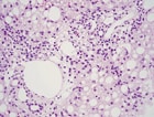
- The histopathology was consistent with spontaneous splenic rupture, and showed the characteristic hemozoin pigment in the Kupffer cells. (ispub.com)
- Fig. 3: Histopathology showing characteristic malarial pigment in Kupffer cells. (ispub.com)
Apoptosis1
- Old or defective cells are removed through apoptosis, as well as through being phagocytized by neighbouring Kupffer cells. (wikipedia.org)
Fibroblasts2
- These are observed in cells of mesenchymal origin, especially fibroblasts. (medscape.com)
- Other sites of abnormal cell vacuolization include the renal glomerular podocytes and in the fibroblasts of the liver's periportal spaces. (medscape.com)
Pathogenesis1
- In this review we propose a model that demonstrates the role of Kupffer cells in the pathogenesis of liver disease. (wanfangdata.com.cn)
Primary2
- The primary function of the Kupffer cell is to remove foreign debris and particles that have come from the hepatic portal system when passing through the liver. (wikipedia.org)
- The cancer cells detach from the primary site, such as the breast or the lungs, and travel through the circulatory or lymphatic system to the liver. (fortherecordmag.com)
Phagocytosis1
- It is possible for the Kupffer cells to take in large particles by phagocytosis and smaller particles via pinocytosis. (wikipedia.org)
Tumor4
- It has necrotizing activity against tumor cell lines and increases ability to reject tumor transplants. (lookformedical.com)
- Alcohol injection (50.94): Pure alcohol is injected directly into tumors, which will dry out the cells in the tumor so the cells eventually die. (fortherecordmag.com)
- In the field of cancer, the possibility to link antibodies to nanoshells using PEG enables the use of these biocompatible nanoshells for the delivery of drugs to targeted tumor cells. (springer.com)
- The goal of such innovative treatment is double to enhance the drug effect on targeted tumor cells and to reduce the toxic effect of the anticancer drug on normal organs. (springer.com)
PBMC1
- Generation of four induced pluripotent stem cells lines from PBMC of the DFNA. (invivogen.com)
Role2
- Because of this detection system, Kupffer cells play a critical role in initiating and mediating immune responses to bacterial infection of the liver. (wikipedia.org)
- The role of Kupffer cell oxidant production in early ethanol-induced liver disease. (nih.gov)
Found3
- What cell types are found in the respiratory mucosa? (histology-world.com)
- Cells involved in immune activity, Kupffer cells, are found in high numbers in the liver. (medicalnewstoday.com)
- What cells are found here? (brainscape.com)
Diffuse1
- Leukemic presentation of diffuse large B-cell lymphoma: an unusual pattern associated with splenic involvement. (rochester.edu)
Organs2
- In addition, carbon-based materials may leave a residue during cell imaging and drug delivery, potentially accumulating in target organs. (biomedcentral.com)
- Thus, it is important to study the damage carbon nanoparticles may cause to different target organs or cell types and to evaluate both their effectiveness and safety before using them in living organisms. (biomedcentral.com)
Bone marrow3
- The liver or bone marrow stores iron released from hemoglobin, which makes the next generation of blood cells. (medicalnewstoday.com)
- possibly because the iron that accumulates in the liver, bone marrow, and spleen is less available for production of red blood cells. (medlineplus.gov)
- Ferroportin also transports iron out of reticuloendothelial cells in the liver, spleen, and bone marrow. (medlineplus.gov)
Proteins1
- They filter bacteria and small foreign proteins out of the blood , and dispose of worn out red blood cells . (online-medical-dictionary.org)