Kidney Medulla
Kidney
Aquaporin 2
Kidney Cortex
Aquaporin 6
Prostaglandins E
Kidney Tubules, Collecting
Medulla Oblongata
Arachidonic Acids
Rabbits
Kidney Tubules
Acute Kidney Injury
Kidney Failure, Chronic
Kidney Concentrating Ability
Kidney Glomerulus
Reduced water permeability and altered ultrastructure in thin descending limb of Henle in aquaporin-1 null mice. (1/1605)
It has been controversial whether high water permeability in the thin descending limb of Henle (TDLH) is required for formation of a concentrated urine by the kidney. Freeze-fracture electron microscopy (FFEM) of rat TDLH has shown an exceptionally high density of intramembrane particles (IMPs), which were proposed to consist of tetramers of aquaporin-1 (AQP1) water channels. In this study, transepithelial osmotic water permeability (Pf) was measured in isolated perfused segments (0.5-1 mm) of TDLH in wild-type (+/+), AQP1 heterozygous (+/-), and AQP1 null (-/-) mice. Pf was measured at 37 degrees C using a 100 mM bath-to-lumen osmotic gradient of raffinose, and fluorescein isothiocyanate (FITC)-dextran as the luminal volume marker. Pf was (in cm/s): 0.26 +/- 0.02 ([+/+]; SE, n = 9 tubules), 0.21 +/- 0.01 ([+/-]; n = 12), and 0.031 +/- 0.007 ([-/-]; n = 6) (P < 0.02, [+/+] vs. [+/-]; P < 0.0001, [+/+] vs. [-/-]). FFEM of kidney medulla showed remarkably fewer IMPs in TDLH from (-/-) vs. (+/+) and (+/-) mice. IMP densities were (in microm-2, SD, 5-12 micrographs): 5,880 +/- 238 (+/+); 5,780 +/- 450 (+/-); and 877 +/- 420 (-/-). IMP size distribution analysis revealed mean IMP diameters of 8.4 nm ([+/+] and [+/-]) and 5.2 nm ([-/-]). These results demonstrate that AQP1 is the principal water channel in TDLH and support the view that osmotic equilibration along TDLH by water transport plays a key role in the renal countercurrent concentrating mechanism. The similar Pf and AQP1 expression in TDLH of (+/+) and (+/-) mice was an unexpected finding that probably accounts for the unimpaired urinary concentrating ability in (+/-) mice. (+info)PST 2238: A new antihypertensive compound that modulates Na,K-ATPase in genetic hypertension. (2/1605)
A genetic alteration in the adducin genes is associated with hypertension and up-regulation of the expression of renal Na, K-ATPase in Milan-hypertensive (MHS) rats, in which increased ouabain-like factor (OLF) levels are also observed. PST 2238, a new antihypertensive compound that antagonizes the pressor effect of ouabain in vivo and normalizes ouabain-dependent up-regulation of the renal Na-K pump, was evaluated for its ability to lower blood pressure and regulate renal Na,K-ATPase activity in MHS genetic hypertension. In this study, we show that PST 2238, given orally at very low doses (1 and 10 microg/kg for 5-6 weeks), reduced the development of hypertension in MHS rats and normalized the increased renal Na,K-ATPase activity and mRNA levels, whereas it did not affect either blood pressure or Na,K-ATPase in Milan-normotensive (MNS) rats. In addition, a similar antihypertensive effect was observed in adult MHS rats after a short-term treatment. In cultured rat renal cells with increased Na-K pump activity at Vmax due to overexpression of the hypertensive variant of adducin, 5 days of incubation with PST 2238 (10(-10-)-10(-9) M) lowered the pump rate to the level of normal wild-type cells, which in turn were not affected by the drug. In conclusion, PST 2238 is a very potent compound that in MHS rats reduces blood pressure and normalizes Na-K pump alterations caused by a genetic alteration of the cytoskeletal adducin. Because adducin gene mutations have been associated with human essential hypertension, it is suggested that PST 2238 may display greater antihypertensive activity in those patients carrying such a genetic alteration. (+info)Tonicity-responsive enhancer binding protein, a rel-like protein that stimulates transcription in response to hypertonicity. (3/1605)
Hypertonicity (most often present as high salinity) is stressful to the cells of virtually all organisms. Cells survive in a hypertonic environment by increasing the transcription of genes whose products catalyze cellular accumulation of compatible osmolytes. In mammals, the kidney medulla is normally hypertonic because of the urinary concentrating mechanism. Cellular accumulation of compatible osmolytes in the renal medulla is catalyzed by the sodium/myo-inositol cotransporter (SMIT), the sodium/chloride/betaine cotransporter, and aldose reductase (synthesis of sorbitol). The importance of compatible osmolytes is underscored by the necrotic injury of the renal medulla and subsequent renal failure that results from the inhibition of SMIT in vivo by administration of a specific inhibitor. Tonicity-responsive enhancers (TonE) play a key role in hypertonicity-induced transcriptional stimulation of SMIT, sodium/chloride/betaine cotransporter, and aldose reductase. We report the cDNA cloning of human TonE binding protein (TonEBP), a transcription factor that stimulates transcription through its binding to TonE sequences via a Rel-like DNA binding domain. Western blot and immunohistochemical analyses of cells cultured in hypertonic medium reveal that exposure to hypertonicity elicits slow activation of TonEBP, which is the result of an increase in TonEBP amount and translocation to the nucleus. (+info)Splicing of a retained intron within ROMK K+ channel RNA generates a novel set of isoforms in rat kidney. (4/1605)
The renal outer medulla K+ channel (ROMK) family of K+ channels may constitute a major pathway for K+ secretion in the distal nephron. To date, four main isoforms of this gene have been identified in the rat that differ only in their NH2-terminal amino acids and that share a common "core exon" that determines the remaining protein sequence. Using RT-PCR, we have identified a new set of ROMK isoforms in rat kidney that are generated by the deletion of a region within the ROMK core sequence that is identifiable as a typical mammalian intron. This splicing event was shown to be reproducible in vitro by detection of deleted ROMK mRNA in Madin-Darby canine kidney (MDCK) cells stably transfected with the gene for ROMK2. Translation of the deletion variant of ROMK2 was confirmed in vitro and visualized in MDCK cells following transient transfection with an enhanced green fluorescent protein tag. The deletion in this core region is predicted to generate hydrophilic proteins that are approximately one-third of the size of native ROMK and lack membrane-spanning domains. (+info)Effect of acidification on the location of H+-ATPase in cultured inner medullary collecting duct cells. (5/1605)
In previous studies, our laboratory has utilized a cell line derived from the rat inner medullary collecting duct (IMCD) as a model system for mammalian renal epithelial cell acid secretion. We have provided evidence, from a physiological perspective, that acute cellular acidification stimulates apical exocytosis and elicits a rapid increase in proton secretion that is mediated by an H+-ATPase. The purpose of these experiments was to examine the effect of acute cellular acidification on the distribution of the vacuolar H+-ATPase in IMCD cells in vitro. We utilized the 31-kDa subunit of the H+-ATPase as a marker of the complete enzyme. The distribution of this subunit of the H+-ATPase was evaluated by immunohistochemical techniques (confocal and electron microscopy), and we found that there is a redistribution of these pumps from vesicles to the apical membrane. Immunoblot evaluation of isolated apical membrane revealed a 237 +/- 34% (P < 0.05, n = 9) increase in the 31-kDa subunit present in the membrane fraction 20 min after the induction of cellular acidification. Thus our results demonstrate the presence of this pump subunit in the IMCD cell line in vitro and that cell acidification regulates the shuttling of cytosolic vesicles containing the 31-kDa subunit into the apical membrane. (+info)Role of renal medullary adenosine in the control of blood flow and sodium excretion. (6/1605)
This study determined the levels of adenosine in the renal medullary interstitium using microdialysis and fluorescence HPLC techniques and examined the role of endogenous adenosine in the control of medullary blood flow and sodium excretion by infusing the specific adenosine receptor antagonists or agonists into the renal medulla of anesthetized Sprague-Dawley rats. Renal cortical and medullary blood flows were measured using laser-Doppler flowmetry. Analysis of microdialyzed samples showed that the adenosine concentration in the renal medullary interstitial dialysate averaged 212 +/- 5.2 nM, which was significantly higher than 55.6 +/- 5.3 nM in the renal cortex (n = 9). Renal medullary interstitial infusion of a selective A1 antagonist, 8-cyclopentyl-1,3-dipropylxanthine (DPCPX; 300 pmol. kg-1. min-1, n = 8), did not alter renal blood flows, but increased urine flow by 37% and sodium excretion by 42%. In contrast, renal medullary infusion of the selective A2 receptor blocker 3, 7-dimethyl-1-propargylxanthine (DMPX; 150 pmol. kg-1. min-1, n = 9) decreased outer medullary blood flow (OMBF) by 28%, inner medullary blood flows (IMBF) by 21%, and sodium excretion by 35%. Renal medullary interstitial infusion of adenosine produced a dose-dependent increase in OMBF, IMBF, urine flow, and sodium excretion at doses from 3 to 300 pmol. kg-1. min-1 (n = 7). These effects of adenosine were markedly attenuated by the pretreatment of DMPX, but unaltered by DPCPX. Infusion of a selective A3 receptor agonist, N6-benzyl-5'-(N-ethylcarbonxamido)adenosine (300 pmol. kg-1. min-1, n = 6) into the renal medulla had no effect on medullary blood flows or renal function. Glomerular filtration rate and arterial pressure were not changed by medullary infusion of any drugs. Our results indicate that endogenous medullary adenosine at physiological concentrations serves to dilate medullary vessels via A2 receptors, resulting in a natriuretic response that overrides the tubular A1 receptor-mediated antinatriuretic effects. (+info)Second messenger production in avian medullary nephron segments in response to peptide hormones. (7/1605)
We examined the sites of peptide hormone activation within medullary nephron segments of the house sparrow (Passer domesticus) kidney by measuring rates of hormone-induced generation of cyclic nucleotide second messenger. Thin descending limbs, thick ascending limbs, and collecting ducts had baseline activity of adenylyl cyclase that resulted in cAMP accumulation of 207 +/- 56, 147 +/- 31, and 151 +/- 41 fmol. mm-1. 30 min-1, respectively. In all segments, this activity increased 10- to 20-fold in response to forskolin. Activity of adenylyl cyclase in the thin descending limb was stimulated approximately twofold by parathyroid hormone (PTH) but not by any of the other hormones tested [arginine vasotocin (AVT), glucagon, atrial natriuretic peptide (ANP), or isoproterenol, each at 10(-6) M]. Thick ascending limb was stimulated two- to threefold by both AVT and PTH; however, glucagon and isoproterenol had no effect, and ANP stimulated neither cAMP nor cGMP accumulation. Adenylyl cyclase activity in the collecting duct was stimulated fourfold by AVT but not by the other hormones; likewise, ANP did not stimulate cGMP accumulation in this segment. These data support a tubular action of AVT and PTH in the avian renal medulla. (+info)Expression of bone morphogenetic protein-7 mRNA in normal and ischemic adult rat kidney. (8/1605)
BMP-7, a member of the bone morphogenic protein subfamily (BMPs) of the transforming growth factor-beta superfamily of secreted growth factors, is abundantly expressed in the fetal kidney. The precise role of this protein in renal physiology or pathology is unknown. A cDNA that encodes rat BMP-7 was cloned and used as a probe to localize BMP-7 mRNA expression by in situ hybridization in the adult rat kidney. The highest expression of BMP-7 mRNA could be seen in tubules of the outer medulla. In glomeruli, a few cells, mainly located at the periphery of the glomerular tuft, showed specific and strong signals. Also, high BMP-7 mRNA expression could be localized to the adventitia of renal arteries, as well as to the epithelial cell layer of the renal pelvis and the ureter. Preliminary evidence suggests that BMP-7 enhances recovery when infused into rats with ischemia-induced acute renal failure. We examined BMP-7 mRNA expression in kidneys with acute renal failure induced by unilateral renal artery clamping. BMP-7 mRNA abundance as analyzed by solution hybridization was reduced in ischemic kidneys after 6 and 16 h of reperfusion compared with the contralateral kidney. In situ hybridization in ischemic kidneys showed a marked decrease of BMP-7 mRNA in the outer medulla and in glomeruli. Utilizing rat metanephric mesenchymal cells in culture, we also demonstrate that BMP-7 induces epithelial cell differentiation. Taken together, these data suggest that BMP-7 is important in both stimulating and maintaining a healthy differentiated epithelial cell phenotype. (+info)The kidney medulla is the inner portion of the renal pyramids in the kidney, consisting of multiple conical structures found within the kidney. It is composed of loops of Henle and collecting ducts responsible for concentrating urine by reabsorbing water and producing a hyperosmotic environment. The kidney medulla has a unique blood supply and is divided into an inner and outer zone, with the inner zone having a higher osmolarity than the outer zone. This region of the kidney helps regulate electrolyte and fluid balance in the body.
A kidney, in medical terms, is one of two bean-shaped organs located in the lower back region of the body. They are essential for maintaining homeostasis within the body by performing several crucial functions such as:
1. Regulation of water and electrolyte balance: Kidneys help regulate the amount of water and various electrolytes like sodium, potassium, and calcium in the bloodstream to maintain a stable internal environment.
2. Excretion of waste products: They filter waste products from the blood, including urea (a byproduct of protein metabolism), creatinine (a breakdown product of muscle tissue), and other harmful substances that result from normal cellular functions or external sources like medications and toxins.
3. Endocrine function: Kidneys produce several hormones with important roles in the body, such as erythropoietin (stimulates red blood cell production), renin (regulates blood pressure), and calcitriol (activated form of vitamin D that helps regulate calcium homeostasis).
4. pH balance regulation: Kidneys maintain the proper acid-base balance in the body by excreting either hydrogen ions or bicarbonate ions, depending on whether the blood is too acidic or too alkaline.
5. Blood pressure control: The kidneys play a significant role in regulating blood pressure through the renin-angiotensin-aldosterone system (RAAS), which constricts blood vessels and promotes sodium and water retention to increase blood volume and, consequently, blood pressure.
Anatomically, each kidney is approximately 10-12 cm long, 5-7 cm wide, and 3 cm thick, with a weight of about 120-170 grams. They are surrounded by a protective layer of fat and connected to the urinary system through the renal pelvis, ureters, bladder, and urethra.
Aquaporin 2 (AQP2) is a type of aquaporin, which is a water channel protein found in the membranes of cells. Specifically, AQP2 is located in the principal cells of the collecting ducts in the kidneys. It plays a crucial role in regulating water reabsorption and urine concentration by facilitating the movement of water across the cell membrane in response to the hormone vasopressin (also known as antidiuretic hormone). When vasopressin binds to receptors on the cell surface, it triggers a cascade of intracellular signals that lead to the translocation of AQP2 water channels from intracellular vesicles to the apical membrane. This increases the permeability of the apical membrane to water, allowing for efficient reabsorption of water and concentration of urine. Dysfunction in AQP2 has been implicated in various kidney disorders, such as nephrogenic diabetes insipidus.
The kidney cortex is the outer region of the kidney where most of the functional units called nephrons are located. It plays a crucial role in filtering blood and regulating water, electrolyte, and acid-base balance in the body. The kidney cortex contains the glomeruli, proximal tubules, loop of Henle, and distal tubules, which work together to reabsorb necessary substances and excrete waste products into the urine.
Aquaporin 6 (AQP6) is a protein that functions as a water channel in the membranes of certain cells. It is a member of the aquaporin family, which are proteins that allow the selective transport of water and small solutes across biological membranes. Aquaporin 6 is primarily expressed in the kidney, where it is localized to the intracellular vesicles of intercalated cells in the collecting ducts. It is thought to play a role in acid-base balance and urine concentration by regulating the movement of water and hydrogen ions (protons) across cell membranes. Aquaporin 6 has also been found to be permeable to anions, making it unique among aquaporins. Additionally, AQP6 has been identified in other tissues such as the brain, lung, and testis, but its function in these tissues is not well understood.
Prostaglandin E (PGE) is a type of prostaglandin, which is a group of lipid compounds that are synthesized in the body from fatty acids and have diverse hormone-like effects. Prostaglandins are not actually hormones, but are similar to them in that they act as chemical messengers that have specific effects on certain cells.
Prostaglandin E is one of the most abundant prostaglandins in the body and has a variety of physiological functions. It is involved in the regulation of inflammation, pain perception, fever, and smooth muscle contraction. Prostaglandin E also plays a role in the regulation of blood flow, platelet aggregation, and gastric acid secretion.
Prostaglandin E is synthesized from arachidonic acid, which is released from cell membranes by the action of enzymes called phospholipases. Once formed, prostaglandin E binds to specific receptors on the surface of cells, leading to a variety of intracellular signaling events that ultimately result in changes in cell behavior.
Prostaglandin E is used medically in the treatment of several conditions, including dysmenorrhea (painful menstruation), postpartum hemorrhage, and patent ductus arteriosus (a congenital heart defect). It is also used as a diagnostic tool in the evaluation of kidney function.
Collecting kidney tubules, also known as collecting ducts, are the final portion of the renal tubule in the nephron of the kidney. They collect filtrate from the distal convoluted tubules and glomeruli and are responsible for the reabsorption of water and electrolytes back into the bloodstream under the influence of antidiuretic hormone (ADH) and aldosterone. The collecting ducts then deliver the remaining filtrate to the ureter, which transports it to the bladder for storage until urination.
The medulla oblongata is a part of the brainstem that is located in the posterior portion of the brainstem and continues with the spinal cord. It plays a vital role in controlling several critical bodily functions, such as breathing, heart rate, and blood pressure. The medulla oblongata also contains nerve pathways that transmit sensory information from the body to the brain and motor commands from the brain to the muscles. Additionally, it is responsible for reflexes such as vomiting, swallowing, coughing, and sneezing.
Arachidonic acids are a type of polyunsaturated fatty acid that is primarily found in the phospholipids of cell membranes. They contain 20 carbon atoms and four double bonds (20:4n-6), with the first double bond located at the sixth carbon atom from the methyl end.
Arachidonic acids are derived from linoleic acid, an essential fatty acid that cannot be synthesized by the human body and must be obtained through dietary sources such as meat, fish, and eggs. Once ingested, linoleic acid is converted to arachidonic acid in a series of enzymatic reactions.
Arachidonic acids play an important role in various physiological processes, including inflammation, immune response, and cell signaling. They serve as precursors for the synthesis of eicosanoids, which are signaling molecules that include prostaglandins, thromboxanes, and leukotrienes. These eicosanoids have diverse biological activities, such as modulating blood flow, platelet aggregation, and pain perception, among others.
However, excessive production of arachidonic acid-derived eicosanoids has been implicated in various pathological conditions, including inflammation, atherosclerosis, and cancer. Therefore, the regulation of arachidonic acid metabolism is an important area of research for the development of new therapeutic strategies.
I believe there may be some confusion in your question. "Rabbits" is a common name used to refer to the Lagomorpha species, particularly members of the family Leporidae. They are small mammals known for their long ears, strong legs, and quick reproduction.
However, if you're referring to "rabbits" in a medical context, there is a term called "rabbit syndrome," which is a rare movement disorder characterized by repetitive, involuntary movements of the fingers, resembling those of a rabbit chewing. It is also known as "finger-chewing chorea." This condition is usually associated with certain medications, particularly antipsychotics, and typically resolves when the medication is stopped or adjusted.
Kidney disease, also known as nephropathy or renal disease, refers to any functional or structural damage to the kidneys that impairs their ability to filter blood, regulate electrolytes, produce hormones, and maintain fluid balance. This damage can result from a wide range of causes, including diabetes, hypertension, glomerulonephritis, polycystic kidney disease, lupus, infections, drugs, toxins, and congenital or inherited disorders.
Depending on the severity and progression of the kidney damage, kidney diseases can be classified into two main categories: acute kidney injury (AKI) and chronic kidney disease (CKD). AKI is a sudden and often reversible loss of kidney function that occurs over hours to days, while CKD is a progressive and irreversible decline in kidney function that develops over months or years.
Symptoms of kidney diseases may include edema, proteinuria, hematuria, hypertension, electrolyte imbalances, metabolic acidosis, anemia, and decreased urine output. Treatment options depend on the underlying cause and severity of the disease and may include medications, dietary modifications, dialysis, or kidney transplantation.
Kidney transplantation is a surgical procedure where a healthy kidney from a deceased or living donor is implanted into a patient with end-stage renal disease (ESRD) or permanent kidney failure. The new kidney takes over the functions of filtering waste and excess fluids from the blood, producing urine, and maintaining the body's electrolyte balance.
The transplanted kidney is typically placed in the lower abdomen, with its blood vessels connected to the recipient's iliac artery and vein. The ureter of the new kidney is then attached to the recipient's bladder to ensure proper urine flow. Following the surgery, the patient will require lifelong immunosuppressive therapy to prevent rejection of the transplanted organ by their immune system.
Kidney tubules are the structural and functional units of the kidney responsible for reabsorption, secretion, and excretion of various substances. They are part of the nephron, which is the basic unit of the kidney's filtration and reabsorption process.
There are three main types of kidney tubules:
1. Proximal tubule: This is the initial segment of the kidney tubule that receives the filtrate from the glomerulus. It is responsible for reabsorbing approximately 65% of the filtrate, including water, glucose, amino acids, and electrolytes.
2. Loop of Henle: This U-shaped segment of the tubule consists of a thin descending limb, a thin ascending limb, and a thick ascending limb. The loop of Henle helps to concentrate urine by creating an osmotic gradient that allows water to be reabsorbed in the collecting ducts.
3. Distal tubule: This is the final segment of the kidney tubule before it empties into the collecting duct. It is responsible for fine-tuning the concentration of electrolytes and pH balance in the urine by selectively reabsorbing or secreting substances such as sodium, potassium, chloride, and hydrogen ions.
Overall, kidney tubules play a critical role in maintaining fluid and electrolyte balance, regulating acid-base balance, and removing waste products from the body.
Acute kidney injury (AKI), also known as acute renal failure, is a rapid loss of kidney function that occurs over a few hours or days. It is defined as an increase in the serum creatinine level by 0.3 mg/dL within 48 hours or an increase in the creatinine level to more than 1.5 times baseline, which is known or presumed to have occurred within the prior 7 days, or a urine volume of less than 0.5 mL/kg per hour for six hours.
AKI can be caused by a variety of conditions, including decreased blood flow to the kidneys, obstruction of the urinary tract, exposure to toxic substances, and certain medications. Symptoms of AKI may include decreased urine output, fluid retention, electrolyte imbalances, and metabolic acidosis. Treatment typically involves addressing the underlying cause of the injury and providing supportive care, such as dialysis, to help maintain kidney function until the injury resolves.
Chronic kidney failure, also known as chronic kidney disease (CKD) stage 5 or end-stage renal disease (ESRD), is a permanent loss of kidney function that occurs gradually over a period of months to years. It is defined as a glomerular filtration rate (GFR) of less than 15 ml/min, which means the kidneys are filtering waste and excess fluids at less than 15% of their normal capacity.
CKD can be caused by various underlying conditions such as diabetes, hypertension, glomerulonephritis, polycystic kidney disease, and recurrent kidney infections. Over time, the damage to the kidneys can lead to a buildup of waste products and fluids in the body, which can cause a range of symptoms including fatigue, weakness, shortness of breath, nausea, vomiting, and confusion.
Treatment for chronic kidney failure typically involves managing the underlying condition, making lifestyle changes such as following a healthy diet, and receiving supportive care such as dialysis or a kidney transplant to replace lost kidney function.
Kidney concentrating ability refers to the capacity of the kidneys to increase the concentration of solutes, such as urea and minerals, and remove waste products while reabsorbing water to maintain fluid balance in the body. This is primarily regulated by the hormone vasopressin (ADH), which signals the collecting ducts in the nephrons of the kidneys to absorb more water, resulting in the production of concentrated urine. A decreased kidney concentrating ability may indicate a variety of renal disorders or diseases, such as diabetes insipidus or chronic kidney disease.
A kidney glomerulus is a functional unit in the nephron of the kidney. It is a tuft of capillaries enclosed within a structure called Bowman's capsule, which filters waste and excess fluids from the blood. The glomerulus receives blood from an afferent arteriole and drains into an efferent arteriole.
The process of filtration in the glomerulus is called ultrafiltration, where the pressure within the glomerular capillaries drives plasma fluid and small molecules (such as ions, glucose, amino acids, and waste products) through the filtration membrane into the Bowman's space. Larger molecules, like proteins and blood cells, are retained in the blood due to their larger size. The filtrate then continues down the nephron for further processing, eventually forming urine.
Kidney neoplasms refer to abnormal growths or tumors in the kidney tissues that can be benign (non-cancerous) or malignant (cancerous). These growths can originate from various types of kidney cells, including the renal tubules, glomeruli, and the renal pelvis.
Malignant kidney neoplasms are also known as kidney cancers, with renal cell carcinoma being the most common type. Benign kidney neoplasms include renal adenomas, oncocytomas, and angiomyolipomas. While benign neoplasms are generally not life-threatening, they can still cause problems if they grow large enough to compromise kidney function or if they undergo malignant transformation.
Early detection and appropriate management of kidney neoplasms are crucial for improving patient outcomes and overall prognosis. Regular medical check-ups, imaging studies, and urinalysis can help in the early identification of these growths, allowing for timely intervention and treatment.
Cortex and medulla4
- Kidney stiffness values were obtained from whole kidney, cortex, and medulla. (nih.gov)
- ISH of SDF1 in mouse kidney cortex and medulla. (jci.org)
- C. Each kidney is divided into 2 parts - cortex and medulla. (syvum.com)
- Urin is made in cortex and medulla of the kidney and drained to the renal pelvis. (quizlet.com)
Outer7
- Each kidney has an outer layer called the cortex , which contains filtering units. (kidshealth.org)
- Rat urine and kidney tissue extract (outer medulla and cortex) Amino Acid concentrations. (sdsc.edu)
- The kidney is divided into an outer cortex and inner medulla. (flashcardmachine.com)
- They then did the same experiment using different types of kidney tissue, with one sample from the outer kidney, called the cortex, and the other from the inner kidney, called the medulla. (sciencedaily.com)
- They are made up of an outer portion called the cortex, and an inner portion called the medulla. (medlineplus.gov)
- Your adrenal glands are composed of two parts: the cortex (outer region) and the medulla (inner part). (clevelandclinic.org)
- The kidneys consist of an outer part (cortex) and an inner part (medulla). (msdmanuals.com)
Ureter5
- Flow continues through the renal tubules, including the proximal tubule, the loop of Henle, through the distal tubule and finally leaves the kidney by means of the collecting duct, leading to the renal pelvis, the dilated portion of the ureter. (wikipedia.org)
- The indentation on the concave side of the kidney, known as the renal hilus, provides a space for the renal artery, renal vein, and ureter to enter the kidney. (innerbody.com)
- The renal pelvis exits the kidney at the renal hilus, where urine drains into the ureter. (innerbody.com)
- 1 Each kidney is connected to the bladder through a ureter. (searchandrestore.com)
- Urine drains from the renal pelvis of each kidney into a ureter. (msdmanuals.com)
Blood Supply to the Kidneys1
- The blood supply to the kidneys arises from the paired renal arteries at the level of L2. (medscape.com)
Nephrons6
- The renal medulla (Latin: medulla renis 'marrow of the kidney') contains the structures of the nephrons responsible for maintaining the salt and water balance of the blood. (wikipedia.org)
- True or false: Kidneys contain 10-12 million nephrons. (syvum.com)
- The artery then branches so blood can get to the nephrons (NEH-fronz) - 1 million tiny filtering units in each kidney that remove the harmful substances from the blood. (kidshealth.org)
- Each kidney contains more than a million microscopic functional units called nephrons. (flashcardmachine.com)
- Each kidney contains around 1 million individual nephrons, the kidneys' microscopic functional units that filter blood to produce urine. (innerbody.com)
- Each kidney contains about one million nephrons. (msdmanuals.com)
Nephron1
- The renal medulla is hypertonic to the filtrate in the nephron and aids in the reabsorption of water. (wikipedia.org)
Inner medulla1
- Kidney anatomy, with pyramids labeled at right Medullipin Kokko and Rector Model, a theory to explain how a gradient is generated in the inner medulla Renal sinus Medullary interstitium Renal capsule This article incorporates text in the public domain from page 1221 of the 20th edition of Gray's Anatomy (1918) Kelly CR, Landman J (March 2012). (wikipedia.org)
Minor calyx1
- The renal papilla is the location where the renal pyramids in the medulla empty urine into the minor calyx in the kidney. (wikipedia.org)
Liver3
- The left kidney is located slightly more superior than the right kidney due to the larger size of the liver on the right side of the body. (innerbody.com)
- If this were the case then the aging kidney would look quite different on the molecular level from an aging liver. (sciencedaily.com)
- The left kidney sits a bit higher in the body because of the size of the liver, which is also on the right side. (searchandrestore.com)
Function of the kidneys2
- How does the body control the function of the kidneys? (searchandrestore.com)
- Several hormones produced elsewhere in the body help to control the function of the kidneys. (searchandrestore.com)
Bean shaped4
- The bean-shaped kidneys filter waste products out of the bloodstream and dispose of them by creating urine. (kidshealth.org)
- The kidneys are bean-shaped with the convex side of each organ located laterally and the concave side medial. (innerbody.com)
- Your kidneys are bean-shaped organs that are located in the middle of your back against the back muscles, with one on either side of your spine. (searchandrestore.com)
- Grossly, the kidneys are bean-shaped structures and weigh about 150 g in the male and about 135 g in the female. (medscape.com)
Tissue17
- The medullary interstitium is the tissue surrounding the loop of Henle in the medulla. (wikipedia.org)
- We will analyze kidney tissue extract and urine samples from SS rats and the newly generated SS.Fh1+ transgenic rats. (sdsc.edu)
- We will analyze kidney tissue extract and urine samples collected from rats maintained on a 0.4% NaCl diet or switched to a 4% NaCl diet for 7 days. (sdsc.edu)
- Adipose tissue known as perirenal fat surrounds the kidneys and acts as protective padding. (innerbody.com)
- A thin layer of fibrous connective tissue forms the renal capsule surrounding each kidney. (innerbody.com)
- We propose to analyze kidney tissue extract and urine samples from SS and SS.Fh1+ transgenic rats in addition to the analysis of urine samples from the DASH2 trial. (sdsc.edu)
- Leveraging the expertise and resources at the Mayo Clinic Metabolomics Resources Core, we propose to perform targeted LC/MS analysis and NMR spectra generation in urine samples obtained from a subset of subjects from the Dietary Approaches to Stop Hypertension - Sodium (DASH2) clinical trial and kidney tissue extract and urine samples from SS rats and a newly generated transgenic rat that overexpresses fumarase (SS.Fh1+). (sdsc.edu)
- Pantomics Array Description: Kidney cancer tissue array, 102 cases of normal/benign (5 cases) and cancer (97 cases with grading and TNM staging. (delos.info)
- Pantomics Array Description: Kidney cancer tissue array containing 28 cases of normal/benign conditions and 200 cases of cancers with grading. (delos.info)
- Pantomics Array Description: Kidney cancer tissue array, set 1, non-overlapping with KIC1502 or KIC1503, 150 cores from normal/benign (5. (delos.info)
- Pantomics Array Description: Kidney cancer tissue array, set 3, non-overlapping with KIC1501 or KIC1502, containing 75 cases in 150 cores. (delos.info)
- The kidney is made up of two distinct tissue regions: the medulla and the cortex. (ucsd.edu)
- Each kidney is held in place by connective tissue, called renal fascia, and is surrounded by a thick layer of adipose tissue, called perirenal fat, which helps to protect it. (searchandrestore.com)
- A tough, fibrous, connective tissue renal capsule closely envelopes each kidney and provides support for the soft tissue that is inside. (searchandrestore.com)
- Which is part of the kidney contains connective tissue? (searchandrestore.com)
- 2 Each kidney is covered in a thick layer of connective tissue and fat that helps shape and protect the organ. (searchandrestore.com)
- Although chromaffin tissue is also present elsewhere in the body, such as in the mediastinum, along the aorta, and in the pelvis, the term pheochromocytoma is reserved for tumors that arise from the adrenal medulla. (medscape.com)
Organs7
- In the pathogenesis of hemorrhagic shock induced by trauma, operative accidents, traffic accidents, and earthquakes, the kidney is one of the organs, in which hypoperfusion initially occurs after hemorrhage because the sympathetic-adrenal medulla system and the renin-angiotensin-aldosterone system are activated. (hindawi.com)
- The kidneys are a pair of organs found along the posterior muscular wall of the abdominal cavity. (innerbody.com)
- Unlike the other abdominal organs, the kidneys lie behind the peritoneum that lines the abdominal cavity and are thus considered to be retroperitoneal organs. (innerbody.com)
- Although these two tissues are both from the kidney, they are as different in function as cells from entirely different organs. (sciencedaily.com)
- This correlation could help screen kidneys from people older than 60 whose organs would ordinarily be rejected for transplants. (sciencedaily.com)
- The adrenal glands are small hormone-releasing organs located on top of each kidney. (medlineplus.gov)
- The kidneys are considered "retroperitoneal" organs, which means they sit behind a lining in the abdominal cavity, unlike all other abdominal organs. (searchandrestore.com)
Glomerular filt3
- Hypercalcemia results when the efflux of calcium is massive or when the glomerular filtration rate of the kidneys is reduced. (medscape.com)
- The glomerular filtration rate (GFR) is the volume of filtrate produced by both kidneys each minute. (flashcardmachine.com)
- This fact is used to measure the volume of blood plasma filtered per minute by the kidneys, or the GFR (glomerular filtration rate). (flashcardmachine.com)
Pituitary1
- Hemorrhagic lesions are found in the pituitary, the right atrium and the kidney. (cdc.gov)
Interstitial3
- Because of this Na+ transport and because of countercurrent exchange in the vasa recta, the interstitial fluid of the medulla becomes hypertonic. (flashcardmachine.com)
- Interstitial tonicity controls TonEBP expression in the renal medulla. (nature.com)
- Frequency of interstitial cellular nodose hyperplasia of the kidney medulla in arterial hypertension]. (bvsalud.org)
Lung2
- M) Lung, kidney and parotid glands (LV). (cdc.gov)
- P2RY8 is highly expressed in lymphocytes, with weaker expression in heart, kidney and lung (Cantagrel et al. (atlasgeneticsoncology.org)
Hormones2
- The hormones of the adrenal medulla, which sits atop your kidney, are called catecholamines. (funtrivia.com)
- Norepinephrine and epinephrine are the two main hormones excreted by the adrenal medulla . (newworldencyclopedia.org)
Surrounds the kidneys1
- A layer of fat surrounds the kidneys to cushion and help hold them in place. (searchandrestore.com)
Chromaffin cells2
- The adrenal medulla, composed of chromaffin cells, secretes the hormone epinephrine, also called adrenaline, in response to stimulation of the sympathetic nervous system at times of stress. (funtrivia.com)
- The tumors arise from the chromaffin cells of the adrenal medulla and are associated with increased catecholamine production. (medscape.com)
Hypertonic1
- Tonicity-responsive enhancer-binding protein (TonEBP), which is also known as nuclear factor of activated T cells 5 (NFAT5), was discovered 20 years ago as a transcriptional regulator of the cellular response to hypertonic (hyperosmotic salinity) stress in the renal medulla. (nature.com)
Retroperitoneal1
- The kidneys are paired retroperitoneal structures that are normally located between the transverse processes of T12-L3 vertebrae, with the left kidney typically somewhat more superior in position than the right. (medscape.com)
Ureters4
- There are two ureters - one draining each kidney. (kidshealth.org)
- From the calyxes, pee travels out of the kidneys through the ureters (YUR-uh-ters) to be stored in the bladder (a muscular sac in the lower belly). (kidshealth.org)
- Pee leaves the kidneys and travels through the ureters to the bladder. (kidshealth.org)
- The rest of the urinary tract consists of the following: Two ureters (the tubes connecting each kidney to the bladder) The bladder (an expandable muscular. (msdmanuals.com)
Structures5
- The center part of the kidney, the medulla (meh-DUH-luh), has fan-shaped structures called pyramids . (kidshealth.org)
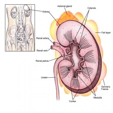
- Next to it appears a detailed cut-away view through a single kidney identifying its major structures. (smartimagebase.com)
- The kidney structures are greatly magnified. (cpr-savers.com)
- The adrenal gland, located atop the kidneys , is separated into two distinct structures, the adrenal medulla and the adrenal cortex. (newworldencyclopedia.org)
- The center part of the kidney, the medulla (pronounced: meh-DUH-luh) has 10 to 15 fan-shaped structures called pyramids. (searchandrestore.com)
Known as the renal1
- The renal medulla is split up into a number of sections, known as the renal pyramids. (wikipedia.org)
Lymph nodes1
- lymph nodes and kidney medullae were hemorrhagic. (cdc.gov)
Lobe1
- 0.05) expressed in the cerebellar cortex, piriform lobe, medulla, and corpus callosum of the adult yak while in the young yak brain tissues, the protein expressions were significantly found in the white matter of the cerebellum, pineal gland, corpus callosum, and cerebellar cortex. (scielo.br)
Skeletal1
- Along with epinephrine (adrenaline), another hormone secreted by the adrenal medulla, norepinephrine underlies the fight-or-flight response to physical or mental stress , directly increasing heart rate, triggering the release of glucose from energy stores, and increasing skeletal muscle readiness, among other actions. (newworldencyclopedia.org)
Collecting ducts2
- The filtrate from the distal convoluted tubule is drained into collecting ducts, which plunge through the medulla to empty into the calyces. (flashcardmachine.com)
- Once the filtrate gets to the collecting ducts in the medulla of the kidney, they converge to a renal papilla, which represents the tip or apex of the renal pyramid. (medscape.com)
Afferent arterioles2
- Inside our kidneys, the renal arteries diverge into the smaller afferent arterioles of the kidneys. (innerbody.com)
- They then radiate into interlobular arteries, which extend into the cortex of the kidney to finally become afferent arterioles, then peritubular capillaries to efferent arterioles. (medscape.com)
Renal arteries1
- The renal arteries branch directly from the abdominal aorta and enter the kidneys through the renal hilus. (innerbody.com)
Regulate blood pressure2
- The kidneys also help regulate blood pressure, the level of salts in the blood, and the acid-base balance (the pH) of the blood. (kidshealth.org)
- Thses mechanisms are needed to ensure that the GFR will be high enough to allow the kidneys to eliminate wastes and regulate blood pressure, but not so high as to cause excessive water loss. (flashcardmachine.com)
Hemorrhage2
- Histopathologic lesions included mild diffuse congestion in the pygmy brocket deer's kidneys and extensive subendocardial hemorrhage. (cdc.gov)
- Our studies showed that the mesenteric lymph duct ligation (MLDL) could alleviate kidney injury following two-hit of hemorrhage and lipopolysaccharide and hemorrhagic shock with fluid resuscitation [ 8 , 9 ]. (hindawi.com)
Catecholamines1
- The adrenal medulla, the inner part of your adrenal glands, produces and releases the catecholamines adrenaline and noradrenaline. (clevelandclinic.org)
Mouse kidney2
- Kim is now studying aging mouse kidney cells to test whether they look different on a molecular level than the human kidney cells in this study. (sciencedaily.com)
- This light micrograph shows a transverse section of a region of mouse kidney cortex stained with toluidine blue. (ucsd.edu)
Anatomy1
- The kidney anatomy is shown in the image below. (medscape.com)
Right kidney1
- Which is better the left kidney or the right kidney? (searchandrestore.com)
Acute kidney3
- In addition, functional studies have shown that TonEBP is involved in the pathogenesis of rheumatoid arthritis, atherosclerosis, diabetic nephropathy, acute kidney injury, hyperlipidaemia and insulin resistance, autoimmune diseases (including type 1 diabetes mellitus and multiple sclerosis), salt-sensitive hypertension and hepatocellular carcinoma. (nature.com)
- This study aimed to investigate the effect of mesenteric lymph drainage on the acute kidney injury induced by hemorrhagic shock without resuscitation. (hindawi.com)
- Therefore, acute kidney injury (AKI) following hemorrhagic shock remains a serious problem. (hindawi.com)
Innermost1
- The renal medulla is the innermost part of the kidney. (wikipedia.org)
Metabolic waste products1
- Overview of Kidney Failure Kidney failure is the inability of the kidneys to adequately filter metabolic waste products from the blood. (msdmanuals.com)
Medullary1
- Ziadie MS. Medullary sponge kidney. (pathologyoutlines.com)
Urinary tract7
- What Are the Kidneys and Urinary Tract? (kidshealth.org)
- The kidneys are the part of the urinary tract that makes urine (pee). (kidshealth.org)
- After the kidneys make urine, it leaves the body using the rest of the urinary tract as a pathway. (kidshealth.org)
- Click through this slideshow to learn more about the kidneys and urinary tract. (kidshealth.org)
- How Do the Kidneys and Urinary Tract Work? (kidshealth.org)
- What Can Help Keep the Kidneys and Urinary Tract Healthy? (kidshealth.org)
- Overview of the Urinary Tract Normally, a person has two kidneys. (msdmanuals.com)
Perirenal fat1
- The second layer is called the perirenal fat capsule, which helps anchor the kidneys in place. (searchandrestore.com)
Necrosis1
- Damage to the renal papillae may result in death to cells in this region of the kidney, called renal papillary necrosis. (wikipedia.org)
Flank pain1
- Pain in your upper abdomen or back and sides is also called flank pain or kidney pain. (searchandrestore.com)
Tissues2
- Renal pyramids (or malpighian pyramids or Malpighi's pyramids named after Marcello Malpighi, a seventeenth-century anatomist) are cone-shaped tissues of the kidney. (wikipedia.org)
- As much as 1/3 of all blood leaving the heart passes into the kidneys to be filtered before flowing to the rest of the body's tissues. (innerbody.com)
Ribcage2
- The kidneys are under the ribcage in the back, one on each side. (kidshealth.org)
- Muscles in your back and your ribcage protect your kidneys from both the front and the back sides of your body. (searchandrestore.com)
0.051
- 0.05 for whole kidney and medulla MRE-derived stiffness). (nih.gov)
Hemorrhagic1
- However, further studies should be conducted to determine whether or not the blockage of mesenteric lymph return can decrease kidney injury after hemorrhagic shock without resuscitation. (hindawi.com)
Adrenal Gland1
- The adrenal medulla is at the center of the adrenal gland and is surrounded by the adrenal cortex, with the adrenal medulla taking up about one-quarter of the adrenal gland and the adrenal cortex the remaining three-quarters. (newworldencyclopedia.org)
Adrenaline1
- The medulla produces the hormone adrenaline (also called epinephrine). (medlineplus.gov)
Left kidney2
- Why is the left kidney higher than the right? (searchandrestore.com)
- Why does the left kidney sit higher? (searchandrestore.com)
Epinephrine1
- While epinephrine is mainly released from the adrenal medulla, norepinephrine has another major source-nerve endings. (newworldencyclopedia.org)
Excretion1
- Creatinine, creatinine clearance, blood urea nitrogen (BUN), and urinary excretion levels, as well as kidney imaging studies, help in the evaluation of the patient's renal function. (medscape.com)
Carries urine2
- This thin, tube-like structure carries urine from the kidney to the bladder. (kidshealth.org)
- Urine next passes through the loop of Henle, a long straight tubule that carries urine into the renal medulla before making a hairpin turn and returning to the renal cortex. (innerbody.com)
Functional units1
- The 3B MICROanatomy Kidney is an extremely detailed model which shows the morphologic/functional units of the kidney. (cpr-savers.com)
Bladder1
- How are the kidneys connected to the bladder? (searchandrestore.com)
Endocrine1
- Your adrenal glands are endocrine glands located on top of your kidneys. (clevelandclinic.org)
Norepinephrine1
- The medulla also secretes the hormone norepinephrine, which plays a role in maintaining normal blood circulation. (funtrivia.com)
Pelvis2
- The minor calyces merge to form 3 larger major calyces, which further merge to form the hollow renal pelvis at the center of the kidney. (innerbody.com)
- Each kidney has several calices, all of which drain into a single central chamber (renal pelvis). (msdmanuals.com)
Hormone2
- Kidneys have many jobs, from filtering blood and making urine to keeping bones healthy and making a hormone that controls the production of red blood cells. (kidshealth.org)
- it is secreted by the adrenal medulla as a hormone into the blood , and as a neurotransmitter from neurons . (newworldencyclopedia.org)