Inclusion Bodies
Myositis, Inclusion Body
Inclusion Bodies, Viral
Protein Refolding
Escherichia coli
Intranuclear Inclusion Bodies
Osteitis Deformans
Solubility
Distal Myopathies
Molecular Sequence Data
Boidae
Microscopy, Electron
Amino Acid Sequence
Cloning, Molecular
Chymosin
Recombinant Fusion Proteins
Polymyositis
Base Sequence
Mutation
Morbillivirus
Protein Denaturation
Frontotemporal Dementia
Lewy Bodies
Morbillivirus Infections
alpha-Synuclein
Cytoplasm
Synucleins
Ubiquitin
Electrophoresis, Polyacrylamide Gel
Plasmids
Microscopy, Electron, Transmission
Distemper
Muscle, Skeletal
Arenavirus
Dermatomyositis
Fowl adenovirus A
Gene Expression
Circular Dichroism
Huntington Disease
Guanidine
Viral Tail Proteins
Caulimovirus
Protein Structure, Secondary
Nerve Tissue Proteins
Protein Binding
Vacuoles
Proteasome Endopeptidase Complex
Hemoglobin H
Neurodegenerative Diseases
Immunohistochemistry
Varicellovirus
Persia
Blotting, Western
Bird Diseases
Enteropeptidase
Bluetongue virus
Cyanamide
Adenosine Triphosphatases
Microscopy, Immunoelectron
Urea
Muscle Weakness
Protein Engineering
Protein Conformation
Distemper Virus, Canine
Autophagy
Peptides
Epidermal Cyst
DNA Primers
Protein Structure, Tertiary
Molecular Chaperones
Serpins
Microscopy, Fluorescence
Adenoviruses, Canine
Reoviridae
Brain
Ophthalmoplegia
Maltose-Binding Proteins
Temperature
Herpesviridae
Polymerase Chain Reaction
Amyloid
Biopsy
Chaperonin 60
Histological Techniques
Genetic Vectors
Cetrimonium Compounds
Cells, Cultured
Green Fluorescent Proteins
Heredodegenerative Disorders, Nervous System
Reducing Agents
Contracture
Chromatography, Gel
Sequence Homology, Amino Acid
Wasting Syndrome
Gammapapillomavirus
Lead
Neurons
Carrier Proteins
Apoferritins
Poxviridae
Muscular Dystrophies, Limb-Girdle
Multienzyme Complexes
Amyloid beta-Peptides
Cupriavidus necator
Lafora Disease
Endopeptidase K
Parvovirus
Circovirus
Hydrogen-Ion Concentration
Novel endotheliotropic herpesviruses fatal for Asian and African elephants. (1/530)
A highly fatal hemorrhagic disease has been identified in 10 young Asian and African elephants at North American zoos. In the affected animals there was ultrastructural evidence for herpesvirus-like particles in endothelial cells of the heart, liver, and tongue. Consensus primer polymerase chain reaction combined with sequencing yielded molecular evidence that confirmed the presence of two novel but related herpesviruses associated with the disease, one in Asian elephants and another in African elephants. Otherwise healthy African elephants with external herpetic lesions yielded herpesvirus sequences identical to that found in Asian elephants with endothelial disease. This finding suggests that the Asian elephant deaths were caused by cross-species infection with a herpesvirus that is naturally latent in, but normally not lethal to, African elephants. A reciprocal relationship may exist for the African elephant disease. (+info)Involvement of DNA end-binding protein Ku in Ty element retrotransposition. (2/530)
Saccharomyces cerevisiae Ty elements are retrotransposons whose life cycles are strikingly similar to those of retroviruses. They transpose via an RNA intermediate that is converted to linear double-stranded cDNA and then inserted into the host genome. Although Ty integration is mediated by the element-encoded integrase, it has been proposed that host factors are involved in this process. Here, we show that the DNA end-binding protein Ku, which functions in DNA double-strand break repair, potentiates retrotransposition. Specifically, by using a galactose-inducible Ty1 system, we found that in vivo, Ty1 retrotransposition rates were substantially reduced in the absence of Ku. In contrast, this phenotype was not observed with yeast strains containing mutations in other genes that are involved in DNA repair. We present evidence that Ku associates with Ty1 viruslike particles both in vitro and in vivo. These results provide an additional role for Ku and suggest that it might function in the life cycles of retroelements in other systems. (+info)Ultrastructure of porcine circovirus in persistently infected PK-15 cells. (3/530)
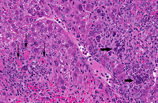
The ultrastructure of porcine circovirus was examined in persistently infected porcine kidney (PK)-15 cells. Virus-infected PK-15 cells had large numbers of intracytoplasmic inclusions, and a few cells had intranuclear inclusions. Intracytoplasmic inclusions were dispersed throughout the cytoplasm but were most numerous in the perinuclear cytoplasm. Inclusion were of various sizes, round to oval, and electron dense and were of two general types. Inclusions of the first type were small (0.1-0.5 microm diameter), not surrounded by trilaminar membranes, and granular with indistinct margins that blended with surrounding cytoplasm. Some contained 12+/-2-nm-diameter icosahedral virions in loose aggregates or rarely forming paracrystalline arrays. Small inclusions could be sites of viral assembly or maturation. Intracytoplasmic inclusions of the second type were larger (0.5-5.0 microm diameter) and more numerous and had abrupt margins surrounded by trilaminar membranes. They were more electron dense than small inclusions and were heterogeneous, containing various proportions of aggregated virions, electron-dense crystalline lamellae of 5 nm periodicity, and/or whorls of myelinoid membranes. Virions usually formed paracrystalline arrays and occasionally were loosely aggregated. Larger inclusions were typical of autophagolysosomes. Intranuclear inclusions were not membrane bound and were often associated with reticulated nucleoli or aggregates of heterochromatin. Some inclusions were irregularly shaped aggregates of indistinct, circular 10-12-nm-diameter viruslike particles. Others were 0.1-1.0 microm in diameter, round or ring shaped, dense, and finely granular, with sharply demarcated margins. (+info)Electron microscopical observations of psittacine beak and feather disease in an Umbrella cockatoo (Cacatua alba). (4/530)
Psittacine beak and feather disease (PBFD) was diagnosed in an umbrella cockatoo (Cacatua alba) with severe feather dystrophy and loss. Electron microscopically, the intranuclear and intracytoplasmic inclusion bodies observed by light microscopy were composed of viral particles forming paracrystalline arrays, whorls, semicircles or concentric circles. Recovered viral particles from the skin and feather follicle tracts were icosahedral and 15 to 20 nm in diameter. (+info)Detection of porcine circovirus from lesions of a pig with wasting disease in Japan. (5/530)
A wasting disease characterized by progressive weight loss and dyspnea has been observed in weaning pigs on a farm in Yamagata Prefecture in 1998. Histopathologic findings in an affected pig were bronchointerstitial pneumonia and intracytoplasmic clusters of basophilic inclusions in macrophages of lymph nodes, which were similar to those in pigs with postweaning multisystemic wasting syndrome (PMWS) recently reported in North America and Europe. Porcine circovirus (PCV)-like particles were observed in bronchial lymph node of the pig by electron microscopy, and PCV antigens were detected in the lesions by immunohistochemical staining. PCV DNA was also detected in the lung and tonsil by PCR, and restriction fragment length polymorphism analysis of the PCR products with HinfI showed the same type of the PCV associated with PMWS (pmws PCV). Homology of nucleotide sequences between the PCR product and corresponding regions of published pmws PCV genomes was very high. These results indicated that virus detected in this study was pmws PCV. To our knowledge, this is the first report on the presence of pmws PCV in Japan. (+info)Herpesvirus infection in tortoises (Malacochersus tornieri and Testudo horsfieldii). (6/530)
Large numbers of pancake tortoises (Malacochersus tornieri) and Horsfield tortoises (Testudo horsfieldii) in three consignments imported into Japan died soon after arrival. Some tortoises in the first consignment were dead on arrival. Postmortem examination of two of the pancake tortoises and four of the Horsfield tortoises revealed necrotizing lesions of the oral mucosa in both species, primarily in the tongue. Eosinophilic to amphophilic inclusion bodies were visible in the nuclei of mucosal epithelial cells in the lesions. Similar inclusion bodies were observed in the liver, spleen, adrenal glands, stomach, lungs, kidneys, small and large intestines, pancreas, and cerebrum of the pancake tortoises and in the liver, spleen, and pancreas of the Horsfield tortoises. Electron microscopic examination of the cells containing inclusion bodies showed herpesvirus-like particles about 100 nm in diameter in the cytoplasm. Nested polymerase chain reaction analysis using a herpesvirus consensus primer method confirmed the presence of a characteristic herpesvirus base sequence in tissue from these lesions. (+info)Measles inclusion-body encephalitis caused by the vaccine strain of measles virus. (7/530)
We report a case of measles inclusion-body encephalitis (MIBE) occurring in an apparently healthy 21-month-old boy 8.5 months after measles-mumps-rubella vaccination. He had no prior evidence of immune deficiency and no history of measles exposure or clinical disease. During hospitalization, a primary immunodeficiency characterized by a profoundly depressed CD8 cell count and dysgammaglobulinemia was demonstrated. A brain biopsy revealed histopathologic features consistent with MIBE, and measles antigens were detected by immunohistochemical staining. Electron microscopy revealed inclusions characteristic of paramyxovirus nucleocapsids within neurons, oligodendroglia, and astrocytes. The presence of measles virus in the brain tissue was confirmed by reverse transcription polymerase chain reaction. The nucleotide sequence in the nucleoprotein and fusion gene regions was identical to that of the Moraten and Schwarz vaccine strains; the fusion gene differed from known genotype A wild-type viruses. (+info)Human granulocytic ehrlichiosis agent infection in a pony vaccinated with a Borrelia burgdorferi recombinant OspA vaccine and challenged by exposure to naturally infected ticks. (8/530)
A pony was vaccinated with recombinant OspA vaccine (rOspA) and then exposed 3 months later to Borrelia burgdorferi-infected ticks (Ixodes scapularis) collected in Westchester County, N.Y. At 2 weeks after tick exposure, the pony developed a high fever (105 degrees F). Buffy coat smears showed that 20% of neutrophils contained ehrlichial inclusion bodies (morulae). Flunixin Meglumine (1 g daily) was given for 2 days, and the body temperature returned to normal. PCR for ehrlichial DNA was performed on blood samples for 10 consecutive days beginning when the pony was first febrile. This pony was monitored for another 3.5 months but developed no further clinical signs. The 44-kDa immunodominant human granulocytic ehrlichiosis antigen gene was amplified by PCR and cloned into a pCR2.1 vector. DNA sequence analysis of this gene showed it was only 8 bp different (99% identity) from the results reported by others (J. W. Ijdo et al., Infect. Immun. 66:3264-3269, 1998). Western blot analysis, growth inhibition assays, and repeated attempts to isolate B. burgdorferi all demonstrated the pony was protected against B. burgdorferi infection. These results highlight the potential for ticks to harbor and transmit several pathogens simultaneously, which further complicates the diagnosis and vaccination of these emerging tick-borne diseases. (+info)Inclusion bodies are abnormal, intracellular accumulations or aggregations of various misfolded proteins, protein complexes, or other materials within the cells of an organism. They can be found in various tissues and cell types and are often associated with several pathological conditions, including infectious diseases, neurodegenerative disorders, and genetic diseases.
Inclusion bodies can vary in size, shape, and location depending on the specific disease or condition. Some inclusion bodies have a characteristic appearance under the microscope, such as eosinophilic (pink) staining with hematoxylin and eosin (H&E) histological stain, while others may require specialized stains or immunohistochemical techniques to identify the specific misfolded proteins involved.
Examples of diseases associated with inclusion bodies include:
1. Infectious diseases: Some viral infections, such as HIV, hepatitis B and C, and herpes simplex virus, can lead to the formation of inclusion bodies within infected cells.
2. Neurodegenerative disorders: Several neurodegenerative diseases are characterized by the presence of inclusion bodies, including Alzheimer's disease (amyloid-beta plaques and tau tangles), Parkinson's disease (Lewy bodies), Huntington's disease (Huntingtin aggregates), and amyotrophic lateral sclerosis (TDP-43 and SOD1 inclusions).
3. Genetic diseases: Certain genetic disorders, such as Danon disease, neuronal intranuclear inclusion disease, and some lysosomal storage disorders, can also present with inclusion bodies due to the accumulation of abnormal proteins or metabolic products within cells.
The exact role of inclusion bodies in disease pathogenesis remains unclear; however, they are often associated with cellular dysfunction, oxidative stress, and increased inflammation, which can contribute to disease progression and neurodegeneration.
Inclusion body myositis (IBM) is a rare inflammatory muscle disease characterized by progressive weakness and wasting (atrophy) of skeletal muscles. The term "inclusion body" refers to the presence of abnormal protein accumulations within muscle fibers, which are observed under a microscope during muscle biopsy. These inclusions are primarily composed of aggregated forms of amyloid-β and tau proteins, similar to those found in neurodegenerative disorders like Alzheimer's disease.
IBM typically affects individuals over 50 years old, and it is more common in men than women. The disease usually starts with weakness in the wrist and finger flexors, making it difficult to perform tasks such as gripping, buttoning shirts, or lifting objects. Over time, the weakness spreads to other muscle groups, including the thigh muscles (quadriceps), resulting in difficulty climbing stairs or rising from a seated position.
The exact cause of inclusion body myositis remains unclear; however, both immune-mediated and degenerative mechanisms are believed to contribute to its pathogenesis. Currently, there is no cure for IBM, and treatment options are primarily aimed at managing symptoms and improving quality of life. Immunosuppressive medications may be used to target the inflammatory component of the disease; however, their efficacy varies among patients. Physical therapy and exercise programs can help maintain muscle strength and function as much as possible.
Inclusion bodies, viral are typically described as intracellular inclusions that appear as a result of viral infections. These inclusion bodies consist of aggregates of virus-specific proteins, viral particles, or both, which accumulate inside the host cell's cytoplasm or nucleus during the replication cycle of certain viruses.
The presence of inclusion bodies can sometimes be observed through histological or cytological examination using various staining techniques. Different types of viruses may exhibit distinct morphologies and locations of these inclusion bodies, which can aid in the identification and diagnosis of specific viral infections. However, it is important to note that not all viral infections result in the formation of inclusion bodies, and their presence does not necessarily indicate active viral replication or infection.
Protein refolding is the process by which a denatured or misfolded protein reverts to its native, functional three-dimensional structure. Proteins are complex molecules that perform a wide range of functions within living organisms. Their function is heavily dependent on their unique three-dimensional shape, which is determined by the specific sequence of amino acids that make up the protein.
When proteins are exposed to certain environmental conditions, such as changes in temperature, pH, or the presence of denaturing agents, they can lose their native conformation and become denatured or misfolded. This can result in the loss of protein function and, in some cases, the formation of aggregates that can be toxic to cells.
Protein refolding is a crucial process for maintaining proper protein function within cells. It involves several steps:
1. Unfolding: The denatured or misfolded protein must first be unfolded into its linear amino acid sequence. This can be accomplished through various methods, such as exposure to chemical denaturants or changes in pH.
2. Renaturation: Once the protein is unfolded, it can begin to refold into its native conformation. This process is often facilitated by chaperone proteins, which help to stabilize the protein and prevent aggregation during the refolding process.
3. Folding: The protein must then fold into its correct three-dimensional structure. This is a complex process that involves the formation of specific bonds between amino acids, as well as the interaction with other molecules in the cell.
4. Quality control: Once the protein has folded, it must be checked for correct folding and function. Misfolded proteins may be targeted for degradation by the cell's quality control mechanisms.
Protein refolding is a critical process that occurs naturally within cells, but it can also be studied in vitro (outside of the cell) using various techniques. Understanding the mechanisms of protein refolding is important for developing therapies for diseases caused by protein misfolding and aggregation, such as Alzheimer's disease and Parkinson's disease.
Protein renaturation is the process of restoring the native, functional structure of a protein that has been denatured due to exposure to external stressors such as changes in temperature, pH, or the addition of chemical agents. Denaturation causes proteins to lose their unique three-dimensional structure, which is essential for their proper function. Renaturation involves slowly removing these stressors and allowing the protein to refold into its original configuration, restoring its biological activity. This process can be facilitated by various techniques, including dialysis, dilution, or the addition of specific chemical chaperones.
'Escherichia coli' (E. coli) is a type of gram-negative, facultatively anaerobic, rod-shaped bacterium that commonly inhabits the intestinal tract of humans and warm-blooded animals. It is a member of the family Enterobacteriaceae and one of the most well-studied prokaryotic model organisms in molecular biology.
While most E. coli strains are harmless and even beneficial to their hosts, some serotypes can cause various forms of gastrointestinal and extraintestinal illnesses in humans and animals. These pathogenic strains possess virulence factors that enable them to colonize and damage host tissues, leading to diseases such as diarrhea, urinary tract infections, pneumonia, and sepsis.
E. coli is a versatile organism with remarkable genetic diversity, which allows it to adapt to various environmental niches. It can be found in water, soil, food, and various man-made environments, making it an essential indicator of fecal contamination and a common cause of foodborne illnesses. The study of E. coli has contributed significantly to our understanding of fundamental biological processes, including DNA replication, gene regulation, and protein synthesis.
Protein folding is the process by which a protein molecule naturally folds into its three-dimensional structure, following the synthesis of its amino acid chain. This complex process is determined by the sequence and properties of the amino acids, as well as various environmental factors such as temperature, pH, and the presence of molecular chaperones. The final folded conformation of a protein is crucial for its proper function, as it enables the formation of specific interactions between different parts of the molecule, which in turn define its biological activity. Protein misfolding can lead to various diseases, including neurodegenerative disorders such as Alzheimer's and Parkinson's disease.
Recombinant proteins are artificially created proteins produced through the use of recombinant DNA technology. This process involves combining DNA molecules from different sources to create a new set of genes that encode for a specific protein. The resulting recombinant protein can then be expressed, purified, and used for various applications in research, medicine, and industry.
Recombinant proteins are widely used in biomedical research to study protein function, structure, and interactions. They are also used in the development of diagnostic tests, vaccines, and therapeutic drugs. For example, recombinant insulin is a common treatment for diabetes, while recombinant human growth hormone is used to treat growth disorders.
The production of recombinant proteins typically involves the use of host cells, such as bacteria, yeast, or mammalian cells, which are engineered to express the desired protein. The host cells are transformed with a plasmid vector containing the gene of interest, along with regulatory elements that control its expression. Once the host cells are cultured and the protein is expressed, it can be purified using various chromatography techniques.
Overall, recombinant proteins have revolutionized many areas of biology and medicine, enabling researchers to study and manipulate proteins in ways that were previously impossible.
Intranuclear inclusion bodies are abnormal, rounded structures found within the nucleus of a cell. They are composed of aggregated proteins or other cellular components and can be associated with various viral infections and certain genetic disorders. These inclusion bodies can interfere with normal nuclear functions, leading to cell damage and contributing to the pathogenesis of diseases such as cytomegalovirus infection, rabies, and some forms of neurodegenerative disorders like polyglutamine diseases. The presence of intranuclear inclusion bodies is often used in diagnostic pathology to help identify specific underlying conditions.
Myositis is a medical term that refers to inflammation of the muscle tissue. This condition can cause various symptoms, including muscle weakness, pain, swelling, and stiffness. There are several types of myositis, such as polymyositis, dermatomyositis, and inclusion body myositis, which have different causes and characteristics.
Polymyositis is a type of myositis that affects multiple muscle groups, particularly those close to the trunk of the body. Dermatomyositis is characterized by muscle inflammation as well as a skin rash. Inclusion body myositis is a less common form of myositis that typically affects older adults and can cause both muscle weakness and wasting.
The causes of myositis vary depending on the type, but they can include autoimmune disorders, infections, medications, and other medical conditions. Treatment for myositis may involve medication to reduce inflammation, physical therapy to maintain muscle strength and flexibility, and lifestyle changes to manage symptoms and prevent complications.
Osteitis deformans, also known as Paget's disease of bone, is a chronic disorder of the bone characterized by abnormal turnover and remodeling of the bone. In this condition, the bone becomes enlarged, thickened, and deformed due to excessive and disorganized bone formation and resorption.
The process begins when the bone-remodeling cycle is disrupted, leading to an imbalance between the activity of osteoclasts (cells that break down bone) and osteoblasts (cells that form new bone). In Paget's disease, osteoclasts become overactive and increase bone resorption, followed by an overzealous response from osteoblasts, which attempt to repair the damage but do so in a disorganized manner.
The affected bones can become weakened, prone to fractures, and may cause pain, deformities, or other complications such as arthritis, hearing loss, or neurological symptoms if the skull or spine is involved. The exact cause of Paget's disease remains unknown, but it is believed that genetic and environmental factors play a role in its development.
Early diagnosis and treatment can help manage the symptoms and prevent complications associated with osteitis deformans. Treatment options include medications to slow down bone turnover, pain management, and orthopedic interventions when necessary.
An Aviadenovirus is a type of virus that belongs to the family *Adenoviridae* and the genus *Aviadenovirus*. These viruses primarily infect avian species, such as birds, and can cause a variety of diseases. The genome of an Aviadenovirus is double-stranded DNA. Some species of Aviadenoviruses have been known to cause respiratory and reproductive problems in poultry, leading to significant economic losses in the poultry industry. It's important to note that Aviadenoviruses are not known to infect or cause disease in humans.
Solubility is a fundamental concept in pharmaceutical sciences and medicine, which refers to the maximum amount of a substance (solute) that can be dissolved in a given quantity of solvent (usually water) at a specific temperature and pressure. Solubility is typically expressed as mass of solute per volume or mass of solvent (e.g., grams per liter, milligrams per milliliter). The process of dissolving a solute in a solvent results in a homogeneous solution where the solute particles are dispersed uniformly throughout the solvent.
Understanding the solubility of drugs is crucial for their formulation, administration, and therapeutic effectiveness. Drugs with low solubility may not dissolve sufficiently to produce the desired pharmacological effect, while those with high solubility might lead to rapid absorption and short duration of action. Therefore, optimizing drug solubility through various techniques like particle size reduction, salt formation, or solubilization is an essential aspect of drug development and delivery.
Distal myopathies are a group of rare genetic muscle disorders that primarily affect the muscles of the hands, feet, and lower legs. These myopathies are characterized by progressive weakness and wasting (atrophy) of the distal muscles, which are located further from the center of the body. The onset of symptoms can vary widely, ranging from early childhood to late adulthood.
There are several different types of distal myopathies, each caused by mutations in specific genes that affect muscle function. Some common forms include:
1. Nonaka Distal Myopathy: This form is caused by mutations in the GNE gene and typically presents in the third or fourth decade of life with weakness and wasting of the ankle dorsiflexors, foot extensors, and wrist and finger extensors.
2. Miyoshi Distal Myopathy: This form is caused by mutations in the DYSF gene and affects the calf muscles initially, followed by weakness in other distal muscles over time.
3. Welander Distal Myopathy: This form is caused by mutations in the TIA1 gene and typically presents in adulthood with weakness and wasting of the hand and forearm muscles.
4. Laing Distal Myopathy: This form is caused by mutations in the CAV3 gene and affects the anterior compartment of the lower leg, resulting in foot drop and weakness of the ankle dorsiflexors.
5. Gowers Distal Myopathy: This form is caused by mutations in the HNRNPDL gene and typically presents in adulthood with weakness and wasting of the hand and forearm muscles, as well as foot drop.
There is no cure for distal myopathies, but treatment can help manage symptoms and improve quality of life. Physical therapy, bracing, and orthotics may be used to support weakened muscles and maintain mobility. In some cases, medications such as corticosteroids or immunosuppressants may be prescribed to reduce muscle inflammation and slow disease progression.
Molecular sequence data refers to the specific arrangement of molecules, most commonly nucleotides in DNA or RNA, or amino acids in proteins, that make up a biological macromolecule. This data is generated through laboratory techniques such as sequencing, and provides information about the exact order of the constituent molecules. This data is crucial in various fields of biology, including genetics, evolution, and molecular biology, allowing for comparisons between different organisms, identification of genetic variations, and studies of gene function and regulation.
Boidae is a family of snakes, also known as boas. This family includes many different species of large, non-venomous snakes found in various parts of the world, particularly in Central and South America, Africa, and Asia. Boas are known for their strong bodies and muscular tails, which they use to constrict their prey before swallowing it whole. Some well-known members of this family include the anaconda, the python, and the boa constrictor.
Electron microscopy (EM) is a type of microscopy that uses a beam of electrons to create an image of the sample being examined, resulting in much higher magnification and resolution than light microscopy. There are several types of electron microscopy, including transmission electron microscopy (TEM), scanning electron microscopy (SEM), and reflection electron microscopy (REM).
In TEM, a beam of electrons is transmitted through a thin slice of the sample, and the electrons that pass through the sample are focused to form an image. This technique can provide detailed information about the internal structure of cells, viruses, and other biological specimens, as well as the composition and structure of materials at the atomic level.
In SEM, a beam of electrons is scanned across the surface of the sample, and the electrons that are scattered back from the surface are detected to create an image. This technique can provide information about the topography and composition of surfaces, as well as the structure of materials at the microscopic level.
REM is a variation of SEM in which the beam of electrons is reflected off the surface of the sample, rather than scattered back from it. This technique can provide information about the surface chemistry and composition of materials.
Electron microscopy has a wide range of applications in biology, medicine, and materials science, including the study of cellular structure and function, disease diagnosis, and the development of new materials and technologies.
Adenoviridae infections refer to diseases caused by members of the Adenoviridae family of viruses, which are non-enveloped, double-stranded DNA viruses. These viruses can infect a wide range of hosts, including humans, animals, and birds. In humans, adenovirus infections can cause a variety of symptoms, depending on the specific type of virus and the age and immune status of the infected individual.
Common manifestations of adenovirus infections in humans include:
1. Respiratory illness: Adenoviruses are a common cause of respiratory tract infections, such as bronchitis, pneumonia, and croup. They can also cause conjunctivitis (pink eye) and pharyngoconjunctival fever.
2. Gastrointestinal illness: Some types of adenoviruses can cause diarrhea, vomiting, and abdominal pain, particularly in children and immunocompromised individuals.
3. Genitourinary illness: Adenoviruses have been associated with urinary tract infections, hemorrhagic cystitis, and nephritis.
4. Eye infections: Epidemic keratoconjunctivitis is a severe form of conjunctivitis caused by certain adenovirus types.
5. Central nervous system infections: Adenoviruses have been linked to meningitis, encephalitis, and other neurological disorders, although these are rare.
Transmission of adenoviruses typically occurs through respiratory droplets, contaminated surfaces, or contaminated water. Preventive measures include good hygiene practices, such as handwashing and avoiding close contact with infected individuals. There is no specific treatment for adenovirus infections, but supportive care can help alleviate symptoms. In severe cases or in immunocompromised patients, antiviral therapy may be considered.
An amino acid sequence is the specific order of amino acids in a protein or peptide molecule, formed by the linking of the amino group (-NH2) of one amino acid to the carboxyl group (-COOH) of another amino acid through a peptide bond. The sequence is determined by the genetic code and is unique to each type of protein or peptide. It plays a crucial role in determining the three-dimensional structure and function of proteins.
Molecular cloning is a laboratory technique used to create multiple copies of a specific DNA sequence. This process involves several steps:
1. Isolation: The first step in molecular cloning is to isolate the DNA sequence of interest from the rest of the genomic DNA. This can be done using various methods such as PCR (polymerase chain reaction), restriction enzymes, or hybridization.
2. Vector construction: Once the DNA sequence of interest has been isolated, it must be inserted into a vector, which is a small circular DNA molecule that can replicate independently in a host cell. Common vectors used in molecular cloning include plasmids and phages.
3. Transformation: The constructed vector is then introduced into a host cell, usually a bacterial or yeast cell, through a process called transformation. This can be done using various methods such as electroporation or chemical transformation.
4. Selection: After transformation, the host cells are grown in selective media that allow only those cells containing the vector to grow. This ensures that the DNA sequence of interest has been successfully cloned into the vector.
5. Amplification: Once the host cells have been selected, they can be grown in large quantities to amplify the number of copies of the cloned DNA sequence.
Molecular cloning is a powerful tool in molecular biology and has numerous applications, including the production of recombinant proteins, gene therapy, functional analysis of genes, and genetic engineering.
Chymosin, also known as rennin or rennet, is a proteolytic enzyme that is naturally present in the stomachs of ruminant animals such as cows, goats, and sheep. It plays an essential role in the digestion of milk in these animals by curdling or coagulating the milk protein casein, which helps in the separation of solid curds from liquid whey during the process of stomach digestion.
In the context of food production, chymosin is often used as a coagulant in the manufacturing of cheese and other dairy products. Traditionally, rennet was obtained by extracting it from the fourth stomach chamber (abomasum) of young calves, but nowadays, most commercial chymosin is produced through microbial fermentation using genetically modified bacteria or yeast that have been engineered to produce this enzyme. This method of production allows for a more consistent and animal-friendly source of chymosin for industrial applications.
The primary function of chymosin in cheese making is to catalyze the coagulation of casein, leading to the formation of a curd that can be further processed into various types of cheese. The enzyme specifically cleaves a bond in the casein protein called Phe105-Met106, resulting in the formation of para-κ-casein and paracaseinompholine, which then interact to form the curd. This reaction is crucial for initiating the cheese making process, as it allows for the separation of solid curds from liquid whey, which can then be pressed, aged, and transformed into a wide variety of cheese styles.
Recombinant fusion proteins are artificially created biomolecules that combine the functional domains or properties of two or more different proteins into a single protein entity. They are generated through recombinant DNA technology, where the genes encoding the desired protein domains are linked together and expressed as a single, chimeric gene in a host organism, such as bacteria, yeast, or mammalian cells.
The resulting fusion protein retains the functional properties of its individual constituent proteins, allowing for novel applications in research, diagnostics, and therapeutics. For instance, recombinant fusion proteins can be designed to enhance protein stability, solubility, or immunogenicity, making them valuable tools for studying protein-protein interactions, developing targeted therapies, or generating vaccines against infectious diseases or cancer.
Examples of recombinant fusion proteins include:
1. Etaglunatide (ABT-523): A soluble Fc fusion protein that combines the heavy chain fragment crystallizable region (Fc) of an immunoglobulin with the extracellular domain of the human interleukin-6 receptor (IL-6R). This fusion protein functions as a decoy receptor, neutralizing IL-6 and its downstream signaling pathways in rheumatoid arthritis.
2. Etanercept (Enbrel): A soluble TNF receptor p75 Fc fusion protein that binds to tumor necrosis factor-alpha (TNF-α) and inhibits its proinflammatory activity, making it a valuable therapeutic option for treating autoimmune diseases like rheumatoid arthritis, ankylosing spondylitis, and psoriasis.
3. Abatacept (Orencia): A fusion protein consisting of the extracellular domain of cytotoxic T-lymphocyte antigen 4 (CTLA-4) linked to the Fc region of an immunoglobulin, which downregulates T-cell activation and proliferation in autoimmune diseases like rheumatoid arthritis.
4. Belimumab (Benlysta): A monoclonal antibody that targets B-lymphocyte stimulator (BLyS) protein, preventing its interaction with the B-cell surface receptor and inhibiting B-cell activation in systemic lupus erythematosus (SLE).
5. Romiplostim (Nplate): A fusion protein consisting of a thrombopoietin receptor agonist peptide linked to an immunoglobulin Fc region, which stimulates platelet production in patients with chronic immune thrombocytopenia (ITP).
6. Darbepoetin alfa (Aranesp): A hyperglycosylated erythropoiesis-stimulating protein that functions as a longer-acting form of recombinant human erythropoietin, used to treat anemia in patients with chronic kidney disease or cancer.
7. Palivizumab (Synagis): A monoclonal antibody directed against the F protein of respiratory syncytial virus (RSV), which prevents RSV infection and is administered prophylactically to high-risk infants during the RSV season.
8. Ranibizumab (Lucentis): A recombinant humanized monoclonal antibody fragment that binds and inhibits vascular endothelial growth factor A (VEGF-A), used in the treatment of age-related macular degeneration, diabetic retinopathy, and other ocular disorders.
9. Cetuximab (Erbitux): A chimeric monoclonal antibody that binds to epidermal growth factor receptor (EGFR), used in the treatment of colorectal cancer and head and neck squamous cell carcinoma.
10. Adalimumab (Humira): A fully humanized monoclonal antibody that targets tumor necrosis factor-alpha (TNF-α), used in the treatment of various inflammatory diseases, including rheumatoid arthritis, psoriasis, and Crohn's disease.
11. Bevacizumab (Avastin): A recombinant humanized monoclonal antibody that binds to VEGF-A, used in the treatment of various cancers, including colorectal, lung, breast, and kidney cancer.
12. Trastuzumab (Herceptin): A humanized monoclonal antibody that targets HER2/neu receptor, used in the treatment of breast cancer.
13. Rituximab (Rituxan): A chimeric monoclonal antibody that binds to CD20 antigen on B cells, used in the treatment of non-Hodgkin's lymphoma and rheumatoid arthritis.
14. Palivizumab (Synagis): A humanized monoclonal antibody that binds to the F protein of respiratory syncytial virus, used in the prevention of respiratory syncytial virus infection in high-risk infants.
15. Infliximab (Remicade): A chimeric monoclonal antibody that targets TNF-α, used in the treatment of various inflammatory diseases, including Crohn's disease, ulcerative colitis, rheumatoid arthritis, and ankylosing spondylitis.
16. Natalizumab (Tysabri): A humanized monoclonal antibody that binds to α4β1 integrin, used in the treatment of multiple sclerosis and Crohn's disease.
17. Adalimumab (Humira): A fully human monoclonal antibody that targets TNF-α, used in the treatment of various inflammatory diseases, including rheumatoid arthritis, psoriatic arthritis, ankylosing spondylitis, Crohn's disease, and ulcerative colitis.
18. Golimumab (Simponi): A fully human monoclonal antibody that targets TNF-α, used in the treatment of rheumatoid arthritis, psoriatic arthritis, ankylosing spondylitis, and ulcerative colitis.
19. Certolizumab pegol (Cimzia): A PEGylated Fab' fragment of a humanized monoclonal antibody that targets TNF-α, used in the treatment of rheumatoid arthritis, psoriatic arthritis, ankylosing spondylitis, and Crohn's disease.
20. Ustekinumab (Stelara): A fully human monoclonal antibody that targets IL-12 and IL-23, used in the treatment of psoriasis, psoriatic arthritis, and Crohn's disease.
21. Secukinumab (Cosentyx): A fully human monoclonal antibody that targets IL-17A, used in the treatment of psoriasis, psoriatic arthritis, and ankylosing spondylitis.
22. Ixekizumab (Taltz): A fully human monoclonal antibody that targets IL-17A, used in the treatment of psoriasis and psoriatic arthritis.
23. Brodalumab (Siliq): A fully human monoclonal antibody that targets IL-17 receptor A, used in the treatment of psoriasis.
24. Sarilumab (Kevzara): A fully human monoclonal antibody that targets the IL-6 receptor, used in the treatment of rheumatoid arthritis.
25. Tocilizumab (Actemra): A humanized monoclonal antibody that targets the IL-6 receptor, used in the treatment of rheumatoid arthritis, systemic juvenile idiopathic arthritis, polyarticular juvenile idiopathic arthritis, giant cell arteritis, and chimeric antigen receptor T-cell-induced cytokine release syndrome.
26. Siltuximab (Sylvant): A chimeric monoclonal antibody that targets IL-6, used in the treatment of multicentric Castleman disease.
27. Satralizumab (Enspryng): A humanized monoclonal antibody that targets IL-6 receptor alpha, used in the treatment of neuromyelitis optica spectrum disorder.
28. Sirukumab (Plivensia): A human monoclonal antibody that targets IL-6, used in the treatment
Polymyositis is defined as a rare inflammatory disorder that causes muscle weakness and inflammation (swelling) of the muscles. It primarily affects the skeletal muscles, which are the muscles responsible for voluntary movements such as walking, talking, and swallowing. The onset of polymyositis can occur at any age but is most commonly seen in adults between 31 to 60 years old, with women being slightly more affected than men.
The exact cause of polymyositis remains unknown; however, it is believed to be an autoimmune disorder, where the body's immune system mistakenly attacks its own muscle tissue. Certain factors such as genetics, viral infections, and exposure to certain drugs may contribute to the development of this condition.
Polymyositis can cause various symptoms, including:
- Progressive muscle weakness and wasting, particularly affecting the proximal muscles (those closest to the trunk of the body) such as the hips, thighs, shoulders, and upper arms.
- Difficulty climbing stairs, lifting objects, or rising from a seated position.
- Fatigue and stiffness, especially after periods of inactivity.
- Joint pain and swelling.
- Difficulty swallowing or speaking.
- Shortness of breath due to weakened respiratory muscles.
Diagnosis of polymyositis typically involves a combination of medical history, physical examination, laboratory tests, electromyography (EMG), and muscle biopsy. Treatment usually includes medications such as corticosteroids and immunosuppressants to reduce inflammation and control the immune response. Physical therapy may also be recommended to help maintain muscle strength and flexibility.
If left untreated, polymyositis can lead to significant disability and complications, including respiratory failure, malnutrition, and cardiovascular disease. Early diagnosis and treatment are crucial for improving outcomes and preventing long-term complications.
A base sequence in the context of molecular biology refers to the specific order of nucleotides in a DNA or RNA molecule. In DNA, these nucleotides are adenine (A), guanine (G), cytosine (C), and thymine (T). In RNA, uracil (U) takes the place of thymine. The base sequence contains genetic information that is transcribed into RNA and ultimately translated into proteins. It is the exact order of these bases that determines the genetic code and thus the function of the DNA or RNA molecule.
A mutation is a permanent change in the DNA sequence of an organism's genome. Mutations can occur spontaneously or be caused by environmental factors such as exposure to radiation, chemicals, or viruses. They may have various effects on the organism, ranging from benign to harmful, depending on where they occur and whether they alter the function of essential proteins. In some cases, mutations can increase an individual's susceptibility to certain diseases or disorders, while in others, they may confer a survival advantage. Mutations are the driving force behind evolution, as they introduce new genetic variability into populations, which can then be acted upon by natural selection.
Morbillivirus is a genus of viruses in the family Paramyxoviridae, order Mononegavirales. It includes several important human and animal pathogens that cause diseases with significant morbidity and mortality. The most well-known member of this genus is Measles virus (MV), which causes measles in humans, a highly contagious disease characterized by fever, rash, cough, and conjunctivitis.
Other important Morbilliviruses include:
* Rinderpest virus (RPV): This virus caused rinderpest, a severe disease in cattle and other cloven-hoofed animals, which was eradicated in 2011 through a global vaccination campaign.
* Canine Distemper Virus (CDV): A pathogen that affects dogs, wild canids, and several other mammalian species, causing a systemic disease with respiratory, gastrointestinal, and neurological symptoms.
* Phocine Distemper Virus (PDV) and Porpoise Morbillivirus (PMV): These viruses affect marine mammals, such as seals and porpoises, causing mass mortality events in their populations.
Morbilliviruses are enveloped, negative-sense, single-stranded RNA viruses with a genome size of approximately 15-16 kilobases. They have a pleomorphic shape and can vary in diameter from 150 to 750 nanometers. The viral envelope contains two glycoproteins: the hemagglutinin (H) protein, which mediates attachment to host cells, and the fusion (F) protein, which facilitates membrane fusion and viral entry.
Transmission of Morbilliviruses typically occurs through respiratory droplets or direct contact with infected individuals or animals. The viruses can cause acute infections with high fatality rates, particularly in naïve populations that lack immunity due to insufficient vaccination coverage or the absence of previous exposure.
In summary, Morbillivirus is a genus of viruses in the family Paramyxoviridae that includes several important human and animal pathogens causing acute respiratory infections with high fatality rates. Transmission occurs through respiratory droplets or direct contact, and vaccination plays a crucial role in preventing outbreaks and controlling disease spread.
Protein denaturation is a process in which the native structure of a protein is altered, leading to loss of its biological activity. This can be caused by various factors such as changes in temperature, pH, or exposure to chemicals or radiation. The three-dimensional shape of a protein is crucial for its function, and denaturation causes the protein to lose this shape, resulting in impaired or complete loss of function. Denaturation is often irreversible and can lead to the aggregation of proteins, which can have negative effects on cellular function and can contribute to diseases such as Alzheimer's and Parkinson's.
Frontotemporal dementia (FTD) is a group of disorders caused by progressive degeneration of the frontal and temporal lobes of the brain. These areas of the brain are associated with personality, behavior, and language.
There are three main types of FTD:
1. Behavioral variant FTD (bvFTD): This type is characterized by changes in personality, behavior, and judgment. Individuals may become socially inappropriate, emotionally indifferent, or impulsive. They may lose interest in things they used to enjoy and have difficulty with tasks that require planning and organization.
2. Primary progressive aphasia (PPA): This type affects language abilities. There are two main subtypes of PPA: semantic dementia and progressive nonfluent aphasia. Semantic dementia is characterized by difficulty understanding words and objects, while progressive nonfluent aphasia is characterized by problems with speech production and articulation.
3. Motor neuron disease (MND) associated FTD: Some individuals with FTD may also develop motor neuron disease, which affects the nerves that control muscle movement. This can lead to weakness, stiffness, and wasting of muscles, as well as difficulty swallowing and speaking.
FTD is a degenerative disorder, meaning that symptoms get worse over time. There is no cure for FTD, but there are treatments available to help manage symptoms and improve quality of life. The exact cause of FTD is not known, but it is believed to be related to abnormalities in certain proteins in the brain. In some cases, FTD may run in families and be caused by genetic mutations.
Erythrocyte inclusions refer to the presence of abnormal structures or substances within red blood cells (erythrocytes). These inclusions can be composed of various materials such as proteins, pigments, or foreign bodies. They may be seen in a variety of medical conditions and can provide important diagnostic clues.
Some examples of erythrocyte inclusions include:
1. Howell-Jolly bodies: small remnants of nuclear material left behind after the red blood cell matures. They are typically seen in individuals with an absent or nonfunctional spleen.
2. Heinz bodies: denatured hemoglobin that forms clumps within the red blood cells. They can be seen in conditions such as hemolytic anemia, G6PD deficiency, and exposure to certain drugs or toxins.
3. Pappenheimer bodies: aggregates of iron-containing proteins called ferritin or hemosiderin. They are typically seen in conditions associated with increased red blood cell destruction, such as thalassemia or lead poisoning.
4. Basophilic stippling: small, basophilic (blue-staining) granules within the red blood cells. They can be seen in various conditions, including lead poisoning, megaloblastic anemias, and certain inherited disorders.
5. Parasites: organisms such as malaria or babesia that infect and multiply within the red blood cells.
The detection of erythrocyte inclusions typically requires specialized testing, such as peripheral blood smears stained with specific dyes to highlight the abnormal structures. The presence and type of inclusions can help diagnose certain medical conditions and guide appropriate treatment.
Lewy bodies are abnormal aggregates of alpha-synuclein protein that develop in nerve cells (neurons) in the brain. They are named after Frederick Lewy, a German-American neurologist who discovered them while working with Dr. Alois Alzheimer. The presence of Lewy bodies is a hallmark feature of Lewy body dementia, which includes both Parkinson's disease dementia and dementia with Lewy bodies.
Lewy bodies can lead to the dysfunction and death of neurons in areas of the brain that control movement, cognition, and behavior. This can result in a range of symptoms, including motor impairments, cognitive decline, visual hallucinations, and mood changes. The exact role of Lewy bodies in the development and progression of these disorders is not fully understood, but they are believed to contribute to the neurodegenerative process that underlies these conditions.
Morbillivirus infections refer to a group of viral illnesses caused by members of the Morbillivirus genus, which is part of the Paramyxoviridae family. The most well-known morbillivirus infection is measles, a highly contagious disease that primarily affects humans. Other examples of morbillivirus infections include:
1. Canine distemper: A viral illness that affects dogs and other animals such as raccoons, ferrets, and skunks. It can cause respiratory, gastrointestinal, and neurological symptoms.
2. Phocine distemper: A viral disease primarily affecting seals, particularly the harbor seal population in Europe. It can lead to severe respiratory and neurological issues.
3. Rinderpest: A highly contagious and fatal disease that affects cattle, buffalo, and other even-toed ungulates (hoofed mammals). This disease has been eradicated globally through vaccination programs.
4. Peste des petits ruminants (PPR): Also known as sheep and goat plague, this morbillivirus infection affects small ruminants such as sheep and goats. It can cause severe respiratory, gastrointestinal, and reproductive symptoms.
5. Cetacean morbillivirus (CeMV) infections: These affect various species of whales, dolphins, and porpoises, causing respiratory, neurological, and immunological issues.
Morbillivirus infections are typically spread through direct contact with infected individuals or their bodily fluids. Vaccination programs have been successful in controlling and eradicating some of these diseases, such as rinderpest and measles.
Alpha-synuclein is a protein that is primarily found in neurons (nerve cells) in the brain. It is encoded by the SNCA gene and is abundantly expressed in presynaptic terminals, where it is believed to play a role in the regulation of neurotransmitter release.
In certain neurological disorders, including Parkinson's disease, dementia with Lewy bodies, and multiple system atrophy, alpha-synuclein can form aggregates known as Lewy bodies and Lewy neurites. These aggregates are a pathological hallmark of these diseases and are believed to contribute to the death of nerve cells, leading to the symptoms associated with these disorders.
The precise function of alpha-synuclein is not fully understood, but it is thought to be involved in various cellular processes such as maintaining the structure of the presynaptic terminal, regulating synaptic vesicle trafficking and neurotransmitter release, and protecting neurons from stress.
Cytoplasm is the material within a eukaryotic cell (a cell with a true nucleus) that lies between the nuclear membrane and the cell membrane. It is composed of an aqueous solution called cytosol, in which various organelles such as mitochondria, ribosomes, endoplasmic reticulum, Golgi apparatus, lysosomes, and vacuoles are suspended. Cytoplasm also contains a variety of dissolved nutrients, metabolites, ions, and enzymes that are involved in various cellular processes such as metabolism, signaling, and transport. It is where most of the cell's metabolic activities take place, and it plays a crucial role in maintaining the structure and function of the cell.
Synucleins are a family of small, heat-stable, water-soluble proteins that are primarily expressed in neurons. They are involved in various cellular processes such as modulating synaptic plasticity, vesicle trafficking, and neurotransmitter release. The most well-known members of this family are alpha-synuclein, beta-synuclein, and gamma-synuclein.
Abnormal accumulation and aggregation of alpha-synuclein into insoluble fibrils called Lewy bodies and Lewy neurites are hallmark features of several neurodegenerative disorders, including Parkinson's disease, dementia with Lewy bodies, and multiple system atrophy. These conditions are collectively referred to as synucleinopathies. The dysfunction and aggregation of alpha-synuclein are thought to contribute to the progressive loss of dopaminergic neurons in the substantia nigra pars compacta, a region of the brain involved in motor control, leading to the characteristic symptoms observed in these disorders.
Ubiquitin is a small protein that is present in all eukaryotic cells and plays a crucial role in the regulation of various cellular processes, such as protein degradation, DNA repair, and stress response. It is involved in marking proteins for destruction by attaching to them, a process known as ubiquitination. This modification can target proteins for degradation by the proteasome, a large protein complex that breaks down unneeded or damaged proteins in the cell. Ubiquitin also has other functions, such as regulating the localization and activity of certain proteins. The ability of ubiquitin to modify many different proteins and play a role in multiple cellular processes makes it an essential player in maintaining cellular homeostasis.
Electrophoresis, polyacrylamide gel (EPG) is a laboratory technique used to separate and analyze complex mixtures of proteins or nucleic acids (DNA or RNA) based on their size and electrical charge. This technique utilizes a matrix made of cross-linked polyacrylamide, a type of gel, which provides a stable and uniform environment for the separation of molecules.
In this process:
1. The polyacrylamide gel is prepared by mixing acrylamide monomers with a cross-linking agent (bis-acrylamide) and a catalyst (ammonium persulfate) in the presence of a buffer solution.
2. The gel is then poured into a mold and allowed to polymerize, forming a solid matrix with uniform pore sizes that depend on the concentration of acrylamide used. Higher concentrations result in smaller pores, providing better resolution for separating smaller molecules.
3. Once the gel has set, it is placed in an electrophoresis apparatus containing a buffer solution. Samples containing the mixture of proteins or nucleic acids are loaded into wells on the top of the gel.
4. An electric field is applied across the gel, causing the negatively charged molecules to migrate towards the positive electrode (anode) while positively charged molecules move toward the negative electrode (cathode). The rate of migration depends on the size, charge, and shape of the molecules.
5. Smaller molecules move faster through the gel matrix and will migrate farther from the origin compared to larger molecules, resulting in separation based on size. Proteins and nucleic acids can be selectively stained after electrophoresis to visualize the separated bands.
EPG is widely used in various research fields, including molecular biology, genetics, proteomics, and forensic science, for applications such as protein characterization, DNA fragment analysis, cloning, mutation detection, and quality control of nucleic acid or protein samples.
A plasmid is a small, circular, double-stranded DNA molecule that is separate from the chromosomal DNA of a bacterium or other organism. Plasmids are typically not essential for the survival of the organism, but they can confer beneficial traits such as antibiotic resistance or the ability to degrade certain types of pollutants.
Plasmids are capable of replicating independently of the chromosomal DNA and can be transferred between bacteria through a process called conjugation. They often contain genes that provide resistance to antibiotics, heavy metals, and other environmental stressors. Plasmids have also been engineered for use in molecular biology as cloning vectors, allowing scientists to replicate and manipulate specific DNA sequences.
Plasmids are important tools in genetic engineering and biotechnology because they can be easily manipulated and transferred between organisms. They have been used to produce vaccines, diagnostic tests, and genetically modified organisms (GMOs) for various applications, including agriculture, medicine, and industry.
Bacterial proteins are a type of protein that are produced by bacteria as part of their structural or functional components. These proteins can be involved in various cellular processes, such as metabolism, DNA replication, transcription, and translation. They can also play a role in bacterial pathogenesis, helping the bacteria to evade the host's immune system, acquire nutrients, and multiply within the host.
Bacterial proteins can be classified into different categories based on their function, such as:
1. Enzymes: Proteins that catalyze chemical reactions in the bacterial cell.
2. Structural proteins: Proteins that provide structural support and maintain the shape of the bacterial cell.
3. Signaling proteins: Proteins that help bacteria to communicate with each other and coordinate their behavior.
4. Transport proteins: Proteins that facilitate the movement of molecules across the bacterial cell membrane.
5. Toxins: Proteins that are produced by pathogenic bacteria to damage host cells and promote infection.
6. Surface proteins: Proteins that are located on the surface of the bacterial cell and interact with the environment or host cells.
Understanding the structure and function of bacterial proteins is important for developing new antibiotics, vaccines, and other therapeutic strategies to combat bacterial infections.
Transmission electron microscopy (TEM) is a type of microscopy in which an electron beam is transmitted through a ultra-thin specimen, interacting with it as it passes through. An image is formed from the interaction of the electrons with the specimen; the image is then magnified and visualized on a fluorescent screen or recorded on an electronic detector (or photographic film in older models).
TEM can provide high-resolution, high-magnification images that can reveal the internal structure of specimens including cells, viruses, and even molecules. It is widely used in biological and materials science research to investigate the ultrastructure of cells, tissues and materials. In medicine, TEM is used for diagnostic purposes in fields such as virology and bacteriology.
It's important to note that preparing a sample for TEM is a complex process, requiring specialized techniques to create thin (50-100 nm) specimens. These include cutting ultrathin sections of embedded samples using an ultramicrotome, staining with heavy metal salts, and positive staining or negative staining methods.
Distemper is a highly contagious viral disease that primarily affects dogs, but can also infect other animals such as cats, ferrets, and raccoons. It is caused by a paramyxovirus and is characterized by respiratory, gastrointestinal, and neurological symptoms.
The respiratory symptoms of distemper include coughing, sneezing, and nasal discharge. Gastrointestinal symptoms may include vomiting and diarrhea. Neurological symptoms can include seizures, twitching, and paralysis. Distemper is often fatal, especially in puppies and young dogs that have not been vaccinated.
The virus is spread through direct contact with infected animals or their bodily fluids, such as saliva and urine. It can also be spread through the air, making it highly contagious in areas where large numbers of unvaccinated animals are housed together, such as animal shelters and kennels.
Prevention is key in protecting against distemper, and vaccination is recommended for all dogs. Puppies should receive their first distemper vaccine at six to eight weeks of age, followed by booster shots every three to four weeks until they are 16 weeks old. Adult dogs should receive a distemper booster shot every one to three years, depending on their risk of exposure.
Skeletal muscle, also known as striated or voluntary muscle, is a type of muscle that is attached to bones by tendons or aponeuroses and functions to produce movements and support the posture of the body. It is composed of long, multinucleated fibers that are arranged in parallel bundles and are characterized by alternating light and dark bands, giving them a striped appearance under a microscope. Skeletal muscle is under voluntary control, meaning that it is consciously activated through signals from the nervous system. It is responsible for activities such as walking, running, jumping, and lifting objects.
Arenavirus is a type of virus that belongs to the family Arenaviridae. These viruses are enveloped and have a single-stranded, bi-segmented RNA genome. They are named after the Latin word "arena" which means "sand" because their virions contain ribosomes which resemble sand granules when viewed under an electron microscope.
Arenaviruses are primarily associated with rodents and can cause chronic infection in their natural hosts. Some arenaviruses can also infect humans and other animals, causing severe hemorrhagic fevers. Examples of human diseases caused by arenaviruses include Lassa fever, Argentine hemorrhagic fever, Bolivian hemorrhagic fever, and Venezuelan hemorrhagic fever.
These viruses are typically transmitted to humans through contact with infected rodents or their excreta, but some can also be spread from person to person through close contact with an infected individual's blood or other bodily fluids. There are currently no vaccines available for most arenaviruses, and treatment is primarily supportive, focusing on managing symptoms and complications.
Dermatomyositis is a medical condition characterized by inflammation and weakness in the muscles and skin. It is a type of inflammatory myopathy, which means that it causes muscle inflammation and damage. Dermatomyositis is often associated with a distinctive rash that affects the skin around the eyes, nose, mouth, fingers, and toes.
The symptoms of dermatomyositis can include:
* Progressive muscle weakness, particularly in the hips, thighs, shoulders, and neck
* Fatigue
* Difficulty swallowing or speaking
* Skin rash, which may be pink or purple and is often accompanied by itching
* Muscle pain and tenderness
* Joint pain and swelling
* Raynaud's phenomenon, a condition that affects blood flow to the fingers and toes
The exact cause of dermatomyositis is not known, but it is believed to be related to an autoimmune response in which the body's immune system mistakenly attacks healthy tissue. Treatment for dermatomyositis typically involves medications to reduce inflammation and suppress the immune system, as well as physical therapy to help maintain muscle strength and function.
Fowl adenovirus A, also known as Fowl aviadenovirus serotype 1 or Fowl adenovirus serotype 1 (FAdV-A), is a species of DNA virus that belongs to the family Adenoviridae and genus Aviadenovirus. It primarily infects birds, particularly chickens, causing various clinical manifestations such as inclusion body hepatitis (IBH) and hydropericardium syndrome (HPS). The virus is transmitted horizontally through the fecal-oral route and can be found in the environment for extended periods. FAdV-A infection can lead to significant economic losses in the poultry industry due to high mortality rates, especially in young chickens.
Gene expression is the process by which the information encoded in a gene is used to synthesize a functional gene product, such as a protein or RNA molecule. This process involves several steps: transcription, RNA processing, and translation. During transcription, the genetic information in DNA is copied into a complementary RNA molecule, known as messenger RNA (mRNA). The mRNA then undergoes RNA processing, which includes adding a cap and tail to the mRNA and splicing out non-coding regions called introns. The resulting mature mRNA is then translated into a protein on ribosomes in the cytoplasm through the process of translation.
The regulation of gene expression is a complex and highly controlled process that allows cells to respond to changes in their environment, such as growth factors, hormones, and stress signals. This regulation can occur at various stages of gene expression, including transcriptional activation or repression, RNA processing, mRNA stability, and translation. Dysregulation of gene expression has been implicated in many diseases, including cancer, genetic disorders, and neurological conditions.
Circular dichroism (CD) is a technique used in physics and chemistry to study the structure of molecules, particularly large biological molecules such as proteins and nucleic acids. It measures the difference in absorption of left-handed and right-handed circularly polarized light by a sample. This difference in absorption can provide information about the three-dimensional structure of the molecule, including its chirality or "handedness."
In more technical terms, CD is a form of spectroscopy that measures the differential absorption of left and right circularly polarized light as a function of wavelength. The CD signal is measured in units of millidegrees (mdeg) and can be positive or negative, depending on the type of chromophore and its orientation within the molecule.
CD spectra can provide valuable information about the secondary and tertiary structure of proteins, as well as the conformation of nucleic acids. For example, alpha-helical proteins typically exhibit a strong positive band near 190 nm and two negative bands at around 208 nm and 222 nm, while beta-sheet proteins show a strong positive band near 195 nm and two negative bands at around 217 nm and 175 nm.
CD spectroscopy is a powerful tool for studying the structural changes that occur in biological molecules under different conditions, such as temperature, pH, or the presence of ligands or other molecules. It can also be used to monitor the folding and unfolding of proteins, as well as the binding of drugs or other small molecules to their targets.
Huntington Disease (HD) is a genetic neurodegenerative disorder that affects both cognitive and motor functions. It is characterized by the progressive loss of neurons in various areas of the brain, particularly in the striatum and cortex. The disease is caused by an autosomal dominant mutation in the HTT gene, which codes for the huntingtin protein. The most common mutation is a CAG repeat expansion in this gene, leading to the production of an abnormal form of the huntingtin protein that is toxic to nerve cells.
The symptoms of HD typically appear between the ages of 30 and 50, but they can start earlier or later in life. The early signs of HD may include subtle changes in mood, cognition, and coordination. As the disease progresses, individuals with HD experience uncontrolled movements (chorea), emotional disturbances, cognitive decline, and difficulties with communication and swallowing. Eventually, they become dependent on others for their daily needs and lose their ability to walk, talk, and care for themselves.
There is currently no cure for HD, but medications and therapies can help manage the symptoms of the disease and improve quality of life. Genetic testing is available to confirm the diagnosis and provide information about the risk of passing the disease on to future generations.
Guanidine is not typically defined in the context of medical terminology, but rather, it is a chemical compound with the formula NH2(C=NH)NH2. However, guanidine and its derivatives do have medical relevance:
1. Guanidine is used as a medication in some neurological disorders, such as stiff-person syndrome, to reduce muscle spasms and rigidity. It acts on the central nervous system to decrease abnormal nerve impulses that cause muscle spasticity.
2. Guanidine derivatives are found in various medications used for treating diabetes, like metformin. These compounds help lower glucose production in the liver and improve insulin sensitivity in muscle cells.
3. In some cases, guanidine is used as a skin penetration enhancer in transdermal drug delivery systems to increase the absorption of certain medications through the skin.
It is essential to note that guanidine itself has limited medical use due to its potential toxicity and narrow therapeutic window. Its derivatives, like metformin, are more commonly used in medical practice.
I'm sorry for any confusion, but "viral tail proteins" is not a widely recognized or established medical term. The term "tail proteins" is used in the context of certain viruses, particularly bacteriophages (viruses that infect bacteria), which have a tail-like structure that helps them attach to and inject their genetic material into host cells.
However, even within this context, there isn't a specific concept known as "viral tail proteins" that has a widely accepted medical definition. The proteins that make up the tail structure of bacteriophages have various functions and are referred to by different names based on their roles. These can include terms like "tail fiber proteins," "tail tube proteins," "tail terminator proteins," etc.
If you're looking for information about a specific protein or group of proteins related to viral tails, I would be happy to help further if you could provide more details.
A caulimovirus is a type of virus that primarily infects plants. It is a double-stranded DNA (dsDNA) virus, which means that its genetic material is composed of a pair of DNA strands. Caulimoviruses are named after the type species of the group, Cauliflower mosaic virus (CaMV).
Caulimoviruses are unique among dsDNA viruses because they replicate through an RNA intermediate, using a reverse transcriptase enzyme to produce DNA copies of their genome. This is similar to the way that retroviruses, which infect animals, replicate.
Caulimoviruses are relatively large viruses, with genomes ranging in size from about 7 to 8 kilobases (kb). They have a complex structure, with several proteins encoded by their genome that are involved in various aspects of the virus's replication and assembly.
Caulimoviruses infect a wide range of plant hosts, including many important crops such as cauliflower, cabbage, tomato, and pepper. They can cause serious diseases in these plants, leading to significant economic losses. There are no known caulimovirus infections of humans or other animals.
'Escherichia coli (E. coli) proteins' refer to the various types of proteins that are produced and expressed by the bacterium Escherichia coli. These proteins play a critical role in the growth, development, and survival of the organism. They are involved in various cellular processes such as metabolism, DNA replication, transcription, translation, repair, and regulation.
E. coli is a gram-negative, facultative anaerobe that is commonly found in the intestines of warm-blooded organisms. It is widely used as a model organism in scientific research due to its well-studied genetics, rapid growth, and ability to be easily manipulated in the laboratory. As a result, many E. coli proteins have been identified, characterized, and studied in great detail.
Some examples of E. coli proteins include enzymes involved in carbohydrate metabolism such as lactase, sucrase, and maltose; proteins involved in DNA replication such as the polymerases, single-stranded binding proteins, and helicases; proteins involved in transcription such as RNA polymerase and sigma factors; proteins involved in translation such as ribosomal proteins, tRNAs, and aminoacyl-tRNA synthetases; and regulatory proteins such as global regulators, two-component systems, and transcription factors.
Understanding the structure, function, and regulation of E. coli proteins is essential for understanding the basic biology of this important organism, as well as for developing new strategies for combating bacterial infections and improving industrial processes involving bacteria.
Secondary protein structure refers to the local spatial arrangement of amino acid chains in a protein, typically described as regular repeating patterns held together by hydrogen bonds. The two most common types of secondary structures are the alpha-helix (α-helix) and the beta-pleated sheet (β-sheet). In an α-helix, the polypeptide chain twists around itself in a helical shape, with each backbone atom forming a hydrogen bond with the fourth amino acid residue along the chain. This forms a rigid rod-like structure that is resistant to bending or twisting forces. In β-sheets, adjacent segments of the polypeptide chain run parallel or antiparallel to each other and are connected by hydrogen bonds, forming a pleated sheet-like arrangement. These secondary structures provide the foundation for the formation of tertiary and quaternary protein structures, which determine the overall three-dimensional shape and function of the protein.
A cell line is a culture of cells that are grown in a laboratory for use in research. These cells are usually taken from a single cell or group of cells, and they are able to divide and grow continuously in the lab. Cell lines can come from many different sources, including animals, plants, and humans. They are often used in scientific research to study cellular processes, disease mechanisms, and to test new drugs or treatments. Some common types of human cell lines include HeLa cells (which come from a cancer patient named Henrietta Lacks), HEK293 cells (which come from embryonic kidney cells), and HUVEC cells (which come from umbilical vein endothelial cells). It is important to note that cell lines are not the same as primary cells, which are cells that are taken directly from a living organism and have not been grown in the lab.
Nerve tissue proteins are specialized proteins found in the nervous system that provide structural and functional support to nerve cells, also known as neurons. These proteins include:
1. Neurofilaments: These are type IV intermediate filaments that provide structural support to neurons and help maintain their shape and size. They are composed of three subunits - NFL (light), NFM (medium), and NFH (heavy).
2. Neuronal Cytoskeletal Proteins: These include tubulins, actins, and spectrins that provide structural support to the neuronal cytoskeleton and help maintain its integrity.
3. Neurotransmitter Receptors: These are specialized proteins located on the postsynaptic membrane of neurons that bind neurotransmitters released by presynaptic neurons, triggering a response in the target cell.
4. Ion Channels: These are transmembrane proteins that regulate the flow of ions across the neuronal membrane and play a crucial role in generating and transmitting electrical signals in neurons.
5. Signaling Proteins: These include enzymes, receptors, and adaptor proteins that mediate intracellular signaling pathways involved in neuronal development, differentiation, survival, and death.
6. Adhesion Proteins: These are cell surface proteins that mediate cell-cell and cell-matrix interactions, playing a crucial role in the formation and maintenance of neural circuits.
7. Extracellular Matrix Proteins: These include proteoglycans, laminins, and collagens that provide structural support to nerve tissue and regulate neuronal migration, differentiation, and survival.
Protein binding, in the context of medical and biological sciences, refers to the interaction between a protein and another molecule (known as the ligand) that results in a stable complex. This process is often reversible and can be influenced by various factors such as pH, temperature, and concentration of the involved molecules.
In clinical chemistry, protein binding is particularly important when it comes to drugs, as many of them bind to proteins (especially albumin) in the bloodstream. The degree of protein binding can affect a drug's distribution, metabolism, and excretion, which in turn influence its therapeutic effectiveness and potential side effects.
Protein-bound drugs may be less available for interaction with their target tissues, as only the unbound or "free" fraction of the drug is active. Therefore, understanding protein binding can help optimize dosing regimens and minimize adverse reactions.
Vacuoles are membrane-bound organelles found in the cells of most eukaryotic organisms. They are essentially fluid-filled sacs that store various substances, such as enzymes, waste products, and nutrients. In plants, vacuoles often contain water, ions, and various organic compounds, while in fungi, they may store lipids or pigments. Vacuoles can also play a role in maintaining the turgor pressure of cells, which is critical for cell shape and function.
In animal cells, vacuoles are typically smaller and less numerous than in plant cells. Animal cells have lysosomes, which are membrane-bound organelles that contain digestive enzymes and break down waste materials, cellular debris, and foreign substances. Lysosomes can be considered a type of vacuole, but they are more specialized in their function.
Overall, vacuoles are essential for maintaining the health and functioning of cells by providing a means to store and dispose of various substances.
The proteasome endopeptidase complex is a large protein complex found in the cells of eukaryotic organisms, as well as in archaea and some bacteria. It plays a crucial role in the degradation of damaged or unneeded proteins through a process called proteolysis. The proteasome complex contains multiple subunits, including both regulatory and catalytic particles.
The catalytic core of the proteasome is composed of four stacked rings, each containing seven subunits, forming a structure known as the 20S core particle. Three of these rings are made up of beta-subunits that contain the proteolytic active sites, while the fourth ring consists of alpha-subunits that control access to the interior of the complex.
The regulatory particles, called 19S or 11S regulators, cap the ends of the 20S core particle and are responsible for recognizing, unfolding, and translocating targeted proteins into the catalytic chamber. The proteasome endopeptidase complex can cleave peptide bonds in various ways, including hydrolysis of ubiquitinated proteins, which is an essential mechanism for maintaining protein quality control and regulating numerous cellular processes, such as cell cycle progression, signal transduction, and stress response.
In summary, the proteasome endopeptidase complex is a crucial intracellular machinery responsible for targeted protein degradation through proteolysis, contributing to various essential regulatory functions in cells.
Hemoglobin H (Hb H) is a type of abnormal hemoglobin that can occur in individuals with certain genetic disorders, such as hemoglobinopathies. It is formed when four beta-globin chains come together, instead of the usual two alpha and two beta chains found in normal adult hemoglobin (Hb A).
This abnormal structure can result from a mutation that causes the absence or deficiency of alpha-globin chains, leading to an excess of beta-globin chains. Hemoglobin H is often associated with conditions such as thalassemia, particularly when there is a severe deficiency of alpha-globin chain production (alpha-thalassemia).
Hemoglobin H can cause hemolytic anemia, which means that the red blood cells are destroyed prematurely. The severity of the condition depends on the degree of imbalance between alpha and beta chains and other genetic factors. Symptoms may include fatigue, jaundice, and splenomegaly (enlarged spleen).
Sonication is a medical and laboratory term that refers to the use of ultrasound waves to agitate particles in a liquid. This process is often used in medical and scientific research to break down or disrupt cells, tissue, or other substances that are being studied. The high-frequency sound waves create standing waves that cause the particles in the liquid to vibrate, which can lead to cavitation (the formation and collapse of bubbles) and ultimately result in the disruption of the cell membranes or other structures. This technique is commonly used in procedures such as sonication of blood cultures to release microorganisms from clots, enhancing their growth in culture media and facilitating their identification.
Muscular diseases, also known as myopathies, refer to a group of conditions that affect the functionality and health of muscle tissue. These diseases can be inherited or acquired and may result from inflammation, infection, injury, or degenerative processes. They can cause symptoms such as weakness, stiffness, cramping, spasms, wasting, and loss of muscle function.
Examples of muscular diseases include:
1. Duchenne Muscular Dystrophy (DMD): A genetic disorder that results in progressive muscle weakness and degeneration due to a lack of dystrophin protein.
2. Myasthenia Gravis: An autoimmune disease that causes muscle weakness and fatigue, typically affecting the eyes and face, throat, and limbs.
3. Inclusion Body Myositis (IBM): A progressive muscle disorder characterized by muscle inflammation and wasting, typically affecting older adults.
4. Polymyositis: An inflammatory myopathy that causes muscle weakness and inflammation throughout the body.
5. Metabolic Myopathies: A group of inherited disorders that affect muscle metabolism, leading to exercise intolerance, muscle weakness, and other symptoms.
6. Muscular Dystonias: Involuntary muscle contractions and spasms that can cause abnormal postures or movements.
It is important to note that muscular diseases can have a significant impact on an individual's quality of life, mobility, and overall health. Proper diagnosis and treatment are crucial for managing symptoms and improving outcomes.
Neurodegenerative diseases are a group of disorders characterized by progressive and persistent loss of neuronal structure and function, often leading to cognitive decline, functional impairment, and ultimately death. These conditions are associated with the accumulation of abnormal protein aggregates, mitochondrial dysfunction, oxidative stress, chronic inflammation, and genetic mutations in the brain. Examples of neurodegenerative diseases include Alzheimer's disease, Parkinson's disease, Huntington's disease, Amyotrophic Lateral Sclerosis (ALS), and Spinal Muscular Atrophy (SMA). The underlying causes and mechanisms of these diseases are not fully understood, and there is currently no cure for most neurodegenerative disorders. Treatment typically focuses on managing symptoms and slowing disease progression.
Immunohistochemistry (IHC) is a technique used in pathology and laboratory medicine to identify specific proteins or antigens in tissue sections. It combines the principles of immunology and histology to detect the presence and location of these target molecules within cells and tissues. This technique utilizes antibodies that are specific to the protein or antigen of interest, which are then tagged with a detection system such as a chromogen or fluorophore. The stained tissue sections can be examined under a microscope, allowing for the visualization and analysis of the distribution and expression patterns of the target molecule in the context of the tissue architecture. Immunohistochemistry is widely used in diagnostic pathology to help identify various diseases, including cancer, infectious diseases, and immune-mediated disorders.
Insect viruses, also known as entomoviruses, are viruses that specifically infect and replicate in insect hosts. These viruses can be found in various insect species, including those of medical and agricultural importance. Insect viruses can cause diseases in insect populations, leading to significant impacts on their growth, development, and survival. Some insect viruses have been studied as potential biological control agents for managing pest insects that affect crops or transmit diseases. Examples of insect viruses include Baculoviridae, Reoviridae, and Picornaviridae families.
Varicellovirus is a genus of viruses in the family Herpesviridae, subfamily Alphaherpesvirinae. This genus includes several human and animal viruses that are closely related to each other. The most well-known member of this genus is the Varicella-zoster virus (VZV), which causes two distinct diseases: chickenpox (varicella) and shingles (zoster).
The Varicellovirus genus includes the following species:
1. Human alphaherpesvirus 3 (Varicella-zoster virus)
2. Simian varicella virus
3. Bovine herpesvirus 1
4. Bovine herpesvirus 5
5. Pseudorabies virus
6. Equid herpesvirus 1
7. Equid herpesvirus 3
8. Equid herpesvirus 4
9. Equid herpesvirus 8
10. Equid herpesvirus 9
11. Cercopithecine herpesvirus 1 (Herpes B virus)
12. Cercopithecine herpesvirus 2
13. Suid herpesvirus 1 (Aujeszky's disease virus)
14. Canid herpesvirus 1
15. Felid herpesvirus 1
These viruses are characterized by their ability to establish latency in the nervous system of their hosts and reactivate later in life, causing recurrent disease. They typically have a broad host range and can infect multiple species within a family or genus.
I'm sorry for any confusion, but "Persia" is not a medical term. It is an ancient name for a region that is now modern-day Iran and parts of neighboring countries. If you have any medical questions or terms you would like defined, please let me know!
Viral proteins are the proteins that are encoded by the viral genome and are essential for the viral life cycle. These proteins can be structural or non-structural and play various roles in the virus's replication, infection, and assembly process. Structural proteins make up the physical structure of the virus, including the capsid (the protein shell that surrounds the viral genome) and any envelope proteins (that may be present on enveloped viruses). Non-structural proteins are involved in the replication of the viral genome and modulation of the host cell environment to favor viral replication. Overall, a thorough understanding of viral proteins is crucial for developing antiviral therapies and vaccines.
Western blotting is a laboratory technique used in molecular biology to detect and quantify specific proteins in a mixture of many different proteins. This technique is commonly used to confirm the expression of a protein of interest, determine its size, and investigate its post-translational modifications. The name "Western" blotting distinguishes this technique from Southern blotting (for DNA) and Northern blotting (for RNA).
The Western blotting procedure involves several steps:
1. Protein extraction: The sample containing the proteins of interest is first extracted, often by breaking open cells or tissues and using a buffer to extract the proteins.
2. Separation of proteins by electrophoresis: The extracted proteins are then separated based on their size by loading them onto a polyacrylamide gel and running an electric current through the gel (a process called sodium dodecyl sulfate-polyacrylamide gel electrophoresis or SDS-PAGE). This separates the proteins according to their molecular weight, with smaller proteins migrating faster than larger ones.
3. Transfer of proteins to a membrane: After separation, the proteins are transferred from the gel onto a nitrocellulose or polyvinylidene fluoride (PVDF) membrane using an electric current in a process called blotting. This creates a replica of the protein pattern on the gel but now immobilized on the membrane for further analysis.
4. Blocking: The membrane is then blocked with a blocking agent, such as non-fat dry milk or bovine serum albumin (BSA), to prevent non-specific binding of antibodies in subsequent steps.
5. Primary antibody incubation: A primary antibody that specifically recognizes the protein of interest is added and allowed to bind to its target protein on the membrane. This step may be performed at room temperature or 4°C overnight, depending on the antibody's properties.
6. Washing: The membrane is washed with a buffer to remove unbound primary antibodies.
7. Secondary antibody incubation: A secondary antibody that recognizes the primary antibody (often coupled to an enzyme or fluorophore) is added and allowed to bind to the primary antibody. This step may involve using a horseradish peroxidase (HRP)-conjugated or alkaline phosphatase (AP)-conjugated secondary antibody, depending on the detection method used later.
8. Washing: The membrane is washed again to remove unbound secondary antibodies.
9. Detection: A detection reagent is added to visualize the protein of interest by detecting the signal generated from the enzyme-conjugated or fluorophore-conjugated secondary antibody. This can be done using chemiluminescent, colorimetric, or fluorescent methods.
10. Analysis: The resulting image is analyzed to determine the presence and quantity of the protein of interest in the sample.
Western blotting is a powerful technique for identifying and quantifying specific proteins within complex mixtures. It can be used to study protein expression, post-translational modifications, protein-protein interactions, and more. However, it requires careful optimization and validation to ensure accurate and reproducible results.
'Bird diseases' is a broad term that refers to the various medical conditions and infections that can affect avian species. These diseases can be caused by bacteria, viruses, fungi, parasites, or toxic substances and can affect pet birds, wild birds, and poultry. Some common bird diseases include:
1. Avian influenza (bird flu) - a viral infection that can cause respiratory symptoms, decreased appetite, and sudden death in birds.
2. Psittacosis (parrot fever) - a bacterial infection that can cause respiratory symptoms, fever, and lethargy in birds and humans who come into contact with them.
3. Aspergillosis - a fungal infection that can cause respiratory symptoms and weight loss in birds.
4. Candidiasis (thrush) - a fungal infection that can affect the mouth, crop, and other parts of the digestive system in birds.
5. Newcastle disease - a viral infection that can cause respiratory symptoms, neurological signs, and decreased egg production in birds.
6. Salmonellosis - a bacterial infection that can cause diarrhea, lethargy, and decreased appetite in birds and humans who come into contact with them.
7. Trichomoniasis - a parasitic infection that can affect the mouth, crop, and digestive system in birds.
8. Chlamydiosis (psittacosis) - a bacterial infection that can cause respiratory symptoms, lethargy, and decreased appetite in birds and humans who come into contact with them.
9. Coccidiosis - a parasitic infection that can affect the digestive system in birds.
10. Mycobacteriosis (avian tuberculosis) - a bacterial infection that can cause chronic weight loss, respiratory symptoms, and skin lesions in birds.
It is important to note that some bird diseases can be transmitted to humans and other animals, so it is essential to practice good hygiene when handling birds or their droppings. If you suspect your bird may be sick, it is best to consult with a veterinarian who specializes in avian medicine.
Enteropeptidase, also known as enterokinase, is an enzyme that is produced by the intestinal brush border cells. Its primary function is to activate other digestive enzymes, most notably trypsinogen, which is a precursor to the digestive enzyme trypsin.
Trypsinogen is inactive until it is cleaved by enteropeptidase, which removes a small peptide from the N-terminus of the molecule, activating it and allowing it to participate in protein digestion. Enteropeptidase also plays a role in activating other zymogens, such as chymotrypsinogen and procarboxypeptidases, which are involved in the breakdown of proteins and peptides in the small intestine.
Deficiency or absence of enteropeptidase can lead to malabsorption and impaired digestion, as the activation of other digestive enzymes is hindered.
Circoviridae is a family of small, non-enveloped viruses that infect a wide range of hosts, including animals and birds. The infection caused by circoviruses in animals and birds can result in a variety of symptoms depending on the species infected and the particular circovirus involved.
In pigs, circovirus type 2 (PCV2) is the most well-known member of this family and is associated with a number of clinical conditions, collectively known as porcine circovirus diseases (PCVD). These conditions include postweaning multisystemic wasting syndrome (PMWS), porcine dermatitis and nephropathy syndrome (PDNS), and reproductive failure.
In birds, circoviruses can cause various symptoms such as runting and stunting, feather abnormalities, and immunosuppression, leading to secondary infections. The most well-known avian circovirus is the beak and feather disease virus (BFDV), which infects psittacine birds, including parrots, causing beak deformities, feather loss, and immune suppression.
However, it's important to note that circoviruses are also found in humans, but currently, there is no evidence that human circovirus infections cause disease.
In general, circoviridae infections can be diagnosed through various laboratory tests such as PCR, sequencing, and serology. Treatment typically involves supportive care and management of secondary infections, as there are no specific antiviral therapies available for circovirus infections. Prevention strategies include good biosecurity practices, vaccination, and avoidance of contact with infected animals or their feces.
Bluetongue virus (BTV) is an infectious agent that causes Bluetongue disease, a non-contagious viral disease affecting sheep and other ruminants. It is a member of the Orbivirus genus within the Reoviridae family. The virus is transmitted by biting midges of the Culicoides species and can infect various animals such as sheep, cattle, goats, and wild ruminants.
The virus has a double-stranded RNA genome and consists of ten segments that encode seven structural and four non-structural proteins. The clinical signs of Bluetongue disease in sheep include fever, salivation, swelling of the head and neck, nasal discharge, and respiratory distress, which can be severe or fatal. In contrast, cattle usually show milder symptoms or are asymptomatic, although they can serve as reservoirs for the virus.
Bluetongue virus is an important veterinary pathogen that has a significant economic impact on the global sheep industry. The disease is prevalent in many parts of the world, particularly in tropical and subtropical regions, but has also spread to temperate areas due to climate change and the movement of infected animals. Prevention and control measures include vaccination, insect control, and restricting the movement of infected animals.
Cyanamide is a chemical compound with the formula NH2CN. It is a colorless, crystalline solid that is highly soluble in water and has an ammonia-like odor. Cyanamide is used as a reagent in organic synthesis and as a fertilizer.
In a medical context, cyanamide may be used as a drug to treat certain conditions. For example, it has been used as a muscle relaxant and to reduce muscle spasms in people with multiple sclerosis. It is also being studied as a potential treatment for alcohol dependence, as it may help to reduce cravings and withdrawal symptoms.
It is important to note that cyanamide can be toxic in high doses, and it should only be used under the supervision of a healthcare professional.
Adenosine triphosphatases (ATPases) are a group of enzymes that catalyze the conversion of adenosine triphosphate (ATP) into adenosine diphosphate (ADP) and inorganic phosphate. This reaction releases energy, which is used to drive various cellular processes such as muscle contraction, transport of ions across membranes, and synthesis of proteins and nucleic acids.
ATPases are classified into several types based on their structure, function, and mechanism of action. Some examples include:
1. P-type ATPases: These ATPases form a phosphorylated intermediate during the reaction cycle and are involved in the transport of ions across membranes, such as the sodium-potassium pump and calcium pumps.
2. F-type ATPases: These ATPases are found in mitochondria, chloroplasts, and bacteria, and are responsible for generating a proton gradient across the membrane, which is used to synthesize ATP.
3. V-type ATPases: These ATPases are found in vacuolar membranes and endomembranes, and are involved in acidification of intracellular compartments.
4. A-type ATPases: These ATPases are found in the plasma membrane and are involved in various functions such as cell signaling and ion transport.
Overall, ATPases play a crucial role in maintaining the energy balance of cells and regulating various physiological processes.
Immunoelectron microscopy (IEM) is a specialized type of electron microscopy that combines the principles of immunochemistry and electron microscopy to detect and localize specific antigens within cells or tissues at the ultrastructural level. This technique allows for the visualization and identification of specific proteins, viruses, or other antigenic structures with a high degree of resolution and specificity.
In IEM, samples are first fixed, embedded, and sectioned to prepare them for electron microscopy. The sections are then treated with specific antibodies that have been labeled with electron-dense markers, such as gold particles or ferritin. These labeled antibodies bind to the target antigens in the sample, allowing for their visualization under an electron microscope.
There are several different methods of IEM, including pre-embedding and post-embedding techniques. Pre-embedding involves labeling the antigens before embedding the sample in resin, while post-embedding involves labeling the antigens after embedding. Post-embedding techniques are generally more commonly used because they allow for better preservation of ultrastructure and higher resolution.
IEM is a valuable tool in many areas of research, including virology, bacteriology, immunology, and cell biology. It can be used to study the structure and function of viruses, bacteria, and other microorganisms, as well as the distribution and localization of specific proteins and antigens within cells and tissues.
Urea is not a medical condition but it is a medically relevant substance. Here's the definition:
Urea is a colorless, odorless solid that is the primary nitrogen-containing compound in the urine of mammals. It is a normal metabolic end product that is excreted by the kidneys and is also used as a fertilizer and in various industrial applications. Chemically, urea is a carbamide, consisting of two amino groups (NH2) joined by a carbon atom and having a hydrogen atom and a hydroxyl group (OH) attached to the carbon atom. Urea is produced in the liver as an end product of protein metabolism and is then eliminated from the body by the kidneys through urination. Abnormal levels of urea in the blood, known as uremia, can indicate impaired kidney function or other medical conditions.
Muscle weakness is a condition in which muscles cannot develop the expected level of physical force or power. This results in reduced muscle function and can be caused by various factors, including nerve damage, muscle diseases, or hormonal imbalances. Muscle weakness may manifest as difficulty lifting objects, maintaining posture, or performing daily activities. It is essential to consult a healthcare professional for proper diagnosis and treatment of muscle weakness.
Protein engineering is a branch of molecular biology that involves the modification of proteins to achieve desired changes in their structure and function. This can be accomplished through various techniques, including site-directed mutagenesis, gene shuffling, directed evolution, and rational design. The goal of protein engineering may be to improve the stability, activity, specificity, or other properties of a protein for therapeutic, diagnostic, industrial, or research purposes. It is an interdisciplinary field that combines knowledge from genetics, biochemistry, structural biology, and computational modeling.
Synthetic genes are artificially created DNA (deoxyribonucleic acid) molecules that do not exist in nature. They are designed and constructed through genetic engineering techniques to encode specific functionalities or properties that do not occur in the original organism's genome. These synthetic genes can be used for various purposes, such as introducing new traits into organisms, producing novel enzymes or proteins, or developing new biotechnological applications.
The creation of synthetic genes involves designing and synthesizing DNA sequences that code for desired proteins or regulatory elements. This is achieved through chemical synthesis methods or using automated DNA synthesizers that can produce short DNA fragments, which are then assembled into longer sequences to form the complete synthetic gene. Once created, these synthetic genes can be introduced into living cells through various techniques like transfection or transformation, enabling the expression of the desired protein or functional trait.
Protein conformation refers to the specific three-dimensional shape that a protein molecule assumes due to the spatial arrangement of its constituent amino acid residues and their associated chemical groups. This complex structure is determined by several factors, including covalent bonds (disulfide bridges), hydrogen bonds, van der Waals forces, and ionic bonds, which help stabilize the protein's unique conformation.
Protein conformations can be broadly classified into two categories: primary, secondary, tertiary, and quaternary structures. The primary structure represents the linear sequence of amino acids in a polypeptide chain. The secondary structure arises from local interactions between adjacent amino acid residues, leading to the formation of recurring motifs such as α-helices and β-sheets. Tertiary structure refers to the overall three-dimensional folding pattern of a single polypeptide chain, while quaternary structure describes the spatial arrangement of multiple folded polypeptide chains (subunits) that interact to form a functional protein complex.
Understanding protein conformation is crucial for elucidating protein function, as the specific three-dimensional shape of a protein directly influences its ability to interact with other molecules, such as ligands, nucleic acids, or other proteins. Any alterations in protein conformation due to genetic mutations, environmental factors, or chemical modifications can lead to loss of function, misfolding, aggregation, and disease states like neurodegenerative disorders and cancer.
Canine distemper virus (CDV) is a single-stranded RNA virus that belongs to the family Paramyxoviridae and causes a contagious and serious disease in dogs and other animals. The virus primarily affects the respiratory, gastrointestinal, and central nervous systems of infected animals.
The symptoms of canine distemper can vary widely depending on the age and immune status of the animal, as well as the strain of the virus. Initial signs may include fever, lethargy, loss of appetite, and discharge from the eyes and nose. As the disease progresses, affected animals may develop vomiting, diarrhea, pneumonia, and neurological symptoms such as seizures, muscle twitching, and paralysis.
Canine distemper is highly contagious and can be spread through direct contact with infected animals or their respiratory secretions. The virus can also be transmitted through contaminated objects such as food bowls, water dishes, and bedding.
Prevention of canine distemper is achieved through vaccination, which is recommended for all dogs as a core vaccine. It is important to keep dogs up-to-date on their vaccinations and to avoid contact with unfamiliar or unvaccinated animals. There is no specific treatment for canine distemper, and therapy is generally supportive, focusing on managing symptoms and preventing complications.
Autophagy is a fundamental cellular process that involves the degradation and recycling of damaged or unnecessary cellular components, such as proteins and organelles. The term "autophagy" comes from the Greek words "auto" meaning self and "phagy" meaning eating. It is a natural process that occurs in all types of cells and helps maintain cellular homeostasis by breaking down and recycling these components.
There are several different types of autophagy, including macroautophagy, microautophagy, and chaperone-mediated autophagy (CMA). Macroautophagy is the most well-known form and involves the formation of a double-membraned vesicle called an autophagosome, which engulfs the cellular component to be degraded. The autophagosome then fuses with a lysosome, an organelle containing enzymes that break down and recycle the contents of the autophagosome.
Autophagy plays important roles in various cellular processes, including adaptation to starvation, removal of damaged organelles, clearance of protein aggregates, and regulation of programmed cell death (apoptosis). Dysregulation of autophagy has been implicated in a number of diseases, including cancer, neurodegenerative disorders, and infectious diseases.
Peptides are short chains of amino acid residues linked by covalent bonds, known as peptide bonds. They are formed when two or more amino acids are joined together through a condensation reaction, which results in the elimination of a water molecule and the formation of an amide bond between the carboxyl group of one amino acid and the amino group of another.
Peptides can vary in length from two to about fifty amino acids, and they are often classified based on their size. For example, dipeptides contain two amino acids, tripeptides contain three, and so on. Oligopeptides typically contain up to ten amino acids, while polypeptides can contain dozens or even hundreds of amino acids.
Peptides play many important roles in the body, including serving as hormones, neurotransmitters, enzymes, and antibiotics. They are also used in medical research and therapeutic applications, such as drug delivery and tissue engineering.
In the context of medicine and pharmacology, "kinetics" refers to the study of how a drug moves throughout the body, including its absorption, distribution, metabolism, and excretion (often abbreviated as ADME). This field is called "pharmacokinetics."
1. Absorption: This is the process of a drug moving from its site of administration into the bloodstream. Factors such as the route of administration (e.g., oral, intravenous, etc.), formulation, and individual physiological differences can affect absorption.
2. Distribution: Once a drug is in the bloodstream, it gets distributed throughout the body to various tissues and organs. This process is influenced by factors like blood flow, protein binding, and lipid solubility of the drug.
3. Metabolism: Drugs are often chemically modified in the body, typically in the liver, through processes known as metabolism. These changes can lead to the formation of active or inactive metabolites, which may then be further distributed, excreted, or undergo additional metabolic transformations.
4. Excretion: This is the process by which drugs and their metabolites are eliminated from the body, primarily through the kidneys (urine) and the liver (bile).
Understanding the kinetics of a drug is crucial for determining its optimal dosing regimen, potential interactions with other medications or foods, and any necessary adjustments for special populations like pediatric or geriatric patients, or those with impaired renal or hepatic function.
An epidermal cyst is a common benign skin condition characterized by the growth of a sac-like structure filled with keratin, a protein found in the outermost layer of the skin (epidermis). These cysts typically appear as round, firm bumps just under the surface of the skin, often on the face, neck, trunk, or scalp. They can vary in size from a few millimeters to several centimeters in diameter.
Epidermal cysts usually develop as a result of the accumulation of dead skin cells that become trapped within a hair follicle or a pilosebaceous unit (a structure that contains a hair follicle and an oil gland). The keratin produced by the skin cells then collects inside the sac, causing it to expand gradually.
These cysts are generally slow-growing, painless, and rarely cause any symptoms. However, they may become infected or inflamed, leading to redness, tenderness, pain, or pus formation. In such cases, medical attention might be necessary to drain the cyst or administer antibiotics to treat the infection.
Epidermal cysts can be removed surgically if they cause cosmetic concerns or become frequently infected. The procedure typically involves making an incision in the skin and removing the entire sac along with its contents to prevent recurrence.
DNA primers are short single-stranded DNA molecules that serve as a starting point for DNA synthesis. They are typically used in laboratory techniques such as the polymerase chain reaction (PCR) and DNA sequencing. The primer binds to a complementary sequence on the DNA template through base pairing, providing a free 3'-hydroxyl group for the DNA polymerase enzyme to add nucleotides and synthesize a new strand of DNA. This allows for specific and targeted amplification or analysis of a particular region of interest within a larger DNA molecule.
Tertiary protein structure refers to the three-dimensional arrangement of all the elements (polypeptide chains) of a single protein molecule. It is the highest level of structural organization and results from interactions between various side chains (R groups) of the amino acids that make up the protein. These interactions, which include hydrogen bonds, ionic bonds, van der Waals forces, and disulfide bridges, give the protein its unique shape and stability, which in turn determines its function. The tertiary structure of a protein can be stabilized by various factors such as temperature, pH, and the presence of certain ions. Any changes in these factors can lead to denaturation, where the protein loses its tertiary structure and thus its function.
Molecular chaperones are a group of proteins that assist in the proper folding and assembly of other protein molecules, helping them achieve their native conformation. They play a crucial role in preventing protein misfolding and aggregation, which can lead to the formation of toxic species associated with various neurodegenerative diseases. Molecular chaperones are also involved in protein transport across membranes, degradation of misfolded proteins, and protection of cells under stress conditions. Their function is generally non-catalytic and ATP-dependent, and they often interact with their client proteins in a transient manner.
SERPINs are an acronym for "serine protease inhibitors." They are a group of proteins that inhibit serine proteases, which are enzymes that cut other proteins. SERPINs are found in various tissues and body fluids, including blood, and play important roles in regulating biological processes such as inflammation, blood clotting, and cell death. They do this by forming covalent complexes with their target proteases, thereby preventing them from carrying out their proteolytic activities. Mutations in SERPIN genes have been associated with several genetic disorders, including emphysema, cirrhosis, and dementia.
Fluorescence microscopy is a type of microscopy that uses fluorescent dyes or proteins to highlight and visualize specific components within a sample. In this technique, the sample is illuminated with high-energy light, typically ultraviolet (UV) or blue light, which excites the fluorescent molecules causing them to emit lower-energy, longer-wavelength light, usually visible light in the form of various colors. This emitted light is then collected by the microscope and detected to produce an image.
Fluorescence microscopy has several advantages over traditional brightfield microscopy, including the ability to visualize specific structures or molecules within a complex sample, increased sensitivity, and the potential for quantitative analysis. It is widely used in various fields of biology and medicine, such as cell biology, neuroscience, and pathology, to study the structure, function, and interactions of cells and proteins.
There are several types of fluorescence microscopy techniques, including widefield fluorescence microscopy, confocal microscopy, two-photon microscopy, and total internal reflection fluorescence (TIRF) microscopy, each with its own strengths and limitations. These techniques can provide valuable insights into the behavior of cells and proteins in health and disease.
Canine adenoviruses are a type of virus that can infect dogs and cause two distinct diseases: Infectious Canine Hepatitis (type 1) and Canine Respiratory Disease Complex (type 2).
Canine adenovirus type 1 primarily affects the liver, causing symptoms such as vomiting, diarrhea, loss of appetite, and abdominal pain. In severe cases, it can lead to liver failure and death.
Canine adenovirus type 2 mainly causes respiratory infections, including kennel cough, which is characterized by a harsh, hacking cough and nasal discharge. It can also cause pneumonia in some cases.
Both types of canine adenoviruses are highly contagious and can be spread through direct contact with infected dogs or their feces and urine. Vaccination is available to protect against both forms of the virus and is recommended for all dogs.
Nitrophenylgalactosides are not a medical term, but a class of synthetic chemical compounds used in scientific research. They are primarily used as substrates in enzyme assays to measure the activity of glycosidases, which are enzymes that break down complex carbohydrates called glycosides.
The nitrophenylgalactosides consist of a galactose molecule linked to a nitrophenol group through a glycosidic bond. The nitrophenol group is a chromophore, which means it has a colored compound that can be detected and measured spectrophotometrically. When the glycosidase enzyme cleaves the glycosidic bond between the galactose and nitrophenol groups, the nitrophenol group is released and converted to a colored product that can be easily measured.
Therefore, the rate of color development in the assay is directly proportional to the activity of the glycosidase enzyme being studied. This makes nitrophenylgalactosides valuable tools for researchers studying carbohydrate metabolism and glycobiology.
Reoviridae is a family of double-stranded RNA viruses that are non-enveloped and have a segmented genome. The name "Reoviridae" is derived from Respiratory Enteric Orphan virus, as these viruses were initially discovered in respiratory and enteric (gastrointestinal) samples but did not appear to cause any specific diseases.
The family Reoviridae includes several important human pathogens such as rotaviruses, which are a major cause of severe diarrhea in young children worldwide, and orthoreoviruses, which can cause respiratory and systemic infections in humans. Additionally, many Reoviridae viruses infect animals, including birds, mammals, fish, and insects, and can cause a variety of diseases.
Reoviridae virions are typically composed of multiple protein layers that encase the genomic RNA segments. The family is divided into two subfamilies, Sedoreovirinae and Spinareovirinae, based on structural features and genome organization. Reoviruses have a complex replication cycle that involves multiple steps, including attachment to host cells, uncoating of the viral particle, transcription of the genomic RNA, translation of viral proteins, packaging of new virions, and release from infected cells.
Nucleoproteins are complexes formed by the association of proteins with nucleic acids (DNA or RNA). These complexes play crucial roles in various biological processes, such as packaging and protecting genetic material, regulating gene expression, and replication and repair of DNA. In these complexes, proteins interact with nucleic acids through electrostatic, hydrogen bonding, and other non-covalent interactions, leading to the formation of stable structures that help maintain the integrity and function of the genetic material. Some well-known examples of nucleoproteins include histones, which are involved in DNA packaging in eukaryotic cells, and reverse transcriptase, an enzyme found in retroviruses that transcribes RNA into DNA.
The brain is the central organ of the nervous system, responsible for receiving and processing sensory information, regulating vital functions, and controlling behavior, movement, and cognition. It is divided into several distinct regions, each with specific functions:
1. Cerebrum: The largest part of the brain, responsible for higher cognitive functions such as thinking, learning, memory, language, and perception. It is divided into two hemispheres, each controlling the opposite side of the body.
2. Cerebellum: Located at the back of the brain, it is responsible for coordinating muscle movements, maintaining balance, and fine-tuning motor skills.
3. Brainstem: Connects the cerebrum and cerebellum to the spinal cord, controlling vital functions such as breathing, heart rate, and blood pressure. It also serves as a relay center for sensory information and motor commands between the brain and the rest of the body.
4. Diencephalon: A region that includes the thalamus (a major sensory relay station) and hypothalamus (regulates hormones, temperature, hunger, thirst, and sleep).
5. Limbic system: A group of structures involved in emotional processing, memory formation, and motivation, including the hippocampus, amygdala, and cingulate gyrus.
The brain is composed of billions of interconnected neurons that communicate through electrical and chemical signals. It is protected by the skull and surrounded by three layers of membranes called meninges, as well as cerebrospinal fluid that provides cushioning and nutrients.
Ophthalmoplegia is a medical term that refers to the paralysis or weakness of the eye muscles, which can result in double vision (diplopia) or difficulty moving the eyes. It can be caused by various conditions, including nerve damage, muscle disorders, or neurological diseases such as myasthenia gravis or multiple sclerosis. Ophthalmoplegia can affect one or more eye muscles and can be partial or complete. Depending on the underlying cause, ophthalmoplegia may be treatable with medications, surgery, or other interventions.
Maltose-binding proteins (MBPs) are a type of protein that are capable of binding to maltose, a disaccharide made up of two glucose molecules. MBPs are found in many organisms, including bacteria and plants. In bacteria such as Escherichia coli, MBPs play a role in the transport and metabolism of maltose and maltodextrins, which are polymers of glucose.
MBPs are often used in laboratory research as model systems for studying protein folding and stability. They have a well-characterized three-dimensional structure and are relatively small, making them easy to produce and study. MBPs are also known for their high binding affinity and specificity for maltose, making them useful for purifying and detecting this sugar in various applications.
Temperature, in a medical context, is a measure of the degree of hotness or coldness of a body or environment. It is usually measured using a thermometer and reported in degrees Celsius (°C), degrees Fahrenheit (°F), or kelvin (K). In the human body, normal core temperature ranges from about 36.5-37.5°C (97.7-99.5°F) when measured rectally, and can vary slightly depending on factors such as time of day, physical activity, and menstrual cycle. Elevated body temperature is a common sign of infection or inflammation, while abnormally low body temperature can indicate hypothermia or other medical conditions.
Herpesviridae is a family of large, double-stranded DNA viruses that includes several important pathogens affecting humans and animals. The herpesviruses are characterized by their ability to establish latency in infected host cells, allowing them to persist for the lifetime of the host and leading to recurrent episodes of disease.
The family Herpesviridae is divided into three subfamilies: Alphaherpesvirinae, Betaherpesvirinae, and Gammaherpesvirinae. Each subfamily includes several genera and species that infect various hosts, including humans, primates, rodents, birds, and reptiles.
Human herpesviruses include:
* Alphaherpesvirinae: Herpes simplex virus type 1 (HSV-1), Herpes simplex virus type 2 (HSV-2), and Varicella-zoster virus (VZV)
* Betaherpesvirinae: Human cytomegalovirus (HCMV), Human herpesvirus 6A (HHV-6A), Human herpesvirus 6B (HHV-6B), and Human herpesvirus 7 (HHV-7)
* Gammaherpesvirinae: Epstein-Barr virus (EBV) and Kaposi's sarcoma-associated herpesvirus (KSHV, also known as HHV-8)
These viruses are responsible for a wide range of clinical manifestations, from mild skin lesions to life-threatening diseases. Primary infections usually occur during childhood or adolescence and can be followed by recurrent episodes due to virus reactivation from latency.
Polymerase Chain Reaction (PCR) is a laboratory technique used to amplify specific regions of DNA. It enables the production of thousands to millions of copies of a particular DNA sequence in a rapid and efficient manner, making it an essential tool in various fields such as molecular biology, medical diagnostics, forensic science, and research.
The PCR process involves repeated cycles of heating and cooling to separate the DNA strands, allow primers (short sequences of single-stranded DNA) to attach to the target regions, and extend these primers using an enzyme called Taq polymerase, resulting in the exponential amplification of the desired DNA segment.
In a medical context, PCR is often used for detecting and quantifying specific pathogens (viruses, bacteria, fungi, or parasites) in clinical samples, identifying genetic mutations or polymorphisms associated with diseases, monitoring disease progression, and evaluating treatment effectiveness.
Amyloid is a term used in medicine to describe abnormally folded protein deposits that can accumulate in various tissues and organs of the body. These misfolded proteins can form aggregates known as amyloid fibrils, which have a characteristic beta-pleated sheet structure. Amyloid deposits can be composed of different types of proteins, depending on the specific disease associated with the deposit.
In some cases, amyloid deposits can cause damage to organs and tissues, leading to various clinical symptoms. Some examples of diseases associated with amyloidosis include Alzheimer's disease (where amyloid-beta protein accumulates in the brain), systemic amyloidosis (where amyloid fibrils deposit in various organs such as the heart, kidneys, and liver), and type 2 diabetes (where amyloid deposits form in the pancreas).
It's important to note that not all amyloid deposits are harmful or associated with disease. However, when they do cause problems, treatment typically involves managing the underlying condition that is leading to the abnormal protein accumulation.
A biopsy is a medical procedure in which a small sample of tissue is taken from the body to be examined under a microscope for the presence of disease. This can help doctors diagnose and monitor various medical conditions, such as cancer, infections, or autoimmune disorders. The type of biopsy performed will depend on the location and nature of the suspected condition. Some common types of biopsies include:
1. Incisional biopsy: In this procedure, a surgeon removes a piece of tissue from an abnormal area using a scalpel or other surgical instrument. This type of biopsy is often used when the lesion is too large to be removed entirely during the initial biopsy.
2. Excisional biopsy: An excisional biopsy involves removing the entire abnormal area, along with a margin of healthy tissue surrounding it. This technique is typically employed for smaller lesions or when cancer is suspected.
3. Needle biopsy: A needle biopsy uses a thin, hollow needle to extract cells or fluid from the body. There are two main types of needle biopsies: fine-needle aspiration (FNA) and core needle biopsy. FNA extracts loose cells, while a core needle biopsy removes a small piece of tissue.
4. Punch biopsy: In a punch biopsy, a round, sharp tool is used to remove a small cylindrical sample of skin tissue. This type of biopsy is often used for evaluating rashes or other skin abnormalities.
5. Shave biopsy: During a shave biopsy, a thin slice of tissue is removed from the surface of the skin using a sharp razor-like instrument. This technique is typically used for superficial lesions or growths on the skin.
After the biopsy sample has been collected, it is sent to a laboratory where a pathologist will examine the tissue under a microscope and provide a diagnosis based on their findings. The results of the biopsy can help guide further treatment decisions and determine the best course of action for managing the patient's condition.
Chaperonin 60, also known as CPN60 or HSP60 (heat shock protein 60), is a type of molecular chaperone found in the mitochondria of eukaryotic cells. Molecular chaperones are proteins that assist in the proper folding and assembly of other proteins. Chaperonin 60 is a member of the HSP (heat shock protein) family, which are proteins that are upregulated in response to stressful conditions such as heat shock or oxidative stress.
Chaperonin 60 forms a large complex with a barrel-shaped structure that provides a protected environment for unfolded or misfolded proteins to fold properly. The protein substrate is bound inside the central cavity of the chaperonin complex, and then undergoes a series of conformational changes that facilitate its folding. Chaperonin 60 has been shown to play important roles in mitochondrial protein import, folding, and assembly, as well as in the regulation of apoptosis (programmed cell death).
Defects in chaperonin 60 have been linked to a variety of human diseases, including neurodegenerative disorders, cardiovascular disease, and cancer.
Histological techniques are a set of laboratory methods and procedures used to study the microscopic structure of tissues, also known as histology. These techniques include:
1. Tissue fixation: The process of preserving tissue specimens to maintain their structural integrity and prevent decomposition. This is typically done using formaldehyde or other chemical fixatives.
2. Tissue processing: The preparation of fixed tissues for embedding by removing water, fat, and other substances that can interfere with sectioning and staining. This is usually accomplished through a series of dehydration, clearing, and infiltration steps.
3. Embedding: The placement of processed tissue specimens into a solid support medium, such as paraffin or plastic, to facilitate sectioning.
4. Sectioning: The cutting of thin slices (usually 4-6 microns thick) from embedded tissue blocks using a microtome.
5. Staining: The application of dyes or stains to tissue sections to highlight specific structures or components. This can be done through a variety of methods, including hematoxylin and eosin (H&E) staining, immunohistochemistry, and special stains for specific cell types or molecules.
6. Mounting: The placement of stained tissue sections onto glass slides and covering them with a mounting medium to protect the tissue from damage and improve microscopic visualization.
7. Microscopy: The examination of stained tissue sections using a light or electron microscope to observe and analyze their structure and composition.
These techniques are essential for the diagnosis and study of various diseases, including cancer, neurological disorders, and infections. They allow pathologists and researchers to visualize and understand the cellular and molecular changes that occur in tissues during disease processes.
A genetic vector is a vehicle, often a plasmid or a virus, that is used to introduce foreign DNA into a host cell as part of genetic engineering or gene therapy techniques. The vector contains the desired gene or genes, along with regulatory elements such as promoters and enhancers, which are needed for the expression of the gene in the target cells.
The choice of vector depends on several factors, including the size of the DNA to be inserted, the type of cell to be targeted, and the efficiency of uptake and expression required. Commonly used vectors include plasmids, adenoviruses, retroviruses, and lentiviruses.
Plasmids are small circular DNA molecules that can replicate independently in bacteria. They are often used as cloning vectors to amplify and manipulate DNA fragments. Adenoviruses are double-stranded DNA viruses that infect a wide range of host cells, including human cells. They are commonly used as gene therapy vectors because they can efficiently transfer genes into both dividing and non-dividing cells.
Retroviruses and lentiviruses are RNA viruses that integrate their genetic material into the host cell's genome. This allows for stable expression of the transgene over time. Lentiviruses, a subclass of retroviruses, have the advantage of being able to infect non-dividing cells, making them useful for gene therapy applications in post-mitotic tissues such as neurons and muscle cells.
Overall, genetic vectors play a crucial role in modern molecular biology and medicine, enabling researchers to study gene function, develop new therapies, and modify organisms for various purposes.
Cetrimonium compounds are a type of chemical compound that contain cetrimonium as the active ingredient. Cetrimonium is a quaternary ammonium compound that has antimicrobial and surfactant properties. It is commonly used in personal care products such as shampoos, conditioners, and cosmetics as a preservative and to improve the product's ability to spread and wet surfaces.
Cetrimonium compounds are often used as a alternative to formaldehyde-releasing preservatives, which have been linked to health concerns. They work by disrupting the bacterial cell membrane, leading to cell death. Cetrimonium compounds are also effective against fungi and viruses.
In addition to their use in personal care products, cetrimonium compounds are also used in medical settings as a antiseptic and disinfectant. They are often found in products used to clean and disinfect medical equipment and surfaces.
It is important to note that while cetrimonium compounds have been deemed safe for use in personal care products and medical settings, they can cause irritation and allergic reactions in some people. It is always recommended to do a patch test before using a new product containing cetrimonium compounds.
A mutant protein is a protein that has undergone a genetic mutation, resulting in an altered amino acid sequence and potentially changed structure and function. These changes can occur due to various reasons such as errors during DNA replication, exposure to mutagenic substances, or inherited genetic disorders. The alterations in the protein's structure and function may have no significant effects, lead to benign phenotypic variations, or cause diseases, depending on the type and location of the mutation. Some well-known examples of diseases caused by mutant proteins include cystic fibrosis, sickle cell anemia, and certain types of cancer.
"Cells, cultured" is a medical term that refers to cells that have been removed from an organism and grown in controlled laboratory conditions outside of the body. This process is called cell culture and it allows scientists to study cells in a more controlled and accessible environment than they would have inside the body. Cultured cells can be derived from a variety of sources, including tissues, organs, or fluids from humans, animals, or cell lines that have been previously established in the laboratory.
Cell culture involves several steps, including isolation of the cells from the tissue, purification and characterization of the cells, and maintenance of the cells in appropriate growth conditions. The cells are typically grown in specialized media that contain nutrients, growth factors, and other components necessary for their survival and proliferation. Cultured cells can be used for a variety of purposes, including basic research, drug development and testing, and production of biological products such as vaccines and gene therapies.
It is important to note that cultured cells may behave differently than they do in the body, and results obtained from cell culture studies may not always translate directly to human physiology or disease. Therefore, it is essential to validate findings from cell culture experiments using additional models and ultimately in clinical trials involving human subjects.
Green Fluorescent Protein (GFP) is not a medical term per se, but a scientific term used in the field of molecular biology. GFP is a protein that exhibits bright green fluorescence when exposed to light, particularly blue or ultraviolet light. It was originally discovered in the jellyfish Aequorea victoria.
In medical and biological research, scientists often use recombinant DNA technology to introduce the gene for GFP into other organisms, including bacteria, plants, and animals, including humans. This allows them to track the expression and localization of specific genes or proteins of interest in living cells, tissues, or even whole organisms.
The ability to visualize specific cellular structures or processes in real-time has proven invaluable for a wide range of research areas, from studying the development and function of organs and organ systems to understanding the mechanisms of diseases and the effects of therapeutic interventions.
Herpesviridae infections refer to diseases caused by the Herpesviridae family of double-stranded DNA viruses, which include herpes simplex virus type 1 (HSV-1), herpes simplex virus type 2 (HSV-2), varicella-zoster virus (VZV), cytomegalovirus (CMV), human herpesvirus 6 (HHV-6), human herpesvirus 7 (HHV-7), and human herpesvirus 8 (HHV-8). These viruses can cause a variety of clinical manifestations, ranging from mild skin lesions to severe systemic diseases.
After the initial infection, these viruses typically become latent in various tissues and may reactivate later in life, causing recurrent symptoms. The clinical presentation of Herpesviridae infections depends on the specific virus and the immune status of the host. Common manifestations include oral or genital ulcers (HSV-1 and HSV-2), chickenpox and shingles (VZV), mononucleosis (CMV), roseola (HHV-6), and Kaposi's sarcoma (HHV-8).
Preventive measures include avoiding close contact with infected individuals during the active phase of the infection, practicing safe sex, and avoiding sharing personal items that may come into contact with infectious lesions. Antiviral medications are available to treat Herpesviridae infections and reduce the severity and duration of symptoms.
Heredodegenerative disorders of the nervous system are a group of inherited conditions that involve progressive degeneration of the nervous system over time. These disorders are caused by genetic mutations that affect the development and function of nerve cells in the brain and spinal cord. The symptoms and severity of these disorders can vary widely, depending on the specific condition and the location and extent of nerve cell damage.
Examples of heredodegenerative disorders of the nervous system include:
1. Huntington's disease: a genetic disorder that causes the progressive breakdown of nerve cells in the brain, leading to uncontrolled movements, emotional problems, and cognitive decline.
2. Friedreich's ataxia: an inherited disorder that affects the nerves and muscle coordination, causing symptoms such as difficulty walking, poor balance, and speech problems.
3. Spinal muscular atrophy: a genetic disorder that affects the motor neurons in the spinal cord, leading to muscle weakness and wasting.
4. Hereditary sensory and autonomic neuropathies: a group of inherited disorders that affect the nerves that control sensation and automatic functions such as heart rate and digestion.
5. Leukodystrophies: a group of genetic disorders that affect the white matter of the brain, leading to symptoms such as motor and cognitive decline, seizures, and vision loss.
Treatment for heredodegenerative disorders of the nervous system typically focuses on managing symptoms and improving quality of life. There is no cure for most of these conditions, but research is ongoing to develop new treatments and therapies that may help slow or stop the progression of nerve cell damage.
A reducing agent, in the context of biochemistry and medicine, is a substance that donates electrons to another molecule, thereby reducing it. This process is known as reduction, which is the opposite of oxidation. Reducing agents are often used in chemical reactions to reduce the oxidation state of other compounds. In medical terms, reducing agents may be used in various treatments and therapies, such as wound healing and antioxidant defense systems, where they help protect cells from damage caused by free radicals and other reactive oxygen species. Examples of reducing agents include ascorbic acid (vitamin C), glutathione, and certain enzymes like NADPH-dependent reductases.
A contracture, in a medical context, refers to the abnormal shortening and hardening of muscles, tendons, or other tissue, which can result in limited mobility and deformity of joints. This condition can occur due to various reasons such as injury, prolonged immobilization, scarring, neurological disorders, or genetic conditions.
Contractures can cause significant impairment in daily activities and quality of life, making it difficult for individuals to perform routine tasks like dressing, bathing, or walking. Treatment options may include physical therapy, splinting, casting, medications, surgery, or a combination of these approaches, depending on the severity and underlying cause of the contracture.
Gel chromatography is a type of liquid chromatography that separates molecules based on their size or molecular weight. It uses a stationary phase that consists of a gel matrix made up of cross-linked polymers, such as dextran, agarose, or polyacrylamide. The gel matrix contains pores of various sizes, which allow smaller molecules to penetrate deeper into the matrix while larger molecules are excluded.
In gel chromatography, a mixture of molecules is loaded onto the top of the gel column and eluted with a solvent that moves down the column by gravity or pressure. As the sample components move down the column, they interact with the gel matrix and get separated based on their size. Smaller molecules can enter the pores of the gel and take longer to elute, while larger molecules are excluded from the pores and elute more quickly.
Gel chromatography is commonly used to separate and purify proteins, nucleic acids, and other biomolecules based on their size and molecular weight. It is also used in the analysis of polymers, colloids, and other materials with a wide range of applications in chemistry, biology, and medicine.
Sequence homology, amino acid, refers to the similarity in the order of amino acids in a protein or a portion of a protein between two or more species. This similarity can be used to infer evolutionary relationships and functional similarities between proteins. The higher the degree of sequence homology, the more likely it is that the proteins are related and have similar functions. Sequence homology can be determined through various methods such as pairwise alignment or multiple sequence alignment, which compare the sequences and calculate a score based on the number and type of matching amino acids.
Wasting syndrome is a condition characterized by significant weight loss and muscle wasting, often accompanied by weakness and decreased appetite. It can be caused by various underlying medical conditions, including HIV/AIDS, cancer, tuberculosis, and other chronic infections or diseases that cause chronic inflammation. In some cases, wasting syndrome can also result from severe malnutrition or gastrointestinal disorders that affect nutrient absorption.
The diagnostic criteria for wasting syndrome vary depending on the underlying cause, but generally, it is defined as a significant loss of body weight (typically more than 10% of body weight) and muscle mass over a period of several months. In addition to weight loss and muscle wasting, individuals with wasting syndrome may also experience fatigue, weakness, decreased immune function, and impaired physical functioning.
Wasting syndrome can have serious consequences on an individual's health and quality of life, and it is often associated with increased morbidity and mortality. Treatment typically involves addressing the underlying cause of the wasting syndrome, as well as providing nutritional support to help individuals regain weight and muscle mass.
Skin diseases of viral origin are conditions that affect the skin caused by viral infections. These infections can lead to various symptoms such as rashes, blisters, papules, and skin lesions. Some common examples of viral skin diseases include:
1. Herpes Simplex Virus (HSV) infection: This causes cold sores or genital herpes, which are characterized by small, painful blisters on the skin.
2. Varicella-zoster virus (VZV) infection: This causes chickenpox and shingles, which are characterized by itchy, fluid-filled blisters on the skin.
3. Human Papillomavirus (HPV) infection: This causes warts, which are small, rough growths on the skin.
4. Molluscum contagiosum: This is a viral infection that causes small, raised, and pearly white bumps on the skin.
5. Measles: This is a highly contagious viral disease characterized by fever, cough, runny nose, and a rash that spreads all over the body.
6. Rubella: Also known as German measles, this viral infection causes a red rash on the face and neck that spreads to the rest of the body.
Viral skin diseases can be spread through direct contact with an infected person or contaminated objects, such as towels or bedding. Some viral skin diseases can be prevented through vaccination, while others can be treated with antiviral medications or other therapies.
Gamma-papillomaviruses, also known as Gammapapillomavirus (γPV), are a genus of papillomaviruses that primarily infect the epithelial cells of birds and some non-human mammals. They have not been definitively associated with any human diseases.
Gamma-papillomaviruses are characterized by their double-stranded DNA genome, which encodes several proteins involved in the virus's replication and regulation. The two main types of proteins encoded by these viruses are early proteins, which are expressed before viral DNA replication, and late proteins, which are expressed after viral DNA replication.
These viruses can cause benign growths called papillomas or warts in their hosts. However, some gamma-papillomaviruses have been associated with the development of cancerous lesions in animals, particularly in birds. It's important to note that while gamma-papillomaviruses have not been definitively linked to human cancers, other types of papillomaviruses are known to cause various types of human cancers, including cervical, anal, and oropharyngeal cancers.
In the context of medicine, "lead" most commonly refers to lead exposure or lead poisoning. Lead is a heavy metal that can be harmful to the human body, even at low levels. It can enter the body through contaminated air, water, food, or soil, and it can also be absorbed through the skin.
Lead poisoning occurs when lead builds up in the body over time, causing damage to the brain, nervous system, red blood cells, and kidneys. Symptoms of lead poisoning may include abdominal pain, constipation, fatigue, headache, irritability, memory problems, and in severe cases, seizures, coma, or even death.
Lead exposure is particularly dangerous for children, as their developing bodies are more sensitive to the harmful effects of lead. Even low levels of lead exposure can cause learning disabilities, behavioral problems, and developmental delays in children. Therefore, it's important to minimize lead exposure and seek medical attention if lead poisoning is suspected.
Neurons, also known as nerve cells or neurocytes, are specialized cells that constitute the basic unit of the nervous system. They are responsible for receiving, processing, and transmitting information and signals within the body. Neurons have three main parts: the dendrites, the cell body (soma), and the axon. The dendrites receive signals from other neurons or sensory receptors, while the axon transmits these signals to other neurons, muscles, or glands. The junction between two neurons is called a synapse, where neurotransmitters are released to transmit the signal across the gap (synaptic cleft) to the next neuron. Neurons vary in size, shape, and structure depending on their function and location within the nervous system.
Carrier proteins, also known as transport proteins, are a type of protein that facilitates the movement of molecules across cell membranes. They are responsible for the selective and active transport of ions, sugars, amino acids, and other molecules from one side of the membrane to the other, against their concentration gradient. This process requires energy, usually in the form of ATP (adenosine triphosphate).
Carrier proteins have a specific binding site for the molecule they transport, and undergo conformational changes upon binding, which allows them to move the molecule across the membrane. Once the molecule has been transported, the carrier protein returns to its original conformation, ready to bind and transport another molecule.
Carrier proteins play a crucial role in maintaining the balance of ions and other molecules inside and outside of cells, and are essential for many physiological processes, including nerve impulse transmission, muscle contraction, and nutrient uptake.
In the field of medicine, "time factors" refer to the duration of symptoms or time elapsed since the onset of a medical condition, which can have significant implications for diagnosis and treatment. Understanding time factors is crucial in determining the progression of a disease, evaluating the effectiveness of treatments, and making critical decisions regarding patient care.
For example, in stroke management, "time is brain," meaning that rapid intervention within a specific time frame (usually within 4.5 hours) is essential to administering tissue plasminogen activator (tPA), a clot-busting drug that can minimize brain damage and improve patient outcomes. Similarly, in trauma care, the "golden hour" concept emphasizes the importance of providing definitive care within the first 60 minutes after injury to increase survival rates and reduce morbidity.
Time factors also play a role in monitoring the progression of chronic conditions like diabetes or heart disease, where regular follow-ups and assessments help determine appropriate treatment adjustments and prevent complications. In infectious diseases, time factors are crucial for initiating antibiotic therapy and identifying potential outbreaks to control their spread.
Overall, "time factors" encompass the significance of recognizing and acting promptly in various medical scenarios to optimize patient outcomes and provide effective care.
Molecular weight, also known as molecular mass, is the mass of a molecule. It is expressed in units of atomic mass units (amu) or daltons (Da). Molecular weight is calculated by adding up the atomic weights of each atom in a molecule. It is a useful property in chemistry and biology, as it can be used to determine the concentration of a substance in a solution, or to calculate the amount of a substance that will react with another in a chemical reaction.
Apoferritins are the protein shells or apoproteins of ferritin molecules that are devoid of iron. Ferritin is a protein in cells that stores iron and releases it in a form that can be used by the body. Apoferritin can bind with iron ions to form ferritin. It has a hollow, spherical structure and is often used as a model for studying protein folding and assembly.
Poxviridae is a family of large, complex, double-stranded DNA viruses that includes many significant pathogens affecting humans and animals. The most well-known member of this family is the Variola virus, which causes smallpox in humans, a highly contagious and deadly disease that has been eradicated through global vaccination efforts. Other important human pathogens in this family include the Monkeypox virus, which can cause a smallpox-like illness, and the Molluscum contagiosum virus, which causes benign skin tumors.
Poxviruses have a unique ability to replicate in the cytoplasm of host cells, rather than in the nucleus like many other DNA viruses. They also have a complex structure, with a large, brick-shaped virion that contains a lateral body, a core, and an outer envelope. The genome of poxviruses is relatively large, ranging from 130 to 375 kilobases in length, and encodes many genes involved in viral replication, host immune evasion, and modulation of host cell processes.
Poxviridae is further divided into two subfamilies: Chordopoxvirinae, which includes viruses that infect vertebrates, and Entomopoxvirinae, which includes viruses that infect insects. The Chordopoxvirinae subfamily is divided into several genera, including Orthopoxvirus (which includes Variola, Monkeypox, and Vaccinia viruses), Parapoxvirus (which includes Orf virus and Bovine papular stomatitis virus), and Yatapoxvirus (which includes Yaba monkey tumor virus and Tanapox virus).
Overall, Poxviridae is a diverse family of viruses that pose significant public health and agricultural threats, and continue to be the subject of ongoing research and development efforts aimed at understanding their biology and developing new vaccines and therapies.
Limb-girdle muscular dystrophy (LGMD) is a group of rare inherited disorders that cause progressive weakness and wasting of the muscles in the arms and legs, particularly those around the shoulders and hips (the limb-girdle region). The condition affects both males and females and presents at different ages, depending on the specific type of LGMD.
LGMD is caused by mutations in various genes that play a role in maintaining muscle integrity and function. These genetic defects lead to a deficiency or dysfunction of certain proteins necessary for muscle health, ultimately resulting in muscle degeneration and weakness. There are more than 30 different subtypes of LGMD, each with its own set of causative genes and inheritance patterns (autosomal dominant or autosomal recessive).
Symptoms of limb-girdle muscular dystrophy may include:
1. Progressive muscle weakness and wasting in the arms, legs, shoulders, and hips
2. Difficulty with activities such as climbing stairs, lifting objects, or getting up from a seated position
3. Enlarged calf muscles (pseudohypertrophy) due to muscle degeneration and fat replacement
4. Muscle contractures, joint stiffness, and limited range of motion
5. Difficulty walking, using wheelchair assistance in advanced stages
6. Respiratory complications due to weakened chest muscles in some cases
Diagnosis of LGMD typically involves a combination of clinical evaluation, family history, muscle biopsy, genetic testing, and blood tests for creatine kinase (CK) levels, which are often elevated in muscular dystrophies. Treatment is primarily supportive and focuses on maintaining mobility, preventing complications, and preserving quality of life through physical therapy, assistive devices, and orthopedic interventions as needed. No cure currently exists for limb-girdle muscular dystrophy, but ongoing research aims to develop targeted therapies based on the underlying genetic defects.
Multienzyme complexes are specialized protein structures that consist of multiple enzymes closely associated or bound together, often with other cofactors and regulatory subunits. These complexes facilitate the sequential transfer of substrates along a series of enzymatic reactions, also known as a metabolic pathway. By keeping the enzymes in close proximity, multienzyme complexes enhance reaction efficiency, improve substrate specificity, and maintain proper stoichiometry between different enzymes involved in the pathway. Examples of multienzyme complexes include the pyruvate dehydrogenase complex, the citrate synthase complex, and the fatty acid synthetase complex.
Amyloid beta-peptides (Aβ) are small protein fragments that are crucially involved in the pathogenesis of Alzheimer's disease. They are derived from a larger transmembrane protein called the amyloid precursor protein (APP) through a series of proteolytic cleavage events.
The two primary forms of Aβ peptides are Aβ40 and Aβ42, which differ in length by two amino acids. While both forms can be harmful, Aβ42 is more prone to aggregation and is considered to be the more pathogenic form. These peptides have the tendency to misfold and accumulate into oligomers, fibrils, and eventually insoluble plaques that deposit in various areas of the brain, most notably the cerebral cortex and hippocampus.
The accumulation of Aβ peptides is believed to initiate a cascade of events leading to neuroinflammation, oxidative stress, synaptic dysfunction, and neuronal death, which are all hallmarks of Alzheimer's disease. Although the exact role of Aβ in the onset and progression of Alzheimer's is still under investigation, it is widely accepted that they play a central part in the development of this debilitating neurodegenerative disorder.
'Chlamydia trachomatis' is a species of bacterium that is the causative agent of several infectious diseases in humans. It is an obligate intracellular pathogen, meaning it can only survive and reproduce inside host cells. The bacteria are transmitted through sexual contact, and can cause a range of genital tract infections, including urethritis, cervicitis, pelvic inflammatory disease, and epididymitis. In women, chlamydial infection can also lead to serious complications such as ectopic pregnancy and infertility.
In addition to genital infections, 'Chlamydia trachomatis' is also responsible for two other diseases: trachoma and lymphogranuloma venereum (LGV). Trachoma is a leading cause of preventable blindness worldwide, affecting mostly children in developing countries. It is spread through contact with contaminated hands, clothing, or eye secretions. LGV is a sexually transmitted infection that can cause inflammation of the lymph nodes, rectum, and genitals.
'Chlamydia trachomatis' infections are often asymptomatic, making them difficult to diagnose and treat. However, they can be detected through laboratory tests such as nucleic acid amplification tests (NAATs) or culture. Treatment typically involves antibiotics such as azithromycin or doxycycline. Prevention measures include safe sex practices, regular screening for STIs, and good hygiene.
"Cupriavidus necator" (formerly known as "Ralstonia eutropha") is a species of gram-negative, aerobic bacteria that is commonly found in soil and water environments. It is a versatile organism capable of using various organic compounds as carbon and energy sources for growth. One notable characteristic of this bacterium is its ability to fix nitrogen from the atmosphere, making it an important player in the global nitrogen cycle. Additionally, "Cupriavidus necator" has gained attention in recent years due to its potential use in bioremediation, as well as its ability to produce hydrogen and other valuable chemicals through metabolic engineering.
Lafora Disease is a rare, inherited, progressive myoclonus epilepsy (PME) disorder. It is characterized by the accumulation of abnormal glycogen particles called Lafora Bodies in nerve cells (neurons) throughout the body, most prominently in the brain and muscle tissue.
The disease typically begins in late childhood or early adolescence with symptoms such as:
- Seizures (myoclonic jerks, tonic-clonic seizures, absence seizures)
- Visual hallucinations
- Dementia
- Speech difficulties
- Muscle stiffness and rigidity
- Difficulty walking and coordinating movements
Lafora Disease is caused by mutations in either the EPM2A or NHLRC1 gene, which play a role in regulating glycogen metabolism. The disease is inherited in an autosomal recessive manner, meaning that an individual must inherit two copies of the mutated gene (one from each parent) to develop the condition.
There is currently no cure for Lafora Disease and treatment is focused on managing symptoms with anti-epileptic drugs and supportive care. The prognosis for individuals with Lafora Disease is poor, with most individuals not surviving beyond their mid-20s.
Endopeptidase K is a type of enzyme that belongs to the family of peptidases, which are proteins that help break down other proteins into smaller molecules called peptides or individual amino acids. Specifically, endopeptidase K is an intracellular serine protease that cleaves peptide bonds within a protein's interior, rather than at its ends.
Endopeptidase K was initially identified as a component of the proteasome, a large protein complex found in the nucleus and cytoplasm of eukaryotic cells. The proteasome plays a critical role in regulating protein turnover and degrading damaged or misfolded proteins. Endopeptidase K is one of several enzymes that make up the proteasome's catalytic core, where it helps cleave proteins into smaller peptides for further processing and eventual destruction.
Endopeptidase K has also been found to be involved in other cellular processes, such as regulating the activity of certain signaling molecules and contributing to the immune response. However, its precise functions and substrates are still being studied and elucidated.
Parvovirus is a type of virus that is known to cause diseases in various animals, including dogs and humans. The most common strain that infects humans is called Parvovirus B19. This particular strain is responsible for the illness known as Fifth disease, which primarily affects young children and causes symptoms such as fever, rash, and joint pain.
Parvovirus B19 spreads through respiratory droplets, such as when an infected person coughs or sneezes. It can also be transmitted through blood or contaminated objects. Once the virus enters the body, it typically targets and infects rapidly dividing cells, particularly those found in the bone marrow and the fetal heart.
In dogs, a different strain of parvovirus called Canine Parvovirus (CPV) is responsible for a highly contagious and often fatal gastrointestinal illness. CPV primarily affects puppies between 6 weeks and 6 months old, but older dogs can also be infected if they haven't been vaccinated.
It is essential to maintain good hygiene practices and ensure proper vaccination to prevent parvovirus infections in both humans and animals.
Circoviruses are a type of small, non-enveloped viruses that belong to the family Circoviridae. They have a single-stranded, circular DNA genome and can infect a wide range of hosts, including birds, pigs, and some mammals. Circoviruses are associated with various diseases in animals, such as porcine circovirus-associated disease (PCVAD) in pigs and beak and feather disease in birds. However, there is currently no evidence to suggest that circoviruses infect or cause disease in humans.
Hydrogen-ion concentration, also known as pH, is a measure of the acidity or basicity of a solution. It is defined as the negative logarithm (to the base 10) of the hydrogen ion activity in a solution. The standard unit of measurement is the pH unit. A pH of 7 is neutral, less than 7 is acidic, and greater than 7 is basic.
In medical terms, hydrogen-ion concentration is important for maintaining homeostasis within the body. For example, in the stomach, a high hydrogen-ion concentration (low pH) is necessary for the digestion of food. However, in other parts of the body such as blood, a high hydrogen-ion concentration can be harmful and lead to acidosis. Conversely, a low hydrogen-ion concentration (high pH) in the blood can lead to alkalosis. Both acidosis and alkalosis can have serious consequences on various organ systems if not corrected.
 Inclusion body disease
Cytopathic effect
Orthopoxvirus inclusion bodies
Burmese python
Herpetic gingivostomatitis
Orbivirus
Cucumber mosaic virus
Inclusion bodies
Inclusion body myositis
Cassava brown streak virus
Potyviridae
Duck plague
Equine multinodular pulmonary fibrosis
Viroplasm
Lymphocystivirus
Moshe Wolman
Haemosporida
Avian adenovirus
Rhabdoviridae
Smallpox
Downie bodies
Tobacco mosaic virus
Viral transformation
Herpes simplex keratitis
Feline viral rhinotracheitis
Canarypox
Progressive rubella panencephalitis
Herpes esophagitis
List of MeSH codes (A11)
Red blood cell
Inclusion body rhinitis
Cytomegalic inclusion body disease
 Sporadic Inclusion Body Myositis: Practice Essentials, Background, Pathophysiology
ArboCat Virus: Semliki Forest (SFV)
Inclusion body disease - Wikipedia
Inclusion body myositis with human immunodeficiency virus infection: Four cases with clonal expansion of viral-specific T cells...
Table 2 - Fatal Human Alphaherpesvirus 1 Infection in Free-Ranging Black-Tufted Marmosets in Anthropized Environments, Brazil,...
 Radiology In Ped Emerg Med, Vol 2, Case 15
Cytomegalovirus (CMV): Practice Essentials, Background, Pathophysiology
 Advanced Search Results - Public Health Image Library(PHIL)
Progressive multifocal leucoencephalopathy: progress in the AIDS era | Journal of Neurology, Neurosurgery & Psychiatry
 Navigate to: Publications
 The AMEDEO Literature Guide
 Natural infections with pigeon paramyxovirus serotype 1: Pathologic changes in Eurasian collared-doves (Streptopelia decaocto)...
 Encephalitis - Neurologic Disorders - Merck Manuals Professional Edition
View source for Granulosis Virus - microbewiki
Guinea Pigs - Exotic and Laboratory Animals - Merck Veterinary Manual
 Viruses | Free Full-Text | Piscine Orthoreovirus 3 Is Not the Causative Pathogen of Proliferative Darkening Syndrome (PDS) of...
Guinea Pigs - Exotic and Laboratory Animals - Merck Veterinary Manual
NIOSHTIC-2 Search Results - Full View
 Identification of a Novel Adenovirus Associated with Vasculitis in a North American River Otter (Lontra canadensis) - AAZV2013 ...
 HIV infection is active and progressive in lymphoid tissue during the clinically latent stage of disease.
 Woo Cheal Cho | MD Anderson Cancer Center
Carlos Antonio Torres-Cabala | MD Anderson Cancer Center
 Body Cam Catches Glimpse Inside a Seattle Police Precinct and What You'll See Will Disgust You
 Porcine Cytomegalovirus | Iowa State University
 Molluscum Contagiosum | Harvard Catalyst Profiles | Harvard Catalyst
Vogt-Koyanagi-Harada (VKH) Disease: Background, Etiology, Epidemiology
 Flashcards - S4M1 neuro
 Stargazing Syndrome in Reptiles | PetMD
 Pseudorabies in a group of Texas hog-hunting dogs - Texas A&M Veterinary Medical Diagnostic Laboratory
 Volume 61 Issue 4 | Avian Diseases
Infection17
- We report the association of sIBM with human immunodeficiency virus (HIV) infection and explore the clonality and viral specificity of the autoinvasive T cells. (elsevierpure.com)
- This study provides a paradigm that a chronic viral infection in genetically susceptible individuals can trigger viral specific T cell clones that persist within the muscle and lead to development of sIBM. (elsevierpure.com)
- Encephalitis can also occur as a late reactivation of latent or subclinical viral infection. (merckmanuals.com)
- Occlusion bodies are crystalline gene products that are responsible for the primary viral infection in the gut basal cells. (kenyon.edu)
- A common, benign, usually self-limited viral infection of the skin and occasionally the conjunctivae by a poxvirus (MOLLUSCUM CONTAGIOSUM VIRUS). (harvard.edu)
- A viral infection usually manifested in nursing or newly-weaned piglets as a rhinitis and conjunctivitis accompanied by sneezing and nasal discharge. (iastate.edu)
- Stargazing is not a disease in and of itself, but is a symptom of other disorders, the most important of which is a viral infection of boa constrictors and pythons called inclusion body disease. (petmd.com)
- Histopathology of one dog showed a mild, multifocal, chronic, lymphocytic encephalitis with lesions consistent with viral infection. (tamu.edu)
- however, these findings are not specific for pseudorabies and are only suggestive of a viral infection. (tamu.edu)
- The results have shown persistence of the cilia in spite of severe viral infection and indicate that the lowered resistance to secondary pneumococcal infection which occurs in influenzal pneumonia of the mouse is not due to interference with the ciliary mechanism. (rupress.org)
- By a process of exclusion, the findings give further support to the theory that lowered resistance to pneumococcal infection in influenzal pneumonia is due to edema fluid in the viral lesion furnishing a culture medium for inhaled pneumococci. (rupress.org)
- The evidence obtained favors the theory that the viral infection does not destroy any of the cells of the bronchial epithelium. (rupress.org)
- On the initial stage of renal infection, characteristic viral inclusion bodies cannot be detected on routine morphologic study. (biomedcentral.com)
- Galls upon plants of species that support infection by virus particles as well as upon non-host plants were analysed by immunoblotting using antisera specific to proteins encoded by viral genes I, IV and VI. (microbiologyresearch.org)
- The presence of syncytial cells is suggestive of viral infection (e.g. (flockandherd.net.au)
- Comparative histopathologic and viral immunohistochemical studies on CeMV infection among Western Mediterranean, Northeast-Central, and Southwestern Atlantic cetaceans. (tamu.edu)
- The immunosensor was successfully applied in the detection and quantification of PB1-F2 in infected mouse lungs and cell lines, providing temporal expression profiles of PB1-F2 during viral infection. (omicsonline.org)
Encephalitis4
- In experimental animals: viral encephalitis, hyperamia of renal cortex, intestinal haemorrhage and mononuclear infiltration (1). (cdc.gov)
- In a few snakes with signs of central nervous system disease, and with a severe encephalitis, no inclusions have been seen in any cells. (wikipedia.org)
- Encephalitis is inflammation of the parenchyma of the brain, resulting from direct viral invasion or occurring as a postinfectious immunologic complication caused by a hypersensitivity reaction to a virus or another foreign protein. (merckmanuals.com)
- Encephalitis can occur as a secondary immunologic complication of certain viral infections or vaccinations. (merckmanuals.com)
Intranuclear viral inclus1
- Diagnosis may be difficult as it relies on finding intranuclear viral inclusion bodies on biopsy samples. (everycat.org)
Eosinophilic1
- Eosinophilic intranuclear inclusion bodies in a melanocytic nevus. (harvard.edu)
Infections2
- Common viral and fungal skin infections. (harvard.edu)
- Some viral infections or exposure to certain drugs have been reported to trigger IBM. (mdaquest.org)
Intracytoplasmic3
- It is so named because of the characteristic intracytoplasmic inclusion bodies that are observed in clinical examinations in epidermal cells, oral mucosal epithelial cells, visceral epithelial cells, and neurons. (wikipedia.org)
- Histology reveals large basophilic intranuclear 'owl's eye' and intracytoplasmic inclusion bodies. (hopkinsguides.com)
- Microscopic lesions consistent with postweaning multisystemic wasting syndrome (PMWS) were present in both PCV2- and PCV2/PRRSV-inoculated pigs and included lymphoid depletion, necrotizing hepatitis, mild necrotizing bronchiolitis, and infiltrates of macrophages that occasionally contained basophilic intracytoplasmic inclusion bodies in lymphoid and other tissues. (scienceopen.com)
PROTEINS1
- computer simulations of proteolysis of marburg and ebola-zaire filovirus coded proteins to generate nonapeptides with motifs of known hla class i haplotypes and detection of antigenic domains in the viral glycoproteins. (liverpool.ac.uk)
Enteritis2
- Frozen section examination of intestinal biopsies were positive for viral inclusion bodies, suggesting cytomegalovirus enteritis. (hawaii.edu)
- The best-known protoparvovirus is canine parvovirus 2 (CPV-2), which was identified in the 1970s as the main cause of viral enteritis in dogs (Cooper et al. (laboklin.com)
Epithelial3
- In several epithelial type cells of swine the virus causes marked cell enlargement and formation of large intranuclear inclusion bodies. (iastate.edu)
- These viral nuclear inclusion bodies are usually associated with epithelial cell necrosis and acute tubular injury. (biomedcentral.com)
- Epithelial scrapings with Giemsa stain may show multinucleated giant cells, resulting from coalescence of infected corneal epithelial cells and intranuclear viral inclusions. (medscape.com)
Polymerase1
- viral DNA polymerase is target for antivirals. (hopkinsguides.com)
Replication5
- We have analysed viral burden and levels of virus replication simultaneously in the blood and lymphoid organs of the same individuals at various stages of HIV disease. (duke.edu)
- Here we report that in early-stage disease there is a dichotomy between the levels of viral burden and virus replication in peripheral blood versus lymphoid organs. (duke.edu)
- Viral inclusion bodies, which house replication machinery and viral mRNA, are hypothesized to impose a physical barrier to the mixing of parental mRNAs. (emory.edu)
- This enzyme is essential for conversion of the viral RNA to proviral DNA, which integrates in the host DNA and results in viral replication. (who.int)
- Sub-optimal adherence may reduce the effectiveness of the regimen by allowing viral replication and the emergence of drug resistant strains. (who.int)
Characteristic viral1
- Multinucleate giant cells/syncytia and characteristic viral inclusion bodies were variably observed in these organs. (tamu.edu)
Myositis6
- Sporadic inclusion body myositis (s-IBM) and hereditary inclusion body myopathies (h-IBM) encompass a group of disorders sharing the common pathological finding of vacuoles and filamentous inclusions. (medscape.com)
- The term inclusion body myositis was originally used by Yunis and Samaha in 1971 for a case of myopathy that phenotypically suggested chronic polymyositis but showed cytoplasmic vacuoles and inclusions on muscle biopsy . (medscape.com)
- Sporadic inclusion body myositis (s-IBM) has been traditionally classified as one of the idiopathic inflammatory myopathies along with dermatomyositis (DM) and polymyositis (PM). However, the pathologic findings of sporadic inclusion body myositis (s-IBM) involve both inflammatory and degenerative characteristics, and the true primary pathogenesis of the disease remains a subject of significant debate. (medscape.com)
- Objective: Sporadic inclusion body myositis (sIBM), a common adult-onset myositis, is characterized by an antigen-driven inflammatory tesponse and vacuolar degeneration. (elsevierpure.com)
- Inclusion body myositis (IBM) is a rare, progressive muscle disease characterized by chronic muscle inflammation and weakness. (mdaquest.org)
- To learn more about the symptoms and prognosis of IBM, visit Simply Stated: What is Inclusion Body Myositis? . (mdaquest.org)
Hepatitis2
- In addition to hepatitis C, the campaign's check-ups include body mass index, glucose and blood pressure tests and will provide free hepatitis C treatment to the positively diagnosed on an equal footing with Egyptian nationals. (who.int)
- UNHCR and WHO work closely with their partners to coordinate all preparatory clinical and laboratory assessments for refugees suffering from hepatitis C, and will follow up with lab assessments during and after the treatment offered by MoHP through the treatment clinics of the national committee for control of viral hepatitis, including after care upon the completion of the treatment. (who.int)
Cells4
- While cells having inclusions may show mild degenerative changes, inflammation is rarely seen in visceral tissues. (wikipedia.org)
- Immunohistochemical studies using human leukocyte antigen-A* 0201-gag tetramers and the most dominant Vb families were performed in serial muscle biopsy sections to examine whether clonally expanded autoinvasive T cells are viral specific and invade muscle fibers expressing the allele-specific monomorphic major histocompatibility complex class I antigen. (elsevierpure.com)
- In HIV-IBM, a subset of CD8 + T cells surrounding muscle fibers are viral specific and may play a role in the disease mechanism by cross-reacting with antigens on the surface of muscle fibers. (elsevierpure.com)
- One example, Negri bodies, are found in the cytoplasm or processes of nerve cells in animals that have died from rabies. (bvsalud.org)
Pneumonia2
- citation needed] Several snakes have been seen with proliferative pneumonia, while inclusions are commonly seen in the liver, kidney, and pancreas. (wikipedia.org)
- His pneumonia was felt to be viral in etiology and he was not placed on any antibiotics. (hawaii.edu)
Lesions2
- Histologically, pseudorabies lesions are characterized by non-suppurative inflammation with intranuclear herpesvirus-type viral inclusion bodies (Figure 1). (tamu.edu)
- however confirmatory viral inclusion bodies were no longer detectable in these subacute lesions. (flockandherd.net.au)
Protein5
- It has genes for granulin/polyhedrin, which is a major protein involved in the formation of viral inclusion bodies. (kenyon.edu)
- Occlusion bodies (Occluded virus, or OV) of granuloviruses contain one or two virions that are wrapped in a protein called granulin (a protein that distinguishes granuloviruses from nuclear polyhedrosis viruses) (Fields Virology, 2013). (kenyon.edu)
- The Ebola Virus Nucleoprotein Recruits the Nuclear RNA Export Factor NXF1 into Inclusion Bodies to Facilitate Viral Protein Expression. (ibidi.com)
- Detection of transcripts properties and location of the gene encoding the virus inclusion body protein. (microbiologyresearch.org)
- The neuropathological hallmarks of PD are the progressive loss of dopaminergic neurons in a region of the brain known as the substantia nigra in the midbrain, and the accumulation of intraneuronal inclusions called Lewy bodies and lewy neurite that consist predominantly of a protein called α-synuclein. (europa.eu)
Disease10
- Inclusion body disease (IBD) is an infectious and invariably fatal viral disease affecting captive specimens of the boid family of snakes, particularly Boa constrictor. (wikipedia.org)
- While the presence of characteristic inclusions is diagnostic for the disease, the absence of such inclusions does not necessarily indicate that the snake is not diseased or is free from the IBD virus. (wikipedia.org)
- Mumps Mumps is an acute, contagious, systemic viral disease, usually causing painful enlargement of the salivary glands, most commonly the parotids. (merckmanuals.com)
- HIV disease is active in the lymphoid tissue throughout the period of clinical latency, even at times when minimal viral activity is demonstrated in blood. (duke.edu)
- Stargazing describes an unusual body position that is seen in some reptiles, especially snakes, which suffer from a disease or injury that inhibits the normal function of the central nervous system (i.e., the brain and spinal cord). (petmd.com)
- Boas with inclusion body disease often have a history of vomiting, disinterest in food, weight loss and skin problems. (petmd.com)
- Unfortunately, no effective treatment is available for inclusion body disease, a disorder causing stargazing syndrome, but with supportive care, some infected boas will live for months before succumbing to the virus. (petmd.com)
- Viral cultures obtained within several days of onset of disease and prior to antiviral therapy have a sensitivity of up to 70% and also allow for identification of the HSV subtypes. (medscape.com)
- IBD is a severe viral disease that primarily affects boas and constrictors. (slitherandcrawlpets.com)
- These clumps are the "inclusion bodies" for which the disease is named. (mdaquest.org)
Vectors1
- Here I will assess the neuroprotective efficacy of viral vectors carrying the BMP2 transgene in the rat α-synuclein model of PD. (europa.eu)
Lewy1
- Decreased neuromelanin production was associated with an attenuation of Lewy body-like inclusion formation and a long-term preservation of dopamine homeostasis, nigrostriatal neuronal integrity and motor function in these animals. (biorxiv.org)
Globular2
- Two commonly studied serotypes of reovirus, T1L and T3D, are known to generate inclusions with filamentous and globular morphologies, respectively. (emory.edu)
- Inclusion bodies were found in the cytoplasm, making it seem likely instead that viral particles grow in colony-like aggregations and that liberation of virus into the lumen takes place not only by simple extrusion of inclusions but also by detachment of inclusion-laden globular portions of the cytoplasm. (rupress.org)
Vesicular1
- Because neuromelanin derives from the oxidation of free cytosolic dopamine, we enhanced dopamine vesicular encapsulation in the SN of neuromelanin-producing rats by viral vector-mediated overexpression of vesicular monoamine transporter 2 (VMAT2). (biorxiv.org)
Porcine2
- Porcine cytomegalovirus (PCMV), also known as inclusion body rhinitis (IBR) occurs in all age groups of previously uninfected swine, including developing fetuses. (iastate.edu)
- Two common viral pathogens of swine, namely, porcine circovirus type 2 (PCV2) and porcine reproductive and respiratory syndrome virus (PRRSV), were investigated in regard to their effects on monolayer cultures of swine alveolar macrophages (AMs). (scienceopen.com)
Epithelium1
- PCR assay using tear samples, corneal epithelium, anterior chamber tap, or corneal buttons may detect viral DNA in cases of herpetic keratitis or keratouveitis . (medscape.com)
Amino acid1
- A single amino acid is responsible for this difference, allowing for the generation of inclusion mutants within the same serotype and the subsequent investigation of the impact of inclusion morphology on reassortment frequency. (emory.edu)
Virus inclusion body1
- The identification of typical nuclear virus inclusion body and any nuclear positive staining on IHC (≥1+ positive result) were considered as definite evidence of PVBKN. (biomedcentral.com)
Coalescence1
- Using this system, we have concluded that inclusion morphology is not a determinant of reassortment frequency, and that inclusion coalescence is not a requirement for genetic exchange. (emory.edu)
Histopathology1
- In both cases, intranuclear inclusion bodies were found on histopathology, but were difficult to detect. (everycat.org)
Host species1
- Viral evolution can facilitate the generation of viral variants that escape therapeutics, subvert vaccination, and enter the human population from other host species. (emory.edu)
Emergence1
- Studies of viral evolution therefore provide mechanistic insight into variant emergence. (emory.edu)
Gene1
- The product of viral gene VI was present in all plants tested, but no other viral gene products could be detected in any host. (microbiologyresearch.org)
Mild1
- [ 3 ] Individual responses, functional improvement, or mild regional improvement in strength have been reported, but sustained remission and improvement in whole-body strength have not been demonstrated. (medscape.com)
Found1
- However, typical viral inclusions may not be found in early stages of the PVBKN and should, instead, be diagnosed using immunohistochemistry (IHC) study. (biomedcentral.com)
Pigs1
- A rhinitis of pigs accompanied by inclusion bodies was described in 1955. (iastate.edu)