Retinal Diseases
Hypertension, Malignant
Diabetic Retinopathy
Fundus Oculi
Hypertension
Retinopathy of Prematurity
Rats, Inbred SHR
Risk Factors
Rats, Inbred WKY
Retinal Neovascularization
Antihypertensive Agents
Arterioles
Encyclopedias as Topic
Mesopic Vision
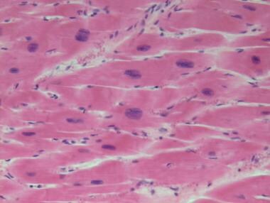
Retina
Photoreceptor Cells, Vertebrate
Vision, Ocular
Retinal Cone Photoreceptor Cells
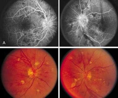
Direct ophthalmoscopy versus detection of hypertensive retinopathy: a comparative study. (1/11)
(+info)Dutch guideline for the management of hypertensive crisis -- 2010 revision. (2/11)
Hypertensive crises are divided into hypertensive urgencies and emergencies. Together they form a heterogeneous group of acute hypertensive disorders depending on the presence or type of target organs involved. Despite better treatment options for hypertension, hypertensive crisis and its associated complications remain relatively common. In the Netherlands the number of patients starting renal replacement therapy because of 'malignant hypertension' has increased in the past two decades. In 2003, the first Dutch guideline on hypertensive crisis was released to allow a standardised evidence-based approach for patients presenting with a hypertensive crisis. In this paper we give an overview of the current management of hypertensive crisis and discuss several important changes incorporated in the 2010 revision. These changes include a modification in terminology replacing 'malignant hypertension' with 'hypertensive crisis with retinopathy and reclassification of hypertensive crisis with retinopathy under hypertensive emergencies instead of urgencies. With regard to the treatment of hypertensive emergencies, nicardipine instead of nitroprusside or labetalol is favoured for the management of perioperative hypertension, whereas labetalol has become the drug of choice for the treatment of hypertension associated with pre-eclampsia. For the treatment of hypertensive urgencies, oral administration of nifedipine retard instead of captopril is recommended as first-line therapy. In addition, a section on the management of hypertensive emergencies according to the type of target organ involved has been added. Efforts to increase the awareness and treatment of hypertension in the population at large may lower the incidence of hypertensive crisis and its complications. (+info)Hypertension-related eye abnormalities and the risk of stroke. (3/11)
Many studies have shown that hypertensive ocular funduscopic abnormalities are clearly related to stroke, even after controlling for blood pressure and other vascular risk factors. Retinal abnormalities indicative of a breakdown of the blood-retina barrier confer a greater increase in risk for stroke than sclerotic retinal changes. Similar retinal changes also have a positive relationship to stroke mortality. In addition, hypertensive ocular fundus abnormalities are reported to be associated with an increased risk for cognitive impairment, cerebral atrophy, progression of magnetic resonance imaging-defined white matter lesions, and subclinical infarction. Recent advances in fundus photography allow for improved accuracy and consistency in interpretation of funduscopic lesions, and improve the feasibility of screening for these abnormalities in at-risk patient populations. Evaluating the ocular fundus for signs of hypertensive retinopathy, in combination with an assessment of the presence or absence of other known vascular risk factors, may allow clinicians to further individualize a risk profile for stroke to each individual patient, thus permitting more accurate risk stratification and, potentially, guiding treatment strategies. (+info)Mild retinopathy is a risk factor for cardiovascular mortality in Japanese with and without hypertension: the Ibaraki Prefectural Health Study. (4/11)
(+info)Microalbuminuria and hypertensive retinopathy among newly diagnosed nondiabetic hypertensive adult Nigerians. (5/11)
(+info)Association between retinopathy and cardiovascular disease in patients with chronic kidney disease (from the Chronic Renal Insufficiency Cohort [CRIC] Study). (6/11)
(+info)Retinopathy and chronic kidney disease in the Chronic Renal Insufficiency Cohort (CRIC) study. (7/11)
OBJECTIVE: To investigate the association between retinopathy and chronic kidney disease. METHODS: In this observational, cross-sectional study, 2605 patients of the Chronic Renal Insufficiency Cohort (CRIC) study, a multicenter study of chronic kidney disease, were offered participation. Nonmydriatic fundus photographs of the disc and macula in both eyes were obtained in 1936 of these subjects. The photographs were reviewed in a masked fashion at a central photograph reading center using standard protocols. Presence and severity of retinopathy (diabetic, hypertensive, or other) and vessel diameter caliber were assessed by trained graders and a retinal specialist using protocols developed for large epidemiologic studies. Kidney function measurements and information on traditional and nontraditional risk factors for decreased kidney function were obtained from the CRIC study. RESULTS: Greater severity of retinopathy was associated with lower estimated glomerular filtration rate after adjustment for traditional and nontraditional risk factors. The presence of vascular abnormalities usually associated with hypertension was also associated with lower estimated glomerular filtration rate. We found no strong direct relationship between estimated glomerular filtration rate and average arteriolar or venular calibers. CONCLUSIONS: Our findings show a strong association between severity of retinopathy and its features and level of kidney function after adjustment for traditional and nontraditional risk factors for chronic kidney disease, suggesting that retinovascular pathology reflects renal disease. (+info)MicroRNAs are involved in end-organ damage during hypertension. (8/11)
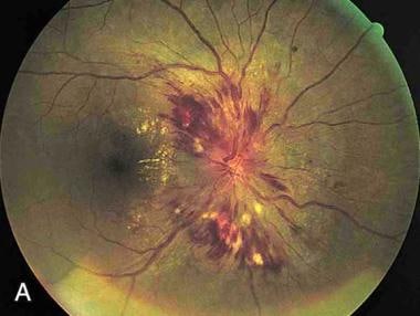
(+info)Hypertensive retinopathy is a term used to describe changes in the blood vessels and other structures in the retina that are caused by high blood pressure (hypertension). These changes can include narrowing of the blood vessels, thickening of their walls, and the formation of small bulges (microaneurysms) or bleeding. In severe cases, there may be swelling of the optic nerve and cotton wool spots, which are fluffy white patches that indicate areas where the blood supply to the retina has been disrupted.
Hypertensive retinopathy is usually asymptomatic in its early stages, but if it becomes advanced, it can lead to vision loss or even blindness. It is typically diagnosed by a doctor or eye care professional during an examination of the retina using specialized equipment such as an ophthalmoscope or a retinal camera. Treatment for hypertensive retinopathy usually involves controlling the underlying high blood pressure, which can help to prevent further damage to the retina and other structures in the eye.
Retinal diseases refer to a group of conditions that affect the retina, which is the light-sensitive tissue located at the back of the eye. The retina is responsible for converting light into electrical signals that are sent to the brain and interpreted as visual images. Retinal diseases can cause vision loss or even blindness, depending on their severity and location in the retina.
Some common retinal diseases include:
1. Age-related macular degeneration (AMD): A progressive disease that affects the central part of the retina called the macula, causing blurred or distorted vision.
2. Diabetic retinopathy: A complication of diabetes that can damage the blood vessels in the retina, leading to vision loss.
3. Retinal detachment: A serious condition where the retina becomes separated from its underlying tissue, requiring immediate medical attention.
4. Macular edema: Swelling or thickening of the macula due to fluid accumulation, which can cause blurred vision.
5. Retinitis pigmentosa: A group of inherited eye disorders that affect the retina's ability to respond to light, causing progressive vision loss.
6. Macular hole: A small break in the macula that can cause distorted or blurry vision.
7. Retinal vein occlusion: Blockage of the retinal veins that can lead to bleeding, swelling, and potential vision loss.
Treatment for retinal diseases varies depending on the specific condition and its severity. Some treatments include medication, laser therapy, surgery, or a combination of these options. Regular eye exams are essential for early detection and treatment of retinal diseases.
Malignant hypertension is a severe form of hypertension (high blood pressure) that is characterized by extremely high blood pressure readings, typically greater than 180/120 mmHg, along with evidence of damage to one or more organ systems. This condition is considered a medical emergency and requires immediate treatment.
Malignant hypertension can cause rapid and severe damage to various organs in the body, including the brain, heart, kidneys, and eyes. Symptoms may include severe headache, visual disturbances, confusion, shortness of breath, chest pain, nausea, vomiting, seizures, and even coma.
The exact cause of malignant hypertension is not always known, but it can be associated with certain underlying medical conditions such as kidney disease, autoimmune disorders, pregnancy-related complications, or the use of certain medications. Treatment typically involves aggressive blood pressure control using intravenous medications in a hospital setting, along with management of any underlying conditions and prevention of further organ damage.
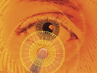
Ophthalmoscopy is a medical examination technique used by healthcare professionals to observe the interior structures of the eye, including the retina, optic disc, and vitreous humor. This procedure typically involves using an ophthalmoscope, a handheld device that consists of a light and magnifying lenses. The healthcare provider looks through the ophthalmoscope and directly observes the internal structures of the eye by illuminating them.
There are several types of ophthalmoscopy, including direct ophthalmoscopy, indirect ophthalmoscopy, and slit-lamp biomicroscopy. Each type has its own advantages and disadvantages, and they may be used in different situations depending on the specific clinical situation and the information needed.
Ophthalmoscopy is an important diagnostic tool for detecting and monitoring a wide range of eye conditions, including diabetic retinopathy, glaucoma, age-related macular degeneration, and other retinal disorders. It can also provide valuable information about the overall health of the individual, as changes in the appearance of the retina or optic nerve may indicate the presence of systemic diseases such as hypertension or diabetes.
Diabetic retinopathy is a diabetes complication that affects the eyes. It's caused by damage to the blood vessels of the light-sensitive tissue at the back of the eye (retina).
At first, diabetic retinopathy may cause no symptoms or only mild vision problems. Eventually, it can cause blindness. The condition usually affects both eyes.
There are two main stages of diabetic retinopathy:
1. Early diabetic retinopathy. This is when the blood vessels in the eye start to leak fluid or bleed. You might not notice any changes in your vision at this stage, but it's still important to get treatment because it can prevent the condition from getting worse.
2. Advanced diabetic retinopathy. This is when new, abnormal blood vessels grow on the surface of the retina. These vessels can leak fluid and cause severe vision problems, including blindness.
Diabetic retinopathy can be treated with laser surgery, injections of medication into the eye, or a vitrectomy (a surgical procedure to remove the gel-like substance that fills the center of the eye). It's important to get regular eye exams to detect diabetic retinopathy early and get treatment before it causes serious vision problems.
"Fundus Oculi" is a medical term that refers to the back part of the interior of the eye, including the optic disc, macula, fovea, retinal vasculature, and peripheral retina. It is the area where light is focused and then transmitted to the brain via the optic nerve, forming visual images. Examinations of the fundus oculi are crucial for detecting various eye conditions such as diabetic retinopathy, macular degeneration, glaucoma, and other retinal diseases. The examination is typically performed using an ophthalmoscope or a specialized camera called a retinal camera.
Hypertension is a medical term used to describe abnormally high blood pressure in the arteries, often defined as consistently having systolic blood pressure (the top number in a blood pressure reading) over 130 mmHg and/or diastolic blood pressure (the bottom number) over 80 mmHg. It is also commonly referred to as high blood pressure.
Hypertension can be classified into two types: primary or essential hypertension, which has no identifiable cause and accounts for about 95% of cases, and secondary hypertension, which is caused by underlying medical conditions such as kidney disease, hormonal disorders, or use of certain medications.
If left untreated, hypertension can lead to serious health complications such as heart attack, stroke, heart failure, and chronic kidney disease. Therefore, it is important for individuals with hypertension to manage their condition through lifestyle modifications (such as healthy diet, regular exercise, stress management) and medication if necessary, under the guidance of a healthcare professional.
Retinopathy of Prematurity (ROP) is a potentially sight-threatening proliferative retinal vascular disorder that primarily affects prematurely born infants, particularly those with low birth weight and/or young gestational age. It is characterized by the abnormal growth and development of retinal blood vessels due to disturbances in the oxygen supply and metabolic demands during critical phases of fetal development.
The condition can be classified into various stages (1-5) based on its severity, with stages 4 and 5 being more severe forms that may lead to retinal detachment and blindness if left untreated. The pathogenesis of ROP involves an initial phase of vessel loss and regression in the central retina, followed by a secondary phase of abnormal neovascularization, which can cause fibrosis, traction, and ultimately, retinal detachment.
ROP is typically managed with a multidisciplinary approach involving ophthalmologists, neonatologists, and pediatricians. Treatment options include laser photocoagulation, cryotherapy, intravitreal anti-VEGF injections, or even surgical interventions to prevent retinal detachment and preserve vision. Regular screening examinations are crucial for early detection and timely management of ROP in at-risk infants.
SHR (Spontaneously Hypertensive Rats) are an inbred strain of rats that were originally developed through selective breeding for high blood pressure. They are widely used as a model to study hypertension and related cardiovascular diseases, as well as neurological disorders such as stroke and dementia.
Inbred strains of animals are created by mating genetically identical individuals (siblings or offspring) for many generations, resulting in a population that is highly homozygous at all genetic loci. This means that the animals within an inbred strain are essentially genetically identical to one another, which makes them useful for studying the effects of specific genes or environmental factors on disease processes.
SHR rats develop high blood pressure spontaneously, without any experimental manipulation, and show many features of human hypertension, such as increased vascular resistance, left ventricular hypertrophy, and renal dysfunction. They also exhibit a number of behavioral abnormalities, including hyperactivity, impulsivity, and cognitive deficits, which make them useful for studying the neurological consequences of hypertension and other cardiovascular diseases.
Overall, inbred SHR rats are an important tool in biomedical research, providing a valuable model for understanding the genetic and environmental factors that contribute to hypertension and related disorders.
Medical Definition:
"Risk factors" are any attribute, characteristic or exposure of an individual that increases the likelihood of developing a disease or injury. They can be divided into modifiable and non-modifiable risk factors. Modifiable risk factors are those that can be changed through lifestyle choices or medical treatment, while non-modifiable risk factors are inherent traits such as age, gender, or genetic predisposition. Examples of modifiable risk factors include smoking, alcohol consumption, physical inactivity, and unhealthy diet, while non-modifiable risk factors include age, sex, and family history. It is important to note that having a risk factor does not guarantee that a person will develop the disease, but rather indicates an increased susceptibility.
WKY (Wistar Kyoto) is not a term that refers to "rats, inbred" in a medical definition. Instead, it is a strain of laboratory rat that is widely used in biomedical research. WKY rats are an inbred strain, which means they are the result of many generations of brother-sister matings, resulting in a genetically uniform population.
WKY rats originated from the Wistar Institute in Philadelphia and were established as a normotensive control strain to contrast with other rat strains that exhibit hypertension. They have since been used in various research areas, including cardiovascular, neurological, and behavioral studies. Compared to other commonly used rat strains like the spontaneously hypertensive rat (SHR), WKY rats are known for their lower blood pressure, reduced stress response, and greater emotionality.
In summary, "WKY" is a designation for an inbred strain of laboratory rat that is often used as a control group in biomedical research due to its normotensive characteristics.
Retinal vessels refer to the blood vessels that are located in the retina, which is the light-sensitive tissue that lines the inner surface of the eye. The retina contains two types of blood vessels: arteries and veins.
The central retinal artery supplies oxygenated blood to the inner layers of the retina, while the central retinal vein drains deoxygenated blood from the retina. These vessels can be visualized during a routine eye examination using an ophthalmoscope, which allows healthcare professionals to assess their health and any potential abnormalities.
Retinal vessels are essential for maintaining the health and function of the retina, and any damage or changes to these vessels can affect vision and lead to various eye conditions such as diabetic retinopathy, retinal vein occlusion, and hypertensive retinopathy.
Blood pressure is the force exerted by circulating blood on the walls of the blood vessels. It is measured in millimeters of mercury (mmHg) and is given as two figures:
1. Systolic pressure: This is the pressure when the heart pushes blood out into the arteries.
2. Diastolic pressure: This is the pressure when the heart rests between beats, allowing it to fill with blood.
Normal blood pressure for adults is typically around 120/80 mmHg, although this can vary slightly depending on age, sex, and other factors. High blood pressure (hypertension) is generally considered to be a reading of 130/80 mmHg or higher, while low blood pressure (hypotension) is usually defined as a reading below 90/60 mmHg. It's important to note that blood pressure can fluctuate throughout the day and may be affected by factors such as stress, physical activity, and medication use.
Retinal neovascularization is a medical condition characterized by the growth of new, abnormal blood vessels on the surface of the retina, which is the light-sensitive tissue located at the back of the eye. This condition typically occurs in response to an insufficient supply of oxygen and nutrients to the retina, often due to damage or disease, such as diabetic retinopathy or retinal vein occlusion.
The new blood vessels that form during neovascularization are fragile and prone to leakage, which can cause fluid and protein to accumulate in the retina, leading to distorted vision, hemorrhages, and potentially blindness if left untreated. Retinal neovascularization is a serious eye condition that requires prompt medical attention and management to prevent further vision loss.
Antihypertensive agents are a class of medications used to treat high blood pressure (hypertension). They work by reducing the force and rate of heart contractions, dilating blood vessels, or altering neurohormonal activation to lower blood pressure. Examples include diuretics, beta blockers, ACE inhibitors, ARBs, calcium channel blockers, and direct vasodilators. These medications may be used alone or in combination to achieve optimal blood pressure control.
Arterioles are small branches of arteries that play a crucial role in regulating blood flow and blood pressure within the body's circulatory system. They are the smallest type of blood vessels that have muscular walls, which allow them to contract or dilate in response to various physiological signals.
Arterioles receive blood from upstream arteries and deliver it to downstream capillaries, where the exchange of oxygen, nutrients, and waste products occurs between the blood and surrounding tissues. The contraction of arteriolar muscles can reduce the diameter of these vessels, causing increased resistance to blood flow and leading to a rise in blood pressure upstream. Conversely, dilation of arterioles reduces resistance and allows for greater blood flow at a lower pressure.
The regulation of arteriolar tone is primarily controlled by the autonomic nervous system, local metabolic factors, and various hormones. This fine-tuning of arteriolar diameter enables the body to maintain adequate blood perfusion to vital organs while also controlling overall blood pressure and distribution.
An encyclopedia is a comprehensive reference work containing articles on various topics, usually arranged in alphabetical order. In the context of medicine, a medical encyclopedia is a collection of articles that provide information about a wide range of medical topics, including diseases and conditions, treatments, tests, procedures, and anatomy and physiology. Medical encyclopedias may be published in print or electronic formats and are often used as a starting point for researching medical topics. They can provide reliable and accurate information on medical subjects, making them useful resources for healthcare professionals, students, and patients alike. Some well-known examples of medical encyclopedias include the Merck Manual and the Stedman's Medical Dictionary.
Mesopic vision is a term used to describe the intermediate level of vision that occurs in conditions of decreased illumination, specifically between 0.02 and 3 candelas per square meter (cd/m²). This range falls between photopic vision, which is vision in bright light (>3 cd/m²), and scotopic vision, which is vision in very low light (
The retina is the innermost, light-sensitive layer of tissue in the eye of many vertebrates and some cephalopods. It receives light that has been focused by the cornea and lens, converts it into neural signals, and sends these to the brain via the optic nerve. The retina contains several types of photoreceptor cells including rods (which handle vision in low light) and cones (which are active in bright light and are capable of color vision).
In medical terms, any pathological changes or diseases affecting the retinal structure and function can lead to visual impairment or blindness. Examples include age-related macular degeneration, diabetic retinopathy, retinal detachment, and retinitis pigmentosa among others.
Photoreceptor cells in vertebrates are specialized types of neurons located in the retina of the eye that are responsible for converting light stimuli into electrical signals. These cells are primarily responsible for the initial process of vision and have two main types: rods and cones.
Rods are more numerous and are responsible for low-light vision or scotopic vision, enabling us to see in dimly lit conditions. They do not contribute to color vision but provide information about the shape and movement of objects.
Cones, on the other hand, are less numerous and are responsible for color vision and high-acuity vision or photopic vision. There are three types of cones, each sensitive to different wavelengths of light: short (S), medium (M), and long (L) wavelengths, which correspond to blue, green, and red, respectively. The combination of signals from these three types of cones allows us to perceive a wide range of colors.
Both rods and cones contain photopigments that consist of a protein called opsin and a light-sensitive chromophore called retinal. When light hits the photopigment, it triggers a series of chemical reactions that ultimately lead to the generation of an electrical signal that is transmitted to the brain via the optic nerve. This process enables us to see and perceive our visual world.
Ocular vision refers to the ability to process and interpret visual information that is received by the eyes. This includes the ability to see clearly and make sense of the shapes, colors, and movements of objects in the environment. The ocular system, which includes the eye and related structures such as the optic nerve and visual cortex of the brain, works together to enable vision.
There are several components of ocular vision, including:
* Visual acuity: the clarity or sharpness of vision
* Field of vision: the extent of the visual world that is visible at any given moment
* Color vision: the ability to distinguish different colors
* Depth perception: the ability to judge the distance of objects in three-dimensional space
* Contrast sensitivity: the ability to distinguish an object from its background based on differences in contrast
Disorders of ocular vision can include refractive errors such as nearsightedness or farsightedness, as well as more serious conditions such as cataracts, glaucoma, and macular degeneration. These conditions can affect one or more aspects of ocular vision and may require medical treatment to prevent further vision loss.
In the context of medical terminology, "light" doesn't have a specific or standardized definition on its own. However, it can be used in various medical terms and phrases. For example, it could refer to:
1. Visible light: The range of electromagnetic radiation that can be detected by the human eye, typically between wavelengths of 400-700 nanometers. This is relevant in fields such as ophthalmology and optometry.
2. Therapeutic use of light: In some therapies, light is used to treat certain conditions. An example is phototherapy, which uses various wavelengths of ultraviolet (UV) or visible light for conditions like newborn jaundice, skin disorders, or seasonal affective disorder.
3. Light anesthesia: A state of reduced consciousness in which the patient remains responsive to verbal commands and physical stimulation. This is different from general anesthesia where the patient is completely unconscious.
4. Pain relief using light: Certain devices like transcutaneous electrical nerve stimulation (TENS) units have a 'light' setting, indicating lower intensity or frequency of electrical impulses used for pain management.
Without more context, it's hard to provide a precise medical definition of 'light'.
Retinal cone photoreceptor cells are specialized neurons located in the retina of the eye, responsible for visual phototransduction and color vision. They are one of the two types of photoreceptors, with the other being rods, which are more sensitive to low light levels. Cones are primarily responsible for high-acuity, color vision during daylight or bright-light conditions.
There are three types of cone cells, each containing different photopigments that absorb light at distinct wavelengths: short (S), medium (M), and long (L) wavelengths, which correspond to blue, green, and red light, respectively. The combination of signals from these three types of cones allows the human visual system to perceive a wide range of colors and discriminate between them. Cones are densely packed in the central region of the retina, known as the fovea, which provides the highest visual acuity.
Known as hypertensive retinopathy2
- The last and the most complicated, Grade 4, is characterized by papilledema, also known as hypertensive retinopathy. (custom-essay.org)
- Nontu often experiences difficulty breathing, chest pain, severe headaches, sleeping disorder and has an eye disease is known as hypertensive retinopathy. (health-e.org.za)
Central retinal v2
- Neovascular glaucoma is a secondary glaucoma occurring as a result of severely reduced blood flow to the eye as may be observed in central retinal vein occlusion or with severe diabetic retinopathy. (pharmacology2000.com)
- Common vascular retinopathies include central retinal artery occlusion, central retinal vein occlusion, diabetic retinopathy, and hypertensive retinopathy. (doctor-clinic.org)
Retina6
- Hypertensive retinopathy is damage to the retina and retinal circulation due to high blood pressure (i.e. hypertension). (wikipedia.org)
- Advanced retinopathy lesions, such as microaneurysms, blot hemorrhages and/or flame hemorrhages, ischemic changes (e.g. "cotton wool spots"), hard exudates and in severe cases swelling of the optic disc (optic disc edema), a ring of exudates around the retina called a "macular star" and visual acuity loss, typically due to macular involvement. (wikipedia.org)
- Hypertensive retinopathy is damage to the retina (the transparent, light-sensitive structure at the back of the eye) caused by high blood pressure. (msdmanuals.com)
- As hypertensive retinopathy progresses, blood may leak into the retina. (msdmanuals.com)
- therefore, the optic nerve must cross through the retina en route to the brain. (wikipedia.org)
- Diabetic retinopathy (DR) is a sight threatening complication of systemic diabetes mellitus that results from damage to the blood vessels of the retina. (institut-vision.org)
Visits for hypertensive emergency2
- From 2006 through 2013, the estimated number of visits for hypertensive emergency more than doubled, but true hypertensive emergency accounted for only 0.2% of adult ED patients overall and 0.6% of adult ED patients with a diagnosis of hypertension. (nursingcenter.com)
- [ 3 ] Nonetheless, despite its relative rarity, the number of US emergency department (ED) visits for hypertensive emergency and the rate per million adult ED visits increased more than two-fold between 2006 and 2013. (medscape.com)
Management of hypertensive emergencies1
- and describe the pathophysiology, clinical manifestations, and management of hypertensive emergencies. (nursingcenter.com)
Disorders8
- Retinopathy among women with hypertensive disorders of pregnancy attending hospitals in Mbarara city, south-western Uganda: a cross-sectional study. (bvsalud.org)
- Retinopathy is one of the complications occurring among women with hypertensive disorders of pregnancy . (bvsalud.org)
- We sought to determine the prevalence and factors associated with retinopathy among women with hypertensive disorders of pregnancy in southwestern Uganda . (bvsalud.org)
- The study included all pregnant women with hypertensive disorders of pregnancy . (bvsalud.org)
- A total of 216 women with hypertensive disorders of pregnancy were enrolled in this study. (bvsalud.org)
- In our study, retinopathy was common among women with hypertensive disorders of pregnancy . (bvsalud.org)
- There is a need to integrate screening for retinopathy in the care cascade of women with hypertensive disorders of pregnancy . (bvsalud.org)
- Background: Preeclampsia is one of the hypertensive disorders in pregnancy that contributes significantly to maternal and fetal morbidity and mortality, with the impact felt more in developing countries. (bvsalud.org)
Hypertension and hypertensive1
- PROCEDURE: Medical records from cats with systemic hypertension and hypertensive retinopathy were reviewed. (avmi.net)
Systemic hypertension2
- Acute and chronic hypertensive changes may manifest in the eyes, respectively, from acute changes from malignant hypertension and chronic changes from long-term, systemic hypertension. (medscape.com)
- OBJECTIVE: To characterize clinical and clinicopathologic findings, response to treatment, and causes of systemic hypertension in cats with hypertensive retinopathy. (avmi.net)
Encephalopathy3
- Neurologic end-organ damage due to uncontrolled BP may include hypertensive encephalopathy, cerebral vascular accident/cerebral infarction, subarachnoid hemorrhage , and/or intracranial hemorrhage . (medscape.com)
- The presence of neurologic symptoms may include seizures, visual disturbances, and altered level of consciousness and may be indicative of hypertensive encephalopathy. (medscape.com)
- This results in hyperperfusion and cerebral edema, which cause the clinical manifestations of hypertensive encephalopathy. (medscape.com)
Prevalence5
- The prevalence of retinopathy was determined and multivariable logistic regression was used to determine the independent factors associated with retinopathy. (bvsalud.org)
- The prevalence of retinopathy was 60.2% (130/216). (bvsalud.org)
- Given the high prevalence of this diagnosis, it's important for coders to have a solid understanding of how to report it in ICD-10-CM. Coders must understand the difference between primary and secondary hypertension and be able to assign codes for related health problems such as cardiomyopathy, kidney disease, and hypertensive retinopathy. (hcmarketplace.com)
- A systematic review and meta-analysis to assess the prevalence of hypertension in sub-Saharan Africa examined data from 33 surveys and found that an average of 27% of people were aware of their hypertensive status before the surveys (5). (cdc.gov)
- An analysis of eight studies conducted in Thailand, France, Italy, and Brazil found that the combined prevalence of hypertensive emergency and hypertensive urgency in EDs was roughly 1.2%, with hypertensive urgency being significantly more common than hypertensive emergency, though prevalence varied across studies. (nursingcenter.com)
Grade 1 retinopathy2
- They showed 70% of those with grade 1 retinopathy were alive after 3 years whereas only 6% of those with grade 4 survived. (wikipedia.org)
- The most common retinal lesions were grade 1 retinopathy (narrowing of arterioles ) accounting for 86.9% (113/130), grade 3 ( retinal haemorrhages) was present in 10% (13/130) of women and grade 4 (papilloedema) in 3% (4/130). (bvsalud.org)
Severity of retinopathy2
- In a classic study in 1939 Keith and colleagues described the prognosis of people with differing severity of retinopathy. (wikipedia.org)
- Careful control of the patient's blood glucose levels during the first 5 years of diabetes may decrease the severity of retinopathy or delay its onset. (doctor-clinic.org)
Diagnosis3
- Prompt and accurate diagnosis of hypertensive retinopathy, especially when associated with malignant hypertension, is necessary to avoid visual and systemic morbidity. (medscape.com)
- Mortality rates, however, were relatively high among patients with qualifying hypertensive emergency who presented to U.S. EDs, at 4.8% in 2006 and 4.5% in 2013, underscoring the need for prompt diagnosis and appropriate management of the condition. (nursingcenter.com)
- Retinopathy is already present at the time of diagnosis in 20% of patients with type 2 diabetes. (institut-vision.org)
Papilledema1
- The presence of new retinal hemorrhages, exudates, or papilledema suggests a hypertensive emergency. (medscape.com)
Microaneurysms2
- The role of retinopathy grading in risk stratification is debated, but it has been proposed that individuals with signs of hypertensive retinopathy signs, especially retinal hemorrhages, microaneurysms and cotton-wool spots, should be assessed carefully. (wikipedia.org)
- OSAS also plays a role in the development of various retinal findings, including microaneurysms, hypertensive retinopathy, and intraocular production of postischemic molecules that are associated with neovascularization, apoptosis, and macular edema. (aao.org)
Develop retinopathy2
- About 40% of patients who have had type I diabetes and about 25% of those who have had type II diabetes for 10 years develop retinopathy. (doctor-clinic.org)
- Nearly all patients with type 1 diabetes (younger-onset patients) and more than 60% of patients with type 2 diabetes (older-onset patients) develop retinopathy during the first two decades of disease, and approximately 4% and 2% of these patients respectively, become legally blind (defined as visual acuity of 1/20). (institut-vision.org)
Radiation retinopathy1
- Radiation Retinopathy. (slackbooks.com)
Proliferative diabetic2
- Cataract, proliferative diabetic retinopathy (PDR) and diabetic maculopathy were the main causes of visual impairment and blindness. (who.int)
- The treatment choice for patients with proliferative diabetic retinopathy is laser photocoagulation. (doctor-clinic.org)
Emergencies6
- Treatment of hypertensive urgencies and emergencies High blood pressure (hypertension) is persistently high pressure in the arteries. (msdmanuals.com)
- True hypertensive emergencies are characterized by a rapid elevation in blood pressure to a level above 180/120 mmHg and are associated with acute target organ damage, which requires immediate hospitalization for close hemodynamic monitoring and IV pharmacotherapy. (nursingcenter.com)
- Hypertensive emergencies encompass a spectrum of clinical presentations in which uncontrolled blood pressures (BPs) lead to progressive or impending end-organ dysfunction. (medscape.com)
- With the advent of antihypertensive agents, the incidence of hypertensive emergencies in the United States has declined from 7% to approximately 1% of patients with hypertension. (medscape.com)
- Malignant hypertension and accelerated hypertension are both hypertensive emergencies, with similar outcomes and therapies. (medscape.com)
- Hypertensive emergencies require immediate therapy to decrease blood pressure within minutes to hours. (medscape.com)
Renal3
- 32-year-old African American male with Grade IV hypertensive retinopathy and acute renal failure. (asrs.org)
- A hypertensive emergency is a sharp rise in blood pressure to a level above 180/120 mmHg that is associated with target organ damage, often involving exigent neurologic, cardiovascular, or renal manifestations. (nursingcenter.com)
- Other organ systems may also be affected by uncontrolled hypertension, which may lead to acute renal failure /insufficiency, retinopathy, eclampsia , or microangiopathic hemolytic anemia. (medscape.com)
Vascular5
- Grade 1 Vascular Attenuation Grade 2 As grade 1 + Irregularly located, tight constrictions - Known as "AV nicking" or "AV nipping" - Salus's sign Grade 3 As grade 2 + Retinal edema, cotton wool spots and flame-hemorrhages "Copper Wiring" + Bonnet's Sign + Gunn's Sign Grade 4 As grade 3 + optic disc edema + macular star "Silver Wiring" There is an association between the grade of retinopathy and mortality. (wikipedia.org)
- Hypertensive retinopathy is retinal vascular harm because of high blood pressure. (ashulaservision.com)
- These extracted markers or characterized fundus digital image features provide insights and relates quantitative retinal vascular topography abnormalities to various pathologies such as diabetic retinopathy, macular degeneration, hypertensive retinopathy, transient ischemic attack, neovascular glaucoma, and cardiovascular diseases. (hindawi.com)
- An interruption in blood supply to the eye can produce a vascular retinopathy, which is a noninflammatory retinal disorder. (doctor-clinic.org)
- Appropriate tests depend on the type of vascular retinopathy. (doctor-clinic.org)
Diabetes7
- Anyone who has diabetes can develop hypertensive retinopathy. (ashulaservision.com)
- In individuals with diabetes, the body's ability to use or produce insulin, a hormone that helps to regulate blood sugar level levels, is impaired diabetic vs hypertensive retinopathy. (microqnix.com)
- In Yemen, diabetes mellitus is a com- Vincent targets of reduction of blind- directions, pupillary reflexes and Gold- mon disease among adult Yemenis with ness due to diabetic retinopathy [9]. (who.int)
- Diabetic retinopathy results from diabetes, which causes microcirculatory changes. (doctor-clinic.org)
- Among the most consistent risk factors, duration of diabetes is probably the strongest predictor for development and progression of the retinopathy. (institut-vision.org)
- A computing system using artificial intelligence is highly accurate in identifying people with diabetes who have diabetic retinopathy and related eye diseases and need to be referred for further care, a new study finds. (medscape.com)
- The DLS for referable diabetic retinopathy was developed and trained using retinal images of patients with diabetes who participated in the Singapore National Diabetic Retinopathy Screening Program (SIDRP) between 2010 and 2013, which had screened half of Singapore's diabetes population by 2015. (medscape.com)
Complications1
- For the classification of diabetic there were 17 300 patients examined at thy and diabetic eye complications is retinopathy, the modified Airlie House the centre over this period, 694 (4%) of on the rise [6]. (who.int)
Ophthalmic2
- Hypertensive retinopathy (HR) represents the ophthalmic findings of end-organ damage secondary to systemic arterial hypertension. (msjonline.org)
- It is also advised that internists should refer hypertensive patients for routine ophthalmic screening. (who.int)
Macular2
- All these features are useful for early detection of hypertensive and diabetic retinopathy, macular degeneration, acute stroke, neovascular glaucoma, and some other cardiovascular diseases [ 1 , 3 , 5 - 9 ]. (hindawi.com)
- In addition, hypertension may exacerbate the vision-threatening effects of diabetic retinopathy and has been implicated in the pathogenesis of age-related macular degeneration. (nih.gov)
Fundus2
- Fundus photograph of RE showing Disc pallor and arteriolar attenuation, with dot-blot and flame-shaped haemorrhages at the posterior pole in a case of Hypertensive Retinopathy. (asrs.org)
- The participants were screened for retinopathy using a fundus camera. (bvsalud.org)
Arteriolar3
- The changes in hypertensive retinopathy result from damage and adaptive changes in the arterial and arteriolar circulation in response to the high blood pressure. (wikipedia.org)
- According to the study of [ 12 ] with a multiethnic cohort, retinal arteriolar narrowing and retinopathy of diabetic free people have an association with increased risk of acute stroke. (hindawi.com)
- Hypertensive retinopathy results from prolonged hypertension, which produces retinal vasospasm and consequent damage to and narrowing of the arteriolar lumen. (doctor-clinic.org)
Patients11
- Most patients with hypertensive retinopathy have no symptoms. (wikipedia.org)
- The aim of our study was to investigate whether Mean platelet volume (MPV) is associated with the severity of hypertensive retinopathy in hypertensive patients. (msjonline.org)
- However, all patients presenting with blood pressure this high should undergo evaluation to confirm or rule out impending target organ damage, which differentiates hypertensive emergency from other hypertensive crises and is vital in facilitating appropriate emergency treatment. (nursingcenter.com)
- In the United States, although 18% of ED patients have severely elevated blood pressure at or above 180/110 mmHg upon presentation, 3 far fewer have hypertensive emergency, as previously defined, which occurs in conjunction with acute or impending target organ damage. (nursingcenter.com)
- Close collaboration between ophthalmologists and general practitioners/physicians is needed to ensure that hypertensive patients are identified and treated. (nih.gov)
- Methods Circulating (n = 179) and hepatic expression (n = 95) of ghrelin and LEAP-2 were measured in patients with severe obesity and available liver pathology analysis undergoing Roux-en-Y gastric bypass (RYGB). (unav.edu)
- RÉSUMÉ Nous avons évalué les causes des déficiences visuelles et de la cécité chez 694 patients diabétiques ayant consulté dans notre centre des soins oculaires à Sanaa (Yémen) entre 2001 et 2005 en examinant leur dossier médical. (who.int)
- All classes of antihypertensive agents are useful in treating hypertensive patients with peripheral arterial disease. (aao.org)
- Treatment for patients with early-stage, nonpro-liferative diabetic retinopathy is prophylactic. (doctor-clinic.org)
- Treatment for patients with hypertensive retinopathy includes controlling blood pressure with appropriate drugs, diet, and exercise. (doctor-clinic.org)
- [ 3 ] In contrast, no evidence suggests a benefit from rapidly reducing blood pressure in patients with hypertensive urgency. (medscape.com)
20191
- 2019). Hypertensive retinopathy. (mhmedical.com)
Glaucoma1
- Training of the DLS entailed exposure of a total 76,370 retinal images (with and without each of the three conditions: diabetic retinopathy, glaucoma, and AMD) to the neural networks, which then adapted to differentiate between normal and abnormal and between conditions. (medscape.com)
Exudates1
- If the constriction is severe, changes associated with hypertensive retinopathy may occur, including diffuse retinal edema, hemorrhages, exudates, and cotton-wool spots. (aao.org)
Cardiovascular disease1
- Markers of cardiovascular disease, such as hypertensive retinopathy and cholesterol emboli, can often manifest in the eye," researchers say. (aoa.org)
Severe2
- Women presenting with severe hypertension were likely to have retinopathy. (bvsalud.org)
- 1 The term hypertensive crisis is sometimes used to describe the spectrum of severe uncontrolled hypertension, encompassing both hypertensive emergency and hypertensive urgency. (nursingcenter.com)
Referable1
- Automated multidimensional deep learning platform for referable diabetic retinopathy detection: a multicentre, retrospective study. (cdc.gov)
CRISES2
- 1 Despite these important distinctions, in all hypertensive crises, the goal of treatment is to reduce blood pressure safely without compromising organ perfusion. (nursingcenter.com)
- Careful preoperative treatment with alpha and beta blockers is required to control blood pressure and prevent intraoperative hypertensive crises. (medscape.com)
Urgency3
- 1 Hypertensive urgency is a term used to describe similarly high blood pressure values that neither produce nor worsen target organ damage. (nursingcenter.com)
- Hypertensive urgency must be distinguished from hypertensive emergency. (medscape.com)
- This article discusses hypertensive emergency, but therapy for hypertensive urgency is discussed briefly. (medscape.com)
Antihypertensive1
- Bitter Kola has some hypertensive properties which means that if you take the Kola along with antihypertensive medications (blood pressure lowering), it could negate some of the effects. (answers.com)
Adult1
- Mild signs of hypertensive retinopathy can be seen quite frequently in normal people (3-14% of adult individuals aged ≥40 years), even without hypertension. (wikipedia.org)
Stroke1
- The authors of [ 15 ] examined the association of hypertensive retinopathy with the risk of stroke in their population base study. (hindawi.com)
Diagnostic1
- Hypertensive retinopathy is commonly considered a diagnostic feature of a hypertensive emergency although it is not invariably present. (wikipedia.org)
Target organ damage1
- A hypertensive emergency is a condition in which elevated blood pressure results in target organ damage. (medscape.com)
Diseases1
- Several other diseases can result in retinopathy that can be confused with hypertensive retinopathy. (wikipedia.org)
Adaptive1
- Does boosting the adaptive immune system reduce hypertensive diabetic retinopathy? (monash.edu)
Blood pressure3
- Hypertensive retinopathy is managed mainly by means of controlling high blood pressure. (ashulaservision.com)
- The goal of treatment for hypertensive retinopathy is to lower blood pressure long term. (msdmanuals.com)
- Hypertensive retinopathy (HR) develops from elevated blood pressure. (mhmedical.com)
Emergency3
- The patient is sent to the emergency department to be evaluated further and treated for a hypertensive emergency. (mhmedical.com)
- Recognizing the clinical signs and symptoms of hypertensive emergency, which may vary widely depending on the target organ involved, is critical. (nursingcenter.com)
- Drug therapy for hypertensive emergency is influenced by end-organ involvement, pharmacokinetics, potential adverse drug effects, and patient comorbidities. (nursingcenter.com)