Hyperemia
Vasodilation
Brachial Artery
Adenosine
Laser-Doppler Flowmetry
Blood Flow Velocity
Endothelium, Vascular
Vascular Resistance
Fractional Flow Reserve, Myocardial
Dogs
Nitric Oxide
Dipyridamole
Hemodynamics
Theophylline
NG-Nitroarginine Methyl Ester
Muscle, Skeletal
Skin
Ketorolac
Prostaglandins
Nitric Oxide Synthase
Ophthalmoscopic abnormalities in adults with falciparum malaria. (1/956)
We studied 424 adults with falciparum malaria admitted over 28 months. They were divided into three groups: cerebral malaria (n = 214); severe non-cerebral malaria (n = 58); and uncomplicated malaria (n = 152). Fundus examination was done daily from admission to discharge, and weekly thereafter in those with persistent changes. All patients were treated by a protocol based on WHO guidelines. Ophthalmoscopic abnormalities were: retinal haemorrhages, 40 (9.43%) (25 cerebral malaria, 10 severe non-cerebral and five uncomplicated malaria); papilloedema, 17 (7.94%) cerebral malaria and two uncomplicated malaria; blurring of disc margins, 25 (11.68%) cerebral and seven non-cerebral; retinal oedema, six (2.80%) cerebral and five non-cerebral malaria; disc pallor, five patients all with cerebral malaria; vitreous haemorrhage and hard exudate in one patient each, both cerebral malaria. Retinal haemorrhage was associated with cerebral malaria and severe non-cerebral malaria, especially with severe anaemia (p < 0.001), as compared to uncomplicated malaria (p < 0.01). The association of papilloedema and cerebral malaria was highly significant compared to severe non-cerebral malaria (p < 0.001). None of these findings was associated with statistically significant mortality, except disc pallor in cerebral malaria (p < 0.05). (+info)Endothelial function in Marfan syndrome: selective impairment of flow-mediated vasodilation. (2/956)
BACKGROUND: The cardiovascular complications of Marfan syndrome arise due to alterations in the structural and functional properties of fibrillin, a constituent of vascular connective tissues. Fibrillin-containing microfibrils are closely associated with arterial endothelial cells, indicating a possible functional role for fibrillin in the endothelium. Plasma concentrations of endothelial cell products are elevated in Marfan subjects, which indirectly indicates endothelial dysfunction. This study directly assessed flow- and agonist-mediated endothelium-dependent brachial artery reactivity in Marfan subjects. METHODS AND RESULTS: In 20 Marfan and 20 control subjects, brachial artery diameter, blood flow, and blood pressure were measured by ultrasonic wall tracking, Doppler ultrasound, and photoplethysmography, respectively. Measurements were taken during hand hyperemia (a stimulus for endothelium-derived nitric oxide [NO] release in the upstream brachial artery) and after sublingual administration of the endothelium-independent vasodilator nitroglycerin. In 9 Marfan and 6 control subjects, the above parameters were also assessed during intra-arterial infusions of acetylcholine and bradykinin (agonists that stimulate NO production) and NG-monomethyl-L-arginine (L-NMMA, an inhibitor of NO production). Flow-mediated responses differed markedly between Marfan and control subjects (-1.6+/-3.5% versus 6. 50+/-4.1%, respectively; P<0.0001), whereas nitroglycerin produced similar vasodilation (14.2+/-5.7% versus 15.2+/-7.8%; P=NS). Agonist-induced vasodilation to incremental intra-arterial infusions of acetylcholine and bradykinin were not significantly different between Marfan and control subjects, and intra-arterial L-NMMA produced similar reductions in brachial artery diameter in both groups. CONCLUSIONS: These data demonstrate impaired flow-mediated but preserved agonist-mediated endothelium-dependent vasodilation in Marfan subjects and suggest preservation of basal NO release. Selective loss of flow-mediated dilation suggests a role for fibrillin in endothelial cell mechanotransduction. (+info)Trigeminal and carotid body inputs controlling vascular resistance in muscle during post-contraction hyperaemia in cats. (3/956)
1. In anaesthetized cats, the effects of stimulation of the receptors in the nasal mucosa and carotid body chemoreceptors on vascular resistance in hindlimb skeletal muscle were studied to see whether the responses were the same in active as in resting muscle. The measurements of vascular resistance were taken, first, in resting muscle, and second, in the immediate post-contraction hyperaemic phase that followed a 30 s period of isometric contractions. 2. Stimulation of the receptors in the nasal mucosa caused reflex apnoea and vasoconstriction in muscle. The latter response was attenuated when the test was repeated during post-contraction hyperaemia. 3. Stimulations of the carotid bodies were made during a period of apnoea evoked reflexly by electrical stimulation of both superior laryngeal nerves. This apnoea prevented any effects of changes in respiration on the carotid body reflex vascular responses. Stimulation of the carotid bodies evoked hindlimb muscle vasoconstriction. In the post-contraction hyperaemic period, the response was reduced or abolished. A similar attenuation of the reflex vasoconstrictor responses occurred in decentralized muscles stimulated through their motor roots in the cauda equina. 4. Evidence is presented that the attenuation of the vasoconstrictor responses evoked by the two reflexes is a phenomenon localized to the contracting muscles themselves resulting from an interaction between sympathetic neuronal activity and the local production of metabolites. 5. The results are discussed in relation to the metabolic needs of tissues in relation to asphyxial defence mechanisms such as occur in the diving response. (+info)Neurogenic origin of articular hyperemia in early degenerative joint disease. (4/956)
It has been speculated that joint instability resulting from anterior cruciate ligament (ACL) rupture could be exacerbated by changes in vasomotor activity in the remaining supporting structures. In this study, the effect of ACL transection on medial collateral ligament (MCL) basal perfusion and its responsiveness to calcitonin gene-related peptide (CGRP) and sympathetic adrenergic influences was examined. Using urethan-anesthetized rabbits, we tested the effects of CGRP and its antagonist CGRP-(8-37) by topical application of these agents to the exposed knee while sympathetic influences were tested by electrically stimulating the saphenous nerve. It was found that MCL basal perfusion was elevated in ACL-sectioned joints; however, this effect was abrogated by prior resection of the articular nerve supply. At the doses tested, the normal vasodilator response to CGRP was abolished in ACL-sectioned joints, whereas the response to CGRP-(8-37) was attenuated. Even under the influence of increased constrictor tone, MCL and capsule blood vessels still showed substantially reduced responses to exogenous CGRP administration. By contrast, nerve-mediated constrictor responses were mostly unaffected by joint instability. This study suggests that posttraumatic knee joint hyperemia is neurogenically mediated, possibly by increased secretion of CGRP. (+info)Endothelial function is impaired in fit young adults of low birth weight. (5/956)
OBJECTIVE: Non-insulin-dependent diabetes, hypertension and ischaemic heart disease, with insulin resistance, are associated with low birth weight (the 'Small Baby Syndrome'). Common to these adult clinical conditions is endothelial dysfunction. We tested the hypothesis that endothelial dysfunction could precede their development in those of low birth weight. METHODS: Endothelial function was measured by ultrasonic 'wall-tracking' of flow-related brachial artery dilatation in fit 19-20 year old subjects randomly selected (blind to the investigators throughout the study) from low (< 2.5 kg) and normal (3.0-3.8 kg) birth weight subjects in the 1975-7 cohort of the Cardiff Births Survey and with no known cause for endothelial dysfunction. RESULTS: Flow-related dilatation was impaired in low birth weight relative to normal birth weight subjects (median 0.04 mm [1.5%] [n = 22] cf. 0.11 mm [4.1%] [n = 17], p < 0.05; 0.04 mm [1.5%] [n = 15] cf. 0.12 mm [4.4%] [n = 12], p < 0.05 after exclusion of inadvertently included ever-smokers). CONCLUSION: The findings suggest that endothelial dysfunction is a consequence of foetal malnutrition, consistent with contributing to the clinical features of the 'Small Baby Syndrome' in later adult life. (+info)The association between laser Doppler reactive hyperaemia curves and the distribution of peripheral arterial disease. (6/956)
OBJECTIVES: To determine whether postocclusive laser Doppler fluxmetry (LDF) curves can be related to the arteriographic distribution of disease. DESIGN: Prospective study. MATERIALS: Sixty-nine patients with symptomatic peripheral ischaemia and 15 healthy subjects. METHODS: Laser Doppler fluxmetry (LDF) was monitored on the dorsum of the symptomatic foot following 2 min of arterial occlusion at the ankle. During reperfusion three patterns of LDF were identified (types I-III). All patients subsequently underwent arteriography which was reported independent of LDF results. The distribution of disease, particularly patency of below-knee vessels, was related to the type of LDF curve observed during reactive hyperaemia. RESULTS: Type I curves were observed in all healthy subjects and 75% of patients with a single arterial lesion. Type II curves were found in 78% of patients with multiple lesions above the knee. The presence of either a type I or II curve was associated with a continuous vessel from knee to ankle (positive predictive value 83%, p < 0.01), whilst type III curve was associated with discontinuous infrapopliteal run-off (positive predictive value 86%, p < 0.01). CONCLUSIONS: This pilot study suggests that post-occlusive LDF curves may identify the distribution of arterial disease and may be useful in the non-invasive management of peripheral ischaemia. (+info)Inhibition of phospholipase A2 attenuates functional hyperemia in the hamster cremaster muscle. (7/956)
Arachidonic acid (AA) is the common precursor for several vasodilatory factors involved in the local control of blood flow. This study was designed to determine the role of phospholipase A2 (PLA2) and AA release in functional hyperemia in the hamster cremaster muscle. The muscle was prepared for in vivo microscopy and subjected to electrical field stimulation for 1 min. First- and second-order arterioles dilated in response from a mean diameter of 66 +/- 5 to 88 +/- 7 micrometer (n = 6). PLA2 was then inhibited with quinacrine (3 x 10(-6) M) for 60 min. PLA2 inhibition was verified by an attenuation of thrombin-induced vasodilation (2 U/ml). Quinacrine had no effect on resting arteriolar diameter but completely abolished functional hyperemia. Quinacrine also had no effect on dilation induced by superfusion of the preparation with 3 x 10(-6)-10(-5) M AA, 10(-6)-10(-4) M adenosine, or 10(-6)-10(-4) M sodium nitroprusside, ruling out nonspecific effects of quinacrine on smooth muscle contractility. These results indicate that functional hyperemia in the hamster cremaster muscle is dependent on PLA2 activation and the availability of AA. (+info)Abnormalities of vascular endothelial function may contribute to increased coronary heart disease risk in UK Indian Asians. (8/956)
OBJECTIVE: To test the hypothesis that abnormalities of endothelial function are present in Indian Asians and may contribute to their increased coronary heart disease risk. SETTING: Single centre in west London. PATIENTS: 26 Indian Asian and 18 European white healthy male subjects, aged 35 to 61 years recruited from general practice lists. DESIGN: Brachial artery diameter responses to reactive hyperaemia and sublingual glyceryl trinitrate were compared using high resolution ultrasound. RESULTS: Mean (SEM) flow mediated, endothelium dependent dilatation was reduced in Indian Asians compared with European whites, at 3.2 (0.8)% v 5.9 (1.0)%, p = 0.03. In contrast, there were no significant differences in baseline brachial arterial diameter (4.6 (0.1) v 4.6 (0.1) mm, p = 0.65) or glyceryl trinitrate induced dilatation (18.8 (1.5)% v 18.5 (1.7)%, p = 0.90) between Indian Asians and European whites, respectively. Univariate analysis showed that Indian Asian race was significantly associated with impaired flow mediated dilatation (regression coefficient = -2.8 (1.3)%, p = 0.03), and in multivariate analysis, this relation was independent of both conventional coronary heart disease risk factors and markers of insulin resistance. CONCLUSIONS: Endothelial function is impaired in healthy UK Indian Asians compared with European whites, and the defect is not accounted for by major coronary heart disease risk factors. Endothelial function may be modulated by novel risk factors in Indian Asians. (+info)Hyperemia is a medical term that refers to an increased flow or accumulation of blood in certain capillaries or vessels within an organ or tissue, resulting in its redness and warmth. This can occur due to various reasons such as physical exertion, emotional excitement, local injury, or specific medical conditions.
There are two types of hyperemia: active and passive. Active hyperemia is a physiological response where the blood flow increases as a result of the metabolic demands of the organ or tissue. For example, during exercise, muscles require more oxygen and nutrients, leading to an increase in blood flow. Passive hyperemia, on the other hand, occurs when there is a blockage in the venous outflow, causing the blood to accumulate in the affected area. This can result from conditions like thrombosis or vasoconstriction.
It's important to note that while hyperemia itself is not a disease, it can be a symptom of various underlying medical conditions and should be evaluated by a healthcare professional if it persists or is accompanied by other symptoms.
Regional blood flow (RBF) refers to the rate at which blood flows through a specific region or organ in the body, typically expressed in milliliters per minute per 100 grams of tissue (ml/min/100g). It is an essential physiological parameter that reflects the delivery of oxygen and nutrients to tissues while removing waste products. RBF can be affected by various factors such as metabolic demands, neural regulation, hormonal influences, and changes in blood pressure or vascular resistance. Measuring RBF is crucial for understanding organ function, diagnosing diseases, and evaluating the effectiveness of treatments.
The forearm is the region of the upper limb between the elbow and the wrist. It consists of two bones, the radius and ulna, which are located side by side and run parallel to each other. The forearm is responsible for movements such as flexion, extension, supination, and pronation of the hand and wrist.
Vasodilation is the widening or increase in diameter of blood vessels, particularly the involuntary relaxation of the smooth muscle in the tunica media (middle layer) of the arteriole walls. This results in an increase in blood flow and a decrease in vascular resistance. Vasodilation can occur due to various physiological and pathophysiological stimuli, such as local metabolic demands, neural signals, or pharmacological agents. It plays a crucial role in regulating blood pressure, tissue perfusion, and thermoregulation.
Coronary circulation refers to the circulation of blood in the coronary vessels, which supply oxygenated blood to the heart muscle (myocardium) and drain deoxygenated blood from it. The coronary circulation system includes two main coronary arteries - the left main coronary artery and the right coronary artery - that branch off from the aorta just above the aortic valve. These arteries further divide into smaller branches, which supply blood to different regions of the heart muscle.
The left main coronary artery divides into two branches: the left anterior descending (LAD) artery and the left circumflex (LCx) artery. The LAD supplies blood to the front and sides of the heart, while the LCx supplies blood to the back and sides of the heart. The right coronary artery supplies blood to the lower part of the heart, including the right ventricle and the bottom portion of the left ventricle.
The veins that drain the heart muscle include the great cardiac vein, the middle cardiac vein, and the small cardiac vein, which merge to form the coronary sinus. The coronary sinus empties into the right atrium, allowing deoxygenated blood to enter the right side of the heart and be pumped to the lungs for oxygenation.
Coronary circulation is essential for maintaining the health and function of the heart muscle, as it provides the necessary oxygen and nutrients required for proper contraction and relaxation of the myocardium. Any disruption or blockage in the coronary circulation system can lead to serious consequences, such as angina, heart attack, or even death.
The brachial artery is a major blood vessel in the upper arm. It supplies oxygenated blood to the muscles and tissues of the arm, forearm, and hand. The brachial artery originates from the axillary artery at the level of the shoulder joint and runs down the medial (inner) aspect of the arm, passing through the cubital fossa (the depression on the anterior side of the elbow) where it can be palpated during a routine blood pressure measurement. At the lower end of the forearm, the brachial artery bifurcates into the radial and ulnar arteries, which further divide into smaller vessels to supply the hand and fingers.
Adenosine is a purine nucleoside that is composed of a sugar (ribose) and the base adenine. It plays several important roles in the body, including serving as a precursor for the synthesis of other molecules such as ATP, NAD+, and RNA.
In the medical context, adenosine is perhaps best known for its use as a pharmaceutical agent to treat certain cardiac arrhythmias. When administered intravenously, it can help restore normal sinus rhythm in patients with paroxysmal supraventricular tachycardia (PSVT) by slowing conduction through the atrioventricular node and interrupting the reentry circuit responsible for the arrhythmia.
Adenosine can also be used as a diagnostic tool to help differentiate between narrow-complex tachycardias of supraventricular origin and those that originate from below the ventricles (such as ventricular tachycardia). This is because adenosine will typically terminate PSVT but not affect the rhythm of VT.
It's worth noting that adenosine has a very short half-life, lasting only a few seconds in the bloodstream. This means that its effects are rapidly reversible and generally well-tolerated, although some patients may experience transient symptoms such as flushing, chest pain, or shortness of breath.
Laser-Doppler flowmetry (LDF) is a non-invasive, investigative technique used to measure microcirculatory blood flow in real time. It is based on the principle of the Doppler effect, which describes the change in frequency or wavelength of light or sound waves as they encounter a moving object or reflect off a moving surface.
In LDF, a low-power laser beam is directed at the skin or other transparent tissue. The light penetrates the tissue and scatters off the moving red blood cells within the microvasculature. As the light scatters, it undergoes a slight frequency shift due to the movement of the red blood cells. This frequency shift is then detected by a photodetector, which converts it into an electrical signal. The magnitude of this signal is directly proportional to the speed and concentration of the moving red blood cells, providing a measure of microcirculatory blood flow.
LDF has various clinical applications, including the assessment of skin perfusion in patients with peripheral arterial disease, burn injuries, and flaps used in reconstructive surgery. It can also be used to study the effects of drugs or other interventions on microcirculation in research settings.
Blood flow velocity is the speed at which blood travels through a specific part of the vascular system. It is typically measured in units of distance per time, such as centimeters per second (cm/s) or meters per second (m/s). Blood flow velocity can be affected by various factors, including cardiac output, vessel diameter, and viscosity of the blood. Measuring blood flow velocity is important in diagnosing and monitoring various medical conditions, such as heart disease, stroke, and peripheral vascular disease.
Vasodilator agents are pharmacological substances that cause the relaxation or widening of blood vessels by relaxing the smooth muscle in the vessel walls. This results in an increase in the diameter of the blood vessels, which decreases vascular resistance and ultimately reduces blood pressure. Vasodilators can be further classified based on their site of action:
1. Systemic vasodilators: These agents cause a generalized relaxation of the smooth muscle in the walls of both arteries and veins, resulting in a decrease in peripheral vascular resistance and preload (the volume of blood returning to the heart). Examples include nitroglycerin, hydralazine, and calcium channel blockers.
2. Arterial vasodilators: These agents primarily affect the smooth muscle in arterial vessel walls, leading to a reduction in afterload (the pressure against which the heart pumps blood). Examples include angiotensin-converting enzyme (ACE) inhibitors, angiotensin receptor blockers (ARBs), and direct vasodilators like sodium nitroprusside.
3. Venous vasodilators: These agents primarily affect the smooth muscle in venous vessel walls, increasing venous capacitance and reducing preload. Examples include nitroglycerin and other organic nitrates.
Vasodilator agents are used to treat various cardiovascular conditions such as hypertension, heart failure, angina, and pulmonary arterial hypertension. It is essential to monitor their use carefully, as excessive vasodilation can lead to orthostatic hypotension, reflex tachycardia, or fluid retention.
Microcirculation is the circulation of blood in the smallest blood vessels, including arterioles, venules, and capillaries. It's responsible for the delivery of oxygen and nutrients to the tissues and the removal of waste products. The microcirculation plays a crucial role in maintaining tissue homeostasis and is regulated by various physiological mechanisms such as autonomic nervous system activity, local metabolic factors, and hormones.
Impairment of microcirculation can lead to tissue hypoxia, inflammation, and organ dysfunction, which are common features in several diseases, including diabetes, hypertension, sepsis, and ischemia-reperfusion injury. Therefore, understanding the structure and function of the microcirculation is essential for developing new therapeutic strategies to treat these conditions.
Plethysmography is a non-invasive medical technique used to measure changes in volume or blood flow within an organ or body part, typically in the lungs or extremities. There are several types of plethysmography, including:
1. **Whole Body Plethysmography (WBP):** This type of plethysmography is used to assess lung function and volumes by measuring changes in pressure within a sealed chamber that contains the patient's entire body except for their head. The patient breathes normally while wearing a nose clip, allowing technicians to analyze respiratory patterns, airflow, and lung volume changes.
2. **Segmental or Local Plethysmography:** This technique measures volume or blood flow changes in specific body parts, such as the limbs or digits. It can help diagnose and monitor conditions affecting peripheral circulation, like deep vein thrombosis, arterial occlusive disease, or Raynaud's phenomenon.
3. **Impedance Plethysmography (IPG):** This non-invasive method uses electrical impedance to estimate changes in blood volume within an organ or body part. By applying a small electrical current and measuring the opposition to flow (impedance), technicians can determine variations in blood volume, which can help diagnose conditions like deep vein thrombosis or heart failure.
4. **Optical Plethysmography:** This technique uses light to measure changes in blood volume, typically in the skin or mucous membranes. By shining a light on the area and analyzing the reflected or transmitted light, technicians can detect variations in blood volume related to cardiac output, respiration, or other physiological factors.
Overall, plethysmography is an essential tool for diagnosing and monitoring various medical conditions affecting circulation, respiratory function, and organ volumes.
The endothelium is a thin layer of simple squamous epithelial cells that lines the interior surface of blood vessels, lymphatic vessels, and heart chambers. The vascular endothelium, specifically, refers to the endothelial cells that line the blood vessels. These cells play a crucial role in maintaining vascular homeostasis by regulating vasomotor tone, coagulation, platelet activation, inflammation, and permeability of the vessel wall. They also contribute to the growth and repair of the vascular system and are involved in various pathological processes such as atherosclerosis, hypertension, and diabetes.
Cerebrovascular circulation refers to the network of blood vessels that supply oxygenated blood and nutrients to the brain tissue, and remove waste products. It includes the internal carotid arteries, vertebral arteries, circle of Willis, and the intracranial arteries that branch off from them.
The internal carotid arteries and vertebral arteries merge to form the circle of Willis, a polygonal network of vessels located at the base of the brain. The anterior cerebral artery, middle cerebral artery, posterior cerebral artery, and communicating arteries are the major vessels that branch off from the circle of Willis and supply blood to different regions of the brain.
Interruptions or abnormalities in the cerebrovascular circulation can lead to various neurological conditions such as stroke, transient ischemic attack (TIA), and vascular dementia.
Blood pressure is the force exerted by circulating blood on the walls of the blood vessels. It is measured in millimeters of mercury (mmHg) and is given as two figures:
1. Systolic pressure: This is the pressure when the heart pushes blood out into the arteries.
2. Diastolic pressure: This is the pressure when the heart rests between beats, allowing it to fill with blood.
Normal blood pressure for adults is typically around 120/80 mmHg, although this can vary slightly depending on age, sex, and other factors. High blood pressure (hypertension) is generally considered to be a reading of 130/80 mmHg or higher, while low blood pressure (hypotension) is usually defined as a reading below 90/60 mmHg. It's important to note that blood pressure can fluctuate throughout the day and may be affected by factors such as stress, physical activity, and medication use.
Vascular resistance is a measure of the opposition to blood flow within a vessel or a group of vessels, typically expressed in units of mmHg/(mL/min) or sometimes as dynes*sec/cm^5. It is determined by the diameter and length of the vessels, as well as the viscosity of the blood flowing through them. In general, a decrease in vessel diameter, an increase in vessel length, or an increase in blood viscosity will result in an increase in vascular resistance, while an increase in vessel diameter, a decrease in vessel length, or a decrease in blood viscosity will result in a decrease in vascular resistance. Vascular resistance is an important concept in the study of circulation and cardiovascular physiology because it plays a key role in determining blood pressure and blood flow within the body.
Fractional Flow Reserve (Myocardial) is a medical term used to describe the ratio of maximum blood flow through a stenosed (narrowed) coronary artery to the maximum flow that could be achieved if the artery were completely normal. It is a pressure-based index, which is measured during cardiac catheterization using a special wire that can measure pressure differences across a stenosis.
The FFR value ranges from 0 (no flow) to 1 (normal flow). An FFR value less than or equal to 0.80 is generally considered indicative of functionally significant coronary artery disease, which may benefit from revascularization (such as angioplasty or bypass surgery).
FFR is used in clinical practice to help guide decisions regarding the management of patients with coronary artery disease and has been shown to improve patient outcomes.
I believe there might be a misunderstanding in your question. "Dogs" is not a medical term or condition. It is the common name for a domesticated carnivore of the family Canidae, specifically the genus Canis, which includes wolves, foxes, and other extant and extinct species of mammals. Dogs are often kept as pets and companions, and they have been bred in a wide variety of forms and sizes for different purposes, such as hunting, herding, guarding, assisting police and military forces, and providing companionship and emotional support.
If you meant to ask about a specific medical condition or term related to dogs, please provide more context so I can give you an accurate answer.
Nitric oxide (NO) is a molecule made up of one nitrogen atom and one oxygen atom. In the body, it is a crucial signaling molecule involved in various physiological processes such as vasodilation, immune response, neurotransmission, and inhibition of platelet aggregation. It is produced naturally by the enzyme nitric oxide synthase (NOS) from the amino acid L-arginine. Inhaled nitric oxide is used medically to treat pulmonary hypertension in newborns and adults, as it helps to relax and widen blood vessels, improving oxygenation and blood flow.
Coronary vessels refer to the network of blood vessels that supply oxygenated blood and nutrients to the heart muscle, also known as the myocardium. The two main coronary arteries are the left main coronary artery and the right coronary artery.
The left main coronary artery branches off into the left anterior descending artery (LAD) and the left circumflex artery (LCx). The LAD supplies blood to the front of the heart, while the LCx supplies blood to the side and back of the heart.
The right coronary artery supplies blood to the right lower part of the heart, including the right atrium and ventricle, as well as the back of the heart.
Coronary vessel disease (CVD) occurs when these vessels become narrowed or blocked due to the buildup of plaque, leading to reduced blood flow to the heart muscle. This can result in chest pain, shortness of breath, or a heart attack.
Dipyridamole is a medication that belongs to a class of drugs called antiplatelet agents. It works by preventing platelets in your blood from sticking together to form clots. Dipyridamole is often used in combination with aspirin to prevent stroke and other complications in people who have had a heart valve replacement or a type of irregular heartbeat called atrial fibrillation.
Dipyridamole can also be used as a stress agent in myocardial perfusion imaging studies, which are tests used to evaluate blood flow to the heart. When used for this purpose, dipyridamole is given intravenously and works by dilating the blood vessels in the heart, allowing more blood to flow through them and making it easier to detect areas of reduced blood flow.
The most common side effects of dipyridamole include headache, dizziness, and gastrointestinal symptoms such as diarrhea, nausea, and vomiting. In rare cases, dipyridamole can cause more serious side effects, such as allergic reactions, abnormal heart rhythms, or low blood pressure. It is important to take dipyridamole exactly as directed by your healthcare provider and to report any unusual symptoms or side effects promptly.
In medical terms, constriction refers to the narrowing or tightening of a body part or passageway. This can occur due to various reasons such as spasms of muscles, inflammation, or abnormal growths. It can lead to symptoms like difficulty in breathing, swallowing, or blood flow, depending on where it occurs. For example, constriction of the airways in asthma, constriction of blood vessels in hypertension, or constriction of the esophagus in certain digestive disorders.
Hemodynamics is the study of how blood flows through the cardiovascular system, including the heart and the vascular network. It examines various factors that affect blood flow, such as blood volume, viscosity, vessel length and diameter, and pressure differences between different parts of the circulatory system. Hemodynamics also considers the impact of various physiological and pathological conditions on these variables, and how they in turn influence the function of vital organs and systems in the body. It is a critical area of study in fields such as cardiology, anesthesiology, and critical care medicine.
Theophylline is a medication that belongs to a class of drugs called methylxanthines. It is used in the management of respiratory diseases such as asthma, chronic obstructive pulmonary disease (COPD), and other conditions that cause narrowing of the airways in the lungs.
Theophylline works by relaxing the smooth muscle around the airways, which helps to open them up and make breathing easier. It also acts as a bronchodilator, increasing the flow of air into and out of the lungs. Additionally, theophylline has anti-inflammatory effects that can help reduce swelling in the airways and relieve symptoms such as coughing, wheezing, and shortness of breath.
Theophylline is available in various forms, including tablets, capsules, and liquid solutions. It is important to take this medication exactly as prescribed by a healthcare provider, as the dosage may vary depending on individual factors such as age, weight, and liver function. Regular monitoring of blood levels of theophylline is also necessary to ensure safe and effective use of the medication.
NG-Nitroarginine Methyl Ester (L-NAME) is not a medication, but rather a research chemical used in scientific studies. It is an inhibitor of nitric oxide synthase, an enzyme that synthesizes nitric oxide, a molecule involved in the relaxation of blood vessels.
Therefore, L-NAME is often used in experiments to investigate the role of nitric oxide in various physiological and pathophysiological processes. It is important to note that the use of L-NAME in humans is not approved for therapeutic purposes due to its potential side effects, which can include hypertension, decreased renal function, and decreased cerebral blood flow.
Skeletal muscle, also known as striated or voluntary muscle, is a type of muscle that is attached to bones by tendons or aponeuroses and functions to produce movements and support the posture of the body. It is composed of long, multinucleated fibers that are arranged in parallel bundles and are characterized by alternating light and dark bands, giving them a striped appearance under a microscope. Skeletal muscle is under voluntary control, meaning that it is consciously activated through signals from the nervous system. It is responsible for activities such as walking, running, jumping, and lifting objects.
In medical terms, the skin is the largest organ of the human body. It consists of two main layers: the epidermis (outer layer) and dermis (inner layer), as well as accessory structures like hair follicles, sweat glands, and oil glands. The skin plays a crucial role in protecting us from external factors such as bacteria, viruses, and environmental hazards, while also regulating body temperature and enabling the sense of touch.
In medical terms, the leg refers to the lower portion of the human body that extends from the knee down to the foot. It includes the thigh (femur), lower leg (tibia and fibula), foot, and ankle. The leg is primarily responsible for supporting the body's weight and enabling movements such as standing, walking, running, and jumping.
The leg contains several important structures, including bones, muscles, tendons, ligaments, blood vessels, nerves, and joints. These structures work together to provide stability, support, and mobility to the lower extremity. Common medical conditions that can affect the leg include fractures, sprains, strains, infections, peripheral artery disease, and neurological disorders.
In the field of medicine, "time factors" refer to the duration of symptoms or time elapsed since the onset of a medical condition, which can have significant implications for diagnosis and treatment. Understanding time factors is crucial in determining the progression of a disease, evaluating the effectiveness of treatments, and making critical decisions regarding patient care.
For example, in stroke management, "time is brain," meaning that rapid intervention within a specific time frame (usually within 4.5 hours) is essential to administering tissue plasminogen activator (tPA), a clot-busting drug that can minimize brain damage and improve patient outcomes. Similarly, in trauma care, the "golden hour" concept emphasizes the importance of providing definitive care within the first 60 minutes after injury to increase survival rates and reduce morbidity.
Time factors also play a role in monitoring the progression of chronic conditions like diabetes or heart disease, where regular follow-ups and assessments help determine appropriate treatment adjustments and prevent complications. In infectious diseases, time factors are crucial for initiating antibiotic therapy and identifying potential outbreaks to control their spread.
Overall, "time factors" encompass the significance of recognizing and acting promptly in various medical scenarios to optimize patient outcomes and provide effective care.
Skin temperature is the measure of heat emitted by the skin, which can be an indicator of the body's core temperature. It is typically lower than the body's internal temperature and varies depending on factors such as environmental temperature, blood flow, and physical activity. Skin temperature is often used as a vital sign in medical settings and can be measured using various methods, including thermal scanners, digital thermometers, or mercury thermometers. Changes in skin temperature may also be associated with certain medical conditions, such as inflammation, infection, or nerve damage.
Ketorolac is a non-steroidal anti-inflammatory drug (NSAID) that is used to treat moderate to severe pain. It works by reducing the levels of prostaglandins, chemicals in the body that cause inflammation and trigger pain signals in the brain. By blocking the production of prostaglandins, ketorolac helps to reduce pain, swelling, and fever.
Ketorolac is available in several forms, including tablets, injection solutions, and suppositories. It is typically used for short-term pain relief, as it can increase the risk of serious side effects such as stomach ulcers, bleeding, and kidney problems with long-term use.
Like other NSAIDs, ketorolac may also increase the risk of heart attack and stroke, especially in people who already have cardiovascular disease or risk factors for it. It should be used with caution and only under the supervision of a healthcare provider.
Prostaglandins are naturally occurring, lipid-derived hormones that play various important roles in the human body. They are produced in nearly every tissue in response to injury or infection, and they have diverse effects depending on the site of release and the type of prostaglandin. Some of their functions include:
1. Regulation of inflammation: Prostaglandins contribute to the inflammatory response by increasing vasodilation, promoting fluid accumulation, and sensitizing pain receptors, which can lead to symptoms such as redness, heat, swelling, and pain.
2. Modulation of gastrointestinal functions: Prostaglandins protect the stomach lining from acid secretion and promote mucus production, maintaining the integrity of the gastric mucosa. They also regulate intestinal motility and secretion.
3. Control of renal function: Prostaglandins help regulate blood flow to the kidneys, maintain sodium balance, and control renin release, which affects blood pressure and fluid balance.
4. Regulation of smooth muscle contraction: Prostaglandins can cause both relaxation and contraction of smooth muscles in various tissues, such as the uterus, bronchioles, and vascular system.
5. Modulation of platelet aggregation: Some prostaglandins inhibit platelet aggregation, preventing blood clots from forming too quickly or becoming too large.
6. Reproductive system regulation: Prostaglandins are involved in the menstrual cycle, ovulation, and labor induction by promoting uterine contractions.
7. Neurotransmission: Prostaglandins can modulate neurotransmitter release and neuronal excitability, affecting pain perception, mood, and cognition.
Prostaglandins exert their effects through specific G protein-coupled receptors (GPCRs) found on the surface of target cells. There are several distinct types of prostaglandins (PGs), including PGD2, PGE2, PGF2α, PGI2 (prostacyclin), and thromboxane A2 (TXA2). Each type has unique functions and acts through specific receptors. Prostaglandins are synthesized from arachidonic acid, a polyunsaturated fatty acid derived from membrane phospholipids, by the action of cyclooxygenase (COX) enzymes. Nonsteroidal anti-inflammatory drugs (NSAIDs), such as aspirin and ibuprofen, inhibit COX activity, reducing prostaglandin synthesis and providing analgesic, anti-inflammatory, and antipyretic effects.
Omega-N-Methylarginine (also known as NG, NG-dimethyl-L-arginine) is not a commonly used medical term and it's not a well-known compound in medicine. However, it is a form of methylated arginine that can be found in the body.
Methylated arginines are a group of compounds that are generated through the post-translational modification of proteins by enzymes called protein arginine methyltransferases (PRMTs). These modifications play important roles in various cellular processes, including gene expression and signal transduction.
Omega-N-Methylarginine is a specific type of methylated arginine that has two methyl groups attached to the nitrogen atom at the end of the side chain (omega position) of the amino acid arginine. It can be formed by the action of PRMTs on proteins, and it may have various biological functions in the body. However, its specific medical significance is not well-established, and more research is needed to fully understand its role in health and disease.
Nitric Oxide Synthase (NOS) is a group of enzymes that catalyze the production of nitric oxide (NO) from L-arginine. There are three distinct isoforms of NOS, each with different expression patterns and functions:
1. Neuronal Nitric Oxide Synthase (nNOS or NOS1): This isoform is primarily expressed in the nervous system and plays a role in neurotransmission, synaptic plasticity, and learning and memory processes.
2. Inducible Nitric Oxide Synthase (iNOS or NOS2): This isoform is induced by various stimuli such as cytokines, lipopolysaccharides, and hypoxia in a variety of cells including immune cells, endothelial cells, and smooth muscle cells. iNOS produces large amounts of NO, which functions as a potent effector molecule in the immune response, particularly in the defense against microbial pathogens.
3. Endothelial Nitric Oxide Synthase (eNOS or NOS3): This isoform is constitutively expressed in endothelial cells and produces low levels of NO that play a crucial role in maintaining vascular homeostasis by regulating vasodilation, inhibiting platelet aggregation, and preventing smooth muscle cell proliferation.
Overall, NOS plays an essential role in various physiological processes, including neurotransmission, immune response, cardiovascular function, and respiratory regulation. Dysregulation of NOS activity has been implicated in several pathological conditions such as hypertension, atherosclerosis, neurodegenerative diseases, and inflammatory disorders.
In medical terms, fingers are not specifically defined as they are common anatomical structures. However, I can provide you with a general anatomy definition:
Fingers are the terminal parts of the upper limb in primates, including humans, consisting of four digits (thumb, index, middle, and ring fingers) and one opposable thumb. They contain bones called phalanges, connected by joints that allow for movement and flexibility. Each finger has a nail, nerve endings for sensation, and blood vessels to supply nutrients and oxygen. Fingers are crucial for various activities such as grasping, manipulating objects, and tactile exploration of the environment.
Blood circulation, also known as cardiovascular circulation, refers to the process by which blood is pumped by the heart and circulated throughout the body through a network of blood vessels, including arteries, veins, and capillaries. This process ensures that oxygen and nutrients are delivered to cells and tissues, while waste products and carbon dioxide are removed.
The circulation of blood can be divided into two main parts: the pulmonary circulation and the systemic circulation. The pulmonary circulation involves the movement of blood between the heart and the lungs, where it picks up oxygen and releases carbon dioxide. The systemic circulation refers to the movement of blood between the heart and the rest of the body, delivering oxygen and nutrients to cells and tissues while picking up waste products for removal.
The heart plays a central role in blood circulation, acting as a pump that contracts and relaxes to move blood through the body. The contraction of the heart's left ventricle pushes oxygenated blood into the aorta, which then branches off into smaller arteries that carry blood throughout the body. The blood then flows through capillaries, where it exchanges oxygen and nutrients for waste products and carbon dioxide with surrounding cells and tissues. The deoxygenated blood is then collected in veins, which merge together to form larger vessels that eventually return the blood back to the heart's right atrium. From there, the blood is pumped into the lungs to pick up oxygen and release carbon dioxide, completing the cycle of blood circulation.
 Hyperaemia
Local blood flow regulation
Terbium(III) chloride
Toxic and nutritional optic neuropathy
Hydralazine
Skeletal muscle pump
Thermal burn
Fowl cholera
Perfusion
Gustavus M. Blech
Scarabiasis
Non-freezing cold injury
Pseudoephedrine
Erythema
Endothelial dysfunction
Emmonsia parva
Neurovascular unit
Bimatoprost
Corneal dystrophy
Raynaud syndrome
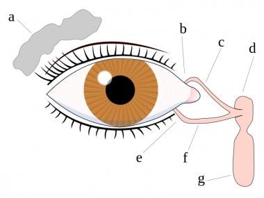
Human eye
Variola caprina
Sarcocystis
Phenylephrine
Cardiovascular physiology
Tafluprost
Ripasudil
Aerospace physiology
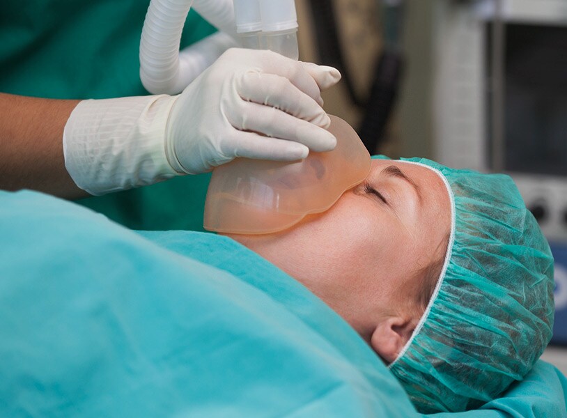
Eye injuries during general anaesthesia
Omidenepag
Hyperaemia - Wikipedia
 Image: Hyperemia, glandular mucosa, adult horse - Merck Veterinary Manual
 GAT Sport Nitraflex Fruit Punch Flavored Hyperemia & Testosterone Enchancing Powder, 10.6 oz - Fred Meyer
 Cerebral Oxygen Supply and Demand in Sickle Cell Disease: Evidence of Local Ischemia Despite Global Hyperemia | Frontiers...
 Occlusive hyperemia: a theory for the hemodynamic complications following resection of intracerebral arteriovenous...
 Mask Distillation Network for Conjunctival Hyperemia Severity Classification
 What Is Conjunctival Hyperemia and How to Manage It | OBN
 REACTIVE HYPEREMIA PERIPHERAL ARTERIAL TONOMETRY SCORE IDENTIFIES RESPONDERS TO CARDIAC RESYNCHRONIZATION THERAPY | AVESİS
 What's Your Science Glossary IQ? - ProProfs Quiz
 Transfusion of stored autologous blood does not alter reactive hyperemia index in healthy volunteers
Noninvasive identification of patients with early coronary atherosclerosis by assessment of digital reactive hyperemia<...
Table - Case Report and Literature Review of Occupational Transmission of Monkeypox Virus to Healthcare Workers, South Korea -...
 Dry Eye Disease (Keratoconjunctivitis Sicca): Practice Essentials, Background, Anatomy
 Cerebral hyperperfusion syndrome after revascularization surgery in patients with moyamoya disease
 Susvimo (Ranibizumab Injection): Uses, Dosage, Side Effects, Interactions, Warning
Combined effects of smoking and hypercholesterolemia on inflammatory process, thrombosis/fibrinolysis system, and forearm...
 A Systematic Evaluation of Cutaneous Microcirculation in the Foot Using Post-Occlusive Reactive Hyperemia - STORE -...
 Case Definitions for Public Health Surveillance
Reactive hyperemia index (RHI) and cognitive performance indexes are associated with histologic markers of liver disease in...
Effect of Extended-Release Niacin on Carotid Intima Media Thickness, Reactive Hyperemia, and Endothelial Progenitor Cell...
 DailyMed - LACRISERT- hydroxypropyl cellulose insert
DailyMed - YUTIQ- fluocinolone acetonide implant
 SciELO - Brazil - Red eyes in the necropsy floor: twenty cases of hyphema in dogs and
cats Red eyes in the...
SciELO - Brazil - Red eyes in the necropsy floor: twenty cases of hyphema in dogs and
cats Red eyes in the...
 Nutrients | Free Full-Text | Mediterranean Diet and Endothelial Function: A Review of its Effects at Different Vascular Bed...
 On the development of conjunctival hyperemia computer-assisted diagnosis tools: Influence of feature selection and class...
Vaccines | Free Full-Text | The Increase of the Magnitude of Spontaneous Viral Blips in Some Participants of Phase II Clinical...
ArboCat Virus: African Horsesickness (AHSV)
Infrainguinal Occlusive Disease Differential Diagnoses
 2020-2021 BCSC Basic and Clinical Science Course™
Conjunctival20
- To achieve automatic, fast and accurate severity classification of bulbar conjunctival hyperemia severity, we proposed a novel prior knowledge-based framework called mask distillation network (MDN). (mi-research.net)
- What is Conjunctival Hyperemia? (ophthalmologybreakingnews.com)
- Conjunctival hyperemia is the medical term for an excessive dilatation or engorgement of the conjunctival blood vessels. (ophthalmologybreakingnews.com)
- While a case of conjunctival hyperemia might sometimes be dismissed as mere eye redness, it can be an indicator of underlying issues that require medical attention. (ophthalmologybreakingnews.com)
- Conditions ranging from dry eyes and allergies to more severe issues like glaucoma or ocular inflammation could present with conjunctival hyperemia as a symptom. (ophthalmologybreakingnews.com)
- Conjunctival hyperemia is quite common and can affect individuals of all ages. (ophthalmologybreakingnews.com)
- Persistent or recurrent conjunctival hyperemia can significantly affect an individual's quality of life, leading to discomfort, visual disturbances, or even more severe complications if left untreated. (ophthalmologybreakingnews.com)
- Both ocular hyperemia and conjunctival hyperemia refer to a red or 'bloodshot' appearance of the eyes, but they are not synonymous terms. (ophthalmologybreakingnews.com)
- Conjunctival hyperemia is specifically localized to the conjunctiva. (ophthalmologybreakingnews.com)
- Conjunctival hyperemia is primarily linked to inflammation, irritation, or infection of the conjunctiva itself. (ophthalmologybreakingnews.com)
- Conjunctival hyperemia usually presents with redness and possibly discomfort or discharge but is unlikely to involve corneal or eyelid symptoms unless it is part of a broader ocular issue. (ophthalmologybreakingnews.com)
- Conjunctival hyperemia is often treated with antihistamines, lubricants, or anti-inflammatory eye drops, depending on the cause. (ophthalmologybreakingnews.com)
- Allergic reactions to pollen, dust, animal dander, or certain eye drops can cause conjunctival hyperemia. (ophthalmologybreakingnews.com)
- Conjunctivitis, caused by viral or bacterial infections, often presents with conjunctival hyperemia as a primary symptom. (ophthalmologybreakingnews.com)
- Exposure to smoke, dust, and pollutants can irritate the eyes, leading to conjunctival hyperemia. (ophthalmologybreakingnews.com)
- Conditions like blepharitis and meibomian gland dysfunction can contribute to conjunctival hyperemia. (ophthalmologybreakingnews.com)
- A sensation of grittiness, burning, or general irritation can accompany conjunctival hyperemia. (ophthalmologybreakingnews.com)
- LACRISERT usually reduces the signs and symptoms resulting from moderate to severe dry eye syndromes, such as conjunctival hyperemia, corneal and conjunctival staining with rose bengal, exudation, itching, burning, foreign body sensation, smarting, photophobia, dryness and blurred or cloudy vision. (nih.gov)
- Conclusion Conjunctival hyperemia, lid edema, and follicular conjunctivitis were observed in at least half of the patients with MIS-C, and those with ophthalmic involvement had a higher risk of cardiac involvement or severe disease. (thieme-connect.com)
- Symptoms are conjunctival hyperemia and ocular discharge and, depending on the etiology, discomfort and itching. (msdmanuals.com)
Ocular Hyperemia4
- Ocular hyperemia can occur in multiple parts of the eye. (ophthalmologybreakingnews.com)
- Ocular hyperemia may be due to a broader range of causes, including issues with the cornea, elevated intraocular pressure, or eyelid disorders. (ophthalmologybreakingnews.com)
- Ocular hyperemia may come with additional symptoms related to other parts of the eye, such as corneal haze or eyelid swelling. (ophthalmologybreakingnews.com)
- Ocular hyperemia may require a more comprehensive approach to treatment, targeting the specific part of the eye that is affected. (ophthalmologybreakingnews.com)
Reactive hyperemia index4
- Blood withdrawal and reactive hyperemia index measurements were performed before and 10 min, 1 h, 2 h, and 4 h after transfusion. (unimib.it)
- The reactive hyperemia index during the first 4 h after transfusion of 40-day compared with 3-day stored packed erythrocytes was unchanged. (unimib.it)
- Reactive hyperemia index (RHI) and cognitive performance indexes are associated with histologic markers of liver disease in subjects with non-alcoholic fatty liver disease (NAFLD): a case control study. (unipa.it)
- Pulse wave velocity (PWV) and augmentation index (Aix) were used as markers of arterial stiffness, whereas endothelial function was assessed using reactive hyperemia index (RHI). (unipa.it)
Peripheral arterial tonometry3
- We investigated the value of reactive hyperemia peripheral arterial tonometry (RH-PAT) as a noninvasive tool to identify individuals with coronary microvascular endothelial dysfunction. (elsevierpure.com)
- BACKGROUND: No study evaluated vascular health markers in subjects with non-alcoholic fatty liver disease (NAFLD) through a combined analysis of reactive hyperemia peripheral arterial tonometry (RH-PAT) and arterial stiffness indexes. (unipa.it)
- Reactive hyperemia peripheral arterial tonometry, as assessed with EndoPAT 2000 (Itamar Medical, Inc) and EPC mobilization assessed with flow cytometry, were measured at enrollment, and at 1 and 12 months. (elsevierpure.com)
Arterial2
- Functional hyperaemia, metabolic hyperaemia, arterial hyperaemia or active hyperaemia, is the increased blood flow that occurs when tissue is active. (wikipedia.org)
- Reactive hyperaemia, a sub-category of arterial hyperaemia, is the transient increase in organ blood flow that occurs following a brief period of ischaemia. (wikipedia.org)
Exercise hyperaemia1
- In conclusion, local hyperthermia -induced limb hyperperfusion and/or small muscle mass exercise hyperaemia are preserved in trained older people despite evident age-related structural and functional alterations in their leg conduit arteries . (bvsalud.org)
Nitric oxide1
- The NITRAFLEX pre-training formula contains ingredients that in vitro, animal and clinical studies suggest possess properties that may help advanced athletes maximize energy, intensity, vascularity and reactive hyperemia (pumps) during their workouts, and provide long-term support of testosterone and nitric oxide levels in the healthy range when used as directed. (fredmeyer.com)
Skeletal muscle1
- While the locus of blood flow control (at least in skeletal muscle tissue) is widely thought to reside at the level of the arteriole, research has begun to suggest that capillary endothelial cells may be coordinators of skeletal muscle blood flow during functional hyperaemia. (wikipedia.org)
Discharge1
- Symptoms include chronic unilateral hyperemia and mucopurulent discharge. (msdmanuals.com)
Mucosa2
- Endoscopic view of the pyloric region of the glandular mucosa, showing mild hyperemia (arrows). (merckvetmanual.com)
- The score of the glandular mucosa is grade 1/4 (mucosa intact, but areas of hyperemia). (merckvetmanual.com)
Humans1
- Lower limb hyperthermia augments functional hyperaemia during small muscle mass exercise similarly in trained elderly and young humans. (bvsalud.org)
Excessive1
- Excessive tear production is another symptom that might be present, especially if the hyperemia is caused by allergies or environmental irritants. (ophthalmologybreakingnews.com)
Lesions1
- Autopsies revealed hyperemia and hemorrhagic lesions of most organs. (cdc.gov)
Functional3
- Functional hyperaemia is an increase in blood flow to a tissue due to the presence of metabolites and a change in general conditions. (wikipedia.org)
- Examples of tissues and organs that are known to have specialized mechanisms for functional hyperaemia include:[citation needed] The brain through the neuron-dependent haemodynamic response. (wikipedia.org)
- Prior work has reported racial differences in exercise hyperemia without a difference in functional sympatholysis (52), highlighting that these outcomes can be dissociated. (researchgate.net)
Tissues2
- Hyperaemia (also hyperemia) is the increase of blood flow to different tissues in the body. (wikipedia.org)
- Clinically, hyperaemia in tissues manifests as erythema (redness of the skin) because of the engorgement of vessels with oxygenated blood. (wikipedia.org)
Occur1
- Hyperaemia can also occur due to a fall in atmospheric pressure outside the body. (wikipedia.org)
Baseline1
- RH-PAT index, a measure of reactive hyperemia, was calculated as the ratio of the digital pulse volume during reactive hyperemia divided by that at baseline. (elsevierpure.com)
Complications1
- Occlusive hyperemia: a theory for the hemodynamic complications following resection of intracerebral arteriovenous malformations. (snacc.org)
Limb1
- Our findings indicate that age per se does not compromise lower limb hyperaemia during local hyperthermia and/or small muscle mass exercise . (bvsalud.org)
Describes1
- Reactive hyperemia" describes the increase in muscle blood flow that occurs during high-intensity resistance exercise and produces the "pump" associated with increases in muscle size. (fredmeyer.com)
Increases1
- Reactive hyperemia: This happens when blood flow increases after a temporary interruption. (medicalnewstoday.com)
Metabolism1
- Hyperaemia is likely mediated by the increased synthesis and/or release of vasodilatory agents during periods of heightened cellular metabolism. (wikipedia.org)
Blood flow1
- citation needed] Reactive hyperaemia often occurs as a consequence of Raynaud's phenomenon, where the vasospasm in the vasculature leads to ischaemia and necrosis of tissue and thus a subsequent increase in blood flow to remove the waste products and clear up cell debris. (wikipedia.org)
Brain1
- Hyperaemia of brain. (homeoint.org)
Area4
- Hyperemia refers to an excess of blood in a specific area of the body. (proprofs.com)
- In hyperemia, the blood vessels in the affected area dilate, allowing more blood to flow through them. (proprofs.com)
- 0.40 for all parameters except the area of hyperemia (ICC = -0.36) and biological zero to peak flow percent change (ICC = -0.46). (staffs.ac.uk)
- 0.40 for all parameters except time to latency (ICC = 0.29), after hyperemia (ICC = 0.37), and max (ICC = -0.01), and area of hyperemia (ICC =-0.36). (staffs.ac.uk)
Small1
- Colonoscopy revealed congestion and hyperemia of the ileocecal valve (ICV) with two small erosions (Figure 1(b) ). (hindawi.com)


![Buy Finalgon® [Nicoboxil, Nonivamide]](https://pillbuys.com/24086-large_default/finalgon-nicoboxil-nonivamide-.jpg)