Hip Joint
Hip
Osteoarthritis, Hip
Joints
Hip Dislocation, Congenital
Hip Dysplasia, Canine
Hip Fractures
Femur Head
Joint Diseases
Acetabulum
Ankle Joint
Joint Capsule
Range of Motion, Articular
Dromaiidae
Biomechanical Phenomena
Finger Joint
Femur Head Necrosis
Cartilage, Articular
Joint Instability
Osteoarthritis
Tarsal Joints
Arthritis, Infectious
Arthrography
Wrist Joint
Weight-Bearing
Bone Diseases, Developmental
Prosthesis Failure
Prosthesis-Related Infections
Pelvis
Synovial Cyst
Torque
Synovial Fluid
Bone Cements
Legg-Calve-Perthes Disease
Ligaments, Articular
Anatomy, Comparative
Femoracetabular Impingement
Obturator Nerve
Walking
Stress, Mechanical
Osteoarthritis, Knee
Temporomandibular Joint Disorders
Foot Joints
Reoperation
Metatarsophalangeal Joint
Joint Prosthesis
Movement
Treatment Outcome
Arthritis, Rheumatoid
Pain
Finite Element Analysis
Pelvic Bones
Locomotion
Shoulder Joint
Bursa, Synovial
Follow-Up Studies
Chromium Alloys
Postural Balance
Chondromatosis, Synovial
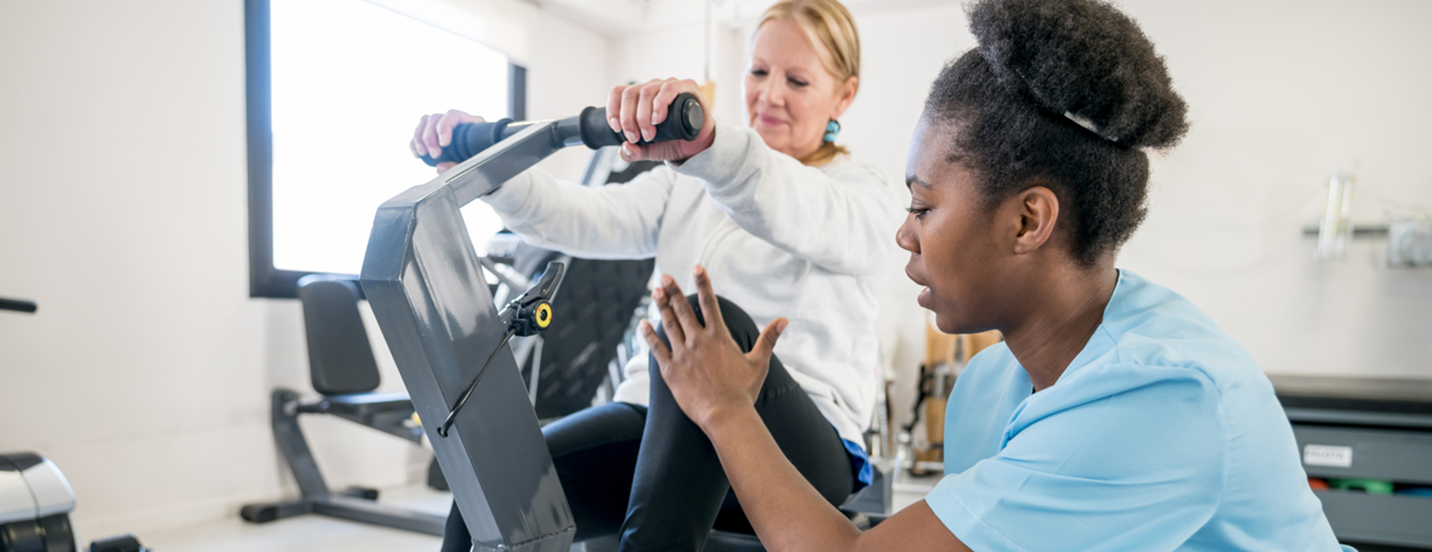
Recovery of Function
Buttocks
Temporomandibular Joint Disc
Acromioclavicular Joint
Models, Anatomic
Rotation
Muscle, Skeletal
Retrospective Studies
Prospective Studies
Reproducibility of Results
Postoperative Complications
Palpation
Electromyography
Pressure
Sternoclavicular Joint
Severity of Illness Index
Synovitis
Suspensions
Magnetic Resonance Imaging
Pain Measurement
Synovial Membrane
Femoral Neck Fractures
Lower Extremity
Dislocations
Tomography, X-Ray Computed
Imaging, Three-Dimensional
Muscle Strength
Risk Factors
Paraplegia
Cementation
Arthritis, Experimental
Anthropometry
Hip Contracture
Non-operative management of acetabular fractures. The use of dynamic stress views. (1/2399)
To assess the stability of the hip after acetabular fracture, dynamic fluoroscopic stress views were taken of 41 acetabular fractures that met the criteria for non-operative management. These included roof arcs of 45 degrees, a subchondral CT arc of 10 mm, displacement of less than 50% of the posterior wall, and congruence on the AP and Judet views of the hip. There were three unstable hips which were treated by open reduction and internal fixation. The remaining 38 fractures were treated non-operatively with early mobilisation and delayed weight-bearing. At a mean follow-up of 2.7 years, the results were good or excellent in 91% of the cases. Three fair results were ascribed to the patients' other injuries. Dynamic stress views can identify subtle instability in patients who would normally be considered for non-operative treatment. (+info)Prevalence of generalised osteoarthritis in patients with advanced hip and knee osteoarthritis: the Ulm Osteoarthritis Study. (2/2399)
OBJECTIVES: Different prevalences of generalised osteoarthritis (GOA) in patients with knee and hip OA have been reported. The aim of this investigation was to evaluate radiographic and clinical patterns of disease in a hospital based population of patient subgroups with advanced hip and knee OA and to compare the prevalence of GOA in patients with hip or knee OA, taking potential confounding factors into account. METHODS: 420 patients with hip OA and 389 patients with knee OA scheduled for unilateral total joint replacement in four hospitals underwent radiographic analysis of ipsilateral and contralateral hip or knee joint and both hands in addition to a standardised interview and clinical examination. According to the severity of radiographic changes in the contralateral joints (using Kellgren-Lawrence > or = grade 2 as case definition) participants were classified as having either unilateral or bilateral OA. If radiographic changes of two joint groups of the hands (first carpometacarpal joint and proximal/distal interphalangeal joints defined as two separate joint groups) were present, patients were categorised as having GOA. RESULTS: Patients with hip OA were younger (mean age 60.4 years) and less likely to be female (52.4%) than patients with knee OA (66.3 years and 72.5% respectively). Intensity of pain and functional impairment at hospital admission was similar in both groups, while patients with knee OA had a longer symptom duration (median 10 years) compared with patients with hip OA (5 years). In 41.7% of patients with hip OA and 33.4% of patients with knee OA an underlying pathological condition could be observed in the replaced joint, which allowed a classification as secondary OA. Some 82.1% of patients with hip and 87.4% of patients with knee OA had radiographic changes in their contralateral joints (bilateral disease). The prevalence of GOA increased with age and was higher in female patients. GOA was observed more often in patients with knee OA than in patients with hip OA (34.9% versus 19.3%; OR = 2.24; 95% CI: 1.56, 3.21). Adjustment for the different age and sex distribution in both patient groups, however, takes away most of the difference (OR = 1.32; 95% CI: 0.89, 1.96). CONCLUSION: The crude results confirm previous reports as well as the clinical impression of GOA being more prevalent in patients with advanced knee OA than in patients with advanced hip OA. However, these different patterns might be attributed to a large part to a different distribution of age and sex in these hospital based populations. (+info)Osteonecrosis of the hip in sickle-cell disease associated with tuberculous arthritis. A review of 15 cases. (3/2399)
We report a study of 15 cases of tuberculous hips with sickle-cell disease who presented during 1991-1993. Although the osteonecrosis was long-standing, biopsy was nearly always required to reveal the more recent tuberculous infection. Management consisted of 6 months of anti-tuberculous chemotherapy with appropriate palliative surgery 5-8 weeks after the start of drug treatment. The operative techniques which we used are described. The results were good both post-operatively, and in 12 patients followed-up at an average of 3 years. We recommend this combined management for the treatment of secondary tuberculous infections of hips previously damaged by sickle-cell disease. (+info)Hip moments during level walking, stair climbing, and exercise in individuals aged 55 years or older. (4/2399)
BACKGROUND AND PURPOSE: Low bone mass of the proximal femur is a risk factor for hip fractures. Exercise has been shown to reduce bone loss in older individuals; however, the exercises most likely to influence bone mass of the proximal femur have not been identified. Net moments of force at the hip provide an indication of the mechanical load on the proximal femur. The purpose of this study was to examine various exercises to determine which exercises result in the greatest magnitude and rate of change in moments of force at the hip in older individuals. SUBJECTS AND METHODS: Walking and exercise patterns were analyzed for 30 subjects (17 men, 13 women) who were 55 years of age or older (X = 65.4, SD = 6.02, range = 55-75) and who had no identified musculoskeletal or neurological impairment. Kinematic and kinetic data were obtained with an optoelectronic system and a force platform. Results. Of the exercises investigated, only ascending stairs generated peak moments higher than those obtained during level walking and only in the transverse plane. Most of the exercises generated moments and rate of change in moments with magnitudes similar to or lower than those obtained during gait. CONCLUSION AND DISCUSSION: Level walking and exercises that generated moments with magnitudes comparable to or higher than those obtained during gait could be combined in an exercise program designed to maintain or increase bone mass at the hip. (+info)The influence of weight-bearing on the measurement of polyethylene wear in THA. (5/2399)
We have studied the influence of weight-bearing on the measurement of wear of the polyethylene acetabular component in total hip arthroplasty using two techniques. The measured vertical wear was significantly greater when radiographs were taken weight-bearing rather than with the patient supine (p = 0.001, method 1; p = 0.007, method 2). Calculations of rates of linear wear of the acetabular component were significantly underestimated (p < 0.05) when radiographs were taken supine. There are two reasons for this. First, a change in pelvic orientation when bearing weight ensures that the thinnest polyethylene is brought into relief, and secondly, the head of the femoral component assumes the position of maximal displacement along its wear path. Interpretation of previous studies on both linear and volumetric polyethylene wear in total hip arthroplasty should be reassessed in the light of these findings. (+info)Accuracy of EBRA-FCA in the measurement of migration of femoral components of total hip replacement. Einzel-Bild-Rontgen-Analyse-femoral component analysis. (6/2399)
Several methods of measuring the migration of the femoral component after total hip replacement have been described, but they use different reference lines, and have differing accuracies, some unproven. Statistical comparison of different studies is rarely possible. We report a study of the EBRA-FCA method (femoral component analysis using Einzel-Bild-Rontgen-Analyse) to determine its accuracy using three independent assessments, including a direct comparison with the results of roentgen stereophotogrammetric analysis (RSA). The accuracy of EBRA-FCA was better than +/- 1.5 mm (95% percentile) with a Cronbach's coefficient alpha for interobserver reliability of 0.84; a very good result. The method had a specificity of 100% and a sensitivity of 78% compared with RSA for the detection of migration of over 1 mm. This is accurate enough to assess the stability of a prosthesis within a relatively limited period. The best reference line for downward migration is between the greater trochanter and the shoulder of the stem, as confirmed by two experimental analyses and a computer-assisted design. (+info)The prediction of failure of the stem in THR by measurement of early migration using EBRA-FCA. Einzel-Bild-Roentgen-Analyse-femoral component analysis. (7/2399)
We report the ten-year results for three designs of stem in 240 total hip replacements, for which subsidence had been measured on plain radiographs at regular intervals. Accurate migration patterns could be determined by the method of Einzel-Bild-Roentgen-Analyse-femoral component analysis (EBRA-FCA) for 158 hips (66%). Of these, 108 stems (68%) remained stable throughout, and five (3%) started to migrate after a median of 54 months. Initial migration of at least 1 mm was seen in 45 stems (29%) during the first two years, but these then became stable. We revised 17 stems for aseptic loosening, and 12 for other reasons. Revision for aseptic loosening could be predicted by EBRA-FCA with a sensitivity of 69%, a specificity of 80%, and an accuracy of 79% by the use of a threshold of subsidence of 1.5 mm during the first two years. Similar observations over a five-year period allowed the long-term outcome to be predicted with an accuracy of 91%. We discuss the importance of four different patterns of subsidence and confirm that the early measurement of migration by a reasonably accurate method can help to predict long-term outcome. Such methods should be used to evaluate new and modified designs of prosthesis. (+info)Retroversion of the acetabulum. A cause of hip pain. (8/2399)
We describe a little-known variety of hip dysplasia, termed 'acetabular retroversion', in which the alignment of the mouth of the acetabulum does not face the normal anterolateral direction, but inclines more posterolaterally. The condition may be part of a complex dysplasia or a single entity. Other than its retroversion, the acetabulum is sited normally on the side wall of the pelvis, and its articular surface is of normal extent and configuration. The retroverted orientation may give rise to problems of impingement between the femoral neck and anterior acetabular edge. We define the clinical and radiological parameters and discuss pathological changes which may occur in the untreated condition. A technique of management is proposed. (+info)The hip joint, also known as the coxal joint, is a ball-and-socket type synovial joint that connects the femur (thigh bone) to the pelvis. The "ball" is the head of the femur, while the "socket" is the acetabulum, a concave surface on the pelvic bone.
The hip joint is surrounded by a strong fibrous capsule and is reinforced by several ligaments, including the iliofemoral, ischiofemoral, and pubofemoral ligaments. The joint allows for flexion, extension, abduction, adduction, medial and lateral rotation, and circumduction movements, making it one of the most mobile joints in the body.
The hip joint is also supported by various muscles, including the gluteus maximus, gluteus medius, gluteus minimus, iliopsoas, and other hip flexors and extensors. These muscles provide stability and strength to the joint, allowing for weight-bearing activities such as walking, running, and jumping.
In medical terms, the hip is a ball-and-socket joint where the rounded head of the femur (thigh bone) fits into the cup-shaped socket, also known as the acetabulum, of the pelvis. This joint allows for a wide range of movement in the lower extremities and supports the weight of the upper body during activities such as walking, running, and jumping. The hip joint is surrounded by strong ligaments, muscles, and tendons that provide stability and enable proper functioning.
Osteoarthritis (OA) of the hip is a degenerative joint disease that affects the articular cartilage and subchondral bone of the hip joint. It is characterized by the progressive loss of cartilage, remodeling of bone, osteophyte formation (bone spurs), cysts, and mild to moderate inflammation. The degenerative process can lead to pain, stiffness, limited range of motion, and crepitus (grating or crackling sound) during movement.
In the hip joint, OA typically affects the femoral head and acetabulum. As the articular cartilage wears away, the underlying bone becomes exposed and can lead to bone-on-bone contact, which is painful. The body responds by attempting to repair the damage through remodeling of the subchondral bone and formation of osteophytes. However, these changes can further limit joint mobility and exacerbate symptoms.
Risk factors for OA of the hip include age, obesity, genetics, previous joint injury or surgery, and repetitive stress on the joint. Treatment options may include pain management (such as NSAIDs, physical therapy, and injections), lifestyle modifications (such as weight loss and exercise), and, in severe cases, surgical intervention (such as hip replacement).
A joint is the location at which two or more bones make contact. They are constructed to allow movement and provide support and stability to the body during motion. Joints can be classified in several ways, including structure, function, and the type of tissue that forms them. The three main types of joints based on structure are fibrous (or fixed), cartilaginous, and synovial (or diarthrosis). Fibrous joints do not have a cavity and have limited movement, while cartilaginous joints allow for some movement and are connected by cartilage. Synovial joints, the most common and most movable type, have a space between the articular surfaces containing synovial fluid, which reduces friction and wear. Examples of synovial joints include hinge, pivot, ball-and-socket, saddle, and condyloid joints.
Hip arthroplasty, also known as hip replacement surgery, is a medical procedure where the damaged or diseased joint surfaces of the hip are removed and replaced with artificial components. These components typically include a metal or ceramic ball that replaces the head of the femur (thigh bone), and a polyethylene or ceramic socket that replaces the acetabulum (hip socket) in the pelvis.
The goal of hip arthroplasty is to relieve pain, improve joint mobility, and restore function to the hip joint. This procedure is commonly performed in patients with advanced osteoarthritis, rheumatoid arthritis, hip fractures, or other conditions that cause significant damage to the hip joint.
There are several types of hip replacement surgeries, including traditional total hip arthroplasty, partial (hemi) hip arthroplasty, and resurfacing hip arthroplasty. The choice of procedure depends on various factors, such as the patient's age, activity level, overall health, and the extent of joint damage.
After surgery, patients typically require rehabilitation to regain strength, mobility, and function in the affected hip. With proper care and follow-up, most patients can expect significant pain relief and improved quality of life following hip arthroplasty.
A hip prosthesis, also known as a total hip replacement, is a surgical implant designed to replace the damaged or diseased components of the human hip joint. The procedure involves replacing the femoral head (the ball at the top of the thigh bone) and the acetabulum (the socket in the pelvis) with artificial parts, typically made from materials such as metal, ceramic, or plastic.
The goal of a hip prosthesis is to relieve pain, improve joint mobility, and restore function, allowing patients to return to their normal activities and enjoy an improved quality of life. The procedure is most commonly performed in individuals with advanced osteoarthritis, rheumatoid arthritis, or other degenerative conditions that have caused significant damage to the hip joint.
There are several different types of hip prostheses available, each with its own unique design and set of benefits and risks. The choice of prosthesis will depend on a variety of factors, including the patient's age, activity level, overall health, and specific medical needs. In general, however, all hip prostheses are designed to provide a durable, long-lasting solution for patients suffering from debilitating joint pain and stiffness.
Congenital hip dislocation, also known as developmental dysplasia of the hip (DDH), is a condition where the hip joint fails to develop normally in utero or during early infancy. In a healthy hip, the head of the femur (thigh bone) fits snugly into the acetabulum (hip socket). However, in congenital hip dislocation, the femoral head is not held firmly in place within the acetabulum due to abnormal development or laxity of the ligaments that support the joint.
There are two types of congenital hip dislocations:
1. Teratologic dislocation: This type is present at birth and occurs due to abnormalities in the development of the hip joint during fetal growth. The femoral head may be completely outside the acetabulum or partially dislocated.
2. Developmental dysplasia: This type develops after birth, often within the first few months of life, as a result of ligamentous laxity and shallow acetabulum. In some cases, it can progress to a complete hip dislocation if left untreated.
Risk factors for congenital hip dislocation include family history, breech presentation during delivery, and female gender. Early diagnosis and treatment are crucial to prevent long-term complications such as pain, limited mobility, and osteoarthritis. Treatment options may include bracing, closed reduction, or surgical intervention, depending on the severity and age of the child at diagnosis.
Canine hip dysplasia (CHD) is a common skeletal disorder in dogs, particularly in large and giant breeds, characterized by the abnormal development and degeneration of the coxofemoral joint - the joint where the head of the femur (thigh bone) meets the acetabulum (hip socket) of the pelvis. This condition is often caused by a combination of genetic and environmental factors that lead to laxity (looseness) of the joint, which can result in osteoarthritis (OA), pain, and decreased mobility over time.
In a healthy hip joint, the femoral head fits snugly into the acetabulum, allowing smooth and stable movement. However, in dogs with CHD, the following abnormalities may occur:
1. Shallow acetabulum: The hip socket may not be deep enough to provide adequate coverage of the femoral head, leading to joint instability.
2. Flared acetabulum: The rim of the acetabulum may become stretched and flared due to excessive forces exerted on it by the lax joint.
3. Misshapen or malformed femoral head: The femoral head may not have a normal round shape, further contributing to joint instability.
4. Laxity of the joint: The ligament that holds the femoral head in place within the acetabulum (ligamentum teres) can become stretched, allowing for excessive movement and abnormal wear of the joint surfaces.
These changes can lead to the development of osteoarthritis, which is characterized by the breakdown and loss of cartilage within the joint, as well as the formation of bone spurs (osteophytes) and thickening of the joint capsule. This results in pain, stiffness, and decreased range of motion, making it difficult for affected dogs to perform everyday activities such as walking, running, or climbing stairs.
Canine hip dysplasia is typically diagnosed through a combination of physical examination, medical history, and imaging techniques such as radiographs (X-rays). Treatment options may include conservative management, such as weight management, exercise modification, joint supplements, and pain medication, or surgical intervention, such as total hip replacement. The choice of treatment depends on the severity of the disease, the age and overall health of the dog, and the owner's financial resources.
Preventing canine hip dysplasia is best achieved through selective breeding practices that aim to eliminate affected animals from breeding populations. Additionally, maintaining a healthy weight, providing appropriate exercise, and ensuring proper nutrition throughout a dog's life can help reduce the risk of developing this debilitating condition.
A hip fracture is a medical condition referring to a break in the upper part of the femur (thigh) bone, which forms the hip joint. The majority of hip fractures occur due to falls or direct trauma to the area. They are more common in older adults, particularly those with osteoporosis, a condition that weakens bones and makes them more prone to breaking. Hip fractures can significantly impact mobility and quality of life, often requiring surgical intervention and rehabilitation.
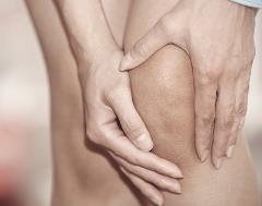
The knee joint, also known as the tibiofemoral joint, is the largest and one of the most complex joints in the human body. It is a synovial joint that connects the thighbone (femur) to the shinbone (tibia). The patella (kneecap), which is a sesamoid bone, is located in front of the knee joint and helps in the extension of the leg.
The knee joint is made up of three articulations: the femorotibial joint between the femur and tibia, the femoropatellar joint between the femur and patella, and the tibiofibular joint between the tibia and fibula. These articulations are surrounded by a fibrous capsule that encloses the synovial membrane, which secretes synovial fluid to lubricate the joint.
The knee joint is stabilized by several ligaments, including the medial and lateral collateral ligaments, which provide stability to the sides of the joint, and the anterior and posterior cruciate ligaments, which prevent excessive forward and backward movement of the tibia relative to the femur. The menisci, which are C-shaped fibrocartilaginous structures located between the femoral condyles and tibial plateaus, also help to stabilize the joint by absorbing shock and distributing weight evenly across the articular surfaces.
The knee joint allows for flexion, extension, and a small amount of rotation, making it essential for activities such as walking, running, jumping, and sitting.
Hip injuries refer to damages or harm caused to the hip joint or its surrounding structures, including bones, muscles, tendons, ligaments, and cartilage. These injuries can occur due to various reasons such as falls, accidents, sports-related activities, or degenerative conditions. Common hip injuries include fractures, dislocations, strains, sprains, bursitis, and labral tears. Symptoms may include pain, swelling, bruising, stiffness, limited mobility, and inability to bear weight on the affected leg. Proper diagnosis and treatment are crucial to ensure optimal recovery and prevent long-term complications.
The femoral head is the rounded, ball-like top portion of the femur (thigh bone) that fits into the hip socket (acetabulum) to form the hip joint. It has a smooth, articular cartilage surface that allows for smooth and stable articulation with the pelvis. The femoral head is connected to the femoral neck, which is a narrower section of bone that angles downward and leads into the shaft of the femur. Together, the femoral head and neck provide stability and range of motion to the hip joint.
Joint diseases is a broad term that refers to various conditions affecting the joints, including but not limited to:
1. Osteoarthritis (OA): A degenerative joint disease characterized by the breakdown of cartilage and underlying bone, leading to pain, stiffness, and potential loss of function.
2. Rheumatoid Arthritis (RA): An autoimmune disorder causing inflammation in the synovial membrane lining the joints, resulting in swelling, pain, and joint damage if left untreated.
3. Infectious Arthritis: Joint inflammation caused by bacterial, viral, or fungal infections that spread through the bloodstream or directly enter the joint space.
4. Gout: A type of arthritis resulting from the buildup of uric acid crystals in the joints, typically affecting the big toe and characterized by sudden attacks of severe pain, redness, and swelling.
5. Psoriatic Arthritis (PsA): An inflammatory joint disease associated with psoriasis, causing symptoms such as pain, stiffness, and swelling in the joints and surrounding tissues.
6. Juvenile Idiopathic Arthritis (JIA): A group of chronic arthritis conditions affecting children, characterized by joint inflammation, pain, and stiffness.
7. Ankylosing Spondylitis: A form of arthritis primarily affecting the spine, causing inflammation, pain, and potential fusion of spinal vertebrae.
8. Bursitis: Inflammation of the fluid-filled sacs (bursae) that cushion joints, leading to pain and swelling.
9. Tendinitis: Inflammation or degeneration of tendons, which connect muscles to bones, often resulting in pain and stiffness near joints.
These conditions can impact the function and mobility of affected joints, causing discomfort and limiting daily activities. Proper diagnosis and treatment are essential for managing joint diseases and preserving joint health.
A hip dislocation is a medical emergency that occurs when the head of the femur (thighbone) slips out of its socket in the pelvis. This can happen due to high-energy trauma, such as a car accident or a severe fall. Hip dislocations can also occur in people with certain health conditions that make their hips more prone to displacement, such as developmental dysplasia of the hip.
There are two main types of hip dislocations: posterior and anterior. In a posterior dislocation, the femur head moves out of the back of the socket, which is the most common type. In an anterior dislocation, the femur head moves out of the front of the socket. Both types of hip dislocations can cause severe pain, swelling, and difficulty moving the affected leg.
Immediate medical attention is necessary for a hip dislocation to realign the bones and prevent further damage. Treatment typically involves sedation or anesthesia to relax the muscles around the joint, followed by a closed reduction procedure to gently guide the femur head back into the socket. In some cases, surgery may be required to repair any associated injuries, such as fractures or damaged ligaments. After treatment, physical therapy and rehabilitation are usually necessary to restore strength, mobility, and function to the affected hip joint.
The acetabulum is the cup-shaped cavity in the pelvic bone (specifically, the os coxa) where the head of the femur bone articulates to form the hip joint. It provides a stable and flexible connection between the lower limb and the trunk, allowing for a wide range of movements such as flexion, extension, abduction, adduction, rotation, and circumduction. The acetabulum is lined with articular cartilage, which facilitates smooth and frictionless movement of the hip joint. Its stability is further enhanced by various ligaments, muscles, and the labrum, a fibrocartilaginous rim that deepens the socket and increases its contact area with the femoral head.
The ankle joint, also known as the talocrural joint, is the articulation between the bones of the lower leg (tibia and fibula) and the talus bone in the foot. It is a synovial hinge joint that allows for dorsiflexion and plantarflexion movements, which are essential for walking, running, and jumping. The ankle joint is reinforced by strong ligaments on both sides to provide stability during these movements.
A joint capsule is the fibrous sac that encloses a synovial joint, which is a type of joint characterized by the presence of a cavity filled with synovial fluid. The joint capsule provides stability and strength to the joint, while also allowing for a range of motion. It consists of two layers: an outer fibrous layer and an inner synovial membrane. The fibrous layer is made up of dense connective tissue that helps to stabilize the joint, while the synovial membrane produces synovial fluid, which lubricates the joint and reduces friction during movement.
Articular Range of Motion (AROM) is a term used in physiotherapy and orthopedics to describe the amount of movement available in a joint, measured in degrees of a circle. It refers to the range through which synovial joints can actively move without causing pain or injury. AROM is assessed by measuring the degree of motion achieved by active muscle contraction, as opposed to passive range of motion (PROM), where the movement is generated by an external force.
Assessment of AROM is important in evaluating a patient's functional ability and progress, planning treatment interventions, and determining return to normal activities or sports participation. It is also used to identify any restrictions in joint mobility that may be due to injury, disease, or surgery, and to monitor the effectiveness of rehabilitation programs.
Dromaiidae is a family of birds that includes only one extant species, the Emu (Dromaius novaehollandiae). The Emu is the second largest bird in the world, after the Ostrich. It is a large, flightless bird native to Australia, known for its long legs and neck. Emus can run at high speeds and have been recorded reaching up to 50 km/h (31 mph). They are omnivorous birds that primarily feed on plants, but will also eat insects and small animals.
Dromaiidae is part of the order Casuariiformes, which also includes the cassowaries, another group of large, flightless birds native to the tropical rainforests of Indonesia, New Guinea, and northeastern Australia. Together, Dromaiidae and Casuariidae are sometimes referred to as the "emu family" or the "cassowary family."
In summary, Dromaiidae is a family of birds that includes only one extant species, the Emu, which is a large, flightless bird native to Australia.
Biomechanics is the application of mechanical laws to living structures and systems, particularly in the field of medicine and healthcare. A biomechanical phenomenon refers to a observable event or occurrence that involves the interaction of biological tissues or systems with mechanical forces. These phenomena can be studied at various levels, from the molecular and cellular level to the tissue, organ, and whole-body level.
Examples of biomechanical phenomena include:
1. The way that bones and muscles work together to produce movement (known as joint kinematics).
2. The mechanical behavior of biological tissues such as bone, cartilage, tendons, and ligaments under various loads and stresses.
3. The response of cells and tissues to mechanical stimuli, such as the way that bone tissue adapts to changes in loading conditions (known as Wolff's law).
4. The biomechanics of injury and disease processes, such as the mechanisms of joint injury or the development of osteoarthritis.
5. The use of mechanical devices and interventions to treat medical conditions, such as orthopedic implants or assistive devices for mobility impairments.
Understanding biomechanical phenomena is essential for developing effective treatments and prevention strategies for a wide range of medical conditions, from musculoskeletal injuries to neurological disorders.
A finger joint, also known as an articulation, is the point where two bones in a finger connect and allow for movement. The majority of finger joints are classified as hinge joints, permitting flexion and extension movements. These joints consist of several components:
1. Articular cartilage: Smooth tissue that covers the ends of the bones, enabling smooth movement and protecting the bones from friction.
2. Joint capsule: A fibrous sac enclosing the joint, providing stability and producing synovial fluid for lubrication.
3. Synovial membrane: Lines the inner surface of the joint capsule and produces synovial fluid to lubricate the joint.
4. Volar plate (palmar ligament): A strong band of tissue located on the palm side of the joint, preventing excessive extension and maintaining alignment.
5. Collateral ligaments: Two bands of tissue located on each side of the joint, providing lateral stability and limiting radial and ulnar deviation.
6. Flexor tendons: Tendons that attach to the bones on the palmar side of the finger joints, facilitating flexion movements.
7. Extensor tendons: Tendons that attach to the bones on the dorsal side of the finger joints, enabling extension movements.
Finger joints are essential for hand function and enable activities such as grasping, holding, writing, and manipulating objects.
Femoral head necrosis, also known as avascular necrosis of the femoral head, is a medical condition that results from the interruption of blood flow to the femoral head, which is the rounded end of the thigh bone that fits into the hip joint. This lack of blood supply can cause the bone tissue to die, leading to the collapse of the femoral head and eventually resulting in hip joint damage or arthritis.
The condition can be caused by a variety of factors, including trauma, alcohol abuse, corticosteroid use, radiation therapy, and certain medical conditions such as sickle cell disease and lupus. Symptoms may include pain in the hip or groin, limited range of motion, and difficulty walking. Treatment options depend on the severity and progression of the necrosis and may include medication, physical therapy, or surgical intervention.
Gait is a medical term used to describe the pattern of movement of the limbs during walking or running. It includes the manner or style of walking, including factors such as rhythm, speed, and step length. A person's gait can provide important clues about their physical health and neurological function, and abnormalities in gait may indicate the presence of underlying medical conditions, such as neuromuscular disorders, orthopedic problems, or injuries.
A typical human gait cycle involves two main phases: the stance phase, during which the foot is in contact with the ground, and the swing phase, during which the foot is lifted and moved forward in preparation for the next step. The gait cycle can be further broken down into several sub-phases, including heel strike, foot flat, midstance, heel off, and toe off.
Gait analysis is a specialized field of study that involves observing and measuring a person's gait pattern using various techniques, such as video recordings, force plates, and motion capture systems. This information can be used to diagnose and treat gait abnormalities, improve mobility and function, and prevent injuries.
Articular cartilage is the smooth, white tissue that covers the ends of bones where they come together to form joints. It provides a cushion between bones and allows for smooth movement by reducing friction. Articular cartilage also absorbs shock and distributes loads evenly across the joint, protecting the bones from damage. It is avascular, meaning it does not have its own blood supply, and relies on the surrounding synovial fluid for nutrients. Over time, articular cartilage can wear down or become damaged due to injury or disease, leading to conditions such as osteoarthritis.
Joint instability is a condition characterized by the loss of normal joint function and increased risk of joint injury due to impaired integrity of the supporting structures, such as ligaments, muscles, or cartilage. This can result in excessive movement or laxity within the joint, leading to decreased stability and increased susceptibility to dislocations or subluxations. Joint instability may cause pain, swelling, and limited range of motion, and it can significantly impact a person's mobility and quality of life. It is often caused by trauma, degenerative conditions, or congenital abnormalities and may require medical intervention, such as physical therapy, bracing, or surgery, to restore joint stability.
Osteoarthritis (OA) is a type of joint disease that is characterized by the breakdown and eventual loss of cartilage - the tissue that cushions the ends of bones where they meet in the joints. This breakdown can cause the bones to rub against each other, causing pain, stiffness, and loss of mobility. OA can occur in any joint, but it most commonly affects the hands, knees, hips, and spine. It is often associated with aging and can be caused or worsened by obesity, injury, or overuse.
The medical definition of osteoarthritis is: "a degenerative, non-inflammatory joint disease characterized by the loss of articular cartilage, bone remodeling, and the formation of osteophytes (bone spurs). It is often associated with pain, stiffness, and decreased range of motion in the affected joint."
The femur is the medical term for the thigh bone, which is the longest and strongest bone in the human body. It connects the hip bone to the knee joint and plays a crucial role in supporting the weight of the body and allowing movement during activities such as walking, running, and jumping. The femur is composed of a rounded head, a long shaft, and two condyles at the lower end that articulate with the tibia and patella to form the knee joint.
The tarsal joints are a series of articulations in the foot that involve the bones of the hindfoot and midfoot. There are three main tarsal joints:
1. Talocrural joint (also known as the ankle joint): This is the joint between the talus bone of the lower leg and the tibia and fibula bones of the lower leg, as well as the calcaneus bone of the foot. It allows for dorsiflexion and plantarflexion movements of the foot.
2. Subtalar joint: This is the joint between the talus bone and the calcaneus bone. It allows for inversion and eversion movements of the foot.
3. Tarsometatarsal joints (also known as the Lisfranc joint): These are the joints between the tarsal bones of the midfoot and the metatarsal bones of the forefoot. They allow for flexion, extension, abduction, and adduction movements of the foot.
These joints play an important role in the stability and mobility of the foot, allowing for various movements during activities such as walking, running, and jumping.
Infectious arthritis, also known as septic arthritis, is a type of joint inflammation that is caused by a bacterial or fungal infection. The infection can enter the joint through the bloodstream or directly into the synovial fluid of the joint, often as a result of a traumatic injury, surgery, or an underlying condition such as diabetes or a weakened immune system.
The most common symptoms of infectious arthritis include sudden onset of severe pain and swelling in the affected joint, fever, chills, and difficulty moving the joint. If left untreated, infectious arthritis can lead to serious complications such as joint damage or destruction, sepsis, and even death. Treatment typically involves antibiotics or antifungal medications to eliminate the infection, along with rest, immobilization, and sometimes surgery to drain the infected synovial fluid.
It is important to seek medical attention promptly if you experience symptoms of infectious arthritis, as early diagnosis and treatment can help prevent long-term complications and improve outcomes.
Arthrography is a medical imaging technique used to diagnose problems within joints. It involves the injection of a contrast agent, such as a radiopaque dye or air, into the joint space, followed by the use of fluoroscopy or X-ray imaging to visualize the internal structures of the joint. This can help to identify injuries, tears, or other abnormalities in the cartilage, ligaments, tendons, or bones within the joint.
The procedure is typically performed on an outpatient basis and may be used to diagnose conditions such as shoulder dislocations, rotator cuff tears, meniscal tears in the knee, or hip labral injuries. It is a relatively safe and minimally invasive procedure, although there may be some temporary discomfort or swelling at the injection site. Patients are usually advised to avoid strenuous activity for a day or two following the procedure to allow the contrast agent to fully dissipate from the joint.
The wrist joint, also known as the radiocarpal joint, is a condyloid joint that connects the distal end of the radius bone in the forearm to the proximal row of carpal bones in the hand (scaphoid, lunate, and triquetral bones). It allows for flexion, extension, radial deviation, and ulnar deviation movements of the hand. The wrist joint is surrounded by a capsule and reinforced by several ligaments that provide stability and strength to the joint.
"Weight-bearing" is a term used in the medical field to describe the ability of a body part or limb to support the weight or pressure exerted upon it, typically while standing, walking, or performing other physical activities. In a clinical setting, healthcare professionals often use the term "weight-bearing exercise" to refer to physical activities that involve supporting one's own body weight, such as walking, jogging, or climbing stairs. These exercises can help improve bone density, muscle strength, and overall physical function, particularly in individuals with conditions affecting the bones, joints, or muscles.
In addition, "weight-bearing" is also used to describe the positioning of a body part during medical imaging studies, such as X-rays or MRIs. For example, a weight-bearing X-ray of the foot or ankle involves taking an image while the patient stands on the affected limb, allowing healthcare providers to assess any alignment or stability issues that may not be apparent in a non-weight-bearing position.
Osteotomy is a surgical procedure in which a bone is cut to shorten, lengthen, or change its alignment. It is often performed to correct deformities or to realign bones that have been damaged by trauma or disease. The bone may be cut straight across (transverse osteotomy) or at an angle (oblique osteotomy). After the bone is cut, it can be realigned and held in place with pins, plates, or screws until it heals. This procedure is commonly performed on bones in the leg, such as the femur or tibia, but can also be done on other bones in the body.
Arthralgia is a medical term that refers to pain in the joints. It does not involve inflammation, which would be referred to as arthritis. The pain can range from mild to severe and may occur in one or multiple joints. Arthralgia can have various causes, including injuries, infections, degenerative conditions, or systemic diseases. In some cases, the underlying cause of arthralgia remains unknown. Treatment typically focuses on managing the pain and addressing the underlying condition if it can be identified.
Developmental bone diseases are a group of medical conditions that affect the growth and development of bones. These diseases are present at birth or develop during childhood and adolescence, when bones are growing rapidly. They can result from genetic mutations, hormonal imbalances, or environmental factors such as poor nutrition.
Some examples of developmental bone diseases include:
1. Osteogenesis imperfecta (OI): Also known as brittle bone disease, OI is a genetic disorder that affects the body's production of collagen, a protein necessary for healthy bones. People with OI have fragile bones that break easily and may also experience other symptoms such as blue sclerae (whites of the eyes), hearing loss, and joint laxity.
2. Achondroplasia: This is the most common form of dwarfism, caused by a genetic mutation that affects bone growth. People with achondroplasia have short limbs and a large head relative to their body size.
3. Rickets: A condition caused by vitamin D deficiency or an inability to absorb or use vitamin D properly. This leads to weak, soft bones that can bow or bend easily, particularly in children.
4. Fibrous dysplasia: A rare bone disorder where normal bone is replaced with fibrous tissue, leading to weakened bones and deformities.
5. Scoliosis: An abnormal curvature of the spine that can develop during childhood or adolescence. While not strictly a developmental bone disease, scoliosis can be caused by various underlying conditions such as cerebral palsy, muscular dystrophy, or spina bifida.
Treatment for developmental bone diseases varies depending on the specific condition and its severity. Treatment may include medication, physical therapy, bracing, or surgery to correct deformities and improve function. Regular follow-up with a healthcare provider is essential to monitor growth, manage symptoms, and prevent complications.
Prosthesis failure is a term used to describe a situation where a prosthetic device, such as an artificial joint or limb, has stopped functioning or failed to meet its intended purpose. This can be due to various reasons, including mechanical failure, infection, loosening of the device, or a reaction to the materials used in the prosthesis.
Mechanical failure can occur due to wear and tear, manufacturing defects, or improper use of the prosthetic device. Infection can also lead to prosthesis failure, particularly in cases where the prosthesis is implanted inside the body. The immune system may react to the presence of the foreign material, leading to inflammation and infection.
Loosening of the prosthesis can also cause it to fail over time, as the device becomes less stable and eventually stops working properly. Additionally, some people may have a reaction to the materials used in the prosthesis, leading to tissue damage or other complications that can result in prosthesis failure.
In general, prosthesis failure can lead to decreased mobility, pain, and the need for additional surgeries or treatments to correct the problem. It is important for individuals with prosthetic devices to follow their healthcare provider's instructions carefully to minimize the risk of prosthesis failure and ensure that the device continues to function properly over time.
Prosthesis-related infections, also known as prosthetic joint infections (PJIs), are infections that occur around or within a prosthetic device, such as an artificial joint. These infections can be caused by bacteria, fungi, or other microorganisms and can lead to serious complications if not treated promptly and effectively.
Prosthesis-related infections can occur soon after the implantation of the prosthetic device (early infection) or months or even years later (late infection). Early infections are often caused by bacteria that enter the surgical site during the procedure, while late infections may be caused by hematogenous seeding (i.e., when bacteria from another source spread through the bloodstream and settle in the prosthetic device) or by contamination during a subsequent medical procedure.
Symptoms of prosthesis-related infections can include pain, swelling, redness, warmth, and drainage around the affected area. In some cases, patients may also experience fever, chills, or fatigue. Diagnosis typically involves a combination of clinical evaluation, laboratory tests (such as blood cultures, joint fluid analysis, and tissue biopsy), and imaging studies (such as X-rays, CT scans, or MRI).
Treatment of prosthesis-related infections usually involves a combination of antibiotics and surgical intervention. The specific treatment approach will depend on the type and severity of the infection, as well as the patient's overall health status. In some cases, it may be necessary to remove or replace the affected prosthetic device.
In medical terms, lubrication refers to the application of a slippery substance or fluid to reduce friction and facilitate smooth movement between two surfaces. This is particularly relevant in the context of human anatomy, where lubrication plays a crucial role in various bodily functions. For instance, the mucous membranes that line body cavities such as the mouth, vagina, and rectum secrete fluids to provide lubrication for easy movement of tissues and foreign substances (like food or during sexual intercourse). Similarly, synovial fluid, a viscous substance found in joints, provides lubrication that enables smooth articulation between bones. Artificial lubricants may also be used in medical procedures to facilitate the insertion and movement of medical devices such as catheters or endoscopes.
The pelvis is the lower part of the trunk, located between the abdomen and the lower limbs. It is formed by the fusion of several bones: the ilium, ischium, and pubis (which together form the hip bone on each side), and the sacrum and coccyx in the back. The pelvis has several functions including supporting the weight of the upper body when sitting, protecting the lower abdominal organs, and providing attachment for muscles that enable movement of the lower limbs. In addition, it serves as a bony canal through which the reproductive and digestive tracts pass. The pelvic cavity contains several vital organs such as the bladder, parts of the large intestine, and in females, the uterus, ovaries, and fallopian tubes.
The sacroiliac (SI) joint is the joint that connects the iliac bone (part of the pelvis) and the sacrum (the triangular bone at the base of the spine). There are two sacroiliac joints, one on each side of the spine. The primary function of these joints is to absorb shock between the upper body and lower body and distribute the weight of the upper body to the lower body. They also provide a small amount of movement to allow for flexibility when walking or running. The SI joints are supported and stabilized by strong ligaments, muscles, and bones.
Disarticulation is a medical term that refers to the separation or dislocation of a joint. It can occur as a result of trauma, disease, or surgical intervention. In some cases, disarticulation may be necessary to relieve pain or improve mobility in a damaged joint. In forensic medicine, disarticulation is used to describe the postmortem separation of body parts at the joints, which can occur naturally in advanced decomposition or as a result of scavenging by animals.
Prosthesis design is a specialized field in medical device technology that involves creating and developing artificial substitutes to replace a missing body part, such as a limb, tooth, eye, or internal organ. The design process typically includes several stages: assessment of the patient's needs, selection of appropriate materials, creation of a prototype, testing and refinement, and final fabrication and fitting of the prosthesis.
The goal of prosthesis design is to create a device that functions as closely as possible to the natural body part it replaces, while also being comfortable, durable, and aesthetically pleasing for the patient. The design process may involve collaboration between medical professionals, engineers, and designers, and may take into account factors such as the patient's age, lifestyle, occupation, and overall health.
Prosthesis design can be highly complex, particularly for advanced devices such as robotic limbs or implantable organs. These devices often require sophisticated sensors, actuators, and control systems to mimic the natural functions of the body part they replace. As a result, prosthesis design is an active area of research and development in the medical field, with ongoing efforts to improve the functionality, comfort, and affordability of these devices for patients.
A Synovial Cyst is a type of benign cyst that typically develops in the synovium, which is the membrane that lines and lubricates joint capsules. These cysts are filled with synovial fluid, which is the same lubricating fluid found inside joints. They usually form as a result of degenerative changes, trauma, or underlying joint diseases such as osteoarthritis.
Synovial cysts commonly occur in the spine (particularly in the facet joints), but they can also develop in other areas of the body, including the knees, hips, and hands. While synovial cysts are generally not harmful, they may cause discomfort or pain if they press on nearby nerves or restrict movement in the affected joint. Treatment options for synovial cysts range from conservative measures like physical therapy and pain management to surgical intervention in severe cases.
"Torque" is not a term that has a specific medical definition. It is a physical concept used in the fields of physics and engineering, referring to a twisting force that causes rotation around an axis. However, in certain medical contexts, such as in discussions of spinal or joint biomechanics, the term "torque" may be used to describe a rotational force applied to a body part. But generally speaking, "torque" is not a term commonly used in medical terminology.
Synovial fluid is a viscous, clear, and straw-colored fluid found in the cavities of synovial joints, bursae, and tendon sheaths. It is produced by the synovial membrane, which lines the inner surface of the capsule surrounding these structures.
The primary function of synovial fluid is to reduce friction between articulating surfaces, providing lubrication for smooth and painless movement. It also acts as a shock absorber, protecting the joints from external forces during physical activities. Synovial fluid contains nutrients that nourish the articular cartilage, hyaluronic acid, which provides its viscoelastic properties, and lubricin, a protein responsible for boundary lubrication.
Abnormalities in synovial fluid composition or volume can indicate joint-related disorders, such as osteoarthritis, rheumatoid arthritis, gout, infection, or trauma. Analysis of synovial fluid is often used diagnostically to determine the underlying cause of joint pain, inflammation, or dysfunction.
Osteonecrosis is a medical condition characterized by the death of bone tissue due to the disruption of blood supply. Also known as avascular necrosis, this process can lead to the collapse of the bone and adjacent joint surfaces, resulting in pain, limited mobility, and potential deformity if left untreated. Osteonecrosis most commonly affects the hips, shoulders, and knees, but it can occur in any bone. The condition may be caused by trauma, corticosteroid use, alcohol abuse, certain medical conditions (like sickle cell disease or lupus), or for no apparent reason (idiopathic).
Bone cements are medical-grade materials used in orthopedic and trauma surgery to fill gaps between bone surfaces and implants, such as artificial joints or screws. They serve to mechanically stabilize the implant and provide a smooth, load-bearing surface. The two most common types of bone cement are:
1. Polymethylmethacrylate (PMMA) cement: This is a two-component system consisting of powdered PMMA and liquid methyl methacrylate monomer. When mixed together, they form a dough-like consistency that hardens upon exposure to air. PMMA cement has been widely used for decades in joint replacement surgeries, such as hip or knee replacements.
2. Calcium phosphate (CP) cement: This is a two-component system consisting of a powdered CP compound and an aqueous solution. When mixed together, they form a paste that hardens through a chemical reaction at body temperature. CP cement has lower mechanical strength compared to PMMA but demonstrates better biocompatibility, bioactivity, and the ability to resorb over time.
Both types of bone cements have advantages and disadvantages, and their use depends on the specific surgical indication and patient factors.
Legg-Calve-Perthes disease is a childhood hip disorder that occurs when the blood supply to the ball part of the thigh bone (femoral head) is disrupted. This causes the bone tissue to die, leading to its collapse and deformity. The femoral head then regenerates itself, but often not as round and smooth as it should be, which can lead to hip problems in later life.
The disease is named after three doctors who independently described it: Arthur Legg, Jacques Calve, and Georg Perthes. It typically affects children between the ages of 4 and 10, more commonly boys than girls. Symptoms may include limping, pain in the hip or knee, reduced range of motion in the hip, and muscle wasting. Treatment often involves rest, physical therapy, and sometimes surgery to realign or reshape the femoral head.
Articular ligaments, also known as fibrous ligaments, are bands of dense, fibrous connective tissue that connect and stabilize bones to each other at joints. They help to limit the range of motion of a joint and provide support, preventing excessive movement that could cause injury. Articular ligaments are composed mainly of collagen fibers arranged in a parallel pattern, making them strong and flexible. They have limited blood supply and few nerve endings, which makes them less prone to injury but also slower to heal if damaged. Examples of articular ligaments include the anterior cruciate ligament (ACL) and posterior cruciate ligament (PCL) in the knee joint, and the medial collateral ligament (MCL) and lateral collateral ligament (LCL) in the elbow joint.
Comparative anatomy is a branch of biology and medicine that deals with the study and comparison of the structures and functions of different species, including humans. It involves the examination of similarities and differences in the anatomy of various organisms to understand their evolutionary relationships and adaptations. This field helps scientists to understand the development and function of body structures, as well as the evolutionary history of different species. By comparing and contrasting the anatomy of different organisms, researchers can gain insights into the functions and workings of various bodily systems and how they have evolved over time.
Posture is the position or alignment of body parts supported by the muscles, especially the spine and head in relation to the vertebral column. It can be described as static (related to a stationary position) or dynamic (related to movement). Good posture involves training your body to stand, walk, sit, and lie in positions where the least strain is placed on supporting muscles and ligaments during movement or weight-bearing activities. Poor posture can lead to various health issues such as back pain, neck pain, headaches, and respiratory problems.
Femoroacetabular impingement (FAI) is a medical condition that affects the hip joint. It occurs when there is abnormal contact between the femoral head (the ball at the top of the thigh bone) and the acetabulum (the socket in the pelvis) during normal movement of the hip. This abnormal contact can cause damage to the cartilage and labrum (a ring of cartilage that helps to stabilize the hip joint) leading to pain, stiffness and decreased range of motion.
FAI is classified into two types: cam impingement and pincer impingement. Cam impingement occurs when there is an abnormal shape of the femoral head or neck, which leads to abnormal contact with the acetabulum during hip flexion and internal rotation. Pincer impingement occurs when there is overcoverage of the acetabulum, leading to abnormal contact with the femoral head or neck.
In some cases, both cam and pincer impingement can be present, which is referred to as mixed impingement. Symptoms of FAI may include hip pain, stiffness, limping, and reduced range of motion. Treatment options for FAI may include physical therapy, activity modification, medications, and in some cases, surgery.
The Obturator Nerve is a nerve that originates from the lumbar plexus, specifically from the ventral rami of spinal nerves L2-L4. It travels through the pelvis and exits the pelvic cavity via the obturator foramen, hence its name. The obturator nerve provides motor innervation to the muscles in the medial compartment of the thigh, specifically the adductor muscles (adductor longus, adductor brevis, adductor magnus, gracilis, and obturator externus). It also provides sensory innervation to a small area on the inner aspect of the thigh.
Medical science often defines and describes "walking" as a form of locomotion or mobility where an individual repeatedly lifts and sets down each foot to move forward, usually bearing weight on both legs. It is a complex motor activity that requires the integration and coordination of various systems in the human body, including the musculoskeletal, neurological, and cardiovascular systems.
Walking involves several components such as balance, coordination, strength, and endurance. The ability to walk independently is often used as a measure of functional mobility and overall health status. However, it's important to note that the specific definition of walking may vary depending on the context and the medical or scientific field in question.
Mechanical stress, in the context of physiology and medicine, refers to any type of force that is applied to body tissues or organs, which can cause deformation or displacement of those structures. Mechanical stress can be either external, such as forces exerted on the body during physical activity or trauma, or internal, such as the pressure changes that occur within blood vessels or other hollow organs.
Mechanical stress can have a variety of effects on the body, depending on the type, duration, and magnitude of the force applied. For example, prolonged exposure to mechanical stress can lead to tissue damage, inflammation, and chronic pain. Additionally, abnormal or excessive mechanical stress can contribute to the development of various musculoskeletal disorders, such as tendinitis, osteoarthritis, and herniated discs.
In order to mitigate the negative effects of mechanical stress, the body has a number of adaptive responses that help to distribute forces more evenly across tissues and maintain structural integrity. These responses include changes in muscle tone, joint positioning, and connective tissue stiffness, as well as the remodeling of bone and other tissues over time. However, when these adaptive mechanisms are overwhelmed or impaired, mechanical stress can become a significant factor in the development of various pathological conditions.
Osteoarthritis (OA) of the knee is a degenerative joint disease that affects the articular cartilage and subchondral bone in the knee joint. It is characterized by the breakdown and eventual loss of the smooth, cushioning cartilage that covers the ends of bones and allows for easy movement within joints. As the cartilage wears away, the bones rub against each other, causing pain, stiffness, and limited mobility. Osteoarthritis of the knee can also lead to the formation of bone spurs (osteophytes) and cysts in the joint. This condition is most commonly found in older adults, but it can also occur in younger people as a result of injury or overuse. Risk factors include obesity, family history, previous joint injuries, and repetitive stress on the knee joint. Treatment options typically include pain management, physical therapy, and in some cases, surgery.
Temporomandibular Joint Disorders (TMD) refer to a group of conditions that cause pain and dysfunction in the temporomandibular joint (TMJ) and the muscles that control jaw movement. The TMJ is the hinge joint that connects the lower jaw (mandible) to the skull (temporal bone) in front of the ear. It allows for movements required for activities such as eating, speaking, and yawning.
TMD can result from various causes, including:
1. Muscle tension or spasm due to clenching or grinding teeth (bruxism), stress, or jaw misalignment
2. Dislocation or injury of the TMJ disc, which is a small piece of cartilage that acts as a cushion between the bones in the joint
3. Arthritis or other degenerative conditions affecting the TMJ
4. Bite problems (malocclusion) leading to abnormal stress on the TMJ and its surrounding muscles
5. Stress, which can exacerbate existing TMD symptoms by causing muscle tension
Symptoms of Temporomandibular Joint Disorders may include:
- Pain or tenderness in the jaw, face, neck, or shoulders
- Limited jaw movement or locking of the jaw
- Clicking, popping, or grating sounds when moving the jaw
- Headaches, earaches, or dizziness
- Difficulty chewing or biting
- Swelling on the side of the face
Treatment for TMD varies depending on the severity and cause of the condition. It may include self-care measures (like eating soft foods, avoiding extreme jaw movements, and applying heat or cold packs), physical therapy, medications (such as muscle relaxants, pain relievers, or anti-inflammatory drugs), dental work (including bite adjustments or orthodontic treatment), or even surgery in severe cases.
"Foot joints" is a general term that refers to the various articulations or connections between the bones in the foot. There are several joints in the foot, including:
1. The ankle joint (tibiotalar joint): This is the joint between the tibia and fibula bones of the lower leg and the talus bone of the foot.
2. The subtalar joint (talocalcaneal joint): This is the joint between the talus bone and the calcaneus (heel) bone.
3. The calcaneocuboid joint: This is the joint between the calcaneus bone and the cuboid bone, which is one of the bones in the midfoot.
4. The tarsometatarsal joints (Lisfranc joint): These are the joints that connect the tarsal bones in the midfoot to the metatarsal bones in the forefoot.
5. The metatarsophalangeal joints: These are the joints between the metatarsal bones and the phalanges (toes) in the forefoot.
6. The interphalangeal joints: These are the joints between the phalanges within each toe.
Each of these foot joints plays a specific role in supporting the foot, absorbing shock, and allowing for movement and flexibility during walking and other activities.
A reoperation is a surgical procedure that is performed again on a patient who has already undergone a previous operation for the same or related condition. Reoperations may be required due to various reasons, such as inadequate initial treatment, disease recurrence, infection, or complications from the first surgery. The nature and complexity of a reoperation can vary widely depending on the specific circumstances, but it often carries higher risks and potential complications compared to the original operation.
The metatarsophalangeal (MTP) joint is the joint in the foot where the metatarsal bones of the foot (the long bones behind the toes) connect with the proximal phalanges of the toes. It's a synovial joint, which means it's surrounded by a capsule containing synovial fluid to allow for smooth movement. The MTP joint is responsible for allowing the flexion and extension movements of the toes, and is important for maintaining balance and pushing off during walking and running. Issues with the MTP joint can lead to conditions such as hallux valgus (bunions) or hammertoe.
A joint prosthesis, also known as an artificial joint or a replacement joint, is a surgical implant used to replace all or part of a damaged or diseased joint. The most common types of joint prostheses are total hip replacements and total knee replacements. These prostheses typically consist of a combination of metal, plastic, and ceramic components that are designed to replicate the movement and function of a natural joint.
Joint prostheses are usually recommended for patients who have severe joint pain or mobility issues that cannot be adequately managed with other treatments such as physical therapy, medication, or lifestyle changes. The goal of joint replacement surgery is to relieve pain, improve joint function, and enhance the patient's quality of life.
Joint prostheses are typically made from materials such as titanium, cobalt-chrome alloys, stainless steel, polyethylene plastic, and ceramics. The choice of material depends on a variety of factors, including the patient's age, activity level, weight, and overall health.
While joint replacement surgery is generally safe and effective, there are risks associated with any surgical procedure, including infection, blood clots, implant loosening or failure, and nerve damage. Patients who undergo joint replacement surgery typically require several weeks of rehabilitation and physical therapy to regain strength and mobility in the affected joint.
In the context of medicine and healthcare, "movement" refers to the act or process of changing physical location or position. It involves the contraction and relaxation of muscles, which allows for the joints to move and the body to be in motion. Movement can also refer to the ability of a patient to move a specific body part or limb, which is assessed during physical examinations. Additionally, "movement" can describe the progression or spread of a disease within the body.
Treatment outcome is a term used to describe the result or effect of medical treatment on a patient's health status. It can be measured in various ways, such as through symptoms improvement, disease remission, reduced disability, improved quality of life, or survival rates. The treatment outcome helps healthcare providers evaluate the effectiveness of a particular treatment plan and make informed decisions about future care. It is also used in clinical research to compare the efficacy of different treatments and improve patient care.
Rheumatoid arthritis (RA) is a systemic autoimmune disease that primarily affects the joints. It is characterized by persistent inflammation, synovial hyperplasia, and subsequent damage to the articular cartilage and bone. The immune system mistakenly attacks the body's own tissues, specifically targeting the synovial membrane lining the joint capsule. This results in swelling, pain, warmth, and stiffness in affected joints, often most severely in the hands and feet.
RA can also have extra-articular manifestations, affecting other organs such as the lungs, heart, skin, eyes, and blood vessels. The exact cause of RA remains unknown, but it is believed to involve a complex interplay between genetic susceptibility and environmental triggers. Early diagnosis and treatment are crucial in managing rheumatoid arthritis to prevent joint damage, disability, and systemic complications.
Pain is an unpleasant sensory and emotional experience associated with actual or potential tissue damage, or described in terms of such damage. It is a complex phenomenon that can result from various stimuli, such as thermal, mechanical, or chemical irritation, and it can be acute or chronic. The perception of pain involves the activation of specialized nerve cells called nociceptors, which transmit signals to the brain via the spinal cord. These signals are then processed in different regions of the brain, leading to the conscious experience of pain. It's important to note that pain is a highly individual and subjective experience, and its perception can vary widely among individuals.
I'm sorry for any confusion, but "Finite Element Analysis" (FEA) is not a medical term. It is a computational technique used in engineering and physical sciences. FEA is a computerized method for predicting how a product reacts to real-world forces, vibration, heat, fluid flow, and other physical effects. It's a way that engineers can simulate the performance of a product or system before it is built, which can help reduce costs, improve quality, and shorten the development time.
However, in a medical context, FEA might be used in the field of biomechanical engineering to analyze the mechanical behavior of biological systems, such as bones, joints, or soft tissues, under various loads and conditions. This can help researchers and clinicians better understand the mechanisms of injury, disease, or the effects of treatment, and develop more effective prevention, diagnostic, or therapeutic strategies.
The pelvic bones, also known as the hip bones, are a set of three irregularly shaped bones that connect to form the pelvic girdle in the lower part of the human body. They play a crucial role in supporting the spine and protecting the abdominal and pelvic organs.
The pelvic bones consist of three bones:
1. The ilium: This is the largest and uppermost bone, forming the majority of the hip bone and the broad, flaring part of the pelvis known as the wing of the ilium or the iliac crest, which can be felt on the side of the body.
2. The ischium: This is the lower and back portion of the pelvic bone that forms part of the sitting surface or the "sit bones."
3. The pubis: This is the front part of the pelvic bone, which connects to the other side at the pubic symphysis in the midline of the body.
The pelvic bones are joined together at the acetabulum, a cup-shaped socket that forms the hip joint and articulates with the head of the femur (thigh bone). The pelvic bones also have several openings for the passage of blood vessels, nerves, and reproductive and excretory organs.
The shape and size of the pelvic bones differ between males and females due to their different roles in childbirth and locomotion. Females typically have a wider and shallower pelvis than males to accommodate childbirth, while males usually have a narrower and deeper pelvis that is better suited for weight-bearing and movement.
The "femur neck" is the narrow, upper part of the femur (thigh bone) where it connects to the pelvis. It is the region through which the femoral head articulates with the acetabulum to form the hip joint. The femur neck is a common site for fractures, especially in older adults with osteoporosis.
Locomotion, in a medical context, refers to the ability to move independently and change location. It involves the coordinated movement of the muscles, bones, and nervous system that enables an individual to move from one place to another. This can include walking, running, jumping, or using assistive devices such as wheelchairs or crutches. Locomotion is a fundamental aspect of human mobility and is often assessed in medical evaluations to determine overall health and functioning.
The shoulder joint, also known as the glenohumeral joint, is the most mobile joint in the human body. It is a ball and socket synovial joint that connects the head of the humerus (upper arm bone) to the glenoid cavity of the scapula (shoulder blade). The shoulder joint allows for a wide range of movements including flexion, extension, abduction, adduction, internal rotation, and external rotation. It is surrounded by a group of muscles and tendons known as the rotator cuff that provide stability and enable smooth movement of the joint.
A bursa is a small fluid-filled sac that provides a cushion between bones and other moving parts, such as muscles, tendons, or skin. A synovial bursa is a type of bursa that contains synovial fluid, which is produced by the synovial membrane that lines the inside of the bursa. Synovial bursae are found in various locations throughout the body, particularly near joints that experience a lot of movement or friction. They help to reduce wear and tear on the bones and other tissues, and can become inflamed or irritated due to overuse, injury, or infection, leading to a condition called bursitis.
Follow-up studies are a type of longitudinal research that involve repeated observations or measurements of the same variables over a period of time, in order to understand their long-term effects or outcomes. In medical context, follow-up studies are often used to evaluate the safety and efficacy of medical treatments, interventions, or procedures.
In a typical follow-up study, a group of individuals (called a cohort) who have received a particular treatment or intervention are identified and then followed over time through periodic assessments or data collection. The data collected may include information on clinical outcomes, adverse events, changes in symptoms or functional status, and other relevant measures.
The results of follow-up studies can provide important insights into the long-term benefits and risks of medical interventions, as well as help to identify factors that may influence treatment effectiveness or patient outcomes. However, it is important to note that follow-up studies can be subject to various biases and limitations, such as loss to follow-up, recall bias, and changes in clinical practice over time, which must be carefully considered when interpreting the results.
A cadaver is a deceased body that is used for medical research or education. In the field of medicine, cadavers are often used in anatomy lessons, surgical training, and other forms of medical research. The use of cadavers allows medical professionals to gain a deeper understanding of the human body and its various systems without causing harm to living subjects. Cadavers may be donated to medical schools or obtained through other means, such as through consent of the deceased or their next of kin. It is important to handle and treat cadavers with respect and dignity, as they were once living individuals who deserve to be treated with care even in death.
Chromium alloys are materials made by combining chromium with other metals, such as nickel, cobalt, or iron. The addition of chromium to these alloys enhances their properties, making them resistant to corrosion and high temperatures. These alloys have a wide range of applications in various industries, including automotive, aerospace, and medical devices.
Chromium alloys can be classified into two main categories: stainless steels and superalloys. Stainless steels are alloys that contain at least 10.5% chromium by weight, which forms a passive oxide layer on the surface of the material, protecting it from corrosion. Superalloys, on the other hand, are high-performance alloys designed to operate in extreme environments, such as jet engines and gas turbines. They contain significant amounts of chromium, along with other elements like nickel, cobalt, and molybdenum.
Chromium alloys have several medical applications due to their excellent properties. For instance, they are used in surgical instruments, dental implants, and orthopedic devices because of their resistance to corrosion and biocompatibility. Additionally, some chromium alloys exhibit superelasticity, a property that allows them to return to their original shape after being deformed, making them suitable for use in stents and other medical devices that require flexibility and durability.
Postural balance is the ability to maintain, achieve, or restore a state of equilibrium during any posture or activity. It involves the integration of sensory information (visual, vestibular, and proprioceptive) to control and adjust body position in space, thereby maintaining the center of gravity within the base of support. This is crucial for performing daily activities and preventing falls, especially in older adults and individuals with neurological or orthopedic conditions.
Heterotopic ossification (HO) is a medical condition where bone tissue forms outside the skeleton, in locations where it does not typically exist. This process can occur in various soft tissues, such as muscles, tendons, ligaments, or even inside joint capsules. The abnormal bone growth can lead to pain, stiffness, limited range of motion, and, in some cases, loss of function in the affected area.
There are several types of heterotopic ossification, including:
1. Myositis ossificans - This form is often associated with trauma or injury, such as muscle damage from a fracture, surgery, or direct blow. It typically affects young, active individuals and usually resolves on its own within months to a few years.
2. Neurogenic heterotopic ossification (NHO) - Also known as "traumatic heterotopic ossification," this form is often linked to spinal cord injuries, brain injuries, or central nervous system damage. NHO can cause significant impairment and may require surgical intervention in some cases.
3. Fibrodysplasia ossificans progressiva (FOP) - This rare, genetic disorder causes progressive heterotopic ossification throughout the body, starting in early childhood. The condition significantly impacts mobility and quality of life, with no known cure.
The exact mechanisms behind heterotopic ossification are not fully understood, but it is believed that a combination of factors, including inflammation, tissue injury, and genetic predisposition, contribute to its development. Treatment options may include nonsteroidal anti-inflammatory drugs (NSAIDs), radiation therapy, physical therapy, or surgical removal of the abnormal bone growth, depending on the severity and location of the HO.
Synovial chondromatosis is a rare condition that affects the synovial membrane, which is the lining of joints, bursae (fluid-filled sacs that cushion bones), and tendon sheaths. In this condition, nodules made up of cartilage form in the synovial membrane. These nodules can detach from the synovial membrane and float freely in the synovial fluid, which lubricates the joint. If they become numerous, they can cause joint pain, stiffness, and decreased range of motion. In some cases, the loose bodies may also cause locking or catching sensations in the joint. Surgery is typically required to remove the cartilaginous nodules and relieve symptoms. If left untreated, synovial chondromatosis can lead to osteoarthritis and other joint problems.
"Recovery of function" is a term used in medical rehabilitation to describe the process in which an individual regains the ability to perform activities or tasks that were previously difficult or impossible due to injury, illness, or disability. This can involve both physical and cognitive functions. The goal of recovery of function is to help the person return to their prior level of independence and participation in daily activities, work, and social roles as much as possible.
Recovery of function may be achieved through various interventions such as physical therapy, occupational therapy, speech-language therapy, and other rehabilitation strategies. The specific approach used will depend on the individual's needs and the nature of their impairment. Recovery of function can occur spontaneously as the body heals, or it may require targeted interventions to help facilitate the process.
It is important to note that recovery of function does not always mean a full return to pre-injury or pre-illness levels of ability. Instead, it often refers to the person's ability to adapt and compensate for any remaining impairments, allowing them to achieve their maximum level of functional independence and quality of life.
Arthroscopy is a minimally invasive surgical procedure where an orthopedic surgeon uses an arthroscope (a thin tube with a light and camera on the end) to diagnose and treat problems inside a joint. The surgeon makes a small incision, inserts the arthroscope into the joint, and then uses the attached camera to view the inside of the joint on a monitor. They can then insert other small instruments through additional incisions to repair or remove damaged tissue.
Arthroscopy is most commonly used for joints such as the knee, shoulder, hip, ankle, and wrist. It offers several advantages over traditional open surgery, including smaller incisions, less pain and bleeding, faster recovery time, and reduced risk of infection. The procedure can be used to diagnose and treat a wide range of conditions, including torn ligaments or cartilage, inflamed synovial tissue, loose bone or cartilage fragments, and joint damage caused by arthritis.
The buttocks are the rounded part of the lower back, above the hips. They are formed by the masses of muscle tissue (gluteal muscles) and fat that cover the coccyx and sacrum, which are the terminal parts of the vertebral column. The primary function of the gluteal muscles is to provide stability and strength for walking, running, and jumping movements.
In anatomical terms, the buttocks are also known as the natis or nates. Medical professionals may use these terms when discussing conditions or treatments related to this area of the body.
The temporomandibular joint (TMJ) disc is a small, thin piece of fibrocartilaginous tissue located within the TMJ, which is the joint that connects the mandible (jawbone) to the temporal bone of the skull. The disc acts as a cushion and allows for smooth movement of the jaw during activities such as eating, speaking, and yawning. It divides the joint into two compartments: the upper and lower compartments.
The TMJ disc is composed of several types of tissue, including collagen fibers, elastin fibers, and a small number of cells called fibroblasts. The disc's unique structure allows it to withstand the forces generated during jaw movement and helps to distribute these forces evenly across the joint.
The TMJ disc can become damaged or displaced due to various factors such as trauma, teeth grinding (bruxism), or degenerative joint diseases like osteoarthritis. This can lead to temporomandibular disorders (TMDs) characterized by pain, stiffness, and limited jaw movement.
The acromioclavicular (AC) joint is the joint located between the acromion process of the scapula (shoulder blade) and the clavicle (collarbone). It allows for a small amount of movement between these two bones and participates in shoulder motion. Injuries to this joint, such as AC joint separations or sprains, are common and can occur due to falls, direct blows, or repetitive motions that cause the ligaments that support the AC joint to become stretched or torn.
Artificial limbs, also known as prosthetics, are artificial substitutes that replace a part or all of an absent extremity or limb. They are designed to restore the function, mobility, and appearance of the lost limb as much as possible. Artificial limbs can be made from various materials such as wood, plastic, metal, or carbon fiber, and they can be custom-made to fit the individual's specific needs and measurements.
Prosthetic limbs can be categorized into two main types: cosmetic and functional. Cosmetic prosthetics are designed to look like natural limbs and are primarily used to improve the appearance of the person. Functional prosthetics, on the other hand, are designed to help the individual perform specific tasks and activities. They may include features such as hooks, hands, or specialized feet that can be used for different purposes.
Advances in technology have led to the development of more sophisticated artificial limbs, including those that can be controlled by the user's nervous system, known as bionic prosthetics. These advanced prosthetic devices can provide a greater degree of mobility and control for the user, allowing them to perform complex movements and tasks with ease.
Anatomic models are three-dimensional representations of body structures used for educational, training, or demonstration purposes. They can be made from various materials such as plastic, wax, or rubber and may depict the entire body or specific regions, organs, or systems. These models can be used to provide a visual aid for understanding anatomy, physiology, and pathology, and can be particularly useful in situations where actual human specimens are not available or practical to use. They may also be used for surgical planning and rehearsal, as well as in medical research and product development.
In the context of medicine, particularly in anatomy and physiology, "rotation" refers to the movement of a body part around its own axis or the long axis of another structure. This type of motion is three-dimensional and can occur in various planes. A common example of rotation is the movement of the forearm bones (radius and ulna) around each other during pronation and supination, which allows the hand to be turned palm up or down. Another example is the rotation of the head during mastication (chewing), where the mandible moves in a circular motion around the temporomandibular joint.
The spine, also known as the vertebral column, is a complex structure in the human body that is part of the axial skeleton. It is composed of 33 individual vertebrae (except in some people where there are fewer due to fusion of certain vertebrae), intervertebral discs, facet joints, ligaments, muscles, and nerves.
The spine has several important functions:
1. Protection: The spine protects the spinal cord, which is a major component of the nervous system, by enclosing it within a bony canal.
2. Support: The spine supports the head and upper body, allowing us to maintain an upright posture and facilitating movement of the trunk and head.
3. Movement: The spine enables various movements such as flexion (bending forward), extension (bending backward), lateral flexion (bending sideways), and rotation (twisting).
4. Weight-bearing: The spine helps distribute weight and pressure evenly across the body, reducing stress on individual vertebrae and other structures.
5. Blood vessel and nerve protection: The spine protects vital blood vessels and nerves that pass through it, including the aorta, vena cava, and spinal nerves.
The spine is divided into five regions: cervical (7 vertebrae), thoracic (12 vertebrae), lumbar (5 vertebrae), sacrum (5 fused vertebrae), and coccyx (4 fused vertebrae, also known as the tailbone). Each region has unique characteristics that allow for specific functions and adaptations to the body's needs.
Skeletal muscle, also known as striated or voluntary muscle, is a type of muscle that is attached to bones by tendons or aponeuroses and functions to produce movements and support the posture of the body. It is composed of long, multinucleated fibers that are arranged in parallel bundles and are characterized by alternating light and dark bands, giving them a striped appearance under a microscope. Skeletal muscle is under voluntary control, meaning that it is consciously activated through signals from the nervous system. It is responsible for activities such as walking, running, jumping, and lifting objects.
Intra-articular injections refer to the administration of medication directly into a joint space. This route of administration is used for treating various joint conditions such as inflammation, pain, and arthritis. Commonly injected medications include corticosteroids, local anesthetics, and viscosupplementation agents. The procedure is usually performed using imaging guidance, like ultrasound or fluoroscopy, to ensure accurate placement of the medication within the joint.
Retrospective studies, also known as retrospective research or looking back studies, are a type of observational study that examines data from the past to draw conclusions about possible causal relationships between risk factors and outcomes. In these studies, researchers analyze existing records, medical charts, or previously collected data to test a hypothesis or answer a specific research question.
Retrospective studies can be useful for generating hypotheses and identifying trends, but they have limitations compared to prospective studies, which follow participants forward in time from exposure to outcome. Retrospective studies are subject to biases such as recall bias, selection bias, and information bias, which can affect the validity of the results. Therefore, retrospective studies should be interpreted with caution and used primarily to generate hypotheses for further testing in prospective studies.
Prospective studies, also known as longitudinal studies, are a type of cohort study in which data is collected forward in time, following a group of individuals who share a common characteristic or exposure over a period of time. The researchers clearly define the study population and exposure of interest at the beginning of the study and follow up with the participants to determine the outcomes that develop over time. This type of study design allows for the investigation of causal relationships between exposures and outcomes, as well as the identification of risk factors and the estimation of disease incidence rates. Prospective studies are particularly useful in epidemiology and medical research when studying diseases with long latency periods or rare outcomes.
Reproducibility of results in a medical context refers to the ability to obtain consistent and comparable findings when a particular experiment or study is repeated, either by the same researcher or by different researchers, following the same experimental protocol. It is an essential principle in scientific research that helps to ensure the validity and reliability of research findings.
In medical research, reproducibility of results is crucial for establishing the effectiveness and safety of new treatments, interventions, or diagnostic tools. It involves conducting well-designed studies with adequate sample sizes, appropriate statistical analyses, and transparent reporting of methods and findings to allow other researchers to replicate the study and confirm or refute the results.
The lack of reproducibility in medical research has become a significant concern in recent years, as several high-profile studies have failed to produce consistent findings when replicated by other researchers. This has led to increased scrutiny of research practices and a call for greater transparency, rigor, and standardization in the conduct and reporting of medical research.
Postoperative complications refer to any unfavorable condition or event that occurs during the recovery period after a surgical procedure. These complications can vary in severity and may include, but are not limited to:
1. Infection: This can occur at the site of the incision or inside the body, such as pneumonia or urinary tract infection.
2. Bleeding: Excessive bleeding (hemorrhage) can lead to a drop in blood pressure and may require further surgical intervention.
3. Blood clots: These can form in the deep veins of the legs (deep vein thrombosis) and can potentially travel to the lungs (pulmonary embolism).
4. Wound dehiscence: This is when the surgical wound opens up, which can lead to infection and further complications.
5. Pulmonary issues: These include atelectasis (collapsed lung), pneumonia, or respiratory failure.
6. Cardiovascular problems: These include abnormal heart rhythms (arrhythmias), heart attack, or stroke.
7. Renal failure: This can occur due to various reasons such as dehydration, blood loss, or the use of certain medications.
8. Pain management issues: Inadequate pain control can lead to increased stress, anxiety, and decreased mobility.
9. Nausea and vomiting: These can be caused by anesthesia, opioid pain medication, or other factors.
10. Delirium: This is a state of confusion and disorientation that can occur in the elderly or those with certain medical conditions.
Prompt identification and management of these complications are crucial to ensure the best possible outcome for the patient.
Palpation is a medical examination technique in which a healthcare professional uses their hands to feel the size, shape, and consistency of body parts, including organs, tissues, and bones. It is used to assess the patient's overall health, identify any abnormalities or areas of pain, monitor healing and disease progression, and guide diagnostic and treatment decisions.
During palpation, the healthcare professional applies gentle pressure with their fingers or hands to specific areas of the body, feeling for any changes in texture, temperature, moisture, or movement. The technique can be used to assess various bodily systems, including the cardiovascular, respiratory, gastrointestinal, musculoskeletal, and nervous systems.
Palpation is a valuable tool in physical examinations because it is non-invasive, relatively quick, and cost-effective. It can provide important information that helps healthcare professionals make accurate diagnoses and develop effective treatment plans for their patients.
Electromyography (EMG) is a medical diagnostic procedure that measures the electrical activity of skeletal muscles during contraction and at rest. It involves inserting a thin needle electrode into the muscle to record the electrical signals generated by the muscle fibers. These signals are then displayed on an oscilloscope and may be heard through a speaker.
EMG can help diagnose various neuromuscular disorders, such as muscle weakness, numbness, or pain, and can distinguish between muscle and nerve disorders. It is often used in conjunction with other diagnostic tests, such as nerve conduction studies, to provide a comprehensive evaluation of the nervous system.
EMG is typically performed by a neurologist or a physiatrist, and the procedure may cause some discomfort or pain, although this is usually minimal. The results of an EMG can help guide treatment decisions and monitor the progression of neuromuscular conditions over time.
In medical terms, pressure is defined as the force applied per unit area on an object or body surface. It is often measured in millimeters of mercury (mmHg) in clinical settings. For example, blood pressure is the force exerted by circulating blood on the walls of the arteries and is recorded as two numbers: systolic pressure (when the heart beats and pushes blood out) and diastolic pressure (when the heart rests between beats).
Pressure can also refer to the pressure exerted on a wound or incision to help control bleeding, or the pressure inside the skull or spinal canal. High or low pressure in different body systems can indicate various medical conditions and require appropriate treatment.
The sternoclavicular joint is the joint where the clavicle (collarbone) meets the sternum (breastbone). It is the only joint that connects the upper limb to the trunk of the body. This joint allows for movement in multiple directions, including elevation and depression of the shoulder, as well as some degree of protraction and retraction. The sternoclavicular joint is supported by several ligaments, which provide stability and strength to the joint.
A Severity of Illness Index is a measurement tool used in healthcare to assess the severity of a patient's condition and the risk of mortality or other adverse outcomes. These indices typically take into account various physiological and clinical variables, such as vital signs, laboratory values, and co-morbidities, to generate a score that reflects the patient's overall illness severity.
Examples of Severity of Illness Indices include the Acute Physiology and Chronic Health Evaluation (APACHE) system, the Simplified Acute Physiology Score (SAPS), and the Mortality Probability Model (MPM). These indices are often used in critical care settings to guide clinical decision-making, inform prognosis, and compare outcomes across different patient populations.
It is important to note that while these indices can provide valuable information about a patient's condition, they should not be used as the sole basis for clinical decision-making. Rather, they should be considered in conjunction with other factors, such as the patient's overall clinical presentation, treatment preferences, and goals of care.
Synovitis is a medical condition characterized by inflammation of the synovial membrane, which is the soft tissue that lines the inner surface of joint capsules and tendon sheaths. The synovial membrane produces synovial fluid, which lubricates the joint and allows for smooth movement.
Inflammation of the synovial membrane can cause it to thicken, redden, and become painful and swollen. This can lead to stiffness, limited mobility, and discomfort in the affected joint or tendon sheath. Synovitis may occur as a result of injury, overuse, infection, or autoimmune diseases such as rheumatoid arthritis.
If left untreated, synovitis can cause irreversible damage to the joint and surrounding tissues, including cartilage loss and bone erosion. Treatment typically involves a combination of medications, physical therapy, and lifestyle modifications to reduce inflammation and manage pain.
In the context of medical definitions, "suspensions" typically refers to a preparation in which solid particles are suspended in a liquid medium. This is commonly used for medications that are administered orally, where the solid particles disperse upon shaking and settle back down when left undisturbed. The solid particles can be made up of various substances such as drugs, nutrients, or other active ingredients, while the liquid medium is often water, oil, or alcohol-based.
It's important to note that "suspensions" in a medical context should not be confused with the term as it relates to pharmacology or physiology, where it may refer to the temporary stopping of a bodily function or the removal of something from a solution through settling or filtration.
Medical Definition:
Magnetic Resonance Imaging (MRI) is a non-invasive diagnostic imaging technique that uses a strong magnetic field and radio waves to create detailed cross-sectional or three-dimensional images of the internal structures of the body. The patient lies within a large, cylindrical magnet, and the scanner detects changes in the direction of the magnetic field caused by protons in the body. These changes are then converted into detailed images that help medical professionals to diagnose and monitor various medical conditions, such as tumors, injuries, or diseases affecting the brain, spinal cord, heart, blood vessels, joints, and other internal organs. MRI does not use radiation like computed tomography (CT) scans.
In medical terms, the leg refers to the lower portion of the human body that extends from the knee down to the foot. It includes the thigh (femur), lower leg (tibia and fibula), foot, and ankle. The leg is primarily responsible for supporting the body's weight and enabling movements such as standing, walking, running, and jumping.
The leg contains several important structures, including bones, muscles, tendons, ligaments, blood vessels, nerves, and joints. These structures work together to provide stability, support, and mobility to the lower extremity. Common medical conditions that can affect the leg include fractures, sprains, strains, infections, peripheral artery disease, and neurological disorders.
The carpometacarpal (CMC) joints are the articulations between the carpal bones of the wrist and the metacarpal bones of the hand. There are five CMC joints in total, with one located at the base of each finger and thumb. The CMC joint of the thumb, also known as the first CMC joint or trapeziometacarpal joint, is the most commonly affected by osteoarthritis. These joints play a crucial role in hand function and movement, allowing for various grips and grasping motions.
Pain measurement, in a medical context, refers to the quantification or evaluation of the intensity and/or unpleasantness of a patient's subjective pain experience. This is typically accomplished through the use of standardized self-report measures such as numerical rating scales (NRS), visual analog scales (VAS), or categorical scales (mild, moderate, severe). In some cases, physiological measures like heart rate, blood pressure, and facial expressions may also be used to supplement self-reported pain ratings. The goal of pain measurement is to help healthcare providers better understand the nature and severity of a patient's pain in order to develop an effective treatment plan.
In the field of medicine, "time factors" refer to the duration of symptoms or time elapsed since the onset of a medical condition, which can have significant implications for diagnosis and treatment. Understanding time factors is crucial in determining the progression of a disease, evaluating the effectiveness of treatments, and making critical decisions regarding patient care.
For example, in stroke management, "time is brain," meaning that rapid intervention within a specific time frame (usually within 4.5 hours) is essential to administering tissue plasminogen activator (tPA), a clot-busting drug that can minimize brain damage and improve patient outcomes. Similarly, in trauma care, the "golden hour" concept emphasizes the importance of providing definitive care within the first 60 minutes after injury to increase survival rates and reduce morbidity.
Time factors also play a role in monitoring the progression of chronic conditions like diabetes or heart disease, where regular follow-ups and assessments help determine appropriate treatment adjustments and prevent complications. In infectious diseases, time factors are crucial for initiating antibiotic therapy and identifying potential outbreaks to control their spread.
Overall, "time factors" encompass the significance of recognizing and acting promptly in various medical scenarios to optimize patient outcomes and provide effective care.
The synovial membrane, also known as the synovium, is the soft tissue that lines the inner surface of the capsule of a synovial joint, which is a type of joint that allows for smooth movement between bones. This membrane secretes synovial fluid, a viscous substance that lubricates and nourishes the cartilage and helps to reduce friction within the joint during movement.
The synovial membrane has a highly specialized structure, consisting of two layers: the intima and the subintima. The intima is a thin layer of cells that are in direct contact with the synovial fluid, while the subintima is a more fibrous layer that contains blood vessels and nerves.
The main function of the synovial membrane is to produce and regulate the production of synovial fluid, as well as to provide nutrients to the articular cartilage. It also plays a role in the immune response within the joint, helping to protect against infection and inflammation. However, abnormalities in the synovial membrane can lead to conditions such as rheumatoid arthritis, where the membrane becomes inflamed and produces excess synovial fluid, leading to pain, swelling, and joint damage.
Arthroplasty, replacement, knee is a surgical procedure where the damaged or diseased joint surface of the knee is removed and replaced with an artificial joint or prosthesis. The procedure involves resurfacing the worn-out ends of the femur (thigh bone) and tibia (shin bone) with metal components, and the back of the kneecap with a plastic button. This surgery is usually performed to relieve pain and restore function in patients with severe knee osteoarthritis, rheumatoid arthritis, or traumatic injuries that have damaged the joint beyond repair. The goal of knee replacement surgery is to improve mobility, reduce pain, and enhance the quality of life for the patient.
A femoral neck fracture is a type of hip fracture that occurs in the narrow, vertical section of bone just below the ball of the femur (thigh bone) that connects to the hip socket. This area is called the femoral neck. Femoral neck fractures can be categorized into different types based on their location and the direction of the fractured bone.
These fractures are typically caused by high-energy trauma, such as car accidents or falls from significant heights, in younger individuals. However, in older adults, particularly those with osteoporosis, femoral neck fractures can also result from low-energy trauma, like a simple fall from standing height.
Femoral neck fractures are often serious and require prompt medical attention. Treatment usually involves surgery to realign and stabilize the broken bone fragments, followed by rehabilitation to help regain mobility and strength. Potential complications of femoral neck fractures include avascular necrosis (loss of blood flow to the femoral head), nonunion or malunion (improper healing), and osteoarthritis in the hip joint.
The term "lower extremity" is used in the medical field to refer to the portion of the human body that includes the structures below the hip joint. This includes the thigh, lower leg, ankle, and foot. The lower extremities are responsible for weight-bearing and locomotion, allowing individuals to stand, walk, run, and jump. They contain many important structures such as bones, muscles, tendons, ligaments, nerves, and blood vessels.
A dislocation is a condition in which a bone slips out of its normal position in a joint. This can happen as a result of trauma or injury, such as a fall or direct blow to the body. Dislocations can cause pain, swelling, and limited mobility in the affected area. In some cases, a dislocation may also damage surrounding tissues, such as ligaments, tendons, and nerves.
Dislocations are typically treated by reducing the dislocation, which means putting the bone back into its normal position. This is usually done with the help of medication to relieve pain and relaxation techniques to help the person stay still during the reduction. In some cases, surgery may be necessary to repair damaged tissues or if the dislocation cannot be reduced through other methods. After the dislocation has been reduced, the joint may be immobilized with a splint or sling to allow it to heal properly.
It is important to seek medical attention promptly if you suspect that you have a dislocation. If left untreated, a dislocation can lead to further complications, such as joint instability and chronic pain.
X-ray computed tomography (CT or CAT scan) is a medical imaging method that uses computer-processed combinations of many X-ray images taken from different angles to produce cross-sectional (tomographic) images (virtual "slices") of the body. These cross-sectional images can then be used to display detailed internal views of organs, bones, and soft tissues in the body.
The term "computed tomography" is used instead of "CT scan" or "CAT scan" because the machines take a series of X-ray measurements from different angles around the body and then use a computer to process these data to create detailed images of internal structures within the body.
CT scanning is a noninvasive, painless medical test that helps physicians diagnose and treat medical conditions. CT imaging provides detailed information about many types of tissue including lung, bone, soft tissue and blood vessels. CT examinations can be performed on every part of the body for a variety of reasons including diagnosis, surgical planning, and monitoring of therapeutic responses.
In computed tomography (CT), an X-ray source and detector rotate around the patient, measuring the X-ray attenuation at many different angles. A computer uses this data to construct a cross-sectional image by the process of reconstruction. This technique is called "tomography". The term "computed" refers to the use of a computer to reconstruct the images.
CT has become an important tool in medical imaging and diagnosis, allowing radiologists and other physicians to view detailed internal images of the body. It can help identify many different medical conditions including cancer, heart disease, lung nodules, liver tumors, and internal injuries from trauma. CT is also commonly used for guiding biopsies and other minimally invasive procedures.
In summary, X-ray computed tomography (CT or CAT scan) is a medical imaging technique that uses computer-processed combinations of many X-ray images taken from different angles to produce cross-sectional images of the body. It provides detailed internal views of organs, bones, and soft tissues in the body, allowing physicians to diagnose and treat medical conditions.
Three-dimensional (3D) imaging in medicine refers to the use of technologies and techniques that generate a 3D representation of internal body structures, organs, or tissues. This is achieved by acquiring and processing data from various imaging modalities such as X-ray computed tomography (CT), magnetic resonance imaging (MRI), ultrasound, or confocal microscopy. The resulting 3D images offer a more detailed visualization of the anatomy and pathology compared to traditional 2D imaging techniques, allowing for improved diagnostic accuracy, surgical planning, and minimally invasive interventions.
In 3D imaging, specialized software is used to reconstruct the acquired data into a volumetric model, which can be manipulated and viewed from different angles and perspectives. This enables healthcare professionals to better understand complex anatomical relationships, detect abnormalities, assess disease progression, and monitor treatment response. Common applications of 3D imaging include neuroimaging, orthopedic surgery planning, cancer staging, dental and maxillofacial reconstruction, and interventional radiology procedures.
Muscle strength, in a medical context, refers to the amount of force a muscle or group of muscles can produce during contraction. It is the maximum amount of force that a muscle can generate through its full range of motion and is often measured in units of force such as pounds or newtons. Muscle strength is an important component of physical function and mobility, and it can be assessed through various tests, including manual muscle testing, dynamometry, and isokinetic testing. Factors that can affect muscle strength include age, sex, body composition, injury, disease, and physical activity level.
Medical Definition:
"Risk factors" are any attribute, characteristic or exposure of an individual that increases the likelihood of developing a disease or injury. They can be divided into modifiable and non-modifiable risk factors. Modifiable risk factors are those that can be changed through lifestyle choices or medical treatment, while non-modifiable risk factors are inherent traits such as age, gender, or genetic predisposition. Examples of modifiable risk factors include smoking, alcohol consumption, physical inactivity, and unhealthy diet, while non-modifiable risk factors include age, sex, and family history. It is important to note that having a risk factor does not guarantee that a person will develop the disease, but rather indicates an increased susceptibility.
Paraplegia is a medical condition characterized by partial or complete loss of motor function and sensation in the lower extremities, typically affecting both legs. This results from damage to the spinal cord, often due to trauma such as accidents, falls, or gunshot wounds, or from diseases like spina bifida, polio, or tumors. The specific area and extent of the injury on the spinal cord determine the severity and location of paralysis. Individuals with paraplegia may require assistive devices for mobility, such as wheelchairs, and may face various health challenges, including pressure sores, urinary tract infections, and chronic pain.
In the medical field, cementation refers to the process of using a type of dental cement or bonding agent to attach a dental restoration (such as a crown, bridge, or false tooth) to a natural tooth or implant. The cement helps to create a strong and secure attachment, while also helping to seal the restoration and prevent the entry of bacteria and saliva.
Dental cement can be made from various materials, including glass ionomers, resin-modified glass ionomers, zinc phosphate, and polycarboxylate cements. The choice of cement depends on several factors, such as the type of restoration being attached, the location in the mouth, and the patient's individual needs and preferences.
Cementation is an important step in many dental procedures, as it helps to ensure the longevity and success of the restoration. Proper technique and material selection are crucial for achieving a successful cementation that will last for years to come.
Experimental arthritis refers to the induction of joint inflammation in animal models for the purpose of studying the disease process and testing potential treatments. This is typically achieved through the use of various methods such as injecting certain chemicals or proteins into the joints, genetically modifying animals to develop arthritis-like symptoms, or immunizing animals to induce an autoimmune response against their own joint tissues. These models are crucial for advancing our understanding of the underlying mechanisms of arthritis and for developing new therapies to treat this debilitating disease.
Anthropometry is the scientific study of measurements and proportions of the human body. It involves the systematic measurement and analysis of various physical characteristics, such as height, weight, blood pressure, waist circumference, and other body measurements. These measurements are used in a variety of fields, including medicine, ergonomics, forensics, and fashion design, to assess health status, fitness level, or to design products and environments that fit the human body. In a medical context, anthropometry is often used to assess growth and development, health status, and disease risk factors in individuals and populations.
A hip contracture is a condition in which the range of motion in the hip joint is limited due to tightness or shortening of the muscles, tendons, or other soft tissues surrounding the joint. This can make it difficult for the person to perform certain movements, such as flexing the hip or bringing the knee up towards the chest. Hip contractures can be caused by a variety of factors, including injury, surgery, prolonged immobility, cerebral palsy, and other neurological conditions. Treatment may include physical therapy, stretching exercises, and in some cases, surgery.
Arthroplasty is a surgical procedure to restore the integrity and function of a joint. The term is derived from two Greek words: "arthro" meaning joint, and "plasty" meaning to mold or form. There are several types of arthroplasty, but most involve resurfacing the damaged joint cartilage with artificial materials such as metal, plastic, or ceramic.
The goal of arthroplasty is to relieve pain, improve mobility, and restore function in a joint that has been damaged by arthritis, injury, or other conditions. The most common types of arthroplasty are total joint replacement (TJR) and partial joint replacement (PJR).
In TJR, the surgeon removes the damaged ends of the bones in the joint and replaces them with artificial components called prostheses. These prostheses can be made of metal, plastic, or ceramic materials, and are designed to mimic the natural movement and function of the joint.
In PJR, only one side of the joint is resurfaced, typically because the damage is less extensive. This procedure is less invasive than TJR and may be recommended for younger patients who are still active or have a higher risk of complications from a full joint replacement.
Other types of arthroplasty include osteotomy, in which the surgeon cuts and reshapes the bone to realign the joint; arthrodesis, in which the surgeon fuses two bones together to create a stable joint; and resurfacing, in which the damaged cartilage is removed and replaced with a smooth, artificial surface.
Arthroplasty is typically recommended for patients who have tried other treatments, such as physical therapy, medication, or injections, but have not found relief from their symptoms. While arthroplasty can be highly effective in relieving pain and improving mobility, it is not without risks, including infection, blood clots, and implant failure. Patients should discuss the benefits and risks of arthroplasty with their healthcare provider to determine if it is the right treatment option for them.
 Capsule of hip joint
Ultrasound-guided hip joint injection
Femoral neck
Obturator membrane
Greater trochanter
Transverse acetabular ligament
Acetabular fossa
Lesser trochanter
Femoral head
Hip examination
Hip fracture
Quanteisha
Mary Ford
Rhythm Killers
Zona orbicularis
Ischiofemoral ligament
Gracilis muscle
Sciatic nerve
Osteoarthritis
Quadratus femoris muscle
Hip
Funk rock
Culture of Philadelphia
Bitch (slang)
Jane's Addiction
Lyte as a Rock
Akai S1000
List of presidents of programming of MTV
Paper Thin (MC Lyte song)
True to the Game
Capsule of hip joint - Wikipedia
Class 2 Device Recall Total Hip Joint Replacement
 Berrier Hip & Joint Soft Chews for Cats - Drugs.com
Infinite Hip and Joint Supplement | Animal Wellness Magazine
 Cosequin® Nutramax Professional Joint Health Dog Supplement - Chewable Tablet | dog Hip & Joint | PetSmart
 PetNC Natural Care Hip & Joint | On Sale | EntirelyPets
 Hip Aspiration | Joint Replacement Patient Forum
 Hemp for Your Dog's Hips & Joints? | Only Natural Pet
 Common Hip and Joint Problems In Dogs - PAW by Blackmores
 Lubricants | Free Full-Text | A Mixed Elasto-Hydrodynamic Lubrication Model for Wear Calculation in Artificial Hip Joints
 Joint Preserving Strategies for the Hip | Article | TPI
Natures Goodness Rose Hip Joint Care Juice 1Ltr
 Hip and Joint Rescue | Doglistener
Hip and Knee Replacements in Canada, 2016–2017: Canadian Joint Replacement Registry Annual Report | CIHI
 Relationship between self-reported and performance-based tests in a hip and knee joint replacement population
 Knee and hip joints | Salli Saddle Chairs
 Jermaine Dupri & Curren$y On Diddy, Blog Era, Atlanta Hip Hop, + Joint Project
 Taking care of your new hip joint | Lima Memorial Health System
 NaturVet Hip and Joint Plus Soft Chew - 120 ct - 1800PetSupplies.com
 Knee and Hip Total Joint Replacement Class - Heritage Valley Health System
 Buy BIXBI - Hip & Joint Beef Liver Jerky Treats
Knee and Hip Total Joint Replacement Class - Heritage Valley Health System
 Auto-segmentation of Hip Joints Using MultiPlanar UNet with Transfer Learning - Resultat
Is the labrum removed during hip replacement surgery? | Joint Replacement Patient Forum
 VetriScience GlycoFlex Stage 3 Hip and Joint Supplement Chews for Dogs vs. Duralactin Feline Plus Fatty Acids Soft Chews...
 Long-term pharmacological treatment patterns for pain in patients with hip or knee osteoarthritis before joint replacement: A...
 Exercises for problems in the hip joints | Intuitive Flow
 Joined at the hip (and at the heart) - managing the inherent risks of joint ventures in the Australian construction industry |...
 Concord Joint Replacement (Hip and Knee) Team | Department of Orthopaedics | DHMC and Clinics
Cartilage20
- Looking after the joint by taking a supplement to protect and nourish the cartilage can also help. (blackmores.com.au)
- This fibrocartilage represents the body's best effort at cartilage reformation and, while it may be imperfect, it is superior to leaving the raw bony surface to rub against the opposite side of the joint. (mytpi.com)
- and a bony build-up on the front of the femoral head can squeeze against the cartilage as the hip is flexed. (mytpi.com)
- There is a prominence of the bone on the front of the femoral head so that when the hip is flexed upward the bony prominence engages with the front of the acetabulum, resulting in breakdown of both the labrum and articular cartilage. (mytpi.com)
- 1. High Strength Glucosamine for dogs (HCL) - Glucosamine assists the repair of your dog's cartilage and is the actual joint lubrication. (doglistener.co.uk)
- Encourages healthy tendons, ligaments, cartilage and Assists with dog joint stiffness and mobility. (doglistener.co.uk)
- In traditional sitting, the weight of the upper body increases the pressure on the hip joint upper cartilage. (salli.com)
- NaturVet Hip and Joint Plus Soft Chews are veterinarian formulated to support synovial fluid that lubricates joints and helps maintain healthy cartilage and connective tissues. (petsupplies.com)
- The labrum is a ring of cartilage that surrounds the top of the hip socket (shown in blue in this illustration). (bonesmart.org)
- OCD in dogs usually occurs when cartilage, which is the tough tissue that cushions the joints, develops abnormally and then separates from the underlying bone. (jewage.org)
- Joint care for dogs with OCD usually ranges from strict rest, use of joint medicine for dogs such as Antinol for dogs and even surgery, depending on the severity of the cartilage separation. (jewage.org)
- Supplements used in joint care for dogs, such as Antinol for dogs, can also help to protect and nourish the dog's cartilage. (jewage.org)
- The cartilage is a hard, slippery tissue that works like a cushion for the bones in the joints of the hip and helps the bones to glide easily while performing movements. (epainassist.com)
- The cartilage covering helps the hip joint to perform movements painlessly and smoothly. (epainassist.com)
- As mentioned earlier, osteoarthritis of the hip is a condition, which results from wearing down of this cartilage. (epainassist.com)
- Arthritis leads to inflammation of the hip joint and the breakdown of the cartilage that cushions your hip bones. (orthosurgeonujjwal.com)
- Cartilage in your hip joint can wear away over time (arthritis). (injurymap.com)
- The cartilage around the hip joint can also become damaged and this may trigger pain when you move your hip. (injurymap.com)
- This study was performed to investigate the safety and efficacy of the intra-articular infusion of ex vivo expanded autologous bone marrow-derived mesenchymal regenerative medicines (BM-MSC) to a cohort of patients with articular cartilage defects in the hip. (jointsrestoration.com)
- During a hip replacement operation, the surgeon removes damaged cartilage and bone from your hip joint and replaces them with new, man-made parts. (medlineplus.gov)
Replacements5
- The aim of this paper was to propose a novel in silico mixed elasto-hydrodynamic lubrication model with the purpose of wear prediction in Total Hip Replacements (THRs). (mdpi.com)
- SBL President and CEO Jerry Esker said, "We have the largest orthopedic group in the region and perform nearly 1,400 total knee and hip replacements for local people and others who travel from throughout the wider area for these procedures. (sarahbush.org)
- 4. American Academy of Orthopaedic Surgeons: Information Statement 1033: Antibiotic Prophylaxis for Bacteremia in Patients with Joint Replacements, February 2009. (albertaboneandjoint.com)
- Hip joint replacements are performed where either only the ball or both the ball and socket are replaced. (orthosurgeonujjwal.com)
- 14. In the absence of effective antibiotics, some of the most advanced interventions available to modern medicine, like organ transplantations, hip and joint replacements, cancer chemotherapy, and care of pre-term infants, could become too dangerous to perform. (who.int)
Support your dog's4
- How Does Hemp Support Your Dog's Hips & Joints? (onlynaturalpet.com)
- Keep reading to learn more about how hemp can support your dog's hips and joints, maintain their quality of life, and help them stay active support them with discomfort . (onlynaturalpet.com)
- If you're looking for a safe and effective hemp supplement to support your dog's hips and joints, look no further than Only Natural Pet Hemp Advanced Hip & Joint Supplement for Dogs. (onlynaturalpet.com)
- Thankfully there are a few different supplements you can use to support your dog's joint health. (blackmores.com.au)
Chews2
Everything from arthritis to hip dysplasia2
- Just like us, our furry, four-legged family members can experience a range of different hip and joint problems - everything from arthritis to hip dysplasia. (blackmores.com.au)
- The problems include everything from arthritis to hip dysplasia. (jewage.org)
Lower-extremity2
- The hip joint is the articulation of the pelvis with the femur, which connects the axial skeleton with the lower extremity. (medscape.com)
- Based on the important role of the proximal joints of the lower extremity in balance maintenance , hip joint mobilization with movement technique can be applied to enhance normal joint arthrokinematics. (bvsalud.org)
Bone9
- Operational improvements included the further development and deployment of bone and joint class so people are better prepared for surgery and understand what to expect. (sarahbush.org)
- These small muscle force increases, along with the observation that the peak contact and muscle forces during vibrations remained far below those during walking, indicate that dynamic muscle force amplitudes cannot be the reason for positive effects of whole body vibrations on muscles, bone remodelling or arthritic joints. (galileo-training.com)
- The iliofemoral, pubofemoral and ischiofemoral ligaments are capsular thickenings that spiral downwards and laterally from the hip bone to the femur. (pediagenosis.com)
- Prof (Dr) Ujjwal K Debnath is a busy practicing Trauma and Orthopaedic Consultant with training in assessment and surgical treatment of bone, joint and soft tissue conditions. (orthosurgeonujjwal.com)
- The anatomic site of this type of hip fracture is the proximal or upper part of the femur or thigh bone. (medscape.com)
- The adult os coxae, or hip bone, is formed by the fusion of the ilium, the ischium, and the pubis, which occurs by the end of the teenage years. (medscape.com)
- Although the parts of the hip bone are fused in adulthood, they are still referred to by their separate origins. (medscape.com)
- The ilium is the largest part of the hip bone and makes up the superior part of the acetabulum. (medscape.com)
- In a hip replacement they'll remove the end of your thigh bone and replace it with an artificial one. (msdmanuals.com)
Osteoarthritis of the Hip Joint1
- Lifelong diet restriction and radiographic evidence of osteoarthritis of the hip joint in dogs. (cdc.gov)
Arthroplasty3
- The device is intended for primary reconstruction of the proximal femur or revision of a previous total hip arthroplasty. (fda.gov)
- Harkess JW, Crockarell JR. Arthroplasty of the hip. (limamemorial.org)
- This advisory statement was updated in 2003 with new information and concluded that AP is not routinely indicated for most patients with total joint arthroplasty who undergo dental procedures, and that, although bacteremia can cause hematogenous seeding of total joints, there is no evidence linking dental procedures to prosthetic joint infection. (albertaboneandjoint.com)
Arthritis17
- This can cause significant pain on its own, but eventually it also leads to the development of arthritis in the hip," says Santos-Smith. (blackmores.com.au)
- While the exact cause remains unknown, the result is that the hip joint's 'ball' starts to break down, eventually leading to arthritis and causing the hip to collapse. (blackmores.com.au)
- There is a growing understanding of conditions that lead to joint deterioration and arthritis. (mytpi.com)
- For an individual with painful arthritis, it is clearly understood that the single most important preventative factor is activity modification to protect and preserve the joint. (mytpi.com)
- We must each take responsibility for looking after our own joints because any surgery in the presence of arthritis is unlikely to restore it to normal. (mytpi.com)
- Arthritis can wear it away and it can have tears that compromise it's ability to cushion the joint. (bonesmart.org)
- The most common hip and joint problems in dogs include osteoarthritis dissecans (OCD), arthritis, panosteitis, hip dysplasia, and Legg-Calve-perthes disease. (jewage.org)
- Thankfully, there are a number of remedies to help with arthritis-related joint pain relief for dogs . (jewage.org)
- Unfortunately, supplements administered toward joint health for dogs won't stop your dog from developing arthritis, but they can help to delay its onset or progression, as well as help to manage any associated pain and inflammation. (jewage.org)
- This causes significant pain for the dog, and eventually leads to the development of arthritis in the dog's hip. (jewage.org)
- While the exact cause of this condition is also unknown, the problem results in the hip joint's ball starting to break down, eventually leading to arthritis and causing the hip to collapse. (jewage.org)
- Hip joint replacement is surgical removal of a painful hip joint with arthritis and replaces it with an artificial joint often made from metal and plastic components. (orthosurgeonujjwal.com)
- Osteoarthritis and rheumatoid arthritis are among the most common causes of hip pain, especially in older adults. (orthosurgeonujjwal.com)
- People with arthritis also fee stiffness and have reduced range of motion in the hip. (orthosurgeonujjwal.com)
- If you experience pain in your hip joint, this may be due to arthritis or tissue damage inside the joint itself. (injurymap.com)
- For people over 50, arthritis is a common cause of hip and groin pain. (injurymap.com)
- In patients with arthritis, fibrin and necrotic debris are seen in affected joints (A). Although rare, immunohistochemistry facilitates the focal detection of hyphae (B) in these areas (red staining). (cdc.gov)
Prosthesis2
- Three years later, she sought care at the emer- almonella enterica serotype Dublin is a host-adapted gency department with acute-onset fever and prosthesis joint bacterium with cattle as a predominant reservoir and infection of the right hip and underwent right hip debride- is responsible for invasive, potentially life-threatening in- ment and implant retention surgery. (cdc.gov)
- The total hip prosthesis: if conservative therapy fails to improve symptoms, the decision to proceed with a total hip prosthesis must be made in agreement with the patient, typically when osteoarthritis is in the advanced stage (destruction of the articular surfaces). (diorghafil.be)
Anatomy2
- Using the Anatomy in Clay system you will learn names, actions, and attachments of the muscles which move the hip joint by modeling them from clay and attaching to a 24 inch skeleton. (themeditationcenter.org)
- Although all three types of fractures are commonly referred to simply as hip fractures, the above distinctions between them are important because the anatomy, prognosis, and management are different for each type. (medscape.com)
Knees5
- On a saddle chair the hips and the knees are in a 135 degree angle, which decreases the pressure on the cartilages. (salli.com)
- Your hips should be higher than your knees when you are sitting. (limamemorial.org)
- Use an elevated toilet seat to keep your knees lower than your hips when you are using the toilet, if you need one. (limamemorial.org)
- Get down on hands and knees, with shoulders vertically above wrists and with hips above knees. (intuitiveflow.com)
- This bleeding is painful and leads to long-term inflammation and deterioration of the joint (typically the ankles in children, and the ankles, knees, and elbows in adolescents and adults), resulting in permanent deformities, misalignment, loss of mobility, and extremities of unequal lengths. (medscape.com)
Ball and so3
- Hip dysplasia is an abnormal development of the hip joint, where the ball and socket that make up the joint don't fit together properly. (blackmores.com.au)
- The hip joint (see the image below) is a ball-and-socket synovial joint: the ball is the femoral head, and the socket is the acetabulum. (medscape.com)
- The ball and socket make up your hip joint. (msdmanuals.com)
Articular3
- The capsule of hip joint, articular capsule, capsular ligament, is strong and dense attachment of the hip joint. (wikipedia.org)
- Two independent authors selected studies assessing accuracy of intra-articular hip injections based on predetermined inclusion and exclusion criteria. (bmj.com)
- Synovial membrane lines the interior of the capsule and the non-articular surfaces of the joint, clothes the ligament of the head of the femur and is reflected over the retinacular fibres and the femoral neck as far as the head. (pediagenosis.com)
Dog's joint health1
- These include supplements that you can use to support the dog's joint health. (jewage.org)
Chewable tablet1
- referred to as Infinite Dog Hip and Joint Formula, this chewable tablet is natural and organic. (animalwellnessmagazine.com)
Dogs25
- Nearly nine-in-ten dogs develops a painful and crippling condition called osteoarthritis in at least one of their joints in their lifetime. (animalwellnessmagazine.com)
- Hemp has been shown to support hip and joint health in dogs, making it a safe, natural solution. (onlynaturalpet.com)
- Why Do Dogs Need Hip and Joint Support? (onlynaturalpet.com)
- Now that we've covered the basics of hemp and how it can help, let's take a closer look at hip and joint support in dogs. (onlynaturalpet.com)
- Hemp supplements can also be used to support hip and joint health in dogs. (onlynaturalpet.com)
- This means that hemp supplements are an excellent, natural option that may maintain hip and joint health in dogs. (onlynaturalpet.com)
- There's no question that there's zero substitute for booking an appointment with your vet if you're concerned, but getting to know a little bit more about a few different hip and joint problems in dogs can certainly help. (blackmores.com.au)
- Proven QUADRUPLE ACTION for very active dogs, sports dogs, working dogs, dogs with tender joints, older dogs and those that are feeling stiffness. (doglistener.co.uk)
- Formulated to supply important joint support for overwieght or adult large breed dogs, also provides dogs and cats in need of advanced joint care such as senior pets and those recovering from joint related surgery. (petsupplies.com)
- Formulated in a tasty salmon flavor to help support the body's normal joint function in dogs of all ages, sizes and activity levels. (pbsanimalhealth.com)
- Joint, dogs and cats can benefit from daily plaque and tartar defense, simply by drinking from their water bowl. (canvasbackpets.com)
- Clinically proven Glyco Flex 3 Hip and Joint Supplement for Dogs represents stage 3 of our comprehensive joint support program designed to maintain joint comfort and mobility through all the stages of your dog's life. (petsupplies4less.com)
- Just like us, dogs experience a range of different hip and joint problems. (jewage.org)
- If your dog has not yet caught up with these problems, do not sit pretty because hip and joint health for dogs doesn't have to wait until the canine is in trouble. (jewage.org)
- It helps knowing a little bit more about a few different hip and joint problems in dogs and how to either prevent them or manage the pain and inflammation associated with them. (jewage.org)
- The most common symptom of these hip and joint problems in dogs is sudden, unexplained, and painful lameness in one or more of the dog's legs, which tends to come and go and can even shift from one leg to another. (jewage.org)
- What is helpful for dogs with panosteitis is restricting their exercise and using joint medicine for dogs, such as Antinol, which is excellent at managing pain and inflammation. (jewage.org)
- A combination of genetics and lifestyle factors usually play a role in the emergence of hip dysplasia in dogs. (jewage.org)
- Dogs with hip dysplasia often have overly large shoulder muscles as a result of compensating for the back legs. (jewage.org)
- Legg-Calve-Perthes disease is another condition that affects dogs' hips. (jewage.org)
- Objective: To evaluate the effects of diet restriction on development of radiographic evidence of hip joint osteoarthritis in dogs. (cdc.gov)
- The dogs' hip joints were radiographed in the standard ventrodorsal hip-extended view at multiple intervals prior to 1 year of age and at annual intervals thereafter on the basis of birth anniversary. (cdc.gov)
- Results: Prevalence of radiographic evidence of hip joint osteoarthritis in all dogs increased linearly throughout the study, from an overall prevalence of 15% at 2 years to 67% by 14 years. (cdc.gov)
- Restricted-fed dogs had lower prevalence and later onset of hip joint osteoarthritis. (cdc.gov)
- In addition, the data indicated that development of hip joint osteoarthritis was not bimodal in these dogs but occurred as a continuum throughout life. (cdc.gov)
Prosthetic hip3
- prosthetic hip infection in an immunocompetent patient. (cdc.gov)
- Blood cultures and host during the course of a recurrent prosthetic hip joint in- a joint aspiration yielded pure cultures of Salmonella spp. (cdc.gov)
- 6. Berbari EF, Osmon DR, Carr A, et al:Dental procedures as risk factors for prosthetic hip or knee infection: A hospital-based prospective case-control study. (albertaboneandjoint.com)
Another condition that affects1
- This is another condition that affects the hip. (blackmores.com.au)
Femur3
- Within the joint is the ligament of the head of the femur (ligamentum teres femoris) (Figs 6.67 & 6.69), which has the form of a flattened cone, the base attaching to the margins of the acetabular fossa and transverse acetabular ligament and the apex to the fovea on the femoral head. (pediagenosis.com)
- The ligament of the head of the femur limits adduction of the hip. (pediagenosis.com)
- Your hip is the upper part of your thighbone (femur). (msdmanuals.com)
Typically4
- Unlike hip dysplasia - which usually affects both hips - this condition typically only affects one. (blackmores.com.au)
- Hip dysplasia is typically treated with a combination of lifestyle changes, including weight loss, anti-inflammatory medications, and surgery. (jewage.org)
- On a typically cloudy August evening in Prenzlauer Berg - a hip district that's akin to Brooklyn's Williamsburg both in terms of its uber-hip character as well as its more recent evolution into a family-friendly spot - children are playing in Kanaan's outdoor garden while adults sit under large café umbrellas, dipping soft, fresh pita into silky smooth hummus and munching on roasted cauliflower glazed with a date honey sauce. (forward.com)
- The pain in hip osteoarthritis is typically located in the groin area and may radiate down the thigh to the knee. (diorghafil.be)
Glucosamine4
- Made with natural hemp and other joint-supporting ingredients including glucosamine and MSM, this holistic vet-formulated supplement is completely THC-free. (onlynaturalpet.com)
- BIXBI Hip & Joint Jerky recipe starts with a drool-worthy protein-free-range chicken, grass-fed beef or wild-caught salmon-and adds in USA-sourced glucosamine and chondroitin to support healthy hip and joint function. (k9cuisine.com)
- Plus, glucosamine promotes healthy hips and joints. (canvasbackpets.com)
- Recommended by veterinarians for over 30 years, this formula offers higher levels of Glucosamine, MSM and Perna canaliculus for superior joint care. (petsupplies4less.com)
Exercises3
- You will need to learn exercises to make your new hip stronger. (limamemorial.org)
- Injurymap's program for treating your hip joint pain consists of exercises that stimulate and help rehabilitate the tendons. (injurymap.com)
- The first exercises focus on light mobility, which will prepare you for the stability and strength exercises that will return your hip joint to normal. (injurymap.com)
Stiff2
- If your dog has stiff hips or joints from normal use, your first step should be to speak with your veterinarian . (onlynaturalpet.com)
- The joint gradually becomes stiff over time. (diorghafil.be)
Proximal1
- The other two types of hip fractures are fractures of the femoral neck , which are proximal or cephalad to trochanteric fractures, and subtrochanteric fractures , which are distal to or below the trochanters. (medscape.com)
Dysplasia4
- A combination of genetics and lifestyle factors play a role in hip dysplasia and while symptoms depend on how severe it is, common signs include stiffness, a swaying 'bunny hop' walk, decreased range of motion and difficulty jumping, running or climbing stairs. (blackmores.com.au)
- Hip dysplasia is an abnormal development of a dog's hip joint. (jewage.org)
- In contrast to hip dysplasia, which usually affects both hips, legg-valves-perthes disease affects only one. (jewage.org)
- Treatment options for the condition are usually similar to those used to take care of a dog with hip dysplasia. (jewage.org)
Computed tomography2
- These can be assessed on xrays and with computed tomography (CT scan) which is a three-dimensional x-ray of the hip showing its bony contour. (mytpi.com)
- We demonstrate this robust yet conceptually simple approach applied with clinically validated results on publicly available computed tomography scans of hip joints. (ku.dk)
Surgery15
- There are a variety of options available for hip and joint problems, including medication, physical therapy, weight management, and surgery. (onlynaturalpet.com)
- We surveyed 200 patients undergoing primary hip or knee replacement surgery for demographic data and outcome scores at baseline and 12-week follow-up. (nih.gov)
- After you have hip replacement surgery , you will need to be careful how you move your hip. (limamemorial.org)
- Put only the amount of weight your doctor or physical therapist told you was OK to put on your hip that had surgery. (limamemorial.org)
- Is the labrum removed during hip replacement surgery? (bonesmart.org)
- During a hip replacement surgery, in preparation for the placement of the metal socket in the acetabulum, the labrum is reamed out using a specially shaped tool. (bonesmart.org)
- Following surgery, the muscles of the hip must adjust to the new implant and the way it works. (bonesmart.org)
- This is a special pre-operative hip and knee education class to help you prepare for total joint replacement surgery. (stclair.org)
- The Provincial Hip and Knee Clinical Committee and the Hip and Knee Working Group have agreed that although there are certainly some patients that are appropriate for AP following their surgery, there is not sufficient evidence to recommend this practice be routine. (albertaboneandjoint.com)
- In early stages, hip joint preserving surgery is recommended like vascularised fibular graft and in late stages a hip replacement surgery is recommended. (orthosurgeonujjwal.com)
- Hip replacement is surgery for people with severe hip damage. (medlineplus.gov)
- If other treatments such as physical therapy, pain medicines, and exercise haven't helped, hip replacement surgery might be an option for you. (medlineplus.gov)
- The most common problem after surgery is hip dislocation. (medlineplus.gov)
- Rehabilitation After a Hip Fracture Rehabilitation is begun as soon as possible after hip fracture surgery, often within a day. (msdmanuals.com)
- Unlike with many other broken bones, if you have a hip fracture, you'll need surgery. (msdmanuals.com)
Degenerative4
- Osteoarthritis (OA) is the most common degenerative joint disease-causing pain and functional impairment. (novapublishers.com)
- Repeated injuries of the same joint triggers degenerative changes in hip joint causing osteoarthritis. (epainassist.com)
- [1] Posttraumatic osteoarthritis of the hip is also known as osteoarthritis of the hip, degenerative osteoarthritis of the hip, and hip osteoarthritis. (epainassist.com)
- Posttraumatic osteoarthritis of hip joint in athletes following sport injury is often referred to as the benign progressive degenerative disease of the joints. (epainassist.com)
Cushions1
- HA cushions joints, nerves, hydrates skin and hair. (doglistener.co.uk)
Groin1
- The hip pain is usually felt in the front side of the groin and hip. (epainassist.com)
Femoral head1
- The hip joint is very stable, largely because of its bony morphology and the deep fit of the femoral head into the acetabulum. (pediagenosis.com)
American Academy1
- In 1997, the American Dental Association (ADA) and the American Academy of Orthopaedic Surgeons (AAOS) published the first advisory statement on antibiotic prophylaxis (AP) for dental patients with prosthetic joints. (albertaboneandjoint.com)
Inflammation2
- coscintigraphy showed right hip inflammation. (cdc.gov)
- Osteoarthritis in the hip is a condition where the surface of the joint of the hip gradually wear away resulting in inflammation. (epainassist.com)
Muscles5
- The external surface of the capsule is rough, covered by numerous muscles, and separated in front from the psoas major and iliacus by the iliopectineal bursa, which not infrequently communicates by a circular aperture with the cavity of the joint. (wikipedia.org)
- Some common issues such as piriformis and IT band syndrome will be discussed also how hip muscles may contribute to pain in the lower back and knee. (themeditationcenter.org)
- Other important factors include the ligaments and the tone of the muscles crossing the joint. (pediagenosis.com)
- The rectus femoris , tibialis anterior, biceps femoris , and medial gastrocnemius muscles ' activations of the affected limb during static balance test markedly changed along with the biceps femoris , erector spine , rectus femoris , and tibialis anterior muscles during dynamic balance test after hip joint mobilization with movement technique . (bvsalud.org)
- The mean onset time of rectus abdominus , erector Spine , rectus femoris , and tibialis anterior muscles activity significantly decreased in the affected limb after hip joint mobilization with movement technique compared to the control group (p ≤ 0.05). (bvsalud.org)
Fracture3
- A trochanteric hip fracture occurs between the greater trochanter, where the gluteus medius and the gluteus minimus (hip extensors and abductors) attach, and the lesser trochanter, where the iliopsoas (hip flexor) attaches. (medscape.com)
- If you have a mild hip fracture, doctors will repair your hip using metal pieces that hold the broken bones together so they can heal. (msdmanuals.com)
- If you have a severe hip fracture, doctors will do a hip replacement. (msdmanuals.com)
Affects3
- The problem usually occurs in the dog's shoulder joint, but it also affects the elbow, hip, and knee. (jewage.org)
- Traumatic osteoarthritis of the hip usually affects after individuals less than 50 years of age. (epainassist.com)
- Osteoarthritis of the hip is a frequent disorder and affects both sexes equally. (diorghafil.be)
Stiffness1
- Stiffness in the joint, specifically first thing in the morning and after rest. (epainassist.com)
Symptoms3
- Post Traumatic Osteoarthritis of the Hip leads to symptoms that develop gradually over time. (epainassist.com)
- However, few cases may not exhibit the symptoms of osteoarthritis in the hip. (epainassist.com)
- What are the symptoms of a broken hip? (msdmanuals.com)
Inguinal2
- Thought folks would enjoy some details on the use of the hip joints (and inguinal fold area) -- called the 'kua' or 'kwa' in Chinese, and 'mata' or 'ko' 胨 in Japanese -- and of body alignment, in the generation of internal power and stability. (e-budo.com)
- The joint is deeply placed behind the midpoint of the inguinal ligament. (pediagenosis.com)
Acetabulum1
- The acetabulum is deficient inferiorly at the acetabular notch ( Fig. 6.66 ), where blood vessels, bridged by the transverse acetabular ligament, enter the joint. (pediagenosis.com)
Gluteus medius1
- Abductors of the hip include gluteus medius and minimus, while adduction is produced by adductors longus, brevis and magnus, pectineus and gracilis. (pediagenosis.com)
Patients12
- The prevalence of pros- care physician referred her to the hospital because of night thetic joints in Salmonella Dublin patients is significantly fevers without local signs or implant dysfunction. (cdc.gov)
- Adequate pain management using non-pharmacological therapy and/or pharmacological treatment may delay or even eliminate the need for total joint replacement (TJR) in some OA patients. (novapublishers.com)
- To gain insights about the switching behavior between different types of pharmacological treatment for osteo¬arthritic-related pain over time, we analyzed long-term pharmacological treatment patterns for pain in patients with hip/knee OA before hip/knee TJR. (novapublishers.com)
- The Joint Commission standards are developed in consultation with healthcare experts and providers, measurement experts and patients. (sarahbush.org)
- We commend Sarah Bush Lincoln for using certification to reduce variation in its clinical processes and to strengthen its program structure and management framework for total hip and knee joint replacement patients. (sarahbush.org)
- This year over 40 000 people in the UK will have a total hip replacement and a further 15 000 patients will have replacement of a knee joint. (bmj.com)
- In 2009, the AAOS released a new statement: "Given the potential adverse outcomes and cost of treating an infected joint replacement, the AAOS recommends that clinicians consider AP for all total joint replacement patients prior to any invasive procedure that may cause bacteremia. (albertaboneandjoint.com)
- There has been much debate about whether routine antibiotic prophylaxis for dental patients should be recommended for all hip and knee replacement patients in Alberta. (albertaboneandjoint.com)
- su_quote]Prescription of prophylactic antibiotics for patients with hip and knee prosthetic joint implants undergoing dental procedures at surgeon's discretion. (albertaboneandjoint.com)
- Our physicians have years of experience with improving joint injuries and joint conditions performing treatments that will help our patients improve with astonishing results and age a little slower as well. (jointsrestoration.com)
- Therefore, the present study aimed to investigate the effectiveness of hip joint mobilization with movement technique on stroke patients ' muscle activity and balance. (bvsalud.org)
- The results of the present study suggest that a combination of hip joint mobilization with movement technique and conventional physiotherapy could improve muscle activity and balance among chronic stroke patients . (bvsalud.org)
Bony1
- The 2 hip bones form the bony pelvis, along with the sacrum and the coccyx, and are united anteriorly by the pubic symphysis. (medscape.com)
Elbow1
- While the exact cause of OCD isn't known, the most common place it occurs is in a dog's shoulder joint, although it can also affect the elbow, hip and knee. (blackmores.com.au)
Surgeries1
- This report provides an overview of key statistics related to hip and knee replacement surgeries performed in Canada, including estimates of cumulative revision risk based on data collected from the Canadian Joint Replacement Registry. (cihi.ca)
Socket3
- It involves the ball and the socket that make up the dog's joint not fitting together properly. (jewage.org)
- Because a man-made hip is smaller than the original joint, the ball can come out of its socket. (medlineplus.gov)
- Sometimes, they also replace the socket part of the hip joint. (msdmanuals.com)
Severe1
- Severe pain is experienced in the joint. (epainassist.com)
Pain8
- Genome degradation has been central to the adaptation mitted to intensive care for sepsis and left hip pain (online of Salmonella enterica serotypes to their hosts throughout Technical Appendix Figure, https://wwwnc.cdc.gov/EID/ evolution. (cdc.gov)
- Hip Pain- Exacerbation of hip pain while performing weight bearing activity. (epainassist.com)
- Hip pain may be experienced at night. (epainassist.com)
- The hip pain is usually felt in the buttock area. (epainassist.com)
- Pain is the body's way of telling you to ease the strain on your hip. (injurymap.com)
- We are on a mission to give people the tools to treat their own muscle & joint pain. (injurymap.com)
- Osteoarthritis causes pain, swelling, and reduced motion in your joints. (medlineplus.gov)
- All of those actions put a lot of pressure on the joints, resulting in pain. (medscape.com)
Anti-inflammatory1
- Galactolipids have been shown to have anti-inflammatory properties and can be beneficial to our joints. (aussiehealthproducts.com.au)
Painlessly1
- It's an artificial joint which replaces the worn parts of the joint and allows it to function painlessly. (diorghafil.be)
Avascular Necrosis1
- AVN (Avascular Necrosis of Hip). (orthosurgeonujjwal.com)
Bones2
- What to take or do for stronger joints or bones? (e-budo.com)
- In infants and children, these large parts of the hip bones are incompletely ossified. (medscape.com)
Pelvis1
- The iliofemoral ligament, in particular, prevents hyperextension, especially in the upright posture when body weight acts behind the transverse axis of the hip joint and tilts the pelvis backwards. (pediagenosis.com)
Range of Mot1
- Range of Motion in Joints. (e-budo.com)
Aspiration1
- I had a hip aspiration yesterday prior to a second revision - looking for infection. (bonesmart.org)
Tendons1
- The rigid parts of the hands are 3D-printed in HIPS while the flexible parts, including the joints and the tendons, are made from polyurethane rubber. (lu.se)
Cavity1
- The iliopsoas tendon and anterior aspect of the capsule are separated by a large bursa (Fig. 6.68), which often is in communication with the joint cavity. (pediagenosis.com)
Wider2
- It's wider because of how the hip and knee are connected in women. (medscape.com)
- Wider hips pull the joint from a different angle. (medscape.com)
Injuries1
- Multiple Joint injuries. (epainassist.com)
Flexion2
- This bending is called hip flexion. (limamemorial.org)
- Avoid hip flexion greater than 90 degrees (a right angle). (limamemorial.org)
Anteriorly1
- Anteriorly, the joint is covered by the iliopsoas and the femoral vessels and nerve. (pediagenosis.com)
Implants1
- In two different cohorts, these forces were measured with instrumented joint implants at different vibration frequencies and amplitudes. (galileo-training.com)
Anterior2
- I have been using the anterior approach for all primary hip prostheses since 2002. (diorghafil.be)
- Hip joints, anterior view. (medscape.com)