Hepatocytes
Liver
Cells, Cultured
Rats, Inbred Strains
Glucagon
Rats, Wistar
Albumins
Asialoglycoproteins
Rats, Inbred F344
Rats, Sprague-Dawley
RNA, Messenger
Gluconeogenesis
Asialoglycoprotein Receptor
Cytochrome P-450 Enzyme System
2-Acetylaminofluorene
Enzyme Induction
Kupffer Cells
Phenobarbital
Mitochondria, Liver
Phosphorylase a
Hep G2 Cells
Liver, Artificial
Bile Canaliculi
Dihydroxyacetone
Drug-Induced Liver Injury
Carcinoma, Hepatocellular
Cytochrome P-450 CYP3A
Phosphorylases
Orosomucoid
Vasopressins
Microsomes, Liver
Taurocholic Acid
Insulin
Bile Ducts
alpha-Fetoproteins
Biological Transport
Carbon Tetrachloride
Glucokinase
Mice, Inbred C57BL
Dose-Response Relationship, Drug
Gene Expression Regulation
Glutathione
Pyrrolizidine Alkaloids
Cell Survival
Glucose
Glucose-6-Phosphatase
Tyrosine Transaminase
Nafenopin
Biotransformation
Pyruvate Kinase
Galactosamine
Cell Membrane
Gene Expression Regulation, Enzymologic
Apoptosis
Bile Acids and Salts
Aryl Hydrocarbon Hydroxylases
Gene Expression
Bile
Digitonin
Microscopy, Electron
Cell Separation
Cell Transplantation
Hepatocyte Growth Factor
Acetaminophen
Signal Transduction
Oxidoreductases, N-Demethylating
Oleic Acid
DNA
Cytosol
L-Lactate Dehydrogenase
Mice, Knockout
Phosphoenolpyruvate Carboxykinase (GTP)
Cytochrome P-450 CYP2B1
Cryopreservation
Receptors, Cytoplasmic and Nuclear
Calcium
Glycogen
Fetuins
Fatty Liver
Carcinogens
Glycochenodeoxycholic Acid
Clofibrate
Cytochrome P-450 CYP2E1
Ketone Bodies
Alanine Transaminase
Bucladesine
Immunohistochemistry
Cell Division
Blotting, Western
Transcription, Genetic
Enzyme Activation
Reverse Transcriptase Polymerase Chain Reaction
Cyclic AMP
Transfection
Fatty Acids
Oleic Acids
Ethanol
Mice, Transgenic
Glycogen Synthase
Lipoproteins, VLDL
Ducks
gamma-Glutamyltransferase
Lipid Metabolism
Epidermal Growth Factor
alpha 1-Antitrypsin
tert-Butylhydroperoxide
Aspartate Aminotransferases
Hepatitis B Virus, Duck
Cytochrome P-450 CYP1A2
Apolipoproteins B
Phosphorylation
Cell Nucleus
Oxidation-Reduction
Adenosine Triphosphate
Carrier Proteins
Base Sequence
Transferrin
Molecular Sequence Data
Isoenzymes
Pyruvates
Cell Differentiation
Phenylephrine
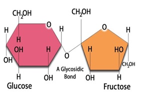
Fructose
Fibric Acids
Triglycerides
Cycloheximide
Glutathione Transferase
Blotting, Northern
Transcription Factors
Liver Cirrhosis
Carbon Tetrachloride Poisoning
Enzyme Inhibitors
Adenoviridae
Taurolithocholic Acid
Histocytochemistry
Microbodies
Organic Anion Transporters
Necrosis
Hepacivirus
Aminoisobutyric Acids
Drug Interactions
Fructosediphosphates
Urea
Hepatitis B virus
Cholestasis
Models, Biological
Tupaia
Marmota
beta-Naphthoflavone
Subcellular Fractions
Glucose-6-Phosphate
Sulfobromophthalein
Lysosomes
Precancerous Conditions
Fetus
Peroxisome Proliferators
Promoter Regions, Genetic
Fluorescent Antibody Technique
Lactic Acid
Serum Albumin
Choline Deficiency
Proteins
Hepatoblastoma
Endoplasmic Reticulum
Genetic Vectors
Hepatic Stellate Cells
Dipeptidyl Peptidase 4
Tumor Necrosis Factor-alpha
Species Specificity
Colchicine
Receptors, Cell Surface
Metabolic Clearance Rate
Rifampin
Receptors, Steroid
Metabolic Detoxication, Drug
Glucuronosyltransferase
Steroid Hydroxylases
Culture Media
Sphingomyelinase treatment of rat hepatocytes inhibits cell-swelling-stimulated glycogen synthesis by causing cell shrinkage. (1/7533)
Breakdown of plasma-membrane sphingomyelin caused by TNF-alpha is known to inhibit glucose metabolism and insulin signalling in muscle and fat cells. In hepatocytes, conversion of glucose to glycogen is strongly activated by amino acid-induced cell swelling. In order to find out whether breakdown of plasma-membrane sphingomyelin also inhibits this insulin-independent process, the effect of addition of sphingomyelinase was studied in rat hepatocytes. Sphingomyelinase (but not ceramide) inhibited glycogen synthesis, caused cell shrinkage, decreased the activity of glycogen synthase a, but had no effect on phosphorylase a. Cell integrity was not affected by sphingomyelinase addition as gluconeogenesis and the intracellular concentration of ATP were unchanged. As a control, glycogen synthesis was studied in HepG2 cells. In these cells, the basal rate of glycogen production was high, could not be stimulated by amino acids, nor be inhibited by sphingomyelinase. Regarding the mechanism responsible for the inhibition of glycogen synthase a, sphingomyelinase did not affect amino acid-induced, PtdIns 3-kinase-dependent, phosphorylation of p70S6 kinase, but caused an increase in intracellular chloride, which is known to inhibit glycogen synthase phosphatase. It is concluded that the decrease in cell volume, following the breakdown of sphingomyelin in the plasma membrane of the hepatocyte, may contribute to the abnormal metabolism of glucose when TNF-alpha levels are high. (+info)Regulation of sterol regulatory-element binding protein 1 gene expression in liver: role of insulin and protein kinase B/cAkt. (2/7533)
Insulin stimulates the transcription of the sterol regulatory- element binding protein (SREBP) 1/ADD1 gene in liver. Hepatocytes in primary culture were used to delineate the insulin signalling pathway for induction of SREBP1 gene expression. The inhibitors of phosphoinositide 3-kinase (PI 3-kinase), wortmannin and LY 294002, abolished the insulin-dependent increase in SREBP1 mRNA, whereas the inhibitor of the mitogen- activated protein kinase cascade, PD 98059, was without effect. To investigate the role of protein kinase B (PKB)/cAkt downstream of PI 3-kinase, hepatocytes were transduced with an adenovirus encoding a PKB--oestrogen receptor fusion protein. The PKB activity of this recombinant protein was rapidly activated in hepatocytes challenged with 4-hydroxytamoxifen (OHT), as was endogenous PKB in hepatocytes challenged with insulin. The addition of OHT to transduced hepatocytes resulted in accumulation of SREBP1 mRNA, with a time-course and magnitude similar to the effect of insulin in non-transduced cells. The level of SREBP1 mRNA was not increased by OHT in hepatocytes expressing a mutant form of the recombinant protein whose PKB activity was not activated by OHT. Thus acute activation of PKB is sufficient to induce SREBP1 mRNA accumulation in primary hepatocytes, and might be the major signalling event by which insulin induces SREBP1 gene expression in the liver. (+info)Interferons activate the p42/44 mitogen-activated protein kinase and JAK-STAT (Janus kinase-signal transducer and activator transcription factor) signalling pathways in hepatocytes: differential regulation by acute ethanol via a protein kinase C-dependent mechanism. (3/7533)
Interferons (IFNs) have been used in the treatment of viral hepatitis. However, their effectiveness is much reduced (<10%) in alcoholics. The mechanism underlying this resistance remains unknown. Here, we report that IFN-alpha/beta and IFN-gamma rapidly activate the JAK-STAT1 (Janus kinase-signal transducer and activator transcription factor 1) and p42/44 mitogen-activated protein kinase (p42/44 MAPK) in freshly isolated rat hepatocytes. Treatment of hepatocytes with 25-100 mM ethanol for 30 min inhibited IFN-beta- or IFN-gamma-induced STAT1 activation and tyrosine phosphorylation. The inhibitory effect of ethanol was not reversed by pretreatment with either sodium vanadate, a non-selective tyrosine phosphatase inhibitor, or with MG132, a specific proteasome inhibitor. This suggests that protein tyrosine phosphatases or the ubiquitin-proteasome pathway are not involved in the inhibitory action of ethanol. In contrast with the JAK-STAT signalling pathway, acute ethanol exposure significantly potentiated IFN-beta or IFN-gamma-induced activation of p42/44 MAPK, and caused marked activation of protein kinase C (PKC). Inhibition of PKC partially antagonized ethanol attenuation of IFN-induced STAT1 activation, suggesting that PKC may be involved. Taken together, these findings suggest that the ability of biologically relevant concentrations of ethanol (less than 100 mM) to markedly inhibit IFN-activated STAT1 is one of the cellular mechanisms responsible for the observed resistance of IFN therapy in alcoholics. (+info)A targeted apolipoprotein B-38.9-producing mutation causes fatty livers in mice due to the reduced ability of apolipoprotein B-38.9 to transport triglycerides. (4/7533)
Nonphysiological truncations of apolipoprotein (apo) B-100 cause familial hypobetalipoproteinemia (FHBL) in humans and mice. An elucidation of the mechanisms underlying the FHBL phenotypes may provide valuable information on the metabolism of apo B-containing lipoproteins and the structure-function relationship of apo B. To generate a faithful mouse model of human FHBL, a subtle mutation was introduced into the mouse apo B gene by targeting embryonic stem cells using homologous recombination followed by removal of the selection marker gene by Cre-loxP-mediated site-specific recombination. The engineered mice bear a premature stop codon at residue 1767 and a 42-base pair loxP inserted into intron 24 of the apo B gene, thus closely resembling the apo B-38.9-producing mutation in humans. Apo B-38.9 was the sole apo B protein in homozygote (apob(38.9/38.9)) plasma. In heterozygotes (apob(+/)(38. 9)), apo B-100 and apo B-48 were reduced by 75 and 40%, respectively, and apo B-38.9 represented 20% of total circulating apo B. Hepatic apo B-38.9 mRNA levels were reduced by 40%. In cultured apob(+/)(38. 9) hepatocytes, apo B-100 was produced in trace quantities, and the synthesis rate of apo B-38.9 relative to apo B-48 was reduced by 40%. However, almost equimolar amounts of apo B-38.9 and apo B-48 were secreted into the media. Pulse-chase studies revealed that apo B-38. 9 was secreted at a faster rate and more efficiently than apoB-48. Nevertheless, both apob(+/)(38.9) and apob(38.9/38.9) mice had reduced hepatic triglyceride secretion rates and fatty livers. Thus, low mRNA levels or defective secretion of apo B-38.9 may not be responsible for the FHBL phenotypes caused by the apo B-38.9 mutation. Rather, a reduced capacity of apo B-38.9 for triglyceride transport may account for the fatty livers in these mice. (+info)Progression of hepatic stellate cell activation is associated with the level of oxidative stress rather than cytokines during CCl4-induced fibrogenesis. (5/7533)
In order to identify a fibrogenic factor associated with the potential of hepatic stellate cells (HSC) activation that arises during the CCl4-induced fibrogenic process, the relationship between the activation of HSC and levels of several fibrogenic factors were investigated. After isolation of HSC from the liver at different stages of CCl4 intoxication, the activation of HSC was assessed by the expression of alpha-smooth muscle actin. Levels of cytokines and oxidative stress in liver homogenates and plasma were measured by enzyme linked immunosorbent assay and the colorimetric method. In primary culture, HSC isolated from a rat liver were gradually activated in a time-dependent manner according to CCl4 administration. The progression of HSC activation was closely correlated with parameters related to oxidative stress in liver homogenates rather than the tissue levels of several cytokines. Also, the levels of antioxidants and arginase activity were inversely correlated with HSC activation. In plasma, the levels of oxidative stress and cytokines in CCl4-treated rat livers were not associated with the activation of HSC found during the CCl4-induced fibrogenic process. The relationship between HSC activation and oxidative stress was also confirmed through several factor-treated HSC cultures. In conclusion, the activation of HSC was accelerated according to CCl4 administration, and the progression of HSC activation is absolutely related to the oxidative stress. These results show that enhanced oxidative stress is an important signal for activation of HSC in experimental liver fibrogenesis. (+info)Functional characterization of the rat gamma-glutamyl transpeptidase promoter that is expressed in transformed rat liver epithelial cells. (6/7533)
In the rat the gamma-glutamyl transpeptidase (GGT) gene codes for at least four different messenger RNAs (mRNA I to mRNA IV) which differ only in their 5' untranslated regions and are transcribed from a single copy gene in a tissue-specific manner. In the liver GGT expression is up-regulated in transformed cells. To understand the induction mechanisms of GGT activity by transformation, we previously cloned the 5' region of the rat GGT gene which contains the 5' untranslated leader sequence for mRNA I. In the present study, using transfection and reporter gene assays, I have demonstrated that (1) the sequence from positions -369 to +226 drives a relatively strong promoter activity in C5 and AKG cell lines, transformed rat liver epithelial cells, but a very weak one in RLE-228 cells, a normal rat liver epithelial cell line; and (2) removal of the region between -418 and -369 increases CAT activity more than 10-fold in RLE-228, C5 and AKG cells, and the DNA fragment spanning nucleotides -761 to -292 significantly reduces CAT activity driven by the adenine phosphoribosyl transferase (APRT) promoter in RLE-228 cells. (+info)Involvement of apoptosis in the endotoxemic lesions of the liver and kidneys of piglets. (7/7533)
The involvement of apoptosis was evaluated in lesions of endotoxemic piglets. A single injection with E. coli O111:B4 lipopolysaccharide (LPS) induced foci of coagulative necrosis in the liver and kidneys. No significant change was observed in these organs at 1.5 hr after LPS injection, but at 6 hr, epithelial cells with chromatin condensation or fragmentation and apoptotic bodies were visible. Foci of coagulative necrosis were formed within 24 hr after LPS inoculation. In and adjacent to the necrotic foci, dead hepatocytes with nuclear condensation or fragmentation were scattered. These dead cells were positively stained by terminal deoxynucleotidyl transferase-mediated dUTP-biotin nick end labeling (TUNEL) methods. Electronmicroscopy revealed apoptotic cells with condensed or fragmented homogeneous nuclear chromatin, and necrotic cells with irregularly destroyed nuclei and cytoplasmic membranes. Apoptotic cell death were also observed in parietal cells of the stomach and lymphocytes in the lymphatic system. DNA ladders with approximately 200-bp multimers were observed in hepatic, renal and thymic samples prepared after 6 and 24 hr of LPS injection by agarose gel electrophoresis. These results suggest that apoptosis is involved in the pathology of swine endotoxemia. (+info)Phosphoinositide 3-kinase-dependent Ras activation by tauroursodesoxycholate in rat liver. (8/7533)
Ursodesoxycholic acid, widely used for the treatment of cholestatic liver disease, causes choleretic, anti-apoptotic and immunomodulatory effects. Here the effects on choleresis of its taurine conjugate tauroursodesoxycholate (TUDC), which is present in the enterohepatic circulation, were correlated with the activation of important elements of intracellular signal transduction in cultured rat hepatocytes and perfused rat liver. TUDC induced a time- and concentration-dependent activation of the small GTP-binding protein Ras and of phosphoinositide 3-kinase (PI 3-kinase) in cultured hepatocytes. Ras activation was dependent on PI 3-kinase activity, without the involvement of protein kinase C- and genistein-sensitive tyrosine kinases. Ras activation by TUDC was followed by an activation of the mitogen-activated protein kinases extracellular-signal-regulated kinase-1 (Erk-1) and Erk-2. In perfused rat liver, PI 3-kinase inhibitors largely abolished the stimulatory effect of TUDC on taurocholate excretion, suggesting an important role for a PI 3-kinase/Ras/Erk pathway in the choleretic effect of TUDC. (+info)Hepatocytes are the predominant type of cells in the liver, accounting for about 80% of its cytoplasmic mass. They play a key role in protein synthesis, protein storage, transformation of carbohydrates, synthesis of cholesterol, bile salts and phospholipids, detoxification, modification, and excretion of exogenous and endogenous substances, initiation of formation and secretion of bile, and enzyme production. Hepatocytes are essential for the maintenance of homeostasis in the body.
The liver is a large, solid organ located in the upper right portion of the abdomen, beneath the diaphragm and above the stomach. It plays a vital role in several bodily functions, including:
1. Metabolism: The liver helps to metabolize carbohydrates, fats, and proteins from the food we eat into energy and nutrients that our bodies can use.
2. Detoxification: The liver detoxifies harmful substances in the body by breaking them down into less toxic forms or excreting them through bile.
3. Synthesis: The liver synthesizes important proteins, such as albumin and clotting factors, that are necessary for proper bodily function.
4. Storage: The liver stores glucose, vitamins, and minerals that can be released when the body needs them.
5. Bile production: The liver produces bile, a digestive juice that helps to break down fats in the small intestine.
6. Immune function: The liver plays a role in the immune system by filtering out bacteria and other harmful substances from the blood.
Overall, the liver is an essential organ that plays a critical role in maintaining overall health and well-being.
"Cells, cultured" is a medical term that refers to cells that have been removed from an organism and grown in controlled laboratory conditions outside of the body. This process is called cell culture and it allows scientists to study cells in a more controlled and accessible environment than they would have inside the body. Cultured cells can be derived from a variety of sources, including tissues, organs, or fluids from humans, animals, or cell lines that have been previously established in the laboratory.
Cell culture involves several steps, including isolation of the cells from the tissue, purification and characterization of the cells, and maintenance of the cells in appropriate growth conditions. The cells are typically grown in specialized media that contain nutrients, growth factors, and other components necessary for their survival and proliferation. Cultured cells can be used for a variety of purposes, including basic research, drug development and testing, and production of biological products such as vaccines and gene therapies.
It is important to note that cultured cells may behave differently than they do in the body, and results obtained from cell culture studies may not always translate directly to human physiology or disease. Therefore, it is essential to validate findings from cell culture experiments using additional models and ultimately in clinical trials involving human subjects.
"Inbred strains of rats" are genetically identical rodents that have been produced through many generations of brother-sister mating. This results in a high degree of homozygosity, where the genes at any particular locus in the genome are identical in all members of the strain.
Inbred strains of rats are widely used in biomedical research because they provide a consistent and reproducible genetic background for studying various biological phenomena, including the effects of drugs, environmental factors, and genetic mutations on health and disease. Additionally, inbred strains can be used to create genetically modified models of human diseases by introducing specific mutations into their genomes.
Some commonly used inbred strains of rats include the Wistar Kyoto (WKY), Sprague-Dawley (SD), and Fischer 344 (F344) rat strains. Each strain has its own unique genetic characteristics, making them suitable for different types of research.
Liver regeneration is the ability of the liver to restore its original mass and function after injury or surgical resection. This complex process involves the proliferation and differentiation of mature hepatocytes, as well as the activation and transdifferentiation of various types of stem and progenitor cells located in the liver. The mechanisms that regulate liver regeneration include a variety of growth factors, hormones, and cytokines, which act in a coordinated manner to ensure the restoration of normal liver architecture and function. Liver regeneration is essential for the survival of individuals who have undergone partial hepatectomy or who have suffered liver damage due to various causes, such as viral hepatitis, alcohol abuse, or drug-induced liver injury.
Glucagon is a hormone produced by the alpha cells of the pancreas. Its main function is to regulate glucose levels in the blood by stimulating the liver to convert stored glycogen into glucose, which can then be released into the bloodstream. This process helps to raise blood sugar levels when they are too low, such as during hypoglycemia.
Glucagon is a 29-amino acid polypeptide that is derived from the preproglucagon protein. It works by binding to glucagon receptors on liver cells, which triggers a series of intracellular signaling events that lead to the activation of enzymes involved in glycogen breakdown.
In addition to its role in glucose regulation, glucagon has also been shown to have other physiological effects, such as promoting lipolysis (the breakdown of fat) and inhibiting gastric acid secretion. Glucagon is often used clinically in the treatment of hypoglycemia, as well as in diagnostic tests to assess pancreatic function.
"Wistar rats" are a strain of albino rats that are widely used in laboratory research. They were developed at the Wistar Institute in Philadelphia, USA, and were first introduced in 1906. Wistar rats are outbred, which means that they are genetically diverse and do not have a fixed set of genetic characteristics like inbred strains.
Wistar rats are commonly used as animal models in biomedical research because of their size, ease of handling, and relatively low cost. They are used in a wide range of research areas, including toxicology, pharmacology, nutrition, cancer, cardiovascular disease, and behavioral studies. Wistar rats are also used in safety testing of drugs, medical devices, and other products.
Wistar rats are typically larger than many other rat strains, with males weighing between 500-700 grams and females weighing between 250-350 grams. They have a lifespan of approximately 2-3 years. Wistar rats are also known for their docile and friendly nature, making them easy to handle and work with in the laboratory setting.
Albumins are a type of protein found in various biological fluids, including blood plasma. The most well-known albumin is serum albumin, which is produced by the liver and is the most abundant protein in blood plasma. Serum albumin plays several important roles in the body, such as maintaining oncotic pressure (which helps to regulate fluid balance in the body), transporting various substances (such as hormones, fatty acids, and drugs), and acting as an antioxidant.
Albumins are soluble in water and have a molecular weight ranging from 65,000 to 69,000 daltons. They are composed of a single polypeptide chain that contains approximately 585 amino acid residues. The structure of albumin is characterized by a high proportion of alpha-helices and beta-sheets, which give it a stable, folded conformation.
In addition to their role in human physiology, albumins are also used as diagnostic markers in medicine. For example, low serum albumin levels may indicate liver disease, malnutrition, or inflammation, while high levels may be seen in dehydration or certain types of kidney disease. Albumins may also be used as a replacement therapy in patients with severe protein loss, such as those with nephrotic syndrome or burn injuries.
Experimental liver neoplasms refer to abnormal growths or tumors in the liver that are intentionally created or manipulated in a laboratory setting for the purpose of studying their development, progression, and potential treatment options. These experimental models can be established using various methods such as chemical induction, genetic modification, or transplantation of cancerous cells or tissues. The goal of this research is to advance our understanding of liver cancer biology and develop novel therapies for liver neoplasms in humans. It's important to note that these experiments are conducted under strict ethical guidelines and regulations to minimize harm and ensure the humane treatment of animals involved in such studies.
Asialoglycoproteins are glycoproteins that have lost their terminal sialic acid residues. In the body, these molecules are typically recognized and removed from circulation by hepatic lectins, such as the Ashwell-Morrell receptor, found on liver cells. This process is a part of the normal turnover and clearance of glycoproteins in the body.
F344 is a strain code used to designate an outbred stock of rats that has been inbreeded for over 100 generations. The F344 rats, also known as Fischer 344 rats, were originally developed at the National Institutes of Health (NIH) and are now widely used in biomedical research due to their consistent and reliable genetic background.
Inbred strains, like the F344, are created by mating genetically identical individuals (siblings or parents and offspring) for many generations until a state of complete homozygosity is reached, meaning that all members of the strain have identical genomes. This genetic uniformity makes inbred strains ideal for use in studies where consistent and reproducible results are important.
F344 rats are known for their longevity, with a median lifespan of around 27-31 months, making them useful for aging research. They also have a relatively low incidence of spontaneous tumors compared to other rat strains. However, they may be more susceptible to certain types of cancer and other diseases due to their inbred status.
It's important to note that while F344 rats are often used as a standard laboratory rat strain, there can still be some genetic variation between individual animals within the same strain, particularly if they come from different suppliers or breeding colonies. Therefore, it's always important to consider the source and history of any animal model when designing experiments and interpreting results.
Sprague-Dawley rats are a strain of albino laboratory rats that are widely used in scientific research. They were first developed by researchers H.H. Sprague and R.C. Dawley in the early 20th century, and have since become one of the most commonly used rat strains in biomedical research due to their relatively large size, ease of handling, and consistent genetic background.
Sprague-Dawley rats are outbred, which means that they are genetically diverse and do not suffer from the same limitations as inbred strains, which can have reduced fertility and increased susceptibility to certain diseases. They are also characterized by their docile nature and low levels of aggression, making them easier to handle and study than some other rat strains.
These rats are used in a wide variety of research areas, including toxicology, pharmacology, nutrition, cancer, and behavioral studies. Because they are genetically diverse, Sprague-Dawley rats can be used to model a range of human diseases and conditions, making them an important tool in the development of new drugs and therapies.
Messenger RNA (mRNA) is a type of RNA (ribonucleic acid) that carries genetic information copied from DNA in the form of a series of three-base code "words," each of which specifies a particular amino acid. This information is used by the cell's machinery to construct proteins, a process known as translation. After being transcribed from DNA, mRNA travels out of the nucleus to the ribosomes in the cytoplasm where protein synthesis occurs. Once the protein has been synthesized, the mRNA may be degraded and recycled. Post-transcriptional modifications can also occur to mRNA, such as alternative splicing and addition of a 5' cap and a poly(A) tail, which can affect its stability, localization, and translation efficiency.
Gluconeogenesis is a metabolic pathway that occurs in the liver, kidneys, and to a lesser extent in the small intestine. It involves the synthesis of glucose from non-carbohydrate precursors such as lactate, pyruvate, glycerol, and certain amino acids. This process becomes particularly important during periods of fasting or starvation when glucose levels in the body begin to drop, and there is limited carbohydrate intake to replenish them.
Gluconeogenesis helps maintain blood glucose homeostasis by providing an alternative source of glucose for use by various tissues, especially the brain, which relies heavily on glucose as its primary energy source. It is a complex process that involves several enzymatic steps, many of which are regulated to ensure an adequate supply of glucose while preventing excessive production, which could lead to hyperglycemia.
An asialoglycoprotein receptor (ASGPR) is a type of cell surface receptor found primarily on hepatocytes, which are the main cell type in the liver. These receptors are responsible for recognizing and removing glycoproteins (proteins with attached carbohydrate molecules) from circulation, particularly those that have lost their terminal sialic acid residues through a process called desialylation.
ASGPRs play an essential role in the liver's clearance function by identifying and removing various substances, such as bacteria, viruses, and abnormal or damaged cells, from the bloodstream. There are two main types of ASGPRs, known as ASGPR1 and ASGPR2, which have different structures and functions but work together to mediate the endocytosis and degradation of desialylated glycoproteins.
Understanding the role of ASGPRs in liver function has important implications for developing targeted therapies and diagnostic tools for various liver-related diseases, including hepatitis, cirrhosis, and liver cancer.
Liver glycogen is the reserve form of glucose stored in hepatocytes (liver cells) for the maintenance of normal blood sugar levels. It is a polysaccharide, a complex carbohydrate, that is broken down into glucose molecules when blood glucose levels are low. This process helps to maintain the body's energy needs between meals and during periods of fasting or exercise. The amount of glycogen stored in the liver can vary depending on factors such as meal consumption, activity level, and insulin regulation.
In the context of medicine and pharmacology, "kinetics" refers to the study of how a drug moves throughout the body, including its absorption, distribution, metabolism, and excretion (often abbreviated as ADME). This field is called "pharmacokinetics."
1. Absorption: This is the process of a drug moving from its site of administration into the bloodstream. Factors such as the route of administration (e.g., oral, intravenous, etc.), formulation, and individual physiological differences can affect absorption.
2. Distribution: Once a drug is in the bloodstream, it gets distributed throughout the body to various tissues and organs. This process is influenced by factors like blood flow, protein binding, and lipid solubility of the drug.
3. Metabolism: Drugs are often chemically modified in the body, typically in the liver, through processes known as metabolism. These changes can lead to the formation of active or inactive metabolites, which may then be further distributed, excreted, or undergo additional metabolic transformations.
4. Excretion: This is the process by which drugs and their metabolites are eliminated from the body, primarily through the kidneys (urine) and the liver (bile).
Understanding the kinetics of a drug is crucial for determining its optimal dosing regimen, potential interactions with other medications or foods, and any necessary adjustments for special populations like pediatric or geriatric patients, or those with impaired renal or hepatic function.
The Cytochrome P-450 (CYP450) enzyme system is a group of enzymes found primarily in the liver, but also in other organs such as the intestines, lungs, and skin. These enzymes play a crucial role in the metabolism and biotransformation of various substances, including drugs, environmental toxins, and endogenous compounds like hormones and fatty acids.
The name "Cytochrome P-450" refers to the unique property of these enzymes to bind to carbon monoxide (CO) and form a complex that absorbs light at a wavelength of 450 nm, which can be detected spectrophotometrically.
The CYP450 enzyme system is involved in Phase I metabolism of xenobiotics, where it catalyzes oxidation reactions such as hydroxylation, dealkylation, and epoxidation. These reactions introduce functional groups into the substrate molecule, which can then undergo further modifications by other enzymes during Phase II metabolism.
There are several families and subfamilies of CYP450 enzymes, each with distinct substrate specificities and functions. Some of the most important CYP450 enzymes include:
1. CYP3A4: This is the most abundant CYP450 enzyme in the human liver and is involved in the metabolism of approximately 50% of all drugs. It also metabolizes various endogenous compounds like steroids, bile acids, and vitamin D.
2. CYP2D6: This enzyme is responsible for the metabolism of many psychotropic drugs, including antidepressants, antipsychotics, and beta-blockers. It also metabolizes some endogenous compounds like dopamine and serotonin.
3. CYP2C9: This enzyme plays a significant role in the metabolism of warfarin, phenytoin, and nonsteroidal anti-inflammatory drugs (NSAIDs).
4. CYP2C19: This enzyme is involved in the metabolism of proton pump inhibitors, antidepressants, and clopidogrel.
5. CYP2E1: This enzyme metabolizes various xenobiotics like alcohol, acetaminophen, and carbon tetrachloride, as well as some endogenous compounds like fatty acids and prostaglandins.
Genetic polymorphisms in CYP450 enzymes can significantly affect drug metabolism and response, leading to interindividual variability in drug efficacy and toxicity. Understanding the role of CYP450 enzymes in drug metabolism is crucial for optimizing pharmacotherapy and minimizing adverse effects.
2-Acetylaminofluorene (2-AAF) is a chemical compound that has been used in research to study the mechanisms of carcinogenesis. It is an aromatic amine and a derivative of fluorene, with the chemical formula C14H11NO.
2-AAF is not naturally occurring and is synthesized in the laboratory. It has been found to be carcinogenic in animal studies, causing tumors in various organs including the liver, lung, and bladder. The compound is metabolically activated in the body to form reactive intermediates that can bind to DNA and other cellular components, leading to mutations and cancer.
2-AAF has been used as a tool in research to investigate the mechanisms of chemical carcinogenesis and the role of metabolic activation in the process. It is not used in medical treatments or therapies.
Enzyme induction is a process by which the activity or expression of an enzyme is increased in response to some stimulus, such as a drug, hormone, or other environmental factor. This can occur through several mechanisms, including increasing the transcription of the enzyme's gene, stabilizing the mRNA that encodes the enzyme, or increasing the translation of the mRNA into protein.
In some cases, enzyme induction can be a beneficial process, such as when it helps the body to metabolize and clear drugs more quickly. However, in other cases, enzyme induction can have negative consequences, such as when it leads to the increased metabolism of important endogenous compounds or the activation of harmful procarcinogens.
Enzyme induction is an important concept in pharmacology and toxicology, as it can affect the efficacy and safety of drugs and other xenobiotics. It is also relevant to the study of drug interactions, as the induction of one enzyme by a drug can lead to altered metabolism and effects of another drug that is metabolized by the same enzyme.
Kupffer cells are specialized macrophages that reside in the liver, particularly in the sinusoids of the liver's blood circulation system. They play a crucial role in the immune system by engulfing and destroying bacteria, microorganisms, and other particles that enter the liver via the portal vein. Kupffer cells also contribute to the clearance of damaged red blood cells, iron metabolism, and the regulation of inflammation in the liver. They are named after the German pathologist Karl Wilhelm von Kupffer who first described them in 1876.
Phenobarbital is a barbiturate medication that is primarily used for the treatment of seizures and convulsions. It works by suppressing the abnormal electrical activity in the brain that leads to seizures. In addition to its anticonvulsant properties, phenobarbital also has sedative and hypnotic effects, which can be useful for treating anxiety, insomnia, and agitation.
Phenobarbital is available in various forms, including tablets, capsules, and elixirs, and it is typically taken orally. The medication works by binding to specific receptors in the brain called gamma-aminobutyric acid (GABA) receptors, which help to regulate nerve impulses in the brain. By increasing the activity of GABA, phenobarbital can help to reduce excessive neural activity and prevent seizures.
While phenobarbital is an effective medication for treating seizures and other conditions, it can also be habit-forming and carries a risk of dependence and addiction. Long-term use of the medication can lead to tolerance, meaning that higher doses may be needed to achieve the same effects. Abruptly stopping the medication can also lead to withdrawal symptoms, such as anxiety, restlessness, and seizures.
Like all medications, phenobarbital can have side effects, including dizziness, drowsiness, and impaired coordination. It can also interact with other medications, such as certain antidepressants and sedatives, so it is important to inform your healthcare provider of all medications you are taking before starting phenobarbital.
In summary, phenobarbital is a barbiturate medication used primarily for the treatment of seizures and convulsions. It works by binding to GABA receptors in the brain and increasing their activity, which helps to reduce excessive neural activity and prevent seizures. While phenobarbital can be effective, it carries a risk of dependence and addiction and can have side effects and drug interactions.
I'd be happy to help! However, I believe there might be a slight misunderstanding in your question. Mitochondria are organelles found in the cells of all complex living organisms, including humans, while the liver is a large, solid organ located in the upper right portion of the abdomen. They don't have a medical definition together. I can certainly provide you with separate definitions for each:
1. Mitochondria: These are double-membrane-bound cellular organelles that generate most of the chemical energy needed to power the cell's biochemical reactions. Commonly known as the "powerhouse of the cell," mitochondria convert organic substrates, such as glucose, fatty acids, and amino acids, into adenosine triphosphate (ATP) through a process called oxidative phosphorylation. Mitochondria are dynamic structures that can change their shape, size, and number through fission (division) and fusion (merging) processes. They play essential roles in various cellular functions, including calcium signaling, apoptosis (programmed cell death), and the regulation of cellular metabolism.
2. Liver: The liver is a large, lobulated organ that lies mainly in the upper right portion of the abdominal cavity, just below the diaphragm. It plays a crucial role in various physiological functions, such as detoxification, protein synthesis, metabolism, and nutrient storage. The liver is responsible for removing toxins from the bloodstream, producing bile to aid in digestion, regulating glucose levels, synthesizing plasma proteins, and storing glycogen, vitamins, and minerals. It also contributes to the metabolism of carbohydrates, lipids, and amino acids, helping maintain energy homeostasis in the body.
I hope this clarifies any confusion! If you have any further questions or need more information, please don't hesitate to ask.
Phosphorylase a is an enzyme that plays a crucial role in the breakdown and metabolism of glycogen, a complex carbohydrate stored primarily in the liver and muscles. It is a phosphorylated form of the enzyme glycogen phosphorylase, which is activated by the addition of a phosphate group.
Phosphorylase a catalyzes the rate-limiting step in glycogenolysis, the process of breaking down glycogen into glucose-1-phosphate, which can then be converted into glucose and used for energy production. The activation of phosphorylase a is mediated by hormones such as adrenaline (epinephrine) and glucagon, which stimulate the enzyme phosphorylase kinase to add a phosphate group to inactive phosphorylase b, converting it to active phosphorylase a.
Phosphorylase a is composed of two identical subunits, each containing a catalytic site and a regulatory site that binds to ATP, glucose, and other molecules. The enzyme's activity is regulated by several factors, including the concentration of glucose, the presence of calcium ions, and the phosphorylation state of the enzyme.
In summary, Phosphorylase a is a key enzyme in glycogen metabolism that catalyzes the breakdown of glycogen into glucose-1-phosphate, providing energy for the body's cells. Its activity is regulated by hormones and other factors, making it an important component of the body's energy homeostasis.
Hep G2 cells are a type of human liver cancer cell line that were isolated from a well-differentiated hepatocellular carcinoma (HCC) in a patient with hepatitis C virus (HCV) infection. These cells have the ability to grow and divide indefinitely in culture, making them useful for research purposes. Hep G2 cells express many of the same markers and functions as normal human hepatocytes, including the ability to take up and process lipids and produce bile. They are often used in studies related to hepatitis viruses, liver metabolism, drug toxicity, and cancer biology. It is important to note that Hep G2 cells are tumorigenic and should be handled with care in a laboratory setting.
An artificial liver is not a actual organ replacement but a device designed to perform some of the functions of a liver in patients with liver failure. These devices can be divided into two types: bioartificial and non-bioartificial. Non-bioartificial devices, such as hemodialysis machines and molecular adsorbent recirculating system (MARS), use physical and chemical processes to remove toxins from the blood. Bioartificial livers, on the other hand, contain living cells, usually hepatocytes, which can perform more advanced liver functions such as synthesizing proteins and drugs metabolism.
It's important to note that currently there is no FDA approved artificial liver device available for use in clinical practice. However, research and development of these devices are ongoing with the hope that they may provide a bridge to transplantation or recovery for patients with acute liver failure.
Bile canaliculi are the smallest bile-transporting structures in the liver. They are formed by the close apposition of hepatocyte (liver cell) plasma membranes, and they are responsible for the majority of bile production. The bile canaliculi merge to form bile ductules, which then merge to form larger bile ducts that transport bile to the gallbladder and small intestine. Bile is a fluid that contains water, electrolytes, bile salts, cholesterol, phospholipids, and bilirubin, which are produced by the liver and play important roles in digestion and elimination of waste products.
Dihydroxyacetone (DHA) is a simple sugar that is used as an ingredient in many self-tanning products. When applied to the skin, DHA reacts with amino acids in the dead layer of the skin to temporarily darken the skin color. This process is known as the Maillard reaction, which is a chemical reaction between an amino acid and a sugar. The effect of DHA is limited to the uppermost layer of the skin and it does not provide any protection against sunburn or UV radiation. The tanning effect produced by DHA usually lasts for about 5-7 days.
It's important to note that while DHA is considered safe for external use, it should not be inhaled or ingested, as it can cause irritation and other adverse effects. Additionally, some people may experience skin irritation or allergic reactions to products containing DHA, so it's always a good idea to do a patch test before using a new self-tanning product.
Hepatectomy is a surgical procedure that involves the removal of part or all of the liver. This procedure can be performed for various reasons, such as removing cancerous or non-cancerous tumors, treating liver trauma, or donating a portion of the liver to another person in need of a transplant (live donor hepatectomy). The extent of the hepatectomy depends on the medical condition and overall health of the patient. It is a complex procedure that requires significant expertise and experience from the surgical team due to the liver's unique anatomy, blood supply, and regenerative capabilities.
Drug-Induced Liver Injury (DILI) is a medical term that refers to liver damage or injury caused by the use of medications or drugs. This condition can vary in severity, from mild abnormalities in liver function tests to severe liver failure, which may require a liver transplant.
The exact mechanism of DILI can differ depending on the drug involved, but it generally occurs when the liver metabolizes the drug into toxic compounds that damage liver cells. This can happen through various pathways, including direct toxicity to liver cells, immune-mediated reactions, or metabolic idiosyncrasies.
Symptoms of DILI may include jaundice (yellowing of the skin and eyes), fatigue, abdominal pain, nausea, vomiting, loss of appetite, and dark urine. In severe cases, it can lead to complications such as ascites, encephalopathy, and bleeding disorders.
The diagnosis of DILI is often challenging because it requires the exclusion of other potential causes of liver injury. Liver function tests, imaging studies, and sometimes liver biopsies may be necessary to confirm the diagnosis. Treatment typically involves discontinuing the offending drug and providing supportive care until the liver recovers. In some cases, medications that protect the liver or promote its healing may be used.
Liver neoplasms refer to abnormal growths in the liver that can be benign or malignant. Benign liver neoplasms are non-cancerous tumors that do not spread to other parts of the body, while malignant liver neoplasms are cancerous tumors that can invade and destroy surrounding tissue and spread to other organs.
Liver neoplasms can be primary, meaning they originate in the liver, or secondary, meaning they have metastasized (spread) to the liver from another part of the body. Primary liver neoplasms can be further classified into different types based on their cell of origin and behavior, including hepatocellular carcinoma, cholangiocarcinoma, and hepatic hemangioma.
The diagnosis of liver neoplasms typically involves a combination of imaging studies, such as ultrasound, CT scan, or MRI, and biopsy to confirm the type and stage of the tumor. Treatment options depend on the type and extent of the neoplasm and may include surgery, radiation therapy, chemotherapy, or liver transplantation.
Hepatocellular carcinoma (HCC) is the most common type of primary liver cancer in adults. It originates from the hepatocytes, which are the main functional cells of the liver. This type of cancer is often associated with chronic liver diseases such as cirrhosis caused by hepatitis B or C virus infection, alcohol abuse, non-alcoholic fatty liver disease (NAFLD), and aflatoxin exposure.
The symptoms of HCC can vary but may include unexplained weight loss, lack of appetite, abdominal pain or swelling, jaundice, and fatigue. The diagnosis of HCC typically involves imaging tests such as ultrasound, CT scan, or MRI, as well as blood tests to measure alpha-fetoprotein (AFP) levels. Treatment options for Hepatocellular carcinoma depend on the stage and extent of the cancer, as well as the patient's overall health and liver function. Treatment options may include surgery, radiation therapy, chemotherapy, targeted therapy, or liver transplantation.
Dexamethasone is a type of corticosteroid medication, which is a synthetic version of a natural hormone produced by the adrenal glands. It is often used to reduce inflammation and suppress the immune system in a variety of medical conditions, including allergies, asthma, rheumatoid arthritis, and certain skin conditions.
Dexamethasone works by binding to specific receptors in cells, which triggers a range of anti-inflammatory effects. These include reducing the production of chemicals that cause inflammation, suppressing the activity of immune cells, and stabilizing cell membranes.
In addition to its anti-inflammatory effects, dexamethasone can also be used to treat other medical conditions, such as certain types of cancer, brain swelling, and adrenal insufficiency. It is available in a variety of forms, including tablets, liquids, creams, and injectable solutions.
Like all medications, dexamethasone can have side effects, particularly if used for long periods of time or at high doses. These may include mood changes, increased appetite, weight gain, acne, thinning skin, easy bruising, and an increased risk of infections. It is important to follow the instructions of a healthcare provider when taking dexamethasone to minimize the risk of side effects.
Cytochrome P-450 CYP3A is a subfamily of the cytochrome P-450 enzyme superfamily, which are primarily involved in drug metabolism in the human body. These enzymes are found predominantly in the liver, but also in other tissues such as the small intestine, kidneys, and brain.
CYP3A enzymes are responsible for metabolizing a wide variety of drugs, including many statins, benzodiazepines, antidepressants, and opioids. They can also metabolize endogenous compounds such as steroids and bile acids. The activity of CYP3A enzymes can be influenced by various factors, including genetic polymorphisms, age, sex, pregnancy, and the presence of other drugs or diseases.
The name "cytochrome P-450" refers to the fact that these enzymes contain a heme group that absorbs light at a wavelength of 450 nanometers when it is complexed with carbon monoxide. The term "CYP3A" denotes the specific subfamily of cytochrome P-450 enzymes that share a high degree of sequence similarity and function.
Phosphorylases are enzymes that catalyze the phosphorolytic cleavage of a bond, often a glycosidic bond, in a carbohydrate molecule, releasing a sugar moiety and a phosphate group. This reaction is important in metabolic pathways such as glycogenolysis, where glycogen is broken down into glucose-1-phosphate by the action of glycogen phosphorylase. The resulting glucose-1-phosphate can then be further metabolized to produce energy. Phosphorylases are widely found in nature and play a crucial role in various biological processes, including energy metabolism and signal transduction.
Orosomucoid, also known as α-1-acid glycoprotein or AAG, is a protein found in human plasma. It's a member of the acute phase proteins, which are produced in higher amounts during inflammation and infection. Orosomucoid has a molecular weight of approximately 41-43 kDa and is composed of a single polypeptide chain with five N-linked glycosylation sites. It plays a role in protecting tissues from various harmful substances, such as proteases and oxidants, by binding to them and preventing their interaction with cells. Additionally, orosomucoid has been studied as a potential biomarker for several diseases due to its altered levels during inflammation and cancer.
Vasopressin, also known as antidiuretic hormone (ADH), is a hormone that helps regulate water balance in the body. It is produced by the hypothalamus and stored in the posterior pituitary gland. When the body is dehydrated or experiencing low blood pressure, vasopressin is released into the bloodstream, where it causes the kidneys to decrease the amount of urine they produce and helps to constrict blood vessels, thereby increasing blood pressure. This helps to maintain adequate fluid volume in the body and ensure that vital organs receive an adequate supply of oxygen-rich blood. In addition to its role in water balance and blood pressure regulation, vasopressin also plays a role in social behaviors such as pair bonding and trust.
Microsomes, liver refers to a subcellular fraction of liver cells (hepatocytes) that are obtained during tissue homogenization and subsequent centrifugation. These microsomal fractions are rich in membranous structures known as the endoplasmic reticulum (ER), particularly the rough ER. They are involved in various important cellular processes, most notably the metabolism of xenobiotics (foreign substances) including drugs, toxins, and carcinogens.
The liver microsomes contain a variety of enzymes, such as cytochrome P450 monooxygenases, that are crucial for phase I drug metabolism. These enzymes help in the oxidation, reduction, or hydrolysis of xenobiotics, making them more water-soluble and facilitating their excretion from the body. Additionally, liver microsomes also host other enzymes involved in phase II conjugation reactions, where the metabolites from phase I are further modified by adding polar molecules like glucuronic acid, sulfate, or acetyl groups.
In summary, liver microsomes are a subcellular fraction of liver cells that play a significant role in the metabolism and detoxification of xenobiotics, contributing to the overall protection and maintenance of cellular homeostasis within the body.
Taurocholic acid is a bile salt, which is a type of organic compound that plays a crucial role in the digestion and absorption of fats and fat-soluble vitamins in the small intestine. It is formed in the liver by conjugation of cholic acid with taurine, an amino sulfonic acid.
Taurocholic acid has a detergent-like effect on the lipids in our food, helping to break them down into smaller molecules that can be absorbed through the intestinal wall and transported to other parts of the body for energy production or storage. It also helps to maintain the flow of bile from the liver to the gallbladder and small intestine, where it is stored until needed for digestion.
Abnormal levels of taurocholic acid in the body have been linked to various health conditions, including gallstones, liver disease, and gastrointestinal disorders. Therefore, it is important to maintain a healthy balance of bile salts, including taurocholic acid, for optimal digestive function.
Insulin is a hormone produced by the beta cells of the pancreatic islets, primarily in response to elevated levels of glucose in the circulating blood. It plays a crucial role in regulating blood glucose levels and facilitating the uptake and utilization of glucose by peripheral tissues, such as muscle and adipose tissue, for energy production and storage. Insulin also inhibits glucose production in the liver and promotes the storage of excess glucose as glycogen or triglycerides.
Deficiency in insulin secretion or action leads to impaired glucose regulation and can result in conditions such as diabetes mellitus, characterized by chronic hyperglycemia and associated complications. Exogenous insulin is used as a replacement therapy in individuals with diabetes to help manage their blood glucose levels and prevent long-term complications.
Bile ducts are tubular structures that carry bile from the liver to the gallbladder for storage or directly to the small intestine to aid in digestion. There are two types of bile ducts: intrahepatic and extrahepatic. Intrahepatic bile ducts are located within the liver and drain bile from liver cells, while extrahepatic bile ducts are outside the liver and include the common hepatic duct, cystic duct, and common bile duct. These ducts can become obstructed or inflamed, leading to various medical conditions such as cholestasis, cholecystitis, and gallstones.
Alpha-fetoprotein (AFP) is a protein produced by the yolk sac and the liver during fetal development. In adults, AFP is normally present in very low levels in the blood. However, abnormal production of AFP can occur in certain medical conditions, such as:
* Liver cancer or hepatocellular carcinoma (HCC)
* Germ cell tumors, including non-seminomatous testicular cancer and ovarian cancer
* Hepatitis or liver inflammation
* Certain types of benign liver disease, such as cirrhosis or hepatic adenomas
Elevated levels of AFP in the blood can be detected through a simple blood test. This test is often used as a tumor marker to help diagnose and monitor certain types of cancer, particularly HCC. However, it's important to note that an elevated AFP level alone is not enough to diagnose cancer, and further testing is usually needed to confirm the diagnosis. Additionally, some non-cancerous conditions can also cause elevated AFP levels, so it's important to interpret the test results in the context of the individual's medical history and other diagnostic tests.
Biological transport refers to the movement of molecules, ions, or solutes across biological membranes or through cells in living organisms. This process is essential for maintaining homeostasis, regulating cellular functions, and enabling communication between cells. There are two main types of biological transport: passive transport and active transport.
Passive transport does not require the input of energy and includes:
1. Diffusion: The random movement of molecules from an area of high concentration to an area of low concentration until equilibrium is reached.
2. Osmosis: The diffusion of solvent molecules (usually water) across a semi-permeable membrane from an area of lower solute concentration to an area of higher solute concentration.
3. Facilitated diffusion: The assisted passage of polar or charged substances through protein channels or carriers in the cell membrane, which increases the rate of diffusion without consuming energy.
Active transport requires the input of energy (in the form of ATP) and includes:
1. Primary active transport: The direct use of ATP to move molecules against their concentration gradient, often driven by specific transport proteins called pumps.
2. Secondary active transport: The coupling of the movement of one substance down its electrochemical gradient with the uphill transport of another substance, mediated by a shared transport protein. This process is also known as co-transport or counter-transport.
Carbon tetrachloride is a colorless, heavy, and nonflammable liquid with a mild ether-like odor. Its chemical formula is CCl4. It was previously used as a solvent and refrigerant, but its use has been largely phased out due to its toxicity and ozone-depleting properties.
Inhalation, ingestion, or skin contact with carbon tetrachloride can cause harmful health effects. Short-term exposure can lead to symptoms such as dizziness, headache, nausea, and vomiting. Long-term exposure has been linked to liver and kidney damage, as well as an increased risk of cancer.
Carbon tetrachloride is also a potent greenhouse gas and contributes to climate change. Its production and use are regulated by international agreements aimed at protecting human health and the environment.
Glucokinase is an enzyme that plays a crucial role in regulating glucose metabolism. It is primarily found in the liver, pancreas, and brain. In the pancreas, glucokinase helps to trigger the release of insulin in response to rising blood glucose levels. In the liver, it plays a key role in controlling glucose storage and production.
Glucokinase has a unique property among hexokinases (enzymes that phosphorylate six-carbon sugars) in that it is not inhibited by its product, glucose-6-phosphate. This allows it to continue functioning even when glucose levels are high, making it an important regulator of glucose metabolism.
Defects in the gene that codes for glucokinase can lead to several types of inherited diabetes and other metabolic disorders.
Liver diseases refer to a wide range of conditions that affect the normal functioning of the liver. The liver is a vital organ responsible for various critical functions such as detoxification, protein synthesis, and production of biochemicals necessary for digestion.
Liver diseases can be categorized into acute and chronic forms. Acute liver disease comes on rapidly and can be caused by factors like viral infections (hepatitis A, B, C, D, E), drug-induced liver injury, or exposure to toxic substances. Chronic liver disease develops slowly over time, often due to long-term exposure to harmful agents or inherent disorders of the liver.
Common examples of liver diseases include hepatitis, cirrhosis (scarring of the liver tissue), fatty liver disease, alcoholic liver disease, autoimmune liver diseases, genetic/hereditary liver disorders (like Wilson's disease and hemochromatosis), and liver cancers. Symptoms may vary widely depending on the type and stage of the disease but could include jaundice, abdominal pain, fatigue, loss of appetite, nausea, and weight loss.
Early diagnosis and treatment are essential to prevent progression and potential complications associated with liver diseases.
C57BL/6 (C57 Black 6) is an inbred strain of laboratory mouse that is widely used in biomedical research. The term "inbred" refers to a strain of animals where matings have been carried out between siblings or other closely related individuals for many generations, resulting in a population that is highly homozygous at most genetic loci.
The C57BL/6 strain was established in 1920 by crossing a female mouse from the dilute brown (DBA) strain with a male mouse from the black strain. The resulting offspring were then interbred for many generations to create the inbred C57BL/6 strain.
C57BL/6 mice are known for their robust health, longevity, and ease of handling, making them a popular choice for researchers. They have been used in a wide range of biomedical research areas, including studies of cancer, immunology, neuroscience, cardiovascular disease, and metabolism.
One of the most notable features of the C57BL/6 strain is its sensitivity to certain genetic modifications, such as the introduction of mutations that lead to obesity or impaired glucose tolerance. This has made it a valuable tool for studying the genetic basis of complex diseases and traits.
Overall, the C57BL/6 inbred mouse strain is an important model organism in biomedical research, providing a valuable resource for understanding the genetic and molecular mechanisms underlying human health and disease.
A dose-response relationship in the context of drugs refers to the changes in the effects or symptoms that occur as the dose of a drug is increased or decreased. Generally, as the dose of a drug is increased, the severity or intensity of its effects also increases. Conversely, as the dose is decreased, the effects of the drug become less severe or may disappear altogether.
The dose-response relationship is an important concept in pharmacology and toxicology because it helps to establish the safe and effective dosage range for a drug. By understanding how changes in the dose of a drug affect its therapeutic and adverse effects, healthcare providers can optimize treatment plans for their patients while minimizing the risk of harm.
The dose-response relationship is typically depicted as a curve that shows the relationship between the dose of a drug and its effect. The shape of the curve may vary depending on the drug and the specific effect being measured. Some drugs may have a steep dose-response curve, meaning that small changes in the dose can result in large differences in the effect. Other drugs may have a more gradual dose-response curve, where larger changes in the dose are needed to produce significant effects.
In addition to helping establish safe and effective dosages, the dose-response relationship is also used to evaluate the potential therapeutic benefits and risks of new drugs during clinical trials. By systematically testing different doses of a drug in controlled studies, researchers can identify the optimal dosage range for the drug and assess its safety and efficacy.
'Gene expression regulation' refers to the processes that control whether, when, and where a particular gene is expressed, meaning the production of a specific protein or functional RNA encoded by that gene. This complex mechanism can be influenced by various factors such as transcription factors, chromatin remodeling, DNA methylation, non-coding RNAs, and post-transcriptional modifications, among others. Proper regulation of gene expression is crucial for normal cellular function, development, and maintaining homeostasis in living organisms. Dysregulation of gene expression can lead to various diseases, including cancer and genetic disorders.
Glutathione is a tripeptide composed of three amino acids: cysteine, glutamic acid, and glycine. It is a vital antioxidant that plays an essential role in maintaining cellular health and function. Glutathione helps protect cells from oxidative stress by neutralizing free radicals, which are unstable molecules that can damage cells and contribute to aging and diseases such as cancer, heart disease, and dementia. It also supports the immune system, detoxifies harmful substances, and regulates various cellular processes, including DNA synthesis and repair.
Glutathione is found in every cell of the body, with particularly high concentrations in the liver, lungs, and eyes. The body can produce its own glutathione, but levels may decline with age, illness, or exposure to toxins. As such, maintaining optimal glutathione levels through diet, supplementation, or other means is essential for overall health and well-being.
Pyrrolizidine alkaloids (PAs) are a group of naturally occurring chemical compounds found in various plants, particularly in the families Boraginaceae, Asteraceae, and Fabaceae. These compounds have a pyrrolizidine ring structure and can be toxic or carcinogenic to humans and animals. They can contaminate food and feed sources, leading to poisoning and health issues. Chronic exposure to PAs has been linked to liver damage, veno-occlusive disease, and cancer. It is important to avoid consumption of plants containing high levels of PAs and to monitor food and feed sources for PA contamination.
Diethylnitrosamine (DEN) is a potent chemical carcinogen that belongs to the class of nitrosamines. It is known to induce tumors in various organs, including the liver, kidney, and lungs, in different animal species. Diethylnitrosamine requires metabolic activation by enzymes such as cytochrome P450 to exert its carcinogenic effects.
Diethylnitrosamine is not typically used for medical purposes but may be employed in laboratory research to study the mechanisms of chemical carcinogenesis and cancer development. It is essential to handle this compound with care, following appropriate safety protocols, due to its potential hazards.
In the field of medicine, "time factors" refer to the duration of symptoms or time elapsed since the onset of a medical condition, which can have significant implications for diagnosis and treatment. Understanding time factors is crucial in determining the progression of a disease, evaluating the effectiveness of treatments, and making critical decisions regarding patient care.
For example, in stroke management, "time is brain," meaning that rapid intervention within a specific time frame (usually within 4.5 hours) is essential to administering tissue plasminogen activator (tPA), a clot-busting drug that can minimize brain damage and improve patient outcomes. Similarly, in trauma care, the "golden hour" concept emphasizes the importance of providing definitive care within the first 60 minutes after injury to increase survival rates and reduce morbidity.
Time factors also play a role in monitoring the progression of chronic conditions like diabetes or heart disease, where regular follow-ups and assessments help determine appropriate treatment adjustments and prevent complications. In infectious diseases, time factors are crucial for initiating antibiotic therapy and identifying potential outbreaks to control their spread.
Overall, "time factors" encompass the significance of recognizing and acting promptly in various medical scenarios to optimize patient outcomes and provide effective care.
Cell survival refers to the ability of a cell to continue living and functioning normally, despite being exposed to potentially harmful conditions or treatments. This can include exposure to toxins, radiation, chemotherapeutic drugs, or other stressors that can damage cells or interfere with their normal processes.
In scientific research, measures of cell survival are often used to evaluate the effectiveness of various therapies or treatments. For example, researchers may expose cells to a particular drug or treatment and then measure the percentage of cells that survive to assess its potential therapeutic value. Similarly, in toxicology studies, measures of cell survival can help to determine the safety of various chemicals or substances.
It's important to note that cell survival is not the same as cell proliferation, which refers to the ability of cells to divide and multiply. While some treatments may promote cell survival, they may also inhibit cell proliferation, making them useful for treating diseases such as cancer. Conversely, other treatments may be designed to specifically target and kill cancer cells, even if it means sacrificing some healthy cells in the process.
Glucose is a simple monosaccharide (or single sugar) that serves as the primary source of energy for living organisms. It's a fundamental molecule in biology, often referred to as "dextrose" or "grape sugar." Glucose has the molecular formula C6H12O6 and is vital to the functioning of cells, especially those in the brain and nervous system.
In the body, glucose is derived from the digestion of carbohydrates in food, and it's transported around the body via the bloodstream to cells where it can be used for energy. Cells convert glucose into a usable form through a process called cellular respiration, which involves a series of metabolic reactions that generate adenosine triphosphate (ATP)—the main currency of energy in cells.
Glucose is also stored in the liver and muscles as glycogen, a polysaccharide (multiple sugar) that can be broken down back into glucose when needed for energy between meals or during physical activity. Maintaining appropriate blood glucose levels is crucial for overall health, and imbalances can lead to conditions such as diabetes mellitus.
Glucose-6-phosphatase is an enzyme that plays a crucial role in the regulation of glucose metabolism. It is primarily located in the endoplasmic reticulum of cells in liver, kidney, and intestinal mucosa. The main function of this enzyme is to remove the phosphate group from glucose-6-phosphate (G6P), converting it into free glucose, which can then be released into the bloodstream and used as a source of energy by cells throughout the body.
The reaction catalyzed by glucose-6-phosphatase is as follows:
Glucose-6-phosphate + H2O → Glucose + Pi (inorganic phosphate)
This enzyme is essential for maintaining normal blood glucose levels, particularly during periods of fasting or starvation. In these situations, the body needs to break down stored glycogen in the liver and convert it into glucose to supply energy to the brain and other vital organs. Glucose-6-phosphatase is a key enzyme in this process, allowing for the release of free glucose into the bloodstream.
Deficiencies or mutations in the gene encoding glucose-6-phosphatase can lead to several metabolic disorders, such as glycogen storage disease type I (von Gierke's disease) and other related conditions. These disorders are characterized by an accumulation of glycogen and/or fat in various organs, leading to impaired glucose metabolism, growth retardation, and increased risk of infection and liver dysfunction.
Tyrosine transaminase, also known as tyrosine aminotransferase or TAT, is an enzyme that plays a crucial role in the metabolism of the amino acid tyrosine. This enzyme catalyzes the transfer of an amino group from tyrosine to a ketoacid, such as alpha-ketoglutarate, resulting in the formation of a new amino acid, glutamate, and a ketone derivative of tyrosine.
Tyrosine transaminase is primarily found in the liver and its activity can be used as a biomarker for liver function. Increased levels of this enzyme in the blood may indicate liver damage or disease, such as hepatitis or cirrhosis. Therefore, measuring tyrosine transaminase activity is often part of routine liver function tests.
Nafenopin is not a medication that has been approved by the US Food and Drug Administration (FDA) for use in humans. Therefore, there is no established medical definition or indication for its use in human medicine.
However, Nafenopin is a drug that has been studied in animals as a potential treatment for brain injuries and neurological disorders. It is a type of medication called a non-selective opioid receptor antagonist, which means it blocks the effects of opioids (drugs that act on the body's natural pain-relieving system) in the brain.
In animal studies, Nafenopin has been shown to have neuroprotective effects and may help reduce damage to brain cells after an injury or stroke. However, more research is needed to determine its safety and effectiveness in humans before it can be approved for use as a medication.
Biotransformation is the metabolic modification of a chemical compound, typically a xenobiotic (a foreign chemical substance found within an living organism), by a biological system. This process often involves enzymatic conversion of the parent compound to one or more metabolites, which may be more or less active, toxic, or mutagenic than the original substance.
In the context of pharmacology and toxicology, biotransformation is an important aspect of drug metabolism and elimination from the body. The liver is the primary site of biotransformation, but other organs such as the kidneys, lungs, and gastrointestinal tract can also play a role.
Biotransformation can occur in two phases: phase I reactions involve functionalization of the parent compound through oxidation, reduction, or hydrolysis, while phase II reactions involve conjugation of the metabolite with endogenous molecules such as glucuronic acid, sulfate, or acetate to increase its water solubility and facilitate excretion.
Pyruvate kinase is an enzyme that plays a crucial role in the final step of glycolysis, a process by which glucose is broken down to produce energy in the form of ATP (adenosine triphosphate). Specifically, pyruvate kinase catalyzes the transfer of a phosphate group from phosphoenolpyruvate (PEP) to adenosine diphosphate (ADP), resulting in the formation of pyruvate and ATP.
There are several isoforms of pyruvate kinase found in different tissues, including the liver, muscle, and brain. The type found in red blood cells is known as PK-RBC or PK-M2. Deficiencies in pyruvate kinase can lead to a genetic disorder called pyruvate kinase deficiency, which can result in hemolytic anemia due to the premature destruction of red blood cells.
Galactosamine is not a medical condition but a chemical compound. Medically, it might be referred to in the context of certain medical tests or treatments. Here's the scientific definition:
Galactosamine is an amino sugar, a type of monosaccharide (simple sugar) that contains a functional amino group (-NH2) as well as a hydroxyl group (-OH). More specifically, galactosamine is a derivative of galactose, with the chemical formula C6H13NO5. It is an important component of many glycosaminoglycans (GAGs), which are complex carbohydrates found in animal tissues, particularly in connective tissue and cartilage.
In some medical applications, galactosamine has been used as a building block for the synthesis of GAG analogs or as a component of substrates for enzyme assays. It is also used in research to study various biological processes, such as cell growth and differentiation.
A cell membrane, also known as the plasma membrane, is a thin semi-permeable phospholipid bilayer that surrounds all cells in animals, plants, and microorganisms. It functions as a barrier to control the movement of substances in and out of the cell, allowing necessary molecules such as nutrients, oxygen, and signaling molecules to enter while keeping out harmful substances and waste products. The cell membrane is composed mainly of phospholipids, which have hydrophilic (water-loving) heads and hydrophobic (water-fearing) tails. This unique structure allows the membrane to be flexible and fluid, yet selectively permeable. Additionally, various proteins are embedded in the membrane that serve as channels, pumps, receptors, and enzymes, contributing to the cell's overall functionality and communication with its environment.
Gene expression regulation, enzymologic refers to the biochemical processes and mechanisms that control the transcription and translation of specific genes into functional proteins or enzymes. This regulation is achieved through various enzymatic activities that can either activate or repress gene expression at different levels, such as chromatin remodeling, transcription factor activation, mRNA processing, and protein degradation.
Enzymologic regulation of gene expression involves the action of specific enzymes that catalyze chemical reactions involved in these processes. For example, histone-modifying enzymes can alter the structure of chromatin to make genes more or less accessible for transcription, while RNA polymerase and its associated factors are responsible for transcribing DNA into mRNA. Additionally, various enzymes are involved in post-transcriptional modifications of mRNA, such as splicing, capping, and tailing, which can affect the stability and translation of the transcript.
Overall, the enzymologic regulation of gene expression is a complex and dynamic process that allows cells to respond to changes in their environment and maintain proper physiological function.
Apoptosis is a programmed and controlled cell death process that occurs in multicellular organisms. It is a natural process that helps maintain tissue homeostasis by eliminating damaged, infected, or unwanted cells. During apoptosis, the cell undergoes a series of morphological changes, including cell shrinkage, chromatin condensation, and fragmentation into membrane-bound vesicles called apoptotic bodies. These bodies are then recognized and engulfed by neighboring cells or phagocytic cells, preventing an inflammatory response. Apoptosis is regulated by a complex network of intracellular signaling pathways that involve proteins such as caspases, Bcl-2 family members, and inhibitors of apoptosis (IAPs).
Bile acids and salts are naturally occurring steroidal compounds that play a crucial role in the digestion and absorption of lipids (fats) in the body. They are produced in the liver from cholesterol and then conjugated with glycine or taurine to form bile acids, which are subsequently converted into bile salts by the addition of a sodium or potassium ion.
Bile acids and salts are stored in the gallbladder and released into the small intestine during digestion, where they help emulsify fats, allowing them to be broken down into smaller molecules that can be absorbed by the body. They also aid in the elimination of waste products from the liver and help regulate cholesterol metabolism.
Abnormalities in bile acid synthesis or transport can lead to various medical conditions, such as cholestatic liver diseases, gallstones, and diarrhea. Therefore, understanding the role of bile acids and salts in the body is essential for diagnosing and treating these disorders.
A cell line is a culture of cells that are grown in a laboratory for use in research. These cells are usually taken from a single cell or group of cells, and they are able to divide and grow continuously in the lab. Cell lines can come from many different sources, including animals, plants, and humans. They are often used in scientific research to study cellular processes, disease mechanisms, and to test new drugs or treatments. Some common types of human cell lines include HeLa cells (which come from a cancer patient named Henrietta Lacks), HEK293 cells (which come from embryonic kidney cells), and HUVEC cells (which come from umbilical vein endothelial cells). It is important to note that cell lines are not the same as primary cells, which are cells that are taken directly from a living organism and have not been grown in the lab.
Aryl hydrocarbon hydroxylases (AHH) are a group of enzymes that play a crucial role in the metabolism of various aromatic and heterocyclic compounds, including potentially harmful substances such as polycyclic aromatic hydrocarbons (PAHs) and dioxins. These enzymes are primarily located in the endoplasmic reticulum of cells, particularly in the liver, but can also be found in other tissues.
The AHH enzymes catalyze the addition of a hydroxyl group (-OH) to the aromatic ring structure of these compounds, which is the first step in their biotransformation and eventual elimination from the body. This process can sometimes lead to the formation of metabolites that are more reactive and potentially toxic than the original compound. Therefore, the overall impact of AHH enzymes on human health is complex and depends on various factors, including the specific compounds being metabolized and individual genetic differences in enzyme activity.
Cell culture is a technique used in scientific research to grow and maintain cells from plants, animals, or humans in a controlled environment outside of their original organism. This environment typically consists of a sterile container called a cell culture flask or plate, and a nutrient-rich liquid medium that provides the necessary components for the cells' growth and survival, such as amino acids, vitamins, minerals, and hormones.
There are several different types of cell culture techniques used in research, including:
1. Adherent cell culture: In this technique, cells are grown on a flat surface, such as the bottom of a tissue culture dish or flask. The cells attach to the surface and spread out, forming a monolayer that can be observed and manipulated under a microscope.
2. Suspension cell culture: In suspension culture, cells are grown in liquid medium without any attachment to a solid surface. These cells remain suspended in the medium and can be agitated or mixed to ensure even distribution of nutrients.
3. Organoid culture: Organoids are three-dimensional structures that resemble miniature organs and are grown from stem cells or other progenitor cells. They can be used to study organ development, disease processes, and drug responses.
4. Co-culture: In co-culture, two or more different types of cells are grown together in the same culture dish or flask. This technique is used to study cell-cell interactions and communication.
5. Conditioned medium culture: In this technique, cells are grown in a medium that has been conditioned by previous cultures of other cells. The conditioned medium contains factors secreted by the previous cells that can influence the growth and behavior of the new cells.
Cell culture techniques are widely used in biomedical research to study cellular processes, develop drugs, test toxicity, and investigate disease mechanisms. However, it is important to note that cell cultures may not always accurately represent the behavior of cells in a living organism, and results from cell culture experiments should be validated using other methods.
Gene expression is the process by which the information encoded in a gene is used to synthesize a functional gene product, such as a protein or RNA molecule. This process involves several steps: transcription, RNA processing, and translation. During transcription, the genetic information in DNA is copied into a complementary RNA molecule, known as messenger RNA (mRNA). The mRNA then undergoes RNA processing, which includes adding a cap and tail to the mRNA and splicing out non-coding regions called introns. The resulting mature mRNA is then translated into a protein on ribosomes in the cytoplasm through the process of translation.
The regulation of gene expression is a complex and highly controlled process that allows cells to respond to changes in their environment, such as growth factors, hormones, and stress signals. This regulation can occur at various stages of gene expression, including transcriptional activation or repression, RNA processing, mRNA stability, and translation. Dysregulation of gene expression has been implicated in many diseases, including cancer, genetic disorders, and neurological conditions.
Bile is a digestive fluid that is produced by the liver and stored in the gallbladder. It plays an essential role in the digestion and absorption of fats and fat-soluble vitamins in the small intestine. Bile consists of bile salts, bilirubin, cholesterol, phospholipids, electrolytes, and water.
Bile salts are amphipathic molecules that help to emulsify fats into smaller droplets, increasing their surface area and allowing for more efficient digestion by enzymes such as lipase. Bilirubin is a breakdown product of hemoglobin from red blood cells and gives bile its characteristic greenish-brown color.
Bile is released into the small intestine in response to food, particularly fats, entering the digestive tract. It helps to break down large fat molecules into smaller ones that can be absorbed through the walls of the intestines and transported to other parts of the body for energy or storage.
Digitonin is a type of saponin, which is a natural substance found in some plants. It is often used in laboratory settings as a detergent to disrupt cell membranes and make it easier to study the contents of cells. Digitonin specifically binds to cholesterol in cell membranes, making it a useful tool for studying cholesterol-rich structures such as lipid rafts. It is not used as a medication in humans.
Electron microscopy (EM) is a type of microscopy that uses a beam of electrons to create an image of the sample being examined, resulting in much higher magnification and resolution than light microscopy. There are several types of electron microscopy, including transmission electron microscopy (TEM), scanning electron microscopy (SEM), and reflection electron microscopy (REM).
In TEM, a beam of electrons is transmitted through a thin slice of the sample, and the electrons that pass through the sample are focused to form an image. This technique can provide detailed information about the internal structure of cells, viruses, and other biological specimens, as well as the composition and structure of materials at the atomic level.
In SEM, a beam of electrons is scanned across the surface of the sample, and the electrons that are scattered back from the surface are detected to create an image. This technique can provide information about the topography and composition of surfaces, as well as the structure of materials at the microscopic level.
REM is a variation of SEM in which the beam of electrons is reflected off the surface of the sample, rather than scattered back from it. This technique can provide information about the surface chemistry and composition of materials.
Electron microscopy has a wide range of applications in biology, medicine, and materials science, including the study of cellular structure and function, disease diagnosis, and the development of new materials and technologies.
Cell separation is a process used to separate and isolate specific cell types from a heterogeneous mixture of cells. This can be accomplished through various physical or biological methods, depending on the characteristics of the cells of interest. Some common techniques for cell separation include:
1. Density gradient centrifugation: In this method, a sample containing a mixture of cells is layered onto a density gradient medium and then centrifuged. The cells are separated based on their size, density, and sedimentation rate, with denser cells settling closer to the bottom of the tube and less dense cells remaining near the top.
2. Magnetic-activated cell sorting (MACS): This technique uses magnetic beads coated with antibodies that bind to specific cell surface markers. The labeled cells are then passed through a column placed in a magnetic field, which retains the magnetically labeled cells while allowing unlabeled cells to flow through.
3. Fluorescence-activated cell sorting (FACS): In this method, cells are stained with fluorochrome-conjugated antibodies that recognize specific cell surface or intracellular markers. The stained cells are then passed through a laser beam, which excites the fluorophores and allows for the detection and sorting of individual cells based on their fluorescence profile.
4. Filtration: This simple method relies on the physical size differences between cells to separate them. Cells can be passed through filters with pore sizes that allow smaller cells to pass through while retaining larger cells.
5. Enzymatic digestion: In some cases, cells can be separated by enzymatically dissociating tissues into single-cell suspensions and then using various separation techniques to isolate specific cell types.
These methods are widely used in research and clinical settings for applications such as isolating immune cells, stem cells, or tumor cells from biological samples.
Cell transplantation is the process of transferring living cells from one part of the body to another or from one individual to another. In medicine, cell transplantation is often used as a treatment for various diseases and conditions, including neurodegenerative disorders, diabetes, and certain types of cancer. The goal of cell transplantation is to replace damaged or dysfunctional cells with healthy ones, thereby restoring normal function to the affected area.
In the context of medical research, cell transplantation may involve the use of stem cells, which are immature cells that have the ability to develop into many different types of specialized cells. Stem cell transplantation has shown promise in the treatment of a variety of conditions, including spinal cord injuries, stroke, and heart disease.
It is important to note that cell transplantation carries certain risks, such as immune rejection and infection. As such, it is typically reserved for cases where other treatments have failed or are unlikely to be effective.
Hepatocyte Growth Factor (HGF) is a paracrine growth factor that plays a crucial role in various biological processes, including embryonic development, tissue repair, and organ regeneration. It is primarily produced by mesenchymal cells and exerts its effects on epithelial cells, endothelial cells, and hepatocytes (liver parenchymal cells).
HGF has mitogenic, motogenic, and morphogenic properties, promoting cell proliferation, migration, and differentiation. It is particularly important in liver biology, where it stimulates the growth and regeneration of hepatocytes following injury or disease. HGF also exhibits anti-apoptotic effects, protecting cells from programmed cell death.
The receptor for HGF is a transmembrane tyrosine kinase called c-Met, which is expressed on the surface of various cell types, including hepatocytes and epithelial cells. Upon binding to its receptor, HGF activates several intracellular signaling pathways, such as the Ras/MAPK, PI3K/Akt, and JAK/STAT pathways, which ultimately regulate gene expression, cell survival, and cell cycle progression.
Dysregulation of HGF and c-Met signaling has been implicated in various pathological conditions, including cancer, fibrosis, and inflammatory diseases. Therefore, targeting this signaling axis represents a potential therapeutic strategy for these disorders.
Acetaminophen is a medication used to relieve pain and reduce fever. It is a commonly used over-the-counter drug and is also available in prescription-strength formulations. Acetaminophen works by inhibiting the production of prostaglandins, chemicals in the body that cause inflammation and trigger pain signals.
Acetaminophen is available in many different forms, including tablets, capsules, liquids, and suppositories. It is often found in combination with other medications, such as cough and cold products, sleep aids, and opioid pain relievers.
While acetaminophen is generally considered safe when used as directed, it can cause serious liver damage or even death if taken in excessive amounts. It is important to follow the dosing instructions carefully and avoid taking more than the recommended dose, especially if you are also taking other medications that contain acetaminophen.
If you have any questions about using acetaminophen or are concerned about potential side effects, it is always best to consult with a healthcare professional.
Signal transduction is the process by which a cell converts an extracellular signal, such as a hormone or neurotransmitter, into an intracellular response. This involves a series of molecular events that transmit the signal from the cell surface to the interior of the cell, ultimately resulting in changes in gene expression, protein activity, or metabolism.
The process typically begins with the binding of the extracellular signal to a receptor located on the cell membrane. This binding event activates the receptor, which then triggers a cascade of intracellular signaling molecules, such as second messengers, protein kinases, and ion channels. These molecules amplify and propagate the signal, ultimately leading to the activation or inhibition of specific cellular responses.
Signal transduction pathways are highly regulated and can be modulated by various factors, including other signaling molecules, post-translational modifications, and feedback mechanisms. Dysregulation of these pathways has been implicated in a variety of diseases, including cancer, diabetes, and neurological disorders.
Oxidoreductases are a class of enzymes that catalyze oxidation-reduction reactions, where a electron is transferred from one molecule to another. N-Demethylating oxidoreductases are a specific subclass of these enzymes that catalyze the removal of a methyl group (-CH3) from a nitrogen atom (-N) in a molecule, which is typically a xenobiotic compound (a foreign chemical substance found within an living organism). This process often involves the transfer of electrons and the formation of water as a byproduct.
The reaction catalyzed by N-demethylating oxidoreductases can be represented as follows:
R-N-CH3 + O2 + H2O → R-N-H + CH3OH + H2O2
where R represents the rest of the molecule. The removal of the methyl group is often an important step in the metabolism and detoxification of xenobiotic compounds, as it can make them more water soluble and facilitate their excretion from the body.
Oleic acid is a monounsaturated fatty acid that is commonly found in various natural oils such as olive oil, sunflower oil, and peanut oil. Its chemical formula is cis-9-octadecenoic acid, and it is a colorless liquid at room temperature with a slight odor. Oleic acid is an important component of human diet and has been shown to have various health benefits, including reducing the risk of heart disease and improving immune function. It is also used in the manufacture of soaps, cosmetics, and other industrial products.
Deoxyribonucleic acid (DNA) is the genetic material present in the cells of organisms where it is responsible for the storage and transmission of hereditary information. DNA is a long molecule that consists of two strands coiled together to form a double helix. Each strand is made up of a series of four nucleotide bases - adenine (A), guanine (G), cytosine (C), and thymine (T) - that are linked together by phosphate and sugar groups. The sequence of these bases along the length of the molecule encodes genetic information, with A always pairing with T and C always pairing with G. This base-pairing allows for the replication and transcription of DNA, which are essential processes in the functioning and reproduction of all living organisms.
Cytosol refers to the liquid portion of the cytoplasm found within a eukaryotic cell, excluding the organelles and structures suspended in it. It is the site of various metabolic activities and contains a variety of ions, small molecules, and enzymes. The cytosol is where many biochemical reactions take place, including glycolysis, protein synthesis, and the regulation of cellular pH. It is also where some organelles, such as ribosomes and vesicles, are located. In contrast to the cytosol, the term "cytoplasm" refers to the entire contents of a cell, including both the cytosol and the organelles suspended within it.
L-Lactate Dehydrogenase (LDH) is an enzyme found in various tissues within the body, including the heart, liver, kidneys, muscles, and brain. It plays a crucial role in the process of energy production, particularly during anaerobic conditions when oxygen levels are low.
In the presence of the coenzyme NADH, LDH catalyzes the conversion of pyruvate to lactate, generating NAD+ as a byproduct. Conversely, in the presence of NAD+, LDH can convert lactate back to pyruvate using NADH. This reversible reaction is essential for maintaining the balance between lactate and pyruvate levels within cells.
Elevated blood levels of LDH may indicate tissue damage or injury, as this enzyme can be released into the circulation following cellular breakdown. As a result, LDH is often used as a nonspecific biomarker for various medical conditions, such as myocardial infarction (heart attack), liver disease, muscle damage, and certain types of cancer. However, it's important to note that an isolated increase in LDH does not necessarily pinpoint the exact location or cause of tissue damage, and further diagnostic tests are usually required for confirmation.
A "knockout" mouse is a genetically engineered mouse in which one or more genes have been deleted or "knocked out" using molecular biology techniques. This allows researchers to study the function of specific genes and their role in various biological processes, as well as potential associations with human diseases. The mice are generated by introducing targeted DNA modifications into embryonic stem cells, which are then used to create a live animal. Knockout mice have been widely used in biomedical research to investigate gene function, disease mechanisms, and potential therapeutic targets.
The biliary tract is a system of ducts that transport bile from the liver to the gallbladder and then to the small intestine. Bile is a digestive fluid produced by the liver that helps in the breakdown and absorption of fats in the small intestine. The main components of the biliary tract are:
1. Intrahepatic bile ducts: These are the smaller branches of bile ducts located within the liver that collect bile from the liver cells or hepatocytes.
2. Gallbladder: A small pear-shaped organ located beneath the liver, which stores and concentrates bile received from the intrahepatic bile ducts. The gallbladder releases bile into the small intestine when food is ingested, particularly fats, to aid digestion.
3. Common hepatic duct: This is a duct that forms by the union of the right and left hepatic ducts, which carry bile from the right and left lobes of the liver, respectively.
4. Cystic duct: A short duct that connects the gallbladder to the common hepatic duct, forming the beginning of the common bile duct.
5. Common bile duct: This is a larger duct formed by the union of the common hepatic duct and the cystic duct. It carries bile from the liver and gallbladder into the small intestine.
6. Pancreatic duct: A separate duct that originates from the pancreas, a gland located near the liver and stomach. The pancreatic duct joins the common bile duct just before they both enter the duodenum, the first part of the small intestine.
7. Ampulla of Vater: This is the dilated portion where the common bile duct and the pancreatic duct join together and empty their contents into the duodenum through a shared opening called the papilla of Vater.
Disorders related to the biliary tract include gallstones, cholecystitis (inflammation of the gallbladder), bile duct stones, bile duct strictures or obstructions, and primary sclerosing cholangitis, among others.
Cytochrome P-450 CYP2B1 is a specific isoform of the cytochrome P-450 enzyme system, which is involved in the metabolism of drugs and other xenobiotics in the liver. This particular isoenzyme is primarily found in rats and is responsible for the metabolism of a variety of substrates, including certain drugs, steroids, and environmental toxins.
The cytochrome P-450 system is a group of enzymes located in the endoplasmic reticulum of cells, particularly in the liver. These enzymes play a crucial role in the metabolism of various substances, including drugs, hormones, and toxins. They work by catalyzing oxidation-reduction reactions that convert lipophilic compounds into more hydrophilic ones, which can then be excreted from the body.
CYP2B1 is one of many isoforms of cytochrome P-450, and it has a preference for certain types of substrates. It is involved in the metabolism of drugs such as cyclophosphamide, ifosfamide, and methadone, as well as steroids like progesterone and environmental toxins like pentachlorophenol.
It's important to note that while CYP2B1 is an essential enzyme in rats, its human counterpart, CYP2B6, plays a similar role in drug metabolism in humans. Understanding the function and regulation of these enzymes can help in predicting drug interactions, designing new drugs, and tailoring therapies to individual patients based on their genetic makeup.
Cryopreservation is a medical procedure that involves the preservation of cells, tissues, or organs by cooling them to very low temperatures, typically below -150°C. This is usually achieved using liquid nitrogen. The low temperature slows down or stops biological activity, including chemical reactions and cellular metabolism, which helps to prevent damage and decay.
The cells, tissues, or organs that are being cryopreserved must be treated with a cryoprotectant solution before cooling to prevent the formation of ice crystals, which can cause significant damage. Once cooled, the samples are stored in specialized containers or tanks until they are needed for use.
Cryopreservation is commonly used in assisted reproductive technologies, such as the preservation of sperm, eggs, and embryos for fertility treatments. It is also used in research, including the storage of cell lines and stem cells, and in clinical settings, such as the preservation of skin grafts and corneas for transplantation.
Cytoplasmic receptors and nuclear receptors are two types of intracellular receptors that play crucial roles in signal transduction pathways and regulation of gene expression. They are classified based on their location within the cell. Here are the medical definitions for each:
1. Cytoplasmic Receptors: These are a group of intracellular receptors primarily found in the cytoplasm of cells, which bind to specific hormones, growth factors, or other signaling molecules. Upon binding, these receptors undergo conformational changes that allow them to interact with various partners, such as adapter proteins and enzymes, leading to activation of downstream signaling cascades. These pathways ultimately result in modulation of cellular processes like proliferation, differentiation, and apoptosis. Examples of cytoplasmic receptors include receptor tyrosine kinases (RTKs), serine/threonine kinase receptors, and cytokine receptors.
2. Nuclear Receptors: These are a distinct class of intracellular receptors that reside primarily in the nucleus of cells. They bind to specific ligands, such as steroid hormones, thyroid hormones, vitamin D, retinoic acid, and various other lipophilic molecules. Upon binding, nuclear receptors undergo conformational changes that facilitate their interaction with co-regulatory proteins and the DNA. This interaction results in the modulation of gene transcription, ultimately leading to alterations in protein expression and cellular responses. Examples of nuclear receptors include estrogen receptor (ER), androgen receptor (AR), glucocorticoid receptor (GR), thyroid hormone receptor (TR), vitamin D receptor (VDR), and peroxisome proliferator-activated receptors (PPARs).
Both cytoplasmic and nuclear receptors are essential components of cellular communication networks, allowing cells to respond appropriately to extracellular signals and maintain homeostasis. Dysregulation of these receptors has been implicated in various diseases, including cancer, diabetes, and autoimmune disorders.
Calcium is an essential mineral that is vital for various physiological processes in the human body. The medical definition of calcium is as follows:
Calcium (Ca2+) is a crucial cation and the most abundant mineral in the human body, with approximately 99% of it found in bones and teeth. It plays a vital role in maintaining structural integrity, nerve impulse transmission, muscle contraction, hormonal secretion, blood coagulation, and enzyme activation.
Calcium homeostasis is tightly regulated through the interplay of several hormones, including parathyroid hormone (PTH), calcitonin, and vitamin D. Dietary calcium intake, absorption, and excretion are also critical factors in maintaining optimal calcium levels in the body.
Hypocalcemia refers to low serum calcium levels, while hypercalcemia indicates high serum calcium levels. Both conditions can have detrimental effects on various organ systems and require medical intervention to correct.
Lactates, also known as lactic acid, are compounds that are produced by muscles during intense exercise or other conditions of low oxygen supply. They are formed from the breakdown of glucose in the absence of adequate oxygen to complete the full process of cellular respiration. This results in the production of lactate and a hydrogen ion, which can lead to a decrease in pH and muscle fatigue.
In a medical context, lactates may be measured in the blood as an indicator of tissue oxygenation and metabolic status. Elevated levels of lactate in the blood, known as lactic acidosis, can indicate poor tissue perfusion or hypoxia, and may be seen in conditions such as sepsis, cardiac arrest, and severe shock. It is important to note that lactates are not the primary cause of acidemia (low pH) in lactic acidosis, but rather a marker of the underlying process.
Glycogen is a complex carbohydrate that serves as the primary form of energy storage in animals, fungi, and bacteria. It is a polysaccharide consisting of long, branched chains of glucose molecules linked together by glycosidic bonds. Glycogen is stored primarily in the liver and muscles, where it can be quickly broken down to release glucose into the bloodstream during periods of fasting or increased metabolic demand.
In the liver, glycogen plays a crucial role in maintaining blood glucose levels by releasing glucose when needed, such as between meals or during exercise. In muscles, glycogen serves as an immediate energy source for muscle contractions during intense physical activity. The ability to store and mobilize glycogen is essential for the proper functioning of various physiological processes, including athletic performance, glucose homeostasis, and overall metabolic health.
Fetuins are a group of proteins that are produced by the liver and found in circulation in the blood. The most well-known fetuin, fetuin-A, is a 64 kDa glycoprotein that is synthesized in the liver and secreted into the bloodstream. Fetuin-A plays a role in several physiological processes, including inhibition of tissue calcification, regulation of insulin sensitivity, and modulation of immune responses.
Fetuin-B is another member of the fetuin family that shares some structural similarities with fetuin-A but has distinct functions. Fetuin-B is also produced by the liver and secreted into the bloodstream, where it plays a role in regulating lipid metabolism and insulin sensitivity.
It's worth noting that while both fetuins have been studied for their roles in various physiological processes, there is still much to be learned about their functions and regulation.
Fatty liver, also known as hepatic steatosis, is a medical condition characterized by the abnormal accumulation of fat in the liver. The liver's primary function is to process nutrients, filter blood, and fight infections, among other tasks. When excess fat builds up in the liver cells, it can impair liver function and lead to inflammation, scarring, and even liver failure if left untreated.
Fatty liver can be caused by various factors, including alcohol consumption, obesity, nonalcoholic fatty liver disease (NAFLD), viral hepatitis, and certain medications or medical conditions. NAFLD is the most common cause of fatty liver in the United States and other developed countries, affecting up to 25% of the population.
Symptoms of fatty liver may include fatigue, weakness, weight loss, loss of appetite, nausea, abdominal pain or discomfort, and jaundice (yellowing of the skin and eyes). However, many people with fatty liver do not experience any symptoms, making it essential to diagnose and manage the condition through regular check-ups and blood tests.
Treatment for fatty liver depends on the underlying cause. Lifestyle changes such as weight loss, exercise, and dietary modifications are often recommended for people with NAFLD or alcohol-related fatty liver disease. Medications may also be prescribed to manage related conditions such as diabetes, high cholesterol, or metabolic syndrome. In severe cases of liver damage, a liver transplant may be necessary.
Carcinogens are agents (substances or mixtures of substances) that can cause cancer. They may be naturally occurring or man-made. Carcinogens can increase the risk of cancer by altering cellular DNA, disrupting cellular function, or promoting cell growth. Examples of carcinogens include certain chemicals found in tobacco smoke, asbestos, UV radiation from the sun, and some viruses.
It's important to note that not all exposures to carcinogens will result in cancer, and the risk typically depends on factors such as the level and duration of exposure, individual genetic susceptibility, and lifestyle choices. The International Agency for Research on Cancer (IARC) classifies carcinogens into different groups based on the strength of evidence linking them to cancer:
Group 1: Carcinogenic to humans
Group 2A: Probably carcinogenic to humans
Group 2B: Possibly carcinogenic to humans
Group 3: Not classifiable as to its carcinogenicity to humans
Group 4: Probably not carcinogenic to humans
This information is based on medical research and may be subject to change as new studies become available. Always consult a healthcare professional for medical advice.
Glycochenodeoxycholic acid (GCDCA) is a type of bile acid that is produced in the liver and then conjugated with glycine. Bile acids are formed from cholesterol and play an important role in the digestion and absorption of fats and fat-soluble vitamins in the small intestine.
GCDCA is a secondary bile acid, which means that it is produced by bacterial metabolism of primary bile acids (such as cholic acid and chenodeoxycholic acid) in the colon. Once formed, GCDCA is then reabsorbed into the bloodstream and transported back to the liver, where it can be conjugated with glycine or taurine and excreted into bile again.
Abnormal levels of GCDCA and other bile acids have been associated with various health conditions, including cholestatic liver diseases, gallstones, and colon cancer. Therefore, measuring the levels of these acids in blood, urine, or feces can provide valuable diagnostic information for these conditions.
Clofibrate is a medication that belongs to the class of drugs known as fibrates. It is primarily used to lower elevated levels of cholesterol and other fats (lipids) in the blood, specifically low-density lipoprotein (LDL), or "bad" cholesterol, and triglycerides, while increasing high-density lipoprotein (HDL), or "good" cholesterol. Clofibrate works by reducing the production of very-low-density lipoproteins (VLDL) in the liver, which in turn lowers triglyceride levels and indirectly reduces LDL cholesterol levels.
Clofibrate is available in oral tablet form and is typically prescribed for patients with high cholesterol or triglycerides who are at risk of cardiovascular disease, such as those with a history of heart attacks, strokes, or peripheral artery disease. It is important to note that clofibrate should be used in conjunction with lifestyle modifications, including a healthy diet, regular exercise, and smoking cessation.
Like all medications, clofibrate can have side effects, some of which may be serious. Common side effects include stomach upset, diarrhea, gas, and changes in taste. Less commonly, clofibrate can cause more severe side effects such as liver or muscle damage, gallstones, and an increased risk of developing certain types of cancer. Patients taking clofibrate should be monitored regularly by their healthcare provider to ensure that the medication is working effectively and to monitor for any potential side effects.
Cytochrome P-450 CYP2E1 is a specific isoform of the cytochrome P-450 enzyme system, which is involved in the metabolism of various xenobiotics and endogenous compounds. This enzyme is primarily located in the liver and to some extent in other organs such as the lungs, brain, and kidneys.
CYP2E1 plays a significant role in the metabolic activation of several procarcinogens, including nitrosamines, polycyclic aromatic hydrocarbons, and certain solvents. It also contributes to the oxidation of various therapeutic drugs, such as acetaminophen, anesthetics, and anticonvulsants. Overexpression or induction of CYP2E1 has been linked to increased susceptibility to chemical-induced toxicity, carcinogenesis, and alcohol-related liver damage.
The activity of CYP2E1 can be influenced by various factors, including genetic polymorphisms, age, sex, smoking status, and exposure to certain chemicals or drugs. Understanding the regulation and function of this enzyme is crucial for predicting individual susceptibility to chemical-induced toxicities and diseases, as well as for optimizing drug therapy and minimizing adverse effects.
Ketone bodies, also known as ketones or ketoacids, are organic compounds that are produced by the liver during the metabolism of fats when carbohydrate intake is low. They include acetoacetate (AcAc), beta-hydroxybutyrate (BHB), and acetone. These molecules serve as an alternative energy source for the body, particularly for the brain and heart, when glucose levels are insufficient to meet energy demands.
In a healthy individual, ketone bodies are present in low concentrations; however, during periods of fasting, starvation, or intense physical exertion, ketone production increases significantly. In some pathological conditions like uncontrolled diabetes mellitus, the body may produce excessive amounts of ketones, leading to a dangerous metabolic state called diabetic ketoacidosis (DKA).
Elevated levels of ketone bodies can be detected in blood or urine and are often used as an indicator of metabolic status. Monitoring ketone levels is essential for managing certain medical conditions, such as diabetes, where maintaining optimal ketone concentrations is crucial to prevent complications.
Hepadnaviridae is a family of viruses that primarily infect the liver of various species, including humans. The most well-known member of this family is the hepatitis B virus (HBV), which causes serious human disease.
Hepadnaviridae infections, specifically HBV infection, can lead to a range of clinical manifestations, from acute self-limiting hepatitis to chronic liver inflammation, fibrosis, cirrhosis, and even hepatocellular carcinoma (liver cancer). The virus is transmitted through contact with infected blood or other bodily fluids, as well as during childbirth from an infected mother to her newborn.
The infection process begins when the viral envelope proteins bind to specific receptors on the surface of hepatocytes, allowing the virus to enter the cell. Once inside, the viral DNA is released and converted into a covalently closed circular DNA (cccDNA) form, which serves as the template for viral replication. The host's immune response plays a crucial role in controlling the infection, but in some cases, it may also contribute to liver damage.
Prevention measures include vaccination against HBV and safe practices to avoid exposure to infected blood or bodily fluids. Treatment options for chronic HBV infection include antiviral medications that can suppress viral replication and reduce the risk of liver complications.
Alanine transaminase (ALT) is a type of enzyme found primarily in the cells of the liver and, to a lesser extent, in the cells of other tissues such as the heart, muscles, and kidneys. Its primary function is to catalyze the reversible transfer of an amino group from alanine to another alpha-keto acid, usually pyruvate, to form pyruvate and another amino acid, usually glutamate. This process is known as the transamination reaction.
When liver cells are damaged or destroyed due to various reasons such as hepatitis, alcohol abuse, nonalcoholic fatty liver disease, or drug-induced liver injury, ALT is released into the bloodstream. Therefore, measuring the level of ALT in the blood is a useful diagnostic tool for evaluating liver function and detecting liver damage. Normal ALT levels vary depending on the laboratory, but typically range from 7 to 56 units per liter (U/L) for men and 6 to 45 U/L for women. Elevated ALT levels may indicate liver injury or disease, although other factors such as muscle damage or heart disease can also cause elevations in ALT.
I apologize for the confusion, but "Bucladesine" is not a recognized medical term or a medication in current use in medicine. It's possible that there may be some mistake or typo in the spelling. If you have any more context about where you encountered this term, I might be able to provide a more accurate and helpful response.
Immunohistochemistry (IHC) is a technique used in pathology and laboratory medicine to identify specific proteins or antigens in tissue sections. It combines the principles of immunology and histology to detect the presence and location of these target molecules within cells and tissues. This technique utilizes antibodies that are specific to the protein or antigen of interest, which are then tagged with a detection system such as a chromogen or fluorophore. The stained tissue sections can be examined under a microscope, allowing for the visualization and analysis of the distribution and expression patterns of the target molecule in the context of the tissue architecture. Immunohistochemistry is widely used in diagnostic pathology to help identify various diseases, including cancer, infectious diseases, and immune-mediated disorders.
Cell division is the process by which a single eukaryotic cell (a cell with a true nucleus) divides into two identical daughter cells. This complex process involves several stages, including replication of DNA, separation of chromosomes, and division of the cytoplasm. There are two main types of cell division: mitosis and meiosis.
Mitosis is the type of cell division that results in two genetically identical daughter cells. It is a fundamental process for growth, development, and tissue repair in multicellular organisms. The stages of mitosis include prophase, prometaphase, metaphase, anaphase, and telophase, followed by cytokinesis, which divides the cytoplasm.
Meiosis, on the other hand, is a type of cell division that occurs in the gonads (ovaries and testes) during the production of gametes (sex cells). Meiosis results in four genetically unique daughter cells, each with half the number of chromosomes as the parent cell. This process is essential for sexual reproduction and genetic diversity. The stages of meiosis include meiosis I and meiosis II, which are further divided into prophase, prometaphase, metaphase, anaphase, and telophase.
In summary, cell division is the process by which a single cell divides into two daughter cells, either through mitosis or meiosis. This process is critical for growth, development, tissue repair, and sexual reproduction in multicellular organisms.
Western blotting is a laboratory technique used in molecular biology to detect and quantify specific proteins in a mixture of many different proteins. This technique is commonly used to confirm the expression of a protein of interest, determine its size, and investigate its post-translational modifications. The name "Western" blotting distinguishes this technique from Southern blotting (for DNA) and Northern blotting (for RNA).
The Western blotting procedure involves several steps:
1. Protein extraction: The sample containing the proteins of interest is first extracted, often by breaking open cells or tissues and using a buffer to extract the proteins.
2. Separation of proteins by electrophoresis: The extracted proteins are then separated based on their size by loading them onto a polyacrylamide gel and running an electric current through the gel (a process called sodium dodecyl sulfate-polyacrylamide gel electrophoresis or SDS-PAGE). This separates the proteins according to their molecular weight, with smaller proteins migrating faster than larger ones.
3. Transfer of proteins to a membrane: After separation, the proteins are transferred from the gel onto a nitrocellulose or polyvinylidene fluoride (PVDF) membrane using an electric current in a process called blotting. This creates a replica of the protein pattern on the gel but now immobilized on the membrane for further analysis.
4. Blocking: The membrane is then blocked with a blocking agent, such as non-fat dry milk or bovine serum albumin (BSA), to prevent non-specific binding of antibodies in subsequent steps.
5. Primary antibody incubation: A primary antibody that specifically recognizes the protein of interest is added and allowed to bind to its target protein on the membrane. This step may be performed at room temperature or 4°C overnight, depending on the antibody's properties.
6. Washing: The membrane is washed with a buffer to remove unbound primary antibodies.
7. Secondary antibody incubation: A secondary antibody that recognizes the primary antibody (often coupled to an enzyme or fluorophore) is added and allowed to bind to the primary antibody. This step may involve using a horseradish peroxidase (HRP)-conjugated or alkaline phosphatase (AP)-conjugated secondary antibody, depending on the detection method used later.
8. Washing: The membrane is washed again to remove unbound secondary antibodies.
9. Detection: A detection reagent is added to visualize the protein of interest by detecting the signal generated from the enzyme-conjugated or fluorophore-conjugated secondary antibody. This can be done using chemiluminescent, colorimetric, or fluorescent methods.
10. Analysis: The resulting image is analyzed to determine the presence and quantity of the protein of interest in the sample.
Western blotting is a powerful technique for identifying and quantifying specific proteins within complex mixtures. It can be used to study protein expression, post-translational modifications, protein-protein interactions, and more. However, it requires careful optimization and validation to ensure accurate and reproducible results.
Genetic transcription is the process by which the information in a strand of DNA is used to create a complementary RNA molecule. This process is the first step in gene expression, where the genetic code in DNA is converted into a form that can be used to produce proteins or functional RNAs.
During transcription, an enzyme called RNA polymerase binds to the DNA template strand and reads the sequence of nucleotide bases. As it moves along the template, it adds complementary RNA nucleotides to the growing RNA chain, creating a single-stranded RNA molecule that is complementary to the DNA template strand. Once transcription is complete, the RNA molecule may undergo further processing before it can be translated into protein or perform its functional role in the cell.
Transcription can be either "constitutive" or "regulated." Constitutive transcription occurs at a relatively constant rate and produces essential proteins that are required for basic cellular functions. Regulated transcription, on the other hand, is subject to control by various intracellular and extracellular signals, allowing cells to respond to changing environmental conditions or developmental cues.
Enzyme activation refers to the process by which an enzyme becomes biologically active and capable of carrying out its specific chemical or biological reaction. This is often achieved through various post-translational modifications, such as proteolytic cleavage, phosphorylation, or addition of cofactors or prosthetic groups to the enzyme molecule. These modifications can change the conformation or structure of the enzyme, exposing or creating a binding site for the substrate and allowing the enzymatic reaction to occur.
For example, in the case of proteolytic cleavage, an inactive precursor enzyme, known as a zymogen, is cleaved into its active form by a specific protease. This is seen in enzymes such as trypsin and chymotrypsin, which are initially produced in the pancreas as inactive precursors called trypsinogen and chymotrypsinogen, respectively. Once they reach the small intestine, they are activated by enteropeptidase, a protease that cleaves a specific peptide bond, releasing the active enzyme.
Phosphorylation is another common mechanism of enzyme activation, where a phosphate group is added to a specific serine, threonine, or tyrosine residue on the enzyme by a protein kinase. This modification can alter the conformation of the enzyme and create a binding site for the substrate, allowing the enzymatic reaction to occur.
Enzyme activation is a crucial process in many biological pathways, as it allows for precise control over when and where specific reactions take place. It also provides a mechanism for regulating enzyme activity in response to various signals and stimuli, such as hormones, neurotransmitters, or changes in the intracellular environment.
Reverse Transcriptase Polymerase Chain Reaction (RT-PCR) is a laboratory technique used in molecular biology to amplify and detect specific DNA sequences. This technique is particularly useful for the detection and quantification of RNA viruses, as well as for the analysis of gene expression.
The process involves two main steps: reverse transcription and polymerase chain reaction (PCR). In the first step, reverse transcriptase enzyme is used to convert RNA into complementary DNA (cDNA) by reading the template provided by the RNA molecule. This cDNA then serves as a template for the PCR amplification step.
In the second step, the PCR reaction uses two primers that flank the target DNA sequence and a thermostable polymerase enzyme to repeatedly copy the targeted cDNA sequence. The reaction mixture is heated and cooled in cycles, allowing the primers to anneal to the template, and the polymerase to extend the new strand. This results in exponential amplification of the target DNA sequence, making it possible to detect even small amounts of RNA or cDNA.
RT-PCR is a sensitive and specific technique that has many applications in medical research and diagnostics, including the detection of viruses such as HIV, hepatitis C virus, and SARS-CoV-2 (the virus that causes COVID-19). It can also be used to study gene expression, identify genetic mutations, and diagnose genetic disorders.
Cyclic adenosine monophosphate (cAMP) is a key secondary messenger in many biological processes, including the regulation of metabolism, gene expression, and cellular excitability. It is synthesized from adenosine triphosphate (ATP) by the enzyme adenylyl cyclase and is degraded by the enzyme phosphodiesterase.
In the body, cAMP plays a crucial role in mediating the effects of hormones and neurotransmitters on target cells. For example, when a hormone binds to its receptor on the surface of a cell, it can activate a G protein, which in turn activates adenylyl cyclase to produce cAMP. The increased levels of cAMP then activate various effector proteins, such as protein kinases, which go on to regulate various cellular processes.
Overall, the regulation of cAMP levels is critical for maintaining proper cellular function and homeostasis, and abnormalities in cAMP signaling have been implicated in a variety of diseases, including cancer, diabetes, and neurological disorders.
Transfection is a term used in molecular biology that refers to the process of deliberately introducing foreign genetic material (DNA, RNA or artificial gene constructs) into cells. This is typically done using chemical or physical methods, such as lipofection or electroporation. Transfection is widely used in research and medical settings for various purposes, including studying gene function, producing proteins, developing gene therapies, and creating genetically modified organisms. It's important to note that transfection is different from transduction, which is the process of introducing genetic material into cells using viruses as vectors.
Fatty acids are carboxylic acids with a long aliphatic chain, which are important components of lipids and are widely distributed in living organisms. They can be classified based on the length of their carbon chain, saturation level (presence or absence of double bonds), and other structural features.
The two main types of fatty acids are:
1. Saturated fatty acids: These have no double bonds in their carbon chain and are typically solid at room temperature. Examples include palmitic acid (C16:0) and stearic acid (C18:0).
2. Unsaturated fatty acids: These contain one or more double bonds in their carbon chain and can be further classified into monounsaturated (one double bond) and polyunsaturated (two or more double bonds) fatty acids. Examples of unsaturated fatty acids include oleic acid (C18:1, monounsaturated), linoleic acid (C18:2, polyunsaturated), and alpha-linolenic acid (C18:3, polyunsaturated).
Fatty acids play crucial roles in various biological processes, such as energy storage, membrane structure, and cell signaling. Some essential fatty acids cannot be synthesized by the human body and must be obtained through dietary sources.
Oleic acid is a monounsaturated fatty acid that is commonly found in various natural oils such as olive oil, sunflower oil, and grapeseed oil. Its chemical formula is cis-9-octadecenoic acid, and it is a colorless liquid at room temperature. Oleic acid is an important component of human diet and has been shown to have potential health benefits, including reducing the risk of heart disease and improving immune function. It is also used in the manufacture of soaps, cosmetics, and other personal care products.
Ethanol is the medical term for pure alcohol, which is a colorless, clear, volatile, flammable liquid with a characteristic odor and burning taste. It is the type of alcohol that is found in alcoholic beverages and is produced by the fermentation of sugars by yeasts.
In the medical field, ethanol is used as an antiseptic and disinfectant, and it is also used as a solvent for various medicinal preparations. It has central nervous system depressant properties and is sometimes used as a sedative or to induce sleep. However, excessive consumption of ethanol can lead to alcohol intoxication, which can cause a range of negative health effects, including impaired judgment, coordination, and memory, as well as an increased risk of accidents, injuries, and chronic diseases such as liver disease and addiction.
Transgenic mice are genetically modified rodents that have incorporated foreign DNA (exogenous DNA) into their own genome. This is typically done through the use of recombinant DNA technology, where a specific gene or genetic sequence of interest is isolated and then introduced into the mouse embryo. The resulting transgenic mice can then express the protein encoded by the foreign gene, allowing researchers to study its function in a living organism.
The process of creating transgenic mice usually involves microinjecting the exogenous DNA into the pronucleus of a fertilized egg, which is then implanted into a surrogate mother. The offspring that result from this procedure are screened for the presence of the foreign DNA, and those that carry the desired genetic modification are used to establish a transgenic mouse line.
Transgenic mice have been widely used in biomedical research to model human diseases, study gene function, and test new therapies. They provide a valuable tool for understanding complex biological processes and developing new treatments for a variety of medical conditions.
Glycogen synthase is an enzyme (EC 2.4.1.11) that plays a crucial role in the synthesis of glycogen, a polysaccharide that serves as the primary storage form of glucose in animals, fungi, and bacteria. This enzyme catalyzes the transfer of glucosyl residues from uridine diphosphate glucose (UDP-glucose) to the non-reducing end of an growing glycogen chain, thereby elongating it.
Glycogen synthase is regulated by several mechanisms, including allosteric regulation and covalent modification. The activity of this enzyme is inhibited by high levels of intracellular glucose-6-phosphate (G6P) and activated by the binding of glycogen or proteins that bind to glycogen, such as glycogenin. Phosphorylation of glycogen synthase by protein kinases, like glycogen synthase kinase-3 (GSK3), also reduces its activity, while dephosphorylation by protein phosphatases enhances it.
The regulation of glycogen synthase is critical for maintaining glucose homeostasis and energy balance in the body. Dysregulation of this enzyme has been implicated in several metabolic disorders, including type 2 diabetes and non-alcoholic fatty liver disease (NAFLD).
VLDL (Very Low-Density Lipoproteins) are a type of lipoprotein that play a crucial role in the transport and metabolism of fat molecules, known as triglycerides, in the body. They are produced by the liver and consist of a core of triglycerides surrounded by a shell of proteins called apolipoproteins, phospholipids, and cholesterol.
VLDL particles are responsible for delivering fat molecules from the liver to peripheral tissues throughout the body, where they can be used as an energy source or stored for later use. During this process, VLDL particles lose triglycerides and acquire more cholesterol, transforming into intermediate-density lipoproteins (IDL) and eventually low-density lipoproteins (LDL), which are also known as "bad" cholesterol.
Elevated levels of VLDL in the blood can contribute to the development of cardiovascular disease due to their association with increased levels of triglycerides and LDL cholesterol, as well as decreased levels of high-density lipoproteins (HDL), which are considered "good" cholesterol.
"Ducks" is not a medical term. It is a common name used to refer to a group of birds that belong to the family Anatidae, which also includes swans and geese. Some ducks are hunted for their meat, feathers, or down, but they do not have any specific medical relevance. If you have any questions about a specific medical term or concept, I would be happy to help if you could provide more information!
Gamma-glutamyltransferase (GGT), also known as gamma-glutamyl transpeptidase, is an enzyme found in many tissues, including the liver, bile ducts, and pancreas. GGT is involved in the metabolism of certain amino acids and plays a role in the detoxification of various substances in the body.
GGT is often measured as a part of a panel of tests used to evaluate liver function. Elevated levels of GGT in the blood may indicate liver disease or injury, bile duct obstruction, or alcohol consumption. However, it's important to note that several other factors can also affect GGT levels, so abnormal results should be interpreted in conjunction with other clinical findings and diagnostic tests.
Hepatitis is a medical condition characterized by inflammation of the liver, often resulting in damage to liver cells. It can be caused by various factors, including viral infections (such as Hepatitis A, B, C, D, and E), alcohol abuse, toxins, medications, and autoimmune disorders. Symptoms may include jaundice, fatigue, abdominal pain, loss of appetite, nausea, vomiting, and dark urine. The severity of the disease can range from mild illness to severe, life-threatening conditions, such as liver failure or cirrhosis.
Starvation is a severe form of malnutrition, characterized by insufficient intake of calories and nutrients to meet the body's energy requirements. This leads to a catabolic state where the body begins to break down its own tissues for energy, resulting in significant weight loss, muscle wasting, and weakness. Prolonged starvation can also lead to serious medical complications such as organ failure, electrolyte imbalances, and even death. It is typically caused by a lack of access to food due to poverty, famine, or other social or economic factors, but can also be a result of severe eating disorders such as anorexia nervosa.
Lipid metabolism is the process by which the body breaks down and utilizes lipids (fats) for various functions, such as energy production, cell membrane formation, and hormone synthesis. This complex process involves several enzymes and pathways that regulate the digestion, absorption, transport, storage, and consumption of fats in the body.
The main types of lipids involved in metabolism include triglycerides, cholesterol, phospholipids, and fatty acids. The breakdown of these lipids begins in the digestive system, where enzymes called lipases break down dietary fats into smaller molecules called fatty acids and glycerol. These molecules are then absorbed into the bloodstream and transported to the liver, which is the main site of lipid metabolism.
In the liver, fatty acids may be further broken down for energy production or used to synthesize new lipids. Excess fatty acids may be stored as triglycerides in specialized cells called adipocytes (fat cells) for later use. Cholesterol is also metabolized in the liver, where it may be used to synthesize bile acids, steroid hormones, and other important molecules.
Disorders of lipid metabolism can lead to a range of health problems, including obesity, diabetes, cardiovascular disease, and non-alcoholic fatty liver disease (NAFLD). These conditions may be caused by genetic factors, lifestyle habits, or a combination of both. Proper diagnosis and management of lipid metabolism disorders typically involves a combination of dietary changes, exercise, and medication.
Epidermal Growth Factor (EGF) is a small polypeptide that plays a significant role in various biological processes, including cell growth, proliferation, differentiation, and survival. It primarily binds to the Epidermal Growth Factor Receptor (EGFR) on the surface of target cells, leading to the activation of intracellular signaling pathways that regulate these functions.
EGF is naturally produced in various tissues, such as the skin, and is involved in wound healing, tissue regeneration, and maintaining the integrity of epithelial tissues. In addition to its physiological roles, EGF has been implicated in several pathological conditions, including cancer, where it can contribute to tumor growth and progression by promoting cell proliferation and survival.
As a result, EGF and its signaling pathways have become targets for therapeutic interventions in various diseases, particularly cancer. Inhibitors of EGFR or downstream signaling components are used in the treatment of several types of malignancies, such as non-small cell lung cancer, colorectal cancer, and head and neck cancer.
Alpha 1-antitrypsin (AAT, or α1-antiproteinase, A1AP) is a protein that is primarily produced by the liver and released into the bloodstream. It belongs to a group of proteins called serine protease inhibitors, which help regulate inflammation and protect tissues from damage caused by enzymes involved in the immune response.
Alpha 1-antitrypsin is particularly important for protecting the lungs from damage caused by neutrophil elastase, an enzyme released by white blood cells called neutrophils during inflammation. In the lungs, AAT binds to and inhibits neutrophil elastase, preventing it from degrading the extracellular matrix and damaging lung tissue.
Deficiency in alpha 1-antitrypsin can lead to chronic obstructive pulmonary disease (COPD) and liver disease. The most common cause of AAT deficiency is a genetic mutation that results in abnormal folding and accumulation of the protein within liver cells, leading to reduced levels of functional AAT in the bloodstream. This condition is called alpha 1-antitrypsin deficiency (AATD) and can be inherited in an autosomal codominant manner. Individuals with severe AATD may require augmentation therapy with intravenous infusions of purified human AAT to help prevent lung damage.
Tert-butylhydroperoxide (t-BuOOH) is not typically considered a medical term, but rather a chemical compound. It is used in some medical and laboratory contexts. Here's a definition:
Tert-butylhydroperoxide (t-BuOOH) is an organic peroxide with the formula (CH3)3COOH. It is a colorless liquid, commercially available in concentrations up to 70%. It is used as an initiator in chemical reactions, a source of hydroxyl radicals in free-radical chemistry, and as a reagent in organic synthesis. Its use in medical contexts is typically limited to laboratory research and not as a therapeutic agent.
Handling tert-butylhydroperoxide requires caution due to its potential to cause fires and explosions when it comes into contact with certain substances, especially reducing agents and strong acids. Always follow safety guidelines and use appropriate personal protective equipment when handling this compound.
Aspartate aminotransferases (ASTs) are a group of enzymes found in various tissues throughout the body, including the heart, liver, and muscles. They play a crucial role in the metabolic process of transferring amino groups between different molecules.
In medical terms, AST is often used as a blood test to measure the level of this enzyme in the serum. Elevated levels of AST can indicate damage or injury to tissues that contain this enzyme, such as the liver or heart. For example, liver disease, including hepatitis and cirrhosis, can cause elevated AST levels due to damage to liver cells. Similarly, heart attacks can also result in increased AST levels due to damage to heart muscle tissue.
It is important to note that an AST test alone cannot diagnose a specific medical condition, but it can provide valuable information when used in conjunction with other diagnostic tests and clinical evaluation.
Duck hepatitis B virus (DHBV) is not a medical definition related to human health, but it is a species of hepatitis B virus that primarily infects various species of ducks and other Anseriformes (waterfowl). It is closely related to the human hepatitis B virus (HBV), but it is not known to infect humans or other mammals.
DHBV, like HBV, is a DNA virus that targets the liver and can cause both acute and chronic infections. The virus is transmitted through the fecal-oral route and primarily affects young ducklings. Infection with DHBV can lead to liver damage and death in infected birds.
Researchers study DHBV as a model system for understanding HBV infection and pathogenesis, due to their similarities in viral structure, replication strategy, and host-virus interactions. However, it is important to note that DHBV is not a human health concern and does not pose a risk of infection to humans or other mammals.
Cytochrome P-450 CYP1A2 is a specific isoform of the cytochrome P-450 enzyme system, which is involved in the metabolism of various xenobiotics, including drugs and toxins, in the body. This enzyme is primarily located in the endoplasmic reticulum of hepatocytes, or liver cells, and plays a significant role in the oxidative metabolism of certain medications, such as caffeine, theophylline, and some antidepressants.
CYP1A2 is induced by various factors, including smoking, charcoal-grilled foods, and certain medications, which can increase its enzymatic activity and potentially affect the metabolism and clearance of drugs that are substrates for this enzyme. Genetic polymorphisms in the CYP1A2 gene can also lead to differences in enzyme activity among individuals, resulting in variable drug responses and potential adverse effects.
In summary, Cytochrome P-450 CYP1A2 is a liver enzyme involved in the metabolism of various drugs and toxins, with genetic and environmental factors influencing its activity and impacting individual responses to medications.
I'm sorry for any confusion, but "Viral Hepatitis, Animal" is not a standard medical classification or definition. Hepatitis refers to inflammation of the liver, and viral hepatitis refers to inflammation caused by a virus. The term "animal" in this context doesn't provide a clear meaning.
However, it's worth noting that some animals can contract viral hepatitis, similar to humans. For instance, there are hepatitis A, B, and C-like viruses that have been identified in various animal species. These are typically not transmissible to humans.
If you're referring to a specific medical condition or context, could you please provide more details? I'd be happy to help further with more information.
Apolipoprotein B (ApoB) is a type of protein that plays a crucial role in the metabolism of lipids, particularly low-density lipoprotein (LDL) or "bad" cholesterol. ApoB is a component of LDL particles and serves as a ligand for the LDL receptor, which is responsible for the clearance of LDL from the bloodstream.
There are two main forms of ApoB: ApoB-100 and ApoB-48. ApoB-100 is found in LDL particles, very low-density lipoprotein (VLDL) particles, and chylomicrons, while ApoB-48 is only found in chylomicrons, which are produced in the intestines and responsible for transporting dietary lipids.
Elevated levels of ApoB are associated with an increased risk of cardiovascular disease (CVD), as they indicate a higher concentration of LDL particles in the bloodstream. Therefore, measuring ApoB levels can provide additional information about CVD risk beyond traditional lipid profile tests that only measure total cholesterol, LDL cholesterol, HDL cholesterol, and triglycerides.
Phosphorylation is the process of adding a phosphate group (a molecule consisting of one phosphorus atom and four oxygen atoms) to a protein or other organic molecule, which is usually done by enzymes called kinases. This post-translational modification can change the function, localization, or activity of the target molecule, playing a crucial role in various cellular processes such as signal transduction, metabolism, and regulation of gene expression. Phosphorylation is reversible, and the removal of the phosphate group is facilitated by enzymes called phosphatases.
A primary cell culture is the very first cell culture generation that is established by directly isolating cells from an original tissue or organ source. These cells are removed from the body and then cultured in controlled conditions in a laboratory setting, allowing them to grow and multiply. Primary cell cultures maintain many of the characteristics of the cells in their original tissue environment, making them valuable for research purposes. However, they can only be passaged (subcultured) a limited number of times before they undergo senescence or change into a different type of cell.
The cell nucleus is a membrane-bound organelle found in the eukaryotic cells (cells with a true nucleus). It contains most of the cell's genetic material, organized as DNA molecules in complex with proteins, RNA molecules, and histones to form chromosomes.
The primary function of the cell nucleus is to regulate and control the activities of the cell, including growth, metabolism, protein synthesis, and reproduction. It also plays a crucial role in the process of mitosis (cell division) by separating and protecting the genetic material during this process. The nuclear membrane, or nuclear envelope, surrounding the nucleus is composed of two lipid bilayers with numerous pores that allow for the selective transport of molecules between the nucleoplasm (nucleus interior) and the cytoplasm (cell exterior).
The cell nucleus is a vital structure in eukaryotic cells, and its dysfunction can lead to various diseases, including cancer and genetic disorders.
Oxidation-Reduction (redox) reactions are a type of chemical reaction involving a transfer of electrons between two species. The substance that loses electrons in the reaction is oxidized, and the substance that gains electrons is reduced. Oxidation and reduction always occur together in a redox reaction, hence the term "oxidation-reduction."
In biological systems, redox reactions play a crucial role in many cellular processes, including energy production, metabolism, and signaling. The transfer of electrons in these reactions is often facilitated by specialized molecules called electron carriers, such as nicotinamide adenine dinucleotide (NAD+/NADH) and flavin adenine dinucleotide (FAD/FADH2).
The oxidation state of an element in a compound is a measure of the number of electrons that have been gained or lost relative to its neutral state. In redox reactions, the oxidation state of one or more elements changes as they gain or lose electrons. The substance that is oxidized has a higher oxidation state, while the substance that is reduced has a lower oxidation state.
Overall, oxidation-reduction reactions are fundamental to the functioning of living organisms and are involved in many important biological processes.
Adenosine Triphosphate (ATP) is a high-energy molecule that stores and transports energy within cells. It is the main source of energy for most cellular processes, including muscle contraction, nerve impulse transmission, and protein synthesis. ATP is composed of a base (adenine), a sugar (ribose), and three phosphate groups. The bonds between these phosphate groups contain a significant amount of energy, which can be released when the bond between the second and third phosphate group is broken, resulting in the formation of adenosine diphosphate (ADP) and inorganic phosphate. This process is known as hydrolysis and can be catalyzed by various enzymes to drive a wide range of cellular functions. ATP can also be regenerated from ADP through various metabolic pathways, such as oxidative phosphorylation or substrate-level phosphorylation, allowing for the continuous supply of energy to cells.
Carrier proteins, also known as transport proteins, are a type of protein that facilitates the movement of molecules across cell membranes. They are responsible for the selective and active transport of ions, sugars, amino acids, and other molecules from one side of the membrane to the other, against their concentration gradient. This process requires energy, usually in the form of ATP (adenosine triphosphate).
Carrier proteins have a specific binding site for the molecule they transport, and undergo conformational changes upon binding, which allows them to move the molecule across the membrane. Once the molecule has been transported, the carrier protein returns to its original conformation, ready to bind and transport another molecule.
Carrier proteins play a crucial role in maintaining the balance of ions and other molecules inside and outside of cells, and are essential for many physiological processes, including nerve impulse transmission, muscle contraction, and nutrient uptake.
A base sequence in the context of molecular biology refers to the specific order of nucleotides in a DNA or RNA molecule. In DNA, these nucleotides are adenine (A), guanine (G), cytosine (C), and thymine (T). In RNA, uracil (U) takes the place of thymine. The base sequence contains genetic information that is transcribed into RNA and ultimately translated into proteins. It is the exact order of these bases that determines the genetic code and thus the function of the DNA or RNA molecule.
Transferrin is a glycoprotein that plays a crucial role in the transport and homeostasis of iron in the body. It's produced mainly in the liver and has the ability to bind two ferric (Fe3+) ions in its N-lobe and C-lobe, thus creating transferrin saturation.
This protein is essential for delivering iron to cells while preventing the harmful effects of free iron, which can catalyze the formation of reactive oxygen species through Fenton reactions. Transferrin interacts with specific transferrin receptors on the surface of cells, particularly in erythroid precursors and brain endothelial cells, to facilitate iron uptake via receptor-mediated endocytosis.
In addition to its role in iron transport, transferrin also has antimicrobial properties due to its ability to sequester free iron, making it less available for bacterial growth and survival. Transferrin levels can be used as a clinical marker of iron status, with decreased levels indicating iron deficiency anemia and increased levels potentially signaling inflammation or liver disease.
Molecular sequence data refers to the specific arrangement of molecules, most commonly nucleotides in DNA or RNA, or amino acids in proteins, that make up a biological macromolecule. This data is generated through laboratory techniques such as sequencing, and provides information about the exact order of the constituent molecules. This data is crucial in various fields of biology, including genetics, evolution, and molecular biology, allowing for comparisons between different organisms, identification of genetic variations, and studies of gene function and regulation.
Isoenzymes, also known as isoforms, are multiple forms of an enzyme that catalyze the same chemical reaction but differ in their amino acid sequence, structure, and/or kinetic properties. They are encoded by different genes or alternative splicing of the same gene. Isoenzymes can be found in various tissues and organs, and they play a crucial role in biological processes such as metabolism, detoxification, and cell signaling. Measurement of isoenzyme levels in body fluids (such as blood) can provide valuable diagnostic information for certain medical conditions, including tissue damage, inflammation, and various diseases.
Pyruvate is a negatively charged ion or group of atoms, called anion, with the chemical formula C3H3O3-. It is formed from the decomposition of glucose and other sugars in the process of cellular respiration. Pyruvate plays a crucial role in the metabolic pathways that generate energy for cells.
In the cytoplasm, pyruvate is produced through glycolysis, where one molecule of glucose is broken down into two molecules of pyruvate, releasing energy and producing ATP (adenosine triphosphate) and NADH (reduced nicotinamide adenine dinucleotide).
In the mitochondria, pyruvate can be further metabolized through the citric acid cycle (also known as the Krebs cycle) to produce more ATP. The process involves the conversion of pyruvate into acetyl-CoA, which then enters the citric acid cycle and undergoes a series of reactions that generate energy in the form of ATP, NADH, and FADH2 (reduced flavin adenine dinucleotide).
Overall, pyruvate is an important intermediate in cellular respiration and plays a central role in the production of energy for cells.
Cell differentiation is the process by which a less specialized cell, or stem cell, becomes a more specialized cell type with specific functions and structures. This process involves changes in gene expression, which are regulated by various intracellular signaling pathways and transcription factors. Differentiation results in the development of distinct cell types that make up tissues and organs in multicellular organisms. It is a crucial aspect of embryonic development, tissue repair, and maintenance of homeostasis in the body.
Phenylephrine is a medication that belongs to the class of drugs known as sympathomimetic amines. It primarily acts as an alpha-1 adrenergic receptor agonist, which means it stimulates these receptors, leading to vasoconstriction (constriction of blood vessels). This effect can be useful in various medical situations, such as:
1. Nasal decongestion: When applied topically in the nose, phenylephrine causes constriction of the blood vessels in the nasal passages, which helps to relieve congestion and swelling. It is often found in over-the-counter (OTC) cold and allergy products.
2. Ocular circulation: In ophthalmology, phenylephrine is used to dilate the pupils before eye examinations. The increased pressure from vasoconstriction helps to open up the pupil, allowing for a better view of the internal structures of the eye.
3. Hypotension management: In some cases, phenylephrine may be given intravenously to treat low blood pressure (hypotension) during medical procedures like spinal anesthesia or septic shock. The vasoconstriction helps to increase blood pressure and improve perfusion of vital organs.
It is essential to use phenylephrine as directed, as improper usage can lead to adverse effects such as increased heart rate, hypertension, arrhythmias, and rebound congestion (when used as a nasal decongestant). Always consult with a healthcare professional for appropriate guidance on using this medication.
Fructose is a simple monosaccharide, also known as "fruit sugar." It is a naturally occurring carbohydrate that is found in fruits, vegetables, and honey. Fructose has the chemical formula C6H12O6 and is a hexose, or six-carbon sugar.
Fructose is absorbed directly into the bloodstream during digestion and is metabolized primarily in the liver. It is sweeter than other sugars such as glucose and sucrose (table sugar), which makes it a popular sweetener in many processed foods and beverages. However, consuming large amounts of fructose can have negative health effects, including increasing the risk of obesity, diabetes, and heart disease.
Fibric acids, also known as fibric acid derivatives, are a class of medications that are primarily used to lower elevated levels of triglycerides (a type of fat) in the blood. They work by increasing the breakdown and removal of triglycerides from the bloodstream, and reducing the production of very-low-density lipoprotein (VLDL), a type of particle that carries triglycerides in the blood.
Examples of fibric acids include gemfibrozil, fenofibrate, and clofibrate. These medications are often prescribed for people with high triglyceride levels who are at risk for pancreatitis (inflammation of the pancreas) or other complications related to high triglycerides.
Fibric acids may also have a modest effect on raising levels of high-density lipoprotein (HDL), or "good" cholesterol, and lowering levels of low-density lipoprotein (LDL), or "bad" cholesterol, in some people. However, they are generally not as effective at lowering LDL cholesterol as statins, another class of cholesterol-lowering medications.
It's important to note that fibric acids can interact with other medications and may cause side effects such as stomach upset, muscle pain, and an increased risk of gallstones. As with any medication, it's important to use fibric acids under the guidance of a healthcare provider.
Triglycerides are the most common type of fat in the body, and they're found in the food we eat. They're carried in the bloodstream to provide energy to the cells in our body. High levels of triglycerides in the blood can increase the risk of heart disease, especially in combination with other risk factors such as high LDL (bad) cholesterol, low HDL (good) cholesterol, and high blood pressure.
It's important to note that while triglycerides are a type of fat, they should not be confused with cholesterol, which is a waxy substance found in the cells of our body. Both triglycerides and cholesterol are important for maintaining good health, but high levels of either can increase the risk of heart disease.
Triglyceride levels are measured through a blood test called a lipid panel or lipid profile. A normal triglyceride level is less than 150 mg/dL. Borderline-high levels range from 150 to 199 mg/dL, high levels range from 200 to 499 mg/dL, and very high levels are 500 mg/dL or higher.
Elevated triglycerides can be caused by various factors such as obesity, physical inactivity, excessive alcohol consumption, smoking, and certain medical conditions like diabetes, hypothyroidism, and kidney disease. Medications such as beta-blockers, steroids, and diuretics can also raise triglyceride levels.
Lifestyle changes such as losing weight, exercising regularly, eating a healthy diet low in saturated and trans fats, avoiding excessive alcohol consumption, and quitting smoking can help lower triglyceride levels. In some cases, medication may be necessary to reduce triglycerides to recommended levels.
Cycloheximide is an antibiotic that is primarily used in laboratory settings to inhibit protein synthesis in eukaryotic cells. It is derived from the actinobacteria species Streptomyces griseus. In medical terms, it is not used as a therapeutic drug in humans due to its significant side effects, including liver toxicity and potential neurotoxicity. However, it remains a valuable tool in research for studying protein function and cellular processes.
The antibiotic works by binding to the 60S subunit of the ribosome, thereby preventing the transfer RNA (tRNA) from delivering amino acids to the growing polypeptide chain during translation. This inhibition of protein synthesis can be lethal to cells, making cycloheximide a useful tool in studying cellular responses to protein depletion or misregulation.
In summary, while cycloheximide has significant research applications due to its ability to inhibit protein synthesis in eukaryotic cells, it is not used as a therapeutic drug in humans because of its toxic side effects.
Glutathione transferases (GSTs) are a group of enzymes involved in the detoxification of xenobiotics and endogenous compounds. They facilitate the conjugation of these compounds with glutathione, a tripeptide consisting of cysteine, glutamic acid, and glycine, which results in more water-soluble products that can be easily excreted from the body.
GSTs play a crucial role in protecting cells against oxidative stress and chemical injury by neutralizing reactive electrophilic species and peroxides. They are found in various tissues, including the liver, kidneys, lungs, and intestines, and are classified into several families based on their structure and function.
Abnormalities in GST activity have been associated with increased susceptibility to certain diseases, such as cancer, neurological disorders, and respiratory diseases. Therefore, GSTs have become a subject of interest in toxicology, pharmacology, and clinical research.
Northern blotting is a laboratory technique used in molecular biology to detect and analyze specific RNA molecules (such as mRNA) in a mixture of total RNA extracted from cells or tissues. This technique is called "Northern" blotting because it is analogous to the Southern blotting method, which is used for DNA detection.
The Northern blotting procedure involves several steps:
1. Electrophoresis: The total RNA mixture is first separated based on size by running it through an agarose gel using electrical current. This separates the RNA molecules according to their length, with smaller RNA fragments migrating faster than larger ones.
2. Transfer: After electrophoresis, the RNA bands are denatured (made single-stranded) and transferred from the gel onto a nitrocellulose or nylon membrane using a technique called capillary transfer or vacuum blotting. This step ensures that the order and relative positions of the RNA fragments are preserved on the membrane, similar to how they appear in the gel.
3. Cross-linking: The RNA is then chemically cross-linked to the membrane using UV light or heat treatment, which helps to immobilize the RNA onto the membrane and prevent it from washing off during subsequent steps.
4. Prehybridization: Before adding the labeled probe, the membrane is prehybridized in a solution containing blocking agents (such as salmon sperm DNA or yeast tRNA) to minimize non-specific binding of the probe to the membrane.
5. Hybridization: A labeled nucleic acid probe, specific to the RNA of interest, is added to the prehybridization solution and allowed to hybridize (form base pairs) with its complementary RNA sequence on the membrane. The probe can be either a DNA or an RNA molecule, and it is typically labeled with a radioactive isotope (such as ³²P) or a non-radioactive label (such as digoxigenin).
6. Washing: After hybridization, the membrane is washed to remove unbound probe and reduce background noise. The washing conditions (temperature, salt concentration, and detergent concentration) are optimized based on the stringency required for specific hybridization.
7. Detection: The presence of the labeled probe is then detected using an appropriate method, depending on the type of label used. For radioactive probes, this typically involves exposing the membrane to X-ray film or a phosphorimager screen and analyzing the resulting image. For non-radioactive probes, detection can be performed using colorimetric, chemiluminescent, or fluorescent methods.
8. Data analysis: The intensity of the signal is quantified and compared to controls (such as housekeeping genes) to determine the relative expression level of the RNA of interest. This information can be used for various purposes, such as identifying differentially expressed genes in response to a specific treatment or comparing gene expression levels across different samples or conditions.
Transcription factors are proteins that play a crucial role in regulating gene expression by controlling the transcription of DNA to messenger RNA (mRNA). They function by binding to specific DNA sequences, known as response elements, located in the promoter region or enhancer regions of target genes. This binding can either activate or repress the initiation of transcription, depending on the properties and interactions of the particular transcription factor. Transcription factors often act as part of a complex network of regulatory proteins that determine the precise spatiotemporal patterns of gene expression during development, differentiation, and homeostasis in an organism.
Liver cirrhosis is a chronic, progressive disease characterized by the replacement of normal liver tissue with scarred (fibrotic) tissue, leading to loss of function. The scarring is caused by long-term damage from various sources such as hepatitis, alcohol abuse, nonalcoholic fatty liver disease, and other causes. As the disease advances, it can lead to complications like portal hypertension, fluid accumulation in the abdomen (ascites), impaired brain function (hepatic encephalopathy), and increased risk of liver cancer. It is generally irreversible, but early detection and treatment of underlying causes may help slow down its progression.
Carbon tetrachloride poisoning refers to the harmful effects on the body caused by exposure to carbon tetrachloride, a volatile and toxic chemical compound. This substance has been widely used in various industrial applications, such as a solvent for fats, oils, and rubber, a fire extinguishing agent, and a refrigerant. However, due to its high toxicity, the use of carbon tetrachloride has been significantly reduced or phased out in many countries.
Ingestion, inhalation, or skin absorption of carbon tetrachloride can lead to poisoning, which may cause various symptoms depending on the severity and duration of exposure. Acute exposure to high concentrations of carbon tetrachloride can result in:
1. Central nervous system depression: Dizziness, headache, confusion, drowsiness, and, in severe cases, loss of consciousness or even death.
2. Respiratory irritation: Coughing, wheezing, shortness of breath, and pulmonary edema (fluid accumulation in the lungs).
3. Cardiovascular effects: Increased heart rate, low blood pressure, and irregular heart rhythms.
4. Gastrointestinal symptoms: Nausea, vomiting, abdominal pain, and diarrhea.
5. Liver damage: Hepatitis, jaundice, and liver failure in severe cases.
6. Kidney damage: Acute kidney injury or failure.
Chronic exposure to carbon tetrachloride can lead to long-term health effects, including:
1. Liver cirrhosis (scarring of the liver) and liver cancer.
2. Kidney damage and kidney disease.
3. Peripheral neuropathy (damage to the nerves in the limbs), causing numbness, tingling, or weakness.
4. Increased risk of miscarriage and birth defects in pregnant women exposed to carbon tetrachloride.
Treatment for carbon tetrachloride poisoning typically involves supportive care, such as oxygen therapy, fluid replacement, and monitoring of vital signs. In some cases, specific treatments like activated charcoal or gastric lavage may be used to remove the substance from the body. Prevention is crucial in minimizing exposure to this harmful chemical by following safety guidelines when handling it and using appropriate personal protective equipment (PPE).
Perfusion, in medical terms, refers to the process of circulating blood through the body's organs and tissues to deliver oxygen and nutrients and remove waste products. It is a measure of the delivery of adequate blood flow to specific areas or tissues in the body. Perfusion can be assessed using various methods, including imaging techniques like computed tomography (CT) scans, magnetic resonance imaging (MRI), and perfusion scintigraphy.
Perfusion is critical for maintaining proper organ function and overall health. When perfusion is impaired or inadequate, it can lead to tissue hypoxia, acidosis, and cell death, which can result in organ dysfunction or failure. Conditions that can affect perfusion include cardiovascular disease, shock, trauma, and certain surgical procedures.
Enzyme inhibitors are substances that bind to an enzyme and decrease its activity, preventing it from catalyzing a chemical reaction in the body. They can work by several mechanisms, including blocking the active site where the substrate binds, or binding to another site on the enzyme to change its shape and prevent substrate binding. Enzyme inhibitors are often used as drugs to treat various medical conditions, such as high blood pressure, abnormal heart rhythms, and bacterial infections. They can also be found naturally in some foods and plants, and can be used in research to understand enzyme function and regulation.
Adenoviridae is a family of viruses that includes many species that can cause various types of illnesses in humans and animals. These viruses are non-enveloped, meaning they do not have a lipid membrane, and have an icosahedral symmetry with a diameter of approximately 70-90 nanometers.
The genome of Adenoviridae is composed of double-stranded DNA, which contains linear chromosomes ranging from 26 to 45 kilobases in length. The family is divided into five genera: Mastadenovirus, Aviadenovirus, Atadenovirus, Siadenovirus, and Ichtadenovirus.
Human adenoviruses are classified under the genus Mastadenovirus and can cause a wide range of illnesses, including respiratory infections, conjunctivitis, gastroenteritis, and upper respiratory tract infections. Some serotypes have also been associated with more severe diseases such as hemorrhagic cystitis, hepatitis, and meningoencephalitis.
Adenoviruses are highly contagious and can be transmitted through respiratory droplets, fecal-oral route, or by contact with contaminated surfaces. They can also be spread through contaminated water sources. Infections caused by adenoviruses are usually self-limiting, but severe cases may require hospitalization and supportive care.
Taurolithocholic acid (TLCA) is not a medical term per se, but rather a chemical compound that can be mentioned in the context of medical or biological research. TLCA is a bile acid, which is a type of organic compound that plays a crucial role in digestion and metabolism. Specifically, TLCA is a taurine conjugate of lithocholic acid, meaning it contains a taurine molecule attached to the lithocholic acid molecule.
Bile acids are synthesized from cholesterol in the liver and then released into the small intestine to aid in the digestion and absorption of fats and fat-soluble vitamins. TLCA is a secondary bile acid, which means it is formed in the gut by the bacterial metabolism of primary bile acids.
Abnormal levels of TLCA or other bile acids can be associated with various medical conditions, such as liver disease, cholestasis (a condition characterized by reduced bile flow), and intestinal disorders. Therefore, measuring the levels of TLCA and other bile acids in blood, urine, or stool samples can provide valuable diagnostic information for these conditions.
Histochemistry is the branch of pathology that deals with the microscopic localization of cellular or tissue components using specific chemical reactions. It involves the application of chemical techniques to identify and locate specific biomolecules within tissues, cells, and subcellular structures. This is achieved through the use of various staining methods that react with specific antigens or enzymes in the sample, allowing for their visualization under a microscope. Histochemistry is widely used in diagnostic pathology to identify different types of tissues, cells, and structures, as well as in research to study cellular and molecular processes in health and disease.
Microbodies are small, membrane-bound organelles found in the cells of eukaryotic organisms. They typically measure between 0.2 to 0.5 micrometers in diameter and play a crucial role in various metabolic processes, particularly in the detoxification of harmful substances and the synthesis of lipids.
There are several types of microbodies, including:
1. Peroxisomes: These are the most common type of microbody. They contain enzymes that help break down fatty acids and amino acids, producing hydrogen peroxide as a byproduct. Another set of enzymes within peroxisomes then converts the harmful hydrogen peroxide into water and oxygen, thus detoxifying the cell.
2. Glyoxysomes: These microbodies are primarily found in plants and some fungi. They contain enzymes involved in the glyoxylate cycle, a metabolic pathway that helps convert stored fats into carbohydrates during germination.
3. Microbody-like particles (MLPs): These are smaller organelles found in certain protists and algae. Their functions are not well understood but are believed to be involved in lipid metabolism.
It is important to note that microbodies do not have a uniform structure or function across all eukaryotic cells, and their specific roles can vary depending on the organism and cell type.
Organic anion transporters (OATs) are membrane transport proteins that are responsible for the cellular uptake and excretion of various organic anions, such as drugs, toxins, and endogenous metabolites. They are found in various tissues, including the kidney, liver, and brain, where they play important roles in the elimination and detoxification of xenobiotics and endogenous compounds.
In the kidney, OATs are located in the basolateral membrane of renal tubular epithelial cells and mediate the uptake of organic anions from the blood into the cells. From there, the anions can be further transported into the urine by other transporters located in the apical membrane. In the liver, OATs are expressed in the sinusoidal membrane of hepatocytes and facilitate the uptake of organic anions from the blood into the liver cells for metabolism and excretion.
There are several isoforms of OATs that have been identified, each with distinct substrate specificities and tissue distributions. Mutations in OAT genes can lead to various diseases, including renal tubular acidosis, hypercalciuria, and drug toxicity. Therefore, understanding the function and regulation of OATs is important for developing strategies to improve drug delivery and reduce adverse drug reactions.
Necrosis is the premature death of cells or tissues due to damage or injury, such as from infection, trauma, infarction (lack of blood supply), or toxic substances. It's a pathological process that results in the uncontrolled and passive degradation of cellular components, ultimately leading to the release of intracellular contents into the extracellular space. This can cause local inflammation and may lead to further tissue damage if not treated promptly.
There are different types of necrosis, including coagulative, liquefactive, caseous, fat, fibrinoid, and gangrenous necrosis, each with distinct histological features depending on the underlying cause and the affected tissues or organs.
Hepacivirus is a genus of viruses in the family Flaviviridae. The most well-known member of this genus is Hepatitis C virus (HCV), which is a major cause of liver disease worldwide. HCV infection can lead to chronic hepatitis, cirrhosis, and liver cancer.
Hepaciviruses are enveloped viruses with a single-stranded, positive-sense RNA genome. They have a small icosahedral capsid and infect a variety of hosts, including humans, non-human primates, horses, and birds. The virus enters the host cell by binding to specific receptors on the cell surface and is then internalized through endocytosis.
HCV has a high degree of genetic diversity and is classified into seven major genotypes and numerous subtypes based on differences in its RNA sequence. This genetic variability can affect the virus's ability to evade the host immune response, making treatment more challenging.
In addition to HCV, other hepaciviruses have been identified in various animal species, including equine hepacivirus (EHCV), rodent hepacivirus (RHV), and bat hepacivirus (BtHepCV). These viruses are being studied to better understand the biology of hepaciviruses and their potential impact on human health.
Aminoisobutyric acids are a type of compounds that contain an amino group (-NH2) and an isobutyric acid group. Isobutyric acid is a type of short-chain fatty acid with the chemical formula (CH3)2CHCO2H. Aminoisobutyric acids can be found in some natural sources, such as certain types of bacteria, and they can also be synthesized in the laboratory for use in research and other applications.
There are several different isomers of aminoisobutyric acid, depending on the position of the amino group relative to the carbon chain. The most common isomer is 2-aminoisobutyric acid, also known as 2-methylalanine or 2-methylpropionic acid. This compound is a naturally occurring amino acid that is found in some proteins and is used in research to study protein structure and function.
Other isomers of aminoisobutyric acid include 3-aminoisobutyric acid, which is also known as tert-leucine or 2-methylbutyric acid, and 4-aminoisobutyric acid, which is also known as neopentylamine or 2,2-dimethylpropionic acid. These compounds are less common than 2-aminoisobutyric acid and have different chemical properties and uses.
In general, aminoisobutyric acids are used in research to study a variety of biological processes, including protein folding, enzyme function, and cell signaling. They can also be used as building blocks for the synthesis of other chemicals and materials.
A drug interaction is the effect of combining two or more drugs, or a drug and another substance (such as food or alcohol), which can alter the effectiveness or side effects of one or both of the substances. These interactions can be categorized as follows:
1. Pharmacodynamic interactions: These occur when two or more drugs act on the same target organ or receptor, leading to an additive, synergistic, or antagonistic effect. For example, taking a sedative and an antihistamine together can result in increased drowsiness due to their combined depressant effects on the central nervous system.
2. Pharmacokinetic interactions: These occur when one drug affects the absorption, distribution, metabolism, or excretion of another drug. For example, taking certain antibiotics with grapefruit juice can increase the concentration of the antibiotic in the bloodstream, leading to potential toxicity.
3. Food-drug interactions: Some drugs may interact with specific foods, affecting their absorption, metabolism, or excretion. An example is the interaction between warfarin (a blood thinner) and green leafy vegetables, which can increase the risk of bleeding due to enhanced vitamin K absorption from the vegetables.
4. Drug-herb interactions: Some herbal supplements may interact with medications, leading to altered drug levels or increased side effects. For instance, St. John's Wort can decrease the effectiveness of certain antidepressants and oral contraceptives by inducing their metabolism.
5. Drug-alcohol interactions: Alcohol can interact with various medications, causing additive sedative effects, impaired judgment, or increased risk of liver damage. For example, combining alcohol with benzodiazepines or opioids can lead to dangerous levels of sedation and respiratory depression.
It is essential for healthcare providers and patients to be aware of potential drug interactions to minimize adverse effects and optimize treatment outcomes.
I'm sorry for any confusion, but "Fructosediphosphates" is not a recognized term in medicine or biochemistry. It's possible there may be a spelling mistake or misunderstanding in the term you're looking for.
If you meant "Fructose 1,6-bisphosphate," that is a key intermediate in carbohydrate metabolism. It's formed from fructose 6-phosphate in the process of glucose breakdown (glycolysis) and is then used in the generation of energy through the citric acid cycle.
If these terms are not what you were looking for, could you please provide more context or check the spelling? I'm here to help!
Urea is not a medical condition but it is a medically relevant substance. Here's the definition:
Urea is a colorless, odorless solid that is the primary nitrogen-containing compound in the urine of mammals. It is a normal metabolic end product that is excreted by the kidneys and is also used as a fertilizer and in various industrial applications. Chemically, urea is a carbamide, consisting of two amino groups (NH2) joined by a carbon atom and having a hydrogen atom and a hydroxyl group (OH) attached to the carbon atom. Urea is produced in the liver as an end product of protein metabolism and is then eliminated from the body by the kidneys through urination. Abnormal levels of urea in the blood, known as uremia, can indicate impaired kidney function or other medical conditions.
Hepatitis B virus (HBV) is a DNA virus that belongs to the Hepadnaviridae family and causes the infectious disease known as hepatitis B. This virus primarily targets the liver, where it can lead to inflammation and damage of the liver tissue. The infection can range from acute to chronic, with chronic hepatitis B increasing the risk of developing serious liver complications such as cirrhosis and liver cancer.
The Hepatitis B virus has a complex life cycle, involving both nuclear and cytoplasmic phases. It enters hepatocytes (liver cells) via binding to specific receptors and is taken up by endocytosis. The viral DNA is released into the nucleus, where it is converted into a covalently closed circular DNA (cccDNA) form, which serves as the template for viral transcription.
HBV transcribes several RNAs, including pregenomic RNA (pgRNA), which is used as a template for reverse transcription during virion assembly. The pgRNA is encapsidated into core particles along with the viral polymerase and undergoes reverse transcription to generate new viral DNA. This process occurs within the cytoplasm of the hepatocyte, resulting in the formation of immature virions containing partially double-stranded DNA.
These immature virions are then enveloped by host cell membranes containing HBV envelope proteins (known as surface antigens) to form mature virions that can be secreted from the hepatocyte and infect other cells. The virus can also integrate into the host genome, which may contribute to the development of hepatocellular carcinoma in chronic cases.
Hepatitis B is primarily transmitted through exposure to infected blood or bodily fluids containing the virus, such as through sexual contact, sharing needles, or from mother to child during childbirth. Prevention strategies include vaccination, safe sex practices, and avoiding needle-sharing behaviors. Treatment for hepatitis B typically involves antiviral medications that can help suppress viral replication and reduce the risk of liver damage.
Cholestasis is a medical condition characterized by the interruption or reduction of bile flow from the liver to the small intestine. Bile is a digestive fluid produced by the liver that helps in the breakdown and absorption of fats. When the flow of bile is blocked or reduced, it can lead to an accumulation of bile components, such as bilirubin, in the blood, which can cause jaundice, itching, and other symptoms.
Cholestasis can be caused by various factors, including liver diseases (such as hepatitis, cirrhosis, or cancer), gallstones, alcohol abuse, certain medications, pregnancy, and genetic disorders. Depending on the underlying cause, cholestasis may be acute or chronic, and it can range from mild to severe in its symptoms and consequences. Treatment for cholestasis typically involves addressing the underlying cause and managing the symptoms with supportive care.
A cell line that is derived from tumor cells and has been adapted to grow in culture. These cell lines are often used in research to study the characteristics of cancer cells, including their growth patterns, genetic changes, and responses to various treatments. They can be established from many different types of tumors, such as carcinomas, sarcomas, and leukemias. Once established, these cell lines can be grown and maintained indefinitely in the laboratory, allowing researchers to conduct experiments and studies that would not be feasible using primary tumor cells. It is important to note that tumor cell lines may not always accurately represent the behavior of the original tumor, as they can undergo genetic changes during their time in culture.
Biological models, also known as physiological models or organismal models, are simplified representations of biological systems, processes, or mechanisms that are used to understand and explain the underlying principles and relationships. These models can be theoretical (conceptual or mathematical) or physical (such as anatomical models, cell cultures, or animal models). They are widely used in biomedical research to study various phenomena, including disease pathophysiology, drug action, and therapeutic interventions.
Examples of biological models include:
1. Mathematical models: These use mathematical equations and formulas to describe complex biological systems or processes, such as population dynamics, metabolic pathways, or gene regulation networks. They can help predict the behavior of these systems under different conditions and test hypotheses about their underlying mechanisms.
2. Cell cultures: These are collections of cells grown in a controlled environment, typically in a laboratory dish or flask. They can be used to study cellular processes, such as signal transduction, gene expression, or metabolism, and to test the effects of drugs or other treatments on these processes.
3. Animal models: These are living organisms, usually vertebrates like mice, rats, or non-human primates, that are used to study various aspects of human biology and disease. They can provide valuable insights into the pathophysiology of diseases, the mechanisms of drug action, and the safety and efficacy of new therapies.
4. Anatomical models: These are physical representations of biological structures or systems, such as plastic models of organs or tissues, that can be used for educational purposes or to plan surgical procedures. They can also serve as a basis for developing more sophisticated models, such as computer simulations or 3D-printed replicas.
Overall, biological models play a crucial role in advancing our understanding of biology and medicine, helping to identify new targets for therapeutic intervention, develop novel drugs and treatments, and improve human health.
"Tupaia" is not a term found in general medical terminology. It is most likely referring to a genus of small mammals known as tree shrews, also called "tupaias." They are native to Southeast Asia and are not closely related to shrews, but rather belong to their own order, Scandentia.
However, if you're referring to a specific medical condition or concept that uses the term "Tupaia," I would need more context to provide an accurate definition.
"Marmota" is a genus of large ground squirrels that are native to North America and Eurasia. These animals, also known as woodchucks or whistle pigs, are well-known for their ability to hibernate during the winter months. They typically live in burrows that they dig themselves, and their diet consists mainly of grasses, leaves, and shrubs. Marmotas are social creatures and often live in colonies with a dominant male and several females. While "Marmota" is a valid term in medical literature, it is more commonly found in the fields of biology and zoology rather than medicine.
Beta-Naphthoflavone is a type of compound known as an aromatic hydrocarbon receptor (AHR) agonist. It is often used in research to study the effects of AHR activation on various biological processes, including the regulation of gene expression and the development of certain diseases such as cancer.
In the medical field, beta-Naphthoflavone may be used in experimental settings to investigate its potential as a therapeutic agent or as a tool for understanding the mechanisms underlying AHR-mediated diseases. However, it is not currently approved for use as a medication in humans.
Subcellular fractions refer to the separation and collection of specific parts or components of a cell, including organelles, membranes, and other structures, through various laboratory techniques such as centrifugation and ultracentrifugation. These fractions can be used in further biochemical and molecular analyses to study the structure, function, and interactions of individual cellular components. Examples of subcellular fractions include nuclear extracts, mitochondrial fractions, microsomal fractions (membrane vesicles), and cytosolic fractions (cytoplasmic extracts).
Glucose-6-phosphate (G6P) is a vital intermediate compound in the metabolism of glucose, which is a simple sugar that serves as a primary source of energy for living organisms. G6P plays a critical role in both glycolysis and gluconeogenesis pathways, contributing to the regulation of blood glucose levels and energy production within cells.
In biochemistry, glucose-6-phosphate is defined as:
A hexose sugar phosphate ester formed by the phosphorylation of glucose at the 6th carbon atom by ATP in a reaction catalyzed by the enzyme hexokinase or glucokinase. This reaction is the first step in both glycolysis and glucose storage (glycogen synthesis) processes, ensuring that glucose can be effectively utilized for energy production or stored for later use.
G6P serves as a crucial metabolic branch point, leading to various pathways such as:
1. Glycolysis: In the presence of sufficient ATP and NAD+ levels, G6P is further metabolized through glycolysis to generate pyruvate, which enters the citric acid cycle for additional energy production in the form of ATP, NADH, and FADH2.
2. Gluconeogenesis: During periods of low blood glucose levels, G6P can be synthesized back into glucose through the gluconeogenesis pathway, primarily occurring in the liver and kidneys. This process helps maintain stable blood glucose concentrations and provides energy to cells when dietary intake is insufficient.
3. Pentose phosphate pathway (PPP): A portion of G6P can be shunted into the PPP, an alternative metabolic route that generates NADPH, ribose-5-phosphate for nucleotide synthesis, and erythrose-4-phosphate for aromatic amino acid production. The PPP is essential in maintaining redox balance within cells and supporting biosynthetic processes.
Overall, glucose-6-phosphate plays a critical role as a central metabolic intermediate, connecting various pathways to regulate energy homeostasis, redox balance, and biosynthesis in response to cellular demands and environmental cues.
Sulfobromophthalein (BSP or bromsulphthalein) is not typically defined in a medical dictionary as it is a chemical compound. However, I can provide you with some information about its use in a medical context:
Sulfobromophthalein is a chemical compound primarily used for liver function tests. It is a dye that is injected into the patient's bloodstream, and then its clearance rate from the blood is measured to evaluate liver function. A healthy liver should quickly remove the dye from the blood and excrete it through the bile ducts into the digestive system. If the liver is not functioning properly, the clearance of sulfobromophthalein will be slower, leading to higher levels of the dye remaining in the bloodstream over time.
The test using sulfobromophthalein has largely been replaced by more modern and specific liver function tests; however, it was once widely used for assessing overall liver health and diagnosing conditions such as hepatitis, cirrhosis, and liver damage due to various causes.
Lysosomes are membrane-bound organelles found in the cytoplasm of eukaryotic cells. They are responsible for breaking down and recycling various materials, such as waste products, foreign substances, and damaged cellular components, through a process called autophagy or phagocytosis. Lysosomes contain hydrolytic enzymes that can break down biomolecules like proteins, nucleic acids, lipids, and carbohydrates into their basic building blocks, which can then be reused by the cell. They play a crucial role in maintaining cellular homeostasis and are often referred to as the "garbage disposal system" of the cell.
A precancerous condition, also known as a premalignant condition, is a state of abnormal cellular growth and development that has a higher-than-normal potential to progress into cancer. These conditions are characterized by the presence of certain anomalies in the cells, such as dysplasia (abnormal changes in cell shape or size), which can indicate an increased risk for malignant transformation.
It is important to note that not all precancerous conditions will eventually develop into cancer, and some may even regress on their own. However, individuals with precancerous conditions are often at a higher risk of developing cancer compared to the general population. Regular monitoring and appropriate medical interventions, if necessary, can help manage this risk and potentially prevent or detect cancer at an early stage when it is more treatable.
Examples of precancerous conditions include:
1. Dysplasia in the cervix (cervical intraepithelial neoplasia or CIN)
2. Atypical ductal hyperplasia or lobular hyperplasia in the breast
3. Actinic keratosis on the skin
4. Leukoplakia in the mouth
5. Barrett's esophagus in the digestive tract
Regular medical check-ups, screenings, and lifestyle modifications are crucial for individuals with precancerous conditions to monitor their health and reduce the risk of cancer development.
A fetus is the developing offspring in a mammal, from the end of the embryonic period (approximately 8 weeks after fertilization in humans) until birth. In humans, the fetal stage of development starts from the eleventh week of pregnancy and continues until childbirth, which is termed as full-term pregnancy at around 37 to 40 weeks of gestation. During this time, the organ systems become fully developed and the body grows in size. The fetus is surrounded by the amniotic fluid within the amniotic sac and is connected to the placenta via the umbilical cord, through which it receives nutrients and oxygen from the mother. Regular prenatal care is essential during this period to monitor the growth and development of the fetus and ensure a healthy pregnancy and delivery.
Peroxisome proliferators are a class of synthetic compounds that can induce the proliferation (i.e., increase in number) of peroxisomes in the cells of various organisms, including mammals. These compounds include certain pharmaceuticals, industrial chemicals, and environmental pollutants.
Peroxisomes are small, membrane-bound organelles found in the cytoplasm of eukaryotic cells (cells with a true nucleus). They play a crucial role in several metabolic processes, including the breakdown of fatty acids, the detoxification of harmful substances, and the biosynthesis of certain lipids.
Peroxisome proliferators exert their effects by binding to and activating specific nuclear receptors called peroxisome proliferator-activated receptors (PPARs). PPARs are transcription factors that regulate the expression of genes involved in cellular metabolism, differentiation, and growth. Activation of PPARs by peroxisome proliferators leads to an increase in peroxisome number and altered peroxisomal functions, which can have various consequences for cellular homeostasis and overall organism health.
It is important to note that long-term exposure to certain peroxisome proliferators has been linked to increased risks of cancer and other diseases in animals, although the evidence in humans is less clear. Further research is needed to fully understand the potential health impacts of these compounds.
Promoter regions in genetics refer to specific DNA sequences located near the transcription start site of a gene. They serve as binding sites for RNA polymerase and various transcription factors that regulate the initiation of gene transcription. These regulatory elements help control the rate of transcription and, therefore, the level of gene expression. Promoter regions can be composed of different types of sequences, such as the TATA box and CAAT box, and their organization and composition can vary between different genes and species.
I'm sorry for any confusion, but "Animal Hepatitis" is not a medical term used to describe a specific disease. Hepatitis refers to inflammation of the liver, and it can be caused by various factors, including viruses, alcohol, drugs, and certain medical conditions.
However, there are several viral hepatitis types that can infect animals, such as Hepatitis A, B, and C, which primarily affect humans. But there are also other hepatitis viruses that are species-specific and primarily infect animals, such as:
1. Canine Hepatitis (Adenovirus Type 1): This is a viral infection that affects dogs and causes liver damage, respiratory signs, and occasionally death.
2. Feline Infectious Peritonitis (FIP) Virus: While not strictly a hepatitis virus, this feline coronavirus can cause severe inflammation of the liver and other organs in cats.
3. Equine Infectious Anemia Virus (EIAV): This retrovirus affects horses and causes cyclic fever, anemia, and occasionally liver disease.
4. Avian Hepatitis E Virus: A recently discovered virus that infects birds and can cause hepatitis and other systemic signs in chickens and other avian species.
If you're looking for information on a specific animal hepatitis virus or a different medical term, please provide more context so I can give you a more accurate answer.
The Fluorescent Antibody Technique (FAT) is a type of immunofluorescence assay used in laboratory medicine and pathology for the detection and localization of specific antigens or antibodies in tissues, cells, or microorganisms. In this technique, a fluorescein-labeled antibody is used to selectively bind to the target antigen or antibody, forming an immune complex. When excited by light of a specific wavelength, the fluorescein label emits light at a longer wavelength, typically visualized as green fluorescence under a fluorescence microscope.
The FAT is widely used in diagnostic microbiology for the identification and characterization of various bacteria, viruses, fungi, and parasites. It has also been applied in the diagnosis of autoimmune diseases and certain cancers by detecting specific antibodies or antigens in patient samples. The main advantage of FAT is its high sensitivity and specificity, allowing for accurate detection and differentiation of various pathogens and disease markers. However, it requires specialized equipment and trained personnel to perform and interpret the results.
Lactic acid, also known as 2-hydroxypropanoic acid, is a chemical compound that plays a significant role in various biological processes. In the context of medicine and biochemistry, lactic acid is primarily discussed in relation to muscle metabolism and cellular energy production. Here's a medical definition for lactic acid:
Lactic acid (LA): A carboxylic acid with the molecular formula C3H6O3 that plays a crucial role in anaerobic respiration, particularly during strenuous exercise or conditions of reduced oxygen availability. It is formed through the conversion of pyruvate, catalyzed by the enzyme lactate dehydrogenase (LDH), when there is insufficient oxygen to complete the final step of cellular respiration in the Krebs cycle. The accumulation of lactic acid can lead to acidosis and muscle fatigue. Additionally, lactic acid serves as a vital intermediary in various metabolic pathways and is involved in the production of glucose through gluconeogenesis in the liver.
Serum albumin is the most abundant protein in human blood plasma, synthesized by the liver. It plays a crucial role in maintaining the oncotic pressure or colloid osmotic pressure of blood, which helps to regulate the fluid balance between the intravascular and extravascular spaces.
Serum albumin has a molecular weight of around 66 kDa and is composed of a single polypeptide chain. It contains several binding sites for various endogenous and exogenous substances, such as bilirubin, fatty acids, hormones, and drugs, facilitating their transport throughout the body. Additionally, albumin possesses antioxidant properties, protecting against oxidative damage.
Albumin levels in the blood are often used as a clinical indicator of liver function, nutritional status, and overall health. Low serum albumin levels may suggest liver disease, malnutrition, inflammation, or kidney dysfunction.
Choline deficiency is a condition that occurs when an individual's diet does not provide adequate amounts of choline, which is an essential nutrient required for various bodily functions. Choline plays a crucial role in the synthesis of phospholipids, which are critical components of cell membranes, and it also serves as a precursor to the neurotransmitter acetylcholine, which is involved in memory, muscle control, and other nervous system functions.
Choline deficiency can lead to several health problems, including fatty liver disease, muscle damage, and cognitive impairment. Symptoms of choline deficiency may include fatigue, memory loss, cognitive decline, and peripheral neuropathy. In severe cases, it can also cause liver dysfunction and even liver failure.
It is important to note that choline deficiency is relatively rare in the general population, as many foods contain choline, including eggs, meat, fish, dairy products, and certain vegetables such as broccoli and Brussels sprouts. However, some individuals may be at higher risk of choline deficiency, including pregnant women, postmenopausal women, and those with certain genetic mutations that affect choline metabolism. In these cases, supplementation with choline may be necessary to prevent deficiency.
Proteins are complex, large molecules that play critical roles in the body's functions. They are made up of amino acids, which are organic compounds that are the building blocks of proteins. Proteins are required for the structure, function, and regulation of the body's tissues and organs. They are essential for the growth, repair, and maintenance of body tissues, and they play a crucial role in many biological processes, including metabolism, immune response, and cellular signaling. Proteins can be classified into different types based on their structure and function, such as enzymes, hormones, antibodies, and structural proteins. They are found in various foods, especially animal-derived products like meat, dairy, and eggs, as well as plant-based sources like beans, nuts, and grains.
Hepatoblastoma is a rare type of liver cancer that primarily affects children, particularly those under the age of 3. It originates from the hepatoblasts, which are immature cells in the liver that eventually develop into mature liver cells (hepatocytes).
The tumor typically grows as a single mass in one lobe of the liver, although multiple tumors can also occur. Hepatoblastoma may cause symptoms such as abdominal pain or swelling, loss of appetite, weight loss, and early satiety. In some cases, it might lead to hormonal imbalances due to the production of certain proteins by the tumor.
The exact cause of hepatoblastoma remains unknown, but genetic factors and certain medical conditions like Beckwith-Wiedemann syndrome and familial adenomatous polyposis (FAP) have been associated with an increased risk of developing this type of cancer. Treatment usually involves surgical resection of the tumor, chemotherapy, and sometimes liver transplantation in advanced cases. Regular follow-up care is essential to monitor for potential recurrence.
The endoplasmic reticulum (ER) is a network of interconnected tubules and sacs that are present in the cytoplasm of eukaryotic cells. It is a continuous membranous organelle that plays a crucial role in the synthesis, folding, modification, and transport of proteins and lipids.
The ER has two main types: rough endoplasmic reticulum (RER) and smooth endoplasmic reticulum (SER). RER is covered with ribosomes, which give it a rough appearance, and is responsible for protein synthesis. On the other hand, SER lacks ribosomes and is involved in lipid synthesis, drug detoxification, calcium homeostasis, and steroid hormone production.
In summary, the endoplasmic reticulum is a vital organelle that functions in various cellular processes, including protein and lipid metabolism, calcium regulation, and detoxification.
A genetic vector is a vehicle, often a plasmid or a virus, that is used to introduce foreign DNA into a host cell as part of genetic engineering or gene therapy techniques. The vector contains the desired gene or genes, along with regulatory elements such as promoters and enhancers, which are needed for the expression of the gene in the target cells.
The choice of vector depends on several factors, including the size of the DNA to be inserted, the type of cell to be targeted, and the efficiency of uptake and expression required. Commonly used vectors include plasmids, adenoviruses, retroviruses, and lentiviruses.
Plasmids are small circular DNA molecules that can replicate independently in bacteria. They are often used as cloning vectors to amplify and manipulate DNA fragments. Adenoviruses are double-stranded DNA viruses that infect a wide range of host cells, including human cells. They are commonly used as gene therapy vectors because they can efficiently transfer genes into both dividing and non-dividing cells.
Retroviruses and lentiviruses are RNA viruses that integrate their genetic material into the host cell's genome. This allows for stable expression of the transgene over time. Lentiviruses, a subclass of retroviruses, have the advantage of being able to infect non-dividing cells, making them useful for gene therapy applications in post-mitotic tissues such as neurons and muscle cells.
Overall, genetic vectors play a crucial role in modern molecular biology and medicine, enabling researchers to study gene function, develop new therapies, and modify organisms for various purposes.
Hepatic stellate cells, also known as Ito cells or lipocytes, are specialized perisinusoidal cells located in the space of Disse in the liver. They play a crucial role in maintaining the normal architecture and function of the liver. In response to liver injury or disease, these cells can become activated and transform into myofibroblasts, which produce extracellular matrix components and contribute to fibrosis and scarring in the liver. This activation process is regulated by various signaling pathways and mediators, including cytokines, growth factors, and oxidative stress. Hepatic stellate cells also have the ability to store vitamin A and lipids, which they can release during activation to support hepatocyte function and regeneration.
Dipeptidyl peptidase 4 (DPP-4) is a serine protease enzyme that is widely distributed in various tissues and organs, including the kidney, liver, intestines, and immune cells. It plays a crucial role in regulating several biological processes, such as glucose metabolism, immune function, and cell signaling.
In terms of glucose metabolism, DPP-4 is responsible for breaking down incretin hormones, including glucagon-like peptide-1 (GLP-1) and glucose-dependent insulinotropic polypeptide (GIP), which are released from the gut in response to food intake. These hormones stimulate insulin secretion from pancreatic beta cells, suppress glucagon release, and promote satiety, thereby helping to regulate blood sugar levels. By degrading GLP-1 and GIP, DPP-4 reduces their activity and contributes to the development of type 2 diabetes.
DPP-4 inhibitors are a class of drugs used to treat type 2 diabetes by blocking the action of DPP-4 and increasing incretin hormone levels, leading to improved insulin secretion and glucose control.
Tumor Necrosis Factor-alpha (TNF-α) is a cytokine, a type of small signaling protein involved in immune response and inflammation. It is primarily produced by activated macrophages, although other cell types such as T-cells, natural killer cells, and mast cells can also produce it.
TNF-α plays a crucial role in the body's defense against infection and tissue injury by mediating inflammatory responses, activating immune cells, and inducing apoptosis (programmed cell death) in certain types of cells. It does this by binding to its receptors, TNFR1 and TNFR2, which are found on the surface of many cell types.
In addition to its role in the immune response, TNF-α has been implicated in the pathogenesis of several diseases, including autoimmune disorders such as rheumatoid arthritis, inflammatory bowel disease, and psoriasis, as well as cancer, where it can promote tumor growth and metastasis.
Therapeutic agents that target TNF-α, such as infliximab, adalimumab, and etanercept, have been developed to treat these conditions. However, these drugs can also increase the risk of infections and other side effects, so their use must be carefully monitored.
Species specificity is a term used in the field of biology, including medicine, to refer to the characteristic of a biological entity (such as a virus, bacterium, or other microorganism) that allows it to interact exclusively or preferentially with a particular species. This means that the biological entity has a strong affinity for, or is only able to infect, a specific host species.
For example, HIV is specifically adapted to infect human cells and does not typically infect other animal species. Similarly, some bacterial toxins are species-specific and can only affect certain types of animals or humans. This concept is important in understanding the transmission dynamics and host range of various pathogens, as well as in developing targeted therapies and vaccines.
Colchicine is a medication that is primarily used to treat gout, a type of arthritis characterized by sudden and severe attacks of pain, swelling, redness, and tenderness in the joints. It works by reducing inflammation and preventing the formation of uric acid crystals that cause gout symptoms.
Colchicine is also used to treat familial Mediterranean fever (FMF), a genetic disorder that causes recurrent fevers and inflammation in the abdomen, chest, and joints. It can help prevent FMF attacks and reduce their severity.
The medication comes in the form of tablets or capsules that are taken by mouth. Common side effects of colchicine include diarrhea, nausea, vomiting, and abdominal pain. In rare cases, it can cause more serious side effects such as muscle weakness, nerve damage, and bone marrow suppression.
It is important to follow the dosage instructions carefully when taking colchicine, as taking too much of the medication can be toxic. People with certain health conditions, such as liver or kidney disease, may need to take a lower dose or avoid using colchicine altogether.
Cell surface receptors, also known as membrane receptors, are proteins located on the cell membrane that bind to specific molecules outside the cell, known as ligands. These receptors play a crucial role in signal transduction, which is the process of converting an extracellular signal into an intracellular response.
Cell surface receptors can be classified into several categories based on their structure and mechanism of action, including:
1. Ion channel receptors: These receptors contain a pore that opens to allow ions to flow across the cell membrane when they bind to their ligands. This ion flux can directly activate or inhibit various cellular processes.
2. G protein-coupled receptors (GPCRs): These receptors consist of seven transmembrane domains and are associated with heterotrimeric G proteins that modulate intracellular signaling pathways upon ligand binding.
3. Enzyme-linked receptors: These receptors possess an intrinsic enzymatic activity or are linked to an enzyme, which becomes activated when the receptor binds to its ligand. This activation can lead to the initiation of various signaling cascades within the cell.
4. Receptor tyrosine kinases (RTKs): These receptors contain intracellular tyrosine kinase domains that become activated upon ligand binding, leading to the phosphorylation and activation of downstream signaling molecules.
5. Integrins: These receptors are transmembrane proteins that mediate cell-cell or cell-matrix interactions by binding to extracellular matrix proteins or counter-receptors on adjacent cells. They play essential roles in cell adhesion, migration, and survival.
Cell surface receptors are involved in various physiological processes, including neurotransmission, hormone signaling, immune response, and cell growth and differentiation. Dysregulation of these receptors can contribute to the development of numerous diseases, such as cancer, diabetes, and neurological disorders.
Metabolic clearance rate is a term used in pharmacology to describe the volume of blood or plasma from which a drug is completely removed per unit time by metabolic processes. It is a measure of the body's ability to eliminate a particular substance and is usually expressed in units of volume (e.g., milliliters or liters) per time (e.g., minutes, hours, or days).
The metabolic clearance rate can be calculated by dividing the total amount of drug eliminated by the plasma concentration of the drug and the time over which it was eliminated. It provides important information about the pharmacokinetics of a drug, including its rate of elimination and the potential for drug-drug interactions that may affect metabolism.
It is worth noting that there are different types of clearance rates, such as renal clearance rate (which refers to the removal of a drug by the kidneys) or hepatic clearance rate (which refers to the removal of a drug by the liver). Metabolic clearance rate specifically refers to the elimination of a drug through metabolic processes, which can occur in various organs throughout the body.
Rifampin is an antibiotic medication that belongs to the class of drugs known as rifamycins. It works by inhibiting bacterial DNA-dependent RNA polymerase, thereby preventing bacterial growth and multiplication. Rifampin is used to treat a variety of infections caused by bacteria, including tuberculosis, Haemophilus influenzae, Neisseria meningitidis, and Legionella pneumophila. It is also used to prevent meningococcal disease in people who have been exposed to the bacteria.
Rifampin is available in various forms, including tablets, capsules, and injectable solutions. The medication is usually taken two to four times a day, depending on the type and severity of the infection being treated. Rifampin may be given alone or in combination with other antibiotics.
It is important to note that rifampin can interact with several other medications, including oral contraceptives, anticoagulants, and anti-seizure drugs, among others. Therefore, it is essential to inform your healthcare provider about all the medications you are taking before starting treatment with rifampin.
Rifampin may cause side effects such as nausea, vomiting, diarrhea, dizziness, headache, and changes in the color of urine, tears, sweat, and saliva to a reddish-orange color. These side effects are usually mild and go away on their own. However, if they persist or become bothersome, it is important to consult your healthcare provider.
In summary, rifampin is an antibiotic medication used to treat various bacterial infections and prevent meningococcal disease. It works by inhibiting bacterial DNA-dependent RNA polymerase, preventing bacterial growth and multiplication. Rifampin may interact with several other medications, and it can cause side effects such as nausea, vomiting, diarrhea, dizziness, headache, and changes in the color of body fluids.
Steroid receptors are a type of nuclear receptor protein that are activated by the binding of steroid hormones or related molecules. These receptors play crucial roles in various physiological processes, including development, homeostasis, and metabolism. Steroid receptors function as transcription factors, regulating gene expression when activated by their respective ligands.
There are several subtypes of steroid receptors, classified based on the specific steroid hormones they bind to:
1. Glucocorticoid receptor (GR): Binds to glucocorticoids, which regulate metabolism, immune response, and stress response.
2. Mineralocorticoid receptor (MR): Binds to mineralocorticoids, which regulate electrolyte and fluid balance.
3. Androgen receptor (AR): Binds to androgens, which are male sex hormones that play a role in the development and maintenance of male sexual characteristics.
4. Estrogen receptor (ER): Binds to estrogens, which are female sex hormones that play a role in the development and maintenance of female sexual characteristics.
5. Progesterone receptor (PR): Binds to progesterone, which is a female sex hormone involved in the menstrual cycle and pregnancy.
6. Vitamin D receptor (VDR): Binds to vitamin D, which plays a role in calcium homeostasis and bone metabolism.
Upon ligand binding, steroid receptors undergo conformational changes that allow them to dimerize, interact with co-regulatory proteins, and bind to specific DNA sequences called hormone response elements (HREs) in the promoter regions of target genes. This interaction leads to the recruitment of transcriptional machinery, ultimately resulting in the modulation of gene expression. Dysregulation of steroid receptor signaling has been implicated in various diseases, including cancer, metabolic disorders, and inflammatory conditions.
Methionine is an essential amino acid, which means that it cannot be synthesized by the human body and must be obtained through the diet. It plays a crucial role in various biological processes, including:
1. Protein synthesis: Methionine is one of the building blocks of proteins, helping to create new proteins and maintain the structure and function of cells.
2. Methylation: Methionine serves as a methyl group donor in various biochemical reactions, which are essential for DNA synthesis, gene regulation, and neurotransmitter production.
3. Antioxidant defense: Methionine can be converted to cysteine, which is involved in the formation of glutathione, a potent antioxidant that helps protect cells from oxidative damage.
4. Homocysteine metabolism: Methionine is involved in the conversion of homocysteine back to methionine through a process called remethylation, which is essential for maintaining normal homocysteine levels and preventing cardiovascular disease.
5. Fat metabolism: Methionine helps facilitate the breakdown and metabolism of fats in the body.
Foods rich in methionine include meat, fish, dairy products, eggs, and some nuts and seeds.
Metabolic detoxification, in the context of drugs, refers to the series of biochemical processes that the body undergoes to transform drugs or other xenobiotics into water-soluble compounds so they can be excreted. This process typically involves two phases:
1. Phase I Detoxification: In this phase, enzymes such as cytochrome P450 oxidases introduce functional groups into the drug molecule, making it more polar and reactive. This can result in the formation of metabolites that are less active than the parent compound or, in some cases, more toxic.
2. Phase II Detoxification: In this phase, enzymes such as glutathione S-transferases, UDP-glucuronosyltransferases, and sulfotransferases conjugate these polar and reactive metabolites with endogenous molecules like glutathione, glucuronic acid, or sulfate. This further increases the water solubility of the compound, allowing it to be excreted by the kidneys or bile.
It's important to note that while these processes are essential for eliminating drugs and other harmful substances from the body, they can also produce reactive metabolites that may cause damage to cells and tissues if not properly regulated. Therefore, maintaining a balance in the activity of these detoxification enzymes is crucial for overall health and well-being.
Glucuronosyltransferase (UDP-glucuronosyltransferase) is an enzyme belonging to the family of glycosyltransferases. It plays a crucial role in the process of biotransformation and detoxification of various endogenous and exogenous substances, including drugs, hormones, and environmental toxins, in the liver and other organs.
The enzyme functions by transferring a glucuronic acid moiety from a donor molecule, uridine diphosphate glucuronic acid (UDP-GlcUA), to an acceptor molecule, which can be a variety of hydrophobic compounds. This reaction results in the formation of a more water-soluble glucuronide conjugate, facilitating the excretion of the substrate through urine or bile.
There are multiple isoforms of glucuronosyltransferase, classified into two main families: UGT1 and UGT2. These isoforms exhibit different substrate specificities and tissue distributions, allowing for a wide range of compounds to be metabolized through the glucuronidation pathway.
In summary, Glucuronosyltransferase is an essential enzyme in the detoxification process, facilitating the elimination of various substances from the body by conjugating them with a glucuronic acid moiety.
Steroid hydroxylases are enzymes that catalyze the addition of a hydroxyl group (-OH) to a steroid molecule. These enzymes are located in the endoplasmic reticulum and play a crucial role in the biosynthesis of various steroid hormones, such as cortisol, aldosterone, and sex hormones. The hydroxylation reaction catalyzed by these enzymes increases the polarity and solubility of steroids, allowing them to be further metabolized and excreted from the body.
The most well-known steroid hydroxylases are part of the cytochrome P450 family, specifically CYP11A1, CYP11B1, CYP11B2, CYP17A1, CYP19A1, and CYP21A2. Each enzyme has a specific function in steroid biosynthesis, such as converting cholesterol to pregnenolone (CYP11A1), hydroxylating the 11-beta position of steroids (CYP11B1 and CYP11B2), or performing multiple hydroxylation reactions in the synthesis of sex hormones (CYP17A1, CYP19A1, and CYP21A2).
Defects in these enzymes can lead to various genetic disorders, such as congenital adrenal hyperplasia, which is characterized by impaired steroid hormone biosynthesis.
Culture media is a substance that is used to support the growth of microorganisms or cells in an artificial environment, such as a petri dish or test tube. It typically contains nutrients and other factors that are necessary for the growth and survival of the organisms being cultured. There are many different types of culture media, each with its own specific formulation and intended use. Some common examples include blood agar, which is used to culture bacteria; Sabouraud dextrose agar, which is used to culture fungi; and Eagle's minimum essential medium, which is used to culture animal cells.
Interleukin-6 (IL-6) is a cytokine, a type of protein that plays a crucial role in communication between cells, especially in the immune system. It is produced by various cells including T-cells, B-cells, fibroblasts, and endothelial cells in response to infection, injury, or inflammation.
IL-6 has diverse effects on different cell types. In the immune system, it stimulates the growth and differentiation of B-cells into plasma cells that produce antibodies. It also promotes the activation and survival of T-cells. Moreover, IL-6 plays a role in fever induction by acting on the hypothalamus to raise body temperature during an immune response.
In addition to its functions in the immune system, IL-6 has been implicated in various physiological processes such as hematopoiesis (the formation of blood cells), bone metabolism, and neural development. However, abnormal levels of IL-6 have also been associated with several diseases, including autoimmune disorders, chronic inflammation, and cancer.
High-performance liquid chromatography (HPLC) is a type of chromatography that separates and analyzes compounds based on their interactions with a stationary phase and a mobile phase under high pressure. The mobile phase, which can be a gas or liquid, carries the sample mixture through a column containing the stationary phase.
In HPLC, the mobile phase is a liquid, and it is pumped through the column at high pressures (up to several hundred atmospheres) to achieve faster separation times and better resolution than other types of liquid chromatography. The stationary phase can be a solid or a liquid supported on a solid, and it interacts differently with each component in the sample mixture, causing them to separate as they travel through the column.
HPLC is widely used in analytical chemistry, pharmaceuticals, biotechnology, and other fields to separate, identify, and quantify compounds present in complex mixtures. It can be used to analyze a wide range of substances, including drugs, hormones, vitamins, pigments, flavors, and pollutants. HPLC is also used in the preparation of pure samples for further study or use.
Animal disease models are specialized animals, typically rodents such as mice or rats, that have been genetically engineered or exposed to certain conditions to develop symptoms and physiological changes similar to those seen in human diseases. These models are used in medical research to study the pathophysiology of diseases, identify potential therapeutic targets, test drug efficacy and safety, and understand disease mechanisms.
The genetic modifications can include knockout or knock-in mutations, transgenic expression of specific genes, or RNA interference techniques. The animals may also be exposed to environmental factors such as chemicals, radiation, or infectious agents to induce the disease state.
Examples of animal disease models include:
1. Mouse models of cancer: Genetically engineered mice that develop various types of tumors, allowing researchers to study cancer initiation, progression, and metastasis.
2. Alzheimer's disease models: Transgenic mice expressing mutant human genes associated with Alzheimer's disease, which exhibit amyloid plaque formation and cognitive decline.
3. Diabetes models: Obese and diabetic mouse strains like the NOD (non-obese diabetic) or db/db mice, used to study the development of type 1 and type 2 diabetes, respectively.
4. Cardiovascular disease models: Atherosclerosis-prone mice, such as ApoE-deficient or LDLR-deficient mice, that develop plaque buildup in their arteries when fed a high-fat diet.
5. Inflammatory bowel disease models: Mice with genetic mutations affecting intestinal barrier function and immune response, such as IL-10 knockout or SAMP1/YitFc mice, which develop colitis.
Animal disease models are essential tools in preclinical research, but it is important to recognize their limitations. Differences between species can affect the translatability of results from animal studies to human patients. Therefore, researchers must carefully consider the choice of model and interpret findings cautiously when applying them to human diseases.
Oxidative stress is defined as an imbalance between the production of reactive oxygen species (free radicals) and the body's ability to detoxify them or repair the damage they cause. This imbalance can lead to cellular damage, oxidation of proteins, lipids, and DNA, disruption of cellular functions, and activation of inflammatory responses. Prolonged or excessive oxidative stress has been linked to various health conditions, including cancer, cardiovascular diseases, neurodegenerative disorders, and aging-related diseases.
Pyruvic acid, also known as 2-oxopropanoic acid, is a key metabolic intermediate in both anaerobic and aerobic respiration. It is a carboxylic acid with a ketone functional group, making it a β-ketoacid. In the cytosol, pyruvate is produced from glucose during glycolysis, where it serves as a crucial link between the anaerobic breakdown of glucose and the aerobic process of cellular respiration in the mitochondria.
During low oxygen availability or high energy demands, pyruvate can be converted into lactate through anaerobic glycolysis, allowing for the continued production of ATP (adenosine triphosphate) without oxygen. In the presence of adequate oxygen and functional mitochondria, pyruvate is transported into the mitochondrial matrix where it undergoes oxidative decarboxylation to form acetyl-CoA by the enzyme pyruvate dehydrogenase complex (PDC). This reaction also involves the reduction of NAD+ to NADH and the release of CO2. Acetyl-CoA then enters the citric acid cycle, where it is further oxidized to produce energy in the form of ATP, NADH, FADH2, and GTP (guanosine triphosphate) through a series of enzymatic reactions.
In summary, pyruvic acid is a vital metabolic intermediate that plays a significant role in energy production pathways, connecting glycolysis to both anaerobic and aerobic respiration.
Aflatoxin B1 is a toxic metabolite produced by certain strains of the fungus Aspergillus flavus and Aspergillus parasiticus. It is a potent carcinogen and is classified as a Group 1 carcinogen by the International Agency for Research on Cancer (IARC). Aflatoxin B1 contamination can occur in a variety of agricultural products, including grains, nuts, spices, and dried fruits, and is a particular concern in regions with hot and humid climates. Exposure to aflatoxin B1 can occur through the consumption of contaminated food and has been linked to various health effects, including liver cancer, immune suppression, and stunted growth in children.
Phosphatidylethanolamine N-Methyltransferase (PEMT) is an enzyme that plays a role in the synthesis of phosphatidylcholine, a major phospholipid component of cell membranes. The enzyme catalyzes the transfer of methyl groups from S-adenosylmethionine to phosphatidylethanolamine, converting it into phosphatidylcholine in a three-step methylation process. This enzyme is found primarily in the endoplasmic reticulum and mitochondria of cells and has implications in lipid metabolism, liver function, and inflammation. Genetic variations and altered expression levels of PEMT have been associated with various diseases, including non-alcoholic fatty liver disease, cardiovascular disease, and neurological disorders.
Cyproterone acetate is a synthetic steroid hormone with anti-androgen and progestogenic properties. It works by blocking the action of androgens (male sex hormones) in the body, which helps to reduce symptoms associated with excessive androgen production such as severe acne or hirsutism (excessive hair growth).
Cyproterone acetate is used in the treatment of conditions such as prostate cancer, where it can help to slow the growth of cancer cells by reducing the levels of androgens in the body. It is also used in the treatment of sexual deviations, such as pedophilia or exhibitionism, as it can reduce sexual desire.
In addition, cyproterone acetate is sometimes used in combination with estrogen in hormone replacement therapy for transgender women to suppress the production of testosterone and promote feminization.
It's important to note that cyproterone acetate can have significant side effects and its use should be under the close supervision of a healthcare professional.
Acute liver failure is a sudden and severe loss of liver function that occurs within a few days or weeks. It can be caused by various factors such as drug-induced liver injury, viral hepatitis, or metabolic disorders. In acute liver failure, the liver cannot perform its vital functions, including protein synthesis, detoxification, and metabolism of carbohydrates, fats, and proteins.
The symptoms of acute liver failure include jaundice (yellowing of the skin and eyes), coagulopathy (bleeding disorders), hepatic encephalopathy (neurological symptoms such as confusion, disorientation, and coma), and elevated levels of liver enzymes in the blood. Acute liver failure is a medical emergency that requires immediate hospitalization and treatment, which may include medications, supportive care, and liver transplantation.
Cytochrome P-450 CYP1A1 is an enzyme that is part of the cytochrome P450 family, which are a group of enzymes involved in the metabolism of drugs and other xenobiotics (foreign substances) in the body. Specifically, CYP1A1 is found primarily in the liver and lungs and plays a role in the metabolism of polycyclic aromatic hydrocarbons (PAHs), which are chemicals found in tobacco smoke and are produced by the burning of fossil fuels and other organic materials.
CYP1A1 also has the ability to activate certain procarcinogens, which are substances that can be converted into cancer-causing agents (carcinogens) within the body. Therefore, variations in the CYP1A1 gene may influence an individual's susceptibility to cancer and other diseases.
The term "P-450" refers to the fact that these enzymes absorb light at a wavelength of 450 nanometers when they are combined with carbon monoxide, giving them a characteristic pink color. The "CYP" stands for "cytochrome P," and the number and letter designations (e.g., 1A1) indicate the specific enzyme within the family.
Annexin A3 is a type of protein that belongs to the annexin family, which are characterized by their ability to bind to calcium ions and membranes. Specifically, annexin A3 is involved in various cellular processes such as exocytosis, endocytosis, and signal transduction. It has been found to play a role in the regulation of blood clotting, inflammation, and cancer metastasis. Annexin A3 can be found on the surface of various cells, including platelets, neutrophils, and tumor cells. In addition, annexin A3 has been identified as a potential biomarker for certain types of cancer, such as ovarian and prostate cancer.
Cell membrane permeability refers to the ability of various substances, such as molecules and ions, to pass through the cell membrane. The cell membrane, also known as the plasma membrane, is a thin, flexible barrier that surrounds all cells, controlling what enters and leaves the cell. Its primary function is to protect the cell's internal environment and maintain homeostasis.
The permeability of the cell membrane depends on its structure, which consists of a phospholipid bilayer interspersed with proteins. The hydrophilic (water-loving) heads of the phospholipids face outward, while the hydrophobic (water-fearing) tails face inward, creating a barrier that is generally impermeable to large, polar, or charged molecules.
However, specific proteins within the membrane, called channels and transporters, allow certain substances to cross the membrane. Channels are protein structures that span the membrane and provide a pore for ions or small uncharged molecules to pass through. Transporters, on the other hand, are proteins that bind to specific molecules and facilitate their movement across the membrane, often using energy in the form of ATP.
The permeability of the cell membrane can be influenced by various factors, such as temperature, pH, and the presence of certain chemicals or drugs. Changes in permeability can have significant consequences for the cell's function and survival, as they can disrupt ion balances, nutrient uptake, waste removal, and signal transduction.
Hepatocyte Nuclear Factor 4 (HNF4) is a type of transcription factor that plays a crucial role in the development and function of the liver. It belongs to the nuclear receptor superfamily and is specifically involved in the regulation of genes that are essential for glucose, lipid, and drug metabolism, as well as bile acid synthesis and transport.
HNF4 exists in two major isoforms, HNF4α and HNF4γ, which are encoded by separate genes but share a high degree of sequence similarity. Both isoforms are expressed in the liver, as well as in other tissues such as the kidney, pancreas, and intestine.
HNF4α is considered to be the predominant isoform in the liver, where it helps regulate the expression of genes involved in hepatocyte differentiation, function, and survival. Mutations in the HNF4α gene have been associated with various forms of diabetes and liver disease, highlighting its importance in maintaining normal metabolic homeostasis.
In summary, Hepatocyte Nuclear Factor 4 is a key transcriptional regulator involved in the development, function, and maintenance of the liver and other tissues, with specific roles in glucose and lipid metabolism, bile acid synthesis, and drug detoxification.
Hormones are defined as chemical messengers that are produced by endocrine glands or specialized cells and are transported through the bloodstream to tissues and organs, where they elicit specific responses. They play crucial roles in regulating various physiological processes such as growth, development, metabolism, reproduction, and mood. Examples of hormones include insulin, estrogen, testosterone, adrenaline, and thyroxine.
Endocytosis is the process by which cells absorb substances from their external environment by engulfing them in membrane-bound structures, resulting in the formation of intracellular vesicles. This mechanism allows cells to take up large molecules, such as proteins and lipids, as well as small particles, like bacteria and viruses. There are two main types of endocytosis: phagocytosis (cell eating) and pinocytosis (cell drinking). Phagocytosis involves the engulfment of solid particles, while pinocytosis deals with the uptake of fluids and dissolved substances. Other specialized forms of endocytosis include receptor-mediated endocytosis and caveolae-mediated endocytosis, which allow for the specific internalization of molecules through the interaction with cell surface receptors.
Experimental liver cirrhosis refers to a controlled research setting where various factors and substances are intentionally introduced to induce liver cirrhosis in animals or cell cultures. The purpose is to study the mechanisms, progression, potential treatments, and prevention strategies for liver cirrhosis. This could involve administering chemicals, drugs, alcohol, viruses, or manipulating genes associated with liver damage and fibrosis. It's important to note that results from experimental models may not directly translate to human conditions, but they can provide valuable insights into disease pathophysiology and therapeutic development.
'Tumor cells, cultured' refers to the process of removing cancerous cells from a tumor and growing them in controlled laboratory conditions. This is typically done by isolating the tumor cells from a patient's tissue sample, then placing them in a nutrient-rich environment that promotes their growth and multiplication.
The resulting cultured tumor cells can be used for various research purposes, including the study of cancer biology, drug development, and toxicity testing. They provide a valuable tool for researchers to better understand the behavior and characteristics of cancer cells outside of the human body, which can lead to the development of more effective cancer treatments.
It is important to note that cultured tumor cells may not always behave exactly the same way as they do in the human body, so findings from cell culture studies must be validated through further research, such as animal models or clinical trials.
Cholesterol is a type of lipid (fat) molecule that is an essential component of cell membranes and is also used to make certain hormones and vitamins in the body. It is produced by the liver and is also obtained from animal-derived foods such as meat, dairy products, and eggs.
Cholesterol does not mix with blood, so it is transported through the bloodstream by lipoproteins, which are particles made up of both lipids and proteins. There are two main types of lipoproteins that carry cholesterol: low-density lipoproteins (LDL), also known as "bad" cholesterol, and high-density lipoproteins (HDL), also known as "good" cholesterol.
High levels of LDL cholesterol in the blood can lead to a buildup of cholesterol in the walls of the arteries, increasing the risk of heart disease and stroke. On the other hand, high levels of HDL cholesterol are associated with a lower risk of these conditions because HDL helps remove LDL cholesterol from the bloodstream and transport it back to the liver for disposal.
It is important to maintain healthy levels of cholesterol through a balanced diet, regular exercise, and sometimes medication if necessary. Regular screening is also recommended to monitor cholesterol levels and prevent health complications.
Epinephrine, also known as adrenaline, is a hormone and a neurotransmitter that is produced in the body. It is released by the adrenal glands in response to stress or excitement, and it prepares the body for the "fight or flight" response. Epinephrine works by binding to specific receptors in the body, which causes a variety of physiological effects, including increased heart rate and blood pressure, improved muscle strength and alertness, and narrowing of the blood vessels in the skin and intestines. It is also used as a medication to treat various medical conditions, such as anaphylaxis (a severe allergic reaction), cardiac arrest, and low blood pressure.
Tyrosinemia is a rare genetic disorder that affects the way the body metabolizes the amino acid tyrosine, which is found in many protein-containing foods. There are three types of tyrosinemia, but type I, also known as hepatorenal tyrosinemia or Hawkins' syndrome, is the most severe and common form.
Tyrosinemia type I is caused by a deficiency of the enzyme fumarylacetoacetase, which is necessary for the breakdown of tyrosine in the body. As a result, toxic intermediates accumulate and can cause damage to the liver, kidneys, and nervous system. Symptoms of tyrosinemia type I may include failure to thrive, vomiting, diarrhea, abdominal pain, jaundice, and mental developmental delays.
If left untreated, tyrosinemia type I can lead to serious complications such as liver cirrhosis, liver cancer, kidney damage, and neurological problems. Treatment typically involves a low-tyrosine diet, medication to reduce tyrosine production, and sometimes liver transplantation. Early diagnosis and treatment are essential for improving outcomes in individuals with tyrosinemia type I.
Recombinant proteins are artificially created proteins produced through the use of recombinant DNA technology. This process involves combining DNA molecules from different sources to create a new set of genes that encode for a specific protein. The resulting recombinant protein can then be expressed, purified, and used for various applications in research, medicine, and industry.
Recombinant proteins are widely used in biomedical research to study protein function, structure, and interactions. They are also used in the development of diagnostic tests, vaccines, and therapeutic drugs. For example, recombinant insulin is a common treatment for diabetes, while recombinant human growth hormone is used to treat growth disorders.
The production of recombinant proteins typically involves the use of host cells, such as bacteria, yeast, or mammalian cells, which are engineered to express the desired protein. The host cells are transformed with a plasmid vector containing the gene of interest, along with regulatory elements that control its expression. Once the host cells are cultured and the protein is expressed, it can be purified using various chromatography techniques.
Overall, recombinant proteins have revolutionized many areas of biology and medicine, enabling researchers to study and manipulate proteins in ways that were previously impossible.
Methyldimethylaminoazobenzene is not typically referred to in a medical context, but it is a chemical compound that has been used in research and industry. It's a type of azo dye with the molecular formula C12H15N3O.
In a medical or toxicological context, this compound might be mentioned due to its potential harmful effects. It is known to be carcinogenic (cancer-causing) and mutagenic (DNA-damaging) in experimental animals, and exposure to it has been associated with an increased risk of liver cancer in humans. However, it's important to note that this compound is not used in medicine or medical research and its use is generally discouraged due to its harmful effects.
Sterol Regulatory Element Binding Protein 1 (SREBP-1) is a transcription factor that plays a crucial role in the regulation of lipid metabolism, primarily cholesterol and fatty acid biosynthesis. It binds to specific DNA sequences called sterol regulatory elements (SREs), which are present in the promoter regions of genes involved in lipid synthesis.
SREBP-1 exists in two isoforms, SREBP-1a and SREBP-1c, encoded by a single gene through alternative splicing. SREBP-1a is a stronger transcriptional activator than SREBP-1c and can activate both cholesterol and fatty acid synthesis genes. In contrast, SREBP-1c primarily regulates fatty acid synthesis genes.
Under normal conditions, SREBP-1 is found in the endoplasmic reticulum (ER) membrane as an inactive precursor bound to another protein called SREBP cleavage-activating protein (SCAP). When cells detect low levels of cholesterol or fatty acids, SCAP escorts SREBP-1 to the Golgi apparatus, where it undergoes proteolytic processing to release the active transcription factor. The active SREBP-1 then translocates to the nucleus and binds to SREs, promoting the expression of genes involved in lipid synthesis.
Overall, SREBP-1 is a critical regulator of lipid homeostasis, and its dysregulation has been implicated in various diseases, including obesity, insulin resistance, nonalcoholic fatty liver disease (NAFLD), and atherosclerosis.
Triiodothyronine (T3) is a thyroid hormone, specifically the active form of thyroid hormone, that plays a critical role in the regulation of metabolism, growth, and development in the human body. It is produced by the thyroid gland through the iodination and coupling of the amino acid tyrosine with three atoms of iodine. T3 is more potent than its precursor, thyroxine (T4), which has four iodine atoms, as T3 binds more strongly to thyroid hormone receptors and accelerates metabolic processes at the cellular level.
In circulation, about 80% of T3 is bound to plasma proteins, while the remaining 20% is unbound or free, allowing it to enter cells and exert its biological effects. The primary functions of T3 include increasing the rate of metabolic reactions, promoting protein synthesis, enhancing sensitivity to catecholamines (e.g., adrenaline), and supporting normal brain development during fetal growth and early infancy. Imbalances in T3 levels can lead to various medical conditions, such as hypothyroidism or hyperthyroidism, which may require clinical intervention and management.
Dimethyl Sulfoxide (DMSO) is an organosulfur compound with the formula (CH3)2SO. It is a polar aprotic solvent, which means it can dissolve both polar and nonpolar compounds. DMSO has a wide range of uses in industry and in laboratory research, including as a cryoprotectant, a solvent for pharmaceuticals, and a penetration enhancer in topical formulations.
In medicine, DMSO is used as a topical analgesic and anti-inflammatory agent. It works by increasing the flow of blood and other fluids to the site of application, which can help to reduce pain and inflammation. DMSO is also believed to have antioxidant properties, which may contribute to its therapeutic effects.
It's important to note that while DMSO has been studied for various medical uses, its effectiveness for many conditions is not well established, and it can have side effects, including skin irritation and a garlic-like taste or odor in the mouth after application. It should be used under the supervision of a healthcare provider.
Cell fractionation is a laboratory technique used to separate different cellular components or organelles based on their size, density, and other physical properties. This process involves breaking open the cell (usually through homogenization), and then separating the various components using various methods such as centrifugation, filtration, and ultracentrifugation.
The resulting fractions can include the cytoplasm, mitochondria, nuclei, endoplasmic reticulum, Golgi apparatus, lysosomes, peroxisomes, and other organelles. Each fraction can then be analyzed separately to study the biochemical and functional properties of the individual components.
Cell fractionation is a valuable tool in cell biology research, allowing scientists to study the structure, function, and interactions of various cellular components in a more detailed and precise manner.
Acute-phase proteins (APPs) are a group of plasma proteins whose concentrations change in response to various inflammatory conditions, such as infection, trauma, or tissue damage. They play crucial roles in the body's defense mechanisms and help mediate the innate immune response during the acute phase of an injury or illness.
There are several types of APPs, including:
1. C-reactive protein (CRP): Produced by the liver, CRP is one of the most sensitive markers of inflammation and increases rapidly in response to various stimuli, such as bacterial infections or tissue damage.
2. Serum amyloid A (SAA): Another liver-derived protein, SAA is involved in lipid metabolism and immune regulation. Its concentration rises quickly during the acute phase of inflammation.
3. Fibrinogen: A coagulation factor produced by the liver, fibrinogen plays a vital role in blood clotting and wound healing. Its levels increase during inflammation.
4. Haptoglobin: This protein binds free hemoglobin released from red blood cells, preventing oxidative damage to tissues. Its concentration rises during the acute phase of inflammation.
5. Alpha-1 antitrypsin (AAT): A protease inhibitor produced by the liver, AAT helps regulate the activity of enzymes involved in tissue breakdown and repair. Its levels increase during inflammation to protect tissues from excessive proteolysis.
6. Ceruloplasmin: This copper-containing protein is involved in iron metabolism and antioxidant defense. Its concentration rises during the acute phase of inflammation.
7. Ferritin: A protein responsible for storing iron, ferritin levels increase during inflammation as part of the body's response to infection or tissue damage.
These proteins have diagnostic and prognostic value in various clinical settings, such as monitoring disease activity, assessing treatment responses, and predicting outcomes in patients with infectious, autoimmune, or inflammatory conditions.
Caprylates are the salts or esters of capric acid, a saturated fatty acid with a chain length of 8 carbon atoms. In medical and biological contexts, caprylate refers to the anion (negatively charged ion) form of capric acid, which has the chemical formula C8H17O2-. Caprylates are used in various applications, including as food additives, pharmaceuticals, and personal care products.
Some examples of caprylate compounds include:
* Sodium caprylate (sodium octanoate): a sodium salt commonly used as a preservative and flavor enhancer in foods.
* Calcium caprylate (calcium octanoate): a calcium salt used as an emulsifier in food products and as a stabilizer in cosmetics.
* Caprylic acid/caprylate triglycerides: esters of glycerin with caprylic acid, used as emollients and solvents in skin care products and pharmaceuticals.
Caprylates have antimicrobial properties against certain bacteria, fungi, and viruses, making them useful in various medical applications. For instance, sodium caprylate is sometimes used as an antifungal agent to treat conditions like candidiasis (yeast infections). However, more research is needed to fully understand the potential benefits and risks of using caprylates for medicinal purposes.
Hepatitis B virus (Woodchuck) refers to the hepadnavirus that naturally infects North American woodchucks (Marmota monax). This virus is closely related to the human Hepatitis B virus (HBV), and it is used as a model for studying HBV infection and related liver diseases in woodchucks. The woodchuck hepatitis virus (WHV) can cause both acute and chronic hepatitis, liver fibrosis, cirrhosis, and liver cancer in its natural host. The virus-host interactions and the disease progression in woodchucks closely mimic those observed in humans with HBV infection. Therefore, studies of WHV infection in woodchucks have contributed significantly to our understanding of HBV biology, host immune responses, and the development of novel therapies for HBV infection in humans.
Sporozoites are a stage in the life cycle of certain parasitic protozoans, including Plasmodium species that cause malaria. They are infective forms that result from the sporulation of oocysts, which are produced in the vector's midgut after the ingestion of gametocytes during a blood meal.
Once mature, sporozoites are released from the oocyst and migrate to the salivary glands of the vector, where they get injected into the host during subsequent feedings. In the host, sporozoites infect liver cells, multiply within them, and eventually rupture the cells, releasing merozoites that invade red blood cells and initiate the erythrocytic stage of the parasite's life cycle.
Sporozoites are typically highly motile and possess a unique gliding motility, which enables them to traverse various host tissues during their invasion process. This invasive ability is facilitated by an actin-myosin motor system and secretory organelles called micronemes and rhoptries, which release adhesive proteins that interact with host cell receptors.
In summary, sporozoites are a crucial stage in the life cycle of Plasmodium parasites, serving as the infective forms responsible for transmitting malaria between hosts via an insect vector.
Membrane proteins are a type of protein that are embedded in the lipid bilayer of biological membranes, such as the plasma membrane of cells or the inner membrane of mitochondria. These proteins play crucial roles in various cellular processes, including:
1. Cell-cell recognition and signaling
2. Transport of molecules across the membrane (selective permeability)
3. Enzymatic reactions at the membrane surface
4. Energy transduction and conversion
5. Mechanosensation and signal transduction
Membrane proteins can be classified into two main categories: integral membrane proteins, which are permanently associated with the lipid bilayer, and peripheral membrane proteins, which are temporarily or loosely attached to the membrane surface. Integral membrane proteins can further be divided into three subcategories based on their topology:
1. Transmembrane proteins, which span the entire width of the lipid bilayer with one or more alpha-helices or beta-barrels.
2. Lipid-anchored proteins, which are covalently attached to lipids in the membrane via a glycosylphosphatidylinositol (GPI) anchor or other lipid modifications.
3. Monotopic proteins, which are partially embedded in the membrane and have one or more domains exposed to either side of the bilayer.
Membrane proteins are essential for maintaining cellular homeostasis and are targets for various therapeutic interventions, including drug development and gene therapy. However, their structural complexity and hydrophobicity make them challenging to study using traditional biochemical methods, requiring specialized techniques such as X-ray crystallography, nuclear magnetic resonance (NMR) spectroscopy, and single-particle cryo-electron microscopy (cryo-EM).
Up-regulation is a term used in molecular biology and medicine to describe an increase in the expression or activity of a gene, protein, or receptor in response to a stimulus. This can occur through various mechanisms such as increased transcription, translation, or reduced degradation of the molecule. Up-regulation can have important functional consequences, for example, enhancing the sensitivity or response of a cell to a hormone, neurotransmitter, or drug. It is a normal physiological process that can also be induced by disease or pharmacological interventions.
 Hepatocyte
Hepatocyte growth factor
Ground glass hepatocyte
Hepatocyte nuclear factors
Hepatocyte nuclear factor 4
Hepatocyte growth factor receptor
Hepatocyte nuclear factor 4 gamma
Hydroxycarboxylic acid receptor 2
Formyl peptide receptor 2
Albert P. Li
Factor XII
Glucagon
Organ-on-a-chip
Leptospirosis
HepaRG
Benoxaprofen
Phenazopyridine
Parthanatos
Factor XIII
Glycolysis
Biliary tract
Waldemar Olszewski
Arsenic biochemistry
Terminalia chebula
Mirus Bio
Hereditary haemochromatosis
Hepatocystis
Plasmodium falciparum
IGFBP3
Hydrodynamic delivery
Hepatocyte nuclear factor 4 alpha
Hepatokine
Liver cytology
Oxaloacetic acid
Bruce F. Scharschmidt
Phosphatidylethanolamine N-methyltransferase
Hepatocyte - Wikipedia
 hepatocytes Archives - British Liver Trust
 The effects of dexamethasone on metabolic activity of hepatocytes in primary monolayer culture
increased hepatocyte apoptosis Mammalian Phenotype Term (MP:0003887)
 Advanced Cell Technology demonstrates highly efficient process for the generation of high-purity hepatocytes
JCI - Cannabinoid receptor 1 signaling in hepatocytes and stellate cells does not contribute to NAFLD
JCI - Cannabinoid receptor 1 signaling in hepatocytes and stellate cells does not contribute to NAFLD
Partial pancreatic transdifferentiation of primary human hepatocytes in the livers of a humanised mouse model
 Liver, Hepatocyte - Glycogen Accumulation and Depletion - Nonneoplastic Lesion Atlas
 BioIVT Webinar Will Discuss How Primary Hepatocytes Are Enabling Antimalarial Drug Discovery - MassBio
 Engineering the 3D Niche to enHANCE functionality of hepatocytes derived from human iPSCs | ENHANCE | Project | Fact sheet |...
 Quantitative characterisation of ageing hepatocytes | Université de Liège
 SCOPe 2.08: Protein: Hepatocyte nuclear factor 4-alpha
 Signal transduction pathways in hepatocyte cell death. New targets for therapy | News articles | University of Groningen
 Inflammatory Cytokine TNFalpha Promotes the Long-Term Expansion of Primary Hepatocytes in 3D Culture. - CIRM
 Receptor-Type Protein Tyrosine Phosphatase ϵ (PTPϵM) is a Negative Regulator of Insulin Signaling in Primary Hepatocytes and...
 Abstract: Stiffness Mediated Regulation of Hepatocyte Functions (2015 Annual Meeting)
 Metabolism and Toxicity of Thioacetamide and Thioacetamide SOxide in Rat Hepatocytes
 Frontiers | In vitro Modulation of the LPS-Induced Proinflammatory Profile of Hepatocytes and Macrophages- Approaches for...
 Dendritic Cells and Hepatocytes Use Distinct Pathways to Process Protective Antigen from Plasmodium in vivo | PLOS Pathogens
 BioreclamationIVT and Qualyst partner to market Transporter-Certified hepatocytes
Induction of CYP2C Genes in Human Hepatocytes in Primary Culture | Drug Metabolism & Disposition
 RCSB PDB - 4AVX: Hepatocyte Growth Factor-Regulated Tyrosine Kinase Substrate (Hgs-Hrs) bound to an IP2 compound at 1.68 A...
Plus it
 Biological roles of hepatocyte growth factor‑Met signaling from genetically modified animals (Review)
EVALUATION OF TIME-DEPENDENT CYTOCHROME P450 INHIBITION USING CULTURED HUMAN HEPATOCYTES | Drug Metabolism & Disposition
Hepatic Tissue Environment in NEMO-Deficient Mice Critically Regulates Positive Selection of Donor Cells after Hepatocyte...
Mechanisms of Acetaminophen-Induced Hepatotoxicity: Role of Oxidative Stress and Mitochondrial Permeability Transition in...
Hepatocyte Nuclear Factors | Profiles RNS
 RPPA Reveals Altered Phosphorylation Profile in Plasmodium-infected Hepatocytes - Grace Bio-Labs
Proteins11
- Hepatocytes manufacture their own structural proteins and intracellular enzymes. (wikipedia.org)
- The endoplasmic reticulum (ER) is involved in conjugation of proteins to lipid and carbohydrate moieties synthesized by, or modified within, the hepatocytes. (wikipedia.org)
- Proteins produced by hepatocytes that function as hormones are known as hepatokines. (wikipedia.org)
- To generate a suite of liver proteins adducted by TA metabolites for proteomic analysis, and to reduce the need for both animals and labeled compounds, we treated isolated hepatocytes directly with TA. (ku.edu)
- Selective induction of P450e by kepone in the hepatocyte cultures, the first pharmacologic dissociation of the induction of P450b and P450e mRNAs and proteins, was not apparent in kepone-treated rats, where both P450b and P450e mRNAs were increased to equivalent extents. (aspetjournals.org)
- Selective protein adducts to membrane proteins in cultured rat hepatocytes exposed to diclofenac: radiochemical and immunochemical analysis. (aspetjournals.org)
- The nonsteroidal anti-inflammatory drug diclofenac can be bioactivated to the reactive acyl glucuronide, which covalently binds to hepatocellular proteins in rat hepatocytes. (aspetjournals.org)
- There were no detectable cross-reactive epitopes of proteins recognized by the antibody on Western blots of cultured hepatocytes not treated with diclofenac. (aspetjournals.org)
- Collectively, these results suggest that binding of diclofenac to rat hepatocyte proteins is selective and that a 60-kDa microsomal membrane protein (or protein subunit) that accumulates in the plasma membrane fraction appears to be the major target for alkylation both in cultured hepatocytes exposed to diclofenac and in vivo. (aspetjournals.org)
- The aim of the current thesis was to determine the effect of induction of stress proteins on the subsequent toxicity of chemicals in isolated hepatocytes. (ucl.ac.uk)
- The use of stress proteins as markers was problematic as hepatocyte monolayers had elevated basal stress protein levels resulting in poor induction following chemical exposure. (ucl.ac.uk)
Livers6
- As with other tissues, there is a scarcity of donor livers and hepatocytes, which is compounded by the low recovery and proliferative capacity of adult hepatocytes. (news-medical.net)
- The present study investigated whether streptozotocin-diabetes could be reversed in FRG mice in which chimeric mouse-human livers can readily be established and, in addition, whether pancreatic transdifferentiation occurred in the engrafted human hepatocytes. (nih.gov)
- Strikingly, in vitro-expanded hepatocytes engrafted, and significantly repopulated, the injured livers of Fah(-/-) mice. (ca.gov)
- The four CYP2C mRNAs were expressed in human livers and cultured primary hepatocytes, but only the CYP2C18 protein was not detected. (aspetjournals.org)
- After acetaminophen administration, SOX9(+) dedifferentiated hepatocytes appeared around the necrotic area, which was suppressed in aged livers. (nii.ac.jp)
- Journal Article] Identification and In Vitro Expansion of Adult Hepatocyte Progenitors from Chronically Injured Livers. (nii.ac.jp)
Level in hepatocytes2
- The intracellular lipid level in hepatocytes from fish fed RO diet and DHA diet were higher, and the expressions of the genes for microsomal transfer protein (MTP) and apolipoprotein A1 (Apo A1) were lower, than those in fish fed the two other diets. (nofima.no)
- Silencing of TFEB decreases the level of autophagy-related protein LC3BII/I and increases p62 expression level in hepatocytes after exposure to Hcy. (lu.se)
Intracellular4
- This thesis describes that hepatocyte death and survival are regulated at the cross-roads of intracellular organelles (e.g., mitochondria and endoplasmic reticulum), membrane-bound receptors (such as GPCRs and EGFR) and cell survival signaling pathways (such as ERK, PI3K and PKC). (rug.nl)
- In sandwich culture, the intracellular concentrations in Transporter Certified hepatocytes reflect physiologic conditions because the hepatocytes express functioning uptake and efflux transporters and develop bile pockets similar to bile canaliculi. (manufacturingchemist.com)
- Specifically, Transporter Certified hepatocytes can be used for transporter studies, quantitation of basolateral versus biliary efflux, drug interaction experiments, intracellular concentration determination, induction, metabolism, hepatotoxicity or any experiment when transporters and in vivo correlation are important. (manufacturingchemist.com)
- When hepatocytes were introduced with the intracellular domain of Notch 2 and Grhl2, they transdifferentiated into CK19(+) cholangiocyte-like cells after chronic injury. (nii.ac.jp)
Genes4
- The expression and inducibility of four CYP2C genes, including CYP2C8 , - 2C9 , - 2C18 , and - 2C19 , was investigated in primary cultures of human hepatocytes. (aspetjournals.org)
- We conclude that the P450b and P450e genes may be expressed independently by process(es), possibly involving extrahepatic factors, that can be defined with the present hepatocyte culture system. (aspetjournals.org)
- Furthermore, drug-induced activation of Nrf2 marker genes was observed when hepatocytes were exposed to test drugs. (edgehill.ac.uk)
- Lastly, to better understand the signaling events that result in the rapid downregulation of mature genes in hepatocytes , we examined changes in chromatin accessibility in these cells during the first 24 h of culture using Assay for Transposase-Accessible Chromatin using sequencing ( ATAC-seq ). (bvsalud.org)
Toxicity12
- As these hepatocytes can mimic liver properties, they allow his team to evaluate the metabolism, drug-drug interaction, and toxicity of drug candidates in addition to their impact on parasite load. (massbio.org)
- Three models of hepatocyte cell death were studied: glycochenodeoxycholic acid (GCDCA)-induced cell death as a model for bile acid toxicity, tumor necrosis factor α (TNFα)/ actinomycin D (ActD, an inhibitor of transcription) as a model for cytokine-induced toxicity and menadione-induced cell death (menadione is a superoxide anion donor) as a model for oxidative stress-induced toxicity. (rug.nl)
- Metabolism and Toxicity of Thioacetamide and Thioacetamide S-Oxide in Rat Hepatocytes. (ku.edu)
- Freshly isolated mouse hepatocytes were used to determine the role of mitochondrial permeability transition (MPT) in acetaminophen (APAP) toxicity. (aspetjournals.org)
- To separate metabolic events from later events in toxicity, hepatocytes were preincubated with APAP for 2 h followed by centrifugation of the cells and resuspension of the pellet to remove the drug and reincubating the cells in media alone. (aspetjournals.org)
- Inclusion of cyclosporine A, trifluoperazine, dithiothreitol (DTT), or N -acetylcysteine (NAC) in the reincubation phase prevented hepatocyte toxicity. (aspetjournals.org)
- Primary hepatocytes, the most common in vitro model for drug-induced liver toxicity testing, are not ideal for high throughput screening due to their limited availability, difficulty to culture, and functional instability. (rndsystems.com)
- Hepatocyte-like cells, derived from human pluripotent stem cells, are emerging as a stable and renewable model for drug-induced liver toxicity testing. (rndsystems.com)
- Hyperthermic treatment of hepatocytes had a bi-phasic effect on the toxicity of subsequent chemical exposure. (ucl.ac.uk)
- Assessment of the roles of P-glycoprotein and cytochrome P450 in triptolide-induced liver toxicity in sandwich-cultured rat hepatocyte model. (curehunter.com)
- Human hepatocytes were exposed to three drugs (flucloxacillin, amoxicillin and isoniazid) associated with DILI potentially mediated by the adaptive immune system as drug-specific T-cells have been isolated from DILI patients, and the metabolite nitroso-sulfamethoxazole (SMX-NO). Hepatocyte toxicity, cytokine release and activation of oxidative stress pathways were measured. (edgehill.ac.uk)
- Isoniazid which did not induce significant hepatocyte toxicity, compared with SMX-NO and flucloxacillin, stimulated the release of a panel of cytokines including the above and IFN-γ, IL-12, IL-17A, IP-10 and IL-10. (edgehill.ac.uk)
Vitro model2
- In vitro model systems based on hepatocytes have been of great help to better understand the role of hepatocytes in (patho)physiological processes of the liver. (wikipedia.org)
- Arguably, human hepatocytes provide the closest in vitro model to human liver by providing the full complement of xenobiotic-metabolizing enzymes and transporters. (aspetjournals.org)
Vivo6
- These data show that human hepatocytes can be induced to undergo partial pancreatic transdifferentiation in vivo, indicating that the technology holds promise for the treatment of type 1 diabetes. (nih.gov)
- However, upon physical or chemical injury to the liver, hepatocytes proliferate extensively in vivo under the direction of multiple extracellular cues, including Wnt and pro-inflammatory signals. (ca.gov)
- These studies were performed in two hepatocyte models, hepatocyte monolayers and liver spheroids, and the hepatic effect was also determined in vivo. (ucl.ac.uk)
- Glibenclamide uptake into hepatocytes was dramatically decreased in vivo and in vitro. (diabetesjournals.org)
- Attempts have been made to better recapitulate the in vivo liver environment in culture , but the full spectrum of signals required to maintain hepatocyte function ex vivo remains elusive. (bvsalud.org)
- To elucidate molecular changes that accompany, and may contribute to dedifferentiation of hepatocytes ex vivo, we performed lineage tracing and comprehensive profiling of alterations in their gene expression profiles and chromatin landscape during culture . (bvsalud.org)
Progenitor cells2
- Journal Article] Hepatocytic parental progenitor cells of rat small hepatocytes maintain a self-renewal capability after long-term culture. (nii.ac.jp)
- Journal Article] Transplantation of Thy1+ cells accelerates liver regeneration by enhancing the growth of small hepatocyte-like progenitor cells via IL17RB signaling. (nii.ac.jp)
Lineage1
- We focus on the lineage plasticity of hepatocytes and cholangiocytes during development and regeneration. (nii.ac.jp)
Hepatotoxicity1
- Primary hepatocytes are widely used in the pharmaceutical industry to screen drug candidates for hepatotoxicity, but hepatocytes quickly dedifferentiate and lose their mature metabolic function in culture . (bvsalud.org)
Adult5
- Anisokaryosis (or variation in the size of the nuclei) is common and often reflects tetraploidy and other degrees of polyploidy, a normal feature of 30-40% of hepatocytes in the adult human liver. (wikipedia.org)
- The effects of dexamethasone on multiple metabolic functions of adult rat hepatocytes in monolayer culture were studied. (nih.gov)
- In the healthy adult liver, most hepatocytes proliferate minimally. (ca.gov)
- Selective induction of cytochrome P450e by kepone (chlordecone) in primary cultures of adult rat hepatocytes. (aspetjournals.org)
- To critically examine this assumption, we used a new system of primary cultures of adult rat hepatocytes on Matrigel to evaluate whether organochlorine pesticides, as "phenobarbital-like" agents, directly induce these cytochromes in parallel in the liver parenchymal cell. (aspetjournals.org)
Nuclear factor4
- 1998).Linkage and molecular scaning analysis of MODY 3/hepatocyte nuclear factor 1-alpha gene in typical familial type 2 diabetes: Evidence for novel mutation in exon 8 and 10. (scirp.org)
- Diabetes in subjects with hepatocyte nuclear factor (HNF)-1α gene mutations (maturity-onset diabetes of the young [MODY]-3) is characterized by impaired insulin secretion. (diabetesjournals.org)
- Mutations in the hepatocyte nuclear factor (HNF)-1α gene cause the most common form of maturity-onset diabetes of the young (MODY), type 3 ( 1 ). (diabetesjournals.org)
- The HNF1B gene provides instructions for making a protein called hepatocyte nuclear factor-1 beta (HNF-1β). (medlineplus.gov)
Human21
- These cells are involved in: Protein synthesis Protein storage Transformation of carbohydrates Synthesis of cholesterol, bile salts and phospholipids Detoxification, modification, and excretion of exogenous and endogenous substances Initiation of formation and secretion of bile The typical hepatocyte is cubical with sides of 20-30 μm, (in comparison, a human hair has a diameter of 17 to 180 μm). (wikipedia.org)
- Engraftment of human hepatocytes was confirmed by measuring human albumin levels. (nih.gov)
- Quantitative RT-PCR showed expression of human and mouse endocrine hormones and β-cell transcription factors, indicating partial pancreatic transdifferentiation of mouse and human hepatocytes. (nih.gov)
- Nonfasting human C-peptide levels were significantly higher than mouse levels, suggesting that transdifferentiated human hepatocytes made a significant contribution to the reversal of diabetes. (nih.gov)
- During this webinar, Dr. Steven Maher, an associate research scientist in the Center for Tropical and Emerging Global Diseases Department at the University of Georgia, will outline how his team used primary human hepatocytes to develop the first medium-throughput, 384-well plate-based liver-stage antimalarial drug discovery platform. (massbio.org)
- Using this technology, Dr. Maher's team was able to determine the optimal conditions for culturing the liver stages of Plasmodium in primary human hepatocyte cultures. (massbio.org)
- They include large lots of highly viable, consistent cryopreserved human hepatocytes, and cynomolgus and rhesus hepatocytes, which have been isolated to increase their robustness and reliability in plated assays. (massbio.org)
- Primary human hepatocytes in culture are commonly used to evaluate cytochrome P450 (P450) induction via an enzyme activity endpoint. (aspetjournals.org)
- Kinetic analysis of enzyme inactivation in hepatocytes was used to describe the effect of these time-dependent inhibitors and derive the inhibition parameters k inact and K I , which generally were in good agreement with the values derived using recombinant P450s and human liver microsomes (HLMs). (aspetjournals.org)
- K I 30 μM and k inact 0.02 min -1 ) effectively abolished CYP2C9 activity over 24 h at low (micromolar) concentrations in primary cultured human hepatocytes. (aspetjournals.org)
- recombinantly expressed P450s, human liver microsomes, and human hepatocytes. (aspetjournals.org)
- We incubated carfentanil with human hepatocytes for up to 1 h and analyzed samples on a Sciex 3200 QTRAP mass spectrometer to measure parent compound depletion and extrapolated that to represent intrinsic clearance. (diva-portal.org)
- Pooled primary human hepatocytes were then incubated with carfentanil up to 6 h and analyzed for metabolite identification on a Sciex 5600+ TripleTOF (QTOF) high-resolution mass spectrometer. (diva-portal.org)
- While the human liver microsomal system demonstrated rapid clearance by CYP enzymes, the hepatocyte incubations showed much slower clearance, possibly providing some insight into the long duration of carfentanils effects. (diva-portal.org)
- In this study, we demonstrate the reliability and efficiency our StemXVivo ® Hepatocyte Differentiation Kit , which uses a standardized protocol with high quality reagents to generate functional hepatocyte-like cells from human pluripotent stem cells. (rndsystems.com)
- Characterisation of drug-specific signalling between primary human hepatocytes and immune cells. (edgehill.ac.uk)
- Exposure of human hepatocytes to doses of fipronil (5-amino-1-[2,6-dichloro-4-(trifluoromethyl)phenyl]-4-[(trifluoromethyl) sulfinyl]-1H-pyrazole-3-carbonitrile) ranging from 0.1 to 25 µM resulted in a dose dependent increase in CYP1A1 mRNA expression (3.5 to approximately 55-fold) as measured by the branched DNA assay. (cdc.gov)
- Cytotoxic effects of fipronil and fipronil sulfone were examined using the adenylate kinase and the trypan blue exclusion assays in HepG2 cells and human hepatocytes. (cdc.gov)
- The results indicate both that HepG2 cells and primary human hepatocytes are sensitive to the cytotoxic effects of fipronil. (cdc.gov)
- The possibility that cytotoxic effects were due to apoptosis was indicated by significant time- and dose-dependent induction of caspase-3/7 activity in both HepG2 cells and human hepatocytes. (cdc.gov)
- When the mosquito feeds on another human, sporozoites are inoculated and quickly reach the liver and infect hepatocytes. (msdmanuals.com)
Apoptosis1
- Hepatocyte cell death can occur via either apoptosis or necrosis (or intermediate forms). (rug.nl)
Nuclei2
- Hepatocyte nuclei are round with dispersed chromatin and prominent nucleoli. (wikipedia.org)
- the volume of hepatocyte nuclei increases from 410 µm 3 at birth to 840 µm 3 at the 12th month and decreases thereafter to 470 µm 3 at the 27th month. (ac.be)
Protein adducts3
- The improved understanding of the metabolism and covalent binding of TA and TASO facilitates the use of hepatocytes to prepare protein adducts for target protein identification. (ku.edu)
- Short term cultured rat hepatocytes were used to further study the formation and nature of protein adducts after exposure to diclofenac. (aspetjournals.org)
- The antidiclofenac antibody did recognize diclofenac-protein adducts on Western blots of homogenates of cultured rat hepatocytes exposed to diclofenac. (aspetjournals.org)
Hepatic2
- The hepatocytes exhibited functional hepatic characteristics such as glycogen storage, indocyanine green uptake and release, and albumin secretion. (news-medical.net)
- The increased availability of these hepatocytes helps researchers develop new drugs for hepatic infectious diseases and ADME Tox models. (massbio.org)
Mice4
- Here, we produced mice that lacked CB-1 in hepatocytes or stellate cells to determine if CB-1 signaling contributes to the development of NAFLD or liver fibrosis. (jci.org)
- Deletion of CB-1 in hepatocytes did not alter the development of NAFLD in mice fed a high-sucrose diet (HSD) or a high-fat diet (HFD). (jci.org)
- To study this hypothesis in more mechanistic detail, we have established a system of primary hepatocytes isolated from 90 day old mice fed one of 3 diets from weaning as a lipid-relevant perturbagen: high fat (65% kcal/fat), medium fat (45% kcal/fat) and normal chow (10% kcal/fat). (bmj.com)
- Given that the survival of mice was remarkably decreased, hepatocyte de-differentiation could be correlated with the protection of liver tissue. (nii.ac.jp)
Uptake2
- Through our new partnership with Qualyst, we can assure our customers that the hepatocyte lots we supply have transporter uptake and efflux levels that are physiologically relevant under defined culture conditions. (manufacturingchemist.com)
- The aluminium uptake by rat isolated hepatocytes and its effect on oxygen uptake has been investigated. (medicaljournal-ias.org)
Monolayer1
- Cryopreserved hepatocytes -Monolayer grade. (3hbiomedical.com)
HepG22
- In this study, macrophages (J774) and hepatocytes (HepG2) were stimulated with lipopolysaccharide (LPS) from E. coli 0111: B4 and analyzed for modulation of this response when preconditioned or stimulated subsequent to LPS, with different doses of Vitamin D 3 or docosahexaenoic acid (DHA) over a time period of 1 and 5 days. (frontiersin.org)
- The maximum induction of adenylate kinase was ca. 3-fold greater than the respective controls in HepG2 and 6-10-fold in the case of primary hepatocytes. (cdc.gov)
Metabolic1
- Transporter Certified is an industry standard that ensures hepatocytes have physiologic transporter function, metabolic competence and appropriate regulatory pathways under conditions Qualyst has defined. (manufacturingchemist.com)
Hepatocellular1
- Furthermore, we found that part of hepatocyte express SOX9 during formation of hepatocellular carcinoma induced by diethylnitrosamine. (nii.ac.jp)
Pathways3
- Signal transduction pathways in hepatocyte cell death. (rug.nl)
- The intricate LME-hepatocytes signaling pathways through which healthy and diseased liver cells communicate within the LME will be further investigated in our future studies. (confex.com)
- Collectively, our study identifies drug-specific signalling pathways between hepatocytes and immune cells that could influence whether drug exposure will result in an immune response and tissue injury. (edgehill.ac.uk)
Secretion3
- The StemXVivo ® Hepatocyte Differentiation Kit yielded a highly enriched population of pluripotent stem cell-derived hepatocyte-like cells that were functionally characterized by staining for lipid and glycogen storage, quantitative analysis of albumin and urea secretion, and assessment of p450 activity. (rndsystems.com)
- We have therefore investigated the effects of rapeseed oil (RO) and n-3 rich diets on the accumulation and secretion of H-3-glycerolipids by salmon hepatocytes. (nofima.no)
- Secretion of hepatocyte glycerolipids was lower in fish fed the EPA diet and DHA diet than it was in fish fed the RO diet. (nofima.no)
Cells17
- The hepatocyte plates are one cell thick in mammals and two cells thick in the chicken. (wikipedia.org)
- The endothelial cells have no basement membrane and are separated from the hepatocytes by the space of Disse, which drains lymph into the portal tract lymphatics. (wikipedia.org)
- Advanced Cell Technology, Inc. has reported for the first time a robust and highly efficient process for the generation of high-purity hepatocytes (liver cells). (news-medical.net)
- The research, described online (ahead of print) in the journal STEM CELLS , signifies a significant step towards the efficient generation of hepatocytes for use in regenerative medicine and drug discovery. (news-medical.net)
- In addition to demonstrating the efficient generation of hepatocytes in research published today, the company has made significant progress in the generation of retinal pigmented epithelial (RPE) cells to treat retinal degenerative diseases and the generation of hemangioblasts to treat vascular disease as well as to create a large-scale and donorless source of red blood cells and platelets. (news-medical.net)
- In addition to the cells ' potential use for the treatment of liver disease, hESC-derived hepatocytes could also provide a valuable model for novel pharmaceutical drug discovery assays as well as new drug metabolism and cytotoxicity screens, particularly because the liver is a major site for detoxification. (news-medical.net)
- We have established a highly-efficient method for deriving hepatocytes from stem cells that mirrors events in embryonic development, " said Robert Lanza, M.D., Chief Scientific Officer at Advanced Cell Technology, Inc. and senior author of the study. (news-medical.net)
- In an animal model of acute liver injury, the hESC-DE cells differentiated into hepatocytes and successfully repopulated the damaged liver. (news-medical.net)
- Currently, liver organoids can be generated readily in vitro from bile-duct epithelial cells, but not hepatocytes. (ca.gov)
- Hepatocyte growth factor (HGF) is produced by stromal and mesenchymal cells, and it stimulates epithelial cell proliferation, motility, morphogenesis and angiogenesis in various organs via tyrosine phosphorylation of its cognate receptor, Met. (spandidos-publications.com)
- HGF was cloned as a growth factor for hepatocytes ( 1 , 2 ), is identical to scatter factor (SF) and was originally discovered as a fibroblast-derived cell motility factor for epithelial cells ( 3 ). (spandidos-publications.com)
- These results demonstrate that the StemXVivo ® Hepatocyte Differentiation Kit provides a reliable and renewable source of hepatocyte-like cells that can be utilized for high throughput toxicology and drug discovery. (rndsystems.com)
- If the cells were allowed a recovery period prior to chemical exposure the hepatocytes became tolerant to the toxic actions of the chemicals. (ucl.ac.uk)
- Here, we showed that hepatocyte growth factor (HGF), a ligand of MET oncoprotein, induces gefitinib resistance of lung adenocarcinoma cells with EGFR -activating mutations by restoring the phosphatidylinositol 3-kinase/Akt signaling pathway via phosphorylation of MET, but not EGFR or ErbB3. (aacrjournals.org)
- Since cross-talk between hepatocytes and immune cells is likely to be critical in determining the outcome of drug exposure, the aim of this study was to profile the signals released by drug-treated hepatocytes and to characterise the impact of these molecules on dendritic cells. (edgehill.ac.uk)
- Incubation of dendritic cells with supernatant from drug-treated hepatocytes resulted in two distinct cytokine profiles. (edgehill.ac.uk)
- First, using genetically tagged hepatocytes we demonstrate that expression of the fetal gene alpha-fetoprotein in cultured hepatocytes comes from cells that previously expressed the mature gene albumin , and not from a population of albumin -negative precursor cells , proving mature hepatocytes undergo true dedifferentiation in culture . (bvsalud.org)
Glycogen5
- Because rodents typically feed at night, the degree of glycogen accumulation within hepatocytes is typically highest in the early morning hours and wanes throughout the day. (nih.gov)
- There is a notable depletion of glycogen within hepatocytes. (nih.gov)
- The morphologic appearance of glycogen in hepatocytes is distinctive, allowing glycogen accumulation or depletion to be readily diagnosed in most cases. (nih.gov)
- If, in contrast to the control animals, there is a treatment-related increase in glycogen in the hepatocytes, glycogen accumulation should be diagnosed in the treated animals and assigned a severity grade. (nih.gov)
- It was also demonstrated that PTPϵM suppressed insulin-induced glycogen synthesis and inhibited insulin-induced suppression of phosphoenol pyruvate carboxykinase (PEPCK) expression in primary hepatocytes. (bioone.org)
Https1
- Heinz David , «Quantitative characterisation of ageing hepatocytes», Acta Stereologica [En ligne], Volume 2 (1983), Number 2 - Proceedings of the second symposium on morphometry in morphological diagnoses, september 7-9, 1983, Kuopio, Finland - Dec. 1983, 408-412 URL : https://popups.ulg.ac.be/0351-580x/index.php?id=4314. (ac.be)
Accumulation1
- Steatosis is the accumulation of fat in hepatocytes which can lead to an overall increase in liver size, so-called hepatomegaly. (frontiersin.org)
Toxicology1
- Developing standardized differentiation protocols that reliably generate functional hepatocytes is important for the future use of stem cell-derived hepatocytes for toxicology and drug discovery. (rndsystems.com)
Markers1
- Single-cell RNA sequencing reveals broad expression of hepatocyte markers. (ca.gov)
Tissue4
- A hepatocyte is a cell of the main parenchymal tissue of the liver. (wikipedia.org)
- Induced pluripotent stem cell derived hepatocytes (i-Heps) afford us the exciting possibility of redressing the growing shortage in liver transplantation tissue and heralding in a new era of patient specific drug and disease modelling. (europa.eu)
- Under a magnification of 500X, 4X greater than that seen in PHIL 22996, this photomicrograph of a rhesus monkey liver tissue specimen, revealed the presence of an enlarged hepatocyte (arrow), which had been infected by the parasitic organism, Plasmodium cynomolgi . (cdc.gov)
- The parasites mature into tissue schizonts within hepatocytes. (msdmanuals.com)
Freshly1
- Clinical applications are restricted by their poor functionality in comparison to the gold standard - freshly isolated primary hepatocytes. (europa.eu)
Cytokine2
- Inflammatory Cytokine TNFalpha Promotes the Long-Term Expansion of Primary Hepatocytes in 3D Culture. (ca.gov)
- Here, we show that TNFalpha, an injury-induced inflammatory cytokine, promotes the expansion of hepatocytes in 3D culture and enables serial passaging and long-term culture for more than 6 months. (ca.gov)
Characterise1
- These hepatocytes perform at the highest level because we uniquely characterise and certify their transporter and bile acid function. (manufacturingchemist.com)
Culture3
- He is an expert both in Plasmodium culture techniques, which are applicable throughout the parasite's lifecycle (blood, mosquito, and liver stages), and complex hepatocyte culture models. (massbio.org)
- During his webinar, Dr. Maher will discuss the drug discovery platform, review complex and multiplex in vitro systems for primary hepatocyte culture, describe how to assess primary hepatocytes lots to determine their suitability for Plasmodium infection, and outline next steps in understanding the Plasmodium lifecycle. (massbio.org)
- Using binding motif analysis of the areas of open chromatin sharing similar temporal profiles, we identify several candidate transcription factors potentially involved in the dedifferentiation of primary hepatocytes in culture . (bvsalud.org)
Albumin2
- The hepatocyte is a cell in the body that manufactures serum albumin, fibrinogen, and the prothrombin group of clotting factors (except for Factors 3 and 4). (wikipedia.org)
- We further demonstrated that hepatocyte cultured on soft substrate showed better maintenance of hepatocyte-specific functions of urea and albumin synthesis. (confex.com)
Humans1
- Conclusions We identified a missense mutation of MET , encoding the hepatocyte growth factor receptor, as a likely cause of hearing loss in humans. (bmj.com)
Transcription Factors1
- Hepatocyte nuclear factors are a family of evolutionarily conserved transcription factors that are preferentially expressed in HEPATOCYTES. (umassmed.edu)
Incubation2
- Incubation of APAP (1 mM) with hepatocytes resulted in cell death as indicated by increased alanine aminotransferase in the media and propidium iodide fluorescence. (aspetjournals.org)
- The hepatocytes aluminium content has elevated after incubation in medium containing aluminium-transferrin (Al-tf) or free aluminium (ultrafiltrable). (medicaljournal-ias.org)
Roles1
- SIGNIFICANCE STATEMENT We found the roles of hepatocyte growth factor (HGF) signaling in stria vascularis development for the first time and that lack of HGF signaling in the inner ear leads to profound hearing loss in the mouse. (jneurosci.org)
Viable1
- Both groups of diseases are characterized by a gradual and progressive loss of viable hepatocytes leading to liver inflammation, fibrogenesis and end-stage liver disease. (rug.nl)
Urea1
- One of the detoxifying functions of hepatocytes is to modify ammonia into urea for excretion. (wikipedia.org)
Chromatin1
- Rapid changes in chromatin structure during dedifferentiation of primary hepatocytes in vitro. (bvsalud.org)
Epithelial1
- G (p.F841V) in MET (mesenchymal epithelial transition factor), which encodes the receptor for hepatocyte growth factor. (bmj.com)
Acute1
- Hepatocyte and immune system: acute phase reaction as a contribution to early defence mechanisms. (bmj.com)
Evaluate1
- Qualyst's patented B-CLEAR Technology is used to evaluate hepatocyte lots and qualify them as Transporter Certified. (manufacturingchemist.com)
Significantly1
- Significantly, we found that hepatocytes produce TA from TASO relatively efficiently by back-reduction. (ku.edu)
Drugs2
- Hepatocytes have the ability to metabolize, detoxify, and inactivate exogenous compounds such as drugs (see drug metabolism), insecticides, and endogenous compounds such as steroids. (wikipedia.org)
- ET on March 4 that will describe how primary hepatocytes are being used to screen small molecule drugs for activity against the Plasmodium parasites that cause malaria. (massbio.org)
Thesis1
- The aim of this thesis is to elucidate the mechanisms of cell death in hepatocytes, in order to develop strategies to protect hepatocytes and prevent liver injury. (rug.nl)
Factor2
- In characterizing the molecular differentiation of developing peripheral auditory structures, we discovered that hepatocyte growth factor ( Hgf ) is expressed in the future stria vascularis of the cochlear epithelium. (jneurosci.org)
- Hepatocyte growth factor (HGF). (spandidos-publications.com)
Biological1
- Primary hepatocytes are commonly used in cell biological and biopharmaceutical research. (wikipedia.org)


![Recombinant Anti-p53 antibody [E26] KO Tested (ab32389) | Abcam](https://www.abcam.com/ps/products/32/ab32389/Images/ab32389-389819-antip53antibodyimmunocytochemistryimmunofluorescencehap1human.jpg)