Hepatitis B
Hepatitis C
Hepatitis A
Antibodies
Hepatitis B virus
Hepatitis B Surface Antigens
Antibody Specificity
Hepatitis C, Chronic
Hepatitis B, Chronic
Hepatitis, Viral, Human
Hepatitis B Antibodies
Hepatitis A virus
Hepatitis B Vaccines
Hepatitis, Chronic
Hepatitis B Core Antigens
Hepatitis A Vaccines
Hepacivirus
Hepatitis C Antibodies
Hepatitis Antibodies
Hepatitis B e Antigens
Hepatitis B Antigens
Antibody Formation
Antibodies, Neutralizing
Hepatitis E
Hepatitis E virus
Hepatitis A Antibodies
Hepatitis, Autoimmune
Hepatitis A Virus, Human
Hepatitis Viruses
Hepatitis Delta Virus
Hepatitis D
Antibody Affinity
Viral Hepatitis Vaccines
Fluorescent Antibody Technique
Antiviral Agents
Antibodies, Anti-Idiotypic
Binding Sites, Antibody
Molecular Sequence Data
Murine hepatitis virus
Hepatovirus
Hepatitis C Antigens
Antibodies, Antinuclear
Antibodies, Neoplasm
Immunoglobulin M
Cross Reactions
Interferon-alpha
Liver Cirrhosis
Hepatitis A Antigens
Amino Acid Sequence
Hepatitis Antigens
Liver
Autoantibodies
Hepatitis, Alcoholic
Ribavirin
Seroepidemiologic Studies
Hepatitis B Virus, Duck
Alanine Transaminase
Viral Nonstructural Proteins
Neutralization Tests
Carcinoma, Hepatocellular
Immunoglobulin G
Base Sequence
Mice, Inbred BALB C
Hepatitis B Virus, Woodchuck
Antigen-Antibody Reactions
Virus Replication
Antigen-Antibody Complex
Enzyme-Linked Immunosorbent Assay
Antibodies, Bispecific
Viral Core Proteins
Polyethylene Glycols
Single-Chain Antibodies
Genotype
Antibodies, Blocking
Lamivudine
Immunoglobulin Fab Fragments
Immunoenzyme Techniques
Hepatitis D, Chronic
Vaccination
Viral Envelope Proteins
Immunization
Antibodies, Heterophile
Fluorescent Antibody Technique, Indirect
Epitope Mapping
Rabbits
Antibodies, Catalytic
Marmota
Blood Donors
Immune Sera
Immunoglobulin A
Carrier State
Viral Load
Liver Transplantation
Polymerase Chain Reaction
Hybridomas
Antibodies, Monoclonal, Humanized
Radioimmunoassay
Immunization, Passive
Immunoassay
Prevalence
Cells, Cultured
Hepatocytes
Blotting, Western
Pan troglodytes
Antibodies, Antiphospholipid
Sensitivity and Specificity
Flaviviridae
Immunohistochemistry
Immunoblotting
Antigens, Surface
Immunoglobulins
T-Lymphocytes
Liver Function Tests
HIV Infections
Drug Therapy, Combination
Complement Fixation Tests
B-Lymphocytes
Hemagglutination Tests
Chronic Disease
Mutation
Immunoglobulin Fragments
Risk Factors
Electrophoresis, Polyacrylamide Gel
Treatment Outcome
Jaundice
Biopsy
Ducks
Aspartate Aminotransferases
Interferons
Recombinant Fusion Proteins
Cloning, Molecular
Drug-Induced Liver Injury
Flow Cytometry
Replicon
Cryoglobulinemia
Immunologic Techniques
Mice, Inbred C57BL
Vaccines, Synthetic
Hemagglutination Inhibition Tests
Protein Binding
Blood Transfusion
Immunoglobulin Variable Region
Biological Markers
Retrospective Studies
Tumor Cells, Cultured
Immunodiffusion
Antibodies, Antineutrophil Cytoplasmic
Antigens, CD81
Glycoproteins
Autoimmune Diseases
Immunoglobulin Idiotypes
Antigens, Neoplasm
Organophosphonates
Peptides
Species Specificity
Peptide Fragments
Immunosorbent Techniques
Antibodies, Monoclonal, Murine-Derived
Transfection
Peptide Library
Liver Failure
Cattle
Antigens, CD
Binding, Competitive
Drug Resistance, Viral
Coinfection
Autoantigens
Immunoglobulin Isotypes
Virion
The epidemiology of viral hepatitis in children in South Texas: increased prevalence of hepatitis A along the Texas-Mexico border. (1/165)
An initial retrospective study of 194 children demonstrated a high prevalence of hepatitis A but not hepatitis B or C infection among children living along the Texas-Mexico border. A larger prospective study of hepatitis A was conducted with 285 children (aged 6 months to 13 years) living in 3 sociodemographically dissimilar areas of South Texas. Children living in colonias along the border had a significantly higher prevalence of hepatitis A virus infection (37%) than children living in urban border communities (17%) or in a large metropolitan area (San Antonio [6%]). Independent risk factors for hepatitis A infection included increased age, colonia residence, and history of residence in a developing country. Use of bottled water (vs. municipal or spring/well water) and years of maternal secondary education were protective. Improved sanitation or routine hepatitis A vaccination in early childhood may reduce the prevalence of hepatitis A in these areas. (+info)Age distribution of Helicobacter pylori seroprevalence among young children in a United States/Mexico border community: evidence for transitory infection. (2/165)
Helicobacter pylori infection has been linked to a spectrum of gastroduodenal diseases of broad public health impact, yet the natural history of this frequently asymptomatic infection remains poorly understood. Evidence suggests that initial acquisition occurs primarily during childhood and may persist throughout life. The seroprevalence of H. pylori antibodies was examined in 365 primary schoolchildren aged 4-7 years in a low-income United States/Mexico border community from January to May 1996. Overall, 21% of the 365 children tested positive, with a significant monotonic decrease in seroprevalence by 1-year age intervals (36% in children aged 4 years, 24% in those aged 5 years, 20% in those aged 6 years, and 14% in those aged 7 years). The odds ratio for each 1-year age increase was 0.76 (95% confidence interval: 0.6, 1.0) after adjustment for relevant covariates. Given that H. pylori antibodies diminish after infection clears, this trend suggests that transient infection may be common in young children. In contrast, hepatitis A virus seroprevalence increased with age. There was a moderate association (odds ratio = 1.47, 95% confidence interval: 0.8, 2.9) of H. pylori with hepatitis A virus seroprevalence that weakened after adjustment for age and socioeconomic status (odds ratio = 1.26, 95% confidence interval: 0.6, 2.5). Follow-up studies are needed to clarify the natural history of Helicobacter pylori infection and identify predictors of initial acquisition, persistence, and recurrence. (+info)Hepatitis A incidence rate estimates from a pilot seroprevalence survey in Rio de Janeiro, Brazil. (3/165)
BACKGROUND: To assess the impact of water sanitation and sewage disposal, part of a major environmental control programme in Rio de Janeiro, we carried out sero-prevalence studies for Hepatitis A virus (HAV) in three micro-regions in Rio de Janeiro. Each region varied with regard to level of sanitation. We are interested in assessing the discriminating power of age-specific prevalence curves for HAV as a proxy for improvement in sanitation. These curves will serve as baseline information to future planned surveys as the sanitation programme progresses. METHODS: Incidence rate curves from prevalence data are estimated parametrically via a Weibull-like survival function, and non-parametrically via maximum likelihood and monotonic splines. Sera collected from children and adults in the three areas are used to detect antibodies against HAV through ELISA. RESULTS: We compare baseline incidence curves at the three sites estimated by the three methods. We observe a strong negative correlation between level of sanitation and incidence rates for HAV infection. Incidence estimates yielded by the parametric and non-parametric approaches tend to agree at early ages in the microregion showing the best level of sanitation and to increasingly disagree in the other two. CONCLUSION: Our results support the choice of HAV as a sentinel disease that is associated with level of sanitation. We also introduce monotonic splines as a novel non-parametric approach to estimate incidence from prevalence data. This approach outperforms current estimating procedures. (+info)Lack of evidence for increased risk of hepatitis A infection in homosexual men. (4/165)
In 1997, prevalence of and risk factors for hepatitis A virus (HAV) infection were evaluated in 146 homosexual and 286 heterosexual men attending a Sexually Transmitted Disease (STD) Clinic in Rome, Italy. Total HAV antibody (anti-HAV) was detected in 60.3% of homosexuals and 62.2% of heterosexuals. After adjustment for the confounding effects of age, years of schooling, number of sexual partners, use of condoms, and history of STD, homosexuals were not found to be at increased risk of previous HAV exposure than heterosexuals (OR 1.1; 95% CI 0.7-1.9). Independent predictors of the likelihood of anti-HAV seropositivity among homosexuals and heterosexuals were: age older than 35 years and positive syphilis serology which is likely a proxy of lifestyles that increase the risk of faecal-oral infections. These findings do not support a higher risk in homosexual men but could suggest a role for the vaccination of susceptible patients attending STD clinics. (+info)Immunogenicity and safety of hepatitis A vaccine in liver and renal transplant recipients. (5/165)
Organ transplant recipients with chronic hepatitis B or hepatitis C virus infection may be at increased risk of fulminant hepatitis A. Liver transplant (LTX) recipients, renal transplant (RTX) recipients, and healthy controls received 2 doses of hepatitis A vaccine 6 months apart. Anti-hepatitis A virus (anti-HAV) seroconversion after the primary dose occurred in 41% of the LTX patients, 24% of the RTX patients, and 90% of the controls. After the booster dose, the respective rates were 97%, 72%, and 100% (P<.001). RTX patients also had significantly lower geometric mean titers (GMTs) of anti-HAV than LTX patients and controls. In the RTX group, the seroconversion rate and GMT were inversely associated with the number of immunosuppressive drugs received by the patients. The vaccine was well tolerated. Hepatitis A vaccine can be recommended to LTX and RTX patients, but the patients should receive a full course of 2 doses before imminent exposure. (+info)Hepatitis A in Latin America: a changing epidemiologic pattern. (6/165)
In a multicenter study, hepatitis A virus (HAV) seroprevalence was surveyed in six countries in Latin America in which in 12,000 subjects were stratified for age. The highest rates of seroprevalence were recorded in the Dominican Republic (89.0%) and Mexico (81.0%), with lower rates in Brazil (64.7%), Chile (58.1%), Venezuela (55.7%), and Argentina (55.0%). The seroprevalence of HAV in children between 1 and 5 years of age was less than 50%, except in the Dominican Republic. In the 5-10-year-old age group, seroprevalence rates have also decreased compared with previous reports. This suggests that the epidemiology is shifting from high to intermediate endemicity, with the population susceptible to HAV infection shifting from children to adolescents and adults. Furthermore, data from Brazil, Argentina, and Mexico show that HAV seroprevalence is significantly lower in people living in medium and high socioeconomic conditions. This study suggests the need for appropriate vaccination programs to be implemented targeting children, adolescents, and adults, particularly in higher socioeconomic groups. (+info)A ten year serological survey of hepatitis A, B and C viruses infections in Nepal. (7/165)
BACKGROUND: In 1987, we reported that the prevalence of hepatitis B virus (HBV) and hepatitis C virus (HCV) infection in Nepal was low, as compared to hepatitis A virus (HAV) infection, and that no human T-lymphotropic type-1 (HTLV-1) infection was found in Nepal. OBJECTIVES: To determine changes in the prevalence of HAV, HBV, and HCV infections between 1987 and 1996 in inhabitants of Bhadrakali (suburban) and Kotyang (rural) villages in Nepal. STUDY DESIGN: We did a cross-sectional survey of 458 inhabitants of two Nepalese villages, to assess the prevalence of antibody to HAV (anti-HAV), antibody to hepatitis B core antigen (anti-HBc), hepatitis B surface antigen (HBsAg), antibody to HCV (anti-HCV), and antibody to HTLV-I (anti-HTLV-I). RESULTS: Anti-HAV was detected in 454 (99.1%), HBsAg in 5 (1.1%), anti-HBc in 33 (7.2%) and anti-HCV in 8 (1.7%) of serum samples tested in 1996. Statistically significant differences by gender or age group were nil. The prevalence of HCV infection was significantly higher in 1996 than in 1987 after adjusting for age of subjects living in the two villages (p < 0.01). The prevalence of HBsAg was significantly higher in 1996 than 1987 in Bhadrakali after adjusting for the factor of age (p < 0.05). Between 1987 and 1996, evidence for HTLV-1 positive residents was nil. CONCLUSION: These results suggest that HAV has been endemic in Nepal for long time while not of HBV, and that HCV infection tends to be increased recently. (+info)Prevalence of anti-hepatitis A antibodies in an urban middle class area of Argentina: some associated factors. (8/165)
OBJECTIVE: This study evaluated the seroprevalence of hepatitis A virus (HAV) antibodies in 360 middle-class subjects from Buenos Aires City and its outskirts. METHODS: The study population included 360 individuals between 10 and 89 years of age, from the socioeconomic middle class in Buenos Aires City and some suburban areas of Buenos Aires province. Antibodies to hepatitis A virus were determined by enzyme immunoassay test kits. RESULTS: The overall prevalence of HAV antibodies was 42.2%. The highest percentage of seronegativity was found in the subgroup of younger people without a history of symptomatic hepatitis and living in houses with more than one bathroom (86.9%). In the subgroup aged 21 to 60 years, the highest rates of seronegativity were found in individuals with higher level of education living in houses with tap water (66.6%). In both groups, seronegativity may be correlated with a higher socioeconomic status. CONCLUSIONS: In the middle-class community studied, more than 50% of people under 30 years of age were unprotected against HAV. Thus, the use of a vaccine against hepatitis A has to be considered for the prevention of symptomatic hepatitis, especially in adults at risk of infection, such as those who travel to areas with poor sanitation, taking into consideration that the severity of the disease increases with age. (+info)Hepatitis B is a viral infection that attacks the liver and can cause both acute and chronic disease. The virus is transmitted through contact with infected blood, semen, and other bodily fluids. It can also be passed from an infected mother to her baby at birth.
Acute hepatitis B infection lasts for a few weeks to several months and often causes no symptoms. However, some people may experience mild to severe flu-like symptoms, yellowing of the skin and eyes (jaundice), dark urine, and fatigue. Most adults with acute hepatitis B recover completely and develop lifelong immunity to the virus.
Chronic hepatitis B infection can lead to serious liver damage, including cirrhosis and liver cancer. People with chronic hepatitis B may experience long-term symptoms such as fatigue, joint pain, and depression. They are also at risk for developing liver failure and liver cancer.
Prevention measures include vaccination, safe sex practices, avoiding sharing needles or other drug injection equipment, and covering wounds and skin rashes. There is no specific treatment for acute hepatitis B, but chronic hepatitis B can be treated with antiviral medications to slow the progression of liver damage.
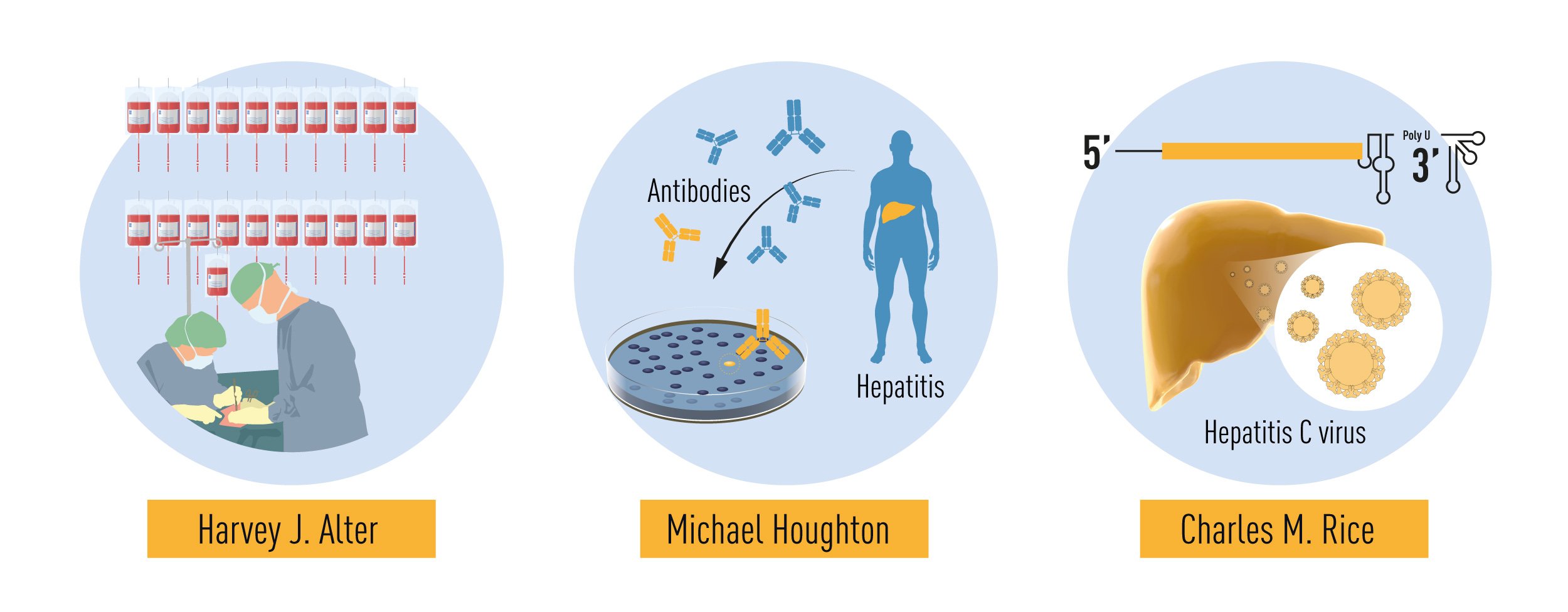
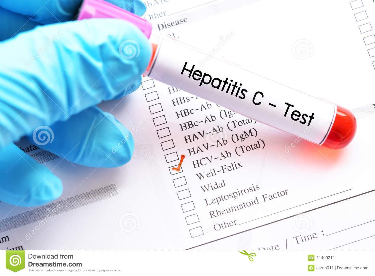
Hepatitis C is a liver infection caused by the hepatitis C virus (HCV). It's primarily spread through contact with contaminated blood, often through sharing needles or other equipment to inject drugs. For some people, hepatitis C is a short-term illness but for most — about 75-85% — it becomes a long-term, chronic infection that can lead to serious health problems like liver damage, liver failure, and even liver cancer. The virus can infect and inflame the liver, causing symptoms like jaundice (yellowing of the skin and eyes), abdominal pain, fatigue, and dark urine. Many people with hepatitis C don't have any symptoms, so they might not know they have the infection until they experience complications. There are effective treatments available for hepatitis C, including antiviral medications that can cure the infection in most people. Regular testing is important to diagnose and treat hepatitis C early, before it causes serious health problems.
Hepatitis A is a viral infection that specifically targets the liver, causing inflammation and impaired function. This disease is caused by the hepatitis A virus (HAV), which spreads primarily through the fecal-oral route, often due to poor sanitation and hygiene. Individuals can become infected by consuming food or water contaminated with HAV or by coming into direct contact with an infected person's stool.
The symptoms of hepatitis A may include fatigue, loss of appetite, nausea, vomiting, abdominal pain, dark urine, clay-colored bowel movements, joint pain, and jaundice (yellowing of the skin and eyes). However, in some cases, particularly in children under six years old, the infection may be asymptomatic.
While hepatitis A can be unpleasant and cause serious complications, it is rarely fatal and most people recover completely within a few months. Preventive measures include vaccination, practicing good hygiene, and avoiding potentially contaminated food and water.
Antibodies are proteins produced by the immune system in response to the presence of a foreign substance, such as a bacterium or virus. They are capable of identifying and binding to specific antigens (foreign substances) on the surface of these invaders, marking them for destruction by other immune cells. Antibodies are also known as immunoglobulins and come in several different types, including IgA, IgD, IgE, IgG, and IgM, each with a unique function in the immune response. They are composed of four polypeptide chains, two heavy chains and two light chains, that are held together by disulfide bonds. The variable regions of the heavy and light chains form the antigen-binding site, which is specific to a particular antigen.
Hepatitis B virus (HBV) is a DNA virus that belongs to the Hepadnaviridae family and causes the infectious disease known as hepatitis B. This virus primarily targets the liver, where it can lead to inflammation and damage of the liver tissue. The infection can range from acute to chronic, with chronic hepatitis B increasing the risk of developing serious liver complications such as cirrhosis and liver cancer.
The Hepatitis B virus has a complex life cycle, involving both nuclear and cytoplasmic phases. It enters hepatocytes (liver cells) via binding to specific receptors and is taken up by endocytosis. The viral DNA is released into the nucleus, where it is converted into a covalently closed circular DNA (cccDNA) form, which serves as the template for viral transcription.
HBV transcribes several RNAs, including pregenomic RNA (pgRNA), which is used as a template for reverse transcription during virion assembly. The pgRNA is encapsidated into core particles along with the viral polymerase and undergoes reverse transcription to generate new viral DNA. This process occurs within the cytoplasm of the hepatocyte, resulting in the formation of immature virions containing partially double-stranded DNA.
These immature virions are then enveloped by host cell membranes containing HBV envelope proteins (known as surface antigens) to form mature virions that can be secreted from the hepatocyte and infect other cells. The virus can also integrate into the host genome, which may contribute to the development of hepatocellular carcinoma in chronic cases.
Hepatitis B is primarily transmitted through exposure to infected blood or bodily fluids containing the virus, such as through sexual contact, sharing needles, or from mother to child during childbirth. Prevention strategies include vaccination, safe sex practices, and avoiding needle-sharing behaviors. Treatment for hepatitis B typically involves antiviral medications that can help suppress viral replication and reduce the risk of liver damage.
Hepatitis B Surface Antigens (HBsAg) are proteins found on the surface of the Hepatitis B virus. They are present in the blood of individuals infected with the Hepatitis B virus and are used as a marker for the presence of a current Hepatitis B infection. The detection of HBsAg in the blood indicates that an individual is infectious and can transmit the virus to others. It is typically used in diagnostic tests to detect and diagnose Hepatitis B infections, monitor treatment response, and assess the risk of transmission.
Hepatitis is a medical condition characterized by inflammation of the liver, often resulting in damage to liver cells. It can be caused by various factors, including viral infections (such as Hepatitis A, B, C, D, and E), alcohol abuse, toxins, medications, and autoimmune disorders. Symptoms may include jaundice, fatigue, abdominal pain, loss of appetite, nausea, vomiting, and dark urine. The severity of the disease can range from mild illness to severe, life-threatening conditions, such as liver failure or cirrhosis.
Antibody specificity refers to the ability of an antibody to bind to a specific epitope or antigenic determinant on an antigen. Each antibody has a unique structure that allows it to recognize and bind to a specific region of an antigen, typically a small portion of the antigen's surface made up of amino acids or sugar residues. This highly specific binding is mediated by the variable regions of the antibody's heavy and light chains, which form a pocket that recognizes and binds to the epitope.
The specificity of an antibody is determined by its unique complementarity-determining regions (CDRs), which are loops of amino acids located in the variable domains of both the heavy and light chains. The CDRs form a binding site that recognizes and interacts with the epitope on the antigen. The precise fit between the antibody's binding site and the epitope is critical for specificity, as even small changes in the structure of either can prevent binding.
Antibody specificity is important in immune responses because it allows the immune system to distinguish between self and non-self antigens. This helps to prevent autoimmune reactions where the immune system attacks the body's own cells and tissues. Antibody specificity also plays a crucial role in diagnostic tests, such as ELISA assays, where antibodies are used to detect the presence of specific antigens in biological samples.
Antibodies, viral are proteins produced by the immune system in response to an infection with a virus. These antibodies are capable of recognizing and binding to specific antigens on the surface of the virus, which helps to neutralize or destroy the virus and prevent its replication. Once produced, these antibodies can provide immunity against future infections with the same virus.
Viral antibodies are typically composed of four polypeptide chains - two heavy chains and two light chains - that are held together by disulfide bonds. The binding site for the antigen is located at the tip of the Y-shaped structure, formed by the variable regions of the heavy and light chains.
There are five classes of antibodies in humans: IgA, IgD, IgE, IgG, and IgM. Each class has a different function and is distributed differently throughout the body. For example, IgG is the most common type of antibody found in the bloodstream and provides long-term immunity against viruses, while IgA is found primarily in mucous membranes and helps to protect against respiratory and gastrointestinal infections.
In addition to their role in the immune response, viral antibodies can also be used as diagnostic tools to detect the presence of a specific virus in a patient's blood or other bodily fluids.
Chronic Hepatitis C is a liver infection caused by the hepatitis C virus (HCV) that lasts for more than six months. This long-term infection can lead to scarring of the liver (cirrhosis), which can cause serious health problems, such as liver failure or liver cancer, in some individuals. The infection is usually asymptomatic until complications arise, but it can be detected through blood tests that identify antibodies to the virus or viral RNA. Chronic hepatitis C is typically managed with antiviral therapy, which can help clear the virus from the body and reduce the risk of liver damage.
Chronic Hepatitis B is a persistent infection of the liver caused by the hepatitis B virus (HBV), which can lead to chronic inflammation and scarring of the liver over time. It is defined as the presence of hepatitis B surface antigen (HBsAg) in the blood for more than six months.
The infection can be asymptomatic or may cause nonspecific symptoms such as fatigue, loss of appetite, nausea, and joint pain. A small percentage of people with chronic HBV infection may develop serious complications, including cirrhosis, liver failure, and liver cancer. Treatment options for chronic hepatitis B include antiviral medications that can help to suppress the virus and reduce the risk of liver damage. Vaccination is available to prevent hepatitis B infection.
Viral hepatitis in humans refers to inflammation of the liver caused by infection with viruses that primarily target the liver. There are five main types of human viral hepatitis, designated as Hepatitis A, B, C, D, and E virus (HAV, HBV, HCV, HDV, and HEV). These viruses can cause a range of illnesses, from acute self-limiting hepatitis to chronic hepatitis, which can lead to cirrhosis and liver cancer.
1. Hepatitis A virus (HAV) is typically spread through the fecal-oral route, often through contaminated food or water. It usually results in an acute self-limiting infection, but rarely can cause chronic hepatitis in individuals with weakened immune systems.
2. Hepatitis B virus (HBV) is primarily transmitted through contact with infected blood, semen, and other bodily fluids. It can lead to both acute and chronic hepatitis, which may result in cirrhosis and liver cancer if left untreated.
3. Hepatitis C virus (HCV) is predominantly spread through exposure to infected blood, such as through sharing needles or receiving contaminated blood transfusions. Chronic hepatitis C is common, and it can lead to serious liver complications like cirrhosis and liver cancer if not treated.
4. Hepatitis D virus (HDV) is an incomplete virus that requires the presence of HBV for its replication. HDV infection occurs only in individuals already infected with HBV, leading to more severe liver disease compared to HBV monoinfection.
5. Hepatitis E virus (HEV) is primarily transmitted through the fecal-oral route, often through contaminated food or water. It usually results in an acute self-limiting infection but can cause chronic hepatitis in pregnant women and individuals with weakened immune systems.
Prevention measures include vaccination for HAV and HBV, safe sex practices, avoiding sharing needles, and ensuring proper hygiene and sanitation to prevent fecal-oral transmission.
Hepatitis B antibodies are proteins produced by the immune system in response to the presence of the Hepatitis B virus. There are two main types of Hepatitis B antibodies:
1. Hepatitis B surface antibody (anti-HBs): This is produced when a person has recovered from a Hepatitis B infection or has been successfully vaccinated against the virus. The presence of anti-HBs indicates immunity to Hepatitis B.
2. Hepatitis B core antibody (anti-HBC): This is produced during a Hepatitis B infection and remains present for life, even after the infection has been cleared. However, the presence of anti-HBC alone does not indicate immunity to Hepatitis B, as it can also be present in people who have a chronic Hepatitis B infection.
It's important to note that testing for Hepatitis B antibodies is typically done through blood tests and can help determine whether a person has been infected with the virus, has recovered from an infection, or has been vaccinated against it.
Hepatitis A virus (HAV) is the causative agent of hepatitis A, a viral infection that causes inflammation of the liver. It is a small, non-enveloped, single-stranded RNA virus belonging to the Picornaviridae family and Hepatovirus genus. The virus primarily spreads through the fecal-oral route, often through contaminated food or water, or close contact with an infected person. After entering the body, HAV infects hepatocytes in the liver, leading to liver damage and associated symptoms such as jaundice, fatigue, abdominal pain, and nausea. The immune system eventually clears the infection, providing lifelong immunity against future HAV infections. Preventive measures include vaccination and practicing good hygiene to prevent transmission.
"Hepatitis B vaccines are vaccines that prevent infection caused by the hepatitis B virus. They work by introducing a small and harmless piece of the virus to your body, which triggers your immune system to produce antibodies to fight off the infection. These antibodies remain in your body and provide protection if you are exposed to the real hepatitis B virus in the future.
The hepatitis B vaccine is typically given as a series of three shots over a six-month period. It is recommended for all infants, children and adolescents who have not previously been vaccinated, as well as for adults who are at increased risk of infection, such as healthcare workers, people who inject drugs, and those with certain medical conditions.
It's important to note that hepatitis B vaccine does not provide protection against other types of viral hepatitis, such as hepatitis A or C."
Monoclonal antibodies are a type of antibody that are identical because they are produced by a single clone of cells. They are laboratory-produced molecules that act like human antibodies in the immune system. They can be designed to attach to specific proteins found on the surface of cancer cells, making them useful for targeting and treating cancer. Monoclonal antibodies can also be used as a therapy for other diseases, such as autoimmune disorders and inflammatory conditions.
Monoclonal antibodies are produced by fusing a single type of immune cell, called a B cell, with a tumor cell to create a hybrid cell, or hybridoma. This hybrid cell is then able to replicate indefinitely, producing a large number of identical copies of the original antibody. These antibodies can be further modified and engineered to enhance their ability to bind to specific targets, increase their stability, and improve their effectiveness as therapeutic agents.
Monoclonal antibodies have several mechanisms of action in cancer therapy. They can directly kill cancer cells by binding to them and triggering an immune response. They can also block the signals that promote cancer growth and survival. Additionally, monoclonal antibodies can be used to deliver drugs or radiation directly to cancer cells, increasing the effectiveness of these treatments while minimizing their side effects on healthy tissues.
Monoclonal antibodies have become an important tool in modern medicine, with several approved for use in cancer therapy and other diseases. They are continuing to be studied and developed as a promising approach to treating a wide range of medical conditions.
Bacterial antibodies are a type of antibodies produced by the immune system in response to an infection caused by bacteria. These antibodies are proteins that recognize and bind to specific antigens on the surface of the bacterial cells, marking them for destruction by other immune cells. Bacterial antibodies can be classified into several types based on their structure and function, including IgG, IgM, IgA, and IgE. They play a crucial role in the body's defense against bacterial infections and provide immunity to future infections with the same bacteria.
Chronic hepatitis is a type of liver inflammation that lasts for more than six months and can lead to scarring of the liver (cirrhosis), liver failure, and even liver cancer in some cases. It can be caused by various factors, including viral infections such as Hepatitis B and C, autoimmune disorders, alcohol abuse, and non-alcoholic fatty liver disease. The symptoms of chronic hepatitis may include fatigue, loss of appetite, nausea, vomiting, abdominal pain, joint pain, dark urine, and jaundice (yellowing of the skin and eyes). Treatment for chronic hepatitis depends on the underlying cause and may include antiviral medications, immunosuppressive drugs, or lifestyle changes.
Hepatitis B core antigen (HBcAg) is a protein found in the core of the hepatitis B virus (HBV). It is present during active replication of the virus and plays a crucial role in the formation of the viral capsid or core. The antibodies produced against HBcAg (anti-HBc) can be detected in the blood, which serves as a marker for current or past HBV infection. It is important to note that HBcAg itself is not detectable in the blood because it is confined within the viral particle. However, during the serological testing of hepatitis B, the detection of anti-HBc IgM indicates a recent acute infection, while the presence of anti-HBc IgG suggests either a past resolved infection or an ongoing chronic infection.
Hepatitis A vaccines are inactivated or live attenuated viral vaccines that are administered to prevent infection and illness caused by the hepatitis A virus. The vaccine contains antigens that stimulate an immune response in the body, leading to the production of antibodies that protect against future infection with the virus.
The inactivated hepatitis A vaccine is made from viruses that have been chemically treated to destroy their ability to cause disease while preserving their ability to stimulate an immune response. This type of vaccine is typically given in two doses, six months apart, and provides long-term protection against the virus.
The live attenuated hepatitis A vaccine contains a weakened form of the virus that is unable to cause illness but can still stimulate an immune response. This type of vaccine is given as a single dose and provides protection against the virus for at least 20 years.
Hepatitis A vaccines are recommended for people who are at increased risk of infection, including travelers to areas where hepatitis A is common, men who have sex with men, people who use injection drugs, and people with chronic liver disease or clotting factor disorders. The vaccine is also recommended for children in certain states and communities where hepatitis A is endemic.
Hepacivirus is a genus of viruses in the family Flaviviridae. The most well-known member of this genus is Hepatitis C virus (HCV), which is a major cause of liver disease worldwide. HCV infection can lead to chronic hepatitis, cirrhosis, and liver cancer.
Hepaciviruses are enveloped viruses with a single-stranded, positive-sense RNA genome. They have a small icosahedral capsid and infect a variety of hosts, including humans, non-human primates, horses, and birds. The virus enters the host cell by binding to specific receptors on the cell surface and is then internalized through endocytosis.
HCV has a high degree of genetic diversity and is classified into seven major genotypes and numerous subtypes based on differences in its RNA sequence. This genetic variability can affect the virus's ability to evade the host immune response, making treatment more challenging.
In addition to HCV, other hepaciviruses have been identified in various animal species, including equine hepacivirus (EHCV), rodent hepacivirus (RHV), and bat hepacivirus (BtHepCV). These viruses are being studied to better understand the biology of hepaciviruses and their potential impact on human health.
Hepatitis C antibodies are proteins produced by the immune system in response to an infection with the hepatitis C virus (HCV). Detection of these antibodies in the blood indicates a past or present HCV infection. However, it does not necessarily mean that the person is currently infected, as antibodies can persist for years even after the virus has been cleared from the body. Additional tests are usually needed to confirm whether the infection is still active and to guide treatment decisions.
Hepatitis antibodies are proteins produced by the immune system in response to an infection caused by a hepatitis virus. There are several types of hepatitis viruses, including A, B, C, D, and E, each with their own specific antibodies.
The presence of hepatitis antibodies in the blood can indicate a current or past infection with the corresponding hepatitis virus. For example, the detection of anti-HAV (hepatitis A virus) antibodies indicates a past infection or immunization against hepatitis A, while the detection of anti-HBs (hepatitis B surface antigen) antibodies indicates immunity due to vaccination or recovery from a hepatitis B infection.
It's important to note that some hepatitis antibodies may not provide immunity to future infections, and individuals can still be infected with the virus even if they have previously produced antibodies against it. Therefore, regular testing and vaccination are essential for preventing the spread of hepatitis viruses and protecting public health.
Hepatitis B e antigen (HBeAg) is a protein produced by the hepatitis B virus (HBV) during its replication process. It can be found in the blood of individuals infected with HBV. The presence of HBeAg generally indicates that the virus is actively replicating in the liver and that the individual has high levels of viral load.
HBeAg is a serological marker used to assess the severity and activity of HBV infection, as well as the response to antiviral treatment. In particular, the disappearance of HBeAg from the blood (known as seroconversion) is often associated with a decrease in viral replication and an improvement in liver disease. However, the presence of HBeAg does not necessarily mean that the individual will develop symptoms or liver damage, as some people can remain asymptomatic carriers of the virus for many years.
It's important to note that not all HBV strains produce HBeAg, and some mutant strains may not produce detectable levels of this antigen even when the virus is actively replicating. Therefore, additional tests may be needed to confirm the presence or absence of HBV infection in these cases.
Hepatitis B antigens are proteins or particles present on the surface (HBsAg) or inside (HBcAg, HBeAg) the hepatitis B virus.
1. HBsAg (Hepatitis B surface antigen): This is a protein found on the outer surface of the hepatitis B virus. Its presence in the blood indicates an active infection with hepatitis B virus. It's also used as a marker to diagnose hepatitis B infection and monitor treatment response.
2. HBcAg (Hepatitis B core antigen): This is a protein found inside the hepatitis B virus core. It's not usually detected in the blood, but its antibodies (anti-HBc) are used to diagnose past or present hepatitis B infection.
3. HBeAg (Hepatitis B e antigen): This is a protein found inside the hepatitis B virus core and is associated with viral replication. Its presence in the blood indicates high levels of viral replication, increased infectivity, and higher risk of liver damage. It's used to monitor disease progression and treatment response.
These antigens play a crucial role in the diagnosis, management, and prevention of hepatitis B infection.
Antibody formation, also known as humoral immune response, is the process by which the immune system produces proteins called antibodies in response to the presence of a foreign substance (antigen) in the body. This process involves several steps:
1. Recognition: The antigen is recognized and bound by a type of white blood cell called a B lymphocyte or B cell, which then becomes activated.
2. Differentiation: The activated B cell undergoes differentiation to become a plasma cell, which is a type of cell that produces and secretes large amounts of antibodies.
3. Antibody production: The plasma cells produce and release antibodies, which are proteins made up of four polypeptide chains (two heavy chains and two light chains) arranged in a Y-shape. Each antibody has two binding sites that can recognize and bind to specific regions on the antigen called epitopes.
4. Neutralization or elimination: The antibodies bind to the antigens, neutralizing them or marking them for destruction by other immune cells. This helps to prevent the spread of infection and protect the body from harmful substances.
Antibody formation is an important part of the adaptive immune response, which allows the body to specifically recognize and respond to a wide variety of pathogens and foreign substances.
Neutralizing antibodies are a type of antibody that defends against pathogens such as viruses or bacteria by neutralizing their ability to infect cells. They do this by binding to specific regions on the surface proteins of the pathogen, preventing it from attaching to and entering host cells. This renders the pathogen ineffective and helps to prevent or reduce the severity of infection. Neutralizing antibodies can be produced naturally in response to an infection or vaccination, or they can be generated artificially for therapeutic purposes.
Hepatitis E is a viral infection that specifically affects the liver, caused by the hepatitis E virus (HEV). The disease is primarily transmitted through the fecal-oral route, often through contaminated water or food. It can also be spread through blood transfusions and vertical transmission from mother to fetus.
The incubation period for hepatitis E ranges from 2 to 10 weeks. Symptoms of the disease are similar to other types of viral hepatitis and may include jaundice (yellowing of the skin and eyes), fatigue, loss of appetite, abdominal pain, nausea, vomiting, joint pain, and dark urine.
In most cases, hepatitis E is a self-limiting disease, meaning that it resolves on its own within a few weeks to months. However, in some individuals, particularly those with weakened immune systems, the infection can lead to severe complications such as acute liver failure and death. Pregnant women, especially those in the third trimester, are at higher risk of developing severe disease and have a mortality rate of up to 25%.
Prevention measures include maintaining good hygiene practices, practicing safe food handling and preparation, and ensuring access to clean water sources. Currently, there is no specific treatment for hepatitis E, but supportive care can help manage symptoms. Vaccines are available in some countries to prevent the disease.
Hepatitis E virus (HEV) is a single-stranded, positive-sense RNA virus that belongs to the family Hepeviridae and genus Orthohepevirus. It primarily infects the liver, causing acute hepatitis in humans. The virus is transmitted through the fecal-oral route, often through contaminated water or food sources. Ingestion of raw or undercooked pork or deer meat can also lead to HEV infection.
HEV infection typically results in self-limiting acute hepatitis, characterized by symptoms such as jaundice, fatigue, loss of appetite, abdominal pain, and dark urine. In some cases, particularly among pregnant women and individuals with weakened immune systems, HEV infection can lead to severe complications, including fulminant hepatic failure and death.
There are four main genotypes of HEV that infect humans: genotype 1 and 2 are primarily found in developing countries and are transmitted through contaminated water; genotype 3 and 4 are found worldwide and can be transmitted through both zoonotic and human-to-human routes.
Prevention measures include improving sanitation, access to clean water, and food safety practices. Currently, there is no specific antiviral treatment for HEV infection, but supportive care can help manage symptoms. A vaccine against HEV is available in China and has shown efficacy in preventing the disease.
Hepatitis A antibodies are proteins produced by the immune system in response to a Hepatitis A virus infection or after vaccination. There are two types of Hepatitis A antibodies:
1. IgM anti-HAV (Hepatitis A Virus) antibodies: These are the first type of antibodies produced by the immune system during a Hepatitis A infection. They appear in the blood within 2 to 4 weeks after exposure to the virus and remain detectable for up to 12 weeks. The presence of IgM anti-HAV antibodies indicates a recent or ongoing Hepatitis A infection.
2. IgG anti-HAV antibodies: These are the second type of antibodies produced by the immune system during a Hepatitis A infection, and they appear in the blood several weeks after the onset of illness. IgG anti-HAV antibodies remain detectable for many years, providing long-term immunity against future Hepatitis A infections. After vaccination, only IgG anti-HAV antibodies are produced, indicating immunity to Hepatitis A.
Testing for Hepatitis A antibodies is used to diagnose acute or past Hepatitis A infections and to assess immunity following vaccination.
Autoimmune hepatitis is a chronic (long-term) disease in which the body's immune system mistakenly attacks the liver, leading to inflammation and damage. This results in decreased liver function over time if not treated. The exact cause of autoimmune hepatitis is unknown, but it is believed to be associated with genetic factors and exposure to certain environmental triggers, such as viral infections or medications.
There are two main types of autoimmune hepatitis:
1. Type 1 (classic) autoimmune hepatitis: This form can affect both adults and children, and it is more common in women than men. People with this type may also have other autoimmune disorders, such as rheumatoid arthritis, thyroid disease, or ulcerative colitis.
2. Type 2 autoimmune hepatitis: This form primarily affects children and young women. It is less common than type 1 and tends to be more severe. People with this type may also have other autoimmune disorders, such as celiac disease or chronic candidiasis.
Symptoms of autoimmune hepatitis can vary widely, from mild to severe. They may include fatigue, loss of appetite, nausea, vomiting, abdominal pain, joint pain, jaundice (yellowing of the skin and eyes), dark urine, and light-colored stools.
Diagnosis typically involves blood tests, imaging studies, and sometimes a liver biopsy to assess the extent of damage. Treatment usually includes medications that suppress the immune system, such as corticosteroids and immunosuppressants, which can help reduce inflammation and slow or stop liver damage. In some cases, lifestyle changes and supportive care may also be necessary.
I'm sorry for any confusion, but "Viral Hepatitis, Animal" is not a standard medical classification or definition. Hepatitis refers to inflammation of the liver, and viral hepatitis refers to inflammation caused by a virus. The term "animal" in this context doesn't provide a clear meaning.
However, it's worth noting that some animals can contract viral hepatitis, similar to humans. For instance, there are hepatitis A, B, and C-like viruses that have been identified in various animal species. These are typically not transmissible to humans.
If you're referring to a specific medical condition or context, could you please provide more details? I'd be happy to help further with more information.
Hepatitis A Virus, Human (HAV): A single-stranded, positive-sense RNA virus belonging to the Picornaviridae family, specifically the Hepatovirus genus. It is the causative agent of Hepatitis A, a viral infection that primarily affects the liver. The virus is typically transmitted through the fecal-oral route, often via contaminated food or water, or close contact with an infected individual. Following incubation (15-50 days), symptoms may include jaundice, fatigue, abdominal pain, loss of appetite, nausea, diarrhea, and fever. Most people recover completely within a few weeks; however, severe complications and death are possible, especially in individuals with preexisting liver disease. Prevention is primarily achieved through vaccination and practicing good hygiene.
Hepatitis viruses refer to a group of viral agents that primarily target the liver, causing inflammation and damage to hepatocytes (liver cells). This results in various clinical manifestations, ranging from an acute infection to a chronic, persistent infection. There are five main types of hepatitis viruses, named Hepatitis A, B, C, D, and E virus, each with distinct genetic material, modes of transmission, and disease severity.
1. Hepatitis A Virus (HAV): This is a single-stranded RNA virus that is primarily transmitted through the fecal-oral route, often via contaminated food or water. Infected individuals may experience symptoms such as jaundice, fatigue, abdominal pain, and loss of appetite. While most people recover completely within a few months, severe complications can occur in rare cases. A vaccine is available to prevent HAV infection.
2. Hepatitis B Virus (HBV): This is a double-stranded DNA virus that is primarily transmitted through contact with infected blood or bodily fluids, such as during sexual contact, sharing needles, or from mother to child during childbirth. HBV can cause both acute and chronic hepatitis, which may lead to severe liver complications like cirrhosis and liver cancer if left untreated. A vaccine is available to prevent HBV infection.
3. Hepatitis C Virus (HCV): This is a single-stranded RNA virus that is primarily transmitted through contact with infected blood, often through sharing needles or during medical procedures using contaminated equipment. Like HBV, HCV can cause both acute and chronic hepatitis, which may lead to severe liver complications if left untreated. No vaccine is currently available for HCV; however, antiviral treatments can cure the infection in many cases.
4. Hepatitis D Virus (HDV): This is a defective RNA virus that requires the presence of HBV to replicate and cause infection. HDV is primarily transmitted through contact with infected blood or bodily fluids, similar to HBV. Co-infection with both HBV and HDV can result in more severe liver disease compared to HBV infection alone. Antiviral treatments are available for HDV; however, a vaccine is not.
5. Hepatitis E Virus (HEV): This is a single-stranded RNA virus that primarily causes acute hepatitis and is usually transmitted through the fecal-oral route, often through contaminated food or water. In most cases, HEV infection resolves on its own without treatment. However, in pregnant women and individuals with weakened immune systems, HEV can cause severe liver complications. No vaccine is currently available for HEV in the United States; however, a vaccine has been approved in some countries.
Hepatitis Delta Virus (HDV) is not a traditional virus but rather a defective RNA particle that requires the assistance of the hepatitis B virus (HBV) to replicate. It's also known as delta agent or hepatitis D. HDV is a unique pathogen that only infects individuals who are already infected with HBV.
The virus causes a more severe form of viral hepatitis than HBV alone, leading to a higher risk of fulminant hepatitis (acute liver failure) and chronic hepatitis, which can progress to cirrhosis and hepatocellular carcinoma. HDV is primarily transmitted through percutaneous or sexual contact with infected blood or body fluids.
Prevention strategies include vaccination against HBV, which also prevents HDV infection, and avoiding high-risk behaviors such as intravenous drug use and unprotected sex with multiple partners. There is no specific treatment for HDV; however, antiviral therapy for HBV can help manage the infection.
Hepatitis D, also known as Delta hepatitis, is a viral infection of the liver that can only occur in people who have a current infection with the hepatitis B virus (HBV). It's caused by the hepatitis delta virus (HDV), which is a small, enveloped, single-stranded RNA virus.
HDV requires the presence of HBV for its replication and survival, so it can't infect someone who doesn't already have HBV. When both viruses are present, they can interact in ways that lead to more severe liver disease than either virus would cause alone.
Hepatitis D can be an acute or chronic infection, and it can range from mild to severe, with symptoms similar to those of other types of viral hepatitis, such as jaundice, fatigue, loss of appetite, nausea, vomiting, abdominal pain, and joint pain. In some cases, hepatitis D can lead to serious complications, including liver failure and death.
Hepatitis D is primarily spread through contact with infected blood or other bodily fluids, such as during sexual contact, sharing needles, or mother-to-child transmission during childbirth. It's preventable through vaccination against hepatitis B, which provides immunity to both viruses. There is no specific treatment for hepatitis D, but antiviral therapy for hepatitis B can help manage the infection and prevent complications.
I'm sorry for any confusion, but "Animal Hepatitis" is not a medical term used to describe a specific disease. Hepatitis refers to inflammation of the liver, and it can be caused by various factors, including viruses, alcohol, drugs, and certain medical conditions.
However, there are several viral hepatitis types that can infect animals, such as Hepatitis A, B, and C, which primarily affect humans. But there are also other hepatitis viruses that are species-specific and primarily infect animals, such as:
1. Canine Hepatitis (Adenovirus Type 1): This is a viral infection that affects dogs and causes liver damage, respiratory signs, and occasionally death.
2. Feline Infectious Peritonitis (FIP) Virus: While not strictly a hepatitis virus, this feline coronavirus can cause severe inflammation of the liver and other organs in cats.
3. Equine Infectious Anemia Virus (EIAV): This retrovirus affects horses and causes cyclic fever, anemia, and occasionally liver disease.
4. Avian Hepatitis E Virus: A recently discovered virus that infects birds and can cause hepatitis and other systemic signs in chickens and other avian species.
If you're looking for information on a specific animal hepatitis virus or a different medical term, please provide more context so I can give you a more accurate answer.
Antibody affinity refers to the strength and specificity of the interaction between an antibody and its corresponding antigen at a molecular level. It is a measure of how strongly and selectively an antibody binds to its target antigen. A higher affinity indicates a more stable and specific binding, while a lower affinity suggests weaker and less specific interactions. Affinity is typically measured in terms of the dissociation constant (Kd), which describes the concentration of antigen needed to achieve half-maximal binding to an antibody. Generally, a smaller Kd value corresponds to a higher affinity, indicating a tighter and more selective bond. This parameter is crucial in the development of diagnostic and therapeutic applications, such as immunoassays and targeted therapies, where high-affinity antibodies are preferred for improved sensitivity and specificity.
Viral hepatitis vaccines are vaccines that prevent infection caused by various hepatitis viruses, including hepatitis A and B. These vaccines contain antigens that stimulate the immune system to produce antibodies that protect against infection with the corresponding virus. The vaccines are typically administered through injection and may require multiple doses for full protection.
The hepatitis A vaccine is made from inactivated hepatitis A virus, while the hepatitis B vaccine is made from recombinant hepatitis B surface antigen. Both vaccines have been shown to be highly effective in preventing infection and reducing the risk of complications associated with viral hepatitis, such as liver disease and liver cancer.
It's important to note that there are no vaccines available for other types of viral hepatitis, such as hepatitis C, D, or E. Prevention strategies for these types of viral hepatitis typically involve measures to reduce exposure to the virus, such as safe injection practices and avoiding high-risk behaviors like sharing needles or having unprotected sex with infected individuals.
The Fluorescent Antibody Technique (FAT) is a type of immunofluorescence assay used in laboratory medicine and pathology for the detection and localization of specific antigens or antibodies in tissues, cells, or microorganisms. In this technique, a fluorescein-labeled antibody is used to selectively bind to the target antigen or antibody, forming an immune complex. When excited by light of a specific wavelength, the fluorescein label emits light at a longer wavelength, typically visualized as green fluorescence under a fluorescence microscope.
The FAT is widely used in diagnostic microbiology for the identification and characterization of various bacteria, viruses, fungi, and parasites. It has also been applied in the diagnosis of autoimmune diseases and certain cancers by detecting specific antibodies or antigens in patient samples. The main advantage of FAT is its high sensitivity and specificity, allowing for accurate detection and differentiation of various pathogens and disease markers. However, it requires specialized equipment and trained personnel to perform and interpret the results.
Antiviral agents are a class of medications that are designed to treat infections caused by viruses. Unlike antibiotics, which target bacteria, antiviral agents interfere with the replication and infection mechanisms of viruses, either by inhibiting their ability to replicate or by modulating the host's immune response to the virus.
Antiviral agents are used to treat a variety of viral infections, including influenza, herpes simplex virus (HSV) infections, human immunodeficiency virus (HIV) infection, hepatitis B and C, and respiratory syncytial virus (RSV) infections.
These medications can be administered orally, intravenously, or topically, depending on the type of viral infection being treated. Some antiviral agents are also used for prophylaxis, or prevention, of certain viral infections.
It is important to note that antiviral agents are not effective against all types of viruses and may have significant side effects. Therefore, it is essential to consult with a healthcare professional before starting any antiviral therapy.
Anti-idiotypic antibodies are a type of immune protein that recognizes and binds to the unique identifying region (idiotype) of another antibody. These antibodies are produced by the immune system as part of a regulatory feedback mechanism, where they can modulate or inhibit the activity of the original antibody. They have been studied for their potential use in immunotherapy and vaccine development.
A binding site on an antibody refers to the specific region on the surface of the antibody molecule that can recognize and bind to a specific antigen. Antibodies are proteins produced by the immune system in response to the presence of foreign substances called antigens. They have two main functions: to neutralize the harmful effects of antigens and to help eliminate them from the body.
The binding site of an antibody is located at the tips of its Y-shaped structure, formed by the variable regions of the heavy and light chains of the antibody molecule. These regions contain unique amino acid sequences that determine the specificity of the antibody for a particular antigen. The binding site can recognize and bind to a specific epitope or region on the antigen, forming an antigen-antibody complex.
The binding between the antibody and antigen is highly specific and depends on non-covalent interactions such as hydrogen bonds, van der Waals forces, and electrostatic attractions. This interaction plays a crucial role in the immune response, as it allows the immune system to recognize and eliminate pathogens and other foreign substances from the body.
An epitope is a specific region on the surface of an antigen (a molecule that can trigger an immune response) that is recognized by an antibody, B-cell receptor, or T-cell receptor. It is also commonly referred to as an antigenic determinant. Epitopes are typically composed of linear amino acid sequences or conformational structures made up of discontinuous amino acids in the antigen. They play a crucial role in the immune system's ability to differentiate between self and non-self molecules, leading to the targeted destruction of foreign substances like viruses and bacteria. Understanding epitopes is essential for developing vaccines, diagnostic tests, and immunotherapies.
Molecular sequence data refers to the specific arrangement of molecules, most commonly nucleotides in DNA or RNA, or amino acids in proteins, that make up a biological macromolecule. This data is generated through laboratory techniques such as sequencing, and provides information about the exact order of the constituent molecules. This data is crucial in various fields of biology, including genetics, evolution, and molecular biology, allowing for comparisons between different organisms, identification of genetic variations, and studies of gene function and regulation.
Murine hepatitis virus (MHV) is a type of coronavirus that primarily infects laboratory mice. It is not related to the human hepatitis viruses A, B, C, D, or E. MHV causes a range of diseases in mice, including hepatitis (liver inflammation), encephalomyelitis (inflammation of the brain and spinal cord), and enteritis (inflammation of the intestine). The virus is transmitted through fecal-oral route and respiratory droplets. It's widely used in research to understand the pathogenesis, immunity, and molecular biology of coronaviruses.
Hepatovirus is a genus of viruses in the Picornaviridae family, and it's most notably represented by the Human Hepatitis A Virus (HAV). These viruses are non-enveloped, with a single-stranded, positive-sense RNA genome. They primarily infect hepatocytes, causing liver inflammation and disease, such as hepatitis. Transmission of hepatoviruses typically occurs through the fecal-oral route, often via contaminated food or water. The virus causes an acute infection that does not usually become chronic, and recovery is usually complete within a few weeks. Immunity after infection is solid and lifelong.
Hepatitis C antigens refer to the proteins present on the surface of the hepatitis C virus (HCV). The most commonly studied and clinically relevant antigen is the core protein, which plays a crucial role in the viral replication process. Detection of HCV antigens in serum or plasma can indicate an ongoing infection, as they appear during the early stages of infection and usually persist until the development of a humoral immune response, which leads to the production of antibodies against these antigens.
The detection of HCV core antigen (HCVcAg) has been used as an alternative diagnostic marker for HCV infection, especially in resource-limited settings where nucleic acid testing (NAT), such as polymerase chain reaction (PCR) for HCV RNA, might not be readily available. However, the sensitivity and specificity of HCVcAg detection are generally lower than those of NAT methods. Nonetheless, it remains a valuable tool in monitoring treatment response and disease progression in individuals with chronic hepatitis C infection.
HIV antibodies are proteins produced by the immune system in response to the presence of HIV (Human Immunodeficiency Virus) in the body. These antibodies are designed to recognize and bind to specific parts of the virus, known as antigens, in order to neutralize or eliminate it.
There are several types of HIV antibodies that can be produced, including:
1. Anti-HIV-1 and anti-HIV-2 antibodies: These are antibodies that specifically target the HIV-1 and HIV-2 viruses, respectively.
2. Antibodies to HIV envelope proteins: These antibodies recognize and bind to the outer envelope of the virus, which is covered in glycoprotein spikes that allow the virus to attach to and enter host cells.
3. Antibodies to HIV core proteins: These antibodies recognize and bind to the interior of the viral particle, where the genetic material of the virus is housed.
The presence of HIV antibodies in the blood can be detected through a variety of tests, including enzyme-linked immunosorbent assay (ELISA) and Western blot. A positive test result for HIV antibodies indicates that an individual has been infected with the virus, although it may take several weeks or months after infection for the antibodies to become detectable.
A viral RNA (ribonucleic acid) is the genetic material found in certain types of viruses, as opposed to viruses that contain DNA (deoxyribonucleic acid). These viruses are known as RNA viruses. The RNA can be single-stranded or double-stranded and can exist as several different forms, such as positive-sense, negative-sense, or ambisense RNA. Upon infecting a host cell, the viral RNA uses the host's cellular machinery to translate the genetic information into proteins, leading to the production of new virus particles and the continuation of the viral life cycle. Examples of human diseases caused by RNA viruses include influenza, COVID-19 (SARS-CoV-2), hepatitis C, and polio.
Antinuclear antibodies (ANA) are a type of autoantibody that target structures found in the nucleus of a cell. These antibodies are produced by the immune system and attack the body's own cells and tissues, leading to inflammation and damage. The presence of ANA is often used as a marker for certain autoimmune diseases, such as systemic lupus erythematosus (SLE), Sjogren's syndrome, rheumatoid arthritis, scleroderma, and polymyositis.
ANA can be detected through a blood test called the antinuclear antibody test. A positive result indicates the presence of ANA in the blood, but it does not necessarily mean that a person has an autoimmune disease. Further testing is usually needed to confirm a diagnosis and determine the specific type of autoantibodies present.
It's important to note that ANA can also be found in healthy individuals, particularly as they age. Therefore, the test results should be interpreted in conjunction with other clinical findings and symptoms.
'Antibodies, Neoplasm' is a medical term that refers to abnormal antibodies produced by neoplastic cells, which are cells that have undergone uncontrolled division and form a tumor or malignancy. These antibodies can be produced in large quantities and may have altered structures or functions compared to normal antibodies.
Neoplastic antibodies can arise from various types of malignancies, including leukemias, lymphomas, and multiple myeloma. In some cases, these abnormal antibodies can interfere with the normal functioning of the immune system and contribute to the progression of the disease.
In addition, neoplastic antibodies can also be used as tumor markers for diagnostic purposes. For example, certain types of monoclonal gammopathy, such as multiple myeloma, are characterized by the overproduction of a single type of immunoglobulin, which can be detected in the blood or urine and used to monitor the disease.
Overall, 'Antibodies, Neoplasm' is a term that encompasses a wide range of abnormal antibodies produced by neoplastic cells, which can have significant implications for both the diagnosis and treatment of malignancies.
Antibodies, protozoan, refer to the immune system's response to an infection caused by a protozoan organism. Protozoa are single-celled microorganisms that can cause various diseases in humans, such as malaria, giardiasis, and toxoplasmosis.
When the body is infected with a protozoan, the immune system responds by producing specific proteins called antibodies. Antibodies are produced by a type of white blood cell called a B-cell, and they recognize and bind to specific antigens on the surface of the protozoan organism.
There are five main types of antibodies: IgA, IgD, IgE, IgG, and IgM. Each type of antibody has a different role in the immune response. For example, IgG is the most common type of antibody and provides long-term immunity to previously encountered pathogens. IgM is the first antibody produced in response to an infection and is important for activating the complement system, which helps to destroy the protozoan organism.
Overall, the production of antibodies against protozoan organisms is a critical part of the immune response and helps to protect the body from further infection.
Immunoglobulin M (IgM) is a type of antibody that is primarily found in the blood and lymph fluid. It is the first antibody to be produced in response to an initial exposure to an antigen, making it an important part of the body's primary immune response. IgM antibodies are large molecules that are composed of five basic units, giving them a pentameric structure. They are primarily found on the surface of B cells as membrane-bound immunoglobulins (mlgM), where they function as receptors for antigens. Once an mlgM receptor binds to an antigen, it triggers the activation and differentiation of the B cell into a plasma cell that produces and secretes large amounts of soluble IgM antibodies.
IgM antibodies are particularly effective at agglutination (clumping) and complement activation, which makes them important in the early stages of an immune response to help clear pathogens from the bloodstream. However, they are not as stable or long-lived as other types of antibodies, such as IgG, and their levels tend to decline after the initial immune response has occurred.
In summary, Immunoglobulin M (IgM) is a type of antibody that plays a crucial role in the primary immune response to antigens by agglutination and complement activation. It is primarily found in the blood and lymph fluid, and it is produced by B cells after they are activated by an antigen.
Cross reactions, in the context of medical diagnostics and immunology, refer to a situation where an antibody or a immune response directed against one antigen also reacts with a different antigen due to similarities in their molecular structure. This can occur in allergy testing, where a person who is allergic to a particular substance may have a positive test result for a different but related substance because of cross-reactivity between them. For example, some individuals who are allergic to birch pollen may also have symptoms when eating certain fruits, such as apples, due to cross-reactive proteins present in both.
Interferon-alpha (IFN-α) is a type I interferon, which is a group of signaling proteins made and released by host cells in response to the presence of viruses, parasites, and tumor cells. It plays a crucial role in the immune response against viral infections. IFN-α has antiviral, immunomodulatory, and anti-proliferative effects.
IFN-α is produced naturally by various cell types, including leukocytes (white blood cells), fibroblasts, and epithelial cells, in response to viral or bacterial stimulation. It binds to specific receptors on the surface of nearby cells, triggering a signaling cascade that leads to the activation of genes involved in the antiviral response. This results in the production of proteins that inhibit viral replication and promote the presentation of viral antigens to the immune system, enhancing its ability to recognize and eliminate infected cells.
In addition to its role in the immune response, IFN-α has been used as a therapeutic agent for various medical conditions, including certain types of cancer, chronic hepatitis B and C, and multiple sclerosis. However, its use is often limited by side effects such as flu-like symptoms, depression, and neuropsychiatric disorders.
Liver cirrhosis is a chronic, progressive disease characterized by the replacement of normal liver tissue with scarred (fibrotic) tissue, leading to loss of function. The scarring is caused by long-term damage from various sources such as hepatitis, alcohol abuse, nonalcoholic fatty liver disease, and other causes. As the disease advances, it can lead to complications like portal hypertension, fluid accumulation in the abdomen (ascites), impaired brain function (hepatic encephalopathy), and increased risk of liver cancer. It is generally irreversible, but early detection and treatment of underlying causes may help slow down its progression.
Hepatitis A antigens refer to the proteins or molecules present on the surface of the Hepatitis A virus (HAV) that can stimulate an immune response in the body. There are two main types of HAV antigens:
1. Hepatitis A Virus Capsid Antigen (also known as HAV VP1): This is a structural protein that makes up the outer shell or capsid of the HAV particle. It contains several epitopes (regions that can be recognized by the immune system) that can induce the production of antibodies in infected individuals.
2. Hepatitis A Virus Non-structural Antigen (also known as HAV NS1): This is a non-structural protein produced during the replication of the HAV genome. It plays a crucial role in the replication and assembly of new HAV particles, but it is not present in the mature virion. However, its detection in serum or liver tissue can indicate an ongoing HAV infection.
The presence of antibodies against these antigens (anti-HAV antibodies) in a person's blood can be used to diagnose past or recent Hepatitis A infections and immunity acquired through vaccination.
An amino acid sequence is the specific order of amino acids in a protein or peptide molecule, formed by the linking of the amino group (-NH2) of one amino acid to the carboxyl group (-COOH) of another amino acid through a peptide bond. The sequence is determined by the genetic code and is unique to each type of protein or peptide. It plays a crucial role in determining the three-dimensional structure and function of proteins.
Hepatitis Delta Antigens (HDAg) are proteins found on the surface of the Hepatitis Delta Virus (HDV), a defective virus that requires the assistance of the Hepatitis B Virus (HBV) to replicate. There are two types of HDAg: small (S-HDAg) and large (L-HDAg). S-HDAg is a 195-amino acid protein that is essential for viral replication, while L-HDAg is a 214-amino acid protein that regulates the packaging of the viral genome into new virus particles. The presence of HDAg can be used to diagnose HDV infection and distinguish it from other forms of hepatitis.
Hepatitis antigens are proteins or molecules present on the surface or inside the hepatitis viruses (hepatitis A, B, C, D, and E) that can stimulate an immune response in the body. These antigens are targeted by the immune system to produce antibodies to fight against the infection.
For example, the Hepatitis B surface antigen (HBsAg) is a protein found on the surface of the hepatitis B virus and its presence in the blood indicates an ongoing infection or evidence of past infection/vaccination. Similarly, the core antigen (HBcAg) is a protein found inside the hepatitis B virus and is a marker of active viral replication.
Detection of these antigens in clinical samples such as blood is useful for diagnosing hepatitis infections and monitoring the effectiveness of treatment.
The liver is a large, solid organ located in the upper right portion of the abdomen, beneath the diaphragm and above the stomach. It plays a vital role in several bodily functions, including:
1. Metabolism: The liver helps to metabolize carbohydrates, fats, and proteins from the food we eat into energy and nutrients that our bodies can use.
2. Detoxification: The liver detoxifies harmful substances in the body by breaking them down into less toxic forms or excreting them through bile.
3. Synthesis: The liver synthesizes important proteins, such as albumin and clotting factors, that are necessary for proper bodily function.
4. Storage: The liver stores glucose, vitamins, and minerals that can be released when the body needs them.
5. Bile production: The liver produces bile, a digestive juice that helps to break down fats in the small intestine.
6. Immune function: The liver plays a role in the immune system by filtering out bacteria and other harmful substances from the blood.
Overall, the liver is an essential organ that plays a critical role in maintaining overall health and well-being.
Autoantibodies are defined as antibodies that are produced by the immune system and target the body's own cells, tissues, or organs. These antibodies mistakenly identify certain proteins or molecules in the body as foreign invaders and attack them, leading to an autoimmune response. Autoantibodies can be found in various autoimmune diseases such as rheumatoid arthritis, lupus, and thyroiditis. The presence of autoantibodies can also be used as a diagnostic marker for certain conditions.
Alcoholic hepatitis is a medical condition characterized by inflammation and damage to the liver caused by excessive alcohol consumption. It is a type of hepatitis that specifically results from alcohol abuse, rather than from viral infections or other causes. The condition can vary in severity, and long-term heavy drinking increases the risk of developing alcoholic hepatitis.
The inflammation in alcoholic hepatitis can lead to symptoms such as jaundice (yellowing of the skin and eyes), abdominal pain, nausea, vomiting, loss of appetite, and fever. In severe cases, it can cause liver failure, which may be life-threatening. Treatment typically involves alcohol abstinence, supportive care, and medications to manage symptoms and prevent further liver damage. In some cases, hospitalization and more intensive treatments may be necessary.
Ribavirin is an antiviral medication used in the treatment of certain viral infections, including hepatitis C and respiratory syncytial virus (RSV) infection. It works by interfering with viral replication, preventing the virus from multiplying within infected cells. Ribavirin is often used in combination with other antiviral drugs for more effective treatment.
It's important to note that ribavirin can have serious side effects and should only be used under the supervision of a healthcare professional. Additionally, it is not effective against all types of viral infections and its use should be based on a confirmed diagnosis and appropriate medical evaluation.
Viral DNA refers to the genetic material present in viruses that consist of DNA as their core component. Deoxyribonucleic acid (DNA) is one of the two types of nucleic acids that are responsible for storing and transmitting genetic information in living organisms. Viruses are infectious agents much smaller than bacteria that can only replicate inside the cells of other organisms, called hosts.
Viral DNA can be double-stranded (dsDNA) or single-stranded (ssDNA), depending on the type of virus. Double-stranded DNA viruses have a genome made up of two complementary strands of DNA, while single-stranded DNA viruses contain only one strand of DNA.
Examples of dsDNA viruses include Adenoviruses, Herpesviruses, and Poxviruses, while ssDNA viruses include Parvoviruses and Circoviruses. Viral DNA plays a crucial role in the replication cycle of the virus, encoding for various proteins necessary for its multiplication and survival within the host cell.
Seroepidemiologic studies are a type of epidemiological study that measures the presence and levels of antibodies in a population's blood serum to investigate the prevalence, distribution, and transmission of infectious diseases. These studies help to identify patterns of infection and immunity within a population, which can inform public health policies and interventions.
Seroepidemiologic studies typically involve collecting blood samples from a representative sample of individuals in a population and testing them for the presence of antibodies against specific pathogens. The results are then analyzed to estimate the prevalence of infection and immunity within the population, as well as any factors associated with increased or decreased risk of infection.
These studies can provide valuable insights into the spread of infectious diseases, including emerging and re-emerging infections, and help to monitor the effectiveness of vaccination programs. Additionally, seroepidemiologic studies can also be used to investigate the transmission dynamics of infectious agents, such as identifying sources of infection or tracking the spread of antibiotic resistance.
Duck hepatitis B virus (DHBV) is not a medical definition related to human health, but it is a species of hepatitis B virus that primarily infects various species of ducks and other Anseriformes (waterfowl). It is closely related to the human hepatitis B virus (HBV), but it is not known to infect humans or other mammals.
DHBV, like HBV, is a DNA virus that targets the liver and can cause both acute and chronic infections. The virus is transmitted through the fecal-oral route and primarily affects young ducklings. Infection with DHBV can lead to liver damage and death in infected birds.
Researchers study DHBV as a model system for understanding HBV infection and pathogenesis, due to their similarities in viral structure, replication strategy, and host-virus interactions. However, it is important to note that DHBV is not a human health concern and does not pose a risk of infection to humans or other mammals.
Alanine transaminase (ALT) is a type of enzyme found primarily in the cells of the liver and, to a lesser extent, in the cells of other tissues such as the heart, muscles, and kidneys. Its primary function is to catalyze the reversible transfer of an amino group from alanine to another alpha-keto acid, usually pyruvate, to form pyruvate and another amino acid, usually glutamate. This process is known as the transamination reaction.
When liver cells are damaged or destroyed due to various reasons such as hepatitis, alcohol abuse, nonalcoholic fatty liver disease, or drug-induced liver injury, ALT is released into the bloodstream. Therefore, measuring the level of ALT in the blood is a useful diagnostic tool for evaluating liver function and detecting liver damage. Normal ALT levels vary depending on the laboratory, but typically range from 7 to 56 units per liter (U/L) for men and 6 to 45 U/L for women. Elevated ALT levels may indicate liver injury or disease, although other factors such as muscle damage or heart disease can also cause elevations in ALT.
Recombinant proteins are artificially created proteins produced through the use of recombinant DNA technology. This process involves combining DNA molecules from different sources to create a new set of genes that encode for a specific protein. The resulting recombinant protein can then be expressed, purified, and used for various applications in research, medicine, and industry.
Recombinant proteins are widely used in biomedical research to study protein function, structure, and interactions. They are also used in the development of diagnostic tests, vaccines, and therapeutic drugs. For example, recombinant insulin is a common treatment for diabetes, while recombinant human growth hormone is used to treat growth disorders.
The production of recombinant proteins typically involves the use of host cells, such as bacteria, yeast, or mammalian cells, which are engineered to express the desired protein. The host cells are transformed with a plasmid vector containing the gene of interest, along with regulatory elements that control its expression. Once the host cells are cultured and the protein is expressed, it can be purified using various chromatography techniques.
Overall, recombinant proteins have revolutionized many areas of biology and medicine, enabling researchers to study and manipulate proteins in ways that were previously impossible.
Viral nonstructural proteins (NS) are viral proteins that are not part of the virion structure. They play various roles in the viral life cycle, such as replication of the viral genome, transcription, translation regulation, and modulation of the host cell environment to favor virus replication. These proteins are often produced in large quantities during infection and can manipulate or disrupt various cellular pathways to benefit the virus. They may also be involved in evasion of the host's immune response. The specific functions of viral nonstructural proteins vary depending on the type of virus.
Neutralization tests are a type of laboratory assay used in microbiology and immunology to measure the ability of a substance, such as an antibody or antitoxin, to neutralize the activity of a toxin or infectious agent. In these tests, the substance to be tested is mixed with a known quantity of the toxin or infectious agent, and the mixture is then incubated under controlled conditions. After incubation, the mixture is tested for residual toxicity or infectivity using a variety of methods, such as cell culture assays, animal models, or biochemical assays.
The neutralization titer is then calculated based on the highest dilution of the test substance that completely neutralizes the toxin or infectious agent. Neutralization tests are commonly used in the diagnosis and evaluation of immune responses to vaccines, as well as in the detection and quantification of toxins and other harmful substances.
Examples of neutralization tests include the serum neutralization test for measles antibodies, the plaque reduction neutralization test (PRNT) for dengue virus antibodies, and the cytotoxicity neutralization assay for botulinum neurotoxins.
Hepatocellular carcinoma (HCC) is the most common type of primary liver cancer in adults. It originates from the hepatocytes, which are the main functional cells of the liver. This type of cancer is often associated with chronic liver diseases such as cirrhosis caused by hepatitis B or C virus infection, alcohol abuse, non-alcoholic fatty liver disease (NAFLD), and aflatoxin exposure.
The symptoms of HCC can vary but may include unexplained weight loss, lack of appetite, abdominal pain or swelling, jaundice, and fatigue. The diagnosis of HCC typically involves imaging tests such as ultrasound, CT scan, or MRI, as well as blood tests to measure alpha-fetoprotein (AFP) levels. Treatment options for Hepatocellular carcinoma depend on the stage and extent of the cancer, as well as the patient's overall health and liver function. Treatment options may include surgery, radiation therapy, chemotherapy, targeted therapy, or liver transplantation.
Immunoglobulin G (IgG) is a type of antibody, which is a protective protein produced by the immune system in response to foreign substances like bacteria or viruses. IgG is the most abundant type of antibody in human blood, making up about 75-80% of all antibodies. It is found in all body fluids and plays a crucial role in fighting infections caused by bacteria, viruses, and toxins.
IgG has several important functions:
1. Neutralization: IgG can bind to the surface of bacteria or viruses, preventing them from attaching to and infecting human cells.
2. Opsonization: IgG coats the surface of pathogens, making them more recognizable and easier for immune cells like neutrophils and macrophages to phagocytose (engulf and destroy) them.
3. Complement activation: IgG can activate the complement system, a group of proteins that work together to help eliminate pathogens from the body. Activation of the complement system leads to the formation of the membrane attack complex, which creates holes in the cell membranes of bacteria, leading to their lysis (destruction).
4. Antibody-dependent cellular cytotoxicity (ADCC): IgG can bind to immune cells like natural killer (NK) cells and trigger them to release substances that cause target cells (such as virus-infected or cancerous cells) to undergo apoptosis (programmed cell death).
5. Immune complex formation: IgG can form immune complexes with antigens, which can then be removed from the body through various mechanisms, such as phagocytosis by immune cells or excretion in urine.
IgG is a critical component of adaptive immunity and provides long-lasting protection against reinfection with many pathogens. It has four subclasses (IgG1, IgG2, IgG3, and IgG4) that differ in their structure, function, and distribution in the body.
A base sequence in the context of molecular biology refers to the specific order of nucleotides in a DNA or RNA molecule. In DNA, these nucleotides are adenine (A), guanine (G), cytosine (C), and thymine (T). In RNA, uracil (U) takes the place of thymine. The base sequence contains genetic information that is transcribed into RNA and ultimately translated into proteins. It is the exact order of these bases that determines the genetic code and thus the function of the DNA or RNA molecule.
A cell line is a culture of cells that are grown in a laboratory for use in research. These cells are usually taken from a single cell or group of cells, and they are able to divide and grow continuously in the lab. Cell lines can come from many different sources, including animals, plants, and humans. They are often used in scientific research to study cellular processes, disease mechanisms, and to test new drugs or treatments. Some common types of human cell lines include HeLa cells (which come from a cancer patient named Henrietta Lacks), HEK293 cells (which come from embryonic kidney cells), and HUVEC cells (which come from umbilical vein endothelial cells). It is important to note that cell lines are not the same as primary cells, which are cells that are taken directly from a living organism and have not been grown in the lab.
Fungal antibodies are a type of protein called immunoglobulins that are produced by the immune system in response to the presence of fungi in the body. These antibodies are specifically designed to recognize and bind to antigens on the surface of fungal cells, marking them for destruction by other immune cells.
There are several types of fungal antibodies, including IgA, IgG, IgM, and IgE, each with a specific role in the immune response. For example, IgG antibodies are the most common type of antibody found in the blood and provide long-term immunity to fungi, while IgE antibodies are associated with allergic reactions to fungi.
Fungal antibodies can be measured in the blood or other bodily fluids to help diagnose fungal infections, monitor the effectiveness of treatment, or assess immune function in individuals who are at risk for fungal infections, such as those with weakened immune systems due to HIV/AIDS, cancer, or organ transplantation.
BALB/c is an inbred strain of laboratory mouse that is widely used in biomedical research. The strain was developed at the Institute of Cancer Research in London by Henry Baldwin and his colleagues in the 1920s, and it has since become one of the most commonly used inbred strains in the world.
BALB/c mice are characterized by their black coat color, which is determined by a recessive allele at the tyrosinase locus. They are also known for their docile and friendly temperament, making them easy to handle and work with in the laboratory.
One of the key features of BALB/c mice that makes them useful for research is their susceptibility to certain types of tumors and immune responses. For example, they are highly susceptible to developing mammary tumors, which can be induced by chemical carcinogens or viral infection. They also have a strong Th2-biased immune response, which makes them useful models for studying allergic diseases and asthma.
BALB/c mice are also commonly used in studies of genetics, neuroscience, behavior, and infectious diseases. Because they are an inbred strain, they have a uniform genetic background, which makes it easier to control for genetic factors in experiments. Additionally, because they have been bred in the laboratory for many generations, they are highly standardized and reproducible, making them ideal subjects for scientific research.
Hepatitis B virus (Woodchuck) refers to the hepadnavirus that naturally infects North American woodchucks (Marmota monax). This virus is closely related to the human Hepatitis B virus (HBV), and it is used as a model for studying HBV infection and related liver diseases in woodchucks. The woodchuck hepatitis virus (WHV) can cause both acute and chronic hepatitis, liver fibrosis, cirrhosis, and liver cancer in its natural host. The virus-host interactions and the disease progression in woodchucks closely mimic those observed in humans with HBV infection. Therefore, studies of WHV infection in woodchucks have contributed significantly to our understanding of HBV biology, host immune responses, and the development of novel therapies for HBV infection in humans.
Liver neoplasms refer to abnormal growths in the liver that can be benign or malignant. Benign liver neoplasms are non-cancerous tumors that do not spread to other parts of the body, while malignant liver neoplasms are cancerous tumors that can invade and destroy surrounding tissue and spread to other organs.
Liver neoplasms can be primary, meaning they originate in the liver, or secondary, meaning they have metastasized (spread) to the liver from another part of the body. Primary liver neoplasms can be further classified into different types based on their cell of origin and behavior, including hepatocellular carcinoma, cholangiocarcinoma, and hepatic hemangioma.
The diagnosis of liver neoplasms typically involves a combination of imaging studies, such as ultrasound, CT scan, or MRI, and biopsy to confirm the type and stage of the tumor. Treatment options depend on the type and extent of the neoplasm and may include surgery, radiation therapy, chemotherapy, or liver transplantation.
An antigen-antibody reaction is a specific immune response that occurs when an antigen (a foreign substance, such as a protein or polysaccharide on the surface of a bacterium or virus) comes into contact with a corresponding antibody (a protective protein produced by the immune system in response to the antigen). The antigen and antibody bind together, forming an antigen-antibody complex. This interaction can neutralize the harmful effects of the antigen, mark it for destruction by other immune cells, or activate complement proteins to help eliminate the antigen from the body. Antigen-antibody reactions are a crucial part of the adaptive immune response and play a key role in the body's defense against infection and disease.
Virus replication is the process by which a virus produces copies or reproduces itself inside a host cell. This involves several steps:
1. Attachment: The virus attaches to a specific receptor on the surface of the host cell.
2. Penetration: The viral genetic material enters the host cell, either by invagination of the cell membrane or endocytosis.
3. Uncoating: The viral genetic material is released from its protective coat (capsid) inside the host cell.
4. Replication: The viral genetic material uses the host cell's machinery to produce new viral components, such as proteins and nucleic acids.
5. Assembly: The newly synthesized viral components are assembled into new virus particles.
6. Release: The newly formed viruses are released from the host cell, often through lysis (breaking) of the cell membrane or by budding off the cell membrane.
The specific mechanisms and details of virus replication can vary depending on the type of virus. Some viruses, such as DNA viruses, use the host cell's DNA polymerase to replicate their genetic material, while others, such as RNA viruses, use their own RNA-dependent RNA polymerase or reverse transcriptase enzymes. Understanding the process of virus replication is important for developing antiviral therapies and vaccines.
An antigen-antibody complex is a type of immune complex that forms when an antibody binds to a specific antigen. An antigen is any substance that triggers an immune response, while an antibody is a protein produced by the immune system to neutralize or destroy foreign substances like antigens.
When an antibody binds to an antigen, it forms a complex that can be either soluble or insoluble. Soluble complexes are formed when the antigen is small and can move freely through the bloodstream. Insoluble complexes, on the other hand, are formed when the antigen is too large to move freely, such as when it is part of a bacterium or virus.
The formation of antigen-antibody complexes plays an important role in the immune response. Once formed, these complexes can be recognized and cleared by other components of the immune system, such as phagocytes, which help to prevent further damage to the body. However, in some cases, the formation of large numbers of antigen-antibody complexes can lead to inflammation and tissue damage, contributing to the development of certain autoimmune diseases.
An Enzyme-Linked Immunosorbent Assay (ELISA) is a type of analytical biochemistry assay used to detect and quantify the presence of a substance, typically a protein or peptide, in a liquid sample. It takes its name from the enzyme-linked antibodies used in the assay.
In an ELISA, the sample is added to a well containing a surface that has been treated to capture the target substance. If the target substance is present in the sample, it will bind to the surface. Next, an enzyme-linked antibody specific to the target substance is added. This antibody will bind to the captured target substance if it is present. After washing away any unbound material, a substrate for the enzyme is added. If the enzyme is present due to its linkage to the antibody, it will catalyze a reaction that produces a detectable signal, such as a color change or fluorescence. The intensity of this signal is proportional to the amount of target substance present in the sample, allowing for quantification.
ELISAs are widely used in research and clinical settings to detect and measure various substances, including hormones, viruses, and bacteria. They offer high sensitivity, specificity, and reproducibility, making them a reliable choice for many applications.
Bispecific antibodies are a type of artificial protein that have been engineered to recognize and bind to two different antigens simultaneously. They are created by combining two separate antibody molecules, each with a unique binding site, into a single entity. This allows the bispecific antibody to link two cells or proteins together, bringing them into close proximity and facilitating various biological processes.
In the context of medicine and immunotherapy, bispecific antibodies are being investigated as a potential treatment for cancer and other diseases. For example, a bispecific antibody can be designed to recognize a specific tumor-associated antigen on the surface of cancer cells, while also binding to a component of the immune system, such as a T cell. This brings the T cell into close contact with the cancer cell, activating the immune system and triggering an immune response against the tumor.
Bispecific antibodies have several potential advantages over traditional monoclonal antibodies, which only recognize a single antigen. By targeting two different epitopes or antigens, bispecific antibodies can increase the specificity and affinity of the interaction, reducing off-target effects and improving therapeutic efficacy. Additionally, bispecific antibodies can bring together multiple components of the immune system, amplifying the immune response and enhancing the destruction of cancer cells.
Overall, bispecific antibodies represent a promising new class of therapeutics that have the potential to revolutionize the treatment of cancer and other diseases. However, further research is needed to fully understand their mechanisms of action and optimize their clinical use.
Viral core proteins are the structural proteins that make up the viral capsid or protein shell, enclosing and protecting the viral genome. These proteins play a crucial role in the assembly of the virion, assist in the infection process by helping to deliver the viral genome into the host cell, and may also have functions in regulating viral replication. The specific composition and structure of viral core proteins vary among different types of viruses.
Polyethylene glycols (PEGs) are a family of synthetic, water-soluble polymers with a wide range of molecular weights. They are commonly used in the medical field as excipients in pharmaceutical formulations due to their ability to improve drug solubility, stability, and bioavailability. PEGs can also be used as laxatives to treat constipation or as bowel cleansing agents prior to colonoscopy examinations. Additionally, some PEG-conjugated drugs have been developed for use in targeted cancer therapies.
In a medical context, PEGs are often referred to by their average molecular weight, such as PEG 300, PEG 400, PEG 1500, and so on. Higher molecular weight PEGs tend to be more viscous and have longer-lasting effects in the body.
It's worth noting that while PEGs are generally considered safe for use in medical applications, some people may experience allergic reactions or hypersensitivity to these compounds. Prolonged exposure to high molecular weight PEGs has also been linked to potential adverse effects, such as decreased fertility and developmental toxicity in animal studies. However, more research is needed to fully understand the long-term safety of PEGs in humans.
Single-chain antibodies (scFvs) are small, artificial protein molecules that contain the antigen-binding sites of immunoglobulins. They are formed by linking the variable regions of the heavy and light chains of an antibody via a flexible peptide linker, creating a single polypeptide chain. This design allows scFvs to maintain the specificity of traditional antibodies while being significantly smaller in size, more stable, and easier to produce. They have various applications in research, diagnostics, and therapeutics, including targeted drug delivery, tumor imaging, and the development of novel therapies for cancer and other diseases.
Genotype, in genetics, refers to the complete heritable genetic makeup of an individual organism, including all of its genes. It is the set of instructions contained in an organism's DNA for the development and function of that organism. The genotype is the basis for an individual's inherited traits, and it can be contrasted with an individual's phenotype, which refers to the observable physical or biochemical characteristics of an organism that result from the expression of its genes in combination with environmental influences.
It is important to note that an individual's genotype is not necessarily identical to their genetic sequence. Some genes have multiple forms called alleles, and an individual may inherit different alleles for a given gene from each parent. The combination of alleles that an individual inherits for a particular gene is known as their genotype for that gene.
Understanding an individual's genotype can provide important information about their susceptibility to certain diseases, their response to drugs and other treatments, and their risk of passing on inherited genetic disorders to their offspring.
Blocking antibodies are a type of antibody that binds to a specific antigen but does not cause the immune system to directly attack the antigen. Instead, blocking antibodies prevent the antigen from interacting with other molecules or receptors, effectively "blocking" its activity. This can be useful in therapeutic settings, where blocking antibodies can be used to inhibit the activity of harmful proteins or toxins.
For example, some blocking antibodies have been developed to target and block the activity of specific cytokines, which are signaling molecules involved in inflammation and immune responses. By blocking the interaction between the cytokine and its receptor, these antibodies can help to reduce inflammation and alleviate symptoms in certain autoimmune diseases or chronic inflammatory conditions.
It's important to note that while blocking antibodies can be useful for therapeutic purposes, they can also have unintended consequences if they block the activity of essential proteins or molecules. Therefore, careful consideration and testing are required before using blocking antibodies as a treatment.
Lamivudine is an antiretroviral medication used in the treatment and management of HIV (Human Immunodeficiency Virus) infection and HBV (Hepatitis B Virus) infection. It is a nucleoside reverse transcriptase inhibitor (NRTI), which means it works by blocking the action of the reverse transcriptase enzyme that the viruses need to multiply. By doing this, Lamivudine helps to reduce the amount of the virus in the body, which in turn helps to slow down or prevent the damage that the virus can cause to the immune system and improve the patient's quality of life.
The medical definition of Lamivudine is: "A synthetic nucleoside analogue with activity against both HIV-1 and HBV. It is used in the treatment of HIV infection and AIDS, as well as chronic hepatitis B."
Immunoglobulin (Ig) Fab fragments are the antigen-binding portions of an antibody that result from the digestion of the whole antibody molecule by enzymes such as papain. An antibody, also known as an immunoglobulin, is a Y-shaped protein produced by the immune system to identify and neutralize foreign substances like bacteria, viruses, or toxins. The antibody has two identical antigen-binding sites, located at the tips of the two shorter arms, which can bind specifically to a target antigen.
Fab fragments are formed when an antibody is cleaved by papain, resulting in two Fab fragments and one Fc fragment. Each Fab fragment contains one antigen-binding site, composed of a variable region (Fv) and a constant region (C). The Fv region is responsible for the specificity and affinity of the antigen binding, while the C region contributes to the effector functions of the antibody.
Fab fragments are often used in various medical applications, such as immunodiagnostics and targeted therapies, due to their ability to bind specifically to target antigens without triggering an immune response or other effector functions associated with the Fc region.
Immunoenzyme techniques are a group of laboratory methods used in immunology and clinical chemistry that combine the specificity of antibody-antigen reactions with the sensitivity and amplification capabilities of enzyme reactions. These techniques are primarily used for the detection, quantitation, or identification of various analytes (such as proteins, hormones, drugs, viruses, or bacteria) in biological samples.
In immunoenzyme techniques, an enzyme is linked to an antibody or antigen, creating a conjugate. This conjugate then interacts with the target analyte in the sample, forming an immune complex. The presence and amount of this immune complex can be visualized or measured by detecting the enzymatic activity associated with it.
There are several types of immunoenzyme techniques, including:
1. Enzyme-linked Immunosorbent Assay (ELISA): A widely used method for detecting and quantifying various analytes in a sample. In ELISA, an enzyme is attached to either the capture antibody or the detection antibody. After the immune complex formation, a substrate is added that reacts with the enzyme, producing a colored product that can be measured spectrophotometrically.
2. Immunoblotting (Western blot): A method used for detecting specific proteins in a complex mixture, such as a protein extract from cells or tissues. In this technique, proteins are separated by gel electrophoresis and transferred to a membrane, where they are probed with an enzyme-conjugated antibody directed against the target protein.
3. Immunohistochemistry (IHC): A method used for detecting specific antigens in tissue sections or cells. In IHC, an enzyme-conjugated primary or secondary antibody is applied to the sample, and the presence of the antigen is visualized using a chromogenic substrate that produces a colored product at the site of the antigen-antibody interaction.
4. Immunofluorescence (IF): A method used for detecting specific antigens in cells or tissues by employing fluorophore-conjugated antibodies. The presence of the antigen is visualized using a fluorescence microscope.
5. Enzyme-linked immunosorbent assay (ELISA): A method used for detecting and quantifying specific antigens or antibodies in liquid samples, such as serum or culture supernatants. In ELISA, an enzyme-conjugated detection antibody is added after the immune complex formation, and a substrate is added that reacts with the enzyme to produce a colored product that can be measured spectrophotometrically.
These techniques are widely used in research and diagnostic laboratories for various applications, including protein characterization, disease diagnosis, and monitoring treatment responses.
Chronic Hepatitis D is a liver infection caused by the hepatitis delta virus (HDV). It is often considered a serious form of hepatitis because it can lead to more severe liver disease than hepatitis B alone. HDV is a defective virus that requires the presence of hepatitis B surface antigen (HBsAg) to replicate and cause infection. Therefore, chronic hepatitis D only occurs in individuals who have an underlying chronic hepatitis B infection.
Chronic hepatitis D is characterized by persistent inflammation and damage to the liver, which can lead to scarring (fibrosis), cirrhosis, liver failure, and increased risk of liver cancer over time. Symptoms may include fatigue, joint pain, loss of appetite, nausea, vomiting, abdominal pain, dark urine, and jaundice. However, some people with chronic hepatitis D may not have any symptoms until the disease has progressed to a more advanced stage.
Chronic hepatitis D is diagnosed through blood tests that detect HDV antibodies and antigens, as well as liver function tests and imaging studies to assess liver damage. Treatment typically involves antiviral therapy to suppress the replication of both HBV and HDV, as well as supportive care to manage symptoms and prevent complications. In some cases, a liver transplant may be necessary for individuals with advanced liver disease due to chronic hepatitis D.
Vaccination is a simple, safe, and effective way to protect people against harmful diseases, before they come into contact with them. It uses your body's natural defenses to build protection to specific infections and makes your immune system stronger.
A vaccination usually contains a small, harmless piece of a virus or bacteria (or toxins produced by these germs) that has been made inactive or weakened so it won't cause the disease itself. This piece of the germ is known as an antigen. When the vaccine is introduced into the body, the immune system recognizes the antigen as foreign and produces antibodies to fight it.
If a person then comes into contact with the actual disease-causing germ, their immune system will recognize it and immediately produce antibodies to destroy it. The person is therefore protected against that disease. This is known as active immunity.
Vaccinations are important for both individual and public health. They prevent the spread of contagious diseases and protect vulnerable members of the population, such as young children, the elderly, and people with weakened immune systems who cannot be vaccinated or for whom vaccination is not effective.
An antigen is any substance that can stimulate an immune response, particularly the production of antibodies. Viral antigens are antigens that are found on or produced by viruses. They can be proteins, glycoproteins, or carbohydrates present on the surface or inside the viral particle.
Viral antigens play a crucial role in the immune system's recognition and response to viral infections. When a virus infects a host cell, it may display its antigens on the surface of the infected cell. This allows the immune system to recognize and target the infected cells for destruction, thereby limiting the spread of the virus.
Viral antigens are also important targets for vaccines. Vaccines typically work by introducing a harmless form of a viral antigen to the body, which then stimulates the production of antibodies and memory T-cells that can recognize and respond quickly and effectively to future infections with the actual virus.
It's worth noting that different types of viruses have different antigens, and these antigens can vary between strains of the same virus. This is why there are often different vaccines available for different viral diseases, and why flu vaccines need to be updated every year to account for changes in the circulating influenza virus strains.
Viral envelope proteins are structural proteins found in the envelope that surrounds many types of viruses. These proteins play a crucial role in the virus's life cycle, including attachment to host cells, fusion with the cell membrane, and entry into the host cell. They are typically made up of glycoproteins and are often responsible for eliciting an immune response in the host organism. The exact structure and function of viral envelope proteins vary between different types of viruses.
Immunization is defined medically as the process where an individual is made immune or resistant to an infectious disease, typically through the administration of a vaccine. The vaccine stimulates the body's own immune system to recognize and fight off the specific disease-causing organism, thereby preventing or reducing the severity of future infections with that organism.
Immunization can be achieved actively, where the person is given a vaccine to trigger an immune response, or passively, where antibodies are transferred to the person through immunoglobulin therapy. Immunizations are an important part of preventive healthcare and have been successful in controlling and eliminating many infectious diseases worldwide.
Heterophile antibodies are a type of antibody that can react with antigens from more than one source, rather than being specific to a single antigen. They are produced in response to an initial infection or immunization, but can also cross-react with antigens from unrelated organisms or substances. A common example of heterophile antibodies are those that are produced in response to Epstein-Barr virus (EBV) infection, which can cause infectious mononucleosis. These antibodies, known as Paul-Bunnell antibodies, can agglutinate (clump together) sheep or horse red blood cells, which is the basis for a diagnostic test for EBV infection called the Monospot test. However, it's important to note that not all cases of infectious mononucleosis are caused by EBV, and other infections or conditions can also cause the production of heterophile antibodies, leading to false-positive results.
The Fluorescent Antibody Technique (FAT), Indirect is a type of immunofluorescence assay used to detect the presence of specific antigens in a sample. In this method, the sample is first incubated with a primary antibody that binds to the target antigen. After washing to remove unbound primary antibodies, a secondary fluorescently labeled antibody is added, which recognizes and binds to the primary antibody. This indirect labeling approach allows for amplification of the signal, making it more sensitive than direct methods. The sample is then examined under a fluorescence microscope to visualize the location and amount of antigen based on the emitted light from the fluorescent secondary antibody. It's commonly used in diagnostic laboratories for detection of various bacteria, viruses, and other antigens in clinical specimens.
Epitope mapping is a technique used in immunology to identify the specific portion or regions (called epitopes) on an antigen that are recognized and bind to antibodies or T-cell receptors. This process helps to understand the molecular basis of immune responses against various pathogens, allergens, or transplanted tissues.
Epitope mapping can be performed using different methods such as:
1. Peptide scanning: In this method, a series of overlapping peptides spanning the entire length of the antigen are synthesized and tested for their ability to bind to antibodies or T-cell receptors. The peptide that shows binding is considered to contain the epitope.
2. Site-directed mutagenesis: In this approach, specific amino acids within the antigen are altered, and the modified antigens are tested for their ability to bind to antibodies or T-cell receptors. This helps in identifying the critical residues within the epitope.
3. X-ray crystallography and NMR spectroscopy: These techniques provide detailed information about the three-dimensional structure of antigen-antibody complexes, allowing for accurate identification of epitopes at an atomic level.
The results from epitope mapping can be useful in various applications, including vaccine design, diagnostic test development, and understanding the basis of autoimmune diseases.
I believe there may be some confusion in your question. "Rabbits" is a common name used to refer to the Lagomorpha species, particularly members of the family Leporidae. They are small mammals known for their long ears, strong legs, and quick reproduction.
However, if you're referring to "rabbits" in a medical context, there is a term called "rabbit syndrome," which is a rare movement disorder characterized by repetitive, involuntary movements of the fingers, resembling those of a rabbit chewing. It is also known as "finger-chewing chorea." This condition is usually associated with certain medications, particularly antipsychotics, and typically resolves when the medication is stopped or adjusted.
Catalytic antibodies, also known as abzymes or catalytic immune proteins, are a type of antibody that possesses enzymatic activity. They are capable of accelerating specific chemical reactions in a manner similar to traditional enzymes. This unique property arises from the ability of certain antibodies to bind substrates and promote their conversion into products through a series of chemical transformations.
Catalytic antibodies are generated by immunizing an organism with a transition state analogue, a molecule that mimics the high-energy, transient structure of a substrate during a chemical reaction. The immune system recognizes this analogue as foreign and produces antibodies against it. Some of these antibodies will bind to the transition state analogue in a way that stabilizes its geometry and lowers the energy barrier for the conversion of the substrate into the product. This results in the formation of a catalytic antibody, which can then accelerate this specific chemical reaction when presented with the appropriate substrate.
These specialized antibodies have attracted significant interest in the fields of chemistry, biochemistry, and immunology due to their potential applications in various areas, including drug design, diagnostics, and environmental monitoring. However, it is important to note that catalytic antibodies are still a subject of ongoing research, and their use as practical tools in these applications is not yet widespread.
"Marmota" is a genus of large ground squirrels that are native to North America and Eurasia. These animals, also known as woodchucks or whistle pigs, are well-known for their ability to hibernate during the winter months. They typically live in burrows that they dig themselves, and their diet consists mainly of grasses, leaves, and shrubs. Marmotas are social creatures and often live in colonies with a dominant male and several females. While "Marmota" is a valid term in medical literature, it is more commonly found in the fields of biology and zoology rather than medicine.
A blood donor is a person who voluntarily gives their own blood or blood components to be used for the benefit of another person in need. The blood donation process involves collecting the donor's blood, testing it for infectious diseases, and then storing it until it is needed by a patient. There are several types of blood donations, including:
1. Whole blood donation: This is the most common type of blood donation, where a donor gives one unit (about 450-500 milliliters) of whole blood. The blood is then separated into its components (red cells, plasma, and platelets) for transfusion to patients with different needs.
2. Double red cell donation: In this type of donation, the donor's blood is collected using a special machine that separates two units of red cells from the whole blood. The remaining plasma and platelets are returned to the donor during the donation process. This type of donation can be done every 112 days.
3. Platelet donation: A donor's blood is collected using a special machine that separates platelets from the whole blood. The red cells and plasma are then returned to the donor during the donation process. This type of donation can be done every seven days, up to 24 times a year.
4. Plasma donation: A donor's blood is collected using a special machine that separates plasma from the whole blood. The red cells and platelets are then returned to the donor during the donation process. This type of donation can be done every 28 days, up to 13 times a year.
Blood donors must meet certain eligibility criteria, such as being in good health, aged between 18 and 65 (in some countries, the upper age limit may vary), and weighing over 50 kg (110 lbs). Donors are also required to answer medical questionnaires and undergo a mini-physical examination before each donation. The frequency of blood donations varies depending on the type of donation and the donor's health status.
'Immune sera' refers to the serum fraction of blood that contains antibodies produced in response to an antigenic stimulus, such as a vaccine or an infection. These antibodies are proteins known as immunoglobulins, which are secreted by B cells (a type of white blood cell) and can recognize and bind to specific antigens. Immune sera can be collected from an immunized individual and used as a source of passive immunity to protect against infection or disease. It is often used in research and diagnostic settings to identify or measure the presence of specific antigens or antibodies.
Immunoglobulin A (IgA) is a type of antibody that plays a crucial role in the immune function of the human body. It is primarily found in external secretions, such as saliva, tears, breast milk, and sweat, as well as in mucous membranes lining the respiratory and gastrointestinal tracts. IgA exists in two forms: a monomeric form found in serum and a polymeric form found in secretions.
The primary function of IgA is to provide immune protection at mucosal surfaces, which are exposed to various environmental antigens, such as bacteria, viruses, parasites, and allergens. By doing so, it helps prevent the entry and colonization of pathogens into the body, reducing the risk of infections and inflammation.
IgA functions by binding to antigens present on the surface of pathogens or allergens, forming immune complexes that can neutralize their activity. These complexes are then transported across the epithelial cells lining mucosal surfaces and released into the lumen, where they prevent the adherence and invasion of pathogens.
In summary, Immunoglobulin A (IgA) is a vital antibody that provides immune defense at mucosal surfaces by neutralizing and preventing the entry of harmful antigens into the body.
In the field of medicine, "time factors" refer to the duration of symptoms or time elapsed since the onset of a medical condition, which can have significant implications for diagnosis and treatment. Understanding time factors is crucial in determining the progression of a disease, evaluating the effectiveness of treatments, and making critical decisions regarding patient care.
For example, in stroke management, "time is brain," meaning that rapid intervention within a specific time frame (usually within 4.5 hours) is essential to administering tissue plasminogen activator (tPA), a clot-busting drug that can minimize brain damage and improve patient outcomes. Similarly, in trauma care, the "golden hour" concept emphasizes the importance of providing definitive care within the first 60 minutes after injury to increase survival rates and reduce morbidity.
Time factors also play a role in monitoring the progression of chronic conditions like diabetes or heart disease, where regular follow-ups and assessments help determine appropriate treatment adjustments and prevent complications. In infectious diseases, time factors are crucial for initiating antibiotic therapy and identifying potential outbreaks to control their spread.
Overall, "time factors" encompass the significance of recognizing and acting promptly in various medical scenarios to optimize patient outcomes and provide effective care.
A carrier state is a condition in which a person carries and may be able to transmit a genetic disorder or infectious disease, but does not show any symptoms of the disease themselves. This occurs when an individual has a recessive allele for a genetic disorder or is infected with a pathogen, but does not have the necessary combination of genes or other factors required to develop the full-blown disease.
For example, in the case of cystic fibrosis, which is caused by mutations in the CFTR gene, a person who carries one normal allele and one mutated allele for the disease is considered a carrier. They do not have symptoms of cystic fibrosis themselves, but they can pass the mutated allele on to their offspring, who may then develop the disease if they inherit the mutation from both parents.
Similarly, in the case of infectious diseases, a person who is infected with a pathogen but does not show any symptoms may still be able to transmit the infection to others. This is known as being an asymptomatic carrier or a healthy carrier. For example, some people who are infected with hepatitis B virus (HBV) may not develop any symptoms of liver disease, but they can still transmit the virus to others through contact with their blood or other bodily fluids.
It's important to note that in some cases, carriers of certain genetic disorders or infectious diseases may have mild or atypical symptoms that do not meet the full criteria for a diagnosis of the disease. In these cases, they may be considered to have a "reduced penetrance" or "incomplete expression" of the disorder or infection.
Viral load refers to the amount or quantity of virus (like HIV, Hepatitis C, SARS-CoV-2) present in an individual's blood or bodily fluids. It is often expressed as the number of virus copies per milliliter of blood or fluid. Monitoring viral load is important in managing and treating certain viral infections, as a higher viral load may indicate increased infectivity, disease progression, or response to treatment.
Liver transplantation is a surgical procedure in which a diseased or failing liver is replaced with a healthy one from a deceased donor or, less commonly, a portion of a liver from a living donor. The goal of the procedure is to restore normal liver function and improve the patient's overall health and quality of life.
Liver transplantation may be recommended for individuals with end-stage liver disease, acute liver failure, certain genetic liver disorders, or liver cancers that cannot be treated effectively with other therapies. The procedure involves complex surgery to remove the diseased liver and implant the new one, followed by a period of recovery and close medical monitoring to ensure proper function and minimize the risk of complications.
The success of liver transplantation has improved significantly in recent years due to advances in surgical techniques, immunosuppressive medications, and post-transplant care. However, it remains a major operation with significant risks and challenges, including the need for lifelong immunosuppression to prevent rejection of the new liver, as well as potential complications such as infection, bleeding, and organ failure.
Polymerase Chain Reaction (PCR) is a laboratory technique used to amplify specific regions of DNA. It enables the production of thousands to millions of copies of a particular DNA sequence in a rapid and efficient manner, making it an essential tool in various fields such as molecular biology, medical diagnostics, forensic science, and research.
The PCR process involves repeated cycles of heating and cooling to separate the DNA strands, allow primers (short sequences of single-stranded DNA) to attach to the target regions, and extend these primers using an enzyme called Taq polymerase, resulting in the exponential amplification of the desired DNA segment.
In a medical context, PCR is often used for detecting and quantifying specific pathogens (viruses, bacteria, fungi, or parasites) in clinical samples, identifying genetic mutations or polymorphisms associated with diseases, monitoring disease progression, and evaluating treatment effectiveness.
A hybridoma is a type of hybrid cell that is created in a laboratory by fusing a cancer cell (usually a B cell) with a normal immune cell. The resulting hybrid cell combines the ability of the cancer cell to grow and divide indefinitely with the ability of the immune cell to produce antibodies, which are proteins that help the body fight infection.
Hybridomas are commonly used to produce monoclonal antibodies, which are identical copies of a single antibody produced by a single clone of cells. These antibodies can be used for a variety of purposes, including diagnostic tests and treatments for diseases such as cancer and autoimmune disorders.
To create hybridomas, B cells are first isolated from the spleen or blood of an animal that has been immunized with a specific antigen (a substance that triggers an immune response). The B cells are then fused with cancer cells using a chemical agent such as polyethylene glycol. The resulting hybrid cells are called hybridomas and are grown in culture medium, where they can be selected for their ability to produce antibodies specific to the antigen of interest. These antibody-producing hybridomas can then be cloned to produce large quantities of monoclonal antibodies.
Monoclonal antibodies are laboratory-produced proteins that mimic the immune system's ability to fight off harmful antigens such as viruses and cancer cells. They are created by fusing a single B cell (the type of white blood cell responsible for producing antibodies) with a tumor cell, resulting in a hybrid cell called a hybridoma. This hybridoma can then be cloned to produce a large number of identical cells, all producing the same antibody, hence "monoclonal."
Humanized monoclonal antibodies are a type of monoclonal antibody that have been genetically engineered to include human components. This is done to reduce the risk of an adverse immune response in patients receiving the treatment. In this process, the variable region of the mouse monoclonal antibody, which contains the antigen-binding site, is grafted onto a human constant region. The resulting humanized monoclonal antibody retains the ability to bind to the target antigen while minimizing the immunogenicity associated with murine (mouse) antibodies.
In summary, "antibodies, monoclonal, humanized" refers to a type of laboratory-produced protein that mimics the immune system's ability to fight off harmful antigens, but with reduced immunogenicity due to the inclusion of human components in their structure.
Radioimmunoassay (RIA) is a highly sensitive analytical technique used in clinical and research laboratories to measure concentrations of various substances, such as hormones, vitamins, drugs, or tumor markers, in biological samples like blood, urine, or tissues. The method relies on the specific interaction between an antibody and its corresponding antigen, combined with the use of radioisotopes to quantify the amount of bound antigen.
In a typical RIA procedure, a known quantity of a radiolabeled antigen (also called tracer) is added to a sample containing an unknown concentration of the same unlabeled antigen. The mixture is then incubated with a specific antibody that binds to the antigen. During the incubation period, the antibody forms complexes with both the radiolabeled and unlabeled antigens.
After the incubation, the unbound (free) radiolabeled antigen is separated from the antibody-antigen complexes, usually through a precipitation or separation step involving centrifugation, filtration, or chromatography. The amount of radioactivity in the pellet (containing the antibody-antigen complexes) is then measured using a gamma counter or other suitable radiation detection device.
The concentration of the unlabeled antigen in the sample can be determined by comparing the ratio of bound to free radiolabeled antigen in the sample to a standard curve generated from known concentrations of unlabeled antigen and their corresponding bound/free ratios. The higher the concentration of unlabeled antigen in the sample, the lower the amount of radiolabeled antigen that will bind to the antibody, resulting in a lower bound/free ratio.
Radioimmunoassays offer high sensitivity, specificity, and accuracy, making them valuable tools for detecting and quantifying low levels of various substances in biological samples. However, due to concerns about radiation safety and waste disposal, alternative non-isotopic immunoassay techniques like enzyme-linked immunosorbent assays (ELISAs) have become more popular in recent years.
Passive immunization is a type of temporary immunity that is transferred to an individual through the injection of antibodies produced outside of the body, rather than through the active production of antibodies in the body in response to vaccination or infection. This can be done through the administration of preformed antibodies, such as immune globulins, which contain a mixture of antibodies that provide immediate protection against specific diseases.
Passive immunization is often used in situations where individuals have been exposed to a disease and do not have time to develop their own active immune response, or in cases where individuals are unable to produce an adequate immune response due to certain medical conditions. It can also be used as a short-term measure to provide protection until an individual can receive a vaccination that will confer long-term immunity.
Passive immunization provides immediate protection against disease, but the protection is typically short-lived, lasting only a few weeks or months. This is because the transferred antibodies are gradually broken down and eliminated by the body over time. In contrast, active immunization confers long-term immunity through the production of memory cells that can mount a rapid and effective immune response upon re-exposure to the same pathogen in the future.
An immunoassay is a biochemical test that measures the presence or concentration of a specific protein, antibody, or antigen in a sample using the principles of antibody-antigen reactions. It is commonly used in clinical laboratories to diagnose and monitor various medical conditions such as infections, hormonal disorders, allergies, and cancer.
Immunoassays typically involve the use of labeled reagents, such as enzymes, radioisotopes, or fluorescent dyes, that bind specifically to the target molecule. The amount of label detected is proportional to the concentration of the target molecule in the sample, allowing for quantitative analysis.
There are several types of immunoassays, including enzyme-linked immunosorbent assay (ELISA), radioimmunoassay (RIA), fluorescence immunoassay (FIA), and chemiluminescent immunoassay (CLIA). Each type has its own advantages and limitations, depending on the sensitivity, specificity, and throughput required for a particular application.
An acute disease is a medical condition that has a rapid onset, develops quickly, and tends to be short in duration. Acute diseases can range from minor illnesses such as a common cold or flu, to more severe conditions such as pneumonia, meningitis, or a heart attack. These types of diseases often have clear symptoms that are easy to identify, and they may require immediate medical attention or treatment.
Acute diseases are typically caused by an external agent or factor, such as a bacterial or viral infection, a toxin, or an injury. They can also be the result of a sudden worsening of an existing chronic condition. In general, acute diseases are distinct from chronic diseases, which are long-term medical conditions that develop slowly over time and may require ongoing management and treatment.
Examples of acute diseases include:
* Acute bronchitis: a sudden inflammation of the airways in the lungs, often caused by a viral infection.
* Appendicitis: an inflammation of the appendix that can cause severe pain and requires surgical removal.
* Gastroenteritis: an inflammation of the stomach and intestines, often caused by a viral or bacterial infection.
* Migraine headaches: intense headaches that can last for hours or days, and are often accompanied by nausea, vomiting, and sensitivity to light and sound.
* Myocardial infarction (heart attack): a sudden blockage of blood flow to the heart muscle, often caused by a buildup of plaque in the coronary arteries.
* Pneumonia: an infection of the lungs that can cause coughing, chest pain, and difficulty breathing.
* Sinusitis: an inflammation of the sinuses, often caused by a viral or bacterial infection.
It's important to note that while some acute diseases may resolve on their own with rest and supportive care, others may require medical intervention or treatment to prevent complications and promote recovery. If you are experiencing symptoms of an acute disease, it is always best to seek medical attention to ensure proper diagnosis and treatment.
Prevalence, in medical terms, refers to the total number of people in a given population who have a particular disease or condition at a specific point in time, or over a specified period. It is typically expressed as a percentage or a ratio of the number of cases to the size of the population. Prevalence differs from incidence, which measures the number of new cases that develop during a certain period.
A viral genome is the genetic material (DNA or RNA) that is present in a virus. It contains all the genetic information that a virus needs to replicate itself and infect its host. The size and complexity of viral genomes can vary greatly, ranging from a few thousand bases to hundreds of thousands of bases. Some viruses have linear genomes, while others have circular genomes. The genome of a virus also contains the information necessary for the virus to hijack the host cell's machinery and use it to produce new copies of the virus. Understanding the genetic makeup of viruses is important for developing vaccines and antiviral treatments.
"Cells, cultured" is a medical term that refers to cells that have been removed from an organism and grown in controlled laboratory conditions outside of the body. This process is called cell culture and it allows scientists to study cells in a more controlled and accessible environment than they would have inside the body. Cultured cells can be derived from a variety of sources, including tissues, organs, or fluids from humans, animals, or cell lines that have been previously established in the laboratory.
Cell culture involves several steps, including isolation of the cells from the tissue, purification and characterization of the cells, and maintenance of the cells in appropriate growth conditions. The cells are typically grown in specialized media that contain nutrients, growth factors, and other components necessary for their survival and proliferation. Cultured cells can be used for a variety of purposes, including basic research, drug development and testing, and production of biological products such as vaccines and gene therapies.
It is important to note that cultured cells may behave differently than they do in the body, and results obtained from cell culture studies may not always translate directly to human physiology or disease. Therefore, it is essential to validate findings from cell culture experiments using additional models and ultimately in clinical trials involving human subjects.
An antigen is a substance (usually a protein) that is recognized as foreign by the immune system and stimulates an immune response, leading to the production of antibodies or activation of T-cells. Antigens can be derived from various sources, including bacteria, viruses, fungi, parasites, and tumor cells. They can also come from non-living substances such as pollen, dust mites, or chemicals.
Antigens contain epitopes, which are specific regions on the antigen molecule that are recognized by the immune system. The immune system's response to an antigen depends on several factors, including the type of antigen, its size, and its location in the body.
In general, antigens can be classified into two main categories:
1. T-dependent antigens: These require the help of T-cells to stimulate an immune response. They are typically larger, more complex molecules that contain multiple epitopes capable of binding to both MHC class II molecules on antigen-presenting cells and T-cell receptors on CD4+ T-cells.
2. T-independent antigens: These do not require the help of T-cells to stimulate an immune response. They are usually smaller, simpler molecules that contain repetitive epitopes capable of cross-linking B-cell receptors and activating them directly.
Understanding antigens and their properties is crucial for developing vaccines, diagnostic tests, and immunotherapies.
Hepatocytes are the predominant type of cells in the liver, accounting for about 80% of its cytoplasmic mass. They play a key role in protein synthesis, protein storage, transformation of carbohydrates, synthesis of cholesterol, bile salts and phospholipids, detoxification, modification, and excretion of exogenous and endogenous substances, initiation of formation and secretion of bile, and enzyme production. Hepatocytes are essential for the maintenance of homeostasis in the body.
Western blotting is a laboratory technique used in molecular biology to detect and quantify specific proteins in a mixture of many different proteins. This technique is commonly used to confirm the expression of a protein of interest, determine its size, and investigate its post-translational modifications. The name "Western" blotting distinguishes this technique from Southern blotting (for DNA) and Northern blotting (for RNA).
The Western blotting procedure involves several steps:
1. Protein extraction: The sample containing the proteins of interest is first extracted, often by breaking open cells or tissues and using a buffer to extract the proteins.
2. Separation of proteins by electrophoresis: The extracted proteins are then separated based on their size by loading them onto a polyacrylamide gel and running an electric current through the gel (a process called sodium dodecyl sulfate-polyacrylamide gel electrophoresis or SDS-PAGE). This separates the proteins according to their molecular weight, with smaller proteins migrating faster than larger ones.
3. Transfer of proteins to a membrane: After separation, the proteins are transferred from the gel onto a nitrocellulose or polyvinylidene fluoride (PVDF) membrane using an electric current in a process called blotting. This creates a replica of the protein pattern on the gel but now immobilized on the membrane for further analysis.
4. Blocking: The membrane is then blocked with a blocking agent, such as non-fat dry milk or bovine serum albumin (BSA), to prevent non-specific binding of antibodies in subsequent steps.
5. Primary antibody incubation: A primary antibody that specifically recognizes the protein of interest is added and allowed to bind to its target protein on the membrane. This step may be performed at room temperature or 4°C overnight, depending on the antibody's properties.
6. Washing: The membrane is washed with a buffer to remove unbound primary antibodies.
7. Secondary antibody incubation: A secondary antibody that recognizes the primary antibody (often coupled to an enzyme or fluorophore) is added and allowed to bind to the primary antibody. This step may involve using a horseradish peroxidase (HRP)-conjugated or alkaline phosphatase (AP)-conjugated secondary antibody, depending on the detection method used later.
8. Washing: The membrane is washed again to remove unbound secondary antibodies.
9. Detection: A detection reagent is added to visualize the protein of interest by detecting the signal generated from the enzyme-conjugated or fluorophore-conjugated secondary antibody. This can be done using chemiluminescent, colorimetric, or fluorescent methods.
10. Analysis: The resulting image is analyzed to determine the presence and quantity of the protein of interest in the sample.
Western blotting is a powerful technique for identifying and quantifying specific proteins within complex mixtures. It can be used to study protein expression, post-translational modifications, protein-protein interactions, and more. However, it requires careful optimization and validation to ensure accurate and reproducible results.
"Pan troglodytes" is the scientific name for a species of great apes known as the Common Chimpanzee. They are native to tropical rainforests in Western and Central Africa. Common Chimpanzees are our closest living relatives, sharing about 98.6% of our DNA. They are highly intelligent and social animals, capable of using tools, exhibiting complex behaviors, and displaying a range of emotions.
Here is a medical definition for 'Pan troglodytes':
The scientific name for the Common Chimpanzee species (genus Pan), a highly intelligent and social great ape native to tropical rainforests in Western and Central Africa. They are our closest living relatives, sharing approximately 98.6% of our DNA. Known for their complex behaviors, tool use, and emotional expression, Common Chimpanzees have been extensively studied in the fields of anthropology, psychology, and primatology to better understand human evolution and behavior.
Antiphospholipid antibodies are a type of autoantibody that targets and binds to certain proteins found in the blood that attach to phospholipids (a type of fat molecule). These antibodies are associated with an increased risk of developing antiphospholipid syndrome, a disorder characterized by abnormal blood clotting.
There are several types of antiphospholipid antibodies, including:
1. Lupus anticoagulant: This type of antiphospholipid antibody can interfere with blood clotting tests and may increase the risk of thrombosis (blood clots) in both arteries and veins.
2. Anticardiolipin antibodies: These antibodies target a specific phospholipid called cardiolipin, which is found in the inner membrane of mitochondria. High levels of anticardiolipin antibodies are associated with an increased risk of thrombosis and pregnancy complications such as recurrent miscarriage.
3. Anti-β2 glycoprotein I antibodies: These antibodies target a protein called β2 glycoprotein I, which binds to negatively charged phospholipids on the surface of cells. High levels of anti-β2 glycoprotein I antibodies are associated with an increased risk of thrombosis and pregnancy complications.
The exact mechanism by which antiphospholipid antibodies cause blood clotting is not fully understood, but it is thought to involve the activation of platelets, the inhibition of natural anticoagulants, and the promotion of inflammation. Antiphospholipid syndrome can be treated with medications that thin the blood or prevent clots from forming, such as aspirin, warfarin, or heparin.
Serologic tests are laboratory tests that detect the presence or absence of antibodies or antigens in a patient's serum (the clear liquid that separates from clotted blood). These tests are commonly used to diagnose infectious diseases, as well as autoimmune disorders and other medical conditions.
In serologic testing for infectious diseases, a sample of the patient's blood is collected and allowed to clot. The serum is then separated from the clot and tested for the presence of antibodies that the body has produced in response to an infection. The test may be used to identify the specific type of infection or to determine whether the infection is active or has resolved.
Serologic tests can also be used to diagnose autoimmune disorders, such as rheumatoid arthritis and lupus, by detecting the presence of antibodies that are directed against the body's own tissues. These tests can help doctors confirm a diagnosis and monitor the progression of the disease.
It is important to note that serologic tests are not always 100% accurate and may produce false positive or false negative results. Therefore, they should be interpreted in conjunction with other clinical findings and laboratory test results.
Sensitivity and specificity are statistical measures used to describe the performance of a diagnostic test or screening tool in identifying true positive and true negative results.
* Sensitivity refers to the proportion of people who have a particular condition (true positives) who are correctly identified by the test. It is also known as the "true positive rate" or "recall." A highly sensitive test will identify most or all of the people with the condition, but may also produce more false positives.
* Specificity refers to the proportion of people who do not have a particular condition (true negatives) who are correctly identified by the test. It is also known as the "true negative rate." A highly specific test will identify most or all of the people without the condition, but may also produce more false negatives.
In medical testing, both sensitivity and specificity are important considerations when evaluating a diagnostic test. High sensitivity is desirable for screening tests that aim to identify as many cases of a condition as possible, while high specificity is desirable for confirmatory tests that aim to rule out the condition in people who do not have it.
It's worth noting that sensitivity and specificity are often influenced by factors such as the prevalence of the condition in the population being tested, the threshold used to define a positive result, and the reliability and validity of the test itself. Therefore, it's important to consider these factors when interpreting the results of a diagnostic test.
Flaviviridae is a family of viruses that includes many important human pathogens. According to the International Committee on Taxonomy of Viruses (ICTV), Flaviviridae is divided into four genera: Flavivirus, Hepacivirus, Pegivirus, and Pestivirus. These viruses are enveloped and have a single-stranded, positive-sense RNA genome.
1. Flavivirus genus includes several medically important viruses, such as dengue virus, yellow fever virus, Japanese encephalitis virus, West Nile virus, Zika virus, and tick-borne encephalitis virus. These viruses are primarily transmitted by arthropod vectors (mosquitoes or ticks) and can cause a wide range of symptoms, from mild febrile illness to severe hemorrhagic fever and neuroinvasive disease.
2. Hepacivirus genus contains hepatitis C virus (HCV), which is a major causative agent of viral hepatitis and liver diseases, such as cirrhosis and hepatocellular carcinoma. HCV is primarily transmitted through percutaneous exposure to infected blood or blood products, sexual contact, and mother-to-child transmission during childbirth.
3. Pegivirus genus includes pegiviruses (formerly known as GB viruses) that are associated with persistent infection in humans and other animals. While pegiviruses can cause acute illness, they are mostly linked to asymptomatic or mild infections.
4. Pestivirus genus contains several animal pathogens, such as bovine viral diarrhea virus (BVDV), Classical swine fever virus (CSFV), and border disease virus (BDV). These viruses can cause significant economic losses in the livestock industry due to reproductive failure, growth retardation, and immunosuppression.
In summary, Flaviviridae is a family of enveloped, single-stranded, positive-sense RNA viruses that includes several important human and animal pathogens. The family is divided into four genera: Flavivirus, Hepacivirus, Pegivirus, and Pestivirus.
Immunohistochemistry (IHC) is a technique used in pathology and laboratory medicine to identify specific proteins or antigens in tissue sections. It combines the principles of immunology and histology to detect the presence and location of these target molecules within cells and tissues. This technique utilizes antibodies that are specific to the protein or antigen of interest, which are then tagged with a detection system such as a chromogen or fluorophore. The stained tissue sections can be examined under a microscope, allowing for the visualization and analysis of the distribution and expression patterns of the target molecule in the context of the tissue architecture. Immunohistochemistry is widely used in diagnostic pathology to help identify various diseases, including cancer, infectious diseases, and immune-mediated disorders.
Immunoblotting, also known as western blotting, is a laboratory technique used in molecular biology and immunogenetics to detect and quantify specific proteins in a complex mixture. This technique combines the electrophoretic separation of proteins by gel electrophoresis with their detection using antibodies that recognize specific epitopes (protein fragments) on the target protein.
The process involves several steps: first, the protein sample is separated based on size through sodium dodecyl sulfate-polyacrylamide gel electrophoresis (SDS-PAGE). Next, the separated proteins are transferred onto a nitrocellulose or polyvinylidene fluoride (PVDF) membrane using an electric field. The membrane is then blocked with a blocking agent to prevent non-specific binding of antibodies.
After blocking, the membrane is incubated with a primary antibody that specifically recognizes the target protein. Following this, the membrane is washed to remove unbound primary antibodies and then incubated with a secondary antibody conjugated to an enzyme such as horseradish peroxidase (HRP) or alkaline phosphatase (AP). The enzyme catalyzes a colorimetric or chemiluminescent reaction that allows for the detection of the target protein.
Immunoblotting is widely used in research and clinical settings to study protein expression, post-translational modifications, protein-protein interactions, and disease biomarkers. It provides high specificity and sensitivity, making it a valuable tool for identifying and quantifying proteins in various biological samples.
Liver diseases refer to a wide range of conditions that affect the normal functioning of the liver. The liver is a vital organ responsible for various critical functions such as detoxification, protein synthesis, and production of biochemicals necessary for digestion.
Liver diseases can be categorized into acute and chronic forms. Acute liver disease comes on rapidly and can be caused by factors like viral infections (hepatitis A, B, C, D, E), drug-induced liver injury, or exposure to toxic substances. Chronic liver disease develops slowly over time, often due to long-term exposure to harmful agents or inherent disorders of the liver.
Common examples of liver diseases include hepatitis, cirrhosis (scarring of the liver tissue), fatty liver disease, alcoholic liver disease, autoimmune liver diseases, genetic/hereditary liver disorders (like Wilson's disease and hemochromatosis), and liver cancers. Symptoms may vary widely depending on the type and stage of the disease but could include jaundice, abdominal pain, fatigue, loss of appetite, nausea, and weight loss.
Early diagnosis and treatment are essential to prevent progression and potential complications associated with liver diseases.
Surface antigens are molecules found on the surface of cells that can be recognized by the immune system as being foreign or different from the host's own cells. Antigens are typically proteins or polysaccharides that are capable of stimulating an immune response, leading to the production of antibodies and activation of immune cells such as T-cells.
Surface antigens are important in the context of infectious diseases because they allow the immune system to identify and target infected cells for destruction. For example, viruses and bacteria often display surface antigens that are distinct from those found on host cells, allowing the immune system to recognize and attack them. In some cases, these surface antigens can also be used as targets for vaccines or other immunotherapies.
In addition to their role in infectious diseases, surface antigens are also important in the context of cancer. Tumor cells often display abnormal surface antigens that differ from those found on normal cells, allowing the immune system to potentially recognize and attack them. However, tumors can also develop mechanisms to evade the immune system, making it difficult to mount an effective response.
Overall, understanding the properties and behavior of surface antigens is crucial for developing effective immunotherapies and vaccines against infectious diseases and cancer.
Immunoglobulins (Igs), also known as antibodies, are glycoprotein molecules produced by the immune system's B cells in response to the presence of foreign substances, such as bacteria, viruses, and toxins. These Y-shaped proteins play a crucial role in identifying and neutralizing pathogens and other antigens, thereby protecting the body against infection and disease.
Immunoglobulins are composed of four polypeptide chains: two identical heavy chains and two identical light chains, held together by disulfide bonds. The variable regions of these chains form the antigen-binding sites, which recognize and bind to specific epitopes on antigens. Based on their heavy chain type, immunoglobulins are classified into five main isotypes or classes: IgA, IgD, IgE, IgG, and IgM. Each class has distinct functions in the immune response, such as providing protection in different body fluids and tissues, mediating hypersensitivity reactions, and aiding in the development of immunological memory.
In medical settings, immunoglobulins can be administered therapeutically to provide passive immunity against certain diseases or to treat immune deficiencies, autoimmune disorders, and other conditions that may benefit from immunomodulation.
T-lymphocytes, also known as T-cells, are a type of white blood cell that plays a key role in the adaptive immune system's response to infection. They are produced in the bone marrow and mature in the thymus gland. There are several different types of T-cells, including CD4+ helper T-cells, CD8+ cytotoxic T-cells, and regulatory T-cells (Tregs).
CD4+ helper T-cells assist in activating other immune cells, such as B-lymphocytes and macrophages. They also produce cytokines, which are signaling molecules that help coordinate the immune response. CD8+ cytotoxic T-cells directly kill infected cells by releasing toxic substances. Regulatory T-cells help maintain immune tolerance and prevent autoimmune diseases by suppressing the activity of other immune cells.
T-lymphocytes are important in the immune response to viral infections, cancer, and other diseases. Dysfunction or depletion of T-cells can lead to immunodeficiency and increased susceptibility to infections. On the other hand, an overactive T-cell response can contribute to autoimmune diseases and chronic inflammation.
Liver function tests (LFTs) are a group of blood tests that are used to assess the functioning and health of the liver. These tests measure the levels of various enzymes, proteins, and waste products that are produced or metabolized by the liver. Some common LFTs include:
1. Alanine aminotransferase (ALT): An enzyme found primarily in the liver, ALT is released into the bloodstream in response to liver cell damage. Elevated levels of ALT may indicate liver injury or disease.
2. Aspartate aminotransferase (AST): Another enzyme found in various tissues, including the liver, heart, and muscles. Like ALT, AST is released into the bloodstream following tissue damage. High AST levels can be a sign of liver damage or other medical conditions.
3. Alkaline phosphatase (ALP): An enzyme found in several organs, including the liver, bile ducts, and bones. Elevated ALP levels may indicate a blockage in the bile ducts, liver disease, or bone disorders.
4. Gamma-glutamyl transferase (GGT): An enzyme found mainly in the liver, pancreas, and biliary system. Increased GGT levels can suggest liver disease, alcohol consumption, or the use of certain medications.
5. Bilirubin: A yellowish pigment produced when hemoglobin from red blood cells is broken down. Bilirubin is processed by the liver and excreted through bile. High bilirubin levels can indicate liver dysfunction, bile duct obstruction, or certain types of anemia.
6. Albumin: A protein produced by the liver that helps maintain fluid balance in the body and transports various substances in the blood. Low albumin levels may suggest liver damage, malnutrition, or kidney disease.
7. Total protein: A measure of all proteins present in the blood, including albumin and other types of proteins produced by the liver. Decreased total protein levels can indicate liver dysfunction or other medical conditions.
These tests are often ordered together as part of a routine health checkup or when evaluating symptoms related to liver function or disease. The results should be interpreted in conjunction with clinical findings, medical history, and other diagnostic tests.
HIV (Human Immunodeficiency Virus) infection is a viral illness that progressively attacks and weakens the immune system, making individuals more susceptible to other infections and diseases. The virus primarily infects CD4+ T cells, a type of white blood cell essential for fighting off infections. Over time, as the number of these immune cells declines, the body becomes increasingly vulnerable to opportunistic infections and cancers.
HIV infection has three stages:
1. Acute HIV infection: This is the initial stage that occurs within 2-4 weeks after exposure to the virus. During this period, individuals may experience flu-like symptoms such as fever, fatigue, rash, swollen glands, and muscle aches. The virus replicates rapidly, and the viral load in the body is very high.
2. Chronic HIV infection (Clinical latency): This stage follows the acute infection and can last several years if left untreated. Although individuals may not show any symptoms during this phase, the virus continues to replicate at low levels, and the immune system gradually weakens. The viral load remains relatively stable, but the number of CD4+ T cells declines over time.
3. AIDS (Acquired Immunodeficiency Syndrome): This is the most advanced stage of HIV infection, characterized by a severely damaged immune system and numerous opportunistic infections or cancers. At this stage, the CD4+ T cell count drops below 200 cells/mm3 of blood.
It's important to note that with proper antiretroviral therapy (ART), individuals with HIV infection can effectively manage the virus, maintain a healthy immune system, and significantly reduce the risk of transmission to others. Early diagnosis and treatment are crucial for improving long-term health outcomes and reducing the spread of HIV.
Combination drug therapy is a treatment approach that involves the use of multiple medications with different mechanisms of action to achieve better therapeutic outcomes. This approach is often used in the management of complex medical conditions such as cancer, HIV/AIDS, and cardiovascular diseases. The goal of combination drug therapy is to improve efficacy, reduce the risk of drug resistance, decrease the likelihood of adverse effects, and enhance the overall quality of life for patients.
In combining drugs, healthcare providers aim to target various pathways involved in the disease process, which may help to:
1. Increase the effectiveness of treatment by attacking the disease from multiple angles.
2. Decrease the dosage of individual medications, reducing the risk and severity of side effects.
3. Slow down or prevent the development of drug resistance, a common problem in chronic diseases like HIV/AIDS and cancer.
4. Improve patient compliance by simplifying dosing schedules and reducing pill burden.
Examples of combination drug therapy include:
1. Antiretroviral therapy (ART) for HIV treatment, which typically involves three or more drugs from different classes to suppress viral replication and prevent the development of drug resistance.
2. Chemotherapy regimens for cancer treatment, where multiple cytotoxic agents are used to target various stages of the cell cycle and reduce the likelihood of tumor cells developing resistance.
3. Cardiovascular disease management, which may involve combining medications such as angiotensin-converting enzyme (ACE) inhibitors, beta-blockers, diuretics, and statins to control blood pressure, heart rate, fluid balance, and cholesterol levels.
4. Treatment of tuberculosis, which often involves a combination of several antibiotics to target different aspects of the bacterial life cycle and prevent the development of drug-resistant strains.
When prescribing combination drug therapy, healthcare providers must carefully consider factors such as potential drug interactions, dosing schedules, adverse effects, and contraindications to ensure safe and effective treatment. Regular monitoring of patients is essential to assess treatment response, manage side effects, and adjust the treatment plan as needed.
Complement fixation tests are a type of laboratory test used in immunology and serology to detect the presence of antibodies in a patient's serum. These tests are based on the principle of complement activation, which is a part of the immune response. The complement system consists of a group of proteins that work together to help eliminate pathogens from the body.
In a complement fixation test, the patient's serum is mixed with a known antigen and complement proteins. If the patient has antibodies against the antigen, they will bind to it and activate the complement system. This results in the consumption or "fixation" of the complement proteins, which are no longer available to participate in a secondary reaction.
A second step involves adding a fresh source of complement proteins and a dye-labeled antibody that recognizes a specific component of the complement system. If complement was fixed during the first step, it will not be available for this secondary reaction, and the dye-labeled antibody will remain unbound. Conversely, if no antibodies were present in the patient's serum, the complement proteins would still be available for the second reaction, leading to the binding of the dye-labeled antibody.
The mixture is then examined under a microscope or using a spectrophotometer to determine whether the dye-labeled antibody has bound. If it has not, this indicates that the patient's serum contains antibodies specific to the antigen used in the test, and a positive result is recorded.
Complement fixation tests have been widely used for the diagnosis of various infectious diseases, such as syphilis, measles, and influenza. However, they have largely been replaced by more modern serological techniques, like enzyme-linked immunosorbent assays (ELISAs) and nucleic acid amplification tests (NAATs), due to their increased sensitivity, specificity, and ease of use.
Bacterial antigens are substances found on the surface or produced by bacteria that can stimulate an immune response in a host organism. These antigens can be proteins, polysaccharides, teichoic acids, lipopolysaccharides, or other molecules that are recognized as foreign by the host's immune system.
When a bacterial antigen is encountered by the host's immune system, it triggers a series of responses aimed at eliminating the bacteria and preventing infection. The host's immune system recognizes the antigen as foreign through the use of specialized receptors called pattern recognition receptors (PRRs), which are found on various immune cells such as macrophages, dendritic cells, and neutrophils.
Once a bacterial antigen is recognized by the host's immune system, it can stimulate both the innate and adaptive immune responses. The innate immune response involves the activation of inflammatory pathways, the recruitment of immune cells to the site of infection, and the production of antimicrobial peptides.
The adaptive immune response, on the other hand, involves the activation of T cells and B cells, which are specific to the bacterial antigen. These cells can recognize and remember the antigen, allowing for a more rapid and effective response upon subsequent exposures.
Bacterial antigens are important in the development of vaccines, as they can be used to stimulate an immune response without causing disease. By identifying specific bacterial antigens that are associated with virulence or pathogenicity, researchers can develop vaccines that target these antigens and provide protection against infection.
B-lymphocytes, also known as B-cells, are a type of white blood cell that plays a key role in the immune system's response to infection. They are responsible for producing antibodies, which are proteins that help to neutralize or destroy pathogens such as bacteria and viruses.
When a B-lymphocyte encounters a pathogen, it becomes activated and begins to divide and differentiate into plasma cells, which produce and secrete large amounts of antibodies specific to the antigens on the surface of the pathogen. These antibodies bind to the pathogen, marking it for destruction by other immune cells such as neutrophils and macrophages.
B-lymphocytes also have a role in presenting antigens to T-lymphocytes, another type of white blood cell involved in the immune response. This helps to stimulate the activation and proliferation of T-lymphocytes, which can then go on to destroy infected cells or help to coordinate the overall immune response.
Overall, B-lymphocytes are an essential part of the adaptive immune system, providing long-lasting immunity to previously encountered pathogens and helping to protect against future infections.
Molecular weight, also known as molecular mass, is the mass of a molecule. It is expressed in units of atomic mass units (amu) or daltons (Da). Molecular weight is calculated by adding up the atomic weights of each atom in a molecule. It is a useful property in chemistry and biology, as it can be used to determine the concentration of a substance in a solution, or to calculate the amount of a substance that will react with another in a chemical reaction.
Viral proteins are the proteins that are encoded by the viral genome and are essential for the viral life cycle. These proteins can be structural or non-structural and play various roles in the virus's replication, infection, and assembly process. Structural proteins make up the physical structure of the virus, including the capsid (the protein shell that surrounds the viral genome) and any envelope proteins (that may be present on enveloped viruses). Non-structural proteins are involved in the replication of the viral genome and modulation of the host cell environment to favor viral replication. Overall, a thorough understanding of viral proteins is crucial for developing antiviral therapies and vaccines.
Hemagglutination tests are laboratory procedures used to detect the presence of antibodies or antigens in a sample, typically in blood serum. These tests rely on the ability of certain substances, such as viruses or bacteria, to agglutinate (clump together) red blood cells.
In a hemagglutination test, a small amount of the patient's serum is mixed with a known quantity of red blood cells that have been treated with a specific antigen. If the patient has antibodies against that antigen in their serum, they will bind to the antigens on the red blood cells and cause them to agglutinate. This clumping can be observed visually, indicating a positive test result.
Hemagglutination tests are commonly used to diagnose infectious diseases caused by viruses or bacteria that have hemagglutinating properties, such as influenza, parainfluenza, and HIV. They can also be used in blood typing and cross-matching before transfusions.
A chronic disease is a long-term medical condition that often progresses slowly over a period of years and requires ongoing management and care. These diseases are typically not fully curable, but symptoms can be managed to improve quality of life. Common chronic diseases include heart disease, stroke, cancer, diabetes, arthritis, and COPD (chronic obstructive pulmonary disease). They are often associated with advanced age, although they can also affect children and younger adults. Chronic diseases can have significant impacts on individuals' physical, emotional, and social well-being, as well as on healthcare systems and society at large.
A mutation is a permanent change in the DNA sequence of an organism's genome. Mutations can occur spontaneously or be caused by environmental factors such as exposure to radiation, chemicals, or viruses. They may have various effects on the organism, ranging from benign to harmful, depending on where they occur and whether they alter the function of essential proteins. In some cases, mutations can increase an individual's susceptibility to certain diseases or disorders, while in others, they may confer a survival advantage. Mutations are the driving force behind evolution, as they introduce new genetic variability into populations, which can then be acted upon by natural selection.
Immunoglobulin fragments refer to the smaller protein units that are formed by the digestion or break-down of an intact immunoglobulin, also known as an antibody. Immunoglobulins are large Y-shaped proteins produced by the immune system to identify and neutralize foreign substances such as pathogens or toxins. They consist of two heavy chains and two light chains, held together by disulfide bonds.
The digestion or break-down of an immunoglobulin can occur through enzymatic cleavage, which results in the formation of distinct fragments. The most common immunoglobulin fragments are:
1. Fab (Fragment, antigen binding) fragments: These are formed by the digestion of an intact immunoglobulin using the enzyme papain. Each Fab fragment contains a single antigen-binding site, consisting of a portion of one heavy chain and one light chain. The Fab fragments retain their ability to bind to specific antigens.
2. Fc (Fragment, crystallizable) fragments: These are formed by the digestion of an intact immunoglobulin using the enzyme pepsin or through the natural breakdown process in the body. The Fc fragment contains the constant region of both heavy chains and is responsible for effector functions such as complement activation, binding to Fc receptors on immune cells, and antibody-dependent cellular cytotoxicity (ADCC).
These immunoglobulin fragments play crucial roles in various immune responses and diagnostic applications. For example, Fab fragments can be used in immunoassays for the detection of specific antigens, while Fc fragments can mediate effector functions that help eliminate pathogens or damaged cells from the body.
Medical Definition:
"Risk factors" are any attribute, characteristic or exposure of an individual that increases the likelihood of developing a disease or injury. They can be divided into modifiable and non-modifiable risk factors. Modifiable risk factors are those that can be changed through lifestyle choices or medical treatment, while non-modifiable risk factors are inherent traits such as age, gender, or genetic predisposition. Examples of modifiable risk factors include smoking, alcohol consumption, physical inactivity, and unhealthy diet, while non-modifiable risk factors include age, sex, and family history. It is important to note that having a risk factor does not guarantee that a person will develop the disease, but rather indicates an increased susceptibility.
Electrophoresis, polyacrylamide gel (EPG) is a laboratory technique used to separate and analyze complex mixtures of proteins or nucleic acids (DNA or RNA) based on their size and electrical charge. This technique utilizes a matrix made of cross-linked polyacrylamide, a type of gel, which provides a stable and uniform environment for the separation of molecules.
In this process:
1. The polyacrylamide gel is prepared by mixing acrylamide monomers with a cross-linking agent (bis-acrylamide) and a catalyst (ammonium persulfate) in the presence of a buffer solution.
2. The gel is then poured into a mold and allowed to polymerize, forming a solid matrix with uniform pore sizes that depend on the concentration of acrylamide used. Higher concentrations result in smaller pores, providing better resolution for separating smaller molecules.
3. Once the gel has set, it is placed in an electrophoresis apparatus containing a buffer solution. Samples containing the mixture of proteins or nucleic acids are loaded into wells on the top of the gel.
4. An electric field is applied across the gel, causing the negatively charged molecules to migrate towards the positive electrode (anode) while positively charged molecules move toward the negative electrode (cathode). The rate of migration depends on the size, charge, and shape of the molecules.
5. Smaller molecules move faster through the gel matrix and will migrate farther from the origin compared to larger molecules, resulting in separation based on size. Proteins and nucleic acids can be selectively stained after electrophoresis to visualize the separated bands.
EPG is widely used in various research fields, including molecular biology, genetics, proteomics, and forensic science, for applications such as protein characterization, DNA fragment analysis, cloning, mutation detection, and quality control of nucleic acid or protein samples.
Treatment outcome is a term used to describe the result or effect of medical treatment on a patient's health status. It can be measured in various ways, such as through symptoms improvement, disease remission, reduced disability, improved quality of life, or survival rates. The treatment outcome helps healthcare providers evaluate the effectiveness of a particular treatment plan and make informed decisions about future care. It is also used in clinical research to compare the efficacy of different treatments and improve patient care.
Substance abuse, intravenous, refers to the harmful or hazardous use of psychoactive substances that are introduced directly into the bloodstream through injection, for non-medical purposes. This behavior can lead to a range of short- and long-term health consequences, including addiction, dependence, and an increased risk of infectious diseases such as HIV and hepatitis C. Intravenous substance abuse often involves drugs such as heroin, cocaine, and amphetamines, and is characterized by the repeated injection of these substances using needles and syringes. The practice can also have serious social consequences, including disrupted family relationships, lost productivity, and criminal behavior.
Jaundice is a medical condition characterized by the yellowing of the skin, sclera (whites of the eyes), and mucous membranes due to an excess of bilirubin in the bloodstream. Bilirubin is a yellow-orange pigment produced when hemoglobin from red blood cells is broken down. Normally, bilirubin is processed by the liver and excreted through bile into the digestive system. However, if there's an issue with bilirubin metabolism or elimination, it can accumulate in the body, leading to jaundice.
Jaundice can be a symptom of various underlying conditions, such as liver diseases (hepatitis, cirrhosis), gallbladder issues (gallstones, tumors), or blood disorders (hemolysis). It is essential to consult a healthcare professional if jaundice is observed, as it may indicate a severe health problem requiring prompt medical attention.
A biopsy is a medical procedure in which a small sample of tissue is taken from the body to be examined under a microscope for the presence of disease. This can help doctors diagnose and monitor various medical conditions, such as cancer, infections, or autoimmune disorders. The type of biopsy performed will depend on the location and nature of the suspected condition. Some common types of biopsies include:
1. Incisional biopsy: In this procedure, a surgeon removes a piece of tissue from an abnormal area using a scalpel or other surgical instrument. This type of biopsy is often used when the lesion is too large to be removed entirely during the initial biopsy.
2. Excisional biopsy: An excisional biopsy involves removing the entire abnormal area, along with a margin of healthy tissue surrounding it. This technique is typically employed for smaller lesions or when cancer is suspected.
3. Needle biopsy: A needle biopsy uses a thin, hollow needle to extract cells or fluid from the body. There are two main types of needle biopsies: fine-needle aspiration (FNA) and core needle biopsy. FNA extracts loose cells, while a core needle biopsy removes a small piece of tissue.
4. Punch biopsy: In a punch biopsy, a round, sharp tool is used to remove a small cylindrical sample of skin tissue. This type of biopsy is often used for evaluating rashes or other skin abnormalities.
5. Shave biopsy: During a shave biopsy, a thin slice of tissue is removed from the surface of the skin using a sharp razor-like instrument. This technique is typically used for superficial lesions or growths on the skin.
After the biopsy sample has been collected, it is sent to a laboratory where a pathologist will examine the tissue under a microscope and provide a diagnosis based on their findings. The results of the biopsy can help guide further treatment decisions and determine the best course of action for managing the patient's condition.
"Ducks" is not a medical term. It is a common name used to refer to a group of birds that belong to the family Anatidae, which also includes swans and geese. Some ducks are hunted for their meat, feathers, or down, but they do not have any specific medical relevance. If you have any questions about a specific medical term or concept, I would be happy to help if you could provide more information!
Aspartate aminotransferases (ASTs) are a group of enzymes found in various tissues throughout the body, including the heart, liver, and muscles. They play a crucial role in the metabolic process of transferring amino groups between different molecules.
In medical terms, AST is often used as a blood test to measure the level of this enzyme in the serum. Elevated levels of AST can indicate damage or injury to tissues that contain this enzyme, such as the liver or heart. For example, liver disease, including hepatitis and cirrhosis, can cause elevated AST levels due to damage to liver cells. Similarly, heart attacks can also result in increased AST levels due to damage to heart muscle tissue.
It is important to note that an AST test alone cannot diagnose a specific medical condition, but it can provide valuable information when used in conjunction with other diagnostic tests and clinical evaluation.
Interferons (IFNs) are a group of signaling proteins made and released by host cells in response to the presence of pathogens such as viruses, bacteria, parasites, or tumor cells. They belong to the larger family of cytokines and are crucial for the innate immune system's defense against infections. Interferons exist in multiple forms, classified into three types: type I (alpha and beta), type II (gamma), and type III (lambda). These proteins play a significant role in modulating the immune response, inhibiting viral replication, regulating cell growth, and promoting apoptosis of infected cells. Interferons are used as therapeutic agents for various medical conditions, including certain viral infections, cancers, and autoimmune diseases.
Recombinant fusion proteins are artificially created biomolecules that combine the functional domains or properties of two or more different proteins into a single protein entity. They are generated through recombinant DNA technology, where the genes encoding the desired protein domains are linked together and expressed as a single, chimeric gene in a host organism, such as bacteria, yeast, or mammalian cells.
The resulting fusion protein retains the functional properties of its individual constituent proteins, allowing for novel applications in research, diagnostics, and therapeutics. For instance, recombinant fusion proteins can be designed to enhance protein stability, solubility, or immunogenicity, making them valuable tools for studying protein-protein interactions, developing targeted therapies, or generating vaccines against infectious diseases or cancer.
Examples of recombinant fusion proteins include:
1. Etaglunatide (ABT-523): A soluble Fc fusion protein that combines the heavy chain fragment crystallizable region (Fc) of an immunoglobulin with the extracellular domain of the human interleukin-6 receptor (IL-6R). This fusion protein functions as a decoy receptor, neutralizing IL-6 and its downstream signaling pathways in rheumatoid arthritis.
2. Etanercept (Enbrel): A soluble TNF receptor p75 Fc fusion protein that binds to tumor necrosis factor-alpha (TNF-α) and inhibits its proinflammatory activity, making it a valuable therapeutic option for treating autoimmune diseases like rheumatoid arthritis, ankylosing spondylitis, and psoriasis.
3. Abatacept (Orencia): A fusion protein consisting of the extracellular domain of cytotoxic T-lymphocyte antigen 4 (CTLA-4) linked to the Fc region of an immunoglobulin, which downregulates T-cell activation and proliferation in autoimmune diseases like rheumatoid arthritis.
4. Belimumab (Benlysta): A monoclonal antibody that targets B-lymphocyte stimulator (BLyS) protein, preventing its interaction with the B-cell surface receptor and inhibiting B-cell activation in systemic lupus erythematosus (SLE).
5. Romiplostim (Nplate): A fusion protein consisting of a thrombopoietin receptor agonist peptide linked to an immunoglobulin Fc region, which stimulates platelet production in patients with chronic immune thrombocytopenia (ITP).
6. Darbepoetin alfa (Aranesp): A hyperglycosylated erythropoiesis-stimulating protein that functions as a longer-acting form of recombinant human erythropoietin, used to treat anemia in patients with chronic kidney disease or cancer.
7. Palivizumab (Synagis): A monoclonal antibody directed against the F protein of respiratory syncytial virus (RSV), which prevents RSV infection and is administered prophylactically to high-risk infants during the RSV season.
8. Ranibizumab (Lucentis): A recombinant humanized monoclonal antibody fragment that binds and inhibits vascular endothelial growth factor A (VEGF-A), used in the treatment of age-related macular degeneration, diabetic retinopathy, and other ocular disorders.
9. Cetuximab (Erbitux): A chimeric monoclonal antibody that binds to epidermal growth factor receptor (EGFR), used in the treatment of colorectal cancer and head and neck squamous cell carcinoma.
10. Adalimumab (Humira): A fully humanized monoclonal antibody that targets tumor necrosis factor-alpha (TNF-α), used in the treatment of various inflammatory diseases, including rheumatoid arthritis, psoriasis, and Crohn's disease.
11. Bevacizumab (Avastin): A recombinant humanized monoclonal antibody that binds to VEGF-A, used in the treatment of various cancers, including colorectal, lung, breast, and kidney cancer.
12. Trastuzumab (Herceptin): A humanized monoclonal antibody that targets HER2/neu receptor, used in the treatment of breast cancer.
13. Rituximab (Rituxan): A chimeric monoclonal antibody that binds to CD20 antigen on B cells, used in the treatment of non-Hodgkin's lymphoma and rheumatoid arthritis.
14. Palivizumab (Synagis): A humanized monoclonal antibody that binds to the F protein of respiratory syncytial virus, used in the prevention of respiratory syncytial virus infection in high-risk infants.
15. Infliximab (Remicade): A chimeric monoclonal antibody that targets TNF-α, used in the treatment of various inflammatory diseases, including Crohn's disease, ulcerative colitis, rheumatoid arthritis, and ankylosing spondylitis.
16. Natalizumab (Tysabri): A humanized monoclonal antibody that binds to α4β1 integrin, used in the treatment of multiple sclerosis and Crohn's disease.
17. Adalimumab (Humira): A fully human monoclonal antibody that targets TNF-α, used in the treatment of various inflammatory diseases, including rheumatoid arthritis, psoriatic arthritis, ankylosing spondylitis, Crohn's disease, and ulcerative colitis.
18. Golimumab (Simponi): A fully human monoclonal antibody that targets TNF-α, used in the treatment of rheumatoid arthritis, psoriatic arthritis, ankylosing spondylitis, and ulcerative colitis.
19. Certolizumab pegol (Cimzia): A PEGylated Fab' fragment of a humanized monoclonal antibody that targets TNF-α, used in the treatment of rheumatoid arthritis, psoriatic arthritis, ankylosing spondylitis, and Crohn's disease.
20. Ustekinumab (Stelara): A fully human monoclonal antibody that targets IL-12 and IL-23, used in the treatment of psoriasis, psoriatic arthritis, and Crohn's disease.
21. Secukinumab (Cosentyx): A fully human monoclonal antibody that targets IL-17A, used in the treatment of psoriasis, psoriatic arthritis, and ankylosing spondylitis.
22. Ixekizumab (Taltz): A fully human monoclonal antibody that targets IL-17A, used in the treatment of psoriasis and psoriatic arthritis.
23. Brodalumab (Siliq): A fully human monoclonal antibody that targets IL-17 receptor A, used in the treatment of psoriasis.
24. Sarilumab (Kevzara): A fully human monoclonal antibody that targets the IL-6 receptor, used in the treatment of rheumatoid arthritis.
25. Tocilizumab (Actemra): A humanized monoclonal antibody that targets the IL-6 receptor, used in the treatment of rheumatoid arthritis, systemic juvenile idiopathic arthritis, polyarticular juvenile idiopathic arthritis, giant cell arteritis, and chimeric antigen receptor T-cell-induced cytokine release syndrome.
26. Siltuximab (Sylvant): A chimeric monoclonal antibody that targets IL-6, used in the treatment of multicentric Castleman disease.
27. Satralizumab (Enspryng): A humanized monoclonal antibody that targets IL-6 receptor alpha, used in the treatment of neuromyelitis optica spectrum disorder.
28. Sirukumab (Plivensia): A human monoclonal antibody that targets IL-6, used in the treatment
Molecular cloning is a laboratory technique used to create multiple copies of a specific DNA sequence. This process involves several steps:
1. Isolation: The first step in molecular cloning is to isolate the DNA sequence of interest from the rest of the genomic DNA. This can be done using various methods such as PCR (polymerase chain reaction), restriction enzymes, or hybridization.
2. Vector construction: Once the DNA sequence of interest has been isolated, it must be inserted into a vector, which is a small circular DNA molecule that can replicate independently in a host cell. Common vectors used in molecular cloning include plasmids and phages.
3. Transformation: The constructed vector is then introduced into a host cell, usually a bacterial or yeast cell, through a process called transformation. This can be done using various methods such as electroporation or chemical transformation.
4. Selection: After transformation, the host cells are grown in selective media that allow only those cells containing the vector to grow. This ensures that the DNA sequence of interest has been successfully cloned into the vector.
5. Amplification: Once the host cells have been selected, they can be grown in large quantities to amplify the number of copies of the cloned DNA sequence.
Molecular cloning is a powerful tool in molecular biology and has numerous applications, including the production of recombinant proteins, gene therapy, functional analysis of genes, and genetic engineering.
Drug-Induced Liver Injury (DILI) is a medical term that refers to liver damage or injury caused by the use of medications or drugs. This condition can vary in severity, from mild abnormalities in liver function tests to severe liver failure, which may require a liver transplant.
The exact mechanism of DILI can differ depending on the drug involved, but it generally occurs when the liver metabolizes the drug into toxic compounds that damage liver cells. This can happen through various pathways, including direct toxicity to liver cells, immune-mediated reactions, or metabolic idiosyncrasies.
Symptoms of DILI may include jaundice (yellowing of the skin and eyes), fatigue, abdominal pain, nausea, vomiting, loss of appetite, and dark urine. In severe cases, it can lead to complications such as ascites, encephalopathy, and bleeding disorders.
The diagnosis of DILI is often challenging because it requires the exclusion of other potential causes of liver injury. Liver function tests, imaging studies, and sometimes liver biopsies may be necessary to confirm the diagnosis. Treatment typically involves discontinuing the offending drug and providing supportive care until the liver recovers. In some cases, medications that protect the liver or promote its healing may be used.
Flow cytometry is a medical and research technique used to measure physical and chemical characteristics of cells or particles, one cell at a time, as they flow in a fluid stream through a beam of light. The properties measured include:
* Cell size (light scatter)
* Cell internal complexity (granularity, also light scatter)
* Presence or absence of specific proteins or other molecules on the cell surface or inside the cell (using fluorescent antibodies or other fluorescent probes)
The technique is widely used in cell counting, cell sorting, protein engineering, biomarker discovery and monitoring disease progression, particularly in hematology, immunology, and cancer research.
I'm sorry for any confusion, but "replicon" is not a term that has a widely accepted or specific medical definition. It is a term that is used in the field of molecular biology, where it refers to a segment of DNA that contains an origin of replication. The origin of replication is the site on the DNA molecule where the process of DNA replication is initiated. This concept is important in the fields of genetics and virology, but it is not a term that is commonly used in clinical medicine.
If you have any questions related to the medical field, I would be happy to try to help answer them for you!
Cryoglobulinemia is a medical condition characterized by the presence of abnormal proteins called cryoglobulins in the blood. These proteins become insoluble at lower temperatures and can form immune complexes that can cause inflammation and damage to small blood vessels when they precipitate in cooler parts of the body.
Cryoglobulinemia is often associated with underlying conditions such as autoimmune diseases (such as rheumatoid arthritis or lupus), chronic infections (such as hepatitis C), and certain types of cancer (such as lymphoma). Symptoms can vary widely, but may include purpura (purple spots on the skin), joint pain, peripheral neuropathy (nerve damage causing numbness or weakness), fatigue, and kidney problems.
The diagnosis of cryoglobulinemia is typically made by detecting cryoglobulins in the blood through a special test that requires the blood sample to be kept at cold temperatures. Treatment for cryoglobulinemia depends on the underlying cause, but may include medications such as corticosteroids, immunosuppressants, or chemotherapy drugs.
Viremia is a medical term that refers to the presence of viruses in the bloodstream. It occurs when a virus successfully infects a host and replicates within the body's cells, releasing new viral particles into the blood. This condition can lead to various clinical manifestations depending on the specific virus involved and the immune response of the infected individual. Some viral infections result in asymptomatic viremia, while others can cause severe illness or even life-threatening conditions. The detection of viremia is crucial for diagnosing certain viral infections and monitoring disease progression or treatment effectiveness.
Immunologic techniques are a group of laboratory methods that utilize the immune system's ability to recognize and respond to specific molecules, known as antigens. These techniques are widely used in medicine, biology, and research to detect, measure, or identify various substances, including proteins, hormones, viruses, bacteria, and other antigens.
Some common immunologic techniques include:
1. Enzyme-linked Immunosorbent Assay (ELISA): A sensitive assay used to detect and quantify antigens or antibodies in a sample. This technique uses an enzyme linked to an antibody or antigen, which reacts with a substrate to produce a colored product that can be measured and quantified.
2. Immunofluorescence: A microscopic technique used to visualize the location of antigens or antibodies in tissues or cells. This technique uses fluorescent dyes conjugated to antibodies, which bind to specific antigens and emit light when excited by a specific wavelength of light.
3. Western Blotting: A laboratory technique used to detect and identify specific proteins in a sample. This technique involves separating proteins based on their size using electrophoresis, transferring them to a membrane, and then probing the membrane with antibodies that recognize the protein of interest.
4. Immunoprecipitation: A laboratory technique used to isolate and purify specific antigens or antibodies from a complex mixture. This technique involves incubating the mixture with an antibody that recognizes the antigen or antibody of interest, followed by precipitation of the antigen-antibody complex using a variety of methods.
5. Radioimmunoassay (RIA): A sensitive assay used to detect and quantify antigens or antibodies in a sample. This technique uses radioactively labeled antigens or antibodies, which bind to specific antigens or antibodies in the sample, allowing for detection and quantification using a scintillation counter.
These techniques are important tools in medical diagnosis, research, and forensic science.
C57BL/6 (C57 Black 6) is an inbred strain of laboratory mouse that is widely used in biomedical research. The term "inbred" refers to a strain of animals where matings have been carried out between siblings or other closely related individuals for many generations, resulting in a population that is highly homozygous at most genetic loci.
The C57BL/6 strain was established in 1920 by crossing a female mouse from the dilute brown (DBA) strain with a male mouse from the black strain. The resulting offspring were then interbred for many generations to create the inbred C57BL/6 strain.
C57BL/6 mice are known for their robust health, longevity, and ease of handling, making them a popular choice for researchers. They have been used in a wide range of biomedical research areas, including studies of cancer, immunology, neuroscience, cardiovascular disease, and metabolism.
One of the most notable features of the C57BL/6 strain is its sensitivity to certain genetic modifications, such as the introduction of mutations that lead to obesity or impaired glucose tolerance. This has made it a valuable tool for studying the genetic basis of complex diseases and traits.
Overall, the C57BL/6 inbred mouse strain is an important model organism in biomedical research, providing a valuable resource for understanding the genetic and molecular mechanisms underlying human health and disease.
Synthetic vaccines are artificially produced, designed to stimulate an immune response and provide protection against specific diseases. Unlike traditional vaccines that are derived from weakened or killed pathogens, synthetic vaccines are created using synthetic components, such as synthesized viral proteins, DNA, or RNA. These components mimic the disease-causing agent and trigger an immune response without causing the actual disease. The use of synthetic vaccines offers advantages in terms of safety, consistency, and scalability in production, making them valuable tools for preventing infectious diseases.
Hemagglutination inhibition (HI) tests are a type of serological assay used in medical laboratories to detect and measure the amount of antibodies present in a patient's serum. These tests are commonly used to diagnose viral infections, such as influenza or HIV, by identifying the presence of antibodies that bind to specific viral antigens and prevent hemagglutination (the agglutination or clumping together of red blood cells).
In an HI test, a small amount of the patient's serum is mixed with a known quantity of the viral antigen, which has been treated to attach to red blood cells. If the patient's serum contains antibodies that bind to the viral antigen, they will prevent the antigen from attaching to the red blood cells and inhibit hemagglutination. The degree of hemagglutination inhibition can be measured and used to estimate the amount of antibody present in the patient's serum.
HI tests are relatively simple and inexpensive to perform, but they have some limitations. For example, they may not detect early-stage infections before the body has had a chance to produce antibodies, and they may not be able to distinguish between different strains of the same virus. Nonetheless, HI tests remain an important tool for diagnosing viral infections and monitoring immune responses to vaccination or infection.
Protein binding, in the context of medical and biological sciences, refers to the interaction between a protein and another molecule (known as the ligand) that results in a stable complex. This process is often reversible and can be influenced by various factors such as pH, temperature, and concentration of the involved molecules.
In clinical chemistry, protein binding is particularly important when it comes to drugs, as many of them bind to proteins (especially albumin) in the bloodstream. The degree of protein binding can affect a drug's distribution, metabolism, and excretion, which in turn influence its therapeutic effectiveness and potential side effects.
Protein-bound drugs may be less available for interaction with their target tissues, as only the unbound or "free" fraction of the drug is active. Therefore, understanding protein binding can help optimize dosing regimens and minimize adverse reactions.
A blood transfusion is a medical procedure in which blood or its components are transferred from one individual (donor) to another (recipient) through a vein. The donated blood can be fresh whole blood, packed red blood cells, platelets, plasma, or cryoprecipitate, depending on the recipient's needs. Blood transfusions are performed to replace lost blood due to severe bleeding, treat anemia, support patients undergoing major surgeries, or manage various medical conditions such as hemophilia, thalassemia, and leukemia. The donated blood must be carefully cross-matched with the recipient's blood type to minimize the risk of transfusion reactions.
The Immunoglobulin (Ig) variable region is the antigen-binding part of an antibody, which is highly variable in its amino acid sequence and therefore specific to a particular epitope (the site on an antigen that is recognized by the antigen-binding site of an antibody). This variability is generated during the process of V(D)J recombination in the maturation of B cells, allowing for a diverse repertoire of antibodies to be produced and recognizing a wide range of potential pathogens.
The variable region is composed of several sub-regions including:
1. The heavy chain variable region (VH)
2. The light chain variable region (VL)
3. The heavy chain joining region (JH)
4. The light chain joining region (JL)
These regions are further divided into framework regions and complementarity-determining regions (CDRs). The CDRs, particularly CDR3, contain the most variability and are primarily responsible for antigen recognition.
A biological marker, often referred to as a biomarker, is a measurable indicator that reflects the presence or severity of a disease state, or a response to a therapeutic intervention. Biomarkers can be found in various materials such as blood, tissues, or bodily fluids, and they can take many forms, including molecular, histologic, radiographic, or physiological measurements.
In the context of medical research and clinical practice, biomarkers are used for a variety of purposes, such as:
1. Diagnosis: Biomarkers can help diagnose a disease by indicating the presence or absence of a particular condition. For example, prostate-specific antigen (PSA) is a biomarker used to detect prostate cancer.
2. Monitoring: Biomarkers can be used to monitor the progression or regression of a disease over time. For instance, hemoglobin A1c (HbA1c) levels are monitored in diabetes patients to assess long-term blood glucose control.
3. Predicting: Biomarkers can help predict the likelihood of developing a particular disease or the risk of a negative outcome. For example, the presence of certain genetic mutations can indicate an increased risk for breast cancer.
4. Response to treatment: Biomarkers can be used to evaluate the effectiveness of a specific treatment by measuring changes in the biomarker levels before and after the intervention. This is particularly useful in personalized medicine, where treatments are tailored to individual patients based on their unique biomarker profiles.
It's important to note that for a biomarker to be considered clinically valid and useful, it must undergo rigorous validation through well-designed studies, including demonstrating sensitivity, specificity, reproducibility, and clinical relevance.
Retrospective studies, also known as retrospective research or looking back studies, are a type of observational study that examines data from the past to draw conclusions about possible causal relationships between risk factors and outcomes. In these studies, researchers analyze existing records, medical charts, or previously collected data to test a hypothesis or answer a specific research question.
Retrospective studies can be useful for generating hypotheses and identifying trends, but they have limitations compared to prospective studies, which follow participants forward in time from exposure to outcome. Retrospective studies are subject to biases such as recall bias, selection bias, and information bias, which can affect the validity of the results. Therefore, retrospective studies should be interpreted with caution and used primarily to generate hypotheses for further testing in prospective studies.
'Tumor cells, cultured' refers to the process of removing cancerous cells from a tumor and growing them in controlled laboratory conditions. This is typically done by isolating the tumor cells from a patient's tissue sample, then placing them in a nutrient-rich environment that promotes their growth and multiplication.
The resulting cultured tumor cells can be used for various research purposes, including the study of cancer biology, drug development, and toxicity testing. They provide a valuable tool for researchers to better understand the behavior and characteristics of cancer cells outside of the human body, which can lead to the development of more effective cancer treatments.
It is important to note that cultured tumor cells may not always behave exactly the same way as they do in the human body, so findings from cell culture studies must be validated through further research, such as animal models or clinical trials.
Immunodiffusion is a laboratory technique used in immunology to detect and measure the presence of specific antibodies or antigens in a sample. It is based on the principle of diffusion, where molecules move from an area of high concentration to an area of low concentration until they reach equilibrium. In this technique, a sample containing an unknown quantity of antigen or antibody is placed in a gel or agar medium that contains a known quantity of antibody or antigen, respectively.
The two substances then diffuse towards each other and form a visible precipitate at the point where they meet and reach equivalence, which indicates the presence and quantity of the specific antigen or antibody in the sample. There are several types of immunodiffusion techniques, including radial immunodiffusion (RID) and double immunodiffusion (Ouchterlony technique). These techniques are widely used in diagnostic laboratories to identify and measure various antigens and antibodies, such as those found in infectious diseases, autoimmune disorders, and allergic reactions.
Antineutrophil cytoplasmic antibodies (ANCAs) are a type of autoantibody that specifically target certain proteins in the cytoplasm of neutrophils, which are a type of white blood cell. These antibodies are associated with several types of vasculitis, which is inflammation of the blood vessels.
There are two main types of ANCAs: perinuclear ANCAs (p-ANCAs) and cytoplasmic ANCAs (c-ANCAs). p-ANCAs are directed against myeloperoxidase, a protein found in neutrophil granules, while c-ANCAs target proteinase 3, another protein found in neutrophil granules.
The presence of ANCAs in the blood can indicate an increased risk for developing certain types of vasculitis, such as granulomatosis with polyangiitis (GPA), eosinophilic granulomatosis with polyangiitis (EGPA), and microscopic polyangiitis (MPA). ANCA testing is often used in conjunction with other clinical findings to help diagnose and manage these conditions.
It's important to note that while the presence of ANCAs can indicate an increased risk for vasculitis, not everyone with ANCAs will develop the condition. Additionally, ANCAs can also be found in some individuals without any associated disease, so their presence should be interpreted in the context of other clinical findings.
CD81 is a type of protein that is found on the surface of certain cells in the human body. It is a member of the tetraspanin family of proteins, which are involved in various cellular processes including cell adhesion, motility, and activation. CD81 has been shown to be important in the function of the immune system, particularly in the regulation of T cells.
CD81 is also known as a potential antigen, which means that it can stimulate an immune response when introduced into the body. Specifically, CD81 can bind to another protein called CD19, and this interaction has been shown to be important for the activation and survival of B cells, which are a type of white blood cell involved in the production of antibodies.
In some cases, CD81 may be targeted by the immune system in certain autoimmune diseases or during rejection of transplanted organs. Additionally, CD81 has been identified as a potential target for cancer immunotherapy, as it is overexpressed on some types of cancer cells and can help to inhibit the anti-tumor immune response.
Glycoproteins are complex proteins that contain oligosaccharide chains (glycans) covalently attached to their polypeptide backbone. These glycans are linked to the protein through asparagine residues (N-linked) or serine/threonine residues (O-linked). Glycoproteins play crucial roles in various biological processes, including cell recognition, cell-cell interactions, cell adhesion, and signal transduction. They are widely distributed in nature and can be found on the outer surface of cell membranes, in extracellular fluids, and as components of the extracellular matrix. The structure and composition of glycoproteins can vary significantly depending on their function and location within an organism.
Autoimmune diseases are a group of disorders in which the immune system, which normally protects the body from foreign invaders like bacteria and viruses, mistakenly attacks the body's own cells and tissues. This results in inflammation and damage to various organs and tissues in the body.
In autoimmune diseases, the body produces autoantibodies that target its own proteins or cell receptors, leading to their destruction or malfunction. The exact cause of autoimmune diseases is not fully understood, but it is believed that a combination of genetic and environmental factors contribute to their development.
There are over 80 different types of autoimmune diseases, including rheumatoid arthritis, lupus, multiple sclerosis, type 1 diabetes, Hashimoto's thyroiditis, Graves' disease, psoriasis, and inflammatory bowel disease. Symptoms can vary widely depending on the specific autoimmune disease and the organs or tissues affected. Treatment typically involves managing symptoms and suppressing the immune system to prevent further damage.
Immunoglobulin idiotypes refer to the unique antigenic determinants found on the variable regions of an immunoglobulin (antibody) molecule. These determinants are specific to each individual antibody and can be used to distinguish between different antibodies produced by a single individual or between antibodies produced by different individuals.
The variable region of an antibody is responsible for recognizing and binding to a specific antigen. The amino acid sequence in this region varies between different antibodies, and it is these variations that give rise to the unique idiotypes. Idiotypes can be used as markers to study the immune response, including the clonal selection and affinity maturation of B cells during an immune response.
Immunoglobulin idiotypes are also important in the development of monoclonal antibodies for therapeutic use. By identifying and isolating a specific antibody with the desired idiotype, it is possible to produce large quantities of identical antibodies that can be used to treat various diseases, including cancer and autoimmune disorders.
Neoplasm antigens, also known as tumor antigens, are substances that are produced by cancer cells (neoplasms) and can stimulate an immune response. These antigens can be proteins, carbohydrates, or other molecules that are either unique to the cancer cells or are overexpressed or mutated versions of normal cellular proteins.
Neoplasm antigens can be classified into two main categories: tumor-specific antigens (TSAs) and tumor-associated antigens (TAAs). TSAs are unique to cancer cells and are not expressed by normal cells, while TAAs are present at low levels in normal cells but are overexpressed or altered in cancer cells.
TSAs can be further divided into viral antigens and mutated antigens. Viral antigens are produced when cancer is caused by a virus, such as human papillomavirus (HPV) in cervical cancer. Mutated antigens are the result of genetic mutations that occur during cancer development and are unique to each patient's tumor.
Neoplasm antigens play an important role in the immune response against cancer. They can be recognized by the immune system, leading to the activation of immune cells such as T cells and natural killer (NK) cells, which can then attack and destroy cancer cells. However, cancer cells often develop mechanisms to evade the immune response, allowing them to continue growing and spreading.
Understanding neoplasm antigens is important for the development of cancer immunotherapies, which aim to enhance the body's natural immune response against cancer. These therapies include checkpoint inhibitors, which block proteins that inhibit T cell activation, and therapeutic vaccines, which stimulate an immune response against specific tumor antigens.
Organophosphonates are a class of organic compounds characterized by the presence of a carbon-phosphorus bond. They contain a phosphonic acid group, which consists of a phosphorus atom bonded to four oxygen or nitrogen atoms, with one of those bonds being replaced by a carbon atom.
In a medical context, organophosphonates are commonly used as radiopharmaceuticals in diagnostic nuclear medicine procedures, such as bone scans. These compounds have the ability to bind to hydroxyapatite, the mineral component of bones, and can be labeled with radioactive isotopes for imaging purposes. They may also be used in therapeutic settings, including as treatments for conditions such as tumor-induced hypercalcemia and Paget's disease of bone.
It is important to note that organophosphonates are distinct from organophosphates, another class of compounds that contain a phosphorus atom bonded to three oxygen or sulfur atoms and one carbon atom. Organophosphates have been widely used as pesticides and chemical warfare agents, and can pose significant health risks due to their toxicity.
Peptides are short chains of amino acid residues linked by covalent bonds, known as peptide bonds. They are formed when two or more amino acids are joined together through a condensation reaction, which results in the elimination of a water molecule and the formation of an amide bond between the carboxyl group of one amino acid and the amino group of another.
Peptides can vary in length from two to about fifty amino acids, and they are often classified based on their size. For example, dipeptides contain two amino acids, tripeptides contain three, and so on. Oligopeptides typically contain up to ten amino acids, while polypeptides can contain dozens or even hundreds of amino acids.
Peptides play many important roles in the body, including serving as hormones, neurotransmitters, enzymes, and antibiotics. They are also used in medical research and therapeutic applications, such as drug delivery and tissue engineering.
Species specificity is a term used in the field of biology, including medicine, to refer to the characteristic of a biological entity (such as a virus, bacterium, or other microorganism) that allows it to interact exclusively or preferentially with a particular species. This means that the biological entity has a strong affinity for, or is only able to infect, a specific host species.
For example, HIV is specifically adapted to infect human cells and does not typically infect other animal species. Similarly, some bacterial toxins are species-specific and can only affect certain types of animals or humans. This concept is important in understanding the transmission dynamics and host range of various pathogens, as well as in developing targeted therapies and vaccines.
A peptide fragment is a short chain of amino acids that is derived from a larger peptide or protein through various biological or chemical processes. These fragments can result from the natural breakdown of proteins in the body during regular physiological processes, such as digestion, or they can be produced experimentally in a laboratory setting for research or therapeutic purposes.
Peptide fragments are often used in research to map the structure and function of larger peptides and proteins, as well as to study their interactions with other molecules. In some cases, peptide fragments may also have biological activity of their own and can be developed into drugs or diagnostic tools. For example, certain peptide fragments derived from hormones or neurotransmitters may bind to receptors in the body and mimic or block the effects of the full-length molecule.
Immunosorbent techniques are a group of laboratory methods used in immunology and clinical chemistry to isolate or detect specific proteins, antibodies, or antigens from a complex mixture. These techniques utilize the specific binding properties of antibodies or antigens to capture and concentrate target molecules.
The most common immunosorbent technique is the Enzyme-Linked Immunosorbent Assay (ELISA), which involves coating a solid surface with a capture antibody, allowing the sample to bind, washing away unbound material, and then detecting bound antigens or antibodies using an enzyme-conjugated detection reagent. The enzyme catalyzes a colorimetric reaction that can be measured and quantified, providing a sensitive and specific assay for the target molecule.
Other immunosorbent techniques include Radioimmunoassay (RIA), Immunofluorescence Assay (IFA), and Lateral Flow Immunoassay (LFIA). These methods have wide-ranging applications in research, diagnostics, and drug development.
Monoclonal murine-derived antibodies are a type of laboratory-produced antibody that is identical in structure, having been derived from a single clone of cells. These antibodies are created using mouse cells and are therefore composed entirely of mouse immune proteins. They are designed to bind specifically to a particular target protein or antigen, making them useful tools for research, diagnostic testing, and therapeutic applications.
Monoclonal antibodies offer several advantages over polyclonal antibodies (which are derived from multiple clones of cells and can recognize multiple epitopes on an antigen). Monoclonal antibodies have a consistent and uniform structure, making them more reliable for research and diagnostic purposes. They also have higher specificity and affinity for their target antigens, allowing for more sensitive detection and measurement.
However, there are some limitations to using monoclonal murine-derived antibodies in therapeutic applications. Because they are composed entirely of mouse proteins, they can elicit an immune response in humans, leading to the production of human anti-mouse antibodies (HAMA) that can neutralize their effectiveness. To overcome this limitation, researchers have developed chimeric and humanized monoclonal antibodies that incorporate human protein sequences, reducing the risk of an immune response.
Transfection is a term used in molecular biology that refers to the process of deliberately introducing foreign genetic material (DNA, RNA or artificial gene constructs) into cells. This is typically done using chemical or physical methods, such as lipofection or electroporation. Transfection is widely used in research and medical settings for various purposes, including studying gene function, producing proteins, developing gene therapies, and creating genetically modified organisms. It's important to note that transfection is different from transduction, which is the process of introducing genetic material into cells using viruses as vectors.
A peptide library is a collection of a large number of peptides, which are short chains of amino acids. Each peptide in the library is typically composed of a defined length and sequence, and may contain a variety of different amino acids. Peptide libraries can be synthesized using automated techniques and are often used in scientific research to identify potential ligands (molecules that bind to specific targets) or to study the interactions between peptides and other molecules.
In a peptide library, each peptide is usually attached to a solid support, such as a resin bead, and the entire library can be created using split-and-pool synthesis techniques. This allows for the rapid and efficient synthesis of a large number of unique peptides, which can then be screened for specific activities or properties.
Peptide libraries are used in various fields such as drug discovery, proteomics, and molecular biology to identify potential therapeutic targets, understand protein-protein interactions, and develop new diagnostic tools.
Recurrence, in a medical context, refers to the return of symptoms or signs of a disease after a period of improvement or remission. It indicates that the condition has not been fully eradicated and may require further treatment. Recurrence is often used to describe situations where a disease such as cancer comes back after initial treatment, but it can also apply to other medical conditions. The likelihood of recurrence varies depending on the type of disease and individual patient factors.
Liver failure is a serious condition in which the liver is no longer able to perform its normal functions, such as removing toxins and waste products from the blood, producing bile to help digest food, and regulating blood clotting. This can lead to a buildup of toxins in the body, jaundice (yellowing of the skin and eyes), fluid accumulation in the abdomen, and an increased risk of bleeding. Liver failure can be acute (sudden) or chronic (developing over time). Acute liver failure is often caused by medication toxicity, viral hepatitis, or other sudden illnesses. Chronic liver failure is most commonly caused by long-term damage from conditions such as cirrhosis, hepatitis, alcohol abuse, and non-alcoholic fatty liver disease.
It's important to note that Liver Failure is a life threatening condition and need immediate medical attention.
"Cattle" is a term used in the agricultural and veterinary fields to refer to domesticated animals of the genus *Bos*, primarily *Bos taurus* (European cattle) and *Bos indicus* (Zebu). These animals are often raised for meat, milk, leather, and labor. They are also known as bovines or cows (for females), bulls (intact males), and steers/bullocks (castrated males). However, in a strict medical definition, "cattle" does not apply to humans or other animals.
CD (cluster of differentiation) antigens are cell-surface proteins that are expressed on leukocytes (white blood cells) and can be used to identify and distinguish different subsets of these cells. They are important markers in the field of immunology and hematology, and are commonly used to diagnose and monitor various diseases, including cancer, autoimmune disorders, and infectious diseases.
CD antigens are designated by numbers, such as CD4, CD8, CD19, etc., which refer to specific proteins found on the surface of different types of leukocytes. For example, CD4 is a protein found on the surface of helper T cells, while CD8 is found on cytotoxic T cells.
CD antigens can be used as targets for immunotherapy, such as monoclonal antibody therapy, in which antibodies are designed to bind to specific CD antigens and trigger an immune response against cancer cells or infected cells. They can also be used as markers to monitor the effectiveness of treatments and to detect minimal residual disease (MRD) after treatment.
It's important to note that not all CD antigens are exclusive to leukocytes, some can be found on other cell types as well, and their expression can vary depending on the activation state or differentiation stage of the cells.
Phylogeny is the evolutionary history and relationship among biological entities, such as species or genes, based on their shared characteristics. In other words, it refers to the branching pattern of evolution that shows how various organisms have descended from a common ancestor over time. Phylogenetic analysis involves constructing a tree-like diagram called a phylogenetic tree, which depicts the inferred evolutionary relationships among organisms or genes based on molecular sequence data or other types of characters. This information is crucial for understanding the diversity and distribution of life on Earth, as well as for studying the emergence and spread of diseases.
"Competitive binding" is a term used in pharmacology and biochemistry to describe the behavior of two or more molecules (ligands) competing for the same binding site on a target protein or receptor. In this context, "binding" refers to the physical interaction between a ligand and its target.
When a ligand binds to a receptor, it can alter the receptor's function, either activating or inhibiting it. If multiple ligands compete for the same binding site, they will compete to bind to the receptor. The ability of each ligand to bind to the receptor is influenced by its affinity for the receptor, which is a measure of how strongly and specifically the ligand binds to the receptor.
In competitive binding, if one ligand is present in high concentrations, it can prevent other ligands with lower affinity from binding to the receptor. This is because the higher-affinity ligand will have a greater probability of occupying the binding site and blocking access to the other ligands. The competition between ligands can be described mathematically using equations such as the Langmuir isotherm, which describes the relationship between the concentration of ligand and the fraction of receptors that are occupied by the ligand.
Competitive binding is an important concept in drug development, as it can be used to predict how different drugs will interact with their targets and how they may affect each other's activity. By understanding the competitive binding properties of a drug, researchers can optimize its dosage and delivery to maximize its therapeutic effect while minimizing unwanted side effects.
Drug resistance, viral, refers to the ability of a virus to continue replicating in the presence of antiviral drugs that are designed to inhibit or stop its growth. This occurs when the virus mutates and changes its genetic makeup in such a way that the drug can no longer effectively bind to and inhibit the function of its target protein, allowing the virus to continue infecting host cells and causing disease.
Viral drug resistance can develop due to several factors, including:
1. Mutations in the viral genome that alter the structure or function of the drug's target protein.
2. Changes in the expression levels or location of the drug's target protein within the virus-infected cell.
3. Activation of alternative pathways that allow the virus to replicate despite the presence of the drug.
4. Increased efflux of the drug from the virus-infected cell, reducing its intracellular concentration and effectiveness.
Viral drug resistance is a significant concern in the treatment of viral infections such as HIV, hepatitis B and C, herpes simplex virus, and influenza. It can lead to reduced treatment efficacy, increased risk of treatment failure, and the need for more toxic or expensive drugs. Therefore, it is essential to monitor viral drug resistance during treatment and adjust therapy accordingly to ensure optimal outcomes.
The spleen is an organ in the upper left side of the abdomen, next to the stomach and behind the ribs. It plays multiple supporting roles in the body:
1. It fights infection by acting as a filter for the blood. Old red blood cells are recycled in the spleen, and platelets and white blood cells are stored there.
2. The spleen also helps to control the amount of blood in the body by removing excess red blood cells and storing platelets.
3. It has an important role in immune function, producing antibodies and removing microorganisms and damaged red blood cells from the bloodstream.
The spleen can be removed without causing any significant problems, as other organs take over its functions. This is known as a splenectomy and may be necessary if the spleen is damaged or diseased.
A cell line that is derived from tumor cells and has been adapted to grow in culture. These cell lines are often used in research to study the characteristics of cancer cells, including their growth patterns, genetic changes, and responses to various treatments. They can be established from many different types of tumors, such as carcinomas, sarcomas, and leukemias. Once established, these cell lines can be grown and maintained indefinitely in the laboratory, allowing researchers to conduct experiments and studies that would not be feasible using primary tumor cells. It is important to note that tumor cell lines may not always accurately represent the behavior of the original tumor, as they can undergo genetic changes during their time in culture.
Coinfection is a term used in medicine to describe a situation where a person is infected with more than one pathogen (infectious agent) at the same time. This can occur when a person is infected with two or more viruses, bacteria, parasites, or fungi. Coinfections can complicate the diagnosis and treatment of infectious diseases, as the symptoms of each infection can overlap and interact with each other.
Coinfections are common in certain populations, such as people who are immunocompromised, have chronic illnesses, or live in areas with high levels of infectious agents. For example, a person with HIV/AIDS may be more susceptible to coinfections with tuberculosis, hepatitis, or pneumocystis pneumonia. Similarly, a person who has recently undergone an organ transplant may be at risk for coinfections with cytomegalovirus, Epstein-Barr virus, or other opportunistic pathogens.
Coinfections can also occur in people who are otherwise healthy but are exposed to multiple infectious agents at once, such as through travel to areas with high levels of infectious diseases or through close contact with animals that carry infectious agents. For example, a person who travels to a tropical area may be at risk for coinfections with malaria and dengue fever, while a person who works on a farm may be at risk for coinfections with influenza and Q fever.
Effective treatment of coinfections requires accurate diagnosis and appropriate antimicrobial therapy for each pathogen involved. In some cases, treating one infection may help to resolve the other, but in other cases, both infections may need to be treated simultaneously to achieve a cure. Preventing coinfections is an important part of infectious disease control, and can be achieved through measures such as vaccination, use of personal protective equipment, and avoidance of high-risk behaviors.
Autoantigens are substances that are typically found in an individual's own body, but can stimulate an immune response because they are recognized as foreign by the body's own immune system. In autoimmune diseases, the immune system mistakenly attacks and damages healthy tissues and organs because it recognizes some of their components as autoantigens. These autoantigens can be proteins, DNA, or other molecules that are normally present in the body but have become altered or exposed due to various factors such as infection, genetics, or environmental triggers. The immune system then produces antibodies and activates immune cells to attack these autoantigens, leading to tissue damage and inflammation.
Immunoglobulins, also known as antibodies, are proteins produced by the immune system to recognize and neutralize foreign substances like pathogens or antigens. The term "immunoglobulin isotypes" refers to the different classes of immunoglobulins that share a similar structure but have distinct functions and properties.
There are five main isotypes of immunoglobulins in humans, namely IgA, IgD, IgE, IgG, and IgM. Each isotype has a unique heavy chain constant region (CH) that determines its effector functions, such as binding to Fc receptors, complement activation, or protection against pathogens.
IgA is primarily found in external secretions like tears, saliva, and breast milk, providing localized immunity at mucosal surfaces. IgD is expressed on the surface of B cells and plays a role in their activation and differentiation. IgE is associated with allergic responses and binds to mast cells and basophils, triggering the release of histamine and other mediators of inflammation.
IgG is the most abundant isotype in serum and has several subclasses (IgG1, IgG2, IgG3, and IgG4) that differ in their effector functions. IgG can cross the placenta, providing passive immunity to the fetus. IgM is the first antibody produced during an immune response and is primarily found in the bloodstream, where it forms large pentameric complexes that are effective at agglutination and complement activation.
Overall, immunoglobulin isotypes play a crucial role in the adaptive immune response, providing specific and diverse mechanisms for recognizing and neutralizing foreign substances.
A virion is the complete, infectious form of a virus outside its host cell. It consists of the viral genome (DNA or RNA) enclosed within a protein coat called the capsid, which is often surrounded by a lipid membrane called the envelope. The envelope may contain viral proteins and glycoproteins that aid in attachment to and entry into host cells during infection. The term "virion" emphasizes the infectious nature of the virus particle, as opposed to non-infectious components like individual capsid proteins or naked viral genome.
In the context of medicine and pharmacology, "kinetics" refers to the study of how a drug moves throughout the body, including its absorption, distribution, metabolism, and excretion (often abbreviated as ADME). This field is called "pharmacokinetics."
1. Absorption: This is the process of a drug moving from its site of administration into the bloodstream. Factors such as the route of administration (e.g., oral, intravenous, etc.), formulation, and individual physiological differences can affect absorption.
2. Distribution: Once a drug is in the bloodstream, it gets distributed throughout the body to various tissues and organs. This process is influenced by factors like blood flow, protein binding, and lipid solubility of the drug.
3. Metabolism: Drugs are often chemically modified in the body, typically in the liver, through processes known as metabolism. These changes can lead to the formation of active or inactive metabolites, which may then be further distributed, excreted, or undergo additional metabolic transformations.
4. Excretion: This is the process by which drugs and their metabolites are eliminated from the body, primarily through the kidneys (urine) and the liver (bile).
Understanding the kinetics of a drug is crucial for determining its optimal dosing regimen, potential interactions with other medications or foods, and any necessary adjustments for special populations like pediatric or geriatric patients, or those with impaired renal or hepatic function.
DNA primers are short single-stranded DNA molecules that serve as a starting point for DNA synthesis. They are typically used in laboratory techniques such as the polymerase chain reaction (PCR) and DNA sequencing. The primer binds to a complementary sequence on the DNA template through base pairing, providing a free 3'-hydroxyl group for the DNA polymerase enzyme to add nucleotides and synthesize a new strand of DNA. This allows for specific and targeted amplification or analysis of a particular region of interest within a larger DNA molecule.
I am not aware of any medical definition for the term "Egypt." Egypt is a country located in the northeastern corner of Africa, known for its rich history and cultural heritage. It is home to various ancient artifacts and monuments, including the Pyramids of Giza and the Sphinx.
If you have any specific medical or health-related questions related to Egypt, such as information about diseases prevalent in the country or healthcare practices there, I would be happy to try to help answer those for you.
A hapten is a small molecule that can elicit an immune response only when it is attached to a larger carrier protein. On its own, a hapten is too small to be recognized by the immune system as a foreign substance. However, when it binds to a carrier protein, it creates a new antigenic site that can be detected by the immune system. This process is known as haptenization.
Haptens are important in the study of immunology and allergies because they can cause an allergic response when they bind to proteins in the body. For example, certain chemicals found in cosmetics, drugs, or industrial products can act as haptens and trigger an allergic reaction when they come into contact with the skin or mucous membranes. The resulting immune response can cause symptoms such as rash, itching, or inflammation.
Haptens can also be used in the development of vaccines and diagnostic tests, where they are attached to carrier proteins to stimulate an immune response and produce specific antibodies that can be measured or used for therapy.
A precipitin test is a type of immunodiagnostic test used to detect and measure the presence of specific antibodies or antigens in a patient's serum. The test is based on the principle of antigen-antibody interaction, where the addition of an antigen to a solution containing its corresponding antibody results in the formation of an insoluble immune complex known as a precipitin.
In this test, a small amount of the patient's serum is added to a solution containing a known antigen or antibody. If the patient has antibodies or antigens that correspond to the added reagent, they will bind and form a visible precipitate. The size and density of the precipitate can be used to quantify the amount of antibody or antigen present in the sample.
Precipitin tests are commonly used in the diagnosis of various infectious diseases, autoimmune disorders, and allergies. They can also be used in forensic science to identify biological samples. However, they have largely been replaced by more modern immunological techniques such as enzyme-linked immunosorbent assays (ELISAs) and radioimmunoassays (RIAs).
Messenger RNA (mRNA) is a type of RNA (ribonucleic acid) that carries genetic information copied from DNA in the form of a series of three-base code "words," each of which specifies a particular amino acid. This information is used by the cell's machinery to construct proteins, a process known as translation. After being transcribed from DNA, mRNA travels out of the nucleus to the ribosomes in the cytoplasm where protein synthesis occurs. Once the protein has been synthesized, the mRNA may be degraded and recycled. Post-transcriptional modifications can also occur to mRNA, such as alternative splicing and addition of a 5' cap and a poly(A) tail, which can affect its stability, localization, and translation efficiency.
Lymphocyte activation is the process by which B-cells and T-cells (types of lymphocytes) become activated to perform effector functions in an immune response. This process involves the recognition of specific antigens presented on the surface of antigen-presenting cells, such as dendritic cells or macrophages.
The activation of B-cells leads to their differentiation into plasma cells that produce antibodies, while the activation of T-cells results in the production of cytotoxic T-cells (CD8+ T-cells) that can directly kill infected cells or helper T-cells (CD4+ T-cells) that assist other immune cells.
Lymphocyte activation involves a series of intracellular signaling events, including the binding of co-stimulatory molecules and the release of cytokines, which ultimately result in the expression of genes involved in cell proliferation, differentiation, and effector functions. The activation process is tightly regulated to prevent excessive or inappropriate immune responses that can lead to autoimmunity or chronic inflammation.
Antibody diversity refers to the variety of different antibodies that an organism can produce in response to exposure to various antigens. This diversity is generated through a process called V(D)J recombination, which occurs during the development of B cells in the bone marrow.
The variable regions of heavy and light chains of antibody molecules are generated by the random selection and rearrangement of gene segments (V, D, and J) from different combinations. This results in a unique antigen-binding site for each antibody molecule, allowing the immune system to recognize and respond to a vast array of potential pathogens.
Further diversity is generated through the processes of somatic hypermutation and class switch recombination, which introduce additional changes in the variable regions of antibodies during an immune response. These processes allow for the affinity maturation of antibodies, where the binding strength between the antibody and antigen is increased over time, leading to a more effective immune response.
Overall, antibody diversity is critical for the adaptive immune system's ability to recognize and respond to a wide range of pathogens and protect against infection and disease.
A viral vaccine is a biological preparation that introduces your body to a specific virus in a way that helps your immune system build up protection against the virus without causing the illness. Viral vaccines can be made from weakened or inactivated forms of the virus, or parts of the virus such as proteins or sugars. Once introduced to the body, the immune system recognizes the virus as foreign and produces an immune response, including the production of antibodies. These antibodies remain in the body and provide immunity against future infection with that specific virus.
Viral vaccines are important tools for preventing infectious diseases caused by viruses, such as influenza, measles, mumps, rubella, polio, hepatitis A and B, rabies, rotavirus, chickenpox, shingles, and some types of cancer. Vaccination programs have led to the control or elimination of many infectious diseases that were once common.
It's important to note that viral vaccines are not effective against bacterial infections, and separate vaccines must be developed for each type of virus. Additionally, because viruses can mutate over time, it is necessary to update some viral vaccines periodically to ensure continued protection.
Isoantibodies are antibodies produced by the immune system that recognize and react to antigens (markers) found on the cells or tissues of another individual of the same species. These antigens are typically proteins or carbohydrates present on the surface of red blood cells, but they can also be found on other cell types.
Isoantibodies are formed when an individual is exposed to foreign antigens, usually through blood transfusions, pregnancy, or tissue transplantation. The exposure triggers the immune system to produce specific antibodies against these antigens, which can cause a harmful immune response if the individual receives another transfusion or transplant from the same donor in the future.
There are two main types of isoantibodies:
1. Agglutinins: These are IgM antibodies that cause red blood cells to clump together (agglutinate) when mixed with the corresponding antigen. They develop rapidly after exposure and can cause immediate transfusion reactions or hemolytic disease of the newborn in pregnant women.
2. Hemolysins: These are IgG antibodies that destroy red blood cells by causing their membranes to become more permeable, leading to lysis (bursting) of the cells and release of hemoglobin into the plasma. They take longer to develop but can cause delayed transfusion reactions or hemolytic disease of the newborn in pregnant women.
Isoantibodies are detected through blood tests, such as the crossmatch test, which determines compatibility between a donor's and recipient's blood before transfusions or transplants.
A case-control study is an observational research design used to identify risk factors or causes of a disease or health outcome. In this type of study, individuals with the disease or condition (cases) are compared with similar individuals who do not have the disease or condition (controls). The exposure history or other characteristics of interest are then compared between the two groups to determine if there is an association between the exposure and the disease.
Case-control studies are often used when it is not feasible or ethical to conduct a randomized controlled trial, as they can provide valuable insights into potential causes of diseases or health outcomes in a relatively short period of time and at a lower cost than other study designs. However, because case-control studies rely on retrospective data collection, they are subject to biases such as recall bias and selection bias, which can affect the validity of the results. Therefore, it is important to carefully design and conduct case-control studies to minimize these potential sources of bias.
Electron microscopy (EM) is a type of microscopy that uses a beam of electrons to create an image of the sample being examined, resulting in much higher magnification and resolution than light microscopy. There are several types of electron microscopy, including transmission electron microscopy (TEM), scanning electron microscopy (SEM), and reflection electron microscopy (REM).
In TEM, a beam of electrons is transmitted through a thin slice of the sample, and the electrons that pass through the sample are focused to form an image. This technique can provide detailed information about the internal structure of cells, viruses, and other biological specimens, as well as the composition and structure of materials at the atomic level.
In SEM, a beam of electrons is scanned across the surface of the sample, and the electrons that are scattered back from the surface are detected to create an image. This technique can provide information about the topography and composition of surfaces, as well as the structure of materials at the microscopic level.
REM is a variation of SEM in which the beam of electrons is reflected off the surface of the sample, rather than scattered back from it. This technique can provide information about the surface chemistry and composition of materials.
Electron microscopy has a wide range of applications in biology, medicine, and materials science, including the study of cellular structure and function, disease diagnosis, and the development of new materials and technologies.
Reverse Transcriptase Polymerase Chain Reaction (RT-PCR) is a laboratory technique used in molecular biology to amplify and detect specific DNA sequences. This technique is particularly useful for the detection and quantification of RNA viruses, as well as for the analysis of gene expression.
The process involves two main steps: reverse transcription and polymerase chain reaction (PCR). In the first step, reverse transcriptase enzyme is used to convert RNA into complementary DNA (cDNA) by reading the template provided by the RNA molecule. This cDNA then serves as a template for the PCR amplification step.
In the second step, the PCR reaction uses two primers that flank the target DNA sequence and a thermostable polymerase enzyme to repeatedly copy the targeted cDNA sequence. The reaction mixture is heated and cooled in cycles, allowing the primers to anneal to the template, and the polymerase to extend the new strand. This results in exponential amplification of the target DNA sequence, making it possible to detect even small amounts of RNA or cDNA.
RT-PCR is a sensitive and specific technique that has many applications in medical research and diagnostics, including the detection of viruses such as HIV, hepatitis C virus, and SARS-CoV-2 (the virus that causes COVID-19). It can also be used to study gene expression, identify genetic mutations, and diagnose genetic disorders.
DNA Sequence Analysis is the systematic determination of the order of nucleotides in a DNA molecule. It is a critical component of modern molecular biology, genetics, and genetic engineering. The process involves determining the exact order of the four nucleotide bases - adenine (A), guanine (G), cytosine (C), and thymine (T) - in a DNA molecule or fragment. This information is used in various applications such as identifying gene mutations, studying evolutionary relationships, developing molecular markers for breeding, and diagnosing genetic diseases.
The process of DNA Sequence Analysis typically involves several steps, including DNA extraction, PCR amplification (if necessary), purification, sequencing reaction, and electrophoresis. The resulting data is then analyzed using specialized software to determine the exact sequence of nucleotides.
In recent years, high-throughput DNA sequencing technologies have revolutionized the field of genomics, enabling the rapid and cost-effective sequencing of entire genomes. This has led to an explosion of genomic data and new insights into the genetic basis of many diseases and traits.
Maternally-acquired immunity (MAI) refers to the passive immunity that is transferred from a mother to her offspring, typically through the placenta during pregnancy or through breast milk after birth. This immunity is temporary and provides protection to the newborn or young infant against infectious agents, such as bacteria and viruses, based on the mother's own immune experiences and responses.
In humans, maternally-acquired immunity is primarily mediated by the transfer of antibodies called immunoglobulins (IgG) across the placenta to the fetus during pregnancy. This process begins around the 20th week of gestation and continues until birth, providing the newborn with a range of protective antibodies against various pathogens. After birth, additional protection is provided through breast milk, which contains secretory immunoglobulin A (IgA) that helps to prevent infections in the infant's gastrointestinal and respiratory tracts.
Maternally-acquired immunity is an essential mechanism for protecting newborns and young infants, who have not yet developed their own active immune responses. However, it is important to note that maternally-acquired antibodies can also interfere with the infant's response to certain vaccines, as they may neutralize the vaccine antigens before the infant's immune system has a chance to mount its own response. This is one reason why some vaccines are not recommended for young infants and why the timing of vaccinations may be adjusted in cases where maternally-acquired immunity is present.
Animal disease models are specialized animals, typically rodents such as mice or rats, that have been genetically engineered or exposed to certain conditions to develop symptoms and physiological changes similar to those seen in human diseases. These models are used in medical research to study the pathophysiology of diseases, identify potential therapeutic targets, test drug efficacy and safety, and understand disease mechanisms.
The genetic modifications can include knockout or knock-in mutations, transgenic expression of specific genes, or RNA interference techniques. The animals may also be exposed to environmental factors such as chemicals, radiation, or infectious agents to induce the disease state.
Examples of animal disease models include:
1. Mouse models of cancer: Genetically engineered mice that develop various types of tumors, allowing researchers to study cancer initiation, progression, and metastasis.
2. Alzheimer's disease models: Transgenic mice expressing mutant human genes associated with Alzheimer's disease, which exhibit amyloid plaque formation and cognitive decline.
3. Diabetes models: Obese and diabetic mouse strains like the NOD (non-obese diabetic) or db/db mice, used to study the development of type 1 and type 2 diabetes, respectively.
4. Cardiovascular disease models: Atherosclerosis-prone mice, such as ApoE-deficient or LDLR-deficient mice, that develop plaque buildup in their arteries when fed a high-fat diet.
5. Inflammatory bowel disease models: Mice with genetic mutations affecting intestinal barrier function and immune response, such as IL-10 knockout or SAMP1/YitFc mice, which develop colitis.
Animal disease models are essential tools in preclinical research, but it is important to recognize their limitations. Differences between species can affect the translatability of results from animal studies to human patients. Therefore, researchers must carefully consider the choice of model and interpret findings cautiously when applying them to human diseases.
The complement system is a group of proteins found in the blood and on the surface of cells that when activated, work together to help eliminate pathogens such as bacteria, viruses, and fungi from the body. The proteins are normally inactive in the bloodstream. When they encounter an invading microorganism or foreign substance, a series of reactions take place leading to the activation of the complement system. Activation results in the production of effector molecules that can punch holes in the cell membranes of pathogens, recruit and activate immune cells, and help remove debris and dead cells from the body.
There are three main pathways that can lead to complement activation: the classical pathway, the lectin pathway, and the alternative pathway. Each pathway involves a series of proteins that work together in a cascade-like manner to amplify the response and generate effector molecules. The three main effector molecules produced by the complement system are C3b, C4b, and C5b. These molecules can bind to the surface of pathogens, marking them for destruction by other immune cells.
Complement proteins also play a role in the regulation of the immune response. They help to prevent excessive activation of the complement system, which could damage host tissues. Dysregulation of the complement system has been implicated in a number of diseases, including autoimmune disorders and inflammatory conditions.
In summary, Complement System Proteins are a group of proteins that play a crucial role in the immune response by helping to eliminate pathogens and regulate the immune response. They can be activated through three different pathways, leading to the production of effector molecules that mark pathogens for destruction. Dysregulation of the complement system has been linked to various diseases.
Membrane proteins are a type of protein that are embedded in the lipid bilayer of biological membranes, such as the plasma membrane of cells or the inner membrane of mitochondria. These proteins play crucial roles in various cellular processes, including:
1. Cell-cell recognition and signaling
2. Transport of molecules across the membrane (selective permeability)
3. Enzymatic reactions at the membrane surface
4. Energy transduction and conversion
5. Mechanosensation and signal transduction
Membrane proteins can be classified into two main categories: integral membrane proteins, which are permanently associated with the lipid bilayer, and peripheral membrane proteins, which are temporarily or loosely attached to the membrane surface. Integral membrane proteins can further be divided into three subcategories based on their topology:
1. Transmembrane proteins, which span the entire width of the lipid bilayer with one or more alpha-helices or beta-barrels.
2. Lipid-anchored proteins, which are covalently attached to lipids in the membrane via a glycosylphosphatidylinositol (GPI) anchor or other lipid modifications.
3. Monotopic proteins, which are partially embedded in the membrane and have one or more domains exposed to either side of the bilayer.
Membrane proteins are essential for maintaining cellular homeostasis and are targets for various therapeutic interventions, including drug development and gene therapy. However, their structural complexity and hydrophobicity make them challenging to study using traditional biochemical methods, requiring specialized techniques such as X-ray crystallography, nuclear magnetic resonance (NMR) spectroscopy, and single-particle cryo-electron microscopy (cryo-EM).
Hepadnaviridae is a family of small, enveloped viruses that primarily infect the liver cells (hepatocytes) of various species, including humans. The name "Hepadnaviridae" is derived from "hepa" for hepatotrophic (liver-tropic) and "DNA" for the DNA genome of the viruses.
The most well-known member of this family is the human hepatitis B virus (HBV), which causes acute and chronic liver infections, leading to various clinical manifestations such as hepatitis, cirrhosis, and hepatocellular carcinoma.
Hepadnaviruses have a unique replication strategy that involves reverse transcription of an RNA intermediate, making them distinct from other DNA viruses. Their genome is partially double-stranded, with the minus strand being complete and the plus strand incomplete or absent. The genome encodes for four overlapping open reading frames (ORFs) that give rise to several viral proteins, including the surface antigen (HBsAg), core protein (HBcAg), polymerase, and X protein (HBx).
The family Hepadnaviridae includes several other members that infect animals, such as woodchuck hepatitis virus (WHV), ground squirrel hepatitis virus (GSHV), and duck hepatitis B virus (DHBV). These viruses serve as valuable models for understanding the biology and pathogenesis of HBV in humans.
Membrane glycoproteins are proteins that contain oligosaccharide chains (glycans) covalently attached to their polypeptide backbone. They are integral components of biological membranes, spanning the lipid bilayer and playing crucial roles in various cellular processes.
The glycosylation of these proteins occurs in the endoplasmic reticulum (ER) and Golgi apparatus during protein folding and trafficking. The attached glycans can vary in structure, length, and composition, which contributes to the diversity of membrane glycoproteins.
Membrane glycoproteins can be classified into two main types based on their orientation within the lipid bilayer:
1. Type I (N-linked): These glycoproteins have a single transmembrane domain and an extracellular N-terminus, where the oligosaccharides are predominantly attached via asparagine residues (Asn-X-Ser/Thr sequon).
2. Type II (C-linked): These glycoproteins possess two transmembrane domains and an intracellular C-terminus, with the oligosaccharides linked to tryptophan residues via a mannose moiety.
Membrane glycoproteins are involved in various cellular functions, such as:
* Cell adhesion and recognition
* Receptor-mediated signal transduction
* Enzymatic catalysis
* Transport of molecules across membranes
* Cell-cell communication
* Immunological responses
Some examples of membrane glycoproteins include cell surface receptors (e.g., growth factor receptors, cytokine receptors), adhesion molecules (e.g., integrins, cadherins), and transporters (e.g., ion channels, ABC transporters).
Systemic Lupus Erythematosus (SLE) is a complex autoimmune disease that can affect almost any organ or system in the body. In SLE, the immune system produces an exaggerated response, leading to the production of autoantibodies that attack the body's own cells and tissues, causing inflammation and damage. The symptoms and severity of SLE can vary widely from person to person, but common features include fatigue, joint pain, skin rashes (particularly a "butterfly" rash across the nose and cheeks), fever, hair loss, and sensitivity to sunlight.
Systemic lupus erythematosus can also affect the kidneys, heart, lungs, brain, blood vessels, and other organs, leading to a wide range of symptoms such as kidney dysfunction, chest pain, shortness of breath, seizures, and anemia. The exact cause of SLE is not fully understood, but it is believed to involve a combination of genetic, environmental, and hormonal factors. Treatment typically involves medications to suppress the immune system and manage symptoms, and may require long-term management by a team of healthcare professionals.
An immunization schedule is a series of planned dates when a person, usually a child, should receive specific vaccines in order to be fully protected against certain preventable diseases. The schedule is developed based on scientific research and recommendations from health organizations such as the World Health Organization (WHO) and the Centers for Disease Control and Prevention (CDC).
The immunization schedule outlines which vaccines are recommended, the number of doses required, the age at which each dose should be given, and the minimum amount of time that must pass between doses. The schedule may vary depending on factors such as the individual's age, health status, and travel plans.
Immunization schedules are important for ensuring that individuals receive timely protection against vaccine-preventable diseases, and for maintaining high levels of immunity in populations, which helps to prevent the spread of disease. It is important to follow the recommended immunization schedule as closely as possible to ensure optimal protection.
Pregnancy is a physiological state or condition where a fertilized egg (zygote) successfully implants and grows in the uterus of a woman, leading to the development of an embryo and finally a fetus. This process typically spans approximately 40 weeks, divided into three trimesters, and culminates in childbirth. Throughout this period, numerous hormonal and physical changes occur to support the growing offspring, including uterine enlargement, breast development, and various maternal adaptations to ensure the fetus's optimal growth and well-being.
Secondary immunization, also known as "anamnestic response" or "booster," refers to the enhanced immune response that occurs upon re-exposure to an antigen, having previously been immunized or infected with the same pathogen. This response is characterized by a more rapid and robust production of antibodies and memory cells compared to the primary immune response. The secondary immunization aims to maintain long-term immunity against infectious diseases and improve vaccine effectiveness. It usually involves administering additional doses of a vaccine or booster shots after the initial series of immunizations, which helps reinforce the immune system's ability to recognize and combat specific pathogens.
In the context of medical and biological sciences, a "binding site" refers to a specific location on a protein, molecule, or cell where another molecule can attach or bind. This binding interaction can lead to various functional changes in the original protein or molecule. The other molecule that binds to the binding site is often referred to as a ligand, which can be a small molecule, ion, or even another protein.
The binding between a ligand and its target binding site can be specific and selective, meaning that only certain ligands can bind to particular binding sites with high affinity. This specificity plays a crucial role in various biological processes, such as signal transduction, enzyme catalysis, or drug action.
In the case of drug development, understanding the location and properties of binding sites on target proteins is essential for designing drugs that can selectively bind to these sites and modulate protein function. This knowledge can help create more effective and safer therapeutic options for various diseases.
Infectious pregnancy complications refer to infections that occur during pregnancy and can affect the mother, fetus, or both. These infections can lead to serious consequences such as preterm labor, low birth weight, birth defects, stillbirth, or even death. Some common infectious agents that can cause pregnancy complications include:
1. Bacteria: Examples include group B streptococcus, Escherichia coli, and Listeria monocytogenes, which can cause sepsis, meningitis, or pneumonia in the mother and lead to preterm labor or stillbirth.
2. Viruses: Examples include cytomegalovirus, rubella, varicella-zoster, and HIV, which can cause congenital anomalies, developmental delays, or transmission of the virus to the fetus.
3. Parasites: Examples include Toxoplasma gondii, which can cause severe neurological damage in the fetus if transmitted during pregnancy.
4. Fungi: Examples include Candida albicans, which can cause fungal infections in the mother and lead to preterm labor or stillbirth.
Preventive measures such as vaccination, good hygiene practices, and avoiding high-risk behaviors can help reduce the risk of infectious pregnancy complications. Prompt diagnosis and treatment of infections during pregnancy are also crucial to prevent adverse outcomes.
Prospective studies, also known as longitudinal studies, are a type of cohort study in which data is collected forward in time, following a group of individuals who share a common characteristic or exposure over a period of time. The researchers clearly define the study population and exposure of interest at the beginning of the study and follow up with the participants to determine the outcomes that develop over time. This type of study design allows for the investigation of causal relationships between exposures and outcomes, as well as the identification of risk factors and the estimation of disease incidence rates. Prospective studies are particularly useful in epidemiology and medical research when studying diseases with long latency periods or rare outcomes.
Reagent kits, diagnostic are prepackaged sets of chemical reagents and other components designed for performing specific diagnostic tests or assays. These kits are often used in clinical laboratories to detect and measure the presence or absence of various biomarkers, such as proteins, antibodies, antigens, nucleic acids, or small molecules, in biological samples like blood, urine, or tissues.
Diagnostic reagent kits typically contain detailed instructions for their use, along with the necessary reagents, controls, and sometimes specialized equipment or supplies. They are designed to simplify the testing process, reduce human error, and increase standardization, ensuring accurate and reliable results. Examples of diagnostic reagent kits include those used for pregnancy tests, infectious disease screening, drug testing, genetic testing, and cancer biomarker detection.
Adenine is a purine nucleotide base that is a fundamental component of DNA and RNA, the genetic material of living organisms. In DNA, adenine pairs with thymine via double hydrogen bonds, while in RNA, it pairs with uracil. Adenine is essential for the structure and function of nucleic acids, as well as for energy transfer reactions in cells through its role in the formation of adenosine triphosphate (ATP), the primary energy currency of the cell.
Lymphocytes are a type of white blood cell that is an essential part of the immune system. They are responsible for recognizing and responding to potentially harmful substances such as viruses, bacteria, and other foreign invaders. There are two main types of lymphocytes: B-lymphocytes (B-cells) and T-lymphocytes (T-cells).
B-lymphocytes produce antibodies, which are proteins that help to neutralize or destroy foreign substances. When a B-cell encounters a foreign substance, it becomes activated and begins to divide and differentiate into plasma cells, which produce and secrete large amounts of antibodies. These antibodies bind to the foreign substance, marking it for destruction by other immune cells.
T-lymphocytes, on the other hand, are involved in cell-mediated immunity. They directly attack and destroy infected cells or cancerous cells. T-cells can also help to regulate the immune response by producing chemical signals that activate or inhibit other immune cells.
Lymphocytes are produced in the bone marrow and mature in either the bone marrow (B-cells) or the thymus gland (T-cells). They circulate throughout the body in the blood and lymphatic system, where they can be found in high concentrations in lymph nodes, the spleen, and other lymphoid organs.
Abnormalities in the number or function of lymphocytes can lead to a variety of immune-related disorders, including immunodeficiency diseases, autoimmune disorders, and cancer.
Duck hepatitis virus (DHV) is not typically included in the medical definition of "hepatitis viruses" as it primarily affects birds, particularly ducks and geese, rather than humans. However, for the sake of completeness:
Duck hepatitis virus (DHV) is a species of small, single-stranded RNA viruses that belong to the family Picornaviridae. There are three serotypes of DHV (1, 2, and 3), which can cause an acute, often fatal, disease in young ducklings known as duck viral hepatitis.
DHV-1 and DHV-3 primarily affect ducklings under 4 weeks of age, causing liver damage, hemorrhage, and neurological symptoms. DHV-2, on the other hand, is associated with a milder disease in ducks but can cause a more severe illness in geese.
It's important to note that DHV is not related to human hepatitis viruses (A, B, C, D, and E) and does not pose a risk to human health.
Immunoelectrophoresis (IEP) is a laboratory technique used in the field of clinical pathology and immunology. It is a method for separating and identifying proteins, particularly immunoglobulins or antibodies, in a sample. This technique combines the principles of electrophoresis, which separates proteins based on their electric charge and size, with immunological reactions, which detect specific proteins using antigen-antibody interactions.
In IEP, a protein sample is first separated by electrophoresis in an agarose or agar gel matrix on a glass slide or in a test tube. After separation, an antibody specific to the protein of interest is layered on top of the gel and allowed to diffuse towards the separated proteins. This creates a reaction between the antigen (protein) and the antibody, forming a visible precipitate at the point where they meet. The precipitate line's position and intensity can then be analyzed to identify and quantify the protein of interest.
Immunoelectrophoresis is particularly useful in diagnosing various medical conditions, such as immunodeficiency disorders, monoclonal gammopathies (like multiple myeloma), and other plasma cell dyscrasias. It can help detect abnormal protein patterns, quantify specific immunoglobulins, and identify the presence of M-proteins or Bence Jones proteins, which are indicative of monoclonal gammopathies.
Inbred strains of mice are defined as lines of mice that have been brother-sister mated for at least 20 consecutive generations. This results in a high degree of homozygosity, where the mice of an inbred strain are genetically identical to one another, with the exception of spontaneous mutations.
Inbred strains of mice are widely used in biomedical research due to their genetic uniformity and stability, which makes them useful for studying the genetic basis of various traits, diseases, and biological processes. They also provide a consistent and reproducible experimental system, as compared to outbred or genetically heterogeneous populations.
Some commonly used inbred strains of mice include C57BL/6J, BALB/cByJ, DBA/2J, and 129SvEv. Each strain has its own unique genetic background and phenotypic characteristics, which can influence the results of experiments. Therefore, it is important to choose the appropriate inbred strain for a given research question.
Interferon-gamma (IFN-γ) is a soluble cytokine that is primarily produced by the activation of natural killer (NK) cells and T lymphocytes, especially CD4+ Th1 cells and CD8+ cytotoxic T cells. It plays a crucial role in the regulation of the immune response against viral and intracellular bacterial infections, as well as tumor cells. IFN-γ has several functions, including activating macrophages to enhance their microbicidal activity, increasing the presentation of major histocompatibility complex (MHC) class I and II molecules on antigen-presenting cells, stimulating the proliferation and differentiation of T cells and NK cells, and inducing the production of other cytokines and chemokines. Additionally, IFN-γ has direct antiproliferative effects on certain types of tumor cells and can enhance the cytotoxic activity of immune cells against infected or malignant cells.
HIV-1 (Human Immunodeficiency Virus type 1) is a species of the retrovirus genus that causes acquired immunodeficiency syndrome (AIDS). It is primarily transmitted through sexual contact, exposure to infected blood or blood products, and from mother to child during pregnancy, childbirth, or breastfeeding. HIV-1 infects vital cells in the human immune system, such as CD4+ T cells, macrophages, and dendritic cells, leading to a decline in their numbers and weakening of the immune response over time. This results in the individual becoming susceptible to various opportunistic infections and cancers that ultimately cause death if left untreated. HIV-1 is the most prevalent form of HIV worldwide and has been identified as the causative agent of the global AIDS pandemic.
Hepadnaviridae is a family of viruses that primarily infect the liver of various species, including humans. The most well-known member of this family is the hepatitis B virus (HBV), which causes serious human disease.
Hepadnaviridae infections, specifically HBV infection, can lead to a range of clinical manifestations, from acute self-limiting hepatitis to chronic liver inflammation, fibrosis, cirrhosis, and even hepatocellular carcinoma (liver cancer). The virus is transmitted through contact with infected blood or other bodily fluids, as well as during childbirth from an infected mother to her newborn.
The infection process begins when the viral envelope proteins bind to specific receptors on the surface of hepatocytes, allowing the virus to enter the cell. Once inside, the viral DNA is released and converted into a covalently closed circular DNA (cccDNA) form, which serves as the template for viral replication. The host's immune response plays a crucial role in controlling the infection, but in some cases, it may also contribute to liver damage.
Prevention measures include vaccination against HBV and safe practices to avoid exposure to infected blood or bodily fluids. Treatment options for chronic HBV infection include antiviral medications that can suppress viral replication and reduce the risk of liver complications.
Insulin antibodies are proteins produced by the immune system that recognize and bind to insulin. They are typically formed in response to an exposure to exogenous insulin, such as in people with diabetes who use insulin therapy. In some cases, the presence of insulin antibodies can affect insulin absorption, distribution, metabolism, and elimination, leading to variable insulin requirements, reduced glycemic control, and potentially an increased risk of hypoglycemia or hyperglycemia. However, not all individuals with insulin antibodies experience clinical consequences, and the significance of their presence can vary between individuals.
An epitope is a specific region on an antigen (a substance that triggers an immune response) that is recognized and bound by an antibody or a B-lymphocyte (a type of white blood cell that produces antibodies). Epitopes are also sometimes referred to as antigenic determinants.
B-lymphocytes, or B cells, are a type of immune cell that plays a key role in the humoral immune response. They produce and secrete antibodies, which are proteins that recognize and bind to specific epitopes on antigens. When a B cell encounters an antigen, it binds to the antigen at its surface receptor, which recognizes a specific epitope on the antigen. This binding activates the B cell, causing it to divide and differentiate into plasma cells, which produce and secrete large amounts of antibody that is specific for the epitope on the antigen.
The ability of an antibody or a B cell to recognize and bind to a specific epitope is determined by the structure of the variable region of the antibody or B cell receptor. The variable region is made up of several loops of amino acids, called complementarity-determining regions (CDRs), that form a binding site for the antigen. The CDRs are highly variable in sequence and length, allowing them to recognize and bind to a wide variety of different epitopes.
In summary, an epitope is a specific region on an antigen that is recognized and bound by an antibody or a B-lymphocyte. The ability of an antibody or a B cell to recognize and bind to a specific epitope is determined by the structure of the variable region of the antibody or B cell receptor.
Disease progression is the worsening or advancement of a medical condition over time. It refers to the natural course of a disease, including its development, the severity of symptoms and complications, and the impact on the patient's overall health and quality of life. Understanding disease progression is important for developing appropriate treatment plans, monitoring response to therapy, and predicting outcomes.
The rate of disease progression can vary widely depending on the type of medical condition, individual patient factors, and the effectiveness of treatment. Some diseases may progress rapidly over a short period of time, while others may progress more slowly over many years. In some cases, disease progression may be slowed or even halted with appropriate medical interventions, while in other cases, the progression may be inevitable and irreversible.
In clinical practice, healthcare providers closely monitor disease progression through regular assessments, imaging studies, and laboratory tests. This information is used to guide treatment decisions and adjust care plans as needed to optimize patient outcomes and improve quality of life.
A newborn infant is a baby who is within the first 28 days of life. This period is also referred to as the neonatal period. Newborns require specialized care and attention due to their immature bodily systems and increased vulnerability to various health issues. They are closely monitored for signs of well-being, growth, and development during this critical time.
Affinity chromatography is a type of chromatography technique used in biochemistry and molecular biology to separate and purify proteins based on their biological characteristics, such as their ability to bind specifically to certain ligands or molecules. This method utilizes a stationary phase that is coated with a specific ligand (e.g., an antibody, antigen, receptor, or enzyme) that selectively interacts with the target protein in a sample.
The process typically involves the following steps:
1. Preparation of the affinity chromatography column: The stationary phase, usually a solid matrix such as agarose beads or magnetic beads, is modified by covalently attaching the ligand to its surface.
2. Application of the sample: The protein mixture is applied to the top of the affinity chromatography column, allowing it to flow through the stationary phase under gravity or pressure.
3. Binding and washing: As the sample flows through the column, the target protein selectively binds to the ligand on the stationary phase, while other proteins and impurities pass through. The column is then washed with a suitable buffer to remove any unbound proteins and contaminants.
4. Elution of the bound protein: The target protein can be eluted from the column using various methods, such as changing the pH, ionic strength, or polarity of the buffer, or by introducing a competitive ligand that displaces the bound protein.
5. Collection and analysis: The eluted protein fraction is collected and analyzed for purity and identity, often through techniques like SDS-PAGE or mass spectrometry.
Affinity chromatography is a powerful tool in biochemistry and molecular biology due to its high selectivity and specificity, enabling the efficient isolation of target proteins from complex mixtures. However, it requires careful consideration of the binding affinity between the ligand and the protein, as well as optimization of the elution conditions to minimize potential damage or denaturation of the purified protein.
Antibody-Dependent Cell Cytotoxicity (ADCC) is a type of immune response in which the effector cells of the immune system, such as natural killer (NK) cells, cytotoxic T-cells or macrophages, recognize and destroy virus-infected or cancer cells that are coated with antibodies.
In this process, an antibody produced by B-cells binds specifically to an antigen on the surface of a target cell. The other end of the antibody then interacts with Fc receptors found on the surface of effector cells. This interaction triggers the effector cells to release cytotoxic substances, such as perforins and granzymes, which create pores in the target cell membrane and induce apoptosis (programmed cell death).
ADCC plays an important role in the immune defense against viral infections and cancer. It is also a mechanism of action for some monoclonal antibody therapies used in cancer treatment.
I am not aware of a specific medical definition for the term "China." Generally, it is used to refer to:
1. The People's Republic of China (PRC), which is a country in East Asia. It is the most populous country in the world and the fourth largest by geographical area. Its capital city is Beijing.
2. In a historical context, "China" was used to refer to various dynasties and empires that existed in East Asia over thousands of years. The term "Middle Kingdom" or "Zhongguo" (中国) has been used by the Chinese people to refer to their country for centuries.
3. In a more general sense, "China" can also be used to describe products or goods that originate from or are associated with the People's Republic of China.
If you have a specific context in which you encountered the term "China" related to medicine, please provide it so I can give a more accurate response.
A dose-response relationship in immunology refers to the quantitative relationship between the dose or amount of an antigen (a substance that triggers an immune response) and the magnitude or strength of the resulting immune response. Generally, as the dose of an antigen increases, the intensity and/or duration of the immune response also increase, up to a certain point. This relationship helps in determining the optimal dosage for vaccines and immunotherapies, ensuring sufficient immune activation while minimizing potential adverse effects.
Gene expression regulation, viral, refers to the processes that control the production of viral gene products, such as proteins and nucleic acids, during the viral life cycle. This can involve both viral and host cell factors that regulate transcription, RNA processing, translation, and post-translational modifications of viral genes.
Viral gene expression regulation is critical for the virus to replicate and produce progeny virions. Different types of viruses have evolved diverse mechanisms to regulate their gene expression, including the use of promoters, enhancers, transcription factors, RNA silencing, and epigenetic modifications. Understanding these regulatory processes can provide insights into viral pathogenesis and help in the development of antiviral therapies.
Viral genes refer to the genetic material present in viruses that contains the information necessary for their replication and the production of viral proteins. In DNA viruses, the genetic material is composed of double-stranded or single-stranded DNA, while in RNA viruses, it is composed of single-stranded or double-stranded RNA.
Viral genes can be classified into three categories: early, late, and structural. Early genes encode proteins involved in the replication of the viral genome, modulation of host cell processes, and regulation of viral gene expression. Late genes encode structural proteins that make up the viral capsid or envelope. Some viruses also have structural genes that are expressed throughout their replication cycle.
Understanding the genetic makeup of viruses is crucial for developing antiviral therapies and vaccines. By targeting specific viral genes, researchers can develop drugs that inhibit viral replication and reduce the severity of viral infections. Additionally, knowledge of viral gene sequences can inform the development of vaccines that stimulate an immune response to specific viral proteins.
Tattooing is defined medically as the process of inserting pigment into the skin's dermis layer to change its color. This procedure creates a permanent design or image. The equipment used for tattooing includes an electrically powered tattoo machine, needles, and ink. Tattooing can carry potential risks such as infection, allergic reactions, and scarring. It is essential to ensure that all tattooing procedures are performed under sterile conditions and by a licensed professional to minimize these risks.
Hemophilia A is a genetic bleeding disorder caused by a deficiency in clotting factor VIII. This results in impaired blood clotting and prolonged bleeding, particularly after injuries or surgeries. Symptoms can range from mild to severe, with the most severe form resulting in spontaneous bleeding into joints and muscles, leading to pain, swelling, and potential joint damage over time. Hemophilia A primarily affects males, as it is an X-linked recessive disorder, and is usually inherited from a carrier mother. However, about one third of cases result from a spontaneous mutation in the gene for factor VIII. Treatment typically involves replacement therapy with infusions of factor VIII concentrates to prevent or control bleeding episodes.
Cellular immunity, also known as cell-mediated immunity, is a type of immune response that involves the activation of immune cells, such as T lymphocytes (T cells), to protect the body against infected or damaged cells. This form of immunity is important for fighting off infections caused by viruses and intracellular bacteria, as well as for recognizing and destroying cancer cells.
Cellular immunity involves a complex series of interactions between various immune cells and molecules. When a pathogen infects a cell, the infected cell displays pieces of the pathogen on its surface in a process called antigen presentation. This attracts T cells, which recognize the antigens and become activated. Activated T cells then release cytokines, chemicals that help coordinate the immune response, and can directly attack and kill infected cells or help activate other immune cells to do so.
Cellular immunity is an important component of the adaptive immune system, which is able to learn and remember specific pathogens in order to mount a faster and more effective response upon subsequent exposure. This form of immunity is also critical for the rejection of transplanted organs, as the immune system recognizes the transplanted tissue as foreign and attacks it.
"Swine" is a common term used to refer to even-toed ungulates of the family Suidae, including domestic pigs and wild boars. However, in a medical context, "swine" often appears in the phrase "swine flu," which is a strain of influenza virus that typically infects pigs but can also cause illness in humans. The 2009 H1N1 pandemic was caused by a new strain of swine-origin influenza A virus, which was commonly referred to as "swine flu." It's important to note that this virus is not transmitted through eating cooked pork products; it spreads from person to person, mainly through respiratory droplets produced when an infected person coughs or sneezes.
Biliary cirrhosis is a specific type of liver cirrhosis that results from chronic inflammation and scarring of the bile ducts, leading to impaired bile flow, liver damage, and fibrosis. It can be further classified into primary biliary cholangitis (PBC) and secondary biliary cirrhosis. PBC is an autoimmune disease, while secondary biliary cirrhosis is often associated with chronic gallstones, biliary tract obstruction, or recurrent pyogenic cholangitis. Symptoms may include fatigue, itching, jaundice, and abdominal discomfort. Diagnosis typically involves blood tests, imaging studies, and sometimes liver biopsy. Treatment focuses on managing symptoms, slowing disease progression, and preventing complications.
Antigens are substances (usually proteins) found on the surface of cells, or viruses, that can be recognized by the immune system and stimulate an immune response. In the context of protozoa, antigens refer to the specific proteins or other molecules found on the surface of these single-celled organisms that can trigger an immune response in a host organism.
Protozoa are a group of microscopic eukaryotic organisms that include a diverse range of species, some of which can cause diseases in humans and animals. When a protozoan infects a host, the host's immune system recognizes the protozoan antigens as foreign and mounts an immune response to eliminate the infection. This response involves the activation of various types of immune cells, such as T-cells and B-cells, which recognize and target the protozoan antigens.
Understanding the nature of protozoan antigens is important for developing vaccines and other immunotherapies to prevent or treat protozoan infections. For example, researchers have identified specific antigens on the surface of the malaria parasite that are recognized by the human immune system and have used this information to develop vaccine candidates. However, many protozoan infections remain difficult to prevent or treat, and further research is needed to identify new targets for vaccines and therapies.
A cohort study is a type of observational study in which a group of individuals who share a common characteristic or exposure are followed up over time to determine the incidence of a specific outcome or outcomes. The cohort, or group, is defined based on the exposure status (e.g., exposed vs. unexposed) and then monitored prospectively to assess for the development of new health events or conditions.
Cohort studies can be either prospective or retrospective in design. In a prospective cohort study, participants are enrolled and followed forward in time from the beginning of the study. In contrast, in a retrospective cohort study, researchers identify a cohort that has already been assembled through medical records, insurance claims, or other sources and then look back in time to assess exposure status and health outcomes.
Cohort studies are useful for establishing causality between an exposure and an outcome because they allow researchers to observe the temporal relationship between the two. They can also provide information on the incidence of a disease or condition in different populations, which can be used to inform public health policy and interventions. However, cohort studies can be expensive and time-consuming to conduct, and they may be subject to bias if participants are not representative of the population or if there is loss to follow-up.
A genetic vector is a vehicle, often a plasmid or a virus, that is used to introduce foreign DNA into a host cell as part of genetic engineering or gene therapy techniques. The vector contains the desired gene or genes, along with regulatory elements such as promoters and enhancers, which are needed for the expression of the gene in the target cells.
The choice of vector depends on several factors, including the size of the DNA to be inserted, the type of cell to be targeted, and the efficiency of uptake and expression required. Commonly used vectors include plasmids, adenoviruses, retroviruses, and lentiviruses.
Plasmids are small circular DNA molecules that can replicate independently in bacteria. They are often used as cloning vectors to amplify and manipulate DNA fragments. Adenoviruses are double-stranded DNA viruses that infect a wide range of host cells, including human cells. They are commonly used as gene therapy vectors because they can efficiently transfer genes into both dividing and non-dividing cells.
Retroviruses and lentiviruses are RNA viruses that integrate their genetic material into the host cell's genome. This allows for stable expression of the transgene over time. Lentiviruses, a subclass of retroviruses, have the advantage of being able to infect non-dividing cells, making them useful for gene therapy applications in post-mitotic tissues such as neurons and muscle cells.
Overall, genetic vectors play a crucial role in modern molecular biology and medicine, enabling researchers to study gene function, develop new therapies, and modify organisms for various purposes.
Iodine radioisotopes are radioactive isotopes of the element iodine, which decays and emits radiation in the form of gamma rays. Some commonly used iodine radioisotopes include I-123, I-125, I-131. These radioisotopes have various medical applications such as in diagnostic imaging, therapy for thyroid disorders, and cancer treatment.
For example, I-131 is commonly used to treat hyperthyroidism and differentiated thyroid cancer due to its ability to destroy thyroid tissue. On the other hand, I-123 is often used in nuclear medicine scans of the thyroid gland because it emits gamma rays that can be detected by a gamma camera, allowing for detailed images of the gland's structure and function.
It is important to note that handling and administering radioisotopes require specialized training and safety precautions due to their radiation-emitting properties.
Protein precursors, also known as proproteins or prohormones, are inactive forms of proteins that undergo post-translational modification to become active. These modifications typically include cleavage of the precursor protein by specific enzymes, resulting in the release of the active protein. This process allows for the regulation and control of protein activity within the body. Protein precursors can be found in various biological processes, including the endocrine system where they serve as inactive hormones that can be converted into their active forms when needed.
Tissue distribution, in the context of pharmacology and toxicology, refers to the way that a drug or xenobiotic (a chemical substance found within an organism that is not naturally produced by or expected to be present within that organism) is distributed throughout the body's tissues after administration. It describes how much of the drug or xenobiotic can be found in various tissues and organs, and is influenced by factors such as blood flow, lipid solubility, protein binding, and the permeability of cell membranes. Understanding tissue distribution is important for predicting the potential effects of a drug or toxin on different parts of the body, and for designing drugs with improved safety and efficacy profiles.
A cell membrane, also known as the plasma membrane, is a thin semi-permeable phospholipid bilayer that surrounds all cells in animals, plants, and microorganisms. It functions as a barrier to control the movement of substances in and out of the cell, allowing necessary molecules such as nutrients, oxygen, and signaling molecules to enter while keeping out harmful substances and waste products. The cell membrane is composed mainly of phospholipids, which have hydrophilic (water-loving) heads and hydrophobic (water-fearing) tails. This unique structure allows the membrane to be flexible and fluid, yet selectively permeable. Additionally, various proteins are embedded in the membrane that serve as channels, pumps, receptors, and enzymes, contributing to the cell's overall functionality and communication with its environment.
Acute liver failure is a sudden and severe loss of liver function that occurs within a few days or weeks. It can be caused by various factors such as drug-induced liver injury, viral hepatitis, or metabolic disorders. In acute liver failure, the liver cannot perform its vital functions, including protein synthesis, detoxification, and metabolism of carbohydrates, fats, and proteins.
The symptoms of acute liver failure include jaundice (yellowing of the skin and eyes), coagulopathy (bleeding disorders), hepatic encephalopathy (neurological symptoms such as confusion, disorientation, and coma), and elevated levels of liver enzymes in the blood. Acute liver failure is a medical emergency that requires immediate hospitalization and treatment, which may include medications, supportive care, and liver transplantation.
Bacterial vaccines are types of vaccines that are created using bacteria or parts of bacteria as the immunogen, which is the substance that triggers an immune response in the body. The purpose of a bacterial vaccine is to stimulate the immune system to develop protection against specific bacterial infections.
There are several types of bacterial vaccines, including:
1. Inactivated or killed whole-cell vaccines: These vaccines contain entire bacteria that have been killed or inactivated through various methods, such as heat or chemicals. The bacteria can no longer cause disease, but they still retain the ability to stimulate an immune response.
2. Subunit, protein, or polysaccharide vaccines: These vaccines use specific components of the bacterium, such as proteins or polysaccharides, that are known to trigger an immune response. By using only these components, the vaccine can avoid using the entire bacterium, which may reduce the risk of adverse reactions.
3. Live attenuated vaccines: These vaccines contain live bacteria that have been weakened or attenuated so that they cannot cause disease but still retain the ability to stimulate an immune response. This type of vaccine can provide long-lasting immunity, but it may not be suitable for people with weakened immune systems.
Bacterial vaccines are essential tools in preventing and controlling bacterial infections, reducing the burden of diseases such as tuberculosis, pneumococcal disease, meningococcal disease, and Haemophilus influenzae type b (Hib) disease. They work by exposing the immune system to a harmless form of the bacteria or its components, which triggers the production of antibodies and memory cells that can recognize and fight off future infections with that same bacterium.
It's important to note that while vaccines are generally safe and effective, they may cause mild side effects such as pain, redness, or swelling at the injection site, fever, or fatigue. Serious side effects are rare but can occur, so it's essential to consult with a healthcare provider before receiving any vaccine.
Immunoglobulin heavy chains are proteins that make up the framework of antibodies, which are Y-shaped immune proteins. These heavy chains, along with light chains, form the antigen-binding sites of an antibody, which recognize and bind to specific foreign substances (antigens) in order to neutralize or remove them from the body.
The heavy chain is composed of a variable region, which contains the antigen-binding site, and constant regions that determine the class and function of the antibody. There are five classes of immunoglobulins (IgA, IgD, IgE, IgG, and IgM) that differ in their heavy chain constant regions and therefore have different functions in the immune response.
Immunoglobulin heavy chains are synthesized by B cells, a type of white blood cell involved in the adaptive immune response. The genetic rearrangement of immunoglobulin heavy chain genes during B cell development results in the production of a vast array of different antibodies with unique antigen-binding sites, allowing for the recognition and elimination of a wide variety of pathogens.
I apologize, but "Brazil" is not a medical term or condition. It is the largest country in both South America and Latin America by land area and population. If you have any questions related to medical terminology or health concerns, please provide more information and I will do my best to help.
A disease outbreak is defined as the occurrence of cases of a disease in excess of what would normally be expected in a given time and place. It may affect a small and localized group or a large number of people spread over a wide area, even internationally. An outbreak may be caused by a new agent, a change in the agent's virulence or host susceptibility, or an increase in the size or density of the host population.
Outbreaks can have significant public health and economic impacts, and require prompt investigation and control measures to prevent further spread of the disease. The investigation typically involves identifying the source of the outbreak, determining the mode of transmission, and implementing measures to interrupt the chain of infection. This may include vaccination, isolation or quarantine, and education of the public about the risks and prevention strategies.
Examples of disease outbreaks include foodborne illnesses linked to contaminated food or water, respiratory infections spread through coughing and sneezing, and mosquito-borne diseases such as Zika virus and West Nile virus. Outbreaks can also occur in healthcare settings, such as hospitals and nursing homes, where vulnerable populations may be at increased risk of infection.
A plasmid is a small, circular, double-stranded DNA molecule that is separate from the chromosomal DNA of a bacterium or other organism. Plasmids are typically not essential for the survival of the organism, but they can confer beneficial traits such as antibiotic resistance or the ability to degrade certain types of pollutants.
Plasmids are capable of replicating independently of the chromosomal DNA and can be transferred between bacteria through a process called conjugation. They often contain genes that provide resistance to antibiotics, heavy metals, and other environmental stressors. Plasmids have also been engineered for use in molecular biology as cloning vectors, allowing scientists to replicate and manipulate specific DNA sequences.
Plasmids are important tools in genetic engineering and biotechnology because they can be easily manipulated and transferred between organisms. They have been used to produce vaccines, diagnostic tests, and genetically modified organisms (GMOs) for various applications, including agriculture, medicine, and industry.
Immunologic adjuvants are substances that are added to a vaccine to enhance the body's immune response to the antigens contained in the vaccine. They work by stimulating the immune system and promoting the production of antibodies and activating immune cells, such as T-cells and macrophages, which help to provide a stronger and more sustained immune response to the vaccine.
Immunologic adjuvants can be derived from various sources, including bacteria, viruses, and chemicals. Some common examples include aluminum salts (alum), oil-in-water emulsions (such as MF59), and bacterial components (such as lipopolysaccharide or LPS).
The use of immunologic adjuvants in vaccines can help to improve the efficacy of the vaccine, particularly for vaccines that contain weak or poorly immunogenic antigens. They can also help to reduce the amount of antigen needed in a vaccine, which can be beneficial for vaccines that are difficult or expensive to produce.
It's important to note that while adjuvants can enhance the immune response to a vaccine, they can also increase the risk of adverse reactions, such as inflammation and pain at the injection site. Therefore, the use of immunologic adjuvants must be carefully balanced against their potential benefits and risks.
Bacterial polysaccharides are complex carbohydrates that consist of long chains of sugar molecules (monosaccharides) linked together by glycosidic bonds. They are produced and used by bacteria for various purposes such as:
1. Structural components: Bacterial polysaccharides, such as peptidoglycan and lipopolysaccharide (LPS), play a crucial role in maintaining the structural integrity of bacterial cells. Peptidoglycan is a major component of the bacterial cell wall, while LPS forms the outer layer of the outer membrane in gram-negative bacteria.
2. Nutrient storage: Some bacteria synthesize and store polysaccharides as an energy reserve, similar to how plants store starch. These polysaccharides can be broken down and utilized by the bacterium when needed.
3. Virulence factors: Bacterial polysaccharides can also function as virulence factors, contributing to the pathogenesis of bacterial infections. For example, certain bacteria produce capsular polysaccharides (CPS) that surround and protect the bacterial cells from host immune defenses, allowing them to evade phagocytosis and persist within the host.
4. Adhesins: Some polysaccharides act as adhesins, facilitating the attachment of bacteria to surfaces or host cells. This is important for biofilm formation, which helps bacteria resist environmental stresses and antibiotic treatments.
5. Antigenic properties: Bacterial polysaccharides can be highly antigenic, eliciting an immune response in the host. The antigenicity of these molecules can vary between different bacterial species or even strains within a species, making them useful as targets for vaccines and diagnostic tests.
In summary, bacterial polysaccharides are complex carbohydrates that serve various functions in bacteria, including structural support, nutrient storage, virulence factor production, adhesion, and antigenicity.
Follow-up studies are a type of longitudinal research that involve repeated observations or measurements of the same variables over a period of time, in order to understand their long-term effects or outcomes. In medical context, follow-up studies are often used to evaluate the safety and efficacy of medical treatments, interventions, or procedures.
In a typical follow-up study, a group of individuals (called a cohort) who have received a particular treatment or intervention are identified and then followed over time through periodic assessments or data collection. The data collected may include information on clinical outcomes, adverse events, changes in symptoms or functional status, and other relevant measures.
The results of follow-up studies can provide important insights into the long-term benefits and risks of medical interventions, as well as help to identify factors that may influence treatment effectiveness or patient outcomes. However, it is important to note that follow-up studies can be subject to various biases and limitations, such as loss to follow-up, recall bias, and changes in clinical practice over time, which must be carefully considered when interpreting the results.
Vertical transmission of infectious diseases refers to the spread of an infection from an infected mother to her offspring during pregnancy, childbirth, or breastfeeding. This mode of transmission can occur through several pathways:
1. Transplacental transmission: The infection crosses the placenta and reaches the fetus while it is still in the womb. Examples include HIV, syphilis, and toxoplasmosis.
2. Intrauterine infection: The mother's infection causes direct damage to the developing fetus or its surrounding tissues, leading to complications such as congenital defects. Examples include rubella and cytomegalovirus (CMV).
3. Perinatal transmission: This occurs during childbirth when the infant comes into contact with the mother's infected genital tract or bodily fluids. Examples include group B streptococcus, herpes simplex virus (HSV), and hepatitis B.
4. Postnatal transmission: This occurs after birth, often through breastfeeding, when the infant ingests infected milk or comes into contact with the mother's contaminated bodily fluids. Examples include HIV and HTLV-I (human T-lymphotropic virus type I).
Vertical transmission is a significant concern in public health, as it can lead to severe complications, congenital disabilities, or even death in newborns. Preventive measures, such as prenatal screening, vaccination, and antimicrobial treatment, are crucial for reducing the risk of vertical transmission and ensuring better outcomes for both mothers and their offspring.
A capsid is the protein shell that encloses and protects the genetic material of a virus. It is composed of multiple copies of one or more proteins that are arranged in a specific structure, which can vary in shape and symmetry depending on the type of virus. The capsid plays a crucial role in the viral life cycle, including protecting the viral genome from host cell defenses, mediating attachment to and entry into host cells, and assisting with the assembly of new virus particles during replication.
Single-domain antibodies (sdAbs), also known as nanobodies or VHHs, are antigen-binding fragments derived from the heavy-chain only antibodies found in camelids (camels, llamas, and alpacas) and some shark species. These unique antibodies lack light chains and consist of a single variable domain (VHH) that can bind to specific antigens with high affinity and stability. The small size (12-15 kDa), robustness, and solubility make sdAbs attractive for various biotechnological and therapeutic applications, including diagnostics, targeted drug delivery, and protein engineering.
Gene expression is the process by which the information encoded in a gene is used to synthesize a functional gene product, such as a protein or RNA molecule. This process involves several steps: transcription, RNA processing, and translation. During transcription, the genetic information in DNA is copied into a complementary RNA molecule, known as messenger RNA (mRNA). The mRNA then undergoes RNA processing, which includes adding a cap and tail to the mRNA and splicing out non-coding regions called introns. The resulting mature mRNA is then translated into a protein on ribosomes in the cytoplasm through the process of translation.
The regulation of gene expression is a complex and highly controlled process that allows cells to respond to changes in their environment, such as growth factors, hormones, and stress signals. This regulation can occur at various stages of gene expression, including transcriptional activation or repression, RNA processing, mRNA stability, and translation. Dysregulation of gene expression has been implicated in many diseases, including cancer, genetic disorders, and neurological conditions.
Protein conformation refers to the specific three-dimensional shape that a protein molecule assumes due to the spatial arrangement of its constituent amino acid residues and their associated chemical groups. This complex structure is determined by several factors, including covalent bonds (disulfide bridges), hydrogen bonds, van der Waals forces, and ionic bonds, which help stabilize the protein's unique conformation.
Protein conformations can be broadly classified into two categories: primary, secondary, tertiary, and quaternary structures. The primary structure represents the linear sequence of amino acids in a polypeptide chain. The secondary structure arises from local interactions between adjacent amino acid residues, leading to the formation of recurring motifs such as α-helices and β-sheets. Tertiary structure refers to the overall three-dimensional folding pattern of a single polypeptide chain, while quaternary structure describes the spatial arrangement of multiple folded polypeptide chains (subunits) that interact to form a functional protein complex.
Understanding protein conformation is crucial for elucidating protein function, as the specific three-dimensional shape of a protein directly influences its ability to interact with other molecules, such as ligands, nucleic acids, or other proteins. Any alterations in protein conformation due to genetic mutations, environmental factors, or chemical modifications can lead to loss of function, misfolding, aggregation, and disease states like neurodegenerative disorders and cancer.
Genetic variation refers to the differences in DNA sequences among individuals and populations. These variations can result from mutations, genetic recombination, or gene flow between populations. Genetic variation is essential for evolution by providing the raw material upon which natural selection acts. It can occur within a single gene, between different genes, or at larger scales, such as differences in the number of chromosomes or entire sets of chromosomes. The study of genetic variation is crucial in understanding the genetic basis of diseases and traits, as well as the evolutionary history and relationships among species.
Mononuclear leukocytes are a type of white blood cells (leukocytes) that have a single, large nucleus. They include lymphocytes (B-cells, T-cells, and natural killer cells), monocytes, and dendritic cells. These cells play important roles in the body's immune system, including defending against infection and disease, and participating in immune responses and surveillance. Mononuclear leukocytes can be found in the bloodstream as well as in tissues throughout the body. They are involved in both innate and adaptive immunity, providing specific and nonspecific defense mechanisms to protect the body from harmful pathogens and other threats.
'Escherichia coli' (E. coli) is a type of gram-negative, facultatively anaerobic, rod-shaped bacterium that commonly inhabits the intestinal tract of humans and warm-blooded animals. It is a member of the family Enterobacteriaceae and one of the most well-studied prokaryotic model organisms in molecular biology.
While most E. coli strains are harmless and even beneficial to their hosts, some serotypes can cause various forms of gastrointestinal and extraintestinal illnesses in humans and animals. These pathogenic strains possess virulence factors that enable them to colonize and damage host tissues, leading to diseases such as diarrhea, urinary tract infections, pneumonia, and sepsis.
E. coli is a versatile organism with remarkable genetic diversity, which allows it to adapt to various environmental niches. It can be found in water, soil, food, and various man-made environments, making it an essential indicator of fecal contamination and a common cause of foodborne illnesses. The study of E. coli has contributed significantly to our understanding of fundamental biological processes, including DNA replication, gene regulation, and protein synthesis.
"Evaluation studies" is a broad term that refers to the systematic assessment or examination of a program, project, policy, intervention, or product. The goal of an evaluation study is to determine its merits, worth, and value by measuring its effects, efficiency, and impact. There are different types of evaluation studies, including formative evaluations (conducted during the development or implementation of a program to provide feedback for improvement), summative evaluations (conducted at the end of a program to determine its overall effectiveness), process evaluations (focusing on how a program is implemented and delivered), outcome evaluations (assessing the short-term and intermediate effects of a program), and impact evaluations (measuring the long-term and broad consequences of a program).
In medical contexts, evaluation studies are often used to assess the safety, efficacy, and cost-effectiveness of new treatments, interventions, or technologies. These studies can help healthcare providers make informed decisions about patient care, guide policymakers in developing evidence-based policies, and promote accountability and transparency in healthcare systems. Examples of evaluation studies in medicine include randomized controlled trials (RCTs) that compare the outcomes of a new treatment to those of a standard or placebo treatment, observational studies that examine the real-world effectiveness and safety of interventions, and economic evaluations that assess the costs and benefits of different healthcare options.
Sequence homology, amino acid, refers to the similarity in the order of amino acids in a protein or a portion of a protein between two or more species. This similarity can be used to infer evolutionary relationships and functional similarities between proteins. The higher the degree of sequence homology, the more likely it is that the proteins are related and have similar functions. Sequence homology can be determined through various methods such as pairwise alignment or multiple sequence alignment, which compare the sequences and calculate a score based on the number and type of matching amino acids.
Erythrocytes, also known as red blood cells (RBCs), are the most common type of blood cell in circulating blood in mammals. They are responsible for transporting oxygen from the lungs to the body's tissues and carbon dioxide from the tissues to the lungs.
Erythrocytes are formed in the bone marrow and have a biconcave shape, which allows them to fold and bend easily as they pass through narrow blood vessels. They do not have a nucleus or mitochondria, which makes them more flexible but also limits their ability to reproduce or repair themselves.
In humans, erythrocytes are typically disc-shaped and measure about 7 micrometers in diameter. They contain the protein hemoglobin, which binds to oxygen and gives blood its red color. The lifespan of an erythrocyte is approximately 120 days, after which it is broken down in the liver and spleen.
Abnormalities in erythrocyte count or function can lead to various medical conditions, such as anemia, polycythemia, and sickle cell disease.
Immunochemistry is a branch of biochemistry and immunology that deals with the chemical basis of antigen-antibody interactions. It involves the application of chemical techniques and principles to the study of immune system components, particularly antibodies and antigens. Immunochemical methods are widely used in various fields such as clinical diagnostics, research, and forensic science for the detection, quantification, and characterization of different molecules, cells, and microorganisms. These methods include techniques like ELISA (Enzyme-Linked Immunosorbent Assay), Western blotting, immunoprecipitation, and immunohistochemistry.
"Chickens" is a common term used to refer to the domesticated bird, Gallus gallus domesticus, which is widely raised for its eggs and meat. However, in medical terms, "chickens" is not a standard term with a specific definition. If you have any specific medical concern or question related to chickens, such as food safety or allergies, please provide more details so I can give a more accurate answer.
Virus internalization, also known as viral entry, is the process by which a virus enters a host cell to infect it and replicate its genetic material. This process typically involves several steps:
1. Attachment: The viral envelope proteins bind to specific receptors on the surface of the host cell.
2. Entry: The virus then enters the host cell through endocytosis or membrane fusion, depending on the type of virus.
3. Uncoating: Once inside the host cell, the viral capsid is removed, releasing the viral genome into the cytoplasm.
4. Replication: The viral genome then uses the host cell's machinery to replicate itself and produce new viral particles.
It's important to note that the specific mechanisms of virus internalization can vary widely between different types of viruses, and are an active area of research in virology and infectious disease.
"Age factors" refer to the effects, changes, or differences that age can have on various aspects of health, disease, and medical care. These factors can encompass a wide range of issues, including:
1. Physiological changes: As people age, their bodies undergo numerous physical changes that can affect how they respond to medications, illnesses, and medical procedures. For example, older adults may be more sensitive to certain drugs or have weaker immune systems, making them more susceptible to infections.
2. Chronic conditions: Age is a significant risk factor for many chronic diseases, such as heart disease, diabetes, cancer, and arthritis. As a result, age-related medical issues are common and can impact treatment decisions and outcomes.
3. Cognitive decline: Aging can also lead to cognitive changes, including memory loss and decreased decision-making abilities. These changes can affect a person's ability to understand and comply with medical instructions, leading to potential complications in their care.
4. Functional limitations: Older adults may experience physical limitations that impact their mobility, strength, and balance, increasing the risk of falls and other injuries. These limitations can also make it more challenging for them to perform daily activities, such as bathing, dressing, or cooking.
5. Social determinants: Age-related factors, such as social isolation, poverty, and lack of access to transportation, can impact a person's ability to obtain necessary medical care and affect their overall health outcomes.
Understanding age factors is critical for healthcare providers to deliver high-quality, patient-centered care that addresses the unique needs and challenges of older adults. By taking these factors into account, healthcare providers can develop personalized treatment plans that consider a person's age, physical condition, cognitive abilities, and social circumstances.
Immunoelectron microscopy (IEM) is a specialized type of electron microscopy that combines the principles of immunochemistry and electron microscopy to detect and localize specific antigens within cells or tissues at the ultrastructural level. This technique allows for the visualization and identification of specific proteins, viruses, or other antigenic structures with a high degree of resolution and specificity.
In IEM, samples are first fixed, embedded, and sectioned to prepare them for electron microscopy. The sections are then treated with specific antibodies that have been labeled with electron-dense markers, such as gold particles or ferritin. These labeled antibodies bind to the target antigens in the sample, allowing for their visualization under an electron microscope.
There are several different methods of IEM, including pre-embedding and post-embedding techniques. Pre-embedding involves labeling the antigens before embedding the sample in resin, while post-embedding involves labeling the antigens after embedding. Post-embedding techniques are generally more commonly used because they allow for better preservation of ultrastructure and higher resolution.
IEM is a valuable tool in many areas of research, including virology, bacteriology, immunology, and cell biology. It can be used to study the structure and function of viruses, bacteria, and other microorganisms, as well as the distribution and localization of specific proteins and antigens within cells and tissues.
Deoxyribonucleic acid (DNA) is the genetic material present in the cells of organisms where it is responsible for the storage and transmission of hereditary information. DNA is a long molecule that consists of two strands coiled together to form a double helix. Each strand is made up of a series of four nucleotide bases - adenine (A), guanine (G), cytosine (C), and thymine (T) - that are linked together by phosphate and sugar groups. The sequence of these bases along the length of the molecule encodes genetic information, with A always pairing with T and C always pairing with G. This base-pairing allows for the replication and transcription of DNA, which are essential processes in the functioning and reproduction of all living organisms.
Coronaviruses are a large family of viruses that can cause illnesses ranging from the common cold to more severe diseases such as pneumonia. The name "coronavirus" comes from the Latin word "corona," which means crown or halo, reflecting the distinctive appearance of the virus particles under electron microscopy, which have a crown-like structure due to the presence of spike proteins on their surface.
Coronaviruses are zoonotic, meaning they can be transmitted between animals and humans. Some coronaviruses are endemic in certain animal populations and occasionally jump to humans, causing outbreaks of new diseases. This is what happened with Severe Acute Respiratory Syndrome (SARS) in 2002-2003, Middle East Respiratory Syndrome (MERS) in 2012, and the most recent Coronavirus Disease 2019 (COVID-19), caused by SARS-CoV-2.
Coronavirus infections typically cause respiratory symptoms such as cough, shortness of breath, and fever. In severe cases, they can lead to pneumonia, acute respiratory distress syndrome (ARDS), and even death, especially in older adults or people with underlying medical conditions. Other symptoms may include fatigue, muscle aches, headache, sore throat, and gastrointestinal issues such as nausea, vomiting, and diarrhea.
Preventive measures for coronavirus infections include frequent hand washing, wearing face masks, practicing social distancing, avoiding close contact with sick individuals, and covering the mouth and nose when coughing or sneezing. There are currently vaccines available to prevent COVID-19, which have been shown to be highly effective in preventing severe illness, hospitalization, and death from the disease.
In epidemiology, the incidence of a disease is defined as the number of new cases of that disease within a specific population over a certain period of time. It is typically expressed as a rate, with the number of new cases in the numerator and the size of the population at risk in the denominator. Incidence provides information about the risk of developing a disease during a given time period and can be used to compare disease rates between different populations or to monitor trends in disease occurrence over time.
Serum, in the context of clinical and medical laboratory science, refers to the fluid that is obtained after blood coagulation. It is the yellowish, straw-colored liquid fraction of whole blood that remains after the clotting factors have been removed. Serum contains various proteins, electrolytes, hormones, antibodies, antigens, and other substances, which can be analyzed to help diagnose and monitor a wide range of medical conditions. It is commonly used for various clinical tests such as chemistry panels, immunological assays, drug screening, and infectious disease testing.
Immunotherapy is a type of medical treatment that uses the body's own immune system to fight against diseases, such as cancer. It involves the use of substances (like vaccines, medications, or immune cells) that stimulate or suppress the immune system to help it recognize and destroy harmful disease-causing cells or agents, like tumor cells.
Immunotherapy can work in several ways:
1. Activating the immune system: Certain immunotherapies boost the body's natural immune responses, helping them recognize and attack cancer cells more effectively.
2. Suppressing immune system inhibitors: Some immunotherapies target and block proteins or molecules that can suppress the immune response, allowing the immune system to work more efficiently against diseases.
3. Replacing or enhancing specific immune cells: Immunotherapy can also involve administering immune cells (like T-cells) that have been genetically engineered or modified to recognize and destroy cancer cells.
Immunotherapies have shown promising results in treating various types of cancer, autoimmune diseases, and allergies. However, they can also cause side effects, as an overactive immune system may attack healthy tissues and organs. Therefore, careful monitoring is necessary during immunotherapy treatment.
"Saguinus" is a genus of small, New World monkeys that are commonly known as tamarins. They are native to the forests of Central and South America. Tamarins have a slender body with long limbs, a specialized claw-like nail on their second digit of the foot, and a distinct coat coloration that varies between species. They primarily feed on fruits, insects, and exudates from trees. Tamarins are also known for their social structure, typically living in family groups consisting of a mated pair and their offspring.
I could not find a specific medical definition for "Vaccines, DNA." However, I can provide you with some information about DNA vaccines.
DNA vaccines are a type of vaccine that uses genetically engineered DNA to stimulate an immune response in the body. They work by introducing a small piece of DNA into the body that contains the genetic code for a specific antigen (a substance that triggers an immune response). The cells of the body then use this DNA to produce the antigen, which prompts the immune system to recognize and attack it.
DNA vaccines have several advantages over traditional vaccines. They are relatively easy to produce, can be stored at room temperature, and can be designed to protect against a wide range of diseases. Additionally, because they use DNA to stimulate an immune response, DNA vaccines do not require the growth and culture of viruses or bacteria, which can make them safer than traditional vaccines.
DNA vaccines are still in the experimental stages, and more research is needed to determine their safety and effectiveness. However, they have shown promise in animal studies and are being investigated as a potential tool for preventing a variety of infectious diseases, including influenza, HIV, and cancer.
CD4-positive T-lymphocytes, also known as CD4+ T cells or helper T cells, are a type of white blood cell that plays a crucial role in the immune response. They express the CD4 receptor on their surface and help coordinate the immune system's response to infectious agents such as viruses and bacteria.
CD4+ T cells recognize and bind to specific antigens presented by antigen-presenting cells, such as dendritic cells or macrophages. Once activated, they can differentiate into various subsets of effector cells, including Th1, Th2, Th17, and Treg cells, each with distinct functions in the immune response.
CD4+ T cells are particularly important in the immune response to HIV (human immunodeficiency virus), which targets and destroys these cells, leading to a weakened immune system and increased susceptibility to opportunistic infections. The number of CD4+ T cells is often used as a marker of disease progression in HIV infection, with lower counts indicating more advanced disease.
Immunosuppression is a state in which the immune system's ability to mount an immune response is reduced, compromised or inhibited. This can be caused by certain medications (such as those used to prevent rejection of transplanted organs), diseases (like HIV/AIDS), or genetic disorders. As a result, the body becomes more susceptible to infections and cancer development. It's important to note that immunosuppression should not be confused with immunity, which refers to the body's ability to resist and fight off infections and diseases.
Rheumatoid factor (RF) is an autoantibody, specifically an immunoglobulin M (IgM) antibody, that can be detected in the blood serum of some people with rheumatoid arthritis (RA), other inflammatory conditions, and infectious diseases. RF targets the Fc portion of IgG, leading to immune complex formation and subsequent inflammation, which contributes to the pathogenesis of RA. However, not all patients with RA test positive for RF, and its presence does not necessarily confirm a diagnosis of RA. Other conditions can also lead to elevated RF levels, such as infections, liver diseases, and certain malignancies. Therefore, the interpretation of RF results should be considered alongside other clinical, laboratory, and imaging findings for an accurate diagnosis and appropriate management.
Infectious canine hepatitis is a viral disease in dogs caused by canine adenovirus type 1 (CAV-1). The disease primarily affects the liver, but other organs such as the kidneys, eyes, and respiratory system may also be involved.
The virus is transmitted through direct contact with infected dogs or their urine, feces, saliva, or ocular/nasal discharge. It can also be spread through contaminated objects, water, or food. After infection, the virus incubates for 4-7 days before signs of illness appear.
Clinical signs of infectious canine hepatitis can vary widely in severity, from mild to severe. They may include fever, lethargy, loss of appetite, vomiting, diarrhea, abdominal pain, increased thirst and urination, jaundice (yellowing of the skin, mucous membranes, and whites of the eyes), and cloudiness or bluish discoloration of the cornea. In severe cases, neurological signs such as seizures or coma may occur.
Treatment for infectious canine hepatitis is primarily supportive, as there is no specific antiviral therapy available. Treatment may include fluid therapy to maintain hydration and electrolyte balance, nutritional support, medications to control vomiting and diarrhea, and management of any secondary bacterial infections that may develop.
Prevention of infectious canine hepatitis is achieved through vaccination with a modified-live or inactivated vaccine containing CAV-1. Puppies should receive their first vaccination at 6-8 weeks of age, followed by a booster at 10-12 weeks and another booster at 14-16 weeks. Annual revaccination is recommended to maintain immunity.
Immunosuppressive agents are medications that decrease the activity of the immune system. They are often used to prevent the rejection of transplanted organs and to treat autoimmune diseases, where the immune system mistakenly attacks the body's own tissues. These drugs work by interfering with the immune system's normal responses, which helps to reduce inflammation and damage to tissues. However, because they suppress the immune system, people who take immunosuppressive agents are at increased risk for infections and other complications. Examples of immunosuppressive agents include corticosteroids, azathioprine, cyclophosphamide, mycophenolate mofetil, tacrolimus, and sirolimus.
Reverse Transcriptase Inhibitors (RTIs) are a class of antiretroviral drugs that are primarily used in the treatment and management of HIV (Human Immunodeficiency Virus) infection. They work by inhibiting the reverse transcriptase enzyme, which is essential for the replication of HIV.
HIV is a retrovirus, meaning it has an RNA genome and uses a unique enzyme called reverse transcriptase to convert its RNA into DNA. This process is necessary for the virus to integrate into the host cell's genome and replicate. Reverse Transcriptase Inhibitors interfere with this process by binding to the reverse transcriptase enzyme, preventing it from converting the viral RNA into DNA.
RTIs can be further divided into two categories: nucleoside/nucleotide reverse transcriptase inhibitors (NRTIs) and non-nucleoside reverse transcriptase inhibitors (NNRTIs). NRTIs are analogs of the building blocks of DNA, which get incorporated into the growing DNA chain during replication, causing termination of the chain. NNRTIs bind directly to the reverse transcriptase enzyme, causing a conformational change that prevents it from functioning.
By inhibiting the reverse transcriptase enzyme, RTIs can prevent the virus from replicating and reduce the viral load in an infected individual, thereby slowing down the progression of HIV infection and AIDS (Acquired Immunodeficiency Syndrome).
Cytokines are a broad and diverse category of small signaling proteins that are secreted by various cells, including immune cells, in response to different stimuli. They play crucial roles in regulating the immune response, inflammation, hematopoiesis, and cellular communication.
Cytokines mediate their effects by binding to specific receptors on the surface of target cells, which triggers intracellular signaling pathways that ultimately result in changes in gene expression, cell behavior, and function. Some key functions of cytokines include:
1. Regulating the activation, differentiation, and proliferation of immune cells such as T cells, B cells, natural killer (NK) cells, and macrophages.
2. Coordinating the inflammatory response by recruiting immune cells to sites of infection or tissue damage and modulating their effector functions.
3. Regulating hematopoiesis, the process of blood cell formation in the bone marrow, by controlling the proliferation, differentiation, and survival of hematopoietic stem and progenitor cells.
4. Modulating the development and function of the nervous system, including neuroinflammation, neuroprotection, and neuroregeneration.
Cytokines can be classified into several categories based on their structure, function, or cellular origin. Some common types of cytokines include interleukins (ILs), interferons (IFNs), tumor necrosis factors (TNFs), chemokines, colony-stimulating factors (CSFs), and transforming growth factors (TGFs). Dysregulation of cytokine production and signaling has been implicated in various pathological conditions, such as autoimmune diseases, chronic inflammation, cancer, and neurodegenerative disorders.
Immunoglobulin light chains are the smaller protein subunits of an immunoglobulin, also known as an antibody. They are composed of two polypeptide chains, called kappa (κ) and lambda (λ), which are produced by B cells during the immune response. Each immunoglobulin molecule contains either two kappa or two lambda light chains, in association with two heavy chains.
Light chains play a crucial role in the antigen-binding site of an antibody, where they contribute to the specificity and affinity of the interaction between the antibody and its target antigen. In addition to their role in immune function, abnormal production or accumulation of light chains can lead to various diseases, such as multiple myeloma and amyloidosis.
Flaviviridae infections refer to a group of diseases caused by viruses that belong to the Flaviviridae family. This family includes several important human pathogens, such as dengue virus, yellow fever virus, West Nile virus, Japanese encephalitis virus, and Zika virus.
These viruses are primarily transmitted to humans through the bites of infected mosquitoes or ticks. The symptoms of Flaviviridae infections can vary depending on the specific virus, but they often include fever, headache, muscle and joint pain, rash, and fatigue. In severe cases, these infections can lead to serious complications such as hemorrhagic fever, encephalitis, or neuropathy.
Prevention measures for Flaviviridae infections include avoiding mosquito and tick bites, using insect repellent, wearing protective clothing, and getting vaccinated if vaccines are available for the specific virus. Treatment is generally supportive and may include fluid replacement, pain relief, and management of complications. There are no specific antiviral treatments available for most Flaviviridae infections.
Viral structural proteins are the protein components that make up the viral particle or capsid, providing structure and stability to the virus. These proteins are encoded by the viral genome and are involved in the assembly of new virus particles during the replication cycle. They can be classified into different types based on their location and function, such as capsid proteins, matrix proteins, and envelope proteins. Capsid proteins form the protein shell that encapsulates the viral genome, while matrix proteins are located between the capsid and the envelope, and envelope proteins are embedded in the lipid bilayer membrane that surrounds some viruses.
Radioimmunotherapy (RIT) is a medical treatment that combines the specificity of antibodies and the therapeutic effects of radiation to target and destroy cancer cells. It involves the use of radioactive isotopes, which are attached to monoclonal antibodies, that recognize and bind to antigens expressed on the surface of cancer cells. Once bound, the radioactivity emitted from the isotope irradiates the cancer cells, causing damage to their DNA and leading to cell death. This targeted approach helps minimize radiation exposure to healthy tissues and reduces side effects compared to conventional radiotherapy techniques. RIT has been used in the treatment of various hematological malignancies, such as non-Hodgkin lymphoma, and is being investigated for solid tumors as well.
Tetanus toxoid is a purified and inactivated form of the tetanus toxin, which is derived from the bacterium Clostridium tetani. It is used as a vaccine to induce active immunity against tetanus, a potentially fatal disease caused by this toxin. The toxoid is produced through a series of chemical treatments that modify the toxic properties of the tetanus toxin while preserving its antigenic qualities. This allows the immune system to recognize and develop protective antibodies against the toxin without causing illness. Tetanus toxoid is often combined with diphtheria and/or pertussis toxoids in vaccines such as DTaP, Tdap, and Td.
In genetics, sequence alignment is the process of arranging two or more DNA, RNA, or protein sequences to identify regions of similarity or homology between them. This is often done using computational methods to compare the nucleotide or amino acid sequences and identify matching patterns, which can provide insight into evolutionary relationships, functional domains, or potential genetic disorders. The alignment process typically involves adjusting gaps and mismatches in the sequences to maximize the similarity between them, resulting in an aligned sequence that can be visually represented and analyzed.
Untranslated regions (UTRs) are sections of an mRNA molecule that do not contain information for protein synthesis. There are two types of UTRs: 5' UTR, which is located at the 5' end of the mRNA molecule, and 3' UTR, which is located at the 3' end.
The 5' UTR typically contains regulatory elements that control the translation of the mRNA into protein. These elements can affect the efficiency and timing of translation, as well as the stability of the mRNA molecule. The 5' UTR may also contain upstream open reading frames (uORFs), which are short sequences that can be translated into small peptides and potentially regulate the translation of the main coding sequence.
The length and sequence composition of the 5' UTR can have significant impacts on gene expression, and variations in these regions have been associated with various diseases, including cancer and neurological disorders. Therefore, understanding the structure and function of 5' UTRs is an important area of research in molecular biology and genetics.
Bilirubin is a yellowish pigment that is produced by the liver when it breaks down old red blood cells. It is a normal byproduct of hemoglobin metabolism and is usually conjugated (made water-soluble) in the liver before being excreted through the bile into the digestive system. Elevated levels of bilirubin can cause jaundice, a yellowing of the skin and eyes. Increased bilirubin levels may indicate liver disease or other medical conditions such as gallstones or hemolysis. It is also measured to assess liver function and to help diagnose various liver disorders.
Signal transduction is the process by which a cell converts an extracellular signal, such as a hormone or neurotransmitter, into an intracellular response. This involves a series of molecular events that transmit the signal from the cell surface to the interior of the cell, ultimately resulting in changes in gene expression, protein activity, or metabolism.
The process typically begins with the binding of the extracellular signal to a receptor located on the cell membrane. This binding event activates the receptor, which then triggers a cascade of intracellular signaling molecules, such as second messengers, protein kinases, and ion channels. These molecules amplify and propagate the signal, ultimately leading to the activation or inhibition of specific cellular responses.
Signal transduction pathways are highly regulated and can be modulated by various factors, including other signaling molecules, post-translational modifications, and feedback mechanisms. Dysregulation of these pathways has been implicated in a variety of diseases, including cancer, diabetes, and neurological disorders.
Immunoglobulin E (IgE) is a type of antibody that plays a key role in the immune response to parasitic infections and allergies. It is produced by B cells in response to stimulation by antigens, such as pollen, pet dander, or certain foods. Once produced, IgE binds to receptors on the surface of mast cells and basophils, which are immune cells found in tissues and blood respectively. When an individual with IgE antibodies encounters the allergen again, the cross-linking of IgE molecules bound to the FcεRI receptor triggers the release of mediators such as histamine, leukotrienes, prostaglandins, and various cytokines from these cells. These mediators cause the symptoms of an allergic reaction, such as itching, swelling, and redness. IgE also plays a role in protecting against certain parasitic infections by activating eosinophils, which can kill the parasites.
In summary, Immunoglobulin E (IgE) is a type of antibody that plays a crucial role in the immune response to allergens and parasitic infections, it binds to receptors on the surface of mast cells and basophils, when an individual with IgE antibodies encounters the allergen again, it triggers the release of mediators from these cells causing the symptoms of an allergic reaction.
Molecular models are three-dimensional representations of molecular structures that are used in the field of molecular biology and chemistry to visualize and understand the spatial arrangement of atoms and bonds within a molecule. These models can be physical or computer-generated and allow researchers to study the shape, size, and behavior of molecules, which is crucial for understanding their function and interactions with other molecules.
Physical molecular models are often made up of balls (representing atoms) connected by rods or sticks (representing bonds). These models can be constructed manually using materials such as plastic or wooden balls and rods, or they can be created using 3D printing technology.
Computer-generated molecular models, on the other hand, are created using specialized software that allows researchers to visualize and manipulate molecular structures in three dimensions. These models can be used to simulate molecular interactions, predict molecular behavior, and design new drugs or chemicals with specific properties. Overall, molecular models play a critical role in advancing our understanding of molecular structures and their functions.
Virus receptors are specific molecules (commonly proteins) on the surface of host cells that viruses bind to in order to enter and infect those cells. This interaction between the virus and its receptor is a critical step in the infection process. Different types of viruses have different receptor requirements, and identifying these receptors can provide important insights into the biology of the virus and potential targets for antiviral therapies.
Tertiary protein structure refers to the three-dimensional arrangement of all the elements (polypeptide chains) of a single protein molecule. It is the highest level of structural organization and results from interactions between various side chains (R groups) of the amino acids that make up the protein. These interactions, which include hydrogen bonds, ionic bonds, van der Waals forces, and disulfide bridges, give the protein its unique shape and stability, which in turn determines its function. The tertiary structure of a protein can be stabilized by various factors such as temperature, pH, and the presence of certain ions. Any changes in these factors can lead to denaturation, where the protein loses its tertiary structure and thus its function.
Concanavalin A (Con A) is a type of protein known as a lectin, which is found in the seeds of the plant Canavalia ensiformis, also known as jack bean. It is often used in laboratory settings as a tool to study various biological processes, such as cell division and the immune response, due to its ability to bind specifically to certain sugars on the surface of cells. Con A has been extensively studied for its potential applications in medicine, including as a possible treatment for cancer and viral infections. However, more research is needed before these potential uses can be realized.
Carrier proteins, also known as transport proteins, are a type of protein that facilitates the movement of molecules across cell membranes. They are responsible for the selective and active transport of ions, sugars, amino acids, and other molecules from one side of the membrane to the other, against their concentration gradient. This process requires energy, usually in the form of ATP (adenosine triphosphate).
Carrier proteins have a specific binding site for the molecule they transport, and undergo conformational changes upon binding, which allows them to move the molecule across the membrane. Once the molecule has been transported, the carrier protein returns to its original conformation, ready to bind and transport another molecule.
Carrier proteins play a crucial role in maintaining the balance of ions and other molecules inside and outside of cells, and are essential for many physiological processes, including nerve impulse transmission, muscle contraction, and nutrient uptake.
Virology is the study of viruses, their classification, and their effects on living organisms. It involves the examination of viral genetic material, viral replication, how viruses cause disease, and the development of antiviral drugs and vaccines to treat or prevent virus infections. Virologists study various types of viruses that can infect animals, plants, and microorganisms, as well as understand their evolution and transmission patterns.
Transgenic mice are genetically modified rodents that have incorporated foreign DNA (exogenous DNA) into their own genome. This is typically done through the use of recombinant DNA technology, where a specific gene or genetic sequence of interest is isolated and then introduced into the mouse embryo. The resulting transgenic mice can then express the protein encoded by the foreign gene, allowing researchers to study its function in a living organism.
The process of creating transgenic mice usually involves microinjecting the exogenous DNA into the pronucleus of a fertilized egg, which is then implanted into a surrogate mother. The offspring that result from this procedure are screened for the presence of the foreign DNA, and those that carry the desired genetic modification are used to establish a transgenic mouse line.
Transgenic mice have been widely used in biomedical research to model human diseases, study gene function, and test new therapies. They provide a valuable tool for understanding complex biological processes and developing new treatments for a variety of medical conditions.
Immunologic cytotoxicity refers to the damage or destruction of cells that occurs as a result of an immune response. This process involves the activation of immune cells, such as cytotoxic T cells and natural killer (NK) cells, which release toxic substances, such as perforins and granzymes, that can kill target cells.
In addition, antibodies produced by B cells can also contribute to immunologic cytotoxicity by binding to antigens on the surface of target cells and triggering complement-mediated lysis or antibody-dependent cellular cytotoxicity (ADCC) by activating immune effector cells.
Immunologic cytotoxicity plays an important role in the body's defense against viral infections, cancer cells, and other foreign substances. However, it can also contribute to tissue damage and autoimmune diseases if the immune system mistakenly targets healthy cells or tissues.
GB virus C (GBV-C), also known as hepatitis G virus (HGV) or human pegivirus (HPgV), is a single-stranded, positive-sense RNA virus that belongs to the Flaviviridae family. It was initially identified in 1995 and is closely related to hepatitis C virus (HCV). However, unlike HCV, GBV-C is not associated with any significant liver disease and does not cause hepatitis.
GBV-C infection is usually asymptomatic and often resolves on its own without treatment. It is primarily transmitted through blood and bodily fluids, such as through sharing needles or during sexual contact. Vertical transmission from mother to child can also occur, although it is less common than with HCV.
While GBV-C infection is generally benign, there is some evidence to suggest that it may have immunomodulatory effects and could potentially impact the course of other viral infections, such as HIV. However, more research is needed to fully understand the clinical significance of GBV-C infection.
Oligopeptides are defined in medicine and biochemistry as short chains of amino acids, typically containing fewer than 20 amino acid residues. These small peptides are important components in various biological processes, such as serving as signaling molecules, enzyme inhibitors, or structural elements in some proteins. They can be found naturally in foods and may also be synthesized for use in medical research and therapeutic applications.
Transaminases, also known as aminotransferases, are a group of enzymes found in various tissues of the body, particularly in the liver, heart, muscle, and kidneys. They play a crucial role in the metabolism of amino acids, the building blocks of proteins.
There are two major types of transaminases: aspartate aminotransferase (AST) and alanine aminotransferase (ALT). Both enzymes are normally present in low concentrations in the bloodstream. However, when tissues that contain these enzymes are damaged or injured, such as during liver disease or muscle damage, the levels of AST and ALT in the blood may significantly increase.
Measurement of serum transaminase levels is a common laboratory test used to assess liver function and detect liver injury or damage. Increased levels of these enzymes in the blood can indicate conditions such as hepatitis, liver cirrhosis, drug-induced liver injury, heart attack, and muscle disorders. It's important to note that while elevated transaminase levels may suggest liver disease, they do not specify the type or cause of the condition, and further diagnostic tests are often required for accurate diagnosis and treatment.
A Severity of Illness Index is a measurement tool used in healthcare to assess the severity of a patient's condition and the risk of mortality or other adverse outcomes. These indices typically take into account various physiological and clinical variables, such as vital signs, laboratory values, and co-morbidities, to generate a score that reflects the patient's overall illness severity.
Examples of Severity of Illness Indices include the Acute Physiology and Chronic Health Evaluation (APACHE) system, the Simplified Acute Physiology Score (SAPS), and the Mortality Probability Model (MPM). These indices are often used in critical care settings to guide clinical decision-making, inform prognosis, and compare outcomes across different patient populations.
It is important to note that while these indices can provide valuable information about a patient's condition, they should not be used as the sole basis for clinical decision-making. Rather, they should be considered in conjunction with other factors, such as the patient's overall clinical presentation, treatment preferences, and goals of care.
Humoral immunity is a type of immune response in which the body produces proteins called antibodies that circulate in bodily fluids such as blood and help to protect against infection. This form of immunity involves the interaction between antigens (foreign substances that trigger an immune response) and soluble factors, including antibodies, complement proteins, and cytokines.
When a pathogen enters the body, it is recognized as foreign by the immune system, which triggers the production of specific antibodies to bind to and neutralize or destroy the pathogen. These antibodies are produced by B cells, a type of white blood cell that is part of the adaptive immune system.
Humoral immunity provides protection against extracellular pathogens, such as bacteria and viruses, that exist outside of host cells. It is an important component of the body's defense mechanisms and plays a critical role in preventing and fighting off infections.
The Fluorescent Antibody Technique (FAT), Direct is a type of immunofluorescence assay used in laboratory diagnostic tests. It is a method for identifying and locating specific antigens in cells or tissues by using fluorescent-labeled antibodies that directly bind to the target antigen.
In this technique, a sample (such as a tissue section or cell smear) is prepared and then treated with a fluorescently labeled primary antibody that specifically binds to the antigen of interest. After washing away unbound antibodies, the sample is examined under a fluorescence microscope. If the antigen is present in the sample, it will be visible as distinct areas of fluorescence, allowing for the direct visualization and localization of the antigen within the cells or tissues.
Direct FAT is commonly used in diagnostic laboratories to identify and diagnose various infectious diseases, including bacterial, viral, and fungal infections. It can also be used to detect specific proteins or antigens in research and clinical settings.
Blood-borne pathogens are microorganisms that are present in human blood and can cause disease. They include viruses such as HIV, Hepatitis B and C, and other bacteria and parasites. These pathogens can be transmitted through contact with infected blood or other bodily fluids, primarily through needlesticks or other sharps-related injuries, mucous membrane exposure, or skin exposure with open wounds or cuts. It's important for healthcare workers and others who may come into contact with blood or bodily fluids to be aware of the risks and take appropriate precautions to prevent exposure and transmission.
A dose-response relationship in the context of drugs refers to the changes in the effects or symptoms that occur as the dose of a drug is increased or decreased. Generally, as the dose of a drug is increased, the severity or intensity of its effects also increases. Conversely, as the dose is decreased, the effects of the drug become less severe or may disappear altogether.
The dose-response relationship is an important concept in pharmacology and toxicology because it helps to establish the safe and effective dosage range for a drug. By understanding how changes in the dose of a drug affect its therapeutic and adverse effects, healthcare providers can optimize treatment plans for their patients while minimizing the risk of harm.
The dose-response relationship is typically depicted as a curve that shows the relationship between the dose of a drug and its effect. The shape of the curve may vary depending on the drug and the specific effect being measured. Some drugs may have a steep dose-response curve, meaning that small changes in the dose can result in large differences in the effect. Other drugs may have a more gradual dose-response curve, where larger changes in the dose are needed to produce significant effects.
In addition to helping establish safe and effective dosages, the dose-response relationship is also used to evaluate the potential therapeutic benefits and risks of new drugs during clinical trials. By systematically testing different doses of a drug in controlled studies, researchers can identify the optimal dosage range for the drug and assess its safety and efficacy.
Agglutination tests are laboratory diagnostic procedures used to detect the presence of antibodies or antigens in a sample, such as blood or serum. These tests work by observing the clumping (agglutination) of particles, like red blood cells or bacteriophages, coated with specific antigens or antibodies when mixed with a patient's sample.
In an agglutination test, the sample is typically combined with a reagent containing known antigens or antibodies on the surface of particles, such as latex beads, red blood cells, or bacteriophages. If the sample contains the corresponding antibodies or antigens, they will bind to the particles, forming visible clumps or agglutinates. The presence and strength of agglutination are then assessed visually or with automated equipment to determine the presence and quantity of the target antigen or antibody in the sample.
Agglutination tests are widely used in medical diagnostics for various applications, including:
1. Bacterial and viral infections: To identify specific bacterial or viral antigens in a patient's sample, such as group A Streptococcus, Legionella pneumophila, or HIV.
2. Blood typing: To determine the ABO blood group and Rh type of a donor or recipient before a blood transfusion or organ transplantation.
3. Autoimmune diseases: To detect autoantibodies in patients with suspected autoimmune disorders, such as rheumatoid arthritis, systemic lupus erythematosus, or Hashimoto's thyroiditis.
4. Allergies: To identify specific IgE antibodies in a patient's sample to determine allergic reactions to various substances, such as pollen, food, or venom.
5. Drug monitoring: To detect and quantify the presence of drug-induced antibodies, such as those developed in response to penicillin or hydralazine therapy.
Agglutination tests are simple, rapid, and cost-effective diagnostic tools that provide valuable information for clinical decision-making and patient management. However, they may have limitations, including potential cross-reactivity with other antigens, false-positive results due to rheumatoid factors or heterophile antibodies, and false-negative results due to the prozone effect or insufficient sensitivity. Therefore, it is essential to interpret agglutination test results in conjunction with clinical findings and other laboratory data.
HeLa cells are a type of immortalized cell line used in scientific research. They are derived from a cancer that developed in the cervical tissue of Henrietta Lacks, an African-American woman, in 1951. After her death, cells taken from her tumor were found to be capable of continuous division and growth in a laboratory setting, making them an invaluable resource for medical research.
HeLa cells have been used in a wide range of scientific studies, including research on cancer, viruses, genetics, and drug development. They were the first human cell line to be successfully cloned and are able to grow rapidly in culture, doubling their population every 20-24 hours. This has made them an essential tool for many areas of biomedical research.
It is important to note that while HeLa cells have been instrumental in numerous scientific breakthroughs, the story of their origin raises ethical questions about informed consent and the use of human tissue in research.
Trans-activators are proteins that increase the transcriptional activity of a gene or a set of genes. They do this by binding to specific DNA sequences and interacting with the transcription machinery, thereby enhancing the recruitment and assembly of the complexes needed for transcription. In some cases, trans-activators can also modulate the chromatin structure to make the template more accessible to the transcription machinery.
In the context of HIV (Human Immunodeficiency Virus) infection, the term "trans-activator" is often used specifically to refer to the Tat protein. The Tat protein is a viral regulatory protein that plays a critical role in the replication of HIV by activating the transcription of the viral genome. It does this by binding to a specific RNA structure called the Trans-Activation Response Element (TAR) located at the 5' end of all nascent HIV transcripts, and recruiting cellular cofactors that enhance the processivity and efficiency of RNA polymerase II, leading to increased viral gene expression.
A cross-sectional study is a type of observational research design that examines the relationship between variables at one point in time. It provides a snapshot or a "cross-section" of the population at a particular moment, allowing researchers to estimate the prevalence of a disease or condition and identify potential risk factors or associations.
In a cross-sectional study, data is collected from a sample of participants at a single time point, and the variables of interest are measured simultaneously. This design can be used to investigate the association between exposure and outcome, but it cannot establish causality because it does not follow changes over time.
Cross-sectional studies can be conducted using various data collection methods, such as surveys, interviews, or medical examinations. They are often used in epidemiology to estimate the prevalence of a disease or condition in a population and to identify potential risk factors that may contribute to its development. However, because cross-sectional studies only provide a snapshot of the population at one point in time, they cannot account for changes over time or determine whether exposure preceded the outcome.
Therefore, while cross-sectional studies can be useful for generating hypotheses and identifying potential associations between variables, further research using other study designs, such as cohort or case-control studies, is necessary to establish causality and confirm any findings.
Tumor Necrosis Factor-alpha (TNF-α) is a cytokine, a type of small signaling protein involved in immune response and inflammation. It is primarily produced by activated macrophages, although other cell types such as T-cells, natural killer cells, and mast cells can also produce it.
TNF-α plays a crucial role in the body's defense against infection and tissue injury by mediating inflammatory responses, activating immune cells, and inducing apoptosis (programmed cell death) in certain types of cells. It does this by binding to its receptors, TNFR1 and TNFR2, which are found on the surface of many cell types.
In addition to its role in the immune response, TNF-α has been implicated in the pathogenesis of several diseases, including autoimmune disorders such as rheumatoid arthritis, inflammatory bowel disease, and psoriasis, as well as cancer, where it can promote tumor growth and metastasis.
Therapeutic agents that target TNF-α, such as infliximab, adalimumab, and etanercept, have been developed to treat these conditions. However, these drugs can also increase the risk of infections and other side effects, so their use must be carefully monitored.
Complementary DNA (cDNA) is a type of DNA that is synthesized from a single-stranded RNA molecule through the process of reverse transcription. In this process, the enzyme reverse transcriptase uses an RNA molecule as a template to synthesize a complementary DNA strand. The resulting cDNA is therefore complementary to the original RNA molecule and is a copy of its coding sequence, but it does not contain non-coding regions such as introns that are present in genomic DNA.
Complementary DNA is often used in molecular biology research to study gene expression, protein function, and other genetic phenomena. For example, cDNA can be used to create cDNA libraries, which are collections of cloned cDNA fragments that represent the expressed genes in a particular cell type or tissue. These libraries can then be screened for specific genes or gene products of interest. Additionally, cDNA can be used to produce recombinant proteins in heterologous expression systems, allowing researchers to study the structure and function of proteins that may be difficult to express or purify from their native sources.
Intravenous Immunoglobulins (IVIG) are a preparation of antibodies, specifically immunoglobulins, that are derived from the plasma of healthy donors. They are administered intravenously to provide passive immunity and help boost the immune system's response in individuals with weakened or compromised immune systems. IVIG can be used for various medical conditions such as primary immunodeficiency disorders, secondary immunodeficiencies, autoimmune diseases, and some infectious diseases. The administration of IVIG can help prevent infections, reduce the severity and frequency of infections, and manage the symptoms of certain autoimmune disorders. It is important to note that while IVIG provides temporary immunity, it does not replace a person's own immune system.
Antiphospholipid syndrome (APS) is an autoimmune disorder characterized by the presence of antiphospholipid antibodies in the blood. These antibodies are directed against phospholipids, a type of fat molecule found in cell membranes and plasma lipoproteins. The presence of these antibodies can lead to abnormal blood clotting, which can cause serious complications such as stroke, heart attack, deep vein thrombosis, and pulmonary embolism.
APS can occur either on its own (primary APS) or in conjunction with other autoimmune disorders, such as systemic lupus erythematosus (secondary APS). The exact cause of APS is not fully understood, but it is believed to involve a combination of genetic and environmental factors.
Symptoms of APS can vary widely depending on the location and severity of the blood clots. They may include:
* Recurrent miscarriages or stillbirths
* Blood clots in the legs, lungs, or other parts of the body
* Skin ulcers or lesions
* Headaches, seizures, or stroke-like symptoms
* Kidney problems
* Heart valve abnormalities
Diagnosis of APS typically involves blood tests to detect the presence of antiphospholipid antibodies. Treatment may include medications to prevent blood clots, such as anticoagulants and antiplatelet agents, as well as management of any underlying autoimmune disorders.
Genetic transcription is the process by which the information in a strand of DNA is used to create a complementary RNA molecule. This process is the first step in gene expression, where the genetic code in DNA is converted into a form that can be used to produce proteins or functional RNAs.
During transcription, an enzyme called RNA polymerase binds to the DNA template strand and reads the sequence of nucleotide bases. As it moves along the template, it adds complementary RNA nucleotides to the growing RNA chain, creating a single-stranded RNA molecule that is complementary to the DNA template strand. Once transcription is complete, the RNA molecule may undergo further processing before it can be translated into protein or perform its functional role in the cell.
Transcription can be either "constitutive" or "regulated." Constitutive transcription occurs at a relatively constant rate and produces essential proteins that are required for basic cellular functions. Regulated transcription, on the other hand, is subject to control by various intracellular and extracellular signals, allowing cells to respond to changing environmental conditions or developmental cues.
Beta 2-glycoprotein I, also known as apolipoprotein H, is a plasma protein that belongs to the family of proteins called immunoglobulin-binding proteins. It has a molecular weight of approximately 44 kDa and is composed of five domains with similar structures.
Beta 2-glycoprotein I is primarily produced in the liver and circulates in the bloodstream, where it plays a role in several physiological processes, including coagulation, complement activation, and lipid metabolism. It has been identified as an autoantigen in certain autoimmune disorders, such as antiphospholipid syndrome (APS), where autoantibodies against beta 2-glycoprotein I can cause blood clots, miscarriages, and other complications.
In medical terminology, the definition of "beta 2-glycoprotein I" is as follows:
A plasma protein that belongs to the family of immunoglobulin-binding proteins and has a molecular weight of approximately 44 kDa. It is primarily produced in the liver and circulates in the bloodstream, where it plays a role in several physiological processes, including coagulation, complement activation, and lipid metabolism. Autoantibodies against beta 2-glycoprotein I are associated with certain autoimmune disorders, such as antiphospholipid syndrome (APS), where they can cause blood clots, miscarriages, and other complications.
A kidney, in medical terms, is one of two bean-shaped organs located in the lower back region of the body. They are essential for maintaining homeostasis within the body by performing several crucial functions such as:
1. Regulation of water and electrolyte balance: Kidneys help regulate the amount of water and various electrolytes like sodium, potassium, and calcium in the bloodstream to maintain a stable internal environment.
2. Excretion of waste products: They filter waste products from the blood, including urea (a byproduct of protein metabolism), creatinine (a breakdown product of muscle tissue), and other harmful substances that result from normal cellular functions or external sources like medications and toxins.
3. Endocrine function: Kidneys produce several hormones with important roles in the body, such as erythropoietin (stimulates red blood cell production), renin (regulates blood pressure), and calcitriol (activated form of vitamin D that helps regulate calcium homeostasis).
4. pH balance regulation: Kidneys maintain the proper acid-base balance in the body by excreting either hydrogen ions or bicarbonate ions, depending on whether the blood is too acidic or too alkaline.
5. Blood pressure control: The kidneys play a significant role in regulating blood pressure through the renin-angiotensin-aldosterone system (RAAS), which constricts blood vessels and promotes sodium and water retention to increase blood volume and, consequently, blood pressure.
Anatomically, each kidney is approximately 10-12 cm long, 5-7 cm wide, and 3 cm thick, with a weight of about 120-170 grams. They are surrounded by a protective layer of fat and connected to the urinary system through the renal pelvis, ureters, bladder, and urethra.
I apologize for any confusion, but "Sciuridae" is not a medical term. It is a scientific name in the field of biology, specifically for the family of animals that include squirrels, chipmunks, marmots, and prairie dogs, among others. These are rodents known for their agility in climbing trees and their ability to store food.
'Staining and labeling' are techniques commonly used in pathology, histology, cytology, and molecular biology to highlight or identify specific components or structures within tissues, cells, or molecules. These methods enable researchers and medical professionals to visualize and analyze the distribution, localization, and interaction of biological entities, contributing to a better understanding of diseases, cellular processes, and potential therapeutic targets.
Medical definitions for 'staining' and 'labeling' are as follows:
1. Staining: A process that involves applying dyes or stains to tissues, cells, or molecules to enhance their contrast and reveal specific structures or components. Stains can be categorized into basic stains (which highlight acidic structures) and acidic stains (which highlight basic structures). Common staining techniques include Hematoxylin and Eosin (H&E), which differentiates cell nuclei from the surrounding cytoplasm and extracellular matrix; special stains, such as PAS (Periodic Acid-Schiff) for carbohydrates or Masson's trichrome for collagen fibers; and immunostains, which use antibodies to target specific proteins.
2. Labeling: A process that involves attaching a detectable marker or tag to a molecule of interest, allowing its identification, quantification, or tracking within a biological system. Labels can be direct, where the marker is directly conjugated to the targeting molecule, or indirect, where an intermediate linker molecule is used to attach the label to the target. Common labeling techniques include fluorescent labels (such as FITC, TRITC, or Alexa Fluor), enzymatic labels (such as horseradish peroxidase or alkaline phosphatase), and radioactive labels (such as ³²P or ¹⁴C). Labeling is often used in conjunction with staining techniques to enhance the specificity and sensitivity of detection.
Together, staining and labeling provide valuable tools for medical research, diagnostics, and therapeutic development, offering insights into cellular and molecular processes that underlie health and disease.
Cell adhesion refers to the binding of cells to extracellular matrices or to other cells, a process that is fundamental to the development, function, and maintenance of multicellular organisms. Cell adhesion is mediated by various cell surface receptors, such as integrins, cadherins, and immunoglobulin-like cell adhesion molecules (Ig-CAMs), which interact with specific ligands in the extracellular environment. These interactions lead to the formation of specialized junctions, such as tight junctions, adherens junctions, and desmosomes, that help to maintain tissue architecture and regulate various cellular processes, including proliferation, differentiation, migration, and survival. Disruptions in cell adhesion can contribute to a variety of diseases, including cancer, inflammation, and degenerative disorders.
HIV (Human Immunodeficiency Virus) is a species of lentivirus (a subgroup of retrovirus) that causes HIV infection and over time, HIV infection can lead to AIDS (Acquired Immunodeficiency Syndrome). This virus attacks the immune system, specifically the CD4 cells, also known as T cells, which are a type of white blood cell that helps coordinate the body's immune response. As HIV destroys these cells, the body becomes more vulnerable to other infections and diseases. It is primarily spread through bodily fluids like blood, semen, vaginal fluids, and breast milk.
It's important to note that while there is no cure for HIV, with proper medical care, HIV can be controlled. Treatment for HIV is called antiretroviral therapy (ART). If taken as prescribed, this medicine reduces the amount of HIV in the body to a very low level, which keeps the immune system working and prevents illness. This treatment also greatly reduces the risk of transmission.
Graft rejection is an immune response that occurs when transplanted tissue or organ (the graft) is recognized as foreign by the recipient's immune system, leading to the activation of immune cells to attack and destroy the graft. This results in the failure of the transplant and the need for additional medical intervention or another transplant. There are three types of graft rejection: hyperacute, acute, and chronic. Hyperacute rejection occurs immediately or soon after transplantation due to pre-existing antibodies against the graft. Acute rejection typically occurs within weeks to months post-transplant and is characterized by the infiltration of T-cells into the graft. Chronic rejection, which can occur months to years after transplantation, is a slow and progressive process characterized by fibrosis and tissue damage due to ongoing immune responses against the graft.
Immunotoxins are biomolecules that combine the specificity of an antibody with the toxicity of a toxin. They are created by chemically linking a monoclonal antibody (that recognizes and binds to a specific cell surface antigen) to a protein toxin (that inhibits protein synthesis in cells). The immunotoxin selectively binds to the target cell, gets internalized, and releases the toxin into the cytosol, leading to cell death. Immunotoxins have been explored as potential therapeutic agents for targeted cancer therapy and treatment of other diseases.
Phosphorylation is the process of adding a phosphate group (a molecule consisting of one phosphorus atom and four oxygen atoms) to a protein or other organic molecule, which is usually done by enzymes called kinases. This post-translational modification can change the function, localization, or activity of the target molecule, playing a crucial role in various cellular processes such as signal transduction, metabolism, and regulation of gene expression. Phosphorylation is reversible, and the removal of the phosphate group is facilitated by enzymes called phosphatases.
HLA (Human Leukocyte Antigen) antigens are a group of proteins found on the surface of cells in our body. They play a crucial role in the immune system's ability to differentiate between "self" and "non-self." HLA antigens are encoded by a group of genes located on chromosome 6, known as the major histocompatibility complex (MHC).
There are three types of HLA antigens: HLA class I, HLA class II, and HLA class III. HLA class I antigens are found on the surface of almost all cells in the body and help the immune system recognize and destroy virus-infected or cancerous cells. They consist of three components: HLA-A, HLA-B, and HLA-C.
HLA class II antigens are primarily found on the surface of immune cells, such as macrophages, B cells, and dendritic cells. They assist in the presentation of foreign particles (like bacteria and viruses) to CD4+ T cells, which then activate other parts of the immune system. HLA class II antigens include HLA-DP, HLA-DQ, and HLA-DR.
HLA class III antigens consist of various molecules involved in immune responses, such as cytokines and complement components. They are not directly related to antigen presentation.
The genetic diversity of HLA antigens is extensive, with thousands of variations or alleles. This diversity allows for a better ability to recognize and respond to a wide range of pathogens. However, this variation can also lead to compatibility issues in organ transplantation, as the recipient's immune system may recognize the donor's HLA antigens as foreign and attack the transplanted organ.
Cell division is the process by which a single eukaryotic cell (a cell with a true nucleus) divides into two identical daughter cells. This complex process involves several stages, including replication of DNA, separation of chromosomes, and division of the cytoplasm. There are two main types of cell division: mitosis and meiosis.
Mitosis is the type of cell division that results in two genetically identical daughter cells. It is a fundamental process for growth, development, and tissue repair in multicellular organisms. The stages of mitosis include prophase, prometaphase, metaphase, anaphase, and telophase, followed by cytokinesis, which divides the cytoplasm.
Meiosis, on the other hand, is a type of cell division that occurs in the gonads (ovaries and testes) during the production of gametes (sex cells). Meiosis results in four genetically unique daughter cells, each with half the number of chromosomes as the parent cell. This process is essential for sexual reproduction and genetic diversity. The stages of meiosis include meiosis I and meiosis II, which are further divided into prophase, prometaphase, metaphase, anaphase, and telophase.
In summary, cell division is the process by which a single cell divides into two daughter cells, either through mitosis or meiosis. This process is critical for growth, development, tissue repair, and sexual reproduction in multicellular organisms.
HIV Envelope Protein gp120 is a glycoprotein that is a major component of the outer envelope of the Human Immunodeficiency Virus (HIV). It plays a crucial role in the viral infection process. The "gp" stands for glycoprotein.
The gp120 protein is responsible for binding to CD4 receptors on the surface of human immune cells, particularly T-helper cells or CD4+ cells. This binding initiates the fusion process that allows the virus to enter and infect the cell.
After attachment, a series of conformational changes occur in the gp120 and another envelope protein, gp41, leading to the formation of a bridge between the viral and cell membranes, which ultimately results in the virus entering the host cell.
The gp120 protein is also one of the primary targets for HIV vaccine design due to its critical role in the infection process and its surface location, making it accessible to the immune system. However, its high variability and ability to evade the immune response have posed significant challenges in developing an effective HIV vaccine.
Macrophages are a type of white blood cell that are an essential part of the immune system. They are large, specialized cells that engulf and destroy foreign substances, such as bacteria, viruses, parasites, and fungi, as well as damaged or dead cells. Macrophages are found throughout the body, including in the bloodstream, lymph nodes, spleen, liver, lungs, and connective tissues. They play a critical role in inflammation, immune response, and tissue repair and remodeling.
Macrophages originate from monocytes, which are a type of white blood cell produced in the bone marrow. When monocytes enter the tissues, they differentiate into macrophages, which have a larger size and more specialized functions than monocytes. Macrophages can change their shape and move through tissues to reach sites of infection or injury. They also produce cytokines, chemokines, and other signaling molecules that help coordinate the immune response and recruit other immune cells to the site of infection or injury.
Macrophages have a variety of surface receptors that allow them to recognize and respond to different types of foreign substances and signals from other cells. They can engulf and digest foreign particles, bacteria, and viruses through a process called phagocytosis. Macrophages also play a role in presenting antigens to T cells, which are another type of immune cell that helps coordinate the immune response.
Overall, macrophages are crucial for maintaining tissue homeostasis, defending against infection, and promoting wound healing and tissue repair. Dysregulation of macrophage function has been implicated in a variety of diseases, including cancer, autoimmune disorders, and chronic inflammatory conditions.
I'm sorry for any confusion, but "goats" is not a term commonly used in medical definitions. It is a common noun referring to the domesticated animal species Capra aegagrus hircus. If you have any questions about a specific medical condition or term, please provide that and I would be happy to help.
"Nude mice" is a term used in the field of laboratory research to describe a strain of mice that have been genetically engineered to lack a functional immune system. Specifically, nude mice lack a thymus gland and have a mutation in the FOXN1 gene, which results in a failure to develop a mature T-cell population. This means that they are unable to mount an effective immune response against foreign substances or organisms.
The name "nude" refers to the fact that these mice also have a lack of functional hair follicles, resulting in a hairless or partially hairless phenotype. This feature is actually a secondary consequence of the same genetic mutation that causes their immune deficiency.
Nude mice are commonly used in research because their weakened immune system makes them an ideal host for transplanted tumors, tissues, and cells from other species, including humans. This allows researchers to study the behavior of these foreign substances in a living organism without the complication of an immune response. However, it's important to note that because nude mice lack a functional immune system, they must be kept in sterile conditions and are more susceptible to infection than normal mice.
Cytotoxic T-lymphocytes, also known as CD8+ T cells, are a type of white blood cell that plays a central role in the cell-mediated immune system. They are responsible for identifying and destroying virus-infected cells and cancer cells. When a cytotoxic T-lymphocyte recognizes a specific antigen presented on the surface of an infected or malignant cell, it becomes activated and releases toxic substances such as perforins and granzymes, which can create pores in the target cell's membrane and induce apoptosis (programmed cell death). This process helps to eliminate the infected or malignant cells and prevent the spread of infection or cancer.
'Cercopithecus aethiops' is the scientific name for the monkey species more commonly known as the green monkey. It belongs to the family Cercopithecidae and is native to western Africa. The green monkey is omnivorous, with a diet that includes fruits, nuts, seeds, insects, and small vertebrates. They are known for their distinctive greenish-brown fur and long tail. Green monkeys are also important animal models in biomedical research due to their susceptibility to certain diseases, such as SIV (simian immunodeficiency virus), which is closely related to HIV.
Immunoglobulin A (IgA), Secretory is a type of antibody that plays a crucial role in the immune function of mucous membranes. These membranes line various body openings, such as the respiratory and gastrointestinal tracts, and serve to protect the body from potential pathogens by producing mucus.
Secretory IgA (SIgA) is the primary immunoglobulin found in secretions of the mucous membranes, and it is produced by a special type of immune cell called plasma cells located in the lamina propria, a layer of tissue beneath the epithelial cells that line the mucosal surfaces.
SIgA exists as a dimer, consisting of two IgA molecules linked together by a protein called the J chain. This complex is then transported across the epithelial cell layer to the luminal surface, where it becomes associated with another protein called the secretory component (SC). The SC protects the SIgA from degradation by enzymes and helps it maintain its function in the harsh environment of the mucosal surfaces.
SIgA functions by preventing the attachment and entry of pathogens into the body, thereby neutralizing their infectivity. It can also agglutinate (clump together) microorganisms, making them more susceptible to removal by mucociliary clearance or peristalsis. Furthermore, SIgA can modulate immune responses and contribute to the development of oral tolerance, which is important for maintaining immune homeostasis in the gut.
CD8-positive T-lymphocytes, also known as CD8+ T cells or cytotoxic T cells, are a type of white blood cell that plays a crucial role in the adaptive immune system. They are named after the CD8 molecule found on their surface, which is a protein involved in cell signaling and recognition.
CD8+ T cells are primarily responsible for identifying and destroying virus-infected cells or cancerous cells. When activated, they release cytotoxic granules that contain enzymes capable of inducing apoptosis (programmed cell death) in the target cells. They also produce cytokines such as interferon-gamma, which can help coordinate the immune response and activate other immune cells.
CD8+ T cells are generated in the thymus gland and are a type of T cell, which is a lymphocyte that matures in the thymus and plays a central role in cell-mediated immunity. They recognize and respond to specific antigens presented on the surface of infected or cancerous cells in conjunction with major histocompatibility complex (MHC) class I molecules.
Overall, CD8+ T cells are an essential component of the immune system's defense against viral infections and cancer.
Radioimmunodetection (RID) is a medical diagnostic technique that combines the specificity of antibodies with the sensitivity of radioisotopes to detect and locate antigens or tumor markers within the body. This technique involves labeling antibodies with radioactive isotopes, which are then introduced into the patient's body. The labeled antibodies bind to the target antigens, allowing for their detection and localization using external gamma cameras.
The process typically begins with the production of monoclonal or polyclonal antibodies that specifically recognize and bind to a particular antigen associated with a disease or condition. These antibodies are then labeled with radioisotopes such as technetium-99m, iodine-131, or indium-111, which emit gamma rays that can be detected by external imaging devices.
Once the labeled antibodies have been administered to the patient, they circulate throughout the body and bind to their respective antigens. The bound radioactive antibodies can then be imaged using a gamma camera or single-photon emission computed tomography (SPECT) scanner, providing information about the location, size, and distribution of the target antigens within the body.
Radioimmunodetection has been widely used in the detection and monitoring of various malignancies, including cancerous tumors and metastases, as well as inflammatory and infectious diseases. It offers several advantages over other diagnostic techniques, such as high sensitivity, specificity, and non-invasiveness, making it an essential tool in modern medical imaging and diagnostics.
Viral activation, also known as viral reactivation or virus reactivation, refers to the process in which a latent or dormant virus becomes active and starts to replicate within a host cell. This can occur when the immune system is weakened or compromised, allowing the virus to evade the body's natural defenses and cause disease.
In some cases, viral activation can be triggered by certain environmental factors, such as stress, exposure to UV light, or infection with another virus. Once activated, the virus can cause symptoms similar to those seen during the initial infection, or it may lead to new symptoms depending on the specific virus and the host's immune response.
Examples of viruses that can remain dormant in the body and be reactivated include herpes simplex virus (HSV), varicella-zoster virus (VZV), cytomegalovirus (CMV), and Epstein-Barr virus (EBV). It is important to note that not all viruses can be reactivated, and some may remain dormant in the body indefinitely without causing any harm.
Hemocyanin is a copper-containing protein found in the blood of some mollusks and arthropods, responsible for oxygen transport. Unlike hemoglobin in vertebrates, which uses iron to bind oxygen, hemocyanins have copper ions that reversibly bind to oxygen, turning the blood blue when oxygenated. When deoxygenated, the color of the blood is pale blue-gray. Hemocyanins are typically found in a multi-subunit form and are released into the hemolymph (the equivalent of blood in vertebrates) upon exposure to air or oxygen. They play a crucial role in supplying oxygen to various tissues and organs within these invertebrate organisms.
A gene product is the biochemical material, such as a protein or RNA, that is produced by the expression of a gene. "pol" in gene products usually refers to "polymerase," which is an enzyme that synthesizes DNA or RNA molecules by adding nucleotides one by one to a growing chain. Therefore, "gene products, pol" typically refer to the proteins that make up various types of RNA and DNA polymerases, which are involved in the transcription and replication of genetic material. These enzymes play crucial roles in many cellular processes, including gene expression, DNA repair, and cell division.
Interleukins (ILs) are a group of naturally occurring proteins that are important in the immune system. They are produced by various cells, including immune cells like lymphocytes and macrophages, and they help regulate the immune response by facilitating communication between different types of cells. Interleukins can have both pro-inflammatory and anti-inflammatory effects, depending on the specific interleukin and the context in which it is produced. They play a role in various biological processes, including the development of immune responses, inflammation, and hematopoiesis (the formation of blood cells).
There are many different interleukins that have been identified, and they are numbered according to the order in which they were discovered. For example, IL-1, IL-2, IL-3, etc. Each interleukin has a specific set of functions and targets certain types of cells. Dysregulation of interleukins has been implicated in various diseases, including autoimmune disorders, infections, and cancer.
Cytoplasm is the material within a eukaryotic cell (a cell with a true nucleus) that lies between the nuclear membrane and the cell membrane. It is composed of an aqueous solution called cytosol, in which various organelles such as mitochondria, ribosomes, endoplasmic reticulum, Golgi apparatus, lysosomes, and vacuoles are suspended. Cytoplasm also contains a variety of dissolved nutrients, metabolites, ions, and enzymes that are involved in various cellular processes such as metabolism, signaling, and transport. It is where most of the cell's metabolic activities take place, and it plays a crucial role in maintaining the structure and function of the cell.
Fluorescence microscopy is a type of microscopy that uses fluorescent dyes or proteins to highlight and visualize specific components within a sample. In this technique, the sample is illuminated with high-energy light, typically ultraviolet (UV) or blue light, which excites the fluorescent molecules causing them to emit lower-energy, longer-wavelength light, usually visible light in the form of various colors. This emitted light is then collected by the microscope and detected to produce an image.
Fluorescence microscopy has several advantages over traditional brightfield microscopy, including the ability to visualize specific structures or molecules within a complex sample, increased sensitivity, and the potential for quantitative analysis. It is widely used in various fields of biology and medicine, such as cell biology, neuroscience, and pathology, to study the structure, function, and interactions of cells and proteins.
There are several types of fluorescence microscopy techniques, including widefield fluorescence microscopy, confocal microscopy, two-photon microscopy, and total internal reflection fluorescence (TIRF) microscopy, each with its own strengths and limitations. These techniques can provide valuable insights into the behavior of cells and proteins in health and disease.
RNA-dependent RNA polymerase, also known as RNA replicase, is an enzyme that catalyzes the production of RNA from an RNA template. It plays a crucial role in the replication of certain viruses, such as positive-strand RNA viruses and retroviruses, which use RNA as their genetic material. The enzyme uses the existing RNA strand as a template to create a new complementary RNA strand, effectively replicating the viral genome. This process is essential for the propagation of these viruses within host cells and is a target for antiviral therapies.
Capsid proteins are the structural proteins that make up the capsid, which is the protective shell of a virus. The capsid encloses the viral genome and helps to protect it from degradation and detection by the host's immune system. Capsid proteins are typically arranged in a symmetrical pattern and can self-assemble into the capsid structure when exposed to the viral genome.
The specific arrangement and composition of capsid proteins vary between different types of viruses, and they play important roles in the virus's life cycle, including recognition and binding to host cells, entry into the cell, and release of the viral genome into the host cytoplasm. Capsid proteins can also serve as targets for antiviral therapies and vaccines.
Sexually Transmitted Diseases (STDs) are infections that can be passed from one person to another through sexual contact. When focusing on viral STDs, these are infections caused by viruses that can be spread through sexual contact including vaginal, anal, and oral sex. Some common examples of viral STDs include HIV/AIDS, genital herpes, human papillomavirus (HPV), hepatitis B, and genital warts. These viral infections can lead to serious health complications if not diagnosed and treated promptly. It's important to note that some viral STDs may not have noticeable symptoms, but can still be passed on to sexual partners and cause long-term health problems.
I'm sorry for any confusion, but "Pakistan" is a country in South Asia and not a medical term or condition. If you have any medical questions or terms you would like defined, I would be happy to help!
"Intramuscular injections" refer to a medical procedure where a medication or vaccine is administered directly into the muscle tissue. This is typically done using a hypodermic needle and syringe, and the injection is usually given into one of the large muscles in the body, such as the deltoid (shoulder), vastus lateralis (thigh), or ventrogluteal (buttock) muscles.
Intramuscular injections are used for a variety of reasons, including to deliver medications that need to be absorbed slowly over time, to bypass stomach acid and improve absorption, or to ensure that the medication reaches the bloodstream quickly and directly. Common examples of medications delivered via intramuscular injection include certain vaccines, antibiotics, and pain relievers.
It is important to follow proper technique when administering intramuscular injections to minimize pain and reduce the risk of complications such as infection or injury to surrounding tissues. Proper site selection, needle length and gauge, and injection technique are all critical factors in ensuring a safe and effective intramuscular injection.
Immunization programs, also known as vaccination programs, are organized efforts to administer vaccines to populations or communities in order to protect individuals from vaccine-preventable diseases. These programs are typically implemented by public health agencies and involve the planning, coordination, and delivery of immunizations to ensure that a high percentage of people are protected against specific infectious diseases.
Immunization programs may target specific age groups, such as infants and young children, or populations at higher risk of certain diseases, such as travelers, healthcare workers, or individuals with weakened immune systems. The goals of immunization programs include controlling and eliminating vaccine-preventable diseases, reducing the morbidity and mortality associated with these diseases, and protecting vulnerable populations from outbreaks and epidemics.
Immunization programs may be delivered through a variety of settings, including healthcare facilities, schools, community centers, and mobile clinics. They often involve partnerships between government agencies, healthcare providers, non-governmental organizations, and communities to ensure that vaccines are accessible, affordable, and acceptable to the populations they serve. Effective immunization programs require strong leadership, adequate funding, robust data systems, and ongoing monitoring and evaluation to assess their impact and identify areas for improvement.
A vaccine is a biological preparation that provides active acquired immunity to a particular infectious disease. It typically contains an agent that resembles the disease-causing microorganism and is often made from weakened or killed forms of the microbe, its toxins, or one of its surface proteins. The agent stimulates the body's immune system to recognize the agent as a threat, destroy it, and "remember" it, so that the immune system can more easily recognize and destroy any of these microorganisms that it encounters in the future.
Vaccines can be prophylactic (to prevent or ameliorate the effects of a future infection by a natural or "wild" pathogen), or therapeutic (to fight disease that is already present). The administration of vaccines is called vaccination. Vaccinations are generally administered through needle injections, but can also be administered by mouth or sprayed into the nose.
The term "vaccine" comes from Edward Jenner's 1796 use of cowpox to create immunity to smallpox. The first successful vaccine was developed in 1796 by Edward Jenner, who showed that milkmaids who had contracted cowpox did not get smallpox. He reasoned that exposure to cowpox protected against smallpox and tested his theory by injecting a boy with pus from a cowpox sore and then exposing him to smallpox, which the boy did not contract. The word "vaccine" is derived from Variolae vaccinae (smallpox of the cow), the term devised by Jenner to denote cowpox. He used it in 1798 during a conversation with a fellow physician and later in the title of his 1801 Inquiry.
Glycosylation is the enzymatic process of adding a sugar group, or glycan, to a protein, lipid, or other organic molecule. This post-translational modification plays a crucial role in modulating various biological functions, such as protein stability, trafficking, and ligand binding. The structure and composition of the attached glycans can significantly influence the functional properties of the modified molecule, contributing to cell-cell recognition, signal transduction, and immune response regulation. Abnormal glycosylation patterns have been implicated in several disease states, including cancer, diabetes, and neurodegenerative disorders.
Immunoglobulin Fc fragments are the crystallizable fragment of an antibody that is responsible for effector functions such as engagement with Fc receptors on immune cells, activation of the complement system, and neutralization of toxins. The Fc region is located at the tail end of the Y-shaped immunoglobulin molecule, and it is made up of constant regions of the heavy chains of the antibody.
When an antibody binds to its target antigen, the Fc region can interact with other proteins in the immune system, leading to a variety of responses such as phagocytosis, antibody-dependent cellular cytotoxicity (ADCC), and complement activation. These effector functions help to eliminate pathogens and infected cells from the body.
Immunoglobulin Fc fragments can be produced artificially through enzymatic digestion of intact antibodies, resulting in a fragment that retains the ability to interact with Fc receptors and other proteins involved in immune responses. These fragments have potential therapeutic applications in a variety of diseases, including autoimmune disorders, inflammatory conditions, and cancer.
Swine diseases refer to a wide range of infectious and non-infectious conditions that affect pigs. These diseases can be caused by viruses, bacteria, fungi, parasites, or environmental factors. Some common swine diseases include:
1. Porcine Reproductive and Respiratory Syndrome (PRRS): a viral disease that causes reproductive failure in sows and respiratory problems in piglets and grower pigs.
2. Classical Swine Fever (CSF): also known as hog cholera, is a highly contagious viral disease that affects pigs of all ages.
3. Porcine Circovirus Disease (PCVD): a group of diseases caused by porcine circoviruses, including Porcine CircoVirus Associated Disease (PCVAD) and Postweaning Multisystemic Wasting Syndrome (PMWS).
4. Swine Influenza: a respiratory disease caused by type A influenza viruses that can infect pigs and humans.
5. Mycoplasma Hyopneumoniae: a bacterial disease that causes pneumonia in pigs.
6. Actinobacillus Pleuropneumoniae: a bacterial disease that causes severe pneumonia in pigs.
7. Salmonella: a group of bacteria that can cause food poisoning in humans and a variety of diseases in pigs, including septicemia, meningitis, and abortion.
8. Brachyspira Hyodysenteriae: a bacterial disease that causes dysentery in pigs.
9. Erysipelothrix Rhusiopathiae: a bacterial disease that causes erysipelas in pigs.
10. External and internal parasites, such as lice, mites, worms, and flukes, can also cause diseases in swine.
Prevention and control of swine diseases rely on good biosecurity practices, vaccination programs, proper nutrition, and management practices. Regular veterinary check-ups and monitoring are essential to detect and treat diseases early.
Cryoglobulins are immunoglobulins (a type of antibody) that precipitate or become insoluble at reduced temperatures, typically below 37°C (98.6°F), and re-dissolve when rewarmed. They can be found in various clinical conditions such as infections, inflammatory diseases, and lymphoproliferative disorders.
The presence of cryoglobulins in the blood can lead to a variety of symptoms, including purpura (a type of skin rash), arthralgias (joint pain), neuropathy (nerve damage), and glomerulonephritis (kidney inflammation). The diagnosis of cryoglobulinemia is made by detecting the presence of cryoglobulins in the serum, which requires special handling and processing of the blood sample. Treatment of cryoglobulinemia depends on the underlying cause and may include medications such as corticosteroids, immunosuppressive agents, or targeted therapies.
Bacterial proteins are a type of protein that are produced by bacteria as part of their structural or functional components. These proteins can be involved in various cellular processes, such as metabolism, DNA replication, transcription, and translation. They can also play a role in bacterial pathogenesis, helping the bacteria to evade the host's immune system, acquire nutrients, and multiply within the host.
Bacterial proteins can be classified into different categories based on their function, such as:
1. Enzymes: Proteins that catalyze chemical reactions in the bacterial cell.
2. Structural proteins: Proteins that provide structural support and maintain the shape of the bacterial cell.
3. Signaling proteins: Proteins that help bacteria to communicate with each other and coordinate their behavior.
4. Transport proteins: Proteins that facilitate the movement of molecules across the bacterial cell membrane.
5. Toxins: Proteins that are produced by pathogenic bacteria to damage host cells and promote infection.
6. Surface proteins: Proteins that are located on the surface of the bacterial cell and interact with the environment or host cells.
Understanding the structure and function of bacterial proteins is important for developing new antibiotics, vaccines, and other therapeutic strategies to combat bacterial infections.
Reproducibility of results in a medical context refers to the ability to obtain consistent and comparable findings when a particular experiment or study is repeated, either by the same researcher or by different researchers, following the same experimental protocol. It is an essential principle in scientific research that helps to ensure the validity and reliability of research findings.
In medical research, reproducibility of results is crucial for establishing the effectiveness and safety of new treatments, interventions, or diagnostic tools. It involves conducting well-designed studies with adequate sample sizes, appropriate statistical analyses, and transparent reporting of methods and findings to allow other researchers to replicate the study and confirm or refute the results.
The lack of reproducibility in medical research has become a significant concern in recent years, as several high-profile studies have failed to produce consistent findings when replicated by other researchers. This has led to increased scrutiny of research practices and a call for greater transparency, rigor, and standardization in the conduct and reporting of medical research.
Syphilis is a sexually transmitted infection (STI) caused by the bacterium Treponema pallidum. It progresses in several stages if left untreated, with symptoms varying in each stage. The primary stage involves the appearance of a single, painless sore or multiple sores at the site where the bacteria entered the body, often on the genitals or around the mouth. During the secondary stage, individuals may experience rashes, fever, swollen lymph nodes, and other flu-like symptoms. In later stages, syphilis can lead to severe complications affecting the heart, brain, and other organs, known as tertiary syphilis. Neurosyphilis is a form of tertiary syphilis that affects the nervous system, causing various neurological problems. Congenital syphilis occurs when a pregnant woman with syphilis transmits the infection to her unborn child, which can result in serious birth defects and health issues for the infant. Early detection and appropriate antibiotic treatment can cure syphilis and prevent further complications.
Alcoholic Liver Cirrhosis is a medical condition characterized by irreversible scarring (fibrosis) and damage to the liver caused by excessive consumption of alcohol over an extended period. The liver's normal structure and function are progressively impaired as healthy liver tissue is replaced by scarred tissue, leading to the formation of nodules (regenerative noduli).
The condition typically develops after years of heavy drinking, with a higher risk for those who consume more than 60 grams of pure alcohol daily. The damage caused by alcoholic liver cirrhosis can be life-threatening and may result in complications such as:
1. Ascites (accumulation of fluid in the abdomen)
2. Encephalopathy (neurological dysfunction due to liver failure)
3. Esophageal varices (dilated veins in the esophagus that can rupture and bleed)
4. Hepatorenal syndrome (kidney failure caused by liver disease)
5. Increased susceptibility to infections
6. Liver cancer (hepatocellular carcinoma)
7. Portal hypertension (increased blood pressure in the portal vein that supplies blood to the liver)
Abstaining from alcohol and managing underlying medical conditions are crucial for slowing down or halting disease progression. Treatment may involve medications, dietary changes, and supportive care to address complications. In severe cases, a liver transplant might be necessary.
Apoptosis is a programmed and controlled cell death process that occurs in multicellular organisms. It is a natural process that helps maintain tissue homeostasis by eliminating damaged, infected, or unwanted cells. During apoptosis, the cell undergoes a series of morphological changes, including cell shrinkage, chromatin condensation, and fragmentation into membrane-bound vesicles called apoptotic bodies. These bodies are then recognized and engulfed by neighboring cells or phagocytic cells, preventing an inflammatory response. Apoptosis is regulated by a complex network of intracellular signaling pathways that involve proteins such as caspases, Bcl-2 family members, and inhibitors of apoptosis (IAPs).
I believe there might be a misunderstanding in your question. "Dogs" is not a medical term or condition. It is the common name for a domesticated carnivore of the family Canidae, specifically the genus Canis, which includes wolves, foxes, and other extant and extinct species of mammals. Dogs are often kept as pets and companions, and they have been bred in a wide variety of forms and sizes for different purposes, such as hunting, herding, guarding, assisting police and military forces, and providing companionship and emotional support.
If you meant to ask about a specific medical condition or term related to dogs, please provide more context so I can give you an accurate answer.
The Predictive Value of Tests, specifically the Positive Predictive Value (PPV) and Negative Predictive Value (NPV), are measures used in diagnostic tests to determine the probability that a positive or negative test result is correct.
Positive Predictive Value (PPV) is the proportion of patients with a positive test result who actually have the disease. It is calculated as the number of true positives divided by the total number of positive results (true positives + false positives). A higher PPV indicates that a positive test result is more likely to be a true positive, and therefore the disease is more likely to be present.
Negative Predictive Value (NPV) is the proportion of patients with a negative test result who do not have the disease. It is calculated as the number of true negatives divided by the total number of negative results (true negatives + false negatives). A higher NPV indicates that a negative test result is more likely to be a true negative, and therefore the disease is less likely to be present.
The predictive value of tests depends on the prevalence of the disease in the population being tested, as well as the sensitivity and specificity of the test. A test with high sensitivity and specificity will generally have higher predictive values than a test with low sensitivity and specificity. However, even a highly sensitive and specific test can have low predictive values if the prevalence of the disease is low in the population being tested.
Serotyping is a laboratory technique used to classify microorganisms, such as bacteria and viruses, based on the specific antigens or proteins present on their surface. It involves treating the microorganism with different types of antibodies and observing which ones bind to its surface. Each distinct set of antigens corresponds to a specific serotype, allowing for precise identification and characterization of the microorganism. This technique is particularly useful in epidemiology, vaccine development, and infection control.
Alpha-fetoprotein (AFP) is a protein produced by the yolk sac and the liver during fetal development. In adults, AFP is normally present in very low levels in the blood. However, abnormal production of AFP can occur in certain medical conditions, such as:
* Liver cancer or hepatocellular carcinoma (HCC)
* Germ cell tumors, including non-seminomatous testicular cancer and ovarian cancer
* Hepatitis or liver inflammation
* Certain types of benign liver disease, such as cirrhosis or hepatic adenomas
Elevated levels of AFP in the blood can be detected through a simple blood test. This test is often used as a tumor marker to help diagnose and monitor certain types of cancer, particularly HCC. However, it's important to note that an elevated AFP level alone is not enough to diagnose cancer, and further testing is usually needed to confirm the diagnosis. Additionally, some non-cancerous conditions can also cause elevated AFP levels, so it's important to interpret the test results in the context of the individual's medical history and other diagnostic tests.
DNA virus infections refer to diseases or conditions caused by the invasion and replication of DNA viruses in a host organism. DNA viruses are a type of virus that uses DNA as their genetic material. They can cause a variety of diseases, ranging from relatively mild illnesses to severe or life-threatening conditions.
Some examples of DNA viruses include herpes simplex virus (HSV), varicella-zoster virus (VZV), human papillomavirus (HPV), hepatitis B virus (HBV), and adenoviruses. These viruses can cause a range of diseases, including cold sores, genital herpes, chickenpox, shingles, cervical cancer, liver cancer, and respiratory infections.
DNA virus infections typically occur when the virus enters the body through a break in the skin or mucous membranes, such as those found in the eyes, nose, mouth, or genitals. Once inside the body, the virus infects cells and uses their machinery to replicate itself, often causing damage to the host cells in the process.
The symptoms of DNA virus infections can vary widely depending on the specific virus and the severity of the infection. Treatment may include antiviral medications, which can help to reduce the severity and duration of symptoms, as well as prevent the spread of the virus to others. In some cases, vaccines may be available to prevent DNA virus infections.
Inactivated vaccines, also known as killed or non-live vaccines, are created by using a version of the virus or bacteria that has been grown in a laboratory and then killed or inactivated with chemicals, heat, or radiation. This process renders the organism unable to cause disease, but still capable of stimulating an immune response when introduced into the body.
Inactivated vaccines are generally considered safer than live attenuated vaccines since they cannot revert back to a virulent form and cause illness. However, they may require multiple doses or booster shots to maintain immunity because the immune response generated by inactivated vaccines is not as robust as that produced by live vaccines. Examples of inactivated vaccines include those for hepatitis A, rabies, and influenza (inactivated flu vaccine).
Carcinoembryonic antigen (CEA) is a protein that is normally produced in small amounts during fetal development. In adults, low levels of CEA can be found in the blood, but elevated levels are typically associated with various types of cancer, particularly colon, rectal, and breast cancer.
Measurement of CEA levels in the blood is sometimes used as a tumor marker to monitor response to treatment, detect recurrence, or screen for secondary cancers in patients with a history of certain types of cancer. However, it's important to note that CEA is not a specific or sensitive indicator of cancer and can be elevated in various benign conditions such as inflammation, smoking, and some gastrointestinal diseases. Therefore, the test should be interpreted in conjunction with other clinical and diagnostic findings.
CHO cells, or Chinese Hamster Ovary cells, are a type of immortalized cell line that are commonly used in scientific research and biotechnology. They were originally derived from the ovaries of a female Chinese hamster (Cricetulus griseus) in the 1950s.
CHO cells have several characteristics that make them useful for laboratory experiments. They can grow and divide indefinitely under appropriate conditions, which allows researchers to culture large quantities of them for study. Additionally, CHO cells are capable of expressing high levels of recombinant proteins, making them a popular choice for the production of therapeutic drugs, vaccines, and other biologics.
In particular, CHO cells have become a workhorse in the field of biotherapeutics, with many approved monoclonal antibody-based therapies being produced using these cells. The ability to genetically modify CHO cells through various methods has further expanded their utility in research and industrial applications.
It is important to note that while CHO cells are widely used in scientific research, they may not always accurately represent human cell behavior or respond to drugs and other compounds in the same way as human cells do. Therefore, results obtained using CHO cells should be validated in more relevant systems when possible.
Hepatic encephalopathy (HE) is a neuropsychiatric syndrome associated with liver dysfunction and/or portosystemic shunting. It results from the accumulation of toxic substances, such as ammonia and inflammatory mediators, which are normally metabolized by the liver. HE can present with a wide range of symptoms, including changes in sleep-wake cycle, altered mental status, confusion, disorientation, asterixis (flapping tremor), and in severe cases, coma. The diagnosis is based on clinical evaluation, neuropsychological testing, and exclusion of other causes of cognitive impairment. Treatment typically involves addressing the underlying liver dysfunction, reducing ammonia production through dietary modifications and medications, and preventing further episodes with lactulose or rifaximin therapy.
Lipopolysaccharides (LPS) are large molecules found in the outer membrane of Gram-negative bacteria. They consist of a hydrophilic polysaccharide called the O-antigen, a core oligosaccharide, and a lipid portion known as Lipid A. The Lipid A component is responsible for the endotoxic activity of LPS, which can trigger a powerful immune response in animals, including humans. This response can lead to symptoms such as fever, inflammation, and septic shock, especially when large amounts of LPS are introduced into the bloodstream.
I apologize, but "sheep" is not a term that has a medical definition. It is a common animal with the scientific name Ovis aries. If you have any medical or health-related questions, I would be happy to try and help answer those for you.
Viral diseases are illnesses caused by the infection and replication of viruses in host organisms. These infectious agents are obligate parasites, meaning they rely on the cells of other living organisms to survive and reproduce. Viruses can infect various types of hosts, including animals, plants, and microorganisms, causing a wide range of diseases with varying symptoms and severity.
Once a virus enters a host cell, it takes over the cell's machinery to produce new viral particles, often leading to cell damage or death. The immune system recognizes the viral components as foreign and mounts an immune response to eliminate the infection. This response can result in inflammation, fever, and other symptoms associated with viral diseases.
Examples of well-known viral diseases include:
1. Influenza (flu) - caused by influenza A, B, or C viruses
2. Common cold - usually caused by rhinoviruses or coronaviruses
3. HIV/AIDS - caused by human immunodeficiency virus (HIV)
4. Measles - caused by measles morbillivirus
5. Hepatitis B and C - caused by hepatitis B virus (HBV) and hepatitis C virus (HCV), respectively
6. Herpes simplex - caused by herpes simplex virus type 1 (HSV-1) or type 2 (HSV-2)
7. Chickenpox and shingles - both caused by varicella-zoster virus (VZV)
8. Rabies - caused by rabies lyssavirus
9. Ebola - caused by ebolaviruses
10. COVID-19 - caused by severe acute respiratory syndrome coronavirus 2 (SARS-CoV-2)
Prevention and treatment strategies for viral diseases may include vaccination, antiviral medications, and supportive care to manage symptoms while the immune system fights off the infection.
Virus assembly, also known as virion assembly, is the final stage in the virus life cycle where individual viral components come together to form a complete viral particle or virion. This process typically involves the self-assembly of viral capsid proteins around the viral genome (DNA or RNA) and, in enveloped viruses, the acquisition of a lipid bilayer membrane containing viral glycoproteins. The specific mechanisms and regulation of virus assembly vary among different viral families, but it is often directed by interactions between viral structural proteins and genomic nucleic acid.
Cell surface receptors, also known as membrane receptors, are proteins located on the cell membrane that bind to specific molecules outside the cell, known as ligands. These receptors play a crucial role in signal transduction, which is the process of converting an extracellular signal into an intracellular response.
Cell surface receptors can be classified into several categories based on their structure and mechanism of action, including:
1. Ion channel receptors: These receptors contain a pore that opens to allow ions to flow across the cell membrane when they bind to their ligands. This ion flux can directly activate or inhibit various cellular processes.
2. G protein-coupled receptors (GPCRs): These receptors consist of seven transmembrane domains and are associated with heterotrimeric G proteins that modulate intracellular signaling pathways upon ligand binding.
3. Enzyme-linked receptors: These receptors possess an intrinsic enzymatic activity or are linked to an enzyme, which becomes activated when the receptor binds to its ligand. This activation can lead to the initiation of various signaling cascades within the cell.
4. Receptor tyrosine kinases (RTKs): These receptors contain intracellular tyrosine kinase domains that become activated upon ligand binding, leading to the phosphorylation and activation of downstream signaling molecules.
5. Integrins: These receptors are transmembrane proteins that mediate cell-cell or cell-matrix interactions by binding to extracellular matrix proteins or counter-receptors on adjacent cells. They play essential roles in cell adhesion, migration, and survival.
Cell surface receptors are involved in various physiological processes, including neurotransmission, hormone signaling, immune response, and cell growth and differentiation. Dysregulation of these receptors can contribute to the development of numerous diseases, such as cancer, diabetes, and neurological disorders.
Convalescence is the period of recovery following a serious illness, injury, or medical treatment. During this time, the body gradually returns to its normal state of health and functioning. The length and intensity of the convalescent period can vary widely depending on the individual and the severity of the condition that required treatment.
During convalescence, it is important for individuals to take care of themselves and allow their bodies to heal properly. This may involve getting plenty of rest, eating a healthy diet, engaging in gentle exercise or physical therapy as recommended by a healthcare provider, and avoiding strenuous activities or stressors that could hinder recovery.
Convalescence is an essential part of the healing process, and it is important to allow oneself enough time to fully recover before returning to normal activities. Rushing the convalescent period can lead to setbacks, complications, or a prolonged recovery time. By taking the time to focus on self-care and healing during convalescence, individuals can help ensure a full and speedy recovery.
I'm sorry for any confusion, but "Japan" is not a medical term. Japan is the name of a country, officially known as Nippon-koku or Nihon-koku in Japanese, and is located in East Asia. It is an island nation in the Pacific Ocean with a population of about 126 million people.
If you have any medical questions or terms that you would like me to define, please let me know!
Cell adhesion molecules (CAMs) are a type of protein found on the surface of cells that mediate the attachment or adhesion of cells to either other cells or to the extracellular matrix (ECM), which is the network of proteins and carbohydrates that provides structural and biochemical support to surrounding cells.
CAMs play crucial roles in various biological processes, including tissue development, differentiation, repair, and maintenance of tissue architecture and function. They are also involved in cell signaling, migration, and regulation of the immune response.
There are several types of CAMs, classified based on their structure and function, such as immunoglobulin-like CAMs (IgCAMs), cadherins, integrins, and selectins. Dysregulation of CAMs has been implicated in various diseases, including cancer, inflammation, and neurological disorders.
Fc receptors (FcRs) are specialized proteins found on the surface of various immune cells, including neutrophils, monocytes, macrophages, eosinophils, basophils, mast cells, and B lymphocytes. They play a crucial role in the immune response by recognizing and binding to the Fc region of antibodies (IgG, IgA, and IgE) after they have interacted with their specific antigens.
FcRs can be classified into several types based on the class of antibody they bind:
1. FcγRs - bind to the Fc region of IgG antibodies
2. FcαRs - bind to the Fc region of IgA antibodies
3. FcεRs - bind to the Fc region of IgE antibodies
The binding of antibodies to Fc receptors triggers various cellular responses, such as phagocytosis, degranulation, and antibody-dependent cellular cytotoxicity (ADCC), which contribute to the elimination of pathogens, immune complexes, and other foreign substances. Dysregulation of Fc receptor function has been implicated in several diseases, including autoimmune disorders and allergies.
Saliva is a complex mixture of primarily water, but also electrolytes, enzymes, antibacterial compounds, and various other substances. It is produced by the salivary glands located in the mouth. Saliva plays an essential role in maintaining oral health by moistening the mouth, helping to digest food, and protecting the teeth from decay by neutralizing acids produced by bacteria.
The medical definition of saliva can be stated as:
"A clear, watery, slightly alkaline fluid secreted by the salivary glands, consisting mainly of water, with small amounts of electrolytes, enzymes (such as amylase), mucus, and antibacterial compounds. Saliva aids in digestion, lubrication of oral tissues, and provides an oral barrier against microorganisms."
Protein biosynthesis is the process by which cells generate new proteins. It involves two major steps: transcription and translation. Transcription is the process of creating a complementary RNA copy of a sequence of DNA. This RNA copy, or messenger RNA (mRNA), carries the genetic information to the site of protein synthesis, the ribosome. During translation, the mRNA is read by transfer RNA (tRNA) molecules, which bring specific amino acids to the ribosome based on the sequence of nucleotides in the mRNA. The ribosome then links these amino acids together in the correct order to form a polypeptide chain, which may then fold into a functional protein. Protein biosynthesis is essential for the growth and maintenance of all living organisms.
Immunologic tests are a type of diagnostic assay that detect and measure the presence or absence of specific immune responses in a sample, such as blood or tissue. These tests can be used to identify antibodies, antigens, immune complexes, or complement components in a sample, which can provide information about the health status of an individual, including the presence of infection, autoimmune disease, or immunodeficiency.
Immunologic tests use various methods to detect these immune components, such as enzyme-linked immunosorbent assays (ELISAs), Western blots, immunofluorescence assays, and radioimmunoassays. The results of these tests can help healthcare providers diagnose and manage medical conditions, monitor treatment effectiveness, and assess immune function.
It's important to note that the interpretation of immunologic test results should be done by a qualified healthcare professional, as false positives or negatives can occur, and the results must be considered in conjunction with other clinical findings and patient history.
I'm sorry for any confusion, but "Protozoan Proteins" is not a specific medical or scientific term. Protozoa are single-celled eukaryotic organisms, and proteins are large biological molecules consisting of one or more chains of amino acid residues. Therefore, "Protozoan Proteins" generally refers to the various types of proteins found in protozoa.
However, if you're looking for information about proteins specific to certain protozoan parasites with medical relevance (such as Plasmodium falciparum, which causes malaria), I would be happy to help! Please provide more context or specify the particular protozoan of interest.
Surface Plasmon Resonance (SPR) is a physical phenomenon that occurs at the interface between a metal and a dielectric material, when electromagnetic radiation (usually light) is shone on it. It involves the collective oscillation of free electrons in the metal, known as surface plasmons, which are excited by the incident light. The resonance condition is met when the momentum and energy of the photons match those of the surface plasmons, leading to a strong absorption of light and an evanescent wave that extends into the dielectric material.
In the context of medical diagnostics and research, SPR is often used as a sensitive and label-free detection technique for biomolecular interactions. By immobilizing one binding partner (e.g., a receptor or antibody) onto the metal surface and flowing the other partner (e.g., a ligand or antigen) over it, changes in the refractive index at the interface can be measured in real-time as the plasmons are disturbed by the presence of bound molecules. This allows for the quantification of binding affinities, kinetics, and specificity with high sensitivity and selectivity.
Prognosis is a medical term that refers to the prediction of the likely outcome or course of a disease, including the chances of recovery or recurrence, based on the patient's symptoms, medical history, physical examination, and diagnostic tests. It is an important aspect of clinical decision-making and patient communication, as it helps doctors and patients make informed decisions about treatment options, set realistic expectations, and plan for future care.
Prognosis can be expressed in various ways, such as percentages, categories (e.g., good, fair, poor), or survival rates, depending on the nature of the disease and the available evidence. However, it is important to note that prognosis is not an exact science and may vary depending on individual factors, such as age, overall health status, and response to treatment. Therefore, it should be used as a guide rather than a definitive forecast.
Protein engineering is a branch of molecular biology that involves the modification of proteins to achieve desired changes in their structure and function. This can be accomplished through various techniques, including site-directed mutagenesis, gene shuffling, directed evolution, and rational design. The goal of protein engineering may be to improve the stability, activity, specificity, or other properties of a protein for therapeutic, diagnostic, industrial, or research purposes. It is an interdisciplinary field that combines knowledge from genetics, biochemistry, structural biology, and computational modeling.
Orthohepadnavirus is a genus of viruses in the family Hepadnaviridae, which includes hepatitis B virus (HBV) and related viruses. These viruses are characterized by having a partially double-stranded DNA genome and causing persistent infection in the liver of various animal hosts. The name "Orthohepadnavirus" distinguishes this genus from other genera within the family Hepadnaviridae, such as Avihepadnavirus and Metahepadnavirus, which infect birds and woodchucks, respectively.
The Orthohepadnavirus genus includes several viral species that infect different mammalian hosts, including humans (Hepatitis B virus), gorillas (Gorilla hepatitis B virus), chimpanzees (Chimpanzee hepatitis B virus), woolly monkeys (Woolly monkey hepadnavirus), and other primates. These viruses can cause acute and chronic liver infections, leading to liver inflammation, fibrosis, cirrhosis, and liver cancer.
The Orthohepadnavirus genome is a small, partially double-stranded DNA molecule that encodes for several viral proteins, including the surface antigen (HBsAg), core protein (HBcAg), polymerase, and X protein. The virus replicates via an RNA intermediate using a reverse transcriptase enzyme, which is encoded by the viral genome.
Prevention of Orthohepadnavirus infections relies on vaccination and safe injection practices, as well as screening of blood products and organ donors for viral markers. Treatment options are available for chronic HBV infection, including antiviral therapy with nucleos(t)ide analogs and interferon-alpha.
Counterimmunoelectrophoresis (CIEP) is a laboratory technique used in the field of immunology and serology for the identification and detection of antigens or antibodies in a sample. It is a type of electrophoretic technique that involves the migration of antigens and antibodies in an electric field towards each other, resulting in the formation of a precipitin line at the point where they meet and react.
In CIEP, the antigen is placed in the gel matrix in a trough or well, while the antibody is placed in a separate trough located perpendicularly to the antigen trough. An electric current is then applied, causing both the antigens and antibodies to migrate towards each other through the gel matrix. When they meet, they form a precipitin line, which can be visualized as a white band or line in the gel.
CIEP is a rapid and sensitive technique that can be used to detect and identify specific antigens or antibodies in a sample. It is often used in the diagnosis of infectious diseases, autoimmune disorders, and other medical conditions where the presence of specific antigens or antibodies needs to be detected.
It's important to note that CIEP has been largely replaced by more modern techniques such as ELISA and Western blotting, which offer greater sensitivity and specificity. However, it is still used in some research and diagnostic settings due to its simplicity and cost-effectiveness.
I'm sorry for any confusion, but "India" is not a medical term or concept. It is a country in South Asia, the second-most populous country in the world, known for its rich history, diverse culture, and numerous contributions to various fields including medicine. If you have any questions related to medical topics, I would be happy to help answer them!
Molecular mimicry is a phenomenon in immunology where structurally similar molecules from different sources can induce cross-reactivity of the immune system. This means that an immune response against one molecule also recognizes and responds to another molecule due to their structural similarity, even though they may be from different origins.
In molecular mimicry, a foreign molecule (such as a bacterial or viral antigen) shares sequence or structural homology with self-antigens present in the host organism. The immune system might not distinguish between these two similar molecules, leading to an immune response against both the foreign and self-antigens. This can potentially result in autoimmune diseases, where the immune system attacks the body's own tissues or organs.
Molecular mimicry has been implicated as a possible mechanism for the development of several autoimmune disorders, including rheumatic fever, Guillain-Barré syndrome, and multiple sclerosis. However, it is essential to note that molecular mimicry alone may not be sufficient to trigger an autoimmune response; other factors like genetic predisposition and environmental triggers might also play a role in the development of these conditions.
I'm sorry for any confusion, but "United States" is a geopolitical entity, specifically the name of the country consisting of 50 states, and it is not a medical term or concept. Therefore, I can't provide a medical definition for it. If you have any questions related to health, medicine, or biology, I would be happy to try to help answer those!
Polysaccharides are complex carbohydrates consisting of long chains of monosaccharide units (simple sugars) bonded together by glycosidic linkages. They can be classified based on the type of monosaccharides and the nature of the bonds that connect them.
Polysaccharides have various functions in living organisms. For example, starch and glycogen serve as energy storage molecules in plants and animals, respectively. Cellulose provides structural support in plants, while chitin is a key component of fungal cell walls and arthropod exoskeletons.
Some polysaccharides also have important roles in the human body, such as being part of the extracellular matrix (e.g., hyaluronic acid) or acting as blood group antigens (e.g., ABO blood group substances).
Medical definitions are often avoided in favor of more objective language when discussing personal characteristics or identities, such as sexual orientation. This is because sexual orientation is not considered a medical condition or disorder, but rather a natural part of human diversity. The American Psychological Association defines sexual orientation as "an enduring emotional, romantic, sexual, or affectional attraction to another person." It can be distinguished into different categories, including heterosexuality (attraction to individuals of the other gender), bisexuality (attraction to individuals of either gender), and homosexuality (attraction to individuals of the same gender).
It's important to note that a person's sexual orientation is not considered a choice or something that can be changed through willpower or therapy. It is a deeply ingrained aspect of a person's identity, and it is protected under laws and regulations in many countries as a fundamental human right.
'Gene expression regulation' refers to the processes that control whether, when, and where a particular gene is expressed, meaning the production of a specific protein or functional RNA encoded by that gene. This complex mechanism can be influenced by various factors such as transcription factors, chromatin remodeling, DNA methylation, non-coding RNAs, and post-transcriptional modifications, among others. Proper regulation of gene expression is crucial for normal cellular function, development, and maintaining homeostasis in living organisms. Dysregulation of gene expression can lead to various diseases, including cancer and genetic disorders.
A nucleocapsid is a protein structure that encloses the genetic material (nucleic acid) of certain viruses. It is composed of proteins encoded by the virus itself, which are synthesized inside the host cell and then assemble around the viral genome to form a stable complex.
The nucleocapsid plays an important role in the viral life cycle. It protects the viral genome from degradation by host enzymes and helps to facilitate the packaging of the genome into new virus particles during assembly. Additionally, the nucleocapsid can also play a role in the regulation of viral gene expression and replication.
In some viruses, such as coronaviruses, the nucleocapsid is encased within an envelope derived from the host cell membrane, while in others, it exists as a naked capsid. The structure and composition of the nucleocapsid can vary significantly between different virus families.
A "false positive reaction" in medical testing refers to a situation where a diagnostic test incorrectly indicates the presence of a specific condition or disease in an individual who does not actually have it. This occurs when the test results give a positive outcome, while the true health status of the person is negative or free from the condition being tested for.
False positive reactions can be caused by various factors including:
1. Presence of unrelated substances that interfere with the test result (e.g., cross-reactivity between similar molecules).
2. Low specificity of the test, which means it may detect other conditions or irrelevant factors as positive.
3. Contamination during sample collection, storage, or analysis.
4. Human errors in performing or interpreting the test results.
False positive reactions can have significant consequences, such as unnecessary treatments, anxiety, and increased healthcare costs. Therefore, it is essential to confirm any positive test result with additional tests or clinical evaluations before making a definitive diagnosis.
Bacterial outer membrane proteins (OMPs) are a type of protein found in the outer membrane of gram-negative bacteria. The outer membrane is a unique characteristic of gram-negative bacteria, and it serves as a barrier that helps protect the bacterium from hostile environments. OMPs play a crucial role in maintaining the structural integrity and selective permeability of the outer membrane. They are involved in various functions such as nutrient uptake, transport, adhesion, and virulence factor secretion.
OMPs are typically composed of beta-barrel structures that span the bacterial outer membrane. These proteins can be classified into several groups based on their size, function, and structure. Some of the well-known OMP families include porins, autotransporters, and two-partner secretion systems.
Porins are the most abundant type of OMPs and form water-filled channels that allow the passive diffusion of small molecules, ions, and nutrients across the outer membrane. Autotransporters are a diverse group of OMPs that play a role in bacterial pathogenesis by secreting virulence factors or acting as adhesins. Two-partner secretion systems involve the cooperation between two proteins to transport effector molecules across the outer membrane.
Understanding the structure and function of bacterial OMPs is essential for developing new antibiotics and therapies that target gram-negative bacteria, which are often resistant to conventional treatments.
I believe there may be a slight misunderstanding in your question. "Prisons" are not a medical term, but rather a term used to describe facilities where individuals who have been convicted of crimes serve their sentences of incarceration. They are run by correctional systems and law enforcement agencies, not healthcare providers or medical organizations.
However, I can certainly provide information about the potential impact of prisons on health and healthcare. Prisons can have significant effects on the physical and mental health of inmates due to factors such as overcrowding, violence, limited access to healthcare services, and the transmission of infectious diseases like HIV and hepatitis C. Additionally, many inmates have underlying health issues that may be exacerbated by incarceration, including substance use disorders, mental illness, and chronic medical conditions.
Therefore, it is important for correctional facilities to provide adequate healthcare services to their inmates, not only to meet basic human rights standards but also to promote public health more broadly by reducing the spread of infectious diseases and improving the health outcomes of individuals who will eventually be released back into the community.
An amino acid substitution is a type of mutation in which one amino acid in a protein is replaced by another. This occurs when there is a change in the DNA sequence that codes for a particular amino acid in a protein. The genetic code is redundant, meaning that most amino acids are encoded by more than one codon (a sequence of three nucleotides). As a result, a single base pair change in the DNA sequence may not necessarily lead to an amino acid substitution. However, if a change does occur, it can have a variety of effects on the protein's structure and function, depending on the nature of the substituted amino acids. Some substitutions may be harmless, while others may alter the protein's activity or stability, leading to disease.
"Macaca mulatta" is the scientific name for the Rhesus macaque, a species of monkey that is native to South, Central, and Southeast Asia. They are often used in biomedical research due to their genetic similarity to humans.
Infectious disease transmission refers to the spread of an infectious agent or pathogen from an infected person, animal, or contaminated object to another susceptible host. This can occur through various routes, including:
1. Contact transmission: Direct contact with an infected person or animal, such as through touching, kissing, or sexual contact.
2. Droplet transmission: Inhalation of respiratory droplets containing the pathogen, which are generated when an infected person coughs, sneezes, talks, or breathes heavily.
3. Airborne transmission: Inhalation of smaller particles called aerosols that can remain suspended in the air for longer periods and travel farther distances than droplets.
4. Fecal-oral transmission: Consuming food or water contaminated with fecal matter containing the pathogen, often through poor hygiene practices.
5. Vector-borne transmission: Transmission via an intermediate vector, such as a mosquito or tick, that becomes infected after feeding on an infected host and then transmits the pathogen to another host during a subsequent blood meal.
6. Vehicle-borne transmission: Consuming food or water contaminated with the pathogen through vehicles like soil, water, or fomites (inanimate objects).
Preventing infectious disease transmission is crucial in controlling outbreaks and epidemics. Measures include good personal hygiene, vaccination, use of personal protective equipment (PPE), safe food handling practices, and environmental disinfection.
I must clarify that the term "Guinea Pigs" is not typically used in medical definitions. However, in colloquial or informal language, it may refer to people who are used as the first to try out a new medical treatment or drug. This is known as being a "test subject" or "in a clinical trial."
In the field of scientific research, particularly in studies involving animals, guinea pigs are small rodents that are often used as experimental subjects due to their size, cost-effectiveness, and ease of handling. They are not actually pigs from Guinea, despite their name's origins being unclear. However, they do not exactly fit the description of being used in human medical experiments.
'C3H' is the name of an inbred strain of laboratory mice that was developed at the Jackson Laboratory in Bar Harbor, Maine. The mice are characterized by their uniform genetic background and have been widely used in biomedical research for many decades.
The C3H strain is particularly notable for its susceptibility to certain types of cancer, including mammary tumors and lymphomas. It also has a high incidence of age-related macular degeneration and other eye diseases. The strain is often used in studies of immunology, genetics, and carcinogenesis.
Like all inbred strains, the C3H mice are the result of many generations of brother-sister matings, which leads to a high degree of genetic uniformity within the strain. This makes them useful for studying the effects of specific genes or environmental factors on disease susceptibility and other traits. However, it also means that they may not always be representative of the genetic diversity found in outbred populations, including humans.
Renal dialysis is a medical procedure that is used to artificially remove waste products, toxins, and excess fluids from the blood when the kidneys are no longer able to perform these functions effectively. This process is also known as hemodialysis.
During renal dialysis, the patient's blood is circulated through a special machine called a dialyzer or an artificial kidney, which contains a semi-permeable membrane that filters out waste products and excess fluids from the blood. The cleaned blood is then returned to the patient's body.
Renal dialysis is typically recommended for patients with advanced kidney disease or kidney failure, such as those with end-stage renal disease (ESRD). It is a life-sustaining treatment that helps to maintain the balance of fluids and electrolytes in the body, prevent the buildup of waste products and toxins, and control blood pressure.
There are two main types of renal dialysis: hemodialysis and peritoneal dialysis. Hemodialysis is the most common type and involves using a dialyzer to filter the blood outside the body. Peritoneal dialysis, on the other hand, involves placing a catheter in the abdomen and using the lining of the abdomen (peritoneum) as a natural filter to remove waste products and excess fluids from the body.
Overall, renal dialysis is an essential treatment option for patients with kidney failure, helping them to maintain their quality of life and prolong their survival.
A phenotype is the physical or biochemical expression of an organism's genes, or the observable traits and characteristics resulting from the interaction of its genetic constitution (genotype) with environmental factors. These characteristics can include appearance, development, behavior, and resistance to disease, among others. Phenotypes can vary widely, even among individuals with identical genotypes, due to differences in environmental influences, gene expression, and genetic interactions.
A clone is a group of cells that are genetically identical to each other because they are derived from a common ancestor cell through processes such as mitosis or asexual reproduction. Therefore, the term "clone cells" refers to a population of cells that are genetic copies of a single parent cell.
In the context of laboratory research, cells can be cloned by isolating a single cell and allowing it to divide in culture, creating a population of genetically identical cells. This is useful for studying the behavior and characteristics of individual cell types, as well as for generating large quantities of cells for use in experiments.
It's important to note that while clone cells are genetically identical, they may still exhibit differences in their phenotype (physical traits) due to epigenetic factors or environmental influences.
I'm sorry for any confusion, but "Taiwan" is not a medical term or concept. It is the name of an island nation located in East Asia. The official name of the country is the Republic of China (ROC). If you have any medical questions or inquiries, I would be happy to help answer those for you!
The cell nucleus is a membrane-bound organelle found in the eukaryotic cells (cells with a true nucleus). It contains most of the cell's genetic material, organized as DNA molecules in complex with proteins, RNA molecules, and histones to form chromosomes.
The primary function of the cell nucleus is to regulate and control the activities of the cell, including growth, metabolism, protein synthesis, and reproduction. It also plays a crucial role in the process of mitosis (cell division) by separating and protecting the genetic material during this process. The nuclear membrane, or nuclear envelope, surrounding the nucleus is composed of two lipid bilayers with numerous pores that allow for the selective transport of molecules between the nucleoplasm (nucleus interior) and the cytoplasm (cell exterior).
The cell nucleus is a vital structure in eukaryotic cells, and its dysfunction can lead to various diseases, including cancer and genetic disorders.
I apologize for any confusion, but "prisoners" is not a medical term. It generally refers to individuals who are deprived of their personal freedom and are being held in a prison or jail as a result of being convicted of a crime or while awaiting trial. If you have any questions related to medical terminology, I would be happy to help!
Immunodominant epitopes refer to specific regions or segments on an antigen (a molecule that can trigger an immune response) that are particularly effective at stimulating an immune response. These epitopes are often the parts of the antigen that are most recognized by the immune system, and as a result, they elicit a strong response from immune cells such as T-cells or B-cells.
In the context of T-cell responses, immunodominant epitopes are typically short peptide sequences (usually 8-15 amino acids long) that are presented to T-cells by major histocompatibility complex (MHC) molecules on the surface of antigen-presenting cells. The T-cell receptor recognizes and binds to these epitopes, triggering a cascade of immune responses aimed at eliminating the pathogen or foreign substance that contains the antigen.
In some cases, immunodominant epitopes may be the primary targets of vaccines or other immunotherapies, as they can elicit strong and protective immune responses. However, in other cases, immunodominant epitopes may also be associated with immune evasion or tolerance, where the immune system fails to mount an effective response against a pathogen or cancer cell. Understanding the properties and behavior of immunodominant epitopes is therefore crucial for developing effective vaccines and immunotherapies.
A nucleoside is a biochemical molecule that consists of a pentose sugar (a type of simple sugar with five carbon atoms) covalently linked to a nitrogenous base. The nitrogenous base can be one of several types, including adenine, guanine, cytosine, thymine, or uracil. Nucleosides are important components of nucleic acids, such as DNA and RNA, which are the genetic materials found in cells. They play a crucial role in various biological processes, including cell division, protein synthesis, and gene expression.
Antibody-producing cells, also known as plasma cells, are a type of white blood cell that is responsible for producing and secreting antibodies in response to a foreign substance or antigen. These cells are derived from B lymphocytes, which become activated upon encountering an antigen and differentiate into plasma cells.
Once activated, plasma cells can produce large amounts of specific antibodies that bind to the antigen, marking it for destruction by other immune cells. Antibody-producing cells play a crucial role in the body's humoral immune response, which helps protect against infection and disease.
Neoplasms are abnormal growths of cells or tissues in the body that serve no physiological function. They can be benign (non-cancerous) or malignant (cancerous). Benign neoplasms are typically slow growing and do not spread to other parts of the body, while malignant neoplasms are aggressive, invasive, and can metastasize to distant sites.
Neoplasms occur when there is a dysregulation in the normal process of cell division and differentiation, leading to uncontrolled growth and accumulation of cells. This can result from genetic mutations or other factors such as viral infections, environmental exposures, or hormonal imbalances.
Neoplasms can develop in any organ or tissue of the body and can cause various symptoms depending on their size, location, and type. Treatment options for neoplasms include surgery, radiation therapy, chemotherapy, immunotherapy, and targeted therapy, among others.
Proteins are complex, large molecules that play critical roles in the body's functions. They are made up of amino acids, which are organic compounds that are the building blocks of proteins. Proteins are required for the structure, function, and regulation of the body's tissues and organs. They are essential for the growth, repair, and maintenance of body tissues, and they play a crucial role in many biological processes, including metabolism, immune response, and cellular signaling. Proteins can be classified into different types based on their structure and function, such as enzymes, hormones, antibodies, and structural proteins. They are found in various foods, especially animal-derived products like meat, dairy, and eggs, as well as plant-based sources like beans, nuts, and grains.
CD20 is not a medical definition of an antigen, but rather it is a cell surface marker that helps identify a specific type of white blood cell called B-lymphocytes or B-cells. These cells are part of the adaptive immune system and play a crucial role in producing antibodies to fight off infections.
CD20 is a protein found on the surface of mature B-cells, and it is used as a target for monoclonal antibody therapies in the treatment of certain types of cancer and autoimmune diseases. Rituximab is an example of a monoclonal antibody that targets CD20 and is used to treat conditions such as non-Hodgkin lymphoma, chronic lymphocytic leukemia, and rheumatoid arthritis.
While CD20 itself is not an antigen, it can be recognized by the immune system as a foreign substance when a monoclonal antibody such as rituximab binds to it. This binding can trigger an immune response, leading to the destruction of the B-cells that express CD20 on their surface.
Monocytes are a type of white blood cell that are part of the immune system. They are large cells with a round or oval shape and a nucleus that is typically indented or horseshoe-shaped. Monocytes are produced in the bone marrow and then circulate in the bloodstream, where they can differentiate into other types of immune cells such as macrophages and dendritic cells.
Monocytes play an important role in the body's defense against infection and tissue damage. They are able to engulf and digest foreign particles, microorganisms, and dead or damaged cells, which helps to clear them from the body. Monocytes also produce cytokines, which are signaling molecules that help to coordinate the immune response.
Elevated levels of monocytes in the bloodstream can be a sign of an ongoing infection, inflammation, or other medical conditions such as cancer or autoimmune disorders.
Natural Killer (NK) cells are a type of lymphocyte, which are large granular innate immune cells that play a crucial role in the host's defense against viral infections and malignant transformations. They do not require prior sensitization to target and destroy abnormal cells, such as virus-infected cells or tumor cells. NK cells recognize their targets through an array of germline-encoded activating and inhibitory receptors that detect the alterations in the cell surface molecules of potential targets. Upon activation, NK cells release cytotoxic granules containing perforins and granzymes to induce target cell apoptosis, and they also produce a variety of cytokines and chemokines to modulate immune responses. Overall, natural killer cells serve as a critical component of the innate immune system, providing rapid and effective responses against infected or malignant cells.
A tissue donor is an individual who has agreed to allow organs and tissues to be removed from their body after death for the purpose of transplantation to restore the health or save the life of another person. The tissues that can be donated include corneas, heart valves, skin, bone, tendons, ligaments, veins, and cartilage. These tissues can enhance the quality of life for many recipients and are often used in reconstructive surgeries. It is important to note that tissue donation does not interfere with an open casket funeral or other cultural or religious practices related to death and grieving.
Host-pathogen interactions refer to the complex and dynamic relationship between a living organism (the host) and a disease-causing agent (the pathogen). This interaction can involve various molecular, cellular, and physiological processes that occur between the two entities. The outcome of this interaction can determine whether the host will develop an infection or not, as well as the severity and duration of the illness.
During host-pathogen interactions, the pathogen may release virulence factors that allow it to evade the host's immune system, colonize tissues, and obtain nutrients for its survival and replication. The host, in turn, may mount an immune response to recognize and eliminate the pathogen, which can involve various mechanisms such as inflammation, phagocytosis, and the production of antimicrobial agents.
Understanding the intricacies of host-pathogen interactions is crucial for developing effective strategies to prevent and treat infectious diseases. This knowledge can help identify new targets for therapeutic interventions, inform vaccine design, and guide public health policies to control the spread of infectious agents.
A Structure-Activity Relationship (SAR) in the context of medicinal chemistry and pharmacology refers to the relationship between the chemical structure of a drug or molecule and its biological activity or effect on a target protein, cell, or organism. SAR studies aim to identify patterns and correlations between structural features of a compound and its ability to interact with a specific biological target, leading to a desired therapeutic response or undesired side effects.
By analyzing the SAR, researchers can optimize the chemical structure of lead compounds to enhance their potency, selectivity, safety, and pharmacokinetic properties, ultimately guiding the design and development of novel drugs with improved efficacy and reduced toxicity.
Rubella virus is the sole member of the genus Rubivirus, within the family Togaviridae. It is a positive-sense single-stranded RNA virus that causes the disease rubella (German measles) in humans. The virus is typically transmitted through respiratory droplets and has an incubation period of 12-23 days.
Rubella virus infection during pregnancy, particularly during the first trimester, can lead to serious birth defects known as congenital rubella syndrome (CRS) in the developing fetus. The symptoms of CRS may include hearing impairment, eye abnormalities, heart defects, and developmental delays.
The virus was eradicated from the Americas in 2015 due to widespread vaccination programs. However, it still circulates in other parts of the world, and travelers can bring the virus back to regions where it has been eliminated. Therefore, maintaining high vaccination rates is crucial for preventing the spread of rubella and protecting vulnerable populations from CRS.
Promoter regions in genetics refer to specific DNA sequences located near the transcription start site of a gene. They serve as binding sites for RNA polymerase and various transcription factors that regulate the initiation of gene transcription. These regulatory elements help control the rate of transcription and, therefore, the level of gene expression. Promoter regions can be composed of different types of sequences, such as the TATA box and CAAT box, and their organization and composition can vary between different genes and species.
IgG receptors, also known as Fcγ receptors (Fc gamma receptors), are specialized protein molecules found on the surface of various immune cells, such as neutrophils, monocytes, macrophages, and some lymphocytes. These receptors recognize and bind to the Fc region of IgG antibodies, one of the five classes of immunoglobulins in the human body.
IgG receptors play a crucial role in immune responses by mediating different effector functions, including:
1. Antibody-dependent cellular cytotoxicity (ADCC): IgG receptors on natural killer (NK) cells and other immune cells bind to IgG antibodies coated on the surface of virus-infected or cancer cells, leading to their destruction.
2. Phagocytosis: When IgG antibodies tag pathogens or foreign particles, phagocytes like neutrophils and macrophages recognize and bind to these immune complexes via IgG receptors, facilitating the engulfment and removal of the targeted particles.
3. Antigen presentation: IgG receptors on antigen-presenting cells (APCs) can internalize immune complexes, process the antigens, and present them to T cells, thereby initiating adaptive immune responses.
4. Inflammatory response regulation: IgG receptors can modulate inflammation by activating or inhibiting downstream signaling pathways in immune cells, depending on the specific type of Fcγ receptor and its activation state.
There are several types of IgG receptors (FcγRI, FcγRII, FcγRIII, and FcγRIV) with varying affinities for different subclasses of IgG antibodies (IgG1, IgG2, IgG3, and IgG4). The distinct functions and expression patterns of these receptors contribute to the complexity and fine-tuning of immune responses in the human body.
Complementarity Determining Regions (CDRs) are the portions of an antibody that recognize and bind to a specific antigen. These regions are located in the variable domains of both the heavy and light chains of the antibody molecule. The CDRs are formed by the hypervariable loops within these domains, which have unique sequences that allow them to bind specifically to a particular epitope on an antigen. There are three CDRs in each variable domain, for a total of six CDRs per antibody. The CDRs are primarily responsible for the antigen-binding specificity and affinity of an antibody.
Histochemistry is the branch of pathology that deals with the microscopic localization of cellular or tissue components using specific chemical reactions. It involves the application of chemical techniques to identify and locate specific biomolecules within tissues, cells, and subcellular structures. This is achieved through the use of various staining methods that react with specific antigens or enzymes in the sample, allowing for their visualization under a microscope. Histochemistry is widely used in diagnostic pathology to identify different types of tissues, cells, and structures, as well as in research to study cellular and molecular processes in health and disease.
SCID mice is an acronym for Severe Combined Immunodeficiency mice. These are genetically modified mice that lack a functional immune system due to the mutation or knockout of several key genes required for immunity. This makes them ideal for studying the human immune system, infectious diseases, and cancer, as well as testing new therapies and treatments in a controlled environment without the risk of interference from the mouse's own immune system. SCID mice are often used in xenotransplantation studies, where human cells or tissues are transplanted into the mouse to study their behavior and interactions with the human immune system.
Graft survival, in medical terms, refers to the success of a transplanted tissue or organ in continuing to function and integrate with the recipient's body over time. It is the opposite of graft rejection, which occurs when the recipient's immune system recognizes the transplanted tissue as foreign and attacks it, leading to its failure.
Graft survival depends on various factors, including the compatibility between the donor and recipient, the type and location of the graft, the use of immunosuppressive drugs to prevent rejection, and the overall health of the recipient. A successful graft survival implies that the transplanted tissue or organ has been accepted by the recipient's body and is functioning properly, providing the necessary physiological support for the recipient's survival and improved quality of life.
I'm sorry for any confusion, but "Italy" is not a medical term or concept. It is a country located in Southern Europe. If you have any questions related to medical terminology or health-related topics, I would be happy to try and help answer those for you.
Opsonins are proteins found in the blood that help enhance the immune system's response to foreign substances, such as bacteria and viruses. They do this by coating the surface of these pathogens, making them more recognizable to immune cells like neutrophils and macrophages. This process, known as opsonization, facilitates the phagocytosis (engulfing and destroying) of the pathogen by these immune cells.
There are two main types of opsonins:
1. IgG antibodies: These are a type of antibody produced by the immune system in response to an infection. They bind to specific antigens on the surface of the pathogen, marking them for destruction by phagocytic cells.
2. Complement proteins: The complement system is a group of proteins that work together to help eliminate pathogens. When activated, the complement system can produce various proteins that act as opsonins, including C3b and C4b. These proteins bind to the surface of the pathogen, making it easier for phagocytic cells to recognize and destroy them.
In summary, opsonin proteins are crucial components of the immune system's response to infections, helping to mark foreign substances for destruction by immune cells like neutrophils and macrophages.
CD4 antigens, also known as CD4 proteins or CD4 molecules, are a type of cell surface receptor found on certain immune cells, including T-helper cells and monocytes. They play a critical role in the immune response by binding to class II major histocompatibility complex (MHC) molecules on the surface of antigen-presenting cells and helping to activate T-cells. CD4 antigens are also the primary target of the human immunodeficiency virus (HIV), which causes AIDS, leading to the destruction of CD4-positive T-cells and a weakened immune system.
Indium radioisotopes refer to specific types of radioactive indium atoms, which are unstable and emit radiation as they decay. Indium is a chemical element with the symbol In and atomic number 49. Its radioisotopes are often used in medical imaging and therapy due to their unique properties.
For instance, one commonly used indium radioisotope is Indium-111 (^111In), which has a half-life of approximately 2.8 days. It emits gamma rays, making it useful for diagnostic imaging techniques such as single-photon emission computed tomography (SPECT). In clinical applications, indium-111 is often attached to specific molecules or antibodies that target particular cells or tissues in the body, allowing medical professionals to monitor biological processes and identify diseases like cancer.
Another example is Indium-113m (^113mIn), which has a half-life of about 99 minutes. It emits low-energy gamma rays and is used as a source for in vivo counting, typically in the form of indium chloride (InCl3) solution. This radioisotope can be used to measure blood flow, ventilation, and other physiological parameters.
It's important to note that handling and using radioisotopes require proper training and safety measures due to their ionizing radiation properties.
Haplorhini is a term used in the field of primatology and physical anthropology to refer to a parvorder of simian primates, which includes humans, apes (both great and small), and Old World monkeys. The name "Haplorhini" comes from the Greek words "haploos," meaning single or simple, and "rhinos," meaning nose.
The defining characteristic of Haplorhini is the presence of a simple, dry nose, as opposed to the wet, fleshy noses found in other primates, such as New World monkeys and strepsirrhines (which include lemurs and lorises). The nostrils of haplorhines are located close together at the tip of the snout, and they lack the rhinarium or "wet nose" that is present in other primates.
Haplorhini is further divided into two infraorders: Simiiformes (which includes apes and Old World monkeys) and Tarsioidea (which includes tarsiers). These groups are distinguished by various anatomical and behavioral differences, such as the presence or absence of a tail, the structure of the hand and foot, and the degree of sociality.
Overall, Haplorhini is a group of primates that share a number of distinctive features related to their sensory systems, locomotion, and social behavior. Understanding the evolutionary history and diversity of this group is an important area of research in anthropology, biology, and psychology.
An open reading frame (ORF) is a continuous stretch of DNA or RNA sequence that has the potential to be translated into a protein. It begins with a start codon (usually "ATG" in DNA, which corresponds to "AUG" in RNA) and ends with a stop codon ("TAA", "TAG", or "TGA" in DNA; "UAA", "UAG", or "UGA" in RNA). The sequence between these two points is called a coding sequence (CDS), which, when transcribed into mRNA and translated into amino acids, forms a polypeptide chain.
In eukaryotic cells, ORFs can be located in either protein-coding genes or non-coding regions of the genome. In prokaryotic cells, multiple ORFs may be present on a single strand of DNA, often organized into operons that are transcribed together as a single mRNA molecule.
It's important to note that not all ORFs necessarily represent functional proteins; some may be pseudogenes or result from errors in genome annotation. Therefore, additional experimental evidence is typically required to confirm the expression and functionality of a given ORF.
Glomerulonephritis is a medical condition that involves inflammation of the glomeruli, which are the tiny blood vessel clusters in the kidneys that filter waste and excess fluids from the blood. This inflammation can impair the kidney's ability to filter blood properly, leading to symptoms such as proteinuria (protein in the urine), hematuria (blood in the urine), edema (swelling), hypertension (high blood pressure), and eventually kidney failure.
Glomerulonephritis can be acute or chronic, and it may occur as a primary kidney disease or secondary to other medical conditions such as infections, autoimmune disorders, or vasculitis. The diagnosis of glomerulonephritis typically involves a combination of medical history, physical examination, urinalysis, blood tests, and imaging studies, with confirmation often requiring a kidney biopsy. Treatment depends on the underlying cause and severity of the disease but may include medications to suppress inflammation, control blood pressure, and manage symptoms.
Guanine is not a medical term per se, but it is a biological molecule that plays a crucial role in the body. Guanine is one of the four nucleobases found in the nucleic acids DNA and RNA, along with adenine, cytosine, and thymine (in DNA) or uracil (in RNA). Specifically, guanine pairs with cytosine via hydrogen bonds to form a base pair.
Guanine is a purine derivative, which means it has a double-ring structure. It is formed through the synthesis of simpler molecules in the body and is an essential component of genetic material. Guanine's chemical formula is C5H5N5O.
While guanine itself is not a medical term, abnormalities or mutations in genes that contain guanine nucleotides can lead to various medical conditions, including genetic disorders and cancer.
"Tupaia" is not a term found in general medical terminology. It is most likely referring to a genus of small mammals known as tree shrews, also called "tupaias." They are native to Southeast Asia and are not closely related to shrews, but rather belong to their own order, Scandentia.
However, if you're referring to a specific medical condition or concept that uses the term "Tupaia," I would need more context to provide an accurate definition.
Sequence homology in nucleic acids refers to the similarity or identity between the nucleotide sequences of two or more DNA or RNA molecules. It is often used as a measure of biological relationship between genes, organisms, or populations. High sequence homology suggests a recent common ancestry or functional constraint, while low sequence homology may indicate a more distant relationship or different functions.
Nucleic acid sequence homology can be determined by various methods such as pairwise alignment, multiple sequence alignment, and statistical analysis. The degree of homology is typically expressed as a percentage of identical or similar nucleotides in a given window of comparison.
It's important to note that the interpretation of sequence homology depends on the biological context and the evolutionary distance between the sequences compared. Therefore, functional and experimental validation is often necessary to confirm the significance of sequence homology.
Neutrophils are a type of white blood cell that are part of the immune system's response to infection. They are produced in the bone marrow and released into the bloodstream where they circulate and are able to move quickly to sites of infection or inflammation in the body. Neutrophils are capable of engulfing and destroying bacteria, viruses, and other foreign substances through a process called phagocytosis. They are also involved in the release of inflammatory mediators, which can contribute to tissue damage in some cases. Neutrophils are characterized by the presence of granules in their cytoplasm, which contain enzymes and other proteins that help them carry out their immune functions.
Hep G2 cells are a type of human liver cancer cell line that were isolated from a well-differentiated hepatocellular carcinoma (HCC) in a patient with hepatitis C virus (HCV) infection. These cells have the ability to grow and divide indefinitely in culture, making them useful for research purposes. Hep G2 cells express many of the same markers and functions as normal human hepatocytes, including the ability to take up and process lipids and produce bile. They are often used in studies related to hepatitis viruses, liver metabolism, drug toxicity, and cancer biology. It is important to note that Hep G2 cells are tumorigenic and should be handled with care in a laboratory setting.
Gangliosides are a type of complex lipid molecule known as sialic acid-containing glycosphingolipids. They are predominantly found in the outer leaflet of the cell membrane, particularly in the nervous system. Gangliosides play crucial roles in various biological processes, including cell recognition, signal transduction, and cell adhesion. They are especially abundant in the ganglia (nerve cell clusters) of the peripheral and central nervous systems, hence their name.
Gangliosides consist of a hydrophobic ceramide portion and a hydrophilic oligosaccharide chain that contains one or more sialic acid residues. The composition and structure of these oligosaccharide chains can vary significantly among different gangliosides, leading to the classification of various subtypes, such as GM1, GD1a, GD1b, GT1b, and GQ1b.
Abnormalities in ganglioside metabolism or expression have been implicated in several neurological disorders, including Parkinson's disease, Alzheimer's disease, and various lysosomal storage diseases like Tay-Sachs and Gaucher's diseases. Additionally, certain bacterial toxins, such as botulinum neurotoxin and tetanus toxin, target gangliosides to gain entry into neuronal cells, causing their toxic effects.
I'm sorry for any confusion, but "Hemolytic Plaque Technique" is not a widely recognized or established medical term. It seems like it might be a combination of two different concepts in medical and scientific research: the Hemolytic Assay and the Plaque Assay technique.
A Hemolytic Assay is a method used to measure the amount of hemolysis, or the rupturing of red blood cells, caused by a substance such as a toxin or an antibody. This assay can help determine the concentration of the hemolysin in a sample.
On the other hand, the Plaque Assay Technique is a method used to measure the number of infectious virus particles in a sample. It involves adding a layer of cells (like bacteria) that the virus can infect and then covering it with a nutrient agar overlay. After a period of incubation, clear areas or "plaques" appear in the agar where the viruses have infected and lysed the cells. By counting these plaques, researchers can estimate the number of infectious virus particles present in the original sample.
Therefore, if you're looking for a definition of a Hemolytic Plaque Technique, it might refer to a research method that combines both concepts, possibly measuring the amount of a substance (like an antibody) that causes hemolysis in red blood cells and correlating it with the number of infectious virus particles present. However, I would recommend consulting the original source or author for clarification on their intended meaning.
HIV seropositivity is a term used to describe a positive result on an HIV antibody test. This means that the individual has developed antibodies against the Human Immunodeficiency Virus (HIV), indicating that they have been infected with the virus. However, it's important to note that this does not necessarily mean that the person has AIDS, as there can be a long period between HIV infection and the development of AIDS.
Cytotoxicity tests, immunologic are a group of laboratory assays used to measure the immune-mediated damage or destruction (cytotoxicity) of cells. These tests are often used in medical research and clinical settings to evaluate the potential toxicity of drugs, biological agents, or environmental factors on specific types of cells.
Immunologic cytotoxicity tests typically involve the use of immune effector cells, such as cytotoxic T lymphocytes (CTLs) or natural killer (NK) cells, which can recognize and kill target cells that express specific antigens on their surface. The tests may also involve the use of antibodies or other immune molecules that can bind to target cells and trigger complement-mediated cytotoxicity.
There are several types of immunologic cytotoxicity tests, including:
1. Cytotoxic T lymphocyte (CTL) assays: These tests measure the ability of CTLs to recognize and kill target cells that express specific antigens. The test involves incubating target cells with CTLs and then measuring the amount of cell death or damage.
2. Natural killer (NK) cell assays: These tests measure the ability of NK cells to recognize and kill target cells that lack self-antigens or express stress-induced antigens. The test involves incubating target cells with NK cells and then measuring the amount of cell death or damage.
3. Antibody-dependent cellular cytotoxicity (ADCC) assays: These tests measure the ability of antibodies to bind to target cells and recruit immune effector cells, such as NK cells or macrophages, to mediate cell lysis. The test involves incubating target cells with antibodies and then measuring the amount of cell death or damage.
4. Complement-dependent cytotoxicity (CDC) assays: These tests measure the ability of complement proteins to bind to target cells and form a membrane attack complex that leads to cell lysis. The test involves incubating target cells with complement proteins and then measuring the amount of cell death or damage.
Immunologic cytotoxicity tests are important tools in immunology, cancer research, and drug development. They can help researchers understand how immune cells recognize and kill infected or damaged cells, as well as how to develop new therapies that enhance or inhibit these processes.
Immunologic factors refer to the elements of the immune system that contribute to the body's defense against foreign substances, infectious agents, and cancerous cells. These factors include various types of white blood cells (such as lymphocytes, neutrophils, monocytes, and eosinophils), antibodies, complement proteins, cytokines, and other molecules involved in the immune response.
Immunologic factors can be categorized into two main types: innate immunity and adaptive immunity. Innate immunity is the non-specific defense mechanism that provides immediate protection against pathogens through physical barriers (e.g., skin, mucous membranes), chemical barriers (e.g., stomach acid, enzymes), and inflammatory responses. Adaptive immunity, on the other hand, is a specific defense mechanism that develops over time as the immune system learns to recognize and respond to particular pathogens or antigens.
Abnormalities in immunologic factors can lead to various medical conditions, such as autoimmune disorders, immunodeficiency diseases, and allergies. Therefore, understanding immunologic factors is crucial for diagnosing and treating these conditions.
Immunity, in medical terms, refers to the body's ability to resist or fight against harmful foreign substances or organisms such as bacteria, viruses, parasites, and fungi. This resistance is achieved through various mechanisms, including the production of antibodies, the activation of immune cells like T-cells and B-cells, and the release of cytokines and other chemical messengers that help coordinate the immune response.
There are two main types of immunity: innate immunity and adaptive immunity. Innate immunity is the body's first line of defense against infection and involves nonspecific mechanisms such as physical barriers (e.g., skin and mucous membranes), chemical barriers (e.g., stomach acid and enzymes), and inflammatory responses. Adaptive immunity, on the other hand, is specific to particular pathogens and involves the activation of T-cells and B-cells, which recognize and remember specific antigens (foreign substances that trigger an immune response). This allows the body to mount a more rapid and effective response to subsequent exposures to the same pathogen.
Immunity can be acquired through natural means, such as when a person recovers from an infection and develops immunity to that particular pathogen, or artificially, through vaccination. Vaccines contain weakened or inactivated forms of a pathogen or its components, which stimulate the immune system to produce a response without causing the disease. This response provides protection against future infections with that same pathogen.
Spontaneous remission in a medical context refers to the disappearance or significant improvement of symptoms of a disease or condition without any specific treatment being administered. In other words, it's a situation where the disease resolves on its own, without any apparent cause. While spontaneous remission can occur in various conditions, it is relatively rare and not well understood. It's important to note that just because a remission occurs without treatment doesn't mean that medical care should be avoided, as many conditions can worsen or lead to complications if left untreated.
Fatty liver, also known as hepatic steatosis, is a medical condition characterized by the abnormal accumulation of fat in the liver. The liver's primary function is to process nutrients, filter blood, and fight infections, among other tasks. When excess fat builds up in the liver cells, it can impair liver function and lead to inflammation, scarring, and even liver failure if left untreated.
Fatty liver can be caused by various factors, including alcohol consumption, obesity, nonalcoholic fatty liver disease (NAFLD), viral hepatitis, and certain medications or medical conditions. NAFLD is the most common cause of fatty liver in the United States and other developed countries, affecting up to 25% of the population.
Symptoms of fatty liver may include fatigue, weakness, weight loss, loss of appetite, nausea, abdominal pain or discomfort, and jaundice (yellowing of the skin and eyes). However, many people with fatty liver do not experience any symptoms, making it essential to diagnose and manage the condition through regular check-ups and blood tests.
Treatment for fatty liver depends on the underlying cause. Lifestyle changes such as weight loss, exercise, and dietary modifications are often recommended for people with NAFLD or alcohol-related fatty liver disease. Medications may also be prescribed to manage related conditions such as diabetes, high cholesterol, or metabolic syndrome. In severe cases of liver damage, a liver transplant may be necessary.
I'm sorry for any confusion, but "horses" is not a medical term. It is the common name given to the species *Equus ferus caballus*, which are large domesticated mammals used for transportation, work, and recreation. If you have any questions about horses or a related topic that you would like a medical perspective on, please let me know and I'd be happy to help!
Kidney transplantation is a surgical procedure where a healthy kidney from a deceased or living donor is implanted into a patient with end-stage renal disease (ESRD) or permanent kidney failure. The new kidney takes over the functions of filtering waste and excess fluids from the blood, producing urine, and maintaining the body's electrolyte balance.
The transplanted kidney is typically placed in the lower abdomen, with its blood vessels connected to the recipient's iliac artery and vein. The ureter of the new kidney is then attached to the recipient's bladder to ensure proper urine flow. Following the surgery, the patient will require lifelong immunosuppressive therapy to prevent rejection of the transplanted organ by their immune system.
Medical mass screening, also known as population screening, is a public health service that aims to identify and detect asymptomatic individuals in a given population who have or are at risk of a specific disease. The goal is to provide early treatment, reduce morbidity and mortality, and prevent the spread of diseases within the community.
A mass screening program typically involves offering a simple, quick, and non-invasive test to a large number of people in a defined population, regardless of their risk factors or symptoms. Those who test positive are then referred for further diagnostic tests and appropriate medical interventions. Examples of mass screening programs include mammography for breast cancer detection, PSA (prostate-specific antigen) testing for prostate cancer, and fecal occult blood testing for colorectal cancer.
It is important to note that mass screening programs should be evidence-based, cost-effective, and ethically sound, with clear benefits outweighing potential harms. They should also consider factors such as the prevalence of the disease in the population, the accuracy and reliability of the screening test, and the availability and effectiveness of treatment options.
Post-translational protein processing refers to the modifications and changes that proteins undergo after their synthesis on ribosomes, which are complex molecular machines responsible for protein synthesis. These modifications occur through various biochemical processes and play a crucial role in determining the final structure, function, and stability of the protein.
The process begins with the translation of messenger RNA (mRNA) into a linear polypeptide chain, which is then subjected to several post-translational modifications. These modifications can include:
1. Proteolytic cleavage: The removal of specific segments or domains from the polypeptide chain by proteases, resulting in the formation of mature, functional protein subunits.
2. Chemical modifications: Addition or modification of chemical groups to the side chains of amino acids, such as phosphorylation (addition of a phosphate group), glycosylation (addition of sugar moieties), methylation (addition of a methyl group), acetylation (addition of an acetyl group), and ubiquitination (addition of a ubiquitin protein).
3. Disulfide bond formation: The oxidation of specific cysteine residues within the polypeptide chain, leading to the formation of disulfide bonds between them. This process helps stabilize the three-dimensional structure of proteins, particularly in extracellular environments.
4. Folding and assembly: The acquisition of a specific three-dimensional conformation by the polypeptide chain, which is essential for its function. Chaperone proteins assist in this process to ensure proper folding and prevent aggregation.
5. Protein targeting: The directed transport of proteins to their appropriate cellular locations, such as the nucleus, mitochondria, endoplasmic reticulum, or plasma membrane. This is often facilitated by specific signal sequences within the protein that are recognized and bound by transport machinery.
Collectively, these post-translational modifications contribute to the functional diversity of proteins in living organisms, allowing them to perform a wide range of cellular processes, including signaling, catalysis, regulation, and structural support.
Solubility is a fundamental concept in pharmaceutical sciences and medicine, which refers to the maximum amount of a substance (solute) that can be dissolved in a given quantity of solvent (usually water) at a specific temperature and pressure. Solubility is typically expressed as mass of solute per volume or mass of solvent (e.g., grams per liter, milligrams per milliliter). The process of dissolving a solute in a solvent results in a homogeneous solution where the solute particles are dispersed uniformly throughout the solvent.
Understanding the solubility of drugs is crucial for their formulation, administration, and therapeutic effectiveness. Drugs with low solubility may not dissolve sufficiently to produce the desired pharmacological effect, while those with high solubility might lead to rapid absorption and short duration of action. Therefore, optimizing drug solubility through various techniques like particle size reduction, salt formation, or solubilization is an essential aspect of drug development and delivery.
CD3 antigens are a group of proteins found on the surface of T-cells, which are a type of white blood cell that plays a central role in the immune response. The CD3 antigens are composed of several different subunits (ε, δ, γ, and α) that associate to form the CD3 complex, which is involved in T-cell activation and signal transduction.
The CD3 complex is associated with the T-cell receptor (TCR), which recognizes and binds to specific antigens presented by antigen-presenting cells. When the TCR binds to an antigen, it triggers a series of intracellular signaling events that lead to T-cell activation and the initiation of an immune response.
CD3 antigens are important targets for immunotherapy in some diseases, such as certain types of cancer. For example, monoclonal antibodies that target CD3 have been developed to activate T-cells and enhance their ability to recognize and destroy tumor cells. However, CD3-targeted therapies can also cause side effects, such as cytokine release syndrome, which can be serious or life-threatening in some cases.
Innate immunity, also known as non-specific immunity or natural immunity, is the inherent defense mechanism that provides immediate protection against potentially harmful pathogens (like bacteria, viruses, fungi, and parasites) without the need for prior exposure. This type of immunity is present from birth and does not adapt to specific threats over time.
Innate immune responses involve various mechanisms such as:
1. Physical barriers: Skin and mucous membranes prevent pathogens from entering the body.
2. Chemical barriers: Enzymes, stomach acid, and lysozyme in tears, saliva, and sweat help to destroy or inhibit the growth of microorganisms.
3. Cellular responses: Phagocytic cells (neutrophils, monocytes, macrophages) recognize and engulf foreign particles and pathogens, while natural killer (NK) cells target and eliminate virus-infected or cancerous cells.
4. Inflammatory response: When an infection occurs, the innate immune system triggers inflammation to increase blood flow, recruit immune cells, and remove damaged tissue.
5. Complement system: A group of proteins that work together to recognize and destroy pathogens directly or enhance phagocytosis by coating them with complement components (opsonization).
Innate immunity plays a crucial role in initiating the adaptive immune response, which is specific to particular pathogens and provides long-term protection through memory cells. Both innate and adaptive immunity work together to maintain overall immune homeostasis and protect the body from infections and diseases.
A Radioimmunoprecipitation Assay (RIA) is a highly sensitive laboratory technique used to measure the presence and concentration of specific antigens or antibodies in a sample. This technique combines the use of radioisotopes, immunochemistry, and precipitation reactions.
In an RIA, a known quantity of a radioactively labeled antigen (or hapten) is incubated with a sample containing an unknown amount of antibody (or vice versa). If the specific antigen-antibody pair is present in the sample, they will bind together to form an immune complex. This complex can then be selectively precipitated from the solution using a second antibody that recognizes and binds to the first antibody, thus forming an insoluble immune precipitate.
The amount of radioactivity present in the precipitate is directly proportional to the concentration of antigen or antibody in the sample. By comparing this value to a standard curve generated with known concentrations of antigen or antibody, the unknown concentration can be accurately determined. RIAs have been widely used in research and clinical settings for the quantification of various hormones, drugs, vitamins, and other biomolecules. However, due to safety concerns and regulatory restrictions associated with radioisotopes, non-radioactive alternatives like Enzyme-Linked Immunosorbent Assays (ELISAs) have become more popular in recent years.
A "knockout" mouse is a genetically engineered mouse in which one or more genes have been deleted or "knocked out" using molecular biology techniques. This allows researchers to study the function of specific genes and their role in various biological processes, as well as potential associations with human diseases. The mice are generated by introducing targeted DNA modifications into embryonic stem cells, which are then used to create a live animal. Knockout mice have been widely used in biomedical research to investigate gene function, disease mechanisms, and potential therapeutic targets.
Torque teno virus (TTV) is a single-stranded DNA virus that belongs to the family Anelloviridae. It was first identified in 1997 and has since been found to be present in the majority of human populations worldwide. The virus is classified into several genotypes and subtypes, with TTV being the prototype member of the genus Alphainellovirus.
TTV is a small virus, measuring only about 30-40 nanometers in diameter. It has a circular genome that ranges in size from 2.8 to 3.9 kilobases and encodes for several non-structural proteins involved in viral replication. The virus does not appear to cause any specific disease symptoms, but it has been associated with various clinical conditions such as liver disease, respiratory tract infections, and cancer.
TTV is primarily transmitted through the fecal-oral route, although other modes of transmission have also been suggested, including saliva, blood, and vertical transmission from mother to child during pregnancy or delivery. The virus has been detected in various body fluids, tissues, and organs, including blood, stool, respiratory secretions, and the liver.
The clinical significance of TTV infection remains unclear, as it is frequently found in both healthy individuals and those with various diseases. However, some studies have suggested that TTV viral load or genotype may be associated with certain clinical conditions, such as liver disease, transplant rejection, and cancer. Further research is needed to better understand the role of TTV in human health and disease.
Cattle diseases are a range of health conditions that affect cattle, which include but are not limited to:
1. Bovine Respiratory Disease (BRD): Also known as "shipping fever," BRD is a common respiratory illness in feedlot cattle that can be caused by several viruses and bacteria.
2. Bovine Viral Diarrhea (BVD): A viral disease that can cause a variety of symptoms, including diarrhea, fever, and reproductive issues.
3. Johne's Disease: A chronic wasting disease caused by the bacterium Mycobacterium avium subspecies paratuberculosis. It primarily affects the intestines and can cause severe diarrhea and weight loss.
4. Digital Dermatitis: Also known as "hairy heel warts," this is a highly contagious skin disease that affects the feet of cattle, causing lameness and decreased productivity.
5. Infectious Bovine Keratoconjunctivitis (IBK): Also known as "pinkeye," IBK is a common and contagious eye infection in cattle that can cause blindness if left untreated.
6. Salmonella: A group of bacteria that can cause severe gastrointestinal illness in cattle, including diarrhea, dehydration, and septicemia.
7. Leptospirosis: A bacterial disease that can cause a wide range of symptoms in cattle, including abortion, stillbirths, and kidney damage.
8. Blackleg: A highly fatal bacterial disease that causes rapid death in young cattle. It is caused by Clostridium chauvoei and vaccination is recommended for prevention.
9. Anthrax: A serious infectious disease caused by the bacterium Bacillus anthracis. Cattle can become infected by ingesting spores found in contaminated soil, feed or water.
10. Foot-and-Mouth Disease (FMD): A highly contagious viral disease that affects cloven-hooved animals, including cattle. It is characterized by fever and blisters on the feet, mouth, and teats. FMD is not a threat to human health but can have serious economic consequences for the livestock industry.
It's important to note that many of these diseases can be prevented or controlled through good management practices, such as vaccination, biosecurity measures, and proper nutrition. Regular veterinary care and monitoring are also crucial for early detection and treatment of any potential health issues in your herd.
Staphylococcal Protein A (SpA) is a cell wall-associated protein found on many strains of the bacterium Staphylococcus aureus. It plays an important role in the pathogenesis of staphylococcal infections. SpA has several domains that allow it to bind to various host proteins, including immunoglobulins (Igs), complement components, and fibrinogen.
The protein A's ability to bind to the Fc region of Igs, particularly IgG, enables it to inhibit phagocytosis by masking the antibodies' binding sites, thus helping the bacterium evade the host immune system. Additionally, SpA can activate complement component C1 and initiate the classical complement pathway, leading to the release of anaphylatoxins and the formation of the membrane attack complex, which can cause tissue damage.
Furthermore, SpA's binding to fibrinogen promotes bacterial adherence and colonization of host tissues, contributing to the establishment of infection. Overall, Staphylococcal Protein A is a crucial virulence factor in S. aureus infections, making it an important target for the development of novel therapeutic strategies.
'Plasmodium falciparum' is a specific species of protozoan parasite that causes malaria in humans. It is transmitted through the bites of infected female Anopheles mosquitoes and has a complex life cycle involving both human and mosquito hosts.
In the human host, the parasites infect red blood cells, where they multiply and cause damage, leading to symptoms such as fever, chills, anemia, and in severe cases, organ failure and death. 'Plasmodium falciparum' malaria is often more severe and life-threatening than other forms of malaria caused by different Plasmodium species. It is a major public health concern, particularly in tropical and subtropical regions of the world where access to prevention, diagnosis, and treatment remains limited.
Blood platelets, also known as thrombocytes, are small, colorless cell fragments in our blood that play an essential role in normal blood clotting. They are formed in the bone marrow from large cells called megakaryocytes and circulate in the blood in an inactive state until they are needed to help stop bleeding. When a blood vessel is damaged, platelets become activated and change shape, releasing chemicals that attract more platelets to the site of injury. These activated platelets then stick together to form a plug, or clot, that seals the wound and prevents further blood loss. In addition to their role in clotting, platelets also help to promote healing by releasing growth factors that stimulate the growth of new tissue.
New World camelids are a family of mammals (Camelidae) that are native to South America. The family includes four species: the llama (Lama glama), the alpaca (Vicugna pacos), the guanaco (Lama guanicoe), and the vicuña (Vicugna vicugna). These animals are characterized by their long necks, long legs, and a pad on their chest instead of a true knee joint. They are known for their ability to survive in harsh environments with limited water and food resources.
The brain is the central organ of the nervous system, responsible for receiving and processing sensory information, regulating vital functions, and controlling behavior, movement, and cognition. It is divided into several distinct regions, each with specific functions:
1. Cerebrum: The largest part of the brain, responsible for higher cognitive functions such as thinking, learning, memory, language, and perception. It is divided into two hemispheres, each controlling the opposite side of the body.
2. Cerebellum: Located at the back of the brain, it is responsible for coordinating muscle movements, maintaining balance, and fine-tuning motor skills.
3. Brainstem: Connects the cerebrum and cerebellum to the spinal cord, controlling vital functions such as breathing, heart rate, and blood pressure. It also serves as a relay center for sensory information and motor commands between the brain and the rest of the body.
4. Diencephalon: A region that includes the thalamus (a major sensory relay station) and hypothalamus (regulates hormones, temperature, hunger, thirst, and sleep).
5. Limbic system: A group of structures involved in emotional processing, memory formation, and motivation, including the hippocampus, amygdala, and cingulate gyrus.
The brain is composed of billions of interconnected neurons that communicate through electrical and chemical signals. It is protected by the skull and surrounded by three layers of membranes called meninges, as well as cerebrospinal fluid that provides cushioning and nutrients.
Hemodialysis units in a hospital setting are specialized departments or facilities that provide hemodialysis treatment to patients with kidney failure. Hemodialysis is a process of purifying the blood of waste products and excess fluids using a machine (hemodialysis machine) and a semi-permeable membrane (dialyzer). The procedure typically involves accessing the patient's bloodstream through a surgically created vascular access, such as a fistula or graft, and passing the blood through the dialyzer to remove waste products and excess fluids.
Hospital hemodialysis units are staffed by trained healthcare professionals, including nephrologists (kidney specialists), nurses, technicians, and support personnel. These units provide inpatient and outpatient services for patients who require hemodialysis due to acute or chronic kidney failure, as well as those who need dialysis while hospitalized for other medical conditions.
Hospital hemodialysis units may offer various types of hemodialysis treatments, including conventional hemodialysis, high-flux hemodialysis, hemofiltration, and hemodiafiltration. They also provide education and support to patients and their families regarding dialysis treatment options, lifestyle modifications, and long-term management of kidney disease.
Interferon type I is a class of signaling proteins, also known as cytokines, that are produced and released by cells in response to the presence of pathogens such as viruses, bacteria, and parasites. These interferons play a crucial role in the body's innate immune system and help to establish an antiviral state in surrounding cells to prevent the spread of infection.
Interferon type I includes several subtypes, such as interferon-alpha (IFN-α), interferon-beta (IFN-β), and interferon-omega (IFN-ω). When produced, these interferons bind to specific receptors on the surface of nearby cells, triggering a cascade of intracellular signaling events that lead to the activation of genes involved in the antiviral response.
The activation of these genes results in the production of enzymes that inhibit viral replication and promote the destruction of infected cells. Interferon type I also enhances the adaptive immune response by promoting the activation and proliferation of immune cells such as T-cells and natural killer (NK) cells, which can directly target and eliminate infected cells.
Overall, interferon type I plays a critical role in the body's defense against viral infections and is an important component of the immune response to many different types of pathogens.
Superinfection is a medical term that refers to a secondary infection which occurs during or following the treatment of an initial infection. This second infection is often caused by a different microorganism that is resistant to the medication used to treat the first infection. Superinfections can occur in various parts of the body, such as the skin, respiratory system, gastrointestinal tract, or urinary tract, and are more common in individuals with weakened immune systems, chronic illnesses, or those who have been on antibiotics for an extended period.
Superinfections can lead to more severe complications, prolonged hospital stays, increased healthcare costs, and higher mortality rates if not promptly diagnosed and treated appropriately. Healthcare providers must be vigilant in monitoring patients' responses to treatment and recognizing signs of superinfection, such as worsening symptoms or the development of new ones, to ensure timely intervention and optimal patient outcomes.
Immunoglobulin allotypes refer to the genetic variations in the constant region of immunoglobulins (antibodies) that are caused by differences in the amino acid sequences. These variations are determined by specific alleles at polymorphic loci on chromosome 14 and 22, which are inherited in a Mendelian fashion.
Immunoglobulin allotypes can be used as markers for ancestry, immune response, and the identification of tissue types in transplantation. They also play a role in the regulation of the immune response and can affect the affinity and specificity of antibodies.
It's important to note that while immunoglobulin allotypes are inherited and do not change over an individual's lifetime, they should not be confused with immunoglobulin isotypes (IgA, IgD, IgE, IgG, and IgM) which refer to the different classes of antibodies that have distinct structures and functions.
Attenuated vaccines consist of live microorganisms that have been weakened (attenuated) through various laboratory processes so they do not cause disease in the majority of recipients but still stimulate an immune response. The purpose of attenuation is to reduce the virulence or replication capacity of the pathogen while keeping it alive, allowing it to retain its antigenic properties and induce a strong and protective immune response.
Examples of attenuated vaccines include:
1. Sabin oral poliovirus vaccine (OPV): This vaccine uses live but weakened polioviruses to protect against all three strains of the disease-causing poliovirus. The weakened viruses replicate in the intestine and induce an immune response, which provides both humoral (antibody) and cell-mediated immunity.
2. Measles, mumps, and rubella (MMR) vaccine: This combination vaccine contains live attenuated measles, mumps, and rubella viruses. It is given to protect against these three diseases and prevent their spread in the population.
3. Varicella (chickenpox) vaccine: This vaccine uses a weakened form of the varicella-zoster virus, which causes chickenpox. By introducing this attenuated virus into the body, it stimulates an immune response that protects against future infection with the wild-type virus.
4. Yellow fever vaccine: This live attenuated vaccine is used to prevent yellow fever, a viral disease transmitted by mosquitoes in tropical and subtropical regions of Africa and South America. The vaccine contains a weakened form of the yellow fever virus that cannot cause the disease but still induces an immune response.
5. Bacillus Calmette-Guérin (BCG) vaccine: This live attenuated vaccine is used to protect against tuberculosis (TB). It contains a weakened strain of Mycobacterium bovis, which does not cause TB in humans but stimulates an immune response that provides some protection against the disease.
Attenuated vaccines are generally effective at inducing long-lasting immunity and can provide robust protection against targeted diseases. However, they may pose a risk for individuals with weakened immune systems, as the attenuated viruses or bacteria could potentially cause illness in these individuals. Therefore, it is essential to consider an individual's health status before administering live attenuated vaccines.
Nonparametric statistics is a branch of statistics that does not rely on assumptions about the distribution of variables in the population from which the sample is drawn. In contrast to parametric methods, nonparametric techniques make fewer assumptions about the data and are therefore more flexible in their application. Nonparametric tests are often used when the data do not meet the assumptions required for parametric tests, such as normality or equal variances.
Nonparametric statistical methods include tests such as the Wilcoxon rank-sum test (also known as the Mann-Whitney U test) for comparing two independent groups, the Wilcoxon signed-rank test for comparing two related groups, and the Kruskal-Wallis test for comparing more than two independent groups. These tests use the ranks of the data rather than the actual values to make comparisons, which allows them to be used with ordinal or continuous data that do not meet the assumptions of parametric tests.
Overall, nonparametric statistics provide a useful set of tools for analyzing data in situations where the assumptions of parametric methods are not met, and can help researchers draw valid conclusions from their data even when the data are not normally distributed or have other characteristics that violate the assumptions of parametric tests.
Influenza vaccines, also known as flu shots, are vaccines that protect against the influenza virus. Influenza is a highly contagious respiratory illness that can cause severe symptoms and complications, particularly in young children, older adults, pregnant women, and people with certain underlying health conditions.
Influenza vaccines contain inactivated or weakened viruses or pieces of the virus, which stimulate the immune system to produce antibodies that recognize and fight off the virus. The vaccine is typically given as an injection into the muscle, usually in the upper arm.
There are several different types of influenza vaccines available, including:
* Trivalent vaccines, which protect against three strains of the virus (two A strains and one B strain)
* Quadrivalent vaccines, which protect against four strains of the virus (two A strains and two B strains)
* High-dose vaccines, which contain a higher amount of antigen and are recommended for people aged 65 and older
* Adjuvanted vaccines, which contain an additional ingredient to boost the immune response and are also recommended for people aged 65 and older
* Cell-based vaccines, which are produced using cultured cells rather than eggs and may be recommended for people with egg allergies
It's important to note that influenza viruses are constantly changing, so the vaccine is updated each year to match the circulating strains. It's recommended that most people get vaccinated against influenza every year to stay protected.
Up-regulation is a term used in molecular biology and medicine to describe an increase in the expression or activity of a gene, protein, or receptor in response to a stimulus. This can occur through various mechanisms such as increased transcription, translation, or reduced degradation of the molecule. Up-regulation can have important functional consequences, for example, enhancing the sensitivity or response of a cell to a hormone, neurotransmitter, or drug. It is a normal physiological process that can also be induced by disease or pharmacological interventions.
An endemic disease is a type of disease that is regularly found among particular people or in a certain population, and is spread easily from person to person. The rate of infection is consistently high in these populations, but it is relatively stable and does not change dramatically over time. Endemic diseases are contrasted with epidemic diseases, which suddenly increase in incidence and spread rapidly through a large population.
Endemic diseases are often associated with poverty, poor sanitation, and limited access to healthcare. They can also be influenced by environmental factors such as climate, water quality, and exposure to vectors like mosquitoes or ticks. Examples of endemic diseases include malaria in some tropical countries, tuberculosis (TB) in many parts of the world, and HIV/AIDS in certain populations.
Effective prevention and control measures for endemic diseases typically involve improving access to healthcare, promoting good hygiene and sanitation practices, providing vaccinations when available, and implementing vector control strategies. By addressing the underlying social and environmental factors that contribute to the spread of these diseases, it is possible to reduce their impact on affected populations and improve overall health outcomes.
An AIDS vaccine is a type of preventive vaccine that aims to stimulate the immune system to produce an effective response against the human immunodeficiency virus (HIV), which causes acquired immunodeficiency syndrome (AIDS). The goal of an AIDS vaccine is to induce the production of immune cells and proteins that can recognize and eliminate HIV-infected cells, thereby preventing the establishment of a persistent infection.
Despite decades of research, there is still no licensed AIDS vaccine available. This is due in part to the unique challenges posed by HIV, which has a high mutation rate and can rapidly evolve to evade the immune system's defenses. However, several promising vaccine candidates are currently being tested in clinical trials around the world, and researchers continue to explore new approaches and strategies for developing an effective AIDS vaccine.
I'm sorry for any confusion, but "Thailand" is not a medical term. It is a country located in Southeast Asia. If you have any questions about medical terms or concepts, I would be happy to help answer those for you!
Feces are the solid or semisolid remains of food that could not be digested or absorbed in the small intestine, along with bacteria and other waste products. After being stored in the colon, feces are eliminated from the body through the rectum and anus during defecation. Feces can vary in color, consistency, and odor depending on a person's diet, health status, and other factors.
HLA-DR antigens are a type of human leukocyte antigen (HLA) class II molecule that plays a crucial role in the immune system. They are found on the surface of antigen-presenting cells, such as dendritic cells, macrophages, and B lymphocytes. HLA-DR molecules present peptide antigens to CD4+ T cells, also known as helper T cells, thereby initiating an immune response.
HLA-DR antigens are highly polymorphic, meaning that there are many different variants of these molecules in the human population. This diversity allows for a wide range of potential peptide antigens to be presented and recognized by the immune system. HLA-DR antigens are encoded by genes located on chromosome 6 in the major histocompatibility complex (MHC) region.
In transplantation, HLA-DR compatibility between donor and recipient is an important factor in determining the success of the transplant. Incompatibility can lead to a heightened immune response against the transplanted organ or tissue, resulting in rejection. Additionally, certain HLA-DR types have been associated with increased susceptibility to autoimmune diseases, such as rheumatoid arthritis and multiple sclerosis.
Nucleic acid conformation refers to the three-dimensional structure that nucleic acids (DNA and RNA) adopt as a result of the bonding patterns between the atoms within the molecule. The primary structure of nucleic acids is determined by the sequence of nucleotides, while the conformation is influenced by factors such as the sugar-phosphate backbone, base stacking, and hydrogen bonding.
Two common conformations of DNA are the B-form and the A-form. The B-form is a right-handed helix with a diameter of about 20 Å and a pitch of 34 Å, while the A-form has a smaller diameter (about 18 Å) and a shorter pitch (about 25 Å). RNA typically adopts an A-form conformation.
The conformation of nucleic acids can have significant implications for their function, as it can affect their ability to interact with other molecules such as proteins or drugs. Understanding the conformational properties of nucleic acids is therefore an important area of research in molecular biology and medicine.
Inbred A mice are a strain of laboratory mice that have been produced by many generations of brother-sister matings. This results in a high degree of genetic similarity among individuals within the strain, making them useful for research purposes where a consistent genetic background is desired. The Inbred A strain is maintained through continued brother-sister mating. It's important to note that while these mice are called "Inbred A," the designation does not refer to any specific medical condition or characteristic. Instead, it refers to the breeding practices used to create and maintain this particular strain of laboratory mice.
Helminth antigens refer to the proteins or other molecules found on the surface or within helminth parasites that can stimulate an immune response in a host organism. Helminths are large, multicellular parasitic worms that can infect various tissues and organs in humans and animals, causing diseases such as schistosomiasis, lymphatic filariasis, and soil-transmitted helminthiases.
Helminth antigens can be recognized by the host's immune system as foreign invaders, leading to the activation of various immune cells and the production of antibodies. However, many helminths have evolved mechanisms to evade or suppress the host's immune response, allowing them to establish long-term infections.
Studying helminth antigens is important for understanding the immunology of helminth infections and developing new strategies for diagnosis, treatment, and prevention. Some researchers have also explored the potential therapeutic use of helminth antigens or whole helminths as a way to modulate the immune system and treat autoimmune diseases or allergies. However, more research is needed to determine the safety and efficacy of these approaches.
RNA-directed DNA polymerase is a type of enzyme that can synthesize DNA using an RNA molecule as a template. This process is called reverse transcription, and it is the mechanism by which retroviruses, such as HIV, replicate their genetic material. The enzyme responsible for this reaction in retroviruses is called reverse transcriptase.
Reverse transcriptase is an important target for antiretroviral therapy used to treat HIV infection and AIDS. In addition to its role in viral replication, RNA-directed DNA polymerase also has applications in molecular biology research, such as in the production of complementary DNA (cDNA) copies of RNA molecules for use in downstream applications like cloning and sequencing.
Macromolecular substances, also known as macromolecules, are large, complex molecules made up of repeating subunits called monomers. These substances are formed through polymerization, a process in which many small molecules combine to form a larger one. Macromolecular substances can be naturally occurring, such as proteins, DNA, and carbohydrates, or synthetic, such as plastics and synthetic fibers.
In the context of medicine, macromolecular substances are often used in the development of drugs and medical devices. For example, some drugs are designed to bind to specific macromolecules in the body, such as proteins or DNA, in order to alter their function and produce a therapeutic effect. Additionally, macromolecular substances may be used in the creation of medical implants, such as artificial joints and heart valves, due to their strength and durability.
It is important for healthcare professionals to have an understanding of macromolecular substances and how they function in the body, as this knowledge can inform the development and use of medical treatments.
"Macaca fascicularis" is the scientific name for the crab-eating macaque, also known as the long-tailed macaque. It's a species of monkey that is native to Southeast Asia. They are called "crab-eating" macaques because they are known to eat crabs and other crustaceans. These monkeys are omnivorous and their diet also includes fruits, seeds, insects, and occasionally smaller vertebrates.
Crab-eating macaques are highly adaptable and can be found in a wide range of habitats, including forests, grasslands, and wetlands. They are also known to live in close proximity to human settlements and are often considered pests due to their tendency to raid crops and steal food from humans.
These monkeys are social animals and live in large groups called troops. They have a complex social structure with a clear hierarchy and dominant males. Crab-eating macaques are also known for their intelligence and problem-solving abilities.
In medical research, crab-eating macaques are often used as animal models due to their close genetic relationship to humans. They are used in studies related to infectious diseases, neuroscience, and reproductive biology, among others.
Gel chromatography is a type of liquid chromatography that separates molecules based on their size or molecular weight. It uses a stationary phase that consists of a gel matrix made up of cross-linked polymers, such as dextran, agarose, or polyacrylamide. The gel matrix contains pores of various sizes, which allow smaller molecules to penetrate deeper into the matrix while larger molecules are excluded.
In gel chromatography, a mixture of molecules is loaded onto the top of the gel column and eluted with a solvent that moves down the column by gravity or pressure. As the sample components move down the column, they interact with the gel matrix and get separated based on their size. Smaller molecules can enter the pores of the gel and take longer to elute, while larger molecules are excluded from the pores and elute more quickly.
Gel chromatography is commonly used to separate and purify proteins, nucleic acids, and other biomolecules based on their size and molecular weight. It is also used in the analysis of polymers, colloids, and other materials with a wide range of applications in chemistry, biology, and medicine.
Heterologous transplantation is a type of transplantation where an organ or tissue is transferred from one species to another. This is in contrast to allogeneic transplantation, where the donor and recipient are of the same species, or autologous transplantation, where the donor and recipient are the same individual.
In heterologous transplantation, the immune systems of the donor and recipient are significantly different, which can lead to a strong immune response against the transplanted organ or tissue. This is known as a graft-versus-host disease (GVHD), where the immune cells in the transplanted tissue attack the recipient's body.
Heterologous transplantation is not commonly performed in clinical medicine due to the high risk of rejection and GVHD. However, it may be used in research settings to study the biology of transplantation and to develop new therapies for transplant rejection.
Phagocytosis is the process by which certain cells in the body, known as phagocytes, engulf and destroy foreign particles, bacteria, or dead cells. This mechanism plays a crucial role in the immune system's response to infection and inflammation. Phagocytes, such as neutrophils, monocytes, and macrophages, have receptors on their surface that recognize and bind to specific molecules (known as antigens) on the target particles or microorganisms.
Once attached, the phagocyte extends pseudopodia (cell extensions) around the particle, forming a vesicle called a phagosome that completely encloses it. The phagosome then fuses with a lysosome, an intracellular organelle containing digestive enzymes and other chemicals. This fusion results in the formation of a phagolysosome, where the engulfed particle is broken down by the action of these enzymes, neutralizing its harmful effects and allowing for the removal of cellular debris or pathogens.
Phagocytosis not only serves as a crucial defense mechanism against infections but also contributes to tissue homeostasis by removing dead cells and debris.
Serum albumin is the most abundant protein in human blood plasma, synthesized by the liver. It plays a crucial role in maintaining the oncotic pressure or colloid osmotic pressure of blood, which helps to regulate the fluid balance between the intravascular and extravascular spaces.
Serum albumin has a molecular weight of around 66 kDa and is composed of a single polypeptide chain. It contains several binding sites for various endogenous and exogenous substances, such as bilirubin, fatty acids, hormones, and drugs, facilitating their transport throughout the body. Additionally, albumin possesses antioxidant properties, protecting against oxidative damage.
Albumin levels in the blood are often used as a clinical indicator of liver function, nutritional status, and overall health. Low serum albumin levels may suggest liver disease, malnutrition, inflammation, or kidney dysfunction.
Protein array analysis is a high-throughput technology used to detect and measure the presence and activity of specific proteins in biological samples. This technique utilizes arrays or chips containing various capture agents, such as antibodies or aptamers, that are designed to bind to specific target proteins. The sample is then added to the array, allowing the target proteins to bind to their corresponding capture agents. After washing away unbound materials, a detection system is used to identify and quantify the bound proteins. This method can be used for various applications, including protein-protein interaction studies, biomarker discovery, and drug development. The results of protein array analysis provide valuable information about the expression levels, post-translational modifications, and functional states of proteins in complex biological systems.
Serine endopeptidases are a type of enzymes that cleave peptide bonds within proteins (endopeptidases) and utilize serine as the nucleophilic amino acid in their active site for catalysis. These enzymes play crucial roles in various biological processes, including digestion, blood coagulation, and programmed cell death (apoptosis). Examples of serine endopeptidases include trypsin, chymotrypsin, thrombin, and elastase.
Alcoholic liver disease (ALD) is a term that encompasses a spectrum of liver disorders caused by excessive alcohol consumption. The three main stages of ALD are:
1. Fatty Liver: This is the earliest stage of ALD, characterized by the accumulation of fat droplets within liver cells (hepatocytes). It's often reversible with abstinence from alcohol.
2. Alcoholic Hepatitis: This is a more severe form of ALD, characterized by inflammation and damage to the liver cells. It can range from mild to severe, and severe cases can lead to liver failure. Symptoms may include jaundice, abdominal pain, and fever.
3. Cirrhosis: This is the most advanced stage of ALD, characterized by widespread scarring (fibrosis) and nodular transformation of the liver. It's irreversible and can lead to complications such as liver failure, portal hypertension, and increased risk of liver cancer.
The development and progression of ALD are influenced by various factors, including the amount and duration of alcohol consumption, genetic predisposition, nutritional status, and co-existing viral hepatitis or other liver diseases. Abstaining from alcohol is the most effective way to prevent and manage ALD.
Vasculitis is a group of disorders characterized by inflammation of the blood vessels, which can cause changes in the vessel walls including thickening, narrowing, or weakening. These changes can restrict blood flow, leading to organ and tissue damage. The specific symptoms and severity of vasculitis depend on the size and location of the affected blood vessels and the extent of inflammation. Vasculitis can affect any organ system in the body, and its causes can vary, including infections, autoimmune disorders, or exposure to certain medications or chemicals.
Fungal antigens are substances found on or produced by fungi that can stimulate an immune response in a host organism. They can be proteins, polysaccharides, or other molecules that are recognized as foreign by the host's immune system. Fungal antigens can be used in diagnostic tests to identify fungal infections, and they can also be targets of immune responses during fungal infections. In some cases, fungal antigens may contribute to the pathogenesis of fungal diseases by inducing inflammatory or allergic reactions. Examples of fungal antigens include the cell wall components of Candida albicans and the extracellular polysaccharide galactomannan produced by Aspergillus fumigatus.
Neoplasm transplantation is not a recognized or established medical procedure in the field of oncology. The term "neoplasm" refers to an abnormal growth of cells, which can be benign or malignant (cancerous). "Transplantation" typically refers to the surgical transfer of living cells, tissues, or organs from one part of the body to another or between individuals.
The concept of neoplasm transplantation may imply the transfer of cancerous cells or tissues from a donor to a recipient, which is not a standard practice due to ethical considerations and the potential harm it could cause to the recipient. In some rare instances, researchers might use laboratory animals to study the transmission and growth of human cancer cells, but this is done for scientific research purposes only and under strict regulatory guidelines.
In summary, there is no medical definition for 'Neoplasm Transplantation' as it does not represent a standard or ethical medical practice.
Rubella, also known as German measles, is a viral infection that primarily affects the skin and lymphatic system. It is caused by the rubella virus. The disease is typically mild with symptoms such as low-grade fever, sore throat, swollen glands (especially around the ears and back of the neck), and a rash that starts on the face and spreads to the rest of the body.
Rubella is preventable through vaccination, and it's part of the MMR (measles, mumps, and rubella) vaccine. It's crucial to get vaccinated against rubella because if a pregnant woman gets infected with the virus, it can cause serious birth defects in her unborn baby, including hearing impairment, eye abnormalities, heart problems, and developmental delays. This condition is called congenital rubella syndrome (CRS).
It's worth noting that rubella has been largely eliminated from many parts of the world due to widespread vaccination programs, but it still remains a public health concern in areas with low vaccination rates or where access to healthcare is limited.
Baculoviridae is a family of large, double-stranded DNA viruses that infect arthropods, particularly insects. The virions (virus particles) are enclosed in a rod-shaped or occlusion body called a polyhedron, which provides protection and stability in the environment. Baculoviruses have a wide host range within the order Lepidoptera (moths and butterflies), Hymenoptera (sawflies, bees, wasps, and ants), and Diptera (flies). They are important pathogens in agriculture and forestry, causing significant damage to insect pests.
The Baculoviridae family is divided into four genera: Alphabaculovirus, Betabaculovirus, Gammabaculovirus, and Deltabaculovirus. The two most well-studied and economically important genera are Alphabaculovirus (nuclear polyhedrosis viruses or NPVs) and Betabaculovirus (granulosis viruses or GVs).
Baculoviruses have a biphasic replication cycle, consisting of a budded phase and an occluded phase. During the budded phase, the virus infects host cells and produces enveloped virions that can spread to other cells within the insect. In the occluded phase, large numbers of non-enveloped virions are produced and encapsidated in a protein matrix called a polyhedron. These polyhedra accumulate in the infected insect's tissues, providing protection from environmental degradation and facilitating transmission to new hosts through oral ingestion or other means.
Baculoviruses have been extensively studied as models for understanding viral replication, gene expression, and host-pathogen interactions. They also have potential applications in biotechnology and pest control, including the production of recombinant proteins, gene therapy vectors, and environmentally friendly insecticides.
Immunologic memory, also known as adaptive immunity, refers to the ability of the immune system to recognize and mount a more rapid and effective response upon subsequent exposure to a pathogen or antigen that it has encountered before. This is a key feature of the vertebrate immune system and allows for long-term protection against infectious diseases.
Immunologic memory is mediated by specialized cells called memory T cells and B cells, which are produced during the initial response to an infection or immunization. These cells persist in the body after the pathogen has been cleared and can quickly respond to future encounters with the same or similar antigens. This rapid response leads to a more effective and efficient elimination of the pathogen, resulting in fewer symptoms and reduced severity of disease.
Immunologic memory is the basis for vaccines, which work by exposing the immune system to a harmless form of a pathogen or its components, inducing an initial response and generating memory cells that provide long-term protection against future infections.
Diphtheria toxoid is a modified form of the diphtheria toxin that has been made harmless but still stimulates an immune response. It is used in vaccines to provide immunity against diphtheria, a serious bacterial infection that can cause breathing difficulties, heart failure, and paralysis. The toxoid is typically combined with other components in a vaccine, such as tetanus toxoid and pertussis vaccine, to form a combination vaccine that protects against multiple diseases.
The diphtheria toxoid is made by treating the diphtheria toxin with formaldehyde, which modifies the toxin's structure and makes it nontoxic while still retaining its ability to stimulate an immune response. When the toxoid is introduced into the body through vaccination, the immune system recognizes it as a foreign substance and produces antibodies against it. These antibodies then provide protection against future infections with the diphtheria bacteria.
The diphtheria toxoid vaccine is usually given as part of a routine childhood immunization schedule, starting at 2 months of age. Booster shots are recommended throughout childhood and adolescence, and adults may also need booster shots if they have not received them previously or if their immune status has changed.
Interleukin-2 (IL-2) is a type of cytokine, which are signaling molecules that mediate and regulate immunity, inflammation, and hematopoiesis. Specifically, IL-2 is a growth factor for T cells, a type of white blood cell that plays a central role in the immune response. It is primarily produced by CD4+ T cells (also known as T helper cells) and stimulates the proliferation and differentiation of activated T cells, including effector T cells and regulatory T cells. IL-2 also has roles in the activation and function of other immune cells, such as B cells, natural killer cells, and dendritic cells. Dysregulation of IL-2 production or signaling can contribute to various pathological conditions, including autoimmune diseases, chronic infections, and cancer.
Ribonucleoproteins (RNPs) are complexes composed of ribonucleic acid (RNA) and proteins. They play crucial roles in various cellular processes, including gene expression, RNA processing, transport, stability, and degradation. Different types of RNPs exist, such as ribosomes, spliceosomes, and signal recognition particles, each having specific functions in the cell.
Ribosomes are large RNP complexes responsible for protein synthesis, where messenger RNA (mRNA) is translated into proteins. They consist of two subunits: a smaller subunit containing ribosomal RNA (rRNA) and proteins that recognize the start codon on mRNA, and a larger subunit with rRNA and proteins that facilitate peptide bond formation during translation.
Spliceosomes are dynamic RNP complexes involved in pre-messenger RNA (pre-mRNA) splicing, where introns (non-coding sequences) are removed, and exons (coding sequences) are joined together to form mature mRNA. Spliceosomes consist of five small nuclear ribonucleoproteins (snRNPs), each containing a specific small nuclear RNA (snRNA) and several proteins, as well as numerous additional proteins.
Other RNP complexes include signal recognition particles (SRPs), which are responsible for targeting secretory and membrane proteins to the endoplasmic reticulum during translation, and telomerase, an enzyme that maintains the length of telomeres (the protective ends of chromosomes) by adding repetitive DNA sequences using its built-in RNA component.
In summary, ribonucleoproteins are essential complexes in the cell that participate in various aspects of RNA metabolism and protein synthesis.
"CBA" is an abbreviation for a specific strain of inbred mice that were developed at the Cancer Research Institute in London. The "Inbred CBA" mice are genetically identical individuals within the same strain, due to many generations of brother-sister matings. This results in a homozygous population, making them valuable tools for research because they reduce variability and increase reproducibility in experimental outcomes.
The CBA strain is known for its susceptibility to certain diseases, such as autoimmune disorders and cancer, which makes it a popular choice for researchers studying those conditions. Additionally, the CBA strain has been widely used in studies related to transplantation immunology, infectious diseases, and genetic research.
It's important to note that while "Inbred CBA" mice are a well-established and useful tool in biomedical research, they represent only one of many inbred strains available for scientific investigation. Each strain has its own unique characteristics and advantages, depending on the specific research question being asked.
Plasmapheresis is a medical procedure where the liquid portion of the blood (plasma) is separated from the blood cells. The plasma, which may contain harmful substances such as antibodies or toxins, is then removed and replaced with fresh plasma or a plasma substitute. The remaining blood cells are mixed with the new plasma and returned to the body. This process is also known as therapeutic plasma exchange (TPE). It's used to treat various medical conditions including certain autoimmune diseases, poisonings, and neurological disorders.
Precipitins are antibodies (usually of the IgG class) that, when combined with their respective antigens in vitro, result in the formation of a visible precipitate. They are typically produced in response to the presence of insoluble antigens, such as bacterial or fungal cell wall components, and can be detected through various immunological techniques such as precipitation tests (e.g., Ouchterlony double diffusion, radial immunodiffusion).
Precipitins are often used in the diagnosis of infectious diseases, autoimmune disorders, and allergies to identify the presence and specificity of antibodies produced against certain antigens. However, it's worth noting that the term "precipitin" is not commonly used in modern medical literature, and the more general term "antibody" is often preferred.
There is no medical definition for "dog diseases" as it is too broad a term. However, dogs can suffer from various health conditions and illnesses that are specific to their species or similar to those found in humans. Some common categories of dog diseases include:
1. Infectious Diseases: These are caused by viruses, bacteria, fungi, or parasites. Examples include distemper, parvovirus, kennel cough, Lyme disease, and heartworms.
2. Hereditary/Genetic Disorders: Some dogs may inherit certain genetic disorders from their parents. Examples include hip dysplasia, elbow dysplasia, progressive retinal atrophy (PRA), and degenerative myelopathy.
3. Age-Related Diseases: As dogs age, they become more susceptible to various health issues. Common age-related diseases in dogs include arthritis, dental disease, cancer, and cognitive dysfunction syndrome (CDS).
4. Nutritional Disorders: Malnutrition or improper feeding can lead to various health problems in dogs. Examples include obesity, malnutrition, and vitamin deficiencies.
5. Environmental Diseases: These are caused by exposure to environmental factors such as toxins, allergens, or extreme temperatures. Examples include heatstroke, frostbite, and toxicities from ingesting harmful substances.
6. Neurological Disorders: Dogs can suffer from various neurological conditions that affect their nervous system. Examples include epilepsy, intervertebral disc disease (IVDD), and vestibular disease.
7. Behavioral Disorders: Some dogs may develop behavioral issues due to various factors such as anxiety, fear, or aggression. Examples include separation anxiety, noise phobias, and resource guarding.
It's important to note that regular veterinary care, proper nutrition, exercise, and preventative measures can help reduce the risk of many dog diseases.
"Age distribution" is a term used to describe the number of individuals within a population or sample that fall into different age categories. It is often presented in the form of a graph, table, or chart, and can provide important information about the demographic structure of a population.
The age distribution of a population can be influenced by a variety of factors, including birth rates, mortality rates, migration patterns, and aging. Public health officials and researchers use age distribution data to inform policies and programs related to healthcare, social services, and other areas that affect the well-being of populations.
For example, an age distribution graph might show a larger number of individuals in the younger age categories, indicating a population with a high birth rate. Alternatively, it might show a larger number of individuals in the older age categories, indicating a population with a high life expectancy or an aging population. Understanding the age distribution of a population can help policymakers plan for future needs and allocate resources more effectively.
T-lymphocytes, also known as T-cells, are a type of white blood cell that plays a key role in the immune response. They help to protect the body from infection and disease by identifying and attacking foreign substances such as viruses and bacteria.
Helper-inducer T-lymphocytes, also known as CD4+ T-cells or Th0 cells, are a specific subset of T-lymphocytes that help to coordinate the immune response. They do this by activating other immune cells, such as B-lymphocytes (which produce antibodies) and cytotoxic T-lymphocytes (which directly attack infected cells). Helper-inducer T-lymphocytes also release cytokines, which are signaling molecules that help to regulate the immune response.
Helper-inducer T-lymphocytes can differentiate into different subsets of T-cells, depending on the type of cytokines they are exposed to. For example, they can differentiate into Th1 cells, which produce cytokines that help to activate cytotoxic T-lymphocytes and macrophages; or Th2 cells, which produce cytokines that help to activate B-lymphocytes and eosinophils.
It is important to note that helper-inducer T-lymphocytes play a crucial role in the immune response, and dysfunction of these cells can lead to immunodeficiency or autoimmune disorders.
RNA helicases are a class of enzymes that are capable of unwinding RNA secondary structures using the energy derived from ATP hydrolysis. They play crucial roles in various cellular processes involving RNA, such as transcription, splicing, translation, ribosome biogenesis, and RNA degradation. RNA helicases can be divided into several superfamilies based on their sequence and structural similarities, with the two largest being superfamily 1 (SF1) and superfamily 2 (SF2). These enzymes typically contain conserved motifs that are involved in ATP binding and hydrolysis, as well as RNA binding. By unwinding RNA structures, RNA helicases facilitate the access of other proteins to their target RNAs, thereby enabling the coordinated regulation of RNA metabolism.
Prednisolone is a synthetic glucocorticoid drug, which is a class of steroid hormones. It is commonly used in the treatment of various inflammatory and autoimmune conditions due to its potent anti-inflammatory and immunosuppressive effects. Prednisolone works by binding to specific receptors in cells, leading to changes in gene expression that reduce the production of substances involved in inflammation, such as cytokines and prostaglandins.
Prednisolone is available in various forms, including tablets, syrups, and injectable solutions. It can be used to treat a wide range of medical conditions, including asthma, rheumatoid arthritis, inflammatory bowel disease, allergies, skin conditions, and certain types of cancer.
Like other steroid medications, prednisolone can have significant side effects if used in high doses or for long periods of time. These may include weight gain, mood changes, increased risk of infections, osteoporosis, diabetes, and adrenal suppression. As a result, the use of prednisolone should be closely monitored by a healthcare professional to ensure that its benefits outweigh its risks.
A polyprotein is a long, continuous chain of amino acids that are produced through the translation of a single mRNA (messenger RNA) molecule. This occurs in some viruses, including retroviruses like HIV, where the viral genome contains instructions for the production of one or more polyproteins.
After the polyprotein is synthesized, it is cleaved into smaller, functional proteins by virus-encoded proteases. These individual proteins then assemble to form new virus particles. The concept of polyproteins is important in understanding viral replication and may provide targets for antiviral therapy.
Biological models, also known as physiological models or organismal models, are simplified representations of biological systems, processes, or mechanisms that are used to understand and explain the underlying principles and relationships. These models can be theoretical (conceptual or mathematical) or physical (such as anatomical models, cell cultures, or animal models). They are widely used in biomedical research to study various phenomena, including disease pathophysiology, drug action, and therapeutic interventions.
Examples of biological models include:
1. Mathematical models: These use mathematical equations and formulas to describe complex biological systems or processes, such as population dynamics, metabolic pathways, or gene regulation networks. They can help predict the behavior of these systems under different conditions and test hypotheses about their underlying mechanisms.
2. Cell cultures: These are collections of cells grown in a controlled environment, typically in a laboratory dish or flask. They can be used to study cellular processes, such as signal transduction, gene expression, or metabolism, and to test the effects of drugs or other treatments on these processes.
3. Animal models: These are living organisms, usually vertebrates like mice, rats, or non-human primates, that are used to study various aspects of human biology and disease. They can provide valuable insights into the pathophysiology of diseases, the mechanisms of drug action, and the safety and efficacy of new therapies.
4. Anatomical models: These are physical representations of biological structures or systems, such as plastic models of organs or tissues, that can be used for educational purposes or to plan surgical procedures. They can also serve as a basis for developing more sophisticated models, such as computer simulations or 3D-printed replicas.
Overall, biological models play a crucial role in advancing our understanding of biology and medicine, helping to identify new targets for therapeutic intervention, develop novel drugs and treatments, and improve human health.
Virus cultivation, also known as virus isolation or viral culture, is a laboratory method used to propagate and detect viruses by introducing them to host cells and allowing them to replicate. This process helps in identifying the specific virus causing an infection and studying its characteristics, such as morphology, growth pattern, and sensitivity to antiviral agents.
The steps involved in virus cultivation typically include:
1. Collection of a clinical sample (e.g., throat swab, blood, sputum) from the patient.
2. Preparation of the sample by centrifugation or filtration to remove cellular debris and other contaminants.
3. Inoculation of the prepared sample into susceptible host cells, which can be primary cell cultures, continuous cell lines, or embryonated eggs, depending on the type of virus.
4. Incubation of the inoculated cells under appropriate conditions to allow viral replication.
5. Observation for cytopathic effects (CPE), which are changes in the host cells caused by viral replication, such as cell rounding, shrinkage, or lysis.
6. Confirmation of viral presence through additional tests, like immunofluorescence assays, polymerase chain reaction (PCR), or electron microscopy.
Virus cultivation is a valuable tool in diagnostic virology, vaccine development, and research on viral pathogenesis and host-virus interactions. However, it requires specialized equipment, trained personnel, and biosafety measures due to the potential infectivity of the viruses being cultured.
Callitrichinae is a subfamily of New World monkeys that includes marmosets and tamarins. These small primates are known for their claw-like nails (called "tegulae"), which they use for grooming and climbing, as well as their small size and social behavior. They are native to the forests of Central and South America. Some notable species in this subfamily include the common marmoset (Callithrix jacchus) and the golden lion tamarin (Leontopithecus rosalia).
Complement C3 is a protein that plays a central role in the complement system, which is a part of the immune system that helps to clear pathogens and damaged cells from the body. Complement C3 can be activated through three different pathways: the classical pathway, the lectin pathway, and the alternative pathway. Once activated, it breaks down into two fragments, C3a and C3b.
C3a is an anaphylatoxin that helps to recruit immune cells to the site of infection or injury, while C3b plays a role in opsonization, which is the process of coating pathogens or damaged cells with proteins to make them more recognizable to the immune system. Additionally, C3b can also activate the membrane attack complex (MAC), which forms a pore in the membrane of target cells leading to their lysis or destruction.
In summary, Complement C3 is an important protein in the complement system that helps to identify and eliminate pathogens and damaged cells from the body through various mechanisms.
Leukocytes, also known as white blood cells (WBCs), are a crucial component of the human immune system. They are responsible for protecting the body against infections and foreign substances. Leukocytes are produced in the bone marrow and circulate throughout the body in the bloodstream and lymphatic system.
There are several types of leukocytes, including:
1. Neutrophils - These are the most abundant type of leukocyte and are primarily responsible for fighting bacterial infections. They contain enzymes that can destroy bacteria.
2. Lymphocytes - These are responsible for producing antibodies and destroying virus-infected cells, as well as cancer cells. There are two main types of lymphocytes: B-lymphocytes and T-lymphocytes.
3. Monocytes - These are the largest type of leukocyte and help to break down and remove dead or damaged tissues, as well as microorganisms.
4. Eosinophils - These play a role in fighting parasitic infections and are also involved in allergic reactions and inflammation.
5. Basophils - These release histamine and other chemicals that cause inflammation in response to allergens or irritants.
An abnormal increase or decrease in the number of leukocytes can indicate an underlying medical condition, such as an infection, inflammation, or a blood disorder.
Confocal microscopy is a powerful imaging technique used in medical and biological research to obtain high-resolution, contrast-rich images of thick samples. This super-resolution technology provides detailed visualization of cellular structures and processes at various depths within a specimen.
In confocal microscopy, a laser beam focused through a pinhole illuminates a small spot within the sample. The emitted fluorescence or reflected light from this spot is then collected by a detector, passing through a second pinhole that ensures only light from the focal plane reaches the detector. This process eliminates out-of-focus light, resulting in sharp images with improved contrast compared to conventional widefield microscopy.
By scanning the laser beam across the sample in a raster pattern and collecting fluorescence at each point, confocal microscopy generates optical sections of the specimen. These sections can be combined to create three-dimensional reconstructions, allowing researchers to study cellular architecture and interactions within complex tissues.
Confocal microscopy has numerous applications in medical research, including studying protein localization, tracking intracellular dynamics, analyzing cell morphology, and investigating disease mechanisms at the cellular level. Additionally, it is widely used in clinical settings for diagnostic purposes, such as analyzing skin lesions or detecting pathogens in patient samples.
Thrombocytopenia is a medical condition characterized by an abnormally low platelet count (thrombocytes) in the blood. Platelets are small cell fragments that play a crucial role in blood clotting, helping to stop bleeding when a blood vessel is damaged. A healthy adult typically has a platelet count between 150,000 and 450,000 platelets per microliter of blood. Thrombocytopenia is usually diagnosed when the platelet count falls below 150,000 platelets/µL.
Thrombocytopenia can be classified into three main categories based on its underlying cause:
1. Immune thrombocytopenia (ITP): An autoimmune disorder where the immune system mistakenly attacks and destroys its own platelets, leading to a decreased platelet count. ITP can be further divided into primary or secondary forms, depending on whether it occurs alone or as a result of another medical condition or medication.
2. Decreased production: Thrombocytopenia can occur when there is insufficient production of platelets in the bone marrow due to various causes, such as viral infections, chemotherapy, radiation therapy, leukemia, aplastic anemia, or vitamin B12 or folate deficiency.
3. Increased destruction or consumption: Thrombocytopenia can also result from increased platelet destruction or consumption due to conditions like disseminated intravascular coagulation (DIC), thrombotic thrombocytopenic purpura (TTP), hemolytic uremic syndrome (HUS), or severe bacterial infections.
Symptoms of thrombocytopenia may include easy bruising, prolonged bleeding from cuts, spontaneous nosebleeds, bleeding gums, blood in urine or stools, and skin rashes like petechiae (small red or purple spots) or purpura (larger patches). The severity of symptoms can vary depending on the degree of thrombocytopenia and the presence of any underlying conditions. Treatment for thrombocytopenia depends on the cause and may include medications, transfusions, or addressing the underlying condition.
I'm sorry for any confusion, but "Lupus Coagulation Inhibitor" is not a recognized medical term or concept in hematology or immunology.
Systemic Lupus Erythematosus (SLE), often referred to simply as lupus, is an autoimmune disease where the body's immune system mistakenly attacks healthy tissue. However, this does not result in a specific coagulation inhibitor.
If you're asking about lupus anticoagulants, these are antibodies that can interfere with clotting tests but paradoxically increase the risk of blood clots in vivo. They are sometimes seen in patients with SLE and other autoimmune diseases.
Please provide more context if you meant something else, so I can give a more accurate response.
Measles virus is a single-stranded, negative-sense RNA virus belonging to the genus Morbillivirus in the family Paramyxoviridae. It is the causative agent of measles, a highly contagious infectious disease characterized by fever, cough, runny nose, and a red, blotchy rash. The virus primarily infects the respiratory tract and then spreads throughout the body via the bloodstream.
The genome of the measles virus is approximately 16 kilobases in length and encodes for eight proteins: nucleocapsid (N), phosphoprotein (P), matrix protein (M), fusion protein (F), hemagglutinin (H), large protein (L), and two non-structural proteins, V and C. The H protein is responsible for binding to the host cell receptor CD150 (SLAM) and mediating viral entry, while the F protein facilitates fusion of the viral and host cell membranes.
Measles virus is transmitted through respiratory droplets and direct contact with infected individuals. The virus can remain airborne for up to two hours in a closed space, making it highly contagious. Measles is preventable through vaccination, which has led to significant reductions in the incidence of the disease worldwide.
Bovine Serum Albumin (BSA) is not a medical term per se, but a biochemical term. It is widely used in medical and biological research. Here's the definition:
Bovine Serum Albumin is a serum albumin protein derived from cows. It is often used as a stabilizer, an emulsifier, or a protein source in various laboratory and industrial applications, including biochemical experiments, cell culture media, and diagnostic kits. BSA has a high solubility in water and can bind to many different types of molecules, making it useful for preventing unwanted interactions between components in a solution. It also has a consistent composition and is relatively inexpensive compared to human serum albumin, which are factors that contribute to its widespread use.
Cell proliferation is the process by which cells increase in number, typically through the process of cell division. In the context of biology and medicine, it refers to the reproduction of cells that makes up living tissue, allowing growth, maintenance, and repair. It involves several stages including the transition from a phase of quiescence (G0 phase) to an active phase (G1 phase), DNA replication in the S phase, and mitosis or M phase, where the cell divides into two daughter cells.
Abnormal or uncontrolled cell proliferation is a characteristic feature of many diseases, including cancer, where deregulated cell cycle control leads to excessive and unregulated growth of cells, forming tumors that can invade surrounding tissues and metastasize to distant sites in the body.
'Infectious disease transmission, professional-to-patient' refers to the spread of an infectious agent or disease from a healthcare professional to a patient within a healthcare setting. This can occur through various routes such as:
1. Direct contact transmission: This involves physical contact between the healthcare professional and the patient, which may result in the transfer of microorganisms. Examples include touching, coughing, or sneezing on the patient.
2. Indirect contact transmission: This occurs when a healthcare professional contaminates an object or surface that is then touched by the patient, leading to the spread of infection. Common examples include contaminated medical equipment, bed rails, or doorknobs.
3. Droplet transmission: This type of transmission occurs when an infected individual generates respiratory droplets containing microorganisms, which can then be dispersed through the air and inhaled by a susceptible host. Healthcare professionals can transmit infectious diseases to patients via this route if they have close contact (within 1 meter) with the patient during procedures that generate aerosols or when coughing or sneezing.
4. Airborne transmission: This occurs when microorganisms are suspended in air and transmitted over long distances. Healthcare professionals can become sources of airborne infections through activities such as suctioning, endotracheal intubation, bronchoscopy, or cardiopulmonary resuscitation.
To prevent professional-to-patient transmission of infectious diseases, healthcare professionals should adhere to standard precautions, including hand hygiene, use of personal protective equipment (PPE), safe injection practices, and environmental cleaning and disinfection. Additionally, they should be vaccinated against vaccine-preventable diseases and follow respiratory etiquette, such as wearing masks and covering their mouths and noses when coughing or sneezing.
Protease inhibitors are a class of antiviral drugs that are used to treat infections caused by retroviruses, such as the human immunodeficiency virus (HIV), which is responsible for causing AIDS. These drugs work by blocking the activity of protease enzymes, which are necessary for the replication and multiplication of the virus within infected cells.
Protease enzymes play a crucial role in the life cycle of retroviruses by cleaving viral polyproteins into functional units that are required for the assembly of new viral particles. By inhibiting the activity of these enzymes, protease inhibitors prevent the virus from replicating and spreading to other cells, thereby slowing down the progression of the infection.
Protease inhibitors are often used in combination with other antiretroviral drugs as part of highly active antiretroviral therapy (HAART) for the treatment of HIV/AIDS. Common examples of protease inhibitors include saquinavir, ritonavir, indinavir, and atazanavir. While these drugs have been successful in improving the outcomes of people living with HIV/AIDS, they can also cause side effects such as nausea, diarrhea, headaches, and lipodystrophy (changes in body fat distribution).
'Phospho-specific antibodies' are a type of antibody that binds to a specific phosphorylated amino acid residue on a protein. Phosphorylation is a post-translational modification that plays a crucial role in regulating various cellular processes, such as signal transduction, cell cycle progression, and metabolism. The addition of a phosphate group to a protein can change its structure, function, and interactions with other molecules, thereby modulating its activity.
Phospho-specific antibodies are used in research and diagnostic applications to detect and quantify the levels of specific phosphorylated proteins or phosphorylation sites in biological samples. These antibodies are generated by immunizing animals with a synthetic peptide or protein that contains a phosphorylated residue, which stimulates the production of antibodies that recognize and bind to the phosphorylated epitope. Phospho-specific antibodies can be used in various techniques, such as Western blotting, immunoprecipitation, immunofluorescence, and enzyme-linked immunosorbent assays (ELISAs), to study the regulation and function of phosphorylation in cells and tissues.
It is important to note that the specificity and sensitivity of phospho-specific antibodies can vary depending on several factors, such as the quality and purity of the antigen used for immunization, the affinity and cross-reactivity of the generated antibodies, and the experimental conditions used for detection. Therefore, it is essential to validate and optimize the use of phospho-specific antibodies for each application to ensure accurate and reliable results.
Autoimmunity is a medical condition in which the body's immune system mistakenly attacks and destroys healthy tissues within the body. In normal function, the immune system recognizes and fights off foreign substances such as bacteria, viruses, and toxins. However, when autoimmunity occurs, the immune system identifies self-molecules or tissues as foreign and produces an immune response against them.
This misguided response can lead to chronic inflammation, tissue damage, and impaired organ function. Autoimmune diseases can affect various parts of the body, including the joints, skin, glands, muscles, and blood vessels. Some common examples of autoimmune diseases are rheumatoid arthritis, lupus, multiple sclerosis, type 1 diabetes, Hashimoto's thyroiditis, and Graves' disease.
The exact cause of autoimmunity is not fully understood, but it is believed to involve a combination of genetic, environmental, and lifestyle factors that trigger an abnormal immune response in susceptible individuals. Treatment for autoimmune diseases typically involves managing symptoms, reducing inflammation, and suppressing the immune system's overactive response using medications such as corticosteroids, immunosuppressants, and biologics.
Chemical precipitation is a process in which a chemical compound becomes a solid, insoluble form, known as a precipitate, from a liquid solution. This occurs when the concentration of the compound in the solution exceeds its solubility limit and forms a separate phase. The reaction that causes the formation of the precipitate can be a result of various factors such as changes in temperature, pH, or the addition of another chemical reagent.
In the medical field, chemical precipitation is used in diagnostic tests to detect and measure the presence of certain substances in body fluids, such as blood or urine. For example, a common test for kidney function involves adding a chemical reagent to a urine sample, which causes the excess protein in the urine to precipitate out of solution. The amount of precipitate formed can then be measured and used to diagnose and monitor kidney disease.
Chemical precipitation is also used in the treatment of certain medical conditions, such as heavy metal poisoning. In this case, a chelating agent is administered to bind with the toxic metal ions in the body, forming an insoluble compound that can be excreted through the urine or feces. This process helps to reduce the amount of toxic metals in the body and alleviate symptoms associated with poisoning.
Cardiolipins are a type of phospholipid that are primarily found in the inner mitochondrial membrane of cells. They play a crucial role in several important cellular processes, including energy production, apoptosis (programmed cell death), and maintenance of the structural integrity of the mitochondria.
Cardiolipins are unique because they contain four fatty acid chains, whereas most other phospholipids contain only two. This gives cardiolipins a distinctive conical shape that is important for their function in maintaining the curvature and stability of the inner mitochondrial membrane.
Cardiolipins have also been implicated in various diseases, including neurodegenerative disorders, cancer, and bacterial infections. For example, changes in cardiolipin composition or distribution have been linked to mitochondrial dysfunction in Parkinson's disease and other neurological conditions. Additionally, certain bacteria, such as Neisseria gonorrhoeae and Chlamydia trachomatis, can manipulate host cell cardiolipins to facilitate their own survival and replication.
In summary, cardiolipins are essential phospholipids found in the inner mitochondrial membrane that play a critical role in several cellular processes, and have been implicated in various diseases.
Lectins are a type of proteins that bind specifically to carbohydrates and have been found in various plant and animal sources. They play important roles in biological recognition events, such as cell-cell adhesion, and can also be involved in the immune response. Some lectins can agglutinate certain types of cells or precipitate glycoproteins, while others may have a more direct effect on cellular processes. In some cases, lectins from plants can cause adverse effects in humans if ingested, such as digestive discomfort or allergic reactions.
Blood is the fluid that circulates in the body of living organisms, carrying oxygen and nutrients to the cells and removing carbon dioxide and other waste products. It is composed of red and white blood cells suspended in a liquid called plasma. The main function of blood is to transport oxygen from the lungs to the body's tissues and carbon dioxide from the tissues to the lungs. It also transports nutrients, hormones, and other substances to the cells and removes waste products from them. Additionally, blood plays a crucial role in the body's immune system by helping to fight infection and disease.
Lymphoma is a type of cancer that originates from the white blood cells called lymphocytes, which are part of the immune system. These cells are found in various parts of the body such as the lymph nodes, spleen, bone marrow, and other organs. Lymphoma can be classified into two main types: Hodgkin lymphoma (HL) and non-Hodgkin lymphoma (NHL).
HL is characterized by the presence of a specific type of abnormal lymphocyte called Reed-Sternberg cells, while NHL includes a diverse group of lymphomas that lack these cells. The symptoms of lymphoma may include swollen lymph nodes, fever, night sweats, weight loss, and fatigue.
The exact cause of lymphoma is not known, but it is believed to result from genetic mutations in the lymphocytes that lead to uncontrolled cell growth and division. Exposure to certain viruses, chemicals, and radiation may increase the risk of developing lymphoma. Treatment options for lymphoma depend on various factors such as the type and stage of the disease, age, and overall health of the patient. Common treatments include chemotherapy, radiation therapy, immunotherapy, and stem cell transplantation.
Vaccinia virus is a large, complex DNA virus that belongs to the Poxviridae family. It is the virus used in the production of the smallpox vaccine. The vaccinia virus is not identical to the variola virus, which causes smallpox, but it is closely related and provides cross-protection against smallpox infection.
The vaccinia virus has a unique replication cycle that occurs entirely in the cytoplasm of infected cells, rather than in the nucleus like many other DNA viruses. This allows the virus to evade host cell defenses and efficiently produce new virions. The virus causes the formation of pocks or lesions on the skin, which contain large numbers of virus particles that can be transmitted to others through close contact.
Vaccinia virus has also been used as a vector for the delivery of genes encoding therapeutic proteins, vaccines against other infectious diseases, and cancer therapies. However, the use of vaccinia virus as a vector is limited by its potential to cause adverse reactions in some individuals, particularly those with weakened immune systems or certain skin conditions.
Rodent-borne diseases are infectious diseases transmitted to humans (and other animals) by rodents, their parasites or by contact with rodent urine, feces, or saliva. These diseases can be caused by viruses, bacteria, fungi, or parasites. Some examples of rodent-borne diseases include Hantavirus Pulmonary Syndrome, Leptospirosis, Salmonellosis, Rat-bite fever, and Plague. It's important to note that rodents can also cause allergic reactions in some people through their dander, urine, or saliva. Proper sanitation, rodent control measures, and protective equipment when handling rodents can help prevent the spread of these diseases.
Zoonoses are infectious diseases that can be transmitted from animals to humans. They are caused by pathogens such as viruses, bacteria, parasites, or fungi that naturally infect non-human animals and can sometimes infect and cause disease in humans through various transmission routes like direct contact with infected animals, consumption of contaminated food or water, or vectors like insects. Some well-known zoonotic diseases include rabies, Lyme disease, salmonellosis, and COVID-19 (which is believed to have originated from bats). Public health officials work to prevent and control zoonoses through various measures such as surveillance, education, vaccination, and management of animal populations.
Alum compounds are a type of double sulfate salt, typically consisting of aluminum sulfate and another metal sulfate. The most common variety is potassium alum, or potassium aluminum sulfate (KAl(SO4)2·12H2O). Alum compounds have a wide range of uses, including water purification, tanning leather, dyeing and printing textiles, and as a food additive for baking powder and pickling. They are also used in medicine as astringents to reduce bleeding and swelling, and to soothe skin irritations. Alum compounds have the ability to make proteins in living cells become more stable, which can be useful in medical treatments.
I'm sorry for any confusion, but "Gambia" is not a medical term. It is the name of a country located in West Africa, officially known as the Republic of The Gambia. If you have any questions about medical terminology or health-related topics, I would be happy to try and help answer those for you.
Lymph nodes are small, bean-shaped organs that are part of the immune system. They are found throughout the body, especially in the neck, armpits, groin, and abdomen. Lymph nodes filter lymph fluid, which carries waste and unwanted substances such as bacteria, viruses, and cancer cells. They contain white blood cells called lymphocytes that help fight infections and diseases by attacking and destroying the harmful substances found in the lymph fluid. When an infection or disease is present, lymph nodes may swell due to the increased number of immune cells and fluid accumulation as they work to fight off the invaders.
Hemagglutinins are glycoprotein spikes found on the surface of influenza viruses. They play a crucial role in the viral infection process by binding to sialic acid receptors on host cells, primarily in the respiratory tract. After attachment, hemagglutinins mediate the fusion of the viral and host cell membranes, allowing the viral genome to enter the host cell and initiate replication.
There are 18 different subtypes of hemagglutinin (H1-H18) identified in influenza A viruses, which naturally infect various animal species, including birds, pigs, and humans. The specificity of hemagglutinins for particular sialic acid receptors can influence host range and tissue tropism, contributing to the zoonotic potential of certain influenza A virus subtypes.
Hemagglutination inhibition (HI) assays are commonly used in virology and epidemiology to measure the antibody response to influenza viruses and determine vaccine effectiveness. In these assays, hemagglutinins bind to red blood cells coated with sialic acid receptors, forming a diffuse mat of cells that can be observed visually. The addition of specific antisera containing antibodies against the hemagglutinin prevents this binding and results in the formation of discrete buttons of red blood cells, indicating a positive HI titer and the presence of neutralizing antibodies.
I'm sorry for any confusion, but "Korea" is not a medical term. It refers to a region in East Asia that is divided into two distinct sovereign states: North Korea (officially the Democratic People's Republic of Korea) and South Korea (officially the Republic of Korea).
If you're looking for medical terms, I'd be happy to help. Could you please provide more context?
Agglutinins are antibodies that cause the particles (such as red blood cells, bacteria, or viruses) to clump together. They recognize and bind to specific antigens on the surface of these particles, forming a bridge between them and causing them to agglutinate or clump. Agglutinins are an important part of the immune system's response to infection and help to eliminate pathogens from the body.
There are two main types of agglutinins:
1. Naturally occurring agglutinins: These are present in the blood serum of most individuals, even before exposure to an antigen. They can agglutinate some bacteria and red blood cells without prior sensitization. For example, anti-A and anti-B agglutinins are naturally occurring antibodies found in people with different blood groups (A, B, AB, or O).
2. Immune agglutinins: These are produced by the immune system after exposure to an antigen. They develop as part of the adaptive immune response and target specific antigens that the body has encountered before. Immunization with vaccines often leads to the production of immune agglutinins, which can provide protection against future infections.
Agglutination reactions are widely used in laboratory tests for various diagnostic purposes, such as blood typing, detecting bacterial or viral infections, and monitoring immune responses.
A ligand, in the context of biochemistry and medicine, is a molecule that binds to a specific site on a protein or a larger biomolecule, such as an enzyme or a receptor. This binding interaction can modify the function or activity of the target protein, either activating it or inhibiting it. Ligands can be small molecules, like hormones or neurotransmitters, or larger structures, like antibodies. The study of ligand-protein interactions is crucial for understanding cellular processes and developing drugs, as many therapeutic compounds function by binding to specific targets within the body.
Factor VIII is a protein in the blood that is essential for normal blood clotting. It is also known as antihemophilic factor (AHF). Deficiency or dysfunction of this protein results in hemophilia A, a genetic disorder characterized by prolonged bleeding and easy bruising. Factor VIII works together with other proteins to help form a clot and stop bleeding at the site of an injury. It acts as a cofactor for another clotting factor, IX, in the so-called intrinsic pathway of blood coagulation. Intravenous infusions of Factor VIII concentrate are used to treat and prevent bleeding episodes in people with hemophilia A.
Complement activation is the process by which the complement system, a part of the immune system, is activated to help eliminate pathogens and damaged cells from the body. The complement system consists of a group of proteins that work together to recognize and destroy foreign substances.
Activation of the complement system can occur through three different pathways: the classical pathway, the lectin pathway, and the alternative pathway. Each pathway involves a series of proteolytic reactions that ultimately result in the formation of the membrane attack complex (MAC), which creates a pore in the membrane of the target cell, leading to its lysis and removal.
The classical pathway is typically activated by the binding of antibodies to antigens on the surface of a pathogen or damaged cell. The lectin pathway is activated by the recognition of specific carbohydrate structures on the surface of microorganisms. The alternative pathway can be spontaneously activated and serves as an amplification loop for both the classical and lectin pathways.
Complement activation plays a crucial role in the immune response, but uncontrolled or excessive activation can also lead to tissue damage and inflammation. Dysregulation of complement activation has been implicated in various diseases, including autoimmune disorders, inflammatory conditions, and neurodegenerative diseases.
Influenza A virus is defined as a negative-sense, single-stranded, segmented RNA virus belonging to the family Orthomyxoviridae. It is responsible for causing epidemic and pandemic influenza in humans and is also known to infect various animal species, such as birds, pigs, horses, and seals. The viral surface proteins, hemagglutinin (HA) and neuraminidase (NA), are the primary targets for antiviral drugs and vaccines. There are 18 different HA subtypes and 11 known NA subtypes, which contribute to the diversity and antigenic drift of Influenza A viruses. The zoonotic nature of this virus allows for genetic reassortment between human and animal strains, leading to the emergence of novel variants with pandemic potential.
Intranasal administration refers to the delivery of medication or other substances through the nasal passages and into the nasal cavity. This route of administration can be used for systemic absorption of drugs or for localized effects in the nasal area.
When a medication is administered intranasally, it is typically sprayed or dropped into the nostril, where it is absorbed by the mucous membranes lining the nasal cavity. The medication can then pass into the bloodstream and be distributed throughout the body for systemic effects. Intranasal administration can also result in direct absorption of the medication into the local tissues of the nasal cavity, which can be useful for treating conditions such as allergies, migraines, or pain in the nasal area.
Intranasal administration has several advantages over other routes of administration. It is non-invasive and does not require needles or injections, making it a more comfortable option for many people. Additionally, intranasal administration can result in faster onset of action than oral administration, as the medication bypasses the digestive system and is absorbed directly into the bloodstream. However, there are also some limitations to this route of administration, including potential issues with dosing accuracy and patient tolerance.
DNA-binding proteins are a type of protein that have the ability to bind to DNA (deoxyribonucleic acid), the genetic material of organisms. These proteins play crucial roles in various biological processes, such as regulation of gene expression, DNA replication, repair and recombination.
The binding of DNA-binding proteins to specific DNA sequences is mediated by non-covalent interactions, including electrostatic, hydrogen bonding, and van der Waals forces. The specificity of binding is determined by the recognition of particular nucleotide sequences or structural features of the DNA molecule.
DNA-binding proteins can be classified into several categories based on their structure and function, such as transcription factors, histones, and restriction enzymes. Transcription factors are a major class of DNA-binding proteins that regulate gene expression by binding to specific DNA sequences in the promoter region of genes and recruiting other proteins to modulate transcription. Histones are DNA-binding proteins that package DNA into nucleosomes, the basic unit of chromatin structure. Restriction enzymes are DNA-binding proteins that recognize and cleave specific DNA sequences, and are widely used in molecular biology research and biotechnology applications.
The Chi-square distribution is a continuous probability distribution that is often used in statistical hypothesis testing. It is the distribution of a sum of squares of k independent standard normal random variables. The resulting quantity follows a chi-square distribution with k degrees of freedom, denoted as χ²(k).
The probability density function (pdf) of the Chi-square distribution with k degrees of freedom is given by:
f(x; k) = (1/ (2^(k/2) * Γ(k/2))) \* x^((k/2)-1) \* e^(-x/2), for x > 0 and 0, otherwise.
Where Γ(k/2) is the gamma function evaluated at k/2. The mean and variance of a Chi-square distribution with k degrees of freedom are k and 2k, respectively.
The Chi-square distribution has various applications in statistical inference, including testing goodness-of-fit, homogeneity of variances, and independence in contingency tables.
Keratins are a type of fibrous structural proteins that constitute the main component of the integumentary system, which includes the hair, nails, and skin of vertebrates. They are also found in other tissues such as horns, hooves, feathers, and reptilian scales. Keratins are insoluble proteins that provide strength, rigidity, and protection to these structures.
Keratins are classified into two types: soft keratins (Type I) and hard keratins (Type II). Soft keratins are found in the skin and simple epithelial tissues, while hard keratins are present in structures like hair, nails, horns, and hooves.
Keratin proteins have a complex structure consisting of several domains, including an alpha-helical domain, beta-pleated sheet domain, and a non-repetitive domain. These domains provide keratin with its unique properties, such as resistance to heat, chemicals, and mechanical stress.
In summary, keratins are fibrous structural proteins that play a crucial role in providing strength, rigidity, and protection to various tissues in the body.
Celiac disease is a genetic autoimmune disorder in which the consumption of gluten, a protein found in wheat, barley, and rye, leads to damage in the small intestine. In people with celiac disease, their immune system reacts to gluten by attacking the lining of the small intestine, leading to inflammation and destruction of the villi - finger-like projections that help absorb nutrients from food.
This damage can result in various symptoms such as diarrhea, bloating, fatigue, anemia, and malnutrition. Over time, if left untreated, celiac disease can lead to serious health complications, including osteoporosis, infertility, neurological disorders, and even certain types of cancer.
The only treatment for celiac disease is a strict gluten-free diet, which involves avoiding all foods, beverages, and products that contain gluten. With proper management, individuals with celiac disease can lead healthy lives and prevent further intestinal damage and related health complications.
Needle sharing is the reuse of needles or syringes by more than one person, often in the context of injecting drugs. This behavior is considered high-risk as it can lead to the transmission of bloodborne pathogens such as HIV, Hepatitis B, and Hepatitis C. It's a significant public health concern due to its association with intravenous drug use.
A Laboratory Infection, also known as a laboratory-acquired infection (LAI), is an infection that occurs in individuals who are exposed to pathogens or other harmful microorganisms while working in a laboratory setting. These infections can occur through various routes of exposure, including inhalation, skin contact, or ingestion of contaminated materials.
Laboratory infections pose significant risks to laboratory workers, researchers, and even visitors who may come into contact with infectious agents during their work or visit. To minimize these risks, laboratories follow strict biosafety protocols, including the use of personal protective equipment (PPE), proper handling and disposal of contaminated materials, and adherence to established safety guidelines.
Examples of laboratory infections include tuberculosis, salmonella, hepatitis B and C, and various other bacterial, viral, fungal, and parasitic infections. Prompt diagnosis, treatment, and implementation of appropriate infection control measures are crucial to prevent the spread of these infections within the laboratory setting and beyond.
Antigenic variation is a mechanism used by some microorganisms, such as bacteria and viruses, to evade the immune system and establish persistent infections. This occurs when these pathogens change or modify their surface antigens, which are molecules that can be recognized by the host's immune system and trigger an immune response.
The changes in the surface antigens can occur due to various mechanisms, such as gene mutation, gene rearrangement, or gene transfer. These changes can result in the production of new variants of the microorganism that are different enough from the original strain to avoid recognition by the host's immune system.
Antigenic variation is a significant challenge in developing effective vaccines against certain infectious diseases, such as malaria and influenza, because the constantly changing surface antigens make it difficult for the immune system to mount an effective response. Therefore, researchers are working on developing vaccines that target conserved regions of the microorganism that do not undergo antigenic variation or using a combination of antigens to increase the likelihood of recognition by the immune system.
Nucleic acid hybridization is a process in molecular biology where two single-stranded nucleic acids (DNA, RNA) with complementary sequences pair together to form a double-stranded molecule through hydrogen bonding. The strands can be from the same type of nucleic acid or different types (i.e., DNA-RNA or DNA-cDNA). This process is commonly used in various laboratory techniques, such as Southern blotting, Northern blotting, polymerase chain reaction (PCR), and microarray analysis, to detect, isolate, and analyze specific nucleic acid sequences. The hybridization temperature and conditions are critical to ensure the specificity of the interaction between the two strands.
Logistic models, specifically logistic regression models, are a type of statistical analysis used in medical and epidemiological research to identify the relationship between the risk of a certain health outcome or disease (dependent variable) and one or more independent variables, such as demographic factors, exposure variables, or other clinical measurements.
In contrast to linear regression models, logistic regression models are used when the dependent variable is binary or dichotomous in nature, meaning it can only take on two values, such as "disease present" or "disease absent." The model uses a logistic function to estimate the probability of the outcome based on the independent variables.
Logistic regression models are useful for identifying risk factors and estimating the strength of associations between exposures and health outcomes, adjusting for potential confounders, and predicting the probability of an outcome given certain values of the independent variables. They can also be used to develop clinical prediction rules or scores that can aid in decision-making and patient care.
A lung is a pair of spongy, elastic organs in the chest that work together to enable breathing. They are responsible for taking in oxygen and expelling carbon dioxide through the process of respiration. The left lung has two lobes, while the right lung has three lobes. The lungs are protected by the ribcage and are covered by a double-layered membrane called the pleura. The trachea divides into two bronchi, which further divide into smaller bronchioles, leading to millions of tiny air sacs called alveoli, where the exchange of gases occurs.
The term "Immune Adherence Reaction" is not widely used in modern immunology or medicine. It appears to be an outdated concept that refers to the attachment of immune complexes (consisting of antigens, antibodies, and complement components) to Fc receptors on phagocytic cells, such as neutrophils and monocytes. This interaction facilitates the clearance of immune complexes from circulation and helps to prevent tissue damage caused by their deposition.
However, it is important to note that this term is not commonly used in current scientific literature or clinical settings. Instead, the processes it describes are typically discussed within the broader context of immune complex-mediated inflammation, complement activation, and phagocytosis.
Active immunity is a type of immunity that occurs when the body's own immune system produces a response to an antigen. This can happen in two ways: naturally or artificially.
Natural active immunity occurs when a person is exposed to a pathogen, such as a virus or bacteria, and their immune system mounts a response to fight off the infection. As part of this response, the immune system produces specific proteins called antibodies that recognize and bind to the antigen, neutralizing it and preventing future infections by the same pathogen. This type of immunity can last for years or even a lifetime, as memory cells are created that remain on alert for future encounters with the same antigen.
Artificial active immunity, also known as vaccination, involves introducing a weakened or killed form of a pathogen into the body, or pieces of the pathogen such as proteins or sugars, to stimulate an immune response. This triggers the production of antibodies and the creation of memory cells, providing protection against future infections by the same pathogen. Vaccines are a safe and effective way to induce active immunity and prevent the spread of infectious diseases.
Population surveillance in a public health and medical context refers to the ongoing, systematic collection, analysis, interpretation, and dissemination of health-related data for a defined population over time. It aims to monitor the health status, identify emerging health threats or trends, and evaluate the impact of interventions within that population. This information is used to inform public health policy, prioritize healthcare resources, and guide disease prevention and control efforts. Population surveillance can involve various data sources, such as vital records, disease registries, surveys, and electronic health records.
A neoplasm is a tumor or growth that is formed by an abnormal and excessive proliferation of cells, which can be benign or malignant. Neoplasm proteins are therefore any proteins that are expressed or produced in these neoplastic cells. These proteins can play various roles in the development, progression, and maintenance of neoplasms.
Some neoplasm proteins may contribute to the uncontrolled cell growth and division seen in cancer, such as oncogenic proteins that promote cell cycle progression or inhibit apoptosis (programmed cell death). Others may help the neoplastic cells evade the immune system, allowing them to proliferate undetected. Still others may be involved in angiogenesis, the formation of new blood vessels that supply the tumor with nutrients and oxygen.
Neoplasm proteins can also serve as biomarkers for cancer diagnosis, prognosis, or treatment response. For example, the presence or level of certain neoplasm proteins in biological samples such as blood or tissue may indicate the presence of a specific type of cancer, help predict the likelihood of cancer recurrence, or suggest whether a particular therapy will be effective.
Overall, understanding the roles and behaviors of neoplasm proteins can provide valuable insights into the biology of cancer and inform the development of new diagnostic and therapeutic strategies.
Fibroblasts are specialized cells that play a critical role in the body's immune response and wound healing process. They are responsible for producing and maintaining the extracellular matrix (ECM), which is the non-cellular component present within all tissues and organs, providing structural support and biochemical signals for surrounding cells.
Fibroblasts produce various ECM proteins such as collagens, elastin, fibronectin, and laminins, forming a complex network of fibers that give tissues their strength and flexibility. They also help in the regulation of tissue homeostasis by controlling the turnover of ECM components through the process of remodeling.
In response to injury or infection, fibroblasts become activated and start to proliferate rapidly, migrating towards the site of damage. Here, they participate in the inflammatory response, releasing cytokines and chemokines that attract immune cells to the area. Additionally, they deposit new ECM components to help repair the damaged tissue and restore its functionality.
Dysregulation of fibroblast activity has been implicated in several pathological conditions, including fibrosis (excessive scarring), cancer (where they can contribute to tumor growth and progression), and autoimmune diseases (such as rheumatoid arthritis).
Immunoprecipitation (IP) is a research technique used in molecular biology and immunology to isolate specific antigens or antibodies from a mixture. It involves the use of an antibody that recognizes and binds to a specific antigen, which is then precipitated out of solution using various methods, such as centrifugation or chemical cross-linking.
In this technique, an antibody is first incubated with a sample containing the antigen of interest. The antibody specifically binds to the antigen, forming an immune complex. This complex can then be captured by adding protein A or G agarose beads, which bind to the constant region of the antibody. The beads are then washed to remove any unbound proteins, leaving behind the precipitated antigen-antibody complex.
Immunoprecipitation is a powerful tool for studying protein-protein interactions, post-translational modifications, and signal transduction pathways. It can also be used to detect and quantify specific proteins in biological samples, such as cells or tissues, and to identify potential biomarkers of disease.
Sjögren's syndrome is a chronic autoimmune disorder in which the body's immune system mistakenly attacks its own moisture-producing glands, particularly the tear and salivary glands. This can lead to symptoms such as dry eyes, dry mouth, and dryness in other areas of the body. In some cases, it may also affect other organs, leading to a variety of complications.
There are two types of Sjögren's syndrome: primary and secondary. Primary Sjögren's syndrome occurs when the condition develops on its own, while secondary Sjögren's syndrome occurs when it develops in conjunction with another autoimmune disease, such as rheumatoid arthritis or lupus.
The exact cause of Sjögren's syndrome is not fully understood, but it is believed to involve a combination of genetic and environmental factors. Treatment typically focuses on relieving symptoms and may include artificial tears, saliva substitutes, medications to stimulate saliva production, and immunosuppressive drugs in more severe cases.
A leukocyte count, also known as a white blood cell (WBC) count, is a laboratory test that measures the number of leukocytes in a sample of blood. Leukocytes are a vital part of the body's immune system and help fight infection and inflammation. A high or low leukocyte count may indicate an underlying medical condition, such as an infection, inflammation, or a bone marrow disorder. The normal range for a leukocyte count in adults is typically between 4,500 and 11,000 cells per microliter (mcL) of blood. However, the normal range can vary slightly depending on the laboratory and the individual's age and sex.
Cell differentiation is the process by which a less specialized cell, or stem cell, becomes a more specialized cell type with specific functions and structures. This process involves changes in gene expression, which are regulated by various intracellular signaling pathways and transcription factors. Differentiation results in the development of distinct cell types that make up tissues and organs in multicellular organisms. It is a crucial aspect of embryonic development, tissue repair, and maintenance of homeostasis in the body.
Bacterial toxins are poisonous substances produced and released by bacteria. They can cause damage to the host organism's cells and tissues, leading to illness or disease. Bacterial toxins can be classified into two main types: exotoxins and endotoxins.
Exotoxins are proteins secreted by bacterial cells that can cause harm to the host. They often target specific cellular components or pathways, leading to tissue damage and inflammation. Some examples of exotoxins include botulinum toxin produced by Clostridium botulinum, which causes botulism; diphtheria toxin produced by Corynebacterium diphtheriae, which causes diphtheria; and tetanus toxin produced by Clostridium tetani, which causes tetanus.
Endotoxins, on the other hand, are components of the bacterial cell wall that are released when the bacteria die or divide. They consist of lipopolysaccharides (LPS) and can cause a generalized inflammatory response in the host. Endotoxins can be found in gram-negative bacteria such as Escherichia coli and Pseudomonas aeruginosa.
Bacterial toxins can cause a wide range of symptoms depending on the type of toxin, the dose, and the site of infection. They can lead to serious illnesses or even death if left untreated. Vaccines and antibiotics are often used to prevent or treat bacterial infections and reduce the risk of severe complications from bacterial toxins.
Antineoplastic agents are a class of drugs used to treat malignant neoplasms or cancer. These agents work by inhibiting the growth and proliferation of cancer cells, either by killing them or preventing their division and replication. Antineoplastic agents can be classified based on their mechanism of action, such as alkylating agents, antimetabolites, topoisomerase inhibitors, mitotic inhibitors, and targeted therapy agents.
Alkylating agents work by adding alkyl groups to DNA, which can cause cross-linking of DNA strands and ultimately lead to cell death. Antimetabolites interfere with the metabolic processes necessary for DNA synthesis and replication, while topoisomerase inhibitors prevent the relaxation of supercoiled DNA during replication. Mitotic inhibitors disrupt the normal functioning of the mitotic spindle, which is essential for cell division. Targeted therapy agents are designed to target specific molecular abnormalities in cancer cells, such as mutated oncogenes or dysregulated signaling pathways.
It's important to note that antineoplastic agents can also affect normal cells and tissues, leading to various side effects such as nausea, vomiting, hair loss, and myelosuppression (suppression of bone marrow function). Therefore, the use of these drugs requires careful monitoring and management of their potential adverse effects.
I'm sorry for any confusion, but "France" is not a medical term or concept. France is the largest country in Western Europe and the third-largest in Europe after Russia and Ukraine. It has been a major player in world affairs for centuries, with a significant cultural and artistic influence. If you have any questions about medical terms or concepts, I would be happy to help answer those for you.
Azathioprine is an immunosuppressive medication that is used to prevent the rejection of transplanted organs and to treat autoimmune diseases such as rheumatoid arthritis, lupus, and inflammatory bowel disease. It works by suppressing the activity of the immune system, which helps to reduce inflammation and prevent the body from attacking its own tissues.
Azathioprine is a prodrug that is converted into its active form, 6-mercaptopurine, in the body. This medication can have significant side effects, including decreased white blood cell count, increased risk of infection, and liver damage. It may also increase the risk of certain types of cancer, particularly skin cancer and lymphoma.
Healthcare professionals must carefully monitor patients taking azathioprine for these potential side effects. They may need to adjust the dosage or stop the medication altogether if serious side effects occur. Patients should also take steps to reduce their risk of infection and skin cancer, such as practicing good hygiene, avoiding sun exposure, and using sunscreen.
A "gene library" is not a recognized term in medical genetics or molecular biology. However, the closest concept that might be referred to by this term is a "genomic library," which is a collection of DNA clones that represent the entire genetic material of an organism. These libraries are used for various research purposes, such as identifying and studying specific genes or gene functions.
Reference values, also known as reference ranges or reference intervals, are the set of values that are considered normal or typical for a particular population or group of people. These values are often used in laboratory tests to help interpret test results and determine whether a patient's value falls within the expected range.
The process of establishing reference values typically involves measuring a particular biomarker or parameter in a large, healthy population and then calculating the mean and standard deviation of the measurements. Based on these statistics, a range is established that includes a certain percentage of the population (often 95%) and excludes extreme outliers.
It's important to note that reference values can vary depending on factors such as age, sex, race, and other demographic characteristics. Therefore, it's essential to use reference values that are specific to the relevant population when interpreting laboratory test results. Additionally, reference values may change over time due to advances in measurement technology or changes in the population being studied.
I'm sorry for any confusion, but "Iran" is not a medical term or concept. It is the name of a country located in southwest Asia. If you have any questions about medical terminology or health-related topics, I would be happy to try and help answer them!
Medical definitions typically focus on the potential risks or reactions related to a substance, rather than providing a general definition. In the context of medicine, shellfish are often defined by the allergens they contain, rather than as a culinary category.
According to the American College of Allergy, Asthma & Immunology (ACAAI), shellfish are divided into two categories: crustaceans and mollusks. Crustaceans include shrimp, crab, lobster, and crayfish. Mollusks include clams, mussels, oysters, scallops, octopus, and squid.
Shellfish allergies are one of the most common food allergies, and they can cause severe reactions, including anaphylaxis. Therefore, in a medical context, it's essential to be specific about which types of shellfish may pose a risk to an individual.
Needlestick injuries are sharp object injuries typically involving hollow-bore needles, which can result in exposure to bloodborne pathogens. They often occur during the use or disposal of contaminated needles in healthcare settings. These injuries pose a significant risk for transmission of infectious diseases such as HIV, Hepatitis B, and Hepatitis C. It is essential to follow strict protocols for handling and disposing of needles and other sharp objects to minimize the risk of needlestick injuries.
Centrifugation, Density Gradient is a medical laboratory technique used to separate and purify different components of a mixture based on their size, density, and shape. This method involves the use of a centrifuge and a density gradient medium, such as sucrose or cesium chloride, to create a stable density gradient within a column or tube.
The sample is carefully layered onto the top of the gradient and then subjected to high-speed centrifugation. During centrifugation, the particles in the sample move through the gradient based on their size, density, and shape, with heavier particles migrating faster and further than lighter ones. This results in the separation of different components of the mixture into distinct bands or zones within the gradient.
This technique is commonly used to purify and concentrate various types of biological materials, such as viruses, organelles, ribosomes, and subcellular fractions, from complex mixtures. It allows for the isolation of pure and intact particles, which can then be collected and analyzed for further study or use in downstream applications.
In summary, Centrifugation, Density Gradient is a medical laboratory technique used to separate and purify different components of a mixture based on their size, density, and shape using a centrifuge and a density gradient medium.
I apologize for the confusion, but "Moldova" is not a medical term. It is a country in Eastern Europe, bordered by Romania to the west and Ukraine to the north, east, and south. If you have any questions related to medical terminology or health science, please don't hesitate to ask!
Northern blotting is a laboratory technique used in molecular biology to detect and analyze specific RNA molecules (such as mRNA) in a mixture of total RNA extracted from cells or tissues. This technique is called "Northern" blotting because it is analogous to the Southern blotting method, which is used for DNA detection.
The Northern blotting procedure involves several steps:
1. Electrophoresis: The total RNA mixture is first separated based on size by running it through an agarose gel using electrical current. This separates the RNA molecules according to their length, with smaller RNA fragments migrating faster than larger ones.
2. Transfer: After electrophoresis, the RNA bands are denatured (made single-stranded) and transferred from the gel onto a nitrocellulose or nylon membrane using a technique called capillary transfer or vacuum blotting. This step ensures that the order and relative positions of the RNA fragments are preserved on the membrane, similar to how they appear in the gel.
3. Cross-linking: The RNA is then chemically cross-linked to the membrane using UV light or heat treatment, which helps to immobilize the RNA onto the membrane and prevent it from washing off during subsequent steps.
4. Prehybridization: Before adding the labeled probe, the membrane is prehybridized in a solution containing blocking agents (such as salmon sperm DNA or yeast tRNA) to minimize non-specific binding of the probe to the membrane.
5. Hybridization: A labeled nucleic acid probe, specific to the RNA of interest, is added to the prehybridization solution and allowed to hybridize (form base pairs) with its complementary RNA sequence on the membrane. The probe can be either a DNA or an RNA molecule, and it is typically labeled with a radioactive isotope (such as ³²P) or a non-radioactive label (such as digoxigenin).
6. Washing: After hybridization, the membrane is washed to remove unbound probe and reduce background noise. The washing conditions (temperature, salt concentration, and detergent concentration) are optimized based on the stringency required for specific hybridization.
7. Detection: The presence of the labeled probe is then detected using an appropriate method, depending on the type of label used. For radioactive probes, this typically involves exposing the membrane to X-ray film or a phosphorimager screen and analyzing the resulting image. For non-radioactive probes, detection can be performed using colorimetric, chemiluminescent, or fluorescent methods.
8. Data analysis: The intensity of the signal is quantified and compared to controls (such as housekeeping genes) to determine the relative expression level of the RNA of interest. This information can be used for various purposes, such as identifying differentially expressed genes in response to a specific treatment or comparing gene expression levels across different samples or conditions.
Oligodeoxyribonucleotides (ODNs) are relatively short, synthetic single-stranded DNA molecules. They typically contain 15 to 30 nucleotides, but can range from 2 to several hundred nucleotides in length. ODNs are often used as tools in molecular biology research for various applications such as:
1. Nucleic acid detection and quantification (e.g., real-time PCR)
2. Gene regulation (antisense, RNA interference)
3. Gene editing (CRISPR-Cas systems)
4. Vaccine development
5. Diagnostic purposes
Due to their specificity and affinity towards complementary DNA or RNA sequences, ODNs can be designed to target a particular gene or sequence of interest. This makes them valuable tools in understanding gene function, regulation, and interaction with other molecules within the cell.
Clinical laboratory techniques are methods and procedures used in medical laboratories to perform various tests and examinations on patient samples. These techniques help in the diagnosis, treatment, and prevention of diseases by analyzing body fluids, tissues, and other specimens. Some common clinical laboratory techniques include:
1. Clinical chemistry: It involves the analysis of bodily fluids such as blood, urine, and cerebrospinal fluid to measure the levels of chemicals, hormones, enzymes, and other substances in the body. These measurements can help diagnose various medical conditions, monitor treatment progress, and assess overall health.
2. Hematology: This technique focuses on the study of blood and its components, including red and white blood cells, platelets, and clotting factors. Hematological tests are used to diagnose anemia, infections, bleeding disorders, and other hematologic conditions.
3. Microbiology: It deals with the identification and culture of microorganisms such as bacteria, viruses, fungi, and parasites. Microbiological techniques are essential for detecting infectious diseases, determining appropriate antibiotic therapy, and monitoring the effectiveness of treatment.
4. Immunology: This technique involves studying the immune system and its response to various antigens, such as bacteria, viruses, and allergens. Immunological tests are used to diagnose autoimmune disorders, immunodeficiencies, and allergies.
5. Histopathology: It is the microscopic examination of tissue samples to identify any abnormalities or diseases. Histopathological techniques are crucial for diagnosing cancer, inflammatory conditions, and other tissue-related disorders.
6. Molecular biology: This technique deals with the study of DNA, RNA, and proteins at the molecular level. Molecular biology tests can be used to detect genetic mutations, identify infectious agents, and monitor disease progression.
7. Cytogenetics: It involves analyzing chromosomes and genes in cells to diagnose genetic disorders, cancer, and other diseases. Cytogenetic techniques include karyotyping, fluorescence in situ hybridization (FISH), and comparative genomic hybridization (CGH).
8. Flow cytometry: This technique measures physical and chemical characteristics of cells or particles as they flow through a laser beam. Flow cytometry is used to analyze cell populations, identify specific cell types, and detect abnormalities in cells.
9. Diagnostic radiology: It uses imaging technologies such as X-rays, computed tomography (CT), magnetic resonance imaging (MRI), and ultrasound to diagnose various medical conditions.
10. Clinical chemistry: This technique involves analyzing body fluids, such as blood and urine, to measure the concentration of various chemicals and substances. Clinical chemistry tests are used to diagnose metabolic disorders, electrolyte imbalances, and other health conditions.
Vero cells are a line of cultured kidney epithelial cells that were isolated from an African green monkey (Cercopithecus aethiops) in the 1960s. They are named after the location where they were initially developed, the Vervet Research Institute in Japan.
Vero cells have the ability to divide indefinitely under certain laboratory conditions and are often used in scientific research, including virology, as a host cell for viruses to replicate. This allows researchers to study the characteristics of various viruses, such as their growth patterns and interactions with host cells. Vero cells are also used in the production of some vaccines, including those for rabies, polio, and Japanese encephalitis.
It is important to note that while Vero cells have been widely used in research and vaccine production, they can still have variations between different cell lines due to factors like passage number or culture conditions. Therefore, it's essential to specify the exact source and condition of Vero cells when reporting experimental results.
RNA (Ribonucleic Acid) is a single-stranded, linear polymer of ribonucleotides. It is a nucleic acid present in the cells of all living organisms and some viruses. RNAs play crucial roles in various biological processes such as protein synthesis, gene regulation, and cellular signaling. There are several types of RNA including messenger RNA (mRNA), ribosomal RNA (rRNA), transfer RNA (tRNA), small nuclear RNA (snRNA), microRNA (miRNA), and long non-coding RNA (lncRNA). These RNAs differ in their structure, function, and location within the cell.
Cholestasis is a medical condition characterized by the interruption or reduction of bile flow from the liver to the small intestine. Bile is a digestive fluid produced by the liver that helps in the breakdown and absorption of fats. When the flow of bile is blocked or reduced, it can lead to an accumulation of bile components, such as bilirubin, in the blood, which can cause jaundice, itching, and other symptoms.
Cholestasis can be caused by various factors, including liver diseases (such as hepatitis, cirrhosis, or cancer), gallstones, alcohol abuse, certain medications, pregnancy, and genetic disorders. Depending on the underlying cause, cholestasis may be acute or chronic, and it can range from mild to severe in its symptoms and consequences. Treatment for cholestasis typically involves addressing the underlying cause and managing the symptoms with supportive care.
Conjugate vaccines are a type of vaccine that combines a part of a bacterium with a protein or other substance to boost the body's immune response to the bacteria. The bacterial component is usually a polysaccharide, which is a long chain of sugars that makes up part of the bacterial cell wall.
By itself, a polysaccharide is not very immunogenic, meaning it does not stimulate a strong immune response. However, when it is conjugated or linked to a protein or other carrier molecule, it becomes much more immunogenic and can elicit a stronger and longer-lasting immune response.
Conjugate vaccines are particularly effective in protecting against bacterial infections that affect young children, such as Haemophilus influenzae type b (Hib) and pneumococcal disease. These vaccines have been instrumental in reducing the incidence of these diseases and their associated complications, such as meningitis and pneumonia.
Overall, conjugate vaccines work by mimicking a natural infection and stimulating the immune system to produce antibodies that can protect against future infections with the same bacterium. By combining a weakly immunogenic polysaccharide with a protein carrier, these vaccines can elicit a stronger and more effective immune response, providing long-lasting protection against bacterial infections.
A sequence deletion in a genetic context refers to the removal or absence of one or more nucleotides (the building blocks of DNA or RNA) from a specific region in a DNA or RNA molecule. This type of mutation can lead to the loss of genetic information, potentially resulting in changes in the function or expression of a gene. If the deletion involves a critical portion of the gene, it can cause diseases, depending on the role of that gene in the body. The size of the deleted sequence can vary, ranging from a single nucleotide to a large segment of DNA.
Blood group antigens are molecular markers found on the surface of red blood cells (RBCs) and sometimes other types of cells in the body. These antigens are proteins, carbohydrates, or glycoproteins that can stimulate an immune response when foreign antigens are introduced into the body.
There are several different blood group systems, but the most well-known is the ABO system, which includes A, B, AB, and O blood groups. The antigens in this system are called ABO antigens. Individuals with type A blood have A antigens on their RBCs, those with type B blood have B antigens, those with type AB blood have both A and B antigens, and those with type O blood have neither A nor B antigens.
Another important blood group system is the Rh system, which includes the D antigen. Individuals who have this antigen are considered Rh-positive, while those who do not have it are considered Rh-negative.
Blood group antigens can cause complications during blood transfusions and pregnancy if there is a mismatch between the donor's or fetus's antigens and the recipient's antibodies. For example, if a person with type A blood receives type B blood, their anti-B antibodies will attack the foreign B antigens on the donated RBCs, causing a potentially life-threatening transfusion reaction. Similarly, if an Rh-negative woman becomes pregnant with an Rh-positive fetus, her immune system may produce anti-D antibodies that can cross the placenta and attack the fetal RBCs, leading to hemolytic disease of the newborn.
It is important for medical professionals to determine a patient's blood group before performing a transfusion or pregnancy-related procedures to avoid these complications.
Haemophilus vaccines are vaccines that are designed to protect against Haemophilus influenzae type b (Hib), a bacterium that can cause serious infections such as meningitis, pneumonia, and epiglottitis. There are two main types of Hib vaccines:
1. Polysaccharide vaccine: This type of vaccine is made from the sugar coating (polysaccharide) of the bacterial cells. It is not effective in children under 2 years of age because their immune systems are not yet mature enough to respond effectively to this type of vaccine.
2. Conjugate vaccine: This type of vaccine combines the polysaccharide with a protein carrier, which helps to stimulate a stronger and more sustained immune response. It is effective in infants as young as 6 weeks old.
Hib vaccines are usually given as part of routine childhood immunizations starting at 2 months of age. They are administered through an injection into the muscle. The vaccine is safe and effective, with few side effects. Vaccination against Hib has led to a significant reduction in the incidence of Hib infections worldwide.
"Cricetulus" is a genus of rodents that includes several species of hamsters. These small, burrowing animals are native to Asia and have a body length of about 8-15 centimeters, with a tail that is usually shorter than the body. They are characterized by their large cheek pouches, which they use to store food. Some common species in this genus include the Chinese hamster (Cricetulus griseus) and the Daurian hamster (Cricetulus dauuricus). These animals are often kept as pets or used in laboratory research.
Isoenzymes, also known as isoforms, are multiple forms of an enzyme that catalyze the same chemical reaction but differ in their amino acid sequence, structure, and/or kinetic properties. They are encoded by different genes or alternative splicing of the same gene. Isoenzymes can be found in various tissues and organs, and they play a crucial role in biological processes such as metabolism, detoxification, and cell signaling. Measurement of isoenzyme levels in body fluids (such as blood) can provide valuable diagnostic information for certain medical conditions, including tissue damage, inflammation, and various diseases.
Arabinofuranosyluracil (AraU) is a nucleoside analogue, which means it is a synthetic compound similar to the building blocks of DNA and RNA. AraU is formed by combining the sugar arabinose with the nucleobase uracil. Nucleoside analogues like AraU are often used in cancer chemotherapy and antiviral therapy because they can interfere with the replication of DNA and RNA, disrupting the growth or replication of cancer cells or viruses.
In the context of medical research and treatment, AraU has been studied for its potential use as an anticancer and antiviral agent. However, it is not currently approved for use as a medication in humans. Like many nucleoside analogues, AraU can have toxic effects on normal cells as well as cancerous or virus-infected cells, which limits its usefulness as a therapeutic agent.
A fatal outcome is a term used in medical context to describe a situation where a disease, injury, or illness results in the death of an individual. It is the most severe and unfortunate possible outcome of any medical condition, and is often used as a measure of the severity and prognosis of various diseases and injuries. In clinical trials and research, fatal outcome may be used as an endpoint to evaluate the effectiveness and safety of different treatments or interventions.
Hemagglutination is a medical term that refers to the agglutination or clumping together of red blood cells (RBCs) in the presence of an agglutinin, which is typically a protein or a polysaccharide found on the surface of certain viruses, bacteria, or incompatible blood types.
In simpler terms, hemagglutination occurs when the agglutinin binds to specific antigens on the surface of RBCs, causing them to clump together and form visible clumps or aggregates. This reaction is often used in diagnostic tests to identify the presence of certain viruses or bacteria, such as influenza or HIV, by mixing a sample of blood or other bodily fluid with a known agglutinin and observing whether hemagglutination occurs.
Hemagglutination inhibition (HI) assays are also commonly used to measure the titer or concentration of antibodies in a serum sample, by adding serial dilutions of the serum to a fixed amount of agglutinin and observing the highest dilution that still prevents hemagglutination. This can help determine whether a person has been previously exposed to a particular pathogen and has developed immunity to it.
T-lymphocyte subsets refer to distinct populations of T-cells, which are a type of white blood cell that plays a central role in cell-mediated immunity. The two main types of T-lymphocytes are CD4+ and CD8+ cells, which are defined by the presence or absence of specific proteins called cluster differentiation (CD) molecules on their surface.
CD4+ T-cells, also known as helper T-cells, play a crucial role in activating other immune cells, such as B-lymphocytes and macrophages, to mount an immune response against pathogens. They also produce cytokines that help regulate the immune response.
CD8+ T-cells, also known as cytotoxic T-cells, directly kill infected cells or tumor cells by releasing toxic substances such as perforins and granzymes.
The balance between these two subsets of T-cells is critical for maintaining immune homeostasis and mounting effective immune responses against pathogens while avoiding excessive inflammation and autoimmunity. Therefore, the measurement of T-lymphocyte subsets is essential in diagnosing and monitoring various immunological disorders, including HIV infection, cancer, and autoimmune diseases.
Cell separation is a process used to separate and isolate specific cell types from a heterogeneous mixture of cells. This can be accomplished through various physical or biological methods, depending on the characteristics of the cells of interest. Some common techniques for cell separation include:
1. Density gradient centrifugation: In this method, a sample containing a mixture of cells is layered onto a density gradient medium and then centrifuged. The cells are separated based on their size, density, and sedimentation rate, with denser cells settling closer to the bottom of the tube and less dense cells remaining near the top.
2. Magnetic-activated cell sorting (MACS): This technique uses magnetic beads coated with antibodies that bind to specific cell surface markers. The labeled cells are then passed through a column placed in a magnetic field, which retains the magnetically labeled cells while allowing unlabeled cells to flow through.
3. Fluorescence-activated cell sorting (FACS): In this method, cells are stained with fluorochrome-conjugated antibodies that recognize specific cell surface or intracellular markers. The stained cells are then passed through a laser beam, which excites the fluorophores and allows for the detection and sorting of individual cells based on their fluorescence profile.
4. Filtration: This simple method relies on the physical size differences between cells to separate them. Cells can be passed through filters with pore sizes that allow smaller cells to pass through while retaining larger cells.
5. Enzymatic digestion: In some cases, cells can be separated by enzymatically dissociating tissues into single-cell suspensions and then using various separation techniques to isolate specific cell types.
These methods are widely used in research and clinical settings for applications such as isolating immune cells, stem cells, or tumor cells from biological samples.
Collagen is the most abundant protein in the human body, and it is a major component of connective tissues such as tendons, ligaments, skin, and bones. Collagen provides structure and strength to these tissues and helps them to withstand stretching and tension. It is made up of long chains of amino acids, primarily glycine, proline, and hydroxyproline, which are arranged in a triple helix structure. There are at least 16 different types of collagen found in the body, each with slightly different structures and functions. Collagen is important for maintaining the integrity and health of tissues throughout the body, and it has been studied for its potential therapeutic uses in various medical conditions.
Bacterial capsules are slimy, gel-like layers that surround many types of bacteria. They are made up of polysaccharides, proteins, or lipopolysaccharides and are synthesized by the bacterial cell. These capsules play a crucial role in the virulence and pathogenicity of bacteria as they help the bacteria to evade the host's immune system and promote their survival and colonization within the host. The presence of a capsule can also contribute to the bacteria's resistance to desiccation, phagocytosis, and antibiotics.
The chemical composition and structure of bacterial capsules vary among different species of bacteria, which is one factor that contributes to their serological specificity and allows for their identification and classification using methods such as the Quellung reaction or immunofluorescence microscopy.
Patient-to-professional transmission of infectious diseases refers to the spread of an infectious agent or disease from a patient to a healthcare professional. This can occur through various routes, including:
1. Contact transmission: This includes direct contact, such as touching or shaking hands with an infected patient, or indirect contact, such as touching a contaminated surface or object.
2. Droplet transmission: This occurs when an infected person coughs, sneezes, talks, or breathes out droplets containing the infectious agent, which can then be inhaled by a nearby healthcare professional.
3. Airborne transmission: This involves the spread of infectious agents through the air over long distances, usually requiring specialized medical procedures or equipment.
Healthcare professionals are at risk of patient-to-professional transmission of infectious diseases due to their close contact with patients and the potential for exposure to various pathogens. It is essential for healthcare professionals to follow standard precautions, including hand hygiene, personal protective equipment (PPE), and respiratory protection, to minimize the risk of transmission. Additionally, proper vaccination and education on infection prevention and control measures can further reduce the risk of patient-to-professional transmission of infectious diseases.
Acquired Immunodeficiency Syndrome (AIDS) is a chronic, life-threatening condition caused by the Human Immunodeficiency Virus (HIV). AIDS is the most advanced stage of HIV infection, characterized by the significant weakening of the immune system, making the person more susceptible to various opportunistic infections and cancers.
The medical definition of AIDS includes specific criteria based on CD4+ T-cell count or the presence of certain opportunistic infections and diseases. According to the Centers for Disease Control and Prevention (CDC), a person with HIV is diagnosed with AIDS when:
1. The CD4+ T-cell count falls below 200 cells per cubic millimeter of blood (mm3) - a normal range is typically between 500 and 1,600 cells/mm3.
2. They develop one or more opportunistic infections or cancers that are indicative of advanced HIV disease, regardless of their CD4+ T-cell count.
Some examples of these opportunistic infections and cancers include:
* Pneumocystis pneumonia (PCP)
* Candidiasis (thrush) affecting the esophagus, trachea, or lungs
* Cryptococcal meningitis
* Toxoplasmosis of the brain
* Cytomegalovirus disease
* Kaposi's sarcoma
* Non-Hodgkin's lymphoma
* Invasive cervical cancer
It is important to note that with appropriate antiretroviral therapy (ART), people living with HIV can maintain their CD4+ T-cell counts, suppress viral replication, and prevent the progression to AIDS. Early diagnosis and consistent treatment are crucial for managing HIV and improving life expectancy and quality of life.
Antigens are substances (usually proteins) on the surface of cells, viruses, fungi, or bacteria that can be recognized by the immune system and provoke an immune response. In the context of differentiation, antigens refer to specific markers that identify the developmental stage or lineage of a cell.
Differentiation antigens are proteins or carbohydrates expressed on the surface of cells during various stages of differentiation, which can be used to distinguish between cells at different maturation stages or of different cell types. These antigens play an essential role in the immune system's ability to recognize and respond to abnormal or infected cells while sparing healthy cells.
Examples of differentiation antigens include:
1. CD (cluster of differentiation) molecules: A group of membrane proteins used to identify and define various cell types, such as T cells, B cells, natural killer cells, monocytes, and granulocytes.
2. Lineage-specific antigens: Antigens that are specific to certain cell lineages, such as CD3 for T cells or CD19 for B cells.
3. Maturation markers: Antigens that indicate the maturation stage of a cell, like CD34 and CD38 on hematopoietic stem cells.
Understanding differentiation antigens is crucial in immunology, cancer research, transplantation medicine, and vaccine development.
Tumor markers are substances that can be found in the body and their presence can indicate the presence of certain types of cancer or other conditions. Biological tumor markers refer to those substances that are produced by cancer cells or by other cells in response to cancer or certain benign (non-cancerous) conditions. These markers can be found in various bodily fluids such as blood, urine, or tissue samples.
Examples of biological tumor markers include:
1. Proteins: Some tumor markers are proteins that are produced by cancer cells or by other cells in response to the presence of cancer. For example, prostate-specific antigen (PSA) is a protein produced by normal prostate cells and in higher amounts by prostate cancer cells.
2. Genetic material: Tumor markers can also include genetic material such as DNA, RNA, or microRNA that are shed by cancer cells into bodily fluids. For example, circulating tumor DNA (ctDNA) is genetic material from cancer cells that can be found in the bloodstream.
3. Metabolites: Tumor markers can also include metabolic products produced by cancer cells or by other cells in response to cancer. For example, lactate dehydrogenase (LDH) is an enzyme that is released into the bloodstream when cancer cells break down glucose for energy.
It's important to note that tumor markers are not specific to cancer and can be elevated in non-cancerous conditions as well. Therefore, they should not be used alone to diagnose cancer but rather as a tool in conjunction with other diagnostic tests and clinical evaluations.
Deltaretroviruses are a genus of retroviruses that include human T-lymphotropic virus (HTLV) types 1 and 2, bovine leukemia virus (BLV), and simian T-lymphotropic viruses. Antibodies against deltaretroviruses are proteins produced by the immune system in response to an infection with one of these viruses.
Antibodies are formed when the immune system recognizes a foreign substance, such as a virus, as harmful. The immune system then produces specific proteins called antibodies to bind to and help neutralize or remove the foreign substance from the body. Detection of deltaretrovirus antibodies in an individual's blood can indicate a current or past infection with one of these viruses.
It is important to note that the presence of deltaretrovirus antibodies does not necessarily mean that the person has symptoms or will develop disease related to the virus. Some people with deltaretrovirus antibodies may never develop symptoms, while others may develop serious illnesses such as adult T-cell leukemia/lymphoma (HTLV-1) or neurological disorders (HTLV-1 associated myelopathy/tropical spastic paraparesis).
If you suspect that you may have been exposed to a deltaretrovirus, it is important to speak with your healthcare provider for further evaluation and testing.
Virus-like particles (VLPs) are nanostructures that mimic the organization and conformation of authentic viruses but lack the genetic material required for replication. VLPs can be produced from one or more viral proteins, which can be derived from various expression systems including bacteria, yeast, insect, or mammalian cells.
VLP-based vaccines are a type of vaccine that uses these virus-like particles to induce an immune response in the body. These vaccines can be designed to target specific viruses or other pathogens and have been shown to be safe and effective in inducing both humoral and cellular immunity.
VLPs resemble authentic viruses in their structure, size, and antigenic properties, making them highly immunogenic. They can be designed to present specific epitopes or antigens from a pathogen, which can stimulate the immune system to produce antibodies and activate T-cells that recognize and attack the pathogen.
VLP vaccines have been developed for several viruses, including human papillomavirus (HPV), hepatitis B virus (HBV), and respiratory syncytial virus (RSV). They offer several advantages over traditional vaccines, such as a strong immune response, safety, and stability.
Malaria vaccines are biological preparations that induce immunity against malaria parasites, thereby preventing or reducing the severity of malaria disease. They typically contain antigens (proteins or other molecules derived from the parasite) that stimulate an immune response in the recipient, enabling their body to recognize and neutralize the pathogen upon exposure.
The most advanced malaria vaccine candidate is RTS,S/AS01 (Mosquirix), which targets the Plasmodium falciparum parasite's circumsporozoite protein (CSP). This vaccine has shown partial protection in clinical trials, reducing the risk of severe malaria and hospitalization in young children by about 30% over four years. However, it does not provide complete immunity, and additional research is ongoing to develop more effective vaccines against malaria.
A CD4 lymphocyte count is a laboratory test that measures the number of CD4 T-cells (also known as CD4+ T-cells or helper T-cells) in a sample of blood. CD4 cells are a type of white blood cell that plays a crucial role in the body's immune response, particularly in fighting off infections caused by viruses and other pathogens.
CD4 cells express a protein on their surface called the CD4 receptor, which is used by human immunodeficiency virus (HIV) to infect and destroy these cells. As a result, people with HIV infection or AIDS often have low CD4 lymphocyte counts, which can make them more susceptible to opportunistic infections and other complications.
A normal CD4 lymphocyte count ranges from 500 to 1,200 cells per cubic millimeter of blood (cells/mm3) in healthy adults. A lower than normal CD4 count is often used as a marker for the progression of HIV infection and the development of AIDS. CD4 counts are typically monitored over time to assess the effectiveness of antiretroviral therapy (ART) and to guide clinical decision-making regarding the need for additional interventions, such as prophylaxis against opportunistic infections.
"Sex factors" is a term used in medicine and epidemiology to refer to the differences in disease incidence, prevalence, or response to treatment that are observed between males and females. These differences can be attributed to biological differences such as genetics, hormones, and anatomy, as well as social and cultural factors related to gender.
For example, some conditions such as autoimmune diseases, depression, and osteoporosis are more common in women, while others such as cardiovascular disease and certain types of cancer are more prevalent in men. Additionally, sex differences have been observed in the effectiveness and side effects of various medications and treatments.
It is important to consider sex factors in medical research and clinical practice to ensure that patients receive appropriate and effective care.
COS cells are a type of cell line that are commonly used in molecular biology and genetic research. The name "COS" is an acronym for "CV-1 in Origin," as these cells were originally derived from the African green monkey kidney cell line CV-1. COS cells have been modified through genetic engineering to express high levels of a protein called SV40 large T antigen, which allows them to efficiently take up and replicate exogenous DNA.
There are several different types of COS cells that are commonly used in research, including COS-1, COS-3, and COS-7 cells. These cells are widely used for the production of recombinant proteins, as well as for studies of gene expression, protein localization, and signal transduction.
It is important to note that while COS cells have been a valuable tool in scientific research, they are not without their limitations. For example, because they are derived from monkey kidney cells, there may be differences in the way that human genes are expressed or regulated in these cells compared to human cells. Additionally, because COS cells express SV40 large T antigen, they may have altered cell cycle regulation and other phenotypic changes that could affect experimental results. Therefore, it is important to carefully consider the choice of cell line when designing experiments and interpreting results.
A chimera, in the context of medicine and biology, is a single organism that is composed of cells with different genetics. This can occur naturally in some situations, such as when fraternal twins do not fully separate in utero and end up sharing some organs or tissues. The term "chimera" can also refer to an organism that contains cells from two different species, which can happen in certain types of genetic research or medical treatments. For example, a patient's cells might be genetically modified in a lab and then introduced into their body to treat a disease; if some of these modified cells mix with the patient's original cells, the result could be a chimera.
It's worth noting that the term "chimera" comes from Greek mythology, where it referred to a fire-breathing monster that was part lion, part goat, and part snake. In modern scientific usage, the term has a specific technical meaning related to genetics and organisms, but it may still evoke images of fantastical creatures for some people.
Immunoglobulin kappa-chains are one of the two types of light chains (the other being lambda-chains) that make up an immunoglobulin molecule, also known as an antibody. These light chains combine with heavy chains to form the antigen-binding site of an antibody, which is responsible for recognizing and binding to specific antigens or foreign substances in the body.
Kappa-chains contain a variable region that differs between different antibodies and contributes to the diversity of the immune system's response to various antigens. They also have a constant region, which is consistent across all kappa-chains. Approximately 60% of all human antibodies contain kappa-chains, while the remaining 40% contain lambda-chains. The relative proportions of kappa and lambda chains can be used in diagnostic tests to identify clonal expansions of B cells, which may indicate a malignancy such as multiple myeloma or lymphoma.
Survival analysis is a branch of statistics that deals with the analysis of time to event data. It is used to estimate the time it takes for a certain event of interest to occur, such as death, disease recurrence, or treatment failure. The event of interest is called the "failure" event, and survival analysis estimates the probability of not experiencing the failure event until a certain point in time, also known as the "survival" probability.
Survival analysis can provide important information about the effectiveness of treatments, the prognosis of patients, and the identification of risk factors associated with the event of interest. It can handle censored data, which is common in medical research where some participants may drop out or be lost to follow-up before the event of interest occurs.
Survival analysis typically involves estimating the survival function, which describes the probability of surviving beyond a certain time point, as well as hazard functions, which describe the instantaneous rate of failure at a given time point. Other important concepts in survival analysis include median survival times, restricted mean survival times, and various statistical tests to compare survival curves between groups.
GB virus B (GBV-B) is not a widely accepted or used term in the medical community. It was initially identified as a possible new virus in the 1960s, during an investigation into hepatitis outbreaks among laboratory workers handling chimpanzee serum and feces. However, further research could not confirm its existence as a separate virus.
The name "GB virus B" comes from the initials of the person (G. Barker) who provided the original serum sample where the virus was first detected, and "B" to differentiate it from another related virus called GB virus A (GBV-A).
Currently, GBV-B is not considered a distinct human pathogen or a significant cause of any known diseases. It is not included in the list of human viruses by the International Committee on Taxonomy of Viruses (ICTV) and is not recognized as a medical condition by major health organizations such as the World Health Organization (WHO) or the Centers for Disease Control and Prevention (CDC).
Immunosorbents are materials or substances that have the ability to bind specifically to certain components of the immune system, such as antibodies or antigens. They are often used in medical testing and treatment to selectively remove or detect specific immune components from a sample or solution. Examples of immunosorbents include protein A or G columns, which can be used to purify antibodies, and magnetic beads coated with antigens, which can be used to capture and detect specific antibodies in a sample.
Homologous transplantation is a type of transplant surgery where organs or tissues are transferred between two genetically non-identical individuals of the same species. The term "homologous" refers to the similarity in structure and function of the donated organ or tissue to the recipient's own organ or tissue.
For example, a heart transplant from one human to another is an example of homologous transplantation because both organs are hearts and perform the same function. Similarly, a liver transplant, kidney transplant, lung transplant, and other types of organ transplants between individuals of the same species are also considered homologous transplantations.
Homologous transplantation is in contrast to heterologous or xenogeneic transplantation, where organs or tissues are transferred from one species to another, such as a pig heart transplanted into a human. Homologous transplantation is more commonly performed than heterologous transplantation due to the increased risk of rejection and other complications associated with xenogeneic transplants.
Herpesviridae infections refer to diseases caused by the Herpesviridae family of double-stranded DNA viruses, which include herpes simplex virus type 1 (HSV-1), herpes simplex virus type 2 (HSV-2), varicella-zoster virus (VZV), cytomegalovirus (CMV), human herpesvirus 6 (HHV-6), human herpesvirus 7 (HHV-7), and human herpesvirus 8 (HHV-8). These viruses can cause a variety of clinical manifestations, ranging from mild skin lesions to severe systemic diseases.
After the initial infection, these viruses typically become latent in various tissues and may reactivate later in life, causing recurrent symptoms. The clinical presentation of Herpesviridae infections depends on the specific virus and the immune status of the host. Common manifestations include oral or genital ulcers (HSV-1 and HSV-2), chickenpox and shingles (VZV), mononucleosis (CMV), roseola (HHV-6), and Kaposi's sarcoma (HHV-8).
Preventive measures include avoiding close contact with infected individuals during the active phase of the infection, practicing safe sex, and avoiding sharing personal items that may come into contact with infectious lesions. Antiviral medications are available to treat Herpesviridae infections and reduce the severity and duration of symptoms.
Dengue virus (DENV) is a single-stranded, positive-sense RNA virus that belongs to the genus Flavivirus in the family Flaviviridae. It is primarily transmitted to humans through the bites of infected female mosquitoes, mainly Aedes aegypti and Aedes albopictus.
The DENV genome contains approximately 11,000 nucleotides and encodes three structural proteins (capsid, pre-membrane/membrane, and envelope) and seven non-structural proteins (NS1, NS2A, NS2B, NS3, NS4A, NS4B, and NS5). There are four distinct serotypes of DENV (DENV-1, DENV-2, DENV-3, and DENV-4), each of which can cause dengue fever, a mosquito-borne viral disease.
Infection with one serotype provides lifelong immunity against that particular serotype but only temporary and partial protection against the other three serotypes. Subsequent infections with different serotypes can increase the risk of developing severe dengue, such as dengue hemorrhagic fever or dengue shock syndrome, due to antibody-dependent enhancement (ADE) and original antigenic sin phenomena.
DENV is a significant public health concern in tropical and subtropical regions worldwide, with an estimated 390 million annual infections and approximately 100-400 million clinical cases. Preventive measures include vector control strategies to reduce mosquito populations and the development of effective vaccines against all four serotypes.
Melanoma is defined as a type of cancer that develops from the pigment-containing cells known as melanocytes. It typically occurs in the skin but can rarely occur in other parts of the body, including the eyes and internal organs. Melanoma is characterized by the uncontrolled growth and multiplication of melanocytes, which can form malignant tumors that invade and destroy surrounding tissue.
Melanoma is often caused by exposure to ultraviolet (UV) radiation from the sun or tanning beds, but it can also occur in areas of the body not exposed to the sun. It is more likely to develop in people with fair skin, light hair, and blue or green eyes, but it can affect anyone, regardless of their skin type.
Melanoma can be treated effectively if detected early, but if left untreated, it can spread to other parts of the body and become life-threatening. Treatment options for melanoma include surgery, radiation therapy, chemotherapy, immunotherapy, and targeted therapy, depending on the stage and location of the cancer. Regular skin examinations and self-checks are recommended to detect any changes or abnormalities in moles or other pigmented lesions that may indicate melanoma.
I'm sorry for any confusion, but "Turkey" is not a medical term. It is a common name for the country located in Eastern Europe and Western Asia, as well as a type of large bird native to North America that is often eaten as a holiday meal. If you have any questions about medical terminology or health-related topics, I'd be happy to try and help answer them!
The "env" gene in the Human Immunodeficiency Virus (HIV) encodes for the envelope proteins gp120 and gp41, which are located on the surface of the viral particle. These proteins play a crucial role in the virus's ability to infect human cells.
The gp120 protein is responsible for binding to CD4 receptors and co-receptors (CCR5 or CXCR4) on the surface of host cells, primarily CD4+ T cells, dendritic cells, and macrophages. This interaction allows the virus to attach to and enter the host cell, initiating infection.
The gp41 protein then facilitates the fusion of the viral and host cell membranes, enabling the viral genetic material to be released into the host cell's cytoplasm. Once inside the host cell, HIV can integrate its genome into the host cell's DNA, leading to the production of new virus particles and the continued spread of infection.
Understanding the function of the env gene products is essential for developing effective HIV treatments and vaccines, as targeting these proteins can prevent viral entry and subsequent infection of host cells.
CD95 (also known as Fas or APO-1) is a type of cell surface receptor that can bind to specific proteins and trigger programmed cell death, also known as apoptosis. It is an important regulator of the immune system and helps to control the activation and deletion of immune cells. CD95 ligand (CD95L), the protein that binds to CD95, is expressed on activated T-cells and can induce apoptosis in other cells that express CD95, including other T-cells and tumor cells.
An antigen is any substance that can stimulate an immune response, leading to the production of antibodies or activation of immune cells. In the context of CD95, antigens may refer to substances that can induce the expression of CD95 on the surface of cells, making them susceptible to CD95L-mediated apoptosis. These antigens could include viral proteins, tumor antigens, or other substances that trigger an immune response.
Therefore, the medical definition of 'antigens, CD95' may refer to substances that can induce the expression of CD95 on the surface of cells and make them targets for CD95L-mediated apoptosis.
Autoimmune thyroiditis, also known as Hashimoto's disease, is a chronic inflammation of the thyroid gland caused by an autoimmune response. In this condition, the immune system produces antibodies that attack and damage the thyroid gland, leading to hypothyroidism (underactive thyroid). The thyroid gland may become enlarged (goiter), and symptoms can include fatigue, weight gain, cold intolerance, constipation, dry skin, and depression. Autoimmune thyroiditis is more common in women than men and tends to run in families. It is often associated with other autoimmune disorders such as rheumatoid arthritis, Addison's disease, and type 1 diabetes. The diagnosis is typically made through blood tests that measure levels of thyroid hormones and antibodies. Treatment usually involves thyroid hormone replacement therapy to manage the symptoms of hypothyroidism.
A "Drug Administration Schedule" refers to the plan for when and how a medication should be given to a patient. It includes details such as the dose, frequency (how often it should be taken), route (how it should be administered, such as orally, intravenously, etc.), and duration (how long it should be taken) of the medication. This schedule is often created and prescribed by healthcare professionals, such as doctors or pharmacists, to ensure that the medication is taken safely and effectively. It may also include instructions for missed doses or changes in the dosage.
Collodion is a clear, colorless, viscous solution that is used in medicine and photography. Medically, collodion is often used as a temporary protective dressing for wounds, burns, or skin abrasions. When applied to the skin, it dries to form a flexible, waterproof film that helps to prevent infection and promote healing. Collodion is typically made from a mixture of nitrocellulose, alcohol, and ether.
In photography, collodion was historically used as a medium for wet plate photography, which was popular in the mid-19th century. The photographer would coat a glass plate with a thin layer of collodion, then sensitize it with silver salts before exposing and developing the image while the collodion was still wet. This process required the photographer to carry a portable darkroom and develop the plates immediately after exposure. Despite its challenges, the wet plate collodion process was able to produce highly detailed images, making it a popular technique for portrait photography during its time.
Dinitrobenzenes are a group of organic compounds that contain two nitro groups (-NO2) attached to a benzene ring. There are three isomers of dinitrobenzenes, depending on the position of the nitro groups on the benzene ring:
1. 1,2-Dinitrobenzene: This isomer has both nitro groups attached to adjacent carbon atoms on the benzene ring. It is a yellow crystalline solid with a melting point of 89-90°C and is soluble in organic solvents such as ethanol, ether, and benzene.
2. 1,3-Dinitrobenzene: This isomer has the nitro groups attached to carbon atoms separated by one carbon atom on the benzene ring. It is a white crystalline solid with a melting point of 90°C and is soluble in organic solvents such as ethanol, ether, and benzene.
3. 1,4-Dinitrobenzene: This isomer has the nitro groups attached to opposite carbon atoms on the benzene ring. It is a white crystalline solid with a melting point of 169°C and is soluble in organic solvents such as ethanol, ether, and benzene.
Dinitrobenzenes are used in chemical synthesis, particularly in the production of dyes, pharmaceuticals, and explosives. However, they are also known to be toxic and can cause skin irritation, respiratory problems, and damage to the liver and kidneys if ingested or inhaled in large quantities. Therefore, handling and use of these compounds should be done with caution and appropriate safety measures.
In the context of medical research, "methods" refers to the specific procedures or techniques used in conducting a study or experiment. This includes details on how data was collected, what measurements were taken, and what statistical analyses were performed. The methods section of a medical paper allows other researchers to replicate the study if they choose to do so. It is considered one of the key components of a well-written research article, as it provides transparency and helps establish the validity of the findings.
Interferon-beta (IFN-β) is a type of cytokine - specifically, it's a protein that is produced and released by cells in response to stimulation by a virus or other foreign substance. It belongs to the interferon family of cytokines, which play important roles in the body's immune response to infection.
IFN-β has antiviral properties and helps to regulate the immune system. It works by binding to specific receptors on the surface of cells, which triggers a signaling cascade that leads to the activation of genes involved in the antiviral response. This results in the production of proteins that inhibit viral replication and promote the death of infected cells.
IFN-β is used as a medication for the treatment of certain autoimmune diseases, such as multiple sclerosis (MS). In MS, the immune system mistakenly attacks the protective coating around nerve fibers in the brain and spinal cord, causing inflammation and damage to the nerves. IFN-β has been shown to reduce the frequency and severity of relapses in people with MS, possibly by modulating the immune response and reducing inflammation.
It's important to note that while IFN-β is an important component of the body's natural defense system, it can also have side effects when used as a medication. Common side effects of IFN-β therapy include flu-like symptoms such as fever, chills, and muscle aches, as well as injection site reactions. More serious side effects are rare but can occur, so it's important to discuss the risks and benefits of this treatment with a healthcare provider.
Aflatoxin B1 is a toxic metabolite produced by certain strains of the fungus Aspergillus flavus and Aspergillus parasiticus. It is a potent carcinogen and is classified as a Group 1 carcinogen by the International Agency for Research on Cancer (IARC). Aflatoxin B1 contamination can occur in a variety of agricultural products, including grains, nuts, spices, and dried fruits, and is a particular concern in regions with hot and humid climates. Exposure to aflatoxin B1 can occur through the consumption of contaminated food and has been linked to various health effects, including liver cancer, immune suppression, and stunted growth in children.
Oral administration is a route of giving medications or other substances by mouth. This can be in the form of tablets, capsules, liquids, pastes, or other forms that can be swallowed. Once ingested, the substance is absorbed through the gastrointestinal tract and enters the bloodstream to reach its intended target site in the body. Oral administration is a common and convenient route of medication delivery, but it may not be appropriate for all substances or in certain situations, such as when rapid onset of action is required or when the patient has difficulty swallowing.
Antigens are substances (usually proteins) on the surface of cells, viruses, fungi, or bacteria that the immune system recognizes as foreign and mounts a response against.
Differentiation in the context of T-lymphocytes refers to the process by which immature T-cells mature and develop into different types of T-cells with specific functions, such as CD4+ helper T-cells or CD8+ cytotoxic T-cells.
T-lymphocytes, also known as T-cells, are a type of white blood cell that plays a central role in cell-mediated immunity. They are produced in the bone marrow and mature in the thymus gland. Once mature, they circulate throughout the body in search of foreign antigens to attack and destroy.
Therefore, 'Antigens, Differentiation, T-Lymphocyte' refers to the process by which T-lymphocytes mature and develop the ability to recognize and respond to specific foreign antigens.
Delayed hypersensitivity, also known as type IV hypersensitivity, is a type of immune response that takes place several hours to days after exposure to an antigen. It is characterized by the activation of T cells (a type of white blood cell) and the release of various chemical mediators, leading to inflammation and tissue damage. This reaction is typically associated with chronic inflammatory diseases, such as contact dermatitis, granulomatous disorders (e.g. tuberculosis), and certain autoimmune diseases.
The reaction process involves the following steps:
1. Sensitization: The first time an individual is exposed to an antigen, T cells are activated and become sensitized to it. This process can take several days.
2. Memory: Some of the activated T cells differentiate into memory T cells, which remain in the body and are ready to respond quickly if the same antigen is encountered again.
3. Effector phase: Upon subsequent exposure to the antigen, the memory T cells become activated and release cytokines, which recruit other immune cells (e.g. macrophages) to the site of inflammation. These cells cause tissue damage through various mechanisms, such as phagocytosis, degranulation, and the release of reactive oxygen species.
4. Chronic inflammation: The ongoing immune response can lead to chronic inflammation, which may result in tissue destruction and fibrosis (scarring).
Examples of conditions associated with delayed hypersensitivity include:
* Contact dermatitis (e.g. poison ivy, nickel allergy)
* Tuberculosis
* Leprosy
* Sarcoidosis
* Rheumatoid arthritis
* Type 1 diabetes mellitus
* Multiple sclerosis
* Inflammatory bowel disease (e.g. Crohn's disease, ulcerative colitis)
Dendritic cells (DCs) are a type of immune cell that play a critical role in the body's defense against infection and cancer. They are named for their dendrite-like projections, which they use to interact with and sample their environment. DCs are responsible for processing antigens (foreign substances that trigger an immune response) and presenting them to T cells, a type of white blood cell that plays a central role in the immune system's response to infection and cancer.
DCs can be found throughout the body, including in the skin, mucous membranes, and lymphoid organs. They are able to recognize and respond to a wide variety of antigens, including those from bacteria, viruses, fungi, and parasites. Once they have processed an antigen, DCs migrate to the lymph nodes, where they present the antigen to T cells. This interaction activates the T cells, which then go on to mount a targeted immune response against the invading pathogen or cancerous cells.
DCs are a diverse group of cells that can be divided into several subsets based on their surface markers and function. Some DCs, such as Langerhans cells and dermal DCs, are found in the skin and mucous membranes, where they serve as sentinels for invading pathogens. Other DCs, such as plasmacytoid DCs and conventional DCs, are found in the lymphoid organs, where they play a role in activating T cells and initiating an immune response.
Overall, dendritic cells are essential for the proper functioning of the immune system, and dysregulation of these cells has been implicated in a variety of diseases, including autoimmune disorders and cancer.
Alkaline phosphatase (ALP) is an enzyme found in various body tissues, including the liver, bile ducts, digestive system, bones, and kidneys. It plays a role in breaking down proteins and minerals, such as phosphate, in the body.
The medical definition of alkaline phosphatase refers to its function as a hydrolase enzyme that removes phosphate groups from molecules at an alkaline pH level. In clinical settings, ALP is often measured through blood tests as a biomarker for various health conditions.
Elevated levels of ALP in the blood may indicate liver or bone diseases, such as hepatitis, cirrhosis, bone fractures, or cancer. Therefore, physicians may order an alkaline phosphatase test to help diagnose and monitor these conditions. However, it is essential to interpret ALP results in conjunction with other diagnostic tests and clinical findings for accurate diagnosis and treatment.
I understand you are asking for a medical definition of "Horse Diseases." However, it's important to note that there isn't a specific medical field dedicated to horse diseases as we typically categorize medical fields by human diseases and conditions. Veterinary medicine is the field responsible for studying, diagnosing, and treating diseases in animals, including horses.
Here's a general definition of 'Horse Diseases':
Horse diseases are health issues or medical conditions that affect equine species, particularly horses. These diseases can be caused by various factors such as bacterial, viral, fungal, or parasitic infections; genetic predispositions; environmental factors; and metabolic disorders. Examples of horse diseases include Strangles (Streptococcus equi), Equine Influenza, Equine Herpesvirus, West Nile Virus, Rabies, Potomac Horse Fever, Lyme Disease, and internal or external parasites like worms and ticks. Additionally, horses can suffer from musculoskeletal disorders such as arthritis, laminitis, and various injuries. Regular veterinary care, preventative measures, and proper management are crucial for maintaining horse health and preventing diseases.
Subcutaneous injection is a route of administration where a medication or vaccine is delivered into the subcutaneous tissue, which lies between the skin and the muscle. This layer contains small blood vessels, nerves, and connective tissues that help to absorb the medication slowly and steadily over a period of time. Subcutaneous injections are typically administered using a short needle, at an angle of 45-90 degrees, and the dose is injected slowly to minimize discomfort and ensure proper absorption. Common sites for subcutaneous injections include the abdomen, thigh, or upper arm. Examples of medications that may be given via subcutaneous injection include insulin, heparin, and some vaccines.
I'm sorry for any confusion, but "travel" is not a term that has a specific medical definition. In general, travel refers to the act of moving or journeying from one place to another, often over long distances. However, in a medical context, it might refer to the recommendation that individuals with certain medical conditions or those who are immunocompromised avoid traveling to areas where they may be at increased risk of exposure to infectious diseases. It's always best to check with a healthcare professional for advice related to specific medical situations and travel.
Thyroglobulin is a protein produced and used by the thyroid gland in the production of thyroid hormones, primarily thyroxine (T4) and triiodothyronine (T3). It is composed of two subunits, an alpha and a beta or gamma unit, which bind iodine atoms necessary for the synthesis of the thyroid hormones. Thyroglobulin is exclusively produced by the follicular cells of the thyroid gland.
In clinical practice, measuring thyroglobulin levels in the blood can be useful as a tumor marker for monitoring treatment and detecting recurrence of thyroid cancer, particularly in patients with differentiated thyroid cancer (papillary or follicular) who have had their thyroid gland removed. However, it is important to note that thyroglobulin is not specific to thyroid tissue and can be produced by some non-thyroidal cells under certain conditions, which may lead to false positive results in some cases.
Necrosis is the premature death of cells or tissues due to damage or injury, such as from infection, trauma, infarction (lack of blood supply), or toxic substances. It's a pathological process that results in the uncontrolled and passive degradation of cellular components, ultimately leading to the release of intracellular contents into the extracellular space. This can cause local inflammation and may lead to further tissue damage if not treated promptly.
There are different types of necrosis, including coagulative, liquefactive, caseous, fat, fibrinoid, and gangrenous necrosis, each with distinct histological features depending on the underlying cause and the affected tissues or organs.
In medical terms, the skin is the largest organ of the human body. It consists of two main layers: the epidermis (outer layer) and dermis (inner layer), as well as accessory structures like hair follicles, sweat glands, and oil glands. The skin plays a crucial role in protecting us from external factors such as bacteria, viruses, and environmental hazards, while also regulating body temperature and enabling the sense of touch.
Longitudinal studies are a type of research design where data is collected from the same subjects repeatedly over a period of time, often years or even decades. These studies are used to establish patterns of changes and events over time, and can help researchers identify causal relationships between variables. They are particularly useful in fields such as epidemiology, psychology, and sociology, where the focus is on understanding developmental trends and the long-term effects of various factors on health and behavior.
In medical research, longitudinal studies can be used to track the progression of diseases over time, identify risk factors for certain conditions, and evaluate the effectiveness of treatments or interventions. For example, a longitudinal study might follow a group of individuals over several decades to assess their exposure to certain environmental factors and their subsequent development of chronic diseases such as cancer or heart disease. By comparing data collected at multiple time points, researchers can identify trends and correlations that may not be apparent in shorter-term studies.
Longitudinal studies have several advantages over other research designs, including their ability to establish temporal relationships between variables, track changes over time, and reduce the impact of confounding factors. However, they also have some limitations, such as the potential for attrition (loss of participants over time), which can introduce bias and affect the validity of the results. Additionally, longitudinal studies can be expensive and time-consuming to conduct, requiring significant resources and a long-term commitment from both researchers and study participants.
Disease susceptibility, also known as genetic predisposition or genetic susceptibility, refers to the increased likelihood or risk of developing a particular disease due to inheriting specific genetic variations or mutations. These genetic factors can make an individual more vulnerable to certain diseases compared to those who do not have these genetic changes.
It is important to note that having a genetic predisposition does not guarantee that a person will definitely develop the disease. Other factors, such as environmental exposures, lifestyle choices, and additional genetic variations, can influence whether or not the disease will manifest. In some cases, early detection and intervention may help reduce the risk or delay the onset of the disease in individuals with a known genetic susceptibility.
Graves' disease is defined as an autoimmune disorder that leads to overactivity of the thyroid gland (hyperthyroidism). It results when the immune system produces antibodies that stimulate the thyroid gland, causing it to produce too much thyroid hormone. This can result in a variety of symptoms such as rapid heartbeat, weight loss, heat intolerance, and bulging eyes (Graves' ophthalmopathy). The exact cause of Graves' disease is unknown, but it is more common in women and people with a family history of the disorder. Treatment may include medications to control hyperthyroidism, radioactive iodine therapy to destroy thyroid tissue, or surgery to remove the thyroid gland.
Iodine isotopes are different forms of the chemical element iodine, which have different numbers of neutrons in their nuclei. Iodine has a total of 53 protons in its nucleus, and its stable isotope, iodine-127, has 74 neutrons, giving it a mass number of 127. However, there are also radioactive isotopes of iodine, which have different numbers of neutrons and are therefore unstable.
Radioactive isotopes of iodine emit radiation as they decay towards a stable state. For example, iodine-131 is a commonly used isotope in medical imaging and therapy, with a half-life of about 8 days. It decays by emitting beta particles and gamma rays, making it useful for treating thyroid cancer and other conditions that involve overactive thyroid glands.
Other radioactive iodine isotopes include iodine-123, which has a half-life of about 13 hours and is used in medical imaging, and iodine-125, which has a half-life of about 60 days and is used in brachytherapy (a type of radiation therapy that involves placing radioactive sources directly into or near tumors).
It's important to note that exposure to radioactive iodine isotopes can be harmful, especially if it occurs through inhalation or ingestion. This is because the iodine can accumulate in the thyroid gland and cause damage over time. Therefore, appropriate safety measures must be taken when handling or working with radioactive iodine isotopes.
Epithelium is the tissue that covers the outer surface of the body, lines the internal cavities and organs, and forms various glands. It is composed of one or more layers of tightly packed cells that have a uniform shape and size, and rest on a basement membrane. Epithelial tissues are avascular, meaning they do not contain blood vessels, and are supplied with nutrients by diffusion from the underlying connective tissue.
Epithelial cells perform a variety of functions, including protection, secretion, absorption, excretion, and sensation. They can be classified based on their shape and the number of cell layers they contain. The main types of epithelium are:
1. Squamous epithelium: composed of flat, scalelike cells that fit together like tiles on a roof. It forms the lining of blood vessels, air sacs in the lungs, and the outermost layer of the skin.
2. Cuboidal epithelium: composed of cube-shaped cells with equal height and width. It is found in glands, tubules, and ducts.
3. Columnar epithelium: composed of tall, rectangular cells that are taller than they are wide. It lines the respiratory, digestive, and reproductive tracts.
4. Pseudostratified epithelium: appears stratified or layered but is actually made up of a single layer of cells that vary in height. The nuclei of these cells appear at different levels, giving the tissue a stratified appearance. It lines the respiratory and reproductive tracts.
5. Transitional epithelium: composed of several layers of cells that can stretch and change shape to accommodate changes in volume. It is found in the urinary bladder and ureters.
Epithelial tissue provides a barrier between the internal and external environments, protecting the body from physical, chemical, and biological damage. It also plays a crucial role in maintaining homeostasis by regulating the exchange of substances between the body and its environment.
"Inbred strains of rats" are genetically identical rodents that have been produced through many generations of brother-sister mating. This results in a high degree of homozygosity, where the genes at any particular locus in the genome are identical in all members of the strain.
Inbred strains of rats are widely used in biomedical research because they provide a consistent and reproducible genetic background for studying various biological phenomena, including the effects of drugs, environmental factors, and genetic mutations on health and disease. Additionally, inbred strains can be used to create genetically modified models of human diseases by introducing specific mutations into their genomes.
Some commonly used inbred strains of rats include the Wistar Kyoto (WKY), Sprague-Dawley (SD), and Fischer 344 (F344) rat strains. Each strain has its own unique genetic characteristics, making them suitable for different types of research.
An epitope is a specific region on an antigen (a substance that triggers an immune response) that is recognized and bound by an antibody or a T-cell receptor. In the case of T-lymphocytes, which are a type of white blood cell that plays a central role in cell-mediated immunity, epitopes are typically presented on the surface of infected cells in association with major histocompatibility complex (MHC) molecules.
T-lymphocytes recognize and respond to epitopes through their T-cell receptors (TCRs), which are membrane-bound proteins that can bind to specific epitopes presented on the surface of infected cells. There are two main types of T-lymphocytes: CD4+ T-cells, also known as helper T-cells, and CD8+ T-cells, also known as cytotoxic T-cells.
CD4+ T-cells recognize epitopes presented in the context of MHC class II molecules, which are typically expressed on the surface of professional antigen-presenting cells such as dendritic cells, macrophages, and B-cells. CD4+ T-cells help to coordinate the immune response by producing cytokines that activate other immune cells.
CD8+ T-cells recognize epitopes presented in the context of MHC class I molecules, which are expressed on the surface of almost all nucleated cells. CD8+ T-cells are able to directly kill infected cells by releasing cytotoxic granules that contain enzymes that can induce apoptosis (programmed cell death) in the target cell.
In summary, epitopes are specific regions on antigens that are recognized and bound by T-lymphocytes through their T-cell receptors. CD4+ T-cells recognize epitopes presented in the context of MHC class II molecules, while CD8+ T-cells recognize epitopes presented in the context of MHC class I molecules.
Nuclear proteins are a category of proteins that are primarily found in the nucleus of a eukaryotic cell. They play crucial roles in various nuclear functions, such as DNA replication, transcription, repair, and RNA processing. This group includes structural proteins like lamins, which form the nuclear lamina, and regulatory proteins, such as histones and transcription factors, that are involved in gene expression. Nuclear localization signals (NLS) often help target these proteins to the nucleus by interacting with importin proteins during active transport across the nuclear membrane.
Intravenous injections are a type of medical procedure where medication or fluids are administered directly into a vein using a needle and syringe. This route of administration is also known as an IV injection. The solution injected enters the patient's bloodstream immediately, allowing for rapid absorption and onset of action. Intravenous injections are commonly used to provide quick relief from symptoms, deliver medications that are not easily absorbed by other routes, or administer fluids and electrolytes in cases of dehydration or severe illness. It is important that intravenous injections are performed using aseptic technique to minimize the risk of infection.
I'm sorry for any confusion, but "Vietnam" is not a medical term. It is the name of a country located in Southeast Asia. If you have any questions about medical conditions or terminology, I would be happy to help clarify those for you.
"Toxoplasma" is a genus of protozoan parasites, and the most well-known species is "Toxoplasma gondii." This particular species is capable of infecting virtually all warm-blooded animals, including humans. It's known for its complex life cycle that involves felines (cats) as the definitive host.
Infection in humans, called toxoplasmosis, often occurs through ingestion of contaminated food or water, or through contact with cat feces that contain T. gondii oocysts. While many people infected with Toxoplasma show no symptoms, it can cause serious health problems in immunocompromised individuals and developing fetuses if a woman becomes infected during pregnancy.
It's important to note that while I strive to provide accurate information, this definition should not be used for self-diagnosis or treatment. Always consult with a healthcare professional for medical advice.
Interleukin-4 (IL-4) is a type of cytokine, which is a cell signaling molecule that mediates communication between cells in the immune system. Specifically, IL-4 is produced by activated T cells and mast cells, among other cells, and plays an important role in the differentiation and activation of immune cells called Th2 cells.
Th2 cells are involved in the immune response to parasites, as well as in allergic reactions. IL-4 also promotes the growth and survival of B cells, which produce antibodies, and helps to regulate the production of certain types of antibodies. In addition, IL-4 has anti-inflammatory effects and can help to downregulate the immune response in some contexts.
Defects in IL-4 signaling have been implicated in a number of diseases, including asthma, allergies, and certain types of cancer.
Antitoxins are substances, typically antibodies, that neutralize toxins produced by bacteria or other harmful organisms. They work by binding to the toxin molecules and rendering them inactive, preventing them from causing harm to the body. Antitoxins can be produced naturally by the immune system during an infection, or they can be administered artificially through immunization or passive immunotherapy. In a medical context, antitoxins are often used as a treatment for certain types of bacterial infections, such as diphtheria and botulism, to help counteract the effects of the toxins produced by the bacteria.
Cell movement, also known as cell motility, refers to the ability of cells to move independently and change their location within tissue or inside the body. This process is essential for various biological functions, including embryonic development, wound healing, immune responses, and cancer metastasis.
There are several types of cell movement, including:
1. **Crawling or mesenchymal migration:** Cells move by extending and retracting protrusions called pseudopodia or filopodia, which contain actin filaments. This type of movement is common in fibroblasts, immune cells, and cancer cells during tissue invasion and metastasis.
2. **Amoeboid migration:** Cells move by changing their shape and squeezing through tight spaces without forming protrusions. This type of movement is often observed in white blood cells (leukocytes) as they migrate through the body to fight infections.
3. **Pseudopodial extension:** Cells extend pseudopodia, which are temporary cytoplasmic projections containing actin filaments. These protrusions help the cell explore its environment and move forward.
4. **Bacterial flagellar motion:** Bacteria use a whip-like structure called a flagellum to propel themselves through their environment. The rotation of the flagellum is driven by a molecular motor in the bacterial cell membrane.
5. **Ciliary and ependymal movement:** Ciliated cells, such as those lining the respiratory tract and fallopian tubes, have hair-like structures called cilia that beat in coordinated waves to move fluids or mucus across the cell surface.
Cell movement is regulated by a complex interplay of signaling pathways, cytoskeletal rearrangements, and adhesion molecules, which enable cells to respond to environmental cues and navigate through tissues.
A kidney glomerulus is a functional unit in the nephron of the kidney. It is a tuft of capillaries enclosed within a structure called Bowman's capsule, which filters waste and excess fluids from the blood. The glomerulus receives blood from an afferent arteriole and drains into an efferent arteriole.
The process of filtration in the glomerulus is called ultrafiltration, where the pressure within the glomerular capillaries drives plasma fluid and small molecules (such as ions, glucose, amino acids, and waste products) through the filtration membrane into the Bowman's space. Larger molecules, like proteins and blood cells, are retained in the blood due to their larger size. The filtrate then continues down the nephron for further processing, eventually forming urine.
Interleukin-6 (IL-6) is a cytokine, a type of protein that plays a crucial role in communication between cells, especially in the immune system. It is produced by various cells including T-cells, B-cells, fibroblasts, and endothelial cells in response to infection, injury, or inflammation.
IL-6 has diverse effects on different cell types. In the immune system, it stimulates the growth and differentiation of B-cells into plasma cells that produce antibodies. It also promotes the activation and survival of T-cells. Moreover, IL-6 plays a role in fever induction by acting on the hypothalamus to raise body temperature during an immune response.
In addition to its functions in the immune system, IL-6 has been implicated in various physiological processes such as hematopoiesis (the formation of blood cells), bone metabolism, and neural development. However, abnormal levels of IL-6 have also been associated with several diseases, including autoimmune disorders, chronic inflammation, and cancer.
HEK293 cells, also known as human embryonic kidney 293 cells, are a line of cells used in scientific research. They were originally derived from human embryonic kidney cells and have been adapted to grow in a lab setting. HEK293 cells are widely used in molecular biology and biochemistry because they can be easily transfected (a process by which DNA is introduced into cells) and highly express foreign genes. As a result, they are often used to produce proteins for structural and functional studies. It's important to note that while HEK293 cells are derived from human tissue, they have been grown in the lab for many generations and do not retain the characteristics of the original embryonic kidney cells.
Cell surface display techniques refer to a group of molecular biology methods that involve the presentation of recombinant proteins or peptides on the outer surface of a cell, typically a bacterial or yeast cell. This is achieved by fusing the protein or peptide of interest to a cell surface anchor protein, which helps tether the fusion protein to the cell membrane.
The displayed protein can then be used for various applications such as antigen presentation for vaccine development, enzyme immobilization, bioremediation, and biosensing. The most commonly used cell surface anchor proteins include ice nucleation protein (INP) in Gram-negative bacteria, autotransporter proteins in Gram-negative bacteria, and the alpha-agglutinin protein in yeast.
Cell surface display techniques offer several advantages, including high expression levels, ease of genetic manipulation, and the ability to screen large libraries of displayed proteins for specific functions or interactions. However, they also have some limitations, such as potential interference from the anchor protein with the function of the displayed protein and the difficulty of recovering the displayed protein from the cell surface.
Gliadin is a protein fraction found in gluten, a complex protein that's present in certain grains such as wheat, barley, and rye. It is particularly known for its role in celiac disease, a disorder where the ingestion of gluten leads to an immune response that damages the lining of the small intestine.
Gliadin, along with another protein fraction called glutenin, makes up gluten. Gliadin is responsible for the elastic properties of dough. When water is added to flour and mixed, these proteins form a sticky network that gives dough its characteristic texture and allows it to rise and maintain its shape during baking.
In individuals with celiac disease, the immune system recognizes gliadin as a foreign invader and mounts an immune response against it. This response leads to inflammation and damage in the small intestine, preventing the absorption of nutrients from food. Over time, this can lead to various health complications if not properly managed through a gluten-free diet.
Medical Definition of "Herpesvirus 4, Human" (Epstein-Barr Virus)
"Herpesvirus 4, Human," also known as Epstein-Barr virus (EBV), is a member of the Herpesviridae family and is one of the most common human viruses. It is primarily transmitted through saliva and is often referred to as the "kissing disease."
EBV is the causative agent of infectious mononucleosis (IM), also known as glandular fever, which is characterized by symptoms such as fatigue, sore throat, fever, and swollen lymph nodes. The virus can also cause other diseases, including certain types of cancer, such as Burkitt's lymphoma, Hodgkin's lymphoma, and nasopharyngeal carcinoma.
Once a person becomes infected with EBV, the virus remains in the body for the rest of their life, residing in certain white blood cells called B lymphocytes. In most people, the virus remains dormant and does not cause any further symptoms. However, in some individuals, the virus may reactivate, leading to recurrent or persistent symptoms.
EBV infection is diagnosed through various tests, including blood tests that detect antibodies against the virus or direct detection of the virus itself through polymerase chain reaction (PCR) assays. There is no cure for EBV infection, and treatment is generally supportive, focusing on relieving symptoms and managing complications. Prevention measures include practicing good hygiene, avoiding close contact with infected individuals, and not sharing personal items such as toothbrushes or drinking glasses.
Histocompatibility antigens Class II are a group of cell surface proteins that play a crucial role in the immune system's response to foreign substances. They are expressed on the surface of various cells, including immune cells such as B lymphocytes, macrophages, dendritic cells, and activated T lymphocytes.
Class II histocompatibility antigens are encoded by the major histocompatibility complex (MHC) class II genes, which are located on chromosome 6 in humans. These antigens are composed of two non-covalently associated polypeptide chains, an alpha (α) and a beta (β) chain, which form a heterodimer. There are three main types of Class II histocompatibility antigens, known as HLA-DP, HLA-DQ, and HLA-DR.
Class II histocompatibility antigens present peptide antigens to CD4+ T helper cells, which then activate other immune cells, such as B cells and macrophages, to mount an immune response against the presented antigen. Because of their role in initiating an immune response, Class II histocompatibility antigens are important in transplantation medicine, where mismatches between donor and recipient can lead to rejection of the transplanted organ or tissue.
Cell survival refers to the ability of a cell to continue living and functioning normally, despite being exposed to potentially harmful conditions or treatments. This can include exposure to toxins, radiation, chemotherapeutic drugs, or other stressors that can damage cells or interfere with their normal processes.
In scientific research, measures of cell survival are often used to evaluate the effectiveness of various therapies or treatments. For example, researchers may expose cells to a particular drug or treatment and then measure the percentage of cells that survive to assess its potential therapeutic value. Similarly, in toxicology studies, measures of cell survival can help to determine the safety of various chemicals or substances.
It's important to note that cell survival is not the same as cell proliferation, which refers to the ability of cells to divide and multiply. While some treatments may promote cell survival, they may also inhibit cell proliferation, making them useful for treating diseases such as cancer. Conversely, other treatments may be designed to specifically target and kill cancer cells, even if it means sacrificing some healthy cells in the process.
A "carbohydrate sequence" refers to the specific arrangement or order of monosaccharides (simple sugars) that make up a carbohydrate molecule, such as a polysaccharide or an oligosaccharide. Carbohydrates are often composed of repeating units of monosaccharides, and the sequence in which these units are arranged can have important implications for the function and properties of the carbohydrate.
For example, in glycoproteins (proteins that contain carbohydrate chains), the specific carbohydrate sequence can affect how the protein is processed and targeted within the cell, as well as its stability and activity. Similarly, in complex carbohydrates like starch or cellulose, the sequence of glucose units can determine whether the molecule is branched or unbranched, which can have implications for its digestibility and other properties.
Therefore, understanding the carbohydrate sequence is an important aspect of studying carbohydrate structure and function in biology and medicine.
A "cell line, transformed" is a type of cell culture that has undergone a stable genetic alteration, which confers the ability to grow indefinitely in vitro, outside of the organism from which it was derived. These cells have typically been immortalized through exposure to chemical or viral carcinogens, or by introducing specific oncogenes that disrupt normal cell growth regulation pathways.
Transformed cell lines are widely used in scientific research because they offer a consistent and renewable source of biological material for experimentation. They can be used to study various aspects of cell biology, including signal transduction, gene expression, drug discovery, and toxicity testing. However, it is important to note that transformed cells may not always behave identically to their normal counterparts, and results obtained using these cells should be validated in more physiologically relevant systems when possible.
A point mutation is a type of genetic mutation where a single nucleotide base (A, T, C, or G) in DNA is altered, deleted, or substituted with another nucleotide. Point mutations can have various effects on the organism, depending on the location of the mutation and whether it affects the function of any genes. Some point mutations may not have any noticeable effect, while others might lead to changes in the amino acids that make up proteins, potentially causing diseases or altering traits. Point mutations can occur spontaneously due to errors during DNA replication or be inherited from parents.
Histocompatibility antigens, class I are proteins found on the surface of most cells in the body. They play a critical role in the immune system's ability to differentiate between "self" and "non-self." These antigens are composed of three polypeptides - two heavy chains and one light chain - and are encoded by genes in the major histocompatibility complex (MHC) on chromosome 6 in humans.
Class I MHC molecules present peptide fragments from inside the cell to CD8+ T cells, also known as cytotoxic T cells. This presentation allows the immune system to detect and destroy cells that have been infected by viruses or other intracellular pathogens, or that have become cancerous.
There are three main types of class I MHC molecules in humans: HLA-A, HLA-B, and HLA-C. The term "HLA" stands for human leukocyte antigen, which reflects the original identification of these proteins on white blood cells (leukocytes). The genes encoding these molecules are highly polymorphic, meaning there are many different variants in the population, and matching HLA types is essential for successful organ transplantation to minimize the risk of rejection.
Coronaviridae is a family of enveloped, positive-sense, single-stranded RNA viruses. They are named for the crown-like (corona) appearance of their surface proteins. Coronaviruses infect a wide range of animals, including mammals and birds, and can cause respiratory, gastrointestinal, and neurological diseases. Some coronaviruses, such as Severe Acute Respiratory Syndrome Coronavirus (SARS-CoV) and Middle East Respiratory Syndrome Coronavirus (MERS-CoV), can cause severe and potentially fatal illness in humans. The most recent example is SARS-CoV-2, which causes COVID-19.
Risk assessment in the medical context refers to the process of identifying, evaluating, and prioritizing risks to patients, healthcare workers, or the community related to healthcare delivery. It involves determining the likelihood and potential impact of adverse events or hazards, such as infectious diseases, medication errors, or medical devices failures, and implementing measures to mitigate or manage those risks. The goal of risk assessment is to promote safe and high-quality care by identifying areas for improvement and taking action to minimize harm.
Blood bactericidal activity refers to the ability of an individual's blood to kill or inhibit the growth of bacteria. This is an important aspect of the body's immune system, as it helps to prevent infection and maintain overall health. The bactericidal activity of blood can be influenced by various factors, including the presence of antibodies, white blood cells (such as neutrophils), and complement proteins.
In medical terms, the term "bactericidal" specifically refers to an agent or substance that is capable of killing bacteria. Therefore, when we talk about blood bactericidal activity, we are referring to the collective ability of various components in the blood to kill or inhibit the growth of bacteria. This is often measured in laboratory tests as a way to assess a person's immune function and their susceptibility to infection.
It's worth noting that not all substances in the blood are bactericidal; some may simply inhibit the growth of bacteria without killing them. These substances are referred to as bacteriostatic. Both bactericidal and bacteriostatic agents play important roles in maintaining the body's defense against infection.
A platelet count is a laboratory test that measures the number of platelets, also known as thrombocytes, in a sample of blood. Platelets are small, colorless cell fragments that circulate in the blood and play a crucial role in blood clotting. They help to stop bleeding by sticking together to form a plug at the site of an injured blood vessel.
A normal platelet count ranges from 150,000 to 450,000 platelets per microliter (µL) of blood. A lower than normal platelet count is called thrombocytopenia, while a higher than normal platelet count is known as thrombocytosis.
Abnormal platelet counts can be a sign of various medical conditions, including bleeding disorders, infections, certain medications, and some types of cancer. It is important to consult with a healthcare provider if you have any concerns about your platelet count or if you experience symptoms such as easy bruising, prolonged bleeding, or excessive menstrual flow.
Platelet membrane glycoproteins are specialized proteins found on the surface of platelets, which are small blood cells responsible for clotting. These glycoproteins play crucial roles in various processes related to hemostasis and thrombosis, including platelet adhesion, activation, and aggregation.
There are several key platelet membrane glycoproteins, such as:
1. Glycoprotein (GP) Ia/IIa (also known as integrin α2β1): This glycoprotein mediates the binding of platelets to collagen fibers in the extracellular matrix, facilitating platelet adhesion and activation.
2. GP IIb/IIIa (also known as integrin αIIbβ3): This is the most abundant glycoprotein on the platelet surface and functions as a receptor for fibrinogen, von Willebrand factor, and other adhesive proteins. Upon activation, GP IIb/IIIa undergoes conformational changes that enable it to bind these ligands, leading to platelet aggregation and clot formation.
3. GPIb-IX-V: This glycoprotein complex is involved in the initial tethering and adhesion of platelets to von Willebrand factor (vWF) in damaged blood vessels. It consists of four subunits: GPIbα, GPIbβ, GPIX, and GPV.
4. GPVI: This glycoprotein is essential for platelet activation upon contact with collagen. It associates with the Fc receptor γ-chain (FcRγ) to form a signaling complex that triggers intracellular signaling pathways, leading to platelet activation and aggregation.
Abnormalities in these platelet membrane glycoproteins can lead to bleeding disorders or thrombotic conditions. For example, mutations in GPIIb/IIIa can result in Glanzmann's thrombasthenia, a severe bleeding disorder characterized by impaired platelet aggregation. On the other hand, increased expression or activation of these glycoproteins may contribute to the development of arterial thrombosis and cardiovascular diseases.
Maternal-fetal exchange, also known as maternal-fetal transport or placental transfer, refers to the physiological process by which various substances are exchanged between the mother and fetus through the placenta. This exchange includes the transfer of oxygen and nutrients from the mother's bloodstream to the fetal bloodstream, as well as the removal of waste products and carbon dioxide from the fetal bloodstream to the mother's bloodstream.
The process occurs via passive diffusion, facilitated diffusion, and active transport mechanisms across the placental barrier, which is composed of fetal capillary endothelial cells, the extracellular matrix, and the syncytiotrophoblast layer of the placenta. The maternal-fetal exchange is crucial for the growth, development, and survival of the fetus throughout pregnancy.
Streptococcus pneumoniae, also known as the pneumococcus, is a gram-positive, alpha-hemolytic bacterium frequently found in the upper respiratory tract of healthy individuals. It is a leading cause of community-acquired pneumonia and can also cause other infectious diseases such as otitis media (ear infection), sinusitis, meningitis, and bacteremia (bloodstream infection). The bacteria are encapsulated, and there are over 90 serotypes based on variations in the capsular polysaccharide. Some serotypes are more virulent or invasive than others, and the polysaccharide composition is crucial for vaccine development. S. pneumoniae infection can be treated with antibiotics, but the emergence of drug-resistant strains has become a significant global health concern.
HIV antigens refer to the proteins present on the surface or within the human immunodeficiency virus (HIV), which can stimulate an immune response in the infected individual. These antigens are recognized by the host's immune system, specifically by CD4+ T cells and antibodies, leading to their activation and production. Two significant HIV antigens are the HIV-1 p24 antigen and the gp120/gp41 envelope proteins. The p24 antigen is a capsid protein found within the viral particle, while the gp120/gp41 complex forms the viral envelope and facilitates viral entry into host cells. Detection of HIV antigens in clinical settings, such as in the ELISA or Western blot tests, helps diagnose HIV infection and monitor disease progression.
The Diphtheria-Tetanus-Pertussis (DTaP) vaccine is a combination immunization that protects against three bacterial diseases: diphtheria, tetanus (lockjaw), and pertussis (whooping cough).
Diphtheria is an upper respiratory infection that can lead to breathing difficulties, heart failure, paralysis, or even death. Tetanus is a bacterial infection that affects the nervous system and causes muscle stiffness and spasms, leading to "lockjaw." Pertussis is a highly contagious respiratory infection characterized by severe coughing fits, which can make it difficult to breathe and may lead to pneumonia, seizures, or brain damage.
The DTaP vaccine contains inactivated toxins (toxoids) from the bacteria that cause these diseases. It is typically given as a series of five shots, with doses administered at 2 months, 4 months, 6 months, 15-18 months, and 4-6 years of age. The vaccine helps the immune system develop protection against the diseases without causing the actual illness.
It is important to note that there are other combination vaccines available that protect against these same diseases, such as DT (diphtheria and tetanus toxoids) and Tdap (tetanus, diphtheria, and acellular pertussis), which contain higher doses of the diphtheria and pertussis components. These vaccines are recommended for different age groups and may be used as booster shots to maintain immunity throughout adulthood.
B-cell lymphoma is a type of cancer that originates from the B-lymphocytes, which are a part of the immune system and play a crucial role in fighting infections. These cells can develop mutations in their DNA, leading to uncontrolled growth and division, resulting in the formation of a tumor.
B-cell lymphomas can be classified into two main categories: Hodgkin's lymphoma and non-Hodgkin's lymphoma. B-cell lymphomas are further divided into subtypes based on their specific characteristics, such as the appearance of the cells under a microscope, the genetic changes present in the cancer cells, and the aggressiveness of the disease.
Some common types of B-cell lymphomas include diffuse large B-cell lymphoma, follicular lymphoma, mantle cell lymphoma, and Burkitt lymphoma. Treatment options for B-cell lymphomas depend on the specific subtype, stage of the disease, and other individual factors. Treatment may include chemotherapy, radiation therapy, immunotherapy, targeted therapy, or stem cell transplantation.
Tumor-associated carbohydrate antigens (TACAs) are a type of tumor antigen that are expressed on the surface of cancer cells. These antigens are abnormal forms of carbohydrates, also known as glycans, which are attached to proteins and lipids on the cell surface.
TACAs are often overexpressed or expressed in a different form on cancer cells compared to normal cells. This makes them attractive targets for cancer immunotherapy because they can be recognized by the immune system as foreign and elicit an immune response. Some examples of TACAs include gangliosides, fucosylated glycans, and sialylated glycans.
Tumor-associated carbohydrate antigens have been studied as potential targets for cancer vaccines, antibody therapies, and other immunotherapeutic approaches. However, their use as targets for cancer therapy is still in the early stages of research and development.
Regulatory T-lymphocytes (Tregs), also known as suppressor T cells, are a subpopulation of T-cells that play a critical role in maintaining immune tolerance and preventing autoimmune diseases. They function to suppress the activation and proliferation of other immune cells, thereby regulating the immune response and preventing it from attacking the body's own tissues.
Tregs constitutively express the surface markers CD4 and CD25, as well as the transcription factor Foxp3, which is essential for their development and function. They can be further divided into subsets based on their expression of other markers, such as CD127 and CD45RA.
Tregs are critical for maintaining self-tolerance by suppressing the activation of self-reactive T cells that have escaped negative selection in the thymus. They also play a role in regulating immune responses to foreign antigens, such as those encountered during infection or cancer, and can contribute to the immunosuppressive microenvironment found in tumors.
Dysregulation of Tregs has been implicated in various autoimmune diseases, including type 1 diabetes, rheumatoid arthritis, and multiple sclerosis, as well as in cancer and infectious diseases. Therefore, understanding the mechanisms that regulate Treg function is an important area of research with potential therapeutic implications.
Poultry diseases refer to a wide range of infectious and non-infectious disorders that affect domesticated birds, particularly those raised for meat, egg, or feather production. These diseases can be caused by various factors including viruses, bacteria, fungi, parasites, genetic predisposition, environmental conditions, and management practices.
Infectious poultry diseases are often highly contagious and can lead to significant economic losses in the poultry industry due to decreased production, increased mortality, and reduced quality of products. Some examples of infectious poultry diseases include avian influenza, Newcastle disease, salmonellosis, colibacillosis, mycoplasmosis, aspergillosis, and coccidiosis.
Non-infectious poultry diseases can be caused by factors such as poor nutrition, environmental stressors, and management issues. Examples of non-infectious poultry diseases include ascites, fatty liver syndrome, sudden death syndrome, and various nutritional deficiencies.
Prevention and control of poultry diseases typically involve a combination of biosecurity measures, vaccination programs, proper nutrition, good management practices, and monitoring for early detection and intervention. Rapid and accurate diagnosis of poultry diseases is crucial to implementing effective treatment and prevention strategies, and can help minimize the impact of disease outbreaks on both individual flocks and the broader poultry industry.
Cyclic peptides are a type of peptides in which the N-terminus and C-terminus of the peptide chain are linked to form a circular structure. This is in contrast to linear peptides, which have a straight peptide backbone with a free N-terminus and C-terminus. The cyclization of peptides can occur through various mechanisms, including the formation of an amide bond between the N-terminal amino group and the C-terminal carboxylic acid group (head-to-tail cyclization), or through the formation of a bond between side chain functional groups.
Cyclic peptides have unique structural and chemical properties that make them valuable in medical and therapeutic applications. For example, they are more resistant to degradation by enzymes compared to linear peptides, which can increase their stability and half-life in the body. Additionally, the cyclic structure allows for greater conformational rigidity, which can enhance their binding affinity and specificity to target molecules.
Cyclic peptides have been explored as potential therapeutics for a variety of diseases, including cancer, infectious diseases, and neurological disorders. They have also been used as tools in basic research to study protein-protein interactions and cell signaling pathways.
'DBA' is an abbreviation for 'Database of Genotypes and Phenotypes,' but in the context of "Inbred DBA mice," it refers to a specific strain of laboratory mice that have been inbred for many generations. The DBA strain is one of the oldest inbred strains, and it was established in 1909 by C.C. Little at the Bussey Institute of Harvard University.
The "Inbred DBA" mice are genetically identical mice that have been produced by brother-sister matings for more than 20 generations. This extensive inbreeding results in a homozygous population, where all members of the strain have the same genetic makeup. The DBA strain is further divided into several sub-strains, including DBA/1, DBA/2, and DBA/J, among others.
DBA mice are known for their black coat color, which can fade to gray with age, and they exhibit a range of phenotypic traits that make them useful for research purposes. For example, DBA mice have a high incidence of retinal degeneration, making them a valuable model for studying eye diseases. They also show differences in behavior, immune response, and susceptibility to various diseases compared to other inbred strains.
In summary, "Inbred DBA" mice are a specific strain of laboratory mice that have been inbred for many generations, resulting in a genetically identical population with distinct phenotypic traits. They are widely used in biomedical research to study various diseases and biological processes.
I cannot provide a medical definition for "equipment reuse" as it is not a term that is commonly used in the medical field. However, I can give you some general information about equipment reuse in healthcare settings.
Equipment reuse refers to the practice of using previously owned or disposable medical equipment and supplies multiple times after they have been sterilized or disinfected according to established protocols. This practice is often adopted by healthcare facilities as a way to reduce costs, conserve resources, and minimize waste.
Examples of medical equipment that may be reused include anesthesia breathing circuits, blood pressure cuffs, stethoscopes, and electronic thermometers. It's important to note that any reprocessed or reused medical equipment must undergo strict cleaning, disinfection, and sterilization procedures to ensure the safety of patients and healthcare workers.
Reusing medical equipment can have benefits such as reducing costs and waste, but it also carries risks if not done properly. Proper training and adherence to established protocols are crucial to ensuring that reused equipment is safe for use.
A chick embryo refers to the developing organism that arises from a fertilized chicken egg. It is often used as a model system in biological research, particularly during the stages of development when many of its organs and systems are forming and can be easily observed and manipulated. The study of chick embryos has contributed significantly to our understanding of various aspects of developmental biology, including gastrulation, neurulation, organogenesis, and pattern formation. Researchers may use various techniques to observe and manipulate the chick embryo, such as surgical alterations, cell labeling, and exposure to drugs or other agents.
A spike glycoprotein in coronaviruses is a type of protein that extends from the surface of the virus and gives it its characteristic crown-like appearance (hence the name "corona," which is Latin for "crown"). This protein plays a crucial role in the infection process of the virus. It allows the virus to attach to and enter specific cells in the host organism, typically through binding to a receptor on the cell surface. In the case of SARS-CoV-2, the coronavirus responsible for COVID-19, the spike protein binds to the angiotensin-converting enzyme 2 (ACE2) receptor found on cells in various tissues, including the lungs, heart, and gastrointestinal tract.
The spike protein is composed of two subunits: S1 and S2. The S1 subunit contains the receptor-binding domain (RBD), which recognizes and binds to the host cell receptor. After binding, the S2 subunit mediates the fusion of the viral membrane with the host cell membrane, allowing the viral genome to enter the host cell and initiate infection.
The spike protein is also a primary target for neutralizing antibodies generated by the host immune system during infection or following vaccination. Neutralizing antibodies bind to specific regions of the spike protein, preventing it from interacting with host cell receptors and thus inhibiting viral entry into cells.
In summary, a spike glycoprotein in coronaviruses is a crucial structural and functional component that facilitates viral attachment, fusion, and entry into host cells. Its importance in the infection process makes it an essential target for vaccine development and therapeutic interventions.
Myasthenia Gravis is a long-term autoimmune neuromuscular disorder that leads to muscle weakness. It occurs when communication between nerves and muscles is disrupted at the nerve endings, resulting in fewer impulses being transmitted to activate the muscles. This results in muscle weakness and rapid fatigue. The condition can affect any voluntary muscle, but it most commonly affects muscles of the eyes, face, throat, and limbs. Symptoms may include drooping eyelids (ptosis), double vision (diplopia), difficulty swallowing, slurred speech, and weakness in the arms and legs. The severity of symptoms can vary greatly from person to person, ranging from mild to life-threatening.
The disorder is caused by an abnormal immune system response that produces antibodies against the acetylcholine receptors in the postsynaptic membrane of the neuromuscular junction. These antibodies block or destroy the receptors, which leads to a decrease in the number of available receptors for nerve impulses to activate the muscle fibers.
Myasthenia Gravis can be treated with medications that improve communication between nerves and muscles, such as cholinesterase inhibitors, immunosuppressants, and plasmapheresis or intravenous immunoglobulin (IVIG) to remove the harmful antibodies from the blood. With proper treatment, many people with Myasthenia Gravis can lead normal or nearly normal lives.
Mutagenesis is the process by which the genetic material (DNA or RNA) of an organism is changed in a way that can alter its phenotype, or observable traits. These changes, known as mutations, can be caused by various factors such as chemicals, radiation, or viruses. Some mutations may have no effect on the organism, while others can cause harm, including diseases and cancer. Mutagenesis is a crucial area of study in genetics and molecular biology, with implications for understanding evolution, genetic disorders, and the development of new medical treatments.
Complement C1q is a protein that is part of the complement system, which is a group of proteins in the blood that help to eliminate pathogens and damaged cells from the body. C1q is the first component of the classical complement pathway, which is activated by the binding of C1q to antibodies that are attached to the surface of a pathogen or damaged cell.
C1q is composed of six identical polypeptide chains, each containing a collagen-like region and a globular head region. The globular heads can bind to various structures, including the Fc regions of certain antibodies, immune complexes, and some types of cells. When C1q binds to an activating surface, it triggers a series of proteolytic reactions that lead to the activation of other complement components and the formation of the membrane attack complex (MAC), which can punch holes in the membranes of pathogens or damaged cells, leading to their destruction.
In addition to its role in the immune system, C1q has also been found to have roles in various physiological processes, including tissue remodeling, angiogenesis, and the clearance of apoptotic cells. Dysregulation of the complement system, including abnormalities in C1q function, has been implicated in a variety of diseases, including autoimmune disorders, inflammatory diseases, and neurodegenerative conditions.
Colostrum is the first type of milk produced by the mammary glands of mammals (including humans) after giving birth. It is a yellowish, sticky fluid that contains a higher concentration of nutrients, antibodies, and immune-boosting components compared to mature milk. Colostrum provides essential protection and nourishment for newborns during their most vulnerable period, helping them establish a healthy immune system and promoting optimal growth and development. It is rich in proteins, vitamins, minerals, and growth factors that support the baby's gut health, brain development, and overall well-being. In humans, colostrum is usually produced in small quantities during the first few days after delivery, and its consumption by newborns is crucial for setting a strong foundation for their health.
Sequence homology is a term used in molecular biology to describe the similarity between the nucleotide or amino acid sequences of two or more genes or proteins. It is a measure of the degree to which the sequences are related, indicating a common evolutionary origin.
In other words, sequence homology implies that the compared sequences have a significant number of identical or similar residues in the same order, suggesting that they share a common ancestor and have diverged over time through processes such as mutation, insertion, deletion, or rearrangement. The higher the degree of sequence homology, the more closely related the sequences are likely to be.
Sequence homology is often used to identify similarities between genes or proteins from different species, which can provide valuable insights into their functions, structures, and evolutionary relationships. It is commonly assessed using various bioinformatics tools and algorithms, such as BLAST (Basic Local Alignment Search Tool), Clustal Omega, and multiple sequence alignment (MSA) methods.
Hemagglutinins are proteins found on the surface of some viruses, including influenza viruses. They have the ability to bind to specific receptors on the surface of red blood cells, causing them to clump together (a process known as hemagglutination). This property is what allows certain viruses to infect host cells and cause disease. Hemagglutinins play a crucial role in the infection process of influenza viruses, as they facilitate the virus's entry into host cells by binding to sialic acid receptors on the surface of respiratory epithelial cells. There are 18 different subtypes of hemagglutinin (H1-H18) found in various influenza A viruses, and they are a major target of the immune response to influenza infection. Vaccines against influenza contain hemagglutinins from the specific strains of virus that are predicted to be most prevalent in a given season, and induce immunity by stimulating the production of antibodies that can neutralize the virus.
"Newborn animals" refers to the very young offspring of animals that have recently been born. In medical terminology, newborns are often referred to as "neonates," and they are classified as such from birth until about 28 days of age. During this time period, newborn animals are particularly vulnerable and require close monitoring and care to ensure their survival and healthy development.
The specific needs of newborn animals can vary widely depending on the species, but generally, they require warmth, nutrition, hydration, and protection from harm. In many cases, newborns are unable to regulate their own body temperature or feed themselves, so they rely heavily on their mothers for care and support.
In medical settings, newborn animals may be examined and treated by veterinarians to ensure that they are healthy and receiving the care they need. This can include providing medical interventions such as feeding tubes, antibiotics, or other treatments as needed to address any health issues that arise. Overall, the care and support of newborn animals is an important aspect of animal medicine and conservation efforts.
Poliovirus is a human enterovirus, specifically a type of picornavirus, that is the causative agent of poliomyelitis (polio). It is a small, non-enveloped, single-stranded, positive-sense RNA virus. There are three serotypes of Poliovirus (types 1, 2 and 3) which can cause different degrees of severity in the disease. The virus primarily spreads through the fecal-oral route and infects the gastrointestinal tract, from where it can invade the nervous system and cause paralysis.
The Poliovirus has an icosahedral symmetry, with a diameter of about 30 nanometers. It contains a single stranded RNA genome which is encapsidated in a protein shell called capsid. The capsid is made up of 60 units of four different proteins (VP1, VP2, VP3 and VP4).
Poliovirus has been eradicated from most countries of the world through widespread vaccination with inactivated poliovirus vaccine (IPV) or oral poliovirus vaccine (OPV). However, it still remains endemic in a few countries and is considered a major public health concern.
Fibronectin is a high molecular weight glycoprotein that is found in many tissues and body fluids, including plasma, connective tissue, and the extracellular matrix. It is composed of two similar subunits that are held together by disulfide bonds. Fibronectin plays an important role in cell adhesion, migration, and differentiation by binding to various cell surface receptors, such as integrins, and other extracellular matrix components, such as collagen and heparan sulfate proteoglycans.
Fibronectin has several isoforms that are produced by alternative splicing of a single gene transcript. These isoforms differ in their biological activities and can be found in different tissues and developmental stages. Fibronectin is involved in various physiological processes, such as wound healing, tissue repair, and embryonic development, and has been implicated in several pathological conditions, including fibrosis, tumor metastasis, and thrombosis.
HIV Envelope Protein gp41 is a transmembrane protein that forms a part of the HIV envelope complex. It plays a crucial role in the viral fusion process, where it helps the virus to enter and infect the host cell. The "gp" stands for glycoprotein, indicating that the protein contains carbohydrate chains. The number 41 refers to its molecular weight, which is approximately 41 kilodaltons.
The gp41 protein exists as a trimer on the surface of the viral envelope and interacts with the host cell membrane during viral entry. It contains several functional domains, including an N-terminal fusion peptide, two heptad repeat regions (HR1 and HR2), a transmembrane domain, and a cytoplasmic tail. During viral fusion, the gp41 protein undergoes significant conformational changes, allowing the fusion peptide to insert into the host cell membrane. The HR1 and HR2 regions then interact to form a six-helix bundle structure, which brings the viral and host cell membranes together, facilitating membrane fusion and viral entry.
The gp41 protein is an important target for HIV vaccine development and antiretroviral therapy. Neutralizing antibodies that recognize and bind to specific epitopes on the gp41 protein can prevent viral entry and infection, while small molecule inhibitors that interfere with the formation of the six-helix bundle structure can also block viral fusion and replication.
The endoplasmic reticulum (ER) is a network of interconnected tubules and sacs that are present in the cytoplasm of eukaryotic cells. It is a continuous membranous organelle that plays a crucial role in the synthesis, folding, modification, and transport of proteins and lipids.
The ER has two main types: rough endoplasmic reticulum (RER) and smooth endoplasmic reticulum (SER). RER is covered with ribosomes, which give it a rough appearance, and is responsible for protein synthesis. On the other hand, SER lacks ribosomes and is involved in lipid synthesis, drug detoxification, calcium homeostasis, and steroid hormone production.
In summary, the endoplasmic reticulum is a vital organelle that functions in various cellular processes, including protein and lipid metabolism, calcium regulation, and detoxification.
Adenoviridae is a family of viruses that includes many species that can cause various types of illnesses in humans and animals. These viruses are non-enveloped, meaning they do not have a lipid membrane, and have an icosahedral symmetry with a diameter of approximately 70-90 nanometers.
The genome of Adenoviridae is composed of double-stranded DNA, which contains linear chromosomes ranging from 26 to 45 kilobases in length. The family is divided into five genera: Mastadenovirus, Aviadenovirus, Atadenovirus, Siadenovirus, and Ichtadenovirus.
Human adenoviruses are classified under the genus Mastadenovirus and can cause a wide range of illnesses, including respiratory infections, conjunctivitis, gastroenteritis, and upper respiratory tract infections. Some serotypes have also been associated with more severe diseases such as hemorrhagic cystitis, hepatitis, and meningoencephalitis.
Adenoviruses are highly contagious and can be transmitted through respiratory droplets, fecal-oral route, or by contact with contaminated surfaces. They can also be spread through contaminated water sources. Infections caused by adenoviruses are usually self-limiting, but severe cases may require hospitalization and supportive care.
There is no single medical definition for "Monkey Diseases." However, monkeys can carry and be infected with various diseases that are zoonotic, meaning they can be transmitted from animals to humans. Some examples include:
1. Simian Immunodeficiency Virus (SIV): A virus similar to Human Immunodeficiency Virus (HIV) that causes AIDS in monkeys. It is not typically harmful to monkeys but can cause AIDS in humans if transmitted, which is rare.
2. Herpes B Virus: Also known as Macacine herpesvirus 1 or Cercopithecine herpesvirus 1, it is a virus that commonly infects macaque monkeys. It can be transmitted to humans through direct contact with an infected monkey's saliva, eye fluid, or cerebrospinal fluid, causing a severe and potentially fatal illness called B encephalitis.
3. Tuberculosis (TB): Monkeys can contract and transmit tuberculosis to humans, although it is not common.
4. Simian Retrovirus (SRV): A virus that can infect both monkeys and great apes, causing immunodeficiency similar to HIV/AIDS in humans. It is not known to infect or cause disease in humans.
5. Various parasitic diseases: Monkeys can carry and transmit several parasites, including malaria-causing Plasmodium species, intestinal worms, and other parasites that can affect human health.
It's important to note that while monkeys can carry and transmit these diseases, the risk of transmission is generally low, and most cases occur in individuals who have close contact with monkeys, such as primatologists, zookeepers, or laboratory workers. Always follow safety guidelines when interacting with animals, including monkeys, to minimize the risk of disease transmission.
Viral fusion proteins are specialized surface proteins found on the envelope of enveloped viruses. These proteins play a crucial role in the viral infection process by mediating the fusion of the viral membrane with the target cell membrane, allowing the viral genetic material to enter the host cell and initiate replication.
The fusion protein is often synthesized as an inactive precursor, which undergoes a series of conformational changes upon interaction with specific receptors on the host cell surface. This results in the exposure of hydrophobic fusion peptides or domains that insert into the target cell membrane, bringing the two membranes into close proximity and facilitating their merger.
A well-known example of a viral fusion protein is the gp120/gp41 complex found on the Human Immunodeficiency Virus (HIV). The gp120 subunit binds to CD4 receptors and chemokine coreceptors on the host cell surface, triggering conformational changes in the gp41 subunit that expose the fusion peptide and enable membrane fusion. Understanding the structure and function of viral fusion proteins is important for developing antiviral strategies and vaccines.
Down-regulation is a process that occurs in response to various stimuli, where the number or sensitivity of cell surface receptors or the expression of specific genes is decreased. This process helps maintain homeostasis within cells and tissues by reducing the ability of cells to respond to certain signals or molecules.
In the context of cell surface receptors, down-regulation can occur through several mechanisms:
1. Receptor internalization: After binding to their ligands, receptors can be internalized into the cell through endocytosis. Once inside the cell, these receptors may be degraded or recycled back to the cell surface in smaller numbers.
2. Reduced receptor synthesis: Down-regulation can also occur at the transcriptional level, where the expression of genes encoding for specific receptors is decreased, leading to fewer receptors being produced.
3. Receptor desensitization: Prolonged exposure to a ligand can lead to a decrease in receptor sensitivity or affinity, making it more difficult for the cell to respond to the signal.
In the context of gene expression, down-regulation refers to the decreased transcription and/or stability of specific mRNAs, leading to reduced protein levels. This process can be induced by various factors, including microRNA (miRNA)-mediated regulation, histone modification, or DNA methylation.
Down-regulation is an essential mechanism in many physiological processes and can also contribute to the development of several diseases, such as cancer and neurodegenerative disorders.
"Treponema pallidum" is a species of spiral-shaped bacteria (a spirochete) that is the causative agent of syphilis, a sexually transmitted infection. The bacterium is very thin and difficult to culture in the laboratory, which has made it challenging for researchers to study its biology and develop new treatments for syphilis.
The bacterium can infect various tissues and organs in the body, leading to a wide range of symptoms that can affect multiple systems, including the skin, bones, joints, cardiovascular system, and nervous system. The infection can be transmitted through sexual contact, from mother to fetus during pregnancy or childbirth, or through blood transfusions or shared needles.
Syphilis is a serious disease that can have long-term health consequences if left untreated. However, it is also curable with appropriate antibiotic therapy, such as penicillin. It is important to diagnose and treat syphilis early to prevent the spread of the infection and avoid potential complications.
Orthomyxoviridae is a family of viruses that includes influenza A, B, and C viruses, which are the causative agents of flu in humans and animals. These viruses are enveloped, meaning they have a lipid membrane derived from the host cell, and have a single-stranded, negative-sense RNA genome. The genome is segmented, meaning it consists of several separate pieces of RNA, which allows for genetic reassortment or "shuffling" when two different strains infect the same cell, leading to the emergence of new strains.
The viral envelope contains two major glycoproteins: hemagglutinin (HA) and neuraminidase (NA). The HA protein is responsible for binding to host cells and facilitating entry into the cell, while NA helps release newly formed virus particles from infected cells by cleaving sialic acid residues on the host cell surface.
Orthomyxoviruses are known to cause respiratory infections in humans and animals, with influenza A viruses being the most virulent and capable of causing pandemics. Influenza B viruses typically cause less severe illness and are primarily found in humans, while influenza C viruses generally cause mild upper respiratory symptoms and are also mainly restricted to humans.
Bone marrow is the spongy tissue found inside certain bones in the body, such as the hips, thighs, and vertebrae. It is responsible for producing blood-forming cells, including red blood cells, white blood cells, and platelets. There are two types of bone marrow: red marrow, which is involved in blood cell production, and yellow marrow, which contains fatty tissue.
Red bone marrow contains hematopoietic stem cells, which can differentiate into various types of blood cells. These stem cells continuously divide and mature to produce new blood cells that are released into the circulation. Red blood cells carry oxygen throughout the body, white blood cells help fight infections, and platelets play a crucial role in blood clotting.
Bone marrow also serves as a site for immune cell development and maturation. It contains various types of immune cells, such as lymphocytes, macrophages, and dendritic cells, which help protect the body against infections and diseases.
Abnormalities in bone marrow function can lead to several medical conditions, including anemia, leukopenia, thrombocytopenia, and various types of cancer, such as leukemia and multiple myeloma. Bone marrow aspiration and biopsy are common diagnostic procedures used to evaluate bone marrow health and function.
"Spodoptera" is not a medical term, but a genus name in the insect family Noctuidae. It includes several species of moths commonly known as armyworms or cutworms due to their habit of consuming leaves and roots of various plants, causing significant damage to crops.
Some well-known species in this genus are Spodoptera frugiperda (fall armyworm), Spodoptera litura (tobacco cutworm), and Spodoptera exigua (beet armyworm). These pests can be a concern for medical entomology when they transmit pathogens or cause allergic reactions. For instance, their frass (feces) and shed skins may trigger asthma symptoms in susceptible individuals. However, the insects themselves are not typically considered medical issues unless they directly affect human health.
Virulence, in the context of medicine and microbiology, refers to the degree or severity of damage or harm that a pathogen (like a bacterium, virus, fungus, or parasite) can cause to its host. It is often associated with the ability of the pathogen to invade and damage host tissues, evade or suppress the host's immune response, replicate within the host, and spread between hosts.
Virulence factors are the specific components or mechanisms that contribute to a pathogen's virulence, such as toxins, enzymes, adhesins, and capsules. These factors enable the pathogen to establish an infection, cause tissue damage, and facilitate its transmission between hosts. The overall virulence of a pathogen can be influenced by various factors, including host susceptibility, environmental conditions, and the specific strain or species of the pathogen.
Real-Time Polymerase Chain Reaction (RT-PCR) is a laboratory technique used in molecular biology to amplify and detect specific DNA sequences in real-time. It is a sensitive and specific method that allows for the quantification of target nucleic acids, such as DNA or RNA, through the use of fluorescent reporter molecules.
The RT-PCR process involves several steps: first, the template DNA is denatured to separate the double-stranded DNA into single strands. Then, primers (short sequences of DNA) specific to the target sequence are added and allowed to anneal to the template DNA. Next, a heat-stable enzyme called Taq polymerase adds nucleotides to the annealed primers, extending them along the template DNA until a new double-stranded DNA molecule is formed.
During each amplification cycle, fluorescent reporter molecules are added that bind specifically to the newly synthesized DNA. As more and more copies of the target sequence are generated, the amount of fluorescence increases in proportion to the number of copies present. This allows for real-time monitoring of the PCR reaction and quantification of the target nucleic acid.
RT-PCR is commonly used in medical diagnostics, research, and forensics to detect and quantify specific DNA or RNA sequences. It has been widely used in the diagnosis of infectious diseases, genetic disorders, and cancer, as well as in the identification of microbial pathogens and the detection of gene expression.
Medical definitions are often provided by authoritative medical bodies such as the World Health Organization (WHO) or the American Psychiatric Association (APA). It's important to note that these organizations have evolved their understanding and classification of homosexuality over time.
According to the Diagnostic and Statistical Manual of Mental Disorders, Fifth Edition (DSM-5), produced by the APA, sexual orientation is not considered a mental disorder. The manual does not provide a definition or classification for 'homosexuality, male' as a medical condition.
The current understanding in the medical community is that homosexuality is a normal and natural variation of human sexual orientation. It is not considered a disorder or an illness. The World Health Organization (WHO) removed homosexuality from its list of mental disorders in 1990.
A conserved sequence in the context of molecular biology refers to a pattern of nucleotides (in DNA or RNA) or amino acids (in proteins) that has remained relatively unchanged over evolutionary time. These sequences are often functionally important and are highly conserved across different species, indicating strong selection pressure against changes in these regions.
In the case of protein-coding genes, the corresponding amino acid sequence is deduced from the DNA sequence through the genetic code. Conserved sequences in proteins may indicate structurally or functionally important regions, such as active sites or binding sites, that are critical for the protein's activity. Similarly, conserved non-coding sequences in DNA may represent regulatory elements that control gene expression.
Identifying conserved sequences can be useful for inferring evolutionary relationships between species and for predicting the function of unknown genes or proteins.
Rabies is a viral disease that affects the nervous system of mammals, including humans. It's caused by the rabies virus (RV), which belongs to the family Rhabdoviridae and genus Lyssavirus. The virus has a bullet-shaped appearance under an electron microscope and is encased in a lipid envelope.
The rabies virus primarily spreads through the saliva of infected animals, usually via bites. Once inside the body, it travels along nerve fibers to the brain, where it multiplies rapidly and causes inflammation (encephalitis). The infection can lead to symptoms such as anxiety, confusion, hallucinations, seizures, paralysis, coma, and ultimately death if left untreated.
Rabies is almost always fatal once symptoms appear, but prompt post-exposure prophylaxis (PEP), which includes vaccination and sometimes rabies immunoglobulin, can prevent the disease from developing when administered after an exposure to a potentially rabid animal. Pre-exposure vaccination is also recommended for individuals at high risk of exposure, such as veterinarians and travelers visiting rabies-endemic areas.
Immune evasion is a term used in immunology to describe the various strategies employed by pathogens (such as viruses, bacteria, parasites) to avoid or subvert the host's immune system. This can include mechanisms that allow the pathogen to directly inhibit or escape the actions of immune cells, like T cells and neutrophils, or to prevent the detection of their presence by masking themselves from the immune system.
For example, some viruses may change their surface proteins to avoid recognition by antibodies, while others may block the presentation of their antigens to T cells. Similarly, some bacteria can produce enzymes that degrade or modify components of the immune system, allowing them to evade detection and destruction.
Immune evasion is a major challenge in the development of effective vaccines and therapies for infectious diseases, as it allows pathogens to persist and cause chronic infections. Understanding the mechanisms of immune evasion can help researchers develop strategies to overcome these challenges and improve outcomes for patients.
Neuraminidase is an enzyme that occurs on the surface of influenza viruses. It plays a crucial role in the life cycle of the virus by helping it to infect host cells and to spread from cell to cell within the body. Neuraminidase works by cleaving sialic acid residues from glycoproteins, allowing the virus to detach from infected cells and to move through mucus and other bodily fluids. This enzyme is a major target of antiviral drugs used to treat influenza, such as oseltamivir (Tamiflu) and zanamivir (Relenza). Inhibiting the activity of neuraminidase can help to prevent the spread of the virus within the body and reduce the severity of symptoms.
Gamma-glutamyltransferase (GGT), also known as gamma-glutamyl transpeptidase, is an enzyme found in many tissues, including the liver, bile ducts, and pancreas. GGT is involved in the metabolism of certain amino acids and plays a role in the detoxification of various substances in the body.
GGT is often measured as a part of a panel of tests used to evaluate liver function. Elevated levels of GGT in the blood may indicate liver disease or injury, bile duct obstruction, or alcohol consumption. However, it's important to note that several other factors can also affect GGT levels, so abnormal results should be interpreted in conjunction with other clinical findings and diagnostic tests.
Toxoplasmosis is a disease caused by the parasitic protozoan Toxoplasma gondii. It can infect humans, birds, and most warm-blooded animals, including marine mammals. In humans, it is usually contracted through eating undercooked, contaminated meat or ingesting oocysts (a form of the parasite) from cat feces, often through contact with litter boxes or gardening in soil that has been contaminated with cat feces.
The infection can also be passed to the fetus if a woman becomes infected during or just before pregnancy. Most healthy individuals who become infected with Toxoplasma gondii experience few symptoms and are not aware they have the disease. However, for those with weakened immune systems, such as people with HIV/AIDS, organ transplant recipients, and pregnant women, toxoplasmosis can cause severe complications, including damage to the brain, eyes, and other organs.
Symptoms of toxoplasmosis in individuals with weakened immune systems may include swollen lymph nodes, fever, fatigue, muscle aches, and headache. In pregnant women, infection can lead to miscarriage, stillbirth, or severe developmental problems in the baby. Treatment typically involves antiparasitic medications such as pyrimethamine and sulfadiazine.
Nucleocapsid proteins are structural proteins that are associated with the viral genome in many viruses. They play a crucial role in the formation and stability of the viral particle, also known as the virion. In particular, nucleocapsid proteins bind to the viral RNA or DNA genome and help to protect it from degradation by host cell enzymes. They also participate in the assembly and disassembly of the virion during the viral replication cycle.
In some viruses, such as coronaviruses, the nucleocapsid protein is also involved in regulating the transcription and replication of the viral genome. The nucleocapsid protein of SARS-CoV-2, for example, has been shown to interact with host cell proteins that are involved in the regulation of gene expression, which may contribute to the virus's ability to manipulate the host cell environment and evade the immune response.
Overall, nucleocapsid proteins are important components of many viruses and are often targeted by antiviral therapies due to their essential role in the viral replication cycle.
Serum globulins are a group of proteins present in the liquid portion of blood, known as serum. They are produced by the immune system in response to foreign substances such as bacteria, viruses, and allergens. Serum globulins include several types of immunoglobulins (antibodies), complement components, and other proteins involved in the immune response.
The serum globulin level is often measured as part of a complete blood count (CBC) or a protein electrophoresis test. An elevated serum globulin level may indicate an ongoing infection, inflammation, or an autoimmune disorder. Conversely, a decreased level may suggest a liver or kidney disease, or a malnutrition condition. It is important to note that the interpretation of serum globulin levels should be done in conjunction with other laboratory and clinical findings.
A virus is a small infectious agent that replicates inside the living cells of an organism. It is not considered to be a living organism itself, as it lacks the necessary components to independently maintain its own metabolic functions. Viruses are typically composed of genetic material, either DNA or RNA, surrounded by a protein coat called a capsid. Some viruses also have an outer lipid membrane known as an envelope.
Viruses can infect all types of organisms, from animals and plants to bacteria and archaea. They cause various diseases by invading the host cell, hijacking its machinery, and using it to produce numerous copies of themselves, which can then infect other cells. The resulting infection and the immune response it triggers can lead to a range of symptoms, depending on the virus and the host organism.
Viruses are transmitted through various means, such as respiratory droplets, bodily fluids, contaminated food or water, and vectors like insects. Prevention methods include vaccination, practicing good hygiene, using personal protective equipment, and implementing public health measures to control their spread.
A rural population refers to people who live in areas that are outside of urban areas, typically defined as having fewer than 2,000 residents and lacking certain infrastructure and services such as running water, sewage systems, and paved roads. Rural populations often have less access to healthcare services, education, and economic opportunities compared to their urban counterparts. This population group can face unique health challenges, including higher rates of poverty, limited access to specialized medical care, and a greater exposure to environmental hazards such as agricultural chemicals and industrial pollutants.
Avihepadnavirus is a genus of viruses in the family Parvoviridae, which infect birds. These viruses are characterized by their small, single-stranded DNA genomes and icosahedral symmetry. They primarily infect the liver of birds and can cause chronic hepatitis and liver cancer. The most well-known member of this genus is the duck hepatitis B virus (DHBV), which has been extensively studied as a model for human hepatitis B virus (HBV) infection. However, it's important to note that avihepadnaviruses are not known to infect humans or other mammals.
The ABO blood-group system is a classification system used in blood transfusion medicine to determine the compatibility of donated blood with a recipient's blood. It is based on the presence or absence of two antigens, A and B, on the surface of red blood cells (RBCs), as well as the corresponding antibodies present in the plasma.
There are four main blood types in the ABO system:
1. Type A: These individuals have A antigens on their RBCs and anti-B antibodies in their plasma.
2. Type B: They have B antigens on their RBCs and anti-A antibodies in their plasma.
3. Type AB: They have both A and B antigens on their RBCs but no natural antibodies against either A or B antigens.
4. Type O: They do not have any A or B antigens on their RBCs, but they have both anti-A and anti-B antibodies in their plasma.
Transfusing blood from a donor with incompatible ABO antigens can lead to an immune response, causing the destruction of donated RBCs and potentially life-threatening complications such as acute hemolytic transfusion reaction. Therefore, it is crucial to match the ABO blood type between donors and recipients before performing a blood transfusion.
Neisseria meningitidis is a Gram-negative, aerobic, bean-shaped diplococcus bacterium. It is one of the leading causes of bacterial meningitis and sepsis (known as meningococcal disease) worldwide. The bacteria can be found in the back of the nose and throat of approximately 10-25% of the general population, particularly in children, teenagers, and young adults, without causing any symptoms or illness. However, when the bacterium invades the bloodstream and spreads to the brain or spinal cord, it can lead to life-threatening infections such as meningitis (inflammation of the membranes surrounding the brain and spinal cord) and septicemia (blood poisoning).
Neisseria meningitidis is classified into 12 serogroups based on the chemical structure of their capsular polysaccharides. The six major serogroups that cause most meningococcal disease worldwide are A, B, C, W, X, and Y. Vaccines are available to protect against some or all of these serogroups.
Meningococcal disease can progress rapidly, leading to severe symptoms such as high fever, headache, stiff neck, confusion, nausea, vomiting, and a rash consisting of purple or red spots. Immediate medical attention is required if someone experiences these symptoms, as meningococcal disease can cause permanent disabilities or death within hours if left untreated.
The endothelium is a thin layer of simple squamous epithelial cells that lines the interior surface of blood vessels, lymphatic vessels, and heart chambers. The vascular endothelium, specifically, refers to the endothelial cells that line the blood vessels. These cells play a crucial role in maintaining vascular homeostasis by regulating vasomotor tone, coagulation, platelet activation, inflammation, and permeability of the vessel wall. They also contribute to the growth and repair of the vascular system and are involved in various pathological processes such as atherosclerosis, hypertension, and diabetes.
Body piercing is a form of body modification where a hole is made in the skin to insert an ornament, jewelry, or decorative item. It can be performed on various parts of the body such as ears, nose, lips, eyebrows, tongue, navel, and other areas. The process typically involves using a needle to puncture the skin and create an opening for the jewelry to pass through.
There are several types of body piercings, including:
1. Ear piercing - the most common type of body piercing, where a hole is made in the ear lobe or cartilage to insert earrings.
2. Navel piercing - a piercing that passes through the skin around the navel or belly button.
3. Lip piercing - a piercing that passes through the lip or the skin surrounding the lips, such as the labret or Monroe piercing.
4. Nose piercing - a piercing that passes through the nostril or septum of the nose.
5. Tongue piercing - a piercing that passes through the tongue.
6. Eyebrow piercing - a piercing that passes through the skin above or below the eyebrow.
Body piercings can pose risks such as infection, bleeding, scarring, and allergic reactions to jewelry materials. It is essential to practice proper hygiene and aftercare procedures to minimize these risks. Additionally, it is recommended to consult a professional piercer who follows safety guidelines and uses sterile equipment.
"Specific Pathogen-Free (SPF)" is a term used to describe animals or organisms that are raised and maintained in a controlled environment, free from specific pathogens (disease-causing agents) that could interfere with research outcomes or pose a risk to human or animal health. The "specific" part of the term refers to the fact that the exclusion of pathogens is targeted to those that are relevant to the particular organism or research being conducted.
To maintain an SPF status, animals are typically housed in specialized facilities with strict biosecurity measures, such as air filtration systems, quarantine procedures, and rigorous sanitation protocols. They are usually bred and raised in isolation from other animals, and their health status is closely monitored to ensure that they remain free from specific pathogens.
It's important to note that SPF does not necessarily mean "germ-free" or "sterile," as some microorganisms may still be present in the environment or on the animals themselves, even in an SPF facility. Instead, it means that the animals are free from specific pathogens that have been identified and targeted for exclusion.
In summary, Specific Pathogen-Free Organisms refer to animals or organisms that are raised and maintained in a controlled environment, free from specific disease-causing agents that are relevant to the research being conducted or human/animal health.
Intrahepatic cholestasis is a medical condition characterized by the interruption or reduction of bile flow within the liver. Bile is a digestive fluid produced by the liver that helps in the absorption of fats and fat-soluble vitamins. Intrahepatic cholestasis occurs when there is a problem with the transport of bile components inside the liver cells (hepatocytes). This can lead to an accumulation of bile acids, bilirubin, and other substances in the liver, which can cause damage to liver cells and result in symptoms such as jaundice, itching, and dark urine.
Intrahepatic cholestasis can be caused by various factors, including medications, alcohol abuse, hepatitis viruses, autoimmune disorders, genetic defects, and cancer. Depending on the underlying cause, intrahepatic cholestasis can be acute or chronic, and it can range from mild to severe. Treatment typically involves addressing the underlying cause of the condition, as well as providing supportive care to manage symptoms and prevent complications.
Sprague-Dawley rats are a strain of albino laboratory rats that are widely used in scientific research. They were first developed by researchers H.H. Sprague and R.C. Dawley in the early 20th century, and have since become one of the most commonly used rat strains in biomedical research due to their relatively large size, ease of handling, and consistent genetic background.
Sprague-Dawley rats are outbred, which means that they are genetically diverse and do not suffer from the same limitations as inbred strains, which can have reduced fertility and increased susceptibility to certain diseases. They are also characterized by their docile nature and low levels of aggression, making them easier to handle and study than some other rat strains.
These rats are used in a wide variety of research areas, including toxicology, pharmacology, nutrition, cancer, and behavioral studies. Because they are genetically diverse, Sprague-Dawley rats can be used to model a range of human diseases and conditions, making them an important tool in the development of new drugs and therapies.
An "injection, intradermal" refers to a type of injection where a small quantity of a substance is introduced into the layer of skin between the epidermis and dermis, using a thin gauge needle. This technique is often used for diagnostic or research purposes, such as conducting allergy tests or administering immunizations in a way that stimulates a strong immune response. The injection site typically produces a small, raised bump (wheal) that disappears within a few hours. It's important to note that intradermal injections should be performed by trained medical professionals to minimize the risk of complications.
Virosomes are artificially created structures that consist of viral envelopes, which have been stripped of their genetic material, combined with liposomes. They maintain the ability to fuse with cell membranes and can be used as delivery systems for vaccines or drugs, as they can carry foreign proteins or nucleic acids into cells. This makes them useful in the development of novel vaccine strategies and targeted therapy.
Rotavirus is a genus of double-stranded RNA virus in the Reoviridae family, which is a leading cause of severe diarrhea and gastroenteritis in young children and infants worldwide. The virus infects and damages the cells lining the small intestine, resulting in symptoms such as vomiting, watery diarrhea, abdominal cramps, and fever.
Rotavirus is highly contagious and can be spread through contact with infected individuals or contaminated surfaces, food, or water. The virus is typically transmitted via the fecal-oral route, meaning that it enters the body through the mouth after coming into contact with contaminated hands, objects, or food.
Rotavirus infections are often self-limiting and resolve within a few days to a week, but severe cases can lead to dehydration, hospitalization, and even death, particularly in developing countries where access to medical care and rehydration therapy may be limited. Fortunately, there are effective vaccines available that can prevent rotavirus infection and reduce the severity of symptoms in those who do become infected.
Immunoglobulins (Igs), also known as antibodies, are proteins produced by the immune system to recognize and neutralize foreign substances such as pathogens or toxins. They are composed of four polypeptide chains: two heavy chains and two light chains, which are held together by disulfide bonds. The variable regions of the heavy and light chains contain loops that form the antigen-binding site, allowing each Ig molecule to recognize a specific epitope (antigenic determinant) on an antigen.
Genes encoding immunoglobulins are located on chromosome 14 (light chain genes) and chromosomes 22 and 2 (heavy chain genes). The diversity of the immune system is generated through a process called V(D)J recombination, where variable (V), diversity (D), and joining (J) gene segments are randomly selected and assembled to form the variable regions of the heavy and light chains. This results in an enormous number of possible combinations, allowing the immune system to recognize and respond to a vast array of potential threats.
There are five classes of immunoglobulins: IgA, IgD, IgE, IgG, and IgM, each with distinct functions and structures. For example, IgG is the most abundant class in serum and provides long-term protection against pathogens, while IgA is found on mucosal surfaces and helps prevent the entry of pathogens into the body.
A viral plaque assay is a laboratory technique used to measure the infectivity and concentration of viruses in a sample. This method involves infecting a monolayer of cells (usually in a petri dish or multi-well plate) with a known volume of a virus-containing sample, followed by overlaying the cells with a nutrient-agar medium to restrict viral spread and enable individual plaques to form.
After an incubation period that allows for viral replication and cell death, the cells are stained, and clear areas or "plaques" become visible in the monolayer. Each plaque represents a localized region of infected and lysed cells, caused by the progeny of a single infectious virus particle. The number of plaques is then counted, and the viral titer (infectious units per milliliter or PFU/mL) is calculated based on the dilution factor and volume of the original inoculum.
Viral plaque assays are essential for determining viral titers, assessing virus-host interactions, evaluating antiviral agents, and studying viral pathogenesis.
The thymus gland is an essential organ of the immune system, located in the upper chest, behind the sternum and surrounding the heart. It's primarily active until puberty and begins to shrink in size and activity thereafter. The main function of the thymus gland is the production and maturation of T-lymphocytes (T-cells), which are crucial for cell-mediated immunity, helping to protect the body from infection and cancer.
The thymus gland provides a protected environment where immune cells called pre-T cells develop into mature T cells. During this process, they learn to recognize and respond appropriately to foreign substances while remaining tolerant to self-tissues, which is crucial for preventing autoimmune diseases.
Additionally, the thymus gland produces hormones like thymosin that regulate immune cell activities and contribute to the overall immune response.
Radioisotopes, also known as radioactive isotopes or radionuclides, are variants of chemical elements that have unstable nuclei and emit radiation in the form of alpha particles, beta particles, gamma rays, or conversion electrons. These isotopes are formed when an element's nucleus undergoes natural or artificial radioactive decay.
Radioisotopes can be produced through various processes, including nuclear fission, nuclear fusion, and particle bombardment in a cyclotron or other types of particle accelerators. They have a wide range of applications in medicine, industry, agriculture, research, and energy production. In the medical field, radioisotopes are used for diagnostic imaging, radiation therapy, and in the labeling of molecules for research purposes.
It is important to note that handling and using radioisotopes requires proper training, safety measures, and regulatory compliance due to their ionizing radiation properties, which can pose potential health risks if not handled correctly.
Integrins are a type of cell-adhesion molecule that play a crucial role in cell-cell and cell-extracellular matrix (ECM) interactions. They are heterodimeric transmembrane receptors composed of non-covalently associated α and β subunits, which form more than 24 distinct integrin heterodimers in humans.
Integrins bind to specific ligands, such as ECM proteins (e.g., collagen, fibronectin, laminin), cell surface molecules, and soluble factors, through their extracellular domains. The intracellular domains of integrins interact with the cytoskeleton and various signaling proteins, allowing them to transduce signals from the ECM into the cell (outside-in signaling) and vice versa (inside-out signaling).
These molecular interactions are essential for numerous biological processes, including cell adhesion, migration, proliferation, differentiation, survival, and angiogenesis. Dysregulation of integrin function has been implicated in various pathological conditions, such as cancer, fibrosis, inflammation, and autoimmune diseases.
I'm sorry for any confusion, but "pilot projects" is not a medical term per se. Instead, it is a general term used in various fields, including healthcare and medicine, to describe a small-scale initiative that is implemented on a temporary basis to evaluate its feasibility, effectiveness, or impact before deciding whether to expand or continue it.
In the context of healthcare, pilot projects might involve testing new treatment protocols, implementing innovative care models, or introducing technology solutions in a limited setting to assess their potential benefits and drawbacks. The results of these projects can help inform decisions about broader implementation and provide valuable insights for improving the quality and efficiency of healthcare services.
A gene product is the biochemical material, such as a protein or RNA, that is produced by the expression of a gene. Env, short for "envelope," refers to a type of gene product that is commonly found in enveloped viruses. The env gene encodes the viral envelope proteins, which are crucial for the virus's ability to attach to and enter host cells during infection. These envelope proteins typically form a coat around the exterior of the virus and interact with receptors on the surface of the host cell, triggering the fusion or endocytosis processes that allow the viral genome to enter the host cell.
Therefore, in medical terms, 'Gene Products, env' specifically refers to the proteins or RNA produced by the env gene in enveloped viruses, which play a critical role in the virus's infectivity and pathogenesis.
Poliovirus Vaccine, Inactivated (IPV) is a vaccine used to prevent poliomyelitis (polio), a highly infectious disease caused by the poliovirus. IPV contains inactivated (killed) polioviruses of all three poliovirus types. It works by stimulating an immune response in the body, but because the viruses are inactivated, they cannot cause polio. After vaccination, the immune system recognizes and responds to the inactivated viruses, producing antibodies that protect against future infection with wild, or naturally occurring, polioviruses. IPV is typically given as an injection in the leg or arm, and a series of doses are required for full protection. It is a safe and effective way to prevent polio and its complications.
Phosphoproteins are proteins that have been post-translationally modified by the addition of a phosphate group (-PO3H2) onto specific amino acid residues, most commonly serine, threonine, or tyrosine. This process is known as phosphorylation and is mediated by enzymes called kinases. Phosphoproteins play crucial roles in various cellular processes such as signal transduction, cell cycle regulation, metabolism, and gene expression. The addition or removal of a phosphate group can activate or inhibit the function of a protein, thereby serving as a switch to control its activity. Phosphoproteins can be detected and quantified using techniques such as Western blotting, mass spectrometry, and immunofluorescence.
Recombinant DNA is a term used in molecular biology to describe DNA that has been created by combining genetic material from more than one source. This is typically done through the use of laboratory techniques such as molecular cloning, in which fragments of DNA are inserted into vectors (such as plasmids or viruses) and then introduced into a host organism where they can replicate and produce many copies of the recombinant DNA molecule.
Recombinant DNA technology has numerous applications in research, medicine, and industry, including the production of recombinant proteins for use as therapeutics, the creation of genetically modified organisms (GMOs) for agricultural or industrial purposes, and the development of new tools for genetic analysis and manipulation.
It's important to note that while recombinant DNA technology has many potential benefits, it also raises ethical and safety concerns, and its use is subject to regulation and oversight in many countries.
DNA-directed DNA polymerase is a type of enzyme that synthesizes new strands of DNA by adding nucleotides to an existing DNA template in a 5' to 3' direction. These enzymes are essential for DNA replication, repair, and recombination. They require a single-stranded DNA template, a primer with a free 3' hydroxyl group, and the four deoxyribonucleoside triphosphates (dNTPs) as substrates to carry out the polymerization reaction.
DNA polymerases also have proofreading activity, which allows them to correct errors that occur during DNA replication by removing mismatched nucleotides and replacing them with the correct ones. This helps ensure the fidelity of the genetic information passed from one generation to the next.
There are several different types of DNA polymerases, each with specific functions and characteristics. For example, DNA polymerase I is involved in both DNA replication and repair, while DNA polymerase III is the primary enzyme responsible for DNA replication in bacteria. In eukaryotic cells, DNA polymerase alpha, beta, gamma, delta, and epsilon have distinct roles in DNA replication, repair, and maintenance.
Organ specificity, in the context of immunology and toxicology, refers to the phenomenon where a substance (such as a drug or toxin) or an immune response primarily affects certain organs or tissues in the body. This can occur due to various reasons such as:
1. The presence of specific targets (like antigens in the case of an immune response or receptors in the case of drugs) that are more abundant in these organs.
2. The unique properties of certain cells or tissues that make them more susceptible to damage.
3. The way a substance is metabolized or cleared from the body, which can concentrate it in specific organs.
For example, in autoimmune diseases, organ specificity describes immune responses that are directed against antigens found only in certain organs, such as the thyroid gland in Hashimoto's disease. Similarly, some toxins or drugs may have a particular affinity for liver cells, leading to liver damage or specific drug interactions.
Aluminum hydroxide is a medication that contains the active ingredient aluminum hydroxide, which is an inorganic compound. It is commonly used as an antacid to neutralize stomach acid and relieve symptoms of acid reflux and heartburn. Aluminum hydroxide works by reacting with the acid in the stomach to form a physical barrier that prevents the acid from backing up into the esophagus.
In addition to its use as an antacid, aluminum hydroxide is also used as a phosphate binder in patients with kidney disease. It works by binding to phosphate in the gut and preventing it from being absorbed into the bloodstream, which can help to control high phosphate levels in the body.
Aluminum hydroxide is available over-the-counter and by prescription in various forms, including tablets, capsules, and liquid suspensions. It is important to follow the dosage instructions carefully and to talk to a healthcare provider if symptoms persist or worsen.
Histocompatibility testing, also known as tissue typing, is a medical procedure that determines the compatibility of tissues between two individuals, usually a potential donor and a recipient for organ or bone marrow transplantation. The test identifies specific antigens, called human leukocyte antigens (HLAs), found on the surface of most cells in the body. These antigens help the immune system distinguish between "self" and "non-self" cells.
The goal of histocompatibility testing is to find a donor whose HLA markers closely match those of the recipient, reducing the risk of rejection of the transplanted organ or tissue. The test involves taking blood samples from both the donor and the recipient and analyzing them for the presence of specific HLA antigens using various laboratory techniques such as molecular typing or serological testing.
A high degree of histocompatibility between the donor and recipient is crucial to ensure the success of the transplantation procedure, minimize complications, and improve long-term outcomes.
Experimental neoplasms refer to abnormal growths or tumors that are induced and studied in a controlled laboratory setting, typically in animals or cell cultures. These studies are conducted to understand the fundamental mechanisms of cancer development, progression, and potential treatment strategies. By manipulating various factors such as genetic mutations, environmental exposures, and pharmacological interventions, researchers can gain valuable insights into the complex processes underlying neoplasm formation and identify novel targets for cancer therapy. It is important to note that experimental neoplasms may not always accurately represent human cancers, and further research is needed to translate these findings into clinically relevant applications.
Peptide mapping is a technique used in proteomics and analytical chemistry to analyze and identify the sequence and structure of peptides or proteins. This method involves breaking down a protein into smaller peptide fragments using enzymatic or chemical digestion, followed by separation and identification of these fragments through various analytical techniques such as liquid chromatography (LC) and mass spectrometry (MS).
The resulting peptide map serves as a "fingerprint" of the protein, providing information about its sequence, modifications, and structure. Peptide mapping can be used for a variety of applications, including protein identification, characterization of post-translational modifications, and monitoring of protein degradation or cleavage.
In summary, peptide mapping is a powerful tool in proteomics that enables the analysis and identification of proteins and their modifications at the peptide level.
The basement membrane is a thin, specialized layer of extracellular matrix that provides structural support and separates epithelial cells (which line the outer surfaces of organs and blood vessels) from connective tissue. It is composed of two main layers: the basal lamina, which is produced by the epithelial cells, and the reticular lamina, which is produced by the connective tissue. The basement membrane plays important roles in cell adhesion, migration, differentiation, and survival.
The basal lamina is composed mainly of type IV collagen, laminins, nidogens, and proteoglycans, while the reticular lamina contains type III collagen, fibronectin, and other matrix proteins. The basement membrane also contains a variety of growth factors and cytokines that can influence cell behavior.
Defects in the composition or organization of the basement membrane can lead to various diseases, including kidney disease, eye disease, and skin blistering disorders.
An immunocompromised host refers to an individual who has a weakened or impaired immune system, making them more susceptible to infections and decreased ability to fight off pathogens. This condition can be congenital (present at birth) or acquired (developed during one's lifetime).
Acquired immunocompromised states may result from various factors such as medical treatments (e.g., chemotherapy, radiation therapy, immunosuppressive drugs), infections (e.g., HIV/AIDS), chronic diseases (e.g., diabetes, malnutrition, liver disease), or aging.
Immunocompromised hosts are at a higher risk for developing severe and life-threatening infections due to their reduced immune response. Therefore, they require special consideration when it comes to prevention, diagnosis, and treatment of infectious diseases.
Carbohydrates are a major nutrient class consisting of organic compounds that primarily contain carbon, hydrogen, and oxygen atoms. They are classified as saccharides, which include monosaccharides (simple sugars), disaccharides (double sugars), oligosaccharides (short-chain sugars), and polysaccharides (complex carbohydrates).
Monosaccharides, such as glucose, fructose, and galactose, are the simplest form of carbohydrates. They consist of a single sugar molecule that cannot be broken down further by hydrolysis. Disaccharides, like sucrose (table sugar), lactose (milk sugar), and maltose (malt sugar), are formed from two monosaccharide units joined together.
Oligosaccharides contain a small number of monosaccharide units, typically less than 20, while polysaccharides consist of long chains of hundreds to thousands of monosaccharide units. Polysaccharides can be further classified into starch (found in plants), glycogen (found in animals), and non-starchy polysaccharides like cellulose, chitin, and pectin.
Carbohydrates play a crucial role in providing energy to the body, with glucose being the primary source of energy for most cells. They also serve as structural components in plants (cellulose) and animals (chitin), participate in various metabolic processes, and contribute to the taste, texture, and preservation of foods.
A "drug user" is a person who uses or consumes illegal drugs, such as heroin, cocaine, or methamphetamine, or misuses prescription medications for recreational purposes or to self-medicate. It's important to note that the term "drug user" can have stigmatizing connotations and may not accurately reflect the complexity of an individual's relationship with drugs. Many prefer terms like "person who uses drugs" or "substance user," which emphasize the personhood and agency of the individual rather than reducing them to their drug use.
It's also worth noting that there is a wide range of drug use behaviors, from occasional recreational use to heavy, dependent use. The medical community recognizes that problematic drug use can lead to negative health consequences, but it's important to approach individuals who use drugs with compassion and understanding rather than judgment. Providing access to evidence-based treatments and harm reduction services can help reduce the risks associated with drug use and support individuals in making positive changes in their lives.
High-performance liquid chromatography (HPLC) is a type of chromatography that separates and analyzes compounds based on their interactions with a stationary phase and a mobile phase under high pressure. The mobile phase, which can be a gas or liquid, carries the sample mixture through a column containing the stationary phase.
In HPLC, the mobile phase is a liquid, and it is pumped through the column at high pressures (up to several hundred atmospheres) to achieve faster separation times and better resolution than other types of liquid chromatography. The stationary phase can be a solid or a liquid supported on a solid, and it interacts differently with each component in the sample mixture, causing them to separate as they travel through the column.
HPLC is widely used in analytical chemistry, pharmaceuticals, biotechnology, and other fields to separate, identify, and quantify compounds present in complex mixtures. It can be used to analyze a wide range of substances, including drugs, hormones, vitamins, pigments, flavors, and pollutants. HPLC is also used in the preparation of pure samples for further study or use.
Immunophenotyping is a medical laboratory technique used to identify and classify cells, usually in the context of hematologic (blood) disorders and malignancies (cancers), based on their surface or intracellular expression of various proteins and antigens. This technique utilizes specific antibodies tagged with fluorochromes, which bind to the target antigens on the cell surface or within the cells. The labeled cells are then analyzed using flow cytometry, allowing for the detection and quantification of multiple antigenic markers simultaneously.
Immunophenotyping helps in understanding the distribution of different cell types, their subsets, and activation status, which can be crucial in diagnosing various hematological disorders, immunodeficiencies, and distinguishing between different types of leukemias, lymphomas, and other malignancies. Additionally, it can also be used to monitor the progression of diseases, evaluate the effectiveness of treatments, and detect minimal residual disease (MRD) during follow-up care.
Enzyme activation refers to the process by which an enzyme becomes biologically active and capable of carrying out its specific chemical or biological reaction. This is often achieved through various post-translational modifications, such as proteolytic cleavage, phosphorylation, or addition of cofactors or prosthetic groups to the enzyme molecule. These modifications can change the conformation or structure of the enzyme, exposing or creating a binding site for the substrate and allowing the enzymatic reaction to occur.
For example, in the case of proteolytic cleavage, an inactive precursor enzyme, known as a zymogen, is cleaved into its active form by a specific protease. This is seen in enzymes such as trypsin and chymotrypsin, which are initially produced in the pancreas as inactive precursors called trypsinogen and chymotrypsinogen, respectively. Once they reach the small intestine, they are activated by enteropeptidase, a protease that cleaves a specific peptide bond, releasing the active enzyme.
Phosphorylation is another common mechanism of enzyme activation, where a phosphate group is added to a specific serine, threonine, or tyrosine residue on the enzyme by a protein kinase. This modification can alter the conformation of the enzyme and create a binding site for the substrate, allowing the enzymatic reaction to occur.
Enzyme activation is a crucial process in many biological pathways, as it allows for precise control over when and where specific reactions take place. It also provides a mechanism for regulating enzyme activity in response to various signals and stimuli, such as hormones, neurotransmitters, or changes in the intracellular environment.
Comorbidity is the presence of one or more additional health conditions or diseases alongside a primary illness or condition. These co-occurring health issues can have an impact on the treatment plan, prognosis, and overall healthcare management of an individual. Comorbidities often interact with each other and the primary condition, leading to more complex clinical situations and increased healthcare needs. It is essential for healthcare professionals to consider and address comorbidities to provide comprehensive care and improve patient outcomes.
In the context of pharmacology, "half-life" refers to the time it takes for the concentration or amount of a drug in the body to be reduced by half during its elimination phase. This is typically influenced by factors such as metabolism and excretion rates of the drug. It's a key factor in determining dosage intervals and therapeutic effectiveness of medications, as well as potential side effects or toxicity risks.
Antiretroviral Therapy, Highly Active (HAART) is a medical treatment regimen used to manage HIV infection. It involves the combination of three or more antiretroviral drugs from at least two different classes, aiming to maximally suppress viral replication and prevent the development of drug resistance. The goal of HAART is to reduce the amount of HIV in the body to undetectable levels, preserve immune function, and improve quality of life for people living with HIV. Commonly used antiretroviral classes include nucleoside/nucleotide reverse transcriptase inhibitors (NRTIs), non-nucleoside reverse transcriptase inhibitors (NNRTIs), protease inhibitors (PIs), integrase strand transfer inhibitors (INSTIs), and fusion inhibitors.
A catalytic RNA, often referred to as a ribozyme, is a type of RNA molecule that has the ability to act as an enzyme and catalyze chemical reactions. These RNA molecules contain specific sequences and structures that allow them to bind to other molecules and accelerate chemical reactions without being consumed in the process.
Ribozymes play important roles in various biological processes, such as RNA splicing, translation regulation, and gene expression. One of the most well-known ribozymes is the self-splicing intron found in certain RNA molecules, which can excise itself from the host RNA and then ligase the flanking exons together.
The discovery of catalytic RNAs challenged the central dogma of molecular biology, which held that proteins were solely responsible for carrying out biological catalysis. The finding that RNA could also function as an enzyme opened up new avenues of research and expanded our understanding of the complexity and versatility of biological systems.
I'm sorry for any confusion, but "Romania" is not a medical term. It is a country located in southeastern Europe. If you have any questions about medical terminology or health-related topics, I would be happy to help. Could you please clarify your question?
Biotin is a water-soluble vitamin, also known as Vitamin B7 or Vitamin H. It is a cofactor for several enzymes involved in metabolism, particularly in the synthesis and breakdown of fatty acids, amino acids, and carbohydrates. Biotin plays a crucial role in maintaining healthy skin, hair, nails, nerves, and liver function. It is found in various foods such as nuts, seeds, whole grains, milk, and vegetables. Biotin deficiency is rare but can occur in people with malnutrition, alcoholism, pregnancy, or certain genetic disorders.
Trypsin is a proteolytic enzyme, specifically a serine protease, that is secreted by the pancreas as an inactive precursor, trypsinogen. Trypsinogen is converted into its active form, trypsin, in the small intestine by enterokinase, which is produced by the intestinal mucosa.
Trypsin plays a crucial role in digestion by cleaving proteins into smaller peptides at specific arginine and lysine residues. This enzyme helps to break down dietary proteins into amino acids, allowing for their absorption and utilization by the body. Additionally, trypsin can activate other zymogenic pancreatic enzymes, such as chymotrypsinogen and procarboxypeptidases, thereby contributing to overall protein digestion.
Inflammation is a complex biological response of tissues to harmful stimuli, such as pathogens, damaged cells, or irritants. It is characterized by the following signs: rubor (redness), tumor (swelling), calor (heat), dolor (pain), and functio laesa (loss of function). The process involves the activation of the immune system, recruitment of white blood cells, and release of inflammatory mediators, which contribute to the elimination of the injurious stimuli and initiation of the healing process. However, uncontrolled or chronic inflammation can also lead to tissue damage and diseases.
Clinical trials are research studies that involve human participants and are designed to evaluate the safety and efficacy of new medical treatments, drugs, devices, or behavioral interventions. The purpose of clinical trials is to determine whether a new intervention is safe, effective, and beneficial for patients, as well as to compare it with currently available treatments. Clinical trials follow a series of phases, each with specific goals and criteria, before a new intervention can be approved by regulatory authorities for widespread use.
Clinical trials are conducted according to a protocol, which is a detailed plan that outlines the study's objectives, design, methodology, statistical analysis, and ethical considerations. The protocol is developed and reviewed by a team of medical experts, statisticians, and ethicists, and it must be approved by an institutional review board (IRB) before the trial can begin.
Participation in clinical trials is voluntary, and participants must provide informed consent before enrolling in the study. Informed consent involves providing potential participants with detailed information about the study's purpose, procedures, risks, benefits, and alternatives, as well as their rights as research subjects. Participants can withdraw from the study at any time without penalty or loss of benefits to which they are entitled.
Clinical trials are essential for advancing medical knowledge and improving patient care. They help researchers identify new treatments, diagnostic tools, and prevention strategies that can benefit patients and improve public health. However, clinical trials also pose potential risks to participants, including adverse effects from experimental interventions, time commitment, and inconvenience. Therefore, it is important for researchers to carefully design and conduct clinical trials to minimize risks and ensure that the benefits outweigh the risks.
Experimental liver neoplasms refer to abnormal growths or tumors in the liver that are intentionally created or manipulated in a laboratory setting for the purpose of studying their development, progression, and potential treatment options. These experimental models can be established using various methods such as chemical induction, genetic modification, or transplantation of cancerous cells or tissues. The goal of this research is to advance our understanding of liver cancer biology and develop novel therapies for liver neoplasms in humans. It's important to note that these experiments are conducted under strict ethical guidelines and regulations to minimize harm and ensure the humane treatment of animals involved in such studies.
Southern blotting is a type of membrane-based blotting technique that is used in molecular biology to detect and locate specific DNA sequences within a DNA sample. This technique is named after its inventor, Edward M. Southern.
In Southern blotting, the DNA sample is first digested with one or more restriction enzymes, which cut the DNA at specific recognition sites. The resulting DNA fragments are then separated based on their size by gel electrophoresis. After separation, the DNA fragments are denatured to convert them into single-stranded DNA and transferred onto a nitrocellulose or nylon membrane.
Once the DNA has been transferred to the membrane, it is hybridized with a labeled probe that is complementary to the sequence of interest. The probe can be labeled with radioactive isotopes, fluorescent dyes, or chemiluminescent compounds. After hybridization, the membrane is washed to remove any unbound probe and then exposed to X-ray film (in the case of radioactive probes) or scanned (in the case of non-radioactive probes) to detect the location of the labeled probe on the membrane.
The position of the labeled probe on the membrane corresponds to the location of the specific DNA sequence within the original DNA sample. Southern blotting is a powerful tool for identifying and characterizing specific DNA sequences, such as those associated with genetic diseases or gene regulation.
Hepevirus is a genus of viruses in the family Hepeviridae, order Picornavirales. This genus contains a single species, Human hepevirus 1 (HHV-1), also known as hepatitis E virus (HEV). HEV is a non-enveloped, positive-sense, single-stranded RNA virus that primarily infects the liver, causing acute hepatitis E in humans. The transmission of HEV occurs mainly through the fecal-oral route, often associated with contaminated water or food. In rare cases, HEV can also be transmitted zoonotically via contact with infected animals or consumption of undercooked meat from infected animals, such as pigs and deer.
HEV infection is usually self-limiting and resolves within 1-6 weeks in immunocompetent individuals; however, it can lead to chronic hepatitis, liver failure, or even death in pregnant women, older adults, and people with weakened immune systems. Prevention strategies include improving sanitation, providing safe drinking water, and developing an effective vaccine against HEV.
Immunocompetence is the condition of having a properly functioning immune system that can effectively respond to the presence of foreign substances, such as pathogens (like bacteria, viruses, and parasites) and other potentially harmful agents. It involves the ability of the immune system to recognize, attack, and eliminate these foreign substances while also maintaining tolerance to self-tissues and promoting tissue repair.
Immunocompetence is essential for overall health and wellbeing, as it helps protect the body from infections and diseases. Factors that can affect immunocompetence include age, genetics, stress, nutrition, sleep, and certain medical conditions or treatments (like chemotherapy or immunosuppressive drugs) that can weaken the immune system.
Measles, also known as rubeola, is a highly infectious viral disease that primarily affects the respiratory system. It is caused by the measles virus, which belongs to the family Paramyxoviridae and the genus Morbillivirus. The virus is transmitted through direct contact with infected individuals or through airborne droplets released during coughing and sneezing.
The classic symptoms of measles include:
1. Fever: A high fever (often greater than 104°F or 40°C) usually appears before the onset of the rash, lasting for about 4-7 days.
2. Cough: A persistent cough is common and may become severe.
3. Runny nose: A runny or blocked nose is often present during the early stages of the illness.
4. Red eyes (conjunctivitis): Inflammation of the conjunctiva, the mucous membrane that covers the inner surface of the eyelids and the white part of the eye, can cause redness and irritation.
5. Koplik's spots: These are small, irregular, bluish-white spots with a red base that appear on the inside lining of the cheeks, usually 1-2 days before the rash appears. They are considered pathognomonic for measles, meaning their presence confirms the diagnosis.
6. Rash: The characteristic measles rash typically starts on the face and behind the ears, then spreads downward to the neck, trunk, arms, and legs. It consists of flat red spots that may merge together, forming irregular patches. The rash usually lasts for 5-7 days before fading.
Complications from measles can be severe and include pneumonia, encephalitis (inflammation of the brain), and ear infections. In rare cases, measles can lead to serious long-term complications or even death, particularly in young children, pregnant women, and individuals with weakened immune systems.
Vaccination is an effective way to prevent measles. The measles vaccine is typically administered as part of the Measles, Mumps, and Rubella (MMR) vaccine, which provides immunity against all three diseases.
Restriction mapping is a technique used in molecular biology to identify the location and arrangement of specific restriction endonuclease recognition sites within a DNA molecule. Restriction endonucleases are enzymes that cut double-stranded DNA at specific sequences, producing fragments of various lengths. By digesting the DNA with different combinations of these enzymes and analyzing the resulting fragment sizes through techniques such as agarose gel electrophoresis, researchers can generate a restriction map - a visual representation of the locations and distances between recognition sites on the DNA molecule. This information is crucial for various applications, including cloning, genome analysis, and genetic engineering.
The Rh-Hr blood group system is a complex system of antigens found on the surface of red blood cells (RBCs), which is separate from the more well-known ABO blood group system. The term "Rh" refers to the Rhesus monkey, as these antigens were first discovered in rhesus macaques.
The Rh system consists of several antigens, but the most important ones are the D antigen (also known as the Rh factor) and the hr/Hr antigens. The D antigen is the one that determines whether a person's blood is Rh-positive or Rh-negative. If the D antigen is present, the blood is Rh-positive; if it is absent, the blood is Rh-negative.
The hr/Hr antigens are less well known but can still cause problems in blood transfusions and pregnancy. The Hr antigen is relatively rare, found in only about 1% of the population, while the hr antigen is more common.
When a person with Rh-negative blood is exposed to Rh-positive blood (for example, through a transfusion or during pregnancy), their immune system may produce antibodies against the D antigen. This can cause problems if they later receive a transfusion with Rh-positive blood or if they become pregnant with an Rh-positive fetus.
The Rh-Hr blood group system is important in blood transfusions and obstetrics, as it can help ensure that patients receive compatible blood and prevent complications during pregnancy.
Hepatoblastoma is a rare type of liver cancer that primarily affects children, particularly those under the age of 3. It originates from the hepatoblasts, which are immature cells in the liver that eventually develop into mature liver cells (hepatocytes).
The tumor typically grows as a single mass in one lobe of the liver, although multiple tumors can also occur. Hepatoblastoma may cause symptoms such as abdominal pain or swelling, loss of appetite, weight loss, and early satiety. In some cases, it might lead to hormonal imbalances due to the production of certain proteins by the tumor.
The exact cause of hepatoblastoma remains unknown, but genetic factors and certain medical conditions like Beckwith-Wiedemann syndrome and familial adenomatous polyposis (FAP) have been associated with an increased risk of developing this type of cancer. Treatment usually involves surgical resection of the tumor, chemotherapy, and sometimes liver transplantation in advanced cases. Regular follow-up care is essential to monitor for potential recurrence.
Interleukin-10 (IL-10) is an anti-inflammatory cytokine that plays a crucial role in the modulation of immune responses. It is produced by various cell types, including T cells, macrophages, and dendritic cells. IL-10 inhibits the production of pro-inflammatory cytokines, such as TNF-α, IL-1, IL-6, IL-8, and IL-12, and downregulates the expression of costimulatory molecules on antigen-presenting cells. This results in the suppression of T cell activation and effector functions, which ultimately helps to limit tissue damage during inflammation and promote tissue repair. Dysregulation of IL-10 has been implicated in various pathological conditions, including chronic infections, autoimmune diseases, and cancer.
Transcription factors are proteins that play a crucial role in regulating gene expression by controlling the transcription of DNA to messenger RNA (mRNA). They function by binding to specific DNA sequences, known as response elements, located in the promoter region or enhancer regions of target genes. This binding can either activate or repress the initiation of transcription, depending on the properties and interactions of the particular transcription factor. Transcription factors often act as part of a complex network of regulatory proteins that determine the precise spatiotemporal patterns of gene expression during development, differentiation, and homeostasis in an organism.
Glycolipids are a type of lipid (fat) molecule that contain one or more sugar molecules attached to them. They are important components of cell membranes, where they play a role in cell recognition and signaling. Glycolipids are also found on the surface of some viruses and bacteria, where they can be recognized by the immune system as foreign invaders.
There are several different types of glycolipids, including cerebrosides, gangliosides, and globosides. These molecules differ in the number and type of sugar molecules they contain, as well as the structure of their lipid tails. Glycolipids are synthesized in the endoplasmic reticulum and Golgi apparatus of cells, and they are transported to the cell membrane through vesicles.
Abnormalities in glycolipid metabolism or structure have been implicated in a number of diseases, including certain types of cancer, neurological disorders, and autoimmune diseases. For example, mutations in genes involved in the synthesis of glycolipids can lead to conditions such as Tay-Sachs disease and Gaucher's disease, which are characterized by the accumulation of abnormal glycolipids in cells.
A plasmacytoma is a discrete tumor mass that is composed of neoplastic plasma cells, which are a type of white blood cell found in the bone marrow. Plasmacytomas can be solitary (a single tumor) or multiple (many tumors), and they can develop in various locations throughout the body.
Solitary plasmacytoma is a rare cancer that typically affects older adults, and it usually involves a single bone lesion, most commonly found in the vertebrae, ribs, or pelvis. In some cases, solitary plasmacytomas can also occur outside of the bone (extramedullary plasmacytoma), which can affect soft tissues such as the upper respiratory tract, gastrointestinal tract, or skin.
Multiple myeloma is a more common and aggressive cancer that involves multiple plasmacytomas in the bone marrow, leading to the replacement of normal bone marrow cells with malignant plasma cells. This can result in various symptoms such as bone pain, anemia, infections, and kidney damage.
The diagnosis of plasmacytoma typically involves a combination of imaging studies, biopsy, and laboratory tests to assess the extent of the disease and determine the appropriate treatment plan. Treatment options for solitary plasmacytoma may include surgery or radiation therapy, while multiple myeloma is usually treated with chemotherapy, targeted therapy, immunotherapy, and/or stem cell transplantation.
An allele is a variant form of a gene that is located at a specific position on a specific chromosome. Alleles are alternative forms of the same gene that arise by mutation and are found at the same locus or position on homologous chromosomes.
Each person typically inherits two copies of each gene, one from each parent. If the two alleles are identical, a person is said to be homozygous for that trait. If the alleles are different, the person is heterozygous.
For example, the ABO blood group system has three alleles, A, B, and O, which determine a person's blood type. If a person inherits two A alleles, they will have type A blood; if they inherit one A and one B allele, they will have type AB blood; if they inherit two B alleles, they will have type B blood; and if they inherit two O alleles, they will have type O blood.
Alleles can also influence traits such as eye color, hair color, height, and other physical characteristics. Some alleles are dominant, meaning that only one copy of the allele is needed to express the trait, while others are recessive, meaning that two copies of the allele are needed to express the trait.
Anti-HIV agents are a class of medications specifically designed to treat HIV (Human Immunodeficiency Virus) infection. These drugs work by interfering with various stages of the HIV replication cycle, preventing the virus from infecting and killing CD4+ T cells, which are crucial for maintaining a healthy immune system.
There are several classes of anti-HIV agents, including:
1. Nucleoside/Nucleotide Reverse Transcriptase Inhibitors (NRTIs): These drugs act as faulty building blocks that the virus incorporates into its genetic material, causing the replication process to halt. Examples include zidovudine (AZT), lamivudine (3TC), and tenofovir.
2. Non-nucleoside Reverse Transcriptase Inhibitors (NNRTIs): These medications bind directly to the reverse transcriptase enzyme, altering its shape and preventing it from functioning properly. Examples include efavirenz, nevirapine, and rilpivirine.
3. Protease Inhibitors (PIs): These drugs target the protease enzyme, which is responsible for cleaving viral polyproteins into functional components. By inhibiting this enzyme, PIs prevent the formation of mature, infectious virus particles. Examples include atazanavir, darunavir, and lopinavir.
4. Integrase Strand Transfer Inhibitors (INSTIs): These medications block the integrase enzyme, which is responsible for inserting the viral genetic material into the host cell's DNA. By inhibiting this step, INSTIs prevent the virus from establishing a permanent infection within the host cell. Examples include raltegravir, dolutegravir, and bictegravir.
5. Fusion/Entry Inhibitors: These drugs target different steps of the viral entry process, preventing HIV from infecting CD4+ T cells. Examples include enfuvirtide (T-20), maraviroc, and ibalizumab.
6. Post-Attachment Inhibitors: This class of medications prevents the virus from attaching to the host cell's receptors, thereby inhibiting infection. Currently, there is only one approved post-attachment inhibitor, fostemsavir.
Combination therapy using multiple classes of antiretroviral drugs has been shown to effectively suppress viral replication and improve clinical outcomes in people living with HIV. Regular adherence to the prescribed treatment regimen is crucial for maintaining an undetectable viral load and reducing the risk of transmission.
Ovalbumin is the major protein found in egg white, making up about 54-60% of its total protein content. It is a glycoprotein with a molecular weight of around 45 kDa and has both hydrophilic and hydrophobic regions. Ovalbumin is a single polypeptide chain consisting of 385 amino acids, including four disulfide bridges that contribute to its structure.
Ovalbumin is often used in research as a model antigen for studying immune responses and allergies. In its native form, ovalbumin is not allergenic; however, when it is denatured or degraded into smaller peptides through cooking or digestion, it can become an allergen for some individuals.
In addition to being a food allergen, ovalbumin has been used in various medical and research applications, such as vaccine development, immunological studies, and protein structure-function analysis.
Mucosal immunity refers to the immune system's defense mechanisms that are specifically adapted to protect the mucous membranes, which line various body openings such as the respiratory, gastrointestinal, and urogenital tracts. These membranes are constantly exposed to foreign substances, including potential pathogens, and therefore require a specialized immune response to maintain homeostasis and prevent infection.
Mucosal immunity is primarily mediated by secretory IgA (SIgA) antibodies, which are produced by B cells in the mucosa-associated lymphoid tissue (MALT). These antibodies can neutralize pathogens and prevent them from adhering to and invading the epithelial cells that line the mucous membranes.
In addition to SIgA, other components of the mucosal immune system include innate immune cells such as macrophages, dendritic cells, and neutrophils, which can recognize and respond to pathogens through pattern recognition receptors (PRRs). T cells also play a role in mucosal immunity, particularly in the induction of cell-mediated immunity against viruses and other intracellular pathogens.
Overall, mucosal immunity is an essential component of the body's defense system, providing protection against a wide range of potential pathogens while maintaining tolerance to harmless antigens present in the environment.
The odds ratio (OR) is a statistical measure used in epidemiology and research to estimate the association between an exposure and an outcome. It represents the odds that an event will occur in one group versus the odds that it will occur in another group, assuming that all other factors are held constant.
In medical research, the odds ratio is often used to quantify the strength of the relationship between a risk factor (exposure) and a disease outcome. An OR of 1 indicates no association between the exposure and the outcome, while an OR greater than 1 suggests that there is a positive association between the two. Conversely, an OR less than 1 implies a negative association.
It's important to note that the odds ratio is not the same as the relative risk (RR), which compares the incidence rates of an outcome in two groups. While the OR can approximate the RR when the outcome is rare, they are not interchangeable and can lead to different conclusions about the association between an exposure and an outcome.
Immunogenetics is the study of the genetic basis of immune responses. It involves the investigation of the genetic factors that control the development, function, and regulation of the immune system, as well as the genetic mechanisms underlying immune-mediated diseases such as autoimmune disorders, allergies, and transplant rejection. This field combines immunology, genetics, and molecular biology to understand how genes contribute to immune response variability among individuals and populations.
Plasma cells are a type of white blood cell that are derived from B cells (another type of white blood cell) and are responsible for producing antibodies. Antibodies are proteins that help the body to fight against infections by recognizing and binding to specific antigens, such as bacteria or viruses. Plasma cells are found in the bone marrow, spleen, and lymph nodes, and they play a crucial role in the immune system's response to infection.
Plasma cells are characterized by their large size, eccentric nucleus, and abundant cytoplasm filled with rough endoplasmic reticulum, which is where antibody proteins are synthesized and stored. When activated, plasma cells can produce and secrete large amounts of antibodies into the bloodstream and lymphatic system, where they can help to neutralize or eliminate pathogens.
It's worth noting that while plasma cells play an important role in the immune response, abnormal accumulations of these cells can also be a sign of certain diseases, such as multiple myeloma, a type of cancer that affects plasma cells.
Th1 cells, or Type 1 T helper cells, are a subset of CD4+ T cells that play a crucial role in the cell-mediated immune response. They are characterized by the production of specific cytokines, such as interferon-gamma (IFN-γ), tumor necrosis factor-alpha (TNF-α), and interleukin-2 (IL-2). Th1 cells are essential for protecting against intracellular pathogens, including viruses, bacteria, and parasites. They activate macrophages to destroy ingested microorganisms, stimulate the differentiation of B cells into plasma cells that produce antibodies, and recruit other immune cells to the site of infection. Dysregulation of Th1 cell responses has been implicated in various autoimmune diseases, such as multiple sclerosis, rheumatoid arthritis, and type 1 diabetes.
Isoelectric focusing (IEF) is a technique used in electrophoresis, which is a method for separating proteins or other molecules based on their electrical charges. In IEF, a mixture of ampholytes (molecules that can carry both positive and negative charges) is used to create a pH gradient within a gel matrix. When an electric field is applied, the proteins or molecules migrate through the gel until they reach the point in the gradient where their net charge is zero, known as their isoelectric point (pI). At this point, they focus into a sharp band and stop moving, resulting in a highly resolved separation of the different components based on their pI. This technique is widely used in protein research for applications such as protein identification, characterization, and purification.
Thyroiditis is a general term that refers to inflammation of the thyroid gland. It can be caused by various factors such as infections, autoimmune disorders, or medications. Depending on the cause and severity, thyroiditis may lead to overproduction (hyperthyroidism) or underproduction (hypothyroidism) of thyroid hormones, or it can result in a temporary or permanent loss of thyroid function.
There are several types of thyroiditis, including:
1. Hashimoto's thyroiditis - an autoimmune disorder where the body attacks and damages the thyroid gland, leading to hypothyroidism.
2. Subacute granulomatous thyroiditis (De Quervain's thyroiditis) - often follows a viral infection and results in painful inflammation of the thyroid gland, causing hyperthyroidism followed by hypothyroidism.
3. Silent thyroiditis - an autoimmune disorder similar to Hashimoto's thyroiditis but without symptoms like pain or tenderness; it can cause temporary hyperthyroidism and later hypothyroidism.
4. Postpartum thyroiditis - occurs in women after childbirth, causing inflammation of the thyroid gland leading to hyperthyroidism followed by hypothyroidism.
5. Acute suppurative thyroiditis - a rare bacterial infection that causes painful swelling and redness of the thyroid gland, usually requiring antibiotics for treatment.
Symptoms of thyroiditis depend on whether it leads to hyperthyroidism or hypothyroidism. Hyperthyroidism symptoms include rapid heartbeat, weight loss, heat intolerance, anxiety, and tremors. Hypothyroidism symptoms include fatigue, weight gain, cold intolerance, constipation, dry skin, and depression. Treatment varies depending on the type of thyroiditis and its severity.
Blood safety is a term used to describe the measures taken to ensure that blood and blood products are free from infectious agents and are safe for transfusion. This includes rigorous screening processes for donors, testing of donated blood for various infectious diseases such as HIV, hepatitis B and C, syphilis, and West Nile virus, and proper handling, storage, and distribution of blood products. Additionally, strict quality control measures are in place to ensure the accuracy and reliability of test results. The goal of blood safety is to protect recipients from transfusion-transmitted infections while ensuring an adequate supply of safe blood for those in need.
Herpes Simplex is a viral infection caused by the Herpes Simplex Virus (HSV). There are two types of HSV: HSV-1 and HSV-2. Both types can cause sores or blisters on the skin or mucous membranes, but HSV-1 is typically associated with oral herpes (cold sores) and HSV-2 is usually linked to genital herpes. However, either type can infect any area of the body. The virus remains in the body for life and can reactivate periodically, causing recurrent outbreaks of lesions or blisters. It is transmitted through direct contact with infected skin or mucous membranes, such as during kissing or sexual activity.
The term "Asian Continental Ancestry Group" is a medical/ethnic classification used to describe a person's genetic background and ancestry. According to this categorization, individuals with origins in the Asian continent are grouped together. This includes populations from regions such as East Asia (e.g., China, Japan, Korea), South Asia (e.g., India, Pakistan, Bangladesh), Southeast Asia (e.g., Philippines, Indonesia, Thailand), and Central Asia (e.g., Kazakhstan, Uzbekistan, Tajikistan). It is important to note that this broad categorization may not fully capture the genetic diversity within these regions or accurately reflect an individual's specific ancestral origins.
Prothrombin time (PT) is a medical laboratory test that measures the time it takes for blood to clot. It's often used to evaluate the functioning of the extrinsic and common pathways of the coagulation system, which is responsible for blood clotting. Specifically, PT measures how long it takes for prothrombin (a protein produced by the liver) to be converted into thrombin, an enzyme that converts fibrinogen into fibrin and helps form a clot.
Prolonged PT may indicate a bleeding disorder or a deficiency in coagulation factors, such as vitamin K deficiency or the use of anticoagulant medications like warfarin. It's important to note that PT is often reported with an international normalized ratio (INR), which allows for standardization and comparison of results across different laboratories and reagent types.
Pneumococcal vaccines are immunizing agents that protect against infections caused by the bacterium Streptococcus pneumoniae, also known as pneumococcus. These vaccines help to prevent several types of diseases, including pneumonia, meningitis, and bacteremia (bloodstream infection).
There are two main types of pneumococcal vaccines available:
1. Pneumococcal Conjugate Vaccine (PCV): This vaccine is recommended for children under 2 years old, adults aged 65 and older, and people with certain medical conditions that increase their risk of pneumococcal infections. PCV protects against 13 or 20 serotypes (strains) of Streptococcus pneumoniae, depending on the formulation (PCV13 or PCV20).
2. Pneumococcal Polysaccharide Vaccine (PPSV): This vaccine is recommended for adults aged 65 and older, children and adults with specific medical conditions, and smokers. PPSV protects against 23 serotypes of Streptococcus pneumoniae.
These vaccines work by stimulating the immune system to produce antibodies that recognize and fight off the bacteria if an individual comes into contact with it in the future. Both types of pneumococcal vaccines have been proven to be safe and effective in preventing severe pneumococcal diseases.
A "camel" is a large, even-toed ungulate that belongs to the genus Camelus in the family Camelidae. There are two species of camels: the dromedary camel (Camelus dromedarius), also known as the Arabian camel, which has one hump, and the Bactrian camel (Camelus bactrianus), which has two humps.
Camels are well adapted to life in arid environments and are native to the Middle East and Central Asia. They have long legs, large, flat feet that help them walk on sand, and a thick coat of hair that helps protect them from the sun and cold temperatures. Camels are also known for their ability to store fat in their humps, which they can convert into water and energy when food and water are scarce.
Camels have been domesticated for thousands of years and have played an important role in human history as transportation, pack animals, and sources of meat, milk, and wool. They are also used in traditional medicine and religious ceremonies in some cultures.
Cell fusion is the process by which two or more cells combine to form a single cell with a single nucleus, containing the genetic material from all of the original cells. This can occur naturally in certain biological processes, such as fertilization (when a sperm and egg cell fuse to form a zygote), muscle development (where multiple muscle precursor cells fuse together to create multinucleated muscle fibers), and during the formation of bone (where osteoclasts, the cells responsible for breaking down bone tissue, are multinucleated).
Cell fusion can also be induced artificially in laboratory settings through various methods, including chemical treatments, electrical stimulation, or viral vectors. Induced cell fusion is often used in research to create hybrid cells with unique properties, such as cybrid cells (cytoplasmic hybrids) and heterokaryons (nuclear hybrids). These hybrid cells can help scientists study various aspects of cell biology, genetics, and disease mechanisms.
In summary, cell fusion is the merging of two or more cells into one, resulting in a single cell with combined genetic material. This process occurs naturally during certain biological processes and can be induced artificially for research purposes.
Virus shedding refers to the release of virus particles by an infected individual, who can then transmit the virus to others through various means such as respiratory droplets, fecal matter, or bodily fluids. This occurs when the virus replicates inside the host's cells and is released into the surrounding environment, where it can infect other individuals. The duration of virus shedding varies depending on the specific virus and the individual's immune response. It's important to note that some individuals may shed viruses even before they show symptoms, making infection control measures such as hand hygiene, mask-wearing, and social distancing crucial in preventing the spread of infectious diseases.
Malaria, Falciparum is defined as a severe and often fatal form of malaria caused by the parasite Plasmodium falciparum. It is transmitted to humans through the bites of infected Anopheles mosquitoes. This type of malaria is characterized by high fever, chills, headache, muscle and joint pain, and vomiting. If left untreated, it can cause severe anemia, kidney failure, seizures, coma, and even death. It is a major public health problem in many tropical and subtropical regions of the world, particularly in Africa.
Molecular epidemiology is a branch of epidemiology that uses laboratory techniques to identify and analyze the genetic material (DNA, RNA) of pathogens or host cells to understand their distribution, transmission, and disease associations in populations. It combines molecular biology methods with epidemiological approaches to investigate the role of genetic factors in disease occurrence and outcomes. This field has contributed significantly to the identification of infectious disease outbreaks, tracking the spread of antibiotic-resistant bacteria, understanding the transmission dynamics of viruses, and identifying susceptible populations for targeted interventions.
I'm sorry for any confusion, but "Greece" is not a medical term or concept. Greece is a country located in southeastern Europe, known for its rich history, culture, and contributions to various fields including philosophy, politics, arts, and sciences. If you have any questions related to medical topics or definitions, I'd be happy to help.
Small interfering RNA (siRNA) is a type of short, double-stranded RNA molecule that plays a role in the RNA interference (RNAi) pathway. The RNAi pathway is a natural cellular process that regulates gene expression by targeting and destroying specific messenger RNA (mRNA) molecules, thereby preventing the translation of those mRNAs into proteins.
SiRNAs are typically 20-25 base pairs in length and are generated from longer double-stranded RNA precursors called hairpin RNAs or dsRNAs by an enzyme called Dicer. Once generated, siRNAs associate with a protein complex called the RNA-induced silencing complex (RISC), which uses one strand of the siRNA (the guide strand) to recognize and bind to complementary sequences in the target mRNA. The RISC then cleaves the target mRNA, leading to its degradation and the inhibition of protein synthesis.
SiRNAs have emerged as a powerful tool for studying gene function and have shown promise as therapeutic agents for a variety of diseases, including viral infections, cancer, and genetic disorders. However, their use as therapeutics is still in the early stages of development, and there are challenges associated with delivering siRNAs to specific cells and tissues in the body.
Hemolysis is the destruction or breakdown of red blood cells, resulting in the release of hemoglobin into the surrounding fluid (plasma). This process can occur due to various reasons such as chemical agents, infections, autoimmune disorders, mechanical trauma, or genetic abnormalities. Hemolysis may lead to anemia and jaundice, among other complications. It is essential to monitor hemolysis levels in patients undergoing medical treatments that might cause this condition.
Coronaviridae is a family of enveloped, positive-sense RNA viruses that cause various diseases in animals and humans. Human coronavirus infections most commonly result in mild to moderate upper respiratory tract illnesses, such as the common cold. However, two highly pathogenic coronaviruses have emerged in the past two decades: Severe Acute Respiratory Syndrome Coronavirus (SARS-CoV) and Middle East Respiratory Syndrome Coronavirus (MERS-CoV). These viruses can cause severe and potentially fatal respiratory illnesses.
In general, coronaviruses are transmitted through respiratory droplets produced when an infected person coughs, sneezes, or talks. In some cases, people may become infected by touching a surface contaminated with the virus and then touching their mouth, nose, or eyes. Preventive measures include frequent handwashing, avoiding close contact with sick individuals, and practicing good respiratory etiquette (e.g., covering coughs and sneezes).
Treatment for coronavirus infections is primarily supportive, focusing on relieving symptoms and managing complications. For severe cases of SARS-CoV and MERS-CoV infections, antiviral medications and supportive care in an intensive care unit may be necessary. Vaccines have been developed to protect against SARS-CoV-2, the virus that causes COVID-19, and are being distributed globally.
Fibrosis is a pathological process characterized by the excessive accumulation and/or altered deposition of extracellular matrix components, particularly collagen, in various tissues and organs. This results in the formation of fibrous scar tissue that can impair organ function and structure. Fibrosis can occur as a result of chronic inflammation, tissue injury, or abnormal repair mechanisms, and it is a common feature of many diseases, including liver cirrhosis, lung fibrosis, heart failure, and kidney disease.
In medical terms, fibrosis is defined as:
"The process of producing scar tissue (consisting of collagen) in response to injury or chronic inflammation in normal connective tissue. This can lead to the thickening and stiffening of affected tissues and organs, impairing their function."
Genetic polymorphism refers to the occurrence of multiple forms (called alleles) of a particular gene within a population. These variations in the DNA sequence do not generally affect the function or survival of the organism, but they can contribute to differences in traits among individuals. Genetic polymorphisms can be caused by single nucleotide changes (SNPs), insertions or deletions of DNA segments, or other types of genetic rearrangements. They are important for understanding genetic diversity and evolution, as well as for identifying genetic factors that may contribute to disease susceptibility in humans.
Hemolytic anemia, autoimmune is a type of anemia characterized by the premature destruction of red blood cells (RBCs) in which the immune system mistakenly attacks and destroys its own RBCs. This occurs when the body produces autoantibodies that bind to the surface of RBCs, leading to their rupture (hemolysis). The symptoms may include fatigue, weakness, shortness of breath, and dark colored urine. The diagnosis is made through blood tests that measure the number and size of RBCs, reticulocyte count, and the presence of autoantibodies. Treatment typically involves suppressing the immune system with medications such as corticosteroids or immunosuppressive drugs, and sometimes removal of the spleen (splenectomy) may be necessary.
A subunit vaccine is a type of vaccine that contains a specific piece or component of the microorganism (such as a protein, sugar, or part of the bacterial outer membrane), instead of containing the entire organism. This piece of the microorganism is known as an antigen, and it stimulates an immune response in the body, allowing the development of immunity against the targeted infection without introducing the risk of disease associated with live vaccines.
Subunit vaccines offer several advantages over other types of vaccines. They are generally safer because they do not contain live or weakened microorganisms, making them suitable for individuals with weakened immune systems or specific medical conditions that prevent them from receiving live vaccines. Additionally, subunit vaccines can be designed to focus on the most immunogenic components of a pathogen, potentially leading to stronger and more targeted immune responses.
Examples of subunit vaccines include the Hepatitis B vaccine, which contains a viral protein, and the Haemophilus influenzae type b (Hib) vaccine, which uses pieces of the bacterial polysaccharide capsule. These vaccines have been crucial in preventing serious infectious diseases and reducing associated complications worldwide.
Polyarteritis nodosa (PAN) is a rare, systemic necrotizing vasculitis that affects medium-sized and small muscular arteries. It is characterized by inflammation and damage to the walls of the arteries, leading to the formation of microaneurysms (small bulges in the artery wall) and subsequent narrowing or complete occlusion of the affected vessels. This can result in tissue ischemia (reduced blood flow) and infarction (tissue death), causing a wide range of clinical manifestations that vary depending on the organs involved.
The exact cause of PAN remains unclear, but it is believed to involve an autoimmune response triggered by various factors such as infections or exposure to certain drugs. The diagnosis of PAN typically requires a combination of clinical findings, laboratory tests, and imaging studies, often supported by histopathological examination of affected tissues. Treatment usually involves the use of immunosuppressive medications to control inflammation and prevent further damage to the arteries and organs.
Laminin is a family of proteins that are an essential component of the basement membrane, which is a specialized type of extracellular matrix. Laminins are large trimeric molecules composed of three different chains: α, β, and γ. There are five different α chains, three different β chains, and three different γ chains that can combine to form at least 15 different laminin isoforms.
Laminins play a crucial role in maintaining the structure and integrity of basement membranes by interacting with other components of the extracellular matrix, such as collagen IV, and cell surface receptors, such as integrins. They are involved in various biological processes, including cell adhesion, differentiation, migration, and survival.
Laminin dysfunction has been implicated in several human diseases, including cancer, diabetic nephropathy, and muscular dystrophy.
Passive Cutaneous Anaphylaxis (PCA) is a type of localized or cutaneous hypersensitivity reaction that occurs when an individual who has been sensitized to a particular antigen is injected with the antigen along with a dye (usually Evans blue) and subsequently intravenously administered with a foreign protein, such as horse serum, that contains antibodies (IgG) against the antigen. The IgG antibodies passively transfer to the sensitized individual and bind to the antigen at the site of injection, forming immune complexes. These immune complexes then activate the complement system, leading to the release of mediators such as histamine, which causes localized vasodilation, increased vascular permeability, and extravasation of the dye into the surrounding tissues. As a result, a blue-colored wheal or skin blanching appears at the injection site, indicating a positive PCA reaction. This test is used to detect the presence of IgG antibodies in an individual's serum and to study the mechanisms of immune complex-mediated hypersensitivity reactions.
Fibrinogen is a soluble protein present in plasma, synthesized by the liver. It plays an essential role in blood coagulation. When an injury occurs, fibrinogen gets converted into insoluble fibrin by the action of thrombin, forming a fibrin clot that helps to stop bleeding from the injured site. Therefore, fibrinogen is crucial for hemostasis, which is the process of stopping bleeding and starting the healing process after an injury.
Glycyrrhizic acid is a compound derived from the root of the licorice plant (Glycyrrhiza glabra). It has been defined medically as a triterpene glycoside with anti-inflammatory and expectorant properties. It is known to inhibit the enzyme 11-beta-hydroxysteroid dehydrogenase, which can lead to increased levels of cortisol in the body, potentially causing side effects such as hypertension and hypokalemia if consumed in large amounts or over an extended period.
In some medical contexts, glycyrrhizic acid may be used for its potential benefits, including its ability to suppress viral replication and inflammation. However, due to the risk of side effects, it is often used in modified forms or at reduced concentrations.
In the context of medicine, plasma refers to the clear, yellowish fluid that is the liquid component of blood. It's composed of water, enzymes, hormones, antibodies, clotting factors, and other proteins. Plasma serves as a transport medium for cells, nutrients, waste products, gases, and other substances throughout the body. Additionally, it plays a crucial role in the immune response and helps regulate various bodily functions.
Plasma can be collected from blood donors and processed into various therapeutic products, such as clotting factors for people with hemophilia or immunoglobulins for patients with immune deficiencies. This process is called plasma fractionation.
DNA Mutational Analysis is a laboratory test used to identify genetic variations or changes (mutations) in the DNA sequence of a gene. This type of analysis can be used to diagnose genetic disorders, predict the risk of developing certain diseases, determine the most effective treatment for cancer, or assess the likelihood of passing on an inherited condition to offspring.
The test involves extracting DNA from a patient's sample (such as blood, saliva, or tissue), amplifying specific regions of interest using polymerase chain reaction (PCR), and then sequencing those regions to determine the precise order of nucleotide bases in the DNA molecule. The resulting sequence is then compared to reference sequences to identify any variations or mutations that may be present.
DNA Mutational Analysis can detect a wide range of genetic changes, including single-nucleotide polymorphisms (SNPs), insertions, deletions, duplications, and rearrangements. The test is often used in conjunction with other diagnostic tests and clinical evaluations to provide a comprehensive assessment of a patient's genetic profile.
It is important to note that not all mutations are pathogenic or associated with disease, and the interpretation of DNA Mutational Analysis results requires careful consideration of the patient's medical history, family history, and other relevant factors.
Interleukin-2 (IL-2) receptors are a type of cell surface receptor that bind to and interact with the cytokine interleukin-2. IL-2 is a protein that plays an important role in the immune system, particularly in the activation and proliferation of T cells, a type of white blood cell that helps protect the body from infection and disease.
IL-2 receptors are composed of three subunits: alpha (CD25), beta (CD122), and gamma (CD132). These subunits can combine to form different types of IL-2 receptors, each with different functions. The high-affinity IL-2 receptor is made up of all three subunits and is found on the surface of activated T cells. This type of receptor has a strong binding affinity for IL-2 and plays a crucial role in T cell activation and proliferation.
The intermediate-affinity IL-2 receptor, which consists of the beta and gamma subunits, is found on the surface of resting T cells and natural killer (NK) cells. This type of receptor has a lower binding affinity for IL-2 and plays a role in activating and proliferating these cells.
IL-2 receptors are important targets for immunotherapy, as they play a key role in the regulation of the immune response. Drugs that target IL-2 receptors, such as aldesleukin (Proleukin), have been used to treat certain types of cancer and autoimmune diseases.
1. Receptors: In the context of physiology and medicine, receptors are specialized proteins found on the surface of cells or inside cells that detect and respond to specific molecules, known as ligands. These interactions can trigger a variety of responses within the cell, such as starting a signaling cascade or changing the cell's metabolism. Receptors play crucial roles in various biological processes, including communication between cells, regulation of immune responses, and perception of senses.
2. Antigen: An antigen is any substance (usually a protein) that can be recognized by the adaptive immune system, specifically by B-cells and T-cells. Antigens can be derived from various sources, such as microorganisms (like bacteria, viruses, or fungi), pollen, dust mites, or even components of our own cells (for instance, in autoimmune diseases). An antigen's ability to stimulate an immune response is determined by its molecular structure and whether it can be recognized by the receptors on immune cells.
3. B-Cell: B-cells are a type of white blood cell that plays a critical role in the adaptive immune system, particularly in humoral immunity. They originate from hematopoietic stem cells in the bone marrow and are responsible for producing antibodies, which are proteins that recognize and bind to specific antigens. Each B-cell has receptors on its surface called B-cell receptors (BCRs) that can recognize a unique antigen. When a B-cell encounters its specific antigen, it becomes activated, undergoes proliferation, and differentiates into plasma cells that secrete large amounts of antibodies to neutralize or eliminate the antigen.
Temperature, in a medical context, is a measure of the degree of hotness or coldness of a body or environment. It is usually measured using a thermometer and reported in degrees Celsius (°C), degrees Fahrenheit (°F), or kelvin (K). In the human body, normal core temperature ranges from about 36.5-37.5°C (97.7-99.5°F) when measured rectally, and can vary slightly depending on factors such as time of day, physical activity, and menstrual cycle. Elevated body temperature is a common sign of infection or inflammation, while abnormally low body temperature can indicate hypothermia or other medical conditions.
Proline is an organic compound that is classified as a non-essential amino acid, meaning it can be produced by the human body and does not need to be obtained through the diet. It is encoded in the genetic code as the codon CCU, CCC, CCA, or CCG. Proline is a cyclic amino acid, containing an unusual secondary amine group, which forms a ring structure with its carboxyl group.
In proteins, proline acts as a structural helix breaker, disrupting the alpha-helix structure and leading to the formation of turns and bends in the protein chain. This property is important for the proper folding and function of many proteins. Proline also plays a role in the stability of collagen, a major structural protein found in connective tissues such as tendons, ligaments, and skin.
In addition to its role in protein structure, proline has been implicated in various cellular processes, including signal transduction, apoptosis, and oxidative stress response. It is also a precursor for the synthesis of other biologically important compounds such as hydroxyproline, which is found in collagen and elastin, and glutamate, an excitatory neurotransmitter in the brain.
Simplexvirus is a genus of viruses in the family Herpesviridae, subfamily Alphaherpesvirinae. This genus contains two species: Human alphaherpesvirus 1 (also known as HSV-1 or herpes simplex virus type 1) and Human alphaherpesvirus 2 (also known as HSV-2 or herpes simplex virus type 2). These viruses are responsible for causing various medical conditions, most commonly oral and genital herpes. They are characterized by their ability to establish lifelong latency in the nervous system and reactivate periodically to cause recurrent symptoms.
Lymphocyte depletion is a medical term that refers to the reduction in the number of lymphocytes (a type of white blood cell) in the body. Lymphocytes play a crucial role in the immune system, as they help to fight off infections and diseases.
Lymphocyte depletion can occur due to various reasons, including certain medical treatments such as chemotherapy or radiation therapy, immune disorders, viral infections, or bone marrow transplantation. This reduction in lymphocytes can make a person more susceptible to infections and diseases, as their immune system is weakened.
There are different types of lymphocytes, including T cells, B cells, and natural killer (NK) cells, and lymphocyte depletion can affect one or all of these types. In some cases, lymphocyte depletion may be temporary and resolve on its own or with treatment. However, in other cases, it may be more prolonged and require medical intervention to manage the associated risks and complications.
Breast neoplasms refer to abnormal growths in the breast tissue that can be benign or malignant. Benign breast neoplasms are non-cancerous tumors or growths, while malignant breast neoplasms are cancerous tumors that can invade surrounding tissues and spread to other parts of the body.
Breast neoplasms can arise from different types of cells in the breast, including milk ducts, milk sacs (lobules), or connective tissue. The most common type of breast cancer is ductal carcinoma, which starts in the milk ducts and can spread to other parts of the breast and nearby structures.
Breast neoplasms are usually detected through screening methods such as mammography, ultrasound, or MRI, or through self-examination or clinical examination. Treatment options for breast neoplasms depend on several factors, including the type and stage of the tumor, the patient's age and overall health, and personal preferences. Treatment may include surgery, radiation therapy, chemotherapy, hormone therapy, or targeted therapy.
Substrate specificity in the context of medical biochemistry and enzymology refers to the ability of an enzyme to selectively bind and catalyze a chemical reaction with a particular substrate (or a group of similar substrates) while discriminating against other molecules that are not substrates. This specificity arises from the three-dimensional structure of the enzyme, which has evolved to match the shape, charge distribution, and functional groups of its physiological substrate(s).
Substrate specificity is a fundamental property of enzymes that enables them to carry out highly selective chemical transformations in the complex cellular environment. The active site of an enzyme, where the catalysis takes place, has a unique conformation that complements the shape and charge distribution of its substrate(s). This ensures efficient recognition, binding, and conversion of the substrate into the desired product while minimizing unwanted side reactions with other molecules.
Substrate specificity can be categorized as:
1. Absolute specificity: An enzyme that can only act on a single substrate or a very narrow group of structurally related substrates, showing no activity towards any other molecule.
2. Group specificity: An enzyme that prefers to act on a particular functional group or class of compounds but can still accommodate minor structural variations within the substrate.
3. Broad or promiscuous specificity: An enzyme that can act on a wide range of structurally diverse substrates, albeit with varying catalytic efficiencies.
Understanding substrate specificity is crucial for elucidating enzymatic mechanisms, designing drugs that target specific enzymes or pathways, and developing biotechnological applications that rely on the controlled manipulation of enzyme activities.
Sexually Transmitted Diseases (STDs), also known as Sexually Transmitted Infections (STIs), are a group of diseases or infections that spread primarily through sexual contact, including vaginal, oral, and anal sex. They can also be transmitted through non-sexual means such as mother-to-child transmission during childbirth or breastfeeding, or via shared needles.
STDs can cause a range of symptoms, from mild to severe, and some may not show any symptoms at all. Common STDs include chlamydia, gonorrhea, syphilis, HIV/AIDS, human papillomavirus (HPV), herpes simplex virus (HSV), hepatitis B, and pubic lice.
If left untreated, some STDs can lead to serious health complications, such as infertility, organ damage, blindness, or even death. It is important to practice safe sex and get regular screenings for STDs if you are sexually active, especially if you have multiple partners or engage in high-risk behaviors.
Preventive measures include using barrier methods of protection, such as condoms, dental dams, and female condoms, getting vaccinated against HPV and hepatitis B, and limiting the number of sexual partners. If you suspect that you may have an STD, it is important to seek medical attention promptly for diagnosis and treatment.
Flavivirus is a genus of viruses in the family Flaviviridae. They are enveloped, single-stranded, positive-sense RNA viruses that are primarily transmitted by arthropod vectors such as mosquitoes and ticks. Many flaviviruses cause significant disease in humans, including dengue fever, yellow fever, Japanese encephalitis, West Nile fever, and Zika fever. The name "flavivirus" is derived from the Latin word for "yellow," referring to the yellow fever virus, which was one of the first members of this genus to be discovered.
The thyroid gland is a major endocrine gland located in the neck, anterior to the trachea and extends from the lower third of the Adams apple to the suprasternal notch. It has two lateral lobes, connected by an isthmus, and sometimes a pyramidal lobe. This gland plays a crucial role in the metabolism, growth, and development of the human body through the production of thyroid hormones (triiodothyronine/T3 and thyroxine/T4) and calcitonin. The thyroid hormones regulate body temperature, heart rate, and the production of protein, while calcitonin helps in controlling calcium levels in the blood. The function of the thyroid gland is controlled by the hypothalamus and pituitary gland through the thyroid-stimulating hormone (TSH).
Reticulin is a type of protein fiber that forms part of the extracellular matrix in various connective tissues in the body. It is composed of collagenous and non-collagenous proteins, and it has a reticular or network-like structure when viewed under a microscope. In histology (the study of the microscopic structure of tissues), reticulin fibers are often stained to help identify certain types of cells or structures.
In particular, reticulin fibers are often found in close association with certain types of cells, such as hematopoietic stem cells and neurons. They provide structural support and help regulate the function of these cells. In addition, reticulin fibers play a role in the immune response, wound healing, and tissue repair.
Abnormal accumulations of reticulin fibers can be seen in various disease states, such as fibrosis (excessive scarring) and certain types of cancer. For example, increased reticulin fibers are often found in the liver in patients with cirrhosis, a condition characterized by extensive scarring and damage to the liver. Similarly, abnormal reticulin fiber deposition is seen in some forms of lymphoma, a type of cancer that affects the lymphatic system.
An enterovirus is a type of virus that primarily infects the gastrointestinal tract. There are over 100 different types of enteroviruses, including polioviruses, coxsackieviruses, echoviruses, and newer enteroviruses such as EV-D68 and EV-A71. These viruses are typically spread through close contact with an infected person, or by consuming food or water contaminated with the virus.
While many people infected with enteroviruses may not experience any symptoms, some may develop mild to severe illnesses such as hand, foot and mouth disease, herpangina, meningitis, encephalitis, myocarditis, and paralysis (in case of poliovirus). Infection can occur in people of all ages, but young children are more susceptible to infection and severe illness.
Prevention measures include practicing good hygiene, such as washing hands frequently with soap and water, avoiding close contact with sick individuals, and not sharing food or drinks with someone who is ill. There are also vaccines available to prevent poliovirus infection.
I'm sorry for any confusion, but "p-Azobenzenearsonate" doesn't seem to have a specific medical definition. The term "azobenzenearsonate" refers to a type of chemical compound known as an azo dye, which is formed by the reaction between diazonium salts and phenols. These compounds are widely used in the textile industry for dyeing and printing.
However, I did find some research where "p-azobenzenearsonate" was used as a photosensitive molecule to study its effects on cells, but it is not a term commonly used in medical terminology. If you are referring to a specific medical context or application for this compound, could you please provide more details? I'll do my best to give you accurate and helpful information based on the context you provide.
Hepatomegaly is a medical term that refers to an enlargement of the liver beyond its normal size. The liver is usually located in the upper right quadrant of the abdomen and can be felt during a physical examination. A healthcare provider may detect hepatomegaly by palpating (examining through touch) the abdomen, noticing that the edge of the liver extends past the lower ribcage.
There are several possible causes for hepatomegaly, including:
- Fatty liver disease (both alcoholic and nonalcoholic)
- Hepatitis (viral or autoimmune)
- Liver cirrhosis
- Cancer (such as primary liver cancer, metastatic cancer, or lymphoma)
- Infections (e.g., bacterial, fungal, or parasitic)
- Heart failure and other cardiovascular conditions
- Genetic disorders (e.g., Gaucher's disease, Niemann-Pick disease, or Hunter syndrome)
- Metabolic disorders (e.g., glycogen storage diseases, hemochromatosis, or Wilson's disease)
Diagnosing the underlying cause of hepatomegaly typically involves a combination of medical history, physical examination, laboratory tests, and imaging studies like ultrasound, CT scan, or MRI. Treatment depends on the specific cause identified and may include medications, lifestyle changes, or, in some cases, surgical intervention.
Immunological models are simplified representations or simulations of the immune system's structure, function, and interactions with pathogens or other entities. These models can be theoretical (conceptual), mathematical, or computational and are used to understand, explain, and predict immunological phenomena. They help researchers study complex immune processes and responses that cannot be easily observed or manipulated in vivo.
Theoretical immunological models provide conceptual frameworks for understanding immune system behavior, often using diagrams or flowcharts to illustrate interactions between immune components. Mathematical models use mathematical equations to describe immune system dynamics, allowing researchers to simulate and analyze the outcomes of various scenarios. Computational models, also known as in silico models, are created using computer software and can incorporate both theoretical and mathematical concepts to create detailed simulations of immunological processes.
Immunological models are essential tools for advancing our understanding of the immune system and developing new therapies and vaccines. They enable researchers to test hypotheses, explore the implications of different assumptions, and identify areas requiring further investigation.
Treatment failure is a term used in medicine to describe the situation when a prescribed treatment or intervention is not achieving the desired therapeutic goals or objectives. This may occur due to various reasons, such as:
1. Development of drug resistance by the pathogen or disease being treated.
2. Inadequate dosage or frequency of the medication.
3. Poor adherence or compliance to the treatment regimen by the patient.
4. The presence of underlying conditions or comorbidities that may affect the efficacy of the treatment.
5. The severity or progression of the disease despite appropriate treatment.
When treatment failure occurs, healthcare providers may need to reassess the patient's condition and modify the treatment plan accordingly, which may include adjusting the dosage, changing the medication, adding new medications, or considering alternative treatments.
Mercaptoethanol, also known as β-mercaptoethanol or BME, is not a medical term itself but is commonly used in laboratories including medical research. It is a reducing agent and a powerful antioxidant with the chemical formula HOCH2CH2SH.
Medical Definition:
Mercaptoethanol (β-mercaptoethanol) is a colorless liquid with an unpleasant odor, used as a reducing agent in biochemical research and laboratory experiments. It functions by breaking disulfide bonds between cysteine residues in proteins, allowing them to unfold and denature. This property makes it useful for various applications such as protein purification, enzyme assays, and cell culture.
However, it is important to note that Mercaptoethanol has a high toxicity level and should be handled with caution in the laboratory setting.
Parvoviridae infections refer to diseases caused by viruses belonging to the Parvoviridae family. These viruses are known to infect a wide range of hosts, including humans, animals, and insects. The most well-known member of this family is the human parvovirus B19, which is responsible for a variety of clinical manifestations such as:
1. Erythema infectiosum (Fifth disease): A common childhood exanthem characterized by a "slapped cheek" rash and a lace-like rash on the extremities.
2. Transient aplastic crisis: A sudden and temporary halt in red blood cell production, which can lead to severe anemia in individuals with underlying hematologic disorders.
3. Hydrops fetalis: Intrauterine death due to severe anemia caused by parvovirus B19 infection in pregnant women, leading to heart failure and widespread fluid accumulation in the fetus.
Parvoviruses are small, non-enveloped viruses with a single-stranded DNA genome. They primarily infect and replicate within actively dividing cells, making them particularly harmful to rapidly proliferating tissues such as bone marrow and fetal tissues. In addition to parvovirus B19, other Parvoviridae family members can cause significant diseases in animals, including cats, dogs, and livestock.
Influenza, also known as the flu, is a highly contagious viral infection that attacks the respiratory system of humans. It is caused by influenza viruses A, B, or C and is characterized by the sudden onset of fever, chills, headache, muscle pain, sore throat, cough, runny nose, and fatigue. Influenza can lead to complications such as pneumonia, bronchitis, and ear infections, and can be particularly dangerous for young children, older adults, pregnant women, and people with weakened immune systems or chronic medical conditions. The virus is spread through respiratory droplets produced when an infected person coughs, sneezes, or talks, and can also survive on surfaces for a period of time. Influenza viruses are constantly changing, which makes it necessary to get vaccinated annually to protect against the most recent and prevalent strains.
Systemic Scleroderma, also known as Systemic Sclerosis (SSc), is a rare, chronic autoimmune disease that involves the abnormal growth and accumulation of collagen in various connective tissues, blood vessels, and organs throughout the body. This excessive collagen production leads to fibrosis or scarring, which can cause thickening, hardening, and tightening of the skin and damage to internal organs such as the heart, lungs, kidneys, and gastrointestinal tract.
Systemic Scleroderma is characterized by two main features: small blood vessel abnormalities (Raynaud's phenomenon) and fibrosis. The disease can be further classified into two subsets based on the extent of skin involvement: limited cutaneous systemic sclerosis (lcSSc) and diffuse cutaneous systemic sclerosis (dcSSc).
Limited cutaneous systemic sclerosis affects the skin distally, typically involving fingers, hands, forearms, feet, lower legs, and face. It is often associated with Raynaud's phenomenon, calcinosis, telangiectasias, and pulmonary arterial hypertension.
Diffuse cutaneous systemic sclerosis involves more extensive skin thickening and fibrosis that spreads proximally to affect the trunk, upper arms, thighs, and face. It is commonly associated with internal organ involvement, such as interstitial lung disease, heart disease, and kidney problems.
The exact cause of Systemic Scleroderma remains unknown; however, it is believed that genetic, environmental, and immunological factors contribute to its development. There is currently no cure for Systemic Scleroderma, but various treatments can help manage symptoms, slow disease progression, and improve quality of life.
Dinitrophenols (DNP) are a class of chemical compounds that contain two nitro groups (-NO2) attached to a phenol group. Dinitrophenols have been used in the past as industrial dyes, wood preservatives, and pesticides. However, they have also been misused as weight loss supplements due to their ability to increase metabolic rate and cause weight loss.
The use of DNP for weight loss is dangerous and has been linked to several fatalities. DNP works by disrupting the normal functioning of the mitochondria in cells, which are responsible for producing energy. This disruption causes an increase in metabolic rate, leading to a rapid breakdown of fat and carbohydrates, and ultimately weight loss. However, this increased metabolism can also produce excessive heat, leading to hyperthermia, dehydration, and damage to organs such as the heart, liver, and kidneys.
Due to their potential for serious harm, DNP-containing products are banned in many countries, including the United States. Medical professionals should be aware of the dangers associated with DNP use and advise patients accordingly.
Aflatoxins are toxic compounds produced by certain types of mold (Aspergillus flavus and Aspergillus parasiticus) that grow on crops such as grains, nuts, and spices. These toxins can contaminate food and animal feed, posing a serious health risk to both humans and animals. Aflatoxin exposure has been linked to various health problems, including liver damage, cancer, immune system suppression, and growth impairment in children. Regular monitoring and control measures are necessary to prevent aflatoxin contamination in food and feed supplies.
Analysis of Variance (ANOVA) is a statistical technique used to compare the means of two or more groups and determine whether there are any significant differences between them. It is a way to analyze the variance in a dataset to determine whether the variability between groups is greater than the variability within groups, which can indicate that the groups are significantly different from one another.
ANOVA is based on the concept of partitioning the total variance in a dataset into two components: variance due to differences between group means (also known as "between-group variance") and variance due to differences within each group (also known as "within-group variance"). By comparing these two sources of variance, ANOVA can help researchers determine whether any observed differences between groups are statistically significant, or whether they could have occurred by chance.
ANOVA is a widely used technique in many areas of research, including biology, psychology, engineering, and business. It is often used to compare the means of two or more experimental groups, such as a treatment group and a control group, to determine whether the treatment had a significant effect. ANOVA can also be used to compare the means of different populations or subgroups within a population, to identify any differences that may exist between them.
Infectious Mononucleosis, also known as "mono" or the "kissing disease," is a common infectious illness caused by the Epstein-Barr virus (EBV). It primarily affects adolescents and young adults. The medical definition of Infectious Mononucleosis includes the following signs and symptoms:
1. Infection: Infectious Mononucleosis is an infection that spreads through saliva, hence the nickname "kissing disease." It can also be transmitted through sharing food, drinks, or personal items such as toothbrushes or utensils with an infected person.
2. Incubation period: The incubation period for Infectious Mononucleosis is typically 4-6 weeks after exposure to the virus.
3. Symptoms: Common symptoms of Infectious Mononucleosis include fever, sore throat (often severe and may resemble strep throat), fatigue, swollen lymph nodes (particularly in the neck and armpits), and skin rash (in some cases).
4. Diagnosis: The diagnosis of Infectious Mononucleosis is typically made based on a combination of clinical symptoms, physical examination findings, and laboratory test results. A complete blood count (CBC) may reveal an increased number of white blood cells, particularly atypical lymphocytes. Additionally, the Paul-Bunnell or Monospot test can detect heterophile antibodies, which are present in about 85% of cases after the first week of illness.
5. Treatment: There is no specific antiviral treatment for Infectious Mononucleosis. Management typically involves supportive care, such as rest, hydration, and pain relief for symptoms like sore throat and fever.
6. Complications: Although most cases of Infectious Mononucleosis resolve without significant complications, some individuals may experience complications such as splenomegaly (enlarged spleen), hepatitis, or neurological issues. Rarely, the virus can cause more severe complications like myocarditis (inflammation of the heart muscle) or hemolytic anemia (destruction of red blood cells).
7. Prevention: Preventing Infectious Mononucleosis is difficult since it is primarily spread through respiratory droplets and saliva. However, practicing good hygiene, such as covering the mouth and nose when coughing or sneezing and avoiding sharing personal items like utensils or drinking glasses, can help reduce the risk of transmission.
Microspheres are tiny, spherical particles that range in size from 1 to 1000 micrometers in diameter. They are made of biocompatible and biodegradable materials such as polymers, glass, or ceramics. In medical terms, microspheres have various applications, including drug delivery systems, medical imaging, and tissue engineering.
In drug delivery, microspheres can be used to encapsulate drugs and release them slowly over time, improving the efficacy of the treatment while reducing side effects. They can also be used for targeted drug delivery, where the microspheres are designed to accumulate in specific tissues or organs.
In medical imaging, microspheres can be labeled with radioactive isotopes or magnetic materials and used as contrast agents to enhance the visibility of tissues or organs during imaging procedures such as X-ray, CT, MRI, or PET scans.
In tissue engineering, microspheres can serve as a scaffold for cell growth and differentiation, promoting the regeneration of damaged tissues or organs. Overall, microspheres have great potential in various medical applications due to their unique properties and versatility.
Hydrogen-ion concentration, also known as pH, is a measure of the acidity or basicity of a solution. It is defined as the negative logarithm (to the base 10) of the hydrogen ion activity in a solution. The standard unit of measurement is the pH unit. A pH of 7 is neutral, less than 7 is acidic, and greater than 7 is basic.
In medical terms, hydrogen-ion concentration is important for maintaining homeostasis within the body. For example, in the stomach, a high hydrogen-ion concentration (low pH) is necessary for the digestion of food. However, in other parts of the body such as blood, a high hydrogen-ion concentration can be harmful and lead to acidosis. Conversely, a low hydrogen-ion concentration (high pH) in the blood can lead to alkalosis. Both acidosis and alkalosis can have serious consequences on various organ systems if not corrected.
Enzyme inhibitors are substances that bind to an enzyme and decrease its activity, preventing it from catalyzing a chemical reaction in the body. They can work by several mechanisms, including blocking the active site where the substrate binds, or binding to another site on the enzyme to change its shape and prevent substrate binding. Enzyme inhibitors are often used as drugs to treat various medical conditions, such as high blood pressure, abnormal heart rhythms, and bacterial infections. They can also be found naturally in some foods and plants, and can be used in research to understand enzyme function and regulation.
Amino acids are organic compounds that serve as the building blocks of proteins. They consist of a central carbon atom, also known as the alpha carbon, which is bonded to an amino group (-NH2), a carboxyl group (-COOH), a hydrogen atom (H), and a variable side chain (R group). The R group can be composed of various combinations of atoms such as hydrogen, oxygen, sulfur, nitrogen, and carbon, which determine the unique properties of each amino acid.
There are 20 standard amino acids that are encoded by the genetic code and incorporated into proteins during translation. These include:
1. Alanine (Ala)
2. Arginine (Arg)
3. Asparagine (Asn)
4. Aspartic acid (Asp)
5. Cysteine (Cys)
6. Glutamine (Gln)
7. Glutamic acid (Glu)
8. Glycine (Gly)
9. Histidine (His)
10. Isoleucine (Ile)
11. Leucine (Leu)
12. Lysine (Lys)
13. Methionine (Met)
14. Phenylalanine (Phe)
15. Proline (Pro)
16. Serine (Ser)
17. Threonine (Thr)
18. Tryptophan (Trp)
19. Tyrosine (Tyr)
20. Valine (Val)
Additionally, there are several non-standard or modified amino acids that can be incorporated into proteins through post-translational modifications, such as hydroxylation, methylation, and phosphorylation. These modifications expand the functional diversity of proteins and play crucial roles in various cellular processes.
Amino acids are essential for numerous biological functions, including protein synthesis, enzyme catalysis, neurotransmitter production, energy metabolism, and immune response regulation. Some amino acids can be synthesized by the human body (non-essential), while others must be obtained through dietary sources (essential).
Tetanus is a serious bacterial infection caused by the bacterium Clostridium tetani. The bacteria are found in soil, dust and manure and can enter the body through wounds, cuts or abrasions, particularly if they're not cleaned properly. The bacterium produces a toxin that affects the nervous system, causing muscle stiffness and spasms, often beginning in the jaw and face (lockjaw) and then spreading to the rest of the body.
Tetanus can be prevented through vaccination, and it's important to get vaccinated if you haven't already or if your immunization status is not up-to-date. If tetanus is suspected, medical attention should be sought immediately, as it can be a life-threatening condition if left untreated. Treatment typically involves administering tetanus immune globulin (TIG) to neutralize the toxin and antibiotics to kill the bacteria, as well as supportive care such as wound cleaning and management, and in some cases, mechanical ventilation may be necessary to assist with breathing.
Respiratory Syncytial Viruses (RSV) are a common type of virus that cause respiratory infections, particularly in young children and older adults. They are responsible for inflammation and narrowing of the small airways in the lungs, leading to breathing difficulties and other symptoms associated with bronchiolitis and pneumonia.
The term "syncytial" refers to the ability of these viruses to cause infected cells to merge and form large multinucleated cells called syncytia, which is a characteristic feature of RSV infections. The virus spreads through respiratory droplets when an infected person coughs or sneezes, and it can also survive on surfaces for several hours, making transmission easy.
RSV infections are most common during the winter months and can cause mild to severe symptoms depending on factors such as age, overall health, and underlying medical conditions. While RSV is typically associated with respiratory illnesses in children, it can also cause significant disease in older adults and immunocompromised individuals. Currently, there is no vaccine available for RSV, but antiviral medications and supportive care are used to manage severe infections.
Streptavidin is not a medical term per se, but rather a biochemical term used in the field of medicine and laboratory research. Streptavidin is a protein that is derived from the bacterium Streptomyces avidinii. It has a unique ability to bind very strongly and specifically to another molecule called biotin, with an association constant that is one of the strongest non-covalent interactions known in nature.
This property makes streptavidin a valuable tool in various medical and research applications such as immunoassays, histology, molecular biology, and drug delivery systems. For example, biotinylated molecules (such as antibodies, DNA, or enzymes) can be linked to streptavidin for detection, purification, or targeting purposes.
In summary, streptavidin is a bacterial protein that binds strongly and specifically to biotin, which is used in various medical and research applications as a tool for detection, purification, or targeting purposes.
Fluorescein-5-isothiocyanate (FITC) is not a medical term per se, but a chemical compound commonly used in biomedical research and clinical diagnostics. Therefore, I will provide a general definition of this term:
Fluorescein-5-isothiocyanate (FITC) is a fluorescent dye with an absorption maximum at approximately 492-495 nm and an emission maximum at around 518-525 nm. It is widely used as a labeling reagent for various biological molecules, such as antibodies, proteins, and nucleic acids, to study their structure, function, and interactions in techniques like flow cytometry, immunofluorescence microscopy, and western blotting. The isothiocyanate group (-N=C=S) in the FITC molecule reacts with primary amines (-NH2) present in biological molecules to form a stable thiourea bond, enabling specific labeling of target molecules for detection and analysis.
Subcellular fractions refer to the separation and collection of specific parts or components of a cell, including organelles, membranes, and other structures, through various laboratory techniques such as centrifugation and ultracentrifugation. These fractions can be used in further biochemical and molecular analyses to study the structure, function, and interactions of individual cellular components. Examples of subcellular fractions include nuclear extracts, mitochondrial fractions, microsomal fractions (membrane vesicles), and cytosolic fractions (cytoplasmic extracts).
I'm sorry for any confusion, but "Urban Population" is not a medical term. It is a demographic term used to describe the portion of a country's population that lives in areas classified as urban. The United Nations defines an urban area as a city, town, or other agglomeration with a population of 20,000 or more. However, the specific definition can vary by country and organization.
In contrast, medical terms typically refer to conditions, diseases, symptoms, treatments, or healthcare-related concepts. If you have any questions related to health or medicine, I'd be happy to help if I can!
Cell culture is a technique used in scientific research to grow and maintain cells from plants, animals, or humans in a controlled environment outside of their original organism. This environment typically consists of a sterile container called a cell culture flask or plate, and a nutrient-rich liquid medium that provides the necessary components for the cells' growth and survival, such as amino acids, vitamins, minerals, and hormones.
There are several different types of cell culture techniques used in research, including:
1. Adherent cell culture: In this technique, cells are grown on a flat surface, such as the bottom of a tissue culture dish or flask. The cells attach to the surface and spread out, forming a monolayer that can be observed and manipulated under a microscope.
2. Suspension cell culture: In suspension culture, cells are grown in liquid medium without any attachment to a solid surface. These cells remain suspended in the medium and can be agitated or mixed to ensure even distribution of nutrients.
3. Organoid culture: Organoids are three-dimensional structures that resemble miniature organs and are grown from stem cells or other progenitor cells. They can be used to study organ development, disease processes, and drug responses.
4. Co-culture: In co-culture, two or more different types of cells are grown together in the same culture dish or flask. This technique is used to study cell-cell interactions and communication.
5. Conditioned medium culture: In this technique, cells are grown in a medium that has been conditioned by previous cultures of other cells. The conditioned medium contains factors secreted by the previous cells that can influence the growth and behavior of the new cells.
Cell culture techniques are widely used in biomedical research to study cellular processes, develop drugs, test toxicity, and investigate disease mechanisms. However, it is important to note that cell cultures may not always accurately represent the behavior of cells in a living organism, and results from cell culture experiments should be validated using other methods.
Dengue is a mosquito-borne viral infection that is primarily transmitted by the Aedes aegypti and Aedes albopictus species of mosquitoes. It is caused by one of four closely related dengue viruses (DENV 1, DENV 2, DENV 3, or DENV 4). The infection can cause a wide range of symptoms, ranging from mild fever and headache to severe flu-like illness, which is often characterized by the sudden onset of high fever, severe headache, muscle and joint pain, nausea, vomiting, and skin rash. In some cases, dengue can progress to more severe forms, such as dengue hemorrhagic fever or dengue shock syndrome, which can be life-threatening if not treated promptly and appropriately.
Dengue is prevalent in many tropical and subtropical regions around the world, particularly in urban and semi-urban areas with poor sanitation and inadequate mosquito control. There is no specific treatment for dengue, and prevention efforts focus on reducing mosquito populations and avoiding mosquito bites. Vaccines are available in some countries to prevent dengue infection, but they are not widely used due to limitations in their effectiveness and safety.
Galactosamine is not a medical condition but a chemical compound. Medically, it might be referred to in the context of certain medical tests or treatments. Here's the scientific definition:
Galactosamine is an amino sugar, a type of monosaccharide (simple sugar) that contains a functional amino group (-NH2) as well as a hydroxyl group (-OH). More specifically, galactosamine is a derivative of galactose, with the chemical formula C6H13NO5. It is an important component of many glycosaminoglycans (GAGs), which are complex carbohydrates found in animal tissues, particularly in connective tissue and cartilage.
In some medical applications, galactosamine has been used as a building block for the synthesis of GAG analogs or as a component of substrates for enzyme assays. It is also used in research to study various biological processes, such as cell growth and differentiation.
Blood proteins, also known as serum proteins, are a group of complex molecules present in the blood that are essential for various physiological functions. These proteins include albumin, globulins (alpha, beta, and gamma), and fibrinogen. They play crucial roles in maintaining oncotic pressure, transporting hormones, enzymes, vitamins, and minerals, providing immune defense, and contributing to blood clotting.
Albumin is the most abundant protein in the blood, accounting for about 60% of the total protein mass. It functions as a transporter of various substances, such as hormones, fatty acids, and drugs, and helps maintain oncotic pressure, which is essential for fluid balance between the blood vessels and surrounding tissues.
Globulins are divided into three main categories: alpha, beta, and gamma globulins. Alpha and beta globulins consist of transport proteins like lipoproteins, hormone-binding proteins, and enzymes. Gamma globulins, also known as immunoglobulins or antibodies, are essential for the immune system's defense against pathogens.
Fibrinogen is a protein involved in blood clotting. When an injury occurs, fibrinogen is converted into fibrin, which forms a mesh to trap platelets and form a clot, preventing excessive bleeding.
Abnormal levels of these proteins can indicate various medical conditions, such as liver or kidney disease, malnutrition, infections, inflammation, or autoimmune disorders. Blood protein levels are typically measured through laboratory tests like serum protein electrophoresis (SPE) and immunoelectrophoresis (IEP).
Colonic neoplasms refer to abnormal growths in the large intestine, also known as the colon. These growths can be benign (non-cancerous) or malignant (cancerous). The two most common types of colonic neoplasms are adenomas and carcinomas.
Adenomas are benign tumors that can develop into cancer over time if left untreated. They are often found during routine colonoscopies and can be removed during the procedure.
Carcinomas, on the other hand, are malignant tumors that invade surrounding tissues and can spread to other parts of the body. Colorectal cancer is the third leading cause of cancer-related deaths in the United States, and colonic neoplasms are a significant risk factor for developing this type of cancer.
Regular screenings for colonic neoplasms are recommended for individuals over the age of 50 or those with a family history of colorectal cancer or other risk factors. Early detection and removal of colonic neoplasms can significantly reduce the risk of developing colorectal cancer.
Two-dimensional (2D) gel electrophoresis is a type of electrophoretic technique used in the separation and analysis of complex protein mixtures. This method combines two types of electrophoresis – isoelectric focusing (IEF) and sodium dodecyl sulfate polyacrylamide gel electrophoresis (SDS-PAGE) – to separate proteins based on their unique physical and chemical properties in two dimensions.
In the first dimension, IEF separates proteins according to their isoelectric points (pI), which is the pH at which a protein carries no net electrical charge. The proteins are focused into narrow zones along a pH gradient established within a gel strip. In the second dimension, SDS-PAGE separates the proteins based on their molecular weights by applying an electric field perpendicular to the first dimension.
The separated proteins form distinct spots on the 2D gel, which can be visualized using various staining techniques. The resulting protein pattern provides valuable information about the composition and modifications of the protein mixture, enabling researchers to identify and compare different proteins in various samples. Two-dimensional gel electrophoresis is widely used in proteomics research, biomarker discovery, and quality control in protein production.
"Intraperitoneal injection" is a medical term that refers to the administration of a substance or medication directly into the peritoneal cavity, which is the space between the lining of the abdominal wall and the organs contained within it. This type of injection is typically used in clinical settings for various purposes, such as delivering chemotherapy drugs, anesthetics, or other medications directly to the abdominal organs.
The procedure involves inserting a needle through the abdominal wall and into the peritoneal cavity, taking care to avoid any vital structures such as blood vessels or nerves. Once the needle is properly positioned, the medication can be injected slowly and carefully to ensure even distribution throughout the cavity.
It's important to note that intraperitoneal injections are typically reserved for situations where other routes of administration are not feasible or effective, as they carry a higher risk of complications such as infection, bleeding, or injury to surrounding organs. As with any medical procedure, it should only be performed by trained healthcare professionals under appropriate clinical circumstances.
I'm sorry for any confusion, but the term "Albania" does not have a medical definition. Albania is a country located in southeastern Europe, known officially as the Republic of Albania. It is bordered by Montenegro to the northwest, Kosovo to the northeast, North Macedonia to the east, and Greece to the south and southeast. Its capital and largest city is Tirana. If you have any questions about medical topics or definitions, I'd be happy to try to help answer them for you!
Lyme disease is not a "medical definition" itself, but it is a medical condition named after the town of Lyme, Connecticut, where it was first identified in 1975. Medical definitions for this disease are provided by authoritative bodies such as the World Health Organization (WHO) and the Centers for Disease Control and Prevention (CDC). According to the CDC, Lyme disease is a "infection caused by the bacterium Borrelia burgdorferi and is transmitted to humans through the bite of infected black-legged ticks."
The WHO defines Lyme borreliosis (LB), also known as Lyme disease, as "an infectious disease caused by spirochetes of the Borrelia burgdorferi sensu lato complex. It is transmitted to humans through the bite of infected Ixodes spp. ticks."
Both definitions highlight that Lyme disease is a bacterial infection spread by tick bites, specifically from black-legged ticks (Ixodes scapularis in the United States and Ixodes pacificus on the Pacific Coast) or deer ticks (Ixodes ricinus in Europe). The primary cause of the disease is the spirochete bacterium Borrelia burgdorferi.
Hemagglutinin (HA) glycoproteins are surface proteins found on influenza viruses. They play a crucial role in the virus's ability to infect and spread within host organisms.
The HAs are responsible for binding to sialic acid receptors on the host cell's surface, allowing the virus to attach and enter the cell. After endocytosis, the viral and endosomal membranes fuse, releasing the viral genome into the host cell's cytoplasm.
There are several subtypes of hemagglutinin (H1-H18) identified so far, with H1, H2, and H3 being common in human infections. The significant antigenic differences among these subtypes make them important targets for the development of influenza vaccines. However, due to their high mutation rate, new vaccine formulations are often required to match the circulating virus strains.
In summary, hemagglutinin glycoproteins on influenza viruses are essential for host cell recognition and entry, making them important targets for diagnosis, prevention, and treatment of influenza infections.
"Health personnel" is a broad term that refers to individuals who are involved in maintaining, promoting, and restoring the health of populations or individuals. This can include a wide range of professionals such as:
1. Healthcare providers: These are medical doctors, nurses, midwives, dentists, pharmacists, allied health professionals (like physical therapists, occupational therapists, speech therapists, dietitians, etc.), and other healthcare workers who provide direct patient care.
2. Public health professionals: These are individuals who work in public health agencies, non-governmental organizations, or academia to promote health, prevent diseases, and protect populations from health hazards. They include epidemiologists, biostatisticians, health educators, environmental health specialists, and health services researchers.
3. Health managers and administrators: These are professionals who oversee the operations, finances, and strategic planning of healthcare organizations, such as hospitals, clinics, or public health departments. They may include hospital CEOs, medical directors, practice managers, and healthcare consultants.
4. Health support staff: This group includes various personnel who provide essential services to healthcare organizations, such as medical records technicians, billing specialists, receptionists, and maintenance workers.
5. Health researchers and academics: These are professionals involved in conducting research, teaching, and disseminating knowledge related to health sciences, medicine, public health, or healthcare management in universities, research institutions, or think tanks.
The World Health Organization (WHO) defines "health worker" as "a person who contributes to the promotion, protection, or improvement of health through prevention, treatment, rehabilitation, palliation, health promotion, and health education." This definition encompasses a wide range of professionals working in various capacities to improve health outcomes.
A disease reservoir refers to a population or group of living organisms, including humans, animals, and even plants, that can naturally carry and transmit a particular pathogen (disease-causing agent) without necessarily showing symptoms of the disease themselves. These hosts serve as a source of infection for other susceptible individuals, allowing the pathogen to persist and circulate within a community or environment.
Disease reservoirs can be further classified into:
1. **Primary (or Main) Reservoir**: This refers to the species that primarily harbors and transmits the pathogen, contributing significantly to its natural ecology and maintaining its transmission cycle. For example, mosquitoes are the primary reservoirs for many arboviruses like dengue, Zika, and chikungunya viruses.
2. **Amplifying Hosts**: These hosts can become infected with the pathogen and experience a high rate of replication, leading to an increased concentration of the pathogen in their bodies. This allows for efficient transmission to other susceptible hosts or vectors. For instance, birds are amplifying hosts for West Nile virus, as they can become viremic (have high levels of virus in their blood) and infect feeding mosquitoes that then transmit the virus to other animals and humans.
3. **Dead-end Hosts**: These hosts may become infected with the pathogen but do not contribute significantly to its transmission cycle, as they either do not develop sufficient quantities of the pathogen to transmit it or do not come into contact with potential vectors or susceptible hosts. For example, humans are dead-end hosts for many zoonotic diseases like rabies, as they cannot transmit the virus to other humans.
Understanding disease reservoirs is crucial in developing effective strategies for controlling and preventing infectious diseases, as it helps identify key species and environments that contribute to their persistence and transmission.
In situ hybridization (ISH) is a molecular biology technique used to detect and localize specific nucleic acid sequences, such as DNA or RNA, within cells or tissues. This technique involves the use of a labeled probe that is complementary to the target nucleic acid sequence. The probe can be labeled with various types of markers, including radioisotopes, fluorescent dyes, or enzymes.
During the ISH procedure, the labeled probe is hybridized to the target nucleic acid sequence in situ, meaning that the hybridization occurs within the intact cells or tissues. After washing away unbound probe, the location of the labeled probe can be visualized using various methods depending on the type of label used.
In situ hybridization has a wide range of applications in both research and diagnostic settings, including the detection of gene expression patterns, identification of viral infections, and diagnosis of genetic disorders.
Merozoite Surface Protein 1 (MSP1) is a malarial antigen, which is a protein present on the surface of merozoites, which are the invasive forms of the Plasmodium parasites that cause malaria. MSP1 plays a crucial role in the invasion of red blood cells by the merozoites during the erythrocytic stage of the parasite's life cycle.
The MSP1 protein is synthesized and processed through several stages, resulting in multiple fragments, including the C-terminal 42 kDa fragment (MSP1-42) that is further cleaved into four smaller fragments (MSP1-19, MSP1-33, MSP1-38, and MSP1-42). These fragments are involved in the recognition and attachment of merozoites to the red blood cells, followed by the formation of a tight junction between the parasite and the host cell membranes.
MSP1 is one of the most abundant and immunogenic proteins on the surface of the merozoites, making it an attractive vaccine candidate. However, despite extensive research, a successful MSP1-based malaria vaccine has yet to be developed due to challenges in eliciting a protective immune response against this complex antigen.
Wild animals are those species of animals that are not domesticated or tamed by humans and live in their natural habitats without regular human intervention. They can include a wide variety of species, ranging from mammals, birds, reptiles, amphibians, fish, to insects and other invertebrates.
Wild animals are adapted to survive in specific environments and have behaviors, physical traits, and social structures that enable them to find food, shelter, and mates. They can be found in various habitats such as forests, grasslands, deserts, oceans, rivers, and mountains. Some wild animals may come into contact with human populations, particularly in urban areas where their natural habitats have been destroyed or fragmented.
It is important to note that the term "wild" does not necessarily mean that an animal is aggressive or dangerous. While some wild animals can be potentially harmful to humans if provoked or threatened, many are generally peaceful and prefer to avoid contact with people. However, it is essential to respect their natural behaviors and habitats and maintain a safe distance from them to prevent any potential conflicts or harm to either party.
Cyclosporine is a medication that belongs to a class of drugs called immunosuppressants. It is primarily used to prevent the rejection of transplanted organs, such as kidneys, livers, and hearts. Cyclosporine works by suppressing the activity of the immune system, which helps to reduce the risk of the body attacking the transplanted organ.
In addition to its use in organ transplantation, cyclosporine may also be used to treat certain autoimmune diseases, such as rheumatoid arthritis and psoriasis. It does this by suppressing the overactive immune response that contributes to these conditions.
Cyclosporine is available in capsule, oral solution, and injectable forms. Common side effects of the medication include kidney problems, high blood pressure, tremors, headache, and nausea. Long-term use of cyclosporine can also increase the risk of certain types of cancer and infections.
It is important to note that cyclosporine should only be used under the close supervision of a healthcare provider, as it requires regular monitoring of blood levels and kidney function.
Fas Ligand Protein (FasL or CD95L) is a type II transmembrane protein belonging to the tumor necrosis factor (TNF) superfamily. It plays a crucial role in programmed cell death, also known as apoptosis. The FasL protein binds to its receptor, Fas (CD95 or APO-1), which is found on the surface of various cells including immune cells. This binding triggers a signaling cascade that leads to apoptosis, helping to regulate the immune response and maintain homeostasis in tissues.
FasL can also be produced as a soluble protein (sFasL) through alternative splicing or proteolytic cleavage of the membrane-bound form. Soluble FasL may have different functions compared to its membrane-bound counterpart, and its role in physiology and disease is still under investigation.
Dysregulation of the Fas/FasL system has been implicated in various pathological conditions, including autoimmune diseases, neurodegenerative disorders, and cancer.
Epithelial cells are types of cells that cover the outer surfaces of the body, line the inner surfaces of organs and glands, and form the lining of blood vessels and body cavities. They provide a protective barrier against the external environment, regulate the movement of materials between the internal and external environments, and are involved in the sense of touch, temperature, and pain. Epithelial cells can be squamous (flat and thin), cuboidal (square-shaped and of equal height), or columnar (tall and narrow) in shape and are classified based on their location and function.
Luminescent measurements refer to the quantitative assessment of the emission of light from a substance that has been excited, typically through some form of energy input such as electrical energy or radiation. In the context of medical diagnostics and research, luminescent measurements can be used in various applications, including bioluminescence imaging, which is used to study biological processes at the cellular and molecular level.
Bioluminescence occurs when a chemical reaction produces light within a living organism, often through the action of enzymes such as luciferase. By introducing a luciferase gene into cells or organisms, researchers can use bioluminescent measurements to track cellular processes and monitor gene expression in real time.
Luminescent measurements may also be used in medical research to study the properties of materials used in medical devices, such as LEDs or optical fibers, or to develop new diagnostic tools based on light-emitting nanoparticles or other luminescent materials.
In summary, luminescent measurements are a valuable tool in medical research and diagnostics, providing a non-invasive way to study biological processes and develop new technologies for disease detection and treatment.
Indicators and reagents are terms commonly used in the field of clinical chemistry and laboratory medicine. Here are their definitions:
1. Indicator: An indicator is a substance that changes its color or other physical properties in response to a chemical change, such as a change in pH, oxidation-reduction potential, or the presence of a particular ion or molecule. Indicators are often used in laboratory tests to monitor or signal the progress of a reaction or to indicate the end point of a titration. A familiar example is the use of phenolphthalein as a pH indicator in acid-base titrations, which turns pink in basic solutions and colorless in acidic solutions.
2. Reagent: A reagent is a substance that is added to a system (such as a sample or a reaction mixture) to bring about a chemical reaction, test for the presence or absence of a particular component, or measure the concentration of a specific analyte. Reagents are typically chemicals with well-defined and consistent properties, allowing them to be used reliably in analytical procedures. Examples of reagents include enzymes, antibodies, dyes, metal ions, and organic compounds. In laboratory settings, reagents are often prepared and standardized according to strict protocols to ensure their quality and performance in diagnostic tests and research applications.
Preclinical drug evaluation refers to a series of laboratory tests and studies conducted to determine the safety and effectiveness of a new drug before it is tested in humans. These studies typically involve experiments on cells and animals to evaluate the pharmacological properties, toxicity, and potential interactions with other substances. The goal of preclinical evaluation is to establish a reasonable level of safety and understanding of how the drug works, which helps inform the design and conduct of subsequent clinical trials in humans. It's important to note that while preclinical studies provide valuable information, they may not always predict how a drug will behave in human subjects.
Sequence analysis in the context of molecular biology and genetics refers to the systematic examination and interpretation of DNA or protein sequences to understand their features, structures, functions, and evolutionary relationships. It involves using various computational methods and bioinformatics tools to compare, align, and analyze sequences to identify patterns, conserved regions, motifs, or mutations that can provide insights into molecular mechanisms, disease associations, or taxonomic classifications.
In a medical context, sequence analysis can be applied to diagnose genetic disorders, predict disease susceptibility, inform treatment decisions, and guide research in personalized medicine. For example, analyzing the sequence of a gene associated with a particular inherited condition can help identify the specific mutation responsible for the disorder, providing valuable information for genetic counseling and family planning. Similarly, comparing the sequences of pathogens from different patients can reveal drug resistance patterns or transmission dynamics, informing infection control strategies and therapeutic interventions.
A Cytopathic Effect (CPE) is a visible change in the cell or group of cells due to infection by a pathogen, such as a virus. When the cytopathic effect is caused specifically by a viral infection, it is referred to as a "Viral Cytopathic Effect" (VCPE).
The VCPE can include various changes in the cell's morphology, size, and structure, such as rounding, shrinkage, multinucleation, inclusion bodies, and formation of syncytia (multinucleated giant cells). These changes are often used to identify and characterize viruses in laboratory settings.
The VCPE is typically observed under a microscope after the virus has infected cell cultures, and it can help researchers determine the type of virus, the degree of infection, and the effectiveness of antiviral treatments. The severity and timing of the VCPE can vary depending on the specific virus and the type of cells that are infected.
Actin is a type of protein that forms part of the contractile apparatus in muscle cells, and is also found in various other cell types. It is a globular protein that polymerizes to form long filaments, which are important for many cellular processes such as cell division, cell motility, and the maintenance of cell shape. In muscle cells, actin filaments interact with another type of protein called myosin to enable muscle contraction. Actins can be further divided into different subtypes, including alpha-actin, beta-actin, and gamma-actin, which have distinct functions and expression patterns in the body.
Postoperative complications refer to any unfavorable condition or event that occurs during the recovery period after a surgical procedure. These complications can vary in severity and may include, but are not limited to:
1. Infection: This can occur at the site of the incision or inside the body, such as pneumonia or urinary tract infection.
2. Bleeding: Excessive bleeding (hemorrhage) can lead to a drop in blood pressure and may require further surgical intervention.
3. Blood clots: These can form in the deep veins of the legs (deep vein thrombosis) and can potentially travel to the lungs (pulmonary embolism).
4. Wound dehiscence: This is when the surgical wound opens up, which can lead to infection and further complications.
5. Pulmonary issues: These include atelectasis (collapsed lung), pneumonia, or respiratory failure.
6. Cardiovascular problems: These include abnormal heart rhythms (arrhythmias), heart attack, or stroke.
7. Renal failure: This can occur due to various reasons such as dehydration, blood loss, or the use of certain medications.
8. Pain management issues: Inadequate pain control can lead to increased stress, anxiety, and decreased mobility.
9. Nausea and vomiting: These can be caused by anesthesia, opioid pain medication, or other factors.
10. Delirium: This is a state of confusion and disorientation that can occur in the elderly or those with certain medical conditions.
Prompt identification and management of these complications are crucial to ensure the best possible outcome for the patient.
A viral attachment, in the context of virology, refers to the initial step in the infection process of a host cell by a virus. This involves the binding or adsorption of the viral particle to specific receptors on the surface of the host cell. The viral attachment proteins, often located on the viral envelope or capsid, recognize and interact with these receptors, leading to a close association between the virus and the host cell. This interaction is highly specific, as different viruses may target various cell types based on their unique receptor-binding preferences. Following attachment, the virus can enter the host cell and initiate the replication cycle, ultimately leading to the production of new viral particles and potential disease manifestations.
In a medical context, "hot temperature" is not a standard medical term with a specific definition. However, it is often used in relation to fever, which is a common symptom of illness. A fever is typically defined as a body temperature that is higher than normal, usually above 38°C (100.4°F) for adults and above 37.5-38°C (99.5-101.3°F) for children, depending on the source.
Therefore, when a medical professional talks about "hot temperature," they may be referring to a body temperature that is higher than normal due to fever or other causes. It's important to note that a high environmental temperature can also contribute to an elevated body temperature, so it's essential to consider both the body temperature and the environmental temperature when assessing a patient's condition.
ICR (Institute of Cancer Research) is a strain of albino Swiss mice that are widely used in scientific research. They are an outbred strain, which means that they have been bred to maintain maximum genetic heterogeneity. However, it is also possible to find inbred strains of ICR mice, which are genetically identical individuals produced by many generations of brother-sister mating.
Inbred ICR mice are a specific type of ICR mouse that has been inbred for at least 20 generations. This means that they have a high degree of genetic uniformity and are essentially genetically identical to one another. Inbred strains of mice are often used in research because their genetic consistency makes them more reliable models for studying biological phenomena and testing new therapies or treatments.
It is important to note that while inbred ICR mice may be useful for certain types of research, they do not necessarily represent the genetic diversity found in human populations. Therefore, it is important to consider the limitations of using any animal model when interpreting research findings and applying them to human health.
Spermatozoa are the male reproductive cells, or gametes, that are produced in the testes. They are microscopic, flagellated (tail-equipped) cells that are highly specialized for fertilization. A spermatozoon consists of a head, neck, and tail. The head contains the genetic material within the nucleus, covered by a cap-like structure called the acrosome which contains enzymes to help the sperm penetrate the female's egg (ovum). The long, thin tail propels the sperm forward through fluid, such as semen, enabling its journey towards the egg for fertilization.
A missense mutation is a type of point mutation in which a single nucleotide change results in the substitution of a different amino acid in the protein that is encoded by the affected gene. This occurs when the altered codon (a sequence of three nucleotides that corresponds to a specific amino acid) specifies a different amino acid than the original one. The function and/or stability of the resulting protein may be affected, depending on the type and location of the missense mutation. Missense mutations can have various effects, ranging from benign to severe, depending on the importance of the changed amino acid for the protein's structure or function.
Freund's adjuvant is not a medical condition but a substance used in laboratory research to enhance the body's immune response to an antigen or vaccine. It is named after its developer, Jules T. Freund.
There are two types of Freund's adjuvants: complete and incomplete. Freund's complete adjuvant (FCA) contains killed Mycobacterium tuberculosis bacteria, which causes a strong inflammatory response when injected into the body. This makes it an effective adjuvant for experimental vaccines, as it helps to stimulate the immune system and promote a stronger and longer-lasting immune response.
Freund's incomplete adjuvant (FIA) is similar to FCA but does not contain Mycobacterium tuberculosis. It is less potent than FCA but still useful for boosting the immune response to certain antigens.
It is important to note that Freund's adjuvants are not used in human vaccines due to their potential to cause adverse reactions, including granulomas and other inflammatory responses. They are primarily used in laboratory research with animals.
Leukemia is a type of cancer that originates from the bone marrow - the soft, inner part of certain bones where new blood cells are made. It is characterized by an abnormal production of white blood cells, known as leukocytes or blasts. These abnormal cells accumulate in the bone marrow and interfere with the production of normal blood cells, leading to a decrease in red blood cells (anemia), platelets (thrombocytopenia), and healthy white blood cells (leukopenia).
There are several types of leukemia, classified based on the specific type of white blood cell affected and the speed at which the disease progresses:
1. Acute Leukemias - These types of leukemia progress rapidly, with symptoms developing over a few weeks or months. They involve the rapid growth and accumulation of immature, nonfunctional white blood cells (blasts) in the bone marrow and peripheral blood. The two main categories are:
- Acute Lymphoblastic Leukemia (ALL) - Originates from lymphoid progenitor cells, primarily affecting children but can also occur in adults.
- Acute Myeloid Leukemia (AML) - Develops from myeloid progenitor cells and is more common in older adults.
2. Chronic Leukemias - These types of leukemia progress slowly, with symptoms developing over a period of months to years. They involve the production of relatively mature, but still abnormal, white blood cells that can accumulate in large numbers in the bone marrow and peripheral blood. The two main categories are:
- Chronic Lymphocytic Leukemia (CLL) - Affects B-lymphocytes and is more common in older adults.
- Chronic Myeloid Leukemia (CML) - Originates from myeloid progenitor cells, characterized by the presence of a specific genetic abnormality called the Philadelphia chromosome. It can occur at any age but is more common in middle-aged and older adults.
Treatment options for leukemia depend on the type, stage, and individual patient factors. Treatments may include chemotherapy, targeted therapy, immunotherapy, stem cell transplantation, or a combination of these approaches.
Ricin is defined as a highly toxic protein that is derived from the seeds of the castor oil plant (Ricinus communis). It can be produced as a white, powdery substance or a mistable aerosol. Ricin works by getting inside cells and preventing them from making the proteins they need. Without protein, cells die. Eventually, this can cause organ failure and death.
It is not easily inhaled or absorbed through the skin, but if ingested or injected, it can be lethal in very small amounts. There is no antidote for ricin poisoning - treatment consists of supportive care. Ricin has been used as a bioterrorism agent in the past and continues to be a concern due to its relative ease of production and potential high toxicity.
Cost-benefit analysis (CBA) is a systematic process used to compare the costs and benefits of different options to determine which one provides the greatest net benefit. In a medical context, CBA can be used to evaluate the value of medical interventions, treatments, or policies by estimating and monetizing all the relevant costs and benefits associated with each option.
The costs included in a CBA may include direct costs such as the cost of the intervention or treatment itself, as well as indirect costs such as lost productivity or time away from work. Benefits may include improved health outcomes, reduced morbidity or mortality, and increased quality of life.
Once all the relevant costs and benefits have been identified and quantified, they are typically expressed in monetary terms to allow for a direct comparison. The option with the highest net benefit (i.e., the difference between total benefits and total costs) is considered the most cost-effective.
It's important to note that CBA has some limitations and can be subject to various biases and assumptions, so it should be used in conjunction with other evaluation methods to ensure a comprehensive understanding of the value of medical interventions or policies.
Cross-linking reagents are chemical agents that are used to create covalent bonds between two or more molecules, creating a network of interconnected molecules known as a cross-linked structure. In the context of medical and biological research, cross-linking reagents are often used to stabilize protein structures, study protein-protein interactions, and develop therapeutic agents.
Cross-linking reagents work by reacting with functional groups on adjacent molecules, such as amino groups (-NH2) or sulfhydryl groups (-SH), to form a covalent bond between them. This can help to stabilize protein structures and prevent them from unfolding or aggregating.
There are many different types of cross-linking reagents, each with its own specificity and reactivity. Some common examples include glutaraldehyde, formaldehyde, disuccinimidyl suberate (DSS), and bis(sulfosuccinimidyl) suberate (BS3). The choice of cross-linking reagent depends on the specific application and the properties of the molecules being cross-linked.
It is important to note that cross-linking reagents can also have unintended effects, such as modifying or disrupting the function of the proteins they are intended to stabilize. Therefore, it is essential to use them carefully and with appropriate controls to ensure accurate and reliable results.
Immunoglobulins, Thyroid-Stimulating (TSI), are autoantibodies that bind to the thyroid-stimulating hormone receptor (TSHR) on the surface of thyroid cells. These antibodies mimic the action of TSH and stimulate the growth and function of the thyroid gland, leading to excessive production of thyroid hormones. This results in a condition known as Graves' disease, which is characterized by hyperthyroidism, goiter, and sometimes ophthalmopathy (eye problems). The presence and titer of TSIs are used in the diagnosis of Graves' disease.
Medical survival rate is a statistical measure used to determine the percentage of patients who are still alive for a specific period of time after their diagnosis or treatment for a certain condition or disease. It is often expressed as a five-year survival rate, which refers to the proportion of people who are alive five years after their diagnosis. Survival rates can be affected by many factors, including the stage of the disease at diagnosis, the patient's age and overall health, the effectiveness of treatment, and other health conditions that the patient may have. It is important to note that survival rates are statistical estimates and do not necessarily predict an individual patient's prognosis.
Mass spectrometry (MS) is an analytical technique used to identify and quantify the chemical components of a mixture or compound. It works by ionizing the sample, generating charged molecules or fragments, and then measuring their mass-to-charge ratio in a vacuum. The resulting mass spectrum provides information about the molecular weight and structure of the analytes, allowing for identification and characterization.
In simpler terms, mass spectrometry is a method used to determine what chemicals are present in a sample and in what quantities, by converting the chemicals into ions, measuring their masses, and generating a spectrum that shows the relative abundances of each ion type.
A Receiver Operating Characteristic (ROC) curve is a graphical representation used in medical decision-making and statistical analysis to illustrate the performance of a binary classifier system, such as a diagnostic test or a machine learning algorithm. It's a plot that shows the tradeoff between the true positive rate (sensitivity) and the false positive rate (1 - specificity) for different threshold settings.
The x-axis of an ROC curve represents the false positive rate (the proportion of negative cases incorrectly classified as positive), while the y-axis represents the true positive rate (the proportion of positive cases correctly classified as positive). Each point on the curve corresponds to a specific decision threshold, with higher points indicating better performance.
The area under the ROC curve (AUC) is a commonly used summary measure that reflects the overall performance of the classifier. An AUC value of 1 indicates perfect discrimination between positive and negative cases, while an AUC value of 0.5 suggests that the classifier performs no better than chance.
ROC curves are widely used in healthcare to evaluate diagnostic tests, predictive models, and screening tools for various medical conditions, helping clinicians make informed decisions about patient care based on the balance between sensitivity and specificity.
Also known as Varicella-zoster virus (VZV), Herpesvirus 3, Human is a species-specific alphaherpesvirus that causes two distinct diseases: chickenpox (varicella) during primary infection and herpes zoster (shingles) upon reactivation of latent infection.
Chickenpox is typically a self-limiting disease characterized by a generalized, pruritic vesicular rash, fever, and malaise. After resolution of the primary infection, VZV remains latent in the sensory ganglia and can reactivate later in life to cause herpes zoster, which is characterized by a unilateral, dermatomal vesicular rash and pain.
Herpesvirus 3, Human is highly contagious and spreads through respiratory droplets or direct contact with the chickenpox rash. Vaccination is available to prevent primary infection and reduce the risk of complications associated with chickenpox and herpes zoster.
A syringe is a medical device used to administer or withdraw fluids, typically liquids or gases. It consists of a narrow tube, usually made of plastic or glass, connected to a handle that contains a plunger. The plunger is used to draw fluid into the tube by creating a vacuum, and then to expel the fluid when pressure is applied to the plunger. Syringes come in various sizes and are used for a wide range of medical procedures, including injections, wound care, and specimen collection. They are an essential tool in the medical field and are used daily in hospitals, clinics, and other healthcare settings.
Circular DNA is a type of DNA molecule that forms a closed loop, rather than the linear double helix structure commonly associated with DNA. This type of DNA is found in some viruses, plasmids (small extrachromosomal DNA molecules found in bacteria), and mitochondria and chloroplasts (organelles found in plant and animal cells).
Circular DNA is characterized by the absence of telomeres, which are the protective caps found on linear chromosomes. Instead, circular DNA has a specific sequence where the two ends join together, known as the origin of replication and the replication terminus. This structure allows for the DNA to be replicated efficiently and compactly within the cell.
Because of its circular nature, circular DNA is more resistant to degradation by enzymes that cut linear DNA, making it more stable in certain environments. Additionally, the ability to easily manipulate and clone circular DNA has made it a valuable tool in molecular biology and genetic engineering.
Cell compartmentation, also known as intracellular compartmentalization, refers to the organization of cells into distinct functional and spatial domains. This is achieved through the separation of cellular components and biochemical reactions into membrane-bound organelles or compartments. Each compartment has its unique chemical composition and environment, allowing for specific biochemical reactions to occur efficiently and effectively without interfering with other processes in the cell.
Some examples of membrane-bound organelles include the nucleus, mitochondria, chloroplasts, endoplasmic reticulum, Golgi apparatus, lysosomes, peroxisomes, and vacuoles. These organelles have specific functions, such as energy production (mitochondria), protein synthesis and folding (endoplasmic reticulum and Golgi apparatus), waste management (lysosomes), and lipid metabolism (peroxisomes).
Cell compartmentation is essential for maintaining cellular homeostasis, regulating metabolic pathways, protecting the cell from potentially harmful substances, and enabling complex biochemical reactions to occur in a controlled manner. Dysfunction of cell compartmentation can lead to various diseases, including neurodegenerative disorders, cancer, and metabolic disorders.
I'm sorry for any confusion, but "Spain" is not a medical term. It is a country located in southwestern Europe. If you have any questions about medical terms or topics, I would be happy to help answer them.
In the context of medicine, risk is the probability or likelihood of an adverse health effect or the occurrence of a negative event related to treatment or exposure to certain hazards. It is usually expressed as a ratio or percentage and can be influenced by various factors such as age, gender, lifestyle, genetics, and environmental conditions. Risk assessment involves identifying, quantifying, and prioritizing risks to make informed decisions about prevention, mitigation, or treatment strategies.
Mucin-1, also known as MUC1, is a type of protein called a transmembrane mucin. It is heavily glycosylated and found on the surface of many types of epithelial cells, including those that line the respiratory, gastrointestinal, and urogenital tracts.
Mucin-1 has several functions, including:
* Protecting the underlying epithelial cells from damage caused by friction, chemicals, and microorganisms
* Helping to maintain the integrity of the mucosal barrier
* Acting as a receptor for various signaling molecules
* Participating in immune responses
In cancer, MUC1 can be overexpressed or aberrantly glycosylated, which can contribute to tumor growth and metastasis. As a result, MUC1 has been studied as a potential target for cancer immunotherapy.
I'm sorry for any confusion, but "Saudi Arabia" is a country, not a medical term or concept. It is located in the Asian continent, and it is known as the birthplace of Islam and home to its two holiest cities, Mecca and Medina. The country's political structure is a monarchy, and it has the largest oil reserves in the world. If you have any questions related to medical terminology or health-related topics, I would be happy to help!
Rosette formation is a term used in pathology and histology, which refers to the circular arrangement of cells or structures around a central point, creating a pattern that resembles a rose flower. This phenomenon can be observed in various tissues and diseases. For example, in the context of cancer, rosette formation may be seen in certain types of tumors, such as medulloblastomas or retinoblastomas, where cancer cells cluster around blood vessels or form distinctive arrangements that are characteristic of these malignancies. In some cases, rosette formation can provide valuable clues for the diagnosis and classification of neoplasms. However, it is essential to consider other histological features and clinical context when interpreting rosette formation in diagnostic pathology.
The Epidermal Growth Factor Receptor (EGFR) is a type of receptor found on the surface of many cells in the body, including those of the epidermis or outer layer of the skin. It is a transmembrane protein that has an extracellular ligand-binding domain and an intracellular tyrosine kinase domain.
EGFR plays a crucial role in various cellular processes such as proliferation, differentiation, migration, and survival. When EGF (Epidermal Growth Factor) or other ligands bind to the extracellular domain of EGFR, it causes the receptor to dimerize and activate its intrinsic tyrosine kinase activity. This leads to the autophosphorylation of specific tyrosine residues on the receptor, which in turn recruits and activates various downstream signaling molecules, resulting in a cascade of intracellular signaling events that ultimately regulate gene expression and cell behavior.
Abnormal activation of EGFR has been implicated in several human diseases, including cancer. Overexpression or mutation of EGFR can lead to uncontrolled cell growth and division, angiogenesis, and metastasis, making it an important target for cancer therapy.
Thyroid diseases are a group of conditions that affect the function and structure of the thyroid gland, a small butterfly-shaped endocrine gland located in the base of the neck. The thyroid gland produces hormones that regulate many vital functions in the body, including metabolism, growth, and development.
Thyroid diseases can be classified into two main categories: hypothyroidism and hyperthyroidism. Hypothyroidism occurs when the thyroid gland does not produce enough hormones, leading to symptoms such as fatigue, weight gain, cold intolerance, constipation, and depression. Hyperthyroidism, on the other hand, occurs when the thyroid gland produces too much hormone, resulting in symptoms such as weight loss, heat intolerance, rapid heart rate, tremors, and anxiety.
Other common thyroid diseases include:
1. Goiter: an enlargement of the thyroid gland that can be caused by iodine deficiency or autoimmune disorders.
2. Thyroid nodules: abnormal growths on the thyroid gland that can be benign or malignant.
3. Thyroid cancer: a malignant tumor of the thyroid gland that requires medical treatment.
4. Hashimoto's disease: an autoimmune disorder that causes chronic inflammation of the thyroid gland, leading to hypothyroidism.
5. Graves' disease: an autoimmune disorder that causes hyperthyroidism and can also lead to eye problems and skin changes.
Thyroid diseases are diagnosed through a combination of physical examination, medical history, blood tests, and imaging studies such as ultrasound or CT scan. Treatment options depend on the specific type and severity of the disease and may include medication, surgery, or radioactive iodine therapy.
Immunoglobulin G (IgG) allotypes refer to the genetic variations in the constant region of the IgG heavy chain that are caused by differences in amino acid sequences. These variations are inherited and can be used to identify an individual's immune response genes. There are several different IgG allotypes, which are designated as G1m, G2m, G3m, etc., based on the specific antigenic markers present on the heavy chain.
The IgG allotypes play a role in the immune response to infections and immunizations, and they can also influence the development of autoimmune diseases. Some allotypes have been associated with increased susceptibility to certain diseases, while others may provide protection against infection or disease progression.
IgG allotypes are important in forensic science for identification purposes, as well as in transplantation medicine to match donors and recipients. They can also be used in research to study the genetic basis of immune responses and diseases.
I'm happy to help! "Rats, Inbred Lew" is a specific strain of laboratory rats that have been inbred for research purposes. The "Lew" part of the name refers to the location where they were first developed, the Lewis Institute in Lake Bluff, Illinois, USA.
Inbreeding is a process of mating closely related individuals over many generations to create a genetically homogeneous population. This results in a high degree of genetic similarity among members of the strain, making them ideal for use as experimental models because any differences observed between individuals are more likely to be due to the experimental manipulation rather than genetic variation.
Inbred Lew rats have been widely used in biomedical research, particularly in studies related to hypertension and cardiovascular disease. They exhibit a number of unique characteristics that make them useful for these types of studies, including their susceptibility to developing high blood pressure when fed a high-salt diet or given certain drugs.
It's important to note that while inbred strains like Lew rats can be very useful tools for researchers, they are not perfect models for human disease. Because they have been bred in a controlled environment and selected for specific traits, they may not respond to experimental manipulations in the same way that humans or other animals would. Therefore, it's important to interpret findings from these studies with caution and consider multiple lines of evidence before drawing any firm conclusions.
Intercellular Adhesion Molecule-1 (ICAM-1), also known as CD54, is a transmembrane glycoprotein expressed on the surface of various cell types including endothelial cells, fibroblasts, and immune cells. ICAM-1 plays a crucial role in the inflammatory response and the immune system by mediating the adhesion of leukocytes (white blood cells) to the endothelium, allowing them to migrate into surrounding tissues during an immune response or inflammation.
ICAM-1 contains five immunoglobulin-like domains in its extracellular region and binds to several integrins present on leukocytes, such as LFA-1 (lymphocyte function-associated antigen 1) and Mac-1 (macrophage-1 antigen). This interaction facilitates the firm adhesion of leukocytes to the endothelium, which is a critical step in the extravasation process.
In addition to its role in inflammation and immunity, ICAM-1 has been implicated in several pathological conditions, including atherosclerosis, cancer, and autoimmune diseases. Increased expression of ICAM-1 on endothelial cells is associated with the recruitment of immune cells to sites of injury or infection, making it an important target for therapeutic interventions in various inflammatory disorders.
Liposomes are artificially prepared, small, spherical vesicles composed of one or more lipid bilayers that enclose an aqueous compartment. They can encapsulate both hydrophilic and hydrophobic drugs, making them useful for drug delivery applications in the medical field. The lipid bilayer structure of liposomes is similar to that of biological membranes, which allows them to merge with and deliver their contents into cells. This property makes liposomes a valuable tool in delivering drugs directly to targeted sites within the body, improving drug efficacy while minimizing side effects.
A fluoroimmunoassay (FIA) is a type of biochemical test that uses fluorescence to detect and measure the presence or concentration of a specific component, such as a protein or hormone, in a sample. In a FIA, the sample is mixed with a reagent that contains a fluorescent label, which binds to the target component. When the mixture is exposed to light of a specific wavelength, the labeled component emits light at a different wavelength, allowing it to be detected and measured.
FIAs are often used in clinical laboratories to diagnose and monitor various medical conditions, as they can provide sensitive and accurate measurements of specific components in biological samples. They are also used in research settings to study the interactions between biomolecules and to develop new diagnostic tests.
Amino acid motifs are recurring patterns or sequences of amino acids in a protein molecule. These motifs can be identified through various sequence analysis techniques and often have functional or structural significance. They can be as short as two amino acids in length, but typically contain at least three to five residues.
Some common examples of amino acid motifs include:
1. Active site motifs: These are specific sequences of amino acids that form the active site of an enzyme and participate in catalyzing chemical reactions. For example, the catalytic triad in serine proteases consists of three residues (serine, histidine, and aspartate) that work together to hydrolyze peptide bonds.
2. Signal peptide motifs: These are sequences of amino acids that target proteins for secretion or localization to specific organelles within the cell. For example, a typical signal peptide consists of a positively charged n-region, a hydrophobic h-region, and a polar c-region that directs the protein to the endoplasmic reticulum membrane for translocation.
3. Zinc finger motifs: These are structural domains that contain conserved sequences of amino acids that bind zinc ions and play important roles in DNA recognition and regulation of gene expression.
4. Transmembrane motifs: These are sequences of hydrophobic amino acids that span the lipid bilayer of cell membranes and anchor transmembrane proteins in place.
5. Phosphorylation sites: These are specific serine, threonine, or tyrosine residues that can be phosphorylated by protein kinases to regulate protein function.
Understanding amino acid motifs is important for predicting protein structure and function, as well as for identifying potential drug targets in disease-associated proteins.
The double-blind method is a study design commonly used in research, including clinical trials, to minimize bias and ensure the objectivity of results. In this approach, both the participants and the researchers are unaware of which group the participants are assigned to, whether it be the experimental group or the control group. This means that neither the participants nor the researchers know who is receiving a particular treatment or placebo, thus reducing the potential for bias in the evaluation of outcomes. The assignment of participants to groups is typically done by a third party not involved in the study, and the codes are only revealed after all data have been collected and analyzed.
Adenocarcinoma is a type of cancer that arises from glandular epithelial cells. These cells line the inside of many internal organs, including the breasts, prostate, colon, and lungs. Adenocarcinomas can occur in any of these organs, as well as in other locations where glands are present.
The term "adenocarcinoma" is used to describe a cancer that has features of glandular tissue, such as mucus-secreting cells or cells that produce hormones. These cancers often form glandular structures within the tumor mass and may produce mucus or other substances.
Adenocarcinomas are typically slow-growing and tend to spread (metastasize) to other parts of the body through the lymphatic system or bloodstream. They can be treated with surgery, radiation therapy, chemotherapy, targeted therapy, or a combination of these treatments. The prognosis for adenocarcinoma depends on several factors, including the location and stage of the cancer, as well as the patient's overall health and age.
Mucins are high molecular weight, heavily glycosylated proteins that are the major components of mucus. They are produced and secreted by specialized epithelial cells in various organs, including the respiratory, gastrointestinal, and urogenital tracts, as well as the eyes and ears.
Mucins have a characteristic structure consisting of a protein backbone with numerous attached oligosaccharide side chains, which give them their gel-forming properties and provide a protective barrier against pathogens, environmental insults, and digestive enzymes. They also play important roles in lubrication, hydration, and cell signaling.
Mucins can be classified into two main groups based on their structure and function: secreted mucins and membrane-bound mucins. Secreted mucins are released from cells and form a physical barrier on the surface of mucosal tissues, while membrane-bound mucins are integrated into the cell membrane and participate in cell adhesion and signaling processes.
Abnormalities in mucin production or function have been implicated in various diseases, including chronic inflammation, cancer, and cystic fibrosis.
Protein isoforms are different forms or variants of a protein that are produced from a single gene through the process of alternative splicing, where different exons (or parts of exons) are included in the mature mRNA molecule. This results in the production of multiple, slightly different proteins that share a common core structure but have distinct sequences and functions. Protein isoforms can also arise from genetic variations such as single nucleotide polymorphisms or mutations that alter the protein-coding sequence of a gene. These differences in protein sequence can affect the stability, localization, activity, or interaction partners of the protein isoform, leading to functional diversity and specialization within cells and organisms.
X-ray crystallography is a technique used in structural biology to determine the three-dimensional arrangement of atoms in a crystal lattice. In this method, a beam of X-rays is directed at a crystal and diffracts, or spreads out, into a pattern of spots called reflections. The intensity and angle of each reflection are measured and used to create an electron density map, which reveals the position and type of atoms in the crystal. This information can be used to determine the molecular structure of a compound, including its shape, size, and chemical bonds. X-ray crystallography is a powerful tool for understanding the structure and function of biological macromolecules such as proteins and nucleic acids.
Culture techniques are methods used in microbiology to grow and multiply microorganisms, such as bacteria, fungi, or viruses, in a controlled laboratory environment. These techniques allow for the isolation, identification, and study of specific microorganisms, which is essential for diagnostic purposes, research, and development of medical treatments.
The most common culture technique involves inoculating a sterile growth medium with a sample suspected to contain microorganisms. The growth medium can be solid or liquid and contains nutrients that support the growth of the microorganisms. Common solid growth media include agar plates, while liquid growth media are used for broth cultures.
Once inoculated, the growth medium is incubated at a temperature that favors the growth of the microorganisms being studied. During incubation, the microorganisms multiply and form visible colonies on the solid growth medium or turbid growth in the liquid growth medium. The size, shape, color, and other characteristics of the colonies can provide important clues about the identity of the microorganism.
Other culture techniques include selective and differential media, which are designed to inhibit the growth of certain types of microorganisms while promoting the growth of others, allowing for the isolation and identification of specific pathogens. Enrichment cultures involve adding specific nutrients or factors to a sample to promote the growth of a particular type of microorganism.
Overall, culture techniques are essential tools in microbiology and play a critical role in medical diagnostics, research, and public health.
"Wistar rats" are a strain of albino rats that are widely used in laboratory research. They were developed at the Wistar Institute in Philadelphia, USA, and were first introduced in 1906. Wistar rats are outbred, which means that they are genetically diverse and do not have a fixed set of genetic characteristics like inbred strains.
Wistar rats are commonly used as animal models in biomedical research because of their size, ease of handling, and relatively low cost. They are used in a wide range of research areas, including toxicology, pharmacology, nutrition, cancer, cardiovascular disease, and behavioral studies. Wistar rats are also used in safety testing of drugs, medical devices, and other products.
Wistar rats are typically larger than many other rat strains, with males weighing between 500-700 grams and females weighing between 250-350 grams. They have a lifespan of approximately 2-3 years. Wistar rats are also known for their docile and friendly nature, making them easy to handle and work with in the laboratory setting.
A questionnaire in the medical context is a standardized, systematic, and structured tool used to gather information from individuals regarding their symptoms, medical history, lifestyle, or other health-related factors. It typically consists of a series of written questions that can be either self-administered or administered by an interviewer. Questionnaires are widely used in various areas of healthcare, including clinical research, epidemiological studies, patient care, and health services evaluation to collect data that can inform diagnosis, treatment planning, and population health management. They provide a consistent and organized method for obtaining information from large groups or individual patients, helping to ensure accurate and comprehensive data collection while minimizing bias and variability in the information gathered.
Isotope labeling is a scientific technique used in the field of medicine, particularly in molecular biology, chemistry, and pharmacology. It involves replacing one or more atoms in a molecule with a radioactive or stable isotope of the same element. This modified molecule can then be traced and analyzed to study its structure, function, metabolism, or interaction with other molecules within biological systems.
Radioisotope labeling uses unstable radioactive isotopes that emit radiation, allowing for detection and quantification of the labeled molecule using various imaging techniques, such as positron emission tomography (PET) or single-photon emission computed tomography (SPECT). This approach is particularly useful in tracking the distribution and metabolism of drugs, hormones, or other biomolecules in living organisms.
Stable isotope labeling, on the other hand, employs non-radioactive isotopes that do not emit radiation. These isotopes have different atomic masses compared to their natural counterparts and can be detected using mass spectrometry. Stable isotope labeling is often used in metabolic studies, protein turnover analysis, or for identifying the origin of specific molecules within complex biological samples.
In summary, isotope labeling is a versatile tool in medical research that enables researchers to investigate various aspects of molecular behavior and interactions within biological systems.
Equipment contamination in a medical context refers to the presence of harmful microorganisms, such as bacteria, viruses, or fungi, on the surfaces of medical equipment or devices. This can occur during use, storage, or transportation of the equipment and can lead to the transmission of infections to patients, healthcare workers, or other individuals who come into contact with the contaminated equipment.
Equipment contamination can occur through various routes, including contact with contaminated body fluids, airborne particles, or environmental surfaces. To prevent equipment contamination and the resulting infection transmission, it is essential to follow strict infection control practices, such as regular cleaning and disinfection of equipment, use of personal protective equipment (PPE), and proper handling and storage of medical devices.
Polysorbates are a type of nonionic surfactant (a compound that lowers the surface tension between two substances, such as oil and water) commonly used in pharmaceuticals, foods, and cosmetics. They are derived from sorbitol and reacted with ethylene oxide to create a polyoxyethylene structure. The most common types of polysorbates used in medicine are polysorbate 20, polysorbate 40, and polysorbate 60, which differ in the number of oxyethylene groups in their molecular structure.
Polysorbates are often added to pharmaceutical formulations as emulsifiers, solubilizers, or stabilizers. They help to improve the solubility and stability of drugs that are otherwise insoluble in water, allowing for better absorption and bioavailability. Polysorbates can also prevent the aggregation and precipitation of proteins in injectable formulations.
In addition to their use in pharmaceuticals, polysorbates are also used as emulsifiers in food products such as ice cream, salad dressings, and baked goods. They help to mix oil and water-based ingredients together and prevent them from separating. In cosmetics, polysorbates are used as surfactants, solubilizers, and stabilizers in a variety of personal care products.
It is important to note that some people may have allergic reactions to polysorbates, particularly those with sensitivities to sorbitol or other ingredients used in their production. Therefore, it is essential to carefully consider the potential risks and benefits of using products containing polysorbates in individuals who may be at risk for adverse reactions.
Parvovirus B19, Human is a single-stranded DNA virus that primarily infects humans. It belongs to the Parvoviridae family and Erbovirus genus. This virus is the causative agent of erythema infectiosum, also known as fifth disease, a mild, self-limiting illness characterized by a facial rash and occasionally joint pain or inflammation.
Parvovirus B19 has a strong tropism for erythroid progenitor cells in the bone marrow, where it replicates and causes temporary suppression of red blood cell production (aplastic crisis) in individuals with underlying hemolytic disorders such as sickle cell disease or spherocytosis.
Additionally, Parvovirus B19 can cause more severe complications in immunocompromised individuals, pregnant women, and fetuses. Infection during pregnancy may lead to hydrops fetalis, anemia, or even fetal death, particularly in the first and second trimesters. Transmission of the virus occurs primarily through respiratory droplets and occasionally via blood transfusions or vertical transmission from mother to fetus.
Autoradiography is a medical imaging technique used to visualize and localize the distribution of radioactively labeled compounds within tissues or organisms. In this process, the subject is first exposed to a radioactive tracer that binds to specific molecules or structures of interest. The tissue is then placed in close contact with a radiation-sensitive film or detector, such as X-ray film or an imaging plate.
As the radioactive atoms decay, they emit particles (such as beta particles) that interact with the film or detector, causing chemical changes and leaving behind a visible image of the distribution of the labeled compound. The resulting autoradiogram provides information about the location, quantity, and sometimes even the identity of the molecules or structures that have taken up the radioactive tracer.
Autoradiography has been widely used in various fields of biology and medical research, including pharmacology, neuroscience, genetics, and cell biology, to study processes such as protein-DNA interactions, gene expression, drug metabolism, and neuronal connectivity. However, due to the use of radioactive materials and potential hazards associated with them, this technique has been gradually replaced by non-radioactive alternatives like fluorescence in situ hybridization (FISH) or immunofluorescence techniques.
The Coombs test is a laboratory procedure used to detect the presence of antibodies on the surface of red blood cells (RBCs). It is named after the scientist, Robin Coombs, who developed the test. There are two types of Coombs tests: direct and indirect.
1. Direct Coombs Test (DCT): This test is used to detect the presence of antibodies directly attached to the surface of RBCs. It is often used to diagnose hemolytic anemia, a condition in which RBCs are destroyed prematurely, leading to anemia. A positive DCT indicates that the patient's RBCs have been coated with antibodies, which can occur due to various reasons such as autoimmune disorders, blood transfusion reactions, or drug-induced immune hemolysis.
2. Indirect Coombs Test (ICT): This test is used to detect the presence of antibodies in the patient's serum that can agglutinate (clump) foreign RBCs. It is commonly used before blood transfusions or during pregnancy to determine if the patient has antibodies against the RBCs of a potential donor or fetus, respectively. A positive ICT indicates that the patient's serum contains antibodies capable of binding to and agglutinating foreign RBCs.
In summary, the Coombs test is a crucial diagnostic tool in identifying various hemolytic disorders and ensuring safe blood transfusions by detecting the presence of harmful antibodies against RBCs.
Dimerization is a process in which two molecules, usually proteins or similar structures, bind together to form a larger complex. This can occur through various mechanisms, such as the formation of disulfide bonds, hydrogen bonding, or other non-covalent interactions. Dimerization can play important roles in cell signaling, enzyme function, and the regulation of gene expression.
In the context of medical research and therapy, dimerization is often studied in relation to specific proteins that are involved in diseases such as cancer. For example, some drugs have been developed to target and inhibit the dimerization of certain proteins, with the goal of disrupting their function and slowing or stopping the progression of the disease.
I apologize for any confusion, but "Alaska" is not a medical term or concept. It is a geographical location, being the largest state in the United States, located in the northernmost and westernmost portion of the country. If you have any questions related to medical terminology or health-related topics, I would be happy to try and help answer those for you.
'Chlamydia trachomatis' is a species of bacterium that is the causative agent of several infectious diseases in humans. It is an obligate intracellular pathogen, meaning it can only survive and reproduce inside host cells. The bacteria are transmitted through sexual contact, and can cause a range of genital tract infections, including urethritis, cervicitis, pelvic inflammatory disease, and epididymitis. In women, chlamydial infection can also lead to serious complications such as ectopic pregnancy and infertility.
In addition to genital infections, 'Chlamydia trachomatis' is also responsible for two other diseases: trachoma and lymphogranuloma venereum (LGV). Trachoma is a leading cause of preventable blindness worldwide, affecting mostly children in developing countries. It is spread through contact with contaminated hands, clothing, or eye secretions. LGV is a sexually transmitted infection that can cause inflammation of the lymph nodes, rectum, and genitals.
'Chlamydia trachomatis' infections are often asymptomatic, making them difficult to diagnose and treat. However, they can be detected through laboratory tests such as nucleic acid amplification tests (NAATs) or culture. Treatment typically involves antibiotics such as azithromycin or doxycycline. Prevention measures include safe sex practices, regular screening for STIs, and good hygiene.
HLA-DRB1 chains are part of the major histocompatibility complex (MHC) class II molecules in the human body. The MHC class II molecules play a crucial role in the immune system by presenting pieces of foreign proteins to CD4+ T cells, which then stimulate an immune response.
HLA-DRB1 chains are one of the two polypeptide chains that make up the HLA-DR heterodimer, the other chain being the HLA-DRA chain. The HLA-DRB1 chain contains specific regions called antigen-binding sites, which bind to and present foreign peptides to CD4+ T cells.
The HLA-DRB1 gene is highly polymorphic, meaning that there are many different variations or alleles of this gene in the human population. These variations can affect an individual's susceptibility or resistance to certain diseases, including autoimmune disorders and infectious diseases. Therefore, the identification and characterization of HLA-DRB1 alleles have important implications for disease diagnosis, treatment, and prevention.
Secondary protein structure refers to the local spatial arrangement of amino acid chains in a protein, typically described as regular repeating patterns held together by hydrogen bonds. The two most common types of secondary structures are the alpha-helix (α-helix) and the beta-pleated sheet (β-sheet). In an α-helix, the polypeptide chain twists around itself in a helical shape, with each backbone atom forming a hydrogen bond with the fourth amino acid residue along the chain. This forms a rigid rod-like structure that is resistant to bending or twisting forces. In β-sheets, adjacent segments of the polypeptide chain run parallel or antiparallel to each other and are connected by hydrogen bonds, forming a pleated sheet-like arrangement. These secondary structures provide the foundation for the formation of tertiary and quaternary protein structures, which determine the overall three-dimensional shape and function of the protein.
Adenoviridae infections refer to diseases caused by members of the Adenoviridae family of viruses, which are non-enveloped, double-stranded DNA viruses. These viruses can infect a wide range of hosts, including humans, animals, and birds. In humans, adenovirus infections can cause a variety of symptoms, depending on the specific type of virus and the age and immune status of the infected individual.
Common manifestations of adenovirus infections in humans include:
1. Respiratory illness: Adenoviruses are a common cause of respiratory tract infections, such as bronchitis, pneumonia, and croup. They can also cause conjunctivitis (pink eye) and pharyngoconjunctival fever.
2. Gastrointestinal illness: Some types of adenoviruses can cause diarrhea, vomiting, and abdominal pain, particularly in children and immunocompromised individuals.
3. Genitourinary illness: Adenoviruses have been associated with urinary tract infections, hemorrhagic cystitis, and nephritis.
4. Eye infections: Epidemic keratoconjunctivitis is a severe form of conjunctivitis caused by certain adenovirus types.
5. Central nervous system infections: Adenoviruses have been linked to meningitis, encephalitis, and other neurological disorders, although these are rare.
Transmission of adenoviruses typically occurs through respiratory droplets, contaminated surfaces, or contaminated water. Preventive measures include good hygiene practices, such as handwashing and avoiding close contact with infected individuals. There is no specific treatment for adenovirus infections, but supportive care can help alleviate symptoms. In severe cases or in immunocompromised patients, antiviral therapy may be considered.
Two-dimensional immunoelectrophoresis (2DE) is a specialized laboratory technique used in the field of clinical pathology and immunology. This technique is a refined version of traditional immunoelectrophoresis that adds an additional electrophoretic separation step, enhancing its resolution and allowing for more detailed analysis of complex protein mixtures.
In two-dimensional immunoelectrophoresis, proteins are first separated based on their isoelectric points (pI) in the initial dimension using isoelectric focusing (IEF). This process involves applying an electric field to a protein mixture contained within a gel matrix, where proteins will migrate and stop migrating once they reach the pH that matches their own isoelectric point.
Following IEF, the separated proteins are then subjected to a second electrophoretic separation in the perpendicular direction (second dimension) based on their molecular weights using sodium dodecyl sulfate-polyacrylamide gel electrophoresis (SDS-PAGE). SDS is a negatively charged molecule that binds to proteins, giving them a uniform negative charge and allowing for separation based solely on size.
Once the two-dimensional separation is complete, the gel is then overlaid with specific antisera to detect and identify proteins of interest. The resulting precipitin arcs formed at the intersection of the antibody and antigen are compared to known standards or patterns to determine the identity and quantity of the separated proteins.
Two-dimensional immunoelectrophoresis is particularly useful in identifying and quantifying proteins in complex mixtures, such as those found in body fluids like serum, urine, or cerebrospinal fluid (CSF). It can be applied to various clinical scenarios, including diagnosis and monitoring of monoclonal gammopathies, autoimmune disorders, and certain infectious diseases.
Inclusion bodies, viral are typically described as intracellular inclusions that appear as a result of viral infections. These inclusion bodies consist of aggregates of virus-specific proteins, viral particles, or both, which accumulate inside the host cell's cytoplasm or nucleus during the replication cycle of certain viruses.
The presence of inclusion bodies can sometimes be observed through histological or cytological examination using various staining techniques. Different types of viruses may exhibit distinct morphologies and locations of these inclusion bodies, which can aid in the identification and diagnosis of specific viral infections. However, it is important to note that not all viral infections result in the formation of inclusion bodies, and their presence does not necessarily indicate active viral replication or infection.
Immunologic contraception refers to the use of the immune system to prevent pregnancy. This is achieved by stimulating the production of antibodies against specific proteins or hormones that are essential for fertilization and implantation of a fertilized egg in the uterus. The most well-known example of immunologic contraception is the development of a vaccine that would induce an immune response against human chorionic gonadotropin (hCG), a hormone produced during pregnancy. By neutralizing hCG, the immune system could prevent the establishment and maintenance of pregnancy. However, this approach is still in the experimental stage and has not yet been approved for use in humans.
Biotinyllation is a process of introducing biotin (a vitamin) into a molecule, such as a protein or nucleic acid (DNA or RNA), through chemical reaction. This modification allows the labeled molecule to be easily detected and isolated using streptavidin-biotin interaction, which has one of the strongest non-covalent bonds in nature. Biotinylated molecules are widely used in various research applications such as protein-protein interaction studies, immunohistochemistry, and blotting techniques.
Nerve tissue proteins are specialized proteins found in the nervous system that provide structural and functional support to nerve cells, also known as neurons. These proteins include:
1. Neurofilaments: These are type IV intermediate filaments that provide structural support to neurons and help maintain their shape and size. They are composed of three subunits - NFL (light), NFM (medium), and NFH (heavy).
2. Neuronal Cytoskeletal Proteins: These include tubulins, actins, and spectrins that provide structural support to the neuronal cytoskeleton and help maintain its integrity.
3. Neurotransmitter Receptors: These are specialized proteins located on the postsynaptic membrane of neurons that bind neurotransmitters released by presynaptic neurons, triggering a response in the target cell.
4. Ion Channels: These are transmembrane proteins that regulate the flow of ions across the neuronal membrane and play a crucial role in generating and transmitting electrical signals in neurons.
5. Signaling Proteins: These include enzymes, receptors, and adaptor proteins that mediate intracellular signaling pathways involved in neuronal development, differentiation, survival, and death.
6. Adhesion Proteins: These are cell surface proteins that mediate cell-cell and cell-matrix interactions, playing a crucial role in the formation and maintenance of neural circuits.
7. Extracellular Matrix Proteins: These include proteoglycans, laminins, and collagens that provide structural support to nerve tissue and regulate neuronal migration, differentiation, and survival.
I'm sorry for any confusion, but "Venezuela" is not a medical term. It is the name of a country located in South America, known officially as the Bolivarian Republic of Venezuela. If you have any questions about medical terms or health-related topics, I would be happy to try and help answer those for you!
A "reporter gene" is a type of gene that is linked to a gene of interest in order to make the expression or activity of that gene detectable. The reporter gene encodes for a protein that can be easily measured and serves as an indicator of the presence and activity of the gene of interest. Commonly used reporter genes include those that encode for fluorescent proteins, enzymes that catalyze colorimetric reactions, or proteins that bind to specific molecules.
In the context of genetics and genomics research, a reporter gene is often used in studies involving gene expression, regulation, and function. By introducing the reporter gene into an organism or cell, researchers can monitor the activity of the gene of interest in real-time or after various experimental treatments. The information obtained from these studies can help elucidate the role of specific genes in biological processes and diseases, providing valuable insights for basic research and therapeutic development.
Myeloblastin is not typically used as a medical term in current literature. However, in the field of hematology, "myeloblast" refers to an immature cell that develops into a white blood cell called a granulocyte. These myeloblasts are normally found in the bone marrow and are part of the body's immune system.
If you meant 'Myeloperoxidase,' I can provide a definition for it:
Myeloperoxidase (MPO) is a peroxidase enzyme that is abundant in neutrophil granulocytes, a type of white blood cell involved in the immune response. MPO plays an essential role in the microbicidal activity of these cells by generating hypochlorous acid and other reactive oxygen species to kill invading pathogens.
Lung neoplasms refer to abnormal growths or tumors in the lung tissue. These tumors can be benign (non-cancerous) or malignant (cancerous). Malignant lung neoplasms are further classified into two main types: small cell lung carcinoma and non-small cell lung carcinoma. Lung neoplasms can cause symptoms such as cough, chest pain, shortness of breath, and weight loss. They are often caused by smoking or exposure to secondhand smoke, but can also occur due to genetic factors, radiation exposure, and other environmental carcinogens. Early detection and treatment of lung neoplasms is crucial for improving outcomes and survival rates.
A needle biopsy is a medical procedure in which a thin, hollow needle is used to remove a small sample of tissue from a suspicious or abnormal area of the body. The tissue sample is then examined under a microscope to check for cancer cells or other abnormalities. Needle biopsies are often used to diagnose lumps or masses that can be felt through the skin, but they can also be guided by imaging techniques such as ultrasound, CT scan, or MRI to reach areas that cannot be felt. There are several types of needle biopsy procedures, including fine-needle aspiration (FNA) and core needle biopsy. FNA uses a thin needle and gentle suction to remove fluid and cells from the area, while core needle biopsy uses a larger needle to remove a small piece of tissue. The type of needle biopsy used depends on the location and size of the abnormal area, as well as the reason for the procedure.
An Enzyme-Linked Immunospot Assay (ELISPOT) is a sensitive and specific assay used to detect and quantify the number of cells secreting a particular cytokine in response to an antigenic stimulus. It combines the principles of enzyme-linked immunosorbent assay (ELISA) and immunospot assays.
In this assay, peripheral blood mononuclear cells (PBMCs) or other cell populations are isolated from a sample and added to a culture plate that has been precoated with an antibody specific to the cytokine of interest. The cells are then stimulated with an antigen, mitogen, or other activating agents. If any of the cells secrete the cytokine of interest, it will bind to the capture antibody on the plate. After a washing step, a detection antibody specific to the same cytokine is added and allowed to bind to the captured cytokine. This antibody is conjugated with an enzyme that catalyzes a colorimetric reaction when a substrate is added. The resulting spots can be visualized under a microscope, counted, and correlated with the number of cells secreting the cytokine in the original sample.
ELISPOT assays are widely used to study various aspects of cell-mediated immunity, such as T-cell responses against viral infections or cancer cells, vaccine efficacy, and autoimmune diseases. They offer several advantages over other methods for cytokine detection, including high sensitivity, the ability to detect individual cytokine-secreting cells, and the capacity to analyze multiple cytokines simultaneously. However, they also have some limitations, such as the requirement for specialized equipment and reagents, potential variability in spot size and morphology, and the possibility of false positives due to non-specific binding or contamination.
C-type lectins are a family of proteins that contain one or more carbohydrate recognition domains (CRDs) with a characteristic pattern of conserved sequence motifs. These proteins are capable of binding to specific carbohydrate structures in a calcium-dependent manner, making them important in various biological processes such as cell adhesion, immune recognition, and initiation of inflammatory responses.
C-type lectins can be further classified into several subfamilies based on their structure and function, including selectins, collectins, and immunoglobulin-like receptors. They play a crucial role in the immune system by recognizing and binding to carbohydrate structures on the surface of pathogens, facilitating their clearance by phagocytic cells. Additionally, C-type lectins are involved in various physiological processes such as cell development, tissue repair, and cancer progression.
It is important to note that some C-type lectins can also bind to self-antigens and contribute to autoimmune diseases. Therefore, understanding the structure and function of these proteins has important implications for developing new therapeutic strategies for various diseases.
Tyrosine is an non-essential amino acid, which means that it can be synthesized by the human body from another amino acid called phenylalanine. Its name is derived from the Greek word "tyros," which means cheese, as it was first isolated from casein, a protein found in cheese.
Tyrosine plays a crucial role in the production of several important substances in the body, including neurotransmitters such as dopamine, norepinephrine, and epinephrine, which are involved in various physiological processes, including mood regulation, stress response, and cognitive functions. It also serves as a precursor to melanin, the pigment responsible for skin, hair, and eye color.
In addition, tyrosine is involved in the structure of proteins and is essential for normal growth and development. Some individuals may require tyrosine supplementation if they have a genetic disorder that affects tyrosine metabolism or if they are phenylketonurics (PKU), who cannot metabolize phenylalanine, which can lead to elevated tyrosine levels in the blood. However, it is important to consult with a healthcare professional before starting any supplementation regimen.
Malaria is not a medical definition itself, but it is a disease caused by parasites that are transmitted to people through the bites of infected female Anopheles mosquitoes. Here's a simple definition:
Malaria: A mosquito-borne infectious disease caused by Plasmodium parasites, characterized by cycles of fever, chills, and anemia. It can be fatal if not promptly diagnosed and treated. The five Plasmodium species known to cause malaria in humans are P. falciparum, P. vivax, P. ovale, P. malariae, and P. knowlesi.
Yttrium radioisotopes are radioactive isotopes or variants of the element Yttrium, which is a rare earth metal. These radioisotopes are artificially produced and have unstable nuclei that emit radiation in the form of gamma rays or high-speed particles. Examples of yttrium radioisotopes include Yttrium-90 and Yttrium-86, which are used in medical applications such as radiotherapy for cancer treatment and molecular imaging for diagnostic purposes.
Yttrium-90 is a pure beta emitter with a half-life of 64.1 hours, making it useful for targeted radionuclide therapy. It can be used to treat liver tumors, leukemia, and lymphoma by attaching it to monoclonal antibodies or other targeting agents that selectively bind to cancer cells.
Yttrium-86 is a positron emitter with a half-life of 14.7 hours, making it useful for positron emission tomography (PET) imaging. It can be used to label radiopharmaceuticals and track their distribution in the body, providing information on the location and extent of disease.
It is important to note that handling and use of radioisotopes require specialized training and equipment due to their potential radiation hazards.
Myeloma proteins, also known as monoclonal immunoglobulins or M-proteins, are entire or abnormal immunoglobulin (antibody) molecules produced by a single clone of plasma cells, which are malignant in the case of multiple myeloma and some related disorders. These proteins accumulate in the blood and/or urine and can cause damage to various organs and tissues.
In multiple myeloma, the excessive proliferation of these plasma cells leads to the overproduction of a single type of immunoglobulin or its fragments, which can be detected and quantified in serum and/or urine electrophoresis. The most common types of myeloma proteins are IgG and IgA, followed by light chains (Bence Jones proteins) and, less frequently, IgD and IgM.
The presence and levels of myeloma proteins are important diagnostic markers for multiple myeloma and related disorders, such as monoclonal gammopathy of undetermined significance (MGUS) and Waldenström macroglobulinemia. Regular monitoring of these proteins helps assess the disease's activity, response to treatment, and potential complications like kidney dysfunction or amyloidosis.
An injection is a medical procedure in which a medication, vaccine, or other substance is introduced into the body using a needle and syringe. The substance can be delivered into various parts of the body, including into a vein (intravenous), muscle (intramuscular), under the skin (subcutaneous), or into the spinal canal (intrathecal or spinal).
Injections are commonly used to administer medications that cannot be taken orally, have poor oral bioavailability, need to reach the site of action quickly, or require direct delivery to a specific organ or tissue. They can also be used for diagnostic purposes, such as drawing blood samples (venipuncture) or injecting contrast agents for imaging studies.
Proper technique and sterile conditions are essential when administering injections to prevent infection, pain, and other complications. The choice of injection site depends on the type and volume of the substance being administered, as well as the patient's age, health status, and personal preferences.
Programmed cell death 1 receptor (PD-1R), also known as CD279, is a type I transmembrane protein that belongs to the immunoglobulin superfamily. It is primarily expressed on the surface of activated T cells, B cells, and myeloid cells. PD-1R plays a crucial role in regulating immune responses by interacting with its ligands, PD-L1 (B7-H1) and PD-L2 (B7-DC), which are mainly expressed on antigen-presenting cells and various tumor cells.
The interaction between PD-1R and its ligands leads to the inhibition of T cell activation, proliferation, and effector functions, thereby promoting immune tolerance and preventing autoimmunity. In the context of cancer, tumor cells upregulate PD-L1/PD-L2 expression as a mechanism to evade anti-tumor immunity by suppressing T cell activation through PD-1R engagement.
Immunotherapies targeting the PD-1/PD-L1 pathway have shown significant clinical benefits in various cancer types, including melanoma, non-small cell lung cancer, and renal cell carcinoma, among others, by restoring T cell-mediated anti-tumor immunity.
Chlamydia infections are caused by the bacterium Chlamydia trachomatis and can affect multiple body sites, including the genitals, eyes, and respiratory system. The most common type of chlamydia infection is a sexually transmitted infection (STI) that affects the genitals.
In women, chlamydia infections can cause symptoms such as abnormal vaginal discharge, burning during urination, and pain in the lower abdomen. In men, symptoms may include discharge from the penis, painful urination, and testicular pain or swelling. However, many people with chlamydia infections do not experience any symptoms at all.
If left untreated, chlamydia infections can lead to serious complications, such as pelvic inflammatory disease (PID) in women, which can cause infertility and ectopic pregnancy. In men, chlamydia infections can cause epididymitis, an inflammation of the tube that carries sperm from the testicles, which can also lead to infertility.
Chlamydia infections are diagnosed through a variety of tests, including urine tests and swabs taken from the affected area. Once diagnosed, chlamydia infections can be treated with antibiotics such as azithromycin or doxycycline. It is important to note that treatment only clears the infection and does not repair any damage caused by the infection.
Prevention measures include practicing safe sex, getting regular STI screenings, and avoiding sharing towels or other personal items that may come into contact with infected bodily fluids.
Transglutaminases are a family of enzymes that catalyze the post-translational modification of proteins by forming isopeptide bonds between the carboxamide group of peptide-bound glutamine residues and the ε-amino group of lysine residues. This process is known as transamidation or cross-linking. Transglutaminases play important roles in various biological processes, including cell signaling, differentiation, apoptosis, and tissue repair. There are several types of transglutaminases, such as tissue transglutaminase (TG2), factor XIII, and blood coagulation factor XIIIA. Abnormal activity or expression of these enzymes has been implicated in various diseases, such as celiac disease, neurodegenerative disorders, and cancer.
End-stage liver disease (ESLD) is a term used to describe advanced and irreversible liver damage, usually caused by chronic liver conditions such as cirrhosis, hepatitis, or alcoholic liver disease. At this stage, the liver can no longer function properly, leading to a range of serious complications.
The symptoms of ESLD may include:
* Jaundice (yellowing of the skin and eyes)
* Ascites (accumulation of fluid in the abdomen)
* Encephalopathy (confusion, drowsiness, or coma caused by the buildup of toxins in the brain)
* Bleeding from the gastrointestinal tract
* Infections
* Kidney failure
Treatment for ESLD typically focuses on managing symptoms and preventing complications. In some cases, a liver transplant may be necessary to improve survival. However, due to the shortage of available donor livers, many people with ESLD are not eligible for transplantation. The prognosis for individuals with ESLD is generally poor, with a median survival time of less than one year.
Cell transformation, viral refers to the process by which a virus causes normal cells to become cancerous or tumorigenic. This occurs when the genetic material of the virus integrates into the DNA of the host cell and alters its regulation, leading to uncontrolled cell growth and division. Some viruses known to cause cell transformation include human papillomavirus (HPV), hepatitis B virus (HBV), and certain types of herpesviruses.
Non-Hodgkin lymphoma (NHL) is a type of cancer that originates in the lymphatic system, which is part of the immune system. It involves the abnormal growth and proliferation of malignant lymphocytes (a type of white blood cell), leading to the formation of tumors in lymph nodes, spleen, bone marrow, or other organs. NHL can be further classified into various subtypes based on the specific type of lymphocyte involved and its characteristics.
The symptoms of Non-Hodgkin lymphoma may include:
* Painless swelling of lymph nodes in the neck, armpits, or groin
* Persistent fatigue
* Unexplained weight loss
* Fever
* Night sweats
* Itchy skin
The exact cause of Non-Hodgkin lymphoma is not well understood, but it has been associated with certain risk factors such as age (most common in people over 60), exposure to certain chemicals, immune system deficiencies, and infection with viruses like Epstein-Barr virus or HIV.
Treatment for Non-Hodgkin lymphoma depends on the stage and subtype of the disease, as well as the patient's overall health. Treatment options may include chemotherapy, radiation therapy, immunotherapy, targeted therapy, stem cell transplantation, or a combination of these approaches. Regular follow-up care is essential to monitor the progression of the disease and manage any potential long-term side effects of treatment.
1. Receptors: In the context of physiology and medicine, receptors are specialized proteins found on the surface of cells or inside cells that detect and respond to specific molecules, known as ligands. These interactions can trigger a range of responses within the cell, such as starting a signaling pathway or changing the cell's behavior. There are various types of receptors, including ion channels, G protein-coupled receptors, and enzyme-linked receptors.
2. Antigen: An antigen is any substance (usually a protein) that can be recognized by the immune system, specifically by antibodies or T-cells, as foreign and potentially harmful. Antigens can be derived from various sources, such as bacteria, viruses, fungi, parasites, or even non-living substances like pollen, chemicals, or toxins. An antigen typically contains epitopes, which are the specific regions that antibodies or T-cell receptors recognize and bind to.
3. T-Cell: Also known as T lymphocytes, T-cells are a type of white blood cell that plays a crucial role in cell-mediated immunity, a part of the adaptive immune system. They are produced in the bone marrow and mature in the thymus gland. There are several types of T-cells, including CD4+ helper T-cells, CD8+ cytotoxic T-cells, and regulatory T-cells (Tregs). T-cells recognize antigens presented to them by antigen-presenting cells (APCs) via their surface receptors called the T-cell receptor (TCR). Once activated, T-cells can proliferate and differentiate into various effector cells that help eliminate infected or damaged cells.
Cluster analysis is a statistical method used to group similar objects or data points together based on their characteristics or features. In medical and healthcare research, cluster analysis can be used to identify patterns or relationships within complex datasets, such as patient records or genetic information. This technique can help researchers to classify patients into distinct subgroups based on their symptoms, diagnoses, or other variables, which can inform more personalized treatment plans or public health interventions.
Cluster analysis involves several steps, including:
1. Data preparation: The researcher must first collect and clean the data, ensuring that it is complete and free from errors. This may involve removing outlier values or missing data points.
2. Distance measurement: Next, the researcher must determine how to measure the distance between each pair of data points. Common methods include Euclidean distance (the straight-line distance between two points) or Manhattan distance (the distance between two points along a grid).
3. Clustering algorithm: The researcher then applies a clustering algorithm, which groups similar data points together based on their distances from one another. Common algorithms include hierarchical clustering (which creates a tree-like structure of clusters) or k-means clustering (which assigns each data point to the nearest centroid).
4. Validation: Finally, the researcher must validate the results of the cluster analysis by evaluating the stability and robustness of the clusters. This may involve re-running the analysis with different distance measures or clustering algorithms, or comparing the results to external criteria.
Cluster analysis is a powerful tool for identifying patterns and relationships within complex datasets, but it requires careful consideration of the data preparation, distance measurement, and validation steps to ensure accurate and meaningful results.
Herpesviridae is a family of large, double-stranded DNA viruses that includes several important pathogens affecting humans and animals. The herpesviruses are characterized by their ability to establish latency in infected host cells, allowing them to persist for the lifetime of the host and leading to recurrent episodes of disease.
The family Herpesviridae is divided into three subfamilies: Alphaherpesvirinae, Betaherpesvirinae, and Gammaherpesvirinae. Each subfamily includes several genera and species that infect various hosts, including humans, primates, rodents, birds, and reptiles.
Human herpesviruses include:
* Alphaherpesvirinae: Herpes simplex virus type 1 (HSV-1), Herpes simplex virus type 2 (HSV-2), and Varicella-zoster virus (VZV)
* Betaherpesvirinae: Human cytomegalovirus (HCMV), Human herpesvirus 6A (HHV-6A), Human herpesvirus 6B (HHV-6B), and Human herpesvirus 7 (HHV-7)
* Gammaherpesvirinae: Epstein-Barr virus (EBV) and Kaposi's sarcoma-associated herpesvirus (KSHV, also known as HHV-8)
These viruses are responsible for a wide range of clinical manifestations, from mild skin lesions to life-threatening diseases. Primary infections usually occur during childhood or adolescence and can be followed by recurrent episodes due to virus reactivation from latency.
Heparin is defined as a highly sulfated glycosaminoglycan (a type of polysaccharide) that is widely present in many tissues, but is most commonly derived from the mucosal tissues of mammalian lungs or intestinal mucosa. It is an anticoagulant that acts as an inhibitor of several enzymes involved in the blood coagulation cascade, primarily by activating antithrombin III which then neutralizes thrombin and other clotting factors.
Heparin is used medically to prevent and treat thromboembolic disorders such as deep vein thrombosis, pulmonary embolism, and certain types of heart attacks. It can also be used during hemodialysis, cardiac bypass surgery, and other medical procedures to prevent the formation of blood clots.
It's important to note that while heparin is a powerful anticoagulant, it does not have any fibrinolytic activity, meaning it cannot dissolve existing blood clots. Instead, it prevents new clots from forming and stops existing clots from growing larger.
I apologize for any confusion, but "Asia" is not a medical term. It is the largest continent in the world, comprising about 30% of the Earth's total land area and containing around 60% of the world's current human population. It is divided into several regions, including Northern Asia (Siberia), Eastern Asia (China, Japan, Korea, Mongolia, Taiwan), Southern Asia (India, Pakistan, Bangladesh, Sri Lanka, Maldives), Southeastern Asia (Vietnam, Thailand, Indonesia, Philippines, Malaysia, Singapore, Myanmar, Cambodia, Laos, Brunei), and Western Asia (Middle East).
If you have any questions related to medical terminology or health-related topics, I'd be happy to help.
Phosphorylcholine is not a medical condition or disease, but rather a chemical compound. It is the choline ester of phosphoric acid, and it plays an important role in the structure and function of cell membranes. Phosphorylcholine is also found in certain types of lipoproteins, including low-density lipoprotein (LDL) or "bad" cholesterol.
In the context of medical research and therapy, phosphorylcholine has been studied for its potential role in various diseases, such as atherosclerosis, Alzheimer's disease, and other inflammatory conditions. Some studies have suggested that phosphorylcholine may contribute to the development of these diseases by promoting inflammation and immune responses. However, more research is needed to fully understand the role of phosphorylcholine in human health and disease.
The placenta is an organ that develops in the uterus during pregnancy and provides oxygen and nutrients to the growing baby through the umbilical cord. It also removes waste products from the baby's blood. The placenta attaches to the wall of the uterus, and the baby's side of the placenta contains many tiny blood vessels that connect to the baby's circulatory system. This allows for the exchange of oxygen, nutrients, and waste between the mother's and baby's blood. After the baby is born, the placenta is usually expelled from the uterus in a process called afterbirth.
Amantadine is an antiviral medication that is primarily used to prevent and treat certain types of influenza (flu). It works by stopping the virus from multiplying in your body. In addition to its antiviral properties, amantadine also has central nervous system (CNS) stimulant and dopaminergic effects, which make it useful in the treatment of Parkinson's disease and various movement disorders.
The medical definition of Amantadine is:
A synthetic symmetrical tricyclic amine used as an antiviral agent to treat and prevent influenza A infection and as an anti-parkinsonian drug to control extrapyramidal symptoms caused by neuroleptic agents. The antiviral effect may be due to interference with viral uncoating or replication. The anti-parkinsonian effect may be due to a combination of dopamine agonist and NMDA receptor antagonist properties. (Stedman's Medical Dictionary, 28th edition)
Please note that the use of Amantadine for various medical conditions should always be under the supervision of a healthcare professional, as they will consider potential benefits and risks and provide appropriate guidance.
Formaldehyde is a colorless, pungent, and volatile chemical compound with the formula CH2O. It is a naturally occurring substance that is found in certain fruits like apples and vegetables, as well as in animals. However, the majority of formaldehyde used in industry is synthetically produced.
In the medical field, formaldehyde is commonly used as a preservative for biological specimens such as organs, tissues, and cells. It works by killing bacteria and inhibiting the decaying process. Formaldehyde is also used in the production of various industrial products, including adhesives, resins, textiles, and paper products.
However, formaldehyde can be harmful to human health if inhaled or ingested in large quantities. It can cause irritation to the eyes, nose, throat, and skin, and prolonged exposure has been linked to respiratory problems and cancer. Therefore, it is essential to handle formaldehyde with care and use appropriate safety measures when working with this chemical compound.
I'm sorry for any confusion, but "Republic of Korea" is a geopolitical term referring to a country located in East Asia, also known as South Korea. It does not have a specific medical definition. The term refers to the political, social, and cultural aspects of the country, rather than medical conditions or health-related concepts. If you have any questions related to medical definitions or health, I'd be happy to try to help answer those!
Cholera toxin is a protein toxin produced by the bacterium Vibrio cholerae, which causes the infectious disease cholera. The toxin is composed of two subunits, A and B, and its primary mechanism of action is to alter the normal function of cells in the small intestine.
The B subunit of the toxin binds to ganglioside receptors on the surface of intestinal epithelial cells, allowing the A subunit to enter the cell. Once inside, the A subunit activates a signaling pathway that results in the excessive secretion of chloride ions and water into the intestinal lumen, leading to profuse, watery diarrhea, dehydration, and other symptoms associated with cholera.
Cholera toxin is also used as a research tool in molecular biology and immunology due to its ability to modulate cell signaling pathways. It has been used to study the mechanisms of signal transduction, protein trafficking, and immune responses.
I'm sorry for any confusion, but "Myanmar" is not a medical term or condition. It is the name of a country in Southeast Asia, also known as Burma. If you have any questions about medical conditions or terminology, I would be happy to help clarify those for you.
Major Histocompatibility Complex (MHC) Class II genes are a group of genes that encode cell surface proteins responsible for presenting peptide antigens to CD4+ T cells, which are crucial in the adaptive immune response. These proteins are expressed mainly on professional antigen-presenting cells such as dendritic cells, macrophages, and B cells. MHC Class II molecules present extracellular antigens derived from bacteria, viruses, and other pathogens, facilitating the activation of appropriate immune responses to eliminate the threat. The genes responsible for these proteins are found within the MHC locus on chromosome 6 in humans (chromosome 17 in mice).
Membranoproliferative Glomerulonephritis (MPGN) is a type of glomerulonephritis, which is a group of kidney disorders characterized by inflammation and damage to the glomeruli, the tiny blood vessels in the kidneys responsible for filtering waste and excess fluids from the blood.
MPGN is specifically characterized by thickening of the glomerular basement membrane and proliferation (increased number) of cells in the mesangium, a region within the glomerulus. This condition can be primary or secondary to other diseases such as infections, autoimmune disorders, or monoclonal gammopathies.
MPGN is typically classified into three types based on the pattern of injury seen on electron microscopy: Type I, Type II (Dense Deposit Disease), and Type III. Each type has distinct clinical features, laboratory findings, and treatment approaches. Symptoms of MPGN may include hematuria (blood in urine), proteinuria (protein in urine), hypertension (high blood pressure), edema (swelling), and eventually progress to chronic kidney disease or end-stage renal disease if left untreated.
DNA replication is the biological process by which DNA makes an identical copy of itself during cell division. It is a fundamental mechanism that allows genetic information to be passed down from one generation of cells to the next. During DNA replication, each strand of the double helix serves as a template for the synthesis of a new complementary strand. This results in the creation of two identical DNA molecules. The enzymes responsible for DNA replication include helicase, which unwinds the double helix, and polymerase, which adds nucleotides to the growing strands.
Agglutination is a medical term that refers to the clumping together of particles, such as cells, bacteria, or precipitates, in a liquid medium. It most commonly occurs due to the presence of antibodies in the fluid that bind to specific antigens on the surface of the particles, causing them to adhere to one another and form visible clumps.
In clinical laboratory testing, agglutination is often used as a diagnostic tool to identify the presence of certain antibodies or antigens in a patient's sample. For example, a common application of agglutination is in blood typing, where the presence of specific antigens on the surface of red blood cells causes them to clump together when mixed with corresponding antibodies.
Agglutination can also occur in response to certain infectious agents, such as bacteria or viruses, that display antigens on their surface. In these cases, the agglutination reaction can help diagnose an infection and guide appropriate treatment.
Chromatography is a technique used in analytical chemistry for the separation, identification, and quantification of the components of a mixture. It is based on the differential distribution of the components of a mixture between a stationary phase and a mobile phase. The stationary phase can be a solid or liquid, while the mobile phase is a gas, liquid, or supercritical fluid that moves through the stationary phase carrying the sample components.
The interaction between the sample components and the stationary and mobile phases determines how quickly each component will move through the system. Components that interact more strongly with the stationary phase will move more slowly than those that interact more strongly with the mobile phase. This difference in migration rates allows for the separation of the components, which can then be detected and quantified.
There are many different types of chromatography, including paper chromatography, thin-layer chromatography (TLC), gas chromatography (GC), liquid chromatography (LC), and high-performance liquid chromatography (HPLC). Each type has its own strengths and weaknesses, and is best suited for specific applications.
In summary, chromatography is a powerful analytical technique used to separate, identify, and quantify the components of a mixture based on their differential distribution between a stationary phase and a mobile phase.
'Aotus trivirgatus' is a species of New World monkey, also known as the owl monkey or the white-bellied night monkey. It is native to South America, particularly in countries like Colombia, Ecuador, Peru, and Brazil. This nocturnal primate is notable for being one of the few monogamous species of monkeys, and it has a diet that mainly consists of fruits, flowers, and insects.
The medical community may study 'Aotus trivirgatus' due to its use as a model organism in biomedical research. Its genetic similarity to humans makes it a valuable subject for studies on various diseases and biological processes, including infectious diseases, reproductive biology, and aging. However, the use of this species in research has been controversial due to ethical concerns regarding animal welfare.
Oligosaccharides are complex carbohydrates composed of relatively small numbers (3-10) of monosaccharide units joined together by glycosidic linkages. They occur naturally in foods such as milk, fruits, vegetables, and legumes. In the body, oligosaccharides play important roles in various biological processes, including cell recognition, signaling, and protection against pathogens.
There are several types of oligosaccharides, classified based on their structures and functions. Some common examples include:
1. Disaccharides: These consist of two monosaccharide units, such as sucrose (glucose + fructose), lactose (glucose + galactose), and maltose (glucose + glucose).
2. Trisaccharides: These contain three monosaccharide units, like maltotriose (glucose + glucose + glucose) and raffinose (galactose + glucose + fructose).
3. Oligosaccharides found in human milk: Human milk contains unique oligosaccharides that serve as prebiotics, promoting the growth of beneficial bacteria in the gut. These oligosaccharides also help protect infants from pathogens by acting as decoy receptors and inhibiting bacterial adhesion to intestinal cells.
4. N-linked and O-linked glycans: These are oligosaccharides attached to proteins in the body, playing crucial roles in protein folding, stability, and function.
5. Plant-derived oligosaccharides: Fructooligosaccharides (FOS) and galactooligosaccharides (GOS) are examples of plant-derived oligosaccharides that serve as prebiotics, promoting the growth of beneficial gut bacteria.
Overall, oligosaccharides have significant impacts on human health and disease, particularly in relation to gastrointestinal function, immunity, and inflammation.
Transforming Growth Factor-beta (TGF-β) is a type of cytokine, which is a cell signaling protein involved in the regulation of various cellular processes, including cell growth, differentiation, and apoptosis (programmed cell death). TGF-β plays a critical role in embryonic development, tissue homeostasis, and wound healing. It also has been implicated in several pathological conditions such as fibrosis, cancer, and autoimmune diseases.
TGF-β exists in multiple isoforms (TGF-β1, TGF-β2, and TGF-β3) that are produced by many different cell types, including immune cells, epithelial cells, and fibroblasts. The protein is synthesized as a precursor molecule, which is cleaved to release the active TGF-β peptide. Once activated, TGF-β binds to its receptors on the cell surface, leading to the activation of intracellular signaling pathways that regulate gene expression and cell behavior.
In summary, Transforming Growth Factor-beta (TGF-β) is a multifunctional cytokine involved in various cellular processes, including cell growth, differentiation, apoptosis, embryonic development, tissue homeostasis, and wound healing. It has been implicated in several pathological conditions such as fibrosis, cancer, and autoimmune diseases.
'Bordetella pertussis' is a gram-negative, coccobacillus bacterium that is the primary cause of whooping cough (pertussis) in humans. This highly infectious disease affects the respiratory system, resulting in severe coughing fits and other symptoms. The bacteria's ability to evade the immune system and attach to ciliated epithelial cells in the respiratory tract contributes to its pathogenicity.
The bacterium produces several virulence factors, including pertussis toxin, filamentous hemagglutinin, fimbriae, and tracheal cytotoxin, which contribute to the colonization and damage of respiratory tissues. The pertussis toxin, in particular, is responsible for many of the clinical manifestations of the disease, such as the characteristic whooping cough and inhibition of immune responses.
Prevention and control measures primarily rely on vaccination using acellular pertussis vaccines (aP) or whole-cell pertussis vaccines (wP), which are included in combination with other antigens in pediatric vaccines. Continuous efforts to improve vaccine efficacy, safety, and coverage are essential for controlling the global burden of whooping cough caused by Bordetella pertussis.
Enterotoxins are types of toxic substances that are produced by certain microorganisms, such as bacteria. These toxins are specifically designed to target and affect the cells in the intestines, leading to symptoms such as diarrhea, vomiting, and abdominal cramps. One well-known example of an enterotoxin is the toxin produced by Staphylococcus aureus bacteria, which can cause food poisoning. Another example is the cholera toxin produced by Vibrio cholerae, which can cause severe diarrhea and dehydration. Enterotoxins work by interfering with the normal functioning of intestinal cells, leading to fluid accumulation in the intestines and subsequent symptoms.
Picornaviridae is a family of small, single-stranded RNA viruses that are non-enveloped and have an icosahedral symmetry. The name "picornavirus" is derived from "pico," meaning small, and "RNA." These viruses are responsible for a variety of human and animal diseases, including the common cold, poliomyelitis, hepatitis A, hand-foot-and-mouth disease, and myocarditis. The genome of picornaviruses is around 7.5 to 8.5 kilobases in length and encodes a single polyprotein that is processed into structural and nonstructural proteins by viral proteases. Picornaviridae includes several important genera, such as Enterovirus, Rhinovirus, Hepatovirus, Cardiovirus, Aphthovirus, and Erbovirus.
Bovine viral diarrhea (BVD) is a viral disease that primarily affects cattle, but can also infect other ruminants such as sheep and goats. The disease is caused by the bovine viral diarrhea virus (BVDV), which belongs to the family Flaviviridae and genus Pestivirus.
There are two biotypes of BVDV, type 1 and type 2, which can be further divided into various subtypes based on their genetic makeup. The virus can cause a range of clinical signs in infected animals, depending on the age and immune status of the animal, as well as the strain of the virus.
Acute infection with BVDV can cause fever, lethargy, loss of appetite, nasal discharge, and diarrhea, which can be severe and life-threatening in young calves. In addition, BVDV can cause reproductive problems such as abortion, stillbirth, and the birth of persistently infected (PI) calves. PI animals are those that were infected with BVDV in utero and have the virus continuously present in their bloodstream and other tissues throughout their lives. These animals serve as a source of infection for other cattle and can spread the virus to naive herds.
BVDV is transmitted through direct contact with infected animals or their bodily fluids, such as saliva, nasal secretions, and feces. The virus can also be spread indirectly through contaminated feed, water, and equipment. Prevention and control measures for BVDV include biosecurity practices, vaccination, and testing to identify and remove PI animals from herds.
Trinitrobenzenes are not typically referred to in the context of medical definitions, as they are chemical compounds that do not have direct relevance to medical fields. However, I can provide you with some general information about trinitrobenzenes.
Trinitrobenzenes are a group of chemical compounds consisting of a benzene ring (a cyclic aromatic hydrocarbon) with three nitro groups (-NO2) attached to it. The most common and well-known compound in this group is 1,3,5-trinitrobenzene (TNB). These compounds are highly explosive and have been used in military and industrial applications for their energetic properties.
In a medical or clinical context, trinitrobenzenes may be encountered only if someone is exposed to these chemicals in an occupational or accidental setting. Exposure can lead to local skin irritation, eye damage, or respiratory issues. Chronic exposure or high-dose acute exposure might cause more severe health problems, including damage to the liver and kidneys. However, trinitrobenzenes are not used as therapeutic agents or diagnostic tools in medicine.
The palatine tonsils, also known as the "tonsils," are two masses of lymphoid tissue located on either side of the oropharynx, at the back of the throat. They are part of the immune system and play a role in protecting the body from inhaled or ingested pathogens. Each tonsil has a surface covered with crypts and follicles that contain lymphocytes, which help to filter out bacteria and viruses that enter the mouth and nose.
The palatine tonsils are visible through the mouth and can be seen during a routine physical examination. They vary in size, but typically are about the size of a large olive or almond. Swelling or inflammation of the tonsils is called tonsillitis, which can cause symptoms such as sore throat, difficulty swallowing, fever, and swollen lymph nodes in the neck. In some cases, enlarged tonsils may need to be removed through a surgical procedure called a tonsillectomy.
"Papio" is a term used in the field of primatology, specifically for a genus of Old World monkeys known as baboons. It's not typically used in human or medical contexts. Baboons are large monkeys with robust bodies and distinctive dog-like faces. They are native to various parts of Africa and are known for their complex social structures and behaviors.
Latex fixation tests are diagnostic procedures used to detect the presence of certain antigens or antibodies in a patient's sample, such as blood or serum. These tests use latex particles that are coated with specific antigens or antibodies that can bind to complementary antigens or antibodies present in the sample. When the sample is added to the latex reagent, if the specific antigen or antibody is present, they will bind to the latex particles, forming an agglutination reaction that can be seen as a visible clumping or agglutination of the latex particles.
Latex fixation tests are commonly used in the diagnosis of infectious diseases, autoimmune disorders, and genetic disorders. For example, a latex fixation test may be used to detect the presence of Streptococcus pneumoniae antigens in a patient's sputum sample or to identify the presence of rheumatoid factor (RF) antibodies in a patient's blood sample. These tests are known for their simplicity, speed, and sensitivity, making them a valuable tool in clinical laboratories.
Chaperonin 60, also known as CPN60 or HSP60 (heat shock protein 60), is a type of molecular chaperone found in the mitochondria of eukaryotic cells. Molecular chaperones are proteins that assist in the proper folding and assembly of other proteins. Chaperonin 60 is a member of the HSP (heat shock protein) family, which are proteins that are upregulated in response to stressful conditions such as heat shock or oxidative stress.
Chaperonin 60 forms a large complex with a barrel-shaped structure that provides a protected environment for unfolded or misfolded proteins to fold properly. The protein substrate is bound inside the central cavity of the chaperonin complex, and then undergoes a series of conformational changes that facilitate its folding. Chaperonin 60 has been shown to play important roles in mitochondrial protein import, folding, and assembly, as well as in the regulation of apoptosis (programmed cell death).
Defects in chaperonin 60 have been linked to a variety of human diseases, including neurodegenerative disorders, cardiovascular disease, and cancer.
"Sex distribution" is a term used to describe the number of males and females in a study population or sample. It can be presented as a simple count, a percentage, or a ratio. This information is often used in research to identify any differences in health outcomes, disease prevalence, or response to treatment between males and females. Additionally, understanding sex distribution can help researchers ensure that their studies are representative of the general population and can inform the design of future studies.
Plasma exchange, also known as plasmapheresis, is a medical procedure where the liquid portion of the blood (plasma) is separated from the blood cells. The plasma, which may contain harmful substances such as antibodies, clotting factors, or toxins, is then removed and replaced with fresh plasma or a plasma substitute. This process helps to remove the harmful substances from the blood and allows the body to replenish its own plasma with normal components. Plasma exchange is used in the treatment of various medical conditions including autoimmune diseases, poisonings, and certain types of kidney diseases.
Thyrotropin receptors (TSHRs) are a type of G protein-coupled receptor found on the surface of cells in the thyroid gland. They bind to thyroid-stimulating hormone (TSH), which is produced and released by the pituitary gland. When TSH binds to the TSHR, it activates a series of intracellular signaling pathways that stimulate the production and release of thyroid hormones, triiodothyronine (T3) and thyroxine (T4). These hormones are important for regulating metabolism, growth, and development in the body. Mutations in the TSHR gene can lead to various thyroid disorders, such as hyperthyroidism or hypothyroidism.
Regression analysis is a statistical technique used in medicine, as well as in other fields, to examine the relationship between one or more independent variables (predictors) and a dependent variable (outcome). It allows for the estimation of the average change in the outcome variable associated with a one-unit change in an independent variable, while controlling for the effects of other independent variables. This technique is often used to identify risk factors for diseases or to evaluate the effectiveness of medical interventions. In medical research, regression analysis can be used to adjust for potential confounding variables and to quantify the relationship between exposures and health outcomes. It can also be used in predictive modeling to estimate the probability of a particular outcome based on multiple predictors.
Serology is a branch of medical laboratory science that involves the identification and measurement of antibodies or antigens in a serum sample. Serum is the liquid component of blood that remains after clotting and removal of cells. Antibodies are proteins produced by the immune system in response to an antigen, which can be a foreign substance such as bacteria, viruses, or other microorganisms.
Serological tests are used to diagnose infectious diseases, monitor the progression of an infection, and determine the effectiveness of treatment. These tests can also help identify the presence of immune disorders or allergies. The results of serological tests are typically reported as a titer, which is the highest dilution of the serum that still shows a positive reaction to the antigen. Higher titers indicate a stronger immune response and may suggest a more recent infection or a greater severity of illness.
'Inbred AKR mice' is a strain of laboratory mice used in biomedical research. The 'AKR' designation stands for "Akita Radioactive," referring to the location where this strain was first developed in Akita, Japan. These mice are inbred, meaning that they have been produced by many generations of brother-sister matings, resulting in a genetically homogeneous population with minimal genetic variation.
Inbred AKR mice are known for their susceptibility to certain types of leukemia and lymphoma, making them valuable models for studying these diseases and testing potential therapies. They also develop age-related cataracts and have a higher incidence of diabetes than some other strains.
It is important to note that while inbred AKR mice are widely used in research, their genetic uniformity may limit the applicability of findings to more genetically diverse human populations.
A consensus sequence in genetics refers to the most common nucleotide (DNA or RNA) or amino acid at each position in a multiple sequence alignment. It is derived by comparing and analyzing several sequences of the same gene or protein from different individuals or organisms. The consensus sequence provides a general pattern or motif that is shared among these sequences and can be useful in identifying functional regions, conserved domains, or evolutionary relationships. However, it's important to note that not every sequence will exactly match the consensus sequence, as variations can occur naturally due to mutations or genetic differences among individuals.
Avidin is a protein found in the white of eggs (egg whites) and some other animal tissues. It has a high binding affinity for biotin, also known as vitamin B7 or vitamin H, which is an essential nutrient for humans and other organisms. This property makes avidin useful in various biochemical and medical applications, such as immunohistochemistry, blotting techniques, and drug delivery systems.
Biotin-avidin interactions are among the strongest non-covalent interactions known in nature, with a dissociation constant (Kd) of approximately 10^-15 M. This means that once biotin is bound to avidin, it is very difficult to separate them. In some cases, this property can be exploited to create stable and specific complexes for various applications.
However, it's worth noting that the high affinity of avidin for biotin can also have negative effects in certain contexts. For example, raw egg whites contain large amounts of avidin, which can bind to biotin in the gut and prevent its absorption if consumed in sufficient quantities. This can lead to biotin deficiency, which can cause various health problems. Cooking egg whites denatures avidin and reduces its ability to bind to biotin, making cooked eggs a safe source of biotin.
Meningococcal vaccines are vaccines that protect against Neisseria meningitidis, a type of bacteria that can cause serious infections such as meningitis (inflammation of the lining of the brain and spinal cord) and septicemia (bloodstream infection). There are several types of meningococcal vaccines available, including conjugate vaccines and polysaccharide vaccines. These vaccines work by stimulating the immune system to produce antibodies that can protect against the different serogroups of N. meningitidis, including A, B, C, Y, and W-135. The specific type of vaccine used and the number of doses required may depend on a person's age, health status, and other factors. Meningococcal vaccines are recommended for certain high-risk populations, such as infants, young children, adolescents, and people with certain medical conditions, as well as for travelers to areas where meningococcal disease is common.
"Serial passage" is a term commonly used in the field of virology and microbiology. It refers to the process of repeatedly transmitting or passing a virus or other microorganism from one cultured cell line or laboratory animal to another, usually with the aim of adapting the microorganism to grow in that specific host system or to increase its virulence or pathogenicity. This technique is often used in research to study the evolution and adaptation of viruses and other microorganisms.
Complement receptors are proteins found on the surface of various cells in the human body, including immune cells and some non-immune cells. They play a crucial role in the complement system, which is a part of the innate immune response that helps to eliminate pathogens and damaged cells from the body. Complement receptors bind to complement proteins or fragments that are generated during the activation of the complement system. This binding triggers various intracellular signaling events that can lead to diverse cellular responses, such as phagocytosis, inflammation, and immune regulation.
There are several types of complement receptors, including:
1. CR1 (CD35): A receptor found on erythrocytes, B cells, neutrophils, monocytes, macrophages, and glomerular podocytes. It functions in the clearance of immune complexes and regulates complement activation.
2. CR2 (CD21): Expressed mainly on B cells and follicular dendritic cells. It facilitates antigen presentation, B-cell activation, and immune regulation.
3. CR3 (CD11b/CD18, Mac-1): Present on neutrophils, monocytes, macrophages, and some T cells. It mediates cell adhesion, phagocytosis, and intracellular signaling.
4. CR4 (CD11c/CD18, p150,95): Expressed on neutrophils, monocytes, macrophages, and dendritic cells. It is involved in cell adhesion, phagocytosis, and intracellular signaling.
5. C5aR (CD88): Found on various immune cells, including neutrophils, monocytes, macrophages, mast cells, eosinophils, and dendritic cells. It binds to the complement protein C5a and mediates chemotaxis, degranulation, and inflammation.
6. C5L2 (GPR77): Present on various cell types, including immune cells. Its function is not well understood but may involve regulating C5a-mediated responses or acting as a receptor for other ligands.
These receptors play crucial roles in the immune response and inflammation by mediating various functions such as chemotaxis, phagocytosis, cell adhesion, and intracellular signaling. Dysregulation of these receptors has been implicated in several diseases, including autoimmune disorders, infections, and cancer.
Sexual behavior refers to any physical or emotional interaction that has the potential to lead to sexual arousal and/or satisfaction. This can include a wide range of activities, such as kissing, touching, fondling, oral sex, vaginal sex, anal sex, and masturbation. It can also involve the use of sexual aids, such as vibrators or pornography.
Sexual behavior is influenced by a variety of factors, including biological, psychological, social, and cultural influences. It is an important aspect of human development and relationships, and it is essential to healthy sexual functioning and satisfaction. However, sexual behavior can also be associated with risks, such as sexually transmitted infections (STIs) and unintended pregnancies, and it is important for individuals to engage in safe and responsible sexual practices.
It's important to note that sexual behavior can vary widely among individuals and cultures, and what may be considered normal or acceptable in one culture or context may not be in another. It's also important to recognize that all individuals have the right to make informed decisions about their own sexual behavior and to have their sexual rights and autonomy respected.
Molecular evolution is the process of change in the DNA sequence or protein structure over time, driven by mechanisms such as mutation, genetic drift, gene flow, and natural selection. It refers to the evolutionary study of changes in DNA, RNA, and proteins, and how these changes accumulate and lead to new species and diversity of life. Molecular evolution can be used to understand the history and relationships among different organisms, as well as the functional consequences of genetic changes.
Cysteine endopeptidases are a type of enzymes that cleave peptide bonds within proteins. They are also known as cysteine proteases or cysteine proteinases. These enzymes contain a catalytic triad consisting of three amino acids: cysteine, histidine, and aspartate. The thiol group (-SH) of the cysteine residue acts as a nucleophile and attacks the carbonyl carbon of the peptide bond, leading to its cleavage.
Cysteine endopeptidases play important roles in various biological processes, including protein degradation, cell signaling, and inflammation. They are involved in many physiological and pathological conditions, such as apoptosis, immune response, and cancer. Some examples of cysteine endopeptidases include cathepsins, caspases, and calpains.
It is important to note that these enzymes require a reducing environment to maintain the reduced state of their active site cysteine residue. Therefore, they are sensitive to oxidizing agents and inhibitors that target the thiol group. Understanding the structure and function of cysteine endopeptidases is crucial for developing therapeutic strategies that target these enzymes in various diseases.
A biological assay is a method used in biology and biochemistry to measure the concentration or potency of a substance (like a drug, hormone, or enzyme) by observing its effect on living cells or tissues. This type of assay can be performed using various techniques such as:
1. Cell-based assays: These involve measuring changes in cell behavior, growth, or viability after exposure to the substance being tested. Examples include proliferation assays, apoptosis assays, and cytotoxicity assays.
2. Protein-based assays: These focus on measuring the interaction between the substance and specific proteins, such as enzymes or receptors. Examples include enzyme-linked immunosorbent assays (ELISAs), radioimmunoassays (RIAs), and pull-down assays.
3. Genetic-based assays: These involve analyzing the effects of the substance on gene expression, DNA structure, or protein synthesis. Examples include quantitative polymerase chain reaction (qPCR) assays, reporter gene assays, and northern blotting.
Biological assays are essential tools in research, drug development, and diagnostic applications to understand biological processes and evaluate the potential therapeutic efficacy or toxicity of various substances.
Genetic engineering, also known as genetic modification, is a scientific process where the DNA or genetic material of an organism is manipulated to bring about a change in its characteristics. This is typically done by inserting specific genes into the organism's genome using various molecular biology techniques. These new genes may come from the same species (cisgenesis) or a different species (transgenesis). The goal is to produce a desired trait, such as resistance to pests, improved nutritional content, or increased productivity. It's widely used in research, medicine, and agriculture. However, it's important to note that the use of genetically engineered organisms can raise ethical, environmental, and health concerns.
"Cell count" is a medical term that refers to the process of determining the number of cells present in a given volume or sample of fluid or tissue. This can be done through various laboratory methods, such as counting individual cells under a microscope using a specialized grid called a hemocytometer, or using automated cell counters that use light scattering and electrical impedance techniques to count and classify different types of cells.
Cell counts are used in a variety of medical contexts, including hematology (the study of blood and blood-forming tissues), microbiology (the study of microscopic organisms), and pathology (the study of diseases and their causes). For example, a complete blood count (CBC) is a routine laboratory test that includes a white blood cell (WBC) count, red blood cell (RBC) count, hemoglobin level, hematocrit value, and platelet count. Abnormal cell counts can indicate the presence of various medical conditions, such as infections, anemia, or leukemia.
Leptospirosis is a bacterial infection caused by pathogenic serovars of the genus Leptospira. It's primarily a zoonotic disease, meaning it can be transmitted from animals to humans. The bacteria are often found in the urine of infected animals and can survive in freshwater environments for weeks or even months.
Humans typically get infected through direct contact with the urine of infected animals or contaminated soil or water. This can occur through cuts or abrasions on the skin, mouth, eyes, or through mucous membranes. Occupational groups like farmers, sewer workers, slaughterhouse workers, and veterinarians are at a higher risk of infection.
The symptoms of leptospirosis can vary widely, but they often include high fever, severe headache, muscle aches, and general weakness. In some cases, it can cause potentially serious complications like meningitis (inflammation of the membrane around the brain and spinal cord), liver damage, kidney failure, and respiratory distress. Early diagnosis and treatment with antibiotics are crucial to prevent these complications.
Lymphocyte subsets refer to distinct populations of white blood cells called lymphocytes, which are crucial components of the adaptive immune system. There are two main types of lymphocytes: T cells and B cells, and each type has several subsets based on their surface receptors, functions, and activation status.
1. T cell subsets: These include CD4+ T helper cells (Th cells), CD8+ cytotoxic T cells (Tc cells), regulatory T cells (Tregs), and memory T cells. Th cells are further divided into Th1, Th2, Th17, and Tfh cells based on their cytokine production profiles and functions.
* CD4+ T helper cells (Th cells) play a central role in orchestrating the immune response by producing various cytokines that activate other immune cells.
* CD8+ cytotoxic T cells (Tc cells) directly kill virus-infected or malignant cells upon recognition of specific antigens presented on their surface.
* Regulatory T cells (Tregs) suppress the activation and proliferation of other immune cells to maintain self-tolerance and prevent autoimmunity.
* Memory T cells are long-lived cells that remain in the body after an initial infection or immunization, providing rapid protection upon subsequent encounters with the same pathogen.
2. B cell subsets: These include naïve B cells, memory B cells, and plasma cells. Upon activation by antigens, B cells differentiate into antibody-secreting plasma cells that produce specific antibodies to neutralize or eliminate pathogens.
* Naïve B cells are resting cells that have not yet encountered their specific antigen.
* Memory B cells are long-lived cells generated after initial antigen exposure, which can quickly differentiate into antibody-secreting plasma cells upon re-exposure to the same antigen.
* Plasma cells are terminally differentiated B cells that secrete large amounts of specific antibodies.
Analyzing lymphocyte subsets is essential for understanding immune system function and dysfunction, as well as monitoring the effectiveness of immunotherapies and vaccinations.
Fixatives are substances used in histology and pathology to preserve tissue specimens for microscopic examination. They work by stabilizing the structural components of cells and tissues, preventing decomposition and autolysis. This helps to maintain the original structure and composition of the specimen as closely as possible, allowing for accurate diagnosis and research. Commonly used fixatives include formalin, glutaraldehyde, methanol, and ethanol. The choice of fixative depends on the specific type of tissue being preserved and the intended use of the specimen.
Peroxidase is a type of enzyme that catalyzes the chemical reaction in which hydrogen peroxide (H2O2) is broken down into water (H2O) and oxygen (O2). This enzymatic reaction also involves the oxidation of various organic and inorganic compounds, which can serve as electron donors.
Peroxidases are widely distributed in nature and can be found in various organisms, including bacteria, fungi, plants, and animals. They play important roles in various biological processes, such as defense against oxidative stress, breakdown of toxic substances, and participation in metabolic pathways.
The peroxidase-catalyzed reaction can be represented by the following chemical equation:
H2O2 + 2e- + 2H+ → 2H2O
In this reaction, hydrogen peroxide is reduced to water, and the electron donor is oxidized. The peroxidase enzyme facilitates the transfer of electrons between the substrate (hydrogen peroxide) and the electron donor, making the reaction more efficient and specific.
Peroxidases have various applications in medicine, industry, and research. For example, they can be used for diagnostic purposes, as biosensors, and in the treatment of wastewater and medical wastes. Additionally, peroxidases are involved in several pathological conditions, such as inflammation, cancer, and neurodegenerative diseases, making them potential targets for therapeutic interventions.
The adrenal cortex hormones are a group of steroid hormones produced and released by the outer portion (cortex) of the adrenal glands, which are located on top of each kidney. These hormones play crucial roles in regulating various physiological processes, including:
1. Glucose metabolism: Cortisol helps control blood sugar levels by increasing glucose production in the liver and reducing its uptake in peripheral tissues.
2. Protein and fat metabolism: Cortisol promotes protein breakdown and fatty acid mobilization, providing essential building blocks for energy production during stressful situations.
3. Immune response regulation: Cortisol suppresses immune function to prevent overactivation and potential damage to the body during stress.
4. Cardiovascular function: Aldosterone regulates electrolyte balance and blood pressure by promoting sodium reabsorption and potassium excretion in the kidneys.
5. Sex hormone production: The adrenal cortex produces small amounts of sex hormones, such as androgens and estrogens, which contribute to sexual development and function.
6. Growth and development: Cortisol plays a role in normal growth and development by influencing the activity of growth-promoting hormones like insulin-like growth factor 1 (IGF-1).
The main adrenal cortex hormones include:
1. Glucocorticoids: Cortisol is the primary glucocorticoid, responsible for regulating metabolism and stress response.
2. Mineralocorticoids: Aldosterone is the primary mineralocorticoid, involved in electrolyte balance and blood pressure regulation.
3. Androgens: Dehydroepiandrosterone (DHEA) and its sulfate derivative (DHEAS) are the most abundant adrenal androgens, contributing to sexual development and function.
4. Estrogens: Small amounts of estrogens are produced by the adrenal cortex, mainly in women.
Disorders related to impaired adrenal cortex hormone production or regulation can lead to various clinical manifestations, such as Addison's disease (adrenal insufficiency), Cushing's syndrome (hypercortisolism), and congenital adrenal hyperplasia (CAH).
Interleukin-1 (IL-1) is a type of cytokine, which are proteins that play a crucial role in cell signaling. Specifically, IL-1 is a pro-inflammatory cytokine that is involved in the regulation of immune and inflammatory responses in the body. It is produced by various cells, including monocytes, macrophages, and dendritic cells, in response to infection or injury.
IL-1 exists in two forms, IL-1α and IL-1β, which have similar biological activities but are encoded by different genes. Both forms of IL-1 bind to the same receptor, IL-1R, and activate intracellular signaling pathways that lead to the production of other cytokines, chemokines, and inflammatory mediators.
IL-1 has a wide range of biological effects, including fever induction, activation of immune cells, regulation of hematopoiesis (the formation of blood cells), and modulation of bone metabolism. Dysregulation of IL-1 production or activity has been implicated in various inflammatory diseases, such as rheumatoid arthritis, gout, and inflammatory bowel disease. Therefore, IL-1 is an important target for the development of therapies aimed at modulating the immune response and reducing inflammation.
Histocompatibility antigens, also known as human leukocyte antigens (HLAs), are proteins found on the surface of most cells in the body. They play a critical role in the immune system's ability to differentiate between "self" and "non-self" cells. Histocompatibility antigens are encoded by a group of genes called the major histocompatibility complex (MHC).
There are two main types of histocompatibility antigens: class I and class II. Class I antigens are found on almost all nucleated cells, while class II antigens are primarily expressed on immune cells such as B cells, macrophages, and dendritic cells. These antigens present pieces of proteins (peptides) from both inside and outside the cell to T-cells, a type of white blood cell that plays a central role in the immune response.
When foreign peptides are presented to T-cells by histocompatibility antigens, it triggers an immune response aimed at eliminating the threat. This is why histocompatibility antigens are so important in organ transplantation - if the donor's and recipient's antigens do not match closely enough, the recipient's immune system may recognize the transplanted organ as foreign and attack it.
Understanding the role of histocompatibility antigens has been crucial in developing techniques for matching donors and recipients in organ transplantation, as well as in diagnosing and treating various autoimmune diseases and cancers.
Pestivirus is a genus of viruses in the family Flaviviridae, which are enveloped, single-stranded, positive-sense RNA viruses. There are several species within this genus that can cause disease in animals, including bovine viral diarrhea virus (BVDV) in cattle, border disease virus (BDV) in sheep, and classical swine fever virus (CSFV) in pigs. These viruses can cause a range of clinical signs, including respiratory and enteric diseases, reproductive failures, and immunosuppression. They are primarily spread through direct contact with infected animals or their bodily fluids, and can also be transmitted through contaminated fomites and semen. Prevention and control measures include vaccination, biosecurity practices, and testing and culling of infected animals.
Digoxigenin is a steroidal glycoside compound that is derived from the digitalis plant, which includes foxglove species. This compound is known for its cardiotonic properties and has been used in the treatment of various heart conditions, such as congestive heart failure and atrial arrhythmias.
In a medical or scientific context, digoxigenin is often used in research and diagnostic applications due to its ability to bind to specific antibodies or other molecules. This binding property makes it useful for techniques like immunohistochemistry, where it can be used to label and visualize specific proteins or structures within cells or tissues.
It's important to note that digoxigenin itself is not a medication or treatment, but rather a component derived from a plant that has been used in the development of certain medications and research tools.
Helicobacter infections are caused by the bacterium Helicobacter pylori (H. pylori), which colonizes the stomach lining and is associated with various gastrointestinal diseases. The infection can lead to chronic active gastritis, peptic ulcers, gastric mucosa-associated lymphoid tissue (MALT) lymphoma, and gastric cancer.
The spiral-shaped H. pylori bacteria are able to survive in the harsh acidic environment of the stomach by producing urease, an enzyme that neutralizes gastric acid in their immediate vicinity. This allows them to adhere to and colonize the epithelial lining of the stomach, where they can cause inflammation (gastritis) and disrupt the normal functioning of the stomach.
Transmission of H. pylori typically occurs through oral-oral or fecal-oral routes, and infection is more common in developing countries and in populations with lower socioeconomic status. The diagnosis of Helicobacter infections can be confirmed through various tests, including urea breath tests, stool antigen tests, or gastric biopsy with histology and culture. Treatment usually involves a combination of antibiotics and proton pump inhibitors to eradicate the bacteria and reduce stomach acidity.
Syphilis serodiagnosis is a laboratory testing method used to diagnose syphilis, a sexually transmitted infection caused by the bacterium Treponema pallidum. It involves detecting specific antibodies produced by the immune system in response to the infection, rather than directly detecting the bacteria itself.
There are two main types of serological tests used for syphilis serodiagnosis: treponemal and nontreponemal tests.
1. Treponemal tests: These tests detect antibodies that specifically target Treponema pallidum. Examples include the fluorescent treponemal antibody absorption (FTA-ABS) test, T. pallidum particle agglutination (TP-PA) assay, and enzyme immunoassays (EIAs) or chemiluminescence immunoassays (CIAs) for Treponema pallidum antibodies. These tests are highly specific but may remain reactive even after successful treatment, indicating past exposure or infection rather than a current active infection.
2. Nontreponemal tests: These tests detect antibodies produced against cardiolipin, a lipid found in the membranes of Treponema pallidum and other bacteria. Examples include the Venereal Disease Research Laboratory (VDRL) test and the Rapid Plasma Reagin (RPR) test. These tests are less specific than treponemal tests but can be used to monitor disease progression and treatment response, as their results often correlate with disease activity. Nontreponemal test titers usually decrease or become nonreactive after successful treatment.
Syphilis serodiagnosis typically involves a two-step process, starting with a nontreponemal test followed by a treponemal test for confirmation. This approach helps distinguish between current and past infections while minimizing false positives. It is essential to interpret serological test results in conjunction with the patient's clinical history, physical examination findings, and any additional diagnostic tests.
A coronavirus is a type of virus that causes respiratory illnesses, such as the common cold, and more severe diseases including Severe Acute Respiratory Syndrome (SARS) and Middle East Respiratory Syndrome (MERS). These viruses are typically spread through close contact with an infected person when they cough or sneeze. They can also spread by touching a surface or object that has the virus on it and then touching your own mouth, nose, or eyes.
Coronaviruses are named for the crown-like spikes on their surface. They are zoonotic, meaning they can be transmitted between animals and people. Common signs of infection include fever, cough, and shortness of breath. In more severe cases, infection can cause pneumonia, severe acute respiratory syndrome, kidney failure, and even death.
One of the most recently discovered coronaviruses is SARS-CoV-2, which causes the disease COVID-19. This virus was first identified in Wuhan, China in late 2019 and has since spread to become a global pandemic.
A gene is a specific sequence of nucleotides in DNA that carries genetic information. Genes are the fundamental units of heredity and are responsible for the development and function of all living organisms. They code for proteins or RNA molecules, which carry out various functions within cells and are essential for the structure, function, and regulation of the body's tissues and organs.
Each gene has a specific location on a chromosome, and each person inherits two copies of every gene, one from each parent. Variations in the sequence of nucleotides in a gene can lead to differences in traits between individuals, including physical characteristics, susceptibility to disease, and responses to environmental factors.
Medical genetics is the study of genes and their role in health and disease. It involves understanding how genes contribute to the development and progression of various medical conditions, as well as identifying genetic risk factors and developing strategies for prevention, diagnosis, and treatment.
Glutathione transferases (GSTs) are a group of enzymes involved in the detoxification of xenobiotics and endogenous compounds. They facilitate the conjugation of these compounds with glutathione, a tripeptide consisting of cysteine, glutamic acid, and glycine, which results in more water-soluble products that can be easily excreted from the body.
GSTs play a crucial role in protecting cells against oxidative stress and chemical injury by neutralizing reactive electrophilic species and peroxides. They are found in various tissues, including the liver, kidneys, lungs, and intestines, and are classified into several families based on their structure and function.
Abnormalities in GST activity have been associated with increased susceptibility to certain diseases, such as cancer, neurological disorders, and respiratory diseases. Therefore, GSTs have become a subject of interest in toxicology, pharmacology, and clinical research.
Ribosomes are complex macromolecular structures composed of ribonucleic acid (RNA) and proteins that play a crucial role in protein synthesis within cells. They serve as the site for translation, where messenger RNA (mRNA) is translated into a specific sequence of amino acids to create a polypeptide chain, which eventually folds into a functional protein.
Ribosomes consist of two subunits: a smaller subunit and a larger subunit. These subunits are composed of ribosomal RNA (rRNA) molecules and proteins. In eukaryotic cells, the smaller subunit is denoted as the 40S subunit, while the larger subunit is referred to as the 60S subunit. In prokaryotic cells, these subunits are named the 30S and 50S subunits, respectively. The ribosome's overall structure resembles a "doughnut" or a "cotton reel," with grooves and binding sites for various factors involved in protein synthesis.
Ribosomes can be found floating freely within the cytoplasm of cells or attached to the endoplasmic reticulum (ER) membrane, forming part of the rough ER. Membrane-bound ribosomes are responsible for synthesizing proteins that will be transported across the ER and ultimately secreted from the cell or inserted into the membrane. In contrast, cytoplasmic ribosomes synthesize proteins destined for use within the cytoplasm or organelles.
In summary, ribosomes are essential components of cells that facilitate protein synthesis by translating mRNA into functional polypeptide chains. They can be found in various cellular locations and exist as either free-floating entities or membrane-bound structures.
Ion exchange chromatography is a type of chromatography technique used to separate and analyze charged molecules (ions) based on their ability to exchange bound ions in a solid resin or gel with ions of similar charge in the mobile phase. The stationary phase, often called an ion exchanger, contains fixed ated functional groups that can attract counter-ions of opposite charge from the sample mixture.
In this technique, the sample is loaded onto an ion exchange column containing the charged resin or gel. As the sample moves through the column, ions in the sample compete for binding sites on the stationary phase with ions already present in the column. The ions that bind most strongly to the stationary phase will elute (come off) slower than those that bind more weakly.
Ion exchange chromatography can be performed using either cation exchangers, which exchange positive ions (cations), or anion exchangers, which exchange negative ions (anions). The pH and ionic strength of the mobile phase can be adjusted to control the binding and elution of specific ions.
Ion exchange chromatography is widely used in various applications such as water treatment, protein purification, and chemical analysis.
The Borrelia burgdorferi group, also known as the Borrelia burgdorferi sensu lato (s.l.) complex, refers to a genetically related group of spirochetal bacteria that cause Lyme disease and other related diseases worldwide. The group includes several species, with Borrelia burgdorferi sensu stricto (s.s.), B. afzelii, and B. garinii being the most common and best studied. These bacteria are transmitted to humans through the bite of infected black-legged ticks (Ixodes scapularis in the United States and Ixodes pacificus on the West Coast; Ixodes ricinus in Europe).
Lyme disease is a multisystem disorder that can affect the skin, joints, nervous system, and heart. Early symptoms typically include a characteristic expanding rash called erythema migrans, fever, fatigue, headache, and muscle and joint pain. If left untreated, the infection can spread to other parts of the body and cause more severe complications, such as arthritis, neurological problems, and carditis.
Diagnosis of Lyme disease is based on a combination of clinical symptoms, exposure history, and laboratory tests. Treatment usually involves antibiotics, such as doxycycline, amoxicillin, or ceftriaxone, and is generally most effective when initiated early in the course of the illness. Preventive measures, such as using insect repellent, checking for ticks after being outdoors, and promptly removing attached ticks, can help reduce the risk of Lyme disease and other tick-borne infections.
Ascites is an abnormal accumulation of fluid in the peritoneal cavity, which is the space between the lining of the abdominal wall and the organs within it. This buildup of fluid can cause the belly to swell and become distended. Ascites can be caused by various medical conditions, including liver cirrhosis, cancer, heart failure, and kidney disease. The accumulation of fluid in the peritoneal cavity can lead to complications such as infection, reduced mobility, and difficulty breathing. Treatment for ascites depends on the underlying cause and may include diuretics, paracentesis (a procedure to remove excess fluid from the abdomen), or treatment of the underlying medical condition.
Lymphoid tissue is a specialized type of connective tissue that is involved in the immune function of the body. It is composed of lymphocytes (a type of white blood cell), which are responsible for producing antibodies and destroying infected or cancerous cells. Lymphoid tissue can be found throughout the body, but it is particularly concentrated in certain areas such as the lymph nodes, spleen, tonsils, and Peyer's patches in the small intestine.
Lymphoid tissue provides a site for the activation, proliferation, and differentiation of lymphocytes, which are critical components of the adaptive immune response. It also serves as a filter for foreign particles, such as bacteria and viruses, that may enter the body through various routes. The lymphatic system, which includes lymphoid tissue, helps to maintain the health and integrity of the body by protecting it from infection and disease.
CD45 is a protein that is found on the surface of many types of white blood cells, including T-cells, B-cells, and natural killer (NK) cells. It is also known as leukocyte common antigen because it is present on almost all leukocytes. CD45 is a tyrosine phosphatase that plays a role in regulating the activity of various proteins involved in cell signaling pathways.
As an antigen, CD45 is used as a marker to identify and distinguish different types of white blood cells. It has several isoforms that are generated by alternative splicing of its mRNA, resulting in different molecular weights. The size of the CD45 isoform can be used to distinguish between different subsets of T-cells and B-cells.
CD45 is an important molecule in the immune system, and abnormalities in its expression or function have been implicated in various diseases, including autoimmune disorders and cancer.
"West Germany" is not a medical term. It is a geopolitical term that refers to the Federal Republic of Germany (FRG) which existed from 1949 to 1990. The FRG was established in the western part of defeated Nazi Germany and was supported by the Western Allies (the United States, the United Kingdom, and France) after World War II.
In medical contexts, references to "West Germany" might appear in older studies or publications that compare health outcomes, disease prevalence, or healthcare systems between different regions or countries, including East and West Germany before reunification in 1990. However, it is essential to understand that such distinctions are historical and do not have current medical relevance.
Kupffer cells are specialized macrophages that reside in the liver, particularly in the sinusoids of the liver's blood circulation system. They play a crucial role in the immune system by engulfing and destroying bacteria, microorganisms, and other particles that enter the liver via the portal vein. Kupffer cells also contribute to the clearance of damaged red blood cells, iron metabolism, and the regulation of inflammation in the liver. They are named after the German pathologist Karl Wilhelm von Kupffer who first described them in 1876.
I'm sorry for any confusion, but "Nigeria" is not a medical term. It is a country located in West Africa, and it is the most populous country in Africa. If you have any questions about medical conditions or terms, I would be happy to help clarify those for you.
I apologize for any confusion, but "Military Personnel" is not a medical term. It refers to individuals who serve in the military forces of a country, including the Army, Navy, Air Force, Marine Corps, and Coast Guard. Medical terms typically refer to specific conditions, diagnoses, treatments, or anatomical features related to healthcare. If you have any questions about medical terminology or concepts, I would be happy to help clarify!
Rabies vaccines are medical products that contain antigens of the rabies virus, which stimulate an immune response in individuals who receive them. The purpose of rabies vaccines is to prevent the development of rabies, a viral disease that is almost always fatal once symptoms appear.
There are two primary types of rabies vaccines available:
1. Pre-exposure prophylaxis (PrEP) vaccines: These vaccines are given to individuals who are at high risk of coming into contact with the rabies virus, such as veterinarians, animal handlers, and travelers visiting areas where rabies is common. The vaccine series typically consists of three doses given over a period of 28 days.
2. Post-exposure prophylaxis (PEP) vaccines: These vaccines are administered to individuals who have already been exposed to the rabies virus, usually through a bite or scratch from an infected animal. The vaccine series typically consists of four doses given over a period of 14 days, along with a dose of rabies immune globulin (RIG) to provide immediate protection while the immune system responds to the vaccine.
Both types of rabies vaccines are highly effective at preventing the disease, but it is essential to receive them as soon as possible after exposure or before potential exposure, as the virus can be fatal if left untreated.
Calcium is an essential mineral that is vital for various physiological processes in the human body. The medical definition of calcium is as follows:
Calcium (Ca2+) is a crucial cation and the most abundant mineral in the human body, with approximately 99% of it found in bones and teeth. It plays a vital role in maintaining structural integrity, nerve impulse transmission, muscle contraction, hormonal secretion, blood coagulation, and enzyme activation.
Calcium homeostasis is tightly regulated through the interplay of several hormones, including parathyroid hormone (PTH), calcitonin, and vitamin D. Dietary calcium intake, absorption, and excretion are also critical factors in maintaining optimal calcium levels in the body.
Hypocalcemia refers to low serum calcium levels, while hypercalcemia indicates high serum calcium levels. Both conditions can have detrimental effects on various organ systems and require medical intervention to correct.
Mixed Connective Tissue Disease (MCTD) is a rare overlapping condition of the connective tissues, characterized by the presence of specific autoantibodies against a protein called "U1-snRNP" or "U1-small nuclear ribonucleoprotein." This disorder has features of various connective tissue diseases such as systemic lupus erythematosus (SLE), scleroderma, polymyositis, and rheumatoid arthritis. Symptoms may include swollen hands, joint pain and swelling, muscle weakness, skin thickening, lung involvement, and Raynaud's phenomenon. The exact cause of MCTD is unknown, but it is believed to involve both genetic and environmental factors leading to an autoimmune response. Early diagnosis and treatment are essential for better disease management and preventing severe complications.
A "false negative" reaction in medical testing refers to a situation where a diagnostic test incorrectly indicates the absence of a specific condition or disease, when in fact it is present. This can occur due to various reasons such as issues with the sensitivity of the test, improper sample collection, or specimen handling and storage.
False negative results can have serious consequences, as they may lead to delayed treatment, misdiagnosis, or a false sense of security for the patient. Therefore, it is essential to interpret medical test results in conjunction with other clinical findings, patient history, and physical examination. In some cases, repeating the test or using a different diagnostic method may be necessary to confirm the initial result.
Pyrimidinones are a class of heterocyclic organic compounds that contain a pyrimidine ring fused with a ketone group. The basic structure of a pyrimidinone consists of two nitrogen atoms and four carbon atoms in a six-membered ring, with a carbonyl (C=O) group attached to one of the carbon atoms.
In a medical context, pyrimidinones are important because many naturally occurring and synthetic compounds that contain this structure have been found to have biological activity. For example, some pyrimidinones have antiviral, antibacterial, or anticancer properties, making them useful in the development of new drugs for various medical conditions.
One well-known drug that contains a pyrimidinone ring is the antiviral medication Ribavirin, which is used to treat hepatitis C and certain viral hemorrhagic fevers. Other pyrimidinones are being studied for their potential therapeutic benefits in areas such as cancer therapy, neuroprotection, and inflammation.
Mumps is a viral infection that primarily affects the parotid salivary glands, causing them to swell and become painful. The medical definition of mumps is: "An acute infectious disease, caused by the mumps virus, characterized by painful enlargement of one or more of the salivary glands, especially the parotids."
The infection spreads easily through respiratory droplets or direct contact with an infected person's saliva. Symptoms typically appear 16-18 days after exposure and include fever, headache, muscle aches, tiredness, and swollen, tender salivary glands. Complications of mumps are rare but can be serious and include meningitis, encephalitis, deafness, and inflammation of the reproductive organs in males.
Prevention is through vaccination with the measles-mumps-rubella (MMR) vaccine, which is part of routine childhood immunization schedules in many countries.
Glutamate decarboxylase (GAD) is an enzyme that plays a crucial role in the synthesis of the neurotransmitter gamma-aminobutyric acid (GABA) in the brain. GABA is an inhibitory neurotransmitter that helps to balance the excitatory effects of glutamate, another neurotransmitter.
Glutamate decarboxylase catalyzes the conversion of glutamate to GABA by removing a carboxyl group from the glutamate molecule. This reaction occurs in two steps, with the enzyme first converting glutamate to glutamic acid semialdehyde and then converting that intermediate product to GABA.
There are two major isoforms of glutamate decarboxylase, GAD65 and GAD67, which differ in their molecular weight, subcellular localization, and function. GAD65 is primarily responsible for the synthesis of GABA in neuronal synapses, while GAD67 is responsible for the synthesis of GABA in the cell body and dendrites of neurons.
Glutamate decarboxylase is an important target for research in neurology and psychiatry because dysregulation of GABAergic neurotransmission has been implicated in a variety of neurological and psychiatric disorders, including epilepsy, anxiety, depression, and schizophrenia.
Biological transport refers to the movement of molecules, ions, or solutes across biological membranes or through cells in living organisms. This process is essential for maintaining homeostasis, regulating cellular functions, and enabling communication between cells. There are two main types of biological transport: passive transport and active transport.
Passive transport does not require the input of energy and includes:
1. Diffusion: The random movement of molecules from an area of high concentration to an area of low concentration until equilibrium is reached.
2. Osmosis: The diffusion of solvent molecules (usually water) across a semi-permeable membrane from an area of lower solute concentration to an area of higher solute concentration.
3. Facilitated diffusion: The assisted passage of polar or charged substances through protein channels or carriers in the cell membrane, which increases the rate of diffusion without consuming energy.
Active transport requires the input of energy (in the form of ATP) and includes:
1. Primary active transport: The direct use of ATP to move molecules against their concentration gradient, often driven by specific transport proteins called pumps.
2. Secondary active transport: The coupling of the movement of one substance down its electrochemical gradient with the uphill transport of another substance, mediated by a shared transport protein. This process is also known as co-transport or counter-transport.
The intestinal mucosa is the innermost layer of the intestines, which comes into direct contact with digested food and microbes. It is a specialized epithelial tissue that plays crucial roles in nutrient absorption, barrier function, and immune defense. The intestinal mucosa is composed of several cell types, including absorptive enterocytes, mucus-secreting goblet cells, hormone-producing enteroendocrine cells, and immune cells such as lymphocytes and macrophages.
The surface of the intestinal mucosa is covered by a single layer of epithelial cells, which are joined together by tight junctions to form a protective barrier against harmful substances and microorganisms. This barrier also allows for the selective absorption of nutrients into the bloodstream. The intestinal mucosa also contains numerous lymphoid follicles, known as Peyer's patches, which are involved in immune surveillance and defense against pathogens.
In addition to its role in absorption and immunity, the intestinal mucosa is also capable of producing hormones that regulate digestion and metabolism. Dysfunction of the intestinal mucosa can lead to various gastrointestinal disorders, such as inflammatory bowel disease, celiac disease, and food allergies.
Melanoma-specific antigens are proteins or other molecules that are present on melanoma cells but not normally found on healthy cells in the body. These antigens can be recognized by the immune system as foreign and trigger an immune response, making them potential targets for immunotherapy treatments for melanoma.
There are two main types of melanoma-specific antigens: tumor-specific antigens (TSAs) and tumor-associated antigens (TAAs). TSAs are unique to cancer cells and are not found on normal cells, while TAAs are overexpressed or mutated versions of proteins that are also present in normal cells.
Examples of melanoma-specific antigens include Melan-A/MART-1, gp100, and tyrosinase. These antigens have been studied extensively as targets for cancer vaccines, adoptive cell therapy, and other immunotherapy approaches to treat melanoma.
Chronic kidney failure, also known as chronic kidney disease (CKD) stage 5 or end-stage renal disease (ESRD), is a permanent loss of kidney function that occurs gradually over a period of months to years. It is defined as a glomerular filtration rate (GFR) of less than 15 ml/min, which means the kidneys are filtering waste and excess fluids at less than 15% of their normal capacity.
CKD can be caused by various underlying conditions such as diabetes, hypertension, glomerulonephritis, polycystic kidney disease, and recurrent kidney infections. Over time, the damage to the kidneys can lead to a buildup of waste products and fluids in the body, which can cause a range of symptoms including fatigue, weakness, shortness of breath, nausea, vomiting, and confusion.
Treatment for chronic kidney failure typically involves managing the underlying condition, making lifestyle changes such as following a healthy diet, and receiving supportive care such as dialysis or a kidney transplant to replace lost kidney function.
"ErbB-2" is also known as "HER2" or "human epidermal growth factor receptor 2." It is a type of receptor tyrosine kinase (RTK) found on the surface of some cells. ErbB-2 does not bind to any known ligands, but it can form heterodimers with other ErbB family members, such as ErbB-3 and ErbB-4, which do have identified ligands. When a ligand binds to one of these receptors, it causes a conformational change that allows the ErbB-2 receptor to become activated through transphosphorylation. This activation triggers a signaling cascade that regulates cell growth, differentiation, and survival.
Overexpression or amplification of the ERBB2 gene, which encodes the ErbB-2 protein, is observed in approximately 20-30% of breast cancers and is associated with a more aggressive disease phenotype and poorer prognosis. Therefore, ErbB-2 has become an important target for cancer therapy, and several drugs that target this receptor have been developed, including trastuzumab (Herceptin), lapatinib (Tykerb), and pertuzumab (Perjeta).
A fetus is the developing offspring in a mammal, from the end of the embryonic period (approximately 8 weeks after fertilization in humans) until birth. In humans, the fetal stage of development starts from the eleventh week of pregnancy and continues until childbirth, which is termed as full-term pregnancy at around 37 to 40 weeks of gestation. During this time, the organ systems become fully developed and the body grows in size. The fetus is surrounded by the amniotic fluid within the amniotic sac and is connected to the placenta via the umbilical cord, through which it receives nutrients and oxygen from the mother. Regular prenatal care is essential during this period to monitor the growth and development of the fetus and ensure a healthy pregnancy and delivery.
Gene deletion is a type of mutation where a segment of DNA, containing one or more genes, is permanently lost or removed from a chromosome. This can occur due to various genetic mechanisms such as homologous recombination, non-homologous end joining, or other types of genomic rearrangements.
The deletion of a gene can have varying effects on the organism, depending on the function of the deleted gene and its importance for normal physiological processes. If the deleted gene is essential for survival, the deletion may result in embryonic lethality or developmental abnormalities. However, if the gene is non-essential or has redundant functions, the deletion may not have any noticeable effects on the organism's phenotype.
Gene deletions can also be used as a tool in genetic research to study the function of specific genes and their role in various biological processes. For example, researchers may use gene deletion techniques to create genetically modified animal models to investigate the impact of gene deletion on disease progression or development.
Restriction Fragment Length Polymorphism (RFLP) is a term used in molecular biology and genetics. It refers to the presence of variations in DNA sequences among individuals, which can be detected by restriction enzymes. These enzymes cut DNA at specific sites, creating fragments of different lengths.
In RFLP analysis, DNA is isolated from an individual and treated with a specific restriction enzyme that cuts the DNA at particular recognition sites. The resulting fragments are then separated by size using gel electrophoresis, creating a pattern unique to that individual's DNA. If there are variations in the DNA sequence between individuals, the restriction enzyme may cut the DNA at different sites, leading to differences in the length of the fragments and thus, a different pattern on the gel.
These variations can be used for various purposes, such as identifying individuals, diagnosing genetic diseases, or studying evolutionary relationships between species. However, RFLP analysis has largely been replaced by more modern techniques like polymerase chain reaction (PCR)-based methods and DNA sequencing, which offer higher resolution and throughput.
'Chlamydophila pneumoniae' is a type of bacteria that can cause respiratory infections in humans. It is the causative agent of a form of pneumonia known as "atypical pneumonia," which is characterized by milder symptoms and a slower onset than other types of pneumonia.
The bacteria are transmitted through respiratory droplets, such as those produced when an infected person coughs or sneezes. 'Chlamydophila pneumoniae' infections can occur throughout the year, but they are more common in the fall and winter months.
Symptoms of a 'Chlamydophila pneumoniae' infection may include cough, chest pain, fever, fatigue, and difficulty breathing. The infection can also cause other respiratory symptoms, such as sore throat, headache, and muscle aches. In some cases, the infection may spread to other parts of the body, causing complications such as ear infections or inflammation of the heart or brain.
Diagnosis of 'Chlamydophila pneumoniae' infection typically involves testing a sample of respiratory secretions, such as sputum or nasal swabs, for the presence of the bacteria. Treatment usually involves antibiotics, such as azithromycin or doxycycline, which are effective against 'Chlamydophila pneumoniae'.
It's important to note that while 'Chlamydophila pneumoniae' infections can cause serious respiratory illness, they are generally not as severe as other types of bacterial pneumonia. However, if left untreated, the infection can lead to complications and worsening symptoms.
Complement C4 is a protein that plays a crucial role in the complement system, which is a part of the immune system that helps to clear pathogens and damaged cells from the body. Complement C4 is involved in the early stages of the complement activation cascade, where it helps to identify and tag foreign or abnormal cells for destruction by other components of the immune system.
Specifically, Complement C4 can be cleaved into two smaller proteins, C4a and C4b, during the complement activation process. C4b then binds to the surface of the target cell and helps to initiate the formation of the membrane attack complex (MAC), which creates a pore in the cell membrane and leads to lysis or destruction of the target cell.
Deficiencies or mutations in the Complement C4 gene can lead to various immune disorders, including certain forms of autoimmune diseases and susceptibility to certain infections.
Protein transport, in the context of cellular biology, refers to the process by which proteins are actively moved from one location to another within or between cells. This is a crucial mechanism for maintaining proper cell function and regulation.
Intracellular protein transport involves the movement of proteins within a single cell. Proteins can be transported across membranes (such as the nuclear envelope, endoplasmic reticulum, Golgi apparatus, or plasma membrane) via specialized transport systems like vesicles and transport channels.
Intercellular protein transport refers to the movement of proteins from one cell to another, often facilitated by exocytosis (release of proteins in vesicles) and endocytosis (uptake of extracellular substances via membrane-bound vesicles). This is essential for communication between cells, immune response, and other physiological processes.
It's important to note that any disruption in protein transport can lead to various diseases, including neurological disorders, cancer, and metabolic conditions.
I'm sorry for any confusion, but "Mongolia" is not a medical term. It is the name of a country located in Central Asia. If you have any questions about medical conditions or terminology, I would be happy to help clarify those for you.
HIV Envelope Protein gp160 is a precursor protein that is cleaved to form the two envelope glycoproteins, gp120 and gp41, on the surface of the Human Immunodeficiency Virus (HIV). The gp160 protein plays a crucial role in the viral life cycle as it mediates the attachment and fusion of the virus to the host cell membrane during infection.
The gp160 protein is composed of an extracellular domain, a transmembrane domain, and an intracellular domain. The extracellular domain contains several important regions that are involved in receptor binding and fusion activation. After the virus infects a host cell, the gp160 protein is cleaved by a protease enzyme into two separate proteins: gp120 and gp41.
The gp120 protein remains on the surface of the viral envelope and functions as the primary binding site for the CD4 receptor on the host cell surface, while gp41 spans the viral membrane and mediates the fusion of the viral and host cell membranes. Together, these proteins facilitate the entry of the viral genome into the host cell, which is a critical step in the HIV replication cycle.
Biosensing techniques refer to the methods and technologies used to detect and measure biological molecules or processes, typically through the use of a physical device or sensor. These techniques often involve the conversion of a biological response into an electrical signal that can be measured and analyzed. Examples of biosensing techniques include electrochemical biosensors, optical biosensors, and piezoelectric biosensors.
Electrochemical biosensors measure the electrical current or potential generated by a biochemical reaction at an electrode surface. This type of biosensor typically consists of a biological recognition element, such as an enzyme or antibody, that is immobilized on the electrode surface and interacts with the target analyte to produce an electrical signal.
Optical biosensors measure changes in light intensity or wavelength that occur when a biochemical reaction takes place. This type of biosensor can be based on various optical principles, such as absorbance, fluorescence, or surface plasmon resonance (SPR).
Piezoelectric biosensors measure changes in mass or frequency that occur when a biomolecule binds to the surface of a piezoelectric crystal. This type of biosensor is based on the principle that piezoelectric materials generate an electrical charge when subjected to mechanical stress, and this charge can be used to detect changes in mass or frequency that are proportional to the amount of biomolecule bound to the surface.
Biosensing techniques have a wide range of applications in fields such as medicine, environmental monitoring, food safety, and biodefense. They can be used to detect and measure a variety of biological molecules, including proteins, nucleic acids, hormones, and small molecules, as well as to monitor biological processes such as cell growth or metabolism.
Green Fluorescent Protein (GFP) is not a medical term per se, but a scientific term used in the field of molecular biology. GFP is a protein that exhibits bright green fluorescence when exposed to light, particularly blue or ultraviolet light. It was originally discovered in the jellyfish Aequorea victoria.
In medical and biological research, scientists often use recombinant DNA technology to introduce the gene for GFP into other organisms, including bacteria, plants, and animals, including humans. This allows them to track the expression and localization of specific genes or proteins of interest in living cells, tissues, or even whole organisms.
The ability to visualize specific cellular structures or processes in real-time has proven invaluable for a wide range of research areas, from studying the development and function of organs and organ systems to understanding the mechanisms of diseases and the effects of therapeutic interventions.
Isoantigens are antigens that are present on the cells or tissues of one individual of a species, but are absent or different in another individual of the same species. They are also known as "alloantigens." Isoantigens are most commonly found on the surface of red blood cells and other tissues, and they can stimulate an immune response when transplanted into a different individual. This is because the recipient's immune system recognizes the isoantigens as foreign and mounts a defense against them. Isoantigens are important in the field of transplantation medicine, as they must be carefully matched between donor and recipient to reduce the risk of rejection.
In the context of medicine, risk-taking refers to the decision-making process where an individual or a healthcare provider knowingly engages in an activity or continues a course of treatment despite the potential for negative outcomes or complications. This could include situations where the benefits of the action outweigh the potential risks, or where the risks are accepted as part of the process of providing care.
For example, a patient with a life-threatening illness may choose to undergo a risky surgical procedure because the potential benefits (such as improved quality of life or increased longevity) outweigh the risks (such as complications from the surgery or anesthesia). Similarly, a healthcare provider may prescribe a medication with known side effects because the benefits of the medication for treating the patient's condition are deemed to be greater than the potential risks.
Risk-taking can also refer to behaviors that increase the likelihood of negative health outcomes, such as engaging in high-risk activities like substance abuse or dangerous sexual behavior. In these cases, healthcare providers may work with patients to identify and address the underlying factors contributing to their risky behaviors, such as mental health issues or lack of knowledge about safe practices.
Inhibitory Concentration 50 (IC50) is a measure used in pharmacology, toxicology, and virology to describe the potency of a drug or chemical compound. It refers to the concentration needed to reduce the biological or biochemical activity of a given substance by half. Specifically, it is most commonly used in reference to the inhibition of an enzyme or receptor.
In the context of infectious diseases, IC50 values are often used to compare the effectiveness of antiviral drugs against a particular virus. A lower IC50 value indicates that less of the drug is needed to achieve the desired effect, suggesting greater potency and potentially fewer side effects. Conversely, a higher IC50 value suggests that more of the drug is required to achieve the same effect, indicating lower potency.
It's important to note that IC50 values can vary depending on the specific assay or experimental conditions used, so they should be interpreted with caution and in conjunction with other measures of drug efficacy.
Gene expression profiling is a laboratory technique used to measure the activity (expression) of thousands of genes at once. This technique allows researchers and clinicians to identify which genes are turned on or off in a particular cell, tissue, or organism under specific conditions, such as during health, disease, development, or in response to various treatments.
The process typically involves isolating RNA from the cells or tissues of interest, converting it into complementary DNA (cDNA), and then using microarray or high-throughput sequencing technologies to determine which genes are expressed and at what levels. The resulting data can be used to identify patterns of gene expression that are associated with specific biological states or processes, providing valuable insights into the underlying molecular mechanisms of diseases and potential targets for therapeutic intervention.
In recent years, gene expression profiling has become an essential tool in various fields, including cancer research, drug discovery, and personalized medicine, where it is used to identify biomarkers of disease, predict patient outcomes, and guide treatment decisions.
An oligonucleotide probe is a short, single-stranded DNA or RNA molecule that contains a specific sequence of nucleotides designed to hybridize with a complementary sequence in a target nucleic acid (DNA or RNA). These probes are typically 15-50 nucleotides long and are used in various molecular biology techniques, such as polymerase chain reaction (PCR), DNA sequencing, microarray analysis, and blotting methods.
Oligonucleotide probes can be labeled with various reporter molecules, like fluorescent dyes or radioactive isotopes, to enable the detection of hybridized targets. The high specificity of oligonucleotide probes allows for the precise identification and quantification of target nucleic acids in complex biological samples, making them valuable tools in diagnostic, research, and forensic applications.
Orthomyxoviridae is a family of viruses that includes influenza A, B, and C viruses, which can cause respiratory infections in humans. Orthomyxoviridae infections are typically characterized by symptoms such as fever, cough, sore throat, runny or stuffy nose, muscle or body aches, headaches, and fatigue.
Influenza A and B viruses can cause seasonal epidemics of respiratory illness that occur mainly during the winter months in temperate climates. Influenza A viruses can also cause pandemics, which are global outbreaks of disease that occur when a new strain of the virus emerges to which there is little or no immunity in the human population.
Influenza C viruses are less common and typically cause milder illness than influenza A and B viruses. They do not cause epidemics and are not usually included in seasonal flu vaccines.
Orthomyxoviridae infections can be prevented through vaccination, good respiratory hygiene (such as covering the mouth and nose when coughing or sneezing), hand washing, and avoiding close contact with sick individuals. Antiviral medications may be prescribed to treat influenza A and B infections, particularly for people at high risk of complications, such as older adults, young children, pregnant women, and people with certain underlying medical conditions.
Demyelinating diseases are a group of disorders that are characterized by damage to the myelin sheath, which is the protective covering surrounding nerve fibers in the brain, optic nerves, and spinal cord. Myelin is essential for the rapid transmission of nerve impulses, and its damage results in disrupted communication between the brain and other parts of the body.
The most common demyelinating disease is multiple sclerosis (MS), where the immune system mistakenly attacks the myelin sheath. Other demyelinating diseases include:
1. Acute Disseminated Encephalomyelitis (ADEM): An autoimmune disorder that typically follows a viral infection or vaccination, causing widespread inflammation and demyelination in the brain and spinal cord.
2. Neuromyelitis Optica (NMO) or Devic's Disease: A rare autoimmune disorder that primarily affects the optic nerves and spinal cord, leading to severe vision loss and motor disability.
3. Transverse Myelitis: Inflammation of the spinal cord causing damage to both sides of one level (segment) of the spinal cord, resulting in various neurological symptoms such as muscle weakness, numbness, or pain, depending on which part of the spinal cord is affected.
4. Guillain-Barré Syndrome: An autoimmune disorder that causes rapid-onset muscle weakness, often beginning in the legs and spreading to the upper body, including the face and breathing muscles. It occurs when the immune system attacks the peripheral nerves' myelin sheath.
5. Central Pontine Myelinolysis (CPM): A rare neurological disorder caused by rapid shifts in sodium levels in the blood, leading to damage to the myelin sheath in a specific area of the brainstem called the pons.
These diseases can result in various symptoms, such as muscle weakness, numbness, vision loss, difficulty with balance and coordination, and cognitive impairment, depending on the location and extent of the demyelination. Treatment typically focuses on managing symptoms, modifying the immune system's response, and promoting nerve regeneration and remyelination when possible.
Bone marrow cells are the types of cells found within the bone marrow, which is the spongy tissue inside certain bones in the body. The main function of bone marrow is to produce blood cells. There are two types of bone marrow: red and yellow. Red bone marrow is where most blood cell production takes place, while yellow bone marrow serves as a fat storage site.
The three main types of bone marrow cells are:
1. Hematopoietic stem cells (HSCs): These are immature cells that can differentiate into any type of blood cell, including red blood cells, white blood cells, and platelets. They have the ability to self-renew, meaning they can divide and create more hematopoietic stem cells.
2. Red blood cell progenitors: These are immature cells that will develop into mature red blood cells, also known as erythrocytes. Red blood cells carry oxygen from the lungs to the body's tissues and carbon dioxide back to the lungs.
3. Myeloid and lymphoid white blood cell progenitors: These are immature cells that will develop into various types of white blood cells, which play a crucial role in the body's immune system by fighting infections and diseases. Myeloid progenitors give rise to granulocytes (neutrophils, eosinophils, and basophils), monocytes, and megakaryocytes (which eventually become platelets). Lymphoid progenitors differentiate into B cells, T cells, and natural killer (NK) cells.
Bone marrow cells are essential for maintaining a healthy blood cell count and immune system function. Abnormalities in bone marrow cells can lead to various medical conditions, such as anemia, leukopenia, leukocytosis, thrombocytopenia, or thrombocytosis, depending on the specific type of blood cell affected. Additionally, bone marrow cells are often used in transplantation procedures to treat patients with certain types of cancer, such as leukemia and lymphoma, or other hematologic disorders.
Tumor Necrosis Factor (TNF) Receptors are cell surface receptors that bind to tumor necrosis factor cytokines. They play crucial roles in the regulation of a variety of immune cell functions, including inflammation, immunity, and cell survival or death (apoptosis).
There are two major types of TNF receptors: TNFR1 (also known as p55 or CD120a) and TNFR2 (also known as p75 or CD120b). TNFR1 is widely expressed in most tissues, while TNFR2 has a more restricted expression pattern and is mainly found on immune cells.
TNF receptors have an intracellular domain called the death domain, which can trigger signaling pathways leading to apoptosis when activated by TNF ligands. However, they can also activate other signaling pathways that promote cell survival, differentiation, and inflammation. Dysregulation of TNF receptor signaling has been implicated in various diseases, including cancer, autoimmune disorders, and neurodegenerative conditions.
Luciferases are a class of enzymes that catalyze the oxidation of their substrates, leading to the emission of light. This bioluminescent process is often associated with certain species of bacteria, insects, and fish. The term "luciferase" comes from the Latin word "lucifer," which means "light bearer."
The most well-known example of luciferase is probably that found in fireflies, where the enzyme reacts with a compound called luciferin to produce light. This reaction requires the presence of oxygen and ATP (adenosine triphosphate), which provides the energy needed for the reaction to occur.
Luciferases have important applications in scientific research, particularly in the development of sensitive assays for detecting gene expression and protein-protein interactions. By labeling a protein or gene of interest with luciferase, researchers can measure its activity by detecting the light emitted during the enzymatic reaction. This allows for highly sensitive and specific measurements, making luciferases valuable tools in molecular biology and biochemistry.
Chemokine (C-X-C motif) ligand 10 (CXCL10), also known as interferon-gamma-inducible protein 10 (IP-10), is a small cytokine protein that belongs to the chemokine family. Chemokines are a group of signaling proteins that play crucial roles in immune responses and inflammation by recruiting various immune cells to the sites of infection or injury.
CXCL10 is primarily produced by several cell types, including monocytes, endothelial cells, and fibroblasts, in response to stimulation by interferon-gamma (IFN-γ), a cytokine that is critical for the activation of immune cells during an immune response. CXCL10 specifically binds to and activates its receptor, CXCR3, which is expressed on various immune cells such as T lymphocytes, natural killer (NK) cells, and monocytes.
The binding of CXCL10 to CXCR3 triggers a cascade of intracellular signaling events that result in the activation and migration of these immune cells towards the site of inflammation or infection. Consequently, CXCL10 plays essential roles in various physiological and pathological processes, including the recruitment of immune cells to sites of viral infections, tumor growth, and autoimmune diseases.
In summary, Chemokine CXCL10 is a crucial signaling protein that mediates immune cell trafficking and activation during inflammation and immune responses.
Active immunotherapy, also known as active immunization or vaccination, is a type of medical treatment that stimulates the immune system to develop an adaptive response against specific antigens, thereby providing protection against future exposures to those antigens. This is typically achieved through the administration of vaccines, which contain either weakened or inactivated pathogens, or components of pathogens (such as proteins or sugars), along with adjuvants that enhance the immune response. The goal of active immunotherapy is to induce long-term immunity by generating memory T and B cells, which can quickly recognize and respond to subsequent infections or reinfections with the targeted pathogen.
In contrast to passive immunotherapy, where preformed antibodies or immune cells are directly administered to a patient for immediate but temporary protection, active immunotherapy relies on the recipient's own immune system to mount a specific and durable response against the antigen of interest. This approach has been instrumental in preventing and controlling various infectious diseases, such as measles, mumps, rubella, polio, hepatitis B, and influenza, among others. Additionally, active immunotherapy is being explored as a potential strategy for treating cancer and other chronic diseases by targeting disease-specific antigens or modulating the immune system to enhance its ability to recognize and eliminate abnormal cells.
HLA-DQ antigens are a type of human leukocyte antigen (HLA) that are found on the surface of cells in our body. They are a part of the major histocompatibility complex (MHC) class II molecules, which play a crucial role in the immune system by presenting pieces of proteins from outside the cell to CD4+ T cells, also known as helper T cells. This presentation process is essential for initiating an appropriate immune response against potentially harmful pathogens such as bacteria and viruses.
HLA-DQ antigens are encoded by genes located on chromosome 6p21.3 in the HLA region. Each individual inherits a pair of HLA-DQ genes, one from each parent, which can result in various combinations of HLA-DQ alleles. These genetic variations contribute to the diversity of immune responses among different individuals.
HLA-DQ antigens consist of two noncovalently associated polypeptide chains: an alpha (DQA) chain and a beta (DQB) chain. There are several isotypes of HLA-DQ antigens, including DQ1, DQ2, DQ3, DQ4, DQ5, DQ6, DQ7, DQ8, and DQ9, which are determined by the specific combination of DQA and DQB alleles.
Certain HLA-DQ genotypes have been associated with an increased risk of developing certain autoimmune diseases, such as celiac disease (DQ2 and DQ8), type 1 diabetes (DQ2, DQ8), and rheumatoid arthritis (DQ4). Understanding the role of HLA-DQ antigens in these conditions can provide valuable insights into disease pathogenesis and potential therapeutic targets.
I apologize for any confusion, but "Argentina" is a country in South America and not a medical term or concept. The term "argyria" may be what you're looking for, which is a rare condition resulting from the accumulation of silver compounds in the body, causing the skin to turn blue-gray. However, Argentina and argyria are two distinct terms with different meanings.
Silymarin is not a medical term itself, but it's the active compound found in the milk thistle plant (Silybum marianum). Medically, silymarin is often referred to as a standardized extract from the seeds of the milk thistle plant. It is a complex mixture of flavonolignans, mainly consisting of silybin, isosilybin, silychristin, and silydianin.
Silymarin has been reported to have various biological activities, including antioxidant, anti-inflammatory, and hepatoprotective effects. It is commonly used in complementary and alternative medicine for the treatment of liver diseases such as hepatitis, cirrhosis, and toxic liver damage due to alcohol or drug abuse. However, its clinical efficacy remains a subject of ongoing research and debate among medical professionals.
Guillain-Barré syndrome (GBS) is a rare autoimmune disorder in which the body's immune system mistakenly attacks the peripheral nervous system, leading to muscle weakness, tingling sensations, and sometimes paralysis. The peripheral nervous system includes the nerves that control our movements and transmit signals from our skin, muscles, and joints to our brain.
The onset of GBS usually occurs after a viral or bacterial infection, such as respiratory or gastrointestinal infections, or following surgery, vaccinations, or other immune system triggers. The exact cause of the immune response that leads to GBS is not fully understood.
GBS typically progresses rapidly over days or weeks, with symptoms reaching their peak within 2-4 weeks after onset. Most people with GBS experience muscle weakness that starts in the lower limbs and spreads upward to the upper body, arms, and face. In severe cases, the diaphragm and chest muscles may become weakened, leading to difficulty breathing and requiring mechanical ventilation.
The diagnosis of GBS is based on clinical symptoms, nerve conduction studies, and sometimes cerebrospinal fluid analysis. Treatment typically involves supportive care, such as pain management, physical therapy, and respiratory support if necessary. In addition, plasma exchange (plasmapheresis) or intravenous immunoglobulin (IVIG) may be used to reduce the severity of symptoms and speed up recovery.
While most people with GBS recover completely or with minimal residual symptoms, some may experience long-term disability or require ongoing medical care. The prognosis for GBS varies depending on the severity of the illness and the individual's age and overall health.
Mitogens are substances that stimulate mitosis, or cell division, in particular, the proliferation of cells derived from the immune system. They are often proteins or glycoproteins found on the surface of certain bacteria, viruses, and other cells, which can bind to receptors on the surface of immune cells and trigger a signal transduction pathway that leads to cell division.
Mitogens are commonly used in laboratory research to study the growth and behavior of immune cells, as well as to assess the function of the immune system. For example, mitogens can be added to cultures of lymphocytes (a type of white blood cell) to stimulate their proliferation and measure their response to various stimuli.
Examples of mitogens include phytohemagglutinin (PHA), concanavalin A (ConA), and pokeweed mitogen (PWM). It's important to note that while mitogens can be useful tools in research, they can also have harmful effects if they are introduced into the body in large quantities or inappropriately, as they can stimulate an overactive immune response.
CD8 antigens are a type of protein found on the surface of certain immune cells called cytotoxic T lymphocytes or cytotoxic T cells. These cells play a critical role in the adaptive immune response, which is the specific and targeted response of the immune system to foreign substances (antigens) that invade the body.
CD8 antigens help cytotoxic T cells recognize and respond to infected or abnormal cells, such as those that have been infected by a virus or have become cancerous. When a cytotoxic T cell encounters a cell displaying a specific antigen bound to a CD8 molecule, it becomes activated and releases toxic substances that can kill the target cell.
CD8 antigens are also known as cluster of differentiation 8 antigens or CD8 receptors. They belong to a larger family of proteins called major histocompatibility complex class I (MHC class I) molecules, which present antigens to T cells and play a crucial role in the immune system's ability to distinguish between self and non-self.
"Helicobacter hepaticus" is a gram-negative, spiral-shaped bacterium that colonizes the liver of various animals, including primates. It was initially identified in 1992 and has been associated with chronic active hepatitis and hepatic adenocarcinoma (liver cancer) in mice. While its role in human disease is not fully understood, some studies have suggested a possible link between H. hepaticus infection and liver inflammation or cancer in humans. However, more research is needed to confirm this association and establish the clinical significance of H. hepaticus in human health.
Lipoproteins are complex particles composed of multiple proteins and lipids (fats) that play a crucial role in the transport and metabolism of fat molecules in the body. They consist of an outer shell of phospholipids, free cholesterols, and apolipoproteins, enclosing a core of triglycerides and cholesteryl esters.
There are several types of lipoproteins, including:
1. Chylomicrons: These are the largest lipoproteins and are responsible for transporting dietary lipids from the intestines to other parts of the body.
2. Very-low-density lipoproteins (VLDL): Produced by the liver, VLDL particles carry triglycerides to peripheral tissues for energy storage or use.
3. Low-density lipoproteins (LDL): Often referred to as "bad cholesterol," LDL particles transport cholesterol from the liver to cells throughout the body. High levels of LDL in the blood can lead to plaque buildup in artery walls and increase the risk of heart disease.
4. High-density lipoproteins (HDL): Known as "good cholesterol," HDL particles help remove excess cholesterol from cells and transport it back to the liver for excretion or recycling. Higher levels of HDL are associated with a lower risk of heart disease.
Understanding lipoproteins and their roles in the body is essential for assessing cardiovascular health and managing risks related to heart disease and stroke.
Th2 cells, or T helper 2 cells, are a type of CD4+ T cell that plays a key role in the immune response to parasites and allergens. They produce cytokines such as IL-4, IL-5, IL-13 which promote the activation and proliferation of eosinophils, mast cells, and B cells, leading to the production of antibodies such as IgE. Th2 cells also play a role in the pathogenesis of allergic diseases such as asthma, atopic dermatitis, and allergic rhinitis.
It's important to note that an imbalance in Th1/Th2 response can lead to immune dysregulation and disease states. For example, an overactive Th2 response can lead to allergic reactions while an underactive Th2 response can lead to decreased ability to fight off parasitic infections.
It's also worth noting that there are other subsets of CD4+ T cells such as Th1, Th17, Treg and others, each with their own specific functions and cytokine production profiles.
A lymphocyte count is a laboratory test that measures the number of white blood cells called lymphocytes in a sample of blood. Lymphocytes are a vital part of the immune system and help fight off infections and diseases. A normal lymphocyte count ranges from 1,000 to 4,800 cells per microliter (µL) of blood for adults.
An abnormal lymphocyte count can indicate an infection, immune disorder, or blood cancer. A low lymphocyte count is called lymphopenia, while a high lymphocyte count is called lymphocytosis. The cause of an abnormal lymphocyte count should be investigated through further testing and clinical evaluation.
Defective viruses are viruses that have lost the ability to complete a full replication cycle and produce progeny virions independently. These viruses require the assistance of a helper virus, which provides the necessary functions for replication. Defective viruses can arise due to mutations, deletions, or other genetic changes that result in the loss of essential genes. They are often non-infectious and cannot cause disease on their own, but they may interfere with the replication of the helper virus and modulate the course of infection. Defective viruses can be found in various types of viruses, including retroviruses, bacteriophages, and DNA viruses.
Canine adenoviruses are a type of virus that can infect dogs and cause two distinct diseases: Infectious Canine Hepatitis (type 1) and Canine Respiratory Disease Complex (type 2).
Canine adenovirus type 1 primarily affects the liver, causing symptoms such as vomiting, diarrhea, loss of appetite, and abdominal pain. In severe cases, it can lead to liver failure and death.
Canine adenovirus type 2 mainly causes respiratory infections, including kennel cough, which is characterized by a harsh, hacking cough and nasal discharge. It can also cause pneumonia in some cases.
Both types of canine adenoviruses are highly contagious and can be spread through direct contact with infected dogs or their feces and urine. Vaccination is available to protect against both forms of the virus and is recommended for all dogs.
The CD4-CD8 ratio is a measurement of the relative numbers of two types of immune cells, CD4+ T cells (also known as helper T cells) and CD8+ T cells (also known as cytotoxic T cells), in the blood. The CD4-CD8 ratio is commonly used as a marker of immune function and health.
CD4+ T cells play an important role in the immune response by helping to coordinate the activity of other immune cells, producing chemical signals that activate them, and producing antibodies. CD8+ T cells are responsible for directly killing infected cells and tumor cells.
A normal CD4-CD8 ratio is typically between 1.0 and 3.0. A lower ratio may indicate an impaired immune system, such as in cases of HIV infection or other immunodeficiency disorders. A higher ratio may be seen in some viral infections, autoimmune diseases, or cancer. It's important to note that the CD4-CD8 ratio should be interpreted in conjunction with other laboratory and clinical findings for a more accurate assessment of immune function.
Endocytosis is the process by which cells absorb substances from their external environment by engulfing them in membrane-bound structures, resulting in the formation of intracellular vesicles. This mechanism allows cells to take up large molecules, such as proteins and lipids, as well as small particles, like bacteria and viruses. There are two main types of endocytosis: phagocytosis (cell eating) and pinocytosis (cell drinking). Phagocytosis involves the engulfment of solid particles, while pinocytosis deals with the uptake of fluids and dissolved substances. Other specialized forms of endocytosis include receptor-mediated endocytosis and caveolae-mediated endocytosis, which allow for the specific internalization of molecules through the interaction with cell surface receptors.
Elasticity imaging techniques are non-invasive medical diagnostic methods used to evaluate the stiffness or elasticity of various tissues in the body, such as organs, muscles, and breast tissue. These techniques can help detect and diagnose abnormalities, including tumors, lesions, and other conditions that may affect tissue stiffness.
There are several types of elasticity imaging techniques, including:
1. Ultrasound Elastography: This technique uses ultrasound waves to apply pressure to tissues and measure their deformation or strain. The degree of deformation is then used to calculate the stiffness of the tissue.
2. Magnetic Resonance Elastography (MRE): MRE uses magnetic resonance imaging (MRI) to create images of tissue elasticity. A mechanical device is used to apply vibrations to the body, and the resulting motion is measured using MRI to determine tissue stiffness.
3. Shear Wave Elastography: This technique uses acoustic radiation force impulses to generate shear waves in tissues. The speed of these waves is then measured to calculate tissue stiffness.
4. Strain Imaging: This technique measures the amount of deformation or strain that occurs in tissues when they are compressed or stretched. It can be used to detect areas of increased stiffness, such as tumors or scar tissue.
Elasticity imaging techniques have several advantages over traditional diagnostic methods, including their non-invasive nature and ability to provide real-time images of tissue elasticity. They are also useful for monitoring changes in tissue stiffness over time, making them valuable tools for evaluating the effectiveness of treatments and monitoring disease progression.
Immunologic deficiency syndromes refer to a group of disorders characterized by defective functioning of the immune system, leading to increased susceptibility to infections and malignancies. These deficiencies can be primary (genetic or congenital) or secondary (acquired due to environmental factors, medications, or diseases).
Primary immunodeficiency syndromes (PIDS) are caused by inherited genetic mutations that affect the development and function of immune cells, such as T cells, B cells, and phagocytes. Examples include severe combined immunodeficiency (SCID), common variable immunodeficiency (CVID), Wiskott-Aldrich syndrome, and X-linked agammaglobulinemia.
Secondary immunodeficiency syndromes can result from various factors, including:
1. HIV/AIDS: Human Immunodeficiency Virus infection leads to the depletion of CD4+ T cells, causing profound immune dysfunction and increased vulnerability to opportunistic infections and malignancies.
2. Medications: Certain medications, such as chemotherapy, immunosuppressive drugs, and long-term corticosteroid use, can impair immune function and increase infection risk.
3. Malnutrition: Deficiencies in essential nutrients like protein, vitamins, and minerals can weaken the immune system and make individuals more susceptible to infections.
4. Aging: The immune system naturally declines with age, leading to an increased incidence of infections and poorer vaccine responses in older adults.
5. Other medical conditions: Chronic diseases such as diabetes, cancer, and chronic kidney or liver disease can also compromise the immune system and contribute to immunodeficiency syndromes.
Immunologic deficiency syndromes require appropriate diagnosis and management strategies, which may include antimicrobial therapy, immunoglobulin replacement, hematopoietic stem cell transplantation, or targeted treatments for the underlying cause.
The Mumps virus is a single-stranded, negative-sense RNA virus that belongs to the Paramyxoviridae family and Rubulavirus genus. It is the causative agent of mumps, an acute infectious disease characterized by painful swelling of the salivary glands, particularly the parotid glands.
The Mumps virus has a spherical or pleomorphic shape with a diameter of approximately 150-250 nanometers. It is surrounded by a lipid bilayer membrane derived from the host cell, which contains viral glycoproteins that facilitate attachment and entry into host cells.
The M protein, located beneath the envelope, plays a crucial role in virus assembly and budding. The genome of the Mumps virus consists of eight genes encoding nine proteins, including two major structural proteins (nucleocapsid protein and matrix protein) and several non-structural proteins involved in viral replication and pathogenesis.
Transmission of the Mumps virus occurs through respiratory droplets or direct contact with infected saliva. After infection, the incubation period ranges from 12 to 25 days, followed by a prodromal phase characterized by fever, headache, malaise, and muscle pain. The characteristic swelling of the parotid glands usually appears 1-3 days after the onset of symptoms.
Complications of mumps can include meningitis, encephalitis, orchitis, oophoritis, pancreatitis, and deafness. Prevention relies on vaccination with the measles-mumps-rubella (MMR) vaccine, which is highly effective in preventing mumps and its complications.
HLA-DR serological subtyping refers to the identification and classification of specific variants or subtypes of the human leukocyte antigen (HLA) class II DR antigens based on their reactivity with a panel of antibodies in serological assays. HLAs are cell surface proteins that play a crucial role in the immune system by presenting peptide antigens to T-cells. The HLA-DR molecules are involved in the presentation of foreign antigens, such as viruses and bacteria, to the immune system.
The serological subtyping of HLA-DR antigens is performed using a panel of antibodies with known specificity for different HLA-DR epitopes. The reactivity of an individual's HLA-DR antigens with these antibodies allows for the determination of their HLA-DR serological subtype. This information can be useful in various clinical settings, such as histocompatibility testing for organ transplantation and the diagnosis of certain autoimmune diseases.
It is important to note that HLA-DR serological subtyping has largely been replaced by molecular typing methods, which provide more precise and detailed information about HLA alleles. Molecular typing involves the direct sequencing or genotyping of HLA genes, allowing for the identification of specific HLA alleles and their associated amino acid sequences.
Arthritis is a medical condition characterized by inflammation in one or more joints, leading to symptoms such as pain, stiffness, swelling, and reduced range of motion. There are many different types of arthritis, including osteoarthritis, rheumatoid arthritis, psoriatic arthritis, gout, and lupus, among others.
Osteoarthritis is the most common form of arthritis and is caused by wear and tear on the joints over time. Rheumatoid arthritis, on the other hand, is an autoimmune disorder in which the body's immune system mistakenly attacks the joint lining, causing inflammation and damage.
Arthritis can affect people of all ages, including children, although it is more common in older adults. Treatment for arthritis may include medications to manage pain and reduce inflammation, physical therapy, exercise, and in some cases, surgery.
DNA viruses are a type of virus that contain DNA (deoxyribonucleic acid) as their genetic material. These viruses replicate by using the host cell's machinery to synthesize new viral components, which are then assembled into new viruses and released from the host cell.
DNA viruses can be further classified based on the structure of their genomes and the way they replicate. For example, double-stranded DNA (dsDNA) viruses have a genome made up of two strands of DNA, while single-stranded DNA (ssDNA) viruses have a genome made up of a single strand of DNA.
Examples of DNA viruses include herpes simplex virus, varicella-zoster virus, human papillomavirus, and adenoviruses. Some DNA viruses are associated with specific diseases, such as cancer (e.g., human papillomavirus) or neurological disorders (e.g., herpes simplex virus).
It's important to note that while DNA viruses contain DNA as their genetic material, RNA viruses contain RNA (ribonucleic acid) as their genetic material. Both DNA and RNA viruses can cause a wide range of diseases in humans, animals, and plants.
Antigens are substances (usually proteins) on the surface of cells, or viruses, bacteria, and other microorganisms, that can stimulate an immune response.
Differentiation in the context of myelomonocytic cells refers to the process by which these cells mature and develop into specific types of immune cells, such as monocytes, macrophages, and neutrophils.
Myelomonocytic cells are a type of white blood cell that originate from stem cells in the bone marrow. They give rise to two main types of immune cells: monocytes and granulocytes (which include neutrophils, eosinophils, and basophils).
Therefore, 'Antigens, Differentiation, Myelomonocytic' refers to the study or examination of how antigens affect the differentiation process of myelomonocytic cells into specific types of immune cells. This is an important area of research in immunology and hematology as it relates to understanding how the body responds to infections, inflammation, and cancer.
Membranous glomerulonephritis (MGN) is a kidney disorder that leads to the inflammation and damage of the glomeruli, which are the tiny blood vessels in the kidneys responsible for filtering waste and excess fluids from the blood. In MGN, the membrane that surrounds the glomerular capillaries becomes thickened and damaged due to the deposit of immune complexes, primarily composed of antibodies and antigens.
The onset of membranous glomerulonephritis can be either primary (idiopathic) or secondary to various underlying conditions such as autoimmune diseases (like systemic lupus erythematosus), infections (hepatitis B or C, syphilis, endocarditis), medications, or malignancies.
The symptoms of membranous glomerulonephritis may include:
1. Proteinuria - the presence of excess protein, specifically albumin, in the urine. This can lead to nephrotic syndrome, characterized by heavy protein loss in urine, edema (swelling), hypoalbuminemia (low blood albumin levels), and hyperlipidemia (high blood lipid levels).
2. Hematuria - the presence of red blood cells in the urine, which can be visible or microscopic.
3. Hypertension - high blood pressure.
4. Edema - swelling in various body parts due to fluid retention.
5. Nephrotic range proteinuria (protein loss greater than 3.5 grams per day) and/or nephritic syndrome (a combination of hematuria, proteinuria, hypertension, and kidney dysfunction) can be observed in some cases.
The diagnosis of membranous glomerulonephritis typically involves a thorough medical history, physical examination, urinalysis, blood tests, and imaging studies. A definitive diagnosis often requires a kidney biopsy to assess the glomerular structure and the nature of the immune complex deposits. Treatment depends on the underlying cause and severity of the disease and may include corticosteroids, immunosuppressants, blood pressure management, and supportive care for symptoms like edema and proteinuria.
Antigens are substances that can stimulate an immune response, particularly the production of antibodies by B-lymphocytes. Differentiation refers to the process by which cells mature and become more specialized in their functions. In the context of B-lymphocytes, differentiation involves the maturation of naive B-cells into plasma cells that are capable of producing large amounts of antibodies in response to an antigenic stimulus.
B-lymphocytes, also known as B-cells, are a type of white blood cell that plays a critical role in the adaptive immune system. They are responsible for producing antibodies, which are proteins that recognize and bind to specific antigens, marking them for destruction by other immune cells.
When a B-lymphocyte encounters an antigen, it becomes activated and begins to differentiate into a plasma cell. During this process, the B-cell undergoes several changes, including an increase in size, the expression of new surface receptors, and the production of large amounts of antibodies specific to the antigen. These antibodies are then released into the bloodstream, where they can bind to the antigen and help to neutralize or eliminate it.
Overall, the differentiation of B-lymphocytes in response to antigens is a critical component of the adaptive immune system, allowing the body to mount targeted responses to specific pathogens and other foreign substances.
Rabies is a viral zoonotic disease that is typically transmitted through the saliva of infected animals, usually by a bite or scratch. The virus infects the central nervous system, causing encephalopathy and ultimately leading to death in both humans and animals if not treated promptly and effectively.
The rabies virus belongs to the Rhabdoviridae family, with a negative-sense single-stranded RNA genome. It is relatively fragile and cannot survive for long outside of its host, but it can be transmitted through contact with infected tissue or nerve cells.
Initial symptoms of rabies in humans may include fever, headache, and general weakness or discomfort. As the disease progresses, more specific symptoms appear, such as insomnia, anxiety, confusion, partial paralysis, excitation, hallucinations, agitation, hypersalivation (excessive saliva production), difficulty swallowing, and hydrophobia (fear of water).
Once clinical signs of rabies appear, the disease is almost always fatal. However, prompt post-exposure prophylaxis with rabies vaccine and immunoglobulin can prevent the onset of the disease if administered promptly after exposure. Preventive vaccination is also recommended for individuals at high risk of exposure to the virus, such as veterinarians, animal handlers, and travelers to areas where rabies is endemic.
The Lewis blood-group system is one of the human blood group systems, which is based on the presence or absence of two antigens: Lea and Leb. These antigens are carbohydrate structures that can be found on the surface of red blood cells (RBCs) as well as other cells and in various body fluids.
The Lewis system is unique because its antigens are not normally present at birth, but instead develop during early childhood or later in life due to the action of certain enzymes in the digestive tract. The production of Lea and Leb antigens depends on the activity of two genes, FUT3 (also known as Lewis gene) and FUT2 (also known as Secretor gene).
There are four main phenotypes or blood types in the Lewis system:
1. Le(a+b-): This is the most common phenotype, where individuals have both Lea and Leb antigens on their RBCs.
2. Le(a-b+): In this phenotype, individuals lack the Lea antigen but have the Leb antigen on their RBCs.
3. Le(a-b-): This is a rare phenotype where neither Lea nor Leb antigens are present on the RBCs.
4. Le(a+b+): In this phenotype, individuals have both Lea and Leb antigens on their RBCs due to the simultaneous expression of FUT3 and FUT2 genes.
The Lewis blood-group system is not typically associated with transfusion reactions or hemolytic diseases, unlike other blood group systems such as ABO and Rh. However, the presence or absence of Lewis antigens can still have implications for certain medical conditions and tests, including:
* Infectious diseases: Some bacteria and viruses can use the Lewis antigens as receptors to attach to and infect host cells. For example, Helicobacter pylori, which causes gastritis and peptic ulcers, binds to Lea antigens in the stomach.
* Autoimmune disorders: In some cases, autoantibodies against Lewis antigens have been found in patients with autoimmune diseases such as rheumatoid arthritis and systemic lupus erythematosus (SLE).
* Pregnancy: The Lewis antigens can be expressed on the surface of placental cells, and changes in their expression have been linked to pregnancy complications such as preeclampsia and fetal growth restriction.
* Blood typing: Although not a primary factor in blood transfusion compatibility, the Lewis blood-group system is still considered when determining the best match for patients who require frequent transfusions or organ transplants.
Thymidine is a pyrimidine nucleoside that consists of a thymine base linked to a deoxyribose sugar by a β-N1-glycosidic bond. It plays a crucial role in DNA replication and repair processes as one of the four nucleosides in DNA, along with adenosine, guanosine, and cytidine. Thymidine is also used in research and clinical settings for various purposes, such as studying DNA synthesis or as a component of antiviral and anticancer therapies.
Heterophile antigens are a type of antigen that can induce an immune response in multiple species, not just the one they originate from. They are called "heterophile" because they exhibit cross-reactivity with antibodies produced against different antigens from other species. A common example of heterophile antigens is the Forssman antigen, which can be found in various animals such as guinea pigs, rabbits, and humans.
Heterophile antibody tests are often used in diagnostic medicine to detect certain infections or autoimmune disorders. One well-known example is the Paul-Bunnell test, which was historically used to diagnose infectious mononucleosis (IM) caused by the Epstein-Barr virus (EBV). The test detects heterophile antibodies produced against EBV antigens that cross-react with sheep red blood cells. However, this test has been largely replaced by more specific and sensitive EBV antibody tests.
It is important to note that heterophile antibody tests can sometimes produce false positive results due to the presence of these cross-reactive antibodies in individuals who have not been infected with the targeted pathogen. Therefore, it is crucial to interpret test results cautiously and consider them alongside clinical symptoms, medical history, and other diagnostic findings.
Nucleoproteins are complexes formed by the association of proteins with nucleic acids (DNA or RNA). These complexes play crucial roles in various biological processes, such as packaging and protecting genetic material, regulating gene expression, and replication and repair of DNA. In these complexes, proteins interact with nucleic acids through electrostatic, hydrogen bonding, and other non-covalent interactions, leading to the formation of stable structures that help maintain the integrity and function of the genetic material. Some well-known examples of nucleoproteins include histones, which are involved in DNA packaging in eukaryotic cells, and reverse transcriptase, an enzyme found in retroviruses that transcribes RNA into DNA.
Cholagogues and choleretics are terms used to describe medications or substances that affect bile secretion and flow in the body. Here is a medical definition for each:
1. Cholagogue: A substance that promotes the discharge of bile from the gallbladder into the duodenum, often by stimulating the contraction of the gallbladder muscle. This helps in the digestion and absorption of fats. Examples include chenodeoxycholic acid, ursodeoxycholic acid, and some herbal remedies like dandelion root and milk thistle.
2. Choleretic: A substance that increases the production of bile by the liver or its flow through the biliary system. This can help with the digestion of fats and the elimination of waste products from the body. Examples include certain medications like ursodeoxycholic acid, as well as natural substances such as lemon juice, artichoke extract, and turmeric.
It is important to note that while cholagogues and choleretics can aid in digestion, they should be used under the guidance of a healthcare professional, as improper use or overuse may lead to complications like diarrhea or gallstone formation.
A xenograft model antitumor assay is a type of preclinical cancer research study that involves transplanting human tumor cells or tissues into an immunodeficient mouse. This model allows researchers to study the effects of various treatments, such as drugs or immune therapies, on human tumors in a living organism.
In this assay, human tumor cells or tissues are implanted into the mouse, typically under the skin or in another organ, where they grow and form a tumor. Once the tumor has established, the mouse is treated with the experimental therapy, and the tumor's growth is monitored over time. The response of the tumor to the treatment is then assessed by measuring changes in tumor size or weight, as well as other parameters such as survival rate and metastasis.
Xenograft model antitumor assays are useful for evaluating the efficacy and safety of new cancer therapies before they are tested in human clinical trials. They provide valuable information on how the tumors respond to treatment, drug pharmacokinetics, and toxicity, which can help researchers optimize dosing regimens and identify potential side effects. However, it is important to note that xenograft models have limitations, such as differences in tumor biology between mice and humans, and may not always predict how well a therapy will work in human patients.
Aging is a complex, progressive and inevitable process of bodily changes over time, characterized by the accumulation of cellular damage and degenerative changes that eventually lead to increased vulnerability to disease and death. It involves various biological, genetic, environmental, and lifestyle factors that contribute to the decline in physical and mental functions. The medical field studies aging through the discipline of gerontology, which aims to understand the underlying mechanisms of aging and develop interventions to promote healthy aging and extend the human healthspan.
Transcriptional activation is the process by which a cell increases the rate of transcription of specific genes from DNA to RNA. This process is tightly regulated and plays a crucial role in various biological processes, including development, differentiation, and response to environmental stimuli.
Transcriptional activation occurs when transcription factors (proteins that bind to specific DNA sequences) interact with the promoter region of a gene and recruit co-activator proteins. These co-activators help to remodel the chromatin structure around the gene, making it more accessible for the transcription machinery to bind and initiate transcription.
Transcriptional activation can be regulated at multiple levels, including the availability and activity of transcription factors, the modification of histone proteins, and the recruitment of co-activators or co-repressors. Dysregulation of transcriptional activation has been implicated in various diseases, including cancer and genetic disorders.
'Virus release' in a medical context typically refers to the point at which a virus that has infected a host cell causes that cell to rupture or disintegrate, releasing new viruses into the surrounding tissue or bodily fluids. This is a key step in the replication cycle of many viruses and can lead to the spread of infection throughout the body.
The process of virus release often follows a phase of viral replication inside the host cell, where the virus uses the cell's machinery to produce multiple copies of its genetic material and proteins. Once enough new viruses have been produced, they can cause the host cell membrane to break down, allowing the viruses to exit and infect other cells.
It is important to note that not all viruses follow this pattern of replication, and some may use alternative mechanisms such as budding or exocytosis to release new viruses from infected cells.
Brucellosis is a bacterial infection caused by the Brucella species, which are gram-negative coccobacilli. It is a zoonotic disease, meaning it can be transmitted from animals to humans. The most common way for humans to contract brucellosis is through consumption of contaminated animal products, such as unpasteurized milk or undercooked meat, from infected animals like goats, sheep, and cattle.
Humans can also acquire the infection through direct contact with infected animals, their tissues, or bodily fluids, especially in occupational settings like farming, veterinary medicine, or slaughterhouses. In rare cases, inhalation of contaminated aerosols or laboratory exposure can lead to brucellosis.
The onset of symptoms is usually insidious and may include fever, chills, night sweats, headache, muscle and joint pain, fatigue, and loss of appetite. The infection can disseminate to various organs, causing complications such as endocarditis, hepatomegaly, splenomegaly, orchitis, and epididymoorchitis.
Diagnosis is confirmed through blood cultures, serological tests, or molecular methods like PCR. Treatment typically involves a long course of antibiotics, such as doxycycline combined with rifampin or streptomycin. Prevention measures include pasteurization of dairy products and cooking meat thoroughly before consumption. Vaccination is available for high-risk populations but not for general use due to the risk of adverse reactions and potential interference with serodiagnosis.
A codon is a sequence of three adjacent nucleotides in DNA or RNA that specifies the insertion of a particular amino acid during protein synthesis, or signals the beginning or end of translation. In DNA, these triplets are read during transcription to produce a complementary mRNA molecule, which is then translated into a polypeptide chain during translation. There are 64 possible codons in the standard genetic code, with 61 encoding for specific amino acids and three serving as stop codons that signal the termination of protein synthesis.
Protein-Tyrosine Kinases (PTKs) are a type of enzyme that plays a crucial role in various cellular functions, including signal transduction, cell growth, differentiation, and metabolism. They catalyze the transfer of a phosphate group from ATP to the tyrosine residues of proteins, thereby modifying their activity, localization, or interaction with other molecules.
PTKs can be divided into two main categories: receptor tyrosine kinases (RTKs) and non-receptor tyrosine kinases (NRTKs). RTKs are transmembrane proteins that become activated upon binding to specific ligands, such as growth factors or hormones. NRTKs, on the other hand, are intracellular enzymes that can be activated by various signals, including receptor-mediated signaling and intracellular messengers.
Dysregulation of PTK activity has been implicated in several diseases, such as cancer, diabetes, and inflammatory disorders. Therefore, PTKs are important targets for drug development and therapy.
I'm sorry for any confusion, but "Indonesia" is not a medical term. It is a country located in Southeast Asia and Oceania, between the Indian and Pacific oceans. It is the world's largest island country, with more than thirteen thousand islands. If you have any questions about medical terms or concepts, I would be happy to help with those!
'Bird diseases' is a broad term that refers to the various medical conditions and infections that can affect avian species. These diseases can be caused by bacteria, viruses, fungi, parasites, or toxic substances and can affect pet birds, wild birds, and poultry. Some common bird diseases include:
1. Avian influenza (bird flu) - a viral infection that can cause respiratory symptoms, decreased appetite, and sudden death in birds.
2. Psittacosis (parrot fever) - a bacterial infection that can cause respiratory symptoms, fever, and lethargy in birds and humans who come into contact with them.
3. Aspergillosis - a fungal infection that can cause respiratory symptoms and weight loss in birds.
4. Candidiasis (thrush) - a fungal infection that can affect the mouth, crop, and other parts of the digestive system in birds.
5. Newcastle disease - a viral infection that can cause respiratory symptoms, neurological signs, and decreased egg production in birds.
6. Salmonellosis - a bacterial infection that can cause diarrhea, lethargy, and decreased appetite in birds and humans who come into contact with them.
7. Trichomoniasis - a parasitic infection that can affect the mouth, crop, and digestive system in birds.
8. Chlamydiosis (psittacosis) - a bacterial infection that can cause respiratory symptoms, lethargy, and decreased appetite in birds and humans who come into contact with them.
9. Coccidiosis - a parasitic infection that can affect the digestive system in birds.
10. Mycobacteriosis (avian tuberculosis) - a bacterial infection that can cause chronic weight loss, respiratory symptoms, and skin lesions in birds.
It is important to note that some bird diseases can be transmitted to humans and other animals, so it is essential to practice good hygiene when handling birds or their droppings. If you suspect your bird may be sick, it is best to consult with a veterinarian who specializes in avian medicine.
Ascitic fluid is defined as the abnormal accumulation of fluid in the peritoneal cavity, which is the space between the two layers of the peritoneum, a serous membrane that lines the abdominal cavity and covers the abdominal organs. This buildup of fluid, also known as ascites, can be caused by various medical conditions such as liver cirrhosis, cancer, heart failure, or infection. The fluid itself is typically straw-colored and clear, but it may also contain cells, proteins, and other substances depending on the underlying cause. Analysis of ascitic fluid can help doctors diagnose and manage the underlying condition causing the accumulation of fluid.
Myositis is a medical term that refers to inflammation of the muscle tissue. This condition can cause various symptoms, including muscle weakness, pain, swelling, and stiffness. There are several types of myositis, such as polymyositis, dermatomyositis, and inclusion body myositis, which have different causes and characteristics.
Polymyositis is a type of myositis that affects multiple muscle groups, particularly those close to the trunk of the body. Dermatomyositis is characterized by muscle inflammation as well as a skin rash. Inclusion body myositis is a less common form of myositis that typically affects older adults and can cause both muscle weakness and wasting.
The causes of myositis vary depending on the type, but they can include autoimmune disorders, infections, medications, and other medical conditions. Treatment for myositis may involve medication to reduce inflammation, physical therapy to maintain muscle strength and flexibility, and lifestyle changes to manage symptoms and prevent complications.
Immune complex diseases are medical conditions that occur when the immune system produces an abnormal response to certain antigens, leading to the formation and deposition of immune complexes in various tissues and organs. These immune complexes consist of antibodies bound to antigens, which can trigger an inflammatory reaction and damage the surrounding tissue.
Immune complex diseases can be classified into two categories: acute and chronic. Acute immune complex diseases include serum sickness and hypersensitivity vasculitis, while chronic immune complex diseases include systemic lupus erythematosus (SLE), rheumatoid arthritis, and membranoproliferative glomerulonephritis.
The symptoms of immune complex diseases depend on the location and extent of tissue damage. They can range from mild to severe and may include fever, joint pain, skin rashes, kidney dysfunction, and neurological problems. Treatment typically involves medications that suppress the immune system and reduce inflammation, such as corticosteroids, immunosuppressants, and anti-inflammatory drugs.
Substance-related disorders, as defined in the Diagnostic and Statistical Manual of Mental Disorders (DSM-5), refer to a group of conditions caused by the use of substances such as alcohol, drugs, or medicines. These disorders are characterized by a problematic pattern of using a substance that leads to clinically significant impairment or distress. They can be divided into two main categories: substance use disorders and substance-induced disorders. Substance use disorders involve a pattern of compulsive use despite negative consequences, while substance-induced disorders include conditions such as intoxication, withdrawal, and substance/medication-induced mental disorders. The specific diagnosis depends on the type of substance involved, the patterns of use, and the presence or absence of physiological dependence.
Drug-induced liver injury (DILI) is a broad term that refers to liver damage or inflammation caused by medications or drugs. When this condition persists for more than three months, it is referred to as chronic DILI. This type of liver injury can be caused by both prescription and over-the-counter medications, as well as herbal supplements and recreational drugs.
Chronic DILI can present with a variety of symptoms, including fatigue, weakness, loss of appetite, nausea, vomiting, abdominal pain, jaundice (yellowing of the skin and eyes), dark urine, and light-colored stools. In some cases, chronic DILI may lead to scarring of the liver (cirrhosis) and liver failure.
The diagnosis of chronic DILI is often challenging, as it requires a thorough evaluation of the patient's medical history, medication use, and laboratory test results. A liver biopsy may be necessary to confirm the diagnosis and assess the severity of the injury. Treatment typically involves discontinuing the offending drug or medication and providing supportive care to manage symptoms and prevent complications. In severe cases, a liver transplant may be necessary.
The cytoskeleton is a complex network of various protein filaments that provides structural support, shape, and stability to the cell. It plays a crucial role in maintaining cellular integrity, intracellular organization, and enabling cell movement. The cytoskeleton is composed of three major types of protein fibers: microfilaments (actin filaments), intermediate filaments, and microtubules. These filaments work together to provide mechanical support, participate in cell division, intracellular transport, and help maintain the cell's architecture. The dynamic nature of the cytoskeleton allows cells to adapt to changing environmental conditions and respond to various stimuli.
Lupus nephritis is a type of kidney inflammation (nephritis) that can occur in people with systemic lupus erythematosus (SLE), an autoimmune disease. In lupus nephritis, the immune system produces abnormal antibodies that attack the tissues of the kidneys, leading to inflammation and damage. The condition can cause a range of symptoms, including proteinuria (protein in the urine), hematuria (blood in the urine), hypertension (high blood pressure), and eventually kidney failure if left untreated. Lupus nephritis is typically diagnosed through a combination of medical history, physical examination, laboratory tests, and imaging studies. Treatment may include medications to suppress the immune system and control inflammation, such as corticosteroids and immunosuppressive drugs.
Claudin-1 is a protein that is a member of the claudin family, which are important components of tight junctions in cells. Tight junctions are specialized structures that help to regulate the paracellular permeability of liquids and solutes between cells, and play a crucial role in maintaining cell polarity and tissue integrity. Claudin-1 is primarily expressed in epithelial and endothelial cells, where it helps to form tight junctions and regulate the movement of molecules across these barriers. Mutations in the gene that encodes claudin-1 have been associated with various human diseases, including skin disorders and cancer.
I'm sorry for any confusion, but "hybrid cells" is not a standard medical term with a widely accepted or specific definition in the field of medicine. The term "hybrid" is used in various scientific and medical contexts to describe combinations or mixtures of different elements, such as hybridoma cells (a type of fusion cell used in research, created by combining a B cell and a tumor cell) or hybridization (in genetics, the process of combining DNA from two different sources).
Without more specific context, it's difficult to provide an accurate medical definition for "hybrid cells." If you could provide more information about the context in which this term was used, I would be happy to help you further!
I'm sorry for any confusion, but "Macaca" is not a medical term. It is the name of a genus that includes several species of monkeys, commonly known as macaques. These primates are often used in biomedical research due to their similarities with humans in terms of genetics and physiology. If you have any questions related to medicine or health, I would be happy to try to help answer them.
Complement C3d is a protein fragment that is formed during the activation of the complement system, which is a part of the immune system. The complement system helps to eliminate pathogens such as bacteria and viruses from the body by tagging them for destruction and attracting immune cells to the site of infection.
C3d is a cleavage product of complement component C3, which is one of the central proteins in the complement system. When C3 is activated, it is cleaved into two fragments: C3a and C3b. C3b can then be further cleaved into C3d and C3c.
C3d plays a role in the activation of the immune system by helping to link the complement system with the adaptive immune response. It does this by binding to receptors on B cells, which are a type of white blood cell that produces antibodies. This interaction can help to stimulate the production of antibodies and enhance the immune response to pathogens.
C3d has also been implicated in the development of certain autoimmune diseases, as it can contribute to the formation of immune complexes that can cause tissue damage.
Drug contamination refers to the presence of impurities or foreign substances in a pharmaceutical drug or medication. These impurities can include things like bacteria, chemicals, or other drugs that are not intended to be present in the final product. Drug contamination can occur at any stage during the production, storage, or distribution of a medication and can potentially lead to reduced effectiveness, increased side effects, or serious health risks for patients. It is closely monitored and regulated by various health authorities to ensure the safety and efficacy of medications.
Q fever is a zoonotic disease caused by the bacterium Coxiella burnetii. It is characterized by acute or chronic flu-like symptoms, pneumonia, and hepatitis. The bacteria are primarily transmitted to humans through inhalation of contaminated dust or aerosols from infected animals such as cattle, sheep, and goats. Q fever can also be transmitted through consumption of unpasteurized milk or direct contact with infected animals. It is often asymptomatic or mildly symptomatic in animals but can cause severe disease in humans.
The acute form of Q fever typically presents with sudden onset of high fever, severe headache, fatigue, muscle pain, and cough. Some patients may also develop pneumonia or hepatitis. The chronic form of the disease is less common but more serious, often affecting people with compromised immune systems. Chronic Q fever can lead to endocarditis, an infection of the inner lining of the heart, which can be life-threatening if left untreated.
Diagnosis of Q fever typically involves a combination of clinical evaluation, serological testing, and PCR (polymerase chain reaction) assays. Treatment usually involves antibiotics such as doxycycline or fluoroquinolones for several weeks to months, depending on the severity and duration of the illness. Prevention measures include avoiding contact with infected animals, wearing protective clothing and masks when handling animal products, and pasteurizing milk before consumption.
A measles vaccine is a biological preparation that induces immunity against the measles virus. It contains an attenuated (weakened) strain of the measles virus, which stimulates the immune system to produce antibodies that protect against future infection with the wild-type (disease-causing) virus. Measles vaccines are typically administered in combination with vaccines against mumps and rubella (German measles), forming the MMR vaccine.
The measles vaccine is highly effective, with one or two doses providing immunity in over 95% of people who receive it. It is usually given to children as part of routine childhood immunization programs, with the first dose administered at 12-15 months of age and the second dose at 4-6 years of age.
Measles vaccination has led to a dramatic reduction in the incidence of measles worldwide and is considered one of the greatest public health achievements of the past century. However, despite widespread availability of the vaccine, measles remains a significant cause of morbidity and mortality in some parts of the world, particularly in areas with low vaccination coverage or where access to healthcare is limited.
I believe you are asking for a description or explanation of the indigenous peoples of South America, rather than a "medical definition." A medical definition would typically apply to a condition or disease. Here is some information about the indigenous peoples of South America:
The indigenous peoples of South America are the original inhabitants of the continent and its islands, who lived there before the European colonization. They include a wide variety of ethnic groups, languages, and cultures, with distinct histories and traditions. Many indigenous communities in South America have faced significant challenges, including displacement from their lands, marginalization, and discrimination.
According to estimates by the United Nations, there are approximately 45 million indigenous people in Latin America, of which about 30 million live in South America. They represent around 7% of the total population of South America. Indigenous peoples in South America can be found in all countries, with the largest populations in Bolivia (62%), Guatemala (41%), and Peru (25%).
Indigenous peoples in South America have a rich cultural heritage, including unique languages, arts, and spiritual practices. Many of these cultures are under threat due to globalization, urbanization, and the loss of traditional lands and resources. In recent years, there has been increased recognition of the rights of indigenous peoples in international law, including the right to self-determination, cultural heritage, and free, prior, and informed consent for projects that affect their territories. However, significant challenges remain, and many indigenous communities continue to face violence, discrimination, and poverty.
Severe Acute Respiratory Syndrome (SARS) is a viral respiratory illness caused by the SARS coronavirus (SARS-CoV). This virus is a member of the Coronaviridae family and is thought to be transmitted most readily through close person-to-person contact via respiratory droplets produced when an infected person coughs or sneezes.
The SARS outbreak began in southern China in 2002 and spread to several other countries before it was contained. The illness causes symptoms such as fever, chills, and body aches, which progress to a dry cough and sometimes pneumonia. Some people also report diarrhea. In severe cases, the illness can cause respiratory failure or death.
It's important to note that SARS is not currently a global health concern, as there have been no known cases since 2004. However, it remains a significant example of how quickly and widely a new infectious disease can spread in today's interconnected world.
I'm happy to help, but I want to clarify that the term "sheep diseases" refers to a wide range of health issues that can affect sheep populations. It's not a single medical condition with a specific definition. Here are some examples of common diseases that can affect sheep:
1. Ovine Johne's Disease (OJD): A chronic, infectious disease caused by the bacterium Mycobacterium avium subspecies paratuberculosis. It affects the intestines and can cause weight loss, diarrhea, and death.
2. Footrot: A highly contagious bacterial infection that affects the feet of sheep, causing lameness, swelling, and pain. It's caused by the bacteria Dichelobacter nodosus.
3. Caseous Lymphadenitis (CL): A chronic infectious disease caused by the bacterium Corynebacterium pseudotuberculosis. It affects the lymph nodes and can cause abscesses, weight loss, and death.
4. Contagious Ecthyma (Orf): A highly contagious viral infection that affects the skin and mucous membranes of sheep, causing sores and lesions.
5. Mastitis: An inflammation of the mammary gland in sheep, usually caused by a bacterial infection. It can cause decreased milk production, fever, and loss of appetite.
6. Pneumonia: A respiratory infection that can affect sheep, causing coughing, difficulty breathing, and fever. It can be caused by various bacteria or viruses.
7. Enterotoxemia: A potentially fatal disease caused by the overproduction of toxins in the intestines of sheep, usually due to a bacterial infection with Clostridium perfringens.
8. Polioencephalomalacia (PEM): A neurological disorder that affects the brain of sheep, causing symptoms such as blindness, circling, and seizures. It's often caused by a thiamine deficiency or excessive sulfur intake.
9. Toxoplasmosis: A parasitic infection that can affect sheep, causing abortion, stillbirth, and neurological symptoms.
10. Blue tongue: A viral disease that affects sheep, causing fever, respiratory distress, and mouth ulcers. It's transmitted by insect vectors and is often associated with climate change.
Microsomes are subcellular membranous vesicles that are obtained as a byproduct during the preparation of cellular homogenates. They are not naturally occurring structures within the cell, but rather formed due to fragmentation of the endoplasmic reticulum (ER) during laboratory procedures. Microsomes are widely used in various research and scientific studies, particularly in the fields of biochemistry and pharmacology.
Microsomes are rich in enzymes, including the cytochrome P450 system, which is involved in the metabolism of drugs, toxins, and other xenobiotics. These enzymes play a crucial role in detoxifying foreign substances and eliminating them from the body. As such, microsomes serve as an essential tool for studying drug metabolism, toxicity, and interactions, allowing researchers to better understand and predict the effects of various compounds on living organisms.
"Drug design" is the process of creating and developing a new medication or therapeutic agent to treat or prevent a specific disease or condition. It involves identifying potential targets within the body, such as proteins or enzymes that are involved in the disease process, and then designing small molecules or biologics that can interact with these targets to produce a desired effect.
The drug design process typically involves several stages, including:
1. Target identification: Researchers identify a specific molecular target that is involved in the disease process.
2. Lead identification: Using computational methods and high-throughput screening techniques, researchers identify small molecules or biologics that can interact with the target.
3. Lead optimization: Researchers modify the chemical structure of the lead compound to improve its ability to interact with the target, as well as its safety and pharmacokinetic properties.
4. Preclinical testing: The optimized lead compound is tested in vitro (in a test tube or petri dish) and in vivo (in animals) to evaluate its safety and efficacy.
5. Clinical trials: If the preclinical testing is successful, the drug moves on to clinical trials in humans to further evaluate its safety and efficacy.
The ultimate goal of drug design is to create a new medication that is safe, effective, and can be used to improve the lives of patients with a specific disease or condition.
Agammaglobulinemia is a medical condition characterized by a severe deficiency or complete absence of gamma globulins (a type of antibodies) in the blood. This deficiency results from a lack of functional B cells, which are a type of white blood cell that produces antibodies to help fight off infections.
There are two main types of agammaglobulinemia: X-linked agammaglobulinemia (XLA) and autosomal recessive agammaglobulinemia (ARA). XLA is caused by mutations in the BTK gene and primarily affects males, while ARA is caused by mutations in other genes and can affect both males and females.
People with agammaglobulinemia are at increased risk for recurrent bacterial infections, particularly respiratory tract infections such as pneumonia and sinusitis. They may also be more susceptible to certain viral and parasitic infections. Treatment typically involves replacement therapy with intravenous immunoglobulin (IVIG) to provide the patient with functional antibodies.
Interleukin-12 (IL-12) is a naturally occurring protein that is primarily produced by activated macrophages and dendritic cells, which are types of immune cells. It plays a crucial role in the regulation of the immune response, particularly in the development of cell-mediated immunity.
IL-12 is composed of two subunits, p35 and p40, which combine to form a heterodimer. This cytokine stimulates the differentiation and activation of naive T cells into Th1 cells, which are important for fighting intracellular pathogens such as viruses and bacteria. IL-12 also enhances the cytotoxic activity of natural killer (NK) cells and CD8+ T cells, which can directly kill infected or malignant cells.
In addition to its role in the immune response, IL-12 has been implicated in the pathogenesis of several autoimmune diseases, including multiple sclerosis, rheumatoid arthritis, and psoriasis. As a result, therapeutic strategies targeting IL-12 or its signaling pathways have been explored as potential treatments for these conditions.
The intestines, also known as the bowel, are a part of the digestive system that extends from the stomach to the anus. They are responsible for the further breakdown and absorption of nutrients from food, as well as the elimination of waste products. The intestines can be divided into two main sections: the small intestine and the large intestine.
The small intestine is a long, coiled tube that measures about 20 feet in length and is lined with tiny finger-like projections called villi, which increase its surface area and enhance nutrient absorption. The small intestine is where most of the digestion and absorption of nutrients takes place.
The large intestine, also known as the colon, is a wider tube that measures about 5 feet in length and is responsible for absorbing water and electrolytes from digested food, forming stool, and eliminating waste products from the body. The large intestine includes several regions, including the cecum, colon, rectum, and anus.
Together, the intestines play a critical role in maintaining overall health and well-being by ensuring that the body receives the nutrients it needs to function properly.
Blood coagulation factors, also known as clotting factors, are a group of proteins that play a crucial role in the blood coagulation process. They are essential for maintaining hemostasis, which is the body's ability to stop bleeding after injury.
There are 13 known blood coagulation factors, and they are designated by Roman numerals I through XIII. These factors are produced in the liver and are normally present in an inactive form in the blood. When there is an injury to a blood vessel, the coagulation process is initiated, leading to the activation of these factors in a specific order.
The coagulation cascade involves two pathways: the intrinsic and extrinsic pathways. The intrinsic pathway is activated when there is damage to the blood vessel itself, while the extrinsic pathway is activated by tissue factor released from damaged tissues. Both pathways converge at the common pathway, leading to the formation of a fibrin clot.
Blood coagulation factors work together in a complex series of reactions that involve activation, binding, and proteolysis. When one factor is activated, it activates the next factor in the cascade, and so on. This process continues until a stable fibrin clot is formed.
Deficiencies or abnormalities in blood coagulation factors can lead to bleeding disorders such as hemophilia or thrombosis. Hemophilia is a genetic disorder that affects one or more of the coagulation factors, leading to excessive bleeding and difficulty forming clots. Thrombosis, on the other hand, occurs when there is an abnormal formation of blood clots in the blood vessels, which can lead to serious complications such as stroke or pulmonary embolism.
A drug carrier, also known as a drug delivery system or vector, is a vehicle that transports a pharmaceutical compound to a specific site in the body. The main purpose of using drug carriers is to improve the efficacy and safety of drugs by enhancing their solubility, stability, bioavailability, and targeted delivery, while minimizing unwanted side effects.
Drug carriers can be made up of various materials, including natural or synthetic polymers, lipids, inorganic nanoparticles, or even cells and viruses. They can encapsulate, adsorb, or conjugate drugs through different mechanisms, such as physical entrapment, electrostatic interaction, or covalent bonding.
Some common types of drug carriers include:
1. Liposomes: spherical vesicles composed of one or more lipid bilayers that can encapsulate hydrophilic and hydrophobic drugs.
2. Polymeric nanoparticles: tiny particles made of biodegradable polymers that can protect drugs from degradation and enhance their accumulation in target tissues.
3. Dendrimers: highly branched macromolecules with a well-defined structure and size that can carry multiple drug molecules and facilitate their release.
4. Micelles: self-assembled structures formed by amphiphilic block copolymers that can solubilize hydrophobic drugs in water.
5. Inorganic nanoparticles: such as gold, silver, or iron oxide nanoparticles, that can be functionalized with drugs and targeting ligands for diagnostic and therapeutic applications.
6. Cell-based carriers: living cells, such as red blood cells, stem cells, or immune cells, that can be loaded with drugs and used to deliver them to specific sites in the body.
7. Viral vectors: modified viruses that can infect cells and introduce genetic material encoding therapeutic proteins or RNA interference molecules.
The choice of drug carrier depends on various factors, such as the physicochemical properties of the drug, the route of administration, the target site, and the desired pharmacokinetics and biodistribution. Therefore, selecting an appropriate drug carrier is crucial for achieving optimal therapeutic outcomes and minimizing side effects.
Tacrolimus is an immunosuppressant drug that is primarily used to prevent the rejection of transplanted organs. It works by inhibiting the activity of T-cells, which are a type of white blood cell that plays a central role in the body's immune response. By suppressing the activity of these cells, tacrolimus helps to reduce the risk of an immune response being mounted against the transplanted organ.
Tacrolimus is often used in combination with other immunosuppressive drugs, such as corticosteroids and mycophenolate mofetil, to provide a comprehensive approach to preventing organ rejection. It is available in various forms, including capsules, oral solution, and intravenous injection.
The drug was first approved for use in the United States in 1994 and has since become a widely used immunosuppressant in transplant medicine. Tacrolimus is also being studied as a potential treatment for a variety of other conditions, including autoimmune diseases and cancer.
Helicobacter pylori (H. pylori) is a gram-negative, microaerophilic bacterium that colonizes the stomach of approximately 50% of the global population. It is closely associated with gastritis and peptic ulcer disease, and is implicated in the pathogenesis of gastric adenocarcinoma and mucosa-associated lymphoid tissue (MALT) lymphoma. H. pylori infection is usually acquired in childhood and can persist for life if not treated. The bacterium's spiral shape and flagella allow it to penetrate the mucus layer and adhere to the gastric epithelium, where it releases virulence factors that cause inflammation and tissue damage. Diagnosis of H. pylori infection can be made through various tests, including urea breath test, stool antigen test, or histological examination of a gastric biopsy. Treatment typically involves a combination of antibiotics and proton pump inhibitors to eradicate the bacteria and promote healing of the stomach lining.
Wegener Granulomatosis is a rare, chronic granulomatous vasculitis that affects small and medium-sized blood vessels. It is also known as granulomatosis with polyangiitis (GPA). The disease primarily involves the respiratory tract (nose, sinuses, trachea, and lungs) and kidneys but can affect other organs as well.
The characteristic features of Wegener Granulomatosis include necrotizing granulomas, vasculitis, and inflammation of the blood vessel walls. These abnormalities can lead to various symptoms such as cough, shortness of breath, nosebleeds, sinus congestion, skin lesions, joint pain, and kidney problems.
The exact cause of Wegener Granulomatosis is unknown, but it is believed to be an autoimmune disorder where the body's immune system mistakenly attacks its own tissues and organs. The diagnosis of Wegener Granulomatosis typically involves a combination of clinical symptoms, laboratory tests, imaging studies, and biopsy findings. Treatment usually includes immunosuppressive therapy to control the inflammation and prevent further damage to the affected organs.
3T3 cells are a type of cell line that is commonly used in scientific research. The name "3T3" is derived from the fact that these cells were developed by treating mouse embryo cells with a chemical called trypsin and then culturing them in a flask at a temperature of 37 degrees Celsius.
Specifically, 3T3 cells are a type of fibroblast, which is a type of cell that is responsible for producing connective tissue in the body. They are often used in studies involving cell growth and proliferation, as well as in toxicity tests and drug screening assays.
One particularly well-known use of 3T3 cells is in the 3T3-L1 cell line, which is a subtype of 3T3 cells that can be differentiated into adipocytes (fat cells) under certain conditions. These cells are often used in studies of adipose tissue biology and obesity.
It's important to note that because 3T3 cells are a type of immortalized cell line, they do not always behave exactly the same way as primary cells (cells that are taken directly from a living organism). As such, researchers must be careful when interpreting results obtained using 3T3 cells and consider any potential limitations or artifacts that may arise due to their use.
Reference standards in a medical context refer to the established and widely accepted norms or benchmarks used to compare, evaluate, or measure the performance, accuracy, or effectiveness of diagnostic tests, treatments, or procedures. These standards are often based on extensive research, clinical trials, and expert consensus, and they help ensure that healthcare practices meet certain quality and safety thresholds.
For example, in laboratory medicine, reference standards may consist of well-characterized samples with known concentrations of analytes (such as chemicals or biological markers) that are used to calibrate instruments and validate testing methods. In clinical practice, reference standards may take the form of evidence-based guidelines or best practices that define appropriate care for specific conditions or patient populations.
By adhering to these reference standards, healthcare professionals can help minimize variability in test results, reduce errors, improve diagnostic accuracy, and ensure that patients receive consistent, high-quality care.
Drug delivery systems (DDS) refer to techniques or technologies that are designed to improve the administration of a pharmaceutical compound in terms of its efficiency, safety, and efficacy. A DDS can modify the drug release profile, target the drug to specific cells or tissues, protect the drug from degradation, and reduce side effects.
The goal of a DDS is to optimize the bioavailability of a drug, which is the amount of the drug that reaches the systemic circulation and is available at the site of action. This can be achieved through various approaches, such as encapsulating the drug in a nanoparticle or attaching it to a biomolecule that targets specific cells or tissues.
Some examples of DDS include:
1. Controlled release systems: These systems are designed to release the drug at a controlled rate over an extended period, reducing the frequency of dosing and improving patient compliance.
2. Targeted delivery systems: These systems use biomolecules such as antibodies or ligands to target the drug to specific cells or tissues, increasing its efficacy and reducing side effects.
3. Nanoparticle-based delivery systems: These systems use nanoparticles made of polymers, lipids, or inorganic materials to encapsulate the drug and protect it from degradation, improve its solubility, and target it to specific cells or tissues.
4. Biodegradable implants: These are small devices that can be implanted under the skin or into body cavities to deliver drugs over an extended period. They can be made of biodegradable materials that gradually break down and release the drug.
5. Inhalation delivery systems: These systems use inhalers or nebulizers to deliver drugs directly to the lungs, bypassing the digestive system and improving bioavailability.
Overall, DDS play a critical role in modern pharmaceutical research and development, enabling the creation of new drugs with improved efficacy, safety, and patient compliance.
3' Untranslated Regions (3' UTRs) are segments of messenger RNA (mRNA) that do not code for proteins. They are located after the last exon, which contains the coding sequence for a protein, and before the poly-A tail in eukaryotic mRNAs.
The 3' UTR plays several important roles in regulating gene expression, including:
1. Stability of mRNA: The 3' UTR contains sequences that can bind to proteins that either stabilize or destabilize the mRNA, thereby controlling its half-life and abundance.
2. Localization of mRNA: Some 3' UTRs contain sequences that direct the localization of the mRNA to specific cellular compartments, such as the synapse in neurons.
3. Translation efficiency: The 3' UTR can also contain regulatory elements that affect the translation efficiency of the mRNA into protein. For example, microRNAs (miRNAs) can bind to complementary sequences in the 3' UTR and inhibit translation or promote degradation of the mRNA.
4. Alternative polyadenylation: The 3' UTR can also contain multiple alternative polyadenylation sites, which can lead to different lengths of the 3' UTR and affect gene expression.
Overall, the 3' UTR plays a critical role in post-transcriptional regulation of gene expression, and mutations or variations in the 3' UTR can contribute to human diseases.
'NZB mice' is a term used to refer to an inbred strain of laboratory mice that are genetically identical to each other and have been used extensively in biomedical research. The 'NZB' designation stands for "New Zealand Black," which refers to the coat color of these mice.
NZB mice are known to spontaneously develop an autoimmune disease that is similar to human systemic lupus erythematosus (SLE), a chronic inflammatory disorder caused by an overactive immune system. This makes them a valuable model for studying the genetic and environmental factors that contribute to the development of SLE, as well as for testing new therapies and treatments.
It's important to note that while NZB mice are an inbred strain, they may still exhibit some variability in their disease phenotype due to genetic modifiers or environmental influences. Therefore, researchers often use large cohorts of mice and standardized experimental conditions to ensure the reproducibility and reliability of their findings.
Japanese Encephalitis Virus (JEV) is a type of flavivirus that is the causative agent of Japanese encephalitis, a mosquito-borne viral infection of the brain. The virus is primarily transmitted to humans through the bite of infected Culex species mosquitoes, particularly Culex tritaeniorhynchus and Culex gelidus.
JEV is endemic in many parts of Asia, including China, Japan, Korea, India, Nepal, Thailand, and Vietnam. It is estimated to cause around 68,000 clinical cases and 13,000-20,000 deaths each year. The virus is maintained in a transmission cycle between mosquitoes and vertebrate hosts, primarily pigs and wading birds.
Most JEV infections are asymptomatic or result in mild symptoms such as fever, headache, and muscle aches. However, in some cases, the infection can progress to severe encephalitis, which is characterized by inflammation of the brain, leading to neurological symptoms such as seizures, tremors, paralysis, and coma. The case fatality rate for Japanese encephalitis is estimated to be 20-30%, and around half of those who survive have significant long-term neurological sequelae.
Prevention of JEV infection includes the use of insect repellent, wearing protective clothing, and avoiding outdoor activities during peak mosquito feeding times. Vaccination is also an effective means of preventing Japanese encephalitis, and vaccines are available for travelers to endemic areas as well as for residents of those areas.
I'm sorry for any confusion, but "Israel" is not a medical term. It is a country located in the Middle East. If you have any medical questions or terms you would like defined, I would be happy to help!
Whoopering Cough, also known as Pertussis, is a highly contagious respiratory infection caused by the bacterium Bordetella pertussis. It is characterized by severe coughing fits followed by a high-pitched "whoop" sound during inspiration. The disease can affect people of all ages, but it is most dangerous for babies and young children. Symptoms typically develop within 5 to 10 days after exposure and include runny nose, low-grade fever, and a mild cough. After a week or two, the cough becomes more severe and is often followed by vomiting and exhaustion. Complications can be serious, especially in infants, and may include pneumonia, seizures, brain damage, or death. Treatment usually involves antibiotics to kill the bacteria and reduce the severity of symptoms. Vaccination is available and recommended for the prevention of whooping cough.
Molecular structure, in the context of biochemistry and molecular biology, refers to the arrangement and organization of atoms and chemical bonds within a molecule. It describes the three-dimensional layout of the constituent elements, including their spatial relationships, bond lengths, and angles. Understanding molecular structure is crucial for elucidating the functions and reactivities of biological macromolecules such as proteins, nucleic acids, lipids, and carbohydrates. Various experimental techniques, like X-ray crystallography, nuclear magnetic resonance (NMR) spectroscopy, and cryo-electron microscopy (cryo-EM), are employed to determine molecular structures at atomic resolution, providing valuable insights into their biological roles and potential therapeutic targets.
An allergen is a substance that can cause an allergic reaction in some people. These substances are typically harmless to most people, but for those with allergies, the immune system mistakenly identifies them as threats and overreacts, leading to the release of histamines and other chemicals that cause symptoms such as itching, sneezing, runny nose, rashes, hives, and difficulty breathing. Common allergens include pollen, dust mites, mold spores, pet dander, insect venom, and certain foods or medications. When a person comes into contact with an allergen, they may experience symptoms that range from mild to severe, depending on the individual's sensitivity to the substance and the amount of exposure.
Oligonucleotides are short sequences of nucleotides, the building blocks of DNA and RNA. They typically contain fewer than 100 nucleotides, and can be synthesized chemically to have specific sequences. Oligonucleotides are used in a variety of applications in molecular biology, including as probes for detecting specific DNA or RNA sequences, as inhibitors of gene expression, and as components of diagnostic tests and therapies. They can also be used in the study of protein-nucleic acid interactions and in the development of new drugs.
Connective tissue diseases (CTDs) are a group of disorders that involve the abnormal production and accumulation of abnormal connective tissues in various parts of the body. Connective tissues are the structural materials that support and bind other tissues and organs together. They include tendons, ligaments, cartilage, fat, and the material that fills the spaces between cells, called the extracellular matrix.
Connective tissue diseases can affect many different systems in the body, including the skin, joints, muscles, lungs, kidneys, gastrointestinal tract, and blood vessels. Some CTDs are autoimmune disorders, meaning that the immune system mistakenly attacks healthy connective tissues. Others may be caused by genetic mutations or environmental factors.
Some examples of connective tissue diseases include:
* Systemic lupus erythematosus (SLE)
* Rheumatoid arthritis (RA)
* Scleroderma
* Dermatomyositis/Polymyositis
* Mixed Connective Tissue Disease (MCTD)
* Sjogren's syndrome
* Ehlers-Danlos syndrome
* Marfan syndrome
* Osteogenesis imperfecta
The specific symptoms and treatment of connective tissue diseases vary depending on the type and severity of the condition. Treatment may include medications to reduce inflammation, suppress the immune system, or manage pain. In some cases, surgery may be necessary to repair or replace damaged tissues or organs.
"Cat" is a common name that refers to various species of small carnivorous mammals that belong to the family Felidae. The domestic cat, also known as Felis catus or Felis silvestris catus, is a popular pet and companion animal. It is a subspecies of the wildcat, which is found in Europe, Africa, and Asia.
Domestic cats are often kept as pets because of their companionship, playful behavior, and ability to hunt vermin. They are also valued for their ability to provide emotional support and therapy to people. Cats are obligate carnivores, which means that they require a diet that consists mainly of meat to meet their nutritional needs.
Cats are known for their agility, sharp senses, and predatory instincts. They have retractable claws, which they use for hunting and self-defense. Cats also have a keen sense of smell, hearing, and vision, which allow them to detect prey and navigate their environment.
In medical terms, cats can be hosts to various parasites and diseases that can affect humans and other animals. Some common feline diseases include rabies, feline leukemia virus (FeLV), feline immunodeficiency virus (FIV), and toxoplasmosis. It is important for cat owners to keep their pets healthy and up-to-date on vaccinations and preventative treatments to protect both the cats and their human companions.
CD19 is a type of protein found on the surface of B cells, which are a type of white blood cell that plays a key role in the body's immune response. CD19 is a marker that helps identify and distinguish B cells from other types of cells in the body. It is also a target for immunotherapy in certain diseases, such as B-cell malignancies.
An antigen is any substance that can stimulate an immune response, particularly the production of antibodies. In the context of CD19, antigens refer to substances that can bind to CD19 and trigger a response from the immune system. This can include proteins, carbohydrates, or other molecules found on the surface of bacteria, viruses, or cancer cells.
Therefore, 'antigens, CD19' refers to any substances that can bind to the CD19 protein on B cells and trigger an immune response. These antigens may be used in the development of immunotherapies for the treatment of B-cell malignancies or other diseases.
Coculture techniques refer to a type of experimental setup in which two or more different types of cells or organisms are grown and studied together in a shared culture medium. This method allows researchers to examine the interactions between different cell types or species under controlled conditions, and to study how these interactions may influence various biological processes such as growth, gene expression, metabolism, and signal transduction.
Coculture techniques can be used to investigate a wide range of biological phenomena, including the effects of host-microbe interactions on human health and disease, the impact of different cell types on tissue development and homeostasis, and the role of microbial communities in shaping ecosystems. These techniques can also be used to test the efficacy and safety of new drugs or therapies by examining their effects on cells grown in coculture with other relevant cell types.
There are several different ways to establish cocultures, depending on the specific research question and experimental goals. Some common methods include:
1. Mixed cultures: In this approach, two or more cell types are simply mixed together in a culture dish or flask and allowed to grow and interact freely.
2. Cell-layer cultures: Here, one cell type is grown on a porous membrane or other support structure, while the second cell type is grown on top of it, forming a layered coculture.
3. Conditioned media cultures: In this case, one cell type is grown to confluence and its culture medium is collected and then used to grow a second cell type. This allows the second cell type to be exposed to any factors secreted by the first cell type into the medium.
4. Microfluidic cocultures: These involve growing cells in microfabricated channels or chambers, which allow for precise control over the spatial arrangement and flow of nutrients, waste products, and signaling molecules between different cell types.
Overall, coculture techniques provide a powerful tool for studying complex biological systems and gaining insights into the mechanisms that underlie various physiological and pathological processes.
'Brucella abortus' is a gram-negative, facultatively anaerobic coccobacillus that is the causative agent of brucellosis, also known as Bang's disease in cattle. It is a zoonotic disease, meaning it can be transmitted from animals to humans, and is typically acquired through contact with infected animal tissues or bodily fluids, consumption of contaminated food or drink, or inhalation of infectious aerosols.
In cattle, 'Brucella abortus' infection can cause abortion, stillbirths, and reduced fertility. In humans, it can cause a systemic illness characterized by fever, sweats, malaise, headache, and muscle and joint pain. If left untreated, brucellosis can lead to serious complications such as endocarditis, hepatomegaly, splenomegaly, and neurological symptoms.
Prevention measures include vaccination of cattle, pasteurization of dairy products, and implementation of strict hygiene practices in occupational settings where exposure to infected animals or their tissues is possible. Treatment typically involves a prolonged course of antibiotics, such as doxycycline and rifampin, and may require hospitalization in severe cases.
Splenomegaly is a medical term that refers to an enlargement or expansion of the spleen beyond its normal size. The spleen is a vital organ located in the upper left quadrant of the abdomen, behind the stomach and below the diaphragm. It plays a crucial role in filtering the blood, fighting infections, and storing red and white blood cells and platelets.
Splenomegaly can occur due to various underlying medical conditions, including infections, liver diseases, blood disorders, cancer, and inflammatory diseases. The enlarged spleen may put pressure on surrounding organs, causing discomfort or pain in the abdomen, and it may also lead to a decrease in red and white blood cells and platelets, increasing the risk of anemia, infections, and bleeding.
The diagnosis of splenomegaly typically involves a physical examination, medical history, and imaging tests such as ultrasound, CT scan, or MRI. Treatment depends on the underlying cause and may include medications, surgery, or other interventions to manage the underlying condition.
The isoelectric point (pI) is a term used in biochemistry and molecular biology to describe the pH at which a molecule, such as a protein or peptide, carries no net electrical charge. At this pH, the positive and negative charges on the molecule are equal and balanced. The pI of a protein can be calculated based on its amino acid sequence and is an important property that affects its behavior in various chemical and biological environments. Proteins with different pIs may have different solubilities, stabilities, and interactions with other molecules, which can impact their function and role in the body.
Fluorescent dyes are substances that emit light upon excitation by absorbing light of a shorter wavelength. In a medical context, these dyes are often used in various diagnostic tests and procedures to highlight or mark certain structures or substances within the body. For example, fluorescent dyes may be used in imaging techniques such as fluorescence microscopy or fluorescence angiography to help visualize cells, tissues, or blood vessels. These dyes can also be used in flow cytometry to identify and sort specific types of cells. The choice of fluorescent dye depends on the specific application and the desired properties, such as excitation and emission spectra, quantum yield, and photostability.
Fetal blood refers to the blood circulating in a fetus during pregnancy. It is essential for the growth and development of the fetus, as it carries oxygen and nutrients from the placenta to the developing tissues and organs. Fetal blood also removes waste products, such as carbon dioxide, from the fetal tissues and transports them to the placenta for elimination.
Fetal blood has several unique characteristics that distinguish it from adult blood. For example, fetal hemoglobin (HbF) is the primary type of hemoglobin found in fetal blood, whereas adults primarily have adult hemoglobin (HbA). Fetal hemoglobin has a higher affinity for oxygen than adult hemoglobin, which allows it to more efficiently extract oxygen from the maternal blood in the placenta.
Additionally, fetal blood contains a higher proportion of reticulocytes (immature red blood cells) and nucleated red blood cells compared to adult blood. These differences reflect the high turnover rate of red blood cells in the developing fetus and the need for rapid growth and development.
Examination of fetal blood can provide important information about the health and well-being of the fetus during pregnancy. For example, fetal blood sampling (also known as cordocentesis or percutaneous umbilical blood sampling) can be used to diagnose genetic disorders, infections, and other conditions that may affect fetal development. However, this procedure carries risks, including preterm labor, infection, and fetal loss, and is typically only performed when there is a significant risk of fetal compromise or when other diagnostic tests have been inconclusive.
Cytosol refers to the liquid portion of the cytoplasm found within a eukaryotic cell, excluding the organelles and structures suspended in it. It is the site of various metabolic activities and contains a variety of ions, small molecules, and enzymes. The cytosol is where many biochemical reactions take place, including glycolysis, protein synthesis, and the regulation of cellular pH. It is also where some organelles, such as ribosomes and vesicles, are located. In contrast to the cytosol, the term "cytoplasm" refers to the entire contents of a cell, including both the cytosol and the organelles suspended within it.
Peptide hydrolases, also known as proteases or peptidases, are a group of enzymes that catalyze the hydrolysis of peptide bonds in proteins and peptides. They play a crucial role in various biological processes such as protein degradation, digestion, cell signaling, and regulation of various physiological functions. Based on their catalytic mechanism and the specificity for the peptide bond, they are classified into several types, including serine proteases, cysteine proteases, aspartic proteases, and metalloproteases. These enzymes have important clinical applications in the diagnosis and treatment of various diseases, such as cancer, viral infections, and inflammatory disorders.
NF-κB (Nuclear Factor kappa-light-chain-enhancer of activated B cells) is a protein complex that plays a crucial role in regulating the immune response to infection and inflammation, as well as in cell survival, differentiation, and proliferation. It is composed of several subunits, including p50, p52, p65 (RelA), c-Rel, and RelB, which can form homodimers or heterodimers that bind to specific DNA sequences called κB sites in the promoter regions of target genes.
Under normal conditions, NF-κB is sequestered in the cytoplasm by inhibitory proteins known as IκBs (inhibitors of κB). However, upon stimulation by various signals such as cytokines, bacterial or viral products, and stress, IκBs are phosphorylated, ubiquitinated, and degraded, leading to the release and activation of NF-κB. Activated NF-κB then translocates to the nucleus, where it binds to κB sites and regulates the expression of target genes involved in inflammation, immunity, cell survival, and proliferation.
Dysregulation of NF-κB signaling has been implicated in various pathological conditions such as cancer, chronic inflammation, autoimmune diseases, and neurodegenerative disorders. Therefore, targeting NF-κB signaling has emerged as a potential therapeutic strategy for the treatment of these diseases.
Tumor suppressor protein p53, also known as p53 or tumor protein p53, is a nuclear phosphoprotein that plays a crucial role in preventing cancer development and maintaining genomic stability. It does so by regulating the cell cycle and acting as a transcription factor for various genes involved in apoptosis (programmed cell death), DNA repair, and cell senescence (permanent cell growth arrest).
In response to cellular stress, such as DNA damage or oncogene activation, p53 becomes activated and accumulates in the nucleus. Activated p53 can then bind to specific DNA sequences and promote the transcription of target genes that help prevent the proliferation of potentially cancerous cells. These targets include genes involved in cell cycle arrest (e.g., CDKN1A/p21), apoptosis (e.g., BAX, PUMA), and DNA repair (e.g., GADD45).
Mutations in the TP53 gene, which encodes p53, are among the most common genetic alterations found in human cancers. These mutations often lead to a loss or reduction of p53's tumor suppressive functions, allowing cancer cells to proliferate uncontrollably and evade apoptosis. As a result, p53 has been referred to as "the guardian of the genome" due to its essential role in preventing tumorigenesis.
Iodide peroxidase, also known as iodide:hydrogen peroxide oxidoreductase, is an enzyme that belongs to the family of oxidoreductases. Specifically, it is a peroxidase that uses iodide as its physiological reducing substrate. This enzyme catalyzes the oxidation of iodide by hydrogen peroxide to produce iodine, which plays a crucial role in thyroid hormone biosynthesis.
The systematic name for this enzyme is iodide:hydrogen-peroxide oxidoreductase (iodinating). It is most commonly found in the thyroid gland, where it helps to produce and regulate thyroid hormones by facilitating the iodination of tyrosine residues on thyroglobulin, a protein produced by the thyroid gland.
Iodide peroxidase requires a heme cofactor for its enzymatic activity, which is responsible for the oxidation-reduction reactions it catalyzes. The enzyme's ability to iodinate tyrosine residues on thyroglobulin is essential for the production of triiodothyronine (T3) and thyroxine (T4), two critical hormones that regulate metabolism, growth, and development in mammals.
HTLV-II antibodies are proteins produced by the immune system in response to the presence of Human T-lymphotropic Virus type II (HTLV-II) in the body. HTLV-II is a retrovirus that can cause chronic infection and is associated with certain diseases, such as adult T-cell leukemia/lymphoma and myelopathy/tropical spastic paraparesis. The detection of HTLV-II antibodies in the blood indicates exposure to the virus, but not all infected individuals will develop symptoms or disease.
HLA-A2 antigen is a type of human leukocyte antigen (HLA) class I molecule, which is found on the surface of cells in our body. HLA molecules are responsible for presenting pieces of proteins (peptides) from inside the cell to the immune system's T-cells, helping them distinguish between "self" and "non-self" proteins.
HLA-A2 is one of the most common HLA class I antigens in the Caucasian population, with an estimated frequency of around 50%. It presents a variety of peptides to T-cells, including those derived from viruses and tumor cells. The presentation of these peptides can trigger an immune response, leading to the destruction of infected or malignant cells.
It is important to note that HLA typing is crucial in organ transplantation, as a mismatch between donor and recipient HLA antigens can lead to rejection of the transplanted organ. Additionally, HLA-A2 has been associated with certain autoimmune diseases and cancer types, making it an area of interest for researchers studying these conditions.
Antibody-Dependent Enhancement (ADE) is a phenomenon in which the presence of antibodies against a particular virus actually enhances the ability of the virus to infect and replicate within host cells, leading to increased severity of infection. This occurs when the antibodies bind to the virus but do not neutralize it, instead facilitating uptake of the virus into immune cells expressing Fc receptors, such as macrophages. The virus can then use these cells as a site for replication and evasion of the host's immune response. ADE has been observed in various viral infections, including dengue fever and respiratory syncytial virus (RSV) infection. It is a concern in the development of vaccines against these viruses, as non-neutralizing antibodies induced by vaccination could potentially enhance subsequent infection with a heterologous strain of the virus.
Proto-oncogene proteins are normal cellular proteins that play crucial roles in various cellular processes, such as signal transduction, cell cycle regulation, and apoptosis (programmed cell death). They are involved in the regulation of cell growth, differentiation, and survival under physiological conditions.
When proto-oncogene proteins undergo mutations or aberrations in their expression levels, they can transform into oncogenic forms, leading to uncontrolled cell growth and division. These altered proteins are then referred to as oncogene products or oncoproteins. Oncogenic mutations can occur due to various factors, including genetic predisposition, environmental exposures, and aging.
Examples of proto-oncogene proteins include:
1. Ras proteins: Involved in signal transduction pathways that regulate cell growth and differentiation. Activating mutations in Ras genes are found in various human cancers.
2. Myc proteins: Regulate gene expression related to cell cycle progression, apoptosis, and metabolism. Overexpression of Myc proteins is associated with several types of cancer.
3. EGFR (Epidermal Growth Factor Receptor): A transmembrane receptor tyrosine kinase that regulates cell proliferation, survival, and differentiation. Mutations or overexpression of EGFR are linked to various malignancies, such as lung cancer and glioblastoma.
4. Src family kinases: Intracellular tyrosine kinases that regulate signal transduction pathways involved in cell proliferation, survival, and migration. Dysregulation of Src family kinases is implicated in several types of cancer.
5. Abl kinases: Cytoplasmic tyrosine kinases that regulate various cellular processes, including cell growth, differentiation, and stress responses. Aberrant activation of Abl kinases, as seen in chronic myelogenous leukemia (CML), leads to uncontrolled cell proliferation.
Understanding the roles of proto-oncogene proteins and their dysregulation in cancer development is essential for developing targeted cancer therapies that aim to inhibit or modulate these aberrant signaling pathways.
Site-directed mutagenesis is a molecular biology technique used to introduce specific and targeted changes to a specific DNA sequence. This process involves creating a new variant of a gene or a specific region of interest within a DNA molecule by introducing a planned, deliberate change, or mutation, at a predetermined site within the DNA sequence.
The methodology typically involves the use of molecular tools such as PCR (polymerase chain reaction), restriction enzymes, and/or ligases to introduce the desired mutation(s) into a plasmid or other vector containing the target DNA sequence. The resulting modified DNA molecule can then be used to transform host cells, allowing for the production of large quantities of the mutated gene or protein for further study.
Site-directed mutagenesis is a valuable tool in basic research, drug discovery, and biotechnology applications where specific changes to a DNA sequence are required to understand gene function, investigate protein structure/function relationships, or engineer novel biological properties into existing genes or proteins.
Genetic predisposition to disease refers to an increased susceptibility or vulnerability to develop a particular illness or condition due to inheriting specific genetic variations or mutations from one's parents. These genetic factors can make it more likely for an individual to develop a certain disease, but it does not guarantee that the person will definitely get the disease. Environmental factors, lifestyle choices, and interactions between genes also play crucial roles in determining if a genetically predisposed person will actually develop the disease. It is essential to understand that having a genetic predisposition only implies a higher risk, not an inevitable outcome.
Cholinergic receptors are a type of receptor in the body that are activated by the neurotransmitter acetylcholine. Acetylcholine is a chemical that nerve cells use to communicate with each other and with muscles. There are two main types of cholinergic receptors: muscarinic and nicotinic.
Muscarinic receptors are found in the heart, smooth muscle, glands, and the central nervous system. They are activated by muscarine, a type of alkaloid found in certain mushrooms. When muscarinic receptors are activated, they can cause changes in heart rate, blood pressure, and other bodily functions.
Nicotinic receptors are found in the nervous system and at the junction between nerves and muscles (the neuromuscular junction). They are activated by nicotine, a type of alkaloid found in tobacco plants. When nicotinic receptors are activated, they can cause the release of neurotransmitters and the contraction of muscles.
Cholinergic receptors play an important role in many physiological processes, including learning, memory, and movement. They are also targets for drugs used to treat a variety of medical conditions, such as Alzheimer's disease, Parkinson's disease, and myasthenia gravis (a disorder that causes muscle weakness).
"Military medicine" is a specific branch of medical practice that deals with the diagnosis, treatment, and prevention of diseases and injuries in military populations. It encompasses the provision of healthcare services to military personnel, both in peacetime and during times of conflict or emergency situations. This may include providing care in combat zones, managing mass casualties, delivering preventive medicine programs, conducting medical research, and providing medical support during peacekeeping missions and humanitarian assistance efforts. Military medicine also places a strong emphasis on the development and use of specialized equipment, techniques, and protocols to ensure the best possible medical care for military personnel in challenging environments.
Ferritin is a protein in iron-metabolizing cells that stores iron in a water-soluble form. It is found inside the cells (intracellular) and is released into the bloodstream when the cells break down or die. Measuring the level of ferritin in the blood can help determine the amount of iron stored in the body. High levels of ferritin may indicate hemochromatosis, inflammation, liver disease, or other conditions. Low levels of ferritin may indicate anemia, iron deficiency, or other conditions.
I'm sorry for any confusion, but "New Mexico" is not a medical term or concept. It is a geographical location, specifically a state in the southwestern United States. If you have any questions about medical terms or concepts, I would be happy to try and help answer those for you!
I'm sorry for any confusion, but the term "Africa" actually refers to a continent, not a medical condition or concept. Africa is the second-largest continent in the world, consisting of 54 countries and a wide range of diverse ethnic groups, cultures, languages, and landscapes. It is home to a vast array of wildlife, including many species that are not found anywhere else in the world. If you have any questions about Africa's geography, history, or culture, I would be happy to try to help answer them!
RNA interference (RNAi) is a biological process in which RNA molecules inhibit the expression of specific genes. This process is mediated by small RNA molecules, including microRNAs (miRNAs) and small interfering RNAs (siRNAs), that bind to complementary sequences on messenger RNA (mRNA) molecules, leading to their degradation or translation inhibition.
RNAi plays a crucial role in regulating gene expression and defending against foreign genetic elements, such as viruses and transposons. It has also emerged as an important tool for studying gene function and developing therapeutic strategies for various diseases, including cancer and viral infections.
CD40 is a type of protein known as a tumor necrosis factor receptor that is found on the surface of various cells in the body, including B cells, dendritic cells, and activated T cells. It plays an important role in the immune system by interacting with another protein called CD154 (also known as CD40 ligand) to activate immune responses.
CD40 antigens are molecules that can stimulate an immune response when introduced into the body because they are recognized as foreign substances by the immune system. They may be used in vaccines or other immunotherapies to induce an immune response against specific targets, such as cancer cells or infectious agents.
CD40 antigens can also be found on some types of tumor cells, and activating CD40 with CD154 has been shown to enhance the anti-tumor immune response in preclinical models. Therefore, CD40 agonists are being investigated as potential cancer therapies.
In summary, CD40 antigens are proteins that can stimulate an immune response and are involved in activating immune cells. They have potential applications in vaccines, immunotherapies, and cancer treatments.
Nucleic acid amplification techniques (NAATs) are medical laboratory methods used to increase the number of copies of a specific DNA or RNA sequence. These techniques are widely used in molecular biology and diagnostics, including the detection and diagnosis of infectious diseases, genetic disorders, and cancer.
The most commonly used NAAT is the polymerase chain reaction (PCR), which involves repeated cycles of heating and cooling to separate and replicate DNA strands. Other NAATs include loop-mediated isothermal amplification (LAMP), nucleic acid sequence-based amplification (NASBA), and transcription-mediated amplification (TMA).
NAATs offer several advantages over traditional culture methods for detecting pathogens, including faster turnaround times, increased sensitivity and specificity, and the ability to detect viable but non-culturable organisms. However, they also require specialized equipment and trained personnel, and there is a risk of contamination and false positive results if proper precautions are not taken.
Vimentin is a type III intermediate filament protein that is expressed in various cell types, including mesenchymal cells, endothelial cells, and hematopoietic cells. It plays a crucial role in maintaining cell structure and integrity by forming part of the cytoskeleton. Vimentin is also involved in various cellular processes such as cell division, motility, and intracellular transport.
In addition to its structural functions, vimentin has been identified as a marker for epithelial-mesenchymal transition (EMT), a process that occurs during embryonic development and cancer metastasis. During EMT, epithelial cells lose their polarity and cell-cell adhesion properties and acquire mesenchymal characteristics, including increased migratory capacity and invasiveness. Vimentin expression is upregulated during EMT, making it a potential target for therapeutic intervention in cancer.
In diagnostic pathology, vimentin immunostaining is used to identify mesenchymal cells and to distinguish them from epithelial cells. It can also be used to diagnose certain types of sarcomas and carcinomas that express vimentin.
Bone marrow transplantation (BMT) is a medical procedure in which damaged or destroyed bone marrow is replaced with healthy bone marrow from a donor. Bone marrow is the spongy tissue inside bones that produces blood cells. The main types of BMT are autologous, allogeneic, and umbilical cord blood transplantation.
In autologous BMT, the patient's own bone marrow is used for the transplant. This type of BMT is often used in patients with lymphoma or multiple myeloma who have undergone high-dose chemotherapy or radiation therapy to destroy their cancerous bone marrow.
In allogeneic BMT, bone marrow from a genetically matched donor is used for the transplant. This type of BMT is often used in patients with leukemia, lymphoma, or other blood disorders who have failed other treatments.
Umbilical cord blood transplantation involves using stem cells from umbilical cord blood as a source of healthy bone marrow. This type of BMT is often used in children and adults who do not have a matched donor for allogeneic BMT.
The process of BMT typically involves several steps, including harvesting the bone marrow or stem cells from the donor, conditioning the patient's body to receive the new bone marrow or stem cells, transplanting the new bone marrow or stem cells into the patient's body, and monitoring the patient for signs of engraftment and complications.
BMT is a complex and potentially risky procedure that requires careful planning, preparation, and follow-up care. However, it can be a life-saving treatment for many patients with blood disorders or cancer.
Pentetic Acid, also known as DTPA (Diethylenetriaminepentaacetic acid), is not a medication itself but a chelating agent used in the preparation of pharmaceutical products. A chelating agent is a compound that can form multiple bonds with metal ions, allowing them to be excreted from the body.
Pentetic Acid is used in medical treatments to remove or decrease the levels of certain toxic metals, such as lead, plutonium, americium, and curium, from the body. It can be given intravenously or orally, depending on the specific situation and the formulation of the medication.
It is important to note that the use of Pentetic Acid should be under the supervision of a healthcare professional, as it can also bind to essential metals like zinc, calcium, and iron, which can lead to deficiencies if not properly managed.
Platelet Factor 4 (PF4), also known as CXCL4, is a chemokine that is primarily secreted by activated platelets and involved in hemostasis and inflammation. It is a small protein with a molecular weight of approximately 8 kDa and is stored in the alpha granules of resting platelets. Upon activation, platelets release PF4 into the bloodstream, where it plays a role in attracting immune cells to sites of injury or infection.
PF4 can bind to various negatively charged molecules, including heparin, DNA, and RNA, which can lead to the formation of immune complexes. In some cases, these immune complexes can trigger an abnormal immune response, resulting in conditions such as heparin-induced thrombocytopenia (HIT) or vaccine-induced immune thrombotic thrombocytopenia (VITT).
In summary, Platelet Factor 4 is a chemokine released by activated platelets that plays a role in hemostasis and inflammation but can also contribute to the development of certain immune-related disorders.
Mycoplasma: A type of bacteria that lack a cell wall and are among the smallest organisms capable of self-replication. They can cause various infections in humans, animals, and plants. In humans, they are associated with respiratory tract infections (such as pneumonia), urogenital infections (like pelvic inflammatory disease), and some sexually transmitted diseases. Mycoplasma species are also known to contaminate cell cultures and can interfere with research experiments. Due to their small size and lack of a cell wall, they are resistant to many common antibiotics, making them difficult to treat.
Single Nucleotide Polymorphism (SNP) is a type of genetic variation that occurs when a single nucleotide (A, T, C, or G) in the DNA sequence is altered. This alteration must occur in at least 1% of the population to be considered a SNP. These variations can help explain why some people are more susceptible to certain diseases than others and can also influence how an individual responds to certain medications. SNPs can serve as biological markers, helping scientists locate genes that are associated with disease. They can also provide information about an individual's ancestry and ethnic background.
A DNA probe is a single-stranded DNA molecule that contains a specific sequence of nucleotides, and is labeled with a detectable marker such as a radioisotope or a fluorescent dye. It is used in molecular biology to identify and locate a complementary sequence within a sample of DNA. The probe hybridizes (forms a stable double-stranded structure) with its complementary sequence through base pairing, allowing for the detection and analysis of the target DNA. This technique is widely used in various applications such as genetic testing, diagnosis of infectious diseases, and forensic science.
Siglec-2, also known as CD22, is a type of cell surface protein that belongs to the sialic acid-binding immunoglobulin-like lectins (Siglecs) family. It is primarily expressed on mature B cells and plays a crucial role in regulating B cell activation and function. Siglec-2 recognizes and binds to sialic acid residues on glycoproteins and gangliosides, which are sugars that are attached to proteins and lipids on the surface of cells. This binding can lead to inhibitory signals that dampen B cell activation and help prevent autoimmunity. Siglec-2 has also been implicated in the regulation of B cell migration and adhesion.
Cebidae is a family of primates that includes monkeys and capuchins found in the tropical rainforests and woodlands of Central and South America. This family is divided into two subfamilies: Cebinae (capuchin monkeys) and Saimiriinae (squirrel monkeys). These animals are known for their adaptability, complex social structures, and diverse behaviors. They have a varied diet that includes fruits, nuts, seeds, insects, and small vertebrates. Some notable members of this family include the white-faced capuchin, the black-capped squirrel monkey, and the golden lion tamarin.
West Nile Virus (WNV) is an Flavivirus, which is a type of virus that is spread by mosquitoes. It was first discovered in the West Nile district of Uganda in 1937 and has since been found in many countries throughout the world. WNV can cause a mild to severe illness known as West Nile fever.
Most people who become infected with WNV do not develop any symptoms, but some may experience fever, headache, body aches, joint pain, vomiting, diarrhea, or a rash. In rare cases, the virus can cause serious neurological illnesses such as encephalitis (inflammation of the brain) or meningitis (inflammation of the membranes surrounding the brain and spinal cord). These severe forms of the disease can be fatal, especially in older adults and people with weakened immune systems.
WNV is primarily transmitted to humans through the bite of infected mosquitoes, but it can also be spread through blood transfusions, organ transplants, or from mother to baby during pregnancy, delivery, or breastfeeding. There is no specific treatment for WNV, and most people recover on their own with rest and supportive care. However, hospitalization may be necessary in severe cases. Prevention measures include avoiding mosquito bites by using insect repellent, wearing long sleeves and pants, and staying indoors during peak mosquito activity hours.
Lymphocyte cooperation is a term used in immunology to describe the interaction and communication between different types of lymphocytes, specifically T cells and B cells, to mount an effective immune response against pathogens.
T cells, also known as T lymphocytes, are a type of white blood cell that plays a central role in cell-mediated immunity. They can directly kill infected cells or produce cytokines that regulate the immune response. B cells, on the other hand, are responsible for humoral immunity, producing antibodies that neutralize pathogens or mark them for destruction by other immune cells.
Lymphocyte cooperation occurs when a T cell recognizes an antigen presented to it by an antigen-presenting cell (APC) in the context of major histocompatibility complex (MHC) molecules. Once activated, the T cell can then interact with B cells that have also been activated by recognizing the same antigen. The T cell provides help to the B cell by producing cytokines that stimulate its proliferation and differentiation into antibody-secreting plasma cells.
This cooperation between T and B cells is crucial for an effective immune response, as it allows for the generation of a targeted and specific response against pathogens. Defects in lymphocyte cooperation can lead to immunodeficiency or autoimmune disorders.
Lymphocyte Function-Associated Antigen-1 (LFA-1) is a type of integrin, which is a family of cell surface proteins that are important for cell-cell adhesion and signal transduction. LFA-1 is composed of two subunits, called alpha-L (CD11a) and beta-2 (CD18), and it is widely expressed on various leukocytes, including T cells, B cells, and natural killer cells.
LFA-1 plays a crucial role in the immune system by mediating the adhesion of leukocytes to other cells, such as endothelial cells that line blood vessels, and extracellular matrix components. This adhesion is necessary for leukocyte migration from the bloodstream into tissues during inflammation or immune responses. LFA-1 also contributes to the activation of T cells and their interaction with antigen-presenting cells, such as dendritic cells and macrophages.
The binding of LFA-1 to its ligands, including intercellular adhesion molecule 1 (ICAM-1) and ICAM-2, triggers intracellular signaling pathways that regulate various cellular functions, such as cytoskeletal reorganization, gene expression, and cell survival. Dysregulation of LFA-1 function has been implicated in several immune-related diseases, including autoimmune disorders, inflammatory diseases, and cancer.
Interleukin-8 (IL-8) is a type of cytokine, which is a small signaling protein involved in immune response and inflammation. IL-8 is also known as neutrophil chemotactic factor or NCF because it attracts neutrophils, a type of white blood cell, to the site of infection or injury.
IL-8 is produced by various cells including macrophages, epithelial cells, and endothelial cells in response to bacterial or inflammatory stimuli. It acts by binding to specific receptors called CXCR1 and CXCR2 on the surface of neutrophils, which triggers a series of intracellular signaling events leading to neutrophil activation, migration, and degranulation.
IL-8 plays an important role in the recruitment of neutrophils to the site of infection or tissue damage, where they can phagocytose and destroy invading microorganisms. However, excessive or prolonged production of IL-8 has been implicated in various inflammatory diseases such as chronic obstructive pulmonary disease (COPD), rheumatoid arthritis, and cancer.
The Major Histocompatibility Complex (MHC) is a group of cell surface proteins in vertebrates that play a central role in the adaptive immune system. They are responsible for presenting peptide antigens to T-cells, which helps the immune system distinguish between self and non-self. The MHC is divided into two classes:
1. MHC Class I: These proteins present endogenous (intracellular) peptides to CD8+ T-cells (cytotoxic T-cells). The MHC class I molecule consists of a heavy chain and a light chain, together with an antigenic peptide.
2. MHC Class II: These proteins present exogenous (extracellular) peptides to CD4+ T-cells (helper T-cells). The MHC class II molecule is composed of two heavy chains and two light chains, together with an antigenic peptide.
MHC genes are highly polymorphic, meaning there are many different alleles within a population. This diversity allows for better recognition and presentation of various pathogens, leading to a more robust immune response. The term "histocompatibility" refers to the compatibility between donor and recipient MHC molecules in tissue transplantation. Incompatible MHC molecules can lead to rejection of the transplanted tissue due to an activated immune response against the foreign MHC antigens.
The immune system is a complex network of cells, tissues, and organs that work together to defend the body against harmful invaders. It recognizes and responds to threats such as bacteria, viruses, parasites, fungi, and damaged or abnormal cells, including cancer cells. The immune system has two main components: the innate immune system, which provides a general defense against all types of threats, and the adaptive immune system, which mounts specific responses to particular threats.
The innate immune system includes physical barriers like the skin and mucous membranes, chemical barriers such as stomach acid and enzymes in tears and saliva, and cellular defenses like phagocytes (white blood cells that engulf and destroy invaders) and natural killer cells (which recognize and destroy virus-infected or cancerous cells).
The adaptive immune system is more specific and takes longer to develop a response but has the advantage of "remembering" previous encounters with specific threats. This allows it to mount a faster and stronger response upon subsequent exposures, providing immunity to certain diseases. The adaptive immune system includes T cells (which help coordinate the immune response) and B cells (which produce antibodies that neutralize or destroy invaders).
Overall, the immune system is essential for maintaining health and preventing disease. Dysfunction of the immune system can lead to a variety of disorders, including autoimmune diseases, immunodeficiencies, and allergies.
H-2 antigens are a group of cell surface proteins found in mice that play a critical role in the immune system. They are similar to the human leukocyte antigen (HLA) complex in humans and are involved in the presentation of peptide antigens to T cells, which is a crucial step in the adaptive immune response.
The H-2 antigens are encoded by a cluster of genes located on chromosome 17 in mice. They are highly polymorphic, meaning that there are many different variations of these proteins circulating in the population. This genetic diversity allows for a wide range of potential peptide antigens to be presented to T cells, thereby enhancing the ability of the immune system to recognize and respond to a variety of pathogens.
The H-2 antigens are divided into two classes based on their function and structure. Class I H-2 antigens are found on almost all nucleated cells and consist of a heavy chain, a light chain, and a peptide fragment. They present endogenous peptides, such as those derived from viruses that infect the cell, to CD8+ T cells.
Class II H-2 antigens, on the other hand, are found primarily on professional antigen-presenting cells, such as dendritic cells and macrophages. They consist of an alpha chain and a beta chain and present exogenous peptides, such as those derived from bacteria that have been engulfed by the cell, to CD4+ T cells.
Overall, H-2 antigens are essential components of the mouse immune system, allowing for the recognition and elimination of pathogens and infected cells.
Bacteriophages, often simply called phages, are viruses that infect and replicate within bacteria. They consist of a protein coat, called the capsid, that encases the genetic material, which can be either DNA or RNA. Bacteriophages are highly specific, meaning they only infect certain types of bacteria, and they reproduce by hijacking the bacterial cell's machinery to produce more viruses.
Once a phage infects a bacterium, it can either replicate its genetic material and create new phages (lytic cycle), or integrate its genetic material into the bacterial chromosome and replicate along with the bacterium (lysogenic cycle). In the lytic cycle, the newly formed phages are released by lysing, or breaking open, the bacterial cell.
Bacteriophages play a crucial role in shaping microbial communities and have been studied as potential alternatives to antibiotics for treating bacterial infections.
Disease notification is the process by which health care professionals, laboratories, or other relevant individuals or organizations inform public health authorities about cases of specific diseases or conditions that are reportable (also known as notifiable) within a particular jurisdiction. Reportable diseases are those that have been designated by law or regulation as posing a significant risk to public health and for which timely reporting is necessary to enable effective surveillance, control measures, and prevention strategies.
The specific diseases and conditions that must be reported, as well as the procedures for reporting, vary by jurisdiction. Common reportable diseases include infectious diseases such as tuberculosis, measles, and sexually transmitted infections (STIs), as well as non-infectious conditions like cancer and lead poisoning.
The purpose of disease notification is to provide public health officials with accurate and up-to-date information about the occurrence and spread of diseases in a population. This information can help inform policy decisions, guide resource allocation, and support the development and implementation of evidence-based interventions to protect and promote the health of communities.
Carcinoma is a type of cancer that develops from epithelial cells, which are the cells that line the inner and outer surfaces of the body. These cells cover organs, glands, and other structures within the body. Carcinomas can occur in various parts of the body, including the skin, lungs, breasts, prostate, colon, and pancreas. They are often characterized by the uncontrolled growth and division of abnormal cells that can invade surrounding tissues and spread to other parts of the body through a process called metastasis. Carcinomas can be further classified based on their appearance under a microscope, such as adenocarcinoma, squamous cell carcinoma, and basal cell carcinoma.
T-independent antigens are types of antigens that can stimulate an immune response without the help of T cells. They are typically small molecules with repetitive structures, such as polysaccharides found on bacterial cell walls, that can directly activate B cells through their surface receptors. This results in the production of antibodies specific to the antigen, but it does not lead to the development of immunological memory. Therefore, immunity to T-independent antigens is usually short-lived and provides limited protection against future infections.
Burkitt lymphoma is a type of aggressive non-Hodgkin lymphoma (NHL), which is a cancer that originates in the lymphatic system. It is named after Denis Parsons Burkitt, an Irish surgeon who first described this form of cancer in African children in the 1950s.
Burkitt lymphoma is characterized by the rapid growth and spread of abnormal B-lymphocytes (a type of white blood cell), which can affect various organs and tissues, including the lymph nodes, spleen, liver, gastrointestinal tract, and central nervous system.
There are three main types of Burkitt lymphoma: endemic, sporadic, and immunodeficiency-associated. The endemic form is most common in equatorial Africa and is strongly associated with Epstein-Barr virus (EBV) infection. The sporadic form occurs worldwide but is rare, accounting for less than 1% of all NHL cases in the United States. Immunodeficiency-associated Burkitt lymphoma is seen in individuals with weakened immune systems due to HIV/AIDS or immunosuppressive therapy after organ transplantation.
Burkitt lymphoma typically presents as a rapidly growing mass, often involving the jaw, facial bones, or abdominal organs. Symptoms may include swollen lymph nodes, fever, night sweats, weight loss, and fatigue. Diagnosis is made through a biopsy of the affected tissue, followed by immunohistochemical staining and genetic analysis to confirm the presence of characteristic chromosomal translocations involving the MYC oncogene.
Treatment for Burkitt lymphoma typically involves intensive chemotherapy regimens, often combined with targeted therapy or immunotherapy. The prognosis is generally good when treated aggressively and promptly, with a high cure rate in children and young adults. However, the prognosis may be poorer in older patients or those with advanced-stage disease at diagnosis.
Platelet aggregation is the clumping together of platelets (thrombocytes) in the blood, which is an essential step in the process of hemostasis (the stopping of bleeding) after injury to a blood vessel. When the inner lining of a blood vessel is damaged, exposure of subendothelial collagen and tissue factor triggers platelet activation. Activated platelets change shape, become sticky, and release the contents of their granules, which include ADP (adenosine diphosphate).
ADP then acts as a chemical mediator to attract and bind additional platelets to the site of injury, leading to platelet aggregation. This forms a plug that seals the damaged vessel and prevents further blood loss. Platelet aggregation is also a crucial component in the formation of blood clots (thrombosis) within blood vessels, which can have pathological consequences such as heart attacks and strokes if they obstruct blood flow to vital organs.
Cancer vaccines are a type of immunotherapy that stimulate the body's own immune system to recognize and destroy cancer cells. They can be prophylactic (preventive) or therapeutic (treatment) in nature. Prophylactic cancer vaccines, such as the human papillomavirus (HPV) vaccine, are designed to prevent the initial infection that can lead to certain types of cancer. Therapeutic cancer vaccines, on the other hand, are used to treat existing cancer by boosting the immune system's ability to identify and eliminate cancer cells. These vaccines typically contain specific antigens (proteins or sugars) found on the surface of cancer cells, which help the immune system to recognize and target them.
It is important to note that cancer vaccines are different from vaccines used to prevent infectious diseases, such as measles or influenza. While traditional vaccines introduce a weakened or inactivated form of a virus or bacteria to stimulate an immune response, cancer vaccines focus on training the immune system to recognize and attack cancer cells specifically.
There are several types of cancer vaccines under investigation, including:
1. Autologous cancer vaccines: These vaccines use the patient's own tumor cells, which are processed and then reintroduced into the body to stimulate an immune response.
2. Peptide-based cancer vaccines: These vaccines contain specific pieces (peptides) of proteins found on the surface of cancer cells. They are designed to trigger an immune response against cells that express these proteins.
3. Dendritic cell-based cancer vaccines: Dendritic cells are a type of immune cell responsible for presenting antigens to other immune cells, activating them to recognize and destroy infected or cancerous cells. In this approach, dendritic cells are isolated from the patient's blood, exposed to cancer antigens in the lab, and then reintroduced into the body to stimulate an immune response.
4. DNA-based cancer vaccines: These vaccines use pieces of DNA that code for specific cancer antigens. Once inside the body, these DNA fragments are taken up by cells, leading to the production of the corresponding antigen and triggering an immune response.
5. Viral vector-based cancer vaccines: In this approach, a harmless virus is modified to carry genetic material encoding cancer antigens. When introduced into the body, the virus infects cells, causing them to produce the cancer antigen and stimulating an immune response.
While some cancer vaccines have shown promising results in clinical trials, none have yet been approved for widespread use by regulatory authorities such as the US Food and Drug Administration (FDA). Researchers continue to explore and refine various vaccine strategies to improve their efficacy and safety.
Fluorescein is not a medical condition, but rather a diagnostic dye that is used in various medical tests and procedures. It is a fluorescent compound that absorbs light at one wavelength and emits light at another wavelength, which makes it useful for imaging and detecting various conditions.
In ophthalmology, fluorescein is commonly used in eye examinations to evaluate the health of the cornea, conjunctiva, and anterior chamber of the eye. A fluorescein dye is applied to the surface of the eye, and then the eye is examined under a blue light. The dye highlights any damage or abnormalities on the surface of the eye, such as scratches, ulcers, or inflammation.
Fluorescein is also used in angiography, a medical imaging technique used to examine blood vessels in the body. A fluorescein dye is injected into a vein, and then a special camera takes pictures of the dye as it flows through the blood vessels. This can help doctors diagnose and monitor conditions such as cancer, diabetes, and macular degeneration.
Overall, fluorescein is a valuable diagnostic tool that helps medical professionals detect and monitor various conditions in the body.
Genetic recombination is the process by which genetic material is exchanged between two similar or identical molecules of DNA during meiosis, resulting in new combinations of genes on each chromosome. This exchange occurs during crossover, where segments of DNA are swapped between non-sister homologous chromatids, creating genetic diversity among the offspring. It is a crucial mechanism for generating genetic variability and facilitating evolutionary change within populations. Additionally, recombination also plays an essential role in DNA repair processes through mechanisms such as homologous recombinational repair (HRR) and non-homologous end joining (NHEJ).
Neoplastic gene expression regulation refers to the processes that control the production of proteins and other molecules from genes in neoplastic cells, or cells that are part of a tumor or cancer. In a normal cell, gene expression is tightly regulated to ensure that the right genes are turned on or off at the right time. However, in cancer cells, this regulation can be disrupted, leading to the overexpression or underexpression of certain genes.
Neoplastic gene expression regulation can be affected by a variety of factors, including genetic mutations, epigenetic changes, and signals from the tumor microenvironment. These changes can lead to the activation of oncogenes (genes that promote cancer growth and development) or the inactivation of tumor suppressor genes (genes that prevent cancer).
Understanding neoplastic gene expression regulation is important for developing new therapies for cancer, as targeting specific genes or pathways involved in this process can help to inhibit cancer growth and progression.
Asymptomatic infections are those in which an individual carries and may transmit a pathogen, such as a virus or bacteria, but does not develop any symptoms associated with the infection. These individuals are often referred to as being "asymptomatically infected" or "asymptomatic carriers."
Asymptomatic infections can occur with various infectious diseases, including COVID-19, HIV, hepatitis B, and some sexually transmitted infections. In many cases, asymptomatic individuals may not realize they are infected and unknowingly transmit the pathogen to others. This makes identifying and controlling asymptomatic infections crucial for preventing outbreaks and limiting the spread of infectious diseases.
It's important to note that while asymptomatic individuals do not experience symptoms, they can still develop immunity to the infection, similar to those who experienced symptoms. Additionally, some asymptomatic infections may progress to symptomatic illness over time or upon subsequent exposures to the pathogen.
Viral matrix proteins are structural proteins that play a crucial role in the morphogenesis and life cycle of many viruses. They are often located between the viral envelope and the viral genome, serving as a scaffold for virus assembly and budding. These proteins also interact with other viral components, such as the viral genome, capsid proteins, and envelope proteins, to form an infectious virion. Additionally, matrix proteins can have regulatory functions, influencing viral transcription, replication, and host cell responses. The specific functions of viral matrix proteins vary among different virus families.
Occupational exposure refers to the contact of an individual with potentially harmful chemical, physical, or biological agents as a result of their job or occupation. This can include exposure to hazardous substances such as chemicals, heavy metals, or dusts; physical agents such as noise, radiation, or ergonomic stressors; and biological agents such as viruses, bacteria, or fungi.
Occupational exposure can occur through various routes, including inhalation, skin contact, ingestion, or injection. Prolonged or repeated exposure to these hazards can increase the risk of developing acute or chronic health conditions, such as respiratory diseases, skin disorders, neurological damage, or cancer.
Employers have a legal and ethical responsibility to minimize occupational exposures through the implementation of appropriate control measures, including engineering controls, administrative controls, personal protective equipment, and training programs. Regular monitoring and surveillance of workers' health can also help identify and prevent potential health hazards in the workplace.
CD18 is a type of protein called an integrin that is found on the surface of many different types of cells in the human body, including white blood cells (leukocytes). It plays a crucial role in the immune system by helping these cells to migrate through blood vessel walls and into tissues where they can carry out their various functions, such as fighting infection and inflammation.
CD18 forms a complex with another protein called CD11b, and together they are known as Mac-1 or CR3 (complement receptor 3). This complex is involved in the recognition and binding of various molecules, including bacterial proteins and fragments of complement proteins, which help to trigger an immune response.
CD18 has been implicated in a number of diseases, including certain types of cancer, inflammatory bowel disease, and rheumatoid arthritis. Mutations in the gene that encodes CD18 can lead to a rare disorder called leukocyte adhesion deficiency (LAD) type 1, which is characterized by recurrent bacterial infections and impaired wound healing.
Endopeptidases are a type of enzyme that breaks down proteins by cleaving peptide bonds inside the polypeptide chain. They are also known as proteinases or endoproteinases. These enzymes work within the interior of the protein molecule, cutting it at specific points along its length, as opposed to exopeptidases, which remove individual amino acids from the ends of the protein chain.
Endopeptidases play a crucial role in various biological processes, such as digestion, blood coagulation, and programmed cell death (apoptosis). They are classified based on their catalytic mechanism and the structure of their active site. Some examples of endopeptidase families include serine proteases, cysteine proteases, aspartic proteases, and metalloproteases.
It is important to note that while endopeptidases are essential for normal physiological functions, they can also contribute to disease processes when their activity is unregulated or misdirected. For instance, excessive endopeptidase activity has been implicated in the pathogenesis of neurodegenerative disorders, cancer, and inflammatory conditions.
Heat-shock proteins (HSPs) are a group of conserved proteins that are produced by cells in response to stressful conditions, such as increased temperature, exposure to toxins, or infection. They play an essential role in protecting cells and promoting their survival under stressful conditions by assisting in the proper folding and assembly of other proteins, preventing protein aggregation, and helping to refold or degrade damaged proteins. HSPs are named according to their molecular weight, for example, HSP70 and HSP90. They are found in all living organisms, from bacteria to humans, indicating their fundamental importance in cellular function and survival.
Mass vaccination is a coordinated effort to administer vaccine doses to a large portion of a population in a short amount of time. This strategy is often used during outbreaks of infectious diseases, such as influenza or measles, to quickly build up community immunity (herd immunity) and reduce the spread of the disease. Mass vaccination campaigns can also be implemented as part of public health initiatives to control or eliminate vaccine-preventable diseases in a population. These campaigns typically involve mobilizing healthcare workers, volunteers, and resources to reach and vaccinate as many people as possible, often through mobile clinics, community centers, and other accessible locations.
Disulfides are a type of organic compound that contains a sulfur-sulfur bond. In the context of biochemistry and medicine, disulfide bonds are often found in proteins, where they play a crucial role in maintaining their three-dimensional structure and function. These bonds form when two sulfhydryl groups (-SH) on cysteine residues within a protein molecule react with each other, releasing a molecule of water and creating a disulfide bond (-S-S-) between the two cysteines. Disulfide bonds can be reduced back to sulfhydryl groups by various reducing agents, which is an important process in many biological reactions. The formation and reduction of disulfide bonds are critical for the proper folding, stability, and activity of many proteins, including those involved in various physiological processes and diseases.
Universal Precautions are a set of guidelines and procedures used in the medical field to prevent the transmission of bloodborne pathogens and other potentially infectious materials, regardless of whether a patient is known to be infected or not. These precautions were introduced by the Centers for Disease Control and Prevention (CDC) in 1987, in response to the HIV/AIDS epidemic.
The key components of Universal Precautions include:
1. Hand hygiene: Washing hands with soap and water or using an alcohol-based hand sanitizer before and after patient contact, as well as after removing gloves.
2. Use of personal protective equipment (PPE): This includes wearing gloves, gowns, masks, face shields, or eye protection when there is potential for exposure to blood or other bodily fluids.
3. Safe injection practices: Using sterile needles and syringes for each patient, never reusing or recapping used needles, and safely disposing of sharps in designated containers.
4. Mouthpieces or resuscitation bags should be used during resuscitation instead of mouth-to-mouth breathing.
5. Proper handling and disposal of contaminated equipment and waste: Using appropriate methods to clean and disinfect reusable equipment, as well as safely disposing of single-use items.
6. Implementing engineering controls: Utilizing devices such as needleless systems, safety catheters, and self-sheathing needles to minimize the risk of accidental injuries from sharp objects.
7. Regularly updating policies and procedures related to Universal Precautions and providing ongoing training for healthcare personnel.
By following these guidelines, healthcare professionals can significantly reduce the risk of exposure to bloodborne pathogens such as HIV, Hepatitis B, and Hepatitis C while caring for patients.
Epidemiologic methods are systematic approaches used to investigate and understand the distribution, determinants, and outcomes of health-related events or diseases in a population. These methods are applied to study the patterns of disease occurrence and transmission, identify risk factors and causes, and evaluate interventions for prevention and control. The core components of epidemiologic methods include:
1. Descriptive Epidemiology: This involves the systematic collection and analysis of data on the who, what, when, and where of health events to describe their distribution in a population. It includes measures such as incidence, prevalence, mortality, and morbidity rates, as well as geographic and temporal patterns.
2. Analytical Epidemiology: This involves the use of statistical methods to examine associations between potential risk factors and health outcomes. It includes observational studies (cohort, case-control, cross-sectional) and experimental studies (randomized controlled trials). The goal is to identify causal relationships and quantify the strength of associations.
3. Experimental Epidemiology: This involves the design and implementation of interventions or experiments to test hypotheses about disease prevention and control. It includes randomized controlled trials, community trials, and other experimental study designs.
4. Surveillance and Monitoring: This involves ongoing systematic collection, analysis, and interpretation of health-related data for early detection, tracking, and response to health events or diseases.
5. Ethical Considerations: Epidemiologic studies must adhere to ethical principles such as respect for autonomy, beneficence, non-maleficence, and justice. This includes obtaining informed consent, ensuring confidentiality, and minimizing harm to study participants.
Overall, epidemiologic methods provide a framework for investigating and understanding the complex interplay between host, agent, and environmental factors that contribute to the occurrence of health-related events or diseases in populations.
I'm sorry for any confusion, but the term "Europe" is a geographical and political designation, rather than a medical one. It refers to the continent located entirely in the Northern Hemisphere and mostly in the Eastern Hemisphere. It is bordered by the Arctic Ocean to the north, the Atlantic Ocean to the west, and the Mediterranean Sea to the south. Europe is made up of approximately 50 countries, depending on how one defines a "country."
If you have any questions related to medical terminology or health-related topics, I'd be happy to help answer them!
Matrix-Assisted Laser Desorption/Ionization Mass Spectrometry (MALDI-MS) is a type of mass spectrometry that is used to analyze large biomolecules such as proteins and peptides. In this technique, the sample is mixed with a matrix compound, which absorbs laser energy and helps to vaporize and ionize the analyte molecules.
The matrix-analyte mixture is then placed on a target plate and hit with a laser beam, causing the matrix and analyte molecules to desorb from the plate and become ionized. The ions are then accelerated through an electric field and into a mass analyzer, which separates them based on their mass-to-charge ratio.
The separated ions are then detected and recorded as a mass spectrum, which can be used to identify and quantify the analyte molecules present in the sample. MALDI-MS is particularly useful for the analysis of complex biological samples, such as tissue extracts or biological fluids, because it allows for the detection and identification of individual components within those mixtures.
Hydrolysis is a chemical process, not a medical one. However, it is relevant to medicine and biology.
Hydrolysis is the breakdown of a chemical compound due to its reaction with water, often resulting in the formation of two or more simpler compounds. In the context of physiology and medicine, hydrolysis is a crucial process in various biological reactions, such as the digestion of food molecules like proteins, carbohydrates, and fats. Enzymes called hydrolases catalyze these hydrolysis reactions to speed up the breakdown process in the body.
Aphthovirus is a genus of viruses in the family Picornaviridae, order Picornavirales. This genus includes several species of viruses that are primarily associated with causing oral and foot lesions in cloven-hoofed animals, such as cattle, sheep, and pigs. The most well-known member of this genus is foot-and-mouth disease virus (FMDV), which causes a highly contagious and economically significant disease in livestock. Other species in the Aphthovirus genus include equine rhinitis A virus, bovine rhinitis virus, and porcine teschovirus. These viruses are typically transmitted through direct contact with infected animals or their secretions and excretions, and they can cause a range of clinical signs including fever, loss of appetite, lameness, and lesions in the mouth and feet. There are currently no vaccines available for all serotypes of FMDV, and control measures typically involve quarantine, slaughter of infected animals, and strict biosecurity practices to prevent spread of the virus.
Ovarian neoplasms refer to abnormal growths or tumors in the ovary, which can be benign (non-cancerous) or malignant (cancerous). These growths can originate from various cell types within the ovary, including epithelial cells, germ cells, and stromal cells. Ovarian neoplasms are often classified based on their cell type of origin, histological features, and potential for invasive or metastatic behavior.
Epithelial ovarian neoplasms are the most common type and can be further categorized into several subtypes, such as serous, mucinous, endometrioid, clear cell, and Brenner tumors. Some of these epithelial tumors have a higher risk of becoming malignant and spreading to other parts of the body.
Germ cell ovarian neoplasms arise from the cells that give rise to eggs (oocytes) and can include teratomas, dysgerminomas, yolk sac tumors, and embryonal carcinomas. Stromal ovarian neoplasms develop from the connective tissue cells supporting the ovary and can include granulosa cell tumors, thecomas, and fibromas.
It is essential to diagnose and treat ovarian neoplasms promptly, as some malignant forms can be aggressive and potentially life-threatening if not managed appropriately. Regular gynecological exams, imaging studies, and tumor marker tests are often used for early detection and monitoring of ovarian neoplasms. Treatment options may include surgery, chemotherapy, or radiation therapy, depending on the type, stage, and patient's overall health condition.
Virus inactivation is the process of reducing or eliminating the infectivity of a virus, making it no longer capable of replicating and causing infection. This can be achieved through various physical or chemical methods such as heat, radiation, chemicals (like disinfectants), or enzymes that damage the viral genome or disrupt the viral particle's structure.
It is important to note that virus inactivation does not necessarily mean complete destruction of the viral particles; it only implies that they are no longer infectious. The effectiveness of virus inactivation depends on factors such as the type and concentration of the virus, the inactivation method used, and the duration of exposure to the inactivating agent.
Virus inactivation is crucial in various settings, including healthcare, laboratory research, water treatment, food processing, and waste disposal, to prevent the spread of viral infections and ensure safety.
Immunoglobulin constant regions are the invariant portions of antibody molecules (immunoglobulins) that are identical in all antibodies of the same isotype. These regions are responsible for effector functions such as complement activation, binding to Fc receptors, and initiating immune responses. They are composed of amino acid sequences that remain unchanged during antigen-driven somatic hypermutation, allowing them to interact with various components of the immune system. The constant regions are found in the heavy chains (CH) and light chains (CL) of an immunoglobulin molecule. In contrast, the variable regions are responsible for recognizing and binding to specific antigens.
Cell migration inhibition refers to the process or agents that restrict the movement of cells, particularly in the context of cancer metastasis. Cell migration is a critical biological process involved in various physiological and pathological conditions, including embryonic development, wound healing, and tumor cell dissemination. Inhibiting cell migration can help prevent the spread of cancer to distant organs, thereby improving treatment outcomes and patient survival rates.
Various factors and mechanisms contribute to cell migration inhibition, such as:
1. Modulation of signaling pathways: Cell migration is regulated by complex intracellular signaling networks that control cytoskeletal rearrangements, adhesion molecules, and other components required for cell motility. Inhibiting specific signaling proteins or pathways can suppress cell migration.
2. Extracellular matrix (ECM) modifications: The ECM provides structural support and biochemical cues that guide cell migration. Altering the composition or organization of the ECM can hinder cell movement.
3. Inhibition of adhesion molecules: Cell-cell and cell-matrix interactions are mediated by adhesion molecules, such as integrins and cadherins. Blocking these molecules can prevent cells from attaching to their surroundings and migrating.
4. Targeting cytoskeletal components: The cytoskeleton is responsible for the mechanical forces required for cell migration. Inhibiting cytoskeletal proteins, such as actin or tubulin, can impair cell motility.
5. Use of pharmacological agents: Several drugs and compounds have been identified to inhibit cell migration, either by targeting specific molecules or indirectly affecting the overall cellular environment. These agents include chemotherapeutic drugs, natural compounds, and small molecule inhibitors.
Understanding the mechanisms underlying cell migration inhibition can provide valuable insights into developing novel therapeutic strategies for cancer treatment and other diseases involving aberrant cell migration.
Transplantation Immunology is a branch of medicine that deals with the immune responses occurring between a transplanted organ or tissue and the recipient's body. It involves understanding and managing the immune system's reaction to foreign tissue, which can lead to rejection of the transplanted organ. This field also studies the use of immunosuppressive drugs to prevent rejection and the potential risks and side effects associated with their use. The main goal of transplantation immunology is to find ways to promote the acceptance of transplanted tissue while minimizing the risk of infection and other complications.
"Schistosoma mansoni" is a specific species of parasitic flatworm, also known as a blood fluke, that causes the disease schistosomiasis (also known as snail fever). This trematode has a complex life cycle involving both freshwater snails and humans. The adult worms live in the blood vessels of the human host, particularly in the venous plexus of the intestines, where they lay eggs that are excreted through feces. These eggs can hatch in fresh water and infect specific snail species, which then release a free-swimming form called cercariae. These cercariae can penetrate the skin of humans who come into contact with infested water, leading to infection and subsequent health complications if left untreated.
The medical definition of "Schistosoma mansoni" is: A species of trematode parasitic flatworm that causes schistosomiasis in humans through its complex life cycle involving freshwater snails as an intermediate host. Adult worms reside in the blood vessels of the human host, particularly those surrounding the intestines, and release eggs that are excreted through feces. Infection occurs when cercariae, released by infected snails, penetrate human skin during contact with infested water.
A living donor is a person who voluntarily donates an organ or part of an organ to another person while they are still alive. This can include donations such as a kidney, liver lobe, lung, or portion of the pancreas or intestines. The donor and recipient typically undergo medical evaluation and compatibility testing to ensure the best possible outcome for the transplantation procedure. Living donation is regulated by laws and ethical guidelines to ensure that donors are fully informed and making a voluntary decision.
Paraffin embedding is a process in histology (the study of the microscopic structure of tissues) where tissue samples are impregnated with paraffin wax to create a solid, stable block. This allows for thin, uniform sections of the tissue to be cut and mounted on slides for further examination under a microscope.
The process involves fixing the tissue sample with a chemical fixative to preserve its structure, dehydrating it through a series of increasing concentrations of alcohol, clearing it in a solvent such as xylene to remove the alcohol, and then impregnating it with melted paraffin wax. The tissue is then cooled and hardened into a block, which can be stored, transported, and sectioned as needed.
Paraffin embedding is a commonly used technique in histology due to its relative simplicity, low cost, and ability to produce high-quality sections for microscopic examination.
I apologize, but "Canada" is not a medical term or concept. It is a country located in North America, known for its vast wilderness, multicultural cities, and significant natural resources. If you have any questions related to healthcare or medical terms, I would be happy to help answer those!
I'm sorry for any confusion, but "Germany" is not a medical term or concept. It is the name of a country in central Europe. If you have any questions related to medical topics, I would be happy to try and help answer those for you!
Idiopathic Thrombocytopenic Purpura (ITP) is a medical condition characterized by a low platelet count (thrombocytopenia) in the blood without an identifiable cause. Platelets are small blood cells that help your body form clots to stop bleeding. When you don't have enough platelets, you may bleed excessively or spontaneously, causing purpura, which refers to purple-colored spots on the skin that result from bleeding under the skin.
In ITP, the immune system mistakenly attacks and destroys platelets, leading to their decreased levels in the blood. This condition can occur at any age but is more common in children following a viral infection, and in adults after the age of 30-40 years. Symptoms may include easy or excessive bruising, prolonged bleeding from cuts, spontaneous bleeding from the gums or nose, blood blisters, and small red or purple spots on the skin (petechiae).
Depending on the severity of thrombocytopenia and the presence of bleeding symptoms, ITP treatment may include observation, corticosteroids, intravenous immunoglobulin (IVIG), or other medications that modify the immune system's response. In severe cases or when other treatments are ineffective, surgical removal of the spleen (splenectomy) might be considered.
The Central African Republic (CAR) is a country located in the central region of Africa. It is not a medical term, but a geographical and political designation for a nation that has its own government, healthcare system, and public health challenges.
The CAR faces significant health issues, including a high burden of infectious diseases such as malaria, HIV/AIDS, tuberculosis, and neglected tropical diseases. Access to healthcare services is limited, particularly in rural areas, and the country has one of the lowest life expectancies in the world. Political instability and conflict have further exacerbated the health challenges in the CAR, leading to displacement, malnutrition, and reduced access to healthcare for many of its citizens.
Intermediate filament proteins (IFPs) are a type of cytoskeletal protein that form the intermediate filaments (IFs), which are one of the three major components of the cytoskeleton in eukaryotic cells, along with microtubules and microfilaments. These proteins have a unique structure, characterized by an alpha-helical rod domain flanked by non-helical head and tail domains.
Intermediate filament proteins are classified into six major types based on their amino acid sequence: Type I (acidic) and Type II (basic) keratins, Type III (desmin, vimentin, glial fibrillary acidic protein, and peripherin), Type IV (neurofilaments), Type V (lamins), and Type VI (nestin). Each type of IFP has a distinct pattern of expression in different tissues and cell types.
Intermediate filament proteins play important roles in maintaining the structural integrity and mechanical strength of cells, providing resilience to mechanical stress, and regulating various cellular processes such as cell division, migration, and signal transduction. Mutations in IFP genes have been associated with several human diseases, including cancer, neurodegenerative disorders, and genetic skin fragility disorders.
Salmonella is a genus of rod-shaped, Gram-negative bacteria that are facultative anaerobes and are motile due to peritrichous flagella. They are non-spore forming and often have a single polar flagellum when grown in certain conditions. Salmonella species are important pathogens in humans and other animals, causing foodborne illnesses known as salmonellosis.
Salmonella can be found in the intestinal tracts of humans, birds, reptiles, and mammals. They can contaminate various foods, including meat, poultry, eggs, dairy products, and fresh produce. The bacteria can survive and multiply in a wide range of temperatures and environments, making them challenging to control completely.
Salmonella infection typically leads to gastroenteritis, characterized by symptoms such as diarrhea, abdominal cramps, fever, and vomiting. In some cases, the infection may spread beyond the intestines, leading to more severe complications like bacteremia (bacterial infection of the blood) or focal infections in various organs.
There are two main species of Salmonella: S. enterica and S. bongori. S. enterica is further divided into six subspecies and numerous serovars, with over 2,500 distinct serotypes identified to date. Some well-known Salmonella serovars include S. Typhi (causes typhoid fever), S. Paratyphi A, B, and C (cause paratyphoid fever), and S. Enteritidis and S. Typhimurium (common causes of foodborne salmonellosis).
Muromonab-CD3 is a type of immunosuppressant medication that is used in the treatment of acute organ rejection in patients who have received organ transplants. It is a monoclonal antibody that specifically targets and binds to the CD3 receptor found on the surface of T-lymphocytes, which are a type of white blood cell that plays a central role in the immune response.
By binding to the CD3 receptor, Muromonab-CD3 inhibits the activation and proliferation of T-lymphocytes, thereby suppressing the immune system's ability to recognize and attack the transplanted organ. This helps to prevent or reverse the process of acute organ rejection.
Muromonab-CD3 is administered intravenously and is typically given as a series of doses over several days. It may be used in combination with other immunosuppressive drugs to achieve optimal results. As with any medication, Muromonab-CD3 can have side effects, including fever, chills, nausea, and headache. More serious side effects, such as anaphylaxis or severe infections, may also occur, and patients should be closely monitored during treatment.
A granuloma is a small, nodular inflammatory lesion that occurs in various tissues in response to chronic infection, foreign body reaction, or autoimmune conditions. Histologically, it is characterized by the presence of epithelioid macrophages, which are specialized immune cells with enlarged nuclei and abundant cytoplasm, often arranged in a palisading pattern around a central area containing necrotic debris, microorganisms, or foreign material.
Granulomas can be found in various medical conditions such as tuberculosis, sarcoidosis, fungal infections, and certain autoimmune disorders like Crohn's disease. The formation of granulomas is a complex process involving both innate and adaptive immune responses, which aim to contain and eliminate the offending agent while minimizing tissue damage.
Sclerosing cholangitis is a chronic progressive disease characterized by inflammation and scarring (fibrosis) of the bile ducts, leading to their narrowing or obstruction. This results in impaired bile flow from the liver to the small intestine, which can cause damage to the liver cells and eventually result in cirrhosis and liver failure.
The condition often affects both the intrahepatic (within the liver) and extrahepatic (outside the liver) bile ducts. The exact cause of sclerosing cholangitis is not known, but it is believed to involve an autoimmune response, genetic predisposition, and environmental factors.
Symptoms of sclerosing cholangitis may include jaundice (yellowing of the skin and eyes), itching, abdominal pain, fatigue, weight loss, dark urine, and light-colored stools. The diagnosis is typically made through imaging tests such as magnetic resonance cholangiopancreatography (MRCP) or endoscopic retrograde cholangiopancreatography (ERCP), which can visualize the bile ducts and detect any abnormalities.
Treatment for sclerosing cholangitis is aimed at managing symptoms, preventing complications, and slowing down the progression of the disease. This may include medications to relieve itching, antibiotics to treat infections, and drugs to reduce inflammation and improve bile flow. In severe cases, a liver transplant may be necessary.
In the context of healthcare, "safety" refers to the freedom from harm or injury that is intentionally designed into a process, system, or environment. It involves the prevention of adverse events or injuries, as well as the reduction of risk and the mitigation of harm when accidents do occur. Safety in healthcare aims to protect patients, healthcare workers, and other stakeholders from potential harm associated with medical care, treatments, or procedures. This is achieved through evidence-based practices, guidelines, protocols, training, and continuous quality improvement efforts.
Medically, "milk" is not defined. However, it is important to note that human babies are fed with breast milk, which is the secretion from the mammary glands of humans. It is rich in nutrients like proteins, fats, carbohydrates (lactose), vitamins and minerals that are essential for growth and development.
Other mammals also produce milk to feed their young. These include cows, goats, and sheep, among others. Their milk is often consumed by humans as a source of nutrition, especially in dairy products. However, the composition of these milks can vary significantly from human breast milk.
Cell communication, also known as cell signaling, is the process by which cells exchange and transmit signals between each other and their environment. This complex system allows cells to coordinate their functions and maintain tissue homeostasis. Cell communication can occur through various mechanisms including:
1. Autocrine signaling: When a cell releases a signal that binds to receptors on the same cell, leading to changes in its behavior or function.
2. Paracrine signaling: When a cell releases a signal that binds to receptors on nearby cells, influencing their behavior or function.
3. Endocrine signaling: When a cell releases a hormone into the bloodstream, which then travels to distant target cells and binds to specific receptors, triggering a response.
4. Synaptic signaling: In neurons, communication occurs through the release of neurotransmitters that cross the synapse and bind to receptors on the postsynaptic cell, transmitting electrical or chemical signals.
5. Contact-dependent signaling: When cells physically interact with each other, allowing for the direct exchange of signals and information.
Cell communication is essential for various physiological processes such as growth, development, differentiation, metabolism, immune response, and tissue repair. Dysregulation in cell communication can contribute to diseases, including cancer, diabetes, and neurological disorders.
Bacterial adhesins are proteins or structures on the surface of bacterial cells that allow them to attach to other cells or surfaces. This ability to adhere to host tissues is an important first step in the process of bacterial infection and colonization. Adhesins can recognize and bind to specific receptors on host cells, such as proteins or sugars, enabling the bacteria to establish a close relationship with the host and evade immune responses.
There are several types of bacterial adhesins, including fimbriae, pili, and non-fimbrial adhesins. Fimbriae and pili are thin, hair-like structures that extend from the bacterial surface and can bind to a variety of host cell receptors. Non-fimbrial adhesins are proteins that are directly embedded in the bacterial cell wall and can also mediate attachment to host cells.
Bacterial adhesins play a crucial role in the pathogenesis of many bacterial infections, including urinary tract infections, respiratory tract infections, and gastrointestinal infections. Understanding the mechanisms of bacterial adhesion is important for developing new strategies to prevent and treat bacterial infections.
I. Definition:
An abortion in a veterinary context refers to the intentional or unintentional termination of pregnancy in a non-human animal before the fetus is capable of surviving outside of the uterus. This can occur spontaneously (known as a miscarriage) or be induced through medical intervention (induced abortion).
II. Common Causes:
Spontaneous abortions may result from genetic defects, hormonal imbalances, infections, exposure to toxins, trauma, or other maternal health issues. Induced abortions are typically performed for population control, humane reasons (such as preventing the birth of a severely deformed or non-viable fetus), or when the pregnancy poses a risk to the mother's health.
III. Methods:
Veterinarians may use various methods to induce abortion depending on the species, stage of gestation, and reason for the procedure. These can include administering drugs that stimulate uterine contractions (such as prostaglandins), physically removing the fetus through surgery (dilation and curettage or hysterectomy), or using techniques specific to certain animal species (e.g., intrauterine infusion of hypertonic saline in equids).
IV. Ethical Considerations:
The ethics surrounding veterinary abortions are complex and multifaceted, often involving considerations related to animal welfare, conservation, population management, and human-animal relationships. Veterinarians must weigh these factors carefully when deciding whether to perform an abortion and which method to use. In some cases, legal regulations may also influence the decision-making process.
V. Conclusion:
Abortion in veterinary medicine is a medical intervention that can be used to address various clinical scenarios, ranging from unintentional pregnancy loss to deliberate termination of pregnancy for humane or population control reasons. Ethical considerations play a significant role in the decision-making process surrounding veterinary abortions, and veterinarians must carefully evaluate each situation on a case-by-case basis.
Thymectomy is a surgical procedure that involves the removal of the thymus gland. The thymus gland is a part of the immune system located in the upper chest, behind the sternum (breastbone), and above the heart. It is responsible for producing white blood cells called T-lymphocytes, which help fight infections.
Thymectomy is often performed as a treatment option for patients with certain medical conditions, such as:
* Myasthenia gravis: an autoimmune disorder that causes muscle weakness and fatigue. In some cases, the thymus gland may contain abnormal cells that contribute to the development of myasthenia gravis. Removing the thymus gland can help improve symptoms in some patients with this condition.
* Thymomas: tumors that develop in the thymus gland. While most thymomas are benign (non-cancerous), some can be malignant (cancerous) and may require surgical removal.
* Myasthenic syndrome: a group of disorders characterized by muscle weakness and fatigue, similar to myasthenia gravis. In some cases, the thymus gland may be abnormal and contribute to the development of these conditions. Removing the thymus gland can help improve symptoms in some patients.
Thymectomy can be performed using various surgical approaches, including open surgery (through a large incision in the chest), video-assisted thoracoscopic surgery (VATS, using small incisions and a camera to guide the procedure), or robotic-assisted surgery (using a robot to perform the procedure through small incisions). The choice of surgical approach depends on several factors, including the size and location of the thymus gland, the patient's overall health, and the surgeon's expertise.
A muscle is a soft tissue in our body that contracts to produce force and motion. It is composed mainly of specialized cells called muscle fibers, which are bound together by connective tissue. There are three types of muscles: skeletal (voluntary), smooth (involuntary), and cardiac. Skeletal muscles attach to bones and help in movement, while smooth muscles are found within the walls of organs and blood vessels, helping with functions like digestion and circulation. Cardiac muscle is the specific type that makes up the heart, allowing it to pump blood throughout the body.
Haemophilus influenzae is a gram-negative, coccobacillary bacterium that can cause a variety of infectious diseases in humans. It is part of the normal respiratory flora but can become pathogenic under certain circumstances. The bacteria are named after their initial discovery in 1892 by Richard Pfeiffer during an influenza pandemic, although they are not the causative agent of influenza.
There are six main serotypes (a-f) based on the polysaccharide capsule surrounding the bacterium, with type b (Hib) being the most virulent and invasive. Hib can cause severe invasive diseases such as meningitis, pneumonia, epiglottitis, and sepsis, particularly in children under 5 years of age. The introduction of the Hib conjugate vaccine has significantly reduced the incidence of these invasive diseases.
Non-typeable Haemophilus influenzae (NTHi) strains lack a capsule and are responsible for non-invasive respiratory tract infections, such as otitis media, sinusitis, and exacerbations of chronic obstructive pulmonary disease (COPD). NTHi can also cause invasive diseases but at lower frequency compared to Hib.
Proper diagnosis and antibiotic susceptibility testing are crucial for effective treatment, as Haemophilus influenzae strains may display resistance to certain antibiotics.
A Lymphocyte Culture Test, Mixed (LCTM) is not a standardized medical test with a universally accepted definition. However, in some contexts, it may refer to a laboratory procedure where both T-lymphocytes and B-lymphocytes are cultured together from a sample of peripheral blood or other tissues. This test is sometimes used in research or specialized diagnostic settings to evaluate the immune function or to study the interactions between T-cells and B-cells in response to various stimuli, such as antigens or mitogens.
The test typically involves isolating lymphocytes from a sample, adding them to a culture medium along with appropriate stimulants, and then incubating the mixture for a period of time. The resulting responses, such as proliferation, differentiation, or production of cytokines, can be measured and analyzed to gain insights into the immune function or dysfunction.
It's important to note that LCTM is not a routine diagnostic test and its use and interpretation may vary depending on the specific laboratory or research setting.
Sialglycoproteins are a type of glycoprotein that have sialic acid as the terminal sugar in their oligosaccharide chains. These complex molecules are abundant on the surface of many cell types and play important roles in various biological processes, including cell recognition, cell-cell interactions, and protection against proteolytic degradation.
The presence of sialic acid on the outermost part of these glycoproteins makes them negatively charged, which can affect their interaction with other molecules such as lectins, antibodies, and enzymes. Sialglycoproteins are also involved in the regulation of various physiological functions, including blood coagulation, inflammation, and immune response.
Abnormalities in sialglycoprotein expression or structure have been implicated in several diseases, such as cancer, autoimmune disorders, and neurodegenerative conditions. Therefore, understanding the biology of sialoglycoproteins is important for developing new diagnostic and therapeutic strategies for these diseases.
AIDS-related opportunistic infections (AROIs) are infections that occur more frequently or are more severe in people with weakened immune systems, such as those with advanced HIV infection or AIDS. These infections take advantage of a weakened immune system and can affect various organs and systems in the body.
Common examples of AROIs include:
1. Pneumocystis pneumonia (PCP), caused by the fungus Pneumocystis jirovecii
2. Mycobacterium avium complex (MAC) infection, caused by a type of bacteria called mycobacteria
3. Candidiasis, a fungal infection that can affect various parts of the body, including the mouth, esophagus, and genitals
4. Toxoplasmosis, caused by the parasite Toxoplasma gondii
5. Cryptococcosis, a fungal infection that affects the lungs and central nervous system
6. Cytomegalovirus (CMV) infection, caused by a type of herpes virus
7. Tuberculosis (TB), caused by the bacterium Mycobacterium tuberculosis
8. Cryptosporidiosis, a parasitic infection that affects the intestines
9. Progressive multifocal leukoencephalopathy (PML), a viral infection that affects the brain
Preventing and treating AROIs is an important part of managing HIV/AIDS, as they can cause significant illness and even death in people with weakened immune systems. Antiretroviral therapy (ART) is used to treat HIV infection and prevent the progression of HIV to AIDS, which can help reduce the risk of opportunistic infections. In addition, medications to prevent specific opportunistic infections may be prescribed for people with advanced HIV or AIDS.
Enterovirus B, Human (HEVB) is a type of enterovirus that infects humans. Enteroviruses are small viruses that belong to the Picornaviridae family and are named after the Greek word "pico" meaning small. They are further classified into several species, including Human Enterovirus B (HEV-B).
HEVB includes several serotypes, such as Coxsackievirus A9, A16, and B types, and Echoviruses. These viruses are typically transmitted through the fecal-oral route or respiratory droplets and can cause a range of illnesses, from mild symptoms like fever, rash, and sore throat to more severe diseases such as meningitis, myocarditis, and paralysis.
HEVB infections are common worldwide, and people of all ages can be affected. However, young children and individuals with weakened immune systems are at higher risk for severe illness. Prevention measures include good hygiene practices, such as washing hands frequently and avoiding close contact with sick individuals. There is no specific treatment for HEVB infections, and most cases resolve on their own within a few days to a week. However, hospitalization may be necessary for severe cases.
Virosomes are artificially constructed spherical vesicles composed of lipids and viral envelope proteins. They are used as a delivery system for vaccines and other therapeutic agents. In the context of vaccines, virosomes can be used to present viral antigens to the immune system in a way that mimics a natural infection, thereby inducing a strong immune response.
Virosome-based vaccines have several advantages over traditional vaccines. For example, they are non-infectious, meaning they do not contain live or attenuated viruses, which makes them safer for certain populations such as immunocompromised individuals. Additionally, virosomes can be engineered to target specific cells in the body, leading to more efficient uptake and presentation of antigens to the immune system.
Virosome-based vaccines have been developed for a variety of diseases, including influenza, hepatitis A, and HIV. While they are not yet widely used, they show promise as a safe and effective alternative to traditional vaccine approaches.
I'm not aware of a specific medical term called "Ape diseases." However, many primates, including apes, can suffer from diseases that are similar to those that affect humans. Some examples include:
1. Tuberculosis (TB): Both humans and apes can be infected with this bacterial disease, which primarily affects the lungs but can also impact other parts of the body.
2. Hepatitis: Apes can contract various forms of hepatitis, such as hepatitis B and C, just like humans. These viral infections affect the liver and can cause acute or chronic illness.
3. Respiratory infections: Both apes and humans are susceptible to respiratory infections caused by bacteria, viruses, or fungi.
4. Gastrointestinal diseases: Apes can suffer from gastrointestinal issues, such as diarrhea, due to various bacterial, viral, or parasitic infections.
5. Retroviral infections: Some apes are known to be infected with retroviruses, like simian immunodeficiency virus (SIV), which is similar to human immunodeficiency virus (HIV). SIV can lead to a condition called simian AIDS in apes.
6. Zoonotic diseases: Apes can contract zoonotic diseases, which are transmitted from animals to humans, such as Ebola and Marburg viruses.
7. Cardiovascular diseases: Apes can develop heart conditions similar to those seen in humans, including hypertension and atherosclerosis.
8. Neurological disorders: Some apes may suffer from neurological issues, like Parkinson's disease or Alzheimer's disease, although research on these topics is still ongoing.
It's important to note that while apes can contract many of the same diseases as humans, there are also numerous diseases specific to each species due to differences in genetics, environment, and behavior.
Limbic encephalitis is a rare type of inflammatory autoimmune disorder that affects the limbic system, which is a part of the brain involved in emotions, behavior, memory, and sense of smell. It is characterized by inflammation of the limbic system, leading to symptoms such as memory loss, confusion, seizures, changes in behavior and mood, and problems with autonomic functions.
Limbic encephalitis can be caused by a variety of factors, including viral infections, cancer, or autoimmune disorders. In some cases, the cause may remain unknown. Diagnosis typically involves a combination of clinical evaluation, imaging studies (such as MRI), and analysis of cerebrospinal fluid. Treatment usually involves immunosuppressive therapy to reduce inflammation, as well as addressing any underlying causes if they can be identified.
It is important to note that limbic encephalitis is a serious condition that requires prompt medical attention and treatment. If you or someone else experiences symptoms such as sudden confusion, memory loss, or seizures, it is essential to seek medical care immediately.
Nasal mucosa refers to the mucous membrane that lines the nasal cavity. It is a delicate, moist, and specialized tissue that contains various types of cells including epithelial cells, goblet cells, and glands. The primary function of the nasal mucosa is to warm, humidify, and filter incoming air before it reaches the lungs.
The nasal mucosa produces mucus, which traps dust, allergens, and microorganisms, preventing them from entering the respiratory system. The cilia, tiny hair-like structures on the surface of the epithelial cells, help move the mucus towards the back of the throat, where it can be swallowed or expelled.
The nasal mucosa also contains a rich supply of blood vessels and immune cells that help protect against infections and inflammation. It plays an essential role in the body's defense system by producing antibodies, secreting antimicrobial substances, and initiating local immune responses.
Protein multimerization refers to the process where multiple protein subunits assemble together to form a complex, repetitive structure called a multimer or oligomer. This can involve the association of identical or similar protein subunits through non-covalent interactions such as hydrogen bonding, ionic bonding, and van der Waals forces. The resulting multimeric structures can have various shapes, sizes, and functions, including enzymatic activity, transport, or structural support. Protein multimerization plays a crucial role in many biological processes and is often necessary for the proper functioning of proteins within cells.
Drug synergism is a pharmacological concept that refers to the interaction between two or more drugs, where the combined effect of the drugs is greater than the sum of their individual effects. This means that when these drugs are administered together, they produce an enhanced therapeutic response compared to when they are given separately.
Drug synergism can occur through various mechanisms, such as:
1. Pharmacodynamic synergism - When two or more drugs interact with the same target site in the body and enhance each other's effects.
2. Pharmacokinetic synergism - When one drug affects the metabolism, absorption, distribution, or excretion of another drug, leading to an increased concentration of the second drug in the body and enhanced therapeutic effect.
3. Physiochemical synergism - When two drugs interact physically, such as when one drug enhances the solubility or permeability of another drug, leading to improved absorption and bioavailability.
It is important to note that while drug synergism can result in enhanced therapeutic effects, it can also increase the risk of adverse reactions and toxicity. Therefore, healthcare providers must carefully consider the potential benefits and risks when prescribing combinations of drugs with known or potential synergistic effects.
Leukocytoclastic vasculitis, cutaneous is a type of vasculitis that is limited to the skin. Vasculitis refers to inflammation of the blood vessels, which can cause damage to the vessel walls and impair blood flow to various tissues in the body. In leukocytoclastic vasculitis, the small blood vessels (capillaries and venules) in the skin become inflamed, leading to damage and destruction of the vessel walls.
The term "leukocytoclastic" refers to the presence of nuclear debris from white blood cells (leukocytes) that have been destroyed within the affected blood vessels. This type of vasculitis is often associated with the deposition of immune complexes (formed by the interaction between antibodies and antigens) in the walls of the blood vessels, which triggers an inflammatory response.
Cutaneous leukocytoclastic vasculitis typically presents as palpable purpura (small to large, raised, purple or red spots on the skin), usually located on the lower extremities, but can also affect other areas of the body. Other symptoms may include burning or itching sensations in the affected area, and in some cases, ulcers or necrosis (tissue death) may occur.
The diagnosis of cutaneous leukocytoclastic vasculitis is typically made based on clinical presentation, laboratory tests, and histopathological examination of a skin biopsy specimen. Treatment usually involves addressing any underlying causes or triggers, as well as managing symptoms with medications such as corticosteroids or immunosuppressive agents.
Hantavirus is an etiologic agent for several clinical syndromes, including hantavirus pulmonary syndrome (HPS) and hemorrhagic fever with renal syndrome (HFRS). It's a single-stranded RNA virus belonging to the family Bunyaviridae, genus Orthohantavirus.
These viruses are primarily transmitted to humans by inhalation of aerosolized excreta from infected rodents. The symptoms can range from flu-like illness to severe respiratory distress and renal failure, depending upon the specific hantavirus species. There are no known treatments for HFRS, but early recognition and supportive care can significantly improve outcomes. Ribavirin has been used in some cases of HPS with apparent benefit, although its general efficacy is not well-established
(References: CDC, NIH, WHO)
Methadone is a synthetic opioid agonist, often used as a substitute for heroin or other opiates in detoxification programs or as a long-term maintenance drug for opiate addiction. It works by changing how the brain and nervous system respond to pain signals. It also helps to suppress the withdrawal symptoms and cravings associated with opiate dependence.
Methadone is available in various forms, including tablets, oral solutions, and injectable solutions. It's typically prescribed and dispensed under strict medical supervision due to its potential for abuse and dependence.
In a medical context, methadone may also be used to treat moderate to severe pain that cannot be managed with other types of medication. However, its use in this context is more limited due to the risks associated with opioid therapy.
HIV-2 (Human Immunodeficiency Virus type 2) is a retrovirus that infects humans and can lead to the development of AIDS (Acquired Immunodeficiency Syndrome). It is closely related to HIV-1, which is the virus more commonly associated with AIDS worldwide. However, HIV-2 is primarily found in West Africa and is less efficiently transmitted than HIV-1, meaning it generally takes longer for the infection to progress to AIDS.
Like HIV-1, HIV-2 infects CD4+ T cells, a type of white blood cell that plays a central role in the immune response. Over time, the progressive loss of these cells weakens the immune system and leaves the individual susceptible to opportunistic infections and cancers.
While there are similarities between HIV-1 and HIV-2, there are also differences. For example, HIV-2 is less pathogenic than HIV-1, meaning it generally progresses more slowly and causes less severe disease. Additionally, HIV-2 is less responsive to some antiretroviral drugs used to treat HIV-1 infection.
It's important to note that both HIV-1 and HIV-2 can be transmitted through sexual contact, sharing of needles, and from mother to child during pregnancy, childbirth, or breastfeeding. Accurate diagnosis and appropriate medical care are crucial for managing either type of HIV infection and preventing its transmission to others.
Drug hypersensitivity is an abnormal immune response to a medication or its metabolites. It is a type of adverse drug reaction that occurs in susceptible individuals, characterized by the activation of the immune system leading to inflammation and tissue damage. This reaction can range from mild symptoms such as skin rashes, hives, and itching to more severe reactions like anaphylaxis, which can be life-threatening.
Drug hypersensitivity reactions can be classified into two main types: immediate (or IgE-mediated) and delayed (or non-IgE-mediated). Immediate reactions occur within minutes to a few hours after taking the medication and are mediated by the release of histamine and other inflammatory mediators from mast cells and basophils. Delayed reactions, on the other hand, can take several days to develop and are caused by T-cell activation and subsequent cytokine release.
Common drugs that can cause hypersensitivity reactions include antibiotics (such as penicillins and sulfonamides), nonsteroidal anti-inflammatory drugs (NSAIDs), monoclonal antibodies, and chemotherapeutic agents. It is important to note that previous exposure to a medication does not always guarantee the development of hypersensitivity reactions, as they can also occur after the first administration in some cases.
The diagnosis of drug hypersensitivity involves a thorough medical history, physical examination, and sometimes skin or laboratory tests. Treatment typically includes avoiding the offending medication and managing symptoms with antihistamines, corticosteroids, or other medications as needed. In severe cases, emergency medical care may be required to treat anaphylaxis or other life-threatening reactions.
I'm sorry for any confusion, but "Netherlands" is not a medical term. It is a country located in Western Europe, known for its artistic heritage, elaborate canal system, and legalized marijuana and prostitution. If you have any questions about medical terms or concepts, I would be happy to help with those!
Paraneoplastic syndromes of the nervous system are a group of rare disorders that occur in some individuals with cancer. These syndromes are caused by an immune system response to the cancer tumor, which can lead to the damage or destruction of nerve cells. The immune system produces antibodies and/or activated immune cells that attack the neural tissue, leading to neurological symptoms.
Paraneoplastic syndromes can affect any part of the nervous system, including the brain, spinal cord, peripheral nerves, and muscles. Symptoms vary depending on the specific syndrome and the location of the affected nerve tissue. Some common neurological symptoms include muscle weakness, numbness or tingling, seizures, memory loss, confusion, difficulty speaking or swallowing, visual disturbances, and coordination problems.
Paraneoplastic syndromes are often associated with specific types of cancer, such as small cell lung cancer, breast cancer, ovarian cancer, and lymphoma. Diagnosis can be challenging because the symptoms may precede the discovery of the underlying cancer. A combination of clinical evaluation, imaging studies, laboratory tests, and sometimes a brain biopsy may be necessary to confirm the diagnosis.
Treatment typically involves addressing the underlying cancer with surgery, chemotherapy, or radiation therapy. Immunosuppressive therapies may also be used to manage the immune response that is causing the neurological symptoms. While treatment can help alleviate symptoms and improve quality of life, paraneoplastic syndromes are often difficult to cure completely.
A syndrome, in medical terms, is a set of symptoms that collectively indicate or characterize a disease, disorder, or underlying pathological process. It's essentially a collection of signs and/or symptoms that frequently occur together and can suggest a particular cause or condition, even though the exact physiological mechanisms might not be fully understood.
For example, Down syndrome is characterized by specific physical features, cognitive delays, and other developmental issues resulting from an extra copy of chromosome 21. Similarly, metabolic syndromes like diabetes mellitus type 2 involve a group of risk factors such as obesity, high blood pressure, high blood sugar, and abnormal cholesterol or triglyceride levels that collectively increase the risk of heart disease, stroke, and diabetes.
It's important to note that a syndrome is not a specific diagnosis; rather, it's a pattern of symptoms that can help guide further diagnostic evaluation and management.
Indium is not a medical term, but it is a chemical element with the symbol In and atomic number 49. It is a soft, silvery-white, post-transition metal that is rarely found in its pure form in nature. It is primarily used in the production of electronics, such as flat panel displays, and in nuclear medicine as a radiation source for medical imaging.
In nuclear medicine, indium-111 is used in the labeling of white blood cells to diagnose and locate abscesses, inflammation, and infection. The indium-111 labeled white blood cells are injected into the patient's body, and then a gamma camera is used to track their movement and identify areas of infection or inflammation.
Therefore, while indium itself is not a medical term, it does have important medical applications in diagnostic imaging.
Diabetes Mellitus, Type 1 is a chronic autoimmune disease characterized by the destruction of insulin-producing beta cells in the pancreas, leading to an absolute deficiency of insulin. This results in an inability to regulate blood glucose levels, causing hyperglycemia (high blood sugar). Type 1 diabetes typically presents in childhood or early adulthood, although it can develop at any age. It is usually managed with regular insulin injections or the use of an insulin pump, along with monitoring of blood glucose levels and adjustments to diet and physical activity. Uncontrolled type 1 diabetes can lead to serious complications such as kidney damage, nerve damage, blindness, and cardiovascular disease.
Encephalitis is defined as inflammation of the brain parenchyma, which is often caused by viral infections but can also be due to bacterial, fungal, or parasitic infections, autoimmune disorders, or exposure to toxins. The infection or inflammation can cause various symptoms such as headache, fever, confusion, seizures, and altered consciousness, ranging from mild symptoms to severe cases that can lead to brain damage, long-term disabilities, or even death.
The diagnosis of encephalitis typically involves a combination of clinical evaluation, imaging studies (such as MRI or CT scans), and laboratory tests (such as cerebrospinal fluid analysis). Treatment may include antiviral medications, corticosteroids, immunoglobulins, and supportive care to manage symptoms and prevent complications.
Single-stranded DNA (ssDNA) is a form of DNA that consists of a single polynucleotide chain. In contrast, double-stranded DNA (dsDNA) consists of two complementary polynucleotide chains that are held together by hydrogen bonds.
In the double-helix structure of dsDNA, each nucleotide base on one strand pairs with a specific base on the other strand through hydrogen bonding: adenine (A) with thymine (T), and guanine (G) with cytosine (C). This base pairing provides stability to the double-stranded structure.
Single-stranded DNA, on the other hand, lacks this complementary base pairing and is therefore less stable than dsDNA. However, ssDNA can still form secondary structures through intrastrand base pairing, such as hairpin loops or cruciform structures.
Single-stranded DNA is found in various biological contexts, including viral genomes, transcription bubbles during gene expression, and in certain types of genetic recombination. It also plays a critical role in some laboratory techniques, such as polymerase chain reaction (PCR) and DNA sequencing.
Clinical enzyme tests are laboratory tests that measure the amount or activity of certain enzymes in biological samples, such as blood or bodily fluids. These tests are used to help diagnose and monitor various medical conditions, including organ damage, infection, inflammation, and genetic disorders.
Enzymes are proteins that catalyze chemical reactions in the body. Some enzymes are found primarily within specific organs or tissues, so elevated levels of these enzymes in the blood can indicate damage to those organs or tissues. For example, high levels of creatine kinase (CK) may suggest muscle damage, while increased levels of aspartate aminotransferase (AST) and alanine aminotransferase (ALT) can indicate liver damage.
There are several types of clinical enzyme tests, including:
1. Serum enzyme tests: These measure the level of enzymes in the blood serum, which is the liquid portion of the blood after clotting. Examples include CK, AST, ALT, alkaline phosphatase (ALP), and lactate dehydrogenase (LDH).
2. Urine enzyme tests: These measure the level of enzymes in the urine. An example is N-acetyl-β-D-glucosaminidase (NAG), which can indicate kidney damage.
3. Enzyme immunoassays (EIAs): These use antibodies to detect and quantify specific enzymes or proteins in a sample. They are often used for the diagnosis of infectious diseases, such as HIV or hepatitis.
4. Genetic enzyme tests: These can identify genetic mutations that cause deficiencies in specific enzymes, leading to inherited metabolic disorders like phenylketonuria (PKU) or Gaucher's disease.
It is important to note that the interpretation of clinical enzyme test results should be done by a healthcare professional, taking into account the patient's medical history, symptoms, and other diagnostic tests.
Thrombosis is the formation of a blood clot (thrombus) inside a blood vessel, obstructing the flow of blood through the circulatory system. When a clot forms in an artery, it can cut off the supply of oxygen and nutrients to the tissues served by that artery, leading to damage or tissue death. If a thrombus forms in the heart, it can cause a heart attack. If a thrombus breaks off and travels through the bloodstream, it can lodge in a smaller vessel, causing blockage and potentially leading to damage in the organ that the vessel supplies. This is known as an embolism.
Thrombosis can occur due to various factors such as injury to the blood vessel wall, abnormalities in blood flow, or changes in the composition of the blood. Certain medical conditions, medications, and lifestyle factors can increase the risk of thrombosis. Treatment typically involves anticoagulant or thrombolytic therapy to dissolve or prevent further growth of the clot, as well as addressing any underlying causes.
I believe there might be a misunderstanding in your question. "Glutaral" does not seem to be a recognized medical term or abbreviation in healthcare and biomedical sciences. It is possible that you may be looking for information on "glutaraldehyde," which is a disinfectant and sterilizing agent used in medical settings.
Glutaraldehyde is a chemical compound with the formula C5H8O2, and it's often used as a 2% solution. It's an effective agent against bacteria, viruses, and fungi, making it useful for sterilizing medical equipment. However, glutaraldehyde can cause respiratory issues and skin irritation in some individuals, so proper handling and use are essential to minimize exposure.
If you meant to ask about a different term or if this answer does not address your question, please provide more context or clarify your request, and I will be happy to help further.
The Golgi apparatus, also known as the Golgi complex or simply the Golgi, is a membrane-bound organelle found in the cytoplasm of most eukaryotic cells. It plays a crucial role in the processing, sorting, and packaging of proteins and lipids for transport to their final destinations within the cell or for secretion outside the cell.
The Golgi apparatus consists of a series of flattened, disc-shaped sacs called cisternae, which are stacked together in a parallel arrangement. These stacks are often interconnected by tubular structures called tubules or vesicles. The Golgi apparatus has two main faces: the cis face, which is closest to the endoplasmic reticulum (ER) and receives proteins and lipids directly from the ER; and the trans face, which is responsible for sorting and dispatching these molecules to their final destinations.
The Golgi apparatus performs several essential functions in the cell:
1. Protein processing: After proteins are synthesized in the ER, they are transported to the cis face of the Golgi apparatus, where they undergo various post-translational modifications, such as glycosylation (the addition of sugar molecules) and sulfation. These modifications help determine the protein's final structure, function, and targeting.
2. Lipid modification: The Golgi apparatus also modifies lipids by adding or removing different functional groups, which can influence their properties and localization within the cell.
3. Protein sorting and packaging: Once proteins and lipids have been processed, they are sorted and packaged into vesicles at the trans face of the Golgi apparatus. These vesicles then transport their cargo to various destinations, such as lysosomes, plasma membrane, or extracellular space.
4. Intracellular transport: The Golgi apparatus serves as a central hub for intracellular trafficking, coordinating the movement of vesicles and other transport carriers between different organelles and cellular compartments.
5. Cell-cell communication: Some proteins that are processed and packaged in the Golgi apparatus are destined for secretion, playing crucial roles in cell-cell communication and maintaining tissue homeostasis.
In summary, the Golgi apparatus is a vital organelle involved in various cellular processes, including post-translational modification, sorting, packaging, and intracellular transport of proteins and lipids. Its proper functioning is essential for maintaining cellular homeostasis and overall organismal health.
"Helicobacter" is a genus of gram-negative, spiral-shaped bacteria that are commonly found in the stomach. The most well-known species is "Helicobacter pylori," which is known to cause various gastrointestinal diseases, such as gastritis, peptic ulcers, and gastric cancer. These bacteria are able to survive in the harsh acidic environment of the stomach by producing urease, an enzyme that neutralizes stomach acid. Infection with "Helicobacter pylori" is usually acquired in childhood and can persist for life if not treated.
Mannans are a type of complex carbohydrate, specifically a heteropolysaccharide, that are found in the cell walls of certain plants, algae, and fungi. They consist of chains of mannose sugars linked together, often with other sugar molecules such as glucose or galactose.
Mannans have various biological functions, including serving as a source of energy for microorganisms that can break them down. In some cases, mannans can also play a role in the immune response and are used as a component of vaccines to stimulate an immune response.
In the context of medicine, mannans may be relevant in certain conditions such as gut dysbiosis or allergic reactions to foods containing mannans. Additionally, some research has explored the potential use of mannans as a delivery vehicle for drugs or other therapeutic agents.
Myxovirus resistance proteins (MX proteins) are a family of large GTPases that play a crucial role in the innate immune response against various viral infections. They were initially discovered as interferon-induced genes that confer resistance to myxoviruses, such as influenza A virus.
There are two main types of MX proteins in humans, MX1 (MXA) and MX2 (MXB), which are encoded by the MX1 and MX2 genes, respectively. Both isoforms share a similar structure, consisting of an N-terminal GTPase domain, a middle domain, and a C-terminal dynamin-like domain. These domains enable MX proteins to hydrolyze GTP, oligomerize, and form higher-order structures that can inhibit viral replication.
MX1 primarily targets negative-strand RNA viruses, such as influenza A virus, vesicular stomatitis virus, and rabies virus, while MX2 has been shown to inhibit human immunodeficiency virus (HIV) and hepatitis B virus (HBV). The antiviral activity of MX proteins is mediated through their interaction with viral components, such as the nucleocapsid or polymerase complexes, leading to the inhibition of viral transcription, replication, or nuclear export.
In summary, Myxovirus resistance proteins are essential components of the innate immune system that provide broad-spectrum antiviral protection against various RNA and DNA viruses by directly targeting and inhibiting their replication processes.
Serine proteinase inhibitors, also known as serine protease inhibitors or serpins, are a group of proteins that inhibit serine proteases, which are enzymes that cut other proteins in a process called proteolysis. Serine proteinases are important in many biological processes such as blood coagulation, fibrinolysis, inflammation and cell death. The inhibition of these enzymes by serpin proteins is an essential regulatory mechanism to maintain the balance and prevent uncontrolled proteolytic activity that can lead to diseases.
Serpins work by forming a covalent complex with their target serine proteinases, irreversibly inactivating them. The active site of serpins contains a reactive center loop (RCL) that mimics the protease's target protein sequence and acts as a bait for the enzyme. When the protease cleaves the RCL, it gets trapped within the serpin structure, leading to its inactivation.
Serpin proteinase inhibitors play crucial roles in various physiological processes, including:
1. Blood coagulation and fibrinolysis regulation: Serpins such as antithrombin, heparin cofactor II, and protease nexin-2 control the activity of enzymes involved in blood clotting and dissolution to prevent excessive or insufficient clot formation.
2. Inflammation modulation: Serpins like α1-antitrypsin, α2-macroglobulin, and C1 inhibitor regulate the activity of proteases released during inflammation, protecting tissues from damage.
3. Cell death regulation: Some serpins, such as PI-9/SERPINB9, control apoptosis (programmed cell death) by inhibiting granzyme B, a protease involved in this process.
4. Embryonic development and tissue remodeling: Serpins like plasminogen activator inhibitor-1 (PAI-1) and PAI-2 regulate the activity of enzymes involved in extracellular matrix degradation during embryonic development and tissue remodeling.
5. Neuroprotection: Serpins such as neuroserpin protect neurons from damage by inhibiting proteases released during neuroinflammation or neurodegenerative diseases.
Dysregulation of serpins has been implicated in various pathological conditions, including thrombosis, emphysema, Alzheimer's disease, and cancer. Understanding the roles of serpins in these processes may provide insights into potential therapeutic strategies for treating these diseases.
Transmission electron microscopy (TEM) is a type of microscopy in which an electron beam is transmitted through a ultra-thin specimen, interacting with it as it passes through. An image is formed from the interaction of the electrons with the specimen; the image is then magnified and visualized on a fluorescent screen or recorded on an electronic detector (or photographic film in older models).
TEM can provide high-resolution, high-magnification images that can reveal the internal structure of specimens including cells, viruses, and even molecules. It is widely used in biological and materials science research to investigate the ultrastructure of cells, tissues and materials. In medicine, TEM is used for diagnostic purposes in fields such as virology and bacteriology.
It's important to note that preparing a sample for TEM is a complex process, requiring specialized techniques to create thin (50-100 nm) specimens. These include cutting ultrathin sections of embedded samples using an ultramicrotome, staining with heavy metal salts, and positive staining or negative staining methods.
Hepatolenticular degeneration, also known as Wilson's disease, is a rare genetic disorder of copper metabolism. It is characterized by the accumulation of copper in various organs, particularly the liver and brain. This leads to progressive damage and impairment of their functions.
The medical definition of Hepatolenticular degeneration (Wilson's disease) is:
A genetic disorder caused by a mutation in the ATP7B gene, resulting in impaired biliary excretion of copper and its accumulation within hepatocytes. This causes liver damage, which can manifest as acute hepatitis, cirrhosis, or fulminant hepatic failure. Additionally, excess copper is released into the bloodstream and deposited in various tissues, including the basal ganglia of the brain, leading to neurological symptoms such as tremors, rigidity, dysarthria, and behavioral changes. Other features include Kayser-Fleischer rings (copper deposition in the cornea), splenomegaly, and hemolytic anemia. Early diagnosis and treatment with copper-chelating agents can significantly improve outcomes and prevent complications.
The Kaplan-Meier estimate is a statistical method used to calculate the survival probability over time in a population. It is commonly used in medical research to analyze time-to-event data, such as the time until a patient experiences a specific event like disease progression or death. The Kaplan-Meier estimate takes into account censored data, which occurs when some individuals are lost to follow-up before experiencing the event of interest.
The method involves constructing a survival curve that shows the proportion of subjects still surviving at different time points. At each time point, the survival probability is calculated as the product of the conditional probabilities of surviving from one time point to the next. The Kaplan-Meier estimate provides an unbiased and consistent estimator of the survival function, even when censoring is present.
In summary, the Kaplan-Meier estimate is a crucial tool in medical research for analyzing time-to-event data and estimating survival probabilities over time while accounting for censored observations.
Lichen Planus is a chronic, autoimmune skin condition that can also affect the mucous membranes inside the mouth, genitals, and eyes. It is characterized by the appearance of purplish, flat-topped bumps or lesions on the skin, which may be itchy. The exact cause of Lichen Planus is unknown, but it is believed to occur when the immune system mistakenly attacks cells in the skin or mucous membranes. Certain medications, viral infections, and genetic factors may increase the risk of developing this condition. Treatment typically focuses on managing symptoms and may include topical corticosteroids, oral medications, or light therapy.
Calcium-binding proteins (CaBPs) are a diverse group of proteins that have the ability to bind calcium ions (Ca^2+^) with high affinity and specificity. They play crucial roles in various cellular processes, including signal transduction, muscle contraction, neurotransmitter release, and protection against oxidative stress.
The binding of calcium ions to these proteins induces conformational changes that can either activate or inhibit their functions. Some well-known CaBPs include calmodulin, troponin C, S100 proteins, and parvalbumins. These proteins are essential for maintaining calcium homeostasis within cells and for mediating the effects of calcium as a second messenger in various cellular signaling pathways.
Ursodeoxycholic acid (UDCA) is a naturally occurring bile acid that is used medically as a therapeutic agent. It is commonly used to treat gallstones, particularly cholesterol gallstones, and other conditions associated with abnormal liver function, such as primary biliary cholangitis (PBC). UDCA works by decreasing the amount of cholesterol in bile and protecting liver cells from damage. It is also known as ursodiol or Ursotan.
Infectious disease medicine is a specialized field of medicine that focuses on the diagnosis, treatment, and prevention of infectious diseases. These are illnesses caused by microorganisms such as bacteria, viruses, fungi, parasites, or prions that can be spread from one person to another through various modes of transmission like air, water, food, bodily fluids, or direct contact.
Practitioners in this field, known as infectious disease specialists, often work in hospitals or public health settings. They collaborate with other healthcare professionals to manage outbreaks, develop infection control policies, and provide care for individuals with complex or severe infections. This may involve prescribing antibiotics or antiviral medications, monitoring treatment response, and conducting research into new diagnostic methods and therapies.
Poly(I):C is a synthetic double-stranded RNA (dsRNA) molecule made up of polycytidylic acid (poly C) and polyinosinic acid (poly I), joined by a 1:1 ratio of their phosphodiester linkages. It is used in research as an immunostimulant, particularly to induce the production of interferons and other cytokines, and to activate immune cells such as natural killer (NK) cells, dendritic cells, and macrophages. Poly(I):C has been studied for its potential use in cancer immunotherapy and as a vaccine adjuvant. It can also induce innate antiviral responses and has been explored as an antiviral agent itself.
I'm sorry for any confusion, but "Luxembourg" is not a medical term or concept. It is actually the name of a country located in Western Europe, known officially as the Grand Duchy of Luxembourg. If you have any questions about medical terminology or health-related topics, I would be happy to try and help answer those for you!
Bile ducts are tubular structures that carry bile from the liver to the gallbladder for storage or directly to the small intestine to aid in digestion. There are two types of bile ducts: intrahepatic and extrahepatic. Intrahepatic bile ducts are located within the liver and drain bile from liver cells, while extrahepatic bile ducts are outside the liver and include the common hepatic duct, cystic duct, and common bile duct. These ducts can become obstructed or inflamed, leading to various medical conditions such as cholestasis, cholecystitis, and gallstones.
2',5'-Oligoadenylate synthetase (2'-5' OAS) is an enzyme that plays a crucial role in the innate immune response to viral infections. It is activated by double-stranded RNA, a molecular pattern often associated with viral replication. Once activated, 2'-5' OAS catalyzes the synthesis of 2'-5'-linked oligoadenylates, which then activate another enzyme called RNase L. RNase L degrades single-stranded RNA, thereby inhibiting viral replication and translation. This defense mechanism helps to limit the spread of viruses within the body. Additionally, 2'-5' OAS has been implicated in regulating cell death pathways and inflammatory responses.
'Influenza A Virus, H1N1 Subtype' is a specific subtype of the influenza A virus that causes flu in humans and animals. It contains certain proteins called hemagglutinin (H) and neuraminidase (N) on its surface, with this subtype specifically having H1 and N1 antigens. The H1N1 strain is well-known for causing the 2009 swine flu pandemic, which was a global outbreak of flu that resulted in significant morbidity and mortality. This subtype can also cause seasonal flu, although the severity and symptoms may vary. It is important to note that influenza viruses are constantly changing, and new strains or subtypes can emerge over time, requiring regular updates to vaccines to protect against them.
Brain chemistry refers to the chemical processes that occur within the brain, particularly those involving neurotransmitters, neuromodulators, and neuropeptides. These chemicals are responsible for transmitting signals between neurons (nerve cells) in the brain, allowing for various cognitive, emotional, and physical functions.
Neurotransmitters are chemical messengers that transmit signals across the synapse (the tiny gap between two neurons). Examples of neurotransmitters include dopamine, serotonin, norepinephrine, GABA (gamma-aminobutyric acid), and glutamate. Each neurotransmitter has a specific role in brain function, such as regulating mood, motivation, attention, memory, and movement.
Neuromodulators are chemicals that modify the effects of neurotransmitters on neurons. They can enhance or inhibit the transmission of signals between neurons, thereby modulating brain activity. Examples of neuromodulators include acetylcholine, histamine, and substance P.
Neuropeptides are small protein-like molecules that act as neurotransmitters or neuromodulators. They play a role in various physiological functions, such as pain perception, stress response, and reward processing. Examples of neuropeptides include endorphins, enkephalins, and oxytocin.
Abnormalities in brain chemistry can lead to various neurological and psychiatric conditions, such as depression, anxiety disorders, schizophrenia, Parkinson's disease, and Alzheimer's disease. Understanding brain chemistry is crucial for developing effective treatments for these conditions.
Neospora is a genus of intracellular parasites that belong to the phylum Apicomplexa. The most common species that affects animals is Neospora caninum, which is known to cause serious disease in cattle and dogs. It can also infect other warm-blooded animals, including sheep, goats, horses, and deer.
Neosporosis, the infection caused by Neospora, primarily affects the nervous system and muscles of the host animal. In cattle, it is a major cause of abortion, stillbirths, and neurological disorders. The parasite can be transmitted through the placenta from an infected mother to her offspring (congenital transmission), or through the ingestion of contaminated feed or water (horizontal transmission).
Neospora is a significant economic concern for the livestock industry, particularly in dairy and beef cattle operations. There is no effective vaccine or treatment available for neosporosis in animals, so prevention efforts focus on identifying and isolating infected animals to reduce the spread of the parasite.
Conditioned culture media refers to a type of growth medium that has been previously used to culture and maintain the cells of an organism. The conditioned media contains factors secreted by those cells, such as hormones, nutrients, and signaling molecules, which can affect the behavior and growth of other cells that are introduced into the media later on.
When the conditioned media is used for culturing a new set of cells, it can provide a more physiologically relevant environment than traditional culture media, as it contains factors that are specific to the original cell type. This can be particularly useful in studies that aim to understand cell-cell interactions and communication, or to mimic the natural microenvironment of cells in the body.
It's important to note that conditioned media should be handled carefully and used promptly after preparation, as the factors it contains can degrade over time and affect the quality of the results.
Medical Definition of "Herpesvirus 2, Human" (also known as Human Herpesvirus 2 or HHV-2):
Herpesvirus 2, Human is a double-stranded DNA virus that belongs to the Herpesviridae family. It is one of the eight herpesviruses known to infect humans. HHV-2 is the primary cause of genital herpes, a sexually transmitted infection (STI) that affects the mucosal surfaces and skin around the genitals, rectum, or mouth.
The virus is typically transmitted through sexual contact with an infected person, and it can also be spread from mother to child during childbirth if the mother has active genital lesions. After initial infection, HHV-2 establishes latency in the sacral ganglia (a collection of nerve cells at the base of the spine) and may reactivate periodically, leading to recurrent outbreaks of genital herpes.
During both primary and recurrent infections, HHV-2 can cause painful blisters or ulcers on the skin or mucous membranes, as well as flu-like symptoms such as fever, swollen lymph nodes, and body aches. While there is no cure for genital herpes, antiviral medications can help manage symptoms, reduce outbreak frequency, and lower the risk of transmission to sexual partners.
It's important to note that HHV-2 infection can sometimes be asymptomatic or cause mild symptoms that go unnoticed, making it difficult to determine the exact prevalence of the virus in the population. According to the World Health Organization (WHO), an estimated 491 million people worldwide aged 15 years and older have HSV-2 infection, with a higher prevalence in women than men.
HLA-DP antigens are a type of human leukocyte antigen (HLA) class II molecule that plays a crucial role in the immune system. The HLAs are proteins found on the surface of cells that help the immune system distinguish between the body's own cells and foreign substances, such as viruses and bacteria.
The HLA-DP antigens are composed of two polypeptide chains, alpha and beta, which are encoded by genes located on chromosome 6 in the human genome. These antigens are expressed on the surface of various cells, including B lymphocytes, dendritic cells, and macrophages.
HLA-DP antigens present peptides to CD4+ T cells, which then become activated and help coordinate the immune response. The HLA-DP antigens have a wide range of peptide specificity, meaning they can bind and present a diverse array of peptides to the immune system.
Variation in HLA genes is common, and differences in these genes can affect an individual's susceptibility or resistance to various diseases, including autoimmune disorders, infectious diseases, and cancer. Therefore, understanding the role of HLA-DP antigens in the immune response is important for developing new therapies and treatments for a variety of medical conditions.
Emigration is the process of leaving one's country of origin or habitual residence to settle in another country. It involves giving up the rights and privileges associated with citizenship in the country of origin and acquiring new rights and responsibilities as a citizen or resident of the destination country. Emigrants are people who choose to leave their native land to live elsewhere, often driven by factors such as economic opportunities, political instability, or conflict.
Immigration, on the other hand, is the process of entering and settling in a new country with the intention of becoming a permanent resident or citizen. Immigrants are individuals who come from another country to live in a new place, often seeking better job opportunities, education, or quality of life. They must comply with the immigration laws and regulations of the host country and may be required to undergo medical examinations, background checks, and other screening processes before being granted permission to enter and reside in the country.
In summary, emigration refers to leaving one's home country, while immigration refers to entering and settling in a new country.
A "newborn infant" refers to a baby in the first 28 days of life outside of the womb. This period is crucial for growth and development, but also poses unique challenges as the infant's immune system is not fully developed, making them more susceptible to various diseases.
"Newborn diseases" are health conditions that specifically affect newborn infants. These can be categorized into three main types:
1. Congenital disorders: These are conditions that are present at birth and may be inherited or caused by factors such as infection, exposure to harmful substances during pregnancy, or chromosomal abnormalities. Examples include Down syndrome, congenital heart defects, and spina bifida.
2. Infectious diseases: Newborn infants are particularly vulnerable to infections due to their immature immune systems. Common infectious diseases in newborns include sepsis (bloodstream infection), pneumonia, and meningitis. These can be acquired from the mother during pregnancy or childbirth, or from the environment after birth.
3. Developmental disorders: These are conditions that affect the normal growth and development of the newborn infant. Examples include cerebral palsy, intellectual disabilities, and vision or hearing impairments.
It is important to note that many newborn diseases can be prevented or treated with appropriate medical care, including prenatal care, proper hygiene practices, and timely vaccinations. Regular check-ups and monitoring of the newborn's health by a healthcare provider are essential for early detection and management of any potential health issues.
"Small cytoplasmic RNAs" (scRNAs) are a heterogeneous group of non-coding RNA molecules that are typically 100-300 nucleotides in length and are located within the cytoplasm of cells. They play various roles in post-transcriptional regulation of gene expression, including serving as components of ribonucleoprotein complexes involved in mRNA splicing, stability, and translation.
Some specific types of scRNAs include small nuclear RNAs (snRNAs), which are involved in spliceosomal complexes that remove introns from pre-mRNA; small nucleolar RNAs (snoRNAs), which guide chemical modifications of other RNA molecules, such as ribosomal RNAs (rRNAs); and microRNAs (miRNAs), which bind to target mRNAs and inhibit their translation or promote their degradation.
It's worth noting that the term "small cytoplasmic RNA" is a broad category, and individual scRNAs can have distinct functions and characteristics.
In medicine, "absorption" refers to the process by which substances, including nutrients, medications, or toxins, are taken up and assimilated into the body's tissues or bloodstream after they have been introduced into the body via various routes (such as oral, intravenous, or transdermal).
The absorption of a substance depends on several factors, including its chemical properties, the route of administration, and the presence of other substances that may affect its uptake. For example, some medications may be better absorbed when taken with food, while others may require an empty stomach for optimal absorption.
Once a substance is absorbed into the bloodstream, it can then be distributed to various tissues throughout the body, where it may exert its effects or be metabolized and eliminated by the body's detoxification systems. Understanding the process of absorption is crucial in developing effective medical treatments and determining appropriate dosages for medications.
Tissue fixation is a process in histology (the study of the microscopic structure of tissues) where fixed tissue samples are prepared for further examination, typically through microscopy. The goal of tissue fixation is to preserve the original three-dimensional structure and biochemical composition of tissues and cells as much as possible, making them stable and suitable for various analyses.
The most common method for tissue fixation involves immersing the sample in a chemical fixative, such as formaldehyde or glutaraldehyde. These fixatives cross-link proteins within the tissue, creating a stable matrix that maintains the original structure and prevents decay. Other methods of tissue fixation may include freezing or embedding samples in various media to preserve their integrity.
Properly fixed tissue samples can be sectioned, stained, and examined under a microscope, allowing pathologists and researchers to study cellular structures, diagnose diseases, and understand biological processes at the molecular level.
Polyradiculoneuropathy is a medical term that refers to a condition affecting multiple nerve roots and peripheral nerves. It's a type of neuropathy, which is damage or disease affecting the peripheral nerves, and it involves damage to the nerve roots as they exit the spinal cord.
The term "poly" means many, "radiculo" refers to the nerve root, and "neuropathy" indicates a disorder of the nerves. Therefore, polyradiculoneuropathy implies that multiple nerve roots and peripheral nerves are affected.
This condition can result from various causes, such as infections (like Guillain-Barre syndrome), autoimmune disorders (such as lupus or rheumatoid arthritis), diabetes, cancer, or exposure to toxins. Symptoms may include weakness, numbness, tingling, or pain in the limbs, which can progress and become severe over time. Proper diagnosis and management are crucial for improving outcomes and preventing further nerve damage.
'Infection Control' is a set of practices, procedures, and protocols designed to prevent the spread of infectious agents in healthcare settings. It includes measures to minimize the risk of transmission of pathogens from both recognized and unrecognized sources, such as patients, healthcare workers, visitors, and the environment.
Infection control strategies may include:
* Hand hygiene (handwashing and use of alcohol-based hand sanitizers)
* Use of personal protective equipment (PPE), such as gloves, masks, gowns, and eye protection
* Respiratory etiquette, including covering the mouth and nose when coughing or sneezing
* Environmental cleaning and disinfection
* Isolation precautions for patients with known or suspected infectious diseases
* Immunization of healthcare workers
* Safe injection practices
* Surveillance and reporting of infections and outbreaks
The goal of infection control is to protect patients, healthcare workers, and visitors from acquiring and transmitting infections.
Anti-inflammatory agents are a class of drugs or substances that reduce inflammation in the body. They work by inhibiting the production of inflammatory mediators, such as prostaglandins and leukotrienes, which are released during an immune response and contribute to symptoms like pain, swelling, redness, and warmth.
There are two main types of anti-inflammatory agents: steroidal and nonsteroidal. Steroidal anti-inflammatory drugs (SAIDs) include corticosteroids, which mimic the effects of hormones produced by the adrenal gland. Nonsteroidal anti-inflammatory drugs (NSAIDs) are a larger group that includes both prescription and over-the-counter medications, such as aspirin, ibuprofen, naproxen, and celecoxib.
While both types of anti-inflammatory agents can be effective in reducing inflammation and relieving symptoms, they differ in their mechanisms of action, side effects, and potential risks. Long-term use of NSAIDs, for example, can increase the risk of gastrointestinal bleeding, kidney damage, and cardiovascular events. Corticosteroids can have significant side effects as well, particularly with long-term use, including weight gain, mood changes, and increased susceptibility to infections.
It's important to use anti-inflammatory agents only as directed by a healthcare provider, and to be aware of potential risks and interactions with other medications or health conditions.
Antigen receptors are specialized proteins found on the surface of immune cells, particularly B cells and T cells. These receptors are responsible for recognizing and binding to specific antigens, which are foreign substances such as proteins, carbohydrates, or lipids that stimulate an immune response.
B cell receptors (BCRs) are membrane-bound antibodies that recognize and bind to native antigens. When a BCR binds to its specific antigen, it triggers a series of intracellular signals that lead to the activation and differentiation of the B cell into an antibody-secreting plasma cell.
T cell receptors (TCRs) are membrane-bound proteins found on T cells that recognize and bind to antigens presented in the context of major histocompatibility complex (MHC) molecules on the surface of antigen-presenting cells. TCRs can distinguish between self and non-self antigens, allowing T cells to mount an immune response against infected or cancerous cells while sparing healthy cells.
Overall, antigen receptors play a critical role in the adaptive immune system's ability to recognize and respond to a wide variety of foreign substances.
HLA-DP alpha-chains are part of the human leukocyte antigen (HLA) system, which is located on chromosome 6 and plays a critical role in the immune system. The HLA system encodes cell surface proteins responsible for presenting peptide antigens to T-cells, thereby initiating an immune response.
HLA-DP alpha-chains are one of the three types of HLA class II alpha chains (the others being HLA-DR and HLA-DQ). They combine with HLA-DP beta-chains to form heterodimeric complexes, which further assemble into HLA-DP dimers. These dimers present peptide antigens to CD4+ T-cells, stimulating a helper T-cell response.
The HLA-DP alpha-chain is encoded by the HLA-DPA gene and has a molecular weight of approximately 33 kDa. Like other HLA genes, HLA-DPA exhibits extensive polymorphism, with multiple alleles contributing to the diversity of peptide presentation and immune response among different individuals.
Hypergammaglobulinemia is a medical condition characterized by an elevated level of gamma globulins (a type of immunoglobulins or antibodies) in the blood. These proteins are part of the body's immune system and help to fight off infections. However, when their levels become too high, it can indicate an underlying medical disorder.
There are several types of hypergammaglobulinemia, including:
1. Primary hypergammaglobulinemia: This is a rare condition that is present at birth or develops during early childhood. It is caused by genetic mutations that lead to overproduction of immunoglobulins.
2. Secondary hypergammaglobulinemia: This type is more common and is caused by an underlying medical condition, such as chronic infections, autoimmune disorders, or certain types of cancer.
Symptoms of hypergammaglobulinemia can vary depending on the cause and severity of the condition. They may include recurrent infections, fatigue, swelling of the lymph nodes, and joint pain. Treatment typically involves addressing the underlying cause of the condition, if possible, as well as managing symptoms and preventing complications.
Typhoid fever is an acute illness caused by the bacterium Salmonella enterica serovar Typhi. It is characterized by sustained fever, headache, constipation or diarrhea, rose-colored rash (in some cases), abdominal pain, and weakness. The bacteria are spread through contaminated food, water, or direct contact with an infected person's feces. If left untreated, typhoid fever can lead to severe complications and even be fatal. It is diagnosed through blood, stool, or urine tests and treated with antibiotics. Vaccination is available for prevention.
Skin tests are medical diagnostic procedures that involve the application of a small amount of a substance to the skin, usually through a scratch, prick, or injection, to determine if the body has an allergic reaction to it. The most common type of skin test is the patch test, which involves applying a patch containing a small amount of the suspected allergen to the skin and observing the area for signs of a reaction, such as redness, swelling, or itching, over a period of several days. Another type of skin test is the intradermal test, in which a small amount of the substance is injected just beneath the surface of the skin. Skin tests are used to help diagnose allergies, including those to pollen, mold, pets, and foods, as well as to identify sensitivities to medications, chemicals, and other substances.
Lymphokines are a type of cytokines that are produced and released by activated lymphocytes, a type of white blood cell, in response to an antigenic stimulation. They play a crucial role in the regulation of immune responses and inflammation. Lymphokines can mediate various biological activities such as chemotaxis, activation, proliferation, and differentiation of different immune cells including lymphocytes, monocytes, macrophages, and eosinophils. Examples of lymphokines include interleukins (ILs), interferons (IFNs), tumor necrosis factor (TNF), and colony-stimulating factors (CSFs).
Cell fractionation is a laboratory technique used to separate different cellular components or organelles based on their size, density, and other physical properties. This process involves breaking open the cell (usually through homogenization), and then separating the various components using various methods such as centrifugation, filtration, and ultracentrifugation.
The resulting fractions can include the cytoplasm, mitochondria, nuclei, endoplasmic reticulum, Golgi apparatus, lysosomes, peroxisomes, and other organelles. Each fraction can then be analyzed separately to study the biochemical and functional properties of the individual components.
Cell fractionation is a valuable tool in cell biology research, allowing scientists to study the structure, function, and interactions of various cellular components in a more detailed and precise manner.
CD44 is a type of protein found on the surface of some cells in the human body. It is a cell adhesion molecule and is involved in various biological processes such as cell-cell interaction, lymphocyte activation, and migration of cells. CD44 also acts as a receptor for hyaluronic acid, a component of the extracellular matrix.
As an antigen, CD44 can be recognized by certain immune cells, including T cells and B cells, and can play a role in the immune response. There are several isoforms of CD44 that exist due to alternative splicing of its mRNA, leading to differences in its structure and function.
CD44 has been studied in the context of cancer, where it can contribute to tumor growth, progression, and metastasis. In some cases, high levels of CD44 have been associated with poor prognosis in certain types of cancer. However, CD44 also has potential roles in tumor suppression and immune surveillance, making its overall role in cancer complex and context-dependent.
Chickenpox is a highly contagious viral infection caused by the varicella-zoster virus. It is characterized by an itchy, blister-like rash that typically covers the body and can also affect the mouth, eyes, and scalp. The rash progresses through various stages, from red bumps to fluid-filled blisters to scabs, before ultimately healing.
Chickenpox is usually a mild disease in children but can be more severe in adults, pregnant women, and individuals with weakened immune systems. Common symptoms include fever, fatigue, headache, and loss of appetite, which often precede the onset of the rash. The infection typically lasts about 1-2 weeks, and once a person has had chickenpox, they usually develop immunity to future infections.
A vaccine is available to prevent chickenpox, and it is routinely administered to children as part of their childhood vaccination schedule. In some cases, the vaccine may be recommended for adults who have not had chickenpox or been vaccinated previously.
Bovine coronavirus (BCoV) is a species of coronavirus that infects cattle and other animals such as yaks, deer, and occasionally humans. It is an enveloped, single-stranded, positive-sense RNA virus belonging to the genus Betacoronavirus in the family Coronaviridae.
BCoV primarily causes respiratory and enteric diseases in cattle, resulting in symptoms such as pneumonia, coughing, diarrhea, and decreased appetite. The virus is transmitted through direct contact with infected animals or their feces, contaminated food, water, or fomites.
In humans, BCoV infection is rare but has been associated with respiratory illnesses in people working closely with cattle, such as farmers, abattoir workers, and veterinarians. The symptoms of human BCoV infection are similar to those caused by other coronaviruses, including fever, cough, and shortness of breath.
Prevention measures for BCoV include good hygiene practices, wearing personal protective equipment when working with cattle, and vaccination of animals against the virus. There is currently no specific treatment or vaccine available for human BCoV infection.
Jurkat cells are a type of human immortalized T lymphocyte (a type of white blood cell) cell line that is commonly used in scientific research. They were originally isolated from the peripheral blood of a patient with acute T-cell leukemia. Jurkat cells are widely used as a model system to study T-cell activation, signal transduction, and apoptosis (programmed cell death). They are also used in the study of HIV infection and replication, as they can be infected with the virus and used to investigate viral replication and host cell responses.
Lymphoproliferative disorders (LPDs) are a group of diseases characterized by the excessive proliferation of lymphoid cells, which are crucial components of the immune system. These disorders can arise from both B-cells and T-cells, leading to various clinical manifestations ranging from benign to malignant conditions.
LPDs can be broadly classified into reactive and neoplastic categories:
1. Reactive Lymphoproliferative Disorders: These are typically triggered by infections, autoimmune diseases, or immunodeficiency states. They involve an exaggerated response of the immune system leading to the excessive proliferation of lymphoid cells. Examples include:
* Infectious mononucleosis (IM) caused by Epstein-Barr virus (EBV)
* Lymph node enlargement due to various infections or autoimmune disorders
* Post-transplant lymphoproliferative disorder (PTLD), which occurs in the context of immunosuppression following organ transplantation
2. Neoplastic Lymphoproliferative Disorders: These are malignant conditions characterized by uncontrolled growth and accumulation of abnormal lymphoid cells, leading to the formation of tumors. They can be further classified into Hodgkin lymphoma (HL) and non-Hodgkin lymphoma (NHL). Examples include:
* Hodgkin lymphoma (HL): Classical HL and nodular lymphocyte-predominant HL
* Non-Hodgkin lymphoma (NHL): Various subtypes, such as diffuse large B-cell lymphoma, follicular lymphoma, mantle cell lymphoma, and Burkitt lymphoma
It is important to note that the distinction between reactive and neoplastic LPDs can sometimes be challenging, requiring careful clinical, histopathological, immunophenotypic, and molecular evaluations. Proper diagnosis and classification of LPDs are crucial for determining appropriate treatment strategies and predicting patient outcomes.
Intracellular membranes refer to the membrane structures that exist within a eukaryotic cell (excluding bacteria and archaea, which are prokaryotic and do not have intracellular membranes). These membranes compartmentalize the cell, creating distinct organelles or functional regions with specific roles in various cellular processes.
Major types of intracellular membranes include:
1. Nuclear membrane (nuclear envelope): A double-membraned structure that surrounds and protects the genetic material within the nucleus. It consists of an outer and inner membrane, perforated by nuclear pores that regulate the transport of molecules between the nucleus and cytoplasm.
2. Endoplasmic reticulum (ER): An extensive network of interconnected tubules and sacs that serve as a major site for protein folding, modification, and lipid synthesis. The ER has two types: rough ER (with ribosomes on its surface) and smooth ER (without ribosomes).
3. Golgi apparatus/Golgi complex: A series of stacked membrane-bound compartments that process, sort, and modify proteins and lipids before they are transported to their final destinations within the cell or secreted out of the cell.
4. Lysosomes: Membrane-bound organelles containing hydrolytic enzymes for breaking down various biomolecules (proteins, carbohydrates, lipids, and nucleic acids) in the process called autophagy or from outside the cell via endocytosis.
5. Peroxisomes: Single-membrane organelles involved in various metabolic processes, such as fatty acid oxidation and detoxification of harmful substances like hydrogen peroxide.
6. Vacuoles: Membrane-bound compartments that store and transport various molecules, including nutrients, waste products, and enzymes. Plant cells have a large central vacuole for maintaining turgor pressure and storing metabolites.
7. Mitochondria: Double-membraned organelles responsible for generating energy (ATP) through oxidative phosphorylation and other metabolic processes, such as the citric acid cycle and fatty acid synthesis.
8. Chloroplasts: Double-membraned organelles found in plant cells that convert light energy into chemical energy during photosynthesis, producing oxygen and organic compounds (glucose) from carbon dioxide and water.
9. Endoplasmic reticulum (ER): A network of interconnected membrane-bound tubules involved in protein folding, modification, and transport; it is divided into two types: rough ER (with ribosomes on the surface) and smooth ER (without ribosomes).
10. Nucleus: Double-membraned organelle containing genetic material (DNA) and associated proteins involved in replication, transcription, RNA processing, and DNA repair. The nuclear membrane separates the nucleoplasm from the cytoplasm and contains nuclear pores for transporting molecules between the two compartments.
Streptococcus is a genus of Gram-positive, spherical bacteria that typically form pairs or chains when clustered together. These bacteria are facultative anaerobes, meaning they can grow in the presence or absence of oxygen. They are non-motile and do not produce spores.
Streptococcus species are commonly found on the skin and mucous membranes of humans and animals. Some strains are part of the normal flora of the body, while others can cause a variety of infections, ranging from mild skin infections to severe and life-threatening diseases such as sepsis, meningitis, and toxic shock syndrome.
The pathogenicity of Streptococcus species depends on various virulence factors, including the production of enzymes and toxins that damage tissues and evade the host's immune response. One of the most well-known Streptococcus species is Streptococcus pyogenes, also known as group A streptococcus (GAS), which is responsible for a wide range of clinical manifestations, including pharyngitis (strep throat), impetigo, cellulitis, necrotizing fasciitis, and rheumatic fever.
It's important to note that the classification of Streptococcus species has evolved over time, with many former members now classified as different genera within the family Streptococcaceae. The current classification system is based on a combination of phenotypic characteristics (such as hemolysis patterns and sugar fermentation) and genotypic methods (such as 16S rRNA sequencing and multilocus sequence typing).
Leptospira is a genus of spirochete bacteria that are thin and tightly coiled, with hooked ends. These bacteria are aerobic and can survive in a wide range of environments, but they thrive in warm, moist conditions. They are known to cause a disease called leptospirosis, which is transmitted to humans and animals through direct contact with the urine of infected animals or through contaminated water, soil, or food.
Leptospira bacteria can infect a wide range of hosts, including mammals, birds, reptiles, and amphibians. In animals, leptospirosis can cause a variety of symptoms, such as fever, muscle pain, kidney damage, and liver failure. In humans, the disease can also cause a range of symptoms, from mild flu-like illness to severe kidney and liver damage, meningitis, and respiratory distress.
There are several species of Leptospira, some of which are pathogenic (cause disease) and others that are non-pathogenic (do not cause disease). The pathogenic species include L. interrogans, L. kirschneri, L. borgpetersenii, L. santarosai, L. weilii, and L. alexanderi. These species contain more than 250 serovars (strains) that can cause leptospirosis in humans and animals.
Prevention of leptospirosis includes avoiding contact with contaminated water or soil, wearing protective clothing and footwear when working outdoors, vaccinating domestic animals against Leptospira infection, and controlling rodent populations. Treatment typically involves antibiotics such as doxycycline or penicillin, and supportive care for severe cases.
Protein folding is the process by which a protein molecule naturally folds into its three-dimensional structure, following the synthesis of its amino acid chain. This complex process is determined by the sequence and properties of the amino acids, as well as various environmental factors such as temperature, pH, and the presence of molecular chaperones. The final folded conformation of a protein is crucial for its proper function, as it enables the formation of specific interactions between different parts of the molecule, which in turn define its biological activity. Protein misfolding can lead to various diseases, including neurodegenerative disorders such as Alzheimer's and Parkinson's disease.
Genetic therapy, also known as gene therapy, is a medical intervention that involves the use of genetic material, such as DNA or RNA, to treat or prevent diseases. It works by introducing functional genes into cells to replace missing or faulty ones caused by genetic disorders or mutations. The introduced gene is incorporated into the recipient's genome, allowing for the production of a therapeutic protein that can help manage the disease symptoms or even cure the condition.
There are several approaches to genetic therapy, including:
1. Replacing a faulty gene with a healthy one
2. Inactivating or "silencing" a dysfunctional gene causing a disease
3. Introducing a new gene into the body to help fight off a disease, such as cancer
Genetic therapy holds great promise for treating various genetic disorders, including cystic fibrosis, muscular dystrophy, hemophilia, and certain types of cancer. However, it is still an evolving field with many challenges, such as efficient gene delivery, potential immune responses, and ensuring the safety and long-term effectiveness of the therapy.
Human platelet antigens (HPAs) are a group of cell surface proteins found on platelets and megakaryocytes, which are the precursor cells that produce platelets. These antigens can stimulate an immune response when they are recognized as foreign by the body's immune system, leading to the production of antibodies against them.
HPAs are classified into several different systems based on their genetic inheritance and immunological properties. The most well-known HPA systems are HPA-1, HPA-2, HPA-3, HPA-4, and HPA-5. Each system consists of a pair of alleles, one inherited from each parent, that code for different variants of the antigen.
HPAs can play a role in the development of certain bleeding disorders, such as neonatal alloimmune thrombocytopenia (NAIT) and post-transfusion purpura (PTP). NAIT occurs when a pregnant woman develops antibodies against her fetus's HPAs, leading to low platelet counts and bleeding in the newborn. PTP can occur after a transfusion of blood products containing HPAs that are not compatible with the recipient's HPAs, leading to an immune response and destruction of the transfused platelets.
It is important for healthcare providers to consider HPA compatibility when performing platelet transfusions or managing pregnant women at risk of developing antibodies against HPAs.
Adoptive transfer is a medical procedure in which immune cells are transferred from a donor to a recipient with the aim of providing immunity or treating a disease, such as cancer. This technique is often used in the field of immunotherapy and involves isolating specific immune cells (like T-cells) from the donor, expanding their numbers in the laboratory, and then infusing them into the patient. The transferred cells are expected to recognize and attack the target cells, such as malignant or infected cells, leading to a therapeutic effect. This process requires careful matching of donor and recipient to minimize the risk of rejection and graft-versus-host disease.
Protein kinases are a group of enzymes that play a crucial role in many cellular processes by adding phosphate groups to other proteins, a process known as phosphorylation. This modification can activate or deactivate the target protein's function, thereby regulating various signaling pathways within the cell. Protein kinases are essential for numerous biological functions, including metabolism, signal transduction, cell cycle progression, and apoptosis (programmed cell death). Abnormal regulation of protein kinases has been implicated in several diseases, such as cancer, diabetes, and neurological disorders.
Communicable diseases, also known as infectious diseases, are illnesses that can be transmitted from one person to another through various modes of transmission. These modes include:
1. Direct contact: This occurs when an individual comes into physical contact with an infected person, such as touching or shaking hands, or having sexual contact.
2. Indirect contact: This happens when an individual comes into contact with contaminated objects or surfaces, like doorknobs, towels, or utensils.
3. Airborne transmission: Infectious agents can be spread through the air when an infected person coughs, sneezes, talks, or sings, releasing droplets containing the pathogen into the environment. These droplets can then be inhaled by nearby individuals.
4. Droplet transmission: Similar to airborne transmission, but involving larger respiratory droplets that don't remain suspended in the air for long periods and typically travel shorter distances (usually less than 6 feet).
5. Vector-borne transmission: This occurs when an infected animal or insect, such as a mosquito or tick, transmits the disease to a human through a bite or other means.
Examples of communicable diseases include COVID-19, influenza, tuberculosis, measles, hepatitis B, and malaria. Preventive measures for communicable diseases often involve public health initiatives like vaccination programs, hygiene promotion, and vector control strategies.
Muramidase, also known as lysozyme, is an enzyme that hydrolyzes the glycosidic bond between N-acetylmuramic acid and N-acetylglucosamine in peptidoglycan, a polymer found in bacterial cell walls. This enzymatic activity plays a crucial role in the innate immune system by contributing to the destruction of invading bacteria. Muramidase is widely distributed in various tissues and bodily fluids, such as tears, saliva, and milk, and is also found in several types of white blood cells, including neutrophils and monocytes.
Albumins are a type of protein found in various biological fluids, including blood plasma. The most well-known albumin is serum albumin, which is produced by the liver and is the most abundant protein in blood plasma. Serum albumin plays several important roles in the body, such as maintaining oncotic pressure (which helps to regulate fluid balance in the body), transporting various substances (such as hormones, fatty acids, and drugs), and acting as an antioxidant.
Albumins are soluble in water and have a molecular weight ranging from 65,000 to 69,000 daltons. They are composed of a single polypeptide chain that contains approximately 585 amino acid residues. The structure of albumin is characterized by a high proportion of alpha-helices and beta-sheets, which give it a stable, folded conformation.
In addition to their role in human physiology, albumins are also used as diagnostic markers in medicine. For example, low serum albumin levels may indicate liver disease, malnutrition, or inflammation, while high levels may be seen in dehydration or certain types of kidney disease. Albumins may also be used as a replacement therapy in patients with severe protein loss, such as those with nephrotic syndrome or burn injuries.
RNA viruses are a type of virus that contain ribonucleic acid (RNA) as their genetic material, as opposed to deoxyribonucleic acid (DNA). RNA viruses replicate by using an enzyme called RNA-dependent RNA polymerase to transcribe and replicate their RNA genome.
There are several different groups of RNA viruses, including:
1. Negative-sense single-stranded RNA viruses: These viruses have a genome that is complementary to the mRNA and must undergo transcription to produce mRNA before translation can occur. Examples include influenza virus, measles virus, and rabies virus.
2. Positive-sense single-stranded RNA viruses: These viruses have a genome that can serve as mRNA and can be directly translated into protein after entry into the host cell. Examples include poliovirus, rhinoviruses, and coronaviruses.
3. Double-stranded RNA viruses: These viruses have a genome consisting of double-stranded RNA and use a complex replication strategy involving both transcription and reverse transcription. Examples include rotaviruses and reoviruses.
RNA viruses are known to cause a wide range of human diseases, ranging from the common cold to more severe illnesses such as hepatitis C, polio, and COVID-19. Due to their high mutation rates and ability to adapt quickly to new environments, RNA viruses can be difficult to control and treat with antiviral drugs or vaccines.
Yellow fever virus (YFV) is an single-stranded RNA virus belonging to the Flaviviridae family, genus Flavivirus. It is primarily transmitted to humans through the bite of infected mosquitoes, most commonly Aedes and Haemagogus species. The virus is named for the jaundice that can occur in some patients, giving their skin and eyes a yellowish color.
Yellow fever is endemic in tropical regions of Africa and South America, with outbreaks occurring when large numbers of people are infected. After an incubation period of 3 to 6 days, symptoms typically begin with fever, chills, headache, back pain, and muscle aches. In more severe cases, the infection can progress to cause bleeding, organ failure, and death.
Prevention measures include vaccination, mosquito control, and personal protective measures such as wearing long sleeves and using insect repellent in areas where yellow fever is endemic or outbreaks are occurring.
Trypanosoma cruzi is a protozoan parasite that causes Chagas disease, also known as American trypanosomiasis. It's transmitted to humans and other mammals through the feces of triatomine bugs, often called "kissing bugs." The parasite can also be spread through contaminated food, drink, or from mother to baby during pregnancy or birth.
The life cycle of Trypanosoma cruzi involves two main forms: the infective metacyclic trypomastigote that is found in the bug's feces and the replicative intracellular amastigote that resides within host cells. The metacyclic trypomastigotes enter the host through mucous membranes or skin lesions, where they invade various types of cells and differentiate into amastigotes. These amastigotes multiply by binary fission and then differentiate back into trypomastigotes, which are released into the bloodstream when the host cell ruptures. The circulating trypomastigotes can then infect other cells or be taken up by another triatomine bug during a blood meal, continuing the life cycle.
Clinical manifestations of Chagas disease range from an acute phase with non-specific symptoms like fever, swelling, and fatigue to a chronic phase characterized by cardiac and gastrointestinal complications, which can develop decades after the initial infection. Early detection and treatment of Chagas disease are crucial for preventing long-term health consequences.
Cross infection, also known as cross-contamination, is the transmission of infectious agents or diseases between patients in a healthcare setting. This can occur through various means such as contaminated equipment, surfaces, hands of healthcare workers, or the air. It is an important concern in medical settings and measures are taken to prevent its occurrence, including proper hand hygiene, use of personal protective equipment (PPE), environmental cleaning and disinfection, and safe injection practices.
I apologize for any confusion, but "Australia" is not a medical term or concept. It is the world's largest island and smallest continent, located in the Southern Hemisphere, surrounded by the Pacific and Indian Oceans. It is composed of many diverse ecosystems, including deserts, rainforests, and coastal areas, and is home to a wide variety of unique plant and animal species.
If you have any questions related to medical terminology or health-related topics, I would be happy to try and help answer them for you!
Immunoglobulin class switching, also known as isotype switching or class switch recombination (CSR), is a biological process that occurs in B lymphocytes as part of the adaptive immune response. This mechanism allows a mature B cell to change the type of antibody it produces from one class to another (e.g., from IgM to IgG, IgA, or IgE) while keeping the same antigen-binding specificity.
During immunoglobulin class switching, the constant region genes of the heavy chain undergo a DNA recombination event, which results in the deletion of the original constant region exons and the addition of new constant region exons downstream. This switch allows the B cell to express different effector functions through the production of antibodies with distinct constant regions, tailoring the immune response to eliminate pathogens more effectively. The process is regulated by various cytokines and signals from T cells and is critical for mounting an effective humoral immune response.
Quaternary protein structure refers to the arrangement and interaction of multiple folded protein molecules in a multi-subunit complex. These subunits can be identical or different forms of the same protein or distinctly different proteins that associate to form a functional complex. The quaternary structure is held together by non-covalent interactions, such as hydrogen bonds, ionic bonds, and van der Waals forces. Understanding quaternary structure is crucial for comprehending the function, regulation, and assembly of many protein complexes involved in various cellular processes.
Culture media is a substance that is used to support the growth of microorganisms or cells in an artificial environment, such as a petri dish or test tube. It typically contains nutrients and other factors that are necessary for the growth and survival of the organisms being cultured. There are many different types of culture media, each with its own specific formulation and intended use. Some common examples include blood agar, which is used to culture bacteria; Sabouraud dextrose agar, which is used to culture fungi; and Eagle's minimum essential medium, which is used to culture animal cells.
Multiple myeloma is a type of cancer that forms in a type of white blood cell called a plasma cell. Plasma cells help your body fight infection by producing antibodies. In multiple myeloma, cancerous plasma cells accumulate in the bone marrow and crowd out healthy blood cells. Rather than producing useful antibodies, the cancer cells produce abnormal proteins that can cause complications such as kidney damage, bone pain and fractures.
Multiple myeloma is a type of cancer called a plasma cell neoplasm. Plasma cell neoplasms are diseases in which there is an overproduction of a single clone of plasma cells. In multiple myeloma, this results in the crowding out of normal plasma cells, red and white blood cells and platelets, leading to many of the complications associated with the disease.
The abnormal proteins produced by the cancer cells can also cause damage to organs and tissues in the body. These abnormal proteins can be detected in the blood or urine and are often used to monitor the progression of multiple myeloma.
Multiple myeloma is a relatively uncommon cancer, but it is the second most common blood cancer after non-Hodgkin lymphoma. It typically occurs in people over the age of 65, and men are more likely to develop multiple myeloma than women. While there is no cure for multiple myeloma, treatments such as chemotherapy, radiation therapy, and stem cell transplantation can help manage the disease and its symptoms, and improve quality of life.
I'm happy to help! However, I believe there might be a slight error in the term you're looking for. "LEC" doesn't seem to be a recognized abbreviation for a specific rat strain. Instead, I believe you may be referring to "Lewis" rats, which are often used as an inbred strain in medical research.
Here is the definition of an inbred Lewis rat:
Inbred Lewis rats (Rattus norvegicus) are a strain of laboratory rats that have been brother-sister mated for over 20 generations, resulting in a high degree of genetic similarity among individuals within the strain. The Lewis rat strain was first developed at the University of California, Los Angeles (UCLA) in the 1920s by Dr. Leonell C. Strong.
Lewis rats are commonly used in biomedical research due to their susceptibility to various diseases and conditions, including autoimmune disorders, neurological disorders, and cancer. They are also known for their calm and passive behavior, making them useful for studies that require handling or surgery. Additionally, Lewis rats have been widely used as a model organism in preclinical research due to their similarities with humans in terms of genetics, anatomy, and physiology.
Glycosphingolipids are a type of complex lipid molecule found in animal cell membranes, particularly in the outer leaflet of the plasma membrane. They consist of a hydrophobic ceramide backbone, which is composed of sphingosine and fatty acids, linked to one or more hydrophilic sugar residues, such as glucose or galactose.
Glycosphingolipids can be further classified into two main groups: neutral glycosphingolipids (which include cerebrosides and gangliosides) and acidic glycosphingolipids (which are primarily gangliosides). Glycosphingolipids play important roles in various cellular processes, including cell recognition, signal transduction, and cell adhesion.
Abnormalities in the metabolism or structure of glycosphingolipids have been implicated in several diseases, such as lysosomal storage disorders (e.g., Gaucher's disease, Fabry's disease) and certain types of cancer (e.g., ganglioside-expressing neuroblastoma).
Cell extracts refer to the mixture of cellular components that result from disrupting or breaking open cells. The process of obtaining cell extracts is called cell lysis. Cell extracts can contain various types of molecules, such as proteins, nucleic acids (DNA and RNA), carbohydrates, lipids, and metabolites, depending on the methods used for cell disruption and extraction.
Cell extracts are widely used in biochemical and molecular biology research to study various cellular processes and pathways. For example, cell extracts can be used to measure enzyme activities, analyze protein-protein interactions, characterize gene expression patterns, and investigate metabolic pathways. In some cases, specific cellular components can be purified from the cell extracts for further analysis or application, such as isolating pure proteins or nucleic acids.
It is important to note that the composition of cell extracts may vary depending on the type of cells, the growth conditions, and the methods used for cell disruption and extraction. Therefore, it is essential to optimize the experimental conditions to obtain representative and meaningful results from cell extract studies.
Antigen-presenting cells (APCs) are a group of specialized cells in the immune system that play a critical role in initiating and regulating immune responses. They have the ability to engulf, process, and present antigens (molecules derived from pathogens or other foreign substances) on their surface in conjunction with major histocompatibility complex (MHC) molecules. This presentation of antigens allows APCs to activate T cells, which are crucial for adaptive immunity.
There are several types of APCs, including:
1. Dendritic cells (DCs): These are the most potent and professional APCs, found in various tissues throughout the body. DCs can capture antigens from their environment, process them, and migrate to lymphoid organs where they present antigens to T cells.
2. Macrophages: These large phagocytic cells are found in many tissues and play a role in both innate and adaptive immunity. They can engulf and digest pathogens, then present processed antigens on their MHC class II molecules to activate CD4+ T helper cells.
3. B cells: These are primarily responsible for humoral immune responses by producing antibodies against antigens. When activated, B cells can also function as APCs and present antigens on their MHC class II molecules to CD4+ T cells.
The interaction between APCs and T cells is critical for the development of an effective immune response against pathogens or other foreign substances. This process helps ensure that the immune system can recognize and eliminate threats while minimizing damage to healthy tissues.
Immunoglobulin D (IgD) is a type of antibody that is present in the blood and other bodily fluids. It is one of the five classes of immunoglobulins (IgA, IgD, IgE, IgG, and IgM) found in humans and plays a role in the immune response.
IgD is produced by B cells, a type of white blood cell that is responsible for producing antibodies. It is primarily found on the surface of mature B cells, where it functions as a receptor for antigens (foreign substances that trigger an immune response). When an antigen binds to IgD on the surface of a B cell, it activates the B cell and stimulates it to produce and secrete antibodies specific to that antigen.
IgD is found in relatively low concentrations in the blood compared to other immunoglobulins, and its precise functions are not fully understood. However, it is thought to play a role in the regulation of B cell activation and the immune response. Additionally, some research suggests that IgD may have a direct role in protecting against certain types of infections.
It's worth noting that genetic deficiencies in IgD are not typically associated with any significant immunological abnormalities or increased susceptibility to infection.
Transferrin receptors are membrane-bound proteins found on the surface of many cell types, including red and white blood cells, as well as various tissues such as the liver, brain, and placenta. These receptors play a crucial role in iron homeostasis by regulating the uptake of transferrin, an iron-binding protein, into the cells.
Transferrin binds to two ferric ions (Fe3+) in the bloodstream, forming a complex known as holo-transferrin. This complex then interacts with the transferrin receptors on the cell surface, leading to endocytosis of the transferrin-receptor complex into the cell. Once inside the cell, the acidic environment within the endosome causes the release of iron ions from the transferrin molecule, which can then be transported into the cytoplasm for use in various metabolic processes.
After releasing the iron, the apo-transferrin (iron-free transferrin) is recycled back to the cell surface and released back into the bloodstream, where it can bind to more ferric ions and repeat the cycle. This process helps maintain appropriate iron levels within the body and ensures that cells have access to the iron they need for essential functions such as DNA synthesis, energy production, and oxygen transport.
In summary, transferrin receptors are membrane-bound proteins responsible for recognizing and facilitating the uptake of transferrin-bound iron into cells, playing a critical role in maintaining iron homeostasis within the body.
Technetium is not a medical term itself, but it is a chemical element with the symbol Tc and atomic number 43. However, in the field of nuclear medicine, which is a branch of medicine that uses small amounts of radioactive material to diagnose or treat diseases, Technetium-99m (a radioisotope of technetium) is commonly used for various diagnostic procedures.
Technetium-99m is a metastable nuclear isomer of technetium-99, and it emits gamma rays that can be detected outside the body to create images of internal organs or tissues. It has a short half-life of about 6 hours, which makes it ideal for diagnostic imaging since it decays quickly and reduces the patient's exposure to radiation.
Technetium-99m is used in a variety of medical procedures, such as bone scans, lung scans, heart scans, liver-spleen scans, brain scans, and kidney scans, among others. It can be attached to different pharmaceuticals or molecules that target specific organs or tissues, allowing healthcare professionals to assess their function or identify any abnormalities.
I'm sorry for any confusion, but "Rhenium" is not a medical term. It is a chemical element with the symbol "Re" and atomic number 75. Rhenium is a heavy, silvery-white, metallic element that is highly resistant to corrosion and is used in high-temperature alloys and electronics.
It does not have any direct medical relevance or application as a drug, treatment, or diagnostic tool in human medicine. However, like many other elements, rhenium compounds are being studied for their potential medicinal uses, such as in cancer therapy. But it's important to note that these are still in the research phase and have not yet been approved for use in humans.
Immunoglobulin lambda-chains (Igλ) are one type of light chain found in the immunoglobulins, also known as antibodies. Antibodies are proteins that play a crucial role in the immune system's response to foreign substances, such as bacteria and viruses.
Immunoglobulins are composed of two heavy chains and two light chains, which are interconnected by disulfide bonds. There are two types of light chains: kappa (κ) and lambda (λ). Igλ chains are one type of light chain that can be found in association with heavy chains to form functional antibodies.
Igλ chains contain a variable region, which is responsible for recognizing and binding to specific antigens, and a constant region, which determines the class of the immunoglobulin (e.g., IgA, IgD, IgE, IgG, or IgM).
In humans, approximately 60% of all antibodies contain Igλ chains, while the remaining 40% contain Igκ chains. The ratio of Igλ to Igκ chains can vary depending on the type of immunoglobulin and its function in the immune response.
In the context of medicine, iron is an essential micromineral and key component of various proteins and enzymes. It plays a crucial role in oxygen transport, DNA synthesis, and energy production within the body. Iron exists in two main forms: heme and non-heme. Heme iron is derived from hemoglobin and myoglobin in animal products, while non-heme iron comes from plant sources and supplements.
The recommended daily allowance (RDA) for iron varies depending on age, sex, and life stage:
* For men aged 19-50 years, the RDA is 8 mg/day
* For women aged 19-50 years, the RDA is 18 mg/day
* During pregnancy, the RDA increases to 27 mg/day
* During lactation, the RDA for breastfeeding mothers is 9 mg/day
Iron deficiency can lead to anemia, characterized by fatigue, weakness, and shortness of breath. Excessive iron intake may result in iron overload, causing damage to organs such as the liver and heart. Balanced iron levels are essential for maintaining optimal health.
Immunomagnetic separation (IMS) is a medical diagnostic technique that combines the specificity of antibodies with the magnetic properties of nanoparticles to isolate and concentrate target cells or molecules from a sample. This method is widely used in research and clinical laboratories for the detection and analysis of various biological components, including bacteria, viruses, parasites, and tumor cells.
The process involves the use of magnetic beads coated with specific antibodies that bind to the target cells or molecules. Once bound, an external magnetic field is applied to separate the labeled cells or molecules from the unbound components in the sample. The isolated targets can then be washed, concentrated, and further analyzed using various methods such as polymerase chain reaction (PCR), flow cytometry, or microscopy.
IMS offers several advantages over traditional separation techniques, including high specificity, gentle handling of cells, minimal sample manipulation, and the ability to process large volumes of samples. These features make IMS a valuable tool in various fields, such as immunology, microbiology, hematology, oncology, and molecular biology.
Food microbiology is the study of the microorganisms that are present in food, including bacteria, viruses, fungi, and parasites. This field examines how these microbes interact with food, how they affect its safety and quality, and how they can be controlled during food production, processing, storage, and preparation. Food microbiology also involves the development of methods for detecting and identifying pathogenic microorganisms in food, as well as studying the mechanisms of foodborne illnesses and developing strategies to prevent them. Additionally, it includes research on the beneficial microbes found in certain fermented foods and their potential applications in improving food quality and safety.
Leprosy, also known as Hansen's disease, is a chronic infectious disease caused by the bacterium Mycobacterium leprae. It primarily affects the skin, peripheral nerves, mucosal surfaces of the upper respiratory tract, and the eyes. The disease mainly spreads through droplets from the nose and mouth of infected people.
Leprosy is characterized by granulomatous inflammation, which leads to the formation of distinctive skin lesions and nerve damage. If left untreated, it can cause progressive and permanent damage to the skin, nerves, limbs, and eyes. However, with early diagnosis and multidrug therapy (MDT), the disease can be cured, and disability can be prevented or limited.
The World Health Organization (WHO) classifies leprosy into two types based on the number of skin lesions and bacteriological index: paucibacillary (one to five lesions) and multibacillary (more than five lesions). This classification helps determine the appropriate treatment regimen.
Although leprosy is curable, it remains a public health concern in many developing countries due to its stigmatizing nature and potential for social exclusion of affected individuals.
Genetic enhancer elements are DNA sequences that increase the transcription of specific genes. They work by binding to regulatory proteins called transcription factors, which in turn recruit RNA polymerase II, the enzyme responsible for transcribing DNA into messenger RNA (mRNA). This results in the activation of gene transcription and increased production of the protein encoded by that gene.
Enhancer elements can be located upstream, downstream, or even within introns of the genes they regulate, and they can act over long distances along the DNA molecule. They are an important mechanism for controlling gene expression in a tissue-specific and developmental stage-specific manner, allowing for the precise regulation of gene activity during embryonic development and throughout adult life.
It's worth noting that genetic enhancer elements are often referred to simply as "enhancers," and they are distinct from other types of regulatory DNA sequences such as promoters, silencers, and insulators.
Hepatic insufficiency, also known as liver insufficiency, refers to the reduced ability of the liver to perform its vital functions due to damage or disease. The liver plays a crucial role in metabolism, detoxification, synthesis, storage, and secretion. When it becomes insufficient, it can lead to various complications such as:
1. Impaired metabolism of carbohydrates, fats, and proteins
2. Buildup of toxic substances in the blood due to reduced detoxification capacity
3. Decreased synthesis of essential proteins, including clotting factors
4. Reduced glycogen storage and impaired glucose regulation
5. Fluid accumulation in the abdomen (ascites) and legs (edema) due to decreased production of albumin and increased pressure in the portal vein
6. Impaired immune function, making the individual more susceptible to infections
7. Hormonal imbalances leading to various symptoms such as changes in appetite, weight loss, and sexual dysfunction
Hepatic insufficiency can range from mild to severe, and if left untreated, it may progress to liver failure, a life-threatening condition requiring immediate medical attention.
Post-exposure prophylaxis (PEP) is the medical practice of using antiviral medications to prevent the development of a disease after an exposure to that disease. It is most commonly used in the context of preventing HIV infection, where it involves taking a combination of antiretroviral drugs for 28 days following potential exposure to the virus, such as through sexual assault or accidental needlestick injuries.
The goal of PEP is to reduce the risk of HIV infection by stopping the virus from replicating and establishing itself in the body. However, it is not 100% effective and should be used in conjunction with other preventative measures such as safe sex practices and proper use of personal protective equipment.
It's important to note that PEP must be started as soon as possible after exposure, ideally within 72 hours, but preferably within 24 hours, for it to be most effective. The decision to initiate PEP should be made in consultation with a medical professional and will depend on various factors such as the type of exposure, the risk of transmission, and the individual's medical history.
The extracellular matrix (ECM) is a complex network of biomolecules that provides structural and biochemical support to cells in tissues and organs. It is composed of various proteins, glycoproteins, and polysaccharides, such as collagens, elastin, fibronectin, laminin, and proteoglycans. The ECM plays crucial roles in maintaining tissue architecture, regulating cell behavior, and facilitating communication between cells. It provides a scaffold for cell attachment, migration, and differentiation, and helps to maintain the structural integrity of tissues by resisting mechanical stresses. Additionally, the ECM contains various growth factors, cytokines, and chemokines that can influence cellular processes such as proliferation, survival, and differentiation. Overall, the extracellular matrix is essential for the normal functioning of tissues and organs, and its dysregulation can contribute to various pathological conditions, including fibrosis, cancer, and degenerative diseases.
A portal system in medicine refers to a venous system in which veins from various tissues or organs (known as tributaries) drain into a common large vessel (known as the portal vein), which then carries the blood to a specific organ for filtration and processing before it is returned to the systemic circulation. The most well-known example of a portal system is the hepatic portal system, where veins from the gastrointestinal tract, spleen, pancreas, and stomach merge into the portal vein and then transport blood to the liver for detoxification and nutrient processing. Other examples include the hypophyseal portal system, which connects the hypothalamus to the anterior pituitary gland, and the renal portal system found in some animals.
Neoplasm metastasis is the spread of cancer cells from the primary site (where the original or primary tumor formed) to other places in the body. This happens when cancer cells break away from the original (primary) tumor and enter the bloodstream or lymphatic system. The cancer cells can then travel to other parts of the body and form new tumors, called secondary tumors or metastases.
Metastasis is a key feature of malignant neoplasms (cancers), and it is one of the main ways that cancer can cause harm in the body. The metastatic tumors may continue to grow and may cause damage to the organs and tissues where they are located. They can also release additional cancer cells into the bloodstream or lymphatic system, leading to further spread of the cancer.
The metastatic tumors are named based on the location where they are found, as well as the type of primary cancer. For example, if a patient has a primary lung cancer that has metastasized to the liver, the metastatic tumor would be called a liver metastasis from lung cancer.
It is important to note that the presence of metastases can significantly affect a person's prognosis and treatment options. In general, metastatic cancer is more difficult to treat than cancer that has not spread beyond its original site. However, there are many factors that can influence a person's prognosis and response to treatment, so it is important for each individual to discuss their specific situation with their healthcare team.
Phytohemagglutinins (PHA) are a type of lectin, specifically a mitogen, found in certain plants such as red kidney beans, white kidney beans, and butter beans. They have the ability to agglutinate erythrocytes (red blood cells) and stimulate the proliferation of lymphocytes (a type of white blood cell). PHA is often used in medical research and diagnostics as a means to study immune system function, particularly the activation and proliferation of T-cells. It's also used in some immunological assays. However, it should be noted that ingesting large amounts of raw or undercooked beans containing high levels of PHA can cause adverse gastrointestinal symptoms due to their ability to interact with the cells lining the digestive tract.
Streptococcus mutans is a gram-positive, facultatively anaerobic, beta-hemolytic species of bacteria that's part of the normal microbiota of the oral cavity in humans. It's one of the primary etiological agents associated with dental caries, or tooth decay, due to its ability to produce large amounts of acid as a byproduct of sugar metabolism, which can lead to demineralization of tooth enamel and dentin. The bacterium can also adhere to tooth surfaces and form biofilms, further contributing to the development of dental caries.
Rubella vaccine is a preventive measure used to immunize individuals against rubella, also known as German measles. It contains inactivated or weakened forms of the rubella virus that stimulate an immune response when introduced into the body. The two types of rubella vaccines available are:
1. Live Attenuated Rubella Vaccine (RAV): This vaccine contains a weakened form of the rubella virus, which triggers an immune response without causing the disease. It is the most commonly used rubella vaccine and is often combined with measles and mumps vaccines to create the Measles-Mumps-Rubella (MMR) or Measles-Mumps-Rubella-Varicella (MMRV) vaccines.
2. Inactivated Rubella Vaccine: This vaccine contains a killed rubella virus, which is less commonly used but can still provide immunity against the disease.
The Centers for Disease Control and Prevention (CDC) recommends that children receive one dose of MMR vaccine at 12-15 months of age and another dose at 4-6 years of age. This schedule ensures optimal protection against rubella and other diseases included in the vaccines.
It is important to note that pregnant women should not receive the rubella vaccine, as it can potentially harm the developing fetus. Women who are planning to become pregnant should ensure they have had their rubella immunization before conceiving.
A tumor virus infection is a condition in which a person's cells become cancerous or transformed due to the integration and disruption of normal cellular functions by a viral pathogen. These viruses are also known as oncoviruses, and they can cause tumors or cancer by altering the host cell's genetic material, promoting uncontrolled cell growth and division, evading immune surveillance, and inhibiting apoptosis (programmed cell death).
Examples of tumor viruses include:
1. DNA tumor viruses: These are double-stranded DNA viruses that can cause cancer in humans. Examples include human papillomavirus (HPV), hepatitis B virus (HBV), and Merkel cell polyomavirus (MCV).
2. RNA tumor viruses: Also known as retroviruses, these single-stranded RNA viruses can cause cancer in humans. Examples include human T-cell leukemia virus type 1 (HTLV-1) and human immunodeficiency virus (HIV).
Tumor virus infections are responsible for approximately 15-20% of all cancer cases worldwide, making them a significant public health concern. Prevention strategies, such as vaccination against HPV and HBV, have been shown to reduce the incidence of associated cancers.
Leukemia, lymphoid is a type of cancer that affects the lymphoid cells, which are a vital part of the body's immune system. It is characterized by the uncontrolled production of abnormal white blood cells (leukocytes or WBCs) in the bone marrow, specifically the lymphocytes. These abnormal lymphocytes accumulate and interfere with the production of normal blood cells, leading to a decrease in red blood cells (anemia), platelets (thrombocytopenia), and healthy white blood cells (leukopenia).
There are two main types of lymphoid leukemia: acute lymphoblastic leukemia (ALL) and chronic lymphocytic leukemia (CLL). Acute lymphoblastic leukemia progresses rapidly, while chronic lymphocytic leukemia has a slower onset and progression.
Symptoms of lymphoid leukemia may include fatigue, frequent infections, easy bruising or bleeding, weight loss, swollen lymph nodes, and bone pain. Treatment options depend on the type, stage, and individual patient factors but often involve chemotherapy, radiation therapy, targeted therapy, immunotherapy, or stem cell transplantation.
Intrahepatic bile ducts are the small tubular structures inside the liver that collect bile from the liver cells (hepatocytes). Bile is a digestive fluid produced by the liver that helps in the absorption of fats and fat-soluble vitamins from food. The intrahepatic bile ducts merge to form larger ducts, which eventually exit the liver and join with the cystic duct from the gallbladder to form the common bile duct. The common bile duct then empties into the duodenum, the first part of the small intestine, where bile aids in digestion. Intrahepatic bile ducts can become obstructed or damaged due to various conditions such as gallstones, tumors, or inflammation, leading to complications like jaundice, liver damage, and infection.
Schistosomiasis mansoni is a parasitic infection caused by the trematode flatworm Schistosoma mansoni. The disease cycle begins when human hosts come into contact with fresh water contaminated with the parasite's larvae, called cercariae, which are released from infected snail intermediate hosts.
Once the cercariae penetrate the skin of a human host, they transform into schistosomula and migrate through various tissues before reaching the hepatic portal system. Here, the parasites mature into adult worms, mate, and produce eggs that can cause inflammation and damage to the intestinal wall, liver, spleen, and other organs.
Symptoms of schistosomiasis mansoni may include fever, chills, cough, diarrhea, abdominal pain, and blood in stool or urine. Chronic infection can lead to severe complications such as fibrosis of the liver, kidney damage, bladder cancer, and neurological disorders.
Preventive measures include avoiding contact with contaminated water sources, proper sanitation, and access to safe drinking water. Treatment typically involves administering a single dose of the drug praziquantel, which is effective in eliminating the adult worms and reducing egg production. However, it does not prevent reinfection.
I'm sorry for any confusion, but "Mexico" is not a medical term or concept. It is the name of a country located in North America. If you have any questions related to medical topics, I would be happy to try and help answer those for you.
Drug resistance, also known as antimicrobial resistance, is the ability of a microorganism (such as bacteria, viruses, fungi, or parasites) to withstand the effects of a drug that was originally designed to inhibit or kill it. This occurs when the microorganism undergoes genetic changes that allow it to survive in the presence of the drug. As a result, the drug becomes less effective or even completely ineffective at treating infections caused by these resistant organisms.
Drug resistance can develop through various mechanisms, including mutations in the genes responsible for producing the target protein of the drug, alteration of the drug's target site, modification or destruction of the drug by enzymes produced by the microorganism, and active efflux of the drug from the cell.
The emergence and spread of drug-resistant microorganisms pose significant challenges in medical treatment, as they can lead to increased morbidity, mortality, and healthcare costs. The overuse and misuse of antimicrobial agents, as well as poor infection control practices, contribute to the development and dissemination of drug-resistant strains. To address this issue, it is crucial to promote prudent use of antimicrobials, enhance surveillance and monitoring of resistance patterns, invest in research and development of new antimicrobial agents, and strengthen infection prevention and control measures.
Dextrans are a type of complex glucose polymers that are formed by the action of certain bacteria on sucrose. They are branched polysaccharides consisting of linear chains of α-1,6 linked D-glucopyranosyl units with occasional α-1,3 branches.
Dextrans have a wide range of applications in medicine and industry. In medicine, dextrans are used as plasma substitutes, volume expanders, and anticoagulants. They are also used as carriers for drugs and diagnostic agents, and in the manufacture of immunoadsorbents for the removal of toxins and pathogens from blood.
Dextrans can be derived from various bacterial sources, but the most common commercial source is Leuconostoc mesenteroides B-512(F) or L. dextranicum. The molecular weight of dextrans can vary widely, ranging from a few thousand to several million Daltons, depending on the method of preparation and purification.
Dextrans are generally biocompatible and non-toxic, but they can cause allergic reactions in some individuals. Therefore, their use as medical products requires careful monitoring and testing for safety and efficacy.
Diphtheria is a serious bacterial infection caused by Corynebacterium diphtheriae. It typically affects the respiratory system, including the nose, throat, and windpipe (trachea), causing a thick gray or white membrane to form over the lining of these areas. This can lead to breathing difficulties, heart complications, and neurological problems if left untreated.
The bacteria can also produce a powerful toxin that can cause damage to other organs in the body. Diphtheria is usually spread through respiratory droplets from an infected person's cough or sneeze, or by contact with contaminated objects or surfaces. The disease is preventable through vaccination.
Antigen presentation is the process by which certain cells in the immune system, known as antigen presenting cells (APCs), display foreign or abnormal proteins (antigens) on their surface to other immune cells, such as T-cells. This process allows the immune system to recognize and mount a response against harmful pathogens, infected or damaged cells.
There are two main types of antigen presentation: major histocompatibility complex (MHC) class I and MHC class II presentation.
1. MHC class I presentation: APCs, such as dendritic cells, macrophages, and B-cells, process and load antigens onto MHC class I molecules, which are expressed on the surface of almost all nucleated cells in the body. The MHC class I-antigen complex is then recognized by CD8+ T-cells (cytotoxic T-cells), leading to the destruction of infected or damaged cells.
2. MHC class II presentation: APCs, particularly dendritic cells and B-cells, process and load antigens onto MHC class II molecules, which are mainly expressed on the surface of professional APCs. The MHC class II-antigen complex is then recognized by CD4+ T-cells (helper T-cells), leading to the activation of other immune cells, such as B-cells and macrophages, to eliminate the pathogen or damaged cells.
In summary, antigen presentation is a crucial step in the adaptive immune response, allowing for the recognition and elimination of foreign or abnormal substances that could potentially harm the body.
Detergents are cleaning agents that are often used to remove dirt, grease, and stains from various surfaces. They contain one or more surfactants, which are compounds that lower the surface tension between two substances, such as water and oil, allowing them to mix more easily. This makes it possible for detergents to lift and suspend dirt particles in water so they can be rinsed away.
Detergents may also contain other ingredients, such as builders, which help to enhance the cleaning power of the surfactants by softening hard water or removing mineral deposits. Some detergents may also include fragrances, colorants, and other additives to improve their appearance or performance.
In a medical context, detergents are sometimes used as disinfectants or antiseptics, as they can help to kill bacteria, viruses, and other microorganisms on surfaces. However, it is important to note that not all detergents are effective against all types of microorganisms, and some may even be toxic or harmful if used improperly.
It is always important to follow the manufacturer's instructions when using any cleaning product, including detergents, to ensure that they are used safely and effectively.
Thyrotropin, also known as thyroid-stimulating hormone (TSH), is a hormone secreted by the anterior pituitary gland. Its primary function is to regulate the production and release of thyroxine (T4) and triiodothyronine (T3) hormones from the thyroid gland. Thyrotropin binds to receptors on the surface of thyroid follicular cells, stimulating the uptake of iodide and the synthesis and release of T4 and T3. The secretion of thyrotropin is controlled by the hypothalamic-pituitary-thyroid axis: thyrotropin-releasing hormone (TRH) from the hypothalamus stimulates the release of thyrotropin, while T3 and T4 inhibit its release through a negative feedback mechanism.
Streptococcus pyogenes is a Gram-positive, beta-hemolytic streptococcus bacterium that causes various suppurative (pus-forming) and nonsuppurative infections in humans. It is also known as group A Streptococcus (GAS) due to its ability to produce the M protein, which confers type-specific antigenicity and allows for serological classification into more than 200 distinct Lancefield groups.
S. pyogenes is responsible for a wide range of clinical manifestations, including pharyngitis (strep throat), impetigo, cellulitis, erysipelas, scarlet fever, rheumatic fever, and acute poststreptococcal glomerulonephritis. In rare cases, it can lead to invasive diseases such as necrotizing fasciitis (flesh-eating disease) and streptococcal toxic shock syndrome (STSS).
The bacterium is typically transmitted through respiratory droplets or direct contact with infected skin lesions. Effective prevention strategies include good hygiene practices, such as frequent handwashing and avoiding sharing personal items, as well as prompt recognition and treatment of infections to prevent spread.
Small nuclear ribonucleoproteins (snRNPs) are a type of ribonucleoprotein (RNP) found within the nucleus of eukaryotic cells. They are composed of small nuclear RNA (snRNA) molecules and associated proteins, which are involved in various aspects of RNA processing, particularly in the modification and splicing of messenger RNA (mRNA).
The snRNPs play a crucial role in the formation of spliceosomes, large ribonucleoprotein complexes that remove introns (non-coding sequences) from pre-mRNA and join exons (coding sequences) together to form mature mRNA. Each snRNP contains a specific snRNA molecule, such as U1, U2, U4, U5, or U6, which recognizes and binds to specific sequences within the pre-mRNA during splicing. The associated proteins help stabilize the snRNP structure and facilitate its interactions with other components of the spliceosome.
In addition to their role in splicing, some snRNPs are also involved in other cellular processes, such as transcription regulation, RNA export, and DNA damage response. Dysregulation or mutations in snRNP components have been implicated in various human diseases, including cancer, neurological disorders, and autoimmune diseases.
Neoplastic cell transformation is a process in which a normal cell undergoes genetic alterations that cause it to become cancerous or malignant. This process involves changes in the cell's DNA that result in uncontrolled cell growth and division, loss of contact inhibition, and the ability to invade surrounding tissues and metastasize (spread) to other parts of the body.
Neoplastic transformation can occur as a result of various factors, including genetic mutations, exposure to carcinogens, viral infections, chronic inflammation, and aging. These changes can lead to the activation of oncogenes or the inactivation of tumor suppressor genes, which regulate cell growth and division.
The transformation of normal cells into cancerous cells is a complex and multi-step process that involves multiple genetic and epigenetic alterations. It is characterized by several hallmarks, including sustained proliferative signaling, evasion of growth suppressors, resistance to cell death, enabling replicative immortality, induction of angiogenesis, activation of invasion and metastasis, reprogramming of energy metabolism, and evading immune destruction.
Neoplastic cell transformation is a fundamental concept in cancer biology and is critical for understanding the molecular mechanisms underlying cancer development and progression. It also has important implications for cancer diagnosis, prognosis, and treatment, as identifying the specific genetic alterations that underlie neoplastic transformation can help guide targeted therapies and personalized medicine approaches.
I'm sorry for any confusion, but the term "Great Britain" is not a medical concept or condition. It is a geographical and political term referring to the largest island in the British Isles, on which the majority of England, Scotland, and Wales are located. It's also used to refer to the political union of these three countries, which is called the United Kingdom. Therefore, it doesn't have a medical definition.
Hyaluronic acid is a glycosaminoglycan, a type of complex carbohydrate, that is naturally found in the human body. It is most abundant in the extracellular matrix of soft connective tissues, including the skin, eyes, and joints. Hyaluronic acid is known for its remarkable capacity to retain water, which helps maintain tissue hydration, lubrication, and elasticity. Its functions include providing structural support, promoting wound healing, and regulating cell growth and differentiation. In the medical field, hyaluronic acid is often used in various forms as a therapeutic agent for conditions like osteoarthritis, dry eye syndrome, and skin rejuvenation.
A Needle-Exchange Program (NEP), also known as a syringe exchange program or needle and syringe program, is a public health intervention aimed at reducing the spread of bloodborne infections, such as HIV and Hepatitis C, among people who inject drugs. NEPs provide sterile needles, syringes, and other injection equipment to people who use drugs, in order to reduce their likelihood of reusing or sharing contaminated needles.
NEPs often operate in the context of harm reduction approaches, which aim to minimize the negative consequences associated with drug use. In addition to providing sterile equipment, NEPs may also offer other services such as education on safe injection practices, testing for bloodborne infections, vaccination, referral to substance use treatment programs, and access to medical and social services.
NEPs have been shown to be effective in reducing the transmission of HIV and Hepatitis C among people who inject drugs, as well as in reducing the number of discarded needles in public spaces. Despite their proven effectiveness, NEPs remain controversial in some communities due to concerns about promoting drug use. However, research has consistently demonstrated that NEPs do not increase drug use or criminal activity.
Azo compounds are organic compounds characterized by the presence of one or more azo groups (-N=N-) in their molecular structure. The term "azo" is derived from the Greek word "azō," meaning "to boil" or "to sparkle," which refers to the brightly colored nature of many azo compounds.
These compounds are synthesized by the reaction between aromatic amines and nitrous acid or its derivatives, resulting in the formation of diazonium salts, which then react with another aromatic compound containing an active methylene group to form azo compounds.
Azo compounds have diverse applications across various industries, including dyes, pigments, pharmaceuticals, and agrochemicals. They are known for their vibrant colors, making them widely used as colorants in textiles, leather, paper, and food products. In addition, some azo compounds exhibit unique chemical properties, such as solubility, stability, and reactivity, which make them valuable intermediates in the synthesis of various organic compounds.
However, certain azo compounds have been found to pose health risks due to their potential carcinogenicity and mutagenicity. As a result, regulations have been imposed on their use in consumer products, particularly those intended for oral consumption or direct skin contact.
Chemokines are a family of small cytokines, or signaling proteins, that are secreted by cells and play an important role in the immune system. They are chemotactic, meaning they can attract and guide the movement of various immune cells to specific locations within the body. Chemokines do this by binding to G protein-coupled receptors on the surface of target cells, initiating a signaling cascade that leads to cell migration.
There are four main subfamilies of chemokines, classified based on the arrangement of conserved cysteine residues near the amino terminus: CXC, CC, C, and CX3C. Different chemokines have specific roles in inflammation, immune surveillance, hematopoiesis, and development. Dysregulation of chemokine function has been implicated in various diseases, including autoimmune disorders, infections, and cancer.
In summary, Chemokines are a group of signaling proteins that play a crucial role in the immune system by directing the movement of immune cells to specific locations within the body, thus helping to coordinate the immune response.
"California" is a geographical location and does not have a medical definition. It is a state located on the west coast of the United States, known for its diverse landscape including mountains, beaches, and forests. However, in some contexts, "California" may refer to certain medical conditions or situations that are associated with the state, such as:
* California encephalitis: a viral infection transmitted by mosquitoes that is common in California and other western states.
* California king snake: a non-venomous snake species found in California and other parts of the southwestern United States, which can bite and cause allergic reactions in some people.
* California roll: a type of sushi roll that originated in California and is made with avocado, cucumber, and crab meat, which may pose an allergy risk for some individuals.
It's important to note that these uses of "California" are not medical definitions per se, but rather descriptive terms that refer to specific conditions or situations associated with the state.
Tubulin is a type of protein that forms microtubules, which are hollow cylindrical structures involved in the cell's cytoskeleton. These structures play important roles in various cellular processes, including maintaining cell shape, cell division, and intracellular transport. There are two main types of tubulin proteins: alpha-tubulin and beta-tubulin. They polymerize to form heterodimers, which then assemble into microtubules. The assembly and disassembly of microtubules are dynamic processes that are regulated by various factors, including GTP hydrolysis, motor proteins, and microtubule-associated proteins (MAPs). Tubulin is an essential component of the eukaryotic cell and has been a target for anti-cancer drugs such as taxanes and vinca alkaloids.
I'm sorry for any confusion, but "Gabon" is not a medical term. It is the name of a country located in Central Africa, known officially as the Gabonese Republic. If you have any questions about medical terminology or health-related topics, I would be happy to help with those!
"Health Knowledge, Attitudes, and Practices" (HKAP) is a term used in public health to refer to the knowledge, beliefs, assumptions, and behaviors that individuals possess or engage in that are related to health. Here's a brief definition of each component:
1. Health Knowledge: Refers to the factual information and understanding that individuals have about various health-related topics, such as anatomy, physiology, disease processes, and healthy behaviors.
2. Attitudes: Represent the positive or negative evaluations, feelings, or dispositions that people hold towards certain health issues, practices, or services. These attitudes can influence their willingness to adopt and maintain healthy behaviors.
3. Practices: Encompass the specific actions or habits that individuals engage in related to their health, such as dietary choices, exercise routines, hygiene practices, and use of healthcare services.
HKAP is a multidimensional concept that helps public health professionals understand and address various factors influencing individual and community health outcomes. By assessing and addressing knowledge gaps, negative attitudes, or unhealthy practices, interventions can be designed to promote positive behavior change and improve overall health status.
Glucocorticoids are a class of steroid hormones that are naturally produced in the adrenal gland, or can be synthetically manufactured. They play an essential role in the metabolism of carbohydrates, proteins, and fats, and have significant anti-inflammatory effects. Glucocorticoids suppress immune responses and inflammation by inhibiting the release of inflammatory mediators from various cells, such as mast cells, eosinophils, and lymphocytes. They are frequently used in medical treatment for a wide range of conditions, including allergies, asthma, rheumatoid arthritis, dermatological disorders, and certain cancers. Prolonged use or high doses of glucocorticoids can lead to several side effects, such as weight gain, mood changes, osteoporosis, and increased susceptibility to infections.
Coccidiosis is a parasitic infection caused by protozoa of the Eimeria genus, which typically affects the intestinal tract of animals, including humans. The infection occurs when a person or animal ingests oocysts (the infective stage of the parasite) through contaminated food, water, or direct contact with infected feces.
In humans, coccidiosis is most commonly found in children living in poor sanitary conditions and in individuals with weakened immune systems, such as those with HIV/AIDS or organ transplant recipients on immunosuppressive therapy. The infection can cause watery diarrhea, abdominal pain, nausea, vomiting, and fever. In severe cases, it may lead to dehydration, weight loss, and even death in individuals with compromised immune systems.
In animals, particularly in poultry, swine, and ruminants, coccidiosis can cause significant economic losses due to decreased growth rates, poor feed conversion, and increased mortality. Preventive measures include improving sanitation, reducing overcrowding, and administering anticoccidial drugs or vaccines.
Paraneoplastic syndromes refer to a group of rare disorders that are caused by an abnormal immune system response to a cancerous (malignant) tumor. These syndromes are characterized by symptoms or signs that do not result directly from the growth of the tumor itself, but rather from substances produced by the tumor or the body's immune system in response to the tumor.
Paraneoplastic syndromes can affect various organs and systems in the body, including the nervous system, endocrine system, skin, and joints. Examples of paraneoplastic syndromes include Lambert-Eaton myasthenic syndrome (LEMS), which affects nerve function and causes muscle weakness; cerebellar degeneration, which can cause difficulty with coordination and balance; and dermatomyositis, which is an inflammatory condition that affects the skin and muscles.
Paraneoplastic syndromes can occur in association with a variety of different types of cancer, including lung cancer, breast cancer, ovarian cancer, and lymphoma. Treatment typically involves addressing the underlying cancer, as well as managing the symptoms of the paraneoplastic syndrome.
Adaptive immunity is a specific type of immune response that involves the activation of immune cells, such as T-lymphocytes and B-lymphocytes, to recognize and respond to specific antigens. This type of immunity is called "adaptive" because it can change over time to better recognize and respond to particular threats.
Adaptive immunity has several key features that distinguish it from innate immunity, which is the other main type of immune response. One of the most important features of adaptive immunity is its ability to specifically recognize and target individual antigens. This is made possible by the presence of special receptors on T-lymphocytes and B-lymphocytes that can bind to specific proteins or other molecules on the surface of invading pathogens.
Another key feature of adaptive immunity is its ability to "remember" previous encounters with antigens. This allows the immune system to mount a more rapid and effective response when it encounters the same antigen again in the future. This is known as immunological memory, and it is the basis for vaccination, which exposes the immune system to a harmless form of an antigen in order to stimulate the production of immunological memory and protect against future infection.
Overall, adaptive immunity plays a crucial role in protecting the body against infection and disease, and it is an essential component of the overall immune response.
Demography is the statistical study of populations, particularly in terms of size, distribution, and characteristics such as age, race, gender, and occupation. In medical contexts, demography is often used to analyze health-related data and trends within specific populations. This can include studying the prevalence of certain diseases or conditions, identifying disparities in healthcare access and outcomes, and evaluating the effectiveness of public health interventions. Demographic data can also be used to inform policy decisions and allocate resources to address population health needs.
The cell cycle is a series of events that take place in a cell leading to its division and duplication. It consists of four main phases: G1 phase, S phase, G2 phase, and M phase.
During the G1 phase, the cell grows in size and synthesizes mRNA and proteins in preparation for DNA replication. In the S phase, the cell's DNA is copied, resulting in two complete sets of chromosomes. During the G2 phase, the cell continues to grow and produces more proteins and organelles necessary for cell division.
The M phase is the final stage of the cell cycle and consists of mitosis (nuclear division) and cytokinesis (cytoplasmic division). Mitosis results in two genetically identical daughter nuclei, while cytokinesis divides the cytoplasm and creates two separate daughter cells.
The cell cycle is regulated by various checkpoints that ensure the proper completion of each phase before progressing to the next. These checkpoints help prevent errors in DNA replication and division, which can lead to mutations and cancer.
Endothelial cells are the type of cells that line the inner surface of blood vessels, lymphatic vessels, and heart chambers. They play a crucial role in maintaining vascular homeostasis by controlling vasomotor tone, coagulation, platelet activation, and inflammation. Endothelial cells also regulate the transport of molecules between the blood and surrounding tissues, and contribute to the maintenance of the structural integrity of the vasculature. They are flat, elongated cells with a unique morphology that allows them to form a continuous, nonthrombogenic lining inside the vessels. Endothelial cells can be isolated from various tissues and cultured in vitro for research purposes.
Thalassemia is a group of inherited genetic disorders that affect the production of hemoglobin, a protein in red blood cells responsible for carrying oxygen throughout the body. The disorder results in less efficient or abnormal hemoglobin, which can lead to anemia, an insufficient supply of oxygen-rich red blood cells.
There are two main types of Thalassemia: alpha and beta. Alpha thalassemia occurs when there is a problem with the alpha globin chain production, while beta thalassemia results from issues in beta globin chain synthesis. These disorders can range from mild to severe, depending on the number of genes affected and their specific mutations.
Severe forms of Thalassemia may require regular blood transfusions, iron chelation therapy, or even a bone marrow transplant to manage symptoms and prevent complications.
Prothrombin is a protein present in blood plasma, and it's also known as coagulation factor II. It plays a crucial role in the coagulation cascade, which is a complex series of reactions that leads to the formation of a blood clot.
When an injury occurs, the coagulation cascade is initiated to prevent excessive blood loss. Prothrombin is converted into its active form, thrombin, by another factor called factor Xa in the presence of calcium ions, phospholipids, and factor Va. Thrombin then catalyzes the conversion of fibrinogen into fibrin, forming a stable clot.
Prothrombin levels can be measured through a blood test, which is often used to diagnose or monitor conditions related to bleeding or coagulation disorders, such as liver disease or vitamin K deficiency.
A Pertussis vaccine is a type of immunization used to protect against pertussis, also known as whooping cough. It contains components that stimulate the immune system to produce antibodies against the bacteria that cause pertussis, Bordetella pertussis. There are two main types of pertussis vaccines: whole-cell pertussis (wP) vaccines and acellular pertussis (aP) vaccines. wP vaccines contain killed whole cells of B. pertussis, while aP vaccines contain specific components of the bacteria, such as pertussis toxin and other antigens. Pertussis vaccines are often combined with diphtheria and tetanus to form combination vaccines, such as DTaP (diphtheria, tetanus, and acellular pertussis) and TdaP (tetanus, diphtheria, and acellular pertussis). These vaccines are typically given to young children as part of their routine immunization schedule.
Cholangiocarcinoma is a type of cancer that arises from the cells that line the bile ducts, which are small tubes that carry digestive enzymes from the liver to the small intestine. It can occur in different parts of the bile duct system, including the bile ducts inside the liver (intrahepatic), the bile ducts outside the liver (extrahepatic), and the area where the bile ducts join the pancreas and small intestine (ampulla of Vater).
Cholangiocarcinoma is a relatively rare cancer, but its incidence has been increasing in recent years. It can be difficult to diagnose because its symptoms are often nonspecific and similar to those of other conditions, such as gallstones or pancreatitis. Treatment options depend on the location and stage of the cancer, and may include surgery, radiation therapy, chemotherapy, or a combination of these approaches.
Simian Immunodeficiency Virus (SIV) is a retrovirus that primarily infects African non-human primates and is the direct ancestor of Human Immunodeficiency Virus type 2 (HIV-2). It is similar to HIV in its structure, replication strategy, and ability to cause an immunodeficiency disease in its host. SIV infection in its natural hosts is typically asymptomatic and non-lethal, but it can cause AIDS-like symptoms in other primate species. Research on SIV in its natural hosts has provided valuable insights into the mechanisms of HIV pathogenesis and potential strategies for prevention and treatment of AIDS.
The endothelium is the thin, delicate tissue that lines the interior surface of blood vessels and lymphatic vessels. It is a single layer of cells called endothelial cells that are in contact with the blood or lymph fluid. The endothelium plays an essential role in maintaining vascular homeostasis by regulating blood flow, coagulation, platelet activation, immune function, and angiogenesis (the formation of new blood vessels). It also acts as a barrier between the vessel wall and the circulating blood or lymph fluid. Dysfunction of the endothelium has been implicated in various cardiovascular diseases, diabetes, inflammation, and cancer.
According to the US Department of Health and Human Services, Asian Americans are defined as "a person having origins in any of the original peoples of the Far East, Southeast Asia, or the Indian subcontinent including, for example, Cambodia, China, India, Japan, Korea, Malaysia, Pakistan, the Philippine Islands, Thailand, and Vietnam."
It's important to note that this definition is used primarily in a US context and may not be applicable or relevant in other parts of the world. Additionally, it's worth noting that the term "Asian American" encompasses a vast array of diverse cultures, languages, histories, and experiences, and should not be essentialized or oversimplified.
"Random allocation," also known as "random assignment" or "randomization," is a process used in clinical trials and other research studies to distribute participants into different intervention groups (such as experimental group vs. control group) in a way that minimizes selection bias and ensures the groups are comparable at the start of the study.
In random allocation, each participant has an equal chance of being assigned to any group, and the assignment is typically made using a computer-generated randomization schedule or other objective methods. This process helps to ensure that any differences between the groups are due to the intervention being tested rather than pre-existing differences in the participants' characteristics.
 Anti-smooth muscle antibody
Anti-actin antibodies
Coeliac disease
Rheumatoid arthritis
BTSB anti-D scandal
Gene therapy
Dihydrolipoyl transacetylase
Ronald C. Kennedy
Edemariam Tsega
Sean P. Pinney
Libivirumab
Michael Houghton
Surinder Singh Bakhshi
Exbivirumab
Saliva testing
IGHG3
Verification and validation
Stuart C. Ray
Viral hepatitis
Alanine transaminase
Viremia
Hepatitis C and HIV coinfection
Western blot
Ivor Royston
Hepatitis B immune globulin
Recombinant DNA
Tuvirumab
Hepatitis C virus nonstructural protein 4B
Hepatitis C virus nonstructural protein 4A
HLA A1-B8 haplotype
Indian Health Service
NHANES 2015-2016: Hepatitis E: IgG & IgM Antibodies Data Documentation, Codebook, and Frequencies
 Guidelines for Laboratory Testing and Result Reporting of Antibody to Hepatitis C Virus
 Antineutrophil Cytoplasmic Antibody Frequency in Chronic Hepatitis B Patients
Hepatitis C Antibodies among Blood Donors, Senegal, 2001 - Volume 9, Number 11-November 2003 - Emerging Infectious Diseases...
 Hepatitis C Workup: Approach Considerations, Hepatitis C Antibody Test, Qualitative and Quantitative Assays for HCV RNA
 Hepatitis A Antibody - Health Encyclopedia - University of Rochester Medical Center
 4WHY: Structure Of The Hepatitis C Virus Envelope Glycoprotein E2 Antigenic Region 412-423 Bound To The Broadly Neutralizing...
 HCV Antibody Testing: Know About Hepatitis A, B & C Testing - Apollo Hospitals
 Discovery and characterization of cross-reactive intrahepatic antibodies in severe alcoholic hepatitis
 Antibodies to filamentous actin (F-actin) in type 1 autoimmune hepatitis | Journal of Clinical Pathology
 Hepatitis B combination restores antibody and T cell responses - Drug Discovery World (DDW)
 Ten-year antibody persistence induced by hepatitis A and B vaccine (Twinrix) in adults.
![Anti-Hepatitis C virus NS3 protein antibody [9-G2] (GTX41696) | GeneTex](data:image/png;base64,iVBORw0KGgoAAAANSUhEUgAAABAAAAAQCAYAAAAf8/9hAAAClklEQVQ4jZ2SX0hTcRTHv/duanNG6rx5J0nNKzpahEVgUSTswaxADFvog5KUPuRbRCFBRgb2UPjQi2+V9JJBL2EqlNDVahJL0WQpuTShlndu88/Wtt+99/SQC5fkQ+ftcL7n+/ucLz9gi1Inq2qZDOtWGn6rIbezvgmA678MlsebrN4v6SeUsLFOEgXDv3SbBpIo2HKzzKeihgNXbNIex+xE365P88YwyLw9N8ush9ai4RTKjU2RKDg4wAHgpfez+7WRi+3TPA4EV/iOIxctLwBYAAz4/Iq2CUUSBXuRKBwDAG3h7n7SVdIjU8RkEJMxtv5AoSQKTZIo/DmdU8fK22Cy33zQM8ifLFeQn6MRMgphLBtNIzUEzeMAZ6mGobibAUAkmuBNbFSn4HPQqvs2AMDz6l6DPNQf11ffkzbfTurYIVI/lJG+/JbU8cNE8e+kK72kTjcSe5eXpGrfiO/obGu4QSwUoGTFvpLmu0rqhJPYSHpyiZgMetJh6k0JsUgUSjggc8bTHoVxxxsKDeZRsA+8teW3KLsSeuApaOkZtNhi2946Ie7zK10b0y+QRKGGyTAyGWtMBqlT1aRHvMSGDaSH5XWqBb9kFaySKJxO7ibTDACIL63wZQDMSLPAUNwNcBxAGrSZRkBdBtLF/OZ6WwkAf4qBz68kAHwbHs+4AAAG6T6QJgCJH48AyIjNQZttRTi8ikDEembWr3j+JgAB3oOlid1cXi24nKoghfrP8tkV5wE4AVyf/9jL3AN3UHvOlfL5UhrmLpg0lD5ebGm+9nBoZG6OA1YJMHJAySVXTK9xmm/ZKnoUPrfy6CYDJmMbMu2t+DndZXfl5RBgArCDA9YI4Hx+ZY7JyOKynZ0UHrqcdhwMAH4BbwI5ogXh5AAAAAAASUVORK5CYII=) Anti-Hepatitis C virus NS3 protein antibody [9-G2] (GTX41696) | GeneTex
Anti-Hepatitis C virus NS3 protein antibody [9-G2] (GTX41696) | GeneTex
 Anti-HEV / Hepatitis E Virus Antibody | Mouse Monoclonal | LSBio
Response to: 'The utility of the HEp-2000 antinuclear antibody substrate' by Lee et al | Annals of the Rheumatic Diseases
 Angiolymphoid hyperplasia with eosinophilia associated with hepatitis C antibodies - Indian Journal of Dermatology, Venereology...
 Hepatitis B Surface Antibody, Quantitative Blood Test | Order Test Online
 Isolated Hepatitis B Core Antibody in People with HIV Infection | AIDS Education and Training Centers National Coordinating...
 Hepatitis B Core Antibodies Pcr Blood - Preparation, Procedure, Cost, Normal Range | Practo
 Hepatitis C Antibodies among Blood Donors, Senegal, 2001
 Hepatitis C Antibody Blood Test | Walk-In Lab
 Hepatitis A virus antibodies | Gentaur
 Hepatitis B Core Antibody
 Hepatitis A Antibody, IgM
 Hepatitis C antibodies - The Native Antigen Company
 HEPATITIS C ANTIBODY - Tribal Diagnostics
 Hepatitis C Virus (HEV) Antibodies IGM Test
 Hepatitis A Virus Antibodies - Essential Biosafety - Scientific Information
 Hepatitis B Archives - GEN - Genetic Engineering and Biotechnology News
Infection43
- Recommendations have also been developed for the prevention and control of hepatitis C virus (HCV) infection. (cdc.gov)
- Infection with hepatitis E virus (HEV) has been responsible for large water-borne epidemics of acute disease in developing countries and for acute sporadic disease in industrialized developed countries. (cdc.gov)
- NHANES testing for markers of infection with hepatitis viruses will be used to determine secular trends in infection rates across most age and racial/ethnic groups, and will provide a national picture of the epidemiologic determinants of these infections. (cdc.gov)
- Testing for the presence of antibody to hepatitis C virus (anti-HCV) is recommended for initially identifying persons with hepatitis C virus (HCV) infection (CDC. (cdc.gov)
- Recommendations for prevention and control of hepatitis C virus [HCV] infection and HCV-related chronic disease. (cdc.gov)
- Presence of hepatitis B surface antigen (HbsAg) and alanine-aminotransferase (ALAT) level are routinely assessed, as well as HIV and human T-lymphotropic virus type l infection. (cdc.gov)
- No co-infection with HCV and hepatitis B virus was found. (cdc.gov)
- While that study did not identify HCV infection in 73 controls, 2 of 73 case-patients (2.7%) had HCV antibodies ( 6 ). (cdc.gov)
- Please note that guidelines for the current diagnostic workup and management of hepatitis C (HCV) infection continue to evolve rapidly. (medscape.com)
- Positive HCV-antibody test with negative HCV RNA by polymerase chain reaction (PCR): Inform patients they do not have evidence of current (active) HCV infection. (medscape.com)
- Hepatitis is an inflammation of your liver, often caused by an infection. (rochester.edu)
- Hepatitis is an inflammation and enlargement of the liver, usually due to hepatitis virus infection. (apollohospitals.com)
- The hepatitis antigen test is a series of blood tests used to detect and diagnose any infection caused by hepatitis virus strain A, B or C. To perform this test, a blood sample is drawn from a vein in the arm. (apollohospitals.com)
- Training slides define and review isolated hepatitis B core antibody, isolated anti-HBc, occult HBV infection and the clinical significance, and HBV immunizations, and considerations for people with HIV. (aidsetc.org)
- If the test results are negative it may indicate that there is no Hepatitis B Core Antibodies in the blood and no infection of Hepatitis B Virus HBV . (practo.com)
- The Hepatitis C Antibody Blood Test helps detect and diagnose acute liver infection and inflammation due to the Hepatitis C virus (HCV). (walkinlab.com)
- Hepatitis C Antibody (HCAb) assesses exposure to Hepatitis C virus infection. (walkinlab.com)
- Since a person with a past infection will have a lifelong antibody response, confirmed positive antibody tests mean that the person had been exposed to the virus at one time or another. (walkinlab.com)
- Hepatitis C virus (HCV) causes both acute and chronic infection. (thenativeantigencompany.com)
- However, Hepatitis C virus is difficult to isolate, and the asymptomatic nature of HCV infection makes clinical diagnosis difficult. (thenativeantigencompany.com)
- Your immune system makes IgM antibodies for the core of HBV during the active stage of infection. (saintpetershcs.com)
- A human monoclonal antibody developed by researchers at MBL has been found to neutralise the HCV and prevent infection in a pre-clinical animal model of the disease, reports IANS. (aarogya.com)
- Transfusion-related hepatitis B infection remains a big problem in developing countries. (bmj.com)
- The prevalence of hepatitis B infection and hepatitis C infection in blood donors in Basra is relatively low compared to other Iraqi governorates and other countries. (bmj.com)
- In screening for HBV infection, testing for antihepatitis B core antigen (HBc) antibody test can detect infected donors who are negative for hepatitis B surface antigen. (bmj.com)
- The importance of anti-HBc antibody testing remains the main strategy to identify all donors with occult HBV infection as it remains positive for life. (bmj.com)
- Hepatitis B virus (HBV) infection is the 10th leading cause of death globally with up to 1.2 million deaths each year. (bmj.com)
- However, screening blood for HBsAg does not rule out the risk of transmission of hepatitis B totally, because during the core window period the HBsAg cannot be detected in the blood, despite the fact that HBV infection is present. (bmj.com)
- During this phase, the antibody to the hepatitis B core antigen (anti-HBc) can be used as a useful serological marker for HBV infection. (bmj.com)
- The presence of Hepatitis B Core Antibodies indicates previous or ongoing infection in an undefined time frame. (iclabs.ca)
- This diagnostic test is indicated for testing symptomatic individuals that may or may not have risk factors for a hepatitis C virus infection. (mayocliniclabs.com)
- For testing such patients with or without risk factors for hepatitis C virus (HCV) infection, order HCSRN / Hepatitis C Virus (HCV) Antibody Screen with Reflex to HCV RNA, PCR, Asymptomatic, Serum. (mayocliniclabs.com)
- Contrary to adult onset SLE, the low prevalence of anti-HCV antibodies in sera from jSLE patients suggests lack of association of HCV infection and jSLE. (biomedcentral.com)
- In light of emerging evidence suggesting the possibility of persistent low-level HCV infection in patients with positive anti-HCV antibody test but negative HCV ribonucleic acid, it is very important to distinguish the truly false positive HCV antibodies, in recipients of continuous flow left ventricular assist devices, from those suggestive of a prior clinically resolved infection or one where a low-level viremia may have persisted. (elsevierpure.com)
- He developed protective levels of humoral antibodies (1:80 hemagglutination inhibition (HI) antibodies) against the three strains of the vaccine that evidently did not prevent respiratory infection. (bgu.ac.il)
- Welcome to the Hep Forums , a round-the-clock discussion area for people who have Fatty Liver Disease, Hepatitis B, C or a co-infection, their friends and family and others with questions about hepatitis and liver health. (hepmag.com)
- The test is used to investigate the evidence of past or current infection with Hepatitis C Virus. (clevelandcliniclabs.com)
- This antibody occurs 8 to 16 weeks after infection and usually signals the end of the acute phase of infection. (athenslab.gr)
- A. Hepatitis E Virus is a self-limiting liver infection that leads to long-term illnesses and can be detected up to 6 months after infection. (redcliffelabs.com)
- They routinely have a HBsAg test to see if they have a chronic hepatitis B infection, which can be passed on to their baby. (vaxopedia.org)
- Because of the devastating effects of chronic hepatitis B infections, it is good to have all of the information you can get to help prevent a possible infection if you are exposed to blood or body fluids while working. (vaxopedia.org)
- Approximately 2.4 million people in the US are coping with chronic Hepatitis C. The Hepatitis C virus (HCV) is a viral infection that affects the liver. (ehg.health)
- Infection with hepatitis C virus (HCV) is population to determine the kit's sensitivity a global health problem, with an estimated and specificity compared to a reference test. (who.int)
HbsAg4
- Therefore, testing for hepatitis B surface antigen (HBsAg), antibodies to hepatitis B core antigen (anti-HBc) and antibodies to hepatitis C antigen (anti-HCV) is a very important preventative measure. (bmj.com)
- 4 Testing for hepatitis B surface antigen (HBsAg) is the commonly used screening test in developing countries. (bmj.com)
- The outer shell contains a protein called hepatitis B surface antigen (HBsAg) or Australian antigen . (athenslab.gr)
- The presence of this antibody concomitantly with a positive result in screening for hepatitis B surface antigen (HBsAg) usually indicates carrier status. (athenslab.gr)
Vaccine20
- Ten-year antibody persistence induced by hepatitis A and B vaccine (Twinrix) in adults. (druglib.com)
- A combined vaccine providing dual protection against hepatitis A and B is available (Twinrix, GlaxoSmithKline Biologicals). (druglib.com)
- CONCLUSIONS: Combined hepatitis A and B vaccine, Twinrix, is safe, well-tolerated and has demonstrated a highly immunogenic profile. (druglib.com)
- Our Hepatitis C antigen is a highly purified core protein for use in assay development, vaccine research and as antigens for the preparation of specific antibodies. (thenativeantigencompany.com)
- People who have had the hepatitis B vaccine will not have the core antibody in their blood. (saintpetershcs.com)
- Scholars@Duke publication: Obesity as a predictor of poor antibody response to hepatitis B plasma vaccine. (duke.edu)
- Factors associated with lack of antibody response to the hepatitis B virus plasma vaccine were retrospectively evaluated by means of a logistic regression in 194 previously seronegative staff members of a community hospital. (duke.edu)
- Predictors of poor immunogenic response to hepatitis B vaccine included higher weight-height index, older age, and vaccine batch. (duke.edu)
- Sex, race, timing of vaccine doses, and timing of postimmunization determination of antibody to hepatitis B surface antigen were not predictors of vaccine efficacy. (duke.edu)
- The screening test for vaccine immunity that is done checks the persons level of anti-HBs (antibody to the hepatitis B surface antigen). (vaxopedia.org)
- It should be done one to two months after your last dose of hepatitis B vaccine, but not later. (vaxopedia.org)
- 10 IU/mL after you have already gotten three doses of hepatitis B vaccine? (vaxopedia.org)
- Then you get another dose of hepatitis B vaccine and the level is checked again in one to two months. (vaxopedia.org)
- If you are still negative after six or more doses of hepatitis B vaccine, then you are considered a non-responder. (vaxopedia.org)
- There is a reason that they don't recommend testing more than one to two months after your last dose of hepatitis B vaccine. (vaxopedia.org)
- Because of an anamnestic response, the hepatitis B vaccine provides long lasting protection, even if your antibody levels appear to have dropped. (vaxopedia.org)
- Plus, there are other diseases that are not yet vaccine preventable that you could get if you are stuck with a needle, so you are hopefully still very careful, no matter how immune you are to hepatitis B. (vaxopedia.org)
- Review the indications for hepatitis A vaccination and use of hepatitis A vaccine in contacts as post-exposure prophylaxis. (cdc.gov)
- Her presentation will focus on the role of hepatitis A vaccination in controlling outbreaks and will discuss recent changes to the Advisory Committee on Immunization Practices recommendation for hepatitis A vaccine. (cdc.gov)
- hepatitis B immune globulin (HBIG) decreases effects of BCG vaccine live by pharmacodynamic antagonism. (medscape.com)
Liver15
- Clinicians are advised to refer frequently to HCV Guidance: Recommendations for Testing, Managing, and Treating Hepatitis C , the most recent recommendations of the American Association for the Study of Liver Diseases (AASLD) and Infectious Diseases Society of America (ISDA). (medscape.com)
- Approximately 400 million individuals are infected with hepatitis B virus (HBV) globally [ 1 , 2 ] and 1 million patients die annually due to HBV-associated complications, such as liver cirrhosis and hepatocellular carcinoma [ 3 , 4 ]. (hindawi.com)
- We analyzed immunoglobulins (Ig) in explanted livers from SAH patients (n=45) undergoing liver transplantation and tissue from corresponding healthy donors (HD, n=10) and found massive deposition of IgG and IgA isotype antibodies associated with complement fragment C3d and C4d staining in ballooned hepatocytes in SAH livers. (elifesciences.org)
- This important study tested the hypothesis that liver-derived but not serum-derived antibodies that are cross-reactive to E.coli and to host proteins can play a role in the hepatic damage found in severe alcoholic hepatitis (SAH). (elifesciences.org)
- Severe alcoholic hepatitis (SAH) is a distinct clinical syndrome that can develop suddenly and quickly lead to liver failure. (elifesciences.org)
- Hepatitis E virus enters liver cells through a dynamin-2, clathrin and membrane cholesterol-dependent pathway. (lsbio.com)
- This type of liver disease is contagious if you come into contact with bodily fluids from someone with hepatitis B. People are at higher risk if they engage in unsafe sex, share drug needles, or work in healthcare settings, among other situations. (personalabs.com)
- Results lower than that may require further testing for hepatitis or other liver issues . (personalabs.com)
- Hepatitis is an inflammation of the liver and can lead to scarring, cirrhosis or liver cancer. (practo.com)
- Long-term complications of hepatitis B include cirrhosis, liver failure or hepatocellular carcinoma. (bmj.com)
- This form of hepatitis is more severe than hepatitis A. It damages the liver cells and can lead to cirrhosis and liver cancer. (athenslab.gr)
- Hepatitis E is another chronic liver illness that can cause your liver to swell up and degrade your health. (redcliffelabs.com)
- At Equality Health Group, we provide treatment of hep C that can stop or slow the progression of cirrhosis, liver damage, and liver cancer. (ehg.health)
- Smooth-muscle antibodies were first discovered in 1965 by Johnson et al when they demonstrated that antibodies in the sera of patients with chronic liver disease were able to bind to the smooth muscle of rat stomachs. (medscape.com)
- [ 6 ] These antibodies were later demonstrated to be present in other conditions, including viral hepatitis, malignancy, heroin use, and other autoimmune liver diseases such as primary biliary cirrhosis. (medscape.com)
Reactivity1
- Specificity and reactivity information for these antibodies can be found here . (thenativeantigencompany.com)
Core Antibodies IgG1
- Hepatitis B Core Antibodies PCR Blood Test has the following components- Hepatitis B Core Antibodies IgM and Hepatitis B Core Antibodies IgG. (practo.com)
Antigens7
- We sought to determine if there was antibody deposition in SAH livers and whether antibodies extracted from SAH livers were cross-reactive against both bacterial antigens and human proteins. (elifesciences.org)
- Hepatitis B Core Antibodies are the antibodies produced by the immune system to fight the hepatitis B Core Antigens. (practo.com)
- Antibodies recognize and attach to the antigens in order to remove them from the body. (practo.com)
- These tests can look for antigens on the surface, envelope, and core of the virus, as well as the antibodies to these antigens. (saintpetershcs.com)
- The human body reacts to the presence of these antigens by producing antibodies against them. (athenslab.gr)
- Thus, laboratory testing includes tests for the presence of antigens as well as antibodies (HBsAb, HBcAb, and HBeAb). (athenslab.gr)
- If present in the patient's serum, antibodies attach to smooth-muscle antigens on the rodent tissue specimens. (medscape.com)
Assay4
- Testing for anti-HCV should include use of an antibody screening assay, and for screening test-positive results, a more specific supplemental assay. (cdc.gov)
- Ig extracted from SAH livers, but not patient serum exhibited hepatocyte killing efficacy in an antibody-dependent cell-mediated cytotoxicity (ADCC) assay. (elifesciences.org)
- Test performed using Atellica IM Hepatitis B core IgM (aHBcM) chemiluminescent assay. (aclab.com)
- One included HCV antibody assay which came back reactive Tuesday. (hepmag.com)
Detect8
- Tests to detect antibody to hepatitis C virus (anti-HCV) were first licensed by the Food and Drug Administration (FDA) in 1990 ( 1 ). (cdc.gov)
- The most up-to-date EIA can detect antibodies as early as six-eight weeks. (walkinlab.com)
- Serological methods to detect HCV IgM antibodies in patient's serum are reported to be unreliable but diagnostic methods to detect HCV total antibody and HCV core protein may have some value. (thenativeantigencompany.com)
- This ELISA kit can be used to detect Canine Hep IgG Antibody in serum. (antibodymall.com)
- If you have doubts or have been recently experiencing the symptoms, book a Hepatitis E Virus Antibody Test to detect the disease and get treatment early. (redcliffelabs.com)
- To detect HCV antibodies in the blood, similar to the EIA. (ehg.health)
- Our nucleic acid tests detect the Hepatitis C virus' genetic material, RNA. (ehg.health)
- Our team of medical professionals undergo a series of testing processes to detect the Hepatitis C virus. (ehg.health)
Laboratories3
- This recommendation is consistent with testing practices for hepatitis B surface antigen and antibody to human immunodeficiency virus (HIV), for which laboratories routinely conduct more specific reflex testing before reporting a result as positive ( 1 , 3 ). (cdc.gov)
- Human IgG antibody Laboratories manufactures the hepatitis a virus antibodies reagents distributed by Genprice. (essentialbiosafety.info)
- Scientists at University of Massachusetts Biologic Laboratories (MBL) seem to have discovered the means to get patients rid of the deadly Hepatitis C virus (HCV). (aarogya.com)
Viral14
- Serum specimens were processed, stored, and shipped to the Division of Viral Hepatitis, National Center for HIV/AIDS, Viral Hepatitis, STD, and TB Prevention, Centers for Disease Control and Prevention. (cdc.gov)
- Vials are stored under appropriate frozen (-30°C) conditions until they are shipped to Division of Viral Hepatitis, National Center for HIV/AIDS, Viral Hepatitis, STD, and TB Prevention for testing. (cdc.gov)
- The material in this report originated in the National Center for Infectious Diseases, James M. Hughes, M.D., Director, and the Division of Viral Hepatitis, Harold S. Margolis, M.D., Director. (cdc.gov)
- Reinfection after previous spontaneous or treatment-related viral clearance: Obtain initial HCV-RNA testing (because an HCV-antibody test is expected to be positive). (medscape.com)
- Chronic hepatitis B (CHB) is a viral disease, common across the world, and associated with several extraintestinal manifestations including vasculitis. (hindawi.com)
- Centre for the Evaluation of Vaccination, WHO Collaborating Centre for Prevention and Control of Viral Hepatitis, Unit of Epidemiology and Social Medicine, University of Antwerp - Campus Drie Eiken, Wilrijk, Belgium. (druglib.com)
- Hepatitis B is a version of viral hepatitis . (personalabs.com)
- Individuals may order this test if they have experienced symptoms related to acute viral hepatitis. (walkinlab.com)
- HCV was first recognised in 1970 and described as non-A, non-B hepatitis, until 1989 when the pathogen was identified as hepatitis C. The structural proteins produced by Hepatitis C virus include the core protein and envelope glycoproteins E1 and E2, which are necessary for viral entry into host cells. (thenativeantigencompany.com)
- The association between viral infections such as hepatitis C virus (HCV) and autoimmunity has been proposed. (biomedcentral.com)
- If we are going to make an impact against hepatitis C, we need to connect more people to treatments and reduce disparities of access to diagnosis and treatment," said Carolyn Wester, MD, MPH, director of the CDC's Division of Viral Hepatitis, during an August 9 press call. (medscape.com)
- These criteria prevent patients from getting the care that they need, said Jonathan Mermin, MD, MPH, director of the CDC's National Center for HIV, Viral Hepatitis, STD, and TB Prevention, during the press call. (medscape.com)
- Dr. Nelson is the Lead Medical Officer for Hepatitis Prevention in the Division of Viral Hepatitis- National Center for HIV Hepatitis, STD, and TB Prevention at CDC. (cdc.gov)
- Dr. Foster is the Lead Medical Officer for Hepatitis A epidemiology in the Division of Viral Hepatitis- National Center for HIV, Hepatitis, STD, and TB Prevention at CDC. (cdc.gov)
Enzyme immunoassay3
- DS-EIA-ANTI-HEV-G is an enzyme immunoassay kit intended for the detection of IgG antibodies to hepatitis E virus in human serum or plasma. (cdc.gov)
- Serum samples were tested for ANAs by standard techniques and for anti-HCV antibodies using a high sensitive third generation microparticle enzyme immunoassay (AxSYM HCV version 3.0, Abbott Lab. (biomedcentral.com)
- In this initial antibody test, we check an Enzyme Immunoassay (EIA) or a Chemiluminescent Immunoassay (CIA) to screen for Hepatitis C virus in the blood sample. (ehg.health)
Monoclonal mouse1
- During a second incubation, horseradish peroxidase-labeled antibody conjugate (monoclonal mouse antibodies against human IgG) binds to any human IgG from the sample that was captured on the well. (cdc.gov)
Actin3
- These antibodies can be directed against actin, troponin, and tropomyosin. (wikipedia.org)
- In 1973, Gabbiani et al suggested that smooth-muscle antibodies were probably towards actin when they demonstrated elimination of all smooth-muscle antibody activity in the sera of 5 patients with chronic active hepatitis using a preparation of platelet-derived actin called thrombosthenin A. (medscape.com)
- [ 11 ] Further studies showed that tubular smooth-muscle antibody (SMA-T) and glomerular smooth-muscle antibody (SMA-G) immunofluorescence staining patterns, which predominantly react with filamentous actin (F-actin), were the main antigenic moiety of smooth-muscle antibodies. (medscape.com)
Immune system5
- Your immune system makes antibodies when you are first infected with HAV. (rochester.edu)
- The hepatitis B surf ab quant test measures the level of antibodies your immune system produces to fight the hepatitis B virus. (personalabs.com)
- Antibodies are the protective proteins produced by the immune system and also known as immunoglobulins. (practo.com)
- At EHG, we perform this type of test to look for antibodies the immune system builds in response to the hepatitis C virus. (ehg.health)
- These tests search for antibodies in the immune system built in response to the Hepatitis C virus. (ehg.health)
Primary Antibodies1
- These primary antibodies are then visualized by tagging them with a fluorescein conjugated anti-immunoglobulin antibody, which serves as the secondary antibody. (medscape.com)
20171
- 2017. https://im.unboundmedicine.com/medicine/view/GDT/619179/all/Hepatitis_C_virus_antibody. (unboundmedicine.com)
Antigen of hepatitis3
- MOUSE ANTI-HEPATITIS A VIRUS VP3 ANTIBODY (1881) Mouse anti Hepatitis A virus VP3 antibody recognises the capsid polypeptide VP3 antigen of hepatitis A virus. (maxanim.com)
- Recognizes c antigen of hepatitis B. (medico.co.kr)
- Recognizes e antigen of hepatitis B. (medico.co.kr)
Test38
- A rapid antibody test for HCV is available. (medscape.com)
- This test looks for antibodies in your blood. (rochester.edu)
- The test can find out if you are infected with the hepatitis A virus (HAV). (rochester.edu)
- Most hepatitis infections are caused by 1 of 5 viruses: hepatitis A, B, C, D, or E. Because the symptoms of all of these infections are similar, this blood test can tell your healthcare provider which type of virus you may have. (rochester.edu)
- Other tests for HAV are available, but the HAV antibody test is considered to be the most accurate. (rochester.edu)
- Hepatitis Antigen Test For A, B, etc. (apollohospitals.com)
- What Does the Hepatitis B Surface Antibody Quantitative Test Measure? (personalabs.com)
- Who Should Take a Hepatitis B Surf AB Quant Test? (personalabs.com)
- Where Can I Get a Hepatitis B Surface Antibody Titer Test Near Me? (personalabs.com)
- A hepatitis B surface antibody quantitative blood test is a critical tool in your fight against the disease, and you can find this test-and hundreds more-at Personalabs. (personalabs.com)
- This test is used to find out the Hepatitis B Core Antibodies in the blood. (practo.com)
- This test is performed to check the presence of Hepatitis B Core Antibodies in the blood. (practo.com)
- If an individual is on the treatment for Hepatitis B virus HBV then the doctor may ask to perform this test to evaluate and to check the effectiveness of the treatment and Hepatitis B Core Antibodies levels in the blood. (practo.com)
- Some medications may change the levels of antibodies in the blood and hence you are advised to inform all your current medications to the doctor prior to the test. (practo.com)
- This test is used for detecting antibodies to the Hepatitis C virus. (walkinlab.com)
- When should I order a Hepatitis C Antibody Blood Test? (walkinlab.com)
- The test is used to find out whether you are actively infected with the hepatitis B virus (HBV). (saintpetershcs.com)
- You may also need this test if you have symptoms of hepatitis B. Symptoms usually start slowly. (saintpetershcs.com)
- Because the symptoms of all 5 hepatitis infections are much the same, this blood test is often done along with other hepatitis blood tests to tell your provider which type of virus you may have. (saintpetershcs.com)
- The Hepatitis A Virus Antibodies reagent is RUO (Research Use Only) to test human serum or cell culture lab samples. (essentialbiosafety.info)
- This test profile is not useful for detection or diagnosis of acute HCV since HCV antibodies may not be detectable until after 2 months following exposure and HCV RNA testing is not performed on specimens with negative anti-HCV screening test results. (mayocliniclabs.com)
- 3 months from time of exposure) or are repeatedly reactive by screening tests and should be confirmed by a more HCV-specific test, order HCVQN / Hepatitis C Virus (HCV) RNA Detection and Quantification by Real-Time Reverse Transcription-PCR (RT-PCR), Serum. (mayocliniclabs.com)
- A blood test that measures the total hepatitis B surface antibodies. (nh.gov)
- While the total incidence of positive HCV antibody with concomitantly negative HCV ribonucleic acid test (19.2%) was in keeping with the incidences reported in prior cross-sectional studies, we longitudinally followed our patients and observed a 100% seroreversion. (elsevierpure.com)
- This test identifies antibodies against hepatitis B virus antigen e. (athenslab.gr)
- Q. What is the cost of a Hepatitis E Virus Test in Motihari? (redcliffelabs.com)
- A. Hepatitis E Virus Test Cost in Motihari varies depending on varied factors. (redcliffelabs.com)
- However, with Redcliffe Labs, Hepatitis E Test Price is Rs 1760. (redcliffelabs.com)
- Q. What preparations are required for the hepatitis E virus test? (redcliffelabs.com)
- Your doctor may often call for an HEV IgM antibodies test in Motihari to diagnose the condition and make the correct treatment accessible. (redcliffelabs.com)
- Redcliffe Labs is a reliable diagnostic center that you can consider to book Hepatitis E Virus IgM Antibodies (HEV IgM) Test in Motihari . (redcliffelabs.com)
- Hepatitis E Virus IgM Antibodies (HEV IgM) Test price in Motihari with Redcliffe Labs is Rs 1760 only with a free home sample collection facility. (redcliffelabs.com)
- You can reach us to get HEV Test IgM Antibodies Blood Test in Motihari at 50% off with home sample collection and free doctor consultation. (redcliffelabs.com)
- Did Your Hepatitis B Antibody Test Come Back Negative? (vaxopedia.org)
- We provide pre-test counseling, which involves instructions about the test, the modes of transmission, and the implications of positive or negative results, before undergoing a Hepatitis C test. (ehg.health)
- You will require additional blood work if the antibody test shows positive results. (ehg.health)
- We return antibody test results in a few minutes to a couple days, whereas confirmatory NAT results can take a week or longer. (ehg.health)
- Titers may be lower in children and still indicate a positive test for autoimmune hepatitis type 1 (AIH-1). (medscape.com)
Indirect immunofluorescence1
- The earliest experiments for detection of smooth-muscle antibodies involved indirect immunofluorescence (IIF). (medscape.com)
Autoimmune hepatitis type4
- and to correlate A-FAA positivity with clinical, immunogenetic, laboratory, and histological features in patients with autoimmune hepatitis type 1 (AIH-1). (bmj.com)
- We studied 78 consecutive untreated AIH-1 patients and 160 controls: 22 with autoimmune hepatitis type 2 (AIH-2), 51 with hepatitis C, 17 with coeliac disease (CD), 20 with primary biliary cirrhosis (PBC) and 50 blood donors. (bmj.com)
- In 1999, Alvarez et al developed a scoring system for autoimmune hepatitis type 1 (AIH-1) in which a negative titer yielded 0 points, a titer of 1:40 was 1 point, a titer of 1:80 was 2 points, and a titer of ≥1:80 was 3 points. (medscape.com)
- [ 12 , 13 , 14 ] This was further shown to be present predominantly in a group of patients with chronic active hepatitis later classified as autoimmune hepatitis type 1 (AIH-1). (medscape.com)
Days to develop symptoms of hepatitis1
- It can take 14 to 50 days to develop symptoms of hepatitis A after you become infected. (rochester.edu)
Infections5
- Hepatitis viruses constitute a major public health problem because of the morbidity and mortality associated with the acute and chronic consequences of these infections. (cdc.gov)
- A systematic screening of HCV antibodies in blood donors could prevent, on average, 120 bloodborne HCV infections each year. (cdc.gov)
- BACKGROUND: Hepatitis A and B infections are prevalent worldwide and cause significant morbidity and mortality. (druglib.com)
- The others are hepatitis A, C, D, and E. Most hepatitis infections are caused by these 5 viruses. (saintpetershcs.com)
- The animals with low or no antibody dosages developed HCV infections, but the animal with the higher dose was protected. (aarogya.com)
Acute hepatitis2
- HCV is responsible for 15-20% of cases of acute hepatitis worldwide (WHO). (thenativeantigencompany.com)
- In August 2004, 399 serum samples were generally causes selftlimited acute hepatitis collected from consecutive, voluntary, apt associated with high mortality in pregnant parently healthy, male blood donors attendt woman where the mortality rate can be as ing the blood transfusion centre in the city high as 20% [ 6 ]. (who.int)
Cirrhosis2
- Employing human proteome arrays, we profiled the antibodies extracted from explanted SAH, alcoholic cirrhosis (AC), nonalcoholic steatohepatitis (NASH), primary biliary cholangitis (PBC), autoimmune hepatitis (AIH), hepatitis B virus (HBV), hepatitis C virus (HCV) and HD livers and found that antibodies of IgG and IgA isotypes were highly accumulated in SAH and recognized a unique set of human proteins as autoantigens. (elifesciences.org)
- Patients may also develop chronic hepatitis, which can progress to cirrhosis or hepatocellular carcinoma. (thenativeantigencompany.com)
Phage3
- Polyclonal antibodies, phage display, hybridoma? (proteogenix.science)
- Considering the poor immunogenicity of the epitope in humans and significant diversity in the global distribution of HCV genotypes, the aim of this study was to construct an anti-HCV phage library by using a series of optimal strategies to screen novel broadly neutralizing antibodies from Chinese donors. (bvsalud.org)
- The optimized procedures ensured that two novel scFv antibodies were isolated from a constructed phage library and showed specific binding to the poorly immunogenic HCV E2 412-423 linear epitope. (bvsalud.org)
Epidemiology3
- In addition, NHANES provides the means to better define the epidemiology of other hepatitis viruses. (cdc.gov)
- At the conclusion of the session, participants will be able to accomplish the following: Describe the epidemiology of the current hepatitis A outbreaks occurring in multiple states. (cdc.gov)
- Dr. Foster's presentation will describe the epidemiology and characteristics of the recent hepatitis A outbreaks compared to historic hepatitis A transmission in the United States. (cdc.gov)
Vaccines1
- Three doses of the hepatitis B vaccines have been shown to provide long lasting protection in most people. (vaxopedia.org)
Prevalence of hepatitis3
- The objective of this study was to establish the prevalence of hepatitis B and C virus seromarkers among blood donors as a foundation for safe blood transfusion in Iraq. (bmj.com)
- The prevalence of hepatitis B and C among blood donors is very low in Basra. (bmj.com)
- In addition, a reports on the prevalence of hepatitis E antt high incidence of sporadic hepatitis E has tibodies in Iranian blood donors have been been observed in several countries in which published. (who.int)
Vaccination1
- Persistence of anti-HAV and anti-HBs antibodies in adults remains high for at least 10 years after primary vaccination. (druglib.com)
Diagnostic Tests1
- A meta-analysis comparing point-of-care screening tests (POCTs) with rapid diagnostic tests (RDTs) indicated that POCTs are highly accurate for diagnosing hepatitis C. (medscape.com)
Detectable2
- These antibodies develop within 2 weeks after first being infected (and are detectable before the onset of symptoms) and lasts for about 3 to 6 months. (practo.com)
- Overall, only 55.7% of subjects developed detectable antibody to hepatitis B surface antigen in serum after immunization. (duke.edu)
Protein2
- There are currently no references for Hepatitis C virus NS3 protein antibody [9-G2] (GTX41696) . (genetex.com)
- To determine whether blood samples contain a particular protein (antigen or antibody). (ehg.health)
Immunoglobulins1
- Anti-smooth muscle antibodies are antibodies (immunoglobulins) formed against smooth muscle. (wikipedia.org)
Blood donors2
- We report an assessment of the proportion of blood donors from the Hôpital Principal de Dakar who had HCV antibodies in 2001. (cdc.gov)
- The prevalence of HCV antibodies in blood donors in Dakar in 2001 appears to be one of the lowest in West Africa, close to published estimates for Mauritania and Benin (1.1% and 1.4%, respectively) and lower than in other West African countries such as Ghana or Guinea, where prevalence ranges from 2.8% to 6.7% ( 1 - 4 ). (cdc.gov)
Detection2
- We provide Hepatitis C testing in Oklahoma City for early detection and prompt treatment at Equality Health Group to avoid health complications and enhance health outcomes. (ehg.health)
- Module B contained human serum samples for the detection of anti-DENV antibodies. (who.int)
Incidence2
- Since mid-March 2018, we've seen a sharp increase in the already elevated incidence of hepatitis A cases throughout the United States, affecting more than 13 states at this point, resulting in the diagnosis of over 8000 cases. (cdc.gov)
- A prospective study of homosexual men showed a 55% incidence in 22 subjects who became antibody-positive compared with 21% in 44 nonconverting control subjects. (medscape.com)
Specimens1
- For testing autopsy/cadaver or hemolyzed specimens, order HCCAD / Hepatitis C Virus Antibody Screen for Cadaveric or Hemolyzed Specimens, Asymptomatic, Serum for asymptomatic individuals or HCCDD / Hepatitis C Virus Antibody in Cadaveric or Hemolyzed Specimens, Symptomatic, Serum for symptomatic individuals. (mayocliniclabs.com)
Transmission of hepatitis1
- Discuss the ongoing transmission of hepatitis A among high-risk populations. (cdc.gov)
Patients3
- Antibodies against HCV have been demonstrated in sera from up to 13% adult patients with SLE, however data regarding juvenile onset SLE (jSLE) is lacking despite potential increased risk factors associated. (biomedcentral.com)
- Measurement of antibodies against hepatitis B virus antigen e is used to determine the infectivity of hepatitis B virus carriers, to monitor the status of patients with chronic hepatitis B, and to monitor the response of patients with chronic hepatitis B receiving antiviral treatment. (athenslab.gr)
- A positive result can also be found in patients with chronic hepatitis B or C. (medscape.com)
Positivity2
- It has been demonstrated that antineutrophil cytoplasmic antibody (ANCA) positivity could be detected in many cases of vasculitis associated with viruses. (hindawi.com)
- Such an approach is especially useful for correlating ASMA positivity with the presence of autoimmune hepatitis, specifically type 1. (medscape.com)
Negative4
- Normal results are negative or nonreactive, meaning that you don't have the hepatitis A antibodies in your blood. (rochester.edu)
- 46 with SMA-T/G, 7 with SMA-V, and 2 SMA-negative), and in 10 controls (6%), of whom five had hepatitis C, two AIH-2, two PBC and one CD. (bmj.com)
- Normal results are negative or nonreactive, meaning that no hepatitis B core IgM antibodies were found. (saintpetershcs.com)
- Negative titers for anti-smooth-muscle antibody (ASMA) are expected in healthy individuals. (medscape.com)
Surface1
- IgD helps in the induction of antibody production and presents on the surface of B-cells. (practo.com)